programa doutoral em neurociências neurociências · o sistema somatossensitivo no rato com...
TRANSCRIPT

O SISTEMA SOMATOSSENSITIVO NO RATO COM NEUROPATIA
DIABÉTICA
THE SOMATOSENSORY SYSTEM DURING PAINFUL DIABETIC
NEUROPATHY
Programa Doutoral em Neurociências
Neurociências
Orientadora: Professora Isaura Ferreira Tavares
Carla Sofia da Costa Morgado
Porto 2011


Constituição do Júri da Prova de Doutoramento em Neurociências
Presidente:
Reitor da Universidade do Porto
Vogais:
Doutor Nuno Jorge Carvalho de Sousa, Professor Catedrático da Escola de Ciências
da Saúde da Universidade do Minho;
Doutora Deolinda Maria Valente Alves Lima Teixeira, Professora Catedrática da
Faculdade de Medicina da Universidade do Porto;
Doutor Davide Maurício Costa Carvalho, Professor Associado da Faculdade de
Medicina da Universidade do Porto;
Doutora Isaura Ferreira Tavares, Professora Associada da Faculdade de Medicina da
Universidade do Porto e orientadora da tese;
Doutora Paula Isabel da Silva Moreira, Assistente Doutorada da Faculdade de
Medicina da Universidade de Coimbra;
Doutor João Bettencourt Relvas, Investigador Principal do Instituto de Biologia Celular
e Molecular da Universidade do Porto.

Dissertação apresentada à Faculdade de Medicina da Universidade do Porto para
candidatura ao grau de Doutor no âmbito do Programa Doutoral em Neurociências
Esta investigação foi realizada no Instituto de Histologia e Embriologia da Faculdade
de Medicina da Universidade do Porto, actualmente integrado no Departamento de
Biologia Experimental, e no Instituto de Biologia Molecular e Celular (IBMC) da
Universidade do Porto, sob orientação da Professora Doutora Isaura Ferreira Tavares.

Artigo 48, Parágrafo 3: “A Faculdade não responde pelas doutrinas expandidas na
dissertação” (Regulamento da Faculdade de Medicina da Universidade do Porto,
Decreto-Lei nº 19337 de 29 de Janeiro de 1931)
A candidata realizou este trabalho ao abrigo de uma bolsa de doutoramento
(SFRH/BD/21555/2005) concedida pela Fundação para a Ciência e a Tecnologia.
O trabalho experimental foi subsidiado pela Fundação para a Ciência e a Tecnologia
(Projecto PTDC/SAU-OSM/64643/2006)

CORPO CATEDRÁTICO DA FACULDADE DE MEDICINA DA UNIVERSIDADE DO PORTO
Professores Efectivos
Manuel Alberto Coimbra Sobrinho Simões
Jorge Manuel Mergulhão Castro Tavares
Maria Amélia Duarte Ferreira
José Agostinho Marques Lopes
Patrício Manuel Vieira Araújo Soares Silva
Daniel Filipe Lima Moura
Alberto Manuel Barros da Silva
José Manuel Lopes Teixeira Amarante
José Henrique Dias Pinto de Barros
Maria Fátima Machado Henriques Carneiro
Isabel Maria Amorim Pereira Ramos
Deolinda Maria Valente Alves Lima Teixeira
Maria Dulce Cordeiro Madeira
Altamiro Manuel Rodrigues Costa Pereira
Rui Manuel Almeida Mota Cardoso
António Carlos Freitas Ribeiro Saraiva
Álvaro Jerónimo Leal Machado de Aguiar
José Carlos Neves da Cunha Areias
Manuel Jesus Falcão Pestana Vasconcelos
João Francisco Montenegro Andrade Lima Bernardes
Maria Leonor Martins Soares David
Rui Manuel Lopes Nunes
José Eduardo Torres Eckenroth Guimarães
Francisco Fernando Rocha Gonçalves
José Manuel Pereira Dias de Castro Lopes
Manuel António Caldeira Pais Clemente

Professores Jubilados/Aposentados
Abel José Sampaio da Costa Tavares
Abel Vitorino Trigo Cabral
Alexandre Alberto Guerra Sousa Pinto
Amândio Gomes Sampaio Tavares
António Augusto Lopes Vaz
António Carvalho Almeida Coimbra
António Fernandes da Fonseca
António Fernandes Oliveira Barbosa Ribeiro Braga
António Germano Pina Silva Leal
António José Pacheco Palha
António Luís Tomé da Rocha Ribeiro
António Manuel Sampaio de Araújo Teixeira
Belmiro dos Santos Patrício
Cândido Alves Hipólito Reis
Carlos Rodrigo Magalhães Ramalhão
Cassiano Pena de Abreu e Lima
Daniel Santos Pinto Serrão
Eduardo Jorge Cunha Rodrigues Pereira
Fernando de Carvalho Cerqueira Magro Ferreira
Fernando Tavarela Veloso
Francisco de Sousa Lé
Henrique José Ferreira Gonçalves Lecour de Menezes
José Augusto Fleming Torrinha
José Carvalho de Oliveira
José Fernando Barros Castro Correia
José Luís Medina Vieira
José Manuel Costa Mesquita Guimarães

Levi Eugénio Ribeiro Guerra
Luís Alberto Martins Gomes de Almeida
Manuel Augusto Cardoso Oliveira
Manuel Machado Rodrigues Gomes
Manuel Maria Paula Barbosa
Maria da Conceição Fernandes Marques Magalhães
Maria Isabel Amorim de Azevedo
Mário José Cerqueira Gomes Braga
Serafim Correia Pinto Guimarães
Valdemar Miguel Botelho dos Santos Cardoso
Walter Friedrich Alfred Osswald


To my supervisor Professor Isaura Tavares


To my son, Gonçalo


To my parents
To Paulo


Prologue My interest for science begun in the high school during the microscopic observation of
onions’ cells in mitotic division. Early in the course of my Degree in Nutrition Sciences I
realized that scientific research would be my future. My first contact with research was
in the Department of Hygiene and Epidemiology of the Faculty of Medicine of Porto, as
a researcher fellowship, but it was in the Department of Experimental Biology (former
Institute of Histology and Embryology) that I found the ideal place to perform the work
with which I dreamed for a couple of years. Therefore, it was in that place that, under
the supervision of Prof. Isaura Tavares, I conducted the studies presented in this PhD
dissertation.
My first thanks goes to Prof. Isaura Tavares, for her scientific enlightenment and critical
thinking. I must also to thank the dedication, the patience and the strength with which
she supervised this thesis. I really appreciated her words of encouragement in the
“blue” moments and, most of all, her words of congratulations in the “gold” ones. I have
a tremendous pride to have worked with Prof. Isaura Tavares and her research team.
Therefore, I should thank my former colleagues Prof. Ana Rita Castro and Prof. Marta
Pinto for teaching me all the laboratory protocols and research “tricks”. I would like to
thank them for their professionalism, camaraderie and, most of all, for their friendship.
The moments that we shared in the lab and in our tight office will stay in my mind and
in my heart forever.
I must also demonstrate my respectful admiration to Prof. Deolinda Lima that received
me in her “home” with open arms. I also would like to thank to Prof. José Castro Lopes
that gave me the opportunity to teach Histology in such a prestigious medical school
and along with a magnificent team of professors. I would like to thank to all my
colleagues that shared with me the concerns and joys of dealing with so demanding
students.
I must not forget who helped me bring this work to fruition: my “vivarium and lab
collaborators” MSc Patrícia Pereira-Terra and MSc Liliana Silva. A special
acknowledgement goes to MSc Patrícia Pereira-Terra who participated in almost all the
experimental tasks and data collection of the studies presented in this PhD
dissertation. I also would like to thank MSc Liliana Silva for her fundamental
contribution to the last work that integrates this thesis and to Prof. Filipa Pinto-Ribeiro
(ICVS, University of Minho) for her help with the immunohistochemical densitometric
analysis.

One special thanks goes to Prof. Dora Pinho by her direct participation in some
practical tasks of the work, but also by her scientific inspiration and critical opinion.
I must to show my enormous gratitude to Dra Adriana Rodrigues e Prof. Carlos
Reguenga, my “Western advisors”, for have taught me how to deal with gels,
membranes and blots. I also thank Prof. Carlos Reguenga for his help in the capture of
photomicrographs using the Apotome.
I would like to thank Prof. Fani Neto, not only for her scientific collaboration, but also for
her words of advice, care and friendship.
I also would like to acknowledge Prof. Célia Cruz for her technical support and
scientific contributions.
Now, to whom with I laughed, at least once a day, Profs. Ana Cristina Charrua, Daniel
Pozza, Alexandra Gouveia, Vasco Galhardo and MScs Clara Monteiro, José Pedro
Castro, Liliana Matos and Inês Tomada. Thanks for your good mood and words of
encouragement.
I also thank to Prof. Vasco Galhardo for the last minute assistance in the thesis’
formatting.
I also would like to thank the “lab babies”, mostly for their constant noise - a sign of
youthfulness. I felt younger each time I listen them talk, sing and laugh.
To all my colleagues and technicians of the Department of Experimental Biology I
should thank their support and camaraderie. I thank, specially, to Prof. António Avelino
and to D. Elisa Nova for their technical help with “lab problems”.
I also would like to thank Elizabete, who helped me to keep my head up in the most
difficult moments.
I thank my little son for his love, his kisses and his beautiful smile. He will be a piece of
me forever. I thank my parents with all my heart…. I am who I am because of them. I
thank the support and patience of my husband, Paulo, who has to deal with all my
doubt, my anger, my disappointments, my fears and my tears.
To finish, a sentence that is not of my own (and who wrote it may remember) but that
faithfully describes what I feel: “with a son meanwhile, with many miles traveled
and a husband not always present….you must always believe”.

Ao abrigo do Art.º 8º do Decreto-Lei nº 388/70 fazem parte desta dissertação as
seguintes publicações:
1. Morgado C, Tavares I. C-fos expression at the spinal dorsal horn of
streptozotocin-induced diabetic rats. Diabetes Metab Res Rev. 2007; 23:644-
652.
2. Morgado C, Pinto-Ribeiro F, Tavares I. Diabetes affects the expression of
GABA and potassium chloride cotransporter in the spinal cord: a study in
streptozotocin diabetic rats. Neurosci Lett. 2008; 438:102-106.
3. Morgado C, Pereira-Terra P, Cruz CD, Tavares I. Minocycline completely reverses
mechanical hyperalgesia in diabetic rats through microglia-induced changes in the
expression of the potassium chloride co-transporter 2 (KCC2) at the spinal cord.
Diabetes Obes Metab. 2011; 13:150-159.
4. Morgado C, Pereira-Terra P, Tavares I. Alpha-lipoic acid normalizes nociceptive
neuronal activity at the spinal cord of diabetic rats. Diabetes Obes Metab. 2011, 13:
736-741.
5. Morgado C, Terra PP, Tavares I. Neuronal hyperactivity at the spinal cord and
periaqueductal grey during painful diabetic neuropathy: effects of gabapentin. Eur J
Pain. 2010; 14:693-699.
6. Morgado C, Silva L, Pereira-Terra P, Tavares I. Changes in serotoninergic and
noradrenergic descending pain pathways during painful diabetic neuropathy: the
preventive action of IGF1. Neurobiol Dis. 2011, 43: 275-284.

Abbreviations
AGE Advanced glycation end products BDNF Brain-derived neurotrophic factor BLA Basolateral amygdala CCL2 Chemokine (C-C motif) ligand 2 CeA Central nucleus of the amygdala CGRP Calcitonin gene-related peptide COX-2 Ciclooxygenase 2 CNS Central Nervous System CREB cAMP response element-binding DN Diabetic Neuropathy Drt Dorsal reticular nucleus ELISA Enzyme linked immunosorbent assay ERK Extracellular-signal regulated kinases STZ Streptozotocin GABA γ-aminobutyric acid GAD Glutamic acid desidrogenase GAPDH Glyceraldehyde 3-phosphate dehydrogenase HT Hydroxytryptamine IGF1 Insulin growth factor 1 IL1 Interleukin 1 IL6 Interleukin 6 KCC2 Potassium chloride co-transporter 2 LA Lateral amygdala MAPK Mitogen activated protein kinases MODY Maturity onset diabetes of the young NFkβ Nuclear factor kβ NO Nitric Oxide PAG Periaqueductal grey matter PARP Poly (ADP-Ribose) polymerase PDN Painful diabetic neuropathy PKC Protein kinase C PW Pathway RAGE Advanced glycation end products receptor ROS Reactive oxygen substances RVM Rostroventromedial medulla T1DM Type 1 Diabetes Mellitus T2DM Type 2 Diabetes Mellitus TH Tyrosine hydroxylase TpH Tryptophan hydroxylase TrKB-Fc Tyrosine kinase B- constant fragment USA United States of America USRDS United States Renal Data System WHO World Health Organization

Index
Resumo 1
Abstract 3
Introdução 7
1. Definition and classification of diabetes mellitus 9 2. The prevalence and costs of diabetes 11 3. Complications of diabetes 13 4. Diabetic neuropathies 14 5. Painful diabetic neuropathy 16
5.1. The pain mechanisms 17
5.2. Pathogenesis of painful diabetic neuropathy 24
5.3. Structural, functional and neurochemical abnormalities at peripheral nervous system 28
5.4. Impairments at central pain control system 30
6. Objectives and study outline 33
7. References 35
Publication 1 49
Publication 2 61
Publication 3 69
Publication 4 81
Publication 5 89
Publication 6 99
Final considerations 111
1. Central neurobiological mechanisms of painful diabetic neuropathy 113 1.1 The spinal dorsal horn during diabetes 114 1.2 The pain modulatory system of the brainstem
during diabetes 116 2. New perspectives in the treatment of painful
diabetic neuropathy 120 3. References 124


Resumo
A diabetes é uma doença metabólica cuja prevalência tem aumentado de forma alarmante. A neuropatia é uma complicação que ocorre em aproximadamente 50% dos doentes diabéticos, dos quais cerca de 25% manifestam queixas de dor espontânea, alodínia e hiperalgesia. O estudo dos mecanismos responsáveis pela dor neuropática diabética tem-se direccionado para as causas periféricas mas o efeito analgésico de fármacos com actuação central e as alterações observadas no sistema nervoso central de indivíduos diabéticos sugerem a ocorrência de causas centrais. A presente dissertação de Doutoramento tem como objectivo geral estudar os mecanismos centrais subjacentes à dor neuropática diabética de modo a desenvolver futuramente estratégias preventivas ou de tratamento deste problema.
Como modelo animal de diabetes utilizámos o rato injectado com estreptozotocina (STZ), uma toxina que destrói selectivamente as células β pancreáticas, induzindo diabetes com características de tipo 1, tais como hiperglicemia, insulinopenia e diminuição dos níveis de IGF1 (insulin-growth factor 1). Utilizámos animais com 4 semanas de diabetes, dado que a este tempo os animais manifestam sinais comportamentais estáveis de hiperalgesia e alodínia mecânicas.
O trabalho inclui 6 publicações. Na primeira, utilizámos o estudo da indução do proto-oncogene c-fos em neurónios da medula espinhal para demonstrar que a diabetes induz hiperactividade espontânea dos neurónios da medula espinhal e hiperexcitabilidade dos mesmos em resposta a estímulos inócuos e nóxicos. Estas alterações neuronais podem contribuir para a dor espontânea, hiperalgesia e alodínia na neuropatia diabética. Na segunda publicação, estudámos os possíveis mecanismos subjacentes às referidas alterações funcionais dos neurónios espinhais. Com base em estudos que apontavam para distúrbios no papel do GABA na medula espinhal de ratos diabéticos, avaliámos a neurotransmissão GABAérgica espinhal, tendo-se verificado aumento na expressão do GABA no corno dorsal da medula espinhal de ratos diabéticos. Estudámos a expressão do co-transportador iónico potássio-cloro do tipo 2 (KCC2) na medula espinhal dos ratos diabéticos, visto ter sido demonstrado que a actividade deste co-transportador contribui para a acção pós-sináptica do GABA. Os ratos diabéticos apresentaram menor expressão de KCC2 no corno dorsal da medula espinhal. O aumento da expressão de GABA em conjunto com a diminuição de expressão de KCC2 poderá inverter o papel inibitório do GABA na medula espinhal e contribuir para a hiperactividade neuronal espinhal.
As duas publicações seguintes destinaram-se a esclarecer os mecanismos indutores da diminuição da expressão espinhal do KCC2. Na terceira publicação, colocámos a hipótese da microglia e do BDNF (brain-derived neurotrophic factor) serem factores determinantes. A activação microglial aumentava significativamente no corno dorsal da medula espinhal dos ratos diabéticos, sem que ocorresse aumento de expressão de BDNF. A inibição da actividade microglial, por administração intratecal de minociclina, normalizou a expressão de KCC2 no corno dorsal da medula espinhal e reverteu totalmente a hiperactividade neuronal e hiperalgesia nos ratos diabéticos. O bloqueio da acção do BDNF, por administração intratecal de sequestrador de BDNF (TrKB-Fc), apresentou efeitos moderados. Este trabalho demonstrou que a microglia tem um papel-chave na modulação da actividade neuronal durante a diabetes, através dos
1

seus efeitos na expressão de KCC2. Na quarta publicação, a hipótese colocada foi a de que o stress oxidativo constitui outro mecanismo de regulação do KCC2. Os níveis de stress oxidativo na medula espinhal de ratos diabéticos estavam aumentados. O tratamento antioxidante com ácido alfa-lipóico reverteu o stress oxidativo e a hiperactividade neuronal na medula espinhal dos ratos diabéticos e aumentou moderadamente a expressão de KCC2. Estes resultados sugerem que o stress oxidativo contribui para a hiperactividade neuronal espinhal, através do seu efeito na expressão de KCC2.
As duas publicações finais da tese estudaram a modulação descendente da dor, direccionando-se para os efeitos da diabetes em centros-chave de controlo da dor. Na quinta publicação, verificámos que a diabetes induz hiperactividade neuronal na substância cinzenta periaqueductal (PAG), que era revertida pelo tratamento com gabapentina, um anticonvulsivante utilizado no tratamento da dor neuropática diabética. Adicionalmente, a gabapentina revertia a hiperalgesia mecânica e a hiperactividade neuronal na medula espinhal. Na sexta publicação, estudámos as regiões do tronco cerebral que servem de “relay” à PAG na modulação descendente da dor, nomeadamente a área serotoninérgica do bolbo rostroventromedial (RVM) e os grupos noradrenérgicos A5, A6 e A7. A imunodetecção de enzimas envolvidas na síntese de serotonina e noradrenalina, a hidroxilase do triptofano (TpH) e a tirosina hidroxilase (TH), respectivamente, mostrou que os ratos diabéticos tinham números superiores de neurónios serotoninérgicos no RVM e noradrenérgicos no A5, o que era acompanhado por aumento dos níveis espinhais de serotonina e noradrenalina, quantificados por ELISA. A actividade neuronal, avaliada pela expressão de formas fosforiladas de ERKs (extracelullar-regulated kinases) nos neurónios serotoninérgicos, e de CREB (cAMP regulated binding protein) nos neurónios noradrenérgicos, estava aumentada no RVM e no A5 dos ratos diabéticos. Este trabalho demonstrou que a diabetes afecta o sistema descendente de controlo da dor através da modulação serotoninérgica e noradrenérgica. O tratamento dos ratos diabéticos com doses de IGF1 que não reverteram a hiperglicemia, normalizou a actividade dos neurónios da PAG, preveniu as alterações no sistema serotoninérgico e noradrenérgico, reverteu a hiperactividade espinhal e diminuiu os sinais comportamentais de hiperalgesia.
Colectivamente, os dados obtidos nesta dissertação de doutoramento demonstram que a etiologia da dor neuropática diabética não se resume apenas às alterações dos nervos periféricos, mas engloba alterações de áreas centrais envolvidas no controlo da dor. A diabetes induz hiperactividade e hiperexcitabilidade dos neurónios nociceptivos da medula espinhal, para as quais parece contribuir a perda de inibição GABAérgica mediada por redução na expressão de KCC2, a qual é provavelmente induzida por activação microglial e stress oxidativo na medula espinhal. A hiperactividade espinhal deverá aumentar a actividade dos neurónios da PAG e recrutar vias descendentes serotoninérgicas e noradrenérgicas envolvidas na modulação da dor, por activação dos neurónios do RVM e do A5. Torna-se agora necessário avaliar o papel da serotonina e noradrenalina na modulação da transmissão nociceptiva na medula espinhal durante a diabetes o que poderá abrir novas linhas de investigação no tratamento da dor neuropática diabética.
2

Abstract
Diabetes is a metabolic disease with an alarmingly increasing prevalence. Neuropathy affects about 50% of diabetic patients and is associated with spontaneous pain, allodynia and hyperalgesia in 25% of the cases. The study of the mechanisms underlying diabetic neuropathic pain has been directed to the peripheral causes but considering the analgesic effect of central-acting drugs and the changes detected in the central nervous system of diabetic patients it is likely that central mechanisms are also involved. This PhD thesis aims at investigating the central mechanisms underlying diabetic neuropathic pain in order to develop new preventive or treatment strategies for this problem.
As animal model of diabetes, we used the rat injected with streptozotocin (STZ rats), a toxin that selectively destroys pancreatic β cells, inducing type 1 diabetes, characterized by hyperglycemia, insulin deficiency and decreased levels of insulin-growth factor 1 (IGF1). We used the animals at 4 weeks of diabetes since at this time the rats show stable behavioral signs of mechanical hyperalgesia and allodynia.
This dissertation includes six publications. In the first publication, we demonstrated that diabetes induces spontaneous hyperactivity and hyperexcitability of spinal nociceptive neurons in response to innocuous and noxious stimuli, using the c-fos method. These neuronal changes may contribute to spontaneous pain, allodynia and hyperalgesia during diabetic neuropathy. In the second publication, we studied the possible mechanisms underlying the functional changes occurring at spinal nociceptive neurons. Based on studies showing impairments in the role of GABA at the spinal cord of diabetic rats, we evaluated the spinal GABAergic neurotransmission and detected increased expression of GABA in the spinal dorsal horn of diabetic rats. We studied the expression of the ionic co-transporter potassium-chloride type 2 (KCC2) in the spinal cord of diabetic rats, as has been demonstrated that the activity of this co-transporter contributes to the postsynaptic action of GABA. Diabetic rats presented lower expression of KCC2 in the spinal dorsal horn. The increased expression of GABA along with the decreased expression of KCC2 may invert the inhibitory role of GABA at the spinal cord and contribute to spinal neuronal hyperactivity.
The following two publications aimed to clarify the mechanisms which induce the reduction in the expression of spinal KCC2. In the third publication, we considered the microglia and brain-derived neurotrophic factor (BDNF) as key factors. The microglial activation increased significantly in the spinal dorsal horn of diabetic rats, with no changes in the expression of BDNF. Inhibition of microglial activity by intrathecal administration of minocycline, normalized the expression of KCC2 in the spinal dorsal horn and reversed completely the neuronal hyperactivity and hyperalgesia in diabetic rats. Blocking the action of BDNF by intrathecal administrations of BDNF sequester (TrkB-Fc) showed moderate effects. This work demonstrated that microglia have a key role in the modulation of neuronal activity during diabetes through its effects on the expression of KCC2. In the fourth publication, we considered oxidative stress as another mechanism of regulation of KCC2. The levels of oxidative stress on the spinal cord of diabetic rats were increased. The antioxidant treatment with alpha-lipoic acid reversed the oxidative stress and neuronal hyperactivity in the spinal cord of diabetic rats and moderately increased the expression of KCC2. These results suggest that
3

oxidative stress contributes to the spinal neuronal hyperactivity through its effect on expression of KCC2.
The two final publications of the thesis studied the descending modulation of pain, focusing on the effects of diabetes in key centers of pain control. In the fifth publication, we observed that diabetes induces neuronal hyperactivity at the periaqueductal grey matter (PAG), which was reversed by treatment with gabapentin, an anticonvulsant used to treat diabetic neuropathic pain. Additionally, gabapentin reversed the mechanical hyperalgesia and neuronal hyperactivity at the spinal cord. In the sixth publication, we studied the regions of the brainstem that act as relay stations of the PAG descending pain modulation, including the serotoninergic rostroventromedial medulla (RVM) and the A5, A6 and A7 nordarenergic groups. The immunodetection of enzymes involved in the synthesis of serotonin and noradrenaline, tryptophan hydroxylase (TpH) and tyrosine hydroxylase (TH), respectively, showed that diabetic rats presented higher numbers of serotonergic and noradrenergic neurons at the RVM and A5. These changes were accompanied by increased levels of serotonin and noradrenaline at the spinal cord, as quantified by ELISA. The neuronal activity, as assessed by the expression of phosphorylated forms of extracelullar-regulated kinases (ERKs) and cAMP-regulated binding protein (CREB) in serotonergic and noradrenergic neurons, respectively, was increased in the RVM and A5 in the diabetic rat. This study showed that diabetes affects the pain control descending system by modulating the serotoninergic and noradrenergic neurotransmission. The treatment of diabetic rats with IGF1, at doses that did not reverse the hyperglycemia, normalized the neuronal activity at the PAG, prevented the changes in the serotonergic and noradrenergic systems, reversed the spinal hyperactivity and ameliorated hyperalgesia detected in STZ rats.
Taken together, the results present in this PhD thesis demonstrate that the etiology of diabetic neuropathic pain is not confined to changes in peripheral nerves, but involves changes in central areas involved in pain control. Diabetes induces hyperactivity and hyperexcitability of spinal nociceptive neurons, probably due to the loss of GABAergic inhibition induced by decreased expression of KCC2, which is mediated by oxidative stress and microglial activation at the spinal cord. It is likely that spinal hyperactivity increases the activity of PAG neurons, recruiting the serotoninergic and noradrenergic descending pathways involved in pain modulation, via activation of neurons at the RVM and at the A5. It is now necessary to evaluate the role of serotonin and noradrenaline in the modulation of spinal nociceptive transmission during diabetes, which could open new lines of research in the treatment of diabetic neuropathic pain.
4

“Life is short, unpleasant and painful…”
Excerpt from the Clinical description of diabetes by Aretaeus of Cappadocia (2nd century AD).
Adapted from Papaspyros NS (1952). The History of Diabetes Mellitus.
5

6

Introdução
7

8
Diabetes is a major health problem worldwide with an alarming increasing
prevalence and was considered a pandemia by the World Health Organization (WHO,
1999). Diabetes is one of the most important leading causes of mortality and morbidity
in developing countries, due not only to the disease by itself, but also to its
complications (Ali et al., 2010; Ma and Tong, 2010; Stene et al., 2010). Complications
of diabetes, such as the cardiovascular diseases, sexual dysfunction, retinopathy,
nephropathy and neuropathy, are responsible for increased disability and reduced
quality of life as well as decreased life expectancy among diabetic patients (Ali et al.
2010; Giacco and Brownlee, 2010; Candido et al., 2010). They represent an enormous
economic and social burden in most societies (Ali et al., 2010). As stated by the
International Diabetes Federation “Diabetes is certain to be one of the most challenging
health problems in the 21st century” (International Diabetes Federation, 2006).
1. Definition and classification of diabetes mellitus
Diabetes mellitus is a metabolic disease characterized by chronic
hyperglycemia induced by defects on insulin secretion, insulin action or both (WHO,
1999). It is classified as type 1 (T1DM), type 2 (T2DM), gestational or other specific
types of diabetes (WHO, 1999).
Type 1 Diabetes Mellitus
T1DM is caused by loss of pancreatic β-cells and comprises autoimmune (type
1A) and non-autoimmune or idiopathic (Type 1B) diabetes (WHO, 1999; Alberti, 2010).
The former is accompanied by evidences of autoimmunity, with detection of antibodies
against insulin, glutamine-acid decarboxilase and/or β-cells (Eisenbarth, 2007;
Concannon et al., 2009; Alberti, 2010; Delli et al., 2010). The etiology of the type 1B
diabetes remains unknown (McLarty et al, 1990; Alberti, 2010).
9

T1DM is characterized by lack of insulin secretion and has an abrupt onset. The
affected patients require insulin replacement, with daily exogenous administrations, to
maintain normoglycemia (Alberti, 2010; Delli et al., 2010).
Type 2 Diabetes Mellitus
T2DM is characterized by resistance to insulin action (WHO, 1999; Alberti,
2010, Yki-Jarvinen, 2010). Initially T2DM patients present hyperinsulinemia, a
response to overcome the insulin resistance. Insulin deficiency develops with the
progression of the disease as a consequence of β-cell dysfunction (Alberti, 2010;
Alsahli and Gerich, 2010). The affected patients are commonly treated with oral
antidiabetic drugs and do not require insulin replacement to maintain reasonable
glycemia control. Treatment with insulin could be necessary within the course of the
disease, due to the crescent deterioration of β-cell function (Alberti, 2010; Alsahli and
Gerich, 2010). This type of diabetes is closely associated with unhealthy life styles,
namely those related with food and physical activity habits.T2DM has an important
hereditary influence, occurring within families and being more prevalent in some ethnic
groups (Ma and Tong, 2010).
Gestational Diabetes Mellitus
Gestational diabetes mellitus is defined as hyperglycemia first detected during
pregnancy (WHO, 1999; Alberti, 2010). In most cases, glycemic levels normalize after
the delivery, but in high risk patients it can trigger a T2DM. There are several
gestational complications associated with gestational diabetes mellitus, namely
intrauterine fetal death, congenital malformations, neonatal hypoglycemia, jaundice,
prematurity and macrosomia (Dornhorst and Banerje, 2010).
10

Other specific types of diabetes
The types of diabetes included in this category comprise diabetes induced by a
wide range of conditions, but account for a small number of cases. Diabetes can occur
as a result of specific genetic defects in insulin secretion and action, as is the case of
maturity onset diabetes of the young (MODY) (WHO, 1999; Alberti, 2010, Jones and
Hattersley, 2010). Additionally, diabetes may also be a consequence of several other
diseases, such as pancreatitis, hemochromatosis, cystic fibrosis, Cushing syndrome,
acromegaly, pheochromocytoma, glucagonoma and hyperthyroidism (WHO, 1999;
Alberti, 2010; Hanley, 2010). Many drugs and chemicals can also cause diabetes
(Gittoes et al, 2010), as well as some infection diseases, as mumps, congenital rubella,
coxsackie B and cytomegalovirus (WHO, 1999; Alberti, 2010). There are also several
genetic syndromes associated with increased risk of diabetes, namely the Down
Syndrome, Turner Syndrome and Klinefelter Syndrome (WHO, 1999; Alberti, 2010).
2. The Prevalence and Costs of Diabetes
According to the International Diabetes Federation, diabetes affects more than
285 million people worldwide and can reach 435 million persons by 2030. It accounts
now for about 6.8% of total global mortality (International Diabetes Federation, 2009)
and the increasing prevalence rates worldwide point for a rise in morbidity and
disability. Life expectancy of diabetic patients is estimated to be about 7-15 years less
than that of general population (Morgan et al., 2000; Franco et al., 2007). Diabetes is
believed to induce important loss of human and social resources in almost all societies,
which is expected to worsen in the next few years (Ali et al, 2010).
T1DM accounts for approximately 5-10% of world diabetic population. Its
incidence is increasing about 3-4% per year (Stene et al., 2010). T1DM is associated
with an approximately 2 - to 10 fold excess risk of premature mortality (Dahlquist and
Kallén, 2005; Soedamah-Muthu et al., 2006; Stene, 2010) and increased morbidity,
11
which are particularly related with acute and late-onset complications of the disease
(Stene, 2010). In fact, after about 10 – 15 years of diabetes, microvascular and
macrovascular chronic diabetes complications start to affect T1DM patients (Stene,
2010), which, considering that the incidence of this type of diabetes peaks at the
puberty (DIAMOND Project group, 2006), afflict the patients at very young ages.
T2DM accounts for 90 – 95% of all cases worldwide and its prevalence has
been growing rapidly, namely in lower socioeconomic groups (WHO, 1999; Ali et al.,
2010; Ma and Tong, 2010). T2DM has, in most of the cases, a late diagnosis and about
30 – 50% of T2DM cases are as yet unrecognized (Aschner, 2002; International
Diabetes Federation, 2009; Ma and Tong, 2010; Ali et al., 2010). The late diagnosis of
T2DM is thought to be the reason for the high prevalence of diabetic complications in
this type of diabetes. Most of the patients already present signs of diabetic
complications at the time of the diagnosis and the majority dies from cardiovascular
and renal disease (Morrish et al., 2001).
Overall, diabetes imposes important health, social and economic burdens
worldwide. It results in an 1.5-5 times increase in health care expenses over the
general population (Rubin et al., 1992; Henriksson and Jonsson, 1998; Shobhana et
al., 2000; Ricordeau et al., 2003; Zhang et al., 2004; Koster et al., 2006), with a
cumulatively total cost of about € 263 billion in 2010 (International Diabetes Federation,
2009). The elevated costs derive mostly from the treatment of its complications, such
as stroke, coronary artery disease, renal failure, blindness, amputation and neuropathic
pain (Ali et al., 2010).
In Portugal, diabetes affects 7,3% of the general population, with an estimated
cost of about € 1500 M in 2009, which represents approximately 1% of the Portuguese
Internal Product and about 9% of the expenses of Portuguese Health Department
(Observatório Nacional de Diabetes, 2010).
12

3. Complications of diabetes
All forms of diabetes may develop complications (Giacco and Brownlee, 2010;
Candido et al, 2010). Diabetes was shown to dramatically increase the risk for coronary
artery disease, ischemic cerebrovascular disease, myocardial infarction and peripheral
artery disease (Candido et al., 2010; Amory and Weinberger, 2010; Sillesen, 2010). In
fact, death from stroke and myocardial infarction remain the major leading causes of
mortality in T1DM and T2DM (Nathan et al., 2005; Dale et al., 2008; Ali et al., 2010).
Diabetes also increases the risk of renal dysfunction and is the primary cause of
dialysis or kidney transplantation in the USA (USRDS, 2008). About one quarter of
diabetic patients present visual impairments (Ali et al., 2010; Scanlon, 2010). In fact,
proliferative diabetic retinopathy was shown to affect approximately 67% and 33% of
T1DM and T2DM patients, respectively (Klein, 1984, Scanlon, 2010). Diabetic
retinopathy was considered to be the major leading cause of adult blindness in the
USA and comprises 5% of all cases of blindness globally (Ali et al., 2010).
Neuropathy is a very common consequence of diabetes. More than 60% of
diabetic patients develop neuropathy, which may include distal symmetrical
polyneuropathy, mononeuropathies and autonomic neuropathies that, ultimately, cause
erectile dysfunction, gastroparesis, foot disorders and altered pain sensations (Wheeler
et al., 2007; Tesfaye, 2009; Obrosova, 2009; Ziegler, 2010; Tesfaye et al., 2010).
Erectile dysfunction, which affects approximately 35-50% of men with diabetes, is
triggered by failure of nitric oxide (NO) – mediated smooth muscle relaxation secondary
to autonomic neuropathy and endothelial dysfunction (Ziegler, 2007; Price, 2010), while
the gastroparesis is induced by sympathetic, parasympathetic and enteric neural
dysfunction (Malagelada, 2007; Tesfaye et al., 2010; Ziegler, 2010). Foot disorders are
triggered by the combined consequences of sensory neuropathy, peripheral vascular
disease and infection and are the major cause of all non-traumatic amputations
worldwide (Boulton, 2005; Lyons, 2007; Boulton, 2010). Altered pain sensation results
13

from the affectation of somatosensory system and will be addressed in more detail later
in the text, since it is the focus of the present PhD dissertation.
4. Diabetic Neuropathies
The most recent definition and classification of diabetic neuropathy (DN) were
updated by a panel of experts in the end of 2009 and DNs were separated as typical
and atypical DNs (Tesfaye et al., 2010).
The typical DN comprises the sensorimotor polyneuropathy, which is the
commonest form of diabetic neuropathy, affecting approximately 30% of diabetic
patients (Shaw et al., 2003; Tesfaye, 2007; Ziegler, 2010). It is characterized by
symmetrical and length-dependent affectation of peripheral nerves with reduction in
sensory and motor nerves conduction velocities. Chronic sensory abnormalities are
very often presented, with neuropathic pain co-existing with loss of sensation.
Autonomic dysfunction may also be present (Tesfaye, 2007; Tesfaye, 2009; Obrosova
2009; Ziegler, 2010; Tesfaye et al., 2010).
Atypical DN comprises the acute painful DN, focal and multifocal neuropathies
and autonomic neuropathy (Tesfaye et al., 2010). Acute painful DN is characterized by
continuous burning pain that affects, predominantly the soles and presents nocturnal
exacerbation. It is accompanied by allodynia, frequently described as unpleasant
cutaneous contact with clothes, and hypersensitivity to painful stimuli (hyperalgesia)
(Ziegler, 2010; Tesfaye et al., 2010). These manifestations have an abrupt onset, but
subside rapidly and no recurrences are normally observed. It is mostly correlated with
severe weight loss and insulin treatment onset (Ziegler, 2010). These acute syndromes
are relatively uncommon when compared with the chronic painful neuropathy
associated with the sensorimotor polyneuropathy (Ziegler, 2010). Focal and multifocal
neuropathies comprise a wide variety of conditions, as cranial neuropathy,
mononeuropathy of the limbs, diabetic truncal neuropathy and diabetic amyotrophy
(Ziegler, 2010). The large majority of these conditions affects long-term diabetic
14

patients and is usually accompanied by pain symptoms. Almost all cases have partial
or complete resolution of the manifestations when carefully diagnosed and treated
(Ziegler, 2010; Tesfaye et al., 2010). Diabetic autonomic neuropathy is a neuropathic
condition that affects predominantly the autonomic nervous system. It may present
repercussions in cardiovascular, gastrointestinal, and genitourinary systems and
sudomotor function (Stevens, 2007; Ziegler, 2007; Malagelada, 2007; Ziegler, 2010;
Tesfaye et al., 2010).
Central nervous system dysfunction
Little attention has been given to the central nervous system (CNS) during DN.
Nevertheless, degenerative lesions in the CNS were already reported in diabetic
patients (Biessels, 2007). Previous studies demonstrated demyelination and loss of
axon cylinders in the posterior columns (Reske-Nielsen and Lundbaek, 1968; Slager,
1978; Mizisin et al., 2007), known as myelopathy, degeneration and loss of cortical and
hypocampal neurons (Reske-Nielsen et al., 1965; Reske-Nielsen and Lundbaek, 1968;
Li et al, 2002) and abnormalities in the midbrain and cerebellum (Reske-Nielsen and
Lundbaek, 1968), defined as encephalopathy (Biessels, 2007). Overall those central
changes may contribute to the higher prevalence of cognitive and memory deficits
detected in diabetic patients (Schmidt et al., 2004; Brands et al., 2005). Recent findings
also show an increased occurrence of Alzheimer-like changes during diabetes (Li et al.,
2007), which may also contribute to such deficits. Recently, there were described
structural and functional impairments at the central pain control system, as the spinal
cord (Eaton et al., 2001; Pertovaara et al., 2001; Chen and Pan, 2002; Selvarajah et
al., 2006; Mizisin et al., 2007) and the thalamus (Selvarajah et al., 2008; Sorensen et
al., 2008; Fischer et al., 2009; Fischer and Waxman, 2010; Selvarajah et al., 2011),
which were suggested to contribute to the altered pain sensations observed in diabetic
patients.
15

5. Painful diabetic neuropathy (PDN)
Pain is the most distressing symptom of DN (Tesfaye and Kempler, 2005;
Backonja et al., 2007; Boulton, 2007; Tesfaye, 2009; Obrosova, 2009; Ziegler, 2010;
Tesfaye et al., 2010). Painful diabetic neuropathy (PDN) is defined by IASP
(International Association for the Study of Pain) as “pain arising as a direct
consequence of abnormalities in the peripheral somatosensory system in people with
diabetes.” The pain is commonly distal, symmetrical and with nocturnal exacerbation
(Tesfaye and Kempler, 2005; Tesfaye, 2007; Tesfaye, 2009; Ziegler, 2010; Tesfaye et
al., 2010). It is perceived spontaneously and described as prickling, burning,
lancinating and shooting “like electric shocks”. It is often accompanied by hyperalgesia
and allodynia (Tesfaye and Kempler, 2005; Tesfaye, 2007; 2009; Obrosova, 2009;
Tesfaye et al., 2010; Ziegler, 2010). This pain condition may persist for several years
(Tesfaye et al., 2010; Ziegler, 2010) and is resistant to conventional pain treatments,
causing considerable disability and impairing the quality of life (Shaw et al., 2003;
Daousi et al., 2006; Ziegler, 2010), namely by interfering with sleep (Galer et al., 2000).
PDN may be accompanied by sensory loss and is commonly associated with
autonomic neuropathy (Tesfaye et al., 2010; Ziegler, 2010).
The estimates of pain occurrence are quite variable, ranging from 3 to 25% of
diabetic patients, according the diagnosis criteria used (Tesfaye et al., 2010). It is
believed that neuropathic pain affects about one quarter of the population with diabetes
(Shaw et al., 2003; Tesfaye and Kempler, 2005; Daousi et al., 2006; Tesfaye, 2009;
Ziegler, 2010; Tesfaye et al., 2010) and is one of the major complains that leads
diabetic patients to seek health care (Tesfaye, 2009).
The treatment of PDN has been almost symptomatic, since most of drugs
directed to counteract the pathogenetic mechanisms were shown to present irrelevant
clinical applicability (Tesfaye, 2009; Ziegler, 2010; Zilliox and Russel, 2011). Currently,
16

only the antioxidant alpha-lipoic acid and epalrestat, an aldose reductase inhibitor, are
used for PDN treatment in several European countries and in Japan, respectively
(Tesfaye, 2009; Ziegler, 2010; Zilliox and Russel, 2011). In what concern the pain
relieving agents it is known that peripherically-acting drugs, as the topical capsaicin
and the 5% lidocaine patches, have only moderate efficacy in this pain condition
(Tesfaye, 2009; Ziegler, 2010; Zilliox and Russel, 2011). Central-acting drugs like
antidepressants (including tryciclic antidepressants and dual serotonin and
noradrenaline reuptake inhibitors) and anticonvulsants (pregabalin and gabapentin),
were shown to be the agents with the greatest pain relief effects and have been used
as first line treatments for PDN (Tesfaye, 2009; Ziegler, 2010; Zilliox and Russel,
2011). Neverthless, their effects are moderate, eliciting 50% pain reduction in about
30%-50% of treated diabetic patients (Tesfaye, 2009; Ziegler, 2010; Zilliox and Russel,
2011). Important concerns exist in the use of such agents during diabetes due to their
several contra-indications, such as unstable angina, myocardial infarction, heart failure,
ventricular arrhythmias, hepatic impairment and end-stage renal disease. These health
complications normally co-exist in patients affected by diabetic neuropathic (Ziegler,
2010; Zilliox and Russel, 2011). Furthermore, these drugs can elicit important side-
effects that compromise the treatment compliance and may worsen the diabetic
condition, as dry mouth, sedation, tiredness, somnolence, dizziness, nausea,
constipation, weight gain and increase in fast blood glucose (Tesfaye, 2009; Ziegler,
2010; Zilliox and Russel, 2011). These data show that the development of new and
more efficacious agents is necessary, which can only be achieved upon a deeper
knowledge of the etiological mechanisms involved in this neuropathic pain condition.
5.1. The pain mechanisms
The etiology of chronic pain associated to diabetic neuropathy remains unclear.
Most of the studies on PDN have been focused on the peripheral aspects of pain
17

(Calcutt, 2002; Sima, 2003; Kles and Vinik, 2006; Calcutt and Backonja, 2007;
Zochodne, 2007; Tavakoli et al., 2008; Obrosova, 2009; 2009a), with central
mechanisms being scarcely studied (Calcutt and Chaplan, 1997; Calcutt et al., 2000;
Pertovaara et al.,2001; Chen and Pan, 2002; Selvarajah et al., 2006; Selvarajah and
Tesfaye, 2006; Malmberg et al., 2006; Ramos et al., 2007; Sorensen et al., 2008;
Selvarajah et al., 2008; Jolivalt et al., 2008; Cauda et al., 2009; Fischer et al., 2009;
Fischer and Waxman, 2010; Selvarajah et al., 2011). Pain perception involves complex
and numerous neuronal networks and mechanisms, with intervention of peripheral
nerve fibers, spinal cord, brainstem, subcortical and cortical brain areas (Figure 1)
(Millan, 2002; Vanegas and Schaible, 2004; Heinricher et al., 2009; Ossipov et al.,
2010).
Nociceptive information is conveyed from the periphery to the spinal dorsal horn
by primary afferents. At the spinal dorsal horn, afferent terminals release excitatory
neurotransmitters (such as glutamate, substance P and calcitonin gene related
peptide- CGRP) which interact with interneurons and projection neurons (Renn and
Dorsey, 2005; Todd, 2010). Subsequently, the projection fibers ascend through the
contralateral spinothalamic tract targeting the thalamus and sending collateral
projections to several areas as periaqueductal grey matter (PAG) and
rostroventromedial medulla (RVM) (Renn and Dorsey, 2005; Ossipov et al., 2010).
Pain modulation involves complex neural supraspinal networks which are
mainly activated by the thalamus. In fact, after targeted by spinal ascending
projections, the thalamus communicates with cortical areas, as the somatosensory,
pre-frontal, insular and cingulate cortices and subcortical areas, as the amygdala,
triggering the activation of the descending pain pathways as well as pathways involved
in the cognitive and emotional responses to pain (Millan, 2002; Heinricher et al., 2009;
Ossipov et al., 2010). In fact, the amygdala projects to the cortex and sends feedback
projections to the thalamus, which was shown to be involved in the integration of
18

cognitive, memory and emotional components of pain (Price, 2002; Neugebauer et al.,
2004; Ossipov et al., 2010). Moreover, descending pathways from the cortex, the
amygdala and the hypothalamus, reach the PAG, which uses relay stations as the
RVM and pontine noradrenergic cell groups (the A5, A6 and A7 groups), to indirectly
modulate the nociceptive transmission at the spinal cord (Bajic and Proudfit, 1999;
Millan et al., 2002; Vanegas and Schaible, 2004; Renn and Dorsey, 2005; Pertovaara,
2006; Yoshimura and Furue, 2006; Heinricher et al., 2009; Ossipov et al., 2010).
Descending projections from the RVM reach the spinal dorsal horn through the
dorsal lateral funiculus and establish synaptic connections with primary afferent
terminals, projection neurons and interneurons (Millan, 2002; Ossipov et al., 2010).
Part of those descending fibers release serotonin, but a considerable proportion is
glycinergic or GABAergic (Vanegas and Schaible, 2004; Ossipov et al., 2010).
Although less studied, the glycinergic and GABAergic descending projections from the
RVM were shown to induce antinociception (Kato et al., 2006). On the other hand,
serotonin presents a bidirectional role, eliciting pain inhibition as well as facilitation,
depending on the serotonin receptors at the spinal dorsal horn (Porreca et al., 2002;
Drogul et al., 2009). Indeed, several studies reported inhibitory effects of serotonin by
binding to 5-HT1 and 5-HT7 (Vanegas and Schaible, 2004; Lopez-Garcia, 2006; Drogul
et al., 2009) receptors, while binding to 5-HT3 elicits neuronal excitation (Drogul et al.,
2009).
19

Figure 1 – Schematic representation of the pain pathways. Nociceptive information is brought from the periphery by primary afferents to the spinal dorsal horn. Then, nociceptive input ascends to the thalamus and sends collateral projections to the PAG, RVM and dorsal reticular nucleus (red lines). Interconnections between higher brain centers (thalamus, cortical areas and amygdala) trigger the descending modulation of pain. Descending pathways project to the PAG, which uses the RVM and pontine noradrenergic cell groups (such as A5, A6 and A7) as relay stations to modulate pain transmission at the spinal cord. The areas “i–iv” in the small diagram correspond to the levels of neuroaxis presented in the larger diagram. BLA - basolateral amygdala; CeA - central nucleus of the amygdala; DRt - dorsal reticular nucleus; LA - lateral amygdala; LC - Locus coeruleus; PAG - periaqueductal grey matter; RVM - rostroventromedial medulla. Adapted from Ossipov et al., 2010.
20

Neurons located at the A5, A6 and A7 cell groups send descending projections,
which terminate in the superficial dorsal horn and release noradrenaline (Pertovaara,
2006; Yoshimura and Furue, 2006). Noradrenaline blocks pain transmission by
inhibiting the activity of projection neurons and the release of excitatory
neurotransmitters from primary afferents (Pertovaara, 2006; Yoshimura and Furue,
2006; Ossipov et al., 2010). These inhibitory actions were shown to be mediated by
binding of noradrenaline to α2 adrenoreceptors located at the terminals of primary
afferents, excitatory interneurons and projection neurons (Pertovaara, 2006; Yoshimura
and Furue, 2006; Ossipov et al., 2010). In addition, noradrenaline was shown to induce
depolarization of spinal GABAergic and, probably, glycinergic interneurons by binding
to pre-synaptic α1 adrenoreceptors, enhancing the release of GABA and glycine, the
major spinal inhibitory neurotransmitters (Pertovaara, 2006; Yoshimura and Furue,
2006; Gassner et al., 2009; Ossipov et al., 2010). Therefore, the net effect of
noradrenaline at spinal cord seems to be inhibitory.
Dysfunctions on neurotransmitters-receptors systems at spinal cord
compromise the final effect of descending pain modulation. Changes in the expression
and activity of spinal serotoninergic and noradrenergic receptors impair the actions of
serotonin and noradrenaline released by descending projections. Moreover, increased
expression and binding affinity of glutamate, substance P or CGRP receptors at spinal
dorsal horn enhance pain transmission, which can be potentiated by impairments in the
expression and activity of GABA and glycine receptors (Kuner, 2010). Indeed, the
inhibitory actions of GABA at spinal cord may be diminished by reduced expression or
impaired activity of GABA receptors (Zeilhofer, 2008; Kuner, 2010; Gwak and
Hulsebosh, 2011). GABA elicits inhibitory effects at the spinal dorsal horn by binding to
GABAA and GABAB receptors (Malcangio and Bowery, 1996; Gwak and Hulsebosh,
2011). The GABAA receptor is ionotropic and when activated induces the opening of
chloride ion (Cl-) channels located at the membranes of excitatory interneurons and
projection neurons (Malcangio and Bowery, 1996; Gwak and Hulsebosh, 2011). The
21
increase in the Cl- conductance in those neurons can induce hyperpolarization and
depolarization, depending on the intracellular Cl- concentration (Payne et al, 2003;
Coull et al., 2003; Price et al., 2009; Gwak and Hulsebosh, 2011). In the mature
nervous systems, Cl- concentration is lower in the intracellular compartment than in the
extracellular space. In such condition, GABA binding to GABAA induces influx of Cl-,
which results in membrane hyperpolarization and neuronal inhibition. During the
nervous system development and in some pathological conditions, Cl- accumulates at
the intracellular compartment due to the low expression and activity of potassium-
chloride co-transporter 2 (KCC2) (Payne et al., 2003; Price et al., 2009). This co-
transporter is markedly expressed at spinal cord and is the responsible for setting low
neuronal intracellular Cl- concentration (Payne et al., 2003; Coull et al., 2003). Reduced
expression and activity of KCC2 were shown to induce increase intracellular Cl-
concentration, which attribute an excitatory role to GABA when binding to GABAA
receptors, since the activation of those receptors will induce Cl- efflux and,
consequently, membrane depolarization (Coull et al., 2003; Wake et al, 2007).
Therefore, a fine homeostasis of Cl- at spinal dorsal horn, achieved by a normal activity
of KCC2, is mandatory for the inhibitory action of GABA. KCC2 impairment was shown
to invert the GABA role, which can, ultimately, compromise the effects of descending
pain modulation. Similar changes are likely to affect the glycinergic neurotransmission
at the spinal cord, since the activity of glycine receptors also depends on Cl- equilibrium
(Payne et al., 2003; Price et al., 2005; Zeilhofer, 2005). Moreover, reduced expression
and activity of GABAB receptors have also an important impact on spinal pain
transmission, mainly by reducing the inhibition of excitatory neurotransmitters release
(Malcangio and Bowery, 1996; Enna and McCarson, 2006; Kuner, 2010).
Recent studies have been shown that neurons are not the only players in pain
transmission and modulation, with increasing evidences pointing for an important role
of glia. Neuron-glia crosstalk was shown to be crucial for initiation and maintenance of
chronic pain conditions at spinal and supraspinal levels (Zhao et al., 2007; Wei et al.,
22

2008; Inoue and Tsuda, 2009; McMahon and Malcangio, 2009; Roberts et al., 2009;
Ren and Dubner, 2010; Gosselin et al., 2010; O’Callaghan and Miller, 2010). In fact,
spinal microglia was shown to be activated in neuropathic pain involving traumatic
injuries of peripheral nerves, chemotherapy-induced neuropathies and in animal
models of diabetic neuropathy (Zhang and De Koninck, 2006; Peters et al., 2007;
Tsuda et al., 2008). Moreover, treatments with microglia inhibitors were shown to
prevent and reverse the pain responses in animal models of neuropathic pain (Clark et
al., 2007), reinforcing its role in pain processing. The events that lead to spinal
microglia activation are not yet fully elucidated. It was demonstrated that microglia
responds to the increased release of excitatory neurotransmitters (glutamate and
substance P), neurotrophic factors (e.g. brain derived neurotrophic factor - BDNF), ATP
and chemokines (e.g. CCL2 and fractalkine) that occur at the spinal cord of animal
models of traumatic neuropathic pain (McMahon and Malcangio, 2009). Upon
stimulation microglia was believed to activate the transcriptional activator nuclear factor
kβ (NFkβ), which was shown to regulate the microglial production of pro-inflamatory
cytokines (Rasley et al., 2002; McMahon and Malcangio, 2009). Furthermore, it was
also observed increased phosphorilation of p38 mitogen activated protein kinase
(MAPK), which was show to potentiate the synthesis and release of cytokines, to
increase the transcription of NFkβ and to augment the expression of interleukynes 1
(IL-1) and 6 (IL-6) and of the cyclooxygenase 2 enzyme (COX 2). Consistent with the
p38-mediated microglia effects on pain are the antiallodynic and antihyperalgesic
effects of p38 MAPK inhibitors in animal models of neuropathic and inflammatory pain
(Svensson et al., 2003; Ji and Suter, 2007; McMahon and Malcangio, 2009).
Interistingly, some molecules that activate microglia were demonstrated to be also
produced by microglia itself. In fact, ATP and BDNF were shown to activate microglial
cells, but also to be released by them in neuropathic pain condition (Coull et al., 2003;
2005). Those molecules were shown to interfere with neuronal and astrocytes
activities, inducing the release of several mediators that affect the microglia
23

responsiveness (McMahon and Malcangio, 2009). It is now consensual that exists a
continuous crosstalk between migroglia and neurons, which may concur to perpetuate
the pain condition. Nevertheless, it remains to be ascertained which are the cells and
molecules that trigger that crosstalk. It is important to mention that microglia activation
was also observed at supraspinal areas involved in pain control in animal models of
traumatic neuropathic pain and that the response states of those cells seem to vary
among the different pain-related brain regions. In fact, microglia was shown to be
activated in the RVM and thalamus during neuropathic pain (Wei et al., 2008; Zhao et
al., 2007), while no changes were observed at the PAG or anterior cingulate cortex
(Zhang et al., 2008). The contribution of microglia activation at the RVM and thalamus
to pain responses were demonstrated by the prevention or reversal of pain behaviors in
animals receiving local infusions of p38 MAPK inhibitors (Wei et al., 2008; Zhao et al.,
2007). The mechanisms that mediate the effects of supraspinal microglia activation on
pain remain unknown.
5.2. Pathogenesis of painful diabetic neuropathy
Diabetes-induced hyperglycemia affects more dramatically the cells that present
insulin-independent glucose uptake, such the endothelial and nerve cells. In these
cells, the intracellular glucose concentrations rise in parallel with hyperglycemia,
inducing a condition known as intracellular hyperglycemia. This phenomenon damages
the cells and, consequently, the tissues using 6 major mechanisms: i) increased polyol
pathway flux; ii) increased formation of advanced glycation end-products (AGEs); iii)
increased expression and activation of AGEs receptors; iv) activation of protein kinase
C (PKC); v) increased activity of hesoxamine pathways and vi) increased production of
reactive oxygen species (ROS) by the mitochondria (Tesfaye, 2009; Obrosova 2009a;
Giacco and Brownlee, 2010; Ziegler, 2010).
The increased polyol pathway flux damage the cell by sorbitol-induced osmotic
stress, reduction in Na/K+ - ATPase activity, increased NADH/NAD+ and decreased
24

NADPH. The decreased in NADPH seems to impair the antioxidant endogenous
defense of the cell by diminishing the regeneration of reduced glutathione (Obrosova,
2009a; Giacco and Brownlee, 2010). AGEs are formed by non-enzymatic reactions of
glucose and AGE precursors (e.g. methylglyoxal and glyoxal) with cytosolic and
extracellular proteins and lipids, altering their normal function. Furthermore,
extracellular AGEs bind to their specific receptors (RAGE) presented in several cells
and tissues, which induces ROS overproduction and activates the transcription of
NFκB. NFκB transcription induces pathologic changes in gene expression, favouring
pro-inflammatory conditions (Toth et al., 2007; Obrosova, 2009a; Giacco and
Brownlee, 2010; Ziegler, 2010). Increased intracellular diacylglicerol, a result of
increased glucose flux through the glycolytic pathway, along with the effects of RAGE-
AGEs interaction enhances PKC activity (Giacco and Brownlee, 2010), which was
shown to potentiate the abovementioned impairments (Obrosova, 2009a; Ziegler,
2010). Activation of the hexosamine pathway by hyperglycemia seems also to cause
several changes in gene expression (e.g. increases in tumor growth factor α and β 1)
and in protein function (Giacco and Brownlee, 2010).
All the above mentioned metabolic effects seem to be triggered by a single
common process: the mitochondrial overproduction of ROS (Figure 2).
It is important to mention that n-6 essential fatty acids and prostaglandin
metabolism is also impaired during diabetes, which was shown to alter nerve
membrane structure and induce microvascular abnormalities (Obrosova, 2009a;
Ziegler, 2010). Several other metabolic mechanisms were also shown to affect the
peripheral nerves during diabetes, such as oxidative stress-independent overactivation
of poly(ADP-ribose) polymerase (PARP), mitochondrial energy failure, activation of
MAPKs, increased expression of COX-2 and 12/15-lipoxygenase, downregulation of
superoxide dismutase activity, altered homeostasis and signaling of calcium (reviewed
by Obrosova, 2009a).
25

Figure 2 – Metabolic disarrangements induced by hyperglycemia. The unifying theory- overproduction of ROS by mitochondria triggers increased polyol pathway flux, AGEs formation, PKC activity and hexosamine pathway flux. From Giacco and Brownlee, (2010).
Importantly, all these metabolic and molecular abnormalities seem to be
interrelated and were shown to affect the vascular and the neuronal elements of the
peripheral nervous systems during diabetes.
The occurrence of these impairments at the CNS was less studied, with few
reports showing spinal cord and brain affectation. Indeed, oxidative stress and cell
death were shown to occur at the CNS of diabetic rats (Li et al., 2002), as well as
increased PARP, PKC and p38 MAPK activities (Ramakrishnan et al., 2005; Honda et
al., 2007; Lupachyck et al., 2011). Furthermore, AGEs-RAGE signaling seems also to
be potentiated in CNS and contribute to its impairment (Toth et al., 2007).
26

It is important to mention that hyperglycemia was not considered the only culprit
of diabetic neuropathy. Insulin, insulin-like growth factor I (IGF1) and C-peptide deficits
were shown to elicit glycemia-independent neuroprotective effects (Ishii, 1995; Jianbo
et al., 2002; Mohamed-Ali and Pinkney, 2002; Pierson et al., 2003; Brussee et al.,
2004; Sima et al., 2008; Kamiya et al., 2009) counteracting the consequences of
hyperglycemia. This suggests that these factors also exert an important role in the
etiology of diabetic neuropathy. The plasma levels of IGF1 are significantly reduced in
patients with diabetic neuropathy (Migdalis et al., 1995; Guo et al., 1999; Busiguina et
al., 2000; Jianbo et al., 2002). In nerve tissues of diabetic rats the levels of IGF1 are
also reduced (Pierson et al., 2003; Sima, 2003). The lower expression and activity of
IGF1 in type 1 animal models seem to contribute to the higher severity of neuropathic
abnormalities in these animals when compared with animals with type 2 diabetes (Ishii
and Lupien, 1995; Zhuang et al., 1996; Schmidt et al., 1999; Pierson et al., 2003; Sima,
2006). Furthermore, IGF1 administrations, which do not alter the blood glucose
concentrations, were shown to improve sensory deficits, nerve regeneration and
autonomic neuropathy in diabetic rats (Ishii and Lupien, 1995; Zhuang et al., 1996;
Schmidt et al., 1999; Russel and Feldman, 1999; Toth et al., 2006). IGF1 was also
suggested to be involved in the neurodegenerative processes detected at the
hippocampus of diabetic rats (Li et al., 2002; 2005). The abovementioned results
suggest that the IGF1 deprivation may concur to diabetic neuropathy. The effects of
IGF1 deficits on the spinal cord and brainstem areas involved in pain control were
never evaluated. Taking into account that those regions present IGF1 receptors (Folli
et al., 1994), IGF1-mediated effects are likely to occur, with possible repercussions on
pain modulation mechanisms.
27

5.3. Structural, functional and neurochemical abnormalities at peripheral nervous
system
Diabetes affects unmyelinated (C) and thinly myelinated (Aδ) fibers as well as
large myelinated fibers (A β) (Obrosova, 2009, 2009a; Ziegler, 2010). The pattern of
affectation is length-related, with the longer fibers of the extremities being damaged
first (“glove and stocking” affectation) and develops as a “dying-back” neuropathic
process (Ziegler, 2010). Morphologically, the affected nerves show Wallerian
degeneration accompanied by axonal atrophy, axonal loss and defective nerve
regeneration (Sima, 2003). Loss of intraepidermal nerve fibers is common and is
negatively correlated with pain manifestations (Ziegler, 2010; Loseth et al., 2010).
Reduced peripheral nerve conduction velocities, affecting both motor and sensory
nerves, were observed both in diabetic patients (Nakamura et al., 1992; Tesfaye, 2009,
Obrosova, 2009; 2009a; Ziegler, 2010) and in animal models of diabetic neuropathy
(Carsten et al., 1989; Calcutt, 2004; 2004a; Loseth et al., 2010). In addition, several
molecular abnormalities were found in the peripheral nerves of animal models of
diabetes. Increased expression and activity of sodium and calcium channels were
observed in the peripheral nerves of diabetic rats (Hirade et al., 1999; Craner et al.,
2002; Hong et al., 2004; Umeda el al., 2010), which may be underlying the ectopic
activity and hyperexcitability of C and Aδ fibers observed during diabetes (Burchiel et
al., 1985; Alhgren et al., 1994; Khan et al., 2002; Chen and Levine, 2001; 2003).
Neurotransmitter systems are also affected, with decreased expression of substance P,
somatostatin, CGRP and vasoactive intestinal peptide detected at the peripheral
nerves of diabetic rats, along with increased levels of neuropeptide Y, dynorphin and
glutamate (Noda et al., 1990; Di Giulio et al., 1992; Diemel et al., 1992; 1994;
Rittenhouse et al., 1996; Jolivalt et al., 2006). The expression and binding affinity of α1
adrenoreceptors were shown to be augmented in the dorsal root ganglia (DRGs) of
diabetic rats (Lee et al., 2000; Teasell and Arnold, 2004). Similar increases were
28

detected in the expression of κ-opioid receptors at peripheral nerves (Jolivalt et al.,
2006). Meanwhile, the expression of transient receptor vanniloid type 1 is decreased in
skin nerve fibers (Facer et al., 2007). The contribution of these changes to PDN and
the mechanisms of action remain unclear. Furthermore, a loss of neurotrophism was
observed during PDN, with peripheral depletion of neurotrophic factors as the nerve
growth factor, neurotrophin-3 and IGF1 (Calcutt, 2002; Sima, 2003; Calcutt and
Backonja, 2007; Ziegler, 2010), which may explain the impaired capacity of nerve
regeneration and the degeneration of peripheral nerves during diabetes (Ishii and
Lupien, 1995; Calcutt, 2002; Sima, 2003; Calcutt and Backonja, 2007). Neurotrophism
impairment seems also to contribute to the altered neurotransmitters expression (Sima,
2003). As a consequence of glycation and hyperphosphorilation of proteins from the
cytoskeleton, axonal transport at peripheral nerves was shown to be reduced (Fink et
al., 1987; Macione et a., 1989; Fernyhoug et al., 1999), which point for a less efficient
communication and molecular transport (as neurotransmitters, receptors and
neurotrophic factors) between axonal terminals and neuronal soma and, consequently,
to inadequate responses to peripheral stimuli (Laduron and Janssen, 1986).
The large majority of functional and neurochemical changes summarized above
were shown to be prevented or reverted by normoglycemia and by approaches that
inhibit the metabolic effects of hyperglycemia (Sima, 2003; Calcutt and Backonja, 2007;
Obrosova, 2009a; Ziegler, 2010). Recently, more attention has been directed to the
glycemia-independent role of insulin, C-peptide and IGF1 (Ishii, 1995; Sima, 2003).
Treatments with those agents were shown to reverse, prevent or ameliorate the
peripheral nerve functional abnormalities as well as PDN symptoms (Ishii and Lupien,
1995; Ishii, 1995; Zhuang et al., 1996; Schmidt et al., 1999; Russel and Feldman,
1999; Mohamed-Ali and Pinkney, 2002; Jianbo et al., 2002; Toth et al., 2003; Pierson
et al., 2003; Brussee et al., 2004; Sima et al., 2008; Kamiya et al., 2009).
The changes affecting the peripheral nerves during diabetes are due to direct
effects on nerve components (neurons and Schwann cells), but are also potentiated by
29
the impaired blood flow observed at the vasa nervorum, which results from endothelial
dysfunction (Obrosova 2009a; Ziegler, 2010). Vascular dysfunction of vasa nervorum
was shown to induce hypoxia in peripheral nerves, contributing substantially to
increase the oxidative stress burden (Tesfaye and Kempler, 2005; Calcutt and
Backonja, 2007; Obrosova, 2009a; Tesfaye, 2009; Ziegler, 2010).
5.4. Impairments at central pain control system
Besides the effects on peripheral nervous system, PDN was also shown to be
associated with impairments at central areas involved in pain transmission, namely the
spinal cord (Eaton et al., 2001; Pertovaara et al., 2001; Chen and Pan, 2002;
Selvarajah et al., 2006; Malmberg et al., 2006) and the thalamus (Selvarajah et al.,
2008; Cauda et al., 2009; Fischer et al., 2009, Fischer and Waxman, 2010; Li et al.,
2010; Selvarajah et al., 2011).
The spinal cord was shown to be affected during PDN in animal models of
diabetes as well as in humans, showing structural and functional impairments (Carsten
et al., 1989; Kamei et al., 1990; 1991; Field et al., 1998; Bitar et al., 1999; 2000; Li et
al., 1999; Suzuki et al., 2000; Gallego et al., 2003; Chen and Pan, 2003; Malmberg et
al., 2006, Tomiyama et al., 2006; Mizisin et al., 2007; Wang et al., 2007; 2010).
Neurochemical changes at the CNS have been studied in animal models of diabetes.
As in the periphery, spinal cord also presents increased expression or/and activity of
voltage-dependent calcium and sodium channels (Voytenko et al., 2000; Luo et al.,
2002; Craner et al., 2002), which probably concur for the spontaneous hyperactivity
and hyperexcitability of spinal nociceptive neurons (Pertovaara et al., 2001; Chen and
Pan, 2002) and support the analgesic effects of anticonvulsants during PDN (Marais et
al., 2001; Jensen, 2002; Moore et al., 2002). Furthermore, changes in spinal excitatory
mechanism were demonstrated (Kamei et al., 1990; Field et al., 1998; Li et al, 1999;
Tomiyama et al., 2006). Despite the decreased spinal glutamate content and release
(Malmberg et al., 2006), increased spinal glutamatergic neurotransmission was shown
30

to occur, probably mediated by increased expression and activation of glutamate
receptors (Li et al, 1999; Tomiyama et al., 2006; Li et al., 2010; Daulhac et al., 2011).
Moreover, pronociceptive actions of substance P are also augmented by increased
expression of neurokinin 1 receptors (Kamei et al., 1990; 1991; Field et al., 1998). On
the other hand, spinal inhibitory mechanisms seem to be reduced namely due to
decreased expression and/or activity of GABAB (Wang et al., 2007; 2011) and µ-opoid
receptors (Chen et al., 2002), along with impaired spinal release of endogenous
opioids (Williams et al., 2008).
More recently, spinal glia was shown to play a role in PDN. Increased activation
of spinal microglia was observed in diabetic rats during PDN (Daulhac et al., 2006;
Tsuda et al., 2008; Wodarski et al., 2009) and was shown to increase the production of
pro-inflammatory mediators (Talbot et al., 2010) and to contribute to pain (Daulhac et
al., 2006; Tsuda et al., 2008; Daulhac et al., 2011). The effects of spinal microglia
activation during PDN were not yet fully elucidated and its mechanisms of action are
almost unknown. Considering that the effects and mediators of microglia activation
seem to depend on pain condition (McMahon and Malcangio, 2009), the study of
microglia contribution to the neuropathic pain condition associated to diabetic
neuropathy should be considered.
More recently, studies using magnetic ressonance spectroscopy in humans and
electrophysiological recordings in animals detected thalamic functional impairments
(Selvarajah et al., 2008; Fischer et al., 2009) during PDN, demonstrating the
involvement of supraspinal pain-related areas. As reviewed above (item 5.1. “The pain
mechanisms”), pain control involves several other brain areas, namely brainstem
regions, which remain to be addressed during PDN.
Despite the high availability of drugs, the treatment of PDN remains a challenge
for the clinicians, mainly due to the low efficacy of those agents and constraints
associated with their contra-indications and side-effects. In the future, it will be
31

important to develop pharmacological agents based on the specific pathogenetic
mechanisms occurring at the pain control system during PDN.
32

6. Objectives and study outline
Considering the poor knowlege of the central effects of diabetes and their contribution
to PDN, it is necessary to perform a detailed study of the central pain control system
during diabetes, in order to develop mechanistic-related pharmacological approaches
to manage this distressing condition. In the present work, we used STZ rats to study
the functional and neurochemical changes occurring at the spinal dorsal horn and at
brainstem areas devoted to descending pain modulation.
This dissertation includes 6 publications, which are presented according the rationale
of the overall study.
In the first study, we evaluated the spontaneous, noxious and innocuous-induced
activity of the spinal cord neurons in STZ rats with behavioural signs of PDN. We used
the expression of Fos protein, which is usually synthesized by spinal dorsal horn
neurons upon peripheral nociceptive stimulation (Coggeshal, 2005), to assess the
activation pattern of spinal nociceptive neurons.
In the second study, we aimed to evaluate the role of GABA-mediated pain modulation
at spinal dorsal horn. We quantified the expression of GABA and its synthesizing
enzyme (glutamic acid decarboxilase, GAD) at the spinal dorsal horn of STZ rats with
PDN. We also assessed the expression of the potassium-chloride co-transporter 2
(KCC2) in order to infer about its contribution for GABA role in this chronic pain
condition.
Based on the findings from the previous studies, we developed additional experiments
aiming the study of possible mechanisms implicated in KCC2 impairment during PDN.
We focused our study in the role of microglia activation (publication 3) and oxidative
stress (publication 4). We evaluated the activation of spinal microglia and the levels of
oxidative stress in the spinal dorsal horn of STZ rats and the effects of their reversal on
KCC2 expression, neuronal activity and nociception.
33

The study of descending pain modulatory circuits during PDN was a core aim of the
present work. The publication 5 included a time-course evaluation of spontaneous
neuronal activity at the PAG and the effects of gabapentin on the activity of neurons
from spinal dorsal horn and PAG. We further evaluated the changes occurring in the
serotoninergic and noradrenergic pathways acting as relay stations of PAG descending
modulation (publication 6) and quantified the serotonin and noradrenaline content at
spinal cord. Then, we focused our study in the most important brainstem areas that
receive inputs from PAG and modulate spinal neurotransmission by sending direct
descending projections to spinal dorsal horn, as the rostroventromedial medulla (RVM)
and the A5 and A7 noradrenergic cell groups, taking into account the features of the
noradrenergic innervation of the spinal cord in the rat strain used in the present study
(Tavares et al, 1997). In the RVM, we assessed the neuronal expression of tryptophan-
hydroxylase (TpH), the rate limiting enzyme in serotonin synthesis, in order to identify
the serotoninergic neurons. We also evaluated this activity by combining TpH
expression with the expression of extracellular signal-regulated kinases (ERKs)
phosphorilated form. The noradrenergic neurons of the A5 and A7 cell groups were
identified by immunodectection of tyrosine hydroxylase (TH), an enzyme involved in
noradrenaline synthesis. The activity of those neurons was quantified by double
immunofluorescence for TH and the phosphorilated form of cAMP element-binding
protein (CREB), which is suitable for the evaluation of neuronal activity.
Another aim of the present study was to evaluate other contributing factors for PDN,
rather than the well-known hyperglycemia. Therefore, in the sixth study we evaluated
the glycemia-independent role of IGF1 in the functional and neurochemical
impairments detected in the previous studies.
34

7. References
Ahlgren SC, Levine JD. Protein kinase C inhibitors decrease hyperalgesia and C-fiber hyperexcitability in the streptozotocin-diabetic rat. J Neurophysiol 1994; 72: 684-692. Alberti KG. The classification and diagnosis of diabetes mellitus. In: Holt R, Cockram C, Flyvbjerg A, Goldstein A (eds). Textbook of Diabetes, 4th edition. Wiley-Blackwell, 2010. Ali MK, Weber MB, Narayan KMV. The global burden of diabetes. In: Holt R, Cockram C, Flyvbjerg A, Goldstein A (eds). Textbook of Diabetes, 4th edition. Wiley-Blackwell, 2010. Alsahli M, Gerich J. Abnormalities of insulin secretion and β - cell defects in type 2 diabetes. In: Holt R, Cockram C, Flyvbjerg A, Goldstein A (eds). Textbook of Diabetes, 4th edition. Wiley-Blackwell, 2010. Amory C, Weinberger J. Cerebrovascular disease. In: Holt R, Cockram C, Flyvbjerg A, Goldstein A (eds). Textbook of Diabetes, 4th edition. Wiley-Blackwell, 2010. Aschner P. Diabetes trends in Latin America. Diabetes Metab Res Rev 2002; 18 (Suppl 3): S27 – S31. Backonja MM. Pathophysiology of neuropathic pain. In: Veves A, Malik R (eds). Diabetic neuropathy: clinical management, 2nd edition. Humana Press, 2007. Bajic D, Proudfit HK. Projections of neurons in the periaqueductal gray to pontine and medullary catecholamine cell groups involved in the modulation of nociception. J Comp Neurol 1999; 405: 359-379. Biessels GJ. Diabetic encephalopathy. In: Veves A, Malik R (eds). Diabetic neuropathy: clinical management, 2nd edition. Humana Press, 2007. Bitar MS, Bajic KT, Farook T, Thomas MI, Pilcher CW. Spinal cord noradrenergic dynamics in diabetic and hypercortisolaemic states. Brain Res 1999; 830: 1-9. Bittar MS, Pilcher CWT. Diabetes attenuates the response of the lumbospinal noradrenergic system to idazoxan. Pharmacol Biochem Behav 2000; 67: 247- 255. Boulton AJM, Vileikyte L, Ragnarson - Tennvall G, Apelqvist J. The global burden of diabetic foot disease. Lancet 2005; 366: 1719 – 1724. Boulton AJM. Treatment of painful diabetic neuropathy. In: Diabetic neuropathy: clinical management, 2nd edition. Humana Press, 2007. Boulton AJM. Foot problems in patients with diabetes. In: Holt R, Cockram C, Flyvbjerg A, Goldstein A (eds). Textbook of Diabetes, 4th edition, Wiley-Blackwell, 2010. Brands AM, Biessels GJ, de Haan EH, Kappelle LJ, Kessels RP. The effects of type 1 diabetes on cognitive performance: a meta-analysis. Diabetes Care 2005, 28: 726-735. Brussee V, Cunningham FA, Zochodne DW. Direct insulin signaling of neurons reverses diabetic neuropathy. Diabetes. 2004; 53: 1824-1830.
35

Burchiel KJ, Russell LC, Lee RP, Sima AA. Spontaneous activity of primary afferent neurons in diabetic BB/Wistar rats. A possible mechanism of chronic diabetic neuropathic pain. Diabetes 1985; 34: 1210-1213. Busiguina S, Fernandez AM, Barrios V, Clark R, Tolbert DL, Berciano J, Torres-Aleman I. Neurodegeneration is associated to changes in serum insulin-like growth factors. Neurobiol Dis 2000; 7: 657-665. Calcutt NA, Backonja MM. Pathogenesis of pain in peripheral diabetic neuropathy. Curr Diab Rep 2007; 7: 429-434. Calcutt NA. Potential mechanisms of neuropathic pain in diabetes. Int Rev Neurobiol 2002; 50: 205-228. Calcutt NA, Stiller CO, Gustafsson H, Malmberg AB. Elevated substance-Plike immunoreactivity levels in spinal dialysates during the formalin test in normal and diabetic rats. Brain Res 2000; 856: 20–27. Calcutt NA, Chaplan S. Spinal pharmacology of tactile allodynia in diabetic rats, Br J Pharmacol 1997; 122: 1478–1482. Calcutt NA. Modeling diabetic sensory neuropathy in rats. Methods Mol Med. 2004; 99: 55-65. Calcutt NA. Experimental models of painful diabetic neuropathy. J Neurol Sci 2004a; 220: 137-139. Candido R, Cooper M, Jandeleit-Dahm KAM. The pathogenesis of macrovascular complications including atherosclerosis in diabetes. In: Holt R, Cockram C, Flyvbjerg A, Goldstein A (eds). Textbook of Diabetes, 4th edition, Wiley-Blackwell, 2010. Carsten RE, Whalen LR, Ishii DN. Impairment of spinal cord conduction velocity in diabetic rats. Diabetes 1989; 38: 730-736. Cauda F, Sacco K, D'Agata F, Duca S, Cocito D, Geminiani G, Migliorati F, Isoardo G. Low-frequency BOLD fluctuations demonstrate altered thalamocortical connectivity in diabetic neuropathic pain. BMC Neurosci 2009; 10: 138. Chen SR, Pan HL. Hypersensitivity of spinothalamic tract neurons associated with diabetic neuropathic pain in rats. J Neurophysiol 2002; 87: 2726-2733. Chen SR, Pan HL. Up-regulation of spinal muscarinic receptors and increased antinociceptive effect of intrathecal muscarine in diabetic rats. J Pharmacol Exp Ther 2003; 307: 676-681. Chen SR, Sweigart KL, Lakoski JM, Pan HL. Functional mu opioid receptors are reduced in the spinal cord dorsal horn of diabetic rats. Anesthesiology 2002; 97: 1602-1608. Chen X, Levine JD. Altered temporal pattern of mechanically evoked C-fiber activity in a model of diabetic neuropathy in the rat. Neuroscience 2003; 121: 1007–1015. Chen X, Levine JD. Hyper-responsivity in a subset of C-fiber nociceptors in a model of painful diabetic neuropathy in the rat. Neuroscience 2001, 102: 185–192.
36

Clark AK, Gentry C, Bradbury E, McMahon SB, Malcangio M. Role of spinal microglia in rat models of peripheral nerve injury and inflammation. Eur J Pain 2007; 11: 223-230. Coggeshall RE. Fos, nociception and the dorsal horn. Prog Neurobiol 2005; 77: 299-352. Concannon P, Rich SS, Nepom GT. Genetics of type 1A diabetes. N Engl J Med 2009; 360: 1646 – 1654. Coull JA, Boudreau D, Bachand K, Prescott SA, Nault F, Sík A, De Koninck P, De Koninck Y. Trans-synaptic shift in anion gradient in spinal lamina I neurons as a mechanism of neuropathic pain. Nature 2003; 424: 938-942. Coull JA, Beggs S, Boudreau D, Boivin D, Tsuda M, Inoue K, Gravel C, Salter MW, De Koninck Y. BDNF from microglia causes the shift in neuronal anion gradient underlying neuropathic pain. Nature 2005; 438: 1017-1021. Craner MJ, Klein JP, Renganathan M, Black JA, Waxman SG. Changes of sodium channel expression in experimental painful diabetic neuropathy. Ann Neurol 2002; 52: 786-792. Dale AC, Vatten LJ, Nilsen TI, Midthjell K, Wiseth R. Secular decline in mortality from coronary heart disease in adults with diabetes mellitus: cohort study. Br Med J 2008; 337: a236. Dahlquist G, Källén B. Mortality in childhood - onset type 1 diabetes: a population - based study. Diabetes Care 2005; 28: 2384 - 2387. Daousi C, Benbow SJ, Woodward A, MacFarlane IA. The natural history of chronic painful peripheral neuropathy in a community diabetes population. Diabet Med 2006; 23: 1021 – 1024. Daulhac L, Maffre V, Mallet C, Etienne M, Privat AM, Kowalski-Chauvel A, Seva C, Fialip J, Eschalier A. Phosphorylation of spinal N-methyl-d-aspartate receptor NR1 subunits by extracellular signal-regulated kinase in dorsal horn neurons and microglia contributes to diabetes-induced painful neuropathy. Eur J Pain 2011; 15: 169.e1-169.e12. Daulhac L, Mallet C, Courteix C, Etienne M, Duroux E, Privat AM, Eschalier A, Fialip J. Diabetes-induced mechanical hyperalgesia involves spinal mitogen-activated protein kinase activation in neurons and microglia via N-methyl-D-aspartate-dependent mechanisms. Mol Pharmacol 2006; 70: 1246-1254. Delli AJ, Larsson HE, Ivarsson S, Lernmark A. Type 1 diabetes: Autoimmune type 1 diabetes. In: Holt R, Cockram C, Flyvbjerg A, Goldstein A (eds). Textbook of Diabetes, 4th edition. Wiley-Blackwell, 2010. DIAMOND Project Group. Incidence and trends of childhood type 1 diabetes worldwide 1990 – 1999. Diabet Med 2006; 23: 857 – 866. Diemel LT, Brewster WJ, Fernyhough P, Tomlinson DR. Expression of neuropeptides in experimental diabetes; effects of treatment with nerve growth factor or brain-derived neurotrophic factor. Mol Brain Res 1994, 21: 171-175.
37

Diemel LT, Stevens EJ, Willars GB, Tomlinson DR. Depletion of substance P and calcitonin gene-related peptide in sciatic nerce of rats with experimental diabetes; effects of insulin and aldose reductase inhibition. Neurosci Lett 1992, 137: 253-256. Di Giulio AM, Gorio A, Bertelli A, Mantegazza P, Ferraris L, Ramacci MT. Acetyl-L-carnitine prevents substance P loss in the sciatic nerve and lumbar spinal cord of diabetic animals. Int J Clin Pharmacol Res 1992; 12: 243-246. Dornhorst A, Banerje A. Diabetes in pregnancy. In: Holt R, Cockram C, Flyvbjerg A, Goldstein A (eds). Textbook of Diabetes, 4th edition. Wiley-Blackwell, 2010. Drogul A, Ossipov MH, Porreca F. Differential mediation of descending pain facilitation and inhibition by spinal 5HT-3 and 5HT-7 receptors. Brain Res, 2009; 1280:52-59. Eaton SE, Harris ND, Rajbhandari SM, Greenwood P, Wilkinson ID, Ward JD, Griffiths PD, Tesfaye S. Spinal-cord involvement in diabetic peripheral neuropathy. Lancet 2001; 358: 35-36. Eisenbarth GS. Update in type 1 diabetes. J Clin Endocrinol Metab 2007; 92: 2403 –2407. Enna SJ, McCarson KE. The role of GABA in the mediation and perception of pain. Adv Pharmacol 2006; 54 :1-27. Facer P, Casula MA, Smith GD, Benham CD, Chessell IP, Bountra C, Sinisi M, Birch R, Anand P. Differential expression of the capsaicin receptor TRPV1 and related novel receptors TRPV3, TRPV4 and TRPM8 in normal human tissues and changes in traumatic and diabetic neuropathy. BMC Neurol 2007; 7:11. Fernyhoug P, Gallagher A, Averill SA, Priestley JV, Hounsom L, Patel J, Tomlinson DR. Aberrant neurofilament phosphorylation in sensory neurons of rats with diabetic neuropathy. Diabetes 1999, 48:881-889. Field M, McCleary S, Boden P, Suman-Chauhan N, Hughes J, Singh L. Involvement of the central tachykinin NK1 receptor during maintenance of mechanical hypersensitivity induced by diabetes in the rat. J Pharmacol Exp Therap 1998, 285: 1226-32. Fink DJ, Purkiss D, Mata M. Alterations in retrograde axonal transport in streptozocin-induced diabetic rats. Diabetes 1987; 36: 996-1000. Fischer TZ, Waxman SG. Neuropathic pain in diabetes-evidence for a central mechanism. Nat Rev Neurol 2010; 6: 462-466. Fischer TZ, Tan AM, Waxman SG. Thalamic neuron hyperexcitability and enlarged receptive fields in the STZ model of diabetic pain. Brain Res 2009; 1268: 154-161. Folli F, Bonfanti L, Renard E, Kahn CR, Merighi A. Insulin Receptor Substrate-l (IRS-I) Distribution in the Rat Central Nervous System. J Neurosci 1994; 14: 6412-6422. Franco OH, Steyerberg EW, Hu FB, Mackenbach J, Nusselder W. Associations of diabetes mellitus with total life expectancy and life expectancy with and without cardiovascular disease. Arch Intern Med 2007; 167: 1145 – 1151. Galer BS, Gianas A, Jensen MP. Painful diabetic neuropathy: epidemiology, pain description, and quality of life. Diabetes Res Clin Pract 2000; 47: 123 – 128.
38

Gallego M, Setién R, Izquierdo MJ, Casis O, Casis E. Diabetes-induced biochemical changes in central and peripheral catecholaminergic systems. Physiol Res 2003; 52: 735-741. Gassner M, Ruscheweyh R, Sandkuhler J. Direct excitation of spinal GABAergic interneurons by noradrenaline. Pain 2009; 145: 204-210. Giacco F, Brownlee M. Pathogenesis of microvascular complications In: Holt R, Cockram C, Flyvbjerg A, Goldstein A (eds). Textbook of Diabetes, 4th edition. Wiley-Blackwell, 2010. Gittoes N, Ayuk J, Ferner R. Drug-induced diabetes. In: Holt R, Cockram C, Flyvbjerg A, Goldstein A (eds). Textbook of Diabetes, 4th edition. Wiley-Blackwell, 2010. Gosselin RD, Suter MR, Ji RR, Decosterd I. Glial cells and chronic pain. Neuroscientist 2010; 16: 519-531. Gwak YS, Hulsebosch CE. GABA and central neuropathic pain following spinal cord injury. Neuropharmacol 2011; 60: 799-808. Hanley N. Endocrine disorders that cause diabetes. In: Holt R, Cockram C, Flyvbjerg A, Goldstein A (eds). Textbook of Diabetes, 4th edition. Wiley-Blackwell, 2010. Guo H, Yang Y, Geng Z, Zhu L, Yuan S, Zhao Y, Gao Y, Fu H. The change of insulin-like growth factor-1 in diabetic patients with neuropathy. Chin Med J (Engl) 1999; 112: 76-79. Heinricher MM, Tavares I, Leith JL, Lumb BM. Descending control of nociception: Specificity, recruitment and plasticity. Brain Res Rev 2009; 60: 214-225. Henriksson F, Jönsson B. Diabetes: the cost of illness in Sweden. J Intern Med 1998; 244: 461 – 468. Hirade M, Yasuda H, Omatsu-kanbe M, Kikkawa R, Kitasato H. Tetrodotoxin-resistente sodium channels of dorsal root ganglion neurons are readily activated in diabetic rats. Neuroscience 1999, 90: 933-939. Honda K, Koguchi M, Koga K, Nakajima K, Kobayashi F, Migita K, Ogata S, Hirabara Y, Takano Y. Contribution of Ca(2+) -dependent protein kinase C in the spinal cord to the development of mechanical allodynia in diabetic mice. Biol Pharm Bul. 2007; 30: 990-993. Hong S, Morrow TJ, Paulson PE, Isom LL, Wiley JW. Early painful diabetic neuropathy is associated with differential changes in tetrodotoxin-sensitive and -resistant sodium channels in dorsal root ganglion neurons in the rat. J Biol Chem 2004; 279:29341-29350. Inoue K, Tsuda M. Microglia and neuropathic pain. Glia 2009; 57: 1469–1479. International Diabetes Federation. Diabetes Atlas, 3rd edition. International Diabetes Federation, 2006. International Diabetes Federation. Diabetes Atlas, 4th edition. International Diabetes Federation , 2009 .
39

Ishii DN. Implication of insulin-like growth factors in the pathogenesis of diabetic neuropathy. Brain Res Brain Res Rev 1995; 20:47-67. Ishii DN, Lupien SB. Insulin-like growth factors protect against diabetic neuropathy: effects on sensory nerve regeneration in rats. J Neurosci Res 1995; 40: 138-144. Jensen TS. Anticonvulsants in neuropathic pain: rationale and clinical evidence. Eur J Pain 2002; 6 (Suppl A): 61-68. Jianbo L, Chengya W, Jiawei C, Xiaolu L, Zhenqing F, Hongtai M. The role of IGF-1 gene expression abnormality in pathogenesis of diabetic peripheral neuropathy. Chin Med Sci J 2002; 17: 204-209. Ji RR, Suter M. p38 MAPK, microglial signaling, and neuropathic pain. Mol Pain 2007; 3: 33-42. Jolivalt CG, Lee CA, Ramos KM, Calcutt NA. Allodynia and hyperalgesia in diabetic rats are mediated by GABA and depletion of spinal potassium-chloride co-transporters. Pain 2008; 140: 48-57. Jolivalt CG, Jiang Y, Freshwater JD, Bartoszyk GD, Calcutt NA. Dynorphin A, kappa opioid receptors and the antinociceptive efficacy of asimadoline in streptozotocin-induced diabetic rats. Diabetologia 2006; 49: 2775-2785. Jones A, Hattersley AT. Monogenic causes of diabetes. In: Holt R, Cockram C, Flyvbjerg A, Goldstein A (eds). Textbook of Diabetes, 4th edition. Wiley-Blackwell, 2010. Kamei J, Ogawa Y, Ohhashi Y, Kasuya Y. Alterations in the potassium-evoked release of substance P from the spinal cord of streptozotocin-induced diabetic rats in vitro. Gen Pharmacol 1991; 22: 1093-1096. Kamei J, Ogawa M, Kasuya Y. Development of supersensitivity to substance P in the spinal cord of the streptozotocin-induced diabetic rats. Pharmacol Biochem Behav 1990; 35: 473-475. Kamiya H, Zhang W, Sima AA. The beneficial effects of C-Peptide on diabetic polyneuropathy. Rev Diabet Stud 2009; 6:187-202. Kato G, Yasaka T, Katafuchi T, Furue H, Mizuno M, Iwamoto Y, Yoshimura M. Direct GABAergic and glycinergic inhibition of the substantia gelatinosa from the rostral ventromedial medulla revealed by in vivo patch-clamp analysis in rats. J Neurosci 2006; 26: 1787-1794. Khan GM, Chen SR, Pan HL. Role of primary afferent nerves in allodynia caused by diabetic neuropathy in rats. Neuroscience 2002; 114: 291-299. Klein R, Klein BE, Moss SE, Davis MD, DeMets DL. The Wisconsin epidemiologic study of diabetic retinopathy. II. Prevalence and risk of diabetic retinopathy when age at diagnosis is less than 30 years .Arch Ophthalmol 1984; 102: 520 – 526. Kles KA, Vinik AI. Pathophysiology and treatment of diabetic peripheral neuropathy: the case for diabetic neurovascular function as an essential component. Curr Diabetes Rev 2006; 2: 131-145.
40

Koster I, vonFerber L, Ihle P, Schubert I, Hauner H. The cost burden of diabetes mellitus: the evidence from Germany: the CoDiM Study. Diabetologia 2006; 49: 1498– 1504. Kuner R. Central mechanisms of pathological pain. Nat Med 2010; 16: 1258-1266. Laduron PM, Janssen PF. Impaired axonal transport of opiate and muscarinic receptors in streptozocin-diabetic rats. Brain Res 1986; 380: 359-362. Lee YH, Ryu TG, park SJ. Alpha1-adrenoceptors involvement in painful diabetic neuropathy: a role in allodynia. Neuroreport 2000, 11:1417-20. Li N, Young MM, Bailey CJ, Smith ME. NMDA and AMPA glutamate receptor subtypes in the thoracic spinal cord in lean and obese-diabetic ob/ob mice. Brain Res 1999; 849: 34–44. Li JQ, Chen SR, Chen H, Cai YQ, Pan HL. Regulation of increased glutamatergic input to spinal dorsal horn neurons by mGluR5 in diabetic neuropathic pain. J Neurochem. 2010; 112: 162-172. Li ZG, Zhang W, Sima AA. Alzheimer-like changes in rat models of spontaneous diabetes. Diabetes 2007; 56: 1817-1824. Li ZG, Zhang W, Grun berger G, Sima AA. Hippocampal neuronal apoptosis in type 1 diabetes. Brain Res 2002; 946: 221-231. Li ZG, Zhang W, Sima AA. The role of impaired insulin/IGF action in primary diabetic encephalopathy. Brain Res 2005; 1037: 12-24. Lopez-Garcia JA. Serotonergic modulation of spinal sensory circuits. Curr Top Med Chem 2006; 6: 1987-1996. Løseth S, Mellgren SI, Jorde R, Lindal S, Stålberg E. Polyneuropathy in type 1 and type 2 diabetes: comparison of nerve conduction studies, thermal perception thresholds and intraepidermal nerve fibre densities. Diabetes Metab Res Rev 2010; 26: 100-106. Luo ZD, Calcutt NA, Higuera ES, Valder CR, Song YH, Svensson CI, Myers RR. Injury type-specific calcium channel alpha 2 delta-1 subunit up-regulation in rat neuropathic pain models correlates with antiallodynic effects of gabapentin. J Pharmacol Exp Ther 2002; 303: 1199-1205. Lupachyk S, Shevalye H, Maksimchyk Y, Drel VR, Obrosova IG. PARP inhibition alleviates diabetes-induced systemic oxidative stress and neural tissue 4 hydroxynonenal adduct accumulation: Correlation with peripheral nerve function. Free Radic Biol Med 2011, 50:1400-1409. Lyons T. Management of diabetic foot complications. In: Veves A, Malik R (eds). Diabetic neuropathy: clinical management, 2nd edition. Humana Press, 2007. Ma R, Tong P. Epidemiology of type 2 diabetes. In: Holt R, Cockram C, Flyvbjerg A, Goldstein A (eds). Textbook of Diabetes, 4th edition. Wiley-Blackwell, 2010.
41

Macioce P, Filliatreau G, Figliomeni B, Hassig R, Thiéry J, Di Giamberardino L. Slow axonal transport impairment of cytoskeletal proteins in streptozocin-induced diabetic neuropathy. J Neurochem 1989; 53: 1261-1267. Malagelada JR. Gastrointestinal syndromes due to diabetes mellitus. In: Veves A, Malik R (eds). Diabetic neuropathy: clinical management, 2nd edition. Humana Press, 2007. Malcangio M, Bowery NG. GABA and its receptors in the spinal cord. Trends Pharmacol Sci 1996;17: 457-462. Malmberg AB, O'Connor WT, Glennon JC, Ceseña R, Calcutt NA. Impaired formalin-evoked changes of spinal amino acid levels in diabetic rats. Brain Res 2006; 1115: 48-53. Marais E, Klugbauer N, Hofmann F. Calcium channel alpha(2)delta subunits-structure and gabapentin binding. Mol Pharmacol 2001; 59: 1243-1248. McLarty DG, Athaide I, Bottazzo GF , Swai AM , Alberti KG. Islet cell antibodies are not specifically associated with insulin - dependent diabetes in Tanzanian Africans. Diabetes Res Clin Pract 1990; 9 : 219 – 224 . McMahon SB, Malcangio M. Current challenges in glia-pain biology. Neuron 2009; 64: 46–54. Migdalis IN, Kalogeropoulou K, Kalantzis L, Nounopoulos C, Bouloukos A, Samartzis M. Insulin-like growth factor-I and IGF-I receptors in diabetic patients with neuropathy. Diabet Med 1995; 12: 823-827. Millan MJ. Descending control of pain. Prog Neurobiol 2002, 66: 355-474. Mizinin AP, Jolivalt CG, Calcutt NA. Spinal cord: structure and function in diabetes. In: Veves A, Malik R (eds). Diabetic neuropathy: clinical management, 2nd edition. Humana Press, 2007. Mohamed-Ali V, Pinkney J. Therapeutic potential of insulin-like growth factor-1 in patients with diabetes mellitus.Treat Endocrinol 2002; 1: 399-410. Moore KA, Baba H, Woolf CJ. Gabapentin-actions on adult superficial dorsal horn neurons. Neuropharmacology 2002; 43: 1077-1081. Morgan CL, Currie CJ, Peters JR. Relationship between diabetes and mortality: a population study using record linkage. Diabetes Care 2000; 23 : 1103 – 1107 . Morrish NJ, Wang S, Stevens LK, Fuller JH, Keen H. Mortality and causes of death in the WHO multinational survey of vascular diseases in diabetes. Diabetologia 2001; 44 (Suppl 2): S14 – 21. Nakamura R, Noritake M, Hosoda Y, Kamakura K, Nagata N, Shibasaki H. Somatosensory conduction delay in central and peripheral nervous system of diabetic patients. Diabetes Care 1992; 15: 532-535. Nathan DM, Cleary PA, Backlund JY, Genuth SM, Lachin JM, Orchard TJ, Raskin P, Zinman B; Diabetes Control and Complications Trial/Epidemiology of Diabetes Interventions and Complications (DCCT/EDIC) Study Research Group. Intensive
42

diabetes treatment and cardiovascular disease in patients with type 1 diabetes. N Engl J Med 2005; 353: 2643 – 2653. Neugebauer V, Li W, Bird GC, Han JS. The amygdala and persistent pain. Neuroscientist. 2004; 10: 221-234. Noda K, Umeda F, Ono H, Hisatomi A, Chijiiwa Y, Nawata H, Ibayashi H. Decreased VIP content in peripheral nerve from streptozocin-induced diabetic rats. Diabetes 1990; 39: 608-612. Obrosova IG. Diabetic painful and insensate neuropathy: pathogenesis and potential treatments. Neurotherapeutics 2009; 6: 638-647. Obrosova IG. Diabetes and the peripheral nerve. Biochim Biophys Acta. 2009a; 1792: 931-940. Observatório Nacional de Diabetes. Diabetes: factos e números. Relatório Anual do Observatório Nacional de Diabetes, 2010. O'Callaghan JP, Miller DB. Spinal glia and chronic pain. Metabolism. 2010; 59 (Suppl 1):S21-26. Ossipov MH, Dussor GO, Porreca F. Central modulation of pain. J Clin Invest 2010; 120: 3779-3787. Payne J, Rivera C, Voipio J, Kaila K. Cation-chloride cotransporters in neuronal communication, development and trauma. Trends Neurosci 2003; 26: 199–206. Pertovaara A, Wei H, Kalmari J, Ruotsalainen M. Pain behavior and response properties of spinal dorsal horn neurons following experimental diabetic neuropathy in the rat: modulation by nitecapone, a COMT inhibitor with antioxidant properties. Exp Neurol 2001; 167: 425-434. Pertovaara A. Noradrenergic pain modulation. Prog Neurobiol 2006; 80: 53-83. Pierson CR, Zhang W, Murakawa Y, Sima AA. Insulin deficiency rather than hyperglycemia accounts for impaired neurotrophic responses and nerve fiber regeneration in type 1 diabetic neuropathy. J Neuropathol Exp Neurol 2003; 62: 260-271. Peters CM, Jimenez-Andrade JM, Kuskowski MA, Ghilardi JR, Mantyh PW. An evolving cellular pathology occurs in dorsal root ganglia, peripheral nerve and spinal cord following intravenous administration of paclitaxel in the rat. Brain Res 2007; 1168: 46-59. Porreca F, Ossipov MH, Gebhart GF. Chronic pain and medullary descending facilitation. Trends Neurosci 2002; 25:319-325. Price T, Cervero F, De Koninck Y. Role of cation-chloride cotransporters (CCC) in pain and hyperalgesia. Curr Top Med Chem 2005; 5: 547–555. Price T, Cervero F, Gold MS, Hammond DL, Prescott ST. Chloride regulation in the pain pathway. Brain Res Rev 2009; 60: 149-170.
43

Price D. Sexual function in men and women with diabetes. In: Holt R, Cockram C, Flyvbjerg A, Goldstein A (eds). Textbook of Diabetes, 4th edition. Wiley-Blackwell, 2010. Price DD. Central neural mechanisms that interrelate sensory and affective dimensions of pain. Mol Interv 2002; 2: 392-403. Ramakrishnan R, Kempuraj D, Prabhakaran K, Jayakumar AR, Devi RS, Suthanthirarajan N, Namasivayam A. A short-term diabetes induced changes of catecholamines and p38-MAPK in discrete areas of rat brain. Life Sci 2005; 77:1825-1835. Ramos KM, Jiang Y, Svensson CI, Calcutt NA. Pathogenesis of spinally mediated hyperalgesia in diabetes. Diabetes 2007; 56: 1569-1576. Rasley A, Bost KL, Olson JK, Miller SD, Marriott I. Expression of functional NK-1 receptors in murine microglia. Glia 2002; 37: 258-267. Renn CL, Dorsey SG. The physiology and processing of pain: a review. AACN Clin Issues 2005; 16: 277-290. Ren K, Dubner R. Interactions between the immune and nervous systems in pain. Nat Med 2010; 16: 1267-1276. Reske - Nielsen E, Lundbaek K. Pathological changes in the central and peripheral nervous system of young long - term diabetics. II. The spinal cord and peripheral nerves. Diabetologia 1968; 4: 34 – 43. Reske - Nielsen E, Lundbaek K, Rafaelsen OJ. Pathological changes in the central and peripheral nervous system of young long – term diabetics. I. Diabetic encephalopathy . Diabetologia 1965; 1: 233 –241. Ricordeau P, Weill A, Vallier N, Bourrel R, Schwartz D, Guilhot J, Fender P, Allemand H. The prevalence and cost of diabetes in metropolitan France: what trends between 1998 and 2000? Diabetes Metab 2003; 29: 497 – 504. Rittenhouse PA, Marchand JE, Chen J, Kream RM, Leeman S. Spreptozotocin-induced diabetes is associated with altered expression of peptide-encoding mRNAs in rat sensory neurons. Peptides 1996, 17: 1017-22. Roberts J, Ossipov MH, Porreca F. Glial activation in the rostroventromedial medulla promotes descending facilitation to mediate inflammatory hypersensitivity. Eur J Neurosci 2009; 30: 229-241. Rubin RJ, Altman WM, Mendelson DN. Health care expenditures for people with diabetes mellitus, 1992. J Clin Endocrinol Metab 1994; 78: 809A – 809F. Russell JW, Feldman EL. Insulin-like growth factor-I prevents apoptosis in sympathetic neurons exposed to high glucose. Horm Metab Res 1999; 31: 90-96. Scanlon P. Diabetic retinopathy. In: Holt R, Cockram C, Flyvbjerg A, Goldstein A (eds). Textbook of Diabetes, 4th edition. Wiley-Blackwell, 2010. Schmidt R, Launer LJ, Nilsson LG, Pajak A, Sans S, Berger K, Breteler MM, de Ridder M, Dufouil C, Fuhrer R, Giampaoli S, Hofman A; CASCADE Consortium. Magnetic
44

resonance imaging of the brain in diabetes: the cardiovascular determinants of dementia (CASCADE) study. Diabetes 2004, 53: 687-692. Schmidt RE, Dorsey DA, Beaudet LN, Plurad SB, Parvin CA, Miller MS. Insulin-like growth factor I reverses experimental diabetic autonomic neuropathy. Am J Pathol 1999; 155: 1651-1660. Selvarajah D, Wilkinson ID, Gandhi R, Griffiths PD, Tesfaye S. Microvascular Perfusion Abnormalities of the Thalamus in painful but not painless diabetic polyneuropathy: A clue to the pathogenesis of pain in type 1 diabetes. Diabetes Care 2011; 34: 718-720. Selvarajah D, Wilkinson ID, Emery CJ, Shaw PJ, Griffiths PD, Gandhi R, Tesfaye S. Thalamic neuronal dysfunction and chronic sensorimotor distal symmetrical polyneuropathy in patients with type 1 diabetes mellitus. Diabetologia 2008; 51: 2088-2092. Selvarajah D, Wilkinson ID, Emery CJ, Harris ND, Shaw PJ, Witte DR, Griffiths PD, Tesfaye S. Early involvement of the spinal cord in diabetic peripheral neuropathy. Diabetes Care 2006; 29: 2664-2669. Selvarajah D, Tesfaye S. Central nervous system involvement in diabetes mellitus. Curr Diab Rep 2006; 6: 431-438. Shaw JE, Zimmet PZ, Gries FA, Ziegler D. Epidemiology of diabetic neuropathy. In: Gries FA , Cameron NE , Low PA , Ziegler D (eds). Textbook of Diabetic Neuropathy. Stuttgart/New York: Thieme , 2003. Shobhana R, Rama Rao P, Lavanya A, Williams R, Vijay V, Ramachandran A. Expenditure on health care incurred by diabetic subjects in a developing country: a study from southern India. Diabetes Res Clin Pract 2000; 48: 37 – 42. Sillesen H. Peripheral vascular disease. In: Holt R, Cockram C, Flyvbjerg A, Goldstein A (eds). Textbook of Diabetes, 4th edition, Wiley-Blackwell, 2010. Sima AA. New insights into the metabolic and molecular basis for diabetic neuropathy. Cell Mol Life Sci 2003; 60: 2445–2464. Sima AA, Kamiya H. Diabetic neuropathy differs in type 1 and type 2 diabetes. Ann N Y Acad Sci 2006; 1084:235-249. Sima AA, Kamiya H. Is C-peptide replacement the missing link for successful treatment of neurological complications in type 1 diabetes? Curr Drug Targets 2008; 9: 37-46. Slager U. Diabetic myelopathy. Arch Pathol Lab Med 1978; 102: 467 – 469. Soedamah - Muthu SS, Fuller JH, Mulnier HE, Raleigh VS, Lawrenson RA, Colhoun HM. All - cause mortality rates in patients with type 1 diabetes mellitus compared with a non - diabetic population from the UK general practice research database, 1992 – 1999. Diabetologia 2006; 49: 660 – 666. Sorensen L, Siddall PJ, Trenell MI, Yue DK. Differences in metabolites in pain-processing brain regions in patients with diabetes and painful neuropathy. Diabetes Care 2008; 31: 980-981.
45
Stene LC, Harjutsalo V, Moltchanova E, Tuomilehto J. Epidemiology of type 1 diabetes. In: Holt R, Cockram C, Flyvbjerg A, Goldstein A (eds). Textbook of Diabetes, 4th edition. Wiley-Blackwell, 2010. Stevens MI. Cardiovascular autonomic neuropathy. In: Veves A, malik R (eds). Diabetic neuropathy:clinical management, 2nd edition. Humana Press, 2007. Suzuki C, Ozaki I, Tanosaki M, Suda T, Baba M, Matsunaga M. Peripheral and central conduction abnormalities in diabetes mellitus. Neurology 2000; 54: 1932-1937. Svensson CI, Marsala M, Westerlund A, Calcutt NA, Campana WM, Freshwater JD, Catalano R, Feng Y, Protter AA, Scott B, Yaksh TL. Activation of p38 mitogen-activated protein kinase in spinal microglia is a critical link in inflammation-induced spinal pain processing. J Neurochem 2003; 86: 1534-1544. Talbot S, Chahmi E, Pena Dias J, Couture R. Key role for spinal dorsal horn microglial kinin B1 receptor in early diabetic pain neuropathy. J Neuroinflammation 2010; 7: 36. Tavakoli M, Mojaddidi M, Fadavi H, Malik RA. Pathophysiology and treatment of painful diabetic neuropathy. Curr Pain Headache Rep 2008; 12: 192-197. Tavares I, Lima D, Coimbra A. The pontine A5 noradrenergic cells which project to the spinal cord dorsal horn are reciprocally connected with the caudal ventrolateral medulla in the rat. Eur J Neurosci 1997; 9: 2452-2461. Teasell RW, Arnold JM. Alpha-1 adrenoceptor hyperresponsiveness in three neuropathic pain states: complex regional pain syndrome 1, diabetic peripheral neuropathic pain and central pain states following spinal cord injury. Pain Res Manag 2004; 9: 89-97. Tesfaye S. Advances in the management of diabetic peripheral neuropathy. Curr Opin Support Palliat Care 2009; 3:136-143. Tesfaye S, Boulton AJ, Dyck PJ, Freeman R, Horowitz M, Kempler P, Lauria G, Malik RA, Spallone V, Vinik A, Bernardi L, Valensi P; Toronto Diabetic Neuropathy Expert Group. Diabetic neuropathies: update on definitions, diagnostic criteria, estimation of severity, and treatments. Diabetes Care 2010; 33: 2285-2293. Tesfaye S. Clinical features of diabetic polyneuropathy. In: Veves A, Malik R (eds). Diabetic neuropathy: clinical management, 2nd edition. Humana press, 2007. Tesfaye S, Kempler P. painful diabetic neuropathy. Diabetologia 2005; 48: 805-807. Todd AJ. Neuronal circuitry for pain processing in the dorsal horn. Nat Rev Neurosci. 2010; 11: 823-836. Tomiyama M, Furusawa K, Kamijo M, Kimura T, Matsunaga M, Baba M. Upregulation of mRNAs coding for AMPA and NMDA receptor subunits and metabotropic glutamate receptors in the dorsal horn of the spinal cord in a rat model of diabetes mellitus. Brain Res 2006; 1115: 48-53. Toth C, Martinez J, Zochodne DW. RAGE, diabetes, and the nervous system. Curr Mol Med 2007; 7: 766-776.
46

Toth C, Brussee V, Zochodne DW. Remote neurotrophic support of epidermal nerve fibres in experimental diabetes. Diabetologia 2006; 49: 1081-1088. Tsuda M, Ueno H, Kataoka A, Tozaki-Saitoh H, Inoue K. Activation of dorsal horn microglia contributes to diabetes-induced tactile allodynia via extracellular signal-regulated protein kinase signaling. Glia 2008; 56: 378–386. Umeda M, Ohkubo T, Ono J, Fukuizumi T, Kitamura K. Molecular and immunohistochemical studies in expression of voltage-dependent Ca2+ channels in dorsal root ganglia from streptozotocin-induced diabetic mice. Diabetes Care 2010; 33: 2285-2293. US Renal Data System, USRDS. Annual Data Report: Atlas of Chronic Kidney Disease and End - Stage Renal Disease in the United States. Bethesda, MD: National Institutes of Health, National Institute of Diabetes and Digestive and Kidney Diseases, 2008. Vanegas H, Schaible HG. Descending control of persistent pain: inhibitory or facilitatory? Brain Res Brain Res Rev 2004; 46: 295-309. Voitenko NV, Kruglikov IA, Kostyuk EP, Kostyuk PG. Effect of streptozotocin-induced diabetes on the activity of calcium channels in rat dorsal horn neurons. Neuroscience. 2000; 95: 519-524. Wake H, Watanabe M, Moorhouse AJ, Kanematsu T, Horibe S, Matsukawa N, Asai K, Ojika K, Hirata M, Nabekura J. Early changes in KCC2 phosphorylation in response to neuronal stress result in functional downregulation. J. Neurosci. 2007; 27:1642-1650. Wang XL, Zhang Q, Zhang YZ, Liu YT, Dong R, Wang QJ, Guo YX. Downregulation of GABAB receptors in the spinal cord dorsal horn in diabetic neuropathy. Neurosci Lett. 2011; 490: 112-115. Wang XL, Zhang HM, Chen SR, Pan HL. Altered synaptic input and GABAB receptor function in spinal superficial dorsal horn neurons in rats with diabetic neuropathy. J Physiol 2007; 579: 849-861. Wei F, Guo W, Zou S, Ren K, Dubner R. Supraspinal glial-neuronal interactions contribute to descending pain facilitation. J Neurosci 2008; 28: 10482-10495. Wheeler S, Singh N, Boyko E. The epidemiology of diabetic neuropathy. In: Veves A, Malik R (eds). Diabetic neuropathy: clinical management. Human Press, Totowa, New Jersey, 2007. Williams J, Haller VL, Stevens DL, Welch SP. Decreased basal endogenous opioid levels in diabetic rodents: effects on morphine and delta-9-tetrahydrocannabinoid-induced antinociception. Eur J Pharmacol 2008; 584: 78-86. Wodarski R, Clark AK, Grist J, Marchand F, Malcangio M. Gabapentin reverses microglial activation in the spinal cord of streptozotocin-induced diabetic rats. Eur J Pain 2009; 13: 807-811. World Health Organization (WHO). Report of a WHO Consultation. Definition, diagnosis and classification of diabetes mellitus and its complications. WHO/NCD/NCS/99.2. Geneva: WHO, 1999.
47

Yki-Jarvinen H. Insulin resistance in type 2 diabetes. In: Holt R, Cockram C, Flyvbjerg A, Goldstein A (eds). Textbook of Diabetes, 4th edition. Wiley-Blackwell, 2010. Yoshimura M, Furue H. Mechanisms for the anti-nociceptive actions of the descending noradrenergic and serotonergic systems in the spinal cord. J Pharmacol Sci 2006; 101: 107-117. Zeilhofer HU. The glycinergic control of spinal pain processing. Cell Mol Life Sci 2005; 62: 2027-2035. Zeilhofer HU. Loss of glycinergic and GABAergic inhibition in chronic pain-contributions of inflammation and microglia. Int Immunopharmacol 2008; 8: 182-187. Zhang F, Vadakkan K, Kim S, Wu LJ, Shang Y, Zhuo M. Selective activation of microglia in spinal cord but not higher cortical regions following nerve injury in adult mouse. Mol Pain 2008; 4: 15. Zhang J, De Koninck Y. Spatial and temporal relationship between monocyte chemoattractant protein-1 expression and spinal glial activation following peripheral nerve injury. J Neurochem 2006; 97: 772-773. Zhang P, Engelgau MM, Norris SL, Gregg EW, Narayan KMV. Application of economic analysis to diabetes and diabetes care. Ann Intern Med 2004; 140: 972 – 977. Zhao P, Waxman SG, Hains BC. Modulation of thalamic nociceptive processing after spinal cord injury through remote activation of thalamic microglia by cysteine cysteine chemokine ligand 21. J Neurosci 2007; 27: 8893-8902. Zhuang HX, Snyder CK, Pu SF, Ishii DN. Insulin-like growth factors reverse or arrest diabetic neuropathy: effects on hyperalgesia and impaired nerve regeneration in rats. Exp Neurol 1996; 140: 198-205. Ziegler D. Diabetic peripheral neuropathy. In: Holt R, Cockram C, Flyvbjerg A, Goldstein A (eds). Textbook of Diabetes, 4th edition. Wiley-Blackwell, 2010. Ziegler D, Stief C. Genitourinary complications. In: Vesves A, Malik R (eds). Diabetic neuropathy: clinical management, 2nd edition. Human Press, 2007. Zilliox L, Russell JW.Treatment of diabetic sensory polyneuropathy. Curr Treat Options Neurol 2011; 13: 143-159. Zochodne DW. Diabetes mellitus and the peripheral nervous system: manifestations and mechanisms. Muscle Nerve 2007; 36: 144-166. Zurek JR, Nadeson R, Goodchild CS. Spinal and supraspinal components of opioid antinociception in streptozotocin induced diabetic neuropathy in rats. Pain 2001; 90: 57-63.
48

Publication 1
c-fos expression at the spinal dorsal horn
of streptozotocin-induced diabetic rats
49

50

DIABETES/METABOLISM RESEARCH AND REVIEWS R E S E A R C H A R T I C L EDiabetes Metab Res Rev 2007; 23: 644–652.Published online 16 May 2007 in Wiley InterScience (www.interscience.wiley.com) DOI: 10.1002/dmrr.751
C-fos expression at the spinal dorsal hornof streptozotocin-induced diabetic rats
C. Morgado1,2
I. Tavares1,2*
1Instituto de Histologia eEmbriologia, Faculdade de Medicina,Universidade do Porto, Portugal2IBMC-Instituto de BiologiaMolecular e Celular, Universidade doPorto, Portugal
*Correspondence to: I. Tavares,Institute of Histology andEmbryology, Faculty of Medicine ofOporto, Alameda Prof. HernaniMonteiro, 4200-319 Porto, Portugal.E-mail: [email protected]
Received: 24 November 2006Revised: 23 February 2007Accepted: 20 March 2007
Abstract
Background Pain during diabetic neuropathy is associated with peripheralnerve damage but recent evidences suggest the occurrence of central effects.We used the activation of the c-fos protooncogene to study the activity ofspinal dorsal horn neurons in streptozotocin (STZ)-induced diabetic rats inthe absence of stimulation or in response to innocuous or noxious stimuli.
Methods Four weeks after saline or STZ (50 mg/kg) injection, rats wereanaesthetized and either not further manipulated or submitted to innocuous(gentle touch), noxious mechanical (pinching) or noxious thermal (radiantheat) stimulation of the hindlimb skin. In each experimental situation, thenumbers of Fos-immunoreactive (Fos-IR) neurons occurring in the superficial(laminae I–II) or deep (laminae III–V) dorsal horn were compared.
Results In the absence of stimulation, STZ-injected rats presentedsignificantly higher numbers of Fos-IR neurons than controls, both in thesuperficial and deep dorsal horn (DDH). In comparison with the respectivebaseline levels, innocuous stimulation did not induce a significant increase inthe numbers of Fos-IR neurons in controls or STZ-rats. Noxious mechanicaland noxious thermal stimuli increased the numbers of Fos-IR neurons, bothin control and STZ-rats, but in a more pronounced manner when diabetic ratswere subjected to noxious mechanical stimulation.
Conclusions The present study demonstrates that the responses of spinalcord neurons are strongly affected during diabetes. The higher baselineneuronal activity probably underlies the spontaneous pain detected duringdiabetes since the spinal dorsal horn is the major relay station in the ascendingtransmission of nociceptive input to the brain. Copyright 2007 John Wiley& Sons, Ltd.
Keywords diabetes; pain; spinal impairment; mechanical hyperalgesia; thermalresponses; spontaneous pain
Introduction
Pain is an important complication of diabetes and can occur spontaneously orresults from innocuous (allodynia) or noxious (hyperalgesia) stimuli [1–4].The study of the mechanisms of painful diabetic neuropathy has been mainlyfocused at the peripheral nervous system, whereas the central effects of thedisease need to be more deeply addressed [1,5]. Several studies suggestedthat the spinal dorsal horn, the first relay station of the nociceptive pathway,is affected by diabetes. Diabetic patients with distal symmetrical neuropathypresent decreases in the spinal cord area [6]. Electrophysiological recordings
Copyright 2007 John Wiley & Sons, Ltd.
51

C-fos Expression at the Spinal Dorsal Horn 645
in diabetic rats demonstrated higher activity of spinaldorsal horn neurons both spontaneously or in response toinnocuous or noxious stimulation [7,8]. Neurochemicalchanges were also reported at the spinal dorsal horn inanimal models of diabetes, affecting the SP/NK1 systemand the expression of NMDA receptors [9–12].
C-fos expression has been used as an anatomicalmarker for neuronal activation by nociceptive-relatedstimuli at the spinal dorsal horn level [13,14]. Sincethese initial demonstrations of c-fos expression in thenucleus of neurons at the spinal dorsal horn, multiplestudies have consistently demonstrated that most of thestimuli used to induce c-fos expression at the dorsal hornare noxious [15]. Innocuous stimulation can occasionallyresult in dorsal horn c-fos expression mainly in chronicpain conditions and due to central sensitization [16].The study of c-fos expression allows the evaluation ofthe nociceptive activation of large numbers of spinalneurons and the determination of the pattern of neuronalactivation, according to the stimulus nature and thelaminae of location of c-fos-expressing neurons [15].Studies with neuropathic pain models due to traumaticnerve injury reported a significant increase in the numbersof Fos-immunoreactive (Fos-IR) neurons in the spinaldorsal horn even in the absence of additional stimulation[17–19] and following the application of innocuousstimuli [20–25]. This demonstrates that the installationof chronic neuropathic pain is associated with plasticchanges at the spinal dorsal horn, which need to bestudied during diabetes, since this is associated withan important non-traumatic neuropathic condition. C-fosexpression has been used to evaluate neuronal activityin the brain of streptozotocin (STZ)-induced diabeticanimals. These studies demonstrated that c-fos expressionincreases in some brain regions [26,27] and decreases inothers [27–29], showing that c-fos expression is not adirect result of STZ or a general reaction to the stressof malnutrition and dehydration during STZ-induceddiabetes, but rather to the diabetic condition per se.
During diabetes, the distinct pain modalities aredifferentially affected. The thresholds to mechanicalstimulation are considerably decreased both in patientsand animal models [30–32]. Reports of thermal responsesin diabetic animals are quite variable and includehypoalgesia [7,31–33], unaltered responses [34], andhyperalgesia [35, 36]. One possibility to explain thisvariability is the lack of correction of thermal responsesto baseline skin temperatures in diabetic rats. In fact,changes in skin temperatures were shown to affect thebehavioural responses to thermal stimuli and hypothermiadue to blood flow impairment, which is a feature ofdiabetes [37–41].
In the present study, we evaluated the responsesof spinal dorsal horn neurons of STZ-injected animalsin baseline conditions and after innocuous, noxiousmechanical or noxious thermal stimuli. In order tocharacterize the behavioural responses of diabetic animalsaccording to the stimulus modality and to confirmthe presence of behavioural signs of neuropathy, we
also studied the mechanical and thermal responsesusing respectively the Randall–Selitto and the tail-flickbehavioural tests. Furthermore, we evaluated the effectsof the decreased basal skin temperature of diabetic ratsin behavioural thermal evaluations by correcting theseresponses to the baseline skin temperatures.
Materials and methods
Male Wistar rats (Charles River; Barcelona, Spain),weighing 250–350 g at the beginning of the experiments,were used. Animals were housed two per cage, in a roomwith a constant temperature (22 ± 2 ◦C) and humidity(55 ± 5%) and with a 12-h light/dark cycle, and receivedfood and water ad libitum. Experiments were performedin accordance with the ethical guidelines for the studyof pain in conscious animals [42] and the EuropeanCommunity Council Directive 86/609/EEC.
Induction of diabetes
Diabetes was induced as described previously [43].Briefly, the animals received an intraperitoneal (i.p.)injection of STZ (50 mg/kg body weight; Sigma-aldrich,St Louis, USA) diluted in 0.9% saline, and were furtherprovided with 10% sucrose in the drinking solutionto prevent hypoglycaemia. Control rats were injectedwith equal volume of saline. Three days after theinjection, glucose concentration was measured in bloodsamples collected from the tail vein using Accu ChekSensor Comfort (Roche Diagnostics, Germany). Only ratswith glucose concentration higher than 300 mg/dL wereconsidered diabetic and were included in the STZ-injectedgroup.
Behavioural assessment ofnociception
A total of 12 animals (6 controls and 6 diabetic patients)were subjected to a time-course behavioural evaluationof mechanical and thermal nociception, at the day priorto STZ or saline injection and at 1, 2, 3, and 4 weekspost-injection. During the 8 days prior to the onset ofbehavioural evaluation, the animals were daily handledby the experimenter for habituation purposes.
Mechanical nociception was assessed using the Ran-dall–Selitto test (Ugo Basile; Comerio VA, Italy). Ascend-ing mechanical forces were applied to the right hindpawand the mechanical force (in grams) that induces hind-paw withdrawal was recorded. The mechanical thresholdof each animal was the average of three consecutive mea-surements, taken at 5-min intervals. Thermal nociceptionwas assessed by recording the tail-flick latency using atail-flick test apparatus with temperature set for 52 ◦C(Ugo Basile; Comerio VA, Italy). The response latency for
Copyright 2007 John Wiley & Sons, Ltd. Diabetes Metab Res Rev 2007; 23: 644–652.DOI: 10.1002/dmrr
52

646 C. Morgado and I. Tavares
each animal was the average of three consecutive mea-surements, taken at 5-min intervals. The cut-off latencyin the tail-flick test was settled to 24 s in order to avoidtissue damage. On the basis of previous observationsthat the skin temperature affects the outcome of tail-flickstudies [37–41], we recorded the tail skin temperatureof each animal just before thermal behavioural evalua-tion using a thermoprobe attached to the tail (Sensortek,USA; Greisinger Electronic, Germany). Tail-flick responselatency data were adjusted for the tail temperatures, asdescribed previously [37].
The results of the behavioural experiments werestatistically analysed using one-way analysis of variance(ANOVA), followed by Tukey post-hoc test for multiplecomparisons. Unpaired sample t-test was used to comparebehavioural data between saline and STZ-injected rats.
C-fos evaluation
One day after the final behavioural assessment (4 weeksafter STZ or saline injection), the animals wereanaesthetized with an i.p. injection of 35% chloral hydrate(1mL/kg body weight). Five saline and five STZ-injectedrats were not subjected to further manipulation andwere used to establish the baseline Fos levels. Theremaining rats (n = 5 per group and per stimulus)were submitted to innocuous, noxious mechanicalor noxious thermal stimuli of the hindlimb skinat the internal aspect of the left thigh, accordingto previously published protocols [44–46]. Briefly,innocuous mechanical stimulation consisted of gentletouch applied manually with the experimenter’s thumb tothe hindlimb skin for 15 s, as described previously [20].Noxious mechanical stimulation consisted of pinching theleft hindlimb skin for 1–3 s with a rat-tooth forceps.Noxious thermal stimulation was performed using aradiant heat source (HLTC-200T, npi electronic GmbH) inpulses of 30 s, at 65 ± 2 ◦C [44–46]. The temperature wascontrolled by the thermoprobe, described in the precedingtext, placed at the skin surface. All the stimuli wereapplied every 2 min during 2 h. Reinforcement of theanaesthesia during the stimulation period was done by anadditional injection of 35% chloral hydrate (0.5 mL/kgbody weight).
At the end of the stimulation period, the animalswere transcardially perfused with 200 mL of phosphate-buffered saline (PBS), followed by 1000 mL of 4%paraformaldehyde in 0.1 M phosphate buffer (PB), pH7.4. Spinal cord segments T13-L3 were removed, post-fixed for 4 h in the same fixative, and cryoprotectedovernight in 30% sucrose in PB. Coronal sections,40 µm thick, were obtained using a freezing microtome,and every fourth section was collected in 0.1 M PBS.Fos protein was immunohistochemically detected usingthe avidin–biotin–peroxidase method, as describedpreviously [46]. Briefly, the sections were treated with 1%hydrogen peroxidase for 10 min to inhibit endogenous
peroxidase activity followed by incubation for 2 h in ablocking solution of 10% normal swine serum in 0.3%Triton X 25% in PBS (PBST) with 0.1 M glycine. Thesections were then incubated overnight at 4 ◦C in therabbit anti-Fos antiserum (Ab5; Oncogene), diluted at1 : 10 000 in PBST. After being washed in PBST, thesections were incubated in biotinylated swine anti-rabbitimmunoglobulin (DakoCytomation, Denmark), dilutedat 1 : 200, for 1 h, at room temperature. They werethen washed in PBST, incubated in the avidin–biotincomplex (Vectastain, Vector Laboratories, USA) for 1 h,and stained with diaminobenzidine (DAB; 10 mg DABin 20 mL of tris-HCL 0.05 M, pH 7.6 solution with 5 µLof 30% hydrogen peroxide). Finally, the sections werewashed in PBS and mounted on gelatine-coated slides, airdried and coverslipped.
In animals not subjected to further stimulation,the baseline Fos levels were determined by countingthe numbers of Fos-IR neurons occurring bilaterally inthe dorsal horn of 40 sections randomly taken fromsegments T13 –L3 (10 sections from each segment),with the aid of a camera lucida. Fos-IR neurons werepositioned in two dorsal horn regions: the superficialdorsal horn (SDH), encompassing laminae I–II and thedeep dorsal horn (DDH), including laminae III–V [47].According to several previous studies, Fos-IR neuronswere counted when the immunoreaction for the Fosprotein was expressed at the nucleus, independently ofthe staining intensity [15,44–46]. Independent samplet-test was used to compare the mean numbers of baselineFos-IR neurons between non-stimulated saline and STZ-injected rats. In stimulated animals, the same analysis wasperformed but counting was performed in the ipsilateraldorsal horn using 20 sections from L1 –L2 segments (10sections from each segment). Counting was limited tothese segments since previous studies using the samestimulation protocol [44,45] showed that they exhibit thehigher Fos expression. Mean numbers of Fos-IR neuronsexpressed after innocuous and noxious stimulation incontrol and STZ-injected groups were compared byone-way analysis of variance (ANOVA), followed by
Figure 1. Time-course changes of mechanical response thresh-olds in control and diabetic rats. Diabetic animals exhibitmechanical hyperalgesia from 2 weeks after STZ injectionuntil the end of the experiments. Significance of sym-bols: ∗ – comparisons of STZ-injected animals with controls;# – comparisons with initial threshold values. One symbol:p < 0.05; two symbols: p < 0.01
Copyright 2007 John Wiley & Sons, Ltd. Diabetes Metab Res Rev 2007; 23: 644–652.DOI: 10.1002/dmrr
53

C-fos Expression at the Spinal Dorsal Horn 647
Figure 2. Time-course changes of thermal response thresholds in control and diabetic rats (tail-flick test temperature 52 ◦C). PanelA depicts changes in tail temperatures during the 4 weeks of the experiments. Panels B and C show thermal response latenciesbefore (B) and after (C) adjustment to the baseline tail temperature. As soon as after 1 week after STZ injection, diabetic animals areslightly hypothermic (A) but this did not affect the thermal hypoalgesia exhibited by these animals (B–C). Significance of symbols:∗ – comparisons of STZ-injected animals with controls; # – comparisons with initial threshold values. One symbol: p < 0.05; twosymbols: p < 0.01
Tukey post-hoc test for multiple comparisons. Results areexpressed as mean ± standard error of the mean (SEM).
Results
Three days after STZ injection, the animals devel-oped hyperglycaemia (blood glucose concentration>300 mg/dL) and remained hyperglycaemic duringthe course of the study. At 4 weeks post-injection,STZ-injected rats presented blood glucose concentra-tions higher than saline-injected rats (STZ-injectedgroup: 484.1 ± 32.21 mg/dL; saline group: 136.9 ±18.71 mg/dL) and body weights lower than their age-matched controls (STZ-injected group: 285 ± 13.4 g;saline group: 420 ± 5.6 g), which agrees with previous
studies using this animal model [31,35,36,43]. STZ-injected animals presented the typical symptoms of dia-betes, as previously described in reports of the model[31,35,36,43], but otherwise appeared normal.
Behavioural results
Before saline or STZ injections, no significant differencesin baseline mechanical or thermal nociceptive responseswere detected (Figures 1, 2).
Mechanical responsesThe mechanical threshold of STZ-injected animalsgradually decreased during the 4 weeks of study(Figure 1). Significant decreases in mechanical thresholds
Copyright 2007 John Wiley & Sons, Ltd. Diabetes Metab Res Rev 2007; 23: 644–652.DOI: 10.1002/dmrr
54

648 C. Morgado and I. Tavares
were detected from 2 weeks after STZ injection, withmarked hyperalgesic responses at 4 weeks (about 31%change over initial values).
Thermal responsesThe baseline tail temperatures of STZ-injected animalssignificantly decreased as soon as 1 week after STZinjection (Figure 2(a)) and remained lower during the4 weeks of study. Contrarily, the thermal responsethresholds in the tail-flick test of STZ-injected animalsincreased significantly 1 week after STZ injection andremained higher during the 4 weeks of the study. This wasverified both before (Figure 2(b)) and after (Figure 2(c))correcting the thermal response latencies to the tailtemperature.
Fos study
Baseline Fos expression
In non-stimulated control rats, only a few Fos-IR neuronswere detected in the spinal dorsal horn. Compared tothe controls, non-stimulated STZ-injected rats presentedmore Fos-IR neurons, located bilaterally (Figure 3(b)). InSTZ-injected rats, the numbers of Fos-IR neurons in thedorsal horn of spinal segments T13–L3 were higher thanthe respective values of the control group, mainly in theSDH (Figure 3(c)), but also in the DDH (Figure 3(d)).
Innocuous and noxious-evoked Fosexpression
Innocuous mechanical stimulationInnocuous mechanical stimulation did not induce asignificant increase in the numbers of Fos-IR neuronsin the dorsal horn of control and STZ-injected animals(Figures 4(a), (b), 5(a) and (b)). Although STZ-injectedrats presented significantly higher numbers of Fos-IRneurons in the SDH than controls after innocuousstimulation, this was due to the higher baseline Fosexpression observed in the former experimental group andnot due to the effects of innocuous stimulation, since thenumbers of Fos-IR neurons after innocuous stimulationwere similar to those obtained in non-stimulated STZ-injected rats (Figure 5(a)).
Noxious mechanical stimulationThe numbers of Fos-IR neurons in the SDH and DDHof control and STZ-injected animals subjected to noxiousmechanical stimulation (Figure 4(c), (d)) were higherthan the values obtained in the respective experimentalgroups of non-stimulated animals (Figure 5(c), (d)). Theincrease in the numbers of Fos-IR neurons was morepronounced in the SDH of STZ-injected animals, andthese animals presented significantly higher numbers ofFos-IR neurons at the SDH than controls.
Figure 3. Baseline Fos expression at 4 weeks post-injection.The photomicrographs were taken at the L2 spinal segmentof a control (A) and an STZ-injected animal (B). Panels C–Dshow the mean numbers of Fos-IR neurons (±SEM) countedbilaterally in laminae I–II (C) or III–V (D). Even in the absenceof stimulation, diabetic animals presented significantly higherlevels of Fos (arrows) than controls in all the analysed spinalsegments. ∗ – p < 0.05; ∗∗ – p < 0.01. Scale bar in: 100 µm
Noxious thermal stimulationThe numbers of Fos-IR neurons in the SDH of controland STZ-injected animals subjected to noxious thermalstimulation (Figure 4(e), (f)) were significantly higherthan the values obtained in the respective experimentalgroups not subjected to stimulation (Figure 5(e), (f)),which did not occur at the DDH (Figure 5(f)). Nodifferences were detected in thermal noxious–evokedFos expression between control and STZ-injected rats(Figure 5(e), (f)).
Copyright 2007 John Wiley & Sons, Ltd. Diabetes Metab Res Rev 2007; 23: 644–652.DOI: 10.1002/dmrr
55

C-fos Expression at the Spinal Dorsal Horn 649
Figure 4. Fos expression at the L2 spinal segment evoked by innocuous (A, B), noxious mechanical (C, D) and noxious thermal(E, F) stimulation of control (A, C, E) and STZ-injected (B, D, F) animals, at 4 weeks post-injection. Significance of lettering:Innocuous Stim. – animals submitted to innocuous mechanical stimulation; Mechanical Stim. – animals submitted to noxiousmechanical stimulation; Thermal Stim. – animals submitted to noxious thermal stimulation. Scale bar: 100 µm
Discussion
Using the activation of the c-fos protooncogene to monitornociceptive activation of spinal dorsal horn neurons,the present study is the first to demonstrate spinalcord impairments during diabetes. The high activity atthe dorsal horn of diabetic animals in the absence ofstimulation is a major finding of the present study andprobably accounts for the spontaneous pain detectedin diabetes [2–4]. This increased spinal activity agrees
with previous electrophysiological recordings in the sameanimal model [7,8]. However, due to the specific featuresof electrophysiological studies, it was neither possible tostudy large neuronal populations nor correlate neuronalresponses with their location at the dorsal horn, animportant issue due to the specific laminar organizationof the spinal cord [48]. The present data show thatboth the superficial and the DDH are affected duringdiabetes. This generalized effect of diabetes at the dorsalhorn probably affects nociceptive transmission to thebrain, since both the superficial and DDH participate
Copyright 2007 John Wiley & Sons, Ltd. Diabetes Metab Res Rev 2007; 23: 644–652.DOI: 10.1002/dmrr
56

650 C. Morgado and I. Tavares
Figure 5. Effects of innocuous (A, B), noxious mechanical (C, D) and noxious thermal (E, F) stimulation in Fos expression atthe L1–L2 spinal segments, at 4 weeks post-injection. The graphs show the mean numbers of Fos-IR neurons (±SEM) countedat the ipsilateral laminae I–II (A, C, E) or III–V (B, D, F). Innocuous stimulation did not increase the baseline levels of Fos incontrol or STZ-injected animals. The effects of noxious mechanical stimulation in increasing Fos expression were more pronouncedthan those of noxious thermal stimulation. Significance of symbols and lettering: No Stim. – non-stimulated animals; InnocuousStim. – animals submitted to innocuous mechanical stimulation; Mechanical Stim. – animals submitted to noxious mechanicalstimulation; Thermal Stim. – animals submitted to noxious thermal stimulation; ∗ – comparisons of STZ-injected animals withcontrols; # – comparisons with baseline values. One symbol: p < 0.05; two symbols: p < 0.01
in the main spinofugal pathways [49,50]. Neurons withincreased baseline responses during diabetes were, infact, shown to belong to the spinothalamic tract [8]. Themechanisms underlying the increased baseline activity ofspinal neurons probably relate to the constant barrage ofperipheral input since peripheral fibres of diabetic ratsshow ectopic activity [51]. It may also reflect plasticchanges at the spinal cord and, more interestingly, ofbrainstem centres devoted to descending pain modulation[52–54].
The high baseline activity during diabetes agreeswith previous reports in traumatic neuropathic models[15]. However, diabetes appears to induce specificeffects in spinal cord neurons in what concerns theresponses to innocuous stimulation. In several traumaticneuropathic models, innocuous stimuli induce c-fosexpression [15,20,23–25], whereas in our diabetic ratsthe innocuous mechanical stimulus failed to increase c-fos
expression in relation to baseline levels, in spite of the longduration of stimulus delivery. Since mechanical allodyniais a feature of diabetic neuropathy [7,8,32], it is likely thatthe high baseline Fos levels in diabetic animals uncoveredany innocuous-evoked increases in c-fos expressioninduced by innocuous mechanical stimulation.
Noxious mechanical stimulation significantly increasedthe numbers of Fos-IR neurons relative to non-stimulateddiabetic rats, both at the SDH and DDH. This stimulus alsoincreased c-fos expression in relation to control animalssubjected to the same stimulation protocol. The effectsof thermal noxious stimulation in c-fos expression at thedorsal horn were only seen at the SDH, both in controland STZ groups, with no significant differences betweenthem. The higher effects of noxious stimulation in thesuperficial dorsal horn, which mimic the pattern of c-fosexpression in normal conditions, appear to be a specificfeature of this chronic pain model since, in inflammatory
Copyright 2007 John Wiley & Sons, Ltd. Diabetes Metab Res Rev 2007; 23: 644–652.DOI: 10.1002/dmrr
57

C-fos Expression at the Spinal Dorsal Horn 651
pain and some neuropathic traumatic conditions, animportant contingent of activated neurons are locatedat DDH laminae [15].
Several studies showed a positive correlation betweenthe magnitude of behavioural pain responses and spinalc-fos expression evoked by those behavioural tests[55–57]. It would be interesting to search for similarcorrelations in our diabetic animals which were not donein the present study due to the fact that the tests used inbehavioural assessment do not induce c-fos expression[13,46]. Incidentally, since the results of mechanicalhyperalgesia match the noxious mechanical–evokedincreases in c-fos expression, the thermal hypoalgesia ishard to conciliate with thermal-evoked increases in c-fosexpression [7,31–33]. On the basis of reports showingthat it is necessary to correct the thermal latency responsesto the tail temperature [37–41], it was proposed that thedecreased skin temperature of diabetic rats could accountfor the thermal hypoalgesia of diabetic animals [7]. Byshowing that, even after adjusting the thermal responselatencies to the decreased baseline tail temperature,diabetic animals exhibited thermal hypoalgesia, wedemonstrate that thermal hypoalgesia should be due tofactors other than hypothermia. Since c-fos is expressedin neurochemically heterogeneous spinal neurons, mostof which contain inhibitory neurotransmitters [58] orare under strong inhibitory actions [46], it is possiblethat thermal stimulation activates spinal c-fos expressionmainly in inhibitory spinal neurons.
In conclusion, the present study demonstrates thatthe baseline activity of spinal dorsal horn neurons isstrongly increased 4 weeks after induction of diabetes.In addition to the structural changes at the spinal cordof diabetic patients [6], the present and previous [7,8]functional evaluations demonstrate that neuronal activityat the spinal cord is strongly affected during diabetes. Thepresent results are putatively relevant to the treatmentof pain during diabetes, suggesting the importance ofpharmacological approaches directed to the spinal dorsalhorn and prevention of possible supraspinal alterationsinduced by the spinal functional impairment.
Acknowledgements
Carla Morgado is a PhD fellow of FCT (SFRH/BD/21555/2005).The work was performed with grants from FundacaoAstraZeneca, Sociedade Portuguesa de Diabetologia/NovoNordisk and FCT Project PTCD/SAL/-NEU/64645/2006. Theauthors acknowledge the expert orientation of Professor ArneTjolsen (University of Bergen, Norway) in adjusting the tail-flickresponses to the baseline skin temperatures.
Conflict of interest
None declared.
References
1. Sima AA. New insights into the metabolic and molecular basisfor diabetic neuropathy. Cell Mol Life Sci 2003; 60: 2445–2464.
2. Galer BS, Gianas A, Jensen MP. Painful diabetic polyneuropa-thy: epidemiology, pain description, and quality of life. DiabetesRes Clin Pract 2000; 47: 123–128.
3. Simmons Z, Feldman EL. Update on diabetic neuropathy. CurrOpin Neurol 2002; 15: 595–603.
4. Podwall D, Gooch C. Diabetic neuropathy: clinical features,etiology, and therapy. Curr Neurol Neurosci Rep 2004; 4: 55–61.
5. Krishnan ST, Rayman G. New treatments for diabeticneuropathy: symptomatic treatments. Curr Diab Rep 2003; 3:459–467.
6. Eaton SE, Harris ND, Rajbhandari SM, et al. Spinal-cordinvolvement in diabetic peripheral neuropathy. Lancet 2001;358: 35–36.
7. Pertovaara A, Wei H, Kalmari J, et al. Pain behavior andresponse properties of spinal dorsal horn neurons followingexperimental diabetic neuropathy in the rat: modulation bynitecapone, a COMT inhibitor with antioxidant properties. ExpNeurol 2001; 167: 425–434.
8. Chen SR, Pan HL. Hypersensitivity of spinothalamic tractneurons associated with diabetic neuropathic pain in rats. JNeurophysiol 2002; 87: 2726–2733.
9. Diemel LT, Brewster WJ, Fernyhough P, et al. Expression ofneuropeptides in experimental diabetes; effects of treatmentwith nerve growth factor or brain-derived neurotrophic factor.Brain Res Mol Brain Res 1994; 21: 171–175.
10. Kamei J, Ogawa M, Kasuya Y. Development of supersensitivityto substance P in the spinal cord of the streptozotocin-induceddiabetic rats. Pharmacol Biochem Behav 1990; 35: 473–475.
11. Field MJ, McCleary S, Boden P, et al. Involvement of the centraltachykinin NK1 receptor during maintenance of mechanicalhypersensitivity induced by diabetes in the rat. J Pharmacol ExpTher 1998; 285: 1226–1232.
12. Tomiyama M, Furusawa K, Kamijo M, et al. Upregulation ofmRNAs coding for AMPA and NMDA receptor subunits andmetabotropic glutamate receptors in the dorsal horn of thespinal cord in a rat model of diabetes mellitus. Brain Res MolBrain Res 2005; 136: 275–281.
13. Harris JA. Using c-fos as a neural marker of pain. Brain Res Bull1998; 45: 1–8.
14. Hunt SP, Pini A, Evan G. Induction of c-fos-like protein in spinalcord neurons following sensory stimulation. Nature 1987; 328:632–634.
15. Coggeshall RE. Fos, nociception and the dorsal horn. ProgNeurobiol 2005; 77: 299–352.
16. Ma QP, Woolf CJ. Basal and touch-evoked fos-likeimmunoreactivity during experimental inflammation in the rat.Pain 1996; 67: 307–316.
17. Kajander KC, Madsen AM, Iadarola MJ, et al. Fos-likeimmunoreactivity increases in the lumbar spinal cord followinga chronic constriction injury to the sciatic nerve of rat. NeurosciLett 1996; 206: 9–12.
18. Williams S, Evan G, Hunt SP. C-fos induction in the spinal cordafter peripheral nerve lesion. Eur J Neurosci 1991; 3: 887–894.
19. Jergova S, Cizkova D. Long-term changes of c-Fos expression inthe rat spinal cord following chronic constriction injury. Eur JPain 2005; 9: 345–354.
20. Catheline G, Le Guen S, Honore P, et al. Are there long-termchanges in the basal or evoked Fos expression in the dorsal hornof the spinal cord of the mononeuropathic rat? Pain 1999; 80:347–357.
21. Bester H, Beggs S, Woolf CJ. Changes in tactile stimuli-inducedbehavior and c-Fos expression in the superficial dorsal horn andin parabrachial nuclei after sciatic nerve crush. J Comp Neurol2000; 428: 45–61.
22. Dai Y, Iwata K, Kondo E, et al. A selective increase in Fosexpression in spinal dorsal horn neurons following gradedthermal stimulation in rats with experimental mononeuropathy.Pain 2001; 90: 287–296.
23. Kosai K, Tateyama S, Ikeda T, et al. MK-801 reduces non-noxious stimulus-evoked Fos-like immunoreactivity in the spinalcord of rats with chronic constrition nerve injury. Brain Res 2001;910: 12–18.
Copyright 2007 John Wiley & Sons, Ltd. Diabetes Metab Res Rev 2007; 23: 644–652.DOI: 10.1002/dmrr
58

652 C. Morgado and I. Tavares
24. Tsai Y, So EC, Chen HH, et al. Effect of intrathecal octreotide onthermal hyperalgesia and evoked spinal c-fos expression in ratswith sciatic contriction injury. Pain 2002; 99: 407–413.
25. Tokunaga A, Kondo E, Fukuoka T, et al. Excitability of spinalcord and gracile nucleus neurons in rats with chronically injuredsciatic nerve examined by c-fos expression. Brain Res 1999; 847:321–331.
26. Revsin Y, Saravia F, Roig P, et al. Neuronal and astroglialalterations in the hippocampus of a mouse model for type 1diabetes. Brain Res 2005; 1038: 22–31.
27. Zheng H, Li Y-F, Weiss M, et al. Neuronal expression of fosprotein in the forebrain of diabetic rats. Brain Res 2002; 956:268–275.
28. Jang M-H, Chang H-K, Shin M-C, et al. Effect of ginseng radix onc-fos expression in the hippocampus of streptozotocin-induceddiabetic rats. J Pharmacol Sci 2003; 91: 149–152.
29. Gouty S, Regalia J, Cai F, et al. α-Lipoic acid treatment preventsthe diabetes-induced attenuation of the afferent limb of thebaroreceptor reflex rats. Auton Neurosci 2003; 108: 32–44.
30. Clark CM Jr, Lee DA. Prevention and treatment of thecomplications of diabetes mellitus. N Engl J Med 1995; 332:1210–1217.
31. Calcutt NA, Freshwater JD, Mizisin AP. Prevention of sensorydisorders in diabetic Sprague-Dawley rats by aldose reductaseinhibition or treatment with ciliary neurotrophic factor.Diabetologia 2004; 47: 718–724.
32. Fox A, Eastwood C, Gentry C, et al. Critical evaluation of thestreptozotocin model of diabetic neuropathy in the rat. Pain1999; 81: 307–316.
33. Malcangio M, Tomlinson DR. A pharmacologic analysis ofmechanical hyperalgesia in streptozotocin/diabetic rats. Pain1998; 76: 151–157.
34. Raz I, Hasdai D, Seltzer Z, et al. Effect of hyperglycemia onpain perception and on efficacy of morphine analgesia in rats.Diabetes 1988; 37: 1253–1259.
35. Lee JH, McCarty R. Pain threshold in diabetic rats: effects ofgood versus poor diabetic control. Pain 1992; 50: 231–236.
36. Courteix C, Eschalier A, Lavarenne J. Streptozotocin-induceddiabetic rats: behavioural evidence for a model of chronic pain.Pain 1993; 53: 81–88.
37. Tjolsen A, Lund A, Berge OG, et al. An improved method for tail-flick testing with adjustment for tail-skin temperature. J NeurosciMethods 1989; 26: 259–265.
38. Hole K, Tjolsen A. The tail-flick and formalin tests in rodents:changes in skin temperature as a confounding factor. Pain 1993;53: 247–254.
39. Sawamura S, Tomioka T, Hanaoka K. The importance of tailtemperature monitoring during tail-flick test in evaluating theantinociceptive action of volatile anesthetics. Acta AnaesthesiolScand 2002; 46: 451–454.
40. Lund A, Tjolsen A, Hole K. The apparent antinociceptive effectof desipramine and zimelidine in the tail flick test in rats ismainly caused by changes in tail skin temperature. Pain 1989;38: 65–69.
41. Berge OG, Garcia-Cabrera I, Hole K. Response latencies in thetail-flick test depend on tail skin temperature. Neurosci Lett1988; 86: 284–288.
42. Zimmermann M. Ethical guidelines for investigations ofexperimental pain in conscious animals. Pain 1983; 16:109–110.
43. Calcutt NA. Modeling diabetic sensory neuropathy in rats.Methods Mol Med 2004; 99: 55–65.
44. Lima D, Avelino A, Coimbra A. Differential activation ofc-fos in spinal neurones by distinct classes of noxious stimuli.Neuroreport 1993; 4: 747–750.
45. Tavares I, Lima D, Coimbra A. Neurons in the superficial dorsalhorn of the rat spinal cord projecting to the medullaryventrolateral reticular formation express c-fos after noxiousstimulation of the skin. Brain Res 1993; 623: 278–286.
46. Castro AR, Pinto M, Lima D, et al. Imbalance between theexpression of NK1 and GABAB receptors in nociceptive spinalneurons during secondary hyperalgesia: a c-Fos study in themonoarthritic rat. Neuroscience 2005; 132: 905–916.
47. Molander C, Grant G. Laminar distribution and somatotopicorganization of primary afferent fibers from hindlimb nervesin the dorsal horn. A study by transganglionic transport ofhorseradish peroxidase in the rat. Neuroscience 1986; 19:297–312.
48. Todd AJ, Ribeiro-da-Silva A. Molecular architecture of the dorsalhorn. In The Neurobiology of Pain, Hunt S, Koltzenburg M (eds).Oxford University Press: New York, 2005; 65–94.
49. Lima D. Anatomical basic for the dynamic processing ofnociceptive input. Eur J Pain 1998; 2: 195–202.
50. Willis WD. Spinal cord nociceptive pathways. In Proceedings ofthe 11th World Congress on Pain, Flor H, Kalso E, Dostrovsky JO(eds). IASP Press: Seattle, WA, 2006; 269–284.
51. Burchiel KJ, Russell LC, Lee RP, et al. Spontaneous activity ofprimary afferent neurons in diabetic BB/Wistar rats. A possiblemechanism of chronic diabetic neuropathic pain. Diabetes 1985;34: 1210–1213.
52. Gogas KR, Cho HJ, Botchkina GI, et al. Inhibition of noxiousstimulus-evoked pain behaviors and neuronal fos-likeimmunoreactivity in the spinal cord of the rat by supraspinalmorphine. Pain 1996; 65: 9–15.
53. Jones SL. Noxious heat-evoked fos-like immunoreactivity in therat lumbar dorsal horn is inhibited by glutamate microinjectionsin the upper cervical spinal cord. Brain Res 1998; 788: 337–340.
54. Ren K, Ruda MA. Descending modulation of Fos expressionafter persistent peripheral inflammation. Neuroreport 1996; 7:2186–2190.
55. Bullit W, Lee CL, Light A, et al. The effect of stimulus duration onnoxious-stimulus induced c-fos expression in the rodent spinalcord. Brain Res 1992; 580: 172–179.
56. Presley RW, Menetrey D, Levine JD, et al. Systemic morphinesuppresses noxious stimulus-evoked Fos protein-likeimmunoreactivity in the rat spinal cord. J Neurosci 1990; 10:323–335.
57. Herdegen T, Leah JD. Inducible and constitutive transcriptionfactors in the mammalian nervous system: control of geneexpression by Jun, Fos and Krox, and CREB/ATF proteins. BrainRes Rev 1998; 28: 370–490.
58. Todd AJ, Spike RC, Brodbelt AR, et al. Some inhibitory neuronsin the spinal cord develop c-fos-immunoreactivity after noxiousstimulation. Neuroscience 1994; 63: 805–816.
Copyright 2007 John Wiley & Sons, Ltd. Diabetes Metab Res Rev 2007; 23: 644–652.DOI: 10.1002/dmrr
59

60

Publication 2
Diabetes affects the expression of GABA and potassium chloride cotransporter in the spinal cord: a study in
streptozotocin diabetic rats
61

62

Neuroscience Letters 438 (2008) 102–106
Contents lists available at ScienceDirect
Neuroscience Letters
journa l homepage: www.e lsev ier .com/ locate /neule t
Diabetes affects the expression of GABA and potassium chloride cotransporterin the spinal cord: A study in streptozotocin diabetic rats
Carla Morgadoa,b, Filipa Pinto-Ribeiroc, Isaura Tavaresa,b,∗
a Instituto de Histologia e Embriologia, Faculdade de Medicina, Universidade do Porto, Portugal
b IBMC-Instituto de Biologia Molecular e Celular, Universidade do Porto, Portugalc Health and Life Sciences Research Institute (ICVS), School of Health Sciences, University of Minho, Braga, Portugala r t i c l e i n f o a b s t r a c t
hy ising pal corfluenzymeal dojecti
ABA,med ilots.ith c2 exp
obloturonlead t
Article history:Received 17 February 2008Received in revised form 1 April 2008Accepted 11 April 2008
Keywords:DiabetesKCC2Neuronal hyperactivityPain modulationSpinal cordCation chloride cotransporterSensitization
Painful diabetic neuropatcord neurons. The underlyrotransmission at the spinactivity, but inhibitory inGABA, its synthesizing enporter (KCC2), in the spinsaline or STZ (60 mg/kg) inand immunoreacted for Gquantification was perfortions and in the immunoband III when compared wsignificant decrease in KCCwas confirmed by immununderlie the abnormal neexpression was shown to
GABA binding to GABAA receptothan inhibition. Based on thesespinal cord may result in neuroPainful diabetic neuropathy is a frequent complication of diabetes.It is characterized by spontaneous pain and increased sensitiv-ity to mechanical and chemical stimuli [11,29,31]. It has beenproposed that hyperexcitability and spontaneous hyperactivity ofprimary afferents [1,6,7,13] and spinal dorsal horn neurons [5,24,27]detected in streptozotocin (STZ)-induced diabetes, might accountfor those signs of diabetic neuropathy. The neurobiological mech-anisms associated with the spinal hyperactivity remain, however,unclear. Glutamate seems to be involved since an increase in bind-ing affinity and density of NMDA and AMPA receptors were detectedin the spinal cord of diabetic rats [15,33], in spite of the decreasein glutamate levels [19]. A higher glutamatergic excitation is alsosupported by pharmacologic studies showing that hyperalgesia and
∗ Corresponding author at: Institute of Histology and Embryology, Faculty ofMedicine of Oporto, Alameda Prof. Hernani Monteiro, 4200-319 Porto, Portugal.Tel.: +351 22 5513654; fax: +351 22 5513655.
E-mail address: [email protected] (I. Tavares).
0304-3940/$ – see front matter © 2008 Elsevier Ireland Ltd. All rights reserved.doi:10.1016/j.neulet.2008.04.032
63
associated to hyperexcitability and spontaneous hyperactivity of spinalathophysiological mechanisms are not clear. Increases in excitatory neu-d, involving glutamate and SP, seem to account for the abnormal neuronalces were never evaluated. This study aims to analyse the expression ofglutamic acid decarboxylase (GAD) and the potassium chloride cotrans-
rsal horn of streptozotocin (STZ)-induced diabetic rats. Four weeks afteron, animals were sacrificed and the spinal segments L2–L3 were removedGAD and KCC2, or processed for western blotting for KCC2. Densitometricn the superficial dorsal horn (laminae I, II and III) of immunoreacted sec-STZ rats presented a significant increase of GABA expression in laminae IIontrol animals, while no differences were detected in GAD expression. Aression was detected by immunohistochemistry in laminae I and II, which
ting. Increased GABA levels, along with decrease in KCC2 expression, mayal activity detected in the spinal cord of diabetic rats. Reduction in KCC2o increases in intracellular chloride concentration and, in such condition,r induces membrane depolarization, provoking neuronal excitation ratherfindings, we propose that a loss of GABA-mediated inhibitory tone at thenal hyperexcitability and spontaneous hyperactivity during diabetes.
© 2008 Elsevier Ireland Ltd. All rights reserved.
allodynia in diabetic animals is reversed by NMDA and non-NMDAreceptors antagonists [2,17]. The increase in excitation is furtherreinforced by similar changes of the SP/NK1 system. The dimin-ished release of SP from primary afferents in the spinal dorsal hornof diabetic rats appears to be counterbalanced by an increase in theexpression of NK1 receptors [14]. Little is know about the inhibitorymodulation of spinal nociceptive transmission during diabetic neu-ropathy. In the adult nervous system, GABA is the major inhibitoryneurotransmitter involved in spinal modulation of pain. However,GABA can also have an excitatory role in immature neurons [8,25]and during pathological conditions like neuropathic pain [9,26].GABA may exert its function on spinal cord neurons through acti-vation of ionotropic post-synaptic GABAA receptors, which willpromote input or output of chloride from the neuron dependingon intracellular concentration of the anion. The influx of chlorideinto the neuron results in hyperpolarization, while the oppositeoccurs with chloride exit. In mature neurons, chloride homeosta-sis is mainly set by two cotransporters: Na+–K+ Cl−cotransporter1 (NKCC1) and K+ Cl−cotransporter 2 (KCC2). The former imports

ence L
C. Morgado et al. / Neuroscichloride into the cell and is present in dorsal root ganglion neuronsand glia [26], but is scarce in spinal dorsal horn neurons [23]. KCC2exports chloride out of the neuron and is expressed by nociceptivespinal neurons [9,26]. KCC2 has, therefore, a more important rolein setting low chloride concentration in nociceptive neurons of thesuperficial dorsal horn, accounting for the inhibitory role of GABA.A reduction in the expression of KCC2 in the superficial spinal dor-sal horn in chronic pain models induced by peripheral nerve injury[9] or inflammation [37] was recently proposed to induce a shift inthe action of GABA from inhibitory to excitatory [9,20,37].
In order to study the role of GABA in the spinal cord duringdiabetes, the present study evaluates the expression of GABA, itssynthesizing enzyme glutamic acid decarboxylase (GAD) and KCC2in the superficial spinal dorsal horn of rats with diabetes inducedby STZ injection.
A total of 18 male Wistar rats (Charles River, France), weigh-ing 250–350 g, were used. The experiments were performed inaccordance with the ethical guidelines for the study of pain in con-scious animals [38] and the European Community Council Directive86/609/EEC. Diabetes was induced by an intraperitoneal (i.p.) injec-tion of STZ (60 mg/kg body weight; Sigma–Aldrich, St. Louis, USA)freshly diluted in 0.9% saline (STZ group; n = 9). Controls receivedequal volumes of saline (control group; n = 9). Three days after theinjection, glucose concentration was measured in tail vein bloodsamples using a glucose oxidase-impregnated test strip (Accu ChekSensor Comfort, Roche Diagnostics, Germany). Only rats with glu-cose concentration higher than 300 mg/dl were considered diabeticand included in the study. The animals were evaluated once a weekto confirm the existence of behavioral signs of diabetic neuropa-thy, as described previously [24] with mechanical and thermalresponses evaluated using, respectively, the Randall-Selitto and thetail-flick tests. Four weeks after STZ or saline injections, five animalsfrom each group were sacrificed by perfusion to perform immuno-histochemistry for GABA, GAD or KCC2 whereas the remainingfour were decapitated for western blotting analysis of KCC2. The
transcardiac perfusions were performed after anaesthesia with ani.p. injection of 35% chloral hydrate (1 ml/kg body weight), usingphosphate-buffered saline (PBS), followed by 4% paraformalde-hyde. Spinal cord segments L2–L3 were then removed, post-fixedfor 4 h in the same fixative, and cryoprotected overnight in 30%sucrose. Coronal frozen sections, 40 �m thick, were obtained andserially collected in four series. Each series was immunohisto-chemically processed for GABA, GAD 65/67 or KCC2 using theavidin–biotin–peroxidase method, whereas the fourth was stainedwith formol–thionin for laminae delimitation, as described pre-viously [28]. The sections processed for immunohistochemistrywere treated with 1% hydrogen peroxidase for 10 min for inhi-bition of endogenous peroxidase activity before incubation in ablocking solution of 10% normal serum in 0.3% Triton X 25% in PBS(PBST) with 0.1 M glycine for 2 h. The sections were then incubatedovernight at 4 ◦C in anti-GABA raised in guinea pig (1:4000; Abcam)or anti-GAD 65/67 (1:2000; Biotrend) or anti-KCC2 (1:20,000;Sigma–Aldrich), both raised in rabbit. All primary antibodies werediluted in PBST with 2% normal serum raised in the species werethe respective secondary antibodies were produced. After beingwashed in PBST, the sections were incubated during 1 h in biotiny-Table 1Body weight, blood glucose concentration, mechanical and thermal nociceptive threshold
Body weight (g) Blood glucose (mg/dl)
Control 412 ± 5.6 131 ± 18.7STZ 282 ± 13.4* 492 ± 32.2*
Data are presented as mean ± S.D.* p < 0.05.
64
etters 438 (2008) 102–106 103
lated secondary antibodies, namely goat anti-guinea pig (1:200;Vector Laboratories) for GABA or swine anti-rabbit (1:200; DaKo)for GAD 65/67 and KCC2. The sections were then washed in PBST,incubated in the avidin–biotin complex (Vectastain, Vector Labo-ratories) and stained with diaminobenzidine (DAB; 10 mg DAB in20 ml of Tris–HCl 0.05 M, pH 7.6 solution with 5 �l of 30% hydro-gen peroxide). Finally, the sections were washed in PBS, mountedon gelatine-coated slides, air-dried and coverslipped with xylol.Sections were observed under a light microscope and the imageswere acquired using a high-resolution digital camera coupled toa computer. Immunolabelling densitometric quantification wasperformed for spinal laminae I, II and III using the NIH Image1.52 software (US National Institutes of Health). In the animalsused for western blotting for KCC2 quantification, the spinal seg-ments L2–L3 were immediately removed after decapitation andhomogenized in buffer (50 mM Tris, pH 7.6, 100 mM NaCl, 0.1% Tri-ton X 100, 1 mM EDTA). The protein concentration was measuredusing Bradford Assay (BioRad) and adjustments were performedto enable equal protein loading on the electrophoresis gel. Sam-ples were heated at 65 ◦C for 15 min and 40 �g protein per lanewere loaded, electrophoresed on 8% SDS-PAGE and electroblot-ted onto nitrocellulose membranes. The membranes were blockedin 5% nonfat milk in Tris-buffered saline with Tween 0.1 M, pH7.2–7.4 (TBST:milk) for 1 h, and incubated overnight at 4 ◦C withthe rabbit anti-KCC2 antibody (1:2000; Sigma–Aldrich) diluted inTBST:milk. The membranes were washed in TBST:milk, incubatedin anti-rabbit secondary antibody conjugated to horseradish perox-idase (1:3000), rinsed in TBST, and the signal was detected using asensitive chemiluminescence reagent (Pierce Biotechnology). Alfa-tubulin was used as an internal standard (mouse anti-�-tubulin,1:5000; Sigma–Aldrich). Densitometric analysis of the bands wascarried out on digitized images using NIH Image 1.52 software(US National Institutes of Health). All the statistical comparisonsbetween values of controls and STZ rats were performed usingnon-parametric Mann–Whitney test (body weight, blood glucose,
mechanical and thermal thresholds and optical densities). Resultsare expressed as mean ± standard error of the mean (S.E.M.).Three days after STZ injection, the animals developed hyper-glycemia and remained hyperglycemic until the end of thestudy (Table 1). At 4 weeks post-injection, STZ rats presentedbody weights significantly lower than their age-matched controlsand developed mechanical hyperalgesia and thermal hypoalgesia(Table 1), which agrees with previous studies using this animalmodel [4,17,24,27]. The distribution of GABA and GAD immunore-action at the spinal grey matter was mainly restricted to laminaeI–III with much more pronounced labelling at lamina II [21,32].Compared with the control group, the STZ group presented higheroptical density values for GABA at laminae II and III (both withp < 0.05; Figs. 1A, B and 2A). However, increased GABA immunore-activity in STZ group was not accompanied by differences in theexpression of its synthesizing enzyme, since optical densities inthe sections immunoreacted for GAD 65/67 were similar in STZand control groups (Figs. 1C, D and 2B). The immunoreaction forKCC2 was confined to the grey matter and it was not seen at whitematter, supporting the neuronal expression of KCC2 previouslydemonstrated [30]. KCC2 immunoreaction prevailed mainly in lam-
s at 4 weeks after saline or STZ injection
Mechanical threshold (g) Thermal threshold (s)
110 ± 6.7 9 ± 1.468 ± 13.8* 21 ± 3.7*

104 C. Morgado et al. / Neuroscience Letters 438 (2008) 102–106
l segm
Fig. 1. GABA (A and B), GAD (C and D) and KCC2 (E and F) expression at the L2 spinabar: 100 �m.inae I–III accounting for about 73% of all the immunolabelling andwas mainly concentrated in laminae I (33%) and II (31%). About15% of KCC2 immunoreaction was located in the ventral horn. STZrats showed a significant decrease in KCC2 expression when com-pared with the control group in laminae I and II (both with p < 0.05;Figs. 1E, F and 2C). Immunoblot analysis supports the immunohis-tochemistry results, showing an important decrease (about 33.8%;
p < 0.05) in expression of KCC2 protein in STZ rats (Fig. 3).The expression of GABA was here shown to be significantlyincreased in the superficial dorsal horn of diabetic rats when com-pared with control animals. This finding is in agreement withprevious studies reporting increased GABA levels in spinal cord[19] of diabetic rats and may represent an attempt to counterbal-ance the increased excitatory input in spinal cord due to peripheralnerve fibres spontaneous hyperactivity. Increased GABA levels werenot accompanied by increased GAD expression, which does notexclude the possible involvement of the enzyme in such deregu-lation, since its expression cannot be considered a surrogate forenzyme function [22]. In spite of normal expression, GAD maybe overactive leading to increased synthesis of GABA during dia-betes. High content of GABA may also be due to reduced removalof GABA from the synaptic cleft resulting from abnormal functionof GABA transporters (GATs). These are unexplored mechanismswhich must be studied in order to explain the aetiology of GABAoverexpression. Another issue to ascertain is the effect of GABAoverexpression on the function of GABAB receptors. A recent elec-trophysiological study showed that the function of GABAB receptorsis reduced in the spinal cord during diabetic neuropathy [35], which
65
ent of control (A, C and E) and STZ (B, D and F) rats, at 4 weeks post-injection. Scale
may explain the reduced analgesic effect of the GABAB agonist bla-cofen in experimental models of diabetes [17]. A decrease in theinhibitory function of GABA in the spinal cord during diabetes isfurther indicated by the changes in the actions on GABAA receptors.Neuronal response induced by GABA binding to GABAA receptors isdetermined by intracellular chloride concentration, which mainlydepends on KCC2 function [9,23,26]. In neuropathic pain models
induced by nerve injury it was recently demonstrated that GABAcan act as an excitatory neurotransmitter when binding to GABAAreceptor in the spinal cord, due to a reduction of KCC2 expression.This affects transmembrane anion gradients in a way that the nor-mally inhibitory mechanisms induced by GABA become excitatory[9]. The present study demonstrates a significant reduction in KCC2expression in the superficial dorsal horn of diabetic rats whichwas confirmed by other research groups (Nigel Calcutt, personalcommunication). Based on the mechanism presented by Coull etal. [9], these results suggest that in superficial dorsal horn of dia-betic rats, GABA may act as an excitatory rather than inhibitoryneurotransmitter by binding to post-synaptic GABAA receptors,concurring to perpetuating of spinal neuronal hyperactivity. This,along with reduced GABAB receptor function, appears to induce aloss of GABA-mediated spinal inhibitory tone in painful diabeticneuropathy. The mechanisms that underlie this change in KCC2expression might involve brain derived neurotrophic factor (BDNF)action, since BDNF was shown to cause a depolarizing shift inanion reversal potential in spinal lamina I neurons and blockade ofendogenous BDNF reversed this depolarizing shift in animals withperipheral nerve injury [10]. A recent study in hippocampal neurons
C. Morgado et al. / Neuroscience Letters 438 (2008) 102–106 105
sensitization, spontaneous pain and hyperalgesia reported during
Fig. 2. Immunolabelling optical densities in laminae I–III for GABA (A), GAD (B) andKCC2 (C). STZ rats presented significantly increased expression of GABA in laminaeII and III and decreased expression of KCC2 in laminae I and II. No changes weredetected in GAD expression. *p < 0.05.
confirmed a BDNF involvement in the decrease KCC2 expressionand function and suggested that neuronal oxidative stress, one ofthe most important consequences of diabetes [31], may also lead
to similar changes [34].Other issue that deserves consideration is the effects of GABAand KCC2 changes during diabetes in other neurochemical systemsat the spinal cord. Regarding GABA, it is known that when bind-ing to GABAB receptors it inhibits the release of glutamate andneuropeptides from the primary afferent terminals and of glycineand GABA from interneurons [16,18,36]. The basal levels of glu-tamate and of noxious-evoked release of substance P (SP) andglutamate were shown to be lower in the spinal cord of diabeticrats, but basal SP content were unaltered [3,19]. Diabetic rats donot present either basal or noxious-evoked differences in the spinalcord glycine release [3,19]. The increased levels of GABA detected inthe present study may contribute to the tonic depression of gluta-matergic spinal input and to the reduced evoked glutamate and SPrelease detected in diabetic rats, but is unlikely to affect glycinergicfunction. Since the present study used animals not stimulated it wasnot expected any difference in spinal SP immunoreactivity betweencontrol and diabetic rats. Despite the decrease in excitatory input,spinal dorsal horn neurons remained hyperactivated [24], sug-gesting an impairment of spinal nociceptive processing involving
66
Fig. 3. KCC2 protein expression in the spinal cord segments L2–L3. Immunoblot-ting revealed that KCC2 levels were decreased in STZ rats (control vs. STZ: 1.4 ± 0.19vs. 0.9 ± 0.02; p < 0.05). Lines present mean values. Insert presents images of theimmunoblot bands of all the animals used in the present study.
mainly spinal neurons rather then primary afferents. As to KCC2,the underexpression detected in the present study is expected toimpair glycine-mediated neuronal inhibition during diabetes, sinceglycine activates chloride channels [26,30]. Similarly to GABA, itsfunction is highly dependent on intracellular chloride concentra-tion and it was shown to function as an excitatory neurotransmitterin KCC2 knockout animals [12,26]. A detailed characterization ofneurochemical changes at the spinal cord of diabetic rats is out ofthe scope of the present study but possible alterations of severalneurotransmitters related to the here reported alterations in GABAand KCC2 need to be ascertained in future studies.
In summary, the present data show that diabetes induces adecrease in spinal KCC2 expression that probably alters neuronalchloride homeostasis and renders GABA excitatory, as occurs inneuropathic pain induced by peripheral nerve injury [9] and ininflammatory pain [37]. Therefore, the increase of GABA expres-sion in the spinal cord of diabetic animals may account for spinal
diabetes.
Acknowledgments
Carla Morgado is a PhD fellow of FCT (SFRH/BD/21555/2005).The work was performed with grants from Fundacao AstraZenecaand FCT (PTDC/SAU/64643/2006). The authors thank ProfessorsCarlos Reguenga and Filipe Monteiro and PhD fellow Adriana R.Rodrigues for advises and technical help with western blotting ofKCC2.
References
[1] S.C. Alhgren, J.D. Levine, Protein kinase C inhibitors decrease hyperalgesia andC-fiber hyperexcitability in the streptozotocin-diabetic rat, J. Neurophysiol. 72(1994) 684–692.
[2] N. Calcutt, S. Chaplan, Spinal pharmacology of tactile allodynia in diabetic rats,Br. J. Pharmacol. 122 (1997) 1478–1482.
[3] N.A. Calcutt, C.O. Stiller, H. Gustafsson, A.B. Malmberg, Elevated substance-P-like immunoreactivity levels in spinal dialysates during the formalin test innormal and diabetic rats, Brain Res. 856 (2000) 20–27.

ence L
[
[
[
[
[
[
[
[
[
[
106 C. Morgado et al. / Neurosci
[4] N. Calcutt, Modeling diabetic sensory neuropathy in rats, Methods Mol. Med.99 (2004) 55–65.
[5] S.R. Chen, H.L. Pan, Hypersensitivity of spinothalamic tract neurons associatedwith diabetic neuropathic pain in rats, J. Neurophysiol. 87 (2002) 2726–2733.
[6] X. Chen, J.D. Levine, Altered temporal pattern of mechanically evoked C-fiberactivity in a model of diabetic neuropathy in the rat, Neuroscience 121 (2003)1007–1015.
[7] X. Chen, J.D. Levine, Hyper-responsivity in a subset of C-fiber nociceptors ina model of painful diabetic neuropathy in the rat, Neuroscience 102 (2001)185–192.
[8] M. Cordero-Erausquin, J. Coull, D. Boudreau, M. Rolland, P. De Koninck, Dif-ferential maturation of GABA action and anion reversal potential in spinallamina I neurons: impact of chloride extrusion capacity, J. Neurosci. 25 (2005)9613–9623.
[9] J. Coull, D. Boudreau, K. Bachand, S. Prescott, F. Nault, A. Sik, P. De Koninck, Y.De Koninck, Trans-synaptic shift in anion gradient in spinal lamina I neuronsas a mechanism of neuropathic pain, Nature 424 (2003)923–942.
[10] J. Coull, S. Beggs, D. Boudreau, D. Boivin, M. Tsuda, K. Inoue, C. Gravel, M.W.Salter, Y. De Koninck, BDNF from migroglia causes the shift in neuronal aniongradient underlying neuropathic pain, Nature 438 (2005) 1017–1021.
[11] B.S. Galer, A. Gianas, M.P. Jensen, Painful diabetic polyneuropathy: epidemiol-
ogy, pain description, and quality of life, Diabetes Res. Clin. Pract. 47 (2000)123–128.12] C.A. Hubner, V. Stein, I. Hermans-Borgmeyer, T. Meyer, K. Ballanyi, T.J. Jentsch,Disruption of KCC2 reveals an essential role of K-CL cotransport already in earlysynaptic inhibition, Neuron 30 (2001) 515–524.
[13] G.M. Khan, S.R. Chen, H.L. Pan, Role of primary afferent nerves in allodyniacaused by diabetic neuropathy in rats, Neuroscience 114 (2002)291–299.
[14] J. Kamei, M. Ogawa, Y. Kasuya, Development of supersensitivity to substanceP in the spinal cord of the streptozotocin-induced diabetic rats, Pharmacol.Biochem. Behav. 35 (1990) 473–475.
[15] N. Li, M.M. Young, C.J. Bailey, M.E. Smith, NMDA and AMPA glutamate receptorsubtypes in the thoracic spinal cord in lean and obese-diabetic ob/ob mice,Brain Res. 849 (1999) 34–44.
[16] D.P. Li, S.R. Chen, Y.Z. Pan, A.Y. Levey, H.L. Pan, Role of presynaptic muscarinicand GABAB receptors in spinal glutamate release and cholinergic analgesia inrats, J. Physiol. 543 (2002) 807–818.
[17] M. Malcangio, D.R. Tomlinson, A pharmacologic analysis of mechanical hyper-algesia in streptozotocin/diabetic rats, Pain 76 (1998) 151–157.
[18] M. Malcangio, N.G. Bowery, GABA and its receptors in the spinal cord, TrendsPharmacol. Sci. 17 (1996) 457–462.
[19] A. Malmberg, W. O’ Connor, J. Glennon, R. Cesena, N. Calcutt, Impaired formalin-evoked changes of spinal amino acids levels in diabetic rats, Brain Res. 1115(2006) 48–53.
20] P. Mantyh, S. Hunt, Setting the tone: superficial dorsal horn projection neuronsregulate pain sensitivity, Trends Neurosci. 27 (2004) 582–584.
21] B.J. McLaughlin, R. Barber, K. Saito, E. Roberts, J.Y. Wu, Immunocytochemicallocalization of glutamate decarboxylase in rat spinal cord, J. Comp. Neurol. 164(1975) 305–321.
[
[
[
[
[
[
[
[
[
[
67
etters 438 (2008) 102–106
22] L.P. Miller, J.R. Walters, D.L. Martin, Post-mortem changes implicate adeninenucleotides and pyridoxal-5′-phosphate in regulation of brain glutamate decar-boxylase, Nature 266 (1977) 847–848.
23] B. Morales-Aza, N. Chilingworth, J. Payne, L. Donaldson, Inflammation alterscation chloride expression in sensory neurons, Neurobiol. Dis. 17 (2004)62–69.
24] C. Morgado, I. Tavares, C-fos expression at the spinal dorsal horn ofstreptozotocin-induced diabetic rats, Diabetes Metab. Res. Rev. 23 (2007)644–652.
25] K. Obata, M. Oide, H. Tanaka, Excitatory and inhibitory actions of GABA andglycine on embryonic chick spinal neurons in culture, Brain Res. 144 (1978)179–184.
26] J. Payne, C. Rivera, J. Voipio, K. Kaila, Cation-chloride cotransporters in neu-ronal communication, development and trauma, Trends Neurosci. 26 (2003)199–206.
27] A. Pertovaara, H. Wei, J. Kalmari, M. Ruotsalainen, Pain behavior and responseproperties of spinal dorsal horn neurons following experimental diabeticneuropathy in the rat: modulation by nitecapone, a COMT inhibitor with antiox-idant properties, Exp. Neurol. 167 (2001) 425–434.
28] M. Pinto, D. Lima, I. Tavares, Neuronal activation at the spinal cord andmedullary pain control centers after joint stimulation: a c-fos study in acute
and chronic articular inflammation, Neuroscience 147 (2007) 1076–1089.29] D. Podwall, C. Gooch, Diabetic neuropathy: clinical features, etiology, and ther-apy, Curr. Neurol. Neurosci. Rep. 4 (2004) 55–61.
30] T. Price, F. Cervero, Y. De Koninck, Role of cation-chloride cotransporters (CCC)in pain and hyperalgesia, Curr. Top. Med. Chem. 5 (2005) 547–555.
31] A.A. Sima, New insights into the metabolic and molecular basis for diabeticneuropathy, Cell. Mol. Life Sci. 60 (2003) 2445–2464.
32] A. Todd, J. McKenzie, GABA-immunoreactive neurons in the dorsal horn of therat spinal cord, Neuroscience 31 (1989) 799–806.
33] M. Tomiyama, K. Furusawa, M. Kamijo, T. Kimura, M. Matsunaga, M. Baba,Upregulation of mRNAs coding for AMPA and NMDA receptor subunits andmetabotropic glutamate receptors in the dorsal horn of the spinal cord in a ratmodel of diabetes mellitus, Brain Res. Mol. Brain Res. 136 (2005) 275–281.
34] H. Wake, M. Watanabe, A. Moorhouse, T. Kanematsu, S. Horibe, N. Matsukawa,K. Asai, K. Ojika, M. Hirata, J. Nabekura, Early changes in KCC2 phosphorylationin response to neuronal stress result in functional downregulation, J. Neurosci.27 (2007) 1642–1650.
35] X.-L. Wang, H.-M. Zhang, S.-R. Chen, H.-L. Pan, Altered synaptic input andGABA(B) receptor function in spinal superficial dorsal horn neurons in rats withdiabetic neuropathy, J. Physiol. 579 (2007) 849–861.
36] X.L. Wang, H.M. Zhang, D.P. Li, S.R. Chen, H.L. Pan, Dynamic regulation of glycin-ergic input to spinal dorsal horn neurons by muscarinic receptor subtypes inrats, J. Physiol. 571 (2006) 403–413.
37] W. Zhang, L.Y. Liw, T.L. Xu, Reduced potassium-chloride co-transporter expres-sion in spinal cord dorsal horn neurons contributes to inflammatory painhypersensitivity in rats, Neuroscience 152 (2008) 502–510.
38] M. Zimmermann, Ethical guidelines for investigations of experimental pain inconscious animals, Pain 16 (1983) 109–110.

68

Publication 3 Minocycline completely reverses mechanical
hyperalgesia in diabetic rats through microglia-induced changes in the expression of the potassium chloride co-
transporter 2 (KCC2) at the spinal cord
69

70

original
article
Diabetes, Obesity and Metabolism 13: 150–159, 2011.
2010 Blackwell Publishing Ltdoriginal article
Minocycline completely reverses mechanical hyperalgesia indiabetic rats through microglia-induced changes in theexpression of the potassium chloride co-transporter 2 (KCC2)at the spinal cord
C. Morgado, P. Pereira-Terra, C. D. Cruz & I. TavaresFaculty of Medicine of Porto and IBMC, Institute of Histology and Embryology, University of Porto, Alameda Prof. Hernani Monteiro, Porto, Portugal
Aim: Neuronal hyperactivity at the spinal cord during mechanical hyperalgesia induced by diabetes may result from a decrease in the localexpression of the potassium chloride co-transporter 2 (KCC2), which shifts the action of the neurotransmitter γ -amminobutiric acid (GABA)
from inhibitory to excitatory. In this study, we evaluated the effects of spinal microglia inhibition or brain-derived neurotrophic factor (BDNF)
blockade on KCC2 expression, spinal neuronal activity and mechanically induced pain responses of streptozotocin (STZ)-diabetic rats.
Methods: Four weeks after induction of diabetes, the STZ-diabetic rats received daily intrathecal injections, for 3 days, of minocycline (microgliainhibitor), TrkB/Fc (BDNF sequester) or saline. Behavioural responses to mechanical nociceptive stimulation of STZ-diabetic rats were evaluated
by the Randall-Selitto test. The lumbar spinal cord was immunoreacted against the Fos protein (marker of neuronal activation) or KCC2, which
was also quantified by western blotting. BDNF levels at the spinal cord were quantified by an enzyme-linked immunosorbent assay (ELISA).
Results: Minocycline treatment reversed the mechanical hyperalgesia, increased Fos expression and decreased the KCC2 expression detectedin STZ-diabetic rats to control levels. Treatment with TrkB/Fc was less effective, inducing moderate effects in mechanical hyperalgesia and Fos
expression and only a partial correction of KCC2 expression. BDNF levels were not increased in STZ-diabetic rats.
Conclusions: This study demonstrates that the microglial activation at the spinal cord contributes to mechanical hyperalgesia and spinalneuronal hyperactivity induced by diabetes, apparently by regulating the KCC2 expression. These effects do not seem to be mediated by BDNF,
which is an important difference from other chronic pain conditions. New targets directed to prevent spinal microglia activation should be
considered for the treatment of mechanical hyperalgesia induced by diabetes.
Keywords: BDNF, c-fos, diabetes, diabetes complications, diabetic neuropathy, experimental pharmacology, glia, minocycline,
neuropharmacology, pain, TrkB/Fc, type 1 diabetes
Date submitted 20 July 2010; date of first decision 28 September 2010; date of final acceptance 1 November 2010
Introduction
Diabetic neuropathy (DN) is a major complication of diabetes,
affecting 30–50% of patients. A major reduction of life quality
is because of the spontaneous and mechanical evoked pain,
which is reported as a major complaint by 25% of patients
with DN [1,2]. Chronic pain induced by diabetes represents
a health and economic burden in industrialized countries
because of the increasing prevalence of diabetes and low efficacy
of pharmacological treatments [3].
Pain induced by diabetes is characterized by spontaneous
pain, mechanical hyperalgesia and tactile allodynia [2,4,5],
which are associated with changes at the peripheral and
central somatosensory systems. As to the latter, functional
changes at the spinal cord were shown to be involved in
Correspondence to: Prof. Isaura Tavares, Faculty of Medicine of Porto, Institute of
Histology and Embryology, Alameda Prof. Hernani Monteiro, Porto 4200-319, Portugal.
E-mail: [email protected]
this pain conditions [5,6]. In streptozotocin (STZ)-diabetic
rats, nociceptive spinal neurons have higher spontaneous
activity [4,5,7] which have been ascribed to local neurochemical
changes. In the spinal cord of STZ-diabetic rats, a
decrease in the expression of the potassium chloride co-
transporter 2 (KCC2) accounts for a shift in the action
of the γ -amminobutiric acid (GABA) from inhibitory to
excitatory [8,9]. In normal conditions, KCC2 sets a low
intracellular chloride concentration and GABA binding to
GABAA receptors induces chloride influx, inhibiting neuronal
activity [10]. During diabetes, the reduced expression of KCC2
in the spinal cord leads to intracellular chloride accumulation
and GABA binding to GABAA receptors induces chloride
output, which results in neuronal excitation and pain. In fact,
intrathecal administration of bicuculline (GABAA receptor
antagonist) in STZ-diabetic rats elicits antinociceptive effects,
supporting the excitatory role of GABA at the spinal cord
during diabetes [8,9].
71
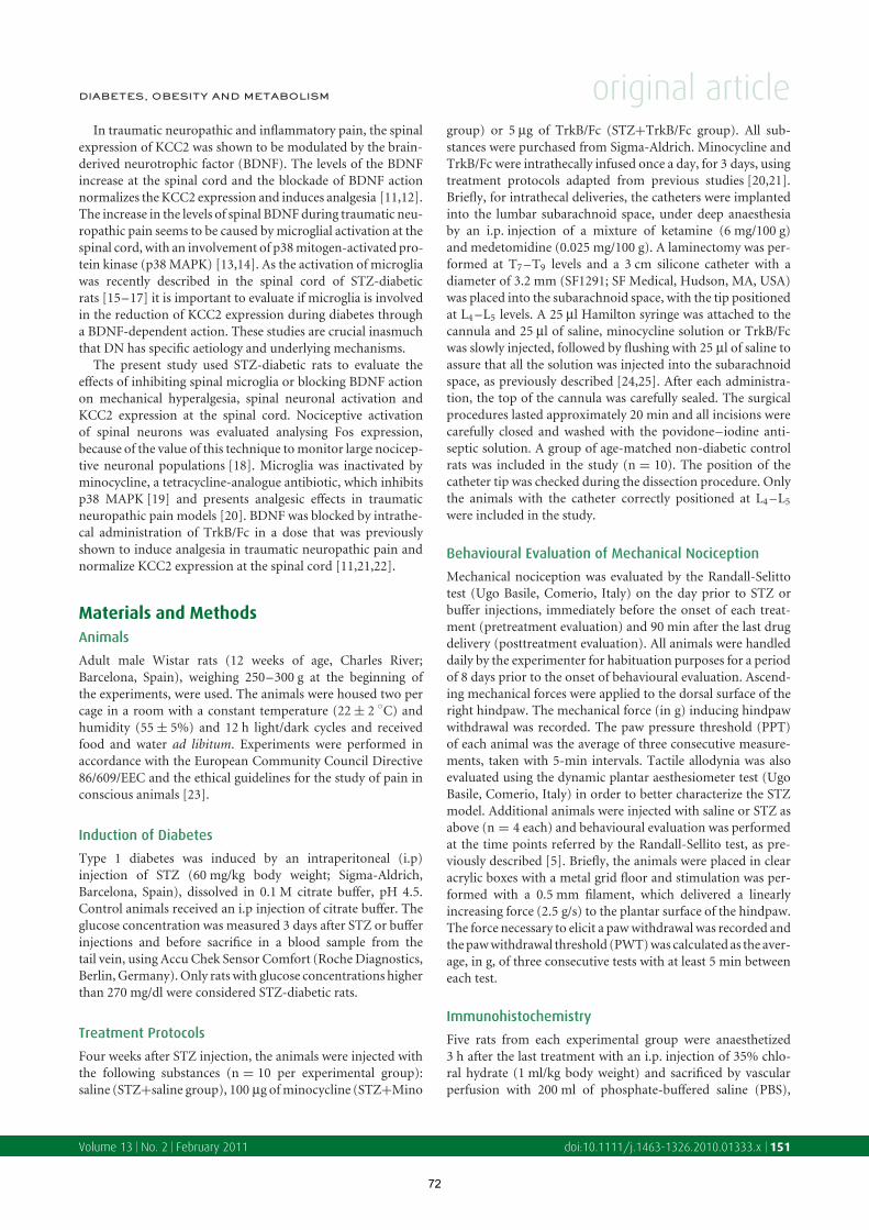
DIABETES, OBESITY AND METABOLISM original articleIn traumatic neuropathic and inflammatory pain, the spinal
expression of KCC2 was shown to be modulated by the brain-
derived neurotrophic factor (BDNF). The levels of the BDNF
increase at the spinal cord and the blockade of BDNF action
normalizes the KCC2 expression and induces analgesia [11,12].
The increase in the levels of spinal BDNF during traumatic neu-
ropathic pain seems to be caused by microglial activation at the
spinal cord, with an involvement of p38 mitogen-activated pro-
tein kinase (p38 MAPK) [13,14]. As the activation of microglia
was recently described in the spinal cord of STZ-diabetic
rats [15–17] it is important to evaluate if microglia is involved
in the reduction of KCC2 expression during diabetes through
a BDNF-dependent action. These studies are crucial inasmuch
that DN has specific aetiology and underlying mechanisms.
The present study used STZ-diabetic rats to evaluate the
effects of inhibiting spinal microglia or blocking BDNF action
on mechanical hyperalgesia, spinal neuronal activation and
KCC2 expression at the spinal cord. Nociceptive activation
of spinal neurons was evaluated analysing Fos expression,
because of the value of this technique to monitor large nocicep-
tive neuronal populations [18]. Microglia was inactivated by
minocycline, a tetracycline-analogue antibiotic, which inhibits
p38 MAPK [19] and presents analgesic effects in traumatic
neuropathic pain models [20]. BDNF was blocked by intrathe-
cal administration of TrkB/Fc in a dose that was previously
shown to induce analgesia in traumatic neuropathic pain and
normalize KCC2 expression at the spinal cord [11,21,22].
Materials and Methods
Animals
Adult male Wistar rats (12 weeks of age, Charles River;
Barcelona, Spain), weighing 250–300 g at the beginning of
the experiments, were used. The animals were housed two per
cage in a room with a constant temperature (22 ± 2 ◦C) and
humidity (55 ± 5%) and 12 h light/dark cycles and received
food and water ad libitum. Experiments were performed in
accordance with the European Community Council Directive
86/609/EEC and the ethical guidelines for the study of pain in
conscious animals [23].
Induction of Diabetes
Type 1 diabetes was induced by an intraperitoneal (i.p)
injection of STZ (60 mg/kg body weight; Sigma-Aldrich,
Barcelona, Spain), dissolved in 0.1 M citrate buffer, pH 4.5.
Control animals received an i.p injection of citrate buffer. The
glucose concentration was measured 3 days after STZ or buffer
injections and before sacrifice in a blood sample from the
tail vein, using Accu Chek Sensor Comfort (Roche Diagnostics,
Berlin, Germany). Only rats with glucose concentrations higher
than 270 mg/dl were considered STZ-diabetic rats.
Treatment Protocols
Four weeks after STZ injection, the animals were injected with
the following substances (n = 10 per experimental group):
saline (STZ+saline group), 100 µg of minocycline (STZ+Mino
group) or 5 µg of TrkB/Fc (STZ+TrkB/Fc group). All sub-
stances were purchased from Sigma-Aldrich. Minocycline and
TrkB/Fc were intrathecally infused once a day, for 3 days, using
treatment protocols adapted from previous studies [20,21].
Briefly, for intrathecal deliveries, the catheters were implanted
into the lumbar subarachnoid space, under deep anaesthesia
by an i.p. injection of a mixture of ketamine (6 mg/100 g)
and medetomidine (0.025 mg/100 g). A laminectomy was per-
formed at T7 –T9 levels and a 3 cm silicone catheter with a
diameter of 3.2 mm (SF1291; SF Medical, Hudson, MA, USA)
was placed into the subarachnoid space, with the tip positioned
at L4 –L5 levels. A 25 µl Hamilton syringe was attached to the
cannula and 25 µl of saline, minocycline solution or TrkB/Fc
was slowly injected, followed by flushing with 25 µl of saline to
assure that all the solution was injected into the subarachnoid
space, as previously described [24,25]. After each administra-
tion, the top of the cannula was carefully sealed. The surgical
procedures lasted approximately 20 min and all incisions were
carefully closed and washed with the povidone–iodine anti-
septic solution. A group of age-matched non-diabetic control
rats was included in the study (n = 10). The position of the
catheter tip was checked during the dissection procedure. Only
the animals with the catheter correctly positioned at L4 –L5
were included in the study.
Behavioural Evaluation of Mechanical Nociception
Mechanical nociception was evaluated by the Randall-Selitto
test (Ugo Basile, Comerio, Italy) on the day prior to STZ or
buffer injections, immediately before the onset of each treat-
ment (pretreatment evaluation) and 90 min after the last drug
delivery (posttreatment evaluation). All animals were handled
daily by the experimenter for habituation purposes for a period
of 8 days prior to the onset of behavioural evaluation. Ascend-
ing mechanical forces were applied to the dorsal surface of the
right hindpaw. The mechanical force (in g) inducing hindpaw
withdrawal was recorded. The paw pressure threshold (PPT)
of each animal was the average of three consecutive measure-
ments, taken with 5-min intervals. Tactile allodynia was also
evaluated using the dynamic plantar aesthesiometer test (Ugo
Basile, Comerio, Italy) in order to better characterize the STZ
model. Additional animals were injected with saline or STZ as
above (n = 4 each) and behavioural evaluation was performed
at the time points referred by the Randall-Sellito test, as pre-
viously described [5]. Briefly, the animals were placed in clear
acrylic boxes with a metal grid floor and stimulation was per-
formed with a 0.5 mm filament, which delivered a linearly
increasing force (2.5 g/s) to the plantar surface of the hindpaw.
The force necessary to elicit a paw withdrawal was recorded and
the paw withdrawal threshold (PWT) was calculated as the aver-
age, in g, of three consecutive tests with at least 5 min between
each test.
Immunohistochemistry
Five rats from each experimental group were anaesthetized
3 h after the last treatment with an i.p. injection of 35% chlo-
ral hydrate (1 ml/kg body weight) and sacrificed by vascular
perfusion with 200 ml of phosphate-buffered saline (PBS),
Volume 13 No. 2 February 2011 doi:10.1111/j.1463-1326.2010.01333.x 151
72

original article DIABETES, OBESITY AND METABOLISM
followed by 1000 ml of 4% paraformaldehyde in 0.1 M phos-
phate buffer (PB), pH 7.4. Spinal segments L4 –L5 were
removed, postfixed in the same fixative and cryoprotected
overnight in 30% sucrose in PB. Coronal sections 40-µm
thick, were obtained using a freezing microtome. Every
fourth section was collected in 0.1 M PBS and each set was
immunohistochemically reacted for KCC2, Fos protein and
CD11b (microglial marker) [26]. Immunohistochemical detec-
tion using the avidin–biotin peroxidase method was performed
as previously described [4,27]. Briefly, the sections were treated
with 1% hydrogen peroxidase for 10 min to inhibit endogenous
peroxidase activity before incubation for 2 h in a blocking solu-
tion containing 10% normal serum and 0.1 M glycine diluted
in 0.3% Triton × 25% in PBS (PBST). The sections were then
incubated 2 overnights at 4 ◦C in each primary antibody,
namely rabbit anti-KCC2 (1 : 10 000; Sigma-Aldrich), rabbit
anti-Fos (1 : 5000; Ab5; Calbiochem, Darmstadt, Germany) or
mouse anti-CD11b (clone OX-42; 1 : 2000; Millipore, Schwal-
bach/Ts, Germany), all diluted in PBST. After being washed
in PBST, the sections were incubated in biotinylated swine
anti-rabbit (for KCC2 and Fos detections) or rabbit anti-
mouse (for CD11b detection) immunoglobulins, from Dako
(Glostrup, Denmark), diluted in 1 : 200, for 1 h. Sections were
then washed in PBST, incubated for 1 h in the avidin–biotin
complex (Vectastain, Vector Laboratories, Burlingame, CA,
USA) and stained with diaminobenzidine (10 mg in 20 ml of
0.05 M Tris–HCl, pH 7.6 solution with 5 µl of 30% hydrogen
peroxide). Sections were then washed in PBS, mounted on
gelatine-coated slides, air-dried and coverslipped with xylol.
After observation under a light microscope, the images were
acquired using a high-resolution digital camera coupled to a
computer. In 10 randomly taken sections, immunolabelling for
KCC2 and CD11b was quantified by densitometric analysis at
the spinal dorsal horn (laminae I–V), using the NIH Image
1.52 software (US National Institutes of Health), as previously
described [9]. The numbers of Fos-immunoreactive (Fos-IR)
neurons were also counted in laminae I–V, as previously
described [28].
To confirm microglia activation, double immunofluores-
cence for CD11b and phosphorylated form of p38 MAPK
(phospho p38) was performed in spinal sections of con-
trols and STZ-diabetic rats (three animals per group). The
sections were treated with 1% borohydrate for 20 min before
incubation for 2 h in a blocking solution of 10% normal
goat serum in PBST. The sections were then incubated 2
overnights at 4 ◦C in the primary antibodies, mouse anti-
CD11b (1 : 1000) and rabbit anti-phospho p38 (1 : 500;
Cell Signalling, Danvers, MA, USA). After being washed in
PBST, the sections were incubated in fluorescent secondary
antibodies, goat anti-mouse Alexa 488 and goat anti-rabbit
Alexa 594 (1 : 500; Molecular Probes, Carlsbad, CA, USA) for
1 h. Sections were observed using an Apo Tome microscope
(Zeiss, Gottingen, Germany) and the images were acquired
using a high-resolution digital camera coupled to a computer.
Serial optical planes separated by 0.3 µm were collected in the
z-axis and merged images were obtained using the Apo Tome
software.
Western Blotting
The expression of phospho p38 and KCC2 were quantified by
western blotting. Five animals from the Control, STZ+saline
and STZ+Mino experimental groups were used in the western
blotting for phospho p38. For the KCC2 western blotting,
five animals per experimental group were used and all the
experimental groups were considered. After decapitation,
the spinal segments L4 –L5 were immediately removed and
homogenized in buffer (50 mM Tris, pH 7.6, 100 mM NaCl,
0.1% Triton X 100, 1 mM EDTA) added with a proteinase
inhibitor cocktail and a phosphatase inhibitor cocktail (both
at 1 : 100; Sigma-Aldrich). The protein concentration
was measured using Bradford Assay (BioRad, Amadora,
Portugal) and adjustments were performed to equalize protein
concentration. Samples were heated at 65 ◦C for 15 min
and 40 µg protein per lane was loaded, electrophoresed on
10% SDS-PAGE and electroblotted onto the nitrocellulose
membranes. The membranes were blocked in 5% non-fat
milk in Tris-buffered saline with Tween 0.1 M, pH 7.2–7.4
(TBST) for KCC2 and in 5% bovine serum albumin in TBST
for phospho p38 for 1 h and incubated overnight at 4 ◦C
with the rabbit anti-KCC2 antibody (1 : 2000; Sigma-Aldrich)
and rabbit anti-phospho p38 (1 : 1000, Cell Signalling). The
membranes were washed and incubated in antirabbit secondary
antibody conjugated to horseradish peroxidase (1 : 5000;
Jackson Labs, Bar Harbor, ME, USA) and rinsed in TBST. The
signal was detected using a sensitive chemiluminescence reagent
(SuperSignal West Pico Chemiluminescent Substrate, Thermo
Scientific, Waltham, MA, USA). Antibody against phospho p38
was then stripped incubating the membranes with 10% SDS for
15 min. Following blocking, the membranes were incubated
2 overnights with rabbit anti-p38 (1 : 1000, Cell Signalling)
at 4 ◦C. The signal detection was performed as previously
described. Alfa-tubulin was used as an internal standard, using
mouse anti-α-tubulin (1: 10 000; Sigma-Aldrich) as the primary
antibody. Digital images of western blots were analysed by
densitometry using NIH image 1.52 software.
BDNF Quantification
The content of BDNF in the spinal cord of control
and untreated STZ-diabetic rats was determined using the
BDNF Emax ImmunoAssay System (Promega, Madison,
WI, USA) in the supernatant of spinal homogenates from
L4 –L5 segments. Assays were performed according to the
manufacturer’s instructions, using an antibody sandwich
format in 96-well plates. Plates were coated with anti-BDNF
polyclonal antibodies. Samples (10 µl) or BDNF standards
were added to each well and plates were incubated. After
several washes, anti-BDNF monoclonal antibodies were added
and plates were again incubated. After thorough washes,
the amount of bound monoclonal antibody was detected
using IgG-horseradish peroxidase-conjugated antibody. The
unbound conjugate was removed by washing. The plate was
then incubated with 100 µl of substrate solution for 10 min.
Hydrochloric acid was added to stop the reactions. Colour
change was measured with a Synergy HT Microplate Reader
(Bio-Tek Instruments, Winooski, VT, USA) at 450 nm. All
152 Morgado et al. Volume 13 No. 2 February 2011
73

DIABETES, OBESITY AND METABOLISM original articlesamples were run in duplicate and values were averaged. BDNF
levels were normalized to the protein concentration in each
sample, quantified by Bradford Assay.
Specificity of Antibodies
The specificity of each primary antibody was previously
demonstrated [9,26,27]. Control experiments were performed
by omission of primary or secondary antibodies. Additionally
for KCC2, p38 and phospho p38 the specificity of the primary
antibodies was demonstrated by western blotting with bands
detected at 140 kDa for KCC2 and at 40 kDa for p38 and
phospho p38.
Statistical Analysis
SPSS statistics version 18 (IBM, Armonk, New York, NY, USA)
was used for statistical analysis. Behavioural, imunohistochem-
istry and western blotting data were compared by ANOVA
followed by Tukey’s post hoc test for multiple comparisons.
BDNF levels were compared by independent sample t-test.
Pretreatment and posttreatment behavioural responses were
compared by paired-sample t-test. Statistical significance was
settled at p < 0.05. Results are expressed as mean ± standard
error of the mean (s.e.m.).
Results
At 4 weeks postinjection, STZ-diabetic rats presented
significantly lower body weights and higher blood glucose levels
than age-matched non-diabetic control animals (Table 1). The
treatments with Mino and TrkB/Fc had no effects on body
weights or glycaemia.
Characterization of STZ-diabetic Rats
STZ-diabetic rats presented a significant increase in the
expressions of CD11b and phospho p38 in microglial cells,
when compared with control animals (figures 1, 2A–C, E).
Table 1. Body weights and glycaemia of experimental groups.
Experimental
groups Body weight (g)
Blood glucose
concentration (mg/dl)
Control 409 ± 5.4 131 ± 18.7
STZ+saline 287 ± 3.8∗ 492 ± 32.2∗
STZ+Mino 283 ± 7.3∗ 501 ± 28.4∗
STZ+TrkB/Fc 285 ± 4.3∗ 498 ± 50.3∗
Mean values ± standard error of the mean (s.e.m.). STZ, streptozotocin.∗p < 0.01 vs. control.
Furthermore, the microglia of STZ-diabetic rats presented
morphological changes that match the well-described signs
of microglial activation, namely larger soma and thicker
and shorter processes than control animals (cf. figures 1A,
B, 2B, C). Untreated STZ-diabetic rats presented much lower
PPTs than controls (p = 0.003; figure 3A). Tactile allodynia
was also detected in untreated STZ-diabetic rats (PWT at 4
weeks postinjection: 19.90 ± 2.04 and 14.10 ± 0.98 in control
animals and STZ-diabetic rats, respectively; p = 0.019), further
confirming the existence of mechanical hypersensitivity in
our STZ-diabetic rats. As the Randall-Selitto test is highly
predictable of clinical applicability of analgesic drugs [29], the
subsequent behavioural evaluations were performed using only
this test. STZ-diabetic rats presented a higher Fos expression at
the spinal dorsal horn than controls (p = 0.025; figure 3B–D).
The expression of KCC2 decreased in STZ+saline rats in
relation to controls (p = 0.009; figure 3F–H).
Effects of Minocycline
After minocycline treatment, the activation of microglia
detected at the spinal cord of untreated STZ-diabetic rats
was inhibited, with a significant decrease in the expression of
CD11b and phospho p38 (figure 2A, E) and reversal of mor-
phological signs of microglial activation (cf. figure 2C, D). The
mechanical hyperalgesia detected in untreated STZ-diabetic
Figure 1. Microglia activation at the spinal cord of streptozotocin (STZ)-diabetic rats. Panels (A) and (B) show double immunoreactions for CD11b
(marker of microglia; in green) and phospho p38 (marker of microglia activation; in red) in representative sections taken from the L4 spinal segment
in control (A) and STZ-diabetic (B) rats. Panel (C) shows representative blots and the quantification by western blotting of phospho p38 in spinal
homogenates of control and STZ-diabetic rats. STZ-diabetic rats present microglia activation at the spinal cord, demonstrated by morphological changes
of microglia (larger soma and thicker and shorter processes) and increased expression of phospho p38 (significant increase in the ratio between phospho
p38 and total p38). Significance of symbol: ∗p < 0.05. Scale bar in (B): 50 µm (both photomicrographs are at the same magnification).
Volume 13 No. 2 February 2011 doi:10.1111/j.1463-1326.2010.01333.x 153
74

original article DIABETES, OBESITY AND METABOLISM
Figure 2. Effects of minocycline treatment on microglial activation. Panel
(A) shows the expression of CD11b (marker of microglia) at the spinal
dorsal horn of control and of streptozotocin (STZ)-diabetic rats injected
with saline (STZ+saline) or treated with minocycline (STZ+Mino). Panels
(B–D) show photomicrographs of the L4 spinal sections labelled for CD11b
in the three experimental groups. Panel (E) shows representative blots and
the quantification by western blotting of phospho p38 expression in
the L4 –L5 spinal cord homogenates in the three experimental groups.
Minocycline significantly reduced CD11b and phospho p38 expression,
indicating that the treatment was effective in the reversion of microglia
activation. Significance of symbol: ∗∗p < 0.01. Scale bar in (D): 100 µm
(all photomicrographs are at the same magnification).
rats was reversed to the levels of control rats in the
STZ+Mino group with statistically significant differences in
relation to untreated STZ rats (p = 0.007; figure 3A). The
increased spinal Fos expression, detected in STZ+saline rats,
was also reversed to control levels after minocycline treat-
ment (figure 3B–E) with statistical significant differences only
between the STZ+saline and STZ+Mino groups (p = 0.017;
figure 3B, D, E). The expression of KCC2 was also reversed to
control levels when STZ-diabetic rats were treated with minocy-
cline (figure 3F–I), with statistical significant differences only
between the STZ+saline and STZ+Mino groups (p = 0.007;
figure 3F, H, I). The result of western blotting performed
for KCC2 supported the quantitative immunohystochemical
analysis (figure 3J).
Effects of TrkB/Fc
Blocking the BDNF action in STZ rats by TrkB/Fc ameliorated
mechanical nociception (figure 4A), with statistical significant
differences in relation to untreated STZ rats (p = 0.037).
TrkB/Fc reduced the number of Fos-IR neurons in STZ-diabetic
rats, with significant differences in relation to the STZ+saline
group (p = 0.018; figure 4B–E). The TrkB/Fc treatment
increased KCC2 expression in STZ-diabetic rats in relation to
untreated STZ-diabetic rats (p = 0.029; figure 4F–H). How-
ever, this reversion did not reach control levels, as the statistical
significant differences were detected between STZ+TrkB/Fc
and control animals (p = 0.047; figure 4F–I). western blotting
results for KCC2 confirmed the immunohystochemical data
(figure 4J).
We also evaluated the effects of TrkB/Fc in the activation
of microglia (figure 5). TrkB/Fc treatment induced a signif-
icant reduction of microglia activation in STZ-diabetic rat
(p = 0.019), as demonstrated by the decrease of CD11b expres-
sion (figure 5), but did not reverse it to the levels of control
animals (p = 0.009).
BDNF Levels
The results of the ELISA study are shown in Table 2. BDNF
levels in untreated STZ-diabetic rats were not different from
control rats. Treatment with minocycline or TrkB/Fc did not
alter BDNF levels.
Discussion
The present study shows that the mechanical hyperalgesia and
spinal neuronal hyperactivity detected during diabetes may
be reversed by correcting the KCC2 levels at the spinal cord,
through the inhibition of spinal microglia. BDNF levels at the
spinal cord did not change in diabetic rats and the inhibition
of BDNF did not reverse the KCC2 expression to control lev-
els, indicating that mechanisms besides BDNF are probably
involved in the correction of KCC2 expression by minocycline.
This BDNF-independent effect of microglia on KCC2 expres-
sion differs from other chronic pain models, which suggests
that the mechanical hyperalgesia induced by diabetes is caused
by particular mechanisms and reinforces the need to develop
specific treatments.
Diabetic rats presented marked microglia activation at the
spinal cord, which agrees with recent results in diabetic rats with
different genetic backgrounds [15–17] and demonstrates that
this is a common feature of diabetes. Minocycline was effective
in inhibiting microglial activity, as evidenced by the induction
of resting-state morphology and by the reduction of the spinal
levels of phospho p38 MAPK and CD11b. In neuropathic
and inflammatory chronic models it was shown that activated
microglia at the spinal cord is associated with increased BDNF
levels, which leads to a decrease in neuronal KCC2 expres-
sion [11,12,30,31]. In our STZ-diabetic rats, the significant
microglial activation in the spinal cord did not affect the spinal
content of BDNF. Furthermore, BDNF inhibition by TrkB/Fc
did not correct KCC2 expression in diabetic rats to control lev-
els. Although it could be argued that higher doses or prolonged
treatment with TrkB/Fc should have been used to increase
the effects of BDNF sequestration, the current protocol was
previously shown to significantly decrease BDNF expression at
154 Morgado et al. Volume 13 No. 2 February 2011
75

DIABETES, OBESITY AND METABOLISM original article
Figure 3. Effects of minocycline treatment on mechanical nociception (A), Fos expression (B–E) and KCC2 expression (F–J). Panel A shows the
behavioural responses in the Randall-Selitto test in control and in streptozotocin (STZ)-diabetic rats, before (pretreatment) and after (posttreatment)
injection of saline (STZ+saline) or minocycline (STZ+Mino). Panel (B) shows the mean numbers per section of Fos-immunoreactive (Fos-IR) neurons
at the spinal dorsal horn of the L4 –L5 segments in the three experimental groups (control, STZ+saline and STZ+Mino) and panels (C–E) show
representative photomicrographs of the L4 spinal cord sections. Panel (F) shows KCC2 expression at the spinal dorsal horn of the three experimental
groups and panels (G–I) show representative photomicrographs of the L4 spinal cord sections labelled for KCC2. Panel (J) shows representative blots
of KCC2 in each experimental group. Minocycline treatment completely normalized mechanical nociception, totally reverted spinal hyperactivity and
corrected KCC2 expression to control levels. Significance of symbols: #, comparison with pretreatment STZ+Mino animals; §, comparison with control
animals; ∗∗p < 0.01. Scale bar in (I): 100 µm (all photomicrographs are at the same magnification).
the spinal cord in traumatic neuropathic pain models [21]. In
addition, increasing TrkB/Fc doses could affect motor func-
tions and impair behavioural evaluation because BDNF is
crucial for the function of motoneurons [32]. The suggestion
that during diabetes the microglia-mediated effect on KCC2
expression is independent of BDNF represents a considerable
difference of pain induced by diabetes from other chronic pain
conditions and suggests that it is caused by specific mech-
anisms. Discarded a role for BDNF, several candidates could
mediate the action of microglia in decreasing KCC2 expression,
namely the cytokines interleukin (IL)-1β, tumour necrosis fac-
tor (TNF)-α, IL-6 and prostaglandin E2 (PGE2) [33,34]. In
a recent study, an increase of IL-1β and TNF-α expressions
in the spinal cords of STZ-diabetic rats was normalized after
Volume 13 No. 2 February 2011 doi:10.1111/j.1463-1326.2010.01333.x 155
76

original article DIABETES, OBESITY AND METABOLISM
Figure 4. Effects of brain-derived neurotrophic factor (BDNF) action blockade on mechanical nociception (A), Fos expression (B–E) and KCC2
expression (F–J). Panel (A) shows behavioural responses in the Randall-Sellito test in control animals and in streptozotocin (STZ)-diabetic rats before
(pretreatment) and after (posttreatment) injection of saline (STZ+saline) or TrkB/Fc (STZ+TrkB/Fc). Panel (B) shows the mean numbers per section of
Fos-immunoreactive (Fos-IR) neurons at the spinal dorsal horn of control, STZ+saline and STZ+TrkB/Fc rats and panels (C–E) show photomicrographs
of representative sections of L4 segment immunoreacted for Fos in the three experimental groups. Panel (F) shows KCC2 expression at the spinal dorsal
horn of control and STZ+saline or STZ+TrkB/Fc rats. Panels (G–I) show representative photomicrographs of the L4 spinal sections labelled for KCC2.
Panel (J) shows representative blots of KCC2 protein for each experimental group. Blocking BDNF action at the spinal cord of STZ rats, ameliorated
mechanical nociception, reverted spinal hyperactivity and increased KCC2 expression but not to the levels of control animals. Significance of symbols:
#, comparison with pretreatment STZ+TrkB/Fc animals; §, comparison with control animals; ∗p < 0.05; ∗∗p < 0.01. Scale bar in (I): 100 µm (all
photomicrographs are at the same magnification).
minocycline treatment [17], indicating an involvement of these
two cytokines. Future studies will be performed to evaluate the
involvement of these cytokines, along with IL-6 and PGE2, in
the changes of KCC2 expression during diabetes.
This study also shows that BDNF inhibition by TrkB/Fc
has beneficial effects on behavioural pain responses and spinal
neuronal hyperactivity during diabetes, despite the moderate
effect on KCC2 expression. A direct effect of TrkB/Fc on
spinal microglia is a possibility suggested by the reduction of
microglial activation detected in the spinal cord of STZ-diabetic
rats treated with TrkB/Fc. This is a new finding which may
be explained by the recent demonstration that microglia
156 Morgado et al. Volume 13 No. 2 February 2011
77

DIABETES, OBESITY AND METABOLISM original article
Figure 5. Effect of brain-derived neurotrophic factor (BDNF) blockade on microglial activation. Panel (A) presents the expression of CD11b (marker
of microglia) at the spinal dorsal horn of control, streptozotocin (STZ)-diabetic rats injected with saline (STZ+saline) or treated with TrkB/Fc
(STZ+TrkB/Fc). Panels (B) and (C) show photomicrographs of L4 spinal sections labelled for CD11b in the STZ+saline and STZ+TrkB/Fc rats. TrkB/Fc
reduced CD11b expression in STZ-diabetic rats, but it remained significantly higher than that from control rats. Significance of symbols: ∗p < 0.05;∗∗p < 0.01. Scale bar in (C): 100 µm (all photomicrographs are at the same magnification).
Table 2. Brain-derived neurotrophic factor (BDNF) content in L4 –L5
spinal segments.
Experimental groups BDNF (ng/mg protein)
Control 0.47 ± 0.04
STZ+saline 0.56 ± 0.08
STZ+Mino 0.49 ± 0.04
STZ+TrkB/Fc 0.52 ± 0.05
Mean values ± standard error of the mean (s.e.m.). STZ, streptozotocin.
is activated by BDNF, through binding with a truncated
tropomyosin-related kinase B (tTrkB) receptor expressed by
microglial cells [35]. The antinociceptive effects of TrkB/Fc may
also be derived from a BDNF action on spinal neurons. BDNF
facilitates the release of GABA from spinal interneurons [36]
and during diabetes the inhibitory action of GABA shifts to
excitatory at the spinal cord [8,9]. It is, therefore, possible that
BDNF blockade decreased GABA release in STZ-diabetic rats
and, herein, reduced spinal neuronal excitability. Another pos-
sibility to explain the antinociception induced by TrkB/Fc is a
blockade of excitatory neurotransmission at the spinal cord as
BDNF induces the release of excitatory neurotransmitters and
activates spinal glutamatergic receptors [37]. Finally, a direct
action of BDNF on cAMP response element-binding protein
phosphorylation is also possible, because this mechanism was
previously shown to account for BDNF-induced increase in
Fos expression in spinal neurons [37]. The mechanisms medi-
ating the beneficial effects of BDNF in mechanical hyperalgesia
and spinal Fos expression in STZ-diabetic rats needs further
clarification.
In summary, this study shows for the first time that microglia
inhibition corrects KCC2 expression during diabetes by mech-
anisms that are independent from BDNF action. As decreased
expression of KCC2 affects spinal GABA neurotransmission,
eliciting excitatory rather than inhibitory actions when binding
to GABAA receptors [10,13] during diabetes [8], it is likely
that the normalization of KCC2 expression at the spinal cord
by minocycline treatment restored the inhibitory function of
GABA at the spinal cord of diabetic rats. In fact, the relationship
between KCC2 expression and GABA action was clearly demon-
strated previously by showing that control animals intrathecally
treated with a KCC2 inhibitor develop a hyperalgesic pattern
of response similar to that of STZ-diabetic rats [8]. Those pain
responses were totally reversed by GABAA receptor antagonist.
In control animals, with normal expression of KCC2, neither
the antagonist nor the agonist of GABAA receptor affected
mechanical nociception [8]. As minocycline induced a com-
plete normalization of KCC2 expression in our STZ-diabetic
rats it is likely that treating the animals with antagonist or
agonists of GABAA receptor will elicit the effects observed in
control non-diabetic animals.
Translational consequences of the present data showing a
complete reversal of mechanical hyperlagesia by intrathecal
minocycline treatment should be considered in the future.
The systemic or oral delivery of minocycline was shown to
be effective in reversing DN [17,38]. Although these effects
were interpreted only by peripheral actions of the drug and
according to the present results, with intrathecal delivery of
minocycline, the effects may be because of direct spinal actions.
Minocycline is a second-generation antibiotic belonging to
the tetracycline family, approved by the FDA for the treat-
ment of acne vulgaris, some sexually transmitted diseases and
rheumatoid arthritis [39]. In humans, long-term treatment
with minocycline was shown to be generally safe and well tol-
erated. However, gastrointestinal and central nervous system
reversible side effects (e.g. vestibular effects, benign intracranial
hypertension) were reported, along with rare cases of autoim-
mune disorders, such as lupus and hepatitis [40]. Minocycline
is being tested in several neurodegenerative diseases and other
disorders, including spinal trauma and diabetes macular edema
(http://www.clinicaltrials.gov). Clinical trials will be needed to
Volume 13 No. 2 February 2011 doi:10.1111/j.1463-1326.2010.01333.x 157
78

original article DIABETES, OBESITY AND METABOLISM
confirm the beneficial effects of minocycline on pain during
DN, to evaluate the safety, efficacy and tolerability of this drug
in comparison with the current first-line therapies for painful
DN. These include tricyclic compounds, selective serotonin
and noradrenaline reuptake inhibitors (such as duloxetine)
and anticonvulsants (such as pregabalin or gabapentin), which
present side effects and limited efficacy. New drugs that inhibit
spinal microglia should be developed as a possible next step in
the treatment of pain induced by diabetes.
Acknowledgements
This work was supported by Fundacao para a Ciencia e
Tecnologia (FCT—PTDC/SAU/64643/2006). C. Morgado is
a PhD fellow of FCT (SFRH/BD/21555/2005).
Conflict of Interest
The authors do not declare any conflict of interest relevant to
this manuscript.
References1. Quattrini C, Tesfaye S. Understanding the impact of painful diabetic
neuropathy. Diabet Metab Res Rev 2003; 19: S2–S8.
2. Tesfaye S. Advances in the management of diabetic peripheral
neuropathy. Curr Opin Support Palliat Care 2009; 3: 136–143.
3. Barrett AM, Lucero MA, Le T, Robinson RL, Dworkin RH, Chappell AS.
Epidemiology, public health burden, and treatment of diabetic
peripheral neuropathic pain: a review. Pain Med 2007; 8(Suppl. 2):
S50–S62.
4. Morgado C, Tavares I. C-fos expression at the spinal dorsal horn of
streptozotocin-induced diabetic rats. Diabet Metab Res Rev 2007; 23:
644–652.
5. Morgado C, Terra PP, Tavares I. Neuronal hyperactivity at the spinal cord
and periaqueductal grey during painful diabetic neuropathy: effects of
gabapentin. Eur J Pain 2010; 14: 693–699.
6. Selvarajah D, Wilkinson ID, Emery CJ et al. Early involvement of the spinal
cord in diabetic peripheral neuropathy. Diabet Care 2006; 29: 2664–2669.
7. Pertovaara A, Wei H, Kalmari J, Ruotsalainen M. Pain behavior and
response properties of spinal dorsal horn neurons following experimental
diabetic neuropathy in the rat: modulation by nitecapone, a COMT inhibitor
with antioxidant properties. Exp Neurol 2001; 167: 425–434.
8. Jolivalt CG, Lee CA, Ramos KM, Calcutt NA. Allodynia and hyperalgesia in
diabetic rats are mediated by GABA and depletion of spinal potassium-
chloride co-transporters. Pain 2008; 140: 48–57.
9. Morgado C, Pinto-Ribeiro F, Tavares I. Diabetes affects the expression of
GABA and potassium chloride cotransporter in the spinal cord: a study in
streptozotocin diabetic rats. Neurosci Lett 2008; 438: 102–106.
10. Payne J, Rivera C, Voipio J, Kaila K. Cation-chloride cotransporters in
neuronal communication, development and trauma. Trends Neurosci
2003; 26: 199–206.
11. Miletic G, Miletic V. Loose ligation of the sciatic nerve is associated with
TrkB receptor-dependent decreases in KCC2 protein levels in the ipsilateral
spinal dorsal horn. Pain 2008; 137: 532–539.
12. Zhang W, Liu LY, Xu TL. Reduced potassium-chloride co-transporter
expression in spinal cord dorsal horn neurons contributes to inflammatory
pain hypersensitivity in rats. Neuroscience 2008; 152: 502–510.
13. Coull JA, Beggs S, Boudreau D et al. BDNF from migroglia causes the shift
in neuronal anion gradient underlying neuropathic pain. Nature 2005;
438: 1017–1021.
14. Ji R-R, Suter MR. p38 MAPK, microglial signaling, and neuropathic pain.
Mol Pain 2007; 3: 33–37.
15. Tsuda M, Ueno H, Kataoka A, Tozaki-Saitoh H, Inoue K. Activation of dorsal
horn microglia contributes to diabetes-induced tactile allodynia via
extracellular signal-regulated protein kinase signaling. Glia 2008; 56:
378–386.
16. Wodarski R, Clark AK, Grist J, Marchand F, Malcangio M. Gabapentin
reverses migroglial activation in the spinal cord of streptozotocin-induced
diabetic rats. Eur J Pain 2009; 13: 807–811.
17. Talbot S, Chahmi E, Pena Dias J, Couture R. Key role for spinal dorsal
horn microglial kinin B1 receptor in early diabetic pain neuropathy. J
Neuroinflammation 2010; 7: 36.
18. Coggeshall RE. Fos, nociception and the dorsal horn. Prog Neurobiol 2005;
77: 299–352.
19. Hua XY, Svensson CI, Matsui T, Fitzsimmons B, Yaksh TL, Webb M. Intrathe-
cal minocyclinecycline attenuates peripheral inflammation-induced hyper-
algesia by inhibiting p38 MAPK in spinal microglia. Eur J Neurosci 2005;
22: 2431–2440.
20. Ledeboer A, Sloane EM, Milligan ED et al. Minocycline attenuates mechan-
ical allodynia and proinflammatory cytokine expression in rat models of
pain facilitation. Pain 2005; 115: 71–83.
21. Ramer LM, McPhail LT, Borisoff JF et al. Endogenous TrkB ligands suppress
functional mechanosensory plasticity in the deafferented spinal cord. J
Neurosci 2007; 27: 5812–5822.
22. Yajima Y, Narita M, Matsumoto N, Suzuki T. Involvement of a spinal brain
derived neurotrophic factor/full-length TrkB pathway in the development
of nerve injury-induced thermal hyperalgesia in mice. Brain Res 2002;
958: 338–346.
23. Zimmermann M. Ethical guidelines for investigations of experimental pain
in conscious animals. Pain 1983; 16: 109–110.
24. Castro AR, Pinto M, Lima D, Tavares I. Secondary hyperalgesia in the
monoarthritic rat is mediated by GABAB and NK1 receptors of spinal
dorsal horn neurons: a behavior and c-fos study. Neuroscience 2006; 141:
2087–2095.
25. Cruz CD, Charrua A, Vieira E, Valente J, Avelino A, Cruz F. Intrathecal
delivery of resiniferatoxin (RTX) reduces detrusor overactivity and spinal
expression of TRPV1 in spinal cord injured animals. Exp Neurol 2008; 214:
301–308.
26. Milligan CE, Cunningham TJ, Levitt P. Differential immunochemical mark-
ers reveal the normal distribution of brain macrophages and
microglia in the developing rat brain. J Comp Neurol 1991; 314:
125–135.
27. Pinto M, Lima D, Tavares I. Neuronal activation at the spinal cord and
medullary pain control centers after joint stimulation: a c-fos study in acute
and chronic articular inflammation. Neuroscience 2007; 147: 1076–1089.
28. Castro AR, Pinto M, Lima D, Tavares I. Nociceptive spinal neurons
expressing NK1 and GABAB receptors are located in lamina I. Brain
Res 2004; 1003: 77–85.
29. Le Bars D, Hansson PT, Plaghki L. Current animal test and models of pain.
In: Bealieu P, Lussier D, Porreca F, Dickenson AH eds. Pharmacology of
Pain. Seattle: IASP Press, 2010.
30. Morales-Aza B, Chilingworth N, Payne J, Donaldson L. Inflammation alters
cation chloride expression in sensory neurons. Neurobiol Dis 2004; 17:
62–69.
31. Coull JA, Boudreau D, Bachand K et al. Trans-synaptic shift in anion
gradient in spinal lamina I neurons as a mechanism of neuropathic
pain. Nature 2003; 424: 923–942.
158 Morgado et al. Volume 13 No. 2 February 2011
79

DIABETES, OBESITY AND METABOLISM original article32. Kishino A, Ishige Y, Tatsuno T, Nakayama C, Noguchi H. BDNF prevents
and reverses adult rat motor neuron degeneration and induces axonal
outgrowth. Exp Neurol 1997; 144: 273–286.
33. Inoue K, Tsuda M. Microglia and neuropathic pain. Glia 2009; 57:
1469–1479.
34. McMahon SB, Malcangio M. Current challenges in glia-pain biology.
Neuron 2009; 64: 46–54.
35. Mizoguchi Y, Monji A, Kato T et al. Brain-derived neurotrophic factor
induces sustained elevation of intracellular Ca2+ in rodent microglia.
J Immunol 2009; 183: 7778–7786.
36. Malcangio M, Bowery NG. GABA and its receptors in the spinal cord.
Trends Pharmacol Sci 1996; 17: 457–462.
37. Merighi A, Salio C, Ghirri A et al. BDNF as a pain modulator. Prog Neurobiol
2008; 85: 297–317.
38. Bhatt LK, Veeranjaneyulu A. Minocycline with aspirin: a therapeutic
approach in the treatment of diabetic neuropathy. Neurol Sci 2010;
31: 705–716.
39. Good ML, Hussey DL. Minocycline: stain devil? Br J Dermatol 2003; 149:
237–239.
40. Blum D, Chtarto A, Tenenbaum L, Brotchi J, Levivier M. Clinical potential
of minocycline for neurodegenerative disorders. Neurol Dis 2004; 17:
359–366.
Volume 13 No. 2 February 2011 doi:10.1111/j.1463-1326.2010.01333.x 159
80

Publication 4
Alpha-lipoic acid normalizes nociceptive neuronal activity at the spinal cord of diabetic rats
81

82

orig
inal
artic
le
Diabetes, Obesity and Metabolism 13: 736–741, 2011.© 2011 Blackwell Publishing Ltdoriginal article
α-Lipoic acid normalizes nociceptive neuronal activityat the spinal cord of diabetic ratsC. Morgado, P. Pereira-Terra & I. TavaresInstitute of Histology and Embryology of Faculty of Medicine of Porto and IBMC, University of Porto, Porto, Portugal
Aim: To evaluate the effects of antioxidant treatment of streptozotocin (STZ)-diabetic rats with α-lipoic acid (α-LA) in neuronal and microglialactivation at the spinal cord, an important relay station of nociceptive transmission. Because of the role of the potassium chloride co-transporter2 (KCC2) in neuronal activation at the spinal cord and the influence of microglia in KCC2 expression, we also evaluated the effects of α-LA inKCC2 expression at the spinal cord.Methods: Four weeks after STZ injection, the rats received daily intraperitoneal injections of α-LA (100 mg/kg), during 2 weeks. Mechanicalnociception was evaluated before and after α-LA treatment. Spinal cords were immunoreacted against 8-OH-dG (marker of oxidative stressdamage), Fos (marker of neuronal activation) and CD11b (marker of microglia). KCC2 expression was evaluated by immunohistochemistry andwestern blotting.Results: Treatment with α-LA decreased the 8-OH-dG and Fos expressions to controls’ levels, but did not affect CD11b. Treatment with α-LAalleviated mechanical hyperalgesia and partially corrected KCC2 expression.Conclusions: This study shows that neuronal hyperactivity at the spinal cord of STZ-diabetic rats can be corrected by α-LA, which mayaccount for alleviation of mechanical hyperalgesia. These effects are probably partially mediated by KCC2, but are independent frommicroglia.Keywords: α-lipoic acid, animal pharmacology, c-fos, diabetes, diabetes complications, diabetic neuropathy, drug mechanism, KCC2, microglia,pain, spinal cord, type 1 diabetes
Date submitted 15 December 2010; date of first decision 17 January 2011; date of final acceptance 14 March 2011
IntroductionOxidative stress damage has been implicated in a varietyof pathological processes that underlie diabetic compli-cations, as retinopathy [1], nephropathy [2] and neuropa-thy [3]. Diabetic neuropathy affects 30–50% of diabeticpatients, with spontaneous and mechanical-evoked painreported as major complaints by about one quarter of thepatients [4]. Diabetes induces oxidative stress in peripheralnerves, which are probably to contribute to nerve dysfunc-tion and altered pain sensations observed during diabeticneuropathy [3,5].
α-Lipoic acid (α-LA) is a powerful scavenger of free radi-cals and metal chelator [6]. In experimental models of diabeticneuropathy, antioxidant treatment with α-LA normalizes nerveblood flow, nerve conduction velocity and nociception [3,5].In diabetic patients, α-LA significantly decreases oxidativestress [7], improves microcirculation [8] and reduces pain [9].The mechanisms of action of α-LA on pain have only beenstudied at the periphery. Considering the ability of α-LA tocross the brain–blood barrier [10] and the observation thatα-LA reverses the impairment in kinin (1) receptor expression
Correspondence to: Isaura Tavares, Institute of Histology and Embryology, Faculty ofMedicine of Porto, Alameda Prof. Hernani Monteiro, 4200-319 Porto, Portugal.E-mail: [email protected]
at the spinal cord of insulin-resistant animal models [11], wehypothesized that α-LA has also central effects in painful dia-betic neuropathy, namely at the spinal cord. This is importantas it was previously showed that streptozotocin (STZ)-diabeticrats presented hyperactivity of spinal nociceptive neurons [12].This increased neuronal activity was shown to be associ-ated with a decreased expression of the potassium chlorideco-transporter 2 (KCC2) at the spinal cord [13,14], whichaccounts for a shift in the action of γ -aminobutiric acid(GABA) from inhibitory to excitatory in the spinal dorsalhorn of STZ-diabetic rats [13]. Spinal microglia activationwas shown to account for those impairments [15]. Oxidativestress is an additional mechanism that may underlie changesin the expression and activity of KCC2 in the spinal cordduring diabetes. In an oxidative stress model of hippocam-pal neuronal cultures, the expression and activity of KCC2is decreased [16]. Antioxidants interfere with the inflamma-tory response of microglial cultures [17]. Oxidative stress may,therefore, trigger KCC2 impairments during diabetes, eitherby a direct neuronal action or by interfering with microglialactivation.
This study evaluates the effects of an antioxidant treatmentwith α-LA in neuronal and microglial activation at the spinalcord of STZ-diabetic rats and investigates an involvement ofKCC2.
83

DIABETES, OBESITY AND METABOLISM original articleMaterials and MethodsAnimals
Male Wistar rats (aged 12 weeks; Charles River, Barcelona,Spain), weighing 250–300 g, were used. The animals werehoused in a room with controlled temperature (22 ± 2◦ C) andhumidity (55 ± 5%) and 12-h light/dark cycles. The animalsreceived food and water ad libitum. Experiments were per-formed in accordance with the European Community CouncilDirective 86/609/EEC and the ethical guidelines for the studyof pain in conscious animals [18].
Induction of Diabetes
Type 1 diabetes was induced by an intraperitoneal (i.p.) injec-tion of STZ (60 mg/kg body weight; Sigma-Aldrich, Barcelona,Spain), dissolved in 0.1 M citrate buffer, pH 4.5. Control ani-mals were injected with citrate buffer. Glucose concentrationwas measured in a blood sample from the tail vein, using AccuChek Sensor Comfort (Roche Diagnostics, Berlin, Germany),3 days after those i.p. injections and before sacrifice. Onlyrats with glucose concentrations higher than 270 mg/dl wereincluded in the STZ-diabetic group.
Treatment Protocols
Four weeks after STZ injection, the rats received dailyi.p. injections of α-LA (STZ + α-LA group) or saline (STZ+ saline group) during the following 2 weeks. Both experimen-tal groups included 10 rats. α-LA (Sigma-Aldrich) was used ina dose (100 mg/kg body weight) settled according to previousstudies [3,5]. Groups of age-matched non-diabetic control ratsand STZ-diabetic animals with 4 weeks of disease (STZ + 4 wgroup) were included in the study (n = 10 per group).
Behavioural Evaluation of Mechanical Nociception
All animals were handled daily by the experimenter for habitu-ation purposes during 8 days prior to the onset of behaviouralevaluation. Mechanical nociception was evaluated using theRandall-Selitto test (Ugo Basile, Comerio, Italy) at three time-points: at the day prior to STZ or buffer injections, immediatelybefore the treatment onset (pretreatment evaluation) and90 min after the last α-LA or saline injections (post-treatmentevaluation). The paw pressure threshold (PPT) was determinedat the right hindpaw as described previously [15].
Immunohistochemistry
Five rats from each experimental group were deeply anaes-thetized 3 h after the last treatment with an i.p. injectionof 35% chloral hydrate (1 ml/kg body weight) and per-fused with phosphate-buffered saline (PBS), followed by4% paraformaldehyde in 0.1 M phosphate buffer (PB),pH 7.4 [12,15]. The spinal segments L4–L5 were removed,post-fixed and cryoprotected overnight in 30% sucrose inPB. Every fourth coronal frozen section, 40-μm thick, wascollected in 0.1 M PBS. Each section set was immuno-histochemically reacted for 8-OH-dG (marker of oxidative
stress nucleic acid damage) [19], Fos protein (marker ofnociceptive neuronal activation at the spinal cord) [20],CD11b (microglial marker) [21] or KCC2 [14], using theavidin–biotin–peroxidase method. The spinal cord sectionsimmunoreacted for 8-OH-dG were pretreated with 2 N HClduring 10 min to denature nucleic acids, followed by 5 minimmersion in Tris-base 1 M. Following treatment with 1%hydrogen peroxidase for 10 min to inhibit endogenous perox-idase activity, sections were incubated in a blocking solution of10% normal serum in 0.3% Triton X 25% in PBS (PBST) with0.1 M glycine for 2 h. The sections were then incubated twoovernights at 4◦ C in each primary antibody, namely mouseanti-8-OH-dG (1 : 1600; Trevigen, Gaithersburg, MD, USA),rabbit anti-Fos (1 : 5000; Ab5; Calbiochem, Darmstadt, Ger-many), mouse anti-CD11b (clone OX-42; 1 : 2000, Millipore,Schwalbach/Ts, Germany) or rabbit anti-KCC2 (1 : 10000;Sigma-Aldrich), all diluted in PBST. After washings in PBST,the sections were incubated in biotinylated rabbit anti-mouse(for 8-OH-dG and CD11b detection) or swine anti-rabbit (forFos and KCC2 detection), all from Dako (Glostrup, Denmark),diluted at 1 : 200, for 1 h. After washings in PBST, the sectionswere incubated for 1 h in the avidin–biotin complex (Vectas-tain, Vector Laboratories, Burlingame, CA, USA) and stainedwith diaminobenzidine [12,15]. Light microscope images wereacquired by a blind-observer using a high-resolution digi-tal camera coupled to a computer. In 10 randomly takensections, immunolabelling for 8-OH-dG, CD11b and KCC2was quantified by densitometric analysis at the spinal dorsalhorn, using the NIH Image 1.52 software (US National Insti-tutes of Health), as described previously [14]. The numbers ofFos-immunoreactive (Fos-IR) neurons were counted at spinaldorsal horn (laminae I–V), as described previously [22].
Western Blotting
The expression of KCC2 was quantified by western blotting inspinal homogenates of spinal segments L4–L5 of 5 animals perexperimental group, as described previously [14]. Briefly, themembranes were incubated with the rabbit anti-KCC2 anti-body used above (1 : 2000) and alfa-tubulin, used as an internalstandard, was identified using a mouse anti-α-tubulin antibody(1 : 10000; Sigma-Aldrich, Spain). Digital images were analysedby densitometry using NIH Image 1.52 software, as describedpreviously [14].
Specificity of Antibodies
Control experiments were performed by omission of pri-mary or secondary antibodies. In the case of Fos experiments,the specificity of immunostaining was further confirmed bylack of immunoreactions in naive animals [22]. Specificity ofKCC2 primary antibody was also showed by western blot-ting with band detected at 140 KDa [14]. The specificities ofthe anti-CD11b and anti-8-OH-dG antibodies were previouslyshowed [19,21].
Statistical Analysis
SPSS Statistics version 18 (IBM, Armonk, NY, USA) was usedfor statistical analysis. Behavioural, imunohistochemistry and
Volume 13 No. 8 August 2011 doi:10.1111/j.1463-1326.2011.01399.x 737
84

original article DIABETES, OBESITY AND METABOLISM
Table 1. Body weights and glycaemia of experimental groups.
Experimentalgroups
Bodyweight (g)
Blood glucoseconcentration (mg/dl)
Control 409 ± 5.4 131 ± 18.7STZ + 4 w 283 ± 7.3∗ 503 ± 28.4∗
STZ + saline 287 ± 3.8∗ 492 ± 32.2∗
STZ + α-LA 290 ± 2.4∗ 486 ± 61.2∗
Mean values ± s.d. α-LA, α-lipoic acid; STZ, streptozotocin.∗p < 0.01 versus control.
western blotting data were compared by analysis of variancefollowed by Tukey post hoc test for multiple comparisons.Pretreatments and post-treatments behavioural responses werecompared by paired sample t-test. Statistical significance wassettled at p < 0.05. Results are expressed as mean ± standarderror of the mean (s.e.m.).
ResultsCharacterization of Untreated STZ-Diabetic Rats
Untreated STZ-diabetic rats at 4 weeks post-injection andsaline-injected STZ-diabetic rats presented significantly lowerbody weights and higher blood glucose levels than age-matchednon-diabetic control animals (Table 1). STZ-diabetic ratspresented increased responses to mechanical stimuli, showedby the lower PPTs in the Randall-Sellito test (figure 1).
STZ-diabetic rats also presented changes at the spinal dorsalhorn, when compared to controls. The expression of 8-OH-dG was significantly higher in untreated and saline-treatedSTZ-diabetic rats in comparison with controls (figure 2),demonstrating increased oxidative stress nucleic acids damageduring diabetes. STZ-diabetic rats presented higher numbersof Fos-IR neurons at the spinal dorsal horn than controls(figure 3A–E), indicating higher nociceptive activation ofspinal neurons. The microglia of the former animals presentedlarger soma and thicker and shorter processes and increasedexpression of CD11b (figure 3F–J), which is considered an
Figure 1. Effects of α-lipoic acid (α-LA) treatment on mechanicalhyperalgesia. The graph represents the behavioural responses (paw pressurethresholds in the Randall-Sellitto test) in non-diabetic control animals,streptozotocin (STZ)-diabetic rats with 4 weeks of the disease (STZ +4 w) and in STZ-diabetic rats before (pretreatment) and after (post-treatment) injections of α-LA or saline. The antioxidant treatment withα-LA ameliorated the mechanical nociception, but without normalizationto the control levels. Significance of symbols: § comparison with non-diabetic control animals; # comparison with pretreatment STZ + α-LAgroup; two symbols: p < 0.01.
indication of microglial activation [15]. The expression ofKCC2 was lower in STZ-diabetic rats (figure 4).
Effects of Treatment with α-LA
Treating STZ-diabetic rats with α-LA had no effects on bodyweights or blood glucose levels (Table 1), which is in accordancewith previous studies using similar α-LA dose and even withhigher treatment duration [3,5].
Behavioural Pain Responses
Treatment of STZ-diabetic rats withα-LA ameliorated mechan-ical nociception, with PPTs values intermediate between con-trols (p = 0.009) and STZ + saline rats (p = 0.000; figure 1).
Oxidative Stress Nucleic Acids Damage
Treatment of STZ-diabetic rats with α-LA significantly reducedthe expression of 8-OH-dG to the levels of control animals(figure 2), showing that α-LA totally reversed the signs ofoxidative stress nucleic acid damage at spinal dorsal hornduring diabetes.
Spinal Neuronal and Microglia Activity
Treatment with α-LA decreased the number of Fos-IR neuronsto the levels of control animals (figure 3A–E), indicatinga reversal of nociceptive activation of spinal neurons aftertreatment. The α-LA had no effects on the CD11b microglialexpression, with microglial cells maintaining the activatedphenotype detected in untreated and saline-treated STZ-diabetic rats (figure 3F–J).
Spinal KCC2 Expression
Treatment with α-LA moderately increased the KCC2immunolabelling to levels intermediate between controls(p = 0.048) and STZ + saline rats (p = 0.031) (figure 4A–F).These results were confirmed by western blotting (figure 4B).
DiscussionThis study showed, for the first time, that α-LA blocks oxidativestress at the spinal cord and controls the activity of nociceptivespinal neurons during diabetes. In a manner similar to otherpathologies, oxidative stress induced by diabetes is probablyto decrease KCC2 expression at the spinal cord, which wasblocked here by the antioxidant treatment with α-LA.
Antioxidant treatment with α-LA fully reversed both oxida-tive stress nucleic acids damage and neuronal hyperactivityat the spinal cord of STZ-diabetic rats. The mechanismsused by α-LA to reduce the neuronal hyperactivity at thespinal cord were investigated in the present study. Treatmentwith α-LA interferes with KCC2 expression at the spinal cordneurons, indicating that this co-transporter accounts for theα-LA-mediated reversion of neuronal hyperactivity during dia-betes. Although microglia activation appears to account to thereduction of spinal KCC2 expression in STZ-diabetic rats [15],microglia does not appear to be involved in the effects of
738 Morgado et al. Volume 13 No. 8 August 2011
85

DIABETES, OBESITY AND METABOLISM original article
Figure 2. Effects of α-lipoic acid (α-LA) treatment on oxidative stress nucleic acids damage in the spinal dorsal horns. Panel (A) represents 8-OH-dGexpression in control animals, streptozotocin (STZ)-diabetic rats with 4 weeks of the disease (STZ + 4 w) and STZ-diabetic rats treated with saline (STZ+ saline) or α-LA (STZ + α-LA). Panels (B–E) are representative photomicrographs of L4 spinal cord sections labelled for 8-OH-dG. Damage inducedin the nucleic acids by oxidative stress was significantly increased during diabetes and was reversed by α-LA. Significance of symbols: * comparison withnon-diabetic control animals and STZ + α-LA rats, two symbols: p < 0.01. Scale bar: 100 μm. All photomicrographs are at the same magnification.
Figure 3. Effects of α-lipoic acid (α-LA) treatment on neuronal and microglial activity at the spinal dorsal horn. Panel (A) presents the mean numbers persection of Fos-IR neurons. Panels (B–E) are representative photomicrographs of L4 spinal cord section labelled for Fos (arrows) in control animals (B),streptozotocin (STZ) + 4 w (C), STZ + saline (D) and STZ + α-LA (E) rats. Panel (F) presents the CD11b expression and panels (G–J) are representativephotomicrographs of L4 spinal cord sections labelled for CD11b in control animals (G), STZ + 4 w (H), STZ + saline (I) and STZ + α-LA (J) rats. α-LAreversed the neuronal hyperactivity observed in STZ + 4 w and STZ + saline rats to control levels, but had no effect on microglial activation. Significanceof symbols: * comparison with non-diabetic control animals and STZ + α-LA rats; § comparison with non-diabetic control group; two symbols: p < 0.01.Scale bar: 100 μm. All photomicrographs are at the same magnification.
α-LA over KCC2 expression. As per the KCC2 role in neuronalfunction, this co-transporter sets the intracellular chloride con-centration and influences the role of GABA when binding toGABAA ionotropic post-synaptic receptor. In normal condi-tions, GABA binding to GABAA receptors induces chlorideinflux inhibiting neuronal activity [23]. During diabetes, thereduced expression of KCC2 in the spinal cord leads to intra-cellular chloride accumulation and GABA binding to GABAA
receptors induces chloride output, which results in neuronalexcitation [13]. In agreement, intrathecal administration ofbicuculline (GABAA receptor antagonist) in STZ-diabetic ratselicits antinociception [13]. The decrease in KCC2 expressionhas been shown to reduce KCC2 activity [16] and increaseneuronal responses in pathological conditions, other than dia-betes, namely in axotomy, seizures and traumatic neuropathicpain [23].
The beneficial effects of α-LA treatment on blockingbehavioural signs of diabetic neuropathy have been ascribed to
its actions on peripheral nerve function [3,5]. In our study, wecannot discard peripheral effects of α-LA treatment as we didnot evaluate nerve function. We used a short α-LA treatment,which was implemented only after diabetic neuropathy wasestablished. Although this treatment clearly reversed neuronalspinal hyperactivity, it was not so effective in mechanical noci-ception. This is probably because of the fact that peripheralnerve damage induced by diabetes is marked at the treatmentonset and, therefore, could not be corrected by our proto-col. Previous studies also reported an incomplete reversal ofdiabetes-induced peripheral nerve changes by α-LA, even withhigher doses and longer treatment period [24]. Whether thehigher effects of α-LA on the nociceptive activation of spinalneurons indicate a preferential site of action of α-LA at thespinal cord will be investigated by performing intrathecal deliv-eries of α-LA. KCC2 is unlikely to represent the sole mechanismof α-LA action, as the treatment completely reversed neuronalhyperactivity but elicited only a partial correction of KCC2
Volume 13 No. 8 August 2011 doi:10.1111/j.1463-1326.2011.01399.x 739
86

original article DIABETES, OBESITY AND METABOLISM
Figure 4. Effects of α-lipoic acid (α-LA) treatment on potassium chloride co-transporter 2 (KCC2) expression at spinal dorsal horn. Panel (A) presentsKCC2 expression detected by immunohistochemistry. Panel (B) shows representative blots of KCC2 protein for each experimental group. Panels (C–F)are representative photomicrographs of L4 spinal cord sections immunoreacted for KCC2. Antioxidant treatment with α-LA increased KCC2 expression,but not to control levels. § comparison with non-diabetic control animals; # comparison with streptozotocin (STZ) + 4 w and STZ + saline groups; onesymbol: p < 0.05; two symbols: p < 0.01. Scale bar: 100 μm. All photomicrographs are at the same magnification.
expression. The inhibitory effects of α-LA on spinal neuronalactivity may also arise from its inhibitory actions on Ca(V)3.2T-type calcium channels [25]. These channels are probablyto be involved, as painful diabetic neuropathy is reversedby knockdown of Ca(V)3.2 T-type calcium channels throughintrathecal administration of antisense oligonucleotides [26].Future studies will evaluate the relative contribution of KCC2and Ca(V)3.2 T-type channels in the α-LA effects at spinal cordduring diabetes.
In summary, this study shows that α-LA presents centraleffects during diabetic neuropathy by reversing the neuronalhyperactivity at spinal dorsal horn, which could be partiallymediated by its effect on KCC2 expression and seems to beindependent from microglia activity. In a manner similar toother areas of the central nervous system, it is possible thatoxidative stress in the spinal cord during diabetes decreasessurface expression of KCC2 in spinal neurons, triggered by itsdephosphorylation, which will induce functional changes ofthis co-transporter [16]. The beneficial effects of α-LA antiox-idant treatment in reversing behavioural signs of diabeticneuropathy in STZ-diabetic rats fully match data with otherantioxidants, namely the resveratrol [27] and vitamin E [28],pointing to the importance of increasing antioxidant intake bydiabetic patients. As the diabetic neuropathy has common fea-tures in type 1 and type 2 diabetes, but α-LA increases insulin
sensitivity and decreases glucose levels in the former, whichdoes not occur in similar treatment protocol of STZ-diabeticrats [3], it is important to study if the neurobiological basis ofthe beneficial effects of α-LA in animal models of type 2 dia-betes [29] are also based in corrections of KCC2 expression atthe spinal cord. The effects of α-LA are not confined to patho-logical conditions as α-LA has also beneficial effects on healthyanimals and humans, namely, by: (i) increasing the expres-sion and activity of antioxidant enzymes [30]; (ii) reducing theoxidative stress damage [7]; and (iii) preventing neurodegener-ative diseases [30]. These findings show that, besides its curativeeffects, α-LA is a health promoter by preventing the disease.
AcknowledgementsThis work was supported by Fundacao para a Ciencia e Tec-nologia (FCT; PTDC/SAU/64643/2006). C. Morgado is a PhDfellow of FCT (SFRH/BD/21555/2005).
Conflict of InterestC. M. helped in the design, conduct/data collection, analysis andwriting of the manuscript. P. P.-T. helped in the conduct/datacollection. I. T. helped in design and writing the manuscript.All the authors have no conflict of interests.
740 Morgado et al. Volume 13 No. 8 August 2011
87

DIABETES, OBESITY AND METABOLISM original articleReferences1. Lin J, Bierhaus A, Bugert P et al. Effects of R-(+)-α-lipoic acid onexperimental diabetic retinopathy. Diabetologia 2006; 49: 1089–1096.
2. Obrosova IG, Fathallah I, Lie E, Nourooz-Zadeh J. Early oxidative stress inthe diabetic kidney:effect of DL-alpha-lipoic acid. Free Radic Biol Med2003; 34: 186–195.
3. Stevens MJ, Obrosova I, Cao X, van Huysen C, Greene DA. Effects of DL-α-lipoic acid on peripheral nerve conduction, blood flow, energy metabolismand oxidative stress in experimental diabetic neuropathy. Diabetes 2000;49: 1006–1015.
4. Quattrini C, Tesfaye S. Understanding the impact of painful diabeticneuropathy. Diabetes Metab Res Rev 2003; 19: S2–S8.
5. Cameron NE, Jack AM, Cotter MA. Effect of α-lipoic acid on vascularresponses and nociception in diabetic rats. Free Radic Biol Med 2001;31: 125–135.
6. Packer L, Witt EH, Tritschler HJ. Alpha-lipoic acid as a biological antioxidant.Free Radic Biol Med 1995; 19: 227–250.
7. Borcea V, Nourooz-Zadeh J, Wolff SP et al. Alpha-lipoic acid decreasesoxidative stress even in diabetic patients with poor glycemic control andalbuminuria. Free Radic Biol Med 1999; 26: 1495–1500.
8. Haak E, Usadel KH, Kusterer K et al. Effects of alpha-lipoic acid onmicrocirculation in patients with peripheral diabetic neuropathy. Exp ClinEndocrinol Diabetes 2000; 108: 168–174.
9. Ziegler D, Ametov A, Barinov A et al. Oral treatment with α-lipoic acidimproves symptomatic diabetic polyneuropathy. Diabetes Care 2006; 29:2365–2370.
10. Pop-Busui R, Sima A, Stevens M. Diabetic neuropathy and oxidative stress.Diabetes Metab Res Rev 2006; 22: 257–273.
11. Lungu C, Dias JP, Franca CE et al. Involvement of kinin B1 receptor andoxidative stress in sensory abnormalities and arterial hypertension in anexperimental rat model of insulin resistance. Neuropeptides 2007; 41:375–387.
12. Morgado C, Tavares I. C-fos expression at the spinal dorsal horn ofstreptozotocin-induced diabetic rats. Diabetes Metab Res Rev 2007; 23:644–652.
13. Jolivalt CG, Lee CA, Ramos KM, Calcutt NA. Allodynia and hyperalgesia indiabetic rats are mediated by GABA and depletion of spinal potassium-chloride co-transporters. Pain 2008; 140: 48–57.
14. Morgado C, Pinto-Ribeiro F, Tavares I. Diabetes affects the expression ofGABA and potassium chloride cotransporter in the spinal cord: a study instreptozotocin diabetic rats. Neurosci Lett 2008; 438: 102–106.
15. Morgado C, Pereira Terra P, Cruz CD, Tavares I. Minocycline completelyreverses mechanical hyperalgesia in diabetic rats through microglia-induced changes in the expression of the potassium chloride co-transporter2 (KCC2) at the spinal cord. Diabetes Obes Metab 2011; 13: 150–159.
16. Wake H, Watanabe M, Moorhouse AJ et al. Early changes in KCC2phosphorylation in response to neuronal stress result in functionaldownregulation. J Neurosci 2007; 27: 1642–1650.
17. Wong A, Dukic-Stefanovic S, Gasic-Milenkovic J et al. Anti-inflammatoryantioxidants attenuate the expression of inducible nitric oxide synthasemediated by advanced glycation endproducts in murine microglia. EurJ Neurosci 2001; 14: 1961–1967.
18. Zimmermann M. Ethical guidelines for investigations of experimental painin conscious animals. Pain 1983; 16: 109–110.
19. Soultanakis RP, Melamede RJ, Bespalov IA et al. Fluorescence detection of8-oxoguanine in nuclear and mitochondrial DNA of cultured cells using arecombinant Fab and confocal scanning laser microscopy. Free Radic BiolMed 2000; 28: 987–998.
20. Coggeshall RE. Fos, nociception and the dorsal horn. Prog Neurobiol 2005;77: 299–352.
21. Milligan CE, Cunningham TJ, Levitt P. Differential immunochemical markersreveal the normal distribution of brain macrophages and microglia in thedeveloping rat brain. J Comp Neurol 1991; 314: 125–135.
22. Tavares I, Lima D, Coimbra A. Neurons in the superficial dorsal horn of therat spinal cord projecting to the medullary ventrolateral reticular formationexpress c-fos after noxious stimulation of the skin. Brain Res 1993; 623:278–286.
23. Payne J, Rivera C, Voipio J, Kaila K. Cation-chloride cotransporters inneuronal communication, development and trauma. Trends Neurosci2003; 26: 199–206.
24. Coppey LJ, Gellet JS, Davidson EP, Dunlap JA, Lund DD, Yorek MA. Effectof antioxidant treatment of streptozotocin-induced diabetic rats onendoneurial blood flow, motor nerve conduction velocity, and vascularreactivity of epineural arterioles of the sciatic nerve. Diabetes 2001; 50:1927–1937.
25. Lee WY, Orestes P, Latham J et al. Molecular mechanisms of lipoic acidmodulation of T-type calcium channels in pain pathway. J Neurosci 2009;29: 9500–9509.
26. Messinger RB, Naik AK, Jagodic MM et al. In vivo silencing of the Ca(V)3.2T-type calcium channels in sensory neurons alleviates hyperalgesia in ratswith streptozocin-induced diabetic neuropathy. Pain 2009; 145: 184–195.
27. Ates O, Cayli SR, Yucel N et al. Central nervous system protection byresveratrol in streptozotocin-induced diabetic rats. J Clin Neurosci 2007;14: 256–260.
28. Kabay SC, Ozden H, Guven G et al. Protective effects of vitamin E oncentral nervous system in streptozotocin-induced diabetic rats. Clin InvestMed 2009; 32: E314–321.
29. Singh U, Jialal I. Alpha-lipoic acid supplementation and diabetes. Nutr Rev2008; 66: 646–657.
30. Cantuti-Castelvetri I, Shukitt-Hale B, Joseph JA. Neurobehavioral aspects ofantioxidants in aging. Int J Dev Neurosci 2000; 18: 367–381.
Volume 13 No. 8 August 2011 doi:10.1111/j.1463-1326.2011.01399.x 741
88

Publication 5 Neuronal hyperactivity at the spinal cord and
periaqueductal grey during painful diabetic neuropathy: effects of gabapentin
89

90

Neuronal hyperactivity at the spinal cord and periaqueductal grey duringpainful diabetic neuropathy: Effects of gabapentin
Carla Morgado, Patrícia Pereira Terra, Isaura Tavares *
Institute of Histology and Embryology, Faculty of Medicine of Porto and IBMC, University of Porto, Portugal
a r t i c l e i n f o
Article history:Received 23 July 2009Received in revised form 4 November 2009Accepted 27 November 2009Available online 28 December 2009
Keywords:c-fosSTZ diabetic ratsDescending modulationHyperalgesiaPAG
a b s t r a c t
Painful diabetic neuropathy may be due to impairments in descending modulation of nociceptive trans-mission at the spinal cord. In the present study, streptozotocin diabetic rats (STZ rats) with neuropathicsymptoms (mechanical hypersensitivity) were used to perform a time-course evaluation of neuronalactivity at the spinal dorsal horn and at the periaqueductal grey matter (PAG), a major brainstem areaof pain modulation. The expression of Fos protein, a marker of nociceptive activation, progressivelyincreased at the spinal dorsal horn at 4 and 10 weeks. At the PAG, increases in Fos expression weredetected until the 4th week, with a reversal to baseline values at 10 weeks in all areas except the ventro-lateral PAG. Co-localisation of Fos with NeuN ascertained the neuronal nature of Fos-expressing cells atthe spinal cord and PAG. Four weeks after diabetes induction, the effects of gabapentin (i.p. injection of50 mg/kg, daily during 3 days) were assessed. Gabapentin decreased Fos expression at the spinal cord andPAG and reversed mechanical hypersensitivity. The present study shows that diabetic neuropathy isaccompanied by a progressive increase of the spontaneous neuronal activity at the spinal cord. Changesin descending modulation of nociceptive transmission from the PAG are likely to occur during diabeticneuropathy, probably with exacerbation of facilitatory actions. The effects of gabapentin in reversingthe behavioural signs of diabetic neuropathy and neuronal hyperactivity in the spinal cord and PAG rein-force the central causes of diabetic neuropathy and point to the central targets of the drug.� 2009 European Federation of International Association for the Study of Pain Chapters. Published by
Elsevier Ltd. All rights reserved.
1. Introduction
Diabetes is the most common cause of peripheral neuropathy(Mokdad et al., 2000). It is frequently associated with mechanicalhyperalgesia, tactile allodynia and spontaneous pain (Courteixet al., 1993; Benbow et al., 1994; Pertovaara et al., 2001). Theperipheral mechanisms of painful diabetic neuropathy are wellestablished but the effects at the central nervous system remainpoorly understood. The spinal cord is affected during diabetic neu-ropathy (Pertovaara et al., 2001; Eaton et al., 2001). Using the c-fosproto-oncogene as a marker of neuronal activation, an increasedactivity of spinal dorsal horn neurons was recently detected instreptozotocin-induced diabetic rats (STZ rats; Morgado and Tav-ares, 2007). This agrees with electrophysiological recordings show-ing increased activity of spinal neurons (Pertovaara et al., 2001),some of which belong to the spinothalamic tract (Chen and Pan,2002). Our previous c-fos study was performed 4 weeks after dia-betes induction. It is unclear if the increased c-fos expression is a
transient reaction of spinal neurons to diabetes and if it reverseswith the progression of the disease.
The causes of that spinal nociceptive neuronal hyperactivityduring diabetic neuropathy remain unclear. It may derive fromthe constant barrage of peripheral input (Burchiel et al., 1985),but may also reflect impairments of descending modulation. Themidbrain periaqueductal grey (PAG) plays a key role in descendingpain control. It conveys to the spinal cord, pain modulation trig-gered at higher brain centres (Behbehani, 1995; Vanegas and Scha-ible, 2004). Increased neuronal activity at the PAG was proposed toaccount to traumatic neuropathic pain (Burgess et al., 2002). In STZanimals, imaging (Paulson et al., 2007) and pharmacology (Kameiet al., 1992) studies indicate that the PAG appears to be affected.Microinjection of higher doses of morphine in the PAG is necessaryto induce analgesia in STZ rats when compared to other pain mod-els, indicating an opioid-dependent dysfunction of the PAG duringdiabetic neuropathy (Kamei et al., 1992).
Gabapentin is a centrally-acting drug, frequently used as anal-gesic in diabetic patients and STZ rats (Cesenã and Calcutt, 1999;Vinik, 2005). Gabapentin inhibits glutamatergic excitatory neuro-transmission at the spinal dorsal horn (Patel et al., 2000; Mooreet al., 2002) and acts on brain regions involved in pain control, suchas the locus coeruleus (LC; Hayashida et al., 2008). The effects of
1090-3801/$36.00 � 2009 European Federation of International Association for the Study of Pain Chapters. Published by Elsevier Ltd. All rights reserved.doi:10.1016/j.ejpain.2009.11.011
* Corresponding author. Address: Institute of Histology and Embryology, Facultyof Medicine of Porto, Alameda Professor Hernâni Monteiro, 4200-319 Porto,Portugal. Tel.: +351 22 551 36 54; fax: +351 22 551 36 55.
E-mail address: [email protected] (I. Tavares).
European Journal of Pain 14 (2010) 693–699
Contents lists available at ScienceDirect
European Journal of Pain
journal homepage: www.EuropeanJournalPain.com
91

gabapentin in neuronal activity were never studied in diabeticneuropathy.
In the present study, we used STZ rats to: (i) perform a time-course study of neuronal activity at the spinal cord and PAG at 4and 10 weeks, and (ii) evaluate the effects of gabapentin on behav-iour and in neuronal activity in the spinal cord and PAG. Neuronalactivity was monitored using the c-fos method since it is a reliableanatomical marker of activation by nociceptive-related stimuli(Hunt et al., 1987; Harris, 1998; Coggeshall, 2005). Contrarily toelectrophysiological recordings, c-fos expression allows to evaluatelarge neuronal populations and to determine their precise anatom-ical locations (Coggeshall, 2005).
2. Methods
2.1. Animals
Adult male Wistar rats (12 weeks age, Charles River, Barcelona,Spain), weighing 250–350 g at the beginning of the experiments,were used. The animals were housed twice per cage in a room witha constant temperature (22 ± 2 �C) and humidity (55 ± 5%) andwith 12 h light/dark cycle, and received food and water ad libitum.Experiments were performed in accordance with the EuropeanCommunity Council Directive 86/609/EEC and the ethical guide-lines for the study of pain in conscious animals (Zimmermann,1983).
2.2. Diabetic neuropathy
Type 1 diabetes was induced by an intraperitoneal (i.p.) injec-tion of STZ (60 mg/kg body weight; Sigma–Aldrich, St. Louis,USA). Three days later, the glucose concentration was measuredin a blood sample collected from the tail vein using Accu ChekSensor Comfort (Roche Diagnostics, Germany). Only rats with glu-cose concentrations higher than 270 mg/dl were considered dia-betic (Calcutt, 2004b), being included in the study as the ‘‘STZ”group.
Mechanical sensitivity is the pain modality more consistentlychanged in different models of diabetes (Courteix et al., 1993; Mal-cangio and Tomlinson, 1998; Fox et al., 1999; Pertovaara et al.,2001; Calcutt et al., 2004a; Morgado and Tavares, 2007; Morgadoet al., 2008). Mechanical hyperalgesia and allodynia were hereevaluated since they are typical in this rat strain (Malcangio andTomlinson, 1998; Pertovaara et al., 2001; Morgado and Tavares,2007; Morgado et al., 2008; Wodarski et al., 2009). Behaviouralevaluation was performed before diabetes induction (time 0) andat 4 and 10 weeks using the paw pressure test (PPT, Ugo-Basile;Comerio, VA, Italy; n = 5) and the dynamic plantar aesthesiometer(DPA, Ugo-Basile; Comerio, VA, Italy; n = 5). During the 8 days priorto the tests, the animals were daily handled by the experimenterfor habituation purposes. In the paw pressure test, ascendingmechanical forces were applied to the dorsal surface of right hind-paw and the mechanical force (in grams) that induced hindpawwithdrawal was recorded. The paw withdrawal threshold (PWT)of each animal was the average of three consecutive measure-ments, taken with 5 min intervals. In the dynamic plantar aesthes-iometer test, animals were placed in clear acrylic boxes with ametal grid floor and acclimatised for 15 min before testing. Thestimulus was applied with a filament (0.5 mm), which applied alinearly increasing force (2.5 g/s) to the plantar surface of the hindpaw. The force necessary to elicit a paw withdrawal was recorded.The PWT was calculated as the average of three consecutive testswith at least 5 min between each test. The statistical analysiswas performed using One-Way Analysis of Variance (ANOVA) fol-lowed by Tukey post hoc test for multiple comparisons.
2.3. c-fos Evaluation
For the study of c-fos expression at the spinal cord and PAG, theSTZ rats were deeply anaesthetised with an i.p. injection of 35%chloral hydrate (1 ml/kg body weight) and sacrificed at one ofthe following time-points (n = 5 per time-point): 0 (prior to STZinjection), 4 and 10 weeks. The anaesthesia was performed30 min after completion of behaviour evaluation. Five additionSTZ rats, naïve as to behavioural evaluation were perfused 4 weeksafter STZ injection to evaluate Fos expression at the spinal cord andPAG. Vascular perfusion was performed with 200 ml of phosphate-buffered saline (PBS), followed by 1000 ml of 4% paraformaldehydein 0.1 M phosphate buffer (PB), pH 7.4. In order to ascertain theglucose levels in STZ rats previously assessed by Accu Check SensorComfort, the colorimetric hexokinase assay (Olympus AU5400;Germany) was performed in 1 ml of blood collected immediatelybefore vascular perfusion in non-fasting animals. Spinal segmentsL4–L5 and the brainstem were removed, post-fixed for 2–4 h inthe same fixative, and cryoprotected overnight in 30% sucrose inPB. Coronal sections, 40 lm thick, were obtained using a freezingmicrotome, and every fourth section was collected in 0.1 M PBS.Fos protein was immunohistochemically detected using the avi-din–biotin–peroxidase method, as described previously (Morgadoand Tavares, 2007). Briefly, the sections were treated with 1%hydrogen peroxidase for 10 min to inhibit endogenous peroxidaseactivity before incubation in a blocking solution of 10% normalswine serum in 0.3% Triton X 100 in PBS (PBST) with 0.1 M glycinefor 2 h. The sections were then incubated during two overnights at4 �C in the primary antibody, rabbit anti-Fos (Ab5; Oncogene; Ger-many), diluted at 1:10,000 in PBST. After being washed in PBST, thesections were incubated in biotinylated swine anti-rabbit immu-noglobulin (Dako, Denmark), diluted at 1:200, for 1 h, at room tem-perature. The sections were then washed in PBST, incubated for 1 hin the avidin–biotin complex (Vectastain, Vector Laboratories, USA)followed by a mixture of 10 mg diaminobenzidine, 5 ll of 30%hydrogen peroxide in 20 ml of Tris–HCl 0.05 M, pH 7.6. The num-bers of Fos-immunoreactive (Fos-IR) cells occurring bilaterally inlaminae I–II (superficial dorsal horn) and III–V (deep dorsal horn)were counted in 20 spinal sections in which laminae delimitationwas performed according to Paxinos and Watson (Paxinos andWatson, 2005), as described previously (Morgado and Tavares,2007). The numbers of Fos-IR cells occurring in 10 sections encom-passing the PAG were counted. The PAG was subdivided in dorsalmedial PAG (DMPAG), dorsal lateral PAG (DLPAG), lateral PAG(LPAG) and ventrolateral PAG (VLPAG), according to Paxinos andWatson (Paxinos and Watson, 2005). Mean numbers of Fos-IR cellsper section were compared by ANOVA followed by Tukey post hoctest for multiple comparisons.
In order to determine the neuronal nature of Fos-IR cells, a dou-ble immunofluorescence reaction using NeuN as a neuronal markerwas performed in one additional set of spinal and brainstem sec-tions from animals with 4 and 10 weeks of diabetes. Briefly, thesections were treated with 1% borohydrate for 20 min before incu-bation in a blocking solution of 10% normal goat serum in PBSTwith 0.1 M glycine for 2 h. The sections were then incubated duringtwo overnights at 4 �C in a mixture of the rabbit anti-Fos referredabove (1:5000) and mouse anti-NeuN (1:300; Chemicon, Ger-many). After being washed in PBST, the sections were incubated,for 1 h at room temperature, in a mixture of two fluorescent sec-ondary antibodies: goat anti-rabbit Alexa 488 and goat anti-mouseAlexa 594 (both at 1:1000; Molecular Probes, USA). Sections wereobserved using an Apo Tome microscope (Zeiss, Germany) and theimages were acquired using a high-resolution digital camera cou-pled to a computer. For the acquisition of higher magnifications,serial optical planes separated by 0.5 lm were collected in the z-axis. Merged images were obtained using the Apo Tome software.
694 C. Morgado et al. / European Journal of Pain 14 (2010) 693–699
92

2.4. Gabapentin treatment
Four weeks after STZ injection, the rats received a daily i.p.injection of gabapentin (‘‘GBP” group, 50 mg/kg; Merck, Portugal)or saline (‘‘saline” group) during 3 days. The gabapentin dose wasdefined based on the analgesic effects in several pain models,including diabetic neuropathy (Cesenã and Calcutt, 1999; Wodar-ski et al., 2009). The behavioural responses were evaluated30 min after the last gabapentin injection using PPT and DPA tests,as described above. An additional non-diabetic age-matched con-trol animal group, not submitted to any treatment, was also eval-uated (‘‘control” group).
All animals were sacrificed by vascular perfusion about 1 h afterthe last gabapentin injection. The spinal segments L4–L5 and thebrainstems were removed, and immunohistochemically processedfor Fos detection. Fos-IR cells in the spinal cord and PAG werecounted following the experimental protocols mentioned above(see c-fos evaluation). The mean numbers of Fos-IR cells per sectionwere compared by independent sample t test. Behavioural dataanalysis was performed by ANOVA followed by Tukey post hoc testfor multiple comparisons.
3. Results
3.1. Characterisation of experimental groups
STZ rats presented significant lower body weights and higherblood glucose concentrations at 4 and 10 weeks when comparedwith values obtained prior diabetes induction (Table 1). In thetwo behavioural tests (PPT and DPA), the PWTs of STZ rats weresignificantly decreased at 4 week post-injection and remainedlow until the 10th week, confirming the painful neuropathiccondition. No differences in PWTs were detected from 4 to10 weeks (Table 1).
3.2. c-fos Expression at spinal cord and PAG
Only a few Fos-IR cells were detected prior to STZ injection inthe spinal dorsal horn (Fig. 1A, B and E). Statistically significant in-creases in the numbers of Fos-IR neurons were detected at 4 weeks(Fig. 1A, C and F), and even in a more pronounced manner at10 weeks (Fig. 1A, D and G), with statistical differences betweenthe two time-points. The increases in Fos expression affected sim-ilarly the superficial (laminae I–II) and deep dorsal horn (laminaeIII–V). No differences in Fos expression in the spinal cord were de-tected between STZ rats subjected to behavioural evaluation andSTZ rats naïve as to behavioural evaluation (superficial dorsal horn:evaluated rats 46.2 ± 16.4 vs. 43.9 ± 16.2 in non-evaluated; deepdorsal horn: evaluated rats 158.0 ± 69.0 vs. 131.0 ± 69.4 innon-evaluated).
In all PAG sub-regions, the expression of Fos was very lowbefore STZ injection (Fig. 2A, B and E). Significant increases in thenumbers of Fos-IR cells were detected in all PAG sub-regions at
4 weeks (Fig. 2A, C and F), which persisted only at the VLPAG atthe 10th week, whereas in other PAG areas a reversal to baselinelevels was detected (Fig. 2A, D and G). No differences in Fos expres-sion in the PAG were detected between STZ rats subjected tobehavioural evaluation and STZ rats naïve as to behavioural evalu-ation (DMPAG: evaluated rats 43.0 ± 5.1 vs. 36.0 ± 15.2 in non-evaluated; DLPAG: evaluated rats 48.0 ± 7.0 vs. 45.5 ± 21.2 innon-evaluated; LPAG: evaluated rats 122.0 ± 9.7 vs. 92.2 ± 42.2 innon-evaluated; VLPAG: evaluated rats 65.8 ± 9.1 vs. 67.0 ± 23.5 innon-evaluated).
Double-immunostaining for Fos and NeuN at the spinal cord(Fig. 3A–C) and PAG (Fig. 3D–F) confirmed that the expression ofFos was confined to neurons, since a total co-localisation was de-tected both at 4 and 10 weeks post-injection.
3.3. Effects of gabapentin treatment
Gabapentin treatment improved mechanical nociception,restoring PWTs to values similar to age-matched non-diabetic con-trol rats both in the PPT (Fig. 4A) and the DPA (Fig. 4B) behaviouraltests. The glycemic values and body weights of gabapentin-treatedrats were similar to STZ rats with the same time of diabetes (bloodglucose concentration (mg/dl): STZ rats: 824 ± 98.7 vs. Gabapen-tin-treated STZ rats: 787 ± 86.2; body weights (g): STZ rats:282 ± 13.4 vs. Gabapentin-treated STZ rats: 286 ± 10.6).
Gabapentin-treated rats presented significant decrease in spinalFos expression when compared with saline-treated rats, both atthe superficial and deep dorsal horn (Fig. 4C). The numbers ofFos-IR neurons in all PAG sub-regions were also reduced by gaba-pentin treatment (Fig. 4D).
4. Discussion
This study demonstrates that diabetes is associated with over-time increases in Fos expression at the spinal cord. In the PAG,the increases in Fos expression occurred in all regions at 4 weekswhereas at 10 weeks they were maintained only at the VLPAG.Gabapentin treatment at 4 weeks prevents the increases in Fosexpression and reverses the behavioural signs of diabeticneuropathy.
Our study shows that Fos expression at the spinal cord and PAGof diabetic rats occurs exclusively in neurons. This is importantsince Fos expression may occur in glia (Reissig et al., 2008) andglial activation increases in diabetic rats (Wodarski et al., 2009;Tsuda et al., 2008). The increased neuronal activity at the spinalcord of STZ diabetic rats detected agrees with previous reports(Pertovaara et al., 2001; Chen and Pan, 2002; Morgado and Tavares,2007) whereas the increased neuronal activity at the PAG of dia-betic rats was here reported for the first time. The value of theanalysis of Fos expression to monitor the nociceptive activity oflarge neuronal populations is solid (Coggeshall, 2005) and is rein-forced by the present findings. Fos expression was previously dem-onstrated at the spinal cord in several chronic pain models in theabsence of additional stimulation, which also occurred in STZ rats(Williams et al., 1991; Kajander et al., 1996; Catheline et al., 1999;Tokunaga et al., 1999; Bester et al., 2000; Dai et al., 2001; Kosaiet al., 2001; Tsai et al., 2002; Coggeshall, 2005; Jergova and Cizk-ova, 2005; Morgado and Tavares, 2007). In the brain of STZ rats,Fos expression was shown to increase in some regions (Zhenget al., 2002; Revsin et al., 2005) and decrease in others (Zhenget al., 2002; Jang et al., 2003; Gouty et al., 2003), showing that itcannot be a direct result of STZ or indicate putative physiologicaldisturbances of diabetic animals. That the gabapentin treatmentdecreased Fos expression at the spinal cord and PAG and reversed
Table 1Body weights, glycemia and PWT of experimental groups.
STZ rats Week 0 Week 4 Week 10
Weight (g) 303 ± 8.6 282 ± 13.4a 286 ± 10.4a
Glycemia (mg/dl) 118 ± 6.3 824 ± 98.7a 666 ± 66.3a
PWT (g) PPT 110 ± 6.7 68 ± 13.8a 69 ± 10.9a
DPA 24 ± 1.4 14 ± 1.1a 13 ± 1.1a
Time-course changes at weights, glycemia and mechanical nociceptive behaviouralresponses (PPT and DPA behavioural tests) of STZ rats along the time of diabetes.Mean values ± SEM.
a p < 0.01 vs. week 0.
C. Morgado et al. / European Journal of Pain 14 (2010) 693–699 695
93

mechanical hyperalgesia in STZ rats further reinforces that Fosexpression is independent of the diabetic condition.
Previous studies showing increased activity of spinal neuronsonly evaluated one time-point and did not perform time-courseanalysis (Pertovaara et al., 2001; Chen and Pan, 2002; Morgadoand Tavares, 2007;). By showing that Fos expression at the spinal
cord increases along the time of diabetes (4 and 10 weeks), thepresent study indicates, for the first time, that spontaneous neuro-nal activity at the spinal cord increases along the time of the dis-ease. Another new result of the present study is the higherincrease in Fos at the deep dorsal horn of L4–L5 segments whencompared with our previous reports at T13–L3 segments (Morgado
Fig. 1. Time-course changes in Fos expression in the spinal dorsal horn of segments L4–L5. Panel A depicts the mean numbers (SEM) of Fos-IR neurons per section and panelsB–D show diagrams of L4 illustrating Fos immunoreaction in rats prior (B) and at 4 (C) and 10 (D) weeks after induction of diabetes. Each dot represents two Fos-IR neurons.Panels E–F are photomicrographs of Fos labelled neurons in rats prior to diabetes induction (E) and at 4 (F) and 10 (G) weeks post-induction. The numbers of Fos-IR neuronsincreased significantly at 4 and 10 weeks after diabetes induction, affecting the superficial (laminae I–II) and deep (laminae III–V) dorsal horn. Significance of symbols:��p < 0.01, week 4 vs. all other time-points, ##p < 0.01, week 10 vs. the other time-points. Scale bars in G: 100 lm (all photomicrographs are at the same magnification).
Fig. 2. Time-course changes in Fos expression at PAG. Panel A presents the mean numbers (SEM) of Fos-IR neurons per section in the DMPAG, DLPAG, LAPG and VLPAG. PanelsB–D show diagrams of the PAG illustrating the distribution of Fos immunoreaction in STZ rats prior (B) and 4 (C) and 10 (D) weeks after diabetes induction. Each dotrepresents two Fos-IR neurons. Panels E and F are photomicrographs showing Fos labelled neurons in rats prior to diabetes induction (E) and at 4 (F) and 10 (G) weeks post-induction. A significant increase in Fos levels was observed at VLPAG in STZ rats at 4 and 10 weeks. �Comparison with time-point 0 weeks. One symbol: p < 0.05, two symbols:p < 0.01. Scale bars in G: 100 lm (all photomicrographs are at the same magnification).
696 C. Morgado et al. / European Journal of Pain 14 (2010) 693–699
94

and Tavares, 2007). It is possible that these differences are relatedto the fact that L4–L5 segments receive input from the hindlimband a prevalence of a distal affectation occurs in diabetes (Calcutt,
2004b). The overtime increase in spinal c-fos expression was notaccompanied by parallel increases in mechanical sensitivity. Sev-eral explanations may be raised for the dissociation between Fos
Fig. 3. Photomicrographs of sections with double immunodetection of Fos (green, A, D) and NeuN (red, B, E) at the spinal dorsal horn (A–C) or VLPAG (D–F) of rats 4 weeksafter diabetes induction. In C and F the merged images demonstrate the total co-localisation of Fos and NeuN. The insets show details of the labelling after optical sectioning(18 optical sections). Scale bar in C and F: 50 lm; scale bar of insets: 25 lm.
Fig. 4. Effects of gabapentin (GBP) treatment in paw withdrawal thresholds (A, B), and Fos expression at the spinal dorsal horn (C) and PAG (D). Gabapentin significantlyincreased the paw withdrawal threshold to levels similar to the non-diabetic ‘‘control” group both in PPT (A) and DPA (B) behavioural tests. Fos expression at the spinal dorsalhorn and PAG was significantly lower in STZ rats treated with gabapentin than in saline-inject STZ rats. Significance of symbols: �/��p < 0.05/p < 0.01 – Gabapentin- vs. saline-injected rats.
C. Morgado et al. / European Journal of Pain 14 (2010) 693–699 697
95

expression and behavioural analysis. A positive correlation be-tween the magnitude of spinal Fos expression and behavioural re-sponses is only achieved if Fos is directly evoked by thebehavioural tests (Presley et al., 1990; Bullit et al., 1992; Herdegenand Leah, 1998). In the present study, Fos expression was not elic-ited by the behavioural tests since no differences were detected be-tween diabetic rats naïve to behavioural evaluation and diabeticrats that underwent behavioural analysis. This agrees with our pre-vious results showing that innocuous stimulation during 2 h doesnot increase Fos expression in the spinal cord (Morgado and Tav-ares, 2007). Diabetic neuropathy appears to differ from inflamma-tory pain, in which a brief innocuous stimulus is enough to triggerFos expression at the spinal cord (Ma and Woolf, 1996; Pinto et al.,2007). The need to evaluate if spontaneous pain increases alongthe time of diabetes, as suggested by the present data, may be im-paired by the lack of reliable methods to study spontaneous pain inanimals, a major caveat of basic pain research (Chizh et al., 2009).Another issue to evaluate is if the increase in Fos expression in thespinal cord of STZ rats is accompanied by Fos increases at the dor-sal root ganglion (DRG) neurons, which seems likely since otherpain studies demonstrate parallel increases in the spinal cordand DRGs (e.g. Zhang et al., 2009). Since diabetic rats present in-creased spontaneous primary afferent activity (Burchiel et al.,1985) it is expected that DRGs neurons of these animals presenthigher Fos expression.
The data on increased neuronal activation at the PAG of STZ ratsdeserves a special analysis since it was recently suggested thatneuronal activity at the PAG of STZ rats is reduced using a blood-flow dependent imaging technique (Paulson et al., 2007). Besidesdifferences in animal strain, dose of STZ for diabetes inductionand the time-point evaluated (6 weeks), the opposite findingsmay be due to some limitations of blood-flow dependent tech-niques to study the diabetic brain, inasmuch that impaired brainflow and vasoreactivity occur during diabetes (Last et al., 2007).The magnitude and time-dependent increases in Fos expressionat the PAG exhibited regional changes that may be explained bythe role of the PAG sub-regions. Only the VLPAG maintained higherFos expression at 10 weeks. The VLPAG is the area that conveys themajority of descending modulatory input (Behbehani, 1995; Keayand Bandler, 2004). It is likely that increased facilitation of noci-ceptive transmission from the PAG occurs during diabetic neurop-athy, in a manner similar to other chronic pain models (Vanegasand Schaible, 2004; Lovick, 2008). The PAG, long considered to in-hibit pain transmission at the spinal cord, was recently shown toexert also facilitation (Vanegas and Schaible, 2004; Lovick, 2008).Besides the increase in facilitation, a decrease in the inhibitory ef-fects of the PAG could also occur. This is supported by the fact thatan important contingent of VLPAG neurons is made of GABAergicinterneurons that express opioid receptors (Vaughan et al., 1997;Commons et al., 2000) and higher doses of morphine need to be in-jected in the PAG of STZ rats to induce analgesia (Kamei et al.,1992). Studies that assess the neurochemical nature of Fos-IR neu-rons at the VLPAG, along with pharmacological manipulations ofthe VLPAG will be performed to fully evaluate the implications ofthe present findings. Besides indicating that nociceptive modula-tion from the VLPAG is progressively impaired during diabetes,our results show increases in Fos expression in other PAG areasonly at 4 weeks, namely at the LPAG, DMPAG and DLPAG. The LPAGis involved in responses to ‘‘escapable” physical stressors, such asacute noxious stimulation (Keay and Bandler, 2004) whereas theDMPAG and DLPAG are mainly activated in response to fear andanxiety (Behbehani, 1995; Keay and Bandler, 2004). These resultssuggest that it is important to evaluate if those physiologicalparameters (stress, fear and anxiety) are higher at 4 than at10 weeks of diabetes or if the decreases at 10 weeks are due toadaptation of CNS to such conditions.
Gabapentin was shown to reduce neuronal activity at the spinalcord and PAG and strongly ameliorate mechanical nociception. Themechanisms of the analgesic action of gabapentin during diabeticneuropathy are unclear. Our study suggests that the inhibition ofneuronal hyperactivity at the spinal cord and PAG could accountfor the analgesic efficacy of gabapentin in diabetic neuropathy.The present study does not clarify the local of action of gabapentin.It may act directly on the spinal cord, reducing ascending transmis-sion of nociceptive input. Gabapentin may also reduce neuronalactivity at the PAG activity and decrease neuronal activation atthe spinal cord, by depressing descending facilitation. An actionof gabapentin in other brain areas cannot be excluded inasmuchthat is know to target brain areas devoted to pain control, suchas the LC (Hayashida et al., 2007, 2008). Instillation of gabapentinin the PAG and intrathecal administration will be performed inSTZ rats. Besides determining the central targets of gabapentin indiabetic neuropathy, we will also study the role of glia. A role formicroglia in the increase of neuronal activity at the spinal cordwas demonstrated in several neuropathic pain models (Cao andZhang, 2008; Inoue and Tsuda, 2009; Owolabi and Saab, 2006),namely of diabetic neuropathy (Tsuda et al., 2008; Wodarskiet al., 2009). Taking into account that gabapentin reverses microg-lia hyperactivity in the spinal cord of diabetic rats (Wodarski et al.,2009), it is important to evaluate if gabapentin-induced decrease inneuronal spinal activity is mediated by inhibition of the microglia.
In conclusion, diabetes induced long-term hyperactivity at thespinal dorsal horn and PAG, which seems to be due to central ef-fects of the disease. It is now important to study more deeply thecentral mechanisms namely to establish if they are only causedby the increased bidirectional transmission between the spinalcord and the brain, as in several chronic pain conditions (Terayamaet al., 2000; Pinto et al., 2006) or if specific features of diabetes,such as insulin deprivation, may also affect the function of spinaland PAG neurons (Freychet, 2000). The knowledge of the centralmechanism involved in pain during diabetic neuropathy can pro-vide the substrate for the development of more effective and selec-tive pharmacological analgesics for painful diabetic neuropathy.
Acknowledgements
The work was supported by the FCT Project PTCD/SAU-OSM/64643/2006. Carla Morgado is a PhD fellow of FCT (SFRH/BD/21555/2005). The authors thank Professor Tiago Guimarães(Department Biochemistry, FMUP) for the help with glycemiaquantification.
References
Behbehani MM. Functional characteristics of the midbrain periaqueductal gray.Prog Neurobiol 1995;46:575–605.
Benbow J, Chan AW, Bowsher D, MacFarlane IA, Williams G. A prospective study ofpainful symptoms, small-fibre function and peripheral vascular disease inchronic painful diabetic neuropathy. Diabetic Med 1994;11:17–21.
Bester H, Beggs S, Woolf CJ. Changes in tactile stimuli-induced behavior and c-fosexpression in the superficial dorsal horn and in parabrachial nuclei after sciaticnerve crush. J Comp Neurol 2000;428:45–61.
Bullit W, Lee CL, Light A, Willcockson H. The effect of stimulus duration on noxious-stimulus induced c-fos expression in the rodent spinal cord. Brain Res1992;580:172–9.
Burchiel KJ, Russell LC, Lee RP, Sima AA. Spontaneous activity of primary afferentneurons in diabetic BB/Wistar rats. A possible mechanism of chronic diabeticneuropathic pain. Diabetes 1985;34:1210–3.
Burgess SE, Gardell LR, Ossipov MH, Malan Jr TP, Vanderah TW, Lai J, et al. Time-dependent descending facilitation from the rostral ventromedial medullamaintains, but does not initiate, neuropathic pain. J Neurosci 2002;22:5129–36.
Calcutt NA, Freshwater JD, Mizisin AP. Prevention of sensory disorders in diabeticSprague–Dawley rats by aldose reductase inhibition or treatment with ciliaryneurotrophic factor. Diabetologia 2004;47:718–24.
Calcutt NA. Modeling diabetic sensory neuropathy in rats. Methods Mol Med2004;99:55–65.
698 C. Morgado et al. / European Journal of Pain 14 (2010) 693–699
96

Cao H, Zhang YQ. Spinal glial activation contributes to pathological pain states.Neurosci Biobehav Rev 2008;32:972–83.
Catheline G, Le Guen S, Honoré P, Besson JM. Are there long-term changes in thebasal or evoked Fos expression in the dorsal horn of the spinal cord of themononeuropathic rat? Pain 1999;80:347–57.
Cesenã RM, Calcutt NA. Gabapentin prevents hyperalgesia during the formalin testin diabetic rats. Neurosci Lett 1999;262:101–4.
Chen SR, Pan HL. Hypersensitivity of spinothalamic tract neurons associated withdiabetic neuropathic pain in rats. J Neurophysiol 2002;87:2726–33.
Chizh BA, Priestley T, Rowbotham M, Schaffler K. Predicting therapeutic efficacy –experimental pain in human subjects. Brain Res Rev 2009;60:243–54.
Coggeshall RE. Fos, nociception and the dorsal horn. Prog Neurobiol2005;77:299–352.
Commons KG, Aicher SA, Kow L-M, Pfaff DW. Presynaptic and postsynapticrelations of l-opioid receptors to c-aminobutyric acid-immunoreactive andmedullary-projecting periaqueductal gray neurons. J Comp Neurol 2000;419:532–42.
Courteix C, Eschalier A, Lavarenne J. Streptozotocin-induced diabetic rats:behavioural evidence for a model of chronic pain. Pain 1993;53:81–8.
Dai Y, Iwata K, Kondo E, Morimoto T, Noguchi K. A selective increase in Fosexpression in spinal dorsal horn neurons following graded thermal stimulationin rats with experimental mononeuropathy. Pain 2001;90:287–96.
Eaton SE, Harris ND, Rajbhandari SM, Greenwood P, Wilkinson ID, Ward JD, et al.Spinal-cord involvement in diabetic peripheral neuropathy. Lancet2001;358:35–6.
Fox A, Eastwood C, Gentry C, Manning D, Urban L. Critical evaluation of thestreptozotocin model of diabetic neuropathy in the rat. Pain 1999;81:307–16.
Freychet P. Insulin receptors and insulin actions in the nervous system. DiabetesMetab Res Rev 2000;16:390–2.
Gouty S, Regalia J, Cai F, Helke CJ. F-Lipoic acid treatment prevents the diabetes-induced attenuation of the afferent limb of the baroreceptor reflex rats. AutonNeurosci 2003;108:32–44.
Harris JA. Using c-fos as a neural marker of pain. Brain Res Bull 1998;45:1–8.Hayashida K, DeGoes S, Curry R, Eisenach JC. Gabapentin activates spinal
noradrenergic activity in rats and humans and reduces hypersensitivity aftersurgery. Anaesthesiol 2007;106:557–62.
Hayashida K, Obata H, Nakajima K, Eisenach JC. Gabapentin acts within the LocusCoeruleus to alleviate neuropathic pain. Anaesthesiol 2008;109:1077–84.
Herdegen T, Leah JD. Inducible and constitutive transcription factors in themammalian nervous system: control of gene expression by Jun, Fos and Krox,and CREB/ATF proteins. Brain Res Rev 1998;28:370–490.
Hunt SP, Pini A, Evan G. Induction of c-fos-like protein in spinal cord neuronsfollowing sensory stimulation. Nature 1987;328:632–4.
Inoue K, Tsuda M. Microglia and neuropathic pain. Glia 2009;57:1469–79.Jang MH, Chang HK, Shin MC, Lee TH, Kim YP, Kim EH, et al. Effect of ginseng radix
on c-fos expression in the hippocampus of streptozotocin-induced diabetic rats.J Pharmacol Sci 2003;91:149–52.
Jergova S, Cizkova D. Long-term changes of c-fos expression in the rat spinal cordfollowing chronic constriction injury. Eur J Pain 2005;9:345–54.
Kajander KC, Madsen AM, Iadarola MJ, Draisci G, Wakisaka S. Fos-likeimmunoreactivity increases in the lumbar spinal cord following a chronicconstriction injury to the sciatic nerve of rat. Neurosci Lett 1996;206:9–12.
Kamei J, Aoki T, Kasuya Y. Periaqueductal gray matter stimulation-producedanalgesia in diabetic rats. Neurosci Lett 1992;142(1):13–6 [August 3].
Keay KA, Bandler R. Periaqueductal gray. In: Paxinos G, editor. The rat nervoussystem. 3rd ed. Oxford: Elsevier Academic Press; 2004. p. 243–257.
Kosai K, Tateyama S, Ikeda T, Uno T, Nishimori T, Takasaki M. MK-801 reducesnonnoxious stimulus-evoked Fos-like immunoreactivity in the spinal cord ofrats with chronic constriction nerve injury. Brain Res 2001;910:12–8.
Last D, Alsop DC, Abduljalil AM, Marquis RP, de Bazelaire C, Hu K, et al. Global andregional effects of type 2 diabetes on brain tissue volumes and cerebralvasoreactivity. Diabetes Care 2007;30:1193–9.
Lovick TA. Pro-nociceptive action of cholecystokinin in the periaqueductal grey: arole in neuropathic and anxiety-induced hyperalgesic states. Neurosci BiobehavRev 2008;32:852–82.
Ma QP, Woolf CJ. Basal and touch-evoked fos-like immunoreactivity duringexperimental inflammation in the rat. Pain 1996;67:307–16.
Malcangio M, Tomlinson DR. A pharmacologic analysis of mechanical hyperalgesiain streptozotocin/diabetic rats. Pain 1998;76:151–7.
Mokdad AH, Ford ES, Bowman BA, Nelson DE, Engelgau MM, Vinicor F, et al.Diabetes trends in the US: 1990–1998. Diabetes Care 2000;23:1278–83.
Moore KA, Baba H, Woolf CJ. Gabapentin – actions on adult superficial dorsal hornneurons. Neuropharmacol 2002;43:1077–81.
Morgado C, Tavares I. C-fos expression at the spinal dorsal horn of streptozotocin-induced diabetic rats. Diabetes Metab Res Rev 2007;23:644–52.
Morgado C, Pinto-Ribeiro F, Tavares I. Diabetes affects the expression of GABA andpotassium chloride cotransporter in the spinal cord: a study in streptozotocindiabetic rats. Neurosci Lett 2008;438(1):102–6 [June 13].
Owolabi SA, Saab CY. Fractalkine and minocycline alter neuronal activity in thespinal cord dorsal horn. FEBS Lett 2006;580:4306–10.
Patel MK, Gonzalez MI, Bramwell S, Pinnock RD, Lee K. Gabapentin inhibitsexcitatory synaptic transmission in the hyperalgesic spinal cord. Brit JPharmacol 2000;130:1731–4.
Paulson PE, Wiley JW, Morrow TJ. Concurrent activation of the somatosensoryforebrain and deactivation of periaqueductal gray associated with diabetes-induced neuropathic pain. Exp Neurol 2007;208:305–13.
Paxinos G, Watson C. The rat brain in stereotaxic coordinates. 5thed. Oxford: Elsevier Academic Press; 2005.
Pertovaara A, Wei H, Kalmari J, Ruotsalainen M. Pain behavior and responseproperties of spinal dorsal horn neurons following experimental diabeticneuropathy in the rat: modulation by nitecapone, a COMT inhibitor withantioxidant properties. Exp Neurol 2001;167:425–34.
Pinto M, Lima D, Tavares I. Correlation of noxious evoked c-fos expression in areasof the somatosensory system during chronic pain: involvement of spino-medullary and intra-medullary connections. Neurosci Lett 2006;409:100–5.
Pinto M, Lima D, Tavares I. Neuronal activation at the spinal cord and medullarypain control centers after joint stimulation: a c-fos study in acute and chronicarticular inflammation. Neuroscience 2007;147:1076–89.
Presley RW, Menetrey D, Levine JD, Basbaum AI. Systemic morphine suppressesnoxious stimulus-evoked Fos protein-like immunoreactivity in the rat spinalcord. J Neurosci 1990;10:323–35.
Reissig CJ, Rabin RA, Winter JC, Duglos CA. D- LSD- induced c-fos expression occursin a population of oligodendrocytes in rat prefrontal cortex. Eur J Pharmacol2008;583:40–7.
Revsin Y, Saravia F, Roig P, Lima A, de Kloet ER, Homo-Delarche F, et al. Neuronaland astroglial alterations in the hippocampus of a mouse model for type 1diabetes. Brain Res 2005;1038:22–31.
Tokunaga A, Kondo E, Fukuoka T, Miki K, Dai Y, Tsujino H, et al. Excitability of spinalcord and gracile nucleus neurons in rats with chronically injured sciatic nerveexamined by c-fos expression. Brain Res 1999;847:321–31.
Tsai Y, So EC, Chen HH, Wang LK, Chien CH. Effect of intrathecal octreotide onthermal hyperalgesia and evoked spinal c-fos expression in rats with sciaticconstriction injury. Pain 2002;99:407–13.
Terayama R, Guan Y, Dubner R, Ren K. Activity-induced plasticity in brain stem painmodulatory circuitry after inflammation. Neuroreport 2000;11:1915–9.
Tsuda M, Ueno H, Kataoka A, Tozaki-Saitoh H, Inoue K. Activation of dorsal hornmicroglia contributes to diabetes-induced tactile allodynia via extracellularsignal-regulated protein kinase signaling. Glia 2008;56(4):378–86.
Vanegas H, Schaible HG. Descending control of persistent pain: inhibitory orfacilitatory? Brain Res Rev 2004;46:295–309.
Vaughan CW, Ingram SL, Connor MA, Christie MJ. How opioids inhibit GABA-mediated neurotransmission. Nature 1997;390:611–4.
Vinik A. Clinical review: use of antiepilectic drugs in the treatment of chronicpainful diabetic neuropathy. J Clin Endocrinol Metab 2005;90:4936–45.
Williams S, Evan G, Hunt SP. C-fos induction in the spinal cord after peripheralnerve lesion. Eur J Neurosci 1991;3:887–94.
Wodarski R, Clark AK, Grist J, Marchand F, Malcangio M. Gabapentin reversesmigroglial activation in the spinal cord of streptozotocin-induced diabetic rats.Eur J Pain 2009;13:807–11.
Zhang XJ, Li Z, Chung EKY, Zhang KQ, Xu HX, Sung Z, et al. Activation of extracellularsignal regulatory protein kinase is associated with colorectal distension-induced spinal and supraspinal neuronal responses and neonatal maternalseparation-induced visceral hyperlagesia in rats. J Mol Neurosci2009;37:274–87.
Zheng H, Li Y-F, Weiss M, Mayhan WG, Patel KP. Neuronal expression of fos proteinin the forebrain of diabetic rats. Brain Res 2002;956:268–75.
Zimmermann M. Ethical guidelines for investigations of experimental pain inconscious animals. Pain 1983;16:109–10.
C. Morgado et al. / European Journal of Pain 14 (2010) 693–699 699
97

98

Publication 6 Changes in serotoninergic and noradrenergic
descending pain pathways during painful diabetic neuropathy: the preventive action of IGF1
99

100

Author's personal copy
Changes in serotoninergic and noradrenergic descending pain pathways duringpainful diabetic neuropathy: The preventive action of IGF1
Carla Morgado 1, Liliana Silva 1, Patrícia Pereira-Terra, Isaura Tavares ⁎Institute of Histology and Embryology, Faculty of Medicine of Porto, IBMC, University of Porto, Alameda Professor Hernâni Monteiro, 4200–319 Porto, Portugal
a b s t r a c ta r t i c l e i n f o
Article history:Received 4 November 2010Revised 24 March 2011Accepted 7 April 2011Available online 15 April 2011
Keywords:IGF1Monoaminesc-fosDiabetesPainPeriaqueductal gray matter
Painful diabetic neuropathy (PDN) induces neuronal hyperactivity at the spinal cord and periaqueductal gray(PAG), a key area in descending nociceptive modulation. Since the PAG uses relay stations at serotoninergicand noradrenergic brainstem areas, we determined the serotonin and noradrenaline levels at the spinal cordof streptozotocin-diabetic rats and at those brainstem areas (serotoninergic rostroventromedial medulla andnoradrenergic A5 and A7 cell groups). Since, during diabetes, the levels of insulin growth factor 1 (IGF1)decrease, reducing its neurotrophic effect in the brain, we also studied the effects of IGF1 treatment. One weekafter diabetes induction, subcutaneous injections of IGF1 (2.5 mg/kg) were performed during 3 weeks. Bodyweights, glycemia, and mechanical nociception were weekly evaluated until the end of the study, the timewhen the animals were subjected to a modified formalin test to study chemical allodynia. Serotonin andnoradrenaline levels were quantified by ELISA at the spinal cord, whereas at the brainstem, the quantificationwas performed by immunohistochemistry against, respectively, tryptophan hydroxylase (TpH) or tyrosinehydroxylase (TH). STZ-diabetic rats exhibited mechanical hyperalgesia and chemical allodynia, along withhigher spinal levels of serotonin and noradrenaline and higher numbers of neurons expressing TpH at theRVM and TH at the A5 noradrenergic cell group. Treatment with IGF1 prevented the behavioral signs of PDNand reversed the neuronal hyperactivity at the spinal cord and ventrolateral PAG and the neurochemicalchanges at the spinal cord and at the brainstem. Based on the facilitatory role of serotoninergic andnoradrenergic descending modulation during chronic pain, the increased serotonin and noradrenalineinnervation of the dorsal horn in STZ-diabetic rats may probably account for enhanced pain during PDN. Thebenefits of IGF1 in PDN are probably due to blockade of the increased peripheral input to the somatosensorysystem, but direct central actions cannot be discarded. The value of IGF1 in PDN treatment deserves furtherevaluation.
© 2011 Elsevier Inc. All rights reserved.
Introduction
Neuropathy affects a large portion of diabetic patients. Their qualityof life is further reduced due to pain complaints, which occurs in aboutone quarter of patients with diabetic neuropathy (Quattrini and Tesfaye,2003; Tesfaye, 2009). Painful diabetic neuropathy (PDN) is character-ized by spontaneous pain,mechanical hyperalgesia, and tactile allodynia(Tesfaye, 2009). It is accompanied by functional and neurochemicalchanges at peripheral nerves, spinal cord, and supraspinal pain controlareas (Sima, 2003; Selvarajah et al., 2006, 2008; Morgado and Tavares,2007; Morgado et al., 2010). Streptozotocin-diabetic rats (STZ-diabeticrats) present spontaneous activity of spinal dorsal horn neurons(Pertovaara et al., 2001; Chen and Pan, 2002; Morgado and Tavares,2007; Morgado et al., 2010). Increased neuronal activity was recently
demonstrated in a key brainstem center of pain modulation—the peri-aqueductal gray matter (PAG; Morgado et al., 2010). The PAG does notproject directly to the spinal cord but uses relay stations located at thebrainstem, with a well-known involvement of the rostroventromedialmedulla (RVM; Millan, 2002; Heinricher et al., 2009). Descendingnociceptive modulation is performed by the release of serotonin andnoradrenaline at the spinal cord (Millan, 2002). Serotoninergicinnervation of the spinal dorsal horn is mainly originated from theRVM. Noradrenergic innervation derives mainly from the A5, A6, and A7cell groups of the brainstem, with variable contributions of each area indifferent rat strains (Proudfit, 2002). Whereas descending modulation isknown to inhibit acute pain, during chronic pain situations, animbalance of inhibitory and facilitatory actions occurs (Porreca et al.,2002). Impairment of descending inhibition and enhancement offacilitation are proposed to increase nociceptive transmission at thespinal cord during chronic pain (Tracey and Mantyh, 2007). Theparticipation of the RVM and noradrenergic cell groups in potentiatingchronic pain was demonstrated in inflammatory (Howorth et al., 2009)and traumatic neuropathic pain models (Ma and Eisenach, 2003;
Neurobiology of Disease 43 (2011) 275–284
⁎ Corresponding author. Fax: +351 22 551 36 55.E-mail address: [email protected] (I. Tavares).
1 These two authors contributed equally to the manuscript.Available online on ScienceDirect (www.sciencedirect.com).
0969-9961/$ – see front matter © 2011 Elsevier Inc. All rights reserved.doi:10.1016/j.nbd.2011.04.001
Contents lists available at ScienceDirect
Neurobiology of Disease
j ourna l homepage: www.e lsev ie r.com/ locate /ynbd i
101

Author's personal copy
Hayashida et al., 2008), but with variable degrees depending on the paincondition (Porreca et al., 2002). During PDN, the role of serotoninergicand noradrenergic areas in descending modulation was never studied.Inconsistent results were obtained as to the levels of serotonin in thebrainstem and spinal cord of STZ-diabetic rats (Sandrini et al., 1997;Padayatti and Paulose, 1999; Gallego et al., 2003). As to thenoradrenergic system, diabetes-induced increases in the levels ofnoradrenaline were reported at the brainstem and spinal cord (Bitaret al., 1999; Bitar and Pilcher, 2000). However, the brainstem areas thatare affected by diabetes and account for those changes were neverevaluated. Supporting a role of descending serotoninergic and norad-renergic systems in PDN, antidepressant drugs that act on the reuptakeof serotonin and noradrenaline are effective in PDN treatment (Tesfaye,2009).
Hyperglycemia is no longer considered the exclusive etiologicalfactor of PDN (Sima, 2003; Pierson et al., 2003). It has been suggestedthat insulin growth factor 1 (IGF1) deprivation, which occurs in type 1diabetes, may account to the higher severity of PDN in this type ofdiabetes, in comparison to type 2 (Ishii, 1995; Schmidt et al., 1999;Sima and Kamiya, 2006; Zhuang et al., 1996). Further supporting arole of IGF1 in diabetic neuropathy, it was shown that the reduction ofcirculating levels of IGF1 is greater in patients with neuropathy than inthose without neuropathic complaints (Migdalis et al., 1995; Guoet al., 1999). IGF1 exhibit glycemia-independent neuroprotectiveeffects, preventing peripheral nerve damage and promoting nerveregeneration, which may account for the analgesic ability of IGF1during PDN (Ishii, 1995; Schmidt et al., 1999; Zhuang et al., 1996; Pirizet al., 2009). Suppression of IGF1 actions and downregulation of IGF1receptors was demonstrated in the brain of diabetic rats (Li et al.,2005; Zhang et al., 2008; Sima et al., 2009), but no studies were everdirected to the specific brainstem areas involved in descending paincontrol. Considering the aforementioned information and the occur-rence of IGF1 receptors at the brainstem (Folli et al., 1994), weperformed a double-goal study. First, we wanted to measure the levelsof serotonin and noradrenaline in the spinal cord of STZ-diabetic ratsand the respective contribution of the serotoninergic and noradren-ergic brainstem centers referred above. Second, we aimed to evaluateif treatment with IGF1 doses that do not reverse hyperglycemia affectsthe behavioral signs of PDN, neuronal hyperactivity at the PAG and atthe spinal cord, and serotoninergic and noradrenergic brainstemsystems.
Materials and methods
Animals
A total of 40 male Wistar rats (Charles River Laboratories, Barcelona,Spain), weighing 280–300 g at the beginning of the experiments, wereused. The experiments were performed in accordance with the ethicalguidelines of the European Community Council Directive 86/609/EECand of the International Association for the Study of Pain in consciousanimals (Zimmermann, 1983).
Animals were housed 2 per cage, in a room with constanttemperature (22±2 °C) and humidity (55%±5%) and a 12-h light/dark cycle, and received food and water ad libitum. The animals weredaily handled by the experimenter for habituation purposes and wereused according to the general experimental procedures summarized inFig. 1.
Induction of diabetes
Diabetes was induced in 20 rats by an intraperitoneal (i.p.)injection of STZ (60 mg/kg body weight) dissolved in 1 M citratebuffer (pH=4.5). Twenty rats received equal volumes of the vehicle(control group). Three days after the injections, glucose concentra-tion was measured in tail vein blood samples using a glucose oxidaseimpregnated test strip (Accu Chek Sensor Comfort, Roche Diagnos-tics, Germany). Only rats with glucose concentration higher than270 mg/dl were considered diabetic and included in the STZ group.All the animals were weekly weighed and daily observed during thestudy.
Treatment protocol
One week after vehicle or STZ injection, the rats were injectedsubcutaneously with 2.5 mg/kg body weight of recombinant humanIGF1 (STZ+IGF1 group; n=10; control+IGF1 group; n=10) or300 μl of saline (STZ+saline group; n=10; control+saline group;n=10). All animals were injected 3 times per week during 3 weeks.The injections were performed at the same time of the day (10 h, a.m.)and were evenly distributed throughout the week (Mondays,Wednesdays, and Fridays). Immediately before the injections ofIGF1 or saline, blood glucose concentrations were measured as above
Fig. 1. Schematic overview of the experimental procedures. Each experimental group included 5 animals. Behavioral evaluation was performed using the Randall–Selitto (RS) andformalin (FT) tests. Animals were sacrificed at 4 weeks post-injection by vascular perfusion or decapitation. Spinal cord and brainstem sections were immunoreacted against Fosprotein, TpH, or TH and noradrenaline and serotonin contents were quantified in lumbar spinal cord homogenates.
276 C. Morgado et al. / Neurobiology of Disease 43 (2011) 275–284
102

Author's personal copy
to confirm the maintenance of hyperglycemia in STZ-diabetic rats.Prior to the sacrifice, 1 ml of blood was collected by cardiac punctionand non-fasting glucose levels were quantified by the colorimetrichexokinase assay (Olympus AU5400; Germany), as described previ-ously (Wright et al., 1971).
Behavioral tests
Mechanical nociception was weekly evaluated using the Randall–Selitto test (Ugo-Basile, Comerio, Italy), as described previously(Morgado et al., 2010). Briefly, an increasing pressure was appliedonto the dorsal surface of right hindpaw with a cone-shaped plunger.The paw withdrawal threshold (PWT) was defined as the force, ingrams, at which the rat withdrew the paw. The PWT of each animalwas the average of three consecutive measurements, taken at 5-minintervals.
At the day after the last injection of IGF1 or saline, a formalin testmodified to evaluate chemical allodynia in STZ-diabetic rats wasperformed as described previously (Freshwater and Calcutt, 2005).Briefly, the animals were injected with 50 μl of 0.2% formalin into theright hindpaw dorsum, immediately placed in an individual openPlexiglas chamber (10×20×24 cm) and their behavior was recordedduring the following 60 min. The total number of paw flinches of theinjected paw or hindquarters was counted by an investigator unawareof the experimental design. Contrary to the original formalin test, thismodified test with a low formalin concentration does not evoke thecharacteristic 3 phases of responses (Freshwater and Calcutt, 2005).The modified formalin test induces almost no behavioral effects incontrol rats whereas in STZ-diabetic rats it triggers a markedmonophasic period of flinching that ceases within an hour of injection(Freshwater and Calcutt, 2005). The value of the test to evaluatechemical allodynia has been described (Calcutt, 2004; Freshwater andCalcutt, 2005).
Sacrifice of the animals
Two hours after formalin injection, the animals were deeplyanesthetized with an i.p. injection of 35% chloral hydrate (1 ml/kgbody weight) and sacrificed by transcardiac vascular perfusion ordecapitation (n=5 from eachexperimental group and sacrificemethod).Vascular perfusion was performed with 200 ml of phosphate-bufferedsaline (PBS), followed by 1000 ml of 4% paraformaldehyde (4% PFA) in0.1 M phosphate buffer (PB), pH 7.4. The spinal segments L4–L5 and thebrainstems were removed and post-fixed for 2–4 h in 4% PFA andcryoprotected overnight in 30% sucrose in 0.1 M PBS at 4 °C or im-mediately storage at −80 °C.
ELISA
Serotonin and noradrenaline contents were evaluated in lumbarspinal cord homogenates from all animals, using the followingresearch ELISA kits from LDN, Germany: BA E-5900 for serotoninand BA 10-5200 for noradrenaline. ELISA assays were performedaccording manufacturer's instructions.
Immunohistochemistry
Coronal sections, 40 μm thick, were obtained using a freezingmicrotome. One of each 4 sections of spinal cord and brainstem levelscomprising the PAG were immunoreacted against Fos protein, aspreviously described (Morgado et al., 2010). Brainstem sectionscomprising the RVM were processed for immunodetection of TpH andthose encompassing the A5–A7 noradrenergic cell groups wereimmunoreacted for TH. Briefly, all sections were treated with 1%hydrogen peroxidase for 10 min to inhibit the activity of endogenousperoxidase before incubation in a blocking solution of 10% normal
serum in 0.3% Triton X 25% in PBS (PBST) with 0.1 M glycine for 2 h. Thesections were then incubated two overnights at 4 °C in rabbit anti-Fos(1:10,000; Ab5, ref. PC38; Calbiochem, Germany), sheep anti-TpH(1:1000; ref. AB1541; Millipore, Spain), or rabbit anti-TH (1:6000; ref.OPA1-04050; Affinity BioReagents, Germany) primary antibodies. Afterbeing washed in PBST, the sections were incubated in biotinylated swineanti-rabbit immunoglobulin for Fos and TH (ref. E0353; Dako, Denmark)and in donkey anti-sheep immunoglobulin for TpH (ref. B-7390; Sigma-Aldrich, Spain), all diluted at 1:200, for 1 h, at room temperature.Sections were then washed in PBST, incubated for 1 h in the avidin–biotin complex (Vectastain, Vector Laboratories, USA), and stained withdiaminobenzidine (10 mg diaminobenzidine in 20 ml of Tris–HCl0.05 M, pH 7.6 solution with 5 μl of 30% hydrogen peroxide). Sectionswere mounted on gelatin-coated slides, air-dried, and coverslipped withxylol. Fos-immunoreactive (Fos-IR) neurons occurring in the spinaldorsal horn (laminae I–VI) ipsilateral to the stimulated hindlimb werecounted in 10 sections. For the PAG, countings were performed in 10randomly selected sections. Only the ventrolateral part of the PAG(VLPAG) was counted since it is the PAG area with more pronouncedincreases in Fos expression during PDN (Morgado et al., 2010). Twentybrainstem sections comprising the RVM were processed for immuno-detection of TpH and 20 sections that encompassed the A5–A7noradrenergic cell groups were immunoreacted for TH. Neuronsimmunoreactive for TpH (TpH-IR) were counted in all immunoreactedsections encompassing the RVM. Neurons immunoreactive for TH (TH-IR) were quantified in all sections presenting A5 and A7 noradrenergiccell groups. The A6 noradrenergic cell group was not quantified since, inWistar rats, this nucleus does not contribute strongly to the noradren-ergic innervation of the spinal cord (Tavares et al., 1996, 1997) and alower participation in such innervation implies a reduced contributionfor pain modulation (West et al., 1993). Delimitation of spinal cordlaminae, VLPAG, RVM, A5, and A7 noradrenergic cells groups wasperformed according to Paxinos and Watson (2005).
In order to ascertain the neuronal nature of the immunoreactivecells for Fos, TpH, or TH, each immunoreaction was combined withimmunostaining for the neuronal marker NeuN. Double immunoflu-orescence reactions were performed in additional spinal andbrainstem sections of STZ-diabetic rats. Briefly, the sections weretreated with 1% borohydrate, for 20 min, before incubation in ablocking solution containing 10% normal horse serum in PBST with0.1 M glycine, for 2 h. The sections were then incubated during 2overnights at 4 °C in a mixture of a mouse anti-NeuN (1:300; ref.MAB377; Chemicon, Germany) and each primary antibody referredabove for single immunoreactions, but in a higher concentration(anti-Fos at 1:5000; sheep anti-TpH at 1:500; rabbit anti-TH at1:3000). After being washed in PBST, the sections were incubated, for1 h at room temperature, in a mixture of two fluorescent secondaryantibodies: goat anti-mouse Alexa 488 (ref. A-11001) for NeuNcombined with donkey anti-rabbit Alexa 594 (ref. A-21207) for Fos orTH and donkey anti-sheep Alexa 568 (ref. A-21099) for TpH. Allsecondary antibodies were diluted at 1:500 (Molecular Probes, USA).Sections were examined using an ApoTome microscope (Zeiss,Germany) and the images were acquired using a high-resolutiondigital camera coupled to a computer. For the acquisition of highermagnifications, serial optical sections, separated by 1 μm, werecollected in the z axis. Merged images were obtained using theApoTome software.
Considering the role of extracellular signal-regulated kinases(ERKs) and cAMP response element-binding protein (CREB) aspromoters of TpH and TH mRNA synthesis, respectively (Wood andRusso, 2001; Shimizu et al., 2004), and as markers of neuronalactivation (Ma and Quirion, 2001; Pancaro et al., 2003; Obata andNoguchi, 2004), we quantified the expression of phosphorylated ERKs(pERKs) and phosphorylated CREB (pCREB) in the TpH and TH-labeling neurons. This was done to assess if the expression of TpH andTH during diabetes is also mediated by these promoters and to infer
277C. Morgado et al. / Neurobiology of Disease 43 (2011) 275–284
103

Author's personal copy
about neuronal activation of serotoninergic and noradrenergicneurons. Double immunofluorescence reactions were performed tocompare the expression of pERKs in TpH-IR neurons along with theexpression of pCREB in TH-IR neurons between control and untreatedSTZ rats. Briefly, the sections were treated with 1% borohydrate for20 min before incubation during 2 h in a blocking solution of 10%suitable normal serum in PBST. One set of sections were thenincubated with rabbit anti-pERKs (1:200; ref. 4370; Cell Signaling,Germany) and sheep anti-TpH (1:500; Millipore, Germany) and otherset was incubated with rabbit anti-pCREB (1:1000; Millipore,Germany) and mouse anti-TH (1:2000; ref. T1299; Sigma-Aldrich,Spain), during two overnights at 4 °C. After being washed in PBST, thesections were incubated in donkey anti-rabbit Alexa 488 (ref.A21206) for pERKs, donkey anti-sheep Alexa 568 for TpH, goat anti-rabbit Alexa 488 (ref. A11034) for pCREB, or goat anti-mouse Alexa594 (ref. A-11005) for TH (Molecular Probes, USA) fluorescentimmunoglobulins diluted at 1:500, for 1 h at room temperature.Sections were observed using an ApoTome microscope (Zeiss,Germany) and the images were acquired using a high-resolutiondigital camera coupled to a computer. Serial optical planes separatedby 1 μm were collected in the z-axis and merged images wereobtained using the ApoTome software. The numbers of TpH-IRneurons and cells double-labeled for TpH and pERKs (TpH+pERKs+
neurons) were counted at the RVM. Neurons immunoreactive for THand those presenting double labeling (TH+pCREB+ neurons) werequantified at A5 and A7 noradrenergic cell groups. The percentage ofdouble labeling in the respective neurochemical type was comparedbetween control and untreated STZ animals. The percentages ofcontrol animals were considered as the basal levels (equal to 1) andthe results for STZ group are presented as fold-increase over thecontrol percentages.
Specificity of antibodies
The specificity of each primary antibody was previously demon-strated (Haycock, 1989; Pinto et al., 2007; Pinna et al., 2007; Riedelet al., 2008; Wu et al., 2009; Malleret et al., 2010). Negative controlexperiments included the omission of primary or secondaryantibodies.
Drugs
Streptozotocin was purchased from Sigma-Aldrich (Barcelona,Spain) and recombinant human IGF1 (mecasermin; Increlex®) waskindly donated by Ipsen Portugal.
Statistical analysis
SPSS Statistics version 18 (IBM, Armonk, New York, USA) was usedfor statistical analysis. Data were compared by one-way analysis ofvariance (ANOVA) followed by Fisher LSD post-hoc test for multiplecomparisons. Time-course analysis of behavioral data was comparedby ANOVA repeated measures for each experimental group. Chisquare test (χ2 test) was used for analysis of proportions of doubleimmunofluorescence results. Statistical significance was settled aspb0.05. Results are presented as mean±SEM.
Results
All STZ-diabetic animals developed hyperglycemia at 3 days post-injection. Saline-treated STZ rats presented a progressive slightweight loss (±5 g per week) during the study (Fig. 2A). IGF1treatment did not affect the blood glucose concentration in STZ rats,which remained above 270 mg/dl during all the study (Table 1) butattenuated the body weight decrease, without normalization to the
levels of non-diabetic control rats (Fig. 2A). IGF1 treatment of non-diabetic rats had no effects in body weight (Fig. 2A).
Behavioral responses
STZ-diabetic rats presented a significant decrease in PWTs at1 week post-injection of STZ (Fig. 2B). The PWTs of STZ+saline ratsdecreased progressively during the 4 weeks of study. The PWTs ofSTZ-diabetic rats treated with IGF1 remained unchanged aftertreatment onset. At 4 weeks post-injection, STZ+IGF1 rats presentedsignificantly higher PWTs than STZ+saline rats, but significantlylower than non-diabetic control rats treated with IGF1 or injectedwith saline (Fig. 2B). IGF1 treatment had no effects on PWTs in non-diabetic control animals (Fig. 2B).
Paw flinches induced by formalin injections in STZ+saline ratswere significantly higher than in non-diabetic control animals(Fig. 3A), indicating the existence of chemical allodynia, as describedin previous studies (Freshwater and Calcutt, 2005). Treatment withIGF1 prevented the increase in the number of paw-flinches in STZ-diabetic rats since STZ+IGF1 rats presented responses similar tocontrol animals (Fig. 3A). IGF1 treatment had no effects in theresponses to formalin injections in non-diabetic control animals(Fig. 3A).
Fig. 2. Body weights and mechanical nociception of the experimental groups. Panel Apresents the body weight changes of saline-treated (control+saline; white squares) andIGF1-treated (control+IGF1; black squares) control rats and saline-treated (STZ+saline;white circles) and IGF1-treated (STZ+IGF1; black circles) STZ-diabetic rats. Panel Bpresents the time-course changes in mechanical nociception in the 4 experimental groups.Treatment with IGF1 prevented the progressive weight loss and mechanical hyperalgesiaobserved in STZ+saline rats. Significance of symbols: # indicates comparison betweencontrols and STZ groups; * indicates comparison between STZ+saline and STZ+IGF1. Onesymbol: pb0.05; two symbols: pb0.01.
Table 1Blood glucose concentrations of experimental groups.
Experimental group Blood glucose concentration (mg/dl)
Control+saline 121±7.1a
Control+IGF1 129±9.1a
STZ+saline 785±87.3STZ+IGF1 777±91.4
a pb0.001 vs. STZ groups.
278 C. Morgado et al. / Neurobiology of Disease 43 (2011) 275–284
104

Author's personal copy
Spinal cord and VLPAG neuronal activity
Double-immunostaining for Fos and NeuN at the spinal cord(Fig. 3B, a–c) and VLPAG (Fig. 3C, a–c) demonstrated that theexpression of Fos was confined to neurons since Fos was exclusivelyexpressed in cells with NeuN immunoreactivity. The numbers of Fos-IR neurons were significantly higher in STZ+saline rats than in allother experimental groups, both at the spinal dorsal horn (Fig. 3D–G)and at the VLPAG (Fig. 3I–L). Treatments with IGF1 reversed Fosexpression to the control levels at the spinal dorsal horn (Fig. 3D–H)and VLPAG (Fig. 3I–M) in STZ-diabetic rats, but had no effects in non-diabetic control animals.
Serotoninergic and noradrenergic pain control system
In STZ+saline rats, serotonin content was higher at the lumbarspinal cord than in control animals (Fig. 4A). The neuronal nature ofTpH-IR cells was evident by the morphology of these cells, whichpresented stained soma and dendrites (Fig. 4C). The neuronal natureof TpH-IR cells was further confirmed by double-immunostaining forTpH and NeuN. All TpH-IR cells were also labeled for NeuN (Fig. 4D).The numbers of TpH-IR neurons at the RVM were significantly higher
in STZ+saline rats in comparison with non-diabetic controls (Fig. 4B,E–G). The proportion of TpH-IR neurons labeled for pERKs (TpH+
pERKs+ neurons) was higher in STZ+saline rats when comparedwith control animals (Fig. 5A and B). Treatments with IGF1 preventedthe increased levels of serotonin at the spinal cord (Fig. 4A) and RVM(Fig. 4B) in STZ-diabetic rats (Fig. 4B, E–H), but had no effects on non-diabetic control animals (Fig. 4B and F).
The content of noradrenaline in the spinal cord of the STZ+salinegroup was significantly higher than in control rats (Fig. 4I). THimmunoreactivity was confined to neurons as demonstrated by theneuronal phenotype of the TH-IR cells (Fig. 4K) and by the exclusiveexpression of TH in NeuN-IR cells (Fig. 4L). The numbers of TH-IRneurons in the pontine A5 noradrenergic cell group were significantlyhigher in STZ+saline animals than in non-diabetic controls (Fig. 4J,M–O). No differences in the numbers of TH-IR neurons were detectedat the A7 noradrenergic cell group between STZ+saline animalsand non-diabetic controls (Fig. 4J). The proportion of TH-IRneurons that were also pCREB-IR (TH+pCREB+ neurons) washigher in the A5 noradrenergic cell group of STZ+saline injectedrats (Fig. 5C and D). No differences in the proportions TH+pCREB+
at A7 noradrenergic cell group were detected between theexperimental groups (Fig. 5E and F). Noradrenaline levels at spinal
Fig. 3. Formalin-induced behavioral responses and neuronal activity at spinal cord and ventrolateral periaqueductal gray (VLPAG). Panel A presents total countings of paw flinches insaline-treated (control+saline) and IGF1-treated (control+IGF1) control rats and saline-treated (STZ+saline) and IGF1-treated (STZ+IGF1) STZ-diabetic rats. Panel B shows theco-localization (Bc) of NeuN (green; Ba) and Fos (red; Bb) at the spinal cord. Panel C shows the co-localization (Cc) of NeuN (green; Ca) and Fos (red; Cb) at VLPAG. Panels D and Ishow the total number of activated neurons (Fos-IR) at spinal dorsal horn of L4–L5 segments and VLPAG, respectively. Panels E–H and J–M are representative photomicrographs of L5spinal segment (E–H) and VLPAG (J–M) immunoreactive for Fos. The modified formalin, used to evaluate chemical allodynia, induced significantly higher numbers of paw flinchesand Fos-IR neurons (arrows) at spinal dorsal horn and VLPAG of STZ+saline rats. Those changes were prevented by IGF1 treatment. Significance of symbols: ** indicates pb0.01.Scale bar in B and C: 50 μm; scale bar in E–H and J–M: 100 μm.
279C. Morgado et al. / Neurobiology of Disease 43 (2011) 275–284
105

Author's personal copy
cord were normalized to control levels by IGF1 administration(Fig. 4I). Treatment with IGF1 also prevented the increase in thenumbers of TH-IR neurons at the A5 noradrenergic cell group inSTZ-diabetic rats (Fig. 4J, M–P). In non-diabetic control rats, IGF1
administration induced a significant decrease in spinal noradren-aline content (Fig. 4I), along with a non-significant tendency for areduction in the numbers of TH-IR neurons at the A5 noradrenergiccell group (Fig. 4J and N).
Fig. 4. Changes in the serotoninergic and noradrenergic pain control system. Panel A shows the content of serotonin in L4–L5 spinal cord segments for all experimental groups andpanel B presents the number of TpH immunoreactive neurons (serotoninergic neurons) at the RVM of saline-treated (control+saline) and IGF1-treated (control+IGF1) control ratsand saline-treated (STZ+saline) and IGF1-treated (STZ+IGF1) STZ-diabetic rats. Panel C is a representative photomicrograph of TpH-IR neuron at the RVM showing the neuronalmorphology of the labeled cells (somata and dendrites). Panel D shows the co-localization of TpH (red) and NeuN (green) immunoreactions (arrows). Panels E–H are representativephotomicrographs of RVM showing TpH immunoreactivity in each experimental group. Panel I presents the levels of noradrenaline in L4–L5 spinal cord segments of all experimentalgroups and Panel J presents the number of TH labeled neurons (noradrenergic neurons) at A5 and A7 noradrenergic cell groups. Panel K is a representative photomicrograph of TH-IRneuron at the A5 cell group showing the neuronal morphology of the labeled cell (soma and primary and secondary dendrites). Panel L shows the co-localization of TH (red) andNeuN (green) immunoreactions (arrows). Panels M–P are representative photomicrographs of A5 cell noradrenergic group labeled for TH. STZ+saline rats presented a significantincrease in the spinal content of serotonin and noradrenaline (A and I), which was accompanied by increased numbers of TpH and TH immunoreactive neurons at RVM (B, G) and A5(J, O), respectively. IGF1 fully prevented those changes. *pb0.05; **pb0.01. Scale bar in C, D, K, and L: 50 μm; scale bar in E–H and M–P: 100 μm.
280 C. Morgado et al. / Neurobiology of Disease 43 (2011) 275–284
106

Author's personal copy
Discussion
The present study shows, for the first time, that, during diabetes, anincrease in the numbers of serotoninergic and noradrenergic neurons inkey brainstem centers for descending pain is associated with increasedlevels of serotonin and noradrenaline at the spinal cord. Based on thefacilitatory role of serotonin and noradrenaline in other chronic painconditions, our study suggests that PDN can probably be due toimpairments in descending pain modulation. This study also shows thattreatment with IGF1 normalizes the levels of serotonin and noradren-aline, reverses the behavioral signs of PDN, and decreases neuronalactivation at the VLPAG and spinal dorsal horn.
Monoaminergic systems during PDN
Regarding the serotoninergic system, higher numbers of TpH-expressing neurons were detected in the RVM of STZ-diabetic rats. Byperforming a study directed to a specific brainstem serotoninergicarea (the RVM), we demonstrated, for the first time, that the RVM canaccount for the increased levels of serotonin in the brainstem ofdiabetic rats (Padayatti and Paulose, 1999). This is important since theRVM is the main relay station used by the PAG in conveying painmodulation from higher brain centers, such as the amygdala and theanterior cingulate cortex (Heinricher et al., 2009; Tracey and Mantyh,2007). The expression of TpH, the enzyme marker of serotoninergicneurons (Grahame-Smith, 1964), is known to be regulated by theactivation of the ERK pathway (Wood and Russo, 2001). Wedemonstrated that the percentages of TpH-IR neurons that expressedpERKs were higher in the RVM of STZ-diabetic rats than in controlanimals. Since, at the cerebral cortex, serotonin levels were previouslyshown to decrease in STZ-diabetic rats (Sandrini et al., 1997), it will beimportant to study if there is suppression in the expression of pERKsin the cortex during diabetes. Such evaluation, in conjunction with thepresent data, could demonstrate a pERK-inducible increase inserotonin expression during diabetes.
As to the noradrenergic system, we detected higher numbers ofTH-expressing neurons at the A5 noradrenergic cell group of STZ-diabetic rats, along with increased levels of noradrenaline at the spinalcord. These results nicely match the fact that the A5 noradrenergic cellgroup is the main contributor to the noradrenergic innervation of thespinal cord in the rat strain used in the present study (Tavares et al.,1996, 1997). The increase in noradrenaline levels could be a specificeffect of diabetes in noradrenergic metabolism since the mRNA levelsfor TH were previously shown to increase in the brain of diabetic ratsand to normalize after euglycemic insulin treatment (Figlewicz et al.,1996). Further supporting a specific effect of diabetes in noradren-ergic metabolism, the levels of noradrenaline were previously shownto increase in other organs of diabetic rats, namely the heart (Gangulyet al., 1986). However, the increased expression of TH could also be aresponse to chronic pain installation since it was also reported intraumatic neuropathic models in the A6 noradrenergic cell group (Maand Eisenach, 2003). The expression of the mRNA for TH is known tobe regulated by the activation of the CREB pathway (Shimizu et al.,2004). Incidentally, it should be noted that the percentages of TH-IRneurons that were also labeled for pCREB were higher in the A5noradrenergic cell group of STZ-diabetic rats than in control animals.This did not occur in the A7 noradrenergic cell group, where thenumbers of TH-IR neurons also did not change in STZ-diabetic rats.These findings suggest a pCREB-dependent increase in TH expressionduring diabetes by molecular mechanisms that remain to bedetermined.
Since pERKs and pCREBs are markers of neuronal activation (Maand Quirion, 2001; Pancaro et al., 2003; Obata and Noguchi, 2004), theincreased expression of these factors in serotoninergic neurons of theRVM and in noradrenergic neurons of the A5 cell group, respectively,indicates that PDN involves a higher recruitment of descendingmodulation from these areas. The VLPAG, generally considered a keyregulator in pain modulation from the brain, also showed higherneuronal activation in our STZ-diabetic rats, as shown by the higher c-fos expression. Since the VLPAG projects both to the RVM and the A5noradrenergic cell group (Bajic and Proudfit, 1999), it remains toascertain if the VLPAG triggers the putatively increased neuronalactivation at those serotoninergic and noradrenergic neurons.Collectively, the data point to an increased activity in pain modulatorycenters of the brain which is in agreement with the increasedmodulation in other chronic pain models (Danzinger et al., 1999).However, the net balance of increased descending modulation in PDNshould be facilitation of nociceptive transmission. These results agreewith other chronic pain models in which an imbalance of inhibition
Fig. 5. Expression of pERKs or pCREB on TpH-IR or TH-IR neurons, respectively, incontrol+saline and STZ+saline rats. Panel A presents the fold-increase over thecontrol percentage of double labeled neurons for TpH and pERKs (TpH+pERKs+) insaline-treated rats (STZ+saline) when compared with control animals at the RVM.Panel B is a representative photomicrograph of RVM neurons double labeled for TpH(red) and pERKs (green). Panels C and E present the fold-increase over the controlpercentage of double labeled neurons for TH and pCREB (TH+pCREB+) in STZ+salinerats when compared with control animals at the A5 (C) and A7 (E) noradrenergic cellgroups. Panels D and F are representative photomicrographs of A5 (D) and A7 (F)neurons double labeled for TH (red) and pCREB (green), showing details of the labelingafter optical sectioning (20 optical sections). Control percentages were considered asthe basal levels (equal to 1) and results for STZ+saline group are presented as the fold-increase over control percentages. STZ+saline rats present higher proportions ofdouble labeled neurons in RVM and A5 noradrenergic cell groups than control animals.Significance of symbols: **pb0.01. Scale bar: 50 μm.
281C. Morgado et al. / Neurobiology of Disease 43 (2011) 275–284
107

Author's personal copy
and facilitation occurs, with a large prevalence of facilitation(Porreca et al., 2002). This has been extensively demonstrated forthe RVM in traumatic neuropathic pain and in inflammatory painmodels (Heinricher et al., 2009). Our STZ-diabetic rats exhibitedmechanical hyperalgesia, chemical allodynia, and increased nociceptiveresponses of spinal neurons. This indicates that, during diabetes, theincreased descending drive by serotonin and noradrenaline is elicitingpronociceptive effects which may be explained by 1) reduction of therelease of serotonin and noradrenaline at the spinal cord due toaccumulation in axonal terminals; 2) decrease in the expression and/oractivity of serotonin and/or noradrenaline receptors; and 3) changes inthe role of serotonin and noradrenaline in pain modulation due to thepreferential action on receptors that induce pain facilitation. Favoringthe first two hypotheses, during diabetes, the release of serotonin andnoradrenaline at spinal cord was shown to be impaired (Suh et al., 1996;Bitar et al., 1999) and the expression and binding affinity of subtypes ofserotonin and noradrenaline receptors is altered (Bitar et al., 1999;Padayatti and Paulose, 1999). Considering that descending painmodulation from the RVM exerts dual influences upon nociception,the third hypothesis should also be considered. Serotonin released at thespinal cord from the RVM inhibits pain transmission through theactivation of spinal 5-HT7 receptors and enhances pain through spinal5-HT3 receptors (Drogul et al., 2009). Pain facilitation from the RVM wasalso demonstrated by the analgesia detected after silencing serotonin-ergic RVM neurons (Wei et al., 2010). Together, the results suggest thatthe increased activation of serotoninergic neurons at the RVM, alongwith the increased serotonin content at the spinal cord observed in STZ-diabetic rats, may contribute to spinal neuronal hyperactivity andincreased pain behavioral responses detected in this model of chronicpain. This hypothesis will be evaluated in a near future.
In what concerns descending noradrenergic modulation, it isknown that noradrenaline acts upon post-synaptic α1 adrenergicreceptors, inducing GABA and glycine release (Pertovaara, 2006;Gassner et al., 2009). The high noradrenaline levels at the spinal cordof STZ-diabetic rats are likely to increase GABA release at the spinalcord. During diabetic neuropathy, GABA is known to elicit excitatoryeffects rather than inhibitory (Jolivalt et al., 2008; Morgado et al.,2008). The release of GABA by α1 adrenoreceptor-mediated actionsduring diabetes will, therefore, enhance the activity of spinal neuronsand account for an increase in pain behavioral responses in STZ-diabetic rats. This pain facilitation mediated by noradrenaline duringdiabetes can further be reinforced by noradrenaline actions upon α2adrenergic receptors. During diabetes, the expression and function ofspinal α2 adrenergic receptors are impaired (Bitar and Pilcher, 2000).Since, in normal conditions, noradrenaline acts upon spinal pre-synaptic α2 adrenergic receptors, inhibiting the release of excitatoryneurotransmitters, any dysfunctions in these receptors will attenuatethe antinociceptive effect of noradrenaline at the spinal cord.Therefore, the inhibitory effects of IGF1 on noradrenergic descendingneurotransmission could probably concur for the reduction in spinalneuronal activity and hyperalgesia/allodynia in STZ-diabetic rats.Intrathecal administration of agonists and antagonists of serotoninand noradrenaline receptor subtypes will be performed to elucidatethe role of these neurotransmitters during PDN. Such study couldprovide important data to design new drugs that are more directed tothe relevant serotonin or noradrenaline receptors affected at thespinal cord during PDN, without the generalized effects of antide-pressants that act upon serotonin and noradrenaline reuptake.
Effects of IGF1
A striking finding of the present study was the observation thatIGF1 prevented the increases in serotonin and noradrenaline levelsand normalized behavioral responses and neuronal hyperactivity atthe VLPAG and spinal cord in STZ-diabetic rats. The improvement ofbehavioral signs of diabetic neuropathy after IGF1 treatment has
been previously described, even with lower IGF1 doses and lateronset of treatment (Ishii, 1995; Schmidt et al., 1999; Zhuang et al.,1996; Piriz et al., 2009). Since these studies demonstrated that IGF1has a peripheral effect by reducing peripheral nerve damage, it islikely that a decrease in peripheral input was induced in our STZ-diabetic rats by the IGF1 treatment. In a previous study, this decreasein peripheral input failed to show central consequences at thesomatosensory cortex (Piriz et al., 2009) whereas, in our IGF1-treated STZ-diabetic rats, the decrease in behavioral signs of PDN wasaccompanied by a reversion of neuronal hyperactivity at the VLPAGand by normalization of the levels of serotonin and noradrenaline inthe brainstem. Besides blocking the peripheral input from theperiphery, as proposed by the studies referred above, IGF1 may actdirectly at the brain since circulating IGF1 crosses the blood–brainbarrier, reaches the brain (Carro et al., 2000), and IGF1 receptorsoccur in the analyzed brainstem areas (Folli et al., 1994). It is,however, unlikely that IGF1 had a direct action on the RVM and A5neurons of STZ rats since IGF1 has been shown to increasemonoamine levels (Hoshaw, 2008) and the reverse effect wasnever reported. This is further supported by the lack of effects ofIGF1 in the serotoninergic system of control rats. Curiously, in theseanimals, IGF1 decreases spinal levels of noradrenaline withoutsignificantly reducing the numbers of TH-IR neurons at the A5 cellgroup. This does not support a direct action of IGF1 at the A5 and israther explained by an IGF1-mediated enhancement of cathecola-mine breakdown, which was already proposed in previous studieswith PC12 rat pheochromocytoma cells (Bach and Leeding, 2002).We cannot exclude a direct effect of IGF1 at the VLPAG which woulddecrease the hyperactivation of local neurons and, herein, depressthe recruitment of descending modulatory drive through theserotoninergic and noradrenergic relay brainstem stations. Support-ing this possibility, IGF1 has been shown to elicit long-termdepression in cultured neurons by downregulating AMPA receptorsignaling (Wang and Linden, 2000). The methodological constraintsof performing continuous infusions of IGF1 at the VLPAG in STZ-diabetic rats to test this hypothesis will be circumvented by blockingthe peripheral effects of IGF1 using an anti-IGF1 antibody (Glasperet al., 2010) and studying the effects of IGF1 treatment. An interestingissue about IGF1 is its interaction with growth hormone (GH). IGF1 ismainly produced by the liver upon the GH stimulation. GH levelswere shown to be decreased in STZ-diabetic rats, which may concurfor IGF1 deficits in this animal model (Yang et al., 1990; Boujon et al.,1995). Therefore, it is likely that a treatment with GH would raiseblood IGF1 concentrations in STZ-diabetic rats and could inducesimilar effects to those of the current IGF1 protocol. This cannot,however, be tested due to ethical constraints. STZ-diabetic rats havereduced liver GH receptor expression, which makes these animalsless responsive to GH treatment (Menon et al., 1994). To effectivelyraise IGF1 levels, it will probably be necessary the administration ofhigh doses of GH, which induces renal hypertrophy (Landau et al.,2003). On the contrary, the IGF1 treatment does not induce anyimpairment in animal welfare.
In summary, we demonstrated an increased recruitment of descend-ing nociceptive modulation during PDN, which affects the serotoninergicand noradrenergic systems. The importance of IGF1 in mediatingfunctional plasticity in the brain during normal conditions and in severalpathologies (Torres-Aleman, 1999), such as diabetes, deserves furtherinvestigation. The benefits of an early treatment with IGF1 in preventingthe signs of PDN may be considered in the future after clarifying themechanisms of its putative actions in pain modulation from the brain.Since diabetic patients present spontaneous pain (Tesfaye, 2009), it willbe important to evaluate if IGF1 relieves spontaneous pain in STZ-diabetic rats, in a manner similar to the improvement of evoked pain(allodynia and hyperalgesia). The lack of behavioral tests to evaluatespontaneous pain is a limitation of current experimental pain research(Mogil et al., 2010) that needs to be circumvented in the future.
282 C. Morgado et al. / Neurobiology of Disease 43 (2011) 275–284
108

Author's personal copy
Acknowledgments
The work was supported by the FCT Project PTCD/SAU-OSM/64643/2006. Carla Morgado is a PhD fellow of FCT (SFRH/BD/21555/2005). The authors thank IPSEN Portugal for the kind donation of therecombinant human IGF1 (Increlex®).
References
Bach, L.A., Leeding, K.S., 2002. Insulin-like growth factors decrease catecholaminecontent in PC12 rat pheochromocytoma cells. Horm. Metab. Res. 34, 487–491.
Bajic, D., Proudfit, H.K., 1999. Projections of neurons in the periaqueductal gray topontine and medullary catecholamine cell groups involved in the modulation ofnociception. J. Comp. Neurol. 405, 359–379.
Bitar, M.S., Bajic, K.T., Farook, T., Thomas, M.I., Pilcher, C.W., 1999. Spinal cordnoradrenergic dynamics in diabetic and hypercortisolaemic states. Brain Res. 830, 1–9.
Bitar, M.S., Pilcher, C.W.T., 2000. Diabetes attenuates the response of the lumbospinalnoradrenergic system to idazoxan. Pharmacol. Biochem. Behav. 67, 247–255.
Boujon, C.E., Bestetti, G.E., Abramo, F., Locatelli, V., Rossi, G.L., 1995. The reduction ofcirculating growth hormone andprolactin in streptozotocin-induced diabetic male rats ispossibly caused by hypothalamic rather than pituitary changes. J. Endocrinol. 145, 19–26.
Calcutt, N., 2004.Modeling diabetic sensory neuropathy in rats.Methods Mol. Med.99, 55–65.Carro, E., Nunez, A., Busiguina, S., Torres-Aleman, I., 2000. Circulating insulin like
growth factor I mediates effects of exercise on the brain. J. Neurosci. 20, 2926–2933.Chen, S.R., Pan, H.L., 2002. Hypersensitivity of spinothalamic tract neurons associated
with diabetic neuropathic pain in rats. J. Neurophysiol. 87, 2726–2733.Danzinger, N., Weil-Fugazza, J., Le Bars, D., Bouhassira, D., 1999. Alteration of
descending modulation of nociception during the course of monoarthritis in therat. J. Neurosci. 19, 2394–2400.
Drogul, A., Ossipov, M.H., Porreca, F., 2009. Differential mediation of descending painfacilitation and inhibition by spinal 5HT-3 and 5HT-7 receptors. Brain Res. 1280, 52–59.
Figlewicz, D.P., Brot, M.D., McCall, A.L., Szot, P., 1996. Diabetes causes differentialchanges in CNS noradrenergic and dopaminergic neurons in the rat: a molecularstudy. Brain Res. 736, 54–60.
Folli, F., Bonfanti, L., Renard, E., Kahn, C.R., Merighi, A., 1994. Insulin receptor substrate-1(IRS-1) distribution in the rat central nervous system. J. Neurosci. 14, 6412–6422.
Freshwater, J.D., Calcutt, N.A., 2005. Low doses of formalin reveal allodynia in diabeticrats. J. Neuropathic Pain Symptom Palliation 1, 39–46.
Gallego, M., Setién, R., Izquierdo, M.J., Casis, O., Casis, E., 2003. Diabetes-inducedbiochemical changes in central and peripheral catecholaminergic systems. Physiol.Res. 52, 735–741.
Ganguly, P.K., Dhalla, K.S., Innes, I.R., Beamish, R.E., Dhalla, N.S., 1986. Alterednorepinephrine turnover and metabolism in diabetic cardiomyopathy. Circ. Res.59, 684–693.
Gassner, M., Ruscheweyh, R., Sandkuhler, J., 2009. Direct excitation of spinal GABAergicinterneurons by noradrenaline. Pain 145, 204–210.
Glasper, E.R., Llorens-Martin, M.V., Leuner, B., Gould, E., Trejo, J.L., 2010. Blockade ofinsulin-like growth factor-I has complex effects on structural plasticity in thehippocampus. Hippocampus 20, 706–712.
Grahame-Smith, D.G., 1964. Tryptophan hydroxylation in brain. Biochem. Biophys. Res.Commun. 16, 586–592.
Guo, H., Yang, Y., Geng, Z., Zhu, L., Yuan, S., Zhao, Y., Gao, Y., Fu, H., 1999. The change ofinsulin-like growth factor-1 in diabetic patients with neuropathy. Chin. Med. J.(Engl.) 112, 76–79.
Hayashida, K., Clayton, B.A., Johnson, J.E., Eisenach, J.C., 2008. Brain derived nervegrowth factor induces spinal noradrenergic fiber sprouting and enhances clonidineanalgesia following nerve injury in rats. Pain 136, 348–355.
Haycock, J.W., 1989. Quantitation of tyrosine hydroxylase, protein levels: spotimmunolabeling with an affinity-purified antibody. Anal. Biochem. 181, 259–266.
Heinricher, M.M., Tavares, I., Leith, J.L., Lumb, B.M., 2009. Descending control ofnociception: specificity, recruitment and plasticity. Brain Res. Rev. 60, 214–225.
Hoshaw, B.A., 2008. Antidepressant-like behavioral effects of IGF-I produced byenhanced serotonin transmission. Eur. J. Pharmacol. 594, 109–116.
Howorth, P.W., Thornton, S.R., O'Brien, V., Smith, W.D., Nikiforova, N., Teschemacher, A.G., Pickering, A.E., 2009. Retrograde viral vector-mediated inhibition of pontospinalnoradrenergic neurons causes hyperalgesia in rats. J Neurosci 29, 12855–12864.
Ishii, D.N., 1995. Implication of insulin-like growth factors in the pathogenesis ofdiabetic neuropathy. Brain Res. Brain Res. Rev. 20, 47–67.
Jolivalt, C.G., Lee, C.A., Ramos, K.M., Calcutt, N.A., 2008. Allodynia and hyperalgesia indiabetic rats are mediated by GABA and depletion of spinal potassium-chloride co-transporters. Pain 140, 48–57.
Landau, D., Israel, E., Rivkis, I., Kachko, L., Schrijvers, B.F., Flyvbjerg, A., Phillip, M., Segev,Y., 2003. The effect of growth hormone on the development of diabetic kidneydisease in rats. Nephrol. Dial. Transplant. 18, 694–702.
Li, Z., Zhang, W., Sima, A.A., 2005. The role of impaired insulin/IGF action in primarydiabetic encephalopathy. Brain Res. 1037, 12–24.
Ma, W., Eisenach, J.C., 2003. Chronic constriction injury of sciatic nerve induces the up-regulation of descending inhibitory noradrenergic innervation to the lumbar dorsalhorn of mice. Brain Res. 970, 110–118.
Ma, W., Quirion, R., 2001. Increased phosphorylation of cyclic AMP response element-binding protein (CREB) in the superficial dorsal horn neurons following partialsciatic nerve ligation. Pain 93, 295–301.
Malleret, G., Alarcon, J.M., Martel, G., Takizawa, S., Vronskaya, S., Yin, D., Chen, I.Z.,Kandel, E.R., Shumyatsky, G.P., 2010. Bidirectional regulation of hippocampal long-
term synaptic plasticity and its influence on opposing forms of memory. J. Neurosci.30, 3813–3825.
Menon, R.K., Stephan, D.A., Rao, R.H., Shen-orr, Z., Downs Jr., L.S., Roberts Jr., C.T.,LeRoith, D., Sperling, M.A., 1994. Tissue-specific regulation of the growth hormonereceptor gene in streptozotocin-induced diabetes in rat. J. Endocrinol. 142,453–462.
Migdalis, I.N., Kalogeropoulou, K., Kalantzis, L., Nounopoulos, C., Bouloukos, A.,Samartzis, M., 1995. Insulin-like growth factor-I and IGF-I receptors in diabeticpatients with neuropathy. Diabet. Med. 12, 823–827.
Millan, M.J., 2002. Descending control of pain. Prog. Neurobiol. 66, 355–474.Mogil, J.S., Davis, K.D., Derbyshire, S.W., 2010. The necessity of animal models in pain
research. Pain 151, 12–17.Morgado, C., Tavares, I., 2007. C-fos expression at the spinal dorsal horn of
streptozotocin-induced diabetic rats. Diabetes Metab. Res. Rev. 23, 644–652.Morgado, C., Pinto-Ribeiro, F., Tavares, I., 2008. Diabetes affects the expression of GABA
and potassium chloride cotransporter in the spinal cord: a study in streptozotocindiabetic rats. Neurosci. Lett. 438, 102–106.
Morgado, C., Terra, P.P., Tavares, I., 2010. Neuronal hyperactivity at the spinal cord andperiaqueductal grey during painful diabetic neuropathy: effects of gabapentin. Eur.J. Pain 14, 693–699.
Obata, K., Noguchi, K., 2004. MAPK activation in nociceptive neurons and painhypersensitivity. Life Sci. 74, 2643–2653.
Padayatti, P.S., Paulose, C.S., 1999. Alpha2 adrenergic and high affinity serotonergicreceptor changes in the brain stem of streptozotocin-induced diabetic rats. Life Sci.65, 403–414.
Pancaro, C., Ma, W., Vincler, M., Duflo, F., Eisenach, J.C., 2003. Clonidine-inducedneuronal activation in the spinal cord is altered after peripheral nerve injury.Anesthesiology 98, 748–753.
Paxinos, G., Watson, C., 2005. The Rat Brain in Stereotaxic Coordinates, 5th ed. ElsevierAcademic Press, Oxford.
Pertovaara, A., 2006. Noradrenergic pain modulation. Prog. Neurobiol. 80, 53–83.Pertovaara, A., Wei, H., Kalmari, J., Ruotsalainen, M., 2001. Pain behavior and response
properties of spinal dorsal horn neurons following experimental diabeticneuropathy in the rat: modulation by nitecapone, a COMT inhibitor withantioxidant properties. Exp. Neurol. 167, 425–434.
Pierson, C.R., Zhang, W., Murakawa, Y., Sima, A.A., 2003. Insulin deficiency rather thanhyperglycemia accounts for impaired neurotrophic responses and nerve fiberregeneration in type 1 diabetic neuropathy. J. Neuropathol. Exp. Neurol. 62, 260–271.
Pinna, A., Pontis, S., Borsini, F., Morelli, M., 2007. Adenosine A2A receptor antagonistsimprove deficits in initiation of movement and sensory motor integration in theunilateral 6-hydroxydopamine rat model of Parkinson's disease. Synapse 61,606–614.
Pinto, M., Lima, D., Tavares, I., 2007. Neuronal activation at the spinal cord andmedullary pain control centers after joint stimulation: a c-fos study in acute andchronic articular inflammation. Neuroscience 147, 1076–1089.
Piriz, J., Torres-Aleman, I., Nuñez, A., 2009. Independent alterations in the central andperipheral somatosensory pathways in rat diabetic neuropathy. Neuroscience 160,402–411.
Porreca, F., Ossipov, M.H., Gebhart, G.F., 2002. Chronic pain and medullary descendingfacilitation. Trends Neurosci. 25, 319–325.
Proudfit, H.K., 2002. The challenge of defining brainstem pain modulation circuits. J.Pain 3, 350–354.
Quattrini, C., Tesfaye, S., 2003. Understanding the impact of painful diabeticneuropathy. Diabetes Metab. Res. Rev. 19, S2–S8.
Riedel, A., Westerholz, S., Braun, K., Edwards, R.H., Arendt, T., Härtig, W., 2008. Vesicularglutamate transporter 3-immunoreactive pericellular baskets ensheath a distinctpopulation of neurons in the lateral septum. J. Chem. Neuroanat. 36, 177–190.
Sandrini, M., Vitale, G., Vergoni, A.V., Ottani, A., Bertolini, A., 1997. Streptozotocin-induced diabetes provokes changes in serotonin concentration and on 5-HT1A and5-HT2 receptors in the rat brain. Life Sci. 60, 1393–1397.
Schmidt, R.E., Dorsey, D.A., Beaudet, L.N., Plurad, S.B., Parvin, C.A., Miller, M.S., 1999.Insulin-like growth factor I reverses experimental diabetic autonomic neuropathy.Am. J. Pathol. 155, 1651–1660.
Selvarajah, D., Wilkinson, I.D., Emery, C.J., Harris, N.D., Shaw, P.J., Witte, D.R., Griffiths, P.D.,Tesfaye, S., 2006. Early involvement of the spinal cord in diabetic peripheralneuropathy. Diabetes Care 29, 2664–2669.
Selvarajah, D., Wilkinson, I.D., Emery, C.J., Shaw, P.J., Griffiths, P.D., Gandhi, R., Tesfaye, S.,2008. Thalamic neuronal dysfunction and chronic sensorimotor distal symmetricalpolyneuropathy in patients with type 1 diabetes mellitus. Diabetologia 51, 2088–2092.
Shimizu, Y., Sugama, S., Degiorgio, L.A., Cho, B.P., Joh, T.H., 2004. Cell-type specific signaltransduction and gene regulation via mitogen-activated protein kinase pathway incatecholaminergic neurons by restraint stress. Neuroscience 129, 831–839.
Sima, A.A., 2003. New insights into the metabolic and molecular basis for diabeticneuropathy. Cell. Mol. Life Sci. 60, 2445–2464.
Sima, A.A., Kamiya, H., 2006. Diabetic neuropathy differs in type 1 and type 2 diabetes.Ann. N. Y. Acad. Sci. 1084, 235–249.
Sima, A.A., Zhang, W., Muzik, O., Kreipke, C.W., Rafols, J.A., Hoffman, W.H., 2009. Sequentialabnormalities in type 1 diabetic encephalopathy and the effects of C-peptide. Rev.Diabet. Stud. 6, 211–222.
Suh, H.W., Song, D.K., Wie, M.B., Jung, J.S., Hong, H.E., Choi, S.R., Kim, Y.H., 1996. Thereduction of antinociceptive effect of morphine administered intraventricularly iscorrelated with the decrease of serotonin release from the spinal cord instreptozotocin-induced diabetic rats. Gen. Pharmacol. 27, 445–450.
Tavares, I., Lima, D., Coimbra, A., 1996. The ventrolateral medulla of the rat is connectedwith the spinal cord dorsal horn by an indirect descending pathway relayed in theA5 noradrenergic cell group. J. Comp. Neurol. 374, 84–95.
283C. Morgado et al. / Neurobiology of Disease 43 (2011) 275–284
109

Author's personal copy
Tavares, I., Lima, D., Coimbra, A., 1997. The pontine A5 noradrenergic cells which projectto the spinal cord dorsal horn are reciprocally connected with the caudalventrolateral medulla in the rat. Eur. J. Neurosci. 9, 2452–2461.
Tesfaye, S., 2009. Advances in the management of diabetic peripheral neuropathy. Curr.Opin. Support. Palliat. Care 3, 136–143.
Torres-Aleman, I., 1999. Insulin-like growth factors as mediators of functional plasticityin the adult brain. Horm. Metab. Res. 31, 114–119.
Tracey, I., Mantyh, P.W., 2007. The cerebral signature for pain perception and itsmodulation. Neuron 55, 377–391.
Wang, Y.T., Linden, D.J., 2000. Expression of cerebellar long-term depression requirespostsynaptic clathrin-mediated endocytosis. Neuron 25, 635–647.
Wei, F., Dubner, R., Zou, S., Ren, K., Bai, G., Wei, D., Guo, W., 2010. Molecular depletion ofdescending serotonin unmasks its novel facilitatory role in the development ofpersistent pain. J. Neurosci. 30, 8624–8636.
West, W.L., Yeomans, D.C., Proudfit, H.K., 1993. The function of noradrenergic neuronsin mediating antinociception induced by electrical stimulation of the locuscoeruleus in two different sources of Sprague–Dawley rats. Brain Res. 626,127–135.
Wood, J.L., Russo, A.F., 2001. Autoregulation of cell-specific MAP kinase control of thetryptophan hydroxylase promoter. J. Biol. Chem. 276, 21262–21271.
Wright, W.R., Rainwater, J.C., Tolle, L.D., 1971. Glucose assay systems: evaluation of acolorimetric hexokinase procedure. Clin. Chem. 17, 1010–1015.
Wu, M., Desai, D.H., Kakarla, S.K., Katta, A., Paturi, S., Gutta, A.K., Rice, K.M., Walker Jr., E.M.,Blough, E.R., 2009. Acetaminophen prevents aging-associated hyperglycemia in agedrats: effect of aging-associated hyperactivation of p38-MAPK and ERK1/2. DiabetesMetab. Res. Rev. 25, 279–286.
Yang, H., Scheff, A.J., Schalch, D.S., 1990. Effects of streptozotocin-induced diabetesmellitus on growth and hepatic insulin-like growth factor I gene expression in therat. Metabolism 39, 295–301.
Zhang, W.J., Tan, Y.F., Yue, J.T.,Vranic, M., Wojtowicz, J.M., 2008. Impairmentof hippocampalneurogenesis in streptozotocin-treated diabetic rats. Acta Neurol. Scand. 117, 205–210.
Zhuang, H.X., Snyder, C.K., Pu, S.F., Ishii, D.N., 1996. Insulin-like growth factors reverseor arrest diabetic neuropathy: effects on hyperalgesia and impaired nerveregeneration in rats. Exp. Neurol. 140, 198–205.
Zimmermann, M., 1983. Ethical guidelines for investigations of experimental pain inconscious animals. Pain 16, 109–110.
284 C. Morgado et al. / Neurobiology of Disease 43 (2011) 275–284
110

Final considerations
111

112

The studies included in this PhD Thesis provide evidences for the existence of
central effects induced by diabetes at the somatossensory system, namely in the spinal
cord and in pain control areas of the braintem. The spinal dorsal horn was shown to be
affected, since our data suggest spontaneous hyperactivity of neurons located in
laminae involved in nociceptive processing (Publication 1). This neuronal hyperactivity
is probably due to a shift in the inhibitory role of GABA, since the expression of the
KCC2 is reduced at the spinal dorsal horn and this impairs the post-synaptic action of
GABA (Publication 2). The reduction in KCC2 expression is probably mediated by
activation of microglia (Publication 3) and oxidative stress (Publication 4), since a
correction of these factors increases KCC2 expression, reverses spinal neuronal
activity and decreases behavioral signs of PDN. In addition, our results show that key
pain control areas in the brain are affected during diabetes. Neurons at the PAG were
shown to be activated during PDN (Publication 5), which was accompanied by an
increase in the number and activity of serotoninergic and noradrenergic neurons,
respectively, in the RVM and A5 cell group, two important relay stations of PAG-
mediated pain modulation (Publication 6). In addition, the spinal levels of serotonin
and noradrenaline were shown to be increased (Publication 6). These findings show
the affectation of the serotoninergic and noradrenergic descending pain modulatory
systems during PDN, which may also account for the increased spinal cord activity
detected in STZ rats.
1. Central neurobiological mechanisms of painful diabetic neuropathy
The studies included in this Thesis provided, for the first time, a simultaneous
analysis of the spinal cord and supraspinal pain control areas, along with behavioral
evaluation of signs of PDN. This comprehensive approach provides some data
regarding the central mechanisms that may subserve PDN, which are centered at the
spinal cord and pain control areas of the brainstem.
113

1.1. The spinal dorsal horn during diabetes
By studying the expression of the Fos protein, we showed that PDN is
associated with spontaneous hyperactivity of nociceptive neurons, affecting all spinal
dorsal horn laminae (Publication 1). This finding suggests a general change in spinal
nociceptive neurotransmission, which probably enhances the responses to both
noxious and innocuous peripheral stimuli. Our data corroborate the findings from
electrophysiological recordings showing hyperactivity of wide-dynamic neurons of the
spinal dorsal horn (Pertovaara et al., 2001), some of which are engaged in supraspinal
transmission of nociceptive information since they belong to the spinothalamic tract
(Chen and Pan, 2002). Besides the increased peripheral barrage due to primary
afferent hyperexcitability (Burchiel et al., 1985; Ahlgren et al., 1994; Chen and Levine,
2001; Khan et al., 2002; Chen and Levine, 2003), the spinal neuronal hyperactivity may
involve alterations in local spinal circuits and descending pain modulatory mechanisms
(Millan, 2002; Vanegas and Schaible, 2004; Pertovaara, 2006; Heinricher et al., 2009;
Ossipov et al., 2010). The supraspinal influences will be discussed in item 1.2 (“The
pain modulatory system of the brainstem during diabetes”). As to the spinal
mechanisms, the increased spinal GABA levels along with the decreased expression of
KCC2 (Publication 2), suggests an inversion of the post-synaptic role of GABA from
inhibitory to excitatory. This has been previously demonstrated in other pain models
(Payne et al., 2003; Coull et al., 2003; Mantyh and Hunt, 2004; Price et al., 2005). It
was also demonstrated in STZ rats after our quantitative data about GABA/KCC2 were
published (Publication 2). Jollivalt and collaborators (2008) showed that bicucculine,
the antagonist of post-synaptic ionotropic GABAA receptor, elicits antinociceptive
effects (Jolivalt et al., 2008). Altogether, the changes on the excitatory mechanisms
and on the pre-synaptic action of GABA that affect the spinal cord of STZ rats (Kamei
et al., 1990; Calcutt and Chaplan, 1997; Malcangio and Tomlinson, 1998; Li et al.,
1999; Calcutt et al., 2000; Li et al., 2002; Tomiyama et al., 2005; Malmberg et al., 2006;
Wang et al., 2007), in addition with the impairment on post-synaptic GABA role, are
114

likely to culminate in spinal hyperactivity and exacerbation of pain behavioral
responses.
As to the mechanisms which may subserve the decreases in the expression of
KCC2 in STZ rats, the results of this PhD Thesis indicate an involvement of microglia
and oxidative stress. The cross-talk between microglia and neurons has been
extensively addressed in the last decade. Studies in animal models of traumatic
neuropathic pain and inflammatory pain showed that microglia is involved in neuronal
function and affects the nociceptive processing (reviewed by Inoue and Tsuda, 2009;
McMahon and Malcangio, 2009; O’Callaghan and Miller, 2010; Gosselin et al., 2010;
Gao and Ji, 2010; Ren and Dubner, 2010). During diabetes, microglia activation was
described at the spinal cord (Tsuda et al., 2008; Wodarski et al, 2009; Talbot et al,
2010). Microglia activation is probably involved in PDN, since gabapentin, an analgesic
drug used in PDN treatment, reduces microglia activity at the spinal cord (Wordaski et
al, 2009) and microglia inhibition ameliorates behavioral signs of PDN (Tsuda et al.,
2008). The results of the studies included in Publication 3 also support the
involvement of microglia in the hyperalgesia detected in the STZ rats, probably through
its effect on KCC2 expression at the spinal cord. It will be important to elucidate the
molecular mechanisms underlying the effects of microglia in KCC2 expression during
diabetes, since, in our study, microglia-mediated reduction of KCC2 expression was
BDNF-independent. This is a specific feature of PDN inasmuch that BDNF-mediated
effect of microglia was demonstrated in other chronic pain models (Yajima et al., 2002;
Coull et al, 2005; Ramer et al., 2007; Miletic and Miletic, 2008; Zhou et al., 2011).
As to oxidative stress, a frequent consequence of diabetes that affects several
tissues, namely peripheral nerves (Stevens et al., 2000; van Dam, 2002; Obrosova et
al., 2003; Pop- Busui et al., 2006; Lin et al., 2006), we showed that it also occurs
centrally, affecting the spinal dorsal horn. Oxidative stress influences the activity of
nociceptive neurons at the spinal cord of STZ rats, which seems to be partially
mediated by its effect on KCC2 expression (Publication 4). Since the antioxidant
115

treatment with α-lipoic acid was performed systemically, we can not exclude that it also
acts in areas of the somatossensory system, other than the spinal cord. However,
since oxidative stress affects the phosphorylation and expression of KCC2 (Wake et
al., 2007), this probably accounted for the decreases in KCC2 expression at the spinal
cord (Publication 4). Collectively, the studies addressing the changes at the spinal
cord during diabetes indicate neuronal hyperactivity which is likely to affect spinofugal
transmission of nociceptive information. The increased arrival of nociceptive input from
the spinal cord to the brain is likely to have important impacts on spinal
neurotransmission since several supraspinal pain control areas form reciprocal loops
with the spinal cord (Almeida et al., 2006; Ossipov et al., 2010).
1.2. The pain modulatory system of the brainstem during diabetes
The studies included in the last 2 publications of this Thesis indicate
impairments in key pain control centres of the brain during diabetes. PAG neurons
were shown to be hyperactivated in STZ rats (Publication 5) and increased neuronal
activity was detected in the thalamus in the same animal model of diabetes (Fischer et
al., 2009; Fischer and Waxman, 2010). The increased thalamic neuronal activity
appears to be independent from the peripheral barrage since blocking the peripheral
input does not block thalamic activity. This has been used to propose that during PDN
the thalamus becomes a “pain generator/amplifier” (Fischer et al., 2009; Fischer and
Waxman, 2010). It will be important to perform similar studies at the PAG by blocking
peripheral input when the PAG has increased neuronal activity to ascertain the moment
when PAG hyperactivity is independent from the peripheral barrage. After this
timepoint, PDN treatment exclusively directed to peripheral causes is unlikely to be
effective.
The activation of descending pain modulation pathways at PAG relay stations
was also demonstrated in the present PhD Thesis (Publication 6). Neurons at the
RVM and A5 noradrenergic group, two important brainstem areas targeted by PAG
116

projections and involved in descending modulation of spinal nociceptive transmission
(Behbehani, 1995; Bajic and Proudfit, 1999; Vanegas and Schaible, 2004; Ossipov et
al., 2010), were shown to be activated in STZ rats. Neurons at the RVM and A5 project
to the spinal dorsal horn, where they release serotonin and noradrenaline, respectively
(Millan, 2002; Pertovaara, 2006; Heinricher et al., 2009; Ossipov et al., 2010). The
increase of serotonin and noradrenaline levels in the spinal cord of STZ rats confirms
the activation of serotoninergic and noradrenergic descending pathways (Publication
6). Serotonin may elicit inhibitory and facilitatory effects, depending on the targeted
serotonin receptors at the spinal cord (Drogul et al., 2009). Noradrenaline was shown
to be exclusively inhibitory by binding to α2 adrenoreceptors and inhibiting the release
of excitatory neurotransmitters and to α1 adrenoreceptors but increasing the release of
GABA and glycine at the spinal cord (Pertovaara, 2006; Gassner et al., 2009). It is
important to ascertain the net pain modulatory effect of each neurotransmitter at the
spinal cord during PDN. Considering the spinal hyperactivity detected in STZ rats, the
increased behavioral responses and the changes in spinal GABA neurotransmission
(item 1.1. “The spinal dorsal horn during diabetes”), pain facilitation is likely to
prevail over inhibition. It is important to mention that disturbances at 5-HT receptors
were reported in STZ rats (Padayatti and Paulose, 1999), which may alter serotonin
actions, probably by reducing its inhibitory effects. Additionally, in STZ rats,
noradrenergic α2 adrenoreceptors showed reduced expression and binding affinity
(Bittar et al., 1999; Bittar and Pilcher, 2000) while α1 adrenoreceptors are
hyperresponsive (Teasell and Arnold, 2004). This could imply a reduction in the pre-
synaptic inhibition mediated by noradrenaline. Moreover, the higher responsiveness of
α1 adrenoreceptors can increase GABA release, which, considering the shift in the
inhibitory role of GABA at spinal dorsal horn of STZ rats (Jolivalt et al., 2008), may
concur for neuronal hyperactivity and pain. A detailed study of the function and integrity
of serotonin and noradrenaline receptors at spinal cord of STZ rats should be
performed in order to study the relative contribution of pain facilitation and loss of pain
117

inhibition during the PDN. Moreover, that study could support the development of more
specific pharmacological approaches, directed to the altered receptor(s) and not
affecting the whole neurotransmitter system.
Since diabetes is a systemic condition, some of the changes detected at the
spinal cord during diabetes can occur also at the brain. As to oxidative stress, our
recent data show the occurrence of oxidative stress damage in neurons of the RVM
and A5 (Morgado et al., 2010). We also showed that oxidative stress occurs in
serotoninergic RVM neurons and noradrenergic A5 neurons (Morgado et al., 2011a).
We are currently evaluating if antioxidant treatments of STZ rats can reverse the
oxidative stress in those brainstem areas. In the case of the antioxidant α- lipoic acid, it
was previously reported that it may affect noradrenaline levels in the brain after
systemic administrations (Santos et al., 2010). Based on the results of our publication
6, it will be important to study the effects of that antioxidant treatment in the brainstem,
namely in the A5 noradrenergic cell group. Another issue to consider is microglia
activation. It was previously shown that microglia is activated in the RVM in other
models of chronic pain and seems to concur for descending pain facilitation, since pain
responses normalize upon local administration of microglial inhibitors (Wei et al., 2008;
Roberts et al., 2009). Furthermore, microglia activation was also observed in the
thalamus and its inhibition was shown to elicit analgesia (Zhao et al., 2007).
Collectively, the findings obtained in the spinal cord and brainstem of STZ rats
in this PhD Thesis are summarized in Figure 1.
118

Figure 1 – Schematic representation of the effects of diabetes in areas of the
somatossensory system involved in pain transmission/modulation. The diagram takes
into account the data analyzed in item 1 of this Discussion.
The results obtained show that during PDN the hyperactivity of spinal
nociceptive neurons probably occurs due to changes in spinal GABAergic
neurotransmission, caused by the reduction in local expression of KCC2. This might be
triggered by microglia activation and oxidative stress. The data also suggest that
descending pain modulation circuits are affected including i) activation of neurons at
the PAG; ii) increased activity and number of serotoninergic and noradrenergic neurons
of the RVM and A5 cell group, respectively and iii) increased serotonin and
noradrenaline content at spinal cord. However, despite the recruitment of descending
modulation pathways, allodynia and hyperalgesia persisted in STZ rats, which point for
disturbances in the spinal responses to descending pain modulation. The loss of
inhibition mediated by serotonin and noradrenaline is probably related with impairments
on neurotransmitter release or/and changes in the respective receptor activities. The
first hypothesis is supported by our recent data showing that the release of serotonin is
119

reduced at the spinal cord of STZ-rats with 10 weeks of diabetes when compared with
age-matched control animals (Morgado et al., 2011b). Curiously, our recent data
comparing STZ rats with 4 and 10 weeks of diabetes suggest that during the
progression of diabetes, descending pain modulation is affected in a time-dependent
manner. Long-term diabetes is associated with decreased numbers of serotoninergic
RVM neurons and noradrenergic A5 neurons. Curiously, the spinal serotonin content
remained increased while the opposite occurs with noradrenaline levels (Morgado et
al., 2011b). The spinal release of serotonin seems to be diminished, considering the
lower content of serotonin in the cerebrospinal fluid from the spinal lumbar enlargement
in STZ-rats when compared with control animals (Morgado et al., 2011b). Future
studies will address the neurobiological mechanisms subserving these changes in
serotoninergic and noradrenergic neurotransmission during diabetes.
2. New perspectives in the treatment of PDN
The management of PDN remains a challenge for the clinicians, since it is often
only partially responsive or even unresponsive to the existing pharmacological
treatments. Most of therapeutic approaches are symptomatic, with α-lipoic acid and
epalrestat being the only pathogenetic-related existing drugs (Ziegler et al., 2009,
Tesfaye, 2009; Obrosova, 2009; Zilliox and Russel, 2011). Besides α-lipoic acid, the
present PhD Thesis used several compounds that may be valuable for PDN treatment,
namely gabapentin, minocycline and IGF1. Regarding α-lipoic acid, our study supports
a role of this antioxidant in neuronal function, considering that it ameliorated KCC2
expression in the spinal cord of STZ rats, indicating that the benefits of α-lipoic acid
extend to the central nervous system and not only to the periphery (Publication 4).
Similar studies of the effects of α-lipoic acid in higher pain-related areas should be
considered in the future since, as discussed above, we recently detected oxidative
stress in serotoninergic neurons of the RVM and noradrenergic neurons of the A5 cell
120

group (Morgado et al., 2010). The present findings reinforce the use of α-lipoic acid for
treat or, eventually, prevent PDN.
Gabapentin or its structurally related compound pregabalin are commonly used
in neuropathic pain treatment (Hwang and Yaksh, 1997; Abdi et al., 1998; Field et al.,
1999; Vinik, 2005). Their predominant mechanism of action is the inhibition of calcium
currents via high-voltage-activated channels containing the α2δ-1 subunit (Marais et
al., 2001; Dooley et al., 2007), which was shown to be elevated in the spinal cord of
STZ rats (Luo et al., 2002). Antinociceptive actions of gabapentin were mainly studied
at the spinal cord. There are studies showing that spinal administration of gabapentin i)
regulates α2δ-1 subunit neuronal expression (Xiao et al., 2007), ii) interacts indirectly
with NMDA receptors, reducing the glutamate excitatory action (Partridge et al., 1998;
Shimoyama et al., 2000); iii) reduces the electrical-evoked and mechanical-evoked
responses of wide-dynamic neurons (Corderre et al., 2005; Donovan-Rodriguez et al.,
2005; Omori et al., 2009) and iv) reduces the release of excitatory amino acids (Patel
at al., 2000; Moore at al., 2002; Maneuf et al., 2004). When given systemically,
gabapentin reduces oxidative stress (Baydas et al., 2005) and spinal microglia
activation (Wodarsky et al., 2009). Our study corroborates the spinal effects of
gabapentin in PDN, showing the normalization of nociceptive spinal cord neurons
activity upon gabapentin treatment (Publication 5). In addition, we show for the first
time that gabapentin reversed the neuronal hyperactivity at the PAG of STZ rats,
supporting the effects of gabapentin on supraspinal pain-related areas (Hayashida et
al., 2008). This effect could result from a direct action on PAG neurons or/and reduced
spinal activity and, thereafter, diminished nociceptive ascending input. The expression
of α2δ-1 subunit of high-voltage-activated calcium channels at the PAG supports a
direct effect of gabapentin on PAG neurons (Cole et al., 2005). In addition, α2δ-1
subunit is also expressed in other areas involved in nociceptive processing, as the
thalamus (Cole et al., 2005), which if inhibited by gabapentin will reduce the ascending
input to higher centers and, consequently, diminish the activation of the descending
121

pathways. It will be interesting to ascertain the relative contribution of the PAG and the
thalamus to the gabapentin-induced analgesia by performing local inoculations in each
brain area. In addition, and based on the effects on PAG activity, it will be important to
assess the effects of gabapentin in serotoninergic and noradrenergic descending
pathways recruitment during PDN.
Considering the increasing amount of data showing that microglia activation is
involved in the installation of chronic pain (reviewed by Inoue and Tsuda, 2009;
McMahon and Malcangio, 2009; O’Callaghan and Miller, 2010; Gosselin et al., 2010;
Gao and Ji, 2010; Ren and Dubner, 2010), it will be important to ascertain the effect of
microglia inhibition in PDN in humans. Minocycline, a microglia inhibitor, was here
shown to reverse the hyperalgesia in STZ rats, by inhibiting neuronal hyperactivity and
normalizing KCC2 expression at the spinal cord. This suggests that minocycline, or
other agents that inhibit microglia activity (such as inhibitors of SFKs or p38 MAPK;
Zhou et al., 2011), could be promising therapeutic compounds to treat PDN, alone or in
combination with other treatments. Indeed, recent studies demonstrated that
minocycline attenuates morphine tolerance, contributing to increase its analgesic
efficacy in post-herpetic neuralgia (Chen et al., 2010). Encouraging the use of
minocycline during PDN, there are studies showing that this drug inhibits
intraepidermal fiber loss, an important clinical feature of PDN (Boyette-Davis and
Dougherty, 2011; Loseth et al., 2010). Since minocycline crosses the blood-brain
barrier (Colovic and Caccia, 2003) systemic administrations could provide dual benefits
in PDN, by preventing peripheral nerve damage and spinal cord impairments. Besides
its inhibitory effects on microglia activity, minocycline was shown to reduce oxidative
stress (Padi and Kulkarni, 2008), inhibit Na+ channels in the primary afferent neurons
(Kim et al., 2011) and decrease spinal glutamate neurotransmission (Nie et al., 2010).
It is also likely that minocycline affects supraspinal areas devoted to pain control. In
fact, minocycline was shown to inhibit microglia in the thalamus (Zhao et al., 2007) and
RVM (Wei et al, 2008; Roberts et al., 2009) and this may contribute for its analgesic
122

effect. It is important to mention that minocycline exerts neuroprotective effect in
various experimental models of neurodegenerative diseases (Blum et al., 2004; Kim
and Suh, 2009) and is currently being tested in humans for the treatment of diabetes
macular edema and several other neurological conditions (http://www.clinicaltrials.gov).
Diabetic neuropathy is no longer exclusively attributed to hyperglycemia. Lack
of insulin and IGF1 play an important role in the ethiopathogeny and severity of this
complication of diabetes (Ishii, 1995; Sima, 2003; Pierson et al., 2003; Sima, 2006).
Our study also supports the involvement of IGF1 deficits in PDN, since the
administration of low doses of IGF1 ameliorated pain responses in STZ rats, prevented
the functional impairments at the spinal cord and PAG and the neurochemical changes
at the serotoninergic and noradrenergic pain control areas (Publication 6). The
mechanisms involved in the antinociceptive action of IGF1 should be evaluated in
future studies namely by performing direct IGF1 administration at pain control centers
of the brain. Based in the positive effects of IGF1 on experimental models of PDN, it
will be important to evaluate if IGF1 is useful to treat diabetic patients. Clinical trials
show that IGF1 is well tolerated, which point for high compliance rates (Rosenbloom,
2009). Caution should, however, be taken with monitoring blood glucose levels,
particularly in diabetic patients under treatment with insulin or antidiabetic drugs, since
IGF1 increases insulin sensitivity (Mohamed-Ali and Pinkney, 2002) and may induce
hypoglycemic episodes.
In conclusion, we show that PDN is associated with several and complex
changes in the central pain system. In order to achieve complete pain relief in this
chronic pain condition, combined treatment approaches, addressing the multiple
changes in several areas of the somatossensory system, should be considered.
123

3. References
Abdi S, Lee DH, Chung JM. The anti-allodynic effects of amitriptyline, gabapentin, and lidocaine in a rat model of neuropathic pain. Anesth Analg 1998; 87: 1360-1366. Ahlgren SC, Levine JD. Protein kinase C inhibitors decrease hyperalgesia and C-fiber hyperexcitability in the streptozotocin-diabetic rat. J Neurophysiol 1994; 72: 684-692. Almeida A, Leite-Almeida H, Tavares I. Medullary control of nociceptive inputs: reciprocal communications with the spinal cord. Drug Discovery Today: Disease Mechanisms: 2006, 3 (3): 305-312. Baydas G, Sonkaya E, Tuzcu M, Yasar A, Donder E. Novel role for gabapentin in neuroprotection of central nervous system in streptozotocine-induced diabetic rats. Acta Pharmacol Sin 2005; 26: 417-422. Bajic D, Proudfit HK. Projections of neurons in the periaqueductal gray to pontine and medullary catecholamine cell groups involved in the modulation of nociception. J Comp Neurol 1999; 405: 359-379. Behbehani MM. Functional characteristics of the midbrain periaqueductal gray. Prog Neurobiol 1995; 46: 575–605. Bitar MS, Bajic KT, Farook T, Thomas MI, Pilcher CW. Spinal cord noradrenergic dynamics in diabetic and hypercortisolaemic states. Brain Res 1999; 830: 1-9. Bittar MS, Pilcher CWT. Diabetes attenuates the response of the lumbospinal noradrenergic system to idazoxan. Pharmacol Biochem Behav 2000; 67: 247- 255. Blum D, Chtarto A, Tenenbaum L, Brotchi J, Levivier M. Clinical potential of minocycline for neurodegenerative disorders. Neurol Dis 2004; 17: 359–366. Boyette-Davis J, Dougherty PM. Protection against oxaliplatin-induced mechanical hyperalgesia and intraepidermal nerve fiber loss by minocycline. Exp Neurol 2011 Mar 5 (in press). Burchiel KJ, Russell LC, Lee RP, Sima AA. Spontaneous activity of primary afferent neurons in diabetic BB/Wistar rats. A possible mechanism of chronic diabetic neuropathic pain. Diabetes. 1985 Nov;34(11):1210-3. Calcutt NA, Stiller CO, Gustafsson H, Malmberg AB Elevated substance-Plike immunoreactivity levels in spinal dialysates during the formalin test in normal and diabetic rats. Brain Res 2000; 856: 20–27. Calcutt NA, Chaplan S. Spinal pharmacology of tactile allodynia in diabetic rats, Br J Pharmacol 1997; 122: 1478–1482. Chen S, Hui H, Zhang D, Xue Y. The combination of morphine and minocycline may be a good treatment for intractable post-herpetic neuralgia. Med Hypotheses 2010; 75: 663-665. Chen SR, Pan HL. Hypersensitivity of spinothalamic tract neurons associated with diabetic neuropathic pain in rats. J Neurophysiol 2002; 87: 2726-2733.
124

Chen X, Levine JD. Altered temporal pattern of mechanically evoked C-fiber activity in a model of diabetic neuropathy in the rat. Neuroscience 2003; 121: 1007–1015. Chen X, Levine JD. Hyper-responsivity in a subset of C-fiber nociceptors in a model of painful diabetic neuropathy in the rat. Neuroscience 2001, 102: 185–192. Coderre TJ, Kumar N, Lefebvre CD, Yu JS. Evidence that gabapentin reduces neuropathic pain by inhibiting the spinal release of glutamate. J Neurochem. 2005; 94: 1131–1139. Cole RL, Lechner SM, Williams ME, Prodanovich P, Bleicher L, Varney MA, Gu G. Differential distribution of voltage-gated calcium channel alpha-2 delta (alpha2delta) subunit mRNA-containing cells in the rat central nervous system and the dorsal root ganglia. J Comp Neurol. 2005; 491: 246-269. Colovic M, Caccia S. Liquid chromatographic determination of minocycline in brain-to-plasma distribution studies in the rat. J Chromatogr B Analyt Technol Biomed Life Sci 2003; 791: 337-343. Coull JA, Beggs S, Boudreau D, Boivin D, Tsuda M, Inoue K, Gravel C, Salter MW, De Koninck Y. BDNF from migroglia causes the shift in neuronal anion gradient underlying neuropathic pain. Nature 2005; 438: 1017–1021. Coull JA, Boudreau D, Bachand K, Prescott SA, Nault F, Sík A, De Koninck P, De Koninck Y. Trans-synaptic shift in anion gradient in spinal lamina I neurons as a mechanism of neuropathic pain. Nature. 2003; 424: 938-942. Donovan-Rodriguez T, Dickenson AH, Urch CE. Gabapentin normalizes spinal neuronal responses that correlate with behavior in a rat model of cancer-induced bone pain. Anesthesiology 2005; 102: 132-140. Dooley DJ, Taylor CP, Donevan S, Feltner D. Ca2+ channel alpha2delta ligands: novel modulators of neurotransmission. Trends Pharmacol Sci 2007; 28: 75-82. Drogul A, Ossipov MH, Porreca F. Differential mediation of descending pain facilitation and inhibition by spinal 5HT-3 and 5HT-7 receptors. Brain Res 2009; 1280: 52-59. Field MJ, McCleary S, Hughes J, Singh L. Gabapentin and pregabalin, but not morphine and amitriptyline, block both static and dynamic components of mechanical allodynia induced by streptozocin in the rat. Pain 1999; 80: 391-398. Fischer TZ, Waxman SG. Neuropathic pain in diabetes--evidence for a central mechanism. Nat Rev Neurol 2010; 6: 462-466. Fischer TZ, Tan AM, Waxman SG. Thalamic neuron hyperexcitability and enlarged receptive fields in the STZ model of diabetic pain. Brain Res 2009; 1268: 154-161. Gao YJ, Ji RR. Chemokines, neuronal-glial interactions, and central processing of neuropathic pain. Pharmacol Ther 2010; 126: 56-68. Gassner M, Ruscheweyh R, Sandkuhler J. Direct excitation of spinal GABAergic interneurons by noradrenaline. Pain 2009; 145: 204-210 Gosselin RD, Suter MR, Ji RR, Decosterd I. Glial cells and chronic pain. Neuroscientist 2010; 16: 519-531.
125

Hayashida K, Obata H, Nakajima K, Eisenach JC. Gabapentin acts within the locus coeruleus to alleviate neuropathic pain. Anesthesiology 2008; 109: 1077-1084. Heinricher MM, Tavares I, Leith JL, Lumb BM. Descending control of nociception: Specificity, recruitment and plasticity. Brain Res Rev 2009; 60: 214-225. Hwang JH, Yaksh TL. Effect of subarachnoid gabapentin on tactile-evoked allodynia in a surgically induced neuropathic pain model in the rat. Reg Anesth 1997; 22: 249-256. Inoue K, Tsuda M. Microglia and neuropathic pain. Glia 2009; 57: 1469–1479. Ishii DN. Implication of insulin-like growth factors in the pathogenesis of diabetic neuropathy. Brain Res Brain Res Rev 1995; 20:47-67. Jolivalt CG, Lee CA, Ramos KM, Calcutt NA. Allodynia and hyperalgesia in diabetic rats are mediated by GABA and depletion of spinal potassium-chloride co-transporters. Pain 2008; 140: 48-57. Kamei J, Ogawa M, Kasuya Y. Development of supersensitivity to substance P in the spinal cord of the streptozotocin-induced diabetic rats. Pharmacol Biochem Behav 1990; 35: 473–475. Khan GM, Chen SR, Pan HL. Role of primary afferent nerves in allodynia caused by diabetic neuropathy in rats. Neuroscience 2002; 114: 291-299. Kim TH, Kim HI, Kim J, Park M, Song JH. Effects of minocycline on Na(+) currents in rat dorsal root ganglion neurons. Brain Res 2011; 1370: 34-42. Kim HS, Suh YH. Minocycline and neurodegenerative diseases. Behav Brain Res 2009; 196: 168-179. Li N, Young MM, Bailey CJ, Smith ME. NMDA and AMPA glutamate receptor subtypes in the thoracic spinal cord in lean and obese-diabetic ob/ob mice. Brain Res 1999; 849: 34–44. Li DP, Chen SR, Pan YZ, Levey AY, Pan HL. Role of presynaptic muscarinic and GABAB receptors in spinal glutamate release and cholinergic analgesia in rats. J Physiol 2002; 543: 807–818. Lin J, Bierhaus A, Bugert P, Dietrich N, Feng Y, Vom Hagen F, Nawroth P, Brownlee M, Hammes HP. Effects of R-(+)-α-lipoic acid on experimental diabetic retinopathy. Diabetologia 2006; 49: 1089-1096. Løseth S, Mellgren SI, Jorde R, Lindal S, Stålberg E. Polyneuropathy in type 1 and type 2 diabetes: comparison of nerve conduction studies, thermal perception thresholds and intraepidermal nerve fibre densities. Diabetes Metab Res Rev 2010; 26: 100-106. Luo ZD, Calcutt NA, Higuera ES, Valder CR, Song YH, Svensson CI, Myers RR. Injury type-specific calcium channel alpha 2 delta-1 subunit up-regulation in rat neuropathic pain models correlates with antiallodynic effects of gabapentin. J Pharmacol Exp Ther 2002; 303: 1199-1205. Malcangio M, Tomlinson DR. A pharmacologic analysis of mechanical hyperalgesia in streptozotocin/diabetic rats. Pain 1998; 76: 151–157.
126

Malmberg AB, O'Connor WT, Glennon JC, Ceseña R, Calcutt NA. Impaired formalin-evoked changes of spinal amino acid levels in diabetic rats. Brain Res 2006; 1115: 48-53. Maneuf YP, Blake R, Andrews NA, McKnight AT. Reduction by gabapentin of K+-evoked release of [3H]-glutamate from the caudal trigeminal nucleus of the streptozotocin-treated rat. Br J Pharmacol 2004; 141: 574-579. Marais E, Klugbauer N, Hofmann F. Calcium channel alpha(2)delta subunits-structure and Gabapentin binding. Mol Pharmacol 2001; 59: 1243-1248. Mantyh P, Hunt S. Setting the tone: superficial dorsal horn projection neurons regulate pain sensitivity. Trends Neurosci 2004; 27: 582–584. McMahon SB, Malcangio M. Current challenges in glia-pain biology. Neuron 2009; 64: 46–54. Miletic G, Miletic V. Loose ligation of the sciatic nerve is associated with TrkB receptor-dependent decreases in KCC2 protein levels in the ipsilateral spinal dorsal horn. Pain 2008; 137: 532–539. Millan MJ. Descending control of pain. Prog Neurobiol 2002, 66: 355-474. Mohamed-Ali V, Pinkney J. Therapeutic potential of insulin-like growth factor-1 in patients with diabetes mellitus. Treat Endocrinol 2002; 1: 399-410. Moore KA, Baba H, Woolf CJ. Gabapentin-actions on adult superficial dorsal horn neurons. Neuropharmacology 2002; 43: 1077-1081. Morgado C, Pereira-Terra P, Martins-Oliveira M, Neto F, Tavares I. Oxidative stress damage at pain control serotoninergic and noradrenergic brainstem centres during diabetic neuropathy. Diabetologia 2010; 53 (suppl 1): S449-S450. Morgado C, Pereira-Terra P, Martins-Oliveira M, Raposo D, Neto F, Tavares I. Cell death at the RVM in the STZ-diabetic rat: contribution of oxidative stress damage in serotoninergic neurons? Communication in XII Meeting of the Portuguese Society for Neurosciences, 2011a. Morgado C, Pereira-Terra P, Neto F, Mixcoatl-Zecuatl T, Calcutt NA, Tavares I. Impaired release of serotonin at the spinal cord of STZ-diabetic rats. Communication in 27th EASD, 2011b. Nie H, Zhang H, Weng HR. Minocycline prevents impaired glial glutamate uptake in the spinal sensory synapses of neuropathic rats. Neuroscience 2010; 170: 901-912.
Obrosova IG, Fathallah I, Lie E, Nourooz-Zadeh J. Early oxidative stress in the diabetic kidney:effect of DL-alpha-lipoic acid. Free Radic Biol Med 2003; 34:186-195.
O'Callaghan JP, Miller DB. Spinal glia and chronic pain. Metabolism. 2010 Oct;59 Suppl 1:S21-6. Omori Y, Kagaya K, Enomoto R, Sasaki A, Andoh T, Nojima H, Takahata H, Kuraishi Y. A mouse model of sural nerve injury-induced neuropathy: gabapentin inhibits pain-
127

related behaviors and the hyperactivity of wide-dynamic range neurons in the dorsal horn. J Pharmacol Sci 2009; 109: 532-539. Ossipov MH, Dussor GO, Porreca F. Central modulation of pain. J Clin Invest 2010; 120: 3779-3787. Padayatti PS, Paulose CS. Alpha2 adrenergic and high affinity serotonergic receptor changes in the brain stem of streptozotocin-induced diabetic rats. Life Sci 1999; 65: 403-414. Padi SS, Kulkarni SK. Minocycline prevents the development of neuropathic pain, but not acute pain: possible anti-inflammatory and antioxidant mechanisms. Eur J Pharmacol 2008; 601: 79-87. Partridge BJ, Chaplan SR, Sakamoto E, Yaksh TL. Characterization of the effects of gabapentin and 3-isobutyl-gamma-aminobutyric acid on substance P-induced thermal hyperalgesia. Anesthesiology 1998; 88:196-205. Patel MK, Gonzalez M, Bramwell S, Pinnock RD, Lee K. Gabapentin inhibits excitatory synaptic transmission in the hyperalgesic spinal cord. Br J Pharmacol 2000; 130: 1731-1734. Payne J, Rivera C, Voipio J, Kaila K. Cation-chloride cotransporters in neuronal communication, development and trauma. Trends Neurosci 2003; 26: 199–206. Pertovaara A, Wei H, Kalmari J, Ruotsalainen M. Pain behavior and response properties of spinal dorsal horn neurons following experimental diabetic neuropathy in the rat: modulation by nitecapone, a COMT inhibitor with antioxidant properties. Exp Neurol. 2001; 167: 425-434. Pertovaara A. Noradrenergic pain modulation. Prog Neurobiol 2006; 80: 53-83. Pop-Busui R, Sima A, Stevens M. Diabetic neuropathy and oxidative stress. Diabetes Metab Res Rev 2006; 22: 257-273. Price T, Cervero F, De Koninck Y. Role of cation-chloride cotransporters (CCC) in pain and hyperalgesia. Curr Top Med Chem 2005; 5: 547–555. Ramer LM, McPhail LT, Borisoff JF, Soril LJ, Kaan TK, Lee JH, Saunders JW, Hwi LP, Ramer MS. Endogenous TrkB ligands suppress functional mechanosensory plasticity in the deafferented spinal cord. J Neurosci 2007; 27: 5812–5822. Ren K, Dubner R.Interactions between the immune and nervous systems in pain. Nat Med 2010;16: 1267-1276. Roberts J, Ossipov MH, Porreca F. Glial activation in the rostroventromedial medulla promotes descending facilitation to mediate inflammatory hypersensitivity. Eur J Neurosci 2009; 30: 229-241. Rosenbloom AL. Mecasermin (recombinant human insulin-like growth factor I). Adv Ther 2009; 26: 40-54.
128

Santos IM, Freitas RL, Saldanha GB, Tomé Ada R, Jordán J, Freitas RM. Alterations on monoamines concentration in rat hippocampus produced by lipoic acid. Arq Neuropsiquiatr 2010; 68: 362-366. Shimoyama M, Shimoyama N, Hori Y. Gabapentin affects glutamatergic excitatory neurotransmission in the rat dorsal horn. Pain 2000; 85: 405-414. Sima AA, Kamiya H. Diabetic neuropathy differs in type 1 and type 2 diabetes. Ann N Y Acad Sci 2006; 1084: 235-249. Sima AA. New insights into the metabolic and molecular basis for diabetic neuropathy. Cell Mol Life Sci 2003; 60: 2445–2464. Talbot S, Chahmi E, Pena Dias J, Couture R. Key role for spinal dorsal horn microglial kinin B1 receptor in early diabetic pain neuropathy. J Neuroinflammation 2010; 7: 36. Teasell RW, Arnold JM. Alpha-1 adrenoceptor hyperresponsiveness in three neuropathic pain states: complex regional pain syndrome 1, diabetic peripheral neuropathic pain and central pain states following spinal cord injury. Pain Res Manag 2004; 9: 89-97. Tesfaye S. Advances in the management of diabetic peripheral neuropathy. Curr Opin Support Palliat Care 2009; 3:136-143. Tomiyama M, Furusawa K, Kamijo M, Kimura T, Matsunaga M, Baba M. Upregulation of mRNAs coding for AMPA and NMDA receptor subunits and metabotropic glutamate receptors in the dorsal horn of the spinal cord in a rat model of diabetes mellitus. Brain Res Mol Brain Res 2005; 136: 275–281. Tsuda M, Ueno H, Kataoka A, Tozaki-Saitoh H, Inoue K. Activation of dorsal horn microglia contributes to diabetes-induced tactile allodynia via extracellular signal-regulated protein kinase signaling. Glia 2008; 56: 378–386. Vanegas H, Schaible HG. Descending control of persistent pain: inhibitory or facilitatory? Brain Res Brain Res Rev 2004; 46: 295-309. van Dam PS. Oxidative stress and diabetic neuropathy: pathophysiological mechanisms and treatment perspectives. Diabetes Metab Res Rev 2002; 18:176-184. Vinik A. Clinical review: se of antiepileptic drugs in the treatment of chronic painful diabetic neuropathy. J Clin Endocrinol Metab 2005; 90: 4936-4945. Wang XL, Zhang HM, Chen SR, Pan HL. Altered synaptic input and GABA(B) receptor function in spinal superficial dorsal horn neurons in rats with diabetic neuropathy. J Physiol 2007; 579: 849–861. Wake H, Watanabe M, Moorhouse AJ, Kanematsu T, Horibe S, Matsukawa N, Asai K, Ojika K, Hirata M, Nabekura J. Early changes in KCC2 phosphorylation in response to neuronal stress result in functional downregulation. J. Neurosci. 2007; 27:1642-1650. Wei F, Guo W, Zou S, Ren K, Dubner R. Supraspinal glial-neuronal interactions contribute to descending pain facilitation. J Neurosci 2008; 28: 10482-10495.
129

Yajima Y, Narita M, Matsumoto N, Suzuki T. Involvement of a spinal brain derived neurotrophic factor/full-length TrkB pathway in the development of nerve injury-induced thermal hyperalgesia in mice. Brain Res 2002; 958: 338–346. Xiao W, Boroujerdi A, Bennett GJ, Luo ZD. Chemotherapy-evoked painful peripheral neuropathy: analgesic effects of gabapentin and effects on expression of the alpha-2-delta type-1 calcium channel subunit. Neuroscience 2007; 144: 714-720. Zhao P, Waxman SG, Hains BC. Modulation of thalamic nociceptive processing after spinal cord injury through remote activation of thalamic microglia by cysteine cysteine chemokine ligand 21. J Neurosci 2007; 27: 8893-8902. Zhou LJ, Yang T, Wei X, Liu Y, Xin WJ, Chen Y, Pang RP, Zang Y, Li YY, Liu XG. Brain-derived neurotrophic factor contributes to spinal long-term potentiation and mechanical hypersensitivity by activation of spinal microglia in rat. Brain Behav Immun 2011; 25: 322-334. Ziegler D. Painful diabetic neuropathy: advantage of novel drugs over old drugs? Diabetes Care 2009; 32 Suppl 2: S414-419. Zilliox L, Russell JW.Treatment of diabetic sensory polyneuropathy. Curr Treat Options Neurol 2011; 13: 143-159.
130