universidade do minho escola de engenharia susana...
TRANSCRIPT

Susana Margarida Gomes Moreira
Agosto 2009
UM
inho
|200
8
Recombinant carbohydrate-binding modules for biomedical applications Biocompatibility of polysaccharide-based materials
Re
com
bin
an
t ca
rbo
hyd
rate
-bin
din
g m
od
ule
s fo
r b
iom
ed
ica
l ap
plic
ati
on
s B
ioco
mp
ati
bili
ty o
f p
oly
sacc
ha
rid
e-b
ase
d m
ate
ria
lsSu
sana
Mar
garid
a G
omes
Mor
eira
Universidade do MinhoEscola de Engenharia

Doutoramento em Engenharia Química e BiológicaÁrea de conhecimento Tecnologia Microbiana
Universidade do MinhoEscola de Engenharia
Susana Margarida Gomes Moreira
Agosto 2009
Recombinant carbohydrate-binding modules for biomedical applications Biocompatibility of polysaccharide-based materials
Trabalho efectuado sob a orientação doDoutor Miguel GamaDoutora Margarida Casal

É AUTORIZADA A REPRODUÇÃO INTEGRAL DESTA TESE APENAS PARA EFEITOS DE INVESTIGAÇÃO,
MEDIANTE DECLARAÇÃO ESCRITA DO INTERESSADO, QUE A TAL SE COMPROMETE;
Universidade do Minho, ___/___/______
Assinatura: ________________________________________________

The most exciting phrase to hear in science, the one
that heralds the most discoveries, is not "Eureka!" but
"That's funny..."
(Isaac Asimov)
)


AGRADECIMENTOS
MOREIRA, S.2009 iii
AGRADECIMENTOS
Ao longo destes anos muitos foram os que me ajudaram e contribuíram para que eu chegasse ao fim desta
etapa.
Agradeço à Fundação para a Ciência e Tecnologia (FCT) o financiamento para a realização deste trabalho
(referência da bolsa SFRH/BD/18418/2004).
Não poderia deixar de agradecer ao meu orientador – Prof. Doutor Miguel Gama – por me ter proporcionado
realizar este trabalho. Nem sempre as coisas correram bem e muitas vezes desesperei à espera dos
resultados que tardavam em aparecer... A determinada altura, cheguei mesmo a duvidar que chegaria ao
fim... Agradeço-lhe, não só todo o apoio cientifico, mas sobretudo a paciência, incentivo e confiança que
depositou em mim.
Agradeço também à Professora Doutora Margarida Casal que me co-orientou. Obrigada pela a orientação
cientifica, pela disponibilidade e pelas palavras de incentivo que sempre teve para me dar.
Uma palavra de agradecimento à Doutora Lucília Domingues, ao Doutor Manuel Vilanova (ICBAS) e ao
Doutor Pedro Castanheira (Biocant) com quem tive o prazer de colaborar e aos funcionários do
departamento de Biologia (Manuela, Amaro, Engª Magda, Carlos), do departamento de Engenharia Biológica
(Dª Adelaide) e do ICBAS (Dr. Luísa) que me ajudaram e facilitaram o trabalho.
Tive o privilégio de trabalhar com amigos no laboratório. Foram eles que me animaram quando as coisas
correram menos bem (que foi em grande parte do tempo) e os que comigo festejaram quando tive algum
sucesso. Por isso terei que referi-los aqui em particular.
Começo por agradecer aos amigos do LTEB e LEMM: Catarina (a chefinha amiga de todas as horas), Fábia
(como vou sentir saudades dessa ‘bichinha’), Carla (companheira de percurso, com quem partilhei muitas
frustrações), Vera (minha conselheira para assuntos do Francis e massagista oficial), Renata, Reinaldo,
Orquídea, Paula, João, Sílvia, Joana, David e Ricardo (que mesmo longe não estão esquecidos). Obrigada a
todos por proporcionarem o ambiente fantástico que temos.
Agradeço aos amigos do Departamento de Biologia, essa grande família, em especial à Isabel João (com
quem tive o privilégio de partilhar bancada e com quem aprendi muito), à Sandra (que no meio do caos
sempre teve as palavras certas para me levantar o ego), à Neide, ao Raul, ao Jorge, à Teresa, à Geninha e a

AGRADECIMENTOS
MOREIRA, S.2009 iv
todos do departamento (Júlia, Xana, Rui, Gabriela, João Pedro, Cristóvão, Patricia) mesmo os que agora
estão longe, mas que não estão esquecido (Sónia, Leonor, Hugo, Clara).
Não seria justa se não deixasse uma palavra especial à Sofia, a única que mergulha comigo nas lagoas
geladas do Gerês (mesmo quando não fazemos ideia de como vamos sair de lá). Sempre pronta para me
dizer o que preciso ouvir e que me diverte muito com as saídas ‘à la Sofia’. À Andreia com quem aprendi
uma grande lição de CORAGEM. A tua força e capacidade de sorrir, mesmo quando tudo está a desmoronar,
são uma inspiração. E à Rita que esteve SEMPRE comigo; a quem pedi a opinião pessoal e cientifica para
muitos trabalhos. Espero que no futuro possamos mesmo trabalhar juntas, seriamos uma dupla e tanto.
OBRIGADA! A vossa paciência, apoio e amizade, tornaram estes anos muito mais fáceis e muito divertidos.
Tive a oportunidade de trabalhar na UFRN (Brasil) e também lá tive a sorte de conhecer pessoas fantásticas.
Obrigada por me fazerem sentir em casa, mesmo no outro lado do Oceano. Uma palavra especial para a
Jailma e a Naisandra. De facto ‘O valor das coisas não está no tempo em que elas duram, mas na
intensidade com que acontecem’ (Fernando Pessoa).
Às minhas amigas de sempre, Ana (Pipoca) e Vanda, agradeço a amizade e paciência. Mesmo sem estarem
comigo no laboratório sempre me ajudaram, principalmente quando tudo parecia não fazer sentido e a
vontade de ir para Braga não era nenhuma.
Aos meus pais agradeço o apoio e o AMOR incondicional que me dedicam. OBRIGADA por serem os
melhores pais do mundo... Aos meus irmãos agradeço a paciência e a terapia de final de semana. É bom
viajar e conhecer coisas novas, mas é muito bom regressar a casa e ter-vos comigo ao jantar a falar de tudo
menos de ciência.
Aprendi muito ao longo destes anos, mas o grande ensinamento que tiro desta aventura é que, o importante
é ter bons contactos, porque quando não sabemos ou não temos alguma coisa, o importante é conhecer
alguém que saiba ou tenha...
Para vencer - material ou imaterialmente - três coisas definíveis são
precisas: saber trabalhar, aproveitar oportunidades e criar relações. O resto
pertence ao elemento indefinível, mas real, a que, à falta de melhor nome, se
chama sorte.
(Fernando Pessoa)

ABSTRACT
MOREIRA, S.2009 v
ABSTRACT
Recombinant carbohydrate-binding modules for biomedical applications. Biocompatibility of
polysaccharide-based materials.
The development of biomaterials for medical applications envisages the design of three-dimensional structures – the
scaffolds. These structures, mimicking the biological structures and interacting with the surrounding tissues through
biomolecular recognition, elicit cellular responses mediated by specific interactions. Among the different scaffolds used in
biomedicine, the materials based on polysaccharides present promising characteristics, due to their biocompatibility,
hydrophilicity, degradability and appropriate mechanical properties, allowing for a favorable controlled interaction with
living systems.
Recombinant proteins are widely used in the biomedical field, namely in the fuctionalization of biomaterials. It is well
established that Carbohydrate-Binding Modules (CBMs) present in several glycanases are structural and functionally
independent of the catalytic module; therefore, their application as fusion partners may be exploited, contributing to
protein expression, solubilization, purification, and finally for the functionalization of polysaccharide-based materials. This
is the main subject of this thesis: the evaluation of the potential of CBMs as tools for the improvement of the
biocompatibility of polysaccharides.
One of the molecules often used to improve cells adhesion is the peptide Arg-Gly-Asp (RGD). The RGD sequence,
present in several proteins of the extra-cellular matrix (ECM), is a ligand for integrin-mediated cell adhesion; this
sequence was recognized as a major functional group responsible for cellular adhesion. Several polysaccharide-based
materials have been produced recently at the DEB-UM laboratories, namely dextrin based hydrogels and bacterial
cellulose scaffolds. In this study, recombinant proteins containing a CBM with starch affinity were fused to the bioactive
molecule RGD, using recombinant DNA technology, in order to functionalize dextrin-based hydrogels.
The general introduction of this thesis is presented in chapter 1 and includes a bibliographic revision of: 1) the
applications of polysaccharides as biomedical biomaterials (this revision is restricted to the dextrin and bacterial cellulose
(BC) based materials, the ones that were used in this work); 2) the strategies available for the production of recombinant
proteins, using bacterial systems; and 3) a state of the art on the CBMs and their applications.
The chapter 2 describes the development of a methodology for the expression and purification of the recombinant protein
CBM-RGD, which has a CBM from the human protein laforin fused to a RGD sequence. Different commercial
heterologous Escherichia coli expression systems (pET 29a, pET 25b and pGEXT41) were used in order to obtain high
levels of soluble protein. Despite the use of the periplasmatic secretion approach (pET25) or the fusion of CBM with
enhancing solubility tag (GST), the recombinant proteins were always obtained in the insoluble fraction. The utilization of
CHAPS and arginine allowed the protein solubilization and purification, but not the production of functional protein with
starch binding ability. Using the pET29a vector, the recombinant proteins were obtained in inclusion bodies (IB). After

ABSTRACT
MOREIRA, S.2009 vi
solubilization and refolding, the CBM was recovered and showed starch affinity. This is the first report on the expression
of the functional CBM from the human protein laforin.
The chapter 3 describes the production of recombinant proteins containing a bacterial CBM, which belongs to an α-
amylase from Bacillus sp. TS-23. This protein, like the laforin CBM, also has starch affinity, being designated a Starch-
Binding Module (SBM). The recombinant SBM and RGD-SBM proteins were cloned, expressed, purified and tested in
vitro. The evaluation of cell attachment, spreading and proliferation on the dextrin-based hydrogel surface activated with
recombinant proteins were performed using mouse embryo fibroblasts 3T3. The results showed that the RGD-SBM
recombinant protein improved, by more than 30%, the adhesion of fibroblasts to dextrin-based hydrogel. In fact, cell
spreading on the hydrogel surface was observed only in the presence of the RGD-SBM. The fusion protein RGD-SBM
provides an efficient way to functionalize the dextrin-based hydrogel, improving the interaction with cells.
The characterization of dextrin-vinyl acrylate (dextrin-VA) and dextrin-hydroxyethylmethacrylate (dextrin-HEMA)
hydrogels was presented in a previous study carried out at the DEB-UM laboratories. In this work (chapter 4) the in vivo
biocompatibility and degradability of these hydrogels are reported. The histological analysis of subcutaneous implants of
these hydrogels, featuring inflammatory and resorption events in mice, was carried out over a period of 16 weeks. While
dextrin-HEMA hydrogel was quickly and completely degraded and reabsorbed, dextrin-VA degradation occurred slowly,
apparently through an erosion controlled process. A thin fibrous capsule was observed 16 weeks post-implantation,
surrounding the non-degradable hydrogel. In the case of the degradable material, only a mild inflammatory reaction was
observed, with few foamy macrophages being detected around the implant. This reaction was followed by complete
resorption, with no signs of capsule formation or fibrosis associated with the implants. Altogether, these results strongly
suggest that the dextrin hydrogels are fully biocompatible, since no toxicity on the tissues surrounding the implants was
found. Moreover, it may be speculated that a controlled degradation rate of the hydrogels may be obtained, using dextrin
with grafted HEMA and VA in different proportions.
Chapter 5 presents the evaluation of Bacterial Cellulose – NanoFibers (BC-NFs) nanotoxicology. BC is a promising
material for biomedical applications, namely due its biocompatibility. Although BC has been shown to be neither cytotoxic
nor genotoxic, the properties of isolated BC-NFs on cells and tissues has never been analysed. Considering the toxicity
associated to other fibre-shaped nanoparticles, it seems crucial to evaluate the toxicity associated to the BC-NFs. The
results from single cell gel electrophoresis (also known as comet assay) and the Salmonella reversion assay showed that
NFs, produced from BC by a combination of acid and ultrasonic treatment, are not genotoxic under the conditions tested.
A proliferation assay using fibroblasts and CHO cells reveals a slight reduction in the proliferation rate, although no
modification in the cell morphology is observed.
Overall, this work reports the successful expression and isolation of the atypical human CBM, from the protein laforin. It
provides a contribution to the development of a strategy based on the use of CBMs as tools for the modification of the
surface properties of biomaterials, improving the interaction with cells. Finally, this work characterizes biocompatibility
aspects of biomaterials currently under development at DEB-UM laboratories.

RESUMO
MOREIRA, S.2009 vii
RESUMO
Utilização de módulos recombinantes de ligação a carbohidratos para aplicações biomédicas.
Biocompatibilidade de materiais baseados em polissacarídeos.
O desenvolvimento de biomateriais para aplicações biomédicas centra-se no desenho de estruturas tri-dimensionais –
scaffolds – capazes de mimetizar as funções biológicas e interagir com os tecidos envolventes, através do
reconhecimento biomolecular. Entre os diferentes materiais usados para produzir scaffolds, os constituídos por
polissacarídeos (como é o caso dos hidrogeis de dextrino e os materiais de celulose bacteriana - BC) apresentam
características promissoras devido à sua biocompatibilidade, hidrofilicidade, degradabilidade e propriedades mecânicas,
permitindo a sua utilização biomédica.
As proteínas recombinantes são amplamente usadas em biomedicina, nomeadamente na funcionalização de diversos
biomateriais. Sabe-se que os módulos de ligação a carbohidratos (CBMs), presentes em várias glicanases, são
estrutural e funcionalmente independentes do domínio catalítico. Assim, a sua utilização em proteínas de fusão tem sido
explorada, com o propósito de facilitar ou aumentar a expressão, solubilidade e purificação das proteínas. Uma das
moléculas frequentemente usada para melhorar a adesão celular é o péptido Arg-Gly-Asp (RGD). Esta sequência,
presente em diversas proteínas da matriz extra-celular, é um ligando para adesão celular mediada por integrinas, sendo
reconhecido como o principal grupo funcional na adesão celular. Nos últimos anos, foram produzidos nos laboratórios
do DEB-UM diversos materiais à base de polissacarídeos, nomeadamente hidrogeis de dextrino. Neste trabalho, usando
tecnologia de DNA recombinante, foram produzidas proteínas bi-funcionais constituídas por um CBM (com afinidade
para o amido) fundido com a molécula bio-activa RGD, com o propósito de os funcionalizar. Pretende-se assim melhorar
a interacção do material com as células, favorecendo a adesão celular pela interacção com a molécula RGD que por
sua vez está ligado ao material através do CBM.
Na Introdução geral desta tese (capítulo 1) apresenta-se: 1) uma revisão sobre biomateriais baseados em
polissacarídeos (em particular dos hidrogels de dextrino e das nanofibras (NFs) de celulose bacteriana); 2) as
estratégias usadas para produzir as proteínas recombinantes em sistemas de expressão bacterianos; 3) e uma revisão
sobre os CBMs e as suas aplicações.
O segundo capítulo descreve a metodologia desenvolvida para a expressão e purificação da proteína de fusão CBM-
RGD, pertencendo este CBM à proteína humana laforina. Foram utilizados diferentes sistemas comerciais para
expressão heteróloga em Escherichia coli (pET 29a, pET 25b e pGEXT41), com o intuito de obter elevados níveis de
proteína solúvel. Os sistemas de expressão que permitem a secreção das proteínas para o espaço periplasmático
(pET25) ou a fusão com a GST (pGEXT4 1), um tag que potencia a solubilidade das proteínas, conduziram à obtenção
de proteínas insolúveis. A adição de CHAPS e arginina ao tampão de lise, embora resultando num aumento da
solubilidade, não permitiu a obtenção de proteína funcional, isto é, com afinidade para o amido. Usando o vector
pET29a, a proteína foi obtida em corpos de inclusão que, depois de solubilizados e submetidos ao processo de

RESUMO
MOREIRA, S.2009 viii
refolding, permitiram obter proteína funcional com afinidade para o amido. Este é o primeiro relato da expressão
funcional deste CBM humano.
No capítulo 3 descreve-se a produção de proteínas de fusão contendo um CBM bacteriano, da α-amilase do Bacillus sp.
TS-23. Este CBM também apresenta afinidade para o amido, sendo por isso designado por SBM (Starch-binding
module). As proteínas recombinantes SBM e RGD-SBM foram produzidas usando um sistema de expressão de E. coli.
O seu efeito na adesão, spreading e proliferação celular foi avaliado in vitro, usando fibroblastos de embrião de rato
3T3. Os resultados mostraram que o tratamento do hidrogel de dextrino com RGD-SBM melhorou a adesão celular em
mais de 30%. Para além disso, só na presença da proteína foi possível observar as células alongadas na sua superfície.
Assim, a proteína de fusão revelou-se eficiente para funcionalizar o hidrogéis de dextrino.
A caracterização dos hidrogéis de dextrino-vinil acrilato (dextrino-VA) e dextrino-hidroxietilmetacrilato (dextrino-HEMA)
foi objecto de estudo em trabalhos anteriores, também desenvolvidos no DEB-UM. Neste trabalho (capítulo 4)
apresentam-se os resultados da caracterização de biocompatibilidade e degradação destes hidrogéis in vivo. A análise
histológica de implantes subcutâneos em ratinhos permitiu estudar os eventos de reabsorção e a resposta inflamatória.
De acordo com os resultados, a degradação e reabsorção dos géis de dextrino-HEMA ocorre rapidamente; a
degradação dos géis de dextrino-VA é mais lenta, devendo-se principalmente a processos de erosão. Após 16
semanas, foi observada uma fina cápsula fibrosa a rodear o implante não degradável. No caso do gel degradável,
observou-se uma resposta inflamatória de baixa intensidade, sendo detectados alguns macrófagos com material
fagocitado a envolver o implante. Esta reacção foi seguida pela completa reabsorção do material, não havendo sinais de
formação de qualquer cápsula fibrosa. Estes resultados sugerem que os hidrogéis de dextrino são biocompatíveis, uma
vez que não foram detectados sinais de toxicidade nos tecidos que envolviam o material. Os resultados sugerem
também que é possível obter hidrogéis com velocidades de degradação controlada, usando dextrino substituído com
HEMA e VA em diferentes proporções.
O capítulo 5 apresenta o estudo da nanotoxicidade de NFs de celulose bacteriana. A BC apresenta grandes
potencialidades para aplicações biomédicas, sendo descrita como um material não citotóxico ou genotóxico. No entanto,
o efeito das NFs, isoladas por tratamento ácido e ultrasons, nas células e nos tecidos não foi descrito. Considerando a
toxicidade associada a outros nanomateriais com forma de agulha, o estudo da nanotoxicidade destas fibras torna-se
crucial. Os resultados obtidos no ensaio cometa e de reversão da Salmonella mostraram que as NFs produzidas a partir
da BC, não são genotóxicas na condições utilizadas. Para além disso, os resultados obtidos nos ensaios de proliferação
celular usando fibroblastos e células CHO mostraram que, apesar de uma ligeira redução na proliferação, não são
detectadas diferenças morfológicas.
Em resumo, este trabalho descreve, pela primeira vez, a expressão funcional do CBM atípico da proteína humana
laforina. Este trabalho também contribui para o desenvolvimento de ferramentas que utilizam os CBMs recombinantes
para a modificação das propriedades da superfície de materiais. Por último, são caracterizados aspectos da
biocompatibilidade de materiais que estão a ser desenvolvidos nos laboratórios do DEB-UM.

TABLE OF CONTENTS
MOREIRA, S.2009 ix
Table of Contents
Agradecimentos ...............................................................................................................................................iii
Abstract..............................................................................................................................................................v
Resumo ............................................................................................................................................................vii
List of Abbreviations........................................................................................................................................xi
List of Figures.................................................................................................................................................xiii
List of Tables .................................................................................................................................................xvii
Scope and Collaborators..................................................................................................................................1
CHAPTER 1
GENERAL INTRODUCTION ..............................................................................................................................3
1.1 Polysaccharide-based biomaterials...............................................................................................................5
1.2 E. coli expression systems towards the production of recombinant proteins ..............................................23
1.3 Carbohydrate-binding modules: functions and applications........................................................................28
CHAPTER 2
Functional expression of the human CBM from laforin .....................................................................................73
CHAPTER 3
Development of a strategy to functionalize a dextrin-based hydrogel for animal cell cultures using a starch-binding module fused to RGD sequence...........................................................................................................95
CHAPTER 4
Dextrin – based hydrogels: in vivo biocompatibility and biodegradability........................................................111
CHAPTER 5
BC nanofibres: in vitro study of genotoxicity and cell proliferation ..................................................................127
CHAPTER 6
Final remarks and Perspectives ......................................................................................................................145


LIST OF ABBREVIATIONS
MOREIRA, S.2009 xi
List of Abbreviations
AMPs Antimicrobial peptides
APS Ammonium persulfate
BC Bacterial cellulose
BSA Bovine serum albumin
CBHI Cellobiohydrolase I
CBM Carbohydrate-binding module
CbpA Cellulose-binding protein A
CBS Calf bovine serum
cDNA Complementary deoxyribonucleic acid
CHAPS 3-[(3-Cholamidopropyl)dimethylammonio]-1-propanesulfonate
CHO Chinese hamster ovary
CPII Cell proliferation inhibitor index
cryoSEM Cryo-scanning electron microscopy
DS Degree of substitution
DSP Dual specific phosphatase
3D Three dimensional
DMEM Dulbecco’s modified Eagle medium
DMSO Dimethyl sulfoxide
DNA Deoxyribonucleic acid
dNTPs Deoxynucleosides
ECM Extracellular matrix
EDTA Ethylenediamine tetraacetic acid
ELP Elastin-like polymers
ER Endoplasmic reticulum
GHs Glycosyl hydrolases
GST Glutathione S-transferase
HAp Hydroxyapatite
H&E Haematoxylin- eosin
HEMA Hydroxyethyl mehacrylate
His-tag Polyhistidine-tag
IB Inclusion bodies
IKVAV Ile-Lys-Val-Ala-Val (signaling domain)
IPTG Isopropyl-D-thiogalactopyranoside
LB Luria broth
MALDI-TOF Matrix-assisted laser desorption/ ionization-time of flight
MTS 3-(4,5-dimethylthiazol-2-yl)-5-(3-carboxymethoxyphenyl)-2-(4-sulfophenyl)-2H-tetrazolium)

LIST OF ABBREVIATIONS
MOREIRA, S.2009 xii
MTT 3-[4,5-dimethylthiazol-2-yl]-2,5-diphenyl-tetrazolium bromide
MW Molecular weight
NFs Nanofibres
NPs Nanoparticles
4 NQO 4-Nitroquinoline 1-oxide
OPH Organophosphate hydrolase
OS Osmotic solution
PAS Periodic acid-Schiff
PBS Phosphate buffer saline
PCR Polymerase chain reaction
PEG Poly(ethylene glycol)
PMSF Phenylmethylsulfonyl fluoride
PLA2 Phospholipase A2
PLGA Poly (lactic/glycolic acid)
RGD Arg-Gly-Asp (signaling domain)
rhEGF Recombinant human epidermal growth factor
SBM Starch-binding module
SDS-PAGE Sodium dodecyl sulfate polyacrylamide gel electrophoresis
SWNTs Single-walled carbon nanotubes
TCPP Cell culture polystyrene plate
TEM Transmission electronic microscopy
TEMED N,N,N’,N’-Tetramethylenethylenediamine
VA Vinyl acrylate
YIGSR Tyr-Ile-Gly-Ser-Arg (signaling domain)

LIST OF FIGURES
MOREIRA, S.2009 xiii
List of figures
CHAPTER 1 | GENERAL INTRODUCTION
Figure 1. Schematic representation of application of hydrogel scaffold used in tissue engineering. 1)
Patient cells (e.g., stem cells) collected and 2) grown in vitro. 3) Then, the cells are mixed
with the hydrogel network, 4) Grown and/or differentiated in a specific culture medium;
finally, the scaffold is implanted in the patient.
(From www.centropede.com/UKSB2006/ePoster/images/background/TE_model_large.jpg).
Figure 2. Schematic representation of molecular structure of starch, showing D-glucose units and
their hydroxyl groups, and the α-1,4 glucosidic linear linkages or α-1,6 linkage in the branch
points. (Adapted from Lu et al., 2009)
Figure 3. Schematic representation of possible interaction of oxyradicals release by NPs with the
antioxidant defense system (Pictured from Oberdorster et al., 2005), Abbreviations: GPx,
glutathione peroxidase; GSH, reduced glutathione; GSSG, oxidized glutathione; ISC,
intersystem crossing; R, any organic molecule; SOD, superoxide dismutase. In addition to
fullerenes, metals such as cadmium, iron, nickel quantum dots, or iron from SWNT
manufacturing, could also act in Fenton-type reactions. Phase II biotransformation, ascorbic
acid, vitamin E, beta-carotene, and other interactions are not shown.
Figure 4. Schematic representation of a cellulosome attached to the cell membrane. CD- catalytic
domain; D- dockerin; C- cohesin, A- anchoring protein.
Figure 5. Summary of strategies to express soluble recombinant proteins (Adapted from Sorensen et
al. 2005).
Figure 6. Schematic representation of the CBM mediated proximity effect. CAZymes with CBMs are
able to bind to the insoluble substrates (such as crystalline cellulose) increasing the
effective concentration of enzyme on substrate.
Figure 7. Schematic representation of the targeting effect of CBMs, showing the specificity of the
CBM type A for insoluble subtract (such as crystalline cellulose) and CBM type B for
soluble derivates of cellulose (such as cellooligosaccharides)
Figure 8. Schematic representation of the disruptive effect of the CBMs on polysaccharide fibers.
CHAPTER 2 | Strategies to produce a human CBM
Figure 1. SDS-PAGE (Coomassie staining) of protein expressed using different E. coli strains grown
at 30 ºC, induced with IPTG 0.3 mM at 20ºC (left pannel). 1- pET25b; 2- pET25b-CBM-
RGD; S-soluble fraction; I-insoluble fraction; MW- molecular weight (Biorad). SDS-PAGE
analysis of protein from E. coli Tuner cells treated with osmotic solution I and II (right
pannel).

LIST OF FIGURES
MOREIRA, S.2009 xiv
Figure 2. Coomassie stained SDS-PAGE of the protein CBM-RGD detected in cells lysates (left
panel) of E. coli Tuner in M9 medium, at 30ºC, induced with IPTG 0.3 mM, at 18ºC, 48 h.
The cell lysates were also treated with arginine, with or woth CHAPS before centrifugation
(central panel) for soluble fraction recover (S); E- fraction eluted with imidazole during
protein purification, using affinity chromatography. Identical results were obtained with E.
coli BL21 (DE3), and Origami strains (data not shown). On the right panel: Western-blot
analysis of E1 and E2 samples, using anti-His antiboby (Sigma).
Figure 3. Coomassie Blue stained SDS-PAGE obtained from cell lysates of E. coli strains (1 and 2
BL21; 3 and 4 Tuner) transformed with pGEX (1 and 3) or pGEX-CBM-RGD (2 and 4)
under different growth and induction conditions. Recombinant GST-CBM-RGD expressed
in soluble fraction (arrows). S – soluble fraction; I – insoluble fraction; MW – molecular
weight (Biorad).
Figure 4. Schematic representation of recombinant protein and thrombin cleavages sites. Time
course analysis by SDS-PAGE silver stained of GST-CBM-RGD protein during thrombin
cleavage (0-16 hours).
Figure 5. Silver stained SDS-PAGE (of adsorption assay. Initial protein (I); protein non-adsorbed to
starch (1) or cellulose (2); Elution fraction (E) with buffer containing glycogen after cellulose
and starch washing.
Figure 6. Native PAGE stained with Coomassie Blue of CBM-RGD obtained by pET29a (1) and
pET25b expression system; Bovine serum albumin (BSA) with 66 kDa was used as MW
marker.
Figure 7. Analysis of CBM-RGD adsorption by SDS-PAGE (Coomassie staining). Initial protein (I);
protein non-adsorbed to starch (1) or cellulose (2); Washing fraction (W); Elution fraction of
CBM-RGD with buffer containing glycogen. Protein eluted from starch (arrow).
Figure 8. Schematic summary of strategies and results.
CHAPTER 3 | Development of a strategy to functionalize a dextrin-based hydrogel
Figure 1. Analysis of protein expression (A) and starch specificity (B) by SDS-PAGE. A-Soluble
protein extract obtained from lyses of E. coli BL 21(DE3) carried pET29a(+)-SBD (1) and
pET29a(+)-RGD-SBD (2) vectors. B-Total soluble protein extract (containing SBM) used in
adsorption assays (3); supernatant obtained after starch (4) and cellulose adsorption (5),
supernatant obtained after protein elution of starch with β-cyclodextrin (6). (MW –
molecular weight, KDa).

LIST OF FIGURES
MOREIRA, S.2009 xv
Figure 2. Microscopic observation and MTS analysis of the cells attached to the polystyrene plate
and polystyrene plate coated with SBM or RGD-SBM peptides, at different times (MTS
results were performed in triplicate). The MTS assay shows the optical density at 490 nm
under different conditions.
Figure 3. SDS-PAGE analysis of the recombinant proteins adsorbed to the dextrin-based hydrogel.
Recombinant proteins SBM and RGD-SBM, purified by affinity chromatography, before (1)
and after (2) adsorption on the hydrogel. (MW – molecular weight, KDa).
Figure 4. MTS assays from non-adherent cells to the hydrogel and hydrogel coated with recombinant
proteins after 4 h of adhesion. CPII of hydrogel with different treatments when compared
with the polystyrene plate at 4, 24 and 48 h of incubation after fibroblasts seeding.
Figure 5. Microscopic analysis and MTS assays of the fibroblasts cultivated on hydrogel without
recombinant proteins, hydrogel coated with SBM or RGD-SBM; and cultivated on
polystyrene plate, at different incubation times. The MTS assay compares the optical
density at 490 nm between hydrogel with the different pre-treatments and the polystyrene
plate at 4, 24 and 48 h of incubation after fibroblasts seeding.
CHAPTER 4| Dextrin-based hydrogel: in vivo biocompatibility and degradability
Figure 1. Dextrin MALDI-TOF mass spectra and chemical structure of dextrin substituted with VA or
HEMA and its structure following polymerization.
Figure 2. Cryo-SEM analysis of polymerized hydrogels: dextrin-VA and dextrin-HEMA (DS 20%; 300
mg/ml). Analysis performed at 15kV, Amp 5000X and 1000X.
Figure 3. DS 20 dextrin-VA implant, 1 week post-implantation. The implant (*) is intact (PAS, bar =
50µm).
Figure 4. DS 20 dextrin-VA implant, 16 weeks post-implantation. The implant (*) is generally intact. a)
Note a ring of macrophages around the implant and a few scattered fragments (arrows) in
its vicinity. (H&E, bar = 200µm). b) Small DS 20 dextrin-VA fragments surrounded by
numerous macrophages showing small amounts of intracytoplasmic PAS-positive material.
(PAS, bar = 50µm). c) The implant (*) is surrounded by a fibrous capsule (arrows), showing
5 consecutive measurements (Masson’s trichrome stain, bar = 20µm).
Figure 5. 300 mg/ml dextrin-HEMA implant, 1 week post-implantation. The implant is generally intact,
however, is surrounded by macrophages with PAS-positive material (PAS, bar = 10µm).
LIST OF FIGURES
MOREIRA, S.2009 xvi
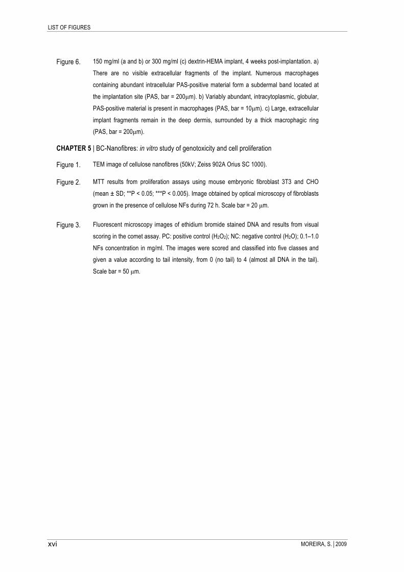
Figure 6. 150 mg/ml (a and b) or 300 mg/ml (c) dextrin-HEMA implant, 4 weeks post-implantation. a)
There are no visible extracellular fragments of the implant. Numerous macrophages
containing abundant intracellular PAS-positive material form a subdermal band located at
the implantation site (PAS, bar = 200µm). b) Variably abundant, intracytoplasmic, globular,
PAS-positive material is present in macrophages (PAS, bar = 10µm). c) Large, extracellular
implant fragments remain in the deep dermis, surrounded by a thick macrophagic ring
(PAS, bar = 200µm).
CHAPTER 5 | BC-Nanofibres: in vitro study of genotoxicity and cell proliferation
Figure 1. TEM image of cellulose nanofibres (50kV; Zeiss 902A Orius SC 1000).
Figure 2. MTT results from proliferation assays using mouse embryonic fibroblast 3T3 and CHO
(mean ± SD; **P < 0.05; ***P < 0.005). Image obtained by optical microscopy of fibroblasts
grown in the presence of cellulose NFs during 72 h. Scale bar = 20 µm.
Figure 3. Fluorescent microscopy images of ethidium bromide stained DNA and results from visual
scoring in the comet assay. PC: positive control (H2O2); NC: negative control (H2O); 0.1–1.0
NFs concentration in mg/ml. The images were scored and classified into five classes and
given a value according to tail intensity, from 0 (no tail) to 4 (almost all DNA in the tail).
Scale bar = 50 µm.

LIST OF TABLES
MOREIRA, S.2009 xvii
List of Tables
CHAPTER 1 | GENERAL INTRODUCTION
Table 1. Example of polysaccharides used in hydrogels formulation for biomedical applications.
Table 2. Examples of tags used to fuse to proteins and their applications.
Table 3. Examples of comparative studies that examine the effects of various fusion partners on soluble
expression yield.
Table 4. Examples of enzyme used to cleave tags in fusion protein (adapted from Arnau et al., 2006).
Table 5 CBM types and families.
CHAPTER 2 | Strategies to produce a human CBM
Table 1. Primers utilized to amplify the coding sequence in the different expression systems. The
sequences recognized by the restriction enzymes are in bold. The RGD coding sequence is
underlined.
Table 2. E. coli strains, expression vectors, growth and induction condition used for recombinant protein
expression.
CHAPTER 5 | BC-Nanofibres: in vitro study of genotoxicity and cell proliferation
Table 1. Results obtained in Salmonella reversion assay.
Table 2. Results from images analysis using the Comet Assay IV software (mean ± SD).


SCOPE
MOREIRA, S.2009 1
SCOPE and COLLABORATORS
The main subject of this thesis is the evaluation of the Carbohydrate Binding Modules (CBMs) potential as
tools for the improvement of the biocompatibility of polysaccharides-based materials. In addition, the
biocompatibility of dextrin hydrogels and bacterial cellulose nanofibres, materials developed at DEB-UM
laboratories, was also analysed.
Chapter 1 presents a revision of these subjects, namely 1) polysaccharide-based biomaterials; 2) strategies to
express recombinant proteins using Escherichia coli systems; and finally, 3) a revision on CBMs. This
subchapter is an adaptation of a book chapter accepted for publication.
The second chapter describes the strategies used to produce the bi-functional recombinant protein containing
a human CBM from laforin fused to a RGD sequence. This work was performed in collaboration with the
Biology Department of Universidade do Minho and the Biomolecular Biotechnology Unit of Biocant.
Chapter 3 describes the strategy to functionalize dextrin-based hydrogel using a recombinant protein
containing a starch-binding module (SBM). The SBM of α-amylase from Bacillus strain TS-23 was fused to
the RGD sequence by recombinant DNA technology and tested, in vitro, using mouse embryo fibroblast 3T3
cells. This work has been published on BMC Biotechnology Journal.
In this work the in vivo biocompatibility of the dextrin hydrogels was also investigated. The in vitro
characterization of dextrin-based hydrogel was the aim of a previous work. However, the in vivo
biocompatibility and degradability of those hydrogels were not evaluated; therefore, in the chapter 4 of this
thesis is presented the study of the in vivo biocompatibility and degradability of dextrin-hydrogels implanted
subcutaneously, in mice. These results were accepted for publication on Journal of Bioactive and Compatible
Polymers and this work was performed in collaboration with Immuno-Phisiology and Pharmacology
Department of Instituto de Ciências Biomédicas Abel Salazar da Universidade do Porto.
It is well known that nanomaterials with needle-like shape, such asbestos fibres, are citotoxic and present
genotoxicity for cells. Because BC-nanofibres present similar structure it seems important to study its potential
genotoxicity. In the chapter 5 the results from in vitro assays to evaluate BC-NFs effect on cells are
presented. This study, performed in collaboration with the Biochemistry Department of Universidade Federal
do Rio Grande do Norte (Brazil), was published on Toxicology Letters.
In the last chapter of this thesis (chapter 6) are presented the final remarks of this work and the future
perspectives.


Chapter 1.
GENERAL INTRODUCTION


CHAPTER 1 | GENERAL INTRODUCTION
MOREIRA, S.2009 5
1.1
Polysaccharide-based biomaterials
The development of tissue engineering strategies is based on the design of three-
dimensional structures made from natural or synthetic materials, termed scaffolds.
Hydrogels are a class of hydrophilic polymeric scaffolds, with remarkable features from
the perspective of biological mimicking. Among the materials used in the development of
hydrogels, polysaccharide-based materials have been referred as promising materials,
presenting appealing properties for biomedical applications.
In the following chapter an overview of biomaterials and tissue engineering developments,
in particular the advantages and applications of polysaccharide-based hydrogels and
bacterial cellulose biomaterials will be presented.


CHAPTER 1 | GENERAL INTRODUCTION
MOREIRA, S.2009 7
Biomaterials and Tissue Engineering
Biomaterials must be especially suitable for the intimate contact with living tissues, ideally
mimicking the biological properties. They are used as drug delivery carriers, tissue
engineering scaffolds, and biomedical devices. Thus, biomaterials are revolutionizing
many aspects of preventive and therapeutic healthcare. With huge potential quality-of-life
benefits owing to the many applications in the biomedical area, biomaterials are the focus
of major research efforts with progresses in this field requiring a multidisciplinary
approach. Indeed, the research on biomaterials gathers contributes from the materials
science, chemical engineering, medical engineering, and pharmacology. The
development of biomaterials for medical applications has focused on the design of
biomimetic materials that interact with the surrounding tissues through biomolecular
recognition, eliciting cellular responses mediated by specific interactions (Shin et al.,
2003). Currently, engineering of hard (i.e., bone, teeth, and cartilage) and soft tissues (i.e.,
skin and internal organs) encompasses the use of scaffolds, growth factors, and stem
cells. Among the various materials assayed as scaffolds - assisting the cell proliferation
and organ development - hydrogels have gained the preference of many researchers.
Scaffolds are ideally three-dimensional, highly porous structures with interconnected
porosity. They are conceived as templates to guide the growth of tissue in the body, as
delivery vehicles for transplanted cells and as drug carriers, activating specific cellular
functions in a localized region, and ultimately regenerating the tissues (Murphy and
Mooney, 1999; Thanos and Emerich, 2008; Vacanti et al., 1998). Therefore, scaffolds may
be implanted into a tissue defect without cells or bioactive compounds in its formulation,
with the tissue regeneration depending only on the ingrowth of the surrounding tissue.
Alternatively, the scaffolds may be loaded with cells or compounds, before implantation,
improving the rate of tissue ingrowth, vascularization, and cell differentiation (Widmer et
al., 1998) (Figure 1).

CHAPTER 1 | GENERAL INTRODUCTION
MOREIRA, S.2009 8
Figure 1 - Schematic representation of the application of a hydrogel scaffold in tissue engineering. 1) Patient
cells (e.g., stem cells) collected and 2) grown in vitro. 3) Then, the cells are mixed with the hydrogel network, 4)
Grown and/or differentiated in a specific culture medium; finally, the scaffold is implanted in the patient. (From
www.centropede.com/UKSB2006/ePoster/images/background/TE_model_large.jpg).
The selection of a scaffold material is both critical and difficult. The sophisticated smart
materials used in the biomedical applications must meet strict criteria, namely convenient
mechanical properties and degradation rate, biocompatibility, porosity and
interconnectivity, functional properties related to the interaction with cells and the release
of pharmaceuticals, among others (Peppas et al., 2000). Candidate materials include: 1)
synthetic polymers, such as polylactic acid (PLA), polyglycolic acid (PGA), poly(lactide-co-
glycolide) acid (PLGA), ethylene oxide block copolymers); 2) inorganic materials, such as
tricalcium phosphate, calcium carbonate, non-sintered hydroxyapatite; and 3) natural
polymers, such as fibrin, collagen, gelatin, hyaluronan. Indeed, natural polymers have

CHAPTER 1 | GENERAL INTRODUCTION
MOREIRA, S.2009 9
played an important role in these efforts, and recombinant polymers that combine the
beneficial aspects of natural polymers with many of the desirable features of synthetic
polymers have been designed and produced.
Besides biocompatibility, the biomaterial biodegradability is generally desirable for tissue
engineering applications. Ideally, the degradation rate also matches the neo tissue
formation rate, performing as a template (Ma, 2008).
Several materials have been exploited as scaffolds for tissue regeneration, each one
presenting advantages and/or disadvantages, depending on the specific application. For
instance, certain metals are an excellent choice for medical implants, due to their superior
mechanical properties (Catledge et al., 2004); of course, they are not a good choice for
scaffold applications because of the lack of degradability in biological environment (Liu
and Ma, 2004). In addition, certain inorganic/ceramic materials, such as hydroxyapatite
(HAP) or calcium phosphates, having good osteoconductivity, have been considered for
mineralized tissue engineering; however, they are of limited application because of its
brittleness and poor processability into highly porous structures. In contrast, polymers
present great design flexibility. Their structure can be tailored to the specific needs, and
therefore have been extensively studied in various tissue engineering applications,
including bone tissue engineering (Huang et al., 2007; Liu and Ma, 2004; Meinel et al.,
2005; Rice et al., 2005)
The overall goal of tissue engineering is to create functional tissue grafts that can
regenerate or replace defective or worn out tissues and organs. Examples of grafts, now
in pre-clinical studies or clinical use, include engineered skin, cartilage, bone, blood
vessels, skeletal muscle, bladder, trachea, and myocardium (Grayson et al., 2008).
Hydrogels as scaffolds for biomedical applications
Hydrogels are a class of materials that swell under conditions of excess of water (or
biological fluids), holding a large amount of water in the wetstate. Chemical crosslinks
(covalent bonds) or physical junctions (e.g. secondary forces, crystallite formation, chain
entanglements) provide the hydrogels unique swelling behavior and three-dimensional
structure (Klouda and Mikos, 2008; Peppas, 2004; Peppas et al., 2006).

CHAPTER 1 | GENERAL INTRODUCTION
MOREIRA, S.2009 10
Hydrogels are one of the upcoming classes of polymer-based systems that embrace
numerous biomedical and pharmaceutical applications, such as tissue engineering,
molecular imprinting, wound dressing materials, immunoisolation, drug delivery, etc
(Kashyap et al., 2005; Peppas et al., 2006). In addition, hydrogels provide new
approaches for culturing mammalian cells ex vivo, which are increasingly needed, to study
cell and tissue physiology and to grow replacement tissues for regenerative medicine.
Two-dimensional culture has been the paradigm for typical in vitro cell culture; however, it
has been demonstrated that cells behave more “in vivo-like” when cultured in three-
dimensional environments such as hydrogels scaffolds (Tibbitt and Anseth, 2009).
Among the various tissue engineering scaffolds comprehensively studied, hydrogels
remain most appealing candidates due to the controllable and reproducible polymer
properties and to the large water uptake, promoting excellent biocompatibility due to low
protein adsorption (Peppas, 2004). In addition, hydrogels present mechanical properties
and hydrophilicity that resembles those of the extracellular matrix (ECM) of native tissue,
tunable viscoelasticity, and high permeability for oxygen and essential nutrients (Jia and
Kiick, 2009; Peppas et al., 2000; Tibbitt and Anseth, 2009).
Apart from favorable physico-chemical and mechanical properties, the most important
requirement for a hydrogel to be used in medical applications is its biocompatibility,
together with the non-cytoxicity of its degradation products. Most of the toxicity associated
with hydrogels regards the unreacted monomers, oligomers and initiators that leach out
(Del Guerra et al., 1996; Del Guerra et al., 1995; Kirkpatrick, 1992; Ratner, 1997).
A variety of hydrogel materials have been utilized for tissue engineering applications,
including reconstituted ECM components or natural proteins and carbohydrates (Long and
Tranquillo, 2003; Robinson et al., 2008; Zhong et al., 2005), self-assembling peptides
(Collier et al., 2001; Kisiday et al., 2002), and synthetic materials (Hicks et al. 2003; Lou et
al., 2001). Thus, the selection of the appropriate hydrogel is governed by the chemical
and physical properties, the mass transport properties and the biological interaction
requirements that are best suited for a given application.
While the chemical properties of hydrogels (such as hydrophilicity) are determined by 1)
the polymer backbone, 2) the functional side chain in the monomer unit, and 3) the cross-
linking agent; the physical properties (e.g. mechanical strength and swelling ratio) are
mainly controlled by the cross-link density. For instance, the amount of water absorbed by
hydrogels is limited by their ability of undergoing elastic network expansion, which can be

CHAPTER 1 | GENERAL INTRODUCTION
MOREIRA, S.2009 11
controlled by controlling the cross-linking degree during the synthesis of chemically cross-
linked hydrogels (Byrne and Salian, 2008).
Hydrogels can show variable swelling behavior, depending on changes in the external
environment. Some of the factors that can affect the swelling of responsive hydrogels
include pH, ionic strength and temperature (Peppas, 2004). Hydrogels can also be made
responsive to diverse external stimuli, such as light, electric current, ultrasound, and the
presence of a magnetic field or a particular molecule (Watanabe et al., 2004). This unique
property of responsiveness has resulted in the development of hydrogel-based sensors
(Bashir et al., 2002; Hilt et al., 2003), self-regulated and externally actuated intelligent
drug delivery systems (Miyata et al., 1999; Sershen and West, 2002; Sershen et al., 2000;
Yoshida et al., 1993) and microfluidic devices (Beebe et al., 2000; Satarkar and Zach Hilt,
2008; Sershen et al., 2005). Physically or chemically cross-linked hydrogels can also be
generated in the presence of living cells, allowing in situ encapsulation for tissue
engineering (Peppas et al., 2006). Furthermore, hydrogels with controlled biodegradation
kinetics may be easily designed using natural polymers susceptible to enzymatic
degradation or synthetic polymers with hydrolyzable moieties (Watanabe et al., 2004)
Natural biomaterials: Polysaccharide-based materials
By far, the majority of carbohydrate materials in Nature occur in the form of
polysaccharides. By definition, polysaccharides include not only those substances
composed uniquely of glycosidically linked sugar residues, but also molecules that contain
polymeric saccharide structures linked via covalent bonds to amino acids, peptides,
proteins, lipids and other structures. Thus, polysaccharides have a large number of
reactive groups, a wide range of molecular weight (MW), and different chemical
compositions, which contribute to their diversity in structure and in properties (d’Ayala et
al., 2008). In nature, polysaccharides from algae (e.g. alginate), plant (e.g. pectin, guar
gum), microbial (e.g. dextran, xanthan gum), and animal origin (chitosan, chondroitin) can
be found (Coviello et al., 2007).
The most common constituent of polysaccharides is D-glucose, but D-fructose, D-
galactose, L-galactose, D-mannose, L-arabinose, and D-xylose are also frequent. Some
monosaccharide derivatives found in polysaccharides include the amino sugars (D-

CHAPTER 1 | GENERAL INTRODUCTION
MOREIRA, S.2009 12
glucosamine and D-galactosamine) as well as their derivatives (N-acetylneuraminic acid
and N-acetylmuramic acid), and simple sugar acids (glucuronic and iduronic acids).
Among the numerous macromolecules that can be used for hydrogel formation,
polysaccharides are advantageous compared to synthetic polymers. Coming from
renewable sources, polysaccharides also have frequently economical advantages over
synthetic materials. Polysaccharides are usually non-toxic, biocompatible and show a
number of convenient physico-chemical properties (such as hydrophilicity, viscosity and
reactive groups) that make them suitable for different applications in drug delivery
systems (Coviello et al., 2007). The major disadvantages of natural polymers, when
compared with synthetic ones, are the difficulty in controlling their physico-chemical
properties, such as molecular weight, strength, degradation time and mechanical
properties. However, there are several strategies to overcome these limitations, including
the combination with other natural or synthetic polymers. The combination with other
natural polymers (e.g., collagen/glycosaminoglycans) or with synthetic polymers (e.g.,
collagen/PLGA), may improve the biocompatibility of the ensuing scaffolds, by reducing
inflammatory response in vivo and improving initial cell attachment and differentiation on
the material (Flanagan et al., 2006; In Jeong et al., 2007; Liu et al., 2008; Zhong et al.,
2005).
Polysaccharides, such as cellulose, starch, chitin/chitosan, alginate, carrageenan, gellan,
guar gum, hyaluronic acid, pullulan, dextran, among others, have been used in the
formulation of several hydrogels (nanogel, microspheres) (Coviello et al., 2007). In
addition, it was also found that water-soluble polysaccharides derivatives – such as
carboxymethylcellulose (CMC), carboxymethylstarch (CMS), carboxymethylchitin (CMCT),
and carboxymethylchitosan (CMCTS) – lead to the formation of hydrogels at high
concentrated aqueous solution (paste-like state) by radiation cross-linking (Yoshii et al.,
2003). Table 1 gives examples of several polysaccharides used in hydrogel formulations
and their potential biomedical applications.

CHAPTER 1 | GENERAL INTRODUCTION
MOREIRA, S.2009 13
Table 1 - Example of polysaccharides used in hydrogels formulation for biomedical applications.
Polysaccharide Application References
Alginate-based materials (hydrogel,
microspheres)
Ocular drug delivery; oral administration of drugs; wound
dressings; regional radio-chemotherapy; driving of angiogenesis
by diffusion of VEGF; cells encapsulation; bone tissue engineering
(repair of osteochondral defects of joints; neurotransplantation)
(Coviello et al., 2007) (Kuo and Ma, 2001) (d’Ayala et al., 2008) (Eiselt et al., 2000)
(Novikova et al., 2006)
Dextran-derivates (hydrogels and
microgels)
Release of proteins with limited aqueous solubility (such as IL2);
tissue engineering bone regeneration (such as BMP release);
nasal drug delivery systems; colon drug delivery
(Bos et al., 2004) (Coviello et al., 2007)
(Lévesque and Shoichet, 2006)
(Kim and Chu, 2000)
Chitin and Chitosan-derivates (hydrogels, films and microgels)
Wound treatment; bioadhesive sustained release formulation;
tissue engineering (cartilage and bone regeneration); gene
delivery; colon drug delivery
(Felt et al., 1999) (Alsarra et al., 2009 ) (Sinha et al., 2004)
(d’Ayala et al., 2008) (Veerapandian and
Yun, 2009)
Gellan-derivates (hydrogels, beads,
microspheres)
Ophthalmic formulations; nasal spray pumps; encapsulation of
biological components; oral drug delivery; drug release
(Veerapandian and Yun, 2009)
(Coviello et al., 2007) (Schwall and
Banerjee, 2009)
Carrageenan and Carrageenan-
derivates (hidrogels, beads)
Promoter of angiogenesis by diffusion of VEGF; wound and burns
dressing application; bone tissue engineering; drug carrier and
devilry
(Santo et al., 2009) (Coviello et al., 2007)
Hyaluronic acid and HA-derivates
(hydrogels, beads)
Local administration of anti-inflammatory drugs in osteoarthritic
knee; immobilization of hydrocortisone; cell encapsulation for cell
delivery tissue regeneration; controlled release of vascular
endothelial growth factor and basic fibroblast growth factor,
promoting neovascularization
(Coviello et al., 2007)
(Schwall and Banerjee, 2009)
Guar-derivates (hydrogels)
Prodrugs formulation (as coating material or as hydrogel
entrapping dugs inside its network); colon delivery; treatment of
open-angle glaucoma
(Coviello et al., 2007) (Schwall and
Banerjee, 2009)
Pullulan-derivates (hydrogels, micro
and nanogels)
Capacity to bind hydrophobic substances (such as anticancer
drugs); imaging of specific sites (such as tumor and ischemic area) (Coviello et al., 2007)
Xanthan- derivates (hydrogels)
Enzyme immobilization; loading bioactive substances; delivery
proteins in nasal cavity
(Andreopoulos and Tarantili, 2001)
(Bejenariu et al., 2008) (Coviello et al., 2007)
Xyloglun-derivates (sol-gel transition)
Vehicle for sustained release of percutaneous formulation (non-
steroidal anti-inflammatory drugs); release of indomethacin
suppositories; orally and intraperitoneal administration of drugs;
neural tissue engineering of the spinal cord
(Coviello et al., 2007) (Nisbet et al., 2009) (Nisbet et al., 2006)

CHAPTER 1 | GENERAL INTRODUCTION
MOREIRA, S.2009 14
Functionalization of polysaccharide-based biomaterials
Biomaterials are expected to fulfill biological functions, such as promoting cell proliferation
and differentiation and enhancing the growth of surrounding tissues for defective
regeneration. A straightforward method to incorporate these functions is to create
hydrogels made up of natural macromolecules or macromolecular blends. Biologically
active molecules can also be incorporated into polymer networks (e.g., by physical or
chemical entrapment) to produce conjugated biomaterials, in order to design biomimetic
scaffolds that can provide biological cues to elicit specific cellular responses and direct
new tissue formation.
The surface and bulk modification of materials with peptide sequences can allow for the
modulation of cellular functions such as adhesion, proliferation and migration through
modulation of the peptide concentration or its spatial distribution. Hydrogels have been
synthesized so that they contain functional groups for enhancing cellular adhesion
(Burdick et al., 2004; Hern and Hubbell, 1998). In this scheme, the addition of such
modalities can dramatically change the properties of the hydrogels. The most common
peptides used to modify hydrogels are derived from natural proteins, such as RGD
(derived from proteins such as fibronectin, laminin, or collagen), IKVAV, and YIGSR from
laminin (Peppas et al., 2006; Tashiro et al., 1989). Using these approaches, PEG (Burdick
et al., 2004; Hern and Hubbell, 1998) and other hydrogels, such as alginate (Rowley et
al., 1999), have been modified with RGD to enhance cellular adhesion.
Despite the recent advances toward the development of biomimetic materials for tissue
engineering applications, several challenges still remain, including the design of adhesion
molecules for specific cell types, as required for guided tissue regeneration and the
synthesis of materials exhibiting the mechanical responsiveness of living tissues.
In particular, carbohydrate-based hydrogel (such as dextrin and cellulose) may be
functionalized, by using recombinant proteins containing a carbohydrate-binding module
(CBM) fused to bioactive peptides. CBMs present several specificity and affinities
(Boraston et al., 2004; Boraston et al., 2007; Shoseyov et al., 2006b); therefore they may
be used to adsorb peptides to a different polysaccharide materials, including starch and
cellulose-based materials (Andrade et al., 2009).

CHAPTER 1 | GENERAL INTRODUCTION
MOREIRA, S.2009 15
Starch-based hydrogels
Among the many applications of starch-based materials, the development of hydrogels for
biomedical applications has drawn the attention of several research groups. The
biocompatibility and degradability makes starch a suitable component of hydrogels with
technological applications in a large number of areas, such as medical, pharmaceutical
and biological (Marques et al., 2002).
Starch is a natural mixture of amylose, a linear polymer of D-glucose unites linked to 1,4-
α-D-glucosidic linkages, and amylopectin or pullulan, a branched polymer of α-D-Glucose
units containing 1,4- α-D-glucosidic linear linkages and 1,6- α-D-glucosidic linkages at the
branch points (Figure 2). There are a several hydroxyl groups on starch chains, two
secondary hydroxyl groups at C-2 and C-3 of each glucose residue, as well as one
primary hydroxyl group at C-6 when it is not linked (Lu et al., 2009). These hydroxyl
groups on the starch chains can be oxidized and reduced, and may participate in the
formation of hydrogen bonds, ethers and esters (Tomasik and Schilling, 2004).
Figure 2 - Schematic representation of molecular structure of starch, showing D-glucose units and their
hydroxyl groups, and the α -1,4 glucosidic linear linkages or α -1,6 linkage in the branch points. (Adapted from
Lu et al., 2009)
Starch itself is poor in processability, as well as in the dimensional stability and
mechanical properties of its end products. In addition, starch is not capable of gelling

CHAPTER 1 | GENERAL INTRODUCTION
MOREIRA, S.2009 16
naturally; it must be modified by chemical derivatization, in order to introduce functional
groups in the raw material, which may then gellify using either of the techniques described
bellow. The chemical modification of starch (and the polysaccharides in general) has
profound effects on its macroscopic behavior (e.g. solubility, stability and viscosity
characteristics). Thus, for extended practical utilization of starch-based products,
reinforcement or modification of starch is often essential (Ulrich Riedel, 1999; Vargha and
Truter, 2005).
Starch-based hydrogels may be produced by using: 1) free radical polymerization (in one-
step or two-step), 2) cross-linking by chemical reaction of complementary groups, 3)
radiation-induced polymerization and 4) cross-linking and physical self-assembly.
1. The polymerization using free radicals may be achieved by one-step or two-step
synthesis. In the first case, the hydrogel is obtained through free radical
copolymerization of low molecular weight hydrophilic vinyl monomers onto the
starch substrate, in the presence of polymerizable cross-linking agents (Zhang et
al., 2005). In a two-step synthesis, the polysaccharide is first functionalized with
reactive double bonds and then cross-linked by free radical polymerization in
water. Hydrogels obtained by these processes can combine the advantages of
natural and synthetic polymeric hydrogels, and usually bear improved mechanical
properties (Coviello et al., 2007; Zhang et al., 2005).
2. Starch-based hydrogels may also be achieved through the reaction of functional
groups with complementary reactivity. The cross-linking is possible when specific
functional groups (mostly -OH, -COOH) are present along the macromolecular
chains of starch and its derivatives, forming covalent bonds (Van Tomme et al.,
2008; Zhang et al., 2005).
3. The chemical cross-linking of starch-based hydrogels can also be achieved by
using high-energy radiation (especially gamma and electron beams (Zhang et al.,
2005). During the irradiation of aqueous starch system, radicals are formed on the
polymer chain by homolytic scission of C—H bonds. In addition, the radiolysis of
water molecules generates hydroxyl radicals which subtract protons from the
polymer chains, resulting in the formation of macroradicals (Zhang et al., 2005).
Under an inert atmosphere, the recombination of the macroradicals on different

CHAPTER 1 | GENERAL INTRODUCTION
MOREIRA, S.2009 17
chains leads to the formation of covalent bonds and finally produces a cross-linked
network structure. The advantage of this process is that it can be done in water,
under mild conditions (room temperature and physiological pH). However, when
these hydrogels are used as matrices for the controlled release of bioactive drug
molecules, drug loading is possible only after preparation of the hydrogels,
because the radicals formed during irradiation could potentially damage the
biologically active substance.
4. For the preparation of physically cross-linked starch-based hydrogels,
amphiphilic starch derivatives, generally synthesized by hydrophobic modification
of water-soluble polysaccharide, self-assembles in aqueous solution to form
physical hydrogels with a micelle-like structure with hydrophobic cores derived
from the association of hydrophobic segments and a hydrophilic shell made of the
polar groups on the polymer (Janes et al., 2001; Zhang et al., 2005). The major
disadvantage of these hydrogels, when compared with chemically cross-linked
ones, is the instability of their mechanical properties (Zhang et al., 2005).
Dextrin as a biomaterial
Among starch-based materials, those based on dextrin are widely used in a variety of
applications, from adhesives used in food and textile industries (Lazarus, 1983),
peritoneal dialysis solution (Hreczuk-Hirst et al., 2001) to the moisture-maintaining
component of powders for skin used in the cosmetic industry (Tipson et al., 1989). Recent
work reported the ability of dextrin conjugates to exhibit anti-endotoxin activity as well as
to regulate the inflammatory response (Davtyan et al., 2007; Davtyan et al., 2009). In
another recent work, dextrin-Hydroxyapatite (HAp) complex was used as a bone filling
material, with good performance (Asai et al., 2009). In addition, nanoparticle based on
dextrin were also described as potential drug carriers (Goncalves et al., 2007), and
dextrin-based microspheres were used for encapsulation of the photosensitizer porphyrin,
which aggregates in aqueous solutions, allowing its administration in the monomeric form,
in photodynamic therapy (Luz et al., 2008).
Hardwicke et al. described the development of a bioresponsive polymer–drug conjugate
designed specifically to promote wound healing. Dextrin was selected for conjugation with
recombinant human epidermal growth factor (rhEGF), as the former is degraded by α-

CHAPTER 1 | GENERAL INTRODUCTION
MOREIRA, S.2009 18
amylase in wound fluid. Dextrin was used to protect rhEGF from proteolytic attack (which
is a major clinical challenge when growth factors are administered topically) and liberate it
at a controlled rate (Hardwicke et al., 2008). More recently, a similar strategy was used to
reduce the nonspecific neurotoxicity of phospholipase A2 (PLA2) crotoxin (an antitumor
protein that appears to act by interaction with epidermal growth factor receptors).
According to this concept, the bioresponsive dextrin−PLA2 conjugate will be activated in
the tumor interstitium (which presents high levels of α-amylase), through dextrin
degradation (Ferguson and Duncan, 2009).
Dextrins are a group of low-molecular-weight carbohydrates produced by partial
hydrolysis of starch, which can be accomplished by the use of acid, enzymes, or a
combination of both. Dextrin is a glucose-containing saccharide polymer linked by α-1,4
D-glucose units, containing few (< 5%) α-1,6 links, having the same general formula as
starch, but smaller and less complex (Carvalho et al., 2007; Hreczuk-Hirst et al., 2001).
Among the many possibilities of starch modification, the creation of reactive double bonds
through a transesterification reaction has been exploited recently by several groups to
functionalize sugars (in particular dextran and dextrin) (Carvalho et al., 2009a; Carvalho et
al., 2007; Carvalho et al., 2008a; De Groot et al., 2001). The final product of the
transesterification of vinyl acrylate monomers has side chains (vinylic groups) attached to
the polysaccharide backbone and polymerizes through well-established free radical
methods (De Graaf et al., 1998; Vargha and Truter, 2005). Depending on the vinyl ester
used in the dextrin substitution, and on the degree of substitution, different degradability
may be achieved. Thus, using vinyl acrylate (VA) or hydroxyethylmethacrylate (HEMA) the
dextrin-based hydrogel obtained is non-degradable or degradable, in vitro, respectively
(Carvalho et al., 2009b). In addition, in both cases, the degree of substitution (DS), which
refers to the average number of substituted polysaccharide hydroxyl groups, is of major
importance in determining the properties of the resulting hydrogel, such as the
degradation rate or the release profiles (Carvalho et al., 2009; Carvalho et al., 2009b).
In vitro biocompatibility and degradability of dextrin-based hydrogels, namely dextrin-VA
and dextrin-HEMA, were already evaluated. It was demonstrated that dextrin-based
hydrogels do not present toxicity; moreover, cells adhere to the hydrogel surface and
remain viable (Carvalho et al., 2009b). In addition, the release profile of those dextrin-

CHAPTER 1 | GENERAL INTRODUCTION
MOREIRA, S.2009 19
based hydrogels using a model protein shows its suitability for their application as a
delivery system (Carvalho et al., 2009).
Bacterial Cellulose as a biomaterial
Bacterial cellulose (BC) is a polysaccharide produced by the Acetobacter xylinum bacteria
into long non-aggregated nanofibrils (Backdahl et al., 2006; Brown et al., 1976; Klemm et
al., 2001).
The cellulose synthesized by A. xylinum is identical to that made by plants in respect to
molecular structure. However, the secreted polysaccharide is free of lignin, pectin, and
hemicellulose as well as other biogenic products, which are associated with plant
cellulose. Additionally, the BC displays many unique properties including high mechanical
strength, high water content, high crystallinity and an ultra-fine highly pure nanofibril
network structure (Backdahl et al., 2006; Czaja et al., 2007; Lee et al., 2001).
One of the main requirements of any biomedical material is its biocompatibility, which is
the ability to remain functional in contact with the living tissue, without causing any toxic or
allergic side effects. Studies carried out in vitro and in vivo have demonstrated the BC
biocompatibility. Thus, several applications were described for this material, including
micro vessel prosthesis (Klemm et al., 2001), temporary skin substitutes (Fontana et al.,
1990), in periodontal treatments and as a replacement for dura mater (the membrane that
surrounds brain tissue) (Andrade et al., 2009; Czaja et al., 2007). In addition, BC can be
combined in composite materials in order to further improve its characteristics. For
instance, Yasuda et al. used microbial cellulose immersed in two types of polymer
solutions (2-acrylamide-2-methyl-propane sulfonic acid and gelatin), to create a cellulose-
based hydrogel with enhanced mechanical toughness, for the replacement of cartilage
tissue in damaged joints (Yasuda et al., 2005).
Some researchers have also obtained modified BC by introducing different additives into
the culture media. The modification of the bacterial cellulose occurs, in this case, during
biosynthesis, by introducing selected bioactive polysaccharides, such as chitosan and
derivatives into the culture medium. Such composite materials can be applied in the
treatment of burns, bedsores, skin ulcers, hard-to-heal wounds and wounds requiring
frequent changes of dressing (Ciechanska, 2004). In addition, polysaccharides

CHAPTER 1 | GENERAL INTRODUCTION
MOREIRA, S.2009 20
degradable in vivo, exhibiting both chitin- and cellulose-like properties and susceptible to
lysozyme attack, could be achieved by introducing GlcNac residues into bacterial
cellulose (Ogawa and Tokura, 1992).
The formation of networks with distinct architecture and the modification of other
molecular features, such as reduction of crystallinity, was also obtained by adding
mannan-based polysaccharides to the culture medium (Whitney et al., 1998). Further, it
was shown that a range of different cellulose-associated networks could be formed,
depending of the levels of glucomannan and galactomannans added. Cellulose with lower
cristallinity and a smaller crystallite size was also obtained by adding sodium alginate to
the culture medium (Zhou et al., 2007).
BC-Nanofibres toxicology
In the recent years there is an increasing interest in nanomaterials, including metallic
nanoparticles (NPs), metal oxide nanoparticles, dendrimers, quantum dots, nanoclusters,
nanocrystals, nanowires, fullerenes, fullerene-based derivatives, single- and multi-wall
carbon nanotubes, functionalized carbon nanotubes, polymer nanoparticles, carbon black,
nano-coatings, among others, and its applications. With the rapid development of
nanotechnology and its applications, a wide variety of nano-structured materials are now
used in commodities, pharmaceutics, cosmetics, biomedical products, and industries
(Ashammakhi et al., 2007; Ma et al., 2005). In particular, BC-based materials, has been
described as a promising scaffold in tissue engineering, since it can better mimic the
nanostructure of extracellular matrix due to its nanofibrilar structure. Furthermore, BC
nanofibres can be combined with other materials in order to improve their characteristics
(Grande et al., 2009; Millon et al., 2008; Yoon et al., 2006).

CHAPTER 1 | GENERAL INTRODUCTION
MOREIRA, S.2009 21
Figure 3 – Schematic representation of possible interaction of oxyradicals release by NPs with the antioxidant
defense system (Picture from Oberdorster et al., 2005), Abbreviations: GPx, glutathione peroxidase; GSH,
reduced glutathione; GSSG, oxidized glutathione; ISC, intersystem crossing; R, any organic molecule; SOD,
superoxide dismutase. In addition to fullerenes, metals such as cadmium, iron, nickel quantum dots, or iron
from SWNT manufacturing, could also act in Fenton-type reactions. Phase II biotransformation, ascorbic acid,
vitamin E, beta-carotene, and other interactions are not shown.
While nanoscale materials possess novel and unique physicochemical properties different
from those of bulk materials, they also have an unpredictable impact on human health.
The impact of nanomaterials on the human body, their interactions with biological
systems, and their risk assessment have generated intense scientific curiosity.
Researchers have demonstrated that NPs may exhibit unique biological behavior, even
when physical and chemical properties remain unaltered from those observed in large
particle. Perhaps the most striking example lies on the fact that the smaller particles size
enables nanoscale particles to cross or circumvent barriers that are impenetrable to larger
particles (Bernstein et al., 2005; Pan et al., 2007). The greater surface area per mass,
compared with larger-sized particles of the same chemistry, renders NPs more active
biologically. This activity includes a potential for inflammatory and pro-oxidant, but also
antioxidant activity, which can explain early findings showing mixed results in terms of
toxicity of NPs to environmentally relevant species. For instance, Zhao et al. have used

CHAPTER 1 | GENERAL INTRODUCTION
MOREIRA, S.2009 22
computer models to predict that C60 molecules can damage DNA if intracellular exposure
occurs (Zhao et al., 2005b) (Figure 3) and Lynch et al. have hypothesized that protein
adsorbed onto nanoparticles may alter their shape, such that normally hidden amino acids
residues are exposed as cryptic epitopes-triggering an immune response (Lynch et al.,
2006).
The interaction of nanomaterials with biological systems is affected by several factors,
such as size, surface area, shape, chemical composition, lattice structure, surface
chemistry and charge (Borm et al., 2006; Pan et al., 2007).
Nanomaterials can exhibit various shapes and structures; among them, needle-like
nanofibres have been described as a potential toxic. For instance, the long, thin geometry
and water insolubility of carbon nanotubes may have the potential to cause effects similar
to those arising from inhalation of asbestos fibres (a well-known harmful material for man
health), even if the chemical composition is completely different (Donaldson et al., 2006).
As refereed above, there is an increasing interest in nanomaterials-based on BC, and
since, BC-NFs also present needle-like shape, it seems important to evaluate their
toxicity. Therefore, in this work, several assays were carried out to access BC-NFs
toxicology.

CHAPTER 1 | GENERAL INTRODUCTION
MOREIRA, S.2009 23
1.2
E. coli expression systems towards the production of
recombinant proteins
The technology of recombinant DNA allows the production of pure, soluble and functional
recombinant proteins for several applications, including immunization, biochemical
studies, three-dimensional structural protein analysis, and biotechnological and
therapeutic use. Although the advances in a number of different expression systems,
including bacteria, yeast, mammalian cells, and even cell-free systems, the bacterial ones
still is the first approach used. However, despite the advantages, well recognized, the
bacterial expression systems present also several limitations, specially for the production
of protein from eukaryotic origin (Demain and Vaishnav, 2009; Sahdev et al., 2008;
Schumann and Ferreira, 2004).
Among the distinct strategies described for the production of functional recombinant
proteins, the fusion with carbohydrate-binding modules (CBMs) is an attractive strategy
towards the functionalization of biomedical materials based on polysaccharides. In the
following subchapters the bacterial expression systems for recombinant proteins
production, as well as the CBMs functions and applications, will be reviewed.


CHAPTER 1 | GENERAL INTRODUCTION
MOREIRA, S.2009 25
Recombinant expression systems for the production of
heterolougues proteins
Production of recombinant proteins involves the cloning of the appropriate gene into an
expression vector, under the control of a well-known regulable promoter. Bacterial
systems remain the most attractive due to low cost, high productivity, well-known genetics
and large number of compatible molecular tools available (Sorensen and Mortensen,
2005a; Terpe, 2006). Thus, many successful Escherichia coli expression systems have
been described and are available, from a variety of academic and commercial sources.
But the efficient production of recombinant proteins depends on a variety of factors, such
as optimal expression signals (both at the level of transcription and translation), correct
protein folding and cell growth conditions. Although high yield may be achieved in the first
expression attempt, proteins vary in structural stability, solubility, and toxicity, resulting in
different rates of protein degradation, formation of insoluble inclusion bodies (IB), and cell
death; thus, optimization of the process is usually required (Doyle, 2005; Schumann and
Ferreira, 2004).
Despite the great diversity of expression systems - including expression vectors, host
strains, and growth medium, among others - for heterologous protein production (Niiranen
et al., 2007) the major drawbacks of E. coli as an expression system include: 1) the
inability to perform many of the posttranslational modifications found in eukaryotic
proteins; 2) the lack of a secretion mechanism for the efficient release of protein into the
culture medium; and 3) the limited ability to facilitate extensive disulfide bond formation
(Esposito and Chatterjee, 2006; Jana and Deb, 2005; Sahdev et al., 2008).
Under normal cellular conditions, a subset of cytoplasmic proteins are able to fold
spontaneously (Anfinsen, 1973). However, many over-produced recombinant proteins in
artificial E. coli factories accumulate as IB. These aggregated proteins are misfolded or
partially-folded intermediates, clustered through inter- or intra-molecular interactions of
solvent-exposed, hydrophobic polypeptide stretches (Stampolidis et al., 2009). Their
formation is attributed to the inability of the bacterial cell factories to maintain protein
quality control during over-expression or thermo-induction of heterologous proteins. Thus,
expression conditions must be found which balance heterologous protein production and
host physiology, to optimize the overall yield of the active product.

CHAPTER 1 | GENERAL INTRODUCTION
MOREIRA, S.2009 26
Although no universal approach has been established for the efficient folding of
recombinant proteins (Sorensen and Mortensen, 2005 a,b), several factors contributing to
protein aggregation must be accounted: 1) lack of sufficient amount of the appropriate
catalytic and molecular chaperoning machinery, e.g. trigger factor (Maier et al., 2005),
DnaK-DnaJ-GrpE (Martinez-Alonso et al., 2006; MartÌnez-Alonso et al., 2007; Xu and
Sigler, 1998) and GroEL-ES or other heat-shock proteins (Gonzalez-Montalban et al.,
2008); 2) failure of the cognate co- or post-translational modifying enzymes (Sklar et al.,
2007) to operate in an orderly manner or to repair the misfolded structures (Gonzalez-
Montalban et al., 2006); 3) failure to maintain precursors in an unfolded state, for their
localisation/translocation to extra-cytoplasmic compartments, which may also contribute to
the formation of protein clusters (Stampolidis et al., 2009).
Several approaches have been developed with the purpose of avoiding the expression of
insoluble protein, including: fusion tags; optimization of expression conditions (e.g.,
medium, inductor concentration, temperature); expression host; co-expression of
chaperons, among others (Figure 4).
Figure 4 - Summary of strategies to express soluble recombinant proteins (Adapted from Sørensen et al. 2005).

CHAPTER 1 | GENERAL INTRODUCTION
MOREIRA, S.2009 27
Enhancing solubility of recombinant proteins
As shown in figure 4, the insolubility of recombinant proteins may be reduced by
optimizing the expression conditions, such as temperature, growth media, induction
parameters, promoters and E. coli strain (Baneyx, 1999; Demain and Vaishnav, 2009;
Hoffmann et al., 2004; Schumann and Ferreira, 2004).
A well-known approach to limit the in vivo aggregation of recombinant proteins consists on
the cultivation of the bacteria at reduced temperature (Baneyx and Mujacic, 2004; Garg
and Quinones, 1997). Lowering the concentration of the inducer, when using expression
systems with an inducible promoter, also contributes to enhancing the solubility of
recombinant protein produced in E. coli (Schein, 1989). Using a lower incubation cell
growth temperature and inductor concentration in culture media slows down the rate of
protein synthesis, minimizing the saturation of the cellular folding machinery and
aggregation (Ellis and Minton, 2006; Rabhi-Essafi et al., 2007). The aggregation reaction
is favored at high temperature, because of the strong temperature dependence of
hydrophobic interactions (Kiefhaber et al., 1991). It has been reported that the direct
consequence of decreasing temperature is the partial elimination of heat-shock proteases,
induced during foreign protein over-expression (Chesshyre and Hipkiss, 1989). Moreover,
expression and activity of a number of E. coli chaperones are increased at lower
temperatures (around 30°C), contributing for proper folding of expression proteins (Hartl
and Hayer-Hartl, 2009; Mogk et al., 2002; Rabhi-Essafi et al., 2007).
The utilization of different E. coli strains (with supplemental tRNA genes) is a strategy
often used to overcome the codon-usage problems or the disulfide-bond formation
necessary for proper folding (Sorensen and Mortensen, 2005a, b). In addition, expression
systems that allow the secretion of recombinant protein for the periplasmatic space, or
into the medium, are also available (Zhang and Huang, 2002). However, higher yields are
normally obtained in the cytoplasm rather than in the periplasmic space. Cytoplasmic
proteins can be exported, to simplify purification and facilitate correct folding. Thus, this
must be done whenever proteins need disulfide bonds to fold properly, since the
cytoplasm is a reducing environment. To achieve the secretion of these proteins into the
periplasm, a fusion is made with a leader peptide at the N-terminus. To get the proteins
out of the periplasm and released into the medium, osmotic shock or cell wall
permeabilization is used (Demain and Vaishnav, 2009).

CHAPTER 1 | GENERAL INTRODUCTION
MOREIRA, S.2009 28
Co-expression of chaperons
The co-production of selected chaperones along with the recombinant protein has been
largely explored, as a strategy to minimize aggregation of the product, with rather
unpredictable and not always consistent results (Martinez-Alonso et al., 2006).
Molecular chaperones are ubiquitous and highly conserved proteins that help other
polypeptides reaching a proper conformation, without becoming part of the final structure.
They are not true folding catalysts, since they do not accelerate folding rates. Rather, they
prevent “off-pathway” aggregation reactions by transiently binding hydrophobic domains in
partially folded polypeptides, thereby shielding them from each other and from the solvent.
Molecular chaperones also facilitate protein translocation, participate in proteolytic
degradation, and help proteins - that have been damaged by heat shock or other types of
stress - regaining an active conformation (Baneyx and Palumbo, 2003).
Fusion proteins and Protein tags
A vast number of tags - proteins, domains or peptides - are available that may be fused to
the target recombinant proteins, for different proposes (table 2). The advantages of using
fusion proteins to facilitate purification and detection of recombinant proteins are well
recognized. Nevertheless, it is difficult to choose the right purification system for a specific
protein of interest.

CHAPTER 1 | GENERAL INTRODUCTION
MOREIRA, S.2009 29
Table 2- Examples of tags used to fuse to proteins and their applications.
Goal
Tag Purification
Solubility enhancer
Detection References
His-tag X X
(Goel et al., 2000; Lomas-Lopez et al., 2008;
Loughran et al., 2006; Terpe, 2003)
Strep II-tag X
(Korndörfer and Skerra, 2002; Skerra and
Schmidt, 2000)
T7-tag X
(Arnau et al., 2006; Chatterjee and Esposito,
2006)
FLAG-tag X X
(Arnau et al., 2006; Einhauer and Jungbauer,
2001)
S-tag X X (Arnau et al., 2006; Backer et al., 2002)
Hemagglutinin
HA-tag X X
(Hage, 1999)
cMyc-tag X X (Arnau et al., 2006; Terpe, 2003)
Chitin-binding domain X (Humphries et al., 2002)
Calmodulin-binding domain X X (Durst et al., 2008)
Cellulose-binding domain X (Terpe, 2003)
Glutathione S-Transferase
GST X X X
(Arnau et al., 2006; Braun et al., 2002; Kim
and Lee, 2008; Shih et al., 2002)
Maltose-binding protein
MBP X X
(Nallamsetty and Waugh, 2006; Rachel B.
Kapust, 1999; Sachdev and Chirgwin, 1998)
N-utilizing substance A
NusA X
(De Marco et al., 2004; Kim and Lee, 2008;
Nallamsetty and Waugh, 2006)
Thioredoxin
Trx X X X
(Kim and Lee, 2008; Sachdev and Chirgwin,
1998)
Small ubiquitin-like modifier
SUMO X
(Panavas et al., 2009; Saitoh et al., 2009)
Ubiquitin
Ub X
(Marblestone et al., 2006)
Mistic X (Roosild et al., 2005)
Green fluorescent protein
GFP X X
(Waldo et al., 1999)
Elastin-like polypeptides
ELP X X
(Araujo et al., 2009; Chow et al., 2006; Ge et
al., 2005; Trabbic-Carlson et al., 2004)

CHAPTER 1 | GENERAL INTRODUCTION
MOREIRA, S.2009 30
Purification Tags
The use of affinity tags to improve protein purification becomes indispensable tools for
structural and functional proteomic initiatives (table 2). Much of the protein-purification
technology is based on the use of affinity chromatography, in which an expressed
recombinant protein is fused to a peptide having high affinity for a particular ligand.
The most popular tag for protein purification, based on the use of immobilized metal
affinity chromatography (IMAC), is the polyhistidine-tag, a motif of at least six histidine
residues (6xHis), held either at the N- or C-terminus of the protein. In addition, this tag
may be used for protein detection, since antibodies recognizing 6xHis are commercial
available (Goel et al., 2000; Lomas-Lopez et al., 2008; Loughran et al., 2006). Another
common tag is the glutathione S-transferase (GST) tag, for purification on glutathione-
based resins. Similarly, Strep-tag II, which consists of a streptavidin-recognizing
octapeptide (WSHPQFEK), can be purified by affinity using a matrix with a modified
streptavidin, being eluted with a biotin analog.
Another approach is based on antibodies-antigen interactions; for instance, affinity tags
like FLAG octapeptide, cMyc, and hemagglutinin (HA) can be purified using the respective
antibodies, immobilized on a chromatographic matrix (Arnau et al., 2006; Sorensen and
Mortensen, 2005b). Several other affinity tags exist and have been extensively reviewed
(Terpe, 2003).
More recently, a new purification tag has been described, based on the transition
properties of an Elastin-like polymers (ELP) and their ability to retain this inverse
temperature-phase transition when conjugated to other molecules (Chow et al., 2006;
Trabbic-Carlson et al., 2004).
Solubility-Enhancing Tags
The enhancement of the recombinant protein solubility may be achieved by fusing a
soluble protein or peptide at its N-terminal. The fusion of a soluble partner - at the protein
N- terminal - may act as chaperon (Kim et al., 2007). No single fusion tag may be
expected to increase the expression and solubility of all target proteins; however, some
fusion tags have been more successful than others in increasing the proteins solubility.

CHAPTER 1 | GENERAL INTRODUCTION
MOREIRA, S.2009 31
Solubility-enhancing tags are generally large peptides, or proteins, that increase the
expression and solubility of the partner fusion proteins (Hammarström et al., 2006).
Recently, several comparative studies examined the effect of various fusion partners on
the total and soluble protein expression yields (Table 3). Considering the data from these
comparative studies, it is clear that the establishment of the best fusion partner for
difficult-to-express proteins in E. coli remains empirical (Marblestone et al., 2006;
Sorensen and Mortensen, 2005a, b).
Table 3- Examples of comparative studies that examine the effects of various fusion partners on heterolougues
protein solublility expression yield.
Reference Proteins Results
(Braun et al., 2002) 32 GST∼MBP>CBP>His6
(De Marco et al., 2004) 3 NusA>GST
(Dyson et al., 2004) 20 Trx∼MBP>His10>GST>GFP
(Hammarstrom et al., 2002) 27 Trx∼MBP∼Gb1>ZZ>NusA>GST> His6
(Marblestone et al., 2006) 3
3
Trx>SUMO∼NusA>Ub∼MBP∼GST
SUMO∼NusA>Ub∼GST∼MBP∼TRX
(Kim and Lee, 2008) 2 Trx> GST>NusA>His
(Shih et al., 2002) 40 NusA∼MBP>GST>Trx∼His6>Intein∼CBP∼CAP
The development of new tags for enhanced protein solubility is still ongoing. For instance,
Araújo et al. suggested that ELP-tags, besides performing as a purification-tag, acts by
enhancing the solubility of subtilisinE in E. coli (Araujo et al., 2009). Recently, also it has
been described that the fusion of chaperones DnaK or GroEL with two target proteins,
which are expressed in insoluble form in E. coli, resulted in their soluble expression
(Kyratsous et al., 2009).
Tags-cleavage
Despite the advantages, the addition of a tag to a fusion protein has also been reported to
negatively affect the target protein resulting in e.g., 1) a change in protein conformation
(Chant et al., 2005), 2) lower protein yields (Goel et al., 2000), 3) inhibition of enzyme
activity (Cadel et al., 2004; Kim et al., 2001), 4) alteration in biological activity (Du et al.,

CHAPTER 1 | GENERAL INTRODUCTION
MOREIRA, S.2009 32
2005; Kenig et al., 2006), 5) undesired flexibility in structural studies (Smyth et al., 2003)
and 6) toxicity (Fonda et al., 2002). Due to the somehow unpredictable changes that
adding a tag may introduce in a protein and its behavior, it is usually desirable to remove
the tag. Tag removal is often achieved utilizing specific endoproteases, to cleave a
peptide sequence engineered between the tag and the target protein. The commercially
available expression plasmids, with sequences encoding 6xhistidine or other tags, usually
also code for a sequence that enables enzymatic removal of the tag. Among the
proteases available for this purpose, the most commonly utilized are present in table 4.
Table 4- Examples of enzyme used to cleave tags in fusion protein (adapted from Arnau et al., 2006)
Enzyme Recognition site Comments
Enterokinase DDDDK↓ Secondary sites at other basic amino acid
Thrombin LVPR↓GS Secondary sites. Biotin labeled for removal of the
protease
Factor Xa IDGR↓ Secondary sites at GR
3C Protease ETLFQ↓GP GST tag for removal of the protease
TEV protease EQLYFQ↓G His-tag for removal of the protease
DAPase (TAGZyme) Exo(di)peptidase Cleaves N-terminal. His-tag (C-terminal) for purification
and removal
Aeromonas
aminopeptidase
Exopeptidase Cleaves N-terminal, effective on M, L. Requires Zn
Aminopeptidase M Exopeptidase Cleaves N-terminal, does not cleave X-P
Carboxypeptidase A Exopeptidase Cleaves C-terminal. No cleavage at X-R, P
PreScission LEVLFQ↓GP GST tag for removal of the protease
SUMO Conformation No affinity purification per se (requires His-tag)
Sortase A LPET↓G Ca2+-induction of cleavage, requires an additional
affinity tag (e.g., his-tag) for on column tag removal
Granzyme B D↓X, N↓X, M↓N, S↓X Serine protease. Risk for unspecific cleavage
↓- Cleavage site.

CHAPTER 1 | GENERAL INTRODUCTION
MOREIRA, S.2009 33
Inclusion bodies
Despite protein expression in the form of inclusion bodies (IB) being often considered
undesirable, several examples demonstrate the production of functional proteins through
IB solubilization and refolding. Thus, this approach presents some advantages in several
cases and it has been most widely used for the commercial production of proteins
(Demain and Vaishnav, 2009; Singh and Panda, 2005; Walsh, 2003).
The major advantages associated with the formation of IB are, namely: 1) expression of a
very high level of protein, more than 30% of the cellular protein in some cases; 2) easy
isolation of the IB from cells due to size and density differences, as compared with cellular
contaminants; 3) lower degradation of the expressed protein; 4) resistance to proteolytic
attack by cellular proteases; and 5) homogeneity of the protein of interest in IB (fewer
contaminants) which helps in reducing the number of purification steps to recover pure
protein. Thus, particularly when the expressed proteins have no easily detectable
bioactivity (e.g. enzymatic), IB facilitates straightforward purification of the protein of
interest (Demain and Vaishnav, 2009; Singh and Panda, 2005; Walsh, 2003).
The production of the pure, active protein, from IB requires the following steps: IB must be
removed from the cell lysate, the proteins solubilized by denaturants- unfolding the
proteins - and disulfide bonds must be eliminated using reducing agents. Refolding is
accomplished by the removal of the denaturant and the reducing agent, followed by
renaturation of the protein in a proper environment (Demain and Vaishnav, 2009).

CHAPTER 1 | GENERAL INTRODUCTION
MOREIRA, S.2009 34
1.3
Carbohydrate-binding modules: fuctions and applications
Adapted from: Moreira, S., Gama, F.M., Carbohydrate Binding Modules: functions and applications. In
Carbohydrate Polymers: Development, Properties and Applications, Nova Science Publishers 2009.
(Accepted)
The CBM Story
The first CBM was described in 1986 by Tilbeurgh and coworkers (Gilkes et al., 1992;
Gilkes et al., 1988; Srisodsuk et al., 1997; van Tilbeurgh et al., 1986 ). These authors
obtained two peptides by treating a cellulase from Trichoderma reesei with a protease; the
higher molecular weight peptide retained the cellulolytic activity, while the smaller one
exhibited cellulose affinity, therefore being designated a cellulose-binding domain. Later
on, other CBM with different specificities were described. “Cellulose-binding domain” are
thus a kind of a more general class of proteins, the “carbohydrate binding modules”
(Boraston et al., 2004; Shoseyov et al., 2006a). CBMs are present in a large variety of
enzymes, with different functions and substrate affinities, crossing a wide range of
species, from archea, bacteria and virus to eukaryotic organisms, including fungi, plant
and mammalian. The CBM specificities include crystalline cellulose, non-crystalline
cellulose, chitin, β-1,3-glucans and β-1,3-1,4-mixed linkage glucans, xylan, mannan,
galactan and starch (Boraston et al., 2007). Furthermore, some CBMs display ‘lectin-like’
specificity, binding to a variety of cell-surface glycans (Boraston et al., 2000). The number
of CBM families is still growing and since the last review on the subject (Boraston et al.,
2007) another 10 families were described. Recent findings establish a connection
between CBMs and host-pathogen interactions (Ficko-Blean and Boraston, 2006), N-
gycosylation in eukaryotic organisms (Schallus et al., 2008), cell energy balance (McBride

CHAPTER 1 | GENERAL INTRODUCTION
MOREIRA, S.2009 35
et al., 2009), among other functions. The knowledge of the CBM structures, elucidating
their function and role in nature, may give rise to new biotechnological applications.
CBM classification
CBM Families
More than 300 proteins are currently classified in the CAZy database, including glycoside
hydrolases (GHs), glicosyltransferases (GTs), polysaccharide lyases (PLs) and
carbohydrate esterases (CEs). Usually, CAZymes present a modular structure, nearly 7%
of the enzymes having at least one carbohydrate-binding module (Cantarel et al., 2009).
CBMs are also classified in families based on sequences similarities. Presently, 54 CBM
families are described in the CAZy database (http://www.cazy.org/). There is considerable
heterogeneity in binding specificity, towards crystalline, amorphous and soluble
polysaccharides, both between and within the families (Boraston et al., 2004; Boraston et
al., 2001; Boraston et al., 2007; Hachem et al., 2000). A CBM is defined as a contiguous
amino acid sequence within a carbohydrate-active enzyme, with a discreet fold having
carbohydrate-binding activity (Shoseyov et al., 2006a). CBMs contain from 30 to about
200 amino acids and exist as a single, double, or triple protein domains. The location
within the parental protein can be either C- or N-terminal; occasionally, the CBM is
centrally positioned within the polypeptide chain (Wang et al., 2002; Zverlov et al., 2001).
A few exceptions include 1) CBMs that integrate the cellulosomal scaffoldin proteins and
2) those not associated with catalytic domains (rare instances of independent putative
CBMs have been described) (Boraston et al., 2007; Gilbert, 2003; Gilbert et al., 2008).
Besides having modular architecture with independent structure and function, in the more
general case integrating a protein with catalytic activity, CBMs are distinguishable from
other non-catalytic sugar binding proteins (such as lectins and sugar transport proteins) by
the scarcity of hydrogen bonds between CBMs and their target ligands; instead, binding is
dominated by hydrophobic interactions (Boraston et al., 2007).
The three-dimensional (3D) structures indicate that CBMs from different families share
structural similarity. The carbohydrate binding capacity can be attributed, at least in part, to
several aromatic amino acids defining an hydrophobic surface (Bayer et al., 1998;
Shoseyov et al., 2006a; Vandermarliere et al., 2009). Other features are also important for
CBMs or CAZymes activity, namely the electrostatic environment (pH, ion strength) and

CHAPTER 1 | GENERAL INTRODUCTION
MOREIRA, S.2009 36
the presence of a linker (Nielsen et al., 2001; Receveur et al., 2002). For instance, the
enzymatic activity of many different cellulolytic enzymes is affected by the deletion,
shortening or lengthening of the linker region bridging the CBM and catalytic modules
(Howard et al., 2004; Receveur et al., 2002; Rixon et al., 1996; Shen et al., 1991; von
Ossowski et al., 2005). Such findings suggest that the two domains act in concert on the
cellulose surface during catalysis, and that a flexible linker is needed for full cellulolytic
activity.
Cellulosomes
Cellulosomes are extracellular multiprotein complexes, first identified in early 1980s on the
thermophilic anaerobic bacteria Clostridium thermocellum. Since then, several other
cellulolytic bacteria and fungi have been reported to produce cellulosomes (Ding et al.,
2008b). In 1999, a cellulosome holding a GH in the scaffoldin subunity was described;
later on Xu and colleagues (2004) reported another scaffoldin protein, from a Bacteroides
cellulosolvens cellulosome that includes a cellulase (Ding et al., 1999; Ding et al., 2008b;
Doi and Kosugi, 2004; Rincon et al., 2005; Xu et al., 2004a; Xu et al., 2004b).
In general, two major types of subunit compose cellulosomes: the noncatalytic
scaffoldin(s) and the catalytically active components (Figure 5). Each of these structures
may be quite complex. The assembly of the cellulosome is facilitated by the high-affinity
recognition between the scaffoldin cohesin and the enzymes dockerin modules. The
scaffoldin often contains multiple cohesin modules, thereby enabling numerous different
enzymes to be assembled into the cellulosome complex. In addition, in some species,
such as Acetivibrio cellulolyticus, the cellulosomes present multiple scaffoldins with
different cohesins (Xu et al., 2004a). The interaction cohesin-dockerins is type and specie-
specific.
Another important cellulosomal component is the cellulose-specific binding module, the
major determinant of substrate recognition. Only a few enzymes in cellulosomes contain a
CBM; this is normally present in the scaffoldin protein (Bayer et al., 1998; Ding et al.,
2008b; Doi and Kosugi, 2004; Mingardon et al., 2007). As shown for the first time by
Goldstein and colleagues, the cellulose-binding protein A (CbpA) from C. cellulovorans, is
a functionally independent domain of the scaffoldin protein (Goldstein et al., 1993). Later
on, Fierobe and coworkers, using a recombinant engineered cellulosome, showed that the

CHAPTER 1 | GENERAL INTRODUCTION
MOREIRA, S.2009 37
proximity of the cellulosomal enzymes and the presence of the CBM3 in the scaffoldin is
responsible for the synergy among the components, resulting in the efficient degradation
of the native substrate (Fierobe et al., 2002).
Figure 5 - Schematic representation of a cellulosome attached to the cell membrane. CD- catalytic domain; D-
dockerin; C- cohesin, A- anchoring protein.
CBM types
The CBMs may also be classified according to the topology of the binding sites, reflecting
the macromolecular structure of the target ligand (Boraston et al., 2004; Boraston et al.,
2007). Despite the large variability of carbohydrate structures, three types of binding
topologies have been identified. This classification is based on both structural and
functional similarities. Although the 3D structure of a number of CBMs has been solved,
most have not been functionally characterized as yet. Furthermore, the binding pattern of
CBMs determined so far vary widely, even within each family. However, it was shown that
the modules are composed almost exclusively of β-strands arranged in a “jelly roll” motif,
whose topography reflects the macroscopic nature of the target substrate. CBMs with this
fold recognize several polysaccharides: crystalline and non-crystalline cellulose, chitin, β-

CHAPTER 1 | GENERAL INTRODUCTION
MOREIRA, S.2009 38
1-3-glucans and β-(1-3)-(1-4)-mixed glucans, xylan, mannan, galactan, and starch. Some
CBMs display ‘lectin-like’ specificity, binding to a variety of cell-surface glycans (Boraston
et al., 2004; Boraston et al., 2007; Ding et al., 2006). Other families of β-sandwich CBMs
are beginning to emerge with more complex glycan-binding specificities (Abbott et al.,
2008; Gregg et al., 2008; Hashimoto, 2006; McDonough et al., 2004).
Based on the macroscopic nature of the target ligand, a classification of CBMs in three
types, A, B or C, has been proposed. Table 5 presents the CBM families and
corresponding type, A B or C. It should be noted that CBMs with type A and B-topology
are found in the same family, while other still remains to be classified.
Table 5- CBM types and families.
CBMs Types A B C Unknown
Families 1, 2, 3, 5, 10
49
4, 6, 11, 15, 17,
22, 27, 28, 29, 30,
35, 36, 44
(?) 2, 20, 25, 26,
34
9, 13, 14, 18, 32,
40, 42
8, 12, 16, 19, 21,
23, 24, 31, 33, 37,
38, 39, 41, 43
Type A CBMs
Type A CBMs, with affinity for crystalline cellulose and chitin, display aromatic amino acid
residues forming a planar hydrophobic surface that interacts with the glucosyl-pyranose
ring of the substrate. These CBMs recognize multiple cellulose chains and strongly prefer
insoluble microfibrils, such as cellulose or chitin, to soluble polysaccharide molecules
(Bolam et al., 1998; Boraston et al., 2004; Boraston et al., 2007; Shoseyov et al., 2006a).
Type B CBMs
Type B, the commonest class of CBMs, bind less-ordered plant structural polysaccharides
such as amorphous cellulose, mannan, or xylan (Boraston et al., 2007). Conversely to

CHAPTER 1 | GENERAL INTRODUCTION
MOREIRA, S.2009 39
type A, type B CBMs have a cleft that accommodates a single chain of the
poly/oligosaccharide ligand (Notenboom et al., 2001), comprising several sub-sites able to
interact with the individual sugar units of the polymeric ligand. Although ligands are
recognized by aromatic side chains, similarly to type A CBMs, the side chains of
tryptophan and tyrosine – and less commonly phenylalanine - form planar, twisted or
sandwich platforms for ligand binding (Hashimoto, 2006).
Type C CBMs
Type C CBMs have a solvent-exposed binding pocket or blind canyon, small binding sites
which interact with mono or disaccharides. Thus, these CBMs are lectin-like, lacking the
extended binding site grooves present in type B CBMs (Boraston et al., 2007). Indeed,
type C proteins (i.e. CBM13, 14 and 18 families) were initially identified as lectins. Indeed,
both kinds of protein are thought to share similar evolutionary origins. They are involved in
toxin delivery, oligosaccharide synthesis, and in host-microbe interaction processes
(Boraston et al., 2007; Gunnarsson et al., 2007).
CBM at work
In general, CBMs are linked to GHs that degrade insoluble polysaccharides. Although
many of these modules target components of the plant cell wall or insoluble storage
polysaccharides (cellulose, starch, glycogen), CBMs also bind soluble oligosaccharides
such as malto-oligosaccharide (Boraston et al., 2007). Indeed, the non-catalytic CBMs are
recognized as an essential component of several CAZymes and are thought to have three
primary functions: proximity effects, substrate targeting and microcrystallite disruption
(Bolam et al., 1998; Boraston et al., 2004; Eriksson et al., 2005; Henshaw et al., 2006).
More recently, multivalency was also described for tandem CBMs (Boraston et al., 2002;
Vaaje-Kolstad et al., 2005). These functions are important in several biological
mechanisms, such as substrate binding, mediation of protein-protein interactions or cell
surface anchoring. Recently, putative cellulose-binding modules that do not bind cellulose
were described. Three homologous CBM3b modules from A. cellulolyticus and C.
thermocellum were over-expressed, and surprisingly none bound to cellulosic substrates
(Jindou et al., 2006). These results raise fundamental questions concerning the possible
role(s) of the newly described CBMs. Phylogenetic analysis and preliminary site-directed

CHAPTER 1 | GENERAL INTRODUCTION
MOREIRA, S.2009 40
mutagenesis studies suggest that the status of the family-3 CBMs and of the family-9 GHs
is much more intricate and diverse than hitherto considered (Jindou et al., 2006).
The proximity effect
CBMs promote the association of the enzyme with the substrate (Figure 6), insuring a
prolonged contact, and thereby increasing their effective concentration (proximity effect)
(Bolam et al., 1998; Reinikainen et al., 1992). In fact, several studies show that enzymes
fail to effectively perform when the CBM is removed by proteolysis or by recombinant DNA
technology (Boraston et al., 2007; Gilkes et al., 1991; Levy et al., 2002a; Linder et al.,
1998; Shoseyov et al., 2006a; Tomme et al., 1995). This effect is observed mostly in
enzymes that act on insoluble substrates and in cellulosomes.
Figure 6 – Schematic representation of the CBM mediated proximity effect. CAZymes with CBMs are able to bind
to the insoluble substrates (such as crystalline cellulose) increasing the effective concentration of enzyme on
substrate.

CHAPTER 1 | GENERAL INTRODUCTION
MOREIRA, S.2009 41
The targeting effect
CBMs have been shown to have selective substrate affinity (Figure 7), distinguishing
different crystalline, amorphous, soluble and non-soluble polysaccharides (targeting
function) (Ganesh et al., 2004; Lamed et al., 1994; Linder and Teeri, 1997; Tomme et al.,
1995; Wang et al., 2002). In 2004, Boraston et al. reviewed the mechanisms of
polysaccharide recognition (Boraston et al., 2004). Since then, other CBMs with novel
specificities were described (Gregg et al., 2008; Henshaw et al., 2006). The data suggest
that CBMs have fine specificity for polysaccharide substructures. Thus, CBMs may be
highly specific, subtle structural differences leading to diverse ligand specificity. This
makes them an attractive system for biotechnological applications, namely as tools for the
elucidation of protein-carbohydrate interaction mechanisms and as probes to identify
different polysaccharides in plant cell-walls (McCartney et al., 2004).
Figure 7– Schematic representation of the targeting effect of CBMs, showing the specificity of the CBM type A
for insoluble subtract (such as crystalline cellulose) and CBM type B for soluble derivates of cellulose (such as
cellooligosaccharides)

CHAPTER 1 | GENERAL INTRODUCTION
MOREIRA, S.2009 42
The disruptive effect
The concept of the CBM disrupting function (Figure 8), rendering the substrate more
susceptible to enzymatic hydrolysis, emerged several years ago (Boraston et al., 2004;
Din et al., 1994). It was first demonstrated in 1991, by Din et al. The non-catalytic
cellulose-binding domain, isolated by these authors from endoglucanase A (Cellulomonas
fimi), was able to disrupt the cellulose fibers, releasing small particles. Further, it was
showed that the isolated catalytic domain did not disrupt the fibril structure, rather
polishing the fibers surface (Din et al., 1991). Other cellulase-associated CBMs with
similar effect on cellulose fibers have been described (Gao et al., 2001). Recently, it was
showed that the CBM from CBHI (cellobiohydrolase I from T. pseudokoningii S-38) not
only addresses the enzyme to the cellulose fibrils, but it also is involved in the structural
disruption of the cellulose fiber surface (LuShan et al., 2008).
The disruption effect was also reported for starch-binding modules (Giardina et al., 2001)
and for expansins, which have significant sequence identity with microbial cellulases
(Cosgrove, 2000; Levy et al., 2002b).
Recently, Vaaje-Kolstad and colleagues demonstrated that also chitin-binding modules
have similar disruption ability. They showed that crystalline chitin is disrupted by a non-
catalytic protein, leading to an increase in substrate access for a range of chitinases
(Vaaje-Kolstad et al., 2005). The modification of cellulose fibers with CBMs may lead to
improved properties of textile and paper pulps (Pinto et al., 2004).
Figure 8 – Schematic representation of the disruptive effect of the CBMs on polysaccharide fibers.

CHAPTER 1 | GENERAL INTRODUCTION
MOREIRA, S.2009 43
The avidity effect
CBMs can be present in single, tandem or multiples copies within the enzymes
architecture (Flament et al., 2007; Shoseyov et al., 2006a). It has been shown that they
can bind their specific glycan targets when isolated from the parent molecule, behaving in
a cooperative manner when organized in tandem (Boraston et al., 2002; Crennell et al.,
1994).
Boraston and coworkers identified a family 6 CBM present as a triplet in C. stercorarium.
The multiple modules act cooperatively in the binding process. It has been suggested that
the duplication or triplication of CBMs may, evolutionary, balance the loss of binding
affinity of thermophilic GHs at higher temperatures (Boraston et al., 2002).
The analysis of CAZymes showed that the same enzyme may be linked to several CBMs
(CBM multimodularity), with similar or dissimilar binding specificity. The authors speculate
that the homogenous multimodularity increases the avidity of the CAZyme for the
substrate, while heterogeneous multimodularity allows the enzyme to bind heterogeneous
substrates (Abbott et al., 2008; Gregg et al., 2008).
Recently, a recombinant protein containing tandem repeats of the CBM40 from a V.
cholerae sialidase was constructed. Identical copies of CBM40 can be fused and
manipulated in order to enhance its affinity through avidity (Connaris et al., 2009). This
approach may be used for the creation of high affinity, multivalent CBMs, that may have
broad application in glycobiology.
CBMs and physiologic function
Protein-carbohydrate recognition plays a pivotal role in key biological processes. These
macromolecular interactions are central in host-pathogen recognition events, cell–cell
communication, cellular defense mechanisms, protein trafficking, and on carbon recycling
through the degradation of the plant cell wall.
Although CBMs are described to play a role in several biological processes, such as: 1)
protein turn over (Glenn et al., 2008; Schallus et al., 2008); 2) energy balance and
diseases (Burnaugh et al., 2008; Hurtado-Guerrero et al., 2009; Langley et al., 2008;
McBride et al., 2009; Minassian et al., 2000; Sheldon et al., 2006; Treviño et al., 2008;
Wang et al., 2002); 3) plant growth, defense and degradation (Boraston et al., 2004;

CHAPTER 1 | GENERAL INTRODUCTION
MOREIRA, S.2009 44
Brotman et al., 2008; Catala and Bennett, 1998; Obembe et al., 2007; Safra-Dassa et al.,
2006; Turner et al., 2007; Urbanowicz et al., 2007; Valdez et al., 2008; Zhang et al.,
2008); only the CBM applications will be reviewed.
CBM applications
The production of CBMs by enzymatic hydrolysis of the enzyme-containing CBMs was
described by several authors (Lemos et al., 2000; Pinto et al., 2006; Tilbeurgh et al.,
1986). However, the recombinant DNA technology allows for several structural and
functional studies, and also for different applications using recombinant proteins fused
with CBMs. The utilization of recombinants CBMs for different applications have been
described: the improvement of fibers in textile and paper industry; as tags in recombinant
proteins for solubilization, purification and immobilization; as probes for protein-
carbohydrate interaction and microarrays; CBMs may also find applications in the
modification of physical and chemical properties of composite materials, allowing the
creation of new materials with improved properties. The CBMs expression in vivo may be
also a valuable tool to modify plant characteristics, as discussed above (Levy et al.,
2002b). Several studies have shown the potential of CBMs for modifying the
characteristics of several enzymes. The basic approach in CBM engineering consist in the
addition or substitution of a CBM in order to improve the enzyme stability or hydrolytic
activity (Ding et al., 2008a).
CBMs in the paper industry
Since CBMs, which do not have catalytic activity, can modify the polymer structure of
cellulose and starch materials, they have been also tested in the papermaking process.
Several strategies were developed using CBMs or CBMs-conjugates (Kenealy and
Jeffries, 2003; Kitaoka and Tanaka, 2001; Yokota et al., 2009).
Recently, Machado et al. (2009) demonstrated that a recombinant CBM from C.
thermocellum conjugated with polyethilenoglycol (PEG) effectively improves the pulps
drainability, without significant effects on the strength parameters. Furthermore, the
authors showed that the CBM alone does not modify the pulp properties, suggesting that
the improved pulp drainability, reported by several researchers, is indeed a strictly

CHAPTER 1 | GENERAL INTRODUCTION
MOREIRA, S.2009 45
interfacial effect, surface hydration playing a key role. Taken together, the results suggest
that CBM technology may have valuable applications in paper recycling.
Recombinant CBMs were also used to improve paper properties. The dry strength of a 3D
cellulose fiber network depends on the strength of the individual fibers, of the inter fiber
bonds, and on the number and distribution of interfiber bonds. Inter-fiber bonding, which
improves the stress transfer between the fibers under tensile deformation, is one of the
most important factors affecting the overall stress development in the fiber web. Dry
strength additives improve the bonding between the fibers, thus resulting in a marked
increase in the dry strength (Levy et al., 2004). Levy et al. constructed a bifunctional
protein, containing two-fused cellulose-binding modules (CBM3 from C. cellulovorans),
able to mimic the chemistry of cellulose cross-linking (Levy et al., 2002a), thus increasing
the dry strength of paper. Interestingly, applying a single CBM to the paper also improved
its mechanical properties, although to a lower extent. In addition, paper sheets treated
with the fusion protein became more hydrophobic and demonstrated water-repellent
properties (Levy et al., 2002a). Later on, the same authors constructed another
bifunctional protein containing a cellulose and a starch-binding module. The treatment of
paper fibers with the recombinant protein, together with corn-starch, improved the paper
dry strength (Levy et al., 2004). The significant improvement in the mechanical and
surface properties of paper by CBMs-containing molecules demonstrates great potential
for the bioengineering of novel paper-modification reagents (Levy et al., 2002a).
CBMs in the textile industry
The textile industry requires large amounts of water, energy, and auxiliary chemicals
(Feitkenhauer and Meyer, 2001). The search for environmental-friendly methods has lead
to the utilization of enzymes. Several enzymes have been used in textile processes in
order to achieve improved and fashionable fabric properties. Among other enzymes for
textile processing, amylases (used for desizing), cellulases (denim finishing), laccase
(decolourization of textile effluents and textile bleaching) are commercially available
(Araújo et al., 2008; Saravanan et al., 2009).
Textile fabric may also be treated with isolated CBMs or CBMs fused with other molecules
or enzymes. Banka et al. demonstrated that a fibril-forming protein from T. reesei causes
non-hydrolytic disruption of cotton fibers (Banka et al., 1998). Lee et al. obtained images,

CHAPTER 1 | GENERAL INTRODUCTION
MOREIRA, S.2009 46
by atomic force microscopy, of holes left in cotton fibers treated with inactivated CBH I.
The holes are attributed to the penetration of fibers by the binding domain (Lee et al.,
2000). It has been shown that the surface of ramie cotton is roughened by treatments with
CBM2 from C. fimi. Gilkes et al. proposed that the treatment of cellulosic fibers with CBMs
could be used in order to alter the dyeing characteristics of cellulose fibers (Gilkes et al.,
1998). Indeed, it was showed that CBM treatment increased the dye affinity of cotton
fibers, especially in the case of acid dyes (Cavaco-Paulo et al., 1999).
Fukuda et al. used a new approach for the enzymatic desizing of starched cotton cloth.
Sizing is required to prevent abrasion, fluffiness, and cutting of the warp during the
weaving process. Among the several desizing methods, the use of enzymes
(e.g.amylase) is well known as an environmental-friendly technology (Murai et al., 1997).
Instead of using an enzyme for desizing, Fukuda et al. constructed a yeast strain that
codisplayed glucoamylase and CBMs on the cell surface. The yeast cell acquired specific
binding ability to cotton-cloth with glucoamylase activity. Furthermore, the codisplaying
strain showed greater activity than a strain displaying only glucoamylase activity (Fukuda
et al., 2008).
The development of biotechnological tools for the modification of cellulose fibers may be
achieved by combining CBMs, specially cellulose binding modules, with catalytic domains
of enzymes that do not normally act on insoluble substrates (e.g. laccase, pectinase or
lipase), or with other functional proteins/polypeptides (e.g. hydrophobic or chemically
reactive) suitable for the modification of the textile surfaces. Since several CBMs belong
to enzymes that act in extreme conditions, the CBM fusion proteins may also improve
enzyme stability (Charnock et al., 2000; Ding et al., 2008a; Kim et al., 1998; Zhao et al.,
2005a). Further, CBMs can be fused with bioactive molecules in order to functionalize the
fabric tissue.
CBMs in the food industry
Enzymes have been used for more than 20 years in poultry feed, mainly to improve the
digestibility of cereals with high soluble non-starch-polysaccharide (NSP) levels, such as
wheat, barley, oats and rye (Wanga et al., 2009; Yua et al., 2007). It is well established
that the inclusion of cell wall hydrolases in wheat, barley and rye-based diets of single-

CHAPTER 1 | GENERAL INTRODUCTION
MOREIRA, S.2009 47
stomach animals, improves the efficiency of feed utilization, enhances growth and
contributes to a better use of low-cost feed ingredients (Chesson, 1993; Ribeiro et al.,
2008). Several studies on the potential application of CBMs for animals feeding are
available (Fontes et al., 2004; Guerreiro et al., 2008a).
Recently, Ribeiro et al. studied the effect of supplementing a barley-based diet with a
family 11 β-glucan-binding domain, fused to a recombinant cellulase from C.
thermocellum. The results showed that birds fed on diets supplemented with the
recombinant proteins, containing the CBM11 or the commercial enzyme mixture, have
improved performance when compared to birds fed with diets without the enzyme
supplement (Ribeiro et al., 2008).
Recombinant enzymes containing CBM may also find applicability on the food industry, for
instance for the production of soy sauce using soybeans and optionally other vegetable
ingredients, such as wheat and rice. During the production process, starch and other
carbohydrates are degraded into sugars, used for aroma development by fermentation. It
has been found that amylolytic enzymes comprising a CBM leads to an increased rate of
starch hydrolysis, as compared to amylolytic enzymes without CBM, under conditions
relevant for soy sauce production (Nielsen and Viksoe-Nielsen, 2007).
CBMs as a microarray and probing tool
Several strategies are described to immobilize the probe on the support, including
adsorption, physical entrapment or covalent binding. The CBM based microarray
technology described by some authors offer fundamental advantages over current non-
DNA microarray technology, such as retention of protein functionality after immobilization,
ease of fabrication, extended stability of the printed microarray, integrated test for quality
control (QC) and the capacity to print test proteins without a purification step (Filonova et
al., 2007a; Filonova et al., 2007b; Ofir et al., 2005). These features, together with the
intrinsic specificity of CBMs for individual carbohydrates and the facile modification with
peptides and fluorescent molecules, allow for efficient production of protein and peptide
microarrays. These can be used in a variety of potential applications technically
impractical via conventional microarray technologies (Filonova et al., 2007a).

CHAPTER 1 | GENERAL INTRODUCTION
MOREIRA, S.2009 48
Ofir and colleagues developed a microarray system using an affinity-based probe
immobilization strategy. They fused the exceptionally stable family-3a CBM, from the
cellulosome of C. thermocellum, with antibodies or peptides. The recombinant proteins
were immobilized on cellulose surfaces by specific adsorption and used for serodiagnosis
of human immunodeficiency virus patients (Cretich et al., 2006; Ofir et al., 2005).
The plant cell wall biology studies require more sensitive and specific probes to target
individual wall components. Traditionally, antibodies have been the primary workhorses
for the spatial localization of cell wall polysaccharides. Currently, nearly 30 monoclonal
antibodies directed toward specific arabinan, galactan, xylan, galacturonan, fucosylated
xyloglucan, and cell wall glycoprotein epitopes are available, from academic and
commercial sources (Gunnarsson et al., 2006). Nevertheless, CBMs may be used for this
purpose, since they present intrinsic specificity for individual carbohydrates.
A quantitative fluorimetric method for the analysis of crystalline cellulose on fiber surfaces
was developed. This method quantitatively shows differences in crystalline cellulose
binding sites of differently processed pulp fibers. The results indicated that CBMs provide
useful, novel tools for monitoring changes in carbohydrate content of non uniform
substrate surfaces, for example, during wood or pulping processes and possibly also
during fiber biosynthesis (Filonova et al., 2007b).
The CBM4-2 from xylanase of Rhodothermus marinus was synthesized and utilized in
vivo as a xylan-specific protein, for the analysis of hemicelluloses in wood and fibrous
materials. It is well known that the CBMs specificity may be altered by genetic
engineering; in particular, the CBM4-2 was modified through direct mutagenesis. Variants
with specificity for two other polysaccharides were identified using phage display
technology (Cicortas Gunnarsson et al., 2004).
CBMs as a protein solubilization, purification and immobilization tool
Several works describe the use of CBMs as a tag for recombinant protein purification
(Boraston et al., 2001; Ito et al., 2004; Kavoosi et al., 2007a; Kavoosi et al., 2004; Kavoosi
et al., 2007b; Rodriguez et al., 2004; Shpigel et al., 2000) and enzyme immobilization
(Ong et al., 1989; Richins et al., 2000; Xu and Foong, 2008). Depending on the binding
reversibility, different applications may be envisioned; CBMs with ‘irreversible’ binding has

CHAPTER 1 | GENERAL INTRODUCTION
MOREIRA, S.2009 49
limited usefulness as an affinity tag for protein purification, because desorption may
require strongly denaturing conditions. In turn, such a CBM may be a very useful tag for
enzyme immobilization (Kwan et al., 2005).
An obvious extension of the CBM-fusion technology is to enable a single-step purification
and immobilization of fusion proteins by generating active CBM–Protein. Moreover, the
utilization of the carbohydrate affinity system, such as cellulose, is attractive because it
does not require a derivatized matrix, and cellulose is available in a variety of inexpensive
forms, such as preformed microporous beads, highly adsorbent sponges or cloth and
microcrystalline powders (Richins et al., 2000).
In fact, several CBMs were already commercialized as protein expression systems (Xu
and Foong, 2008). A cellulose-binding module from C. cellulovorans scaffoldin CbpA
protein has been well characterized and commercialized as a fusion domain for protein
purification, using a cellulose matrix (Novagen). In such applications, the use of CBMs
offers many industrially attractive advantages. Since CBMs adsorb spontaneously to
cellulose, very little or no pretreatment of the samples is required prior to immobilization
(Linder et al., 1998; Morassutti et al., 2002). In addition, some CBMs seem to enhance the
solubility of recombinant protein (Murashima et al., 2003; Yeh et al., 2005).
Craig and colleagues described the design and application of a recombinant fusion
protein containing a cellulose-binding domain (from C. cellulovorans) and an antibody-
binding domain (protein LG), for direct immobilization of antibodies and cells onto
regenerated cellulose hollow fiber membranes. Hollow fiber affinity cell separation is a
monoclonal antibody based cell separation process. Cells are bound directly or indirectly
via surface epitopes by monoclonal antibody or secondary ligand immobilized on the
lumen side of hollow fibers. Deposited cells are fractionated, on the basis of adhesion
strength, using the uniform shear field generated by the culture medium flowing through
the hollow fiber modules with well-defined header geometry (Craig et al., 2007). With this
strategy, several problems associated to covalent binding are avoided: low coupling yield,
random orientation of antibody, possible alteration of the structural properties of the hollow
fiber membrane resulting from chemical cross-linking or protein degradation.

CHAPTER 1 | GENERAL INTRODUCTION
MOREIRA, S.2009 50
CBM as bioremediation tool
Another field for CBM application is bioremediation. Richins et al. produced a bifunctional
fusion protein, consisting of an organophosphate hydrolase (OPH) linked to a Clostridium-
derived cellulose-binding module. The recombinant hydrolase is highly effective in
degrading organophosphate compounds. Furthermore, the CBM enable the purification
and immobilization onto different cellulosic materials, in a single step (Richins et al.,
2000). In this manner, OPH-activated cellulose materials are generated for a variety of
relatively low cost applications, such as reactors with immobilized enzyme for the
detoxification of hazardous organophosphates (Richins et al., 2000).
In another study, Xu et al. presented a strategy to remove heavy metals from
contaminated waters. They reported the cloning and expression of a bifunctional fusion
protein, consisting of a synthetic phytochelatin linked to a Clostridium-derived cellulose-
binding domain. Once again, the CBM enabled purification and immobilization of the
fusions onto different cellulose materials, in a single step. The immobilized sorbents were
shown to be highly effective in removing cadmium present in parts per million levels (Xu et
al., 2002).
CBM as biomedical tool
Cellulose is a chemically inert matrix that has stable physical properties, as well as low
affinity for non-specific protein binding. It is pharmaceutically safe and relatively
inexpensive. The binding of biomolecules to cellulose through a cellulose-binding domain
further enhances its potential as a scaffold or carrier material.
Maurice et al. fused an antigen protein (from Aeromonas salmonicida) with a CBM (from
C. cellulovorans), in order to develop a vaccine suitable for fish immunization. Vaccines
vary in their efficacy depending on the antigen composition and accompanying adjuvant.
Studies have shown that soluble immunogens rarely induce high titers of antibodies,
unless strong adjuvants are used (Maurice et al., 2003). Surprisingly, binding Orbicell
cellulose beads to a recombinant protein, Maurice and colleagues obtained a significant
adjuvant effect. In addition, Orbicell cellulose beads were well tolerated by the fish and no
deleterious response reactions were detected (Maurice et al., 2003).

CHAPTER 1 | GENERAL INTRODUCTION
MOREIRA, S.2009 51
Guerreiro et al. recently described the expression of antimicrobial peptides (AMPs) fused
with a CBM3 from C. thermocellum in a bacterial host. AMPs are cationic molecules with a
wide range of antimicrobial activities. The authors suggested CBM3 as a good candidate
to overcome difficulties related to the expression of these molecules, namely associated
to the small size and potential toxicity for host (Guerreiro et al., 2008b). Furthermore the
authors suggested the possible use of the fusion CBM-AMP to confer antimicrobial
properties to cellulosic materials.
CBMs were also described as a tool to adsorb bioactive peptides to carbohydrate-based
materials (Andrade et al., 2008; Carvalho et al., 2008b). Bacterial cellulose is being
studied as a biocompatible scaffold for the engineering of cartilage and blood vessels,
wound dressing, guided tissue regeneration, among other applications (Svensson et al.,
2005). Andrade et al. cloned and expressed a recombinant protein containing a cellulose-
binding module (CBM3 from C. thermocellum cellulosome) fused with a tripeptide of Arg-
Gly-Asp (RGD sequence is a ligand for integrin-mediated cell adhesion), showing that the
bifunctional protein improved the fibroblast adhesion and spreading on bacterial cellulose
(Andrade et al., 2008).
The utilization of a recombinant CBM (a domain from the Celk gene from C.
thermocellum) to stabilize single-walled carbon nanotubes (SWNTs) in water was recently
described (Xu et al., 2009). After production of SWNTs, the strong non-covalent
interactions give rise to aggregated material. Functional molecules including surfactants,
polymers, carbohydrates, nucleic acids and peptides or proteins have been reported to
debundle and suspend SWNTs via a non-covalent adsorption. A family 4 CBM, cloned
and over-expressed in E. coli, was successfully used to stabilize SWNTs. However, the
mechanism of SWNTs - protein interaction has not been explained. Moreover, another
recombinant CBM belonging to family 3 (type A) was also tested, but it did not show
binding affinity for SWNTs. The authors suggested that, beside aromatic residues, higher-
order protein structure could also play a key role (Xu et al., 2009).
Future perspectives
Although the functions of CBMs were firstly related with cellulase and other enzymes
activity, the current research and development in the CBMs field heads in different

CHAPTER 1 | GENERAL INTRODUCTION
MOREIRA, S.2009 52
directions. In recent years, besides the utilization in textile or paper industry, the CBMs
are seen as tools for biomedical application. CBMs are involved in anabolic processes
(such as oligosaccharide synthesis), host-microbe interaction, toxin delivery, recognition
of complex glycan present on eukaryotic cell surface and extracellular matrix. CBMs may
thus be used as tools to elucidate several carbohydrate-protein interactions and targets
for the modulation of those processes.
The determination of the 3D structures and mechanism of action of protein modules, such
as CBM from family 6 or 2a (Michel et al., 2009) is still ongoing. The finding of new 3D
structures may help elucidating the evolution of CBMs.
The combined effect of CBMs from GHs in the recognition of host glycans by bacteria for
pathogenesis, colonization, as a nutritional source, and evading the host immune system,
defines a new avenue of CBM research, apart from plant cell wall recognition. Future
studies might also reveal new avenues for biotechnology applications, such as design of
antibacterial or anti-carcinogenic drugs, functionalization of biomaterials or cloth.

CHAPTER 1 | GENERAL INTRODUCTION
MOREIRA, S.2009 53
REFERENCES
Abbott, D.W., Eirín-López, J.M., Boraston, A.B., 2008. Insight into ligand diversity and novel biological roles for family 32 carbohydrate-binding modules. Mol Biol Evol 25, 155-167.
Alsarra, I.A., Hamed, A.Y., Mahrous, G.M., El Maghraby, G.M., Al-Robayan, A.A., Alanazi, F.K., 2009 Mucoadhesive polymeric hydrogels for nasal delivery of acyclovir. Drug Dev Ind Pharm 35, 352-362.
Andrade, F.K., Moreira, S.M.G., Domingues, L., Gama, F.M.P., 2008. Improving the affinity of fibroblasts for bacterial cellulose using carbohydrate-binding modules fused to RGD. J Biomed Mater Res A.
Andreopoulos, A.G., Tarantili, P.A., 2001. Xanthan Gum as a Carrier for Controlled Release of Drugs. J Biomater Appl 16, 34-46.
Anfinsen, C.B., 1973. Principles that govern the folding of protein chains. Science 181, 223-230.
Araújo, R., Casal, M., Cavaco-Paulo, A., 2008. Application of enzymes for textile fibres processing Biocatalysis and Biotransformation 26.
Araujo, R., Silva, C., Machado, R., Casal, M., Cunha, A.M., Rodriguez-Cabello, J.C., Cavaco-Paulo, A., 2009. Proteolytic Enzyme Engineering: A Tool for Wool. Biomacromolecules 10, 1655-1661.
Arnau, J., Lauritzen, C., Petersen, G.E., Pedersen, J., 2006. Current strategies for the use of affinity tags and tag removal for the purification of recombinant proteins. Protein Expression and Purification 48, 1-13.
Asai, T., Hayashi, T., Hamajima, S., Mieki, A., Kataoka, H., Kawai, T., 2009. Development of Bone Filling Material made from the Dextrin Complex American Association for Dental Research, Maiami, Florida.
Ashammakhi, N., Ndreu, A., Yang, Y., Ylikauppila, H., Nikkola, L., 2007. Nanofiber-based scaffolds for tissue engineering. Eur J Plast Surg.
Backdahl, H., Helenius, G., Bodin, A., Nannmark, U., Johansson, B.R., Risberg, B., Gatenholm, P., 2006. Mechanical properties of bacterial cellulose and interactions with smooth muscle cells. Biomaterials 27, 2141-2149.
Backer, M.V., Gaynutdinov, T.I., Aloise, R., Przekop, K., Backer, J.M., 2002. Engineering S-protein fragments of bovine ribonuclease A for targeted drug delivery. Protein Expression and Purification 26, 455-461.
Baneyx, F., 1999. Recombinant protein expression in Escherichia coli. Curr Opin Biotechnol 10, 411-421.
Baneyx, F., Mujacic, M., 2004. Recombinant protein folding and misfolding in Escherichia coli. Nat Biotechnol 22, 1399-1408.
Baneyx, F., Palumbo, J.L., 2003. Improving Heterologous Protein Folding via Molecular Chaperone and Foldase Co-Expression. E coli Gene Expression Protocols, pp. 171-197.
Banka, R.R., Mishra, S., Ghose, T.K., 1998. Fibril formation from cellulose by a novel protein from Trichoderma reesei: A nonhydrolytic cellulolytic component? World J Microbiol Biotechnol 14, 551 -558.
Bashir, R., Hilt, J.Z., Elibol, O., Gupta, A., Peppas, N.A., 2002. Micromechanical cantilever as an ultrasensitive pH microsensor. Applied Physics Letters 81, 3091-3093.
Bayer, E.A., Chanzy, H., Lamed, R., Shoham, Y., 1998. Cellulose, cellulases and cellulosomes. Current Opinion in Structural Biology 8, 548-557.

CHAPTER 1 | GENERAL INTRODUCTION
MOREIRA, S.2009 54
Beebe, D.J., Moore, J.S., Bauer, J.M., Yu, Q., Liu, R.H., Devadoss, C., Jo, B.-H., 2000. Functional hydrogel structures for autonomous flow control inside microfluidic channels. Nature 404, 588-590.
Bejenariu, A., Popa, M., Le Cerf, D., Picton, L., 2008. Stiffness xanthan hydrogels: synthesis, swelling characteristics and controlled release properties. Polymer Bulletin 61, 631-641.
Bernstein, D., Castranova, V., Donaldson, K., Fubini, B., Hadley, J., Hesterberg, T., Kane, A., Lai, D., McConnell, E.E., Muhle, H., Oberdorster, G., Olin, S., Warheit, D.B., 2005. Testing of fibrous particles: short-term assays and strategies. Inhal Toxicol 17, 497-537.
Bolam, D.N., Ciruela, A., McQueen-Mason, S., Simpson, P., Williamson, M.P., Rixon, J.E., Boraston, A.B., Hazlewood, G.P., Gilbert, H.J., 1998. Pseudomonas cellulose-binding domains mediate their effects by increasing enzyme substrate proximity. Biochem J 331, 775-781.
Boraston, A.B., Bolam, D.N., Gilbert, H.J., Davies, G.J., 2004. Carbohydrate-binding modules: fine-tuning polysaccharide recognition. Biochem J 382, 769–781.
Boraston, A.B., McLean, B.W., Chen, G., Li, A., Warren, R.A.J., Kilburn, D.G., 2002. Co-operative binding of triplicate carbohydrate-binding modules from a thermophilic xylanase. Molecular Microbiology 43, 187–194.
Boraston, A.B., McLean, B.W., Guarna, M.M., Amandaron-Akow, E., Kilburn, D.G., 2001. A family 2a carbohydrate-binding module suitable as an affinity tag for proteins produced in Pichia pastoris. Protein Expr Purif 21, 417-423.
Boraston, A.B., Tomme, P., Amandoron, E.A., Kilburn, D.G., 2000. A novel mechanism of xylan binding by a lectin-like module from Streptomyces lividans xylanase 10A. Biochem J 350, 933-941.
Boraston, A.B., van Bueren, A.L., Ficko-Blean, E., Abbott, D.W., 2007. Carbohydrate–Protein Interactions: Carbohydrate-Binding Modules. In: Kamerling, J.P. (Ed.) Comprehensive Glycoscience From Chemistry to Systems Biology vol. 3. Elsevier Ltd., Victoria, pp. 66-696.
Borm, P., Robbins, D., Haubold, S., Kuhlbusch, T., Fissan, H., Donaldson, K., Schins, R., Stone, V., Kreyling, W., Lademann, J., Krutmann, J., Warheit, D., Oberdorster, E., 2006. The potential risks of nanomaterials: a review carried out for ECETOC. Particle and Fibre Toxicology 3, 11.
Bos, G.W., Jacobs, J.J.L., Koten, J.W., Van Tomme, S., Veldhuis, T., van Nostrum, C.F., Den Otter, W., Hennink, W.E., 2004. In situ crosslinked biodegradable hydrogels loaded with IL-2 are effective tools for local IL-2 therapy. European Journal of Pharmaceutical Sciences 21, 561-567.
Braun, P., Hu, Y., Shen, B., Halleck, A., Koundinya, M., Harlow, E., LaBaer, J., 2002. Proteome-scale purification of human proteins from bacteria. Proc Natl Acad Sci U S A 99, 2654-2659.
Brotman, Y., Briff, E., Viterbo, A., Chet, I., 2008. Role of Swollenin, an Expansin-Like Protein from Trichoderma, in Plant Root Colonization. Plant Physiology 147, 779-789.
Brown, R.M., Jr., Willison, J.H., Richardson, C.L., 1976. Cellulose biosynthesis in Acetobacter xylinum: visualization of the site of synthesis and direct measurement of the in vivo process. Proc Natl Acad Sci U S A 73, 4565-4569.
Burdick, J.A., Khademhosseini, A., Langer, R., 2004. Fabrication of gradient hydrogels using a microfluidics/photopolymerization process. Langmuir 20, 5153-5156.
Burnaugh, A.M., Frantz, L.J., King, S.J., 2008. Growth of Streptococcus pneumoniae on human glycoconjugates is dependent upon the sequential activity of bacterial exoglycosidases. J Bacteriol 190, 221-230.
Byrne, M.E., Salian, V., 2008. Molecular imprinting within hydrogels II: Progress and analysis of the field. International Journal of Pharmaceutics 364, 188-212.

CHAPTER 1 | GENERAL INTRODUCTION
MOREIRA, S.2009 55
Cadel, S., Gouzy-Darmon, C., Petres, S., Piesse, C., Pham, V.-L., Beinfeld, M.C., Cohen, P., Foulon, T., 2004. Expression and purification of rat recombinant aminopeptidase B secreted from baculovirus-infected insect cells. Protein Expression and Purification 36, 19-30.
Cantarel, B.L., Coutinho, P.M., Rancurel, C., Bernard, T., Lombard, V., Henrissat, B., 2009. The Carbohydrate-Active EnZymes database (CAZy): an expert resource for Glycogenomics. Nucleic Acids Research 37.
Carvalho, J., Coimbra, M., Gama, F., 2009a. New dextrin-vinylacrylate hydrogel: studies on protein diffusion and release. Carbohydrate Polymers 75, 322-327.
Carvalho, J., Gil, A., Gama, F., 2007. Production and characterization of a new dextrin based hydrogel. Eur Polym J 43, 3050 - 3059.
Carvalho, J., Moreira, S.M.G., Maia, J., Gama, M., 2009b. Characterization of dextrin-based hydrogels: rheology, biocompatibility and degradation J Biomed Mater Res A.
Carvalho, V., Domingues, L., Gama, M., 2008b. The inhibitory effect of an RGD-human chitin-binding domain fusion protein on the adhesion of fibroblasts to reacetylated chitosan films. Mol Biotechnol 40, 269-279.
Catala, C., Bennett, A.B., 1998. Cloning and sequence analysis of tomcel8; a new plant endo-beta-1,4-glucanase gene, encoding a protein with a putative carbohydrate binding domain. Plant Physiol 118, 1535.
Catledge, S.A., Vohra, Y.K., Bellis, S.L., Sawyer, A.A., 2004. Mesenchymal stem cell adhesion and spreading on nanostructured biomaterials. J Nanosci Nanotechnol 4, 986-989.
Cavaco-Paulo, A., Morgado, J., Andreaus, J., Kilburn, D.G., 1999. Interactions of cotton with CBD peptides. Enzyme Microb Technol 25, 639–643.
Chant, A., Kraemer-Pecore, C.M., Watkin, R., Kneale, G.G., 2005. Attachment of a histidine tag to the minimal zinc finger protein of the Aspergillus nidulans gene regulatory protein AreA causes a conformational change at the DNA-binding site. Protein Expression and Purification 39, 152-159.
Charnock, S.J., Bolam, D.N., Turkenburg, J.P., Gilbert, H.J., Ferreira, L.M.A., Davies, G.J., A., F.C.M.G., 2000. The X6 “Thermostabilizing” Domains of Xylanases Are Carbohydrate-Binding Modules: Structure and Biochemistry of the Clostridium thermocellum X6b Domain. Biochemistry 39, 5013-5021.
Chatterjee, D.K., Esposito, D., 2006. Enhanced soluble protein expression using two new fusion tags. Protein Expression and Purification 46, 122-129.
Chesshyre, J.A., Hipkiss, A.R., 1989. Low temperatures stabilize interferon α-2 against proteolysis in Methylophilus methylotrophus and Escherichia coli. Applied Microbiology and Biotechnology 31, 158-162.
Chesson, A., 1993. Feed enzymes. Anim Feed Sci Tech 45, 65-69.
Chow, D.C., Dreher, M.R., Trabbic-Carlson, K., Chilkoti, A., 2006. Ultra-High Expression of a Thermally Responsive Recombinant Fusion Protein in E. coli. Biotechnology Progress 22, 638-646.
Ciechanska, D., 2004. Multifunctional bacterial cellulose/chitosan composite materials for medical applications. Fibres and Textiles in Eastern Europe 12, 69-72.
Collier, J.H., Hu, B.à., Ruberti, J.W., Zhang, J., Shum, P., Thompson, D.H., Messersmith, P.B., 2001. Thermally and Photochemically Triggered Self-Assembly of Peptide Hydrogels. Journal of the American Chemical Society 123, 9463-9464.
Connaris, H., Crocker, P.R., Taylor, G.L., 2009. Enhancing the receptor affinity of the sialic acid-binding dmain of Vibrio cholerase sialidase through multivalncy. JBC.

CHAPTER 1 | GENERAL INTRODUCTION
MOREIRA, S.2009 56
Cosgrove, D.J., 2000. Loosening of plant cell walls by expansins. Nature 407, 321-326.
Coviello, T., Matricardi, P., Marianecci, C., Alhaique, F., 2007. Polysaccharide hydrogels for modified release formulations. J Control Release 119, 5-24.
Craig, S.J., Shu, A., Xu, Y., Foong, F.C., Nordon, R., 2007. Chimeric protein for selective cell attachment onto cellulosic substrates. PEDS 20, 235-241.
Crennell, S., Garman, E., Laver, G., Vimr, E., Taylor, G., 1994. Crystal structure of Vibrio cholerae neuraminidase reveals dual lectin-like domains in addition to the catalytic domain Structure 2, 535-544.
Cretich, M., Damina, F., Pirria, G., Chiari, M., 2006. Protein and peptide arrays: Recent trends and new directions. Biomolecular Engineering 23, 77-88.
Czaja, W.K., Young, D.J., Kawecki, M., Brown, R.M., 2007. The Future Prospects of Microbial Cellulose in Biomedical Applications. Biomacromolecules 8, 1-12.
d’Ayala, G.G., Malinconico, M., Laurienzo, P., 2008. Marine Derived Polysaccharides for Biomedical Applications: Chemical Modification Approaches. Molecules 13, 2069-2106.
Davtyan, T.K., Hakobyan, I.S., Muradyan, R.E., Hovhannisyan, H.G., Gabrielyan, E.S., 2007. Evaluation of amino acids as mediators for the antibacterial activity of iodine-lithium-{alpha}-dextrin in vitro and in vivo. J Antimicrob Chemother, dkm094.
Davtyan, T.K., Mkhitaryan, L.M., Gabrielyan, E.S., 2009. Design of iodine-lithium-alpha-dextrin liquid crystal with potent antimicrobial and anti-inflammatory properties. Curr Pharm Des 15, 1172-1186.
De Graaf, R.A., Broekroelofs, A., Janssen, L.P.B.M., 1998. The Acetylation of Starch by Reactive Extrusion. Starch 50, 198-205.
De Groot, C.J., Van Luyn , M.J.A., Van Dijk-Wolthuis , W.N.E., Cadée, J.A., Plantinga, J.A., Otter, W.D., Hennink, W.E., 2001. In vitro biocompatibility of biodegradable dextran-based hydrogels tested with human fibroblasts. Biomaterials 22, 1197-1203.
De Marco, V., Stier, G., Blandin, S., de Marco, A., 2004. The solubility and stability of recombinant proteins are increased by their fusion to NusA. Biochemical and Biophysical Research Communications 322, 766-771.
Del Guerra, R., Cascone, M., Ricci, D., Martinoia, S., Parodi, M., Ahluwalia, A., Van Mourk, J., Grattarola, M., 1996. Optimization of the interaction between ethylene-vinyl alcohol copolymers and human endothelial cells. J Mater Sci Mater Med 7, 8-12.
Del Guerra, R., Gazzetti, P., Lazzerini, G., Cerrai, P., Guerra, G., Tricoli, M., Cristallini, C., 1995. Degradation products of poly(ester-esther-ester) block copolymers do not alter endothelial metabolism in vitro. J Mater Sci Mater Med 6, 824–828.
Demain, A.L., Vaishnav, P., 2009. Production of recombinant proteins by microbes and higher organisms. Biotechnology Advances 27, 297-306.
Din, N., Forsythe, I.J., Burtnick, L.D., Gilkes, N.R., Miller, R.C., Warren, R.A., Kilburn, D.G., 1994. The cellulose-binding domain of endoglucanase A (CenA) from Cellulomonas fimi: evidence for the involvement of tryptophan residues in binding. Mol Microbiol 4, 747-755.
Din, N., Gilkes, N.R., Tekant, B., Miller Jr, R.C., Warren, R.A.J., Kilburn, D.G., 1991. Non−Hydrolytic Disruption of Cellulose Fibres by the Binding Domain of a Bacterial Cellulase. Bio/Technology 9, 1096 - 1099

CHAPTER 1 | GENERAL INTRODUCTION
MOREIRA, S.2009 57
Ding, M., Teng, Y., Yin, Q., Zhao, J., Zhao, F., 2008a. The N-terminal cellulose-binding domain of EGXA increases thermal stability of xylanase and changes its specific activities on different substrates. Acta Biochimica et Biophysica Sinica 40, 949 - 954.
Ding, S., Bayer, E.A., Steiner, D., Shoham, Y., Lamed, R., 1999. A Novel Cellulosomal Scaffoldin from Acetivibrio cellulolyticus That Contains a Family 9 Glycosyl Hydrolase Journal of Bacteriology 181, 6720-6729.
Ding, S., Xu, Q., Bayer, E.A., Quian, X., Rumbles, G., Himmel, M.E., 2006. Bacterial complexes with potential applications in nanotechnology. In: Rehm, B. (Ed.) Microbial Bionanotechnology: Biological Self-assembly Systems and Biopolymer-based Nanostructures. Horizon Scientific Press, pp. 269-305.
Ding, S., Xu, Q., Crowley, M., Zeng, Y., Nimlos, M., Lamed, R., Bayer, E.A., Himmel, M.E., 2008b. A biophysical perspective on the cellulosome: new opportunities for biomass conversion. Current Opinion in Biotechnology 19, 218–227.
Doi, R.H., Kosugi, A., 2004. Cellulosomes: plant-cell-wall-degrading enzyme complexes. Nature Reviews Microbiology 2, 541-551.
Donaldson, K., Aitken, R., Tran, L., Stone, V., Duffin, R., Forrest, G., Alexander, A., 2006. Carbon Nanotubes: A Review of Their Properties in Relation to Pulmonary Toxicology and Workplace Safety. Toxicological sciences 92, 5-22.
Doyle, S.A., 2005. Screening for the Expression of Soluble Recombinant Protein in Escherichia coli. vol. 310, pp. 115-121.
Du, P., Loulakis, P., Luo, C., Mistry, A., Simons, S.P., LeMotte, P.K., Rajamohan, F., Rafidi, K., Coleman, K.G., Geoghegan, K.F., Xie, Z., 2005. Phosphorylation of serine residues in histidine-tag sequences attached to recombinant protein kinases: A cause of heterogeneity in mass and complications in function. Protein Expression and Purification 44, 121-129.
Durst, F.G., Ou, H.D., LoÃàhr, F., DoÃàtsch, V., Straub, W.E., 2008. The Better Tag Remains Unseen. Journal of the American Chemical Society 130, 14932-14933.
Dyson, M.R., Shadbolt, S.P., Vincent, K.J., Perera, R.L., McCafferty, J., 2004. Production of soluble mammalian proteins in Escherichia coli: identification of protein features that correlate with successful expression. BMC Biotechnol 4, 32.
Einhauer, A., Jungbauer, A., 2001. The FLAG(TM) peptide, a versatile fusion tag for the purification of recombinant proteins. Journal of Biochemical and Biophysical Methods 49, 455-465.
Eiselt, P., Yeh, J., Latvala, R.K., Shea, L.D., Mooney, D.J., 2000. Porous carriers for biomedical applications based on alginate hydrogels. Biomaterials 21, 1921-1927.
Ellis, R.J., Minton, A.P., 2006. Protein aggregation in crowded environments. Biological Chemistry 387, 485-497.
Eriksson, J., Malmsten, M., Tiberg, F., Callisen, T.H., Damhus, T., Johansen, K.S., 2005. Model cellulose films exposed to H. insolens glucoside hydrolase family 45 endo-cellulase—the effect of the carbohydrate-binding module. Journal of Colloid and Interface Science 285, 94-99
Esposito, D., Chatterjee, D.K., 2006. Enhancement of soluble protein expression through the use of fusion tags. Current Opinion in Biotechnology 17, 353-358.
Feitkenhauer, H., Meyer, U., 2001. Integration of biotechnological wastewater treatment units in textile finishing factories: from end of the pipe solutions to combined production and wastewater treatment units. J Biotechnol 89, 185–192.
Felt, O., Furrer, P., Mayer, J.M., Plazonnet, B., Buri, P., Gurny, R., 1999. Topical use of chitosan in ophthalmology: tolerance assessment and evaluation of precorneal retention. International Journal of Pharmaceutics 180, 185-193.

CHAPTER 1 | GENERAL INTRODUCTION
MOREIRA, S.2009 58
Ferguson, E.L., Duncan, R., 2009. Dextrin-Phospholipase A2: Synthesis and Evaluation as a Bioresponsive Anticancer Conjugate. Biomacromolecules 10, 1358-1364.
Ficko-Blean, E., Boraston, A.B., 2006. The interaction of a carbohydratebinding module from a Clostridium perfringens N-acetyl-beta-hexosaminidase with its carbohydrate receptor. J Biol Chem, 37748-37757.
Fierobe, H., Bayer, E.A., Tardif, C., Czjzek, M., Mechaly, A., Bélaïch, A., Lamed, R., Shoham, Y., Bélaïch, J., 2002. Degradation of Cellulose Substrates by Cellulosome Chimeras J Biol Chem 277, 49621-49630.
Filonova, L., Gunnarsson, L.C., Daniel, G., Ohlin, M., 2007a. Synthetic xylan-binding modules for mapping of pulp fibres and wood sections. BMC Plant Biology 7.
Filonova, L., Kallas, A.M., Greffe, L., Johansson, G., Teeri, T.T., Daniel, G., 2007b. Analysis of the surfaces of wood tissues and pulp fibers using carbohydrate-binding modules specific for crystalline cellulose and mannan. Biomacromolecules 8, 91-97.
Flament, D., Barbeyron, T., Jam, M., Potin, P., Czjzek, M., Kloareg, B., Michel, G., 2007. Alpha-Agarases Define a New Family of Glycoside Hydrolases, Distinct from Beta-Agarase Families. Applied and Environmental Microbiology 73, 4691-4694.
Flanagan, T.C., Wilkins, B., Black, A., Jockenhoevel, S., Smith, T.J., Pandit, A.S., 2006. A collagen-glycosaminoglycan co-culture model for heart valve tissue engineering applications. Biomaterials 27, 2233-2246.
Fonda, I., Kenig, M., Gaberc-Porekar, V., Pristovaek, P., Menart, V., 2002. Attachment of histidine tags to recombinant tumor necrosis factor-alpha drastically changes its properties. ScientificWorldJournal 2, 1312-1325.
Fontana, J.D., de Souza, A.M., Fontana, C.K., Torriani, I.L., Moreschi, J.C., Gallotti, B.J., de Souza, S.J., Narcisco, G.P., Bichara, J.A., Farah, L.F., 1990. Acetobacter cellulose pellicle as a temporary skin substitute. Appl Biochem Biotechnol 24-25, 253-264.
Fontes, C.M., Ponte, P.I., Reis, T.C., Soares, M.C., Gama, L.T., Dias, F.M., Ferreira, L.M., 2004. A family 6 carbohydrate-binding module potentiates the efficiency of a recombinant xylanase used to supplement cereal-based diets for poultry. Br Poult Sci 45, 648-656.
Fukuda, T., Kato-Murai, M., Kuroda, K., Ueda, M., Suye, S., 2008. Improvement in enzymatic desizing of starched cotton cloth using yeast codisplaying glucoamylase and cellulose-binding domain. Appl Microbiol Biotechnol 77, 1225–1232.
Ganesh, S., Tsurutani, N., Suzuki, T., Hoshii, Y., Ishihara, T., Delgado-Escueta, A.V., Yamakawa, K., 2004. The carbohydrate-binding domain of Lafora disease protein targets Lafora polyglucosan bodies. Biochem Biophys Res Commun 313, 1101-1109.
Gao, P.J., Chen, G.J., Wang, T.H., Zhang, Y.S., Liu, J., 2001. Non-hydrolytic disruption of crystalline structure of cellulose by cellulose binding domain and linker sequence of cellobiohydrolase I from Penicillium janthinellum. Acta Biochim Biophys Sin 33, 13-18.
Garg, A.K., Quinones, C.R., 1997. Augmentation of the maxillary sinus: a surgical technique. Pract Periodontics Aesthet Dent 9, 211-219; quiz 220.
Ge, X., Yang, D.S.C., Trabbic-Carlson, K., Kim, B., Chilkoti, A., Filipe, C.D.M., 2005. Self-Cleavable Stimulus Responsive Tags for Protein Purification without Chromatography. Journal of the American Chemical Society 127, 11228-11229.
Giardina, T., Gunning, A.P., Juge, N., Faulds, C.B., Furniss, C.S., Svensson, B., Morris, V.J., Williamson, G., 2001. Both binding sites of the starch-binding domain of Aspergillus niger glucoamylase are essential for inducing a conformational change in amylose. J Mol Biol 313, 1149-1159.
Gilbert, H.J., 2003. How Carbohydrate Binding Modules Overcome Ligand Complexity. Structure 11, 609-610.

CHAPTER 1 | GENERAL INTRODUCTION
MOREIRA, S.2009 59
Gilbert, H.J., Stalbrand, H., Brumer, H., 2008. How the walls come crumbling down: recent structural biochemistry of plant polysaccharide degradation. Current Opinion in Plant Biology 11, 338–348.
Gilkes, N.R., Douglas, Q., Miller Jr., R.C., Warren, A., 1998. Methods and compositions for modification of polysaccharide characteristics. University of British Columbia (Vancouver, CA), United States.
Gilkes, N.R., Henrissat, B., Kilburn, D.G., Miller Jr, R.C., Warren, R.A., 1991. Domains in microbial beta-1, 4-glycanases: sequence conservation, function, and enzyme families. Microbiol Mol Biol Rev 55, 303-315.
Gilkes, N.R., Jervis, E., Henrissat, B., Tekant, B., Miller, R.C., Warren, R.A.J., Kilburn, D.G., 1992. The adsorption of a bacterial cellulase and its two isolated domains to crystalline cellulose. Journal of Biological Chemistry 267, 6743-6749
Gilkes, N.R., Warren, R.A., Miller, R.C., Kilburn, D.G., 1988. Precise excision of the cellulose binding domains from two Cellulomonas fimi cellulases by a homologous protease and the effect on catalysis. The Journal of Biological Chemistry 263, 10401-10407.
Glenn, K.A., Nelson, R.F., Wen, H.M., Mallinger, A.J., Paulson, H.L., 2008. Diversity in Tissue Expression, Substrate Binding, and SCF Complex Formation for a Lectin Family of Ubiquitin Ligases. J Biol Chem 283, 12717-12729.
Goel, A., Colcher, D., Koo, J.-S., Booth, B.J.M., Pavlinkova, G., Batra, S.K., 2000. Relative position of the hexahistidine tag effects binding properties of a tumor-associated single-chain Fv construct. Biochimica et Biophysica Acta (BBA) - General Subjects 1523, 13-20.
Goldstein, M.A., Takagi, M., Hashida, S., Shoseyov, O., Doi, R.H., Segel, I.H., 1993. Characterization of the cellulose-binding domain of the Clostridium cellulovorans cellulose-binding protein A. . J Bacteriol 175, 5762-5768.
Goncalves, C., Martins, J.A., Gama, F.M., 2007. Self-Assembled Nanoparticles of Dextrin Substituted with Hexadecanethiol. Biomacromolecules 8, 392-398.
Gonzalez-Montalban, N., Antonino Natalello, Elena GarcÌa-FruitÛs, Antonio Villaverde, Silvia Maria Doglia, 2008. In situ protein folding and activation in bacterial inclusion bodies. Biotechnology and Bioengineering 100, 797-802.
Gonzalez-Montalban, N., Garcia-Fruitos, E., Ventura, S., Aris, A., Villaverde, A., 2006. The chaperone DnaK controls the fractioning of functional protein between soluble and insoluble cell fractions in inclusion body-forming cells %U http://www.microbialcellfactories.com/content/5/1/26. Microbial Cell Factories 5, 26.
Grande, C.J., Torres, F.G., Gomez, C.M., Carmen BaÒÛ, M., 2009. Nanocomposites of bacterial cellulose/hydroxyapatite for biomedical applications. Acta Biomaterialia 5, 1605-1615.
Grayson, W.L., Martens, T.P., Eng, G.M., Radisic, M., Vunjak-Novakovic, G., 2008. Biomimetic approach to tissue engineering. Semin Cell Dev Biol.
Gregg, K.J., Finn, R., Abbott, D.W., Boraston, A.B., 2008. Divergent modes of glycan recognition by a new family of carbohydrat-binding modules. J Biol Chem 283, 12604-12613.
Guerreiro, C.I., Ribeiro, T., Ponte, P.I., Lordelo, M.M., Falcao, L., Freire, J.P., Ferreira, L.M., Prates, J.A., Fontes, C.M., 2008a. Role of a family 11 carbohydrate-binding module in the function of a recombinant cellulase used to supplement a barley-based diet for broiler chickens. Br Poult Sci 2008 Jul;49(4): 49, 446-454.
Guerreiro, C.I.P.D., Fontes, C.M.G.A., Gama, F.M., Domingues, L., 2008b. Escherichia coli expression and purification of four antimicrobial peptides fused to a family 3 carbohydrate-binding module (CBM) from Clostridium thermocellum. Protein Expression and Purification 59, 161-168.
Gunnarsson, L.C., Montanier, C., Tunnicliffe, R.B., Williamson, M.P., Gilbert, H.J., Karlsson, E.N., Ohlin, M., 2007. Novel xylan-binding properties of an engineered family 4 carbohydrate-binding module. Biochem J 406, 209-214.

CHAPTER 1 | GENERAL INTRODUCTION
MOREIRA, S.2009 60
Gunnarsson, L.C., Zhou, Q., Montanier, C., Karlsson, E.N., Brumer, H., Ohlin, M., 2006. Engineered xyloglucan specificity in a carbohydrate-binding module Glycobiology 16, 1171-1180.
Hachem, M.A., Karlsson, E.N., Bartonek-Roxâ, E., Raghothama, S., Simpson, P.J., Gilbert, H.J., Williamson, M.P., Holst, O., 2000. Carbohydrate-binding modules from a thermostable Rhodothermus marinus xylanase: cloning, expression and binding studies. Biochem J 345, 53–60.
Hage, D.S., 1999. Affinity Chromatography: A Review of Clinical Applications. Clin Chem 45, 593-615.
Hammarstrom, M., Hellgren, N., van Den Berg, S., Berglund, H., Hard, T., 2002. Rapid screening for improved solubility of small human proteins produced as fusion proteins in Escherichia coli. Protein Sci 11, 313-321.
Hammarström, M., Woestenenk, E., Hellgren, N., Härd, T., Berglund, H., 2006. Effect of N-terminal solubility enhancing fusion proteins on yield of purified target protein. Journal of Structural and Functional Genomics 7, 1-14.
Hardwicke, J., Ferguson, E.L., Moseley, R., Stephens, P., Thomas, D.W., Duncan, R., 2008. Dextrin–rhEGF conjugates as bioresponsive nanomedicines for wound repair. Journal of Controlled Release 130, 275–283.
Hartl, F.U., Hayer-Hartl, M., 2009. Converging concepts of protein folding in vitro and in vivo. Nat Struct Mol Biol 16, 574-581.
Hashimoto, H., 2006. Recent structural studies of carbohydrate-binding modules. Cell Mol Life Sci 63, 2954-2967.
Henshaw, J., Horne-Bitschy, A., van Bueren, A.L., Money, V.A., Bolam, D.N., Czjzek||, M., Ekborg, N.A., Weiner, R.M., Hutcheson, S.W., Davies, G.J., Boraston, A.B., Gilbert, H.J., 2006. Family 6 Carbohydrate Binding Modules in beta-Agarases Display Exquisite Selectivity for the Non-reducing Termini of Agarose Chains. J Biol Chem 281, 17099-17107.
Hern, D.L., Hubbell, J.A., 1998. Incorporation of adhesion peptides into nonadhesive hydrogels useful for tissue resurfacing. J Biomed Mater Res 39, 266-276.
Hicks, C.R., Crawford, G.J., Lou, X., Tan, D.T., Snibson, G.R., Sutton, G., Downie, N., Werner, L., Chirila, T.V., Constable, I.J., 2003. Corneal replacement using a synthetic hydrogel cornea, AlphaCorTM: device, preliminary outcomes and complications. Eye 17, 385-392.
Hilt, J.Z., Gupta, A.K., Bashir, R., Peppas, N.A., 2003. Ultrasensitive Biomems Sensors Based on Microcantilevers Patterned with Environmentally Responsive Hydrogels. Biomedical Microdevices 5, 177-184.
Hoffmann, F., Heuvel, J.V.D., Zidek, N., Rinas, U., 2004. Minimizing inclusion body formation during recombinant protein production in Escherichia coli at bench and pilot plant scale. Enzyme and Microbial Technology 34, 235-241.
Howard, M.B., Ekborg, N.A., Taylor, L.E., Hutcheson, S.W., Weiner, R.M., 2004. Identification and analysis of polyserine linker domains in prokaryotic proteins with emphasis on the marine bacterium Microbulbifer degradans. Protein Sci 13, 1422–1425.
Hreczuk-Hirst, D., Chicco, D., German, L., Duncan, R., 2001. Dextrins as potential carriers for drug targeting: tailored rates of dextrin degradation by introduction of pendant groups. Int J Pharm 230, 57-66.
Huang, L., Hu, J., Lang, L., Wang, X., Zhang, P., Jing, X., Wang, X., Chen, X., Lelkes, P.I., MacDiarmid, A.G., Wei, Y., 2007. Synthesis and characterization of electroactive and biodegradable ABA block copolymer of polylactide and aniline pentamer. Biomaterials 28, 1741-1751.
Humphries, H.E., Christodoulides, M., Heckels, J.E., 2002. Expression of the class 1 outer-membrane protein of Neisseria meningitidis in Escherichia coli and purification using a self-cleavable affinity tag. Protein Expression and Purification 26, 243-248.

CHAPTER 1 | GENERAL INTRODUCTION
MOREIRA, S.2009 61
Hurtado-Guerrero, R., Schüttelkopf, A.W., Mouyna, I., Ibrahim, A.F.M., Shepherd, S., Fontaine, T., Latgé, J.-P., van Aalten, D.M.F., 2009. Molecular Mechanisms of Yeast Cell Wall Glucan Remodeling. J Biol Chem 284, 8461–8469.
In Jeong, S., Kim, S.Y., Cho, S.K., Chong, M.S., Kim, K.S., Kim, H., Lee, S.B., Lee, Y.M., 2007. Tissue-engineered vascular grafts composed of marine collagen and PLGA fibers using pulsatile perfusion bioreactors. Biomaterials 28, 1115-1122.
Ito, S., Kuno, A., Suzuki, R., Kaneko, S., Kawabata, Y., Kusakabe, I., Hasegaw, T., 2004. Rational affinity purification of native Streptomyces family 10 xylanase Journal of Biotechnology 110, 137-142.
Jana, S., Deb, J.K., 2005. Strategies for efficient production of heterologous proteins in Escherichia coli. Appl Microbiol Biotechnol 67, 289-298.
Janes, K.A., Calvo, P., Alonso, M.J., 2001. Polysaccharide colloidal particles as delivery systems for macromolecules. Adv Drug Deliv Rev 47, 83-97.
Jia, X., Kiick, K.L., 2009. Hybrid multicomponent hydrogels for tissue engineering. Macromol Biosci 9, 140-156.
Jindou, S., Xu, Q., Kenig, R., Shulman, M., Shoham, Y., Bayer, E.A., Lamed, R., 2006. Novelarchitectureoffamily-9glycoside hydrolases identified in cellulosomal enzymesof Acetivibrio cellulolyticus and Clostridium thermocellum. FEMS Microbiol Lett 254, 308-316.
Kashyap, N., Kumar, N., Kumar, M.N., 2005. Hydrogels for pharmaceutical and biomedical applications. Crit Rev Ther Drug Carrier Syst 22, 107-149.
Kavoosi, M., Creagh, A.L., Kilburn, D.G., Haynes, C.A., 2007a. Strategy for selecting and characterizing linker peptides for CBM9-tagged fusion proteins expressed in Escherichia coli. Biotechnology and Bioengineering 98, 599–610.
Kavoosi, M., Meijer, J., Kwan, E., Creagh, A.L., Kilburn, D.G., Haynes, C.A., 2004. Inexpensive one-step purification of polypeptides expressed in Escherichia coli as fusions with the family 9 carbohydrate-binding module of xylanase 10A from T. maritima. J Chromatogr B Analyt Technol Biomed Life Sci 807, 87-94.
Kavoosi, M., Sanaie, N., Dismer, F., Hubbuch, J., Kilburn, D.G., Haynes, C.A., 2007b. A novel two-zone protein uptake model for affinity chromatography and its application to the description of elution band profiles of proteins fused to a family 9 cellulose binding module affinity tag. Journal of Chromatography A 1160, 137-149.
Kenealy, W.R., Jeffries, T.W., 2003. Enzyme Processes for Pulp and Paper: A Review of Recent Developments. In: Society, A.C. (Ed.) Wood deterioration and preservation: advances in our changing world, vol. 845. Distributed by Oxford University Press, Washington, DC, pp. 210-239.
Kenig, M., Peternel, S., Gaberc-Porekar, V., Menart, V., 2006. Influence of the protein oligomericity on final yield after affinity tag removal in purification of recombinant proteins. Journal of Chromatography A 1101, 293-306.
Kiefhaber, T., Rudolph, R., Kohler, H.H., Buchner, J., 1991. Protein aggregation in vitro and in vivo: a quantitative model of the kinetic competition between folding and aggregation. Biotechnology (N Y) 9, 825-829.
Kim, C.W., Han, K.S., Ryu, K.S., Kim, B.H., Kim, K.H., Choi, S.I., Seong, B.L., 2007. N-terminal domains of native multidomain proteins have the potential to assist de novo folding of their downstream domains in vivo by acting as solubility enhancers. Protein Sci 16, 635-643.
Kim, H., Goto, M., Jeong, H.J., Jung, K.H., Kwon, I., Furukawa, K., 1998. Functional analysis of a hybrid endoglucanase of bacterial origin having a cellulose binding domain from a fungal exoglucanase. Appl Biochem Biotechnol 75, 193-204.
Kim, K.M., Yi, E.C., Baker, D., Zhang, K.Y.J., 2001. Post-translational modification of the N-terminal His tag interferes with the crystallization of the wild-type and mutant SH3 domains from chicken src tyrosine kinase. Acta Crystallographica Section D 57, 759-762.

CHAPTER 1 | GENERAL INTRODUCTION
MOREIRA, S.2009 62
Kim, S., Lee, S.B., 2008. Soluble expression of archaeal proteins in Escherichia coli by using fusion-partners. Protein Expression and Purification 62, 116-119.
Kim, S.-H., Chu, C.-C., 2000. In Vitro Release Behavior of Dextran-Methacrylate Hydrogels Using Doxorubicin and other Model Compounds. J Biomater Appl 15, 23-46.
Kirkpatrick, C.J., 1992. A critical view of current and proposed methodologies for biocompatibility testing: cytotoxicity in vitro. Regulatory Affairs 4: 13–32 4, 13-32.
Kisiday, J., Jin, M., Kurz, B., Hung, H., Semino, C., Zhang, S., Grodzinsky, A.J., 2002. Self-assembling peptide hydrogel fosters chondrocyte extracellular matrix production and cell division: implications for cartilage tissue repair. Proc Natl Acad Sci U S A 99, 9996-10001.
Kitaoka, T., Tanaka, H., 2001. Novel paper strength additive containing cellulose-binding domain of cellulase Journal of Wood Science 47.
Klemm, D., Schumann, D., Udhardt, U., Marsch, S., 2001. Bacterial synthesized cellulose-artificial blood vessels for microsurgery. Progress in Polymer Science 26, 1561-1603.
Klouda, L., Mikos, A.G., 2008. Thermoresponsive hydrogels in biomedical applications. European Journal of Pharmaceutics and Biopharmaceutics 68, 34-45.
Korndörfer, I.P., Skerra, A., 2002. Improved affinity of engineered streptavidin for the Strep-tag II peptide is due to a fixed open conformation of the lid-like loop at the binding site. Protein Sci 11, 883–893.
Kuo, C.K., Ma, P.X., 2001. Ionically crosslinked alginate hydrogels as scaffolds for tissue engineering: Part 1. Structure, gelation rate and mechanical properties. Biomaterials 22, 511-521.
Kwan, E.M., Boraston, A.B., McLean, B.W., Kilburn, D.G., A.J., W.R., 2005. N-Glycosidase–carbohydrate-binding module fusion proteins as immobilized enzymes for protein deglycosylation. PEDS 18, 497-501.
Kyratsous, C.A., Silverstein, S.J., DeLong, C.R., Panagiotidis, C.A., 2009. Chaperone-fusion expression plasmid vectors for improved solubility of recombinant proteins in Escherichia coli. Gene 440, 9-15.
Lamed, R., Tormo, J., Chirino, A.J., Morag, E., Bayer, E.A., 1994. Crystallization and preliminary X-ray analysis of the major cellulose-binding domain of the cellulosome from Clostridium thermocellum. J Mol Biol 244, 236-237.
Langley, D.B., Harty, D.W., Jacques, N.A., Hunter, N., Guss, J.M., Collyer, C.A., 2008. Structure of N-acetyl-beta-D-glucosaminidase (GcnA) from the endocarditis pathogen Streptococcus gordonii and its complex with the mechanism-based inhibitor NAG-thiazoline. J Mol Biol 377, 104-116.
Lazarus, D.M., 1983. Adhesives Based on Starch. In: Allen, K.W. (Ed.) Adhesives and Adhesion, vol. 7. Applied Science, London.
Lee, I., Evans, B.R., Woodward, J., 2000. The mechanism of cellulase action on cotton fibres: evidence from atomic force microscopy. Ultramicroscopy 82, 213 -221.
Lee, J.W., Deng, F., Yeomans, W.G., Allen, A.L., Gross, R.A., Kaplan, D.L., 2001. Direct incorporation of glucosamine and N-acetylglucosamine into exopolymers by Gluconacetobacter xylinus (=Acetobacter xylinum) ATCC 10245: production of chitosan-cellulose and chitin-cellulose exopolymers. Appl Environ Microbiol 67, 3970-3975.
Lemos, M.A., Teixeira, J.A., Mota, M., Gama, F.M., 2000. A simple method to separate cellulose-binding domains of fungal cellulases after digestion by a protease. Biotechnology Letters 22, 703–707.
Lévesque, S.G., Shoichet, M.S., 2006. Synthesis of cell-adhesive dextran hydrogels and macroporous scaffolds. Biomaterials 27, 5277-5285.

CHAPTER 1 | GENERAL INTRODUCTION
MOREIRA, S.2009 63
Levy, I., Nussinovitch, A., Shpigel, E., Shoseyov, O., 2002a. Recombinant cellulose crosslinking protein: a novel paper-modification biomaterial. Cellulose 9, 91–98.
Levy, I., Paldi, T., Shoseyov, O., 2004. Engineering a bifunctional starch–cellulose cross-bridge protein. Biomaterials 25, 1841-1849.
Levy, I., Shani, Z., Shoseyov, O., 2002b. Modification of polysaccharides and plant cell wall by endo-1,4-beta-glucanase and cellulose-binding domains. Biomol Eng 19, 17-30.
Linder, M., Nevanen, T., Soderholm, L., Outi Bengs, O., Teeri, T.T., 1998. Improved Immobilization of Fusion Proteins via Cellulose-Binding Domains. Biotechnology and Bioengineering 60, 642-647.
Linder, M., Teeri, T.T., 1997. The roles and function of cellulose-binding domains Journal of Biotechnology 57, 15-28
Liu, B., Cai, S.-X., Ma, K.-W., Xu, Z.-L., Dai, X.-Z., Yang, L., Lin, C., Fu, X.-B., Sung, K.L., Li, X.-K., 2008. Fabrication of a PLGA-collagen peripheral nerve scaffold and investigation of its sustained release property in vitro. Journal of Materials Science: Materials in Medicine 19, 1127-1132.
Liu, X., Ma, P.X., 2004. Polymeric Scaffolds for Bone Tissue Engineering. Annals of Biomedical Engineering 32, 477-486.
Lomas-Lopez, R., Cozzone, A.J., Duclos, B., 2008. A modified His-tag vector for the production of recombinant protein kinases. Analytical Biochemistry 377, 272-273.
Long, J.L., Tranquillo, R.T., 2003. Elastic fiber production in cardiovascular tissue-equivalents. Matrix Biol 22, 339-350.
Lou, X., Garrett, K.L., Rakoczy, P.E., Chirila, T.V., 2001. Synthetic Hydrogels as Carriers in Antisense Therapy: Preliminary Evaluation of an Oligodeoxynucleotide Covalent Conjugate with a Copolymer of 1-Vinyl-2-Pyrrolidinone and 2-Hydroxyethyl Methacrylate. J Biomater Appl 15, 307-320.
Loughran, S.T., Loughran, N.B., Ryan, B.J., D'Souza, B.N., Walls, D., 2006. Modified His-tag fusion vector for enhanced protein purification by immobilized metal affinity chromatography. Analytical Biochemistry 355, 148-150.
Lu, D.R., Xiao, C.M., Xu, S.J., 2009. Starch-based completely biodegradable polymer materials. Express Polymer Letters 3, 366–375.
LuShan, W., YuZhong, Z., PeiJi, G., 2008. A novel function for the cellulose binding module of cellobiohydrolase I. Sci China Ser C-Life Sci 51, 620-629.
Luz, P.P., Neri, C.R., Serra, O.A., 2008. Dextrin-Microencapsulated Porphyrin: Luminescent Properties. Annals of the New York Academy of Sciences 1130, 91-96.
Lynch, I., Dawson, K.A., Linse, S., 2006. Detecting Cryptic Epitopes Created by Nanoparticles. Sci STKE 2006, pe14-.
Ma, P.X., 2008. Biomimetic materials for tissue engineering. Advanced Drug Delivery Reviews 60, 184-198.
Ma, Z., Kotaki, M., Inai, R., Ramakrishna, S., 2005. Potential of Nanofiber Matrix as Tissue-Engineering Scaffolds. Tissue Engineering 11.
Machado, J., Araújo, A., Pinto, R., Gama, F.M., 2009. Studies on the interaction of the Carbohydrate Binding Module 3 from the Clostridium thermocellum CipA scaffolding protein with cellulose and paper fibres. Cellulose.
Maier, T., Ferbitz, L., Deuerling, E., Ban, N., 2005. A cradle for new proteins: trigger factor at the ribosome. Current Opinion in Structural Biology 15, 204-212.
Marblestone, J.G., Edavettal, S.C., Lim, Y., Lim, P., Zuo, X., Butt, T.R., 2006. Comparison of SUMO fusion technology with traditional gene fusion systems: Enhanced expression and solubility with SUMO. Protein Sci 15, 182–189.

CHAPTER 1 | GENERAL INTRODUCTION
MOREIRA, S.2009 64
Marques, A.P., Reis, R.L., Hunt, J.A., 2002. The biocompatibility of novel starch-based polymers and composites: in vitro studies. Biomaterials 23, 1471-1478.
Martinez-Alonso, M., Vera, A., Garcia-Fruitos, E., Gonzalez-Montalban, N., Aris, A., Villaverde, A., 2006. DnaK-J are limiting for proper recombinant protein folding only at low production rates and when the physiological heat-shock stress response is not triggered. Microbial Cell Factories 5, P10.
MartÌnez-Alonso, M., Vera, A., Villaverde, A., 2007. Role of the chaperone DnaK in protein solubility and conformational quality in inclusion body-forming Escherichia coli cells. FEMS Microbiology Letters 273, 187-195.
Maurice, S., Dekel, M., Shoseyov, O., Gertler, A., 2003. Cellulose beads bound to cellulose binding domain-fused recombinant proteins; an adjuvant system for parenteral vaccination of fish. Vaccine 21, 3200-3207.
McBride, A., Ghilagaber, S., Nikolaev, A., Hardie, D.G., 2009. The Glycogen-Binding Domain on the AMPK b Subunit Allows the Kinase to Act as a Glycogen Sensor. Cell Metabolism 9, 23-43.
McCartney, L., Gilbert, H.J., Bolam, D.N., Boraston, A.B., Knox, J.P., 2004. Glycoside hydrolase carbohydrate-binding modules as molecular probes for the analysis of plant cell wall polymers. Analytical Biochemistry 326, 49–54.
McDonough, M.A., Kadirvelraj, R., Harris, P., Poulsen, J.C., Larsen, S., 2004. Rhamnogalacturonan lyase reveals a unique three-domain modular structure for polysaccharide lyase family 4. FEBS Lett 565, 188-194.
Meinel, L., Fajardo, R., Hofmann, S., Langer, R., Chen, J., Snyder, B., Vunjak-Novakovic, G., Kaplan, D., 2005. Silk implants for the healing of critical size bone defects. Bone 37, 688-698.
Michel, G., Barbeyron, T., Kloareg, B., Czjzek, M., 2009. The family 6 carbohydrate binding modules have co-evolved with their appended catalytic modules towards similar substrate specificity. Glycobiology.
Millon, L.E., Guhados, G., Wan, W., 2008. Anisotropic polyvinyl alcohol†-†Bacterial cellulose nanocomposite for biomedical applications. Journal of Biomedical Materials Research Part B: Applied Biomaterials 86B, 444-452.
Minassian, B.A., Ianzano, L., Meloche, M., Andermann, E., Rouleau, G.A., Delgado-Escueta, A.V., Scherer, S.W., 2000. Mutation spectrum and predicted function of laforin in Lafora’s progressive myoclonus epilepsy Neurology 55, 341-346.
Mingardon, F., Chanal, A., Tardif, C., Bayer, E.A., Fierobe, H., 2007. Exploration of New Geometries in Cellulosome-Like Chimeras[down-pointing small open triangle. Appl Environ Microbiol 73, 7138–7149.
Miyata, T., Asami, N., Uragami, T., 1999. A reversibly antigen-responsive hydrogel. Nature 399, 766-769.
Mogk, A., Mayer, M.P., Deuerling, E., 2002. Mechanisms of protein folding: molecular chaperones and their application in biotechnology. Chembiochem 3, 807-814.
Morassutti, C., De Amicis, F., Skerlavaj, B., Zanetti, M., Marchetti, T., 2002. Production of a recombinant antimicrobial peptide in transgenic plants using a modified VMA intein expression system. FEBS Letters 519, 141-146
Murai, T., Ueda, M., Yamamura, M., Atomi, H., Shibasaki, Y., Kamasawa, N., Osumi, M., Amachi, T., Tanaka, A., 1997. Construction of a starch-utilizing yeast by cell surface engineering. Appl Environ Microbiol, 1362–1366.
Murashima, K., Kosugi, A., Doi, R.H., 2003. Solubilization of cellulosomal cellulases by fusion with cellulose-binding domain of noncellulosomal cellulase engd from Clostridium cellulovorans. Proteins: Structure, Function, and Bioinformatics 50, 620 - 628.
Murphy, W.L., Mooney, D.J., 1999. Controlled delivery of inductive proteins, plasmid DNA and cells from tissue engineering matrices. J Periodontal Res 34, 413-419.
Nallamsetty, S., Waugh, D.S., 2006. Solubility-enhancing proteins MBP and NusA play a passive role in the folding of their fusion partners. Protein Expression and Purification 45, 175-182.

CHAPTER 1 | GENERAL INTRODUCTION
MOREIRA, S.2009 65
Nielsen, J.E., Borchert, T.V., Vriend, G., 2001. The determinants of alpha-amylase pH activity profiles. Protein engineering 14, 505-512.
Nielsen, P.M., Viksoe-Nielsen, A., 2007. Method for producing soy sauce.
Niiranen, L., Espelid, S., Karlsen, C.R., Mustonen, M., Paulsen, S.M., Heikinheimo, P., Willassen, N.P., 2007. Comparative expression study to increase the solubility of cold adapted Vibrio proteins in Escherichia coli. Protein Expression and Purification 52, 210-218.
Nisbet, D.R., Crompton, K.E., Hamilton, S.D., Shirakawa, S., Prankerd, R.J., Finkelstein, D.I., Horne, M.K., Forsythe, J.S., 2006. Morphology and gelation of thermosensitive xyloglucan hydrogels. Biophys Chem 121, 14-20.
Nisbet, D.R., Moses, D., Gengenbach, T.R., Forsythe, J.S., Finkelstein, D.I., Horne, M.K., 2009. Enhancing neurite outgrowth from primary neurones and neural stem cells using thermoresponsive hydrogel scaffolds for the repair of spinal cord injury. J Biomed Mater Res A 89, 24-35.
Notenboom, V., Boraston, A.B., Kilburn, D.G., Rose, D.R., 2001. Crystal structures of the family 9 carbohydrate-binding module from Thermotoga maritima xylanase 10A in native and ligand-bound forms. . Biochemistry 40, 6248–6256.
Novikova, L.N., Mosahebi, A., Wiberg, M., Terenghi, G., Kellerth, J.-O., Novikov, L.N., 2006. Alginate hydrogel and matrigel as potential cell carriers for neurotransplantation. Journal of Biomedical Materials Research Part A 77A, 242-252.
Obembe, O.O., Jacobsen, E., Visser, R., Vincken, J., 2007. Expression of an expansin carbohydrate-binding module affects xylem and phloem formation. African Journal of Biotechnology 6, 1608-1616.
Oberdorster, G., Oberdorster, E., Oberdorster, J., 2005. Nanotoxicology: an emerging discipline evolving from studies of ultrafine particles. Environ Health Perspect 113, 823-839.
Ofir, K., Berdichevsky, Y., Benhar, I., Azriel-Rosenfeld, R., Lamed, R., Barak, Y., Bayer, E.A., Morag, E., 2005. Versatile protein microarray based on carbohydrate-binding modules. Proteomics 5, 1806–1814.
Ogawa, R., Tokura, S., 1992. Preparation of bacterial cellulose containing N-acetylglucosamine residues. Carbohydrate Polymers 19, 171-178.
Ong, E., Gilkes, N.R., Warren, R.A.J., Miller, R.C., Kilburn, D.G., 1989. Enzyme Immobilization Using the Cellulose-Binding Domain of a Cellulomonas Fimi Exoglucanase. Nature Biotechnology 7, 604 - 607.
Pan, Y., Neuss, S., Leifert, A., Fischler, M., Wen, F., Simon, U., Schmid, G., Brandau, W., Jahnen-Dechent, W., 2007. Size-Dependent Cytotoxicity of Gold Nanoparticles. small 3, 1941 – 1949.
Panavas, T., Sanders, C., Butt, T.R., 2009. SUMO Fusion Technology for Enhanced Protein Production in Prokaryotic and Eukaryotic Expression Systems. SUMO Protocols, pp. 303-317.
Peppas, M., Huang, Y., Torres-Lugo, M., Ward, J., Zhang, J., 2000. Physicochemical foundations and structural design oh hydrogels. Annu Rev Biomed Eng 2, 9 - 29.
Peppas, N.A., 2004. Hydrogels. In: B.D. Ratner, A.S.H., F.J. Schoen and J.E. Lemons (Ed.) Biomaterials science: an introduction to materials in medicine. Academic Press, New York
Peppas, N.A., Hilt, J.Z., Khademhosseini, A., Langer, R., 2006. Hydrogels in Biology and Medicine: From Molecular Principles to Bionanotechnology. Advanced Materials 18, 1345-1360.
Pinto, R., Carvalho, J., Mota, M., Gama, M., 2006. Large-scale production of cellulose-binding domains. Adsorption studies using CBD-FITC conjugates. Cellulose 13, 557 –569.

CHAPTER 1 | GENERAL INTRODUCTION
MOREIRA, S.2009 66
Pinto, R., Moreira, S., Mota, M., Gama, M., 2004. Studies on the Cellulose-Binding Domains Adsorption to Cellulose. Langmuir 20, 1409–1413.
Rabhi-Essafi, I., Sadok, A., Khalaf, N., Fathallah, D.M., 2007. A strategy for high-level expression of soluble and functional human interferon alpha as a GST-fusion protein in E. coli. Protein Eng Des Sel 20, 201-209.
Rachel B. Kapust, D.S.W., 1999. Escherichia coli maltose-binding protein is uncommonly effective at promoting the solubility of polypeptides to which it is fused. Protein Science 8, 1668-1674.
Ratner, B.D., 1997. Biomaterials science: overview and opportunities with special reference to organic and polymeric glow discharge plasma treatments. In Plasma processing of polymers. In Plasma processing of polymers Agostino R et al (ed) Kluwer Academic Publishers, 137-139.
Receveur, V., Czjzek, M., Schülein, M., Panine, P., Henrissat, B., 2002. Dimension, shape and conformational flexibility of a two-domain fungal cellulase in solution probed by small angle X-ray scattering. J Biol Chem 277, 40887-40892.
Reinikainen, T., Ruohonen, L., Nevanen, T., Laaksonen, L., Kraulis, P., Jones, T.A., Knowles, J.K., Teeri, T.T., 1992. Investigation of the function of mutated cellulose-binding domains of Trichoderma reesei cellobiohydrolase I. Proteins 14, 475-482.
Ribeiro, T., Ponte, P.I.P., Guerreiro, C.I.P.D., Santos, H.M., Falcatildeo, L., Freire, J.P.B., Ferreira, L.M.A., Prates, J.A.M., Fontes, C.M.G.A., Lordelo, M.M., 2008. A family 11 carbohydrate-binding module (CBM) improves the efficacy of a recombinant cellulase used to supplement barley-based diets for broilers at lower dosage rates. British Poultry Science 49, 600—608.
Rice, M.A., Dodson, B.T., Arthur, J.A., Anseth, K.S., 2005. Cell-based Therapies and Tissue Engineering. Otolaryngologic Clinics of North America 38, 199-214.
Richins, R.D., Mulchandani, A., Chen, W., 2000. Expression, immobilization, and enzymatic characterization of cellulose-binding domain organophosphorus hydrolase fusion enzymes. Biotechnology and Bioengineering 69, 591 - 596.
Rincon, M.T., Cepeljnik, T., Martin, J.C., Lamed, R., Barak, Y., Bayer, E.A., Flint, H.J., 2005. Unconventional mode of attachment of the Ruminococcus flavefaciens cellulosome to the cell surface. J Bacteriol 187, 7569-7578.
Rixon, J.E., Clarke, J.H., Hazlewood, G.P., Hoyland, R.W., McCarthy, A.J., Gilbert, H.J., 1996. Do the non-catalytic polysaccharide-binding domains and linker regions enhance the biobleaching properties of modular xylanases? Appl Microbiol Biotechnol 46, 514-520.
Robinson, P.S., Johnson, S.L., Evans, M.C., Barocas, V.H., Tranquillo, R.T., 2008. Functional tissue-engineered valves from cell-remodeled fibrin with commissural alignment of cell-produced collagen. Tissue Eng Part A 14, 83-95.
Rodriguez, B., Kavoosi, M., Koska, J., Creagh, A.L., Kilburn, D.G., Haynes, C.A., 2004. Inexpensive and generic affinity purification of recombinant proteins using a family 2a CBM fusion tag. Biotechnol Prog 20, 1479-1489.
Roosild, T.P., Greenwald, J., Vega, M., Castronovo, S., Riek, R., Choe, S., 2005. NMR Structure of Mistic, a Membrane-Integrating Protein for Membrane Protein Expression. Science 307, 1317-1321.
Rowley, J.A., Madlambayan, G., Mooney, D.J., 1999. Alginate hydrogels as synthetic extracellular matrix materials. Biomaterials 20, 45-53.
Sachdev, D., Chirgwin, J.M., 1998. Solubility of Proteins Isolated from Inclusion Bodies Is Enhanced by Fusion to Maltose-Binding Protein or Thioredoxin. Protein Expression and Purification 12, 122-132.
Safra-Dassa, L., Shani, Z., Danin, A., Roiz, L., Shoseyov, O., Wolf, S., 2006. Growth modulation of transgenic potato plants by heterologous expression of bacterial carbohydrate-binding module. Molecular Breeding 17, 355-364.

CHAPTER 1 | GENERAL INTRODUCTION
MOREIRA, S.2009 67
Sahdev, S., Khattar, S., Saini, K., 2008. Production of active eukaryotic proteins through bacterial expression systems: a review of the existing biotechnology strategies. Molecular and Cellular Biochemistry 307, 249-264.
Saitoh, H., Uwada, J., Azusa, K., 2009. Strategies for the Expression of SUMO-Modified Target Proteins in Escherichia coli. SUMO Protocols, pp. 211-221.
Santo, V.E., Frias, A.M., Carida, M., Cancedda, R., Gomes, M.E., Mano, J.F., Reis, R.L., 2009. Carrageenan-Based Hydrogels for the Controlled Delivery of PDGF-BB in Bone Tissue Engineering Applications. Biomacromolecules 10, 1392-1401.
Saravanan, D., Vasanthi, N.S., Ramachandran, T., 2009. A review on influential behaviour of biopolishing on dyeability and certain physico-mechanical properties of cotton fabrics. Carbohydrate Polymers 76 (2009) 1–7 76, 1–7.
Satarkar, N.S., Zach Hilt, J., 2008. Hydrogel nanocomposites as remote-controlled biomaterials. Acta Biomaterialia 4, 11-16.
Schallus, T., Jaeckh, C., Fehér, K., Palma, A.S., Liu, Y., Simpson, J.C., Mackeen, M., Stier, G., Gibson, T.J., Feizi, T., Pieler, T., Muhle-Goll, C., 2008. Malectin: A Novel Carbohydrate-binding Protein of the Endoplasmic Reticulum and a Candidate Player in the Early Steps of Protein N-Glycosylation. Molecular Biology of the Cell 19, 3404–3414.
Schein, C.H., 1989. Production of soluble recombinant proteins in bacteria. Biotechnology and Bioengineering, 1141–1148.
Schumann, W., Ferreira, L.C.S., 2004. Production of recombinant proteins in Escherichia coli. Genetics and Molecular Biology 27, 442-453.
Schwall, C., Banerjee, I., 2009. Micro- and Nanoscale Hydrogel Systems for Drug Delivery and Tissue Engineering. Materials 2, 577-612.
Sershen, S., West, J., 2002. Implantable, polymeric systems for modulated drug delivery. Adv Drug Deliv Rev 54, 1225-1235.
Sershen, S.R., Mensing, G.A., Ng, M., Halas, N.J., Beebe, D.J., West, J.L., 2005. Independent Optical Control of Microfluidic Valves Formed from Optomechanically Responsive Nanocomposite Hydrogels. Advanced Materials 17, 1366-1368.
Sershen, S.R., Westcott, S.L., Halas, N.J., West, J.L., 2000. Temperature-sensitive polymer-nanoshell composites for photothermally modulated drug delivery. Journal of Biomedical Materials Research 51, 293-298.
Sheldon, W.L., Macauley, M.S., Taylor, E.J., Robinson, C.E., Charnock, S.J., Davies, G.J., Vocadlo, D.J., Black, G.W., 2006. Functional analysis of a group A streptococcal glycoside hydrolase Spy1600 from family 84 reveals it is a beta-N-acetylglucosaminidase and not a hyaluronidase. . Biochem J 399, 241-247.
Shen, H., Schmuck, M., Pilz, I., Gilkes, N.R., Kilburn, D.G., Miller, R.C.J., Warren, R.A., 1991. Deletion of the linker connecting the catalytic and cellulose-binding domains of endoglucanase A (CenA) of Cellulomonas fimi alters its conformation and catalytic activity. J Biol Chem 266, 11335-11340.
Shih, Y.P., Kung, W.M., Chen, J.C., Yeh, C.H., Wang, A.H., Wang, T.F., 2002. High-throughput screening of soluble recombinant proteins. Protein Sci 11, 1714-1719.
Shin, H., Jo, S., Mikos, A.G., 2003. Biomimetic materials for tissue engineering. Biomaterials 24, 4353-4364.
Shoseyov, O., Shani, Z., Levy, I., 2006a. Carbohydrate Binding Modules: Biochemical Properties and Novel Applications. Microbiology and Molecular Biology Reviews 70, 283–295.
Shoseyov, O., Shani, Z., Levy, I., 2006b. Carbohydrate binding modules: biochemical properties and novel applications. Microbiol Mol Biol Rev 70, 283-295.

CHAPTER 1 | GENERAL INTRODUCTION
MOREIRA, S.2009 68
Shpigel, E., Goldlust, A., Eshel, A., Ber, I.K., Efroni, G., Singer, Y., Levy, I., Dekel, M., Shoseyov, O., 2000. Expression, purification and applications of staphylococcal protein A fused to cellulose-binding domain. Biotechnol Appl Biochem 31, 97-203.
Singh, S.M., Panda, A.K., 2005. Solubilization and refolding of bacterial inclusion body proteins. Journal of Bioscience and Bioengineering 99, 303-310.
Sinha, V.R., Singla, A.K., Wadhawan, S., Kaushik, R., Kumria, R., Bansal, K., Dhawan, S., 2004. Chitosan microspheres as a potential carrier for drugs. International Journal of Pharmaceutics 274, 1-33.
Skerra, A., Schmidt, T.G.M., 2000. Use of the Strep-tag and streptavidin for detection and purification of recombinant proteins. Methods Enzymol 326, 271–304.
Sklar, J.G., Wu, T., Kahne, D., Silhavy, T.J., 2007. Defining the roles of the periplasmic chaperones SurA, Skp, and DegP in Escherichia coli. Genes Dev 21, 2473-2484.
Smyth, D.R., Mrozkiewicz, M.K., McGrath, W.J., Listwan, P., Kobe, B., 2003. Crystal structures of fusion proteins with large-affinity tags. Protein Science 12, 1313-1322.
Sorensen, H.P., Mortensen, K.K., 2005a. Advanced genetic strategies for recombinant protein expression in Escherichia coli. J Biotechnol 115, 113-128.
Sorensen, H.P., Mortensen, K.K., 2005b. Soluble expression of recombinant proteins in the cytoplasm of Escherichia coli. Microb Cell Fact 4, 1.
Srisodsuk, M., Lehtiõ, J., M., L., Msrgolles-Clark, E., Reinikainen, T., Teeri, T.T., 1997. Trichoderma reesei cellobiohydrolase I with an endoglucanase cellulose-binding domain: action on bacterial microcrystalline cellulose. Journal of Bacteriology 57, 49-57.
Stampolidis, P., Kaderbhai, N.N., Kaderbhai, M.A., 2009. Periplasmically-exported lupanine hydroxylase undergoes transition from soluble to functional inclusion bodies in Escherichia coli. Archives of Biochemistry and Biophysics 484, 8-15.
Svensson, A., Nicklasson, E., Harrah, T., Panilaitis, B., Kaplan, D.L., Brittberg, M., Gatenholm, P., 2005. Bacterial cellulose as a potential scaffold for tissue engineering of cartilage. Biomaterials 26, 419-431.
Tashiro, K., Sephel, G.C., Weeks, B., Sasaki, M., Martin, G.R., Kleinman, H.K., Yamada, Y., 1989. A synthetic peptide containing the IKVAV sequence from the A chain of laminin mediates cell attachment, migration, and neurite outgrowth. J Biol Chem 264, 16174-16182.
Terpe, K., 2003. Overview of tag protein fusions: from molecular and biochemical fundamentals to commercial systems. Applied Microbiology and Biotechnology 60, 523-533.
Terpe, K., 2006. Overview of bacterial expression systems for heterologous protein production: from molecular and biochemical fundamentals to commercial systems. Appl Microbiol Biotechnol 72, 211-222.
Thanos, C.G., Emerich, D.F., 2008. On the use of hydrogels in cell encapsulation and tissue engineering system. Recent Pat Drug Deliv Formul 2, 19-24.
Tibbitt, M.W., Anseth, K.S., 2009. Hydrogels as extracellular matrix mimics for 3D cell culture. Biotechnol Bioeng 103, 655-663.
Tilbeurgh, H.V., Tomme, P., Claeyssens, M., Bhikhabhai, R., Pettersson, G., 1986. Limited proteolysis of the cellobiohydrolas I from Trichoderma reesei. Separation of functional domains. FEBS Letters 204, 223 -227.
Tipson, S.R., Tipson, R.S., Horton, D., 1989. Advances in Carbohydrate Chemistry and Biochemistry.

CHAPTER 1 | GENERAL INTRODUCTION
MOREIRA, S.2009 69
Tomasik, P., Schilling, C.H., 2004. Chemical modification of starch. Advances in Carbohydrate Chemistry and Biochemistry 59, 175–403.
Tomme, P., Driver, D.P., Amandoron, E.A., Miller Jr, R.C., Antony, R., Warren, J., Kilburn, D.G., 1995. Comparison of a fungal (family I) and bacterial (family II) cellulose- binding domain. J Bacteriol 177, 4356-4363.
Trabbic-Carlson, K., Liu, L., Kim, B., Chilkoti, A., 2004. Expression and purification of recombinant proteins from Escherichia coli: Comparison of an elastin-like polypeptide fusion with an oligohistidine fusion. Protein Sci 13, 3274–3284.
Treviño, M.A., Palomares, O., Castrillo, I., Villalba, M., Rodríguez, R., Manuel Rico, Santoro, J., Bruix, M., 2008. Solution structure of the C-terminal domain of Ole e 9, a major allergen of olive pollen. Protein Sci 17, 371-376.
Turner, P., Mamo, G., Karlsson, E.N., 2007. Potential and utilization of thermophiles and thermostable enzymes in biorefining. Microbial Cell Factories 6.
Ulrich Riedel, J.N., 1999. Natural fibre-reinforced biopolymers as construction materials - new discoveries. Die Angewandte Makromolekulare Chemie 272, 34-40.
Urbanowicz, B.R., Catala, C., Irwin, D., Wilson, D.B., Ripoll, D.R., Rose, J.K.C., 2007. A Tomato Endo-b-1,4-glucanase, SlCel9C1, Represents a Distinct Subclass with a New Family of Carbohydrate Binding Modules (CBM49). The Journal of Biological Chemistry 282, 12066–12074.
Vaaje-Kolstad, G., Horn, S.J., van Aalten, D.M.F., Synstad, B., Eijsink, V.G.H., 2005. The Non-catalytic Chitin-binding Protein CBP21 from Serratia marcescens Is Essential for Chitin Degradation. The Journal of Biological Chemistry 280, 28492–28497.
Vacanti, J.P., Langer, R., Upton, J., Marler, J.J., 1998. Transplantation of cells in matrices for tissue regeneration. Adv Drug Deliv Rev 33, 165-182.
Valdez, H.A., Busi, M.V., Wayllace, N.Z., Parisi, G., Ugalde, R.A., Gomez-Casati, D.F., 2008. Role of the N-terminal starch-binding domains in the kinetic properties of starch synthase III from Arabidopsis thaliana. Biochemistry 47, 3026-3032.
van Tilbeurgh, H., Tomme, P., Claeyssens, M., Bhikhabhai, R., Pettersson, G., 1986 Limited proteolysis of the cellobiohydrolase I from Trichoderma reesei Separation of functional domains FEBS 2004, 223-227.
Van Tomme, S.R., Storm, G., Hennink, W.E., 2008. In situ gelling hydrogels for pharmaceutical and biomedical applications. Int J Pharm 355, 1-18.
Vandermarliere, E., Bourgois, T.M., Winn, M.D., Van Campenhout, S., Volckaert, G., Delcour, J.A., Strlkov, S.V., Rabijns, A., Courtin, C.M., 2009. Structural analysis of a glycoside hydrolase family 43 arabinoxylan arabinofuranohydrolase in complex with xylotetraose reveals a different binding mechanism compared with other members of the same family. Biochem J 418, 39–47.
Vargha, V., Truter, P., 2005. Biodegradable polymers by reactive blending trans-esterification of thermoplastic starch with poly(vinyl acetate) and poly(vinyl acetate-co-butyl acrylate). European Polymer Journal 41, 715-726.
Veerapandian, M., Yun, K., 2009. The state of the rt in biomaterials as nanobiopharmaceuticals. Digest Journal of Nanomaterials and Biostructures 4, 243 - 262.
von Ossowski, I., Eaton, J.T., Czjzek, M., Perkins, S.J., Frandsen, T.P., Schülein, M., Panine, P., Henrissat, B., Receveur-Bréchot, V., 2005. Protein disorder: Conformational distribution of the flexible linker in a chimeric double cellulase. Biophysical Journal 88, 2823-2832.
Waldo, G.S., Standish, B.M., Berendzen, J., Terwilliger, T.C., 1999. Rapid protein-folding assay using green fluorescent protein. Nat Biotechnol, 691-695.

CHAPTER 1 | GENERAL INTRODUCTION
MOREIRA, S.2009 70
Walsh, G., 2003. Biopharmaceutical benchmarks--2003. Nat Biotechnol 21, 865-870.
Wang, J., Stuckey, J.A., Wishart, M.J., Dixon, J.E., 2002. A unique carbohydrate binding domain targets the lafora disease phosphatase to glycogen. J Biol Chem 277, 2377-2380.
Wanga, Y., Yuana, H., Wang, J., Yu, Z., 2009. Truncation of the cellulose binding domain improved thermal stability of endo-β-1,4-glucanase from Bacillus subtilis JA18 Bioresource Technology 100, 345-349.
Watanabe, J., Kiritoshi, Y., Nam, K.W., Ishihara, K., 2004. Hydrogels. Encyclopedia of Biomaterials and Biomedical Engineering, 790 - 801.
Whitney, S.E.C., Brigham, J.E., Darke, A.H., Reid, J.S.G., Gidley, M.J., 1998. Structural aspects of the interaction of mannan-based polysaccharides with bacterial cellulose. Carbohydrate Research 307, 299-309.
Widmer, M.S., Mikos, A.G., Charles, W.P., Jr, Antonios, G.M., Larry, V.M., Langer, R.S., 1998. Fabrication of Biodegradable Polymer Scaffolds for Tissue Engineering. Frontiers in Tissue Engineering. Pergamon, Oxford, pp. 107-120.
Xu, Q., Barak, Y., Kenig, R., Shoham, Y., Bayer, E.A., Lamed, R., 2004a. A novel Acetivibrio cellulolyticus anchoring scaffoldin that bears divergent cohesins. J Bacteriol 186, 5782-5789.
Xu, Q., Bayer, E.A., Goldman, M., Kenig, R., Shoham, Y., Lamed, R., 2004b. Architecture of the Bacteroides cellulosolvens cellulosome: description of a cell surface-anchoring scaffoldin and a family 48 cellulase. J Bacteriol 166, 968-977.
Xu, Q., Song, Q., Ai, X., McDonald, T.J., Long, H., Ding, S., Himmel, M.E., Rumbles, G., 2009. Engineered carbohydrate-binding module (CBM) protein-suspended single-walled carbon nanotubes in water. Chem Commun, 337–339.
Xu, Y., Foong, F.C., 2008. Characterization of a cellulose binding domain from Clostridium cellulovorans endoglucanase-xylanase D and its use as a fusion partner for soluble protein expression in Escherichia coli. J Biotechnol 135, 319-325.
Xu, Z., Bae, W., Mulchandani, A., Mehra, R.K., Chen, W., 2002. Heavy metal removal by novel CBD-EC20 sorbents immobilized on cellulose. Biomacromolecules 3, 462-465.
Xu, Z., Sigler, P.B., 1998. GroEL/GroES: Structure and Function of a Two-Stroke Folding Machine. Journal of Structural Biology 124, 129-141.
Yasuda, K., Ping Gong, J., Katsuyama, Y., Nakayama, A., Tanabe, Y., Kondo, E., Ueno, M., Osada, Y., 2005. Biomechanical properties of high-toughness double network hydrogels. Biomaterials 26, 4468-4475.
Yeh, M., Craig, S.J., Lum, M., Foong, F.C., 2005. Effects of the PT region of EngD and HLD of CbpA on solubility, catalytic activity and purification characteristics of EngD-CBDCbpA fusions from Clostridium cellulovorans. Journal of Biotechnology 116 233-244.
Yokota, S., Matsuo, K., Kitaoka, T., Wariishi, H., 2009. Retention and paper-strength characteristics of anionic polyacrylamides conjugated with carbohydrate-binding modules. BioResources 4, 234-244.
Yoon, S.H., Jin, H.-J., Kook, M.-C., Pyun, Y.R., 2006. Electrically Conductive Bacterial Cellulose by Incorporation of Carbon Nanotubes. Biomacromolecules 7, 1280-1284.
Yoshida, R., Sakai, K., Okano, T., Sakurai, Y., 1993. Pulsatile drug delivery systems using hydrogels. Advanced Drug Delivery Reviews 11, 85-108.
Yoshii, F., Zhao, L., Wach, R.A., Nagasawa, N., Mitomo, H., Kume, T., 2003. Hydrogels of polysaccharide derivatives crosslinked with irradiation at paste-like condition. Nuclear Instruments and Methods in Physics Research Section B: Beam Interactions with Materials and Atoms 208, 320-324.

CHAPTER 1 | GENERAL INTRODUCTION
MOREIRA, S.2009 71
Yua, B., Wu, S.T., Liu, C.C., Gauthier, R., Chiou, P.W.S., 2007. Effects of enzyme inclusion in a maize–soybean diet on broiler performance Animal Feed Science and Technology 134, 283-294.
Zhang, E., Li-Ming., Yang., Chuan., Li., Y., 2005. Perspectives on: strategies to fabricate starch-based hydrogels with potential biomedical applications. Journal of Bioctive and Compatible Polymers 20, 297-314.
Zhang, X., Szydlowski, N., Delvallé, D., D'Hulst, C., James, M.G., Myers, A.M., 2008. Overlapping functions of the starch synthases SSII and SSIII in amylopectin biosynthesis in Arabidopsis. BMC Plant Biol 8.
Zhang, Z., Huang, H.L., 2002. Escherichia coli disulfide-forming related proteins: structures, functions and their application in gene engineering for expressing heterologous proteins in Escherichia coli. Sheng Wu Gong Cheng Xue Bao 18, 261-266.
Zhao, G., Ali, E., Araki, R., Sakka, M., Kimura, T., Sakka, K., 2005a. Function of the Family-9 and Family-22 Carbohydrate-Binding Modules in a Modular β-1,3-1,4-Glucanase/Xylanase Derived from Clostridium stercorarium Xyn10B. Bioscience, Biotechnology, and Biochemistry 69, 1562-1567.
Zhao, X., Striolo, A., Cummings, P.T., 2005b. C60 Binds to and Deforms Nucleotides. 89, 3856-3862.
Zhong, S., Teo, W.E., Zhu, X., Beuerman, R., Ramakrishna, S., Yung, L.Y., 2005. Formation of collagen-glycosaminoglycan blended nanofibrous scaffolds and their biological properties. Biomacromolecules 6, 2998-3004.
Zhou, L.L., Sun, D.P., Hu, L.Y., Li, Y.W., Yang, J.Z., 2007. Effect of addition of sodium alginate on bacterial cellulose production by Acetobacter xylinum. J Ind Microbiol Biotechnol 34, 483-489.
Zverlov, V.V., Volkov, I.Y., Velikodvorskaya, G.A., Schwarz, W.H., 2001. The binding pattern of two carbohydrate-binding modules of laminarinase Lam16A from Thermotoga neapolitana: differences in ß-glucan binding within family CBM4 Microbiology 147, 621-629.


MOREIRA, S.2009 73
Chapter 2
Functional expression of the human CBM from Laforin
ABSTRACT
The goal of this work was the development of a methodology for the expression and purification of a
recombinant Carbohydrate-Binding Module (CBM) from the human protein Laforin. When fused with the
bioactive peptides, CBMs may dramatically improve the biocompatibility and interaction with cells of
biomaterials, and therefore, in the long run, our aim is to improve the functional properties of biomaterials
based on polysaccharides. Laforin is a human protein associated with glycogen metabolism, composed of two
structural and functionally independent domains: a phosphatase and a substrate-binding module with
glycogen and starch affinity. The Laforin’s CBM sequence was originally cloned by PCR from a human
muscle cDNA library. CBM was fused to the tri-peptide Arg-Gly-Asp (RGD), which plays a role on cell
adhesion. Commercially heterologous expression systems of Escherichia coli (pET 29a, pET 25b and
pGEXT4-1) were used in order to obtain high levels of soluble protein, that could be purified by affinity
chromatography using 6xHis-tag or GST-tag.
With pGEXT4-1 expression system, CBM was fused with the GST protein at the N-terminal, which in theory
increases solubility, but the amount of recombinant protein obtained was very low and the CBM did not
present starch adsorption. Using pET25b, in the presence of arginine and CHAPS in the lyses buffer, the
amount of soluble protein purified was higher. However, it formed aggregates and again the CBM did not
present starch affinity. When expressed using the vector pET29a, the CBM was obtained in inclusion bodies
and, after solubilization and refolding the soluble and functional protein was recovered. This is the first report
on the expression of the functional CBM from the human protein Laforin.


CHAPTER 2Functional expression of the human CBM
MOREIRA, S.2009 75
INTRODUCTION
Laforin is a human dual specific phosphatase (DSP) involved in the glycogen
metabolism and related with the human disorder Lafora disease (Fernandez-Sanchez et
al., 2003; Ganesh et al., 2004a; Girard et al., 2005). Laforin presents the modular
structure frequently found in the CAZymes (enzymes that act on carbohydrates)
(Cantarel et al., 2009a), containing a carbohydrate-binding module (CBM) at the N
terminal connected by a linker to a catalytic module, at the C terminal (Wang et al.,
2002a). Since each module performs and folds independently, several applications have
been described using CBM as tags in fusion proteins, namely for protein purification and
solubilization (Shoseyov et al., 2006a). CBM are classified in families according to the
sequence homology. The laforin’s CBM belongs to family 20 (http://www.cazy.org/). It
has been shown that this CBM binds complex carbohydrates in vivo and in vitro (Wang
et al., 2002a), and the DSP motif can hydrolyze phosphotyrosine and
phosphoserine/threonine substrates in vitro (Ganesh et al., 2000; Girard et al., 2006).
The main function of CBMs is to target the enzyme to the substrate, increasing its
activity, particularly in the case of insoluble carbohydrates (Shoseyov et al., 2006a).
Accordingly, the CBM of laforin targets the enzyme to Lafora bodies (LB), which are
composed of dense aggregates of polyglucosan fibrils, more similar to starch rather than
glycogen (Chan et al., 2004). Therefore, it may be expected that it would bind starch-
based biomaterials. In this context, bioactive peptides may be adsorbed to those
biomaterials, through the fusion with CBM, in order to achieve their functionalization for
biomedical applications.
Several CBMs were already used for protein targeting (Andrade et al., 2009; Guerreiro et
al., 2008a), including CBM from family 20 (α-amylase) with starch affinity, which was
used to functionalize starch-based materials, by fusing it with a bioactive tripeptide Arg-
Gly-Asp (RGD) (Moreira et al., 2008b). The RGD sequence, present in several proteins
from the extra-cellular matrix (ECM), is a ligand for integrin-mediated cell adhesion; this
sequence was recognized as a major functional group responsible for cellular adhesion
(Mann and West, 2002; Ruoslahti, 2003).
Laforin has been expressed using in in vivo and in vitro systems, namely mammalian
cells fused with various epitopes for cellular localization. It was expressed as a fusion

CHAPTER 2Functional expression of the human CBM
MOREIRA, S.2009 76
protein (with GST, 6x-His) in Echerichiea coli and showed phosphatase activity on model
substrate and affinity for glycogen and starch (Fernandez-Sanchez et al., 2003; Ganesh
et al., 2000; Girard et al., 2006; Minassian et al., 2001; Wang et al., 2002a; Wang and
Roach, 2004). It is known that, in vivo, laforin dimerization is essential for its
phosphatase activity and the CBM is involved in the dimerization process (Liu et al.,
2006). Although laforin has been cloned and purified, the application of laforin-CBM as a
target partner has never been described; indeed, so far there are no reports on the
successful expression of the functional expression of this unique human-CBM.
Therefore, in this study, several strategies were employed to express, purify and
functionally characterize a recombinant protein containing the human-laforin CBM fused
to RGD tripeptide in E. coli.
Heterologous bacterial expression systems remain the most attractive ones due to the
low cost, the high productivity, the well-known genetics and the large number of
compatible molecular tools available (Sorensen and Mortensen, 2005b; Terpe, 2006). In
general, overexpressed recombinant proteins accumulate either in the cytoplasm and/or
in the periplasmic space. However, overexpression of recombinant proteins in bacterial
hosts frequently results in a missfold structure with no biological activity that associates
into amorphous protein granules termed inclusion bodies (IB) (Sorensen and Mortensen,
2005a, b; Thomas and Baneyx, 1996; Villaverde and Carrio, 2003). The IB formation
frequently occurs when overexpressing mammalian proteins, since the posttranslation
modification processes are often required for their correct folding and functionality
(Dyson et al., 2004; Esposito and Chatterjee, 2006; Jana and Deb, 2005). Refolding
from IB is, in many cases, considered undesirable due to the poor recovery yields, the
requirement for optimization of refolding conditions for each target protein and the
possibility that the resolubilization procedures could affect the integrity of refolded
proteins (Dyson et al., 2004; Sorensen and Mortensen, 2005b). Therefore, several
approaches were developed in order to overcome IB formation such as: the fusion of the
target protein to a soluble peptide, the use of host strains with specific characteristics,
and the optimization of fermentation conditions, among others (Dyson et al., 2004; Shih
et al., 2002; Wagner et al., 2008). In this study, several strategies were tested in order to
produce functional recombinant human-laforin CBM, which was achieved only by
solubilization and refolding from IB.

CHAPTER 2Functional expression of the human CBM
MOREIRA, S.2009 77
MATERIALS and METHODS
Reagents and strains
All reagents used were laboratory grade reagents from Sigma-Aldrich (St. Louis, USA),
unless stated otherwise. E. coli strain XL1 Blue, from Stratagene (Carlsbad, CA, USA)
was used has bacterial host for DNA cloning. For protein expression E. coli BL21 (DE3),
Origami, and Tuner strains and the T7 plasmids (pET25b (+), pET 29a and pGEX) were
purchased from Novagen (Madison, USA) and GE Healthcare (Piscataway, USA). The
oligonucleotides (0.01 and 0.05 µmol scale) presented in table 1 were purchased from
MWG Biotech (Germany). The restriction enzymes and T4 DNA ligase were purchased
from Roche Diagnostics GmbH (Penzberg, Germany). The Pfu DNA polymerase used
was from Stratagene, and the MasterAmp 10X PCR Enhancer from EPICENTRE
Biotechnologies. The Thrombin protease and IPTG (Isopropyl β-D-1-
thiogalactopyranoside) were from GE Healthcare. The theoretical molecular masses of
recombinant proteins were calculated using the Compute pI/Mw application from Expasy
(http://www.expasy.ch/tools).
Gene cloning
The DNA coding sequence of the glycogen-binding module from Laforin was amplified
using a human muscle cDNA library (CLONTECH). This sequence was used as
template to clone SBM and RGD-SBM codifying sequences by PCR. The PCR reactions
were performed using the Pfu DNA polymerase (2.5U), 0.5 mM of each primer (forward
and reverse according to table 1), 1.2 mM MgSO4, 0.24 mM dNTP, 1.2x enzyme buffer,
and 1.2x of PCR enhancer solution. The PCR conditions were: denaturation at 95ºC,
annealing at 56ºC and extension at 72ºC, all steps for 45 seconds (this cycle was
repeated 30 times).
The DNA coding sequences were cloned in different expression system, which allowed
the fusion of recombinant proteins with a hexa-histidine tag (6xHis) on the C terminal
(pET expression systems) or GST on the N terminal (pGEX expression system), for

CHAPTER 2Functional expression of the human CBM
MOREIRA, S.2009 78
purification. The nucleotide sequences of the cloned genes were verified by sequencing.
The E. coli XL1 Blue was used as cloning strain and expression was carried out in E. coli
BL21 (DE3), Origami or Tuner. In all cases, negative control (cells carrying the plasmids
without CBM coding sequence) was used.
Table 1- Primers utilized to amplify the coding sequence in the different expression systems. The sequences
recognized by the restriction enzymes are in bold. The RGD coding sequence is underlined.
Plasmid Primer (5’→3’) Restriction
enzyme
pET25b
and
pET29a
For CATGCCATGGGGATGCGCTTCCGCTTTGGGG
Rev GGAATTCATGGCTTGGTGGCCTGC
Rev CCGCTCGAGATCACCTCTCATGGTTGGTGGCCTGC
NcoI
EcoRI
XhoI
pGEX 4T1
For GGATCCATGCGCTTCCGCTTTGGGG
Rev GGAATTCATGGCTTGGTGGCCTGC
Rev CCGCTCGAGATCACCTCTCATGGTTGGTGGCCTGC
BamHI
EcoRI
XhoI
The integrity of cloned PCR products was verified by DNA sequencing (Sanger et al.
1977) using ABI PRISM310 Genetic Analyser.
Fermentation conditions
Several fermentation conditions were used in order to achieve soluble recombinant
protein in cytoplasmatic (pET29a, pGEX4T1) or periplasmatic space (pET25b). The
optimization of fermentative conditions was performed changing fermentation medium
(Luria broth, M9 medium, Overnight ExpressTM Autoinduction System from Novagen),
supplemented with the respective antibiotic (100 µg/ml ampicillin or 50 µg/ml
kanamycin), induction time (from 3 h until 48 h), IPTG concentration (from 0.1 to 1 mM),
and temperature (20, 30 or 37ºC).

CHAPTER 2Functional expression of the human CBM
MOREIRA, S.2009 79
Table 2– E. coli strains, expression vectors, growth and induction condition used for recombinant protein
expression.
E. coli Strains and vectors
Growth temp/ºC
Induction temp/ºC
IPTG concentration/
mM
Induction time/h
BL21(DE3)/pET29 37 37 0.5 3
4 20 37 37
0.0 – 1.0 20
20
BL21 (DE3) Origami Tuner
pET25b or pGEX 30 30 0.3 48
Expression of 6xHis-tagged recombinant protein in periplasmic space
The pET25-unmodified, pET25-CBM or pET25-CBM-RGD were used to transform E. coli
expression host (BL21(DE3), Origami, Tuner) and cells were cultivated according to the
condition presented in table 2. At the end of fermentation, cells were harvested and the
periplasmatic proteins recovered using the protocol previous described (Sroga and
Dordick, 2002). Briefly, the cells were resuspended in osmotic solution (OS) I (20 mM
Tris-HCl, 2.5 mM EDTA, 2 mM CaCl2, 20% (w/v) sucrose, pH 8) at A600 of 0.5. The
suspension was incubated in OS I for 10 min on ice, and then centrifuged (4 000g, 15
min, 4ºC). The pellet was resuspended in OS II (same as OS I without sucrose) and then
the cells were incubated on ice, for 20 min, and centrifuged again. The supernatant
containing periplasmatic protein and the cellular pellet were analysed by SDS-PAGE.
Expression and purification of citoplasmatic 6xHis-tagged recombinant
proteins
The pET25b-CBM, pET25-CBM-RGD and the respective unmodified plasmids were
introduced into E. coli host and the expression of recombinant proteins was performed
according to conditions on table 2. After fermentation, the cells were harvested,
resuspended in buffer A (50 mM Tris-HCl, pH 7.4, 300 mM NaCl, 0.1% β-
mercaptoethanol, 1 mM PMSF) and lysed by sonication.

CHAPTER 2Functional expression of the human CBM
MOREIRA, S.2009 80
After sonication, 0.6 M arginine and 1% CHAPS (3-[(3-Cholamidopropyl)
dimethylammonio]-1-propanesulfonate) were added to the lysate. The mixture was
incubated at 4ºC, with gentle agitation for 16 h and centrifuged (30 min, 15 000 rpm,
4ºC). The recombinant protein in the supernatant was purified by immobilized metal ion
affinity chromatography (IMAC), using 5 mL Niquel Hi-Trap Columns (GE Health).
Briefly, imidazole was added to the cell lysate (40 mM final concentration), the pH
adjusted to 7.4 before its application on the column, that was previously charged with 0.1
M NiSO4 and equilibrated with washing buffer (20 mM Na3PO4, 500 mM NaCl, 40 mM
Imidazole, pH 7.4). The elution was carried out using different imidazole concentration
(200, 300 and 500 mM). Purified protein was stored in buffer A containing 10% glycerol
(v/v), at -20ºC.
Expression and purification of GST-tagged recombinant protein
E. coli host strains (BL21 (DE3), Origami, Tuner) transformed with pGEX-CBM, pGEX-
CBM-RGD or pGEX vectors were cultivated as indicated in table 1 and the proteins
purified according to the conditions on table 2. Cells grown at 37 °C in LB medium (A600
0.7), were induced by decreasing temperature to 20ºC for 1 h followed by IPTG addition
(0.1 mM final concentration). After 20 h, cells were harvested, resuspended in lysis
buffer B (10 mM Tris-HCl, 150 mM NaCl, 1 mM EDTA, pH 8.0) and incubated with
deoxyribonuclease I (100 µg/ml) and MgCl2 (100 mM). After DNA digestion, 100 µl of
Triton X-100 was added and the supernatant incubated with Glutathione-sepharose CL
4B (GE Healthcare). Protein elution was performed using Tris buffer (50 mM Tris-HCl,
pH 8.0) containing 10 mM reduced glutathione. Recombinant protein was treated with
thrombin protease according to the manufacturer’s instructions (GE Healthcare). During
the purification and thrombin treatment, samples were collected for SDS-PAGE analysis.
Recombinant protein expression in inclusion bodies, refolding, and
purification
Expression of CBM-RGD was achieved in E. coli BL21 (DE3) transformed with pET29a-
CBM-RGD after 3 h induction with IPTG (0.5 mM final concentration) when cell culture at
A600 reached 0.6 at 37 °C in LB medium. The cells were harvested, resuspended in

CHAPTER 2Functional expression of the human CBM
MOREIRA, S.2009 81
buffer C (50 mM Tris-HCl, 50 mM NaCl, pH 7.4), and lysed by adding lysozyme (100
µg/ml). After freezing and thawing, deoxyribonuclease I (100 µg/ml) and MgCl2 (100 mM)
were added and incubated at 4°C for 1 h. The IB were then washed for 3 h with buffer C,
centrifuged (at 10 000 g for 20 min at 4 °C), and then washed again for another 3 h with
buffer C containing 0.1% Triton X-100 (v/v). Upon centrifugation (10 000 g for 20 min at
4 °C), the purified IB were dissolved in 8 M urea containing 100 mM β-mercaptoethanol.
The protein was refolded by rapid dilution (20-fold) into 20 mM Tris-base, and the pH
was slowly adjusted to pH 8.0. The recombinant protein was then concentrated in a
tangential flow ultrafiltration system (Pellicon 2; Millipore, Billerica, MA), ultracentrifuged
(50 000 g for 20 min at 4 °C), and the supernatant was applied onto a Superdex 200 gel
filtration chromatographic column (GE Healthcare) equilibrated in 20 mM Tris-HCl, 0.4 M
urea, pH 8.0 buffer. The fractions corresponding to the second protein peak, which
corresponds to the non-aggregated forms of recombinant protein, were then combined
and further purified by ion exchange chromatography on a Mono Q column (GE
Healthcare) using the same buffer as for the Superdex 200 chromatographic experiment
with a gradient of NaCl (0 – 0.5 M).
Recombinant protein analysis
Recombinant proteins were analysed by 12% SDS-PAGE (sodium dodecyl sulfate –
polyacrylamide gel electrophoresis) and the aggregation was evaluated by 10% native
PAGE and visualized using standard protocols of Coomassie blue or silver staining.
Protein samples were mixed with SDS–PAGE loading buffer and heated at 95 °C for 5
min. For Western-blot analysis, the protein samples separated by SDS–PAGEwere
electro-transferred (wet transfer) to a nitrocellulose membrane at 100 V for 60 min. The
membrane was blocked with 2% (w/v) BSA in PBS-T buffer (PBS, 0.05% Tween 20) and
incubated with peroxidase conjugated anti-His anti-body (Sigma) diluted 1:2000. Image
detection and analysis was performed by ChemiDoc XRS and Quantity One software
(BioRad).
Adsorption assay
To evaluate the human-laforin CBM substrate affinity and specificity, adsorption assays
using starch (positive control) and cellulose (negative control) were carried out. The

CHAPTER 2Functional expression of the human CBM
MOREIRA, S.2009 82
purified protein samples (0.25 mg/ml) were mixed with 50 mg of starch or cellulose
(previously washed with buffer A for 1 h, at 4ºC. Then, the mixture was centrifuged (13
000 rpm, 10 min, 4ºC) and supernatants were analysed by SDS-PAGE. The
recombinant CBM was eluted from starch with 5 mg/ml glycogen solution (0.3 ml, at 4ºC
for 1 h).
RESULTS
Expression and purification of recombinant proteins using pET25b (+)
expression system
The DNA coding sequences were successfully cloned in pET25b expression vector, and
cells carrying the vectors pET25-CBM, pET25-CBM-RGD or pET25 (unmodified vector)
were grown in LB or M9 medium, supplemented with ampicilin (leucine was added to M9
medium for Origami fermentation). The molecular weight expected for recombinant
proteins was 20.5 KDa for CBM-RGD and 21.2 KDa for CBM, assuming that the pelB
leader sequence is correctly processed; otherwise, proteins will have 22.5 and 23.2 KDa,
respectively.
The transformed E. coli strains were grown in LB or M9 medium at 37ºC and induced
with 1 mM IPTG (37ºC, 3 h). Under these conditions, the recombinant proteins were
expressed in the insoluble fraction (pellet). It was assumed that combining a low
temperature with low concentration of inducer would prevent overloading the E. coli
periplasmic transport system and the recombinant proteins would be able to fold
properly. Therefore, the cells were grown under low temperature (at 30 and 20°C) and
the induction phase was performed with low concentrations of IPTG (0, 0.1, 0.3 and 0.5
mM) at low incubation temperatures (30, 20 and 4 °C). Figure 1 shows representative
results of all conditions tested using two different E. coli strains transformed with pET25b
or pET25b-CBM-RGD, grown in M9 medium at 30ºC, and induced with IPTG (0.3 mM) at
20ºC for 20 h. Among all the conditions tested recombinant proteins CBM and CBM-
RGD were expressed, except for IPTG concentrations lower than 0.3 mM. However,
none of the recombinant proteins were detected in the periplasmic space, irrespectively
to the strain and fermentative conditions utilized, which indicates that the pET25b (+)
pelB leader sequence is not a suitable signal sequence to export these particular

CHAPTER 2Functional expression of the human CBM
MOREIRA, S.2009 83
proteins, in the set of conditions tested. Moreover, recombinant proteins were not
detected by SDS-PAGE analysis in soluble fraction under all the conditions utilized.
Figure 1 presents the SDS-PAGE analysis of the soluble fraction obtained by Tuner lysis
using buffer A before and after purification by affinity chromatography. IPTG (0.3 mM
final concentration) was added to the cells grown at 30°C in M9 medium, when culture
reached A600 of 0.4. Although no protein was detected in soluble fraction by SDS-PAGE
analysis, it was still possible to purifyit using IMAC system. Nevertheless, the production
by this method was very inefficient not only due to the low amount of soluble protein
present (figure 1), but also due to the protein precipitation after buffer exchange (PD10
column).
Figure 1 – SDS-PAGE (Coomassie staining) of protein expressed using different E. coli strains grown at 30 ºC,
induced with IPTG 0.3 mM at 20ºC (left pannel). 1- pET25b; 2- pET25b-CBMRGD; S-soluble fraction; I-insoluble
fraction; MW- molecular weight (Biorad). SDS-PAGE analysis of protein from E. coli Tuner cells treated with
osmotic solution I and II (right pannel).
The addition of 0.6 M of arginine to the Tuner cell lysates increased the recombinant
protein in soluble fraction; however, it was not possible to obtain pure recombinant
protein after His-Trap affinity chromatography (Figure 2). The purification was achieved
by incubation of cell lysate with arginine and CHAPS. The soluble fraction obtained was
then passed through nickel column and the pure recombinant protein His-tagged CBM-
RGD was eluted with imidazole (300 mM) as confirmed by the Western-blot analysis.
Eluted protein was stable in buffer A, however it did not show starch affinity (data not

CHAPTER 2Functional expression of the human CBM
MOREIRA, S.2009 84
shown). Furthermore, the analysis of purified protein by native electrophoresis revealed
the production of CBM-RGD aggregates (Figure 7). All these data point to the non-
functionality of the protein due to its incorrect folding.
Figure 2 – Coomassie stained SDS-PAGE of the protein CBM-RGD detected in cells lysates (left panel) of E.
coli Tuner in M9 medium, at 30ºC, induced with IPTG 0.3 mM, at 18ºC, 48 h. The cell lysates were also treated
with arginine, with or woth CHAPS before centrifugation (central panel) for soluble fraction recover (S); E-
fraction eluted with imidazole during protein purification, using affinity chromatography. Identical results were
obtained with E. coli BL21 (DE3), and Origami strains (data not shown). On the right panel: Western-blot
analysis of E1 and E2 samples, using anti-His antiboby (Sigma).
Expression and purification of recombinant proteins using pGEX expression
system
The E. coli (BL21 (DE3), Origami, Tuner) cells transformed with the different expression
vectors (pGEX-CBM, pGEX-CBM-RGD or pGEX), were grown and induced under
several conditions. The recombinant GST and GST-CBM-RGD with 26 KDa and 46 KDa,
respectively, were found in soluble fraction of cell lysates that were grown at low
temperature culture conditions and 0.1 mM IPTG (Figure 3). The same results were
obtained in cells grown in autoinductive medium at 20ºC or 30ºC for 20 h, however when
fermentation was carried out at 37ºC, the recombinant proteins were expressed in the
insoluble fraction (data not shown).

CHAPTER 2Functional expression of the human CBM
MOREIRA, S.2009 85
Figure 3– Coomassie Blue stained SDS-PAGE obtained from cell lysates of E. coli strains (1 and 2 BL21; 3 and
4 Tuner) transformed with pGEX (1 and 3) or pGEX-CBM-RGD (2 and 4) under different growth and induction
conditions. Recombinant GST-CBM-RGD expressed in soluble fraction (arrows). S – soluble fraction; I –
insoluble fraction; MW – molecular weight (Biorad).
For GST-CBM-RGD purification the soluble fraction of E. coli BL21 (DE3) grown in LB
medium, induced with 0.1 mM IPTG, 20ºC for 20 h was passed through Glutathione
sepharose column, dialyzed and treated with thrombin to cleave the GST tag. Figure 4
shows the results of the time course analysis of thrombin cleavage. Thrombin cleaves on
LVPRGS sequence releasing two peptides: GST with 26 KDa and CBM-RGD with 20
KDa. However, this recombinant protein presents a second sequence potentially
recognized by thrombin (WEPRGA). A peptide with 15 KDa is also observed in the gel
on Figure 4, revealing that the second site is also cleaved, for longer digestion times. By
limiting the hydrolysis at 20 min it was possible to obtain the CBM-RGD with the correct
sequence (20 KDa).

CHAPTER 2Functional expression of the human CBM
MOREIRA, S.2009 86
Figure 4 – Schematic representation of recombinant protein and thrombin cleavages sites. Time course
analysis by SDS-PAGE silver stained of GST-CBM-RGD protein during thrombin cleavage (0-16 hours).
The adsorption assay carried out with unpurified protein showed that recombinant
protein adsorbed both to starch and to cellulose, suggesting that CBM was not
functional. Furthermore, protein did not desorbed from starch when incubated with
glycogen, confirming that the unspecific adsorption reaction (Figure 5).
Figure 5 – Silver stained SDS-PAGE (of adsorption assay. Initial protein (I); protein non-adsorbed to starch (1)
or cellulose (2); Elution fraction (E) with buffer containing glycogen after cellulose and starch washing.

CHAPTER 2Functional expression of the human CBM
MOREIRA, S.2009 87
Expression and purification of recombinant proteins using pET29a expression
system
E. coli BL21 (DE3) transformed with pET29a or pET29a-CBM-RGD expression vectors
was grown in LB medium, at 37ºC and induced with 0.5 mM IPTG for 3 h. Protein was
expressed, with the expected molecular weigh (22 KDa), in insoluble fraction as
inclusion bodies. After solubilization and refolding, the recombinant protein was passed
through Superdex 200 gel filtration chromatographic column. The fractions
corresponding to the second protein peak, which corresponds to the non-aggregated
forms of CBM-RGD, were then combined and further purified by ion exchange
chromatography on a Mono Q column. Native PAGE analysis showed that the refolded
protein was dimeric (44 KDa); on the contrary, protein obtained with pET25b expression
system, although soluble, was highly aggregated (Figure 6).
Figure 6 – Native PAGE stained with Coomassie Blue of CBM-RGD obtained by pET29a (1) and pET25b
expression system; Bovine serum albumin (BSA) with 66 kDa was used as MW marker.
The functionality of the recombinant protein was evaluated by an adsorption assay on
starch and cellulose. It was observed that only a 10% of initial CBM-RGD adsorbed to
starch, and surprisingly almost 100% adsorbed to the cellulose. The starch and cellulose
particles with protein adsorbed were washed out before protein elution using buffer
containing glycogen. Protein was eluted only from starch, indicating that protein was
functional (Figure 7).

CHAPTER 2Functional expression of the human CBM
MOREIRA, S.2009 88
Figure 7– Analysis of CBM-RGD adsorption by SDS-PAGE (Coomassie staining). Initial protein (I); protein non-
adsorbed to starch (1) or cellulose (2); Washing fraction (W); Elution fraction of CBM-RGD with buffer
containing glycogen. Protein eluted from starch (arrow).
DISCUSSION
Expression of soluble and functional proteins in heterologous system may be a difficult
task, especially for mammalian proteins (Dyson et al., 2004). There are several tags
described for protein expression and purification, among them, GST has been described
as a useful tag to enhance the solubility of recombinant proteins (Braun et al., 2002;
Hammarstrom et al., 2002; Shih et al., 2002). In addition, it was referred that Laforin
presents different solubility depending on the tag fused (Girard et al., 2006), namely,
6xHIS-tagged Laforin showed to be less soluble then GST-Laforin.
In order to express soluble recombinant human-laforin CBM and CBM-RGD, several
conditions were tested, including vector systems, bacterial hosts, fermentative medium,
cell culture temperature, and induction and lysis conditions (Figure 8). Since the results
obtained for CBM and CBM-RGD expression were similar, only the results from CBM-
RGD were presented.

CHAPTER 2Functional expression of the human CBM
MOREIRA, S.2009 89
Figure 8- Schematic summary of strategies and results.
The DNA coding sequence of CBM and CBM-RGD were successfully cloned in the
different expression systems (pET25b, pET29a and pGEXT1-4). However, the stability
and functionality of recombinant proteins were different depending on the expression
system used, as previously described for recombinant Laforin expressed in E. coli. In
addition, the proteins expressed showed higher molecular weigh than theoretically
expected in SDS-PAGE analysis, again, as described for recombinant Laforin (Wang
and Roach, 2004).
In this work, the CBM was fused to 6xHis-tag at the C-terminal and a pleB leader
sequence at the N-ternimal. The pelB leader signal peptide directs the recombinant
protein to the periplasmic space, where it was expected to be mostly soluble and
correctly processed. However, independently of the host strain and fermentation
conditions used, the recombinant proteins were never detected in the periplasmic space.
It is well established that lowering the expression may enhance the solubility, folding and

CHAPTER 2Functional expression of the human CBM
MOREIRA, S.2009 90
activity of difficult target proteins (Dyson et al., 2004; Winograd et al., 1993). Therefore,
the temperature used for cell growth and heterologous protein induction was a
parameter analysed. In addition, expression was carried out using Tuner cells, which
enable adjustable levels of protein expression throughout the culture by adjusting the
concentration of IPTG. When recombinant proteins were expressed with pET25b system
using Tuner strain, cultivated under low temperature, M9 minimal medium, and low IPTG
concentration, it was not possible to obtain a clear separation of the soluble and
insoluble fractions. Instead, the cell lysate originated two phases hard to separate by
centrifugation (30 m, 15 000 rpm). When supernatant was applied to nickel column, a
few amount of purified recombinant protein was obtained. However, a high concentration
of imidazole (500 mM) was required for protein elution. Moreover, the purified protein
was not stable after desalting. To overcome the insolubility of expressed protein arginine
was added to the lysis buffer. Arginine has been described as effective in suppressing
aggregation of proteins and several works show its beneficial effect when included
during purification processes (Arakawa et al., 2007a; Arakawa et al., 2007b; Tsumoto et
al., 2004). The addition of arginine to the cell lysate increased the soluble protein
fraction, but purification through affinity chromatography was again not efficient. It has
been reported that laforin is a plasma membrane and ER - associated protein (Ianzano
et al., 2003; Minassian et al., 2001), therefore it was speculated that the presence of a
detergent may increase its solubility. In addition, in a previous work, CHAPS (a
zwitterionic detergent) was required to solubilize His-tagged laforin (Girard et al., 2006).
When CHAPS was added to cell lysate, the recombinant protein was solubilized and
purified by affinity chromatography using 300 mM of imidazole. After purification,
recombinant proteins remained stable in solution. Despite solubility and stability of
purified proteins, the CBM did not adsorb to starch, indicating that it was not functional.
Furthermore, the native PAGE analysis revealed protein aggregation.
In a second approach, CBM was expressed fused with GST at the N-terminal and after
optimization of fermentation conditions, soluble GST-fused proteins were expressed
under low temperature (20ºC) and inducer concentration (0.1 mM). With this expression
system, similar results were obtained using E. coli BL21(DE3), Origami or Tuner strains.
Recombinant proteins were purified and GST was cleaved by thrombin hydrolysis.
Thrombin cleaved GST-CBM in two sites; however, by limiting the proteolysis to 20 min,
the integral CBM could be recovered. Girard et al. reported that GST-laforin was
resistant to thrombin cleavage; therefore, those authors inserted a 5-glycine linker

CHAPTER 2Functional expression of the human CBM
MOREIRA, S.2009 91
between GST and laforin. In addition, same authors showed that laforin purification
requires an extra-purification by hydroxyapatite chromatography to remove the GroEL
(E. coli chaperon with 63 KDa) that was not removed by Glutathione-Sepharose (Girard
et al., 2006). In the case of recombinant GST-CBM, it was not necessary to introduce a
glycine linker to enable proteolytic cleavage and GroEL was not observed after
purification with Glutathione-Sepharose. Since CBM did not show starch affinity, the
purification protocol of GST-CBM protein was not further optimized.
Commonly, the strategies to express and purify recombinant proteins avoid the protein
expression in IB. In our work, the recombinant CBM was obtained functional only
through IB solubilization and refolding. Moreover, the protein obtained by this method
was highly pure, even without any affinity chromatography, dimeric and stable at 4ºC, in
buffer containing 0.4 mM urea, for up to 2 months. Unexpectedly, CBM adsorbed to
cellulose, most likely through unspecific adsorption; indeed, the CBM was only desorbed
using glycogen from starch, indicating that CBM was removed from cellulose during the
washing steps. Furthermore, this result indicating that starch may be used, as a final
purification step, to select functional CBM-containing proteins. Laforin was described as
aggregating easily, and that the same applies to the CBM (Girard et al., 2006; Girard et
al., 2005). Moreover, results using chaperon co-expression and Pichia pastoris
expression systems (unpublished results) also lead to aggregate and no functional
recombinant CBM. Laforin and its CBM are therefore proteins very difficult to express in
soluble fraction in prokaryotic and fungal expression systems. The production of laforin
in inclusion bodies was never described, and CBM obtained by this approach showed
high purity without affinity chromatography, stable and functional.
Since CBM of laforin binds to starch it may be used as tag molecule for adsorb small
bioactive peptides to starch-based biomedical materials. The future work includes
assays using CBM-RGD on dextrin hydrogel, as described in a previous work for
bacterial CBM (Moreira et al., 2008b).

CHAPTER 2Functional expression of the human CBM
MOREIRA, S.2009 92
REFERENCES
Andrade, F.K., Moreira, S.M., Domingues, L., Gama, F.M., 2009. Improving the affinity of fibroblasts for bacterial cellulose using carbohydrate-binding modules fused to RGD. J Biomed Mater Res A.
Arakawa, T., Ejima, D., Tsumoto, K., Ishibashi, M., Tokunaga, M., 2007a. Improved performance of column chromatography by arginine: Dye-affinity chromatography. Protein Expression and Purification 52, 410-414.
Arakawa, T., Tsumoto, K., Nagase, K., Ejima, D., 2007b. The effects of arginine on protein binding and elution in hydrophobic interaction and ion-exchange chromatography. Protein Expr Purif 54, 110-116.
Braun, P., Hu, Y., Shen, B., Halleck, A., Koundinya, M., Harlow, E., LaBaer, J., 2002. Proteome-scale purification of human proteins from bacteria. Proc Natl Acad Sci U S A 99, 2654-2659.
Cantarel, B.L., Coutinho, P.M., Rancurel, C., Bernard, T., Lombard, V., Henrissat, B., 2009. The Carbohydrate-Active EnZymes database (CAZy): an expert resource for Glycogenomics. Nucleic Acids Res 37.
Chan, E.M., Ackerley, C.A., Lohi, H., Ianzano, L., Cortez, M.A., Shannon, P., Scherer, S.W., Minassian, B.A., 2004. Laforin preferentially binds the neurotoxic starch-like polyglucosans, which form in its absence in progressive myoclonus epilepsy. Hum Mol Genet 13, 1117-1129.
Dyson, M.R., Shadbolt, S.P., Vincent, K.J., Perera, R.L., McCafferty, J., 2004. Production of soluble mammalian proteins in Escherichia coli: identification of protein features that correlate with successful expression. BMC Biotechnol 4, 32.
Esposito, D., Chatterjee, D.K., 2006. Enhancement of soluble protein expression through the use of fusion tags. Current Opinion in Biotechnology 17, 353-358.
Fernandez-Sanchez, M.E., Criado-Garcia, O., Heath, K.E., Garcia-Fojeda, B., Medrano-Fernandez, I., Gomez-Garre, P., Sanz, P., Serratosa, J.M., Rodriguez de Cordoba, S., 2003. Laforin, the dual-phosphatase responsible for Lafora disease, interacts with R5 (PTG), a regulatory subunit of protein phosphatase-1 that enhances glycogen accumulation. Hum Mol Genet 12, 3161-3171.
Ganesh, S., Agarwala, K.L., Ueda, K., Akagi, T., Shoda, K., Usui, T., Hashikawa, T., Osada, H., Delgado-Escueta, A.V., Yamakawa, K., 2000. Laforin, defective in the progressive myoclonus epilepsy of Lafora type, is a dual-specificity phosphatase associated with polyribosomes. Hum Mol Genet 9, 2251-2261.
Girard, J.M., LÍ, K.H.D., Lederer, F., 2006. Molecular characterization of laforin, a dual-specificity protein phosphatase implicated in Lafora disease. Biochimie 88, 1961-1971.
Girard, J.M., Lê, K.H.D., Lederer, F., 2005. Laforin, a dual-specificity phosphatase with a starch binding-domain. FEBS J 272 295.
Guerreiro, C.I., Fontes, C.M., Gama, M., Domingues, L., 2008. Escherichia coli expression and purification of four antimicrobial peptides fused to a family 3 carbohydrate-binding module (CBM) from Clostridium thermocellum. Protein Expr Purif 59, 161-168.
Hammarstrom, M., Hellgren, N., van Den Berg, S., Berglund, H., Hard, T., 2002. Rapid screening for improved solubility of small human proteins produced as fusion proteins in Escherichia coli. Protein Sci 11, 313-321.
Ianzano, L., Zhao, X.C., Minassian, B.A., Scherer, S.W., 2003. Identification of a novel protein interacting with laforin, the EPM2a progressive myoclonus epilepsy gene product. Genomics 81, 579-587.

CHAPTER 2Functional expression of the human CBM
MOREIRA, S.2009 93
Jana, S., Deb, J.K., 2005. Strategies for efficient production of heterologous proteins in Escherichia coli. Appl Microbiol Biotechnol 67, 289-298.
Liu, Y., Wang, Y., Wu, C., Zheng, P., 2006. Dimerization of Laforin is required for its optimal phosphatase activity, regulation of GSK3beta phosphorylation, and Wnt signaling. J Biol Chem 281, 34768-34774.
Mann, B.K., West, J.L., 2002. Cell adhesion peptides alter smooth muscle cell adhesion, proliferation, migration, and matrix protein synthesis on modified surfaces and in polymer scaffolds. J Biomed Mater Res 60, 86-93.
Minassian, B.A., Andrade, D.M., Ianzano, L., Young, E.J., Chan, E., Ackerley, C.A., Scherer, S.W., 2001. Laforin is a cell membrane and endoplasmic reticulum-associated protein tyrosine phosphatase. Ann Neurol 49, 271-275.
Moreira, S.M., Andrade, F.K., Domingues, L., Gama, M., 2008. Development of a strategy to functionalize a dextrin-based hydrogel for animal cell cultures using a starch-binding module fused to RGD sequence. BMC Biotechnol 8, 78.
Ruoslahti, E., 2003. The RGD story: a personal account. Matrix Biol 22, 459-465.
Sanger, F., Nicklen S., Coulson, A.R. 1977. DNA sequencing with chain-terminating inhibitors. Proc. Natl. Acad. Sci. USA 74:5463–5467.
Shih, Y.P., Kung, W.M., Chen, J.C., Yeh, C.H., Wang, A.H., Wang, T.F., 2002. High-throughput screening of soluble recombinant proteins. Protein Sci 11, 1714-1719.
Shoseyov, O., Shani, Z., Levy, I., 2006. Carbohydrate binding modules: biochemical properties and novel applications. Microbiol Mol Biol Rev 70, 283-295.
Sorensen, H.P., Mortensen, K.K., 2005a. Advanced genetic strategies for recombinant protein expression in Escherichia coli. J Biotechnol 115, 113-128.
Sorensen, H.P., Mortensen, K.K., 2005b. Soluble expression of recombinant proteins in the cytoplasm of Escherichia coli. Microb Cell Fact 4, 1.
Sroga, G.E., Dordick, J., 2002. A strategy for in vivo screening of subtilisin E reaction specificity in E. coli periplasm. Biotechnology and Bioengineering 78, 761–769.
Terpe, K., 2006. Overview of bacterial expression systems for heterologous protein production: from molecular and biochemical fundamentals to commercial systems. Appl Microbiol Biotechnol 72, 211-222.
Thomas, J.G., Baneyx, F., 1996. Protein misfolding and inclusion body formation in recombinant Escherichia coli cells overexpressing Heat-shock proteins. J Biol Chem 271, 11141-11147.
Tsumoto, K., Umetsu, M., Kumagai, I., Ejima, D., Philo, J.S., Arakawa, T., 2004. Role of arginine in protein refolding, solubilization, and purification. Biotechnol Prog 20, 1301-1308.
Villaverde, A., Carrio, M.M., 2003. Protein aggregation in recombinant bacteria: biological role of inclusion bodies. Biotechnol Lett 25, 1385-1395.
Wagner, S., Klepsch, M.M., Schlegel, S., Appel, A., Draheim, R., Tarry, M., Hogbom, M., van Wijk, K.J., Slotboom, D.J., Persson, J.O., de Gier, J.W., 2008. Tuning Escherichia coli for membrane protein overexpression. Proc Natl Acad Sci U S A 105, 14371-14376.
Wang, J., Stuckey, J.A., Wishar, M.J., Dixon, J.E., 2002. A Unique Carbohydrate Binding Domain Targets the Lafora Disease Phosphatase to Glycogen. J Biol Chem 277, 2377-2380.
Wang, W., Roach, P.J., 2004. Glycogen and related polysaccharides inhibit the laforin dual-specificity protein phosphatase. Biochem Biophys Res Commun 325, 726-730.

CHAPTER 2Functional expression of the human CBM
MOREIRA, S.2009 94
Winograd, E., Pulido, M.A., Wasserman, M., 1993. Production of DNA-recombinant polypeptides by tac-inducible vectors using micromolar concentrations of IPTG. Biotechniques 14, 886, 890.
MOREIRA, S.2009 95
Chapter 3
Development of a strategy to functionalize a dextrin-
based hydrogel for animal cell cultures using a starch-
binding module fused to RGD sequence
Results published on BMC biotechnology
Moreira, S., Andrade, F., Domingues, L., Gama, M., 2008. Development of a strategy to functionalize a dextrin-based hydrogel for animal cell cultures using a starch-binding module fused to RGD sequence. BMC Biotechnology 8, 78.
ABSTRACT
Several approaches can be used to functionalize biomaterials, such as hydrogels, for biomedical applications.
One of the molecules often used to improve cells adhesion is the peptide Arg-Gly-Asp (RGD). The RGD
sequence, present in several proteins from the extra-cellular matrix (ECM), is a ligand for integrin-mediated
cell adhesion; this sequence was recognized as a major functional group responsible for cellular adhesion. In
this work a bi-functional recombinant protein, containing a starch binding module (SBM) and RGD sequence
was used to functionalize a dextrin-based hydrogel. The SBM, which belongs to an α-amylase from Bacillus
sp. TS-23, has starch (and dextrin, depolymerized starch) affinity, acting as a binding molecule to adsorb the
RGD sequence to the hydrogel surface.
The recombinant proteins SBM and RGD-SBM were cloned, expressed, purified and tested in in vitro assays.
The evaluation of cell attachment, spreading and proliferation on the dextrin-based hydrogel surface activated
with recombinant proteins were performed using mouse embryo fibroblasts 3T3. A polystyrene cell culture
plate was used as control. The results showed that the RGD-SBM recombinant protein improved, by more
than 30%, the adhesion of fibroblasts to dextrin-based hydrogel. In fact, cell spreading on the hydrogel
surface was observed only in the presence of the RGD-SBM.
The fusion protein RGD-SBM provides an efficient way to functionalize the dextrin-based hydrogel. Many
proteins in nature that hold a RGD sequence are not cell adhesive, probably due to the
conformation/accessibility of the peptide. We therefore emphasise the successful expression of a bi-functional
protein with potential for different applications.


CHAPTER 3Development of a strategy to functionalize a dextrin-based hydrogel
MOREIRA, S.2009 97
INTRODUCTION
Hydrogels are a class of water-swollen polymeric materials, capable of maintaining a
distinct three-dimensional structure (Kopecek, 2007; Wang et al., 2006), which can be
used as scaffolds in tissue engineering, as wound dressing, and drug delivery systems,
among other applications (Carvalho et al., 2007). Several approaches have been
developed to produce hydrogels from different synthetic and natural polymers (Peppas et
al., 2000). Among them, the starch-based hydrogels have appealing characteristics in the
perspective of biomedical application. They are biocompatible, have convenient
degradation kinetics and release profiles and also present appropriate mechanical
properties (Marques et al., 2002; Wong and Mooney, 1997; Zhang et al., 2005b). Despite
its wide and successful application, the resistance of the hydrogel surfaces to cell
adhesion and differentiation might represent a considerable limitation. In this context, the
hydrogel functionalization, through the incorporation of adhesive molecules, emerges as a
promising approach to overcome these limitations.
Several molecules, namely proteins of the ECM (extra-cellular matrix), poly-L-lysine (PLL)
and a natural adhesive protein extracted from mussel (MAP) (Hwang et al., 2007) have
been successfully applied in promoting cell adhesion and proliferation (Dai and Saltzman,
1996; Hwang et al., 2007; Mann and West, 2002; Schraa et al., 2002; Tan et al., 2005). In
addition, the Arg-Gly-Asp (RGD) motif – found in ECM proteins and in the blood, such as
fibronectin, vitronectin, osteopontin, collagens, thrombospondin, fibrinogen, and von
Willebrand factor – was described as the major functional group responsible for cellular
adhesion (Hwang et al., 2007; Ruoslahti, 2003; Ruoslahti and Pierschbacher, 1987).
Various strategies of surface functionalization, which include the coupling or grafting of the
RGD peptide, have been already reported. Most of these involve complex chemical
reactions, to activate the chemical groups in the polymer or in the RGD containing
sequence, to allow for the covalent binding (Hersel et al., 2003; Li et al., 2008; Massia and
Stark, 2001). In this study, a new approach of RGD-activation of dextrin-hydrogel (a
depolymerized starch) is proposed. It has been reported that RGD bioactivity can be
conserved in fusion proteins (Wang et al., 2006; Wierzba et al., 1995). Likewise, a
recombinant protein containing a starch-binding module (SBM) and a RGD sequence was
used in this work. Several enzymes that metabolize carbohydrates have a modular
structure with two independent domains, a catalytic domain and a substrate-binding

CHAPTER 3Development of a strategy to functionalize a dextrin-based hydrogel
MOREIRA, S.2009 98
domain, generically designated as carbohydrate-binding module (CBM). A CBM is defined
as a contiguous amino acid sequence within a carbohydrate-active enzyme with a discrete
fold having carbohydrate-binding activity. The CBM used in this work, is a starch-binding
module (SBM), belonging to a α-amylase from Bacillus sp. strain TS-23 (Lin et al., 1994),
which specifically binds to starch (Lin et al., 1997).
The present work shows the successful functionalization of a dextrin-based hydrogel,
using a fusion protein containing a C-terminal SBM and a N-terminal RGD sequence.
Viability and microscopic evaluation of the protein-activated hydrogels, revealed an
effective improvement of cellular adhesion and spreading.
MATERIALS and METHODS
Reagents and strains
All reagents used were laboratory grade reagents from Sigma-Aldrich, St. Louis, USA,
unless stated otherwise.
The bacterial hosts used for cloning and expression of the fusion proteins were
Escherichia coli strain XL1 Blue [recA1 endA1 gyrA96 thi-1 hsdR17 supE44 relA1 lac [F
proAB lacIqZΔM15 Tn10 (Tetr)] (Stratagene) and strain BL21 (DE3) [F- ompT hsdSB (r-B
mB-) gal dem Δ(srl-recA) 306::Tn10(DE3)](Novagen), respectively. The pET 29a(+)
(Novagen) was used as expression vector. The restriction enzymes and T4 DNA ligase
were purchased from Roche Diagnostics GmbH (Penzberg, Germany). The Vent DNA
polymerase used was from New England Biolabs.
In vitro assays were performed using mouse embryo fibroblasts 3T3 (ATCC CCL-164),
grown in Dulbecco's modified Eagle's media (DMEM) supplemented with 10% newborn
calf serum (Invitrogen) and penicillin/streptomycin (1 µg/mL) (Sigma-Aldrich, St. Louis,
USA), at 37°C, in a fully modified air containing 5% CO2.

CHAPTER 3Development of a strategy to functionalize a dextrin-based hydrogel
MOREIRA, S.2009 99
Gene cloning
The DNA coding sequence of the starch-binding module from Bacillus sp. strain TS-23
was synthesized (Epoch Biolabs, Missouri City, USA). This sequence was used as
template to clone SBM and RGD-SBM codifying sequences by PCR. In the case of RGD-
SBM peptide, 10 amino acids were cloned in the N-terminal between SBM and RGD, to
act as a linker and allow some mobility of the RGD. Briefly, SBM and RGD-SBM
sequences were cloned using the forward primers 5'-
GGGAATTCCATATGACGTCAAACGTCACATTTAC-3' and 5'-
GGGAATTCCATATGAGAGGTGATGGAGGCTCCGTTTCGATTTGG-3', respectively,
and the reverse primer 5'-CCGCTCGAGTGGCACATTCCAGCTCGC-3', the underline
sequences are the restriction sites for the Nde I and Xho I, and the bold sequence codify
the RGD. The PCR reactions were performed using the Vent DNA polymerase and the
PCR conditions were: denaturation at 95°C, annealing at 53°C and extension at 72°C, all
steps for 45 seconds (this cycle was repeated 30 times).
Both DNA coding sequences were cloned in pET29a (+) expression system, which allows
the fusion of recombinant proteins with a hexa-histidine tag on the C-terminal, for
purification. The nucleotide sequences of cloned genes were verified by sequencing. The
E. coli XL1 Blue was used as cloning strain and expression was carried out in E. coli BL21
(DE3).
Production and purification of recombinant proteins
For the production of the recombinant proteins, the E. coli BL21 (DE3) cells transformed
with the expression vectors, pET29a(+)-SBM and pET29a-RGD-SBD were grown at 37°C,
in LB medium supplemented with Kanamycin (50 µg/ml). Cultures were induced with
IsoPropyl β-D-1-ThioGalactopyranoside (IPTG, Invitrogen) at 1 mM. Five hours after
induction, the cells were separated from the culture medium by centrifugation (13 000 g,
10 min) and resuspended in buffer A (20 mM Tris, 20 mM NaCl, 5 mM CaCl2, pH 7.4 and
PMSF 0.1 mM) and then lysed by sonication. The soluble and insoluble fractions were
separated by centrifugation (15 000 g, 4°C, 30 min). The purification was made by affinity
chromatography, using a HisTrapTM HP (GE health care). For that, imidazole was added
to the cell lysated (40 mM final concentration) and the pH was adjusted to 7.4 before its

CHAPTER 3Development of a strategy to functionalize a dextrin-based hydrogel
MOREIRA, S.2009 100
application on the nickel column. After purification, proteins were dialyzed against the
buffer A, sterilized by filtration (0.22 µm) and stored at -20°C, prior to use.
SBM adsorptions assays
To evaluate the SBM starch affinity and specificity, an adsorption assay using starch
(positive control) and cellulose (negative control). The protein of the soluble fraction (0.5
mL) obtained from the cells lyses (0.5 mg/mL) was mixed with 50 mg of starch or
cellulose, for 1 h, at 4°C. Then, the mixture was centrifuged (13 000 rpm, 10 min, 4°C)
and the total protein in supernatant was quantified by the Bradford assay (BioRad), using
BSA as standards, and analysed by SDS-PAGE. The recombinant SBM was eluted from
starch with 2% β-cyclodextrin solution (0.5 mL, 4°C, 1 h).
Effect of the recombinant proteins on the adhesion and spreading of fibroblasts
on the tissue culture polystyrene plate (TCPP)
The cell viability was determined by the (3-(4,5-dimethylthiazol-2-yl)-5-(3-
carboxymethoxyphenyl)-2-(4-sulfophenyl)-2H-tetrazolium) (MTS, Promega) assay, a
colorimetric assay that gives a measure of the mitochondrial metabolic activity. The fusion
proteins were added to the 96-well TCPP (0.05 µg of protein per well) to allow adsorption
(4°C, overnight). The unbound protein was washed out with phosphate buffer saline
(PBS); then 200 µL of fibroblast suspension was plated in each well, yielding a final
density of 5 × 10³ cells. After 1 h the wells were washed with PBS and the culture medium
refreshed. The MTS assay and microscope observations of the attachment and spreading
of fibroblasts were carried out at 1, 5, 24 and 48 h after the addition of the cells.
Effect of the recombinant proteins on the adhesion and spreading of fibroblasts
on the dextrin-based hydrogel
In a second test, the recombinant proteins were added to the hydrogel. For this propose
hydrogels were prepared in a 96-well polypropylene plate (autoclavable), as described by
Carvalho et al. (2007). Briefly, 30 µL of the dextrin-based solution (300 mg/mL in PBS)

CHAPTER 3Development of a strategy to functionalize a dextrin-based hydrogel
MOREIRA, S.2009 101
was placed on the bottom of each well and the initiators were added to allow the
polymerization. After sterilization (20 min, at 121°C and 1 atm) the hydrogel was washed
with PBS and then the recombinant proteins were added (0.25 µg of protein per well).
Afterwards, plates were incubated overnight at 4°C. Unbound protein was removed and
analysed by SDS – PAGE and the hydrogel was washed out with PBS before cells
seeding. A fibroblasts suspension was plated into each well, to yield a final density of 2 ×
104 cells. The plates were incubated and after 4 h, the wells were washed with PBS and
the medium refreshed. The MTS assays were carried out on the non-adherent cells.
Microscope observations of the attachment and spreading of 3T3 fibroblasts was carried
out at 4, 24 and 48 h after the addition of the cells and then trypsinized from the hydrogel
before MTS analysis. The results expressed as cell proliferation inhibition index (CPII)
were calculated as CPII = 100 - (OD490 nm of test culture/OD490 nm of control culture) × 100.
RESULTS and DISCUSSION
Expression and purification of recombinant proteins
As shown in figure 1, the SBM and RGD-SBM recombinant proteins were successfully
expressed using the pET29a(+) expression system and E. coli BL21(DE3) cells. The
proteins were presented in the soluble fraction, exhibiting the expected MW, 12.0 and
13.3 KDa respectively. Previous works used another expression system (pQE, Quiagen)
and host (E. coli M15) to produce recombinant proteins fused to this SBM (Hua et al.,
2005; Hua et al., 2004; Lin et al., 2003). However, in the present work the amount of the
recombinant protein obtained in the soluble fraction was much higher (40%), indicating
that this expression system is preferable.
The SBM functionality was analysed through a starch adsorption assay, confirming that
the binding module is functional. The SBM specificity was evaluated using starch (positive
control) and cellulose (negative control). The SBM adsorbed only to starch and it could be
eluted using soluble β-cyclodextrin (Figure 1).

CHAPTER 3Development of a strategy to functionalize a dextrin-based hydrogel
MOREIRA, S.2009 102
Figure 1- Analysis of protein expression (A) and starch specificity (B) by SDS-PAGE. A-Soluble protein extract
obtained from lyses of E. coli BL 21(DE3) carried pET29a(+)-SBD (1) and pET29a(+)-RGD-SBD (2) vectors. B-
Total soluble protein extract (containing SBM) used in adsorption assays (3); supernatant obtained after starch
(4) and cellulose adsorption (5), supernatant obtained after protein elution of starch with β-cyclodextrin (6). (MW
– molecular weight, KDa).
Attachment of fibroblasts to the recombinant protein-coated TCPP
The TCPP was used as a first approach to observe the effect of the presence of protein
coating the material on the adhesion of cells. Thus, both polystyrene and fibroblasts are
here considered as a model system. The actual applications envisaged involve the use of
dextrin made materials (as described ahead) and other cell lines (not in the scope of this
work). The microscopic analysis and MTS results showed that, when the polystyrene plate
was coated with RGD-SBM, fibroblast adhesion was improved, as compared to the
uncoated wells or the ones treated with SBM (controls). In fact, after 1 h adhesion, the
MTS results showed that fewer cells adsorb to the plate coated with SBM, as compared to
the ones coated with RGD-SBM (figure 2). Furthermore, the wells with or without SBM
adsorbed exhibited the same amount of adherent cells. It seems, thus, reasonable to
conclude that SBM did not affect cell adhesion, being the RGD sequence the responsible
for the improvement of the CPII. Indeed, a decrease in CPII of about 30 to 35% was
achieved in the presence of the RGD-SBM, the same trend being also observed in the
assays carried out for longer periods of times (5, 24, 48 h). Regarding morphology, the
cells cultivated in different conditions were similar.

CHAPTER 3Development of a strategy to functionalize a dextrin-based hydrogel
MOREIRA, S.2009 103
Figure 2 - Microscopic observation and MTS analysis of the cells attached to the polystyrene plate and
polystyrene plate coated with SBM or RGD-SBM peptides, at different times (MTS results were performed in
triplicate). The MTS assay shows the optical density at 490 nm under different conditions.
Attachment of fibroblasts on dextrin-based hydrogel surface
The recombinant proteins were added to the hydrogel and left adsorbing overnight. Before
the addition of cells, the hydrogel was washed out with PBS, in order to remove the
unbound proteins. The supernatant containing the unbound proteins was analysed by
SDS-PAGE (figure 3), confirming that the SBM successfully adsorbs to the hydrogel and
the amount of recombinant proteins used was enough as to saturate the hydrogel. A
TCPP was used as a control, for comparison of cell adhesion and morphology. The
specificity and stability of the adhesive proteins present in the surface of the biomaterial
have been referred as a critical factor for the cells attachment and behaviour (Neff et al.,
1999). The SBM used in this study belongs to the α-amylase secreted by the Bacillus sp.
TS-23, a thermophilic and alcaliphilic bacteria (Lin et al., 1997). This enzyme is functional

CHAPTER 3Development of a strategy to functionalize a dextrin-based hydrogel
MOREIRA, S.2009 104
under extreme conditions, and it was described as rather stable in a large range of
temperatures and pH. Furthermore, it should be remarked that CBM's have been used in
the development of commercial systems for recombinant protein purification (Shoseyov et
al., 2006a). Therefore, the adsorption of the SBM on the hydrogel surface may be
expected to be stable and specific, allowing its successful application to functionalize the
hydrogel surface.
Figure 3 - SDS-PAGE analysis of the recombinant proteins adsorbed to the dextrin-based hydrogel.
Recombinant proteins SBM and RGD-SBM, purified by affinity chromatography, before (1) and after (2)
adsorption on the hydrogel. (MW – molecular weight, KDa).
The fibroblasts were added to the hydrogel treated with SBM or RGD-SBM and incubated
for 4 h (polystyrene and non treated hydrogel were used as controls). Then, the non-
adherent cells were washed out and fresh medium was added. To evaluate the cellular
adhesion to the hydrogel, MTS assays were performed, both with the non-adherent cells
and the ones trypsinized from the hydrogel. The results obtained using the two cell
samples (adherent; non adherent) are in good agreement. The MTS results showed that,
in the case of the polystyrene, 100% of the cells adhered after 4 h of incubation. In the
case of the hydrogel coated with RGD-SBM, about 80% of the total cells were present in
the adherent fraction and only 50% of the cells adhered to the hydrogel controls
(untreated hydrogel or containing SBM peptide) (figure 4). These results demonstrated
that the RGD sequence was able to significantly increase the adhesion of fibroblasts to
the hydrogel surface, as compared to the controls. Previous work reported that the CPII of
the starch-based hydrogels were in the range 50% – 60% when compared to polystyrene

CHAPTER 3Development of a strategy to functionalize a dextrin-based hydrogel
MOREIRA, S.2009 105
(Carvalho et al., 2008a). Herein, the same range of values was obtained for hydrogel
controls. Nevertheless, when the hydrogel was treated with RGD-SBM, it was possible to
reduce the CPII to 17%, which represents an improvement on cell adhesion of more than
30% (the values reported are the average of 2 different assays, each one performed in
triplicate).
Figure 4 - MTS assays from non-adherent cells to the hydrogel and hydrogel coated with recombinant proteins
after 4 h of adhesion. CPII of hydrogel with different treatments compared to the polystyrene plate at 4, 24 and
48 h of incubation after fibroblasts seeding.
The ability of the biomaterials to promote cell attachment is an important factor for tissue
engineering applications. However, other important factors for the survival of the cells
must be considered, namely cell spreading, migration, proliferation and matrix proteins
production (Galbraith et al., 2002; Mann and West, 2002). The cellular spreading on the
hydrogel was evaluated by microscopic observation. Previous studies on starch-based
hydrogels have shown differences in the morphology of the cells growing on the hydrogel
surface (Ferreira et al., 2004; Massia and Stark, 2001). Although viable, cells appear
rounded and clustered when grown in the hydrogels. In contrast, in the presence of RGD
sequence the cells are uniformly distributed on the hydrogel, exhibiting the characteristic
fibroblast morphology (figure 5). The cellular spreading was only achieved under these
conditions and the confluence was reached after 48 h of incubation.

CHAPTER 3Development of a strategy to functionalize a dextrin-based hydrogel
MOREIRA, S.2009 106
Figure 5 - Microscopic analysis and MTS assays of the fibroblasts cultivated on hydrogel without recombinant
proteins, hydrogel coated with SBM or RGD-SBM; and cultivated on polystyrene plate, at different incubation
times. The MTS assay compares the optical density at 490 nm between hydrogel with the different pre-
treatments and the polystyrene plate at 4, 24 and 48 h of incubation after fibroblasts seeding.
The effect of the RGD-SBM peptide on proliferation was also evaluated. The MTS results
suggested that the cell proliferation was only moderate irrespective of the hydrogel
activation, as can be seen by the evolution of the non-normalized absorbance values
(figure 5). These results are in good agreement with previous reports (Carvalho et al.,
2009b; Ferreira et al., 2004) of a moderate cell growth on similar non-activated hydrogels.
It could be expected that the presence of RGD-SBM peptide would increase the
proliferation rate. However, despite the relevant effect on cell morphology, the presence of
the RGD-SBM does not lead to a significant increase in the rate of proliferation. It is well

CHAPTER 3Development of a strategy to functionalize a dextrin-based hydrogel
MOREIRA, S.2009 107
known that the fibroblast migration and proliferation on a biomaterial surface, coated with
adhesive peptides, is dependent on the peptides density (Mann and West, 2002; Neff et
al., 1999; Tan et al., 2005). Neff et al. (1999) found that fibroblast migration and
proliferation decreased with the increasing of the adhesion peptide concentration. These
authors found that for a maximum fibroblast proliferation, the adhesive peptide density
should be intermediate. A similar effect was observed for other cells lines, namely, murine
melanoma cells (Burgess et al., 2000) and smooth muscle cells (DiMilla et al., 1993). In
this work, the effect of the RGD-SBM concentration on both cells migration and
proliferation was not evaluated. Likewise, it is possible that the peptide concentration
should be under or over the optimum, which would explain the proliferation rate observed.
Thus, future work should address the optimization of the peptide concentration in order to
maximize cell proliferation on the dextrin-based hydrogel (Mann and West, 2002).
CONCLUSIONS
In this work, a new approach was applied to functionalize a dextrin-based hydrogel: a
recombinant protein, with a C-terminal starch-binding module and a N-terminal RGD
sequence was cloned, expressed and successfully used to improve fibroblast adhesion
and spreading on the hydrogel surface. The recombinant DNA techniques allow the fusion
of different peptides in order to obtain chimeric proteins with specific functionality.
However, the fusion of two peptides individually functional does not necessarily lead to a
bi-functional fusion protein, according to our own experience. The loss of peptide
functionally in the recombinant fusion protein may be a result of conformational changes
that interfere with substrate accessibility or cell interaction. This is not the case with the
protein produced in this work. The RGD-SBM recombinant protein improved by more than
30% the adhesion and spreading of fibroblast on the starch-based hydrogel.
The major advantages of the approach developed in this work may be summarized as
follows: 1) the RGD sequence is expressed in E. coli fused to the SBM, not chemically
synthesized; normally, the RGD sequence obtained chemically is attached to a linker
and/or a reactive amino acids, and thus are expensive products. 2) The fusion protein
strongly and specifically binds dextrin or starch-based materials, without the need for
complex chemistry, toxic chemicals, etc. The specific affinity of the SBM may be used also

CHAPTER 3Development of a strategy to functionalize a dextrin-based hydrogel
MOREIRA, S.2009 108
for the purification of the protein. 3) This approach, here demonstrated with the RGD case
study, may be adopted to a wide range of peptides, particularly to short peptides like
SBM, which may easily be produced using this expression system. 4) Several applications
may be envisaged for this system: the production of biomimetic materials for the
development of cell culture medium, the functionalization of materials for cell
immobilization or even biomedical applications (addressing multifunctional nanoparticles
made of starch to neoplastic tissue by RGD active targeting, for instance). Preliminary
results show that the foreign body reaction to hydrogel implants is not affected by the
presence of RGD-SBM.

CHAPTER 3Development of a strategy to functionalize a dextrin-based hydrogel
MOREIRA, S.2009 109
REFERENCES
Burgess, B.T., Myles, J.L., Dickinson, R.B., 2000. Quantitative analysis of adhesion-mediated cell migration in three-dimensional gels of RGD-grafted collagen. Ann Biomed Eng 28, 110-118.
Carvalho, J., Gil, A., Gama, F., 2007. Production and characterization of a new dextrin based hydrogel. Eur Polym J 43, 3050 - 3059.
Carvalho, J., Moreira, S.M.G., Maia, J., Gama, M., 2009. Characterization of dextrin-based hydrogels: rheology, biocompatibility and degradation J Biomed Mater Res A.
Dai, W., Saltzman, W.M., 1996. Fibroblast aggregation by suspension with conjugates of poly(ethylene glycol) and RGD. Biotechnol Bioeng 50, 349-356.
DiMilla, P.A., Stone, J.A., Quinn, J.A., Albelda, S.M., Lauffenburger, D.A., 1993. Maximal migration of human smooth muscle cells on fibronectin and type IV collagen occurs at an intermediate attachment strength. J Cell Biol 122, 729-737.
Ferreira, L., Rafael, A., Lamghari, M., Barbosa, M.A., Gil, M.H., Cabrita, A.M., Dordick, J.S., 2004. Biocompatibility of chemoenzymatically derived dextran-acrylate hydrogels. J Biomed Mater Res A 68, 584-596.
Galbraith, C.G., Yamada, K.M., Sheetz, M.P., 2002. The relationship between force and focal complex development. J Cell Biol 159, 695-705.
Hersel, U., Dahmen, C., Kessler, H., 2003. RGD modified polymers: biomaterials for stimulated cell adhesion and beyond. Biomaterials 24, 4385-4415.
Hua, Y., Chi, M., Lo, H., Kuo, L., Ku, K., Lin, L., 2005. Adsorption-elution purification of chimeric stearothermophilus leucine aminopeptidase II with the raw-starch-binding activity. World J Microbiol Biotechnol 21, 689 - 694.
Hua, Y.W., Chi, M.C., Lo, H.F., Hsu, W.H., Lin, L.L., 2004. Fusion of Bacillus stearothermophilus leucine aminopeptidase II with the raw-starch-binding domain of Bacillus sp. strain TS-23 alpha-amylase generates a chimeric enzyme with enhanced thermostability and catalytic activity. J Ind Microbiol Biotechnol 31, 273-277.
Hwang, D.S., Sim, S.B., Cha, H.J., 2007. Cell adhesion biomaterial based on mussel adhesive protein fused with RGD peptide. Biomaterials 28, 4039-4046.
Kopecek, J., 2007. Hydrogel biomaterials: a smart future? Biomaterials 28, 5185-5192.
Li, J., Ding, M., Fu, Q., Tan, H., Xie, X., Zhong, Y., 2008. A novel strategy to graft RGD peptide on biomaterials surfaces for endothelization of small-diamater vascular grafts and tissue engineering blood vessel. J Mater Sci Mater Med 19, 2595-2603.
Lin, L., Lo, H., Chi, M., Ku, K., 2003. Functional expression of the raw starch-binding domain of Bacillus sp. strain TS-23 alpha-amylase in recombinant Escherichia coli. Starch/Starke 55, 197 - 202.
Lin, L., Tsau, M., Chu, W., 1994. General characteristics of thermostable amylopullulanases and amylases from the alkalophilic Bacillus sp. TS-23. Appl Microbiol Biotechnol 42, 51 - 56.
Lin, L.L., Hsu, W.H., Chu, W.S., 1997. A gene encoding for an alpha-amylase from thermophilic Bacillus sp. strain TS-23 and its expression in Escherichia coli. J Appl Microbiol 82, 325-334.
Mann, B.K., West, J.L., 2002. Cell adhesion peptides alter smooth muscle cell adhesion, proliferation, migration, and matrix protein synthesis on modified surfaces and in polymer scaffolds. J Biomed Mater Res 60, 86-93.

CHAPTER 3Development of a strategy to functionalize a dextrin-based hydrogel
MOREIRA, S.2009 110
Marques, A.P., Reis, R.L., Hunt, J.A., 2002. The biocompatibility of novel starch-based polymers and composites: in vitro studies. Biomaterials 23, 1471-1478.
Massia, S.P., Stark, J., 2001. Immobilized RGD peptides on surface-grafted dextran promote biospecific cell attachment. J Biomed Mater Res 56, 390-399.
Neff, J.A., Tresco, P.A., Caldwell, K.D., 1999. Surface modification for controlled studies of cell-ligand interactions. Biomaterials 20, 2377-2393.
Peppas, M., Huang, Y., Torres-Lugo, M., Ward, J., Zhang, J., 2000. Physicochemical foundations and structural design oh hydrogels. Annu Rev Biomed Eng 2, 9 - 29.
Ruoslahti, E., 2003. The RGD story: a personal account. Matrix Biol 22, 459-465.
Ruoslahti, E., Pierschbacher, M.D., 1987. New perspectives in cell adhesion: RGD and integrins. Science 238, 491-497.
Schraa, A.J., Kok, R.J., Berendsen, A.D., Moorlag, H.E., Bos, E.J., Meijer, D.K., de Leij, L.F., Molema, G., 2002. Endothelial cells internalize and degrade RGD-modified proteins developed for tumor vasculature targeting. J Control Release 83, 241-251.
Shoseyov, O., Shani, Z., Levy, I., 2006. Carbohydrate binding modules: biochemical properties and novel applications. Microbiol Mol Biol Rev 70, 283-295.
Tan, J., Gemeinhart, R.A., Ma, M., Saltzman, W.M., 2005. Improved cell adhesion and proliferation on synthetic phosphonic acid-containing hydrogels. Biomaterials 26, 3663-3671.
Wang, T.W., Wu, H.C., Huang, Y.C., Sun, J.S., Lin, F.H., 2006. The effect of self-designed bifunctional RGD-containing fusion protein on the behavior of human keratinocytes and dermal fibroblasts. J Biomed Mater Res B Appl Biomater 79, 379-387.
Wierzba, A., Reichl, U., Turner, R.F., Warren, R.A., Kilburn, D.G., 1995. Adhesion of mammalian cells to a recombinant attachment factor, CBD/RGD, analysed by image analysis. Biotechnol Bioeng 46, 185-193.
Wong, W., Mooney, D., 1997. Synthesis and properties of biodegradable polymers used as synthetic matrices for tissue engineering in: synthetic biodegradable polymer scaffolds. Birkhau ser: Boston 51, 51 - 82.
Zhang, L.-M., Yang, C., Yan, L., 2005. Perspectives on: strategies to fabricate starch-based hydrogels with potential biomedical applications. Journal of Bioctive and Compatible Polymers 20, 297 - 314.

MOREIRA, S.2009 111
Chapter 4
Dextrin – based hydrogels: in vivo biocompatibility and
biodegradability
Results accepted for publication on JBCP Moreira, S., Costa, C., Guardão, L., Gärtner, F., Vilanova, M., Gama, M., 2009. Dextrin-based hydrogel; in
vivo biocompatibility and biodegradability.
ABSTRACT
In vivo biocompatibility studies of dextrin hydrogels, obtained by radical polymerization of dextrin-
hydroxyethylmethacrylate (dextrin-HEMA) and dextrin-vinyl acrylate (dextrin-VA), are reported in this work.
The histological analysis of subcutaneous implants of these hydrogels, featuring inflammatory and
reabsorption events, was carried out over a period of 16 weeks, in mice. While dextrin-HEMA hydrogel was
quickly and completely degraded and reabsorbed, dextrin-VA degradation occurred slowly, apparently
through an erosion controlled process. Indeed, although amylase is present in human plasma, the
degradation of dextrin-VA hydrogel was very limited. A thin fibrous capsule was observed 16 weeks post-
implantation, surrounding the non-degradable hydrogel. In the case of the degradable material, only a mild
inflammatory reaction was observed, a few foamy macrophages being detected around the implant. This
reaction was followed by complete resorption, with no signs of capsule formation or fibrosis associated with
the implants. Altogether, these results strongly suggest that the dextrin hydrogels are fully biocompatible,
since no toxicity on the tissues surrounding the implants was found. Moreover, it may be speculated that a
controlled degradation rate of the hydrogels may be obtained, using dextrin with grafted HEMA and VA in
different proportions.


CHAPTER 4Dextrin-based hydrogel: in vivo biocompatibility and degradability
MOREIRA, S.2009 113
INTRODUCTION
Hydrogels are water-swollen polymeric materials, with three-dimensional stable structure
(Kopecek, 2007) providing good scaffolds for tissue engineering and replacement, wound
dressings, drug delivery systems, among other applications. Hydrogels have great
potential for drug delivery purposes. They can be used to encapsulate cells that produce
therapeutic agents, such as growth factors, to be released in the surrounding tissues
(Carvalho et al., 2007; Hardwicke et al., 2008; Hoffman, 2002; Langer and Peppas, 2003).
Hydrogels must meet several strict requirements to make it eligible for biomedical
applications. Among them, biocompatibility is an essential issue, together with the non-
toxicity of the leachable and degradation products. Therefore, the comprehensive
characterization of the degradation processes and the biological effects of the by-products
are crucial for the long-term success of the hydrogel applications (Del Guerra et al., 1996;
Del Guerra et al., 1995; Kirkpatrick, 1992; Ratner, 1997)
Natural polymeric materials, modified using different strategies, are an important source of
hydrogels (Cadée et al., 2000; Draye et al., 1998; Hasırcı et al., 2001; Vieira et al., 2008).
Our group recently developed hydrogels made of dextrin, a polysaccharide obtained by
partial hydrolysis of starch, composed of α(1→4) linked D-glucose residues. In a previous
study, it was shown that the dextrin-based hydrogels have distinct degradation profiles, in
vitro, depending on the acrylate ester used to functionalize the polysaccharide: dextrin-
vinyl acrylate (VA) originates a non-degradable hydrogel, while polymerized dextrin-
hydroxyethylmethacrylate (HEMA) is a degradable one (Carvalho et al., 2009a).
Moreover, the degree substitution (DS) of the polymer was shown to influence the
degradation profile.
Although similar to previously developed dextran-VA and dextran-HEMA materials (Cadée
et al., 2000), the use of dextrin made hydrogels represents, in our view, a better choice for
biomedical applications. Dextrin, although already used in clinical treatments, namely in
end-stage renal failure patients, as peritoneal dialysis solution, and as a carrier for the
anticancer agent 5-fluorouracil (Frampton and Plosker, 2003; Hardwicke et al., 2008;
Hosie et al., 2003; Hreczuk-Hirst et al., 2001), is relatively unexploited in the biomedical
field. However, it presents excellent properties: biocompatibility, non-immunogenicity and
in vivo degradability by α-amylases, yielding maltose and isomaltose. Dextrin has

CHAPTER 4Dextrin-based hydrogel: in vivo biocompatibility and degradability
MOREIRA, S.2009 114
appropriate molecular weight to ensure renal elimination, excluding the threat of
progressive accumulation, after repeated administration (Carvalho et al., 2009b; Carvalho
et al., 2007; Garcia et al., 2008; Hreczuk-Hirst et al., 2001). Indeed, it has a very low
molecular weight, and is likely to be degraded to glucose by the amylases present in the
human fluids. Thus, it is expected to be completely metabolized or removed by renal
filtration. According to in vitro studies, the dextrin made hydrogels are not cytotoxic
(Carvalho et al., 2009a).
Biodegradability and resorption are highly desirable for tissue engineering and also for
other biomedical applications where a temporary use of a medical device is envisaged,
avoiding a second surgery for its extraction. Therefore, the extent of hydrogel
degradation, under a set of conditions, must be characterized in order to determine the
suitability of the material for a given application. This work describes the in vivo
biocompatibility and degradability of two hydrogels, composed of dextrin-HEMA and
dextrin-VA, respectively in vitro degradable and non-degradable.
MATERIAL AND METHODS
Reagents and Animals
Dextrin - Koldex 60 starch was a generous gift from Tate & Lyle (Decatur, IL, USA). All
chemicals used in the preparation of the hydrogels, including Vinyl acrylate (VA), 2-
hydroxyethylmethacrylate (HEMA), N,N,N’,N’-tetramethylenethylenediamine (TEMED),
ammonium persulphate (APS) were laboratory grade from Sigma-Aldrich, St. Louis, USA.
The in vivo biocompatibility studies were performed using male BALB/c mice (8 weeks
old) purchased from Charles River (Barcelona, Spain). Animals were kept at the animal
facilities of the Institute Abel Salazar during the experiments. Procedures involving mice
were performed according to the European Convention for the Protection of Vertebrate
Animals used for Experimental and Other Scientific Purposes (ETS 123) and 86/609/EEC
Directive and Portuguese rules (DL 129/92).

CHAPTER 4Dextrin-based hydrogel: in vivo biocompatibility and degradability
MOREIRA, S.2009 115
Preparation of Dextrin-based Hydrogels
The functionalization of the dextrin with vinyl acrylate (VA) or hydroxyethylmethacrylate
(HEMA) was achieved as described by Carvalho et al. (Carvalho et al., 2007; Carvalho et
al., 2009a).
Dextrin-VA and dextrin-HEMA monomers were synthesized from dextrin in DMSO in the
presence of different amounts of the VA or HEMA monomers and the degree of
substitution (DS) was determined by proton nuclear resonance spectroscopy (1H-NMR) in
D2O as previously described (Carvalho et al., 2007; Carvalho et al., 2009a). The hydrogel
slabs with different DS were prepared by radical polymerization of aqueous solution of
either dextrin-VA or dextrin-HEMA. The hydrogel discs for implantation were prepared
dissolving 300 mg of dextrin-VA (degree of substitution, DS 20 or 70) in 1 ml PBS
(phosphate buffered saline), and a volume of 30 µl of this solution was transferred into the
circular casts (5 mm diameter and 5 mm height). Following the radicalar polymerization,
started by adding 3 µl TEMED (13.3 %, v/v) and 3 µl APS (80%, w/v) to the dextrin
solution, the discs were transferred into an eppendorf with PBS buffer and sterilized
(121ºC, 1 atm, 20 min). The dextrin-HEMA hydrogel with DS 20 was prepared using two
concentrations, 300 mg/ml and 150 mg/ml.
MALDI-TOF Analysis
The molecular weight of dextrin used in the hydrogels preparation was evaluated by
matrix-assisted laser desorption/ionisation-time of flight mass spectrometry (MALDI-TOF),
using a 4700 MALDI-TOF Proteomics Analyser (Applied Biosystems) and a matrix of 2, 5-
dihydroxybenzoic acid.
Cryo-SEM Analysis
The cryo-scanning electron microscopy (cryo-SEM) analysis of polymerized hydrogels
was performed using a JEOL JSM 6301F/ Oxford INCA Energy 350 / Gatan Alto 2500,
15kV, WD 15 mm.

CHAPTER 4Dextrin-based hydrogel: in vivo biocompatibility and degradability
MOREIRA, S.2009 116
Subcutaneous Implantation
The hydrogels were implanted subcutaneously, by surgery, on the back of mice, each one
receiving two implants. Mice were anaesthetised by intramuscular injection of a mixture of
ketamine (Imalgene 1000, Material) and Bompun 2% (Bayer Healthcare), at a 50 mg/Kg
and 10 mg/Kg dose, respectively. Then, hair was shaved and two small incisions were
made in the skin, to prepare bilateral subcutaneous pockets along the backbone where
the hydrogel discs were implanted. Wounds were closed by stitches. The animals used as
control received no implant. Two animals, each one holding two implants, were used for
each post-implantation period analysed.
Histological Analysis
The aspect of the wound and the presence of oedema were observed before extraction of
the implants. Two animals, each one containing two implants, were sacrificed sequentially
at 1, 2, 4, 8 and 16 weeks after implantation. The implantations sites were completely
excised for histological analysis. Samples were fixed in 10% neutral buffered formalin for
24 h and paraffin-embedded. Sections 4 µm-thick were obtained and used for
haematoxylin-eosin (H&E), Schiff’s periodic acid (PAS) and Masson’s trichrome stains.
Slides were examined under a light microscope (Nikon E600) and photographs were
obtained using a digital camera (Nikon DS-5M).
RESULTS
Dextrin hydrogels developed in our lab were previously described; details on the structure,
rheology and in vitro biocompatibility has been published elsewhere (Carvalho et al.,
2007; Carvalho et al., 2009a; Garcia et al., 2008). Further characterization is provided in
this work. The dextrin MALDI-TOF mass spectrum and the chemical structures of dextrin-
VA, dextrin-HEMA and of the corresponding hydrogels are presented in Figure 1.
The dextrin mass spectrum confirms previous results, obtained by Gas Chromatography
methylation analysis: the polymer has very low molecular weight and high polydispersity.

CHAPTER 4Dextrin-based hydrogel: in vivo biocompatibility and degradability
MOREIRA, S.2009 117
The MALDI-TOF analysis is not quantitative, the lower molecular weight oligomers
ionizing more easily and consequently giving rise to peaks with higher intensity. However,
peaks corresponding to a degree of polymerization of 15 are detected, and the average
degree of polymerization, as evaluated previously, is probably of about 13. This is a
relevant finding, since the dextrin low molecular weight will probably contribute to the
efficient clearance of the hydrogel degradation products.
Figure 1- Dextrin MALDI-TOF mass spectra and chemical structure of dextrin substituted with VA or HEMA and
its structure following polymerization.

CHAPTER 4Dextrin-based hydrogel: in vivo biocompatibility and degradability
MOREIRA, S.2009 118
In both cases, the polymer was functionalized by transesterification with acrylate esters,
yielding the water-soluble functionalized dextrin-VA or dextrin-HEMA. Gelation is achieved
through free radical polymerization of the reactive double bonds cross-links, in the
presence of TEMED and APS. In figure 1 is the schematic representation of the dextrin
substituted with VA or HEMA as well as their structures after polymerization, based on the
dextran-HEMA and dextran-VA structures described in previous works (van Dijk-Wolthuis
et al., 1997; Van Tomme et al., 2006). The ester bonds obtained using VA or HEMA are
different: the VA cross-links are highly resistant to hydrolysis, while HEMA are
hydrolysable in an enzyme free system (shown elsewhere Carvalho et al., 2009a).
Figure 2- Cryo-SEM analysis of polymerized hydrogels: dextrin-VA and dextrin-HEMA (DS 20%; 300 mg/ml).
Analysis performed at 15kV, Amp 5000X and 1000X.
Figure 2 reveals the structural pattern of the hydrogels, as obtained by cryo-SEM analysis.
The dextrin-VA and dextrin-HEMA hydrogels exhibit rather distinct porosities, the former
being much more compact and less porous, for the same DS (20%) and concentration

CHAPTER 4Dextrin-based hydrogel: in vivo biocompatibility and degradability
MOREIRA, S.2009 119
(300 mg/ml). This is an unexpected finding, since the hydrogels are obtained from dextrin
with similar DS. However, it must also be remarked that the hydrogels present rather
distinct mechanical properties (Carvalho et al., 2009a), differences that are also
noticeable by visual evaluation.
In order to evaluate the biocompatibility of dextrin-based hydrogels and its degradation
behaviour in vivo, BALB/c mice were implanted subcutaneously with dextrin-VA or dextrin-
HEMA hydrogels. The implanted tissue was removed at different times and histological
analyses were carried out.
Dextrin–VA is a Non-degradable Hydrogel in vivo
After 1 week post-implantation, a sub-acute, mild to moderate, focal inflammatory
response was present on surgical sites, on both controls and implanted animals. Both
groups showed marked vascular congestion and interstitial oedema, with moderate, mixed
infiltration of neutrophils, a few macrophages and lymphocytes, together with proliferation
of fibroblastic and endothelial cells (Figure 3). This was interpreted as a reaction to
surgical trauma rather than to the implants, since treated and control animals reacted
identically.
Figure 3- DS 20 dextrin-VA implant, 1 week post-implantation. The implant (*) is intact (PAS, bar = 50µm).
From 2 to 4 weeks post-implantation, the vascular phenomena decreased in magnitude.
Macrophages progressively became predominant within the cellular infiltrate and

CHAPTER 4Dextrin-based hydrogel: in vivo biocompatibility and degradability
MOREIRA, S.2009 120
concentrated around the implanted material. Smaller hydrogel particles were often
observed while surrounded by foamy macrophages, which showed moderate amounts of
intracytoplasmic PAS-positive hydrogel material. This was more noticeable with DS 20
hydrogels, possibly because these were softer and more easily fragmented during
surgery.
Figure 4- DS 20 dextrin-VA implant, 16 weeks post-implantation. The implant (*) is generally intact. a) Note a ring
of macrophages around the implant and a few scattered fragments (arrows) in its vicinity. (H&E, bar = 200µm).
b) Small DS 20 dextrin-VA fragments surrounded by numerous macrophages showing small amounts of
intracytoplasmic PAS-positive material. (PAS, bar = 50µm). c) The implant (*) is surrounded by a fibrous capsule
(arrows), showing 5 consecutive measurements (Masson’s trichrome stain, bar = 20µm).
By week 16 post-implantation, the inflammatory reaction had subsided, with only a thin
macrophagic ring surrounding the implants. While small hydrogel fragments seemed to be
amenable to phagocytosis, the main bulk of the implants showed no signs of resorption
(Figures 4a and b). Using Masson’s trichrome stain, a thin fibrous capsule could be
demonstrated, at 16 weeks post-implantation, around both DS 20 (30.78±1.79µm) and DS
70 (34.78±2.64µm) implants (Figure 4c).
Dextrin-HEMA is a Degradable Hydrogel in vivo
After 1 week post-implantation, while control animals showed identical changes to those
described for dextrin-VA, implanted animals presented a sub-acute, moderate to intense,
inflammatory response, focused around the implanted material. There was moderate
vascular congestion and mild interstitial oedema, with moderate to intense, mixed
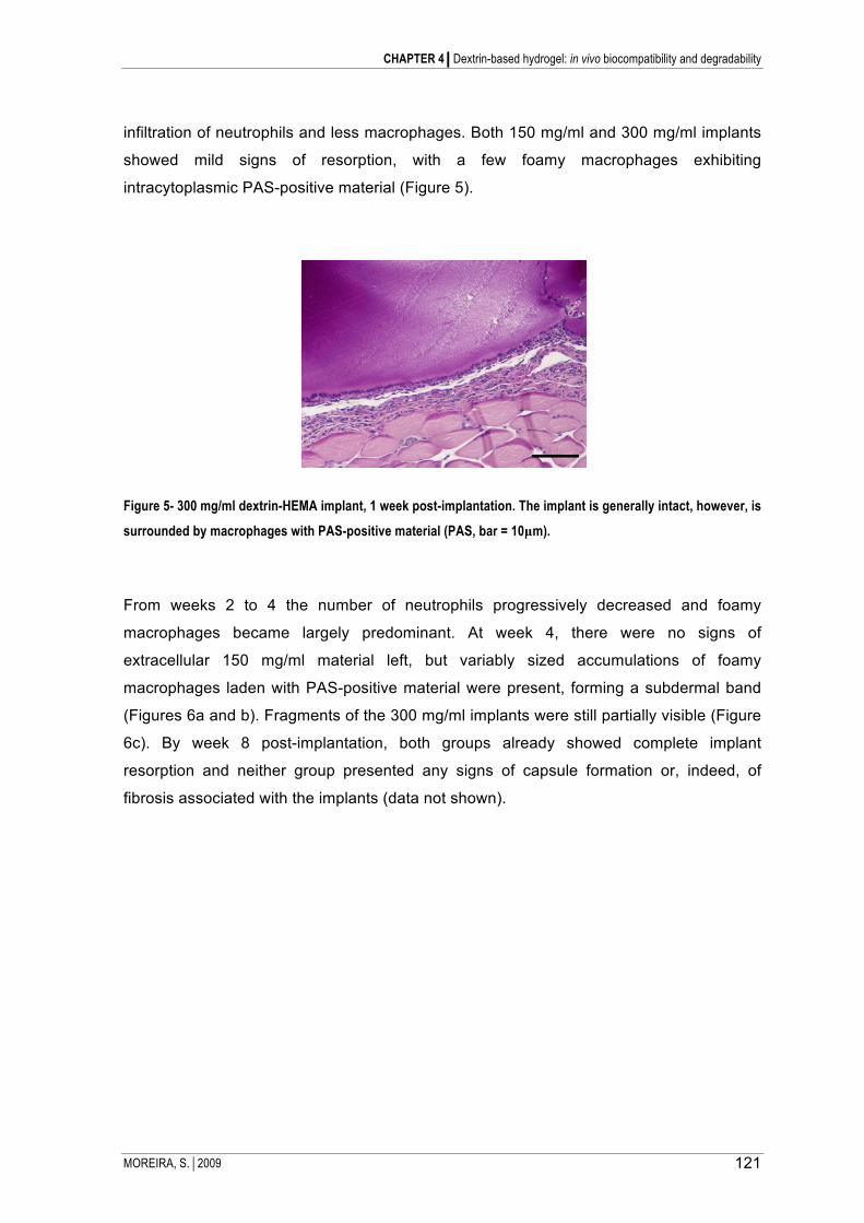
CHAPTER 4Dextrin-based hydrogel: in vivo biocompatibility and degradability
MOREIRA, S.2009 121
infiltration of neutrophils and less macrophages. Both 150 mg/ml and 300 mg/ml implants
showed mild signs of resorption, with a few foamy macrophages exhibiting
intracytoplasmic PAS-positive material (Figure 5).
Figure 5- 300 mg/ml dextrin-HEMA implant, 1 week post-implantation. The implant is generally intact, however, is
surrounded by macrophages with PAS-positive material (PAS, bar = 10µm).
From weeks 2 to 4 the number of neutrophils progressively decreased and foamy
macrophages became largely predominant. At week 4, there were no signs of
extracellular 150 mg/ml material left, but variably sized accumulations of foamy
macrophages laden with PAS-positive material were present, forming a subdermal band
(Figures 6a and b). Fragments of the 300 mg/ml implants were still partially visible (Figure
6c). By week 8 post-implantation, both groups already showed complete implant
resorption and neither group presented any signs of capsule formation or, indeed, of
fibrosis associated with the implants (data not shown).

CHAPTER 4Dextrin-based hydrogel: in vivo biocompatibility and degradability
MOREIRA, S.2009 122
Figure 6- 150 mg/ml (a and b) or 300 mg/ml (c) dextrin-HEMA implant, 4 weeks post-implantation. a) There are no
visible extracellular fragments of the implant. Numerous macrophages containing abundant intracellular PAS-
positive material form a subdermal band located at the implantation site (PAS, bar = 200µm). b) Variably
abundant, intracytoplasmic, globular, PAS-positive material is present in macrophages (PAS, bar = 10µm). c)
Large, extracellular implant fragments remain in the deep dermis, surrounded by a thick macrophagic ring (PAS,
bar = 200µm).
DISCUSSION
Hydrogels may be used implanted or otherwise in touch with body fluids and tissues, to
assist or substitute the function of organs and tissues. Therefore, the characterization of
the interaction with the living organism is of paramount importance. In spite of dextrin
being a biodegradable polymer, the degradation profile of a polymeric material could be
affected by its chemical modification and also by the degree of substitution (Hreczuk-Hirst
et al., 2001; Vercauteren et al., 1990). Therefore, the evaluation of its toxicity and
degradation behavior in vivo was analysed in this work. The results from cryo-SEM
showed that, although both hydrogel are made of dextrin, the structures of the
polymerized hydrogels depended deeply of the molecule used to functionalize it. As
showed in Figure 2 the dextrin-VA have smaller porous than dextrin-HEMA for the same
DS (20%) and concentration (300 mg/ml).
The results obtained with dextrin-VA hydrogels indicate its apparent non-degradability in
vivo, irrespective of the DS of the polymer used. Previous results showed that in vitro the
degradation of the dextrin–VA hydrogels in phosphate saline buffer is very slow. Still, the
hydrogel could be rendered degradable, through the incorporation of amylase; indeed,
this approach proved to be an effective route to modulated the gel degradation and
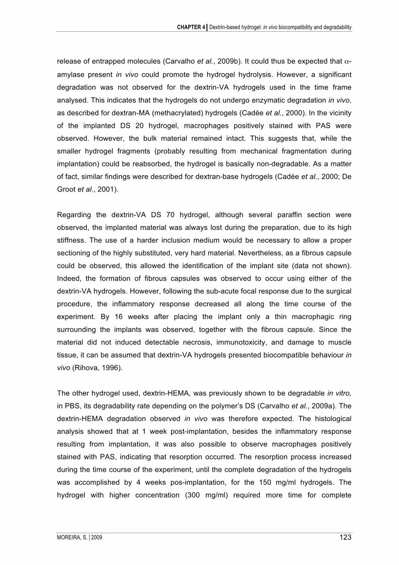
CHAPTER 4Dextrin-based hydrogel: in vivo biocompatibility and degradability
MOREIRA, S.2009 123
release of entrapped molecules (Carvalho et al., 2009b). It could thus be expected that α-
amylase present in vivo could promote the hydrogel hydrolysis. However, a significant
degradation was not observed for the dextrin-VA hydrogels used in the time frame
analysed. This indicates that the hydrogels do not undergo enzymatic degradation in vivo,
as described for dextran-MA (methacrylated) hydrogels (Cadée et al., 2000). In the vicinity
of the implanted DS 20 hydrogel, macrophages positively stained with PAS were
observed. However, the bulk material remained intact. This suggests that, while the
smaller hydrogel fragments (probably resulting from mechanical fragmentation during
implantation) could be reabsorbed, the hydrogel is basically non-degradable. As a matter
of fact, similar findings were described for dextran-base hydrogels (Cadée et al., 2000; De
Groot et al., 2001).
Regarding the dextrin-VA DS 70 hydrogel, although several paraffin section were
observed, the implanted material was always lost during the preparation, due to its high
stiffness. The use of a harder inclusion medium would be necessary to allow a proper
sectioning of the highly substituted, very hard material. Nevertheless, as a fibrous capsule
could be observed, this allowed the identification of the implant site (data not shown).
Indeed, the formation of fibrous capsules was observed to occur using either of the
dextrin-VA hydrogels. However, following the sub-acute focal response due to the surgical
procedure, the inflammatory response decreased all along the time course of the
experiment. By 16 weeks after placing the implant only a thin macrophagic ring
surrounding the implants was observed, together with the fibrous capsule. Since the
material did not induced detectable necrosis, immunotoxicity, and damage to muscle
tissue, it can be assumed that dextrin-VA hydrogels presented biocompatible behaviour in
vivo (Rihova, 1996).
The other hydrogel used, dextrin-HEMA, was previously shown to be degradable in vitro,
in PBS, its degradability rate depending on the polymer’s DS (Carvalho et al., 2009a). The
dextrin-HEMA degradation observed in vivo was therefore expected. The histological
analysis showed that at 1 week post-implantation, besides the inflammatory response
resulting from implantation, it was also possible to observe macrophages positively
stained with PAS, indicating that resorption occurred. The resorption process increased
during the time course of the experiment, until the complete degradation of the hydrogels
was accomplished by 4 weeks pos-implantation, for the 150 mg/ml hydrogels. The
hydrogel with higher concentration (300 mg/ml) required more time for complete

CHAPTER 4Dextrin-based hydrogel: in vivo biocompatibility and degradability
MOREIRA, S.2009 124
resorption. Similarly to dextrin-VA, the dextrin-HEMA hydrogels presented biocompatible
behaviour. Moreover these findings also showed that its degradation rate could be
controlled through selection of the polymer concentration.
CONCLUSION
Work currently ongoing in our lab explores the potential of the dextrin hydrogels as
delivery systems and tissue engineering scaffolds. Carvalho et al. showed that, in vitro,
dextrin-hydrogels effectively operate as a protein depot, the release being controlled
through the use of amylase, selection of the polymer concentration and DS (Carvalho et
al., 2009a). Furthermore, it was also shown that these hydrogels may be easily
functionalized, using recombinant proteins containing a starch binding module fused with
bioactive molecules, in order to improve cell adhesion and spreading (Moreira et al.,
2008). In this work two dextrin-hydrogels were subcutaneously implanted in mice; both
presented biocompatible behaviour in vivo, since none induced necrosis, immunotoxicity,
or damage to muscle tissue.
The degradation rate of the biomaterial is an important issue depending on its application.
While in some applications, such as a pacemaker or breast implants, maintenance of the
physical integrity and mechanical properties are required, in other, such as surgical
suture, rapid biodegradability is required. The control of the rate and extent of
degradability of a biomaterial is therefore critical for its assigned function. Here we
presented two biocompatible hydrogels that can be used in different applications once
their degradation profile can be controlled by selecting the acrylate ester used in the
dextrin functionalization, the polymer DS and its concentration. Based on the results
obtained, may be speculated that dextrin hydrogel with mixed reticulation chemistry, either
HEMA or VA, in different proportions, may be used to fine-tune the degradation rate.

CHAPTER 4Dextrin-based hydrogel: in vivo biocompatibility and degradability
MOREIRA, S.2009 125
REFERENCES
Cadée, J.A., van Luyn, M.J.A., Brouwer, L.A., Plantinga, J.A., van Wachem, P.B., De Groot, C.J., Otter, W.D., Hennink, W.E., 2000. In vivo biocompatibility of dextran-based hydrogels. Inc J Biomed Mater Res 50, 397–404.
Carvalho, J., Coimbra, M., Gama, F., 2009b. New dextrin-vinylacrylate hydrogel: studies on protein diffusion and release. Carbohydrate Polymers 75, 322-327.
Carvalho, J., Gil, A., Gama, F., 2007. Production and characterization of a new dextrin based hydrogel. Eur Polym J 43, 3050 - 3059.
Carvalho, J., Moreira, S.M.G., Maia, J., Gama, M., 2009a. Characterization of dextrin-based hydrogels: rheology, biocompatibility and degradation J Biomed Mater Res A.
De Groot, C.J., Van Luyn , M.J.A., Van Dijk-Wolthuis , W.N.E., Cadée, J.A., Plantinga, J.A., Otter, W.D., Hennink, W.E., 2001. In vitro biocompatibility of biodegradable dextran-based hydrogels tested with human fibroblasts. Biomaterials 22, 1197-1203.
Del Guerra, R., Cascone, M., Ricci, D., Martinoia, S., Parodi, M., Ahluwalia, A., Van Mourk, J., Grattarola, M., 1996. Optimization of the interaction between ethylene-vinyl alcohol copolymers and human endothelial cells. J Mater Sci Mater Med 7, 8-12.
Del Guerra, R., Gazzetti, P., Lazzerini, G., Cerrai, P., Guerra, G., Tricoli, M., Cristallini, C., 1995. Degradation products of poly(ester-esther-ester) block copolymers do not alter endothelial metabolism in vitro. J Mater Sci Mater Med 6, 824–828.
Draye, J.P., Bernard, D., Voordea, A.V., Bulckeb, A.V.D., Reub, B., Schacht, E., 1998. In vitro and in vivo biocompatibility of dextran dialdehyde cross-linked gelatin hydrogel films. Biomaterials 19, 1677-1687.
Frampton, J.E., Plosker, G.L., 2003. Icodextrin: a review of its use in peritoneal dialysis. Drugs 63, 2079-2105.
Garcia, H., Barros, A.S., Gonçalves, C., Gama, F.M., Gil, A.M., 2008. Characterization of dextrin hydrogels by FTIR spectroscopy and solid state NMR spectroscopy. European Polymer Journal l 44, 2318–2329.
Hardwicke, J., Ferguson, E.L., Moseley, R., Stephens, P., Thomas, D.W., Duncan, R., 2008. Dextrin–rhEGF conjugates as bioresponsive nanomedicines for wound repair. Journal of Controlled Release 130, 275–283.
Hasırcı, V., Lewandrowski, K., Gresser, J.D., Wise, D.L., Trantolo, D.J., 2001. Versatility of biodegradable biopolymers: degradability and an in vivo application. Journal of Biotechnology 86, 135–150.
Hoffman, A.S., 2002. Hydrogels for biomedical applications. Advanced Drug Delivery Reviews 54, 3-12.
Hosie, K.B., Kerr, D.J., Gilbert, J.A., Downes, M., Lakin, G., Pemberton, G., Timms, K., Young, A., Stanley, A., 2003. A pilot study of adjuvant intraperitoneal 5-fluorouracil using 4% icodextrin as a novel carrier solution. Eur J Surg Oncol 29, 254-260.
Hreczuk-Hirst, D., Chicco, D., German, L., Duncan, R., 2001. Dextrins as potential carriers for drug targeting: tailored rates of dextrin degradation by introduction of pendant groups. Int J Pharm 230, 57-66.
Kirkpatrick, C.J., 1992. A critical view of current and proposed methodologies for biocompatibility testing: cytotoxicity in vitro. Regulatory Affairs 4: 13–32 4, 13-32.
Kopecek, J., 2007. Hydrogel biomaterials: a smart future? Biomaterials 28, 5185-5192.

CHAPTER 4Dextrin-based hydrogel: in vivo biocompatibility and degradability
MOREIRA, S.2009 126
Langer, R., Peppas, A.N., 2003. Advances in Biomaterials, Drug Delivery, and Bionanotechnology. Bioengineering, Food, and Natural Products 49, 2990- 3006.
Moreira, S., Andrade, F., Domingues, L., Gama, M., 2008. Development of a strategy to functionalize a dextrin-based hydrogel for animal cell cultures using a starch-binding module fused to RGD sequence. BMC Biotechnology 8, 78.
Ratner, B.D., 1997. Biomaterials science: overview and opportunities with special reference to organic and polymeric glow discharge plasma treatments. In Plasma processing of polymers. In Plasma processing of polymers Agostino R et al (ed) Kluwer Academic Publishers, 137-139.
Rihova, B., 1996. Biocompatibility of biomaterials: hemocompatibility, immunocompatibility and biocompatibility of solid polymeric materials and soluble targetable polymeric carriers. Advanced Drug Delivery Reviews 21, 157-176.
van Dijk-Wolthuis, W.N.E., Hoogeboom, J.A.M., van Steenbergen, M.J., Tsang, S.K.Y., Hennink, W.E., 1997. Degradation and Release Behavior of Dextran-Based Hydrogels. Macromolecules 30, 4639-4645.
Van Tomme, S.R., van Nostrum, C.F., de Smedt, S.C., Hennink, W.E., 2006. Degradation behavior of dextran hydrogels composed of positively and negatively charged microspheres. Biomaterials 27, 4141-4148.
Vercauteren, R., Bruneel, D., Schacht, E., Duncan, R., 1990. Effect of the chemical modification of dextran on the degradation by dextranase. Bioactive Comp Polymers 5, 4-15.
Vieira, E., Cestari, A., Airoldi, C., Loh, W., 2008. Polysaccharide-Based Hydrogels: Preparation, Characterization, and Drug Interaction Behaviour. Biomacromolecules 9, 1195–1199.

MOREIRA, S.2009 127
Chapter 5
BC nanofibres: in vitro study of genotoxicity and cell
proliferation
Results published on Toxicology Letters
Moreira, S., Silva, N., Almeida-Lima, J., Rocha, H., Medeiros, S., Alves, C., Gama, M., 2009. BC nanofibres:
in vitro study of genotoxicity and cell proliferation. Toxicology Letters 189, 235–241
ABSTRACT
Nanomaterials have unusual properties not found in the bulk materials, which can be exploited in numerous
applications such as biosensing, electronics, scaffolds for tissue engineering, diagnostics and drug delivery.
However, research in the past few years has turned up a range of potential health hazards, which has given
birth to the new discipline of nanotoxicology. Bacterial cellulose (BC) is a promising material for biomedical
applications, namely due to its biocompatibility. Although BC has been shown not to be cytotoxic or genotoxic,
the properties of isolated BC nanofibres (NFs) on cells and tissues has never been analysed. Considering the
toxicity associated to other fibre-shaped nanoparticles, it seems crucial to evaluate the toxicity associated to
the BC-NFs.
In this work, nanofibres were produced from bacterial cellulose by a combination of acid and ultrasonic
treatment. The genotoxicity of nanofibres from bacterial cellulose was analysed in vitro, using techniques
previously demonstrated to detect the genotoxicity of fibrous nanoparticles. The results from single cell gel
electrophoresis (also known as comet assay) and the Salmonella reversion assays showed that NFs are not
genotoxic under the conditions tested. A proliferation assay using fibroblasts and CHO cells reveals a slight
reduction in the proliferation rate, although no modification in the cell morphology is observed.

CHAPTER 5 BC- Nanofibres: in vitro study of genotoxicity and cell proliferation
MOREIRA, S.2009 128
INTRODUCTION
The development of artificial materials with biomimetic behavior is essential for tissue
engineering purposes. Scaffolds based on nanofibres mimic the natural extracellular
matrix and its nanoscale fibrous structure. Several approaches have been described in
order to achieve materials based on nanofibres from synthetic or natural polymers
(Ashammakhi et al., 2007; Ma et al., 2005).
Bacterial cellulose (BC), secreted by Gluconacetobacter xylinus, has been presented as a
biocompatible scaffold for the engineering of cartilage and blood vessels, wound dressing,
guided tissue regeneration, among other applications (Andrade et al., 2008; Astley et al.,
2003; Backdahl et al., 2008; Czaja et al., 2007; Entcheva et al., 2004; Maneerung et al.,
2008; Rambo et al., 2008; Svensson et al., 2005; Tabuchi and Baba, 2005; Teeri et al.,
2007). BC has unique characteristics including high purity, high crystallinity and
remarkable mechanical properties, due to the uniform ultrafine-fiber network structure, the
high planar orientation of the ribbon-like fibres when compressed into sheets, the good
chemical stability, and the high water holding capacity (Svensson et al., 2005). Several
materials based on bacterial cellulose, recognized as non genotoxic and non cytotoxic,
have been commercialized (Jonas and Farah, 1998; Schmitt et al., 1991).
Since nanomaterials have unusual properties, not found in the bulk material, such as high
surface reactivity and ability to cross cell membranes, concerns about their safety and
toxicology have emerged. The impact of nanostructural features in the interaction of a
material with cells and tissues is dependent on the size, chemical composition, surface
structure, solubility, shape, and on the supramolecular structural organization (Barnes et
al., 2008). A major concern with fibres is their carcinogenic potential. There is sufficient
evidence that all forms of asbestos (generic term for a group of six naturally occurring
fibrous silicate minerals) are carcinogenic and co-carcinogen to man (Dopp et al., 2005;
Speit, 2002). Moreover, recent studies described the toxicity of materials associated to
size or shape; namely, the toxicity of carbon nanotubes (Donaldson et al., 2006; Poland et
al., 2008) and the size-dependence toxicity of gold or ferric oxide nanoparticles was
reported (Backdahl et al., 2008; Pan et al., 2007; Wang et al., 2009).
The toxicity associated with inhaled fibres such as asbestos has been described. Inhaled
fibres may be toxic, particularly when they are "long, thin and durable" (Donaldson et al.,

CHAPTER 5 BC- Nanofibres: in vitro study of genotoxicity and cell proliferation
MOREIRA, S.2009 129
2006). Asbestos fibres are dangerous because the fibres split lengthwise, producing thin
fibres that can enter the lungs, being "moderately durable" once there (Speit, 2002).
Although cellulose fibres, from wood pulp and textile fabric, are used without "significant
concern", cellulose fibres share similar features with asbestos, including the needle-like
shape and biopersistence. Moreover, the inflammatory responses of respirable cellulose
fibres (wood pulp) using animal models were already reported (Cullen et al., 2000). In light
of these results, it seems crucial to evaluate the toxicity of the BC nanofibres. It must be
remarked that, although BC cannot be enzymatically degraded in the human body, the
inflammatory processes may actually degrade cellulose to some extent. Given the current
focus of BC as a promising biomaterial with a variety of applications, it is relevant to
evaluate not only the toxicity of BC membranes or scaffolds, but also of its degradation
products, including BC nanofibres.
Indeed, although in vivo studies demonstrate the BC biocompatibility (Helenius et al.,
2006), and lack of mutagenicity (Schmitt et al., 1991), no reports are available on the BC
nanofibres toxicity. Although BC is not expected to be degraded in vivo, safety concerns
makes this study mandatory. It is well accepted that in vitro studies using cell systems are
valuable tools to clarify the cellular mechanisms involved in genototoxic effects, including
DNA damage (Dusinská et al., 2004 ; Speit, 2002). Therefore, the aim of this study is to
evaluate the genotoxicity of cellulose nanofibres at cellular level using the single cell gel
electrophoresis and the Salmonella reversion assays. The cell proliferation in the
presence of nanofibres was also evaluated. These tests are useful as a screening tool for
setting priorities because they are an inexpensive and a quick way to help single out
substances that should be targeted for further testing. Furthermore, these assays were
already used to demonstrate the genotoxic effect of asbestos fibres in mammalian cells in
vitro (Dusinská et al., 2004 ; Speit, 2002).

CHAPTER 5 BC- Nanofibres: in vitro study of genotoxicity and cell proliferation
MOREIRA, S.2009 130
MATERIALS and METHODS
Bacterial strain, cells and culture medium
The cellulose was produced by Gluconacetobacter xylinus (ATCC 53582), purchased
from the American Type Culture Collection, grown statically in Hestrin-Schramm medium
(Hestrin and Schramm, 1954), pH 5 at 30ºC, during 5 days.
In the Salmonella reversion assay, four strains of Salmonella tryphimurium (Dr. B. N.
Ames, Biochemistry Department, University of California, Berkeley, USA) were used,
namely, TA97a [his D6610, rfa, Δ uvrB, bio-, pKM101 (ApR)], TA98 [his D3052, rfa, Δ uvrB,
bio-, pKM101 (ApR)], TA100 [his G46, rfa, Δ uvrB, bio-, pKM101 (ApR)], and TA102 [his
D428, rfa, pKM101 (ApR), pQA1 (TtR)] (Levin et al., 1982; Maron and Ames, 1983).
The proliferation assays were performed using mouse embryo fibroblasts 3T3 (ATCC
CCL-164), grown in Dulbecco’s modified Eagle’s media (DMEM) supplemented with 10%
newborn calf serum (Invitrogen), and Chinese Hamster Ovary (CHO), grown in DMEM
media supplemented with 10% fetal bovine serum (Invitrogen), both culture medium were
supplemented with penicillin/streptomycin (1 µg/ml) (Sigma-Aldrich, St. Louis, USA) and
the incubation was at 37ºC, in a fully modified air containing 5% CO2. The same
conditions were used to grow CHO cells for comet assay. The cell viability was assessed
using the MTT (3-[4, 5-dimethylthiazol-2-yl]-2, 5-diphenyl-tetrazolium bromide) assay,
obtained from Invitrogen.
Production of BC nanofibres
The production of bacterial cellulose (BC) was performed by growing G. xylinus in Hestrin-
Schramm medium, pH 5. After inoculation, the culture (100 ml) was incubated, first with
agitation during 8 h, and then statically at 30ºC, for 5-7 days. BC pellicles were purified in
a 4% NaOH solution at 70ºC, for 90 min. BC was then neutralised by thoroughly washing
with water. Finally, BC pellicles were lyophilised prior to use.
The nanofibres production, by acidic and/or ultrasonic treatment, was based on previous
works (Roman and Winter, 2004; Zhao et al., 2007). The acid hydrolysis was performed
as follows: 20 mg of dry BC was sliced in small pieces and 2 ml of 50% H2SO4 (v/v) was

CHAPTER 5 BC- Nanofibres: in vitro study of genotoxicity and cell proliferation
MOREIRA, S.2009 131
added. The mixture was kept at 40ºC, for 2 h with vigorous stirring. To stop the hydrolysis,
10 ml of cold water was added and the cellulose was recovered by filtration, using a
membrane with a 0.45 µm pore size. Then, the cellulose was washed out with 20 ml of
water and the recovered pellet was resuspended in 10 ml of water. This suspension was
treated by sonication at 40 W (Branson Ultrasonic Disruptor, Sonifier II/W450) for 10 min
(samples were maintained on ice during sonication). Then, the NFs suspension was
centrifuged (1 h, 15000 rpm), and the pellet resuspended in water and sonicated again, in
the same condition, for another 10 min. The yield of the process was evaluated by
quantifying the total sugar in the samples, using the phenol-sulphuric method (Dubois et
al., 1956).
TEM analysis
The NFs obtained were stained with uranyl acetate and analysed by transmission
electronic microscopy (TEM, Zeiss 902A Orius SC 1000; 50kV).
Evaluation of cellulose nanofibres mutagenicity by Salmonella reversion assay
Four Salmonella tryphimurium strains were used to study the potential mutagenicity effect
of the cellulosic NFs. The procedure was to some extent modified from the original
description by Kado et al. (1986). This assay was performed in miscrosuspension with or
without S9 mixture (MoltoxTM, North Carolina, USA), using 0.1, 0.5 or 1.0 mg/ml of NFs
suspension. The negative control was distilled water, and the positive controls employed
were: 0.1 µg/plate 4NQO (4-Nitroquinoline 1-oxide) for the TA97a and TA98 strains; 5.0
µg/plate sodium azide for the TA100 strain; and 0.5 µg/plate mytomicyn C for the TA102
strain. Briefly, 105 µl of a mixture containing the NFs suspension and cell suspension (109
cells/ml) were incubated at 37ºC for 90 min. Then, 2.5 ml of molten Top agar (0.6% bacto-
agar and 0.5% NaCl) was added, before plating in a Petri dish containing minimal agar
(1.5% agar, Vogel-Bonner E medium). The His+ revertant colonies were counted after 72
h of incubation at 37ºC. All experiments were repeated at least three times with three
replicas. The mutagenicity of cellulose NFs was evaluated according to the following
parameters: the maximum number of revertants in the presence of the NFs should be 2-
fold or more relative to the negative control; a dose-dependent increase in the number of
revertants should be observed (Mortelmans and Zeiger, 2000).

CHAPTER 5 BC- Nanofibres: in vitro study of genotoxicity and cell proliferation
MOREIRA, S.2009 132
Proliferation assays
The proliferation assays were performed in vitro as follows: 1 ml of the CHO or mouse
embryo fibroblast 3T3 cell suspension (104 cells/ml) was seeded in a 24-well polystyrene
plate (TPP, Switzerland). The cells were allowed to adhere for 4 h. Before the addition of
cellulose NFs, the medium with non-adherent cells was removed and the NFs containing
medium (to a final concentration of: 1, 0.5 or 0.1 mg/ml) was added. A control without NFs
was carried out. The cellular growth at 0, 24, 48 and 72 h of incubation was evaluated by
MTT assay, a colorimetric test that gives a measure of the mitochondrial activity. The
effect of NFs on the cell morphology was evaluated by microscopic observation (Nikon
Eclipse TE300 Inverted Microscope).
Evaluation of cellulose nanofibres genotoxicity by single cell gel assay (comet
assay)
The DNA integrity was evaluated by alkaline single cell gel assay (also kwon as comet
assay) using CHO cells grown in the presence of different NFs concentration.
In this assay, 2 ml of CHO cell suspension (105 cells/ml) were seeded on a 6-well
polystyrene plate (TPP, Switzerland). After 16 h, the medium was refreshed with medium
containing the NFs (0.1, 0.5 or 1 mg/ml). Cells were incubated with NFs suspension
during 48 h. Hydrogen peroxide (100 mM) and water were used as positive and negative
controls, respectively. The alkaline comet assay was performed as described by Singh
(Singh et al., 1988). Briefly, cells were trypsinized from 6-well polystyrene plate, and
resuspended in 50 µl of medium. The cell viability was determined in a Neubauer counting
chamber using the trypan blue exclusion test. A volume of 10 µl of the cellular suspension
were embedded in 0.5% low-melting-point agarose and plated on agarose-coated
microscope slide. Then, the slides with cells were treated with lysis solution (2.5 M NaOH,
0.1 M EDTA, 0.010 M Tris, 1% Triton X-100, 10% DMSO, adjusted to pH 10) for 12 hours
at 4ºC, rinsed with distilled water, and placed in the electrophoresis buffer (0.3 M NaHO,
pH 13 and 0.001 M EDTA), for 20 min to allow DNA unwinding. Following electrophoresis
(30 min, at 25 V and 300 mA), the slides were neutralized with 0.4 M Tris buffer (pH 7.5)
and stained with ethidium bromide (20 mg/ml). The slides were analysed through
fluorescence microscopy (Nikon Eclipse TE300 microscope equipped with a Nikon E600
camera, 0.488 microns/pixel). At least 300 cells per condition tested were analysed.

CHAPTER 5 BC- Nanofibres: in vitro study of genotoxicity and cell proliferation
MOREIRA, S.2009 133
The DNA damages were evaluated by image analysis using the ‘‘Comet Assay IV version
4.2” image analysis system. Data collected from each cell included tail length (TL), tail
migration (TMi), percent tail DNA (TI), and tail moment (TM), which correspond to the
product of the comet length and the amount of DNA in the tail (Olive and Durand, 1992).
Statistic analysis
The One-way Analysis of Variance (ANOVA) was applied to statistics evaluation of the
comet scores and to the proliferation assays results. The post-test Tukey-Kramer Multiple
Comparisons test was used to compare the scores of the samples and positive control
(PC). The analyses were performed using GraphPad Prism 3.05.
RESULTS and DISCUSSION
Production of BC nanofibres
G. xylinum synthesizes cellulose nanofibres with 40–50 nm width (the bacterial cellulose
ribbons), which assemble in a static culture as a white gelatinous material (pellicle) on the
surface of the culture liquid. The native cellulose consists of sets of parallel chains of β-
1,4-D-glucopyranose units interlinked by intermolecular hydrogen bonds (Czaja et al.,
2007). Several works describe the production of nanofibres from different cellulosic
sources, using acid hydrolysis (Araki et al., 1999; Roman and Winter, 2004) or mechanic
treatment (Zhao et al., 2007). These two approaches were used in order to extract NFs
from BC. The acid hydrolysis was tested using a range of acid concentrations,
temperatures and treatment time. Concentrations of H2SO4 superior to 50% resulted in
extensive hydrolysis, yielding less than 20% of the material used (data not shown). The
acid concentration is in fact the critical parameter in the acid hydrolysis approach. The use
50 % H2SO4, for 2 h at 40ºC, yielded 50% of nanofibres. According to Zhao et al. (2007),
sonication can also be successfully used to extract NFs from natural materials, including
cellulose from wood, cotton, bamboo. This approach was also applied to BC. Using acid
hydrolysis (50% H2SO4, 2 h, 40ºC) and sonication (20 min, 40 W), needle shaped
cellulose NFs with 50-1500 nm length and 3-5 nm width, were obtained (figure 1).

CHAPTER 5 BC- Nanofibres: in vitro study of genotoxicity and cell proliferation
MOREIRA, S.2009 134
Figure 1 - TEM image of cellulose nanofibres (50kV; Zeiss 902A Orius SC 1000).
Evaluation of cellulose nanofibres mutagenicity by Salmonella reversion assay
The purpose of the bacterial reverse mutation assay is to evaluate the mutagenicity of the
cellulose NFs, by measuring its ability to induce reverse mutations at selected loci, in
several bacterial strains. Having into account that mutations are essential for cancer
formation, the reliable characterization of mutagenicity is mandatory, while characterizing
the safety of a biomaterial. The Kado test (Kado et al., 1983) is a modification with
improved sensitivity of the Ames test (Ames et al., 1972). This is a simple, quick and
inexpensive mutagenicity test, required for safety testing of a variety of compounds,
including drugs, medical devices, food additives, industrial chemicals and pesticides
(McCann et al., 1975). Furthermore, the potential mutagenicity of BC and of some its

CHAPTER 5 BC- Nanofibres: in vitro study of genotoxicity and cell proliferation
MOREIRA, S.2009 135
derivates were already accessed using the Ames assay (Schmitt et al., 1991), therefore it
was selected as a first approach in this work to investigate the possible mutagenicity of
the cellulose NFs. The strains used were specially constructed to allow detection of
mutagens acting via different mechanisms, namely frameshift mutations (TA97a and TA98
strains), base-pair substitution mutations (TA100 and TA102), detection of oxidative and
alkylating mutagens and active forms of oxygen (TA102) (Hakura et al., 2005). Table 1
presents the results obtained with the different strains.
Table 1 - Results obtained in Salmonella reversion assay.
Revertant colonies/plate ± SD (without S9) Revertant colonies/plate ± SD (with S9)
Strain TA97a TA98 TA100 TA102 TA97a TA98 TA100 TA102
PC 540±54 389±17 1531±183 1026±36 191±21 195±76 485±14 2356±196
NC 143±17 36±6 228±18 350±27 93±8 16±4 82±44 958±20
0.1 124±6 31±6 235±9 327±12 93±4 20±1 133±7 691±61
0.5 132±14 43±2 220±2 327±13 91±10 20±1 112±14 656±35
1.0 147±12 42±4 225±7 333±18 108±7 26±4 112±33 859±109
PC: positive control: 0.1 µg/plate of 4NQO to TA97a and TA98, 5.0 µg/plate sodium azide to TA100 and 0.5
µg/plate mytomicyn C to TA102; NC: negative control: H2O; SD: standard deviation.
The reversion of the histidine phenotype in Salmonella strains is often adopted as a
criteria for the classification of molecules as mutagenic. The results obtained in the
presence of the cellulose NFs, without S9 mixture, correspond to the spontaneous
reversion for each strain and are similar to those obtained to negative control (table 1). In
the presence of S9 mixture, an increase of revertant colonies per plate, for the TA98 and
TA100 strains, is detected as compared with control; however, the increases was in each
case < 2-fold and does not appear to be dose-related. The results suggest that, under the
conditions tested, the cellulose NFs do not present mutagenic behaviour, as described
previously for BC and some fibrous BC-based materials (Schmitt et al., 1991).

CHAPTER 5 BC- Nanofibres: in vitro study of genotoxicity and cell proliferation
MOREIRA, S.2009 136
In vitro proliferation assay
The cellular morphology and proliferation may be affected by the presence of
nanostructural patterns. Several studies analysed the proliferation of different cell lines on
BC membranes, confirming its non-toxicity and applicability as scaffold for cell
proliferation. However, depending on the cells used, the effect of the biomaterial on the
proliferation rate and the cell morphology may be quite different (Sanchavanakit et al.,
2006). Several studies showed that the cytotoxicity of a nanomaterial is many times cell-
specific (Cullen et al., 2002). Recently, De Nicola et al. (2007) reported that, although
carbon nanotubes do not present cytotoxic effect on human leukemic U937 cells, the
proliferation rate is deeply altered. Moreover, Bottini et al. (2006) showed that the same
nanotubes refereed above induce apoptosis in T lymphocytic cells, suggesting that
cytotoxicity may be cell-specific. In addition, it has been reported that asbestos fibres
inhibits the growth of CHO cells (Speit, 2002), and yet the same fibres stimulate the
proliferation of different kinds of cells, in vitro, including fibroblasts (Bernstein et al., 2005).
Taking in consideration the evidence of contradictory, cell-specific effects arising from the
interaction cell-biomaterial, the evaluation of the NFs effect on proliferative rate was
performed both with CHO cells and fibroblasts. In both cases, the proliferation was about
15-20% lower in the presence of NFs, after 72 h of cell culture, irrespective of the
concentration used (Figure 2). The lower proliferation rate may stem from the insolubility
of NFs and their slow deposition on the polystyrene plate. It is known that cell proliferation
is dependent on characteristics of material surface, such as its roughness. In addition, it
was also described that cell proliferation on BC membrane is slower than on the cell
culture plate (Backdahl et al., 2006). However, the microscopic observations did not
reveal differences in the cellular morphology.

CHAPTER 5 BC- Nanofibres: in vitro study of genotoxicity and cell proliferation
MOREIRA, S.2009 137
Figure 2 – MTT results from proliferation assays using mouse embryonic fibroblast 3T3 and CHO (mean ± SD;
**P < 0.05; ***P < 0.005). Image obtained by optical microscopy of fibroblasts grown in the presence of cellulose
NFs during 72 h. Scale bar = 20 µm.
Evaluation of cellulose nanofibres genotoxicity by comet assay
The genotoxicity of a material may be measured by analysing the damages caused on
DNA. The comet assay is based on the ability of negatively charged loops/fragments of
DNA to be drawn through an agarose gel, in response to an electric field. The extent of
DNA migration depends directly on the DNA damage present in the cells (Collins et al.,
2008). The advantages of the comet assay, relative to other genotoxicity tests, include its
high sensitivity for detecting low levels of both single and double stranded breaks in
damaged DNA, the requirement for small numbers of cells per sample, flexibility, low cost,
and ease of application (Collins et al., 1997; Collins et al., 2008). Moreover, the comet
assay is arguably one of the most widely available tests used for genotoxicity, being
already described as a reproducible assay to evaluate nanoparticles genotoxicity (Collins

CHAPTER 5 BC- Nanofibres: in vitro study of genotoxicity and cell proliferation
MOREIRA, S.2009 138
et al., 1997), and suggested as a diagnostic tool for clinical management of cancer
(Collins et al., 2008). The nanomaterial’s genotoxicity may result from a direct interaction
with DNA, or from an indirect response caused by several factors, including surface stress
through direct particle influences on DNA, the release of toxic ions from soluble
nanoparticles, or generation of oxidative stress (Donaldson et al., 2006). It has been
proposed that (oxidative) DNA damage plus structural and numerical chromosome
aberrations are the most sensitive genetic endpoints for detection of asbestos-induced
genotoxicity detectable by in vitro assay. The comet assay has indeed proven to be a
sensitive test to detect genotoxic effects of asbestos fibres in mammalian cell in vitro
(Dusinská et al., 2004; Speit, 2002). Therefore, cells grown in NFs – containing medium
were analysed by the comet assay, in order to evaluate their genotoxicity. Cells grown on
bacterial cellulose membrane were also tested as a control.
The DNA damages were evaluated by visual scoring and image analysis. Figure 3 shows
a representative image obtained for each NFs concentration tested (0.1, 0.5, 1.0 mg/ml),
negative and positive control (NC, PC) and the results obtained from the visual score. The
results show that the DNA damages in the presence of NFs are similar to the negative
control for each NFs concentration used. Around 95 % of cells present comet class 0 and
1, corresponding to no or insignificant DNA damage. The cell percentage showing comet
class 2, 3 and 4 under different conditions are in the graphics of figure 3, and represent
around 5 % of cell. Similar results were obtained with the cells grown on BC membranes
(comet class 2: 4.7±3.72; comet class 3: 1.3±2.31, and comet class 4: 1.3±1.53).

CHAPTER 5 BC- Nanofibres: in vitro study of genotoxicity and cell proliferation
MOREIRA, S.2009 139
Figure 3 – Fluorescent microscopy images of ethidium bromide stained DNA and results from visual scoring in
the comet assay. NC: negative control (H2O); PC: positive control (H2O2); 0.1–1.0 NFs concentration in mg/ml.
The images were scored and classified into five classes and given a value according to tail intensity, from 0 (no
tail) to 4 (almost all DNA in the tail). Scale bar = 50 µm.
Regarding the comet parameters obtained from image analyses (table 2), tail length (TL),
tail % DNA (TI), tail moment (TM) and tail migration (TMi), the NFs did not induce DNA
damages under the concentrations tested, since the negative control and samples with
NFs presented similar results, significantly (TL, TM, TI, TMi, P< 0.001) lower than the
positive control. The same results were obtained for the cells grown on surface of BC

CHAPTER 5 BC- Nanofibres: in vitro study of genotoxicity and cell proliferation
MOREIRA, S.2009 140
membrane (data not shown); in fact, our results confirmed the previous reports describing
the non genotoxicity of BC (Schmitt et al., 1991).
Table 2 – Results from images analysis using the Comet Assay IV software (mean ± SD).
PC: positive control (H2O2); NC: negative control (H2O); 0.1–1.0 NFs concentration in mg/ml *** P < 0.001.
Taking together the results from visual scoring and image analysis, it may be concluded
that the cellulose NFs do not present genotoxicity, under the tested conditions. Since
alkaline comet assay allows for detection of DNA strand breaks, cross-links and alkali-labil
sites induced by a series of physical and chemical agents it may be concluded that NFs
do not induce those damages in DNA.
CONCLUSION
This work presents the first evaluation of the potential genotoxicity of nanofibres extracted
from bacterial cellulose. Regarding the results of Salmonella reversion and comet assays,
cellulose NFs did not present genotoxicity under the conditions tested, as already
described for bacterial cellulose membrane. The cell culture systems have been shown to
be valuable tools in fibre genotoxicity testing. Unlike in vivo studies, secondary
inflammatory effects do not affect in vitro findings. Induction of DNA damages has been
demonstrated for various types of asbestos fibres in several cell systems including CHO
cell lines, which was not observed for BC nanofibres. Nevertheless, further studies must
be performed in order to comprehensively characterize the toxicology of cellulose-based
materials, since small modification in the material could result in drastic changes in cell-
Sample Tail Length (µm) Tail DNA (%) Tail Moment Tail Migration
0.1 17.78 ± 1.73 6.26 ± 1.20 0.66 ± 0.17 2.11 ± 0.82
0.5 21.25 ± 4.99 6.99 ± 3.48 1.03 ± 0.94 4.82 ± 4.25
1.0 15.88 ± 1.44 6.16 ± 1.78 0.71 ± 0.23 1.59 ± 0.55
NC 19.69 ± 3.31 6.88 ± 1.84 1.09 ± 0.67 3.94 ± 2.53
PC 101.36 ± 35.11*** 49.06 ± 14.51*** 25.42 ± 14.38*** 83.11 ± 35.13***

CHAPTER 5 BC- Nanofibres: in vitro study of genotoxicity and cell proliferation
MOREIRA, S.2009 141
material interactions. Work in progress includes the interaction of BC nanofibres with
macrophages and in vivo assays.

CHAPTER 5 BC- Nanofibres: in vitro study of genotoxicity and cell proliferation
MOREIRA, S.2009 142
REFERENCES
Ames, B.N., Gurney, E.G., Miller, J.A., Bartsch, H., 1972. Carcinogens as Frameshift Mutagens: Metabolites and Derivatives of 2-Acetylaminofluorene and Other Aromatic Amine Carcinogens. Proc Nat Acad Sci USA 69, 3128-3132.
Andrade, F.K., Moreira, S.M.G., Domingues, L., Gama, F.M.P., 2008. Improving the affinity of fibroblasts for bacterial cellulose using carbohydrate-binding modules fused to RGD. J Biomed Mater Res A.
Araki, J., Wada, M., Kuga, S., Okano, T., 1999. Influence of surface charge on viscosity behavior of cellulose microcrystal suspension. J Wood Sci 45, 258-261.
Ashammakhi, N., Ndreu, A., Yang, Y., Ylikauppila, H., Nikkola, L., 2007. Nanofiber-based scaffolds for tissue engineering. Eur J Plast Surg.
Astley, O.M., Chanliaud, E., Donald, A.M., Gidley, M.J., 2003. Tensile deformation of bacterial cellulose composites. Int J Biol Macromol 32, 28-35.
Backdahl, H., Esguerra, M., Delbro, D., Risberg, B., Gatenholm, P., 2008. Engineering microporosity in bacterial cellulose scaffolds. J Tissue Eng Regen Med 2, 320-330.
Backdahl, H., Helenius, G., Bodin, A., Nannmark, U., Johansson, B.R., Risberg, B., Gatenholm, P., 2006. Mechanical properties of bacterial cellulose and interactions with smooth muscle cells. Biomaterials 27, 2141-2149.
Barnes, C.A., Elsaesser, A., Arkusz, J., Smok, A., Palus, J., Lesniak, A., Salvati, A., Hanrahan, J.P., Jong, W.H., Dziubałtowska, E., Stepnik, M., Rydzynski, K., McKerr, G., Lynch, I., Dawson, K.A., Howard, C.V., 2008. Reproducible Comet Assay of Amorphous Silica Nanoparticles Detects No Genotoxicity. Nano Letters 8, 3069-3074.
Bernstein, D., Castranova, V., Donaldson, K., Fubini, B., Hadley, J., Hesterberg, T., Kane, A., Lai, D., McConnell, E.E., Muhle, H., Oberdorster, G., Olin, S., Warheit, D.B., 2005. Testing of fibrous particles: short-term assays and strategies. Inhal Toxicol 17, 497-537.
Bottini, M., Bruckner, S., Nika, K., Bottini, N., Bellucci, S., Magrini, A., Bergamaschi, A., Mustelin, T., 2006. Multi-walled carbon nanotubes induce T lymphocyte apoptosis. Toxicology Letters 160, 121–126.
Collins, A.R., Dobson, V.L., Dusinska, M., Kennedy, G., Stetina, R., 1997. The comet assay: what can it really tell us? Mutation Research 375, 183–193.
Collins, A.R., Oscoz, A.A., Brunborg, G., Gaivão, I., Giovannelli, L., Kruszewski, M.-., Smith, C.C., Stetina, R., 2008. The comet assay: topical issues. Mutagenesis Advance 23, 143-151.
Cullen, R.T., Moller, B.G., Jones, A.D., Davis, J.M.G., 2002. Toxicity of Cellulose Fibres. Ann occup Hyg 46, 81–84.
Cullen, R.T., Searl, A., Miller, B.G., Davis, J.M.G., Jones, A.D., 2000. Pulmonary and Intraperitoneal Inflammation Induced by Cellulose Fibres. J Appl Toxicol 20, 49–60.
Czaja, W.K., Young, D.J., Kawecki, M., Brown, R.M., 2007. The Future Prospects of Microbial Cellulose in Biomedical Applications. Biomacromolecules 8, 1-12.
De Nicola, M., Gattia, D.M., Bellucci, S., De Bellis, G., Micciulla, F., Pastore, R., Tiberia, A., Cerella, C., D’Alessio, M., Antisari, M.V., Marazzi, R., Traversa, E., Magrini, A., Bergamaschi, A., Ghibelli, L., 2007. Effect of different carbon nanotubes on cell viability and proliferation. J Phys: Condens Matter 19, 395013-395020.
Donaldson, K., Aitken, R., Tran, L., Stone, V., Duffin, R., Forrest, G., Alexander, A., 2006. Carbon Nanotubes: A Review of Their Properties in Relation to Pulmonary Toxicology and Workplace Safety. Toxicological sciences 92, 5-22.

CHAPTER 5 BC- Nanofibres: in vitro study of genotoxicity and cell proliferation
MOREIRA, S.2009 143
Dopp, E., Yadav, S., Ansari, F.A., Bhattacharya, K., von Recklinghausen, U., Rauen, U., Rödelsperger, K., Shokouhi, B., Geh, S., Rahman, Q., 2005. ROS-mediated genotoxicity of asbestos-cement in mammalian lung cells in vitro. Particle and Fibre Toxicology 2.
Dubois, M., Gilles, K.A., K., H.J., Rebers, P.A., Smith, F., 1956. A simple technique for quantitation of low levels of DNA damage in individual cells. Analytical Chemistry 28, 350-356.
Dusinská, M., Collins, A., Kazimírová, A., Barancoková, M., Harrington, V., Volkovová, K., Staruchová, M., Horská, A., Wsólová, L., Kocan, A., Petrík, J., Machata, M., Ratcliffe, B., Kyrtopoulos, S., 2004 Genotoxic effects of asbestos in humans. Mutat Res 553, 91-102.
Entcheva, E., Bien, H., Yin, L., Chung, C., Farrell, M., Kostov, Y., 2004. Functional cardiac cell constructs on cellulose-based scaffolding. Biomaterials 25, 5753–5762.
Hakura, A., Shimada, H., Nakajima, M., Sui, H., Kitamoto, S., Suzuki, S., Satoh, T., 2005. Salmonella/ human S9 mutagenicity test: a collaborative study with 58 compounds. Mutagenesis 20, 217-228.
Helenius, G., Backdahl, H., Bodin, A., Nannmark, U., Gatenholm, P., Risberg, B., 2006. In vivo biocompatibility of bacterial cellulose. J Biomed Mater Res A 76, 431-438.
Hestrin, S., Schramm, M., 1954. Synthesis of Cellulose by Acetobacter xylinum. Stimulation of respiration by denitrophenol 58, 345-352.
Jonas, R., Farah, L.F., 1998. Production and application of microbial cellulose. Polymer degradation and Strability 59, 101-106.
Kado, N.Y., Guirguis, G.N., Flessel, C.P., Chan, R.C., Chang, K.I., Wesolowski, J.J., 1986. Mutagenicity of fine (less than 2.5 lm) airborne particles: diurnal variation in community air determined by a Salmonella micro preincubation (microsuspension) procedure. Environ Mutagen 8, 53-66.
Kado, N.Y., Langley, D., Eisenstadt, E., 1983. A simple modification of the Salmonella liquid-incubation assay. Increased sensitivity for detecting mutagens in human urine. Mutation research 121, 25-32.
Levin, D.E., Hollstein, M., Christman, M.F., Schwiers, E.A., Ames, B.N., 1982. A new Salmonella tester strain (TA102) with AT base pairs at the site of mutation detects oxidative mutagens Proc Nadl Acad Sci USA 79, 7445-7449.
Ma, Z., Kotaki, M., Inai, R., Ramakrishna, S., 2005. Potential of Nanofiber Matrix as Tissue-Engineering Scaffolds. Tissue Engineering 11.
Maneerung, T., Tokura, S., Rujiravanit, R., 2008. Impregnation of silver nanoparticles into bacterial cellulose for antimicrobial wound dressing. Carbohydrate Polymers 72, 43–51.
Maron, D.M., Ames, B.N., 1983. Revised methods for the Salmonella mutagenicity test. Mutat Res 113, 173-215.
McCann, J., Spingarn, N.E., Kobori, J., Ames, B.N., 1975. Detection of Carcinogens as Mutagens: Bacterial Tester Strains with R Factor Plasmids. Proc Nat Acad Sci USA 72, 979-983.
Mortelmans, K., Zeiger, E., 2000. The Ames Salmonella/microsome mutagenicity test. Mutat Res 455, 29-60.
Olive, P.L., Durand, R.E., 1992. Detection of Hypoxic cells in a Murine Tumour With the Use of the Comet Assay. Journal of the National Cancer Institute 84, 707-711.
Pan, Y., Neuss, S., Leifert, A., Fischler, M., Wen, F., Simon, U., Schmid, G., Brandau, W., Jahnen-Dechent, W., 2007. Size-Dependent Cytotoxicity of Gold Nanoparticles. small 3, 1941 – 1949.

CHAPTER 5 BC- Nanofibres: in vitro study of genotoxicity and cell proliferation
MOREIRA, S.2009 144
Poland, C.A., Duffin, R., Kinloch, I., Maynard, A., Wallace, W.A.H., Seaton, A., Stone, V., Brown, S., Macnee, W., Donaldson, K., 2008. Carbon nanotubes introduced into the abdominal cavity of mice show asbestos-like pathogenicity in a pilot study. Nature Nanotechnology 3, 423-428.
Rambo, C.R., Recouvreux, D.O.S., Carminatti, C.A., Pitlovanciv, A.K., Anto!nio, R.V., M., P.L., 2008. Template assisted synthesis of porous nanofibrous cellulose membranes for tissue engineering. Materials Science and Engineering C 28, 549 – 554
Roman, M., Winter, W.T., 2004. Effect of sulfate groups from sulfuric acid hydrolysis on the thermal degradation behavior of bacterial cellulose. Biomacromolecules 5, 1671-1677.
Sanchavanakit, N., Sangrungraungroj, W., Kaomongkolgit, R., Banaprasert, T., Prasit Pavasant, P., Phisalaphong, M., 2006. Growth of Human Keratinocytes and Fibroblasts on Bacterial Cellulose Film. Biotechnol Prog 22, 1194-1199.
Schmitt, D.F., Frankos, V.H., Westland, J., Zoetis, T., 1991. Toxicologic Evaluation of CellulonTMFiber; Genotoxicity, Pyrogenicity, Acute and Subchronic Toxicity. Journal of the American College of Toxicology 10.
Singh, N.P., McCoy, M.T., Tice, R.R., Schneider, E.L., 1988. A simple technique for quantitation of low levels of DNA damage in individual cells. Exp Cell Res 175, 184-191.
Speit, G., 2002. Appropriate in vitro test conditions for genotoxicity testing of fibers. Inhal Toxicol 2002 14, 79-90.
Svensson, A., Nicklasson, E., Harrah, T., Panilaitis, B., Kaplan, D.L., Brittberg, M., Gatenholm, P., 2005. Bacterial cellulose as a potential scaffold for tissue engineering of cartilage. Biomaterials 26, 419-431.
Tabuchi, M., Baba, Y., 2005. Design for DNA separation medium using bacterial cellulose fibrils. Anal Chem 77, 7090-7093.
Teeri, T.T., Brumer, H., Daniel, G., Gatenholm, P., 2007. Biomimetic engineering of cellulose-based materials. Trends in Biotechnology 25, 299-306.
Wang, B., Feng, W., Zhu, M., Wang, Y., Wang, M., Gu, Y., Ouyang, H., Wang, H., Li, M., Zhao, Y., Chai, Z., Wang, H., 2009. Neurotoxicity of low-dose repeatedly intranasal instillation of nano- and submicron-sized ferric oxide particles in mice. J Nanopart Res 11, 41–53.
Zhao, H.P., Fenga, X.Q., Gao, H., 2007. Ultrasonic technique for extracting nanofibers from nature materials. Applied Physics Letters 90, 1-2.

MOREIRA, S.2009 145
Chapter 6
Final remarks and Perspectives
A CBM from the human protein laforin was cloned and expressed in different E. coli
expression systems (pET25b, pET29a and pGEXT4-1). The attempts to produce the
soluble and functional protein were unsuccessful. The solubilization and refolding of
inclusion bodies proved to be an efficient approach to produce the functional protein, with
starch and glycogen affinity. This is the first report of cloning this human CBM, through IB
solubilization and refolding. Nevertheless, further purification using starch affinity may be
necessary to provide the separation of the functional protein. This purification step may be
important to solve the 3D structure, by crystallography. In addition, future work includes in
vitro tests to evaluate the effect of the recombinant human CBM-RGD on the interaction of
cells with activated biomaterials. Regarding the results obtained with recombinant protein
containing a bacterial SBM fused to a RGD, it may be concluded that CBM may be
effectively used to functionalize dextrin hydrogels. Furthermore, recombinant proteins can
be purified using starch, a non-expensive material. Although the enhancement of cell
adhesion and spreading was observed, cell proliferation was not improved under the
conditions tested. Therefore, in future work, different conditions (such RGD
concentrations) may be optimized. In addition, several other small peptides could be fused
to the SBM in order to obtain materials with different bioactivities.
Dextrin is a very promising biomaterial and, surprisingly, it is rather unexploited. The DEB-
UM group is one of the few in the world currently developing dextrin-based biomaterials.
The work presented under the scope of this thesis contributes to the characterization of
dextrin hydrogels, namely the study of the in vivo biocompatibility and degradability of
subcutaneous implants in mice. Dextrin-hydrogels presented different degradability
behavior, depending on the molecule used in the cross-linking and on the DS of the
polymer. Dextrin-VA is non-degradable hydrogel in vivo, irrespective of the DS, as
described in vitro study. On the other hand, dextrin-HEMA hydrogel is a degradable

CHAPTER 6 Final remarks and perspectives
MOREIRA, S.2009 146
material and the degradability rate depends on its concentration. Despite the degradability
differences, results strongly suggest that both dextrin hydrogels are fully biocompatible.
The results also suggest that a hydrogel with controlled degradation rate may be obtained
using different mixtures of dextrin-VA and dextrin-HEMA. This approach will be studied in
a future work. These hydrogels can also be exploited as drug delivery systems through
the incorporation of dextrin nanogels (already developed at DEB-UM), obtained by self-
assembling. The nanogels allow the solubilization of poorly water soluble pharmaceuticals
and therapeutic proteins, thus provide a smart and simple controlled delivery system.
Bacterial cellulose, like dextrin, is another very promising biomaterial. Although usually
recognized as biocompatible, no information is available regarding BC nanofibres. The
results from comet assay and Salmonella reversion assay show that BC-NFs do not
present genotoxicity under the conditions tested. Moreover, the results from cell
proliferation assay also confirmed their non-toxicity. In vivo assays, to be carried out in the
future, will provide further information about their interaction with tissues, in particular
whether NFs trigger acute and chronic inflammatory responses.
The following main results from this work are thus highlighted:
- The use of CBMs as a tool to coat biomaterials made of polysaccharides with
bioactive peptides was demonstrated to be an effective and simple approach to
functionalize the biomaterials
- The CBM from the human protein laforin was expressed and isolated in functional
form for the first time, making possible structural studies for the elucidation of
mechanistic aspects of this uncommon CBM
- Dextrins hydrogels, confirming previous work from our lab, exhibit excellent
biocompatibility, as demonstrated by in vivo results.
- Biocompatibility aspects of the BC nanofibres are studied for the first time in this
project. The possible toxic effects related to its needle shape (shared with
asbestos nanofibres) are not confirmed, thus reinforcing the general view of BC as
a highly biocompatible biomaterial.