subsÍdios para o estudo da leishmaniose visceral...
TRANSCRIPT

UNIVERSIDADE ESTADUAL DO CEARÁ
PRÓ-REITORIA DE PÓS-GRADUAÇÃO E PESQUISA FACULDADE DE VETERINÁRIA
PROGRAMA DE PÓS-GRADUAÇÃO EM CIÊNCIAS VETERINÁRIAS
JOSÉ CLAUDIO CARNEIRO DE FREITAS
SUBSÍDIOS PARA O ESTUDO DA LEISHMANIOSE VISCERAL CANINA NA CIDADE DE FORTALEZA,
CEARÁ
FORTALEZA-Ce 2011

JOSÉ CLAUDIO CARNEIRO DE FREITAS
SUBSÍDIOS PARA O ESTUDO DA LEISHMANIOSE VISCERAL CANINA NA CIDADE DE FORTALEZA, CEARÁ
Tese apresentada ao Programa de Pós-Graduação em Ciências Veterinárias da Faculdade de Veterinária da Universidade Estadual do Ceará, como requisito parcial para a obtenção do título de Doutor em Ciências Veterinárias. Área de Concentração: Reprodução e Sanidade Animal. Linha de Pesquisa: Reprodução e sanidade de carnívoros, onívoros, herbívoros e aves. Orientador(a): Profa. Dra. Diana Célia Sousa Nunes-Pinheiro.
FORTALEZA-Ce 2011

Dados Internacionais de Catalogação na Publicação Universidade Estadual do Ceará
Biblioteca Central Prof. Antônio Martins Filho
F566s Freitas, José Cláudio Carneiro de
Subsídios para o estudo da leishmaniose visceral canina na cidade de Fortaleza, Ceará / José Cláudio Carneiro de Freitas. – 2011.
146 f. : il. color., enc. ; 30 cm. Tese (Doutorado) – Universidade Estadual do Ceará,
Faculdade de Medicina Veterinária, Curso de Doutorado em Ciências Veterinárias, Fortaleza, 2011.
Área de Concentração: Reprodução e Sanidade Animal. Orientação: Profª. Drª. Diana Célia Sousa Nunes Pinheiro. 1. Cães. 2. Leishmania chagasi. 3. Perfil de imunoglobulinas. 4.
Parâmetros histológicos. 5. Parâmetros laboratoriais. I. Título. CDD: 616.9364
JOSÉ CLAUDIO CARNEIRO DE FREITAS
SUBSÍDIOS PARA O ESTUDO DA LEISHMANIOSE VISCERAL CANINA NA CIDADE DE FORTALEZA, CEARÁ
Tese apresentada ao Programa de Pós-Graduação em Ciências Veterinárias da Faculdade de Veterinária da Universidade Estadual do Ceará, como requisito parcial para a obtenção do título de Doutor em Ciências Veterinárias.
Aprovada em 09 / 12 / 2011
BANCA EXAMINADORA
Profª Dra Diana Célia Sousa Nunes-Pinheiro Profº Dr Alexandre Barbosa Reis Universidade Estadual do Ceará Universidade Federal de Ouro Preto
Orientadora Examinador
Profª Dra Romélia Pinheiro Gonçalves Dr. Francisco Valdeci Ferreira Universidade Federal do Ceará Universidade Federal do Ceará
Examinadora Examinador
Dr Wendel Coura Vital Profª Dra Erika Freitas Mota Universidade Federal de Minas Gerais Universidade Federal do Ceará
Examinador Examinadora

À Deus, pelo apoio e bênçãos que a mim foram concedidos e
que muito me auxiliaram e fortaleceram no decorrer do curso
e da minha. Nos momentos mais difíceis foi a Ele quem
procurei em primeiro lugar, e, com certeza, sempre fui
prontamente atendido e compreendido. Pela obra é que se
conhece o autor.
À minha amada esposa, Cyntia Rafaelle Amaral de Abreu,
que, com toda certeza, sem o seu apoio e sua companhia
muito mais difícil seria a execução de todos os trabalhos.
Então, declaro que, todas as virtudes obtidas com a execução
dos trabalhos têm participação igual dessa maravilhosa
pessoa que Deus me deu de presente.
Aos meus queridos sobrinhos, João Lucas Rodrigues de
Freitas, Ana Catarina Rodrigues de Freitas e Maria Tereza
Carneiro de Freitas Cavalcante (Eza), que só pela suas
existências no nosso meio torna o nosso mundo melhor e
muito mais feliz.
Dedico

AGRADECIMENTOS
À Universidade Estadual do Ceará por viabilizar minha formação integral e
continuada, fornecer subsídios para a execução desse trabalho e contribuir
decisivamente à construção de novos conhecimentos.
À Fundação Cearense de Apoio ao Desenvolvimento Científico e Tecnológico
(FUNCAP) pelo auxílio financeiro, que nos foi concedido na forma de bolsa, para a
execução dos trabalhos.
À Profª Dra Diana Célia Sousa Nunes-Pinheiro, pelo seu valoroso auxílio e orientação
ao longo de todos esses anos, colocando-se sempre à disposição para colaborar. Mesmo
nos momentos familiares mais difíceis ela nos vem com palavras de força e estímulo
para que continuemos a enfrentar as turbulências naturais da nossa vida.
Ao Profº Dr Alexandre Barbosa Reis, pela confiança que nos foi dispensada com a
disposição dos estabelecimentos e equipamentos que estão sob sua responsabilidade na
Universidade Federal de Ouro Preto, que, com certeza, muito contribuiu para a
qualificação da proposta de trabalho.
Ao grande amigo Dr Wendel Coura Vital, pela valiosa colaboração que nos foi dada na
execução dos trabalhos, além de, com muita paciência, sempre nos atender em todas as
situações em que foi necessária sua valiosa opinião. Sempre que entramos em contato
em busca de socorro, imediatamente éramos atendidos.
Ao Profº Dr Francisco Valdeci de Almeida Ferreira, que muito abrilhantou nosso
trabalho com o seu vasto conhecimento adquirido ao longo dos anos, nunca se furtando
de nos atender e da melhor maneira possível dar seu auxílio. Além disso, sempre
colocou a nossa disposição as suas instalações de trabalho, bem como a prestação de
serviços de seus funcionários, que dessa forma agilizou bastante a execução das técnicas
empregadas.
Aos colaboradores e amigos do Laboratório de Imunologia e Bioquímica Animal
(LIBA), Glauco Jonas Lemos dos Santos e Belarmino Eugênio Lopes Neto, que sempre
se colocaram a disposição e muito colaboraram na execução de todas as fases do
trabalho.
Aos valiosos irmãos da Augusta e Respeitável Loja Simbólica Cavaleiros de York nº
111 e da mui respeitável Grande Loja Maçônica do Estado do Ceará, que muito
clamaram ao Grande Arquiteto do Universo e, dessa forma, me ajudaram no
aperfeiçoamento dos meus conhecimentos e na correção de meus erros, transmitindo
sempre conhecimento e fraternidade a todos aqueles que necessitem.
De uma forma toda especial, a todos os meus familiares, Pais, Irmãos e Irmã, que desde
sempre estiveram ao meu lado, sempre se colocando à disposição para me amparar e
confortar nos momentos mais difíceis.
A todos, os quais a limitação de inspiração e ingrata lembrança me impediram de citar
nessas poucas linhas, mas que, com certeza, têm os seus nomes gravados na história de
minha vida e sempre estarão em minhas preces e quando precisarem podem contar com
esse humilde servo, que com todas as minhas limitações estarei à disposição.

O sábio não é aquele que senta para
lamentar, e sim o que se põe alegremente de
pé para corrigir os erros cometidos.
William Shakespeare
RESUMO
A leishmaniose visceral canina (LVC) é uma doença infecciosa crônica causada por
protozoários pertencentes ao gênero Leishmania e o cão doméstico é apontado como
principal hospedeiro reservatório e responsável pela manutenção da cadeia
epidemiológica da doença. O Brasil apresenta várias regiões endêmicas, sendo o
município de Fortaleza carente de informações que envolvem a patologia no animal. O
objetivo deste trabalho foi avaliar os parâmetros laboratoriais, sorológicos,
parasitológicos e histológicos relacionados à leishmaniose visceral em cães
soropositivos da cidade de Fortaleza. O protocolo experimental foi aprovado pelo
CEUA/UECE, SPU n° 08622833-1. Após exame clínico, onde foram observados sinais
clínicos característicos de LVC, e sedação, amostras de medula óssea e sangue de 85
cães adultos, sendo 68 cães com infecção clínica (CC), 10 com infecção subclínica (CS)
e 7 cães negativos (CN) para LVC, foram coletadas para avaliação dos parâmetros
hematológicos, bioquímicos e sorológicos. Em seguida, todos os animais foram pesados
e eutanasiados. Baço e fígado foram dissecados para mensuração dos pesos relativos.
Amostras de baço, fígado, linfonodo poplíteo e pele foram coletadas e submetidas aos
procedimentos de histologia clássica e imunohistoquímica. Foram considerados
positivos os animais com títulos de RIFI ≥ 1:40 e pelo exame parasitológico das formas
amastigotas de Leishmania chagasi em esfregaços de medula óssea. O principal achado
hematológico foi anemia, enquanto os achados bioquímicos séricos foram uremia,
hiperproteinemia e hiperglobulinemia, no grupo CC. Os animais infectados (CS e CC)
apresentaram aumento no título de anticorpos específicos anti-Leishmania associado às
classes IgG, IgA e IgE e subclasses IgG1 e IgG2 em relação a CN. Foram observados
que IgG2 (r=0,12) e IgM (r=0,38) apresentaram forte correlação com a sintomatologia
compatível com LVC, enquanto IgG total (r=-0,28), IgG1 (r=-0,13) e IgA (r=-0,62)
apresentaram correlação negativa e IgE (r=0,0) não apresentou correlação. Foi
observado aumento significativo nos valores médios do peso relativo de baço e fígado
dos cães infectados (CS e CC). Na avaliação histológica do baço foram observadas
hipoplasia de polpa vermelha e branca, espessamento de cápsula, fibrose subcapsular,
hipertrofia de polpa vermelha e branca e congestão. Avaliação de fragmentos de fígado
demonstrou infiltrado inflamatório peri-portal, hipertrofia/hiperplasia de células de
Küpffer, congestão, espessamento de cápsula e granulomas intralobulares. Avaliação de

linfonodos poplíteos demonstrou inflamação capsular, congestão, hemossiderose,
hipertrofia/hiperplasia de nódulos linfáticos, e hipertrofia/hiperplasia de cortical e
medular. Na pele, a principal alteração observada foi infiltrado inflamatório
histiolinfocitário. Através da imunohistoquímica foram comprovadas as presenças das
formas amastigotas de Leishmania chagasi nos órgãos avaliados, com maior quantidade
de parasitos observados na região de polpa branca esplênica. Portanto, pode-se concluir
que a resposta imunológica dos cães estudados é compatível com alta produção de
anticorpos específicos relacionada aos diferentes estágios clínicos da doença e que
apresentam comprometimento de vários órgãos e alterações laboratoriais possivelmente
associados a esta resposta.
Palavras-chave: Cães. Leishmania chagasi. Perfil de Imunoglobulinas. Parâmetros
Histológicos. Parâmetros Laboratoriais.

ABSTRACT
Canine visceral leishmaniasis (CVL) is a chronic infectious disease caused by protozoa
of the genus Leishmania. The domestic dog has been appointed as the main reservoir
host and maintainer of the epidemiological chain of the disease. Brazil has several
endemic regions, and the Fortaleza city in need of information involving the animal
pathology. The objective of this study was to evaluate the laboratory, serological,
parasitological and histological parameters related to visceral leishmaniasis in
seropositive dogs of Fortaleza city. The experimental protocol was approved by
CEUA/UECE, SPU nº 08622833-1. After clinical examination, where clinical signs
characteristic of CVL were observed, and sedation, samples of bone marrow and blood
of 85 adult dogs, 68 dogs with clinical infection (CD), 10 with subclinical infection
(SD) and 7 dogs negative (ND) to CVL, were collected for evaluation of hematological,
biochemical and serological parameters. Then, all animals were weighed and
euthanized. Spleen and liver were dissected for measurement of relative weights.
Samples of spleen, liver, skin and popliteal lymph node were collected and submitted to
the procedures for classical histology and immunohistochemistry. Animals were
considered positive with IFAT titers ≥ 1:40 and by parasitological examination of
Leishmania chagasi amastigotes in bone marrow smears. The main hematological
finding was anemia, while the serum biochemical findings were uremia,
hyperglobulinemia and hyperproteinemia in CD group. Infected animals (SD and CD)
showed an increase in anti-Leishmania specific antibody titer associated with IgG, IgA
and IgE and IgG1 and IgG2 subclasses in relation to ND. It was observed that IgG2
(r=0.12) and IgM (r=0.38) showed a strong correlation with symptoms compatible with
CVL, while total IgG (r=-0.28), IgG1 (r=-0.13) and IgA (r=-0.62) were negatively
correlated and IgE (r=0.0) showed no correlation. It was observed significant increase
in mean relative weight of spleen and liver of infected dogs (SD and CD). In the
histological evaluation of the spleen were observed hypoplasia of the red and white
pulp, capsule thickening, subcapsular fibrosis, hypertrophy of red and white pulp and
congestion. The evaluation of liver fragments showed periportal inflammatory infiltrate,
hypertrophy/hyperplasia of Kupffer cells, congestion, capsule thickening and
intralobular granulomas. Evaluation of popliteal lymph nodes showed capsular
inflammation, congestion, hemosiderosis, hypertrophy/hyperplasia of lymph nodes, and

hypertrophy/hyperplasia of cortical and medullar. In the skin, the main change observed
was histiolymphocyte inflammatory infiltrate. It was confirmed, by
immunohistochemistry, the presence of Leishmania chagasi amastigotes, in the organs
evaluated, with the largest number of parasites observed in the region of the splenic
white pulp. Therefore, it can be concluded that the immune response of dogs is
compatible with high production of specific antibodies related to the different clinical
stages of disease and who presented damage of several organs and laboratory
abnormalities possibly associated with this response.
Keywords: Dogs. Leishmania chagasi. Immunoglobulins Profile. Histological
Parameters. Laboratory Parameters.

LISTA DE FIGURAS
Capítulo 2
Figure 1. Anti-Leishmania antibodies profiles in dogs naturally infected by
Leishmania chagasi and showing different clinical forms. …………………..
62
Capítulo 3
Figura 1. Peso relativo de baço e fígado de cães naturalmente infectados por
Leishmania chagasi, nas diferentes formas clínicas..........................................
83
Figura 2. Alterações histológicas no baço de cães naturalmente infectados
com Leishmania chagasi....................................................................................
84
Figura 3. Alterações histológicas no fígado de cães naturalmente infectados
por Leishmania chagasi.....................................................................................
85
Figura 4. Alterações histológicas no linfonodo poplíteo de cães naturalmente
infectados por Leishmania chagasi....................................................................
86
Figura 5. Alterações histológicas na pele de cães naturalmente infectados
com Leishmania chagasi....................................................................................
87
Figura 6. Amastigotas de Leishmania chagasi detectadas pela técnica de
imunohistoquímica em cães naturalmente infectados........................................
88

LISTA DE TABELAS
Capítulo 1
Table 1. Clinical signs observed in dogs in group CD (clinical dogs)
naturally infected by Leishmania chagasi, from the zoonosis control center in
Fortaleza, Ceará……………………………………………………………….
44
Table 2. Hematology and platelet parameters in dogs with clinical and
subclinical infections, naturally infected by Leishmania chagasi………..…
45
Table 3. Leukocyte parameters in dogs with clinical and subclinical
infections, naturally infected by Leishmania chagasi.………………………...
46
Table 4. Biochemical parameters in dogs with clinical and subclinical
infections, naturally infected by Leishmania chagasi.………………………...
47
Table 5. P value of hematological, platelet, leukocyte, and biochemical
assessments of clinical dogs naturally infected by Leishmania chagasi, in
relation to subclinical and negative dogs...........................................................
48
Capítulo 2
Table 1. Clinical signs observed in dogs, in group CD, naturally infected by
Leishmania chagasi..…………………………………………………………..
61
Table 2. P and r values of anti-Leishmania antibodies profiles. Where P
compares differences in absorbance of immunoglobulins in different clinical
forms (SD and CD) with the negative control group (ND) of naturally
infected dogs by Leishmania chagasi..……………..…………………………
61
Capítulo 3
Tabela 1. Parâmetros histológicos do baço de cães naturalmente infectados
com L. chagasi em diferentes formas clínicas...………...……….......………..
79
Tabela 2. Parâmetros histológicos do fígado de cães naturalmente infectados
com L. chagasi em diferentes formas clínicas...................................................
80
Tabela 3. Parâmetros histológicos do linfonodo poplíteo de cães
naturalmente infectados com L. chagasi em diferentes formas clínicas............
81
Tabela 4. Parâmetros histológicos da pele de cães naturalmente infectados
com L. chagasi em diferentes formas clínicas...................................................
82

LISTA DE ABREVIATURAS E SIGLAS
% - Percentual
ºC – Grau Celsius
ANOVA – Análise de Variância
CC – Cães com Infecção Clínica
CCZ – Centro de Controle de Zoonoses
CD – Clinical Dogs
CEP – Comitê de Ética em Pesquisa
CEUA – Comitê de Ética para Uso de
Animais
CVL – Canine Visceral Leishmaniasis
CN – Cães Negativos
CS – Cães com Infecção Subclínica
DAT –Técnica de Aglutinação Direta
DTH – Hipersensibilidade do Tipo
Retardada
EDTA - Ethylenediamine Tetraacetic
Acid
ELISA – Enzyme Linked Immunosorbent
Assay
FIOCRUZ – Fundação Instituto Oswaldo
Cruz
FUNCAP – Fundação Cearense de Apoio
ao Desenvolvimento Científico e
Tecnoloógico
g - Grama
g/dL – Gramas por Decilitros
gp 63 – Glicoproteína 63
gp 70 – Glicoproteína 70
gp 72 – Glicoproteína 72
H&E – Hematoxilina e Eosina
IDRM – Intradermoreação de Montenegro
IFA – Immunofluorescence Assay
IgA – Imunoglobulina A
IgE - Imunoglobulina E
IgG - Imunoglobulina G
IgM - Imunoglobulina M
IL-2 – Interleucina 2
IL-4 – Interleucina 4
IL-5 – Interleucina 5
IL-6 – Interleucina 6
IL-10 – Interleucina 10
IL-12 – Interleucina 12
IL-13 – Interleucina 13
IL-17 – Interleucina 17
IL-21 – Interleucina 21
IL-22 – Interleucina 22
INF-γ – Interferon-γ
LN – Linfonodo Poplíteo
LV – Leishmaniose Visceral
LVC – Leishmaniose Visceral Canina
mg/Kg – Miligrama por Kilograma
mL – Mililitro
ND – Negative Dogs
OMS – Organização Mundial de Saúde
PBS – Phosphate Buffer Saline
PCLV – Programa de Controle da
Leishmaniose Visceral
PCR – Reação em Cadeia Polimerase
pH – Potencial Hidrogeniônico
PPGCV – Programa de Pós-Graduação
em Ciências Veterinárias
r – Coeficiente de Correlação

RIFI – Reação de Imunofluorescência
Indireta
rK39 – Proteína Recombinante K39
SD – Subclinical Dogs
SER – Secretaria Executiva Regional
SLA – Antígeno Solúvel de Leishmania
TGF-β – Fator Transformador de
Crescimento β
Th0 – Linfócitos T Helper 0
Th1 – Linfócitos T Helper 1
Th2 – Linfócitos T Helper 2
Th17 – Linfócitos T Helper 17
TNF-α – Fator de Necrose Tumoral α
Treg – Linfócitos T Regulatórios
UECE – Universidade Estadual do
Ceará
UFOP – Universidade Federal de Ouro
Preto
VL – Visceral Leishmaniasis

SUMÁRIO
RESUMO.................................................................................................... i
ABSTRACT................................................................................................ iii
LISTA DE FIGURAS................................................................................ v
LISTA DE TABELAS............................................................................... vi
LISTA DE ABREVIATURAS E SIGLAS.............................................. viii
INTRODUÇÃO.......................................................................................... 12
REVISÃO DE LITERATURA................................................................. 15
JUSTIFICATIVA...................................................................................... 23
HIPÓTESES CIENTÍFICAS................................................................... 25
OBJETIVOS............................................................................................... 27
CAPÍTULO 1
• Clinical and laboratory alterations in dogs naturally infected
by Leishmania chagasi.................................................................
30
CAPÍTULO 2
• Profile of anti-Leishmania antibodies related to clinical picture in
canine visceral leishmaniasis...........................................................
49
CAPÍTULO 3
• Histopatologia de órgãos linfóides de cães naturalmente
infectados por Leishmania chagasi em diferentes formas clínicas..
63
CONCLUSÕES GERAIS......................................................................... 90
PERSPECTIVAS....................................................................................... 92
REFERÊNCIAS BIBLIOGRÁFICAS..................................................... 94
ANEXO 1
• Parecer do Comitê de Ética em Pesquisa.........................................
131
ANEXO 2
• Parecer do Comitê de Ética para o Uso de Animais........................
132
ANEXO 3
• Comprovante de Aceite do Artigo “Alterações clínicas e

laboratoriais em cães naturalmente infectados por Leishmania
chagasi” ...........................................................................................
135
ANEXO 4
• Artigo “Geographical variation in clinical signs and prevalence of
Leishmania sp. infection among dogs in Fortaleza, Ceará State,
Brazil”..............................................................................................
136
ANEXO 5
• Artigo “ Perfil sócio-econômico e conhecimento sobre
leishmaniose visceral de proprietários de cães da cidade de
Fortaleza, Ceará, Brasil....................................................................
139
ANEXO 6
• Comprovante de Aceite do Artigo “Perfil sócio-econômico e
conhecimento sobre leishmaniose visceral de proprietários de
cães da cidade de Fortaleza, Ceará, Brasil”.....................................
142
ANEXO 7
• Artigo “ Aspectos celulares e moleculares da resposta imunitária a
Leishmania sp.”...............................................................................
143

Introdução
___________________________________

INTRODUÇÃO
As doenças tropicais negligenciadas são responsáveis por mais de um milhão de
mortes humanas em todo o mundo por ano, sendo que 50.000 desses óbitos são devido
às leishmanioses (OMS, 2010). Desta forma, a leishmaniose visceral (LV) é apontada
como problema de saúde pública, e o Brasil está entre os países da América Latina que
apresenta maior número de casos humanos, cerca de 90% dos casos anuais
diagnosticados (MONTEIRO et al., 2005).
Existe uma convenção de que a primeira descrição do parasito Leishmania foi
feita em 1903 por William Leishman, na Índia, ao realizar autópsia do cadáver de um
soldado, vindo da estação de Dum-Dum, tendo como sintomatologia diarréia e hepato-
esplenomegalia (VERONESI e FOCACCIA, 2002). Entretanto, já em 1898 um cientista
russo chamado Borovsky fazia a primeira descrição detalhada do parasito oriundo de
um paciente humano com a forma cutânea da doença (PESSOA e MARTINS, 1982). A
primeira descrição da leishmaniose felina data de 1927 (BONFANTE-GARRIDO et al.,
1991). Desde então, a doença vem sendo notificada, em diversas partes do mundo, tanto
na forma cutânea quanto na forma visceral (COSTA-DURÃO et al., 1994). A infecção
canina, no Brasil, foi primeiramente estudada pela equipe do Dr Joaquim Eduardo
Alencar, no interior do estado do Ceará (ALENCAR, 1959). Este trabalho detectou o
primeiro surto epidêmico e forneceu uma grande colaboração para a implantação de
medidas de prevenção e controle da doença no país.
A LV é uma antropozoonose crônica causada por protozoários pertencentes ao
gênero Leishmania. Inúmeros casos foram registrados em áreas do Mediterrâneo, Meio-
Leste, Ásia e América Latina (DESJEUX, 2001, GRAMICCIA, 2011). As principais
espécies causadoras da doença são Leishmania donovani, no Velho Mundo e
Leishmania infantum chagasi nas Américas. A transmissão do parasito ocorre através
da picada de insetos dípteros fêmeas, da família Psychodidae, gênero Phlebotomus e
Lutzomyia (GONTIJO e MELO, 2004).
Diversas espécies de vertebrados são consideradas como hospedeiros
reservatórios da LV, incluindo animais domésticos e selvagens. Embora o homem
também possa atuar como reservatório do agente e tenha participação no ciclo de
transmissão, o cão doméstico é apontado como o principal responsável pela manutenção

em longo prazo da cadeia epidemiológica urbana da doença (DESJEUX, 2002,
RIBEIRO, 2007).
A leishmaniose visceral canina (LVC) é caracterizada por diferentes
manifestações clínicas, variando de uma forma assintomática e subclínica a casos
severos e sintomáticos, que levam o cão à morte (BRASIL, 2006). Estes diferentes
quadros podem estar diretamente associados à resposta imunológica do animal (SILVA,
2007), que podem desencadear uma série de distúrbios fisiológicos, com alterações
hematológicas e bioquímicas séricas, aumento na produção imunoglobulinas com
formação de complexos imunes solúveis, além de alterações histológicas de órgãos
linfóides (IKEDA et al., 2003, REIS et al., 2006, MIRANDA et al., 2007, CORBETT et
al., 1992).
No Brasil, muitos estudos vêm sendo realizados sobre os aspectos laboratoriais e
imunológicos da infecção natural causada por Leishmania chagasi. Contudo, no Ceará,
ainda não existem relatos que abordem esses aspectos, sendo o presente trabalho
pioneiro na avaliação dos parâmetros imunológicos de cães naturalmente infectados.
Desta forma, este estudo fornecerá uma importante contribuição científica, com
abordagem nos aspectos laboratoriais e imunológicos da leishmaniose visceral canina,
gerando subsídios para estudos futuros, além de auxiliar na implementação de medidas
de prevenção e controle da doença no município de Fortaleza, Ceará.

Revisão de Literatura
___________________________________

REVISÃO DE LITERATURA
Leishmania spp são organismos digenéticos, que variam entre a forma
promastigota metacíclica no trato digestivo do inseto vetor e a forma amastigota
intracelular no hospedeiro mamífero (ALEXANDER et al., 1999), pertencentes ao reino
Protista, subreino Protozoa, classe Zoomastigophorea, ordem Kinetoplastida, família
Trypanosomatidae e gênero Leishmania, com dois subgêneros, Viannia e Leishmania
(BRASIL, 2006).
São conhecidos como agentes etiológicos da LV, L. infantum chagasi nas
Américas, no Sul da Europa, Norte da África e subcontinente indiano, e L. donovani no
resto da Europa e África (TESH, 1995). No Ceará, foi relatado que o principal agente
etiológico, causador da infecção na população canina, de acordo com estudos realizados
na década de 50 do século passado, é L. chagasi (ALENCAR, 1959).
Os principais vetores da LV no Velho e Novo Mundo são insetos flebotomíneos
pertencentes aos gêneros Phlebotomus e Lutzomyia, respectivamente. Eles apresentam
hábitos crepusculares e noturnos, tendo as fêmeas adultas de Lutzomyia longipalpis
atividade durante todo o ano (BRASIL, 2006).
LV é uma das zoonoses de maior importância em todo o mundo devido à
morbimortalidade a ela associada e em virtude de sua rápida expansão (PARANHOS-
SILVA et al., 1996; FRANKE e STAUBACH, 2002). Em estudos realizados em São
Paulo foi demonstrada uma prevalência da LV de 20 a 40% da população canina
(IKEDA et al., 2003). Já em trabalho realizado na Espanha, onde foram avaliados cães
assintomáticos pela técnica de PCR de amostras de linfonodos, a prevalência foi de 67%
(CHITIMIA et al., 2011). No Ceará, FREITAS et al. (2010), através de estudos
realizados no município de Fortaleza, detectaram um maior número de casos na
população canina na Regional V (SER V), situada na zona periférica da cidade, contudo
sem diferenças significativas em relação a outras regiões do município. Isto demonstra
não haver variação do número de casos em função da área, o que torna a população uma
importante fonte de informação.
Além do cão doméstico, existe a possibilidade da participação de outros
reservatórios no ciclo da LV e muito embora a leishmaniose felina ainda seja
considerada um achado raro, vários casos tanto da forma visceral como tegumentar da
doença em gatos já foram registrados em várias partes do mundo (GRAMICCIA e

GRADONI, 2005; HATAM et al., 2010; ROUGERON et al., 2011). Vale ressaltar que,
em todos os estudos realizados, os gatos infectados foram encontrados em regiões
endêmicas para a LVC. Contudo, a evidência da transmissibilidade de parasitos felinos
através de um vetor foi comprovada recentemente no Brasil (DA SILVA et al., 2010),
sugerindo a participação dos gatos como hospedeiros secundários.
O ciclo biológico da Leishmania chagasi é heteroxeno e digenético,
desenvolvendo-se no aparelho digestivo do flebotomíneo fêmea, no qual atravessa
estágios morfológicos de diferenciação até se tornar na forma promastigota metacíclica
infectante (BRASIL, 2006). Através da picada do inseto, o parasito é inserido dentro do
hospedeiro mamífero, ultrapassando as barreiras físicas, continuando seu
desenvolvimento em uma célula, podendo ser fagocitado por macrófagos, células
dendriticas e neutrófilos. Com a internalização da forma promastigota na célula o
parasito transforma-se de sua forma promastigota para amastigota no fagolisossomo,
escapando dos mecanismos de defesa celular, persistindo e proliferando no interior de
algumas células do sistema imunológico (GREENE, 2006, REILING et al, 2010;
KAYE e SCOTT, 2011).
Imediatamente após a infecção, as primeiras células a serem recrutadas para o
local são os neutrófilos, seguidos pelos monócitos/macrófagos dois ou três dias depois,
caracterizando a resposta imunológica inata (AGA et al., 2002). Estas células participam
ativamente na defesa inicial contra a infecção, porém elas também podem participar
como elementos de evasão do protozoário, conferindo-lhes diversos mecanismos de
proteção (LAUFS et al., 2002; PETERS et al., 2008; RITTER et al., 2009; CHARMOY
et al., 2010). Além disso, postula-se a hipótese de que os neutrófilos infectados são
responsáveis pela entrada “silenciosa” e sobrevivência do parasito no interior de
macrófagos (LASKAY et al., 2003; LASKAY et al., 2008; RITTER et al., 2009).
O estabelecimento da infecção implica que o parasito tenha de ser internalizado
por células do sistema fagocítico, como os macrófagos residentes, as células dendríticas
e os neutrófilos e, uma vez no interior da célula, resistir à ação microbicida (RITTIG e
BOGDAN, 2000).
Após a fagocitose, os parasitos têm a capacidade de desenvolver mecanismos
intrínsecos de escape, conferindo-lhes resistência à ação das enzimas hidrolíticas e
espécies reativas ao oxigênio, resultantes da ação das enzimas dependentes de oxigênio,
capacitando-os a se multiplicarem no interior das células (ASSCHE et al., 2011).

Depois de se multiplicarem dentro dos fagócitos, no local da infecção, os
parasitos podem deixar a pele e se disseminarem para fagócitos mononucleares do
sistema reticuloendotelial, incluindo os que estão localizados no baço, fígado e medula
óssea, podendo causar uma doença crônica e, às vezes, fatal (GOTO e PRIANTI, 2009).
Já a eliminação do material fagocitado pode ocorrer por mecanismos oxidativos
e não-oxidativos. O mecanismo oxidativo é gerado através de processos que utilizam
um elevado consumo de oxigênio plasmático (explosão respiratória), sendo produzido
quando um complexo enzimático, NADPH oxidase (NOX), na membrana dos fagócitos
é formado, resultando na geração de produtos microbicidas, como espécies reativas ao
oxigênio, ao nitrogênio e hipoaletos produzidos pela mieloperoxidase (SEGAL, 2005).
Contudo, o acúmulo dos produtos microbicidas tem como contrapartida a lesão
de biomoléculas e, se não controlado pelo sistema antioxidante, estes produtos tornam-
se excessivos, com desenvolvimento do estresse oxidativo, característico nos animais
com LV (BILDIK et al., 2004; BRITTI et al., 2008).
A disseminação e localização estável do parasito no organismo do hospedeiro é
um requisito prévio para a progressão do processo infeccioso e, conseqüente, a
manifestação clínica da doença. Desta forma, o elemento patogênico primário na LVC é
a infecção, sobrevivência e multiplicação do parasito no interior das células do sistema
mononuclear fagocitário (MARQUES, 2008).
A sintomatologia da LVC tem forte correlação com as alterações imunológicas
associadas às células T. Estas alterações incluem a ausência de hipersensibilidade
cutânea retardada (DTH) aos antígenos da L. chagasi, decréscimo de células T no
sangue periférico, ausência de produção de interferon gama (IFN-γ) e de Interleucina-2
(IL-2) pelas células mononucleares do sangue periférico in vitro (BARBIÉRI, 2006,
MARQUES, 2008).
Na LVC, as células T CD4+ auxiliares, ativadas por IL-12 produzidas pelos
fagócitos mononucleares, podem se diferenciar em subpopulações de células efetoras,
que produzem distintos padrões de citocinas e são denominadas de Th0, Th1, Th2,Th17
e T regulatórias (Treg) (BELKAID et al., 2002; BARBIERI, 2006; KORN et al., 2009).
A diferenciação dessas subpopulações celulares está relacionada a três fatores: citocinas
presentes no ambiente da estimulação, o tipo de célula apresentadora de antígeno e a
natureza e quantidade do antígeno (HAILU et al., 2005).
Na LV, INF-γ, IL-2 e TNF-α são as citocinas características de Th1; IL-4, IL-5,
IL-10 e IL-13 as citocinas de Th2, IL-10 caracteriza a resposta de Treg (BELKAID et

al., 2002; BARBIERI, 2006), enquanto IL-17, IL-21, IL-22, IL-6 e TGF-β formam o
perfil da resposta de Th17 que tem função desconhecida na L. chagasi (KORN et al.,
2009) O INF-γ secretado pelas células Th1 promove a diferenciação de Th1 e inibe a
proliferação das células Th2, com posterior ativação de linfócitos B e produção de IgG2
(BARBIERI, 2006). De outro modo, a IL-4 produzida pelas células Th2 promove a
diferenciação das próprias células Th2 e, juntamente com IL-10, inibe a ativação das
células Th1, aumentando a produção de IgG1, IgA e IgE (BELKAID et al., 2002). As
células Th17 têm uma importante função na eliminação de patógenos durante as reações
de defesa do hospedeiro e na indução da resposta inflamatória tecidual (KORN et al.,
2009).
Na LVC tem sido reportado que altos títulos de anticorpos anti-Leishmania,
principalmente IgG e IgM, são detectados em animais sintomáticos (BARBIÉRI, 2006,
MARQUES, 2008), contudo, não fornecem imunoproteção. Esse perfil de
imunoglobulinas elevado, conseqüentemente leva a formação de complexos imunes
solúveis no sangue, que são depositados em vários órgãos e tecidos como os rins, vasos
sanguíneos, dentre outros, favorecendo o aparecimento de vários sintomas com
epistaxe, poliúria e polidipsia, uveíte, conjuntivite e episclerite, úlceras de pele,
hiperqueratose e poliartrite, hepatoesplenomegalia, perda acentuada de peso, febre e
onicogrifose, dentre outros (CIARAMELLA e CORONA, 2003, BRACHELENTE et
al., 2005, BARBIERI, 2006, CARDOSO et al., 2007, REIS et al., 2010,). O depósito de
complexos imunes sobre o endotélio vascular desencadeia uma reação de
hipersensibilidade do tipo III (MARQUES, 2008). O elemento patogênico secundário
está diretamente relacionado com o tipo de resposta imunológica desenvolvida pelo
hospedeiro mamífero, sendo responsáveis pelas manifestações clínicas características
(CIARAMELLA e CORONA, 2003).
A LVC, de acordo com o aparecimento da sintomatologia, pode ser classificada
como animais assintomáticos, oligossintomáticos e sintomáticos (IKEDA et al., 2003;
BRACHELENTE et al., 2005; REIS et al., 2006; CARDOSO et al., 2007). Contudo,
outra classificação vem sendo proposta de acordo com os sinais clínicos, as alterações
laboratoriais características e a confirmação do parasito L. chagasi,em doença clínica ou
subclínica (SOLANO-GALLEGO et al., 2009).
Na LVC a anemia é um dos principais achados laboratoriais em cães
naturalmente infectados, entretanto sua patogênese é muito complexa e pouco
conhecida, podendo ser ocasionada por hemorragia, falha renal, aplasia medular, dentre

outros motivos (KOUTINAS et al. 1999, CIARAMELLA e CORONA, 2003, REIS et
al., 2006, DANESHVAR et al., 2009). Com relação à contagem de células brancas, as
principais alterações relatadas são leucocitose e neutropenia (FREITAS et al., 2011),
leucopenia com eosinopenia, linfopenia e monocitopenia (REIS et al., 2006; PALUDO
et al., 2007).
Na avaliação bioquímica, comumente é observado aumento nos valores de uréia
(ABREU-SILVA et al., 2008), proteínas totais e globulinas (REIS et al., 2006, ABREU-
SILVA et al., 2008). A uremia está diretamente relacionada com a falha da função renal
(CIARAMELLA e CORONA, 2003). Já a hiperproteinemia com hiperglobulinemia está
associada com o aumento na produção de anticorpos anti-Leishmania (CIARAMELLA
e CORONA, 2003).
Os parâmetros hematológicos e bioquímicos séricos, embora sejam limitados no
diagnóstico da LVC, são de grande utilidade na avaliação do estado clínico do animal e
da extensão das lesões em órgãos e tecidos, podendo dar indicações sobre o prognóstico
do animal (IKEDA et al., 2003, REIS et al., 2006).
Apesar de a LVC ser caracterizada como uma severa doença sistêmica e causar
diversas alterações macroscópicas, poucos estudos foram realizados, descrevendo as
principais alterações histopatológicas encontradas nos diversos órgãos do hospedeiro
afetado pelo parasito (GIUNCHETTI et al., 2008). As principais alterações histológicas
observadas são congestão intensa e hipoplasia de polpa vermelha e polpa branca, no
baço (TAFURI et al., 2001; TASCA et al., 2009); infiltrado inflamatório na região
portal e formação de granulomas intralobulares no fígado (MURRAY et al., 2001;
SANCHEZ et al., 2004) e hipertrofia/hiperplasia de cortical, medular e folículos em
linfonodos (LIMA et al., 2004; KRAUSPENHAR, 2007).
O diagnóstico de certeza para a LVC pode ser obtido através de pesquisa direta
ou indireta do parasito. O exame mais simples é a pesquisa direta das formas
amastigotas em material obtido das lesões por escarificação, aspiração ou biópsia,
enquanto que os métodos indiretos seriam o cultivo in vitro, o xenodiagnóstico, a
inoculação em meios de cultura ou em animais de laboratório (GONTIJO e
CARVALHO, 2003; GRADONI e GRAMICCIA, 2008).
Os métodos imunológicos mais utilizados para o diagnóstico da LVC são a
Reação de Imunofluorescência Indireta (RIFI), Ensaio Imunoenzimático (ELISA e suas
variações), a Técnica de Aglutinação Direta (DAT) e a Intradermoreação de
Montenegro (IDRM). IDRM consiste na reação intradérmica da inoculação de antígenos

de cultura de L. chagasi, com o objetivo de se detectar uma reação de hipersensibilidade
tardia (BRASIL, 2006). Vale ressaltar que a IDRM é uma técnica pouco utilizada para o
diagnóstico da LVC, havendo relatos apenas em estudos experimentais.
RIFI é o método sorológico indicado pela OMS para inquéritos sorológicos,
entretanto pode apresentar com facilidade reações-cruzadas, além de não diferenciar as
formas visceral e tegumentar (BRASIL, 2006). Já o teste imunoenzimático ELISA vem
sendo bastante utilizado. Bio-Manguinhos desenvolveu um ensaio imunoenzimático que
apresenta sensibilidade e especificidade elevadas (72% e 87,5%, respectivamente) para
o diagnóstico da LVC, sendo sugerida a substituição da RIFI (LIRA et al., 2006; SILVA
et al., 2006; FERREIRA et al., 2007). Testes rápidos vêm sendo elaborados destacando-
se os imunocromatográficos.
A necessidade de testes mais sensíveis e específicos aumentaram os estudos e
novos kits em que aprimoram o ELISA-padrão, como as variações: DOT-ELISA, FML
-ELISA, BSM-ELISA, Fast-ELISA, micro ELISA, entre outras (CABRERA et al.,
1999; CHATTERJEE et al., 1999). A utilização de antígenos recombinantes ou
purificados como as glicoproteínas de membranas gp63, gp72, gp70 e rK39 específicas
do gênero Leishmania, melhoram a sensibilidade e a especificidade da técnica
(OZENZOY, 1998). O teste de aglutinação direta (DAT), apresenta uma grande
vantagem por ser de execução simples e de baixo custo, quando comparado aos outros.
Para o diagnóstico molecular, desenvolveu-se a técnica de PCR (reação em
cadeia polimerase), que é a mais específica e sensível para o diagnóstico da LVC
(ALVES e BEVILÁCQUA, 2004). Infelizmente, suas limitações para uso em inquéritos
epidemiológicos se baseiam no custo, disponibilidade de reagentes, equipamentos e
pouca adaptabilidade do método ao campo (ALVES e BEVILÁCQUA, 2004).
Diferentes técnicas foram demonstradas para o diagnóstico da LVC e muitos
avanços têm ocorrido nos últimos anos, mas a despeito do grande número de testes
disponíveis para o diagnóstico, nenhum apresenta 100% de especificidade e
sensibilidade (PALATNICK DE SOUZA et al., 2001).
Vale ressaltar que, o diagnóstico da LVC não pode ser realizado, em hipótese
alguma, observando-se a sintomatologia e sinais clínicos, devido ao grande número de
cães que são assintomáticos ou oligossintomáticos, devendo-se então associar ao
histórico do animal a sua área domiciliar. Caso seja proveniente de área endêmica,
pode-se chegar a uma suspeita clínica forte utilizando-se a partir daí outros métodos de
diagnóstico (BRASIL, 2006; PALTRINIERI et al., 2010).

Portanto, tendo em vista a complexidade que envolve a infecção causada por L.
chagasi e a escassez de dados ainda mais aprofundados, mais estudos são necessários
para avaliar os mecanismos envolvidos na resposta imunitária inata e adquirida, a
resistência e susceptibilidade ao parasito.

Justificativa
___________________________________

JUSTIFICATIVA
O município de Fortaleza é apontado como um dos principais centros urbanos do
país e nos últimos anos vem apresentando uma elevada incidência de casos humanos e
caninos da leishmaniose visceral, inclusive com um grande número de óbitos. Sendo o
cão doméstico apontado como o principal responsável pela manutenção do ciclo da
doença na região peridomiciliar, um grande número de animais soropositivos vem
sendo eutanaziados como medidas de controle. Contudo, essa medida causa um impacto
social negativo com os proprietários e criadores, além de não ter se verificado redução
do número de casos humanos.
A leishmaniose visceral canina apresenta diferentes manifestações clínicas, com
relatos de alterações hematológicas, bioquímicas, sorológicas e histológicas, e o
aparecimento desses sinais envolve uma série de fatores que estão associados ao
parasitismo medular e à resposta imunológica do animal. Para dar subsídios ao estudo
da leishmaniose visceral canina, faz-se necessária a junção dos dados clínicos
característicos com a avaliação das alterações hematológicos, bioquímicos,
imunológicas dos cães infectados. Estes dados contribuirão para o estudo da doença no
Ceará e podem ser utilizados como ferramentas importantes para o diagnóstico e a
conduta clínica da população canina.
Além disso, o presente trabalho é pioneiro no estudo da resposta imunológica a
Leishmania chagasi em cães naturalmente infectados no estado do Ceará.

Hipóteses Científicas
___________________________________
HIPÓTESES CIENTÍFICAS
A infecção causada por Leishmania chagasi pode apresentar diversas
anormalidades clínico-patológicas, dentre elas alterações hematológicas e bioquímicas.
As diferentes classes e subclasses de imunoglobulinas envolvidas na resposta
imunológica a Leishmania chagasi estão diretamente relacionadas com o aparecimento
dos sinais clínicos característicos da leishmaniose visceral canina.
A resposta imunológica a Leishmania chagasi, com o aumento da produção de
anticorpos específicos, provoca danos teciduais em órgãos linfóides, os quais estão
implicados no aparecimento de alterações histopatológicas com manifestações clínicas.

Objetivos
___________________________________

OBJETIVOS
OBJETIVO GERAL
Avaliar os parâmetros laboratoriais, sorológicos, parasitológicos e histológicos
relacionados à leishmaniose visceral em cães soropositivos da cidade de Fortaleza.
OBJETIVOS ESPECÍFICOS
1- Avaliar os parâmetros clínicos, hematológicos e bioquímicos de cães naturalmente
infectados por Leishmania chagasi;
4- Avaliar o perfil de anticorpos anti-Leishmania em diferentes formas clínicas da
leishmaniose canina, com ênfase na correlação com os sinais clínicos;
5- Avaliar as alterações histológicas do baço, fígado, linfonodo e pele, nas diferentes
formas clínicas de cães naturalmente infectados por Leishmania chagasi.

Capítulos
___________________________________

CAPÍTULO 1
Alterações clínicas e laboratoriais em cães naturalmente infectados por Leishmania
chagasi
Clinical and laboratory alterations in dogs naturally infected by Leishmania
chagasi
Periódico: Revista da Sociedade Brasileira de Medicina Tropical (Submetido em
Fevereiro de 2011 e Aceito em Outubro de 2011).

ARTICLE (on line) 929-48824
Clinical and laboratory alterations in dogs naturally infected by Leishmania
chagasi
Alterations in dogs infected by Leishmania chagasi.
José Cláudio Carneiro de Freitas1, Diana Célia Sousa Nunes-Pinheiro1, Belarmino
Eugênio Lopes Neto1, Glauco Jonas Lemos Santos1, Cyntia Rafaelle Amaral de
Abreu1, Roberta Rocha Braga2, Rafael de Morais Campos3 and Ligene Fernandes
de Oliveira4
1. Programa de Pós-Graduação em Ciências Veterinárias, Faculdade de Veterinária, Universidade
Estadual do Ceará, Fortaleza, CE.
2. Núcleo Regional de Ofiologia, Universidade Federal do Ceará, Fortaleza, CE.
3. Laboratório de Fisiofarmacologia Cardio-Renal, Instituto Superior de Ciências Biomédicas,
Universidade Estadual do Ceará, Fortaleza, CE.
4. Centro de Controle de Zoonoses de Fortaleza, Fortaleza, CE.
Address to: Dr José Claudio Carneiro de Freitas. Programa de Pós-Graduação em Ciências
Veterinárias/FAVET/UECE. Av. Paranjana 1700, Campus do Itaperi, Serrinha, 60740-000, Fortaleza,
CE, Brasil.
Phone: 55 85 31019840
e-mail: [email protected]
ABSTRACT
Introduction: Canine visceral leishmaniasis (CVL) is a zoonotic disease with different
clinical manifestations. Parasitism often occurs in bone marrow, but changes have been
observed in peripheral blood and serum biochemical parameters. The aim of this study
was to evaluate the hematological and biochemical parameters in dogs naturally
infected by Leishmania chagasi. Methods: Eighty-five adult dogs of both sexes and
various weights and ages from the Zoonosis Control Center of Fortaleza (CCZ) were
used, selected by immunofluorescence assay (IFA) and considered positive with IFA
titers greater than 1:40 and by visualizing amastigotes of Leishmania chagasi in smears

obtained by bone marrow aspiration. The dogs (n = 85) were grouped according to
clinical signs: negative (CN = 7), subclinical (CS = 10), and clinical (CC = 68). Blood
samples were collected for determination of hematological and biochemical serum
values. The experimental protocol was approved by the CEUA/UECE. Results: The
most frequent clinical signs were cachexia (77.9%), keratitis (61.8%), and
lymphadenopathy (55.9%), and 86.8% of the animals showed more than one clinical
sign characteristic of CVL. In CC were observed reductions in red blood cells (63%),
hematocrit (72%), and hemoglobin (62%), as well as leukocytosis (33%), neutropenia
(28%), thrombocytopenia (50%), uremia (45%), hyperproteinemia (53%, p<0.05),
hypergammaglobulinemia (62%, p<0.01), and hypoalbuminemia (58%). Conclusions:
Animals with the clinical form of the disease demonstrate hematological and
biochemical changes consistent with anemia, uremia, hyperproteinemia, and
hyperglobulinemia, which present themselves as strong clinical markers of visceral
leishmaniasis associated with the signs previously reported.
Keywords: Dogs. Canine visceral leishmanisis. Biomarkers. Anemia. Uremia.
Hyperglobulinemia.
INTRODUCTION
Visceral leishmaniasis is a zoonosis that affects humans when they come into
contact with the transmission cycle of the parasite1. It is one of the most relevant
emerging diseases worldwide, and Brazil is among the countries of Latin America that
present the greatest number of human cases, about 90% of annual cases2.
Although humans can also act as reservoirs of the agent and play a role in the
transmission cycle, the dog is considered one of the most important links in the
epidemiological chain of leishmaniasis3. Canine visceral leishmaniasis (CVL) is
transmitted through the bite of insects known as sand flies, mainly the species
Lutzomyia longipalpis and L. cruzi, which convey the infective promastigotes. The main
agent of visceral leishmaniasis in Brazil is Leishmania (Leishmania) chagasi1,4.
The pathogenesis of CVL involves several factors, and a decisive factor in the
disease progression is associated with the immune response that the animal develops
against the parasite5-7. In this case, the antibodies, rather than having a protective
function, become highly harmful, participating in inflammatory processes and being
responsible for most of the clinical signs associated with CVL6,8,9.

The infection may present itself in clinical form (clinical dogs), in which dogs
show clinical signs and/or typical clinical and laboratory changes with confirmation of
Leishmania chagasi, or in subclinical form (subclinical dogs), in which dogs show no
clinical and laboratory changes, but the presence of Leishmania chagasi is confirmed by
routine diagnostic tests10.
The hematological and serum biochemical parameters, although limited in the diagnosis
of CVL, are very useful in evaluating the clinical status of the animal and the extent of
lesions and might give indications on the animal prognosis11,12. However, there is little
information on these parameters and on biomarkers of leishmaniasis.
Considering the relevance of the disease and the scarcity of information about
the clinical parameters and biomarkers of CVL, we carried out this study to evaluate the
hematological and biochemical aspects of dogs naturally infected by Leishmania
chagasi.
METHODS
Animals
Adult dogs (n = 85), varying in age and weight and of no defined breeds, were
used. The dogs were from the Zoonosis Control Center of Fortaleza (CCZ), collected
through the program SOS Cão.
Immunofluorescence assay for selection of animals
Animals suspected of being infected by Leishmania chagasi were selected by
the immunofluorescence assay (IFA) technique, with those having IFA titers greater
than 1:40 considered seropositive.
The serological diagnosis of CVL was performed in the CCZ of Fortaleza using
standardized kits supplied by Bio-Manguinhos. The principle of the test used consists of
the reaction of sera eluted with antigens from Leishmania chagasi set on microscope
slides. Subsequently, we used a fluorescent conjugate to elucidate the reaction,
considering the sera that showed fluorescence as reactive and the sera that showed no
fluorescence as nonreactive. These were used as positive and negative reference
controls, respectively.
Parasitological diagnosis

With the animal anesthetized, a puncture was made in the bone marrow to obtain
smears, which were placed on microscope slides set in methanol and stained with fast
dye using the principle of eosin. The smears were observed under an optical microscope
under immersion oil (1,000x magnification), and samples that showed the presence of
amastigotes of Leishmania chagasi in bone marrow were considered positive.
Experimental groups
All dogs were examined by observing the typical clinical signs of CVL, such as
onychogryphosis, apathy, keratoconjunctivitis, hepatosplenomegaly, cachexia,
lymphadenopathy, skin ulcers, fever, alopecia, mucosal ulceration, peeling, eczema,
vomiting, and rectal bleeding and edema formation.
The dogs were divided into three groups according to Solano-Gallego et al.10:
negative dogs (ND = 7), which did not show clinical and laboratory alterations
(hematology and biochemistry) and were negative for leishmaniasis by serology and
parasitology; subclinical dogs (SD = 10), which did not show clinical and laboratory
alterations but were positive for Leishmania chagasi infection; and clinical dogs (CC =
68), which showed clinical and laboratory alterations in routine testing and had
infection confirmed by serological and parasitological diagnosis.
Collection of blood samples
Blood (10mL) was collected from dogs in the different groups by jugular
venipuncture with a sterile syringe; 5ml of blood was placed into a tube containing
anticoagulant EDTA (ethylenediamine tetraacetic acid) for hematological evaluation,
and another 5mL into a tube containing separation gel, without anticoagulant, for serum
biochemistry evaluation. Sera were obtained by centrifugation, aliquoted, and stored at -
20°C until biochemical analysis.
Hematological assessment
The blood samples in EDTA were mixed and subjected to an automated blood
analyzer (Cell Dyn 3600) for complete blood count. The hematological parameters
evaluated were white blood cells (in x103/dL), including total leukocytes (TL) and
differential leukocytes, neutrophils (Neu), eosinophils (Eos), monocytes (Mon),
basophils (Bas), and lymphocytes (Lym); red blood cells; erythrocytes (RBC, in
x106/dL); hemoglobin (Hb, in g/dL); hematocrit (Ht, in %); and total platelets (Plt, in

x103/mm3). The results of the blood tests were compared to the reference values for
canine species according to Meyer et al.13.
Biochemical evaluation
In the serum samples from dogs, the levels of urea (U, in mg/dL), creatinine
(Crea, in mg/dL), total protein (TP, in g/dL), albumin (Alb, in g/dL), and globulin
(Glob, in g/dL); and the enzyme activity of glutamic oxaloacetic transaminase (GOT, in
U/L) and glutamic pyruvic transaminase (GPT, in U/L) were determined. For total
protein, the albumin/globulin (A/G) ratio was used. The serum dosage was determined
by an automated system (Konelab 60i) using specific commercial kits (Wiener Lab®),
according to the manufacturer's methodology.
The biochemical evaluation results obtained were compared to the reference values for
canine species according to Kaneko et al.14.
Statistical analysis
The results were expressed as means and standard deviations. For comparison
between groups, an analysis of variance (ANOVA) for parametric data was performed.
Tukey's test was used to determine differences between groups (p < 0.05). The results
on the A/G ratio were compared between groups using the Kruskal-Wallis test and
Dunn's test (p < 0.05).
Ethical considerations
The experimental protocol was approved by the Ethics Committee for Animal
Use of the State University of Ceará (CEUA/UECE), protocol SPU 08622833-1.
RESULTS
Clinical signs of dogs positive for Leishmania chagasi
The results of the evaluation of typical clinical signs of CVL were expressed in
percentages (%) and are shown in Table 1. The more frequent clinical signs were
cachexia (77.9%), keratoconjunctivitis (61.8%), and lymphadenopathy (55.9%), and
86.8% of the animals showed more than one typical clinical sign of CVL.
Hematological changes in dogs positive for Leishmania chagasi
The results of the evaluation of red blood cells from animals in groups ND, SD,
and CD are presented in Table 2. There was a reduction in the mean values of
erythrocyte (4.88 x 10⁶/mL), hematocrit (31.87%), and hemoglobin (10.84g/dL) in
group CD compared to the reference values for dogs. There was no significant
difference between groups. It was observed that among the animals belonging to the CD
group, 63% had reduced erythrocyte counts (below 5.5x10⁶/µL), 72% had decreased
hematocrit levels (below 37%), and 62% presented a decrease in hemoglobin (below 12
g/dL). There were no significant changes in red blood cells in group SD.
The average platelet counts were within the normal limits among the groups
(Table 2). However, 50% of group CD showed a reduction in the number of platelets.
The results of the evaluation of white blood cells from animals in groups ND,
SD, and CD are presented in Table 3. The CD and SD groups showed, on average, a
mild lymphocytosis in relation to the reference values for the species. The average
counts of total leukocytes, neutrophils, monocytes, eosinophils, and basophils in both
groups showed no changes compared to the reference values. The groups did not differ
statistically (p < 0.05). However, among the animals belonging to the CD group, 33%
had total leukocyte counts exceeding 17x103/dL, and 28% had neutrophil counts greater
than 11.5x10³/dL. Among the SD group animals, there were no changes observed in the
parameters of the white blood cells.
Biochemical changes in serum of dogs seropositive for Leishmania chagasi
The average levels of GOT and GPT in animals from different groups are shown
in Table 4. In all the groups, the activity of transaminases was within the normal range
for dogs (GOT: 23 to 66 IU, GPT: 23 to 66 IU), and there were no significant
differences between groups (p <0.05).
The levels of urea and creatinine are presented in Table 4. It can be observed
that the CD group showed an average serum urea level (65.12mg/dL) above the
reference value for the species (21 to 60mg/dL). This change was observed in 45% of
the animals. The average concentration of creatinine was within the reference values
(0.5 to 1.5mg/dL) in all groups, although 17% of group CD presented higher levels.
There were no significant changes between groups (p <0.05).
The average levels of total protein, globulin, albumin, and albumin/globulin
(A/G) are shown in Table 4. The total protein was increased in the CD group
(7.36g/dL) compared to the reference values (5.4 to 7.1g/dL), and the change is

significant for the ND and SD groups (p < 0.05) (Table 5). In the CD group, 53% of the
animals showed elevated levels of total protein, which is associated with increased
levels of globulin fractions (4.81g/dL) compared to the reference values (2.7 to
4.4g/dL), while the albumin fraction (2.54g/dl) was low compared to the reference
values for dogs (2.6 to 3.3g/dL). There were significant differences in the levels of
globulin in group CD compared to those in the ND and SD groups (p < 0.01) (Table 5).
Hyperglobulinemia was presented by 62% of group CD, while hypoalbuminemia was
reported in 58% of the animals.
There were no changes in the A/G ratio between the groups when compared to
the reference values (0.59 to 1.11). Although the average A/G ratios in the SD group
showed significant changes compared to those in the ND and CD groups (p<0.05), the
changes were not relevant since the values were within the normal limits.
DISCUSSION
Visceral leishmaniasis is a chronic infectious disease that can be characterized
by the development of a symptomatic or asymptomatic infection accompanied by the
appearance of various typical clinical signs1.
The high percentage of animals with typical clinical signs of leishmaniasis
(Table 1) demonstrates that a clinical form of the disease may evolve with signs such as
vomiting and cachexia and involve more than one clinical sign, as observed in this study
(86.8% of the animals). These data confirm the clinical findings that have been reported
in the literature15,16. It is noteworthy that Mattos Jr. et al.15 found 88.8% of animals with
leishmaniasis presenting more than one clinical sign.
In this study we found alterations consistent with anemic conditions in animals
belonging to group CD. Anemia in dogs naturally infected with Leishmania chagasi is
one of the most common laboratory findings, as reported by Reis et al.12 in symptomatic
dogs, and by Ciaramella and Corona6 in about 60% of infected animals, but the factors
involved in its pathogenesis are complex and poorly known. The reason for anemia may
be related to bleeding, hemolysis, inflammation, renal failure, chronic disease, and
marrow aplasia or hypoplasia17. However, no correlation has been found between
anemia and the appearance of clinical signs18.
The hematocrit and hemoglobin levels were below the reference values in the
CD group; nevertheless, there were no significant changes found between the groups.
Costa-Val et al.18 reported significant changes in hematocrit and hemoglobin in dogs

with leishmaniasis regardless of the presence of multiple, few, or no typical signs of
CVL in the animals.
Although the average platelet counts found in this study were within the normal
range independent of the evaluated group, 50% of the animals belonging to the CD
group had thrombocytopenia. Some studies have reported thrombocytopenia as a typical
sign of CVL19,20. Moreover, in a study by Costa-Val et al.18 with 42 dogs positive for
CVL, only 15% of the animals showed a decrease in the platelet counts.
With regard to the white blood cell count, there was no significant change in the
studied groups. Leukocytosis (33%) and neutropenia (28%) were reported. Amusategui
et al.21 reported that the leukocyte counts of symptomatic, oligosymptomatic, and
asymptomatic dogs did not differ statistically among themselves, and there was no
correlation between leukocyte count and clinical signs found in the studied groups.
However, this study verified a trend towards increased levels of total leukocytes on the
basis of clinical symptoms.
With respect to the lymphocyte count, there was a slight increase in the averages
in the SD and CD groups compared to the reference values for dogs. Moreover, Paludo
et al.22 reported that the main alteration found in the white blood cell count of
asymptomatic and symptomatic animals was a reduction in the average levels of
lymphocytes.
The leishmaniases are a complex of diseases that involve immunological
mechanisms, and as such, its worsening has been associated with increased antibody
production. As a result of this production, formation of soluble and circulating immune
complexes may occur; these complexes are deposited in organs and tissues, making
them targets and leading to tissue damage23.
Assessment of liver function was performed in this study by measuring the
plasma activity of transaminases. In general, there was no great activity observed for
both GPT and GOT in all tested groups. And analyzing the data from this study, we
found that only 11% of the animals belonging to the CD group had increased levels of
GPT. CVL generally does not cause severe liver injuries6 because most liver lesions are
due to the spread of infected macrophages, thus causing a chronic infection in this
organ24. In this regard, these results do not corroborate the findings from studies done
by Ciaramella et al.25, which revealed a considerable increase in the concentration of
GPT in animals with clinical symptoms.

In this study we observed an increase in the average levels of urea in group CD,
which could mean a probable renal compromise, although the average creatinine level
in all groups was within the normal limits, according to the reference values for the
species. It was found that only 17% of the animals belonging to the CD group presented
creatinine levels above 1.5mg/dL, thereby demonstrating that the disease was still in the
acute phase. These results are similar to those found by Abreu-Silva et al.26 which
demonstrated that uremia is a major finding typical in dogs naturally infected by
Leishmania chagasi. This uremia may have contributed to the anemia in the CD group,
because urea, which has toxic effects on red blood cells, may decrease the half-life of
erythrocytes18. Regarding renal function, it is important to determine the degree of
injury and the prognosis of dogs with leishmaniasis by assessing the levels of creatinine
and urea6. The renal damage may also be attributed to deposits associated with the
specific IgM and IgG antibodies27.
In this work, the CD group had high average levels of total protein and globulin,
and low levels of albumin. This increase may be associated with an increase in the
levels of anti-Leishmania antibodies, related to the symptoms of the disease. The profile
of proteins in plasma is considered one of the most reliable markers for monitoring
CVL. The levels of total protein in serum are substantially increased in dogs with CVL
and can reach levels above 10g/dL, due mainly to high levels of β- and γ-globulin6.
Furthermore, it has been observed that both hyperproteinemia and
hypergammaglobulinemia are the most common findings in dogs seropositive for
Leishmania spp12, 26. As CVL is a chronic disease that leads to an increase in the total
protein concentration and its globulin fraction, a decrease in the albumin concentration
can be observed as well6.
The animal infected with Leishmania spp can develop a cellular immune
response mediated by Th1 cells secreting IFN-γ and TNF-α, which are the predominant
cytokines in asymptomatic dogs that show apparent resistance to visceral leishmaniasis.
Moreover, there is evidence of a strong correlation between progression of the disease
and the IL-4 and IL-10 from Th2 cells28. There are reports linking the development of
symptoms of CVL with the increased amount of immunoglobulins24, indicating a direct
correlation between high titers of IgG1 anti-Leishmania and the appearance of clinical
signs, while IgG2 has been associated with asymptomatic dogs29.
It is noteworthy that 58% of group CD showed a reduction in the levels of serum
albumin, which can be directly correlated with the edema formation observed in 33.8%

of the animals. This has been observed in dogs with the appearance of clinical signs12
and can be explained by the migration of albumin into the extravascular regions,
associated with fluid accumulation, with consequent edema formation30.
Therefore, it can be concluded that animals with the clinical form of the disease
show hematological and biochemical changes consistent with anemia, uremia,
hyperproteinemia, and hypergammaglobulinemia, which present themselves as strong
markers for canine leishmaniasis associated with the signs previously reported.
FINANCIAL SUPPORT
The first author has a scholarship provided by the Fundação Cearense de Apoio
ao Desenvolvimento Científico e Tecnológico (FUNCAP).
CONFLICTS OF INTEREST
The authors declare no conflict of interest in developing the study.
REFERENCES
1. Brasil MS. Manual de Vigilância e Controle da Leishmaniose Visceral. 1ª ed.
Brasília: Ministério da Saúde; 2006.
2. Monteiro EM, Silva JCF, Costa RT, Costa DC, Barata RA, Paula EV et al.
Leishmaniose visceral: estudo de flebotomíneos e infecção canina em Monte
Claros, Minas Gerais. Rev Soc Bras Med Trop 2005; 38:147-152.
3. Ribeiro VM. Leishmaniose visceral canina: aspectos de tratamento e controle.
Clín Vet 2007; 71:66-76.
4. Camargo-Neves VLF, Glasser CM, Cruz LL, Almeida RG. Manual de
Vigilância e Controle da Leishmaniose Visceral Americana do Estado de São
Paulo. 1ª ed. São Paulo: Secretaria de Estado da Saúde; 2006.
5. Ferrer L. Canine Leishmaniosis: Evalution of the immunocompromised patient.
Wsava Congress Chooses 8 2002. [Acesso nov. 2010] Disponível em:
http;//www.vin.com/proceedings/Proceedings.plx?
CID=WSAVA2002&PID=PR02653.

6. Ciaramella P, Corona M. Canine leishmaniasis: clinical and diagnostic aspects.
VetLearn 2003; 25:358-368.
7. Miranda S, Martorell S, Costa M, Ferrer L, Ramis A. Characterization of
circulating lymphocyte subpopulation in canine leishmaniasis throughout
treatment with antimonials and allopurinol. Vet Parasitol 2007; 144:251-260.
8. Ciaramella P, Oliva G, De Luna L, Gradoni R, Ambrosio L, Cortese A et al. A
retrospective clinical study of canine leishmaniasis in 150 naturally infected by
Leishmania infantum. Vet Rec 1997; 141:539-543.
9. Trotz-Williams L, Gradoni L. Disease risks for the travelling pet Leishmaniasis.
In Practice 2003; 25:190-197.
10. Solano-Gallego L, Koutinas AF, Miró G, Cardoso L, Pennisi MG, Ferrer L et al.
Directions for the diagnosis, clinical staging, treatment end prevention of canine
leishmaniosis. Vet Parasitol 2009; 14:1-18.
11. Ikeda FA, Luvizotto MCR, Gonçalves ME, Feitosa MM, Ciarlini PC, Lima
VMF. Perfil hematológico de cães naturalmente infectados por Leishmania
chagasi no município de Araçatuba, São Paulo (Brasil): um estudo retrospectivo
de 191 casos. Clín Vet 2003; 47:42-48.
12. Reis AB, Martins-Filho OA, Teixeira Carvalho A, Carvalho MG, Mayrink W,
França-Silva JC et al. Parasite density and impaired biochemical/hematological
status are associated with severe clinical aspects of canine visceral
leishmaniasis. Res Vet Sci 2006; 81:68-75.
13. Meyer DJ, Coles EH, Rich LJ. Medicina de Laboratório Veterinário –
interpretação e diagnóstico. 1ª ed. São Paulo: Roca; 1995.
14. Kaneko JJ, Harvey JW, Bruss ML. Clinical biochemistry of domestic animals. 6ª
ed. San Diego: Academic Press; 2008.
15. Mattos Jr DG, Pinheiro JM, Menezes RC, Costa DA. Aspectos clínicos e de
laboratório de cães soropositivos para leishmaniose. Arq Bras Med Vet Zootec
2004; 56:119-122.

16. Baneth G, Koutinas AF, Solano-Gallego L, Bourdeau P, Ferrer L. Canine
leishmaniosis - new concepts and insights on an expanding zoonosis: part one.
Trends Parasitol 2008; 24:324-330.
17. Koutinas AF, Polizopoulou ZS, Saridomichelakis MN, Argyriadis D, Fytianou
A, Plevraki K. Clinical considerations on canine visceral leishmaniasis in
Greece: a retrospective study of 158 cases (1989 - 1996). J Am Anim Hosp
Assoc 1999; 35:376-383.
18. Costa-Val AP, Cavalcanti RR, Gontijo NF, Michalick MSM, Alexander B,
Williams P et al. Canine visceral leishmaniasis: Relationships between clinical
status, humoral immune response, haematology and Lutzomyia (Lutzomyia)
longipalpis infectivity. Vet J 2007; 174:636-643.
19. Soares MJV, Moraes JRE, Palmeira-Borges V, Miyazato LG, Moraes FR.
Alterações renais em cães com leishmaniose visceral. J Ven Anim Toxins Includ
Trop Dis 2005; 11:579-593.
20. Cortese L, Terrazzano G, Piantedosi D, Sica M, Prisco M, Ruggiero G et al.
Prevalence of anti-platelet antibodies in dogs naturally co-infected by
Leishmania infantum and Erlichia canis. Vet J 2011; 188:118-121.
21. Amusategui I, Sainz A, Rodriguez F, Tesouro MA. Distribution and
relationships between clinical and biopathological parameters in canine
leishmaniasis. Eur J Epidemiol 2003; 18:147-156.
22. Paludo GR, Aquino LC, Lopes BCC, Silva PHC, Borges TS, Dias CA, Castro
MB. Laboratorial findings of canine visceral leishmaniosis in Brasília, Brazil.
Vet Clin Pathol 2007; 36:382-398.
23. Tizard IR. Imunologia Veterinária - Uma Introdução. 8ª ed. Rio de Janeiro:
Elsevier; 2009.
24. Silva FS. Patologia e patogênese da leishmaniose visceral canina. Rev Trop: Ci
Agr Biol 2007; 1:20-31.

25. Ciaramella P, Pelagalli A, Cortese L, Pero ME, Corona M, Lombardi P et al.
Altered platelet aggregation and coagulation disorders related to clinical findings
in 30 dogs naturally infected by Leishmania infantum. Vet J 2005; 169: 465-467.
26. Abreu-Silva AL, Lima TB, Macedo AA, Moraes-Júnior FJ, Dias EL, Batista ZS
et al. Soroprevalência, aspectos clínicos e bioquímicos da infecção por
Leishmania em cães naturalmente infectados e fauna de flebotomíneos em uma
área endêmica na ilha de São Luís, Maranhão, Brasil. Rev Bras Parasitol Vet
2008; 17:197-203.
27. Soares MJV, Moraes JRE, Moraes FR. Renal involvement in canine
leishmaniasis: a morphological and immunohistochemical study. Arq Bras Med
Vet Zootec 2009; 61:785-790.
28. Barbiéri CL. Immunology of canine leishmaniasis. Parasite Immunol 2006; 28:
329-337.
29. Iniesta L, Gállego M, Portús M. Immunoglobulin G and E responses in various
stages of canine leishmaniosis. Vet Immunol Immunopathol 2005; 103:77-81.
30. Kumar V, Abbas AK, Fausto N. Robins e Cotran: Patologia: Bases Patológicas
das Doenças. 7ª ed. São Paulo: Elsevier; 2005.

TABLE 1 - Clinical signs observed in dogs in group CD (clinical dogs) naturally infected by Leishmania
chagasi, from the Zoonosis Control Center in Fortaleza, Ceará.
Clinical sign Number Percentage
Onychogryphosis 23 33.8
Hepatosplenomegaly 31 45.6
Cachexia 53 77.9
Lymphadenopathy 38 55.9
Keratoconjunctivitis 42 61.8
Injuries by ectoparasites 35 51.5
Skin ulcers 24 35.3
Fever 22 32.4
Apathy 19 27.9
Alopecia 21 30.9
Mucosal ulceration 8 11.8
Peeling and eczema 22 32.4
Vomiting 6 8.8
Rectal bleeding 5 7.4
Edemaciation 23 33.8
More than one clinical sign 59 86.8

TABLE 2 - Hematology and platelet parameters in dogs with clinical and subclinical infections, naturally
infected by Leishmania chagasi
Parameter Reference
Mean±SD Median Range
Negative dogs (n=7)
RBC x106/µL 5.5-8.5 5.66±0.49 5.58 5.04-6.33
Hb g/dL 12-18 12.24±1.33 12.7 10.3-13.7
Ht % 37-55 37.23±3.76 38.7 31.7-41.1
MCV fL 60-77 65.56±2.47 66.1 62.9-69.4
MCH pg 19.3-24.3 21.46±0.84 21.6 20.2-22.9
MCHC % 32-36 32.74±0.75 32.8 31.8-34.1
RDW % - 17.4±1.03 17.2 16.4-19.4
Plt x103/mm
3 175-500 169.61±22.73 176.5 124-189.5
Subclinical dogs (n=10)
RBC x106/µL 5.5-8.5 5.71±1.64 6.03 1.74-7.4
Hb g/dL 12-18 13.24±3.84 13.7 4.2-17.5
Ht % 37-55 38.88±11.14 39.75 12.4-50.8
MCV fL 60-77 67.84±4.4 67.7 61.4-76.3
MCH pg 19.3-24.3 23.25±1.26 23.2 21.4-25.1
MCHC % 32-36 34.04±0.93 33.85 32.8-35.6
RDW % - 15.53±1.54 15.4 13.1-18.9
Plt x103/mm
3 175-500 226.8±99.85 257 65.7-330
Clinical dogs (n=68)
RBC x106/µL 5.5-8.5 4.88±1.64 4.61 1.57-7.93
Hb g/dL 12-18 10.84±3.91 10.05 3.78-19.7
Ht % 37-55 31.87±10.7 29.85 11.3-54.9
MCV fL 60-77 66.14±5.66 66.05 48.6-78.8
MCH pg 19.3-24.3 22.16±2.2 22.4 13.9-26
MCHC % 32-36 33.5±1.59 33.7 28.6-38.9
RDW % - 17.15±2.54 16.9 12-28.1
Plt x103/mm
3 175-500 223.3±159.4 183 1.53-792
SD: Standard Deviation; RBC: Red Blood Cells; Hb: Hemoglobin; Ht: Hematocrit; MCV: Mean corpuscular Volume;
MCH: Mean Corpuscular Hemoglobin; MCHC: Mean Corpuscular Hemoglobin Concentration; RDW: Red Cell
Distribution Width; Plt: Platelet
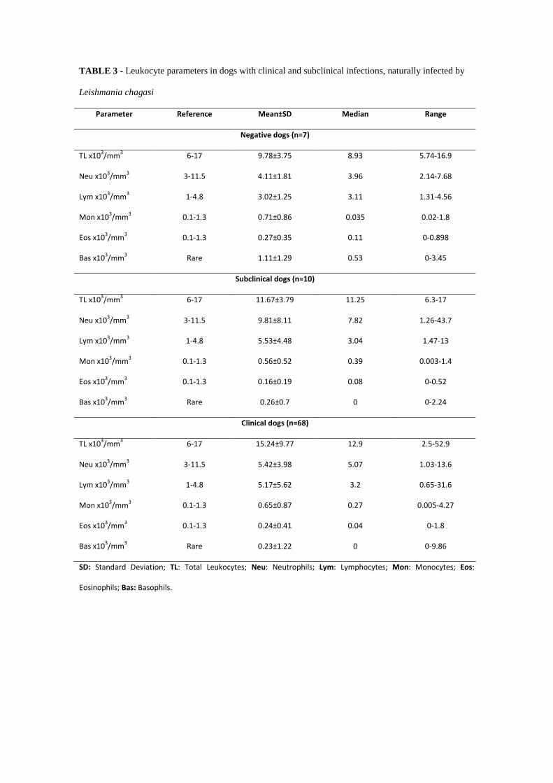
TABLE 3 - Leukocyte parameters in dogs with clinical and subclinical infections, naturally infected by
Leishmania chagasi
Parameter Reference
Mean±SD Median Range
Negative dogs (n=7)
TL x103/mm
3 6-17 9.78±3.75 8.93 5.74-16.9
Neu x103/mm
3 3-11.5 4.11±1.81 3.96 2.14-7.68
Lym x103/mm
3 1-4.8 3.02±1.25 3.11 1.31-4.56
Mon x103/mm
3 0.1-1.3 0.71±0.86 0.035 0.02-1.8
Eos x103/mm
3 0.1-1.3 0.27±0.35 0.11 0-0.898
Bas x103/mm
3 Rare 1.11±1.29 0.53 0-3.45
Subclinical dogs (n=10)
TL x103/mm
3 6-17 11.67±3.79 11.25 6.3-17
Neu x103/mm
3 3-11.5 9.81±8.11 7.82 1.26-43.7
Lym x103/mm
3 1-4.8 5.53±4.48 3.04 1.47-13
Mon x103/mm
3 0.1-1.3 0.56±0.52 0.39 0.003-1.4
Eos x103/mm
3 0.1-1.3 0.16±0.19 0.08 0-0.52
Bas x103/mm
3 Rare 0.26±0.7 0 0-2.24
Clinical dogs (n=68)
TL x103/mm
3 6-17 15.24±9.77 12.9 2.5-52.9
Neu x103/mm
3 3-11.5 5.42±3.98 5.07 1.03-13.6
Lym x103/mm
3 1-4.8 5.17±5.62 3.2 0.65-31.6
Mon x103/mm
3 0.1-1.3 0.65±0.87 0.27 0.005-4.27
Eos x103/mm
3 0.1-1.3 0.24±0.41 0.04 0-1.8
Bas x103/mm
3 Rare 0.23±1.22 0 0-9.86
SD: Standard Deviation; TL: Total Leukocytes; Neu: Neutrophils; Lym: Lymphocytes; Mon: Monocytes; Eos:
Eosinophils; Bas: Basophils.
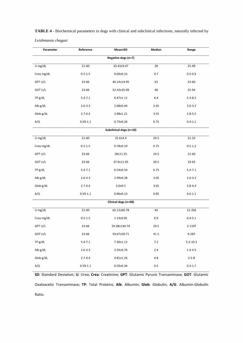
TABLE 4 - Biochemical parameters in dogs with clinical and subclinical infections, naturally infected by
Leishmania chagasi
Parameter Reference
Mean±SD Median Range
Negative dogs (n=7)
U mg/dL 21-60 33.43±9.47 28 25-49
Crea mg/dL 0.5-1.5 0.69±0.15 0.7 0.5-0.9
GPT U/L 23-66 40.14±14.95 43 23-60
GOT U/L 23-66 52.43±25.09 48 25-94
TP g/dL 5.4-7.1 6.47±1.11 6.4 5.3-8.2
Alb g/dL 2.6-3.3 2.68±0.44 2.65 2.6-3.3
Glob g/dL 2.7-4.4 3.98±1.21 3.55 2.8-5.5
A/G 0.59-1.1 0.73±0.26 0.75 0.4-1.1
Subclinical dogs (n=10)
U mg/dL 21-60 25.6±4.4 24.5 21-33
Crea mg/dL 0.5-1.5 0.78±0.19 0.75 0.5-1.2
GPT U/L 23-66 28±11.55 24.5 21-60
GOT U/L 23-66 37.9±11.95 39.5 19-65
TP g/dL 5.4-7.1 6.54±0.54 6.75 5.4-7.1
Alb g/dL 2.6-3.3 2.99±0.28 3.05 2.6-3.3
Glob g/dL 2.7-4.4 3.6±0.5 3.65 2.8-4.4
A/G 0.59-1.1 0.86±0.13 0.85 0.6-1.1
Clinical dogs (n=68)
U mg/dL 21-60 65.12±60.78 44 21-356
Crea mg/dL 0.5-1.5 1.14±0.81 0.9 0.4-5.1
GPT U/L 23-66 59.38±144.74 29.5 2-1107
GOT U/L 23-66 59.67±50.71 41.5 9-287
TP g/dL 5.4-7.1 7.36±1.12 7.2 5.2-10.3
Alb g/dL 2.6-3.3 2.54±0.76 2.4 1.4-4.5
Glob g/dL 2.7-4.4 4.81±1.26 4.8 2.5-8
A/G 0.59-1.1 0.59±0.34 0.5 0.3-1.7
SD: Standard Deviation; U: Urea; Crea: Creatinine; GPT: Glutamic Pyruvic Transaminase; GOT: Glutamic
Oxaloacetic Transaminase; TP: Total Proteins; Alb: Albumin; Glob: Globulin; A/G: Albumin-Globulin
Ratio.

TABLE 5 - P value of hematological, platelet, leukocyte, and biochemical assessments of clinical dogs
naturally infected by Leishmania chagasi, in relation to subclinical and negative dogs.
Hematological and platelet
parameters
p Leukocyte
parameter
p Biochemical parameter p
RBC x106/dL 0.173 TL x10
3/mm
3 0.192 U mg/dL 0.294
Hb g/dL 0.137 Neu x103/mm
3 0.053 Crea mg/dL 0.131
Ht % 0.082 Lym x103/mm
3 0.565 GPT U/L 0.746
MCV fL 0.602 Mon x103/mm
3 0.926 GOT U/L 0.378
MCH pg 0.175 Eos x103/mm
3 0.790 TP g/dL 0.018*
MCHC % 0.214 Bas x103/mm
3 0.175 Alb g/dL 0.176
RDW % 0.122 Glob g/dL 0.006**
Plt x103/mm
3 0.651 A/G 0.034*
RBC: Red Blood Cells; Hb: Hemoglobin; Ht: Hematocrit; MCV: Mean Corpuscular Volume; MCH: Mean
Corpuscular Hemoglobin; MCHC: Mean Corpuscular Hemoglobin Concentration; RDW: Red Cell
Distribution Width; Plt: Platelet; TL: Total Leukocytes; Neu: Neutrophils; Lym: Lymphocytes; Mon:
Monocytes; Eos: Eosinophils; Bas: Basophils; U: Urea; Crea: Creatinine; GPT: Glutamic Pyruvic
Transaminase; GOT: Glutamic Oxaloacetic Transaminase; TP: Total Proteins; Alb: Albumin; Glob:
Globulin; A/G: Albumin-Globulin Ratio.
* Represents differences at a significance level of 5%. ** Represents differences at a significance level of
1%.

CAPÍTULO 2
Perfil de anticorpos anti-Leishmania associados aos sinais clínicos na leishmaniose
visceral canina
Profile of anti-Leishmania antibodies related to clinical picture in canine visceral
leishmaniasis
Periódico: Research in Veterinary Science (Submetido em Julho de 2011 e Publicado
em Janeiro de 2012).

Profile of anti-Leishmania antibodies related to clinical picture in canine
visceral leishmaniasis
José Cláudio Carneiro de Freitas1*, Belarmino Eugênio Lopes Neto1, Cyntia Rafaelle
Amaral de Abreu1, Wendel Coura-Vital2, Samuel Leôncio Braga2, Alexandre Barbosa
Reis2, Diana Célia Sousa Nunes-Pinheiro1.
1Programa de Pós-Graduação em Ciências Veterinárias, Faculdade de Veterinária,
Universidade Estadual do Ceará (UECE), Avenida Paranjana, 1700, Campus do Itaperi,
Serrinha, CEP 60740-000, Fortaleza, CE. 2Laboratório de Imunopatologia, Núcleo de Pesquisas em Ciências Biológicas,
Universidade Federal de Ouro Preto (UFOP), Campus Universitário, Morro do
Cruzeiro, CEP 35400-000, Ouro Preto, MG.
* Corresponding author. Tel.: +55 85 31019859
E-mail address: [email protected] (J.C.C. Freitas)
Abstract
This research investigated the profile of anti-Leishmania antibodies in different
clinical forms of canine visceral leishmaniasis (CVL). Naturally infected dogs were
divided into two groups: subclinical dogs (SD, n=10) and clinical dogs (CD, n=68).
Non-infected dogs (ND, n=7) comprised the negative control group. The humoral
response was evaluated by the profile of total IgG, IgG1, IgG2, IgM, IgA and IgE,
determined by ELISA. Infected animals showed increased levels of total IgG, IgA and
IgE in addition to IgG1 and IgG2 in groups SD and CD, when compared with group
ND. Furthermore, it was observed that IgG2 and IgM were correlated with
symptomatology, while total IgG, IgG1 and IgA were negatively correlated and IgE
showed no correlation. It follows that serum levels of IgG2 anti-Leishmania are
correlated with typical clinical signs of disease, and the determination of specific anti-
Leishmania antibodies is an important tool in monitoring of clinical picture of CVL.
Keywords: Canine visceral leishmaniasis; Anti-Leishmania antibodies; Clinical picture;
Leishmania chagasi.

Introduction
The leishmaniasis are a complex of infectious diseases caused by different
species of protozoa of the genus Leishmania. The infection is transmitted by the bite of
infected insects of the genus Phlebotomus and Lutzomyia in the New and Old World,
respectively (Peters and Sacks, 2006). In Brazil, the Programme for the Control of
Visceral Leishmaniasis recommends early diagnosis and treatment of human cases,
vector control and the detection and euthanasia of seropositive dogs (Brasil, 2006).
Although humans can also act as reservoir of the agent and plays a role in the
transmission cycle, the dog is considered one of the most important links in the
epidemiological chain of leishmaniasis (Ribeiro, 2007). Studies conducted in São Paulo
showed a prevalence of visceral leishmaniasis up to 40% in the canine population
(Ikeda et al., 2003). However, the prevalence in endemic areas may reach higher levels,
as demonstrated by Freitas et al. (2010), through an epidemiological survey in
Fortaleza, whose highest and lowest prevalence was 80.2% and 64% respectively.
Dogs with clinical leishmaniasis (CD) are characterized by clinical signs and/or
clinical pathological abnormalities, with infection confirmed by specific tests, while
dogs with subclinical leishmaniasis (SD) are characterized by no clinical signs or
clinicopathological abnormalities, but with confirmed infection (Solano-Gallego et al.,
2009).
The onset of clinical signs in canine visceral leishmaniasis (CVL) involves a
number of factors, and these are associated with the animal's immune response
(Ciaramella and Corona, 2003). In CVL, the immune response mediated by Th1
lymphocytes, that secrete stimulatory cytokines (IFN-γ), activates macrophages
infected, effectively controlling the infection. It can also occur, in this case, of the host
remains asymptomatic. (Miranda et al., 2007). On the other hand, when the immune
response is mediated by Th2 lymphocytes, IL-4 secretors, there is a high production of
antibodies that are associated with severe clinical manifestations (Miranda et al., 2007).
It is noteworthy that when the immune response is mediated by regulatory T
lymphocytes, producing IL-10, Th1 is inhibited and the infection is worsen (Miyara and
Sakaguchi, 2007, Belkaid and Tarbell, 2009).
Studies report the role of antibodies in the CVL, relating the clinical picture with
the presence of different classes and subclasses of immunoglobulins involved in the
inflammatory response (Trotz-Williams and Gradoni, 2003; Almeida et al., 2005). The
soluble immune complexes are deposited in various organs and tissues such as kidneys,

blood vessels, joints, among others, favoring the appearance of various symptoms such
as epistaxis, polyuria and polydipsia, uveitis, conjunctivitis and episcleritis immune-
mediated, skin ulcers and tips of ears, hyperkeratosis and limping by poliartritre
(Ciaramella and Corona, 2003).
Given the great importance of the disease, this study aims to evaluate the profile
of anti-Leishmania antibodies in different clinical pictures of CVL with emphasis on
correlation with the clinical symptoms.
Material and Methods
Animals
Adult dogs (n = 85) were used, varying in age, weight and breeds (including
cross-breed). The seropositive dogs were captured by the Zoonosis Control Center of
Fortaleza (CCZ), as a CVL control measure. This study was approved by the Ethics
Committee for Animal Use of the State University of Ceará (CEUA/UECE), protocol
number 08622833-1.
Immunofluorescence Assay (IFA)
In all animals the immunofluorescence assay (IFA) was performed for canine
visceral leishmaniasis, being considered seropositive dogs the ones with titers above of
1:40, according to recommendations of the Ministry of Health of Brazil. Serologic
testing was performed at CCZ using Bio-Manguinhos (FIOCRUZ-RJ) kits, following
the manufacturer's recommendations.
Parasitological diagnosis
After anesthetizing the dog with Xylazine (2 mg/kg) and Ketamine (10 mg/kg)
bone marrow aspiration for making imprints on microscope slides was performed. This
material was fixed with methanol and stained with Panótipo fast dye. The stained
imprints were observed under an optical microscope, and samples where it was detected
the presence of amastigotes of Leishmania chagasi was investigated positive.
Clinical classification
All dogs were examined by observing the typical clinical signs of canine visceral
leishmaniasis as onychogryphosis, hepatosplenomegaly, cachexia, lymphadenopathy,
keratoconjunctivitis, skin ulcers, apathy, alopecia.

The dogs were divided into three groups, according to Solano-Gallego et al.,
2009. Negative dogs (ND = 7), which do not show clinical and laboratory alterations
(hematology and biochemistry) and negative for visceral leishmaniasis, by serology and
parasitology; subclinical dogs (SD = 10) which do not show clinical and laboratory
alterations and positive for Leishmania chagasi infection or clinical dogs (CD = 68)
which show clinical and laboratory alterations for routine testing and have infection
confirmed by serological and parasitological diagnosis.
Collection of blood samples
Blood samples were collected by jugular venipuncture with a sterile syringe on
dogs of different groups which were placed into a tube containing gel separation,
without anticoagulant to obtain serum. Sera samples were stored at -20 °C for further
test.
Immunoenzymatic reaction - ELISA
To determine the profile of anti-Leishmania antibodies, ELISA assay was
performed, using soluble antigen (MHOM/BR/1972/BH46) from promastigotes of L.
chagasi (SLA) from the axenic culture in LIT medium (Reis et al., 2006).
96-well microplates (MaxisorpTM Nunc International Nalgas, USA) were coated
with SLA at a concentration of 2 mg/well, overnight at 4 °C. After incubation, the plates
were washed four times with phosphate buffer solution (PBS) containing 0.05% Tween
20 and blocked for 45 min at 37 °C with 100 mL of fetal bovine serum (5%) in PBS per
cell. Then, serum samples were added at a dilution of 1:80 for IgG, IgG1, IgG2, IgM
and IgE and 1:40 for IgA. After this procedure, they were washed and added peroxidase
conjugate (Bethyl Laboratories Inc., Montgomery, TX, USA) previously diluted as
follows: anti-dog IgG1 (anti-heavy chain specific), 1:8000; IgM (anti-m chain specific),
1:1000; IgA (anti-a chain specific), 1:500; IgE (anti-e chain specific), 1:500, or anti-dog
IgG and IgG2 (both anti-heavy chain specific), 1:16000. After four washings (as
described above), the reaction was started by adding 100 mL of 0.1 M citrate solution
(pH 5.0) containing 0.03% α-phenylenediamine and 0.012% H2O2, followed by
incubation at 37 °C for 10 min. The reaction was stopped by adding 32 µL of H2SO4 2.5
M and the absorbance (492 nm) was measured with a plate reader-ELISA ELX800
(Biotek Instruments® VT, USA).

Statistical analysis
Statistical analysis was performed using the software GraphPad Prism 5.0. To
compare the absorbance values of anti-Leishmania antibodies among groups in different
clinical forms, analysis of variance (ANOVA) one-way was used, followed by Tukey
test. To identify the association between the profile of anti-Leishmania antibodies and
the clinical symptoms, we used the Spearman correlation test (r). In all cases the
differences were considered significant at P ≤ 0.05.
Results
The results of the evaluation of typical clinical signs of CVL were expressed in
percentage (%) and are shown in Table 1. The more frequent clinical signs were
cachexia (77.94%), keratoconjunctivitis (61.76%) and lymphadenopathy (55.88%), and
86.76% of the animals showed more than one typical clinical sign of CVL.
The profile of anti-Leishmania serum antibodies (total IgG, IgG1, IgG2, IgM,
IgA and IgE) from naturally infected dogs in relation to different clinical pictures are
shown in Fig. 1. The correlation coefficient values (r) between the two factors are
presented in Table 2.
Increase in levels of IgG, IgG1, IgG2, IgA and IgE anti-Leishmania in SD and
CD groups was observed compared to control group (ND). As for IgM, there was no
change in the response profile among the evaluated groups. When evaluating the
association between the profile of anti-Leishmania antibodies and the different clinical
pictures, it was observed that serum levels of IgG2 (r = 0.12) and IgM (r = 0.38)
showed positive correlation with the clinical signs (Table 2). It was also observed
negative correlation between serum levels of total IgG (r = -0.28), IgG1 (r = -0.13) and
IgA (r = -0.62) and the clinical signs. Moreover, IgE levels showed no correlation
between SD and CD (r = 0.0), i.e., in these animals the increases of serum IgE are not
correlated with the clinical changes evident in infected animals.
Discussion
It has been reported that the appearance of clinical signs characteristic of
visceral leishmaniasis is mainly as consequences of the host immune response
associated with deposition of immune complexes soluble in different tissues (Quinnell
et al., 2003). In this direction, it could be interesting to assess the serum levels of anti-

Leishmania antibodies verifying the classes and subclasses of immunoglobulins
involved, and their association with clinical signs shown by infected animal.
In our study, we found that dogs naturally infected by Leishmania chagasi (CD
and SD) showed results of total IgG, IgG1, IgG2, IgA and IgE anti-Leishmania
significantly increased when compared to ND (Fig. 1). Furthermore, we observed a
negative correlation between total IgG, IgG1 and IgA and symptomatology (r = -0.28, r
= -0.13 and r = -0.62, respectively), demonstrating that, the increase in these
immunoglobulins is not associated with clinical signs of visceral leishmaniasis (Table
2).
Almeida et al. (2005) found that the titers of IgG anti-Leishmania in
symptomatic dogs increased significantly when compared to asymptomatic and the
control group. This fact was also found by Vercammen et al. (2002) that linked the
increased levels of total IgG with progression from onset of symptoms.
In our study, the values of IgG1 anti-Leishmania showed a negative correlation
coefficient with the clinical condition of the animals (r = -0.13), and also their average
serum levels decreased in animals showing clinical signs. These data corroborate the
findings of Vercammen et al., (2002) and Cordeiro-da-Silva et al., (2003). With respect
to IgG2 subclass, it happened differently, because the average serum absorbance values
are almost the same as in all infected animals, not differing between CD and SD.
However, these values are correlated with typical clinical signs of visceral leishmaniasis
(r = 0.12), a fact which is mainly due to the findings of the CD group.
It has been reported that the clinical signs are directly related to IgG1/IgG2
relationship, where the titers of the subclass IgG2 are strongly correlated with the
symptoms of the animals. In this case, the titers of the subclass IgG1 also show
increased, however with lower values than those found in IgG2. (Solano-Gallego et al.
2001; Leandro et al., 2001; Almeida et al., 2005).
Reis et al. (2006) found that IgG1 anti-Leishmania has a negative correlation
with the clinical status of animals with visceral leishmaniasis, with their values
diminished with clinical outcome, demonstrating association with the maintenance of
the chronic and asymptomatic disease. With respect to IgG2, the same authors found
that the values did not differ significantly between the infected animals, and only
differed from the uninfected control group (Reis et al., 2006). These findings are in
agreement with those obtained by other authors previously (Vercammen et al., 2002;
Cordeiro-da-Silva et al., 2003).

In the present study, serum levels of IgM, anti-Leishmania from naturally
infected dogs (SD and CD), remained with no significant differences compared to the
control group (ND). However, with respect to the association with the symptomatology,
there was a positive correlation coefficient between the groups and the clinical picture (r
= 0.38). Yet, according to the one proposed by Reis et al. (2006), serum levels of IgM,
in the L. chagasi infection, remain high until the chronic phase of disease. It is
noteworthy, that this immunoglobulin is strongly associated with acute forms of
parasitic diseases and that it does not present significant correlation with the clinical
picture of CVL. Despite the above, little is known about the profile of IgM in CVL,
because it is easily inactivated by reagents used in the main techniques of serology
(Schallig et al., 2002), making it difficult to detect more accurate findings.
In relation to IgA anti-Leishmania, in our study, infected dogs showed negative
correlation with the onset of the clinical picture of CVL (r = -0.62), despite serum IgA
levels were significantly increased (P = 0.0025) when compared to ND. Few studies
emphasize the importance of IgA in the CVL. However, Reis et al. (2006) demonstrated
a positive correlation of patterns of immunoglobulin-A with the clinical status of
naturally infected animals. However, these data differ from our results. Whilst there are
few data in literature, Day (2007) in their review reported that the levels of IgA anti-
Leishmania were increased in the mucosa of infected dogs showing symptomatology,
suggesting a source of aggravation of the infectious process.
It is noteworthy that the infected dogs by Leishmania chagasi develop immune
response mediated by Th1 lymphocytes, they may be capable of preventing the spread
of the parasite to the mucosal surface and, consequently, produce lower levels of
specific IgA, with no differentiation with the clinical progression of CVL (Rodriguez-
Cortes et al., 2007). Glomerulonephritis is a main clinical finding in the CVL, and the
main triggering event of this abnormality has been attributed to the accumulation and
deposition of immune complexes mediated by IgA in renal glomeruli (Nieto et al.,
1992). Moreover, in post-mortem analysis, it was found that this clinical disorder is the
leading cause of death of infected animals (Feitosa et al., 2000).
Regarding the levels of IgE anti-Leishmania, the results differed significantly
between the infected animals (CD and SD) compared to non-infected (P = 0.0103).
Still, when we evaluated the profile of IgE in the ND group, it was observed high
absorbance values in some animals, which may represent false-negative results, by the
diagnostic technique used, with possible evolution to a subclinical canine disease in

these animals. These observations were also suggested by Almeida et al. (2005), who
associated the IgE levels with the development of symptomatic cases of CVL. In this
case, the predominant immune response is mediated by Th2 lymphocytes, which induce
a decrease in the synthesis of IFN-γ, with increased expression of IL-4 and IL-10, with
data obtained by measuring the levels of mRNA (Quinnell et al., 2001).
In our study there was no correlation between the titers of IgE anti-Leishmania
with symptomatology (r = 0.00), similar titers were detected in groups SD and CD.
From these data we can consider that the appearance of symptoms is due to action of
other immunoglobulins, and other elements of the immune response that were not
objects of study. However works carried out by Iniesta et al. (2005) and Reis et al.
(2006) observed a strong correlation between the titers of IgE and the clinical status of
animals.
Conclusion
Our data demonstrate that dogs naturally infected with Leishmania chagasi
present high titers of IgE, IgA and IgG2 anti-Leishmania and indicate that serum levels
of IgG2 anti-Leishmania are correlated with typical clinical signs of disease, but serum
levels of IgE anti-Leishmania do not correlate with the disease progression. Further
studies are needed to finally demonstrate that the determination of specific anti-
Leishmania antibodies are an important tool to predict the clinical course of the disease
as suggested by this study.
Acknowledgments
The authors would like to express their appreciation to the Fundação Cearense
de Apoio ao Desenvolvimento Científico e Tecnológico (FUNCAP) for the financial
help granted to the first author, which provided subsidies for the implementation of the
project.
References
Almeida, M.A.O., Jesus, E.E.V., Sousa-Atta, M.L.B., Alves, L.C., Berne, M.E.A., Atta,
A.M., 2005. Antileishmanial antibody profile in dogs naturally infected with
Leishmania chagasi. Veterinary Immunology and Immunopathology 106, 151-158.

Belkaid, Y., Tarbell, K., 2009. Regulatory T cells in the control of host-microorganism
interactions. Annual Reviews of Immunology 27, 551-589.
Brasil, M.S., 2006. Manual de Vigilância e Controle da Leishmaniose Visceral. 1st ed.
Editora MS, Brasília, pp.11-18.
Ciaramella, P., Corona, M., 2003. Canine leishmaniasis: clinical and diagnostic aspects.
VetLearn. 25, 358-368.
Cordeiro da Silva, A., Cardoso, L., Araújo, N., Castro, H., Tomas, A., Rodrigues, M.,
Cabral, M., Vergnes, B., Sereno, D., Ouaissi, A., 2003. Identification of antibodies to
Leishmania silent information regulatory 2 (SIR2) protein homologue during canine
natural infections: pathological implications. Immunology Letters 86, 155-162.
Day, M.J., 2007. Immunoglobulin G subclass distribution in canine leishmaniasis: A
review and analysis of pitfalls in interpretation. Veterinary Parasitology 147, 2-8.
Feitosa, M.M., Ikeda, F.A., Luvizotto, M.C., Perri, S.H.V., 2000. Aspectos clínicos de
cães com leishmaniose visceral no município de Araçatuba – São Paulo (Brasil). Clínica
Veterinária 28, 36-44.
Freitas, J.C.C., Nunes-Pinheiro, D.C.S., Abreu, C.R.A., 2010. Geographical variation in
clinical signs and prevalence of Leishmania sp. infection among dogs in Fortaleza,
Ceará State, Brazil. Acta Scientiae Veterinariae 38, 293-297.
Ikeda, F.A., Luvizotto, M.C.R., Goçalves, M.E., Feitosa, M.M., Ciarlini, P.C., Lima,
V.M.F., 2003. Perfil hematológico de cães naturalmente infectados por Leishmania
chagasi no município de Araçatuba, São Paulo (Brasil): um estudo retrospectivo de 191
casos. Clínica Veterinária 47, 42-48.
Iniesta, L., Gallego, M., Portus, M., 2005. Immunoglobulin G and E responses in
various stages of canine leishmaniosis. Veterinary Immunology and Immunopathology
103, 77-81.

Leandro, C., Santos-Gomes, G.M., Campino, L., Romão, P., Cortes, S., Rolao, N.,
Gomes-Pereira, S., Rica Capela, M.J., Abranches, P., 2001. Cell mediated immunity
and specific IgG1 and IgG2 antibody response in natural and experimental canine
leishmaniosis. Veterinary Immunology and Immunopathology 79, 273-284.
Miranda, S., Martorell, S., Costa, M., Ferrer, L., Ramis, A., 2007. Characterization of
circulating lymphocyte subpopulation in canine leishmaniasis throughout treatment with
antimonials and allopurinol. Veterinary Parasitology 144, 251-260.
Miyara,M., Sakaguchi, S., 2007. Natural regulatory T cells: mechanisms of suppression.
Trends in Molecular Medicine 13, 108-116.
Nieto, C.G., Navarrete, I., Habela, M.A., Serrano, F., Redondo, E., 1992. Pathological
changes in kidneys of dogs with natural Leishmania infections. Veterinary Parasitology
45, 33-47.
Peters, N., Sacks, D., 2006. Immune privilege in sites of chronic infection: Leishmania
and regulatory T cells. Immunological Reviews 213, 159-179.
Quinnell, R.J., Courtenay, O., Shaw, M.A., Day, M.J., Garcez, L.M., Dye, C., Kaye,
P.M., 2001. Tissue cytokine responses in canine visceral leishmaniasis. Journal of
Infectious Disease 183, 1421-1424.
Quinnell, R.J., Courtenay, O., Garcez, L.M., Kaye, P.M., Shaw, M.A., Dye, C., Day,
M.J., 2003. IgG subclass responses in a longitudinal study of canine visceral
leishmaniasis. Veterinary Immunology and Immunopathology 91, 161-168.
Reis, A.B., Teixeira-Carvalho, A., Vale, A.M., Marques, M.J., Giunchetti, R.C.,
Mayrink, W., Guerra, L.L., Andrade, R.A., Corrêa-Oliveira, R., Martins-Filho, O.A.,
2006. Isotype patterns of immunoglobulins: Hallmarks for clinical status and tissue
parasite density in brazilian dogs naturally infected by Leishmania (Leishmania)
chagasi. Veterinary Immunology and Immunopathology 112, 102-116.
Ribeiro, V.M., 2007. Leishmaniose visceral canina: aspectos de tratamento e controle.
Clínica Veterinária 71, 66-76.
Rodriguez-Cortes, A., Fernandez-Bellon, H., Ramis, A., Ferrer, L., Alberola, J., Solano-
Gallego, L., 2007. Leishmania-specific isotype levels and their relationship with
specific cell-mediated immunity parameters in canine leishmaniasis. Veterinary
Immunology and Immunopathology 116, 190-198.
Schallig, H.D.F.H., Schoone, G.J., Beijer, E.G.M., Kroon, C.C.M., Ozbel, Y., Ozensoy,
S., Da Silva, E.S., Cardoso, L.M., Da Silva, E.D., 2002. Development of a fast
agglutination screening test (FAST) for detection of anti-Leishmania antibodies in dogs.
Veterinary Parasitology 109, 1-8.
Solano-Gallego, L., Riera, C., Roura, X., Iniesta, L., Gallego, M., Valladares, J.E., Fisa,
R., Castillejo, S., Alberola, J., Ferrer, L., Arboix, M., Portus, M., 2001. Leishmania
infantum-specific IgG, IgG1 and IgG2 antibody responses and healthy and ill dogs from
endemic areas. Evolution in the course of infection and after treatment. Veterinary
Parasitology 96, 265-276.
Solano-Gallego, L., Koutinas, A., Miró, G., Cardoso, L., Pennisi, M.G., Ferrer, L.,
Bourdeau, P., Oliva, G., Baneth, G., 2009. Directions for diagnosis, clinical staging,
treatment and prevention of canine leishmaniosis. Veterinary Parasitology 165, 1-18.
Trotz-Williams, L., Gradoni, L., 2003. Disease risks for the travelling pet
Leishmaniasis, In Practice 25, 190-197.
Vercammen, F., Fernandez-Perez, F.J., Del Amo, C., Alunda, J.M., 2002. Follow-up of
Leishmania infantum naturally infected dogs treated with allupurinol:
immunofluorescence antibody test, ELISA and Western-blot. Acta Tropica 84, 175-181.
Table 1: Clinical signs observed in dogs, in group CD, naturally infected by Leishmania
chagasi.
Clinical Sign N %
Onychogryphosis 23 33.82
Hepatosplenomegaly 31 45.59
Cachexia 53 77.94
Lymphadenopathy 38 55.88
Keratoconjunctivitis 42 61.76
Skin Ulcers 24 35.29
Apathy 19 27.94
Alopecia 21 30.88
More than one clinical sign 59 86.76
Table 2: P and r values of anti-Leishmania antibodies profiles. Where P compares
differences in absorbance of immunoglobulins in different clinical forms (SD and CD)
with the negative control group (ND) of naturally infected dogs by Leishmania chagasi.
r value determines the correlation coefficient between immunoglobulins profiles and the
clinical signs (CD) of naturally infected dogs by Leishmania spp.
Immunoglobulin P value
ND x SD ND x CD SD x CD
r value
total IgG 0,009 0,007 0,12 -0.28
IgG1 0,02 0,04 0,08 -0.13
IgG2 0,009 0,008 0,11 0.12
IgM 0,08 0,11 0,09 0.38
IgA 0,009 0,008 0,09 -0.62
IgE 0,04 0,03 0,07 0.00

Figure 1: Anti-Leishmania antibodies profiles in dogs naturally infected by Leishmania
chagasi and showing different clinical forms. SD (subclinical dogs), CD (clinical dogs)
and ND (negative dogs). *Represents difference with a significance level of 5% and
**represents difference with a significance level of 1%.
.IgM
CN CS CC0.0
0.2
0.4
0.6
0.8
IgG1
CN CS CC0.0
0.2
0.4
0.6*
*
IgA
CN CS CC0.0
0.2
0.4
0.6
0.8
**
**
IgG2
CN CS CC0.0
0.1
0.2
0.3
0.4 **
**
IgE
CN CS CC0.0
0.2
0.4
0.6
0.8
**
Op
tica
l D
en
sity
Clinical Signs
Total IgG
ND SD CD0.0
0.1
0.2
0.3
0.4
**
**
ND SD CD
ND SD CDND SD CD
ND SD CDND SD CD

CAPÍTULO 3
Histopatologia de órgãos linfóides de cães naturalmente infectados por Leishmania
chagasi em diferentes formas clínicas
Histopathology of lymphoid organs of dogs naturally infected by Leishmania chagasi in
different clinical forms
(Artigo em Elaboração)

Histopatologia de órgãos linfóides de cães naturalmente infectados por
Leishmania chagasi em diferentes formas clínicas
Histopathology of lymphoid organs of dogs naturally infected by Leishmania chagasi in
different clinical forms
José Cláudio Carneiro de Freitas1, Francisco Valdeci de Almeida Ferreira2, Diana Célia
Sousa Nunes-Pinheiro1
1Programa de Pós-Graduação em Ciências Veterinárias, Faculdade de Veterinária,
Universidade Estadual do Ceará (UECE), Avenida Paranjana, 1700, Campus do Itaperi,
Serrinha, CEP 60740-000, Fortaleza, CE. 2Laboratório de Patologia, Centro de Ciências da Saúde, Universidade Federal do Ceará
(UFC), Rua Alexandre Baraúna, 949, Rodolfo Teófilo, CEP 60430-161, Fortaleza, CE.
Resumo
A leishmaniose visceral (LV) é uma zoonose com diferentes manifestações
clínicas tendo sido relatado comprometimento de diferentes órgãos. O objetivo do
presente trabalho foi avaliar as principais alterações histológicas em fragmentos de
órgãos de cães naturalmente infectados por L. chagasi. Utilizaram-se 85 cães adultos,
ambos os sexos, peso e idade variados, oriundos do Centro de Controle de Zoonoses de
Fortaleza, selecionados pela Reação de Imunofluorescência Indireta (RIFI), sendo
considerados positivos os animais com títulos de RIFI ≥ 1:40 e pelo exame
parasitológico das formas amastigotas de L. chagasi em esfregaços de medula óssea. Os
cães foram agrupados conforme os sinais clínicos associados à doença: negativos
(CN=7); subclínicos (CS=10) e clínicos (CC=68). Após o procedimento de eutanásia,
todos os animais foram pesados, e em seguida o baço e o fígado foram dissecados para
mensuração dos pesos relativos. Fragmentos de baço, fígado, linfonodo poplíteo (LNP)
e pele foram coletados e submetidos aos procedimentos de histologia clássica, corados
pela técnica de hematoxilina e eosina (H&E) e examinados à microscopia óptica para
avaliação dos parâmetros histológicos. As mesmas amostras foram submetidas à análise
imunohistoquímica para detecção de amastigotas de L. chagasi. O protocolo
experimental foi aprovado pelo CEUA/UECE, protocolo n° 08622833-1. Foram
observados aumento do peso relativo do baço e do fígado de cães dos grupos CS e CC
(P<0,05). No baço, foram observadas discretas hipoplasia de polpas vermelha e branca
e espessamento de cápsula, moderadas hipertrofia de polpas vermelha e branca e intensa
congestão esplênica no grupo CS, enquanto discreto espessamento de cápsula e
hipertrofia de polpas vermelha e branca, intensa congestão e moderadas fibrose
subcapsular e hipoplasia de polpa vermelha e branca esplênica foram encontradas no
grupo CC. A avaliação histológica do fígado demonstrou moderado infiltrado
inflamatório peri-portal, hipertrofia/hiperplasia de células de Küpffer e congestão,
discreto espessamento de cápsula e granulomas intralobulares em cães do grupo CS,
enquanto moderados infiltrado inflamatório peri-portal, hipertrofia/hiperplasia de
células de Küpffer, congestão, espessamento de cápsula e granulomas intralobulares
foram encontrados em cães do grupo CC. No LN a avaliação histológica apresentou-se
com discreta inflamação capsular, congestão, hemossiderose e hipertrofia/hiperplasia de
nódulos linfáticos e moderada hipertrofia/hiperplasia de cortical e medular em cães do
grupo CS, enquanto discreta inflamação capsular, congestão, hemossiderose e
hipertrofia/hiperplasia de nódulos linfáticos e moderada hipertrofia/hiperplasia de
cortical e medular foram encontrados em cães do grupo CC. A principal alteração
observada na pele foi infiltrado inflamatório histiolinfocitário moderado em CS e
discreto em CC. A imunohistoquímica comprovou a presença de amastigotas L. chagasi
nos diferentes órgãos avaliados, sendo a maior densidade observada na polpa branca
esplênica. Conclui-se que órgãos e tecidos de cães naturalmente infectados por L.
chagasi apresentam alterações histológicas compatíveis com resposta imune-
inflamatória, demonstrando o comprometimento funcional dos mesmos.
Palavras-Chave: leishmaniose visceral canina, órgãos linfóides, histologia,
imunohistoquímica.
Introdução
As leishmanioses representam um complexo de zoonoses que acometem o
homem quando ele entra em contato com o ciclo de transmissão do parasito (Brasil,
2006). São classificadas entre as doenças mais relevantes e emergentes em todo mundo,
e o Brasil está entre os países da América Latina que apresenta maior número de casos
humanos, cerca de 90% dos casos anuais registrados (Monteiro et al., 2005).
Embora o homem também possa atuar como reservatório do agente e tenha
participação no ciclo de transmissão, o cão é considerado um dos elos mais importantes
na cadeia epidemiológica da leishmaniose visceral (LV) (Ribeiro, 2007). A LV é

transmitida através da picada de insetos denominados flebotomíneos, principalmente as
espécies Lutzomyia longipalpis e L. cruzi, que veiculam as formas promastigotas
infectantes. O principal agente etiológico da LV no Brasil é a Leishmania (Leishmania)
chagasi (Brasil, 2006, Camargo-Neves et al., 2006).
A patogenia da leishmaniose visceral canina (LVC) envolve vários fatores, e um
fator decisivo na evolução da doença está associado à resposta imunológica que o
animal desenvolve contra o parasito (Engwerda et al., 2004, Sanchez et al., 2004,
Miranda et al., 2007). A resposta imunológica do tipo celular tem sido associada à
proteção e cura (Barbieri, 2006), enquanto que a resposta imunológica do tipo humoral,
mediada por anticorpos (Reis et al., 2006) vem sendo associada a processos
inflamatórios que provocam danos teciduais que repercutem em sinais clínicos
associados à LVC (Ciaramella et al., 1997, Ciaramella e Corona, 2003, Trotz-Williams
e Gradoni, 2003).
A infecção pode se apresentar de forma clínica, em que cães apresentam sinais
clínicos e/ou alterações clínico-patológicas características com confirmação de
Leishmania chagasi, ou subclínica, em que cães não apresentam alterações clínico-
patológicas, mas têm a presença de L. chagasi confirmada por testes diagnósticos
(Solano-Gallego et al., 2009).
Apesar de a LVC ser caracterizada como uma severa doença sistêmica, poucos
estudos foram realizados descrevendo as principais alterações histopatológicas
encontradas nos diversos órgãos do hospedeiro afetado pelo parasito (Giunchetti et al.,
2008). Portanto, o objetivo do presente trabalho foi avaliar as alterações histológicas do
baço, fígado, linfonodo e pele, de cães naturalmente infectados nas diferentes formas
clínicas da leishmaniose visceral canina.
Material e Métodos
Animais
Foram utilizados cães (n=85) adultos, de idade e peso variados e sem raças
definidas. Os cães eram provenientes do Centro de Controle de Zoonoses de Fortaleza
(CCZ). Este trabalho foi aprovado previamente pelo Comitê de Ética para Uso de
Animais da Universidade Estadual do Ceará (CEUA/UECE), protocolo SPU n°
08622833-1.
Reação de Imunofluorescência Indireta (RIFI)

Todos os animais foram submetidos à Reação de Imunofluorescência Indireta
(RIFI), para pesquisa de anticorpos anti-Leishmania chagasi sendo considerados
soropositivos os cães com títulos superiores a 1:40. O teste sorológico foi realizado no
CCZ empregando kits Bio-Manguinhos (FIOCRUZ-RJ), seguindo as recomendações do
fabricante.
Diagnóstico Parasitológico
Todos os animais foram submetidos ao exame parasitológico em medula óssea.
Os cães foram anestesiados com Xilazina (2mg/Kg) e Ketamina (10mg/Kg) e em
seguida foram realizadas as punções de medula óssea para a confecção de imprints em
lâminas de microscopia e posterior avaliação. Este material foi fixado em metanol e
corado com corante rápido (Panótipo®). Os imprints corados foram observados ao
microscópio óptico, sendo consideradas positivas as amostras que foram detectadas a
presença de amastigotas de L. chagasi.
Classificação Clínica e Eutanásia
Todos os cães foram examinados observando-se os sinais clínicos característicos
da LVC, como onicogrifose, apatia, caquexia, ceratoconjutivite, hepatoesplenomegalia,
linfoadenopatia, úlceras de pele, hipertermia, alopecia, ulceração de mucosas,
descamação, eczema, vômitos e enterorragia e submetidos à coleta de sangue para
análises laboratoriais de rotina (hemograma completo e avaliação bioquímica sérica).
Os cães foram divididos em três grupos, de acordo com Solano-Gallego et al.,
2009. Cães negativos (CN = 7), animais que não apresentam alterações clínicas e
laboratoriais (hematologia e bioquímica) e são negativos para LVC, pelo diagnóstico
sorológico e parasitológico; cães subclínicos (CS = 10), animais que não apresentam
alterações clínicas e laboratoriais e são positivos para infecção por L. chagasi e cães
clínicos (CC = 68), animais que apresentam alterações clínicas e laboratoriais, pelos
testes de rotina, e tem a infecção confirmada pela sorologia e parasitologia.
Após a avaliação clínica os animais foram submetidos ao procedimento de
eutanásia, através de uma injeção intravenosa de 0,3 ml de T-61® (5g de Iodeto de
Mebezônio; 20g de Embutrimida; 0,5g de Cloridrato de Tetracaína; excipiente q.s.p.
100 ml) por kilograma de peso vivo. Os animais foram pesados e, em seguida, foram
realizadas as coletas dos órgãos e fragmentos de tecidos.
Peso Relativo dos Órgãos
O baço e o fígado foram dissecados e, em seguida, pesados para o cálculo dos
pesos relativo dos órgãos. Hepatomegalia e esplenomegalia foram avaliadas em relação
ao controle negativo.
Análise Histopatológica
Fragmentos de baço, fígado, linfonodo poplíteo e pele (orelha) foram coletados e
colocados em frascos contendo solução tamponada de formalina a 10%, pH 7,0, e foram
mantidos em temperatura ambiente e, posteriormente, foram submetidos aos
procedimentos de histologia clássica. Os cortes histológicos foram corados por
hematoxilina e eosina (H&E), e examinados à microscopia óptica para avaliação dos
parâmetros histológicos: infiltrado inflamatório, hipertrofia, hiperplasia/hipoplasia,
congestão, fibrose, granuloma, entre outros, aos quais foram atribuídos os escores de
ausente, discreto, moderado e intenso.
Análise Imunohistoquímica para detecção de amastigotas de L. chagasi
Cortes histológicos do baço, fígado, linfonodo poplíteo e pele foram montados
em lâminas silanizadas para serem submetidos aos procedimentos de
imunohistoquímica. As lâminas foram hidratadas e incubadas em uma solução de
peróxido de hidrogênio a 4% e PBS 0,01M, pH 7,2, seguida pela incubação em soro
caprino normal (diluído em 1:100). Soro heterólogo de coelhos experimentalmente
infectados com L. chagasi (diluído em 1:200 em PBS 0,01M), cedido pelo Instituto de
Medicina Tropical de São Paulo, foi utilizado como anticorpo primário. As lâminas
foram então incubadas por 22h a 4ºC em câmara úmida. Depois foi feita lavagem com
PBS, e as lâminas foram novamente incubadas em solução biotinizada, e, em seguida,
lavadas novamente em PBS e incubadas com o complexo estreptoavidina-peroxidase
por 20 min, a temperatura ambiente. A reação foi realizada com a adição de 0,024% de
diaminobenzidina e 0,16% de peróxido de hidrogênio. Finalmente, as lâminas foram
desidratadas, limpas e coradas em H&E e montadas com lamínula, para visualização em
microscopia ótica de formas amastigotas de L. chagasi.
Análise Estatística
As principais alterações histológicas dos parâmetros avaliados nos diferentes
grupos estudados foram expressas em porcentagem. Para comparar os valores do peso
relativo dos órgãos entre os grupos foi utilizada a análise de variância (ANOVA), one-
way acompanhado do teste Tukey (p<0,05).
Resultados
Peso Relativo do Baço e Fígado
Os resultados do peso relativo dos órgãos, baço e fígado, estão demonstrados na
Figura 1. Os pesos relativos do baço e do fígado dos cães pertencentes aos grupos CS e
CC estavam aumentados em relação ao grupo CN (P<0,05), contudo CS e CC não
diferiram significativamente entre si.
Avaliação Histológica
Os resultados da avaliação histológica do baço, fígado, linfonodo poplíteo e pele
dos animais pertencentes aos grupos CN, CS e CC foram expressos em porcentagem e
estão apresentados nas Tabelas 1, 2, 3 e 4.
As mais freqüentes alterações histológicas esplênicas encontradas foram:
espessamento de cápsula (10%) e hipoplasia de polpa vermelha e polpa branca, de
caráter discreto (40%) (Figura 2c e 2d); moderada hipertrofia de polpa vermelha e polpa
branca (20%) e intensa congestão (30%) (Figura 2a) no grupo CS (Tabela 1). No grupo
CC, também foi observado discreto espessamento de cápsula (4,4%) (Figura 2b) e
hipertrofia de polpa vermelha e polpa branca (13,2%); moderada hipoplasia de polpa
vermelha e polpa branca (51,5%) (Figura 2c e 2d) e fibrose subcapsular (2,9%) (Figura
2b), além de congestão intensa (19,1%). Em CN observou-se discreta congestão
(85,7%) (Tabela 1).
No fígado foi observado infiltrado inflamatório histiolinfocitário peri-portal
(40%) (Figura 3a), hipertrofia/hiperplasia de células de Küpffer (40%) (Figura 3b),
congestão (50%) moderados (Figura 3a), além de espessamento de cápsula (40%)
(Figura 3c) e granulomas intralobulares (20%) discretos (Figura 3d), no grupo CS
(Tabela 2). Com relação ao grupo CC, observou-se infiltrado inflamatório peri-portal
(33,8%) (Figura 3a), espessamento de cápsula (10,3%) (Figura 3c),
hipertrofia/hiperplasia de células de Küpffer (48,5%) (Figura 3b), congestão (33,8%)
(Figura 3a) e granulomas intralobulares (10,3%) (Figura 3d), todos com o caráter
moderado. Em CN verificou-se um discreto infiltrado inflamatório peri-portal (14,3%),
hipertrofia/hiperplasia de células de Küpffer (42,8%) e congestão (28,6%) (Tabela 2).

No linfonodo poplíteo, o grupo CS apresentou como principais alterações,
inflamação capsular (40%) (Figura 4a), congestão (40%) (Figura 4b), hemossiderose
(40%) (Figura 4c) e hipertrofia/hiperplasia de nódulos linfáticos (40%) (Figura 4d)
discretas, hipertrofia/hiperplasia de cortical (40%) (Figura 4e) e hipertrofia/hiperplasia
de medular (60%) (Figura 4f) moderadas (Tabela 3). Já no grupo CC, as principais
alterações foram: inflamação capsular (41,2%) (Figura 4a), congestão (32,3%) (Figura
4b), hemossiderose (33,8%) (Figura 4c) e hipertrofia/hiperplasia de nódulos linfáticos
(45,6%) (Figura 4d) também de forma discreta, hipertrofia/hiperplasia de cortical (50%)
(Figura 4e) e hipertrofia/hiperplasia de medular (33,8%) (Figura 4f) com caráter
moderado. No grupo CN foi observada inflamação capsular (57,2%),
hipertrofia/hiperplasia de cortical (28,6%), congestão (28,6%) e hemossiderose (42,8%),
todos com caráter discreto (Tabela 3).
Na pele foi observado infiltrado inflamatório linfocitário moderado em CS
(40%) e discreto em CC (35,3%) (Figura 5a) (Tabela 4). Vale ressaltar que, 32,4% dos
cães pertencentes à CC apresentaram infiltrado inflamatório de caráter intenso. Outros
achados também foram visualizados, como a presença de discreta congestão
extravascular (40% e 29,4%) (Figura 5b), angiogênese (20% e 14,7%) e edema (10% e
11,8%) em CS e CC, respectivamente. Em CN foi observado discreto infiltrado
inflamatório em 14,3% dos cães.
Detecção por Imunohistoquímica de amastigotas de L. chagasi
Os resultados da imunohistoquímica para pesquisa de L. chagasi em órgãos de
cães naturalmente infectados estão apresentados na Figura 6 (a-d).
No baço foi observada uma maior quantidade de formas amastigotas de L.
chagasi, principalmente, na região de polpa branca (Figura 6a). No fígado, foi
observada uma menor quantidade de amastigotas de L. chagasi, contudo, foram
detectadas em maior número no interior das células de Küpffer, principalmente na
região peri-portal (Figura 6b). No LNP e na pele poucos parasitos foram detectados,
sendo encontrados, preferencialmente, nos nódulos linfáticos e próximos aos folículos
pilosos (Figuras 6c e 6d).
Discussão
A leishmaniose visceral canina (LVC) é uma doença de caráter crônico e de
grande importância epidemiológica, sendo o cão o principal reservatório urbano do

parasito (Ashford, 2000). Atualmente, estudos relacionados à patogênese da doença
canina têm se mostrado importantes para esclarecimentos dos sinais e sintomas clínicos
apresentados pelos animais infectados (França-Silva et al., 2003; Alves e Bevilacqua,
2004; Palatinik e Lavor, 2004; Giunchetti, et al., 2006).
Apesar das importantes alterações macroscópicas observadas nos órgãos e
tecidos em resposta a infecção por L. chagasi, poucos estudos foram realizados sobre as
alterações histopatológicas na LVC (Keenan et al., 1984; Tafuri et al., 2001; Lima et al.,
2004; Giunchetti et al., 2008).
As principais alterações macroscópicas visualizadas em cães infectados por L.
chagasi são similares as que são normalmente descritas na infecção humana, dentre elas
incluindo hepatoesplenomegalia e linfadenopatia (Keenan et al., 1984; Krauspenhar,
2007), confirmando os dados observados pelo nosso trabalho.
Devido ao elevado parasitismo tecidual do baço de cães naturalmente infectados
por L. chagasi, esse órgão foi escolhido como o melhor modelo experimental para
obtenção de material biológico e para o diagnóstico da LVC (Barrouin-Melo et al.,
2004, 2006). Em nosso estudo a maior densidade parasitária visualizada por
imunohistoquímica foi observada em amostras esplênicas (Figura 6a), o que coorobora
com os achados da literatura. A infecção esplênica se caracteriza por uma fase aguda
inicial, ausência da proliferação de formas amastigotas de L. chagasi, desenvolvimento
de esplenomegalia e persistência tecidual do parasito, no modelo murino (Engwerda e
Kaye, 2000; Engwerda et al., 2004). A resposta do hospedeiro ao parasitismo esplênico
pode explicar a esplenomegalia observada pelo peso relativo do órgão (Fig. 1).
No presente estudo, na avaliação microscópica do baço foi observada congestão
intensa (Figura 2a), tanto em CS quanto em CC. Resultados semelhantes foram
descritos por Tasca et al., (2009), em que cães infectados apresentavam tecido esplênico
hemorrágico e vasos dilatados e com número de arteríolas aumentados. Hipoplasia de
polpa vermelha e polpa branca em animais pertencentes aos grupos CS e CC foi
observada (Figuras 2c e 2d), entretanto estes resultados diferem dos relatados por Tasca
et al., (2009) e Daneshvar et al., (2009), que observaram hiperplasia de polpa branca
em animais oligossintomáticos e sintomáticos. Esse caráter hipoplásico esplênico pode
ser explicado pela cronicidade da doença, onde em trabalho realizado por Tafuri et al.,
(2001), foi relatada como sintomatologia característica em um cão, com infecção
crônica, uma reação inflamatória difusa, desde a região capsular, subcapsular, trabecular
e polpa branca. Além destes, Corbett et al., (1992) e Keenan et al., (1984) relataram, em

hamsters e cães experimentalmente infectados com L. donovani, uma depleção de áreas
T-dependentes e da bainha periarteriolar da polpa branca. Outros achados como
espessamento de cápsula e fibrose subcapsular (Figura 2b) foram observados, o que
também confirma o caráter crônico da doença.
No fígado, as principais alterações histológicas observadas foram: infiltrado
inflamatório peri-portal, hipertrofia/hiperplasia de células de Küpffer, congestão
moderados, em CS e CC, além de espessamento de cápsula e granulomas intralobulares
discretos, em CS e moderado em CC (Figuras 3a, 3b, 3c e 3d). Os mesmos achados
foram descritos por Murray (2001), em modelo experimental murino e por Tafuri et al.,
(1996), Sanchez et al., (2004) e Daneshvar et al., (2009) em cães experimentalmente e
naturalmente infectados por L. donovani e L. chagasi. Giunchetti et al., (2007) relataram
hipertrofia/hiperplasia de células de Küpffer em cães sintomáticos, quando comparados
aos cães assintomáticos.
A infecção hepática, causada por L. chagasi, tem como uma de suas
características a capacidade de auto-resolução, através de uma resposta imunológica
mediada por linfócitos Th1, predominando as células mononucleares e envolvendo
células de Küpffer, macrófagos e com consequente formação de granulomas, após
visceralização do parasito por via linfática ou sanguínea (Rousseau et al., 2001, Tafuri
et al., 2001, Kaye et al., 2004). Por outro lado, tem sido atribuído que macrófagos
ativados possam agir promovendo a perpetuação do parasito os quais são facilmente
encontrados no interior de macrófagos dos órgãos afetados (Tafuri et al., 2004).
Sendo os linfonodos considerados um dos mais importantes órgãos linfóides
envolvidos na interface parasito-hospedeiro durante os estágios de infecção por L.
chagasi, a resposta imune celular e molecular ainda é pouco elucidada. Giunchetti et al.
(2008) sugeriram que as principais alterações nos linfonodos durante a LVC podem
refletir não somente sobre o perfil da resposta imune do hospedeiro, mas também sobre
a intensidade da carga parasitária, numa infecção causada por L. chagasi.
A linfadenomegalia generalizada é um achado clínico frequentemente descrito
na LVC, contudo, os aspectos histológicos que envolvem esta condição patológica ainda
são pouco investigados (Tafuri et al., 2001; Lima et al., 2004). Nosso trabalho verificou
que linfonodos poplíteos apresentavam hipertrofia e hiperplasia de cortical e medular,
inflamação capsular, congestão, hemossiderose e hipertrofia e hiperplasia de nódulos
linfáticos (Figuras 4a, 4b, 4c, 4d, 4e e 4f). As alterações de hipertrofia e hiperplasia de
cortical e medular em linfonodos são consideradas as mais relevantes, pois sugerem que

a condição imunopatológica dos linfonodos é uma característica comum de infecção
crônica difusa, independente da forma clínica da doença (Lima et al., 2004). Inflamação
capsular também foi observada em trabalho realizado por Tafuri et al. (1996). Contudo,
Lima et al. (2004) e Xavier et al. (2006) relataram ausência de hipertrofia e hiperplasia
de nódulos linfáticos, estes dados vão de encontro aos apresentados em nosso estudo.
Na LVC, a pele tem fundamental importância clínica, pois é a porta de entrada
do parasito, através da picada do inseto vetor, além de facilitar a identificação dos
animais doentes. Entretanto, os animais assintomáticos apresentam parasitismo
semelhante ao dos animais sintomáticos, tornando-os potencialmente transmissores
“ocultos” (Ciaramella et al., 1997; Solano-Gallego et al., 2004).
Tem sido descrito que o intenso parasitismo cutâneo relatado em cães infectados
representa o principal achado que classifica os cães como o principal hospedeiro
reservatório doméstico para a infecção humana (Deane e Deane, 1962; Giunchetti et al.,
2006). Na análise histológica de fragmentos de pele não lesionada obtidas de orelha, foi
observada dermatite crônica, caracterizada por um infiltrado linfocitário em CS e CC
(Figura 5a). Achados semelhantes foram encontrados por Travi et al. (2001), Xavier et
al. (2006) e Giunchetti et al. (2006).
Em cães infectados por Leishmania chagasi, as áreas de pele onde mais se
evidenciam lesões e infiltrados inflamatórios são as orelhas. Esse tropismo desigual não
se dá tão somente pelo fato de ser o local de preferência pelos insetos transmissores,
mas também, pela elevada ocorrência de lesões causadas por ectoparasitos e traumas
causados por reações pruriginosas. Dessa forma, um maior número de células, como os
macrófagos, migra para essa região (Tafuri et al., 2004).
Conclusão
Pode-se concluir que os achados histopatológicos são característicos de resposta
imune-inflamatória e que os animais infectados com L. chagasi apresentam alterações
morfológicas e estruturais em órgãos alvo, comprometendo a sua funcionalidade.
Conflitos de Interesse
Nenhum dos autores tem relações pessoais ou financeiras com outras pessoas ou
organizações que possam, inapropriadamente, influenciar no conteúdo do artigo.

Agradecimentos
À Fundação Cearense de Apoio ao Desenvolvimento Científico e Tecnológico
(FUNCAP) pela concessão da bolsa de doutorado.
Ao Dr Heitor de Franco Andrade do Instituto de Medicina Tropical de São Paulo
pela alíquota de anticorpo anti-L. chagasi.
Referências Bibliográficas
Alves, W.A., Bevilacqua, P.D., 2004. Quality of diagnosis of canine visceral
leishmaniasis in epidemiological surveys: an epidemic in Belo Horizonte, MG, Brazil,
1993-1997. Cad. Saude Publica. 20, 259-265.
Ashford, R.W., 2000. The leishmaniases as emerging and reemerging zoonoses. Int. J.
Parasitol. 30, 1269-1281.
Barbieri, C.L., 2006. Immunology of canine leishmaniasis. Parasite Immunol. 28, 329-
337.
Barrouin-Melo, S.M., Larangeira, D.F., Trigo, J., Aguiar, P.H.P., Santos, W.L.C.,
Carvalho, L.P., 2004. Comparison between splenic and lymph node aspirations as
sampling methods for the parasitological detection of Leishmania chagasi infection in
dogs. Mem. Inst. Oswaldo Cruz. 99, 195-197.
Barrouin-Melo, S.M., Larangeira, D.F., Andrade Filho, F.A., Trigo, J., Julião, F.S.,
Franke, C.R., Aguiar, P.H.P., Santos, W.L.C., Carvalho, L.P., 2006. Can spleen
aspirations be safely used for the parasitological diagnosis of canine visceral
leishmniasis? A study on asymptomatic and polysymptomatic animals. Vet. J. 171, 331-
339.
Brasil, M.S., 2006. Manual de Vigilância e Controle da Leishmaniose Visceral, Editora
MS, 120 pp.
Camargo-Neves, V.L.F., Glasser, C.M., Cruz, L.L., Almeida, R.G., 2006. Manual de
Vigilância e Controle da Leishmaniose Visceral Americana do Estado de São Paulo,
São Paulo, Secretaria de Estado da Saúde, 158 pp.
Ciaramella, P., Oliva, G., De Luna, L., Gradoni, R., Ambrosio, L., Cortese, A., Scalone,
A., Persechino, A., 1997. A retrospective clinical study of canine leishmaniasis in 150
naturally infected by Leishmania infantum. Vet. Rec. 141, 539-543.
Ciaramella, P., Corona, M., 2003. Canine leishmaniasis: clinical and diagnostic aspects.
VetLearn. 25, 358-368.
Corbett, C.E.P, Paes, R.A.P., Laurenti, M.D., Andrade, J., Duarte, M.I.S., 1992.
Histopathology of lymphoid organs in experimental leishmaniasis, Inst. J. Exp. Pathol.
73, 417–433.
Daneshvar, H., Molaei, M.M., Afshar, R.M., Kamiabi, H., Burchmore, R., Hagan, P.,
Phillips, R.S., 2009. Gentamicin-attenuated Leishmania infantum: A clinicopathological
study in dogs. Vet. Immunol. Immunopathol. 129, 28-35.
Deane, L.M., Deane, M.P., 1962. Visceral leishmaniasis in Brazil. Geographical
distribution and transmission. Rev. Inst. Med. Trop. São Paulo. 4, 149–212
Engwerda, C.R., Kaye, P.M., 2000. Organ-specific immune responses associated with
infectious disease. Immunol.Today. 21, 73-78.
Engwerda, C.R., Ato, M., Kaye, P.M., 2004. Macrophages, pathology and parasite
persistence in experimental visceral leishmaniasis. Trends Parasitol. 20, 524–530.
França-Silva, J.C., Costa, R.T., Siqueira, A.M., Machado-Coelho, G.L.L., Costa, C.A.,
Mayrink, W., Vieira, E. P., Costa, J.S., Genaro, O., Nascimento, E., 2003.
Epidemiology of canine visceral leishmaniosis in the endemic area of Montes Claros
Municipality, Minas Gerais State, Brazil. Vet Parasitol. 111, 161-173.
Giunchetti, R. C., Mayrink, W., Genaro, O., Carneiro, C.M., Corrêa-Oliveira, R.,
Martins-Filho, O.A., Marques, M.J., Tafuri, W.L., Reis, A.B., 2006. Relationship
between canine visceral leishmaniosis and the Leishmania (Leishmania) chagasi burden
in dermal inflamatory foci. J. Comp. Pathol. 135, 100-107.

Giunchetti, R.C., Mayrink, W., Carneiro, C.M., Corrêa-Oliveira, R., Martins-Filho,
O.A., Marques, M.J., Tafuri, W.L., Reis, A.B., 2007. Histopathological and
immunohistochemical investigations of the hepatic compartment associated with
parasitism and serum biochemical changes in canine visceral leishmaniasis. Res. Vet.
Sci., 84, 269-277.
Giunchetti, R.C., Martins-Filho, O.A., Carneiro, C.M., Mayrink, W., Marques, M.J.,
Tafuri, W.L., Correa-Oliveira, R., Reis, A.B., 2008. Histopathology, parasite density
and cell phenotypes of the popliteal lymph node in canine visceral leishmanisis. Vet.
Immunol. Immunopathol. 121, 23-33.
Kaye, P.M., Svensson, M., Ato, M., Maroof, A., Polley, R., Stager, S., Zubairi, S.,
Engwerda, C.R., 2004. The immunopathology of experimental visceral leishmaniasis.
Immunol. Rev. 201, 239–253.
Keenan, C.M., Hendricks L.D., Lightner, L., Johnson, A.J., 1984. Visceral
leishmaniasis in a german sheeperdog. I. infeccion, clinical disease, and clinical
pathology. Vet. Pathology. 21, 74-79.
Krauspenhar, C., 2007. Leishmaniose visceral em um canino de Cruz Alta, Rio Grande
do Sul, Brasil. Ciênc. Rural. 37, 907-910.
Lima, W.G., Michalick, M.S., Melo, M.N., Tafuri, W.L., 2004. Canine visceral
leishmaniasis: a histopathological study of lymph nodes. Acta Trop. 92, 43–53.
Miranda, S., Martorell, S., Costa, M., Ferrer, L., Ramis, A., 2007. Characterization of
circulating lymphocyte subpopulation in canine leishmaniasis throughout treatment with
antimonials and allopurinol. Vet. Parasitol. 144, 251-260.
Monteiro, E.M., Silva, J.C.F., Costa, R.T., Costa, D.C., Barata, R.A., Paula, E.V.,
Machado-Coelho, G.L.L., Rocha, M.F., Fortes-Dias, C.L., Dias, E.S., 2005.
Leishmaniose visceral: estudo de flebotomíneos e infecção canina em Monte Claros,
Minas Gerais. Rev. Soc. Bras. Med. Trop. 38(2), 147-152.

Murray, H.W., 2001. Tissue granuloma structure-function in experimental visceral
leishmaniasis. Int. J. Exp. Pathol. 82, 249–267.
Palatnik, M., Lavor, C.C., 2004. Improving methods for epidemiological control of
canine visceral leishmaniasis based on a mathematical model. Impact on the incidence
of the canine and human disease. An. Acad. Brás. Cienc. 76, 583-593.
Reis, A.B., Teixeira-Carvalho, A., Vale, A.M., Marques, M.J., Giunchetti, R.C.,
Mayrink, W., Guerra, L.L., Andrade, R.A., Correa-Oliveira, R., Martins-Filho, A.O.,
2006. Isotype patterns of immunoglobulins: hallmarks for clinical status and tissue
parasite density in brazilian dogs naturally infected by Leishmania (Leishmania)
chagasi. Vet. Immunol. Immunopathol. 112, 102-116.
Ribeiro, V.M., 2007. Leishmaniose visceral canina: aspectos de tratamento e controle.
Clín. Vet. 71, 66-76.
Rousseau, D., Demartino, S., Anjuere, F., Ferrua, B., Fragaki, K., Lê Fichoux, Y.,
Kubar, J., 2001. Sustained parasite burden in the spleen of Leishmania infantum-
infected BALB/c mice is accompanied by expression of MCP-1 transcripts and lack of
protection against challenge. Eur. Cytokine Netw. 12, 340–347.
Sanchez, M.A., Diaz, N.L., Zerpa, O., Negron, E., Convit, J., Tapia, F.J., 2004. Organ-
specific immunity in canine visceral leishmaniasis: analysis of symptomatic and
asymptomatic dogs naturally infected with Leishmania chagasi. Am. J. Trop. Med. Hyg.
70, 618–624.
Solano-Gallego, L., Fernandez-Bellon, H., Morell, P., Fondevila, D., Alberola, J.,
Ramis, A., Ferrer, L., 2004. Histological and immunohistochemical study of clinically
normal skin of Leishmania infantum -infected dogs. J. Comp. Pathol. 130, 7-12.
Solano-Gallego, L., Koutinas, A.F., Miró, G., Cardoso, L., Pennisi, M.G., Ferrer, L.,
Bourdeau, P., Oliva, G., Baneth, G., 2009. Directions for the diagnosis, clinical staging,
treatment and prevention of canine leishmaniosis. Vet. Parasitol. 165, 1-18.

Tafuri, W.L., Tafuri, W.L., Barbosa, A.J.A., Michalick, M.S., Genaro, O., Franca-Silva,
J.C., Mayrink, W., Nascimento, E., 1996. Histopathology and immunocytochemical
study of type 3 and type 4 complement receptors in the liver and spleen of dogs
naturally and experimentally infected with Leishmania (Leishmania) chagasi. Rev. Inst.
Med. Trop. 38, 81–89.
Tafuri, W.L., Oliveira, M.R., Melo, M.N., 2001. Canine visceral leishmaniosis: a
remarkable histopathological picture of one case reported from Brazil. Vet. Parasitol.
96, 203-212.
Tafuri, W.L., Santos, R.L., Arantes, R.M.E., Gonçalves, R., Melo, M.N., Michalik,
M.S.M., Tafuri, W.L., 2004. An alternative immunohistochemical method for detecting
Leishmania amastigotes in paraffin-embedded canine tissues. J. Immunol. Methods.
292, 17–23.
Tasca, K.Y., Buzetti, W.A.S., Tenório, M.S., Paulan, S.C., Lima, F.L., Queiroz,
N.M.G.P., Machado, R.Z., Oliveira, T.M.F.S., Neves, M.F., Noronha-Jr, A.C.F., Assis,
J., 2009. Exames parasitológicos, imunohistoquímicos e histopatológicos para detecção
de Leishmania chagasi em tecidos esplênicos de cães com leishmaniose visceral. Rev.
Bras. Parasitol. Vet. 18, 27-33.
Travi, B.L., Tabares, C.J., Cadena, H., Ferro, C., Osorio, Y., 2001. Canine visceral
leishmaniasis in Colombia: relationship between clinical and parasitologic status and
infectivity for sand flies. Am. J. Trop. Med. Hyg. 64, 119-124.
Trotz-Williams, L., Gradoni, L., 2003. Disease risks for the travelling pet
Leishmaniasis. In Practice. 25, 190-197.
Xavier, S.C., Chiarelli, I.M., Lima, W.G., Gonçalves, R., Tafuri, W.L., 2006. Canine
visceral leishmaniasis: a remarkable histopathological picture of one asymptomatic
animal reported from Belo Horizonte, Minas Gerais, Brazil. Arq. Bras. Med. Vet.
Zootec. 12, 994-1000.

Tabela 1: Parâmetros histológicos do baço de cães naturalmente infectados com L.
chagasi em diferentes formas clínicas.
Grau da Lesão
Ausente Discreto Moderado Intenso
Parâmetros
n(%) n(%) n(%) n(%)
Espessamento de Cápsula 7 (100) 0 (0) 0 (0) 0 (0)
Hipertrofia de Polpa Vermelha e Polpa
Branca
6 (85,7) 1 (14,3) 0 (0) 0 (0)
Hipoplasia de Polpa Vermelha e Polpa
Branca
6 (85,7) 1 (14,3) 0 (0) 0 (0)
Fibrose Subcapsular 6 (85,7) 1 (14,3) 0 (0) 0 (0)
Cãe
s N
egat
ivos
Congestão 1 (14,3) 6 (85,7) 0 (0) 0 (0)
Espessamento de Cápsula 9 (90) 1 (10) 0 (0) 0 (0)
Hipertrofia de Polpa Vermelha e Polpa
Branca
7 (70) 1 (10) 2 (20) 0 (0)
Hipoplasia de Polpa Vermelha e Polpa
Branca
3 (30) 4 (40) 3 (30) 0 (0)
Fibrose Subcapsular 10 (100) 0 (0) 0 (0) 0 (0)
Cãe
s S
ubcl
ínic
os
Congestão 3 (30) 1 (10) 3 (30) 3 (30)
Espessamento de Cápsula 65 (95,6) 3 (4,4) 0 (0) 0 (0)
Hipertrofia de Polpa Vermelha e Polpa
Branca
53 (80) 9 (13,2) 6 (8,8) 0 (0)
Hipoplasia de Polpa Vermelha e Polpa
Branca
27 (39,7) 6 (8,8) 35 (51,5) 0 (0)
Fibrose Subcapsular 64 (94,2) 2 (2,9) 2 (2,9) 0 (0)
Cãe
s C
línic
os
Congestão 29 (42,7) 13 (19,1) 13 (19,1) 13 (19,1)

Tabela 2: Parâmetros histológicos do fígado de cães naturalmente infectados com L.
chagasi em diferentes formas clínicas.
Grau da Lesão
Ausente Discreto Moderado Intenso
Parâmetros
n(%) n(%) n(%) n(%)
Infiltrado Inflamatório Peri-Portal 6 (85,7) 1 (14,3) 0 (0) 0 (0)
Espessamento de Cápsula 7 (100) 0 (0) 0 (0) 0 (0)
Hipertrofia/Hiperplasia de Células de
Küpffer
4 (57,2) 3 (42,8) 0 (0) 0 (0)
Congestão 5 (71,4) 2 (28,6) 0 (0) 0 (0) Cãe
s N
egat
ivos
Granulomas Intralobulares 7 (100) 0 (0) 0 (0) 0 (0)
Infiltrado Inflamatório Peri-Portal 4 (40) 2 (20) 4 (40) 0 (0)
Espessamento de Cápsula 6 (60) 4 (40) 0 (0) 0 (0)
Hipertrofia/Hiperplasia de Células de
Küpffer
3 (30) 3 (30) 4 (40) 0 (0)
Congestão 0 (0) 1 (10) 5 (50) 4 (40) Cãe
s S
ubcl
ínic
os
Granulomas Intralobulares 7 (70) 2 (20) 1 (10) 0 (0)
Infiltrado Inflamatório Peri-Portal 15 (22) 18 (26,5) 23 (33,8) 12 (17,6)
Espessamento de Cápsula 54 (79,4) 7 (10,3) 7 (10,3) 0 (0)
Hipertrofia/Hiperplasia de Células de
Küpffer
6 (8,8) 29 (42,6) 33 (48,5) 0 (0)
Congestão 19 (27,9) 12 (17,6) 23 (33,8) 14 (20,6) Cãe
s C
línic
os
Granulomas Intralobulares 57 (83,8) 3 (4,4) 7 (10,3) 1 (1,5)

Tabela 3: Parâmetros histológicos do linfonodo poplíteo de cães naturalmente
infectados com L. chagasi em diferentes formas clínicas.
Grau da Lesão
Ausente Discreto Moderado Intenso
Parâmetros
n(%) n(%) n(%) n(%)
Inflamação Capsular 3 (42,8) 4 (57,2) 0 (0) 0 (0)
Hipertrofia/Hiperplasia de Cortical 5 (71,4) 2 (28,6) 0 (0) 0 (0)
Hipertrofia/Hiperplasia de Medular 7 (100) 0 (0) 0 (0) 0 (0)
Congestão 5 (71,4) 2 (28,6) 0 (0) 0 (0)
Hemossiderose 4 (57,2) 3 (42,8) 0 (0) 0 (0)
Cãe
s N
egat
ivos
Hipertrofia/Hiperplasia de Nódulos
Linfáticos
7 (100) 0 (0) 0 (0) 0 (0)
Inflamação Capsular 3 (30) 4 (40) 3 (30) 0 (0)
Hipertrofia/Hiperplasia de Cortical 3 (30) 2 (20) 4 (40) 1 (10)
Hipertrofia/Hiperplasia de Medular 3 (30) 0 (0) 6 (60) 1 (10)
Congestão 6 (60) 4 (40) 0 (0) 0 (0)
Hemossiderose 4 (40) 4 (40) 0 (0) 2 (20)
Cãe
s S
ubcl
ínic
os
Hipertrofia/Hiperplasia de Nódulos
Linfáticos
3 (30) 4 (40) 3 (30) 0 (0)
Inflamação Capsular 11 (16,2) 28 (41,2) 27 (39,7) 2 (2,9)
Hipertrofia/Hiperplasia de Cortical 7 (10,3) 20 (29,4) 34 (50) 7 (10,3)
Hipertrofia/Hiperplasia de Medular 19 (27,9) 17 (25) 23 (33,8) 9 (13,2)
Congestão 37 (54,4) 22 (32,3) 9 (13,2) 0 (0)
Hemossiderose 32 (47,1) 23 (33,8) 12 (17,6) 1 (1,5)
Cãe
s C
línic
os
Hipertrofia/Hiperplasia de Nódulos
Linfáticos
12 (17,6) 31 (45,6) 23 (33,8) 2 (2,9)
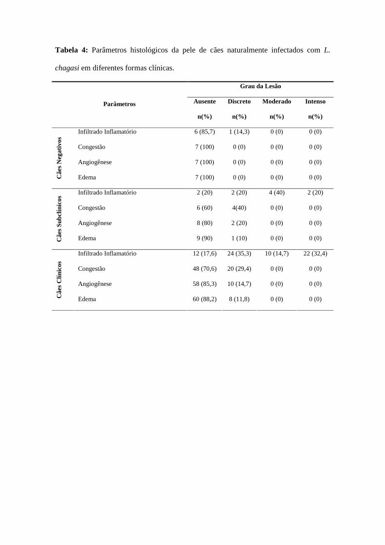
Tabela 4: Parâmetros histológicos da pele de cães naturalmente infectados com L.
chagasi em diferentes formas clínicas.
Grau da Lesão
Ausente Discreto Moderado Intenso
Parâmetros
n(%) n(%) n(%) n(%)
Infiltrado Inflamatório 6 (85,7) 1 (14,3) 0 (0) 0 (0)
Congestão 7 (100) 0 (0) 0 (0) 0 (0)
Angiogênese 7 (100) 0 (0) 0 (0) 0 (0)
Cãe
s N
egat
ivos
Edema 7 (100) 0 (0) 0 (0) 0 (0)
Infiltrado Inflamatório 2 (20) 2 (20) 4 (40) 2 (20)
Congestão 6 (60) 4(40) 0 (0) 0 (0)
Angiogênese 8 (80) 2 (20) 0 (0) 0 (0)
Cãe
s S
ubcl
ínic
os
Edema 9 (90) 1 (10) 0 (0) 0 (0)
Infiltrado Inflamatório 12 (17,6) 24 (35,3) 10 (14,7) 22 (32,4)
Congestão 48 (70,6) 20 (29,4) 0 (0) 0 (0)
Angiogênese 58 (85,3) 10 (14,7) 0 (0) 0 (0)
Cãe
s C
línic
os
Edema 60 (88,2) 8 (11,8) 0 (0) 0 (0)

Figura 1: Peso relativo do baço e do fígado de cães naturalmente infectados com
Leishmania chagasi em diferentes formas clínicas. CN (cães negativos), CS (cães
subclínicos) e CC (cães clínicos) * Representa diferença com nível de significância de
5%.

Figura 2: Alterações histológicas no baço de cães naturalmente infectados com
Leishmania chagasi. (A) – Congestão e hipertrofia de polpa vermelha e polpa branca
(100x). (B) – Espessamento de cápsula e fibrose subcapsular (400x). (C) – Hipoplasia
de polpa vermelha (1000x). (D) – Hipoplasia de polpa branca (1000x).
A
B
C
D
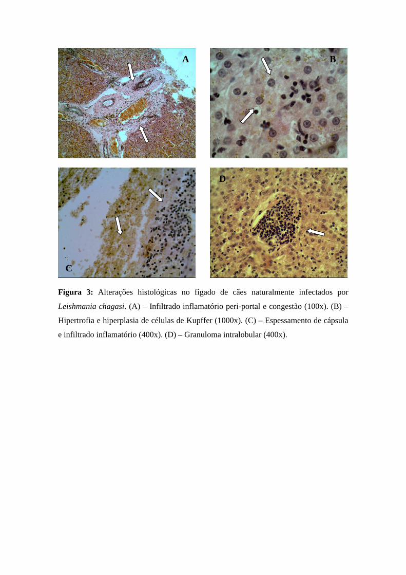
Figura 3: Alterações histológicas no fígado de cães naturalmente infectados por
Leishmania chagasi. (A) – Infiltrado inflamatório peri-portal e congestão (100x). (B) –
Hipertrofia e hiperplasia de células de Kupffer (1000x). (C) – Espessamento de cápsula
e infiltrado inflamatório (400x). (D) – Granuloma intralobular (400x).
A B
C
D

Figura 4: Alterações histológicas no linfonodo poplíteo de cães naturalmente
infectados por Leishmania chagasi. (A) – Inflamação capsular (400x). (B) – Congestão
(400x). (C) – Hemossiderose (400x). (D) – Hipertrofia/Hiperplasia de nódulos linfáticos
(100x). (E) – Hipertrofia/Hiperplasia de cortical (1000x). (F) - Hipertrofia/Hiperplasia
de medular (1000x).
A B
C D
E F

Figura 5: Alterações histológicas na pele de cães naturalmente infectados com
Leishmania chagasi. (A) – Infiltrado inflamatório linfocitário (1000x). (B) – Congestão
extravascular e infiltrado inflamatório (400x).
A B

Figura 6: Amastigotas de Leishmania chagasi detectadas pela técnica de
imunohistoquímica em cães naturalmente infectados. (A) – Inúmeras amastigotas de
Leishmania chagasi na região de polpa branca esplênica (400x). (B) – Amastigotas de
Leishmania chagasi na região peri-portal hepática (400x). (C) – Amastigotas de
Leishmania chagasi no interior de nódulo linfático (1000x) e (D) – Amastigota de
Leishmania chagasi próximo ao folículo piloso (1000x).
A
B
C
D

Conclusões
___________________________________

CONCLUSÕES GERAIS
A partir da avaliação dos resultados obtidos no presente estudo, podemos concluir que:
Cães naturalmente infectados por Leishmania chagasi apresentam alterações
laboratoriais como anemia, uremia, hiperproteinemia e hipergamaglobulinemia. Esses
achados são importantes marcadores, associados aos sinais clínicos característicos da
doença, para auxiliar no diagnóstico da leishmaniose visceral canina. Vale ressaltar, no
entanto, que os mesmos, de forma isolada, não são conclusivos para o diagnóstico,
necessitando de testes mais específicos.
Cães naturalmente infectados por Leishmania chagasi demonstraram níveis séricos
aumentados de IgE, IgA e IgG anti-Leishmania, com valores aumentados de IgG2, os
quais apresentaram correlação com o aparecimento de sintomatologia nos animais.
Estes dados reforçam que a avaliação do perfil de imunoglobulinas anti-Leishmania é
um importante biomarcador no auxiliar no diagnóstico da leishmaniose visceral canina.
Os achados histopatológicos são característicos de resposta imune-inflamatória e que os
animais infectados com L. chagasi apresentam alterações morfológicas e estruturais em
órgãos alvo, comprometendo a sua funcionalidade.
Portanto, pode-se concluir que a resposta imunológica dos cães estudados é compatível
com alta produção de anticorpos específicos relacionada aos diferentes estágios clínicos
da doença e que apresentam comprometimento de vários órgãos e alterações
laboratoriais possivelmente associados a esta resposta.

Perspectivas
___________________________________

PERSPECTIVAS
A leishmaniose visceral se caracteriza por desencadear várias alterações
fisiológicas, e os cães domésticos são apontados como os principais hospedeiros
reservatórios. O município de Fortaleza é uma região endêmica para a forma visceral da
doença, com um grande número de casos caninos e humanos registrados, inclusive com
um grande número de óbitos nos últimos anos. O principal enfoque do Programa de
Controle da Leishmaniose Visceral (PCLV) está baseado na detecção e remoção dos
cães sorologicamente positivos para a doença, onde o padrão-ouro é a Reação de
Imunofluorescência Indireta, mesmo sabendo que o diagnóstico sorológico apresenta
uma série de limitações, inclusive com um grande número de reações-cruzadas com
doenças comumente encontradas nos cães. Baseado nisso, um grande número de cães
vem sendo eutanasiados, fato que causa um impacto social negativo sem, contudo, o
número de casos humanos da doença não vem sendo reduzido.
O presente trabalho gerou subsídios para a realização de um grande estudo
epidemiológico e populacional, envolvendo o mapeamento e caracterização do inseto
vetor, o estudo clínico dos cães naturalmente infectados, o isolamento e caracterização
das amostras de Leishmania e a avaliação demográfica nas diferentes regionais do
município de Fortaleza, de forma controlada.
Espera-se, com esse estudo, caracterizar a leishmaniose visceral canina no
município de Fortaleza.

Referências Bibliográficas
___________________________________

REFERÊNCIAS BIBLIOGRÁFICAS
ABREU-SILVA, A. L.; LIMA, T. B.; MACEDO, A. A.; MORAES-JÚNIOR, F. J.;
DIAS, E. L.; BATISTA, Z. S. Soroprevalência, aspectos clínicos e bioquímicos da
infecção por Leishmania em cães naturalmente infectados e fauna de flebotomíneos em
uma área endêmica na ilha de São Luís, Maranhão, Brasil. Revista Brasileira de
Parasitologia Veterinária, v. 17, p. 197-203, 2008.
AGA, E.; KATSCHINSKI, D. M.; van ZANDBERGEN, G.; LAUFS, H.; HANSEN,
B.; MULLER, K.; SOLBACH, W.; LASKAY, T. Inhibition of the spontaneous
apoptosis of neutrophil granulocytes by the intracellular parasite Leishmania major.
Journal of Immunology, v. 169, p. 898-905, 2002.
ALBUQUERQUE, A. R.; ALVES, L. C.; FAUSTINO, M. A. G.; BRITO, F. L. C.; SÁ,
F. B.; PINA, F.; GOMES, Y. M.; ALVEZ, R. Ocorrência da leishmaniose visceral
canina no município de Amaraji, Estado de Pernambuco, Brasil. In: Congresso
Nacional de Saúde Pública Veterinária. 2005. Guarapari, Espírito Santo, 27-30.
ALENCAR, J. E. Calazar canino: Contribuição para o estudo da epidemiologia do
calazar no Brasil. 1959. Tese - Universidade Federal do Ceará, Fortaleza. 1959.
ALEXANDER, J.; BRYSON K. T helper (Th)1/Th2 and Leishmania: paradox rather
than paradigm. Immunology Letters. v. 99, p. 17–23, 2005.
ALEXANDER, J.; SATOSKAR, A. R.; RUSSELL, D. G. Leishmania species: models
of intracellular parasitism. Journal of Cell Science, v. 112, p. 2993-3002, 1999.
ALEXENDER, J.; RUSSELL, D. G. The interaction of Leishmania spp species with
macrophages. Advances in Parasitology, v. 31, p. 175-254, 1992.
ALLI, R. S.; KHAR, A. Interleukin-12 secreted by mature dendritic cells mediates
activation of NK cell function. FEBS Letter, v. 559, p. 71-76, 2004.

ALMEIDA, M. A. O.; JESUS, E. E. V.; SOUSA-ATTA, M. L. B.; ALVES, L. C.;
BERNE, M. E. A.; ATTA, A. M. Antileishmanial antibody profile in dogs naturally
infected with Leishmania chagasi. Veterinary Immunology and Immunopathology,
v. 106, p. 151-158, 2005.
ALVES, W. A.; BEVILACQUA, P. D. Quality of diagnosis of canine visceral
leishmaniasis in epidemiological surveys: an epidemic in Belo Horizonte, MG, Brazil,
1993-1997. Cadernos de Saude Publica, v. 20, p. 259-265, 2004.
ALVES, C. F.; AMORIM, I. F. G.; MOURA, E. P.; RIBEIRO, R. R.; ALVES, C. F.;
MICHALICK, M. S.; KALAPOTHAKIS, E.; BRUNA-ROMERO, O.; TAFURI, W. L.;
TEIXEIRA, M. M.; MELO, M. N. Expression of IFN-γ, TNF-α, IL-10 and TGF-β in
lymph nodes associates with parasite load and clinical form of disease in dogs naturally
infected with Leishmania (Leishmania) chagasi. Veterinary Immunology and
Immunopathology, v. 128, p. 349-358, 2009.
AMUSATEGUI, I.; SAINZ, A.; RODRIGUEZ, F.; TESOURO, M. A. Distribution and
relationships between clinical and biopathological parameters in canine leishmaniasis.
European Journal of Epidemiology, v. 18, p. 147-156, 2003.
ANAM, K.; AFRIN, F.; BANERJEE, D. Diferential decline in Leishmania membrane
antigen-specific immunoglobulin G (IgG), IgM, IgE and IgG subclass antibodies in
Indian kala-azar patients after chemotherapy. Infection and Immunity, v. 67 p. 6663-
6669, 1999.
ANDERSON, C. F.; OUKKA, M.; KUCHROO, V. J.; SACKS, D. CD4+CD25_Foxp3_
Th1 cells are the source of IL-10 mediated immune suppression in chronic cutaneous
leishmaniais. Journal of Experimental Medicine, v. 204 p. 285–297, 2007.
ARAUJO, M. S. S.; ANDRADE, R. A.; SATHLER-AVELAR, R.; MAGALHÃES, C.
P.; CARVALHO, A. T.; ANDRADE, M. C.; CAMPOLINA, S. S.; MELLO, M. N.;
VIANNA, L. R.; MAYRINK, W.; REIS, A. B.; MALAQUIAS, L. C. C.; ROCHA, L.
M.; MARTINS-FILHO, O. A. Immunological changes in canine peripheral blood
leukocytes triggered by immunization with first or second generation vaccines against

canine visceral leishmaniasis. Veterinary Immunology and Immunopathology, v.
141 p. 64-75, 2011.
ASHFORD, R. W. The leishmaniases as emerging and reemerging zoonoses.
International Journal of Parasitology, v. 30, p. 1269-1281, 2000.
ASSCHE, T. V.; DESCHACHT, M.; DA LUZ, R. A. I.; MAES, L.; COS, P.
Leishmania–macrophage interactions: Insights into the redox biology. Free Radical
Biology & Medicine, v.51, p. 337-351, 2011.
ATTA, A. M.; D’OLIVEIRA, A.; CORREA, J.; ATTA, M. L.; ALMEIDA, R. P.;
CARVALHO, E. M. Anti-leishmanial IgE antibodies: a marker of active disease in
visceral leishmaniasis. American Journal of Tropical Medicine and Hygiene, v. 59 p.
426-430, 1998.
AWASTHI, A.; MATHUR, R.; SAHA, B. Immune response to Leishmania infection.
Indian Journal of Medical and Research, v. 119, p. 238-258, 2004.
BANETH, G.; KOUTINAS, A. F.; SOLANO-GALLEGO, L.; BOURDEAU, P.;
FERRER L. Canine leishmaniosis - new concepts and insights on an expanding
zoonosis: part one. Trends in Parasitology, v. 24, p. 324-330, 2008.
BANETH, G.; SHAW, S. E. Chemotherapy of canine leishmaniasis. Veterinary
Parasitology, v. 106, p. 313-324, 2002.
BARBIÉRI, C. L. Immunology of canine leishmaniosis. Parasite Immunology, v. 28,
p. 329-337, 2006.
BARNES, A.; QUINNELL, R. J.; SHORT, A. D.; KENNEDY, L. J.; COURTENAY,
O.; SOREMEKUN, S.; DYE, C.; GARCEZ, L. M.; REITHINGER, R.; DAVIES, C.;
SHAW, M. A.; OLLIER, W. E. R. Genetics of Susceptibility to Visceral Leishmaniasis
in the Domestic Dog. In: Proceedings of European Veterinary Immunology. 2006.
Workshop, Paris.

BARROUIN-MELO, S. M.; LARANGEIRA, D. F.; ANDRADE FILHO, F. A.;
TRIGO, J.; JULIÃO, F. S.; FRANKE, C. R.; AGUIAR, P. H. P.; SANTOS, W. L. C.;
CARVALHO, L. P. Can spleen aspirations be safely used for the parasitological
diagnosis of canine visceral leishmniasis? A study on asymptomatic and
polysymptomatic animals. Veterinary Journal , v. 171, p. 331-339, 2006.
BARROUIN-MELO, S.M., LARANGEIRA, D.F., TRIGO, J., AGUIAR, P.H.P.,
SANTOS, W.L.C., CARVALHO, L.P. Comparison between splenic and lymph node
aspirations as sampling methods for the parasitological detection of Leishmania chagasi
infection in dogs. Memorias do Instituto Oswaldo Cruz, v. 99, p. 195-197, 2004.
BECKER, I.; SALAIZA, N.; AGUIRRE, M.; DELGADO, J.; CARRILLO-
CARRASCO, N.; KOBEH, L. G.; RUIZ, A.; CERVANTES, R.; TORRES, A. P.;
CABRERA, N.; GONZÁLEZ, A.; MALDONADO, C.; ISIBASI, A. Leishmania
lipophosphoglycan (LPG) activates NK cells through toll-like receptor-2. Molecular
and Biochemical Parasitology, v. 130, p. 65-74, 2003.
BEHRING, E.; KITASATO, S. Ueber das Zustandekommen der Diphtherie-Immunitat
und der Tetanus-Immunitat bei Thieren. Deutscher Med Wochenschr, v. 16 p. 1113–
1114, 1890.
BELKAID, Y.; PICCIRILLO, C. A.; MENDEZ, S.; SHEVACH, E. M.; SACKS, D. L.
CD4+CD25+ regulatory T cells control Leishmania major persistence and immunity.
Nature, v. 420, p. 502-507, 2002.
BELKAID, Y.; TARBELL, K. Regulatory T cells in the control of host-microorganism
interactions. Annual Reviews of Immunology, v. 27, p. 551-589, 2009.
BILDIK, A.; KARGIN, F.; SEYREK, K.; PASA, S.; ÖZENSOY, S. Oxidative stress
and non-enzymatic antioxidative status in dogs with visceral leishmaniasis. Research in
Veterinary Science, v. 77, n. 1, p. 63-66, 2004.
BIRON, C. A.; SHARMA, O.; KAYE, P. M. Dendritic cells at the host-pathogen
interface. Nature Immunology, v. 3, p. 699-702, 2002.

BOGDAN, C., ROLLINGHOFF, M.; DIEFENBACH, A. Reactive oxygen and reactive
nitrogen intermediates in innate and specific immunity. Current Opinion in
Immunology, v. 12, p. 64-76, 2000.
BONFANTE-GARRIDO, R.; URDANETA, I.; URDANETA, R.; ALVARADO, J.
Natural infection of cats with Leishmania in Barquisimeto, Venezuela. Transactions of
the Royal Society of Tropical Medicine and Hygiene, v. 85, n. 1, p. 53, 1991.
BORASCHI, C. S. S.; PERRI, S. H. V.; NUNES, C. M. Leishmaniose visceral: o que a
população de três lagoas sabe sobre esta enfermidade? Veterinária e Zootecnia, v.15,
p.478-485, 2008.
BORJA-CABRERA, G. P.; CRUZ MENDES, A.; PARAGUAI DE SOUZA, E.;
OKADA, L. Y. H.; TRIVELLATO, F. A. A.; KAWASAKI, J. K. A.; COSTA, A. C.;
REIS, A. B.; GENARO, O.; BATISTA, L. M. M.; PALATNIK, M.; PALATNIK-DE-
SOUSA, C. B. Effective immunotherapy against canine visceral leishmaniasis with the
FML-vaccine. Vaccine, v. 22 p. 2234-2243, 2004.
BOURDOISEAU, G.; BONNEFONT, C.; HOAREAU, E.; BOEHRINGER, C.;
STOLLE, T.; CHABANNE, L. Specific IgG1 and IgG2 antibody and lymphocyte
subset levels in naturally Leishmania infantum infected treated and untreated dogs.
Veterinary Immunology and Immunopathology, v. 59, p. 21–30, 1997.
BRACHELENTE, C.; MULLER, N.; DOHERR, M.; SATTLER, U.; WELLE, M.
Cutaneous leishmaniasis in naturally infected dogs is associated with a T-helper-2-
biased immune response. Veterinary Pathology, v. 42, p. 166-175, 2005.
BRASIL, M. S. Manual de Vigilância e Controle da Leishmaniose Visceral. 1ª ed.
Brasília: Editora MS, 2006.
BRITTI, D.; SCONZA, S.; MORITTU, V. M.; SANTORI, D.; BOARI, A. Superoxide
dismutase and Glutathione peroxidase in the blood of dogs with leishmaniasis.
Veterinary Research Communication, v. 32, n. 1, p. 251-254, 2008.

CABRERA, G. P.; SILVA, V. O.; COSTA, R. T.; GENARO, O. The fucose mannose
ligant-Elisa in the diagnosis and prognoses of canine visceral leishmanasis in Brazil.
American Journal of Tropical Medicine and Hygiene, v. 61, p. 296-301, 1999.
CAMARGO-NEVES, V. L. F.; GLASSER, C. M.; CRUZ, L. L.; ALMEIDA, R. G.
Manual de Vigilância e Controle da Leishmaniose Visceral Americana do Estado
de São Paulo. 1ª ed. São Paulo: Secretaria de Estado da Saúde, 2006.
CAMPANELLI, A.; ROSELINO, A.; CAVASSANI, K.; PEREIRA, M.; MORTARA,
R.; BRODSKYN, C.; GONÇALVES, H.; BELKAID, Y.; BARRAL-NETO, M.;
BARRAL, A.; SILVA, J. CD4+CD25+ T cells in skin lesions of patients with cutaneous
leishmaniasis exhibit phenotypic and functional characteristics of natural regulatory T
cells. Journal of Infectious Disease, v. 193, p. 1313-1322, 2006.
CARDOSO, L.; NETO, F.; SOUSA, J. C.; RODRIGUES, M.; CABRAL, M. Use of a
leishmanin test in the detection of canine Leishmania spp specific cellular immunity.
Veterinary Parasitology, v. 79, p. 213–220, 1998.
CARDOSO, L.; SCHALLIG, H. D. F. H.; CORDEIRO-DA-SILVA, A.; CABRAL, A.;
ALUNDA, J. M.; RODRIGUEZ, M. Anti-Leishmania humoral and cellular immune
responses in naturally infected symptomatic and asymptomatic dogs. Veterinary
Immunology and Immunopathology, v. 117, p. 35–41, 2007.
CHAMIZO, C.; MORENO, J.; ALVAR, J. Semi-quantitative analysis of cytokine
expression in asymptomatic canine leishmaniasis. Veterinary Immunology and
Immunopathology, v. 103, p. 67–75, 2005.
CHARMOY, M.; AUDERSET, F.; ALLENBACH, C.; TACCHINI-COTTIE, F. The
prominent role of neutrophils during the initial phase of infection by Leishmania
parasites. Journal of Biomedicine and Biotechnology, v. 2010, p. 1-8, 2010.
CHATTERJEE, M.; BANETH, G.; MANDAL, C. Diagnostic and prognostic potential
of antibodies against oacetylated sialic acids in canine visceral leishmaniais in canine

visceral leishmaniasis. Veterinary Immunology and Immunopathology, v. 70, p. 55-
56, 1999.
CHITIMIA, L.; MUNOZ-GARCÍA, C. I.; SÁNCHEZ-VELASCO, D.; LIZANA, V.;
DEL RÍO, L.; MURCIA, L.; FISA, R.; RIERA, C.; GIMÉNEZ-FONT, P.; JIMÉNEZ-
MONTALBÁN, P.; MARTÍNEZ-RAMÍREZ, A.; MESEGUER-MESEGUER, J. M.;
GARCÍA-BACETE, I.; SÁNCHEZ-ISARRIA, M. A.; SANCHIS-MONSONÍS, G.;
GARCÍA-MARTÍNEZ, J. D.; VICENTE, V.; SEGOVIA, M.; BERRIATUA, E. Cryptic
Leishmaniosis by Leishmania infantum, a feature of canines only? A study of natural
infection in wild rabbits, humans and dogs in southeastern Spain. Veterinary
Parasitology, v. 181, p. 12-16, 2011.
CIARAMELLA, P.; CORONA, M. Canine leishmaniasis: clinical and diagnostic
aspects. VetLearn, v. 25, p. 358-368, 2003.
CIARAMELLA, P.; OLIVA, G.; DE LUNA, L.; GRADONI, R.; AMBROSIO, L.;
CORTESE A. A retrospective clinical study of canine leishmaniasis in 150 naturally
infected by Leishmania infantum. Veterinary Records, v. 141, p.539-543, 1997.
CIARAMELLA, P.; PELAGALLI, A.; CORTESE, L.; PERO, M. E.; CORONA, M.;
LOMBARDI, P. Altered platelet aggregation and coagulation disorders related to
clinical findings in 30 dogs naturally infected by Leishmania infantum. Veterinary
Journal, v. 169, p. 465-467, 2005.
COELHO, A. L.; HOGABOAM, C. M.; KUNKEL, S. L. Chemokines provide the
sustained inflammatory bridge between innate and acquired immunity. Cytokine and
Growth Factor Reviews, v. 16 p. 553-560, 2005.
CORBETT, C. E. P.; PAES, R. A. P.; LAURENTI, M. D.; ANDRADE, J.; DUARTE,
M. I. S. Histopathology of lymphoid organs in experimental leishmaniasis.
International Journal of Experimental Pathology, v. 73, p. 417–433, 1992.
CORDEIRO DA SILVA, A.; CARDOSO, L.; ARAÚJO, N.; CASTRO, H.; TOMAS,
A.; RODRIGUES, M.; CABRAL, M.; VERGNES, B.; SERENO, D.; OUAISSI, A.

Identification of antibodies to Leishmania silent information regulatory 2 (SIR2) protein
homologue during canine natural infections: pathological implications. Immunology
Letters, v. 86, p. 155-162, 2003.
CORTESE, L.; TERRAZZANO, G.; PIANTEDOSI, D.; SICA, M.; PRISCO, M.;
RUGGIERO, G. Prevalence of anti-platelet antibodies in dogs naturally co-infected by
Leishmania infantum and Erlichia canis. Veterinary Journal , 2010; doi:
10.1016/j.tvjl.2010.03.015.
COSTA-DURÃO, J. F.; REBELO, E.; PELETEIRO, M. C.; CORREIA, J. J.; SIMÕES,
G. Primeiro caso de leishmaniose em gato doméstico (Felis catus domesticus) detectado
em Portugal (Conselho de Sesimbra). Revista Portuguesa de Ciências Veterinárias, v.
89, p. 140-144, 1994.
COSTA-VAL, A. P.; CAVALCANTI, R. R.; GONTIJO, N. F.; MICHALICK, M. S.
M.; ALEXANDER, B.; WILLIAMS, P. Canine visceral leishmaniasis: Relationships
between clinical status, humoral immune response, haematology and Lutzomyia
(Lutzomyia) longipalpis infectivity. Veterinary Journal , v. 174, p. 636-643, 2007.
COURA-VITAL, C.; MARQUES, M. J.; VELOSO, V. M.; ROATT, B. M.; AGUIAR-
SOARES, R. D. O.; REIS, L. E. S.; BRAGA, S. L.; MORAIS, M. H. F.; REIS, A. B.;
CARNEIRO, M. Prevalence and factors associated with Leishmania infantum infection
of dogs from an urban area of Brazil as identified by molecular methods. Plos
Neglected Tropical Diseases, v. 5 p. 1-10, 2011.
COURA-VITAL, W.; MARQUES, M. J.; GIUNCHETTI, R. C.; TEIXEIRA-
CARVALHO, A.; MOREIRA, N. D.; VITORIANO-SOUZA, J.; VIEIRA, P. M.;
CARNEIRO, C. M.; CORRÊA-OLIVEIRA, R.; MARTINS-FILHO, O. A.;
CARNEIRO, M.; REIS, A. B. Humoral and cellular immune responses in dogs with
inapparent natural Leishmania infantum infection. The Veterinary Journal, In Press.
COUTINHO, M. T. Z.; BUENO, L. L.; STERZIK, A.; FUJIWARA, R. T.; BOTELHO,
J. R.; DE MARIA, M. Participation of Rhipicephalus sanguineus (Acari: Ixodidae) in

the epidemiology of canine visceral leishmaniasis. Veterinary Parasitology, v. 128, p.
149-155, 2005.
COUTINHO, M. T. Z.; LINARDI, P. M. Can fleas from dogs infected with canine
visceral leishmaniasis transfer the infection to other mammals? Veterinary
Parasitology, v. 147 p. 320-325, 2007.
DA SILVA, S. M.; RABELLO, P. F.; GONTIJO, F.; RIBEIRO, R. R.; MELO, M. N.;
RIBEIRO, V. M.; MICHALICS, M. S. First report of Lutzomyia longipalpis by
Leishmania (Leishmania) infantum from a naturally infected cat of Brazil. Veterinary
Parasitology, v. 174, p. 150–154, 2010.
DANESHVAR, H.; MOLAEI, M. M.; AFSHAR, R. M.; KAMIABI, H.;
BURCHMORE, R.; HAGAN, P.; PHILLIPS, R. S. Gentamicin-attenuated Leishmania
infantum: A clinicopathological study in dogs. Veterinary Immunology and
Immunopathology, v. 129, p. 28-35, 2009.
DANTAS-TORRES, F. Leishmune vaccine: the newest tool for prevention and control
of canine visceral leishmaniosis and its potential as a transmission-blocking vaccine.
Veterinary Parasitology, v. 141 p. 1-8, 2006.
DAY, M. J. Immunoglobulin G subclass distribution in canine leishmaniasis: A review
and analysis of pitfalls in interpretation. Veterinary Parasitology, v. 147, p. 2-8, 2007.
DE LUNA, R.; VUOTTO, M. L.; IELPO, M. T. L. Early suppression of
lymphoproliferative response in dogs with natural infection by Leishmania infantum.
Veterinary Immunology and Immunopathology, v. 70, p. 95–103, 1999.
DEAGLIO, S.; DWYER, K. M.; GAO, W.; FRIEDMAN, D.; USHEVA, A.; ERAT, A.;
CHEN, J. F.; ENJYOJI, K.; LINDEN, J.; OUKKA, M.; KUCHROO, V. K.; STROM,
T. B.; ROBSON, S. C. Adenosine generation catalyzed by CD39 and CD73 expressed
on regulatory T cells mediates immune suppression. Journal of Experimental
Medicine, v. 204, p. 1257-1265, 2007.

DEANE, L. M.; DEANE, M. P. Visceral leishmaniasis in Brazil. Geographical
distribution and transmission. Revista do Instituto de Medicina Tropical de São
Paulo, v. 4, p. 149–212, 1962.
DEGROSSOLI, A.; BOSETTO, M. C.; LIMA, C. B.; GIORGIO, S. Expression of
hypoxia-inducible factor 1 alpha in mononuclear phagocytes infected with Leishmania
amazonensis. Immunology Letters, v. 114, p. 119–125, 2007.
DENIZ, G.; ERTEN, G.; KUCUKSEZER, U. C.; KOCACIK, D.; KARAGIANNIDIS,
C.; AKTAS, E.; AKDIS, C. A.; AKDIS, M. Regulatory NK cells suppress antigen-
specific T cell responses. Journal of Immunology, v. 180, p. 850-857, 2008.
DESJEUX, P. The increase in risk factors for leishmaniasis worldwide. Transactions
of Royal Society of Tropical Medicine and Hygiene, v. 95, p. 239–243, 2001.
DESJEUX, P. Urbanisation of the leishmaniasis Canine leishmaniasis: moving towards
a solution. In: PROCEEDINGS OF THE 2ª INTERNATIONAL FORUM OF THE
CANINE LEISHMANIASIS, 2002. Sevilla, Spain. p. 49-55.
DESPLAZES, P.; SMITH, N. C.; ARNOLD, P.; LUTZ, H.; ECKERT, J. Specific IgG1
and IgG2 antibody responses of dogs to Leishmania infantum and other parasites.
Parasite Immunology, v. 17, p. 451–458, 1995.
ENGWERDA, C. R.; ATO, M.; KAYE, P. M. Macrophages, pathology and parasite
persistence in experimental visceral leishmaniasis. Trends in Parasitology, v. 20, p.
524–530, 2004.
ENGWERDA, C. R.; KAYE, P. M. Organ-specific immune responses associated with
infectious disease. Immunology Today, v. 21, p. 73-78, 2000.
FEITOSA, M. M.; IKEDA, F. A.; LUVIZOTTO, M. C.; PERRI, S. H. V. Aspectos
clínicos de cães com leishmaniose visceral no município de Araçatuba – São Paulo
(Brasil). Clínica Veterinária, v. 28, p. 36-44, 2000.

FERNANDEZ-PEREZ, F. J.; GOMEZ-MUNOZ, M. T.; MENDEZ, S.; ALUNDA, J.
M. Leishmania-specific lymphoproliferative responses and IgG1/IgG2
immunodetection patterns by Western blot in asymptomatic, symptomatic and treated
dogs. Acta Tropica, v. 86, p. 83-91, 2003.
FERREIRA, E. C., LANA, M., CARNEIRO, M., REIS, A. B., PAES, D. V., SILVA, E.
S., SCHALLIG, H., GONTIJO, C. M. F. Comparison of serological assays for the
diagnosis of canine visceral leishmaniasis in animals presenting different clinical
manifestations. Veterinary Parasitology, v. 146, p. 235-241, 2007.
FERRER, L. Canine leishmaniasis: evaluation of the immunocompromised patient. In:
ANNUAL WORLD CONGRESS OF THE WORLD SMALL ANIMAL
VETERINARY ASSOCIATION. Granada, Spain, 2002. pp. 27.
FRAGAKI, K.; FERRUA, B.; MOGRABI, B.; WALDISPUHL, J.; KUBAR, J. A novel
Leishmania infantum nuclear phosphoprotein Lepp12 which stimulates IL-1 synthesis in
THP-1 transfectants. BMC Microbiology , v. 3, p. 7, 2003.
FRANÇA-SILVA, J. C.; COSTA, R. T.; SIQUEIRA, A. M.; MACHADO-COELHO,
G. L. L.; COSTA, C. A.; MAYRINK, W.; VIEIRA, E. P.; COSTA, J. S.; GENARO, O.;
NASCIMENTO, E. Epidemiology of canine visceral leishmaniosis in the endemic area
of Montes Claros Municipality, Minas Gerais State, Brazil. Veterinary Parasitology, v.
111, p. 161-173, 2003.
FRANKE, C. R.; STAUBACH, C. Trends in the temporal and spatial distribution of
visceral leishmaniasis in the state of Bahia. American Journal of Tropical Medicine
and Hygiene, v. 96, p.1-6, 2002.
FREITAS, J. C. C.; LOPES-NETO, B. E.; ABREU, C. R. A.; COURA-VITAL, W.;
BRAGA, S. L.; REIS, A. B.; NUNES-PINHEIRO, D. C. S. Profile of anti-Leishmania
antibodies related to clinical picture in canine leishmaniosis. Research in Veterinary
Science, In Press

FREITAS, J. C. C.; NUNES-PINHEIRO, D. C. S.; ABREU, C. R. A. Geographical
variation in clinical signs and prevalence of Leishmania sp. infection among dogs in
Fortaleza, Ceará State, Brazil. Acta Scientiae Veterinariae, v. 38, p. 293-297, 2010.
FREITAS, J. C. C.; NUNES-PINHEIRO, D. C. S.; LOPES-NETO, B. E.; SANTOS, G.
J. L.; ABREU, C. R. A.; BRAGA, R. R.; CAMPOS, R. M.; OLIVEIRA, L. F. Clinical
and laboratory alterations in dogs naturally infected by Leishmania spp. Revista
da Sociedade Brasileira de Medicina Tropical, In Press.
FUJIWARA, R. T.; VALE, A. M.; FRANCA DA SILVA, J. C.; DA COSTA, R. T.;
QUETZ, J. S.; MARTINS FILHO, O. A.; REIS, A. B.; OLIVEIRA, R. C.;
MACHADO-COELHO, G. L.; BUENO, L. L.; BETHONY, J. M.; FRANK, G.;
NASCIMENTO, E.; GENARO, O.; MAYRINK, W.; REED, S.; CAMPOS-NETO, A.
Immunogenicity in dogs of three recombinant antigens (TSA, LeIF and LmSTI1)
potential vaccine candidates for canine visceral leishmaniasis. Veterinary Research, v.
36 p. 827-838, 2005.
GANTT, K.; SCHULTZ-CHERRY, S.; RODRIGUEZ, N.; JERONIMO, S.;
NASCIMENTO, E.; GOLDMAN, T.; RECKER, T.; MILLER, M.; WILSON, M.
Activation of TGF-ß by Leishmania chagasi: importance for parasite survival in
macrophages. Journal of Immunology, v. 170, p. 2613-2620, 2003.
GARG, R.; TRUDEL, N.; TREMBLAY, M. J. Consequences of the natural propensity
of Leishmania and HIV-1 to target dendritic cells. Trends in Parasitology, v. 23, p.
317-324, 2007.
GAVRON, D. Pioneer of tropical medicine. 1st edn. Rehovot, Israel: Balaban
Publishers, 1997. 197 p.
GENARO, O.; RASO, P.; DA COSTA, C. A.; CARVALHO, M. D.; AMARAL, F.;
BOTELHO, A. C. C.; WILLIAMS, P.; DIAS, M.; MAYRINK, W. Montenegro skin
tests in dogs experimentally infected with Leishmania (Viannia) braziliensis. Memorias
do Instituto Oswaldo Cruz, v. 87, p. 163-164, 1992.

GHALIB, H. W.; PIUVEZAM, M. R.; SHEIKY, Y. A. Interleukin 10 production
correlates with pathology in human Leishmania donovani infections. Journal of
Clinical Investigation, v. 92, p. 324–329, 1993.
GIUNCHETTI, R. C.; MARTINS-FILHO, O. A.; CARNEIRO, C. M.; MAYRINK, W.;
MARQUES, M. J.; TAFURI, W. L.; CORREA-OLIVEIRA, R.; REIS, A. B.
Histopathology, parasite density and cell phenotypes of the popliteal lymph node in
canine visceral leishmanisis. Veterinary Immunology and Immunopathology, v. 121,
p. 23-33, 2008.
GIUNCHETTI, R. C.; MAYRINK, W.; CARNEIRO, C. M.; CORRÊA-OLIVEIRA, R.;
MARTINS-FILHO, O. A.; MARQUES, M. J.; TAFURI, W. L.; REIS, A. B.
Histopathological and immunohistochemical investigations of the hepatic compartment
associated with parasitism and serum biochemical changes in canine visceral
leishmaniasis. Research in Veterinary Science, v. 84, p. 269-277, 2007.
GIUNCHETTI, R. C.; MAYRINK, W.; GENARO, O.; CARNEIRO, C. M.; CORRÊA-
OLIVEIRA, R.; MARTINS-FILHO, O. A.; MARQUES, M. J.; TAFURI, W. L.; REIS,
A. B. Relationship between canine visceral leishmaniosis and the Leishmania
(Leishmania) chagasi burden in dermal inflamatory foci. Journal of Comparative
Pathology, v. 135, p. 100-107, 2006.
GONTIJO, B.; CARVALHO, M. L. R. Leishmaniose Tegumentar Americana. Revista
da Sociedade Brasileira de Medicina Tropical, v. 36, p. 71-80, 2003.
GONTIJO, C. M. F.; MELO, M. N. Leishmaniose Visceral no Brasil: quadro atual,
desafios e perspectivas. Revista Brasileira de Epidemiologia, v. 7, n. 3, p. 338-349,
2004.
GORDON, S. Pattern recognition receptors: doubling up for the innate immune
response. Cell, v. 111, p. 927-930, 2002.

GOTO, H.; PRIANTI, M. G. Immunoactivation and immunopathogeny during active
visceral leishmaniasis. Revista do Instituto de Medicina tropical de São Paulo, v. 51,
n. 5, p. 241-246, 2009.
GRADONI, L. An update on antileishmanial vaccine candidates and prospects for a
canine Leishmania vaccine. Veterinary Parasitology, v. 100, p. 87-103, 2001.
GRADONI, L.; GRAMICCIA, M. Leishmaniosis. In: OIE Manual of Diagnostic
Tests and Vaccines for Terrestrial Animals (mammals, birds and bees). Paris:
Office International des Epizooties, 2008. p. 240–250.
GRAMICCIA, M.; GRADONI, L. The current status of zoonotic leishmaniases and
approaches to disease control. International Journal of Parasitology, v. 35, p. 1169–
1180, 2005.
GRAMICCIA, M. Recent advances in leishmaniosis in pet animals: Epidemiology,
diagnostics and anti-vectorial prophylaxis. Veterinary Parasitology, v. 181, p. 23-30,
2011.
GRANT, L. R.; YAO, Z. J.; HEDRICH, C. M.; WANG, F.; MOORTHY, A.; WILSON,
K.; RANATUNGA, D.; BREAM, J. H. Stat-4-dependent, T-bet-independent regulation
of IL-10 in NK cells. Gennes Immunology, v. 9, p. 316-327, 2008.
GRANUCCI, F.; ZANONI, I.; PAVELKA, N.; VAN DOMMELEN, S. L. H.;
ANDONIOU, C. E.; BELARDELLI, F.; ESPOSTI, M. A. D.; RICCIARDI-
CASTAGNOLI, A. A contribution of mouse dendritic cell-derived IL-2 for NK cell
activation. Journal of Experimental Medicine, v. 200, p. 287-295, 2004.
GRAZZINELLI, R. T.; DENKERS, E. Y. Protozoan encounters with Toll-like receptors
signalling pathways: implications for host parasitism. Nature Reviews of Immunology,
v. 6, p. 895-906, 2006.
GREENE, C. E. Infectious diseases of the dog and cat. 3ª edição. Philadelphia:
Saunders Elsevier 2006. 1440 p.

GUEIRARD, P.; LAPLANTE, A.; RONDEAU, C.; MILON, G.; DESJARDINS, M.
Trafficking of Leishmania donovani promastigotes in non-lytic compartments in
neutrophils enables the subsequent transfer of parasites to macrophages. Cellular
Microbiology , v. 10 p. 100-111, 2008.
GUIMARÃES, E. T.; SANTOS, L. A.; RIBEIRO DOS SANTOS, R.; TEIXEIRA, M.
M.; DOS SANTOS, W. L.; SOARES, M. B. Role of interleukin-4 and prostaglandin E2
in Leishmania amazonensis infection of BALB/c mice. Microbes and Infection, v. 8,
p. 1219–1226, 2006.
GUIMARÃES, K. S.; BATISTA, Z. S.; DIAS, E. L.; GUERRA, R. M. S. N. C.;
COSTA, A. D. C.; OLIVEIRA, A. S. Canine Visceral leishmaniasis in São José de
Ribamar, Maranhão State, Brazil. Veterinary Parasitology, v. 131, p. 305-309, 2005.
HAILU, A.; BAARLE, D.; KNOL, G. J.; BERHE, N.; MIEDEMA, F.; KAGER, P. A.
T cell subset and cytokine profiles in human visceral leishmaniasis during active and
symptomatic or sub-clinical infection with Leishmania donovani. Clinical
Immunology, v. 117, p. 182-191, 2005.
HAMMERBERG, B. Canine immunoglobulin E. Veterinary Immunology and
Immunopathology, v. 132 p. 7-12, 2009.
HANDMAN, E. Cell biology of Leishmania. Advances in Parasitology, v. 44, p. 1-39,
1999.
HANDMAN, E.; BULLEN, D. V. R. Interaction of Leishmania with the host
macrophage. Trends in Parasitology, v. 18, p. 332-334, 2002.
HANDMAN, E.; SPIRA, D. T. Growth of Leishmania amastigotes in macrophages
from normal and immune mice. Z Parasitenk, v. 53, p. 75-81, 1977.
HASSAN, P.; FERGUSSON, D.; GRANT, K. M.; MOTTRAM, J. C. The CRK3
protein kinase is essential for cell cycle progression of Leishmania mexicana.
Molecular and Biochemical Parasitology, v. 113, p. 189-198, 2001.
HATAM, G. R.; ADNANI, S. J.; ASGAR, I. Q.; FALLAH, E.; MOTAZEDIAN, M. H.;
SADJJADI, S. M.; SARKARI, B. First report of natural infection in cats with
Leishmania infantum in Iran. Vector Borne Zoonotic Diseases, v. 10, p. 313–316,
2010.
HOLZMULLER, P.; BRAS-GONÇALVES, R.; LEMESRE, J. L. Phenotypical
characteristics, biochemical pathways, molecular targets and putative role of nitric
oxide-mediated programmed cell death in Leishmania. Parasitology, v. 132, p. 19-32,
2006.
IBGE – INSTITUTO BRASILEIRO DE GEOGRAFIA E ESTATÍSTICA. Dados
preliminares do Censo. Disponível em: http://www.ibge.gov.br. Acesso em: 30 de
dezembro de 2010.
IKEDA, F. A.; LUVIZOTTO, M. C. R.; GOÇALVES, M. E.; FEITOSA, M. M.;
CIARLINI, P. C.; LIMA, V. M. F. Perfil hematológico de cães naturalmente infectados
por Leishmania chagasi no município de Araçatuba, São Paulo (Brasil): um estudo
retrospectivo de 191 casos. Clínica Veterinária, v. 47, p. 42-48, 2003.
INIESTA, L.; GALLEGO, M.; PORTUS, M. Immunoglobulin G and E responses in
various stages of canine leishmaniosis. Veterinary Immunology and
Immunopathology, v. 103, p. 77-81, 2005.
INIESTA, L.; GALLEGO, M.; PORTUS, M. Idiotype expression of IgG1 and IgG2 in
dogs naturally infected with Leishmania infantum. Veterinary Immunology and
Immunopathology, v. 119, p. 189-197, 2007.
JANEWAY, C. A.; MEDZHITOV, R. Innate immune recognition. Annual Reviews of
Immunology, v. 20, p. 197-216, 2002.

JERONIMO, S. M. B.; TEIXEIRA, M. J.; SOUSA, A. Q.; THIELKING, P.;
PEARSON, R. D.; EVANS, T. G. Natural history of Leishmania (Leishmania) chagasi
infection in northeastern Brazil: long-term follow-up. Clinical of Infectious Disease, v.
30, p. 608-609, 2000.
JOCHIM, R. C.; TEIXEIRA, C. Leishmania commandeers the host inflammatory
response through neutrophils. Trends in Parasitology, v. 25, p. 145-157, 2009.
JULIÃO, F. S.; SOUZA, B. M. P. S.; FREITAS, D. S.; OLIVEIRA, L. S.;
LARANJEIRA, D. F.; DIAS-LIMA, A. G.; SOUZA, V. M. M.; BARROUIN-MELO,
S. M.; MOREIRA Jr, E. D.; PAULE, B. J. A.; FRANKE, C. R. Investigação de áreas de
risco como metodologia complementar ao controle da leishmaniose visceral canina.
Pesquisa Veterinária Brasileira, v. 27, p. 319-324, 2007.
KANEKO, J. J.; HARVEY, J. W.; BRUSS, M. L. Clinical biochemistry of domestic
animals. San Diego: Academic Press, 2008. 6ª ed.
KAWAKAMI, T.; GALLI, S. J. Regulation of mast cell and basophil function and
survival by IgE. Nature Review of Immunology, v. 2, p. 773-786, 2002.
KAYE, P. M.; SVENSSON, M.; ATO, M.; MAROOF, A.; POLLEY, R.; STAGER, S.;
ZUBAIRI, S.; ENGWERDA, C. R. The immunopathology of experimental visceral
leishmaniasis. Immunology Reviews, v. 201, p. 239–253, 2004.
KAYE, P.; SCOTT, P. Leishmaniasis: complexity at the host–pathogen interface.
Nature Reviews: Microbiology, v. 9, p. 604-615, 2011.
KEENAN, C. M.; HENDRICKS L. D.; LIGHTNER, L.; JOHNSON, A. J. Visceral
leishmaniasis in a german sheeperdog. I. infeccion, clinical disease, and clinical
pathology. Veterinary Pathology, v. 21, p. 74-79, 1984.
KELSALL, B. L.; BIRON, C. A.; SHARMA, O.; KAYE, P. M. Dendritic cells at the
host-pathogen interface. Nature Immunology, v. 3, p. 699-702, 2002.

KORN, T.; BETTELLI, E.; OUKKA, M.; KUCHROO, V. K. IL-17 and Th17 cells.
Annual Review of Immunology, v. 27 p. 485-517, 2009.
KOUTINAS, A. F.; POLIZOPOULOU, Z. S.; SARIDOMICHELAKIS, M. N.;
ARGYRIADIS, D.; FYTIANOU, A.; PLEVRAKI, K. Clinical considerations on canine
visceral leishmaniasis in Greece: a retrospective study of 158 cases (1989 – 1996).
Journal of American Animal Hospital Association, v. 35, p. 376-383, 1999.
KRAUSPENHAR, C. Leishmaniose visceral em um canino de Cruz Alta, Rio Grande
do Sul, Brasil. Ciência Rural, v. 37, p. 907-910, 2007.
KUBAR, J.; FRAGAKI, K. Leishmania proteins derived from recombinant DNA:
current status and next steps. Trends in Parasitology, v. 22, p. 111-116, 2006.
KUMAR, V.; ABBAS, A. K.; FAUSTO, N. Robins e Cotran: Patologia: Bases
Patológicas das Doenças. São Paulo: Elsevier, 2005, 7ª ed.
LAGE, R. S.; OLIVEIRA, G. C.; BUSEK, S. U.; GUERRA, L. L.; GIUNCHETTI, R.
C.; CORRÊA-OLIVEIRA, R.; REIS, A. B. Analysis of the cytokine profile in spleen
cells from dogs naturally infected by Leishmania chagasi. Veterinary Immunology
and Immunopathology, v. 115, p. 135-145, 2007.
LAINSON, R.; SHAW, J. J. Evolution, classification and geographical distribution. In
PETERS, W.; KILLICK-KENDRICK, R. (Eds). The Leishmaniases in Biology and
Medicine, London: Academic Press, 1987. p. 1-120.
LANDIS, J. R.; KOCH, G. G. The measurement of observer agreement for categorical
data. Biometrics, v. 33, p. 159-174, 1977.
LASKAY, T.; VAN ZANDBERGEN, G.; SOLBACH, W. Neutrophil granulocytes:
trojan horses for Leishmania major and other intracellular microbes? Trends in
Microbiology , London, v. 11, n. 5, p. 210- 214, 2003.

LASKAY, T.; van ZANDBERGEN, G.; SOLBACH, W. Neutrophil granulocytes as
host cells and transport vehicles for intracellular pathogens: apoptosis as
infectionpromoting factor. Immunobiology, v. 213, p. 183–191, 2008.
LAUFS, H.; MÜLLER, K.; FLEISCHER, J.; REILING, N.; JAHNKE, N.;
JENSENIUS, J.C.; SOLBACH, W.; LASKAY,T. Intracellular survival of Leishmania
major in neutrophil granulocytes after uptake in the absence of heat-labile serum
factors. Infection and Immunity , v.70, n.2, p. 826-835, 2002.
LEANDRO, C.; SANTOS-GOMES, G. M.; CAMPINO, L.; ROMÃO, P.; CORTES, S.;
ROLAO, N.; GOMES-PEREIRA, S.; RICA CAPELA, M. J.; ABRANCHES, P. Cell
mediated immunity and specific IgG1 and IgG2 antibody response in natural and
experimental canine leishmaniosis. Veterinary Immunology and Immunopathology,
v. 79, p. 273-284, 2001.
LEONTIDES, L. S.; SARIDOMICHELAKIS, M. N.; BILLINIS, C.; KONTOS, V.;
KOUTINAS, A. F.; GALATOS, A. D.; MYLONAKIS, M. E. A cross-sectional study
of Leishmania spp infection in clinically healthy dogs with polymerase chain reaction
and serology in Greece. Veterinary Parasitology, v. 109, p. 19-27, 2002.
LIMA, W. G.; MICHALICK, M. S.; MELO, M. N.; TAFURI, W. L. Canine visceral
leishmaniasis: a histopathological study of lymph nodes. Acta Tropica, v. 92, p. 43–53,
2004.
LIRA, R. A.; PAIVA-CAVALCANTI, M.; NAKAZAWA, M.; FE RRERIA, A. G. P.;
SILVA, E. D.; GOMES, Y. M. Canine visceral leishmaniasis: A comparative analysis
of the EIE-leishmaniose-visceral-canina-Bio-Manguinhos and the IFI-leishmaniose-
visceral-canina-Bio-Manguinhos kits. Veterinary Parasitology, v. 137, p. 11-16, 2006.
MAIOLI, T. U.; TAKANE, E.; ARANTES, R. M.; FIETTO, J. L.; AFONSO, L. C.
Immune response induced by New World Leishmania spp ecies in C57BL/6 mice.
Parasitology Research, v. 94, p. 207–212, 2004.
MANZ, R. A.; HAUSER, A. E.; HIEPE, F.; RADBRUCH, A. Maintenance of serum
antibody levels. Annual Reviews of Immunology, v. 23 p. 367-386, 2005.
MAROOF, A.; LYNETTE, B.; ZUBAIRI, S.; SVENSSON, M.; STAGER, S.; KAYE,
P. M. Posttranscriptional regulation of IL-10 gene expression allows Natural Killer cells
to express immunoregulatory function. Immunity , v. 29, p. 295-305, 2008.
MARQUES, M. I. L. M. Leishmaniose canina. 2008. 131p. Dissertação (Mestrado) –
Faculdade de Medicina Veterinária, Universidade Técnica de Lisboa, 2008.
MARQUES-DA-SILVA, E. A.; AFONSO, L. C. C.; COELHO, E. A. F.; TAVARES,
C. A. P.; FERNANDES, A. P.; GOMES, D. C. O.; MASIOLI, C. Z.; VILELA, M. C.;
REZENDE, S. A. Intramuscular immunization with p36 (LACK) DNA vaccine induces
IFN-gamma production but does not protect BALB/c mice against Leishmania chagasi
intravenous challenge. Parasitology Research, v. 98, p. 67-74, 2005.
MARTINEZ, F. O.; HELMING, L.; GORDON, S. Alternative activation of
macrophages: An immunologic functional perspective. Annual Reviews of
Immunology, 27, 451-483, 2009.
MARTÍNEZ-MORENO, A.; MORENO, T.; MARTÍNEZ-MORENO, F. J.; ACOSTA,
I.; HERNÁNDEZ, S. Humoral and cell-mediated immunity in natural and experimental
canine leishmaniasis. Veterinary Immunology and Immunopathology, v. 48, p. 209–
220, 1995.
MATTOS JR, D. G.; PINHEIRO, J. M.; MENEZES, R. C.; COSTA, D. A. Aspectos
clínicos e de laboratório de cães soropositivos para leishmaniose. Arquivo Brasileiro
de Medicina Veterinária e Zootecnia, v. 56, p. 119-122, 2004.
MAYRINK, W.; SANTOS, G. C.; TOLEDO, V.; GUIMARAES, T. M.; MACHADO-
COELHO, G. L.; GENARO, O.; DA COSTA, C. A. Vaccination of C57BL/10 mice
against cutaneous Leishmaniasis using killed promastigotes of different strains and
species of Leishmania. Revista da Sociedade Brasileira de Medicina Tropical, v. 35,
p. 125–132, 2002.

MEDZHITOV, R.; JANEWAY, C. A. Innate immunity: the virtues of a nonclonal
system of recognition. Cell, v. 91, p. 295-308, 1997.
MENEZES-SOUZA, D.; CORRÊA-OLIVEIRA, R.; GUERRA-SÁ, R.; GIUNCHETTI,
R. C.; TEIXEIRA-CARVALHO, A.; MARTINS-FILHO, O. A.; OLIVEIRA, G. C.;
REIS, A. B. Cytokine and transcription factor profiles in the skin of dogs naturally
infected by Leishmania (Leishmania) chagasi presenting distinct cutaneous parasite
density and clinical status. Veterinary Parasitology, v. 177, p. 39-49, 2011.
MEYER, D. J.; COLES, E. H.; RICH, L. J. Medicina de Laboratório Veterinário –
interpretação e diagnóstico. São Paulo: Roca, 1995, 1ª ed.
MIRANDA, S.; MARTORELL, S.; COSTA, M.; FERRER, L.; RAMIS, A.
Characterization of circulating lymphocyte subpopulation in canine leishmaniasis
throughout treatment with antimonials and allopurinol. Veterinary Parasitology, v.
144, p. 251-260, 2007.
MIYARA, M.; SAKAGUCHI, S. Natural regulatory T cells: mechanisms of
suppression. Trends in Molecular Medicine, v. 13, p. 108-116, 2007.
MOHEBALI, M.; HAJJARAN, H.; HAMZAVI, Y.; MOBEDI, I.; ARSHI, S.; ZAREI,
Z.; AKHOUNDI, B. Epidemiological aspects of canine visceral leishmaniasis in the
Islamic Republic of Iran. Veterinary Parasitology, v. 129, p. 243-251, 2005.
MONTEIRO, E. M.; SILVA, J. C. F.; COSTA, R. T.; COSTA, D. C.; BARATA, R. A.;
PAULA, E. V.; MACHADO-COELHO, G. L. L.; ROCHA, M. F.; FORTES-DIAS, C.
L.; DIAS, E. S. Leishmaniose visceral: estudo de flebotomíneos e infecção canina em
Monte Claros, Minas Gerais. Revista da Sociedade Brasileira de Medicina Tropical,
v. 38, n. 2, p. 147-152, 2005.
MOREIRA Jr, E. D.; SOUZA, V. M. M.; SREENIVASAN, M.; LOPES, N. L.;
BARRETO, R. B.; CARVALHO, L. P. Peridomestic risk factors for canine

leishmaniasis in urban dwellings: new findings from a prospective study in Brazil.
American Journal of Tropical Medicine and Hygiene, v. 69, n.4, p. 393-397, 2003.
MORENO, E. C.; MELO, M. N.; GENARO, O.; LAMBERTUCCI, J. R.; SERUFO,
J.C.; ANDRADE, A. S. R.; ANTUNES, C. M. F.; CARNEIRO, M. Risk factors for
Leishmania chagasi infection in an urban area of Minas Gerais State. Revista da
Sociedade Brasileira de Medicina Tropical, v.38, p.456-463, 2005.
MORENO, J. Changing views of Langerhans cell functions in leishmaniasis. Trends in
Parasitology, v. 23, p. 86-88, 2007.
MOTTRAM, J. C.; BROOKS, D. R.; COOMBS, G. H. Roles of cysteine proteinases of
trypanosomes and Leishmania in host-parasite interactions. Current Opinion in
Microbiology , v. 1, p. 455-460, 1998.
MURRAY, H. W. Tissue granuloma structure-function in experimental visceral
leishmaniasis. International Journal of Experimental Pathology, v. 82, p. 249–267,
2001.
MUZIO, M.; BOSISIO, D.; POLENTARUTTI, N.; D’AMICO, G.; STOPPACCIARO,
A.; MANCINELLI, R.; VAN’T VEER, C.; PENTON-ROL, G.; RUÇO, L. P.;
ALLAVENA, P.; MANTOVANI, A. Differential expression and regulation of toll-like
receptors (TLR) in human leukocytes: selective expression of TLR3 in dendritic cells.
Journal of Immunology, v. 164, p. 5998-6004, 2000.
NIETO, C. G.; NAVARRETE, I.; HABELA, M. A.; SERRANO, F.; REDONDO, E.
Pathological changes in kidneys of dogs with natural Leishmania infections. Veterinary
Parasitology, v. 45, p. 33-47, 1992.
NOLI, C.; AUXILIA, S. T. Treatment of canine Old World visceral leishmaniasis: a
systematic review. Veterinary Dermatology, v. 16, p. 213-232, 2005.
NUNES, C. M.; LIMA, V. F. Dog replacement and susceptibility to canine visceral
leishmaniasis in an endemic area of the Brazil. In: THIRD WORLD CONGRESS ON
LEISHMANIOSIS. Sicily, Italy, 2005, p. 10.
NYLÉN, S.; GAUTAM, S. Immunological perspectives of leishmaniasis. Journal of
Global Infectious Diseases, v. 2, p. 135-146, 2010.
OLIVA, G.; SCALONE, A.; FOGLIA MANZILLO, V.; GRAMICCIA, M.; PAGANO,
A.; DI MUCCIO, T.; GRADONI, L. Incidence and time course of Leishmania infantum
infections examined by parasitological, serologic, and nested-PCR techniques in a
cohort of naïve dogs exposed to three consecutive transmission seasons. Journal of
Clinical Microbiology , v. 44, p. 1318–1322, 2006.
OMS - ORGANIZAÇÃO MUNDIAL DA SAÚDE. Working to overcome the global
impact of neglected tropical diseases. Disponível em:
http://whqlibdoc.who.int/publications/2010/9789241564090_eng.pdf. Acesso em: 16 de
julho de 2011.
OZENZOY, S. Serodiagnosis and epidemiology of visceral leishmaniasis in Turkey.
American Journal of Tropical Medicine and Hygiene, v. 59, p. 363-369, 1998.
PALATNIK DE SOUSA, C. B.; SANTOS, W. R.; FRANÇA-SILVA, J. C.; COSTA, R.
T.; REIS, A. B.; PALATNICK, M.; MAYRINK, W.; GENARO, O. Impact of canine
control in the epidemiology of canine and human visceral leishmaniasis in Brazil.
American Journal of Tropical Medicine and Hygiene, v. 65, p. 510–517, 2001.
PALATNIK-DE-SOUSA, C. B. Vaccines for leishmaniasis in the fore coming 25 years.
Vaccine, v. 26, p. 1709-1724, 2008.
PALATNIK, M.; LAVOR, C. C. Improving methods for epidemiological control of
canine visceral leishmaniasis based on a mathematical model. Impact on the incidence
of the canine and human disease. Anais da Academia Brasileira de Ciências, v. 76, p.
583-593, 2004.

PALTRINIERI, S.; SOLANO-GALLEGO, L.; FONDATI, A.; LUBAS, G.;
GRADONI, L.; CASTAGNARO, M.; CROTTI, A.; MAROLI, M.; OLIVA, G.;
ZATELLI, A.; ZINI, E. Guidelines for diagnosis and clinical classification of
leishmaniasis in dogs. Journal of American Veterinary and Medical Association, v.
236, p. 1184–1191, 2010.
PALUDO, G. R.; AQUINO, L. C.; LOPES, B. C. C.; SILVA, P. H. C.; BORGES, T. S.;
DIAS, C. A.; CASTRO, M. B. Laboratorial findings of canine visceral leishmaniosis in
Brasília, Brazil. Veterinary and Clinical Pathology, v. 36, p. 382-398, 2007.
PARANHOS-SILVA, M.; FREITAS, L. A. R.; SANTOS, W. C.; GRIMALDI Jr, G.;
PONTES-DE-CARVALHO, L. C.; OLIVEIRA-DOS-SANTOS, A. J. A cross-sectional
serodiagnostic survey of canine leishmaniasis due to Leishmania chagasi. American
Journal of Tropical Medicine and Hygiene, v. 55, p. 39-44, 1996.
PATEL, M.; SELINGER, D.; MARK, G. E.; HICKEY, G. J.; HOLLIS, G. F. Sequence
of the dog immunoglobulin alpha and epsilon constant region genes. Immunogenetics,
v. 41, p. 282–286, 1995.
PESSÔA, S. B.; MARTINS, A. V. Parasitologia Médica. 11ª edição. Rio de Janeiro:
Guanabara Koogan, 1982.
PETERS, I. R.; HELPS, C. R.; CALVERT, E. L.; HALL, E. J.; DAY, M. J.
Identification of four allelic variants of the dog IGHA gene. Immunogenetics, v. 56, p.
254–260, 2004.
PETERS, N.; SACKS, D. Immune privilege in sites of chronic infection: Leishmania
and regulatory T cells. Immunological Reviews, v. 213, p. 159-179, 2006.
PETERS, N. C.; EGEN, J. G.; SECUNDINO, N.; DEBRABANT, A.; KIMBLIN, S. K.;
LAWYER, P.; FAY, M. P.; GERMAIN, R. N.; SACKS, D. In vivo imaging reveals an
essential role for neutrophils in leishmaniasis transmitted by sand flies. Science, v. 321,
p. 970–974, 2008.
PINELLI, E.; GONZALO, R. M.; BOOG, C. J.; RUTTEN, V. P.; GEBHARD, D.; DEL
REAL, G. Leishmania infantum-specific T cell lines derived from asymptomatic dogs
that lyse infected macrophages in a major histocompatibility complex-restricted manner.
European Journal of Immunology, v. 25, p. 1594-1600, 1995.
PINELLI, E.; VAN DER KAAIJ, S. Y.; SLAPPENDEL, R. Detection of canine
cytokine gene expression by reverse transcription polymerase chain reaction.
Veterinary Immunology and Immunopathology, v. 69, p. 121–126, 1999.
QUINNELL, R. J.; COURTENAY, O.; SHAW, M. A.; DAY, M. J.; GARCEZ, L. M.;
DYE, C.; KAYE, P. M. Tissue cytokine responses in canine visceral leishmaniasis.
Journal of Infectious Disease, v. 183, p. 1421-1424, 2001.
QUINNELL, R. J.; COURTENAY, O.; GARCEZ, L. M.; KAYE, P. M.; SHAW, M. A.;
DYE, C.; DAY, M. J. IgG subclass responses in a longitudinal study of canine visceral
leishmaniasis. Veterinary Immunology and Immunopathology, v. 91, p. 161-168,
2003.
RAFATI, S.; NAKHAEE, A.; TAHERI, T.; TASLIMI, Y.; DARABI, H.; ERAVANI,
D. Protective vaccination agaisnt canine visceral leishmaniasis using a combination of
DNA and protein immunization with cysteine proteinases type I and Type II of L.
infantum. Vaccine, v. 23, p. 3716-3725, 2005.
RAJEWSKY, K. Clonal selection and learning in the antibody system. Nature, v. 381,
p. 751–758, 1996.
RAMON, M. E., SLATER, M. R., WARD, M. P., LOPEZ, R. R. Repeatability of
telephone questionnaire on cat-ownership patterns and pet-owner demographics
evaluation in a community in Texas, USA. Preventive Veterinary Medicine, v. 85, p.
23-33, 2008.
REILING, L.; CHROBAK, M.; SCHMETZ, C.; CLOS, J. Overexpression of a single
Leishmania major gene enhances parasite infectivity in vivo and in vitro. Molecular
Microbiology, v.76, n.5, p.1175–1190, 2010.

REIS e SOUSA, C. Toll-like receptors and dendritic cells: for whom the bug tolls.
Seminarium Immunology, v. 16, p. 27-34, 2004.
REIS, A. B. Avaliação de parâmetros laboratoriais e imunológicos de cães
naturalmente infectados com Leishmania chagasi, portadores de diferentes formas
clínicas da infecção. 2001. Tese. Universidade de Minas Gerais, Belo Horizonte. 2001.
REIS, A. B.; MARTINS-FILHO,O. A.; TEIXEIRA-CARVALHO, A.; CARVALHO,
M. G.; MAYRINK, W.; FRANÇA-SILVA, J. C.; GIUNCHETTI, R. C.; GENARO, O.;
CORRÊA-OLIVEIRA, R. Parasite density and impaired biochemical/hematological
status are associated with severe clinical aspects of canine visceral leishmaniasis.
Research in Veterinary Science, v. 81 n. 1, p. 68-75, 2006.
REIS, A. B.; TEIXEIRA-CARVALHO, A.; VALE, A. M.; MARQUES, M. J.;
GIUNCHETTI, R. C.; MAYRINK, W.; GUERRA, L. L.; ANDRADE, R. A.;
CORRÊA-OLIVEIRA, R.; MARTINS-FILHO, O. A. Isotype patterns of
immunoglobulins: Hallmarks for clinical status and tissue parasite density in brazilian
dogs naturally infected by Leishmania (Leishmania) chagasi. Veterinary Immunology
and Immunopathology, v. 112, p. 102-116, 2006.
REIS, A. B.; GIUNCHETTI, R. C.; CARRILLO, E.; MARTINS-FILHO, O. A.;
MORENO, J. Immunity to Leishmania and the rational search for vaccines against
canine leishmaniasis. Trends in Parasitology, v. 26, p. 341-349, 2010.
RIBEIRO, V. M. Leishmaniose visceral canina: aspectos de tratamento e controle.
Clínica Veterinária, v. 71, p. 66-76, 2007.
RITTER, U.; FRISCHKNECHT, F.; van ZANDBERGEN, G. Are neutrophils
important host cells for Leishmania parasites? Trends in Parasitology, v. 25, n. 11, p.
505-510, 2009.

RITTER, U.; OSTERLOH, A. A new view on cutaneous dendritic cell subsets in
experimental leishmaniasis. Medicine Microbiology and Immunology, v. 196, p. 51-
59, 2006.
RITTIG, M. G.; BOGDAN, C. Leishmania – Host-Cell interaction: Complexities and
alternative views. Parasitology Today, v. 16, p. 292-297, 2000.
RODRIGUEZ, V.; CENTENO, M.; ULRICH, M. The IgG isotypes of specific
antibodies in patients with American cutaneous leishmaniasis; relationship to the cell-
mediated immune response. Parasite Immunology, v. 18 p. 341–345, 1996.
RODRIGUEZ-CORTES, A.; FERNANDEZ-BELLON, H.; RAMIS, A.; FERRER, L.;
ALBEROLA, J.; SOLANO-GALLEGO, L. Leishmania-specific isotype levels and their
relationship with specific cell-mediated immunity parameters in canine leishmaniasis.
Veterinary Immunology and Immunopathology, v. 116, p. 190-198, 2007.
ROGERS, K. A.; RICHARDSON, J. P.; SCINICARIELLO, F.; ATTANASIO, R.
Molecular characterization of immunoglobulin D in mammals: immunoglobulin heavy
constant delta genes in dogs, chimpanzees and four old world monkey species.
Immunology, v. 118, p. 88–100, 2006.
ROMANI, N.; EBNER, S.; TRIPP, C. H.; FLACHER, V.; KOCH, F.; STOITZNER, P.
Epidermal Langerhans cells – changing views ontheir function in vivo. Immunology
Letters, v. 106, p. 119-125, 2006.
ROSS, G. D. Regulation of the adhesion versus cytotoxic functions of the Mac-
1/CR3alphaMbeta2-integrin glycoprotein. Critical Reviews of Immunology, v. 20, p.
197-222, 2000.
ROUGERON, V.; CATZEFLIS, F.; HIDE, M.; DE MEEÛS, T.; BANULS, A. L. First
clinical case of cutaneous leishmaniasis due to Leishmania (Viannia) braziliensis in a
domestic cat from French Guiana. Veterinary Parasitology, v. 181, p. 325-328, 2011.

ROUSSEAU, D.; DEMARTINO, S.; ANJUERE, F.; FERRUA, B.; FRAGAKI, K.; LÊ
FICHOUX, Y.; KUBAR, J. Sustained parasite burden in the spleen of Leishmania
infantum-infected BALB/c mice is accompanied by expression of MCP-1 transcripts
and lack of protection against challenge. European Cytokine Networks, v. 12, p. 340–
347, 2001.
SAKAGUCHI, S.; YAMAGUCHI, T.; NOMURA, T.; ONO, M. Regulatory T cells and
immune tolerance. Cell, v. 133, p. 775-787, 2008.
SAKAI, T.; HISAEDA, H.; NAKANO, Y.; ISHIKAWA, H.; MAEKAWA, Y.; ISHII,
K.; NITTA, Y.; MIYAZAKI, J. Gene gun-mediated delivery of an interleukin-12
expression plasmid protects against infections with the intracellular protozoan parasites
Leishmania major and Trypanosoma cruzi in mice. Immunology, v. 99, p. 615–624,
2000.
SANCHEZ, M.A.; DIAZ, N.L.; ZERPA, O.; NEGRON, E.; CONVIT, J.; TAPIA, F.J.
Organ-specific immunity in canine visceral leishmaniasis: analysis of symptomatic and
asymptomatic dogs naturally infected with Leishmania chagasi. American Journal of
Tropical Medicine and Hygiene, v. 70, p. 618–624, 2004.
SANTIAGO, H. C.; BRAGA-PIRES, M. F.; SOUZA, D. G.; ROFFÊ, E.; CÔRTES, D.
F.; TAFURI, W. L.; TEIXEIRA, M. M.; VIEIRA, L. Q. Platelet activating factor
receptor-deficient mice present delayed interferon-gamma upregulation and high
susceptibility to Leishmania amazonensis infection. Microbes and Infection, v. 8, p.
2569–2577, 2006.
SANTIAGO, M. A.; RIBEIRO, F. C.; MOUTA-CONFORT, E.; NASCIMENTO, L.
D.; SCHUBACH, A. O.; MADEIRA, M. F.; BERTHO, A. L. Differentiation between
canine cutaneous and visceral leishmaniasis by detection of immunoglobulin G specific
for Leishmania (Viannia) braziliensis and Leishmania (Leishmania) chagasi antigens
using flow cytometry. Veterinary Parasitology, v. 154, p. 341-346, 2008.
SANTOS, F. N.; BORJA-CABRERA, G. P.; MIYASHIRO, L. M.; GRECHI, J.; REIS,
A. B.; MOREIRA, M. A.; MARTINS FILHO, O. A.; LUVIZOTTO, M. C. R.; MENZ,

I.; PESSOA, L. M.; GONÇALVES, P. R.; PALATNIK, M.; PALATNIK-DE-SOUSA,
C. B. Immunotherapy against experimental canine visceral leishmaniasis with the
saponin enriched-Leishmune vaccine. Vaccine, v. 25 p. 6176-6190, 2007.
SANTOS, F. R.; VIEIRA, P. M. A.; CORREA-OLIVEIRA, R.; GIUNCHETTI, R. C.;
CARNEIRO, C. M.; REIS, A. B.; MALAQUIAS, L. C. C. Qualitative and quantitative
immunohistochemical evaluation of iNOS expression in the spleen of dogs naturally
infected with Leishmania chagasi. Parasitology Research, v. 108, p. 1397-1403, 2011.
SANTOS-GOMES, G.M.; ROSA, R.; LEANDRO, C.; CORTES, S.; ROMÃO, P.;
SILVEIRA, H. Cytokine expression during the outcome of canine experimental
infection by Leishmania infantum. Veterinary Immunology and Immunopathology,
v. 88, p. 21–30, 2002.
SCHALLIG, H. D. F. H.; SCHOONE, G. J.; BEIJER, E. G. M.; KROON, C. C. M.;
OZBEL, Y.; OZENSOY, S.; DA SILVA, E. S.; CARDOSO, L. M.; DA SILVA, E. D.
Development of a fast agglutination screening test (FAST) for detection of anti-
Leishmania antibodies in dogs. Veterinary Parasitology, v. 109, p. 1-8, 2002.
SCHNARE, M.; BARTON, G.M.; HOLT, A.C.; TAKEDA, K.; AKIRA, S.;
MEDZHITOV, R. Toll-like receptors control activation of adaptative immune
responses. Nature Immunology, v. 2, p. 947-950, 2001.
SEGAL, A. W. How neutrophils kill microbes. Annual Review of Immunology, v. 23,
p. 197-223, 2005.
SERARSLAN, G.; ATIK, E. Expression of inducible nitric oxide synthase in human
cutaneous leishmaniasis. Molecular and Cellular Biochemistry, v. 280, p. 147-149,
2005.
SEREZANI, C. H.; PERRELA. J. H.; RUSSO, M.; PETERS-GOLDEN, M.; JANCAR,
S. Leukotrienes are essential for the control of Leishmania amazonensis infection and
contribute to strain variation in susceptibility. Journal of Immunology, v. 177, p.
3201–3208, 2006.

SHAW, J. J. The relationship of sandfly ecology to the transmission of leishmaniasis in
South America with particular reference to Brazil. In: Burger J. Contributions to the
knowledge of Diptera, v. 14. Gainsville, Florida: Associated Publishers, p. 503-517,
1999.
SHAW, J. J.; LAINSON, R. Leishmaniasis in Brazil: Some observations on intradermal
reactions to different trypanosomatid antigens of patients suffering from cutaneous and
mucocutaneous leishmaniasis. Transactions of the Royal Society of Tropical
Medicine and Hygiene, v. 69, p. 323-335, 1975.
SHERLOCK, I.A. Ecological interactions of visceral leishmaniasis in the State of
Bahia. Memórias do Instituto Oswaldo Cruz, v. 91, p. 671-683, 1996.
SHEVACH, E. M.; DIPAOLO, R. A.; ANDERSSON, J.; ZHAO, D. M.; STEPHENS,
G. L.; THORNTON, A. M. The lifestyle of naturally occurring CD4+CD25+ Foxp3+
regulatory T cells. Immunol Reviews, v. 212, p. 60-73, 2006.
SHIN, T.; WEINSTOCK, D.; CASTRO, D. M.; ACLAND, H.; WALTER, M.; KIM, H.
Y.; PURCHASE, H. G. Immunohistochemical study of constutive neuronal and
inducible nitric oxide synthase in the central nervous system of goat with natural
listeriosis. Journal of Veterinary Science, v. 1, p. 77-80, 2000.
SILVA, A. V. M.; PAULA, A. A.; CABRERA, M. A. A.; CARREIRA, J. C. A.
Leishmaniose em cães domésticos: aspectos epidemiológicos. Cadernos de Saúde
Pública, v. 21, p. 324-328, 2005.
SILVA, E. S.; GONTIJO, C. M. F. Visceral Leishmaniasis in the metropolitan region of
Belo Horizonte, State of Minas Gerais. Memórias do Instituto Oswaldo Cruz, v. 3, p.
285-291, 2001.
SILVA, E. S.; VAN DER MEIDE, W. F.; SCHOONE, G. J.; GONTIJO, C. M. F.;
SCHALING, H. D. F. H.; BRAZIL, R. P. Diagnosis of canine leishmaniasis in the

endemic area of Belo Horizonte, Minas Gerais, Brazil by parasite, antibody and DNA
detection assays. Veterinary Research Communications, v. 30, p. 637-643, 2006.
SILVA, F. S. Patologia e patogênese da leishmaniose visceral canina. Revista tropica –
Ciências Agrárias e Biológicas, v. 1, p. 20-31, 2007.
SISTO, M.; BRANDONISIO, O.; PANARO, M.A.; ACQUAFREDDA, A.;
LEOGRANDE, D.; FASANELLA, A.; TROTTA, T.; FUMAROLA, L.; MITOLO, V.
Inducible nitric oxide synthase expression in Leishmania-infected dog macrophages.
Immunology and Microbiology of Infect Disease, v. 24, p. 247-254, 2001.
SOARES, M. J. V.; MORAES, J. R. E.; MORAES, F. R. Renal involvement in canine
leishmaniasis: a morphological and immunohistochemical study. Arquivo Brasileiro
de Medicina Veterinária e Zootecnia, v. 61, p. 785-790, 2009.
SOARES, M. J. V.; MORAES, J. R. E.; PALMEIRA-BORGES, V.; MIYAZATO, L.
G.; MORAES, F. R. Alterações renais em cães com leishmaniose visceral. Journal of
Venomous of Animal Toxins Including Tropical Disease, v. 11, p. 579-593, 2005.
SOLANO-GALLEGO, L.; LLULL, J.; RAMOS, G. The Ibizian hound presents a
predominantly cellular immune response against natural Leishmania infection.
Veterinary Parasitology, v. 90, p. 37–45, 2000.
SOLANO-GALLEGO, L.; RIERA, C.; ROURA, X.; INIESTA, L.; GALLEGO, M.;
VALLADARES, J. E.; FISA, R.; CASTILLEJO, S.; ALBEROLA, J.; FERRER, L.;
ARBOIX, M.; PORTUS, M. Leishmania infantum-specific IgG, IgG1 and IgG2
antibody responses and healthy and ill dogs from endemic areas. Evolution in the course
of infection and after treatment. Veterinary Parasitology, v. 96, 265-276, 2001.
SOLANO-GALLEGO, L.; FERNANDEZ-BELLON, H.; MORELL, P.; FONDEVILA,
D.; ALBEROLA, J.; RAMIS, A.; FERRER, L. Histological and immunohistochemical
study of clinically normal skin of Leishmania infantum -infected dogs. Journal of
Comparative Pathology, v. 130, p. 7-12, 2004.

SOLANO-GALLEGO, L.; KOUTINAS, A.; MIRÓ, G.; CARDOSO, L.; PENNISI, M.
G.; FERRER, L.; BOURDEAU, P.; OLIVA, G.; BANETH, G. Directions for diagnosis,
clinical staging, treatment and prevention of canine leishmaniosis. Veterinary
Parasitology, v. 165, p. 1-18, 2009.
STEINMAN, R. M.; HAWIGER, D.; NUSSENZWEIG, M. C. Tolerogenic dendritic
cells. Annual Reviews of Immunology, v. 21, p. 685-711, 2003.
TAFURI, W. L.; OLIVEIRA, M. R.; MELO, M. N. Canine visceral leishmaniosis: a
remarkable histopathological picture of one case reported from Brazil. Veterinary
Parasitology, v. 96, p. 203-212, 2001.
TAFURI, W. L.; SANTOS, R. L.; ARANTES, R. M. E.; GONÇALVES, R.; MELO, M.
N.; MICHALIK, M. S. M.; TAFURI, W. L. An alternative immunohistochemical
method for detecting Leishmania amastigotes in paraffin-embedded canine tissues.
Journal of Immunology Methods, v. 292, p. 17–23, 2004.
TAFURI, W. L.; TAFURI, W. L.; BARBOSA, A. J. A.; MICHALICK, M. S.;
GENARO, O.; FRANCA-SILVA, J. C.; MAYRINK, W.; NASCIMENTO, E.
Histopathology and immunocytochemical study of type 3 and type 4 complement
receptors in the liver and spleen of dogs naturally and experimentally infected with
Leishmania (Leishmania) chagasi. Revista do Instituto de Medicina Tropical, v. 38,
p. 81–89, 1996.
TANG, L.; SAMPSON, C.; DREITZ, M. J.; MCCALL, C. Cloning and characterization
of cDNAs encoding four different canine immunoglobulin G chains. Veterinary
Immunology and Immunopathology, v. 80, p. 259–270, 2001.
TASCA, K. Y.; BUZETTI, W. A. S.; TENÓRIO, M. S.; PAULAN, S. C.; LIMA, F. L.;
QUEIROZ, N. M. G. P.; MACHADO, R. Z.; OLIVEIRA, T. M. F. S.; NEVES, M. F.;
NORONHA-JR, A. C. F.; ASSIS, J. Exames parasitológicos, imunohistoquímicos e
histopatológicos para detecção de Leishmania chagasi em tecidos esplênicos de cães
com leishmaniose visceral. Revista Brasileira de Parasitologia Veterinária, v. 18, p.
27-33, 2009.

TEIXEIRA, M. J.; TEIXEIRA, C. R.; ANDRADE, B. B.; BARRAL-NETO, M.;
BARRAL, A. Chemokines in host-parasite interactions in leishmaniasis. Trends in
Parasitology, v. 22, p. 32-40, 2006.
TEIXEIRA-NETO, R. G.; GIUNCHETTI, R. C.; CARNEIRO, C. M.; VITOR, R. W.
A.; COURA-VITAL, W.; QUARESMA, P. F.; KER, H. G.; MELO, L. A.; GONTIJO,
C. M. F.; REIS, A. B. Relationship of Leishmania-specific IgG levels and IgG avidity
with parasite density and clinical signs in canine leishmaniasis. Veterinary
Parasitology, v. 169, p. 248-257, 2010.
TESH, R. Control of zoonotic visceral leishmaniasis. Is it time to change strategies ?
American Journal of Tropical Medicine and Hygiene, v. 57, p. 287-292, 1995.
TIZARD, I. R. Imunologia Veterinária - Uma Introdução. 8ª ed. Rio de Janeiro:
Elsevier, 2009.
TRAVI, B. L.; TABARES, C. J.; CADENA, H.; FERRO, C.; OSORIO, Y. Canine
visceral leishmaniasis in Colombia: relationship between clinical and parasitologic
status and infectivity for sand flies. American Journal of Tropical Medicine and
Hygiene, v. 64, p. 119-124, 2001.
TRINCHIERI, G.; SHER, A. Cooperation of Toll-like receptor signals in innate
immune defence. Nature Reviews of Immunology, v. 7, p. 179-190, 2007.
TROTZ-WILLIAMS, L.; GRADONI, L. Disease risks for the travelling pet
Leishmaniasis. In Practice, v. 25, p. 190-197, 2003.
TURCO, S. J.; SPATH, G. F.; BEVERLEY, S. M. Is lipophosphoglycan a virulence
factor? A surprising diversity between Leishmania spp ecies. Trends in Parasitology,
v. 17, p. 223-226, 2001.
VAN ZANDBERGEN, G.; KLINGER, M.; MUELLER, A.; DANNENBERG, S.;
GEBERT, A.; SOLBACH, W.; LASKAY, T. Cutting edge: neutrophil granulocyte

serves as a vector for Leishmania entry into macrophages. Journal of Immunology,
173, 6521-6525, 2004.
VAN ZANDBERGEN, G.; BOLLINGER, A.; WENZEL, A.; KAMHAWI, S.; VOLL,
R.; SACKS, D.; LASKAY, T. Leishmania disease development depends on the
presence of apoptotic promastigotes in the virulent inoculums. Proceedings of National
Academy of Science U.S.A., v. 103, p. 13837-13842, 2006.
VENDRAME, C. M.; CARVALHO, M. D.; RIOS, F. J.; MANULI, E. R.; PETITTO-
ASSIS, F.; GOTO, H. Effect of insulin-like growth factor-I on Leishmania amazonensis
promastigote arginase activation and reciprocal inhibition of NOS2 pathway in
macrophage in vitro. Scandinavian Journal of Immunology, v. 66, p. 287–296, 2007.
VERCAMMEN, F.; FERNANDEZ-PEREZ, F. J.; DEL AMO, C.; ALUNDA, J. M.
Follow-up of Leishmania infantum naturally infected dogs treated with allupurinol:
immunofluorescence antibody test, ELISA and Western-blot. Acta Tropica, v. 84, p.
175-181, 2002.
VERONESI, R.; FOCACCIA, R. Tratado de Infectologia. 2ª ed. São Paulo: Atheneu,
2002.
VOULDOUKIS, I.; DRAPIER, J. C.; NÜSSLER, A. K. Canine visceral leishmaniasis:
successful chemotherapy induces macrophage antileishmanial activity via the L-
arginine nitric oxide pathway. Antimicrobial Agents and Chemotherapy, v. 40, p.
253–256, 1996.
WIESE, M.; GORCKE, I. Homologues of LMPK, a mitogen-activated protein kinase
from Leishmania mexicana, in different Leishmania spp ecies. Medical Microbiology
and Immunology, v. 190, p. 19-22, 2001.
WILSON, N.S.; VILLADANGOS, J.A. Lymphoid organ dendritic cells: beyond the
Langerhans cells paradigm. Immunology and Cell Biology, v. 82, p. 91-98, 2004.

XAVIER, S. C.; CHIARELLI, I. M.; LIMA, W. G.; GONÇALVES, R.; TAFURI, W.
L. Canine visceral leishmaniasis: a remarkable histopathological picture of one
asymptomatic animal reported from Belo Horizonte, Minas Gerais, Brazil. Arquvio
Brasileiro de Medicina Veterinária e Zootecnia, v. 12, p. 994-1000, 2006.
XIMENES, M. E. F. M.; CASTELLON, E. G.; SOUZA, M. F.; FREITAS, R. A.;
PEARSON, R. D.; WILSON, M. E. Distribution of phlebotomine sandflies in the state
of Rio Grande do Norte. Journal of Medical Entomology, v. 37, p. 162-169, 2000.
YAMAGUCHI, T.; HIROTA, K.; NAGAHAMA, K.; OHKAWA, K. ; TAKAHASHI,
T.; NOMURA, T.; SAKAGUCHI, S. Control of Immune Responses by Antigen-
Specific Regulatory T Cells Expressing the Folate Receptor. Immunity , v. 27, p. 145-
159, 2007.
YANG, M.; BECKER, A. B.; SIMONS, E. R.; PENG, Z. Identification of a dog IgD-
like molecule by a monoclonal antibody. Veterinary Immunology and
Immunopathology, v. 47, p. 215–224, 1995.
YAO, C.; DONELSON, J. E.; WILSON, M. E. The major surfaceprotease (MSP or
GP63) of Leishmania spp. Biosynthesis, regulation of expression, and function.
Mollecular and Biochemical Parasitology, v. 132, p. 1-16, 2003.
YOSHIMOTO, T.; YASUDA, K.; TANAKA, H.; NAKAHIRA, M.; IMAI, Y.;
FUJIMORI, Y.; NAKANISHI, K. Basophils contribute to Th2-IgE response in vivo via
IL-4 production and presentation of peptide-MHC class II complexes to CD4+ T cells.
Nature Immunology, v. 10, p. 706-712, 2009.
YOSHIMURA, T.; MATSUSHIMA, K.; TANAKA, S.; ROBINSON, E. A.;
APPELLA, E.; OPPENHEIM, J. J. Purification of a human monocyte-derived
neutrophil chemotactic factor that has peptide sequence similarity to other host defense
cytokines. Proceedings of National Academy of Science U.S.A., v. 84, p. 9233-9237,
1987.

ZAFRA, R.; JABER, J. R.; PÉREZ-ECIJA, R. A.; BARRAGAN, A.; MARTINEZ-
MORENO, A.; PEREZ, J. High iNOS expression in macrophages in canine
leishmaniasis is associated with low intracellular parasite burden. Veterinary
Immunology and Immunopathology, v. 123, p. 353-359, 2008.
ZANINI, M. S.; VIANA, K. F.; REIS, A. B.; CAMPOS, D. R.; MUSSI, J. M. S.;
ZANINI, S.; LEMOS, E. M. Leishmania (Viannia) braziliensis: Immunoblotting
analysis for the detection of IgG subclasses in the diagnosis of symptomatic and
asymptomatic dogs. Veterinary Parasitology, v. 173 p. 143-146, 2010.
ZER, R.; YAROSLAVSKI, I.; ROSEN, L.; WARBURG, A. Effect of sand fly saliva on
Leishmania uptake by murine macrophages. International Journal of Parasitology, v.
31, p. 810-814, 2001.
ZHAO, A.; URBAN JR, J. F.; ANTHONY, R. M.; SUN, R.; STILTZ, J.; VAN
ROOIJEN, N.; WYNN, T. A.; GAUSE, W. C.; SHEA-DONOHUE, T. Th2 cytokine-
induced alterations in intestinal smooth muscle function depend on alternatively actived
macrophages. Gastroenterology, v. 135 p. 217-225, 2008.

Anexos
___________________________________

ANEXOS
Anexo I
(Parecer do Comitê de Ética em Pesquisa)

Anexo II
(Parecer do Comitê de Ética para o Uso de Animais)


Anexo III
(Comprovante de Aceite do Artigo “Alterações clínicas e laboratoriais em cães
naturalmente infectados por Leishmania chagasi”)

Anexo IV
Variação geográfica de sinais clínicos e prevalência da infecção por Leishmania sp.
entre cães em Fortaleza, Ceará, Brasil.
Geographical variation in clinical signs and prevalence of Leishmania sp. infection
among dogs in Fortaleza, Ceará State, Brazil
Periódico: Acta Scientiae Veterinariae (Submetido em Janeiro de 2009 e Publicado em
Abril de 2010).

SHORT COMMUNICATION
Pub. 913
Geographical variation in clinical signs and prevalence of infection with
Leishmania sp among dogs in Fortaleza, Ceará State, Brazil
Variação geográfica de sinais clínicos e prevalência da infecção por Leishmania sp entre
cães em Fortaleza, Ceará, Brasil
José Claudio Carneiro de Freitas1, Diana Célia Sousa Nunes-Pinheiro1, Cyntia
Rafaelle Amaral de Abreu1. 1Universidade Estadual do Ceará (UECE), Faculdade de Veterinária (FAVET),
Programa de Pós-Graduação em Ciências Veterinárias (PPGCV). Av. Paranjana, 1700,
CEP: 60740-903, Fortaleza-Ceará. CORRESPONDÊNCIA: J.C.C. Freitas
[[email protected] – Fax: +55 (85) 31019840].
Abstract
Background: Leishmaniosis are anthropozoonosis caused by protozoa of the genus
Leishmania, representing a complex of diseases with significant diversity
epidemiological and clinical spectrum and can affect 40% of the canine population.
Infected dogs may be asymptomatic or may develop canine leishmaniasis (CL), a severe
and progressive disease associated with the appearance of clinical signs. Serological
tests are frequently used for screening of dogs, however, these techniques present
limitations in terms of reproducibility and specificity. In this way, information on the
geographical distribution and prevalence of CL is essential to the implementation of
appropriate control measures. Therefore, the aim of this study was to determine
geographical variations in clinical signs and prevalence of infection with Leishmania sp
from dogs in Fortaleza, Ceara state, Brazil.
Material, Methods & Results: Bone marrow samples of 2829 domestic dogs were
collected by puncture for parasitological diagnosis of canine leishmaniasis (CL), being
considered positive by the presence of Leishmania sp. All dogs were examined and
clinical signs were classified as score 0: no clinical signs, score 1: skin lesions, score 2:
visceral signs and score 3: skin lesions + visceral signs. Kruskal-Wallis (p <0.05) was
used to compare the scores and parasitological diagnosis among the seven regions of
Fortaleza. 72% of dogs were positive, and the regions I and V with the highest
prevalence (78.3% and 80%, respectively), however no significant differences among

the seven regions. 55% of infected dogs were asymptomatic and the symptomatic group
(45%), 43% of the dogs had skin lesions. 70% of all dogs had ectoparasites.
Discussion: These results show that, despite of the campaign against the disease, there
is no great progress in the control of visceral leishmaniasis in the urban areas like
Fortaleza, which present an easy access in the fight against the disease. The diagnostic
in the dogs based in the Indirect Fluorescent Antibody Test (IFAT) associated to others
factors like, irregular serological inquiry and no-treated performer personal; maybe they
made possible the permanence of susceptibility animals in this area, beyond of the high
number of asymptomatic and no-identified dogs. In this study, it was observed that 55%
of dogs were asymptomatic, without showing evidence of risk of transmission to human
population, escaping from controlling methods. From this we can conclude that most of
these dogs to LC are asymptomatic and the prevalence did not differ significantly in
different regions of Fortaleza, requiring equal attention throughout the city. Moreover,
our results demonstrate that the Fortaleza city has favorable areas for the maintenance
of the parasite cycle, with a high risk of transmission to human and canine population,
and it requires special attention because of the large number of asymptomatic cases.
Therefore, it is necessary to control the population of asymptomatic dogs with more
efficient controlling methods, with the change of methodology of serological screening
raising the number of detected dogs, reducing the diagnostic time and the use of
diagnostic techniques with levels of specificity higher.
Keywords: Dogs; leishmaniasis; clinical score; parasitological test.
Descritores: Cães; leishmaniose; escore clínico, teste parasitológico.

Anexo V
Perfil sócio-econômico e conhecimento sobre leishmaniose visceral de proprietários de
cães da cidade de Fortaleza, Ceará, Brasil.
Socio-economic profile and knowledge about visceral leishmaniasis of dog owners in
the city of Fortaleza, Ceara, Brazil
Periódico: Ciência Animal (Submetido em Janeiro de 2011 e Aceito em Dezembro de
2011).

Perfil sócio-econômico e conhecimento sobre leishmaniose visceral de
proprietários de cães da cidade de Fortaleza, Ceará, Brasil.
(Socio-economic profile and knowledge about visceral leishmaniasis of dog owners in
the city of Fortaleza, Ceara, Brazil).
José Cláudio Carneiro de Freitas1*, Diana Célia Sousa Nunes-Pinheiro1, Cyntia Rafaelle
Amaral de Abreu1
1Programa de Pós-Graduação em Ciências Veterinárias, Faculdade de Veterinária,
Universidade Estadual do Ceará (UECE), Avenida Paranjana, 1700, Serrinha, CEP
60740-000, Fortaleza, CE.
Resumo
Este trabalho tem como objetivo avaliar o perfil sócio-econômico e o
conhecimento de proprietários de cães no município de Fortaleza, Ceará, sobre
leishmaniose visceral, nas regionais com maior (SER V) e menor número de casos
(SER III) de leishmaniose visceral canina. Para tanto, um questionário telefônico foi
empregado a 200 proprietários de cães, residentes nas SER III (n=100) e V (n=100). O
questionário constou de nove perguntas sobre os cães domiciliados e cinco sobre os
dados sócio-econômicos dos proprietários, feito em teste-reteste com avaliação de
concordância entre os resultados, além de cinco que envolviam o conhecimento sobre
leishmaniose visceral pelo entrevistado. Os moradores da SER III informaram que a
maioria dos domicílios possui apenas um cão de companhia com raça definida, vivendo
no domicílio, diferente do relatado pelos moradores da SER V. Os entrevistados da SER
III informaram que o tipo de residência é predominantemente apartamento, sem
crianças, possuem nível superior e renda familiar superior a três salários mínimos,
enquanto na SER V predominam casas com crianças, nível Fundamental/Médio e renda
igual ou menor que três salários mínimos. Os resultados demonstram que o índice de
concordância das respostas para as variáveis em estudo foi considerado excelente, além
de explicar a maior prevalência da leishmaniose canina em SER V, relatada em estudo
anterior. Conclui-se que a população tem conhecimento sobre leishmanioses, com
destaque para SER III e que os dados do perfil sócio-econômico e do conhecimento da
população podem auxiliar em políticas de saúde pública para a prevenção e controle de
doenças, como a leishmaniose visceral.
Palavras-chave: cães; leishmaniose visceral canina; perfil sócio-econômico;
conhecimento.
Abstract
This study aims to evaluate the socio-economic profile and knowledge of the
population of Fortaleza, about visceral leishmaniasis, in regional with the biggest (SER
V) and the lowest number of cases (SER III) of canine visceral leishmaniasis. Thus, a
telephone questionnaire was used to 200 dog owners living in SER III (n = 100) and
SER V (n = 100). The questionnaire consisted of nine questions about housed dogs and
five about the socio-economic data of the owners, made in the test-retest with
assessment of agreement between the results, and five that involved the knowledge
about visceral leishmaniasis by the interviewee. In SER III, the most of interviewee
have only one companion dog with defined breed, living at home. Occurs at different in
SER V. In addition, the respondents reported that in SER III, the type of residence is
predominantly flat, with no children, have higher education and family income above
three minimum wages, and while in SER V predominate homes with children, at
elementary / middle education and income equal to or less than three minimum wages.
The results show that the rate of agreement between responses to the variables under
study was considered excellent, and explain the higher prevalence of canine
leishmaniasis in SER V, reported in the previous study. It follows that the population
has knowledge of leishmaniasis, especially SER III and the data of socio-economic
profile of the population and knowledge can assist in public health policies for
prevention and control of diseases such as visceral leishmaniasis.
Keywords: dogs; canine visceral leishmaniasis; socio-economic status;
knowledgement; test-retest.
Anexo VI
(Comprovante de Aceite do Artigo “Perfil sócio-econômico e conhecimento sobre
leishmaniose visceral de proprietários de cães da cidade de Fortaleza, Ceará, Brasil”)

Anexo VII
Aspectos celulares e moleculares da resposta imunitária a Leishmania sp.
Cellular and molecular aspects of immune response to Leishmania sp.
Periódico: Revista Portuguesa de Ciências Veterinárias (Submetido em Novembro de
2009 e Publicado em Dezembro de 2010).

Aspectos Celulares e Moleculares da Resposta Imunitária a Leishmania spp
Cellular and Molecular Aspects of Immune Response to Leishmania spp
José C.C. de Freitas*, Diana C.S.N. Pinheiro
Faculdade de Veterinária, Programa de Pós-Graduação em Ciências Veterinárias,
Universidade Estadual do Ceará, Fortaleza-Ceará.
*Correspondência: [email protected]
Tel.: 55 85 32818446; Fax: 55 85 31019840
Resumo: O sistema imunitário apresenta funcionamento complexo e envolve interações
específicas entre os seus componentes, conferindo imunidade inata e adquirida ao
indivíduo. A integração entre os sistemas inato e adaptativo trabalha de uma maneira
orquestrada para a manutenção da homeostase do organismo. Dentre os agentes
invasores destacam-se parasitos intracelulares como Leishmania spp. A resposta
imunitária a Leishmania é iniciada no local de entrada do parasito, através das células
sentinelas, onde as formas promastigotas são interiorizadas, promovendo a ativação da
resposta imunitária. A interação de proteínas moleculares associadas ao patógeno
(PAMPs) com os receptores Toll-Like das células sentinelas desencadeará a ativação de
fatores de transcrição, como o fator nuclear κ de células B ativadas (NF-κB) e fator
nuclear de células T ativadas (NF-AT), que estão envolvidos na ativação de genes que
codificam para citocinas, como fator de necrose tumoral-α (TNF-α), interleucina-1 (IL-
1) e interleucina-12 (IL-12), quimiocinas, e a óxido nítrico sintetase induzível. A
resposta imunitária direcionada por Th1, mediada por interferon-γ (INF- γ) e TNF-α,
predominante nos cães assintomáticos tem sido relacionada à resistência à doença,
enquanto que a resposta imunitária mediada por Th2, com produção de IL-4 e IL-10, é
relatada nos casos sintomáticos. Embora o papel das citocinas liberadas por Th2 nos
casos sintomáticos seja controverso, há evidências de sua correlação com a progressão
da doença. Vêm sendo discutidas as atuações dos neutrófilos, mastócitos, basófilos,
células natural killer (NK), linfócitos TCD8+ e T regulatórios (Treg) na leishmaniose. O
estudo da resposta imunitária hospedeiro-parasito como fator de desencadeamento e

severidade das lesões patológicas é essencial para melhor compreensão e caracterização
da doença.
Palavras-Chave: Resposta imunitária, Leishmania spp, Células, Mediadores.
Summary: The immune system shows complex operation and involves specific
interactions between its components, providing innate and acquired immunity to the
individual. The integration between the innate and adaptive systems works in an
orchestrated manner to maintain homeostasis. Among the invaders agents stand out
intracellular parasites like Leishmania spp. The immune response to Leishmania starts
at the entrance of the parasite, through the sentinel cells, where the promastigotes are
internalized by promoting the activation of the immune response. The interaction of
pathogen associated molecular patterns (PAMPs) with Toll-Like receptors of sentinel
cells trigger the activation of transcription factors such as nuclear factor kappa of
activated B cells (NF-κB) and nuclear factor of activated T cells (NF-AT), which are
involved in the activation of genes coding for cytokines such as tumoral necrosis factor-
α (TNF-α), interleukin-1 (IL-1) and interleukin-12 (IL-12), chemokines, and inducible
nitric oxide syntethase. The immune response directed by Th1 mediated by interferon-γ
(INF- γ) and TNF-α, predominantly in asymptomatic dogs has been linked to disease
resistance, while the immune response mediated by Th2 cells, with production of IL-4
and IL-10 is reported in symptomatic cases. Although the role of cytokines released by
Th2 cells in symptomatic cases is controversial, there is evidence of its correlation with
disease progression. Have been discussed the actions of neutrophils, mast cells,
basophils, natural killer cells (NK), TCD8+ lymphocytes and T regulatory (Treg) in
leishmaniasis. The study of immune response host-parasite as a factor in triggering and
severity of pathological lesions is essential for better understanding and characterization
of the disease.
Keywords: Immune response, Leishmania spp, Cells, Mediators.