pontifÍcia universidade catÓlica de minas gerais … · insetos, algas, moluscos, peixes e...
TRANSCRIPT

PONTIFÍCIA UNIVERSIDADE CATÓLICA DE MINAS GERAIS
Programa de Pós-graduação em Zoologia de Vertebrados
MORFOLOGIA COMPARATIVA DA ESTRUTURA GONADAL
RELACIONADA ÀS ESTRATÉGIAS REPRODUTIVAS EM SEIS ESPÉCIES
DE PEIXES SILURIFORMES DA BACIA DO RIO SÃO FRANCISCO
Rafael Magno Costa Melo
BELO HORIZONTE
2010
Rafael Magno Costa Melo
MORFOLOGIA COMPARATIVA DA ESTRUTURA GONADAL
RELACIONADA ÀS ESTRATÉGIAS REPRODUTIVAS EM SEIS ESPÉCIES
DE PEIXES SILURIFORMES DA BACIA DO RIO SÃO FRANCISCO
Dissertação apresentada ao Programa de Pós
Graduação em Zoologia de Vertebrados da
PUC Minas, como requisito parcial para
obtenção do grau de Mestre em Zoologia.
Orientador: Prof. Dr. Nilo Bazzoli
BELO HORIZONTE-MG
2010
FICHA CATALOGRÁFICA
Elaborada pela Biblioteca da Pontifícia Universidade Católica de Minas Gerais
Melo, Rafael Magno Costa
M528m Morfologia comparativa da estrutura gonadal relacionada às estratégias
reprodutivas em seis espécies de peixes siluriformes da Bacia do Rio São
Francisco / Rafael Magno Costa Melo. Belo Horizonte, 2010.
55f. : il.
Orientador: Nilo Bazzoli Dissertação (Mestrado) – Pontifícia Universidade Católica de Minas Gerais.
Programa de Pós-Graduação em Zoologia de Vertebrados.
1. Morfologia. 2. Bagre (Peixe) - São Francisco, Rio. 3. Gônadas. 4.
Inseminação. 5. Reprodução. I. Bazzoli, Nilo. II. Pontifícia Universidade
Católica de Minas Gerais. Programa de Pós-Graduação em Zoologia de
Vertebrados. III. Título.
CDU: 597.5

Essa dissertação foi desenvolvida no Laboratório de Ictiologia do Programa de
Pós Graduação em Zoologia de Vertebrados da Pontifícia Universidade Católica de
Minas Gerais, sob orientação do Prof. Dr. Nilo Bazzoli e com apoio das seguintes
instituições:
Coordenação de Aperfeiçoamento de Pessoal de Nível Superior (CAPES);
Conselho Nacional de Desenvolvimento Científico e Tecnológico (CNPq);
Fundação de Amparo à Pesquisa do Estado de Minas Gerais (FAPEMIG);
Companhia de Desenvolvimento dos Vales do São Francisco e do Parnaíba
(CODEVASF).
AGRADECIMENTOS
Ao Dr. Nilo Bazzoli, pela precisa orientação, ensinamentos, amizade e
crescimento profissional proporcionado ao longo dos anos;
Aos pesquisadores Dr. Yoshimi Sato, Dr. Fábio Pereira Arantes, Dra. Elizete
Rizzo e Dr. José Enemir dos Santos, pela colaboração e contribuição ao
trabalho;
Ao Rogério, pela confecção das lâminas histológicas e companheirismo;
Aos técnicos da CODEVASF e pescadores de Três Marias, pela coleta dos
peixes;
Aos amigos do Mestrado em Zoologia: Pedro, Alessandro, Yuri, Felipe, Paulo,
Vanderlei, Lorena, Alex, Adriano, Luis Salvador, André, Rafael Zeferino,
Vinícius, Bianco, Diego e demais amigos;
Aos professores e funcionários do Mestrado em Zoologia, pela verdadeira
transmissão do conhecimento;
À Marina, pelo amor, paz e tranquilidade neste momento da minha vida;
À minha família e amigos, pelo constante apoio e incentivo profissional;
Aos peixes, pela permanente inquietação intelectual.
RESUMO
A relação entre a morfologia gonadal e as estratégias reprodutivas foi investigada
através de análises macro e microscópicas de 151 gônadas de peixes pertencentes a seis
espécies e quatro famílias da ordem Siluriformes. A morfologia reprodutiva das
espécies analisadas exibiu características distintas, porém permitiu agrupamentos de
acordo com as estratégias reprodutivas, como migradores, não migradores guardadores
e inseminador interno. Os migradores reprodutivos de longa distância,
Pseudoplatystoma corruscans e Conorhynchos conirostris, caracterizam-se pelo grande
tamanho corporal, pequenas franjas testiculares filiformes, ausência de secreção
testicular e estruturas acessórias, pequeno diâmetro dos ovócitos vitelogênicos, zona
pelúcida delgada e células foliculares cúbicas. O migrador de curta distância, Pimelodus
maculatus, diferencia-se morfologicamente dos demais migradores por apresentar
franjas testiculares filiformes mais longas e secreção testicular. Os não migradores
guardadores, Lophiosilurus alexandri e Rhinelepis aspera, apresentam tamanho
corporal mediano, projeções testiculares não filiformes, secreção homogênea produzida
na região caudal dos testículos, grande diâmetro dos ovócitos vitelogênicos e células
foliculares prismáticas. O aparelho reprodutor mais especializado foi verificado no
inseminador interno Trachelyopterus galeatus, que possui vesícula seminal acessória ao
testículo, espermatozóides com núcleo alongado que formam compactos pacotes de
espermatozeugma e epitélio lamelar ovariano com funções armazenadora e secretora.
As características morfológicas do sistema reprodutor dos Siluriformes analisados
apresentaram uma clara influência do tipo de inseminação e das estratégias reprodutivas
das espécies.
Palavras chaves: morfologia gonadal; secreção; espermatozeugma; tipo de
inseminação; estratégia reprodutiva.
ABSTRACT
The relationship among the morphology of the reproductive apparatus and the
reproductive strategies of six Neotropical catfishes was studied through macro and
microscopic analyses of 151 gonads. The reproductive apparatus of the examined
Siluriformes showed diversified characteristics, but allowed their grouping according to
the reproductive strategies as migratory, non-migratory keepers and internal
inseminating. The great reproductive migratory Pseudoplatystoma corruscans and
Conorhynchos conirostris are characterised by small filiform testicular fringes, absence
of testicular secretion, small diameter of the vitellogenic oocytes, thin zona radiata and
cubic follicular cells. The short distance migratory Pimelodus maculatus is
morphologically distinct from the other migratory catfishes due to the presence of
longer filiform testicular fringes and testicular secretion. The non-migratory keepers
Lophiosilurus alexandri and Rhinelepis aspera present non-filiform testicular
projections, homogeneous testicular secretion, large diameter of the vitellogenic oocytes
and prismatic follicular cells. The most specialized reproductive apparatus was observed
in the internal inseminating Trachelyopterus galeatus which presents a seminal vesicle
accessory to the testes, spermatozoa with elongated nuclei that form spermatozeugmata
and ovarian lamellar epithelium with storage and secretory functions. The morphology
of the reproductive apparatus of the Neotropical catfishes studied presented a clear
influence of the insemination type and of the reproductive strategies of the species.
Keywords: gonad morphology; secretion; spermatozeugmata; insemination type;
reproductive strategy.
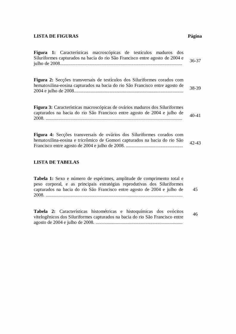
LISTA DE FIGURAS
Página
Figura 1: Características macroscópicas de testículos maduros dos
Siluriformes capturados na bacia do rio São Francisco entre agosto de 2004 e
julho de 2008.......................................................................................................
36-37
Figura 2: Secções transversais de testículos dos Siluriformes corados com
hematoxilina-eosina capturados na bacia do rio São Francisco entre agosto de
2004 e julho de 2008............................................................................................
38-39
Figura 3: Características macroscópicas de ovários maduros dos Siluriformes
capturados na bacia do rio São Francisco entre agosto de 2004 e julho de
2008. ...................................................................................................................
40-41
Figura 4: Secções transversais de ovários dos Siluriformes corados com
hematoxilina-eosina e tricrômico de Gomori capturados na bacia do rio São
Francisco entre agosto de 2004 e julho de 2008. ................................................
42-43
LISTA DE TABELAS
Tabela 1: Sexo e número de espécimes, amplitude de comprimento total e
peso corporal, e as principais estratégias reprodutivas dos Siluriformes
capturados na bacia do rio São Francisco entre agosto de 2004 e julho de
2008. ...................................................................................................................
45
Tabela 2: Características histométricas e histoquímicas dos ovócitos
vitelogênicos dos Siluriformes capturados na bacia do rio São Francisco entre
agosto de 2004 e julho de 2008. .........................................................................
46
SUMÁRIO Página
1. INTRODUÇÃO GERAL.............................................................................. 11
1.1. Bacia do rio São Francisco....................................................................
1.2. Ordem Siluriformes...............................................................................
11
11
1.3. Características biológicas das espécies................................................ 12
1.4. Morfologia dos testículos....................................................................... 15
1.5. Morfologia dos ovários.......................................................................... 16
1.6. Estratégias reprodutivas....................................................................... 17
2. JUSTIFICATIVA.......................................................................................... 19
3. OBJETIVOS.................................................................................................. 20
3.1. Objetivo geral......................................................................................... 20
3.2. Objetivos específicos.............................................................................. 20
4. ARTIGO SUBMETIDO................................................................................ 21
4.1. Abstract.................................................................................................. 23
4.2. Introduction............................................................................................ 24
4.3. Material and Methods........................................................................... 25
4.4. Results..................................................................................................... 26
4.5. Discussion............................................................................................... 29
4.6. Acknowledgements................................................................................ 34
5. CONCLUSÕES.............................................................................................. 47
REFERÊNCIAS............................................................................................. 49
11
1. INTRODUÇÃO GERAL
1.1. Bacia do rio São Francisco
A ictiofauna da bacia do rio São Francisco é representada por cerca de 185
espécies de água doce (Britski et al., 1988; Sato & Godinho, 1999; Alves & Pompeu,
2001), incluindo 18 espécies ameaçadas de extinção, diversas espécies endêmicas e
grande número de espécies de importância econômica e ecológica (Godinho &
Godinho, 2003). Essa ictiofauna encontra-se bastante ameaçada em várias regiões da
bacia, principalmente no trecho a jusante da barragem de Sobradinho até o Oceano
Atlântico, a montante da barragem de Três Marias e nos rios Paraopeba e das Velhas,
devido principalmente, ao desmatamento ciliar, à construção de barragens, às poluições
industriais e domésticas, ao garimpo, à pesca predatória e à destruição de várzeas e
lagoas marginais (Sato & Godinho, 1999).
O reservatório de Três Marias, localizado no rio São Francisco, Minas Gerais
(18-20º S, 44-46º W), foi formado em 1960 e apresenta 2.700 metros de extensão e
altura máxima de 75 metros. O reservatório foi construído com objetivo de geração de
energia elétrica, controle de cheia e irrigação (Britski et al., 1988). A literatura científica
relata que a alteração do ambiente lótico em lêntico altera a dinâmica da água, a
qualidade do habitat, o processo de produção primária e a estrutura e composição da
ictiofauna (Godinho & Godinho, 2003; Agostinho et al., 2007).
1.2. Ordem Siluriformes
A ordem Siluriformes é representada por 36 famílias e 3088 espécies
reconhecidamente válidas, sendo a maioria das famílias restritas a água doce. As
12
espécies desta ordem são caracterizadas pela ausência de escamas e comumente
denominadas peixes de couro (Ferraris, 2007). Os Siluriformes possuem ampla
distribuição geográfica e são encontrados em toda a América, África, Europa e Sudeste
da Ásia (Teugels, 1996). As seis espécies de Siluriformes analisadas no presente
trabalho Trachelyopterus galeatus (Linnaeus, 1766), Pimelodus maculatus La cépède,
1803, Pseudoplatystoma corruscans (Spix and Agassiz, 1829), Lophiosilurus alexandri
Steindachner, 1877, Rhinelepis aspera Spix and Agassiz, 1829 e Conorhynchos
conirostris (Valenciennes, 1840) estão distribuídas originalmente nas bacias
hidrográficas Neotropicais e ocorrem na bacia do rio São Francisco (Reis et al., 2003).
1.3. Características biológicas das espécies
Trachelyopterus galeatus
O cangati pertence à família Auchenipteridae e distribui-se por toda América do
Sul, sendo anteriormente denominado Parauchenipterus galeatus (Ferraris, 2007). Há
alguns trabalhos na literatura sobre a reprodução do cangati, com ênfase em sua
viviparidade, modificação da nadadeira anal dos machos em órgão copulador para
inseminar os ovários com espermatozóides, presença de vesículas seminais e morfologia
diferenciada do espermatozóide (Loir et al., 1989; Meisner et al., 2000; Burns et al.,
2002; Parreira et al., 2009). Porém há poucos trabalhos comparando a morfologia
gonadal desta espécie com outros Siluriformes.
13
Pimelodus maculatus
Conhecido como mandi-amarelo ou mandi-pintado, é um Siluriformes
pertencente à família Pimelodidae, amplamente distribuído nas bacias dos rios Paraná e
São Francisco (Lundenberg & Littmann, 2003; Ferraris, 2007). Vive e alimenta-se
principalmente no fundo dos rios, possuindo dieta variada constituída de larvas de
insetos, algas, moluscos, peixes e fragmentos de vegetais (Bennemann et al., 2000). Esta
espécie pode alcançar 40 cm de comprimento total e 1,5 a 2,0 kg de peso corporal e
apresenta ferrões cobertos por muco tóxico nas nadadeiras peitorais e dorsal
(Lundenberg & Littmann, 2003). O mandi-amarelo, espécie de porte médio, é de
interesse na pesca esportiva e profissional no rio São Francisco, sendo também
importante para alimentação da população ribeirinha (Sato et al., 2003).
Trata-se de uma espécie cujo tipo de desova é ponto de controvérsias na
literatura, sendo admitida desova parcelada (Godinho et al., 1974) e desova total (Sato
& Godinho, 1988; Carvalho et al., 1995). Possui ovos livres com pequeno espaço
perivitelino e altos valores de fecundidade (Rizzo et al., 2002; Sato et al., 2003).
Pseudoplatystoma corruscans
O surubim pertence à família Pimelodidae e é a espécie com maior valor
comercial e recreativo da bacia do rio São Francisco (Godinho et al., 2007). P.
corruscans é piscívoro, atinge grande peso corporal (até 120 Kg) e ocorre nas bacias
dos rios São Francisco e Paraná (Reis et al., 2003; Sato et al., 2003). A pesca do
surubim no rio São Francisco encontra-se em declínio, uma vez que em 1987
representava 86,3% do pescado, enquanto em 1999 sua participação na produção estava
reduzida a 26,6% (Godinho & Godinho, 2003).
14
O surubim reproduz no período chuvoso, possui desova total, ovos não-adesivos
e altos valores de fecundidade (Brito & Bazzoli, 2003; Sato et al., 2003). Esta espécie
realiza migrações reprodutivas de grande distância no rio São Francisco, podendo
migrar por, aproximadamente, 100 Km para desovar (Sato & Godinho, 2004; Godinho
et al., 2007).
Lophiosilurus alexandri
O pacamã, pertence à família Pseudopimelodidae, é endêmico da bacia do rio
São Francisco e possui corpo achatado dorso-ventralmente com acúleo da nadadeira
dorsal curto e forte (Shibatta, 2003). L. alexandri não realiza migrações, vive
preferencialmente em águas lênticas e apresenta hábito noturno. A espécie reproduz-se
naturalmente em cativeiro, constrói ninhos em fundos de areia ou terra e tem potencial
econômico para aquicultura (Sato et al., 2003).
O pacamã pode atingir comprimento máximo de 72 cm, peso corporal de 8 Kg,
apresenta ovários com desenvolvimento assincrônico dos ovócitos, os quais são
esféricos e com coloração amarelada (Bazzoli & Godinho, 1997). Possui ovos adesivos,
não necessita efetuar migração reprodutiva e os machos realizam cuidado parental (Sato
et al., 2003; Barros et al., 2007).
Rhinelepis aspera
O cascudo-preto pertence à família Loricariidae, é importante peixe comercial e
atualmente raro na bacia do rio São Francisco, ocorrendo com maior abundância no rio
Paracatu. Entre as espécies de Loricariidae da bacia do São Francisco, R. aspera é a que
15
alcança maior porte podendo alcançar 4 Kg de peso corporal (Sato, 1999; Reis et al.
2003).
O cascudo-preto da bacia do rio Paraná, atinge a maturação sexual aos 24 e 23
cm, respectivamente para machos e fêmeas (cerca de 2,7 anos de idade) e o período
reprodutivo estende-se de novembro a julho (Agostinho, 1985). Possui ovos adesivos
com pequeno espaço perivitelino e córion espesso. No rio São Francisco, R. aspera não
necessita efetuar migração reprodutiva, possivelmente apresenta cuidado parental e
reproduz-se em locais de rochas e cascalhos (Rizzo et al., 2002; Sato et al., 2003).
Conorhynchos conirostris
Considerada espécie símbolo do rio São Francisco é endêmica da bacia e
conhecida popularmente como pirá. Anteriormente pertencia à família Pimelodidae e
atualmente encontra-se no grupo incertae sedis na ordem Siluriformes (Ferraris, 2007).
Pode alcançar até 100 cm de comprimento total e 13 kg de peso corporal (Sato, 1999).
A espécie é reofílica, realiza migrações durante o período reprodutivo (Sato &
Godinho, 2004), possui ovos não-adesivos e desova total (Rizzo et al., 2002; Sato et al.
2003). É peixe de interesse comercial e está incluído nas listas das espécies ameaçadas
de extinção, na categoria vulnerável, em Minas Gerais e no Brasil (Rosa & Lima, 2008).
1.4. Morfologia dos testículos
Os testículos dos Siluriformes têm ampla variabilidade morfológica, uma vez
que muitas espécies apresentam testículo com projeções digitiformes ou franjas de
aspecto variado (Loir et al., 1989; Meisner et al., 2000; Santos et al., 2001; Guimarães-
Cruz & Santos, 2004; Lopes et al., 2004; Mazzoldi et al., 2007). Em algumas famílias, a
16
região caudal dos testículos contém vesículas seminais conectadas ao ducto
espermático, sem atividade espermatogênica, possuindo função secretora e/ou
armazenadora (Loir et al., 1989; Legendre et al., 1996; Meisner et al., 2000; Mazzoldi et
al., 2007). Algumas espécies de Siluriformes, notadamente da família Auchenipteridae,
possuem inseminação interna, apresentando modificações morfofuncionais do
espermatozóide e do sistema reprodutor, como o órgão copulador originado de
modificação da nadadeira anal (Loir et al., 1989; Meisner et al., 2000; Burns et al.,
2002; Parreira et al., 2009).
1.5. Morfologia dos ovários
Os ovários da maioria dos Siluriformes são do tipo cistovarianos, nos quais o
lúmen ovariano tem continuidade com o oviduto, através do qual ovócitos são liberados
diretamente no meio externo (Bazzoli, 2003; Santos et al., 2006; Barros et al., 2007).
Durante o desenvolvimento ovariano, ovócitos de teleósteos passam por duas
fases distintas: crescimento primário, independente de gonadotrofinas, caracterizado por
alterações nucleares e multiplicação dos nucléolos nos ovócitos perinucleolares, e
crescimento secundário, sob influência de gonadotrofinas, caracterizado pela formação
dos alvéolos corticais nos ovócitos pré-vitelogênicos e glóbulos de vitelo nos ovócitos
vitelogênicos (Tyler & Sumpter, 1996; Patiño & Sullivan, 2002).
As mudanças morfológicas no núcleo, ooplasma e camadas circundantes dos
ovócitos caracterizam os diferentes estádios do desenvolvimento ovocitário (Bazzoli,
2003). A morfologia dos folículos ovarianos dos Siluriformes pode variar de acordo
com as diferentes estratégias reprodutivas, sendo similar entre espécies de mesmo grupo
taxonômico (Suzuki et al., 2000; Rizzo et al., 2002; Santos et al., 2006).
17
1.6. Estratégias reprodutivas
Os peixes representam o maior e mais diversificado grupo entre os vertebrados e
as variações na história evolutiva e na morfologia gonadal resultaram em padrões
reprodutivos variados (Balon, 1984; Loir et al., 1989; Sato et al., 2003). Segundo
Wootton (1984), o sucesso alcançado pelos peixes em ambientes distintos deve-se às
variadas estratégias reprodutivas desenvolvidas pelo grupo, que englobam táticas
extremas que podem variar em função do ambiente, mas algumas são conservativas.
Entre os Siluriformes da bacia do rio São Francisco, Sato et al. (2003)
reconheceram três padrões reprodutivos: espécies migradoras, espécies não migradoras
e espécies não migradoras que apresentam cuidado parental. As espécies migradoras
normalmente se reproduzem no leito dos rios durante a estação chuvosa e apresentam
período reprodutivo curto, ausência de cuidado parental, alta fecundidade absoluta, ovos
livres e embriogênese rápida (Rizzo et al. 2002; Brito & Bazzoli, 2003; Sato &
Godinho, 2004; Godinho et al. 2007). As espécies não migradoras que não apresentam
cuidado parental, possuem período reprodutivo intermediário, fecundidade absoluta
intermediária, ovos livres e embriogênese rápida (Rizzo et al. 2002; Sato et al., 2003).
As espécies não migradoras que apresentam cuidado parental, normalmente se
reproduzem em substratos específicos e possuem período reprodutivo de intermediário a
longo, baixa fecundidade absoluta, ovos adesivos e desenvolvimento embrionário longo
(Rizzo et al., 2002; Sato et al., 2003; Barros et al., 2007).
Recentemente, Godinho et al. (2009) corroboraram a hipótese que peixes
teleósteos ovíparos de água doce do Brasil podem ser divididos em dois grupos: peixes
de ambiente lótico e de ambiente lêntico. Os peixes lóticos apresentam migração
reprodutiva, curto período de desova, ausência de cuidado parental, ovos livres, alta

18
fecundidade relativa, embriogênese rápida e grande tamanho corporal. Em contraste,
peixes lênticos não realizam migração reprodutiva e apresentam longo período de
desova, cuidado parental, ovos adesivos, baixa fecundidade relativa, embriogênese
longa e pequeno a médio tamanho corporal.
19
2. JUSTIFICATIVA
Estudos agrupando características morfológicas reprodutivas de peixes permitem
comparar os padrões de história de vida das diferentes espécies. Estes estudos também
possibilitam analisar as adaptações morfológicas do aparelho reprodutor de cada espécie
frente às estratégias adotadas para alcançar o sucesso reprodutivo. As características
morfológicas do aparelho reprodutor, associadas às adaptações para a reprodução,
definem padrões reprodutivos como os tipos de migração, fertilização, ovo, desova e
cuidado parental, dentre outros.
Pelo fato dos Siluriformes apresentarem muitas famílias e espécies, eles
constituem excelente grupo para estudos comparativos. Apesar da abundância e da
diversidade, não existem estudos comparativos sobre a morfologia gonadal de
Siluriformes brasileiros relacionados com as diferentes estratégias reprodutivas do
grupo. Portanto, o presente estudo analisa comparativamente as características
morfológicas de testículos e de ovários de seis espécies de Siluriformes, de importância
comercial e ecológica da bacia do rio São Francisco, com o intuito de fornecer subsídios
para análises da reprodução e da filogenia das espécies, bem como para administração
racional da pesca e conservação dos estoques naturais.
20
3. OBJETIVOS
3.1. Objetivo geral
Comparar as características morfológicas do aparelho reprodutor de machos e
fêmeas de seis espécies de peixes Siluriformes da bacia do rio São Francisco, em Minas
Gerais.
3.2. Objetivos específicos
Verificar relações anatômicas, forma, volume, coloração e presença de estruturas
acessórias ao aparelho reprodutor de testículos e ovários em atividade
reprodutiva;
Descrever a histologia dos testículos maduros quanto ao padrão de distribuição
das células espermatogênicas e da secreção testicular;
Analisar a histologia dos ovários maduros quanto a morfologia dos ovócitos
vitelogênicos e camadas envoltórias;
Realizar a morfometria do diâmetro dos ovócitos vitelogênico e da espessura da
zona radiata e das células foliculares,
Verificar o conteúdo histoquímico da secreção testicular e dos folículos
ovarianos dos ovócitos vitelogênicos;
Comparar as características anatômicas, histológicas, histométricas e
histoquímicas das gônadas de machos e de fêmeas das espécies em estudo;
Relacionar as características morfológicas reprodutivas das espécies com as
diferentes estratégias reprodutivas do grupo.

21
4. ARTIGO SUBMETIDO
22
Comparative morphology of the gonadal structure related to reproductive
strategies in six species of Neotropical catfishes (Teleostei: Siluriformes)
Rafael Magno Costa Melo¹, Fábio Pereira Arantes², Yoshimi Sato³, José Enemir dos
Santos¹, Elizete Rizzo² and Nilo Bazzoli¹.
¹Graduate Program on Zoology of Vertebrates, Pontifical Catholic University of Minas
Gerais, Belo Horizonte, Minas Gerais, Brazil. ²Institute of Biological Sciences,
Morphology Department, Federal University of Minas Gerais, Belo Horizonte, Minas
Gerais, Brazil. ³Hydrobiology and Fishculture Station of Três Marias, Três Marias,
Minas Gerais, Brazil.
Running title: Gonad morphology of Neotropical catfishes
Author to whom correspondence: Nilo Bazzoli, Programa de Pós-Graduação em
Zoologia de Vertebrados, PUC Minas - Av. Dom José Gaspar, nº 500 - 30535-610, Belo
Horizonte - MG, Brazil. E-mail: [email protected]
23
4.1. Abstract
The relationship among the morphology of the reproductive apparatus and the
reproductive strategies of six Neotropical catfishes was studied through macro and
microscopic analyses of 151 gonads. The reproductive apparatus of the examined
Siluriformes showed diversified characteristics, but allowed their grouping according to
the reproductive strategies as migratory, non-migratory keepers and internal
inseminating. The great reproductive migratory Pseudoplatystoma corruscans and
Conorhynchos conirostris are characterised by small filiform testicular fringes, absence
of testicular secretion, small diameter of the vitellogenic oocytes, thin zona radiata and
cubic follicular cells. The short distance migratory Pimelodus maculatus is
morphologically distinct from the other migratory catfishes due to the presence of
longer filiform testicular fringes and testicular secretion. The non-migratory keepers
Lophiosilurus alexandri and Rhinelepis aspera present non-filiform testicular
projections, homogeneous testicular secretion, large diameter of the vitellogenic oocytes
and prismatic follicular cells. The most specialized reproductive apparatus was observed
in the internal inseminating Trachelyopterus galeatus which presents a seminal vesicle
accessory to the testes, spermatozoa with elongated nuclei that form spermatozeugmata
and ovarian lamellar epithelium with storage and secretory functions. The morphology
of the reproductive apparatus of the Neotropical catfishes studied presented a clear
influence of the insemination type and of the reproductive strategies of the species.
Keywords: fishes; gonad morphology; secretion; spermatozeugmata; insemination
type; reproductive strategy.
24
4.2. Introduction
The Siluriformes are represented by 36 families and 3088 valid known species,
and have a wide geographical distribution with representation on every continent
(Ferraris, 2007). With relation to the distribution of the world’s freshwater catfishes,
nearly 64% are found in South and Central America, 19% of the species occur in Africa,
15% are present in Europe and Southeast Asia while only 2% of the species are found in
North America (Teugels, 1996; Ferraris, 2007). The South American siluriforms
developed a great adaptive radiation that started during the isolation of South America
in the Tertiary period (Lowe-McConnell, 1987). The success achieved by these
catfishes in different environments is partially related to the diverse reproductive
strategies developed by the group (Suzuki et al., 2000; Sato et al., 2003; Sato &
Godinho, 2004; Godinho et al., 2009). Several studies have demonstrated the relation of
the insemination modalities and the reproductive strategies on the morphological
characteristics of the reproductive apparatus of Siluriformes (Loir et al., 1989; Meisner
et al., 2000; Rizzo et al., 2002; Mazzoldi et al., 2007; Spadella et al., 2008).
The morphology of the reproductive apparatus of siluriform males is very
diversified; many siluriform families present testes with digitiform projections or
fringes (Loir et al., 1989; Meisner et al., 2000; Santos et al., 2001; Guimarães-Cruz &
Santos, 2004; Lopes et al., 2004), while others present compact testes without
projections (Loir et al., 1989; Mazzoldi et al., 2007). In some catfish families the testes
present spermatogonia distributed along the entire extension of the seminiferous tubules
(Guimarães-Cruz & Santos, 2004; Lopes et al., 2004), and in others the caudal region of
the testes contains seminal vesicles or secretory cells without spermatogenic activity,
having secretory and/or storage functions (Legendre et al., 1996; Meisner et al., 2000;
25
Santos et al., 2001). Some species of the Auchenipteridae and Scoloplacidae families
have internal insemination, presenting morphofunctional modifications to the
spermatozoon and in the reproductive apparatus (Burns et al., 2002; Spadella et al.,
2008; Parreira et al., 2009).
Catfish females also have reproductive apparatus with varied morphology
(Suzuki et al., 2000; Bazzoli, 2003). In general, the vitellogenic oocytes of Neotropical
Siluriformes, when compared to those from other teleost orders, present common
characteristics such as thin zona radiata, high follicular cells and a jelly coat
surrounding the released eggs (Rizzo et al., 2002; Bazzoli, 2003). The histological,
histometrical and histochemical variations of the ovarian follicles can be associated with
the different reproductive strategies used by catfishes (Suzuki et al., 2000; Santos et al.,
2006; Barros et al., 2007).
Considering the importance of South American Siluriformes for the
understanding of this order’s, the present study compares the morphological
characteristics of the testes and ovaries of six Neotropical catfishes in relation to some
reproductive strategies of the species.
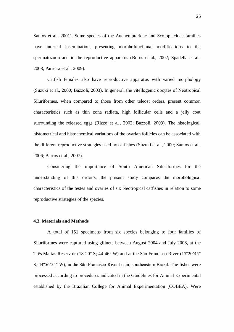
4.3. Materials and Methods
A total of 151 specimens from six species belonging to four families of
Siluriformes were captured using gillnets between August 2004 and July 2008, at the
Três Marias Reservoir (18-20° S; 44-46° W) and at the São Francisco River (17º20’45"
S; 44º56’55" W), in the São Francisco River basin, southeastern Brazil. The fishes were
processed according to procedures indicated in the Guidelines for Animal Experimental
established by the Brazilian College for Animal Experimentation (COBEA). Were
26
selected for this study fishes sexually mature or in reproductive activity. For each
specimen, the total length (LT) and body mass (MB) were determined (Table 1). The data
on reproductive strategies of the species are summarized on the table 1, with the
respective references.
After dissection, the fresh testes and ovaries were photographed and
macroscopically analysed regarding their shape, colouration and presence of accessory
structures. For the microscopic analysis, fragments of the cranial and caudal regions of
77 testes and 74 ovaries were fixed in Bouin’s solution for 8 to 12 hours, embedded in
paraffin, cut into 3-5µm thick sections and stained with haematoxylin-eosin and
Gomori’s trichromic. In histological slides, the diameters of 50 vitellogenic oocytes
from mature ovaries of each species and the thickness of their surrounding layers (zona
radiata and follicular cells) were measured using a micrometric ruler attached to the
ocular light microscope.
To determine the histochemical content of proteins and carbohydrates in the
testicular secretions and in the zona radiata and the follicular cells of the vitellogenic
oocytes from mature ovaries the following histochemical techniques were used (Pearse,
1985): periodic acid Schiff (PAS), Alcian blue (AB) pH 2.5 and pH 0.5, and hydrolysis
with HCl 0.1M (8h at 60ºC) for the extraction of sialic acid followed by PAS and AB
pH 2.5.
4.4. Results
Male reproductive apparatus
In the species analysed remarkable morphophysiological differences were
observed mainly in the mature testes, which presented a continuous central portion from
27
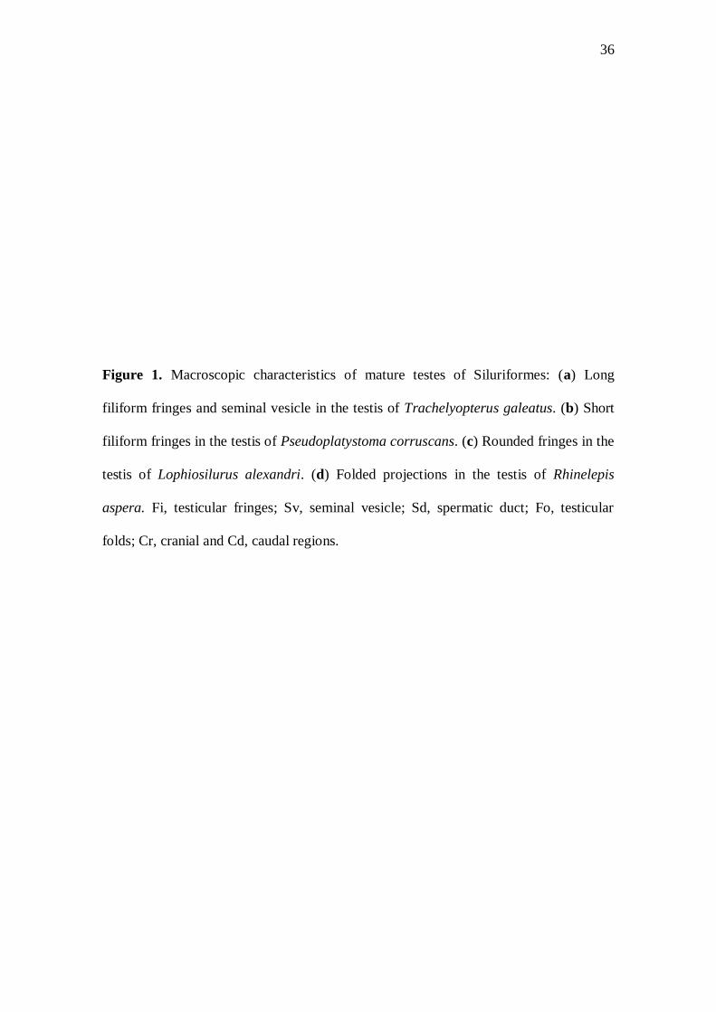
where projections of varied morphology originated (Fig. 1). Testicular projections in the
form of long filiform fringes were found in Trachelyopterus galeatus and Pimelodus
maculatus (Fig. 1a). Short filiform fringes were observed in Pseudoplatystoma
corruscans and Conorhynchos conirostris (Fig. 1b). In Lophiosilurus alexandri the
testicular fringes were rounded (Fig. 1c) and in Rhinelepis aspera the testicular
projections presented folds, not forming fringes (Fig. 1d). In T. galeatus, in the cranial
region of the testes were observed longer and thinner fringes of whitish colouration,
whilst in the caudal region occurred seminal vesicles with thicker and shorter fringes of
pink colouration (Fig. 1a).
Histologically, the seminiferous tubules from the mature testes of the species
examined showed compartments that communicate with each other, forming a
continuous anastomosed tubular apparatus (Fig. 2a,g,h). The testes of P. corruscans and
C. conirostris exhibited spermatogenic activity throughout their entire extension (Fig.
2a,b), while in T. galeatus, P. maculatus, L. alexandri and R. aspera spermatogenic
activity was observed only in the cranial region and secretory activity in the caudal
region (Fig. 2c,d). In T. galeatus, P. maculatus, L. alexandri and R. aspera was not
observed spermatogenic activity in the caudal region of the testes. Globular acidophilic
secretion associated with spermatozoa was detected in the lumen of the secretory
tubules and in the spermatic duct of the caudal region of testes in P. maculatus (Fig.
2c,d,e). This secretion reacted positively to the PAS and AB pH 2.5 and 0.5 techniques,
indicating the presence of neutral glycoproteins and carboxylic and sulphated acid
glycoconjugates. Homogeneous acidophilic secretion associated with spermatozoa was
detected in the secretory tubules of the caudal region of testes in T. galeatus, L.
alexandri and R. aspera (Fig. 2f), which reacted positively to the PAS technique,
28
indicating the presence of neutral glycoproteins. In the testes of P. corruscans and C.
conirostris no type of secretion was observed. In the species P. maculatus, P.
corruscans, L. alexandri, R. aspera and C. conirostris the heads of the spermatozoa
were rounded (Fig. 2g), while in T. galeatus the head of the spermatozoon was
elongated (Fig. 2h).
Female reproductive apparatus
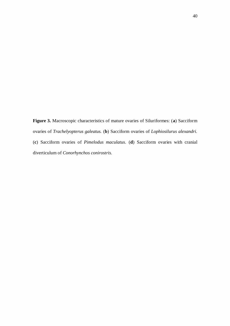
The mature ovaries of the catfishes analysed showed yellowish colouration with
different hues (Fig. 3) and were sacciform in shape (Fig. 3a,b,c,d). A digitiform
protuberance was observed in the cranial region of the ovary of C. conirostris (Fig. 3d).
In the ovaries of the females in reproductive activity, the vitellogenic oocytes presented
distinct morphologic and histometric characteristics, with the largest diameter in L.
alexandri and the smallest in P. corruscans (Table 2). Vitellogenic oocytes with cubic
follicular cells were observed in P. maculatus, P. corruscans and C. conirostris (Fig.
4b) and prismatic follicular cells in the rest of the species (Fig. 4a,c). Elliptical yolk
globules were found in the vitellogenic oocytes of R. aspera (Fig. 4c), while in the other
species these globules were spherical (Fig. 4a,b). The thickest zona radiata was
observed in R. aspera (Fig. 4c) and the tallest follicular cells were found in T. galeatus
(Table 2 and Fig. 4a). The histochemical content of the zona radiata and of the follicular
cells varied among the siluriforms studied (Table 2). In the ovaries of the catfishes
analysed no remarkable morphophysiological differences were detected among the
cranial and caudal regions.
In the lumen of the mature and partially spawned ovaries of T. galeatus, folds of
the cubic epithelium from the ovarian lamellae were observed containing packed
29
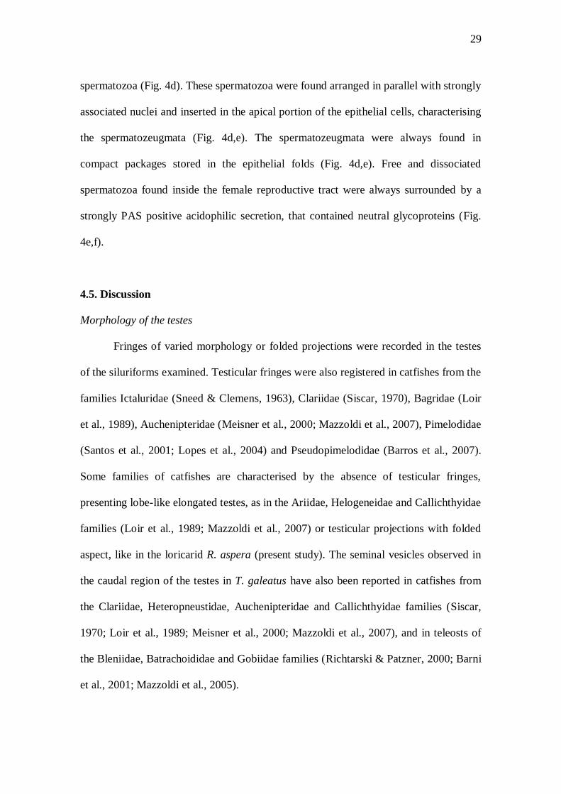
spermatozoa (Fig. 4d). These spermatozoa were found arranged in parallel with strongly
associated nuclei and inserted in the apical portion of the epithelial cells, characterising
the spermatozeugmata (Fig. 4d,e). The spermatozeugmata were always found in
compact packages stored in the epithelial folds (Fig. 4d,e). Free and dissociated
spermatozoa found inside the female reproductive tract were always surrounded by a
strongly PAS positive acidophilic secretion, that contained neutral glycoproteins (Fig.
4e,f).
4.5. Discussion
Morphology of the testes
Fringes of varied morphology or folded projections were recorded in the testes
of the siluriforms examined. Testicular fringes were also registered in catfishes from the
families Ictaluridae (Sneed & Clemens, 1963), Clariidae (Siscar, 1970), Bagridae (Loir
et al., 1989), Auchenipteridae (Meisner et al., 2000; Mazzoldi et al., 2007), Pimelodidae
(Santos et al., 2001; Lopes et al., 2004) and Pseudopimelodidae (Barros et al., 2007).
Some families of catfishes are characterised by the absence of testicular fringes,
presenting lobe-like elongated testes, as in the Ariidae, Helogeneidae and Callichthyidae
families (Loir et al., 1989; Mazzoldi et al., 2007) or testicular projections with folded
aspect, like in the loricarid R. aspera (present study). The seminal vesicles observed in
the caudal region of the testes in T. galeatus have also been reported in catfishes from
the Clariidae, Heteropneustidae, Auchenipteridae and Callichthyidae families (Siscar,
1970; Loir et al., 1989; Meisner et al., 2000; Mazzoldi et al., 2007), and in teleosts of
the Bleniidae, Batrachoididae and Gobiidae families (Richtarski & Patzner, 2000; Barni
et al., 2001; Mazzoldi et al., 2005).
30
In the catfishes studied, the testes have tubular anastomosed seminiferous
tubules, characteristic of the order Siluriformes and from more primitive teleosts (Grier,
1993). In this type of anastomosis, the seminiferous tubules are strongly connected
along the full extension of the testis. In contrast, neoteleosts present anastomoses only
among the seminiferous tubules located in the central region of the testes (Parenti &
Grier, 2004).
The secretory activity of the testes in siluriforms has been attributed to
specialized structures or to specific testicular regions (Meisner et al., 2000; Santos et al.,
2001; Guimarães-Cruz & Santos, 2004; Mazzoldi et al., 2007). In the present study,
during the reproductive period and immediately after spermiation, an acidophilic
secretion associated with the spermatozoa was observed in the secretory tubules of the
caudal region of the testes of T. galeatus, P. maculatus, L. alexandri and R. aspera. In
T. galeatus, this secretion originated in the seminal vesicles which have secretory and
storage functions (Meisner et al., 2000; Parreira et al., 2009). In the species P.
maculatus, L. alexandri and R. aspera, that do not present seminal vesicles, the origin of
the secretion can be attributed to secretory cells located in the epithelium of the testes’
caudal region, as has also been observed in catfishes Iheringichthys labrosus, P.
maculatus and L. alexandri (Santos et al., 2001; Guimarães-Cruz & Santos, 2004;
Barros et al., 2007). Through histochemical reactions, glycoproteins and
glycoconjugates were detected in the testicular secretions of the studied catfishes. The
presence of these compounds suggests that these secretions can have similar functions
to the secretions produced by the seminal vesicles of some teleosts, being involved in
the maturation and nutrition of the spermatozoa and playing a role in fertilization
(Lahnsteiner et al., 1990; Mazzoldi et al., 2005; Chowdhury & Joy, 2007).
31
In general, fishes with external insemination have aquatic spermatozoa with
spherical or ovoid head, considered evolutionarily primitive (Jamieson, 1991), while
fishes with internal insemination have spermatozoa with elongated head, as observed in
catfishes of the Auchenipteridae family (Loir et al., 1989; Meisner et al., 2000; Parreira
et al., 2009). Our observations confirmed such relationships, where spermatozoa with
rounded head occurred in the external inseminating P. maculatus, P. corruscans, L.
alexandri, R. aspera and C. conirostris. In T. galeatus, a species with internal
insemination, were observed spermatozoa with elongated head, which form compact
packages of spermatozeugmata. In teleosts with internal insemination the nuclear
elongation of the spermatozoa frequently occurs to facilitate the passage of these
through the lobular gonopodium of the males and through the labyrinthic structures of
the females, besides easing the packing for the formation of the spermatozeugmata
(Jamieson, 1991; Burns & Weitzman, 2005). The spermatozeugmata in Trachelyopterus
lucenai and Auchenipterus nuchalis are associated with the secretion from the epithelial
cells in the seminal vesicles, whose function is to integrate and maintain the compact
packages of this structure (Burns et al., 2002; Mazzoldi et al., 2007).
Morphology of the ovaries
In all the species analysed the female reproductive apparatus exhibited an
anatomical organization found in most teleosts, with paired ovaries of the cystovarian
type, in which the ovarian lumen is continuous with the oviduct where the oocytes reach
the external environment (Nagahama, 1983; Bazzoli, 2003). The colouration of the
vitellogenic oocytes from the studied catfishes is yellowish with their hue varying
among the species. The same colouration pattern was observed in eight siluriform
32
species, except in Pseudopimelodus charus that have green oocytes (Sato et al., 2003).
The yellow colouration of the oocytes is probably due to the presence of carotenoid
pigments, which include endogenous oxygen sources to be used when the respiratory
apparatus is inefficient in obtaining exogenous oxygen (McElman & Balon, 1980).
Variations in the egg diameter can be related to the different reproductive
strategies of the fishes (Suzuki et al., 2000; Kolm & Ahnesjö, 2005). The diameter of
the vitellogenic oocytes varied among the siluriforms analysed, being largest in L.
alexandri and smallest in P. corruscans. The migratory species P. corruscans, P.
maculatus and C. conirostris, with smaller vitellogenic oocytes, have high absolute
fecundity, non-adhesive eggs and do not display parental care, while the non-migratory
species L. alexandri and R. aspera, with larger vitellogenic oocytes, have low fecundity,
adhesive eggs and exhibit parental care (Rizzo et al., 2002; Sato et al., 2003; Godinho et
al., 2009).
The thickness of the zona radiata in teleosts can reflects adaptations to different
ecological conditions (Fausto et al., 2004; Santos et al., 2006), since demersal eggs tend
to have a thicker zona radiata than pelagic eggs to avoid injury to the developing
embryo (Stehr & Hawkes, 1979; Riehl, 1996). Rhinelepis aspera have demersal eggs
and spawns in rocky substrates (Sato et al., 2003), where the eggs suffer more abrasion,
therefore this species presents vitellogenic oocyte with thick zona radiata, as has also
been observed in some loricariid catfishes (Suzuki et al., 2000). Lophiosilurus alexandri
also have demersal eggs but since it spawns in sandy substrates (Sato et al., 2003;
Barros et al., 2007), and hence subject to a less physical damage, it presents thinner
zona radiata. The species P. maculatus, P. corruscans and C. conirostris spawns on the
33
surface of the water and their pelagic eggs disperse freely downstream (Sato &
Godinho, 2004; Godinho et al., 2007), therefore they also present thin zona radiata.
The morphology of the follicular cells is variable depending on the species and
the stage of oocyte development (Suzuki et al., 2000; Bazzoli, 2003). Normally, high
follicular cells of the vitellogenic oocytes have greater synthesizing capacity (Fávaro et
al., 2005; Santos et al., 2006), which probably are able to synthesise mucosubstances
that are transferred to the surface of the zona radiata, aiding in the egg’s adhesiveness
and facilitating parental care, as has been observed in the catfishes Silurus glanis and L.
alexandri (Abraham et al., 1993; Barros et al., 2007). In fact, during this study the
highest follicular cells were registered in the vitellogenic oocytes of T. galeatus, L.
alexandri and R. aspera, which have adhesive eggs and parental care. Regarding to T.
galeatus the results of this study and the information available in literature are not
enough to state that the species has parental care.
In the mature ovaries of the catfishes examined, the histochemical analyses of
the zona radiata and the follicular cells showed differences in content between
migratory and non-migratory catfishes. The neutral glycoproteins associated with the
carboxylic acid glycoconjugates found in the follicular cells of the non-migratory L.
alexandri and R. aspera can form mucosubstances that are transferred to the zona
radiata, conferring adhesiveness to the eggs, as observed in other Neotropical catfishes
(Fávaro et al., 2005; Santos et al., 2006; Barros et al., 2007).
In the present study, spermatozeugmata packages were observed stored in
narrow and tortuous folds of cubic lamellar epithelium in T. galeatus females. In
siluriforms, the insemination of spermatozoa inside the ovaries was registered only in
the Auchenipteridae and Scoloplacidae families (Loir et al., 1989; Meisner et al., 2000;
34
Burns et al., 2002; Mazzoldi et al., 2007; Spadella et al., 2008; Parreira et al., 2009).
The spermatozeugmata stored in the epithelial cells from the ovarian lamellae of the
mature and partially spawned ovaries of T. galeatus can represent an important strategy
to maintain the viability of the spermatozoa for long periods. Meisner et al. (2000) state
that these spermatozoa can remain in the ovaries for six months in T. lucenai. The
abundant acidophilic secretion observed in the ovarian lumen of T. galeatus, during the
reproductive period, appears to be secreted by ovarian lamellar epithelium, as reported
in the teleosts Syngnathus scovelli and Alcichthys alcicornis (Begovac & Wallace, 1987;
Koya et al., 1995). This secretion was always detected associated with free spermatozoa
inside the female reproductive tract of T. galeatus. Histochemical analyses revealed the
presence of neutral glycoproteins in this secretion, which due to their chemical nature,
can possibly act as an energy source for the spermatozoa (Chowdhury & Joy, 2007).
When comparing the reproductive characteristics of the analysed siluriforms a
clear influence of the insemination type and of the reproductive strategies are observed
on the morphological specializations of the reproductive apparatus of both males and
females.
4.6. Acknowledgements
The authors wish to thank the technicians at the Três Marias Hydrobiology and
Pisciculture Station (CODEVASF) for their assistance during the collection of
biological material, to CNPq, FAPEMIG and FIP PUC Minas for financial support and
to the laboratory technician Rogério Silva Matos for the preparation of the histological
slides.

35
FIGURES
36
Figure 1. Macroscopic characteristics of mature testes of Siluriformes: (a) Long
filiform fringes and seminal vesicle in the testis of Trachelyopterus galeatus. (b) Short
filiform fringes in the testis of Pseudoplatystoma corruscans. (c) Rounded fringes in the
testis of Lophiosilurus alexandri. (d) Folded projections in the testis of Rhinelepis
aspera. Fi, testicular fringes; Sv, seminal vesicle; Sd, spermatic duct; Fo, testicular
folds; Cr, cranial and Cd, caudal regions.
37
38
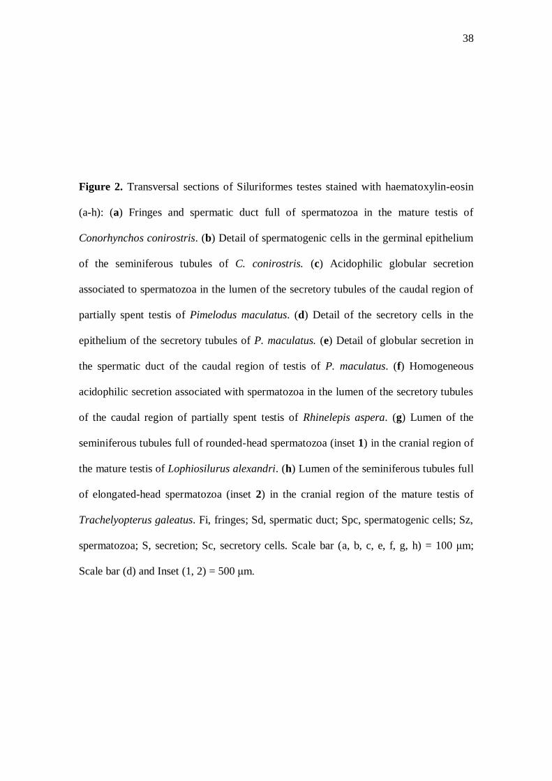
Figure 2. Transversal sections of Siluriformes testes stained with haematoxylin-eosin
(a-h): (a) Fringes and spermatic duct full of spermatozoa in the mature testis of
Conorhynchos conirostris. (b) Detail of spermatogenic cells in the germinal epithelium
of the seminiferous tubules of C. conirostris. (c) Acidophilic globular secretion
associated to spermatozoa in the lumen of the secretory tubules of the caudal region of
partially spent testis of Pimelodus maculatus. (d) Detail of the secretory cells in the
epithelium of the secretory tubules of P. maculatus. (e) Detail of globular secretion in
the spermatic duct of the caudal region of testis of P. maculatus. (f) Homogeneous
acidophilic secretion associated with spermatozoa in the lumen of the secretory tubules
of the caudal region of partially spent testis of Rhinelepis aspera. (g) Lumen of the
seminiferous tubules full of rounded-head spermatozoa (inset 1) in the cranial region of
the mature testis of Lophiosilurus alexandri. (h) Lumen of the seminiferous tubules full
of elongated-head spermatozoa (inset 2) in the cranial region of the mature testis of
Trachelyopterus galeatus. Fi, fringes; Sd, spermatic duct; Spc, spermatogenic cells; Sz,
spermatozoa; S, secretion; Sc, secretory cells. Scale bar (a, b, c, e, f, g, h) = 100 μm;
Scale bar (d) and Inset (1, 2) = 500 μm.
39
40
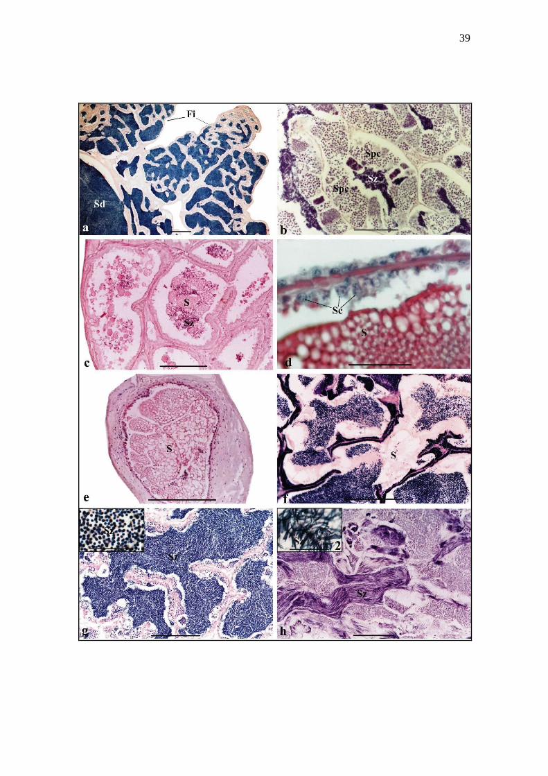
Figure 3. Macroscopic characteristics of mature ovaries of Siluriformes: (a) Sacciform
ovaries of Trachelyopterus galeatus. (b) Sacciform ovaries of Lophiosilurus alexandri.
(c) Sacciform ovaries of Pimelodus maculatus. (d) Sacciform ovaries with cranial
diverticulum of Conorhynchos conirostris.
41
42
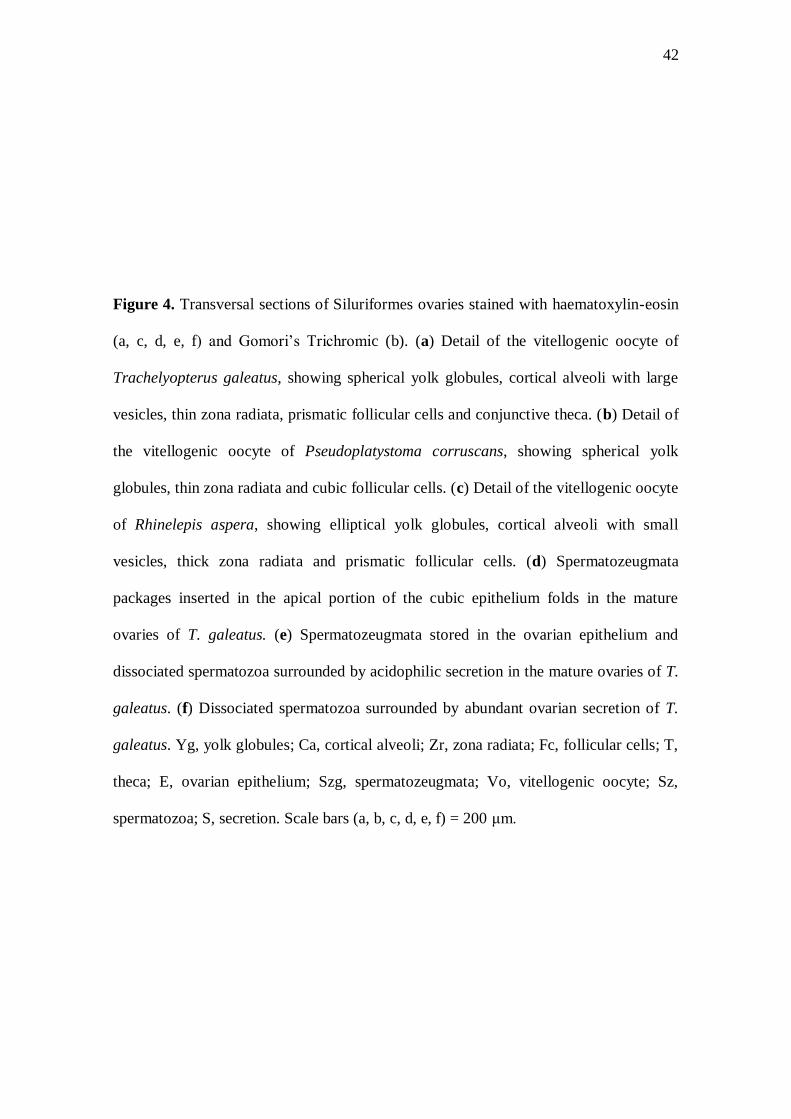
Figure 4. Transversal sections of Siluriformes ovaries stained with haematoxylin-eosin
(a, c, d, e, f) and Gomori’s Trichromic (b). (a) Detail of the vitellogenic oocyte of
Trachelyopterus galeatus, showing spherical yolk globules, cortical alveoli with large
vesicles, thin zona radiata, prismatic follicular cells and conjunctive theca. (b) Detail of
the vitellogenic oocyte of Pseudoplatystoma corruscans, showing spherical yolk
globules, thin zona radiata and cubic follicular cells. (c) Detail of the vitellogenic oocyte
of Rhinelepis aspera, showing elliptical yolk globules, cortical alveoli with small
vesicles, thick zona radiata and prismatic follicular cells. (d) Spermatozeugmata
packages inserted in the apical portion of the cubic epithelium folds in the mature
ovaries of T. galeatus. (e) Spermatozeugmata stored in the ovarian epithelium and
dissociated spermatozoa surrounded by acidophilic secretion in the mature ovaries of T.
galeatus. (f) Dissociated spermatozoa surrounded by abundant ovarian secretion of T.
galeatus. Yg, yolk globules; Ca, cortical alveoli; Zr, zona radiata; Fc, follicular cells; T,
theca; E, ovarian epithelium; Szg, spermatozeugmata; Vo, vitellogenic oocyte; Sz,
spermatozoa; S, secretion. Scale bars (a, b, c, d, e, f) = 200 μm.
43

44
TABLES
45
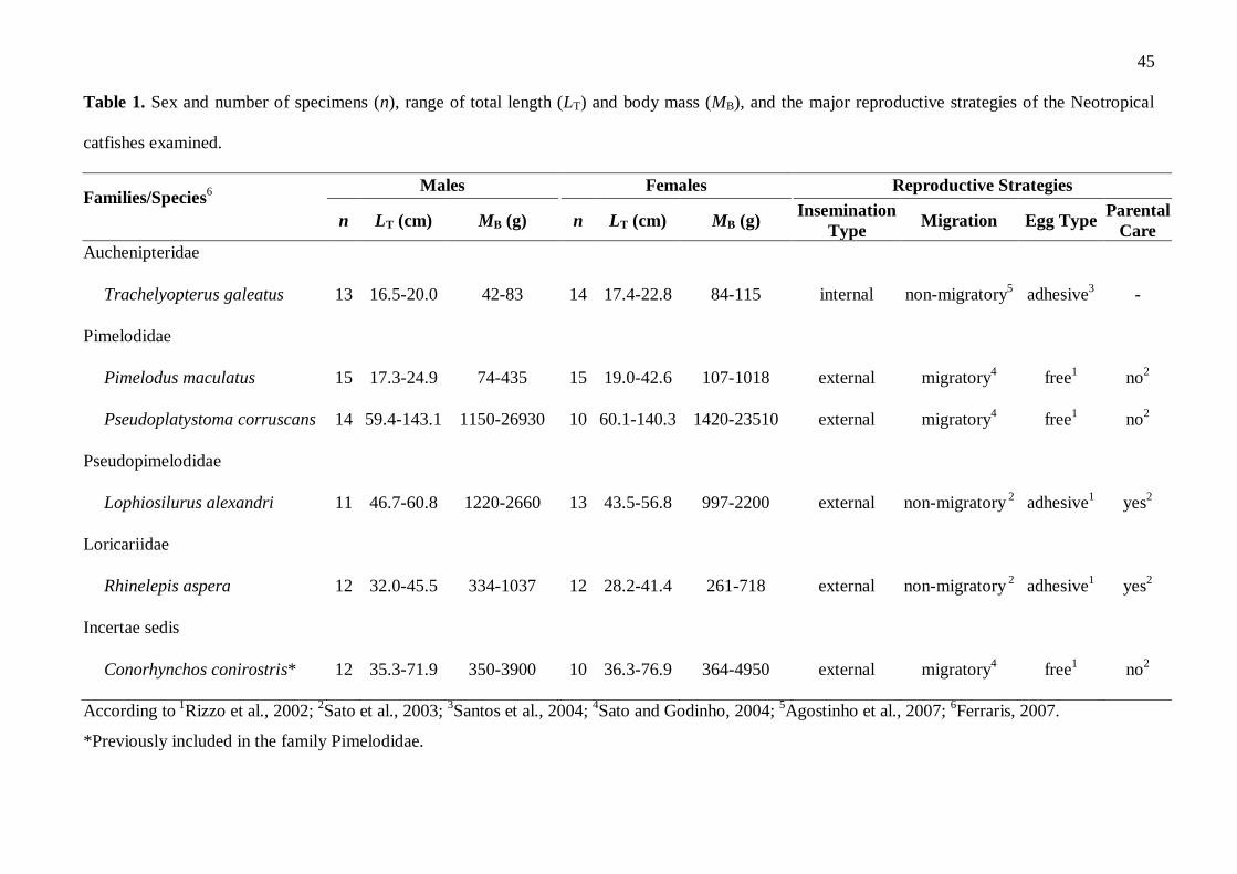
Table 1. Sex and number of specimens (n), range of total length (LT) and body mass (MB), and the major reproductive strategies of the Neotropical
catfishes examined.
Families/Species6 Males Females Reproductive Strategies
n LT (cm) MB (g) n LT (cm) MB (g) Insemination
Type Migration Egg Type
Parental
Care
Auchenipteridae
Trachelyopterus galeatus 13 16.5-20.0 42-83 14 17.4-22.8 84-115 internal non-migratory5
adhesive3
-
Pimelodidae
Pimelodus maculatus 15 17.3-24.9 74-435 15 19.0-42.6 107-1018 external migratory4
free1
no2
Pseudoplatystoma corruscans 14 59.4-143.1 1150-26930 10 60.1-140.3 1420-23510 external migratory4 free
1 no
2
Pseudopimelodidae
Lophiosilurus alexandri 11 46.7-60.8 1220-2660 13 43.5-56.8 997-2200 external non-migratory 2
adhesive1
yes2
Loricariidae
Rhinelepis aspera 12 32.0-45.5 334-1037 12 28.2-41.4 261-718 external non-migratory 2
adhesive1
yes2
Incertae sedis
Conorhynchos conirostris* 12 35.3-71.9 350-3900 10 36.3-76.9 364-4950 external migratory4
free1
no2
According to 1Rizzo et al., 2002;
2Sato et al., 2003;
3Santos et al., 2004;
4Sato and Godinho, 2004;
5Agostinho et al., 2007;
6Ferraris, 2007.
*Previously included in the family Pimelodidae.
46
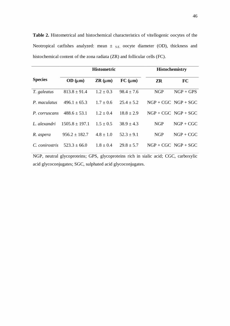
Table 2. Histometrical and histochemical characteristics of vitellogenic oocytes of the
Neotropical catfishes analyzed: mean ± S.E. oocyte diameter (OD), thickness and
histochemical content of the zona radiata (ZR) and follicular cells (FC).
NGP, neutral glycoproteins; GPS, glycoproteins rich in sialic acid; CGC, carboxylic
acid glycoconjugates; SGC, sulphated acid glycoconjugates.
Species
Histometric Histochemistry
OD (m) ZR (m) FC (m) ZR FC
T. galeatus 813.8 ± 91.4 1.2 ± 0.3 98.4 ± 7.6 NGP NGP + GPS
P. maculatus 496.1 ± 65.3 1.7 ± 0.6 25.4 ± 5.2 NGP + CGC NGP + SGC
P. corruscans 488.6 ± 53.1 1.2 ± 0.4 18.8 ± 2.9 NGP + CGC NGP + SGC
L. alexandri 1505.8 ± 197.1 1.5 ± 0.5 38.9 ± 4.3 NGP NGP + CGC
R. aspera 956.2 ± 182.7 4.8 ± 1.0 52.3 ± 9.1 NGP NGP + CGC
C. conirostris 523.3 ± 66.0 1.8 ± 0.4 29.8 ± 5.7 NGP + CGC NGP + SGC
47
5. CONCLUSÕES
A morfologia do aparelho reprodutor de machos e fêmeas dos Siluriformes
analisados apresentou características similares entre peixes migradores, não migradores
com cuidado parental e inseminador interno, tais como:
Espécies Migradoras (P. maculatus, P. corruscans e C. conirostris)
- Grande tamanho corporal;
- Franjas testiculares filiformes;
- Espermatozóides com cabeça arredondada;
- Ausência de secreção testicular (exceto P. maculatus - secreção globosa);
- Pequeno diâmetro dos ovócitos vitelogênicos;
- Zona radiata delgada;
- Células foliculares cúbicas.
Espécies não migradoras com cuidado parental (L. alexandri e R. aspera)
- Tamanho corporal mediano;
- Franjas arredondadas ou projeções pregueadas;
- Espermatozóides com cabeça arredondada;
- Secreção homogênea produzida na região caudal dos testículos;
- Grande diâmetro dos ovócitos vitelogênicos;
- Zona radiata com espessura variada;
- Células foliculares prismáticas.
48
Espécie com inseminação interna (T. galeatus)
- Tamanho corporal pequeno;
- Franjas filiformes;
- Vesícula seminal acessória ao testículo;
- Espermatozóides com cabeça alongada que formam pacotes de
espermatozeugma;
- Grande diâmetro dos ovócitos vitelogênicos;
- Zona radiata delgada;
- Células foliculares prismáticas;
- Epitélio lamelar ovariano com funções armazenadora e secretora.
49
REFERÊNCIAS
ABRAHAM M, HILGE V, RIEHL R, IGER Y. 1993. Muco-follicle cells of the jelly
coat in the oocyte envelope of the sheatfish (Silurus glanis L.). J Morphol 217:37-43.
AGOSTINHO AA. 1985. Estrutura da população, idade e crescimento de
Rhinelepis aspera (Agassiz, 1829) (Osteichthyes, Loricariidae) do rio
Paranapanema. PHD Dissertation, Universidade Federal de São Carlos.
AGOSTINHO AA, PELICICE FM, PETRY AC, GOMES LC, JÚLIO JR. HF. 2007.
Fish diversity in the upper Paraná River basin: habitats, fisheries, management and
conservation. Aquat Ecosyst Health 10:174-186.
ALVES CBM, POMPEU PS. 2001. Peixes do rio das Velhas: passado e presente.
Belo Horizonte: SEGRAC. 194p.
BALON EK. 1984. Patterns in the evolution of reproduction styles in fishes. In: POTTS
GW, WOOTTON RJ, editors. Fish reproduction: strategies and tactics. London:
Academic Press. pp 35-53.
BARNI A, MAZZOLDI C, RASOTTO MB. 2001. Reproductive apparatus and male
accessory structures in two batrachoid species (Teleostei, Batrachoididae). J Fish Biol
58:1557-1569.
BARROS MDM, GUIMARÃES-CRUZ RJ, VELOSO-JÚNIOR VC, SANTOS JE.
2007. Reproductive apparatus and gametogenesis of Lophiosilurus alexandri
Steindachner (Pisces, Teleostei, Siluriformes). Rev Bras Zool 24:213-221.
BAZZOLI N. 2003. Parâmetros reprodutivos de peixes de interesse comercial na região
de Pirapora. In: GODINHO HP, GODINHO AL, editors. Águas, peixes e pescadores do
São Francisco das Minas Gerais. Belo Horizonte: PUC Minas. pp 291-306.
BAZZOLI N, GODINHO HP. 1997. Ovócitos vitelogênicos do surubim
Pseudoplatystoma corruscans e do pacamã Lophiosilurus alexandri. In: MIRANDA
MOT, editor. Surubim. Belo Horizonte: Ibama. pp 81-90.
BENNEMANN ST, SHIBATTA OA, GARAVELLO JC. 2000. Peixes do rio Tibaji:
uma abordagem ecológica. Londrina: Ed. Universidade Estadual de Londrina. 62 p.
50
BEGOVAC PC, WALLACE RA. 1987. Ovary of the Pipefish, Syngnathus scovelli. J
Morphol 193:117-133.
BRITSKI HA, SATO Y, ROSA ABS. 1988. Manual de identificação de peixes da
região de Três Marias: com chaves de identificação para os peixes da bacia do São
Francisco. 3ª ed. Brasília: Câmara dos Deputados/Codevasf. 143p.
BRITO MFG, BAZZOLI N. 2003. Reproduction of the surubim catfish (Pisces,
Pimelodidae) in the São Francisco River, Pirapora Region, Minas Gerais, Brazil. Arch
Med Vet 55:624-633.
BURNS JR, WEITZMAN SH. 2005. Insemination in ostariophysan fishes. In: GRIER
HJ, URIBE MC, editors. Viviparous fishes. Homestead: New Life Publications. pp
107–134.
BURNS JR, MEISNER AD, WEITZMAN SH, MALABARBA LR. 2002. Sperm and
spermatozeugma ultrastructure in the inseminating catfish, Trachelyopterus lucenai
(Ostariophysi: Siluriformes: Auchenipteridae). Copeia 1:173-179.
CARVALHO ED, ALEGRETTI JR, FUJIHARA CY, GRASSIOTO IQ. 1995. Análise
premilinar da biologia reprodutiva de mandiúva Pimelodus maculatus (Pisces –
Siluriformes -Pimelodidae) no reservatório de Jurumirim (alto do rio Paranapanema).
In: Resumos Encontro de Ictiologia, Sociedade Brasileira de Ictiologia.
CHOWDHURY I, JOY KP. 2007. Seminal vesicle and its role in the reproduction of
teleosts. Fish Physiol Biochem 33:383-398.
FAUSTO MA, PICCHIETTI S, TARDEI AR, ZENI C, SCAPIGLIATI G, MAZZINI
M, ABELLI L. 2004. Formation of the egg envelope of a teleost, Dicentrarchus labrax
(L.): immunochemical and cytochemical detection of multiple components. Anat
Embryol 208:43-53.
FÁVARO LF, FREHSE FA, OLIVEIRA RN, SCHWARZ-JÚNIOR R. 2005.
Reprodução do bagre amarelo, Cathorops spixii (Agassiz) (Siluriformes, Ariidae), da
Baía de Pinheiros, região estuarina do litoral do Paraná, Brasil. Rev Bras Zool 22:1022-
1029.
51
FERRARIS CJ. 2007. Checklist of catfishes, recent and fossil (Osteichthyes:
Siluriformes), and catalogue of siluriform primary types. Zootaxa 1418:1-628.
GODINHO AL, KYNARD B, GODINHO HP. 2007. Migration and spawning of
female surubim (Pseudoplatystoma corruscans, Pimelodidae) in the Sao Francisco
river, Brazil. Environ Biol Fish 80:421-433.
GODINHO AL, LAMAS IR, GODINHO HP. 2009. Reproductive ecology of Brazilian
freshwater fishes. Environ Biol Fish 87:143-162.
GODINHO HM, FERRI S, MEDEIROS LO, BARKER JMB. 1974. Morphological
changes in the ovary of Pimelodus maculatus Lacépède, 1803 (Pisces, Siluroidei),
related to the reproductive cycle. Rev Bras Biol, 34:581-588.
GODINHO HP, GODINHO AL. 2003. Águas, peixes e pescadores do São Francisco
das Minas Gerais. Belo Horizonte: Ed. PUC Minas. 440 p.
GRIER HJ. 1993. Comparative organization of Sertoli cells including the Sertoli cell
barrier. In: RUSSELL LD, GRISWOLD MD, editors. The Sertoli Cell. Clearwater:
Cache River Press. pp 704-739.
GUIMARÃES-CRUZ RJ, SANTOS JE. 2004. Testicular structure of three species of
neotropical freshwater pimelodids (Pisces, Pimelodidae). Rev Bras Zool 21:267-271.
JAMIESON BGM. 1991. Fish evolution and systematics: Evidence from
spermatozoa. Cambridge: Cambridge University Press. 319 p.
KOLM N, AHNESJÖ I. 2005. Do egg size and parental care coevolve in fishes? J Fish
Biol 66:1499-1515.
KOYA Y, TAKANO K, TAKAHASHI H. 1995. Annual changes in fine structure of
inner epithelial lining of the ovary of a marine sculpin, Alcichthys alcicornis (Teleostei:
Scorpaeniformes), with internal gametic association. J Morphol 223:85-97.
LAHNSTEINER F, RICHTARSKI U, PATZNER RA. 1990. Function of the testicular
gland in two blenniid fishes, Salaria (=Blennius) pavo and Lipophrys (=Blennius)
dalmatinus (Blenniidae, Teleostei) as revealed by electron microscopy and enzyme
histochemistry. J Fish Biol 37:85-97.
52
LEGENDRE M, LINHART O, BILLARD R. 1996. Spawning and management of
gametes, fertilized eggs and embryos in silurioidei. Aquat Living Resour 9:59-80.
LOIR M, CAUTY C, PLANQUETTE P, LE BAIL PY. 1989. Comparative study of the
male reproductive tract in seven families of South-American catfishes. Aquat Living
Resour 2:45-56.
LOPES DCJR, BAZZOLI N, BRITO MFG, MARIA TA. 2004. Male reproductive
apparatus in the South American catfish Conorhynchus conirostris. J Fish Biol
64:1419-1424.
LOWE-MCCONNELL RH. 1987. Ecological Studies in Tropical Fish Communities.
Cambridge: Cambridge University Press. 382 p.
LUNDENBERG JG, LITTMANN MW. 2003. Pimelodidae. In: REIS RE,
KULLANDER SO, FERRARIS CJ, editors. Check list of the freshwater fishes of
South and Central America. Porto Alegre: EDIPUCRS. pp 432-446.
MAZZOLDI C, PETERSEN CW, RASOTTO MB. 2005. The influence of mating
apparatus on seminal vesicle variability among gobies (Teleostei, Gobiidae). J Zool
Systemat Evol Res 43:307-314.
MAZZOLDI C, LORENZI V, RASOTTO MB. 2007. Variation of male reproductive
apparatus in relation to fertilization modalities in the catfish families Auchenipteridae
and Callichthyidae (Teleostei: Siluriformes). J Fish Biol 70:243-256.
MCELMAN JF, BALON EK. 1980. Early ontogeny of white sucker, Catostomus
commersoni, with steps of saltatory development. Environ Biol Fish 5:191-224.
MEISNER AD, BURNS JR, WEITZMAN SH, MALABARBA LR. 2000. Morphology
and histology of the male reproductive system in two species of internally inseminating
South American catfishes. Trachelyopterus lucenai and T. galeatus (Teleostei:
Auchenipteridae). J Morphol 246:131-141.
NAGAHAMA Y. 1983. The functional morphology of teleost gonads. In: HOAR WS,
RANDALL DJ, DONALDSON EM, editors. Fish Physiology. New York: Academic
Press. pp 223-276.
53
PARENTI LR, GRIER HJ. 2004. Evolution and phylogeny of gonadal morphology in
bony fishes. Integr Comp Biol 44:333-348.
PARREIRA GG, CHIARINI-GARCIA H, MELO RCN, VIEIRA FO, GODINHO, HP.
2009. Spermatozoon and its relationship with the ovarian lamellae in the internally
inseminating catfish Trachelyopterus galeatus. Microsc Res and Tech 72:889-897.
PATIÑO R, SULLIVAN CV. 2002. Ovarian follicle growth, maturation, and ovulation
in teleost fish. Fish Physiology and Biochemistry 26:57–70.
PEARSE AGE. 1985. Histochemistry, Theoretical and Applied Analytical
Technology. Edinburgh: Churchill Livingstone. 1055 p.
REIS RE, KULLANDER SO, FERRARIS, CJ. 2003. Check list of the freshwater
fishes of South and Central America. Porto Alegre: EDIPUCRS. 729 p.
RICHTARSKI U, PATZNER RA. 2000. Comparative morphology of male
reproductive apparatus in Mediterranean blennies (Blenniidae). J Fish Biol 56:22-36.
RIEHL R. 1996. The ecological significance of the egg envelope in teleosts with special
reference to limnic species. Limnologica 26:183-189.
RIZZO E, SATO Y, BARRETO BP, GODINHO HP. 2002. Adhesiveness and surface
patterns of eggs in neotropical freshwater teleosts. J Fish Biol 61:615-632.
ROSA RS, LIMA FCT. 2008. Peixes. In: MACHADO ABM, DRUMMOND GM,
PAGLIA AP, editors. Livro vermelho da fauna brasileira ameaçada de extinção.
Brasília: Ministério do Meio Ambiente. pp 8-285.
SANTOS GM, MÉRONA B, JURAS AA, JÉGU M. 2004. Peixes do baixo Rio
Tocantins: 20 anos depois da Usina Hidrelétrica Tucuruí. Brasília: Eletronorte. 216
p.
SANTOS JE, BAZZOLI N, SANTOS GB. 2001. Morphofunctional organization of the
male reproductive apparatus of the catfish Iheringichthys labrosus (Lütken, 1874).
Tissue Cell 33:533-540.
54
SANTOS JE, PADILHA GEV, BOMCOMPAGNI-JÚNIOR O, SANTOS GB, RIZZO
E, BAZZOLI N. 2006. Ovarian follicle growth in the catfish Iheringichthys labrosus
(Siluriformes: Pimelodidae). Tissue Cell 38:303–310.
SATO Y. 1999. Reprodução de peixes da bacia do rio São Francisco: indução e
caracterização de padrões. PHD Dissertation, Universidade Federal de São Carlos.
179 p.
SATO Y, GODINHO HP. 1988. Adesividade de ovos e tipo de desova dos peixes de
Três Marias, MG. In: Anais Encontro Anual de Aquicultura de Minas Gerais, Belo
Horizonte.
SATO Y, GODINHO HP. 1999. Peixes da bacia do rio São Francisco. In: Lowe-
McConnell RH, editor. Estudos ecológicos de comunidades de peixes tropicais. São
Paulo: EDUSP. 534 p.
SATO Y, GODINHO HP. 2004. Migratory fishes of the São Francisco River. In:
CAROLSFELD J, HARVEY B, ROSS C, BAER A, editors. Migratory fishes of South
America: Biology, Fisheries and Conservation Status. Victoria: World Fisheries
Trust. pp 195-232.
SATO Y, FENERICH-VERANI N, NUÑER APO, GODINHO HP, VERANI JR. 2003.
Padrões reprodutivos de peixes da bacia do São Francisco. In: GODINHO HP,
GODINHO AL, editors. Águas, peixes e pescadores do São Francisco das Minas
Gerais. Belo Horizonte (Brazil): PUC Minas. pp 229-274.
SHIBATTA OA. 2003. Pseudopimelodidae (Bumblebee catfishes, dwarf marbled
catfishes). In: REIS RE, KULLANDER SO, FERRARIS CJ, editors. Check list of the
freshwater fishes of South and Central America. Porto Alegre: EDIPUCRS. pp 401-
405.
SISCAR AK. 1970. Morphology of the urinogenital apparatus of some siluroid fishes.
Proc Zool Soc 23:93-117.
SNEED KE, CLEMENS HP. 1963. The morphology of the testes and accessory
reproductive glands of the catfishes (Ictaluridae). Copeia 4:606-611.
55
SPADELLA MA, OLIVEIRA C, QUAGIO-GRASSIOTTO, I. 2008. Morphology and
histology of male and female reproductive apparatus in the inseminating species
Scoloplax distolothrix (Teleostei: Siluriformes: Scoloplacidae). J Morphol 269:1114-
1121.
STEHR CM, HAWKES JW. 1979. The comparative ultrastructure of the egg membrane
and associated pore structures in the starry flounder Platichthys stellatus (Pallas) and
pink salmon Oncorhynchus gorbuscha (Walbaum). Cell Tissue Res 202:341-356.
SUZUKI HI, AGOSTINHO AA, WINEMILLER KO. 2000. Relationship between
oocyte morphology and reproductive strategy in loricariid catfishes of the Paraná River,
Brazil. J Fish Biol 57:791-807.
TEUGELS GG. 1996. Taxonomy, phylogeny and biogeography of catfishes
(Ostariophysi: Siluroidei): an overview. Aquat Living Resour 9:9-34.
TYLER CR, SUMPTER JP. 1996. Oocyte growth and development in teleosts.
Reviews in Fish Biology and Fisheries, 6:287-318.
WOOTTON RJ. 1984. Introduction: tactics and strategies in fish reproduction. In:
POTTS GW, WOOTTON RJ, editors. Fish reproduction: strategies and tactics.
London: Academic Press. 410 p.