Élcio mÁrio faria jÚnior - s3.amazonaws.com · um dos maiores avanços da ortodontia na montagem...
TRANSCRIPT

Londrina 2013
ÉLCIO MÁRIO FARIA JÚNIOR
CENTRO DE CIÊNCIAS BIOLÓGICAS E DA SAÚDE MESTRADO EM ODONTOLOGIA
AVALIAÇÃO IN VIVO DA RUGOSIDADE E MORFOLOGIA SUPERFICIAL DO ESMALTE APÓS REMOÇÃO DE
BRAQUETES COM DIFERENTES POLIMENTOS

Londrina 2013
AVALIAÇÃO IN VIVO DA RUGOSIDADE E MORFOLOGIA SUPERFICIAL DO ESMALTE APÓS REMOÇÃO DE
BRAQUETES COM DIFERENTES POLIMENTOS
Dissertação apresentada ao Programa de Pós-Graduação em Odontologia da Universidade Norte do Paraná - UNOPAR, como requisito parcial à obtenção do título de Mestre em Odontologia.
Área de Concentração: Dentística Restauradora. Orientador: Prof. Dr. Ricardo Danil Guiraldo
ÉLCIO MÁRIO FARIA JÚNIOR

AUTORIZO A REPRODUÇÃO TOTAL OU PARCIAL DESTE TRABALHO, POR QUALQUER MEIO CONVENCIONAL OU ELETRÔNICO, PARA FINS DE ESTUDO E PESQUISA, DESDE QUE CITADA A FONTE.
Dados Internacionais de catalogação-na-publicação Universidade Norte do Paraná
Biblioteca Central Setor de Tratamento da Informação
Faria Júnior, Élcio Mário F233a Avaliação in vivo da rugosidade e morfologia superficial do esmalte
após remoção de braquetes com diferentes polimentos / Élcio Mário Faria Júnior Londrina: [s.n], 2013.
ix; 41f. Dissertação (Mestrado). Odontologia. Dentística Restauradora.
Universidade Norte do Paraná. Orientador: Prof. Dr. Ricardo Danil Guiraldo 1- Odontologia - dissertação - mestrado - UNOPAR 2- Dentística
restauradora 3- Remoção de braquetes 4- Rugosidade I- Guiraldo, Ricardo Danil, orient. III- Universidade Norte do Paraná.
CDU 616.314-089.27/.28

ÉLCIO MÁRIO FARIA JÚNIOR
AVALIAÇÃO IN VIVO DA RUGOSIDADE E MORFOLOGIA SUPERFICIAL DO ESMALTE APÓS REMOÇÃO DE BRAQUETES
COM DIFERENTES POLIMENTOS
Dissertação apresentada ao Programa de Pós-Graduação em Odontologia da Universidade Norte do Paraná - UNOPAR, como requisito parcial à obtenção do título de Mestre em Odontologia. Área de Concentração: Dentística Preventiva e Restauradora.
BANCA EXAMINADORA
____________________________________ Prof. Dr. Ricardo Danil Guiraldo Universidade Norte do Paraná
____________________________________ Profa. Dra. Sandrine Bittencourt Berger
Universidade Norte do Paraná
____________________________________ Prof. Dr. Lourenço Correr Sobrinho
Universidade Estadual de Campinas
Londrina, 25 de fevereiro de 2013.

DEDICO ESTE TRABALHO
Primeiramente a DEUS por conceder a oportunidade de concluir o
Mestrado em Odontologia e sempre estar presente em minha vida,
capacitando-me durante minha trajetória.
Aos meus pais, Élcio Mário Faria e Sandra Irene Barbeiro Faria,
por serem a base e complemento da minha formação tanto pessoal
quanto profissional, não medindo esforços para que eu pudesse chegar
até aqui, sem seus apoios e ajuda, tudo isso seria impossível.
Aos meus irmãos, Élton e Renata, e a minha esposa Michelle
que não cansaram de torcer por minha realização pessoal e profissional.
Agradeço a Deus por tê-los como irmãos, esposa e amigos, grande
companheiros que posso compartilhar tantos momentos de minha vida.

AGRADECIMENTOS ESPECIAIS
Ao meu orientador Prof. Dr. Ricardo Danil Guiraldo, por sua
amizade e contribuição, que se esmerou em cuidados para comigo e
com este trabalho, conduzindo excelentemente o seu desenvolvimento.
Agradeço por sua confiança na minha capacidade de trabalho, paciência
e por sua dedicação em fazer de mim um melhor profissional.
Ao Prof. Dr. Luiz Roberto Venturim, que desde a graduação
contribui para minha formação profissional e me permitiu uma chance
como estagiário e posteriormente professor da faculdade de Odontologia
de Presidente Prudente – Unoeste.

AGRADECIMENTOS À Universidade Norte do Paraná (UNOPAR), na pessoa da Reitora Wilma
Jandre Melo e pró-reitor de pesquisa e pós-graduação Prof. Dr. Hélio Hiroshi Suguimoto, por sua estrutura e corpo docente, que contribuiu para o meu crescimento intelectual.
Ao Prof. Dr. Ricardo Danil Guiraldo por seu apoio, amizade e oportunidades
oferecidas. Aos demais professores da Universidade Norte do Paraná – Prof. Dr. Alcides
Gonini Júnior (Coordenador do Programa de Pós-graduação de Odontologia da Unopar), Profª. Drª. Regina Célia Poli Frederico, Prof. Dr. Rodrigo Varella de Carvalho, Profª. Drª. Sandra Kiss Moura, Profª. Drª. Sandra Mara Maciel e Profª. Drª. Sandrine Bittencourt Berger, por colaborarem com minha formação.
À Universidade do Oeste Paulista - Faculdade de Odontologia de Presidente
Prudente– nas pessoas Profª. Drª. Renata Ricco e Profª. Drª. Cláudia de Oliveira Lima que me permitiram algumas ausências enquanto estive em Londrina para a realização das pesquisas.
Aos demais Familiares e Amigos que sempre participaram da minha formação
pessoal e estiveram presente em vários momentos da minha vida. A todos que indiretamente contribuíram para a conclusão desta pesquisa.
OS MEUS SINCEROS AGRADECIMENTOS.

FARIA JÚNIOR, Élcio Mário. Avaliação in vivo da rugosidade e morfologia
superficial do esmalte após remoção de braquetes com diferentes polimentos.
2013. 41 f. [Dissertação de Mestrado]. Programa de Pós-Graduação em Odontologia
– Universidade Norte do Paraná, Londrina, 2014.
RESUMO
Objetivos: o objetivo nesse estudo foi avaliar a rugosidade e a morfologia do
esmalte por rugosímetro e Microscopia Eletrônica de Varredura (MEV), após a
remoção de braquetes metálicos. Materiais e Métodos: 10 pacientes voluntários
que não tinham cárie, restauração, trauma, bruxismo, ou rachaduras nos incisivos
superiores foram selecionados. Após a conclusão do tratamento ortodôntico, os
braquetes foram removidos. Os dentes dos pacientes foram aleatoriamente polidos,
para um lado previamente sorteado foi realizado com acabamento e polimento com
Sof-Lex e no outro lado com broca carbide multi-laminada (n = 10). Réplicas
dentárias com dentes polidas foram obtidas utilizando resina epóxica. Três
mensurações de rugosidade superficial foram realizadas em diferentes direções com
angulação de 120º entre elas. Os dados de rugosidade foram avaliados
estatisticamente pelo teste t Student. Após, três amostras de cada grupo foram
utilizadas para a análise MEV. Resultados: teste t Student mostrou que o grupo da
broca carbide (0,31 µm) tinha irregularidades significativamente superiores quando
comparados com o grupo Sof-Lex (0,25 µm), após remoção da resina. Conclusão:
Entre os sistemas estudados, o polimento Sof-Lex mostrou melhor polimento no
esmalte.
Palavras-chave: Polimento Dental; Esmalte Dentário; Microscopia Eletrônica de
Varredura.

FARIA JÚNIOR, Élcio Mário. Evaluation in vivo of the roughness and surface
morphology of enamel after removal of brackets with different polishing. 2013.
41 f. [Dissertação de Mestrado]. Programa de Pós-Graduação em Odontologia –
Universidade Norte do Paraná, Londrina, 2014.
ABSTRACT
Objectives: the aim of this study was to evaluate the roughness and morphology of
the enamel by surface roughness tester and Scanning Electron Microscopy (SEM)
after removal of metal brackets. Materials and Methods: 10 volunteer patients were
selected that no caries, restoration, trauma, bruxism or cracks on the upper incisors.
After the conclusion of orthodontic treatment, the brackets were removed. The teeth
of the patients were randomly polished, to one side previously drawn was performed
finishing and polishing with Sof-Lex or carbide bur multi-laminated (n = 10). Replicas
dental with polished teeth were obtained using epoxy resin. Three surface roughness
measurements were performed in different directions with an angle of 120° among
them. The roughness data were statistically evaluated by Student t test. After, three
specimens from each group were used for the SEM analysis. Results: Student t test
showed that the carbide bur group (0.31 µm) had significantly higher irregularities
when compared with the Sof-Lex group (0.25 µm) after resin removal. Conclusion:
Between the systems studied, the Sof-Lex polishing system showed the best
polishing enamel.
Keywords: Dental Polishing; Dental Enamel; Scanning Electron Microscopy.
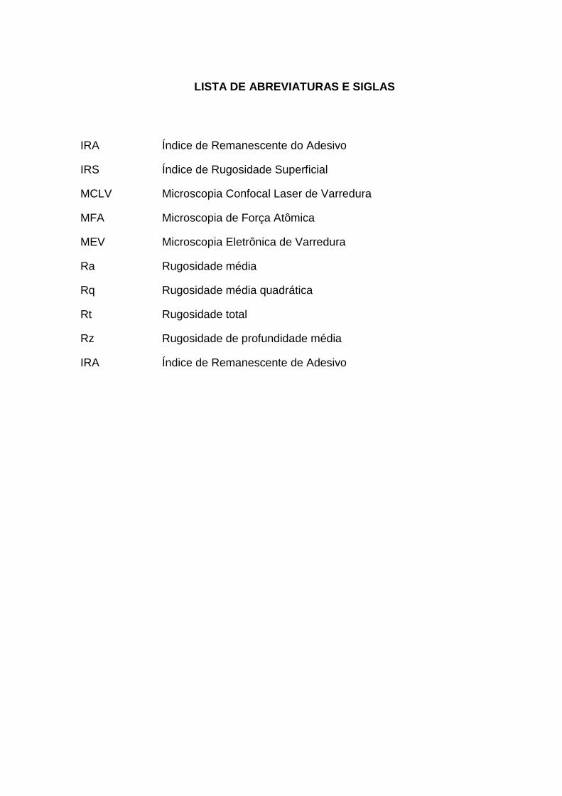
LISTA DE ABREVIATURAS E SIGLAS
IRA Índice de Remanescente do Adesivo
IRS Índice de Rugosidade Superficial
MCLV Microscopia Confocal Laser de Varredura
MFA Microscopia de Força Atômica
MEV Microscopia Eletrônica de Varredura
Ra Rugosidade média
Rq Rugosidade média quadrática
Rt Rugosidade total
Rz Rugosidade de profundidade média
IRA Índice de Remanescente de Adesivo

SUMÁRIO
1 INTRODUÇÃO ...................................................................................................... 11
2 REVISÃO DA LITERATURA ................................................................................ 12
3 PROPOSIÇÃO ...................................................................................................... 17
4 ARTIGO ................................................................................................................ 18
5 CONCLUSÃO ....................................................................................................... 37
REFERÊNCIAS ..................................................................................................... 38
APÊNDICES ......................................................................................................... 39
APÊNDICE A ......................................................................................................... 39
APÊNDICE B ......................................................................................................... 40

11
1 INTRODUÇÃO
O procedimento de colagem de braquetes em dentes humanos representou
um dos maiores avanços da Ortodontia na montagem de aparelhos ortodônticos,
diminuindo acentuadamente o uso de bandas. Essa evolução somente tornou-se
possível após a técnica do condicionamento ácido desenvolvida por Buonocore1,
possibilitando o aumento na aderência dos materiais resinosos na coroa dentária.
Este procedimento simplificou a montagem do aparelho ortodôntico fixo e
promoveu a redução das fases e do tempo de tratamento, proporcionando um
avanço significativo no tratamento ortodôntico. Em função disso, a facilidade na
remoção da placa bacteriana foi obtida, redução das gengivites e hiperplasias, não
utilização de separadores, ausência de espaços gerados pelas bandas após a
remoção do aparelho, possibilidade de colagem de braquetes em dentes
parcialmente erupcionados, menor probabilidade de descalcificações geradas por
infiltrações, maior facilidade na detecção de cárie e estética2.
A remoção do remanescente de compósito ou cimento de ionômero de vidro
das superfícies dos dentes é o procedimento final necessário para retornar a
superfície do esmalte, tanto quanto possível, para a condição inicial de pré-
tratamento3. Por isso, muitos pesquisadores têm introduzido diferentes técnicas para
a remoção da resina composta e subseqüente polimento do esmalte sem causar
danos iatrogénicos que incluem a raspagem com um dispositivo de escalonamento,
ou remoção de banda com alicate e polimento com broca carbide de tungstênio em
contra-ângulo, bem como o uso de discos abrasivos3-6. Estudos clínicos e
laboratoriais revelaram que instrumentos rotatórios podem alterar irreversivelmente a
superfície do esmalte, causando riscos profundos ou perda do esmalte.
Freqüentemente, adesivo remanescente foi encontrado na superfície do esmalte,
mesmo após a limpeza e polimento com instrumentos rotativos7.
A rugosidade de superfície após a descolagem pode ser medida de várias
maneiras. Ferramentas de contacto lineares de medição podem ser utilizadas tais
como o rugosímetro8. Além desta, existe a análise visual através de microscopia
eletrônica de varredura9. Existem muitos estudos laboratoriais7-9, entretanto não
existe estudo in vivo em incisos centrais superiores. Assim o objetivo nesse estudo
será avaliar a rugosidade e morfologia do esmalte através de rugosímetro e
Microscopia Eletrônica de Varredura (MEV) após a remoção de braquetes metálicos.

12
2 REVISÃO DA LITERATURA
Muito tem se estudado sobre a descolagem de braquetes ortodônticos,
principalmente em relação à remoção dos remanescentes resinosos. As
modificações da superfície do esmalte em decorrência de diferentes técnicas,
métodos e mecanismos para este fim, podem ser irreversíveis, e independem da
técnica de colagem ou do tipo de braquete. Existe sempre o risco de danificar o
esmalte, e o ortodontista deve se preocupar com a integridade da superfície do
esmalte.
Buonocore1, estudando formas de aumentar a adesão das resinas acrílicas ao
esmalte dentário no ano de 1955, usou o condicionamento em 15 superfícies
dentárias com ácido phosphomolybdate 50% (com 15 superfícies controle) e com
ácido fosfórico 85% em 10 superfícies dentárias (com 10 superfícies controle).
Observando que a adesão da resina acrílica ao esmalte aumentava, quando o ácido
fosfórico era previamente aplicado sobre o esmalte. Relatando como fatores
prováveis da causa desse aumento de adesão: a) grande aumento da área de
superfície; b) exposição da matéria orgânica do esmalte, que funcionaria como uma
rede, onde a resina se adere; c) criação de espaços em profundidade, ao longo da
área interprismática, dentro dos quais a resina pode penetrar; d) remoção da
camada superficial do esmalte, expondo uma superfície mais favorável à adesão; e)
a incorporação, na superfície do esmalte, de uma camada de grupos fosfatos de alta
polaridade, derivados do ácido fosfórico.
Rouleau et al.4 observaram no ano de 1982 que diferenças significativas eram
encontradas na lisura da superfície do esmalte, com as diferentes técnicas usadas
para a remoção da resina remanescente. Foram avaliados 45 dentes de pacientes
tratados ortodonticamente após a remoção dos braquetes e da resina remanescente.
Os meios empregados para a remoção da resina remanescente foram: (1) alicates
de remoção de resina, (2) brocas de 12 lâminas, em baixa rotação e (3) brocas de
tungstênio ultrafinas. Os autores concluíram que: 1. O uso de alicates, não é
desejável para a remoção do remanescente de resina. 2. Brocas de 12 lâminas, em
baixa rotação são boas para a remoção da resina, mas deixam uma fina camada de
arranhões e depressões. 3. A broca de tungstênio carbide, em alta velocidade e
refrigerada à água, produzia uma superfície que se aproximava do esmalte sem
tratamento. Apresentando como desvantagem maior tempo de trabalho. 4. A

13
aspereza diminuiu com a seguinte ordem das técnicas de remoção: (1) alicate
removedor de resina, (2) broca carbide 12 lâminas, (3) broca de tungstênio ultrafina.
Além disto, polimento com pedra pomes também se mostrou benéfico.
Howell & Weeks5 avaliaram em 1990 a rugosidade superficial do esmalte
seguindo a utilização de várias combinações de agentes de união, brocas e
procedimentos de polimento. Foram colados braquetes ortodônticos em 135 pré-
molares extraídos de adolescentes. Após a remoção dos braquetes e acabamento, a
superfície do esmalte foi coberta com ouro e examinada com microscópio eletrônico
de varredura. Cada micrografia com aumento de 200 vezes foi classificada de
acordo com a rugosidade de superfície. O uso de diferentes compósitos e diferentes
brocas não mostraram efeito significante no acabamento da superfície do esmalte.
Somente dois procedimentos de acabamento tiveram um efeito significante na
rugosidade superficial. A utilização de discos Soflex seguido de Pedra Pomes
resultou numa superfície mais rugosa quando comparado ao uso somente de pedra
pomes que produziu uma superfície do esmalte mais lisa.
Campbell3 avaliou a superfície do esmalte após a remoção de braquetes em
1995. A proposta nesse trabalho foi apresentar um método clínico prático e eficiente
para devolver a superfície do esmalte o mais próximo possível da original, com o
menor dano. Foi realizado um levantamento de questões e essas foram enviadas
para 72 ortodontistas. Desses levantamentos 62 retornaram, representando 86.1%.
Os resultados foram quantificados como se segue: 80% dos profissionais
reconheciam danos ao esmalte, após a remoção da resina residual, enquanto 19%
destes não observaram problemas algum. Aproximadamente 55% dos que foram
questionados usavam alicate de corte de ligaduras ou alicate removedor de bandas
para remoção dos braquetes. Quanto à resina residual, 45% dos profissionais
utilizavam broca de tungstênio carbide, enquanto 32% utilizavam algum instrumento
raspador (alicates). A maioria dos profissionais utilizava pedra pomes para o
polimento final, 50% desses não observavam problemas na aparência do esmalte
após a descolagem de braquetes e polimento e 50% achavam que o esmalte intacto
era melhor. Foram também avaliados seis métodos de remoção de resina. Os
dentes foram divididos em seis grupos e a resina foi removida com: 1) ponta
montada pedra verde, 2) broca diamantada, 3) alicate removedor de banda, 4) broca
para acabamento 30 laminas, 5) broca carbide e 6) discos abrasivos (Sof-Lex). Os
dentes, em cada uma das amostras, foram polidos usando variedade de abrasivos:

14
1) ponta de resina com pasta de oxido de alumínio com glicerina; 2) pedra pomes; 3)
taça de borracha com pasta para polimento de porcelana; e 4) taça de borracha
verde e marrom para polimento de amálgama. O trabalho conclui que a broca de
tungstênio carbide 30 laminas parece ser o método mais eficiente de remoção de
resina residual e produz o mínimo de danos e sugere uma seqüência de polimento
usando-se as pontas de resina e taças de borracha com pedra-pomes e taças
marrom e verde para polimento de amálgama.
Hong & Lew6 avaliaram 4 métodos para remoção de resina remanescente
após a remoção de braquetes ortodônticos em comparação com o método de
remoção com brocas carbide em baixa rotação no ano de 1995. Foram colados 50
braquetes em pré-molares extraídos com finalidade ortodôntica. Após a remoção dos
braquetes, os dentes foram divididos em 5 grupos. Grupo A - Alicate removedor de
banda Ormco; Grupo B - Broca carbide em baixa rotação Komet; Grupo C - Ponta
dimantada ultrafina em alta rotação; Grupo D – Broca carbide em alta rotação Jet;
Grupo E – Pedra branca de acabamento em alta rotação. Cada procedimento de
acabamento foi limitado a 15 segundos. O compósito remanescente no esmalte foi
classificado por 4 avaliadores independentes usando o Índice de Remanescente do
Adesivo (IRA). Posteriormente, a superfície do esmalte foi examinada com
microscópio eletrônico de varredura. As micrografias foram classificadas pelos
mesmos 4 avaliadores utilizando o Índice de Rugosidade Superficial (IRS). Não
houve diferença estatística significante na variabilidade entre os examinadores em
ambas avaliações IRA e IRI. Nenhum método foi considerado absoluto e ideal para a
remoção dos compósitos remanescente.
Bishara et al.2 avaliaram o efeito da repetida colagem com 2 adesivos
diferentes (um compósito e um cianoacrilato) sobre a resistência ao cisalhamento de
brackets ortodônticos no ano de 2002. Trinta e um molares humanos recém-
extraídos foram coletados. Braquetes foram colados com um dos adesivos
ortodônticos de acordo com as instruções do fabricante. No grupo I, os dentes foram
condicionados com ácido fosfórico a 37%, um adesivo foi aplicado, e os braquetes
foram colados com Transbond XT e fotoativados por 20 segundos. No grupo II, os
dentes foram condicionados com ácido fosfórico a 35%, e os braquetes foram
colados com SmartBond. Em cada grupo, os dentes foram colados e descolados 3
vezes com o mesmo adesivo. Os resultados indicaram que, na primeira sequência
de descolagem, os dois adesivos não têm diferença significante à resistência ao

15
cisalhamento. O compósito tinha uma resistência maior ao cisalhamento que
cianoacrilato na segunda seqüência de colagem / descolagem, mas não na terceira.
As mudanças na força de ligação após a colagem repetida pode estar relacionada
com alterações nas características morfológicas da superfície do dente causada por
restos de adesivo.
Eliades et al.9 avaliaram quantitativamente em 2004 a rugosidade da
superfície do esmalte após a remoção de braquetes utilizando diferentes métodos de
remoção de resina. A superfície do esmalte da coroa de 30 pré-molares foi coberta
com fita preta com uma abertura de 3 mm no terço médio da face vestibular para
padronizar a área a ser analisada. As superfícies dos esmaltes iniciais foram sujeitas
a perfilometria, registrando quatro parâmetros de rugosidade (Ra, Rq, Rt e Rz). Os
braquetes foram colados na superfície do esmalte com resina quimicamente ativada,
sem mistura de adesivo, e removidos após 1 semana. A remoção da resina em
metade das amostras foi realizada com uma broca carbide 8 lâminas, e na outra
metade com uma ponta diamantada ultra-fina, ambas em alta rotação. Uma segunda
medição perfilométrica foi realizada após a remoção da resina. Pra finalizar, todas as
superfícies foram submetidas ao polimento com discos Soflex, e um terceiro registro
de rugosidade foi obtido. Com relação à rugosidade da superfície do esmalte, os
dois métodos de remoção de resina mostraram diferenças significativas, entretanto
não foi observado redução da rugosidade após o polimento com os discos Soflex.
Os autores concluíram que o aumento na maioria das variáveis de rugosidade
induzida pelos procedimentos de remoção de braquetes não foi revertido no final da
etapa de acabamento, independentemente do protocolo de remoção de resina
utilizada, o que sugere um efeito irreversível da textura do esmalte.
Karan et al.7 em 2010 avaliaram a rugosidade superficial de esmalte após a
remoção de braquetes ortodônticos para testar a hipótese de que não há diferença
significativa entre os efeitos de duas brocas. Vinte pré-molares foram sujeitos a
Microscopia de Força Atômica (MFA), obtendo os valores de rugosidade inicial. Os
braquetes foram colados com compósitos fotopolimerizados e descolados com
alicate removedor de braquetes. Em metade das amostras, o compósito
remanescente foi removido com broca de carbide de tungstênio, enquanto que broca
de compósito reforçado por fibra foi utilizado na outra metade. Na segunda análise
com MFA foi realizada após a remoção da resina. A duração do procedimento de
remoção da resina também foi registrada. Os autores concluíram que os dois

16
instrumentos de remoção de resina tiveram efeitos significantemente diferentes na
rugosidade do esmalte; maior rugosidade foi obtido com o uso de broca carbide de
tungstênio. O tempo necessário para a remoção da resina com a broca de compósito
foi significativamente maior do que o tempo requerido com a broca carbide de
tungstênio.
Brauchli et al.8 examinaram a rugosidade das superfícies do esmalte após
diferentes procedimentos de colagem e remoção de braquetes ortodônticos em
2011. Utilizaram 42 incisivos bovinos e realizaram abrasão convencional com ácido
fosfórico a 37%, abrasão com jato de ar e a combinação das mesmas. Foram
colados braquetes ortodônticos e em seguida removidos, e os remanescentes
resinosos removidos com brocas carbide e jato de ar abrasivo. A rugosidade do
esmalte foi avaliada usando Microscopia Confocal Laser de Varredura (MCLV). Os
autores concluíram que com relação ao condicionamento do esmalte, não houve
efeito significante na superfície do esmalte após a remoção dos braquetes. A
remoção dos remanescentes resinosos com broca carbide ou jato de ar abrasivo
resultaram em diferenças na rugosidade superficial.

17
3 PROPOSIÇÃO
O objetivo nessa Dissertação1 foi avaliar a rugosidade e morfologia do
esmalte através de rugosímetro e microscopia eletrônica de varredura (MEV) após a
remoção de braquetes metálicos colados com o compósito Filtek Z100 e polidos com
diferentes sistemas.
1 Este estudo foi realizado no formato alternativo, na forma de artigo científico intitulado “In vivo
evaluation of the surface roughness and morphology of enamel after bracket removal and polishing by different techniques”. Este artigo será submetido à publicação ao periódico American Journal of Orthodontics and Dentofacial Orthopedics, assim, formulado conforme suas normas.

18
4 ARTIGO CIENTÍFICO
In vivo evaluation of the surface roughness and morphology of enamel after
bracket removal and polishing by different techniques
ABSTRACT
Introduction: The aim of this study was to evaluate the surface roughness and
morphology of enamel with a surface roughness tester and scanning electron
microscopy (SEM) after the removal of metal brackets. Methods: Ten orthodontic
patients were selected for the study. At the conclusion of orthodontic treatment, the
patients’ metal brackets were removed. For each patient, teeth on one side of the
mouth were randomly chosen for finishing and polishing with aluminum oxide disc (n
= 10). Teeth on the other side were finished with multilaminated carbide bur (n = 10).
Final polishing of all teeth was performed with a different polisher. Dental replicas
with polished teeth were obtained with epoxy resin. Three surface roughness
measurements were performed in different directions with an angle of 120° among
them. The roughness data were statistically evaluated by Student’s t-test. Three
specimens from each group were also used for SEM analysis. Results: After resin
removal, the average roughness in the carbide bur group (0.31 µm) was significantly
greater than that in the aluminum oxide disc group (0.25 µm). Conclusions: The
aluminum oxide disc polishing system showed better results for polishing enamel
than the multilaminated carbide bur system.
KEY WORDS: Dental Polishing, Dental Enamel; Electron Microscopy

19
INTRODUCTION
Since 1970, the bonding of orthodontic brackets to tooth enamel has become
an accepted clinical technique.1 A typical bonding procedure involves alteration of the
enamel surface by acid-etching, followed by the application of adhesive primer and
resin.1-3 Adhesive systems for bracket bonding play a key role in fixed orthodontic
therapy. Accordingly, many research teams have evaluated the bond strength of the
adhesive interface,4,5 paying special attention to the adhesive or bracket type5-7 and
the enamel preconditioning method.4,5 Relevant factors in bracket debonding5,6 and
the subsequent polishing of the enamel surface5-7 have also been investigated.
Incorrect removal of the brackets and adhesive can lead to permanent
damage of the enamel and an extended period of debonding.5,8 The final procedure
in returning the enamel surface to the original, pretreatment condition involves the
removal of all attachments and remaining resin from the tooth surfaces.9,10
Researchers have tested different techniques for resin removal and enamel polishing
to avoid causing iatrogenic damage. These techniques include scraping with a scaler
or band-removing plier, resin removal with a tungsten carbide bur (the most common
method)5,9 in a contra-angle hand-piece, and the use of abrasive discs.9,10 However,
any debonding procedure can result in variable surface quality and enamel loss,
depending on the instruments used.5-7
Esthetics is a primary consideration for patients seeking orthodontic treatment,
and many treatment options are available that maximize the likelihood of an attractive
outcome. The form and brightness of the maxillary anterior teeth are important for
both dental and facial esthetics. These teeth should be treated so as to restore the
optimal dentolabial relationship in harmony with the overall facial appearance.11

20
However, little scientific data are available in the dental literature to use as a guide for
defining the proper form and brightness of the anterior teeth or for determining their
normal relationships. The brightness of the buccal surface must be restored after
orthodontic treatment, a goal that may not be possible to achieve with rough
surfaces.
Surface roughness after debonding can be measured in several ways,
including linear contact measurement5 and visual analysis through scanning electron
microscopy (SEM).12 Many laboratory studies, but few in vivo ones, have examined
the surface roughness after debonding.5,10,12 Fewer studies have evaluated this
condition for the upper central incisors. Thus, the aim of this study was to evaluate
the surface roughness and morphology of the enamel by using a surface roughness
tester and SEM after the removal of metal brackets. The null hypothesis was that the
surface roughness and morphology of the enamel in areas close to adhesive
remnants (ARs) would not differ from those in areas that were polished by aluminum
oxide disc or carbide bur.

21
MATERIALS AND METHODS
The present study was approved by the Research Ethics Committee of the
University of North Paraná. For this study, 10 patients were selected (5 men and 5
women), ranging in age from 14 years to 21 years 7 months (mean: 17 years 6
months). No patient had any caries, restorations, history of trauma, bruxism, or
cracks on the upper incisors. The middle third of the buccal face of all teeth was
etched with 37% phosphoric acid gel (Condac 37, FGM, Joinville, PR, Brazil), rinsed
with air-water spray, and air-dried for 20 seconds each. One layer of adhesive (Adper
Single Bond 2, 3M ESPE, St. Paul, MN, USA) was applied to the etched area. Then,
stainless steel standard maxillary incisor brackets (Roth, Morelli, Sorocaba, SP,
Brazil) were positioned and firmly bonded to the teeth with Filtek Z100 (3M ESPE). A
microbrush was used to remove excess.
Bonding of the brackets was achieved by light activation with an Ultraled (Dabi
Atlante, Ribeirão Preto, SP, Brazil) light-emitting diode (LED) at an irradiance of 800
mW/cm2. Each side of the bracket was exposed to the LED four times, with a total
exposure time of 40 s and an energy density of 32 J/cm2. In total, 10 brackets were
bonded in each different polishing.
The orthodontic treatment duration for patients ranged from 15 to 28 months
(mean: 21.5 months). At the conclusion of the orthodontic treatment, the brackets
were removed by pliers. Then, prophylactic procedures were performed with pumice
and a Robinson brush in a hand-piece under low speed (Dabi-Atlante). The teeth
were washed, dried, and molded with Aquasil Ultra silicone (Dentsply Caulk, Milford,
DE, USA) by adding in single simultaneous printing technique (Ultra-light – Aquasil
Ultra XLV and Putty – Aquasil Easy Mix Putty). The mold was cast with epoxy resin

22
(20-8130-032, Lake Bluff, IL, USA).
The adhesive remnant index (ARI) values of the dental replicas (of the
debonded tooth and remaining resin) were analyzed with an optical microscope
(SZM; Bel Engineering srl, MI, Italy) at 40× magnification. The ARI was used to
classify the failure mode as follows: 0 = no bonding resin left on the tooth; 1 = less
than half of the bonding resin left on the tooth; 2 = more than half of the bonding resin
left on the tooth; and 3 = all of the bonding resin left on the tooth, with a distinct
impression of the bracket mesh.13
For each patient, one side of the mouth was randomly selected, and the teeth
on that side were finished and polished with aluminum oxide disc (Sof-Lex, 3M/Espe)
disks (n = 10). The disks were used at low rpm under intermittent cooling, in
decreasing order of abrasiveness for 20 seconds each, until a visibly smooth and
polished surface was obtained. On the other side, teeth were finished with a
multilaminated carbide bur (FF 9642, Morrisburg, ON, Canada) (n = 10), also used at
low rpm under intermittent cooling until a visibly smooth and polished surface was
obtained (80 seconds). After a final polish was performed, new dental replicas with
polished teeth were obtained through the aforementioned procedures.
For the surface roughness measurements, three sets of specimens were
tested: dental replicas after debonding and before polishing (initial condition), after
polishing with the aluminum oxide disc system, and after polishing with the carbide-
bur system. The surface roughness was measured with a surface roughness tester
(SJ-400, Kawasaki-Shi, Kanagawa, Japan) at a speed of 0.05 mm/s, with a length of
2.5 mm and a cutoff of 0.25 mm. It was verified that the AR values of teeth 11 and 21
were similar (± 0.05 µm). Three measurements were performed in different
directions, with an angle of 120° among them.

23
For the initial condition (before polishing), the average (mean) surface
roughness (Ra) in areas close to the ARs were determined for each specimen (n =
10). The Ra measurements were also performed for experimental groups after resin
removal. Student’s t-test was used to compare the Ra values in areas close to the
ARs and the Ra values of the experimental groups.
Three specimens from each group were analyzed by SEM (JSM 5600, Jeol
Inc., Peabody, MA, USA). Specimens were gold-sputtered to a thickness of ~50 Å in
a vacuum evaporator (Balzers SCD 050, Balzers Union, Balzers, Liechtenstein).
Photomicrographs at 500× were taken in representative areas of the surfaces.

24
RESULTS
Results for the ARI values are shown in Table 1. For both experimental
groups, an ARI score of 3 was predominant. After resin removal, the Ra of the
carbide bur group (0.31 µm) was higher than that of the aluminum oxide disc group
(0.25 µm) (p<.001 by Student’s t-test). The Ra value before resin removal in areas
close to the ARs (0.37 µm) was not significantly different from the Ra value in the
carbide bur group (p=.14, Table 2).
Figures 1–3 show the SEM results (magnification ×500) for representative
areas of the enamel surface in each group. Figure 1 shows an SEM micrograph of
the enamel in areas close to the ARs before resin removal. Grooves are apparent,
which were probably due to the prophylaxis performed with pumice. Figure 2 shows
an SEM micrograph of enamel that was polished by aluminum oxide disc. No
grooves are apparent in this figure. Figure 3 shows an SEM micrograph of enamel
treated with the carbide bur. There are slightly more grooves in this image compared
to the initial condition.

25
DISCUSSION
Lombardi was the first author to emphasize the importance of order in dental
composition,14 noting a recurring ratio between all teeth from the central incisor to the
first premolar.11 In particular, the upper central incisors are key determinants in
evaluating anterior dental esthetics14-16 and probably play a subconscious role in
people’s judgments concerning dental esthetics.16 A pleasant esthetic condition of
the central incisor is important to a beautiful smile. The surface roughness is
connected directly to the brightness and, thus, the esthetics of the teeth. However,
few in vivo studies have evaluated surface roughness after bracket debonding for the
upper central incisors.
Due to the difficulty of obtaining extracted teeth, in the present study, the
surface roughness of the upper central incisors was examined through the use of
dental replicas, before and after polishing the teeth with different systems. This
choice was justified by the fact that extracted teeth do not show an overview of the
esthetics. Moreover, extracted teeth do not always reproduce the chemical, physical,
mechanical, and biological mechanisms that occur within the oral environment.
The site of bonding failure provides information about the quality of the bond
between the adhesive and the tooth, and between the adhesive and the bracket
base.1,17 The ARI scores indicated that most of the debonding events resulted in all
of the resin being left on the tooth, leaving a distinct impression of the bracket mesh
(score of 3, Table 1). The ARI results were similar for both experimental groups; thus,
this result did not interfere with the roughness measurements between groups after
polishing.
The debonding outcome (i.e., that all of the resin was left on the tooth) could

26
be clinically advantageous when compared to debonding with the concomitant
removal of enamel fragments, which could damage the tooth surface. A previous
study showed a significant correlation between the remnant adhesive left on the tooth
and the surface appearance after clean-up.18 Bond failure at the enamel-adhesive
interface was suggested to be an advantage because it reduced the amount of
residual adhesive, the need for rotary instrument use for clean-up, and, therefore, the
incidence of subsequent iatrogenic injury.18 In the present study, polishing did not
appear to damage the enamel surface, as evidenced by the Ra and SEM results in
areas close to the ARs, compared to areas polished by aluminum oxide disc or
carbide bur (compare before vs. after polishing in Table 2). Moreover, a high ARI
score is associated with higher bond strengths.4,5
Orthodontic treatment has an inevitable influence on the enamel surface.
Regardless of the method used, some scarring occurs after bracket debonding and
resin removal. Enamel surface alterations after bracket removal are particularly
important for the outer layer of enamel, which contains more minerals and fluoride
compared to the deeper layers. Damage to the enamel surface may lead to
decreased enamel resistance and increased risk of decalcification.10
Considering the biochemical factors involved in dental caries development, a
shift in demineralization has been suggested to be related to higher concentrations of
insoluble polysaccharides.19 When combined with orthodontic treatment, the
mechanical retention of foods containing insoluble polysaccharides could increase
the roughness in areas close to ARs. However, high roughness values were not
found in areas close to ARs in the present study. Moreover, a previous study showed
that polishing enamel with an initial Ra of 0.03 µm increased the roughness by up to
0.16 µm.20 The present results may be due to the use of pumice for polishing, a

27
prophylactic clinical procedure that is necessary after bracket removal.
Segura et al.21 considered the enamel roughness to be closely connected with
the enamel shininess, light reflection, and accumulation and retention of bacterial
plaque. Studies in the literature have evaluated the roughness of human primary and
permanent teeth,22 the effect of microabrasion on the surface roughness of
restorative materials, dentin, and enamel,23,24 the correlation between enamel
roughness and wettability,25 and the influence of various methods of ceramic surface
etching on the roughness and the bond strength of metal brackets.26,27
In the present study, the Ra value obtained after finishing with aluminum oxide
disc was lower than the pre-polishing value in areas close to the ARs. Thus, polishing
with aluminum oxide disc may improve the light reflection of enamel. No significant
difference was found between the polished and initial Ra values for the carbide bur
group. Although not shown, different microscopically obtained roughness values can
indicate small changes in morphology. It cannot be observed for the experimental
group aluminum oxide disc. Furthermore, the carbide bur experimental group showed
no polishing similar to the group experimental aluminum oxide disc. Although
clinically difficult to perform, the carbide bur system could be used to polish the
posterior teeth. Polishing by this approach would provide no statistical difference in
roughness compared to areas close to the ARs and a small change in morphology.
From the results of this study, the null hypothesis was not accepted, as significant
differences in the roughness and differences in the surface morphology of the
enamel were found between the experimental groups. Aluminum oxide disc was
more effective than the carbide bur at promoting a smooth enamel surface.

28
CONCLUSIONS
Within the experimental design of this study, it can be concluded that:
1 - The aluminum oxide disc polishing system showed better results than the
carbide bur system, in terms of polishing enamel after bracket removal; and
2- Unpolished areas close to the AR showed no difference in surface
roughness compared to enamel polished by the carbide bur polishing system.

29
REFERENCES
1. Abdelnaby YL, Al-Wakeel Eel S. Effect of early orthodontic force on shear
bond strength of orthodontic brackets bonded with different adhesive systems. Am J
Orthod Dentofacial Orthop 2010;138:208-14.
2. Buonocore MG. A simple method of increasing the adhesion of acrylic filling
materials to enamel surfaces. J Dent Res 1955;34:849-53.
3. Buyukyilmaz T, Usumez S, Karaman AI. Effect of self-etching primers on
bond strength—are they reliable? Angle Orthod 2003;73:64-70.
4. Van Waveren Hogervorst WL, Feilzer AJ, Prahl-Andersen B. The air-
abrasion technique versus the conventional acid-etching technique: A quantification
of surface enamel loss and a comparison of shear bond strength. Am J Orthod
Dentofacial Orthop 2000;117:20-6
5. Brauchli LM, Baumgartner EM, Ball J, Wichelhaus A. Roughness of enamel
surfaces after different bonding and debonding procedures: An in vitro study. J
Orofac Orthop 2011;72:61-7.
6. Zarrinia K, Eid NM, Kehoe MJ. The effect of different debonding techniques
on the enamel surface: An in vitro qualitative study. Am J Orthod Dentofacial Orthop
1995;108:284-93.
7. Diedrich P. Enamel alterations from bracket bonding and debonding: Astudy
with the scanning electron microscope. Am J Orthod 1981;79:500-22.
8. Yamada R, Hayakawa T, Kasai K. Effect of using self-etching primer for
bonding orthodontic brackets. Angle Orthod 2002;72:558-64.
9. Campbell PM. Enamel surfaces after orthodontic bracket debonding. Angle
Orthod 1995;65:103-10.

30
10. Karan S, Kircelli BH, Tasdelen B. Enamel surface roughness after
debonding. Angle Orthod 2010;80:1081-8.
11. Hasanreisoglu U, Berksun S, Aras K, Arslan I. An analysis of maxillary
anterior teeth: facial and dental proportions. J Prosthet Dent 2005;94:530-8.
12. Eliades T, Gioka C, Eliades G, Makou M. Enamel surface roughness
following debonding using two resin grinding methods. Eur J Orthod 2004;26:333-8.
13. Artun J, Bergland S. Clinical trials with crystal growth conditioning as an
alternative to acid-etch enamel pretreatment. Am J Orthod 1984;85:333-40.
14. Lombardi RE. The principles of visual perception and their clinical
application to denture esthetics. J Prosthet Dent 1973;23:358-82.
15. Rosenstiel SF, Ward DH, Rashid RG. Dentists’ preferences of anterior
tooth proportion – a web-based study. J Prosthodont 2000;9:123-36.
16. Wolfart S, Quaas AC, Freitag S, Kropp P, Gerber WD, Kern M. Subjective
and objective perception of upper incisors. J Oral Rehabil 2006;33:489-95.
17. Fox NA, McCabe JF, Buckley JG. A critique of bond strength testing in
orthodontics. Br J Orthod 1994;21:33-43.
18. Sessa T, Civović J, Pajević T, Juloski J, Beloica M, Pavlović V, et al.
Scanning electron microscopic examination of Enamel surface after fixed orthodontic
treatment: in-vivo study. Srp Arh Celok Lek 2012;140:22-8
19. Tenuta LM, Lima JE, Cardoso CL, Tabchoury CP, Cury JA. Effect of
plaque accumulation and salivary factors on enamel demineralization and plaque
composition in situ. Pesqui Odontol Bras 2003;17:326-31.
20. Bollen CM, Lambrechts P, Quirynen M. Comparison of surface roughness
of oral hard materials to the threshold surface roughness for bacterial plaque
retention: a review of the literature. Dent Mater 1997;13:258-69.

31
21. Segura A, Donly KJ, Wefel JS, Drake D. Effect of enamel microabrasion
on bacterial colonization. Am J Dent 1997;10:272-4.
22. Arman A, Cehreli SB, Ozel E, Arhun N, Çetinsahin A, Soyman M.
Qualitative and quantitative evaluation of enamel after various stripping methods. Am
J Orthod Dentofacial Orthop 2006;130:131.e7-14.
23. Chan DCN, Lemke KC, Howell ML, Barghi N. The effect of microabrasion
on restorative materials and tooth surface. Oper Dent 1996;21:63-8.
24. Reisner KR, Levitt H, Mante F. Enamel preparation for orthodontic
bonding: a comparison between the use of a sandblaster and current techniques. Am
J Orthod Dentofacial Orthop 1997;111:366-73.
25. Al-Omari WM, Mitchell CA, Cunningham JL. Surface roughness and
wettability of enamel and dentine surfaces prepared with different dental burs. J Oral
Rehabil 2001;28:645-50.
26. Sarac¸ YS, Elekdag-Turk S, Sarac¸ D, Turk T. Surface conditioning
methods and polishing techniques effect on surface roughness of feldspar ceramic.
Angle Orthod 2007;77:723-8.
27. Sabatoski MA, Maruo IT, Camargo ES, Filho OG, Tanaka OM, Maruo H.
Influence of natural bovine enamel roughness on bond strength after etching. Angle
Orthod 2010;80:562-9.

32
Table 1. Results for the ARI Scores.
Experimental
Groups
ARI Scores (%)
0 1 2 3
Sof-Lex 0 0 10 90
Carbide Bur 0 10 0 90
The ARI was used to classify the failure modes as follows: 0 = no bonding resin left on the tooth; 1 =
less than half of the bonding resin left on the tooth; 2 = more than half of the bonding resin left on the
tooth; and 3 = all bonding resin left on the tooth, with distinct impression of the bracket mesh.

33
Table 2. Results of Surface Roughness Measurement for the Experimental Groups.
Sof-Lex (n = 10) Carbide Bur (n = 10)
Mean ± Standard Deviation Mean ± Standard Deviation P value
Ra (µm)
Areas close
to the AR 0.38 ± 0.06 0.37 ± 0.07 .90
After resin
removal 0.25 ± 0.02 0.31 ± 0.07 <.001
P value <.001 .14

34
Figure 1 – An SEM micrograph of the enamel in areas close to the ARs before
resin removal.

35
Figure 2 – An SEM micrograph of enamel that was polished by Sof-Lex.

36
Figure 3 – An SEM micrograph of enamel treated with the carbide bur.

37
5 CONCLUSÃO
De acordo com os materiais e métodos empregados no presente estudo, foi
possível concluir que:
1- Entre os sistemas estudados, o sistema polidor Sof-lex apresentou o
melhor polimento do esmalte dental.

38
REFERÊNCIAS
1. Buonocore MG. A simple method of increasing the adhesion of acrylic filling
materials to enamel surfaces. J Dent Res 1955;34(6):849-53.
2. Bishara SE, Laffoon JF, Vonwald L, Warren JJ. The effect of repeated bonding
on the shear bond strength of different orthodontic adhesives. Am J Orthod
Dentofacial Orthop. 2002; 121(5): 521-5.
3. Campbell PM. Enamel surfaces after orthodontic bracket debonding. Angle
Orthod 1995;65(2):103-10.
4. Rouleau BD Jr, Marshall GW Jr, Cooley RO. Enamel surface evaluations after
clinical treatment and removal of orthodontic brackets. Am J Orthod
1982;81(5):423-6.
5. Howell S, Weekes WT. An electron microscopic evaluation of the enamel
surface subsequent to various debonding procedures. Aust Dent J
1990;35(3):245-52.
6. Hong YH, Lew KK. Quantitative and qualitative assessment of enamel surface
following five composite removal methods after bracket debonding. Eur J
Orthod 1995;1792):121-8.
7. Karan S, Kircelli BH, Tasdelen B. Enamel surface roughness after debonding.
Angle Orthod. 2010;80(6):1081-8.
8. Brauchli LM, Baumgartner EM, Ball J, Wichelhaus A. Roughness of enamel
surfaces after different bonding and debonding procedures: An in vitro study. J
Orofac Orthop 2011;72(1):61-7.
9. Eliades T, Gioka C, Eliades G, Makou M. Enamel surface roughness following
debonding using two resin grinding methods. Eur J Orthod 2004;26(3):333-8.

39
APÊNDICES
Apêndice A – Termo de Consentimento Livre e Esclarecido
Termo de Consentimento Livre e Esclarecido
Titulo do projeto de estudo – Avaliação in vivo da rugosidade e micro-trincas do esmalte após remoção de
braquetes com diferentes polimentos
Pesquisadores responsáveis – Prof. Dr. Ricardo Danil Guiraldo, Élcio Mário Faria Jr.
Instituição – Universidade Norte do Paraná – UNOPAR.
Objetivo do estudo – Avaliar a rugosidade e micro-trincas do esmalte após a remoção dos braquetes utilizando
dois sistemas de polimento do esmalte.
Metodologia do estudo – Este estudo irá comparar a rugosidade e as micro-trincas do esmalte com dois sistemas
de polimento. Ao final do tratamento, após a remoção dos braquetes tomaremos um registro dos dois incisivos
ainda com remanescente de resina composta oriunda da descolagem dos braquetes. Posteriormente será feito o
polimento do esmalte (incisivo superior do lado direito será polido com o sistema Soflex e incisivo superior do
lado esquerdo será polido com broca multilaminada). Ao final o paciente terá macroscopicamente o mesmo
polimento em todos os dentes. A diferença poderá ser notada microscopicamente.
Riscos do estudo – Este estudo não apresenta nenhum risco, já que esse mesmo procedimento seria realizado
após a remoção doas braquetes ortodônticos.
Doação – O paciente doará os braquetes, se estiver de acordo para realização do Índice de Remanescente
Adesivo (IRA).
Documentação do estudo – Uma cópia deste Termo de Consentimento Livre ficará na UNOPAR, outra no
presente consultório e o paciente ficará com a outra. Caso precise entrar em contato ou tenha alguma dúvida, por
favor, ligue (18 3908.5685) ou procure pessoalmente o seu ortodontista Élcio Mário Faria Jr., no endereço: Rua
José Bongiovani, 875, CEP 19050.680, Vila Liberdade na cidade de Presidente Prudente-SP.
Confidencialidade – Seu nome não será publicado em nenhum momento do estudo. Seus registros serão
confidenciais, segundo leis federais, estaduais e locais.
Participação – Sua participação no estudo é completamente voluntária. Você poderá desistir a qualquer
momento e por qualquer motivo.
Consentimento – Eu li e entendi todas as informações citadas acima.
Pelo presente instrumento que atende as exigências legais, o (a) senhor (a)
____________________________________________________________, portador da cédula de identidade
____________________________SSP/_______, após leitura minuciosa deste documento, está ciente e de
acordo dos procedimentos que serão realizados e não restando quaisquer dúvidas a respeito do lido e do
explicado, dá o seu CONSENTIMENTO LIVRE E ESCLARECIDO em concordância a participar do estudo
“Avaliação in vivo da rugosidade e micro-trincas do esmalte após remoção de braquetes com diferentes
polimentos”. Fica claro que o (a) senhor (a), pode a qualquer momento retirar o seu CONSENTIMENTO LIVRE
E ESCLARECIDO e deixar de participar do estudo, e ciente de que todo o processo torna-se informação
confidencial e será guardado por força do sigilo profissional (Art. 9 do Código de Ética em Odontologia).
Por estar de acordo, assino o presente termo.
Presidente Prudente, ________ de __________________ de 20_____.
__________________________ __________________________ __________________________
Assinatura por extenso do (a)
voluntario (a) do estudo
Prof. Dr. Ricardo Danil Guiraldo Élcio Mário Faria Jr.

40
Apêndice B – Parecer Consubstanciado do CEP

41