adesÃo de estreptococos orais em diferentes...
TRANSCRIPT

MESTRADO EM ODONTOLOGIA
PEDRO PAULO CARDOSO PITA
ADESÃO DE ESTREPTOCOCOS ORAIS EM DIFERENTES SUPERFÍCIES DE IMPLANTE
Guarulhos 2015

1
MESTRADO EM ODONTOLOGIA
PEDRO PAULO CARDOSO PITA
ADESÃO DE ESTREPTOCOCOS ORAIS EM DIFERENTES SUPERFÍCIES DE IMPLANTE
Dissertação apresentada à Universidade Guarulhos para obtenção do Título de Mestre em Odontologia Área de Concentração em Implantodontia
Orientador: Prof. Dr. José Augusto Rodrigues Co-Orientador: Prof. Dr. Jamil Awad Shibli
Guarulhos 2015

2
Ficha catalográfica elaborada pelo Sistema de Bibliotecas Fernando Gay da Fonseca
P681a
Pita, Pedro Paulo Cardoso
Adesão de estreptococos orais em diferentes superfícies de implante / Pedro Paulo Cardoso Pita. -- 2015. 32 f.; 31 cm.
Orientador: Profº. Dr. José Augusto Rodrigues Dissertação (Mestrado em Odontologia) – Centro de Pós Graduação e
Pesquisa e Extensão, Universidade Guarulhos, Guarulhos, SP, 2015. 1. Bactéria 2. Biofilme 3. Implante dentário 4. Estreptococos 5. Peri-implantite 6. Superfície I. Título II. Rodrigues, José Augusto, (Orientador). III. Universidade Guarulhos CDD. 617.6

4
Dedico este trabalho à Deus, que sempre me acompanha em cada momento da
minha vida.
Aos meus pais, João e Luiza que sempre apoiaram e incentivaram o estudo e
a cultura dos filhos, minha gratidão, amor, e respeito eternos. A eles dedico muito
mais que somente este trabalho, dedico tudo o que conquistei e que conquistarei em
meu futuro.
À minha esposa Denise, sempre paciente nos momentos da minha ausência,
meu muito obrigado pela paciência, compreensão, companheirismo, e por me fazer
viver em constante felicidade. Tenha certeza de meu amor e gratidão sinceros.
E aos meus filhos Tiago, Marina e Diego que essa conquista mostre quão
importante é o estudo e a dedicação em nossas vidas.


5
AGRADECIMENTOS ESPECIAIS
Ao orientador Prof. Dr. José Augusto Rodrigues meus sinceros
agradecimentos. Muito obrigado pelo profissionalismo, pela sincera amizade e pela
total disponibilidade que sempre revelou para comigo, sendo muito mais que um
orientador, uma prova de que conhecimento científico e humildade moral devem
crescer sempre proporcionalmente.
Ao co-orientador Prof. Dr. Jamil Awad Shibli, meu agradecimento pelo grande
conhecimento científico e clínico que me passou e pela capacidade de ver a
necessidade de cada aluno do curso.

6
AGRADECIMENTOS
À Reitoria e a Pró-Reitoria do centro de Pós Graduação e pesquisa da
Universidade Guarulhos na pessoa da Profa. Dra. Luciane Lúcio Pereira e à
Coordenação do Curso de Pós Graduação em Odontologia na pessoa da Prof. Dra.
Magda Feres.
Aos professores, discentes e funcionários da Universidade Guarulhos, pela
acolhida e convivência nestes anos de curso.
Ninguém vence sozinho...
OBRIGADO A TODOS!

7
“A mente que se abre a uma nova idéia,
jamais volta ao seu tamanho inicial”
Albert Einstein

8
RESUMO
O estabelecimento da microbiota subgengival é dependente da colonização e sucessão microbiana da superfície do implante. Entretanto, a topografia da superfície do implante pode influenciar na adesão bacteriana e prejudicar a sobrevida do implante. Este estudo avaliou a capacidade de adesão de 5 espécies de Estreptococos orais em duas superfícies de titânio com diferentes topografias. A formação de biofilme in vitro foi induzida em 30 discos de titânio divididos em 2 grupos: um grupo com superfície tratada (sand blasted acid etched, SAE - n= 15) e um grupo com superfície usinada (M - n= 15). Os espécimes foram imersos em saliva total não estimulada estéril e inseridos em culturas bacterianas frescas de 5 espécies de Estreptococos orais: Streptococcus sanguinis, Streptococcus salivarius, Streptococcus mutans, Streptococcus sobrinus e Streptococcus cricetus. Os espécimes foram fixados, corados e mensurados. A caracterização dos espécimes foi realizada por microscopia de força atômica e de varredura. Os dados de caracterização de superfície e microbiológicos foram analisados pelos testes T de Student e ANOVA duas vias, respectivamente (p<0,05). S. Cricetus, S. Mutans e S. exibiram altos níveis de formação de biofilme, porém não foram observadas diferenças estatísticas entre as superfícies analisadas e entre essas espécies (p>0,05). S. Sanguinis exibiu um comportamento similar na formação de biofilme em ambos os tipos de superfícies, enquanto S. Salivarius apresentou a menor capacidade de formação de biofilme. Pode-se concluir que a formação de adesão bacteriana sobre a superfície do titânio depende apenas da espécie bacteriana envolvida.
Palavras Chaves: Bactéria. Biofilme. Implante dentário. Estreptococos. Peri-implantite. Superfície.

9
ABSTRACT
The establishment of the subgingival microbiota is dependent on successive colonization of the implant surface by several bacterial species. However, the different implant surface topographies could influence the bacterial adsorption, and therefore jeopardize the implant survival. This study evaluated the biofilm formation capacity of 5 oral streptococci species on two titanium surface topographies. In vitro biofilm formation was induced on 30 titanium discs divided in 2 groups: Sandblasted acid-etched surface (SAE - n= 15 specimens) and As-machined (M - n= 15 specimens). The specimens were immersed in sterilized whole human unstimulated saliva and then in fresh bacterial culture with 5 oral streptococci species: Streptococcus sanguinis, Streptococcus salivarius, Streptococcus mutans, Streptococcus sobrinus and Streptococcus cricetus. Follow, the specimens were fixed, stained and the adsorbed dye measured. Surface characterization was performed by atomic force and scanning electron microscopy. The surface characterization and microbiologic data were analyzed by Student t test and two-way ANOVA test respectively (p<0.05). S. Cricetus, S. Mutans and S. Sobrinus exhibited higher levels of biofilm formation and no differences were observed between surfaces analyzed within each species (p>0.05). S. Sanguinis exhibited a similar behavior to form biofilm on both implant surface topographies, while S. Salivarius showed the lowest ability to form biofim. It was concluded that for the biofilm formation on titanium surfaces depends on both the surface topography and the species involved. Key-Words: Bacteria. Biofilm. Dental implant. Streptococcus. Peri-implantitis. Surface.

10
SUMÁRIO
INTRODUÇÃO E JUSTIFICATIVA ........................................................................ 11
PROPOSIÇÃO....................................................................................................... 14
ARTIGO – ACEITO PARA PUBLICAÇÃO na revista BioMed Research
International ........................................................................................................... 15
CONCLUSÃO ........................................................................................................ 29
REFERÊNCIAS ..................................................................................................... 30

11
INTRODUÇÃO E JUSTIFICATIVA
Implantes dentários intraósseos são estruturas normalmente de titânio,
utilizadas como suporte por substituição das raízes dentárias. Sua introdução
permitiu uma série de possibilidades para reabilitação oral, sejam em prótese
unitárias, parciais ou totais.
Contudo, para que estes implantes apresentem um desempenho satisfatório a
médio e longo prazo é necessário que ocorra e mantenha-se a osseointegração.
Essa consiste na união estável e funcional entre a superfície do implante e o osso
por um processo de migração das células ósseas e formação de tecido ósseo ao
redor do implante, sem crescimento de tecido fibroso na interface (Miller et al. 1992;
Albrektsson et al. 2001). Trabalhos recentes apontam alta previsibilidade da
osseointegração (Jung et al. 2008; Pjetursson et al. 2012), com índices de
sobrevivência dos implantes de 94,0 a 95,6% após 5 anos. Contudo, alguns fatores
podem prejudicar a osseointegração, trazendo transtornos aos profissionais e
pacientes, além de diminuir os índices de sucesso. Dentre esses fatores, um dos
principais é a peri-implantite (Pjetursson et al. 2012).
A analogia das doenças periodontais, gengivite e periodontite, aos processos
inflamatórios ao redor de implantes podem ser divididos em dois tipos: a
perimucosite, quando a inflamação é limitada aos tecidos moles e a peri-implantite,
quando afeta o osso peri-implantar, podendo levar a perda óssea e, dependendo da
extensão dessa perda, ao comprometimento do implante (Heitz-Mayfield et al. 2008;
Zitzmann et al. 2008).
Uma revisão sistemática da literatura apontou que os fatores de risco para a
peri-implantite são: higiene oral inadequada, tabagismo, histórico de periodontite,
diabete, aspectos genéticos, consumo de álcool e tipo de superfície do implante
(Heitz-Mayfield, 2008). Muitos desses fatores de risco estão associados à formação
e qualidade do biofilme peri-implantar e considerando que a peri-implantite é
influenciada por um desequilíbrio entre microrganismos e a resposta de defesa do
hospedeiro, torna-se oportuna a análise dos mecanismos que favorecem a formação
do biofilme.

12
Existem mais de 700 diferentes espécies de bactérias detectadas na cavidade
oral (Marsh 2005; Kolenbrander et al. 2006; Hojo et al. 2009), que se agregam para
a formação do biofilme. Nos dentes sua formação é dividida em quatro estágios
(Huang et al. 2011): o primeiro é chamado de formação da película adquirida e essa
consiste num fino filme derivado de glicoproteínas salivares aderidas a superfície
limpa do dente; o segundo consiste na adesão inicial de bactérias pioneiras às
proteínas da película, o terceiro na maturação em que bactérias de diferentes
espécies coagregam e se ligam ao biofilme e o quarto consiste na dispersão, em
que bactérias do biofilme se espalham para colonização de outro sítios. Deste modo,
o estabelecimento da microbiota subgengival é dependente da sucessão microbiana
no biofilme e da colonização das superfícies dentais por uma série de espécies
bacterianas (Socransky et al. 2005). Na fase de adesão inicial, as bactérias pioneiras
dominantes são: Actinomyces sp., Estreptococos sp., Haemophilus sp.,
Capnocytophaga sp., Veillonella sp. e Neisseria (Ritz 1967; Foster et al. 2004; Dige
et al. 2009). Essas bactérias são fundamentais para a maturação do biofilme, uma
vez que existe um processo de cooperação e competição intrínseco na sua
formação (Huang et al. 2011). Por exemplo, o S. Sanguinis pode inibir o crescimento
de S. Mutans e outros patógenos orais (Becker et al. 2002; Tong et al. 2008).
Estudos demonstram que se ocorrer primeiro a colonização de S. Sanguinis, esse
inibe o S. Mutans, enquanto se ocorrer primeiro a colonização do S. Mutans, ocorre
inibição do S. Sanguinis, e no caso de uma colonização ao mesmo tempo ambos se
inibem (Kemp et al. 1983). Por sua vez, um estudo (Kreth et al. 2005) demonstra
forte potencial inibidor mútuo do S. Mutans com S. Gordonii, S. Piógenes, S. Mitis, S.
Pneumoniae, S. Cristatus e S. Sanguinis, um potencial inibidor médio contra S.
Oralis, S. Mitis e S. Parasanguis, além de um potencial inibidor fraco contra S.
Sobrinus.
Os Estreptococos pertencem a microbiota indígena e são encontrados
principalmente no ambiente supra gengival na cavidade oral (Quirynen et al. 1994).
As espécies do grupo mutans, como os S. Mutans, S. Sobrinus e S. Cricetus estão
relacionadas a indivíduos dentados, pois são capazes de realizar a adesão em
superfícies rígidas que não descamam e o S. Mutans é a bactéria mais prevalentes
no ser humano (Kuramitsu et al. 2007). Outras espécies como o S. Sanguinis e o S.
Salivarius, comumente encontrados em indivíduos periodontalmente saudáveis,

13
estão relacionadas à agregação em superfícies mucosas, apesar de contribuírem
para a coagregação de bactérias periodontopatogênicas como as Porphyromonas
gingivalis. Assim, os Estreptococos orais são considerados bactérias pioneiras e
desempenham um importante papel no processo de maturação do biofilme, quando
se torna patogênico, e a longo prazo levar a perda de implantes dentários (Nakazato
et al. 1989).
Essas espécies bacterianas pioneiras, logo que se multiplicam modificam o
ecossistema e novas espécies bacterianas iniciam a colonização do biofilme
(Socransky et al. 2005), resultando em um biofilme mais complexo e anaeróbico.
Nesse momento, estabelece-se uma nova microbiota, predominantemente
anaeróbia e Gram-negativa (Kolenbrander et al. 2006). Não obstante, esse mesmo
processo ocorre ao redor das coroas sobre implantes, e pode se proliferar nos
tecidos circundantes aos implantes causando a peri-implantite (Shibli et al. 2003;
Shibli et al. 2007; Shibli et al. 2008).
Outro fator que afeta de forma significativa a formação do biofilme na peri-
implantite diz respeito às diferentes topografias da superfície dos implantes, que
podem influenciar a capacidade de adesão das diferentes espécies bacterianas
(Nakazato et al. 1989; Sardin et al. 2004). A rugosidade dos diferentes implantes
dentais podem favorecer a adesão dos patógenos periodontais pela presença de
sulcos (Nakazato et al. 1989; Quirynen et al., 1994; Grossner-Schreiber et al. 2001)
e, em um nível micrométrico, pode aumentar a área de superfície de um implante e,
consequentemente, aumentar a colonização bacteriana. Áreas rugosas provem,
ainda, proteção contra forças de cisalhamento e dificultam os métodos de limpeza,
além de acelerar o crescimento do biofilme após a colonização inicial (Quirynen et
al. 1994).
Dessa forma, a análise da interação entre a capacidade de colonização de
bactérias orais, mais especificamente os Estreptococos, em diferentes tipos de
superfícies, pode auxiliar na melhor compreensão da peri-implantite, bem como
estabelecer terapias preventivas para auxiliar pacientes de risco e prever o potencial
patogênico de biofilmes maduros a partir da análise dos estágios iniciais.

14
PROPOSIÇÃO
O objetivo do presente estudo foi verificar a habilidade de cinco espécies de
Estreptococos orais na adesão em duas superfícies de titânio de diferentes
topografias utilizadas em implantes odontolgógicos.

15
ARTIGO – ACEITO PARA PUBLICAÇÃO NA REVISTA BIOMED RESEARCH INTERNATIONAL
Article ID 159625, in press. http://www.hindawi.com/journals/bmri/aa/159625/
ORAL STREPTOCOCCI BIOFILM FORMATION ON DIFFERENT IMPLANT SURFACE TOPOGRAPHIES
Pedro Paulo Cardoso Pita,1 José Augusto Rodrigues,1,2 Claudia Ota-Tsuzuki,1
Tatiane Ferreira Miato,1 Elton G. Zenobio,3 Gabriela Giro,1 Luciene C. Figueiredo,1
Cristiane Gonçalves,1 Sergio A. Gehrke,4 Alessandra Cassoni,1 and Jamil Awad
Shibli1
1- Department of Periodontology andOral Implantology, Dental Research Division,
Guarulhos University, Praça Tereza Cristina 229, Guarulhos, SP, Brazil. 07023-070
2- Department of Operative Dentistry, Dental Research Division, Guarulhos
University, Praça Tereza Cristina 229, Guarulhos, SP, Brazil. 07023-070
3- Department of Oral Implantology, PUC Minas, Av. Dom Cabral - 500 - Prédio: 46,
Coração Eucarístico, Belo Horizonte, MG, Brazil. 30535-901
4- Biotecnos - Tecnologia e Ciência Ltda, Rua Dr. Bozano, 571 Centro - Santa Maria
- RS, Brazil. 97015-001
Received 7 September 2014; Revised 4 November 2014; Accepted 4 November
2014
Short Title: Biofilm formation on different implant surfaces Correspondence to: Prof. José Augusto Rodrigues, Centro de Pós-Graduação e Pesquisa – CEPE, Universidade Guarulhos Praça Tereza Cristina, 229 – 07023-070 – Guarulhos, SP - Brazil e-mail: [email protected] and [email protected]/ FAX: +55 11 24641758

16
ORAL STREPTOCOCCI BIOFILM FORMATION ON DIFFERENT IMPLANT SURFACE TOPOGRAPHIES
Abstract
The establishment of the subgingival microbiota is dependent on successive
colonization of the implant surface by bacterial species. Different implant surface
topographies could influence the bacterial adsorption, and therefore jeopardize the
implant survival. This study evaluated the biofilm formation capacity of five oral
streptococci species on two titanium surface topographies. In vitro biofilm formation
was induced on 30 titanium discs divided in two groups: Sandblasted acid-etched
(SAE- n=15) and As-machined (M- n=15) surface. The specimens were immersed in
sterilized whole human unstimulated saliva and then in fresh bacterial culture with
five oral streptococci species: Streptococcus sanguinis, Streptococcus salivarius,
Streptococcus mutans, Streptococcus sobrinus and Streptococcus cricetus. The
specimens were fixed, stained and the adsorbed dye measured. Surface
characterization was performed by atomic-force and scanning-electron microscopy.
Surface and microbiologic data were analyzed by Student t-test and two-way
ANOVA, respectively (p<0.05). S. Cricetus, S. Mutans and S. Sobrinus exhibited
higher biofilm formation and no differences were observed between surfaces
analyzed within each species (p>0.05). S. Sanguinis exhibited similar behavior to
form biofilm on both implant surface topographies, while S. Salivarius showed the
lowest ability to form biofim. It was concluded that biofilm formation on titanium
surfaces depends on surface topography and species involved.

17
Introduction
Establishment of the dental subgingival microbiota is dependent on
successive colonization of the tooth surface by several bacterial species [1]. Each of
these bacterial species appears to facilitate enamel surface colonization by the next
wave of bacterial settlers, resulting in the establishment of an anaerobic Gram-
negative microbiota [2]. So far, it is believed that a similar pattern of the same
colonization process may occur on the titanium implant surfaces [3-5].
However, the different implant surface topographies could influence the
bacterial adsorption [6-8]. Physical and chemical factors may affect the attachment of
biofilms to hard surfaces. The surface roughness at micrometer level can increase
the surface area and hence increase the bacterial colonization. Roughness also
provides protection from shear forces and increases the difficulty of cleaning
methods. Furthermore, Kolenbrander et al. [2] have shown that supragingival plaque
formation, after initial bacterial colonization, was faster on a rough surface. The
roughness of different dental implant surfaces can work like grooves for initial
periodontal pathogen adhesion [6,9,10].
The oral streptococci are members of the indigenous microbiota mainly in the
supragingival environment [11] and species of mutans group such as Streptococcus
mutans, Streptococcus sobrinus and Streptococcus cricetus were related to
individuals with teeth because they are able to adhere to non-shedding surfaces, and
S. Mutans being the most prevalent species in humans [12]. Other species such as
Streptococcus sanguinis and Streptococcus salivarius are commonly found in healthy
periodontal individuals and the latter is related to mucosal surfaces, besides it can
contribute to the coaggregation of pathogenic bacteria, such as Porphyromonas
gingivalis. Thus the oral streptococci are considered the pioneer colonizers and might
participate of the process, which can lead to implant failure in the long-term [6].
Therefore, the aim of this in vitro study was to verify the ability of five oral
streptococci species to form biofilm on two different titanium surface topographies.

18
Material and Methods
Implant surface Topography
Thirty discs (5mm diameter and 3mm thickness) made of grade-4 titanium
(Implacil De Bortoli, Sao Paulo, SP, Brazil) were prepared with 2 surface
topographies: as-machined (M) and sandblasted acid-etched surfaces (SAE). The
titanium discs with sandblasted acid-etched surface were blasted with 50-100 µm
TiO2 particles. After sandblasting, the specimens were ultrasonically cleaned with an
alkaline solution, washed in distilled water and pickled with maleic acid.
Implant Surface Characteristics
The samples were first checked for chemical composition with XPS/ESCA (X-
Ray Photoelectron Spectroscopy/Electron Spectroscopy for Chemical Analysis), and
no significant pollution was detected [13]. The topographies at the microscale were
then visualized using routine Scanning Electron Microscopy (SEM) control. At the
nanoscale, the SEM confirmed that both surface types were nanosmooth, following
the current definition [13,14]. The sole difference between these 2 tested implant
types was therefore the specific surface microtopography.
Atomic force microscopy (AFM - PicoSPM I plus 2100 PicoScan Controller, in
contact mode) was used for the surface topography analysis, in contact mode. The
AFM scanned areas of 60µm X 60µm of each specimen. The measured parameters,
such as the arithmetic average of all profile point absolute values (Ra), the root-
mean-square of all point values (Rq), and the average absolute height values of the
five highest peaks and the depths of the five deepest valleys (Rz) were measured for
each group. Representative images of the surfaces of each group of specimens were
also taken by scanning electronic microscopy.
Strains
Streptococcus sanguinis (ATCC 10556), Streptococcus salivarius (ATCC
7073), Streptococcus mutans (ATCC 25175) and Streptococcus sobrinus (ATCC
33478), Streptococcus cricetus (ATCC 19642) were used in this study to biofilm
formation.

19
Saliva coating of the specimens
Unstimulated saliva from 6 healthy non-smoker donors was collected after
their informed consent for one hour per day from each subject for seven days. The
saliva samples were frozen at – 20oC until a total of 500 ml was collected.
Subsequently, the saliva samples were pooled and centrifuged (30 min; 4oC; 27,000
X g). The supernatant was pasteurized (60oC, 30 min) to inactivate endogenous
enzymes, re-centrifuged (30 min, 4oC; 27,000 X g) in sterile bottles and stored at –
20oC. The pasteurization efficacy was evaluated by plating 100 µL of saliva onto
Brain Heart Infusion (BHI) agar and by observing the absence of bacterial growth
after 72 hours. The sterile disks placed in a well of a sterile 24-well polystyrene cell
culture plate containing 500µL of saliva for 4 hours to allow salivary pellicle formation.
Biofilm formation assay
After coating period, saliva was aspirated from each well and replaced with
500 µL of BHI broth (double concentrated) and 500 µL of saliva. Inocula were
prepared by harvesting each standard reference strain cells from BHI agar plates
previously inoculated and incubated under microaerophilic conditions for 24 hours
(candle jar, 37oC). The bacterial cells were suspended in sterile saline solution,
adjusting the turbidity to OD630 0.15 (~106 UFC/ml) and each well was inoculated with
100 µl of this inoculum suspension. Plates were then incubated for 16 hours under
microaerophilic conditions. Afterwards, the specimens were gently washed in sterile
saline solution three times in order to remove unattached cells.
The specimens with remaining attached bacteria were fixed using 0.25 mL of
2.5% glutaraldehyde per well for 15 min and, subsequently, air-dried. The specimens
were transferred to clean well plates and were stained with 0.25 mL of crystal violet
for 5 min. Excess stain was rinsed off by placing the microplate under running tap
water, and after this it was air dried. The specimens were transferred to clean tubes,
and in order to re-solubilize the dye bound to the adherent cells on specimen
surfaces, 0.3 mL of ethanol were added per well. The supernatant was transferred to
a clean 96 well microplate, and the absorbance was measured at 570 nm using an
automated 96-well microplate reader.
20
Statistical analysis
The surface characterization was test using Student’ t test. Two-way analysis
of variance (ANOVA) was used in order to compare the groups of species within the
same group of implant surface topography and to verify possible differences among
specimen surfaces within the same species (α=0.05).
Results
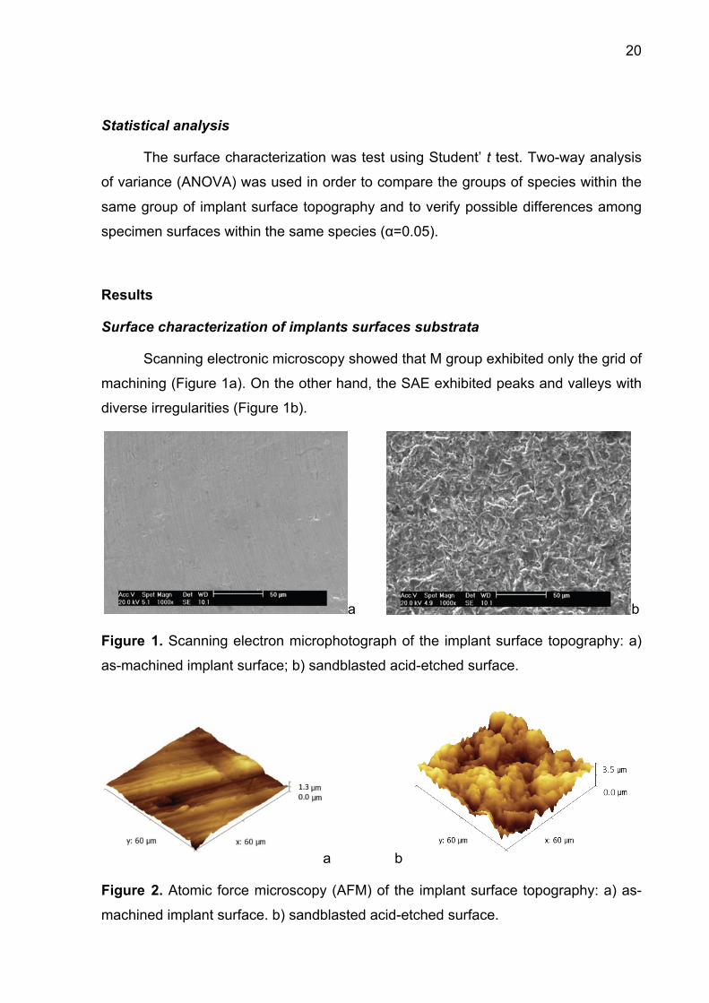
Surface characterization of implants surfaces substrata
Scanning electronic microscopy showed that M group exhibited only the grid of
machining (Figure 1a). On the other hand, the SAE exhibited peaks and valleys with
diverse irregularities (Figure 1b).
a b
Figure 1. Scanning electron microphotograph of the implant surface topography: a)
as-machined implant surface; b) sandblasted acid-etched surface.
a b
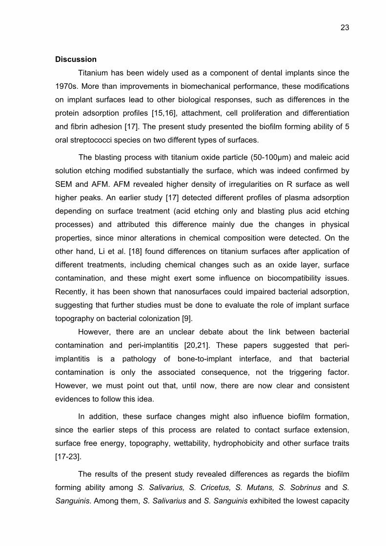
Figure 2. Atomic force microscopy (AFM) of the implant surface topography: a) as-
machined implant surface. b) sandblasted acid-etched surface.

21
The surfaces were characterized by atomic force microscopy, which revealed
differences between the surfaces (p<0.0001). M showed only the machining grids
with peaks of 1.3µm and some regions that were almost flat (Figure 2a). The SAE
exhibited irregular surfaces with peaks of about 6.5µm (Figure 2b). The roughness
values are shown in Table 1.
Table 1. Mean+standard deviation of the as-machined (MS) and titanium discs
blasted with titanium oxide particles and washed with maleic acid solution (SAE)
profilometry.
Implant Surface Topography* Ra (µm) Rq (µm) Rz (µm)
As-machined - M 0.14 ± 0.02 0.16 ± 0.01 1.61 ± 0.10
Sandblasted acid-etched surface SAE 0.87 ± 0.14 1.12 ± 0.18 5.14 ± 0.69
*Statistically significant between the implant surface topographies (Student t test
p=0.0001), M<SAE; Ra - arithmetic average of the absolute values of all profile
points; Rq - the root-mean-square of the values of all points; Rz - the average value
of the absolute heights of the five highest peaks and the depths of the five deepest
valleys
In vitro determination of microbial adhesion
The biofilm forming ability was evaluated and the means of readings are
shown in (Figure 3). The group mutans streptococci (S. Cricetus, S. Mutans and S.
Sobrinus) exhibited higher levels of biofilm formation and no differences were
observed between surfaces analyzed within each species (p>0.05). It was observed
that although S. Cricetus exhibited the highest ability to form biofilm on SAE, among
all species, within this species this difference was not significant (p>0.05) between
the surfaces analyzed.

22
Figure 3. Mean + standard deviation of the amount of adsorbed dye released after
the assay (p>0.05; two-way ANOVA). Letters: differences among biofilm
accumulated by each species (p<0.05; two-way ANOVA/Tukey test).
S. Sanguinis exhibited a similar behavior to form biofilm on both implant
surface topographies (Figure 4), and their ability to do so was lower than that of the
group mutans streptococci species. The lowest ability was observed for S. Salivarius.
a b
Figure 4. Representative scanning electron microscopy (x10,000) in a back
scattering mode (BSE) of the Streptococcus sanguinis in a) as-machined (M) and b) sandblasted acid-etched surface (SAE). Note proliferation of the S. Sanguinis in the
pitches and notches of the SAE surface.
23
Discussion Titanium has been widely used as a component of dental implants since the
1970s. More than improvements in biomechanical performance, these modifications
on implant surfaces lead to other biological responses, such as differences in the
protein adsorption profiles [15,16], attachment, cell proliferation and differentiation
and fibrin adhesion [17]. The present study presented the biofilm forming ability of 5
oral streptococci species on two different types of surfaces.
The blasting process with titanium oxide particle (50-100µm) and maleic acid
solution etching modified substantially the surface, which was indeed confirmed by
SEM and AFM. AFM revealed higher density of irregularities on R surface as well
higher peaks. An earlier study [17] detected different profiles of plasma adsorption
depending on surface treatment (acid etching only and blasting plus acid etching
processes) and attributed this difference mainly due the changes in physical
properties, since minor alterations in chemical composition were detected. On the
other hand, Li et al. [18] found differences on titanium surfaces after application of
different treatments, including chemical changes such as an oxide layer, surface
contamination, and these might exert some influence on biocompatibility issues.
Recently, it has been shown that nanosurfaces could impaired bacterial adsorption,
suggesting that further studies must be done to evaluate the role of implant surface
topography on bacterial colonization [9].
However, there are an unclear debate about the link between bacterial
contamination and peri-implantitis [20,21]. These papers suggested that peri-
implantitis is a pathology of bone-to-implant interface, and that bacterial
contamination is only the associated consequence, not the triggering factor.
However, we must point out that, until now, there are now clear and consistent
evidences to follow this idea.
In addition, these surface changes might also influence biofilm formation,
since the earlier steps of this process are related to contact surface extension,
surface free energy, topography, wettability, hydrophobicity and other surface traits
[17-23].
The results of the present study revealed differences as regards the biofilm
forming ability among S. Salivarius, S. Cricetus, S. Mutans, S. Sobrinus and S.
Sanguinis. Among them, S. Salivarius and S. Sanguinis exhibited the lowest capacity

24
to form biofilm. Two aspects of biofilm forming ability must be pointed out: the
specific traits of each species and surface topographies.
Differences on adhesion to glass surface among mutans streptococci group
were already observed [24]. The authors found that S. rattus adhered less than the
other species (S. Sobrinus, S. Mutans and S. cricettus) and attributed these results to
different properties of the S. rattus surface like as negative zeta-potentials. In the
present study, three species of mutans streptococci group (S. Mutans, S. Cricetus
and S. Sobrinus) were evaluated and although the raw values showed a high
capacity of S. Mutans to accumulate biofilm on titanium surface followed by S.
Cricetus and S. Sobrinus; statistically differences were observed only between S.
Mutans and S. Sobrinus (p<0.05).
Although roughness seems to promote an increase in the amount of plaque,
the biofilm composition did not show substantial changes and the establishment of
irreversible attachment in the surface irregularities, where microorganisms are
protected against mechanical shear [10]. Despite this, the results of our study
demonstrated that biofilm formation does not increase markedly on rougher surfaces.
Oral strains, most of them having high-surface-free-energy, might adhere
better to hydrophilic substrata [25]. Differences with regard to surface hydrophobicity
could be attributed to the acid etching, which could introduce -OH groups on the
surface, thus modifying its chemical properties [26]. According to this hypothesis,
these treatments can originate different surfaces, and consequently, new patterns of
adsorbed substances will be originated, which may offer different profiles of receptors
for bacterial colonization.
Another issue concerns with virulence traits of each species like tooth
colonization mechanisms; S. Mutans apparently attach by adhesin and glucan
mediated mechanisms, whereas S. Sobrinus utilize primarily the latter process [27].
Conclusions
In conclusion, within the limitations of the study, the present findings showed
that: a) biofilm formation by oral streptococci might varies according the species; b)
S. Salivarius and S. Sanguinis showed the lowest ability to accumulate biofilm; c)
group mutans streptococci accumulated higher amounts of biofilm; d) the substratum

25
roughness is not the only issue to be considered with regard to bacterial biofilm
formation.
Author Contributions Jamil Awad Shibli, Claudia Ota-Tsuzuki, and José Augusto Rodrigues were in charge
of the elaboration of the study proposal and the financial support of the study, and
they participated in the elaboration of the paper. Luciene C. Figueiredo and Elton G.
Zenobio were in charge of the statistical analysis, the implant surface
characterization, and the financial support for the study. Pedro Paulo Cardoso Pita,
Tatiane Ferreira Miato, Sergio A. Gehrke, Gabriela Giro, and Cristiane Gonçalves
were in charge of the saliva collection, laboratory processing, and oral biofilm
maintenance and they participated in the elaboration of the paper. Claudia Ota-
Tsuzuki and Alessandra Cassoni were in charge of the laboratory processing of the
samples and participated in the data analyses and elaboration of the paper.
Acknowledgments
Dr. Miato receive grant from University of Guarulhos (fellowship PIBIC-UnG). Implacil
De Bortolli, Sao Paulo, Brazil, provided the titanium discs. The authors declare that
there is no conflict of interests related to this study.
References
(1) S.S. Socransky and A.D. Haffajee, "Periodontal microbial ecology,”
Periodontology 2000, vol. 38, pp. 135-187, 2005.
(2) P.E. Kolenbrander, R.J. Palmer Jr., A.H. Rickard, N.S. Jakubovics, N.I.
Chalmers and P.I. Diaz, "Bacterial interactions and successions during plaque
development,” Periodontology 2000,vol. 42, no. 1, pp. 47-79, 2006.
(3) J.A. Shibli, L. Melo, D.S. Ferrari, L.C. Figueiredo, M. Faveri and M. Feres,
"Composition of supra- and subgingival biofilm of subjects with healthy and
diseased implants,” Clinical Oral Implants Research vol. 19, pp.975-982, 2008.

26
(4) J.A. Shibli, M.C. Martins, R.F. Lotufo and E. Marcantonio Jr., "Microbiologic and
radiographic analysis of ligature-induced peri-implantitis with different dental
implant surfaces.," International Journal of Oral and Maxillofacial Implants,
vol.18, pp. 383-390, 2003.
(5) J.A. Shibli, T.R. Vitussi, R.V. Garcia et al, "Implant surface analysis and
microbiologic evaluation of failed implants retrieved from smokers." Journal of
Oral Implantology, vol. 33, no. 4, pp. 232-238, 2007.
(6) G. Nakazato, H. Tsuchiya, M. Sato and M. Yamauchi, "In vivo plaque formation
on implant materials," International Journal of Oral and Maxillofacial Implants,
vol.4, pp. 321-326, 1989.
(7) S. Sardin, J.J. Morrier, G. Benay and O. Barsotti, "In vitro streptococcal
adherence on prosthetic and implant materials. Interactions with
physicochemical surface properties,” Journal of Oral Rehabilitation vol. 31,
pp.140-148, 2004.
(8) J.A Shibli, M.C. Martins, L.H. Theodoro, R.F. Lotufo, V.G. Garcia and E.
Marcantonio JR, “Lethal photosensitization in microbiological treatment of
ligature-induced peri-implantitis: a preliminary study in dogs,” Journal of Oral
Science, vol.45, no. 1, pp.17-23, 2003.
(9) B. Grössner-Schreiber, M. Griepentrog, I. Haustein, et al. "Plaque formation on
surface modified dental implants. An in vitro study," Clinical Oral Implants
Research, vol. 12, pp.543-551, 2001.
(10) M. Quirynen H.C. van der Mei, C.M.L. Bollen, et al. "An in vivo study about the
influence of the surface roughness of implants on the microbiology of supra-and-
subgingival plaque,” Journal of Dental Research, vol. 72, pp. 1304-1309, 1993.
(11) M. Quirynen H.C. van der Mei, C.M.L. Bollen, et al. "The influence of surface-
free energy on supra- and subgingival plaque microbiology. An in vivo study on
implants,” Journal of Periodontology, vol.65, pp. 162-167, 1994.

27
(12) H.K. Kuramitsu, X. He, R. Lux, M.H. Anderson and W. Shi, "Interspecies
interactions within oral microbial communities,” Microbiology Molecular Biology
Reviews, vol.71, pp. 653-670, 2007.
(13) A.L. Coykendall, "Classification and identification of the viridans Streptococci,”
Clinical Microbiology Reviews, vol. 2, pp.315-328, 1989.
(14) D.M. Dohan Ehrenfest, B.S. Kang, G. Sammartino et al. "Guidelines for the
publication of articles related to implant surfaces and design from the POSEIDO:
a standard for surface characterization,” POSEIDO. vol. 1, no.1, pp.7-15, 2013.
(15) J.A. Shibli and D.M. Dohan Ehrenfest, "In dental implant surfaces, NanoWar has
begun... but NanoQuest is still at stake!," POSEIDO, vol.1, no. 3, pp.131-140,
2013.
(16) J.P. Davidas, "Looking for a new international standard for characterization,
classification and identification of surfaces in implantable materials: the long
march for the evaluation of dental implant surfaces has just begun,” POSEIDO,
vol. 2, no. 1, pp.1-5, 2014.
(17) M.N. Sela, L. Badihi, G. Rosen, D. Steinberg, D. Kohavi, "Adsorption of human
plasma proteins to modified titanium surfaces," Clinical Oral Implants Research,
vol. 18, pp. 630-638, 2007.
(18) D. Li, S.J. Ferguson, T. Beutler et al. "Biomechanical comparison of the
sandblasted and acid-etched and the machined and acid-etched titanium surface
for dental implant,” Journal Biomedical Materials Research vol. 60, pp.325-332,
2002.
(19) E.M. Lima, H. Koo, A.M. Vacca Smith, P.L. Rosalen and A.A. Del Bel Cury,
"Adsorption of salivary and serum proteins, and bacterial adherence on titanium
and zirconia ceramic surfaces,” Clinical Oral Implants Research, vol. 19, pp.
780-785, 2008
(20) K. Mustafa, A. Wennerberg, J. Wroblewski, K. Hultenby, B.S. Lopez and K.
Arvidson, "Determining optimal surface roughness of TiO(2) blasted titanium
implant material for attachment, proliferation and differentiation of cells derived

28
from human mandibular alveolar bone,” Clinical Oral Implants Research, vol. 12,
pp.515-525, 2001.
(21) L. Le Guéhennec, A. Soueidan, P. Layrolle and Y. Amouriq, "Surface treatments
of titanium dental implants for rapid osseointegration," Dental Materials, vol. 23,
pp.844-854, 2007.
(22) W. Teughels, N. Van Assche, I. Sliepen and M. Quirynen, "Effect of material
characteristics and/or surface topography on biofilm development," Clinical Oral
Implants Research, vol. 17, pp. 68-81, 2006.
(23) A. Leonhardt, J. Olsson and G. Dahlén, "Bacterial colonization on titanium,
hydroxyapatite, and amalgam surfaces in vivo”, Journal of Dental Research, vol.
74, pp. 1607-1612, 1995.
(24) H. J. Busscher and H. C. van der Mei, "Physico-chemical interactions in initial
microbial adhesion and relevance for biofilm formation,” Adv Dental Research,
vol. 11, pp.24-32, 1997.
(25) M.M. Fürst, G.E. Salvi, N.P. Lang and G.R. Persson, "Bacterial colonization
immediately after installation on oral titanium implants," Clinical Oral Implants
Research, vol. 18,pp. 501-501, 2007.
(26) A. H. Weerkamp, H. M. Uyen, and H. J. Busscher, "Effect of zeta potential and
surface energy on bacterial adhesion to uncoated and saliva-coated human
enamel and dentin," Journal of Dental Research, vol. 67, pp. 1483-1487, 1998.
(27) H. M. Uyen, J. M. Schakenraad, J. Sjollema, et al., “Amount and surface
structure of albumin adsorbed to solid substrata with different wettabilities in a
parallel plate flow cell," Journal of Biomedical Material Research, vol. 24, pp.
1599-1614, 1990.

29
CONCLUSÃO
Pode-se concluir que a adesão bacteriana sobre a superfície do titânio
depende das espécies bacterianas envolvidas, a topografia da superfície não
influenciou na adesão dos estreptococos.

30
REFERÊNCIAS
Albrektsson T, Johansson C. Osteoinduction, osteoconduction and osseointegration.
Eur Spine J. 2001;10 Suppl 2:S96-101.
Becker MR, Paster BJ, Leys EJ, Moeschberger ML, Kenyon SG, Galvin JL, et al.
Molecular analysis of bacterial species associated with childhood caries. J Clin
Microbiol. 2002;40(3):1001-9.
Dige I, Nyengaard JR, Kilian M, Nyvad B. Application of stereological principles for
quantification of bacteria in intact dental biofilms. Oral Microbiol Immunol.
2009;24(1):69-75.
Foster JS, Kolenbrander PE. Development of a multispecies oral bacterial community
in a saliva-conditioned flow cell. Appl Environ Microbiol. 2004;70(7):4340-8.
Grossner-Schreiber B, Griepentrog M, Haustein I, Muller WD, Lange KP, Briedigkeit
H, et al. Plaque formation on surface modified dental implants. An in vitro study. Clin
Oral Implants Res. 2001;12(6):543-51.
Heitz-Mayfield LJ. Peri-implant diseases: diagnosis and risk indicators. J Clin
Periodontol. 2008;35(8 Suppl):292-304.
Hojo K, Nagaoka S, Ohshima T, Maeda N. Bacterial Interactions in Dental Biofilm
Development. J Dent Res. 2009;88(11):982-90.
Huang R, Li M, Gregory RL. Bacterial interactions in dental biofilm. Virulence.
2011;2(5):435-44.
Jung RE, Pjetursson BE, Glauser R, Zembic A, Zwahlen M, Lang NP. A systematic
review of the 5-year survival and complication rates of implant-supported single
crowns. Clin Oral Implants Res. 2008;19(2):119-30.
Kemp CW, Robrish SA, Curtis MA, Sharer SA, Bowen WH. Application of a
competition model to the growth of Streptococcus mutans and Streptococcus sanguis
in binary continuous culture. Appl Environ Microbiol. 1983;45(4):1277-82.

31
Kolenbrander PE, Palmer RJ, Rickard AH, Jakubovics NS, Chalmers NI, Diaz PI.
Bacterial interactions and successions during plaque development. Periodontol 2000.
2006;42:47-79.
Kreth J, Merritt J, Shi W, Qi F. Competition and coexistence between Streptococcus
mutans and Streptococcus sanguinis in the dental biofilm. J Bacteriol.
2005;187(21):7193-203.
Kuramitsu HK, He X, Lux R, Anderson MH, Shi W. Interspecies interactions within
oral microbial communities. Microbiol Mol Biol Rev. 2007;71(4):653-70.
Marsh PD. Dental plaque: biological significance of a biofilm and community life-
style. J Clin Periodontol. 2005;32 Suppl 6:7-15.
Miller BF, Keane CB. Miller-Keane Encyclopedia & Dictionary of Medicine, Nursing,
and Allied Health. Philadelphia: Saunders; 1992.
Nakazato G, Tsuchiya H, Sato M, Yamauchi M. In vivo plaque formation on implant
materials. Int J Oral Maxillofac Implants. 1989;4(4):321-6.
Pjetursson BE, Thoma D, Jung R, Zwahlen M, Zembic A. A systematic review of the
survival and complication rates of implant-supported fixed dental prostheses (FDPs)
after a mean observation period of at least 5 years. Clin Oral Implants Res. 2012;23
Suppl 6:22-38.
Quirynen M, Van der Mei HC, Bollen CM, Van den Bossche LH, Doornbusch GI, van
Steenberghe D, et al. The influence of surface-free energy on supra- and subgingival
plaque microbiology. An in vivo study on implants. J Periodontol. 1994;65(2):162-7.
Ritz HL. Microbial population shifts in developing human dental plaque. Arch Oral
Biol. 1967;12(12):1561-8.
Sardin S, Morrier JJ, Benay G, Barsotti O. In vitro streptococcal adherence on
prosthetic and implant materials. Interactions with physicochemical surface
properties. J Oral Rehabil. 2004;31(2):140-8.

32
Shibli JA, Martins MC, Lotufo RF, Marcantonio E, Jr. Microbiologic and radiographic
analysis of ligature-induced peri-implantitis with different dental implant surfaces. Int
J Oral Maxillofac Implants. 2003;18(3):383-90.
Shibli JA, Melo L, Ferrari DS, Figueiredo LC, Faveri M, Feres M. Composition of
supra- and subgingival biofilm of subjects with healthy and diseased implants. Clin
Oral Implants Res. 2008;19(10):975-82.
Shibli JA, Vitussi TR, Garcia RV, Zenobio EG, Ota-Tsuzuki C, Cassoni A, et al.
Implant surface analysis and microbiologic evaluation of failed implants retrieved
from smokers. J Oral Implantol. 2007;33(4):232-8.
Socransky SS, Haffajee AD. Periodontal microbial ecology. Periodontol 2000.
2005;38:135-87.
Tong H, Chen W, Shi W, Qi F, Dong X. SO-LAAO, a novel L-amino acid oxidase that
enables Streptococcus oligofermentans to outcompete Streptococcus mutans by
generating H2O2 from peptone. J Bacteriol. 2008;190(13):4716-21.
Zitzmann NU, Berglundh T. Definition and prevalence of peri-implant diseases. J Clin
Periodontol. 2008;35(8 Suppl):286-91.