utilidade da dermatoglifia como ferramenta diagnóstica não
TRANSCRIPT
Arch Oral Res. 2012 Sept./Dec.;8(3)211-18
doi: 10.7213/archives.08.003.AC03 ISSN 2236-8035 Archives of Oral Research, v. 8, n. 3, p. 211-18, Sept./Dec. 2012
Licensed under a Creative Commons License
[T]
Utility of dermatoglyphics as a non-invasive diagnostic tool in recurrent aphthous stomatitis[I]
Utilidade da dermatoglifia como ferramenta diagnóstica não invasiva para estomatite aftosa recorrente
[A]
Suwarna Dangore-Khasbage[a], Shirish S. Degwekar[b], Rahul R. Bhowate[c], Vidya K. Lohe[d], Dharmaraj Tamgire[e]
[a] MDS Oral Medicine & Radiology, associate professor, Sharad Pawar Dental College & Hospital, Wardha - India, e-mail: [email protected]
[b] MDS Oral Medicine & Radiology, professor and head, Sharad Pawar Dental College & Hospital, Wardha - India, e-mail: [email protected]
[c] MDS Oral Medicine & Radiology, professor, Sharad Pawar Dental College & Hospital, Wardha - India, e-mail: [email protected]
[d] MDS Oral Medicine & Radiology, associate professor, Sharad Pawar Dental College & Hospital, Wardha - India, e-mail: [email protected]
[e] MS Human Anatomy, associate professor, JNM College, Wardha - India, e-mail: [email protected]
[R]
Abstract
Introduction: Dermatoglyphics is an advancing branch of medical science in which the dermal ridge pat-terns are studied and used in prediction of genetic disorders. Objective: To assess the usefulness of der-matoglyphics, as a non-invasive early predicator in RAS, since genetics plays a role in both. Materials and methods: This case control study comprised of 40 patients with recurrent aphthous stomatitis compared to 30 control subjects of similar age and sex. All subjects were investigated for their dermatoglyphic patterns of both hands, which were obtained by smearing the ink uniformly over the palm and fingers and pressing the hands firmly against the good quality paper. Qualitative analysis of prints was done for arches, loops and whorls and quantitative analysis included Total Finger Ridge Count (TFRC) and atd angle. Fisher’s Exact Tests were used to evaluate the significance between recurrent aphthous stomatitis and the dermatoglyphic features. Results: Among various dermatoglyphic parameters analyzed, recurrent aphthous stomatitis pa-tients showed 9% arches, 62% loops and 29% whorls as the finger ridge configuration. In comparing the
Arch Oral Res. 2012 Sept./Dec.;8(3)211-18
Dangore-Khasbage S, Degwekar SS, Bhowate RR, Lohe VK, Tamgire D212
results between the two groups, the frequency of arches was observed more in study group while composite whorl and the ulnar pattern were more frequent in control group, the results were statistically significant (p < 0.05). In addition to this, study group patients demonstrated higher frequency of total finger ridge count, and control group showed higher frequency for atd angle on both hands, statistically significant diffe-rence is observed (p < 0.05). Conclusion: The study found significant correlation between palmar dermato-glyphics and recurrent aphthous stomatitis, suggesting that genetics is one of the host risk factor associated with the latter, and could aid in early detection of the disease.
Keywords: Arches. Dermatoglyphics. Loops. Recurrent aphthous stomatitis (RAS). Whorls.
Resumo
Introdução: Dermatoglifia é um avançado ramo da ciência médica em que os padrões de cristas dérmicas são estu-dados e utilizados para predizer distúrbios genéticos. Objetivo: Avaliar a utilidade da dermatoglifia, como método de diagnóstico não invasivo para a estomatite aftosa recorrente, uma vez que a genética desempenha um papel em ambos. Materiais e métodos: Estudo caso-controle composto por 40 pacientes com estomatite aftosa recorrente comparados a 30 controles de mesma idade e sexo. Todos os sujeitos foram avaliados quanto a seus padrões der-matóglifos de ambas as mãos. Os padrões dermatóglifos foram obtidos espalhando uniformemente uma tinta sobre a palma da mão e os dedos e pressionada firmemente contra um papel de boa qualidade. A análise qualitativa das impressões foi feita por arcos, laços e espirais e a análise quantitativa pela contagem total da polpa do dedo (TFRC) e ângulo atd. Teste de Fischer foi utilizado para avaliar a significância entre estomatites recorrentes e características dermatoglíficas. Resultados: Entre os vários parâmetros dermatoglíficos analisados, os pacientes com estomatite aftosa recorrente mostraram 9% de arcos, 62% de laços e 29% de espirais na configuração da polpa digital. Ao comparar os resultados entre os dois grupos, a frequência de arcos foi mais observada no grupo de estudo, enquanto verticilos e padrão tênar foram mais frequentes no grupo controle, com resultados estatisticamente significativos (p < 0,05). Além disso, os pacientes do grupo de estudo demonstraram maior frequência de contagem total da polpa digital e o grupo controle apresentou maior frequência para o ângulo atd em ambas as mãos, com diferenças esta-tisticamente significativas (p < 0,05). Conclusão: O estudo encontrou correlação significativa entre a dermatoglifia palmar e estomatites recorrentes, sugerindo que a genética é um dos fatores de risco de hospedeiro associado com estomatite aftosa recorrente, e poderia ajudar na detecção precoce da doença.
Palavras-chave: Arcos. Dermatoglifia. Laços. Estomatite aftosa recorrente. Espirais.#]
[B]
Introduction
Recurrent aphthous stomatitis (RAS) is the most common type of ulcerative oral disorder, which affects approximately 20% of the general popula-tion. Although it is not life threatening, it does dimi-nish the overall quality of life of the sufferer, since even eating, talking and swallowing become painful. Despite of its worldwide occurrence and the exten-sive amount of research devoted to RAS, its etiology remains somewhat unclear. Though hereditary, nu-tritional, infectious and psychological factors have long been commonly associated with RAS (1, 2).
There are a number of previous studies do-cumenting an inheritance as a strong risk factor for RAS and genetically specific HLAs have been
identified in these patients (3, 4). Also an increased susceptibility to RAS has been demonstrated among the children of RAS-positive parents (3). Thus, with an ever-growing population, it becomes imperative to develop methods to identify the individuals who are at risk. The use of dermatoglyphics can be con-sidered as a unique clinical approach at low cost for identifying such individuals.
Dermatoglyphics is an advancing branch of me-dical science in which the dermal ridge configura-tions on the digits, palms and soles are studied and used in prediction of genetic disorders (5, 6). The digit patterns are broadly classified into three ma-jor types: whorl, loops and arches, which have been subdivided into various subtypes whereas the whole of the human palm show certain other features such
Arch Oral Res. 2012 Sept./Dec.;8(3)211-18
Utility of dermatoglyphics as a non-invasive diagnostic tool in recurrent aphthous stomatitis213
as atd angle, H-loop, IV loop and t-triradius (7, 8). Dermatoglyphic patterns are genetically determi-ned and influenced by environmental forces that are operating even before birth. They remain unchan-ged throughout life and unusual dermatoglyphic patterns often relate to genetic disorders (5).
Dermatoglyphics is currently used as a non-invasi-ve early predicator of many illnesses (5). This science has been widely used to identify systemic disorders and to analyze the role of genetics in their etiology and is becoming acceptable now as a diagnostic tool among medical personnel (5, 6, 9). It is considered a window into congenital abnormalities and is a sensiti-ve indicator of intrauterine dental anomalies too (10).
In the field of dentistry, several studies have shown the association of dermatoglyphics with di-fferent types of oral diseases, like cancer, precancer, bruxism, cleft palate, caries and periodontal disea-ses (7, 8, 11-15). However, there is no publication to date that addresses the dermatoglyphic patterns in recurrent aphthous stomatitis. Therefore this study was undertaken with the following aims: (i) to study a correlation of dermatoglyphic patterns with RAS; (ii) to determine the usefulness of dermaoglyphics in order to identify and isolate the higher risk asso-ciated with certain patients.
Materials and methods
This Institutional Ethical Committee approved (approbation number - DMIMS (DU) / IEC/ 2010-11/ 44) case control study included 70 subjects, se-lected from those attending the outpatient depart-ment of Oral Medicine and Radiology, Sharad Pawar Dental College, Wardha. Out of these, 40 patients were suffering from recurrent aphthous stomatitis and 30 were age and sex matched controls. Control subjects were recruited randomly among the volun-teer patients who had reported to the department for their dental check-up.
In inclusion criteria, there were patients with clinical presentation of aphthous ulcers, and had apositive history of recurrent painful similar ulce-rations, three to six episodes per year. In exclusion criteria were patients having any oral diseases (like oral submucous fibrosis and candidiasis), syste-mic diseases (such as Behçet Syndrome, Celiac or Crohn’s disease, nutritional deficiency, cardiovascu-lar, respiratory, metabolic and endocrinal disorders)
and patients taking medication for systemic disea-ses or having history such as history of radiothera-py, which could cause oral ulcerations.
Patients were informed about the research study, its procedure and their written consent was obtained. Diagnosis of RAS was based on detailed history and its characteristic clinical presentation. A painful round or oval, shallow ulcer, regular in outline, with a grey-whi-te pseudomembrane and surrounded by a thin erythe-matous halo was diagnosed as aphthous ulcer. Based on the size and clinical characteristics, these were classified into minor, major and herpetiform (2, 16). In doubtful cases, blood investigations were performed to rule out the ulcers associated with other diseases. Biopsy was not performed for any of the patients.
Minor ulcers, which comprise over 80% of RAS cases, are less than 1 cm in diameter and heal wi-thout scars. Major ulcers are over 1 cm in diameter and take longer to heal and are accompanied by scar. Herpetiform ulcers are considered a distinct clinical entity that manifests as recurrent crops of dozens of small ulcers throughout the oral mucosa (3, 16). Then the dermatoglyphic data, which included bi-lateral finger and palm prints, were collected from the same subjects and 30 control subjects, using the stamp pad ink method. These were then analyzed qualitatively and quantitatively.
Procedure for obtaining prints
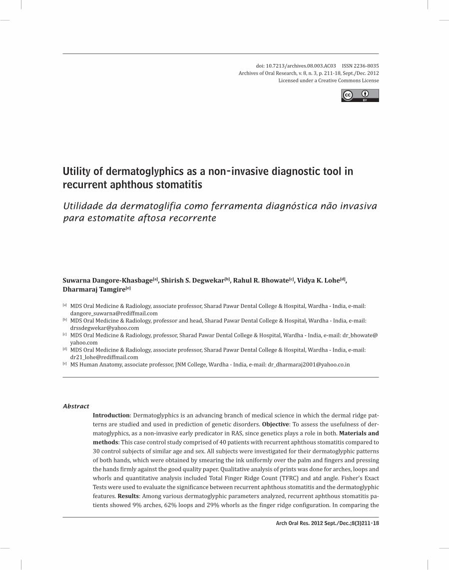
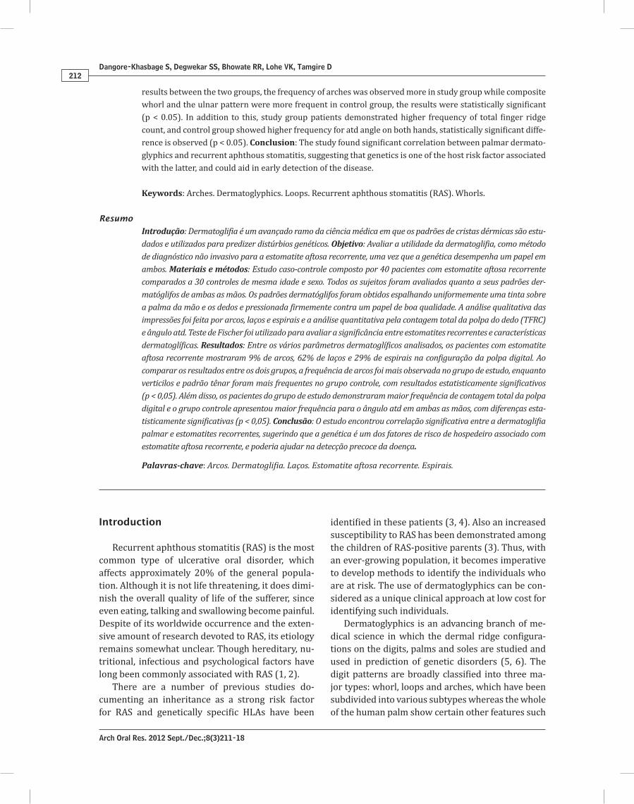
Hands of all patients were washed with soap and water to remove sweat, oil and dirt from the skin in order to enhance the quality of dermatoglyphic prints. Then a small amount of ink was smeared thoroughly and uniformly over the palm and fingers with a piece of gauze. Prints of the distal phalan-ges of the ten fingers and the palms were taken by pressing firmly against the good quality paper kept over the table. The finger prints were taken from thumb to little finger. Qualitative analysis of fin-gers and palm prints was done. Fingertip patterns were studied for arches (A), loops (L) and whorls (W) as shown in Figure 1, whereas palmar patterns were analyzed for the presence of arches, loops and whorls in hypothenar area, thenar/first interdigital area (I1), I2, I3 and I4 interdigital area (Qualitative analysis) as shown in Figure 2 (11, 12). Quantitative analysis included total finger ridge count (TFRC) and atd angle.
Arch Oral Res. 2012 Sept./Dec.;8(3)211-18
Dangore-Khasbage S, Degwekar SS, Bhowate RR, Lohe VK, Tamgire D214
Results
The dermatoglyphic patterns were evaluated in 40 RAS patients (mean age 23.60 ± 7.99) and 30 age and sex matched controls (mean age 25.23 ± 9.32). Amongst all participants, 57% were female and 43%, male. Out of all RAS patients, 60% (24 out of 40) confirmed a positive history of RAS amongst their family members and 77% (31 out of 40) RAS patients were suffering from minor type of aphthous ulcers. The most prevalent site of RAS was tongue, followed by labial mucosa (14 patients – 35%, 11 patients – 27%, respectively). Table 1 presents in-formation related to RAS in the study subjects.
Analysis of finger print pattern between the study and control groups
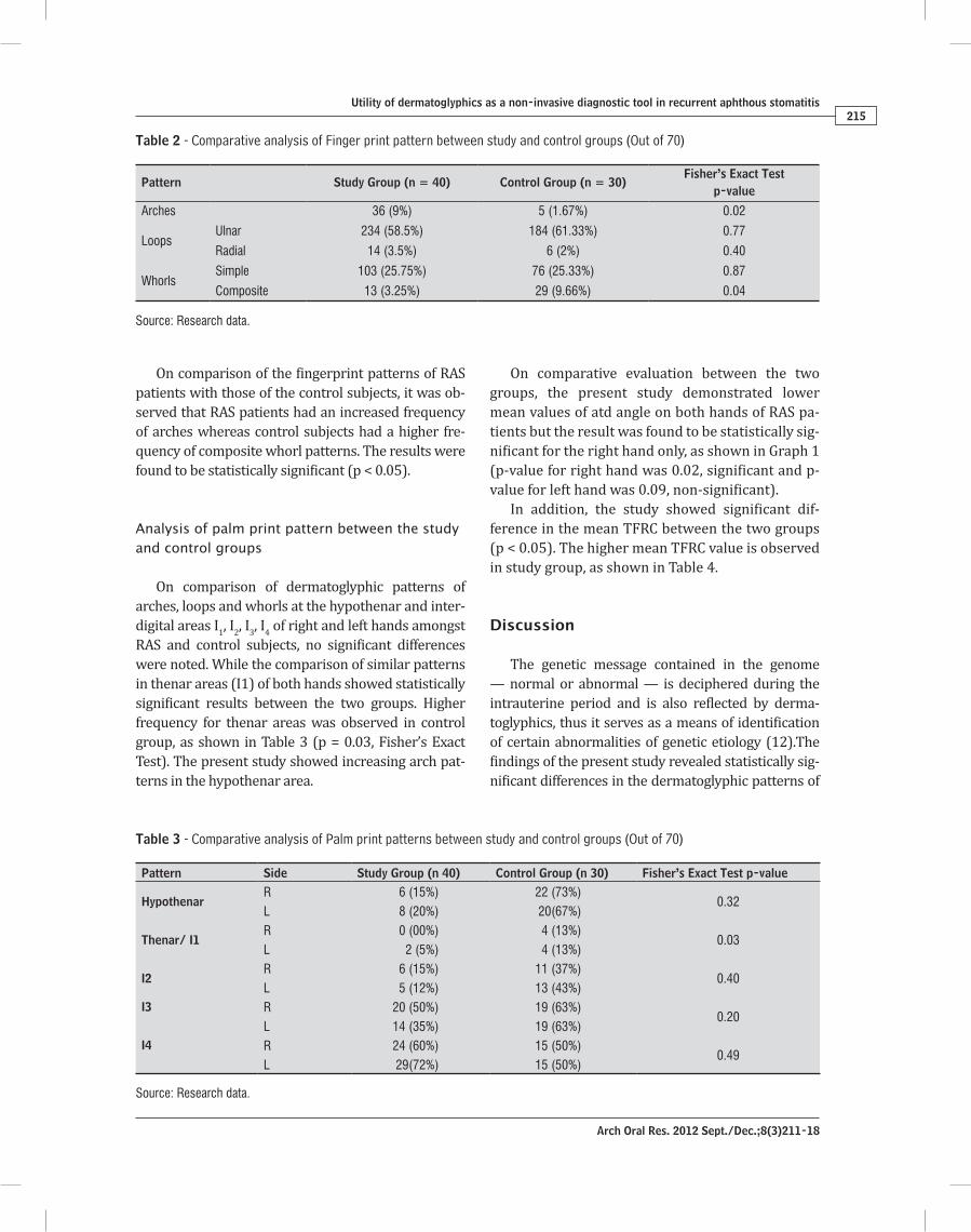
The distribution and comparison of various fin-gerprint patterns in all subjects is shown in Table 2. In RAS patients, 9% were arches, 62% were loops and 29%, whorls, as the ridge configuration. Amongst all dermatoglyphic patterns, the frequency of ulnar loops was highest in RAS subjects (Table 2).
The statistical analysis was performed using SPSS package and MINITAB (Version IV, USA) by recording the findings in tabular format. Fisher’s Exact Test and Student’s t-test were used to evalu-ate the significance of the parameters between dif-ferent groups. For the parameters, 95% confidence intervals were calculated.
A1 B1 C1 D1 E1
A2 B2 C2 D2 E2
Figure 1- Finger print patterns for dermatoglyphicsLegend: A1 and A2 = arch; B1 and B2 = ulner loop; C1 and C2 = radial loop; D1 and D2 = Simple whorl; E1 and E2 = composite whorl.
Source: Research data.
Figure 2 - Palmar landmarks for dermatoglyphicsSource: Research data.
Table 1 - Information related to recurrent aphthous stoma-titis in study subjects (Out of 40)
Variants n (%)
History of RAS in familymembers
Positive 24 (60)
Negative 16 (40)
Duration of RAS < 1 year 15 (37)
1-5 years 21 (52)
> 5 years 4 (10)
Type of RAS Minor 31 (77)
Major 2 (5)
Herpetiform 7 (17)
Site of ulcers Tongue 14 (35)
Labial mucosa 11 (27)
Buccal mucosa 8 (20)
Floor of mouth 4 (10)
Gingiva 3 (8)
Association with pain Yes 37 (92)
No 3 (8)
Source: Research data.
Arch Oral Res. 2012 Sept./Dec.;8(3)211-18
Utility of dermatoglyphics as a non-invasive diagnostic tool in recurrent aphthous stomatitis215
On comparative evaluation between the two groups, the present study demonstrated lower mean values of atd angle on both hands of RAS pa-tients but the result was found to be statistically sig-nificant for the right hand only, as shown in Graph 1 (p-value for right hand was 0.02, significant and p-value for left hand was 0.09, non-significant).
In addition, the study showed significant dif-ference in the mean TFRC between the two groups (p < 0.05). The higher mean TFRC value is observed in study group, as shown in Table 4.
Discussion
The genetic message contained in the genome — normal or abnormal — is deciphered during the intrauterine period and is also reflected by derma-toglyphics, thus it serves as a means of identification of certain abnormalities of genetic etiology (12).The findings of the present study revealed statistically sig-nificant differences in the dermatoglyphic patterns of
On comparison of the fingerprint patterns of RAS patients with those of the control subjects, it was ob-served that RAS patients had an increased frequency of arches whereas control subjects had a higher fre-quency of composite whorl patterns. The results were found to be statistically significant (p < 0.05).
Analysis of palm print pattern between the study and control groups
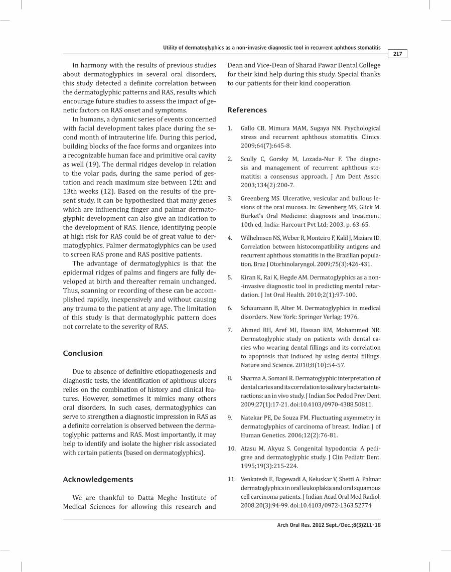
On comparison of dermatoglyphic patterns of arches, loops and whorls at the hypothenar and inter-digital areas I1, I2, I3, I4 of right and left hands amongst RAS and control subjects, no significant differences were noted. While the comparison of similar patterns in thenar areas (I1) of both hands showed statistically significant results between the two groups. Higher frequency for thenar areas was observed in control group, as shown in Table 3 (p = 0.03, Fisher’s Exact Test). The present study showed increasing arch pat-terns in the hypothenar area.
Table 2 - Comparative analysis of Finger print pattern between study and control groups (Out of 70)
Pattern Study Group (n = 40) Control Group (n = 30)Fisher’s Exact Test
p-value
Arches 36 (9%) 5 (1.67%) 0.02
LoopsUlnar 234 (58.5%) 184 (61.33%) 0.77
Radial 14 (3.5%) 6 (2%) 0.40
WhorlsSimple 103 (25.75%) 76 (25.33%) 0.87
Composite 13 (3.25%) 29 (9.66%) 0.04
Source: Research data.
Table 3 - Comparative analysis of Palm print patterns between study and control groups (Out of 70)
Pattern Side Study Group (n 40) Control Group (n 30) Fisher’s Exact Test p-value
HypothenarR 6 (15%) 22 (73%)
0.32L 8 (20%) 20(67%)
Thenar/ I1R 0 (00%) 4 (13%)
0.03L 2 (5%) 4 (13%)
I2R 6 (15%) 11 (37%)
0.40L 5 (12%) 13 (43%)
I3 R 20 (50%) 19 (63%)0.20
L 14 (35%) 19 (63%)I4 R 24 (60%) 15 (50%)
0.49L 29(72%) 15 (50%)
Source: Research data.

Arch Oral Res. 2012 Sept./Dec.;8(3)211-18
Dangore-Khasbage S, Degwekar SS, Bhowate RR, Lohe VK, Tamgire D216
Regarding the type of RAS, the results of the present study are consistent with previous litera-ture, which described that approximately 80% and 67.74% of patients with recurrent aphthous ulcera-tions were exhibiting minor type of RAS (4, 16).
Various dermatoglyphic patterns such as higher frequency of ulnar loops (58.5%), arches (9%), sim-ple whorls (25.75%) and lower frequency of com-posite whorls (3.25%) in RAS patients are observed to be comparable with the dermatoglyphic patterns of children having cleft palate as well as patients suffering from oral cancer and precancer (11, 12). On the other hand, patients with history of bruxism demonstrated decrease in frequency of loops (13). Actually, decrease in frequency of loops was a cons-tant feature in dental caries patients in a number of previous studies while there was an increase in fre-quency of whorls amongst them (7, 8, 18).
The present study showed higher frequency of arches in RAS patients, comparable to the derma-toglyphic pattern of patients with oral leukoplakia and oral squamous cell carcinoma (11). However, individuals with cleft palate did not show signifi-cant difference in arch pattern between study and control group (12).
The frequency of composite whorls was higher in control group as compared to RAS patients in the pre-sent study (significant, pe was 0.04) while composite whorls were found common in dental caries patients who wore fillings, as mentioned in the study by Ahmed et al. (7).
On comparative analysis of palm prints of both hands between the two groups, statistically significant difference was noted between the dermatoglyphic patterns of I1 interdigital areas only. The present stu-dy also revealed that the control group was characte-rized by increasing arch patterns in the hypothenar area, which is in agreement with Atasu (18).
The higher mean TFRC value in study patients was an additional significant finding of the present study, which is analogous to dental caries patients wearing fillings (7). On the contrary, bruxism patients and the ones suffering from oral cancer and precancer showed no significant TFRC values (11, 13).
In this study it was observed that RAS patients had lower mean values of atd angle on right and left hands when compared to control subjects, whereas the stu-dy by Mathew et al. (12) showed controversial results with reference to atd angles between two groups, whi-ch was stated to be higher in the oral cleft children.
fingers and palm between the controls and RAS po-pulation, allowing us to conclude a genetic difference between the two groups. This may be used to identify the RAS prone patients and enhance their awareness about the condition.
The etiological role of genetics in RAS is well kno-wn (1, 4). Undoubtedly family history has the stron-gest correlation with RAS. Also, in this study, 60% of patients gave a positive history of RAS amongst their family members, which is comparable to the study by Koybasi et al. (17). It has been stated in literature that patients with RAS-positive parents had a 90% chan-ce of developing RAS, whereas patients with no RAS-positive parents had a 20% chance of developing the lesions (3). According to Ahmed et al. (7), in response to environmental exposures, genetic damage accumu-lates more quickly in individuals with genetic suscep-tibility to DNA damage. Consequently, those individu-als might be at a greater risk of developing lesions.
Table 4 - Comparative analysis of mean values of Total finger ridge count between study and control groups (Out of 70)
Groups Mean s.d. t-value p-value
Study group(n=40) 152.82 35.873.32 0.001
Control group(n=30) 127.31 23.72
Source: Research data.
ControlGroup
Studygroup
Right hand
37
38
39
40
41
42
38,47
41,16 41,33
39,37
Me
an
atd
an
gle
Left hand
Graph 1 - Comparative mean values of atd angle in RAS and control subjectsSource: Research data.
Note: Student’s t-test, p-value for right hand - 0.02, p-value for left hand - 0.09.
Arch Oral Res. 2012 Sept./Dec.;8(3)211-18
Utility of dermatoglyphics as a non-invasive diagnostic tool in recurrent aphthous stomatitis217
Dean and Vice-Dean of Sharad Pawar Dental College for their kind help during this study. Special thanks to our patients for their kind cooperation.
References
1. Gallo CB, Mimura MAM, Sugaya NN. Psychological stress and recurrent aphthous stomatitis. Clinics. 2009;64(7):645-8.
2. Scully C, Gorsky M, Lozada-Nur F. The diagno-sis and management of recurrent aphthous sto-matitis: a consensus approach. J Am Dent Assoc. 2003;134(2):200-7.
3. Greenberg MS. Ulcerative, vesicular and bullous le-sions of the oral mucosa. In: Greenberg MS, Glick M. Burket’s Oral Medicine: diagnosis and treatment. 10th ed. India: Harcourt Pvt Ltd; 2003. p. 63-65.
4. Wilhelmsen NS, Weber R, Monteiro F, Kalil J, Miziara ID. Correlation between histocompatibility antigens and recurrent aphthous stomatitis in the Brazilian popula-tion. Braz J Otorhinolaryngol. 2009;75(3):426-431.
5. Kiran K, Rai K, Hegde AM. Dermatoglyphics as a non--invasive diagnostic tool in predicting mental retar-dation. J Int Oral Health. 2010;2(1):97-100.
6. Schaumann B, Alter M. Dermatoglyphics in medical disorders. New York: Springer Verlag; 1976.
7. Ahmed RH, Aref MI, Hassan RM, Mohammed NR. Dermatoglyphic study on patients with dental ca-ries who wearing dental fillings and its correlation to apoptosis that induced by using dental fillings. Nature and Science. 2010;8(10):54-57.
8. Sharma A. Somani R. Dermatoglyphic interpretation of dental caries and its correlation to salivary bacteria inte-ractions: an in vivo study. J Indian Soc Pedod Prev Dent. 2009;27(1):17-21. doi:10.4103/0970-4388.50811.
9. Natekar PE, De Souza FM. Fluctuating asymmetry in dermatoglyphics of carcinoma of breast. Indian J of Human Genetics. 2006;12(2):76-81.
10. Atasu M, Akyuz S. Congenital hypodontia: A pedi-gree and dermatoglyphic study. J Clin Pediatr Dent. 1995;19(3):215-224.
11. Venkatesh E, Bagewadi A, Keluskar V, Shetti A. Palmar dermatoglyphics in oral leukoplakia and oral squamous cell carcinoma patients. J Indian Acad Oral Med Radiol. 2008;20(3):94-99. doi:10.4103/0972-1363.52774
In harmony with the results of previous studies about dermatoglyphics in several oral disorders, this study detected a definite correlation between the dermatoglyphic patterns and RAS, results which encourage future studies to assess the impact of ge-netic factors on RAS onset and symptoms.
In humans, a dynamic series of events concerned with facial development takes place during the se-cond month of intrauterine life. During this period, building blocks of the face forms and organizes into a recognizable human face and primitive oral cavity as well (19). The dermal ridges develop in relation to the volar pads, during the same period of ges-tation and reach maximum size between 12th and 13th weeks (12). Based on the results of the pre-sent study, it can be hypothesized that many genes which are influencing finger and palmar dermato-glyphic development can also give an indication to the development of RAS. Hence, identifying people at high risk for RAS could be of great value to der-matoglyphics. Palmer dermatoglyphics can be used to screen RAS prone and RAS positive patients.
The advantage of dermatoglyphics is that the epidermal ridges of palms and fingers are fully de-veloped at birth and thereafter remain unchanged. Thus, scanning or recording of these can be accom-plished rapidly, inexpensively and without causing any trauma to the patient at any age. The limitation of this study is that dermatoglyphic pattern does not correlate to the severity of RAS.
Conclusion
Due to absence of definitive etiopathogenesis and diagnostic tests, the identification of aphthous ulcers relies on the combination of history and clinical fea-tures. However, sometimes it mimics many others oral disorders. In such cases, dermatoglyphics can serve to strengthen a diagnostic impression in RAS as a definite correlation is observed between the derma-toglyphic patterns and RAS. Most importantly, it may help to identify and isolate the higher risk associated with certain patients (based on dermatoglyphics).
Acknowledgements
We are thankful to Datta Meghe Institute of Medical Sciences for allowing this research and
Arch Oral Res. 2012 Sept./Dec.;8(3)211-18
Dangore-Khasbage S, Degwekar SS, Bhowate RR, Lohe VK, Tamgire D218
17. Koybasi S, Parlak AH, Serin E, Yilmaz F, Serin D. Recurrent aphthous stomatitis: investigation of possible etiologic factors. Am J Otolaryngol. 2006;27(4):229-32.
18. Atasu M. Dermatoglyphic findings in dental car-ies: a preliminary report. J. Clin Pediatr Dent. 1998;22(2):147-9.
19. Avery JK. Development of branchial arches, face and palate. In: Avery JK, Steele PF, Avery N, editor. Oral development and Histology. 2nd ed. New York: Thieme Medical Publishers; 1994. p. 20-41.
Received: 10/27/2012Recebido: 27/10/2012
Accepted: 11/30/2012Aceito: 30/11/2012
12. Mathew L, Hegde AM, Rai K. Dermatoglyphic pe-culiarities in children with oral clefts. J Indian Soc Pedod Prev Dent. 2005;23(4):179-82.
13. Polat MH, Azak A, Evlioglu G, Malkondu OK, Atasu M. The relation of bruxism and dermatoglyphics. J Clin Pediatr Dent. 2000;24(3):191-4.
14. Polat MH, Evlioglu G, Karayazagan B. Dermatoglyphic findings in patients with oral cancers. Balkan J Stomatol. 2004;8(2):105-108.
15. Atasu M, Kuru B, Firatli E, Meriç H. Dermatoglyphic findings in periodontal diseases. Int J Anthropology. 2005;20(1/2):63-75.
16. Natah SS, Konttinen YT, Enattah NS, Ashammakhi N, Sharkey KA, Häyrinen-Immonen R. Recurrent aphthous ulcers today: a review of the grow-ing knowledge. Int J Oral Maxillofac Surg. 2004;33(3):221-34.