avaliação clínica de restaurações reparadas por resina composta à
TRANSCRIPT
Da
nie
la A
raú
jo V
elo
so
Po
po
ff
DANIELA ARAÚJO VELOSO POPOFF
AVALIAÇÃO CLÍNICA DE RESTAURAÇÕES REPARADAS POR RESINA COMPOSTA À
BASE DE SILORANO: ESTUDO LONGITUDINAL RANDOMIZADO
CONTROLADO
Faculdade de Odontologia Universidade Federal de Minas Gerais
Belo Horizonte
2011
Avali
ação
clí
nic
a d
e r
esta
ura
çõ
es r
ep
ara
da
s p
or
resin
a c
om
po
sta
à b
ase d
e
sil
ora
no
: estu
do
lo
ng
itu
din
al ra
nd
om
izad
o c
on
tro
lad
o
20
11
Daniela Araújo Veloso Popoff
AVALIAÇÃO CLÍNICA DE RESTAURAÇÕES REPARADAS POR RESINA COMPOSTA À BASE DE
SILORANO: ESTUDO LONGITUDINAL RANDOMIZADO CONTROLADO
Tese apresentada ao Colegiado do Programa de Pós-Graduação da Faculdade de Odontologia da Universidade Federal de Minas Gerais, como requisito parcial para obtenção do grau de Doutor em Odontologia – área de concentração em Clínica Odontológica. Orientador: Prof. Dr. Allyson Nogueira Moreira Co-Orientadora: Prof. Dra. Cláudia Silami de Magalhães
Faculdade de Odontologia – UFMG Belo Horizonte
2011
Dedicada a Deus,
Bem sei, Senhor, que não é o homem dono do seu destino, e que ao caminhante não lhe assiste o poder de dirigir seus passos.
Jr 10:23
AGRADECIMENTOS A Deus e à Nossa Senhora Rosa Mística, a Quem ofereci meus estudos e meu trabalho e os frutos que deles nascessem. A Quem tenho pedido discernimento, sabedoria, humildade, coragem e paciência. A Quem me responde em todos os momentos com oportunidades e bênçãos. A Mamãe e Papai - meus espelhos, meus exemplos, minhas melhores experiências, minha força e fé - por todo o amor que me tem e por me fazerem maior que as minhas adversidades. A Yaroslav, meu esposo, por acreditar em nosso encontro, por seguir comigo e por sonhar os mesmos sonhos. Aos meus irmãos Valéria e Wagner, pelos exemplos, pela dedicação e verdadeira amizade. A Ricardo, Rejane, Victor, Gustavo e Guilherme, meus cunhados e sobrinhos, pela torcida, presteza e disponibilidade de sempre. A Allyson e Cláudia, meus orientadores, pela confiança em mim depositada, pela excelência do trabalho que desempenham e pela generosidade com que compartilham seus conhecimentos. À Carla Camilo, Manoel Brito Jr., Thalita Santa Rosa, Raquel Ferreira, Lia Castilho, Altair Moura, Raquel e Ruy Muniz, pelo decisivo e contínuo estímulo em todos os momentos dessa jornada. Aos colegas Rodrigo Caldeira, Belmiro Jr, André Luís Faria, Sérgio Boaventura, Denisar Fonseca, Neilor Braga, Adrianne Calixto e Agnaldo Jr. pela competência com que conduziram nossas atividades em minha ausência. Aos alunos Isabella Marques, Karina Guimarães, Simone Kawatani, Lana Yamamoto e João Vitor Oliveira, que muito nos ensinam com sua convivência.
Aos pacientes, por terem acreditado em nosso trabalho e, sobretudo, na pesquisa científica. À Thalita Santa Rosa, Marina Etrusco, Fabiana Gonçalves, Danielle Peluso, G. Guerra, Izabella Mendonça, Pedro Eleutério e Karine Maia, pela eterna amizade.

Agradecimentos especiais à Fundação de Amparo à Pesquisa do Estado de Minas Gerais - FAPEMIG, pelo suporte financeiro a esta pesquisa.
RESUMO
Objetivo: Este estudo investigou o desempenho clínico de restaurações Classes I e II de resina composta à base de dimetacrilato reparadas por uma resina composta de baixa contração à base de silorano ou por uma resina composta à base de dimetacrilato, em baseline e ao longo de 6 e 12 meses. Materiais e métodos: Cem restaurações defeituosas de resina composta à base de dimetacrilato foram reparadas neste estudo. Destas, 93 foram examinadas uma semana após terem sido reparadas - baseline , 91 após 6 meses e 83 após 12 meses. As restaurações foram alocadas aleatoriamente em dois grupos de tratamento – Controle (n=50): Adper SE Plus 3M /ESPE + Filtek P60 3M /ESPE e Teste(n=50): Sistema adesivo P90 3M /ESPE + Filtek P90 3M /ESPE. Dois examinadores devidamente calibrados (Kw = 0,78) fizeram a avaliação das restaurações reparadas de forma cega, independente e por meio de observação direta, tendo sido o estudo mascarado também para os pacientes. Os parâmetros clínicos analisados foram adaptação marginal, forma anatômica, rugosidade de superficíe, descoloração marginal, sensibilidade pós-operatória e cárie secundária, sendo as restaurações classificadas em Alfa, Bravo ou Charlie (critérios clínicos USPHS modificados). O teste de Mann-Whitney comparou os materiais testados, para todos os parâmetos clínicos, em baseline e após 6 e 12 meses (α = 0,05). O teste de Wilcoxon comparou os compósitos entre si em função do tempo – baseline, 6 e 12 meses, para os mesmos parâmetros (α = 0,05). Resultados: A perda amostral neste estudo foi de 9 % em 6 meses e de 17 % em 12 meses . Não houve diferença estatisticamente significativa entre as resinas Filtek P60 e Filtek P90 quando avaliados os parâmetros supracitados, em baseline, em 6 e 12 meses (p > 0,05). Não houve diferença estatisticamente significativa entre os períodos de avaliação quando cada resina foi testada, considerando os mesmos parâmetros (p > 0,05). Conclusões: Após um ano de avaliações, resinas compostas à base de silorano apresentaram um desempenho clínico semelhante às resinas compostas à base de dimetacrilato quando utilizadas para reparar restaurações de resina composta à base de dimetacrilato. Quando bem planejados, os reparos podem ser um tratamento restaurador alternativo à substituição de restaurações com defeitos localizados, porém clinicamente aceitáveis, preservando estruturas dentais sadias, reduzindo custos e tempo clínico. Palavras-chave: resina composta à base de silorano, resina composta à base de dimetacrilato, reparo.
TITLE:
Repair of dimethacrylate-based composite restorations using a
silorane-based composite: a prospective, randomized clinical trial
ABSTRACT
Purpose: To investigate clinical performance of defective conventional
dimethacrylate-based composite resin restorations repaired by a low-shrinkage silorane-based composite or a dimethacrylate-based composite resin, at baseline, 6 and 12 months. Material and Methods: One hundred defective dimethacrylate-
based composite resin restorations were repaired in this study. From those, 93 were examined at baseline, 91 at 6 months and 83 at 1 year. The restorations were randomly assigned to one of two treatment groups: Control (n = 50) Adper SE Plus, 3M /ESPE + Filtek™ P60 Posterior Restorative, 3M/ESPE and Test (n = 50) Repair
with P90 System Adhesive Self-Etch Primer and Bond, 3M/ESPE and Filtek™ P90 Low Shrink Posterior Restorative, 3M/ESPE. Two calibrated examiners (Kw ≥ 0.78) evaluated all repaired restorations by direct observation, blindly and independently, at baseline, six months and at one year. The parameters examined were marginal adaptation, anatomic form, surface roughness, marginal discoloration, post-operative sensitivity and secondary caries. The restorations were classified as Alpha, Bravo or Charlie (Modified U.S. Public Health Service criteria). Mann-Whitney test compared the materials tested, for all clinical criteria, at baseline and at 6 and 12 months (α = 0.05). Wilcoxon test compared each material independently, for the same criteria, at baseline, and at 6 and 12 months (α = 0.05). Results: Drop-out in this study was
about 9% after 6 months and 17% after 12 months. No statistically significant differences were found between the materials for all clinical criteria, at baseline and at 6 and 12-month recalls (p > 0.05). No statistically significant differences were found between the examination periods, when each composite resin was tested, for all clinical criteria (p > 0.05). Conclusions: After one-year evaluations, silorane-
based composites exhibited a similar performance compared to dimethacrylate-based composites when used for making repairs. When proper planned, repairs may be an alternative restorative treatment to replacement of defective restorations with localized defects, but clinically acceptable, preserving healthy tooth structure, reducing costs and clinical time. Descriptors: Silorane-based resin composite, dimethacrylate-based resin composite, repair.
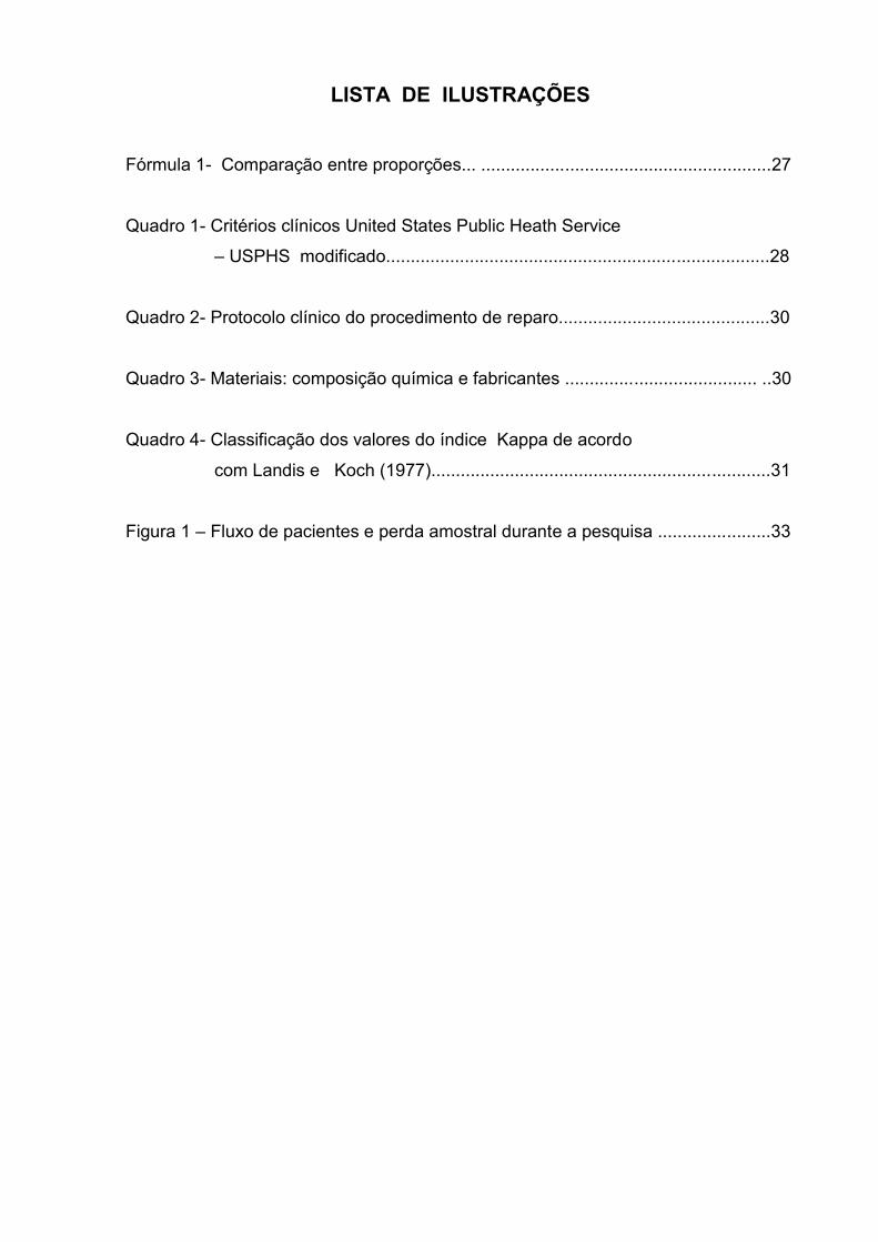
LISTA DE ILUSTRAÇÕES
Fórmula 1- Comparação entre proporções... ...........................................................27
Quadro 1- Critérios clínicos United States Public Heath Service
– USPHS modificado..............................................................................28
Quadro 2- Protocolo clínico do procedimento de reparo...........................................30
Quadro 3- Materiais: composição química e fabricantes ....................................... ..30
Quadro 4- Classificação dos valores do índice Kappa de acordo
com Landis e Koch (1977).....................................................................31
Figura 1 – Fluxo de pacientes e perda amostral durante a pesquisa .......................33
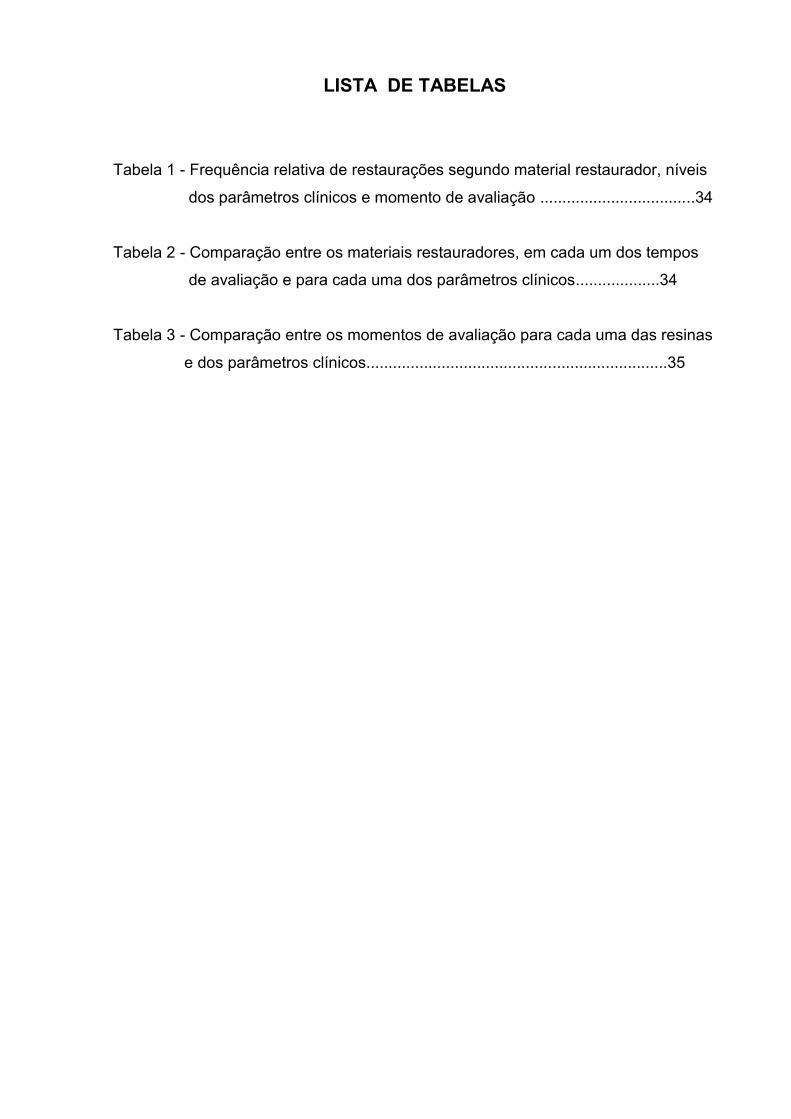
LISTA DE TABELAS
Tabela 1 - Frequência relativa de restaurações segundo material restaurador, níveis
dos parâmetros clínicos e momento de avaliação ...................................34
Tabela 2 - Comparação entre os materiais restauradores, em cada um dos tempos
de avaliação e para cada uma dos parâmetros clínicos...................34
Tabela 3 - Comparação entre os momentos de avaliação para cada uma das resinas
e dos parâmetros clínicos....................................................................35
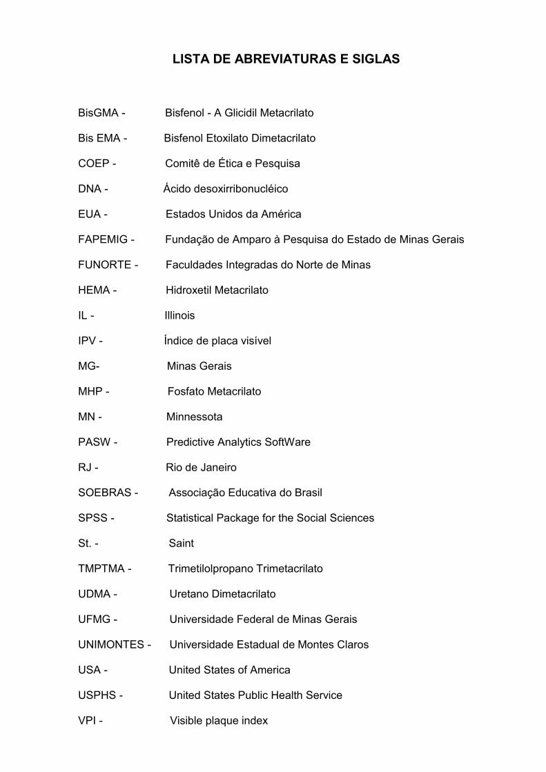
LISTA DE ABREVIATURAS E SIGLAS
BisGMA - Bisfenol - A Glicidil Metacrilato
Bis EMA - Bisfenol Etoxilato Dimetacrilato
COEP - Comitê de Ética e Pesquisa
DNA - Ácido desoxirribonucléico
EUA - Estados Unidos da América
FAPEMIG - Fundação de Amparo à Pesquisa do Estado de Minas Gerais
FUNORTE - Faculdades Integradas do Norte de Minas
HEMA - Hidroxetil Metacrilato
IL - Illinois
IPV - Índice de placa visível
MG- Minas Gerais
MHP - Fosfato Metacrilato
MN - Minnessota
PASW - Predictive Analytics SoftWare
RJ - Rio de Janeiro
SOEBRAS - Associação Educativa do Brasil
SPSS - Statistical Package for the Social Sciences
St. - Saint
TMPTMA - Trimetilolpropano Trimetacrilato
UDMA - Uretano Dimetacrilato
UFMG - Universidade Federal de Minas Gerais
UNIMONTES - Universidade Estadual de Montes Claros
USA - United States of America
USPHS - United States Public Health Service
VPI - Visible plaque index
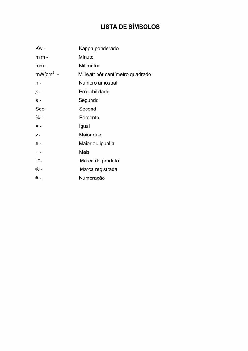
LISTA DE SÍMBOLOS
Kw - Kappa ponderado
mim - Minuto
mm- Milímetro
mW/cm2 - Miliwatt pór centímetro quadrado
n - Número amostral
p - Probabilidade
s - Segundo
Sec - Second
% - Porcento
= - Igual
>- Maior que
≥ - Maior ou igual a
+ - Mais
™- Marca do produto
® - Marca registrada
# - Numeração
SUMÁRIO
1 INTRODUÇÂO ................................................................... 14
2 REVISÃO DE LITERATURA ............................................. 17
2.1 Compósitos ........................................................................ 17
2.2 Compósitos à base de Silorano ......................................... 18
2.3 Reparos .............................................................................. 24
3 MATERIAIS E MÉTODOS ................................................ 26
3.1 Desenho do estudo ........................................................... 26
3.2 Grupos de tratamento ....................................................... 28
3.3 Métodos do estudo ........................................................... 31
3.4 Análise estatística ..............................................................
31
4 RESULTADOS ................................................................. 33
5 DISCUSSÃO .................................................................... 36
5.1 Artigo I .............................................................................. 36
5.2 Artigo II ............................................................................ 60
6 CONSIDERAÇÕES FINAIS ............................................ 82
REFERÊNCIAS ................................................................ 83
ANEXOS ........................................................................... 86
14
1- INTRODUÇÃO
A demanda por restaurações estéticas, o desenvolvimento de novos
sistemas adesivos e sistemas de polimerização, bem com a melhoria das
propriedades físicas e químicas dos compósitos diretos, fez com que o uso
desses se tornasse rotineiro, ganhando popularidade entre os pacientes
também para a restauração de dentes posteriores.1-5 No entanto, apesar da
evolução sofrida pelos compósitos modernos, duas características ainda
requerem aprimoramento: a contração de polimerização e as tensões geradas
pela polimerização,6-9 e estudos sobre geração de tensões e forças com
características para analisar as propriedades mecânicas dos compósitos
sinalizam para uma mudança no monômero como o caminho mais promissor
para minimizar os efeitos da contração.7-12
Um sistema recentemente disponibilizado no mercado utiliza um
monômero de natureza hidrófoba - o silorano, que deriva da combinação dos
componentes básicos dos grupos epóxicos siloxanos e oxiranos. Siloxanos são
conhecidos por sua hidrofobia, enquanto os oxiranos são conhecidos por sua
baixa contração e estabilidade diante de influências físicas e químico-físicas.8,13
Enquanto resinas convencionais à base de dimetacrilato polimerizam por uma
reação de adição iniciada por radicais livres, a polimerização do silorano ocorre
através da reação catiônica de abertura de um anel, resultando em menor
contração de polimerização.
Estudos laboratoriais têm comparado esses novos compósitos àqueles à
base de dimetacrilato, revelando para o silorano uma menor contração de
polimerização, maior estabilidade à luz ambiente, mais baixa sorção e
solubilidade em água e menor coeficiente de difusão.7,10,14,15 Outros parâmetros

15
como módulo de elasticidade, resistência à flexão e biocompatibilidade em
testes toxicológicos são comparáveis aos dos compósitos à base de
dimetacrilato. 7,10,15
O controle da contração e das tensões geradas pela polimerização
influenciam positivamente a integridade marginal das restaurações. Por outro
lado, margens imperfeitas constituem uma importante causa de substituição
das restaurações.6 A substituição total é o tratamento mais comumente
adotado para restaurações diagnosticadas como defeituosas. Entretanto, na
maioria das vezes, a medida da qualidade das restaurações é feita de forma
subjetiva e mínimos desvios do ideal têm determinado a substituição
sistemática de restaurações clinicamente aceitáveis. 16,17
À exceção dos casos de fratura da estrutura da resina, profundo
manchamento da interface resina/dente e cárie secundária, a remoção total é
tida como indesejável e inapropriada.2,4 Assim, tendo-se em vista as atuais
tendências por condutas clínicas mais conservadoras, vários estudos têm
sugerido a substituição parcial da restauração, ou seja, o seu reparo.2,4,16-18 Tal
procedimento, minimamente invasivo, permite conservar o que está
convenientemente restaurado sem sacrifícios de estruturas dentais sadias
remanescentes e implica na adição de material restaurador após a remoção do
defeito, havendo ou não preparo em material restaurador e/ou tecidos dentais
duros.19
Estudos clínicos têm demonstrado que, quando bem planejados, os
reparos são de simples confecção e podem aumentar a longevidade clínica das
restaurações.16,17,20,21 Apesar das limitações dos estudos in vitro, seus
resultados também são encorajadores quando variáveis como diferentes

16
compósitos, agentes de união, fontes de luz, técnicas incrementais e
tratamentos de superfície são avaliadas através de diferentes testes
laboratoriais.2,4,22
Diante dessas considerações, torna-se oportuno conhecer os materiais
odontológicos atualmente disponíveis para a confecção de reparos e mesmo
avaliar o desempenho clínico desse tipo alternativo de tratamento restaurador.
Como os estudos laboratoriais são limitados em predizer condições clínicas em
curto e longo prazos, e por não terem sido encontrados estudos clínicos
controlados que tenham trabalhado a hipótese de que uma resina composta de
baixa contração (silorano) apresentaria desempenho similar às resinas
convencionais quando usadas para reparar restaurações de resina, a presente
pesquisa objetivou fazê-lo. Os resultados desta investigação poderiam
colaborar na construção do corpo de evidência científica para justificar ou não a
inclusão da resina de silorano ao arsenal terapêutico da Odontologia como
material de reparo, justificando a presente proposta.
17
2. REVISÃO DA LITERATURA
2.1 Compósitos
Durante os anos 50 e inicio dos 60, Bowen (1958)23 modificou a
molécula do Bisfenol-A, associando-a a radicais metacrilatos, e assim
sintetizou o Bisfenol-A Glicidil Metacrilato (Bis-GMA). Esta resina mostrou
menor contração de polimerização e maior estabilidade térmica com menor
tempo de cura, apresentando propriedades favoráveis ao seu uso como
material restaurador. No desenvolvimento desse produto, Bowen (1963)24
incorporou pó de quartzo ao Bis-GMA, prática que vinha sendo empregada em
resinas acrílicas para restaurações, passando também a tratar a superfície
dessas partículas com um silano para promover a união química entre as
partículas de carga e a matriz resinosa, aumentando sua resistência.
Atualmente, as resinas compostas têm sido largamente utilizadas em
odontologia restauradora e na última década ganharam popularidade entre os
pacientes também para a restauração de dentes posteriores.1,2,22,25 A demanda
por restaurações estéticas, o desenvolvimento de novos sistemas adesivos e
sistemas de polimerização, bem com a melhoria das propriedades físicas e
químicas dos compósitos diretos, fez com que o uso desses se tornasse
rotineiro,1-5,25 em especial por apresentarem vantagens em relação às
restaurações indiretas tais como, custo e tempo clínico reduzidos, preparos
menos invasivos e pela redução da microinfiltração dada pelos agentes
adesivos à dentina e ao esmalte.5
No entanto, apesar da evolução que conferiu aos compósitos modernos
uma maior resistência mecânica e resistência ao desgaste, maior estabilidade
na cavidade bucal, além de uma ótima estética, duas características ainda
18
requerem aprimoramento: a contração e as tensões geradas pela
polimerização.6-9
Até o momento, as principais estratégias utilizadas para controlar a
contração de polimerização têm sido a adoção de técnicas clínicas como a
inserção incremental e o controle da velocidade de polimerização. Os
fabricantes, por outro lado, têm tentado alcançar a redução da contração
volumétrica, aumentando o conteúdo de partículas de carga. Contudo, a
contração intrínseca do compósito permanece um desafio, e mudar o
monômero parece ser o caminho mais promissor para minimizar os efeitos da
contração.7-12
2.2 Compósitos à base de silorano
Um sistema recentemente disponibilizado no mercado utiliza uma resina
de natureza hidrófoba, o silorano, que deriva da combinação dos componentes
básicos dos grupos epóxicos siloxanos e oxiranos. Siloxanos são conhecidos
por sua hidrofobia, enquanto os oxiranos são conhecidos por sua baixa
contração e estabilidade diante das influências físicas e químico-físicas6.
Enquanto resinas convencionais à base de dimetacrilato polimerizam por uma
reação de adição iniciada por radicais livres, o processo de polimerização do
silorano ocorre através da reação catiônica de abertura de um anel aromático
que resulta em menor contração de polimerização. Seu sistema iniciador é
composto de canforoquinona, sais de iodônio e doadores de elétrons.7
Os novos compósitos à base de silorano caracterizam-se por sua distinta
polimerização, capaz de reduzir as tensões geradas pela polimerização. A
matriz do silorano é formada por um anel que se abre quando uma reação
catiônica induz a polimerização dos monômeros de silorano. A molécula

19
silorano é híbrida e formada em parte pela estrutura do siloxano e em parte
pela estrutura do oxirano. A introdução do silorano abre novos horizontes na
busca pela redução das tensões geradas pela polimerização e pelo equilíbrio
das tensões volumétricas causadas pela contração de polimerização.8
Em um estudo no qual um compósito à base de silorano foi comparado a
materiais à base de metacrilato, o compósito à base de silorano revelou a
menor contração de polimerização entre os compósitos testados, com uma
contração volumétrica de 0,94% (método do disco aderido) e 0,99% do volume
(método de Arquimedes). A estabilidade à luz ambiente dos materiais à base
de silorano foi maior (10 min) que dos outros compósitos à base de metacrilato
(55-90 s). A química de anéis abertos do silorano permitiu valores de contração
volumétrica menores que 1%. Parâmetros mecânicos como módulo de
elasticidade e resistência à flexão foram comparáveis aos dos já bem aceitos
compósitos à base de metacrilato.7
A contração de polimerização ainda é uma grande preocupação e
apesar de as resinas compostas serem o material de escolha para a maioria
das restaurações, sua contração de polimerização permanece um problema. A
tensão de contração associada à contração de polimerização pode causar a
desunião da interface resina composta/dente e pode contribuir para a
sensibilidade pós-operatória, trincas de esmalte, lesões de cáries recorrentes,
pigmentação marginal e a eventual falha da restauração.26
Testes de contração de polimerização foram conduzidos e mostraram
que a resina Filtek P90 tem uma contração significativamente menor do que
compósitos metacrilatos convencionais testados, independente do método
usado. As formas mais comuns para medir a contração de polimerização são o
20
método de discos aderidos, conhecido com o método Watts e o método
Archimedes que foi recentemente desenvolvido para um padrão alemão. O
método Watts resulta em baixos valores de contração uma vez que apenas a
contração linear do disco de resina aderido é medida e então convertida em %
do volume. Já o método Archimedes mede a real contração do volume de
acordo com o princípio da flutuação de corpos. Dessa forma, ambos os
métodos mostram uma correlação.9
A tensão de contração pode ser medida pelo método de investigação
fotoelástica. Amostras de resina foram aderidas à uma placa de Araldite®
foram fotopolimerizadas usando luz halógena. As forças de tensão induzidas
pela tensão de polimerização da resina à placa podem ser visualizadas como
anéis isocromáticos em um microscópio de polarização. A tensão de
polimerização foi calculada desde o diâmetro dos primeiros anéis
isocromáticos, após 4 minutos e novamente após 24 horas de exposição. Os
resultados revelam que a resina Filtek P90 gerou os menores valores de
tensão de polimerização entre todas as resinas compostas testadas.
Adicionalmente, todos os materiais metacrilatos continuaram gerando tensão:
os valores medidos após 24 horas foram sempre maiores do que após 5
minutos. A resina à base de silorano foi o único material que manteve o mesmo
baixo valor de tensão de polimerização observado após 5 minutos e não
continuou a gerar tensão.27
A fotopolimerização de um protótipo de um sistema contendo
oxiranos/poliol mostrou-se útil para o desenvolvimento de compósitos dentais.
Resultados favoráveis obtidos com os novos compósitos indicam que a fórmula
oxirano/poliol pode ser utilizada para o desenvolvimento de compósitos dentais
21
com propriedades mecânicas e biocompatibilidade aceitáveis. Entretanto, a
análise de extratos obtidos a partir de períodos de incubação mais longos são
necessárias para estabelecer conclusões finais sobre os componentes
lixiviáveis do oxirano.10
As diferenças nas propriedades físicas e mecânicas exibidas pelas
resinas compostas de baixa contração comparadas às convencionais à base
de metacrilato podem contribuir para o sucesso clínico do material. Quando
investigados o efeito da absorção de água e solubilidade em água nas
propriedades mecânicas de dois metacrilatos (Filtek Z250 e Z100), um oxirano
experimental (OXI) e um silorano (SIL), em curto e médio prazos de imersão,
os compósitos com silorano exibiram sorção de água, solubilidade e coeficiente
de difusão significativamente menores, em cada período de imersão.14
O desenvolvimento de compósitos de baixa contração pode ainda
oferecer uma redução na tensão de polimerização gerada na interface dente
restauração em comparação com as resinas de metacrilato. Quando avaliados,
in vitro, a deflexão das cúspides e a microinfiltração em cavidades restauradas
com resinas experimentais de oxirano (EXL596) e silorano (H1), e resinas de
metacrilato (Filtek Z250 e Z100), a menor deformação das cúspides e
microinfiltração foram registradas por cavidades restauradas com H1, sendo
essas características vantajosas em termos de integridade marginal, em
comparação com a Filtek Z250. No entanto, como a diferença na
microinfiltração entre a H1 e Z100 não foi significativa, uma redução apenas
modesta nos efeitos deletérios da tensão gerada pela polimerização foi
registrada.22
22
A consequência clínica da alta tensão de polimerização e contração de
polimerização é o deslocamento de cúspides, que pode resultar em dano à
estrutura hígida do dente como trincas no esmalte e hipersensibilidade induzida
por tensão. Em um estudo em que a interferometria eletrônica foi utilizada para
medir a deformação do dente em resposta à polimerização de cinco resinas
compostas de baixa contração de polimerização, concluiu-se que a resina tipo
flow não provocou deformação significativamente maior do que uma resina
convencional híbrida. Porém, o material experimental à base de silorano
induziu o dente a uma menor deformação.29
Um estudo comparou as propriedades de uma resina à base de silorano
(Sil-Mix, 3M-ESPE), uma mistura de SIL-Mix com um monômero redutor da
tensão (TOSU), e como controles uma resina de Bis-GMA / TEGDMA e o
compósito (Filtek Z250, 3M ESPE). Foram medidas as mudanças de volume na
polimerização e a tensão de polimerização nas amostras. Foram determinados
também o módulo de elasticidade, a resistência à fratura e à resiliência. Os
valores da tensão de polimerização para resinas contendo TOSU foram
significativamente menores do que os valores dos outros materiais. Os valores
de contração de polimerização para as formulações de Sil-Mix não diferiram
entre si e foram significativamente menores que os padrões de metacrilato.
Formulações contendo TOSU apresentaram, em geral, algumas propriedades
mecânicas inferiores às Sil-Mix ou aos metacrilatos. Os valores de tensão de
polimerização para compósitos à base de Sil-Mix foram significativamente
menores em comparação com a Z250. O compósito com TOSU (1%)
apresentou a menor tensão de polimerização. Nenhuma diferença entre os
grupos de compósitos foi notada para tenacidade de fratura ou fratura ao
23
trabalho. Por último, os compósitos com TOSU (5%) diferiram
significativamente da Z250. Todas as formulações de Sil-Mix apresentaram
módulos de elasticidade significativamente menores que a Z250. A capacidade
do TOSU para reduzir a tensão de polimerização sem uma proporcional
redução das propriedades mecânicas fornece uma base para a melhoria dos
compósitos à base de silorano.10
O potencial reativo e as propriedades estruturais dos oxiranos (epóxidos)
são vantagens a serem consideradas quando se idealiza um polímero. No
entanto, compostos epoxídicos são amplamente conhecidos por terem
propriedades genotóxicas. Em um estudo objetivando avaliar a citotoxicidade
e a genotoxicidade (danos ao DNA) induzidos por oxiranos e siloranos, os
achados reforçaram relatos anteriores nos quais os siloranos têm um baixo
potencial genotóxico e podem ser componentes adequados para o
desenvolvimento de biomateriais.30
Um recente estudo clínico avaliou a adaptação marginal de um novo
compósito à base de silorano comparando-o com um compósito à base de
metacrilato. A hipótese testada foi a de que a reduzida contração do silorano
melhoraria a adaptação marginal das restaurações confeccionadas com esse
material. As restaurações incluídas neste estudo foram avaliadas em baseline e
após um ano. Em baseline, não houve diferença estatística significativa entre o
desempenho clínico de ambos os materiais. Apesar de as comparações
estatísticas terem demonstrado uma melhor performance do compósito à base
de metacrilato (p = 0,01 e p < 0,01) para adaptação marginal nas avaliações de
um ano, o baixo valor do índice de concordância Kappa (32%) reflete a
dificuldade de mensurar adaptação marginal clinicamente. Desta forma, a

24
reduzida contração de polimerização demonstrada em laboratório não foi
clinicamente significativa.31
Outro estudo clínico avaliou a durabilidade de uma resina composta de
baixa contração desenvolvida com o atrativo de reduzir a geração de forças
nas restaurações confeccionadas com resinas compostas diretas. Como
controle foi utilizada uma resina composta convencional híbrida. Para ambos os
grupos um sistema adesivo autocondicionante foi empregado. As restaurações
deste estudo foram avaliadas ao longo de 5 anos. Os achados deste estudo
mostraram que a resina composta de baixa contração apresentou boa
durabilidade, porém não diferente estatisticamente do grupo controle. As
restaurações falharam, em sua maioria, nos anos finais do estudo e a maior
causa das falhas foi a cárie secundária.32
2.3 Reparos
Entende-se por reparo um recurso restaurador alternativo que possibilita
a recuperação de uma restauração já existente, ou mesmo a remoção de parte
de uma restauração com falhas localizadas e o reparo destas. 22, 33
O reparo é um método minimamente invasivo, que implica a adição de
do material restaurador após a remoçao do defeito, não sendo invasivo, com ou
sem preparo no material restaurador e / ou tecidos dentais duros19.
Tal procedimento permite conservar o que está convenientemente
restaurado sem sacrifícios de estruturas dentais sadias remanescentes,
evitando ainda que o novo preparo cavitário seja aumentado, assim como o
tamanho da nova restauração. 2,22,17,18
De simples confecção, os reparos provêem também um aumento da
longevidade clínica das restaurações, o que atende às necessidades dos
25
pacientes que, nos últimos cinquenta anos, vivem um aumento de sua
expectativa de vida, necessitando de restaurações que possam servi-los por
mais tempo.16,33
Apesar de estudos sobre reparos em resina composta terem sido
largamente publicados, essa técnica não é comumente considerada para o
tratamento de restaurações com defeitos localizados, mas clinicamente
aceitáveis. Ao contrário, essas são usualmente substituídas e muitas vezes a
referida técnica nem mesmo é ensinada nas escolas norte-americanas.16
Uma explicação para isso estaria na falta de estudos em longo prazo
que proveriam suporte para o uso dos reparos como uma alternativa às
substituições.17
Os resultados de um estudo clínico de três anos avaliando a efetividade
de tratamentos alternativos à substituição total de restaurações defeituosas
classes I e II de resina composta e de amálgama levam a concluir que o
reparo, assim como outras formas de intervenção minimamente invasivas, são
procedimentos conservadores e simples, capazes de aumentar a longevidade
das restaurações de resina composta e de amálgama, uma vez que a maior
parte das restaurações do referido estudo mantiveram suas características
clínicas após um período de três anos sendo avaliadas. 20
26
3. MATERIAIS E MÉTODOS
3.1 Desenho do estudo
O presente estudo, aprovado pelo Comitê de Ética e Pesquisa da
Universidade Federal de Minas Gerais (ETIC 0546.0.203.000-09 tratou-se de
um ensaio clínico prospectivo controlado randomizado, cuja unidade
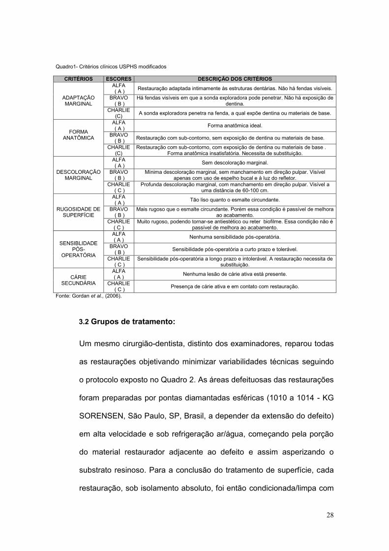
experimental foi a restauração reparada. A variável resposta, qualitativa
categórica ordinal, foi cada um dos seguintes critérios clínicos: adaptação
marginal, forma anatômica, rugosidade de superfície, descoloração marginal,
sensibilidade pós-operatória e cárie secundária, a partir dos quais as
restaurações foram classificadas como Alfa, Bravo ou Charlie16 ( Quadro 1).
Participaram desta pesquisa, pacientes recrutados dentre aqueles que
comparecem rotineiramente às clínicas odontológicas das faculdades de
Odontologia da Universidade Federal de Minas Gerais - UFMG, Belo Horizonte
- MG e das Faculdades Unidas do Norte de Minas – FUNORTE/SOEBRAS,
Montes Claros – MG.
Para o cálculo do tamanho da amostra (n), foi considerada uma
diferença de 50% entre os tipos de tratamento. Essa variação foi adotada
porque dados sobre a performance dos compósitos à base de silorano como
material de reparo para restaurações defeituosas de resina composta à base
de dimetacrilato ainda não estão disponíveis na literatura.35
Considerando tal diferença, um poder de estudo (1 – β) = 0,90 e um erro
tipo I (α) = 0,05, um (n) = 19 unidades amostrais em cada grupo de teste foi
encontrado.
27
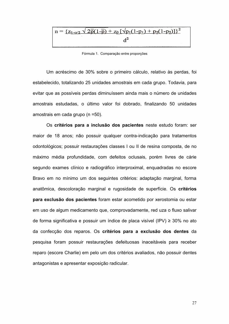
Fórmula 1. Comparação entre proporções
Um acréscimo de 30% sobre o primeiro cálculo, relativo às perdas, foi
estabelecido, totalizando 25 unidades amostrais em cada grupo. Todavia, para
evitar que as possíveis perdas diminuíssem ainda mais o número de unidades
amostrais estudadas, o último valor foi dobrado, finalizando 50 unidades
amostrais em cada grupo (n =50).
Os critérios para a inclusão dos pacientes neste estudo foram: ser
maior de 18 anos; não possuir qualquer contra-indicação para tratamentos
odontológicos; possuir restaurações classes I ou II de resina composta, de no
máximo média profundidade, com defeitos oclusais, porém livres de cárie
segundo exames clínico e radiográfico interproximal, enquadradas no escore
Bravo em no mínimo um dos seguintes critérios: adaptação marginal, forma
anatômica, descoloração marginal e rugosidade de superfície. Os critérios
para exclusão dos pacientes foram estar acometido por xerostomia ou estar
em uso de algum medicamento que, comprovadamente, red uza o fluxo salivar
de forma significativa e possuir um índice de placa visível (IPV) ≥ 30% no ato
da confecção dos reparos. Os critérios para a exclusão dos dentes da
pesquisa foram possuir restaurações defeituosas inaceitáveis para receber
reparo (escore Charlie) em pelo um dos critérios avaliados, não possuir dentes
antagonistas e apresentar exposição radicular.
28
Quadro1- Critérios clínicos USPHS modificados
CRITÉRIOS ESCORES DESCRIÇÃO DOS CRITÉRIOS
ADAPTAÇÃO MARGINAL
ALFA ( A )
Restauração adaptada intimamente às estruturas dentárias. Não há fendas visíveis.
BRAVO ( B )
Há fendas visíveis em que a sonda exploradora pode penetrar. Não há exposição de dentina.
CHARLIE (C)
A sonda exploradora penetra na fenda, a qual expõe dentina ou materiais de base.
FORMA ANATÔMICA
ALFA ( A )
Forma anatômica ideal.
BRAVO ( B )
Restauração com sub-contorno, sem exposição de dentina ou materiais de base.
CHARLIE (C)
Restauração com sub-contorno, com exposição de dentina ou materiais de base . Forma anatômica insatisfatória. Necessita de substituição.
DESCOLORAÇÃO MARGINAL
ALFA ( A )
Sem descoloração marginal.
BRAVO ( B )
Mínima descoloração marginal, sem manchamento em direção pulpar. Visível apenas com uso de espelho bucal e à luz do refletor.
CHARLIE ( C )
Profunda descoloração marginal, com manchamento em direção pulpar. Visível a uma distância de 60-100 cm.
RUGOSIDADE DE SUPERFÍCIE
ALFA ( A )
Tão liso quanto o esmalte circundante.
BRAVO ( B )
Mais rugoso que o esmalte circundante. Porém essa condição é passível de melhora ao acabamento.
CHARLIE ( C )
Muito rugoso, podendo tornar-se antiestético ou reter biofilme. Essa condição não é passível de melhora ao acabamento.
SENSIBLIDADE PÓS-
OPERATÓRIA
ALFA ( A )
Nenhuma sensibilidade pós-operatória.
BRAVO ( B )
Sensibilidade pós-operatória a curto prazo e tolerável.
CHARLIE ( C )
Sensibilidade pós-operatória a longo prazo e intolerável. A restauração necessita de substituição.
CÁRIE SECUNDÁRIA
ALFA ( A )
Nenhuma lesão de cárie ativa está presente.
CHARLIE ( C )
Presença de cárie ativa e em contato com restauração.
Fonte: Gordan et al., (2006).
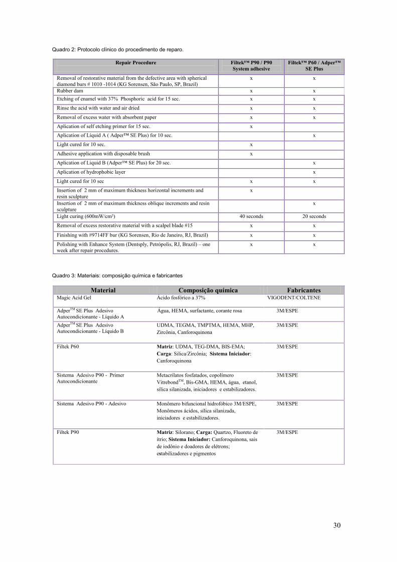
3.2 Grupos de tratamento:
Um mesmo cirurgião-dentista, distinto dos examinadores, reparou todas
as restaurações objetivando minimizar variabilidades técnicas seguindo
o protocolo exposto no Quadro 2. As áreas defeituosas das restaurações
foram preparadas por pontas diamantadas esféricas (1010 a 1014 - KG
SORENSEN, São Paulo, SP, Brasil, a depender da extensão do defeito)
em alta velocidade e sob refrigeração ar/água, começando pela porção
do material restaurador adjacente ao defeito e assim asperizando o
substrato resinoso. Para a conclusão do tratamento de superfície, cada
restauração, sob isolamento absoluto, foi então condicionada/limpa com
29
ácido fosfórico a 37% (Magic Acid Gel - VIGODENT COLTENE). Em
seguida, as restaurações foram aleatoriamente reparadas por um dos
dois grupos de tratamento - Controle e Teste - da seguinte forma: um
sorteio foi feito para determinar por qual dos grupos de tratamento a
restauração seria reparada. Ao se completar 50 sorteios para um
mesmo grupo, todas as demais restaurações foram reparadas pelo outro
grupo. Os pacientes foram informados dos sorteios mas não de seus
resultados.
Grupo Controle (n=50) - sistema adesivo autocondicionante
(Adper SE Plus, 3M /ESPE, St. Paul, MN, EUA) + resina
composta convencional à base de dimetacrilato (Filtek P60
Posterior Restorative, 3M/ESPE, St. Paul, MN, EUA);
Grupo teste (n=50) - sistema adesivo autocondicionante (P90
System Adhesive Self-Etch Primer and Bond, 3M/ESPE, St. Paul,
MN, EUA) + resina composta de baixa contração à base de
silorano (Filtek P90 Low Shrink Posterior Restorative, 3M/ESPE,
St. Paul, MN, EUA) (Quadro 3).
Todos os materiais restauradores empregados neste estudo foram
utilizados segundo expressas recomendações de seus fabricantes.
30
Quadro 2: Protocolo clínico do procedimento de reparo.
Repair Procedure
Filtek™ P90 / P90
System adhesive
Filtek™ P60 / Adper™
SE Plus
Removal of restorative material from the defective area with spherical
diamond burs # 1010 -1014 (KG Sorensen, São Paulo, SP, Brazil)
x x
Rubber dam x x
Etching of enamel with 37% Phosphoric acid for 15 sec. x x
Rinse the acid with water and air dried x x
Removal of excess water with absorbent paper x x
Aplication of self etching primer for 15 sec. x
Aplication of Líquid A ( Adper™ SE Plus) for 10 sec. x
Light cured for 10 sec. x
Adhesive application with disposable brush x
Aplication of Liquid B (Adper™ SE Plus) for 20 sec. x
Aplication of hydrophobic layer x
Light cured for 10 sec x x
Insertion of 2 mm of maximum thickness horizontal increments and
resin sculpture
x
Insertion of 2 mm of maximum thickness oblique increments and resin
sculpture
x
Light curing (600mW/cm²) 40 seconds 20 seconds
Removal of excess restorative material with a scalpel blade #15 x x
Finishing with #9714FF bur (KG Sorensen, Rio de Janeiro, RJ, Brazil) x x
Polishing with Enhance System (Dentsply, Petrópolis, RJ, Brazil) – one
week after repair procedures.
x x
Quadro 3: Materiais: composição química e fabricantes
Material Composição química Fabricantes Magic Acid Gel
Ácido fosfórico a 37% VIGODENT/COLTENE
AdperTM SE Plus Adesivo
Autocondicionante - Líquido A
Água, HEMA, surfactante, corante rosa 3M/ESPE
AdperTM SE Plus Adesivo Autocondicionante - Líquido B
UDMA, TEGMA, TMPTMA, HEMA, MHP,
Zircônia, Canforoquinona
3M/ESPE
Filtek P60 Matriz: UDMA, TEG-DMA, BIS-EMA;
Carga: Silica/Zircônia; Sistema Iniciador:
Canforoquinona
3M/ESPE
Sistema Adesivo P90 - Primer
Autocondicionante Metacrilatos fosfatados, copolímero
VitrebondTM, Bis-GMA, HEMA, água, etanol,
sílica silanizada, iniciadores e estabilizadores.
3M/ESPE
Sistema Adesivo P90 - Adesivo
Monômero bifuncional hidrofóbico 3M/ESPE,
Monômeros ácidos, sílica silanizada,
iniciadores e estabilizadores.
3M/ESPE
Filtek P90 Matriz: Silorano; Carga: Quartzo, Fluoreto de
ítrio; Sistema Iniciador: Canforoquinona, sais
de iodônio e doadores de elétrons;
estabilizadores e pigmentos
3M/ESPE
31
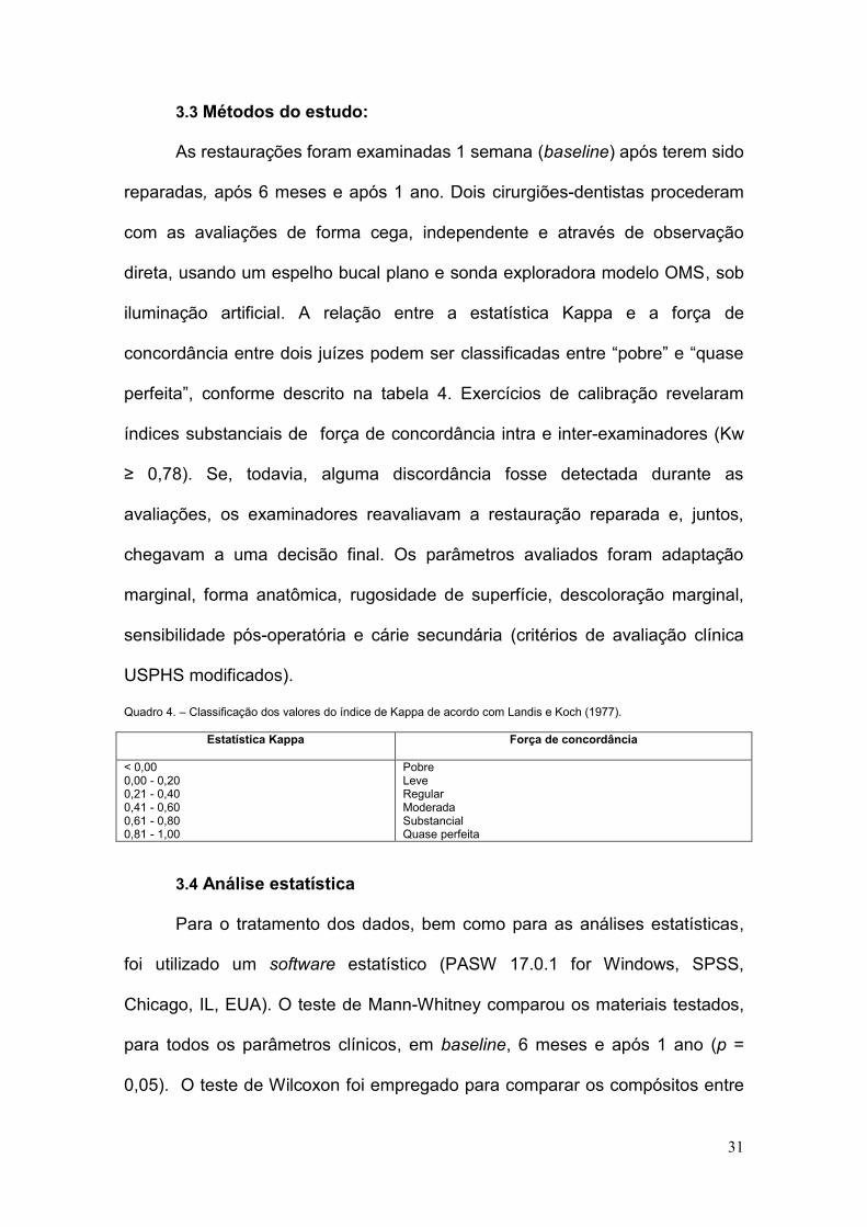
3.3 Métodos do estudo:
As restaurações foram examinadas 1 semana (baseline) após terem sido
reparadas, após 6 meses e após 1 ano. Dois cirurgiões-dentistas procederam
com as avaliações de forma cega, independente e através de observação
direta, usando um espelho bucal plano e sonda exploradora modelo OMS, sob
iluminação artificial. A relação entre a estatística Kappa e a força de
concordância entre dois juízes podem ser classificadas entre “pobre” e “quase
perfeita”, conforme descrito na tabela 4. Exercícios de calibração revelaram
índices substanciais de força de concordância intra e inter-examinadores (Kw
≥ 0,78). Se, todavia, alguma discordância fosse detectada durante as
avaliações, os examinadores reavaliavam a restauração reparada e, juntos,
chegavam a uma decisão final. Os parâmetros avaliados foram adaptação
marginal, forma anatômica, rugosidade de superfície, descoloração marginal,
sensibilidade pós-operatória e cárie secundária (critérios de avaliação clínica
USPHS modificados).
Quadro 4. – Classificação dos valores do índice de Kappa de acordo com Landis e Koch (1977).
Estatística Kappa Força de concordância
< 0,00 0,00 - 0,20 0,21 - 0,40 0,41 - 0,60 0,61 - 0,80 0,81 - 1,00
Pobre Leve Regular Moderada Substancial Quase perfeita
3.4 Análise estatística
Para o tratamento dos dados, bem como para as análises estatísticas,
foi utilizado um software estatístico (PASW 17.0.1 for Windows, SPSS,
Chicago, IL, EUA). O teste de Mann-Whitney comparou os materiais testados,
para todos os parâmetros clínicos, em baseline, 6 meses e após 1 ano (p =
0,05). O teste de Wilcoxon foi empregado para comparar os compósitos entre

32
si em função do tempo – baseline , 6 meses e 1 ano, em todos os parâmetros
clínicos (p = 0,05).
33
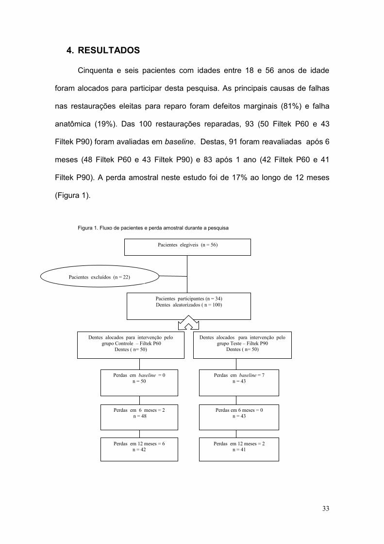
4. RESULTADOS
Cinquenta e seis pacientes com idades entre 18 e 56 anos de idade
foram alocados para participar desta pesquisa. As principais causas de falhas
nas restaurações eleitas para reparo foram defeitos marginais (81%) e falha
anatômica (19%). Das 100 restaurações reparadas, 93 (50 Filtek P60 e 43
Filtek P90) foram avaliadas em baseline. Destas, 91 foram reavaliadas após 6
meses (48 Filtek P60 e 43 Filtek P90) e 83 após 1 ano (42 Filtek P60 e 41
Filtek P90). A perda amostral neste estudo foi de 17% ao longo de 12 meses
(Figura 1).
Figura 1. Fluxo de pacientes e perda amostral durante a pesquisa
Pacientes elegíveis (n = 56)
Pacientes participantes (n = 34)
Dentes aleatorizados ( n = 100)
Dentes alocados para intervenção pelo
grupo Controle – Filtek P60
Dentes ( n= 50)
Dentes alocados para intervenção pelo
grupo Teste – Filtek P90 Dentes ( n= 50)
Perdas em baseline = 0
n = 50
Perdas em baseline = 7
n = 43
Perdas em 6 meses = 2
n = 48
Perdas em 6 meses = 0
n = 43
Perdas em 12 meses = 6 n = 42
Perdas em 12 meses = 2 n = 41
Pacientes excluídos (n = 22)
34
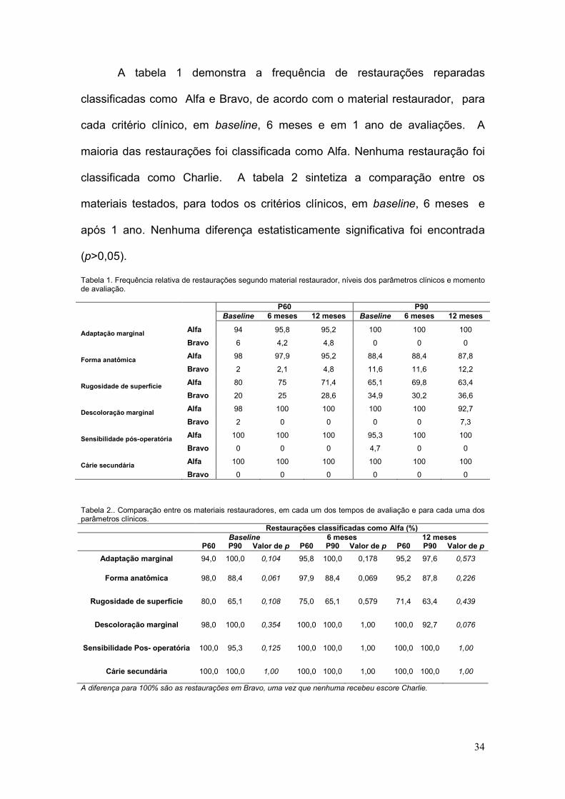
A tabela 1 demonstra a frequência de restaurações reparadas
classificadas como Alfa e Bravo, de acordo com o material restaurador, para
cada critério clínico, em baseline, 6 meses e em 1 ano de avaliações. A
maioria das restaurações foi classificada como Alfa. Nenhuma restauração foi
classificada como Charlie. A tabela 2 sintetiza a comparação entre os
materiais testados, para todos os critérios clínicos, em baseline, 6 meses e
após 1 ano. Nenhuma diferença estatisticamente significativa foi encontrada
(p>0,05).
Tabela 1. Frequência relativa de restaurações segundo material restaurador, níveis dos parâmetros clínicos e momento de avaliação.
P60 P90
Baseline 6 meses 12 meses Baseline 6 meses 12 meses
Adaptação marginal Alfa 94 95,8 95,2 100 100 100
Bravo 6 4,2 4,8 0 0 0
Forma anatômica Alfa 98 97,9 95,2 88,4 88,4 87,8
Bravo 2 2,1 4,8 11,6 11,6 12,2
Rugosidade de superfície Alfa 80 75 71,4 65,1 69,8 63,4
Bravo 20 25 28,6 34,9 30,2 36,6
Descoloração marginal Alfa 98 100 100 100 100 92,7
Bravo 2 0 0 0 0 7,3
Sensibilidade pós-operatória Alfa 100 100 100 95,3 100 100
Bravo 0 0 0 4,7 0 0
Cárie secundária Alfa 100 100 100 100 100 100
Bravo 0 0 0 0 0 0
Tabela 2.. Comparação entre os materiais restauradores, em cada um dos tempos de avaliação e para cada uma dos parâmetros clínicos.
Restaurações classificadas como Alfa (%)
Baseline 6 meses 12 meses P60 P90 Valor de p P60 P90 Valor de p P60 P90 Valor de p
Adaptação marginal 94,0
100,0 0,104 95,8 100,0 0,178 95,2 97,6 0,573
Forma anatômica 98,0
88,4 0,061 97,9 88,4 0,069 95,2 87,8 0,226
Rugosidade de superfície 80,0
65,1 0,108 75,0 65,1 0,579 71,4 63,4 0,439
Descoloração marginal 98,0 100,0 0,354 100,0 100,0 1,00 100,0 92,7 0,076
Sensibilidade Pos- operatória 100,0 95,3 0,125 100,0 100,0 1,00 100,0 100,0 1,00
Cárie secundária 100,0 100,0 1,00 100,0 100,0 1,00 100,0 100,0 1,00
A diferença para 100% são as restaurações em Bravo, uma vez que nenhuma recebeu escore Charlie.
35
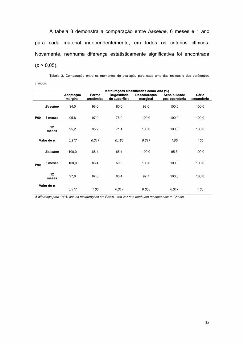
A tabela 3 demonstra a comparação entre baseline, 6 meses e 1 ano
para cada material independentemente, em todos os critérios clínicos.
Novamente, nenhuma diferença estatisticamente significativa foi encontrada
(p > 0,05).
Tabela 3. Comparação entre os momentos de avaliação para cada uma das resinas e dos parâmetros
clínicos.
Restaurações classificadas como Alfa (%)
Adaptação marginal
Forma anatômica
Rugosidade de superfície
Descoloração marginal
Sensibilidade pós-operatória
Cárie secundária
P60
Baseline 94,0 98,0 80,0 98,0 100,0
100,0
6 meses 95,8 97,9 75,0 100,0 100,0
100,0
12 meses
95,2 95,2 71,4 100,0 100,0
100,0
Valor de p
0,317 0,317 0,180 0,317 1,00 1,00
P90
Baseline
100,0 88,4 65,1 100,0 95,3 100,0
6 meses
100,0 88,4 69,8 100,0 100,0 100,0
12
meses
97,6 87,8 63,4 92,7 100,0 100,0
Valor de p 0,317 1,00 0,317 0,083 0,317 1,00
A diferença para 100% são as restaurações em Bravo, uma vez que nenhuma recebeu escore Charlie.
36
5. DISCUSSÃO
Os resultados serão discutidos na forma de dois artigos.
5.1 Artigo I
RUNNING HEAD
Silorane-based composites as repair material
TITLE
Silorane-based composites as repair material: a six-month randomized
clinical trial
ABSTRACT
The aim of this study was to investigate the clinical performance
of a low-shrinkage silorane-based composite resin when used for repairing
conventional dimethacrylate-based composite restorations. Despite the
continued development of resin-based materials, polymerization shrinkage and
shrinkage stress still require improvement. In this context, a silorane-based
monomer system was recently made available for dental restorations. One
operator repaired one hundred defective dimethacrylate-based composite resin
restorations that were randomly assigned to receive one of two treatment
groups: Control (n = 50) Adper SE Plus, 3M /ESPE, and Filtek™ P60 Posterior
Restorative, 3M/ESPE; and Test (n = 50) P90 System Adhesive Self-Etch
Primer and Bond, 3M/ESPE and Filtek™ P90 Low Shrink Posterior Restorative,
3M/ESPE. After one week, restorations were finished and polished. Two
calibrated examiners (Kw ≥ 0.78) evaluated all repaired restorations, blindly and
37
independently, by direct observation. The parameters examined were marginal
adaptation, anatomic form, surface roughness, marginal discoloration, post-
operative sensitivity and secondary caries. The restorations were classified as
Alpha, Bravo or Charlie, according to modified U.S. Public Health Service
criteria. Mann-Whitney and Wilcoxon tests were used to compare the groups.
Of the 100 restorations repaired in this study, 93 were reexamined at baseline
and 91 at 6-month recall. Drop-out was about 9%. No statistically significant
differences were found between the materials for all clinical criteria, at baseline
and at 6-month recall (p > 0.05). No statistically significant differences were
registered (p > 0.05) for each material when compared for all clinical criteria, at
baseline and at 6-month recall. No restorations were registered with Charlie
rating. After six-months evaluations, silorane-based composite exhibit a similar
performance compared to dimethacrylate-based composite when used as repair
material.
Key words: Low-shrinkage silorane-based composite, dimethacrylate-based
composite, resin-based restoration, repair.
TÍTULO EM PORTUGUÊS:
Resina composta à base de silorano como material de reparo: estudo
clínico randomizado de seis meses
RESUMO EM PORTUGUÊS
Este estudo objetivou investigar o desempenho clínico de uma resina de baixa
contração à base de silorano quando utilizada para reparar restaurações
convencionais de resina composta à base de dimetacrilato, em baseline e ao
38
longo de 6 meses. Cem restaurações defeituosas de resina composta à base
de dimetacrilato foram reparadas neste estudo. Destas, 93 foram examinadas
uma semana após terem sido reparadas – baseline e 91 após 6 meses. As
restaurações foram alocadas aleatoriamente em dois grupos de tratamento –
Controle (n=50): Adper SE Plus 3M /ESPE + Filtek P60 3M /ESPE e Teste
(n=50): Sistema adesivo P90 3M /ESPE + Filtek P90 3M /ESPE. Uma semana
após terem sido reparadas as restaurações foram polidas e acabadas. Dois
examinadores devidamente calibrados (Kw = 0,78) fizeram a avaliação das
restaurações reparadas de forma cega, independente e por meio de
observação direta. Os parâmetros clínicos analisados foram adaptação
marginal, forma anatômica, rugosidade de superficíe, descoloração marginal,
sensibilidade pós-operatória e cárie secundária, sendo as restaurações
classificadas em Alpha, Bravo ou Charlie (critérios clínicos USPHS
modificados). O teste de Mann-Whitney comparou os materiais testados, para
todos os parâmetos clínicos, em baseline e após 6 meses (α = 0,05). O teste
de Wilcoxon comparou cada material independentemente, para todos os
mesmos parâmetros, em baseline e após 6 meses (α = 0,05). A perda neste
estudo foi de 9 % em 6 meses. Não houve diferença estatisticamente
significativa entre as resinas Filtek P60 e Filtek P90 quando avaliados os
parâmetros supracitados, em baseline e ao longo de 6 meses (p > 0,05). Não
houve diferença estatisticamente significativa entre os períodos de avaliação
quando cada resina foi testada, considerando os mesmos parâmetros (p >
0,05). Nenhuma restauração foi classificada como Charlie. Após 6 meses de
avaliações, resinas compostas à base de silorano apresentaram desempenho
clínico semelhante às resinas compostas à base de dimetacrilato quando
39
utlizadas para reparar restaurações de resina composta à base de
dimetacrilato.
Palavras-chave: resina composta à base de silorano, resina composta à base
de dimetacrilato, reparo.
INTRODUCTION
Composite resins are today‟s most widely used direct restorative
material. Their main advantages are the adhesive capacity allowing for minimal
cavity preparation and superior esthetics.1 Since the introduction of dental resin-
based composites, intense research has attempted to develop materials with
acceptable mechanical and physical properties to significantly improve their
longevity and aesthetic quality of restorations.2
Recently, in order to minimize the effects of shrinkage, an innovative
monomer system was made available for dental restorations – silorane.
Obtained from the reaction of oxirane and siloxane molecules, this material
contains traditional filler particles, whereas the conventional resin is replaced by
silorane monomers. While siloxanes are known for their hydrophobicity,
oxiranes are known for their low shrinkage.3-5
Results from in vitro studies have shown that silorane-based composites
demonstrate the lowest polymerization shrinkage as well as more ambient light
stability.3,6-9 The new system also has the lowest sorption and water solubility
and a lower diffusion coefficient than conventional monomers. Parameters such
as tensile modulus, flexural strength and biocompatibility in toxicology tests are
comparable to dimethacrylate-based composite. 3,6-9
40
Despite extensive improvements in the mechanical properties of resin-
based tooth-colored restorative materials, volumetric shrinkage and subsequent
contraction stress arising during the polymerization reaction are still significant
drawbacks.10 Shrinkage may also cause microleakage, marginal staining, and
gap formation, this one an important factor in the development of caries,
because it may act as a retention groove.4,11,12.
Reducing shrinkage and the stress generated by polymerization may
positively influence marginal integrity. Imperfect margins result in marginal
discoloration and secondary caries lesions, the most important cause for the
replacement of defective restorations.13
However, according to the philosophy of „minimum intervention‟ operative
dentistry, with the exception of conditions in which there is a fracture of the resin
restoration, staining of the entire resin/tooth interface and secondary caries,
defective restorations should be first evaluated for the possibility of repair,
rather than being routinely replaced.14 This approach allows preservation of
sound tooth structure15,16 being considered a viable long-term clinical procedure
for treatment of restorations.17-19
Clinical studies involving composite resin repairs have shown that, when
properly planned, the repairs may increase the clinical longevity of restorations.
Thus, once in vitro studies suggest that bonding of silorane-based composites
to old dimethacrylate-based composites may be a viable clinical procedure,20-22
it would be desirable to evaluate the clinical performance of this new system for
making repairs. The hypothesis tested in this randomized controlled clinical trial
was that low–shrinkage silorane-based composites exhibit a similar
41
performance when compared to conventional dimethacrylate-based composites
when used to repair composite resin restorations.
MATERIALS AND METHODS
Study Design
This prospective randomized clinical trial had the repaired restorations
like observation units. Patients aged 18 to 56 years with 100 defective
composite resin restorations participated in this study. They were routinely
assigned for treatment at the operative dentistry clinics, School of Dentistry -
Federal University of Minas Gerais, Belo Horizonte, Minas Gerais, Brazil and
School of Dentistry, Educational Association of Brazil, Montes Claros, Minas
Gerais, Brazil
The patients included were: those who were older than 18 years of age;
those who with no contraindications for dental treatment and those who had
class I or class II composite resin restorations with occlusal defects and no
diagnosis of caries according to clinical and bite-wing radiographic exams,
scored Bravo according to Modified United States Public Health Service
(USPHS) clinical criteria (Table 1). The patients excluded were: those with
xerostomy, including patients taking medications that are proven to significantly
reduce salivary flow and patients with visible plaque index (VPI) > 30% at the
moment of the repair procedure. The tooth excluded were: those with defective
restorations, unacceptable for repairs, that scored Charlie (Modified USPHS
clinical criteria), those which had no antagonist teeth and those with root
exposure.
This study was approved by the Institutional Ethics Committee (ETIC
0546.0.203.000-09).
42
Study methods
The restorations were examined one week after they were repaired for
baseline assessment, and at six-month. Two examiners blindly and
independently evaluated all repaired restorations by direct observation, using a
plane buccal mirror and a WHO model explorer. Calibration exercises revealed
an inter-examiner agreement ratio ≥ 0.78. Since there was disagreement on
the rating, the clinicians reexamined the repaired restoration together and
arrived at a joint final decision. The clinical criteria examined were marginal
adaptation, anatomic form, surface roughness, marginal discoloration, post-
operative sensitivity and secondary caries. The examiners classified all
restoration as Alpha, Bravo or Charlie, according to modified USPHS clinical
criteria.
Treatment groups
The same operator repaired, polished and finished all defective
composite resin restorations in order to minimize preparation variability.
Defective surfaces of the restorations were explored using high-speed spherical
diamond burs (KG Sorensen, São Paulo, SP, Brazil) compatible with the size of
the defect in a hand piece with air-water coolant, beginning with the removal of
the restorative material in the area of the defect as well as any stained and soft
tooth tissues. The restorations were randomly assigned to one of two treatment
groups: Control group (n = 50): Repair with a self-etching primer (Adper SE
Plus, 3M /ESPE, St. Paul, MN, USA) and a dimethacrylate-based composite
(Filtek P60 Posterior Restorative, 3M/ESPE, St. Paul, MN, USA); Test group (n
= 50): Repair with a self-etching primer (P90 System Adhesive Self-Etch Primer
and Bond, 3M/ESPE, St. Paul, MN, USA) and a low-shrinking silorane-based
43
composite (Filtek P90 Low Shrink Posterior Restorative, 3M/ESPE, St. Paul,
MN, USA) (Table 2).
Rubber dam isolation was used for the restorative procedures. The
surfaces of restorations and enamel margins were etched with 37% phosphoric
acid (Magic Acid Gel, VIGODENT/COLTENE, Rio de Janeiro, RJ, Brazil) before
adhesive procedures, being the materials used according to manufacturer's
recommendations (Table 3).
Outcome measurements and Statistical Analysis
The ordinal dependent variable was each modified USPHS criteria –
marginal adaptation, anatomic form, surface roughness, marginal discoloration,
post-operative sensitivity and secondary caries - from which the restorations
received a clinical rating like Alpha, Bravo or Charlie.
Data management and analysis were done using a statistical analysis
system (PASW 17.0.1 for Windows, SPSS, Chicago, IL, USA). Mann-Whitney
test was used to assess differences between the materials tested and for all
clinical criteria, at baseline and at 6-month recall examination (α = 0.05).
Wilcoxon test was used to compare each composite resin for all clinical criteria
at baseline examinations and at 6-month recall (α = 0.05).
RESULTS
Drop-out in this study was 9%. Of the 100 repaired restorations, 93 (50
for Filtek P60 and 43 for Filtek P90) were examined at baseline and 91 at the 6-
month recall (48 for Filtek P60 and 43 for Filtek P90). The flow of participants
and the number of restorations through each examination period are shown on
44
Figure 1. The main reasons for restorations being repaired were marginal
defects (81%) and loss of anatomic form (19%).
Table 4 summarizes the frequency of Alpha and Bravo ratings for
restorations in both groups for each clinical criterion at baseline and at 6-month
recall examination. No restoration received Charlie ratings. Table 5 shows the
comparison between the materials tested for all clinical criteria, at baseline and
at six-month recall examination. No statistically significant difference between
the materials was found (p > 0.05). Table 6 shows the comparison between
baseline and six-month recall examination for each material independently, for
all clinical criteria. No statistically significant difference was found in any criteria
between the examination periods (p > 0.05).
DISCUSSION
The low-shrinkage silorane-based composites exhibited a similar clinical
performance to dimethacrylate-based composites when used for repairing
dimethacrylate-based composite restorations after a six-month observation
period, confirming, thus, the null hypothesis tested. Drop-out in this study was
about 9%. This response rate is in accordance with other similar clinical studies
that had rates of 0% to 15% for the first year recall. 4,17,19,23,24
In general, aproximately 50% of resin-based composite restorations are
replaced after seven years of service, and the main reasons are secondary
caries, marginal defects, discoloration, degradation/wear and loss of anatomic
form.17-19 For many years, despite the subjectivity of restoration removal criteria,
total replacement has been the most common treatment in general dental
practice.19,25 Nevertheless, it is known that when a restoration is replaced, there
45
is a loss of healthy dental tissue, including areas away from localized defects.17
Repairs are treatments that can increase the longevity of restorations, and
several studies have shown a positive impact after first, second and third year
observation periods.17-19,26 Thus, this longitudinal prospective study aimed to
discuss the effectiveness of a new low-shrinkage composite – silorane – as a
repair material
Silorane is a nonmethacrylate-based resin that has been introduced in
order to control polymerization shrinkage. The new monomer is obtained from
the reaction of oxirane and siloxane molecules and was developed with the
primary purpose of overcoming some drawbacks related to polymerization of
dimethacrylate-based composites, such as radical oxygen inhibition,
polymerization shrinkage, polymerization stress, water sorption and instability of
conventional monomers in aqueous systems. As a result, silorane has the
ability to compensate shrinkage by opening the oxirane ring during
polymerization, reducing volume shrinkage to 1% from 1.7- 3.5% in
dimethacrylate-based materials. Due to the presence of siloxane species, the
hydrophobicity is also increased.3,4,30,31
Silorane-based composites have been thoroughly investigated by in vitro
tests, and promising results have been obtained regarding biocompatibility and
mechanical characteristics, including reduced polymerization shrinkage.3,5,32
However, in vitro studies are limited in predicting short- and long-term clinical
conditions, and laboratory findings should be substantiated by clinical
investigations.
In the present study, the main reasons for repairing restorations were
marginal defects and loss of anatomical form. Six modified USPHS criteria –
46
marginal adaptation, anatomic form, surface roughness, marginal discoloration,
post-operative sensitivity and secondary caries – were used to verify the clinical
performance of repairs performed on failed dimethacrylate-based composite
restorations. No statistically significant differences between the groups were
found for all clinical parameters tested at each time interval.
It is a consensus that the information provided by USPHS criteria is too
broad and may also lead to a misinterpretation as a good clinical performance
since any changes over time are not easily detected by the limited sensitivity in
short-term clinical investigation.17,33 Despite these considerations, it is the most
widely used method for clinical evaluations of restorations worldwide, and the
main reason for adopting it relies on the fact that it can be compared to previous
studies. In addition, this criteria involves visual inspection as well as the use of a
dental explorer.17
Laboratory studies have shown lower values of polymerization shrinkage
related to silorane-based composites, but it is difficult to show the effects in
clinical studies, mainly because in short-term six-month evaluations, many
factors may not still influenced the final result.3,31,32In the current study, no
statistically significant differences between the materials tested were found for
marginal adaptation for the entire six-month follow-up. There are no results from
clinical trials that have tested silorane-based composite as repair material
available for comparison. However, a recent study investigated marginal
adaptation of a low-shrinkage silorane-based composite and compared it with a
dimethacrylate-based composite material across one-year interval.4 Even
though such study had outcomes related to total-replaced restorations, their
47
results from 6-month investigations are in accordance with the findings from the
present study.
No statistically significant differences have been found between the
materials tested for secondary caries, which are usually associated with
marginal integrity and marginal adaptation is usually associated with reduced
polymerization shrinkage. Favourable results were, thus, expected for a low-
shrinkage resin-based composite.5 Furthermore, within six months, the patients
in the study did not develop carious lesions, most likely because patients with
inadequate oral hygiene (VPI > 30%) and decreased salivary flow were
excluded.
No statistically difference was found for anatomic form when each
composite resin was evaluated independently at baseline and after six months.
In general, restorations remained stable and unchanged over the six-month
observation period. Previous studies that have investigated the longevity of
dimethacrylate-based restoration by minimal intervention have found the same
good performance when dimethacrylate-based composites were used as repair
materials.17-19
Laboratory studies have investigated some mechanical properties of
dental composites after artificial aging,33,34 and the lowest values for surface
roughness were observed for a silorane-based composite.33 This effect was not
demonstrated in the current clinical study, perhaps due to the short time
elapsed after the repairs were performed. No statistically significant difference
between the materials was found for surface roughness at any recall
examination. This result is in agreement with studies investigating the longevity
of dimethacrylate-based composite restorations by minimal interventions.17-19
48
These studies found that surface roughness returned to their original values
from the defective restoration values after only a three-year recall examination.
In a recent study related to the repair potential of composite resin
materials, the highest bond strength when a dimethacrylate-based composite
was used as substrate was when Filtek P90 was used as the repair material
and the P90 System as the adhesive. Although it is customarily assumed that
the bond between old and new composite is micromechanical, data from when
Filtek P90 was the substrate suggest that there is a possibility of chemical
bonding, most likely because products that contain a silane coupling agent have
improved wetability of the substrate surface in addition to higher binding of
siloxane to inorganic filler particles. In Filtek P90, these are silanated
ceramics.22 It may explain the results from marginal discoloration, when no
statistically significant difference between the two materials was found at recall
examinations
At baseline examination, the low incidence of restorations that received
Bravo rating for post-operative sensitivity can be explained by the use of a self-
etching bonding system in both treatment groups. These systems make the
smear layers part of the hybrid layer, providing better penetration of the
monomers onto the collagen fibers of the demineralized dentin. At follow-up, the
same good performance was observed for all composites, likely because resin-
based agents may provide pulp protection as long as the dentin is sealed by
hydrophilic resins.23,35 Initial post-operative sensitivity has been reported in
clinical studies with resin-based composites, but the sensitivity generally
decreases during the first weeks after placement of restorations.23,36
49
Thus, after six months, this clinical trial shows that low-shrinkage
silorane-based composites exhibited a similar performance to the conventional
dimethacrylate-based composites when used to repair composite resin
restorations. The reduced polymerization shrinkage assigned to silorane-based
composites did not establish better clinical performance, indicating that
laboratory findings should be substantiated by clinical investigations.
ACKNOWLEDGMENTS
We owe our thanks to FAPEMIG, who supported the present study.
REFERENCES
1. Min SH, Ferracane J, Lee IB. Effect of shrinkage strain, modulus, and instrument compliance on polymerization shrinkage stress of light-cured composites during the initial curing stage. Dent Mater 20; 26:1024-33.
2. Leprince J, Palin WM, Mullier T, Devaux J, Vreven J, Leloup G. Investigating filler morphology and mechanical properties of new low-shrinkage resin composite types. J Oral Rehabil 2010; 37:364–376.
3. Weinmann W, Thalacker C, Guggenberger R. Siloranes in dental composites. Dent Mater 2010; 21:68–74.
4. Schmidt M, Kirkevang LL, Hǿrsted-Bindslev P, Poulsen S. Marginal adaptation of a low-shrinkage silorane-based composite: 1-year randomized clinical trial. Clin Oral Invest 2011; 15: 291-295.
5. Ilie N, Hickel R. Macro- micro- and nano-mechanical investigations o silorane and methacrylate-based composites. Dent Mater 2009; 25: 810-819
50
6. Eick D, Kotha SP, Chappelow CC, Kilway KV, Giese G, Glaros AG, Pinzino CS. Properties of silorane-based dental resins and composites containing a stress-reducing monomer. Dent Mater 2007 23: 1011-1017.
7. Condon JR, Ferracane JL. Assessing the effect of composite formulation on polymerization stress. JADA 2000; 131: 497-503.
8. Palin WM, Fleming GJP, Burke FJT, Marquis PM, Randall RC. The influence of short and medium term water immersion on the hydrolytic stability of novel low shrink dental composites. Dent Mater 2005; 21:852-863.
9. Soh MS, Yap AUJ, Sellinger A. Physicomechanical evaluation of low-shrinkage dental nanocomposites based on silsesquioxane cores. Eur J Oral Sci 2007; 115: 230–238.
10. Marchesi G, Breschi L, Antoniolli F, DiLenarda R, Ferracane J, Cadenaro M. Contraction stress of low-shrinkage composite materials assessed with different testing systems. Dent Mater 2007; 26: 947-953
11. Giachetti L, Scaminaci Russo D, Bambi C, Grandini R. A review of polymerization shrinkage stress: current techniques for posterior direct resin restorations. J Contemp Dent Pract 2007; 7:79–88.
12. Totiam P, Gonzalez-Cabezas C, Fontana MR, Zero DT. A new in vitro model to study the relationship of gap size and secondary caries. Caries Res 2007; 41:467–473.
13. Rodriguez GDR, Pereira SNA. Current trends and evolution on dental composites Acta Odontol Venez 2008; 46: 1-18.
14. Tyas MJ, Anusavice KJ, Frencken JE, Mount GJ. Minimal intervention dentistry – a review. Int Dent J 2000; 50:1–12.
51
15. Mjör IA, Reep RL, Kubilis PS, Mondragon BE. Change in size of replaced amalgam restorations: A methodological study. Oper Dent 1998; 23: 272-277.
16. Popoff DAV, Gonçalves FS, Ferreira RC, Magalhães CS, Moreira AN, Mjör IA. Repair of amalgam restorations with conventional and bonded amalgam: an in vitro study. Rev. odonto ciênc. 2010; 25: 154-158.
17. Gordan VV, Shen C, Riley J, Mjor IA. Two-Year clinical evaluation of repair versus replacement of composite restorations. J Esthet Dent 2006; 18:144-154.
18. Moncada G, Fernández E, Martin J, Arancibia C, Mjor I, Gordan VV. Increasing the longevity of restorations by minimal intervention: A two-year clinical trial. Oper Dent 2008; 33: 258-264.
19. Moncada G, Martin J, Fernández E, Hampel MC, Mjör IA, Gordan VV. Sealing, refurbishment and repair of class I and class II defective restorations: A three-year clinical trial. JADA 2009; 140: 425-432.
20. Lührs AK, Görmann B, Jaker-Guhr S, GeurtsenW. Repairability of dental siloranes in vitro. Dent Mater 2011; 27: 144-149.
21. Ivanovas S, Hickel R, Ilie N. How to repair fillings made by silorane-based composites. Clin Oral Invest 2010 In press.
22. Mannenut C, Sakoolnamarka R, Tyas MJ. The repair potential of resin composite materials. Dent Mater 2011; 27: 20-27.
23. Gordan VV, Shen C, Watson RE, Mjor IA. Four-year clinical evaluation of self-etching primer and resin-based restorative material. Am J Dent 2005; 18: 45-49.
52
24. Gordan VV, Mondragon E, Watson RE, Garvan C, Mjör IA. A clinical evaluation of a self-etching primer and giomer restorative material. JADA 2007; 138: 621-627.
25. Çehreli SB, Arhun N, Celik C. Amalgam repair: quantitative evaluation of amalgam-resin and resin-tooth interfaces with different surface treatments. Oper Dent 2010; 35: 337-344.
26. Christensen GJ. When and how to repair a failing restoration. JADA 2007; 138:1605-1607.
27. Ilie N, Hickel R. Silorane-based dental composite:behavior and abilities. Dent Mater J 2006; 25: 445-454.
28. Eick JD, Smith RE, Pinzino CS, Kostoryz EL. Stability of silorane dental monomers in aqueous systems. J Dent 2006; 34: 405-410.
29. Papadogiannis D, Kakaboura A, Palaghias G, Eliades G. Setting characteristics and cavity adaptation of low-shrinking resin composites. Dent Mater 2010; 25: 1509-1516.
30. Hickel R, Roulet JF, Bayne S, Heintze SD, Mjör IA, Peters M, Rousson V, Randall R, Schmalz G, Tyas M, Vanherle G. Recommendations for conducting controlled clinical studies of dental restorative materials. Science Committee Project 2/98--FDI World Dental Federation study design (Part I) and criteria for evaluation (Part II) of direct and indirect restorations including onlays and partial crowns. J. Adh. Dent. 2007; 9: 121-147.
31. Ruttermann S, Kruger S, Raab WH, Janda R. Polymerization shrinkage and hygroscopic expansion of contemporary posterior resin-based filling materials - a comparative study. J Dent 2007; 35:806–813.
53
32. Park J, Chang J, Ferracane J, Lee IB. How should composite be layered to reduce shrinkage stress: incremental or bulk filling? Dent Mater 2008; 24: 1501–1505.
33. Hahnel S, Henrich A, Bürgers R, Handel G, Rosentrit M. Investigation of mechanical properties of modern dental composites after artificial aging for one year. Oper Dent 2010; 35: 412-419.
34. Zhang Y, Xu J. Effect if immersion in various media on the sorption, solubility, elution of unreacted monomers, and flexural properties of two model dental composite compositions. J Mater Sci Mater Med 2008; 19: 2477-2483.
35. Pashley DH. The effects of acid etching on the pulpodentin complex. Oper Dent 1992; 17: 229-242.
36. Baratieri LN, Ritter AV. Four-year clinical evaluation of posterior resin-based composite restorations placed using the total-etching technique. J Esthetic Rest Dent 2001; 13: 50-57.
FIGURE LEGENDS
Table 1. Modified U.S. Public Health Service clinical criteria.
Table 2. Materials: Chemical composition and manufacturers.
Table 3. Clinical sequence of repair procedures.
Figure 1. Flowchart of patients and number of restorations through each stage
of the study.
Table 4. Frequency of Alpha and Bravo ratings according to the materials tested
at baseline and at six-month recall examination.
Table 5. Comparison between the materials tested for all clinical criteria at each
examination period.
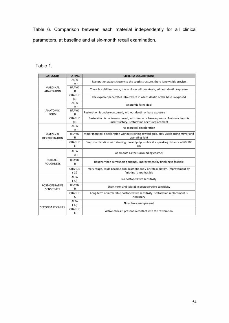
54
Table 6. Comparison between each material independently for all clinical
parameters, at baseline and at six-month recall examination.
Table 1.
CATEGORY RATING CRITERIA DESCRIPTIONS
MARGINAL ADAPTATION
ALFA ( A )
Restoration adapts closely to the tooth structure, there is no visible crevice
BRAVO ( B )
There is a visible crevice, the explorer will penetrate, without dentin exposure
CHARLIE (C)
The explorer penetrates into crevice in which dentin or the base is exposed
ANATOMIC FORM
ALFA ( A )
Anatomic form ideal
BRAVO ( B )
Restoration is under-contoured, without dentin or base exposure
CHARLIE (C)
Restoration is under-contoured, with dentin or base exposure. Anatomic form is unsatisfactory. Restoration needs replacement
MARGINAL DISCOLORATION
ALFA ( A )
No marginal discoloration
BRAVO ( B )
Minor marginal discoloration without staining toward pulp, only visible using mirror and operating light
CHARLIE ( C )
Deep discolaration with staining toward pulp, visible at a speaking distance of 60-100 cm
SURFACE ROUGHNESS
ALFA ( A )
As smooth as the surrounding enamel
BRAVO ( B )
Rougher than surrounding enamel. Improvement by finishing is feasible
CHARLIE ( C )
Very rough, could become anti-aesthetic and / or retain biofilm. Improvement by finishing is not feasible
POST-OPERATIVE SENSITIVITY
ALFA ( A )
No postoperative sensitivity
BRAVO ( B )
Short-term and tolerable postoperative sensitivity
CHARLIE ( C )
Long-term or intolerable postoperative sensitivity. Restoration replacement is necessary
SECONDARY CARIES
ALFA ( A )
No active caries present
CHARLIE ( C )
Active caries is present in contact with the restoration
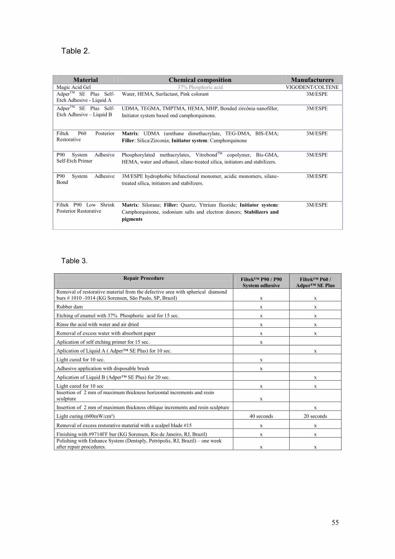
55
Table 2.
Table 3.
Repair Procedure
Filtek™ P90 / P90
System adhesive
Filtek™ P60 /
Adper™ SE Plus
Removal of restorative material from the defective area with spherical diamond
burs # 1010 -1014 (KG Sorensen, São Paulo, SP, Brazil) x x
Rubber dam x x
Etching of enamel with 37% Phosphoric acid for 15 sec. x x
Rinse the acid with water and air dried x x
Removal of excess water with absorbent paper x x
Aplication of self etching primer for 15 sec. x
Aplication of Líquid A ( Adper™ SE Plus) for 10 sec. x
Light cured for 10 sec. x
Adhesive application with disposable brush x
Aplication of Liquid B (Adper™ SE Plus) for 20 sec. x
Light cured for 10 sec x x
Insertion of 2 mm of maximum thickness horizontal increments and resin
sculpture x
Insertion of 2 mm of maximum thickness oblique increments and resin sculpture x
Light curing (600mW/cm²) 40 seconds 20 seconds
Removal of excess restorative material with a scalpel blade #15 x x
Finishing with #9714FF bur (KG Sorensen, Rio de Janeiro, RJ, Brazil) x x
Polishing with Enhance System (Dentsply, Petrópolis, RJ, Brazil) – one week after repair procedures. x x
Material Chemical composition Manufacturers Magic Acid Gel 37% Phosphoric acid VIGODENT/COLTENE
AdperTM SE Plus Self-
Etch Adhesive - Liquid A
Water, HEMA, Surfactant, Pink colorant 3M/ESPE
AdperTM SE Plus Self-
Etch Adhesive – Liquid B UDMA, TEGMA, TMPTMA, HEMA, MHP, Bonded zircônia nanofiller,
Initiator system based ond camphorquinone.
3M/ESPE
Filtek P60 Posterior Restorative
Matrix: UDMA (urethane dimethacrylate, TEG-DMA, BIS-EMA;
Filler: Silica/Zirconia; Initiator system: Camphorquinone
3M/ESPE
P90 System Adhesive Self-Etch Primer
Phosphorylated methacrylates, VitrebondTM copolymer, Bis-GMA,
HEMA, water and ethanol, silane-treated silica, initiators and stabilizers.
3M/ESPE
P90 System Adhesive Bond
3M/ESPE hydrophobic bifunctional monomer, acidic monomers, silane-
treated sílica, initiators and stabilizers.
3M/ESPE
Filtek P90 Low Shrink
Posterior Restorative Matrix: Silorane; Filler: Quartz, Yttrium fluoride; Initiator system:
Camphorquinone, iodonium salts and electron donors; Stabilizers and
pigments
3M/ESPE
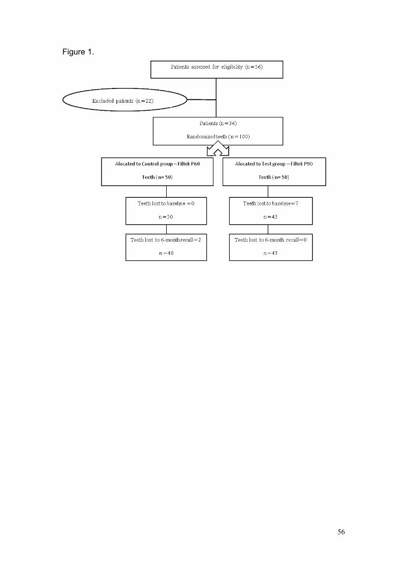
56
Figure 1.
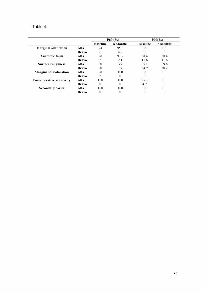
57
Table 4.
P60 (%) P90(%)
Baseline 6 Months Baseline 6 Months
Marginal adaptation Alfa 94 95.8 100 100
Bravo 6 4.2 0 0
Anatomic form Alfa 98 97.9 88.4 88.4
Bravo 2 2.1 11.6 11.6
Surface roughness Alfa 80 75 65.1 69.8
Bravo 20 25 34.9 30.2
Marginal discoloration Alfa 98 100 100 100
Bravo 2 0 0 0
Post-operative sensitivity Alfa 100 100 95.3 100
Bravo 0 0 4.7 0
Secondary caries Alfa 100 100 100 100
Bravo 0 0 0 0
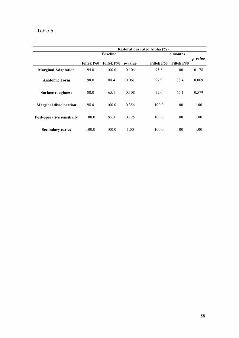
58
Table 5.
Restorations rated Alpha (%)
Baseline 6 months
Filtek P60 Filtek P90 p-value
Filtek P60
Filtek P90
p-value
Marginal Adaptation 94.0
100.0 0.104 95.8 100 0.178
Anatomic Form 98.0
88.4 0.061 97.9 88.4 0.069
Surface roughness 80.0
65.1 0.108 75.0 65.1 0.579
Marginal discoloration 98.0 100.0 0.354 100.0 100 1.00
Post-operative sensitivity 100.0 95.3 0.125 100.0 100 1.00
Secondary caries 100.0 100.0 1.00 100.0 100 1.00
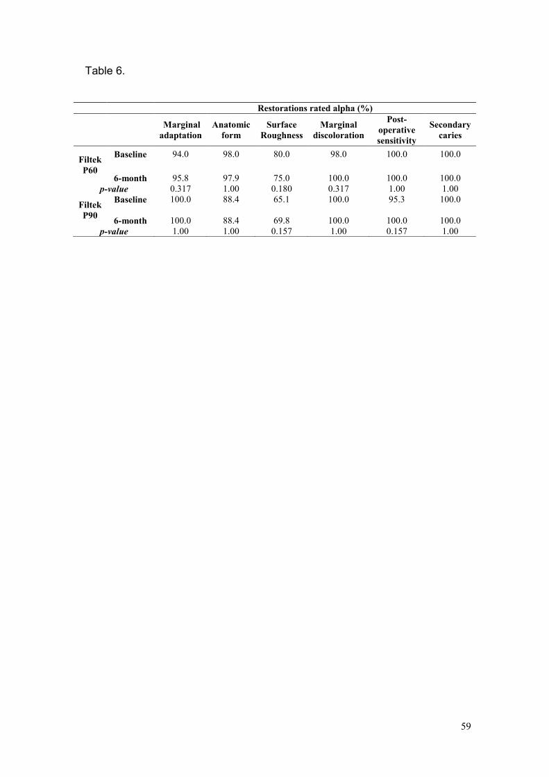
59
Table 6.
Restorations rated alpha (%)
Marginal
adaptation
Anatomic
form
Surface
Roughness
Marginal
discoloration
Post-
operative
sensitivity
Secondary
caries
Filtek
P60
Baseline 94.0 98.0 80.0 98.0 100.0 100.0
6-month 95.8 97.9 75.0 100.0 100.0 100.0
p-value 0.317 1.00 0.180 0.317 1.00 1.00
Filtek
P90
Baseline 100.0 88.4 65.1 100.0 95.3 100.0
6-month 100.0 88.4 69.8 100.0 100.0 100.0
p-value 1.00 1.00 0.157 1.00 0.157 1.00
60
5.2 Artigo II
TITLE
Repair of dimethacrylate-based composite restorations using a silorane-based composite: a one-year randomized clinical trial RUNNING TITLE Silorane-based composites as repair material CLINICAL RELEVANCE
A one-year clinical trial showed that low-shrinkage silorane-based
composite exhibited a similar performance to conventional dimethacrylate-
based composites when used to repair composite resin restorations. This
corroborates in vitro studies suggesting that bonding of silorane-based
composites to old dimethacrylate-based composites can be a viable clinical
procedure.
ABSTRACT
Purpose: To investigate clinical performance of a low-shrinkage silorane-based
composite resin when used for repairing conventional dimethacrylate-based
composite restorations. Background: Despite the continued development of
resin-based materials, polymerization shrinkage and shrinkage stress still
require improvement. A silorane-based monomer system is recently made
available for dental restorations. This report refers to the use of this material for
making repairs and evaluates the clinical performance of this alternative
treatment. Material and Methods: One operator repaired the defective
dimethacrylate-based composite resin restorations that were randomly assigned
to one of two treatment groups: Control (n = 50) Repair with Adper SE Plus, 3M
/ESPE, and Filtek™ P60 Posterior Restorative, 3M/ESPE; and Test (n = 50)
61
Repair with P90 System Adhesive Self-Etch Primer and Bond, 3M/ESPE and
Filtek™ P90 Low Shrink Posterior Restorative, 3M/ESPE. After one week,
restorations were finished and polished. Two calibrated examiners (Kw ≥ 0.78)
evaluated all repaired restorations, blindly and independently, at baseline, and
one year. The parameters examined were marginal adaptation, anatomic form,
surface roughness, marginal discoloration, post-operative sensitivity and
secondary caries. The restorations were classified as Alpha, Bravo or Charlie,
according to modified U.S. Public Health Service criteria. Mann-Whitney and
Wilcoxon tests were used to compare the groups. Results: Of the 100
restorations repaired in this study, 93 were reexamined at baseline. Drop-out
from baseline to one-year recall was 11%. No statistically significant
differences were found between the materials for all clinical criteria, at baseline
and at one-year recall (p > 0.05). No statistically significant differences were
registered (p > 0.05) for each material when compared for all clinical criteria, at
baseline and at one-year recall. Conclusions: The hypothesis tested in this
randomized controlled clinical trial was accepted. After the one-year
evaluations, silorane-based composite exhibit a similar performance compared
to dimethacrylate-based composite when used to making repairs.
Descriptors: Low-shrinkage silorane-based composite, dimethacrylate-based
composite, resin-based restoration, repair.
INTRODUCTION
The demand for aesthetic restorations, the development of new
adhesives and curing systems, and improvement of material properties have
made dental composites the most widely used direct restorative material
today.1,2,3 Despite such developments, two features still require improvement:
62
polymerization shrinkage and the development of polymerization shrinkage
stress.4
The intrinsic contraction of the composite remains a challenge, and
changes in the monomer composition seem to be the most promising way to
minimize the effects of shrinkage.5-8 Clinically, the incremental insertion and
control of polymerization rate are the main strategies used to control
polymerization shrinkage.
Recently, an innovative monomer system was made available for dental
restorations – Silorane, obtained from the reaction of oxirane and siloxane
molecules. Oxiranes are known for their low shrinkage, while siloxanes are
known for their hydrophobicity.5,9 In vitro studies have compared the new
system to dimethacrylate-based composites. The results show that silorane-
based composites demonstrate the lowest polymerization shrinkage as well as
more ambient light stability, contributing to the convenience of handling the
composite material. The new system also has the lowest sorption and water
solubility and a lower diffusion coefficient than conventional monomers. Other
parameters such as tensile modulus, flexural strength and biocompatibility in
toxicology tests are comparable to dimethacrylate-based composite.5,8,10-12
Imperfect margins result in marginal discoloration and secondary caries
lesions, the most important cause for the replacement of restorations4.
Reducing shrinkage and the stress generated by polymerization may positively
influence marginal integrity. Total replacement is the most common treatment
adopted for restorations that are clinically diagnosed as defective. However, the
assessment of the quality of restorations is made subjectively, and often
63
minimum deviations from ideal determine the systematic replacement of
restorations.13,14
With the exception of conditions in which there is a fracture of the resin
restoration, staining of the entire resin/tooth interface and secondary caries,
total removal is considered undesirable and inappropriate.3,15 Thus, keeping in
mind the current trends towards minimally interventional procedures, several
studies have suggested partial removal of the restoration.14,16-21 This approach
allows preservation of sound tooth structure.22,23
Clinical studies involving composite resin repairs have shown that, when
properly planned, the repairs may increase the clinical longevity of restorations,
representing a conservative choice for treatment of restorations.13,14,24
Thus, once in vitro studies suggest that bonding of silorane-based
composites to old dimethacrylate-based composites may be a viable clinical
procedure,25-27 it would be desirable to evaluate the clinical performance of this
new system for making repairs. The hypothesis tested in this randomized
controlled clinical trial was that low–shrinkage silorane-based composites
exhibit a similar performance when compared to that of conventional
dimethacrylate-based composites when repairing composite resin restorations.
MATERIALS AND METHODS
Study Design
This was a prospective randomized clinical trial. The observation unit
was the restoration, and the dependent variable was qualitative categorical
ordinal. Patients aged 18 to 56 years with 100 defective composite resin
restorations participated in this study. They were routinely assigned for
treatment at the operative dentistry clinics, School of Dentistry - Federal
64
University of Minas Gerais, Belo Horizonte, Minas Gerais, Brazil and School of
Dentistry – Educational Association of Brazil, Montes Claros, Minas Gerais,
Brazil.
The inclusion criteria were: patients who were older than 18 years of age;
patients with no contraindications for dental treatment; patients who had class I
or class II composite resin restorations with occlusal defects and no diagnosis
of caries according to clinical and bite-wing radiographic exams, that scored at
least Bravo according to Modified United States Public Health Service (USPHS)
clinical criteria (Table 1). The exclusion criteria were: patients with
contraindications for regular dental treatment according to their medical history;
patients with xerostomy, including those taking medications that are proven to
significantly reduce salivary flow; and patients with visible plaque index (VPI) ≥
30% at the moment of the repair procedure. The tooth exclusion criteria were:
Teeth with defective restorations, unacceptable for repairs, that scored Charlie
(Modified USPHS clinical criteria); and teeth with no antagonist teeth.
This study was approved by the Institutional Ethics Committee (ETIC
0546.0.203.000-09). A written informed consent was obtained from all patients.
Study methods
The restorations were examined one week after they were repaired for
baseline assessment, and at one year. Two examiners blindly and
independently evaluated all repaired restorations by direct observation, using a
plane buccal mirror and a WHO model explorer. A calibration exercise revealed
an inter-examiner agreement ratio ≥ 0.78. If there was disagreement on the
rating, the clinicians reexamined the repaired restoration together and arrived at
a joint final decision. The parameters examined were marginal adaptation,
65
anatomic form, surface roughness, marginal discoloration, post-operative
sensitivity and secondary caries. The examiners classified all restoration as
Alpha, Bravo or Charlie, according to modified USPHS clinical criteria.
Treatment groups
In order to minimize preparation variability, the same operator repaired all
defective composite resin restorations. The defective surfaces of the
restorations were explored using high-speed spherical diamond burs (KG
Sorensen, São Paulo, SP, Brazil) compatible with the size of the defect in a
hand piece with air-water coolant, beginning with the removal of the restorative
material in the area of the defect as well as any stained and soft tooth tissues.
The operator randomly assigned the restorations to one of two treatment
groups: Control group (n = 50): Repair with a self-etching primer (Adper SE
Plus, 3M /ESPE, St. Paul, MN, USA) and a dimethcrylate-based composite
(Filtek P60 Posterior Restorative, 3M/ESPE, St. Paul, MN, USA); Test group (n
= 50): Repair with a self-etching primer (P90 System Adhesive Self-Etch Primer
and Bond, 3M/ESPE, St. Paul, MN, USA) and a low-shrinking silorane-based
composite (Filtek P90 Low Shrink Posterior Restorative, 3M/ESPE, St. Paul,
MN, USA) (Table 2).
Rubber dam isolation was used for the restorative procedures. The
surfaces of restorations and enamel margins were etched with 37% phosphoric
acid (Magic Acid Gel, VIGODENT/COLTENE, Rio de Janeiro, RJ, Brazil) before
adhesive procedures. Materials were used according to manufacturer's
recommendations (Table 3).
66
Outcome measurements and Statistical Analysis
At baseline and 12-month recall, all restorations received a clinical rating
of Alpha, Bravo or Charlie. The ordinal dependent variable was the percentage
of Alpha, Bravo or Charlie ratings.
Data management and analysis were done using a statistical analysis
system (PASW 17.0.1 for Windows, SPSS, Chicago, IL, USA). Mann-Whitney
test was used to assess differences between the materials tested and for all
clinical criteria, at baseline and one-year recall examination (α = 0.05). Wilcoxon
test was used to compare each composite resin for all clinical criteria at
baseline examinations and one-year recall (p = 0.05).
RESULTS
In the present study, the main reasons for restorations being repaired
were marginal defects (81%) and loss of anatomic form (19%). Of the 100
repaired restorations, 93 (50 for Filtek P60 and 43 for Filtek P90) were
examined at baseline. From those, 83 were reexamined at the one-year recall
(42 for Filtek P60 and 41 for Filtek P90). The flow of participants and the
number of restorations through each examination period of the study are shown
on Figure 1. Drop-out in this study was about 17% after one-year.
Table 4 shows the frequency of Alpha and Bravo ratings for restorations
in both groups for each clinical criterion at baseline and one-year recall
examination. No restoration received Charlie ratings. Table 5 summarizes the
comparison between the materials tested for all clinical criteria at one-year
recall examination and baseline. No statistically significant difference between
the materials was found (p > 0.05). Table 6 shows the comparison between
67
baseline and one-year recall examination for each material independently, for
all clinical parameters. No statistically significant difference was found in any
criteria between the examination periods (p > 0.05).
DISCUSSION
The null hypothesis tested in this study was confirmed since the low-
shrinkage silorane-based composites exhibited a similar clinical performance to
the dimethacrylate-based composites when repairing dimethacrylate-based
composite restorations after a one-year observation period.
Drop-out in this study was about 17% after one year. This response rate
is in accordance with other similar clinical studies that had rates of 0% to 15%
for the first year recall.13, 24,28-30 The drop-out rates highlight part of the problems
associated with long-term clinical studies.28
Approximately 50% of resin-based composite restorations are replaced
after seven years of service, and the main reasons are secondary caries,
marginal defects, discoloration, degradation/wear and loss of anatomic
form.13,14,24 For many years, despite the subjectivity of restoration removal
criteria, total replacement has been the most common treatment in general
dental practice.17,24 Nevertheless, it is known that when a restoration is
replaced, there is a loss of healthy dental tissue, including areas away from
localized defects.13 Repairs are alternative treatments that can increase the
longevity of restorations, and several studies have shown a positive impact after
first, second and third year observation periods.13,14,24,31 Thus, this longitudinal
prospective study aimed to discuss the effectiveness of a new low-shrinkage
composite – silorane – as a repair material
68
Silorane is a nonmethacrylate-based resin that has been introduced in
order to control polymerization shrinkage. The new monomer is obtained from
the reaction of oxirane and siloxane molecules and was developed with the
primary purpose of overcoming some drawbacks related to polymerization of
dimethacrylate-based composites, such as radical oxygen inhibition,
polymerization shrinkage, polymerization stress, water sorption and instability of
conventional monomers in aqueous systems. As a result, silorane has the
ability to compensate shrinkage by opening the oxirane ring during
polymerization, reducing volume shrinkage to 1% from 1.7- 3.5% in
dimethacrylate-based materials. Due to the presence of siloxane species, the
hydrophobicity is also increased.5, 30,32,33
Silorane-based composites have been thoroughly investigated by in vitro
tests, and promising results have been obtained regarding biocompatibility and
mechanical characteristics, including reduced polymerization shrinkage.5,9,34
However, in vitro studies are limited in predicting short- and long-term clinical
conditions, and laboratory findings should be substantiated by clinical
investigations.
In the present study, the main reasons for repairing restorations were
marginal defects and loss of anatomical form. Six modified USPHS criteria –
marginal adaptation, anatomic form, surface roughness, marginal discoloration,
post-operative sensitivity and secondary caries – were used to verify the clinical
performance of repairs performed on failed dimethacrylate-based composite
restorations. No statistically significant differences between the groups were
found for all clinical parameters tested at each time interval (p > 0.05).
69
It is generally agreed that USPHS criteria may have a limited application
since the information provided is too broad; the criteria may also lead to a
misinterpretation as a good clinical performance since any changes over time
are not easily detected by the limited sensitivity in short-term clinical
investigation.13,35 However, it is the most widely used method for clinical
evaluations of restorations worldwide, and the main reason for adopting it relies
on the fact that it can be compared to previous studies. In addition, this criteria
involves visual inspection as well as the use of a dental explorer.13
Marginal adaptation and secondary caries
In the current study, no statistically significant differences between the materials
tested were found for marginal adaptation for the entire one-year follow-up. No
results from clinical trials that have tested silorane-based composite as repair
material was found for comparison. However, a recent clinical trial investigated
marginal adaptation of a low-shrinkage silorane-based composite and
compared it with material across one-year interval, finding better clinical
performance for dimethacrylate-based composite restorations,30 showing that
the low values found for silorane-based materials in laboratory studies are
difficult to be demonstrated in clinical studies, where so many factors influence
the final result.5,36,37
In this study, no statistically significant differences have been found
between the materials tested for secondary caries. Because secondary caries
are usually associated with marginal integrity and marginal adaptation is usually
associated with reduced polymerization shrinkage, it was expected favourable
results for a low-shrinkage resin-based composite.5 Furthermore, within one
year, the patients in the study did not develop carious lesions, most likely
70
because patients presenting inadequate oral hygiene (VPI > 30%) and
decreased salivary flow were excluded.
Anatomic form
No statistically significant difference was found when each composite
resin was evaluated independently at baseline and after one year. In general,
restorations remained stable and unchanged over the first-year observation
period. Previous studies that have investigated the longevity of dimethacrylate-
based restoration by minimal intervention have found the same good
performance when dimethacrylate-based composites were used as repair
materials.13,14,24
Surface Roughness
The surface roughness property of any material is the result of the interaction of
intrinsic and extrinsic factors. Some intrinsic factors are related to the material
itself, such as the filler, the resinous matrix as well as the ultimate degree of
cure reached, and the bond efficiency at the filler/matrix interface. The extrinsic
factors are associated with the type of polishing system used, such as the
flexibility of the packing material in which the abrasives are embedded, the
hardness of abrasives, the geometry of instruments, the lightcuring method, and
the way by which the finishing tools are used.38
In the current study, no statistically significant difference between the
materials was found for surface roughness at any recall examination. This result
is in agreement with studies investigating the longevity of dimethacrylate-based
composite restorations by minimal interventions.13,14,24 These studies found that
surface roughness returned to their original values from the defective
restoration values after only a three-year recall examination. Laboratory studies
71
have investigated some mechanical properties of dental composites after
artificial aging, and the lowest values for surface roughness were observed for a
silorane-based composite.38,39,40 This effect was not demonstrated in the current
clinical study, perhaps due to the short time elapsed after the repairs were
performed or due to the geometry of instruments that were used.
Marginal discoloration
In a recent study related to the repair potential of composite resin
materials, the highest bond strength when a dimethacrylate-based composite
was used as substrate was when Filtek P90 was used as the repair material
and the P90 Adhesive System as the adhesive. Although it is customarily
assumed that the bond between old and new composite is micromechanical,
data from when Filtek P90 was the substrate suggest that there is a possibility
of chemical bonding, most likely because products that contain a silane
coupling agent have improved wetability of the substrate surface in addition to
higher binding of siloxane to inorganic filler particles. In Filtek P90, these are
silanated ceramics.27 Even though, no statistically significant difference between
the two materials was found at recall examinations for marginal discoloration, in
the present study.
Post-operative sensitivity
Initial post-operative sensitivity has been reported in clinical studies with
resin-based composites, but the sensitivity generally decreases during the first
weeks after placement of restorations.28,41 At baseline examination, the low
incidence of restorations that received Bravo rating can be explained by the use
of a self-etching bonding system in both treatment groups. These systems
make the smear layers part of the hybrid layer, providing better penetration of
72
the monomers onto the collagen fibers of the demineralized dentin. At follow-up,
the same good performance was observed for all composites, likely because
resin-based agents may provide pulp protection as long as the dentin is sealed
by hydrophilic resins.28,42
CONCLUSIONS
After one year, this clinical trial shows that low-shrinkage silorane-based
composites exhibited a similar performance to the conventional dimethacrylate-
based composites when used to repair composite resin restorations. The
reduced polymerization shrinkage assigned to silorane-based composites did
not establish better clinical performance, indicating that laboratory findings
should be substantiated by clinical investigations. Based on the one-year
examinations, repairs may be considered a reliable, conservative and effective
treatment to treat defective restorations that are still clinically acceptable.
Acknowledgments
We owe our thanks to FAPEMIG, who supported the present study.
Conflict of interest
The authors declare no conflict of interest.
REFERENCES
1. Frankenberger R, Roth S, Krämer N, Pelka M, Petschelt A (2003) Effect of preparation mode on class II resin composite repair Journal of Oral Rahabilitation 30(6) 559-564.
2. Tesvergil A, Lassila LVJ, Vallittu PK (2003) Composite-composite repair bond strenght: effect of different adhesion primers Journal of Dentistry 31(8) 521-525.
73
3. Papacchini F, Magni E, Radovic I, Mazzitelli C, Monticelli F, Goracci C, Polimeni A, Ferrari M (2007) Effect of intermediate agents and pre-heating of repairing resin on composite-repair bonds Operative Dentistry 32(4) 363-371.
4. Rodriguez GDR & Pereira SNA Current trends and evolution on dental composites (2008) Acta Odontologica Venezolana 46(3) 1-18.
5. Weinmann W, Thalacker C, Guggenberger R (2005) Siloranes in dental composites Dental Materials 21(1) 68–74.
6. Kishikawa R, Koiwa A, Chikawa H, Cho E, Inai N, Tagami J (2005) Effect of cavity form on adhesion to cavity floor American Journal of Dentistry 18(6) 311-314.
7. dos Santos AJ, Giannini M, Paulillo LA, Lovadino JR, de Carvalho RM (2004) Effect of irradiation mode and filling technique on resin/dentin bonding strength in Class I cavities Brazilian Oral Research 18(3) 260-265.
8. Eick D, Kotha SP, Chappelow CC, Kilway KV, Giese G, Glaros AG, Pinzino CS (2007) Properties of silorane-based dental resins and composites containing a stress-reducing monomer Dental Materials 23(8) 1011-1017.
9. Ilie N & Hickel R (2009) Macro-, micro- and nano-mechanical investigations o silorane and methacrylate-based composites Dental Materials 25(6) 810-819.
10. Condon JR & Ferracane JL (2000) Assessing the effect of composite formulation on polymerization stress The Journal of the American Dental Association 131(4) 497-503.
11. Palin WM, Fleming GJP, Burke FJT, Marquis PM, Randall RC (2005) The influence of short and medium term water immersion on the hydrolytic stability of novel low shrink dental composites Dental Materials 21(9) 852-863.
12. Soh MS, Yap AUJ, Sellinger A (2007) Physicomechanical evaluation of low-shrinkage dental nanocomposites based on silsesquioxane cores European Journal of Oral Science 115(3) 230–238.
13. Gordan VV, Shen C, Riley J, Mjor IA (2006) Two-Year clinical evaluation of repair versus replacement of composite restorations Journal of Esthetic Dentistry 18(3) 144-154.
14. Moncada G, Fernández E, Martin J, Arancibia C, Mjor I, Gordan VV (2008) Increasing the longevity of restorations by minimal intervention: A two-year clinical trial Operative Dentistry 33(3) 258-264.
74
15. Teixeira EC, Bayne SC, Thompson JY, Ritter AV, Edward JS (2005) Shear bond strength of self-etching bonding systems in combination with various composites used for repairing aged composites The Journal of Adhesive Dentistry 7(2) 159-164.
16. Forss H & Widström E (2004) Reasons for restorative therapy and the longevity of restorations in adults Acta Odontologica Scandinavica 62(2) 82-86.
17. Çehreli SB, Arhun N, Celik C (2010) Amalgam repair: quantitative evaluation of amalgam-resin and resin-tooth interfaces with different surface treatments Operative Dentistry 35(3) 337-344.
18. Fedorowicz Z, Nasser M, Wilson N. Adhesively bonded versus non-bonded amalgam restorations for dental caries. Cochrane Database of Systematic Reviews 2009, Issue 4. Art. No.: CD007517. DOI: 10.1002/14651858.CD007517.pub2.
19. Machado C, Sanchez E, Alapati S, Seghi R, Johnston W (2007) Shear bond strength of the amalgam-resin composite interface Operative Dentistry 32(4) 341-346.
20. Hadavi F, Hey JH, Ambrose ER, Elbadrawy HE (1993) Effect of different adhesive systems on microleakage at the amalgam/composite resin interface Operative Dentistry 18(1) 2-7.
21. Alani AH & Toh CG (1997) Detection of microleakage around dental restorations: a review Operative Dentistry 22(4) 173-185.
22. Mjör IA, Reep RL, Kubilis PS, Mondragon BE (1998) Change in size of replaced amalgam restorations: A methodological study Operative Dentistry 23(5) 272-277.
23. Popoff DAV, Gonçalves FS, Ferreira RC, Magalhães CS, Moreira AN, Mjör IA (2010) Repair of amalgam restorations with conventional and bonded amalgam: an in vitro study Journal of Dental Science 25(2) 154-158.
24. Moncada G, Martin J, Fernández E, Hampel MC, Mjör IA, Gordan VV (2009) Sealing, refurbishment and repair of class I and class II defective restorations: A three-year clinical trial The Journal of the American Dental Association 140(4) 425-432.
25. Lührs AK, Görmann B, Jaker-Guhr S, GeurtsenW (2011) Repairability of dental siloranes in vitro. Dental Materials 27 (2) 144-149.
26. Ivanovas S, Hickel R, Ilie N (2010) How to repair fillings made by silorane-based composites. Clinical Oral Investigations In press.
75
27. Mannenut C, Sakoolnamarka R, Tyas MJ (2011) The repair potential of resin composite materials Dental Materials 27 (2) 20-27.
28. Gordan VV, Shen C, Watson RE, Mjor IA (2005) Four-year clinical evaluation of self-etching primer and resin-based restorative material American Journal of Dentistry 18(1) 45-49.
29. Gordan VV, Mondragon E, Watson RE, Garvan C, Mjör IA (2007) A clinical evaluation of a self-etching primer and giomer restorative material The Journal of the American Dental Association 138(5) 621-627.
30. Schmidt M, Kirkevang LL, Hǿrsted-Bindslev P, Poulsen S (2011) Marginal adaptation of a low-shrinkage silorane-based composite: 1-year randomized clinical trial Clinical Oral Investigations 15(2) 291-295.
31. Christensen GJ (2007) When and how to repair a failing restoration. The Journal of the American Dental Association 138(5) 1605-1607.
32. Ilie N & Hickel R (2006) Silorane-based dental composite: behavior and abilities Dental Materials Journal 25(3) 445-454.
33. Eick JD, Smith RE, Pinzino CS, Kostoryz EL (2006) Stability of silorane dental monomers in aqueous systems Journal of Dentistry 34(6) 405-410.
34. Papadogiannis D, Kakaboura A, Palaghias G, Eliades G (2010) Setting characteristics and cavity adaptation of low-shrinking resin composites Dental Materials 25 (2) 1509-1516.
35. Hickel R, Roulet JF, Bayne S, Heintze SD, Mjör IA, Peters M, Rousson V, Randall R, Schmalz G, Tyas M, Vanherle G (2007) Recommendations for conducting controlled clinical studies of dental restorative materials. Science Committee Project 2/98--FDI World Dental Federation study design (Part I) and criteria for evaluation (Part II) of direct and indirect restorations including onlays and partial crowns Journal of Adhesive Dentistry 9(10) 121-147.
36. Ruttermann S, Kruger S, Raab WH, Janda R (2007) Polymerization shrinkage and hygroscopic expansion of contemporary posterior resin-based filling materials - a comparative study Journal of Dentistry 35(10) 806–813.
37. Park J, Chang J, Ferracane J, Lee IB (2008) How should composite be layered to reduce shrinkage stress: incremental or bulk filling? Dental Materials 24(11) 1501–1505.
38. Marghalani HY (2010) Effect of finishing/folishing systems on the surface soughness of novel posterior composites. Journal of Esthetic and Restorative Dentistry 22(2)127-138
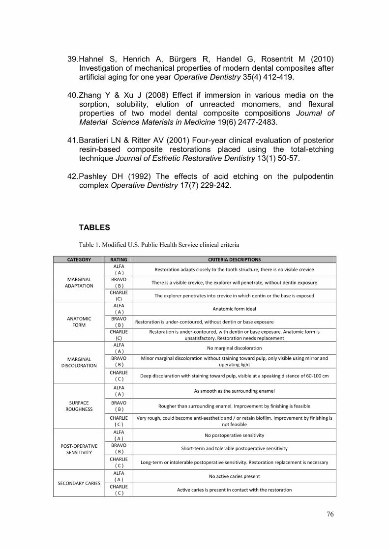
76
39. Hahnel S, Henrich A, Bürgers R, Handel G, Rosentrit M (2010)
Investigation of mechanical properties of modern dental composites after artificial aging for one year Operative Dentistry 35(4) 412-419.
40. Zhang Y & Xu J (2008) Effect if immersion in various media on the sorption, solubility, elution of unreacted monomers, and flexural properties of two model dental composite compositions Journal of Material Science Materials in Medicine 19(6) 2477-2483.
41. Baratieri LN & Ritter AV (2001) Four-year clinical evaluation of posterior resin-based composite restorations placed using the total-etching technique Journal of Esthetic Restorative Dentistry 13(1) 50-57.
42. Pashley DH (1992) The effects of acid etching on the pulpodentin complex Operative Dentistry 17(7) 229-242.
TABLES
Table 1. Modified U.S. Public Health Service clinical criteria
CATEGORY RATING CRITERIA DESCRIPTIONS
MARGINAL ADAPTATION
ALFA ( A )
Restoration adapts closely to the tooth structure, there is no visible crevice
BRAVO ( B )
There is a visible crevice, the explorer will penetrate, without dentin exposure
CHARLIE (C)
The explorer penetrates into crevice in which dentin or the base is exposed
ANATOMIC FORM
ALFA ( A )
Anatomic form ideal
BRAVO ( B )
Restoration is under-contoured, without dentin or base exposure
CHARLIE (C)
Restoration is under-contoured, with dentin or base exposure. Anatomic form is unsatisfactory. Restoration needs replacement
MARGINAL DISCOLORATION
ALFA ( A )
No marginal discoloration
BRAVO ( B )
Minor marginal discoloration without staining toward pulp, only visible using mirror and operating light
CHARLIE ( C )
Deep discolaration with staining toward pulp, visible at a speaking distance of 60-100 cm
SURFACE ROUGHNESS
ALFA ( A )
As smooth as the surrounding enamel
BRAVO ( B )
Rougher than surrounding enamel. Improvement by finishing is feasible
CHARLIE ( C )
Very rough, could become anti-aesthetic and / or retain biofilm. Improvement by finishing is not feasible
POST-OPERATIVE SENSITIVITY
ALFA ( A )
No postoperative sensitivity
BRAVO ( B )
Short-term and tolerable postoperative sensitivity
CHARLIE ( C )
Long-term or intolerable postoperative sensitivity. Restoration replacement is necessary
SECONDARY CARIES
ALFA ( A )
No active caries present
CHARLIE ( C )
Active caries is present in contact with the restoration
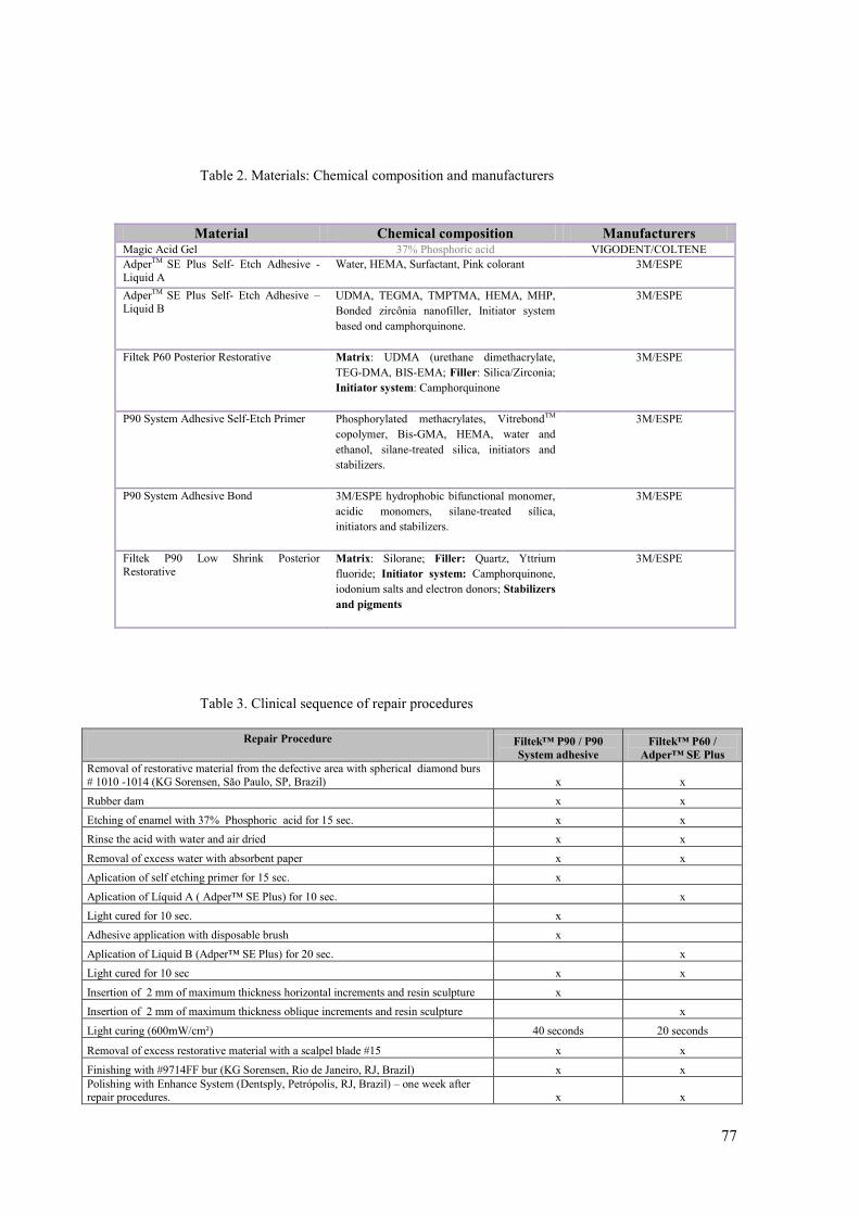
77
Table 2. Materials: Chemical composition and manufacturers
Table 3. Clinical sequence of repair procedures
Repair Procedure
Filtek™ P90 / P90
System adhesive
Filtek™ P60 /
Adper™ SE Plus
Removal of restorative material from the defective area with spherical diamond burs
# 1010 -1014 (KG Sorensen, São Paulo, SP, Brazil) x x
Rubber dam x x
Etching of enamel with 37% Phosphoric acid for 15 sec. x x
Rinse the acid with water and air dried x x
Removal of excess water with absorbent paper x x
Aplication of self etching primer for 15 sec. x
Aplication of Líquid A ( Adper™ SE Plus) for 10 sec. x
Light cured for 10 sec. x
Adhesive application with disposable brush x
Aplication of Liquid B (Adper™ SE Plus) for 20 sec. x
Light cured for 10 sec x x
Insertion of 2 mm of maximum thickness horizontal increments and resin sculpture x
Insertion of 2 mm of maximum thickness oblique increments and resin sculpture x
Light curing (600mW/cm²) 40 seconds 20 seconds
Removal of excess restorative material with a scalpel blade #15 x x
Finishing with #9714FF bur (KG Sorensen, Rio de Janeiro, RJ, Brazil) x x
Polishing with Enhance System (Dentsply, Petrópolis, RJ, Brazil) – one week after repair procedures. x x
Material Chemical composition Manufacturers Magic Acid Gel 37% Phosphoric acid VIGODENT/COLTENE
AdperTM SE Plus Self- Etch Adhesive -
Liquid A
Water, HEMA, Surfactant, Pink colorant 3M/ESPE
AdperTM SE Plus Self- Etch Adhesive –
Liquid B UDMA, TEGMA, TMPTMA, HEMA, MHP,
Bonded zircônia nanofiller, Initiator system
based ond camphorquinone.
3M/ESPE
Filtek P60 Posterior Restorative Matrix: UDMA (urethane dimethacrylate,
TEG-DMA, BIS-EMA; Filler: Silica/Zirconia;
Initiator system: Camphorquinone
3M/ESPE
P90 System Adhesive Self-Etch Primer Phosphorylated methacrylates, VitrebondTM
copolymer, Bis-GMA, HEMA, water and
ethanol, silane-treated silica, initiators and
stabilizers.
3M/ESPE
P90 System Adhesive Bond
3M/ESPE hydrophobic bifunctional monomer,
acidic monomers, silane-treated sílica,
initiators and stabilizers.
3M/ESPE
Filtek P90 Low Shrink Posterior
Restorative Matrix: Silorane; Filler: Quartz, Yttrium
fluoride; Initiator system: Camphorquinone,
iodonium salts and electron donors; Stabilizers
and pigments
3M/ESPE
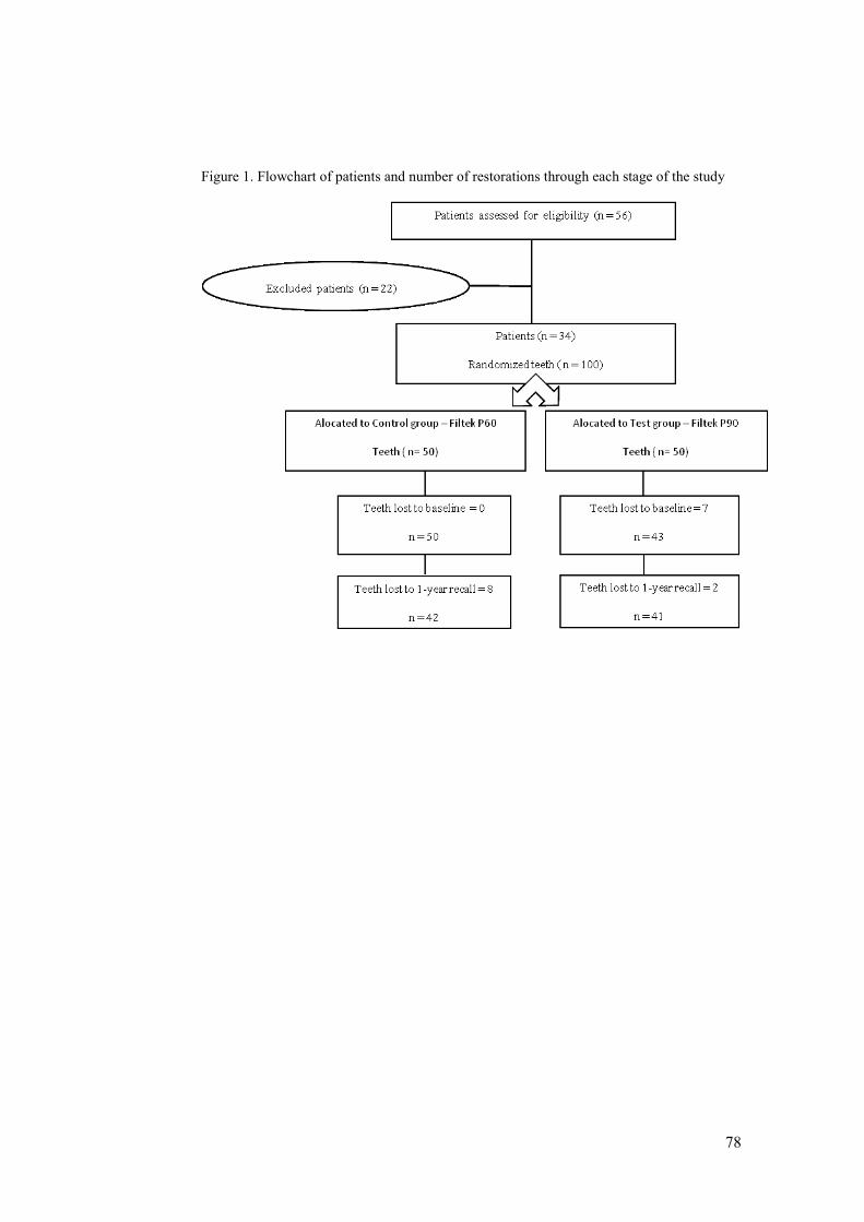
78
Figure 1. Flowchart of patients and number of restorations through each stage of the study
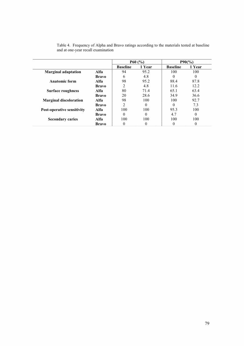
79
Table 4. Frequency of Alpha and Bravo ratings according to the materials tested at baseline
and at one-year recall examination
P60 (%) P90(%)
Baseline 1 Year Baseline 1 Year
Marginal adaptation Alfa 94 95.2 100 100
Bravo 6 4.8 0 0
Anatomic form Alfa 98 95.2 88.4 87.8
Bravo 2 4.8 11.6 12.2
Surface roughness Alfa 80 71.4 65.1 63.4
Bravo 20 28.6 34.9 36.6
Marginal discoloration Alfa 98 100 100 92.7
Bravo 2 0 0 7.3
Post-operative sensitivity Alfa 100 100 95.3 100
Bravo 0 0 4.7 0
Secondary caries Alfa 100 100 100 100
Bravo 0 0 0 0
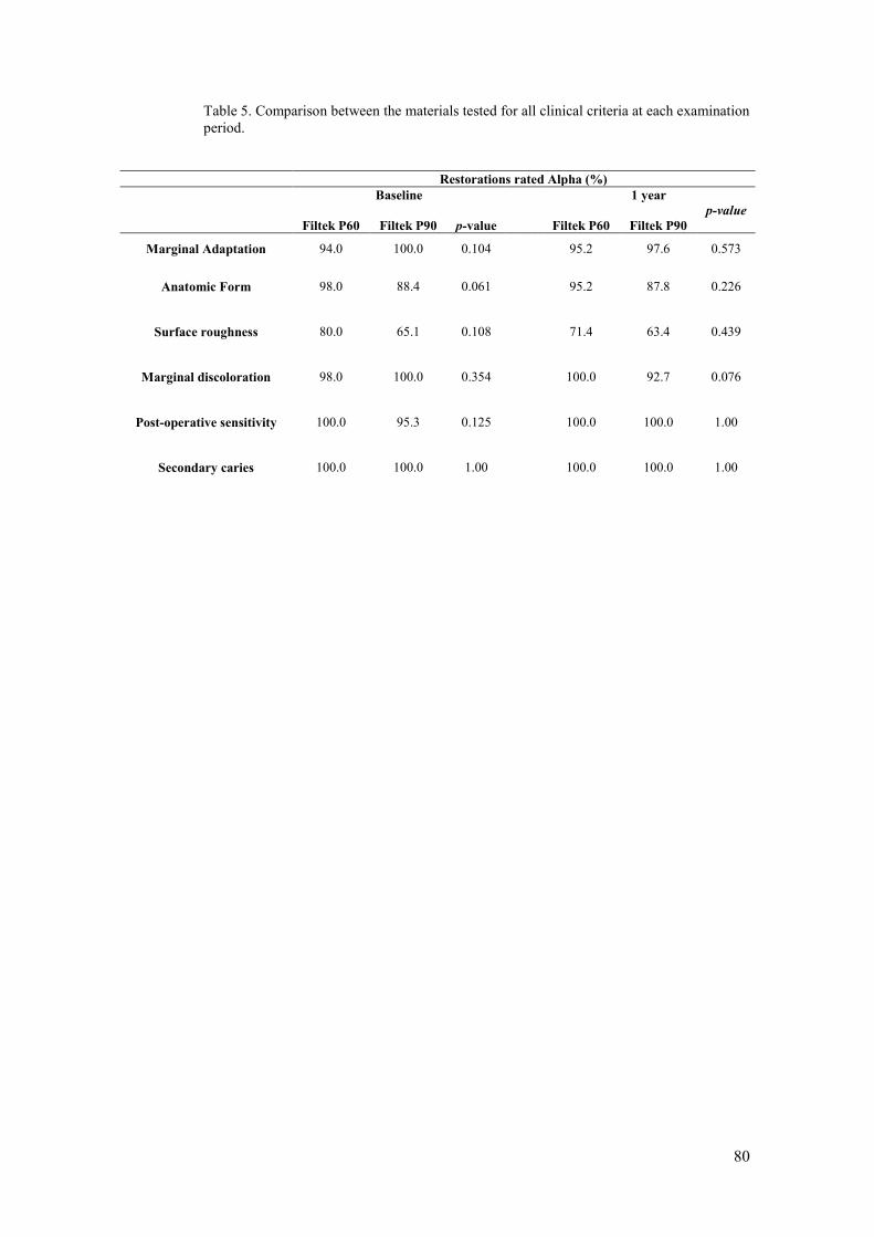
80
Table 5. Comparison between the materials tested for all clinical criteria at each examination
period.
Restorations rated Alpha (%)
Baseline 1 year
Filtek P60 Filtek P90 p-value
Filtek P60
Filtek P90
p-value
Marginal Adaptation 94.0 100.0 0.104 95.2 97.6 0.573
Anatomic Form 98.0 88.4 0.061 95.2 87.8 0.226
Surface roughness 80.0 65.1 0.108 71.4 63.4 0.439
Marginal discoloration 98.0 100.0 0.354 100.0 92.7 0.076
Post-operative sensitivity 100.0 95.3 0.125 100.0 100.0 1.00
Secondary caries 100.0 100.0 1.00 100.0 100.0 1.00
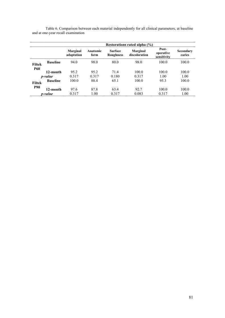
81
Table 6. Comparison between each material independently for all clinical parameters, at baseline
and at one-year recall examination
Restorations rated alpha (%)
Marginal
adaptation
Anatomic
form
Surface
Roughness
Marginal
discoloration
Post-
operative
sensitivity
Secondary
caries
Filtek
P60
Baseline 94.0 98.0 80.0 98.0 100.0 100.0
12-month 95.2 95.2 71.4 100.0 100.0 100.0
p-value 0.317 0.317 0.180 0.317 1.00 1.00
Filtek
P90
Baseline 100.0 88.4 65.1 100.0 95.3 100.0
12-month 97.6 87.8 63.4 92.7 100.0 100.0
p-value 0.317 1.00 0.317 0.083 0.317 1.00
82
6. CONSIDERAÇÕES FINAIS
Após um ano, a resina composta à base de silorano apresentou
desempenho clínico similar à resina composta à base de dimetacrilato
quando utilizada para reparar restaurações de resina composta à base
de dimetacrilato.
O adequado desempenho clínico dos reparos realizados com a
resina composta à base de silorano, de nova tecnologia química, está
relacionado ao uso de um sistema adesivo de nova formulação química,
especificamente desenvolvido para proporcionar adesão ao compósito à
base de silorano.
A reduzida contração de polimerização atribuída ao silorano não
foi suficiente para estabelecer uma melhor performance clínica para
esse compósito.
Achados laboratoriais devem ser reforçados por investigações
clínicas.
Baseados nos resultados de um ano, os reparos podem ser
considerados procedimentos clínicos seguros, eficazes e conservadores
para o tratamento de restaurações com defeitos localizados, contudo
ainda clinicamente aceitáveis.
No entanto, avaliações em períodos mais longos são necessárias
para determinar o desempenho clínico dos reparos realizados com o
novo compósito a longo prazo.
83
REFERÊNCIAS
01- Gordan VV, Shen C, Watson RE, Mjor IA. Four-year clinical evaluation of self-etching primer and resin-based restorative material. Am J Dent 2005; 18:45-49.
02- Teixeira EC, Bayne SC, Thompson JY, Ritter AV, Edward JS. Shear bond strength of self-etching bonding systems in combination with various composites used for repairing aged composites. J Adhes Dent 2005; 7:159-164.
03- Lührs AK, Görmann B, Jaker-Guhr S, Geurtsen W. Repairability of dental siloranes in vitro. Dent Mater 2011; 27: 144-149.
04- Papacchini F, Magni E, Radovic I, Mazzitelli C, Monticelli F, Goracci C, Polimeni A, Ferrari M. Effect of intermediate agents and pre-heating of repairing resin on composite-repair bonds. Oper Dent 2007; 32:363-371.
05- Bonstein T, Garlapo D, Donarummo Jr J, Bush PJ. Evaluation of varied repair protocols apllied to aged composite resin. J Adhes Dent 2005; 7:41-49.
06- Rodriguez G D R, Pereira S N A. Evolución y tendencias actuales en resinas compuestas. Acta Odontol Venez 2008; 46: 1-18.
07- Weinmann W, Thalacker C, Guggenberger R. Siloranes in dental composites. Dent Mater 2005; 21: 68–74.
08- Lien W, Vandewalle, KS. Physical properties of a new silorane-based restorative system. Dent Mater 2010; 26:337-344.
09- Min SH, Ferracane J, Lee IB. Effect of shrinkage strain, modulus, and instrument compliance on polymerization shrinkage stress of light-cured composites during the initial curing stage. Dent Mater 2010; 26:1024-1033.
10- Eick JD, Kotha SP, Chappelow CC, Kilway KV, Giese G, Glaros AG, Pinzino CS. Properties of silorane-based dental resins and composites containing a stress-reducing monomer. Dent Mater 2007; 23: 1011-1017.
11- Ilie N, Hickel R. Silorane-based dental composite: behaviors and abilities. Dent Mater J 2006; 25: 445-454.
12- Pérez MM, Ghinea R, Ugarte-Alván LI, Pulgar R, Paravina RD. Colour and translucency in silorane-based resin composite compared to universal and nonofilled composites. J Dent 2010; 38:110-116.
13- Eick JD, Smith RE, Pinzino CS, Kostoryz EL. Stability of silorane dental monomers in aqueous systems. J Dent 2006; 34:405-410
14- Palin W M, Fleming G J P, Burke F JT, Marquis P M, Randall R C. The influence of short and medium term water immersion on the hydrolytic stability of novel low shrink dental composites. Dent Mater 2005; 21: 852-863.
84
15- Soh MS, Yap AUJ, Sellinger A. Physicomechanical evaluation of low-shrinkage dental nanocomposites based on silsesquioxane cores. Eur J Oral Sci 2007;115: 230–238.
16- Gordan VV, Shen C, Riley J, Mjor IA. Two-year clinical evaluation of repair versus replacement of composite restorations. J Esthet Dent 2006;18:144-154
17- Moncada G, Fernández E, Martin J, Arancibia C, Mjor I, Gordan VV. Increasing the longevity of restorations by minimal intervention: A two-year clinical trial. Oper Dent 2008; 33: 258-264.
18- Fawzy AS, El-Askary FS, Amer MA. Effect of surface treatment on the tensile bond strength of repaired water-aged anterior restorative micro-fine hybrid resin composite. J Dent 2008; 36: 969-976.
19- Hickel R, Peschke A, Tyas M, Mjör IA, Bayne S, Peters M, Hiller KA, Randall R, Vanherle G, Heintze SD. FDI World Dental Federation: clinical criteria for the evaluation of direct and indirect restorations - update and clinical examples. Clin Oral invest 2010; 14:349-336
20- Moncada G, Martin J, Fernández E, Hempel MC, Mjör IA, Gordan VV. Sealing, Refurbishment and Repair of Class I and II defective restorations: A three-year clinical trial. JADA 2009; 140:425-432.
21- Christensen GJ. When and how to repair a failing restoration. J Am Dental Assoc 2007; 138:1605-1607.
22- Cavalcanti AN, Lavigne C, Fontes CM, Mathias P. Microinfiltração na interface compósito-reparo: efeito de diferentes sistemas adesivos. J Appl Oral Sci 2004; 12: 219-222
23- Bowen R L. Use of epoxy resin in restorative materials. J Dent Res 1958; 35: 360-369.
24- Bowen R L. Properties of silica reinforced polymer for dental restorations. J Am Dental Assoc 1963; 66: 57-64.
25- Frankenberger R, Roth S, Krämer N, Pelka M, Petschelt A. Effect of preparation mode on class II resin composite repair. J. Oral Rahabil 2003; 30:559-564.
26- Yamazaki PCV, Bedran-Russo AKB, Pereira PNR, Wsift-Jr EJ. Microleakage evaluation of a new low-shrinkage composite restorative material. Oper Dent 2006; 31:670-676.
27- Ernst CP, Meyer GR, Klöcker K, Willershausen B. Determination of
polymerization shrinkage stress by means of a photoelastic investigation. Dent Mater 2004; 20:313-321.
28- Palin WM, Fleminga GJP, Nathwania H, Burkeb FJT, Randall RC. In vitro cuspal deflection and microleakage of maxillary premolars restored with novel low-shrink dental composites. Dent Mater 2005; 21: 324-335.
85
29- Bouillaguet S, Gamba J, Forchelet J, Krejci I , Wataha J C. Dynamics of composite polymerization mediates the development of cuspal strains. Dent Mater 2006; 22: 896–902.
30- Kostoryz E L, Zhu Q, Zhao Hong, Glaros A G , Eick J D. Assessment of cytotoxicity and DNA damage exhibited by siloranes and oxiranes in cultured mammalian cells. Mutat Res 2007; 634: 156–162.
31- Schmidt M, Kirkevang LL, Hørsted-Bindslev P, Poulsen S. Marginal adaptation of a low-shrinkage silorane-based composite: 1-year randomized clinical trial. Clin Oral Invest 2011; 15: 291-295.
32- van Dijken JW, Linberg A. Clinical effectiveness of a low-shrinkage resin composite: a five-year evaluation. J Adhes Dent 2009; 11:143-148.
33- Elderton RJ. Ciclo Restaurador Repetitivo. Promoção de Saúde Bucal, São Paulo: p. 193-200, 1997.
34- Landis JR, Koch GG. The measurement of observer agreement for categorical data. Biometrics 1977; 33:159-174.
35- Escosteguy CC. Tópicos Metodológicos e Estatísticos em Ensaios Clínicos Controlados Randomizados. Arq Bras Cardiol 1999; 72:139-143.
86
ANEXO I
87
ANEXO II
Termo de Consentimento Livre e Esclarecido
Convido o/a
Sr.(a):_____________________________________________, a participar da
pesquisa “Comportamento clínico de reparos em restaurações de resina
composta: estudo longitudinal randomizado”, sob a responsabilidade dos
pesquisadores: Daniela Araújo Veloso Popoff, Allyson Nogueira Moreira,
Cláudia Silami de Magalhães e Raquel Conceição Ferreira.
Nesta pesquisa vamos comparar o desempenho de dois tipos de resinas
compostas para o reparo de obturações dentárias defeituosas, avaliando, ao
longo do tempo, se as restaurações permanecem bem adaptadas, lisas, com
boa cor e bom formato. As avaliações serão feitas no dia do polimento das
obturações e depois de passados 6, 12 e 18 meses.
Sobre a pesquisa: Para participar dessa pesquisa, o paciente deve
possuir pré-molares e/ou molares com obturações clinicamente insatisfatórias,
além de uma boa higiene bucal. Um pesquisador da equipe fará o exame de
sua boca confirmando o estado dos dentes. Em consultas que serão marcadas
com antecedência, os dentes serão anestesiados, quando necessário, e
preparados para receber os reparos, de acordo com as técnicas adequadas por
um outro pesquisador também da equipe. Após uma semana, as obturações
serão polidas e avaliadas pelo primeiro pesquisador. O paciente será chamado
de volta à clínica para novas avaliações em 6, 12 e 18 meses, quando poderá
ser feito um molde de cada obturação para observação em um microscópio.
Serão feitas fotografias dos dentes reparados.
A pesquisa será realizada na clínicas odontológicas das faculdades de
Odontologia da Universidade Federal de Minas Gerais (UFMG) e da
Associação Educativa do Brasil (SOEBRAS), pelos seguintes pesquisadores:
Daniela Araújo Veloso Popoff, mestra, professora da UNIMONTES e aluna da
pós-graduação em Odontologia da UFMG; Thalita Thyrza Almeida Santa Rosa,
mestra e professora da UNIMONTES; Allyson Nogueira Moreira, Doutor,
professor da UFMG e orientador desta pesquisa e Cláudia Silami de
Magalhães, Doutora professora da UFMG e orientadora desta pesquisa.
Não haverá risco à saúde e bem estar dos pacientes e em nenhum
momento esses terão seu nome divulgado. Os resultados da pesquisa serão
publicados e ainda assim a sua identidade será preservada. Os pacientes não
terão qualquer ônus ou ganho financeiro por participar da pesquisa, porém
serão beneficiados com os tratamentos propostos, pois seus dentes
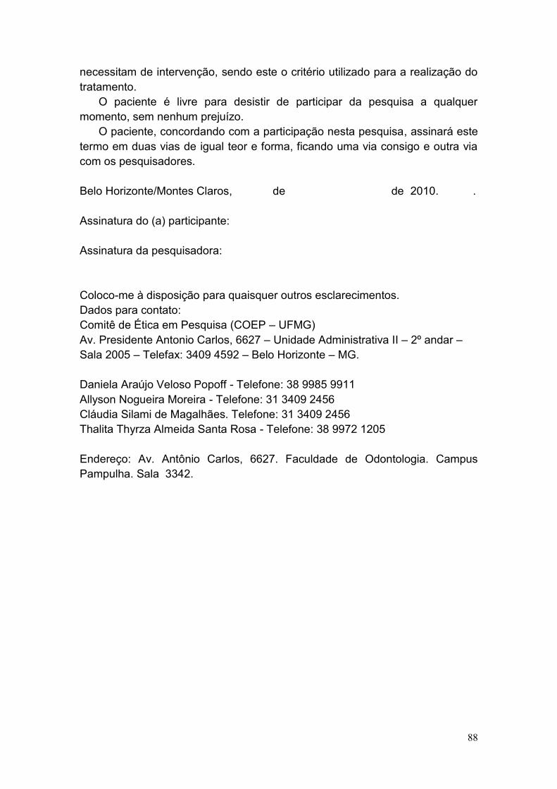
88
necessitam de intervenção, sendo este o critério utilizado para a realização do
tratamento.
O paciente é livre para desistir de participar da pesquisa a qualquer
momento, sem nenhum prejuízo.
O paciente, concordando com a participação nesta pesquisa, assinará este
termo em duas vias de igual teor e forma, ficando uma via consigo e outra via
com os pesquisadores.
Belo Horizonte/Montes Claros, de de 2010. .
Assinatura do (a) participante:
Assinatura da pesquisadora:
Coloco-me à disposição para quaisquer outros esclarecimentos.
Dados para contato:
Comitê de Ética em Pesquisa (COEP – UFMG)
Av. Presidente Antonio Carlos, 6627 – Unidade Administrativa II – 2º andar –
Sala 2005 – Telefax: 3409 4592 – Belo Horizonte – MG.
Daniela Araújo Veloso Popoff - Telefone: 38 9985 9911
Allyson Nogueira Moreira - Telefone: 31 3409 2456
Cláudia Silami de Magalhães. Telefone: 31 3409 2456
Thalita Thyrza Almeida Santa Rosa - Telefone: 38 9972 1205
Endereço: Av. Antônio Carlos, 6627. Faculdade de Odontologia. Campus
Pampulha. Sala 3342.
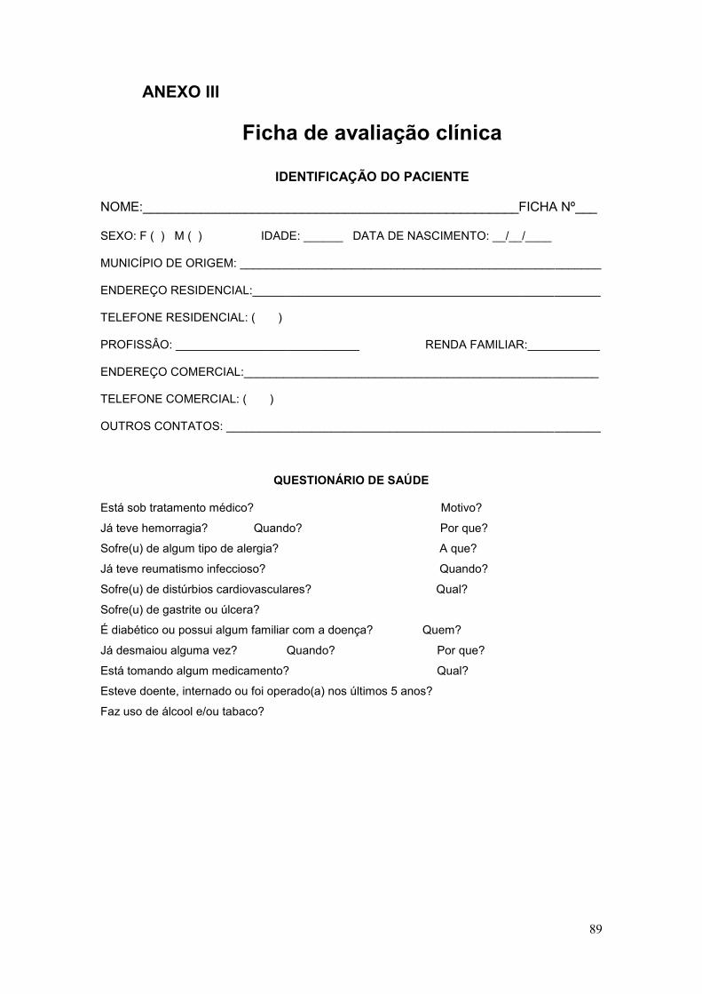
89
ANEXO III
Ficha de avaliação clínica
IDENTIFICAÇÃO DO PACIENTE
NOME:____________________________________________________FICHA Nº___
SEXO: F ( ) M ( ) IDADE: ______ DATA DE NASCIMENTO: __/__/____
MUNICÍPIO DE ORIGEM: _______________________________________________________
ENDEREÇO RESIDENCIAL:_____________________________________________________
TELEFONE RESIDENCIAL: ( )
PROFISSÂO: ____________________________ RENDA FAMILIAR:___________
ENDEREÇO COMERCIAL:______________________________________________________
TELEFONE COMERCIAL: ( )
OUTROS CONTATOS: _________________________________________________________
QUESTIONÁRIO DE SAÚDE
Está sob tratamento médico? Motivo?
Já teve hemorragia? Quando? Por que?
Sofre(u) de algum tipo de alergia? A que?
Já teve reumatismo infeccioso? Quando?
Sofre(u) de distúrbios cardiovasculares? Qual?
Sofre(u) de gastrite ou úlcera?
É diabético ou possui algum familiar com a doença? Quem?
Já desmaiou alguma vez? Quando? Por que?
Está tomando algum medicamento? Qual?
Esteve doente, internado ou foi operado(a) nos últimos 5 anos?
Faz uso de álcool e/ou tabaco?
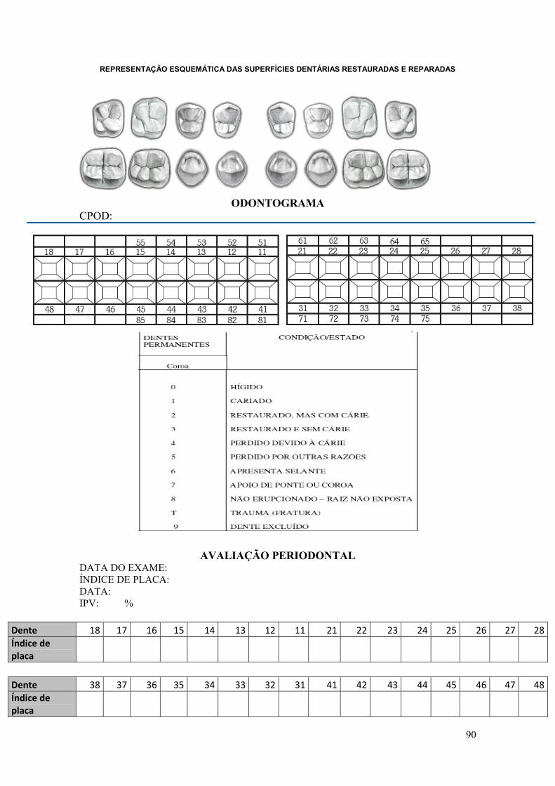
90
REPRESENTAÇÃO ESQUEMÁTICA DAS SUPERFÍCIES DENTÁRIAS RESTAURADAS E REPARADAS
ODONTOGRAMA
CPOD:
AVALIAÇÃO PERIODONTAL DATA DO EXAME:
ÍNDICE DE PLACA:
DATA:
IPV: %
Dente 18 17 16 15 14 13 12 11 21 22 23 24 25 26 27 28
Índice de placa
Dente 38 37 36 35 34 33 32 31 41 42 43 44 45 46 47 48
Índice de placa
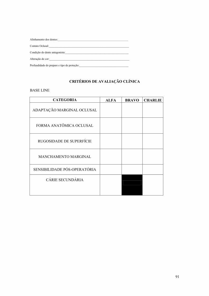
91
Alinhamento dos dentes:__________________________________________________
Contato Oclusal:_________________________________________________________
Condição do dente antagonista:_____________________________________________
Alteração de cor:_________________________________________________________
Profundidade do prepare e tipo de proteção:___________________________________
CRITÉRIOS DE AVALIAÇÃO CLÍNICA
BASE LINE
CATEGORIA ALFA BRAVO CHARLIE
ADAPTAÇÃO MARGINAL OCLUSAL
FORMA ANATÔMICA OCLUSAL
RUGOSIDADE DE SUPERFÍCIE
MANCHAMENTO MARGINAL
SENSIBILIDADE PÓS-OPERATÓRIA
CÁRIE SECUNDÁRIA
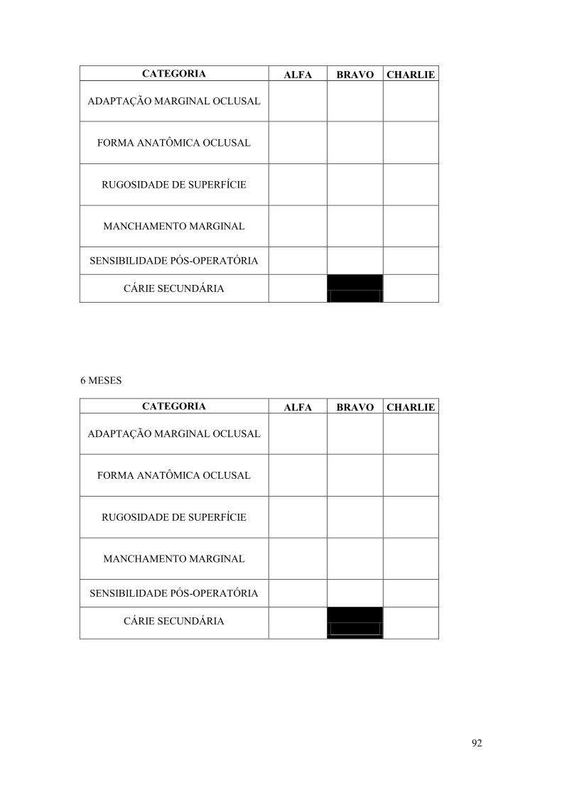
92
CATEGORIA ALFA BRAVO CHARLIE
ADAPTAÇÃO MARGINAL OCLUSAL
FORMA ANATÔMICA OCLUSAL
RUGOSIDADE DE SUPERFÍCIE
MANCHAMENTO MARGINAL
SENSIBILIDADE PÓS-OPERATÓRIA
CÁRIE SECUNDÁRIA
6 MESES
CATEGORIA ALFA BRAVO CHARLIE
ADAPTAÇÃO MARGINAL OCLUSAL
FORMA ANATÔMICA OCLUSAL
RUGOSIDADE DE SUPERFÍCIE
MANCHAMENTO MARGINAL
SENSIBILIDADE PÓS-OPERATÓRIA
CÁRIE SECUNDÁRIA
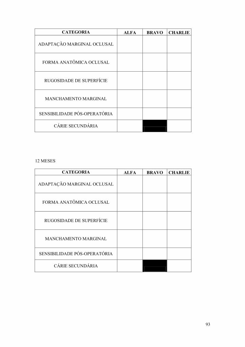
93
CATEGORIA ALFA BRAVO CHARLIE
ADAPTAÇÃO MARGINAL OCLUSAL
FORMA ANATÔMICA OCLUSAL
RUGOSIDADE DE SUPERFÍCIE
MANCHAMENTO MARGINAL
SENSIBILIDADE PÓS-OPERATÓRIA
CÁRIE SECUNDÁRIA
12 MESES
CATEGORIA ALFA BRAVO CHARLIE
ADAPTAÇÃO MARGINAL OCLUSAL
FORMA ANATÔMICA OCLUSAL
RUGOSIDADE DE SUPERFÍCIE
MANCHAMENTO MARGINAL
SENSIBILIDADE PÓS-OPERATÓRIA
CÁRIE SECUNDÁRIA
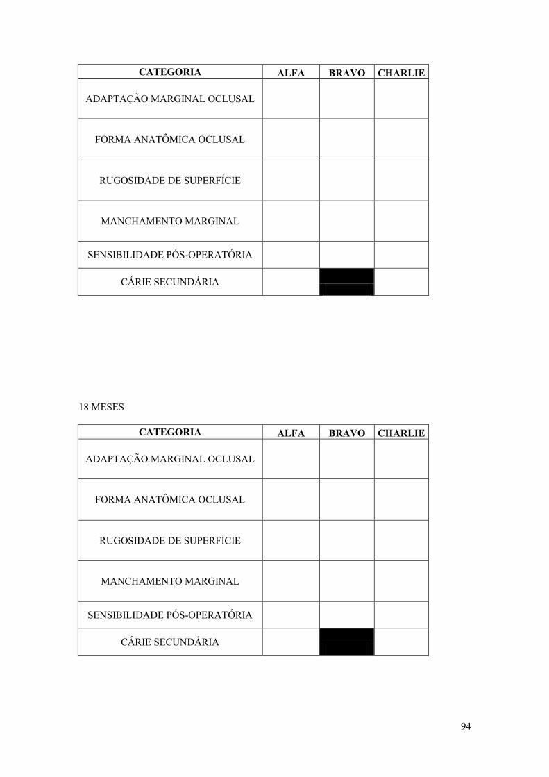
94
CATEGORIA ALFA BRAVO CHARLIE
ADAPTAÇÃO MARGINAL OCLUSAL
FORMA ANATÔMICA OCLUSAL
RUGOSIDADE DE SUPERFÍCIE
MANCHAMENTO MARGINAL
SENSIBILIDADE PÓS-OPERATÓRIA
CÁRIE SECUNDÁRIA
18 MESES
CATEGORIA ALFA BRAVO CHARLIE
ADAPTAÇÃO MARGINAL OCLUSAL
FORMA ANATÔMICA OCLUSAL
RUGOSIDADE DE SUPERFÍCIE
MANCHAMENTO MARGINAL
SENSIBILIDADE PÓS-OPERATÓRIA
CÁRIE SECUNDÁRIA
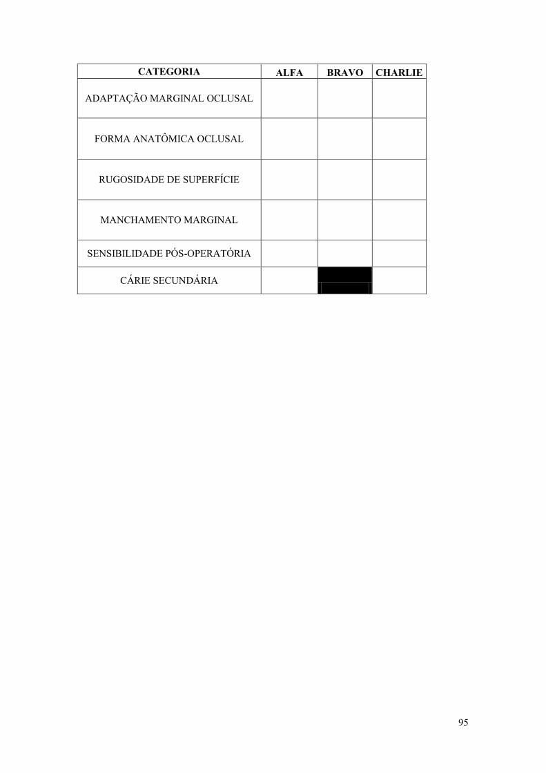
95
CATEGORIA ALFA BRAVO CHARLIE
ADAPTAÇÃO MARGINAL OCLUSAL
FORMA ANATÔMICA OCLUSAL
RUGOSIDADE DE SUPERFÍCIE
MANCHAMENTO MARGINAL
SENSIBILIDADE PÓS-OPERATÓRIA
CÁRIE SECUNDÁRIA
96
ANEXO IV Instruções aos autores para a submissão de manuscritos à Acta Odontologica Latinoamericana – Artigo I
Modified according to the recommendations of the Committee of Medical Journal Editors (www.icmje.org). Please e-mail your manuscript, tables and figures to [email protected] with copy to [email protected]. Authors are advised to keep a copy for their files. Authors are responsible for all statements made in their papers. It is understood that the manuscript is not under consideration in any other journal. In view of the considerations expressed in our Editorial Policy, papers will be published in English. In addition to the scientific review, AOL will conduct a technical review and a language review. To facilitate the work of the authors, AOL offers a manuscript translation service, and authors who wish to make use of it should indicate so in the letter accompanying their paper. The translation fee is 5 dollars per 100 words of translated text. AOL may correct the language in papers submitted in English, and authors will be billed according to how much correcting the manuscripts require. The translation/correction service will only be performed if the paper has been accepted for publication, after scientific review. Manuscripts should be double spaced and organized as follows: running head – no more than 40 letters; full title in English; authors separated by commas including the full name used for indexing and the rest with initials; affiliation(s) in English, indicating each author‟s affiliation by means of a superscript; 150-300 word abstract; keywords (no more than six, which must be listed in PubMed‟s MeSH); title, abstract and keywords in Spanish or Portuguese. If desired, the abstract in Spanish or Portuguese may be longer (up to 500 words). The text should include the following headings: introduction, materials and methods, results, discussion, acknowledgments, contact information (name, mailing address and e-mail address of the corresponding author), references and legends for figures. References should be indicated in the manuscript with superscripts and numbered consecutively according to the order in which they have been cited in the text. Format and punctuation should match the following example:
1. Ghiabi M, Gallagher GT, Wong DT. Eosinophils, tissue eosinophilia and eosinophil-derived transforming growth factor alpha in hamster oral carcinogenesis. Cancer Res 1992; 52:389-393.
97
Journal names should be abbreviated as in MEDLINE/PubMed. If abstracts are cited, [Abstract] should be added after the title. Book citations should include book title and the chapter title, where applicable, author(s) of the book and chapter, year of publication and publisher. Photographs should be sent in separate files, without their legends, in *.tiff ,*.jpg or *.epf format, resolution 300dpi, size 100 %, and numbered consecutively according to their appearance in the text. Diagrams, graphs and tables should have proportions that render them legible, in the width of one or two columns of the printed journal (7.5 or 16.5 cm), and be designed on Excel or Power Point. Photographs, diagrams and graphs should be indicated as Fig. (figures) and numbered with consecutive Arabic numerals according to their order of appearance in the text. Tables should be numbered with a separate series of Arabic numerals, have a title, and any abbreviations should be explained in a footnote. Legends for figures should be included at the end of the manuscript. Figures or diagrams in color will only be published if the author covers the cost. There is a publication fee of 15 dollars per printed page. Authors will be sent a pdf. file of their paper and one copy of the printed journal. Annual subscription (three issues): 55 dollars. Members of the Argentine Society for Dental Research (Sociedad Argentina de Investigación Odontológica) may purchase a subscription at a preferential rate if ordered on paying yearly fees. Updated fees may be viewed at www.saio.org.ar.
98
ANEXO V
Instruções aos autores para a submissão de manuscritos à Operative Dentistry – Artigo II
New Instructions as of 20 September 2008
Operative Dentistry requires electronic submission of all manuscripts. All submissions must be sent to Operative Dentistry using the Allen Track upload site. Your manuscript will only be considered officially submitted after it has been approved through our initial quality control check, and any problems have been fixed. You will have 6 days from when you start the process to submit and approve the manuscript. After the 6 day limit, if you have not finished the submission, your submission will be removed from the server. You are still able to submit the manuscript, but you must start from the beginning. Be prepared to submit the following manuscript files in your upload:
A Laboratory or Clinical Research Manuscript file must include: o a title o a running (short) title o a clinical relevance statement o a concise summary (abstract) o introduction, methods & materials, results, discussion and
conclusion o references (see Below) o The manuscript MUST NOT include any:
identifying information such as: Authors Acknowledgements Correspondence information
Figures Graphs Tables
An acknowledgement, disclaimer and/or recognition of support (if applicable) must in a separate file and uploaded as supplemental material.
All figures, illustrations, graphs and tables must also be provided as individual files. These should be high resolution images, which are used by the editor in the actual typesetting of your manuscript. Please refer to the instructions below for acceptable formats.
All other manuscript types use this template, with the appropriate changes as listed below.
Complete the online form which includes complete author information and select the files you would like to send to Operative Dentistry. Manuscripts that do not meet our formatting and data requirements listed below will be sent back to the corresponding author for correction.
99
GENERAL INFORMATION
All materials submitted for publication must be submitted exclusively to Operative Dentistry.
The editor reserves the right to make literary corrections. Currently, color will be provided at no cost to the author if the editor
deems it essential to the manuscript. However, we reserve the right to convert to gray scale if color does not contribute significantly to the quality and/or information content of the paper.
The author(s) retain(s) the right to formally withdraw the paper from consideration and/or publication if they disagree with editorial decisions.
International authors whose native language is not English must have their work reviewed by a native English speaker prior to submission.
Spelling must conform to the American Heritage Dictionary of the English Language, and SI units for scientific measurement are preferred.
While we do not currently have limitations on the length of manuscripts, we expect papers to be concise; Authors are also encouraged to be selective in their use of figures and tables, using only those that contribute significantly to the understanding of the research.
Acknowledgement of receipt is sent automatically. If you do not receive such an acknowledgement, please contact us at [email protected] rather than resending your paper.
IMPORTANT: Please add our e-mail address to your address book on your server to prevent transmission problems from spam and other filters. Also make sure that your server will accept larger file sizes. This is particularly important since we send page-proofs for review and correction as .pdf files.
REQUIREMENTS
FOR ALL MANUSCRIPTS
1. CORRESPONDING AUTHOR must provide a WORKING / VALID e-mail address which will be used for all communication with the journal. NOTE: Corresponding authors MUST update their profile if their e-mail or postal address changes. If we cannot contact authors within seven days, their manuscript will be removed from our publication queue.
2. AUTHOR INFORMATION must include: full name of all authors complete mailing address for each author degrees (e.g. DDS, DMD, PhD) affiliation (e.g. Department of Dental Materials, School of
Dentistry, University of Michigan)
3. MENTION OF COMMERCIAL PRODUCTS/EQUIPMENT must include:
full name of product
100
full name of manufacturer city, state and/or country of manufacturer
4. MANUSCRIPTS AND TABLES must be provided as Word files. Please limit size of tables to no more than one US letter sized page. (8 ½ ” x 11”)
5. ILLUSTRATIONS, GRAPHS AND FIGURES must be provided as TIFF or JPEG files with the following parameters
line art (and tables that are submitted as a graphic) must be sized at approximately 5” x 7” and have a resolution of 1200 dpi.
gray scale/black & white figures must have a minimum size of 3.5” x 5”, and a maximum size of 5” x 7” and a minimum resolution of 300 dpi and a maximum of 400 dpi.
color figures must have a minimum size of 2.5” x 3.5”, and a maximum size of 3.5” x 5” and a minimum resolution of 300 dpi and a maximum of 400 dpi.
color photographs must be sized at approximately 3.5” x 5” and have a resolution of 300 dpi.
OTHER MANUSCRIPT TYPES
1. CLINICAL TECHNIQUE/CASE STUDY MANUSCRIPTS must include:
a running (short) title purpose description of technique list of materials used potential problems summary of advantages and disadvantages references (see below)
2. LITERATURE AND BOOK REVIEW MANUSCRIPTS must include:
a running (short) title a clinical relevance statement based on the conclusions of
the review conclusions based on the literature review…without this,
the review is just an exercise references (see below)
FOR REFERENCES
REFERENCES must be numbered (superscripted numbers) consecutively as they appear in the text and, where applicable, they should appear after punctuation.
101
The reference list should be arranged in numeric sequence at the end of the manuscript and should include: 1. Author(s) last name(s) and initial (ALL AUTHORS must be
listed) followed by the date of publication in parentheses. 2. Full article title. 3. Full journal name in italics (no abbreviations), volume and issue
numbers and first and last page numbers complete (i.e. 163-168 NOT attenuated 163-68).
4. Abstracts should be avoided when possible but, if used, must include the above plus the abstract number and page number.
5. Book chapters must include chapter title, book title in italics, editors‟ names (if appropriate), name of publisher and publishing address.
6. Websites may be used as references, but must include the date (day, month and year) accessed for the information.
7. Papers in the course of publication should only be entered in the references if they have been accepted for publication by a journal and then given in the standard manner with “In press” following the journal name.
8. DO NOT include unpublished data or personal communications in the reference list. Cite such references parenthetically in the text and include a date.
EXAMPLES OF REFERENCE STYLE
Journal article: two authors Evans DB & Neme AM (1999) Shear bond strength of composite resin and amalgam adhesive systems to dentin American Journal of Dentistry 12(1) 19-25.
Journal article: multiple authors Eick JD, Gwinnett AJ, Pashley DH & Robinson SJ (1997) Current concepts on adhesion to dentin Critical Review of Oral and Biological Medicine 8(3) 306-335.
Journal article: special issue/supplement Van Meerbeek B, Vargas M, Inoue S, Yoshida Y, Peumans M, Lambrechts P & Vanherle G (2001) Adhesives and cements to promote preservation dentistry Operative Dentistry (Supplement 6) 119-144.
Abstract: Yoshida Y, Van Meerbeek B, Okazaki M, Shintani H & Suzuki K (2003) Comparative study on adhesive performance of functional monomers Journal of Dental Research 82(Special Issue B) Abstract #0051 p B-19.
Corporate publication: ISO-Standards (1997) ISO 4287 Geometrical Product Specifications Surface texture: Profile method – Terms, definitions and surface texture parameters Geneve: International Organization for Standardization 1st edition 1-25.
102
Book: single author Mount GJ (1990) An Atlas of Glass-ionomer Cements Martin Duntz Ltd, London.
Book: two authors Nakabayashi N & Pashley DH (1998) Hybridization of Dental Hard Tissues Quintessence Publishing, Tokyo.
Book: chapter Hilton TJ (1996) Direct posterior composite restorations In: Schwarts RS, Summitt JB, Robbins JW (eds) Fundamentals of Operative Dentistry Quintessence, Chicago 207-228.
Website: single author Carlson L (2003) Web site evolution; Retrieved online July 23, 2003 from: http://www.d.umn.edu/~lcarlson/cms/evolution.html
Website: corporate publication National Association of Social Workers (2000) NASW Practice research survey 2000. NASW Practice Research Network, 1. 3. Retrieved online September 8, 2003 from: http://www.socialworkers.org/naswprn/default