transseptopelucidorostrostomia. considerações anatômicas e ... · ântero-posterior do forame...
TRANSCRIPT
Acta Cirúrgica Brasileira - Vol. 26 (Suppl. 2) 2011 - 133
25 - ORIGINAL ARTICLENEW METHODOLOGY
Transseptumpellucidumrostrostomy. Anatomical considerations and neuroendoscopicapproach1
Transseptopelucidorostrostomia. Considerações anatômicas e abordagem neuroendoscópica
Eduardo José Takashi FuzikiI, Roberto Alexandre DezenaII, Benedicto Oscar ColliIII
IPhD, Neurosurgeon, Division of Neurosurgery, Department of Surgery and Anatomy, FMRP-USP, Ribeirao Preto-SP, Brazil. Responsible for acquisitionand interpretation of data, statistical analysis, designed the protocol, involved with technical procedures.IIPhD, Neurosurgeon, Division of Neurosurgery of Department of Surgery and Anatomy, FMRP-USP, Ribeirao Preto-SP, Brazil. Responsible formanuscript writing, responsible for English language.IIIPhD, Chairman and Head of Division of Neurosurgery of Department of Surgery and Anatomy, FMRP-USP, Ribeirao Preto-SP, Brazil. Responsiblefor intellectual and scientific content of the study, supervised all phases of study, critical revision.
ABSTRACTPURPOSE: Verify the presence of the rostral lamina of the corpus callosum, and set parameters for neuroendoscopy.METHODS: Relationship of the floor of the frontal horn of lateral ventricle and the hypothalamic-septal region were studiedafter sagittal and axial sections of the brains. Measurements were compared using F and Student t tests. The correlationsbetween anterior-posterior diameter of the interventricular foramen / anterior-posterior diameter of the fornix column, andbetween anterior-posterior diameter of the interventricular foramen / length of the rostral lamina were performed by Pearsonindex test.RESULTS: There was no statistically significant difference in measurements performed in both hemispheres (p<0.05). Positivecorrelations were observed between the anterior-posterior diameter of the interventricular foramen / anterior-posterior diameter ofthe fornix column (R = 0.35), and between the anterior-posterior diameter of the interventricular foramen / length of the rostrallamina (R = 0.23).CONCLUSION: Rostral lamina was observed in all brains. It was possible to perform an endoscopic fenestration in the rostrallamina, communicating safely the lateral ventricle with a polygonal subcallosal cistern.Key words: Hydrocephalus. Neuroendoscopy. Septum Pellucidum. Anatomy.
RESUMOOBJETIVO: Verificar a presença da lâmina rostral do corpo caloso e padronizar parâmetros para a realização de neuroendoscopia.MÉTODOS: A relação do assoalho do corno frontal do ventrículo lateral e a região hipotálamo-septal lateral foi estudadaatravés de secções sagitais e axiais dos cérebros. As medidas foram comparadas utilizando os testes F e t-Student. As correlaçõesentre diâmetro ântero-posterior do forame interventricular / diâmetro ântero-posterior da coluna do fornix, e entre o diâmetroântero-posterior do forame interventricular / comprimento da lâmina rostral foram estudadas pelo teste de Pearson.RESULTADOS: Não houve diferença estatisticamente significante nas medidas realizadas em ambos hemisférios (p <0.05).Correlações positivas foram observadas entre diâmetros ântero-posteriores do forame interventricular / coluna do fornix (R = 0.35),os diâmetros ântero-posteriores do forame interventricular / comprimento da lâmina rostral (R = 0.23).CONCLUSÃO: A lâmina rostral foi observada em todos espécimes. Foi possível realizar uma fenestração endoscópica na lâminarostral, comunicando com segurança o ventrículo lateral a uma cisterna poligonal subcalosa.Descritores: Hidrocefalia. Neuroendoscopia. Septo Pelúcido. Anatomia.
Fuziki EJT et al.
134 - Acta Cirúrgica Brasileira - Vol. 26 (Suppl. 2) 2011
Introduction
Surgical techniques for treating obstructive hydrocephalushave been described even before the identification ofcommunication between the CSF and other compartments of thebrain. Dandy, in 1922, for the first time opened the laminaterminalis through a craniotomy, communicating the third ventriclewith the basal cysterns1. Mixter, in 1923, performed the firstendoscopic third ventriculostomy toward interpeduncular cistern1.Contemporary authors have observed reduction in the incidenceof hydrocephalus in subarachnoid hemorrhage, opening the laminaterminalis during surgical clipping of cerebral aneurysms1,2,3. Nakaoand Itakura described the opening of the lamina terminalis withflexible endoscope to treat hydrocephalus secondary to tuberculousmeningitis1 and since then the procedure became routine to treatobstructive hydrocephalus. Another brain internal shunt described,performed with endoscope, is the communication of the thirdventricle with the quadrigeminal cistern, through the suprapinealrecess4.
In some situations, third ventriculostomy may becametechnically difficult, mainly when there is thickening of the floorof the third ventricle (for instance due to inflammatory reaction),hindering the identification of anatomical parameters, or when theinterpeduncular cistern is reduced by an ecstatic basilar artery. Inthese situations, the knowledge of internal ventricular systemalternative shunts may have its value. Remarks of functioningcatheters from ventricular shunts placed accidentally in thelongitudinal fissure of the brain are evidence that this place canact as an alternative shunt pathway.
The objective of this paper was to study the feasibility toperform anatomical communication between the ventricular systemand the longitudinal fissure of the brain through a fenestration ofthe rostral lamina of the corpus callosum and establish theanatomical parameters for its completion as a neuroendoscopicprocedure.
Methods
Identification and measurement of anatomicalreferences to the floor of the frontal horn of lateral ventricle andcerebral longitudinal fissure
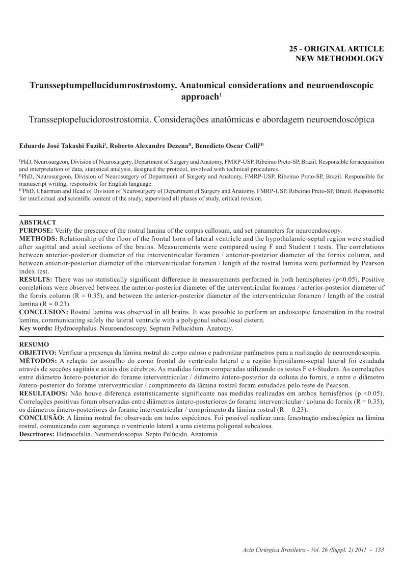
Sixteen 10% formalin fixed brains provided byDepartment of Pathology, Faculty of Medicine – Alfenas University(UNIFENAS), obtained from autopsies, which showed normalappearance in the initial macroscopic inspection, were used. Ninewere female and seven males, aged ranged from 13-69 years (43.5± 13.14) and weights varied from 940-1533 grams (1162.7 ±148.75). After a midline sagittal cut, the following anatomicalstructures were identified in each hemisphere: the corpus callosumand its divisions, the interventricular foramen, the fornix column,the lamina terminalis, the supra-optic recess, the mammillarybodies, the tuber cinereum, the paraterminal gyrus and anteriorcerebral artery (Figure 1).
FIGURE 1 - Midline sagittal section of the brain showing the rostrallamina, hypothalamic-septal triangle, and neighbor structures.
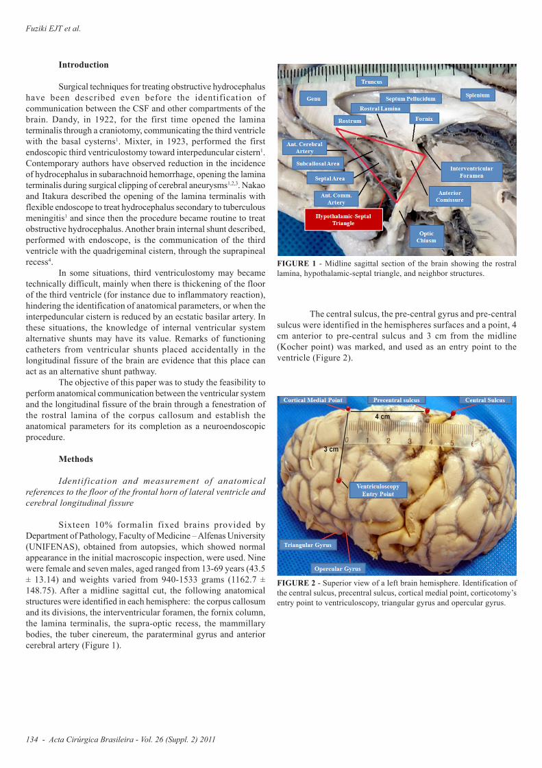
The central sulcus, the pre-central gyrus and pre-centralsulcus were identified in the hemispheres surfaces and a point, 4cm anterior to pre-central sulcus and 3 cm from the midline(Kocher point) was marked, and used as an entry point to theventricle (Figure 2).
FIGURE 2 - Superior view of a left brain hemisphere. Identification ofthe central sulcus, precentral sulcus, cortical medial point, corticotomy’sentry point to ventriculoscopy, triangular gyrus and opercular gyrus.
Acta Cirúrgica Brasileira - Vol. 26 (Suppl. 2) 2011 - 135
Transseptumpellucidumrostrostomy. Anatomical considerations and neuroendoscopic approach
Needles were placed in the rostrum, genu, body andsplenium of the corpus callosum, the supra-optic recess and thetuber cinereum/mammillary bodies, located anterior to the floorof the third ventricle to measure distances among these structures.The portion of the rostrum of the corpus callosum which appearedthinner, laminar, adjacent to the bottom of the septum pellucidumwas identified as the rostral lamina5, which is limited posteriorlyby the fornix column. The diameter of the interventricular foramenwas measured from posterior portion of fornix column to theposterior wall of the foramen. Two reference points were markedin the anterior cerebral artery: one at the origin of the anteriorcommunicating artery, and another in its first flexure of portionA2, at the point of change in the upward path, below the rostrumof the corpus callosum.
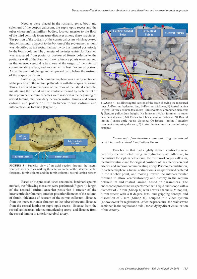
Following, each brain hemisphere was axially sectionedat the junction of the septum pellucidum with the corpus callosum.This cut allowed an overview of the floor of the lateral ventricle,maintaining the medial wall of ventricle formed by each leaflet ofthe septum pellucidum. Needles were inserted in the beginning ofrostral lamina, the boundary between rostral lamina and fornixcolumn and posterior limit between fornix column andinterventricular foramen (Figure 3).
FIGURE 3 - Superior view of an axial section through the lateralventricle with needles marking the anterior border of the interventricularforamen / fornix column and the fornix column / rostral lamina border.
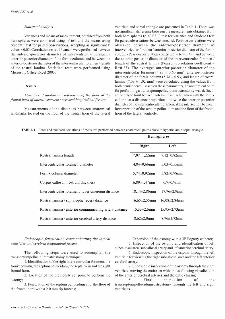
Based on the pre-established anatomical landmarks pointsmarked, the following measures were performed (Figure 4): lengthof the rostral lamina; anterior-posterior diameter of theinterventricular foramen; anterior-posterior diameter of the columnof fornix; thickness of rostrum of the corpus callosum; distancefrom the interventricular foramen to the tuber cinereum; distancefrom the rostral lamina to supra-optic recess; distance from therostral lamina to anterior communicating artery; and distance fromthe rostral lamina to anterior cerebral artery.
FIGURE 4 - Midline sagittal section of the brain showing the measuredlines. A) Rostrum / splenium line; B) Rostrum thickness; F) Rostral laminalength; G) Fornix column thickness; H) Interventricular foramen diameter;J) Septum pellucidum height; K) Interventricular foramen to tubercinereum distance; M) Cortex to tuber cinereum distance; N) Rostrallamina / supra-optic recess distance; O) Rostral lamina / anteriorcommunicating artery distance; P) Rostral lamina / anterior cerebral arterydistance.
Endoscopic fenestration communicating the lateralventricles and cerebral longitudinal fissure
Two brains that had slightly dilated ventricles werecarefully reconstructed using methylmetacrylate adhesive, toreconstruct the septum pellucidum, the rostrum of corpus callosum,the third ventricle and the original positions of the anterior cerebralarteries and anterior communicating artery. Prior to reconstitution,in each hemisphere, a tunnel corticectomia was performed centeredin the Kocher point, and moving toward the interventricularforamen to allow ventriculoscopy and ostomy in the septumpellucidum and rostral lamina, based on parameters. Theendoscopic procedure was performed with rigid endoscope with adiameter of 2.7 mm (Minop ®) with 4 work channels (Minop ®),6 mm trocar with a 0 degree lens, and gripping forceps anddissection of 2 mm (Minop ®), coupled to a video system(Endoview®) for registration. After the procedure, the brains weresectioned in the sagittal and axial, for study by direct visualizationof the ostomy.
Fuziki EJT et al.
136 - Acta Cirúrgica Brasileira - Vol. 26 (Suppl. 2) 2011
Statistical analysis
Variances and means of measurement, obtained from bothhemispheres were compared using F test and the means usingStudent t test for paired observations, accepting as significant Pvalues <0.05. Correlation tests of Pearson were performed betweenthe anterior-posterior diameter of interventricular foramen /anterior-posterior diameter of the fornix column, and between theanterior-posterior diameter of the interventricular foramen / lengthof the rostral lamina. Statistical tests were performed usingMicrosoft Office Excel 2003.
Results
Measures of anatomical references of the floor of thefrontal horn of lateral ventricle / cerebral longitudinal fissure
Measurements of the distances between anatomicallandmarks located on the floor of the frontal horn of the lateral
ventricle and septal triangle are presented in Table 1. There wasno significant difference between the measurements obtained fromboth hemispheres (p <0.05, F test for variance and Student t testfor paired observations between means). Positive correlations wereobserved between the anterior-posterior diameter ofinterventricular foramen / anterior-posterior diameter of the fornixcolumn (Pearson correlation coefficient – R = 0.35), and betweenthe anterior-posterior diameter of the interventricular foramen /length of the rostral lamina (Pearson correlation coefficient –R=0.23). The averages anterior-posterior diameter of theinterventricular foramen (4.93 ± 0.60 mm), anterior-posteriordiameter of the fornix column (5.78 ± 0.93) and length of rostrallamina (7.09 ± 1.02 mm) were calculated using the values fromboth hemispheres. Based on these parameters, an anatomical pointfor performing a transseptumpellucidumrostrostomy was defined:anteriorly to limit between interventricular foramen with the fornixcolumn, at a distance proportional to twice the anterior-posteriordiameter of the interventricular foramen, at the intersection betweenlower portion of the septum pellucidum and the floor of the frontalhorn of the lateral ventricle.
Hemispheres
Right Left
Rostral lamina length 7,07±1,22mm 7,12±0,82mm
Interventricular foramen diameter 4,84±0,66mm 5,03±0,55mm
Fornix column diameter 5,74±0,92mm 5,82±0,98mm
Corpus callosum rostrum thickness 6,89±1,47mm 6,7±0,9mm
Interventricular foramen / tuber cinereum distance 18,14±2,86mm 17,76±2,9mm
Rostral lamina / supra-optic recess distance 16,65±2,55mm 16,08±2,84mm
Rostral lamina / anterior communicating artery distance 15,33±2,6mm 15,95±2,73mm
Rostral lamina / anterior cerebral artery distance 8,62±2,0mm 8,76±1,72mm
TABLE 1 - Rates and standard deviations of measures performed between anatomical points close to hypothalamic-septal triangle.
Endoscopic fenestration communicating the lateralventricles and cerebral longitudinal fissure
The following steps were used to accomplish thetransseptumpellucidumrostrostomy technique:
1. Identification of the right interventricular foramen, thefornix column, the septum pellucidum, the septal vein and the rightfrontal horn.
2. Location of the previously set point to perform theostomy;
3. Perforation of the septum pellucidum and the floor ofthe frontal horn with a 2.0 mm tip forceps;
4. Expansion of the ostomy with a 3F Fogarty catheter;5. Inspection of the ostomy and identification of left
subcallosal area, subcallosal artery and left anterior cerebral artery;6. Endoscopic inspection of the ostomy through the left
ventricle for viewing the right subcallosal area and the left anteriorcerebral artery;
7. Endoscopic inspection of the ostomy through the rightventricle, moving the entire set with optics allowing visualizationof the anterior cerebral arteries and the optic chiasm;
8. Final inspection of thetransseptumpellucidumrostrostomy through the left and rightventricles.
Acta Cirúrgica Brasileira - Vol. 26 (Suppl. 2) 2011 - 137
Transseptumpellucidumrostrostomy. Anatomical considerations and neuroendoscopic approach
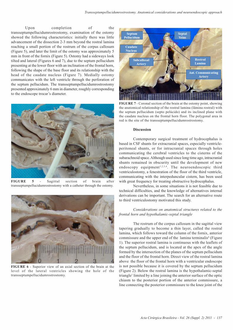
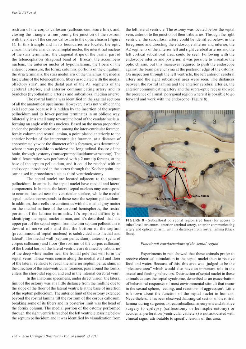
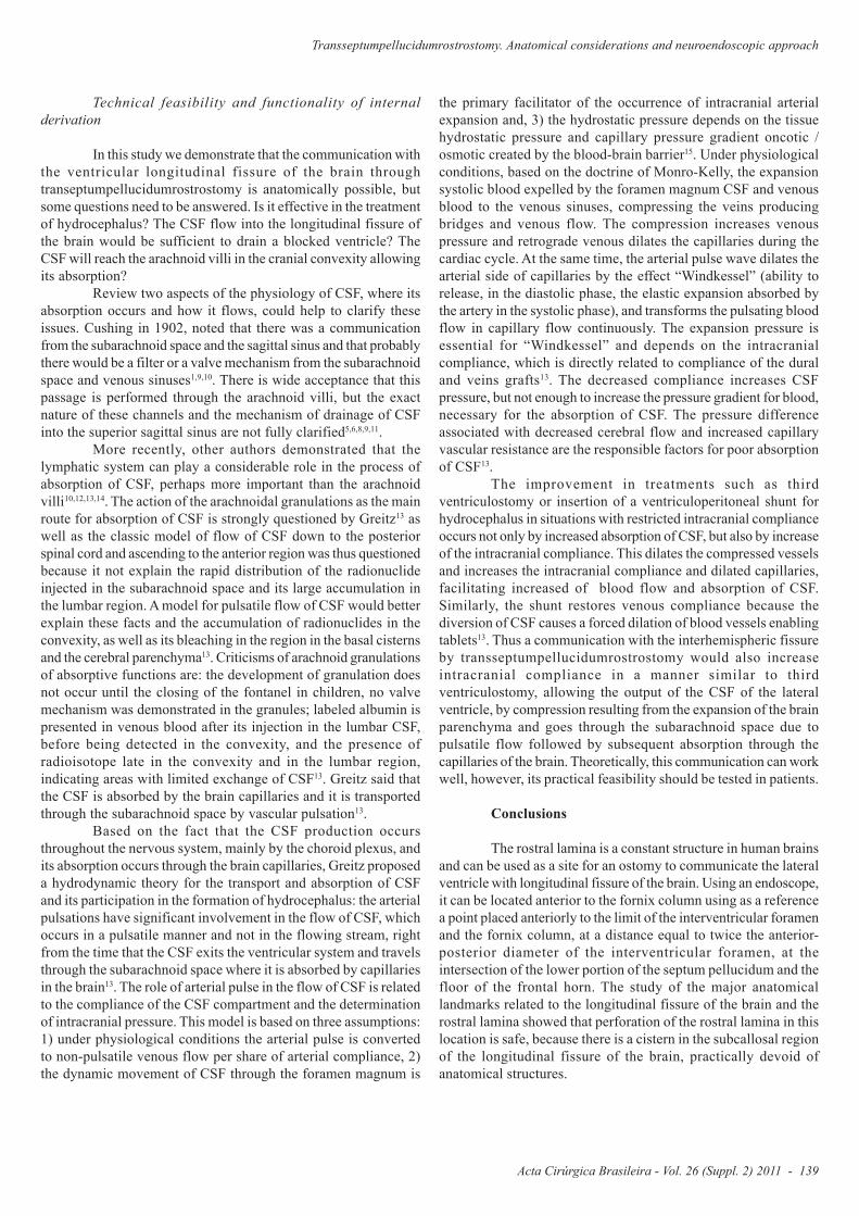
Upon completion of thetransseptumpellucidumrostrostomy, examination of the ostomyshowed the following characteristics: initially there was littleadvancement of the dissection 2-3 mm beyond the rostral laminareaching a small portion of the rostrum of the corpus callosum(Figure 5), and later the limit of the ostomy was approximately 5mm in front of the fornix (Figure 5). Ostomy had a sideways looktilted and lateral (Figures 6 and 7), due to the septum pellucidumpresenting at the lower floor with an inclination of the frontal horn,following the shape of the base floor and its relationship with thehead of the caudate nucleus (Figure 7). Medially ostomycommunicates with the left ventricle through the perforation ofthe septum pellucidum. The transseptumpellucidumrostrostomypresented approximately 6 mm in diameter, roughly correspondingto the endoscope trocar’s diameter.
FIGURE 5 - Sagittal section of brain aftertransseptumpellucidumrostrostomy with a catheter through the ostomy.
FIGURE 6 - Superior view of an axial section of the brain at thelevel of the lateral ventricles showing the hole of thetransseptumpellucidumrostrostomy.
FIGURE 7 - Coronal section of the brain at the ostomy point, showingthe anatomical relationship of the rostral lamina (lâmina rostral) withthe septum pellucidum (septo pelúcido) and its inclined plane withthe caudate nucleus on the frontal horn floor. The polygonal area inred is the site of the transseptumpellucidumrostrostomy.
Discussion
Contemporary surgical treatment of hydrocephalus isbased in CSF shunts for extracranial spaces, especially ventricle-peritoneal shunts, or for intracranial spaces through holescommunicating the cerebral ventricles to the cisterns of thesubarachnoid space. Although used since long time ago, intracranialshunts remained in obscurity until the development of newendoscopy equipment1,2,3,4. The neuroendoscopic thirdventriculostomy, a fenestration of the floor of the third ventricle,communicating with the interpeduncular cistern, has been usedwith great frequency for treating obstructive hydrocephalus.
Nevertheless, in some situations it is not feasible due totechnical difficulties, and the knowledge of alternatives internalderivations can be important. The search for an alternative routeto third ventriculostomy motivated this study.
Considerations on anatomical structures related to thefrontal horn and hypothalamic-septal triangle
The rostrum of the corpus callosum in the sagittal viewtapering gradually to become a thin layer, called the rostrallamina, which follows toward the column of the fornix, anteriorcommissure and the upper end of the lamina terminalis6 (Figure1). The superior rostral lamina is continuous with the leaflets ofthe septum pellucidum, and is located at the apex of the angleformed by the intersection of the planes of the septum pellucidumand the floor of the frontal horn. Direct view of the rostral laminaabove the floor of the frontal horn with a ventricular endoscopeis not possible because it is covered by the septum pellucidum(Figure 2). Below the rostral lamina is the hypothalamic-septaltriangle1 limited by a line joining the anterior surface of the opticchiasm to the posterior portion of the anterior commissure, aline connecting the posterior commissure to the knee joint of the
Fuziki EJT et al.
138 - Acta Cirúrgica Brasileira - Vol. 26 (Suppl. 2) 2011
rostrum of the corpus callosum (callosus-comissure line), and,closing the triangle, a line joining the junction of the rostrumwith the knee of the corpus callosum to the optic chiasm (Figure1). In this triangle and in its boundaries are located the opticchiasm, the lateral and medial septal nuclei, the interstitial nucleusof the stria terminalis, the diagonal stripe of the basilar part ofthe telencephalon (diagonal band of Broca), the accumbensnucleus, the anterior nuclei of hypothalamus, the fibers of theanterior comissure, the fornix columns, portions of the cingulum,the stria terminalis, the stria medullaris of the thalamus, the medialfasciculus of the telencephalon, fibers associated with the medialolfactory stria6, and the distal part of the A1 segments of thecerebral arteries, and anterior communicating artery and itsbranches (hypothalamic arteries and subcallosal median artery).
The rostral lamina was identified in the sagittal sectionsof all the anatomical specimens. However, it was not visible in theaxial sections because it is hidden by the insertion of the septumpellucidum and its lower portion terminates in an oblique way,bilaterally, in a small ramp toward the head of the caudate nucleus,forming an angle with this nucleus. Based on the mean proportionand on the positive correlation among the interventricular foramen,fornix column and rostral lamina, a point placed anteriorly to theanterior border of the interventricular foramen, at a distance ofapproximately twice the diameter of this foramen, was determined,where it was possible to achieve the longitudinal fissure of thebrain, through a ostomy (transseptumpellucidumrostrostomy). Theinitial fenestration was performed with a 2 mm tip forceps, at thebase of the septum pellucidum, and it could be reached with anendoscope introduced in the cortex through the Kocher point, thesame used in procedures such as third ventriculostomy.
The septal nuclei are located adjacent to the septumpellucidum. In animals, the septal nuclei have medial and lateralcomponents. In humans the lateral septal nucleus may correspondto neurons located near the ventricular surface, while the medialseptal nucleus corresponds to those near the septum pellucidum7.In addition, these cells are continuous with the medial gray matteron the medial surface of the cerebral hemispheres, the rostralportion of the lamina terminalis. It’s reported difficulty inidentifying the septal nuclei in man, and it’s described that theupper part of the septal region from the thin septum pellucidum isdevoid of nerve cells and that the bottom of the septum(precommissural septal nucleus) is subdivided into medial andlateral8. The medial wall (septum pellucidum), anterior (genu ofcorpus callosum) and floor (the rostrum of the corpus callosum)of the frontal horn of the lateral ventricle are drained by tributariesof the deep white matter near the frontal pole that will form theseptal veins. These veins course along the medial wall and floorof the lateral ventricle to reach the anterior septum pellucidum, inthe direction of the interventricular foramen, pass around the fornix,enters the choroidal region and end in the internal cerebral vein3.
In the anatomic specimens, under direct vision, the laterallimit of the ostomy was at a little distance from the midline due tothe slope of the floor of the lateral ventricle at the base of insertionof the septum pellucidum. The anterior limit of the ostomy extendedbeyond the rostral lamina till the rostrum of the corpus callosum,breaking some of its fibers and its posterior limit was the head ofthe fornix column. The medial portion of the ostomy performedthrough the right ventricle reached the left ventricle, passing belowthe septum pellucidum and it was identified by visualization from
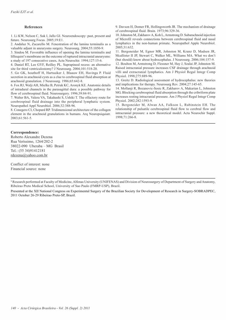
the left lateral ventricle. The ostomy was located below the septalvein, anterior to the junction of their tributaries. Through the rightventricle, the subcallosal artery could be identified below, in theforeground and directing the endoscope anterior and inferior, theA2 segments of the anterior left and right cerebral arteries and theleft cortical subcallosal area, could be seen. Following with theendoscope inferior and posterior, it was possible to visualize theoptic chiasm, but this maneuver required to push the endoscopeagainst the brain parenchyma at the posterior edge of the ostomy.On inspection through the left ventricle, the left anterior cerebralartery and the right subcallosal area were seen. The distancesbetween the rostral lamina and the anterior cerebral arteries, theanterior communicating artery and the supra-optic recess showedthe presence of a small polygonal region where it is possible to goforward and work with the endoscope (Figure 8).
FIGURE 8 - Subcallosal polygonal region (red lines) for access tosubcallosal structures: anterior cerebral artery, anterior communicatingartery and optical chiasm, with its distances from rostral lamina (blacklines).
Functional considerations of the septal region
Experiments in rats showed that these animals prefer toreceive electrical stimulation in the septal nuclei than to receivefood and water. Because of this, this area was judged to be the“pleasure area” which would also have an important role in thesexual and feeding behaviors. Destruction of septal nuclei in theseanimals causes the septal syndrome, described as an exacerbationof behavioral responses of most environmental stimuli that occurin the sexual sphere, feeding, and reactions of aggression3. Littleis known about the function of the septal nuclei in humans.Nevertheless, it has been observed that surgical section of the rostrallamina during surgeries to treat subcallosal aneurysms and ablativesurgery to epilepsy (callosotomy or hemispherectomy) oraccidental perforation (ventricular catheters) is not associated withclinical signs attributable to specific lesions of this area.
Acta Cirúrgica Brasileira - Vol. 26 (Suppl. 2) 2011 - 139
Transseptumpellucidumrostrostomy. Anatomical considerations and neuroendoscopic approach
Technical feasibility and functionality of internalderivation
In this study we demonstrate that the communication withthe ventricular longitudinal fissure of the brain throughtranseptumpellucidumrostrostomy is anatomically possible, butsome questions need to be answered. Is it effective in the treatmentof hydrocephalus? The CSF flow into the longitudinal fissure ofthe brain would be sufficient to drain a blocked ventricle? TheCSF will reach the arachnoid villi in the cranial convexity allowingits absorption?
Review two aspects of the physiology of CSF, where itsabsorption occurs and how it flows, could help to clarify theseissues. Cushing in 1902, noted that there was a communicationfrom the subarachnoid space and the sagittal sinus and that probablythere would be a filter or a valve mechanism from the subarachnoidspace and venous sinuses1,9,10. There is wide acceptance that thispassage is performed through the arachnoid villi, but the exactnature of these channels and the mechanism of drainage of CSFinto the superior sagittal sinus are not fully clarified5,6,8,9,11.
More recently, other authors demonstrated that thelymphatic system can play a considerable role in the process ofabsorption of CSF, perhaps more important than the arachnoidvilli10,12,13,14. The action of the arachnoidal granulations as the mainroute for absorption of CSF is strongly questioned by Greitz13 aswell as the classic model of flow of CSF down to the posteriorspinal cord and ascending to the anterior region was thus questionedbecause it not explain the rapid distribution of the radionuclideinjected in the subarachnoid space and its large accumulation inthe lumbar region. A model for pulsatile flow of CSF would betterexplain these facts and the accumulation of radionuclides in theconvexity, as well as its bleaching in the region in the basal cisternsand the cerebral parenchyma13. Criticisms of arachnoid granulationsof absorptive functions are: the development of granulation doesnot occur until the closing of the fontanel in children, no valvemechanism was demonstrated in the granules; labeled albumin ispresented in venous blood after its injection in the lumbar CSF,before being detected in the convexity, and the presence ofradioisotope late in the convexity and in the lumbar region,indicating areas with limited exchange of CSF13. Greitz said thatthe CSF is absorbed by the brain capillaries and it is transportedthrough the subarachnoid space by vascular pulsation13.
Based on the fact that the CSF production occursthroughout the nervous system, mainly by the choroid plexus, andits absorption occurs through the brain capillaries, Greitz proposeda hydrodynamic theory for the transport and absorption of CSFand its participation in the formation of hydrocephalus: the arterialpulsations have significant involvement in the flow of CSF, whichoccurs in a pulsatile manner and not in the flowing stream, rightfrom the time that the CSF exits the ventricular system and travelsthrough the subarachnoid space where it is absorbed by capillariesin the brain13. The role of arterial pulse in the flow of CSF is relatedto the compliance of the CSF compartment and the determinationof intracranial pressure. This model is based on three assumptions:1) under physiological conditions the arterial pulse is convertedto non-pulsatile venous flow per share of arterial compliance, 2)the dynamic movement of CSF through the foramen magnum is
the primary facilitator of the occurrence of intracranial arterialexpansion and, 3) the hydrostatic pressure depends on the tissuehydrostatic pressure and capillary pressure gradient oncotic /osmotic created by the blood-brain barrier15. Under physiologicalconditions, based on the doctrine of Monro-Kelly, the expansionsystolic blood expelled by the foramen magnum CSF and venousblood to the venous sinuses, compressing the veins producingbridges and venous flow. The compression increases venouspressure and retrograde venous dilates the capillaries during thecardiac cycle. At the same time, the arterial pulse wave dilates thearterial side of capillaries by the effect “Windkessel” (ability torelease, in the diastolic phase, the elastic expansion absorbed bythe artery in the systolic phase), and transforms the pulsating bloodflow in capillary flow continuously. The expansion pressure isessential for “Windkessel” and depends on the intracranialcompliance, which is directly related to compliance of the duraland veins grafts13. The decreased compliance increases CSFpressure, but not enough to increase the pressure gradient for blood,necessary for the absorption of CSF. The pressure differenceassociated with decreased cerebral flow and increased capillaryvascular resistance are the responsible factors for poor absorptionof CSF13.
The improvement in treatments such as thirdventriculostomy or insertion of a ventriculoperitoneal shunt forhydrocephalus in situations with restricted intracranial complianceoccurs not only by increased absorption of CSF, but also by increaseof the intracranial compliance. This dilates the compressed vesselsand increases the intracranial compliance and dilated capillaries,facilitating increased of blood flow and absorption of CSF.Similarly, the shunt restores venous compliance because thediversion of CSF causes a forced dilation of blood vessels enablingtablets13. Thus a communication with the interhemispheric fissureby transseptumpellucidumrostrostomy would also increaseintracranial compliance in a manner similar to thirdventriculostomy, allowing the output of the CSF of the lateralventricle, by compression resulting from the expansion of the brainparenchyma and goes through the subarachnoid space due topulsatile flow followed by subsequent absorption through thecapillaries of the brain. Theoretically, this communication can workwell, however, its practical feasibility should be tested in patients.
Conclusions
The rostral lamina is a constant structure in human brainsand can be used as a site for an ostomy to communicate the lateralventricle with longitudinal fissure of the brain. Using an endoscope,it can be located anterior to the fornix column using as a referencea point placed anteriorly to the limit of the interventricular foramenand the fornix column, at a distance equal to twice the anterior-posterior diameter of the interventricular foramen, at theintersection of the lower portion of the septum pellucidum and thefloor of the frontal horn. The study of the major anatomicallandmarks related to the longitudinal fissure of the brain and therostral lamina showed that perforation of the rostral lamina in thislocation is safe, because there is a cistern in the subcallosal regionof the longitudinal fissure of the brain, practically devoid ofanatomical structures.
Fuziki EJT et al.
140 - Acta Cirúrgica Brasileira - Vol. 26 (Suppl. 2) 2011
References
1. Li KW, Nelson C, Suk I, Jallo GI. Neuroendoscopy: past, present andfuture. Neurosurg Focus. 2005;19:E1.2. Andaluz N, Zucarello M. Fenestration of the lamina terminalis as avaluable adjunt in aneurysms surgery. Neurosurg. 2004;55:1050-9.3. Sindou M. Favorable influence of opening the lamina terminalis andlilliequist’s membrane on the outcome of ruptured intracranial aneurysms:a study of 197 consecutive cases. Acta Neurochir. 1994;127:15-6.4. Daniel RT, Lee GYF, Reilley PL. Suprapineal recess: an alternativesite for third ventriculostomy? J Neurosurg. 2004;101:518-20.5. Go GK, houthoff H, Hartsuiker J, Blaauw EH, Havinga P. Fluidsecretion in arachnoid cysts as a clue to cerebrospinal fluid absorption atarachnoid granulation. J Neurosurg. 1986;65:642-8.6. Fox RJ, Walji AH, Mielke B, Petruk KC, Aronyk KE. Anatomic detailsof intradural channels in the parasagittal dura: a possible pathway forflow of cerebrospinal fluid. Neurosurgery. 1996;39:84-91.7. Walter BA, Valera VA, Takahashi S, Ushiki T. The olfactory route forcerebrospinal fluid drainage into the peripheral lymphatic system.Neuropathol Appl Neurobiol. 2006;32:388-96.8. Conegero CI, Chopard RP. Tridimensional architecture of the collagenelement in the arachnoid granulations in humans. Arq Neuropsiquiatr.2003;61:561-5.
9. Davson H, Domer FR, Hollingsworth JR. The mechanism of drainageof cerebrospinal fluid. Brain. 1973;96:329-36.10. Johnston M, Zakharov A, Koh L, Armstrong D. Subarachnoid injectionof Microfil reveals connections between cerebrospinal fluid and nasallymphatics in the non-human primate. Neuropathol Apple Neurobiol.2005;31:632.11. Bergsneider M, Egnor MR, Johnston M, Kranz D, Madsen JR,Mcallister II JP, Stewart C, Walker ML, Williams MA. What we don’t(but should) know about hydrocephalus. J Neurosurg. 2006;104:157-9.12. Boulton M, Armstrong D, Flessner M, Hay J, Szalai JP, Johnston M.Raised intracranial pressure increases CSF drainage through arachnoidvilli and extracranial lymphatics. Am J Physiol Regul Integr CompPhysiol. 1998;275:889-96.13. Greitz D. Radiological assessment of hydrocephalus: new theoriesand implications for therapy. Neurosurg Rev. 2004;27:145-65.14. Mollanji R, Bozanovic-Sosic R, Zakharov A, Makarian L, JohnstonMG. Blocking cerebrospinal fluid absorption through the cribriform plateincreases resting intracranial pressure. Am J Physiol Regul Integr CompPhysiol. 2002;282:1593-9.15. Bergsneider M, Alwan AA, Falkson L, Rubinstein EH. Therelationship of pulsatile cerebrospinal fluid flow to cerebral flow andintracranial pressure: a new theoretical model. Acta Neurochir Suppl.1998;71:266-8.
Correspondence:Roberto Alexandre DezenaRua Veríssimo, 1264/202-238022-090 Uberaba – MG BrasilTel.: (55 34)[email protected]
Conflict of interest: noneFinancial source: none
1 Research performed at Faculty of Medicine, Alfenas University (UNIFENAS) and Division of Neurosurgery of Department of Surgery and Anatomy,Ribeirao Preto Medical School, University of Sao Paulo (FMRP-USP), Brazil.Presented at the XII National Congress on Experimental Surgery of the Brazilian Society for Development of Research in Surgery-SOBRADPEC,2011 October 26-29 Ribeirao Preto-SP, Brazil.