revista de anestesia regional e terapÊutica da … · resumo: contêm informação sucinta sobre o...
TRANSCRIPT

Junho | June 2011
PUBLICAÇÃO TRIMESTRAL
QUARTERLY PUBLICATION
ANO | YEAR XVIII
N.º 64
REVISTA DE ANESTESIA REGIONALE TERAPÊUTICA DA DOR
JOURNAL OF REGIONAL ANAESTHESIAAND PAIN TREATMENT
REVISTA OFICIAL DO CLUBE DE ANESTESIA REGIONAL / ESRA PORTUGAL
OFFICIAL JOURNAL OF CLUBE DE ANESTESIA REGIONAL / ESRA PORTUGAL
CLUBE DE ANESTESIA REGIONAL / ESRA PORTUGAL

clube de anestesia regional
O CAR está na eminência de ter de suspender a edição da Revista, por não haver capacidade financeira para suportar o défice de cada número
que se eleva neste momento a mais de 2.500 euros.
Os patrocinadores habituais, a Indústria Farmacêutica, estão a reduzir o seu investimento em publicidade, e alguns retiraram mesmo
os seus anúncios da Revista
Se todos os associados do CAR (mais de 1.300) colaborarem, com o pagamento das suas quotas, o problema fica resolvido.
A quota é de 25 euros anuais, que pode ser paga no Multibanco ou por Transferência Bancária para o NIB 00320-12300-20150-291-940.
Agradecemos que nos seja enviada uma mensagem, para nos informar do pagamento, para
[email protected] ou para [email protected]
O recibo será posteriormente enviado por correio.
É indispensável a colaboração de todos, para se poder manter a publicação da Revista que é de todos.

Junho 2011 | June 2011Revista de Anestesia Regional e Terapia da Dor | Journal of Regional Anaesthesia and Pain Treatment 1
www.anestesiaregional.com
Director | DirectorSobral de Campos
Directores Adjuntos | Adjunct DirectorsAna ValentimDuarte Correia
Editores Regionais | Regional EditorsTeresa FerreiraHenrique GonçalvesJoana CarvalhasJosé RomãoSandra Gestosa
Editores em Espanha | Spanish EditorsLídia Castro FreitasLuis AliagaMaria Jose MorisMiguel CaramésElena Segura (Hospital Viseu)
Conselho Científico | Scientific CounselAnabela Roncon Roxo Anestesista, Maternidade Alfredo da CostaArmando Almeida Biólogo, Universidade do Minho, PhdJoão Paulo Barbot Anestesista, Hospital da PreladaJoão Mota Dias Farmacêutico, LisboaJoão Santos Pereira Anestesista, LisboaJosé De Andrés Anestesista, Universidade de ValênciaJosé Luís Portela Anestesista, IPO LisboaLuis Aliaga Anestesista, BarcelonaMaria Rui Crisóstomo Anestesista, Hospital de BragaMathieu Gielen Anestesista, HolandaMariana Jorge Sousa Jurista, LisboaMiguel Caramés Anestesista, Gran CanáriaNarinder Rawal Anestesista, Orebro, SuéciaPedro Ponce Nefrologista, Hospital Garcia de OrtaReinaldo Cabanita Anestesista, Hospital de SantarémRita Oliveira Farmacêutica, Faculdade de Farmácia de LisboaSuzette Morais Anestesista, Hospital Fernando da FonsecaVictor Coelho Anestesista, Hospitais da Universidade de CoimbraZeferino Bastos Anestesista, Porto
Boletim DOR | PAIN BulletinDirector | Director Laurinda Lemos
REVISTA CAR | CAR JOURNALPropriedade | Publisher Clube de Anestesia Regional /ESRA Portugal
NIF | VAT 502 687 541Sede Social | Headquarters
Praceta Rita Ferreira da Silva, Nº 44 Edifício 8 R/c Esq. - 2755 - 075 ALCABIDECHECorrespondência | Mail AddressApartado 214 – 2776-903 Carcavelos
Fax: 351-21 925 01 09 E-mail: [email protected]: www.anestesiaregional.com
Depósito Legal: 142340/99 ISSN: 0872-5888Peridiocidade | Published: trimestral / quarterly
Pré-impressão e Impressão | Printers:QUADRICOR - artes gráficas, lda.
Rua Comandante Oliveira e Carmo, 18-C • Cova da Piedade, 2805-212 AlmadaTel.: 21 274 46 07 • Fax: 21 274 31 90 • NIPC: 501 388 532
[email protected] • www.quadricor.ptTradução | Translation: Cristina Dias
Direcção | Board
Presidente | PresidentRui Sobral de Campos
Vice-Presidente | Vice PresidentReinaldo Coelho Cabanita
Secretário Geral | General SecretaryJoão Paulo Barbot
Tesoureiro | TreasurerJosé Cordeiro Veiga
Vogais | DelegatesAna Preto MarcosFrancisco Duarte CorreiaJosé Peralta
Assembleia Geral | General Assembley
Presidente | PresidentEdgar Ribeiro Lopes
Secretário | SecretaryManuel Costa de Sousa
Vogal | DelegateRui Manuel Araújo
Conselho Fiscal | Fiscal Counsel
Presidente | PresidentMargarida Faro
Vogais | DelegatesAna do Rosário ValentimJoana Carvalhas
Delegados | Delegates
Luísa GomesAçores
Francisco Duarte CorreiaMadeira
Junho | June 2011
PUBLICAÇÃO TRIMESTRAL
QUARTERLY PUBLICATION
ANO | YEAR XVIII
N.º 64
REVISTA DE ANESTESIA REGIONAL
E TERAPÊUTICA DA DOR
JOURNAL OF REGIONAL ANAESTHESIA
AND PAIN TREATMENT
REVISTA OFICIAL DO CLUBE DE ANESTESIA REGIONAL / ESRA PORTUGAL
OFFICIAL JOURNAL OF CLUBE DE ANESTESIA REGIONAL / ESRA PORTUGAL
CLUBE DE ANESTESIA REGIONAL / ESRA PORTUGAL

Revista de Anestesia Regional e Terapia da Dor | Journal of Regional Anaesthesia and Pain Treatment2Junho 2011 | June 2011
NORMAS DE PUBLICAÇãOGUIDE FOR AUthORS
Guide for AuthorsCAr JourNAL publishes manuscripts (original articles, clinical cases, clinical trials, review articles, short communications and letters to the editor) relevant to local/regional anaesthesia and/or pain management.CAr JourNAL welcomes all physicians, members or not members of the Clube de Anestesia Regional, who wish to publish in CAr JourNAL.Manuscripts submitted must not have been previously published or submitted simultaneously to other(s) publication(s), except if previous agreed with th Executive Director.All manuscripts, except letters to the editor, will be reviewed by the Editor-in-Chief and/or members of the Editorial Board. Once accepted, they become property of the CAr JourNAL and can only be reproduced with permission.
ethiCsAll experimental work should be in accordance with the ethical standards of the Helsinki Declaration guidelines for research in animals or in humans.
MANusCripts prepArAtioN ANd subMissioNThey could be submitted as follows:1. In Cd, or by email using an established word processor, not as a PDF file. All the pages should be numbered serially. If possible, is advisable Microsoft Word. Images must be sent separately as JPEG or TIFF files.2. CAR Email: [email protected]
oriGiNAL ArtiCLesManuscript StructureTitle: The title of the article, this should be short and concise.Abstract: Should not exceed 250 words and should describe the background, the aims, and the conclusions reached. It should contain only standard abbreviations and no references.Key words: Maximum 6.Methodology: Should describe the methods, so that the experiment can be easy interpreted or reproduced by the reader. Regarding the statistical analysis the method should be identified.Results: The results presented should be clear. If possible, the results should be accompanied by confidence intervals and exact level of statistical significance.Conclusions: Describe only the findings that are based on the results obtained, its clinical application, or if is required further investigation. Equal emphasis should be given to positive and/or negative results that have scientific merit.
review ArtiCLesManuscript StructureTitle: The title of the article, this should be short and concise.Abstract: Should not exceed 250 words and should describe the background, the aims, and the conclusions reached. It should contain only standard abbreviations and no references.Key words: Maximum 6.Objective: Description of the main objective.Method: Describe the surveyed sources. Identify the number of review studies and criteria for their selection.Results: Describe the main results and the methods used to obtain them.Conclusions: Describe the main findings and their clinical application. Suggest areas for further investigation if necessary.
refereNCe forMAt: List the references by the order they are mentioned in the manuscript, using Arabic characters. References to cited materials should be listed at the end of the article.EXAMPLE: 1. Vandam LD, Dripps RD. Long-term foolow-up of patientes who received 10,098 spinal anesthetics. JAMA 1956: 161: 586-591.
NorMAs de pubLiCAÇÃoA CAr revistA publica manuscritos (artigos originais, casos clínicos, artigos de revisão, comunicação e correspondência) que sejam relevantes nos campos da anestesia local, anestesia regional e tratamento da dor.A CAr revistA tem as suas páginas abertas a todos os médicos, sócios ou não, do Clube de Anestesia Regional.Os manuscritos enviados para publicação não devem ter sido já publicados, ou propostos simultaneamente em qualquer outra parte, excepto após acordo com a direcção da Revista.Todos os manuscritos, excepto a correspondência são revistos pelos Editores Executivos e/ou por membros do Conselho Cientifico. Uma vez aceites, ficam propriedade da revista, só podendo ser reproduzidos com a sua autorização.
CoNsiderAÇões LeGAisOs artigos baseados em investigação clínica no Homem, devem explicar que os ensaios foram conduzidos segundo as normas éticas da declaração de Helsínquia.
prepArAÇÃo dos MANusCritosDevem ser enviados da seguinte forma:1. em Cd ou por email, utilizando qualquer processador de texto para PC ou Apple. Se possivel, aconselha-se o Microsoft Word ©. As imagens, devem ser enviadas separadamente do texto preferencialmente nos formatos JPEG ou TIFF.2. O endereço electrónico do CAR é: [email protected]
ArtiGo oriGiNALTítulo: deve ser curto e conciso.Resumo: contêm informação sucinta sobre o objectivo, metodologia, resultados e conclusões, com um máximo de 250 palavras.Palavra Chave: máximo de 6.Metodologia: devem ser descritos os métodos utilizados, de modo a que a experiência possa ser interpretada e reproduzida pelo leitor. No que se refere à análise estatística deve ser referido o método utilizado.Resultados: a apresentação dos resultados deve ser feita de forma clara. Se possível, os resultados devem ser acompanhados por intervalos de confiança e o nível exacto de significância estatística.Conclusões: descreva somente as conclusões do estudo que têm base nos resultados obtidos, assim como a sua aplicação clínica, ou se é necessária maior investigação. Deve ser dado igual ênfase a resultados positívos e negativos que tenham mérito científico.
ArtiGo de revisÃoObjectivo: descreva o objectivo principal.Método: descreva as fontes pesquisadas. Identifique o número de estudos de revisão e o critério para essa selecção.Resultados: descreva os principais resultados da revisão e os métodos utilizados para obter esses resultados.Conclusões: descreva as principais conclusões e a sua aplicação clínica. Sugira áreas para futura investigação se necessário.
referêNCiAs: Numere as referências pela ordem em que são mencionadas no texto, usando numeração árabe.EXEMPLO: 1. Vandam LD, Dripps RD. Long-term foolow-up of patientes who received 10,098 spinal anesthetics. JAMA 1956: 161: 586-591.

4 Envio da nossa revista trimestral, a publicação de maior difusão da especialidade
4 Inscrições com preços mais baixos em todas as nossas iniciativas
4 valor da quota anual: 25€
Inscreva-se no e beneficie das vantagens de Associado
Preencha e remeta a proposta de associado(no verso desta página) com pagamento por multibanco para o
NIB: 00320 12300 20150 291 940
Seja sócio de uma das maisdinâmicas associações representativas de
Anestesiologia Portuguesa

Os dados pessoais estão protegidos pela lei, não sendo permitida a sua divulgação a terceiros, a não ser para fins idênticos, ou seja divulgação de congressos ou outras iniciativas que tenham interesse para os associados e mesmo neste caso só com expressa autorização do interessado. Quotização anual: 25€
Personal data is protected by law and it isn’t permitted to disclosure third parties, except for identical, or disclosure of congresses or other initiatives of interest to members and even in this case only if with express authorization of the person concerned. Annual fee: 25€
Proposta de AssociadoMembership Offer
Nome | Name:
Morada | Address:
Código Postal | Postal Code:
Telefone | Telephone: Fax | Fax:
Hospital | Hospital:
$
Correspondência | Mail Address: Apartado 214 – 2776 - 903 CARCAVELOS PORTUGALFax: +351 21 925 01 09
NIB: 00320 12300 20150 291 940SWIFT: PT00320 12300 20150 291 940
autorizo a divulgação dos meus dados pessoaisI authorize the release of my personal data
Não autorizo a divulgação dos meus dados pessoaisI do not authorize disclosure of my personal data
c
c
Local e data
Assinatura
City and Date
Signature
Pode ser fotocopiado ou digitalizadocan be photocopied or scanned

Revista de Anestesia Regional e Terapia da Dor | Journal of Regional Anaesthesia and Pain Treatment6Junho 2011 | June 2011
Índice
índice
contents
EditorialSobral de Campos
7
Avaliação Neurológica em Anestesia Loco-RegionalA Silva; A Guedes; JP Assunção
11
Avaliação Pós-Operatória de Doentes Submetidos a Anestesia Loco-RegionalFigueiredo E.; Ribeiro S.; Arede MJ; Assunção JP
16
Complicações em Anestesia Locorregional – uma actualizaçãoVítor Miguel Oliveira; Eduarda Figueiredo; José Pedro Assunção
22
Registo em Anestesia Loco-RegionalCarvalho, Rita; Ferreira, Tatiana; Pinto, Joana; Carvalho, Joana; Loureiro, M. C.; Arede, M. J.; Assunção, J. P.
31
Nevralgia do TrigémioMargarida Faria; Francisco Correia; Cláudia Barbosa
37
Reflexão sobre a Problemática da Dor AgudaInês Bolina
46
Agenda 58
EditorialSobral de Campos
8
Neurological Assessment in Loco-Regional AnaesthesiaA Silva; A Guedes; JP Assunção
14
Postoperative Evaluation of Patients Submitted to Loco-Regional AnaesthesiaFigueiredo E.; Ribeiro S.; Arede MJ; Assunção JP
18
Complications in Loco-Regional Anaesthesia - an updateVítor Miguel Oliveira; Eduarda Figueiredo; José Pedro Assunção
26
Loco-Regional Anaesthesia RecordsCarvalho, Rita; Ferreira, Tatiana; Pinto, Joana; Carvalho, Joana; Loureiro, M. C.; Arede, M. J.; Assunção, J. P.
36
Trigeminal NeuralgiaMargarida Faria; Francisco Correia; Cláudia Barbosa
42
Reflection on the Acute Pain ProblemInês Bolina
48
Calendar of Events 58
Table of Contents

Junho 2011 | June 2011Revista de Anestesia Regional e Terapia da Dor | Journal of Regional Anaesthesia and Pain Treatment 7
Caros associados,
Este número da nossa Revista é sobretudo dedicado ao 38º Sábado do CAR, que se realizou no Hospital de S. Teotónio em Viseu em 19 de Fevereiro passado.
Foi uma excelente reunião com várias dezenas de participantes, dedicada aos bloqueios periféricos e à utilização da ultrassonografia, com particular ênfase nas complicações neurológicas que, não obstantes as preocupações de segurança que a boa prática recomenda, vão no entanto aparecendo em várias estatísticas, e é de realçar o grande cuidado que o Serviço de Anestesia deste Hospital coloca na sua prática, facto que ficou bem patente nas comunicações apresentadas.
A Silva, A Guedes e JP Assunção abordam a avaliação neurológica em anestesia loco-regional através de uma pesquisa efectuada na Pubmed e na Md Consult, fazendo um levantamento das manifestações clínicas relacionadas com lesões neurológicas na prática da anestesia loco-regional.
Figueiredo E, Ribeiro S, Arede MJ e Assunção JP falam-nos na mesma linha na avaliação pós-operatória do doente submetido a anestesia loco-regional no registo do próprio Hospital de S. Teotónio abrangendo um período de 7 meses.
Vitor Miguel Oliveira, Eduarda Figueiredo e José Pedro Assunção abordam o tema das complicações em ALR fazendo uma actualização.
Rita Carvalho, Tatiana Ferreira, Joana Pinto, Joana Carvalho, Loureiro MC, Arede MJ e Assunção JP falam-nos do registo em anestesia loco-regional fundamentando a necessidade do respectivo registo.
De uma área diferente os Médicos Dentistas, Margarida Faria, Francisco Correia e Cláudia Barbosa descrevem uma importante e incapacitante manifestação dolorosa a nevralgia do trigémio, numa análise bem conseguida do síndroma.
A fechar este número a Enfermeira Inês Bolina do Hospital CUF Infante Santo aborda a necessidade de tratar com eficácia a dor aguda pós-operatória, um tema “velho” mas sempre actual, cuja solução terapêutica, não obstante a evidência de recentes melhorias, continua a não oferecer uma resposta completamente satisfatória.
Em 28 de Maio passado, no âmbito das comemorações do XX Aniversário do CAR realizou-se, num barco que partiu do Cais de Peso da Régua e subiu o Rio Douro até Barca D’Alva, o 39º Sábado do CAR onde foram debatidos alguns temas polémicos da anestesia loco-regional e terapêutica da dor.
O Programa Preliminar do XX Congresso do CAR encontra-se nas páginas interiores. O Congresso terá lugar a 29 e 30 de Setembro e 1 de Outubro no Hotel Sana Metropolitan em Lisboa.
Os associados do CAR que ainda não forneceram o endereço de e-mail vão ser contactados pelo correio para que o façam, pois é desejo da direcção quer por maior rapidez no contacto, quer para contenção de custos privilegiar o contacto electrónico. Podem fazê-lo para [email protected] ou para [email protected].
Desejo a todos uma boa leitura
Editorial
editorial
Rui Sobral de Campos

Revista de Anestesia Regional e Terapia da Dor | Journal of Regional Anaesthesia and Pain Treatment8Junho 2011 | June 2011
editorial
Dear members,
This number is especially dedicated to the 38th CAR Saturday which took place on February 19th at Hospital de St. Teotónio in Viseu.
It was an excellent meeting with several dozen participants, dedicated to peripheral blocks and ultrasonography, with particular emphasis on neurological complications witch appear in various statistics, despite the safety precautions recommended by good practice. It is worth noting the great importance given to its practice by the Anaesthesia Service of this Hospital, which was well demonstrated in their communications.
A Silva, A Guedes and JP Assunção address the neurological assessment in loco-regional anaesthesia through a search in Pubmed and Md Consult, making a survey of clinical manifestations related to neurological injuries in loco-regional anaesthesia.
Figueiredo E, Ribeiro S, Arede MJ and Assunção JP follow the same line of postoperative evaluation of patients undergoing loco-regional anaesthesia based on the Hospital de St. Teotónio records covering a 7 months period.
Vitor Miguel Oliveira, Eduarda Figueiredo and José Pedro Assunção address the LRA complications with an update.
Rita Carvalho, Tatiana Ferreira, Joana Pinto, Joana Carvalho, Loureiro MC, Arede MJ and Assunção JP wrote about loco-regional anaesthesia records, emphasizing the need for its recording.
From a different area, the dentists Margarida Faria, Francisco Correia and Cláudia Barbosa describe an important, painful and disabling condition, the trigeminal neuralgia, through a good analysis of the syndrome.
Last but not the least, the nurse Inês Bolina from Hospital CUF Infante Santo tells us about the need to effectively treat acute pain after surgery. It is an “old” but still relevant issue witch therapeutic solution, despite recent improvements, still does not provide a complete satisfactory answer.
On May 28th, as part of the twentieth anniversary of CAR and from the quayside of Peso da Régua, in a boat that went up Rio Douro till Barca D’Alva, the 39th CAR Saturday took place, where were discussed some controversial issues of loco-regional anaesthesia and pain therapy.
In this journal you can also find the Preliminary Program of the XX Congress of CAR. The Congress will be held on September 29 and 30 and October 1st in Sana Metropolitan Hotel in Lisbon.
CAR members who have not yet provided the email address will be contacted by post. Nevertheless we ask you please to provide us with that information, because the Direction wishes a greater speed in contact, and also cost control, with focus on the email contact. You can do it directly to: [email protected] or [email protected].
I wish you all a good reading
Editorial
Rui Sobral de Campos

Junho 2011 | June 2011Revista de Anestesia Regional e Terapia da Dor | Journal of Regional Anaesthesia and Pain Treatment 11
Introdução e Objectivos
A Anestesia Loco-Regional (ALR) tem assu mido preponderância na prática anes tésica, levando a um aumento da
experiência e do desenvolvimento técnico, permitindo uma maior eficácia analgésica no pós-operatório, reduzindo taxas de mortali-dade e morbilidade em associação com baixos custos.Tem-se afirmado como técnica segura e efi-caz, sendo rara a ocorrência de complicações graves. Para tal, exige do Anestesiologista conhecimento anatómico, domínio da técnica e identificação das possíveis complicações e factores de risco que potenciem as suas incidências.A avaliação clínica prévia é fundamental para a estratificação dos riscos inerentes à prática anestésica. Apesar das complicações neuroló-gicas serem raras, quando ocorrem podem ser devastadoras para o doente.Neste âmbito e no contexto da prática da ALR, ganha especial ênfase a necessidade de uma avaliação neurológica cuidada. Integrando uma actuação continuada, a avaliação de alte-rações neurológicas deverá incidir sobre três momentos: o pré, o intra e o pós-operatório.Este trabalho visa salientar a importância da vigilância neurológica na prática da ALR, assu-mindo um ponto de vista prático e procurando enfatizar a clínica de alarme associada à mani-festação de possíveis lesões neurológicas e suas causas.
Método
Este trabalho teve como base uma pesquisa feita usando a PubMed e MdConsult com a articulação dos termos “anestesia loco-regio-nal”, “anestesia regional”, “bloqueio do neu-roeixo”, “bloqueio de nervos periféricos”, “complicações neurológicas”e ”lesões neuro-lógicas”. O objectivo desta pesquisa foi fazer o levantamento de manifestações clínicas rela-cionadas com lesões neurológicas evidentes na prática da ALR.
Resultados e Discussão
Pré-operatório
O exame neurológico sumário feito durante a avaliação pré-anestésica, deverá ter a preo-cupação de identificar ou excluir a presença de alterações do estado de consciência, ante-cedentes de convulsões, alterações da sensi-bilidade (considerando disestesias, dor, assi-metrias sensoriais) e défices motores (com avaliação da força muscular e assimetrias motoras).
Intra-operatório
Posteriormente, durante a execução da téc-nica loco-regional e no período intra-ope-ratório, dever-se-á dar especial atenção às manifestações neurológicas que possam estar relacionadas directamente com a actividade anestésica, traduzindo possível mecanismo de lesão nervosa. Nesta fase, a colaboração do doente é fundamental, devendo ser incen-tivada a cooperação através da verbalização da possível sintomatologia desencadeada com a técnica.Na realização de um bloqueio do neuroeixo, as queixas de dor radicular durante a intro-dução de agulha (ou cateter), parestesias ou dor à injecção, serão motivos para suspender e reavaliar a técnica. Da mesma forma, na prá-tica de bloqueio de nervo periférico a presença de parestesias e dor à injecção manifestadas durante a execução, serão sinais de alarme.Em ambas as situações podem ocorrer sin-tomas e sinais de toxicidade sistémica pro-vocadas pela administração do anestésico local (sabor metálico, parestesias peri-bucais, alterações visuais/auditivas, cefaleias, convul-sões, alterações do estado de consciência) que devem ser precocemente identificados, permitindo agir minimizando o risco poten-cialmente fatal.
Pós-operatório
A maioria das manifestações no período pós--operatório, são devidas a lesões temporárias (resolução até aos 12 meses). As complicações graves são raras mas dramáticas, sendo impor-tante vigiar para as identificar precocemente.Associadas à realização do bloqueio do neu-roeixo, podem ocorrer manifestações neuro-lógicas por trauma directo, neurotoxicidade, complicações infecciosas, hemorragia ou isquémia medular.O trauma directo provocado pela agulha (ou cateter) pode envolver a medula, o cone medular ou uma raiz nervosa. Está associado, na maioria dos casos, a queixas de dor e/ou parestesias à inserção da agulha ou injecção do anestésico local (0,06% BSA e 0,016% blo-queios epidurais, Auroy e colaboradores). Na sequência do trauma directo, pode surgir Cefaleia pós-punção da duramater, geral-mente ao 2º dia após a técnica (1º ao 7º dia), caracterizada por dor intensa fronto-occipital, desencadeada com o ortostatismo, podendo ser acompanhada de rigidez da nuca, lom-balgias e náuseas, ou, mais raramente, surgir com envolvimento de pares craneanos ou espasmos musculares localizados. Na maioria dos casos, resolve espontaneamente por volta do 5º dia pós punção. Face à clínica, e para
diagnóstico diferencial com outras entidades, deverá excluir-se a presença de febre, fotofo-bia e sinais neurológicos focais.As manifestações neurológicas provocadas por neurotoxicidade devem-se a lesões químicas induzidas pela presença de anestésico local, conservantes ou antisépticos. O Síndroma da Cauda Equina (cujos casos mais severos se associam a raquianestesia con tínua) carac-teriza-se por incontinência uri nária e fecal, perda de sensibilidade perineal e por vezes diminuição da força muscular nos membros inferiores, com início após a reversão do blo-queio subaracnoideu (BSA). A recuperação é lenta e gradual, podendo demorar semanas/meses.Os Sintomas Neurológicos Transitórios (enti-dade outrora designada por “irritação radi-cular transitória”) correlacionam-se com todos os anestésicos locais, apesar da maior incidência ser atribuída à Lidocaína. Para além da toxicidade directa, contribuem, como fac-tores causais, os posicionamentos cirúrgicos com estiramento das estruturas nervosas, o trauma directo e os espasmos musculares. Manifesta-se por dor lombar de intensidade variável, intermitente, com irradiação para as nádegas e/ou membros inferiores seguindo o trajecto de L5-S1, bilateralmente, com início 12 a 24 horas após a realização do bloqueio, resolvendo espontaneamente até ao 4º dia. No entanto, a ausência de défices motores e alterações electrofisiológicas durante a mani-festação aguda, a resposta favorável a alguns fármacos anti-inflamatórios e relaxantes, têm questionado a atribuição da neurotoxicidade como principal etiologia.A Aracnoidite Adesiva (que surge por reacção proliferativa das leptomeninges com oblitera-ção do espaço subaracnoideu, podendo con-duzir ao aumento da pressão do líquor) apre-senta-se com um quadro de instalação mais lenta, surgindo dias ou semanas após o BSA, caracterizado por incontinência urinária e fecal, alterações da sensibilidade do períneo e membros inferiores, diminuição da força mus-cular/ paraplegia, podendo mesmo ser letal.O aparecimento de um quadro clínico de cefaleia intensa, rigidez da nuca, febre alta e alterações do estado de consciência deverá conduzir ao diagnóstico de Meningite, sendo a avaliação do líquido cefaloraquídeo manda-tória para a diferenciação da etiologia bacte-riana ou não. Na Meningite bacteriana o início da clínica é geralmente 48 horas após o blo-queio do neuroeixo, tendo indicação para dre-nagem cirúrgica e antibioterapia prolongada. A Meningite asséptica apresenta um início agudo da clínica (<24 horas após BSA) e é um quadro clínico autolimitado, geralmente com regressão espontânea nas primeiras 72 horas (máximo 1 semana).
Avaliação Neurológica em Anestesia Loco-Regional
A Silva1; A Guedes2; JP Assunção3
1. Interna Complementar de Anestesiologia do Hospital de S. Teotónio, EPE – Viseu; 2. Assistente Hospitalar Graduada de Anestesiologia do Hospital de S. Teotónio, EPE – Viseu; 3. Director de Serviço de Anestesiologia do Hospital de S. Teotónio, EPE – Viseu
4

Revista de Anestesia Regional e Terapia da Dor | Journal of Regional Anaesthesia and Pain Treatment12Junho 2011 | June 2011
Outra complicação infecciosa que pode ocor-rer é o Abcesso espinhal, cujo risco aumenta com perda de assépsia durante a técnica, cate-terização prolongada ou imunodepressão. A clínica surge habitualmente 24 a 72 horas após o bloqueio, apresentando lombalgia, febre, infecção no local da punção (ou inserção do cateter) e défices neurológicos (inicialmente com paralisia flácida e aumento dos reflexos tendinosos que evoluem para espasticidade e diminuição dos reflexos). O diagnóstico exige avaliação imagiológica por RM ou TC e o trata-mento requer drenagem cirúrgica e antibiote-rapia prolongada.O Hematoma espinhal é a única complicação da ALR cujo tratamento constitui uma emer-gência cirúrgica. A etiologia está associada a anomalias morfológicas da medula e coluna vertebral, alterações da hemostase, anticoa-gulação e dificuldades técnicas. O quadro ins-tala-se subitamente, lombalgia com irradiação para os membros inferiores, diminuição da força muscular/ paralisia flácida, parestesias, abolição dos reflexos tendinosos e inconti-nência de esfíncteres, exigindo avaliação ima-giológica por RM ou TAC e descompressão cirúrgica célere.Por último, tendo a isquémia como factor cau-sal, o Síndroma da artéria espinhal anterior ocorre de forma súbita, manifestando-se com paralisia flácida dos membros inferiores e abo-lição dos reflexos segmentares, estando pouco comprometida a sensibilidade.
À semelhança do bloqueio do neuroeixo, as causas das alterações neurológicas atribuí-das às complicações decorrentes da prática do bloqueio de nervos periféricos são a lesão directa (nervosa e medular), a toxici-dade das substâncias injectadas e a lesão de estruturas envolventes, sendo a clínica de alerta supramencionada aplicada também a esta prática.
Alguns dos estudos que procuram traduzir a incidência das complicações associadas à prá-tica da ALR são antigos, retrospectivos (e por isso dependentes dos registos efec tuados) e focados, essencialmente, na prática de blo-queios do neuroeixo, não reflectindo a evo-lução técnica, o uso dos novos anestésicos locais, nem a influência dos factores de risco já identificados para complicações neuroló gicas. A inicidência das complicações associadas a bloqueios de nervos periféricos está actual-mente a ser alvo de um estudo prospectivo multicêntrico.Traduzindo a raridade desta ocorrência, segundo Cook e colaboradores, a incidência das complicações major mais frequentes na prática de bloqueio de neuroeixo são a menin-gite em 0,006%, o hematoma epidural em 0,004% e o abcesso espinhal em 0,003%; a incidência de lesões permanentes ocorreu em 0,002%, verificando-se a resolução da maio-ria das complicações ao fim de 6 meses. No âmbito dos bloqueios dos nervos periféricos Barrington e colaboradores publicou resul-tados preliminares, havendo evidência de toxicidade do anestésico local em 0,098% e de lesão nervosa em 0,04%.
O follow-up dos doentes submetidos a ALR é uma preocupação actual. Na suspeita de uma lesão neurológica após cirurgia é fundamental saber como agir de forma a esclarecer a etio-logia, minimizar o dano e orientar para o trata-mento adequado após diagnóstico.É essencial a revisão detalhada das técnicas e circunstâncias que envolveram o acto anesté-sico-cirúrgico, um novo exame clínico e neu-rológico sumário e o esclarecimento da clínica com a possibilidade de recurso a técnicas de imagem ou avaliação neuroelectrofisioló-gicas (. Para toda esta abordagem é crucial o envolvimento de uma equipa multidisciplinar contemplando Anestesiologista, Cirurgião, Neurologista e Imagiologista.
Assim, e de uma forma esquemática, sugeri-mos um fluxograma de actuação, represen-tado na Figura 1 - página 13.
É na prevenção das lesões que toda a prática anestésica se deve apoiar.A adopção de métodos e equipamentos que minimizem os riscos, é a base da redução da incidência de complicações na ALR. Desta forma, preconiza-se a execução das técnicas em doentes acordados com nível de sedação adequado; parar a injecção do anestésico local na ocorrência de parestesias e reposiciona-mento da agulha se a queixa persistir; evitar a administração de anestésico local de forma rápida e com alta pressão (<20psi); evitar o uso de concentrações elevadas de anestésico local; o uso de agulhas de pequeno calibre e bisel curto; o uso de neuroestimulador devi-damente calibrado com agulhas apropriadas (>0,2mA); sempre que adequado, o recurso à ecografia permitindo a visualização directa de nervos e estruturas envolventes.
Conclusões
As complicações neurológicas graves decor-rentes da prática da ALR são raras, mas quando ocorrem podem ser devastadoras.A maioria das lesões neurológicas que ocor-rem da execução da ALR são temporárias, apresentando-se resolvidas ao fim de 6 meses.O conhecimento das alterações sensitivo--motoras prévias facilita a identificação pre-coce de manifestações neurológicas durante o acto anestésico e a valorização de sintomas neurológicos no pós-operatório, permitindo uma actuação adequada e atempada.Saber agir mediante a identificação precoce de uma possível complicação neurológica é funda-mental para melhorar o outcome desses doentes.Prevenir é a melhor forma de evitar complicações.
avaliação neurológica em anestesia loco-regional
4
Bibliografia1. Jeng C L, Rosen Blatt M A. Intraneural injections and regional anesthesia: the known and the unknouwn. Minerva 2011; 54: 54-58.2. Cook T M, Counsell D, Wildsmith J A W. Major complications of central neuroaxial block: report oh Third National Audit Project of the Royal College of Anaesthetists.
Br J Anaesth 2009; 102: 179-90.3. Barrington M J et al. Preliminary results of Australasian Regional Anaesthesia Collaboration. Reg Anesth Pain Med 2009; 34: 534-541.4. Grossi P, Barbaglio C, Violini A et al. Regional Anesthesia update. Minerva 2010; 76: 629-36.5. Schulz-Stübner S et al. Regional Anesthesia Surveillance System: first experiences with a quality assessment tool for regional anesthesia and analgesia. Acta Anaesthe-
siol Scand 2007; 51: 305–315.6. Agarwal A, Kishore K. Complications and controversies of Regional Anaesthesia: a review. Indian J Anaesth 2009; 53 (5):543-53.7. Brull R et all. Neurological complications after regional anesthesia: contemporary estimates of risk. Anesth Analg 2007; 104: 965-74.8. Zaric D et all. Transient neurologic symptoms after Spinal anesthesia with lidocaine versus other local anesthetics: a systematic review of randomized, controlled trials.
Anesth Analg 2005; 100: 1811-6.9. Evron S et all. Transient neurological symptoms after isobaric Subarachnoid anesthesia with 2% lidocaine: the impact of needle type. Anesth Analg 2007; 105: 1494-9.10. Ganem E M et al. Complicações neurológicas determinadas pela Anestesia subaracnóideia. Rev Bras Anestesiol 2002; 52 (4): 471-80.11. Longnecker D E et al. Anesthesiology. McGrawHill 2008. Charpter 49: 1053-80.12. Département d’Anesthésie-Réanimation de Bicêtre. Protocoles 2007.MAPAR Éditions.342-4.12. www.NYSORA.com/regional_anesthesia (Complications of peripheral nerve blocks)

Junho 2011 | June 2011Revista de Anestesia Regional e Terapia da Dor | Journal of Regional Anaesthesia and Pain Treatment 13
avaliação neurológica em anestesia loco-regional
neurological assessment in loco-regional anaesthesia
Figura 1 – Algoritmo de actuação perante suspeita de lesão neurológica no pós--operatório [adaptado de “Protocoles 2007” – Départment d’Anesthésie-Réanima-tion de Bicêtre].
ALR anestesia loco-regional, NE neuroestimulação, TC tomografia computorizada, RM ressonância magnética, AINE’s anti-inflamatórios não esteróides
Figure 1 - Action algorithm against suspected of neurological injury after surgery [adapted from “The Protocols 2007” – Départment d’Anesthésie-Réanimation of Bicêtre].
LRA loco-regional anaesthesia, NE neurostimulation, CT computed tomography, MRI magnetic resonance imaging, NSAID’s non steroidal anti-inflammatory drugs

Revista de Anestesia Regional e Terapia da Dor | Journal of Regional Anaesthesia and Pain Treatment14Junho 2011 | June 2011
Introduction and Objectives
Loco-Regional Anaesthesia (LRA) has assu-med prominence in anaesthetic practice, leading to an increase in experience and
technical development, allowing a greater analgesic efficacy in the postoperative period, reducing mortality and morbidity in associa-tion with low costs.It has been claimed as a safe and effective technique, with rare occurrence of serious complications. The Anaesthesiologist needs to have anatomical knowledge, mastery of technique and identification of possible com-plications and risk factors that may enhance its effects.The early clinical evaluation is essential for the stratification of inherent anaesthesia risks. Although neurological complications are rare, when they occur, can be devastating to the patient.In this context, special emphasis is given to the need for a thorough neurological evaluation. Integrating a continued action, the evalua-tion of neurological disorders should focus on three points: pre, intra and postoperative.This work aims to emphasize the importance of neurologic monitoring in the practice of LRA, assuming a practical point of view and trying to emphasize the alarm clinic associated with pos-sible neurological injuries and their causes.
Method
This work was based on a survey using the PubMed and MDConsult with the articula-tion of the words “loco-regional anaesthe-sia,” “regional anaesthesia”, “neuraxial block,” “peripheral nerve block”, “neurological com-plications” and ”neurological injuries”. The purpose of this study was to survey clinical manifestations related to neurological damage evident in the practice of LRA.
Results and Discussion
Preoperative
The neurologic exam performed during pre-anaesthetic evaluation should mainly identify or exclude the presence of changes in the level of consciousness, history of seizures, sensitivity changes (considering dysesthesia, pain, sensory asymmetries) and motor defi-cits (with assessment of muscle strength and motor asymmetries).
Intraoperative
Later, during the implementation of loco- -re gional technique and intraoperatively, it
should be given special attention to neurologi-cal manifestations that may be directly related to anaesthetic activity, reflecting a possible mechanism of nerve damage. At this stage the patient cooperation is essential and should be encouraged through verbalization of possible symptoms triggered by the technique.When performing a neuraxial block, the com-plaints of radicular pain during the insertion of the needle (or catheter), paresthesia or pain on injection, will be sufficient to suspend and reassess the technique. Likewise, the practice of peripheral nerve block in the presence of paresthesias and pain on injection during exe-cution, are warning signs.In both situations there may be symptoms and signs of systemic toxicity caused by the admin-istration of local anaesthetic (metallic taste, peri-oral paresthesia, visual / hearing impair-ment, headache, seizures, changes in level of consciousness) wich should be identified early, enabling to minimize a potentially fatal risk.
Postoperative
Most events in the postoperative period are due to temporary injuries (resolution up to 12 months). Severe complications are rare but dramatic, therefore are important to look after for early identification.Associated with neuraxial block, direct neuro-logical trauma, neurotoxicity, infectious com-plications, bleeding or spinal cord ischemia may occur.The direct trauma caused by the needle (or catheter) may involve the medulla, the med-ullary cone or a nerve root. In most cases it is associated with complaints of pain or pares-thesia at needle insertion or injection of local anaesthetic (0.06% BSA and 0.016% epidural blocks, Auroy et al.) Directly following the trauma, headache after puncture of the dura mater can arise, usually within 2 days after the technique (1 to 7 days), characterized by intense fronto-occipital pain, triggered with orthostatism and may be accompanied by a stiff neck, back pain and nausea or, more rarely, arise with involvement of cranial nerves or located muscle spasms. In most cases, it resolves spontaneously around the 5th day after puncture. Due to clinical, and for differ-ential diagnosis with other entities, the pres-ence of fever, photophobia, and focal neuro-logical signs should be excluded.The neurological manifestations induced by neurotoxicity are due to chemical injuries induced by the presence of local anaesthe-tics, preservatives or antiseptics. The Cauda Equina Syndrome (whose most severe cases are associated with continuous subarach-noid block) is characterized by urinary and
fecal incontinence, loss of perineal sensation and sometimes decreased muscle strength in lower limbs, and begins after the reversal of subarachnoid block (SAB). Recovery is slow and gradual and may take weeks / months.Transient Neurological Symptoms (entity formerly known as “transient radicular irri-tation) correlates with all local anaesthetics, despite the higher incidence be attributed to lidocaine. In addition to direct toxicity, sur-gical placement with stretch of nerve struc-tures, direct trauma and spasms contributes as causal factors. Manifested by low back pain of variable intensity, intermittent, radia-ting to the buttocks and / or lower limbs fol-lowing the path of L5-S1 bilaterally, begins 12 to 24 hours after the block, resolving spon-taneously before the 4th day. However, the absence of motor deficits and electrophy-siological changes during the acute manifes-tation, the favorable response to certain anti-inflammatory and relaxing, has questioned the allocation of neurotoxicity as primary etiology.The Adhesive Arachnoiditis (which arises as proliferative response of the meninges with obliteration of the subarachnoid space and may lead to increased cerebrospinal fluid pressure) presents itself with a slower installa-tion frame, days or weeks after the LRA, char-acterized by urinary and fecal incontinence, changes in the perineum sensitivity and lower limbs, decreased muscle strength / paraplegia, and may even be lethal.The appearance of a clinical picture of intense headache, stiff neck, high fever and mental status changes should lead to the diagnosis of meningitis and the evaluation of the cere-brospinal fluid is mandatory for the differ-entiation or not of bacterial etiology. With Bacterial meningitis, the onset of clinical usu-ally begins 48 hours after neuraxial block, with indication for surgical drainage and prolonged antibiotic therapy. The Aseptic Meningitis has a clinical acute onset (<24 hours after LRA) and is a self-limited clinical picture, usually with spontaneous regression in the first 72 hours (maximum 1 week).Another infectious complication that can occur is the Spinal Abscess, whose risk increases with loss of asepsis during the tech-nique, prolonged catheterization or immuno-suppression. The clinical usually appears 24 to 72 hours after the block, with low back pain, fever, infection at the puncture site (or cath-eter insertion) and neurological deficits (ini-tially with flaccid paralysis and increased ten-don reflexes, which progress to spasticity and diminished reflexes) . The diagnosis requires evaluation by MRI or CT imaging and treat-ment requires surgical drainage and prolonged antibiotic therapy.
Neurological Assessment in Loco-Regional Anaesthesia
A Silva1; A Guedes2; JP Assunção3
1. Resident of Anaesthesiology from Hospital de S. Teotónio, EPE – Viseu; 2. Graduate Anaesthesiology Assistent from Hospital de S. Teotónio, EPE – Viseu; 3. Director of Anaesthesiology Department from Hospital de S. Teotónio, EPE – Viseu
44

Junho 2011 | June 2011Revista de Anestesia Regional e Terapia da Dor | Journal of Regional Anaesthesia and Pain Treatment 15
The Spinal Hematoma is the only complication of LRA whose treatment is a surgical emergency. The etiology is associated with morpho logical abnormalities of the spinal cord and spine, bleed-ing disorders, anticoagulation and technical dif-ficulties. The framework installs itself suddenly, back pain radiating to lower limbs, decreased muscle strength / flaccid paralysis, paresthe-sia, abolition of tendon reflexes and sphincters incontinence, requiring evaluation by MRI or CT scan and prompt surgical decompression.Finally, being ischemia the causal factor, the Anterior Spinal Artery Syndrome occurs sud-denly, manifesting with lower limbs flaccid paralysis and segmental reflexes abolition, being sensitivity slightly compromised.
Like the neuraxial block, the causes of neuro-logical complications attributed to the practice of peripheral nerve block are the direct injury (nerve and spinal cord), the toxicity of injected substances and the lesion of the surrounding structures, being also applied to this practice the above mentioned clinical alert.
Some of the studies that seek to translate the incidence of complications associated with the practice of LRA are old, retrospective, (and therefore dependent on the registrations) and primarily focused, on the practice of neuraxial block, not reflecting the technical evolution, the use of new local anaesthetics, nor the influence of risk factors already identified for neurological complications. The incidence of complications associated with peripheral nerve block is currently undergoing a multi-center prospective study.
Reflecting the rarity of this occurrence, according to Cook et al, the incidence of more frequent major complications in the practice of neuraxial block are meningitis with 0.006%, epidural hematoma with 0.004% and cord abscess with 0.003%; the incidence of per-manent injury occurred at 0.002%, verifying the resolution of most complications after 6 months. Under the peripheral nerves block Barrington and colleagues published prelimi-nary results, with evidence of local anaesthetic toxicity in 0.098% and nerve injury in 0.04%.
The follow-up of patients undergoing LRA is a current concern. Suspecting of neurological injury after surgery is essential to know how to act to clarify the etiology, minimize damage and provide guidance for appropriate treat-ment after diagnosis.It is essential the detailed review of tech-niques and circumstances surrounding the anaesthetic/surgical act, a new clinical and neurologic exam and clinical clarification with the possibility of using imaging techniques or neuro electrophysiological evaluation.(For this whole approach, the involve-ment of a multidisciplinary team comprising Anaesthesiologist, Surgeon, Neurologist and Imaging is crucial).Thus, and in a schematic way, we suggest a flow chart of action, showed in Figure 1 - page 13.
All the anaesthetic practice should be based on the prevention of injuries.The adoption of methods and equipment wich minimize the risks is the basis of reduced
incidence of complications in the LRA. Thus, it is recommended the implementation of the techniques in awaked patients with appro-priate sedation level; stop the injection of local anaesthetic in the occurrence of pares-thesia and repositioning the needle if the complaint persists; preventing the adminis-tration of local anaesthetic quickly and with high pressure (<20psi); avoid the use of high concentrations of local anaesthetic; the use of small caliber and short bevel needles; the use of properly calibrated neuro stimulator with appropriate needles (> 0.2 mA); the use of echography allowing direct visualization of nerves and surrounding structures when appropriate.
Conclusions
Severe neurological complications resulting from the practice of LRA are rare, but when they occur can be devastating.The majorities of neurological injuries which occur from the implementation of LRA are temporary, and resolve after 6 months.The knowledge of previous sensory -motor changes facilitates early identification of neu-rological manifestations during the anaes-thetic act and the evaluation of neurological symptoms in the postoperative period, allow-ing an appropriate and timely action.Knowing how to act upon the early identifica-tion of possible neurological complications is fundamental to improve the outcome of these patients.Prevention is the best way to avoid complications.
neurological assessment in loco-regional anaesthesia
44
References1. Jeng C L, Rosen Blatt M A. Intraneural injections and regional anesthesia: the known and the unknouwn. Minerva 2011; 54: 54-58.2. Cook T M, Counsell D, Wildsmith J A W. Major complications of central neuroaxial block: report oh Third National Audit Project of the Royal College of Anaesthetists.
Br J Anaesth 2009; 102: 179-90.3. Barrington M J et al. Preliminary results of Australasian Regional Anaesthesia Collaboration. Reg Anesth Pain Med 2009; 34: 534-541.4. Grossi P, Barbaglio C, Violini A et al. Regional Anesthesia update. Minerva 2010; 76: 629-36.5. Schulz-Stübner S et al. Regional Anesthesia Surveillance System: first experiences with a quality assessment tool for regional anesthesia and analgesia. Acta Anaesthe-
siol Scand 2007; 51: 305–315.6. Agarwal A, Kishore K. Complications and controversies of Regional Anaesthesia: a review. Indian J Anaesth 2009; 53 (5):543-53.7. Brull R et all. Neurological complications after regional anesthesia: contemporary estimates of risk. Anesth Analg 2007; 104: 965-74.8. Zaric D et all. Transient neurologic symptoms after Spinal anesthesia with lidocaine versus other local anesthetics: a systematic review of randomized, controlled trials.
Anesth Analg 2005; 100: 1811-6.9. Evron S et all. Transient neurological symptoms after isobaric Subarachnoid anesthesia with 2% lidocaine: the impact of needle type. Anesth Analg 2007; 105: 1494-9.10. Ganem E M et al. Complicações neurológicas determinadas pela Anestesia subaracnóideia. Rev Bras Anestesiol 2002; 52 (4): 471-80.11. Longnecker D E et al. Anesthesiology. McGrawHill 2008. Charpter 49: 1053-80.12. Département d’Anesthésie-Réanimation de Bicêtre. Protocoles 2007.MAPAR Éditions.342-4.12. www.NYSORA.com/regional_anesthesia (Complications of peripheral nerve blocks)

Revista de Anestesia Regional e Terapia da Dor | Journal of Regional Anaesthesia and Pain Treatment16Junho 2011 | June 2011
Introdução e Objectivos:
As complicações em Anestesia Loco-Re-gional são raras.1 Embora comecem a surgir estudos prospectivos para
estimativa da sua incidência,1,2 esta é, na maioria dos casos, estimada com base em casos reportados na literatura, e em estu-dos rectrospectivos de análise de dados de registos médicos. Não existem documentos uniformizados de registo no que respeita à anestesia Loco-Regional tendo vindo a ser descrita a importância e a utilidade clínica do registo de vários parâmetros.3
Com base em todos estes factos e cientes da sua relevância, foi dado início, em Maio de 2010, pelo grupo de trabalho da UCPA do Hospital S. Teotónio – Viseu, uma avaliação e registo do pós-operatório de um grupo heterogéneo de doentes, tendo alguns des-tes doentes sido submetidos a Anestesia Loco-Regional (ALR).O objectivo deste trabalho foi analisar estes dados e responder à necessidade de moni-torizar a nossa prática e registos, e obter um ponto de partida para aferição da qualidade.
Metodologia:
Analisámos os registos acima citados de 7 meses, de 1 Junho de 2010 a 31 de Dezembro de 2010, e filtrámos os dados relativos aos doentes submetidos a ALR. Estudamos os seguintes parâmetros: características da população no que res-peita à idade, sexo, classificação do ASA, especia lidade cirúrgica, posicionamentos intra-operatórios e duração das cirurgias; técnicas realizadas; intercorrências no per-operatório e pós-operatório (UCPA e enfermaria); e doentes orientados para consulta de follow-up.
Resultados:
Durante este período foram intervenciona-dos no BO Central 5030 doentes, 3405 dos
quais foram programados e 1625 urgentes. Destes 5030 doentes, 478 foram referen-ciados para o seguimento pós-operatório, dos quais 393 foram doentes submetidos a ALR, 73 a Anestesia Geral e 12 doentes foram doentes de obstetrícia que foram analisados separadamente por estarem à partida fora das referenciações para segui-mento no pós-operatório.Estes 393 doentes submetidos a ALR não correspondem no entanto ao total de téc-nicas realizadas neste período, tendo sido excluídos: doentes que tiveram alta antes da avaliação pós-operatória, grande parte dos doentes de urgência, doentes do foro obs-tétrico, doentes do foro pediátrico e ainda outros doentes que por diversas razões tive-ram a sua referenciação falhada.
Caracterização da População:
• 143 doentes com idade compreendida entre 12/60 anos, 146 doentes entre 60/75 anos e 104 com mais de 75 anos;
• 217 doentes do sexo feminino e 176 do sexo masculino;
• 48 doentes ASA I, 231 ASAII, 111 ASA III, 3 ASA IV;
• 199 doentes de Ortopedia, 121 de Cirurgia Geral, 43 de Cirurgia Vascular, 19 de Urologia e 11 de Ginecologia;
• 159 doentes foram submetidos a cirur-gia major;
• Decúbito dorsal foi o posicionamento mais frequente;
• Todas as cirurgias tiveram duração infe-rior a 4 horas.
Técnicas de ALR Realizadas
Foram avaliados os registos de 362 doentes submetidos a técnicas que envolveram o neuroeixo e de 42 submetidos a bloqueios de plexos/nervos periféricos, dos quais 21 foram ecoguiados. (Tabela 1)
EpiduralCatéter epidural para analgesia 65
Combinada (AG + Bl epidural) 30
BSA 249
Sequencial do Neuroeixo 7
Bloqueio de N. Periférico
Bloqueio N. Femural 3
Bloqueio N. Ciático 3
AG + Bloqueio N. Periférico 1
BSA + Bloqueio Nervo Femural 9
BSA + Bloqueio Nervo Ciático 2
Bloqueio de Plexo Braquial
Via Interescalénica 4
Via Supra-clavicular 2
Via axilar 14
AG + Bloqueio Plexo Braquial 4
Tabela 1 – Técnicas de ALR realizadas
Intercorrências no Per-Operatório
Foram registados 10 casos de instabilidade eléc-trica dos quais 7 correspondem a bradicardias revertidas com Atropina, 1 bradicardia resis-tente à Atropina e 2 Fibrilhações auri culares. Observou-se instabilidade hemodinâmica (hipo-tensão revertida com efedrina) em 22 doentes. Foram registadas 3 punções acidentais da dura-mater. Em 7 casos o bloqueio foi insuficiente com necessidade de alterar a técnica, quer por dificuldade da técnica quer por duração da cirur-gia maior do que o inicialmente previsto.Salientamos aqui, no entanto, a falta de registo sobre parestesias ou punções vas-culares aquando da realização das técnicas e igualmente registos muito incompletos sobre a dificuldade na execução das mesmas.
Intercorrências na UCPA
As intercorrências observadas na UCPA foram em baixa percentagem e de pouca gravidade. (Tabela 2)
Avaliação Pós-Operatória de DoentesSubmetidos a Anestesia Loco-Regional
Figueiredo E.1; Ribeiro S.2; Arede MJ3; Assunção JP4
1. Interna Anestesiologia; 2. Assistente Hospitalar Anestesiologia; 3. Assistente Hospitalar Graduada Anestesiologia; 4. Director de Serviço de AnestesiologiaHospital São Teotónio EPE - Viseu
RESUMOIntrodução e Objectivos: As complicações em Anestesia Loco-Regional são raras, sendo na sua maioria estimadas com base em análise de registos médicos. O objectivo desde trabalho foi analisar registos de uma avaliação pós-operatória realizada a doentes submetidos a Anestesia Loco-Regional do Hospital S. Teotónio – Viseu.Resultados: Apresentamos a análise dos registos quanto a vários parâmetros: características da população no que respeita à idade, sexo, classificação do ASA, especialidade cirúrgica, posicionamentos intra-operatórios e duração das cirurgias; técnicas realizadas; intercorrências no per-operatório e pós-operatório (UCPA e enfermaria).Discussão e Conclusão: Salientamos a importância da avaliação pré-anestésica, com particular atenção à avaliação neurológica e ainda se dá ênfase à necessidade de rigor nos registos para alcançar um modo de aferição com acuidade.Palavras-Chave: Anestesia Loco-Regional; Avaliação de Registos; complicações
4

Junho 2011 | June 2011Revista de Anestesia Regional e Terapia da Dor | Journal of Regional Anaesthesia and Pain Treatment 17
INSTABILIDADE ELÉCTRICA 1 (Bradicardia)
INSTABILIDADE HEMODINÂMICA 2 (Hipotensão)
AGITAÇÃO 1
RETENÇÃO URINÁRIA 5
PRURIDO 5
DOR MODERADA A SEVERA 5
NÁUSEAS / VÓMITOS 2
Tabela 2 – Intercorrências na UCPA
Intercorrências Avaliadas na Enfermaria
Verificamos que a todos os doentes foi pres-crita analgesia sistémica pelo anestesiologista ou pelo cirurgião. De salientar neste campo, igualmente, a avaliação e registo de dor ainda muito negligenciados, com atraso na admi-nistração de analgesia de resgate ou mesmo omissão.
No que respeita à dor, verificamos que 41 doentes (10%) apresentaram dor moderada a severa no internamento (a maioria doentes subme tidos a BSA). Em relação aos efeitos adversos da medicação analgésica: 27 doentes
(6,6%) apresentaram náuseas/vómitos, 8 (2%) prurido, 2 (0,4%) retenção urinária, e 3 (0,7%) hipotensão.
As complicações registadas foram: 5 Doentes com queixas de cefaleias, todos com técni-cas envolvendo o neuroeixo; 2 doentes com retenção urinária; 1 doente com queixas de diminuição da sensibilidade térmica/álgica dos dedos dos pés 24h após realização de BSA tendo tido alta bem; 1 doente com queixas de parestesias (que já tinha antes da cirurgia mas não tendo sido avaliadas, nem registadas).
Doentes Orientados para a Consulta de Follow-Up
1. Doente do sexo feminino, 51 anos, ASA III, submetida a cirurgia ortopédica do pé direito sob BSA + Bloqueio do Nervo Ciático (poplíteo). No 2º dia pós-opera-tório refere cefaleias temporo-occipitais, fotofobia ligeira e tonturas, queixas que se mantiveram até ao 5ºdia pós-operatório.
2. Doente do sexo masculino, 74 anos, ASA II, submetido a Prótese Total do Joelho sob BSA. No 1º dia pós-operatório refere parestesias nos pés sem alteração das força muscular. Foi observado por Neurologia tendo realizado EMG que revelou “polineuropatia ligeira”. Manteve
as queixas, embora com alguma melho-ria, até à data de alta.
Por fim, uma breve referência às doentes de obstetrícia que, como já foi referido, foram analisadas separadamente pelos motivos também já citados. Neste período de tempo foram realizadas 173 técnicas de anestesia do neuroeixo no BO para obstetrícia. Destas doentes, 12 foram referenciadas para avalia-ção pós-operatória por apresentarem especial preocupação para o anestesista ou uma com-plicação. Há a registar 2 punções acidentais da duramater.
Discussão e Conclusões:
Deste trabalho resultou a necessidade de pro-curar melhorar o registo e avaliação de doen-tes submetidos a técnicas loco-regionais.Salientamos a importância da avaliação pré--anestésica, com particular atenção à avalia-ção neurológica e registo de eventuais déficits e sintomas prévios. Damos ainda ênfase à necessidade de rigor nos registos das técni-cas, dificuldades, efeitos esperados e efeitos adversos, das complicações e seu modo de resolução no intuito de alcançar um modo de aferição, com acuidade, dos níveis de quali-dade e dos ganhos obtidos na prática anesté-sica diária.
avaliação Pós-oPeratória de doentes submetidos a anestesia loco-regional
4
Bibliografia1. Schulz-Stubner S. and Kelley J; Regional Anesthesia Surveillance System: first experiences with a quality assessment tool for regional anesthesia and analgesia; Acta
Anaesthesiol Scand 2007; 51: 305–3152. Cook T.M., Counsell D. and Wildsmith J. A. W; Major complications of central neuraxial block: report on the Third National Audit Project of the Royal College of Anaes-
thetists; British Journal of Anaesthesia 102 (2): 179–90 (2009)3. Gerancher J. C., MD et al. Development of a standardized peripheral nerve block procedure note form. Reg Anesth Pain Med. 2005; 30:67-71

Revista de Anestesia Regional e Terapia da Dor | Journal of Regional Anaesthesia and Pain Treatment18Junho 2011 | June 2011
Introduction and Objectives:
Complications in loco-regional anaesthe-sia are rare.1 Although prospective stu-dies to estimate the incidence begin to
emerge,1.2 this is, in most cases, estimated on cases reported in literature, and in physicians data analysis retrospective studies. There are no standardized registration documents of Loco-regional Anaesthesia, so that the impor-tance and usefulness of Clinical Registration of several parameters has been described.3
Based on all these facts and aware of its rele-vance, in May 2010, the PACU Working Group of Hospital de S. Teotónio - Viseu, began the evalua-tion and registration of a post-operative hetero-geneous group of patients, some of these having undergone Loco-Regional Anaesthesia (LRA).The purpose of this study was to analyze this data and respond to the need of monitoring our practice and records, and obtained a start-ing point to measure quality.
Methodology:
We reviewed 7months of the above mentioned records, from June 1, 2010 till December 31, 2010, and filtered the data of patients submitted to LRA. The following parameters were studied: population characteristics regarding age, gen-der, ASA classification, surgery specialty, intra-operative placement and duration of surgery, techniques performed; intraoperative and post-operative intercurrences (PACU and ward) and patients targeted to follow-up appointment.
Results:
During this period 5030 patients were submit-ted to surgery in the Central OR, 3405 of wich were planned and 1625 were urgent. Of these 5030 patients, 478 were referred for postop-erative follow-up, 393 of which were patients undergoing LRA, 73 to general anaesthesia and 12 obstetric patients were analyzed separately as they are outside postoperative follow-up.These 393 patients undergoing LRA however, do not correspond to the total techniques performed in this period, being excluded: patients who were
discharged on the postoperative evaluation, most emergency patients, obstetric patients, pediatric patients and even other patients whom, for vari-ous reasons failed its reference.
Characterization of the population:
• 143 Patients aged 12/60 years old, 146 patients between 60/75 years old and 104 over 75 years old;
• 217 Patients were female and 176 male;• 48 Patients ASA I, 231 Asa II, 111 ASA III,
3 ASA IV;• 199 orthopedic patients, 121 General
Surgery, 43 Vascular Surgery, 19 Urology, and 11 Gynecology;
• 159 Patients underwent major surgery;• Dorsal decubitus was the most frequent
position;• All procedures lasted less than four hours.
LRA Technologies Held
The records of 362 patients undergoing tech-niques involving the neuraxis and 42 under-went plexus block/ peripheral nerves, 21 of which ecoguiados were evaluated. (Table 1).
EpiduralEpidural catheter for analgesia 65
Combined (AG + Bl epidural) 30
SAB 249
Sequential Neuraxis 7
Peripheral N. Block
Femural N. Block 3
Sciatic N. Block 3
AG + Peripheral N. Block 1
SAB+ Femural Nerve Block 9
SAB + Sciatic Nerve Block 2
Brachial Plexus Block
Interscalene 4
Supra-clavicular 2
Axillary 14
AG + Brachial Plexus Block 4
Table 1 – LRA techniques performed
Per-Operative Intercurrences
10 cases of electrical instability were recor-ded, 7 of which correspond to bradycardia reversed with atropine, 1 to bradycardia resistant to atropine and 2 headsets fibrilla-tion. Hemodynamic instability (hypotension reversed with ephedrine) in 22 patients. 3 acci-dental punctures in the dura were recorded. In 7 cases the block was insufficient, needing technique change, whether by the difficulty of the technique or by surgery duration larger than initially anticipated.We highlight, however, the lack of registration about paresthesias or vascular punctures at the time, and also, very incomplete records about the difficulty in implementing them.
Intercurrences in PACU
The intercurrences observed in PACU were minor and in low percentage. (Table 2)
ELECTRIC INSTABILITY 1 (bradycardia)
HEMODYNAMICS INSTABILITY 2 (Hypotension)
AGITATION 1
URINARY RETENTION 5
ITCHING 5
MODERATE TO SEVERE PAIN 5
NAUSEA/ VOMITING 2
Table 2 – Intercurrences in PACU
Intercurrences Evaluated in Ward
Systemic analgesia was prescribed by the anaesthesiologist or by the surgeon to all patients. In this case is important to notice the evaluation and pain intensity registration, still largely neglected, with delay in rescue analge-sic administration or even omission.
Regarding pain, we found that in the hospital, 41 patients (10%) had moderate to severe pain
Postoperative Evaluation of PatientsSubmitted to Loco-Regional Anaesthesia
Figueiredo E.1; Ribeiro S.2; Arede MJ3; Assunção JP4
1. Anaesthesiology Resident; 2. Consultant Anaesthetic; 3. Graduate Anaestesiology Assistent; 4. Head of Anaesthesiology DepartmentHospital São Teotónio EPE – Viseu
ABSTRACTIntroduction and Objectives: Complications of loco-regional anaesthesia are rare, mostly estimated on medical records analysis. The goal of this work was to examine records of postoperative evaluation in patients undergoing Loco-Regional Anaesthesia in Hospital S. Teotónio - Viseu.Results: We present the records analysis regarding several parameters: population characteristics concerning age, gender, ASA classification, surgical specialty, intraoperative positioning and surgery duration, techniques used, intraoperative and postoperative complications (PACU and ward).Discussion and Conclusion: We highlight the importance of pre-anaesthetic evaluation, with particular attention to neurological evaluation and still emphasize the need for rigorous registration to accurately achieve a way of measuring.Keywords: Loco-Legional Anaesthesia; Registration Assessment; Complications
44

Junho 2011 | June 2011Revista de Anestesia Regional e Terapia da Dor | Journal of Regional Anaesthesia and Pain Treatment 19
(most patients undergoing SAB). Regarding the analgesic adverse effects: 27 patients (6.6%) experienced nausea / vomiting, 8 (2%), itching, 2 (0.4%) urinary retention and 3 (0.7%) hypotension.
The complications recorded were: 5 patients with headache complaints, all of them with techniques involving the neuraxis, 2 patients with urinary retention; 1 patient with com-plaints of decreased heat sensitivity / pain in finger toes 24 hours after completion of SAB was discharged in good condition, 1 patient with paresthesia complaints (felt also before surgery but were not evaluated, nor recorded).
Patients Oriented for Follow-Up Appointment
1. Female patient, 51 years old, ASA III, who underwent Orthopedic right foot surgery
under SAB + Sciatic Nerve Block (pop-liteal). In the 2nd postoperative day the patient referred temporo-occipital head-ache, photophobia and slight dizziness, complaints which remained until the 5th postoperative day.
2. Male patient, 74 years old, ASA II, under-went total knee prosthesis under SAB. On the 1st postoperative day he referred feet paresthesias with no change in muscle strength. He was observed in Neurology, EMG done which revealed “mild polyneu-ropathy”. He maintained the complaints, although with some improvement, until the date of discharge.
Finally, brief reference to obstetric patients, whom, as already mentioned above, were analyzed separately. In this period 173 neu-raxis anaesthetic techniques were performed in obstetric OR. 12 of these patients were
referred to postoperative evaluation as they were a particular concern or a possible com-plication for the anaesthetist. There have been 2 accidental punctures of the dura.
Discussion and Conclusions:
This study resulted in the need to seek improvements in the registration and evalua-tion of patients undergoing loco-regional techniques.We highlight the importance of pre-anaes-thetic evaluation, with particular attention to neurological assessment and registration of any deficits and prior symptoms. We also emphasize the need for accuracy in the tech-niques records, difficulties, expected effects and side effects, complications and their reso-lution in order to achieve a way of measuring, accurately, the levels of quality and gains in daily anaesthetic practice.
PostoPerative evaluation of Patients submitted to loco-regional anaesthesia
44
References1. Schulz-Stubner S. and Kelley J; Regional Anaesthesia Surveillance System: first experiences with a quality assessment tool for regional anaesthesia and analgesia; Acta
Anaesthesiol Scand 2007; 51: 305–3152. Cook T.M., Counsell D. and Wildsmith J. A. W; Major complications of central neuraxial block: report on the Third National Audit Project of the Royal College of Anaes-
thetists; British Journal of Anaesthesia 102 (2): 179–90 (2009)3. Gerancher J. C., MD et al. Development of a standardized peripheral nerve block procedure note form. Reg Anesth Pain Med. 2005; 30:67-71

Revista de Anestesia Regional e Terapia da Dor | Journal of Regional Anaesthesia and Pain Treatment22Junho 2011 | June 2011
Introdução
O recurso a diferentes técnicas de anes-tesia/analgesia locorregional (ALR) iso-ladamente ou combinadas com anes-
tesia geral (AG) aumentou exponencialmente nos últimos anos. (1,2) De eficácia e segu-rança cabalmente demonstradas, as técnicas locorregionais constituem o goldstantard na analgesia obstétrica e na (quase) totalidade da cirurgia ortopédica, onde benefícios com-provados incluem a melhoria da satisfação global dos doentes, melhor controlo de dor obstétrica/pós-operatória, diminuição de efei-tos laterais de fármacos analgésicos, diminui-ção do risco de fenómenos tromboembólicos, diminuição das perdas hemáticas e utilização mais eficiente de recursos logísticos. (3,4) Estudos recentes apontam para o papel da anestesia combinada (AG+ALR Epidural) na diminuição da incidência de recorrência de doença neoplásica, na prevenção de dor cró-nica pós-cirúrgica e na reabilitação precoce no pós-operatório imediato. (5)Apesar da eficácia e segurança estabelecidas, será expectável que, quanto maior o número de técnicas executadas e de profissionais a praticá-las, maior seja o número de complica-ções registadas.O objectivo desta comunicação é o de rever, de forma não sistematizada, literatura recen-tementemente publicada acerca da incidência de complicações associadas a ALR.
Anestesiologia, um modelo de especialidade segura
Comparativamente a outras especialidade médicas, a Anestesiologia é pioneira e líder nas questões relacionadas com a segurança do doente e com a prestação de cuidados de saúde. De acordo com o Institute of Medicine, apesar da sua natureza de processo de alto risco, complexo e dinâmico, pela combinação de avanços tecnológicos, estandardização de equipamento e mudanças na formação e treino, a Anestesiologia conseguiu obter reduções de morbimortalidade significativas e sustentadas ao longo das últimas décadas, tornando-se um modelo de profissionalismo e segurança para os demais intervenientes dos sistemas de saúde. (6) Assim, submeter-se a um procedimento anestésico é comummente considerado ser seguro. De um modo geral, estima-se que o risco de mortalidade asso-ciada a complicações ou eventos adversos é inferior a 1 para 100 000 casos (7) - havendo
mesmo publicações que situam esta incidên-cia em 1 para 200 000 casos. (8)Não obstante a elevada segurança no que respeita à mortalidade anestésica, estudos de morbilidade mostram que as complicações directamente relacionadas com o acto anes-tésico são frequentes, calculando-se que a incidência actual de complicações minor varie entre 18-22% e a de complicações severas entre 0,45-1,4%. (7,9)No que concerne à ALR, apesar da marcada evolução tecnológica ao nível de segurança de fármacos anestésicos (margens de segu-rança terapêutica maiores), inovação no trata-mento de toxicidade anestésica (ex: soluções lípidicas), agulhas menos traumáticas, novas metodologias de identificação de estruturas (neurostimulação, ecografia), e mecanismos para prevenção de erros de lateralidade (ex: uso de checklists), a morbilidade associada às técnicas de ALR não é dispicienda. Na aná-lise de “closed claims” da American Society of Anesthesiology (ASA), a segunda queixa mais frequente (21%) refere-se a lesões neuroló-gicas decorrentes quer de inadequado posicio-namento, quer por lesões de bloqueios locor-regionais. (10) Num estudo semelhante de análise de “closed claims” realizado na Suíça, referente ao período 1987-2008, a maioria das queixas apresentadas relacionam-se com téc-nicas de ALR (54% em 171 queixas). (7) Dados do National Health Service (NHS) demons-tram que a litigância relacionada com queixas resultantes de técnicas de ALR acarretou cus-tos superiores a 14 milhões de euros, sendo metade do número de queixas apresentadas relacionadas com técnicas analgésicas/anesté-sicas em obstetrícia. Curiosamente, as queixas referentes a ALR em situações não-obstétricas (maioritariamente, bloqueios do neuroeixo e bloqueios regionais oftálmicos) associaram-se a sequelas mais graves e a maiores valores de indemnização. (11)
Complicações em ALR - dificul-dade em chegar a consensos
Nos últimos 5 anos, o número de publicações relacionados com as diferentes complicações associadas a ALR tem crescido exponencial-mente, sejam artigos publicados em revistas indexadas ou livros de texto especializados. (12,13) Apesar das complicações graves serem consideradas raras, quando ocorrem têm um impacto devastador para os doentes. A incidência exacta de complicações associa-das a ALR não é conhecida e as estimativas
baseiam-se em extrapolações a partir de relatos voluntários pelos profissionais, casos clínicos publicados, registos de seguradoras, análise de “closed claims” ou revisões retros-pectivas de registos clínicos, incorrendo no risco de subestimação da incidência real. Por outro lado, aquando a detecção de uma com-plicação potencial, existe grande variabilidade no seguimento da investigação neurológica e na definição de dano/lesão.Uma das principais dificuldades na análise da literatura publicada e na determinação da incidência de complicações prende-se com a definição rigorosa dos diferentes tipos de complicação. Schultz-Stübner et al propuse-ram uma ferramenta de diagnóstico e vigi-lância das diferentes complicações associadas a ALR (RASS-Regional Anesthesia Surveillance System), mediante a listagem e definição des-tas complicações baseada em critérios clínicos agrupando-as em complicações infecciosas, neurológicas, hemorrágicas, técnicas (ex: pun-ção acidental de meninge ou de vaso, punção acidental orgão ou de víscera, pneumotórax, depressão respiratória, paragem cardiorrespi-ratória, deslocação de catéter, dor associada a catéter), toxicidade de anesté sicos locais, falência de bloqueio e outras não-especifi-cadas. Esta sistematização das complicações associadas a ALR permite criar um instru-mento de benchmarking e de avaliação de indicadores de qualidade, possibilitando análi-ses futuras e estudos comparativos de perfor-mance. (14)Outra das dificuldades na comparação dos diferentes resultados publicados, prende-se com o facto de a maioria dos estudos publica-dos não reflectir a recente evolução tecnoló-gica ocorrida na área da ALR, nomeadamente, com a generalização das técnicas guiadas por ecografia ou o uso de fármacos mais seguros e eficazes. Num livro editado em 2007 dedi-cado exclusivamente a complicações em ALR e Medicina da Dor, da autoria de J.Neal e J. Rathmell apoiado em bibliografia datada entre 2000-2006, apenas 2 referências são mencio-nadas acerca do papel da ecografia na ALR. (13) Esta “insensibilidade” dos estudos publi-cados à inovação tecnológica, impossibilita o recurso a comparações ou meta-análises e introduz a caducidade nos resultados.Na equação de cálculo da incidência de complicações associadas a ALR, para além da determinação do valor do denominador (número de técnicas executadas), outras difi-culdades surgem na determinação rigorosa do numerador (número de complicações
Complicações em AnestesiaLocorregional – uma actualização
Vítor Miguel Oliveira1; Eduarda Figueiredo2; José Pedro Assunção3
1. Assistente Hospitalar; 2. Interna Complementar; 3. Director ServiçoHospital S. Teotónio – Viseu
4
Resumo da Comunicação Oral apresentada no 38º Sábado CAR, em Viseu a 19/02/2011

Junho 2011 | June 2011Revista de Anestesia Regional e Terapia da Dor | Journal of Regional Anaesthesia and Pain Treatment 23
ocorridas), pelo facto de estas complicações serem fenómenos raros e de se necessitar de um elevado número de casos para constituir uma amostra significativa. Deste modo, estu-dos retrospectivos ou ensaios clínicos rando-mizados têm valor limitado e a metodologia de investigação mais adequada no estudo des-tas complicações e no cálculo de incidências seja o recurso a estudos/auditorias prospec-tivas e multicêntricas.
Complicações associadas a ALR - Estudos prospectivos iniciais
Publicado em 1997 um dos primeiros e prin-cipais estudos prospectivos acerca de compli-cações em ALR realizado em França por Auroy et al incluiu a participação voluntária de 736 anestesistas. Neste estudo, foram registadas 103 730 técnicas locorregionais, sendo 20,5% (n:21 278) bloqueios de nervos periféricos. Em 4 casos foi descrita lesão neurológica atri-buível à técnica locorregional, sintomática e diagnosticada nas primeiras 48h, tendo sida posteriormente confirmada por avaliação por Neurologia. Neste estudo, a incidência de complicações neurológicas após bloqueios subaracnoideus foi superior à incidência de complicações após bloqueios de nervos periféricos (6:10 000 e 2:10 000, respectiva-mente). (1, 12)Com o intuito de corrigir insuficiências do pri-meiro trabalho, em 2002, Auroy et al, publica um novo estudo prospectivo durante 10 meses, com a participação de 487 anestesiolo-gistas, assente na referenciação voluntária via telefónica, de complicações graves associadas a ALR, número de técnicas e tipo de bloqueios (do neuroeixo e bloqueios de nervos peri-féricos), incluindo técnicas em Obstetrícia e Pediatria- Tab.1 - página 25. Foram registadas 56 complicações major em 158 083 técnicas locorregionais realizadas (3,5:10000). A para-gem cardíaca ocorreu em 10 casos de bloqueio subaracnoideu (2,7:10000) e em 1 caso de blo-queio do plexo lombar posterior (80:10000), sempre precedida por bradicardia. Foram reportadas 4 fatalidades. Nos bloqueios suba-racnoideus, o uso de lidocaína associou-se a maior número de complicações neurológicas, comparativamente à bupivacaína (14,4:10000 vs 2,2:10000, respectivamente). Foram regis-tadas complicações neurológicas em 29 doen-tes, tendo a maioria das complicações rever-tido espontaneamente. Contudo, 7 dos casos permaneceram sintomáticos seis meses após o diagnóstico. Não há qualquer menção no artigo se os doentes estavam anestesiados, sedados ou acordados durante a realização da técnica. (2, 12)
Complicações em Bloqueios de Plexos e Nervos Periféricos
No intuito de clarificar vieses e descrever a qua-lidade e a segurança dos bloqueios de nervos periféricos, foi criada a Australasian Regional Anaesthesia Collaboration (ARAC), tendo recen-temente publicado os resultados preliminares referentes ao registo prospectivo e multicêntrico de dados relativos a 6069 doentes, correspon-dendo a 7156 bloqueios de plexos/nervos peri-féricos, ocorrido entre Janeiro de 2006 e Maio de 2008. No follow-up por via telefónica, ocorrido ao 7º ou 10º dia pós-operatório e novamente aos 6 meses pós-operatórios, foram detecta-dos 30 casos (0,5%) de sintomatologia neuroló-gica que condicionou avaliação por Neurologia. Destes, apenas em 3 casos foi diagnosticada lesão neurológica directamente relacionada com bloqueio locorregional, correspondendo a uma incidência de 0,4 lesões por cada 1000 blo-queios (IC95%: [0,08-1,1:1000]) - Tab.2 - página 25. Ocorreram 3 casos de toxicidade sistémica major (presença de convulsões e perda de cons-ciência) por anestésico local, correspondendo a uma incidência de 0,98:1000 bloqueios (IC95%: [0,42-1,9:1000]). Não houve qualquer registo de complicações relacionadas com a punção aci-dental de órgão, pneumotórax ou paragem ou depressão cardiorrespiratória. Os autores con-cluem que estes resultados apontam para uma baixa incidência de complicações severas após bloqueios nervosos periféricos e que o apareci-mento de sintomatologia neurológica de novo no período pos-operatório frequentemente está relacionado com outras causas que não compli-cação directa da técnica locorregional. (15)
Complicações em Bloqueios do Neuroeixo
No que concerne a incidência de complicações associadas a bloqueios do neuroeixo, o Royal College of Anaesthetists promoveu entre 2006 e 2007, um projecto de auditoria nacional abrangendo todas as instituições pertencen-tes ao National Health Service e englobando todos os bloqueios do neuroeixo realizados em Obstetrícia, Medicina Perioperatória, Dor Crónica, Pediatria e aqueles realizados por não--anestesiologistas (Neurocirurgia, Ortopedia, Reumatologia, etc). Este projecto decorreu em 2 etapas distintas: numa primeira fase foi rea-lizado um censo nacional com a duração de 2 semanas para contabilização do número de téc-nicas realizadas e estimativa do número de blo-queios/ano; numa fase posterior, procedeu-se ao registo e avaliação durante 1 ano de todas as complicações major associadas a bloqueios do neuroeixo, nomeadamente, abcesso do canal
vertebral, hematoma, meningite, lesão ner-vosa, isquemia medular, paragem cardiorrespi-ratória, ou erro na via administração. Cada caso registado, foi revisto por um painel de peritos para estabelecer a causalidade, a gravidade e outcome das complicações, definindo-se como lesão permanente aquela cujos sintomas per-sistissem após 6 meses de diagnóstico. A fase inicial de censo produziu uma estimativa de 707 455 bloqueios executados anualmente. Durante a fase de avaliação de complicações foram reportadas 84 complicações major, das quais 52 cumpriam os critérios de inclusão. Após revisão por peritos, os dados foram inter-pretados sob uma perspectiva “pessimista” (incluindo todas as complicações comprova-damente relacionadas com a técnica e as com-plicações suspeitas de estarem relacionadas) e sob uma perspectiva “optimista” (incluindo apenas as complicações comprovadamente relacionadas com a técnica locorregional). Na perspectiva “pessimista”, foram registadas 30 lesões permanentes associadas a bloqueio do neuroeixo, na perspectiva “optimista” foram registadas 14, a que corresponde a uma incidência de lesão permanente associada a bloqueio do neuroeixo, “pessimisticamente” 4,2:100000 casos (IC95%: [2,9-6,1:100000]) e “optimisticamente” 2,0:100000 casos (IC95%: [1,1-3,3:100000]). No que respeita ao registo de mortes ou paraplegias, na perspectiva “pes-simista” foram registados 13 casos (incidência de 1,8:100000 (IC95%: [1,0-3,1:100000]) e numa perspectiva “optimista” 5 casos (incidên-cia de 0,7:100000 (IC95%: [0,0-1,6]). A maioria (2/3) das lesões diagnosticadas inicialmente resolveram completamente. (16)
Conclusões
Nesta revisão de literatura bibliográfica acerca de complicações associadas à ALR, foi-nos pos-sível concluir que a incidência estimada destas é baixa e a maioria das complicações resolve em menos de 6 meses. Dada a raridade deste tipo de complicações, torna-se difícil a ava-liação por meta-análises ou ensaios clínicos randomizados, exigindo outras metodologias de análise mais exaustivas. Para tal, é funda-mental uniformizar conceitos/definições, pro-tocolos de orientação e instituir sistemas de notificação e monitorização eficazes.Apesar da significativa evolução ocorrida na segurança do doente submetido a um acto anestésico, existe margem para melhoria adi-cional, sendo de realçar a importância da adop-ção de novas estratégias e tecnologias que permitam minimizar o risco de complicações associadas à ALR, nomeadamente, recurso a fármacos e técnicas mais seguros e normas ins-titucionais de prevenção do erro e do dano.
comPlicações em anestesia locorregional – uma actualização
4
4
Bibliografia1. Auroy Y et al, Serious complications related to regional anesthesia:results of a prospective survey in France. Anesthesiology 1997; 87:479-4862. Auroy Y et al, Major Complications of Regional Anesthesia in France. Anesthesiology 2002; 97:1274–803. Greensmith JE, Bosseau Murray W, Complications of regional anesthesia. Curr Opin Anaesthesiol 2006; 19:531-5374. Tziavrangos E, Schug SA. Regional anaesthesia and perioperative outcome. Curr Opin Anaesthesiol 2006; 19:521-5255. Curatolo M, Adding regional analgesia to general anaesthesia: increase of risk or improved outcome? Eur J Anaesthesiol 2010;27:586–5916. Institute of Medicine. To Err is Human: Building Safer Health System, 19997. Staender SE, Mahajan RP, Anesthesia and patient safety: have we reached our limits?, Curr Opin Anaesthesiol, 2011, 24:349-3538. Longnecker D et al. Anesthesiology. 2008.Ed. McGraw Hill Medical. pag. 425-428

Revista de Anestesia Regional e Terapia da Dor | Journal of Regional Anaesthesia and Pain Treatment24Junho 2011 | June 2011
comPlicações em anestesia locorregional – uma actualização
4
9. Bothner U, Georgieff M, Schwilk B. Building a large-scale perioperative anaesthesia outcome-tracking database: methodology, implementation, and experiences from one provider within the German quality project. Br J Anaesth 2000; 85:271–280.
10. Lee LA, Posner KL, Domino KB, et al. Injuries associated with regional anesthesia in the 1980s and 1990s: a closed claims analysis. Anesthesiology 2004; 101:143–152.11. Szypul K e tal Litigation related to regional anaesthesia: an analysis of claims against the NHS in England 1995–2007. Anaesthesia, 2010, 65, 443–45212. Ridgway S, Herrick M, Perioperative nerve dysfunction and peripheral nerve blockade. Continuing Education in Anaesthesia, Critical Care & Pain 2006, vol.6, nº2,
71-7413. Joseph M. Neal, James P. Rathmell, Complications of Regional Anesthesia and Pain Medicine. Saunders Elsevier; Philadelphia, 2007.14. Schulz-Stübner S et al. Regional Anesthesia Surveillance System: first experiences with a quality assessment tool for regional anesthesia and analgesia. Acta
Anaesthesiol Scand 2007; 51: 305–315.15. Barrington MJ et al, Preliminary Results of the Australasian Regional Anaesthesia Collaboration-A Prospective Audit of More Than 7000 Peripheral Nerve and
Plexus Blocks for Neurologic and Other Complications. Reg Anesth Pain Med 2009;34: 534-541.16. Cook TM et al; Major complications of central neuraxial block: report on the Third National Audit Project of the Royal College of Anaesthetists, Br J Anaesth 2009;
102: 179–90

Junho 2011 | June 2011Revista de Anestesia Regional e Terapia da Dor | Journal of Regional Anaesthesia and Pain Treatment 25
Tabela 1 – Complicações registadas e a sua relação com a ALR. Retirado de Auroy Y et al, Major Complications of Regional Anesthesia in France. Anesthesiology 2002; 97:1274–80Table 1 – Complications recorded and its relationship with LRA. Withdraw from Auroy Y et al, Major Complications of Regional Anaesthesia in France. Anaesthesiology 2002; 97:1274-80
Tabela 2 – Complicações Neurológicas relacionadas com Bloqueios de Plexos/Nervos Periféricos- Retirado de Barrington MJ et al, Preliminary Results of the Australasian Regional Anaesthesia Collaboration-A Prospective Audit of More Than 7000 Peripheral Nerve and Plexus Blocks for Neurologic and Other Complications. Reg Anesth Pain Med 2009;34: 534-541Table 2 – Neurologic Complications related to Plexus Blockade / Peripheral Nerve – withdraw from Barrington, MJ et al, Preliminary Results of the Australasian Regional Anaesthesia Collaboration-A Prospective Audit of Peripheral Nerve More Than 7000 and Plexus Blocks for Neurologic and Other Complications. Reg Anesth Pain Med 2009; 34: 534-541
comPlicações em anestesia locorregional – uma actualização
comPlications in local-regional anaesthesia - an uPdate

Revista de Anestesia Regional e Terapia da Dor | Journal of Regional Anaesthesia and Pain Treatment26Junho 2011 | June 2011
Introduction
The use of different loco-regional anaes-thetic/analgesic techniques (LRA) alone or combined with general anaesthesia
(GA) exponentially increased in recent years. (1,2) Being its efficacy and safety fully dem-onstrated, these techniques are the goldstan-tard in loco-regional obstetric analgesia and in (almost) all Orthopedic Surgeries , where proven benefits include improving the patients overall satisfaction, better control of obstetri-cal / post surgery pain, reduction of analgesics side effects , decrease the risk of thrombo-embolic events, reduction of blood loss and more efficient use of logistical resources. (3,4) Recent studies enhance the role of combined anaesthesia (GA + epidural LRA) to decrease the incidence of cancer recurrence , preven-tion of chronic post-surgical pain and early rehabilitation in the immediate postoperative period. (5)Despite the established efficacy and safety, it shall be expected that, the higher the number of performed techniques and professionals practicing them, the higher will be the number of recorded complications.The purpose of this communication is to review, on a non-systematic basis, recently published literature about the incidence of complications associated with LRA.
Anaesthesiology, a model of secure specialty
Compared with other medical specialties, Anaesthesiology is pioneer and leader in issues related to patient safety and the provision of health care. According to the Institute of Medicine, despite the nature of the high-risk, complex and dynamic process, by the combina-tion of technological advances, standardization of equipment and changes in education and training, Anaesthesiology achieved significant and sustained reductions in morbidity over the last decades, becoming a model of professio-nalism and security for others involved in health systems. (6) Thus, undergo to an anaesthetic procedure is commonly considered secure. In general, it is estimated that the mortality risk associated with complications or adverse events is less than 1 to 100 000 cases (7) - there are even publications wich place this incidence in 1 to 200 000 cases. (8)Despite the high safety regarding anaes-thetic mortality, morbidity studies show that
complications directly related to the anaes-thetic action are frequent, therefore calcula-ting that the actual incidence of minor com-plications varies between 18-22% and severe complications between 0.45 to 1, 4%. (7,9)Regarding the LRA, despite the marked tech-nological evolution in anaesthetic drugs safety level (larger therapeutic safety margins), inno-vation in anaesthetic toxicity treatment (eg, lipid emulsions), less traumatic needles, new methodologies for the identification of struc-tures (neuro stimulation, echography), and mechanisms to prevent laterality errors (eg, use of checklists), morbidity associated with the techniques of LRA is not dispiciendus. In the analysis of “closed claims” of the American Society of Anesthesiology (ASA), the second most frequent complaint (21%) refers to neuro-logical damage arising as a result of inadequate positioning, or loco-regional block lesions. (10) In a similar study analysis of “closed claims” made in Switzerland for the period of 1987-2008, most complaints relate to techniques of LRA (54% in 171 complaints). (7) Data from the National Health Service (NHS) show that litigation related to claims resulting from LRA techniques entailed costs over 14 million Euros, with half the number of complaints related to analgesic / anaesthetic techniques in obste-trics. Curiously, the complaints about LRA in non-obstetric situations (mostly neuraxial blocks and ophthalmic regional blocks) were associated with more severe sequelae and higher compensation values. (11)
LRA Complications - difficulty in reaching consensus
Over the past 5 years, the number of publica-tions related with different LRA complications has grown exponentially, whether in refereed journals published articles or specialized text-books. (12,13) Despite the serious complica-tions are considered rare, when they occur have a devastating impact for patients. The precise incidence of complications associ-ated with LRA is not known and estimations are based on extrapolations from voluntary reporting by health professionals, clinical cases published, records of insurance compa-nies, analysis of “closed claims” or retrospec-tive reviews of medical records, incurring the risk of actual incidence underestimation. On the other hand, when detecting a potential complication, there is great variability in fol-low-up neurological investigation and on the definition of damage or injury.
A major difficulty in the analysis of the published literature and in determining the complications incidence is related to the strict definition of the different types of complications. Schultz-Stübner et al proposed a diagnostic and moni-toring tool of the various complications asso-ciated with LRA (RASS-Regional Anaesthesia Surveillance System) by listing and defining these complications, based on clinical criteria by grouping them in infectious, neurologic, hemor-rhagic, techniques (eg, accidental puncture of the meninges or vessels, accidental puncture of viscera or organ, pneumothorax, respiratory depression, cardiac arrest, displacement of catheter, pain associated with catheter), local anaesthetics toxicity, block failure complications and other non-specified. This systematization of the complications associated with LRA allows us to create a benchmarking tool and quality indi-cators evaluation, allowing future analysis and performance comparisons. (14)Another difficulty in comparing the various published results, relates to the fact that most published studies do not reflect LRA recent technological developments, particularly with the widespread use of echography guided tech-niques or the use of safer and more effective drugs. In a book published in 2007 devoted exclusively to LRA complications and Pain Medicine, by J. Neal and J. Rathmell supported with literature data between 2000-2006, only two references are listed about the role of ultra sonography in the LRA. (13) This “insensitivity” of the published studies to technological inno-vation, invalids comparisons or meta-analysis and introduces the expiry in the results.In the incidence of complications associated with the LRA calculating equation, in addition to determining the denominator value (number of techniques executed), other difficulties arise in the accurate determination of the numerator (number of complications occurred), because these complications are rare phenomena and a large number of cases to constitute a mean-ingful sample is needed. Thus, retrospective stu dies or randomized clinical trials are of limi-ted value and the most appropriate research metho dology in these complications study and in incidence calculations is the use of multi-center prospective studies / audits.
Complications associated with LRA - Prospective initial studies
Published in 1997 one of the first and major prospective studies about LRA complications
Complications in Loco-RegionalAnaesthesia - an update
Vítor Miguel Oliveira1; Eduarda Figueiredo2; José Pedro Assunção3
1. Consultant; 2. Resident; 3. Head of DepartmentHospital S. Teotónio – Viseu
44
Summary of the Oral Communication presented in Viseu 19/02/2011 in the 38th CAR Saturday

Junho 2011 | June 2011Revista de Anestesia Regional e Terapia da Dor | Journal of Regional Anaesthesia and Pain Treatment 27
performed in France by Auroy et al included the voluntary participation of 736 anaes-thesiologists. In this study, 103 730 loco- -regional techniques we registered, being 20.5% (n = 21,278) peripheral nerve blocks. In 4 cases, neurological damage was described, due to loco-regional technique, symptomatic and diagnosed within the first 48 hours, later confirmed by neurology evaluation. In this study, the incidence of neurological compli-cations after subarachnoid blocks was higher than the incidence of complications after peripheral nerve blocks (6:10 000 and 2:10 000, respectively). (1, 12)In order to correct insufficiencies in the first work, in 2002 Auroy et al, published a new prospective study over 10 months, with the participation of 487 anaesthesiologists, based on voluntary telephone referral of severe complications associated with LRA, number of techniques and type of blocks (neuraxial and peripheral nerve blocks), including obstetric and pediatric techniques - Tab.1 - page 25. 56 major complications were recorded in 158 083 loco-regional techniques performed (3,5:10000). Cardiac arrest occurred in 10 cases of subarachnoid block (2,7:10000) and in 1 case of posterior lumbar plexus block (80:10000), always proceeded by bradycardia. 4 fatalities were reported. In subarachnoid blocks, the use of lidocaine was associated with a higher number of neurologic complica-tions, compared with bupivacaine (14,4:10000 vs 2,2:10000, respectively). Neurological com-plications were recorded in 29 patients, most reversed spontaneously. However, 7 cases remained symptomatic 6 months after diag-nosis. The article does not refer if the patients were anaesthetized, sedated or awake during the technique. (2, 12)
Complications in Plexus Blocks and Peripheral Nerves
In order to clarify biases and describe the quality and safety of peripheral nerve blocks, the Australasian Regional Anaesthesia Colla-boration (ARAC) was created, wich recently published the preliminary results for the pros pective, multicenter data registry related 6069 patients, corresponding to 7156 plexus / peripheral nerve blocks, occurred between
January 2006 and May 2008. At telephone follow-up, between 7 or 10 days post-opera-tively and again at 6 months postoperatively, we detected 30 cases (0.5%) of neurological symptoms that conditioned neurology evalua-tion. Of these, only 3 cases were diagnosed neurological injury directly related to loco- -regional block, corresponding to an incidence of 0.4 injuries for every 1000 blocks (95% CI: [0,08-1,1:1000])- Tab.2 - page 25. There were 3 cases of major systemic toxicity (presence of seizures and loss of consciousness) due to local anaesthetic, corresponding to an inci-dence of 0, 98:1000 blocks (95% CI: [0,42-1,9:1000]). There were any reported compli-cations related to accidental organ puncture, pneumothorax or arrest or cardio respiratory depression. The authors conclude that these results point to a low incidence of severe com-plications after peripheral nerve blocks and that the appearance of neurological symp-toms in the new post-operative period is often related to causes other than direct complica-tions of loco-regional technique. (15)
Complications of neuraxial block
Regarding the incidence of complications asso-ciated with neuraxial block, the Royal College of Anaesthetists promoted between 2006 and 2007, a national audit project covering all the institutions belonging to the National Health Service and comprising all neuraxial blocks perfor med in Obstetrics, Perioperative Medicine, Chronic Pain, Pediatrics and those performed by non-anaesthesiologists (Neu ro-surgery, Ortho pedics, Rheumatology, etc.). This project occurred in 2 distinct stages: initially a national census was conducted with a duration of two weeks in order to account the number of techniques used and the estimated number of blocks per year; at a later stage, proceeded during 1 year, to the registration and evaluation of all the major complications associated with neuraxial block, including spinal canal abscess, hematoma, meningitis, nerve damage, spinal cord ischemia, cardiac arrest, or error in the administration route. Each reported case was reviewed by a panel of experts to establish cau-sality, severity and outcome of complications, defining as permanent damage the one whose
symptoms persist after 6 months of diagnosis. The early census phase produced an estimate of 707 455 blocks performed annually. During the evaluation of complications 84 major complica-tions were reported, 52 of which met the inclu-sion criteria. After review by experts, the data were interpreted from a “pessimistic “perspec-tive (including all the complications proven to be related to the technique, and complications suspected to be related) and from an “opti-mist” perspective (including only the complica-tions proven to be related to the loco-regional technique). In the “pessimistic” perspective 30 permanent injuries associated with neuraxial block were registered , in the “optimistic” pers-pective 14 were recorded, which corresponds to an “pessimistically” incidence of perma-nent injury associated with neuraxial block, 4,2:100000 cases (95% CI: [2,9-6,1:100000]) and “optimistically” 2,0:100000 cases (95% CI: [1,1-3,3:100000]). Concerning deaths or para-plegia registration in the “pessimistic” perspec-tive 13 cases have been registered (incidence of 1,8:100000 (95% CI: [1,0-3,1:100000]) and in an “optimistic” perspective 5 cases (incidence 0,7:100000 (95% CI [0.0 to 1.6]). The majority (2/3) of the initially diagnosed lesions resolved completely (16).
Conclusions
In this literature review about complications associated with LRA, we were able to conclude that its estimated incidence is low and most of these complications resolved in less than 6 months. Due to the rarity of such complica-tions, it is difficult to assess by meta-analysis or randomized trials, requiring other methods of more comprehensive analysis. Therefore, it is essential to standardize concepts and defini-tions, protocols, guidance and reporting sys-tems and establish effective monitoring.Despite the significant developments in the safety of patients undergoing an anes-thetic action, there is opportunity for further improvement, and reference should be made to the importance of adopting new strategies and technologies that minimize the risk of complications associated with LRAs, including the use of drugs, safer techniques and insti-tutional standards for the prevention of error and harm.
comPlications in local-regional anaesthesia - an uPdate
44
44
References1. Auroy Y et al, Serious complications related to regional anaesthesia:results of a prospective survey in France. Anesthesiology 1997; 87:479-4862. Auroy Y et al, Major Complications of Regional Anaesthesia in France. Anesthesiology 2002; 97:1274–803. Greensmith JE, Bosseau Murray W, Complications of regional anaesthesia. Curr Opin Anaesthesiol 2006; 19:531-5374. Tziavrangos E, Schug SA. Regional anaesthesia and perioperative outcome. Curr Opin Anaesthesiol 2006; 19:521-5255. Curatolo M, Adding regional analgesia to general anaesthesia: increase of risk or improved outcome? Eur J Anaesthesiol 2010;27:586–5916. Institute of Medicine. To Err is Human: Building Safer Health System, 19997. Staender SE, Mahajan RP, Anaesthesia and patient safety: have we reached our limits?, Curr Opin Anaesthesiol, 2011, 24:349-3538. Longnecker D et al. Anesthesiology. 2008.Ed. McGraw Hill Medical. pag. 425-428

Revista de Anestesia Regional e Terapia da Dor | Journal of Regional Anaesthesia and Pain Treatment28Junho 2011 | June 2011
comPlications in local-regional anaesthesia - an uPdate
44
9. Bothner U, Georgieff M, Schwilk B. Building a large-scale perioperative anaesthesia outcome-tracking database: methodology, implementation, and experiences from one provider within the German quality project. Br J Anaesth 2000; 85:271–280.
10. Lee LA, Posner KL, Domino KB, et al. Injuries associated with regional anaesthesia in the 1980s and 1990s: a closed claims analysis. Anesthesiology 2004; 101:143–152.11. Szypul K e tal Litigation related to regional anaesthesia: an analysis of claims against the NHS in England 1995–2007. Anaesthesia, 2010, 65, 443–45212. Ridgway S, Herrick M, Perioperative nerve dysfunction and peripheral nerve blockade. Continuing Education in Anaesthesia, Critical Care & Pain 2006, vol.6, nº2,
71-7413. Joseph M. Neal, James P. Rathmell, Complications of Regional Anaesthesia and Pain Medicine. Saunders Elsevier; Philadelphia, 2007.14. Schulz-Stübner S et al. Regional Anaesthesia Surveillance System: first experiences with a quality assessment tool for regional anaesthesia and analgesia. Acta
Anaesthesiol Scand 2007; 51: 305–315.15. Barrington MJ et al, Preliminary Results of the Australasian Regional Anaesthesia Collaboration-A Prospective Audit of More Than 7000 Peripheral Nerve and
Plexus Blocks for Neurologic and Other Complications. Reg Anesth Pain Med 2009;34: 534-541.16. Cook TM et al; Major complications of central neuraxial block: report on the Third National Audit Project of the Royal College of Anaesthetists, Br J Anaesth 2009;
102: 179–90

Junho 2011 | June 2011Revista de Anestesia Regional e Terapia da Dor | Journal of Regional Anaesthesia and Pain Treatment 31
Introdução e Objectivos:
O registo anestésico é parte funda-mental da prática diária de um anes-tesista e funciona como um diário de
bordo, com informação clínica em tempo real, para ser usado como instrumento de orientação clínica. Constitui, por isso, uma importante ferramenta de investigação, ava-liação de critérios de qualidade e ainda um documento médico-legal.A ficha anestésica é um documento elec-trónico ou manuscrito que visa a elabora-ção de tendências, detalhando os fármacos e fluidos administrados, os procedimentos efectuados, os parâmetros fisiológicos e cardiovasculares recolhidos dos meios de monitorização, bem como os incidentes/complicações ocorridos.Em 1894, Ernst A. Codman e Harvey Cushing, cirurgiões do Massachusetts General Hos-pital, iniciaram a prática de manter um registo gráfico em papel após um caso de morte pós-anestésica. Este registo ficou conhecido como “Ether-Chart” e incluía os seguintes parâmetros: pulso, frequência respiratória, profundidade anestésica e quantidade de éter administrada durante as cirurgias (Figura 1 - página 33). Estes dois autores acredita-vam que a práti ca deste registo tornava as cirurgias mais seguras1.A primeira publicação de um registo anesté-sico surge em 1900, curiosamente após uma anestesia espinhal com cocaína.2 Cushing introduz o registo da tensão arterial em 1901.3 Lundy publica um artigo em 1924 no qual reforça a importância do Registo Anestésico.4
Entretanto, a utilização do registo anestésico globalizou-se. Ainda assim, não existe actual-mente um documento standard em aneste-sia loco-regional. O número e tipo de parâ-metros registados e a forma de apresentação (layout, cores, etc) são muito variáveis5.Em 2010, Bhattarai e Mehlawat efectuaram uma revisão do registo anestésico de 44 hos-pitais públicos do Reino Unido e verificaram que 27-55% dos registos anestésicos não apresentavam parâmetros para anestesia loco-regional6.Também na instituição a que pertencem os autores se verificou esta carência, pelo que
estes vêm propor o registo dos critérios que consideram fundamentais para que a ficha anestésica de um doente submetido a anestesia loco-regional possa cumprir cabal-mente a sua finalidade.
Métodos:
Foi realizada uma pesquisa em várias bases de dados electrónicas utilizando as seguin-tes palavras-chave: “regional anesthesia”, “anesthesia record”, “documentation” e “procedure note”. Nos 17 artigos resultan-tes da pesquisa foram encontradas algu-mas propostas de folha de registo para procedimentos de anestesia loco-regional, nas suas várias vertentes (Nerve Block Procedure Record6, Anaesthetic Record Set Suggestions7, Anesthesiology Procedure Note for Neuraxial/Peripheral Nerve Blo-ckade8), das quais se retiraram sugestões e parâmetros de avaliação, que foram poste-riormente sujeitos a discussão em reuniões de serviço.
Resultados:
Foi elaborada uma folha de registo que, para além da grelha de elaboração de tendências, inclui a globalidade das técni-cas em anestesia loco-regional e as suas especificidades. Como detalhes principais, citam-se: a classificação ASA, o estado neurológico prévio, a utilização de inibi-dores da hemostase (antiagregação e anti-coagulação), a medicação pré-anestésica, o grau de sedação, a monitorização, o tipo e modo de bloqueio (espinhal, epidural, nervos periféricos; single-shot ou contí-nuo), a descrição da técnica (posiciona-mento, abordagem, punção, tipo e cali-bre da agulha, cateter, nível de punção, uso de neuroestimulação ou ecografia), a pesquisa do nível de bloqueio sensitivo/motor, os anestésicos locais e adjuvantes (concentração e volume), as intercorrên-cias/incidentes, os efeitos secundários e um espaço destinado a informação adicio-nal (Figura 2 - página 33).Esta folha de registo acompanha o doente durante toda a abordagem anestésica. É importante não descurar a avaliação pós-anestésica, registando-se dados como:
prescrição terapêutica do pós-operatório e intercorrências relacionadas, incluindo a referenciação ou não para uma posterior consulta de follow-up (Figura 3 - página 34).
Discussão e Conclusões:
O registo anestésico deve ser reali-zado de forma clara e simples, com uma organi zação fácil e intuitiva. A escolha do número e diversidade de parâmetros deve ser judiciosa e relevante do ponto de vista clínico e médico-legal, de modo a não tor-nar a folha de registo demasiado exaustiva e detalhada.A literatura descreve a importância e a uti-lidade clínica de vários parâmetros7, cons-tituindo o maior desafio a sua selecção, com vista aos registos.A Association of Anaesthetists of Great Britain and Ireland e o Royal College of Anaesthetists reforçaram em 2008 a neces-sidade absoluta da presença de bons registos anestésicos e para isso sugerem, relativamente à anestesia loco-regional, o registo dos seguintes parâmetros: tipo de bloqueio, local de punção, tipo de agulha, método de localização, cateter e fármaco utilizado, fazendo referência à concentra-ção e dose.8
O conjunto de parâmetros que os autores apresentam acompanham as tendências do registo anestésico e incluem, também, dados da avaliação pré-anestésica e do período pós-operatório, que é decisivo no prognóstico dos doentes. É esta con-tinuidade dos registos que vai permitir a avaliação de todo o peri-operatório e ana-lisar a qualidade dos cuidados prestados, nomeadamente o cumprimento de uma analgesia adequada, a prevenção e/ou tra-tamento de efeitos secundários, a identifi-cação (acompanhamento e resolução) de possíveis complicações, a referenciação para a consulta de follow-up e a avaliação do outcome.Em conclusão, o desenvolvimento de uma estratégia de avaliação e registo de todos os doentes submetidos a procedi mentos anestésicos é de grande importância devendo constituir uma prioridade de cada instituição.
Registo em Anestesia Loco-Regional
Carvalho, Rita1; Ferreira, Tatiana1; Pinto, Joana2; Carvalho, Joana2; Loureiro, M. C.3; Arede, M. J.3; Assunção, J. P.4
1. Interna de Anestesiologia; 2. Assistente Hospitalar de Anestesiologia;3. Assistente Hospitalar Graduada de Anestesiologia; 4. Director do Serviço de Anestesiologia
Hospital São Teotónio EPE
Resumo: Apesar do grande desenvolvimento tecnológico e da utilização crescente da anestesia loco-regional, não existe um documento universal de registo da sua prática. O nosso objectivo foi criar um documento de registo em anestesia loco-regional baseado em parâmetros colhidos da literatura e na experiência dos elementos do nosso Serviço. Descrevemos o processo e resultados do esforço da nossa equipa em construir uma folha de registo para os vários procedimentos em anestesia loco-regional. Da discussão e pesquisa bibliográfica resultou a formulação de um registo de actos de anestesia loco-regional, de forma a documentar, padronizar parâmetros de avaliação e registo peri-operatório, podendo assim servir de monitorização e fonte de dados para futuras utilizações.Palavras-Chave: Anestesia loco-regional; registo anestésico.
4

Revista de Anestesia Regional e Terapia da Dor | Journal of Regional Anaesthesia and Pain Treatment32Junho 2011 | June 2011
registo em anestesia loco-regional
4
Bibliografia1. Beecher HK. The first anesthesia records (Codman, Cushing). Surg Gynecol Obstet. 1940; 71: 689-6932. Goldan SO. Intraspinal cocainization for surgical anesthesia. Philadelphia Med J. 1900; 6: 850-8573. Cushing H. On routine determinations of arterial tension in operating room and clinic. Boston Med Surg J. 1903; 148: 250-2564. Lundy JS. Keeping anesthetic records: And what they show. Am J Surg. 1924; 38:165. Can J Anaesth. 1994; 41: 973-836. Gandhi, K., MD MPH et al. Universal documentation sheet for peripheral nerve blocks. The Journal of NYSORA. 2009; 12: 23-247. Gerancher J. C., MD et al. Development of a standardized peripheral nerve block procedure note form. Reg Anesth Pain Med. 2005; 30:67-718. Information Management: Guidance for Anaesthetists The Association of Anaesthetists of Great Britain and Ireland. November 20089. Practice Guidelines for Postanesthetic Care. A Report by the American Society of Anesthesiologists Task Force on Postanesthetic Care. Anesthesiology 2002; 96:742-52

Junho 2011 | June 2011Revista de Anestesia Regional e Terapia da Dor | Journal of Regional Anaesthesia and Pain Treatment 33
Figura 1 – “Ether-Chart” in www.countway.harvard.edu/chm/rarebooksFigura 1 – “Ether-Chart” in www.countway.harvard.edu/chm/rarebooks
registo em anestesia loco-regional
loco-regional anaesthesia records
DIAGNÓSTICO: ………………………………………………………………………………….…. U R A
PROCEDIMENTO: ……………………………….…………………………….…………..…….… LATERALIDADE: Esq Dto
POSICIONAMENTO: ………………………..…….…. INÍCIO: ...…h…… FIM: …...h…… DATA: ..…./…./.……...
CIRURGIÃO……………….. ANESTESISTA………………………. INTERNO………………….
ASA I II III IV V Patologia associada…………………………………………………… Alergias…………….
ESTADO NEUROLÓGICO PRÉVIO
□ Normal □ Défice ………………..…………………………………..…………… INIBIDORES HEMOSTASE
□ Anticoagulante………………………………
□ Antiagregante …………………………….
MEDICAÇÃO PRÉ-ANESTÉSICA
□ Midazolam
□ Alfentanil
□ Fentanil
□ Outra GRAU DE SEDAÇÃO
□ Vigil
□ Despertável
□ AG ………………………………...……….
MONITORIZAÇÃO
□ SpO2
□ TA
□ ECG
□ Tº
□ Capnografia
□ Outras …………...
□ BSA □ Epidural ( D L C ) □ Sequencial □ Interscalénico □ Supraclavicular □ Infraclavicular □ Axilar □ Paravertebral □ Plexo Lombar □ TAP □ Outros…...……….
TIPO DEBLOQUEIO
□ Single-shot
□ Contínuo □ Ciático □ Poplíteo □ Femoral ……………...…….
Posição □ DD □ DL □ ST □ DV
Nível:……………...
Abordagem □ mediana □ paramediana
Agulha □ Tuohy…..G □ Quincke…..G □ NS……mm …G □ Outra ………….… …..G
Perda de resistência com □ ar □ soro
□ LCR límpido Nº Tentativas ……
Distância Pele-Alvo ………. cm
Emergência do cateter aos ……… cm
TÉCNICA
□ Ecoguiado □ sonda curva □ sonda linear □ outra………….
□ Neuroestimulação: Corrente mínima …..mA Resposta motora ……………………………................
□ Fluoroscopia
□ Bupivacaína …..% ……ml
□ Levobupivacaína …..% …..ml
□ Lidocaína …..% …..ml ANESTÉSICOS LOCAIS
□ Ropivacaína …..% ……ml
□ Mepivacaína …..% ……ml
□ ………………………… □ Fentanil □ Sufentanil □ Morfina
FÁRMACOS
ADJUVANTES □ Adrenalina ………………. □ Bicarbonato □ Outros ………………...
NÍVEL DE BLOQUEIO
Motor ………………………………………………..
Sensitivo ………………………………………………………..
□ Punção de vaso
□ Parestesia
□ Dor à injecção
□ Resistência à injecção
□ Punção acidental da dura
□ Lateralização de bloqueio
□ Bloqueio insuficiente
□ Conversão a AG INTERCORRÊNCIAS
□ Obs.: ………………………………………………………………………………………………...
□ Alt. Cardiovasculares ……………………………..
□ Tremores
□ Náuseas/Vómitos
□ Prurido □ Alt. Consciência □ Outros …………………….
EFEITOS SECUNDÁRIOS
□ Obs.: ………………………………………………………………………………………………...
NOTAS
………………………………………………………………………………………………….……… ………………………………………………………………………………………………….……… ………………………………………………………………………………………………….……… ………………………………………………………………………………………………….……… ………………………………………………………………………………………………….……… ………………………………………………………………………………………………….……… ………………………………………………………………………………………………….………
REGISTO DE ANESTESIA LOCO-REGIONAL
HOSPITAL SÃO TEOTÓNIO, EPE SERVIÇO DE ANESTESIOLOGIA
IDENTIFICAÇÃO:
Figura 2 – Registo de anestesia loco-regional
DIAGNOSIS: ………………………………………………………………………………….…. U R A
PROCEDURE: ……………………………….…………………………….…………..…….… LATERALITY: Left Right
POSITIONING: ………………………..…….… BEGINING: ...…h…… END: …...h…… DATE: ..…./…./.……...
SURGEON……………….. ANAESTESIOLOGIST…………………… RESIDENT………………
ASA I II III IV V Associated pathology……………………………………………… Alergies…………….
PREVIOUS NEUROLOGIC CONDITION
□ Normal □ Deficit ………………..…………………………………..…………… HEMOSTASIS INHIBITORS
□ Anticoagulant………………………………
□ Antiplatelet …………………………….
PRE-ANAESTHETIC TREATMENT
□ Midazolan
□ Alfentanil
□ Fentanyl
□ Other SEDATION DEGREE
□ Vigil
□ Arousable
□ AG ………………………………...……….
MONITORING
□ SpO2
□ TA
□ ECG
□ Tº
□ Capnography
□ Others …………...
□ BSA □ Epidural ( D L C ) □ Sequential □ Interscalenic □ Supraclavicular □ Infraclavicular □ Axillary □ Paravertebral □ Lumbar Plexus □ TAP □ Others…...……….
BLOCK TYPE
□ Single-shot
□ Continuous □ Sciatic □ Popliteal □ Femoral ……………...…….
Position □ DD □ DL □ ST □ DV
Level:……………...
Approach □ median □ paramedian
Needle □ Tuohy…..G □ Quincke…..G □ NS……mm …G □ Other ………….… …..G
Loss of resistence with □ air □ serum
□ CSF Clear Nr Attempts ……
Distance skin-target ………. cm
Emergency Catheter at ……… cm
TECHNIQUE
□ Ecoguided □ curve probe □ linear probe □ other………….
□ Neuroestimulation: Minimum Current …..mA Motor response ……………………………................
□ Fluoroscopy
□ Bupivacain …..% ……ml
□ Levobupivacain …..% …..ml
□ Lidocain …..% …..ml LOCAL ANAESTHETICS
□ Ropivacain …..% ……ml
□ Mepivacain …..% ……ml
□ ………………………… □ Fentanyl □ Sufentanil □ Morphine
DRUGS
ADJUVANTS □ Adrenalin ………………. □ Bicarbonate □ Others ………………...
BLOCK LEVEL
Motor ………………………………………………..
Sensitiv ………………………………………………………..
□ Vessel puncture
□ Paresthesia
□ Pain on injection
□ Injection resistence
□ Acidental dura puncture
□Block Lateralization
□ Insuficient block
□ AG converting INTERCURRENCES
□ Commt.: …………………………………………………………………………………………...
□ Cardiovascular Change ……………………………..
□ Tremor
□ Nausea/Vomitting
□ Itching □ Change of Awareness □ Others…………………….
SIDE EFFECTS
□ Commt.: …………………………………………………………………………………………...
NOTES
………………………………………………………………………………………………….……… ………………………………………………………………………………………………….……… ………………………………………………………………………………………………….……… ………………………………………………………………………………………………….……… ………………………………………………………………………………………………….……… ………………………………………………………………………………………………….……… ………………………………………………………………………………………………….………
LOCO-REGIONAL ANAESTHESIA RECORD FORM
HOSPITAL SÃO TEOTÓNIO, EPE ANAESTHESIOLOGY DEPARTMENT
IDENTIFICATION:
Figure 2 - Loco-regional anaesthesia record

Revista de Anestesia Regional e Terapia da Dor | Journal of Regional Anaesthesia and Pain Treatment34Junho 2011 | June 2011
registo em anestesia loco-regional
loco-regional anaesthesia records
HOSPITAL SÃO TEOTÓNIO E.P.E. SERVIÇO de ANESTESIOLOGIA
DIAGNÓSTICO: ……………………………………………………………………………... U R A DATA: .…/…./.… ASA: ……...
CIRURGIA: …………………………………………………………………………………… ANESTESISTA: …...................................
TECNICA ANESTÉSICA…………………………………………………………………..…................SINGLE SHOT CONTÍNUOTIPO DE BOMBA ………………………….. INÍCIO…………….. BOLUS………….…LOCK-OUT……………PERFUSÃO……………………
UCPA D1 D2 ……..H APÓS RETIRAR CATETER PRESCRIÇÃO
TERAPÊUTICA Dose Via Ritmo H Dose Via Ritmo H Dose Via Ritmo H Dose Via Ritmo H
MORFINA PETIDINA
TRAMADOL OUTRO
………………………… PARACETAMOL
AINE……………………….
ANESTÉSICO LOCAL ……………………….
ANTIEMÉTICO ……………………… ANTI-HISTAMÍNICO
………………………….. OUTRO
………………………….
DOR/EFEITOS
SECUNDÁRIOS UCPA D1 D2 ……..H APÓS RETIRAR CATETER
DOR REPOUSO (EVA MIN E MÁX)
DOR MOVIMENTO (EVA MIN E MÁX)
BLOQUEIO SENSITIVO BLOQUEIO MOTOR
PARESTESIAS PRURIDO
CEFALEIAS RETENÇÃO URINÁRIA NAUSEAS/VÓMITOS
INSTABILIDADE ELÉCTRICA
INSTABILIDADE HEMODINÂMICA
OUTROS………………… ANESTESISTA
NOTAS……………………………………………………………………………………………………………………………………………………… ……………………………………………………………………………………………………………………………………………………................. DATA DE ALTA: .…./…./.… ANESTESISTA: …............................... CONSULTA DE FOLLOW UP: SIM NÃO
REGISTO DE AVALIAÇÃO PÓS-ANESTÉSICA
IDENTIFICAÇÃO:
Figura 3 – Registo de avaliação pós-anestésica
HOSPITAL SÃO TEOTÓNIO E.P.E. ANAESTHESIOLOGY DEPARTMENT
DIAGNOSIS: ……………………………………………………………………………... U R A DATE: .…/…./.… ASA: ……...
SURGERY: ……………………………………………………………………… ANAESTHESIOLOGIST: …...................................
ANAESTHETIC TECHNIQUE…………………………………………………………………..…...........SINGLE SHOT CONTINUOUSPUMP TYPE …………………………..BEGINING…………….. BOLUS………….…LOCK-OUT……………INFUSION……………………
PACU D1 D2 ……..H AFTER WITHDRAW CATHETER THERAPEUTIC
PRESCRIPTION Dose Way Rithm H Dose Way Rithm H Dose Way Rithm H Dose Way Rithm H
MORPHINE PETIDIN
TRAMADOL OTHER…… PARACETAMOL
NSAIDS
LOCAL ANAESTHETIC ANTIEMETIC
ANTI-HISTAMINICS OTHER
PAIN/SIDEEFFECTS PACU D1 D2 ……..H AFTER
WITHDRAW CATHETER PAIN REST
(VAS MIN AND MÁX) PAIN MOVEMENT
(VAS MIN AND MÁX) SENSITIV BLOCK MOTOR BLOCK PARESTHESIA
ITCHING HEADACHE
URINARY RETENTION NAUSEA/VOMITTING
ELECTRIC INSTABILITY
HEMODINAMIC INSTABILITY
OTHERS………………… ANAESTHESIOLOGIST
NOTES……………………………………………………………………………………………………………………………………………………………………………………………………………………………………………………………………………………………………………................. DISCHARGE DATE: .…./…./.… ANAESTHESIOLOGIST: …............................... FOLLOW UP APPOINTEMENT: YES NO
POST ANAESTHETIC ASSESSMENT RECORD FORM
IDENTIFICATION:
Figure 3 - Post-anaesthetic evaluation record

Revista de Anestesia Regional e Terapia da Dor | Journal of Regional Anaesthesia and Pain Treatment36Junho 2011 | June 2011
Introduction and Objectives:
The anaesthetic record is fundamental for the daily practice of an anaesthesiologist, and serves as a logbook, with clinical infor-
mation in real time, to be used as an instrument for clinical guidelines. It is therefore an impor-tant tool for research, evaluation of quality cri-teria and also a medico-legal document.The anaesthesia form is an electronic or manus-cript document, wich aims the elaboration of trends, detailing drugs and fluids administered, conducted procedures, cardiovascular and phys-iology parameters collected, as well as incidents/complications occurred.In 1894, Ernst A. Harvey Cushing and Codman, surgeons at Massachusetts General Hospital, began the practice of keeping a paper log graph after a case of post-anaesthetic death. This record is known as “Ether-Chart” and included the following parameters: pulse, respiratory rate, depth of anaesthesia and amount of ether administered during surgery (Figure 1 - page 33). These two authors believed that the practice of this record could make surgery more secure1.The first publication of an anaesthetic record appears in 1900, curiously after spinal anaesthe-sia with cocain.2 Cushing introduced the registra-tion of blood pressure in 1901.3 Lundy published an article in 1924 in which reinforces the impor-tance of the Anaesthetic Record.4
Meanwhile, the use of anaesthetic record has gone global. Still, there isn’t a standard do cument in loco-regional anaesthesia. The number and type of recorded parameters and its presentation (layout, colors, etc.) are variable5.In 2010, Bhattarai and Mehlawat reviewed the record of 44 anaesthetic public hospi-tals in the UK and found that 27-55% of the anaesthetic records had no parameters for loco-regional6.
Also at the authors institution that gap was also noticed, therefore they are proposing the crite-ria for the record they consider essential so that the anaesthetic record of a patient undergoing loco-regional anaesthesia can fulfill its purpose.
Methods:
We conducted a survey in several electronic data-bases using the following keywords: “regional anaesthesia”, “anaesthesia record”, “documenta-tion” and “procedure note”. Over 17 articles resul-ting from the research some proposals for record form for loco-regional anaesthetic procedures were found in its various aspects (Nerve Block Procedure Record6, Anaesthetic Record Set7 Suggestions, Anaesthesiology Procedure Note for Neuraxial/Peri pheral Nerve Block8), from which suggestions and feedback parameters were withdrew, which were then subject to discussion at meetings.
Results:
A record form was developed wich, in addi-tion to the grid preparation trends, includes all techniques in loco-regional anaesthesia and their specificities. As major details, refer to: ASA classification, prior neurological condition, the use of haemostasis inhibitors (antiplatelet and anticoagulation), the premedication, the degree of sedation, monitoring, type and block mode (spinal, epidural, peripheral nerve, single-shot or continuous), the description of the technique (positioning, approach, puncture, needle type and size, catheter puncture level, use of neuro stimulation or ultrasound), the research of sen-sory / motor block level, local anaesthetics and adjuvants (concentration and volume), the inter-currences / incidents, side effects and a space for additional information (Figure 2 - page 33).This record form accompanies the patient throu-ghout the anaesthetic approach. It is important
not to overlook the post-anaesthesia evaluation, recording data as: prescription therapy after sur-gery and complications related, including refer-ral or not for subsequent follow-up appointment (Figure 3 - page 34).
Discussion and Conclusions:
The anaesthetic record must be clear and sim-ple with an easy and intuitive organization. The choice of number and diversity of parameters must be wise and relevant from the standpoint of clinical and medico-legal, in order not to make the record form too comprehensive and detailed.The literature describes the importance and usefulness of several clinical parameters7, being its selection the biggest challenge.In 2008, The Association of Anaesthetists of Great Britain and Ireland and the Royal College of Anaesthetists reinforced the absolute neces-sity of the presence of good anaesthetic records and suggest, with regard to loco-regional anaes-thesia, the record of the following parameters: type of block, puncture site, needle type, locali-zation method, catheter and drug used, concen-tration and dose referral.8
The set of parameters presented by the authors follow trends in the Anaesthetic record and also include data from pre-anaesthetic evaluation and postoperative period , which is decisive for the prognosis of patients. It is the records conti-nuity that will allow the evaluation of the entire peri-operative and examine the quality of care, including the achievement of adequate analge-sia, the prevention or treatment of side effects, the identification (and monitoring resolution) of possible complications, referral for follow-up appointment and outcome assessment.In conclusion, the development of a strategy for evaluation and record of all patients undergoing anaesthetic procedures is of great importance and should be a priority for each institution.
Loco-Regional Anaesthesia Records
Carvalho, Rita1; Ferreira, Tatiana1; Pinto, Joana2; Carvalho, Joana2; Loureiro, M. C.3; Arede, M. J.3; Assunção, J. P.4
1. Resident of Anaesthesiology; 2. Hospital Assistent of Anaesthesiology;3. Graduate Assistent of Anaesthesiology; 4. Head of Anaesthesiology Department
Hospital São Teotónio EPE
Summary: Despite the technological development and the increase use of loco-regional anaesthesia, there isn’t a universal document wich record its practice. Our goal was to create a document for loco-regional anaesthesia record based on parameters from literature and from the experience of the people of our Service. We describe the process and results of our team’s effort in building a record sheet for the various procedures in loco-regional anaesthesia. From the discussion and literature research result a record form of acts in loco-regional anaesthesia, to document, standardize evaluation parameters and peri-operative record wich may therefore be used in monitoring and data source for future use.Keywords: Loco-regional anaesthesia; anaesthetic record.
References1. Beecher HK. The first anaesthesia records (Codman, Cushing). Surg Gynecol Obstet. 1940; 71: 689-6932. Goldan SO. Intraspinal cocainization for surgical anaesthesia. Philadelphia Med J. 1900; 6: 850-8573. Cushing H. On routine determinations of arterial tension in operating room and clinic. Boston Med Surg J. 1903; 148: 250-2564. Lundy JS. Keeping anesthetic records: And what they show. Am J Surg. 1924; 38:165. Can J Anaesth. 1994; 41: 973-836. Gandhi, K., MD MPH et al. Universal documentation sheet for peripheral nerve blocks. The Journal of NYSORA. 2009; 12: 23-247. Gerancher J. C., MD et al. Development of a standardized peripheral nerve block procedure note form. Reg Anesth Pain Med. 2005; 30:67-718. Information Management: Guidance for Anaesthetists The Association of Anaesthetists of Great Britain and Ireland. November 20089. Practice Guidelines for Postanesthetic Care. A Report by the American Society of Anesthesiologists Task Force on Postanesthetic Care. Anesthesiology 2002; 96:742-52

Junho 2011 | June 2011Revista de Anestesia Regional e Terapia da Dor | Journal of Regional Anaesthesia and Pain Treatment 37
I. Introdução
A Nevralgia da Trigémio (NT) foi descrita pela primeira vez pelo médico John Locke em 1677. Em 1756 Nicolaus Andre
utilizou para definir a doença “tic douloureux” (espasmo doloroso). Esta nevralgia é também conhecida por doença de Fothergill, pois foi John Fothergill que deu uma descrição com-pleta e precisa à NT, no entanto esta termi-nologia não é utilizada hoje em dia (Fothergill, 1999 cit in Bagheri et alii, 2004).
1.1 Nervo Trigémio Características Anatómicas
É o quinto par dos doze nervos cranianos e é considerado o grande nervo sensitivo da cabeça e o nervo motor dos músculos da mastigação.As fibras sensitivas são de grande interesse para esta nevralgia pois são responsáveis pela sensibilidade proprioceptiva (pressão pro-funda e cinestesia) e pela sensibilidade exte-roceptiva (tacto, dor e temperatura) da face e parte do crânio. Enervam também os mús-culos responsáveis pela mastigação (Peterson et alii, 2007 cit in Borbolato et alii, 2009; Frizzo et alii 2004).A raiz motora do nervo trigémio é consti-tuída por fibras que acompanham o nervo mandi bular e distribuem-se pelos músculos da mastigação (temporal, masseter, pterigoi-deo lateral, pterigoideo medial, milo-hióideo e o ventre anterior do músculo digástrico) (Machado, 2000 cit in Borbolato et alii, 2009).Os corpos sensitivos celulares estão locali-zados no gânglio de Gasser, de onde emergem as três divisões sensoriais:– Divisão mandibular: o nervo mandibular sai
da margem inferior do gânglio trigeminal e deixa o crânio através do buraco oval. A raiz motora do nervo trigémio também atravessa o buraco oval e une-se ao componente sen-sitivo do nervo mandibular, fora do crânio. Recebe também ramos sensitivos da pele da parte inferior da face, do lábio inferior,
da orelha, do meato acústico externo e da região temporal, dos dois terços anteriores da língua, dos dentes inferiores, das células aéreas do processo mastóide, das mucosas da face, da mandíbula e da dura-máter da fossa média do crânio.
A porção sensitiva do nervo soma-se à raiz motora que supre os músculos mastigatórios.
– Divisão maxilar: atravessa o forame redondo e entra na fossa pterigopalatina, recebe ramos sensitivos da dura-máter nas fossas anterior e média do crânio, da pele e tecido subcutâ-neo do lábio superior, asa do nariz, bochecha, pálpebra inferior, região temporal, mucosa do véu do palato, abóbada palatina, amígdala, seio maxilar, fossa nasal, gengiva, polpas den-tárias superiores, periósteos da órbita e dura--máter da fossa cerebral média.
– Divisão oftálmica: segue anteriormente, na dura-máter da parede lateral do seio caver-noso, sai da cavidade do crânio e entra na órbita, através da fissura orbital superior. É res-ponsável pelas informações de tacto, dor, tem-peratura e propriocepção do terço superior da face, seios adjacentes e regiões do couro cabe-ludo (Drake, R. L., et alli, 2005, p.804).
1.2 Classificação da NT
Segundo a International Headache Society – IHS, a Nevralgia do Trigémeo é classificada em Clássica e Sintomática. Se não existe mais nenhum factor patológico a não ser a com-pressão vascular a NT é definida como clássica. O diagnóstico da NT clássica exige, que não haja nenhum défice neurológico clinicamente evidente. A sintomática é desenca deada por tumores, alterações vasculares, alterações inflamatórias, quistos intracranianos (José, 2006; Borbolato et alii, 2009; Gronseth et alii, 2008; Leeuw, 2008, pág.85).
Critérios de diagnóstico da NT Clássica (IHS):i. Dor paroxística com duração de uma frac-
ção de segundos a 2 minutos, afectando uma ou duas divisões do trigémio e cum-prindo os critérios ii e iii
ii. A dor tem pelo menos uma das seguintes características:1. Intensa, aguda, superficial, tipo “facada”2. Desencadeada a partir de zonas gatilho
ou por factores gatilhoiii. Os ataques são estereotipados para cada
pacienteiv. Não existe nenhuma evidência clínica de
uma alteração neurológicav. Não atribuída a outra desordem
Critérios de diagnóstico da NT Sintomática (IHS):i. Dor paroxística com duração de uma frac-
ção de segundos a 2 minutos, afectando uma ou duas divisões do trigémio e cum-prindo os critérios ii e iii
ii. A dor tem pelo menos uma das seguintes características:ii(a). Intensa, aguda, superficial, tipo “facada”ii(b). Desencadeada a partir das zonas de
gatilho ou por factores gatilhoiii. Os ataques são estereotipados para cada
pacienteiv. Uma lesão, outra sem ser a compressão
vascular, foi demonstrada por exames auxiliares de diagnóstico ou exploração da fossa posterior. Devido a relação existente entre a Nevralgia do Trigémio e a Esclerose Múltipla (EM), o paciente deve ser ques-tionado sobre sintomas neurológicos, em particular aqueles comuns à EM. Este diag-nóstico deve ser realizado em pacientes mais novos, em que a NT clássica não é muito comum.
1.3 Epidemiologia
Um estudo realizado no Minesota por Katusic et alii, em 1990 indicou que incidência anual da NT é de 4:100.000, com predominância no sexo feminino com uma proporção de 1:2 a 2:3, e em pacientes com mais de 50 anos.Foram realizados estudos nos Estados Unidos por Beaver 1963, em França por Zakrzewska 1999 e em Itália por Buscaino 1980 (cit in
Nevralgia do Trigémio
Margarida Faria1; Francisco Correia2; Cláudia Barbosa3
1. Médica Dentista; 2. Médico Dentista; 3. Médica Dentista; Docente da FCS - UFP
RESUMOIntrodução: A Nevralgia do Trigémio (NT) é uma desordem caracterizada por dor paroxística, que se localiza nos ramos do quinto nervo craniano, o nervo trigémio. É uma dor lancinante, intensa, de curta duração. Manifesta-se como dor tipo facada, agulha, queimor ou choque eléctrico, é quase sempre unilateral, tem predominância no sexo feminino por volta dos 50 anos de idade. O diagnóstico é essencialmente clínico. A fisiopatologia é muito controversa, sendo a principal hipótese etiológica da NT a compressão vascular sobre a raiz sensitiva do nervo. Existem dois tipos de tratamento: o medicamentoso e o clínico. Na terapêutica medicamentosa, a carbamazepina é o fármaco de primeira linha para a NT. A neurocirurgia é apenas utilizada quando o tratamento medicamentoso é ineficaz ou devido aos efeitos adversos dos fármacos.Objectivo: Este artigo de revisão bibliográfica, visa a descrição completa da Nevralgia do Trigémio, referindo a sua etiologia, características clínicas, diagnóstico e alguns tipos de tratamento.Material e Métodos: Foram seleccionados diversos artigos do tema em questão nos seguintes motores de busca: Pub-med, Science Direct , Google Academics e Scielo Br. As palavras - chave utilizadas foram: “Trigeminal Neuralgia”, “Diagnosis”, “Treatment”, “Trigeminal Pain”, “Neurophatic pain”.Conclusão: A NT é das dores mais insuportáveis para o ser humano. O Médico Dentista deve conhecer todos os diagnósticos diferenciais de maneira a não confundir a NT com odontalgia atípica, prevenindo assim procedimentos dentários desnecessários.Palavras-chave: “TRIGEMINAL NEURALGIA”, ” “DIAGNOSIS”, “ TREATMENT”, “TRIGEMINAL PAIN”, “NEUROPHATIC PAIN”
4

Revista de Anestesia Regional e Terapia da Dor | Journal of Regional Anaesthesia and Pain Treatment38Junho 2011 | June 2011
Siqueira 2009), que verificaram que 155 em cada 1.000.000 de habitantes são afectados por Nevralgia do Trigémio.Nos Estados Unidos, no sexo feminino e no masculino, a incidência é de 2,5 e 5,6 casos por 100.000 habitantes por ano, estando incluídos pacientes de 60 e 70 anos de idade (Gonçalves, 2005 cit in Góes et alii, 2008).Os autores Gusmão et alii (2003) fizeram um estudo em que participaram 135 pacientes com NT em que 42,2% eram homens e 57,8% eram mulheres, as idades estavam entre os 48 e 82 anos com uma média de 61 anos. A dor localizava-se predominantemente à direita (57%), atingindo o ramo maxilar em 25,9 % e o mandibular em 31,9%. O ramo oftálmico foi o menos atingindo apenas 1,5 %.A ocorrência familiar tem sido descrita em 4,1% dos pacientes com NT unilateral e em 17% dos pacientes com NT bilateral (Pollack, 1985 cit in Fariba, 2004).
II. Material e Métodos
As palavras-chave utilizadas para a pesquisa deste artigo foram: “Trigeminal Neuralgia”, “Diagnosis”, “Treatment”,“Trigeminal pain”, and “Neurophatic pain”- “Nevralgia do Trigé-mio”, “Diagnóstico”, “Tratamento”, “Dor Trige-minal” e “Dor Neuropática”A pesquisa bibliográfica foi utilizada em diver-sos motores de busca. Na Pub-med e Science Direct a pesquisa foi realizada entre 2000 e 2009, para artigos de revisão bibliográfica em língua inglesa. Foram obtidos todos aque-les que existiam na biblioteca da FCS-UFP e FMDUP. No Google Academics e Scielo Br foram eleitos artigos em língua portuguesa e não foi colocado limite temporal.Foram ainda utilizados 2 livros de texto, com-plementares ao tema do trabalho, publicados nos anos 2005 e 2008.
III. Discussão
3.1 Etiologia e Fisiopatologia
Muitos estudos têm sido realizados com vista ao entendimento da etiologia e fisiopatologia subjacentes a esta nevralgia. Apesar dos esfor-ços continua a existir controvérsia pelo que, ainda muitos pacientes sofrem de nevralgia do trigémio de origem idiopática.(Siddiqui, 2003).Foram estudadas várias teorias sobre a fisio-patologia da NT, que tentam explicar a sua causa. Compressão vascular, agentes infec-ciosos como o vírus herpes simplex humano, a esclerose múltipla e ainda o comprometi-mento alveolar pós extracção dentária, são algumas das causas a apontadas como respon-sáveis pela NT (Góes et alii, 2008).De acordo com vários autores, uma das prin-cipais causas para a ocorrência de NT é a compressão vascular. Esta pode induzir des-mielinização da raiz do nervo trigémio ao nível da sua emergência no tronco encefá-lico (Leeuw, 2008, p.84). A artéria cerebelar superior tem uma relação íntima com a raiz sensitiva do trigémio, pelo que é conside-rada a principal responsável na ocorrência de compressão vascular. No entanto, a litera-tura aponta também a veia petrosa superior e a artéria cerebelar Antero-Inferior, como
capazes de induzir compressão ao nível trige-minal (Campos, 2005).Outra causa para a compressão desmielini-zante, pode ser devido a um tumor em si, ou devido ao facto do nervo trigémio ficar preso entre o tumor e uma estrutura adjacente. Uma alteração vascular, como um aneurisma sacular, uma má formação arteriovenosa, são outros exemplos de situações clínicas res-ponsáveis pela desmielinização das fibras ner-vosas (Leeuw, 2008, p.84).A esclerose múltipla como doença desmie-linizante também é capaz de induzir NT. Esta pode-se apresentar com dor como sintoma inicial e uma vez estabelecida permanece durante todo o percurso da doença. Têm sido encontradas placas escleróticas na entrada da raiz do nervo trigémio, quando a NT está pre-sente (Leuuw, 2008, p.84)A desmielinização neural pode provocar uma actividade eléctrica anómala entre os neuró-nios trigeminais. O impulso nervoso em vez de ser transmitido apenas ao nível sináptico pode ocorrer, por contacto, entre neurónios, resul-tando em comunicação efática neural. Essa alteração pode desencadear o episódio de dor (Okeson, 2006, p.460).
3.2 Características Clínicas
A dor neuropática é definida como uma lesão, uma doença ou uma disfunção do sistema ner-voso central ou do periférico, que ocorre em resposta a um estímulo não nóxico dos recep-tores nervosos. Esta dor pode ser episódica ou contínua (Leeuw R, 2008,p.83; Okeson JP, 2005, pág.449) A dor neuropática episódica mais comum é a NT (Okeson JP, 2005, p.449). É das mais conhecidas e debilitantes formas de nevralgia facial e pertence ao grupo das condi-ções álgicas crónicas. É uma dor intensa, paro-xística (curta duração, segundos ou minutos), que aparece e desaparece em diferentes inter-valos de tempo. Manifesta-se como, dor tipo facada, agulha, queimor ou choque eléctrico. Normalmente é uma dor que está limitada ao território de distribuição do nervo trigémio, é profunda, podendo ser superficial quando atinge o lábio superior, supercílios, pálpebras ou regiões próximas ao frontal. (Quesada et alii, 2005; Costa; Capote; Gaspar; 2006; Teixeira et alli, 2003 cit in Góes et alii, 2003).A dor é tipicamente unilateral, e normal-mente, não tem qualquer aproximação com a linha média, no entanto pode afectar a face bilateralmente até 3-5% dos pacientes (Leeuw, 2008, p.83).Os episódios de dor normalmente são desen-cadeados por estímulos sensoriais leves (não nociceptivos), quando o indivíduo toca ou manipula determinadas áreas da face, situa-das ipsilateralmente à dor, nas chamadas zonas de gatilhos que podem ser na pele, na mucosa ou mesmo num dente. A dor é pro-vocada por estímulos não dolorosos que em condições normais são indolores, como: uma leve brisa ao passar pela cara, fazer a barba, lavar a cara, escovar os dentes ou mastigar (Petterson, 2000; Teixeira, 2001; Quesada, 2005; Barros, 1995). Após o episódio de dor, existe um período refractário em que a esti-mulação dos pontos de gatilho é ineficaz, que vai depender apenas do paroxismo doloroso,
ou seja da intensidade e do tempo que durou a dor (Lama; Lémo, 2003; Borbolato et alii, 2009). No inicio da nevralgia, a dor é menos intensa, mas com o passar do tempo os epi-sódios vão aumentando a sua intensidade, tornando - a dor mais forte e com carácter lan-cinante (Quesada et alii, 2005; Frizzo;Hasse; Veronese, 2004, Borbolato et alii, 2009).
3.3 Diagnóstico
Um diagnóstico preciso e rápido é importante, pois a dor da NT é insuportável. A história clí-nica, as características clínicas, os sinais e sin-tomas, a localização da dor, factores de alívio ou agravamento, fazem parte de um exame clínico minucioso. Os exames complemen-tares como: radiografias, tomografia com-putadorizada, ressonância magnética; uma avaliação oftalmológica, otorrinolaringológica e /ou odontológica, e o resultado do bloqueio dos anestésicos (podem apontar com preci-são o nervo envolvido), devem ser cuida do-samente ponderados no diagnóstico diferen-cial. (Quesada et alii, 2005; Borbolato et alii, 2009, Kraft et alii, 2008).Para avaliar doentes com dor facial atípica, em particular quando são casos de défices neuro-lógicos ou evidências de processos expansivos extras ou intra-cranianos, afecções vasculares, inflamatórias, infecciosas ou doenças desmielini-zantes no sistema nervoso, são reco mendados, exames de imagem electrofisiológicos, bioquí-micos ou morfológicos ao sangue e ao líquido cefalorraquidiano (Teixeira et alii, 2005; Siqueira et alii, 2003; Borbolato et alii, 2009).Petterson et alii, 2007 (cit in Borbolato et alii 2009) descreveram um protocolo para a história e exame clínico de um paciente com dor maxi-lofacial ou disfunção facial que pode ser útil na sistematização do diagnóstico da NT. Tabela I
HISTÓRIA CLÍNICA
Caracterís-ticas dor
História da dor
História médica, atenção:
Revisão dos sistemas: atenção
• Locali za-ção
• Intensi-dade
•Natureza
•Duração
• Influências
• Início
• Factor(es) desenca-deantes suspeitos
•Curso do sintoma
• Terapia anterior
•Doenças sistémicas
•Medicação
• Problemas neuroló-gicos
•Doenças autoimunes
• Processo inflamatóriomaxilofacial
• Febre
• Perda de peso,fadiga
•Cabeça
•Ouvidos,olhos
•Nariz,boca
•Garganta,pescoço
• Pele
• Sistema nervoso
• Sistema musculoesquelético
EXAME FÍSICO COM ATENÇÃO PARTICULAR PARA:
• Aparênciageral
•Região maxilo facial (cabeça, ouvidos, olhos, nariz, boca, gargante, pescoço)
• Sistema nervoso (nervos cranianos, SNS e SNP)
Tabela 1 – Características a ter em conta para a história clínica da NT
A dor trigeminal, pode ser confusa para o pro-fissional, pois as nevralgias como a NT podem causar odontalgias de origem não dentária (odontalgia atípica) que por vezes são difíceis de identificar, induzindo o médico dentista a erro,
nevralgia do trigémio
4
4

Junho 2011 | June 2011Revista de Anestesia Regional e Terapia da Dor | Journal of Regional Anaesthesia and Pain Treatment 39
e desta maneira levar a procedimentos dentá-rios desnecessários (Góes et alii, 2008; Okeson, 2005, p.453). É importante diferenciar odontal-gia atípica de uma dor dentária de origem pulpar. O clínico deve ter o conhecimento das seguintes características de uma odontalgia atípica:
i) Dor constante no dente, sem foco de patologia local
ii) A dor dentária pode não sofrer trans-formações durante meses ou semanas, enquanto a dor pulpar pode melhorar ou piorar com o tempo
iii) Tratamentos dentários repetidos não conseguem eliminar a dor
iv) Não tem resposta à anestesia localLeeuw, 2008, p. 92
Existem vários tipos de algias faciais, que em con-junto com as suas características clínicas, podem dificultar o diagnóstico definitivo da Nevralgia do Trigémio, por isso deve-se ter em conta no diag-nóstico diferencial (IHS, 2003) – Tabela II
Diagnóstico Diferencial
Características da dor Localização da dor
Nevralgia do Glossofaríngeo
Dor paroxística, severaCaracterísticas clí-nicas semelhantes á NT
Dor no ouvido, base da língua, fossa tonsilar e por baixo do ângulo da mandíbula
Nevralgia Laríngeo Superior
Dor paroxística, unilateral(minutos ou horas)
Dor na gargantaRegião submandibularPontos de gatilho na zona lateral da garganta
Short-lasting uni-lateral neuralgiform headache attacks with conjunctival injection and tearing (SUNCT)
Dor paroxística, unilateral, severa(5-24 seg.)
Dor orbitaria, supre orbitaria ou temporalDor acompanhada de hiperemia con-juntival epsilateral e lacrimejamento
Nevralgia Occipital Dor paroxística do tipo pontadas
Dor região posterior occipitalRegião cervical
Nevralgia Intermédio
Dor paroxística, unilateral
Dor na orelhaPonto de gatilho na parede da zona posterior do canal auditivo
Nevralgia Pós-Herpética
Dor tipo queimorHipoestesia e hiperestesiaDor recorrente ou persistente mais do 3 meses
Dor sentida na área de ataque mais agudo virús (Herpes Zoster)Dor no gânglio trigeminalPredominantemente afecta a divisão oftálmica do nervo trigémio
Tabela 2 – Diagnóstico Diferencial da Nevralgia do Trigémio
A NT pode aparecer numa forma inicial Pré- -Nevralgia do Trigémio com características atípicas. Esta não é caracterizada por dor paroxística, mas permanece constante durante o episódio dolo-roso. É uma condição álgica que devido às carac-terísticas que assume, torna complicado o diag-nóstico como NT, e torna-se num quadro doloroso mais típico de Desordens Temperomandibulares (DTM). (Okeson, 2005, p.457).
3.4 Tratamento
Existem vários factores que podem influen-ciar o plano de tratamento, nomeadamente a idade (criança vs adulto), a esperança média
de vida, condições médicas e psiquiátricas, tolerância aos efeitos adversos dos fármacos, duração dos sintomas e o subtipo clínico da NT (Siddiqui et alii, 2003).Existem duas modalidades de tratamento: clí-nico ou medicamentoso e cirúrgico. (Góes et alii, 2008; Quesada et alii, 2005; Borbolato et alii, 2009). O tratamento da NT normalmente, inicia-se com recurso a fármacos, recorrendo--se apenas à cirurgia quando a terapêutica medicamentosa se mostra inadequada (Frizzo et alii, 2004; Alves, Azevedo, 2004; Carvalho, 2004 cit in Borbolato et alii, 2009).
3.4.1 Tratamento Medicamentoso
A terapêutica é normalmente realizada com medicamentos anticonvulsivantes à base de carbamazepina e /ou difenil-hidantoína (Frizzo et alii, 2004; Alves, Azevedo,2004; Carvalho, 2004 cit in Borbolato et alii, 2009).A carbamazepina é um medicamento anti-convulsivante que aumenta a inactivação dos canais de sódio voltagem dependentes, atra-vés da redução dos disparos repetitivos de alta frequência do potencial de acção, impe-dindo o episódio doloroso (Frizzo et alii, 2004; Borbolato et alii, 2009, Okeson, 2006 cit in Góes et alii, 2008).A carbamazepina tem vindo a ser estudada há 40 anos, e começou a ser utilizada em 1962. Esta droga apresenta uma grande eficácia quando é utilizada, diminuindo de imediato os sintomas da NT, contribuindo assim, mui-tas vezes, para o seu diagnóstico (Góes et alii, 2008; Borbolato et alii 2009).Esta droga, é quase sempre escolhida como tra-tamento de primeira linha, no entanto seu uso prolongado pode causar efeitos adversos como: tremores, vertigens, sonolência, con fusão men-tal, hiper ou hipotensão, bradicardia, erupções eritematosas, esfoliativas ou obstrutivas, diar-reia, epigastralgia e problemas na acomodação visual (Góes et alii, 2008, Alves et alii, 2004; Frizzo et alii, 2004). Também causa problemas hepáti-cos e medulares e por esse facto é recomendado aos pacientes rea lizarem regularmente hemo-gramas e provas de função hepática, além das dosagens dos níveis de carbamazepina (Frizzo et alii, 2004; Góes et alii 2008).A European Federation of Neurological Socie-ties, em 2006 recomenda para o tratamento da NT Clássica: carbamazepina (200-1200 mg/ dia), ou a oxcarbazepina (600-1800 mg/dia). A oxcarbazepina não é tão eficiente como a carbamazepina, mas apresenta menos efeitos laterais do que a carbamazepina. Estes fárma-cos são recomendados para tratamento de primeira linha. A NT normalmente dura para sempre, com períodos de remissão parcial ou total. Assim os pacientes devem adap-tar as dosagens dos fármacos dependendo da frequência dos episódios de dor. Não há nenhuma evidência de que a associação entre terapias seja vantajosa, pelo que aos pacientes que não reagirem bem à carbamazepina ou à oxcarbazepina, deve ser proposto o baclofeno em particular em pacientes que não aceitam ser submetidos à cirurgia.No caso de existirem pacientes que não tole-ram a carbamazepina e o baclofeno, a feni-toína é uma alternativa, no entanto não é eficaz na redução dos sintomas a longo prazo,
normal mente funciona como coadjuvante da carbamazepina (Quesada et alii, 2005; Costa et alii, 2006 cit in Borbolato et alii, 2009).Um estudo recente demonstrou que um fár-maco intranasal a lidocaína (Xylocaine), apre-sentou melhorias significativas na diminuição da dor relativamente à 2ª divisão do nervo trigé mio por mais de 4 horas (Kanai et alii, 2006 cit in Kraft et alii, 2009).Não há muitos estudos de controlo rando mizado que demonstrem resultados satisfa tórios sobre drogas não anticonvulsivantes para pacientes com NT (He et alii, 2006; Kraft et alii, 2009).
3.3.2 Tratamento Cirúrgico
Quando a terapia com anticonvulsivantes é ineficaz ou devido aos seus efeitos colaterais, está indicada a neurocirurgia (Góes et alii, 2008; Borbolato et alii, 2009).As técnicas cirúrgicas podem ser pericutâneas ou abertas. A maior parte dos procedimentos conseguem proporcionar um certo alívio a curto prazo, no entanto foram realizados estudos que referem alguma recidiva para muitos pacientes no prazo de alguns anos (cit in Zakrzewska, 2004; cit inTaha et alii, 1996; Kraft, 2008).Fazem parte das técnicas pericutâneas: rizoto-mia por radiofrequência, compressão com balão, rizotomia com glicerol e radiocirurgia. Estas téc-nicas têm a vantagem de serem não invasivas, não apresentarem muitos efeitos adversos e requerem apenas um dia de internamento hos-pitalar (Tronnier et alii 2001 cit in Kraft, 2008).A rizotomia por radiofrequência é uma técnica na qual são seleccionadas fibras nervosas que se encontram próximas ou no interior do gân-glio trigeminal. Estas são destruídas, através da colocação de uma agulha guiada por fluo-roscopia radiográfica no interior do buraco oval. As fibras seleccionadas são detectadas através de uma manipulação cuidadosa orien-tada pelo paciente, uma vez que este se encon-tra parcialmente sedado, consegue indiciar as áreas dolorosas, e desta maneira encontrar as fibras envolvidas na condição dolorosa. Assim, as fibras seleccionadas são destruídas por meio de termocoagulação por radiofrequência ou então através da introdução de uma subs-tância tóxica como o glicerol. Existe ainda um tipo de rizotomia onde as fibras nervosas são traumatizadas por um balão (microcompressão percutânea) (Góes et alii, 2008 cit in Ribeiro et alii, 2009; Borbolato et alii 2009). Outra técnica que pode ser utilizada, é a radiocirurgia. Esta é considerada uma técnica minimamente inva-siva, pois é realizada uma irradiação mínima (40 a 90 Gy) na entrada da raiz trigeminal. Não estão descritos danos nos tecidos adjacentes no entanto, em comparação com outras técni-cas cirúrgicas produz um menor e mais tardio alívio da dor (José S, 2006).A descompressão vascular é uma técnica aberta, em que é realizada a separação do vaso responsável pela compressão de um dos ramos do nervo trigémio. Este procedimento obriga a que haja uma exploração da fossa posterior, o córtex é levantado de forma a expor as zonas de entrada da raiz do trigémio, intervindo sobre o vaso ou então sobre uma eventual lesão que possa ser a causa da com-pressão (Góes et alii 2009; Costa et alii, 2006, Borbolato et alii, 2009; Kraft, 2008).
nevralgia do trigémio
4
4

Revista de Anestesia Regional e Terapia da Dor | Journal of Regional Anaesthesia and Pain Treatment40Junho 2011 | June 2011
Este procedimento cirúrgico tem vários riscos inerentes como: acidente vascular cerebral, meningite e até morte. No entanto a incidência destas complicações relativamente a esta téc-nica é menor que 2%. Esta cirurgia produz um alívio sintomático persistente durante 10 anos em mais de 70 % dos pacientes É aconselhável para pacientes mais jovens, que têm menos ris-cos com a cirurgia invasiva em causa, estando desaconselhado a pacientes com mais de 65 anos (Tronnier et alii, 2001 cit in Kraft, 2008).Singla e seus colaboradores (2007) demons -traram diferenças entre NT clássica e a
sin tomática, 88% dos pacientes com NT clás-sica e 56% dos pacientes com NT sintomática apresentavam compressão neurovascular. Os doentes submetidos a cirurgia (descompres-são vascular), para a NT clássica, apresenta-ram melhores resultados (80%), do que os pacientes com NT sintomática (56%).
IV. Conclusão
A NT é uma dor facial atípica, dor paroxística que se manifesta como choque eléctrico ou queimor. É considerada pela maior parte dos
pacientes como uma das dores mais insupor-táveis que alguma vez sentiram. Esta nevral-gia de etiologia muito controversa e confusa, muitas vezes pode levar o profissional com dúvidas quanto ao diagnóstico da NT, Não é da competência do MD tratar esta nevralgia, no entanto deve saber realizar o diagnóstico clí-nico e diferencial, enviando os pacientes para acompanhamento médio adequado.
V. AgradecimentosDra. Cláudia Barbosa
nevralgia do trigémio
4
Bibliografia1. Alves, T. C. A., Azevedo, G. S., Carvalho, E. S. (2004). Tratamento farmacológico da neuralgia do trigémio. Revisão sistemática e metanálise. In: Revista Brasileira de
Anestesiologia. V.54(6),Novembro - Dezembro, pp-836-8492. Bagheri, S. C., Farhidvash, F., Perciaccante, V. J. (2004). Diagnosis and treatment of patients with trigeminal neuralgia. In: The Journal of the American Dental Association. v.135, Dezembro3. Barros. (1995). Tratamento de disfunções craniomandibulares. In: Frizzo, H. M., et alii. (2004). Neuralgia do Trigémio: Revisão Bibliográfica Analítica. In: Revista de
Cirurgia e Traumatológica Buco-Maxilo-Facial. v.4, n.4, Outubro-Dezembro, pp-212-2174. Barker,G.F.(2007).Quality of life and individual treatment choice in trigeminal neuralgia. Disponível em www.elsevier.com/locate/pain. (Consultado em 15/03/2010)5. Bennetto, L., Patel, N. K., Fuller, G. (2007). Trigeminal neuralgia and its management. In:British Medical Jornal.v.334, Janeiro6. Borbolato, R. M., Ambiel, C. R. (2009). Neuralgia do Trigémio: Aspectos Importantes na clínica odontológica. In: Revista Saúde e Pesquisa.v.2, Maio-Agosto, pp-201-2087. Campolongo, G. D., Nosé, A. R. (2001). Tratmento medicamentoso da neuralgia do trigémio. In: Revista de Odontologia. v.1, Maio-Julho, pp-15-198. Campos, W. K. (2005). Neuralgia do Trigémio:análise dos resultados do tratamento pós compressão percutânea com balão no gânglio de Gasser. In: Trabalho de con-
clusão de curso (Graduação em Medicina) – Uniuversidade Federal de Santa Catarina. Florianópolis9. Costa, C.C, Capote, T. S. O., Gaspar, A. M. M. (2006). Neuralgia Trigeminal – uma revisão bibliográfica sobre etiologia, sintomas e tratamento. In: Borbolato, R. M., -
Ambiel, C. R. (2009). Neuralgia do Trigémio: Aspectos Importantes na clínica odontológica. In: Revista Saúde e Pesquisa.v.2, Maio-Agosto, pp-201-20810. Denmark, J. O. et alii. (2004). The International Classification of Headache Disoreders. Disponível em http://www.i-h-s.org/upload/ct_clas/ihc_II_main_no_print.pdf.
(Consultado em 12/05/2010)11. Drake, R. L., Vogl, W., Mitchell, A. W. (2005). Grays´s Anatomia para Estudantes.Rio de Janeiro, Elsevier12. Fothergill, J. (1999). On a painful affliction of the face. In: Bagheri, S. C., Farhidvash, F., Perciaccante, V. J. (2004). Diagnosis and treatment of patients with trigeminal
neuralgia. In: The Journal of the American Dental Association. v.135, Dezembro13. Frizzo, H. M., et alii. (2004). Neuralgia do Trigémio: Revisão Bibliográfica Analítica. In: Revista de Cirurgia e Traumatológica Buco-Maxilo-Facial. v.4, n.4, Outubro-Dezembro, pp-212-21714. Góes, T. M. P. L., Fernandes, R. S. M. (2008). Neuralgia do Trigémio:diagnóstico e tratamento. In: Int J Recife.v 7(2), Janeiro-Março, pp-104-11515. Gonçalves, DAG., Speciali, JG. (2005). Neuralgia Trigeminal causada por meningioma. In: Góes, T. M. P. L., Fernandes, R. S. M. (2008). Neuralgia do Trigémio:diagnóstico
e tratamento. In: Int J Recife.v 7(2), Janeiro-Março, pp-104-11516. Gusmão, S., Magaldi, M., Arantes, A. (2003). In: -Góes, T. M. P. L., Fernandes, R. S. M. (2008). Neuralgia do Trigémio:diagnóstico e tratamento. In: Int J Recife.v 7(2), Janeiro-Março, pp-104-11517. He, L., Wu, B., Zhou, M. (2006). Non-antiepiletic drugs for trigeminal neuralgia. In: Kraft, M. D., Rudolph, M. D.(2008) . Neuralgia. In:American Family Physician.v77, n.77, Maio18. Kabatas, S. (2009). Microvascular decompression as a surgical management for trigeminal neuralgia: A critical review of the literature. In: Neurolagy India, v.57, Março, pp-134-13819. Kanai, A., Suzuki, A., Kobayashi, M., Hoka, S. (2006). Intranasal lidocaine 8% spray for second-division trigeminal neuralgia. In: British Journal of Anaesthesia. v.97(4), Agosto20. Katusic, S., Beard, CM., Bergstralh, R., Kurkuland, LT. (1990). Incidence and clinical features of trigeminal neuralgia. In: Fariba, S. B., et alii. (2004). Diagnosis and treat-
ment of patients with trigeminal neuralgia. In: The Journal of the American Dental Association. v.135, Dezembro21. Kraft, M. D., Rudolph, M. D.(2008) . Neuralgia. In:American Family Physician.v77, n.77, Maio22. Lama, E. A. L., Mélo, F. M. L. (2003). Abordagem da Fisioterapia no Tratamento da Neuralgia do Trigêmio. In: Lato & Sensu.v.4, n.1, Outubro, pp-3-523. Leeuw, R. (2008). Orofacial Pain: Guidelines for Assessment, Diagnosis, and Management. Quarta edição. USA. Quintessences books24. Machado, A. B. M. (2000). Neuroanatomia Funcional. In: Borbolato, R. M., Ambiel, C. R. (2009). Neuralgia do Trigémio: Aspectos Importantes na clínica odontológica.
In: Revista Saúde e Pesquisa.v.2, Maio-Agosto, pp-201-20825. Okeson, J. P. (2005). Orofacial Pains: The Clinical Management of Orofacial Pain. Sexta edição. Canadá. Quintessence books26. Okeson, J. P. (2006). Dores bucofaciais de Bell: tratamento clínico da dor bucofacial. In: Góes, T. M. P. L., Fernandes, R. S. M. (2008). Neuralgia do Trigémio:diagnóstico
e tratamento. In: Int J Recife.v 7(2), Janeiro-Março, pp-104-11527. Peterson, L. J. (2007). Cirurgia oral e maxilo facial contemporânea. In: Borbolato, R. M., Ambiel, C. R. (2009). Neuralgia do Trigémio: Aspectos Importantes na clínica
odontológica. In: Revista Saúde e Pesquisa.v.2, Maio-Agosto, pp-201-20828. Pollack, IF., Jannetta, PJ., Bissonette, DJ. (1988). Bilateral trigeminal neuralggia: a 14-year experience with microvascular decompression. In: Bagheri, S. C., Farhidvash,
F., Perciaccante, V. J. (2004). Diagnosis and treatment of patients with trigeminal neuralgia. In: The Journal of the American Dental Association. v.135, Dezembro29. Quesada, G. A. T., Baptista, C. E., Pedroso, C. S., Flores, D. L. (2005). Neuralgia Trigeminal - do diagnóstico ao tratamento. In:Revista Dentística on line. v.5, n.11, Janeiro-Junho30. Ribeiro, C. T. R., Fleming, N. R. P. A clínicade dor na emergência. In: Alves, N.(2009). Dor: Princípios e prática.RS:Artemed, Porto Alegre, pp-819-827. In: Borbolato, R.
M., Ambiel, C. R. (2009). Neuralgia do Trigémio: Aspectos Importantes na clínica odontológica. In: Revista Saúde e Pesquisa.v.2, Maio-Agosto, pp-201-20831. Siddiqui, M. N., Siddiqui, S., Ranasinghe, J. S., Furgang, F. A.(2003). Pain Management:Trigeminal Neuralgia. In:Hospital Physician. Janeiro32. Singla, V., et alii. (2007). Dolichoectasia of vertebrobasilar system: A rare cause of tice douloureux. In: Indian Journal Med Sci (VER MELHor933. Siqueira, S. R. D. T., Teixeira, M. J., Siqueira, J. T. T. (2009). Clinical Characteristics of Patients with Trigeminal Neuralgia Referred to Neurosurgery. In:European Journal of Dentstry.v3.Julho34. S, José. (2006). Nevralgia do Trigémio. In: Monografia apresentada na Universidade Fernando Pessoa para Licenciatura de Medicina Dentária. Porto35. Teixeira, M. J., Siqueira, S. R. D. T. (2003) . Neuralgias do segmento facial. In: Góes, T. M. P. L., Fernandes, R. S. M. (2008). Neuralgia do Trigémio:diagnóstico e trata-
mento. In: Int J Recife.v 7(2), Janeiro-Março, pp-104-11536. Tronnier, V. M., Rasche, D., Harner, J., Kienle, A., Kunze, S. (2001). Treatment of idiopathic trigeminal neuralgia: comparison of long-term outcome after radiofrequency
rhizotomy and microvascular decompression. In: Kraft, M. D., Rudolph, M. D.(2008) . Neuralgia. In:American Family Physician.v77, n.77, Maio37. Zakrzewska, J. M. (2004). Trigeminal neuralgia and facial pain. In: Kraft, M. D., Rudolph, M. D.(2008) . Neuralgia. In:American Family Physician.v77, n.77, Maio

Revista de Anestesia Regional e Terapia da Dor | Journal of Regional Anaesthesia and Pain Treatment42Junho 2011 | June 2011
I. Introduction
Trigeminal neuralgia (TN) was first des-cribed in 1677 by the physician John Locke. In 1756 Nicolaus Andre defined the
disease as “tic douloureux” (painful spasms). This neuralgia is also known by Fothergill dis-ease, as it was John Fothergill, who made a full and accurate description of TN, however, this terminology is not used nowadays (Fothergill, 1999 cit in Bagheri et al, 2004).
1.1 Trigeminal nerve Anatomical Features
It is the fifth pair of the twelve cranial nerves and is considered the great sensory nerve of the head and the motor nerve of the mastica-tion muscles.Sensory fibers are of great interest for this neuralgia because they are responsible for the sensitivity proprioceptive (deep pressure and kinesthesia) and the sensitivity exteroceptive (touch, pain and temperature) part of the face and skull. Also enervate the muscles responsi-ble for mastication (Peterson et al, 2007 cit in Borbolato et al, 2009; Frizzo et al 2004).The motor root of the trigeminal nerve con-sists of fibers wich accompany the mandibular nerve and are distributed among the muscles of mastication (temporalis, masseter, lateral pterygoid, medial pterygoid, mylohyoid and anterior belly of digastric muscle) (Machado, 2000 cit in Borbolato et al, 2009).The sensory cell bodies are located in the Gasserian ganglion, from which emerge the three sensory divisions:– Mandibular division: the mandibular nerve
exits from the bottom of the trigeminal gan-glion and leaves the skull through the oval hole. The motor root of the trigeminal nerve also passes through the oval hole and joins the sensory component of the mandibu-lar nerve outside the skull. It also receives sensory branches of the lower face skin, lower lip, ear, external auditory canal and temporal region of the anterior two thirds
of the tongue, lower teeth, the air cells of the mastoid process, mucous membranes of the face, mandible and dura mater of skull’s middle fossa.
The sensory nerve portion adds to the motor root which supplies the masticatory muscles.
– Jaw division: crosses the round foramen and enters the pterygopalatine fossa, receives sensory branches of the dura mater in the skull’s anterior and middle fossa, the skin and subcutaneous tissue of the upper lip, nose wing, cheek, lower eyelid, temporal region, palate mucosa, palate, tonsil, maxil-lary sinus, nasal cavity, gums, superior dental pulp, orbital periosteum and middle cranial cavity dura mater.
– Ophthalmic division: follows earlier, in the dura mater of the cavernous sinus lateral wall, leaves the skull cavity and enters the orbit through the superior orbital fissure. It is responsible for touch, pain, temperature and proprioception of the upper face infor-mation, adjacent sinuses and scalp areas (Drake, RL, et alli, 2005, p.804).
1.2 TN Classification
According to the International Headache Society - IHS, the Trigeminal Neuralgia is clas-sified into Classical and Symptomatic. If there is no other factor except the pathological vascu-lar compression, TN is defined as classical. The diagnosis of classical TN requires that there is any clinically evident neurological deficit. The symptoms are triggered by tumors, vascular abnormalities, inflammatory changes, intracra-nial cysts (Joseph, 2006; Borbolato et al, 2009; Gronseth et al, 2008; Leeuw, 2008, pág.85).
Diagnosis criteria of Classical TN (IHS):i. Paroxysmal pain lasting from a fraction of
seconds to 2 minutes, affecting one or two divisions of the trigeminal and fulfilling ii and iii criteria
ii. Pain has at least one of the following characteristics:
1. Intense, sharp, superficial, “stabbing” type
2. Initiated from trigger areas or by trigger factors
iii. Attacks are stereotyped for each patientiv. There is no clinical evidence of a neurologi-
cal changev. Not attributed to another disorder
Diagnosis criteria of Symptomatic TN (IHS):
i. Paroxysmal pain lasting from a fraction of seconds to 2 minutes, affecting one or two divisions of the trigeminal and fulfilling ii and iii criteria
ii. Pain has at least one of the following characteristics:ii(a). Intense, sharp, superficial, “stabbing”
typeii(b). Initiated from trigger areas or by trig-
ger factorsiii. Attacks are stereotyped for each patientiv. An injury, other than vascular compression,
was demonstrated by diagnostic exams or exploring the posterior fossa. Due to the relationship between trigeminal neuralgia and multiple sclerosis (MS), the patient should be questioned about neurological symptoms, particularly those common to MS. This diagnosis should be performed in younger patients, in which the Classic TN is not very common.
1.3 Epidemiology
A study conducted in Minnesota by Katusic et al in 1990 indicated that the annual incidence of TN is 4:100.000, predominantly in women with a ratio of 1:2 to 2:3, and in patients over 50 years old.Studies conducted in the United States by Beaver 1963, by Zakrzewska 1999 in France and Italy for Buscaino 1980 (cit in Smith 2009), found that 155 out of 1,000,000 people are affected by trigeminal neuralgia.
Trigeminal Neuralgia
Margarida Faria1; Francisco Correia2; Cláudia Barbosa3
1. Médica Dentista; 2. Médico Dentista; 3. Médica Dentista; Docente da FCS - UFP
ABSTRACTIntroduction: Trigeminal Neuralgia (TN) is a disorder characterized by paroxysmal pain, which is located in the branches of the fifth cranial nerve, the trigeminal nerve. It is an excruciating, intense, and short duration pain. It manifests as stabbing, pins and needle, burning or electric shock pain, almost always unilateral, has higher prevalence in women around 50 years old. The diagnosis is essentially clinical. The pathophysiology is very controversial, once the main etiology of TN is vascular compression of the sensory nerve root. There are two types of treatment: pharmacological and clinical. In drug therapeutic, carbamazepine is the first line drug for the TN. Neurosurgery is only used when drug therapy is ineffective or when drugs are causing adverse effects.Objective: The aim of this review article is a complete description of Trigeminal Neuralgia, referring to its etiology, clinical characteristics, diagnosis and some types of treatment.Material and Methods: We selected several items of the topic in question in the following search engines: Pub-med, Science Direct, Google and Academics Scielo Br. The key words were: “Trigeminal Neuralgia”, “Diagnosis,” “Treatment,” “Trigeminal Pain” “Neuropathic pain.”Conclusion: The TN is one of the most unbearable pain for humans. The Dentist must know all the differential diagnoses in order not to confuse the TN with atypical toothache, thus preventing unnecessary dental procedures.Key-words: “TRIGEMINAL NEURALGIA”, “DIAGNOSIS”, “TREATMENT”, “TRIGEMINAL PAIN”, “NEUROPHATIC PAIN”
44
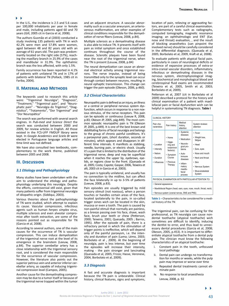
Junho 2011 | June 2011Revista de Anestesia Regional e Terapia da Dor | Journal of Regional Anaesthesia and Pain Treatment 43
In the U.S., the incidence is 2.5 and 5.6 cases per 100,000 inhabitants per year in female and male, including patients aged 60 and 70 years (Gill, 2005 cit in Garcia et al, 2008).The authors Gusmão et al (2003) conducted a study involving 135 patients with TN in wich 42.2% were men and 57.8% were women, aged between 48 and 82 years old with an average of 61 years old. The pain was predom-inantly located on the right side (57%), reach-ing the maxillary branch in 25.9% of the cases and mandibular in 31.9%. The ophthalmic branch was the less affected, with only 1.5%.Family occurrence has been reported in 4.1% of patients with unilateral TN and in 17% of patients with bilateral TN (Pollack, 1985 cit in Fariba, 2004).
II. Material and Methods
The keywords used to research this article were: “Trigeminal Neuralgia”, “Diagnosis”, “Treat ment,” “Trigeminal pain”, and “Neuro-phatic pain” - “Nevralgia do Trigémio”, “Diag-nóstico”, “Tratamento”, “Dor Trigeminal” and “Dor Neuropática”The search was performed with several search engines. In Pub-med and Science Direct the search was conducted between 2000 and 2009, for review articles in English. All those existed in the FCS-UFP FMDUP library were kept. In Google Academics and Scielo Br were elected in Portuguese language articles and time limit was not defined.We have also consulted two textbooks, com-plementary to the work theme, published between 2005 and 2008.
III. Discussion
3.1 Etiology and Pathophysiology
Many studies have been undertaken with the aim to understand the etiology and patho-physiology underlying this neuralgia. Despite the efforts, controversial still exist, given that many patients suffer from trigeminal neuralgia of idiopathic origin. (Siddiqui, 2003).Various theories about the pathophysiology of TN were studied, which attempt to explain its cause. Vascular compression, infectious agents such as human herpes simplex virus, multiple sclerosis and even alveolar compro-mise after tooth extraction, are some of the reasons pointed out as responsible for TN (Garcia et al, 2008).According to several authors, one of the main causes for the occurrence of TN is vascular compression. This can induce demyelization of the trigeminal nerve root at the level of its emergence in the brainstem (Leeuw, 2008, p.84). The superior cerebellar artery has a close relationship with the trigeminal sensory root, and is considered the main responsible for the occurrence of vascular compression. However, the literature also points out the superior petrous vein and anterior inferior cer-ebellar artery, as capable of inducing trigemi-nal compression level (Campos, 2005).Another cause for the demyelinating compres-sion may be due to a tumor itself or because of the trigeminal nerve trapped within the tumor
and an adjacent structure. A vascular abnor-mality such as a saccular aneurysm, an arterio-venous malformation, are other examples of clinical conditions responsible for the demyeli-zation of nerve fibers (Leeuw, 2008, p.84).Multiple sclerosis as a demyelinating disease is also able to induce TN. It presents itself with pain as initial symptom and once esta blished continues throughout the course of the di sease. Sclerotic plaques have been found near the root of the trigeminal nerve, when the TN is present (Leuuw, 2008, p.84)The neural demyelization can cause an abnor-mal electrical activity among trigeminal neu-rons. The nerve impulse, instead of being transmitted only to the synaptic level can occur through contact between neurons, resulting in neural ephaptic transmission. This change can trigger the pain episode (Okeson, 2006, p.460).
3.2 Clinical Characteristics
Neuropathic pain is defined as an injury, an illness or a central or peripheral nervous system dys-function, which occurs in response to a non nox-ious stimulus, of the nerve receptors. This pain can be episodic or continuous (Leeuw R, 2008, p.83; Okeson JP, 2005, pág.449). The most com-mon episodic neuropathic pain is TN (Okeson JP, 2005, p.449). It is one of the best known and debilitating forms of facial neuralgia and belongs to the group of chronic painful conditions. It’s a paroxysmal pain, (short duration, seconds or minutes), which appears and disappears in dif-ferent time intervals. It manifests as stabbing, needle, burning pain, or electric shock. Usually it’s a pain that is limited to the distribution of the trigeminal nerve, deep and may be superficial when it reaches the upper lip, eyebrows, eye-lids, or regions close to the front. (Quesada et al, 2005; Costa; Capote; Gaspar, 2006, Teixeira et alli, 2003 cit in Garcia et al, 2003).The pain is typically unilateral, and usually has no connection to the midline, but can affect the face bilaterally in up to 3-5% of patients (Leeuw, 2008, p.83).Pain episodes are usually triggered by mild sensory stimuli (not noxious), when a person touches or handles certain areas of the face, located ipsilaterally to the pain, in so-called trigger zones wich can be located in the skin, mucosa or even a tooth. The pain is caused by non-painful stimuli that normally are painless, as a breeze passing over his face, shave, wash face, brush your teeth or chew (Petterson, 2000; Teixeira, 2001; Quesada, 2005 ; Barros, 1995). After the episode of pain, there is a refractory period in which the stimulation of trigger points is ineffective, which will depend only of the painful paroxysm, i.e. the inten-sity and duration of pain (Lama; Lémo, 2003; Borbolato et al , 2009). At the beginning of neuralgia, pain is less intense, but over time the episodes will increase their intensity, making - the pain stronger and lancinating (Quesada et al, 2005; Frizzo; Hasse, Veronese, 2004, Borbolato et al, 2009).
3.3 Diagnosis
A fast and accurate diagnosis is important because the TN pain is unbearable. Clinical history, clinical features, signs and symptoms,
location of pain, relieving or aggravating fac-tors, are part of a careful clinical examination. Complementary tests such as radiographs, computed tomography, magnetic resonance imaging; an ophthalmologic and ENT (Ear, nose and throat) evaluation, , and the result of blocking anaesthetics (can pinpoint the involved nerve) should be carefully considered in the differential diagnosis. (Quesada et al, 2005; Borbolato et al, 2009, Kraft et al, 2008).To evaluate patients with atypical facial pain, particularly in cases of neurological deficits or evidence of expansive processes of extra-or intra-cranial vascular disorders, inflammatory, infectious or demyelinating diseases in the nervous system, electrophysiological imag-ing, biochemical and morphological blood and cerebrospinal fluid exams are recom mended (Teixeira et al, 2005, Smith et al, 2003; Borbolato et al, 2009).Petterson et al, 2007 (cit in Borbolato et al 2009) described a protocol for the history and clinical examination of a patient with maxil-lofacial pain or facial dysfunction wich can be helpful in systematizing TN diagnosis. Table I
CLINICAL HISTORY
Pain cha-racteristics Pain history
Medical history, attention
System reviews: attention
• Location
• Intensity
•Nature
•Duration
• Influences
•Home
• trigge-ring and suspicious factor(s)
• Symptom Course
• Previous Therapy
• Systemic diseases
•Medication
•Neurolo- gical pro- blems
• Auto- immune diseases
• Inflam- matory maxillo- facial process
• Fever
•Weight loss, fatigue
•Head
• Ears, eyes
•Nose and mouth
•Neck and throat
• Skin
•Nervous System
•Musculoskeletal System
PHYSICAL EXAMINATION WITH PARTICULAR ATTENTION TO:
•General appearance
•Maxillofacial Region (head, ears, eyes, nose, mouth, throat, neck)
•Nervous system (cranial nerves, SNS and PNS)
Table 1 – Characteristics to be considered for a medi-cal history of the TN
The trigeminal pain, can be confusing for the professional, as TN neuralgia can cause non-dental toothache (atypical toothache) wich sometimes are difficult to identify, inducing the dentist to error, and thus lead to unnec-essary dental procedures (Garcia et al, 2008; Okeson, 2005, p.453). It is important to differ-entiate atypical toothache from a dental pulp pain. The clinician must know the following characteristics of an atypical toothache:
i. Constant pain in the tooth, unfocused local pathology
ii. Dental pain can undergo no transforma-tion for months or weeks, while the pulp pain may improve or worse over time
iii. Repeated dental treatments cannot eli-minate pain
iv. No response to local anaesthesiaLeeuw, 2008, p. 92
trigeminal neuralgia
44
44

Revista de Anestesia Regional e Terapia da Dor | Journal of Regional Anaesthesia and Pain Treatment44Junho 2011 | June 2011
There are several types of facial pain, which together with their clinical characteristics, can make it difficult to determine the definitive diagnosis of trigeminal neuralgia, so it should be taken into account in differential diagnosis (IHS, 2003) - Table II
Differential Diagnosis Pain Characteristics Pain location
Glossopharyngeal neuralgia
Severe paroxysmal pain, Clinical fea-tures similar to TN
Pain in the ear, tongue, tonsillar fossa and below the angle of the jaw
Superior Laryngeal Neuralgia
Severe unilateral Paroxysmal pain, (minutes or hours)
Sore throatSubmandibularTrigger points on the side of the neck
Short-lasting uni-lateral neuralgiform headache attacks with conjunctival injection and tearing (SUNCT)
Paroxysmal, unila-teral, severe pain(5-24 sec.)
Orbital pain, orbital or temporal suppliesPain accompanied by conjunctival injection and ipsila-terally tearing
Occipital Neuralgia Paroxysmal pain, like stabbing
The posterior occipital painCervical region
Intermediate neuralgia
Paroxysmal pain, unilateral
Ear pain Trigger point on the posterior ear canal wall
Pos- herpetic neuralgia
Burning PainHypoesthesia and hyperesthesiaRecurrent or persis-tent pain more than 3 months
Pain felt in the area of most acute virusattack (Herpes Zoster)Pain in the trige-minal ganglionPredominantly affects the ophthal-mic division of trigeminal nerve
Table 2 – Differential Diagnosis of trigeminal neuralgia
The TN may appear in an early form Pre-Trigeminal Neuralgia with atypical features. This is characterized by paroxysmal pain, but remains constant during the painful epi-sode. It’s a pain condition wich, due to its characteristics, difficult the diagnoses as TN, and becomes a more typical painful picture of Temperomandibular Disorders (TMD). (Okeson, 2005, p.457).
3.4 Treatment
Many factors can influence treatment plan, including age (child vs. adult), the average life expectancy, medical and psychiatric condi-tions, tolerance to drugs adverse effects, dura-tion of symptoms and clinical subtype of the TN (Siddiqui et al , 2003).There are two treatment types: clinical or medical and surgical. (Garcia et al, 2008; Quesada et al, 2005; Borbolato et al, 2009). The TN treatment usually starts by using drugs, resorting to surgery only when medical therapy is woefully inadequate (Frizzo et al, 2004; Alves Azevedo, 2004; Carvalho, 2004 cit in Borbolato et al, 2009 ).
3.4.1 Drug Treatment
Therapy is usually performed with carbamaze-pine-based anticonvulsants and / or diphenylhy-dantoin (Frizzo et al, 2004; Alves Azevedo, 2004; Carvalho, 2004 cit in Borbolato et al, 2009).Carbamazepine is an anticonvulsant drug that increases the inactivation of vol tage-dependent
sodium channels, through the reduction of high frequency action potentials repetitive firing preventing the painful episode (Frizzo et al, 2004; Borbolato et al, 2009, Okeson, 2006 cit in Garcia et al, 2008).Carbamazepine has been studied for 40 years, and was first used in 1962. This is a very effec-tive drug, reducing the immediate symptoms of TN, thus often contributing for its diagnosis (Garcia et al, 2008; Borbolato et al 2009).This drug is often chosen as first-line treat-ment, but its prolonged use can cause adverse effects such as tremors, dizziness, drowsiness, confusion, hyper-or hypotension, bradycardia, erythematous rash, exfoliative or obstruction, diarrhea, epigastric pain and problems in vi sual accommodation (Garcia et al, 2008, Alves et al, 2004; Frizzo et al, 2004). It also causes liver and spinal cord problems and for that reason it is recommended that patients carry out regular blood counts and liver function tests, besides the dosage levels of carbamazepine (Frizzo et al, 2004, Garcia et al 2008).In 2006 the European Federation of Neuro-logical Societies, recommends for the treat-ment of Classical TN: carbamazepine (200-1200mg / day), or oxcarbazepine (600-1800 mg / day). Oxcarbazepine is not as effective as carbamazepine, but has fewer side effects than carbamazepine. These drugs are recom-mended for first-line treatment. TN typically lasts forever, with periods of partial or total remission. So, patients need to adjust dosages depending on the frequency of pain episodes. There is no evidence that the combination of therapies is beneficial so that to patients who do not respond well to carbamazepine or oxi-carbamazepine, should be offered blacofen, especially in patients who do not accept to undergo surgery.If there are patients who cannot tolerate car-bamazepine and baclofen, phenytoin is an alternative, however is not effective in redu-cing long term symptoms, usually works as a carbamazepine adjuvant (Quesada et al, 2005; Costa et al, 2006 cit in Borbolato et al, 2009).A recent study showed that an intranasal drug, lidocaine (Xylocaine), showed significant improvements in pain relief for the 2nd divi-sion of the trigeminal nerve for more than 4 hours (Kanai et al, 2006 cit in Kraft et al, 2009).There are not many randomized control stu-dies that demonstrate satisfactory results of non- anticonvulsant drugs for patients with TN (He et al, 2006, Kraft et al, 2009).
3.3.2 Surgical Treatment
When anticonvulsant therapy is ineffective or even because of their side effects, neurosur-gery is indicated (Garcia et al, 2008; Borbolato et al, 2009).Surgical techniques can be opened or per-cutaneous. Most procedures can provide some short-term relief; however some studies refer that many patients relapse within a few years (cit in Zakrzewska, 2004; cit inTaha et al, 1996; Kraft, 2008).Radiofrequency rhizotomy, balloon compres-sion, glycerol rhizotomy, and radio surgery are part of percutaneous techniques. These techniques have the advantage of being non-invasive, not having many adverse effects
and require only one day of hospitalization (Tronnier et al 2001 cit in Kraft, 2008).The radiofrequency rhizotomy is a technique in which are selected the nerve fibers that are located near or within the trigeminal gan-glion. These are destroyed by placing a nee-dle guided by fluoroscopy X-ray within the oval hole. The fibers selected are detected through a careful manipulation guided by the patient, since he is partially sedated, can indicate the painful areas, and thus find the fibers involved in the painful condition. Thus, the fibers selected are destroyed by radio-frequency thermo coagulation or through the introduction of a toxic substance such as gly-cerol. There is also a kind of rhizotomy where nerve fibers are traumatized by a balloon (per-cutaneous microcompression) (Garcia et al, 2008 cit in Ribeiro et al, 2009; Borbolato et al 2009). Another technique that can be used is radio surgery. This is considered a minimally invasive technique; it is held a minimum irra-diation (40-90 Gy) in the trigeminal root entry. Damage to adjacent tissues are not described, however, compared with other surgical tech-niques produces a smaller and delayed pain relief (Joseph S, 2006).Vascular decompression is an open technique, in which the separation of the vessel respon-sible for the compression of one branch of the trigeminal nerve is held. This procedure requires that there be an exploration of the posterior fossa, the cortex is lifted to expose the root entry zone of trigeminal intervening on the vessel or on a possible injury that could cause compression (Garcia et al 2009, Costa et al, 2006, Borbolato et al, 2009; Kraft, 2008).This surgical procedure has several risks such as stroke, meningitis and even death. However, the incidence of these complications for this technique is less than 2%. This surgery produces a persistent symptomatic relief for 10 years in more than 70% of patients. It is recommended for younger patients wich have less risks with the invasive surgery involved, but not recommended for patients over 65 years (Tronnier et al, 2001 cit in Kraft, 2008).Singla and colleagues (2007) demonstrated differences between classical and sympto-matic TN, 88% of patients with classic TN and 56% of patients with symptomatic TN showed neurovascular compression. Patients under-going surgery (vascular decompression) for classic TN performed better results (80%) than patients with symptomatic TN (56%).
IV. Conclusion
TN is an atypical facial pain, paroxysmal pain manifested by electric shock or burning. It is considered by most patients as one of the most excruciating pain ever felt. The etiology of this neuralgia is very controversial and con-fusing, and can often origin doubts regarding the TN diagnosis. The MD is not responsible for dealing with this neuralgia; however he should know how to accomplish clinical and differential diagnosis, sending patients to fol-low appropriate clinical help.
V. Acknowledgment
Dra. Cláudia Barbosa
trigeminal neuralgia
44
44

Junho 2011 | June 2011Revista de Anestesia Regional e Terapia da Dor | Journal of Regional Anaesthesia and Pain Treatment 45
trigeminal neuralgia
44
References1. Alves, T. C. A., Azevedo, G. S., Carvalho, E. S. (2004). Tratamento farmacológico da neuralgia do trigémio. Revisão sistemática e metanálise. In: Revista Brasileira de
Anestesiologia. V.54(6),Novembro - Dezembro, pp-836-8492. Bagheri, S. C., Farhidvash, F., Perciaccante, V. J. (2004). Diagnosis and treatment of patients with trigeminal neuralgia. In: The Journal of the American Dental Association. v.135, Dezembro3. Barros. (1995). Tratamento de disfunções craniomandibulares. In: Frizzo, H. M., et alii. (2004). Neuralgia do Trigémio: Revisão Bibliográfica Analítica. In: Revista de
Cirurgia e Traumatológica Buco-Maxilo-Facial. v.4, n.4, Outubro-Dezembro, pp-212-2174. Barker,G.F.(2007).Quality of life and individual treatment choice in trigeminal neuralgia. Disponível em www.elsevier.com/locate/pain. (Consultado em 15/03/2010)5. Bennetto, L., Patel, N. K., Fuller, G. (2007). Trigeminal neuralgia and its management. In:British Medical Jornal.v.334, Janeiro6. Borbolato, R. M., Ambiel, C. R. (2009). Neuralgia do Trigémio: Aspectos Importantes na clínica odontológica. In: Revista Saúde e Pesquisa.v.2, Maio-Agosto, pp-201-2087. Campolongo, G. D., Nosé, A. R. (2001). Tratmento medicamentoso da neuralgia do trigémio. In: Revista de Odontologia. v.1, Maio-Julho, pp-15-198. Campos, W. K. (2005). Neuralgia do Trigémio:análise dos resultados do tratamento pós compressão percutânea com balão no gânglio de Gasser. In: Trabalho de con-
clusão de curso (Graduação em Medicina) – Uniuversidade Federal de Santa Catarina. Florianópolis9. Costa, C.C, Capote, T. S. O., Gaspar, A. M. M. (2006). Neuralgia Trigeminal – uma revisão bibliográfica sobre etiologia, sintomas e tratamento. In: Borbolato, R. M., - Ambiel, C. R. (2009). Neuralgia do Trigémio: Aspectos Importantes na clínica odontológica. In: Revista Saúde e Pesquisa.v.2, Maio-Agosto, pp-201-20810. Denmark, J. O. et alii. (2004). The International Classification of Headache Disoreders. Disponível em http://www.i-h-s.org/upload/ct_clas/ihc_II_main_no_print.pdf.
(Consultado em 12/05/2010)11. Drake, R. L., Vogl, W., Mitchell, A. W. (2005). Grays´s Anatomia para Estudantes.Rio de Janeiro, Elsevier12. Fothergill, J. (1999). On a painful affliction of the face. In: Bagheri, S. C., Farhidvash, F., Perciaccante, V. J. (2004). Diagnosis and treatment of patients with trigeminal
neuralgia. In: The Journal of the American Dental Association. v.135, Dezembro13. Frizzo, H. M., et alii. (2004). Neuralgia do Trigémio: Revisão Bibliográfica Analítica. In: Revista de Cirurgia e Traumatológica Buco-Maxilo-Facial. v.4, n.4, Outubro-Dezembro, pp-212-21714. Góes, T. M. P. L., Fernandes, R. S. M. (2008). Neuralgia do Trigémio:diagnóstico e tratamento. In: Int J Recife.v 7(2), Janeiro-Março, pp-104-11515. Gonçalves, DAG., Speciali, JG. (2005). Neuralgia Trigeminal causada por meningioma. In: Góes, T. M. P. L., Fernandes, R. S. M. (2008). Neuralgia do Trigémio:diagnóstico
e tratamento. In: Int J Recife.v 7(2), Janeiro-Março, pp-104-11516. Gusmão, S., Magaldi, M., Arantes, A. (2003). In: -Góes, T. M. P. L., Fernandes, R. S. M. (2008). Neuralgia do Trigémio:diagnóstico e tratamento. In: Int J Recife.v 7(2), Janeiro-Março, pp-104-11517. He, L., Wu, B., Zhou, M. (2006). Non-antiepiletic drugs for trigeminal neuralgia. In: Kraft, M. D., Rudolph, M. D.(2008) . Neuralgia. In:American Family Physician.v77, n.77, Maio18. Kabatas, S. (2009). Microvascular decompression as a surgical management for trigeminal neuralgia: A critical review of the literature. In: Neurolagy India, v.57, Março, pp-134-13819. Kanai, A., Suzuki, A., Kobayashi, M., Hoka, S. (2006). Intranasal lidocaine 8% spray for second-division trigeminal neuralgia. In: British Journal of Anaesthesia. v.97(4), Agosto20. Katusic, S., Beard, CM., Bergstralh, R., Kurkuland, LT. (1990). Incidence and clinical features of trigeminal neuralgia. In: Fariba, S. B., et alii. (2004). Diagnosis and treat-
ment of patients with trigeminal neuralgia. In: The Journal of the American Dental Association. v.135, Dezembro21. Kraft, M. D., Rudolph, M. D.(2008) . Neuralgia. In:American Family Physician.v77, n.77, Maio22. Lama, E. A. L., Mélo, F. M. L. (2003). Abordagem da Fisioterapia no Tratamento da Neuralgia do Trigêmio. In: Lato & Sensu.v.4, n.1, Outubro, pp-3-523. Leeuw, R. (2008). Orofacial Pain: Guidelines for Assessment, Diagnosis, and Management. Quarta edição. USA. Quintessences books24. Machado, A. B. M. (2000). Neuroanatomia Funcional. In: Borbolato, R. M., Ambiel, C. R. (2009). Neuralgia do Trigémio: Aspectos Importantes na clínica odontológica.
In: Revista Saúde e Pesquisa.v.2, Maio-Agosto, pp-201-20825. Okeson, J. P. (2005). Orofacial Pains: The Clinical Management of Orofacial Pain. Sexta edição. Canadá. Quintessence books26. Okeson, J. P. (2006). Dores bucofaciais de Bell: tratamento clínico da dor bucofacial. In: Góes, T. M. P. L., Fernandes, R. S. M. (2008). Neuralgia do Trigémio:diagnóstico
e tratamento. In: Int J Recife.v 7(2), Janeiro-Março, pp-104-11527. Peterson, L. J. (2007). Cirurgia oral e maxilo facial contemporânea. In: Borbolato, R. M., Ambiel, C. R. (2009). Neuralgia do Trigémio: Aspectos Importantes na clínica
odontológica. In: Revista Saúde e Pesquisa.v.2, Maio-Agosto, pp-201-20828. Pollack, IF., Jannetta, PJ., Bissonette, DJ. (1988). Bilateral trigeminal neuralggia: a 14-year experience with microvascular decompression. In: Bagheri, S. C., Farhidvash,
F., Perciaccante, V. J. (2004). Diagnosis and treatment of patients with trigeminal neuralgia. In: The Journal of the American Dental Association. v.135, Dezembro29. Quesada, G. A. T., Baptista, C. E., Pedroso, C. S., Flores, D. L. (2005). Neuralgia Trigeminal - do diagnóstico ao tratamento. In:Revista Dentística on line. v.5, n.11, Janeiro-Junho30. Ribeiro, C. T. R., Fleming, N. R. P. A clínicade dor na emergência. In: Alves, N.(2009). Dor: Princípios e prática.RS:Artemed, Porto Alegre, pp-819-827. In: Borbolato, R.
M., Ambiel, C. R. (2009). Neuralgia do Trigémio: Aspectos Importantes na clínica odontológica. In: Revista Saúde e Pesquisa.v.2, Maio-Agosto, pp-201-20831. Siddiqui, M. N., Siddiqui, S., Ranasinghe, J. S., Furgang, F. A.(2003). Pain Management:Trigeminal Neuralgia. In:Hospital Physician. Janeiro32. Singla, V., et alii. (2007). Dolichoectasia of vertebrobasilar system: A rare cause of tice douloureux. In: Indian Journal Med Sci (VER MELHor933. Siqueira, S. R. D. T., Teixeira, M. J., Siqueira, J. T. T. (2009). Clinical Characteristics of Patients with Trigeminal Neuralgia Referred to Neurosurgery. In:European Journal of Dentstry.v3.Julho34. S, José. (2006). Nevralgia do Trigémio. In: Monografia apresentada na Universidade Fernando Pessoa para Licenciatura de Medicina Dentária. Porto35. Teixeira, M. J., Siqueira, S. R. D. T. (2003) . Neuralgias do segmento facial. In: Góes, T. M. P. L., Fernandes, R. S. M. (2008). Neuralgia do Trigémio:diagnóstico e trata-
mento. In: Int J Recife.v 7(2), Janeiro-Março, pp-104-11536. Tronnier, V. M., Rasche, D., Harner, J., Kienle, A., Kunze, S. (2001). Treatment of idiopathic trigeminal neuralgia: comparison of long-term outcome after radiofrequency
rhizotomy and microvascular decompression. In: Kraft, M. D., Rudolph, M. D.(2008) . Neuralgia. In:American Family Physician.v77, n.77, Maio37. Zakrzewska, J. M. (2004). Trigeminal neuralgia and facial pain. In: Kraft, M. D., Rudolph, M. D.(2008) . Neuralgia. In:American Family Physician.v77, n.77, Maio

Revista de Anestesia Regional e Terapia da Dor | Journal of Regional Anaesthesia and Pain Treatment46Junho 2011 | June 2011
Contextualização da Problemática
A dor aguda é um dos problemas mais comuns em todas as áreas da medicina, seja em cuidados de saúde primários,
medicina desportiva, serviço de urgência e traumatologia ou medicina interna, mas é de facto a dor cirúrgica a que constitui a mais importante e frequente causa de dor aguda.Nestas situações cirúrgicas, em que a dor perdeu a sua função de alerta, uma correcta abordagem contribui para a humanização dos cuidados de saúde e a para a satisfa-ção dos doentes com os mesmos. Para além disto, o controlo da dor permite proporcio-nar maior conforto ao doente, favorecer a precocidade da alta e prevenir potenciais complicações desencadeadas pela dor no pós-operatório, como sejam alterações res-piratórias, neuroendócrinas e metabólicas, cardiovasculares, tromboembólicas, altera-ções na recuperação da função gastro-intes-tinal e no estado psíquico, em que situações de confusão mental são agravadas pelo medo e ansiedade da dor.É portanto indiscutível que uma insuficiente analgesia no pós-operatório acarreta eleva-dos custos para as instituições de saúde e consequentemente para a sociedade em geral. A alta precoce é sem dúvida uma das vantagens para as entidades responsáveis, mas também não devemos esquecer os recursos humanos consumidos quando esta-mos perante um doente com dor não contro-lada. Por outro lado, um paciente sem dor é um paciente satisfeito, o que se traduz em ganhos para a instituição de saúde em ter-mos de marketing.Tem sido amplamente discutido que um con-trolo ineficaz da dor aguda pós-operatória pode evoluir para dor crónica [1]. De acordo com dados da IASP [2] , 10 a 50% dos doen-tes experimentam este problema, sendo que destes 2 a 10% apresentam dor crónica severa. Para além do indiscutível sofrimento que isto causa, tanto no paciente como naqueles que o rodeiam, os custos económicos são tre-mendos. Estima-se que só em Portugal a dor crónica custe anualmente três mil milhões de euros e a par dos quadros dolorosos com etiologia em lombalgias, cefaleias e traumatis-mos, a dor crónica pós-cirúrgica encontra-se
entre as causas de dor crónica mais comuns no nosso [3]. O absentismo laboral e a baixa de produtividade são problemas sociais graves que daqui decorrem.
A Situação Actual
O alívio da dor é sem dúvida um direito de qual-quer cidadão e nos últimos anos tem-se assis-tido a uma crescente sensibilização para esta temática, tanto por parte dos profissionais de saúde, como por parte da população em geral. A par disto, o avanço da ciência disponibiliza novas drogas, técnicas e instrumentos de ava-liação da dor que permitem que o tratamento da dor seja de facto uma realidade. Contudo, o tratamento da dor aguda parece estar longe do ideal. De acordo com dados da IASP [2], menos de metade dos doentes com dor aguda do pós--operatório recebem um adequado tratamento para a mesma e o controlo da dor agrava-se quando os doentes têm alta hospitalar.Se o Plano Nacional de Luta Contra a Dor [4] prevê que a analgesia do pós-operatório siga uma actuação organizada em torno de progra-mas de acção, à semelhança das unidades de dor aguda europeias, isto está longe da rea-lidade no nosso país, sobretudo quando nos deslocamos dos grandes centros hospitalares para zonas mais interiores.Podemos assumir que o problema do controlo da dor aguda começa exactamente no sistema de saúde, com uma insuficiente valorização da questão. A pouca sensibilização dos órgãos de gestão para os ganhos em saúde, fazem com que o investimento em dor seja visto em fun-ção dos custos que implica. Isto traduz-se em ausência de infra-estruturas adequadas e num baixo investimento em recursos humanos e materiais para o tratamento da dor aguda.Simultaneamente, ainda que com uma evolução notória nos últimos anos, os currículos acadé-micos continuam a não privilegiar a temática da dor e o incentivo à formação contínua nesta área é baixo. Como consequência, persiste entre os profissionais a crença de que a dor é um efeito inevitável da cirurgia e que, apesar de desagra-dável, é pouco prejudicial e limitada no tempo constituindo também um sinal de alarme de uma eventual complicação pós-cirúrgica.A avaliação sistemática da dor (feita com recurso a escalas de avaliação da dor) ainda não se impôs nas rotinas hospitalares o que
pode traduzir alguma dificuldade por parte dos profissionais em valorizarem a dor referida pelos pacientes. Como consequência gera-se um desconhecimento sobre a verdadeira efi-cácia das medidas anti-álgicas implementadas a cada paciente. Tudo isto resulta na ausência de um compromisso real em tratar a dor de forma sistemática e os esforços pon tuais aca-bam por ser insuficientes se não forem inte-grados num planeamento multidisciplinar.A sociedade civil demonstra também um certo desconhecimento desta problemática. Em parte devido a factores culturais, um certo estoicismo em relação à sensação de dor continua a persistir. A dor é percebida como uma consequência da cirurgia e a subvalori-zação da importância do seu controlo para prevenir complicações contribuem para a aceitação de uma inadequada analgesia. Não se pode deixar de referir que os mitos asso-ciados à morfina são igualmente outro factor de peso na relutância em alguns doentes refe-rirem agravamento da intensidade da dor aos profissionais.
Perspectivas futuras
Ainda que existam alguns esforços pelas enti-dades responsáveis em dar alguma visibili-dade à temática da dor, a realidade está ainda aquém do desejável. Ao contrário do que sucede com a dor crónica, em que começa a surgir um compromisso conjunto das entida-des responsáveis e dos pacientes e suas famí-lias para o seu alívio, a dor aguda é ainda des-curada e encarada como algo inevitável.Urge tomar medidas para a mudança de ati-tudes que contrariem o subtratamento da dor aguda. É necessário que os órgãos de governo e administrações hospitalares apoiem a cria-ção de estruturas multidisciplinares, direccio-nadas para abordagem à dor aguda. Sobre-tudo na dor aguda pós-operatória, em que a sua ocorrência é perfeitamente previsível, a sua abordagem tem se assentar num planea-mento integrado, tornando a dor parte dos cuidados peri-operatórios [5].É essencial que os profissionais saibam ava-liar e valorizar a dor dos pacientes e este pro-cesso não pode continuar a dependente de acções individuais. A abordagem da dor tem de ser integrada de forma categórica na for-mação académica base. A IASP [2] defende
Reflexão sobre a Problemática da Dor Aguda
Inês Bolina
Enfermeira Hospital CUF Infante Santo
Resumo: Tendo presente a problemática da dor aguda, a IASP (International Association for the Study of Pain) implementou o Ano Mundial de Luta Contra a Dor Aguda, iniciado em Outubro de 2010 e que se prolonga até Outubro de 2011.A dor aguda, com a sua função de alerta e protecção da vida, é sem dúvida a experiência de dor mais comum, sendo experimentada em alguma altura da vida do ser humano, nomeadamente em situações de trauma, parto ou cirurgia.Se por um lado, o alívio da dor aguda é facilmente alcançável com recurso a medicação e técnicas especializadas e a qualidade da intervenção dos profissionais de saúde evoluí a par dos conhecimentos sobre a fisiologia da dor, também é um facto que o tratamento da dor aguda tem ainda um longo caminho a percorrer até que a situação se aproxime do ideal.Palavras Chave: Dor, Dor Aguda, Subtratamento, Humanização de Cuidados
4

Junho 2011 | June 2011Revista de Anestesia Regional e Terapia da Dor | Journal of Regional Anaesthesia and Pain Treatment 47
exactamente que para além de directrizes e recomendações implementadas pelos órgãos governamentais, é necessário que estas sejam alvo de auditorias e se tornem parte inte-grante dos sistemas de certificação e de quali-dade hospitalares.Para além disto, a educação da sociedade civil poderá ser um bom ponto de partida para a mudança de atitudes. Se os pacientes
tomarem consciência da importância de um adequado controlo da dor, irão exigir uma mudança de atitudes, tanto no sistema de saúde em geral como nos profissionais. A criação de protocolos de actuação e de estru-turas adequadas, estabelecidas a nível nacio-nal, fará com que o acesso ao tratamento dor aguda seja de facto uma realidade para todos aqueles que a experimentam [5].
Em suma, o ideal será que a postura face à dor aguda se desenvolva a par das novas tecno logias disponíveis para o seu trata-mento. Talvez desta forma, criar um “Ano Internacional Contra a Dor Aguda” deixe de fazer sentido, porque o seu tratamento e a efectiva humanização dos cuidados que pres-tamos já serão uma realidade.
reflexão sobre a Problemática da dor aguda
4
Bibliografia1. Wilder, S.; Scheffer, GJ Schreyer, T.; Arendt-Nielsen, L.” Patients with chronic pain after abdominal surgery show less preoperative endogenous pain inhibition and more
postoperative hyperalgesia: a pilot study”. J pain Palliat Care Pharmacother. 2010 Jun, 24 (2): 119-28. In Pub Med2. IASP www.iasp.org.3. “Estudo da Prevalência da Dor Crónica na Sociedade Portuguesa”. Faculdade de Medicina do Porto: 2008.4. “Plano Nacional de Luta Contra a Dor”. Direcção Geral de Saúde: 2001.5. Caseiro, J. A organização da analgesia do pós-operatório. Biblioteca da Dor. APED: 2004.

Revista de Anestesia Regional e Terapia da Dor | Journal of Regional Anaesthesia and Pain Treatment48Junho 2011 | June 2011
Problem Context
Acute pain is one of the most com-mon problems in all areas of medi-cine, whether in primary health care,
sports medicine, emergency department and trauma or internal medicine, but in fact, surgical pain is the most important and fre-quent cause of acute pain.In these surgical situations, where pain has lost its warning function, a cor-rect approach contributes to health care humanization and to patients’ satisfaction. In addition, pain control allows to provide more comfort to the patient, encourage early discharge and prevent potential com-plications triggered by postoperative pain, such as respiratory, neuroendocrine, meta-bolic, cardiovascular and thromboembolic disorders, changes in gastro-intestinal recovery function and in psychological condition in which situations of mental confusion are exacerbated by fear and pain anxiety.It is therefore unquestionable that an insuf-ficient postoperative analgesia involves high costs for health care institutions and therefore to society in general. The early discharge is without doubt one of the advantages for the responsible entities, but we must not forget the human resources consumed when we face a patient with uncontrolled pain. On the other hand, a patient without pain is a satisfied patient, which leads to marketing gains for the health institution.It has been widely discussed that an inef-fective control of acute postoperative pain can develop into chronic pain [1]. According to data from IASP [2], 10 to 50% of patients experience this problem, and of these 10-20% have severe chronic pain. Apart from the unspeakable suffering that it causes, to both patient and those around him, the economic burden is tremendous. It is estimated that only in Portugal chronic pain annually cost three billion Euros and along with the painful conditions with low back pain etiology, headache and trauma, chronic pain after surgery is, in our country, among the causes of more common chronic pain [3]. Absenteeism and low productivity are severe social problems wich arise from this situation.
The State of the Art
Pain relief is undoubtedly every citizen’s right and in recent years it has been witnessed a growing awareness to this issue, both by health professionals and general popula-tion. Also, the progress of science offers new drugs, techniques and tools for pain assess-ment that allows pain treatment to be in fact a reality. However, the treatment of acute pain appears to be far from ideal. According to data from the IASP [2], less than half of patients with acute pain after surgery receive appropriate treatment, and pain control worsens when patients are discharged.If the National Plan against Pain [4] indi-cates that postoperative analgesia follows an organized performance around action programs, like the European units of acute pain, this is far from our country’s reality, especially when we move from big hospital centers to inland areas.We can assume that the problem of acute pain control starts exactly in the health sys-tem, with an insufficient appreciation of the problem. The lack of awareness of the gains in healthcare, turn the investment in pain management seen relatively too costly. This leads into the absence of adequate infra-structure and a low investment in human and material resources for acute pain treatment.Simultaneously, although with a noticeable trend in recent years, the academic curri-culums still do not focus pain themes and the encouragement of continuous education in this area is low. Consequently, among health professionals remains a belief that pain is an inevitable effect of surgery and, although unpleasant, is relatively harmless, limited in time and is also a warning signal of a possible complication after surgery.Systematic evaluation of pain (using pain rat-ing scales) has not been established in the hospital routines which mean some difficulty from health professionals to value the pain reported by the patients. As a result, there is enormous ignorance about the true effective-ness of the anti-algic measures implemented for each patient. All this results, in the absence of a real commitment to pain mana-gement in a systematic way, and occasional efforts turn out to be insufficient unless they are integrated into a multidisciplinary plan.
The civil society also demonstrates a certain ignorance of this problem. Partly due to cul-tural factors, continues to persist some stoi-cism about the sensation of pain. The pain is perceived as a consequence of surgery and the undervaluation of the importance of its control to prevent complications contributes to the acceptance of an inadequate analge-sia. It’s important to mention that the myths associated with morphine are also another important factor because some patients have reluctance to refer to the health pro-fessionals the worsening of pain intensity.
Future Prespectives
Although there are some efforts by the enti-ties responsible in giving some visibility to the issue of pain, the reality is still far from desirable. Unlike the case with chronic pain, which is emerging a joint commitment of the authorities and the patients and their families for their relief, acute pain is still neglected and seen as inevitable.It is urgent to take measures to change atti-tudes contrary to the undertreatment of acute pain. It is necessary for government agencies and hospital administrations to create structures to support multidisci-plinary, targeted approach to the acute pain. Especially in acute postoperative pain, in that their occurrence is perfectly predic-table; its approach has become an inte-grated planning, including the perioperative pain care [5].It is essential that professionals know how to evaluate and enhance patients’ pain and this process cannot continue to depend on individual actions. The pain management must be integrated categorically based on academic training. IASP [2] argues exactly that in addition to guidelines and recom-mendations implemented by government, it is necessary that they be subject to audits and become part of the certification systems and quality hospitals.In addition, the education of civil society may be a good starting point for changing attitudes. If patients become aware of the importance of adequate pain control, they will require a change in attitudes, both in the health system in general and professionals. The establish-ment of protocols for action and appropriate structures established at national level will
Reflection on the Acute Pain Problem
Inês Bolina
Nurse, Hospital CUF Infante Santo
Abstract: Bearing in mind the problem of acute pain, the IASP (International Association for the Study of Pain) implemented the International Year Against Acute Pain started in October 2010 and draws tilll October 2011.Acute pain, with its warning and life protection function, is undoubtedly the most common experience of pain, being experienced at some point in human life, particularly in situations of trauma, surgery or childbirth.If on one hand, the relief of acute pain is easily reachable using medication and specialized techniques and the intervention quality of health care professionals has evolved along with the knowledge about pain physiology, is also a fact that acute pain treatment has still a long way to go through until the situation is close to ideal.Keywords: Pain, Acute Pain Undertreatment, Care Humanization
44

Junho 2011 | June 2011Revista de Anestesia Regional e Terapia da Dor | Journal of Regional Anaesthesia and Pain Treatment 49
turn access to treatment acute pain a reality for all those who experience it [5].In short, the ideal is that the attitude towards
the acute pain can develop alongside the new technologies available for pain treat-ment. Maybe so, create an “International
Year Against Acute Pain” stop making sense, because its effective treatment and humane care we provide has become a reality.
reflection over the acute Pain Problem
44
References1. Wilder, S.; Scheffer, GJ Schreyer, T.; Arendt-Nielsen, L.” Patients with chronic pain after abdominal surgery show less preoperative endogenous pain inhibition and more
postoperative hyperalgesia: a pilot study”. J pain Palliat Care Pharmacother. 2010 Jun, 24 (2): 119-28. In Pub Med2. IASP www.iasp.org.3. “Estudo da Prevalência da Dor Crónica na Sociedade Portuguesa”. Faculdade de Medicina do Porto: 2008.4. “Plano Nacional de Luta Contra a Dor”. Direcção Geral de Saúde: 2001.5. Caseiro, J. A organização da analgesia do pós-operatório. Biblioteca da Dor. APED: 2004.

Solicitamos que envie o seu e-mail para que sempre que
possível a correspondência seja feita por via electrónica
Website do CAR estáde novo operacional e com novidades
CAR tem um novo e-mail
Visite o nosso siteCriticas e sugestões serão
muito bem vindas
www.anestesiaregional.com XVXV REUNIÃO ZONALDO E.S.R.A.
XV REUNIÃO ZONALDO E.S.R.A.
W W W . A N E S T E S I A R E G I O N A L . C O MW W W . A N E S T E S I A R E G I O N A L . C O M
Local de realização das Sessões do CongressoHOTEL SANA METROPOLITAN
R. Soeiro Pereira Gomes, Parcela 2 | 1600-198 LisboaTel: + 351 217 982 500 | Fax: + 351 217 950 864
[email protected] | www.metropolitan.sanahotels.com
ORGANIZAÇÃO
L I S B O AL I S B O A
2011201129 de Setembro a 1 de Outubro
SÁBADOSÁBADO1 de Outubro
9h
PRÓ-CON: PARTOS DISTÓCICOS E SOFRIMENTO FETALAR OU GERAL PARA CESARIANA EMERGENTE?
Moderador – a definir
Pró AGLara Ribeiro
Pró ARJoana Carvalhas
10h
ANALGESIA NEUROAXIAL NO TRABALHO DE PARTO.A VERDADE DA MENTIRA.
Moderador - a definirPaulo Sá
10h30
PRÓ-CON: CIRURGIA AMBULATÓRIO AR / AGModerador - Rui Sobral de Campos
Pró AGa definir
Pró ARElena Segura
11h30 COFFEE BREAK
12h
PRÓ-CON: AR para Artroplas�a TotalModerador - Edgar Lopes
Pró ARa definir
Con ARAldara Beiras
13h ALMOÇO DE TRABALHO
14h
ANESTESIA REGIONALModerador - Luís Aliaga
NOVOS DESENVOLVIMENTOS MSTeresa Parras
NOVOS DESENVOLVIMENTOS MIEdgar Lopes
15h
NEUROESTIMULAÇÃO E ULTRASONOGRAFIA.QUAL O PAPEL ACTUAL DE CADA UMA?
Moderador – Edgar SemedoRafael Blanco
15h30 COFFEE BREAK
16h
AR EM POPULAÇÕES ESPECIAISModerador - Clara Lobo
...NA 1ª INFANCIASusana Vargas
...NO DOENTE ONCOLÓGICOSteven Delwaere
DELÍRIO E AGITAÇÃO NO DOENTE SOB ARSusana Favaios
SEXTA FEIRASEXTA FEIRADia 30 de Setembro
8h ABERTURA DO SECRETARIADO
9h
AR ESSENCIAL: MUST-KNOW BLOCKS...Moderador: Clara Lobo
...NA UCIPSteven Delweare
...NA EMERGÊNCIA/TRAUMARoman Zuercher
...NA UNIDADE DE AMBULATÓRIOAlain Borgeat
10h
SESSÃO DE ABERTURARui Sobral de Campos
XX ANOS CAR: HISTÓRIA DA ARModerador: Joaquim Viana
Jorge TavaresSÓCIOS DE MÉRITO:
BEATRIZ TEIXEIRA, BERNARDO LEAL E JOSÉ CASEIRO
11h30 COFFEE BREAK
12h
O ENSINO DA ANESTESIA REGIONALModerador: Rafael Blanco
PONTO DE VISTA DO INTERNOAndreia Mafra
PONTO DE VISTA DO ESPECIALISTAa definir
13h ALMOÇO DE TRABALHO
14h30
LOMBALGIA E DOR MÚSCULO-ESQUELÉTICA:DO NEURÓNIO À IMAGEMModerador: Jorge Cortez
DA DOR AGUDA À DOR CRÓNICA:ONDE PODEREMOS ACTUAR
Ana ValenmDIAGNÓSTICOS DIFERENCIAIS: UM GRANDE DESAFIO
Filipe AntunesTRATAMENTOS INVASIVOS: O ESTADO DA ARTE
Luis Agualuza
16h COFFEE BREAK
16h30
COLUNA “INSTRUMENTADA” E AR:DIFERENTES PONTOS DE VISTA
Moderador - Beatriz Craveiro LopesTeresa Ferreira (ponto de vista da Unidade de Dor Crónica)
Maria Joao Centeno (ponto de vista do Anestesiologista)Miguel Casimiro (ponto de vista do Neurocirurgião)
WS 1 - LOMBALGIA DA SEMIOLOGIA À CLÍNICAWS 2 - DOR MÚSCULO-ESQUELÉTICA DA CLÍNICA AO DIAGNÓSTICOWS 3 - ECOGRAFIA NA DOR MÚSCULO-ESQUELÉTICOWS 4 - LOMBALGIA DA CLÍNICA À IMAGEM
QUINTA FEIRAQUINTA FEIRADia 29 de Setembro
WORKSHOPS E CURSOS PRÉ-CONGRESSO
IV CURSO PRÁTICO DE BLOQUEIOS PERIFÉRICOSOrganização - Hospital Fernando da Fonseca,
Amadora – Sintra
WORKSHOP DE ABORDAGEM DA VIA AÉREA DIFÍCILOrganização - Serviço de Anestesiologia do Hospital
Distrital de Santarém
WORKSHOP DE ULTRASONOGRAFIA EM BLOQUEIOSCENTRAIS , PARAVERTEBRAIS E NA PEDIATRIA
Organização - CARDocentes: Rafael Blanco, Roman Zuercher,
Teresa Parras e Susana Vargas
13h
ALMOÇO DE TRABALHO
ENCONTRO COM O ESPECIALISTA(sujeito a inscrição previa - 5 pessoas/mesa)
MR. FREEZE:OMBRO CONGELADO. COMO AVALIAR. QUE SOLUÇÕES.
Gustavo Montanha
COMO AVALIAR UM ARTIGO ATRAVÉS DA SUAFORMULAÇÃO ESTATÍSTICA.
Joaquim Viana
BLOQUEIO SUBDURAL: DIAGNÓSTICO E ABORDAGEM.Lara Ribeiro
ANALGESIA NO DOENTE OBESO.Teresa Parras
LESÃO NEUROLÓGICA PÓS-ARLuís Aliaga
INTUBÇÃO DIFÍCIL... PROVA SUPERADA!COMO EXTUBAR O DOENTE COM VIA AÉREA DIFÍCIL?
Fernando Roxo
PAPEL DA TOXINA BOTULINICA NA DORa definir
A INSCRIÇÃO NO CONGRESSO INCLUI:Pasta e Diploma de par�cipação no Congresso | Acesso livre a todas as sessões do Congresso | Coffee-Breaks | Almoço de trabalho de sexta e sábado (sujeitos a marcação prévia)Almoço com os experts (inscrição prévia obrigatória, máx. 9 pessoas/mesa) | WS de ecografia do neuroeixo e espaço paravertebral (inscrição prévia obrigatória, máx. 15 pessoas)
Curso de de Via Aérea Di�cil (inscrição prévia obrigatória, máx. 20 pessoas)
Inscrições até 16 de Agosto:Inscrições até 16 de Agosto:Sócios CAR - Especialistas 140€ | Internos 100€Não Sócios - Especialistas 190€ | Internos 150€Curso APED - 10€Curso Via Aérea Di�cil - 10€
Inscrições a partir de 17 de Agosto:Inscrições a partir de 17 de Agosto:Sócios CAR - Especialistas 170€ | Internos 120€Não Sócios - Especialistas 220€ | Internos 170€Curso APED - 20€Curso Via Aérea Di�cil - 20€

Website do CAR estáde novo operacional e com novidades
XVXV REUNIÃO ZONALDO E.S.R.A.
XV REUNIÃO ZONALDO E.S.R.A.
W W W . A N E S T E S I A R E G I O N A L . C O MW W W . A N E S T E S I A R E G I O N A L . C O M
Local de realização das Sessões do CongressoHOTEL SANA METROPOLITAN
R. Soeiro Pereira Gomes, Parcela 2 | 1600-198 LisboaTel: + 351 217 982 500 | Fax: + 351 217 950 864
[email protected] | www.metropolitan.sanahotels.com
ORGANIZAÇÃO
L I S B O AL I S B O A
2011201129 de Setembro a 1 de Outubro
SÁBADOSÁBADO1 de Outubro
9h
PRÓ-CON: PARTOS DISTÓCICOS E SOFRIMENTO FETALAR OU GERAL PARA CESARIANA EMERGENTE?
Moderador – a definir
Pró AGLara Ribeiro
Pró ARJoana Carvalhas
10h
ANALGESIA NEUROAXIAL NO TRABALHO DE PARTO.A VERDADE DA MENTIRA.
Moderador - a definirPaulo Sá
10h30
PRÓ-CON: CIRURGIA AMBULATÓRIO AR / AGModerador - Rui Sobral de Campos
Pró AGa definir
Pró ARElena Segura
11h30 COFFEE BREAK
12h
PRÓ-CON: AR para Artroplas�a TotalModerador - Edgar Lopes
Pró ARa definir
Con ARAldara Beiras
13h ALMOÇO DE TRABALHO
14h
ANESTESIA REGIONALModerador - Luís Aliaga
NOVOS DESENVOLVIMENTOS MSTeresa Parras
NOVOS DESENVOLVIMENTOS MIEdgar Lopes
15h
NEUROESTIMULAÇÃO E ULTRASONOGRAFIA.QUAL O PAPEL ACTUAL DE CADA UMA?
Moderador – Edgar SemedoRafael Blanco
15h30 COFFEE BREAK
16h
AR EM POPULAÇÕES ESPECIAISModerador - Clara Lobo
...NA 1ª INFANCIASusana Vargas
...NO DOENTE ONCOLÓGICOSteven Delwaere
DELÍRIO E AGITAÇÃO NO DOENTE SOB ARSusana Favaios
SEXTA FEIRASEXTA FEIRADia 30 de Setembro
8h ABERTURA DO SECRETARIADO
9h
AR ESSENCIAL: MUST-KNOW BLOCKS...Moderador: Clara Lobo
...NA UCIPSteven Delweare
...NA EMERGÊNCIA/TRAUMARoman Zuercher
...NA UNIDADE DE AMBULATÓRIOAlain Borgeat
10h
SESSÃO DE ABERTURARui Sobral de Campos
XX ANOS CAR: HISTÓRIA DA ARModerador: Joaquim Viana
Jorge TavaresSÓCIOS DE MÉRITO:
BEATRIZ TEIXEIRA, BERNARDO LEAL E JOSÉ CASEIRO
11h30 COFFEE BREAK
12h
O ENSINO DA ANESTESIA REGIONALModerador: Rafael Blanco
PONTO DE VISTA DO INTERNOAndreia Mafra
PONTO DE VISTA DO ESPECIALISTAa definir
13h ALMOÇO DE TRABALHO
14h30
LOMBALGIA E DOR MÚSCULO-ESQUELÉTICA:DO NEURÓNIO À IMAGEMModerador: Jorge Cortez
DA DOR AGUDA À DOR CRÓNICA:ONDE PODEREMOS ACTUAR
Ana ValenmDIAGNÓSTICOS DIFERENCIAIS: UM GRANDE DESAFIO
Filipe AntunesTRATAMENTOS INVASIVOS: O ESTADO DA ARTE
Luis Agualuza
16h COFFEE BREAK
16h30
COLUNA “INSTRUMENTADA” E AR:DIFERENTES PONTOS DE VISTA
Moderador - Beatriz Craveiro LopesTeresa Ferreira (ponto de vista da Unidade de Dor Crónica)
Maria Joao Centeno (ponto de vista do Anestesiologista)Miguel Casimiro (ponto de vista do Neurocirurgião)
WS 1 - LOMBALGIA DA SEMIOLOGIA À CLÍNICAWS 2 - DOR MÚSCULO-ESQUELÉTICA DA CLÍNICA AO DIAGNÓSTICOWS 3 - ECOGRAFIA NA DOR MÚSCULO-ESQUELÉTICOWS 4 - LOMBALGIA DA CLÍNICA À IMAGEM
QUINTA FEIRAQUINTA FEIRADia 29 de Setembro
WORKSHOPS E CURSOS PRÉ-CONGRESSO
IV CURSO PRÁTICO DE BLOQUEIOS PERIFÉRICOSOrganização - Hospital Fernando da Fonseca,
Amadora – Sintra
WORKSHOP DE ABORDAGEM DA VIA AÉREA DIFÍCILOrganização - Serviço de Anestesiologia do Hospital
Distrital de Santarém
WORKSHOP DE ULTRASONOGRAFIA EM BLOQUEIOSCENTRAIS , PARAVERTEBRAIS E NA PEDIATRIA
Organização - CARDocentes: Rafael Blanco, Roman Zuercher,
Teresa Parras e Susana Vargas
13h
ALMOÇO DE TRABALHO
ENCONTRO COM O ESPECIALISTA(sujeito a inscrição previa - 5 pessoas/mesa)
MR. FREEZE:OMBRO CONGELADO. COMO AVALIAR. QUE SOLUÇÕES.
Gustavo Montanha
COMO AVALIAR UM ARTIGO ATRAVÉS DA SUAFORMULAÇÃO ESTATÍSTICA.
Joaquim Viana
BLOQUEIO SUBDURAL: DIAGNÓSTICO E ABORDAGEM.Lara Ribeiro
ANALGESIA NO DOENTE OBESO.Teresa Parras
LESÃO NEUROLÓGICA PÓS-ARLuís Aliaga
INTUBÇÃO DIFÍCIL... PROVA SUPERADA!COMO EXTUBAR O DOENTE COM VIA AÉREA DIFÍCIL?
Fernando Roxo
PAPEL DA TOXINA BOTULINICA NA DORa definir
A INSCRIÇÃO NO CONGRESSO INCLUI:Pasta e Diploma de par�cipação no Congresso | Acesso livre a todas as sessões do Congresso | Coffee-Breaks | Almoço de trabalho de sexta e sábado (sujeitos a marcação prévia)Almoço com os experts (inscrição prévia obrigatória, máx. 9 pessoas/mesa) | WS de ecografia do neuroeixo e espaço paravertebral (inscrição prévia obrigatória, máx. 15 pessoas)
Curso de de Via Aérea Di�cil (inscrição prévia obrigatória, máx. 20 pessoas)
Inscrições até 16 de Agosto:Inscrições até 16 de Agosto:Sócios CAR - Especialistas 140€ | Internos 100€Não Sócios - Especialistas 190€ | Internos 150€Curso APED - 10€Curso Via Aérea Di�cil - 10€
Inscrições a partir de 17 de Agosto:Inscrições a partir de 17 de Agosto:Sócios CAR - Especialistas 170€ | Internos 120€Não Sócios - Especialistas 220€ | Internos 170€Curso APED - 20€Curso Via Aérea Di�cil - 20€

Revista de Anestesia Regional e Terapia da Dor | Journal of Regional Anaesthesia and Pain Treatment58Junho 2011 | June 2011
Agenda | Calendar of Events2011Congressos e Reuniões NacionaisCongresses and National MeetingsSetembro | September 28 – 1 Outubro | October, 2011XX Congresso do CARLisboa | Lisbon, Portugal
Congressos e Reuniões InternacionaisCongresses and International MeetingsSetembro | September 7 - 10, 2011Annual ESRA Congress - European Society of Regional Anaesthesia & pain TherapyDresden, Alemanha | Germany
Setembro | September 14 - 17, 2011European Burns Association Congress 2011Haia, Holanda | The Hague, Netherlands
Setembro | September 21 - 24, 2011EFIC Congress: Pain in Europe VIILocal: CCH - Congress Center HamburgHamburg, Alemanha | GermanyWeb: http://www.kenes.com/eficEmail: [email protected] / [email protected]
Outubro | October 10 - 14, 2011European Week Against Pain 2011Chronic Back Pain
Outubro | October 15 - 19, 2011ASA 2011: American Society of Anesthesiologists Annual MeetingChicago, IL, Estados Unidos | USA
Maio | May 24 - 25, 2011World Congress of Ophthalmic AnaesthesiaAnkara, Turquia | Turkey
2012/2013Congressos e Reuniões InternacionaisCongresses and International MeetingsMarço | March 25 - 30, 201215th World Congress of Anesthesiologists (WCA) 2012Buenos Aires, Argentina | Argentina
Junho | June 9 - 12, 2012Euroanaesthesia 2012Paris, França | France
Outubro | October 13 - 17, 2012ASA 2012: American Society of Anesthesiologists Annual MeetingWashington, DC, Estados Unidos | USA
Maio | May 23 - 26, 20134th International Congress on Neuropathic PainToronto, Canadá| Canada
Outubro | October 12 - 16, 2013ASA 2013: American Society of Anesthesiologists Annual MeetingSan Francisco, CA, Estados Unidos | USA
Informações complementares (secretariados etc.) contactar o CARe-mail: [email protected]

Revista de Anestesia Regional e Terapia da Dor | Journal of Regional Anaesthesia and Pain Treatment60Junho 2011 | June 2011
Temas | ContentsPróximo Número | NEXT ISSUEnúmero 65: Setembro 2011 | Number 65: September 2011
• Número dedicado ao XX Aniversário do CAR | Issue devoted to the twentieth anniversary of the CAR
Visite o nosso siteCriticas e sugestões serão
muito bem vindas
www.anestesiaregional.com

clube de anestesia regional
O CAR está na eminência de ter de suspender a edição da Revista, por não haver capacidade financeira para suportar o défice de cada número
que se eleva neste momento a mais de 2.500 euros.
Os patrocinadores habituais, a Indústria Farmacêutica, estão a reduzir o seu investimento em publicidade, e alguns retiraram mesmo
os seus anúncios da Revista
Se todos os associados do CAR (mais de 1.300) colaborarem, com o pagamento das suas quotas, o problema fica resolvido.
A quota é de 25 euros anuais, que pode ser paga no Multibanco ou por Transferência Bancária para o NIB 00320-12300-20150-291-940.
Agradecemos que nos seja enviada uma mensagem, para nos informar do pagamento, para
[email protected] ou para [email protected]
O recibo será posteriormente enviado por correio.
É indispensável a colaboração de todos, para se poder manter a publicação da Revista que é de todos.
