efeitos metabólicos do ácido p cumárico no fígado de...
TRANSCRIPT
Efeitos metabólicos do ácido p-cumárico no fígado de rato
Leonardo Cristiano Neves Lima
Dissertação apresentada ao Programa de Pós-Graduação em Ciências Biológicas da Universidade
Estadual de Maringá, área de concentração em Biologia Celular, para a obtenção do grau de
Mestre
Orientadora: Dra. Jorgete Constantin
Maringá 2005

Livros Grátis
http://www.livrosgratis.com.br
Milhares de livros grátis para download.
2
Este é um trabalho de equipe, realizado no Laboratório de Metabolismo Hepático da Universidade Estadual de Marin-gá, formado pelo artigo: Lima LCN, Buss GD, Ishii-Iwamoto EL, Salgueiro-Pagadi-gorria C, Comar JF, Bracht A, Constantin J. Metabolic effects of p-coumaric acid in the perfused rat liver. Journal of Biochemical and Molecular Toxicology (submetido).
Ficha catalográfica BCE/FUEM de Lima, Leonardo Cristiano Neves Efeitos metabólicos do ácido p-cumárico no fígado de rato Maringá: UEM, 2005. 31 p., gráficos. Diss. (mestrado) UEM-DBQ, 2005. Orientadora: Dra. Jorgete Constantin. 1. Perfusão de fígado de rato. 2. Ácido p-cumárico 3.
Metabolismo oxidativo e energético. 4. Neoglicogênese. 5. Transporte de piruvato. I. Universidade Estadual de Maringá.
3
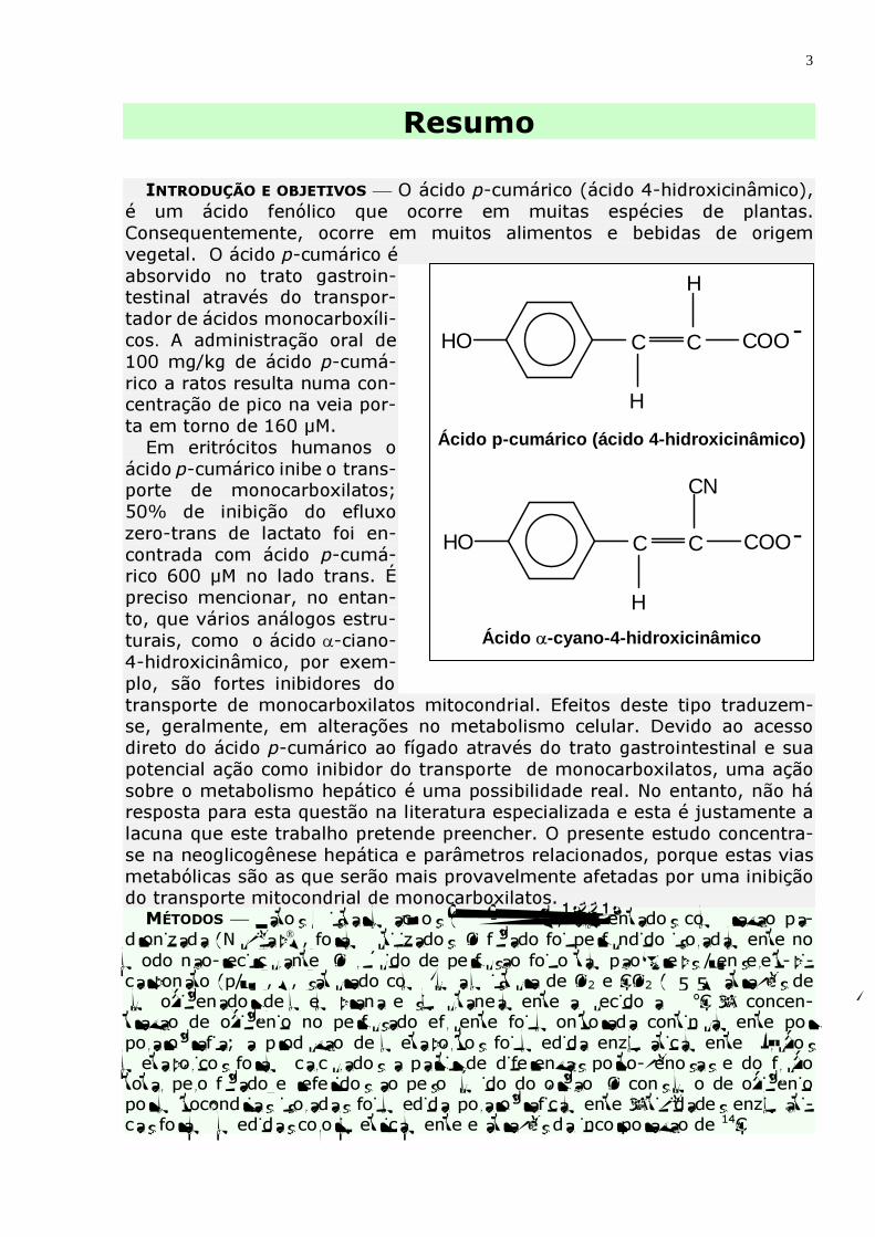
Resumo INTRODUÇÃO E OBJETIVOS O ácido p-cumárico (ácido 4-hidroxicinâmico), é um ácido fenólico que ocorre em muitas espécies de plantas. Consequentemente, ocorre em muitos alimentos e bebidas de origem vegetal. O ácido p-cumárico é absorvido no trato gastroin-testinal através do transpor-tador de ácidos monocarboxíli-cos. A administração oral de 100 mg/kg de ácido p-cumá-rico a ratos resulta numa con-centração de pico na veia por-ta em torno de 160 µM. Em eritrócitos humanos o ácido p-cumárico inibe o trans-porte de monocarboxilatos; 50% de inibição do efluxo zero-trans de lactato foi en-contrada com ácido p-cumá-rico 600 µM no lado trans. É preciso mencionar, no entan-to, que vários análogos estru-turais, como o ácido α-ciano-4-hidroxicinâmico, por exem-plo, são fortes inibidores do transporte de monocarboxilatos mitocondrial. Efeitos deste tipo traduzem-se, geralmente, em alterações no metabolismo celular. Devido ao acesso direto do ácido p-cumárico ao fígado através do trato gastrointestinal e sua potencial ação como inibidor do transporte de monocarboxilatos, uma ação sobre o metabolismo hepático é uma possibilidade real. No entanto, não há resposta para esta questão na literatura especializada e esta é justamente a lacuna que este trabalho pretende preencher. O presente estudo concentra-se na neoglicogênese hepática e parâmetros relacionados, porque estas vias metabólicas são as que serão mais provavelmente afetadas por uma inibição do transporte mitocondrial de monocarboxilatos. MÉTODOS Ratos Wistar machos (180-220 g), alimentados com ração pa-
dronizada (Nuvilab), foram utilizados. O fígado foi perfundido isoladamente no modo não-recirculante. O líquido de perfusão foi o tampão Krebs/Henseleit-bi-carbonato (pH 7,4), saturado com uma mistura de O2 e CO2 (95:5) através de um oxigenador de membrana e simultaneamente aquecido a 37oC. A concen-tração de oxigênio no perfusado efluente foi monitorada continuamente por polarografia; a produção de metabólitos foi medida enzimaticamente. Fluxos metabólicos foram calculados a partir de diferenças porto-venosas e do fluxo total pelo fígado e referidos ao peso úmido do órgão. O consumo de oxigênio por mitocôndrias isoladas foi medida polarograficamente. Atividades enzimáti-cas foram medidas colorimetricamente e através da incorporação de 14C.
C C COOHO
H
H
H
HO COOCC
CN
-
-
Ácido p-cumárico (ácido 4-hidroxicinâmico)
Ácido α-cyano -4-hidroxicinâmico
4
RESULTADOS 1) A transformação do lactato em glicose (neoglicogênese) no fígado de ratos em jejum foi inibida pelo ácido p-cumárico (IC50 = 92,5 µM). A inibição máxima atingida na faixa entre 10 e 500 µM foi de 65%. O consumo de oxigênio estimulado pelo lactato também foi inibido; o decréscimo máximo situou-se em torno de 55%.
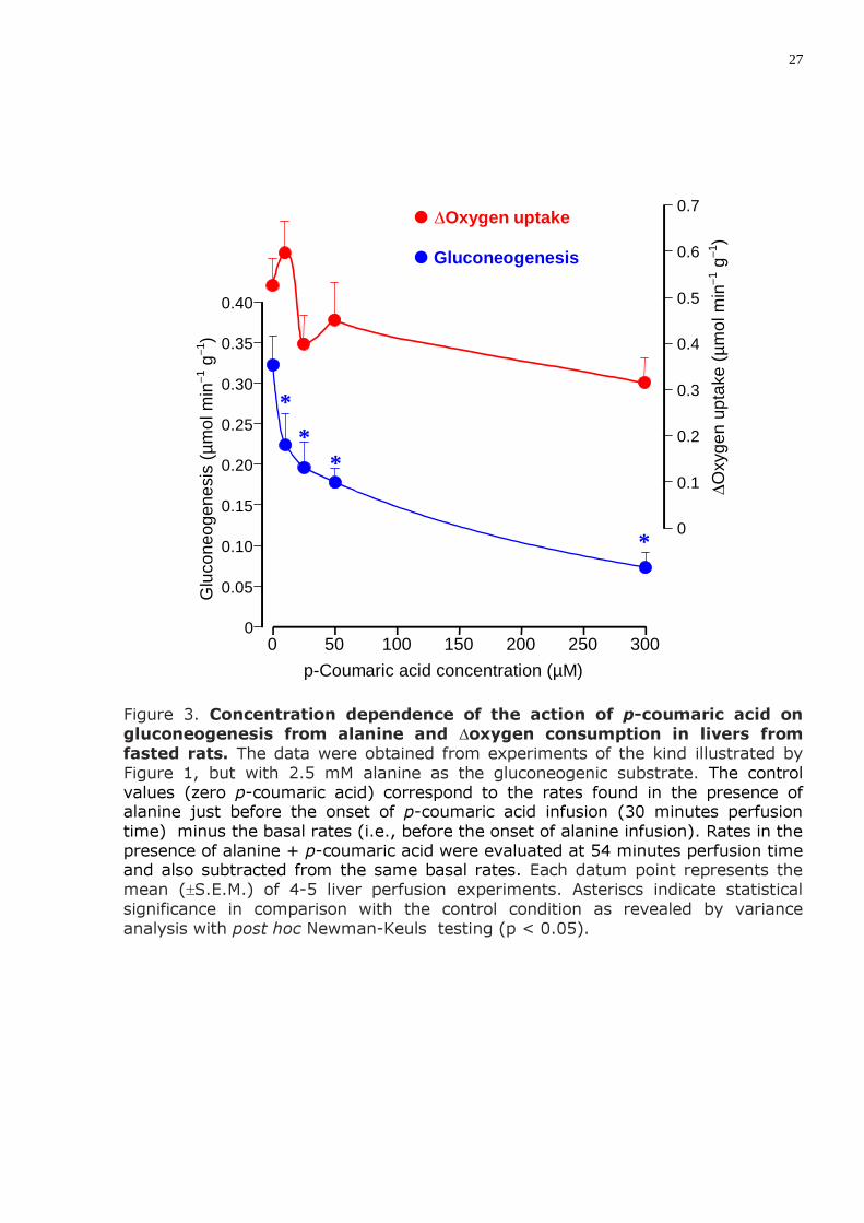
2) A produção de glicose a partir de alanina exógena em fígado de ratos em jejum também foi inibida pelo ácido p-cumárico (IC50 = 75,6 µM); a uma concentração de 300 µM o decréscimo foi de 85%.
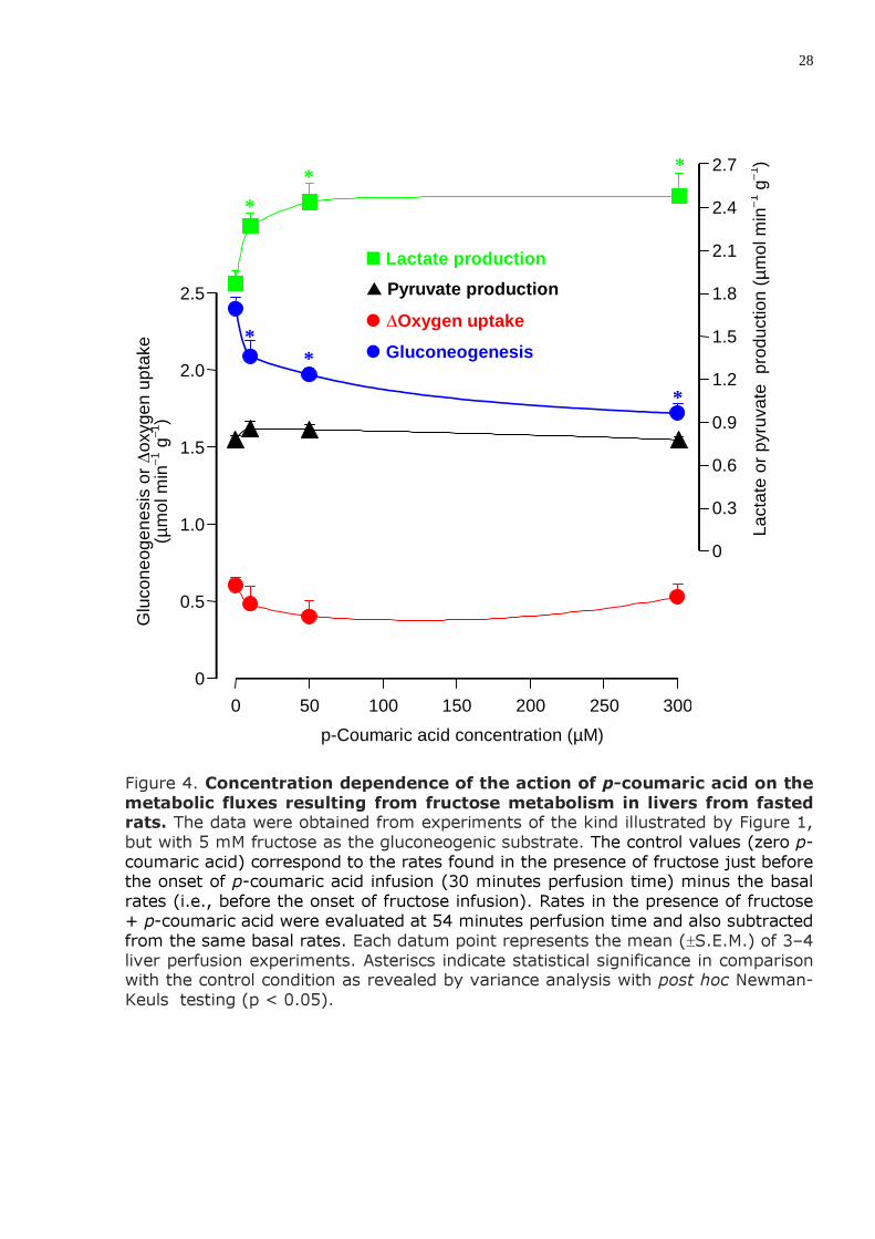
3) A produção de lactato a partir de frutose fornecida exogenamente (gli-cólise) em figados de ratos em jejum foi estimulada pelo ácido p-cumárico (máximo de 32,4% com ácido p-cumárico 300 µM). A produção de glicose a partir de frutose (neoglicogênese) foi inibida (28,3% com ácido p-cumárico 300 µM). O consumo de oxigênio estimulado pela frutose e a produção de piruvato não foram afetados significativamente.
4) No fígado de ratos alimentados o ácido p-cumárico 300 µM aumentou em 18% a produção de lactato e em 8,7% o consumo de oxigênio. A produção de piruvato e a liberação de glicose não foram afetados.
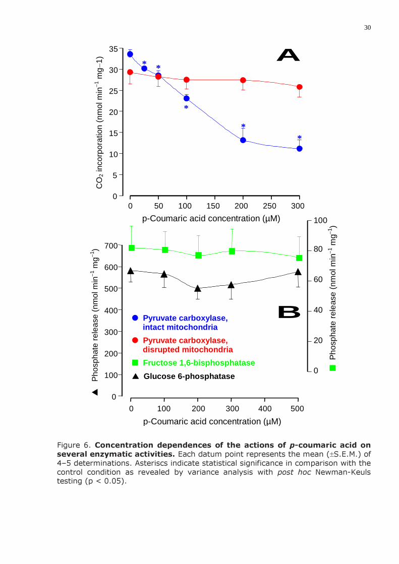
5) O ácido p-cumárico não afetou significativamente as enzimas glicose 6-fosfatase e frutose 1,6-bisfosfatase. Inibiu, porém, a carboxilação do piruvato em mitocôndrias isoladas intactas (IC50 = 160,1 µM); em mitocôndrias rompidas por congelamento e descongelamento este efeito não foi observado.
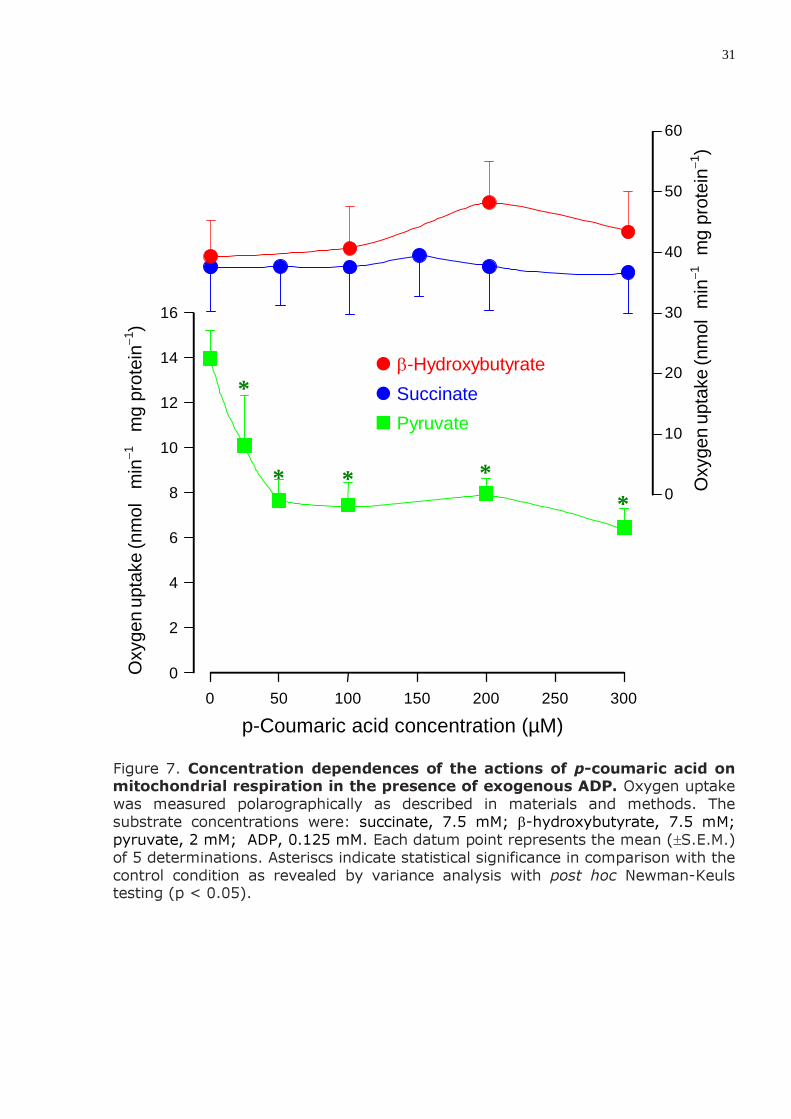
6) Em mitocôndrias isoladas o ácido p-cumárico (até 500 µM) não afetou a respiração mitocondrial dependente da oxidação de succinato e de β-hidroxi-butirato, mas inibiu a respiração dependente da oxidação de piruvato.
DISCUSSÃO E CONCLUSÕES A principal causa da diminuição da neoglicogênese
a partir de lactato e alanina deve ser a inibição do transporte de piruvato para o interior das mitocôndrias. A concomitante inibição do consumo de oxigênio deve-se, possivelmente, à menor demanda por ATP causada pela inibição desta via biossintética. Este mecanismo é indicado pela inibição da carboxilação do piruvato em mitocôndrias intactas e ausência de efeito em mitocôndrias rompidas. A conclusão é também corroborada pela inibição seletiva da respi-ração de mitocôndrias intactas dependente de piruvato e a ausência de efeito com outros substratos. Também apóia esta conclusão a observação de que sob certas condições o ácido p-cumárico aumenta a liberação de lactato. Este aumento deve refletir a menor transfomação do piruvato que logo entra em equilíbrio com o lactato através da reação catalisada pela lactato desidrogenase.
A produção de glicose a partir de frutose, que foi inibida em grau menor do que no caso da neoglicogênese a partir de alanina e lactato, não depende, em princípio do transporte de piruvato para o interior das mitocôndrias. Porém, é bem provável que uma parte da glicose formada a partir de frutose provenha, na realidade, de uma reciclagem do piruvato. Isto é sugerido pelo aumento na liberação de lactato. Não se pode excluir, no entanto, a existência de outras ações do ácido p-cumárico além da inibição do transporte mitocondrial de piruvato. Estas ações podem incluir inibição de enzimas, inibição de outros sistemas de transporte ou ainda, interferência, em sistemas regulatórios.
6
RESULTS The main results were the following: 1) Transformation of lactate into glucose (gluconeogenesis) in the liver of
fasted rats was inhibited by p-coumaric acid with an IC50 = 92.5 µM. Maximal inhibition in the range between 10 and 500 µM was equal to 65%. The lactate stimulated oxygen consumption was also inhibited; the maximal decrease was around 55%.
2) The production of glucose from exogenous alanine in livers from fasted rats was also inhibited by p-coumaric acid with an IC50 = 75.6 µM; the inhibition degree was 85% at a concentration of 300 µM.
3) The production of lactate from exogenously supplied fructose (glycolysis) in livers from fasted rats was stimulated by p-coumaric acid (maximally 32.4% at 300 µM p-coumaric acid). Glucose production from fructose (gluconeogenesis) was inhibited (28.3% at 300 µM p-coumaric acid). The fructose stimulated oxygen consumption and pyruvate production were not significantly changed.
4) In the liver of fed rats 300 µM p-coumaric acid increased both lactate production (18%) and oxygen uptake (8.7%); no actions on glucose and pyruvate release were found.
5) p-Coumaric acid did not significantly affect the enzymes glucose 6-phosphatase and fructose 1,6-bisphosphatase. It inhibited, however, pyruvate carboxylation in isolated intact mitochondria (IC50 = 160.1 µM); no such effect was observed in freeze-thawing disrupted mitochondria.
6) In isolated mitochondria p-coumaric acid up to 300 µM did no affect mitochondrial respiration dependent on succinate and β-hydroxybutyrate oxidation, but it inhibited respiration dependent on pyruvate oxidation.
DISCUSSION AND CONCLUSIONS The main cause for the diminished gluco-neogenesis from lactate and alanine is probably the inhibition of pyruvate transport into the mitochondria. The concomitant inhibition of oxygen uptake is probably related to the reduced ATP demand caused by the inhibition of this biossynthetic route. This mechanism is indicated by the inhibition of pyruvate carboxylation in intact mitochondria and the absence of such effect in disrupted mitochondria. The conclusion is also corroborated by the selective inhibition of the pyruvate driven mitochondrial respiration. The conclusion also receives support from the observation that under certain conditions the p-coumaric acid increases lactate release by the liver. This increase is an expected consequence of a reduced pyruvate transformation and it reflects the rapid equilibrium between pyruvate and lactate due to the lactate dehydrogenase activity.
Glucose production from fructose, which was also inhibited to a lesser extent than gluconeogenesis from alanine and lactate, does not in principle depend on pyruvate transport into mitochondria. However, it is probable that some fraction of the glucose formed from fructose in reality comes from lactate-pyruvate recycling via pyruvate carboxylase. This is suggested by the increase in lactate release. The existence of other actions of p-coumaric acid in addition to pyruvate transport inhibition cannot be excluded however. These actions can include enzyme inhibitions, inhibition of other transport systems and also interference with regulatory mechanisms.
7
Metabolic effects of p-coumaric acid in the perfused rat liver
Leonardo C.N. Lima, Gisele D. Buss, Emy L. Ishii-Iwamoto, Clairce Salgueiro-Pagadigorria, Jurandir Fernando Comar, Adelar Bracht and Jorgete Constantin
Laboratory of Liver Metabolism, University of Maringá,
87020900 Maringá (Brazil)
Address for correspondence: Adelar Bracht Laboratory of Liver Metabolism Department of Biochemistry University of Maringá 87020900 Maringá, Brazil E-mail: [email protected] Fax: 55-44-3261-4896

8
Abstract
The p-coumaric acid, a phenolic acid, occurs in several plant species and, consequently, in many foods and beverages of vegetable origin. Its antioxidant activity is well documented, but there is also a single report about an inhibitory action on the monocarboxylate carrier, which operates in the plasma and mitochondrial membranes. The latter observation suggests that p-coumaric acid could be able to inhibit gluconeogenesis and related parameters. The present investigation was planned to test this hypothesis in the isolated and hemoglobin-free perfused rat liver. Transformation of lactate and alanine into glucose (gluconeogenesis) in the liver was inhibited by p-coumaric acid (IC50 values of 92.5 and 75.6 µM, respectively). Transformation of fructose into glucose was inhibited to a considerably lower degree (maximally 28%). The oxygen uptake increase accompanying gluconeogenesis from lactate was also inhibited. Pyruvate carboxylation in isolated intact mitochondria was inhibited (IC50 = 160.1 µM); no such effect was observed in freeze-thawing disrupted mito-chondria. Glucose 6-phosphatase and fructose 1,6-bisphosphatase were not inhibit. In isolated intact mitochondria p-coumaric acid inhibited respiration dependent on pyruvate oxidation but was ineffective on respiration driven by succinate and β-hydroxybutyrate. It can be concluded that inhibition of pyruvate transport into the mitochondria is the most prominent primary effect of p-coumaric acid and also the main cause for gluconeogenesis inhibition. The existence of additional actions of p-coumaric acid, such as enzyme inhibitions and interference with regulatory mechanisms, cannot be excluded.
9
Introduction
The p-coumaric acid (4-hydroxycinnamic acid) is a phenolic acid that occurs in several plant species. Typically, it presents itself esterified to arabino-xylose residues in hemicellulose or lignin of gramineae, including maize, oat and wheat (1,2). In free or esterified form it occurs in apples, pears, grapes, oranges, tomatoes, beans, potatoes and spinach leaves (3-5). Additionally, it is also found in
beverages such as tea, coffee, wine, choccolate, beer (6,7) and in olive oil (8).
p-Coumaric acid is readily absorbed from the gastrointestinal tract via the monocarboxylic acid transporter. This was deduced from the observation that absorption of p-coumaric acid can be inhibited by benzoic an acetic acids (9). After an oral administration of 100 mg/kg to rats, a peak portal concentration of 160 µM was found (10).
In mammalian tissues a variety of effects have been suggested for p-coumaric acid. Its antioxidant activity is well documented and it results in protection against low-density lipoprotein oxidation (11). In a study aiming to elucidate the mechanism of this action it was concluded that p-coumaric acid effectively scavanges •OH in a dose-dependent manner (5). Besides its antioxidant activity, p-coumaric acid is also able to reduce plasma cholesterol levels (5) and seems to possess antiinflammatory properties (12,13), a weak antileukemic activity (14) and goitrogenic activity (15), the latter at relatively high doses. There is also a single report about a direct action of p-coumaric acid on the monocarboxylate transport system. In human erythrocytes 50% inhibition of lactate zero-trans efflux was found with 600 µM p-coumaric acid in the trans side (16). It must also be mentioned that several structural analogs, such as α-cyano-4-hydroxycinnamate, are effective inhibitors of monocarboxylate transport across the mitochondrial (17) and cellular membrane (18). Primary effects of this kind are usually translated into cell metabolism changes. Due to the direct access of p-coumaric acid to the liver via the intestinal tract (10) and its potential action as an inhibitor of monocarboxylate transport (17), an action on hepatic metabolism is a possibility that deserves attention. However, no responses about this question can be found in the especialized literature and this is precisely the gap that the present work intends to fill. The present study concentrates on hepatic gluconeogenesis and related pathways because these metabolic routes are the most likely to be affected by an inhibition of mitochondrial monocarboxylate transport.
10
Materials and methods
Materials The liver perfusion apparatus was built in the workshops of the University of
Maringá. Several peristaltic pumps used in the experiments and the precision infusion pumps were a gift of Dr. Roland Scholz of the Institute for Physiological Chemistry of the University of Munich, Germany. p-Coumaric acid and all enzymes and coenzymes used in the enzymatic assays were purchased from Sigma Chemical Co. (St. Louis, US). Sodium [14C]bicarbonate, specific activity of 58 Ci/mmol, was purchased from Amersham Pharmacia Biotech (Buckinghamshire, UK). All other chemicals were from the best available grade (98-99.8% purity).
Animals
Male albino rats (Wistar), weighing 180-220 g, were fed ad libitum with a stan-
dard laboratory diet (Nuvilab, Colombo, Brazil). In some experimental protocols the rats were starved for 24 hours before the surgical removal of the liver.
Liver perfusion
For the surgical procedure the rats were anesthetized by intraperitoneal injection
of thiopental (50 mg/kg). Hemoglobin-free, non-recirculating perfusion was performed. The surgical technique was that one described by Scholz and Bücher (19). After cannulation of the portal and cava veins the liver was positioned in a plexiglass chamber. The perfusion fluid was Krebs/Henseleit-bicarbonate buffer (pH 7.4), saturated with a mixture of oxygen and carbon dioxide (95:5) by means of a membrane oxygenator with simultaneous temperature adjustment at 37°C. The flow, provided by a peristaltic pump, was between 30 and 33 ml/min.
Livers from animals in two different metabolic conditions were used: ad libitum fed rats and 24-hours fasted rats. Substrate-free perfused livers from fed rats respire mainly at the expense of endogenous fatty acids, but at the same time they exhibit extensive glycogenolytic and glycolytic activity (19). Livers from fasted rats also respire at the expense of endogenous fatty acids, but their glycogen levels are very low (20). This is a suitable condition for measuring gluconeogenesis from exogenous substrates without interference by glycogen catabolism. In the present work three different substrates were infused: lactate, alanine and fructose.
11
Analytical
Samples of the effluent perfusion fluid were collected according to the
experimental protocol and analysed for their metabolite contents. The following compounds were assayed by means of standard enzymatic procedures: glucose (21), lactate (22) and pyruvate (23). The oxygen concentration in the outflowing perfusate was monitored continuously, employing a teflon-shielded platinum electrode adequately positioned in a plexiglass chamber at the exit of the perfusate (19). Metabolic rates were calculated from input-output differences and the total flow rates and were referred to the wet weight of the liver.
Mitochondria isolation and measurement of the respiratory activity
Fed rats, weighing between 180 and 220 g, were decapitated and their livers
removed immediately and cut into small pieces. These fragments were suspended in a medium containing 0.2 M mannitol, 75 mM sucrose, 1.0 mM Tris-HCl (pH 7.4), 1 mM EGTA and 50 mg% fatty acid-free bovine serum albumin. Homogenization was carried out in the same medium by means of a van Potter-Elvehjem homogenizer. After homogenization the mitochondria were isolated by differential centrifugation (24) and suspended in the same medium, which was kept at 0-4oC.
Oxygen uptake by isolated mitochondria was measured polarographically using a teflon shielded platinum electrode (24,25). Mitochondria (0.85±0.35 mg protein/ml) were incubated in the closed oxygraph chamber in a medium (2.0 ml) containing 0.25 M mannitol, 5 mM sodium diphosphate, 10 mM KCl, 0.2 mM EDTA, 25 mg% fatty acid-free bovine serum albumin, 10 mM Tris-HCl (pH 7.4) and three different substrates in addition to various p-coumaric acid concentrations in the range between 25 and 500 µM. The substrates were succinate (7.5 mM), β-hydroxy-butyrate (7.5 mM) and pyruvate (2 mM). ADP, for a final concentration of 0.125 mM, was added at appropriate times. Rates of oxgen uptake were computed from the slopes of the recorder tracings and expressed as nmol per minute per mg protein.
Protein content of the mitochondrial suspensions was measured by means of the method described by Lowry et al. (26), using the Folin-phenol reagent and bovine-serum albumin as a standard.
12
Enzyme assays For assaying glucose 6-phosphatase microsomes were isolated by differential
centrifugation according to Bygrave (27). The pellet of the 105 000g centrifugation step, containing the microsomal fraction, was suspended in cold medium (4oC) containing 250 mM sucrose and 2.5 mM N-2-hydroxyethylpiperazine-N0-2-ethanesulfonic acid (HEPES, pH 6.8). The incubation medium for the glucose 6-phosphatase assay contained 100 mM KCl, 5 mM MgCl2, 20 mM Tris-HCl (pH 7.2), 15 mM glucose 6-phosphate and 0.1–0.2 mg microsomal protein. After 20 minutes incubation at 37oC the reaction was stopped by the addition of one volume of 5% trichloroacetic acid and phosphate release was measured according to Fiske and Subbarow (28). The supernatant of the 105 000g centrifugation step (high-speed centrifugation supernatant) was used for the assay of D-fructose 1,6-bisphos-phatase according to the procedure described by Mendicino et al. (29). The reaction mixture contained 0.4-0.8 mg protein/ml, 100 mM Tris-HCl (pH 8), 12 mM MgCl2, 1 mM D-fructose 1,6-bisphosphate and 5 mM cysteine. After 20 minutes incubation at 38oC the reation was interrupted by the addition of one volume of 5% trichloroacetic acid and phosphate release was measured according to Fiske and Subbarow (28).
The pyruvate carboxylase activity of intact mitochondria was measured by a modification of the technique described by Adam and Haynes (30). Rat liver mitochondria were isolated as described above (24). The incubation medium contained 5 mM pyruvate, 12.5 mM MgCl2, 2.5 mM potassium phosphate, 2.5 mM KCl, 10 mM N-2-hyroxyethylpiperazine-N-2-ethane-sulfonic acid (HEPES; pH 7.5) and 3 mg protein/ml. The reaction was initiated by introducing 12 mM [14C]NaHCO3 (0.25 µCi). After 10 minutes of incubation at 37oC the reaction was arrested by the addition of 0.5 volume of 2 N perchloric acid. After expulsion of the remaining [14C]NaHCO3 (5 minutes), aliquots were taken for counting the acid stable incorporated radioactivity.
The pyruvate carboxylase of disrupted mitochondria was measured using an medium able to generate steady-state concentrations of acetyl-CoA, as originally described by Henning et al. (31). Rat liver mitochondria, isolated as described above (24), were disrupted by successive freeze and thawing procedures using liquid nitrogen. The incubation medium contained 3 mg protein/ml of disrupted mitochondria, 5 mM sodium pyruvate, 12.5 mM MgCl2, 2.5 mM potassium phosphate, 0.3 M sucrose, 1 mM ethylenediamine tetraacetate, 5 mM
13
tris(hydroxymethyl)aminomethane (TRIS; pH 7.5), 0.5 mM lithium coenzyme A, 5 mM adenosine triphosphate, 1.1 mM acetyl phosphate, 6 µg/ml phosphotrans-acetylase and 12 µg/ml citrate synthase. The reaction was initiated by introducing 12 mM [14C]NaHCO3 (0.25 µCi). After 10 minutes of incubation at 37oC the reaction was arrested by the addition of 0.5 volume of 2 N perchloric acid. After expulsion of the remaining [14C]NaHCO3 (5 minutes), aliquots were taken for counting the acid stable incorporated radioactivity.
The incorporated radioactivity in both incubations, intact and disrupted mitochondria, was expressed as nmol minute−1 (mg protein)−1. The scintillation solution for counting 14C was composed of toluene/Triton X100 (1.5/0.5), 10 g/liter 1,5-diphenyloxazole plus 0.4 g/liter 2,2-p-phenyl-bis-5-phenyleneoxazole.
Treatment of data
The statistical significance of the differences between parameters obtained in the
liver perfusion experiments was evaluated by means of Student's t-test or by Newman-Keuls test after submitting the data to variance analysis according to context. The results are mentioned in the text as the p values; p < 0.05 was the criterion of significance.
15
the presence of p-coumaric acid. Statistically no clear inhibition was detected, although a tendency toward inhibition was evident at 300 µM.
Fructose can also be transformed into glucose in the liver, but ramification of the fructose pathway at the enolase step can also lead to the production of lactate and pyruvate. The action of p-coumaric acid on the production of all these metabolites in consequence of fructose infusion was investigated in a series of experiments similar to those shown in Figure 1 except that fructose was infused at a concentration of 5 mM. The results are summarized in Figure 4 and show that lactate production from exogenously supplied fructose (glycolysis) was stimulated by p-coumaric acid. With 300 µM p-coumaric acid this stimulation reached 32.44%. Glucose production, on the other hand (gluconeogenesis), was inhibited; at 300 µM p-coumaric acid inhibition reached 28.3%. The actions of p-coumaric acid on pyruvate production and oxygen consumption were not significant.
Figure 5 shows the mean results of experiments in which the action of 300 µM p-coumaric acid on glycogen catabolism and oxygen uptake in livers from fed rats was investigated. Such livers produce glucose, lactate and pyruvate from the endogenous glycogen stores but respire mainly at the expense of endogenous fatty acids (19). Figure 5 shows that the infusion of 300 µM p-coumaric acid did not affect glucose and pyruvate release. However, small but significant increases in oxygen uptake and lactate release were found. These effects were reversible, i.e., they vanished upon cessation of the infusion. The peak increase in lactate release was equal to 0.24±0.07 µmol min−1 g−1 (18%; p = 0.027 in Student's paired t test); the peak increase in oxygen uptake was equal to 0.14±0.01 µmol min−1 g−1 (8.7%; p < 0.001 in Student's paired t test).
Three key enzymatic activities of the gluconeogenic pathway were analyzed in the present work: glucose 6-phosphatase, fructose 1,6-bisphosphatase and pyruvate carboxylase. The results of several measurements at various p-coumaric acid concentrations are shown in Figure 6. No action was found on glucose 6-phos-phatase and fructose 1,6-bisphosphatase. Pyruvate carboxylation in isolated intact mitochondria, however, was inhibited with a well defined concentration dependence (IC50 = 160.1 µM). On the other hand, no such effect was observed in freeze-thawing disrupted mitochondria.
In isolated mitochondria p-coumaric acid did not affect mitochondrial respiration dependent on succinate and β-hydroxybutyrate oxidation, but it inhibited respira-tion dependent on pyruvate oxidation. This is revealed by Figure 7, which shows experiments conducted with isolated rat liver mitochondria incubated in the
16
presence of ADP. Inhibition of pyruvate-dependent respiration, however, was already maximal or nearly so at a concentration of 50 µM.
17
Discussion
The most prominent effect of p-coumaric acid in the perfused liver was inhibition of gluconeogenesis from lactate and alanine. The concomitant inhibition of oxygen uptake is probably related to the reduced ATP demand caused by the inhibition of this biosynthetic route. The main cause for the diminished gluconeogenesis from lactate and alanine is probably the inhibition of pyruvate transport into the mitochondria. This is indicated by the inhibition of pyruvate carboxylation in intact mitochondria and the absence of such effect in disrupted mitochondria. A direct action on pyruvate carboxylase can thus be excluded. Inhibition of pyruvate transport into the mitochondria diminishes glucose synthesis from substrates that depend on such a transport as amply demonstrated in experiments with the classical inhibitor of pyruvate transport α-cyano-4-hydroxycinnamic acid (34). Both lactate (lactate dehydrogenase) and alanine (alanine aminotransferase) must be first transformed into pyruvate before entering the gluconeogenic pathway. Although a mitochondrial form of alanine aminotransferase also exists, in the rat liver the ratio of the cytosolic to the mitochondrial activity is equal 5.12 (35). It must be stressed that pyruvate transport across the mitochondrial membrane is rate-limiting for carboxylation, as can be deduced from the similar IC50 values for the inhibition of both transport and carboxylation (∼6 µM) that were found for α-cyano-4-hydroxycinnamate (36). It is also noteworthy to mention that inhibition of gluconeogenesis occurs within the same concentration range of p-coumaric acid that also produces inhibition of pyruvate carboxylation. An inhibition of pyruvate transport also explains the selective inhibition of the pyruvate driven mitochondrial respiration and the observation that under certain conditions p-coumaric acid increases lactate release by the liver. The latter phenomenon was observed under two different conditions, namely, glycolysis from endogenous glycogen and glycolysis from exogenous fructose. This increase is an expected consequence of a reduced pyruvate transformation and it reflects the rapid equilibrium between pyruvate and lactate due to the lactate dehydrogenase activity and the increased availability of cytosolic reducing equivalents (19,37).
Glucose production from fructose, which was inhibited to a lesser extent than gluconeogenesis from alanine and lactate, does not in principle depend on pyruvate transport into the mitochondria. Furthermore, no actions of p-coumaric acid were found on glucose 6-phosphatase and fructose 1,6-bisphosphatase, the enzymes that are believed to catalize the rate-limiting steps of gluconeogenesis after the

18
triose-phosphate step. An action on the fructose phosphorylation step (fructokinase) is unlikely if one considers the increased rates of lactate production from fructose in the presence of p-coumaric acid. Moreover, the increased rates of lactate release during glycogen-dependent glycolysis in the presence of p-coumaric acid also disproves actions of the compound on the enzymes shared by glycolysis and gluconeogenesis. All these considerations make it more difficult to divise an explanation for the small, though significant, action of p-coumaric acid on glucose production from fructose. One possibility would be that some fraction of the glucose formed from fructose in reality comes from lactate/pyruvate recycling into the hexose-phosphate pool via pyruvate carboxylase. The increased rates of lactate release could be an indication that this recycling was significant previous to p-coumaric acid infusion. Recycling of lactate/pyruvate into the hexose-phosphate pool in the perfused liver really occurs as demonstrated by studies using radioactively labeled precursors. In livers from fasted rats, recycling of the glycolytic products into glucose is equal to 13% in the presence of 33 mM glucose, as shown by Kimmig et al. (38). Under the latter conditions, production of lactate + pyruvate is equal to 0.64 µmol min−1 g−1 (38). In the presence of 5 mM fructose, lactate + pyruvate production is much higher, 2.4 µmol min−1 g−1, leading thus to higher cellular lactate+pyruvate concentrations. Recycling of lactate and pyruvate into glucose in the present experiments is thus a highly probable event and could explain in part the action of p-coumaric acid on glucose production in the presence of fructose. However, additional effects of p-coumaric acid on enzymatic and regulatory systems cannot be excluded. Fructose-1,6-bis-phosphatase and phosphofructokinase, for example, are regulatory enzymes (39) and p-coumaric acid could be affecting the net flux through these enzymes by changing, even slightly, the cytosolic concentrations of their allosteric regulators.
Inhibitors of monocarboxylate transport are generally much more effective on the mitochondrial carrier than on the plasma membrane carrier. For example, α-cyanocinnamate inhibits the mitochondrial carrier with an IC50 of 0.2 µM (17), whereas the IC50 for inhibition of the plasma membrane carrier of hepatocytes is 1 mM (39). The same seems also to occur with p-coumaric acid. The IC50 of 600 µM reported by Deuticke (16) for lactate transport inhibition in human erythrocytes is much higher than the IC50 found in the present work for pyruvate carboxylation inhibition (160.1 µM). In addition to the higher p-coumaric acid concentrations that are probably required for a significant inhibition of lactate transport across the cell membrane, it must also be mentioned that the unidirectional fluxes of lactate
19
between the extra- and intracellular spaces are considerably greater than the net uptake rates (40). In other words, lactate transport across the cell membrane, under steady-state conditions, is not rate-limiting for metabolic transformation (40). For these reasons the influence of an inhibition of lactate transport into the cell on the gluconeogenic rates is probably much less important than the influence on pyruvate transport across the mitochondrial membrane. This is indeed corroborated by the inhibitory action of p-coumaric acid on gluconeogenesis from alanine which depends on the mitochondrial pyruvate transport (35), but is independent of monocarboxylate transport across the cell membrane. The action of p-coumaric acid on the latter was even stronger than that on gluconeogenesis from lactate, possibly because the cytosolic pyruvate concentration generated by lactate is higher than that one generated by alanine. On the other hand, the fact that lactate release was stimulated by p-coumaric acid is not incompatible with an action on the monocarboxylate carrier of the plasma membrane. If the intracellular lactate production is increased concomitantly with a relatively low degree of efflux inhibition, the first phenomenon will be an increase in intracellular lactate concentration which is followed by the establishment of a new and higher steady-state net efflux. The latter reflects, evidently, the new steady-state rate of lactate production.
In livers of fed rats p-coumaric acid stimulated oxygen uptake, a phenomenon that was not detected in experiments with isolated mitochondria. It was also not found in livers of fasted rats. So far we have no explanation for this small but reproducible effect. At least two possibilites can be considered. It could be a specific answer of the cellular metabolism of the liver of fed rats to the new condition of reduced pyruvate transport into the mitochondria. Alternatively, it could be reflecting an additional effect of p-coumaric which was not detected in livers of fasted rats because it was either exceeded or couterbalanced by the inhibitory effect caused by the reduced needs of ATP in consequence of gluconeogenesis inhibition.
The fact that significant inhibition of gluconeogenesis occurs at portal p-coumaric acid concentrations in the range between 10 and 150 µM may have implications for the proposal that the compound could be useful to prevent lipid peroxidation, reduce serum cholesterol levels and enhance the resistance of LDL to oxidation (5). In the experiments of Zang et al. (5), for example, significant inhibition of LDL oxidation and lower plasma cholesterol levels were found after oral administration of 317 mg/day in the drinking water during 30 days. It is known that a single oral
20
dose of 100 mg/kg produces a peak portal concentration of 160 µM, which should in principle be quite effective in inhibiting gluconeogenesis. It is thus likely that there are several periods in which an oral administration of 317 mg/day during 30 days generates portal p-coumaric acid concentrations that are able to inhibit hepatic gluconeogenesis. On the other hand, diminution of gluconeogenesis is not the only consequence of an impairment of pyruvate transport into the mitochondria. This event is also one of the steps that leads to fatty acid and cholesterol synthesis from glucose which is diminished by inhibitors of monocarboxylate transport (36). Consequently, it is not improbable that the action of p-coumaric, even if restricted to an inhibition of monocarboxylate transport, produces many changes in the concentrations of key cellular metabolites, thus also affecting many regulatory phenomena. In this way, inhibition of gluconeogenesis, fatty acid synthesis and cholesterol synthesis should be taken into account as probable side effects when investigating the in vivo action of p-coumaric acid as a reactive oxygen species scavenger.
21
Acknowledgements
This work was supported by a grants from the Programa Nacional de Núcleos de Excelência (PRONEX) and from the Conselho Nacional de Desenvolvimento Científico e Tecnológico (CNPq).
22
References
1. Xing Y, White PJ. Identification and function of antioxidants from oat groats and hulls. J. Am. Oil Chem. Soc. 1997;74:303-307. 2. Pan GX, Bolton JL, Learly GJ. Determination of ferulic and p-coumaric acids in what straw and the amounts released by mild acid and alkaline treatment. J. Agric. Food Chem. 1998;46:5283-5288. 3. Plumb GW, Chambers SJ, Lambert N, Bartoleme B, Haeney RK, Wanigatunga S, Aruoma OI, Halliwell B, Willianson G. Antioxidant actions of fruit, herb and spice extracts. J. Food Lipids 1996;3:171-188. 4. Clifford MN. Chlorogenic acids and other cinnamates: nature, occurrence and dietary burden. J. Sci. Food Agric. 1992;79:362-372. 5. Zang LY, Cosma G, Gardner H, Shi X, Castranova V, Vallyathan V. Effect of antioxidant protection by p-coumaric acid on low-density lipoprotein cholesterol oxidation. Am. J. Physiol. 2000;279:C954-C-960. 6. Abdel-Wahab MH, El-Mahdy MA, Abd-Ellah MF, Helal GH, Kalifa F, Hamada FMA. Influence of p-coumaric acid on doxorubicin-induced oxidative stress in rat's heart. Pharm. Res. 2003;48:461-465. 7. Scalbert A, Willianson G. Dietary intake and bioavailability of polyphenols. J. Nutr. 2000;130:2073S-2085S. 8. Brenes M, Garcia A, Garcia P, Rios JJ, Garrido A. Phenolic compounds in spanish olive oils. J. Agric. Food Chem. 1999;47:3535-3540. 9. Konishi Y, Kobayashi S, Shimizu M. Transepithelial transport of p-coumaric acid and gallic acid in Caco-2 cell monolayers. Biosci. Biotech. Biochem. 2003;67:2317-2324. 10. Konishi Y, Hitomi Y, Yoshioka E. Intestinal absorption of p-coumaric and gallic acids in rats after oral administration. J. Agric. Food Chem. 2004;52:2527-2532. 11. Kalkan-Yildrim H, Delen-Akcay Y, Guvenc U, Yildrim-Sozmen E. Protection capacity against low-density lipoprotein oxidation and antioxidant potential of some organic and non-organic wines. Int. J. Food Sci. Nutr. 2004;55:351-362. 12. Fernandez MA, Saenz MT, Garcia MD. Anti-inflammatory activity in rats and mice of phenolic acids isolated from Scrophularia frutescens. J. Pharm. Pharmacol. 1998;50:1183-1186. 13. Luceri C, Guglielmi F, Lodovici M, Giannini L, Messerini L, Dolara P. Plant phenolic 4-coumaric acid protects against intestinal inflammation in rats. Scand. J. Gastroenterol. 2004;39:1128-1133. 14. Chiang LC, Chiang W, Chang MY, Ng LT, Lin CC. Antileukemic activity of selected natural products in Taiwan. Am. J. Chinese Med. 2003;31:37-46.
24
31. Henning HV, Stumpf B, Ohly B, Seubert W. The glucogenic capacity and the activities of pyruvate carboxylase and phosphoenolpyruvate-carboxylase of rat kidney and rat liver after cortisol treatment and starvation. The mechanism of gluconeogenesis and its regulation. III. Biochem. Z. 1966;344:274-288. 32. Acco A, Comar JF, Bracht A. Metabolic effects of propofol in the isolated perfused rat liver. Basic Clin. Pharmacol. Toxicol. 2004;95:166-174. 33. Wagon S. Mathematica in Action. New York: Springer-Verlag; 1999. 34. Mendes-Mourão J, Halestrap AP, Crisp DM, Pogson CI. The involvement of mitochondrial pyruvate transport in the pathways of gluconeogenesis from serine and alanine in isolated rat and mouse liver cells. FEBS Lett. 1975;53:29-32. 35. Derosas G, Swick RW. Metabolic implications of the distribution of the alanine aminotransferase isoenzymes. J. Biol. Chem. 1975;250:7961-7967. 36. Halestrap AP, Denton RM. The specificity and metabolic implications of the inhibition of pyruvate transport in isolated mitochondria and intact tissue prepara-tions by α-cyano-4-hydroxycinnamate and related compounds. Biochem. J. 1975; 148:97-106. 37. Sies H. Nicotinamide nucleotide compartmentation. In: Sies H, editor. Metabolic Compartmentation. New York: Academic Press; 1982. p 205–231. 38. Kimmig R, Mauchi TJ, Kerzl W, Schwabe U, Scholz R. Actions of glucagon on flux rates in perfused rat liver. Eur. J. Biochem. 1983;136:609-616. 39. Koerner TA Jr, Voll RJ, Younathan ES. A proposed model for the regulation of phosphofructokinase and fructose 1,6-bisphosphatase based on their reciprocal anomeric specificities. FEBS Lett. 1977;84:207-213. 40. Schwab AJ, Zwiebel FM, Bracht A, Scholz R. Transport and metabolism of L-lactate in perfused rat liver studied by multiple pulse labelling. In: Carrier Mediated Transport of Solutes from Blood to Tissue. London: Longman; 1985. p 339-344.
25
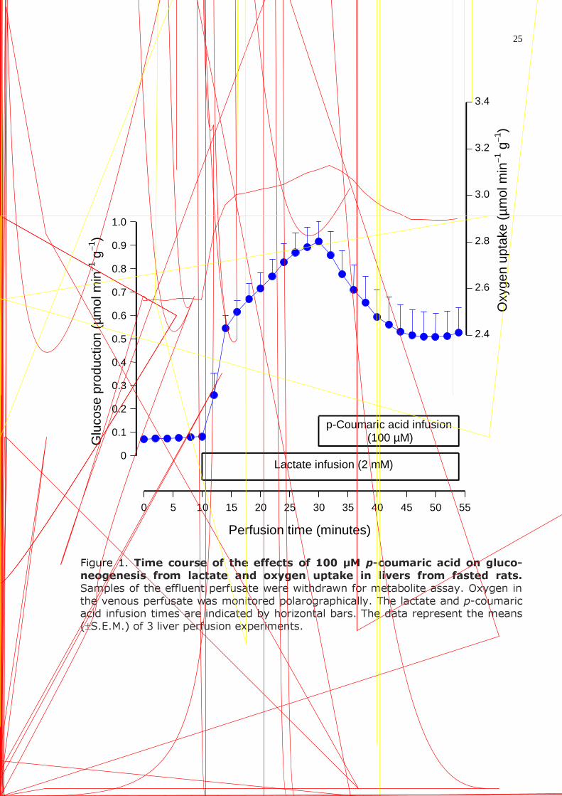
Figure 1. Time course of the effects of 100 µM p-coumaric acid on gluco-neogenesis from lactate and oxygen uptake in livers from fasted rats. Samples of the effluent perfusate were withdrawn for metabolite assay. Oxygen in the venous perfusate was monitored polarographically. The lactate and p-coumaric acid infusion times are indicated by horizontal bars. The data represent the means (±S.E.M.) of 3 liver perfusion experiments.
0
0.1
0.2
0.3
0.4
0.5
0.6
0.7
0.8
0.9
1.0
Glu
cose
pro
duct
ion
(µm
ol m
in−1 g
−1 )
0 5 10 15 20 25 30 35 40 45 50 55
Perfusion time (minutes)
Lactate infusion (2 mM)
p-Coumaric acid infusion (100 µM)
2.4
2.6
2.8
3.0
3.2
3.4
Oxy
gen
upta
ke (
µm
ol m
in−1 g
−1 )
26
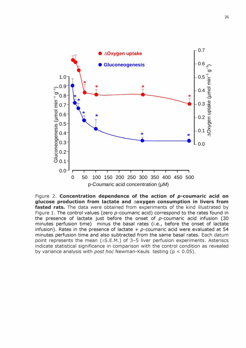
Figure 2. Concentration dependence of the action of p-coumaric acid on glucose production from lactate and ∆oxygen consumption in livers from fasted rats. The data were obtained from experiments of the kind illustrated by Figure 1. The control values (zero p-coumaric acid) correspond to the rates found in the presence of lactate just before the onset of p-coumaric acid infusion (30 minutes perfusion time) minus the basal rates (i.e., before the onset of lactate infusion). Rates in the presence of lactate + p-coumaric acid were evaluated at 54 minutes perfusion time and also subtracted from the same basal rates. Each datum point represents the mean (±S.E.M.) of 3–5 liver perfusion experiments. Asteriscs indicate statistical significance in comparison with the control condition as revealed by variance analysis with post hoc Newman-Keuls testing (p < 0.05).
0 50 100 150 200 250 300 350 400 450 500 0.0
0.1
0.2
0.3
0.4
0.5
0.6
0.7
0.8
0.9
1.0
p-Coumaric acid concentration (µM)
Glu
cone
ogen
esis
(µ
mol
min
−1 g
−1 )
* *
* *
* *
0.0
0.1
0.2
0.3
0.4
0.5
0.6
0.7
∆Oxy
gen
upta
ke (
µm
ol m
in−1 g
−1 )
� ∆Oxygen uptake � Gluconeogenesis
*
* * *
27
Figure 3. Concentration dependence of the action of p-coumaric acid on gluconeogenesis from alanine and ∆oxygen consumption in livers from fasted rats. The data were obtained from experiments of the kind illustrated by Figure 1, but with 2.5 mM alanine as the gluconeogenic substrate. The control values (zero p-coumaric acid) correspond to the rates found in the presence of alanine just before the onset of p-coumaric acid infusion (30 minutes perfusion time) minus the basal rates (i.e., before the onset of alanine infusion). Rates in the presence of alanine + p-coumaric acid were evaluated at 54 minutes perfusion time and also subtracted from the same basal rates. Each datum point represents the mean (±S.E.M.) of 4-5 liver perfusion experiments. Asteriscs indicate statistical significance in comparison with the control condition as revealed by variance analysis with post hoc Newman-Keuls testing (p < 0.05).
0 50 100 150 200 250 300
p-Coumaric acid concentration (µM)
0
0.05
0.10
0.15
0.20
0.25
0.30
0.35
0.40
Glu
cone
ogen
esis
(µ
mol
min
−1 g
−1 )
*
*
* *
0
0.1
0.2
0.3
0.4
0.5
0.6
0.7
∆O
xyge
n up
take
(µ
mol
min
−1 g
−1 )
� ∆Oxygen uptake � Gluconeogenesis
28
Figure 4. Concentration dependence of the action of p-coumaric acid on the metabolic fluxes resulting from fructose metabolism in livers from fasted rats. The data were obtained from experiments of the kind illustrated by Figure 1, but with 5 mM fructose as the gluconeogenic substrate. The control values (zero p-coumaric acid) correspond to the rates found in the presence of fructose just before the onset of p-coumaric acid infusion (30 minutes perfusion time) minus the basal rates (i.e., before the onset of fructose infusion). Rates in the presence of fructose + p-coumaric acid were evaluated at 54 minutes perfusion time and also subtracted from the same basal rates. Each datum point represents the mean (±S.E.M.) of 3–4 liver perfusion experiments. Asteriscs indicate statistical significance in comparison with the control condition as revealed by variance analysis with post hoc Newman-Keuls testing (p < 0.05).
0 50 100 150 200 250 300
0
0.5
1.0
1.5
2.0
2.5
p-Coumaric acid concentration (µM)
Glu
cone
ogen
esis
or ∆o
xyge
n up
take
(µ
mol
min
−1 g
−1 )
* *
*
0
0.3
0.6
0.9
1.2
1.5
1.8
2.1
2.4
2.7
Lact
ate
or p
yruv
ate
pro
duct
ion
(µm
ol m
in−1 g
−1 )
* *
*
� Lactate production
� Pyruvate production
� ∆Oxygen uptake
� Gluconeogenesis

30
Figure 6. Concentration dependences of the actions of p-coumaric acid on several enzymatic activities. Each datum point represents the mean (±S.E.M.) of 4–5 determinations. Asteriscs indicate statistical significance in comparison with the control condition as revealed by variance analysis with post hoc Newman-Keuls testing (p < 0.05).
0 50 100 150 200 250 300 0
5
10
15
20
25
30
35
CO
2 in
corp
orat
ion
(nm
ol m
in−1 m
g−1)
p-Coumaric acid concentration (µM)
A
* *
*
* *
0 100 200 300 400 500
p-Coumaric acid concentration (µM)
0
100
200
300
400
500
600
700
� Pyruvate carboxylase, intact mitochondria
� Pyruvate carboxylase, disrupted mitochondria
� Fructose 1,6-bisphosphatase
� Glucose 6-phosphatase
�
Pho
spha
te r
elea
se (
nmol
min
−1 m
g−1 )
B
0
20
40
60
80
100
� P
hosp
hate
rel
ease
(nm
ol m
in−1 m
g−1 )
31
Figure 7. Concentration dependences of the actions of p-coumaric acid on mitochondrial respiration in the presence of exogenous ADP. Oxygen uptake was measured polarographically as described in materials and methods. The substrate concentrations were: succinate, 7.5 mM; β-hydroxybutyrate, 7.5 mM; pyruvate, 2 mM; ADP, 0.125 mM. Each datum point represents the mean (±S.E.M.) of 5 determinations. Asteriscs indicate statistical significance in comparison with the control condition as revealed by variance analysis with post hoc Newman-Keuls testing (p < 0.05).
0 50 100 150 200 250 300
p-Coumaric acid concentration (µM)
0
2
4
6
8
10
12
14
16
Oxy
gen up
take
(nm
ol
m
in−1
m
g pr
otei
n−1 )
*
* * * *
0
10
20
30
40
50
60
O
xyge
n upta
ke (n
mol
m
in−1
mg
prot
ein−
1 )
� β-Hydroxybutyrate
� Succinate
� Pyruvate
Livros Grátis( http://www.livrosgratis.com.br )
Milhares de Livros para Download: Baixar livros de AdministraçãoBaixar livros de AgronomiaBaixar livros de ArquiteturaBaixar livros de ArtesBaixar livros de AstronomiaBaixar livros de Biologia GeralBaixar livros de Ciência da ComputaçãoBaixar livros de Ciência da InformaçãoBaixar livros de Ciência PolíticaBaixar livros de Ciências da SaúdeBaixar livros de ComunicaçãoBaixar livros do Conselho Nacional de Educação - CNEBaixar livros de Defesa civilBaixar livros de DireitoBaixar livros de Direitos humanosBaixar livros de EconomiaBaixar livros de Economia DomésticaBaixar livros de EducaçãoBaixar livros de Educação - TrânsitoBaixar livros de Educação FísicaBaixar livros de Engenharia AeroespacialBaixar livros de FarmáciaBaixar livros de FilosofiaBaixar livros de FísicaBaixar livros de GeociênciasBaixar livros de GeografiaBaixar livros de HistóriaBaixar livros de Línguas
Baixar livros de LiteraturaBaixar livros de Literatura de CordelBaixar livros de Literatura InfantilBaixar livros de MatemáticaBaixar livros de MedicinaBaixar livros de Medicina VeterináriaBaixar livros de Meio AmbienteBaixar livros de MeteorologiaBaixar Monografias e TCCBaixar livros MultidisciplinarBaixar livros de MúsicaBaixar livros de PsicologiaBaixar livros de QuímicaBaixar livros de Saúde ColetivaBaixar livros de Serviço SocialBaixar livros de SociologiaBaixar livros de TeologiaBaixar livros de TrabalhoBaixar livros de Turismo