avaliaÇÃo do efeito da casca de …repositorio.unicamp.br/bitstream/reposip/255062/1/...vii ao...
TRANSCRIPT

AVALIAÇÃO DO EFEITO DA CASCA DE JABUTICABA LIOFILIZ ADA SOBRE
O GANHO DE PESO, PERFIL LIPÍDICO E RESISTÊNCIA À IN SULINA EM
CAMUNDONGOS ALIMENTADOS COM DIETA HIPERLIPÍDICA.
NATHALIA ROMANELLI VICENTE DRAGANO
PROF. DR. MÁRIO ROBERTO MARÓSTICA JÚNIOR
Orientador
Campinas
2011
Universidade Estadual d e Campinas
Faculdade de Engenharia de Alimentos
Departamento de Alimentos e Nutrição
Curso de Pós-Graduação em Alimentos e Nutrição
Área de Nutrição Experimental e Aplicada à Tecnolog ia de Alimentos

ii
NATHALIA ROMANELLI VICENTE DRAGANO
Bióloga
AVALIAÇÃO DO EFEITO DA CASCA DE JABUTICABA LIOFILIZ ADA SOBRE
O GANHO DE PESO, PERFIL LIPÍDICO E RESISTÊNCIA À IN SULINA EM
CAMUNDONGOS ALIMENTADOS COM DIETA HIPERLIPÍDICA.
Orientador:
Prof. Dr. Mário Roberto Maróstica Júnior
Campinas
2011
Dissertação apresentada à
Faculdade de Engenharia de
Alimentos da Universidade
Estadual de Campinas, para
obtenção do Título de Mestre
em Alimentos e Nutrição - Área
de Nutrição Experimental e
Aplicada à Tecnologia de
Alimentos.
iii
BANCA EXAMINADORA
Este exemplar corresponde à redação final da dissertação defendida por Nathalia
Romanelli Vicente Dragano aprovado pela comissão julgadora em 03 de março de
2011.
________________________________________________
Prof. Dr. Mário Roberto Maróstica Júnior
Orientador
_________________________________________________
Profa. Dra. Alessandra Gambero
Membro
_________________________________________________
Prof. Dr. Dennys Esper Corrêa Cintra
Membro
_________________________________________________
Profa. Dra. Lilia Zago Ferreira dos Santos
Membro
_________________________________________________
Prof. Dr. Márcio Alberto Torsoni
Membro

iv
DEDICO
Aos meus queridos pais, Cássia e Hélvio, e ao Ciro, pelo imenso
amor, incentivo constante e compreensão incondicional a minha
dedicação à pesquisa científica.

v
“Fora da mente humana, e à maneira de um impulso livre, produz-se
a criação da ciência. Esta se renova, assim como as gerações, graças a uma atividade que constitui o melhor jogo do "homo ludens": a ciência
é, no mais estrito e melhor dos sentidos, uma gloriosa diversão.”
Jacques Barzun

vi
AGRADECIMENTOS
À Deus, por sua presença constante na minha vida, por me amparar nos
momentos difíceis e pelo auxílio nas minhas escolhas. Agradeço por me
proporcionar força para lutar, saúde para trabalhar, estudar, viver e sorrir, hoje e
sempre.
À Universidade Estadual de Campinas – UNICAMP e ao Departamento de
Alimentos e Nutrição da Faculdade de Engenharia de Alimentos pela oportunidade
em realizar mais esta etapa da minha formação acadêmica.
À FAPESP e ao CNPq pelo suporte financeiro.
Ao Prof. Mário por ter acreditado em mim e pela oportunidade de trabalharmos
juntos. Agradeço-o, sobretudo pelo incentivo e compreensão ao longo desta
jornada.
À querida Anne, minha eterna gratidão não caberia nestas linhas. Companheira
incansável. Meus sinceros agradecimentos pelo estímulo constante, pelo convívio
diário e pela enriquecedora troca de conhecimentos profissionais e pessoais. Pela
sinceridade, competência e amizade sempre demonstradas, na alegria dos bons
momentos e na ansiedade das horas difíceis, dando-me forças para que eu
continuasse persistindo. Você se fez essencial nessa importante etapa da minha
vida!
Ao Dennys, obrigada por todos os ensinamentos desde o início deste trabalho, por
ter me apresentado um novo mundo de expectativas e por ter entrado em ‘nossas’
vidas. Pelo exemplo de profissionalismo e dedicação. Sua paixão pela Ciência é
contagiante!
vii
Ao Prof. Lício Augusto Velloso meus sinceros agradecimentos por abrir as portas
do Laboratório de Sinalização Celular e pela oportunidade de conviver em
ambiente de grande qualidade científica contribuindo, assim, para meu
crescimento experimental e acadêmico.
Aos Animais Experimentais meu respeito e gratidão. “Vítimas solicitadas pela
ciência para o benefício da humanidade que com tanta dignidade, fizeram-se
imensamente humildes, enquanto ajudavam-nos a entender a vida e a arte de
vivê-la”.
À Luciana Azevedo, minha eterna orientadora. Muito obrigada por guiar meus
primeiros passos no caminho da Ciência. Seus ensinamentos foram fundamentais
para a condução deste trabalho.
Ao Luciano Bruno que me conduziu a este caminho. Muito Obrigada!
A todos os colegas do Laboratório de Sinalização Celular, em especial, a
Andrezza, Carol, Carina, Dani Razolli, Érika Anne, Jose, Lelê e Lucas, muito
obrigada pela valiosa cooperação, pelo convívio agradável e pela paciência em
ensinar detalhes das práticas. Vocês são pessoas íntegras e sempre, de forma
totalmente despretensiosa dispostas a ajudar.
À Alice por dar os primeiros e grandes passos nos estudos com a casca de
jabuticaba.
À Cinthia pela maneira bastante agradável com que nos relacionamos,
aprendemos, construímos e solidificamos a nossa amizade. Exemplo de
determinação, seriedade e compromisso com a pesquisa.
À Glaucia, por enfrentarmos juntas essa jornada, compartilhando as mesmas
emoções e desafios. Obrigada pela amizade e pelos momentos de diversão fora
dos laboratórios. Vamos em frente!
viii
Ao Diogo que acompanhou de perto todo esse caminho mesmo ‘sem querer’.
Agradeço muito por todas as caronas e pela companhia nos momentos de
descontração.
Aos colegas da Pós-Graduação em Alimentos e Nutrição pelos bons momentos
compartilhados.
Aos técnicos de laboratório e funcionários da Pós-Graduação, muito obrigada pela
prestatividade e eficiência com que me atenderam todas as vezes que necessitei.
Agradeço especialmente...
Aos meus queridos pais por abrirem a porta do meu futuro, iluminando meu
caminho com a luz mais brilhante que poderiam encontrar: o estudo. Por serem
não somente pais, mas amigos e companheiros fiéis mesmo nas horas em que
meus ideais pareciam distantes e inatingíveis. Por compartilharem e alimentarem
todos os meus sonhos sempre. Ainda há muito pela frente, ainda há muito a
descobrir e a presença de vocês é fundamental.
Ao meu irmão Ricardo que, do seu jeito, manifesta seu apoio e carinho.. Toda a
minha força e garra para vencer eu devo a você. Sinto sua falta desde o dia que
deixamos de conviver diariamente. Você nunca saberá o tamanho da saudade que
senti...
Ao meu namorado Ciro pelo apoio incessante, amor incondicional e por sonhar
comigo. Desculpe-me pelos momentos nos quais pude estar presente apenas em
pensamentos.
Aos primos Clayton, Dayse e Dudu. Serei eternamente grata por me receberem
de braços abertos e por tornarem-se minha mais nova família. Agradeço também

ix
por suas palavras de encorajamento, suprimento afetivo e cuidado constantes.
Enfim, pela alegria que representam para mim.
À Helena por cuidar tão bem de mim. Pelo carinho de cada gesto e por todos os
lanches nas tardes que passei em frente ao computador. Exemplo de mulher
batalhadora e de honestidade. Muito obrigada.
Às demais pessoas amigas, que direta ou indiretamente, contribuíram não
somente com nesse trabalho, mas em toda a minha vida. Peço-lhes desculpas ao
não mencioná-las, por limitações alheias à minha vontade. A todos esses talentos
o meu muito obrigado.
CERTEZA
De tudo ficaram três coisas:
A certeza de que estamos sempre começando
A certeza de que precisamos continuar
A certeza de que seremos interrompidos antes de terminar
Portanto devemos:
Fazer da interrupção um novo caminho
Da queda, um passo de dança
Do medo, uma escada
Do sonho, uma ponte
Da procura, um encontro
Fernando Pessoa

SUMÁRIO
RESUMO 2
CAPÍTULO 1 3
1. INTRODUÇÃO 3
2. REVISÃO DE LITERATURA 5
2.1 OBESIDADE: EPIDEMIOLOGIA, CONCEITO E IMPLICAÇÕES. 5
2.2 OBESIDADE, RESISTÊNCIA À INSULINA E INFLAMAÇÃO. 7
2.3 ANTOCIANINAS: CARACTERÍSTICAS QUÍMICAS, PROPRIEDADES BIOLÓGICAS E METABOLISMO. 14
2.4 O PAPEL DAS ANTOCIANINAS NO TRATAMENTO E CONTROLE DA OBESIDADE. 16
2.5 JABUTICABA (MYRCIARIA SSP) 19
REFERÊNCIAS BIBLIOGRÁFICAS 22
3. OBJETIVOS 28
OBJETIVO GERAL 28
OBJETIVOS ESPECÍFICOS 28
CAPÍTULO 2 29
FREEZE-DRIED JABOTICABA PEEL POWDER IMPROVES INSULIN SENSITIVITY IN HIGH-FAT FED MICE. 29
ABSTRACT 30
INTRODUCTION 31
MATERIALS AND METHODS 33
RESULTS 40
DISCUSSION 49
LITERATURE CITED 55
CONCLUSÃO 59
ANEXO 60
COMPROVANTE DE APROVAÇÃO DO COMITÊ DE ÉTICA EM EXPERIMENTAÇÃO ANIMAL 60
2
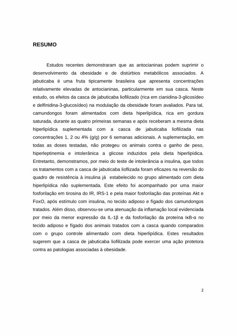
RESUMO
Estudos recentes demonstraram que as antocianinas podem suprimir o
desenvolvimento da obesidade e de distúrbios metabólicos associados. A
jabuticaba é uma fruta tipicamente brasileira que apresenta concentrações
relativamente elevadas de antocianinas, particularmente em sua casca. Neste
estudo, os efeitos da casca de jabuticaba liofilizado (rica em cianidina-3-glicosídeo
e delfinidina-3-glucosídeo) na modulação da obesidade foram avaliados. Para tal,
camundongos foram alimentados com dieta hiperlipídica, rica em gordura
saturada, durante as quatro primeiras semanas e após receberam a mesma dieta
hiperlipídica suplementada com a casca de jabuticaba liofilizada nas
concentrações 1, 2 ou 4% (g/g) por 6 semanas adicionais. A suplementação, em
todas as doses testadas, não protegeu os animais contra o ganho de peso,
hiperleptinemia e intolerânica a glicose induzidos pela dieta hiperlipídica.
Entretanto, demonstramos, por meio do teste de intolerância a insulina, que todos
os tratamentos com a casca de jabuticaba lioflizada foram eficazes na reversão do
quadro de resistência à insulina já estabelecido no grupo alimentado com dieta
hiperlipídica não suplementada. Este efeito foi acompanhado por uma maior
fosforilação em tirosina do IR, IRS-1 e pela maior fosforilação das proteínas Akt e
FoxO, após estímulo com insulina, no tecido adiposo e fígado dos camundongos
tratados. Além disso, observou-se uma atenuação da inflamação local evidenciada
por meio da menor expressão da IL-1β e da fosforilação da proteína IκB-α no
tecido adiposo e fígado dos animais tratados com a casca quando comparados
com o grupo controle alimentado com dieta hiperlipídica. Estes resultados
sugerem que a casca de jabuticaba liofilizada pode exercer uma ação protetora
contra as patologias associadas à obesidade.
3
CAPÍTULO 1
1. INTRODUÇÃO Durante a segunda metade do século XX, populações de vários países do
mundo passaram por mudanças comportamentais que levaram ao aumento do
consumo de alimentos com alta densidade energética e à redução da atividade
física. Tais modificações tiveram consequências marcantes no incremento da
prevalência da obesidade e de doenças associadas como resistência à insulina,
diabetes tipo 2 e doenças cardiovasculares, que exercem forte influência no perfil
de morbi-mortalidade da população (VELLOSO, 2006).
Evidências epidemiológicas sugerem que dietas ricas em frutas e vegetais
podem desempenhar um papel importante na prevenção de várias doenças
crônicas não transmissíveis. Esta capacidade tem sido associada principalmente à
presença de compostos bioativos, dentre estes podemos destacar as
antocianinas, maior grupo de pigmentos solúveis encontrados no reino vegetal
(SCHRÖDER, 2007; CEFALU et al, 2008).
As antocianinas são compostos fenólicos pertencentes à família dos
flavonóides. Diversos estudos já comprovaram que estes pigmentos exibem
efeitos biológicos múltiplos com benefícios potenciais à saúde humana e animal
(HE e GIUSTI, 2010). Trabalhos mais recentes demonstraram que as
antocianinas, assim como frutas e extratos ricos nesses compostos, podem
suprimir o desenvolvimento da obesidade e desordens metabólicas associadas in
vivo.
4
O maior interesse na caracterização dos mecanismos fisiopatológicos
envolvidos com a gênese da obesidade, aliados à necessidade de se
desenvolverem métodos terapêuticos mais eficientes e seguros, servem como
base para o avanço das pesquisas em Nutrição que visam a identificar alimentos e
compostos bioativos capazes de intervir no desenvolvimento desta epidemia
mundial.

5
2. REVISÃO DE LITERATURA
2.1 Obesidade: Epidemiologia, Conceito e Implicações.
A obesidade é reconhecida hoje como um dos mais importantes problemas
de saúde pública em todo o mundo devido a sua contribuição para o aumento
significativo de morte prematura e invalidez precoce, uma vez que esta condição
está intimamente relacionada a um maior risco para o desenvolvimento de uma
série de doenças crônicas não transmissíveis, como resistência à insulina,
diabetes tipo 2, hipertensão arterial sistêmica, dislipidemia aterogênica, doença
hepática gordurosa não-alcoólica, doenças cardiovasculares e certos tipos de
câncer (KOPELMAN, 2000; GALIC et al., 2010).
Em relação às tendências ao longo do tempo, a prevalência da obesidade
começou a aumentar progressivamente no ínicio do último século, mas somente
nas últimas duas décadas as taxas de prevalência tornaram-se alarmantes tanto
em países desenvolvidos quanto em países em desenvolvimento. A natureza
global da epidemia da obesidade foi formalmente reconhecida pela Organização
Mundial de Sáude em 1997 (CABALLERO, 2007). Estimativas recentes indicam
que em 2030 existirão, aproximadamente, 2,16 bilhões de adultos com sobrepeso
e 1,12 bilhões de obesos em todo o mundo (KELLY et al., 2009; WHO, 2009).
Seguindo a tendência mundial, a prevalência de sobrepeso e obesidade no
Brasil também têm aumentado de forma contínua e intensa de acordo com
estudos de larga escala realizados no país (Estudo Nacional sobre Despesa
Familiar, 1974-1975; Pesquisa Nacional sobre Saúde e Nutrição, 1989 e Pesquisa
6
de Orçamentos Familiares, 2002-2003; Pesquisa de Orçamentos Familiares,
2008-2009). Dados da última pesquisa mostram que o excesso de peso atingiu
aproximadamente metade da população de adultos. A situação e a evolução da
prevalência da obesidade no País reproduzem, em linhas gerais, o quadro descrito
para a prevalência do excesso de peso. A obesidade cresceu mais de quatro
vezes entre os homens, de 2,8% em 1973-74 para 12,4% em 2008-09 e mais de
duas vezes entre as mulheres, onde as taxas passaram de 8% para 16,9%. O
problema alcança significância em todas as faixas etárias, classes econômicas e
regiões do País. Além disso, foi observado que, nos últimos seis anos, a
frequência de pessoas com excesso de peso tem aumentado em mais de um
ponto percentual ao ano, isto indica que, se essa tendência se mantiver, em cerca
de dez anos, o excesso de peso poderá alcançar dois terços da população adulta
do Brasil; magnitude idêntica à encontrada na população dos Estados Unidos
(Pesquisa de Orçamentos Familiares, 2008-2009).
A obesidade é resultante da quebra do equilíbrio entre ingestão e gasto
energético, prevalecendo, sutilmente, o primeiro sobre o segundo, o que resulta
num estado de balanço energético positivo, promovendo aumento nos estoques
de energia e, consequentemente, no peso corporal (FLIER, 2004; CABALLERO,
2007). Tal desequilíbrio se deve à interação complexa entre diversos fatores
genéticos, metabólicos, hormonais e ambientais, ainda não totalmente
esclarecidos. Assim, a obesidade não é considerada uma doença singular, e sim
um grupo heterogêneo de condições com múltiplas e causas que, em última
análise, refletem no fenótipo obeso (PEREIRA et al, 2003).

7
O aumento da prevalência da obesidade está relacionado com a mudança
no estilo de vida das populações, ao longo dos anos, o que é expresso por meio
da modificação dos padrões alimentares e de atividade física resultantes da
industrialização, urbanização e desenvolvimento econômico. A adoção de padrões
comportamentais que priorizam atividades profissionais sedentárias, e o aumento
do consumo e da disponibilidade de alimentos altamente energéticos, ricos em
açúcares e gorduras, sobretudo as saturadas, são os principais fatores ambientais
associados ao progressivo aumento da prevalência desta doença nos dias atuais
(POPKIN et al. 2006; MCALLISTER et al. 2009).
2.2 Obesidade, Resistência à Insulina e Inflamaç ão.
Nas últimas décadas observou-se um aumento surpreendente do
conhecimento científico a respeito dos fenômenos fisiopatológicos decorrentes do
acúmulo de gordura corporal.
Originalmente considerado um depósito passivo de energia, atualmente, o
tecido adiposo é considerado um órgão endócrino ativo, que se comunica com o
sistema nervoso central e com tecidos periféricos por meio da secreção de
substâncias, chamadas adipocinas, que regulam múltiplos processos metabólicos,
homeostase energética e resposta imuno-inflamatória (FRANSSEN et al, 2008).
Alterações nos níveis fisiológicos destas adipocinas representam um fator crítico
para o desenvolvimento das comorbidades associadas à obesidade (RITCHIE e
CONNEL, 2007; SINGH et al, 2009).
8
As primeiras evidências que comprovaram a função endócrina do tecido
adiposo aconteceram no início dos anos 90. Em 1994, Friedman e colaboradores
identificaram e caracterizaram o hormônio leptina, cujo nome deriva do grego
leptos (magro), produto do gene Ob (ZHANG et al, 1994) . A leptina é um
hormônio produzido pelo tecido adiposo branco em quantidade diretamente
proporcional à massa total deste tecido. Este hormônio é, sem dúvida, o mais
importante sinal periférico responsável por estabelecer uma conexão entre os
sítios de estoque de energia e o sistema nervoso central; por esta razão, a leptina
é considerada uma molécula-chave na regulação da homeostase energética e
controle da ingestão alimentar, juntamente com a insulina, considerada o segundo
mais importante sinalizador periférico para o hipotálamo (Velloso, 2006). Os
efeitos metabólicos da leptina não se limitam ao hipotálamo; a leptina atua em
vários tecidos periféricos, como fígado, músculo esquelético, pâncreas e o próprio
tecido adiposo onde estimula a oxidação de ácidos graxos, captação de glicose,
inibe a deposição ectópica de TG, reduz os efeitos lipogênicos da insulina e regula
a resposta imune (VÁZQUEZ-VELA et al, 2008; LAGO et al, 2009; MAURY et al,
2009).
Outro estudo identificou que, além das proteínas envolvidas na regulação
do metabolismo, o tecido adiposo também secreta o fator de necrose tumoral
(TNF-α), uma citocina pró-inflamatória. Em 1993, Hotamisligil et al., além de
evidenciarem que o TNF- α tinha sua expressão aumentada no tecido adiposo de
camundongos obesos, verificaram também que a administração de TNF-α
recombinante em cultura de células ou em animais reduz a sensibilidade à

9
insulina. Adicionalmente, os mesmos pesquisadores demonstraram que
camundongos obesos que apresentam TNF-α não funcionante ou que não
expressam receptores de TNF-α apresentam melhora da sensibilidade à insulina,
quando comparados aos camundongos controles.
Estudos posteriores evidenciaram que, além do TNF-α, outros mediadores
da inflamação e citocinas, como as interleucinas 1 beta (IL1-β) e 6 (IL-6), têm sua
expressão aumentada no tecido adiposo e em outros tecidos em modelos
experimentais de obesidade e em humanos, demonstrando que um estado de
inflamação crônica subclínica está associado à obesidade, e que contribui para o
desenvolvimento da resistência à insulina (WELLEN e HOTAMISLIGIL, 2005).
O tecido adiposo de camundongos e humanos obesos é caracterizado por
um acúmulo de macrófagos que é diretamente proporcional à magnitude da
massa adiposa (WEISBERG et al, 2003). Os macrófagos são usualmente
recrutados em locais com dano tecidual e contribuem para o aumento dos níveis
de citocinas pró-inflamatórias (MAURY et al, 2009).
Atualmente, é sabido que citocinas pró-inflamatórias também prejudicam a
sinalização de leptina e insulina em diversos tecidos, inclusive no hipotálamo.
A interferência nos sinais emitidos no hipotálamo pelos hormônios reguladores da
fome prejudica a homeostase local, o que por sua vez culmina no desarranjo
contínuo da ingestão alimentar, perpetuando a falta de controle na ingestão
alimentar e o estabelecimento da obesidade e suas decorrências (SKALICKY et
al., 2008).

10
A insulina é um hormônio polipeptídico anabólico produzido pelas células β
pancreáticas, cuja síntese e secreção são ativadas pelo aumento dos níveis
circulantes de glicose e aminoácidos após as refeições. A insulina atua em vários
tecidos periféricos, incluindo músculo, fígado e tecido adiposo, sendo um
componente essencial para o metabolismo de carboidratos, lipídeos e proteínas
(GRUNDY, 2004). Este hormônio exerce suas ações por meio da sua ligação com
o receptor de insulina (IR), em seguida, este receptor se autofosforila e fosforila
vários substratos da família do receptor de insulina (IRS), iniciando uma cascata
sinalizadora que desencadeia suas atividades metabólicas.
A resistência à insulina pode ser definida como um fenômeno biológico no
qual há uma diminuição da capacidade da insulina endógena ou exógena em
estimular a utilização celular de glicose e manter normais as respostas
metabólicas com ela relacionadas (DIAS et al, 2008). Entretanto, a RI tem como
mecanismo compensador a hiperinsulinemia, resultante da capacidade adaptativa
das células β, que mantêm a homeostase glicêmica; porém quando há falência
nestas células, surge então a intolerância à glicose e, posteriormente, o
estabelecimento da Diabetes Melito tipo 2 (OLEFSKY et al, 2010). Defeitos na via
de sinalização em nível dos pré-receptores, receptores e/ou pós-receptores são
alguns dos possíveis mecanismos que atuam na gênese da RI (MILNAR et al,
2007).
Várias serina/treonina quinases que são ativadas por citocinas pró-
inflamatórias e altos níveis de ácidos graxos livres (AGL) contribuem para a
inibição da sinalização da insulina através da fosforilação do IRS-1 em serina,

11
incluindo a JNK (c-Jun NH2-terminal quinase) e IKKβ (quinase β do inibidor do NF-
κB) (AGUIRRE et al., 2000; AKIRA et al., 2006).
A ativação da JNK leva à fosforilação em serina dos substratos tradicionais
do receptor de insulina (IRSs), o que contribui para resistência à transdução do
sinal da insulina por meio desta via (DEMPSEY et al., 2003). A outra via pró-
inflamatória que pode ser ativada tanto pelo TNF-α quanto pela IL-1β é a
IKK/IκB/NFκB. A IKKβ pode bloquear a sinalização da insulina por pelo menos
dois mecanismos. Primeiramente, a IKKβ pode induzir a fosforilação direta em
serina dos IRSs. Além disso, a IKKβ pode fosforilar o inibidor do NF-κB (IκB) que
culmina na dissociação do complexo IκB/NFκB, levando a ativação do NFκB e sua
translocação para o núcleo, onde este promove a expressão de vários genes
controladores de moléculas pró e antiinflamatórias, alguns dos quais induzem
resistência à ação da insulina (SCHENK et al., 2008; BASTOS et al., 2009).
A resposta inflamatória presente na obesidade é decorrente da interação
entre o sistema metabólico e o sistema imune inato, pois estas são as mesmas
quinases que são ativadas na resposta imune inata pelos Toll-like receptors
(TLRs). O sistema de resposta imunológica mediada por Toll representa uma
família evolucionariamente conservada do sistema de defesa do hospedeiro
(MEDZHITOV et al, 1997).
Os TLRs são proteínas transmembranas responsáveis pela detecção da
invasão do organismo por patógenos. Tais receptores são expressos em várias
células do sistema imune, incluindo macrófagos, células dentríticas, linfócitos B e
tipos específicos de linfócitos T. A expressão dos TLRs também é observada em

12
outros tipos de células como adipócitos, células endoteliais e células do epitélio
intestinal (TAKEDA et al., 2003).
Atualmente existem 12 membros da família dos TLRs identificados em
mamíferos. O TLR-4 é um subtipo de TLRs que é responsável pelo
reconhecimento de lipopolissacarídeo (LPS) de bactérias gram-negativas. Da
mesma forma com que os receptores TLR-4 reconhecem estruturas lipídicas
presentes em agentes invasores, ácidos graxos oriundos do consumo dietético
podem também ser reconhecidos por este sistema, desencadeando a ativação da
via do sistema imune, mesmo na ausência de patógenos (LEE et al, 2001; SHI et
al., 2006; MILANSKI et al, 2009).
Portanto, alguns ácidos graxos ao se ligarem a esse receptor de membrana
celular, acionam proteínas de resposta inflamatória, incluindo a JNK e IKKβ, que
bloqueiam a ação da insulina e atuam no desenvolvimento da resistência à
insulina (Figura 1).

13
Figura 1. Resistência à Insulina e Inflamação. A ativação de serina/quinases na obesidade, especialmente IKKβ e JNK, ressalta a sobreposição das vias metabólicas e inflamatórias: essas são as mesmas quinases ativadas na resposta imune inata pelo TLR (toll-like receptor) em resposta ao LPS e aos ácidos graxos (AGS). (A) Via de sinalização da insulina; (B) Via de sinalização do TLR-4; e (C) Via de sinalização do TNF-α. FONTE: Pauli et al, 2009.

14
2.3 Antocianinas: Características químicas, Proprie dades Biológicas e
Metabolismo.
O padrão dietético desempenha um papel importante na etiologia e
prevenção de várias doenças crônicas não transmissíveis. Evidências
epidemiológicas sugerem que dietas ricas em frutas e vegetais estão associadas à
menor prevalência de obesidade, diabetes tipo 2 e doenças cardiovasculares
(KIEC et al., 2008; ESPÍN et al., 2007).
As antocianinas (do grego anthos = flor; kianos = azul) são metabólitos
secundários de plantas responsáveis pela pigmentação vermelha, azul e violeta de
diferentes tecidos vegetais, tais como, flores, frutos, folhas, sementes e raízes
(LIEBERMAN et al., 2007). Na alimentação humana, estes compostos estão
disponíveis em diversas frutas como amoras, cerejas, pêssegos, uvas, romãs,
ameixas assim como muitas verduras de cor escura, como rabanete, feijão preto,
cebola roxa, berinjela, repolho roxo e batata doce, representando componentes
substanciais da fração não energética da dieta humana (BOBBIO et al., 2000;
BRITO et al., 2007; HE e GIUSTI, 2010).
Quimicamente, as antocianinas pertencem à grande classe dos compostos
fenólicos e são classificadas como flavonóides devido à estrutura característica de
sua cadeia carbônica (C6-C6-C6), na qual os dois anéis aromáticos estão
separados por um anel heterocíclico intermediário. São moléculas glicosiladas,
polihidroxi ou polimetóxi derivadas do cátion 2-fenilbenzopirilium, também
conhecido por cátion flavilíco, e apresentam uma rica diversidade estrutural,
devido a diferenças no número de grupos hidroxila na molécula, grau de metilação

15
destes grupos hidroxila, natureza, número e posição de moléculas de açúcares
ligadas aos anéis aromáticos, bem como a natureza e o número de ácidos
alifáticos ou aromáticos anexado aos açúcares (MCGUIE e WALTON, 2007). A
figura 2 ilustra a estrutura genérica de algumas antocianinas.
Os interesses da comunidade científica em relação aos aspectos
bioquímicos e efeitos biológicos das antocianinas aumentaram substancialmente
durante a última década em razão de inúmeras evidências que demonstram seu
amplo potencial terapêutico. A propriedade melhor descrita das antocianinas é a
sua atividade antioxidante, a qual está intimamente relacionada com outros efeitos
promotores da saúde, como redução do risco de doenças cardiovasculares,
prevenção de certos tipos de câncer e de sua progressão, inibição da agregação
plaquetária, ação antiinflamatória e moduladora da resposta imune, proteção
contra déficits neuronais associados com o envelhecimento e doenças
neurodegenerativas (TALAVERA et al, 2005; HE et al, 2010).
Figura 2. Estrutura genérica das antocianinas a partir do esqueleto das antocianidinas (agliconas). Onde R1: H ou substituinte glicosídico; R2 e R3: H ou CH3
16
A atividade biológica das antocianinas está intimamente relacionada com a
sua biodisponibilidade de forma que a quantidade deste composto presente nos
alimentos não reflete, necessariamente, a quantidade absorvida e metabolizada
pelo organismo (McGUIE e WALTON, 2007; BIESALKI et al., 2009).
Em geral, as antocianinas são absorvidas rapidamente no estômago e
intestino delgado após a ingestão, sobretudo na sua forma glicosilada intacta.
Após a absorção, elas entram na circulação sistêmica após passagem pelo fígado.
Neste órgão, uma parte destes compostos pode ser metabolizada por reações de
metilação e/ou glucoronidação, que são consideradas as duas rotas majoritárias
no metabolismo das antocianinas. Além destas conversões enzimáticas, estes
pigmentos podem ser degradados pela microflora intestinal em moléculas de
açúcar, ácidos fenólicos e aldeídos e são, posteriormente, absorvidos no cólon
(HE e GIUSTI, 2010; SHIPP e ABDEL-AAL, 2010).
Entretanto, o entendimento dos fatores que levam à liberação destes
compostos da matriz do alimento, a extensão da absorção dos mesmos e o modo
por meio do qual a diversidade estrutural e, a consequente grande quantidade de
metabólitos gerados afetam sua biodisponibilidade e eficácia biológica, ainda não
foram completamente esclarecidos.
2.4 O papel das Antocianinas no Tratamento e Contro le da obesidade.
Nos últimos anos, observou-se um avanço substancial no conhecimento
científico sobre os compostos biotivos presentes em alimentos e seus efeitos no

17
tratamento e prevenção da obesidade. Dentre os fitoquimícos que são conhecidos
por exercerem efeitos anti-obesidade in vivo encontram-se as antocianinas.
Neste sentido, alguns estudos têm demonstrado que as antocianinas,
provenientes de frutos e vegetais ricos neste composto, podem não só suprimir o
desenvolvimento da obesidade, mas também melhorar diversos parâmetros
metabólicos associados, in vivo.
O primeiro trabalho, neste sentido, foi realizado por Tsuda e colaboradores
(2003). Estes pesquisadores comprovaram que o corante de milho roxo (Purple
corn color), rico em cianidina 3-O-β-glicosídeo, reduziu o ganho de peso, os níveis
séricos de glicose e insulina e ainda normalizou a expressão de TNF-α no tecido
adiposo de camundongos alimentados com dieta hiperlipídica. Em trabalhos
subsequentes, estes autores mostraram, por meio da técnica de Microarray, que a
cianidina 3-O-β-glicosídeo, modula expressão de adipocinas devido ao aumento
na expressão de adiponectina e leptina, diminuição na expressão de IL-6 e do
inibidor da ativação de plasminogênio 1 (PAI-1), além de modular a expressão de
genes relacionados com o metabolismo lipídico em adipócitos isolados de ratos e
de humanos (TSUDA et al., 2004; 2005; 2006).
Paralelamente, Jayaprakasan et al. (2006) demonstraram que as
antocianinas purificadas de Cornelian cherry, quando adicionadas à dieta
hiperlipídica (0,01%) melhoraram a obesidade e resistência à insulina em
camundongos. O tratamento com antocianinas, durante oito semanas, reduziu
24% do ganho de peso, independente do consumo, melhorou a tolerância a
glicose e diminuiu a acumulação de lipídios no fígado.
18
Em outro estudo, a dieta hiperlipídica foi suplementada com 10% de soja
negra ou com 0,04% de antocianinas isoladas da casca da soja. Ambos os
tratamentos reduziram significativamente o ganho de peso e melhoraram o perfil
lipídico dos ratos por meio da diminuição do colesterol total e triglicérides e
aumento dos níveis séricos de HDL colesterol (KNOW et al., 2007).
Ao contrário, outro grupo de pesquisadores (PRIOR et al., 2008) observou
que a suplementação de dietas hiperlipídicas com Blueberries ou com morangos
inteiros liofilizados (10%) promoveu maior ganho de peso e adiposidade; no
entanto, os animais alimentados com as antocianinas purificadas destes frutos
exibiram menor ganho de peso quando comparados ao grupo controle. Em
estudos subseqüentes, estes pesquisadores demonstraram também que o
tratamento tanto com o fruto inteiro liofilizado quanto com as antocianinas
purificadas de Raspberry também não alteraram o desenvolvimento da obesidade
induzida por dieta hiperlipidíca (PRIOR et al.,2010).
Em 2008, Sasaki e colaboradores sugeriram que a cianidina-3-O-β-
glicosídeo, purificada do corante de milho roxo, melhora a sensibilidade a insulina
e hiperglicemia por meio da diminuição da expressão da proteína 4 transportadora
de retinol (RBP4) e aumento da expressão do transportador de glicose 4 (GLUT4)
no tecido adiposo de camundongos diabéticos tratados com a antocianina (0,02%
dieta) em questão. Este efeito foi também acompanhado pela expressão
diminuída de marcadores inflamatórios, como o TNF-α e a proteína quimiotáxica
de monócitos 1 (MCP-1).
19
Resultados similares foram obtidos por De Furia et al, 2009. Em seu estudo,
camundongos foram alimentados com dieta hiperlipídica acrescida de 4% de
Blueberry liofilizada. Embora a suplementação não tenha reduzido o ganho de
peso e adiposidade induzida pela dieta, foram observadas melhoras na resistência
à insulina, hiperglicemia e na inflamação do tecido adiposo devido à menor
expressão de genes relacionados à inflamação e à menor infiltração de
macrófagos neste tecido.
Seymour e colaboradores (2009), também observaram uma diminuição do
estado pró-inflamatório crônico associado à obesidade. Neste estudo, a
suplementação da dieta hiperlipídica com Tart Cherries inteiras e liofilizadas (1%)
foi associada com expressão reduzida da atividade do NFκB, TNF-α e IL-6.
Assim, as informações disponíveis na literatura são cada vez mais
favoráveis à ação anti-obesogênica das antocianinas; porém, ainda não são
completamente entendidos os mecanismos pelos quais estes pigmentos são
capazes de modular as complexas vias metabólicas que atuam na gênese da
obesidade.
2.5 Jabuticaba (Myrciaria ssp)
A jabuticabeira (Myrciaria spp.) é uma árvore frutífera nativa do Brasil,
encontrada em extensa faixa do país, desde o Pará até o Rio Grande do Sul,
porém exibe maior ocorrência e produtividade nos estados da região Sudeste
(SATO e CUNHA, 2007). A frutificação ocorre duas vezes por ano, de janeiro a
fevereiro e de novembro a dezembro, sendo esta última a safra de maior

20
produção. O fruto da jabuticabeira apresenta de 3-4 cm de diâmetro contendo de
uma a quatro sementes grandes, com uma espessa casca roxa que recobre uma
polpa gelatinosa, branca e intensamente doce.
Dentre as espécies atualmente conhecidas, destaca-se a Myrciaria
cauliflora (DC) Berg (jabuticaba Paulista ou Açu) e a Myrciaria jaboticaba (Vell)
Berg (jabuticaba Sabará), sendo esta última a mais apreciada e doce das
jabuticabas além de ocupar a maior área cultivada no Brasil (DONADIO, 2000;
MATTOS, 1983).
A jabuticaba é utilizada para vários fins culinários e medicinais. Geralmente,
o fruto é consumido in natura ou utilizado no preparo de geléias e licores. Na
medicina popular, sua casca tem sido utilizada no tratamento contra
uma variedade de condições inflamatórias, incluindo hemoptise, asma, diarréia e
amigdalite (LIMA et al., 2008).
Além disso, a jabuticaba é um fruto bastante interessante do ponto de vista
nutricional. A polpa apresenta quantidades consideráveis de vitamina C e
minerais, onde se destaca o ferro, cálcio, fósforo e potássio, já a casca, embora
normalmente desprezada, é rica em minerais, fibras solúveis e insolúveis e
também apresenta quantidades relevantes de antocianinas (OLIVEIRA et al.,
2003). Reynertson (2007) determinou o teor de antocianinas do fruto inteiro e os
valores encontrados foram de 433 mg para a cianidina-3-O-β-glicosídeo e 81mg
por 100 g de peso seco para a delfinidina-3-O-β- glicosídeo. Estudos realizados
em nosso laboratório, para a identificação e quantificação das antocianinas da
casca de jabuticaba liofilizada, revelaram a presença de delfinidina-3-O-β-
21
glicosídeo e cianidina-3-O-β-glicosídeo, os valores encontrados para estas duas
antocianinas foram de 635,3 e 1964 mg/100 de peso seco, respectivamente.
Apesar de existir um consumo pronunciado de jabuticaba, principalmente
na região sudeste, há poucos estudos na literatura sobre seus constituintes
químicos e propriedades funcionais. Além disso, conhecimentos mais
aprofundados dos compostos bioativos presentes na jabuticaba, assim como de
possíveis efeitos promotores de saúde são imprescindíveis, uma vez que irão
contribuir para um melhor aproveitamento e, consequentemente, para a maior
valorização econômica desta fruta tipicamente brasileira.
22
REFERÊNCIAS BIBLIOGRÁFICAS
AGUIRRE, V. et al. The c-Jun NH(2)-terminal kinase promotes insulin resistance during association with insulin receptor substrate-1 and phosphorylation of Ser(307). The Journal of Biological Chemistry , v. 275, p.9047-9054, 2000.
AKIRA, S. et al. Pathogen recognition and innate immunity. Cell , v. 124, p. 783-801, 2006. BASTOS, D.H.M.; ROGERO, M.M.; ARÊAS, J.A.G. Mecanismos de ação de compostos bioativos dos alimentos no contexto de processos inflamatórios relacionados à obesidade. Arquivos Brasileiros de Endocrinologia e Metabologia , v. 53, p. 646-656, 2009. BIESALSKI, H.K. et al. Bioactive compounds: Definition and assessment of activity. Nutrition , v. 25, p. 1202–1205, 2009. BOBBIO, F.O. et al. Identificação e quantificação da antocianinas do fruto do açaizeiro (Euterpe oleracea). Ciência e Tecnologia de Alimentos ,v.20, n.3, 2000. BRITO, E.S. et al. Anthocyanins present in selected tropical fruits: acerola, jambolão, jussara, and guajiru. Journal of Agricutural and Food Chemistry, v. 55, n. 23, p. 9389–9394, 2007. CABALLERO, B. The Global Epidemic of Obesity: An Overview. Epidemiologic Reviews , v. 29, p.1–5, 2007. CEFALU, W.T. et al. Botanicals and the metabolic syndrome. The American Journal of Clinical Nutrition, v. 87, p.481S–7S, 2008. DEFURIA, J. et al. Dietary Blueberry Attenuates Whole-Body Insulin Resistance in High Fat-Fed Mice by Reducing Adipocyte Death and Its Inflammatory Sequelae. Journal of Nutrition , v. 139, n.8, p.1510-1516,2009. DEMPSEY, P.W. et al. The signaling adaptors and pathways activated by TNF superfamily. Cytokine & Growth Factor Reviews , v.14, p. 193-209, 2003. DIAS, J.C.R.; SANTOS, L. C.; COUTINHO, V.F. Aspectos clínicos e nutricionais na síndrome metabólica. Revista Brasileira de Nutrição Clínica, v. 24, n.1, p.72-78, 2009. DONADIO, L.C. Jabuticaba (Myrciaria jaboticaba (Vell.)Berg). Jaboticabal: FUNEP, 55 p., 2000.
23
DUARTE, O.; LUDDERS, P.; HUETE, M. Extending storage life of jaboticaba (Myrciaria cauliflora Berg) fruits. 14° Congresso Brasileiro de Fruticultura , 1996, Curitiba, PR, Resumos..., Curitiba-PR:SBF, p. 556, 1996. ESPÍN, J.C.; GARCÍA-CONESA, M.T.; TOMÁS-BARBERÁN, F,A. Nutraceuticals: Facts and fiction. Phytochemistry, v. 68, p.2986–3008, 2009. FLIER, J.S. Obesity wars: molecular progress confronts an expanding epidemic. Cell , v. 16, n.2, p.337-50, 2004. FRANSSEN, R. et al. Obesity and Dyslipidemia. Endocrinology Metabolism Clinics of North America, v. 37, p.623–633, 2008. GALIC, S.; OAKHILL, J.S.; STEINBERG, G.R. Adipose tissue as an endocrine organ. Molecular and Cellular Endocrinology , v. 316, p.129–139, 2010. GRUNDY, S. What is the contribution of obesity to the metabolic syndrome? Endocrinology Metabolism Clinics of North America, v. 33, p.267–282, 2004. GUTIERREZ, D.A.; PUGLISI, M. J.; HASTY, A.H. Impact of increased adipose tissue mass on inflammation, insulin resistance, and dyslipidemia. Current Diabetes Reports , v.9, n.1, p.26–32, 2009. HE, J.; GIUSTI, M.M. Anthocyanins: Natural Colorants with Health-Promoting Properties. Annual Review of Food Science and Technology , v. 1, p. 163–87, 2010. HOTAMISLIGIL, G.S. et al. Adipose expression of tumor necrosis factor-alpha: direct role in obesity-linked insulin resistance. Science , v.259, p. 87-91, 1993. IBGE (Instituto Brasileiro de Geografia e Estatísticas). Pesquisa de Orçamentos Familiares 2008-2009. Rio de Janeiro: IBGE, 2010. Disponível em: <http://www.ibge.gov.br/home/estatistica/populacao/condicaodevida/pof/2008_2009_encaa/pof_20082009_encaa.pdf>. Acesso em: Nov. 2010. JAYAPRAKASAM B. et al. Amelioration of obesity and glucose intolerance in highfat-fed C57BL/6 mice by anthocyanins and ursolic acid in cornelian cherry (Cornus mas). Journal of Agricutural and Food Chemistry , v. 54, p.243–248, 2006. KELLY, T. et al. Global burden of obesity in 2005 and projections to 2030. International Journal of Obesity , v. 32, p. 431–1437, 2008. KIEC, A.D. et al. Antioxidant phytochemicals against type 2 diabetes. British Journal of Nutrition , v. 99, p. ES109–ES117, 2008.

24
KOPELMAN P. G. Obesity as a medical problem. Nature , v. 404, p.635- 643, 2000. KWON, S. et al. Anti-Obesity and Hypolipidemic Effects of Black Soybean Anthocyanins. Journal of Medicinal Food , v. 10, n.3, p. 552-556, 2007. LAGO, F. et al. Adipokines as novel modulators of lipid metabolism. Trends in Biochemical Sciences , v. 34, n.10, p. 500-510, 2009.
LEE J.Y. et al. Saturated fatty acids, but not unsaturated fatty acids, induce the expression of cyclooxygenase-2 mediated through Toll-like receptor 4. The Journal of Biological Chemistry , v. 276, p.16683-16689, 2001.
LIEBERMAN, S. The antioxidant Power of purple corn: A research review. Alternative & Complementary Therapies , 2007. LIMA, A.J.B. et al. Caracterização química do fruto jabuticaba (Myrciaria cauliflora Berg) e de suas frações. Archivos Latinoamericanos de Nutricion , v. 58, n. 4, p. 416-421, 2008. MATTOS, J.R. Frutíferas nativas do Brasil: jaboticabeiras . Porto Alegre, 92p., 1983. MAURY, E.; BRICHARD, S.M. Adipokine dysregulation, adipose tissue inflammation and metabolic syndrome. Molecular and Cellular Endocrinology , v. 314, p.1–16, 2010. MCALLISTER, E.J. et al. Ten Putative Contributors to the Obesity Epidemic. Critical Reviews in Food Science and Nutrition , v. 49, n.10, p. 868–913, 2009. MCGHIE T.K.; WALTON M.C. The bioavailability and absorption of anthocyanins: Towards a better understanding. Molecular Nutrition & Food Research , v. 51, p. 702-713, 2007. MEDZHITOV R. et al. A human homologue of the Drosophila Toll protein signals activation of adaptive immunity. Nature , v.388, p.394-397, 1997. MESHKANI, R.; ADELI, K. Hepatic insulin resistance, metabolic syndrome and cardiovascular disease. Clinical Biochemistry , v. 42, p.1331–1346, 2009. MILANSKI M. et al. Saturated fatty acids produce an inflammatory response predominantly through the activation of TLR4 signaling in hypothalamus:

25
implications for the pathogenesis of obesity. The Journal of Neuroscience ,v.29, p.359-70, 2009. MLINAR, B. et al. Molecular mechanisms of insulin resistance and associated diseases. Clinica Chimica Acta , v.375, p.20-35, 2007. OLEFSKY, J.M.; GLASS, C.K. Macrophages, Inflammation,and Insulin Resistance. Annual Review of Physiology , v. 72, p.219–46, 2010. OLIVEIRA, A.L. et al. Caracterização tecnológica de jabuticabas Sabará provenientes de diferentes regiões de cultivo. Revista Brasileira de Fruticultura , v. 25, p. 397-400, 2003. PAULI, J.R. et al. Novos mecanismos pelos quais o exercício físico melhora a resistência à insulina no músculo esquelético. Arquivos Brasileiros de Endocrinologia e Metabolismo, v.53, p. 399-408, 2009. POPKIN, B.M. et al. Measuring the full economic costs of diet, physical activity and obesity-related chronic diseases. Obesity Reviews , v. 7, p. 271–293, 2006. PRIOR, R.L. et al. Whole Berries versus Berry Anthocyanins: Interactions with Dietary Fat Levels in the C57BL/6J Mouse Model of Obesity. Journal of Agricutural and Food Chemistry , v. 56, p.647–653, 2008. PRIOR, R.L. et al. Dietary black raspberry anthocyanins do not alter development of obesity in mice fed an obesogenic high-fat diet. Journal of Agricutural and Food Chemistry 2010, v. 58, p. 3977-3983, 2010. REEVES, P.G.; NIELSEN, F.H.; FAHEY JR., G.C. AIN-93 Purified diets for laboratory rodents: final report of the American Institute of Nutrition Ad Hoc Writing Committee on the Reformulation of the AIN-76A rodent diet. Journal of Nutrition , v.123, p.1939-1951, 1993. REYNERTSON, K.A. Phytochemical analysis of bioactive constituents fr om edible myrtaceae fruits . New York: Tese-Graduate Faculty in Biology- City University of New York, 2007. RITCHIE, S.; CONNELL, J. The link between abdominal obesity, metabolic syndrome and cardiovascular disease. Nutrition, Metabolism and Cardiovascular Diseases , v. 17, n. 4, p.319-326, 2007. SASAKI R. et al. Cyanidin 3-glucoside ameliorates hyperglycemia and insulin sensitivity due to downregulation of retinol binding protein 4 expression in diabetic mice. Biochemical Pharmacology , v.74, p. 1619-1627, 2007.
26
SATO A.C.K e CUNHA R.L. Influência da temperatura no comportamento reológico da polpa de jabuticaba. Ciência e Tecnologia de Alimentos , v. 27, n.4, p. 890-896, 2007. SCHENK, S.; SABERI, M.; OLEFSKY, J.M. Insulin sensitivity: modulation by nutrients and inflammation. The Journal of Clinical Investigation , v. 118, p.2992–3002, 2008. SCHRÖDER, H. Protective mechanisms of the Mediterranean diet in obesity and type 2 diabetes. Journal of Nutritional Biochemistry , v. 18, p.149–160, 2007. SEYMOUR, E.M. et al. Regular Tart Cherry Intake Alters Abdominal Adiposity, Adipose Gene Transcription, and Inflammation in Obesity-Prone Rats Fed a High Fat Diet. Journal of Medicinal Food , v. 2, n.5, p. 935–942, 2009. SHI, H. et al. TLR4 links innate immunity and fatty acid-induced insulin resistance. The Journal of Clinical Investigation , v. 116, p.3015-25, 2006.
SHIPP, J.; ABDEL-AAL, E-S.M. Food Applications and Physiological Effects of Anthocyanins as Functional Food Ingredients. The Open Food Science Journal , v. 4,p. 7-22, 2010.
SINGH, B. et al. Metabolic syndrome: A review of emerging markers and management. Diabetes & Metabolic Syndrome: Clinical Research & Reviews, v. 3, n. 3, p. 240-254, 2009. SKALICKY, J. et al. Evaluation of oxidative stress and inflammation in obese adults with metabolic syndrome. Clinical Chemistry and Laboratory Medicine, v.46, n.4, p. 499-505, 2008. TALAVÉRA, S. et al. Anthocyanin metabolism in rats and their distribution to digestive area, kidney, and brain. Journal of Agricutural and Food Chemistry, v.53, n.10, p.3902-3908, 2005. TAKEDA K. et al. Toll-like receptors. Annual Review of Immunology , v.21, p.335-376, 2003. TSUDA, T. et al. Anthocyanin enhances adipocytokine secretion and adipocyte-specific gene expression in isolated rat adipocytes. Biochemical and Biophysical Research Communications , v.316, p.149–57, 2004. TSUDA, T. et al. Dietary cyanidin 3-O-b-D-glucoside-rich purple corn color prevents obesity and ameliorates hyperglycemia in mice. Journal of Nutrition , v.133, p.2125–30, 2003.

27
TSUDA, T. et al. Gene expression profile of isolated rat adipocytes treated with anthocyanins. Biochimica et Biophysica Acta , v.1733, p.137– 147, 2005. TSUDA, T. et al. Microarray profiling of gene expression in human adipocytes in response to anthocyanins. Biochemical Pharmacology , v.71, p.1184–97, 2006. VÁZQUEZ-VELA, M. et al. White Adipose Tissue as Endocrine Organ and Its Role in Obesity. Archives of Medical Research , v. 39, p.715-728, 2008. VELLOSO L.A. O Controle Hipotalâmico da Fome e da Termogênese – Implicações no Desenvolvimento da Obesidade. Arquivos Brasileiros de Endocrinologia e Metabolismo , v. 50, n. 2, p.165-176, 2006. WEISBERG, S.P. et al. Obesity is associated with macrophage accumulation in adipose tissue. Journal of Clinical Investigation , v. 112, p.1796-1808, 2003. WELLEN, K.E.; HOTAMISLIGIL, G.S. Inflammation, stress, and diabetes. Journal of Clinical Investigation , v. 115, p. 1111-1119, 2005. ZHANG, Y. et al. Positional cloning of the mouse obese gene and its human homologue. Nature , v. 372, p.425-432, 1994.

28
3. OBJETIVOS
Objetivo Geral
Os objetivos do presente trabalho foram investigar os efeitos da casca de
jabuticaba liofilizada, rica em antocianinas, sobre o ganho de peso, perfil lipídico e
resistência à insulina em camundongos Swiss alimentados com dieta hiperlipídica.
Objetivos Específicos
1) Avaliar e monitorar a ingestão dietética e o ganho de peso em
camundongos submetidos às dietas experimentais durante todo o
experimento;
2) Determinar os níveis séricos de colesterol total, triglicérides e HDL
colesterol;
3) Determinar os níveis séricos de leptina;
4) Analisar a tolerância à glicose, pelo teste de tolerância à glicose (GTT) e a
sensibilidade periférica à insulina pelo teste de tolerância à insulina (ITT);
5) Avaliar o efeito da suplementação com a casca de jabuticaba liofilizada
sobre a transdução do sinal da insulina em fígado e tecido adiposo;
6) Avaliar o efeito da suplementação com a casca de jabuticaba liofilizada
sobre a expressão de marcadores inflamatórios em fígado e tecido adiposo.
29
CAPÍTULO 2
Freeze-dried jaboticaba peel powder improves insuli n sensitivity
in high-fat fed mice .

30
ABSTRACT
The peel of the native Brazilian fruit jaboticaba, is rich in anthocyanins
(ACNs), which are known for its anti-obesity effects in animal models. Here, we
evaluated the effects of freeze-dried jaboticaba peel powder on a number of
metabolic parameters in a model of experimental obesity.. Mice were initially fed on
a high-fat diet (HFD) for four weeks and then switched to a high-fat diet
supplemented with freeze-dried jaboticaba peel powder (1, 2 or 4% wt:wt) for
additional six weeks. The freeze-dried jaboticaba peel powder exerted no
protective effect on HFD-induced weight gain, hyperleptinemia and glucose
intolerance. However, the supplementation was effective to reduce insulin
resistance as determined by improved signal transduction through the insulin
receptor (IR)/insulin receptor substrate-1 (IRS1)/Akt/forkhead box protein (FoxO)
pathway and by the attenuation of inflammatory signaling. These results suggest
that freeze-dried jaboticaba peel powder may exert a protective role against
obesity-associated insulin resistance.
Keywords : Obesity; insulin resistance; jaboticaba; Myrciaria jaboticaba;
anthocyanins.

31
INTRODUCTION
Obesity has reached epidemic proportions in the world, with substantial adverse
consequences for human health (1). A number of life-threatening diseases such as,
type 2 diabetes mellitus, dyslipidemia, cardiovascular diseases, hypertension and
certain types of cancer are significantly associated with this condition (2).
Nutritional factors are known to play an important role in the development of a
number of diseases. The recent changes in human life-style with the introduction of
carbohydrate and fat-rich diet standards is amongst the most important factors
leading to the increased prevalence of obesity. Conversely, epidemiological data
suggest that increased consumption of fruits, vegetables, legumes, nuts, whole grains
and olive oil are favorably associated with the prevention of metabolic diseases
including obesity, and by the reduction of cardiovascular disease, hypertension and
diabetes (3, 4). The property of some vegetable-derived nutrients to reduce the risk of
chronic diseases has been associated, at least in part, to the occurrence of the
bioactive non-nutrient compounds which are known to exert a wide range of biological
activities (5, 6).
Anthocyanins (ACNs) are an example of such bioactive compounds. They
constitute the largest and probably the most important group of water-soluble natural
pigments responsible for the vivid blue, purple, and red color of many vegetables and
fruits (7). Many reports have shown that anthocyanin exhibits an array of
pharmacological properties, such as anti-inflammatory and anticarcinogenic activities,
as well as preventive effects on cardiovascular and degenerative diseases. All the
32
putative health-promoting effects are more or less associated with their potent
antioxidant property, related to their chemical characteristics (8).
The recent findings have suggested that the consumption of ACNs may
reduce obesity and some of its associated disorders, at least in animal models.
Tsuda and coworkers (9) demonstrated that diet supplemented with purple corn
color, rich in cyanidin-3-glucoside, significantly suppressed the gain of body weight
and also improved the insulin sensitivity and hyperlipidemia in high-fat-fed mice.
ACNs from the Cornelian cherry and black soybeans are also reported to
effectively reverse the obesity and improve certain metabolic parameters
associated with diets high in saturated fats (10, 11). However, additional in vivo
studies have reported that whole powdered berries in a high-fat diet, compared to
purified anthocyanins, were not quite effective in preventing obesity and, actually,
they showed a potential to increase obesity on the models used on the study (12,
13).
Jaboticaba (Myrciaria jaboticaba) is an indigenous Brazilian fruit found in
extense areas of the country, particularly in the Southeast. The mature fruits are
round and have a thick, dark purple, astringent skin that covers a sweet, white,
gelatinous flesh. The fruit is widely used in Brazil, where it is mostly consumed
fresh and, to a smaller extent, it is used to make jams and liqueurs. Its peel is a
rich source of biologically active phenolic compounds, containing high
concentrations of anthocyanins, particularly cyanidin-3-glucoside and delphinidin-3-
glucoside, similar to other well-studied berries and fruits (14, 15).
33
The current study was designed to investigate the effect of freeze-dried
jaboticaba peel powder on weight loss, glucose tolerance, insulin resistance and
lipid profile in high-fat-fed mice. The results show that, although no changes in
body mass were achieved, freeze-dried jaboticaba peel powder significantly
improved insulin signal transduction and inflammatory signaling.
MATERIALS AND METHODS
Antibodies, chemicals, and buffers. All the reagents for SDS-PAGE and
immunoblotting were from Bio-Rad (Richmond, CA, USA). HEPES,
phenylmethylsulfonyl fluoride, aprotinin, dithiothreitol, Triton X-100, Tween 20,
glycerol, and BSA (fraction V) were purchased from Sigma Chemical Co. (St.
Louis, MO, USA). The human regular insulin (Novolin R) was from Novo Nordisk
(Clayton, CN, USA). The antibodies against phospho-insulin receptor β (IR)
(#3070, rabbit monoclonal), phospho–insulin receptor substrate (IRS)-1 (#3026,
rabbit polyclonal) and phosphorylated- forkhead transcription factor box (FoxO)
(#9464, rabbit monoclonal) were from Cell Signaling (Danvers, MA, USA). Anti-
phospho-Akt (sc-7985-R, rabbit polyclonal), phospho-inhibitor of NFκB alpha (IκB-
α) (sc-7977-R, rabbit polyclonal) and interleukin 1 β (IL-1β) (sc-1252, goat
polyclonal) were from Santa Cruz Biotechnology (Santa Cruz, CA, USA).
Chemicals for Real-time PCR were from Invitrogen (Carlsbad, CA, USA) and
Applied Biosystems (Foster City, CA, USA). Intron-skipping primer for IL-1β
(Rn00580432_m1) and Glyceraldehyde-3-phosphate dehydrogenase (GAPD)
(#4352339E) was obtained from Applied Biosystems.

34
Jaboticaba. The jaboticaba fruits (Myrciaria jaboticaba Vell berg) were
obtained directly from producer (Aguaí, São Paulo State, Brazil), during main
harvest season in September 2008.
Preparation of freeze-dried jaboticaba peels . The fruits were manually
washed with fresh water and the peels were separated and frozen at -20 °C. The
frozen peels were lyophilized and grounded into fine powder by an electrical mill.
The freeze-dried powdered was kept in airtight containers and stored at -80 °C until
the time of further use.
Animals and diets. Male Swiss inbred strain mice (21 days of age), originally
imported from the Jackson Laboratory and currently bred at the University of
Campinas Breeding Center were used in the study. The investigation was
approved by Ethics Committee for Animal (Permission: 2020-1/2010) and followed
the University guidelines for the use of animals in experimental studies. The
animals were maintained at 21 ± 3 °C, on a 12 h art ificial light/dark cycle and
housed in individual cages. They were then randomly divided into five groups with
eight animals per treatment (following an initial acclimatization period for 7 days).
All groups were balanced for initial body weight. The treatments were Control
group (C): mice were fed on normal diet, based on the AIN-93G (16), containing
7% (wt/wt) soybean oil (15); Control High fat group (HF): mice were fed on high-fat
diet, containing 4% (wt/wt) soybean oil and 31% (wt/wt) lard; High-fat diet plus 1%
(wt/wt) freeze-dried jaboticaba peel powder (HFJ1%); High-fat diet plus 2% (wt/wt)
freeze-dried jaboticaba peel powder (HFJ2%) and High-fat diet plus 4% (wt/wt)
freeze-dried jaboticaba peel powder (HFJ4%). Mice were initially fed on a high-fat

35
diet for 4 weeks and then switched to a high-fat diet containing freeze-dried
jaboticaba peel powder for an additional 6 weeks. The diets were replaced at
intervals of 3 days to prevent oxidation of the fat and the anthocyanins present in
freeze-dried jaboticaba peel powder. All diets were balanced for fiber and energy.
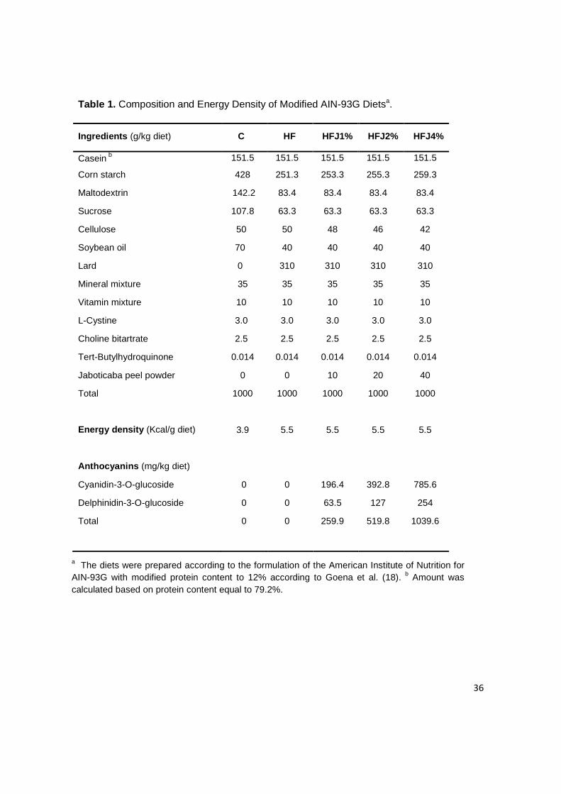
The concentrations of freeze-dried powder in the diets and ACNs composition and
contents were determined according to preliminary studies conducted by our
research group (17, in press). The composition of the diets, energy densities and
ACNs contents are presented in Table 1. Body weight and food intake for each
mouse were measured weekly throughout the study.
36
Table 1. Composition and Energy Density of Modified AIN-93G Dietsa.
a The diets were prepared according to the formulation of the American Institute of Nutrition for AIN-93G with modified protein content to 12% according to Goena et al. (18). b Amount was calculated based on protein content equal to 79.2%.
Ingredients (g/kg diet) C HF HFJ1% HFJ2% HFJ4%
Casein b 151.5 151.5 151.5 151.5 151.5
Corn starch 428 251.3 253.3 255.3 259.3
Maltodextrin 142.2 83.4 83.4 83.4 83.4
Sucrose 107.8 63.3 63.3 63.3 63.3
Cellulose 50 50 48 46 42
Soybean oil 70 40 40 40 40
Lard 0 310 310 310 310
Mineral mixture 35 35 35 35 35
Vitamin mixture 10 10 10 10 10
L-Cystine 3.0 3.0 3.0 3.0 3.0
Choline bitartrate 2.5 2.5 2.5 2.5 2.5
Tert-Butylhydroquinone 0.014 0.014 0.014 0.014 0.014
Jaboticaba peel powder 0 0 10 20 40
Total 1000 1000 1000 1000 1000
Energy density (Kcal/g diet) 3.9 5.5 5.5 5.5 5.5
Anthocyanins (mg/kg diet)
Cyanidin-3-O-glucoside 0 0 196.4 392.8 785.6
Delphinidin-3-O-glucoside 0 0 63.5 127 254
Total 0 0 259.9 519.8 1039.6

37
Intraperitoneal glucose tolerance test (iGTT) and i nsulin tolerance test (iITT).
iGTT and iITT were performed on food-deprived (6h) nonanesthetize mice after 9
and 10 weeks of treatment, respectively. Blood glucose levels were measured with
OptiumTM mini (Abbott Diabetes Care, Alameda, CA, USA) handheld glucometer
using appropriate test strips. For iGTT, a solution of 20% glucose (2.0g/kg body
weight) was administered into the peritoneal cavity. Blood samples were collected
from the tail vein at 30, 60, 90, and 120 min for determination of glucose
concentrations. The area under the curve (AUC) was calculated using these
values. For iITT, glucose blood levels were sampled 5, 10, 15, 20, 25, and 30 min
following Intraperitoneal (ip) injection of human insulin (0.75 U/kg). The rate
constant for glucose disappearance during an insulin tolerance test (KITT) was
calculated using the formula 0.693/t1/2.The glucose t1/2 was calculated from the
slope of the least-square analysis of the plasma glucose concentrations during the
linear decay phase (19).
Collection of Serum, Liver and Adipose tissue. After 11 weeks of consuming
the diets, the fasting mice (12h) were anesthetized by an ip injection of
ketamin/diazepam (1/1 v/v) and the collection were initiated after the loss of
corneal and pedal reflexes. Blood samples were taken from the interior vena cava
and the serum was obtained from de coagulated blood by centrifugation at 4000
rpm for 15 min at 4 °C. The serum was immediately f rozen at - 80°C until use. The
liver and epididymal adipose tissue were removed and processed according to the
subsequent analysis.

38
Determination of Serum Lipid Parameters and leptin levels. The serum
triglycerides, total cholesterol and high-density lipoprotein (HDL) cholesterol were
assayed enzymatically using commercial kits according to manufacturer’s
directions (Laborlab, Guarulhos, SP, Brazil). Serum leptin was measured by
enzyme-linked immunosorbent assay (ELISA) using a commercial assay kit
(Mouse leptin ELISA kit, Millipore, Billerica, MA, USA).
Imunoblotting. For evaluation of cytokine expression, protein activity and insulin
signal transduction in liver and adipose tissue, the abdominal cavities of
anesthetized mice (n=4) were opened, the portal vein exposed and 100µl (10-6
mol/liter) of insulin or saline was injected. Fragments (3.0 X 3.0 X 3.0mm) of liver
(at 30s) and epididymal adipose tissue (at 2 min) were excised after the infusion of
insulin and immediately homogenized in solubilization buffer at 4 °C (1% Triton X-
100, 100 mmol/liter Tris-HCl (pH 7.4), 100 mmol/liter sodium pyrophosphate, 100
mmol/liter sodium fluoride, 10 mmol/liter EDTA, 10 mmol/liter sodium
orthovanadate, 2.0 mmol/liter phenylmethylsulfonyl fluoride, and 0.1 mg
aprotinin/ml). The extracts were centrifuged at 15,000 rpm at 4 °C for 40 min to
remove insoluble material, and the supernatant was used as sample. The protein
concentration of the samples was determined by the Biuret method. The
immunoblotting was performed on tissue extracts as previously described by
Thirone et al. (20), with minor modifications. The proteins were separated by SDS-
PAGE, transferred to nitrocellulose membranes and blotted with antibodies against
phospho-IR, phospho-IRS-1, phospho-Akt, phospho-FoxO, IL-1β and phospho-IκB-
α. Specific bands were labeled by chemiluminescence and visualization was
39
performed by exposure of the membranes to RX-films. Bands intensities were
quantified by digital densitometry (ScionCorp, Frederick, MD, USA) and normalized
with β-actin.
RNA extraction and quantitative real-time PCR. Total RNA was extracted using
a commercially available acid-phenol reagent Trizol (Invitrogen Corp.). RNA
integrity was confirmed by nondenaturing agarose gel electrophoresis. The first-
strand cDNA was synthesized using SuperScript III reverse transcriptase and
random hexamer primers as described in the manufacturer’s protocol (Invitrogen
Corp.) The quantitative PCR was run to determine the expression of IL-1β in the
liver of treated mice using primer supplied with commercially available assays from
Applied Biosystems. The reference gene was GAPD (Glyceraldehyde-3-phosphate
dehydrogenase, (Applied Biosystems). Real-time PCR analysis of gene expression
was carried out in an ABI Prism 7500 sequence detection system (Applied
Biosystems). The optimal concentration of complementary DNA and primers, as
well as the maximum efficiency of amplification, was obtained through 5-point, 2-
fold dilution curve analysis for each gene. Amplification was performed in a 20 µL
final volume containing 6.25 ng of reverse-transcribed RNA according to the
manufacturer's recommendations using the TaqMan PCR master mix. Real-time
data were analyzed using the Sequence Detector System 1.7 (Applied
Biosystems). Results were expressed as relative transcript amount as previously
optimized (21).
Statistical analysis. All results are reported as means ± SEM. Differences
between the treatment groups were evaluated using one-way analysis of variance
40
(ANOVA). When the ANOVA indicated significance, a Tukey-Kramer post hoc test
was performed. p<0.05 was accepted as statistically significant.
RESULTS
Mice were fed on control (n=8) or high-fat diet (n=32) for 4 weeks prior to
treatment with freeze-dried jaboticaba peel powder; the initial body mass
(23.45±0.9 g) was similar between. As expected, after 11 weeks, body mass and
cumulative body mass gain were significantly higher in mice fed on HFD as
compared to the low-fat control diet (CD). The addition of freeze-dried jaboticaba
peel powder on HFD did not protect against HFD-induced body mass gain, while
the cumulative body mass gain in mice fed on HFD plus 2% freeze-dried
jaboticaba peel powder (HFJ2%) was significantly greater than HFD treatment
alone, 31.62±0.9 g and 23.25±1.40 g, respectively. In all the remaining groups
receiving freeze-dried jaboticaba peel powder, body mass was increased as
compared to HDF but this was not significant. (Figures 1a and 1b) .

41
Figure 1. Body weight trajectories (A) and Cumulative weight gain (B) of mice fed the CD,
HFD, HFJ1%, HFJ2% or HJ4% for 11 weeks. Values are means ± S.E.M., n = 8. #p < 0.05,
HF vs C group and *p < 0.05, HF2% vs HF group. No differences were observed between
HFJ groups.
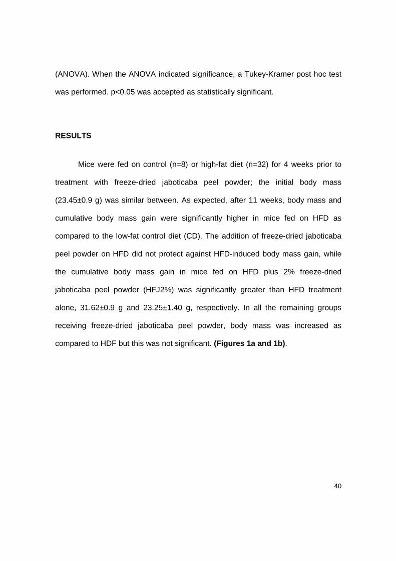
The overall food intake (g/mouse/day) did not differ between control and
high-fat groups throughout the experiment. However, at fifth week of experiment
the HFJ2% group consumed statistically more food (7.33±0.9 g) than all the other
groups (4.05±0.16 g) (Figure 2a) . This may be attributed to the fact that transition
to the HFJ2% led to a marked increase in their food consumption, in particular for
the first week, perhaps due to novelty or palatability of the diet.
It is important to notice that control mice on CD consumed the same amount
of food as mice on HFD by weight, but the energy intake was higher in the HF
control and treatments, approximately 23.0 Kcal/mouse/day, whereas the mice on
the CD consumed 16.0 Kcal/mouse/day. In mice fed on HFD plus 2% and 4%
freeze-dried jaboticaba peel powder the energy intake were significantly greater
42
than HF group (Figure 2b) . Thus, the higher energy intake showed to be closely
associated with a greater cumulative weight gain throughout the experiment in
these treatments.
Figure 2. Food intake (g/mouse/day) during the 11-week study period (A). No differences
were observed among CD fed mice and HFD fed mice. Except at week 5 when food intake
by HF2% was significantly different from all others groups (*** p < 0.001). Energy intake
(Kcal/mouse/day) (B). #p < 0.05; ***p < 0.001, HF2% vs HF group; **p < 0.01, HF4% vs HF
group.Values are means ± S.E.M., n = 8.
Fasting serum triglycerides, total cholesterol and HDL cholesterol were not
altered by level of fat or freeze-dried jaboticaba peel powder in the diet (Table 2) .
Serum leptin concentrations were highly correlated with the fat mass, the leptin
level was 3.4 fold greater in HFD mice than in CD mice, but the levels were not
changed in all treatments with HFD plus freeze-dried jaboticaba peel powder
(Figure 3) .

43
Table 2. Serum lipids profile of the experimental animalsa.
a Values are presented as means ± SEM of eight mice per treatment, except for triglycerides (n=4); data from sacrifice, after a 12 hours fast. No statistic differences were observed (p≥0.05, ANOVA).
Figure 3. Fasting serum leptin levels of C, HF, HFJ1%, HFJ2% and HJ4% groups. Bars
represent means ± S.E.M. of n = 8 mice. #p < 0.05, HF vs C group. No differences were
observed among the HFJ and HF groups.
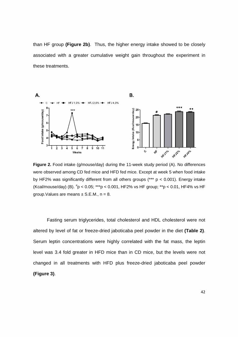
iGTT were performed after 5 weeks of dietary treatment to determine the
effects of freeze-dried jaboticaba peel powder on glucose tolerance. The basal
blood glucose levels, prior to the iGTT, was significantly higher in HF control
Contents (mg/dL)
C HF HFJ1% HFJ2% HFJ4%
Triglycerides 39.68 ± 9.9 52.84 ± 1.8 65.17 ± 8.3 67.07 ± 8.3 67.0 ± 9.0
Cholesterol 236.12 ± 18.1 220.89 ± 17.1 231.36 ± 18.8 233.12 ± 18.1 241.05 ± 13.05
HDL-cholesterol 88.73 ± 6.8 71.52 ± 4.0 65.76 ± 3.2 65.76 ± 4.1 81.71 ± 6.4
44
compared with control mice, 73.71±5.27 and 130.71±11.06 mg/dL, respectively. In
HFJ1%, HFJ2% and HFJ4% groups the average basal blood levels were higher
than HF control group (166±11.97, 140.43±17.67 and 142.71±10.90 mg/dL,
respectively), but this was not statistically significant. Similarly, the area under the
curve (AUC) values for plasma glucose levels during the iGTT were significantly
increased in all HFD groups compared to control group (Figure 4a) . These results
demonstrated that glucose intolerance in control HFD fed mice was not
ameliorated by freeze-dried jaboticaba peel powder treatments.
The contribution of insulin sensitivity in the glucose intolerance was
investigated by determining the clearance of plasma glucose as a function of time
after insulin injection (ITT). This measure of whole body insulin sensitivity can be
expressed by constant rate for glucose disappearance (KITT); larger values indicate
greater tissue insulin sensitivity. Notably, mice fed on HFD plus freeze-dried
jaboticaba peel powder showed significant increase in KITT values compared to
mice fed the HFD and the KITT did not differ from the control group (Figure 4b) .
These data suggest that freeze-dried jaboticaba peel powder may prevent HFD
induced insulin resistance (IR) in mice.

45
Figure 4. Glucose areas under curves during intraperitoneal glucose tolerance test (iGTT
[AUC]) (A) and KITT during intraperitoneal insulin tolerance test (B) were determined in C, HF,
HFJ1%, HFJ2% and HJ4% groups after 10 and 11 weeks of the experiment, respectively.
Bars represent means ± S.E.M. of n = 6 mice. #p < 0.05, HF vs C group; *p < 0.05, HF1%
vs HF group; ***p < 0.001, HF1% vs HF group; **p < 0.01, HF4% vs HF group.
In order to evaluate candidate mechanisms responsible for the
enhancement of insulin sensitivity of the freeze-dried jaboticaba peel powder
treatments, we examined insulin signaling through the IR/IRS1/Akt/FoxO pathway
in liver and adipose tissue of mice. Impairment in all steps of the insulin signaling
cascate was detected in HF control group. As shown in Figure 5 , significant
improvement in insulin signal transduction was observed through the increased
insulin-mediated tyrosine phosphorylation of IR, IRS-1 and Akt in liver and adipose
tissue and in threonine phosphorylation of FoxO in liver of mice treated with freeze-
dried jaboticaba peel powder when compared with HF control mice.
46
Figure 5. Representative immunoblots of insulin signaling proteins in adipose tissue (A)
and liver (B) of C, HF, HFJ1%, HFJ2% and HJ4% groups before (-) or after (+) insulin
stimulation. Tissues extracts from mice were prepared and immunoblotted (IB) with
respective antibodies, as described in Materials and Methods. The membrane was
stripped and immunoblotted with anti-β-actin antibody and used as loaded protein (lower
panels in figures A and B).
Because previous studies have shown that chronic activation of intracellular
proinflammatory pathways within the insulin target cells might contribute to the
obesity-related insulin resistance we decided to evaluate whether freeze-dried
jaboticaba peel powder can modulate HFD-induced inflammation. Remarkably, the
protein and mRNA expression of the cytokine IL-1β in adipose tissue and liver of

47
HFJ1% and HFJ4% groups were significantly reduced, in HFJ2% group, we also
observed this trend but it was not significantly different from HF control group
(Figures 6a and 6b) . This effect was accompanied by a significant decrease in
phosphorylated-IκBα protein levels in liver of all mice treated with freeze-dried
jaboticaba peel powder (Figure 6c) .
Figure 6. Immunoblot analysis of IL-1β in adipose tissue (A) extracts of C, HF, HFJ1%,
HFJ2% and HJ4% groups. The membrane was stripped and immunoblotted with anti-β-
actin antibody and used as loaded protein (lower panel in figure A). Transcript amount of
IL-1β in liver of HF, HFJ1%, HFJ2% and HJ4% groups (B) was determined by real-time
PCR, as described in Materials and Methods. Bars represent means ± S.E.M, n=4; *p <
0.05 vs HF group. The phosphorylation of IκB in liver of HF, HFJ1%, HFJ2% and HJ4%
groups (C) was determined by immunoblot (representative blot). The results of scanning

48
densitometry are expressed as arbitrary units. Bars represent means ± S.E.M. B, n=4; **p
< 0.01 vs HF group.
49
DISCUSSION
Recently, much attention has been focused on natural bioactive
phytochemicals present in foods that may provide desirable health benefits beyond
basic nutrition and play important roles in the prevention of chronic disorders and
metabolic diseases including obesity. The potential of bioactive components to
treat or prevent obesity is under intense exploration, and this represents an
attractive and alternative strategy for developing future of more safe anti-obesity
approaches (22, 23).
Although in recent years, emerging reports have been evidenced the
importance of anthocyanins as dietary antioxidants for prevention of oxidative
damage, several studies gradually focused on its beneficial effects in preventing
obesity and diabetes (24).
The present study demonstrated that supplementation of HFD with freeze-
dried jaboticaba peel powder did not produce a significant protection against
dietary saturated fat induced increase in body weight gain and in fact, tended to
increase cumulative weight gain, accompanied by no difference in food and energy
intake. We also observed no changes in serum lipids contents and leptin levels in
mice fed on the freeze-dried jaboticaba peel-enriched diets. Similarly, recent
studies have also reported that whole freeze-dried powders of concord grapes,
blueberry and black raspberry were ineffective in preventing obesity and, in some
cases, it could increase body weight gain and adiposity relative to HF fat fed
control (25, 12, 13). However, purified ACNs from various sources, including purple
50
corn, Cornelian cherry, black soybean coats, blueberries and strawberries, have
been shown positive responses in HFD-induced obesity, such as decrease in body
weight gain, improvement in lipid profile, hyperleptinemia and hyperglycemia to
levels similar to mice fed control diet (9-12). Reasons for the differential response
between whole foods and purified extracts of ACNs are still not fully elucidated.
The possible therapeutic effects of anthocyanins are certainly dependent on
sufficient bioavailability and not on the exact amount of ACNs consumed (26).
Possibly other components present in whole food matrix may reduce the rate of
absorption and bioactivity of ACNs counteracting in some way to prevent any
protective effect against obesity.
The freeze-dried jaboticaba peel powder supplementation did not attenuate
HFD-induced hyperglycemia as well, which was reflected in a larger incremental
area under the curve iGTT[AUC] of the plasma glucose. The pathogenesis of
glucose intolerance is complex and is mainly a function of the interplay between
insulin sensitivity and endocrine pancreatic function (27). In this study, in an
attempt to clarify whether impaired insulin secretion or decreased insulin sensitivity
is the primary defect of hyperglycemia after intraperitoneal glucose load, we
evaluated the insulin sensitivity by the iITT method. We demonstrated that mice fed
on HFJ diets for 6 weeks were protected from IR based on significant increase in
KITT when compared with HFD control group. The KITT value reflects the rate of
glucose removal by increased glucose uptake in insulin-sensitive tissues,
particularly skeletal muscle, and suppression of hepatic glucose production in
response to exogenous insulin.
51
The molecular action and mechanisms of the ACNs responsible for
amelioration of insulin sensitivity and prevention of obesity are not fully understood.
Possible suggested mechanisms have included improvement of insulin sensitivity
via up-regulation of glucose transporter 4 (GLUT 4) accompanied by down-
regulation of retinol binding protein 4 (RBP4) in type 2 diabetic mice (28) and by
inhibition of the inflammatory adipocytokines expression [interleukin-6 (IL-6), tumor
necrosis factor-α (TNF-α) and monocyte chemoattractant protein-1 (MCP-1)] and
decreased nuclear factor kB (NFkB) activity in white adipose tissue in high-fat-fed
mice (25, 29).
To address the molecular action and possible mechanisms by which freeze-
dried jaboticaba peel powder treatments ameliorates insulin sensitivity, we
evaluated key molecular steps of the insulin signaling machinery in liver and
adipose tissue of treated mice.
The effects of insulin on cell metabolism are mediated by a heterotetrameric
receptor expressed in a number of tissues such as liver, adipose and skeletal
muscle cells. Binding of insulin to its receptor initiates a cascade of events such as
receptor autophosphorylation on tyrosine residues and tyrosine phosphorylation of
insulin receptor substrate (IRS) proteins that subsequently activate the
downstream signalling pathways. Tyrosine phosphorylation of IRS1 leads to the
phosphorylation and activation of Akt, which plays an essential role in translocation
of GLUT4 to facilitate the transport of glucose into cells. Akt also mediates the
inhibitory effects of insulin on hepatic glucose output by induce phosphorylation of
transcriptional factors FoxO that results in nuclear export of FoxO proteins,
52
transcriptional inactivation and consequently inhibition of key gluconeogenic
enzymes, phosphoenolpyruvate carboxykinase (PEPCK) and the glucose-6
phosphatase (G6Pase) (30).
Our results showed that the impaired signal transduction through IR/ IRS1/
Akt/FoxO pathway was completely restored in liver and adipose tissue from freeze-
dried jaboticaba peel powder treated mice compared to HFD mice. Enhanced
insulin signaling was demonstrated in our study by increased tyrosine
phosphorylation of IR, IRS1 and Akt and in threonine phosphorylation of FoxO.
It is now commonly accepted that obesity is characterized by a broad
inflammatory response and many inflammatory mediators, overproduced by
expanded adipose tissue, may exhibit impact on insulin action contributing
significantly to establishment of insulin resistance. TNF-α, IL-6, IL-1β, and other
cytokines, activate various serine kinases, such as Jun N-terminal kinase (JNK)
and inhibitors of nuclear factor kappa B kinase (IκB), which can phosphorylate IRS
proteins and possibly other insulin signaling molecules interfering with normal
insulin action, creating a state of cellular insulin resistance (31, 32).
The reactive oxygen species (ROS) and oxidative stress, triggered by
hyperglycemia, also plays important role in development of insulin resistance. The
increases generation of ROS can lead to the activation of multiple stress-sensitive
serine/threonine (Ser/Thr) kinase signaling cascades such as IκB-β, and others
resulting in reduced insulin action (33).
53
Some reports showed that anthocyanins have a potency of the anti-
inflammatory activity and the downregulation of some inflammatory adipocitokines
expression can contribute to amelioration of diabetic state (25, 28).
Accordingly, the insulin-sensitizing effect of freeze-dried jaboticaba peel
powder in the present study were associated with attenuated inflammation
manifested as lower expressions of IL-1β in adipose tissue and liver accompanied
by decreased phosphorylated-IκBα protein levels in liver of all treated mice in a
dose-independent manner. Similarly, DeFuria et al. (25) demonstrated that
blueberry supplementation can protect against whole-body insulin resistance by
inhibiting the early inflammatory events in adipose tissue, induced by a high fat diet
in mice.
Furthermore, our previous study demonstrated that freeze-dried jaboticaba
peel powder has potency of an antioxidant in vivo (17). Taken all these information
together, our results suggest that one of the possible mechanisms by which the
freeze-dried jaboticaba peel powder exerts a protective role against HFD-induced
insulin resistance in mice, may be due to ability of jaboticaba anthocyanins exert
their effects as free radical scavengers and/or modulate cellular stress signaling
processes during inflammation acting as signaling agents by inhibiting the NFκB
signal pathway.
Here, we demonstrated that impaired glucose tolerance observed in mice
fed the HFJ diets was not due to insulin resistance. It is well known that dietary
antioxidants including anthocyanins protect pancreatic beta cells from glucose-
induced oxidative stress (34). Thus, further studies should be done in order to

54
investigate whether freeze-dried jaboticaba peel powder supplementation may be
associated with dysfunction of pancreatic islet β cells and inadequate insulin
secretion.
In conclusion, the consumption of high fat diet containing freeze-dried
jaboticaba peel powder, rich in cyanidin-3-glucoside and delphinidin-3-glucoside,
was not quite as effective in reverse the effects of HFD on body weight and
glucose intolerance; however, the supplementation was effective to correct diet-
induced insulin resistance by improved insulin signal transduction and reduced
inflammation in liver and adipose tissue of treated mice.

55
LITERATURE CITED
(1) World Health Organization. Diet, Nutrition and the Prevention of Chronic Diseases: Report of a WHO Study Group; Technical Report Series 916; WHO: Geneva, Switzerland, 2003.
(2) Kopelman, P. G. Obesity as a medical problem. Nature. 2000, 404, 635-643.
(3) Hu, F. B. Plant-based foods and prevention of cardiovascular disease: an overview. Am. J. Clin. Nutr. 2003, 78, 544S-551S.
(4) Dembinska-Kiec, A.; Mykkaänen, O.; Kiec-Wilk, B.; Mykkaönen, H. Antioxidant phytochemicals against type 2 diabetes. Brit. J. Nutr. 2008, 99, ES109-ES117.
(5) Liu, R. H. Health benefits of fruit and vegetables are from additive and synergistic combinations of phytochemicals. Am. J. Clin. Nutr. 2003, 78, 517S-520S.
(6) Espín, J. C.; García-Conesa, M. T.; Tomás-Barberán, F, A. Nutraceuticals: Facts and fiction. Phytochemistry, 2009, 68, 2986-3008.
(7) Kong, J-M.; Chia, L-S.; Goh, N-K.; Chia, T-F.; Brouillard. Analysis and biological
activities of anthocyanins. Phytochemistry, 2003, 64, 923-933.
(8) Clifford M. N. Anthocyanins—nature, occurrence and dietary burden. J Sci Food Agric. 2000; 80, 1063-1072.
(9) Tsuda, T.; Horio, F.; Uchida, K.; Aoki, H.; Osawa, T. Dietary cyanidin 3-O-β-D-
glucoside-rich purple corn color prevents obesity and ameliorates hyperglycemia in mice. J. Nutr. 2003, 133, 2125–2130.
(10) Jayaprakasam, B.; Olson, L. K.; Schutzki, R. E.; Tai, M. H.; Nair, M. G. Amelioration
of obesity and glucose intolerance in high-fat-fed C57BL/6 mice by anthocyanins and ursolic acid in cornelian cherry (Cornus mas). J. Agric. Food Chem. 2006, 54, 243–248.
(11) Kwon, S. H.; Ahn, I. S.; Kim, S. O.; Kong, C. S.; Chung, H. Y.; Do, M. S.; Park, K. Y.
Anti-obesity and hypolipidemic effects of black soybean anthocyanins. J. Med. Food 2007, 10, 552–556.

56
(12) Prior, R. L.; Wu, X.; Gu, L.; Hager, T. J.; Hager, A.; Howard, L. R. Whole berries versus berry anthocyanins: interactions with dietary fat levels in the C57BL/6J mouse model of obesity. J. Agric. Food Chem. 2008, 56, 647–653.
(13) Prior, R. L.; Wilkes, S.; Khanal, R. C.; Wu, X.; Gu, L.; Hager, T. J.; Hager, A.;
Howard, L. R. Dietary black raspberry anthocyanins do not alter development of obesity in mice fed an obesogenic high-fat diet. J. Agric. Food Chem. 2010, 58, 3977-3983.
(14) Reynertson, K. A.; Wallace; A. M.; Adachi, S.; Gil, R. R.; Yang, H.; Basile, M. J.; D’Armiento, J.; Weinstein, B.; Kennelly, E. J. Bioactive depsides and anthocyanins from jaboticaba (Myrciaria cauliflora). J. Nat. Prod. 2006, 69, 1228-1230.
(15) Reynertson, K. A.; Yang, H.; Jiang, B.; Basile, M. J.; Kennelly, E. J. Quantitative analysis of antiradical phenolic constituents from fourteen edible Myrtaceae fruits. Food Chem. 2008, 109, 883-890.
(16) Reeves, P. G. Components of the AIN-93 diets as improvements in the AIN-76A diet. J. Nutr. 1997, 127, 838-841.
(17) Leite, A.; Malta, L.; Riccio, M. F.; Eberlin, M.; Pastore, G.; Maróstica-Júnior, M. R. Antioxidant Potential of Rat Plasma by Administration of Freeze-Dried Jaboticaba Peel (Myrciaria jaboticaba Vell Berg). J. Agric. Food Chem. 2006, in press.
(18) Goena, M.; Marzo, F.; Fernández-González, L.; Tosar, A.; Frühbeck, G.; Santidrián, S. Effect of the raw legume Vicia ervilha on muscle and liver protein metabolism in growing rats. Rev. Esp. Fisiol. 1989, 45, 55-60.
(19) Bonora, E.; Manicardi, V.; Zavaroni, I.; Coscelli, C.; Butturini, U. Relationships between insulin secretion, insulin metabolism and insulin resistance glucose intolerance. Diabetes Metab. 1987, 13, 116-121.
(20) Thirone, A. C. P.; Carvalheira, J. B. C.; Hirata, A. E. ; Velloso, L. A.; Saad, M. J. A. Regulation of cbl-associated protein/cbl pathway in muscle and adipose tissues of two animal models of insulin resistance. Endocrinology. 2004, 145, 281-293.
(21) Bertelli, D. F.; Araujo, E. P.; Cesquini, M.; Stoppa, G.R.; Gasparotto-Contessotto, M.; Toyama, M. H.; Felix, J. C. V.; Carvalheira, J. B.; Michelini, L. C.; Chiavegatto, S.; Boschero, A. C.; Saad, M. J. A.; Lopes-Cendes, I.; Velloso, L. A. Phosphoinositide-specific inositol polyphosphate 5-phosphatase IV inhibits inositide trisphosphate accumulation in hypothalamus and regulates food intake and body weight. Endocrinology. 2006, 147, 5385-5399.
57
(22) Meydani, M.; Hasan, S. T. Dietary polyphenols and obesity. Nutrients. 2010, 2, 737-751.
(23) Yun, J. W. Possible anti-obesity therapeutics from nature – A review. Phytochemistry. 2010, 71, 1625-1641.
(24) He, J.; Giusti, M. M. Anthocyanins: Natural colorants with health-promoting properties. Annu. Rev. Food Sci. Technol. 2010, 1, 163-187.
(25) DeFuria, J.; Bennett, G.; Strissel, K. J.; Perfield, J. W.; Milbury, P. E.; Greenberg, A. S.; Obin, M. S. Dietary blueberry attenuates whole-body insulin resistance in high fat-fedmice by reducing adipocyte death and its inflammatory sequelae. J. Nutr. 2009, 139, 1510-1516.
(26) McGhie, T. K.; Walton, M. C.The bioavailability and absorption of anthocyanins: Towards a better understanding. Mol. Nutr. Food Res. 2007, 51, 702-713.
(27) Båvenholm, P. N.; Pigon, J.; Östenson, C-G.; Efendic, S. Insulin sensitivity of suppression of endogenous glucose production is the single most important determinant of glucose tolerance. Diabetes. 2001, 50, 1449-1454.
(28) Sasaki, R.;Nishimura, N.;Hoshino, H.; Isa, Y.;Kadowaki, M.; Ichi, T.; Tanaka, A.; Nishiumi, S.; Fukuda, I.; Ashida, H.; Horio, F.; Tsuda, T. Cyanidin 3-glucoside ameliorates hyperglycemia and insulin sensitivity due to down regulation of retinol binding protein 4 expression in diabetic mice. Biochem. Pharmacol. 2007, 74, 1619-1627.
(29) Seymour, E. M.; Lewis, S. K.; Urcuyo-Llanes, D. E.; Tanone, I. I.; Kirakosyan, A.; Kaufman, P. B.; Bolling, S. F. Regular Tart Cherry Intake Alters Abdominal Adiposity, Adipose Gene Transcription, and Inflammation in Obesity-Prone Rats Fed a High Fat Diet. J Med Food. 2009, 12, 935-942.
(30) Meshkani, R.; Adeli, K. Hepatic insulin resistance, metabolic syndrome and cardiovascular disease. Clin. Biochem. 2009, 42, 1331-1346.
(31) Wellen, K. E.; Hotamisligil, G. S. Inflammation, stress, and diabetes. J. Clin. Invest. 2005, 115, 1111-1119.
(32) Olefsky, J. M.; Glass, C. K. Macrophages, inflammation and insulin resistance. Annu. Rev. Physiol. 2010, 72, 219-246.
(33) Evans, J. L.; Maddux, B. A.; Goldfine, I. D. he Molecular Basis for Oxidative Stress-Induced Insulin Resistance. Antioxid. Redox Signal. 2005, 7, 1040-1052.

58
(34) Lapidot, T.; Walker, M. D.; Kanner, J. Antioxidant and Prooxidant Effects of Phenolics on Pancreatic β-Cells in Vitro. J. Agric. Food Chem., 2002, 50, 7220-7225.

59
CONCLUSÃO
Os resultados deste trabalho permitem concluir que a suplementação da
dieta hiperlipídica com casca de jabuticaba liofilizada, durante 6 semanas, não foi
eficaz na reversão dos efeitos da dieta hiperlipídica sobre o peso corporal e
intolerância à glicose, no entanto, a suplementação reverteu a resistência à
insulina por meio da preservação da sinalização da insulina e atenuação da
inflamação no fígado e no tecido adiposo dos camundongos tratados. Estes
resultados sugerem que a casca de jabuticaba liofilizada pode exercer uma ação
protetora contra as patologias associadas à obesidade.

60
Anexo
Comprovante de Aprovação do Comitê de Ética em Expe rimentação Animal