anÁlise molecular do gene sh3bp2 em lesÕes …livros01.livrosgratis.com.br/cp130405.pdf · d...
TRANSCRIPT
Vinicius Magalhães Carvalho
ANÁLISE MOLECULAR DO GENE SH3BP2 EM
LESÕES CENTRAIS DE CÉLULAS GIGANTES E NO
QUERUBISMO
Belo Horizonte
Instituto de Ciências Biológicas da UFMG
2009

Livros Grátis
http://www.livrosgratis.com.br
Milhares de livros grátis para download.

Vinicius Magalhães Carvalho
ANÁLISE MOLECULAR DO GENE SH3BP2 EM
LESÕES CENTRAIS DE CÉLULAS GIGANTES E NO
QUERUBISMO
Belo Horizonte
Instituto de Ciências Biológicas da UFMG
2009
Tese apresentada ao Curso de pós-graduação em
Farmacologia Bioquímica e Molecular do Instituto de
Ciências Biológicas da Universidade Federal de
Minas Gerais, como requisito parcial à obtenção do
título de Doutor.
Orientador: Prof. Ricardo Santiago Gomez


SUMÁRIO
LISTA DE ABREVIATURAS.......................................................................................VI
LISTA DE TABELAS..................................................................................................VII
LISTA DE FIGURAS.................................................................................................VIII
RESUMO....................................................................................................................IX
1 INTRODUÇÃO........................................................................................................10
2 OBJETIVOS............................................................................................................25
3 ARTIGOS................................................................................................................27
4 CONSIDERAÇÕES FINAIS....................................................................................36
5 CONCLUSÕES.......................................................................................................41
6 REFERÊNCIAS BIBLIOGRÁFICAS........................................................................43

LISTA DE ABREVIATURAS E SIGLAS
AP-1 Fator de transcrição
C-terminal Carboxi-terminal
cDNA Ácido desoxirribonucléico codificador
LCCG Lesão Central de Células Gigantes
M-CSF Fator Estimulador de Colônias de Macrófagos
MIM Herança Mendeliana no Homem
Msx-1 Gene msh homeobox 1
N-terminal Amino-terminal
NF-1 Neurofibromatose do tipo 1
NFAT Fator nuclear de células T ativadas
NFATc1 NFAT citoplasmático calcineurina-dependente 1
OPG Osteoprotegerina
PCR Reação em cadeia da polimerase
PH Domínio de Homologia pleckstrin
PLCg Fosfolipase C gamma
pmol Picomol
PTH Hormônio da Paratireóide
PTHrP Proteína relacionada ao PTH
RNA Ácido ribonucléico
RANK Receptor de ativação do fator nuclear Kapa B
RANKL Ligante do Receptor de ativação do fator nuclear Kappa B
SH2 domínio de homologia 2 Src
IV
SH3BP2 Gene SH3BP2
SH3BP2 Proteína 2 ligante do domínio de homologia 3 Src
SMLCG/TN Síndrome de múltiplas lesões de células gigantes / tipo-
Noonan
sRANK RANK solúvel
Taq Thermus aquaticus
TCG tumor de células gigantes
TNF Fator de Necrose Tumoral
TRAP Fosfatase ácida tartarato-resistente
µg Micrograma
V

CÓDIGOS DOS NUCLEOTÍDEOS E AMINOÁCIDOS
Nucleotídeos
A Adenina
C Citosina
G Guanina
T Timina
Aminoácidos
A Alanina Ala C Cisteína Cis
D Ácido Aspártico Asp E Ácido Glutâmico Glu
F Fenilalanina Phe G Glicina Gly
H Histidina His I Isoleucina Ile
K Lisina Lis L Leucina Leu
M Metionina Met N Asparigina Asn
P Prolina Pro Q Glutamina Gln
R Arginina Arg S Serina Ser
T Treonina Thr V Valina Val
W Triptofano Trp Y Tirosina Tyr
VI

LISTA DE TABELAS
TABELA 1 – Dados clínicos, aspectos radiográficos e mutações detectadas em
pacientes com Querubismo (caso1) e LCCG (caso 2-5)....................... 32
VII
LISTA DE FIGURAS
Oral Oncology (2008)
FIGURA 1 a - Seqüência de DNA do exon 3 no tipo-selvagem.................................29
FIGURA 1 b - Seqüência de DNA mostrando a deleção de uma Citosina (408delC)
no caso de Querubismo .........................................................................29
FIGURA 2 a - Estrutura esquemática do SH3BP2 e seus domínios: domínio de
homologia pleckstrin (PH), região rica em prolina (Pro) e um domínio
SH2 (SH2)...............................................................................................29
FIGURA 2 b - Proteína truncada e perda de domínios
remanescentes........................................................................................29
Oral Diseases (2009)
FIGURA 1 a - Dados da seqüência do cromatograma (caso 1) mostrando a mutação
heterozigótica
c.320C>T.................................................................................................33
FIGURA 1 b - Dados da seqüência do cromatograma (caso 2) mostrando a mutação
heterozigótica
c.1442A>T..............................................................................................33
VIIIX

RESUMO
A lesão central de células gigantes (LCCG) é uma lesão óssea que ocorre
principalmente na mandíbula e é caracterizada histologicamente por células
gigantes multinucleadas em um estroma de células mononucleares ovóides. Sua
etiologia é desconhecida, sendo mais comum em adultos jovens. O padrão
radiográfico e histológico das LCCG pode ser encontrado em outras doenças e
síndromes que acometem os maxilares, sendo uma delas o Querubismo. O
Querubismo é uma síndrome autossômica dominante, com penetrância variada,
caracterizada por uma extensa destruição óssea maxilo-mandibular, causando o
aspecto alterado da face. A síndrome foi relacionada com mutações no gene
SH3BP2, localizadas no exon 9, mas um modelo para sua patogênese não foi
definido até o momento. No presente estudo, quatro casos de LCCG e dois casos de
Querubismo foram investigados, sendo um deles agressivo. O DNA foi extraído do
sangue periférico e do tecido tumoral. Todas as regiões codificadoras do SH3BP2
foram amplificadas por PCR e seqüenciadas. Uma mutação heterozigótica de
sentido trocado (A1442T - Q481L) foi encontrada no exon 11 de um dos casos
esporádicos de LCCG. O caso de Querubismo não-agressivo apresentou mutação
heterozigótica no exon 4, germinativa e no tecido tumoral: C320T (T107M). O caso
de Querubismo agressivo apresentou uma nova mutação localizada no exon 3,
domínio de homologia pleckstrin (PH) do SH3BP2. Os resultados demonstraram que
as mutações no gene SH3BP2 podem estar fora da região hot spot em casos de
Querubismo e que podem estar presentes em LCCG esporádicas. Estes achados
sugerem uma nova perspectiva na investigação da possível relação entre a
patogênese do Querubismo e da LCCG.
IX

1 INTRODUÇÃO
10
1 Introdução
1.1 Lesão Central de Células Gigantes (LCCG)
A Lesão Central de Células Gigantes (LCCG) foi inicialmente descrita como
sendo um granuloma reparativo de células gigantes dos maxilares (JAFFE, 1953).
Recentemente, a Organização Mundial de Saúde definiu a LCCG como uma lesão
benigna localizada, mas que eventualmente apresenta-se como uma proliferação
osteolítica agressiva, consistindo de tecido fibroso com áreas de hemorragia e
depósitos de hemossiderina, presença de células gigantes semelhantes a
osteoclastos e formação óssea reacional (BARNES et al., 2005).
A LCCG é uma lesão intra-óssea, geralmente única, que ocorre com maior
freqüência na região anterior da mandíbula, em pacientes do sexo feminino, na faixa
etária entre 10 a 25 anos (WHITAKER & WALDRON, 1993). O termo reparativo,
inicialmente descrito por Jaffe, foi abandonado, mas ainda é utilizado por alguns
autores para as lesões que ocorrem em áreas de extração e ao redor de implantes
(DE LANGE, 2007c).
Chuong et al. (1986), baseados em sinais, sintomas e nos aspectos
histológicos, foram os primeiros a classificar as lesões em agressivas e não-
agressivas. As lesões agressivas são caracterizadas por um ou mais aspectos
como: dor, parestesia, reabsorção radicular, crescimento rápido, perfuração da
cortical e um alto índice de recorrência após a curetagem cirúrgica. Elas são
também maiores e histologicamente apresentam uma fração de superfície com
maior ocupação por células gigantes. As lesões agressivas apresentam aspecto
radiolúcido multilocular e o deslocamento dentário ocorre com freqüência, podendo
11
acarretar mal-oclusão. No aspecto intra-bucal pode-se observar um aumento de
volume, às vezes marrom-azulado (VAN DAMME & MOOREN, 1994; DE LANGE et
al., 2007c). As lesões não-agressivas normalmente apresentam-se indolores,
assintomáticas, com crescimento lento, sem reabsorção de cortical ou envolvimento
de raízes dentárias e com baixa recorrência. Radiograficamente, são solitárias e
com aspecto unilocular (HORNER, 1989).
Na literatura, sempre houve uma tentativa de distinguir o tumor de células
gigantes dos ossos longos (TCG) das LCCG, sendo que o TCG é mais agressivo e
mais difícil de ser tratado. O TCG se destaca por ser um processo neoplásico que
acomete epífises de ossos longos. Ele apresenta taxa de recorrência mais elevada
do que a LCCG, além de envolver pacientes mais idosos (AUCLAIR et al., 1988).
Esses autores demonstraram que histologicamente, a única diferença quantitativa
estatisticamente significativa entre as duas lesões foi o maior numero de núcleos
nas células gigantes do TCG. Eles sugeriram que o TCG e a LCCG representam um
único processo patológico, modificado pela idade do paciente e área de ocorrência.
Souza et al. (1999) verificaram pequenas diferenças histológicas entre essas
lesões: no TCG existe uma extensa área fibrosa, células gigantes mais
arredondadas e com um maior número de núcleos; enquanto que na LCCG, a
presença de hemorragia, depósito de hemossiderina e tecido osteóide são mais
freqüentes.
As células gigantes multinucleadas apresentam tamanho e morfologia
variada, com núcleos ovóides, vesiculosos e centralizados. Características típicas de
osteoclastos foram observadas nelas. Localizadas em maior número ao redor de
áreas hemorrágicas, não são necessariamente abundantes e podem estar
12

agregadas focalmente ou difusas na lesão, em um fundo de células mononucleares,
cuja morfologia varia de fusiforme a ovóide (FLANAGAN et al., 1988; MARX &
STERN, 2002).
Em alguns casos, áreas de calcificação distrófica e ossificação metaplásica
podem ser encontradas, especialmente na periferia das lesões (LOMBARDI et al.,
2006; DE LANGE et al., 2007c).
A origem e a formação das células gigantes multinucleadas na LCCG e na
lesão periférica de células gigantes (LPCG) tem sido alvo de vários estudos. Uma
teoria hipotetizada é da fusão entre as células mononucleares da lesão, através de
interdigitações na membrana citoplasmática das mesmas (TIFFEE &
AUFDEMORTE, 1997).
As células gigantes presentes nas LCCG são derivadas de uma sub-série de
fagócitos mononucleares. As células mononucleares precursoras diferenciam-se em
células gigantes maduras, sob a influência das células fusiformes do estroma
(semelhantes a osteoblastos), que se mantêm em proliferação e expressam o ligante
do receptor de ativação do fator nuclear κB (RANKL) (MIYAMOTO et al., 2000).
Apesar de estarem presentes de forma destacada, não se presume que as
células gigantes multinucleadas sejam as células tumorais proliferativas no TCG e
na LCCG. Hipotetiza-se que estas células advenham de células mononucleares do
sangue periférico e sejam recrutadas pelas células fusiformes do estroma.
Provavelmente, estas últimas são as células proliferativas tumorais (ITONAGA et al.,
2002).
13

Itonaga et al. (2003) realizaram uma melhor definição das células
mononucleares e gigantes da LCCG. Dentre as células mononucleares, três
populações distintas foram identificadas através de reações de imunoistoquímica e
imunofluorescência. A maioria das células apresentou características típicas de
macrófagos, como positividade para CD68, CD11a, CD11b, CD14 e HLA-DR. A
minoria das células mononucleares apresentou características típicas de
precursores de osteoclastos como positividade para receptor de vitronectina (VNR) e
para fosfatase ácida tartarato-resistente (TRAP). Poucas células fusiformes isoladas
apresentaram características de osteoblastos como positividade para fosfatase
alcalina e se encontravam espalhadas pela lesão ou ao redor de áreas de formação
óssea. Estas células também foram positivas para o PCNA, ou seja, estavam em
proliferação. Através de culturas de células da LCCG, os autores observaram ainda
que as células fusiformes proliferavam e permaneciam por maior tempo que as
células gigantes e as mononucleares ovóides. Elas ainda expressavam os
marcadores de osteoblasto, RANKL e fosfatase alcalina, além de marcadores de
fibroblasto como prolil-4-hidroxilase e vimentina. Ainda neste trabalho, os autores
fizeram co-cultura de células mononucleares da LCCG com células mononucleares
de sangue periférico de indivíduos sadios; acrescentaram à cultura M-CSF;
observando posteriormente a formação de células multinucleadas TRAP+ e VNR+,
dado este que confirma a tese de que as células tipo-osteoblastos possibilitam a
formação de osteoclastos (ITONAGA et al., 2003).
As células gigantes multinucleadas presentes na LCCG e na LPCG são
morfologicamente e histoquimicamente semelhantes à osteoclastos. Possuem
enzimas hidrolíticas e antígenos específicos de osteoclastos, além de receptores
para calcitonina. São capazes de reabsorver osso in vitro, tendo sua função de
14

reabsorção óssea e mobilidade celular moduladas na presença de calcitonina (LIU et
al., 2003).
As LCCG podem ocorrer de forma isolada, em formas múltiplas, recorrentes
ou ainda associadas a Síndromes como o Querubismo, a Síndrome de múltiplas
lesões de células gigantes / tipo-Noonan (SMLCG/TN) e a Neurofibromatose do tipo
1 (NF-1) (HYCKEL et al., 2005). Tais lesões são histológica e
imunoistoquimicamente indistinguíveis (VAN DAMME & MOOREN, 1994).
Existe uma diferença considerável na literatura referente aos dados
estatísticos destas lesões, provavelmente devido ao número de casos e critérios de
seleção utilizados. Na maioria dos artigos, a amostra é relativamente pequena
(n<20) e os pacientes são referenciados de um único Centro de Saúde, sendo este
um Bias (DE LANGE, 2007c).
de Lange & Van den Akker (2005) realizaram um estudo com o objetivo de
avaliar aspectos clínicos e radiográficos das LCCG diagnosticadas na Holanda no
período entre Janeiro de 1990 e Janeiro de 1995. A incidência foi de 89 lesões em
83 pacientes. Sinais e sintomas de agressividade (dor, parestesia e reabsorção
radicular) foram encontrados em 16 pacientes (19,3%). Lesões múltiplas ocorreram
em três pacientes (3,6%). A taxa de recorrência foi de 26,3%, sendo maior nos
pacientes com lesões agressivas (RR=1.6). Em cinco pacientes (14 lesões) houve
um diagnostico clínico de Querubismo isolado ou concomitante com a NF-1. Os
autores concluíram que em uma população em geral, lesões de células gigantes
maiores e agressivas são menos comuns do que o sugerido pela literatura.
Entretanto, lesões múltiplas ocorrem com maior freqüência do que foi descrito
previamente. Em pacientes com sinais e sintomas agressivos, a curetagem cirúrgica,
15

isoladamente, não é uma terapia efetiva.
O diagnóstico clínico diferencial para LCCG deve ser feito com Querubismo,
tumor marrom do hiperparatireoidismo, Ameloblastoma e Ceratocisto. Nos casos em
que a lesão se localiza periapicalmente, pode ser confundida com cistos ou
granulomas radiculares de origem endodôntica, principalmente quando não há
resposta pulpar (LOMBARDI et al., 2006).
1.2 Querubismo
O Querubismo foi inicialmente descrito por Jones (JONES, 1933). É uma
doença rara que apresenta herança autossômica dominante (MIM 118400).
Geralmente caracteriza-se pela destruição bilateral da mandíbula, maxila ou ambas,
podendo alcançar a região orbitária. A destruição é frequentemente acompanhada
de um aumento de volume facial, causando uma assimetria indolor. A pele na região
da bochecha fica esticada e projeta as pálpebras inferiores para baixo. Os olhos
parecem estar deslocados para cima, lembrando os anjos querubins das pinturas
renascentistas (MANGION et al., 1999; UEKI et al., 2001).
Radiograficamente, o Querubismo é caracterizado por áreas radiolúcidas
multiloculares que podem ser unilaterais. Normalmente a doença restringe-se às
regiões maxilar e mandibular, apesar de anormalidades radiográficas serem
ocasionalmente observadas nas costelas (ARNOTT, 1978).
Avaliações histopatológicas das lesões mostram um tecido conjuntivo fibroso
proliferativo, contendo numerosas células gigantes multinucleadas (SOUTHGATE et
al., 1998).
16

Os aspectos clínicos e histológicos do Querubismo podem apresentar
similaridade diagnóstica com: TCG, LCCG, fibroma ossificante, displasias fibrosas
dos maxilares e doença de Paget (WHITAKER & SINGH, 1995).
Os indivíduos afetados não apresentam alterações ao nascimento.
Normalmente a doença se manifesta no início da infância (entre 2 a 5 anos) e se
torna mais evidente até a puberdade, fase na qual as lesões ósseas começam a
regredir. Entretanto, a distorção dos maxilares leva a anormalidades dentárias
permanentes. Outras complicações como distúrbios visuais (devido as lesões que
atingem a órbita) e mesmo óbito, foram descritos; porém são relativamente raros
(HAWES, 1989). Segundo o autor, a penetrância da doença é alta, mas a estimativa
precisa depende do critério diagnóstico utilizado. A freqüência e gravidade da
doença é maior no sexo masculino que no feminino.
Se apenas a mandíbula está envolvida e não há histórico familiar, torna-se
impossível diferenciar o quadro de Querubismo de múltiplas LCCG (DE LANGE et
al., 2007b). Os achados microscópicos do Querubismo são essencialmente
semelhantes àqueles da lesão central de células gigantes isoladas, não permitindo
um diagnóstico específico de Querubismo na ausência de informações clínico-
radiográficas (YAMAGUCHI et al., 1999).
Histologicamente, as lesões do Querubismo contêm células gigantes
multinucleadas com atividade osteoclástica, em um estroma fibroso contendo células
mesenquimais ovais e fusiformes. Estes aspectos são similares àqueles presentes
em outras lesões que contêm células gigantes, incluindo displasia fibrosa, LCCG e
LPCG, granuloma reparativo do osso de células gigantes, TCG e tumor marrom do
hiperparatireoidismo (DE LANGE et al., 2005).
17
Normalmente, o contexto clínico e a história natural são pré-requisitos para
um diagnóstico diferencial definitivo entre estas lesões histologicamente similares. A
distinção e mesmo a nomenclatura entre estas doenças podem depender de cada
instituição (LIETMAN et al., 2007).
MANGION et al. (1999) e TIZIANI et al. (1999) demonstraram que o
Querubismo é causado por mutação na região codificadora do gene SH3BP2,
localizado no cromossomo 4p16.3.
Em 2003, Imai et al. encontraram uma mutação de sentido trocado Pro418Arg
no exon 9 do gene SH3BP2, em uma paciente diagnosticada com Querubismo não-
familiar. Segundo os autores, casos de Querubismo não-familiar como o estudado,
apresentam grande similaridade clínica e difícil diagnóstico diferencial com LCCG.
Li & Yu (2006) investigaram uma família com 21 membros, sendo que 3 foram
diagnosticadas com Querubismo, todas do sexo feminino. Um dos casos de
Querubismo apresentou uma particularidade: o envolvimento da região orbitária
ocorreu já na idade adulta, após remissão das lesões mandibulares. Dezessete
membros foram recrutados para análise genética. Os autores realizaram o
seqüenciamento do gene SH3BP2 no exon 9, e encontraram uma mutação de
sentido trocado nos pacientes com Querubismo: uma troca de base A1517G,
causando uma substituição de aminoácido Asp419Gly. Esta mutação não foi
encontrada em nenhum dos 100 controles avaliados.
de Lange et al. (2007a) realizaram a análise clínica, radiográfica e genética de
11 membros de uma família com histórico de Querubismo. Cinco membros
apresentaram mutação pontual no exon 9, até então não descrita: uma transição
C1513A, resultando em uma substituição Pro418Thr. Dois indivíduos apresentavam
18

os aspectos clássicos de Querubismo e a mãe de um deles, sinais de um
“Querubismo solucionado”. Dos dois outros membros que não apresentavam sinais,
sintomas e ou histórico de Querubismo, um era do sexo masculino, o que segundo
os autores pode contradizer a literatura, que mostra 100% de penetrância da doença
nos homens.
Lee et al. (2008) examinaram um paciente com 6 anos de idade e sua família.
Encontraram uma mutação no gene SH3BP2 Pro418Arg no probando e em sua
mãe.
1.3 O gene SH3BP2
As mutações encontradas em pacientes com Querubismo foram evidenciadas
em uma porção do exon 9 que codifica uma região rica em prolina (aminoácidos
415-420) (UEKI et al., 2001).
A função específica desta região, codificada pelas mutações de sentido
trocado no SH3BP2 em pacientes com Querubismo permanece incerta, apesar
deste padrão de sentido trocado sugerir uma mutação ativadora (UEKI et al., 2001;
LIETMAN et al., 2006).
Todas as mutações identificadas no SH3BP2 estão localizadas no exon 9 e
afetam três aminoácidos, em uma seqüência de seis (RSPPDG), no domínio de
ligação SH3. As mutações no SH3BP2 promovem um ganho de função ou atuam de
forma negativa-dominante. Apesar desse gene ser um regulador da função da c-ABL
em algumas células como osteoblastos, ele pode afetar diferentes moléculas
19

sinalizadoras em outras células, como as progenitoras de osteoclastos (UEKI et al.,
2001).
A região do cromossoma 4p16.3, localização do SH3BP2, está
freqüentemente deletada nos pacientes com a Síndrome de Wolf-Hirschhorn; uma
condição rara caracterizada pelo desenvolvimento debilitado, incluindo tamanho e
crescimento limitados. Apesar da haploinsuficiência do SH3BP2 nesta síndrome,
estes pacientes possuem aspectos faciais e de desenvolvimento distintos que não
se assemelham ao Querubismo (BATTAGLIA & CAREY, 1999).
A proteína codificada pelo cDNA com as mutações do SH3BP2 no
Querubismo é ativadora, pois ela aumenta a atividade do Fator nuclear de células T
ativadas (NFAT) mais que no cDNA do SH3BP2 tipo-selvagem (LIETMAN et al.,
2006).
É possível que a presença da SH3BP2 nas LCCG, evidenciada pela
imunoistoquímica, represente na verdade, uma relativa expressão aumentada do
SH3BP2 tipo-selvagem nas lesões de células gigantes não-sindrômicas. Tal
aumento na quantidade de SH3BP2 poderia aumentar a ativação de sua própria via,
simulando assim, a atividade aumentada observada nas mutações de ativação do
gene SH3BP2 no Querubismo (LIETMAN et al., 2006). De acordo com estes
autores, existem indicações que o gene SH3BP2 apresenta um papel na regulação
da atividade aumentada de osteoblasto e osteoclasto, observada na erupção
dentaria normal.
Segundo Foucault et al. (2003), proteínas adaptadoras são componentes
sinalizadores críticos que podem integrar múltiplos caminhos, ao permitirem a
reunião de complexos sinalizadores multimoleculares. O adaptador citoplasmático
20

SH3BP2 promove atividades transcricionais do NFAT e da proteína ativadora 1 (AP-
1) em células T através da ativação dos caminhos RAS e calcineurina dependentes.
Entretanto, os mecanismos moleculares pelos quais o SH3BP2 regula a sinalização
e ativação celular permanecem pouco conhecidos. Os autores apresentaram
evidências da interação física entre o SH3BP2 e a proteína chaperona 14-3-3. Uma
proteína SH3BP2 mutante, incapaz de se ligar a 14-3-3, mostrou potencial
aumentado para estimular atividades transcricionais do NFAT, sugerindo que esta
interação regula negativamente a função adaptadora da SH3BP2 em linfócitos.
Miah et al. (2004) sugeriram que mutações pontuais no SH3BP2 poderiam
causar a ativação patológica de osteoclastos, presumidamente pela disfunção deste
gene na via regulatória da osteoclastogênese. Além disto, hipotetiza-se que a
mutação no gene SH3BP2 tem provavelmente alguma influência na regulação do
desenvolvimento dental pelo gene Msx-1 e que o desenvolvimento das LCCG
poderia estar ligada a desregulação desse gene. A expressão não fisiológica do
Msx-1 poderia levar a uma falha na diferenciação osteoclástica que então causaria a
formação de um tecido lesional.
Neste processo, o SH3BP2 possui uma influência na regulação do receptor
do Hormônio da Paratireóide (PTH) e na proteína relacionada ao PTH (PTHrP). O
PTH medeia uma redução na expressão da osteoprotegerina (OPG) nos folículos
dentários, que promovem osteoclastogênese (TAZAWA et al. 2003; WISE et al.
2004).
Segundo Hyckel et al. (2005), a ocorrência hereditária do Querubismo indica
uma provável etiologia genética, uma vez demonstrada sua correlação com a
mutação no gene SH3BP2. Uma definição de sua patogênese ainda não foi
21

elucidada. Devido a sua associação com o desenvolvimento dos segundos e terceiro
molares, o Querubismo poderia ser definido como uma alteração do
desenvolvimento dentário, geneticamente determinada. A determinação temporal e
espacial dos sintomas clínicos é explicada pela interação entre o caminho de
transdução de sinais SH3BP2-dependentes com a morfogênese mandibular. Devido
à indeterminação da fase de capuz dos segundos e terceiros molares induzida pela
doença, uma compartimentação espacial necessária para o desenvolvimento
dentário normal não ocorre. Isto causa a desregulação da formação de tecido ósseo
a partir do mesênquima.
Lietman et al. (2006) demonstraram que as mutações encontradas no
SH3BP2 comportam-se como ativadoras do NFAT e determinam um ganho de
função ao promoverem a ativação de osteoclastos, o que é consistente com as
lesões que produzem o fenótipo clínico do Querubismo. Ainda permanece
desconhecido como as mutações relacionam-se com o fato das lesões serem sítio-
específicas.
A função precisa do SH3BP2 é incerta; seu domínio de homologia 2 Src
(SH2) e domínio ligante de homologia 3 Src (SH3BD) são característicos de
proteínas adaptadoras, que apesar de serem limitadas na atividade catalítica
intrínseca, exercem papéis críticos ao acoplar receptores quinase a processos
específicos de sinalização celular (DECKERT et al., 1998; FOUCAULT et al., 2003).
O domínio SH2 no SH3BP2 liga-se às proteínas citoplasmáticas Syk, Vav,
PLC-γ, 14-3-3, c-CBL e LAT. Em um nível celular, o gene SH3BP2 foi relacionado à
sinalização de células T e ao processo inflamatório, e sua atividade é reduzida ao
interagir com a proteína chaperona 14-3-3. O NFAT está aumentado em resposta à
22
expressão elevada do SH3BP2, sendo que o NFAT citoplasmático calcineurina-
dependente 1 (NFATc1), a isoforma primária do NFAT presente nos osteoclastos, é
o regulador mestre da osteoclastogênese (DECKERT et al., 1998; FOUCAULT et al.,
2003, TAKAYANAGI, 2005).
A proteína adaptadora SH3BP2 é conhecida por exercer um papel regulador
na sinalização de imunoreceptores. Em linfócitos, a ligação de receptores de células
B ou T desencadeia a fosforilação de tirosina. A análise genética revela que a
SH3BP2 é requisitada para a proliferação de células B e receptores de sinalização
de células B. As mutações pontuais no gene SH3BP2 incluem substituições e
deleções. As células mielóides em Camundongos Knockout exibem respostas
aumentadas ao Fator Estimulador de Colônia de Macrófago (M-CSF) e ao RANKL,
acarretando a ativação de osteoclastos (HATANI & SADA, 2008).
As células gigantes multinucleadas mostraram-se positivas para CD68 e para
Fosfatase ácida tartarato-resistente (TRAP). Inúmeras células fusiformes do estroma
expressaram RANKL. Estes resultados apontam evidências que o RANKL tem um
papel crítico na diferenciação de células precursoras de osteoclastos em células
gigantes multinucleadas no Querubismo (LEE et al., 2008).
Lietman et al. (2008) investigaram o significado funcional da proteína SH3BP2
na osteoclastogênese, na presença do RANKL solúvel (sRANKL). O estudo indicou
que a SH3BP2 aumenta tanto a translocação nuclear de NFATc1 quanto a
expressão de TRAP em células pré-osteoclásticas tratadas com sRANK. Diante
destes achados, os autores propuseram que as lesões do Querubismo resultam do
efeito de ganho de função produzido pelas mutações no gene SH3BP2 nas células
pré-osteoclásticas.
23

1.4 Estudos moleculares das LCCG
de Lange et al. (2007b) analisaram o DNA extraído do sangue de quatro
pacientes diagnosticados com LCCG agressiva – caracterizada por um ou mais dos
seguintes aspectos: dor, parestesia, crescimento rápido e reabsorção radicular. Os
autores não encontraram mutações no exon 9 do gene SH3BP2. Porém, sugeriram
que uma mutação somática em um grupo específico de células poderia causar as
lesões focais nas LCCG e indicaram a necessidade de análise do DNA tecidual
destas lesões.
Lietman et al. (2007) investigaram se lesões de células gigantes esporádicas
possuem mutações na mesma área do gene SH3BP2 como nas raras lesões
sindrômicas do Querubismo. Eles hipotetizaram que as mutações no SH3BP2,
similarmente àquelas encontradas no Querubismo, estavam presentes em lesões
centrais não-sindrômicas. A expressão das proteínas do SH3BP2 e do NFATc1 não
pareceram correlacionar diretamente, uma vez que em algumas amostras a
expressão da NFATc1 é mais difusa que da SH3BP2. Neste estudo não foram
identificadas mutações no exon 9 do gene SH3BP2 no tecido da lesão de 10
amostras de TCG e nem em 9 casos de LCCG, sugerindo que mutações
germinativas ou somáticas tipo-Querubismo são incomuns em Lesões de células
gigantes não-sindrômicas.
Idowu et al. (2008) avaliaram o tecido de Lesões de células gigantes para
mutações no exon 9 do gene SH3BP2. As células foram obtidas pela micro-
dissecação do tecido lesional embebido em parafina. Os autores não detectaram
mutações em 26 Lesões (15 centrais e 11 periféricas) e sugeriram que pacientes
24

com Lesões de células gigantes não portam as mutações germline no SH3BP2
relacionadas ao Querubismo.
Apesar da etiologia da LCCG ser desconhecida, os aspectos histológicos
similares desta lesão com o Querubismo sugerem que ambas as condições
poderiam compartilhar da mesma alteração genética. Como até o momento, as
mutações encontradas no SH3BP2 foram pesquisadas somente no exon 9, torna-se
extremamente relevante avaliar outras regiões do gene.
24

2 OBJETIVO
25
2 Objetivo
Realizar a análise molecular do gene SH3BP2, no Querubismo e em Lesões
Centrais de Células Gigantes esporádicas, através do seqüenciamento das regiões
codificadoras dos 13 exons.
26

3 ARTIGOS
27

A novel mutation of the SH3BP2 gene in an aggressivecase of cherubism
Vinicius Magalhaes Carvalho a, Paolla Freitas Perdigao a,b,Flavio Juliano Pimenta a, Paulo Eduardo Alencar de Souza c,Ricardo Santiago Gomez b, Luiz De Marco a,*
a Department of Pharmacology, Universidade Federal de Minas Gerais, Avenida Antonio Carlos, 6627, Belo Horizonte,MG, Brazilb Department of Oral Surgery and Pathology, School of Dentistry, Universidade Federal de Minas Gerais, AvenidaAntonio Carlos, 6627, Belo Horizonte, MG, Brazilc Dentistry School, Pontifıcia Universidade Catolica de Minas Gerais, Avenida Dom Jose Gaspar 500, Belo Horizonte,MG, Brazil
Received 8 January 2007; accepted 9 January 2007Available online 23 March 2007
Summary Cherubism is an autosomal dominant inherited syndrome characterized by exces-sive bone degradation of upper and lower jaw and its replacement with large amounts of fibroustissue, which causes a characteristic facial swelling. A correlation with a mutation in the geneSH3BP2 has been previously demonstrated, but a model for its pathogenesis is not yet available.Here we describe a novel mutation in an aggressive case of cherubism located in the pleckstrinhomology domain (PH) of the SH3BP2.
!c 2007 Elsevier Ltd. All rights reserved.
KEYWORDSCherubism;Pleckstrin homologydomain;SH3BP2
Introduction
Cherubism (MIM #118400) is a rare autosomal dominant dis-order with incomplete penetrance first described by Jonesin 1933.1 This condition is caused by germline mutations inthe gene that encodes the adapter protein SH3BP2 (Src
Homology-3 Binding Protein-2; MIM *602104).2–6 Cherubism
starts in early childhood, usually affecting both jaws witha symmetric distribution, causing an angel-like appearanceof the patients. During its evolution, the bone is replacedby a proliferating fibrous tissue containing numerous giantcells, but spontaneous regression is expected to occur evenwithout surgical intervention.3 The size and extent of thejaw lesions and the clinical presentation of patients withthe condition range from minor lesions without facial defor-mity to large, deforming, destructive lesions with massiveinvolvement of both jaws.7–9
1368-8375/$ - see front matter !c 2007 Elsevier Ltd. All rights reserved.doi:10.1016/j.oraloncology.2007.01.012
* Corresponding author. Tel.: +55 31 3499 2717; fax: +55 31 34992983.
E-mail address: [email protected] (L. De Marco).
Oral Oncology (2008) 44, 153–155
ava i lab le a t www.sc iencedi rec t . com
journal homepage: ht tp : / / in t l .e lsevierheal th .com/ journals /oron/
The adapter protein SH3BP2 contains three modular pep-tide recognition domains, an N-terminal pleckstrin homol-ogy domain (PH), a ten-amino acid SH3 binding site and aC-terminal SH2 domain.10 All mutations identified so farare in exon nine and affect three amino acids within a six-amino acid sequence (RSPPDG) located 31–36 amino acidsupstream of the SH2 domain and 205–210 amino acidsdownstream of the SH3-binding domain.3
We have recently reported a case in an 8-year boy, whichprogressed rapidly and 17 months later he died of septice-mia.11 Considering the rapid and aggressive course of the le-sions and the fact that SH3BP2 is related to the condition,we aimed to investigate this gene in the case previously re-ported and advance the understanding of the etiology andpathogenesis of cherubism.
Materials and methods
Extraction of the tumor cell DNA was performed from theparaffin block in accordance with protocols described byIsola et al (1994).12 The primers used have been previouslydescribed.13 Briefly, PCR reactions were performed in a finalvolume of 50 ll containing a proofreading taq DNA polymer-ase (Platinum! Taq DNA Polymerase High Fidelity, Invitro-gen). Thirty-five cycles of amplification were done in aPTC-100-60 thermocycler (MJ Research, Watertown, MA,USA) with the appropriate parameters. The amplified prod-ucts were analyzed by electrophoresis on a 6.5% polyacryl-amide gel followed by silver stain. DNA sequencingreactions were carried out using the BigDye terminator kitand a DNA ABI PRISM 310 Genetic Analyzer (Applied Biosys-tems, Foster City, CA) always performed in duplicate, bothstrands, from different amplification products to lessen arti-ficial artifacts. Numbering of nucleotide and amino acid re-fers to the complete cDNA sequence of SH3BP2 (GenBankNM003023).
Results
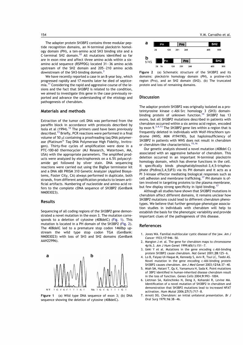
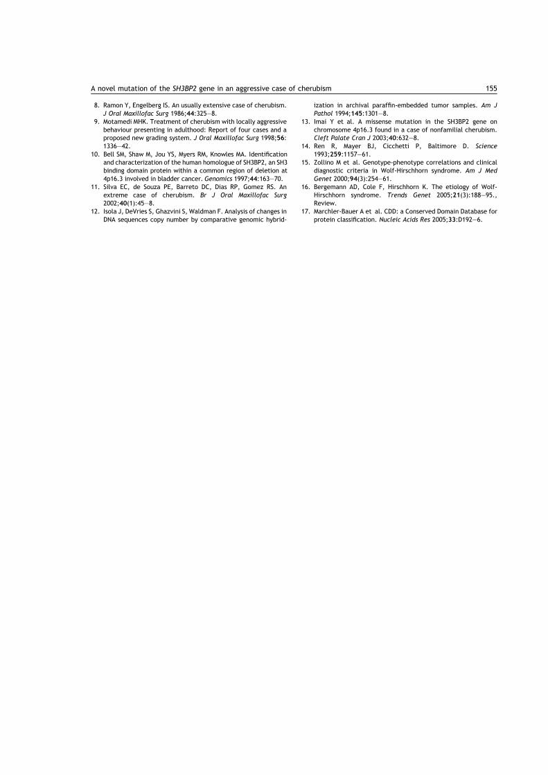
Sequencing of all coding regions of the SH3BP2 gene demon-strated a novel mutation in the exon 3. The mutation corre-sponds to a deletion of cytosine (408delC) (Fig. 1). Thismutation is located in a PH domain of the SH3BP2 (Fig. 2).The 408delC led to a premature stop codon 1460bp up-stream the wild type stop codon TGA (GenBank:NM003023) with loss of SH3 and SH2 domains (GenBankAAH22996).
Discussion
The adaptor protein SH3BP2 was originally isolated as a pro-teintyrosine kinase c-Abl-Src homology 3 (SH3) domain-binding protein of unknown function.14 SH3BP2 has 13exons, but all SH3BP2 mutations described in patients withcherubism occurred within a six amino acid region, encodedby exon 9.3,4,13 The SH3BP2 gene lies within a region that isfrequently deleted in individuals with Wolf-Hirschhorn syn-drome (WHS; MIM #194190), but haploinsufficiency ofSH3BP2 in patients with WHS does not result in cherubismor cherubism-like characteristics.15,16
Our genetic analysis showed a novel mutation (408del C)associated with an aggressive behavior of cherubism. Thisdeletion occurred in an important N-terminal pleckstrinhomology domain, which has diverse functions in the cell.It specifically binds phosphatidylinositol-3,4,5-trisphos-phate (PtdIns(3,4,5)P3) via its PH domain and it acts as aPI 3-kinase effector mediating biological responses such ascell adhesion and membrane trafficking.17 PH domain is of-ten involved in targeting proteins to the plasma membrane,but few display strong specificity in lipid binding.17
Although all studies have shown that SH3BP2mutations incherubism affect different domains, it remains unclear howSH3BP2 mutations could lead to different cherubism pheno-types. We believe that further genotype-phenotype associa-tion studies in individuals with cherubism will help toestablish the basis for the phenotypic variability and provideimportant clues of the pathogenesis of this disease.
References
1. Jones WA. Familial multilocular cystic disease of the jaw. Am JCancer 1933;17:946–50.
2. Mangion J et al. The gene for cherubism maps to chromosome4p16.3. Am J Hum Genet 1999;65(1):151–7.
3. Ueki Y et al. Mutations in the gene encoding c-Abl-bindingprotein SH3BP2 cause cherubism. Nat Genet 2001;28:125–6.
4. Lo B, Faiyaz-Ul-Haque M, Kennedy S, Aviv R, Tsui LC, Teebi AS.Novel mutation in the gene encoding c-Abl-binding proteinSH3BP2 causes cherubism. Am J Med Genet 2003;121A:37–40.
5. Miah SM, Hatani T, Qu X, Yamamura H, Sada K. Point mutationsof 3BP2 identified in human-inherited disease cherubism resultin the loss of function. Genes Cells 2004;9:993–1004.
6. Lietman SA, Kalinchinko N, Deng X, Kohanski R, Levine MA.Identification of a novel mutation of SH3BP2 in cherubism anddemonstration that SH3BP2 mutations lead to increased NFATactivation. Hum Mutat 2006;27(7):717–8.
7. Arnott DG. Cherubism: an initial unilateral presentation. Br JOral Surg 1979;16:38–46.
Figure 1 (a) Wild type DNA sequence of exon 3; (b) DNAsequence showing the deletion of cytosine (408delC).
Figure 2 (a) Schematic structure of the SH3BP2 and itsdomains: pleckstrin homology domain (PH), a proline-richregion (Pro), and an SH2 domain (SH2). (b) The truncatedprotein and loss of remaining domains.
154 V.M. Carvalho et al.
8. Ramon Y, Engelberg IS. An usually extensive case of cherubism.J Oral Maxillofac Surg 1986;44:325–8.
9. Motamedi MHK. Treatment of cherubism with locally aggressivebehaviour presenting in adulthood: Report of four cases and aproposed new grading system. J Oral Maxillofac Surg 1998;56:1336–42.
10. Bell SM, Shaw M, Jou YS, Myers RM, Knowles MA. Identificationand characterization of the human homologue of SH3BP2, an SH3binding domain protein within a common region of deletion at4p16.3 involved in bladder cancer. Genomics 1997;44:163–70.
11. Silva EC, de Souza PE, Barreto DC, Dias RP, Gomez RS. Anextreme case of cherubism. Br J Oral Maxillofac Surg2002;40(1):45–8.
12. Isola J, DeVries S, Ghazvini S, Waldman F. Analysis of changes inDNA sequences copy number by comparative genomic hybrid-
ization in archival paraffin-embedded tumor samples. Am JPathol 1994;145:1301–8.
13. Imai Y et al. A missense mutation in the SH3BP2 gene onchromosome 4p16.3 found in a case of nonfamilial cherubism.Cleft Palate Cran J 2003;40:632–8.
14. Ren R, Mayer BJ, Cicchetti P, Baltimore D. Science1993;259:1157–61.
15. Zollino M et al. Genotype-phenotype correlations and clinicaldiagnostic criteria in Wolf-Hirschhorn syndrome. Am J MedGenet 2000;94(3):254–61.
16. Bergemann AD, Cole F, Hirschhorn K. The etiology of Wolf-Hirschhorn syndrome. Trends Genet 2005;21(3):188–95.,Review.
17. Marchler-Bauer A et al. CDD: a Conserved Domain Database forprotein classification. Nucleic Acids Res 2005;33:D192–6.
A novel mutation of the SH3BP2 gene in an aggressive case of cherubism 155
ORIGINAL ARTICLE
Novel mutations in the SH3BP2 gene associated withsporadic central giant cell lesions and cherubism
VM Carvalho1,2, PF Perdigao1,2, FR Amaral1,2, PEA de Souza3, L De Marco1, RS Gomez2
1Department of Pharmacology, Universidade Federal de Minas Gerais, Belo Horizonte, MG, Brazil; 2Department of Oral Surgeryand Pathology, School of Dentistry, Universidade Federal de Minas Gerais, Belo Horizonte, MG, Brazil; 3School of Dentistry,Pontifıcia Universidade Catolica de Minas Gerais, Belo Horizonte, MG, Brazil
Central giant cell lesion (CGCL) is a reactive bone lesionthat occurs mainly in the mandible, characterized by themultinucleated osteoclast-like giant cells in a backgroundof oval to spindle-shaped mononuclear cells. The etiologyis unknown and occurs more commonly in young adults.Cherubism, a rare disease found predominantly infemales has histologic characteristics indistinguishablefrom those of CGCL and is caused by mutations mostlypresent in exon 9 of the SH3BP2 gene. In this study, weinvestigated four cases of CGCL and one case of cher-ubism. DNA was extracted from peripheral blood andtumor tissue and all coding and flanking regions of theSH3BP2 amplified by PCR and directly sequenced toidentify underlying mutations. Two novel mutations werefound; a heterozygous missense mutation c.1442A>T(Q481L) in exon 11 in one sporadic case of CGCL and aheterozygous germline and tumor tissue missensemutation c.320C>T (T107M) in exon 4 in one patient withcherubism. These findings open a new window to inves-tigate the possible relationship between the pathogenesisof the cherubism and CGCL.
Oral Diseases (2009) 15, 106–110
Keywords: central giant cell lesion; SH3-binding protein;SH3BP2; cherubism
Introduction
The central giant cell lesion (CGCL) was first describedby Ja!e (1953) as a giant-cell reparative granuloma ofthe jaw bones. CGCL is a reactive intra-osseous lesionresulting from a local reparative reaction of unknown
etiology. It occurs mainly in the mandible and the age ofmost patients ranges from 10 to 25 years (de Langeet al, 2007c). Radiographic findings are diverse, varyingfrom small unilocular lesions to large multilocularlesions with displacement of teeth, root resorption,and cortical perforation (Horner, 1989). Histologically,CGCL is characterized by the presence of multinucle-ated osteoclast-like giant cells in a background of ovalto spindle-shaped mononuclear cells (Neville et al,2002). Cherubism (MIM 118400) is an autosomaldominant inherited syndrome characterized by excessivebone degradation of the upper and lower jaws followedby development of fibrous tissue masses, which causes acharacteristic facial swelling (Ueki et al, 2001). The generesponsible for cherubism is located on chromosome4p16.3 and named SH3BP2 (Mangion et al, 1999). Todate, all mutations of the SH3BP2 identified in familialcherubism are present in exon nine and a!ect threeamino acids within a conserved six amino acid sequence(RSPPDG), the SH3-binding domain (Hatani and Sada,2008). Recently, a mutation in the SH3BP2 gene wasdescribed in a case of non-familial cherubism (Imai et al,2003) and also in one aggressive case of sporadiccherubism (Carvalho et al, 2008). Although the etiologyof CGCL is unknown, the similar histologic features ofthis lesion with cherubism suggest that both conditionsshare the same genetic alteration. Therefore, we inves-tigated all coding and flanking sequences of the SH3BP2gene in patients with CGCL.
Material and methods
In this report, we studied frozen samples from four casesof CGCL and one case of cherubism, collected between1998 and 2004 and stored at )80!C. The criteria forCGCL diagnosis is described elsewhere (Neville et al,2002; de Lange et al, 2007c). All lesions were located inthe midline or posterior region of the mandible. TheUniversity Ethics Committee approved the work. Afteran informed written consent signed by all patients,fragments of the lesions were obtained during surgical
Correspondence: RS Gomez, Faculdade de Odontologia, UniversidadeFederal de Minas, Gerais, Av. Antonio Carlos, 6627 Belo Horizonte,CEP 31270-901 MG, Brazil. Tel: +55 31 3499 2477, Fax:+55 31 3499 2472, E-mail: [email protected] 6 August 2008; revised 24 September 2008; accepted 29September 2008
Oral Diseases (2009) 15, 106–110. doi:10.1111/j.1601-0825.2008.01499.x" 2008 The Authors. Journal compilation " 2008 Blackwell Munksgaard
All rights reserved
http://www.blackwellmunksgaard.com

removal procedures. A piece of the fragment was fixedin 10% formalin bu!er and para"n-embedded tissueblocks were used for routine histologic staining toconfirm the clinical diagnosis. All five lesions studiedshowed typical histologic features of CGCL described inthe literature (Neville et al, 2002; de Lange et al, 2007c).Another piece of the specimen sample was immediatelystored at )80!C for subsequent molecular analyzes. Inaddition, peripheral blood was collected from all sub-jects. The CGCL was considered aggressive when thelesion was larger than 4 cm in size and the cortical bonewas damaged or expanded and teeth displaced (Chuonget al, 1986).
One case was referred to the School of Dentistry asbilateral CGCL and her clinical chart did not reportcherub face or familial cases of cherubism. After an intraoral examination, a discreet expansion of the mandiblewas noted. Radiographic findings showed bilateralmultilocular lesions of the mandible with tooth dis-placement. Although no cherub face was seen, ourdiagnostic hypothesis was that of cherubism with lowexpressivity. Therefore, in addition to clinical andbiochemical data, DNA of tumor and blood cells wasextracted and the SH3BP2 gene investigated.
All patients were unrelated and underwent detailedhematologic and biochemical evaluation, includingparathyroid hormone (PTH) levels, alkaline phospha-tase activity, serum calcium and phosphorus concen-trations. All these parameters were within normalranges.
Genomic DNA was extracted from lesions andperipheral blood using the GenomicPrep kit accordingto the manufacturer’s instructions (Amersham Pharma-cia Biotech Inc., Piscataway, NJ, USA). All coding andflanking regions of the SH3PB2 gene were investigatedusing previously described PCR primers and reactionconditions (Imai et al, 2003). Briefly, PCR reactionswere performed in a final volume of 50 ll containing200 ng of template DNA, 200 lM dNTPs, 10 pM ofeach primer and 1.25 U of a proofreading Taq poly-merase (Platinum" TaqDNA Polymerase High Fidelity;Invitrogen, Carlsbad, CA, USA). Thirty-five cycles ofamplification were performed in a PTC-100-60 thermo-cycler (MJ Research, Watertown, MA, USA) with theappropriate parameters. The amplified products wereanalyzed by electrophoresis on a 6.5% polyacrylamidegel followed by silver stain. DNA bands correspondingto expected size were purified using the GFX PCR DNA
and Gel Band Purification Kit (Amersham Biosciences,Piscataway, NJ, USA). DNA sequencing reactions werecarried out using the BigDye Terminator kit in a DNAABI PRISM 310 Genetic Analyzer (Applied Biosystems,Foster City, CA, USA). All experiments were performedin duplicate with both strands and from di!erentamplification products to lessen artificial artifacts.Numbering of nucleotide and amino acid refers to thecomplete cDNA sequence of SH3BP2. In order toexclude single nucleotide polymorphisms (SNPs), wesequenced DNA from 50 unrelated control subjects withthe same ethnic background. This group was comprisedof patients without history of CGCL or Cherubism. TheNCBI SNP database was also checked to rule outpolymorphisms.
Results
To verify the presence of mutations of the SH3BP2 genein CGCL and cherubism patients, all 13 coding andflanking regions were sequenced from PCR productsfrom tumor DNA as well as peripheral blood DNA.Clinical and radiological features as well as the molec-ular alterations found in the SH3BP2 gene are shown inTable 1.
The patient with cherubism (#1) presented a germlineheterozygous substitution of cytosine to thyminec.320C>T (T107M) in exon 4 (Figure 1a). This muta-tion leads to an exchange of the amino acid threonine tomethionine in the N-terminal pleckstrin domain. Thepresence of this germline mutation led us to screen thepatient’s close relatives. Besides her parents, no otherrelatives were available for clinical assay. Oral mucosalcells were collected from them, DNA extracted andsequencing of the SH3BP2 gene performed.
The mother was shown to carry the same heterozy-gous mutation c.320C>T found in the propositus (datanot shown). To predict whether the molecular altera-tions present in both patients would a!ect proteinfunction, the algorithm SORTING INTOLERANT FROM
TOLERANT (SIFT) (http://blocks.fhcrc.org/sift/SIFT.html)was used.
Analysis of the SH3BP2 gene in patient with non-aggressive CGCL (#2) showed the presence of a novelheterozygous mutation c.1442A>T (Q481L) in exon 11(Figure 1b), leading to an exchange of amino acidGlutamine to Leucine in the C-terminal SH2 domain.No germline mutation was found in this patient.
Table 1 Clinical data, radiological features and mutations detected in patients with cherubism (#1) and central giant cell lesion (#2 to #5)
Caseno.
Age(years) Sex Aggressiveness X-ray findings
Mutation(germline)
Mutation(tumor)
1 12 F Yes Large destructive multilocular radiolucent lesionswith tooth displacement
c.320C>T c.320C>T
2 41 F No Small unilocular radiolucent lesion Wild type c.1442A>T3 46 M Yes Large destructive multilocular radiolucent lesion Wild type Wild type4 11 M Yes Large destructive multilocular radiolucent lesion Wild type Wild type5 19 M No Multilocular radiolucent lesion Wild type Wild type
Nucleotides are numbered according to the ENST00000356331.
Novel mutations in the SH3BP2 geneVM Carvalho et al
107
Oral Diseases

Discussion
A number of conditions can present with lesions thathistologically are indistinguishable from CGCL of thejaws, including giant cell tumors, aneurysmal bone cyst,brown tumors of hyperparathyroidism, cherubism andother inherited syndromes associated with geneticmutations such as Noonan-like⁄multiple giant cell lesionsyndrome, Neurofibromatosis type 1 and Ramon syn-drome (Pina-Neto et al, 1986; van Damme and Mooren,1994; Lee et al, 2005).
The gene SH3BP2 that codes for the adapter proteinSH3BP2 was identified in 2001 on chromosome 4p16.3(Mangion et al, 1999; Tiziani et al, 1999; Ueki et al,2001). A mutation in exon 9 of the SH3BP2 gene wasdescribed in a case of non-familial cherubism (Imai et al,2003). Subsequently, SH3BP2 gene molecular analysisof exon 9 in patients with sporadic central or peripheralgiant cells lesions have not demonstrated the presence ofmutations (de Lange et al, 2007b; Lietman et al, 2007;Idowu et al, 2008). In this report, we analyzed all codingand flanking regions of the SH3BP2 gene and found twonovel mutations of SH3BP2, one associated with asporadic case of CGCL and one in a patient withcherubism.
Three functional domains of SH3BP2 have beendescribed; the N-terminal pleckstrin homology domain(PH), a 10-amino acid SH3 binding site and C-terminalSH2 domain (Ren et al, 1993). Patient #2 presented a
heterozygous mutation c.1442A>T (Q481L) in exon 11,a SH2 domain of the SH3BP2. This mutation waspresent only in tumor cells. The Src homology 2 domain(SH2) is involved in recognition of phosphorylatedtyrosine (pTyr) and SH2 domains typically bind pTyr-containing ligands via two surface pockets allowingproteins with SH2 domains to localize to tyrosinephosphorylated sites (Ingley, 2008). Analysis of theprotein function based on sequence homology and thephysical properties of amino acids using the SIFT
software program shows that the Q481L alteration inthe SH2 domain a!ects protein function, possiblyarresting SH3BP2 to localize tyrosine phosphorylatedsites. To date, functional studies of SH3BP2 mutationshave not been performed. But their association withdisease, location in areas of the molecule that are highlyconserved between species (http://www.ensembl.org/Homo_sapiens/searchview?species=;idx=;q=sh3bp2)and most important, their absence in normal controls,suggest that they are pathogenic.
Lietman et al (2007) studied exon 9 of SH3BP2 gene in10 giant cell tumors of bone and nine giant cell reparativegranulomas and did not find any molecular alteration. Inaddition, they showed that SH3BP2 transcript andprotein as well as NFATc1 protein were abundantlyexpressed in CGCL. They also found a smaller transcriptin the RNA from one giant cell lesion and speculated thata mutation outside exon 9 could be responsible for thistruncated protein. Idowu et al (2008) did not findmutations in exon 9 of the SH3BP2 gene in CGCL ofthe jaw and neither did de Lange et al (2007b). It isimportant to emphasize that these authors limited to exon9 their search for SH3BP2 gene mutations.
At this study, we found a novel germline mutation inexon 4 (c.320C>T) in one young female patientpresenting with cherubism with low expressivity. Thismutation led to a substitution of threonine to methio-nine in the N-terminal pleckstrin domain. PH domain isonly found in eukaryotes and is the eleventh mostpopulous domain family in the human genome (Landeret al, 2001). PH domains is only found in cellularsignaling proteins such as serine ⁄ threonine kinase,tyrosine kinases, regulators of G-proteins, endocytoticGTPases, adaptors, as well as cytoskeleton associatedmolecules and in lipid associated enzymes (Ingley andHemmings, 1994; Lemmon, 2004).
Our patient with cherubism fulfilled some clinical andradiographic criteria used in its diagnosis such asbilateral swelling of the jaw with expansion or thinnessof the cortical bone, bilateral multilocular radiolucen-cies, tooth displacement and root resorption (de Langeet al, 2007a). Cherubism shows 100% penetrance inmales and 50–70% in females (von Wowern, 2000;Horton, 2002; Marx and Stern, 2002) and the expres-sivity is wide, ranging from bilateral non-expansible tohighly destructive lesions (von Wowern, 2000). Becauseof incomplete penetrance of cherubism in females and toits variability of expressivity, some cases of cherubismmay be under-diagnosed. Recently, we reported a novelmutation in the SH3BP2 gene outside exon 9 in oneaggressive case of cherubism (Carvalho et al, 2008).
C CCG A G GT
C CC GA AG T
(a)
(b)
Figure 1 Chromatogram sequence data of the propositus #1 showingthe heterozygous c.320C>T mutation (a); Chromatogram sequencedata of the propositus #2 showing the heterozygous c.1442A>Tmutation (b)
Novel mutations in the SH3BP2 geneVM Carvalho et al
108
Oral Diseases

Given that all but one previous report of mutations inthe SH3BP2 have looked only in exon 9, additionalstudies analyzing the entire gene and trying to correlategenotype–phenotype features in individuals with cher-ubism will help to establish the basis for the disease’sphenotypic variability.
Up to now, it has been suggested that the SH3BP2gene plays a role in regulating increased osteoblast andosteoclast activities (Ueki et al, 2007). Furthermore, it isproposed that mutations in the SH3BP2 gene couldcause pathologic activation of osteoclasts, probably bydysfunction of the SH3BP2 gene in the regulatorypathway of osteoclastogenesis (Miah et al, 2004; Hyckelet al, 2005). In this process, SH3BP2 has an influence onthe regulation of the PTH receptor and PTH-relatedprotein (PTHrP), interacting with the chaperone protein14-3-3 (Foucault et al, 2003), which was recentlydescribed as a regulatory protein of type I PTH ⁄PTHrPreceptor (Tazawa et al, 2003).
Lietman et al (2006) showed that heterozygoticmutations in exon 9 of the SH3BP2 led to increasedNFAT (nuclear factor of activated T-cells), an oste-oclastogenic mediator, indicating that the cherubismresults from gain of function mutations in SH3BP2.The NFATc1 isoform (also termed NFAT2) has beenproposed as an important master transcription factorfor osteoclastogenesis (Foucault et al, 2003a,b). Dys-function of SH3BP2 protein by point mutationscauses pathologic activation of osteoclasts in cherub-ism, suggesting that endogenous SH3BP2 has aregulatory role in the development and ⁄ or activationof osteoclasts in jaw bones (Miah et al, 2004). Thematuration of osteoclasts requires osteoblasts andstromal cells releasing macrophage colony-stimulatingfactor and the receptor for activation of nuclear factorjB ligand (RANKL) that are essential to promoteosteoclastogenesis (Roodman, 1999).
In this study, novel SH3BP2 gene mutations insporadic CGCL and in one case of cherubism weredemonstrated. Considering the diverse clinicalbehavior of cherubism it is possible that somenon-familial cases of cherubism are undistinguishablefrom sporadic cases of CGCL. Further sequencing of allcoding as well as flanking and promoter regions of theSH3BP2 gene in CGCL and non-familial cherubismcases with variable expressivity and penetrance arenecessary to provide important insights into molecularmechanisms associated with these lesions.
Acknowledgements
This study was supported in part by grants from Fundacao deAmparo a Pesquisa do Estado de Minas Gerais (FAPEMIG)and from Milenio⁄Conselho Nacional de DesenvolvimentoCientıfico e Tecnologico (CNPq), Brazil; L De Marco and RSGomez are Research Fellows of CNPq.
Author contributions
VM Carvalho, PF Perdigao, L De Marco and RS Gomezcontributed in study concepts, study design, definition of
intellectual content and in manuscript preparation and editing.VM Carvalho, PF Perdigao, FR Amaral and PEA Souzacontributed in data acquisition.
References
Carvalho VM, Perdigao PF, Pimenta FJ, de Souza PE, GomezRS, De Marco L (2008). A novel mutation of the SH3BP2gene in an aggressive case of cherubism. Oral Oncol 44: 153–155.
Chuong R, Kaban LB, Kozakewich H, Perez-Atayde A(1986). Central giant cell lesions of the jaws; a clinicopath-ologic study. J Oral Maxillofac Surg 44: 708–713.
van Damme PA, Mooren RE (1994). Di!erentiation ofmultiple giant cell lesions, Noonan-like syndrome, andoccult hyperparathyroidism. Case report and review of theliterature. Int J Oral Maxillofac Surg 23: 32–36.
Foucault I, Liu YC, Bernard A, Deckert M (2003). Thechaperone protein 14-3-3 interacts with 3BP2 ⁄SH3BP2and regulates its adapter function. J Biol Chem 278: 7146–7153.
Hatani T, Sada K (2008). Adaptor protein 3BP2 and cherub-ism. Curr Med Chem 15: 549–554.
Horner K (1989). Central giant cell granuloma of the jaws: aclinico-radiological study. Clin Radiol 40: 622–626.
Horton WA (2002). Abnormalities of bone structure. In:Rimoin DL, Connor JM, Pyeritz RE, Korf BR, eds.Emery and Rimoin’s principles and practice of medicalgenetics, 4th edn. Churchill Livingstonel: London, pp.4155–4156.
Hyckel P, Berndt A, Schleier P et al (2005). Cherubism – newhypotheses on pathogenesis and therapeutic consequences.J Craniomaxillofac Surg 33: 61–68.
Idowu BD, Thomas G, Frow R, Diss TC, Flanagan AM(2008). Mutations in SH3BP2, the cherubism gene, were notdetected in central or peripheral giant cell tumours of thejaw. Br J Oral Maxillofac Surg 46: 229–230.
ImaiY,KannoK,MoriyaT et al (2003).Amissensemutation inthe SH3BP2 gene on chromosome 4p16.3 found in a case ofnonfamilial cherubism. Cleft Palate Craniofac J 40: 632–638.
Ingley E (2008). Src family kinases: regulation of theiractivities, levels and identification of new pathways. BiochimBiophys Acta 1784: 56–65.
Ingley E, Hemmings BA (1994). Pleckstrin homology (PH)domains in signal transduction. J Cell Biochem 56: 436–443.
Ja!e HL (1953). Giant-cell reparative granuloma, traumaticbone cyst, and fibrous (fibro-osseous) dysplasia of thejawbones. Oral Surg 6: 159–175.
Lander ES, Linton LM, Birren B et al (2001). Initialsequencing and analysis of the human genome. Nature409: 860–921.
de Lange J, van Maarle MC, van den Akker HP, Redeker EJ(2007a). A new mutation in the SH3BP2 gene showingreduced penetrance in a family a!ected with cherubism. OralSurg Oral Med Oral Pathol Oral Radiol Endod 103: 378–381.
de Lange J, van Maarle MC, van den Akker HP, Redeker EJ(2007b). DNA analysis of the SH3BP2 gene in patients withaggressive central giant cell granuloma. Br J Oral MaxillofacSurg 45: 499–500.
de Lange J, van den Akker HP, Van den Berg H (2007c).Central giant cell granuloma of the jaw: a review of theliterature with emphasis on therapy options. Oral Surg OralMed Oral Pathol Oral Radiol Endod 104: 603–615.
Lee JS, Tartaglia M, Gelb BD et al (2005). Phenotypic andgenotypic characterisation of Noonan-like⁄multiple giant celllesion syndrome. J Med Genet 42: e11.
Novel mutations in the SH3BP2 geneVM Carvalho et al
109
Oral Diseases
Lemmon MA (2004). Pleckstrin homology domains: not justfor phosphoinositides. Biochem Soc Trans 32: 707–711.
Lietman SA, Kalinchinko N, Deng X, Kohanski R, LevineMA(2006). Identification of a novel mutation of SH3BP2 incherubismanddemonstration that SH3BP2mutations lead toincreased NFAT activation. HumMutat 27: 717–718.
Lietman SA, Prescott NL, Hicks DG, Westra WH, Levine MA(2007). SH3BP2 is rarely mutated in exon 9 in giant celllesions outside cherubism. Clin Orthop Relat Res 459: 22–27.
Mangion J, Rahman N, Edkins S et al (1999). The gene forcherubism maps to chromosome 4p16.3. Am J Hum Genet65: 151–157.
Marx RE, Stern D (2002). Oral and maxillofacial pathology.Quintessence: Chicago, pp. 744.
Miah SM, Hatani T, Qu X, Yamamura H, Sada K (2004).Point mutations of 3BP2 identified in human-inheriteddisease cherubism result in the loss of function. Genes Cells9: 993–1004.
Neville BW, Damm DD, Allen CM, Bouquot JE (2002). Oraland maxillofacial pathology, 2nd edn. WB Saunders Co:Philadelphia.
Pina-Neto JM, Moreno AF, Silva LR et al (1986). Cherubism,gingival fibromatosis, epilepsy, and mental deficiency(Ramon syndrome) with juvenile rheumatoid arthritis. AmJ Med Genet 25: 433–441.
Ren R, Mayer BJ, Cicchetti P, Baltimore D (1993). Identifi-cation of a ten-amino acid proline-rich SH3 binding site.Science 259: 1157–1161.
Roodman GD (1999). Cell biology of the osteoclast. ExpHematol 27: 1229–1241.
Tazawa H, Takahashi S, Zilliacus J (2003). Interaction of theparathyroid hormone receptor with the 14-3-3 protein.Biochim Biophys Acta 1620: 32–38.
Tiziani V, Reichenberger E, Buzzo CL et al (1999). The genefor cherubism maps of chromosome 4p16. Am J Hum Genet65: 158–166.
Ueki Y, Tiziani V, Santanna C et al (2001). Mutations in thegene encoding c-Abl-binding protein SH3BP2 cause cher-ubism. Nat Genet 28: 125–126.
Ueki Y, Lin CY, Senoo M et al (2007). Increased myeloid cellresponses to M-CSF and RANKL cause bone lossand inflammation in SH3BP2 !cherubism’ mice. Cell 128:71–83.
von Wowern N (2000). Cherubism: a 36-year long-termfollow-up of 2 generations in di!erent families and reviewof the literature. Oral Surg Oral Med Oral Pathol OralRadiol Endod 90: 765–772.
Novel mutations in the SH3BP2 geneVM Carvalho et al
110
Oral Diseases

4 CONSIDERAÇÕES FINAIS
36

4 Considerações Finais O TCG, o tumor marrom do hiperparatireoidismo, o Querubismo e outras
Síndromes herdadas como a de Noonan, a Neurofibromatose tipo1 e a de Ramon
são doenças podem apresentar lesões histologicamente indistinguíveis das LCCG
(VAN DAMME & MOOREN, 1994; PINA-NETO et al., 1986; LEE et al., 2005).
O gene SH3BP2 foi identificado no cromossomo 4p16.3 e relacionado à
etiologia do Querubismo (UEKI et al., 2001; MANGION et al., 1999; TIZIANI et al.,
1999). Três domínios funcionais do SH3BP2 foram descritos: um domínio de
homologia pleckstrin N-terminal (PH), um sítio de ligação SH3 com 10 aminoácidos e
um domínio SH2 C-terminal (REN et al., 1993).
O gene SH3BP2 possui 13 exons, mas todas as mutações descritas em
pacientes com Querubismo ocorreram em uma região de 6 aminoácidos, codificada
pelo exon 9 (UEKI et al., 2001; IMAI et al. 2003, LO et al. 2003). Outros trabalhos
demonstraram a ocorrência de mutação ao avaliarem o mesmo exon em casos de
Querubismo (LI & YU, 2006; DE LANGE et al., 2007a).
Apesar de todos os estudos mostrarem que as mutações no SH3BP2 do
Querubismo afetam diferentes domínios, ainda permanece incerto como estas
mutações poderiam acarretar diferentes fenótipos.
Neste trabalho, todas as regiões codificadoras do gene SH3BP2 foram
analisadas e três novas mutações encontradas: uma associada a LCCG esporádica
e outras duas associadas a casos de Querubismo.
A nova mutação (408delC) no caso de Querubismo agressivo deste estudo
ocorreu em um importante domínio de homologia pleckstrin N-terminal, que tem
37

funções diversas na célula. Ela se liga especificamente ao fosfatidilinositol 3,4,5-
trifosfato através de seu domínio PH e medeia respostas como adesão celular e
tráfego de membrana. O domínio PH está normalmente envolvido na captura de
proteínas para a membrana plasmática (MARCHLER-BAUER et al., 2005).
A mutação heterozigótica associada ao caso de LCCG: A1442T (Gln481Leu),
ocorreu no exon 11, no domínio SH2 do SH3BP2. A análise da função da proteína
baseada na seqüência de homologia e nas propriedades físicas de aminoácidos
usando o programa SIFT (http://blocks.fhcrc.org/sift/SIFT.html) mostra que esta
alteração afeta a função da proteína, possivelmente impedindo a SH3BP2 de
localizar regiões de tirosina fosforiladas.
Enfatiza-se que tanto Lietman et al. (2007) quanto Idowu et al. (2008) e de
Lange et al. (2007b) não encontraram mutações nas LCCG, porém, eles limitaram
seus estudos ao exon 9 do gene SH3BP2.
Estudos funcionais das mutações no SH3BP2 não foram realizados até o
momento, mas por elas estarem associadas com a doença, localizadas em áreas da
molécula que são altamente conservadas entre espécies
(www.ensembl.org/Homo_sapiens/Search/Summary?species=Homo_sapiens;idx=;q
=sh3bp2) e principalmente, por estarem ausentes em controles normais, torna-se
sugestivo que elas sejam patogênicas.
Uma nova mutação germinativa no exon 4 (C320T) foi encontrada na paciente
diagnosticada com Querubismo de baixa expressividade. Esta mutação acarretou a
substituição de Treonina por Metionina em um domínio Pleckstrin N-terminal. O
domínio PH é encontrado apenas em Eucariotos e é uma das famílias de domínio
mais populosa no genoma humano (LANDER et al., 2001). Ele está presente em
38

proteínas de sinalização celular, tais como serina-treonina quinase, tirosina quinase,
reguladores de proteina G, GTPases, assim como moléculas associadas ao
citoesqueleto e em enzimas associadas a lipídeos (INGLEY & HEMMINGS, 1994;
LEMMON, 2004).
O Querubismo apresenta 100% de penetrância em homens e 50-70% em
mulheres (VON WOWERN, 2000; HORTON, 2002; MARX AND STERN, 2002) e a
expressividade é ampla, variando de lesões bilaterais sem expansão óssea a lesões
muito destrutivas (VON WOWERN, 2000).
Devido à penetrância incompleta do Querubismo em mulheres e de sua
variabilidade de expressão, alguns casos podem ser diagnosticados erroneamente.
Considerando seu comportamento clínico variável, é provável que alguns casos de
Querubismo não-familiar sejam indistinguíveis de casos esporádicos de LCCG.
Lietman et al. (2007) mostraram que o transcrito e a proteína do SH3BP2,
assim como a proteína NFATc1, estavam expressos de forma abundante nas LCCG.
Eles encontraram também um transcrito menor no RNA de uma LCCG e sugeriram
que uma mutação fora do exon 9 poderia ser a responsável por esta proteína
truncada.
Ueki et al. (2007) sugeriram que o gene SH3BP2 exerce um papel regulador
em osteoblastos e osteoclastos, aumentando a atividade deles. Foi proposto que
mutações neste gene podem causar ativação patológica de osteoclastos,
provavelmente pela disfunção do SH3BP2 na regulação da via da
osteoclastogênese (MIAH et al., 2004; HYCKEL et al., 2005).
A disfunção da proteína SH3BP2, devido a mutações pontuais no gene,
causam a ativação patológica de osteoclastos observada no Querubismo, sugerindo
39

que a SH3BP2 endógena exerce um papel regulador no desenvolvimento e/ ou
ativação de osteoclastos nesta Síndrome (MIAH et al., 2004).
Mutações heterozigóticas no exon 9 do SH3BP2 causaram o aumento de
NFAT, que é um mediador osteoclastogênico, indicando que o Querubismo resulta
de mutações funcionais (LIETMAN et al., 2006).
Considerando que este é o primeiro trabalho a avaliar o gene SH3BP2 por
completo, estudos adicionais similares, que relacionem genótipo-fenótipo em
indivíduos com Querubismo e LCCG esporádicas, ajudarão a estabelecer a base da
variabilidade fenotípica destas doenças, assim como aspectos patogênicos
compartilhados entre elas.
40

5 CONCLUSÕES
41
5 CONCLUSÕES
1 Existe uma variabilidade genética nas alterações moleculares das Lesões
centrais de células gigantes e do Querubismo, assim como na expressão fenotípica
destas doenças.
2 As Lesões centrais de células gigantes esporádicas podem apresentar
mutações no gene SH3BP2.
3 Avaliações moleculares do gene SH3BP2 em LCCG e no Querubismo
devem incluir o seqüenciamento de todos os exons, uma vez que as mutações
podem podem estar fora da região hot spot em ambos os casos.
42

6 REFERÊNCIAS BIBLIOGRÁFICAS
43

6 Referências Bibliográficas
1 Arnott DG. Cherubism: An initial unilateral presentation. Br J Oral Surg 1979;16:38-46.
2 Auclair PL, Cuenin P, Kratochvil FJ, Slater LJ, Ellis GL. A clinical and histomorphologic comparison of the central giant cell granuloma and the giant cell tumour. Oral Surg Oral Med Oral Pathol 1988;66:197-208.
3 Barnes L, Eveson JW, Reichart P, Sidransky D, eds. Pathology and Genetics of Head and Neck Tumours. Kleihues P, Sobin LH, series eds. World Health Organization Classification of Tumours. Lyon, France: IARC Press, 2005. p. 324.
4 Battaglia A, Carey JC.Health supervision and anticipatory guidance of individuals with Wolf-Hirschhorn syndrome. Am J Med Genet. 1999 Jun 25;89(2):111-5.
5 Bell SM, Shaw M, Jou YS, Myers RM, Knowles MA. (1997) Identification and characterization of the human homologue of SH3BP2, an SH3 binding domain protein within a common region of deletion at 4p16.3 involved in bladder cancer. Genomics, 44:163-170.
6 Carvalho VM, Perdigão PF, Pimenta FJ, de Souza PE, Gomez RS, De Marco L. A novel mutation of the SH3BP2 gene in an aggressive case of cherubism. Oral Oncol 2008; 44(2):153-5.
7 Chuong R, Kaban LB, Kozakewich H, Perez-Atayde A. Central giant cell lesions of the jaws; a clinicopathologic study. J Oral Maxillofac Surg 1986; 44:708-13.
8 Cossio, P.F. et al. Recurrent central giant cell granuloma in the mandible: surgical treatment and dental implant restoration. Med Oral Patol Oral Cir Bucal 2007; 12:229-32.
9 de Lange J, van den Akker HP, Klip H. Incidence and diseasefree survival after surgical therapy of central giant-cell granulomas of the jaw in The Netherlands: 1990-1995. Head Neck 2004;26:792-5.
10 de Lange J, van den Akker HP, Veldhuijzen van Zanten GO, Engelshove HA, van den Berg H, Klip H. Calcitonin therapy in central giant cell granuloma of the jaw: a randomized double-blind placebo-controlled study. Int J Oral Maxillofac Surg 2006; 35:791-5.
11 de Lange J, van Maarle MC, van den Akker HP, Redeker EJ. A new mutation in the SH3BP2 gene showing reduced penetrance in a family affected with cherubism. Oral Surg Oral Med Oral Pathol Oral Radiol
44
Endod 2007a;103:378-81.
12 de Lange J, van Maarle MC, van den Akker HP, Redeker EJ. DNA analysis of the SH3BP2 gene in patients with aggressive central giant cell granuloma. Br J Oral Maxillofac Surg 2007b;45:499-500.
13 de Lange J, van den Akker HP, Van den Berg H. Central giant cell granuloma of the jaw: a review of the literature with emphasis on therapy options. Oral Surg Oral Med Oral Pathol Oral Radiol Endod 2007c;104(5):603-15.
14 Deckert M, Tartare-Deckert S, Hernandez J, Rottapel R, Altman A. Adaptor function for the Syk kinases-interacting protein 3BP2 in IL-2 gene activation. Immunity. 1998 Nov;9(5):595-605.
15 Flanagan AM, Nui B, Tinkler SM, Horton MA, Williams DM, Chambers TJ. The multinucleate cells in giant cell granulomas of the jaw are osteoclasts. Cancer. 1988 Sep 15;62(6):1139-45.
16 Foucault I, Liu YC, Bernard A, Deckert M: The chaperone protein 14-3-3 interacts with 3BP2/SH3BP2 and regulates its adapter function. J Biol Chem 2003;278(9): 7146–53.
17 Hatani T, Sada K. Adaptor protein 3BP2 and cherubism. Curr Med Chem 2008;15(6):549-54.
18 Hawes M. J. Cherubism and its orbital manifestations. Ophthal Plast Reconstr Surg 1989;5:133–140.
19 Hirschhorn, K., Cooper, H.L. & Firschein, I.L. Humangenetik 1965;1:479–482.
20 Horner K. Central giant cell granuloma of the jaws: a clinico-radiological study. Clin Radiol 1989;40(6):622-6.
21 Horton WA. Abnormalities of bone structure. In: Rimoin DL, Connor JM, Pyeritz RE, Korf BR. Emery and Rimoin’s principles and practice of medical genetics. 4th ed. London: Churchill Livingstonel, 2002. p. 4155-6.
22 Hyckel P, Berndt A, Schleier P, Clement JH, Beensen V, Peters H, et al. Cherubism - new hypotheses on pathogenesis and therapeutic consequences. J Craniomaxillofac Surg 2005;33(1):61-8.
23 Idowu BD, Thomas G, Frow R, Diss TC, Flanagan AM. Mutations in SH3BP2, the cherubism gene, were not detected in central or peripheral giant cell tumours of the jaw. Br J Oral Maxillofac Surg 2008;46(3):229-30.

24 Imai Y, Kanno K, Moriya T, Kayano S, Seino H, Matsubara Y, et al. A missense mutation in the SH3BP2 gene on chromosome 4p16.3 found in a case of nonfamilial cherubism. Cleft Palate Craniofac J 2003;40(6):632-38.
25 Ingley E, Hemmings BA. Pleckstrin homology (PH) domains in signal transduction. J Cell Biochem 1994; 56(4):436-43.
26 Ingley E. Src family kinases: regulation of their activities, levels and identification of new pathways. Biochim Biophys Acta 2008;1784(1):56-65.
27 Isola J, DeVries, Ghazvini S, Waldman F. Analysis of changes in DNA sequences copy number by comparative genomic hybridization in archival paran-embedded tumor samplas. Am J Pathol 1994;145:1301–8.
28 Itonaga I, Schulze E, Burge PD, Gibbons CLMH, Ferguson D, Athanasou NA. Phenotypic characterization of mononuclear and multinucleated cells of giant cell reparative granuloma of small bones. J Pathol 2002;198:30-6.
29 Itonaga I, Hussein I, Kudo O, Sabokbar A, Watt-Smith S, Ferguson D, Athanasou NA. Cellular mechanisms of osteoclast formation and lacunar resorption in giant cell granuloma of the jaw. J Oral Pathol Med 2003;32(4):224-31.
30 Jones WA. Familial multilocular cystic disease of the jaws. Am J Cancer 1933; 17:946–950
31 Jaffe HL. Giant-cell reparative granuloma, traumatic bone cyst, and fibrous (fibro-osseous) dysplasia of the jawbones. Oral Surg 1953;6:159-75.
32 Katsikeris N, Kakarantza-Angelopoulou E, Angelopoulos AP. Peripheral giant cell granuloma. Clinicopathologic study of 224 new cases and review of 956 reported cases. Int J Oral Maxillofac Surg 1988;17(2):94-9.
33 Lander ES, Linton LM, Birren B, Nusbaum C, Zody MC, Baldwin J, et al. Initial sequencing and analysis of the human genome. Nature 2001; 409(6822):860-921.
34 Lee JS, Tartaglia M, Gelb BD, Fridrich K, Sachs S, Stratakis CA, et al. Phenotypic and genotypic characterisation of Noonan-like/multiple giant cell lesion syndrome. J Med Genet 2005;42(2):e11.
35 Lee JY, Jung YS, Kim SA, Lee SH, Ahn SG, Yoon JH. Investigation of the SH3BP2 gene mutation in cherubism. Acta Med Okayama 2008; 62(3):209-12.
36 Lemmon MA. Pleckstrin homology domains: not just for phosphoinositides. Biochem Soc Trans 2004;32(Pt 5):707-11.

37 Lerner UH. New molecules in the tumor necrosis factor ligand and receptor superfamilies with importance for physiological and pathological bone resorption. Crit Rev Oral Biol Med 2004;15:64-81.
38 Li, C.; Yu, S. A novel mutation in the SH3BP2 gene causes cherubism: case report. BMC Medical Genetics 2006;84:17.
39 Lietman SA, Kalinchinko N, Deng X, Kohanski R, Levine MA. Identification of a novel mutation of SH3BP2 in cherubism and demonstration that SH3BP2 mutations lead to increased NFAT activation. Hum Mutat 2006; 27(7):717-8.
40 Lietman SA, Prescott NL, Hicks DG, Westra WH, Levine MA. SH3BP2 is rarely mutated in exon 9 in giant cell lesions outside cherubism. Clin Orthop Relat Res 2007;459:22-7.
41 Lietman SA, Yin L, Levine MA. SH3BP2 is an activator of NFAT activity and osteoclastogenesis. Biochem Biophys Res Commun 2008;371:644-8.
42 Liu B, Yu SF, Li TJ. Multinucleated giant cells in various forms of giant cell containing lesions of the jaws express features of osteoclasts. J Oral Pathol Med 2003;32(6):367-75.
43 Lo B, Faiyaz-Ul-Haque M, Kennedy S, Aviv R, Tsui LC, Teebi AS. (2003) Novel mutation in the gene encoding c-Abl-binding protein SH3BP2 causes cherubism. Am J Med Genet 121A:37-40.
44 Lombardi T, Bischof M, Nedir R, Vergain D, Galgano C, Samson J, Küffer R. Periapical central giant cell granuloma misdiagnosed as odontogenic cyst. Int Endod J 2006;39(6):510-5.
45 Mangion J, Rahman N, Edkins S, Barfoot R, Nguyen T, Sigurdsson A, et al. The gene for cherubism maps to chromosome 4p16.3. Am J Hum Genet 1999;65(1):151-7.
46 Marchler-Bauer A, Anderson JB, Cherukuri PF, DeWeese-Scott C, Geer LY, Gwadz M, He S, Hurwitz DI, Jackson JD, Ke Z, Lanczycki CJ, Liebert CA, Liu C, Lu F, Marchler GH, Mullokandov M, Shoemaker BA, Simonyan V, Song JS, Thiessen PA, Yamashita RA, Yin JJ, Zhang D, Bryant SH (2005), "CDD: a Conserved Domain Database for protein classification.", Nucleic Acids Res. 33: D192-6.
47 Marx RE, Stern D. Oral and maxillofacial pathology. Chicago: Quintessence, 2002. p. 744.
48 Miah SM, Hatani T, Qu X, Yamamura H, Sada K. Point mutations of 3BP2 identified in human-inherited disease cherubism result in the loss of

function. Genes Cells 2004;9(11):993–1004.
49 Miyamoto N, Higuchi Y, Tajima M, Ito M, Tsurudome M, Nishio M, et al. Spindle-shaped cells derived from giant-cell tumor of bone support differentiation of blood monocytes to osteoclast-like cells. J Orthop Res 2000;18:647-54.
50 Motamedi MHK. Treatment of cherubism with locally aggressive behaviour presenting in adulthood: Report of four cases and a proposed new grading system. J Oral Maxillofac Surg 1998;56:1336-1342.
51 Neville BW, Damm DD, Allen CM, Bouquot JE. Oral & Maxillofacial Pathology, 2nd Ed, Philadelphia: WB Saunders Co, 2002.
52 Pina-Neto JM, Moreno AF, Silva LR, Velludo MA, Petean EB, Ribeiro MV, et al. Cherubism, gingival fibromatosis, epilepsy, and mental deficiency (Ramon syndrome) with juvenile rheumatoid arthritis. Am J Med Genet 1986;25(3):433-41.
53 Ren R, Mayer BJ, Cicchetti P, Baltimore D. Identification of a ten-amino acid proline-rich SH3 binding site. Science 1993;259(5098):1157-61.
54 Roodman GD. Cell biology of the osteoclast. Exp Hematol 1999;27(8):1229-41.
55 Southgate J, Sarma U, Townend JV, Barron J, Flanagan AM. Study of the cell biology and biochemistry of cherubism. J Clin Pathol 1998;51(11):831-7.
56 Souza, P.E.A.; Paim, J.F.O.; Carvalhais, J.N.; Gomez, R.S. Immunohistochemical expression of p53, MDM2, Ki-67 and PCNA in central giant cell granuloma and giant cell tumor. J Oral Pathol Med 1999;28:54-58.
57 Takayanagi H. Mechanistic insight into osteoclastdifferentiation in osteoimmunology. J Mol Med. 2005 Mar;83(3):170-9. Epub 2005 Jan 26.
58 Tartaglia M, Mehler EL, Goldberg R, Zampino G, Brunner HG, Kremer H, et al. Mutations in PTPN11, encoding the protein tyrosine phosphatase SHP-2, cause Noonan syndrome. Nat Genet 2001;29:465-8.
59 Tazawa H, Takahashi S, Zilliacus J. Interaction of the parathyroid hormone receptor with the 14-3-3 protein. Biochim Biophys Acta 2003;1620(1-3):32-8.
60 Tiffee JC, Aufdemorte TB. Markers for macrophage and osteoclast lineages in giant cell lesions of the oral cavity. J Oral Maxillofac Surg 1997;55(10):1108-12.

61 Tiziani V, Reichenberger E, Buzzo CL, Niazi S, Fukai N, Stiller M, et al. The gene for cherubism maps of chromosome 4p16. Am J Hum Genet 1999;65(1):158-66.
62 Ueki Y, Tiziani V, Santanna C, Fukai N, Maulik C, Garfinkle J, et al. Mutations in the gene encoding c-Abl-binding protein SH3BP2 cause cherubism. Nat Genet 2001;28(2):125-6.
63 Ueki Y, Lin CY, Senoo M, Ebihara T, Agata N, Onji M, Saheki Y, Kawai T, Mukherjee PM, Reichenberger E, Olsen BR. Increased myeloid cell responses to M-CSF and RANKL cause bone loss and inflammation in SH3BP2 "cherubism" mice. Cell 2007;128(1):71-83.
64 van Damme PA, Mooren RE. Differentiation of multiple giant cell lesions, Noonan-like syndrome, and occult) hyperparathyroidism. Case report and review of the literature. Int J Oral Maxillofac Surg 1994;23(1):32-6.
65 von Wowern N. Cherubism: a 36-year long-term follow-up of 2 generations in different families and review of the literature. Oral Surg Oral Med Oral Pathol Oral Radiol Endod 2000;90(6):765-72.
66 Whitaker SB, Singh BB. Intraoral giant cell lesions: the peripheral and central forms of these entities. Pract Periodontics Aesthet Dent 199;7(6):41-7.
67 Whitaker SB, Waldron CA. Central giant cell lesions of the jaws. A clinical, radiologic, and histopathologic study. Oral Surg Oral Med Oral Pathol 1993;75(2):199-208.
68 Wise GE, Ding D, Yao S. Regulation of secretion of osteoprotegerin in rat dental follicle cells. Eur J Oral Sci 2004; 112:439-44.
69 Yamaguchi T, Dorfman HD, Eisig S. Cherubism: clinicopathologic features. Skeletal Radiol 1999;28(6):350-3.

Livros Grátis( http://www.livrosgratis.com.br )
Milhares de Livros para Download: Baixar livros de AdministraçãoBaixar livros de AgronomiaBaixar livros de ArquiteturaBaixar livros de ArtesBaixar livros de AstronomiaBaixar livros de Biologia GeralBaixar livros de Ciência da ComputaçãoBaixar livros de Ciência da InformaçãoBaixar livros de Ciência PolíticaBaixar livros de Ciências da SaúdeBaixar livros de ComunicaçãoBaixar livros do Conselho Nacional de Educação - CNEBaixar livros de Defesa civilBaixar livros de DireitoBaixar livros de Direitos humanosBaixar livros de EconomiaBaixar livros de Economia DomésticaBaixar livros de EducaçãoBaixar livros de Educação - TrânsitoBaixar livros de Educação FísicaBaixar livros de Engenharia AeroespacialBaixar livros de FarmáciaBaixar livros de FilosofiaBaixar livros de FísicaBaixar livros de GeociênciasBaixar livros de GeografiaBaixar livros de HistóriaBaixar livros de Línguas
Baixar livros de LiteraturaBaixar livros de Literatura de CordelBaixar livros de Literatura InfantilBaixar livros de MatemáticaBaixar livros de MedicinaBaixar livros de Medicina VeterináriaBaixar livros de Meio AmbienteBaixar livros de MeteorologiaBaixar Monografias e TCCBaixar livros MultidisciplinarBaixar livros de MúsicaBaixar livros de PsicologiaBaixar livros de QuímicaBaixar livros de Saúde ColetivaBaixar livros de Serviço SocialBaixar livros de SociologiaBaixar livros de TeologiaBaixar livros de TrabalhoBaixar livros de Turismo