universidade federal de pernambuco centro de … · figura 7: estrutura do núcleo de espirostano....
TRANSCRIPT

UNIVERSIDADE FEDERAL DE PERNAMBUCO
CENTRO DE CIÊNCIAS BIOLÓGICAS
PÓS-GRADUAÇÃO EM CIÊNCIAS BIOLÓGICAS
AVALIAÇÃO DA ATIVIDADE BIOLÓGICA DE EXTRATOS ORGÂNICOS E
CARACTERIZAÇÃO PARCIAL DE UMA LECTINA DE MANILKARA RUFULA
JOSÉ HELTON DE VASCONCELOS ARCOVERDE
Recife
Fevereiro de 2015

______________________________________________________________________
JOSÉ HELTON DE VASCONCELOS ARCOVERDE
AVALIAÇÃO DA ATIVIDADE BIOLÓGICA DE EXTRATOS ORGÂNICOS E
CARACTERIZAÇÃO PARCIAL DE UMA LECTINA DE MANILKARA RUFULA
Tese apresentada ao programa de
Pós Graduação em Ciências
Biológicas da Universidade Federal
de Pernambuco, como requisito final
exigido para a obtenção do título de
Doutor em Ciências Biológicas, área
de concentração: Biotecnologia.
ORIENTADORA:Profa. Dra.
Maria das Graças Carneiro da
Cunha.
_______________________________________________________________
Recife, Fevereiro de 2015

Catalogação na Fonte: Bibliotecário Bruno Márcio Gouveia, CRB-4/1788
Acorverde, José Helton de Vasconcelos
Avaliação da atividade biológica de extratos orgânicos e caracterização parcial de lectina de Manilkara Rufula / José Helton de Vasconcelos Arcoverde. – Recife: O Autor, 2016. 179 f.: il. .
Orientadora: Maria das Graças Carneiro da Cunha Tese(doutorado) – Universidade Federal de Pernambuco. Centro de Biociências. Programa de Pós-graduação em Ciências Biológicas, 2016.
Inclui referências e anexos
1. Caatinga 2. Plantas da Caatinga3. Lectinas I. Cunha, Maria das
Graças Carneiro II. Título.
634.909811 CDD (22.ed.) UFPE/CB-2010-268

ARCOVERDE J.H.V. – Avaliação da atividade biológica...
JOSÉ HELTON DE VASCONCELOS ARCOVERDE
"Avaliação da atividade biológica de extratos orgânicos e caracterização
parcial de uma lectina de Manilkara rufula"
Tese apresentada ao programa de Pós Graduação em Ciências Biológicas da Universidade Federal de Pernambuco, como requisito final exigido para a obtenção do título de Doutor em Ciências Biológicas, área de concentração: Biotecnologia.
Data de Aprovação:_23_/_02_/_2015
COMISSÃO EXAMINADORA
Profa. Dra. Maria das Graças Carneiro da Cunha- (Orientadora) Departamento de Bioquímica (LIKA/UFPE)
Prof. Dr. Thiago Henrique Napoleão - (UFPE) Departamento de Bioquímica
_______________________________________________________________
Prof. Dr. Rafael Matos Ximenes - (UFPE) Departamento de Antibióticos
_______________________________________________________________
Prof. Dr. Romero Marcos Pedrosa Brandão Costa- (UFRPE)
_______________________________________________________________ Profa. Dra. Marthyna Pessoa de Souza - (UFPE)
SUPLENTES
______________________________________________________________
Profa. Dra. Maria Terezados Santos Correia - (UFPE) Departamento de Bioquímica
______________________________________________________________
Profa. Dra. Márcia Vanuza da Silva - (UFPE) Departamento de Bioquímica
ARCOVERDE J.H.V. – Avaliação da atividade biológica...
DEDICATÓRIA
“Dedico este trabalho a todos que me deram apoio,
principalmente a minha família que esteve sempre ao
meu lado, e nos mementos difíceis me incentivaram
para que essa conquista pudesse ser alcançada”.

ARCOVERDE J.H.V. – Avaliação da atividade biológica...
AGRADECIMENTOS
A Deus, por ter me abençoado, iluminado e estar sempre presente
diante das dificuldades, me concedendo força e fé todos os dias para que
pudesse ter concluído mais esse objetivo em minha vida;
A meus pais, José Hiran e Maria Ideane, meu eterno agradecimento, por
acreditaram em meu potencial, e estarem sempre me apoiando e incentivando,
isso me fez mais forte para dar sempre o melhor de mim;
A minha esposa Suzana, pelo seu companheirismo, amizade,
compreensão, apoio e amor. A Hemilly Susan da qual nos últimos anos não
pude estar muito próximo, mas que esteve sempre em meu pensamento.
Vocês foram grande parte da minha inspiração para o desenvolvimento e
conclusão desse trabalho;
A meus irmãos Hiran e Jemima e a meus avós, agradeço por sempre se
orgulharem de mim e confiarem em meu trabalho;
Às minhas orientadoras Professoras Dra. Graça Cunha, Dra. Tereza
Correia e Dra. Oliane Magalhães, por acreditarem em mim. Agradeço por me
mostrarem o caminho da ciência e dividirem os vossos conhecimento, e
principalmente por fazerem parte da minha vida por alguns anos, pois vocês
serão sempre exemplos de profissionalismo e dedicação;
A Professora Dra. Marcia Vanusa, pois foi uma das pessoas que mais
contribuiu para que esse trabalho rendesse bons frutos;
A meus amigos da Pós-Graduação, pelos momentos divididos juntos,
especialmente aos colegas do Laboratório de Biotecnologia, Paulo, Pricila,
Fernando, Isabel, Carlos, Carol e Rita, que além de companheiros de trabalho
se tornaram verdadeiros amigos. Aos colegas do Laboratório de Enzimologia
nas pessoas de Douglas, Janilson, Guto e Robinson; do Laboratório de
Glicoproteínas Raiana, Michele e Aline; do Laboratório de Produtos Naturais
Barbara, Cecília, Alexandre e Seu João, e demais amigos do Departamento de
Bioquímica. Obrigado por tornarem meu trabalho mais leve e divertido, por
dividirem comigo as angústias e alegrias. Foi bom poder contar com vocês!

ARCOVERDE J.H.V. – Avaliação da atividade biológica...
Ao Programa de Pós-Graduação em Ciências Biológicas pelo título de
Doutor que me foi concedido;
A Fundação de Amparo à Ciência e Tecnologia de Pernambuco
(FACEPE) pelo apoio financeiro para o desenvolvimento deste projeto;
Ao Departamento de Bioquímica na pessoa de Miron, e todos desse
departamento, que de uma forma ou de outra contribuíram para a conclusão
deste trabalho;
Muito obrigado...

ARCOVERDE J.H.V. – Avaliação da atividade biológica...
Tudo o que somos nasce com nossos pensamentos e, em
nossos pensamentos, fazemos o nosso mundo.
“Buda”

ARCOVERDE J.H.V. – Avaliação da atividade biológica...
RESUMO Este trabalho teve como objetivos realizar um screening com diversas espécies
da Caatinga, isolar, caracterizar e testar quimicamente moléculas bioativas
presentes nas folhas de Manilkararufula (Maçaranduba), visando agregar valor
biotecnológico às plantas desse bioma. Foram avaliadas 28 espécies vegetais
quanto à presença de lectinas, a partir do teste de atividade hemaglutinante
(AH), e inibição de tripsina. Utilizando aparelho soxhlet deferentes extratos
orgânicos foram preparados, em ordem eluotropica, com folhas de M. rufula,
utilizando os solventes clorofórmio, acetato de etila e metanol, e o perfil
fitoquímico dos extratos obtidos traçado por cromatografia em camada delgada
(CCD). Testes de atividade antioxidante,invitro, com os extratos foram
realizados pelos métodos de DPPH e Fosfomolibidênio, e os mesmos tiveram
ainda seus conteúdos de flavonoides determinados. A ação protetora de DNA
também foi avaliada para os extratos e a lectina obtida.A atividade
antimicrobiana foi realizada por diluição seriada em microplaca contra seis
espécies de bactérias.Neste estudo foi avaliada ainda a atividade termiticida
para cada um dos extratos obtidos.Uma lectina foi obtida a partir do extrato
salino das folhas de M.rufula, aprengando-se métodos tradicionais de
purificação, como fracionamento salino e cromatografia de afinidade. Dentre as
espécies analisadas, 20 apresentaram AH, onde as concentrações proteicas
nos extratos salinos variaram de 0,95 a 20,88 mg/mL, e 21 foram capazes de
inibir, em seus extratos, a atividade de tripsina em algum nível. O ensaio em
CCD revelou pouca diversidade de compostos secundários nas folhas de M.
rufula, sendo encontrados heterosídeos cianogênicos, flavonoides, triterpenos,
esteroides e taninos. Os melhores resultados de atividade antioxidante foram
obtidos com os extratos preparados com metanol e acetato de etila, os quais
apresentaram excelente sequestro de radicais livres a uma concentração de
200 µg/mL. Nestes extratos também foram encontradas uma alta concentração
de compostos fenólicos e flavonódies, os mesmos também apresentaram
atividade protetora de DNA e mostraram-se eficazes como agentes
antimicrobianos, atuando contra algumas cepas de bactérias Gram-positivas e
Gram-negativas. Os três extratos obtidos apresentaram atividade termiticida
com eliminação de 100% dos insetos em até 48 horas. A lectina foi obtida por

ARCOVERDE J.H.V. – Avaliação da atividade biológica...
cromatografia em suporte de quitina e mostrou-se altamente termoestável,
mantendo sua AH mesmo quando submetida a estresses de calor e resistente
a variações de pH. A lectina não teve sua AH alterada pela presença de íons, e
em SDS-PAGE observou-se a presença de duas bandas com 42,3 e 36,2 kDa.
Esta proteína também apresentou atividade protetora de DNA e antibacteriana
contra M. luteus. Este é o primeiro relato que apresenta M. rufula como uma
espécie vegetal com potencial biotecnológico, tornando-a uma promissora fonte
de novos compostos bioativos.
Palavras chave: Caatinga; Manilkararufula; Compostos bioativos; Atividade
biológica.

ARCOVERDE J.H.V. – Avaliação da atividade biológica...
ABSTRACT
This work had as objectives to perform a screening with several species from
Caatinga, isolate, characterize and test chemically bioactive molecules present
in the leaves of Manilkara rufula (maçaranduba), aiming to add value to this
biome plant biotechnology. Twenty eight plant species were evaluated for the
presence of lectins, from the hemagglutinating activity (AH) and inhibition of
trypsin. Using soxhlet apparatus different organic extracts were prepared, in
order eluotropic, with leaves of M. rufula, using solvents chloroform, ethyl
acetate and methanol. The phytochemical profile of extracts obtained charted
by thin-layer chromatography (TLC). Testing of antioxidant activity in vitro with
extracts were carried out by methods of DPPH and phosphomolybdeno and
they even had their content of flavonoids determined. The protective action of
DNA has also been evaluated for lectin and extracts. The antimicrobial activity
was performed by serial dilution in microplate against six species of bacteria. In
this study was to evaluate the termiticide activity for each of the extracts
obtained. A lectin was obtained from the saline extract of leaves of M. rufula,
utility traditional methods of purification, as salt fractionation and affinity
chromatography. Among the species analyzed, 20 presented AH, where
protein concentrations in saline extracts ranged from 0.95 to 20.88 mg/mL, 21
were able to inhibit, in their statements, the activity of trypsin on some level. The
test in TLC revealed little diversity of secondary compounds in this tissue, and
found cyanogenic glycosides, flavonoids, triterpenes, steroids and tannins. The
best results were obtained with antioxidant activity of the extracts prepared from
methanol and ethyl acetate, which showed excellent scavenging free radicals at
a concentration of 200/mL. In these extracts were also found a high
concentration of phenolic compounds and flavonods, the same also presented
protective activity of DNA and proved effective as antimicrobial agents, acting
against a few strains of Gram-positive and Gram-negative bacteria. The three
extracts obtained showed termiticide activity with 100% elimination of insects
within 48 hours. The lectin was obtain by chromatography on chitin support and
was highly thermostable, maintaining its HA even when subjected to heat and
resistant to pH changes. The lectin did not have its HA amended by presence of
ions, and SDS-PAGE showed the presence of two bands with 42.3 and 36.2

ARCOVERDE J.H.V. – Avaliação da atividade biológica...
kDa. This protein also presented DNA and antibacterial protective activity
against m. luteus. This is the first report that presents M. rufula as a species
with biotechnological potential, making it a promising source of new bioactive
compounds.
Keywords: Caatinga; Manilkara rufula; Bioactive compounds; Biologic activity.
ARCOVERDE J.H.V. – Avaliação da atividade biológica...
LISTA DE FIGURAS
REVISÃO BIBLIOGRÁFICA
Figura 1: Área de Caatinga no PARNA do Catimbau. FONTE: (ARCOVERDE, 2015)... 20
Figura 2: Localização geográfica do PARNA do Catimbau (municípios de Buíque,
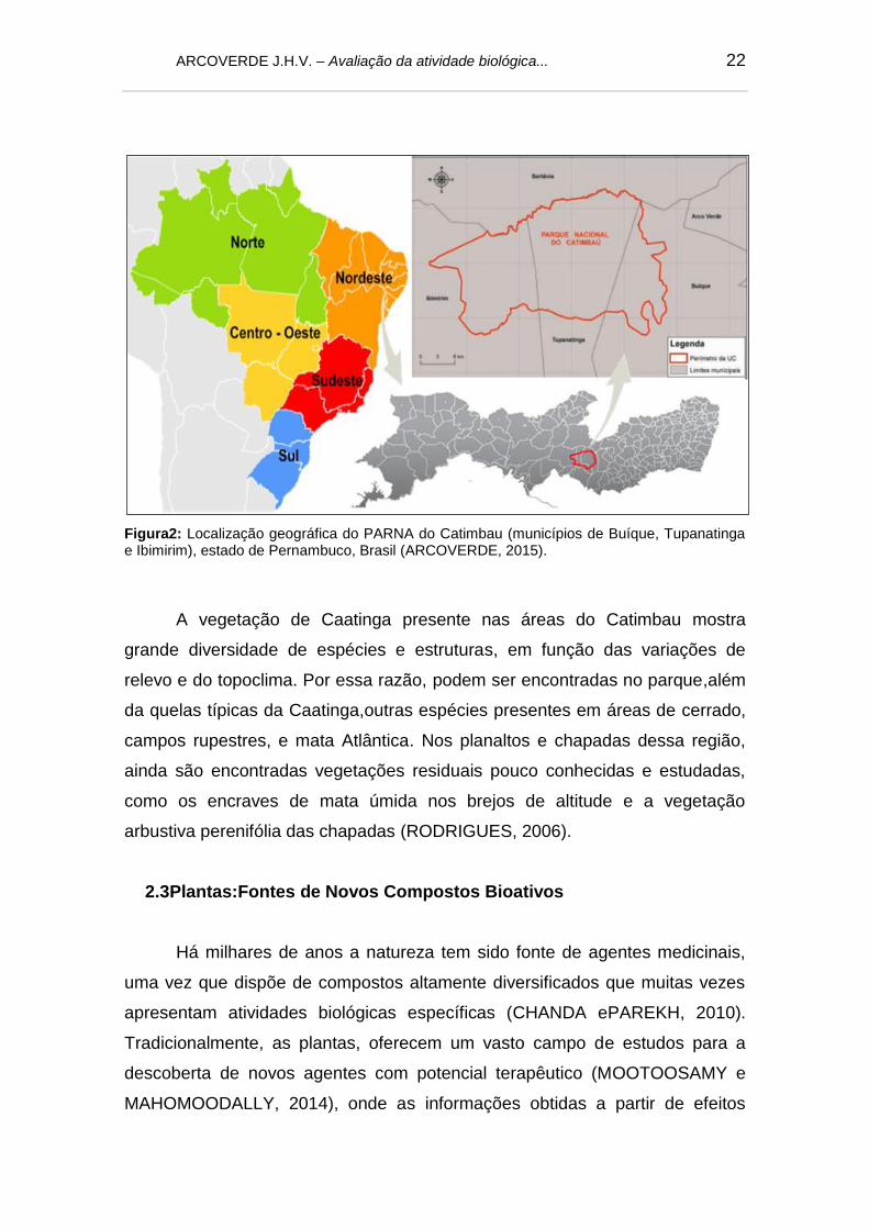
Tupanatinga e Ibimirim), estado de Pernambuco, Brasil. FONTE: (ARCOVERDE, 2015 22
Figura 3: Arvore (A), folhas (B) e frutos (C) de Manilkara rufula. FONTE:
(ARCOVERDE, 2015)....................................................................................................... 30
Figura 4: Estrutura química geral dos flavonoides e suas diferentes classes. FONTE:
(RAVISHANKAR et al., 2013)............................................................................................ 42
Figura 5: Dímeros comuns de taninos condensados, (proantocianidinas) encontrados
em plantas. Variação estrutural, devido aos tipos de ligações. FONTE: (BARBEHENN
e CONSTABEL, 2011).......................................................................................................
45
Figura 6: Estrutura básica dos esteróides com a respectiva numeração dos seus
carbonos............................................................................................................................ 48
Figura 7: Estrutura do núcleo de espirostano. FONTE: (THAKUR et al., 2011).............. 49
MANUSCRITO A SER SUBMETIDO À JOURNAL OF CHEMICAL ECOLOGY
Figura 1: Protective effects of different extracts in DNA nicking assay. (A) Lane 1:
negative control (distilled water + DNA), (B) Lane 2: control (DNA + Fenton’s reagent),
(C) Lane 3-5: AcOEt (125 µg/mL) + Fenton’s reagent, (D) Lane 6-8: CHCl3 (125 µg/mL)
+ Fenton’s reagent, (E) Lane 9-11: MeOH (125 µg/mL) + Fenton’s reagent……………... 114
Figura 2: Termiticide activity of the chloroform (A), ethyl acetate (B) and methanol (C)
extracts prepared from the leaves of M. rufula.................................................................. 117
MANUSCRITO A SER SUBMETIDO À PROCESSBIOCHEMISTRY
Fig. 1: Profile of quitin affinity chromatographic column eluted with acetic acid 1mol/L… 152
Fig. 2: Molecular Characterisation. SDS-PAGE gel (12,5%) obtained through lectin
purification on quieten chromatography, stained with bright blue Coomassie. Band
molecular weights were 43,2 (A) e 36,3 (B) KDa, respectively........................................ 153
Fig. 3: Chromatography of MkaLL on Superdex-75 10/300GL column accepted to a
ÄKTA purifying system, equilibrated and eluted with NaCl 0,15 mol/L…………………….
154

ARCOVERDE J.H.V. – Avaliação da atividade biológica...
Fig. 4: Effect of pH at hemagglutinating activity after incubation with different pH (4,5-
11) buffers......................................................................................................................... 155
Fig. 5: Effect of temperature at MkaLL HA in rabbit erythrocytes. The initial activity was
512 and each point over the line represents the average of three repetitions................... 156
Fig. 6: Protective effects of lectin of M. rufula in DNA nicking assay. (A) Lane 1:
negative control (distilled water + DNA), (B) Lane 2: control (DNA + Fenton’s reagent),
(3) Lane 3-5: MkaLL (5o µg/mL) + Fenton’s reagent…….................................................. 157

ARCOVERDE J.H.V. – Avaliação da atividade biológica...
LISTA DE TABELAS
ARTIGO CIENTÍFICO PUBLICADO PELA NATURAL PRODUCT RESEACH:
FORMERLY NATURAL PRODUCT LETTERS.
Table 1: Studied species and their tissues examined for lectin and trypsin inhibitor
activity with therespective titres of HA, protein concentration (mg/mL) and trypsin
inhibitory effect of the saline extracts………………………………………………………….. 94
Table S1. Species collected in the Vale do Catimbau National Park (Caatinga of
Pernambuco)……………………………………………………………………………………… 102
MANUSCRITO A SER SUBMETIDO À JOURNAL OF CHEMICAL ECOLOGY
Table 1: Phytochemical profile obtained by thin layer chromatography (TLC) of
chloroform, ethyl acetate and methanol extracts prepared from the leaves of M.
rufula................................................................................................................................... 112
Table 2: Concentrations (µg/ml) of the MeOH, AcOEt and CHCl3 extracts, from the
leaves of M. rufula, able to scavenge 50% of the free radicals of DPPH………………….. 113
Table 3: Minimum Inhibitory Concentration (MIC) and Minimum Bactericidal Concentration (MBC) obtained with the organic extracts from the leaves of M. rufula…... 116
MANUSCRITO A SER SUBMETIDO À PROCESSBIOCHEMISTRY
Table 1: Inhibition of hemagglutinating activity of 40-80% saline fraction of leaves from M. ruful. using rabbit red blood erythrocytes.…………………………………………………. 149
Table 2: Purification summary of leaf lectins from Manilkararufula................................... 150
Table 3: Minimum Inhibitory Concentration (MIC) and Minimum Bactericidal
Concentration (CBM) of MkaLL against bacterias……………………………………………. 151

ARCOVERDE J.H.V. – Avaliação da atividade biológica...
LISTA DE ABREVIATURAS E SÍMBOLOS
AAT: Atividade Antioxidante Total
AH: Atividade Hemaglutinante
BSA: albumina sérica bovina (Bovine Serum Albumine)
CBM: Concentração Bactericida Mínima
CCD: Cromatografia em Camada Delgada
DMSO: Dimetilsulfóxido
DPPH: 2,2-difenil-1-picril-hidrazilo
EAG: Equivalente ao Ácido Gálico
EROs: Espécies Reativas de Oxigênio
ha: hectare
MIC: Concentração Inibitória Mínima
MkaLL: lectina das folhas de Manilkararufula
PARNA: Parque Nacional
SDS-PAGE: eletroforese em gel de proliacrilamida (Sodium Dodecyl Sulphat-
Polyacrylamide Gel Electrophoresis)
SUMÁRIO Pag
1.INTRODUÇÃO................................................................................................ 17
2.REVISÃO BIBLIOGRÁFICA.......................................................................... 19
2.1 Bioma Caatinga........................................................................................ 19
2.2 Parque Nacional do Catimbal................................................................... 21
2.3 Plantas:Fontes de Novos Compostos Bioativos...................................... 22
2.4 Família Sapotaceae.................................................................................. 26
2.4.1 Gênero Manilkara............................................................................. 26
2.5 Compostos Bioativos................................................................................ 30
2.5.1 Metabólitos Primários....................................................................... 31
2.5.1.1 Lectinas......................................................................................... 32
2.5.1.1.2 Inibidores de tripisina.................................................................. 35
2.5.2 Metabolitos Secundários.................................................................. 36
2.5.2.1 Classificação.................................................................................. 38
2.5.2.1.1 Compostos Fenólicos................................................................. 38
2.5.2.1.1.1 Fenóis...................................................................................... 39
2.5.2.1.1.2 Flavonóides............................................................................. 41
2.5.2.1.1.3 Taninos.................................................................................... 43
2.5.2.1.2 Terpenóides................................................................................ 46
2.5.2.1.2.1 Esteroides................................................................................ 47
2.5.2.1.3 Saponinas................................................................................... 48
2.5.2.1.4 Compostos Nitrogenados........................................................... 51
2.5.2.1.4.1 Alcalóides................................................................................ 51
2.5.4 Atividades Biológicas de de Metabólitos Secundários...................... 52
2.5.4.1 Atividade Antioxidante................................................................... 52
2.5.4.2 Atividade Antimicrobiana............................................................... 53
2.5.4.3 Atividade termiticida...................................................................... 54
3. OBJETIVOS.................................................................................................... 56
3.1 Geral......................................................................................................... 56
3.2 Específicos................................................................................................ 56
4. REFERÊNCIAS BIBLIOGRÁFICAS.............................................................. 58
5. CAPITULO I - ARTIGO CIENTÍFICO PUBLICADO PELA NATURAL
PRODUCT RESEACH: FORMERLY NATURAL PRODUCT LETTERS ……… 89

ARCOVERDE J.H.V. – Avaliação da atividade biológica...
6. CAPITULO II - MANUSCRITO A SER SUBMETIDO À JOURNAL OF
CHEMICAL ECOLOGY....................................................................................... 104
7. CAPITULO III - MANUSCRITO A SER SUBMETIDO À PROCESS
BIOCHEMISTRY................................................................................................. 127
8. CONSIDERAÇÕES FINAIS........................................................................... 158

ARCOVERDE J.H.V. – Avaliação da atividade biológica... 17
1. INTRODUÇÃO
As plantas com propriedades medicinais constituem a base dos sistemas
de cuidados a saúde em muitas sociedades. Ouso dos conhecimentos e
práticas associadas a esses recursos vegetais fazem parte de uma importante
estratégia ligada à conservação da biodiversidade, à descoberta de novos
medicamentos, e melhoria na qualidade de vida em comunidades rurais
(BALAKRISHNANet al., 2014).
A partir do metabolismo primário das plantas, dentre vários outros
compostos, são produzidos ácidos nucleicos, proteínas e carboidratos e,
juntamente com as reações bioquímicas básicas da mesma, como a respiração
e fotossíntese, as plantas evoluíram na produção de uma ampla variedade de
substâncias secundárias. Essas substâncias foram denominadas de compostos
ou metabólitos secundários (TAIZ eZEIGER, 2004).
As lectinas são um grupo de proteínas ou glicoproteínas de origem não
imune,altamente diversas, ubiquamente distribuídas em plantas, animais e
micro-organismos (LEI-LEI et al., 2011).Apresentam a capacidade de
reconhecer e ligar-se especificamente a mono ou oligossacarídeos sem induzir
alterações na estrutura dos mesmos (SOUZA et al., 2011).
Estudos mostram que um grande número de lectinas têm sido isoladas e
caracterizadas em diversas plantas, e estas proteínas estão emergindo como
moléculas promissoras, principalmente por conterem sítios de ligação
específicos, pois esta habilidade especial de reconhecimento pode ser utilizada
como importante ferramenta biotecnológica (COSTA et al., 2010;
CHARUNGCHITRAK et al., 2011).
A família Sapotaceae, presente no bioma da Caatinga, possui
distribuição tropical e subtropical, incluindo cerca de 50 gêneros contendo 1000
espécies de hábito arbóreo e arbustivo. No Brasil, ocorrem 14 gêneros e cerca
de 200 espécies, com destaque para Chrysophyllum, Manilkara, Micropholis,
Pouteria, Pradosiaos e Sideroxylon (ALMEIDA-JUNIOR, 2010). Quimicamente,
as espécies desta família são caracterizadas por apresentarem uma variedade
de classes de substâncias, sendo os compostos mais conhecidos as
saponinas, flavonoides, polifenois, triterpenos, carotenoides, esteroides, ácidos

ARCOVERDE J.H.V. – Avaliação da atividade biológica... 18
graxos e taninos, com diversas atividades biológicas já comprovadas
(KUSHIMA, 2005; MONTENEGROet al., 2006; BARBOSA-FILHOet at., 2008).
No gênero Manilkara são encontradas algumas espécies que têm sido
utilizadas popularmente como plantas medicinais no tratamento de várias
enfermidades, como inflamações, febre, hemorragias, dores de estômago e
comocicatrizantes. Alguns estudos já realizados com espécies deste gênero
mostraram que, as mesmas são possuidoras de compostos com diversas
propriedades biológicas, como antimicrobiana, antiparasitária, antitumoral e
antioxidante (CHANDA e NAGANI, 2010; KANERIA e CHANDA, 2012). Na
literatura não há relatos com M. rufula que tratem do seu potencial como fonte
de compostos bioativos, sendo este o primeiro trabalho que mostra a presença
de tais componentes na espécie.
Estudos mostram que compostos naturais oferecem inúmeras
oportunidades para o desenvolvimento de novas drogas com potencial
terapêutico. Logo, explorar as potencialidades oferecidas pelas plantas da
Caatinga, é uma forma de não só contribuir para a obtenção de novos
princípios bioativos, mas também uma maneira de agregar valor à região e
contribuir para sua preservação.Diante do exposto, este trabalho teve como
objetivos, determinar o potencial biotecnológico de espécies deste bioma,
identificar e avaliarquímica e biologicamentecompostos secundários oriundos
do metabolismo celular, e obter uma lectina das folhas de Manilkararufula,
visando novas e futuras aplicaçõesde biomoléculas desta espécie como
antioxidantes e antimicrobianas.
ARCOVERDE J.H.V. – Avaliação da atividade biológica... 19
2. REVISÃO BIBLIOGRÁFICA
2.1 Bioma Caatinga
A Caatinga é um bioma de clima semiárido com florestas arbustivas e
espinhosas, de folhas decíduas, encontradas no Nordeste brasileiro(SILVA et
al., 2014).A mesma responde por aproximadamente 10% do território nacional
e 60% da região Nordeste. Caatinga, que em Tupi significa “matabranca”,
aparência que quase sempre tem no período mais seco, é um dos tipos de
vegetação mais heterogêneo presentes no Brasil. Devido às variações de
relevo e microclima, são encontrados nesse bioma ecotopos formados por
espécies de cerrado de campos rupestres, de mata atlântica e de restinga
(RODRIGUES, 2006; PORDEUS, 2007).
Suas faixas de precipitação média anual estão entre 240-1500 mm
(LEALet al., 2005).A estação seca dura entorno de sete meses e o inverno é a
estação das chuvas, onde as temperaturas não são tão elevadas. Estas
características fazem da Caatinga um tipo peculiar de vegetação adaptada às
condições climáticas locais encontradas nesta região (TRENTIN et al., 2011).
No bioma Caatinga, já foi comprovada a existência de 510 gêneros e
5.344 espécies de plantas vasculares, dentre os quais 18 gêneros e 318
espécies são endêmicas (GIULIETTIet al., 2002), o que caracteriza essa região
como uma das grandes áreas de maior biodiversidade no Brasil.
Na Caatinga predomina a vegetação xerófita (figura 1) que carrega um
grande número de espécies vegetais. Este bioma é uma matriz de matagais
espinhosos e florestas sazonalmente secas, com bolsões de florestas tropicais
de altitude e cerrado, que abrange a maior parte dos estados do Piauí, Ceará,
Rio Grande do Norte, Paraíba, Pernambuco, Alagoas, Sergipe, Bahia e parte
do nordeste de Minas Gerais, no Vale do Jequitinhonha, ocupando uma área
de aproximadamente 735.000 km2(BASSO et al., 2005), representando a
quarta maior área coberta por uma única forma de vegetação no Brasil
(CARTAXO et al., 2010), onde estão presentes espécies que não podem ser
encontradas em nenhum outro lugar do mundo.

ARCOVERDE J.H.V. – Avaliação da atividade biológica... 20
Figura 1: Área de Caatinga no PARNA do Catimbau (ARCOVERDE, 2015).
Áreas desse bioma têm sofrido forte influência humana ao longo dos
anoslevando à perda de paisagens e trazendo consequências graves para a
manutenção da biodiversidade local(ALBUQUERQUE et al., 2005). A
fragmentação de toda Caatinga pode levar ao desaparecimento de espécies e
organismos únicos da região (NETO, 2009). Silva et al. (2014) afirmam que a
pressão antrópica associada a aridez local levou a uma extrema degradação
de grandes extensões desse bioma, dando origem aos chamados núcleos de
desertificação,o que enfatiza a importância e urgência do desenvolvimento de
estratégias sustentáveis para preservação da biodiversidade nesta área.
Neste ambiente, encontram-se espécies de ocorrência endêmica, bem
como plantas com atividades farmacológicas reconhecidas pela população
local, muitas delas ainda desconhecidas pela ciência, tanto do ponto de vista
botânico como químico e farmacológico (RODRIGUES, 2006; NETO, 2009).
Nos últimos anos tem havido um crescente interesse em adquirir
conhecimentos sobre as plantas dentro desta área (ALBUQUERQUE et al.,
2007a), e algumas publicações já descreveram a rica flora da região como
promissora fonte de compostos bioativos(ALMEIDA et al., 2005; AGRA et al.,
2007a; ALBUQUERQUE et al., 2007b; ALBUQUERQUE e OLIVEIRA, 2007;
AGRA et al., 2008).
ARCOVERDE J.H.V. – Avaliação da atividade biológica... 21
Em termos de utilizações medicinais, alguns compostos obtidos de
plantas da Caatinga já demonstraram propriedades interessantes como anti-
inflamatória, antimicrobiana, cicatrizante e antioxidante. E estes compostos
aparecem essencialmente em algumasfamilias de plantas desta região
(ALMEIDA et al., 2005). Além disso, esses coonstituintes aparecem em
concentrações elevadas em algumas espécies que são intensamente utilizadas
para fins terapêuticos (MONTEIRO et al., 2006).Segundo Agra et al. (2007) no
nordeste do Brasil as práticas medicinais tradicionais têm sido usadas para
tratar uma imensa variedade de doenças, incluindo enfermidades da pele,
distúrbios gastrointestinais, tuberculose, infecções do trato urinário, e outras.
No entanto, algumas destas plantas utilizadas necessitam de estudos
científicos para confirmarem sua eficácia (CARTAXO et al., 2010).
2.2 Parque Nacional do Catimbau
Criado em 2002 elocalizado a 278 Km da cidade do Recife, Capital do
Estado de Pernambuco, o Parque Nacional da Serra do Catimbau (PARNA do
Catimbau), está inserido na zona de transição entre o Agreste e Sertão do
Estado, entre as coordenadas geográficas: 8°24’00” e 8°36’35” S e 37°09’30” e
37°14’40” W(figura2). Sua área é de 62.300 ha, na forma de uma poligonal e
encontra-se distribuída entre os municípios de Buíque (12.438 ha.);
Tupanatinga (23.540 ha) na Microrregião do Vale do Ipanema, e Ibimirim
(24.809 ha) na Microrregião do Moxotó Semiárido Pernambucano
(RODRIGUES, 2006). A região em estudo é considerada o segundo maior
parque arqueológico do Brasil, com aproximadamente trinta sítios
arqueológicos (PORDEUS, 2007). Em sua vegetação predomina a Caatinga
característica marcante da flora do parque, (RODRIGUES, 2006; PORDEUS,
2007), sendo uma das poucas áreas onde ainda podem ser encontradas matas
desse bioma não alteradas pela ação do homem.
O PARNA do Catimbau é constituído de paisagens com grandes
riquezas e biodiversidade impar em seu Estado. Em pesquisa realizada pela
Sociedade Nordestina de Ecologia (S.N.E), a região do Catimbau por suas
características, foi considera Área de Extrema Importância Biológica
(RODRIGUES, 2006).
ARCOVERDE J.H.V. – Avaliação da atividade biológica... 22
Figura2: Localização geográfica do PARNA do Catimbau (municípios de Buíque, Tupanatinga e Ibimirim), estado de Pernambuco, Brasil (ARCOVERDE, 2015).
A vegetação de Caatinga presente nas áreas do Catimbau mostra
grande diversidade de espécies e estruturas, em função das variações de
relevo e do topoclima. Por essa razão, podem ser encontradas no parque,além
da quelas típicas da Caatinga,outras espécies presentes em áreas de cerrado,
campos rupestres, e mata Atlântica. Nos planaltos e chapadas dessa região,
ainda são encontradas vegetações residuais pouco conhecidas e estudadas,
como os encraves de mata úmida nos brejos de altitude e a vegetação
arbustiva perenifólia das chapadas (RODRIGUES, 2006).
2.3Plantas:Fontes de Novos Compostos Bioativos
Há milhares de anos a natureza tem sido fonte de agentes medicinais,
uma vez que dispõe de compostos altamente diversificados que muitas vezes
apresentam atividades biológicas específicas (CHANDA ePAREKH, 2010).
Tradicionalmente, as plantas, oferecem um vasto campo de estudos para a
descoberta de novos agentes com potencial terapêutico (MOOTOOSAMY e
MAHOMOODALLY, 2014), onde as informações obtidas a partir de efeitos

ARCOVERDE J.H.V. – Avaliação da atividade biológica... 23
biológicos dos fitoterápicos tornam-se um valioso contributo para a descoberta
de novos compostos bioativos alem de permitir o desenvolvimento de novas
linhas terapêuticas (EISSA et al., 2014).
A ampliação do conhecimento etnobotânico sobre o uso de plantas com
propriedades medicinais e o desenvolvimento de pesquisas nessa área,
ganhou substancial consideração entre as comunidades científicas aolongo do
tempo. Ainda, o aumento no custo dos tratamentos convencionais para
algumas doenças, os custos crescentes das drogas de origem sintéticas e a
resistência antimicrobiana frente a vários antibióticos, tem incentivado a busca
por novos medicamentos derivados das plantas (AREEJ et al., 2013; KAYANI
et al., 2014).
Mesmo com os avanços da indústria farmacêutica na produção de
medicamentos sintéticos, diversos agentes quimioterapêuticos ainda têm sido
desenvolvidos como resultado de estudos de plantas com propriedades
medicinais (OSMAN et al., 2011). Estas têm sido usadas em várias partes
domundo para tratar diferentes enfermidades, pricipalmente em regiões com
difícil acesso a programas de saúde, onde a utilização de plantas na medicina
tradicional é muito comum por grande parte da população local (AREEJ et
al.,2013).
Várias pesquisas etnofarmacológicas têm sido publicadas nos últimos
anos com base na medicina tradicional em várias culturas da África
(KARUNAMOORTHI e TSEHAYE, 2012; SEMENYA et al., 2012), Ásia (DEY
eKUHAD, 2014), Austrália (PACKER et al., 2012), China (GHORBANI et al.,
2011), América do Sul (REHECHO et al., 2011) e em regiões específicas de
países ocidentais (CALVO et al., 2011; CAVERO et al., 2011) com o objetivo
de preservar o uso de ervas como remédios, e encontrar uma abordagem
baseada em evidências, para seu uso como medicamentos (EISSA et al.,
2014).
Estudos etnobotânicos realizados nas regiões tropicais demonstraram
que as populações locais possuem um amplo conhecimento a cerca das
espécies de plantas farmacologicamente úteis (TACHER et al., 2002; TORRE-
CUADROS e ISLEBE, 2003; ALBUQUERQUE e OLIVEIRA, 2007). Ainda,
essas informações sobre o uso dos recursos naturais também podem contribuir
para a conservação da biodiversidade local. A importância global de plantas
ARCOVERDE J.H.V. – Avaliação da atividade biológica... 24
com propriedades medicinais pode ser atribuída pelos numerosos
medicamentos convencionais que foram derivados a partir destas, e que são
atualmente utilizados na prática clínica (HÜBSCH et al., 2014). Muitas espécies
têm sido reconhecidas por suas propriedades curativas e exercerem um
impacto benéfico sobre a saúde, apresentando diversas atividades biológicas
como, por exemplo, anti-inflamatória, antimicrobiana, antioxidante,
hipolipemiantes,antimutagênicos e outros(BALAKRISHNAN et. al., 2014).
Estudos mostram que compostos naturais puros e extratos vegetais
padronizados oferecem inúmeras oportunidades para o desenvolvimento de
novas drogas com potencial biotecnológico, devido a suadiversidade química
inigualável (ASWATHANARAYAN eVITTAL, 2013).Viegas-Jr et al. (2006)
afirmam que nos países ocidentais, onde a base da indústria farmacêutica é a
química sintética, 25% dos fármacos foram originalmente isolados de plantas.
De acorco com Almeida et al. (2005), vários estudos têm chamado a
atenção para o uso de plantas com propriedades medicinais, discutindo
diferentes hipóteses na tentativa de explicar os padrões de uso encontrados.
Stepp (2004), em seu trabalho, observou uma alta frequência de ervas
daninhassendo utilizadas para fins terapêuticos em várias partes do mundo,
sugerindo que esta preferência possa estar relacionada a aspectos químicos
das mesmas. No entanto, fatores culturais, influenciam fortemente a seleção e
uso de plantas para tais fins (AMOROZO, 2004; VANDEBROEK et al., 2004).
A eficácia dos fitoterápicos está geralmente influenciada por vários
fatores, incluindo diferenças genéticas, condições ambientais, tais como
temperatura, exposição ao sol, disponibilidade hidrica, variação nutricional e
presença de microorganismos, que afetam os processos celulares nas plantas
e as suas respostas a estímulos (GUO et al., 2008; LI et al., 2012).A
variabilidade de compostos químicos entre as populações também tem sido
atribuída a efeitos abióticos do meio ambiente, tais como a salinidade
(BOURGOU et al., 2010;TAARIT et al., 2011) e umidade (BETTAIEB et al.,
2011) além dos anteriormente citados. Neste sentido, as composições dos
compostos ativos e as potências terapêuticas das plantas são alterados,
portanto, torna-se essencial diferenciar os locais de cultivo e as condições das
ervas para entender suas propriedades e otimizar seus usos (LEE et al., 2013).

ARCOVERDE J.H.V. – Avaliação da atividade biológica... 25
O número de estudos sobre plantas com propriedades medicinais na
região semiárida do Nordeste brasileiro tem crescido progressivamente. No
entanto, a maioria destes são descritivos, se concentrando em apenas listar
plantas, bem como suas indicações terapêuticas, as partes utilizadas e seu
modo de uso (SILVA eANDRADE, 2002;MOREIRA et al.,
2002;ALBUQUERQUE eANDRADE, 2002;ALMEIDA et al., 2006).
O uso de plantas medicinais da Caatinga está geralmente associado à
remoção de partes destas e sua utilização como remédio. Algumas espécies
são altamente exploradas devido à extensa colheita para abastecer o setor
farmacêutico (ALBUQUERQUE eANDRADE, 2002). Numerosas espécies de
árvores do semiárido brasileirotêm sua casca retirada e usada para fins
medicinais, sendo esta uma prática extremamente comum na região. Contudo,
muitas destas árvores crescem lentamente, e em alguns casos as práticas de
coleta destrutiva podem reduzir o tamanho de uma população aumentando a
probabilidade de sua extinção local (MONTEIROet al., 2006). Muitas destas
espécies têm outras utilizações não farmacêuticas, especialmente como
madeira (ALBUQUERQUE et al., 2005; MONTEIRO et al., 2006). Desse modo,
a sustentabilidade da colheita de plantas medicinais depende de uma série de
fatores que operam em diferentes níveis. Logo as ameaças às populações de
plantas locais podem surgir a partir de outras atividades de uso das mesmas
(BOTHA et al., 2004).
A utilização e manejo de plantas é uma prática cotidiana em muitas
comunidades do Brasil que residem próximos a fragmentos de matas ou
reservas florestais. O uso frequente de recursos vegetais indica que estas
populações têm um amplo conhecimento a cerca das espécies nativas. A
importância desta informação para melhoria de vida cotidiana das populações
rurais, bem como o uso sustentável desses recursos vegetais tem sido
frequentemente apontada para a tomada de decisões sobre preservação
(CAMPOS eEHRINGHAUS, 2003; ALBUQUERQUE, 2004; MONTEIROet al.,
2006).
O problema relacionado à diminuição da biodiversidade envolve um
conjunto de alterações, como reduções da heterogeneidade biológica em uma
determinada região, por exemplo. Para se reverter o problema, é necessária
uma melhor compreensão das relações entre a biodiversidade local e o

ARCOVERDE J.H.V. – Avaliação da atividade biológica... 26
ecossistema. Logo, as ligações entre a diversidade total de espécies
quimicamente úteis em determinada área esua disponibilidadepara as
populações locais devem ser examinadas, para que tais recursos sejam
preservados (ALBUQUERQUE eOLIVEIRA, 2007).
2.4 Família Sapotaceae
A família Sapotaceae possui distribuição tropical e subtropical, incluindo
cerca de 60 gêneros contendo 1.343 espécies de hábito arbóreo e arbustivo.
No Brasil ocorrem 14 gêneros e cerca de 200 espécies, com destaque para
Chrysophyllum, Manilkara, Micropholis, Pouteria, Pradosia e Sideroxylon
(ALMEIDA-JÚNIOR, 2010; THE-PLANT-LIST, 2013).
As plantas pertencentes à família Sapotaceae são lenhosas, arbustivas
ou arbóreas, com folhas alternas, raramente opostas ou verticiladas (Pradosia),
simples, geralmente sem estípulas e com margem geralmente inteira. A
Inflorescência é cimosa, frequentemente em panículas ou reduzidas a
fascículos axilares; as flores são pouco vistosas, unissexuadas ou
bissexuadas, actinomorfas, com estipes geralmente em número igual ou duplo
ao das pétalas (SOUZA e LORENZI, 2005).
Quimicamente, as espécies desta família são caracterizadas por
apresentarem uma variedade de classes de substâncias, sendo os compostos
mais conhecidos as saponinas, flavonoides, polifenois, triterpenos e
politerpenos, naftol, benzoquinonas, carotenoides, esteroides, ácidos graxos,
taninos e óleos essenciais, com diversas atividades biológicas, dentre elas,
antiulcerogênica, antimicrobiana e antiviral (BERROUGUI et al., 2004;
KUSHIMA, 2005; STEVENS, 2005, MONTENEGRO et al., 2006; BARBOSA-
FILHO et al., 2008).
2.4.1 Gênero Manilkara
O gênero Manilkara é constituído por cerca de 35 espécies de hábitos
arbóreos e arbustivos,sendo 19 destas espécies encontradas no Brasil, das

ARCOVERDE J.H.V. – Avaliação da atividade biológica... 27
quais, 12 estão presentes na região Nordeste, sendo a Bahia o estado mais
diverso (GOMES et al., 2010).
No Brasil, os estudos floristicos que contemplaram a família Sapotaceae
compreendem áreas pontuais e restritas de diferentes regiões do país. Ducke
(1950) listou oito espécies amazônicas, três com ocorrência no Nordeste e três
no Sudeste; segundo o autor, as espécies de Manilkara ocorrentes na floresta
Amazônica, popularmente conhecidas como maçaranduba, maçaranduba-
vermelha, maçaranduba verdadeira ou maparajuba (CORRÊA, 1978), tem
importância não só pelo número, mas também pelo valor dos seus produtos
(madeira e látex).Ducke (1957) revisou a listagem e reconheceu 16 espécies
de Manilkara para o território nacional.
No Nordeste, algumas listagens florísticas contemplaram a ocorrência de
espécies de Manilkara, com destaque para os estudos desenvolvidos por Sales
et al. (1998), que apresentaram um checklist da flora ameaçada dos brejos de
altitude em Pernambuco; Barbosa et al. (2006) que também produziram
umchecklist das plantas do Nordeste brasileiro; e Amorim et al. (2008) que
inventariaram a flora da Reserva Biologica de Una, Bahia. Na região Norte,
Ribeiro et al. (1999) e Pennington (2006) registraram as espécies de Manilkara
ocorrentes na Reserva Ducke.
Apesar da representatividade das espécies de Manilkara nos diferentes
biomas (Amazônico, Atlântico, Caatinga e Cerrado), a floresta Atlântica possui
a maior diversidade do gênero quando se considera apenas a região Nordeste
do país (CALVENTE et al., 2005).
Aplicações no tratamento de algumas enfermidades como,inflamação,
dores no estômago e cicatrização de feridas, foram encontradas para algumas
espécies do gênero Manilkara,que têm sido utilizadas popularmente como
plantas medicinais. Estudos já realizados com espécies deste gênero
mostraram diversas propriedades invitro (FERNANDES, 2011).Em seu trabalho
Chanda e Parekh (2012) mostraram que extratos de Manilkarahexandra
possuem atividade antibacteriana contra diversas espécies de interesse
clínico,enfatizando assim a importância biotecnológica do gênero.
Um ensaio de atividade antibacteriana in vitro contra 14 importantes
bactérias patogênicas a humanos foi realizado com extrato aquoso das folhas
deManilkara zapota, e revelou uma maior atividade inibitória contra

ARCOVERDE J.H.V. – Avaliação da atividade biológica... 28
Staphylococcus aureus, Citrobacter sp., e Proteusmirabilis. (SATISH et al.,
2008).Vários estudos já desenvolvidos com esta espécie mostraram que alguns
tecidos da mesma apresentam propriedades biológicas diversas. As folhas e
casca são utilizadas para tratar a tosse, resfriado, disenteria e diarreia. As
atividades antimicrobianas e antioxidantes são também relatadas a partir de
suas folhas (NAIR e CHANDA, 2008; KANERIA et. al., 2009).
Os resultados do estudo desenvolvido por Osman et al. (2011)
demonstraram claramente a atividade anti-cancerígena (carcinoma ascítico de
Ehrlich)invivode extratos preparados com a casca e o caule de Manilkara
zapota. Neste estudo eles obtiveram um prolongamento do tempo de vida dos
animais avaliados, alem de uma diminuição na contagem de glóbulos brancos
do sangue. Estes dois fatores são os critérios confiáveis para avaliar uma
droga anticâncer segundo os autores.
A casca da M. zapota também é utilizada tradicionalmente para o
tratamento da febre e dor. O extrato etanólico preparado com a casca desta
espécie mostrou uma inibição significativa em ensaios de edema da pata com
ratos, induzido por injeção da carragenina. A atividade anti-inflamatória,
observada pode ser devido a um efeito inibidor do extrato sobre a liberação de
histamina e/ou serotonina (GANGULY et al., 2013).
Ganguly et al. (2013) segerem que a presença de flavonóides,
saponinas, taninos e glicosídeos no extrato bruto das folhas de M. zapota,
sugerindo quea presença destes compostos pode ser responsável por sua
atividade anti-inflamatória. Outros estudos também atribuem à atividade anti-
inflamatória encontradas em extratos de plantas, a presença de fitoconstituintes
como terpenóides eesteroides, alémdeoutros já citados (MULLA et al., 2010;
ADJENE et al., 2012).
M.rufula (Miq.) é uma árvore que mede de 5 a 10 metros de altura (figura
3-A); com ramo acinzentado, indumento tomentoso a puberulento, ferrugíneo a
esbranquiçado. Suas folhas (figura 3-B)são alternas, distribuídas ao longo do
ramo, raras no ápice do mesmo. O fruto (figura 3-C)apresenta-se oblongo-
elipsóide, glabro, vermelho, alaranjado ou marrom. A M. rufula compartilha
caracteres com M. dardanoi, diferindo por apresentar inflorescência de 5 a 8
flores, enquanto M. dardanoi possui flor solitária (LAM, 1941).

ARCOVERDE J.H.V. – Avaliação da atividade biológica... 29
M.rufula floresce entre os meses de outubro e janeiro, com maior
intensidade de floração registrada em outubro; frutifica entre janeiro e maio,
com pico de frutificação no mês de março (LAM, 1941). Está distribuída na
região Nordeste (Alagoas, Bahia, Ceará, Pernambuco, Piauí e Sergipe), em
áreas de caatinga, cerrado e na transição caatinga-cerrado. Apresenta
ocorrência na Paraíba e Rio Grande do Norte, além do registro na divisa entre
os estados de Tocantins e Bahia em vegetação de cerrado (LAM, 1941).
O último estudo relacionado à presença dessa espécie mostrou que M.
rufula apresentava-se na categoria de baixo risco de extinção (IUCN, 2001),
mas problemas com a destruição acelerada e perda das áreas de Caatinga e
Cerrado, podem em um futuramente levar a mesma a ser incluída entre as
espécies ameaçadas de extinção. Logo, faz-se necessária a tomada de
medidas e projetos para conservação das populações de M. rufula para
manutenção desses espécimes in situ.

ARCOVERDE J.H.V. – Avaliação da atividade biológica... 30
Figura 3: Arvore (A), folhas (B) e frutos (C) de Manilkara rufula (ARCOVERDE, 2015).
2.5Compostos Bioativos
O metabolismo da planta é frequentemente dividido em duas classes
principais: a) metabolismo primário, considerado aquele que permite uma
planta utilizar água, dióxido de carbono e sais minerais para sintetizar os
metabolitos primários essenciais (açúcares, ácidos graxos, aminoácidos e
ácidos nucleicos), necessários para produzir e manter as células,estes
compostos têm uma origem muito antiga e como tal, os genes requeridos para
A
B
C
C

ARCOVERDE J.H.V. – Avaliação da atividade biológica... 31
a sua síntese são largamente conservados entre todas as plantas conhecidas;
b) metabolitos secundários (também referidos como os produtos naturais, ou
metabolitos especializados fitoquímicos), considerados como sendo os
produtos químicos necessários para as interações de plantas com o meio
ambiente (p.ex., compostos de defesa contra pragas e agentes patogênicos)
(HURTADO-FERNÁNDEZ et al., 2011; KLIEBENSTEIN eOSBOURN, 2012).
O nível de diversidade química de uma população vegetal pode ser
associadoà intensidade com que a planta é estimulada a produzir novos
compostos. Por exemplo, o ataque de patógenos pode estimular a produção de
metabólitos como mecanismo autodefesa(BRAVO‑MONZÓN et al.,
2014).Segue-se que a capacidade de evoluir e desenvolver novas vias
metabólicas seja benéfica para adaptação a novos nichos ambientais, um
aspecto essencial de especiação da planta. Isso levanta questões importantes
sobre os mecanismos subjacentes à evoluçãometabólica em plantas
(YONEKURA-SAKAKIBARA e SAITO, 2009; GOODSTEIN et al., 2012).
A composição dos metabólitos individuais e as suas concentrações não
são constantes, mas diferem de órgão para órgão, dentro do ciclo de
desenvolvimento de uma planta e, além disso, inter e intra populações. Essa
variação leva a formação de misturas complexas de metabólitos secundários, o
que é, provavelmente, uma estratégia desenvolvida pelos vegetais contra a
seleção de herbívoros ou patógenos especializados (WINK, 2008).
2.5.1 Metabólitos Primários
Os principais compostos produzidos pelas plantas são chamados
metabólitos primários e se encontram em todas as células vegetais (PUENTES,
2009). Estes têm sido amplamente estudados na busca por novos fármacos
e/ou por moléculas de interesse biotecnológico.
Estresses bióticos e abióticos podem influenciar o metabolismo primário
das plantas, afetando a produção de alguns metabólitos importantes como
carboidratos, por exemplo (LAWO et al., 2011; SCHOEDL et al., 2013). Para os
metabólitos primários, observou-se que o estresse hídrico leva a um aumento
de mio-inositol e sacarose em algumas plantas, o que pode refletir seus
respectivos papéis como osmoprotetores (GRIMPLET et al., 2009). Também foi

ARCOVERDE J.H.V. – Avaliação da atividade biológica... 32
observado um aumento significativo de prolina em folhas de videira (DOUPIS et
al., 2011) e bagas (DELUC et al., 2009), sob condições de déficit hídrico,
assumindo que este aminoácido possa participar da proteção contra a
formação de espécies reativas de oxigênio em excesso nos vegetais. Os
metabólitos primários estão intimamente relacionados com o fenótipo de um
organismo, podendo inclusive fornecer características nutricionais importantes
ao mesmo (HOEKENGA, 2008).
Sabe-se que as plantas se protegem contra organismos patogênicos por
diversas respostas de defesa, que incluem a produção de peptídeos
antimicrobianos a partir do seu metabolismo primário. Alguns destes produtos,
tais como lectinas (LEI-LEI et al., 2011)e inibidores de proteases (LINGARAJU
e GOWDA, 2008), são importantes componentes do sistema protetordos
vegetais, podendo haver variações entre espécies quanto a sua produção
(HOSSAIN et al., 2012).
2.5.1.1 Lectinas
As lectinas foram relatadas pela primeira vez em 1888, por Stillmark.Ao
estudar a toxicidadede extratos deRicinuscommunis (mamona), ele observou a
capacidade em aglutinar eritrócitos, devido à presença da “ricina”,proteína
extraída desta planta (KENNEDY et al., 1995). Algum tempo depois, outra
proteína com capacidade hemaglutinante, chamada “abrina”, foi encontrada em
sementes deAbrusprecatorius (Jequiriti). Mesmo com estas descobertas, o
estudo sobre lectinas só começou a ganhar força em 1960, abrindo uma ampla
área de aplicação para estas proteínas (GABOR et al., 2004).
O termo lectina (do latim lectus, que significa escolher) refere-se à
destreza dessas proteínas em ligarem-se específica e reversivelmente a
carboidratos (HE et al., 2013). As lectinas contêm pelo menos um domínio não
catalítico que lhes permite reconhecer e ligar-se seletiva e reversivelmente a
açúcares livres ou glicanos específicos, presentes em glicoproteínas e
glicolipidios, sem alterar a estrutura dos mesmos (LEI-LEI et al., 2011).
As lectinas interagem com carboidratos livres ou glicoconjugados
através de um sítio de ligação específico e por ligações não covalentes. A
diversidade estrutural das lectinas e a elevada especificidade de ligação

ARCOVERDE J.H.V. – Avaliação da atividade biológica... 33
permitem a estas várias aplicações biotecnológicas, tais como reconhecimento
de células e glicoproteínas (SOUZA, 2011) e monitoramento dos carboidratos
na superfície celular (FAIS et al., 2009). Os procedimentos de isolamento e
caracterização para estas proteínas geralmente envolvem rastreio pela sua
capacidade de aglutinar células e precipitar glicoconjugadose o isolamento em
colunas de afinidade com ligantes específicos (VILLANUEVA, 2002). As
lectinas, quando imobilizados em suportes inertes, podem atuar como matrizes
de afinidade para isolamento de outras moléculas (PAIVA et al., 2003; SILVA et
al., 2011).
Lectinas podem ser encontradas em animais (ALPUCHE et al.,
2005;YANG et al., 2007), micro-organismos (BHOWAL et al.,2005;KHAN et al.,
2007), e plantas,nestas elas são isoladas principalmente a partir de sementes,
mas a sua ocorrência em outros tecidos vegetais, como folhas (COELHO
eSILVA, 2000), rizomas (KAUR et al., 2005), tubérculos (KAUR et al., 2006),
casca (SÁ et al., 2009a), cerne (SÁ et al., 2008) e raízes (WONG eNG,
2006),também foi relatada.
As Lectinas vegetais estão distribuídas nas seguintes famílias:
Leguminosae, Ligadoras de quitina, Ligadoras de manose de
monocotiledôneas, Cucurbitaceae, Amaranthaceae, Jacalina e RIP Tipo 2
(PEUMANS e VANDAMME et al., 1998).
A maioria das lectinas de origem vegetal apresenta especificidade por
carboidratos simples (monossacarídeos) ou complexos (oligossacarídeos e
glicanas), os quais podem ser de origem vegetal ou não, como N-
acetilglicosamina e ácidos N-glucurônico, galacturônico, xilurônico, L-idurônico,
siálico e N-acetilmurâmico (VANDAMME et al., 2008).
Lectinas ligadoras de quitina têm sido estudadas do ponto de vista
estrutural. As mais conhecidas são aquelas pertencentes à família das
heveínas, assim chamadas por possuírem em comum o dominío heveínico
como motivo estrutural de reconhecimento da quitina. Sendo especialmente
rica em resíduos de glicina e cisteína, a estrutura da haveína é mantida por
quatro pontes dissulfeto, o que lhe confere uma notável estabilidade,
característica que se estende às demais lectinas da família das heveínas.
Mesmo depois de aquecida a 90ºC por 10 minutos, a heveína ainda inibe o
crescimento de fungos (NEUMANN et al., 2004).

ARCOVERDE J.H.V. – Avaliação da atividade biológica... 34
Lectinas tem uma importante participação nos processos fisiológicos das
plantas. Segundo Charungchitrak et al., (2011) o papel das lectinas no sistema
de defesas das plantas pode ter evoluído a partir da capacidade destas
moléculas em aglutinar e imobilizar micro-organismos. As evidências para este
papel são: a presença de lectinas em locais com potencial de invasão por
agentes infecciosos, como sementes, e a ligação dessasproteínas a vários
microrganismos e sua capacidade de inibir o crescimento destes.
As lectinas, por terem a habilidade de se ligar especificamente a
carboidratos, apresentam uma variedade de efeitos biológicos, alguns dos
quais servindo como base para a aplicação das mesmas na investigação de
atividades químicas e biotecnológicas. Estas proteínas têm sido sugeridas
como compostos naturais para controle de insetos e fungos.Souza et al.
(2011), isolaram lectina de raízes secundárias de B. monandra edemonstraram
que essa proteína possui atividade antifúngica e termiticida, ratificando o
potencial biotecnológico de lectinas para aplicações no controle de pragas
agrícolas.
A toxicidade das lectinas tem sido estudada com o objetivo de garantir
segurança ao meio ambiente e a saúde humana. Vaz et al. (2010) avaliaram
aLectina com atividade antifúngica obtida da casca de Sebstiania jacobinensis,
aqual não apresentou efeito deletério sobre náuplias de Artemia salina e
embriões de Biomphalaria glabrata, o que indica a baixa toxicidade dessas
proteínas.
Lam e Ng (2011) realizaram uma extensa revisão bibliográfica e
concluíram Lectinas podem ser expressas com sucesso heterologamente para
uma produção em larga escala. Podendo serem aplicadas em uma variedade
de culturas para uso como estratégias de manejo integrado de pragas;
provando que o uso dessas proteínas pode reduzir a carga de pesticidas
químicos atualmente usados.
Lei-Lei et al. (2011) relatam que lectinas de leguminosas são bem
conhecidas por possuirem deversas funções biológicas tais como anti-tumoral,
antiviral e anti-fúngicas.
O crescimento de fungos é considerado um fator importante na
biodeterioração de diversos materiais, como grãos armazenados, madeira,
pinturas, esculturas, couro e óleo (STERFLINGER, 2010). Espécies de

ARCOVERDE J.H.V. – Avaliação da atividade biológica... 35
Fusarium podem causar várias doenças no trigo (WAGACHA eMUTHOMI,
2007), tomate (AGRIOS, 2005), amendoim (ROJO et al., 2007), e de outras
culturas. Efeito antifúngico sobre Fusarium foi relatado a partir de lectinas
isoladas de raízes de Astragalusmongholicus (YAN et al., 2005), cerne em
Myracrodruonurundeuva (SÁ et al., 2009b), e cascade Sebastianiajacobinensis
(VAZ et al., 2010) e raízes de B. monandra (SOUZA et al., 2011).Lei-Lei et al.
(2011) também afirmam quelectinas de leguminosas são bem conhecidas por
possuirem deversas funções biológicas tais como anti-tumoral, antiviral e
antimicrobiana.
2.5.1.1.2 Inibidores de tripisina
Os inibidores de protease estão amplamente distribuídos na natureza,
podendo ser encontrados em diversas espécies vegetais, variando
desdepequenas moléculas até peptídeos e proteínas de alto peso molecular
(IBRAHIM e PATTABHI, 2005).Geralmente são proteínas que interagem
específica e reversivelmente com enzimas proteolíticas, podendo promovendo
sua inibição por meio da competição com o substrato pelo sítio ativo da enzima
(LASKOWSKI-Jr. e QASIM, 2000; LINGARAJU e GOWDA, 2008). Desta forma,
essas proteínas são classificadas de acordo com a sua especificidade de
interação e podem afetar a atividade de serinoproteinases, cisteínoproteinases,
proteinases aspárticas e metaloproteinases (TREMACOLDI e PASCHOLATI,
2004).
Diversas atividades biológicas têm sido relatadas envolvendo inibidores
de protease, por exemplo, como agentes farmacológicos no combate a
nematóides (SAMAC e SMIGOCKI, 2003),na agriculturacontra insetos
causadores de pragas (TELANG et al., 2003), para avaliação da lipodistrofia
associada ao HIV em humanos (DIEHL et al., 2008), também quanto a sua
ação inseticida e antimicrobiana (MARINHO et al., 2008).
Essa duas classes de proteínas vegetais, lectinas e inibidores de
proteases, têm sido investigadas pelas suas aplicações em diversos processos
biológicos e em futuro próximo essas podem vir a se tornar importantes
ferramentas biotecnológicas.

ARCOVERDE J.H.V. – Avaliação da atividade biológica... 36
2.5.2 Metabolitos Secundários
Metabólitos secundários, também referidos como compostos naturais,
são os produtos oriundos do metabolismo celular, não sendo essenciais para o
crescimento normal, desenvolvimento ou a reprodução de um organismo,
sendo utilizados para satisfazer as exigências secundárias do mesmo, por
exemplo, na competição para sobrevivência interespécies, fornecendo
mecanismos de defesa (VAISHNAV e DEMAIN, 2011).
Plantas, bactérias, fungos e organismos marinhos, como esponjas,
tunicados e corais são fontes bem conhecidas de metabólitos secundários.
Muitos destes compostos provaram valor inestimável como agentes
antibacterianos ou antifúngicos, drogas anticâncer, agentes de redução de
colesterol, imunossupressores, antiparasitários, herbicidas e ferramentas para
diagnósticos e pesquisa (VAISHNAV e DEMAIN, 2011).
Os metabólitos secundários foram considerados durante muito tempo,
produtos de excreção do vegetal, sem função definida sendo apenas produtos
uriundos do metabolismo celular (SANTOS, 2004; ALMEIDA-CORTEZ, 2005).
De acordo com Macel e Klinkhamer (2010), os metabolitos secundários de
plantaspodem variar dentro e entre as populações,podendopermitir que os
indivíduos sejam agrupados em função da proporção relativa de cada
composto que se é encontrado. A variabilidade química destes metabólitos
desempenha um importante papel, por exemplo, nas relações planta-inseto,
determinando a fragilidade da planta ou resistência da mesma a predadores.
A coevolução entre plantas, insetos, microrganismos e mamíferos
conduz à síntese desses metabólitos como forma de defesa ou atração.
Estima-se que o número desses compostos ultrapasse 400 mil, sendo em sua
maioria nitrogenados, terpenóides e fenólicos, podendo ser encontrados em
várias partes da planta e suas concentrações variando com a idade (LARA,
1991;SIMAS et al., 2004).
Nas plantas, os metabólitos secundários têm um papel importante no
processo de adaptação ao seu ambiente, contribuindo para que as mesmas
possam ter uma boa interação com os diferentes ecossistemas. Os produtos
secundários também aumentam a expectativa de sobrevivência de uma dada
espécie, por estarem envolvidos em diversas atividades biológicas com este

ARCOVERDE J.H.V. – Avaliação da atividade biológica... 37
fim, atuando, por exemplo, como antimicrobiano, protegendo a planta de
patógenos, podendo apresentar ainda atividades antigerminativas ou tóxicas
para outros vegetais como as fitoalexinas (FUMAGALI et al., 2008).
A produção de metabólitos deve estar relacionada, ao menos
parcialmente, com a imobilidade das plantas, em vista de não poderem escapar
das pressões ambientais pelo movimento, restando suas defesas estruturais e
a produção e/ou acumulação de substâncias químicas (ALMEIDA-CORTEZ,
2005). Esta relação de metabolitos secundários com o ambiente biótico
transmite uma pressão evolutiva sobre plantas para criar novos compostos e
como tal, a maioria dos metabolitos secundários são de linhagem específica
(KLIEBENSTEIN e OSBOURN, 2012).
A importância ecológica do metabolismo secundário é ilustrada pela
observação de que mutantes vegetais que lhes faltam alguns metabólitos
específicos comumente não conseguem sobreviver no ambiente, de forma que
esses genótipos seriam rapidamente extintos em competição com plantas do
tipo selvagem (ZUEST et al., 2011). Assim, metabólitos secundários são
passíveis de serem tão essenciais para o sucesso de uma planta no ambiente
natural quanto um metabolito primário (KLIEBENSTEIN eOSBOURN, 2012).
Acredita-se que estruturas celulares reforcem a síntese de metabólitos
secundários para se protegerem contra efeitos oxidativos sob condições
estressantes. Dentre estes compostos incluem-se algumas vitaminas,
terpenóides, carotenóides, óleos essenciais e compostos fenólicos, que são
importantes em plantas para o seu desenvolvimento, crescimento normal e
defesa contra infecções (MAZID et al., 2011).
Em muitas plantas, esses metabólitos podem reduzir a disponibilidade
de proteínas para herbívoros. Estes compostos se ligam às proteínas e inibem
a sua digestão. Insetos que se alimentam de plantas ricas em taninos podem
reduzir os efeitos inibitórios deste, pela produção de surfactantes que se
assemelham a detergentes, em seus fluidos intestinais (SIMAS et al., 2004).
Estudos mostram que a presença desses metabólitos pode conferir resistência
a planta frente a alguns predadores, por exemplo, os alcaloides pirrolizidina em
Seneciojacobaea e a traça especializada Tyriajacobaeae (MACEL e
KLINKHAMER, 2010); alguns glucosinolatos em Brassicaoleracea e larvas dos
herbívoros Pierisrapae e Mamestrabrassicae (POELMAN et al., 2009.);
ARCOVERDE J.H.V. – Avaliação da atividade biológica... 38
glicosídeos fenólicos em Populustremuloides e larvas da mariposa
Lymantriadispar (DONALDSON e LINDROTH, 2007); terpenoides em
Perseaamericana e aTriozapsyllid formadores de galhas (TORRES-GURROLA
et al., 2011).
A avaliação da variabilidade de metabólitos químicos é importante para o
entendimento das interações da planta com o ambiente, principalmente na
compreensão da estrutura química em nível de população, o que pode facilitar
a busca por agentes biologicamente ativos, concentrando-se em áreas
selecionadas, em vez de toda a distribuição de espécies (BRAVO‑MONZÓN et
al., 2014).
Devido às inúmeras atividades biológicas dos metabólitos secundários
de plantas, estes vêm sendo utilizados há séculos na medicina popular e nos
dias atuais, como medicamentos, cosméticos, matéria-prima para a química
fina e outros fins diversos (BIAVATTI et al., 2007; SAÚDE-GUIMARÃES e
FARIA, 2007; BARBOSA-FILHO et al., 2008). As plantas podem apresentar
importante atividade antioxidante devido a produção de compostos
polifenólicos. Os flavonoides e terpenóides, principalmente, apresentam-se
como corantes e pigmentos de plantas, funcionando também como
antioxidantes potentes (HOSSAIN et. al. 2012).
A riqueza em metabólitos secundários garante vantagens às plantas
para a sua sobrevivência (SANTOS, 2004). Estes metabólitos podem ser
classificados quanto à sua estrutura química: nitrogenados (alcalóides,
aminoácidos não-protéicos e glicosídeos cianogênicos), terpenóides
(triterpenos, saponinas e glicosídeos cardioativos) e fenólicos (ligninas,
flavonóides e taninos) (SANTOS, 2004; CARVALHO et al., 2004).
2.5.2.1 Classificação
2.5.2.1.1 Compostos Fenólicos
Os compostos fenólicos são um grupo diverso de substâncias, podendo
ser constituídas por moléculas simples de baixo peso molecular até moléculas
altamente complexas de peso molecular elevado. Devido a grande diversidade
em sua estrutura, eles possuem diferentes propriedades, tais como solubilidade

ARCOVERDE J.H.V. – Avaliação da atividade biológica... 39
e polaridade que lhes permite ter diferentes interações com outras moléculas
(JAKOBEK, 2015).
Compostos fenólicos podem interagir uns com os outros e com outras
moléculas que os rodeiam. Moléculas maiores têm um maior número de grupos
hidroxila, o que as torna mais susceptíveis a um grande número de interações
com outros compostos. Mesmo aqueles com uma estrutura mais simples estão
frequentemente associados com unidades de açúcar, que por sua vez
aumentam o número de grupos hidroxila da molécula, elevando a capacidade
de interação da mesma(JAKOBEK, 2015).
Shishikura et al. (2006) estudaram o processo de emulsificação in vitro e
a influência de polifenóis no mesmo. Um modelo de emulsão de azeite,
fosfatidilcolina e sais biliares foi criado para simular pequenas condições
intestinais. Verificou-se que os polifenóis do chá verde e preto afetaram as
emulsões, aumentando o tamanho das gotas e diminuindo a área superficial
específica. Os altores sugeriram que as interações entre os polifenóis do chá e
fosfatidilcolina foram razões para tal acontecimento.
Estes com estes compostos revelaram que os mesmos possuem
algumas atividades biológicas importantes, como por exemplo, propriedades
anticancerígenas (BELLION et al., 2010) e prevenção contra doenças
cardiovasculares (HOLLMAN et al., 2011).
2.5.2.1.1.1Fenóis
Os fenóis, simples e ácidos fenólicos,constituem um grupo de
compostos fitoquímicos bastante diversificado, oriundos do metabolismo
secundário das plantas (DEY eKUHAD,2014).Galotaninos (taninos
hidrolisáveis) e proantocianidinos (taninos condensados) são compostos que
contem várias unidades básicas de polifenóis interligados numa estrutura maior
e mais complexa (CROZIER et al., 2009; LANDETE, 2011). Por outro lado, os
ácidos fenólicos são um grupo de fenóis das plantas que têm estruturas
relativamente simples, mas também podem ser ligados a unidades de açúcar.
Eles são um grande grupo de fenóis hidrofílicos (JAKOBEK, 2015).

ARCOVERDE J.H.V. – Avaliação da atividade biológica... 40
Estruturalmente, o grupo dos fenóis apresenta um anel aromático com
um ou mais substituintes hidroxila; que variam desde moléculas simples a
compostos altamente polimerizados (KSOURIet al., 2009).
Ao longo das últimas décadas, estes compostos tornaram-se populares
por sua aplicação potencial na prevenção de várias doenças crônicas(onde os
mesmos agem protegendo as células por suas propriedades antioxidantes),
onde podem ser citados os problemas cardiovasculares, câncer, osteoporose,
diabetes melitus e doenças neurodegenerativas(MARTINSet al., 2011).
A capacidade antioxidante de extratos de polifenóis depende não só da
qualidade da biomassa de partida (origem geográfica, condições climáticas,
data de colheita e das condições de armazenamento), mas também dos
processos tecnológicos envolvidos na sua produção (NKHILI et al.,
2009).Diferentes estratégias de extração (líquido-líquido/sólido-líquido)
utilizando solventes convencionais têm sido empregadas para a obtenção de
compostos fenólicos oriundosde vegetais, como por exemplo, a extração em
Soxhlet, maceração, extração assistida por microondas, extração por alta
pressão hidrostática e extração com fluido supercrítico (IGNAT et al., 2011).
De acordo com Ksouriet al.(2009) os compostos fenólicos agemcomo
mediadores em reações oxidativas, podendo portanto ser usados na
prevençãodo envelhecimento precoce, além de outras doenças associadas a
ação de radicais livres.Segundo Jaramillo et al. (2010), legumes e frutas são
boas fontes destes fenóis. Por este motivo acredita-se que a ingestão de frutas
e vegetais frescos esteja relacionada com a redução de doenças
cardiovasculares e câncer.
Experimentos com modelos animais têm demonstrado que polifenóis do
chá verde reduziram os danos oxidativos causados por radicais aniônicos
superóxido e aumentaram a expectativa de vida no organismo modelo
Caenorhabditiselegans. Ainda, estes polifenóis também se mostraram capazes
de reduzir a formação de oligômeros ß-amiloide envolvidos na doença de
Alzheimer(ABBAS e WINK, 2009).
Os produtos da oxidação de fenóis recebem o nome de quinonas, sendo
as antraquinonas as mais conhecidas, devido a seu alto número presente na
natureza. As antraquinonas possuem um papel na defesa das plantas,
apresentando toxicidade para diversos insetos herbívoros, proteção contra o

ARCOVERDE J.H.V. – Avaliação da atividade biológica... 41
ataque de fungos e alelopatia, onde a planta libera substâncias no ambiente,as
quais são capazes de inibir o crescimento de espécies competidoras
(FALKENBERG, 2003).
Cumarinas também são constituintes fenólicos derivados do
metabolismo da fenilalanina e estão amplamente distribuídas nos vegetais,
podendo ser encontradas em todas as partes de uma planta (KUSTER e
ROCHA, 2003).
2.5.2.1.1.2Flavonóides
Flavonóides constituem uma classe de numerosos compostos
quimicamente diversos que ocorrem em muitas frutas, legumes e especiarias
(CARLSEN et al., 2010). Estudos sobre estes compostos vêm sendo realizados
há muito tempo e suas propriedades quimiopreventivas estão bem
estabelecidas e documentadas na literatura (KNASMÜLLER et al.,2009). São
eles, dentre outras coisas, os responsáveis pela cor, sabor e aroma de frutas e
flores, desempenhando assim o importante papel de atraentes ou repelentes
de insetos e herbívoros (WOJAKOWSKA et al., 2013).
Flavonóides e seus derivados constituem um expressivo grupo,
diferenciado, de metabólitos secundários em plantas. Estes compostos
desempenham importantes papeis fisiológicos e bioquímicos, em muitos
processos celulares (ANDERSEN eMARKHAM, 2006). Representam um dos
maiores grupos de compostos fenólicos, podendo ser classificados em várias
subcategorias como antocianidinas, flavonóis, flavonol (catequinas e
proantocianidinas), flavanonas, flavonas e isoflavonas (CROZIER et al., 2009).
Os flavonoides possuem uma estrutura básica semelhante, consistindo
de dois grupos fenil ligados entre si por uma ponte com três carbonos
comumente ciclizados com oxigênio. São diferenciados de acordo com o grau
de insaturação e oxidação do segmento de três carbonos. Além disso, várias
moléculas de açúcar podem ser ligadas à estrutura dos flavonoides, sobre os
seus grupos hidroxila, que tornam o arranjo destas moléculas mais complexo
(figura 4). Geralmente eles são encontrados em formas glicosídicas eessa
glicosilação os torna mais solúveis em água (JAKOBEK, 2015).

ARCOVERDE J.H.V. – Avaliação da atividade biológica... 42
Figura 4: Estrutura química geral dos flavonoides e suas diferentes classes. FONTE: (RAVISHANKAR et al., 2013).
Os flavonóides, bem como outros compostos fenólicos, apresentam
importantes funções nas plantas (ARAÚJO et al., 2008), inclusive em
taxonomia, utilizados especialmente para distinguir espécies estreitamente
relacionadas (JUDD et al., 2007).
Flavonóides podem afetar também as funções das células ligadas a
processos inflamatórios no corpo humano, agindo sobre as enzimas e vias
envolvidas neste processo, como, por exemplo, interferindo na produção de
prostaglandina E2 e Ciclooxigenases-2 (TALHOUK et al., 2007).
O interesse neste grupo de compostos é crescente, devido às suas
inúmeras funções em plantas, bem como os seus efeitos benéficos sobre a
Estrutura Geral dos Flavonoides
Flavona
Isoflavona
Flavonona
Flavonol
Flavanonol
Flavanols

ARCOVERDE J.H.V. – Avaliação da atividade biológica... 43
saúde humana. A Identificação de metabólitos derivados fenólicos presentes
em extratos de plantas é uma tarefa importante em estudos nas muitas áreas
de pesquisas biológicas ou médicas (WOJAKOWSKA et al., 2013).Nos últimos
anos, os flavonóides e os seus análogos sintéticos têm sido intensamente
investigados no tratamento de alguns tipos de câncer (ovário, mama, cervical,
pâncreas, próstata) (LIN et al., 2009; LAZAREVIC et al., 2011).
2.5.2.1.1.3Taninos
Inicialmente os taninos foram identificados pelo seu sabor adstringente e
por sua capacidade de precipitar proteínas. Esses compostos podem ser
encontrados em muitas plantas que são utilizadas pelo homem como ervas
medicinais, na alimentação e na fabricação de bebidas. Nos vegetais, os
taninos podem ser encontrados em suas raízes, flores, frutos, folhas, cascas e
na madeira (QUEIROZ et al.,2002).
Estes compostos apresentam solubilidade em água e peso molecular
compreendido entre 500 e 3000 Dalton, possuindo a habilidade de formar
complexos om proteínas e alcalóides (MELLO e SANTOS 2001).
Existem duas classes principais de taninos: hidrolizáveis e condensados
(também conhecidos como proantocianidinas, assim denominados
provavelmente pelo fato de apresentarem pigmentos avermelhados da classe
das antocianidinas) (QUEIROZ et al., 2002; SCIONEAUX et al., 2011).
Os taninos hidrolisáveis consistem de ésteres de ácidos gálicos e seus
dímeros (ácido digálico ou hexaidroxidifênico e elágico) ligados a
monossacarídeos, principalmente a glicose, sendoos taninos elágicos bem
mais frequentes que os gálicos. Dentro dos taninos condensados, existem
vários subtipos que diferem na estereoquímica e padrão de hidroxilação dos
flavonoides, sendo denominados grupos constitutivos (SCIONEAUX et al.,
2011).
Taninos condensados são oligômeros ou polímeros de dois ou mais
flavonoides, geralmente catequinas e epicatequinas, e frequentemente as
correspondentes galocatequinas tri-hidroxiladas. Dois dos taninos condensados
mais comuns são: as procianidinas e as prodelpinidinas, cadeias lineares de
flavanol com ligações via C4-C8 (Figura5) (SCIONEAUX et al., 2011).

ARCOVERDE J.H.V. – Avaliação da atividade biológica... 44
No entanto, as diferenças de ligações e composição de subunidades
também contribuem para a diversidade da estrutura do tanino
condensado(figura 6). Por fim, outros tipos de compostos fenólicos podem ser
conjugados com taninos oligoméricos, tais como o ácido gálico. É importante
notar que detalhes estruturais nos taninos podem influenciar diretamente seus
efeitos biológicos (BARBEHENN e CONSTABEL, 2011).
Os taninos condensados perfazem, aproximadamente, a metade da
matéria seca da casca de muitas árvores. Eles constituem a segunda fonte de
polifenóis do reino vegetal, perdendo apenas para a lignina (QUEIROZ et al.,
2002). Os mesmos podem se acumular nos vacúolos, frequentemente nas
camadas epidérmicas ou subepidérmicas de folhas e frutos (McMAHON et al.,
2000).
As plantas lenhosas tendem a sintetizar taninos mais do que as
herbáceas, mas há muitas exceções. As gramíneas são desprovidas destes
compostos, e algumas famílias de plantas, que incluem membros produtores
de lenhas, não sintetizam taninos. Eles são encontrados em coníferas e
espécies caducifólias, bem como em árvores tropicais.A concentração de
taninos é uma característica altamente plástica, que varia com o genótipo da
planta, tecido, estágio de desenvolvimento e condições ambientais (McMAHON
et al., 2000; OSIER eLINDROTH, 2001).
Alguns grupos de taninos podem agir sobre o metabolismo do ácido
araquidônico desempenhando importante função no processo inflamatório
(OKUDA, 2005).Araújo et al. (2008) diz que as atividades anti-inflamatória,
analgésica e anti-úlcerogênica da casca de Myracrodruonurundeuva (aroeira),
observadas em ratos, estão relacionadas a um aumento na concentração de
compostos secundários produzidos por esta planta, especialmente taninos.
Na literatura são encontrados diversos relatos que demonstram os
efeitos benéficos, principalmente antioxidantes, de uma dieta com níveis
moderados de taninos condensados (PRIOR e GU, 2005; RASMUSSEN et al.,
2005). Devido a sua capacidade de eliminar os radicais livres, ou reduzir outros
compostos oxidados, e formar radicais semiquinonas relativamente estáveis,
estes compostos podem atuar como antioxidantes no organismo
(BARBEHENN e CONSTABEL, 2011).

ARCOVERDE J.H.V. – Avaliação da atividade biológica... 45
Figura 5: Dímeros comuns de taninos condensados, (proantocianidinas) encontrados em plantas. Variação estrutural, devido aos tipos de ligações. FONTE: (BARBEHENN e CONSTABEL, 2011).
Assim como em insetos herbívoros, os taninos também podem ser
prejudiciais para animais vertebrados. Diversos efeitos tóxicos do ácido tânico,
por exemplo, já foram relatados para diversos animais domésticos, incluindo
bovinos e suínos (MUELLER-HARVEY, 2006).
A toxicologia de taninos em herbívoros e mamíferos tem recebido pouca
atenção. A questão chave é o nível que os taninos são degradados ou
absorvidos durante a digestão, visto que as moléculas menores são as que
exercem maiores efeitos farmacológicos ou tóxicos, quando comparados aos
taninos com maior peso molecular (BARBEHENN e CONSTABEL, 2011).
Segundo Prior e Gu (2005) e Rasmussen et al. (2005) apenas taninos
condensados com um baixo grau de polimerização, tais como os dímeros, são
eficientemente absorvidos pelos herbívoros. Nos animais vertebrados, como os
humanos, acredita-se que estes compostos permanecem intactos em suas
vísceras.
Há inúmeras questões que ainda estão pendentes sobre taninos. Por
exemplo, as razões pelas quais as plantas produzem muitas estruturas
diferentes destes compostos permanecem um mistério. A questão maior é

ARCOVERDE J.H.V. – Avaliação da atividade biológica... 46
quais são as diferenças funcionais entre as distintas estruturas de taninos em
papéis como antimicrobianos e toxinas.Apesar de várias pesquisas realizadas
sob diferentes abordagens com teores de taninos, a associação dessas
informações com o aproveitamento de plantas medicinais ainda deve ser mais
bem explorada (MONTEIRO et al., 2006; BARBEHENN e CONSTABEL, 2011).
2.5.2.1.2Terpenóides
Dentre os metabólitos produzidos pelas plantasos terpenóides aparecem
como a maior classe, com cerca de 50.000 estruturasjá identificadas
(NIOGRET et al., 2013). São bastante diversificados quimicamente, e quando
associados a outras moléculas, podem formar outros compostos secundários
(COLEY e BARONE, 2001).
Os terpenos podem alterar o ambiente abiótico local, através da
qualidade do ar e, portanto, do clima local; por exemplo, florestas de eucaliptos
australianas são conhecidas por liberarem terpenos, principalmente durante o
dia, mudando a composição iônica do ar, resultando em altas temperaturas
locais e baixa umidade local e chuvas (SUNI et al., 2008; VICKERS et al.,
2009).
Uma planta pode sintetizar vários terpenóides. As diferenças qualitativas
e quantitativas podem ser observadas em diferentes estágios do
desenvolvimento e os perfis químicos podem variar em seus diferentes tecidos
e órgãos (WINK et al., 2003; BRENES-ARGUEDAS e COLEY 2005). Essas
mudanças na proporção de terpenóides em diferentes locais são resultantes da
acumulação, transformação e/ou emissão desses compostos (NIOGRET et al.,
2013).
Terpenóides, principalmente os monoterpenos C10 e C15-sesquiterpenos,
são conhecidos por desempenhar um papel importante na biologia e ecologia
de plantas, direta ou indiretamente, e influenciar suas interações com o meio
ambiente(CHENG et al., 2007). Eles foram identificados como sendo tóxicos
podendo repelir os ataques de herbívoros (BYERS et al., 2004). Além da ação
contra herbívoros, os terpenos desempenham outras funções de grande
importância nas interações entre plantas, animais e micro-organismos. Os

ARCOVERDE J.H.V. – Avaliação da atividade biológica... 47
terpenos também podem ser considerados como “feromônios”, aleloquímicos,
hormônios vegetais e agentes de atração polínica (TAIZ e ZEIGER, 2004).
As plantas geralmente produzem misturas complexas de terpenóides
que podem diferir significativamente entre as espécies. Para os indivíduos da
mesma espécie, estas misturas frequentemente diferemna proporção e
quantidade de cada composto químico, proporcionando fenótipos químicos
distintos (CHENG et al., 2007).
Padrões e variações fenotípicas em metabólitos secundários de plantas
têm forte importância ecológica,sendoum fator relevante para a compreensão
da história evolutiva de populações naturais (ANDREW et al., 2007).
Estes compostos são detentores de um metabolismo e bioquímica
especial, sendo responsáveis por caracterizarem cada espécie vegetal, e
funcionando como um elemento de diferenciação e especiação, que possibilita
a garantia de sobrevivência e propagação em seus ecossistemas (SANTOS,
2003).
Os triterpenóides compreendem um grupo de substâncias de origem
natural derivadas predominantemente do esqualeno, um precursor acíclico com
30 átomos de carbono. Este grande grupo de produtos naturais reúne cerca de
100 esqueletos químicos distintos. Durante o processo bioquímico de
formação dessas substâncias podem ocorrer diversas ciclizações, gerando
moléculas complexas como os triterpenos tetracíclicos e pentacíclicos (XU et
al., 2004).
Dentre as diversas atividades biológicas relatadas para triterpenos,
podemos citar os efeitos antiinflamatórios, hepatoprotetores, analgésicos,
antimicrobianos, antimicóticos, imunomodulatórios e tônicos (MUFFLER et al.,
2011).
2.5.2.1.2.1 Esteróides
Os terpenóides compõem uma grande e estruturalmente diversificada
família de compostos naturais, derivada de unidades isoprênicas. Os
compostos terpênicos podem ser classificados de acordo com a quantidade
de unidades isoprênicas em: monoterpenos (C10), sesquiterpenos (C15),
diterpenos (C20), sesterterpenos (C25), triterpenos (C30) e tetraterpenos (C40).

ARCOVERDE J.H.V. – Avaliação da atividade biológica... 48
Segundo Dewick (2002) os esteróides são triterpenóides modificados. A união
de duas moléculas de farnesildifosfato (C15) irá formar os triterpenos e
esteróides, elas se ligam cauda-cauda para formar o esqualeno que sofrerá
uma epoxidação catalisada por enzima gerando o esqualeno-2,3-óxido.
Os esteróides de fungos, plantas e algas podem ter um ou dois carbonos
extras ligados ao C24 da cadeia lateral em sua molécula (figura 6). Estes
metabólitos são encontrados como glicosídeos ou como álcool livre (3-OH),
esterificados a ácidos graxos, sendo constituintes das membranas dos
organismos citados anteriormente, podendo afetar a sua permeabilidade
(DEWICK, 2002; GROS, 1985).
Figura 6: Estrutura básica dos esteróides com a respectiva numeração dos seus carbonos.
2.5.2.1.3 Saponinas
Saponinas são uma das muitas classes de metabólitos secundários
encontrados em fontes naturais, particularmente abundantes em várias
espécies de plantas, mas também presentes em alguns organismos marinhos e
insetos (THAKUR et al., 2011).
Devido às suas propriedades químicas, as saponinas são capazes de
interagir com as membranas celulares e de diminuir a tensão superficial de
uma solução aquosa. Esta atividade é a razão para o nome "saponina",

ARCOVERDE J.H.V. – Avaliação da atividade biológica... 49
derivado da palavra latina "sapo", que se refere à formação de uma espuma
estável como espuma de sabão em solução aquosa (MELZIG et al., 2001).
De acordo com o carácter químico da aglicona (conhecido como
sapogenina), elas podem ser classificadas em dois grupos com base na
natureza do seu esqueleto: um grupo de saponinas esteroidais e um grupo de
saponinas triterpenóides (HOSTETTMANN e MARSTON, 2005).
Saponinas esteroidais de plantas são principalmente os compostos
contendo 27 átomos de carbono que formam as estruturas do núcleo
espirostano (figura 7) e furostano. Na natureza, as saponinas consistem
principalmente de derivados espirostano chamadas de saponinas "reais", ou
neosaponinas, sendo raros os relatos de derivados de furostano. Alguns
autores também levam em consideração os glicosídeos de alcalóides
esteroidais e cucurbitacinas para a classificação química de saponinas
(THAKUR et al., 2011).
O amplo espectro de saponinas é resultado principalmente de um grau
variável de hidroxilação na aglicona. Grupos metila em posições epecíficas
também podem ser oxidados dando origem a outros radicais. Este arranjo
estruturalde um elemento é especialmente interessante nestes compostos, com
efeitos específicos sobre suas atividades farmacológicas (THAKUR et al.,
2011).
Figura 7: Estrutura do núcleo de espirostano. FONTE: (THAKUR et al., 2011).
As saponinas mais conhecidas são metabólitos secundários produzidos
em Magnoliophyta, abrangendo tanto dicotiledôneas quanto monocotiledôneas.

ARCOVERDE J.H.V. – Avaliação da atividade biológica... 50
Sendo as saponinas esteroidais produzidas especialmente em algumas
famílias de monocotiledôneas, e as saponinas triterpenóides encontradas
principalmente em dicotiledôneas. Cerca de 60 famílias deste taxon produzem
este tipo de saponina, dentre elas a Sapotaceae (THAKUR et al., 2011).
Estas saponinas são compostos possuidores de diferentes propriedades
farmacológicas, como: atividade antimicrobiana, insecticida, moluscicida (ODA
et al, 2000; SPARG et al, 2004; MAZZA e GUCLU, 2007), e, recentemente,
foram avaliadas quanto à sua atividade antitumoralinvitro (ESKANDER et al.,
2013).
Existem saponinas presentes em inúmeras espécies vegetais que
apresentam toxicidade para diversos artrópodes, incluindo insetos herbívoros
(COLEY e BARONE, 2001). A toxicidade destas saponinas está associada à
capacidade desta substância em formar complexos com esteróis, dando origem
a compostos importantes que podem interferirna muda de muitos insetos, por
exemplo. Estes complexos interferem na absorção dos esteróis pelo sistema
digestivo dos animais, promovendo uma desorganização das membranas
celulares depois que entram na corrente sanguínea. Desta forma, a ação das
saponinas acaba por interferir no desenvolvimento dos insetos (GUREVITCH et
al., 2009).
Saponinas isoladas a partir de plantas mostraram, invitro,a capacidade
de inibição de crescimento celulare indução a apoptose em linhagens tumorais
(PARK et al., 2010). Sua atividade citotóxica foi descrita para uma gama destes
compostos, e novos relatos continuam a aparecer na literatura (WENG et al.,
2008; PODOLAK et al., 2010).
Um dos problemas associados com o uso destes metabólitoscomo
agentes antitumorais é a sua elevada toxicidade singular, a qual é
acompanhada de uma correlação enganosa entre dados in vitro e in vivo, este
fato complica o possível uso das saponinas como agentes citotóxicos no
cenário clínico (GAUTHIER et al., 2009).
Segundo Thakur et al. (2011) estudos desenvolvidos até o momento,
mostram que as saponinas seguramente apresentam um grande potencial
terapêutico, e com o avanço da nanotecnologia, é correto afirmar que
saponinas em forma de nanopartículas futuramente poderão mostrar diversas
aplicações biotecnológicas.

ARCOVERDE J.H.V. – Avaliação da atividade biológica... 51
2.5.2.1.4 Compostos Nitrogenados
2.5.2.1.4.1Alcaloides
Os alcalóides, segundo Coley e Barone (2001), são os metabólitos
secundários estrutural e bioquimicamente mais diversificados. Hoje, cerca de
10.000 alcalóides já foram identificados, sendo os mais conhecidos a cocaína,
a nicotina, a morfina e a cafeína (RAVEN et al., 2001).
Alcaloides são compostos nitrogenados farmacologicamente ativos,
encontrados predominantemente nas angiospermas. Em sua grande maioria,
possuem caráter alcalino, com exceções tal como colchicina, piperina, oximas
e alguns sais quaternários como o cloridrato de larifolina (SIMÕES et al., 2004).
Alcalóides podem ser encontrados em todas as partes de um vegetal, podendo
haver acúmulo preferencial dessas substâncias em um ou mais órgãos.
Estes metabólitos se subdividem em inúmeras subclasses, de acordo
com o seu aminoácido precursor: triptofano (alcaloides indólicos e
quinolínicos), ornitina e lisina (alcaloides pirrolidínicos, tropânicos,
pirrolizidínicos, piperidínicos e quimolizidínicos), fenilalanina e tirosina
(protoalcaloide, alcaloides isoquinolínicos e benzilisoquinolínicos). Os
alcaloides pirrolizidínicos apresentam ação protetora na planta contra
predadores, sendo substâncias muito tóxicas, agindo de maneira deletéria
principalmente sobre hepatócitos (COSTA, 2008). Muitos podem atuar sobre
componentes do sistema nervoso de predadores, afetando o transporte nas
membranas, à síntese e atividade proteica. A planta contendo alcalóides pode
aumentar a quantidade deste composto, após uma agressão, como resposta
ao dano inicial provocado por herbívoros, fortalecendo dessa forma o vegetal
contra futuros ataques (TAIZ e ZEIGER, 2004; DEL-CLARO e TOREZAN-
SILINGARDI, 2012).

ARCOVERDE J.H.V. – Avaliação da atividade biológica... 52
2.5.4 Atividdades Biológicas deMetabólitos Secundários
2.5.4.1Atividade Antioxidante
O conhecimento acerca das espécies reativas de oxigênio (EROs), um
subproduto do metabolismo normal, tem despertado grande interesse nas
últimas décadas, devido a sua participação em várias situações clínicas.EROs
podem ser produzidos em células como uma resposta a diversos fatores, como
por exemplo, tensões térmicas, agentes químicos e radiação UV e ionizante.
Dentre outras causas o estresse oxidativo pode promover danos ao DNA,
inibição das funções celulares e a peroxidação dos lipídios e proteínas.
Ademais, os EROs contribuem para o desenvolvimento do câncer, diabetes,
arteriosclerose, doenças inflamatórias, e envelhecimento (DAKAH et al., 2014).
Alguns benefícios trazidos à saúde pelas plantas são, em parte,
atribuídos aos componentes antioxidantes, incluindo vitamina C, vitamina E,
glutationa, carotenóides, ácidos fenólicos e flavonóides (XIA et al., 2012).
Os polifenóis presentes em vários vegetais são relatados como
possuidores de boas propriedades antioxidantes e protegem contra os danos
celulares causados pelos EROs. O dano ao DNA induzido pelos radicais livres
é um evento comum em todas as células vivas, sendo uma das principais
formas de agressão, podendo levar ao aparecimento de várias doenças. Nos
últimos anos, houve um crescente interesse na identificação de captadores de
radicais livres ou antioxidantes que inibissem a ação dos EROs sobre o DNA
(GIRISH et al., 2012).
Acredita-se que geralmente as plantas com maior conteúdo de
compostos fenólicos apresentam boa atividade antioxidante, pois sabe-se que
há uma correlação direta entre o teor de fenóis totais e atividade antioxidante
(BARAVALIA et al., 2009; GHOLIVAND et al., 2010). Kaneria et al. (2009),
mostraram que o extrato metanólico de M. zapota é rico em conteúdo fenólico e
mostraram ter uma boa atividade sequestradora de radicais livres DPPH.

ARCOVERDE J.H.V. – Avaliação da atividade biológica... 53
2.5.4.2 Atividade Antimicrobiana
O aumento da resistência a múltiplas drogas em microrganismos
patogênicos associado a efeitos colaterais indesejáveis de determinados
antibióticos tem promovido um grande interesse pela busca de novos
compostos de origem vegetal com potencial antimicrobiano (KRISHNAIAH et
al., 2007; KANERIA e CHAND, 2012). Nos últimos 20 anos, tem se
intensificado o interesse por produtos naturais como fontes de novos agentes
com potencial terapêutico (CHANDA e PAREKH, 2010; TRENTIN et al., 2011).
Bactérias e fungos são agentes causadores de importantes doenças,
especialmente em pacientes imunocomprometidos. Apesar da existência de
vários antibacterianos e anti-fúngicos potentes, nas últimas décadas nota-se
um exorbitante aumento da resistência aos medicamentos convencionais
(CHANDA e PAREKH, 2010), oque tornou-se um importante problema de
saúde pública (HÜBSCH et al., 20014). O interesse em pesquisar plantas com
propriedades medicinais tem aumentado, sobretudo, com o objetivo de
identificar alternativas para superar essa resistência desenvolvida (AIYEGORO
e OKOH, 2009).
Há, no entanto, consenso entre diversos estudos, que compostos
antimicrobianos derivados de plantas apresentam uma potência inferior aos
sintéticos (VAN-VUUREN e VILJOEN, 2011).Esse fato tem impulsionado a
pesquisa no sentido de se descobrir terapias combinadas para uma maior
eficácia.Vários trabalhos já vêm investigando os resultados das interações
entre produtos antimicrobianos naturais e sintéticos (VAN-VUUREN eVILJOEN,
2006; SULIMAN et al., 2010).
Embora os antibióticos diminuam a propagação e gravidade de uma
ampla variedade de doenças infecciosas, devido ao seu uso descontrolado
destes medicamentos, as bactérias e fungos têm desenvolvido inúmeros
mecanismos para escapar dos agentes antimicrobianos (AREEJ et al., 2013).
Logo, o uso indiscriminado de antibióticos e os problemas com doenças
infecciosas emergentes tornaram inevitável à busca por novos agentes
antimicrobianos de origem vegetal (ASWATHANARAYAN e VITTAL, 2013).
Muitos compostos bioativos têm sido isolados a partir de extratos de
algas e relatados como possuidores de excelente atividade antibacteriana

ARCOVERDE J.H.V. – Avaliação da atividade biológica... 54
(LAKSHMI, et al., 2006; OSMAN, et al., 2010) e antifúngica (ERTÜRK e TAS,
2011; STEIN et al., 2011). Suffredini et al (2004) prepararam 705 extratos
orgânicos e aquosos utilizando plantas da Floresta Amazônica e Mata
Atlântica, e observaram que muitos foram ativos contra Staphylococcus aureus,
Enterococcus faecalis, Pseudomonas aeruginosa e Escherichiacoli.
Um estudo desenvolvido por Ramos et al. (2009) com espécies
utilizadas na medicina tradicional na região Nordeste, mostrou, in vitro, que a
maioria dos extratos de plantas dos gêneros Croton, Protium e Muntingia
calabura, apresentaram atividade contra S. aureus e B. subtilis.
Atividade antibacteriana de quatro espécies pertencentes ao gênero
Cnidoscoluspresentes na Caatinga foi avaliada por Sobrinho et al. (2012), os
resultados mostraram que cepas resistentes de S. aureus e S.
epidermidisforam sensíveis aos extratos metanólicos da casca de C.
quercifolius.
Brustein et al., (2012) em se estudo isolaram e testaram, frente a
diferentes bactérias, uma lectina (EmaL) das sementes de Eugenia
malaccensis. Os resultados mostraram que EmaL tinha uma potente atividade
antibacteriana invitro com a inibição do crescimento de algumas bactérias
patogênicas importantes. A lectina mostrou ser mais eficaz contra S. aureus e
Streptococcus sp.
Segundo Paiva et al. (2010) os tecidos vegetais contêm metabólitos
secundários e lectinas possuidores de atividade antibacteriana e antifúngica,
sendo, portanto, fontes de moléculas bioativas naturais para controlar agentes
patogénicos que causam doenças em plantas e humanos.
2.5.4.3Atividade termiticida
Cupins são pragas de insetos altamente destrutivas, que podem
danificar diversos tipos de materiais, como por exemplo a madeira de casas,
plantas e culturas agrícolas, como cana de açúcar, milho, cevada e arroz. As
colônias de cupins também causam a biodegradação da madeira, sendo estes
reconhecidos como causadores de um dos mais sérios problemas para
utilização da madeira. Sabe-se também que as térmitas podem danificar uma
variedade de outros materiais que variam desde tecidos, papel e até mesmo

ARCOVERDE J.H.V. – Avaliação da atividade biológica... 55
aqueles não celulósicos como o amianto, betume de asfalto e folhas de metal
(BULTMAN et al., 1979).
A Natureza produziu uma grande variedade de plantas com uma
variedade de estratégias químicas defensivas. A partir da diversidade química
encontrada nas plantas, é possível se obter compostos bioativos com ação
contra diversos tipos de insetos. Pesticidas seguros, com novos mecanismos
de ação são necessárias tanto para retardar o desenvolvimento de resistência
como para ajudar a atender as regulamentações de saúde e ambientais cada
vez mais rigorosas (ANONYMO, 2000).
O uso de compostos termiticidas sintéticos vem sendo realizado há
muito tempo para tratamento do solo, bem como de culturas agrícolas diversas.
Mas atualmente existe grande interesse em se descobrir novos compostos
eficazes, que possam reduzir os danos causados por térmitas, e ao mesmo
tempo não tragam prejuízos ao meio ambientalmente e a saúde humana
(ELANGO et al., 2012). Para se evitar a poluição ambiental e problemas à
saúde, causados pelo uso de pesticidas sintéticos, frequentemente são
estudadas substâncias tóxicas aos cupins, que possam ser isoladas
naturalmente a partir de plantas (CHANG et al., 2001). Muitos extratos vegetais
e óleos essenciais (ARIHARA et al., 2004a,b; CHANG et al., 2001; CHENG et
al., 2004), podem ser as fontes alternativas destes agentes de controle para
térmitas, pois os mesmos representam fontes ricas de produtos químicos
bioativos.
Materiais lenhocelulósicos são degradados pelos térmitas, uma vez que
estes insetos produzem enzimas como as celulases e hemicelulases. As
celulases são normalmente classificadas como endoglucanases,
exoglucanases e β-glicosidases. Endoglucanases clivam as ligações de
glicosídeo aleatoriamente dentro da molécula de celulose. As exoglucanase
clivam ligações nas extremidades da cadeia de celulose e as β-glucosidases,
também conhecidas como cellobiases, catalisam a hidrólise da celobiose
(FISCHER et al., 2013).Compostos naturais anti-cupim podem apresentar
diferentes atividades contra diferentes espécies de térmitas (OSBRINK et al.,
2001) e os produtos naturais são uma fonte promissora de compostos que
exibem atividades pesticidas (IBRAHIM et al., 2004;. LAX e OSBRINK, 2003;.
ZHU et al., 2003).

ARCOVERDE J.H.V. – Avaliação da atividade biológica... 56
3. OBJETIVOS
3.1 Geral
Realizar um screening de diversas plantas da Caatinga avaliando o seu
potencial biotecnológico, e identificar e purificar moléculas bioativas a partir de
folhas de Manilkararufula.
3.2 Específicos
3.2.1 Realizar um screening de diversas plantas da Caatinga buscando a
presença de lectinas e inibidores de tripsina;
3.2.2 Selecionar uma espécie vegetal para ser estudada;
3.2.3 Realizar um screening fitoquímico para determinação dos compostos
secundários presentes nos extratos clorofórmico, acetato de etila e metanólico
das folhas de M. rufula;
3.2.4Avaliar a atividade antioxidante dos extratos clorofórmico, acetato de etila
e metanólico das folhas de M. rufula utilizando deferentes métodos: sequestro
do radical 2,2-diphenil-2-picrilhidrazil (RSA-DPPH) e capacidade antioxidante
total utilizando o fosfomolibdenium (P-Mo);
3.2.5Determinar o conteúdo de fenois totais presentes nos extratos
clorofórmico, acetato de etila e metanólico das folhas de M. rufula;
3.2.6Efetuar estudos para determinar as melhores condições de isolamento de
lectina das folhas de M. rufula através de variações dos parâmetros de
temperatura, pH e tempo de extração, bem como presença ou ausência de
NaCl;
3.2.7Purificar uma lectina de M. rufula através de precipitação com sulfato
ecromatografia por afinidade;
3.2.8 Determinar a estabilidade da lectina frente a variações de temperatura e
pH;
3.2.9 Avaliar a atividade da lectina quanto à necessidade de íons;
3.2.10 Avaliar a atividade protetora de DNA dos extratos obtidos com
clorofórmio, acetato de etila e metanol, bem como da lectina obtidos das folhas
de M. rufula.

ARCOVERDE J.H.V. – Avaliação da atividade biológica... 57
3.2.11Avaliar a atividade antimicrobiana dos extratos clorofórmico, acetato de
etila, metanólicoe da lectina obtidosdas folhas de M. rufula frentea diferentes
espécies de bactérias e fungos.
3.2.12 Avaliar a atividade anti-temitas dos extratos clorofórmico, acetato de
etila, metanólico e da lectina obtidos das folhas de M. rufula.

ARCOVERDE J.H.V. – Avaliação da atividade biológica... 58
4. REFERÊNCIAS BIBLIOGRÁFICAS
ALBUQUERQUE, U. P.; MEDEIROS, P. M.; ALMEIDA, A. L.; MONTEIRO, J.
M.; LINS-NETO, E. M. F.; MELO, J. G.; SANTOS, J. P. Medicinal plants of
the Caatinga (semiarid) vegetation of NE Brazil: a quantitative
approach.Journal of Ethnopharmacology. 2007a; 114:325–354.
ABBAS, S.; WINK, M. Epigallocatechin gallate from green tea (Camellia
sinensis) increases lifespan and stress resistance in Caenorhabditis elegans.
Planta Medica. 2009; 75: 216–221.
ADJENE, J. O.; AGBONGIASEDE, M. O.; IGBIGBI, P. S. Effects of chronic
administration of aqueous Alchornea cordifolia leaf on the kidney of adult wistar
rats. Anat Journal Afr. 2012; 50-56.
AGRA, M. F.; FREITAS, P. F.; BARBOSA-FILHO, J. M. Synopsis of the plants
known as medicinal and poisonous in Northeast of Brazil. Brazilian Journal of
Pharmacognosy. 2007; 17:114–140.
AGRA, M. F.; SILVA, N. K.; BASÍLIO, I. J. L. D.; FREITAS, P. F.; BARBOSA-
FILHO, J. M. Survey of medicinal plants used in the region Northeast of Brazil.
Brazilian Journal of Pharmacognosy. 2008; 18:472–508.
AGRIOS, G.N. Plant pathology.(fifth ed.) Elsevier Academic Press, New York
2005. p. 343–346.
AIYEGORO, O. A.; OKOH, A.I. Use of bioactive plant products in combination
with standard antibiotics: implications in antimicrobial chemotherapy. Journal of
Medicinal Plants Research. 2009; 3:1147–1152.
ALBUQUERQUE, U. P. Etnobotânica aplicada para a conservação da
biodiversidade. In:ALBUQUERQUE, U. P.; LUCENA, R. F. P. (Eds.). Métodos e
Técnicas na Pesquisa Etnobotânica (Recife Ed.), Livro Rápido, NUPEEA.
2004. p. 139–159.
ALBUQUERQUE, U. P.; ANDRADE, L. H. C. Conhecimento botânico
tradicional e conservação em uma área de Caatinga no estado de
Pernambuco, nordeste do Brasil. Acta Botanica Brasílica. 2002; 16:273–285.

ARCOVERDE J.H.V. – Avaliação da atividade biológica... 59
ALBUQUERQUE, U. P.; ANDRADE, L. H. C.; SILVA, A. C. O. Use of plant
resources in a seasonal dry forest (northeastern Brazil). Acta Botanica
Brasilica. 2005; 19:19–38.
ALBUQUERQUE, U. P.; OLIVEIRA, R. F. Is the use impact on native Caatinga
species in Brazil reduced by the high species richness of medicinal plants?
Journal of Ethnopharmacology. 2007; 113:156–170.
ALMEIDA, C. F. C. B. E.; SILVA, T. C. L.; AMORIM, E. L. C.; MAIA, M. B. S.;
ALBUQUERQUE, U. P. Life strategy and chemical composition as predictors of
the selection of medicinal plants from the Caatinga (Northeast Brazil). Journal
of Arid Environments. 2005; 62:127–142.
ALMEIDA, C. F. C. B. R.; AMORIM, E. L. C.; ALBUQUERQUE, U. P.; MAIA, M.
B. S. Medicinal plants popularly used in the Xingo regiona semi-arid location in
northeastern Brazil.Journal of Ethnobiology and Ethnomedicine, 2006; 2:15.
ALMEIDA-CORTEZ, J. “Herbivoria e mecanismos de defesa vegetal”. In:
NOGUEIRA, R. J. M. C.; ARAÚJO, E. L.; WILLADINO, L.G.; CAVALCANTE, U.
M. T. (Org.). Estresses ambientais: danos e benefícios em plantas. Recife.
2005. p.389-396.
ALMEIDA-JÚNIOR, E. B. Diversidade de Manilkaraadans. (Sapotaceae) para o
Nordeste do Brasil. Tese (Doutorado em Botânica). Universidade Federal
Rural de Pernambuco. Departamento de Botânica. 2010. p. 157.
ALPUCHE, J.; PEREYRA, A.; AGUNDIS, C.; ROSAS, C.; PASCUAL, C.;
SLOMIANNY, M.C.; VÁZQUEZ, L.; ZENTENO, E. Purification and
characterization of a lectin from the white shrimp Litopenaeus
setiferus (Crustacea decapoda) hemolymph.Biochimica et Biophysica Acta,
2005,1724:86–93.
AMORIM, A. M.; THOMAS, W. W.; CARVALHO, A. M. V.; JARDIM, J. G.
Floristics of the Una Biological Reserve, Bahia, Brazil; p. 67-146. In THOMAS,
W. W.; and BRITTON E. G. (ed.). The Atlantic coastal forest of Northeastern
Brazil. New York: The New York Botanical Garden. 2008.

ARCOVERDE J.H.V. – Avaliação da atividade biológica... 60
AMOROZO, M.C.M. Pluralistic medical settings and medicinal plant use in rural
communities, Mato Grosso, Brazil. Journal of Ethnobiology. 2004;
24(1):139–161.
ANDERSEN, M.; MARKHAM, K.R. (Eds.). Flavonoids: Chemistry, Biochemistry
and Applications.CRC Press Taylor & Francis Group, Boca Raton. 2006;
33487-2742.
ANDREW, R. L.; PEAKALL, R.; WALLIS, I. R.; FOLEY, W. J. Ecology. 2007;
88, 716.
ANONYMOUS. Pesticide registrants on voluntary pesticide resistance
management labeling based on mode/target site of action of pest. Fed. Reg.
2000; 65:30115–30117
ARAÚJO, T. A. S.; ALENCAR, N. L.; AMORIM, E. L. C.; ALBUQUERQUE, U.
P. A new approach to study medicinal plants with tannins and flavonoids
contents from the local knowledge. Journal of Ethnopharmacology. 2008;
120:72–80.
AREEJ, M. A.; RANDA, N. H.; NEDHAL, A. A.; REEM, A.; SUNDUS, M.;
MOHAMMAD, M.; YASSER, B. Anti-cancer, anti-inflammatory and anti-
microbial activities of plant extracts used against hematological tumors in
traditional medicine of Jordan. Journal of Ethnopharmacology. 2013; 145:
728–736.
ARIHARA, S.; UMEYAMA, A.; BANDO, S.; IMOTO, S.; ONO, M.; YOSHIKAWA,
K. Three new sesquiterpenes from the black heart wood of
Cryptomeriajaponica. Chem. Pharma. Bull. 2004a; 52:463–465.
ARIHARA, S.; UMEYAMA, A.; BANDO, S.; KOBUKE, S.; IMOTO, S.; ONO, M.;
YOSHIKAWA, K.; AMITA, K.; HASHIMOTO, S. Termiticidal constituents of the
black-heartwood of Cryptomeriajaponica. Mokuzai Gakkaishi.2004b;50:413–
421.
ASWATHANARAYAN, J. B.; VITTAL, R. R.In vitro evaluation of antioxidant and
antibacterial activities ofRotula aquatica and Ancistrocladus heyneanus:

ARCOVERDE J.H.V. – Avaliação da atividade biológica... 61
Antioxidant and antimicrobial activity of medicinal plants.Journal of Pharmacy
Research. 2013; 6: 313–317.
BALAKRISHNAN, B.; PARAMASIVAM, S.; ARULKUMAR, A. Evaluation of the
lemongrass plant (Cymbopogoncitratus) extracted in different solvents for
antioxidant and antibacterial activity against human pathogens. Asian Pacific
Journal of Tropical Disease. 2014; 4 (1): 134-139.
ALBUQUERQUE, U. P.; MONTEIRO, J. M.; RAMOS, M. A.; AMORIM, E. L. C.
Medicinal and magic plants from a public market in northeastern Brazil.Journal
of Ethnopharmacology. 2007b; 110:76–91.
BARAVALIA, Y.; KANERIA, M.; VAGHASIYA, Y.; PAREKH, J.; CHANDA, S.
Antioxidant and antibacterial activity of Diospyrosebenum Roxb. leaf extracts.
Turk Journal Biology. 2009; 33: 159-164.
BARBEHENN, R. V.; CONSTABEL, C. P. Tannins in plant–herbivore
interactions. Phytochemistry. 2011; 72: 1551–1565.
BARBOSA, M. R. V.; SOTHERS, C.; MAYO, S.; GAMARRA-ROJAS, C. F. L.;
MESQUITA, C. A. Checklist das Plantas do Nordeste Brasileiro: Angiospermas
e Gymnospermas. Ministério da Ciência e Tecnologia. 2006. p.156.
BARBOSA-FILHO, J. M.; ALENCAR, A. A.; NUNES, X. P.; TOMAZ, A. C. A.;
SENA-FILHO, J. G.; ATHAYDE-FILHO, P. F.; SILVA, M. S.; SOUZA, M. F. V.;
DA-CUNHA, E. V. L. Sources of alpha-, beta, gamma-, delta- and epsilon-
carotenes: A twentieth century review. Revista Brasileira Farmacognosia.
2008; 18: 135-154.
BASSO, L. A.; SILVA, L. H.; FETT-NETO, A. G.; AZEVEDO Jr, W. F.;
MOREIRA, I. S.; PALMA, M. S.; CALIXTO, J. B.; ASTOLFI-FILHO, S.;
SANTOS, R. R.; SOARES, M. B.; SANTOS, D. S. The use of biodiversity as
source of new chemical entities against defined molecular targets for treatment
of malaria, tuberculosis, and T-cell mediated diseases.Memórias do Instituto
Oswaldo Cruz. 2005; 100:475–50.
BELLION, P.; DIGLES, J.; WILL, F.; DIETRICH, H.; BAUM, M.; EISENBRAND,
G., et al. Polyphenolic apple extracts: Effects of raw material and production

ARCOVERDE J.H.V. – Avaliação da atividade biológica... 62
method on antioxidant effectiveness and reduction of DNA damage in Caco-2
cells. Journal of Agricultural and Food Chemistry. 2010; 58:6636–6642.
BERROUGUI, H.; ALVAREZ DE SOTOMAYOR, M.; PEREZ-GUERRERO, C.
Argan (Argania spinosa) oil lowers blood pressure and improves endothelial
dysfunction in spontaneously hypertensive rats. Br Jornal Nutrição. 2004;
92(6):921-929.
BETTAIEB, I.; KNIOUA, S.; HAMROUNI, I.; LIMAM, F.; MARZOUK, B. Water-
deficit impact on fatty acid and essential oil composition and antioxidant
activities of cumin (Cuminum cyminum L.) aerial parts. Journal Agric Food
Chemistry. 2011; 59:328–334. doi:10.1021/jf1037618.
BHOWAL, J.; GUHA, A. K.; CHATTERJEE, B. P. Purification and molecular
characterization of a sialic acid specific lectin from the phytopathogenic fungus
Macrophomina phaseolina.Carbohydrate Research. 2005; 340:1973–1982.
BIAVATTI, M.; MARENSI, V.; LEITE, S.N.; REIS, A. Ethnopharmacognostic
survey on botanical compendia for potential cosmeceutic species from Atlantic
Forest. Revista Brasileira Farmacognosia. 2007; 17:640-653.
BOTHA, J.; WITKOWSKI, E. T. F.; SHACKLETON, C. M.; Harvesting patterns
of Catha edulis (Bushman's tea) and Rapanea melanophloeos (Cape beech)
populations under different land management regimes in Mpumalanga, South
Africa.Koedoe. 2004; 47:1–17.
BOUE, S. M.; RAINA, A.K. Effects of plant flavonoids on fecundity, survival, and
feeding of the Formosan subterranean termite. J. Chem. Ecol. 2003; 29:2575–
2584.
BOURGOU, S.; BETTAIEB, I.; SAIDANI, M.; MARZOUK, B. Fatty acids,
essential oil, and phenolics modifications of black cumin fruit under NaCl stress
conditions. Journal Agric Food Chemistry. 2010; 58: 12399–12406.
BRAVO‑MONZÓN, A. E.; RÍOS‑VÁSQUEZ, E.; DELGADO‑LAMAS, G.;
ESPINOSA‑GARCÍA, F. J. Chemical diversity among populations of Mikania
micrantha: geographic mosaic structure and herbivory. Plant microbe animal
interactions. Original Research,Oecologia. 2014; 174: 195–203.

ARCOVERDE J.H.V. – Avaliação da atividade biológica... 63
BRENES-ARGUEDAS, T.; COLEY, P. D. Phenotypic variation and spatial
structure of secondary chemistry in a natural population of a tropical tree
species. Oikos. 2005; 108: 410–420.
BRUSTEIN, V. P.; SOUZA-ARAUJO, F. V.; VAZ, A. F. M.; ARAUJO, R. V. S.;
PAIVA, P. M. G.; COELHO, L. C. B. B.; CARNEIRO-LEÃO, A. M. A.; TEIXEIRA,
J. A.; CARNEIRO-DA-CUNHA, M. G.; CORREIA, M. T. S. A novel antimicrobial
lectin from Eugeniamalaccensis that stimulates cutaneous healing in mice
model. Inflammopharmacol. 2012; 20:315–322.
BULTMAN, J.D.; BEAL, R.H.; AMPONG, F.F.K. Natural resistance of some
tropical African woods toCoptotermes formosanusShiraki. Forest. Prod. J.
1979; 29:46–51
BYERS, J. A.; ZHANG, Q. H.; BIRGERSSON, G. Avoidance of non-host plants
by a bark beetle,Pityogenes bidentatus, in a forest of odors.
Naturwissenschaften. 2004; 91: 215–219.
CALVENTE, A. M.; FREITAS, M. F.; ANDREATA, R. H. P. Listagem,
distribuição geográfica e conservação das espécies de Cactaceae no estado do
Rio de Janeiro. Rodriguésia. 2005; 56(87): 141-162.
CALVO, M. I.; AKERRETA, S.; CAVERO, R. Y. Pharmaceutical ethnobotany in
the Riverside of Navarra (Iberian Peninsula).Journal of Ethnopharmacology.
2011; 135:22–33.
CAMPOS, M. T.; EHRINGHAUS, C. Plant virtues are in the eyes of the
beholders: a comparison of known palm uses among indigenous and folk
communities of southwestern Amazônia. Economic Botany. 2003; 57:324–
344.
CANTRELL, C.L., DUKE, S.O., FRONCZEK, F.R., OSBRINK, W.L.,
MAMONOV, L.K., VASSILYEV, J.I., WEDGE, D.E., DAYAN, F.E. Phytotoxic
Eremophilanes fromLigularia macro-phylla. J. Agric. Food. Chem. 2007;
55(26):10656–10663.
CARLSEN, M. H.; HALVORSEN, B. L.; HOLTE, K.; BOHN, S.K.; DRAGLAND,
S.; SAMPSON, L.; WILLEY, C.; SENOO, H.; UMEZONO, Y.; SANADA, C. et al.
ARCOVERDE J.H.V. – Avaliação da atividade biológica... 64
The total antioxidant content of more than 3100 foods, beverages, spices, herbs
and supplements used worldwide. Nutr Journal. 2010; 9:3.doi:10.1186/1475-
2891-9-3.
CARTAXO, S. L.; SOUZA, M. M.; ALBUQUERQUE, U. P. Medicinal plants with
bioprospecting potential used in semi-arid northeastern Brazil.Journal of
Ethnopharmacology. 2010; 131:326–342.
CARVALHO, J. C. T.; GOSMANN, G.; SCHENKEL, E. P. “Compostos fenólicos
simples e heterosídicos”. In: SIMÕES, C. M. O.; SCHENKEL, E. P.;
GOSMANN, G.; MELLO, J. C. P.; MENTZ, L. A.; PETROVICK, P. R. (Ed.)
Farmacognosia: da planta ao medicamento. Editora da UFRGS/UFSC, Porto
Alegre, Florianópolis. 2004; p. 519-535.
CAVERO, R. Y.; AKERRETA, S.; CALVO, M. I. Pharmaceutical ethnobotany in
the Middle Navarra (Iberian Peninsula). Journal of Ethnopharmacology.
2011; 137:844–855.
CHANDA, S. V.; NAGANI, K. V. Antioxidant Capacity of Manilkarazapota L.
Leaves Extracts Evaluated by Four in vitroMethods. Nature and Science. Res.
2010; 8: 260-266.
CHANDA, S.; PAREKH, J. Assessment of Antimicrobial Potential of
Manilkarahexandra Leaf. Copyright: Phcog.net. 2010; 2(12):448-455.
CHANDA, S.; PAREKH, J.; Assessment of Antimicrobial Potential of
Manilkarahexandra. Leaf. Phcog J., Res. 2012; 2: 448–455.
CHANG, S. T.; CHENG, S. S.; WANG, S. Y.Antitermitic activity of essential oils
and components from Taiwania (Taiwaniacryptomerioides). J. Chem. Ecol.
2001; 27: 717–724.
CHARUNGCHITRAK, S.; PETSOM. A.; SANGVANICH, P.; KARNCHANATAT,
A. Antifungal and antibacterial activities of lectin from the seeds of
Archidendronjiringa Nielsen. Food Chemistry. 2011; 126:1025–1032.

ARCOVERDE J.H.V. – Avaliação da atividade biológica... 65
CHENG, A. X.; LOU, Y. G.; MAO, Y. B.; LU, S.; WANG, L. J. et al. Plant
terpenoids: biosynthesis and ecological functions. Journal Integrative Plant
Biology. 2007; 49: 179–186.
CHENG, S. S.; WU, C. L.; CHANG, H. T.; KAO, Y. T.; CHANG, S. T.
Antitermitic and anti-fungal activity of essential oil of Calocedrusformosana leaf
and its composition. J. Chem. Ecol.2004; 30:1957–1967.
COELHO, L. C. B. B.; SILVA, M. B. R. Simple method to purify milligram
quantities of the galactose-specific lectin from the leaves of Bauhinia monandra.
Phytochemical Analysis. 2000; 11:1–6.
COLEY, P. D.; BARONE. J. A. Ecology of Defenses, In: Levin, S.A.
Encyclopedia of Biodiversity, New Jersey. 2001; 2:11-21.
CORNELIUS, M. L.; GRACE, J. K.; YATES, J. R. Toxicity of monoterpenoids
and other natural products to the formosan subterranean termite (Isoptera:
Rhinotermi-tidae). J. Econ. Entomol. 1997; 87:705–708.
CORRÊA, M. P. Dicionário das plantas úteis do Brasil e das exóticas
cultivadas. Rio de Janeiro: Imprensa Nacional. 1978.
COSTA, C. T. C. Atividade anti-helmíntica e imunomoduladora de extratos de
Cocos nucifera L. UECE - Programa de Pós-Graduação em Ciências
Veterinárias. Fortaleza – CE. 2008. p.117.
COSTA, R. M. P. B.; VAZ, A. F. M.; OLIVA, M. L. V.; COELHO, L. C. B. B.;
CORREIA, M. T. S.; CARNEIRO-DA-CUNHA, M. G. A new mistletoe
Phthirusapyrifolia leaf lectin with antimicrobial properties. Process
Biochemistry. 2010; 45: 526–533.
CROZIER, A.; JAGANATH, I. B.; CLIFFORD, M. N. Dietary phenolics:
Chemistry, bioavailability and effects on health. Natural Products Reports.
2009; 26:1001–10043.
DAKAH, A.; ZAID, S.; SULEIMAN, M.; ABBAS, S.; WINK, M.In vitro
propagation of the medicinal plant Ziziphoratenuior L. and evaluation of its

ARCOVERDE J.H.V. – Avaliação da atividade biológica... 66
antioxidant activity. Saudi Journal of Biological Sciences. 2014; 21(4): 317-
323.
DEL-CLARO, K.; TOREZAN-SILINGARDI, H. M. Ecologia das interações
plantas-animais: uma abordagem ecológico-evolutiva. Rio de Janeiro.
Technical Books, 2012. 1° ed.
DELUC, L. G.; QUILICI, D. R.; DECENDIT, A.; GRIMPLET, J.; WHEATLEY, M.
D.; SCHLAUCH, K. A.; MERILLON, J. M.; CUSHMAN, J. C.; CRAMER, G. R.
Water deficit alters differentially metabolic pathways affecting important flavor
and quality traits in grape berries of Cabernet Sauvignon and Chardonnay.BMC
Genomics. 10. 2009.
DEWICK, P. M. Medicinal Natural Products: A biosynthetic approach. 2nd
Edition. John Wiley & Sons, Ltd. Capítulo 5, 167-289. 2002.
DEY, T. B.; KUHAD, R. C. Enhanced production and extraction of phenolic
compounds from wheat by solid-state fermentation with Rhizopusoryzae
RCK2012. Biotechnology Reports. 2014; 4:120–127.
DIEHL, L. A.; DIAS, J. R.; DIAS, A. C. S. P.; THOMAZINI; M. C.; GARCIA, L.
R.; CINAGAWA, E.; WIECHMANN, S. L.; ARRILHODIEHL, A. J. F. Prevalência
da lipodistrofia associada ao HIV em pacientes ambulatoriais
brasileiros: relação com síndrome metabólica e fatores de risco
cardiovascular. Arq Bras Endocrinol Metab. 2008; 52(4) 658-667.
DONALDSON, J. R.; LINDROTH, R. L. Genetics, environment, and their
interaction determine efficacy of chemical defense in trembling aspen. Ecology.
2007; 88:729–739.
DOUPIS, G.; CHARTZOULAKIS, K.; BEIS, A.; PATAKAS, A. Allometric and
biochemical responses of grapevines subjected to drought and enhanced
ultraviolet-B radiation.Aust. J. Grape Wine Res. 2011; 17: 36–42
DUCKE, A. As maçarandubas amazonicas. Anuário Brasileiro de Economia
Florestal. 1950;3(3): 231-244.

ARCOVERDE J.H.V. – Avaliação da atividade biológica... 67
DUCKE, A. The genus Manilkaraadans.in Brazil, seen by a field botanist.
Journal of the Linnean Society, Botany. 1957; 55: 644-656.
EISSA, T. A. F.; PALOMINO, O. M.; CARRETERO, M. E.; GÓMEZ-
SERRANILLOS, M. P. Ethnopharmacological study of medicinal plants used in
the treatment of CNS disorders in Sinai Peninsula, Egypt. Journal of
Ethnopharmacology. 2014; 151: 317–332.
ERTÜRK, O.; TAS, B. Antibacterial and antifungal effects of some marine
algae. Kafkas Univ Vet Fak Derg. 2011; 17:121–4.
ESKANDER, J.; SAKKA, O. K.; HARAKAT, D.; LAVAUD, C. Steroidal saponins
from the leaves of Yucca desmetiana and their invitro antitumor activity:
structure activity relationship through a molecular modeling approach. Medinal
Chemistry Res. 2013; 22:507–1028.
FAIS, M.; KARAMANSKA, R.; RUSSELL, D.A.; FIELD, R.A. Lectin and
carbohydrate microarrays: New high-throughput methods for glycoprotein,
carbohydrate-binding protein and carbohydrate-active enzyme analysis.Journal
of Cereal Science. 2009, 50:306–311.
FALKENBERG, M. B. QUINONAS. In: SIMÕES, C. M. O.; SCHENKEL, E. P.;
GOSMANN, G.; MELLO, J. C. P.; MENTZ, L. A.; PETROVICK, P. R.
Farmacognosia: da planta ao medicamento. Porto Alegre: UFRGS. Revista
ampl, 2003; 5: 555-580.
FERNANDES, C. P. Estudo fitoquímico e biológico da espécie vegetal
Manilkara Subsericea (Mart.) Dubard. Dissertação (Mestrado). Faculdade de
Farmácia. Programa de Pós-Graduação em Ciências Aplicadas a Produtos
para Saúde – UFF, 2011. p. 172.
FISCHER, R.; OSTAFE, R.; TWYMAN, R. M. Cellulases from insects. In:
Vilcinskas, A. (Ed.), Yellow Biotechnology II: Insect Biotechnology in Plant
Protection and Industry. Advances in Biochemical
Engineering/Biotechnology.2013; 136:51–64.
FUMAGALI, E.; GONÇALVES, R. A. C.; MACHADO, M. F. P. S.; VIDOTI, G. J.;
OLIVEIRA, A. J. B. Produção de metabólitos secundários em cultura de células

ARCOVERDE J.H.V. – Avaliação da atividade biológica... 68
e tecidos de plantas: O exemplo dos gêneros Tabernaemontana e
Aspidosperma.Brazilian Journal of Pharmacognosy. 2008; 18(4): 627-641.
ELANGO, G.; ABDUL RAHUMAN, A.; KAMARAJ, C.; BAGAVAN, A.; ABDUZ
ZAHIR, A.; SANTHOSHKUMAR, T.; MARIMUTHU, K.; VELAYUTHAM, S.;
JAYASEELAN, C.; VISHNU KIRTHI, A.; RAJAKUMAR, G. Efficacy of medicinal
plant extracts against Formosansubterranean termite, Coptotermesformosanus.
Industrial Crops and Products. 2012; 36: 524–530.
GABOR, F.; BOGNER, E.;WEISSENBOECK, A.; WIRTH, M. The lectin-cell
interaction and its implications to intestinal lectin-mediated drug delivery.
Advanced Drug Delivery Reviews. 2004; 56(4): 459-480.
GANGULY, A.; MAHMUD, Z. A.; UDDIN, M. M. N.; RAHMAN, S. M. A. In vivo
anti-inflammatory and anti-pyretic activities of Manilkarazapota leaves in albino
Wistar rats. Asian Pacific Journal of Tropical Disease. 2013; 3(4): 301–307.
GAUTHIER, C.; LEGAULT, J.; PIOCHON, M.; LAVOIE S.; TREMBLAY, S.;
PICHETTE, A. Synthesis, cytotoxicity, and hemolytic activity of chacotrioside
lupanetype neosaponins and their germanicane-type rearrangement products.
Bioorg Medicinal Chemistry Lett. 2009; 19(8):2310–2314.
GHOLIVAND, M. B.; NASRABADI, M. R.; BATOOLI, H.; EBRAHIMABADI, A. H.
Chemical composition and antioxidant activities of essential oil and methanol
extracts of Psammogeoncanescens. Food Chemistry Toxicology. 2010; 48:
24-28.
GHORBANI, A.; LANGENBERGER, G.; FENG, L.; SAUERBORN, J.
Ethnobotanical study of medicinal plants utilised by Hani ethnicity in Naban
River Watershed National Nature Reserve, Yunnan, China. Journal of
Ethnopharmacology. 2011;134: 651–667.
GIRISH, T. K.; VASUDEVARAJU, P.; UMMITI, J. S.; RAO, P. Protection of DNA
and erythrocytes from free radical induced oxidative damage by black gram
(Vignamungo L.) husk extract. Food and Chemical Toxicology. 2012; 50:
1690–1696.

ARCOVERDE J.H.V. – Avaliação da atividade biológica... 69
GIULIETTI, A. M.; HARLEY, R. M.; QUEIROZ, L. P.; BARBOSA, M. R. V.;
BOCAGE-NETA, A. L.; FIGUEIREDO, M. A. Espécies endêmicas da caatinga.
SAMPAIO, E. V. S. B.; GIULIETTI, A. M.; VIRGÍNIO, J.; GAMARRA-ROJAS, C.
F. L. (Eds.), Vegetação e flora da caatinga, Associação Plantas do
Nordeste e Centro Nordestino de Informação sobre Plantas, Recife. 2002.
p. 103–105.
GOMES, R.; PINHEIRO, M. C. B.; LIMA, H. A; SANTIAGO-FERNANDES, L. D.
R. Biologia floral de Manilkara subsericea e de Sideroxylon obtusifolium
(Sapotaceae) em restinga. Revista Brasileira de Botânica. 2010; 33: 271-283.
GOODSTEIN, D.M.; SHU, S.; HOWSON, R.; NEUPANE, R.; HAYES, R.D.;
FAZO, J.; MITROS, T.; DIRKS, W.; HELLSTEN, U.; PUTNAM, N. et al.
Phytozome: a comparative platform for green plant genomics. Nucleic Acids
Res. 2012; 40:1178–1186.
GRIMPLET, J.; WHEATLEY, M. D.; JOUIRA, H. B.; DELUC, L. G.; CRAMER,
G. R.; Cushman, J. C. Proteomic and selected metabolite analysis of grape
berry tissues under well-watered and water-deficit stress conditions.
Proteomics. 2009; 9: 2503–2528.
GROS, E. G. Introductión al estudio de los productos naturales. Secretaria
geral de La Organización de los Estados Americanos. 1985.
GUO, L.Y.; HUNG, T.M.; BAE, K.H.; SHIN, E.M.; ZHOU, H.Y.; HONG, Y.N. et
al.Anti-inflammatory effects of schisandrin isolated from the fruit of Schisandra
chinensis Baill.European Journal of Pharmacology. 2008; 591(1–3):293–299.
GUREVITCH, J.; SCHEINER, S.M.; FOX, G.A. Ecologia vegetal. Porto Alegre.
Artmed, 2°ed. 2009.
HE, S.; SHI, J. WALID, E.; MA, Y.; XUE, S. J. Extraction and purification of a
lectin from small black kidney bean (Phaseolus vulgaris) using a reversed
micellar system. Process Biochemistry. 2013; 48(4): 746–752.
ARCOVERDE J.H.V. – Avaliação da atividade biológica... 70
HOEKENGA, O. A. Using metabolomics to estimate unintended effects in
transgenic crop plants: problems, promises, and opportunities.Journal of
Biomolecular Techniques. 2008; 19:159–166.
HOLLMAN, P.C.H.; CASSIDY, A.; COMTE, B.; HEINONEN, M.; RICHELLE ,M.;
RICHLING, E., et al.The biological relevance of direct antioxidant effects of
polyphenols for cardiovascular health in humans is not established. The
Journal of Nutrition. 2011; 141:989–1009.
HOSSAIN, H.; JAHAN, F.; HOWLADER, SI.; DEY, SK.; HIRA, A.; AHMED, A.
et al. Evaluation of anti-inflammatory activity and total flavonoids content of
Manilkara zapota (L.) Bark. Int Journal Pharmacology and
Phytopharmacology Res. 2012; 2(1): 35-39.
HOSTETTMANN, K.; MARSTON, A. Saponins chemistry and pharma-cology of
natural products. Cambridge University Press, Cambridge. 2005.
HÜBSCH, Z.; VAN ZYL, R. L.; COCK, I. E.; VAN VUUREN, S. F. Interactive
antimicrobial and toxicity profiles of conventional antimicrobials with Southern
African medicinal plants. South African Journal of Botany. 2014; 93:185–197.
HURTADO-FERNÁNDEZ, E.; PACCHIAROTTA, T.; GÓMEZ-ROMERO, M.;
SCHOENMAKER, B.; DERKS, R.; DEELDER, A.M. et al.Ultra high performance
liquid chromatography–time of flight mass spectrometry for analysis of avocado
fruit metabolites: Method evaluation and applicability to the analysis of ripening
degrees.Journal of Chromatography A. 2011, 1218(42):7723–7738.
IBRAHIM, B. S.; PATTABHI, V. Trypsin Inhibition by a Peptide Hormone:
Crystal Structure of Trypsin–Vasopressin Complex. J. Mol. Biol. 2005; 348:
1191–1198.
IBRAHIM, S. A.; HENDERSON, G.; ZHU, B. C. R.; FEI, H.; LAINE, R. Toxicity
and behavioral effects of nootkatone, 1,10 dihydronootkatone, and
tetrahydronootkatone to the Formosansubterranean termite (Isoptera:
Rhinotermitidae). J. Econ. Ento-mol. 2004; 97:102–111.

ARCOVERDE J.H.V. – Avaliação da atividade biológica... 71
IGNAT, I.; VOLF, I.; POPA, V.I. A critical review of methods for characterisation
of polyphenolic compounds in fruits and vegetables.Food Chemistry. 2011;
126:1821–1835.
IUCN - International Union for Conservation of Nature. IUCN Red List
Categories and Criteria: Version 3.1. IUCN Species Survival Commission.
IUCN, Gland, Switzerland and Cambridge, UK. 2001.
JAKOBEK, L. Interactions of polyphenols with carbohydrates, lipids and
proteins. Food Chemistry. 2015; 175:556–567.
JARAMILLO, S.; LOPEZ, S.; VARELA, L. M.; RODRIGUEZ-ARCOS, R.;
JIMENEZ, A.; ABIA, R. et al.The flavonol isorhamnetin exhibits cytotoxic effects
on human colon cancer cells.Journal of Agricultural and Food Chemistry.
2010; 58:10869–10875.
JUDD, W. S.; CAMPBELL, C. S.; KELLOGG, E. A.; STEVENS, P. F.;
DONOGHUE, M. J. Plant Systematics: A Phylogenetic Approach.(3rd ed.)
Sinauer Association, Sunderland, MA. 2007.
KANERIA, M.; BARAVALIA, Y.; VAGHASIYA, Y.; CHANDA, S. Determination of
antibacterial and antioxidant potential of some medicinal plants from Saurashtra
region, India. Indian J Pharm Sci. 2009, 71:406-412.
KANERIA, M.; CHANDA, S. Evaluation of antioxidant and antimicrobial
properties of Manilkara zapotaL. (chiku) leaves by sequential soxhlet extraction
method. Asian Pacific Journal of Tropical Biomedicine. 2012; 2(3):1526-
1533.
KANG, H. Y.; MATSUSHIMA, N.; SAMESHIMA, K.; TAKAMURA, N. Termite
resistance tests of hardwoods of Kochi growth. The strong termiticidal activity of
kagonoki (Litseacoreana Léveillé). Mokuzai Gakkaishi. 1990; 36:78-84.
KARUNAMOORTHI, K.; TSEHAYE, E.; Ethnomedicinal knowledge, belief and
self-reported practice of local inhabitants on traditional antimalarial plants and
phytotherapy. Journal of Ethnopharmacology. 2012, 141:143–150.

ARCOVERDE J.H.V. – Avaliação da atividade biológica... 72
KAUR, A.; SINGH, J.; KAMBOJ, S.S.; SEXANA, A.K.; PANDITA, R.M.;
SHAMNUGAVEL, M. Isolation of an N-acetyl-D-glucosamine specific lectin from
the rhizomes of Arundo donax with antiproliferative activity. Phytochemistry,
2005, 66:1933–1940.
KAUR, M.; SINGH, K.; RUP, P.J.; SAXENA, A.K.; KHAN, R.H.; ASHRAF, M.T.;
KAMBOJ, S.S.; SINGH, J. A tuber lectin from Arisaema helleborifolium Schott
with anti-insect activity against melon fruit fly,Bactrocera cucurbitae (Coquillett)
and anti-cancer effect on human cancer cell. Archives of Biochemistry and
Biophysics. 2006, 445:156–165.
KAYANI, S.; AHMAD, M.; ZAFAR, M.; SULTANA, S.; KHAN, M. P. Z; ASHRAF,
M. A.; HUSSAIN, J.; YASEEN, G. Ethnobotanical uses of medicinal plants for
respiratory disorders among the inhabitants of Gallies – Abbottabad, Northern
Pakistan. Journal of Ethnopharmacology. 2014; 156:47–60.
KENNEDY, J. F. PAIVA, P. M .G.; COREIA, M. T. S.; CAVALCANTI, M. S. M.;
COELHO, L. C. B. B. Lectins, versatile proteins of recognition: a review.
Carbohydrate Polymers. 1995; 26(3): 219-30.
KHAN, F.; AHMAD, A.; KHAN, M.I. Purification and characterization of a lectin
from endophytic fungus Fusarium solani having complex sugar
specificity.Archives of Biochemistry and Biophysics. 2007; 457:243–251.
KLIEBENSTEIN, D. J.; OSBOURN, A. Making new molecules – evolution of
pathways for novel metabolites in plants. Current Opinion in Plant Biology.
2012; 15:415–423.
KNASMÜLLER, S.; DE MARINI, D.M.; JOHNSON, I.; GERHÄUSER, C.
Chemoprevention of Cancer and DNA Damage by Dietary Factors. Wiley-VCH
Verlag GmbH & Co, Weinheim. 2009.
KRISHNAIAH, D.; SARBATLY, R.; BONO, A. Phytochemical antioxidants for
health and medicine-a move towards nature. Biotech Mol Biol Rev. 2007;
1(4):97-104.
KSOURI, R.; FALLEH, H.; MEGDICHE, W.; TRABELSI, N.; MHAMDI, B.;
CHAIEB, K.; BAKROUF, A.; MAGNÉ, C.; ABDELLY, C. Antioxidant and

ARCOVERDE J.H.V. – Avaliação da atividade biológica... 73
antimicrobial activities of the edible medicinal halophyte Tamarixgallica L.
and related polyphenolic constituents. Food Chem. Toxicol. 2009; 47:
2083–2091.
KUSHIMA, H.; HIRUMA-LIMA, C.A.; SANTOS, M.A.; VIANA, E.; COELHO-
FERREIRA, M.; BRITO, A.R. Gastroprotective activity of Pradosia huberi on
experimentally induced gastric lesions in rodents: role of endogenous
sulphydryls and nitric oxide. J. Ethnopharmacol. 2005; 101(1-3):61-67,.
KUSTER, R. M.; ROCHA, L. M. Cumarinas, Cromonas e Xantonas. In: Simões,
C. M. O.; Schenkel, E. P.; Gosmann, G.; Mello, J. C. P.; Mentz, L. A.; Petrovick,
P. R. Farmacognosia: da planta ao medicamento. Porto Alegre: UFRGS,
2003; 5:333-364,
LAJIDE, L.; ESCOUBAS, P.; MIZUTANI, J. Termite antifeedant activity in
Detarium microcarpum. Phytochemistry.1995; 40:1101–1104.
LAKSHMI, V.; GOEL. A. K.; SRIVASTAVA. M. N.; KULSHRESHTA, D. K.;
RAGHUBIR, R. Bioactivity of marine organisms: Part IX—Screening of some
marineflora from the Indian coasts. Indian J Exp Biol. 2006; 44:137–41.
LAM, H. J. Mimusopsrufula Miq. In Martius, Fl. bras. 7:44. 1863. Blumea. 1941;
4(2): 356.
LAM, S. K.; NG, T. B. Lectins: Production and practical applications.Applied
Microbiology and Biotechnology. 2011; 89:45–55.
LANDETE, J.M. Ellagitannins, ellagic acid and their derived metabolites: A
review about source, metabolism, functions and health. Food Research
International. 2011; 44: 1150–1160.
LARA, F. M. Princípios de resistência de plantas a insetos. 2 ed. Ícone. São
Paulo. 1991; p.331.
LASKOWSKI, Jr., M., QASIM, M. A. What can the structures of enzyme-
inhibitor complexes tell us about the structures of enzyme substrate
complexes? Biochimica et Biophysica Acta. 2000; 1477: 324-337.

ARCOVERDE J.H.V. – Avaliação da atividade biológica... 74
LAWO, N. C.; WEINGART, G. J.; SCHUHMACHER, R.; FORNECK, A. The
volatile metabolome of grapevine roots: first insights into the metabolic
response upon phylloxera attack.Plant Physiol. Biochem. 2011; 49: 1059–
1063.
LAX, A.; OSBRINK, W. United States Department of Agriculture – Agriculture
Research Service research on targeted management of the
Formosansubterranean termite Coptotermesformosanus Shiraki (Isoptera:
Rhinotermitidae). Pest Manag. Sci. 2003; 59:788–800.
LAZAREVIC, B.; BOEZELIJN, G.; DIEP, L. M.; KVERNROD, K.; OGREN, O.;
RAMBERG, H.; et al. Efficacy and safety of short-term genistein intervention in
patients with localized prostate cancer prior to radical prostatectomy: a
randomized, placebo-controlled, double-blind phase 2 clinical trial. Nutr
Cancer. 2011; 63:889–98.
LEAL, I.R.; DA SILVA, J.M.C.; TABARELLI, M.; LACHER JR., T.E. Changing
the course of biodiversity conservation in the caatinga of Northeastern Brazil.
Conserv Biology. 2005; 19 (3):701–706.
LEE, D. K.; YOON, M. H.; KANG, Y. P.; YU, J.; PARK, J. H.; LEE, J.; KWON, S.
W. Comparison of primary and secondary metabolites for suitability to
discriminate the origins of Schisandrachinensis by GC/MS and LC/MS. Food
Chemistry. 2013; 141:3931–3937.
LEI-LEI, F.; CHENG-CHENG, Z.; SHUN, Y.; JIA-YING, Y.; BO, L.; JIN-KU, B.
Plant lectins: Targeting programmed cell death pathways as antitumor agents.
The International Journal of Biochemistry & Cell Biology. 2011; 43: 1442–
1449.
LI, X.; DUAN, T.L.; CHEN, M.; LI, M.; CHEN, Y.Q.; ZHOU, G.H.; CUI, ET AL.
Use of the metabolomics approach to characterize Chinese medicinal material
Huangqi. Molecular Plant. 2012: 5(2):376–386.
LIN, T. S.; RUPPERT, A. S.; JOHNSON, A. J.; FISCHER, B.; HEEREMA, N. A.;
ANDRITSOS, L. A. et al. Phase II study of flavopiridol in relapsed chronic

ARCOVERDE J.H.V. – Avaliação da atividade biológica... 75
lymphocytic leukemia demonstrating high response rates in genetically high-risk
disease. J Clin ncol. 2009; 27:6012–8.
LINGARAJU, M. H., GOWDA, L. R.. A Kunitz trypsin inhibitor of Entada
scandens seeds: Another member with single disulfite bridge. Biochimica et
Biophysica Acta. 2008; 1784:850-855.
MACEL, M.; KLINKHAMER, P. Chemotype of Senecio jacobaea affects
damage by pathogens and insect herbivores in the field. Evol Ecology. 2010;
24:237–250.
MARINHO, J. S.; OLIVEIRA, M. G. A.; GUEDES, R. N. C.; PALLINI, A.;
OLIVEIRA, C. L. Inibidores de proteases de hospedeiros nativos e exóticos e
sua ação em intestinos de lagartas de Thyrinteinaleucoceraea. R. Árvore,
Viçosa-MG. 2008; 32(6): 1125-1132.
MARTINS, S.; MUSSATTO, S. I.; MARTÍNEZ-AVILA, G.; MONTAÑEZ-SAENZ,
J.; AGUILAR, C. N.; TEIXEIRA, J. A. Bioactive phenolic compounds: production
and extraction by solid-state fermentation. A review. Biotech. Adv. 2011;
29:365–373.
MAZID, M.; KHAN, T.A.; MOHAMMAD, F. Role of secondary metabo-lites in
defense mechanisms of plants, Biol. Med. 2011; 3:232–249.
MAZZA, G.; GUCLU, Y. Saponins properties, applications and processing. Crit
Review Food Science Nutr. 2007, 47:231–258.
McMAHON, L.R.; MCALLISTER, T.A.; BERG, B.P.; MAJAK, W.; ACHARYA,
S.N.; POPP, J.D.; COULMAN, B.E.; WANG, Y.; CHENG, K.J. A review of the
effects of forage condensed tannins on ruminal fermentation and bloat in
grazing cattle. Can. Journal Plant Science. 2000; 80:469–485.
MEEPAGALA, K. M.; OSBRINK, W.; STURTZ, G.; LAX, A. Plant-derived
natural prod-ucts exhibiting activity against Formosan subterranean termites
(Coptotermesformosanus). Pest Manage. Sci. 2006; 62 (6):565–570.

ARCOVERDE J.H.V. – Avaliação da atividade biológica... 76
MELLO, J.P.C.; SANTOS, S.C.; Simões, C.M.O.; Schenckel, E.P. (Org) Em
Farmacognosia: da planta ao medicamento. Ed. UFSC: Porto Alegre. 3°ed.
2001.
MELZIG M.F.; BADER G.; LOOSE R. Investigations of the mechanism of
membrane activity of selected triterpenoid saponins. Planta Medinal. 2001;
67(1):43–48.
MONTEIRO, J. M., ALBUQUERQUE, U. P., LINS-NETO, E. M. F., ARAUJO, E.
L., AMORIM, E. L. C. Use patterns and knowledge of medicinal species among
two rural communities in Brazil's semi-arid northeastern region.Journal of
Ethnopharmacology. 2006; 105:173–186.
MONTENEGRO, L. H. M.; OLIVEIRA, P. E. S; CONSERVA, L. M.; ROCHA, E.
M. M.; BRITO, A. C.; ARAÍJO, R. M. TREVISAN, M. T. S.; LEMOS, R. P. L.
Terpenóidese avaliação do potencial antiinflamatório, lavircida, anti-radicular e
anticolinesterásico de Pouteria venosa (Sapotaceae). Revista brasileira de
Farmacognosia. 2006; 16: 611-617.
MOOTOOSAMY, A.; MAHOMOODALLY, M. F. Ethnomedicinal application of
native remedies used against diabetes and related complications in Mauritius.
Journal of Ethnopharmacology. 2014; 151: 413–444.
MOREIRA, R. C. T.; COSTA, L. C. B.; COSTA, R. C. S.; ROCHA, E. A.
Abordagem etnobotânica acerca do uso de plantas medicinais na vila
Cachoeira, Ilhéus, Bahia, Brasil.Acta Farmaceutica Bonaerense. 2002;
20:205–211.
MUELLER-HARVEY, I. Unravelling the conundrum of tannins in animal nutrition
and health. Journal Science Food Agric. 2006; 86:2010–2037.
MUFFLER, K.; LEIPOLD, D.; SCHELLER, M.C.; HAAS, C.; STEINGROEWER,
J.; BLEY, T.; NEUHAUS, H.E.; MIRATA, M.A.; SCHRADER, J.; ULBER, R.
Biotransformation of triterpenes. Process Biochemistry. 2011; 46: 1–15.
MULLA, W. A.; KUCHEKAR, S. B.; KUCHEKAR, B. S. Antioxidant, anti-
nociceptive and anti-inflammatory activities of ethanolic extract of leaves of
Alocasiaindica (Schott.). J Young Pharm. 2010; 2(2): 137-143.

ARCOVERDE J.H.V. – Avaliação da atividade biológica... 77
NAIR, R.; CHANDA, S. Antimicrobial activity of Terminaliacatappa,
Manilkarazapota and Piperbetel leaf extract. Indian Journal of
Pharmaceutical Sciences. 2008; 70: 390-393.
NETO, V. A. Estudo das atividades antinociceptiva, anti-inflamatória e
antioxidante de Sideroxylonobtusifolium (Sapotaceae). Dissertação
apresentada ao Programa de Pós-Graduação em Biotecnologia da
Universidade Federal de Sergipe. 2009. p.76.
NEUMANN, D.; Lehr, C. M.; Lenhof, H. P.; Kohlbacher, O. Computational
modeling of the sugar-lectin interaction. Advanced Drug Delivery Reviews.
2004; 56(4): 437-457.
NIOGRET, J.; EPSKY, N.D.; SCHNELL, R.J.; BOZA, E.J.; KENDRA, P.E. et al.
Terpenoid Variations within and among Half-Sibling Avocado Trees,
Perseaamericana Mill. (Lauraceae). PLoS ONE. 2013; 8(9): 73601.
NKHILI, E.; TOMAO, V.; HAJJI, H.; BOUSTANI, E.S.; CHEMAT, F. O. Dangles.
Microwave-assisted water extraction of green tea polyphenols. Phytochem.
Anal. 2009; 20:408–415.
ODA, K.; MATSUDA, H.; MURAKAMI, T.; KATAYAMA, S.; OHGITANI, T.;
YOSHIKAWA, M. Adjuvant and haemolytic activities of 47 saponins derived
from medicinal and food plants. Biology Chemistry. 2000; 381:67–74.
OHMURA, W.; DOI, S.; AOYAMA, M.; OHARA, S. Antifeedant activity of
flavonoids and related compounds against the subterranean termite
Coptotermesformosanus Shiraki. J. Wood. Sci. 2000; 46: 149–153.
OKUDA, T. Systematics and health effects of chemically distinct tannins in
medicinal plants.Phytochemistry. 2005; 66: 2012–2031.
OSBRINK, W. L. A.; LAX, A. R.; BRENNER, R. J. Insecticide susceptibility in
Coptotermesformosanus and Reticulitermes virginicus (Isoptera:
Rhinotermitidae). J. Econ. Entomol.2001; 94:1217–1228.

ARCOVERDE J.H.V. – Avaliação da atividade biológica... 78
OSIER, T.L.; LINDROTH, L.R. Effects of genotype, nutrient availability, and
defoliation on aspen phytochemistry and insect performance. Journal
Chemistry Ecology. 2001; 27:1289–1313.
OSMAN, M. A.; RASHID, M. M.; AZIZ, M. A.; HABIB, M. R.; KARIM, M. R.
Inhibition of Ehrlich ascites carcinoma by Manilkara zapotaL. stem bark in Swiss
albino mice. Asian Pacific Journal of Tropical Biomedicine. 2011; 448-451.
OSMAN, M. E. H.; ABUSHADY, A. M.; ELSHOBARY, M. E. In vitro screening of
antimicrobial activity of extracts of some macroalgae collected from Abu-Qir bay
Alexandria, Egypt. Afr J Biotechnol. 2010; 9:7203–8.
PACKER, J.; BROUWER, N.; HARRINGTON, D.; GAIKWAD J.; HERON, R.;
Yaegl Community Elders, S. Ranganathan, S. Vemulpad, J. Jamie. An
ethnobotanical study of medicinal plants used by the Yaegl Aboriginal
community in northern New South Wales, Australia. Journal of
Ethnopharmacology. 2012; 139:244–255.
PAIVA, P. M. G.; GOMES, F. S.; NAPOLEÃO, T. H.; SÁ, R. A.; CORREIA, M.
T. S.; COELHO, L. C. B. B. Antimicrobial activity of secondary metabolites and
lectins from plants. Current Resceach, Technology and Education Topics in
Applied Microbiology and Microbial Biotechnology. A. Mendez-Vilas. 2010.
(Ed.).
PAIVA, P. M. G.; SOUZA, A. F.; OLIVA, M. L. V.; KENNEDY, J. F.;
CAVALCANTI, M. S. M.; COELHO, L. C. B. B.; SAMPAIO, C. A. M. Isolation of
trypsin inhibitor from Echinodorus paniculatus seeds by affinity chromatography
on immobilized Cratylia mollis isolectins.Bioresource Technology. 2003;
88:75–79.
PARK, J. H.; KWAK, J. H.; KHOO, J. H. et al. Cytotoxic effects of triterpenoid
saponins from Androsace umbellata against multidrug resistance (MDR) and
non-MDR cells. Arch Pharm Res. 2010; 33(8):1175–1180.
PENNINGTON, T. D. Flora da Reserva Ducke, Amazonas, Brasil: Sapotaceae.
Rodriguésia. 2006; 57 (2): 251-366.

ARCOVERDE J.H.V. – Avaliação da atividade biológica... 79
PEUMANS, W. J.; VANDAMME, E. J. M. Plant lectins: versatile proteins with
important perspectives in biotechnology. Biotechnology and Geneteic
Engineering Rewiews. 1998, 15: 199 - 228.
PODOLAK, I.; GALANTY, A.; SOBOLEWSKA, D. Saponins as cytotoxic agents:
a review. Phytochemistry Review. 2010; 9(3):425–474.
POELMAN, E. H.; DAM, N. M.; LOON, J. J. A.; VET, L. E. M.; DICKE, M.
Chemical diversity in Brassicaoleracaea ffects biodiversity of insect herbivores.
Ecology. 2009;90:1863–1877.
PORDEUS, R. B. Diagnóstico Ambiental por Geoprocessamento do Parque
Nacional do Catimbau Dirigido a Proteção de seus Sítios Arqueológicos e
Espeleológicos. Tese (Doutorado em Geografia) - UFRJ, Programa da Pós-
Graduação em Geografia. Rio de Janeiro 2007. p.176.
PRIOR, R. L.; GU, L. W. Occurrence and biological significance of
proanthocyanidins in the American diet. Phytochemistry. 2005; 66:2264–2280.
PUENTES, L.N.D. Molecular interactions between plants and microorganisms:
saponins as plant chemical defences and their microbial tolerance. Revista de
Estudios Transdisciplinarios. 2009; 1(2):32-55.
QUEIROZ C. R. A. A.; MORAIS S. A. L.; NASCIMENTO E. A. Caracterizaçao
dos taninos da Aroeira-preta (Myracrodruonurundeuva). R. Árvore, Viçosa-
MG. 2002; 26(4):485-492.
RAMOS, S. C. S.; OLIVEIRA, J. C. S.; CÂMARA, C. A. G.; CASTELAR, I.;
CARVALHO, A. F. F. U.; LIMA-FILHO, J. V. Antibacterial and cytotoxic
properties of some plant crude extracts used in Northeastern folk medicine.
Braziliam Journal of Pharmacognosy. 2009; 19(2): 376-381.
RASMUSSEN, S. E.; FREDERIKSEN, H.; KROGHOLM, K. S.; POULSEN, L.
Dietary proanthocyanidins: occurrence, dietary intake, bioavailability, and
protection against cardiovascular disease. Mol. Nutr. Food Res. 2005. 49:159–
174.
ARCOVERDE J.H.V. – Avaliação da atividade biológica... 80
RAVEN, P.; EVERT, R. F.; EICHHORN, S. E. Biologia vegetal. 6 ed. Rio de
Janeiro: Guanabara Koogan. 2001.
RAVISHANKAR, D.; RAJORA, A. K..; GRECO, F., OSBORN, H. M. I.
Flavonoids as prospective compounds for anti-cancer therapy. The
International Journal of Biochemistry & Cell Biology. 2013; 45: 2821–2831.
REHECHO, S.; URIARTE-PUEYO, I.; CALVO, J.; VIVAS, L. A.; CALVO, M. I.
Ethnopharmacological survey of medicinal plants in Nor-Yauyos, a part of the
Landscape Reserve Nor-Yauyos-Cochas, Peru.Journal of
Ethnopharmacology. 2011; 133:75–85.
RIBEIRO, J. E. L. S.; HOPKINS, M. J. G.; VICENTINI, A.; SOTHERS, C. A.;
COSTA, M. A. S.; BRITO, J. M.; SOUZA, M. A. D.; MARTINS, L. H. P.;
LOHMANN, L. G.; ASSUNÇÃO, P. A. C. L.; PEREIRA, E. C.; SILVA, C. F.;
MESQUITA, M. R.; PROCÓPIO, L. C. Flora da Reserva Ducke: Guia de
identificação das plantas vasculares de uma floresta de terra-firme na
Amazônia Central. Manaus: INPA. 1999. p.816.
RODRIGUES, N. M. Potencialidades e impactos ambientais no parque nacional
do catimbau e sua zona de amortecimento. Dissertação apresentada ao
Curso de Mestrado em Gestão e Políticas Ambientais da Universidade
Federal de Pernambuco. 2006. p.108.
ROJO, F.G.; REYNOSO, M.M.; FEREZ, M.; CHULZE, S.N.; TORRES, A.M.
Biological control by Trichodermaspecies of Fusarium solani causing peanut
brown root rot under field conditions.Crop Protection. 2007; 26:549–555.
SÁ, R. A.; NAPOLEÃO, T. H.; SANTOS, N. D. L.; GOMES, F. S.;
ALBUQUERQUE, A. C.; XAVIER, H. S.; COELHO, L. C. B. B.; BIEBER, L. W.;
PAIVA, P. M. G. Induction of mortality on Nasutitermes corniger (Isoptera,
Termitidae) by Myracrodruon urundeuva heart wood lectin.International
Biodeterioration and Biodegradation, 2008; 62: 460–464.
SÁ, R. A.; SANTOS, N. D. L.; SILVA, C. S. B.; NAPOLEÃO, T. H.; GOMES, F.
S.; CAVADA, B. S.; COELHO, L. C. B. B.; NAVARRO, D. M. A. F.; BIEBER, L.
W.; PAIVA, P. M. G. Larvicidal activity of lectins from Myracrodruon
ARCOVERDE J.H.V. – Avaliação da atividade biológica... 81
urundeuva on Aedes aegypti.Comparative Biochemistry and Physiology
Part C, Toxicology and Pharmacology. 2009a; 149: 300–306.
SÁ, R. A.; GOMES, F. S.; NAPOLEÃO, T. H.; SANTOS, N. D. L.; MELO, C. M.
L.; GUSMÃO, N. B.; COELHO, L. C. B. B.; PAIVA, P. M. G.; BIEBER, L. W.
Antibacterial and antifungal activities of Myracrodruon urundeuva heart
wood.Wood Science and Technology. 2009b; 43:85–95.
SALES, M. F.; MAYO, S. J.; RODAL, M. J. N. Plantas vasculares das Florestas
Serranas de Pernambuco: Um Checklist da Flora Ameaçada dos Brejos de
Altitude, Pernambuco, Brasil. Recife, Imprensa Universitária Universidade
Federal Rural de Pernambuco. 1998. p.130.
SAMAC, D. A.; SMIGOCKI, A. C. Expression of oryzacystatin I and II in alfalfa
increases resistance to the root-lesion nematode. Phytopathology. 2003; 93:
799–804.
SANTOS, R. I. “Metabolismo básico e origem dos metabólitos secundários”. In:
SIMÕES, C. M. O.; SCHENKEL, E. P.; GOSMANN, G.; MELLO, J. C. P.;
MENTZ, L. A.; PETROVICK, P. R. Farmacognosia: da planta ao
medicamento. 5ª ed. Editora da UFSC/UFSC, Porto Alegre, Florianópolis.
2004. 403-434.
SANTOS, R. I. Metabolismo básico e origem dos metabólitos secundários. In:
SIMÕES, C. M. O.; SCHENKEL, E. P.; GOSMANN, G.; MELLO, J. C. P.;
MENTZ, L. A.; PETROVICK, P. R. Farmacognosia: da planta ao
medicamento. Porto Alegre: UFRGS. 5.ed. (rev. ampl.). 2003; 333-364.
SANTOS, S. C.; MELLO, J. C. P. Taninos. In: Farmacognosia: da planta ao
medicamento. SIMÕES, C. M. O.; GUERRA, M. P. et al. 5ed. Porto
Alegre/Florianópolis: Editora da UFRGS/Editora da UFSC. 2004. p.1096.
SATISH, S.; RAGHAVENDRA, M. P.; RAVEESHA, K. A. Evaluation of the
antibacterial potential of some plants against human pathogenic bacteria.
Advances in Biological Research. 2008; 2:44–48.
SAÚDE-GUIMARÃES, D. A.; FARIAS, A. R. Substâncias da natureza com
atividade anti-Trypanosoma cruzi. Rev Bras Farmacogn.2007; 17:455-465.

ARCOVERDE J.H.V. – Avaliação da atividade biológica... 82
SCHOEDL, K.; SCHUHMACHER, R.; FORNECK, A. Correlating physiological
parameters with biomarkers for UV-B stress indicators in leaves of grapevine
cultivars Pinot noir and Riesling.J. Agric. Sci. 2013; 151: 189–200.
SCIONEAUX, A. N.; SCHMIDT, M. A.; MOORE, M. A.; LINDROTH, R. L.;
WOOLEY, S. C.; HAGERMAN, A. E. Qualitative variation in proanthocyanidin
composition of Populus species and hybrids: genetics is the key. J. Chem.
Ecol. 2011; 37(1): 57-70.
SEMENYA, S.; POTGIETER, M.; ERASMUS, L. Ethnobotanical survey of
medicinal plants used by Bapedi healers to treat diabetes mellitus in the
Limpopo Province, South Africa. Journal of Ethnopharmacology. 2012; 141:
440–445.
SHISHIKURA, Y.; KHOKHAR, S.; MURRAY, B.S. Effects of tea polyphenols on
emulsification of olive oil in a small intestine model system. Journal of
Agricultural and Food Chemistry, 2006, 54:1906–1913.
SILVA, M.C.C.; SANTANA, L.A.; SILVA-LUCCA, R.A.; LIMA, A.L.R.;
FERREIRA, J.G.; PAIVA, P.M.G.; COELHO, L.C.B.B.; OLIVA, M.L.V.; ZINGALI,
R.B.; CORREIA, M.T.S. Immobilized Cratylia mollis lectin: An affinity matrix to
purify a soybean (Glycine max) seed protein with in vitro platelet
antiaggregation and anticoagulant activities.Process Biochemistry. 2011,
46:74–80.
SILVA, S. I.; OLIVEIRA, A. F. M.; NEGRI, G.; SALATINO, A. Seed oils of
Euphorbiaceae from the Caatinga, a Brazilian tropical dry forest. Biomassand
Bioenergy. 2014; 69:124-134.
SILVA, V. A.; ANDRADE, L. H. C. Etnobotânica Xucuru: espécies
místicas.Biotemas. 2002; 15: 45–57.
SIMAS, V. R.; COSTA, E. C.; SIMAS, C. A. “Principais espécies vegetais
herbáceas em locais forrageados e não forrageados por Atta vollenweideri
Forel, 1983 (Hymenoptera: Formicidae)”. Revista da Faculdade de Zootecnia,
Veterinária e Agronomia. 2004; 10: 202-213.

ARCOVERDE J.H.V. – Avaliação da atividade biológica... 83
SIMÕES, C. M. O.; SCHENKEL, E. P.; GOSMAN, G.; MELLO, J. C. P.;
MENTZ, L. A.; PETROVICK, P. R. Farmacognosia: da planta ao medicamento.
Ed. 4ª. Porto Alegre/ Florianópolis: Editora UFRS/ UFSC. 2004.
SOBRINHO, T. J. S. P.; CASTRO, V. T. N. DE A.; SARAIVA, A. M.; ALMEIDA,
D. M.; TAVARES, E. A.; PISCIOTTANO, M. N. C.; AMORIM, E. L. C.
Phytochemical screening and antibacterial activity of four Cnidoscolus species
(Euphorbiaceae) against standard strains and clinical isolates. Journal of
Medicinal Plants Research. 2012; 6(21): 3742-3748.
SOUZA, J. D.; SILVA, M. B. R.; ARGOLO, A. C. C.; NAPOLEÃO, T. H.; SÁ, R.
A.; CORREIA, M. T. S.; PAIVA, P. M. G.; SILVA, M. D. C.; COELHO, L. C. B. B.
A new Bauhiniamonandra galactose-specific lectin purified in milligram
quantities from secondary roots with antifungal and termiticidal activities.
International Biodeterioration & Biodegradation. 2011; 65: 696-702.
SOUZA, V. C.; LORENZI, H. Botânica Sistemática: Guia ilustrado para
identificação das famílias de Angiospermas da flora brasileira, baseado em
APG II. Nova Odessa, SP: Instituto plantarum. 2005. p.640.
SPARG, S. G.; LIGHT, M. E.; VAN-STADEN, J. Biological activities and
distribution of plant saponins. Journal Ethnopharmacology. 2004; 94:219–
243.
STEIN, E. M.; COLEPICOLO, P.; ALFONSO, F. A. K.; FUJII, M. T. Screening
for antifungal activities of extracts of the Brazilian seaweed genus Laurencia
(Ceramiales, Rhodophyta). Braz J Pharmacog. 2011; 21:290–5.
STEPP, R. The role of weeds as sources of pharmaceuticals. Journal of
Ethnopharmacology. 2004; 92:163–166.
STERFLINGER, K.; Fungi: Their role in deterioration of cultural heritage.Fungal
Biology Reviews. 2010, 24:47–55.
STEVENS, P. F. Angiosperm Phylogeny. website, version 5. 2005.
STILLMARK, H. Doctoral Dissertation University of Dorpat, Dorpat (Tartu),
1888.

ARCOVERDE J.H.V. – Avaliação da atividade biológica... 84
SUFFREDINI, J. B.; SADER, H. S.; GONCALVES, A. G.; REIS, A. O.; GALES,
A. C.; VARELLA, A. D.; YOUNES, R. N. Screening of antimicrobial extracts
from plants native to the Brazilian Amazon rainforest and Atlantic forest. Braz.
J. Med. Biol.Res. 2004; 37: 379-384.
SULIMAN, S.; VAN VUUREN, S. F.; VILJOEN, A. M. Validating the in
vitro antimicrobial activity of Artemisia afra in polyherbal combinations to treat
respiratory infections.South African Journal of Botany. 2010; 76: 655–661.
SUNI, T.; KULMALA, M.; HIRSIKKO, A.; BERGMAN, T.; LAAKSO, L.; AALTO,
P.; LEUNING, R.; CLEUGH, H.; ZEGELIN, S.; HUGHES, D.; VAN GORSEL, E.;
KITCHEN, M.; VANA, M.; HORRAK, U.; MIRME, S.; MIRME, A.; SEVANTO, S.;
TWINING, J.; TADROS, C. Formation and characteristics of ions and charged
aerosol particles in a native Australian eucalypt forest. Atmos Chem Phys.
2008; 8:129–139.
TAARIT, M. B.; MSAADA, K.; HOSNI, K.; MARZOUK, B. Physiological changes
and essential oil composition of clary sage (Salviasclareal.) rosette leaves as
affected by salinity. Acta Physiol Plant. 2011; 33:153–162.
TACHER, S. I.; RIVERA, J. R. A.; ROMERO, M. M.; FERNÁNDEZ, A. D.
Caracterización del uso tradicional de la flora espontánea en la comunidad
Lacandona de Lacanha, Chiapas, México. Interciencia. 2002; 27:512–518.
TAIZ, L.; ZEIGER, E. Fisiologia vegetal. 3ª ed., Porto Alegre: Artmed, 2004.
719p.
TALHOUK, R. S.; KARAM, C.; FOSTOK, S.; EL-JOUNI, W.; BARBOUR, E. K.
Anti-inflammatory bioactivities in plant extracts. Journal of Medicinal Food.
2007; 10:1–10.
TELANG, M.; SRINIVASAN, A.; PATANKAR, A.; HARSULKAR, A.; JOSHI, V.;
DAMLE, A.; DESHPANDE, V.; SAINANI, M.; RANJEKAR, P.; GUPTA, G.;
BIRAH, A.; RANI, S.; KACHOLE, M.; GIRI, A.; Gupta, V. Bitter gourd proteinase
inhibitors: potential growth inhibitors of Helicoverpaarmigera and Spodoptera
litura. Phytochemistry. 2003; 63: 643–652.

ARCOVERDE J.H.V. – Avaliação da atividade biológica... 85
TELLEZ, M., ESTELL, R., FREDRICKSON, E.; POWELL, J., WEDGE, D.,
SCHRADER, K., KOBAISY, M. Extracts of Flourensiacernua (L): volatile
constituents and antifungal, antialgal, and antitermite bioactivities. J. Chem.
Ecol.2001; 27(11): 2263-73.
THAKUR, M.; MELZIG, M. F.; FUCHS, H.; WENG A. Chemistry and
pharmacology of saponins: special focus on cytotoxic properties. Botanics:
Targets and Therapy. 2011; 1:19–29.
The Plant List. http://www.theplantlist.org/1.1/browse/A/Sapotaceae/: acessado
em 24/02/2015.
TORRE-CUADROS, M. A.; ISLEBE, G. A. Traditional ecological knowledge and
use of vegetation in southeastern Mexico: a case study from Solferino, Quitana
Roo. Biodiversity and Conservation. 2003, 12:2455–2476.
TORRES-GURROLA, G.; DELGADO-LAMAS, G.; ESPINOSA-GARCÍA, F. J.
The foliar chemical profile of criollo avocado, PerseaAmericana var. drymifolia
(Lauraceae), and its relationship with the incidence of a gall-forming insect,
Triozaanceps (Triozidae). Biochem Syst Ecol. 2011; 39:102–111.
TREMACOLDI, C. R.; PASCHOLATI, S. F. Inibidor de tripsina em raízes de
Eucalyptus urophylla. Fitopatologia Brasileira. 2004; 29(2): 135-140.
TRENTIN, D. S.; GIORDANI, R. B.; ZIMMER, K. R.; SILVA, A. G.; SILVA, M.
V.; CORREIA, M. T. S.; BAUMVOL, I. J. R.; MACEDO, A. J. Potential of
medicinal plants from the Brazilian semiarid region (Caatinga) against
Staphylococcusepidermidis planktonic and biofilm lifestyles. Journal of
Ethnopharmacology. 2011; 137:327–335.
VAISHNAV, P.; DEMAIN, A.L. Unexpected applications of secondary
metabolites. Biotechnology Advances. 2011: 29(2): 223–229.
VAN-VUUREN, S. F.; VILJOEN, A. M. A comparative investigation of the
antimicrobial properties of indigenous South African aromatic plants with
popular commercially available essential oils.Journal of Essential Oil
Research. 2006; 18:66–71.

ARCOVERDE J.H.V. – Avaliação da atividade biológica... 86
VAN-VUUREN, S. F.; VILJOEN, A. Plant-based antimicrobial studies —
methods and approaches to study the interaction between natural
products.Planta Medica. 2011; 77:1168–1182.
VANDAMME, E. J.; LANNOO, N.; PEUMANS, W. J. Plant lectins.Adv Bot
Res. 2008; 48:107–209.
VANDEBROEK, I.; CALEWAERT, J.; DE JONCKHEERE, S.; SANCA, S.;
SEMO, L.; VAN DAMME, P.; VAN PUYVELDE, L.; DE KIMPE, N. Use of
medicinal plants and pharmaceuticals by indigenous communities in the
Bolivian Andes and Amazon. Bulletin of the World Health Organization.
2004; 82(4):243–250.
VAZ, A.F.M.; COSTA, R.M.P.B.; MELO, A.M.M.A.; OLIVA, M.L.V.; SANTANA,
L.A.; SILVA-LUCCA, R.A.; COELHO, L.C.B.B.; CORREIA, M.T.S. Biocontrol
of Fusarium species by a novel lectin with low ecotoxicity isolated
from Sebastiania jacobinensis. Food Chemistry, 2010; 119: 1507–1513.
VERMA, R.K., VERMA, S.K. Phytochemical and termiticidal study of
Lantanacamaravar. aculeata leaves. Fitoterapia. 2006; 77(6):466–468.
VICKERS, C. E.; GERSHENZON, J.; LERDAU, M. T.; LORETO, F. A unified
mechanism of action for volatile isoprenoids in plant abiotic stress. Nat Chem
Biol. 2009; 5:283–291.
VIEGAS-Jr. C.; BOLZANI, C.; SILVA, V.; ELIEZER, J. Os produtos naturais e a
química medicinal moderna. Quimica Nova.2006; 29: 326-337.
VILLANUEVA, M.A. Elimination of artefacts on native Western blots arising from
endogenous lectin activity.Journal of Biochemical and Biophysical Methods.
2002; 50:141–149.
WAGACHA, J.M.; MUTHOMI, J. W.Fusarium culmorum: Infection process,
mechanisms of mycotoxin production and their role in pathogenesis in wheat.
Crop Protection. 2007; 26:877–885.

ARCOVERDE J.H.V. – Avaliação da atividade biológica... 87
WENG, A.; BACHRAN, C.; FUCHS, H.; MELZIG, M. F. Soapwort saponins
trigger clathrin-mediated endocytosis of saporin, a type I ribosome-inactivating
protein. Chem Biol Interact. 2008; 176 (2–3): 204–211.
WINK, M. Evolution of secondary metabolites from an ecological and molecular
perspective. Phytochemistry. 2003; 64:3–19.
WINK, M. Plant secondary metabolism: diversity, function and its
evolution.Natural Products Communications, 2008, 3:1205–1216.
WOJAKOWSKA, A.; PIASECKA, A.; GARCÍA-LÓPEZ, P.M.; ZAMORA-
NATERA, F.; KRAJEWSKI, P.; MARCZAK, Ł.; KACHLICKI, P.; STOBIECKI, M.
Structural analysis and profiling of phenolic secondary metabolites of Mexican
lupine species using LC–MS techniques. Phytochemistry. 2013; 92:71–86.
WONG, J.H.; NG, T.B. Isolation and characterization of a glucose/mannose-
specific lectin with stimulatory effect on nitric oxide production by macrophages
from the emperor banana.The International Journal of Biochemistry and
Cell Biology. 2006; 38:234–243.
XIA, L.; JUNYING, Z.; WENYUAN, G.; HAIYANG, W. Study on chemical
composition, anti-inflammatory and anti-microbial activities of extracts from
Chinese pear fruit (Pyrusbretschneideri Rehd.). Food and Chemical
Toxicology. 2012; 50: 3673–3679.
XU, R.; FAZIO, G. C.; MATSUDA, S. P. T. On the origins of triterpenoid skeletal
diversity. Phytochemistry. 2004; 65: 261-291.
YAN, Q.; JIANG, Z.; YANG, S.; DENG, W.; HAN, L. A novel homodimeric lectin
fromAstragalus mongholicus with antifungal activity.Archives of Biochemistry
and Biophysics. 2005; 442: 72–81.
YANG, T. L.; FANG, L.; SHAOJING, L.; XUN, X. Purification and
characterisation of a calcium-independent lectin (PjLec) from the haemolymph
of the shrimp Penaeus japonicas. Fish & Shellfish Immunology. 2007; 22:88–
97.

ARCOVERDE J.H.V. – Avaliação da atividade biológica... 88
YONEKURA-SAKAKIBARA, K.; SAITO, K. Functional genomics for plant
natural product biosynthesis. Nat Prod Rep, 2009, 26:1466–1487.
ZHU, B. C. R.; HENDERSON, G.; ADAMS, R. P.; MAO, L.; YU, Y.; LAIN, R.A.
Repellency of vetiver oils from different biogenetic and geographical origins
against Formosan subterranean termites (Isoptera: Rhinotermitidae).
Sociobiology. 2003; 42: 623–638.
ZUEST, T.; JOSEPH, B.; SHIMIZU, K. K.; KLIEBENSTEIN, D. J.; TURNBULL,
L. A. Using knockout mutants to reveal the growth costs of defensive traits.Proc
R Soc B-Biol Sci. 2011; 278: 2598–2603.

ARCOVERDE J.H.V. – Avaliação da atividade biológica... 89
5. CAPITULO I
ARTIGO CIENTÍFICO PUBLICADO
REVISTA:
Natural Product Reseach: Formerly Natural Product Letters
TÍTULO:
Screening of Caatinga Plants as Sources of Lectins and Trypsin Inhibitors
Publishednonline: 26, Mar, 2014.

ARCOVERDE J.H.V. – Avaliação da atividade biológica... 90
ARCOVERDE J.H.V. – Avaliação da atividade biológica... 91

ARCOVERDE J.H.V. – Avaliação da atividade biológica... 92

ARCOVERDE J.H.V. – Avaliação da atividade biológica... 93

ARCOVERDE J.H.V. – Avaliação da atividade biológica... 94

ARCOVERDE J.H.V. – Avaliação da atividade biológica... 95

ARCOVERDE J.H.V. – Avaliação da atividade biológica... 96

ARCOVERDE J.H.V. – Avaliação da atividade biológica... 97
SUPPLEMENTARY MATERIAL
Screening of Caatinga plants as sources of lectins and trypsin inhibitors
José Hélton Vasconcelos Arcoverdea, Aline de Souza Carvalho
a, Fernanda Pacífico de
Almeida Nevesa, Bianca Paiva Dionízio
a, Emmanuel Viana Pontual
a, Patrícia Maria
Guedes Paivaa, Thiago Henrique Napoleão
a,*, Maria Tereza dos Santos Correia, Márcia
Vanusa da Silva, and Maria das Graças Carneiro-da-Cunha
aDepartamento de Bioquímica, CCB, Universidade Federal de Pernambuco, Cidade
Universitária, 50670-420, Recife, Pernambuco, Brasil.
Abstract
Although it is one of the most threatened areas in the Earth, there are few studies on the
biotechnological potential of the Caatinga. This work evaluated 36 extracts from 27
Caatinga plants for lectin and trypsin inhibitor activities. The presence of lectin was
detected in 77.7% of samples by haemagglutinating assay. The highest values of
specific haemagglutinating activity were found to extracts of leaves from Mimosa
lewesii, Bauhinia acuruana and Manilkara rufula and of branches from Myracrodruon
urundeuva. Trypsin inhibitor activity was detected in 63.9% of the tested extracts being
strong inhibitory effect (> 70%) found in 11 samples. This work demonstrates that
Caatinga is a potential source of bioactive plant proteins that can be isolated and studied
for several applications. The biochemical prospecting of Caatinga is essential for
collection of bioactive principles so as to add conservation value to the region.
Keywords: biochemical prospecting; Caatinga; lectin; plant proteins; protease inhibitor.

ARCOVERDE J.H.V. – Avaliação da atividade biológica... 98
1. Experimental
Plant material
The plants were collected at several sites in the Vale do Catimbau National Park
(PARNA do Catimbau) in Pernambuco, Brazil. Botanical specimens were identified at
the Herbarium of the Instituto Agronômico de Pernambuco (IPA), where a voucher for
each species was deposited. The species studied are listed in Table S1 with their
respective scientific names, common names in Portuguese, voucher numbers and uses.
Extracts and protein concentration
The aerial parts of plants (leaves, fruit), the bark of the stems and the roots were
dried in an oven at 45 °C for 2-3 days. The dry material was ground into powder using a
bench grinder. The powders were added to a 0.15 M NaCl solution at a ratio of 10%
(w/v), under constant and gentle agitation for 4 h, at 4 °C or 28 °C. The mixtures were
passed through filter paper and centrifuged (9,000 g, 4 °C, 10 min) and the supernatant
corresponded to extracts. The extracts were then assessed for protein concentration and
haemagglutinating and trypsin inhibitor activities. The estimation of protein
concentration was carried out according to Lowry et al. (1951), using a standard curve
of bovine serum albumin (BSA), with values between 31.25 and 500 g/ml.
Haemagglutinating activity
The presence of lectins was determined in microtiter plates (Paiva & Coelho
1992) using a 2.5% (v/v) suspension of rabbit erythrocytes treated with glutaraldehyde
(Bing et al. 1967). The haemagglutinating activity (HA) was determined by adding 50
L of 0.15 M NaCl in all wells and 50 L of the sample to be evaluated in the second
well of the horizontal row, with successive dilutions until 1:2048 and disregarding the
final 50 L. Then 50 L of the erythrocyte suspension were added to each well and the
plate left to stand for 45 min at 28 ºC. One haemagglutinating unit (titer-1
) was defined
as the reciprocal of the highest dilution of the sample promoting full agglutination of
erythrocytes. The specific HA corresponds to the ratio between titer and protein
concentration in mg/mL.

ARCOVERDE J.H.V. – Avaliação da atividade biológica... 99
Trypsin inhibitor activity
The inhibition of trypsin activity was evaluated according to Pontual et al.
(2012) in microplates of 96 wells using 0.1 mg/mL of bovine trypsin in 0.1 M Tris-HCl
(pH 8.0), containing 0.02 M CaCl2. The bovine trypsin (5 μl) was incubated (5 min,
37°C) with the extract (50 μl) in Tris-HCl pH 8.0 (150 μl). Subsequently, the synthetic
substrate BApNA (N-benzoyl-arginine-ρ-nitroanilide), dissolved in dimethyl sulfoxide,
was added (5 μl) and the mixture was incubated (30 min, 37 °C). The hydrolysis of the
substrate was tracked by the measurement of absorbance at 405 nm. The inhibitory
activity was determined by remaining hydrolytic activity and inhibition percentages
were calculated as follows: % inhibition = 100 - [100 x (residual activity /activity in
control)]. Assays were performed in triplicate. Inhibition effect was ranked as low
(<30%), moderate (30-70%) and strong (>70%). Reaction blanks containing only
substrate and/or extract were also performed.
2. References
Agra MF, Baracho GS, Silva NK, Basílio IJLD, Coelho VPM. 2007a. Medicinal and
poisonous diversity of the flora of “Cariri Paraibano”, Brazil. Journal of
Ethnopharmacology 111:383–395.
Agra MF, Freitas PF, Barbosa-Filho JM. 2007b. Synopsis of the plants known as
medicinal and poisonous in Northeast of Brazil. Rev. Bras. Farmacogn. 17:114–
140.
Alves JJA, Nascimento SS. 2010. Levantamento fitogeográfico das plantas medicinais
nativas do cariri Paraibano. Rev. Geogr. Acadêmica 4:73-85.
Bing DH, Weyand JG, Stavinsky AB. 1967. Hemagglutination with aldehyde-fixed
erythrocytes for assay of antigens and antibodies. Proc. Soc. Exp. Biol. Med.
124:1166-1170.
Braga R. 1976. Plantas do Nordeste, especialmente do Ceará. Mossoró: ESAM.
Ducke A. 1953. As leguminosas de Pernambuco e Paraíba. Mem. Inst. Oswaldo Cruz
51: 417-461
Ferraz JSF, Albuquerque UP, Meunier IMJ. 2006. Valor de uso e estrutura da vegetação
lenhosa às margens do riacho Navio, Floresta, PE, Brasil. Acta Bot. Bras. 20:125-
134.

ARCOVERDE J.H.V. – Avaliação da atividade biológica... 100
Foroughbakhch R, Haudd LA, Maiti RK, Rodriguez M, Hernandez-Pinheiro J, Baddi
MH, Cespedes AE, Ponce-Moreno EE. 2000. Techniques of germination and
growth potential of some fuelwood species in Northeastern Mexico. Phyton 69:17-
22.
Leal IR, Vicente A, Tabarelli M. 2003. Ecologia e conservação da Caatinga. Editora
Universitária da UFPE. Herbivoria por caprinos na caatinga. p. 695-715.
Loiola MIB, Paterno GBC, Diniz JA, Calada JF, Oliveira ACP. 2010. Leguminosae e
seu potencial de uso em comunidades rurais de São Miguel do Gostoso – RN. Rev.
Caatinga 23:59-70.
Lowry OH, Rosebrough NJ, Farr AL, Randall RJ. 1951. Protein measurement with the
Folin phenol reagent. J. Biol. Chem. 193:265-275.
Milano MS, Rizzi NE, Kaniaok VC. 2004. Princípios básicos de manejo e
administração de áreas silvestres. Curitiba: Departamento de Recursos Naturais
Renováveis, Instituto de Terras, Cartografia e Florestas.
Moraes LG, Alonso AM, Oliveira-Filho EC. 2011. Plantas medicinais no tratamento do
câncer: uma breve revisão de literatura. Universitas: Ciências da Saúde, 9:77-99.
Moreira DL, Guarim-Neto G. 2009. Usos múltiplos de plantas do cerrado: um estudo
etnobotânico na comunidade sítio Pindura, Rosário Oeste, Mato Grosso, Brasil.
Polibotánica 27:159-190.
Paiva PMG, Coelho LCBB. 1992. Purification and partial characterization of two lectin
isoforms from Cratylia mollis Mart (camaratu bean). Appl. Biochem. Biotechnol.
36:113–118.
Pereira SC, Gamarra-Rojas G, Lima M, Gallindo FAT. 2003. Plantas úteis do Nordeste
do Brasil. Recife: Centro Nordestino de Informações sobre Plantas/Associação
Plantas do Nordeste.
Pinos-Rodríguez JM, Aguirre-Rivera JR, Mellado M, García-López JC, Álvarez-
Fuentes G, Méndez-Villazana JC. 2007. Chemical and digestibility characteristics
of some woody species browsed by goats in Central Mexico. J. Appl. Animal Res.
32: 149-153.
Pontual EV, Napoleão TH, de Assis CR, Bezerra RS, Xavier HS, Navarro DMAF,
Coelho LCBB, Paiva PMG. 2012. Effect of Moringa oleifera flower extract on
larval trypsin and acethylcholinesterase activities in Aedes aegypti. Arch. Insect
Biochem. Physiol. 70:135-152.

ARCOVERDE J.H.V. – Avaliação da atividade biológica... 101
Queiroz CRAA, Morais SAL, Nascimento EA. 2002. Caracterização dos taninos da
aroeira-preta (Myracrodruon urundeuva). Rev. Árvore 26:485-492.
Ramírez RG, González-Rodrígues H, Ramírez-Orduña R, Cerrillo-Soto MA, Juárez-
Reyes AS. 2006. Seasonal trends of macro and micro minerals in 10 browse species
that grow in northeastern Mexico. Animal Feed Sci. Technol. 128:155–164.
Salin TC. 2010. Caracterização de sistemas de produção no município de Ibimirim,
região semiárida de Pernambuco: as bases para o planejamento agroflorestal.
Recife: Universidade Federal Rural de Pernambuco.
Silva TMS, Lins ACS, Costa DA, Cavalcante JMS, Matias WN, Souza MFV, Filho RB.
2006. Constituintes químicos e atividade antioxidante de Sida galheirensis Ulbr.
(Malvaceae). Quim. Nova 29:1250-1254.
Silva VA, Andrade LHC. 2004. O significado cultural das espécies botânicas entre
indígenas de Pernambuco: o caso Xucuru. Biotemas 17:79-94.
Souza, M. A. 2011. Fitossociologia em áreas de caatinga e conhecimento etnobotânico
do murici (Byrsonima gardneriana A. Juss.), Semiárido Alagoano. Areia:
UFPB/CCA.
Tokarnia CH, Peixoto PV, Dobereiner L. 1994. Intoxicação experimental por
Piptadenia macrocarpa (Leg. Mimodoideae) em bovinos. Pesq. Vet. Bras. 14:57-
63.

ARCOVERDE J.H.V. – Avaliação da atividade biológica... 102
Table S1. Species collected in the Vale do Catimbau National Park (Caatinga of Pernambuco).
Scientific name Common name (in
Brazil)
Voucher Ethnobotanical uses
Abarema cochliacapos ---- ----
Anadenanthera colubrina var. cebil Angico IPA 84.039 Timber, fuel, animal feed, human food, construction, veterinary
and technological use
Apuleia leiocarpa Garapa, grápia IPA 84.886 Timber, fuel, animal feed, ornamental use, reforestation
Bauhinia acuruana Mororó IPA 84.042 Timber, forrage, fuel
Buchenavia capitata Tanimbuca Landscaping
Byrsonima gardneriana Murici IPA 84.882 Fuel, veterinary use
Chamaecrista desvauxii Camponesa IPA 84.064 Fuel
Commiphora leptophloeos Amburana IPA 84.037 Timber
Dioclea grandiflora Mucunã-de-caroço IPA 84.057 Timber, animal feed, veterinary and medicinal use
Eugenia brejoensis Cutia IPA 84.033 ----
Harpochilus neesianus ----- IPA 84.879 -----
Hymenaea courbaril var. courbaril Jatobá IPA 84.888 Fuel
Ipomea brasiliana ---- IPA 84.883 ----
Jacaranda rugosa Jacarandá IPA 84.881 Landscaping
Manilkara rufula Maçaranduba IPA 84.889 Fuel, animal feed, veterinary use
Mimosa lewesii ---- IPA 84.053 ----

ARCOVERDE J.H.V. – Avaliação da atividade biológica... 103
Myracrodruon urundeuva Aroeira preta IPA 84.059 Timber, fuel, animal feed, landscaping, veterinary & apicultural
use
Myroxylum peruiferum Cabreúva IPA 84.110 Timber, fuel, construction, perfume, ornamental use
Ouratea blanchetiana ----- IPA 84.044 -----
Parkinsonia aculeata Acácia, turco, junco IPA 84.113 Fuel, reforestation, landscaping, apicultual & ornamental use
Piptadenia viridiflora Espinheiro, icarapé IPA 84.058 Fuel, animal feed
Pityrocarpa moniliformis Canzenzo, quipembe IPA 84.048 Timber, forrage, animal feed, human food, apicultural & medicinal
use
Poincianella microphylla Catingueira IPA 84.880 Forrage, fuel, animal feed, human food, construction, medicinal &
technological use
Senna splendida Fedegoso grande IPA 84.040 Fuel
Sida galheirensis Malva branca IPA 84.078 Medicinal use
Sideroxylum obtusifolium Umbuzeiro, quixaba IPA 84.076 Timber, fuel, veterinary use
Stigmaphyllon paralias Amarelinho IPA 84.090 -----
References: Agra et al. 2007a,b; Ducke 1953; Braga 1976; Tokarnia et al. 1994; Queiroz et al. 2002; Leal et al. 2003; Pereira et al. 2003; Milano
et al. 2004; Silva et al. 2004; Foroughbakhch et al. 2005; Ferraz et al. 2006; Ramírez et al. 2006; Silva et al. 2006; Pinos-Rodríguez et al. 2007;
Moreira & Guarim-Neto 2009; Alves & Nascimento 2010; Loiola et al. 2010; Salin 2010; Moraes et al. 2011; Souza 2011.

ARCOVERDE J.H.V. – Avaliação da atividade biológica... 104
6. CAPITULO II
MANUSCRITO A SER SUBMETIDO
REVISTA:
Journal of Chemical Ecology
TÍTULO:
EVALUATION OF ANTIOXIDANT, ANTIBACTERIAL, DNA-PROTECTIVE
AND ANTI-TERMITE ACTIVITIES OF MANILKARARUFULA (MIQ.) H. J.
LAM. (SAPOTACEAE)

ARCOVERDE J.H.V. – Avaliação da atividade biológica... 105
EVALUATION OF ANTIOXIDANT, ANTIBACTERIAL, DNA-PROTECTIVE
AND ANTI-TERMITE ACTIVITIES OF MANILKARA RUFULA (MIQ.) H. J.
LAM. (SAPOTACEAE)
1JOSÉ HELTON VASCONCELOS ARCOVERDE, 1RAIANA APOLINÁRIO DE
PAULA, 1MYCHELY SHEILA MELO, 1BARBARA DE AZEVEDO RAMOS,
1DANIEL RODRIGO DE ARAUJO, 2RAFAEL MATOS XIMENES,
1ALEXANDRE GOMES DA SILVA, , 1THIAGO HENRIQUE NAPOLEÃO,
1MÁRCIA VANUZA DA SILVA, 1MARIA TEREZA DOS SANTOS CORREIA,
1,2MARIA DAS GRAÇAS CARNEIRO-DA-CUNHA*
1Departamento de Bioquímica, CCB, Universidade Federal de Pernambuco,
Cidade Universitária, 50670-420 Recife, Pernambuco, Brazil.
2Laboratório de Biotecnologia, LIKA, Universidade Federal de Pernambuco,
Cidade Universitária, 50670-901 Recife, Pernambuco, Brazil.
3Departamento de Antibióticos, CCB, Universidade Federal de Pernambuco,
Cidade Universitária, 50740-520 Recife, Pernambuco, Brazil.
(*) Corresponding author: Phone: +55.81.21268547; Fax: +55.81.21268576
E-mail address: [email protected] (M.G. Carneiro-da-Cunha)
Abstract - Bioactive compounds are found in many plants and are known to
possess biological properties. Manilkara, which belongs to the family
Sapotaceae, contains species that are popularly used to treat various diseases.
The objective of this work was to evaluate the antioxidant, antibacterial, DNA-
protective and anti-termite activities of organic extracts prepared from the
leaves of M. rufula, which were extracted in an eluotropic series with the
solvents chloroform, ethyl acetate and methanol. The phytochemical profile
obtained from the extracts was traced using thin-layer chromatography and
some compounds were observed, which were mostly phenolic derivatives, such
as tannins and flavonoids. The extracts prepared with ethyl acetate and
methanol showed significant antioxidant activity, in vitro, which was detected
using DPPH and phosphomolybdenum methods. These extracts also

ARCOVERDE J.H.V. – Avaliação da atividade biológica... 106
contained high concentrations of phenolic compounds and flavonoids, had
DNA-protective activity, and were shown to be effective antimicrobial agents
that acted against some strains of Gram-negative and Gram-positive bacteria.
The three extracts also exhibited termiticide activity and eliminated 100% of the
insects within 48 hours. This is the first study that shows M. rufula is a valuable
plant species with biotechnological potential, making it a promising source of
new biological compounds.
Keywords- bioactive compounds – antioxidant – DNA nicking - termiticide.
INTRODUCTION
Plants are a focus in many fields of study, mainly because they present
diverse pharmacological activities and have been used for therapeutic purposes
by many traditional societies for a long period of time (Sittiwet and
Puangpronpitag 2009). Actually, studies have shown that pure natural
compounds and standardized plant extracts offer numerous opportunities to
develop new drugs with biotechnological potential (Aswathanarayan and Vittal
2013; Mendez et al. 2012).
Reactive oxygen species (ROS), a subproduct of the normal metabolism,
have aroused great interest over the last few decades due to their involvement
in pathologies. ROS can be produced in cells as a response to various factors,
for example, chemical agents and radiation. Oxidative stress can promote DNA
damage, inhibiting cellular functions and the peroxidation of lipids and proteins,
contributing to the development of sicknesses, such as cancer, diabetes,
arteriosclerosis, inflammation problems and premature aging (Dakah et al.
2014). Polyphenols found in many plants are reported to have good antioxidant
properties and protect against cell damage caused by ROS. In recent years,
there has been a growing interest in identifying scavengers of free radicals and
antioxidants that specifically inhibit ROS actions in cellular genetic material
(Girish et al. 2012).
The rising prevalence of multidrug resistance in pathogenic
microorganisms and the undesirable side effects of certain antibiotics have
ARCOVERDE J.H.V. – Avaliação da atividade biológica... 107
further increased an interest in searching for new antimicrobial drugs that
originate from plants. This is because one of the various ways plants defend
themselves against pathogenic microorganisms is the production of bioactive
molecules (Hossain et al. 2011).
The family Sapotaceae has a tropical and subtropical distribution and
includes approximately 50 genera and 1000 species of trees and shrubs. In
Brazil there are 200 species within 14 genera of Sapotaceae, such as
Chrysophyllum, Manilkara, Micropholis, Pouteria, Pradosiaos and Sideroxylon
(Almeida-Junior 2010). Chemically, species of this family are characterized by
having various classes of substances, for example, saponins, flavonoids,
polyphenols, triterpenes, carotenoids, steroids, fatty acids and tannins;
scientifically, these compounds have already been shown to have diverse
biological activities (Barbosa-Filho et al. 2008; Kushima 2005; Montenegro et al.
2006). Some species of Manilkara, have been popularly used as medicinal
plants to heal wounds and to treat various illnesses, such as inflammation,
fever, postpartum hemorrhages and, upset stomachs. In vitro, species of this
genus have shown antimicrobial, anti-parasitic, anti-tumor and antioxidant
activity (Fernandes et al. 2011). Manilkara rufula, also known as maçaranduba,
is used by some traditional communities to treat stomach problems, as an
antipyretic, to heal wounds and to control worm infestations.
There is a continuous necessity to search for new species of plants that
have biotechnological potential. The objective of this work was to evaluate the
antioxidant, antimicrobial, DNA-protective and anti-termite activities of organic
extracts prepared from the leaves of Manilkara rufula, which were extracted in
an eluotropic series with the solvents chloroform, ethyl acetate and methanol.
METHODS AND MATERIALS
Collection of Plant Material. Leaves of M. rufula, free of diseases, were
collected in an area of Parque Nacional do Catimbau (PARNA do Catimbau),
northeastern Brazil, in January (dry season) and June (rainy season) of 2012.
Voucher specimens were identified and deposited in the herbarium at the
Instituto Agronômico de Pernambuco (IPA) under the numbers 86769 and

ARCOVERDE J.H.V. – Avaliação da atividade biológica... 108
86770. The collected leaves were dried in a dryer at 37ºC for 72h. The dried
material was ground and stored at 4°C until use.
Organic Extracts. The extracts, from the powder of the leaves of M. rufula, were
made in a Soxhlet apparatus using organic solvents, chloroform (CHCl3), ethyl
acetate (AcOEt) and methanol (MeOH), in an eluotropic series. One hundred
grams of the sample was packed in muslin cloth and used for the extraction at a
temperature below the boiling point of each solvent. All of the samples were
submitted to reflux until saturation (24 h). After this period, the extracts were
filtered through Whatman filter paper (nº 1). In all cases, the solvents were
removed until the organic extracts were dry, separately concentrated using a
rotary evaporator at 45°C under reduced pressure, and stored at -10 ºC. The
concentrated residue was dissolved in sterilized dimethyl sulfoxide (DMSO-
1:9), methanol (PA) or NaCl (0.15 M) when necessary. The extracts were
filtered with a 0.22µm filter (type GV Millipore) and used for the study
Phytochemical Screening. To determine the phytochemical compounds present
in the extracts, standard reference assays were developed. The identification of
flavonoid aglycone, flavonoid heterosides, cinnamic derivatives and alkaloids
was made according to Wagner and Bladt (1996); mono and sesquiterpenes,
triterpenes and steroids according to Harborne (1998); and condensed
proanthocyanidins and leucoanthocyanidins according to Roberts et al. (1957).
Assays of Antioxidant Activity
Scavenging activity of the radical 2,2-diphenyl-1-picrylhydrazyl (DPPH).
Antioxidant activity was determined from the reactions of scavenging the stable
radical DPPH (2,2-diphenyl-1-picrylhydrazyl) by the molecular components
present in the extracts of M. rufula; the methodology was based on Blois (1958).
A methanol solution of DPPH (20 mg/ml) was prepared and its concentration
adjusted for an absorbance between 0.6 and 0.7 at 517nm. A 250μl aliquot of
this solution was mixed with 40μl of different concentrations of the extracts (10,
25, 50, 100, 200, 300 and 400 µg /ml dissolved in methanol) and the
absorbance was read after thirty minutes. Quercetin and ascorbic acid were
used as reference compounds for the assay. The tests were performed in
triplicate.
The radical scavenging of DPPH was calculated by the following formula:
Scavenged [DPPH](%) = (Abs sample Abs control)Abs control x 100

ARCOVERDE J.H.V. – Avaliação da atividade biológica... 109
Total Antioxidant Activity. The total antioxidant activity (TAA) was evaluated
based on the method used by Pietro et al. (1999). An aliquot (100 µL) of the
sample in methanol (1mg/mL) was added to 1000 µL of phosphomolybdenum
(600 mM of sulfuric acid, 28 mM of sodium phosphate and 4 mM of ammonium
molybdate) and incubated in a water bath (bain-marie) at 95°C for 90 minutes.
The same procedure was used to prepare the control, which was a methanol
and phosphomolybdenum solution. After cooling to 25°C, the absorbance of
the samples was measured at 695 nm. The test was performed in triplicate and
the total antioxidant activity was expressed in relation to the ascorbic acid and
calculated by the following formula:
TAA%=(Aa – Ac)(Aaa – Ac) x 100
Where: Ac=Absorbance of the control, Aa=Absorbance of the sample,
Aaa=Absorbance of the ascorbic acid.
Dosage of Phenolic Compounds. The total phenolic content was determined by
the Folin-Ciocalteu method according to McDonald et al. (2001). The extracts
were adjusted to a concentration of 1 mg/ml in methanol, and 900 µL of distilled
water, 100 µL of the sample and 500 µL of the Folin-Ciocalteu reagent (1:10)
were added to each sample tube. After 10 minutes in the dark, 2.5 ml of 7.5%
sodium carbonate was added and left for 20 more minutes in the dark at room
temperature (25 ± 2°C). After this period the absorbance of the samples, at
765nm, was read against white. A curve of the calibration pattern was prepared
using gallic acid dissolved in methanol and the following linear equation was
found: y=0.023x + 0.62, r²= 0.995. The concentration of total phenol in the
sample was determined from this curve. The total phenol in the extract was
expressed in terms of the equivalent of gallic acid (GAE mg/g of extract).
Dosage of Flavonoids. Determining the flavonoids was based on the
methodology described by Woisky and Salatino (1998), with modifications. The
results were expressed as quercetin equivalents (EQ) per milligram of sample.
The extracts were prepared at 1mg/mL.

ARCOVERDE J.H.V. – Avaliação da atividade biológica... 110
DNA Nicking Assay. The DNA nicking assay was performed using pBR 322
plasmid DNA. The reaction mixture contained 0.3 µL of plasmid DNA, 10 µL of
Fenton’s reagent (30 mM H2O2, 50 mM ascorbic acid, and 80 mM FeCl3)
followed by the addition of extracts. The final volume of the mixture was brought
up to 25 µL using distilled water. The mixture was then incubated for 30 min at
37 °C and the DNA was analyzed on 1% agarose gel (prepared by dissolving 1
g of agarose in 100 mL of 0.5 x TBE Buffer) (Kaur et al. 2008). The
measurement of DNA damage protection was analyzed by Total Lab Quant.
Antibacterial activity
Determination of the Minimum Inhibitory Concentration (MIC) and Minimum
Bactericidal Concentration (MBC). The bacteria was provided in nutrient agar
(DifcoTM) by the Departamento de Antibióticos (DA) at the Universidade Federal
de Pernambuco (UFPE), Brazil, and stored at 4°C. The Gram-positive strains
were Staphylococcus aureus (UFPEDA-02), Micrococcus luteus (UFPEDA-100)
and Bacillus subtilis (UFPEDA-86), and the Gram-negative strains were
Pseudomonas aeruginosa (UFPEDA-416), Escherichia coli (UFPEDA-224) and
Klebsiella pneumoniae (UFPEDA-396). The bacteria were cultivated in nutrient
broth (DifcoTM) in 250 ml, shaken bottles, and incubated overnight in an orbital
agitator at 100 rpm and 37°C. The cellular concentration was determined by
measuring the turbidity of the suspension using calibration curves (turbidity
equivalent to 0.5 on the MacFarland scale).
The susceptibility tests for determining the minimum inhibitory
concentration (MIC) and minimum bactericidal concentration (MBC) were
performed using CLSI (2011). The bacteria were cultivated for 24 hours at
37oC. The extracts were dissolved in 10% DMSO and the dilutions were
prepared in microtiter plates with 96 wells containing culture medium; final
concentrations varied from 0.190 to 50 mg/ml. Posteriorly, 20 µl of the bacterial
suspension was inoculated in the microplate and the tests were carried out
using a final volume of 200 µl. The plates were incubated at 37°C for 24 hours.
Chloramphenicol was used as a positive control. Cell viability was observed
using Resazurin solution (0.01%) and was indicated by a change in color.
Changes from pink to purple were considered to have bacterial growth. For
each extract corresponding to the lowest concentrations tested, which did not

ARCOVERDE J.H.V. – Avaliação da atividade biológica... 111
change color after macroscopic evaluation, the MIC was determined and
expressed in mg/mL. Using the results of the MIC test, the concentrations that
were shown to be completely absent of bacterial growth were identified and 10
µl of each culture was transferred to Petri plates containing Mueller-Hinton agar
and incubated for 24 h at 37°C to determine the MBC. The lowest sample
concentration, where there was no growth on the agar surface, was defined as
the MBC.
Anti-termite Activity
Insects. A colony of Nasutitermes corniger was collected from an area of
Atlantic Forest located on the campus of the Universidade Federal Rural de
Pernambuco, Brazil. The termite colony was selected according to overall
integrity criteria. The nest was carefully removed from the trunk of a tree using a
machete and then transferred to the laboratory. The colony was maintained at
28 ± 2°C (70 ± 5% relative humidity) in the dark for 6 h and subsequently used
in the bioassays.
Termiticidal Assay. Termiticidal activity was evaluated by a no-choice bioassay
based on the method described by Kang et al. (1990). Each experimental unit
consisted of a Petri dish (90 x 15 mm, TPP-Techno Plastic Products,
Trasadingen, Switzerland) with the bottom covered with filter paper. A filter
paper disk (4 cm in diameter) impregnated with 200ml of sample in 0.15 M NaCl
was put in each dish. Termiticidal activity was evaluated for the organic extracts
made from the leaves M. rufula with chloroform, ethyl acetate and methanol
(2.5, 5 and 10 mg/ml of extract). For the negative controls, the filter paper was
impregnated with 0.15 M NaCl. A total of 20 active termites (at a worker-to-
soldier ratio of 4:1) were transferred to each dish; these were maintained at
28°C in the dark. Insect survival was evaluated daily until all insects were dead.
Bioassays were carried out in four replicates for each concentration and survival
rates (as percentages) were obtained for each treatment.
Statistical Analysis. Each experiment was performed at least three times and
results are presented as the mean ± SD. The statistical analysis was performed
using the Student’s t-test. Differences were considered significant at p< 0.05.
The concentration needed for 50% inhibition (IC50) was estimated graphically by
a linear regression analysis.

ARCOVERDE J.H.V. – Avaliação da atividade biológica... 112
RESULTS
Extracts and Phytochemical Screening. The extracts obtained with the
chloroform, ethyl acetate and methanol solvents yielded 3.71%, 2.35%
and15.71% (p/p respectively) of dry weight in relation to the powder of the
leaves (100g) used to prepare the extracts.
The phytochemical analyses using M. rufula revealed the presence of
cyanogenic heterosides, phenolic compounds (flavonoids and tannins),
triterpenes and steroids; alkaloids, cinnamic derivatives and saponins were not
observed (Table 1).
Table 1: Phytochemical profile obtained by thin layer chromatography (TLC) of chloroform, ethyl acetate and methanol extracts prepared from the leaves of M. rufula.
Presence (+) or absence (-) of compound. Visualization of a band with low color intensity (+), intermediate intensity (++) and high intensity (+++) using TLC.
Scavenging of the DPPH Radical. The molecule DPPH (2,2-diphenyl-
picrilidrazil) is characterized as a free radical that possesses the ability to
donate a free electron to an antioxidant molecule, making it stable. This transfer
causes the reagent to lose its purple color (Md, et al., 2013) and the degree of
discoloration indicates the antioxidant potential of the sample. In this study, the
extracts of Manilkara rufula exhibited a high degree of scavenging of the DPPH
radical compared to the controls used (quercetin and ascorbic acid).
Class of secondary metabolites Manilkara rufula
Leaves
Cyanogenic heterosides +
CHCl3 AcOEt MeOH
Flavonoids (aglycones) - + +
Flavonoids ( heterosides) - ++ ++
Cinnamic derivatives - - -
Triterpenes +++ ++ -
Steroids +++ ++ -
Monoterpenes and Sesquiterpenes - - -
Alkaloids - - -
Condensed proanthocyanidins and leucoanthocyanidins
- + (monomers) ++ (condensates)

ARCOVERDE J.H.V. – Avaliação da atividade biológica... 113
Of the three organic extracts tested, two (AcOEt and MeOH) decolorized
the DPPH reagent at concentrations of 84.15 µg/ml and 42.93 µg/ml,
respectively, and the potential to eliminate free radicals was in the following
order: extract MeOH > AcOEt > CHCl3 (Table 2). These results show that
Manilkara rufula is an important source of bioactive compounds with antioxidant
potential.
Table 2: Concentrations (µg/ml) of the MeOH, AcOEt and CHCl3 extracts, from the leaves of M. rufula, able to scavenge 50% of the free radicals of DPPH.
IC50 Values Confidence interval
CONTROLS
Quercetin >10
Ascorbic Acid 26.93 26.14 - 27.75
EXTRACTS
Chloroform <400
Ethyl acetate 84.15 71.26 - 99.37
Methanol 42.93 38.61 - 47.72
The extract CHCl3 did not present satisfactory activity. The highest
concentration only scavenged 6.637% of the free radical. This low antioxidant
action could be related to the absence of phenolic compounds in this fraction.
The AcOEt and MeOH extracts showed better results, at concentrations of
200 µg/ml, and scavenged 84.37% and 84.01% of the DPPH, respectively. For
the highest concentration tested (400 µg/ml) the free radical scavenging activity
was 86.358% and 84.145%, respectively. As seen in figure 1, at 200 µg/ml and
above there was little variation in the antioxidant power of the extracts from M.
rufula, and the scavenging power was as high as the controls used.
Total Antioxidant Activity. The results show that the total antioxidant activities of
the CHCl3, AcOEt and MeOH fractions were 35.78% (± 2.15), 40.80% (± 1.84)
and 76.25% (± 1.08), respectively, when compared to the ascorbic acid
compound that was considered to have 100% antioxidant capacity. Even
though the fraction obtained from the chloroform did not present satisfactory

ARCOVERDE J.H.V. – Avaliação da atividade biológica... 114
antioxidant activity for the DPPH assay, it did show 35.78% (± 2.15) anti-radical
activity for the same method of analysis.
Total Phenols and Flavonoids. Among the three extracts, MeOH showed the
highest quantity of phenolic compounds (79.913 ± 0.86 mg EAG / g), followed
by AcOEt (73.493 ± 1.19 mg EAG / g). The high quantity of phenolic
compounds could be related to the high antioxidant activity that was
demonstrated in this study. The fraction obtained with CHCl3 did not reveal the
presence of phenolic compounds, which could be related to its low antioxidant
activity.
The amount of flavonoids found in this study showed that the highest
concentration of this metabolite was present in the AcOEt extract (56.03 ± 0.03
mg EQ/mg), followed by MeOH (72.23 ± 0.04 mg EQ/mg). In the CHCl3 extract,
a low concentration of flavonoids was found (5.30 ± 0.02 mg EQ/mg), which
justifies the failure to detect these compounds in the TLC assay for that fraction.
DNA Nicking Assay. In the DNA protection assay, with exception to the
methanol fraction, it was possible to observe that the extracts prepared from the
leaves of M. rufula offered protection against damage to the supercoiled
plasmid pBR322 DNA, induced by the hydroxyl radical present in Fenton’s
reagent (Figure 1). In this test the activity of the fractions obtained from CHCl3,
AcOEt and MeOH was evaluated by the degree of protection to prevent the
degradation of the DNA. Figure 3 shows the protective activity of the extracts
evaluated. Different concentrations were analyzed and the CHCl3 and AcOEt
extracts at 125 µ/mL reduced the damage caused to the DNA by the hydroxyl
radical from the reagent. The MeOH fraction showed no protective activity, even
at a concentration of 250 µ/mL, because all of the DNA was degraded.
Figurea 1: Protective effects of different extracts in the DNA nicking assay. (A) Lane 1: negative control (distilled water + DNA), (B) Lane 2: control (DNA + Fenton’s reagent), (C) Lane 3-5: AcOEt (125 µg/mL) + Fenton’s reagent, (D) Lane 6-8: CHCl3 (125 µg/mL) + Fenton’s reagent, (E) Lane 9-11: MeOH (125 µg/mL) + Fenton’s reagent.

ARCOVERDE J.H.V. – Avaliação da atividade biológica... 115
Antibacterial Activity. The three extracts prepared from the leaves of M. rufula
were evaluated for their antimicrobial activity against different strains of
bacteria. The best results obtained were with the methanol and ethyl acetate
extracts. The extract prepared with ethyl acetate showed inhibitory activity
against one of the strains tested, Micrococus luteus (MIC 0.78 mg/mL).
However, the methanol extract stood out for showing inhibitory activity against
four of the six strains analyzed (Table 3). The chloroform extract did not
promote growth inhibition for the pathogens tested.
Termiticide Activity. In the present study, the organic extracts from the leaves of
M. rufula, prepared with chloroform, ethyl acetate and methanol, were evaluated
for their termiticide potential against workers and soldiers of N. corniger. All of
the extracts, at concentrations of 2.5, 5 and 10 mg/mL, exhibited significant (p <
0.05) termiticide activity against the workers compared with the negative control
(Figure 2-A,B,C), and all of the termites were dead after 48 hours of exposure to
the extracts. However, the extracts did not exhibit activity against the soldiers.

ARCOVERDE J.H.V. – Avaliação da atividade biológica... 116
Table 3: Minimum Inhibitory Concentration (MIC) and Minimum Bactericidal Concentration (MBC) obtained with the organic extracts from the leaves of M. rufula.
Bacteria Extracts
MIC (mg/ml) MBC (mg/ml)
Chloramphenicol (µg/mL)
CLO ACE MET CLO ACE MET
S. aureus – UFPEDA-02 3.125 ND 25 3.125 - > 25 ND
M. luteus - UFPEDA-100 0.781 ND 0.78 0.78 - 3.12 3.12
B. subtillis- UFPEDA-86 1.562 ND 12.5 12.5 - ND 25.0
E. coli – UFPEDA-224 0.781 ND 25.0 3.125 - > 25 >12.5
K. pneumonieae – UFPEDA-396 1.562 ND 12.5 3.125 - 12.5 6.25
P. aeruginosa – UFPEDA-416 6.250 ND 25.0 - - 25.0 -
CLO: chloroform extract; ACE: ethyl acetate extract; MET: methanol extract. ND: not detected.

ARCOVERDE J.H.V. – Avaliação da atividade biológica... 117
Figure 2: Termiticide activity of the chloroform (A), ethyl acetate (B) and methanol (C) extracts prepared from the leaves of M. rufula.
DISCUSSION
In this study the highest yielding extract, in relation to the leaf, was produced
with the methanol solvent. These data corroborate a study by Hossain et al.
(2011), which reported that yields of an extracted compound are higher when
using methanol compared to other solvents, such as chloroform, ethyl acetate
and hexane.
The results of this work show that the leaves of M. rufula possess
metabolites with high antioxidant activity, compared to the controls, which might
be the result of elevated levels of phenolic and flavonoid compounds present in
the extracts prepared with ethyl acetate and methanol. Studies that evaluated
the chemical composition of some representative of the genus Manilkara
showed the presence of triterpenes, saponins and flavonoids in leaves and
roots (Oliveira et al. 2014). Works have conclusively shown a direct relation
between the content of phenolic compounds and antioxidant activity of some

ARCOVERDE J.H.V. – Avaliação da atividade biológica... 118
plants. Thus, the chemical composition found in an active extract is an
important factor governing the efficacy of natural antioxidants (Hossain et al.
2011; Shah and Hossain 2014). For some plants, the presence of
phytoconstituents (such as, terpenoids, steroids, flavonoids, tannins and
glycosides) has been reported to be responsible for anti-inflammatory and
antipyretic properties (Adjene et al. 2012; Mulla et al. 2010).
The antioxidant activity of the extracts made with ethyl acetate and
methanol could be related to the extraction power these solvents have on
phenolic compounds, as well as to the number of hydroxyl groups and the
structural conformation that these phenols might have in the sample (Alves et
al. 2010). According to Medini et al. (2014), extracts prepared with nonpolar
solvents, such as hexane, have lower anti-free radical activity, whereas those
prepared with more polar solvents, such as methanol, have higher activity
against these radicals. According to Medini et al. (2014), plants contain a high
variation of compounds with antioxidant capacity, but this is difficult to measure
separately for each compound. This study shows for the first time the anti-free
radical action, in vitro, of organic extracts prepared from the leaves of M. rufula.
This finding corroborates a study by Kaneria et al. (2009) that focused on a
species in the same genus (M. zapota) and found that the methanol extract
from the leaves was also rich in phenolic content and had good scavenging
activity of DPPH free radicals.
The highest antioxidant activity evaluated by the phosphomolybdenum
method, among the extracts prepared with the leaves of M. rufula, was obtained
from the MeOH fraction. Likewise, Uma et al. (2011) observed that methanol
extracts exhibited a higher potential for all antioxidant trials when compared to
an ethanol extract and acetone for the green microalgae Desmococcus
olivaceous and Chiorococcum humicola. In our work it was observed that the
chloroform extract also presented antioxidant activity for this method; this
activity was not detected by the DPPH method. Chanda and Nagani (2010)
showed there was no relation between the extracts of leaves of Manilkara
zapota, prepared with different solvents, and their antioxidant activity. These
results agree with those of Baravalia et al. (2009) and Gholivand et al. (2010),
which affirmed that, in general, plants with a higher content of phenolic
compounds exhibit good antioxidant activity, since it is known that there is a

ARCOVERDE J.H.V. – Avaliação da atividade biológica... 119
direct correlation between the amount of total phenols and such activity.
According to the present results it is possible to conclude that the extracts from
the same plant, prepared with different solvents, show distinct levels of
antioxidant activity. It is essential to perform more than one test to evaluate the
antioxidant potential of a plant species since each solvent is capable of
extracting different phytoconstituents.
As with the phenolic compounds, the presence of high concentrations of
flavonoids in the AcOEt and MeOH extracts, as well as the low quantity in the
CHCl3 fraction, can influence their antioxidant potential. According to Sousa et
al. (2007), phenolic compounds are stable mainly because of the resonance of
the aromatic rings present in these structures and, thus, antioxidant activity is
related to the reducing properties and chemical structure of these compounds,
and plays an important role in the neutralization or scavenging of free radicals
and chelation of transition metals acting on the oxidation process. Flavonoides,
present in the extracts analyzed here, are considered one of the most common
groups of natural components encountered in plants and are involved in various
biological actions (Suhartono et al. 2012). Already, it is known that flavonoids
and phenolic acids, such as rutin, quercetin, luteolin, gallic acid and caffeic acid,
can produce significant antioxidant activity (Li et al. 2012).
The results of this study showed that the CHCl3 and AcOEt fractions were
capable of impeding the degradation of the supercoiled plasmid pBR322 DNA.
Hydroxyl radicals are highly reactive, can form in all biological systems, have
been characterized has highly harmful, are involved in some pathologies, and
can cause damage to almost all types of molecules found in living cells
(Hochestein and Atallah 1988). ROS have the ability to break DNA molecules,
which can contribute to carcinogenesis and mutagenesis (Babu et al. 2001),
and in the human body there is no specific enzyme defense against these
agents. For this reason, the discovery of compounds capable of eliminating
hydroxyl radicals is of critical importance for the prevention of some diseases
induced by oxidative stress (Zhou et al. 2010). Polyphenols are compounds
capable of chelating active ions of iron and make them inert, and, consequently,
they are not involved in Fenton’s reaction (Walia et al. 2009); in this case, the
presence of these metabolites in the extracts is involved in inhibiting DNA
damage.
ARCOVERDE J.H.V. – Avaliação da atividade biológica... 120
Previous studies have demonstrated that triterpenoids and flavonoid
compounds exhibit significant antimicrobial activity against Gram-positive
bacteria, such as S. aureus (Li et al. 2012). These data reinforce our results
since flavonoids were also present in the methanol extract, which had inhibitory
action against S. aureus.
Patel and Rao (2012) studied various plant species belonging to the
family Sapotaceae. They showed that methanol extracts, among various others
tested, had the best antibacterial activity because this solvent is capable of
extracting compounds that exhibit antibacterial action as some phenolic
derivatives. The same authors also pointed out that extracts from the fruits of
Manilkara hexandra, prepared with acetone and methanol, presented significant
antibacterial activity against Salmonella typhi, Bacillus subtilis, Escherichia coli
and Salmonella paratyphi. In the same way, Chanda and Parekh (2012)
showed in a study with extracts prepared from the leaves M. hexandra that
there was high antimicrobial activity against different species of bacteria.
Another study conducted with M. zapota, in vitro, showed its leaves and bark
possess antimicrobial and antioxidant activity (Kaneria et al. 2009).
Antimicrobial activity assays of aqueous and ethanol extracts from the
stem of M. zapota used against different strains bacteria revealed that the
ethanol fraction showed activity against various species of bacteria, such as S.
aureus, S. epidermides, B. subtilis, P. aeruginosa, P. vulgeris, K. pneumoniae,
E. coli, Salmonela typhimurium, Proteus vulgaris, P. mirabilis and Micrococcus
flavus (Nair and Chanda 2008).
Natural products derived from extracts of plants have been tested in
programs to discover new insecticide agents that control termites but do not
damage the environment. Some of the natural products tested are vulgarone B
(isolated from Artemisia douglasiana), apiol (from Ligusticum hultenii) and cnicin
(from Centaurea maculosa), which killed species of the termite Coptotermes
formosanus (Meepagala et al. 2006). Cantrell et al. (2007) showed that
compounds isolated from plants present significant termiticide activity, which
was observed by the reduction in the consumption of filter paper used in the
trials and by the death of the insects.
In our study, the presence of flavonoids was detected ethyl acetate and
methanol extracts and triterpenos were detected in the chloroform extract

ARCOVERDE J.H.V. – Avaliação da atividade biológica... 121
(among others), and all of the termiticide agents were effective, killing 100% of
the termites within 48h. Alkaloids, monoterpenoids, toxic hydrocarbons
(Cornelius et al. 1997), flavonoids and related compounds obtained from plants
are toxic against ants and termites (Boue and Raina 2003; Ohmura et al.,
2000). A methanol extract from the leaves of Detarium microcarpum possessed
strong anti-termite activity (Lajide et al. 1995). Fractions made with hexane,
diethyl ether and ethanol, from the leaves of Flourensia cernua, exhibited high
termiticide activity; the majority of the hexane fraction was monoterpenoids and
the majority of the ethanol fraction was sesquiterpenoids (Tellez et al. 2001).
Verma and Verma (2006) reported that extracts of 5% chloroform, from the
leaves of Lantana camara, presented significant termiticide activity. It is worth
mentioning that this is the first study developed with M. rufula, where organic
extracts prepared from the leaves of this species were evaluated for there
antimicrobial and anti-termite potential.
The organic extracts from the leaves of M. rufula, prepared with the ethyl
acetate and methanol solvents, showed antioxidant activity and were effective
antimicrobial agents that acted against Gram-positive and Gram-negative
bacteria. The three organic extracts prepared from the leaves of M. rufula
presented termiticide activity against Nasutitermes corniger workers. This is the
first study that demonstrates the biological potential of Manilkara rufula, and
revealed that the leaves of this plant can be a good source of bioactive compounds. Future
studies are necessary to isolate and identify the compounds responsible for the activities
detected
ACKNOWLEDGMENTS
The author José Helton V. Arcoverde is recipient of a scholarship
from Fundação de Amparo a Ciência e Tecnologia do Estado de Pernambuco
(FACEPE). The authors express their gratitude to the Conselho Nacional de
Desenvolvimento Científico e Tecnológico (CNPq) for research grants and
fellowship (MTSC and MGCC) and by financial support. Rede NanoBiotec,
Núcleo de Bioprospecção da Caatinga/ INSA, as well as IcmBio for the
authorization to collect in Parque Nacional do Catimbau (16.888 Sisbio).
ARCOVERDE J.H.V. – Avaliação da atividade biológica... 122
REFERENCES
Adjene JO, Agbongiasede MO, Igbigbi PS (2012) Effects of chronic
administration of aqueous Alchornea cordifolialeaf on the kidney of adult wistar
rats. Anat J Afr. Res 1: 50-56.
Almeida-Júnior EB (2010) Diversidade de Manilkara Adans. (Sapotaceae) para
o Nordeste do Brasil. Tese (Doutorado em Botânica), Universidade Federal
Rural de Pernambuco. Departamento de Botânica, pp 157.
Alves CQ, David JM, David JP, Bahia MV, Aguiar M (2010) Métodos para
determinação de atividade antioxidante in vitro em substratos orgânicos.
Química Nova 33: 2202-2210.
Aswathanarayan JB, Vittal RR (2013) In vitro evaluation of antioxidant and
antibacterial activities ofRotula aquatica and Ancistrocladus heyneanus:
Antioxidant and antimicrobial activity of medicinal plants. Journal of Pharmacy
Research 6: 313–317.
Babu BH, Shylesh BS, Padikkala J (2001) Antioxidant and hepatoprotective
effect of Alanthus icicifocus. Fitoterapia 72:272–277.
Baravalia Y, Kaneria M, Vaghasiya Y, Parekh J, Chanda S (2009) Antioxidant
and antibacterial activity of Diospyros ebenum Roxb. leaf extracts. Turk J Biol
33:159-164.
Barbosa-Filho JM, Alencar AA, Nunes XP, Tomaz ACA, Sena-Filho JG,
Athaide-Filho PF, Silva MS, Souza MFV, Da-Cunha EVL (2008) Sources of
alpha-, beta-, gamma, delta- and epsilon-carotenes: A twentieth century review.
Revista brasileira de Farmacognosia 18:135-154.
Blois MS (1958) Antioxidant determinations by the use of a stable free radical.
Nature Res 181:1199-1200.
Boue SM, Raina AK (2003) Effects of plant flavonoids on fecundity, survival,
and feeding of the Formosan subterranean termite. J Chem Ecol 29:2575–
2584.
Cantrell CL, Duke SO, Fronczek FR, Osbrink WL, Mamonov LK, Vassilyev JI,
Wedge DE, Dayan FE (2007) Phytotoxic Eremophilanes fromLigularia macro-
phylla. J Agric Food Chem 55:10656–10663.
Chanda SV, Nagani KV (2010) Antioxidant Capacity of Manilkara zapota L.
Leaves Extracts Evaluated by Four in vitroMethods. Nature and Science 8:260-
266.
Chanda S, Parekh J (2012) Assessment of Antimicrobial Potential of Manilkara
hexandra. Leaf. Phcog J Res 2:448–455.

ARCOVERDE J.H.V. – Avaliação da atividade biológica... 123
Clinical and Laboratory Standards Institute (CLSI). (2011) Performance
standards for antimicrobial susceptibility testing. Twenty-first informational
supplement. CLSI.M100-S21.
Cornelius ML, Grace JK, Yates JR, (1997) Toxicity of monoterpenoids and other
natural products to the formosan subterranean termite (Isoptera: Rhinotermi-
tidae). J Econ Entomol 87:705–708.
Dakah A. et al. (2014) In vitro propagation of the medicinal plant Ziziphora
tenuior L. and evaluation of its antioxidant activity. Saudi Journal of Biological
Sciences 21:317- 323.
Fernandes CP, Correa AL, Cruz RAS et al. (2011) Anticholinesterasic activity of
Manilkara subsericea (Mart.) Dubardt riterpenes. Latin American Journal of
Pharmacy 30:1631–1634.
Gholivand MB, Nasrabadi MR, Batooli H, Ebrahimabadi AH (2010) Chemical
composition and antioxidant activities of essential oil and methanol extracts of
Psammogeon canescens. Food Chem Toxicol 48:24-28.
Girish TK, Vasudevaraju P, Ummiti JS, Rao P (2012) Protection of DNA and
erythrocytes from free radical induced oxidative damage by black gram (Vigna
mungo L.) husk extract. Food and Chemical Toxicology 50:1690–1696.
Harborne JB (1998) Phytochemical Methods. 3 (ed) Londres Chapman & Hall
1998.
Hochestein P, Atallah AS (1988) The nature of oxidant and antioxidant systems
in the inhibition of mutation and cancer. Mutat Res 202:363–375.
Hossain MA, Shah MD, Gnanaraj C, Iqbal M (2011) In vitro total phenolics,
flavonoids contents and antioxidant activity of essential oil, various organic
extracts from the leaves of tropical medicinal plant Tetrastigma from Sabah.
Asian Pacific Journal of Tropical 4: 717–721
Kaneria M, Baravalia Y, Vaghasiya Y, Chanda S (2009) Determination of
antibacterial and antioxidant potential of some medicinal plants from Saurashtra
region. India. Indian J Pharma Sci Res 71:406-412.
Kang HY, Matsushima N, Sameshima K, Takamura N (1990) Termite
resistance tests of hardwoods of Kochi growth. The strong termiticidal activity of
kagonoki (Litsea coreana Léveillé). Mokuzai Gakkaishi 36:78-84.
Kaur R, Arora S, Singh B (2008) Antioxidant activity of the phenol rich fractions
of leaves of Chukrasia tabularis. A Juss Bio Technol 99:7692–7698.
Kushima H, Hiruma-Lima CA, Santos MA, Viana E, Coelho-Ferreira M, Brito AR
(2005) Gastroprotective activity of Pradosia huberi on experimentally induced

ARCOVERDE J.H.V. – Avaliação da atividade biológica... 124
gastric lesions in rodents: role of endogenous sulphydryls and nitric oxide. J
Ethnopharmacol 101:61-67.
Lajide L, Escoubas P, Mizutani J (1995) Termite antifeedant activity in Detarium
microcarpum. Phytochemistry 40:1101–1104.
Li X, Zhang J, Gao W, Wang H (2012) Study on chemical composition, anti-
inflammatory and anti-microbial activities of extracts from Chinese pear fruit
(Pyrus bretschneideri Rehd.). Food and Chemical Toxicology 50:3673–3679.
McDonald S et al. (2001) Phenolic content and antioxidant activity of olive
extracts. Food Chem Res 73:73-84.
Medini F, Fellah H, Ksouri R, Abdelly C (2014) Total phenolic, flavonoid and
tannin contents and antioxidant and antimicrobial activities of organic
extracts of shoots of the plant Limonium delicatulum. Journal of Taibah
University for Science 3:216-224.
Meepagala KM, Osbrink W, Sturtz G, Lax A (2006) Plant-derived natural prod-
ucts exhibiting activity against Formosan subterranean termites (Coptotermes
formosanus). Pest Manage Sci 62:565–570
Mendez M, Rodríguez R, Ruiz J, Morales-Adame D, Castillo F, Hernández-
Castillo FD, Aguilar CN, (2012) Antibacterial activity of plant extracts obtained
with alternative organics solvents against food-borne pathogen bactéria.
Industrial Crops and Products 37:445–450.
Montenegro LHM, Oliveira PES, Conserva LM, Rocha EMM, Brito AC, Araíjo
RM, Trevisan MTS, Lemos RPL (2006) Terpenóides e avaliação do potencial
antiinflamatório, lavircida, anti-radicular e anticolinesterásico de Pouteria
venosa (Sapotaceae). Revista brasileira de Farmacognosia 16:611-617.
Mulla WA, Kuchekar SB, Kuchekar BS (2010) Antioxidant, anti-nociceptive and
anti-inflammatory activities of ethanolic extract of leaves of Alocasia indica
(Schott.). J Young Pharm 2:137-143.
Nair R, Chanda S (2008) Antimicrobial activity of Termina catappa, Manilkara
zapota and Piper betel leaf extract. Indian Journal of Pharmaceutical Sciences
70:390–393.
Ohmura W, Doi S, Aoyama M, Ohara S (2000) Antifeedant activity of flavonoids
and related compounds against the subterranean termite Coptotermes
formosanus Shiraki. J Wood Sci 46:149–153.
Oliveira EC, Fernandes CP, Sanchez EF, Rocha L, Fuly AL (2014) Inhibitory
Effect of Plant Manilkara subsericea against Biological Activities of Lachesis
muta Snake Venom. BioMed Research International 9:21-25.

ARCOVERDE J.H.V. – Avaliação da atividade biológica... 125
Patel PR, Rao TVR (2012) Screening of antibacterial activity of some
underutilized fruits of Sapotaceae. International Food Research Journal
19:1227-1231.
Pietro P, Pineda M, Aguilar M (1999) Spectrophotometric quantification of
antioxidant capacity through the formation of a phosphomolybdenum complex:
specific application to the determination of vitamin E. Anal Biochem 269:337–
341.
Roberts EAH, Cartwright RA, Oldschool M (1957) Phenolic substances of
manufactured tea. I. Fractionation and paper chromatography of water-soluble
substances. J Sci Food Agr 8: 72-80.
Shah MD, Hossain MA (2014) Total flavonoids content and biochemical
screening of the leaves of tropical endemic medicinal plant Merremia
borneensis. Arab J Chem 7:1034-1038.
Sittiwet C, Puangpronpitag D (2009) Antibacterial activity of Cryptolepis
buchanani aqueous Extract. Int J Biol Chem 3:90–94.
Sousa CMM, Silva HR, Vieira-Jr GM, Ayres MCC, Costa CLS, Araújo DS,
Cavalcante LCD, Barros EDS, Araújo PBM, Brandão MS, Chaves MH (2007)
Fenóis Totais e atividade antioxidante de cinco plantas medicinais. Quimica
Nova 30:351-355.
Suhartono E, Viani E, Rahmadhan MA, Gultom IS, Rakhman MF,
Indrawardhana D (2012) Total flavonoid and Antioxidant Activity of Some
Selected Medicinal Plants in South Kalimantan of Indonesian. APCBEE
Procedia 4:235 – 239.
Tellez M, Estell R, Fredrickson E, Powell J, Wedge D, Schrader K, Kobaisy M
(2001) Extracts of Flourensia cernua (L): volatile constituents and antifungal,
antialgal, and antitermite bioactivities. J Chem Ecol 27:2263-2273.
Uma R, Sivasubramanian V, Devaraj SNJ (2011) Evaluation of in vitro
antioxidant activities and antiproliferative activity of green microalgae,
Desmococcus olivaceous and Chlorococcum humicola. Algal Biomass Utln
2:82– 93.
Verma RK, Verma SK (2006) Phytochemical and termiticidal study of Lantana
camaravar. aculeata leaves. Fitoterapia 77:466–468.
Wagner H, Bladt S (1996) Plant drug analysis – A thin layer chromatography
atlas. Springer. 2(ed). Munich.
Walia H, Kumar S, Arora S (2009) Antiradical efficacy and protective effect of
water/fractions of Terminalia chebula on DNA cleavage. J Chin Clin Med 4:671–
673.

ARCOVERDE J.H.V. – Avaliação da atividade biológica... 126
Woisky RG, Salatino A (1998) Analysis of pró polis: some parameters and
procedures for chemical quality control. Journal Apicultural Research 37:99-
105.
Zhou D, Ruan J, Cai Y, Xiong Z, Fu W, Wei A (2010) Antioxidant and
hepatoprotective activity of ethanol extract of Arachniodes exilis (Hance) Ching.
J Ethnopharmacol 129:232–237.
Zhu BCR, Henderson G, Adams RP, Mao L, Yu Y, Lain RA (2003) Repellency
of vetiver oils from different biogenetic and geographical origins against
Formosan subterranean termites (Isoptera: Rhinotermitidae). Sociobiology
42:623–638.

ARCOVERDE J.H.V. – Avaliação da atividade biológica... 127
7. CAPITULO III
MANUSCRITO A SER SUBMETIDO
REVISTA:
Process Biochemistry
TÍTULO:
PARCIAL PURIFICATION AND CHARACTERIZATION OF Manilkararufula
(Miq.) H. J. LAM. (Sapotaceae) LEAF LECTIN AND ITS DNA PROTECTION
CAPACITIES AND ANTIMICROBIAL ACTIVITY

ARCOVERDE J.H.V. – Avaliação da atividade biológica... 128
PARCIAL PURIFICATION AND CHARACTERIZATION OF Manilkara rufula 1
(Miq.) H. J. LAM. (Sapotaceae) LEAF LECTIN AND ITS DNA PROTECTION 2
CAPACITIES AND ANTIMICROBIAL ACTIVITY 3
1José H. V. Arcoverde, 1Raiana A. de Paula; 1Douglas H. O. Andrade; 3Kilma C, 4
Paz; 1Márcia V. da Silva, 1Maria T. S. Correia, 2Oliane M. C. Magalhães,*1,3 5
Maria G. Carneiro-da-Cunha 6
7
1Departamento de Bioquímica, CCB, Universidade Federal de 8
Pernambuco, Cidade Universitária, 50670-420 Recife, Pernambuco, Brasil. 9
2Departamento de Micologia, CCB, Universidade Federal de 10
Pernambuco, Cidade Universitária, 50670-420 Recife, Pernambuco, Brasil. 11
3Laboratório de Imunopatologia Keizo Asami - LIKA, Universidade 12
Federal de Pernambuco, Cidade Universitária, 50670-901 Recife, Pernambuco, 13
Brasil. 14
15
* Corresponding author at: Departamento de Bioquímica, Centro de 16
Ciências Biológicas, Universidade Federal de Pernambuco (UFPE), Av. Prof. 17
Moraes Rego s/n, CEP 50.670-420 Recife, PE, Brazil. Tel.: +55 81 2126 8540; 18
fax: +55 81 21268576. E-mail address: [email protected] (M.G. Carneiro-da-19
Cunha). 20
21
22
23
24
25

ARCOVERDE J.H.V. – Avaliação da atividade biológica... 129
Abstract 26
Manilkara rufula leaf lectin was obtained from different seasons collected 27
material through ammonium sulfate saturation. The crude extract precipitated 28
termed 40-80% fraction, which was applied into a chitin chromatographic 29
column,capable to bind lectin. The column was eluted with 1,0 M acetic acid for 30
6 hours, and the adsorbed colect, lyophilised and named MkaLL. DNA 31
protective activity was performed using pBR322 plasmid and Fenton reagent. 32
Antibacterial activity was determined by micro dilution in titer plate. High 33
resolution chromatography through AKTA presented two distinct peaks, and 34
SDS-PAGE of the lectin under reducing conditions in the presence of 2-35
mercaptoethanol, showed two bands with molecular weight of 46,2 and 36,3 36
kDa. Seasonality was not a determinant factor for lectin obtention, MkaLL was 37
not dependant on mono or divalent cations. It was stable up to 80°C and 38
exhibited maximum hemagglutination at basic pH (8,5-9,5). MkaLL revealed an 39
antimicrobial activity against M. luteus and DNA protective activity. This was the 40
first report of a lectin obtained from a specimen of Manilcara genus, due to this 41
these data could be significant for better determine its biotecnology potential. 42
43
Keywords: Lectin, Manilkara rufula; DNA protection ; antibacterial activity. 44
45
46
47
48
49
50

ARCOVERDE J.H.V. – Avaliação da atividade biológica... 130
Introduction 51
The plant kingdom presents as a valuable source of biomolecules among these, 52
lectins shows to have unique specifications [1]. These proteins have the ability 53
to specifically and reversibly bind different mono and polysaccharides without 54
changing their structures. 55
Lectins are naturally found in different plant tissues, with various 56
functions, also occurring in other organisms such as animals [2]. 57
Plant lectins carbohydrates specificity make them a valuable 58
biotechnology tool with multiple applications. Some lectins were used as 59
vaccine additives due to there ability to bind glycosylated molecules present on 60
cell surfaces [3]. 61
Previous studies demonstrated its anti tumoral activity, and importance 62
for diagnostic through cell type differentiation [4,5,6]. It has been proved to have 63
a wide potential for insect control [7], antibacterial activity [8] and applications 64
against diverse microorganisms infections [9]. 65
Oxygen reactive species (OREs) are produced by all aerobic cells. Their 66
defence against free radicals is accomplished through antioxidant molecules, 67
like enzymes [10]. However, when OREs levels exceeds the defence capacity, 68
the result could be a DNA oxidative damage, which promotes breaks in the 69
molecules chain inducing a mutation, that can develop a cancer [11]. 70
Despite the growing interest on isolation,structural and functional 71
characterisation of bioactive compounds, still have a lot to be studied at 72
Caatinga region [12]. 73
This biome represents the fifth larger area covered with a single type of 74
vegetation in Brazil, distributed among 60% of Northeast region [13]. In past 75
ARCOVERDE J.H.V. – Avaliação da atividade biológica... 131
years, plants containing medicinal properties of this area have drawn attention 76
to be researched more about them [14], as some publications already described 77
therapeutic actions of Caatinga´s flora [14,15,16]. 78
Included in this environment is the Sapotaceae family, encompassing 50 79
genus with 1000 species,approximately. In Brazil occurs 14 different genrera, 80
like Manilkara [17]. In this genera has many species used as medicinal plants 81
for treatment of fever, haemorrhage, cicatrising, antimicrobials,antiparasitc, 82
antitumoral and antioxidants activity [18]. Studies carried with M. zapota 83
demonstrated that various tissues presents biological properties. From its 84
leaves were related antimicrobial,antioxidant and pesticide activities [19,20]. 85
The work showed Manilkara hexandra extracts possess antimicrobial activity 86
against many clinical interest species. 87
The aim of this study was to purify and partially characterise lectins from 88
M. rufula leaves, well as evaluating its DNA protective and antibacterial activity. 89
Until the moment there is no studies leading to information about presence and 90
biotechnology potential of this genus lectins, this being the first report. 91
92
Materials and Metods 93
Chemicals 94
Sugars (D-glucose, D-xylose, D-arabinose, D-galactose, Sacarose, N-acetyl-D-95
glucosamine), sodium chloride, ammonium sulfate, acetic acid and 96
chromatographic matrices (Chitin, Superdex-75 10/300GL) were purchased 97
from Sigma-Aldrich Chemical Company (USA). All reagents were of analytical 98
grade. 99
100
ARCOVERDE J.H.V. – Avaliação da atividade biológica... 132
Plant Specimen 101
M. rufula leaves were collected from Catimbau National Park (PARNA do 102
Catimbau), located in Pernambuco, Brazil, between January and June ( dry and 103
raining season,respectively). A specimen was identified and deposited in 104
Pernambuco Agronomic Institute (IPA) herbarium, through vouchers numbered 105
86769 e 86770, dry and raining season specimen, respectively. 106
107
Lectin Extraction 108
Green leaves from the top of the host plant were washed with distilled water, 109
dried in a hothouse at 37°C for 3 days. Dried leaves were then powdered and 110
stored at 4°C until utilisation. The powder was homogenised in 10% (w/v) 0.15 111
mol L�1
NaCl and maintained under constant agitation at 25°C for 8 h [22]. 112
The mixture was filtered through Whatman (nº: 1) filter paper and centrifuged at 113
4°C for 10 minutes at 8.000rpm. All the supernatants obtained were designated 114
crude extract. 115
116
Hemagglutinating activity and hemagglutinating activity inhibition 117
Hemagglutinating activity (HA) of lectins was evaluated in microtiter plates 118
utilising glutaraldehyde treated rabbit erythrocytes. Lectin aliquots (50uL) were 119
2-fold serially diluted with 0.15 mol L�1
NaCl forward the addition of 2.5% (v/v) 120
erythrocytes. Succeeding 45 min of incubation,HA was determined as the 121
contrary of the titer; this matching to the lowest sample dilution exhibiting 122
complete hemagglutination , visually validated through the absence of 123
erythrocyte precipitation. The specific HA was determined by the relation of HA 124
to protein concentration [22]. 125

ARCOVERDE J.H.V. – Avaliação da atividade biológica... 133
Protein Determination 126
According to Smith [23], the protein concentration was analysed using a 127
standard curve of bovine seric albumin (BSA), with values among 20 and 2.000 128
μg/ml. 129
130
Purification of M. rufula (MkaLL) leaf lectin 131
CE was 20%-saturated with ammonium sulfate for 4h at room temperature and 132
centrifuged at 12.000 µ g for 15 min at 4°C. The precipitate was stored and 133
corresponded to the 0-20% fraction, and the supernatant was adjusted to 20–134
40% saturation. This procedure continued until 100% saturation. The 135
precipitates were resuspended in 0.15 mol L-1
NaCl and dialysed against 136
distilled water and 0.15 mol L-1
NaCl for 6 hours and the highest saline fraction 137
(40-80%) was used for chromatographic procedures. Affinity chromatography 138
was done by applying 25 ml of 40-80% fraction (51,4 mg/mL of protein) onto a 139
chitin column (100ml) previously packed and equilibrated 0.15 mol L-1
NaCl at 140
a flow rate of 20 mL h-1 after collecting unabsorbed samples of 6 mL (more 141
than 0.03 absorbance at 280 nm), the column was eluted with 1,0 mol L-1
142
acetic acid. Fractions with absorbance higher than 0,1 were pooled, dialysed 143
against NaCl 5 mM at 25ºC, lyophilised, suspended in distilled water and 144
denominated MkaLL. To analyse the purification protocol efficiency, MkaLL was 145
suspended in 0.15 mol L-1
NaCl (1 mg mL-1 ) was loaded onto a Superdex-75 146
10/300GL column coupled to an A KTA purifier system and eluted with 0.3 mol 147
c NaCl at 0.5 mL min-1. 148
149

ARCOVERDE J.H.V. – Avaliação da atividade biológica... 134
Gel Polyacrylamide gel electrophoresis (PAGE) 150
Polyacrylamide electrophoresis in the presence of sodium dodecyl sulphate 151
(SDS–PAGE) was performed on a 12,5% (w/v) gel following the method 152
described by Laemmli [24]. MkaLL lectin, samples were submitted to reducing 153
and non-reducing conditions in the presence or absence of 2-mercaptoethanol 154
reducing agent, respectively. Standard marker proteins ( Broad Range Protein 155
Molecular Weight Markers with proteins sizes 10, 15, 25, 35, 50, 75, 100, 150 156
and 225 kDa), purchased from Promega. Protein bands were stained with Coomassie 157
Brilliant Blue (CBB) 0,02% (w/v) for 16 hours at room temperature. 158
Discolouration was proceeded of consecutive washes on 10% (v/v) acetic acid. 159
160
Effect of mono and divalent ions, pH and temperature 161
Samples of MkaLL (500 µg/mL and HA=512) were used to evaluate the effect of 162
metal ions, pH and temperature on MkaLL HA. The result of mono (Na+
and 163
K+
) and divalent metal ions (Ca2+
, Mg2+
and Mn2+
) on HA was accomplished 164
according to Pajic et al. [25]. 165
The purified lectin solution was previously dialysed against 0.025 mol 166
L-1
EDTA for 16 h at 4°C and then dialysed against deionised water (8 h at 167
4°C). Subsequently, the HA was carried out in microtiter plates with dialysed 168
lectin preparation (50 mL) serially 2-fold diluted 0.02 mol L-1
of the different ions 169
(50 mL) prepared in 0.15 mol L-1 NaCl. The microtiter plate was standing at 170
25°C for 45 min before the inclusion of a 2.5% (v/v) erythrocyte suspension (50 171
µ L). 172

ARCOVERDE J.H.V. – Avaliação da atividade biológica... 135
The effect of pH on the HA of purified lectin was carried out in 0.01 mol 173
L-1
buffers that differed in terms of their pKa value (citrate phosphate pH 3,0-174
6,5; Tris-HCl pH 7.0- 9,5; glycine-NaOH pH 10-12), where lectin samples were 175
incubated for 30 min and HA posteriorly determined. The effect of temperature 176
on HA of MkaLL was evaluated by incubating 1 mL of purified lectin at 30, 40, 177
50, 60, 70, 80, 90 and 100°C for 30 min [26], followed by a HA assay. 178
179
DNA Nicking Assay 180
The DNA nicking assay was performed using pBR322 plasmid DNA. The 181
reaction mixture contained 0.3 µL of plasmid DNA, 10 µL of Fenton’s reagent 182
(30 mM H2O2, 50 mM ascorbic acid, and 80 mM FeCl3) followed by the addition 183
of extracts. The final volume of the mixture was brought up to 25 µL using 184
distilled water. The mixture was then incubated for 30 min at 37 °C and the DNA 185
was analysed on 1% agarose gel (prepared by dissolving 1 g of agarose in 100 186
mL of 0,5 x TBE Buffer) [27]. The measurement of DNA damage protection was 187
analysed by Total Lab Quant. 188
189
Antibacterial Activity 190
Bacteria were provided by the Department of Antibiotics (DA), Universidade 191
Federal de Pernambuco (UFPE), Brazil in DifcoTM
Nutrient Agar (NA) and 192
stored at 4°C. Gram-positive strains were Staphylococcus aureus (UFPEDA-193
02), Micrococcus luteus (UFPEDA-100),Bacillus subtilis (UFPEDA-86), and 194
Gram-negative strains were Pseudomonas aeruginosa (UFPEDA-416) and 195
Klebsiella pneumoniae (UFPEDA-396). Bacteria were grown in shaker flasks 196

ARCOVERDE J.H.V. – Avaliação da atividade biológica... 136
(250 mL) containing DifcoTM
Nutrient Broth (NB) and incubated overnight in a 197
orbital shaker at 100 rpm and 37°C. The cell concentration was determined by 198
measuring the suspension turbidity at 600 nm and then converted to colony 199
forming units (105
to 106
CFU mL-1
) using appropriate calibration curves 200
(turbidity equivalent to 0.5 in the McFarland scale). One CFU represents a 201
viable cell capable of promoting bacterial growth. 202
The susceptibility assay for determination of minimum inhibition 203
concentration (MIC) and minimum bactericidal concentration (MBC) was 204
executed by micro dilution through CSLI [28]. Bacteria were grown for 24 205
hours at 37oC. MkaLL samples were adjusted for 1mg/mL and serial diluted in 206
titer plates, with final concentrations ranging from 0,09 to 500 µg/ml. Afterwards, 207
20 µl of bacterial suspension (106–10-7 CFU/ml) were inoculated,tests realised 208
in a final volume of 200 µl and incubated at 30°C for 24 hours. Cloranfenicol 209
was applied as a positive standard and Resazurin (0,01%) as an indicator 210
through color changing, a bacterial growth is indicated by changing from purple 211
to pink. The minimum inhibitory concentration (MIC) corresponded to the 212
minimum lectin concentration that inhibited visible bacterial growth, expressed 213
in µg/mL. 10 µl of each concentration without growth were inoculated in petri 214
plates with Agar Mueller-Hinton and incubated as described above for further 215
identification. The minimum bactericidal concentration corresponded to the 216
minimum concentration of lectin that inhibited 100% growth in the Agar surface. 217
All experiments were done in triplicates. 218
219
Results and discussion 220
Lectin obtention 221

ARCOVERDE J.H.V. – Avaliação da atividade biológica... 137
By Schwob et al. [29] the season were a sample is collected is one of most 222
important factors, as quantity and some active components are not constant 223
during the year, since season variation could interfere in diverse metabolic 224
contents, such as lectins. 225
Total content variation likewise relative proportion of plant metabolic 226
occurs in different seasons [30]. Seasonality is a factor that acts directly on 227
photosynthesis in plants. Plants use a variety of ways to regulate foliar light 228
absorption, thereby preventing light energy absorption in excess, which may 229
cause modifications to the synthesis of biomolecules [31]. 230
Considering that plant lectins are part of this products, our investigation 231
of seasonal dynamics on M. rufula extracts by HA analysis revealed a non 232
variation between heavy rain and drought periods extracts. We suggest that M. 233
rufula leaf lectin is not sensible for seasonal alterations 234
235
Purification and parcial characterisation of M. rufula leaf lectin. 236
Among the fractions saturated with ammonium sulfate from CE, F40-48% 237
showed the most SHA (39,8) and was, therefore, chosen for lectin purification 238
protocol. 239
AH inhibition test of saline fraction showed that it can occur in the 240
presence of some sugars (table 1). The affinity chromatography method is 241
based on the specific bind capacity of a protein, also reversible for some 242
carbohydrates. This way quieten column was chosen to purify MkaLL. 243
The chitin chromatographic step revealed a single active peak eluted 244
with 1 M/L acetic acid, suggesting isolation of the lectin (fig. 1). This MkaLL 245
active peak presented a yield of 0,9% and a 4,5-fold purification factor (table 246

ARCOVERDE J.H.V. – Avaliação da atividade biológica... 138
2),yielding a total of 11,28mg of MkaLL from 10 g of M. rufula leaves [31]. 247
Bauhinia forficata yielded 13 mg of purified lectin from 10g of seeds [32]. 248
SDS-PAGE of eluted material showed two polypeptide bands between 42,3 and 249
36,2 kDa (fig. 2). By Oliveira et al. [33], plant lectins are isolated front their 250
natural source, even though isoforms presence decreases the sample 251
heterogeneity. Upon non reduction with 2-mercaptoethanol, this lectin 252
consistently dissociated into two bands. Stoeva et al. [34] also reported lectins 253
isoforms which binds chitin in Viscum album mistletoe. Similar results were 254
reported by Agrawal et al. [35] for presence of six isoforms of SL-I lectin. 255
In addition, MkaLL was submitted for a second purification, ÄKTA system 256
of liquid chromatography with high capacity of purification, using Superdex-75 257
10/300GL. The Superdex-75 10/300GL step revealed two peaks (fig. 3). 258
Associating to SDS-PAGE result, it suggests isolation of two lectins or one 259
isoform of MkaLL. 260
Oliveira et al. [36] reported that different lectins could present 261
agglutination differences according to the nature of glycoproteins on celular 262
surfaces. MkaLL presented agglutination on rabbit erythrocytes in relation to HA 263
to human erythrocytes (A+ and O+). B. variegata candida lectin (BVL) was not 264
able to agglutinate rabbit erythrocytes [37], the B. monandra [38], showed a HA 265
on rabbit and human erythrocytes. This variations confirms that each lectin is 266
particular for it specificity for carbohydrates or glycoproteins. 267
MkaLL showed a high affinity for D-glucose and N-Acetyl-D-268
Glycosamine, where these sugars inhibited HA in 5 unities at a minimum 269
concentration of 62,5 µg/mL. Other lectins have HA inhibited by low 270
concentration of these sugars [39, 40]. Silva, et al. [32] related even though BfL 271

ARCOVERDE J.H.V. – Avaliação da atividade biológica... 139
lectin was little inhibited by N-Acetyl-D-Glucosamine,it was adsorbed in chitin 272
support. In regard of F40-80% been inhibited by glucose , the support with this 273
carbohydrate was not sufficient for MkaLL isolation. It is noteworthy, this is the 274
first work which shows a Manilkara lectin isolation. 275
MkaLL HA was not affected by mono or divalent ions. MkaLL HA was 276
stable at different pH values(4,5-11), and the highest HA was obtained at 277
alkaline pH (8,5 – 9,5) (fig.4), in acid pH the lectins tends to decrease activity. 278
MkaLL was heat-stable up to 80°C, with a progressive loss until 100°C 279
(fig. 5), this lectin was high thermostable as it HA only diminished at 80°C, and 280
when heated for three hours at 100°C still presenting HA. MkaLL was resistant 281
to chemical or physical stress, such as pH and temperature. 282
A Medeiros et al. [41] study on the effect of temperature and pH 283
demonstrated that the lectin acquired was stable for heat and pH, however it HA 284
was lost with temperatures over 60°C, also the HA of CaL was not disturbed by 285
divalent cations (Ca2+, Mg2+ ou M2+). In same way, the lectin isolated from 286
Haliclona cratera [25] showed a low stability, maintaining the HA until 56°C and 287
pH ranging from 4,6 to 10,2. 288
289
DNA Nicking 290
The inhibitory action of MkaLL over the attack of hydroxyl radicals to the DNA 291
was evaluated through in vitro cleavage of pBR322 plasmid. According to Jung 292
and Surh [42] DNA damage is associated to the conversion form of the plasmid 293
from the supercoiled to a linear and round. The figure 6 shows the MkaLL 294
inhibition effect on a 50 µg/mL, which blocked the cleavage of DNA plasmid, 295

ARCOVERDE J.H.V. – Avaliação da atividade biológica... 140
consequently changing format and total degradation of the molecule in a lack of 296
lectin. 297
Mutations or breaks in the double-stranded chain may contribute to 298
formation of chromosomal aberrations leading to cancer. Therefore, oxidative 299
injuries seems to be relevant to the carcinogenic process [43, 44]. Hence, it is 300
thought that MkaLL could be a future tool to inhibit these injuries and previne 301
cancer development through ORE´s. This is the first report on a Manilkara 302
lectin, MkaLL, able to protect plasmodia DNA against oxidative injuries caused 303
by hydroxyl radicals. 304
305
Antibacterial activity 306
Several studies have been performed to evaluate interactions involving plant 307
lectins with bacteria [45, 46]. Others done to evaluate this interaction focused in 308
obtain new bioactive molecules [26, 47, 48]. 309
Bacterial resistant microorganism are increasing, emerging the interest 310
to research new antibiotics derived from plants. 311
In vitro antibacterial assays demonstrated that the MkaLL exhibited antibacterial 312
activity against only one tested pathogenic bacteria (M. luteus) (table 3). 313
A lectin isolated from Araucaria angustifolia seeds led to the formation of 314
pores and disruption of the Gram-positive bacteria membrane, being more 315
active against Gram positive bacteria than Gram-negative bacteria [49]. 316
Lectin concentrations over 500 µg/ml were not used for this assay, since 317
according to Costa et al. [31] becomes unfeasible the use of high 318
concentrations of proteins, by virtue of these biomolecules may interfere with 319
the growth of microorganisms, not per its direct action as an antimicrobial but by 320

ARCOVERDE J.H.V. – Avaliação da atividade biológica... 141
increasing the osmolarity killing microorganisms and thereby leading false-321
positive results. 322
This is the first report of a lectin (MkaLL) for Manilkara genera obtained from the 323
leaves of Manilkara rufula (Maçaranduba). The purification protocol using chitin 324
allowed obtaining a lectin, which appeared as two polypeptide bands in SDS-325
PAGE. The purification on AKTA system suggests the isolation of two lectins or 326
MkaLL isoforms in chitin column. This showed to be highly thermostable and 327
resistant to pH variations. MkaLL revealed to possess DNA protective activity 328
against hydroxyl radicals and antimicrobial activity against M. luteus. 329
330
Acknowledgments 331
The author José Helton V. Arcoverde is recipient of a scholarship 332
from Fundação de Amparo a Ciência e Tecnologia do Estado de Pernambuco 333
(FACEPE). The authors express their gratitude to the Conselho Nacional de 334
Desenvolvimento Científico e Tecnológico (CNPq) for research grants and 335
fellowship (MTSC and MGCC) and by financial support. Rede NanoBiotec, 336
Núcleo de Bioprospecção da Caatinga/ INSA, as well as IcmBio for the 337
authorization to collect in Parque Nacional do Catimbau (16.888 Sisbio). 338
339
Bibliographic References 340
[1] Fernandez-del-Carmen A, Juárez P, Presa S, Granell A, Orzáez D. 341
Recombinant jacalin-like plant lectins are produced at high levels in Nicotiana 342
benthamiana and retain agglutination activity and sugar specificity. Journal of 343
Biotechnology 2013; 163:391–400. 344
[2] Atalaha BA, Vleesschauwer D, Xub J, Fouquaert E, Höfte M, Van Damme 345
EJM. Transcriptional behavior of EUL-related rice lectins toward important 346
abiotic and biotic stresses. Journal of Plant Physiology 2014; 171:986–992. 347

ARCOVERDE J.H.V. – Avaliação da atividade biológica... 142
[3] Granell A, Fernandez-del-Carmen A, Orzaez D. In planta production of plant- 348
derived and non-plant-derived adjuvants. Expert Review of Vaccines 2010; 349
9:843–858. 350
[4] Liu B, Bian H-j, Bao Jk. Plant lectins: potential antineoplastic drugs from 351
bench to clinic. Cancer Letters 2010; 287:1–12. 352
[5] Rillahan CD, Paulson JC. Glycan microarrays for decoding the glycome. 353
Annual Review of Biochemistry 2011; 80:797–823. 354
[6] Wang SY, Yu QJ, Bao JK, Liu B. Polygonatum cyrtonema lectin, a potential 355
antineoplastic drug targeting programmed cell death pathways. Biochem 356
Biophys Res Commun 2011; 406:497–500 357
[7] Oliveira CFR, Luz LA, Paiva PMG, Coelho LCBB, Marangoni S, Macedo 358
MLR. Evaluation of seed coagulant Moringa oleifera lectin (cMoL) as a 359
bioinsecticidal tool with potential for the control of insects. Process Biochem 360
2011; 46:498–504. 361
[8] Brustein VP, Souza-Arauíjo FV, Vaz AFM, Arauújo RVS, Paiva PMG, 362
Coelho LCBB, Carneiro-Leaão AMA, Teixeira JA, Carneiro-da-Cunha MG, 363
Correia MTS. A novel antimicrobial lectin from Eugenia malaccensis that 364
stimulates cutaneous healing in mice model. Inflammopharmacol 2012; 20:315–365
322. 366
[9] Gemeiner P, Mislovicova D, Tkac J, Svitel J, Patoprsty V, Hrabarova E, 367
Kogan G, Kozar T. Lectinomics II. A highway to biomedical/clinical diagnostics. 368
Biotechnol Adv 2009; 27:1–15. 369
[10] Genestra M. Oxyl radicals, redox-sensitive signaling cascades and 370
antioxidants. Cell Signal 2007; 19:1807–1819. 371
[11] Jeong JB, Park JH, Lee HK, Ju SY, Hong SC, Lee JR, Chung GY, Lim JH, 372
Jeong HJ. Protective effect of the extracts from Cnidium officinale against 373
oxidative damage induced by hydrogen peroxide via antioxidant effect. Food 374
and Chemical Toxicology 2009; 47:525–529. 375

ARCOVERDE J.H.V. – Avaliação da atividade biológica... 143
[12] Arcoverde JHV, Carvalho AS, Neves FP, Dionízio BP, Pontual EV, Paiva 376
PM, Napoleão TH, Correia MT, Da Silva MV, Carneiro-da-Cunha MG. 377
Screening of Caatinga plants as sources of lectins and trypsin inhibitors. Natural 378
Product Research 2014; 28:1297-301. 379
[13] Sampaio EVSB, Giuiietti AM, Vírginio J, Gamarra-Rojas CFL. Vegetação e 380
flora da Caatinga, 1ht Ed. Associação Plantas do Nordeste, Recife 2002. p. 381
176. 382
[14] Albuquerque UP, Medeiros PM, Almeida AL, Monteiro JM, Lins-Neto EMF, 383
Melo JG, Santos JP. Medicinal plants of the caatinga (semi-arid) vegetation of 384
NE Brazil: a quantitative approach. Journal of Ethnopharmacology 2007; 385
114:325–354. 386
[15] Agra MF, Silva NK, Basílio IJLD, Freitas PF, Barbosa-Filho JM. Survey of 387
medicinal plants used in the region Northeast of Brazil. Brazilian Journal of 388
Pharmacognosy 2008; 18:472–508. 389
[16] Almeida CFCBE, Silva TCL, Amorim ELC, Maia MBS, Albuquerque UP. 390
Life strategy and chemical composition as predictors of the selection of 391
medicinal plants from the caatinga (Northeast Brazil). Journal of Arid Envi-392
ronments 2005; 62:127–142. 393
[17] Almeida-Júnior EB. Diversidade de Manilkara Adans. (Sapotaceae) para o 394
Nordeste do Brasil. Tese (Doutorado em Botânica). Universidade Federal Rural 395
de Pernambuco. Departamento de Botânica. 2010. p. 157. 396
[18] Fernandes CP. Estudo fitoquímico e biológico da espécie vegetal 397
Manilkara Subsericea (Mart.) Dubard. Dissertação (Mestrado). Faculdade de 398
Farmácia. Programa de Pós-Graduação emCiências Aplicadas a Produtos para 399
Saúde – UFF. 2011. p. 79. 400
[19] Kaneria M, Baravalia Y, Vaghasiya Y, Chanda S. Determination of 401
antibacterial and antioxidant potential of some medicinal plants from Saurashtra 402
region, India. Indian J Pharm Sci 2009; 71:406-412. 403

ARCOVERDE J.H.V. – Avaliação da atividade biológica... 144
[20] Kamaraj C, Rajakumar G, Rahuman AA, Velayutham K, Bagavan A, Zahir 404
AA, Elango G. Feeding deterrent activity of synthesized silver nanoparticles 405
using Manilkara zapota leaf extract against the house fly, Musca domestica 406
(Diptera: Muscidae). Parasitol Res 2012; 111:2439–2448. 407
[21] Chanda S, Parekh A. Assessment of Antimicrobial Potential of Manilkara 408
hexandra Leaf. Phcog J 2012; 12:448–455. 409
[22] Correia MTS, Coelho LCBB. Purification of a glucose/manose specific 410
lectin, isoforma 1, fromseeds of Cratylia mollis Mart. (Camaratu Bean). Appl 411
Biochem Biotechnol 1995; 55:61–273. 412
[23] Smith PK, et al. Measurement of protein Using Bicinchoninic Ácid. Anal. 413
Biochem 1985; 150:75-85. 414
[24] Laemmli UK. Cleavage of structural proteins during the assembly of the 415
head of bacteriophage T4. Nature 1970; 227:680–5. 416
[25] Pajic I, Kljajic Z, Dogovic N, Sladic D, Juranic Z, Gasic MJ. A novel lectin 417
from the sponge Haliclona cratera: isolation, characterization and biological 418
activity. Comp Biochem Physiol C 2002; 132:213–221. 419
[26] Oliveira MDL, Andrade CAS, Santos-Magalhaães NS, Coelho LCBB, 420
Teixeira JA, Carneiro-da-Cunha MG, et al. Purification of a lectin from Eugenia 421
uniflora L. seeds and its potential antibacterial activity. Lett Appl Microbiol 2008; 422
46:371–6. 423
[28] Clinical and Laboratory Standards Institute (CLSI). Performance standards 424
for antimicrobial susceptibility testing. Twenty-first informational supplement 425
2011; CLSI.M100-S21. 426
[27] Kaur R, Arora S, Singh B. Antioxidant activity of the phenol rich fractions of 427
leaves of Chukrasia tabularis. A Juss Bio Technol 2008; 99:7692–7698. 428
[29] Schwob I, Bessiere JM, Masotti V, Viano J. Biochem Syst Ecol 2004. p. 429
735. 430

ARCOVERDE J.H.V. – Avaliação da atividade biológica... 145
[30] Gobbo-Neto L, Lopes NP. Plantas medicinais: fatores de influência no 431
conteúdo de metabólitos secundários. Quim. Nova 2007; 2:374-381. 432
[31] Costa RMPB, Vaz AFM, Oliva MLV, Coelho LCBB, Correia MTS, Carneiro-433
da-Cunha MG. A new mistletoe Phthirusa pyrifolialeaf lectin with antimicrobial 434
properties. Process Biochemistry 2010; 45:526–533. 435
[32] Silva MCC, Santana LA, Mentelee R, Ferreira RS, Miranda A, Silva-Lucca 436
RA, Sampaio MU, Correia MTS, Oliva MLV. Purification, primary structure and 437
potential functions of a novel lectin from Bauhinia forficata seeds. Process 438
Biochemistry 2012; 47:1049–1059. 439
[33] Oliveira C, Teixeira JA, Domingues L. Recombinant lectins: an array of 440
tailor-made glycan-interaction biosynthetic tools. Critical Reviews in 441
Biotechnology 2013; 33:66-80. 442
[34] Stoeva S, Franz M, Wacker R, Krauspenhaar R, Guthohrlein E, MikhailovA, 443
et al. Primary Structure, isoforms, and molecular modeling of a chitin-binding 444
mistletoe lectin. Arch Biochem Biophys 2001; 392:23–31. 445
[35] Agrawal P, Kumar S, Rani Das H. Mass spectrometric characterization of 446
isoform variants of peanut (Arachis hypogaea) stem lectin (SL-I). Journa L of 447
Proteomics 2010; 73:1573–1586. 448
[36] Oliveira JTA, Melo VMM, Camara MFL, Vasconcelos IM, Beltramini LM, 449
Machado OLT, et al. Purification and physicochemical characterization of a 450
cotyledonary lectin from Luetzelburgia auriculata. Phytochemistry 2002; 451
63:301–10. 452
[37] Silva JA, Damico DCS, Baldasso PA, Mattioli MA, Winck FV, Fraceto LF, et 453
al. Isolation and biochemical characterization of a galactoside binding lectin 454
from Bauhinia variegata Candida (BvcL) seeds. Protein J 2007; 26:193–201. 455
[38] Coelho LCBB, Silva MBR. Simple method to purify milligram quantities of 456
the galactose-specific lectin from the leaves of Bauhinia monandra. Phytochem 457
Anal 2000; 11:295–300. 458

ARCOVERDE J.H.V. – Avaliação da atividade biológica... 146
[39] Silva JA, Damico DCS, Baldasso PA, Mattioli MA, Winck FV, Fraceto LF, et 459
al. Isolation and biochemical characterization of a galactoside binding lectin 460
from Bauhinia variegata Candida (BvcL) seeds. Protein J 2007; 26:193–201. 461
[40] Pinto LS, Nagano CS, Oliveira TM, Moura TR, Sampaio AH, Debray H, et 462
al. Purification and molecular cloning of a new galactose-specific lectin from 463
Bauhinia variegata seeds. J Biosci 2008; 33:355–63. 464
[41] Medeiros DS, Medeiros TL, Ribeiro JKC, Monteiro NKV, Migliolo L, Uchoa 465
AF, Vasconcelos IM, Oliveira AS, Sales MP, Santos EA. A lactose specific 466
lectin from the sponge Cinachyrella apion: Purification, characterization, N-467
terminal sequences alignment and agglutinating activity on Leishmania 468
promastigotes. Comparative Biochemistry and Physiology 2010; 155:211–216. 469
[42] Jung Y, Surh Y. Oxidative DNA damage and cytotoxicity induced by 470
copperstimulated redox cycling of salsolinol, a neurotoxic tetrahydroisoquinoline 471
alkaloid. Free Radic. Biol. Med 2001; 30:1407–1417. 472
[43] Wang H, Gao XD, Zhou GC, Cai L, YaO WB. In vitro and in vivo antioxidant 473
activity of aqueous extract from Choerospondias axillaris fruit. Food Chem 474
2008; 106:888–895. 475
[44] Wang T, Chen LX, Long Y, Wu WM, Wang R. DNA damage induced by 476
caffeic acid phenyl ester in the presence of Cu(II) ions: potential mechanism of 477
its anticancer properties. Cancer Lett 2008; 263:77–88. 478
[45] Wang XW, Wang JX. Diversity and multiple functions of lectins in shrimp 479
immunity. Dev. Comp. Immunol 2013; 39:27–38. 480
[46] Zhang Y, Qiu L, Song L, Zhang H, Zhao J, Wang L, et al. Cloning and 481
characterization of a novel C-type lectin gene from shrimp Litopenaeus 482
vannamei. Fish Shellfish Immunol 2009; 26:183–192 483
[47] Randrianarivelo R, Sarter S, Odoux E, Brat P, Lebrun M, Romestand B, et 484
al. Composition and antimicrobial activity of essential oils of Cinnamosma 485
fragrans. Food Chem 2009; 114:680–4. 486

ARCOVERDE J.H.V. – Avaliação da atividade biológica... 147
[48] Gaidamashvili M, Staden J. Interaction of lectin-like proteins of South 487
African medicinal plants with Staphylococcus aureus and Bacillus subtilis. J 488
Ethno-pharmacol 2002; 80:131–5. 489
[49] Santi-Gadelha T, Gadelha CAA, Aragão KS, Oliveira CC, Mota MRL, 490
Gomes RC, et al. Purification and biological effects of Araucaria angustifolia 491
(Araucaria-ceae) seed lectin. Biochem Biophys Res Commun 2006; 350:1050–492
5 493
494
495

ARCOVERDE J.H.V. – Avaliação da atividade biológica... 148
Superscription of figures 496
497
Fig. 1: Profile of quitin affinity chromatographic column eluted with acetic acid 498
1mol/L. 499
Chromatogram: Sample abs at 280nm (▲); Sample Log of HA(●); (↓) 500
beginning of elution, MkaLL was obtained between intervals of fractions 110-501
132. 502
503
Fig. 2: Molecular Characterisation. SDS-PAGE gel (12,5%) obtained through 504
lectin purification on quieten chromatography, stained with bright blue 505
Coomassie. Band molecular weights were 43,2 (A) e 36,3 (B) KDa, 506
respectively. 507
508
Fig. 3: Chromatography of MkaLL on Superdex-75 10/300GL column accepted 509
to a ÄKTA purifying system, equilibrated and eluted with NaCl 0,15 mol/L. 510
511
Fig. 4: Effect of pH at hemagglutinating activity after incubation with different pH 512
(4,5-11) buffers. 513
514
Fig. 5: Effect of temperature at MkaLL HA in rabbit erythrocytes. The initial 515
activity was 512 and each point over the line represents the average of three 516
repetitions. 517
518
Fig. 6: Protective effects of lectin of M. rufula in DNA nicking assay. (A) Lane 1: 519
negative control (distilled water + DNA), (B) Lane 2: control (DNA + Fenton’s 520
reagent), (3) Lane 3-5: MkaLL (5o µg/mL) + Fenton’s reagent. 521
522
523
524

ARCOVERDE J.H.V. – Avaliação da atividade biológica... 149
Table 1: Inhibition of hemagglutinating activity of 40-80% saline fraction of 525
leaves from M. ruful. using rabbit red blood erythrocytes. 526
527
528
Lectin concentration: 500 µg/mL 529
Initial HAtiter corresponded: 2048. 530
Assay achieved at room temperature (25°C) using a 2.5% suspension of 531
erythrocytes. 532
533
534
535
Carbohydrates
(µg/mL) 500 250 125 62.50 31.25
D-glucose + + + + -
D-fructose - - - - -
D-xylose - - - - -
D-arabinose - - - - -
D-galactose + + - - -
Sucrose - - - - -
N-acetyl-D-
glucosamine + + + + -

ARCOVERDE J.H.V. – Avaliação da atividade biológica... 150
Tabela 2: Purification summary of leaf lectins from Manilkara rufula. 536
537
Lectin
Fraction
Vol.
(ml)
Protein
(mg)
Activity
total
(AH)
aAHE
(AH/mg
)
bfold
Purific.
c Yield (%)
Ext.Crud
e
100 5.360 204.800 38.2 1.00 100
F: 40-
80%
45 2.313 92.160 39.8 1.04 45.0
MakaLL 240 11.28 1.920 170.2 4.45 0.9
538
MkaLL: lectin obtained from M. rufula leaves by quitin chromatography.. 539
a Specific Hemagglutinating Activity; agglutinating against rabbit erythrocytes. 540
b Purification fold: relation between SHA of lectin and SHA of crude extract. 541
c Yield: relation between HA of crude extract and lectin. 542
543
544

ARCOVERDE J.H.V. – Avaliação da atividade biológica... 151
Table 3: Minimum Inhibitory Concentration (MIC) and Minimum Bactericidal 545
Concentration(CBM) of MkaLL against bacterias. 546
547
Bactérias MIC (µg/mL) CMB (µg/mL)
S. aureus – UFPEDA-02 ND ND
M. luteus - UFPEDA-100 250 > 250
B. subtillis- UFPEDA-86 ND ND
E. coli – UFPEDA-224 ND ND
K. pneumonieae – UFPEDA-396 ND ND
P. Aeruginosa – UFPEDA-416 ND ND
548
Mkal: M. rufula lectin. 549
ND: Not detected. 550
MIC: Minimum Inhibitory Concentration corresponded to the minimal lectin 551
concentration that inhibited bacterial growth, in titer plates. 552
CBM: Minimun Bactericidal Concentration was determined as the highest 553
dilution (lowest concentration) without growth on Agar plates. 554
555
556
557
558
559
560
561
562

ARCOVERDE J.H.V. – Avaliação da atividade biológica... 152
563
564
565
566
567
568
569
570
571
572
573
574
575
576
577
578
579
580
581
582
583
584
585
586
587
588

ARCOVERDE J.H.V. – Avaliação da atividade biológica... 153
589
590
591
592
593
594
595
596
597
598
599
600
601
602

ARCOVERDE J.H.V. – Avaliação da atividade biológica... 154
603
604
605
606
607
608
609
610
611
612
613
614
615
616
617
618
619
620
621
622
623
624
625
626
627
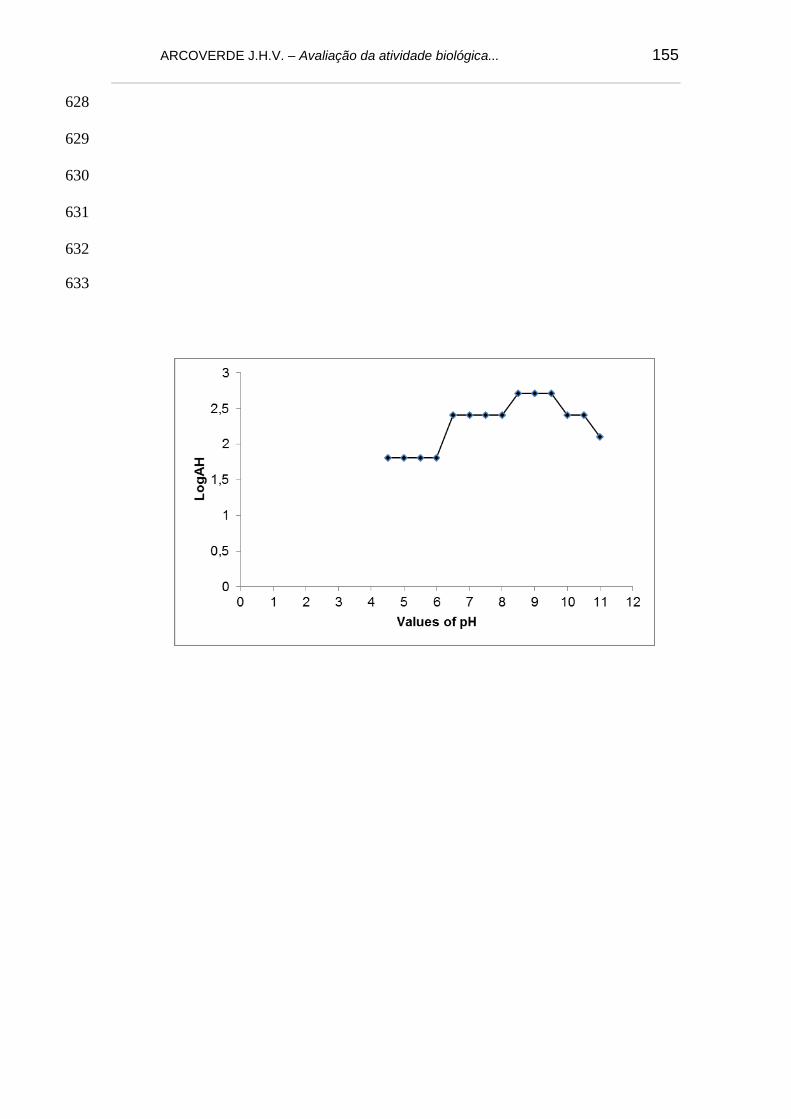
ARCOVERDE J.H.V. – Avaliação da atividade biológica... 155
628
629
630
631
632
633

ARCOVERDE J.H.V. – Avaliação da atividade biológica... 156
634
635
636
637
638
639
640
641
642
643
644
645
646
647
648
649
650
651
652
653
654
655
656
657
658

ARCOVERDE J.H.V. – Avaliação da atividade biológica... 157
659
660
661
662
663
664
665
666
667
668
669
670
671
672
673
674
675
676

ARCOVERDE J.H.V. – Avaliação da atividade biológica... 158
8. CONSIDERAÇÕES FINAIS 1. Na Caatinga há uma grande vareidade de espéceis vegetais ricas em
compostos bioquímicos que devem ser melhor explorados.
2. Os solventes clorofórmio, acetato de etila e metanol mostraram-se eficientes
para extração de metabólitos secundários das folhas de M. rufula.
3. Nas folhas de M. rufula estão presentesuma expressiva quantidade de
flavonoides, esteroides e taninos.
4. Os extratos orgânicos das folhas de M. rufula mostraram atividade
antioxidante, protetora de DNA, anti-bacteriana e termiticida.
5. Uma lectina (MkaLL) com atividade protetora de DNA e anti-bacteriana foi
isolada das folhas de M. rufula.
7. M. rufula apresenta-se como uma promissora fonte de compostos bioativos.