universidade estadual do cearÁ prÓ … · avaliação da atividade de bombas de efluxo nas cepas...
TRANSCRIPT

UNIVERSIDADE ESTADUAL DO CEARÁ PRÓ-REITORIA DE PÓS-GRADUAÇÃO E PESQUISA
FACULDADE DE VETERINÁRIA PROGRAMA DE PÓS-GRADUAÇÃO EM CIÊNCIAS VETERINÁRIAS
MANOEL PAIVA DE ARAÚJO NETO
LEVEDURAS ISOLADAS DE Macrobrachium amazonicum E DE
ECOSSISTEMAS AQUÁTICOS: DETECÇÃO DE RESISTÊNCIA A
DERIVADOS AZÓLICOS E O POTENCIAL USO DESSE CAMARÃO
PARA O MONITORAMENTO AMBIENTAL
Fortaleza
2014
MANOEL PAIVA DE ARAÚJO NETO
LEVEDURAS ISOLADAS DE Macrobrachium amazonicum E DE ECOSSISTEMAS AQUÁTICOS: DETECÇÃO DE RESISTÊNCIA A
DERIVADOS AZÓLICOS E O POTENCIAL USO DESSE CAMARÃO PARA O MONITORAMENTO AMBIENTAL
Tese apresentada ao Programa de Pós-Graduação em Ciências Veterinárias da Faculdade de Veterinária da Universidade Estadual do Ceará, como requisito parcial para a obtenção do título de Doutor em Ciências Veterinárias.
Área de Concentração: Reprodução e Sanidade Animal. Linha de Pesquisa: Reprodução e sanidade de carnívoros, herbívoros, onívoros e aves. Orientador: Prof. Dr. Marcos Fábio Gadelha Rocha
Fortaleza
2014


Eu estou sempre fazendo aquilo que não sou capaz, numa tentativa de assim aprender como fazê-lo.
Pablo Ruiz Picasso

Dedico este trabalho às pessoas que me apoiam, que querem o meu bem e o meu desenvolvimento pessoal e profissional. Também àqueles que suportaram minhas alterações de humor e estresse durante o período do doutorado... principalmente, dedico àquela que mais me deu suporte e força, meu amor... Míriam Martins.

AGRADECIMENTOS
Ao Programa de Pós-Graduação em Ciências Veterinárias, da Faculdade de Veterinária, da
Universidade Estadual do Ceará.
Ao Conselho Nacional de Desenvolvimento Científico e Tecnológico (CNPq), pelo apoio
financeiro.
Ao Centro Especializado em Micologia Médica (CEMM), da Universidade Federal do Ceará.
Ao Laboratório de Carcinicultura (LACAR), da Universidade Estadual do Ceará.
Ao Professor Marcos Fábio Gadelha Rocha pela orientação, pelos ensinamentos, conselhos,
paciência, apoio e cobranças que só fazem favorecer o meu crescimento como pesquisador e
êxito como profissional.
À Professora Célia Maria de Souza Sampaio pela atenção, paciência, confiança, apoio,
orientação e dedicação em todos esses anos de graduação e pós-graduação, minha segunda mãe.
À Professora Raimunda Sâmia Nogueira Brilhante, pela orientação, pelo apoio, pelos
conselhos, atenção e incentivos, dedicados durante o mestrado e doutorado, e pela sensação de
bem estar no ambiente de trabalho que senti desde minha qualificação de mestrado, pois senti
que desde aquele momento, um objetivo de sua orientação foi o meu crescimento pessoal e
profissional.
Ao Professor José Júlio Costa Sidrim, pelo comprometimento com o Centro Especializado em
Micologia Médica, base para realização deste trabalho, além de toda sua base filosófica que
fazem instrumento no despertar de um pesquisador.
À Professora Rossana Aguiar Cordeiro, pelos ensinamentos, dedicação, paciência, orientação e
apoio intelectual.
Ao Professor André Jalles Monteiro, pelos ensinamentos, capacidade e apoio estatístico.
A Professora Tereza de Jesus Pinheiro Gomes Bandeira, pelo exemplo e tranquilidade no
laboratório, orientação e apoio, atos fundamentais para a realização desta pesquisa.
A Professora Débora Castelo Branco de Souza Collares Maia pelo auxílio, apoio intelectual e
prático, orientação, amizade, ombro conselheiro e base intelectual para realização deste
trabalho.
Ao Professor Aldeney Andrade Soares Filho pela amizade, incentivo, sugestões e brilhante
orientação durante a realização de diversas pesquisas.
A Professora Janaína Andrade dos Santos pela amizade, dedicação e incentivo na prática da
pesquisa.

Ao Carlos Eduardo Cordeiro, pela ajuda, disposição e interesse na melhor forma de execução
do projeto, seja prática ou intelectual, braço direito na pesquisa realizada.
Ao Lucas Pereira de Alencar, pela amizade, auxílio na pesquisa e intelectual, suporte no
trabalho no dia-a-dia do CEMM e exemplo nas atitudes como colega profissional.
À Joyce Fonteles, pela amizade e apoio intelectual, auxílio fundamental para o êxito deste
trabalho.
À Amanda Chaves, pela amizade, discussões sobre a pesquisa, questionamentos e
direcionamento nas resoluções dos problemas encontrados.
À Teresinha de Jesus, pelo apoio e amizade, suporte na pesquisa e acolhimento no laboratório,
além do auxílio e ensinamentos para execução cada vez melhor de cada passo desta pesquisa.
Ao Daniel, pelo apoio e suporte para realização desta pesquisa no CEMM.
A todos que fazem parte do grupo CEMM, por todo o suporte proporcionado para a realização
de nossas pesquisas, pela convivência divertida e prazerosa.
A todos que fazem parte do LACAR, por todo o suporte proporcionado para a realização de
nossas pesquisas, pela convivência divertida e prazerosa, nas coletas e cultivo dos espécimes
utilizados na pesquisa.
Ao Giliardi de Paula Queiroz, essencial para realização desta pesquisa, com horas de coleta na
lagoa do Catu, pela amizade e pelo empenho para o sucesso nas coletas.
Aos meus pais, Lindemberg Leite Paiva e Solange Silva Paiva, às minhas irmãs, Ândria Silva
Paiva e Ânnya Lindemberg Silva Paiva, as minhas Sobrinhas, Andressa Silva Paiva Tavares e
Nicolle Silva Paiva Caetano, ao meu cunhado, Tobias Sousa Caetano, que sempre estiveram ao
meu lado, com toda paciência e companheirismo, que somente essa família sabe dedicar.
À Míriam Luzia Nogueira Martins de Sousa, pela paciência, pelo companheirismo, pelos
conselhos, tão importantes, por sua ajuda na pesquisa, de forma direta e indireta, pelo carinho,
pela amizade, todo meu amor e carinho.
A Deus, por ter me iluminado, ter dado força, paciência, perseverança e motivação para esse
caminho de pesquisador, que tanto me encantou e que me realiza como Biólogo e Professor.

RESUMO
Em ambientes aquáticos, a crescente poluição causada principalmente pelo despejo de
efluentes industriais, sanitários e agrícolas vem se tornando um dos principais problemas
ambientais da atualidade. Desta forma, a utilização de bioindicadores se torna uma
ferramenta importante para investigação deste tipo de impacto ambiental. Vale destacar
que nos últimos anos estudos vem demonstrando que espécies de Candida podem ser
consideradas como bioindicadores, sendo, muitas vezes, mais isoladas que bactérias
indicadoras em ambientes aquáticos poluídos por esgotos. Adicionalmente o uso de
sentinelas pode ser eficaz na avaliação de ecossistemas aquáticos poluídos, destacando-
se espécies de crustáceos. Assim, buscou-se investigar o uso de Candida spp. isoladas do
camarão de água doce Macrobrachium amazonicum e ecossistemas aquáticos como
bioindicadores de poluição ambiental, por meio da análise quali-quantitativa da
composição da microbiota e do monitoramento da resistência antifúngica, bem como da
avaliação da atividade de bombas de efluxo nas cepas resistentes aos derivados azólicos.
Para tanto foram utilizados os tratos gastrintestinais de espécimes adultos de M.
amazonicum oriundos da lagoa do Catu, Aquiraz, Ceará, Brasil e amostras da água desse
ambiente. Os 53 isolados de leveduras, obtidos dos animais, da água de superfície e
substrato, foram identificados com base nas características morfológicas e bioquímicas e,
em seguida, foram submetidos ao teste de microdiluição em caldo, frente à anfotericina
B, itraconazol e fluconazol, segundo a metodologia padronizada pelo CLSI (documento
M27-A3). Foi realizada ainda a comparação molecular entre as cepas de Candida spp.
isoladas do ambiente aquático (n=20) e do camarão (n=7), pelas técnicas M13-PCR
fingerprinting e RAPD-PCR. As concentrações inibitórias mínimas (CIM) para
anfotericina B, itraconazol e fluconazol foram de 0,03125 a 2 μg/mL, 0,0625 a ≥16 μg/mL
e 0,5 a ≥64 µg/mL, respectivamente. De todos os isolados do gênero Candida, 20,45%
foram resistentes a ambos os derivados azólicos, 25% foram resistentes ao fluconazol e
34,09% foram resistentes ao itraconazol. Vale destacar que dois isolados de C.
guilliermondii resistentes ao itraconazol também foram resistentes a anfotericina B. Não
foi observado resistência aos antifúngicos para os gêneros Kodamaea, Cryptococcus,
Rhodotorula e Trichosporon. Na avaliação da atividade de bombas de efluxo, foi
observado uma redução de CIM das cepas resistentes de oito a 256 vezes em relação a
inicial. A análise molecular, através das técnicas M13-PCR fingerprinting e RAPD-PCR,
revelou similaridades entre os padrões de bandas entre 11 isolados de Candida spp. para

9
o primer OPQ16 e de oito para o primer M13, indicando forte similaridade entre os
isolados. Estes dados apontam a importância da resistência antifúngica como ferramenta
bioindicadora de poluição de ambientes de água doce, assim como o uso de M.
amazonicum como sentinela no monitoramento deste fenômeno.
Palavras-chave: Camarão da Amazônia. Candida spp. Resistência antifúngica. Bombas
de efluxo. Sentinela ambiental.
ABSTRACT
In aquatic environments, increasing pollution mainly caused by the dumping of industrial,
sanitary and agricultural wastewater is becoming one of the main environmental problems
nowadays. Therefore, the use of bioindicators becomes an important tool for investigation
of this kind of environmental impact. It is noteworthy that over recent years studies have
been showing that Candida species can be considered a bioindicator, often more isolated
than bacteria indicators in polluted aquatic environments by sewage. Additionally the use
of sentinels may be effective in the evaluation of polluted aquatic ecosystems, especially
species of crustaceans. Thus, it was sought to investigate the use of Candida spp. isolated
from freshwater prawn Macrobrachium amazonicum and aquatic ecosystems as
bioindicators of environmental pollution, through qualitative and quantitative analysis of
the composition of the microbiota and monitoring of antifungal resistance, well as the
evaluation of the activity of efflux pumps in strains resistant to azoles. For that, we used
gastrointestinal tracts of adult specimens of M. amazonicum originated from Catu Lake,
Aquiraz, Brazil and samples of water from this environment. The 53 yeasts isolated,
obtained from animals, surface water and substrate, were identified based on
morphological and biochemical characteristics, and then were submitted to the broth
microdilution test, against to amphotericin B, itraconazole and fluconazole, according to
methodology standardized by CLSI (document M27-A3). It was performed a molecular
comparison of strains of Candida spp. isolated from the aquatic environment (n=20) and
prawns (n=7), by the M13-PCR fingerprinting and RAPD-PCR techniques. The minimal
inhibitory concentration (MIC) to amphotericin B, itraconazole and fluconazole were
0.03125 to 2 µg/mL, 0.0625 to ≥16 µg/mL and 0.5 to ≥64 µg/mL, respectively. Of all
isolates of the Candida genus, 20.45% were resistant to both azole derivatives, 25% were
resistant to fluconazole and 34.09% were resistant to itraconazol. It is noteworthy that
two isolates of C. guilliermondii resistant to itraconazole were also resistant to
amphotericin B. Was not observed resistance to antifungals for genera Kodamaea,
Cryptococcus, Rhodotorula and Trichosporon. In evaluating the activity of efflux pumps,
a reduction of MIC of resistant strains from eight to 256 times compared to initial was
observed. Molecular analysis through the techniques M13-PCR fingerprinting and
RAPD-PCR revealed similarities among the band patterns from 11 isolates of Candida
spp. for the primer OPQ16 and eight for the primer M13, indicating strong similarity
among the isolates. These findings indicate the importance of antifungal resistance as a

11
bioindicator tool of the freshwater environments pollution, as well as the use of M.
amazonicum as a sentinel monitoring of this phenomenon.
Palavras-chave: Amazon prawn. Candida spp. Antifungal resistance. Efflux pumps.
Environmental Sentinel.
LISTA DE FIGURAS
Figura 1 – Distribuição geografica de Macrobrachium amazonicum......................... 18
Figura 2 – Macho e fêmea ovada de M. amazonicum, morfologia externa................. 19
Figura 3 – Sistema digestório de M. amazonicum....................................................... 20
Figura 4 – Programa internacional Mussel Watch....................................................... 23
Figura 5 – Microdiluição em placas de 96 poços em formato de U para Candida spp................................................................................................................................ 27
Figura 6 – Representação das bombas de efluxo ABC binding cassette e transportadores MFS (Major Facilitators) de Candida spp........................................ 30
Figura 7 – Representação da estrutura das bombas de efluxo ABC binding cassette......................................................................................................................... 30
Figura 8 – Mecanismo de ação de drogas antifúngicas afetando a via biossintética do ergosterol................................................................................................................ 31
CAPÍTULO 1
Figure 1: A: Release of chemical compounds in the aquatic environment from industrial, agricultural and residential waste; B: Chemical compounds are transported and go through transformation, adsorption and sedimentation in the aquatic environment; C: Several organisms (mammals, birds, reptiles, amphibians, fish, invertebrates and microorganisms) are exposed to these chemical compounds; D: These compounds penetrate Candida spp. cells, which, in turn, increase efflux activity to decrease the intracellular concentration of chemicals compounds, avoiding cellular damages and promoting the survival of Candida cells………….………………………….………………………………..………….. 59
CAPÍTULO 2
Figure 1 – Dendrograms resulting from the analysis of 27 isolates of Candida spp. obtained from the gastrointestinal tract of Macrobrachium amazonicum (n=7) and the natural environment (n=20) through M-13-fingerprinting and RAPD-PCR with primer OPQ16. P: prawn; SW: surface water; S: sediment. *indicates antifungal resistance. Dendrograms generated by the BioNumerics program (Applied Math, Inc.)………………………………….......................................................................... 74
CAPÍTULO 3
Figure 1 – Collection points. Catú Lake, Aquiraz, Ceará, Brazi. Point 1: Leisure area: bars, restaurants, boats. Area used for activities such as boating and jet skiing. 3°55’59.79” S and 38°21’50.10” W; Point 2: Agricultural area, with potato and bean fields, with possible use of azoles. 3°55’47.25” S and 38°22’14.16” W; Point 3: Industrial area, near the state highway (CE-040). 3°56’03.70” S and 38°22’25.15” W; Point 4: Residential area, discharge of raw household sewage, near the confluence with the Catú River. 3°56’56.72” S and 38°22’31.57” W…..… 92

LISTA DE TABELAS CAPÍTULO 1
Table 1 – Main Candida species isolated from aquatic environments………………….. 58
CAPÍTULO 2
Table 1 – Species, access number, origin, isolation period and antifungal susceptibility of 27 Candida spp. isolates used for molecular analysis……………………………....... 75
CAPÍTULO 3
Table 1 – Yeast species isolated from different collection points of Catu Lake………. 93
Table 2 – Minimum inhibitory concentration (MIC) of amphotericin B, itraconazole and fluconazole against 46 yeast isolates from Catú Lake……………………………...
94

LISTA DE ABREVIATURAS E SIGLAS ATCC – American Type Culture Collection
CEMM – Centro Especializado em Micologia Médica
CFU – Colony Forming Units (Unidades Formadoras de Colônia)
CIM – Concentração inibitória mínima
CLSI – Clinical Laboratory Standarts Institute
HAP – Hidrocarbonetos aromáticos policíclicos
LACAR – Laboratório de Carcinicultura da Universidade Estadual do Ceará
MDR – Multidroga Resistente
MFS – Major Facilitators
MIC - Minimum Inhibitory Concentrations
NBD – Nucleotide Biding Domain
PCR-REA - Polymerase Chain Reaction-Restriction Endonuclease Analysis
P-gp – Glicoproteína P
RAPD-PCR – Random Amplified Polymorphic DNA- Polymerase Chain Reaction
S – Sediment
SISBIO – Sistema de Informação e Autorização em Biodiversidade
SW – Surface Water
UPGMA – Unweighted Pair Group Method

Sumário 1 INTRODUÇÃO ...................................................................................................... 16
2 REVISÃO DE LITERATURA .............................................................................. 18
2.1 Camarão da Amazônia (Macrobrachium amazonicum) ................................. 18
2.2 Leveduras na Aquicultura .............................................................................. 22
2.3 Sentinelas para o isolamento de leveduras ..................................................... 22
2.4 Leveduras e relação com ambientes aquáticos alterados ............................... 25
2.5 Candida spp. como bioindicadores da qualidade do ecossistema aquático ... 26
2.6 Teste de sensibilidade a antifúngicos .............................................................. 27
2.7 Resistência antifúngica a derivados azólicos como estratégia para o monitoramento ambiental ..................................................................................... 30
3 JUSTIFICATIVA................................................................................................... 35
4 HIPÓTESE ............................................................................................................. 36
5 OBJETIVOS .......................................................................................................... 37
5.1 Objetivo Geral ................................................................................................. 37
5.2 Objetivos Específicos ....................................................................................... 37
6 CAPITULO 1 ......................................................................................................... 38
Surveillance of azole resistance among Candida spp. as a strategy for the indirect monitoring of freshwater environments ............................................................... 38
7 CAPÍTULO 2 ......................................................................................................... 61
Macrobrachium amazonicum: an alternative for microbiological monitoring of aquatic environments in Brazil ................................................................................ 61
8 CAPÍTULO 3 ......................................................................................................... 77
Azole resistance in Candida spp. from water: an efflux-pump mediated mechanism ............................................................................................................. 77
10 CONCLUSÃO ...................................................................................................... 96
11 PERSPECTIVAS ................................................................................................. 97
12 REFERÊNCIAS BIBLIOGRÁFICAS ................................................................ 98
1 INTRODUÇÃO
Na literatura científica, há registros relatando a importância de leveduras para a
aquicultura, tanto como componentes da microbiota, como causadores de enfermidades
em variados grupos de interesse aquícola (LEAÑO et al., 2005; GATESOUPE, 2007).
Nesses registros, distintas espécies de leveduras foram isoladas de uma grande variedade
de ecossistemas aquáticos, mas apenas um número limitado é prevalente, com destaque
para as espécies dos gêneros Candida, Cryptococcus, Rhodotorula, Saccharomyces e
Trichosporon (ROSA et al., 1995). Adicionalmente, Candida spp. representam o maior
número de espécies isoladas em cultivos de camarão (JOHNSON; BUENO, 2000).
Nos últimos anos, alguns estudos demonstraram a alta frequência de isolados de
Candida spp. resistentes à antifúngicos, oriundos de ambientes aquáticos e embora
associem essa manifestação com o risco à saúde pública, não se sabe ao certo qual a causa
deste fenômeno (MEDEIROS et al., 2008; BRANDÃO et al., 2010). Em pesquisa
realizada com leveduras isoladas de camarões da espécie Macrobrachium amazonicum,
oriundos da lagoa do Catu, Ceará, Brasil, foram obtidos 28% de isolados de Candida spp.
com resistência in vitro a derivados azólicos (BRILHANTE et al., 2010). Tal observação
despertou interesse pela busca das possíveis causas da ocorrência de cepas resistentes de
Candida spp., no ambiente natural.
Embora existam muitos trabalhos abordando o monitoramento do perfil de
sensibilidade de leveduras isoladas de amostras clínicas humanas (PFALLER et al.,
2010), ainda são escassos os trabalhos com leveduras isoladas do ambiente.
Demonstrando, assim, a importância da realização de estudos para a melhor compreensão
do fenômeno de resistência em amostras oriundas do ambiente aquático.
Sabe-se que, de modo em geral, existem dois mecanismos primordiais para o
desenvolvimento de resistência a derivados azólicos: o primeiro consiste no
desenvolvimento de bombas de efluxo ativo; e o segundo na alteração da enzima
lanosterol C14α-desmetilase, molécula-alvo dos derivados azólicos (KANAFANI;
PERFECT, 2008; MANASTIR et al., 2009).
Nos estudos realizados por Brilhante et al. (2011), analisando a microbiota
gastrintestinal, por leveduras, em camarões da espécie M. amazonicum, observou-se o
fenômeno de resistência ao itraconazol e ao fluconazol, em isolados de Candida spp.
Acredita-se que a observação desse fenômeno esteja associada à presença de agentes
químicos potencialmente mutagênicos, passíveis de serem encontrados nos efluentes

17
industriais que contaminam ambientes aquáticos. Adicionalmente, a superexpressão de
bombas de efluxo pode estar relacionada com sua atividade inespecífica, que esta
diretamente ligada com a desintoxicação celular e sobrevivência dessas leveduras no
ambiente aquático poluído.
Assim, se torna necessário a realização de estudos avaliando o perfil de
sensibilidade a antifúngicos em leveduras oriundas de ambientes aquáticos para a melhor
compreensão deste fenômeno em ambientes com diferentes níveis de degradação
ambiental.

2 REVISÃO DE LITERATURA
2.1 Camarão da Amazônia (Macrobrachium amazonicum)
A expressão “camarão de água doce” é utilizada para caracterizar tanto espécies
que têm todo seu ciclo de vida restrito a esse ambiente, como espécies que necessitam de
água salobra no início de seu desenvolvimento e de água doce depois da metamorfose
(COELHO; RAMOS-PORTO; SOARES, 1981). Embora sejam também chamados de
camarões, como os de água salgada, os de água doce são evolutivamente mais próximos
das lagostas, expressando muitas semelhanças com estas, principalmente quanto aos
hábitos reprodutivos, pois as fêmeas de ambas as espécies incubam seus ovos no abdômen
até a eclosão das larvas (ISMAEL; NEW, 2000).
Os camarões de água doce são crustáceos decápodes pertencentes à subordem
Pleocyemata e família Palaemonidae (HOLTHUIS; NG, 2010). O gênero mais
representativo desta família é o Macrobrachium, que segundo Holthuis e Ng (2010), é
circuntropical e nativo em todos os continentes, exceto na Europa e Antartida.
Atualmente, existem em todo o mundo cerca de 243 espécies de camarões pertencentes a
esse gênero (DE GRAVE; FRANSEN, 2011), dentre as quais 46 são registradas nas
Américas e 19 no Brasil (MELO, 2003; SANTOS; HAYD; ANGER, 2013). Vale destacar
que, a maioria das espécies de camarão de água doce de interesse comercial pertence ao
gênero Macrobrachium (MELO, 2003).
M. amazonicum pertence ao grupo de espécies continentais de desenvolvimento
larval completo, e ocorre desde a bacia do Orinoco, passando pelo rio Amazonas, até a
bacia do rio Paraguai (Figura 1) (HOLTHUIS, 1952).

19
(Fonte: PAIVA, 2014a)
Figura 1 – Distribuição geografica de Macrobrachium amazonicum.
Decápode de água doce de maior importância econômica no Sudeste do
Continente Sul-americano, M. amazonicum possui elevado potencial para a aquicultura,
sendo consumido por povos indígenas e brasileiros de todos os grupos econômicos, e
amplamente explorada pela pesca artesanal no Norte e Nordeste do Brasil, chegando a
representar 85% do pescado de camarão selvagem de água doce no Brasil, no final da
década de 1990 (MACIEL; VALENTI, 2009).
Os espécimes apresentam rostro longo e delgado com margem superior provida
de nove a 12 dentes, margem inferior com oito a dez dentes distribuídos irregularmente;
carapaça e abdômen lisos e transparentes e télson terminando em uma extremidade aguda
com dois pares de espinhos na margem posterior. A segunda pata no adulto é a mais forte.
Machos adultos exibem mero, carpo e própode cobertos por espínulos curtos os quais
estão ausentes nas fêmeas (MELO, 2003). O sexo é separado, além de indicar

20
características morfológicas externas que permitem distinguir facilmente, em exemplares
maduros, machos e fêmeas (MORAES-VALENTI; VALENTI, 2010).
Machos e fêmeas de camarões palaemonídeos apresentam compleição física
semelhante até atingirem a maturidade sexual, quando têm início os processos
reprodutivos. Desde então, as fêmeas destinam suas reservas energéticas à produção e à
incubação dos ovos, enquanto os machos direcionam o gasto energético ao crescimento
somático, fazendo com que se tornem os maiores indivíduos da população (AMMAR;
MÜLLER; NAZARI, 2001) (Figura 2).
Círculo vermelho = Ovos na câmara de incubação. Reta azul = Segundo par de pereiópodo mais desenvolvido no macho.
Figura 2 – Macho e fêmea de M. amazonicum, morfologia externa.
Os juvenis são translúcidos, com o segundo par de pereiópodes delgado. As
fêmeas de M. amazonicum normalmente são menores do que os machos. Os ovos se
desenvolvem em uma câmara de incubação, formada pelo arqueamento e alongamento
da pleura abdominal. Estes são geralmente pequenos e passam, durante o
desenvolvimento embrionário, da tonalidade verde-escuro, no início, a verde-claro, até
apresentarem uma coloração verde-acinzentada, cinza-clara ou esbranquiçada quando se
encontram próximo à eclosão das larvas (MORAIS-VALENTI; VALENTI, 2010).
(Fonte:PAIVA, 2005)

21
O sistema digestório em camarões do gênero Macrobrachium é completo e
tubular; inicia-se com a boca anteroventral, percorre dorsalmente o corpo do animal e
termina em um ânus localizado na base do télson (Figura 3). Segundo Ismael e New
(2000), o sistema digestório se divide em três regiões - anterior, média e posterior - e
inclui também uma glândula digestiva, cecos pilóricos e divertículos. O intestino anterior
está situado na porção dorsal do cefalotórax e possui esôfago e estômago, este com duas
câmaras - a cardíaca e a pilórica - responsáveis pelos mecanismos de trituração e filtração
e que, junto com as enzimas iniciam o processo digestivo. O moinho gástrico, um
mecanismo de trituração formado por ossículos na câmara anterior do intestino anterior,
está presente em todos os decápodes, exceto em alguns carídeos, como os camarões de
água doce do gênero Macrobrachium. Desta maneira, a mastigação nestes animais
depende exclusivamente das mandíbulas que quebram e separam as partículas
alimentares, encaminhando-as para o intestino médio.
Figura 3 – Sistema digestório de M. amazonicum.
(Fonte:PAIVA, 2010)

22
2.2 Leveduras na Aquicultura
De acordo com De Hoog et al. (2000), levedura é um termo descritivo para
qualquer fungo que se reproduz por brotamento, cuja unidade funcional é o blastoconídio.
Os fungos denominados de leveduras se encontram classificados nas classes
Hemiascomycetes, da divisão Ascomycota, e Hymenomycetes e Urediniomycetes, da
divisão Basidyomycota.
Artificialmente, as leveduras são definidas como organismos unicelulares,
brancos ou avermelhados. A formação de cadeias celulares coesas (pseudomicélio) é
comum, inclusive, algumas espécies produzem hifas verdadeiras e artroconídios. Estes
fungos são tratados como leveduras por serem filogeneticamente semelhantes a uma das
classes retrocitadas (DE HOOG et al., 2000).
A maioria das leveduras clinicamente relevantes se reproduz por processos
vegetativos, cujos principais mecanismos são o brotamento celular, a fissão celular e a
formação de artroconídios (DE HOOG et al., 2000).
Trabalhos avaliando o papel de leveduras em espécies de interesse para
aquicultura são escassos e relatam, principalmente, o papel como patógeno e como
representante da microbiota de peixes e crustáceos de interesse comercial. Nos estudos
realizados com esses animais, os principais gêneros de leveduras isolados são
Rhodotorula, Saccharomyces, Trichosporon, Candida e Cryptococcus (BRUCE;
MORRIS, 1973; LEAÑO et al., 2005; MENDONÇA-HAGLER; HAGLER, 1989;
PAGNOCCA).
Brilhante et al. (2011) realizaram um estudo em um lago localizado no Nordeste
do Brasil (Ceará), com a utilização de camarões para o isolamento de leveduras desse
ambiente, e observaram cepas de Candida spp. resistentes a derivados azólicos. Nesse
estudo, os autores sugeriram a utilização deste crustáceo como um sentinela ambiental e
as leveduras como bioindicadores para presença de poluentes, por meio da avaliação do
perfil de sensibilidade a antifúngicos in vitro.
2.3 Sentinelas para o isolamento de leveduras
A microbiota intestinal de invertebrados aquáticos é semelhante a do meio
ambiente em que estão inseridos, assim leveduras podem ser mais facilmente isoladas
com origem nestes animais do que diretamente do ambiente aquático (HAGLER et al.,
23
1995). Desta forma, estes animais, especialmente moluscos bivalves e camarões, podem
ser utilizados para melhorar o isolamento de leveduras em ambientes aquáticos, que, por
sua vez, atuam como bioindicadores para a análise da saúde ambiental (HAGLER et al.,
1995; KUTTY; PHILIP, 2008).
O termo sentinela foi utilizado pela primeira vez na década de 1950 para descrever
os bivalves utilizados a fim de detectar e mapear radioatividade (WASHINGTON, 1984)
e, desde então, o programa Mussel Watch é utilizado em vários estudos que envolvem a
utilização de bioacumuladores (BEEBY, 2001). Em geral, sentinelas são monitores
biológicos capazes de acumular poluentes sem receber efeitos secundários significativos
e são, principalmente, utilizados para medir a quantidade de um determinado poluente,
com vistas a aumentar a sensibilidade de determinada técnica analítica e/ou para facilitar
a interpretação de um sinal complexo de poluição. Com efeito, as espécies sentinelas
podem ser classificadas em três grandes grupos: 1) espécies monitoras, cujas funções
biológicas são reduzidas na presença de certos poluentes; 2) espécies indicadoras,
apontando a presença de um desequilíbrio por aumento ou diminuição de sua ocorrência
no ambiente; e 3) espécies sentinelas, que acumulam poluentes em seus tecidos sem
sofrer danos significativos, permitindo a quantificação da fração biodisponível de uma
determinada substância química em um ecossistema. Adicionalmente, os sentinelas
também podem ser descritos como organismos que podem acumular algumas espécies de
microrganismos bioindicadores nos seus sistemas (BEEBY, 2001).
Variados organismos são sugeridos como sentinelas, tais como anelídeos
(BARUS; JARKOVSKY; PROKES, 2007), peixes (ZORITA et al., 2008), moluscos
(BEEBY, 2001; MAZZIA et al., 2011), abelhas (LAMBERT et al., 2012),
macroinvertebrados bentônicos (BRILHANTE et al., 2011; CHIBA; PASSERINI;
TUNDISI, 2011), anfíbios (HOPKINS, 2007), aves (BRILHANTE et al., 2012) e
mamíferos (MARIANO et al., 2009). Entre essas espécies, macroinvertebrados
bentônicos são particularmente interessantes em razão de suas características como
filtradores e da facilidade de recuperação desses animais em ambientes aquáticos
(BEEBY, 2001; MAZZIA et al., 2011). Os estudos com estes animais têm abordado o
acúmulo de metais pesados, hidrocarbonetos, pesticidas e outros poluentes, em seus
tecidos (MAHMOUD; TALEB, 2013).
Em 1993, o programa internacional Mussel Watch inseriu o Brasil em suas
análises para o monitoramento de contaminantes químicos costeiros utilizando bivalves,
e foi demonstrada a necessidade de avaliar melhor a extensão e a gravidade das
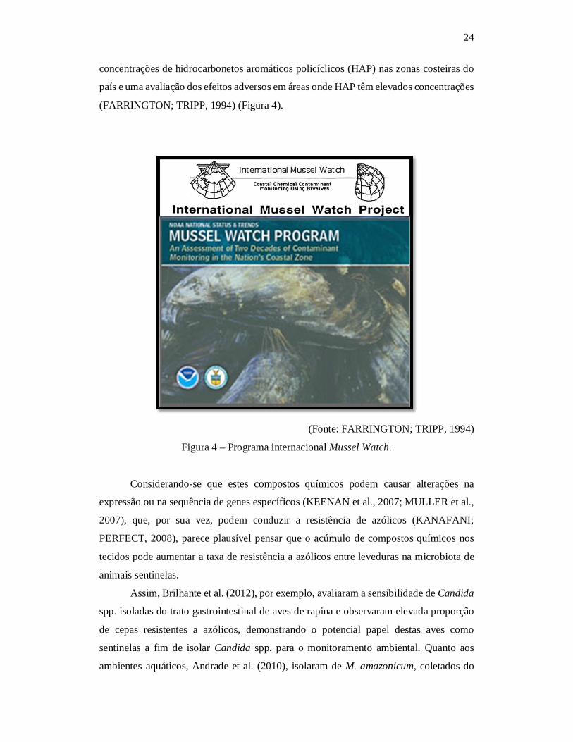
24
concentrações de hidrocarbonetos aromáticos policíclicos (HAP) nas zonas costeiras do
país e uma avaliação dos efeitos adversos em áreas onde HAP têm elevados concentrações
(FARRINGTON; TRIPP, 1994) (Figura 4).
(Fonte: FARRINGTON; TRIPP, 1994)
Figura 4 – Programa internacional Mussel Watch.
Considerando-se que estes compostos químicos podem causar alterações na
expressão ou na sequência de genes específicos (KEENAN et al., 2007; MULLER et al.,
2007), que, por sua vez, podem conduzir a resistência de azólicos (KANAFANI;
PERFECT, 2008), parece plausível pensar que o acúmulo de compostos químicos nos
tecidos pode aumentar a taxa de resistência a azólicos entre leveduras na microbiota de
animais sentinelas.
Assim, Brilhante et al. (2012), por exemplo, avaliaram a sensibilidade de Candida
spp. isoladas do trato gastrointestinal de aves de rapina e observaram elevada proporção
de cepas resistentes a azólicos, demonstrando o potencial papel destas aves como
sentinelas a fim de isolar Candida spp. para o monitoramento ambiental. Quanto aos
ambientes aquáticos, Andrade et al. (2010), isolaram de M. amazonicum, coletados do
25
ambiente natural, diversas espécies de bactérias resistentes e sugeriram que essa
resistência estava associada ao emprego inadequado de antibacterianos em fazendas de
carcinicultura e piscicultura.
Da mesma forma, Brilhante et al. (2011) sugeriram que o M. amazonicum seria
um bom sentinela para monitorar a resistência a antifúngicos entre Candida spp.
ambientais, enquanto que estas Candida spp. resistentes poderiam ser um indicador
importante para a presença de compostos químicos em um determinado ambiente
aquático.
2.4 Leveduras e relação com ambientes aquáticos alterados
Muitos ensaios têm demonstrado uma tendência mundial no uso de leveduras
oriundas do ambiente e de animais como uma ferramenta para avaliar a qualidade dos
ambientes aquáticos (CHEN; YANAGIDA; CHEN, 2009; COELHO et al., 2010;
HAGLER; MENDONÇA-HAGLER, 1981; SIMARD, 1971). As leveduras mais
comumente isoladas nesses estudos pertencem aos gêneros Candida, Cryptococcus,
Rhodotorula, Saccharomyces e Trichosporon (HAGLER et al., 1995), com destaque para
as espécies C. famata, R. mucilaginosa e C. laurentii, que têm sido considerados
bioindicadoras de poluição em ambientes aquáticos (ARVANITIDOU et al., 2002;
BRILHANTE et al., 2011; CHUN, 1984; COELHO et al., 2010; HAGLER et al., 1995;
HAGLER; MENDONÇA-HAGLER, 1981; LIBKIND et al., 2003; MEDEIROS et al.,
2008; SLÁVIKOVÁ; VADKERTIOVÁ; KOCKOVÁ-KRATOCHVÍLOVÁ, 1992).
Vários autores asseguram que a análise da microbiota leveduriforme é um
instrumento válido para a avaliação do estado de eutrofização em um determinado
ambiente aquático (ARVANITIDOU et al., 2002; BRILHANTE et al., 2011;
MEDEIROS et al., 2008; ROSA et al., 1990). A utilização de leveduras como
bioindicadores foi inicialmente proposto na década de 1970, baseado num estudo
realizado no rio St. Lawrence, no Canadá. Esse estudo demonstrou que em áreas
localizadas perto de esgoto, muitas espécies de leveduras cresceram abundantemente e o
número de isolados e a frequência de recuperação poderiam ser usados como parâmetros
para avaliar a poluição (SIMARD, 1971). Assim, Rosa et al. (1990) investigaram a
ocorrência de leveduras e coliformes fecais em cinco estações de coleta de água na lagoa
Olhos D'Água, no Estado de Minas Gerais, Brasil, e observaram que, em três estações, o
26
número de leveduras recuperadas foi maior do que a de coliformes fecais, fato sugestivo
de que esses fungos poderiam ser melhores bioindicadoras de poluição em fontes de água.
Nas pesquisas realizadas em regiões de clima temperado, como Argentina
(LIBKIND et al., 2003) e Austrália (LATEGAN et al., 2012), leveduras carotenogênicas,
como Rhodotorula spp., foram importantes para a avaliação da influência antrópica no
ambiente aquático, considerando que estas leveduras só foram isoladas em áreas
degradadas pela atividade antrópica. Além disso, Rhodotorula spp. podem ser usadas para
monitorar a qualidade de águas subterrâneas, como mostrado por Lategan et al. (2012),
que avaliaram a diversidade de fungos em fontes de águas subterrâneas superficiais na
Austrália.
Adicionalmente, leveduras podem ser utilizadas como bioindicadores para avaliar
as alterações ambientais, por meio de dois mecanismos: o primeiro, correlaciona a taxa
de recuperação de várias espécies de leveduras para as distintas condições ambientais,
tendo em conta a presença das fontes de poluição, como esgotos e efluentes industriais; o
segundo avalia a presença de alterações fenotípicas nestes microrganismos recuperados
em ambientes alterados. Esses mecanismos fundamentam-se na utilização de leveduras
como bioindicadores de alterações nos ambientes aquáticos (BRILHANTE et al., 2011;
HAGLER et al., 1995; HAGLER; MENDONÇA-HAGLER 1981; MEDEIROS et al.,
2008; ROSA et al., 1990; SIMARD, 1971).
2.5 Candida spp. como bioindicadores da qualidade do ecossistema aquático
As pesquisas envolvendo o isolamento de leveduras demonstram a onipresença
do gênero Candida. Este gênero mostra ser predominante, quando se considera o número
elevado de amostras, bem como a elevada taxa de recuperação, independentemente da
estação do ano (RASPOR; ZUPAN, 2006). Este fato se torna ainda mais evidente quando
as amostras são de fontes aquáticas, ademais, pesquisas propõem o uso de Candida spp.
como bioindicadores ambientais (COELHO et al., 2010; HAGLER et al., 1995;
MEDEIROS et al., 2008). Em pesquisas que visam correlacionar a degradação ambiental
às atividades antrópicas, tais como esgotos domésticos e efluentes industriais e agrícolas,
Candida spp. são espécies de interesse, uma vez que são comumente associadas com a
presença de fezes humanas e de animais (HAGLER, 2006; HAGLER; MENDONÇA-
HAGLER, 1981).

27
C. famata é a espécie mais comumente isolada de fontes aquáticas e seu
isolamento com taxas elevadas caracteriza a degradação ambiental com esgotos
domésticos (BRANDÃO et al., 2010; BRILHANTE et al., 2011; MEDEIROS et al.,
2008). Além disso, essa espécie tem associação com o isolamento de coliformes fecais, o
que demonstra o seu papel como um indicador de poluição em ambientes aquáticos
(MEDEIROS et al., 2008; ROSA et al., 1990). A predominância de C. famata nesses
ambientes pode estar relacionada com a sua grande capacidade de resistir a baixas
temperaturas, variadas salinidades e variações de osmolaridade (RASPOR; ZUPAN,
2006). Ademais, além de indicar a eutrofização, esta espécie parece estar intimamente
associada com a remoção de poluentes, como os hidrocarbonetos (HAP), com origem na
água contaminada (FARAG; SOLIMAN, 2011).
C. guilliermondii também foi isolada em ambientes aquáticos, especialmente
aqueles eutrofizados ou contaminados com esgotos domésticos e efluentes industriais,
como demonstrado por Medeiros et al. (2008), Brandão et al. (2010) e Brilhante et al.
(2011). Papon et al. (2013) sugeriram que essa espécie é capaz de usar várias fontes de
carbono, incluindo os hidrocarbonetos, contribuindo para a biorremediação de solos e
água contaminados por derivados de petróleo.
Medeiros et al. (2008), ao avaliarem a diversidade de leveduras em lagos e rios da
região Sudeste do Brasil, descobriram que o gênero Candida foi o que exibiu o maior
número de espécies e o maior quantitativo de isolados, dos quais 50% foram resistentes
ao itraconazol. Assim, com base nestes resultados, os autores sugeriram que, nos
ambientes aquáticos investigados, a presença de Candida spp. resistentes a derivados
azólicos estava associada com as atividades antrópicas a que estes ambientes foram
submetidos.
2.6 Teste de sensibilidade a antifúngicos
O conhecimento do perfil de sensibilidade a antifúngicos de determinada região é
de grande importância para o estabelecimento de condutas terapêuticas e profiláticas
adequadas. Em 1985, o Comitê da Área de Micologia do Clinical Laboratory Standards
Institute (CLSI) publicou o seu primeiro relatório, no qual se delinearam os resultados de
um pequeno estudo colaborativo. Constatou-se que 20% dos laboratórios membros da
instituição realizavam testes de sensibilidade a agentes antifúngicos, na maioria das
vezes, com Candida spp., empregando o método de diluição em caldo, e obtinham

28
resultados de concentração inibitória mínima (CIM) discrepantes. Desde então, foi
decidido desenvolver e padronizar uma metodologia reprodutível e exequível para
laboratórios de rotina. Em 1997, a Norma M27-A foi publicada, especificando os pontos
de corte para os antifúngicos disponíveis. Em 2002, a Norma M27-A2 padronizou as
faixas de referência de CIM 24 e 48 horas, e em 2008 a Norma M27-A3 revisou esses
dados para drogas previamente estabelecidas e recém-lançadas (CLSI, 2008).
O teste de microdiluição em caldo é realizado em placas acrílicas estéreis, com 96
poços em formato de U, e consiste na exposição de um inóculo definido de um
determinado microorganismo a concentrações conhecidas das drogas testadas, sendo
possível observar o efeito destas sobre o crescimento fúngico. A leitura final determina a
menor concentração da droga, capaz de inibir o crescimento do microorganismo,
denominada de concentração inibitória mínima (CIM) (CLSI, 2008) (Figura 5).
Controle negativo = Avaliação da esterilidade do meio e da droga. Controle positivo = Avaliação do crescimento e esterilidade do
inóculo. Sentido da diluição.
(Fonte: PAIVA, 2014b)
Figura 5 – Microdiluição em placas de 96 poços em formato de U para Candida spp.
29
O objetivo final dos testes de sensibilidade é prever a resposta dos pacientes à
terapia a ser instituída. Muitos fatores, no entanto, além do perfil de sensibilidade in vitro,
influenciam a resposta clínica, como o sítio de infecção, o status imunológico do
hospedeiro, a farmacocinética da droga e a adesão do paciente à terapia. Portanto, o
estabelecimento da correlação clínica direta entre os valores de CIM e o desfecho
terapêutico ainda é limitado na terapia antifúngica (REX; PFALLER, 2002;
HOSPENTHAL; MURRAY; RINALDI, 2004). A realização de testes de sensibilidade,
contudo, se torna importante, uma vez que pode orientar a instituição da terapia
antifúngica mais adequada, direcionando o paciente aos 90% de sucesso terapêutico
(REX; PFALLER, 2002).
Em geral, a taxa de resistência aos azólicos permanece baixa, entre a maioria das
espécies de Candida spp., variando de 1-2,1% em C. albicans, de 0,4-4,6% em C.
parapsilosise de 1,4-6,6% em C. tropicalis. No entanto, C. glabrata, a segunda espécie
mais prevalente em infecções fúngicas sistêmicas nos Estados Unidos, exprime crescente
resistência ao fluconazol, cuja taxa de resistência aumentou de 7 para 12%, de 2001 a
2004 (KANAFANI; PERFECT, 2008).
Existem muitos trabalhos com o monitoramento do perfil de sensibilidade a
drogas de leveduras isoladas de amostras clínicas humanas, como pode ser observado nas
metanálises realizadas por Pfaller e Diekema (2007) e Pfaller et al. (2010). São escassos,
no entanto, os trabalhos com leveduras isoladas de animais. Brito et al. (2009) observaram
resistência intrínseca a cetoconazol, fluconazol e itraconazol em isolados de C. albicans
e de C. tropicalis oriundos de cães, e Brilhante et al. (2010), em um estudo que foi
realizado com calopsitas (Nymphicus hollandicus), ave pertencente à família Psittacidae,
observaram fenômeno de resistência a itraconazol e fluconazol, em isolados de C.
albicans.
Ante o exposto, diversas espécies de leveduras isoladas de ambientes aquáticos e
de animais de interesse aquicola são conhecidas como patógenos e podem comprometer
a saúde humana (PFALLER; DIEKEMA, 2007). Algumas cepas de leveduras,
previamente consideradas colonizadores simples ou contaminantes, são resistentes a
diversas drogas antifúngicas disponíveis e podem causar infecções em humanos. Estas
infecções são descritas com frequência crescente em relação às permissivas condições
ambientais, a pressão seletiva antifúngica e a pacientes imunodeprimidos (CANUTO;
RODERO, 2002; ENOCH; LUDLAM; BROWN, 2006; PFALLER; DIEKEMA, 2007).

30
Medeiros et al. (2008) isolaram leveduras de amostras de água e de sedimento de
lagos poluídos, localizados no Sudeste do Brasil, e traçaram o perfil de sensibilidade a
varias drogas antifúngicas. De todos os isolados submetidos ao teste de sensibilidade in
vitro (68 leveduras), 13% foram sensíveis ao cetoconazol, 79% para fluconazol, 50% para
itraconazol, 31% para terbinafina e 78% das cepas foram sensíveis a anfotericina B. Sete
isolados de diferentes espécies de Candida foram resistentes a todos esses antifúngicos.
Brilhante et al. (2011) observaram que 28,6% (4/14) do total de isolados de
Candida spp. do trato digestório de M. amazonicum coletados no ambiente natural foram
resistentes a esses derivados azólicos. Este fenômeno de resistência aos derivados
azólicos despertou a curiosidade de buscar as possíveis causas associadas a este fenômeno
no ambiente.
2.7 Resistência antifúngica a derivados azólicos como estratégia para o
monitoramento ambiental
De maneira em geral, existem dois tipos principais de mecanismos envolvidos no
desenvolvimento de resistência aos derivados de azólicos. O primeiro mecanismo está
associado com o desenvolvimento de bombas de efluxo ativo, codificadas pelos genes
CDR1 e CDR2, que pertencem à superfamília ATP binding cassette, e o gene MDR1, que
pertence à classe Major Facilitators. A superexpressão destes genes evita o aumento da
concentração do fármaco no interior da célula, consequentemente, diminuindo a sua
eficácia. O aumento da regulação de CDR1 e CDR2 leva à resistência a quase todos os
antifúngicos, enquanto que aumento da regulação do MDR1 conduz à resistência ao
fluconazol (KANAFANI; PERFECT, 2008) (Figuras 6 e 7). O segundo mecanismo de
resistência está associado a alterações na enzima lanosterol C14α -desmetilase, molécula-
alvo dos derivados de azólicos, que é codificada pelo gene ERG11 (Figura 8).
Superexpressão ou mutações nestes genes diminui a sensibilidade ou leva a resistência
aos derivados azólicos (KANAFANI; PERFECT, 2008; MANASTIR; ERGON;
YÜCESOY, 2009).

31
O NBDs (Nucleotide Binding Domains) das bombas ABC são responsáveis pela hidrólise de ATP, o que facilita a retirada da droga
do meio intracelular, enquanto os transportadores MFS utilizam gradiente de prótons para expelir drogas.
(Fonte: PRASAD; SHARMA; RAWAL, 2011)
Figura 6 – Representação das bombas de efluxo ABC binding cassette e transportadores
MFS (Major Facilitators) de Candida spp.
(Fonte: MARTINEZ; FALSON, 2014)
Figura 7 – Representação da estrutura das bombas de efluxo ABC binding cassette.

32
(Fonte: LUPETTI et al., 2002)
Figura 8 - Mecanismo de ação de drogas antifúngicas afetando a via biossintética do
ergosterol.
Adicionalmente, o desenvolvimento da resistência aos azólicos entre Candida
spp. pode estar relacionado com as atividades antrópicas, uma vez que a resistência
antifúngica é mediada principalmente por mudanças na expressão gênica e/ou sequência
de nucleotídeos do gene (FENG et al., 2010) e resíduos industriais e poluentes são capazes
de promover tais alterações (KEENAN et al., 2007; MÜLLER et al., 2007). Foi
demonstrado que os microrganismos resistem aos efeitos tóxicos de variados tipos de
solventes, derivados de petróleo e agentes mutagênicos, mediante a ação destas bombas
de efluxo, que exportam para o ambiente extracelular vários substratos, como metais
pesados e seus derivados químicos, incluindo cádmio, arsenato, hexano, tolueno, xileno,
e outros (BRUINS; KAPIL; OEHME, 2000; KIEBOOM et al., 1998). Além disso,
bombas de efluxo também são responsáveis por diversos fenômenos biológicos, atuando

33
em processos celulares vitais, como a secreção de peptídeos envolvidos na comunicação
celular, os feromônios, regulação mitocondrial, adaptação ao estresse e desintoxicação
(JUNGWIRTH; KUCHLER, 2006).
Efetivamente, a resistência aos derivados azólicos mediada pelo aumento da
atividade de bombas de efluxo ativo pode ser avaliada in vitro com base em substâncias
moduladoras que diminuem a atividade das bombas de efluxo, dentre as quais se destacam
as drogas neurolépticas/antidopaminérgicas, como as fenotiazinas (KOLACZKOWSKI;
MICHALAK; MOTOHASHI, 2003) e os derivados de butirofenona (RAMÓN-GARCÍA
et al., 2011).
As fenotiazinas a as butirofenonas são similares, antagonistas de receptores
dopaminérgicos, ligando-se aos receptores de histamina H1 e dopamina D2,
respectivamente, agindo como agentes anti-histamínicos ou neurolépticos para o controle
de transtornos psicóticos (RAMÓN-GARCÍA et al., 2011; SMITH; COX; SMITH, 2012).
As fenotiazinas apresentam atividade contra bombas de efluxo ativo que pertencem à
superfamília ATP binding cassette (ABC) e à classe Major Facilitator (MFS), enquanto
as butirofenonas apresentam atividade contra bombas MFS (TEGOS et al., 2011).
Diferentes estudos demonstraram que essas drogas expressam atividade
antibacteriana (OHLOW; MOOSMANN, 2011; RAMÓN-GARCÍA et al., 2011; TEGOS
et al., 2011), bem como foi demonstrada atividade de fenotiazina contra Candida spp.
(CASTELO-BRANCO et al., 2013; GALGÓCZY et al. 2011) e sobre a atividade das
proteínas de bombas de efluxo do fungo Saccharomyces cerevisae. Por meio da adição
de concentrações subinibitórias, foi possível reverter a resistência a cetoconazol em uma
cepa multidroga resistente (MDR), em decorrência da produção elevada de Pdr5p,
homólogo da proteínas Cdr1p e Cdr2p que pertencem à superfamília de bombas de efluxo
ABC. Ademais, o efeito sinérgico das fenotiazinas combinadas com cetoconazol contra
a superprodução da proteína Pdr5p em cepas MDR se assemelharam notavelmente ao
efeito da deleção do gene PDR5, que codifica esta proteína, o que destaca a atividade
inibitória desta droga sobre superfamília de bombas de efluxo ABC (KOLACZKOWSKI;
MICHALAK; MOTOHASHI, 2003).
O mecanismo da ação das fenotiazinas contra bombas de efluxo ainda não está
totalmente compreendido, podendo ter ação de interação com as glicoproteínas-P (P-gp)
das bombas (KOLACZKOWSKI; MICHALAK; MOTOHASHI, 2003) ou ação na
membrana plasmática. Assim, os efeitos das fenotiazinas sobre membranas sugerem que
a sua ação de inversão de MDR poderia, eventualmente, ser exercida, não por inibição

34
direta da P-gp, mas indiretamente por perturbar a matriz de fosfolípideo em que a P-gp
está incorporada (NERDAL et al., 2000).
Brilhante et al. (2011), Brilhante et al. (2012) e Castelo-Branco et al. (2013)
sugeriram que o mecanismo envolvido na resistência aos azólicos encontrado entre
Candida spp. isolados a partir de animais de vida livre, que nunca tinham sido tratados
com a droga antifúngica, é a superexpressão de bombas de efluxo, em resposta à
exposição a estes animais e a sua microbiota levedura a poluentes ambientais. Embora a
avaliação da resistência a antifúngicos tenha sido bastante estudada, ainda são poucos os
estudos com isolados de ambientes aquático (BRANDÃO et al., 2010; BRILHANTE et
al., 2011; MEDEIROS et al., 2008).

35
3 JUSTIFICATIVA
Existem muitos trabalhos com o monitoramento do perfil de sensibilidade a
drogas de leveduras isoladas de amostras clínicas humanas. São escassos, porém, os
trabalhos com leveduras isoladas do ambiente. Em pesquisa realizada por Medeiros et al.
(2008), Brandão et al. (2010) e Brilhante et al. (2011), com amostras oriundas de
ambientes aquáticos, foram obtidos elevado número de isolados de Candida spp.
apresentando resistência in vitro a derivados azólicos. Medeiros et al. (2008) e Brandão
et al. (2010) associaram o fenômeno de resistência à presença de efluentes de esgotos
domésticos no corpo d’água, e alertaram ao risco dessas leveduras à saúde pública,
enquanto que Brilhante et al. (2011) sugeriram que a presença de poluentes poderia estar
relacionado com esse fenômeno.
Acredita-se que o fenômeno de resistência observado nessas cepas de Candida
esteja relacionado às atividades antrópicas desenvolvidas na área estudada, uma vez que
dejetos industriais e poluentes são capazes de alterar a expressão ou a composição gênica
(MÜLLER et al., 2007; WEGRZYN; CZYZ, 2003) e que a resistência a antifúngicos está
principalmente associada às modificações genéticas dessa natureza (FENG et al., 2010).
Ademais, fertilizantes, pesticidas e antifúngicos utilizados na agricultura também são
implicados neste fenômeno (MÜLLER et al., 2007).
Vale destacar que o uso de animais sentinelas no isolamento de leveduras em
ambientes aquáticos parece ser promissor. Ademais, no Brasil, a espécie de camarão M.
amazonicum parece ter um papel central no monitoramento da microbiota de ambientes
aquáticos (ANDRADE et al., 2010; BRILHANTE et al., 2011).
Ante o exposto, torna-se de fundamental importância avaliar o uso de leveduras
do gênero Candida isoladas de camarões M. amazonicum e ecossistemas aquáticos como
bioindicadores de poluição ambiental, por meio da análise quali-quantitativa da
composição da microbiota e do monitoramento da resistência antifúngica.

36
4 HIPÓTESES
4.1. Existe resistência a derivados azólicos em leveduras isoladas de
Macrobrachium amazonicum e da lagoa do Catú, Aquiraz, Ceará, Brasil.
4.2. A resistência antifúngica em leveduras do gênero Candida isoladas deste
camarão e de seu ambiente aquático é mediada por bombas de efluxo.
4.3. O camarão M. amazonicum é um sentinela para o isolamento de cepas de
Candida spp. resistentes aos derivadoa azólicos fluconazol e itraconazol em ecossistemas
de água doce.
37
5 OBJETIVOS
5.1 Objetivo Geral
Analisar o perfil de sensibilidade antifúngico in vitro em leveduras isoladas de
Macrobrachium amazonicum e ecossistemas aquáticos, assim como investigar o papel
deste camarão no monitoramento ambiental.
5.2 Objetivos Específicos 1 Identificar e quantificar as leveduras presentes no trato digestório de Macrobrachium
amazonicum, coletados na água da lagoa do Catú, Aquiraz, Ceará-Brasil.
2 Identificar e quantificar as leveduras presentes na água da lagoa do Catu, Aquiraz,
Ceará-Brasil.
3 Estabelecer o perfil de sensibilidade à antifúngicos in vitro, das cepas isoladas de
camarão de vida livre e da lagoa do Catú, ante a anfotericina B e derivados azólicos
itraconazol e fluconazol.
4 Avaliar a atividade da bomba de efluxo nas cepas de Candida spp. resistentes aos
derivados azólicos por meio da utilização dos moduladores da atividade de bombas
Prometazina (Fenotiazina) e Haloperidol (Butirofenona).
5 Avaliar o papel do camarão de água doce M. amazonicum como carreadores de Candida
spp. a partir do ambiente aquático por meio das técnicas moleculares M13-
fingerprinting e RAPD-PCR, a partir da construção de dendrogramas na análise das
bandas de DNA das cepas avaliadas.
38
6 CAPITULO 1
Surveillance of azole resistance among Candida spp. as a strategy for the indirect
monitoring of freshwater environments
Vigilância da resistência aos azólicos em Candida spp. como uma estratégia para o
monitoramento indireto de ambientes de água doce.
Periódico: Water, Air, & Soil Pollution (Submetido em Agosto de 2014)

39
Water, Air, & Soil Pollution – Mini Review
Surveillance of azole resistance among Candida spp. as a strategy for the indirect
monitoring of freshwater environments
Raimunda S.N. Brilhantea*, Manoel A.N. Paivaa,b, Célia M. S. Sampaiob, Débora S. C.
M. Castelo-Brancoa, Carlos E. C. Teixeiraa, Lucas P. Alencara,b, Tereza J. P. G.
Bandeiraa,c, Rossana A. Cordeiroa, José L. B. Moreiraa, José J.C. Sidrima, Marcos F.G.
Rochaa,b
Running title: Candida spp. for monitoring aquatic environments
aDepartment of Pathology and Legal Medicine, Faculty of Medicine, Postgraduate
Program in Medical Microbiology, Specialized Medical Mycology Center, Federal
University of Ceará, Fortaleza, Ceará, Brazil.
bSchool of Veterinary Medicine, Postgraduate Program in Veterinary Sciences, State
University of Ceará, Fortaleza, Ceará, Brazil.
cSchool of Medicine, Christus College - UNICHRISTUS, Fortaleza, Ceará, Brazil.
*Corresponding Author. R. S. N. Brilhante. Rua Barão de Canindé, 210; Montese. CEP:
60.425-540. Fortaleza, CE, Brazil. Fax: 55 85 3295-1736 E-mail: [email protected]

40
Abstract
The growing pollution mainly caused by the discharge of industrial, sanitary and
agricultural wastes has become one of the main current environmental issues. Thus, the
use of bioindicators has become an important tool for investigating environmental
imbalance. In this context, microorganisms have shown to be important for the
identification of altered environments because of their ubiquity and their ability to grow
in inhospitable habitats. Yeasts of the genus Candida are potential bioindicators because
of their ability to survive in contaminated freshwater environments. Besides, they are
more frequently recovered than fecal coliforms. It is noteworthy that the nonspecific
activity of efflux pumps, which help in cellular detoxification processes, may be
associated with the presence of chemical compounds in contaminated environments.
Thus, the activity of efflux pumps may be the main mechanism involved in the resistance
to azole derivatives in Candida spp. and the assessment of their activity may also be a
tool for environmental monitoring. As a result, the phenotypical and molecular evaluation
of this antifungal resistance in Candida species has been pointed as a promising tool for
monitoring the quality of aquatic environments. Hence, the objective of this study was to
collect and systematize data pointing to an alternative use of Candida spp. as
bioindicators by assessing the occurrence of azole resistance among environmental
Candida as a strategy to monitor the quality of freshwater environments.
Keywords: Yeasts, Candida spp., aquatic environments, azole resistance, environmental
monitoring.
41
1 Introduction
Strategies to evaluate, control and develop tools that allow determining quali-
quantitatively the chemical, physical and biological risks to which ecosystems and human
and animal health are exposed have become a demand from the international society,
especially due to the appearance of substances with toxic and mutagenic properties
(Hacon 2003).
The increasing of the chemical and metal contamination of fresh water ecosystems
and its consequent impact on living organisms represent one of the most important current
environmental issues (Medeiros et al. 2008; Mariano et al. 2009; Andrade et al. 2010;
Buchberger 2011; Chiba et al. 2011). The evaluation of the environmental risk associated
with a given anthropic activity allows to predict the occurrence of environmental damages
and/or its consequence for human and environmental health. The main goal of analyzing
environmental risks is to promote the self-sustainable development of populations,
decreasing the deleterious impacts on ecosystems (Hacon 2003).
Two different approaches can be used to monitor the quality of aquatic
environments: direct detection of chemical pollutants and metals, through various
methods of analysis, including those of high-performance, using the mass spectrometer,
and immunochemistry, using antibodies (APHA/AWWA/WEF 1998; Buchberger 2011;
Van Dyk and Pletschke 2011); or indirect detection of pollutants through the use of
sentinels and bioindicators, which are recovered from degraded environments or have a
direct relationship between the degree of pollution and the number of isolates. The
techniques for the direct detection of pollutants share some positive features such as high
sensitivity and specificity, and fast results. However, high performance techniques are
expensive and need skilled and specific techniques for each of the assayed compounds.
42
Furthermore, although enzymatic and immunochemical methods are simpler and cheaper
techniques, they often lead to false results, and can be inhibited by a large number of
compounds including heavy metals and organic compounds. In addition the comercially
available kits are not specific for the use to analyze aquatic environments (Van Dyk and
Pletschke 2011).
The use of sentinels and bioindicators have been encouraged since studies have
demonstrated their applicability for monitoring different environments (Medeiros et al.
2008;Gerba 2009; Brandão et al. 2010; Mahmoud and Taleb 2013). In this context,
bioindicator organisms should be useful for different types of environments, their density
should have a direct correlation with the degree of environmental pollution, they should
be a component of the microflora of long-living warm-blooded animals and should be
analyzed or tested through simple methods (Gerba 2009). These requirements may pose
an obstacle to the selection of good bioindicators organisms, which reinforces the need to
search for alternatives for the indirect detection of pollutants.
Although yeasts of genus Candida are ubiquitous and do not completely fit the
concept of bioindicator described above, these microorganisms are sensitive to
environmental changes, presenting phenotypical and genotypical alterations, such as the
overexpression of efflux pumps for cellular detoxification. Therefore, the use these
microorganisms as bioindicators becomes a promising tool to monitor different types of
aquatic habitats, for the presence of chemical and metal pollution, without the need of
identifying the recovered strains to the species level, which would make this a faster,
simpler and less expensive analysis (Brilhante et al. 2012; Castelo-Branco et al. 2013).
Thus, in this work, a collection and a systematization of data pointing to an
alternative use of Candida spp. as bioindicators of the quality of freshwater environments,
through the surveillance of azole resistance among environmental strain, were performed.
43
2 Sentinels for the isolation of bioindicator yeasts
The intestinal microbiota of aquatic invertebrates is similar to that of the
environment where they are inserted and yeasts can be more easily recovered from these
animals than directly from water (Hagler et al. 1995). Thus, these animals, especially
bivalve mollusks and prawns, can be used to enhance the isolation of yeasts from aquatic
environments, which in turn act as bioindicators for the analysis of the environmental
health (Hagler et al. 1995; Kutty and Philip, 2008).
The term sentinel was used for the first time, during the 1950's, to describe
bivalves that were used for detecting and mapping radioactivity (Washington 1984) and,
since then, the program Mussel Watch has been used in several subsequent studies,
involving the use of bioaccumulators (Beeby 2001). In general, sentinels are biological
monitors that are able to accumulate pollutants without suffering significant side effects
and are, mainly, used to measure the quantity of a given pollutant, to increase the
sensitivity of a given analytical procedure and/or to simplify the interpretation of a
complex sign of pollution. In this context, sentinel species can be classified into three
major groups: 1) monitor species, whose biological functions are decreased by certain
pollutants; 2) indicator species, which indicate the presence of an imbalance through the
increase or decrease of their occurrence in the environment, and 3) accumulator species,
which accumulate pollutants in their tissues without suffering significant damage,
allowing the quantification of the bioavailable fraction of a given chemical substance in
an ecosystem (Beeby 2001).
Different organisms have been suggested as sentinels, such as annelids (Barus et
al. 2007), fish (Zorita et al. 2008), mollusks (Beeby 2001; Mazzia et al. 2011), bees

44
(Lambert et al. 2012), benthic macroinvertabrates (Brilhante et al. 2011; Chiba et al.
2011), amphibians (Hopkins 2007), birds (Brilhante et al. 2012) and mammals (Mariano
et al. 2009). Among these species, invertebrates are particularly interesting because of
their filtrating characteristics and easy recovery from aquatic and terrestrial environments
(Beeby 2001; Mazzia et al. 2011). The studies with these animals have addressed the
tissue accumulation of heavy metals, hydrocarbons, pesticides and other pollutants
(Mahmoud and Taleb 2013). Considering that these chemical compounds may cause
alterations in the expression or in the sequence of specific genes (Keenan et al. 2007;
Müller et al. 2007), which, in turn, (Kanafani and Perfect 2008) may lead to azole
resistance, it seems plausible to think that the tissue accumulation of chemical compounds
might increase the azole resistance rate among yeasts from the microbiota of sentinel
animals.
Brilhante et al. (2012), for example, evaluated the susceptibility of Candida spp.
isolated from the gastrointestinal tract of birds of prey and observed a high proportion of
azole resistant strains, demonstrating the potential role of these birds as sentinels for the
recovery of Candida spp. for environmental monitoring. The origin of this resistance,
however, seems to be related to the exposure of these microorganisms to chemical
compounds and heavy metals that are ingested by the animals with food and water.
Concerning aquatic environments, Andrade et al. (2010) isolated from wild-harvested M.
amazonicum different species of resistant bacteria and suggested that this resistance was
associated with the promiscuous use of antibacterial drugs in shrimp and fish farming,
representing a risk for human health. In this study, the relationship between the
occurrence of antimicrobial resistance and the presence of pollutants in the aquatic
environment is clear and strengthens the proposal of using Candida species as
bioindicators, since the resistance mechanisms involving efflux pump are very similar

45
among bacteria and fungi (Alexander et al. 1999). Similarly, Brilhante et al. (2011)
suggested that M. amazonicum is a good sentinel for monitoring antifungal resistance
among environmental Candida spp., while these resistant Candida may be an important
indicator for the presence of chemical compounds in a given aquatic environment. Since
the environment where these animals were collected holds agricultural activities, which
include the use of chemicals, as well as industrial effluents, domestic sewage and
recreational activities, such as the use of boats and jetskis, contributing for the release of
petroleum products in water bodies.
3 Yeasts and the relationship with altered aquatic environments
Different studies have shown a global tendency in using yeasts from the
freshwater environment and from animals as a tool for assessing the environmental
quality (Simard 1971; Sage et al. 1997; Hagler, 2006; Chen et al. 2009; Coelho et al.
2010). Although pathogenic yeast species, such as C. albicans, C. parapsilosis and C.
glabrata, are target species for research involving polluted freshwater environments, they
are not isolated in an expected frequency (Hagler 2006). The most commonly recovered
yeasts from these environments belong to the genera Candida, Cryptococcus,
Rhodotorula, Saccharomyces and Trichosporon (Hagler et al. 1995), with emphasis on
the species C. famata, R. mucilaginosa, T. beigelii and Cryptococcus laurentii, which
have been considered bioindicators of pollution in aquatic environments (Hagler 2006).
These species deserve special mention, as they have been isolated in several studies, often
surpassing the amount of the recovered fecal coliform isolates (Chun 1984; Arvanitidou
et al. 2002; Medeiros et al. 2008; Hagler 2006; Coelho et al. 2010; Brilhante et al. 2011).
However, some studies show that even under low anthropogenic influences, some
46
freshwater environments seem to be ideal for the development of these yeasts (Sláviková
et al. 1992; Libkind et al. 2003). This fact falls back into the concept of bio-indicator,
emphasizing the importance of developing phenotypical studies with these ubiquitous
microorganisms, such as antifungal susceptibility assays, to validate their use as
indicators of freshwater pollution.
Several authors have stated that the analysis of the yeast microbiota is a valid tool
for the evaluation of the state of eutrophication in a given aquatic environment
(Arvanitidou et al. 2002; Hagler 2006; Medeiros et al. 2008; Brandão et al. 2010;
Brilhante et al. 2011). In the 1970's, the use of yeasts as bioindicators was proposed, based
on a study developed in St. Lawrence river, in Canada. This study demonstrated that in
areas located close to sewer, different yeast species grew abundantly and the number of
recovered isolates and the frequency of recovery could be used as a parameter to evaluate
pollution (Simard 1971). In this context, Rosa et al. (1990) investigated the occurrence of
yeasts and fecal coliforms in five water collection stations in Olhos D'Água Lake, in the
state of Minas Gerais, Brazil, and observed that in three stations, the number of recovered
yeasts was higher than that of fecal coliforms, which suggested that yeasts could be better
bioindicators of pollution in water sources.
In researches performed in temperate regions, such as Australia, carotenogenic
yeasts, such as Rhodotorula spp., were important for the evaluation of the anthropic
influence in the aquatic environment, considering that these yeasts presented high
recovery rate from groundwater degraded by anthropogenic activity and associated
environmental changes (Lategan et al. 2012). It is worth noting that these yeasts were also
found in oligotrophic environments, under low anthropogenic influences (Libkind et al.
2003). Hence, the role of these yeasts as bioindicators can be discarded, based on the

47
concept of bioindicator. However, studies assessing the recovery rate and the occurrence
of antifungal resistance among these microorganisms should be encouraged.
Mainly, yeasts may be used as bioindicators to evaluate environmental changes
through two mechanisms. The first mechanism correlates the recovery rate of different
yeast species to different environmental conditions, taking into account the presence of
pollution sources, such as sewage and industrial effluents, while the second one evaluates
the presence of phenotypical alterations in these microorganisms recovered from altered
environments. These mechanisms substantiate the use of yeasts as bioindicators of
changes in aquatic environments (Simard 1971; Rosa et al. 1990; Hagler et al. 1995;
Hagler 2006; Medeiros et al 2008; Brandão et al. 2010; Brilhante et al. 2011).
4 Candida spp. as bioindicators of the quality of aquatic ecosystem
Researches involving the recovery of yeasts have demonstrated the omnipresence
of the genus Candida and several species of this genus can be easily recovered from
aquatic environments (Table 1). This genus has shown to be predominant, when
considering the elevated number of isolates, as well as the high recovery rate,
independently of the season (Raspor and Zurpan 2006). This fact becomes even more
evident when the samples are from aquatic sources. Therefore, different researches
propose the use of Candida spp. as environmental bioindicators (Hagler et al., 1995;
Medeiros et al., 2008; Coelho et al., 2010; Brandão et al. 2010). In researches that aim at
correlating environmental degradation to anthropic activities, such as domestic sewage
and industrial and agricultural effluents, Candida spp. has been one of the species of
interest, since it is commonly associated with the presence of human and animal feces
(Hagler 2006; Brandão et al. 2010).
48
C. famata is the most commonly isolated species from aquatic sources and its
isolation at high rates characterizes environmental degradation with domestic sewage
(Medeiros et al. 2008; Brilhante et al. 2011). Additionally, C. famata has been strongly
associated with the recovery of fecal coliforms, which demonstrates its role as an
indicator of pollution in aquatic environments (Rosa et al. 1990; Medeiros et al. 2008).
The predominance of C. famata in these environments may be related to its great capacity
to resist low temperatures and salinity and osmolarity variations (Raspor and Zurpan
2006). In addition, besides indicating eutrophication, this species appears to be closely
associated with the removal of pollutants, such as hydrocarbons, from contaminated water
(Farag and Soliman 2011).
C. guilliermondii has also been recovered from aquatic environments, especially
those eutrophized or contaminated with domestic sewage and industrial wastewater, as
demonstrated by Medeiros et al. (2008), Brandão et al. (2010) and Brilhante et al. (2011).
It has been suggested that C. guilliermondii is capable of using several carbon sources,
including hydrocarbons, contributing for the bioremediation of petroleum contaminated
soils and water (Papon et al. 2013). Therefore, the use of this species must be further
studied because these characteristics may guide the definition of an exclusive
phenotypical approach for monitoring polluted environments.
Medeiros et al. (2008), for example, evaluated the yeast diversity in lakes and
rivers in Southeastern Brazil and found that the genus Candida was the one with the
highest number of species and the greatest number of isolates, out of which 50% were
resistant to itraconazole. Thus, based on these results, the authors suggested that in the
investigated aquatic environments, the presence of azole resistant Candida spp. was
associated with the anthropic activities to which these environments were submitted.
Brandão et al. (2010), also evaluated the diversity of yeasts in three lakes in Southeastern

49
Brazil and observed a high resistance rate to amphotericin B, itraconazole and fluconazole
among Candida species. Both authors emphasized the occurrence of antifungal resistance
among environmental Candida strains and treated it as a potential threat to human health.
However, this antifungal resistance should also be associated with the quality of
freshwater environments, since it may be directly related to the overexpression of efflux
pumps, as a consequence of the exposure to chemical and metal pollutants.
5 Antifungal resistance to azole derivatives as a strategy for environmental
monitoring
As previously mentioned, it seems plausible to think that the development of azole
resistance among Candida spp. is related to the anthropic activities performed in the
studied areas, since antifungal resistance is mainly mediated by changes in gene
expression and/or gene nucleotide sequence (Feng et al. 2010) and industrial wastes and
pollutants are capable of promoting such alterations (Keenan et al. 2007; Müller et al.
2007).
In general, there are two main mechanisms involved in the development of
resistance to azole derivatives. The first mechanism is associated with the development
of active efflux pumps that are encoded by the genes CDR1 and CDR2, which belong to
the superfamily of the ATP binding cassette, and the gene MDR1, which belongs to the
class of the major facilitators. The superexpression of these genes avoids the increase of
drug concentration within the cell, hence, compromising its efficacy. Up-regulation of
CDR1 and CDR2 leads to resistance to almost all azole antifungals, while up-regulation
of MDR1 leads to fluconazole resistance (Kanafani and Perfect 2008). The second
resistance mechanism is associated with alterations in the enzyme lanosterol C14α-

50
demethylase, target molecule of the azole derivatives, which is encoded by the gene
ERG11. Superexpression or mutations in that gene decreases the susceptibility or leads
to azole resistance (Kanafani and Perfect 2008; Manastir et al. 2009).
Out of these two resistance mechanisms, the unspecific action of transmembrane
efflux pumps seems to be more common, as demonstrated in previous studies (Cannon et
al. 2009; Brilhante et al. 2012; Castelo-Branco et al. 2013). It has been demonstrated that
microorganisms resist the toxic effects of different types of solvents, petroleum
derivatives and mutagenic agents through the action of these efflux pumps, which export
to the extracellular environment several substrates, such as heavy metals and chemical
derivatives, including cadmium, arsenate, hexane, toluene, xylene, and others (Kieboom
et al. 1998; Bruins et al. 2000). Additionally, efflux pumps are also responsible for several
biological phenomena, acting in vital cellular processes, such as the secretion of peptides
involved in cellular communication, pheromones, mitochondrial regulation, stress
adaption and detoxification (Fig. 1) (Jungwirth and Kuchler 2006).
In this context, Brilhante et al. (2011), Brilhante et al. (2012) and Castelo-Branco
et al. (2013) suggested that the mechanism involved in the azole resistance found among
Candida spp. recovered from free-ranging animals that had never been treated with
antifungal drugs was the overexpression of efflux pumps, as a response to the exposure
of these animals and their yeast microbiota to environmental pollutants.
Thus, the evaluation of the yeast microbiota of a given aquatic environment and
the assessment of the antifungal susceptibility of these yeasts seem particularly
interesting, since the exposure of these microrganisms to chemical compounds interferes
with yeast cell physiology, which may lead to azole resistance, as a consequence of
alterations in gene expression and gene composition (Feng et al. 2010). In addition, the
phenotypical evaluation of the antifungal susceptibility of Candida spp. from

51
environmental or animal sources seems particularly promising as a tool for environmental
monitoring because Candida spp. are important biodegraders breaking down or absorbing
chemical compounds, such as hydrocarbons and heavy metals, contributing to the
bioremediation and detoxification of contaminated water (Farag and Soliman 2011),
which decreases the concentration of pollutants in water sources hence interfering with
the sensitivity of the direct detection of chemical compounds. However, in order to obtain
reliable results and validate this phenotypical analysis, some aspects need to be better
understood, such as the types and the concentration of the pollutants and the time of
exposure necessary to promote antifungal resistance in these yeasts.
6 Final Considerations
Maintaining the environmental health has been one of the greatest concerns of the
21st century. Considering that yeasts, particularly Candida spp., have been associated
with polluted freshwater environments, it is essential to standardize the use of these
organisms for monitoring freshwater environments. Even though these yeasts are
ubiquitous, monitoring the occurrence of efflux-mediated azole resistance could be
considered an applicable strategy for the use of Candida spp. as a bioindicator of
freshwater pollution. The presence of chemical pollutants in aquatic ecosystems where
these yeasts are inserted increase efflux activity to decrease the intracellular concentration
of chemicals compounds, avoiding damages in yeast cells and promoting the survival of
Candida spp. It is important to emphasize that the increased activity of efflux pumps leads
to azole resistance and the surveillance of this resistance among Candida spp. may act as
a phenotypical strategy for monitoring aquatic environments.

52
In this context, we strongly believe that assessing the occurrence of azole
resistance among Candida spp. from freshwater environments is a promising tool for this
purpose. Thus, further studies must be conducted to better understand the relationship
between the development of azole resistance and the presence of chemical pollutants in
water sources.
References
Alexander, N.J., McComick, S.P., & Hohn, T.M. (1999). TRI12, a trichothecene afflux
pump from Fusarium sporotrichioides: gene isolation and expression in yeast. Molecular
Genetics and Genomics, 261: 977-984.
Almeida, J.M. (2005). Yeast community survey in the Tagus estuary. FEMS
Microbiology Ecology, 53(2): 295-303.
Andrade, N.P.C., Filho, F.M., Carrera, M.V., Silva, L.J., Franco, I., & Costa, M.M. 2010.
Bacterial microbiota of the Macrobrachium amazonicum from São Francisco river,
Brazil. Acta Veterinaria Brasilica, 4(3):176-180.
American Public Health Association (APHA), American Water Works Association
(AWWA) and Water Environmental Federation (WEF). Standard methods for the
examination of water and wastewater. 20. ed. Washington: APHA/ AWWA/ WEF, 1998.
Arvanitidou, M., Kanellou, K., Katsouyannopoulos, V., & Tsakris, A. (2002). Occurrence
and densities of fungi from northern Greek coastal bathing waters and their relation with
faecal pollution indicators. Water Research, 36(20): 5127–5131.
Barus, V., Jarkovsky, J., & Prokes, M. (2007). Philometra ovata (Nematoda:
Philometroidea): a potential sentinel species of heavy metal accumulation. Parasitology
Research, 100: 929–933.

53
Beeby, A. (2001). What do sentinels stand for? Environmental Pollution, 112(2): 285-
298.
Brandão, L.R., Duarte, M.C., Barbosa, A.C., & Rosa, C.A. (2010). Diversity and
antifungal susceptibility of yeasts isolated by multiple-tube fermentation from three
freshwater lakes in Brazil. Journal of Water and Health, 8(2): 279-289.
Brilhante, R.S.N., Castelo-Branco, D.S.C.M., Duarte, G.P.S. , Paiva, M.A.N., Teixeira, C.E.C.,
Zeferino, J.P.O., Monteiro, A.J., Cordeiro, R.A., Sidrim, J.J.C., & Rocha, M.F.G. (2012). Yeast
microbiota of raptors: a possible tool for environmental monitoring. Environmental
Microbiology Reports, 4(2): 189-193.
Brilhante, R.S.N., Paiva, M.A.N., Sampaio, C.M.S., Teixeira, C.E.C., Castelo-Branco,
D.S.C.M., Leite, J.J.G., Moreira, C.A., Silva, L.P., Cordeiro, R.A., Monteiro, A.J., Sidrim
J.J.C., & Rocha, M.F.G. (2011). Yeasts from Macrobrachium amazonicum: a focus on
antifungal susceptibility and virulence factors of Candida spp. FEMS Microbiology
Ecology, 76(2): 268–277.
Bruins, M.R., Kapil, S., & Oehme, F.W. (2000). Microbial resistance to metals in the
environment. Ecotoxicology and Environmental Safety, 45(3): 198-207.
Butinar, L., Santos, S., Spencer-Martins, I., Oren, A., & Gunde-Cimerman, N. (2005).
Yeast diversity in hypersaline habitats. FEMS Microbiology Letters, 15(2): 229-234.
Buchberger, W.W. (2011). Current approaches to trace analysis of pharmaceuticals and
personal care products in the environment. Journal of Chromatography A, 1218: 603-
618.
Cannon, R.D., Lamping, E., Holmes, A.R., Niimi, K., Baret, P.V., Keniya, M.V., Tanabe,
K., Niimi, M., Goffeau, A., & Monk, B.C. (2009). Efflux-Mediated Antifungal Drug
Resistance. Clinical Microbiology Reviews, 22(2): 291-321.

54
Castelo-Branco, D.S.C.M., Brilhante, R.S.N., Paiva, M.A.N., Teixeira, C.E.C., Caetano,
E.P., Ribeiro, J.F., Cordeiro, R.A., Sidrim, J.J.C., Monteiro, A.J., & Rocha, M.F.G.
(2013). Azole-resistant Candida albicans from a wild Brazilian porcupine (Coendou
prehensilis): a sign of an environmental imbalance? Medical Mycology, 51(5): 555-560.
Chen, Y.S., Yanagida, F., & Chen, L.Y. (2009). Isolation of marine yeasts from coastal
waters of northeastern Taiwan. Aquatic Biology, 8(55-60): 55-60.
Chiba, W.A.C., Passerini, M.D., & Tundisi, J.G. (2011). Metal contamination in benthic
macroinvertebrates in a sub-basin in the southeast of Brazil. Brazilian Journal of Biology,
71(2): 391-399.
Chun, SB. (1984) Ecological studies on Yeasts in the waters of the Yeong San River
Estuary. Korean Journal of Microbiology, 22(1): 1-18.
Coelho, M.A., Almeida, J.M.F., Martins, I.M., Silva, A.J., & Sampaio, J.P. (2010). The
dynamics of the yeast community of the Tagus river estuary: testing the hypothesis of the
multiple origins of estuarine yeasts. Antonie van Leeuwenhoek, 98: 331-342.
Farag, S., & Nadia, S.A. (2011). Biodegradation of crude petroleum oil and
environmental pollutants by Candida tropicalis strain. Brazilian Archives of Biology
and Technology, 54(4): 821-830.
Feng, L., Wan, Z., Wang, X., Li, R., & Li, W. (2010). Relationship between antifungal
resistance of fluconazole resistant. Strain, 123(5): 544-548.
Gadanho, M., & Sampaio, J.P. (2005). Occurrence and Diversity of Yeasts in the Mid-
Atlantic Ridge Hydrothermal Fields Near the Azores Archipelago. Microbial Ecology,
50(3): 408–417.
Gerba, C.P. (2009). Indicator Microrganisms. In R.M. Maier, I.L. Pepper, & C.P. Gerba
(Eds.), Environmental Microbiology (pp. 485-499). San Diego, California: Elsevier.

55
Hacon, S. (2003). Avaliação e gestão do risco ecotoxicológico à saúde humana. In F.A.
Azevedo, & A.A.M. Chasin (Eds.), As Bases Toxicológicas da Ecotoxicologia (pp. 245-
322). São Carlos: RiMa.
Hagler, A.N. (2006). Yeast as Indicators of Environmental Quality. In C. Rosa, & P.
Gábor (Eds.), Biodiversity and Ecophysiology of Yeasts (pp. 514-532). Berlin
Heidelberg: Springer.
Hagler, A.N., Mendonça-Hagler, L.C. Rosa, C.A., & Morais, P.P. (1995). Yeast as an
example of microbial diversity in brazilian ecosystems. Oecologia Brasiliensis, 1: 225-
244.
Hopkins, W.A. (2007). Amphibians as Models for Studying Environmental Change.
ILAR Journal, 48(3): 270:277.
Jungwirth, H., & Kuchler, K. (2006). Yeast ABC transporters-- a tale of sex, stress, drugs
and aging. FEBS letters, 580(4): 1131-1138.
Kanafani, Z.A., & Perfect, J.R. (2008). Resistance to antifungal agents: mechanisms and
clinical impact. Clinical Infectious Diseases, 46(1): 120-128.
Keenan, P.O., Knight, A.W., Billinton, N., Cahill, P.A., Dalrymple, I.M., & Hawkyard,
C.J. (2007). Clear and present danger? The use of a yeast biosensor to monitor changes
in the toxicity of industrial effluents subjected to oxidative colour removal treatments.
Journal of Environmental Monitoring, 9: 1394-1401.
Kieboom, J., Dennis, J.J., De Bont, J.A., & Zylstra, G.J. (1998). Identification and
molecular characterization of an efflux pump involved in Pseudomonas putida S12
solvent tolerance. Journal of Biological Chemistry, 273: 85-91.
Kutty, S.N., & Philip, R. (2008). Marine yeasts — a review. Yeast, 25: 465–483.

56
Lambert, O., Piroux, M., Puyo, S., Thorin, C., Larhantec, M., Delbac, F., & Pouliquen,
H. (2012). Bees, honey and pollen as sentinels for lead environmental contamination.
Environment Pollution, 170: 254:259.
Lategan, M.J., Torpy, F.R., Newby, S., & Sterphenson, S. (2012). Fungal diversity of
shallow aquifers in southeastern Australia. Geomicrobiology Journal, 29(4): 352-361.
Libkind, D., Brizzio, S., Ruffini, A., Gadanho, M., Broock, M.V., & Sampaio, P.J. (2003).
Molecular characterization of carotenogenic yeasts from aquatic environments in
Patagonia, Argentina. Antonie van Leeuwenhoek, 84(4): 313-322.
Loureiro, S.T.A., Cavalcanti, M.A.Q., Neves, R.P., & Passavante, J.Z.O. (2005). Yeasts
isolated from sand and sea water in beaches of Olinda, Pernambuco state, Brazil.
Brazilian Journal Microbiology, 36(4): 333-337.
Mahmoud, K.M.A., & Taleb, H.M.A.A. (2013). Fresh water snails as bioindicator for
some heavy metals in the aquatic environment. African Journal of Ecology, 51(2): 193–
198.
Manastir, L., Ergon M.C., & Yücesoy, M. (2009). Investigation of mutations in ERG11
gene of fluconazole resistant Candida albicans isolates from Turkish hospitals. Mycoses,
54(2): 99-104.
Mariano, V., McCrindle, C.M.E., Cenci-Goga, B., & Picard, J.A. (2009). Case-Control
Study To Determine whether River Water Can Spread Tetracycline Resistance to
Unexposed Impala (Aepyceros melampus) in Kruger National Park (South Africa).
Applied and Environmental Microbiology, 75(1): 113-118.
Mazzia, C., Capowiez, Y., Sanchez-Hernandez, J.C., Köhler, H.R., Triebskorn, R., &
Rault, M. (2011). Acetylcholinesterase activity in the terrestrial snail Xeropicta
derbentina transplanted in apple orchards with different pesticide management strategies.
Environmental Pollution, 159(1): 319:323.
57
Medeiros, A.O., Kohler, L.M., Hamdan, J.S., Missagia, B.S., Barbosa, F.A.R., & Rosa,
C.A. (2008). Diversity and antifungal susceptibility of yeasts from tropical freshwater
environments in Southeastern Brazil. Water Research, 42(14): 3921-3929.
Müller, F.M.C., Staudigel, A., Salvenmoser, S., Tredup, A., Miltenberger, R., &
Hermann, J.V. (2007). Cross-resistance to medical and agricultural azole drugs in yeasts
from the oropharynx of human immunodeficiency virus patients and from environmental
bavarian vine grapes. Antimicrobial Agents and Chemotherapy, 51(8): 3014-3016.
Papon, N., Savini, V., Lanoue, A., Simkin, A.J., Crèche, J., Giglioli-Guivarc’h, N.,
Clastre, M., Courdavault, V., & Sibirny, A.A. (2013). Candida guilliermondii:
biotechnological applications, perspectives for biological control, emerging clinical
importance and recent advances in genetics. Current Genetics, 59(3): 73-90.
Raspor, P., & Zupan, J. (2006). Yeasts in extreme environments. In C. Rosa, & P. Gábor
(Eds.), Biodiversity and Ecophysiology of Yeasts (pp. 371-417). Berlin Heidelberg:
Springer.
Rosa, C.A., Resende, M.A., Franzot, S.P., Morais, P.B., & Barbosa, F.A.R. (1990).
Yeasts and coliforms distribution in a palaeo-karstic lake of Lagoa Santa Plateau – MG,
Brazil. Revista de Microbiologia, 21(1): 19-24.
Sage, L., Bennasser, L., Steiman, R., & Seigle-Murandi, F. (1997). Fungal microflora
biodiversity as a function of pollution in oued sebou (MOROCCO). Chemosphere, 35(4):
751-759.
Simard, R.E. (1971). Yeasts as an indicator of pollution. Marine Pollution Bulletin, 2(8):
123-125.
Sláviková, E., & Vadkertiová, R. (1997). Seasonal occurrence of yeasts and yeast-like
organisms in the river Danube. Antonie van Leeuwenhoek, 72(2): 77–80.
58
Sláviková, E., Vadkertiová, R., & Kocková-Kratochvílová, A. (1992). Yeast isolated
from artificial lake water. Canadian Journal of Microbiology, 38(11): 1206-1209.
Soares, C.A.G., Maury, M., Pagnocca, F.C., Araújo, F.V., Mendonça-Hagler, L.C., &
Hagler, A.N. (1997). Ascomycetous yeast from tropical intertidal dark mud of southeast
Brazilian estuaries. Journal of General and Applied Microbiology, 43(5): 265-272.
Valdes-Collazo, L., Schultz, A.J., & Hazenv, T.C. (1987). Survival of Candida albicans
in Tropical Marine and Fresh Waters. Applied and Environmental Microbiology, 53(8):
1762-1767.
Van Dyk, J.S., & Pletschke, B. (2011). Review on the use of enzymes for the detection
of organochlorine, organophosphate and carbamate pesticides in the environment.
Chemosphere, 82(3): 291-307.
Washington, H.G. (1984). Diversity, biotic and similarity indices: a review with special
relevance to aquatic ecosystems. Water Research, 18(6): 653-694.
Zorita, I., Ortiz-Zarragoitia, M., Apraiz, I., Cancio, I., Orbea, A., Soto, M., Marigómez,
I., & Cajaraville, M.P. (2008). Assessment of biological effects of environmental
pollution along the NW Mediterranean Sea using red mullets as sentinel organisms.
Environmental Pollution, 153(1): 157-168.
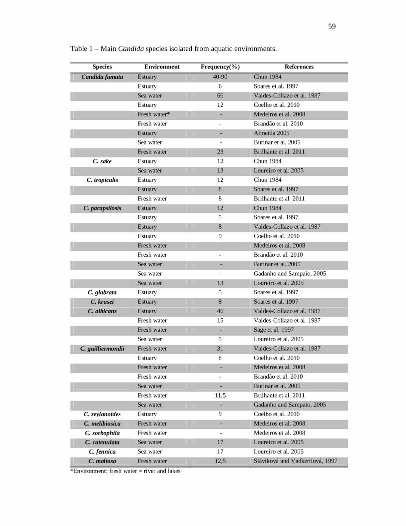
59
Table 1 – Main Candida species isolated from aquatic environments.
*Environment: fresh water = river and lakes
Species Environment Frequency(%) References Candida famata Estuary 40-90 Chun 1984
Estuary 6 Soares et al. 1997 Sea water 66 Valdes-Collazo et al. 1987 Estuary 12 Coelho et al. 2010 Fresh water* - Medeiros et al. 2008 Fresh water - Brandão et al. 2010 Estuary - Almeida 2005 Sea water - Butinar et al. 2005 Fresh water 23 Brilhante et al. 2011
C. sake Estuary 12 Chun 1984 Sea water 13 Loureiro et al. 2005
C. tropicalis Estuary 12 Chun 1984 Estuary 8 Soares et al. 1997 Fresh water 8 Brilhante et al. 2011
C. parapsilosis Estuary 12 Chun 1984 Estuary 5 Soares et al. 1997 Estuary 8 Valdes-Collazo et al. 1987 Estuary 9 Coelho et al. 2010 Fresh water - Medeiros et al. 2008 Fresh water - Brandão et al. 2010 Sea water - Butinar et al. 2005 Sea water - Gadanho and Sampaio, 2005
Sea water 13 Loureiro et al. 2005 C. glabrata Estuary 5 Soares et al. 1997 C. krusei Estuary 8 Soares et al. 1997
C. albicans Estuary 46 Valdes-Collazo et al. 1987 Fresh water 15 Valdes-Collazo et al. 1987 Fresh water - Sage et al. 1997
Sea water 5 Loureiro et al. 2005 C. guilliermondii Fresh water 31 Valdes-Collazo et al. 1987
Estuary 8 Coelho et al. 2010 Fresh water - Medeiros et al. 2008 Fresh water - Brandão et al. 2010 Sea water - Butinar et al. 2005 Fresh water 11,5 Brilhante et al. 2011 Sea water - Gadanho and Sampaio, 2005
C. zeylanoides Estuary 9 Coelho et al. 2010 C. melibiosica Fresh water - Medeiros et al. 2008 C. sorbophila Fresh water - Medeiros et al. 2008 C. catenulata Sea water 17 Loureiro et al. 2005
C. fennica Sea water 17 Loureiro et al. 2005 C. maltosa Fresh water 12,5 Sláviková and Vadkertiová, 1997
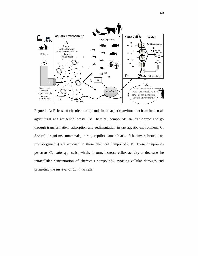
60
Figure 1: A: Release of chemical compounds in the aquatic environment from industrial,
agricultural and residential waste; B: Chemical compounds are transported and go
through transformation, adsorption and sedimentation in the aquatic environment; C:
Several organisms (mammals, birds, reptiles, amphibians, fish, invertebrates and
microorganisms) are exposed to these chemical compounds; D: These compounds
penetrate Candida spp. cells, which, in turn, increase efflux activity to decrease the
intracellular concentration of chemicals compounds, avoiding cellular damages and
promoting the survival of Candida cells.

61
7 CAPÍTULO 2 Macrobrachium amazonicum: an alternative for microbiological monitoring of aquatic
environments in Brazil
Macrobrachium amazonicum: uma alternativa para o monitoramento microbiológico
de ambientes aquáticos no Brasil
Periódico: Ciência Rural (Aceito para publicação em abril de 2014)

62
Ciência Rural – Artigo Científico
Macrobrachium amazonicum: an alternative for microbiological monitoring of aquatic
environments in Brazil
Macrobrachium amazonicum uma alternativa para o monitoramento microbiológico de
ambientes aquáticos no Brasil
Raimunda Sâmia Nogueira Brilhante1*, Manoel de Araújo Neto PaivaI,II, Célia Maria de
Souza SampaioII, Carlos Eduardo Cordeiro TeixeiraI, Joyce Fonteles RibeiroI, Débora de
Souza Collares Maia Castelo-BrancoI, Tereza de Jesus Pinheiro Gomes BandeiraI,III,
André Jalles MonteiroIV, Rossana de Aguiar CordeiroI, José Júlio Costa SidrimI,
Frederico Ozanan Barros MonteiroV, José Luciano Bezerra MoreiraI, Marcos Fábio
Gadelha RochaI,II
1 Departmento de Patologia e Medicina Legal, Faculdade de Medicina, Programa de Pós-Graduação em Microbiologia Médica, Centro Especializado em Micologia Médica, Universidade Federal do Ceará. Rua Coronel Nunes de Melo, s/n, Rodolfo Teófilo. CEP: 60.430-270, Fortaleza-CE, Brazil. Tel.: +55 85 3295 1736; fax: +55 85 3366 8303 *Autor para correspondência: e-mail: [email protected]. II Faculdade de Veterinária, Programa de Pós-Graduação em Ciências Veterinárias, Universidade Estadual do Ceará. Av. Paranjana, 1700. CEP: 60.740-903, Fortaleza-CE, Brazil. III Faculdade de Medicina, Centro Universitário Christus. Rua João Adolfo Gurgel, 133. CEP: 60.190-060, Fortaleza, Ceará, Brazil. IV Departmento de Estatística e Matemática Aplicada, Universidade Federal do Ceará. Campus do Pici, Bloco 910, CEP: 60455-760, Fortaleza-CE, Brazil. V Faculdade de Medicina Veterinária, Programa de Pós-Graduação em Saúde e Produção Animal da Amazônia, Universidade Federal Rural da Amazônia. Av. Presidente Tancredo Neves, 2501, CEP: 66.077-830, Belém, Pará, Brazil.

63
ABSTRACT
This study aimed to evaluate the role of the Amazon River prawn,
Macrobrachium amazonicum, as carrier of Candida spp., by analyzing the correlation
between Candida spp. from these prawns and their environment (surface water and
sediment), through M13-PCR fingerprinting and RAPD-PCR. For this purpose, 27 strains
of Candida spp. were evaluated. These strains were recovered from the gastrointestinal
tract of adult M. amazonicum (7/27) from Catú Lake, Ceará State, Brazil and from the
aquatic environment (surface water and sediment) of this lake (20/27). Molecular
comparison between the strains from prawns and the aquatic environment was conducted
by M13-PCR fingerprinting and RAPD-PCR, utilizing the primers M13 and OPQ16,
respectively. The molecular analysis revealed similarities between the band patterns of
eight Candida isolates with the primer M13 and 11 isolates with the primer OPQ16,
indicating that the same strains are present in the digestive tract of M. amazonicum and
in the aquatic environment where these prawns inhabit. Therefore, these prawns can be
used as sentinels for environmental monitoring through the recovery of Candida spp.
from the aquatic environment in their gastrointestinal tract.
Key words: Macrobrachium amazonicum prawn, environmental sentinel, Candida
spp., pollution, monitoring.

64
RESUMO
Este estudo teve como objetivo avaliar o papel do camarão Macrobrachium
amazonicum como carreador de Candida spp. do ambiente aquático, por meio da análise
da correlação entre Candida spp. isoladas desse camarão e do seu ambiente (água de
superfície e sedimento) através das técnicas de M13-PCR fingerprinting e RAPD-PCR.
Para tanto, 27 cepas de Candida spp. foram avaliadas. Essas cepas foram recuperadas a
partir do trato gastrointestinal de M. amazonicum adultos (7/27), oriundos da lagoa do
Catú, Ceará, Brasil, e do meio aquático (águas superficiais e sedimentos) desse lago
(20/27). A comparação molecular entre as cepas de camarões e o ambiente aquático foi
conduzida por M13-PCR fingerprinting e RAPD-PCR, utilizando os iniciadores M13 e
OPQ16, respectivamente. A análise molecular revelou semelhanças entre os padrões de
bandas de oito isolados de Candida com o iniciador M13 e 11 isolados com o primer
OPQ16, indicando que elas estão presentes no trato digestivo de M. amazonicum e no
ambiente aquático, onde esses camarões habitam. Portanto, essa espécie de camarão pode
ser usada como sentinela para monitoramento ambiental através da recuperação de
Candida spp. do ambiente aquático, a partir do seu trato gastrointestinal.
Palavras-chave: camarão Macrobrachium amazonicum, sentinela ambiental, Candida
spp, poluição, monitoramento.

65
INTRODUCTION
Several studies have identified Candida spp. as potential biological indicators of
environmental degradation (MEDEIROS et al., 2008; BRILHANTE et al., 2011, 2012;
CASTELO-BRANCO et al., 2013), particularly in samples obtained from aquatic sources
(BUTINAR et al., 2005; MEDEIROS et al., 2008). In these studies, the isolation of this
genus was greater than that of other microorganisms, including bacteria, demonstrating
the potential use of this yeast for environmental monitoring.
Monitoring aquatic environments requires an adequate water sampling technique,
including the selection of representative collection sites and considering environmental
factors, such as seasonality, temperature, the water column and the presence of affluent
or effluent waters (APHA/AWWA/WEF, 1998). These requirements may represent an
obstacle for the adequate monitoring of fresh water environments, because of the large
number of samples needed. Hence, it is important to seek alternatives to facilitate
monitoring of aquatic ecosystems. In this context, the use of aquatic crustaceans has been
reported as a reliable alternative for that purpose, especially because of their feeding
habits (filter feeding) and benthic behavior, as described by VIRGA et al. (2007) and
BRILHANTE et al. (2011).
More recently, BRILHANTE et al. (2011) performed a research with the
freshwater prawn M. amazonicum (Amazon River prawn) in captivity and from the
natural environment for the isolation of yeasts and Candida was the most isolated genus,
showing that it belongs to the gastrointestinal microbiota of these animals. In addition,
these authors suggested that these prawns may be an important environmental sentinels
if they harbor in their gastrointestinal tract Candida spp. from the aquatic environment.
Thus, in light of these findings and considering the wide distribution of the species M.

66
amazonicum in South America, the objective of the present study was to evaluate the role
of these prawns as carriers of Candida spp. from the aquatic environment.
MATERIALS AND METHODS
Microorganisms
In this study, 27 strains of Candida spp. were analyzed, out of which seven were
recovered from wild-harvested M. amazonicum, while 20 were recovered from the
aquatic environment and were deposited in our culture collection. It is important to
emphasize that the analyzed Candida strains, from animal and environmental sources,
were simultaneously recovered.
Of the 20 environmental strains, 13 were isolated from surface water (SW) and
seven from sediment (S). These strains belong to the culture collection of the Specialized
Medical Mycology Center of the Federal University of Ceará, where they are kept on 2%
Sabouraud dextrose agar. They were manipulated under level II biosafety procedures.
Candida spp. from the aquatic environment were obtained from Catú Lake, which
is located at the municipality of Aquiraz, Ceará state, Brazil, about 35 kilometers from
the state capital. It is delimited by the UTM coordinates 0567000E, 9561273N and
0575000E, 9569000N. It is a rich freshwater body with mangrove areas that have been
degraded by uncontrolled occupation of the surrounding area and pollution with residues
from industrial, commercial and farming activities (BRILHANTE et al., 2011).
Water samples were collected, according to MEDEIROS et al. (2008). Then, four
collection sites were included, as follows: recreational area and prawn collection site
(point 1, 3°55’59.79” S and 38°21’50.10” W), agricultural wastewater (point 2,
3°55’47.25” S and 38°22’14.16” W), industrial wastewater (point 3, 3°56’03.70” S and

67
38°22’25.15” W) and residential area (point 4, 3°56’56.72” S and 38°22’31.57” W). Two
water samples (SW and S) were monthly collected from each collection site, during one
year (from March 2011 to February 2012). Overall, a total of 96 water samples were
obtained for this study.
Adults of M. amazonicum were monthly harvested from Catú Lake (point 1) in
the same period as the water samples. Afterwards, the digestive tracts of 10 individuals
were removed, placed in sterile slants containing sterile saline and treated as one single
sample (BRILHANTE et al., 2011). Overall, 12 collections were performed. This study
was previously approved by the Chico Mendes Institute for Conservation of
Biodiversity/Biodiversity Authorization and Information System – SISBIO, under the
number 28175-1.
Microbiological processing
Samples were processed in a biosafety level II cabinet and were seeded on 2%
Sabouraud agar plus chloramphenicol (0.5g L-1), in Petri dishes for primary isolation.
Water samples were processed according to MEDEIROS et al. (2008) with some
modifications. Briefly, a 100-µL aliquot of the SW samples was spread on the medium,
after homogenization, while the S samples were processed, after centrifuging for 20
minutes at 3,000rpm. Then, the supernatant was removed and the substrate was suspended
again in 2mL of a sterile 0.9% solution of NaCl. Afterwards, the suspension was agitated
in a vortex mixer for 3 minutes and left to rest for 30 minutes at 25°C. Next, 100-µL
aliquots of the supernatant of each sample were seeded on the culture medium. The
digestive tracts of adult prawns were processed as described by BRILHANTE et al.
(2011) and seeded onto the culture medium. The inoculated Petri dishes containing the
culture media were incubated at 25oC, for 10 days, and were daily observed
(BRILHANTE et al., 2011).
68
The yeast colonies were identified through specific morphological and
biochemical tests, including growth on chromogenic medium (CHROMagar Candida,
BD, USA), micromorphology on cornmeal-Tween 80 agar, carbohydrate and nitrogen
assimilation and urease production (BRILHANTE et al., 2011), and the results were
interpreted according to (DE HOOG et al., 2000). Strains that presented dubious identity
were also identified through VITEK II automated system (BioMérieux, USA).
Additionally, the susceptibility of these microorganisms to amphotericin B, fluconazole
and itraconazole was evaluated through broth microdilution method. Minimum inhibitory
concentrations (MIC) of >1, ≥64, ≥1µg mL-1 were considered resistant to amphotericin
B, fluconazole and itraconazole, respectively (CLSI, 2008).
- M13-PCR fingerprinting and OPQ-16 RAPD
The DNA from the strains was extracted after 48 hours of growth on potato
dextrose agar, according to the methodology described by CASTELO-BRANCO et al.
(2013).
For molecular comparison between the Candida isolates from the aquatic
environment (SW and S) and from prawns, the PCR-fingerprinting technique was used,
according to the method described by CASTELO-BRANCO et al. (2013), using the single
primer M13 (59-GAGGGTGGCG GTTCT-39) and the PCR mix (25 µL), containing
10mM of Tris/HCI (pH 8.3), 50mM of KCl, 1.5mM of MgCl2, 0.2mM of dNTPs,
0.15mM of the primer, 2.5U of Taq polymerase (MBI Fermentas) and 25ng of yeast
DNA. The RAPD reactions were performed with the primer OPQ16 (5’
AAGAGCCCGT3’), according to the method described by CASTELO-BRANCO et al.
(2013). The RAPD reaction was carried out with a total volume of 10µL, containing 50ng
of genomic DNA, 1X buffer, 1mM of MgCl2, 2pmol of primer, 0.5mM each of
deoxynucleoside triphosphate and 1 U µL-1 of Hot Start Taq polymerase.

69
Dice similarity coefficient was measured and a dendrogram was obtained through
the use of the Unweighted Pair Group Method with Arithmetic Average (UPGMA),
through the software BioNumerics (version 6.6), resulting in the analysis of clusters and
measure of relatedness among isolates.
RESULTS
Data referring to the identity, the origin and the antifungal susceptibility profile of
the recovered Candida strains are listed in table 1. Five to eight DNA bands were
generated through the M13-PCR fingerprinting, while three to ten DNA bands were
generated through RAPD-PCR with the primer OPQ16. The molecular analysis
employing both techniques revealed strong similarities between the DNA band patterns
of the isolates belonging to the same Candida species. For the primer M13, eight isolates
of Candida spp. with 100% band similarity were obtained, while with the primer OPQ16,
11 isolates were obtained with 100% band similarity (Figure 1).
The M13-PCR fingerprinting identified 100% similarity between two C.
tropicalis strains from prawn (n=1) and sediment (point 3, n=1); four C. famata strains
from prawns (n=2) and surface water (points 1 and 3; n=2) and two C. ciferrii strains
from prawns (n=1) and sediment (point 1, n=1). In turn, the RAPD-PCR with the primer
OPQ16 allowed identifying 100% similarity between two C. guilliermondii strains from
surface water (points 1 and 4); five C. famata strains, two from prawn (n=1) and surface
water (point 1, n=1) and three from surface water (points 1 and 2); two strains of C.
parapsilosis from prawn (n=1) and surface water (point 4, n=1) and two C. ciferrii strains
from prawn (n=1) and sediment (point 1, n=1) (Figure 1).

70
DISCUSSION
This study demonstrated the similarity among Candida spp. isolated from wild-
harvested prawns and the aquatic environment where the animals inhabit, including
surface water and sediment. The molecular analysis through M13-PCR fingerprinting and
RAPD-PCR with OPQ-16 allowed evaluating this correlation, since these techniques
generated varied band patterns among different Candida species and similar ones within
the same species, thus presenting desirable and reliable results. In the present study, the
primer OPQ16 was used to complement the results obtained through the M13-PCR
fingerprinting and it generated a greater variety of DNA bands and identified a greater
number of strains with 100% of similarity.
The recovered Candida species were simultaneously isolated from prawns and the
aquatic environment and some of these isolates presented 100% of similarity, even when
recovered from different collection points. Thus, it was demonstrated that M.
amazonicum contains in its gastrointestinal tract a representative cross-section of
Candida spp. that colonize the water and the substrate where they live.
In addition, three sets of azole resistant strains were observed among the isolates
from prawns and aquatic environment that presented 100% of similarity. These findings
are in accordance with those of BRILHANTE et al. (2011), who observed that 28.6% of
the Candida spp. recovered from the intestinal tract of wild-harvested M. amazonicum
isolated from Catú Lake were resistant to azole antifungals. Considering that the main
mechanism of azole resistance among Candida spp. is the overexpression of efflux pumps
(FENG et al., 2010), which is possibly related to the exposure of these microorganisms
to chemical compounds, as an unspecific mechanism of cellular detoxification
(JUNGWIRTH & KUCHLER, 2006), we strongly believe that Candida spp. could be
71
used as indicators of environmental pollution, through the phenotypical assessment of
their in vitro susceptibility profile.
Crustaceans accumulate pollutants in their tissues, such as hydrocarbons,
pesticides and heavy metals (YILMAZ & YILMAZ, 2007), which might increase the
azole resistance rate among yeasts from the microbiota, due to the overexpression of
efflux pumps (KEENAN et al., 2007; MÜLLER et al., 2007), as a consequence of the
chronic exposure to these chemical compounds. In this context, the use of this prawn as
a sentinel for the isolation of Candida spp. seems potentially advantageous.
CONCLUSION
In conclusion, based on the obtained results, the use of M. amazonicum as a
sentinel for the isolation of Candida spp. from aquatic environments is an interesting
alternative for evaluating the environmental quality, considering that these animals harbor
yeasts from the environment in their gastrointestinal tract. Additionally, due to their
capacity to accumulate chemical pollutants in their tissues, they simulate the
environmental conditions to which these yeasts are exposed, potentially contributing for
monitoring the presence of resistant Candida spp. in the environment.
ACKNOWLEDGEMENTS
This research was supported by grants from the National Council for Scientific
and Technological Development (CNPq; Brazil; Processes 302574/2009-3,
481614/2011-7, 504189/2012-3) and the Coordination Office for the Improvement of

72
Higher Education Personnel (CAPES/PNPD 2103/2009, AE1-0052-000650100/11,
Casadinho/PROCAD 552215/2011-2).
REFERENCES
AMERICAN PUBLIC HEALTH ASSOCIATION – APHA; AMERICAN WATER
WORKS ASSOCIATION – AWWA; WATER ENVIRONMENTAL FEDERATION –
WEF. Standard methods for the examination of water and wastewater. 20. ed.
Washington: APHA/ AWWA/ WEF, 1998.
BRILHANTE, R.S.N. et al. Yeast microbiota of raptors: a possible tool for environmental
monitoring. Environmental Microbiology Reports, v. 4, n. 2, p. 189-193, apr. 2012.
Disponível em: http://onlinelibrary.wiley.com/doi/10.1111/j.1758-
2229.2011.00319.x/abstract. Acesso em: 08 nov. 2013. doi: 10.1111/j.1758-
2229.2011.00319.x.
BRILHANTE, R.S.N. et al. Yeasts from Macrobrachium amazonicum: a focus on
antifungal susceptibility and virulence factors of Candida spp. FEMS Microbiology
Ecology, v. 76, n. 2, p. 268–277, may 2011. Acesso em:
http://onlinelibrary.wiley.com/doi/10.1111/j.1574-6941.2011.01050.x/abstract. Acesso
em: 08 nov. 2013. doi: 10.1111/j.1574-6941.2011.01050.x.
BUTINAR, L. et al. Yeast diversity in hypersaline habitats. FEMS Microbiology
Letters, v. 15, n. 2, p. 229-234, feb. 2005. Disponível em:
http://onlinelibrary.wiley.com/doi/10.1016/j.femsle.2005.01.043/pdf. Acesso Em: 08
nov. 2013. doi:10.1016/j.femsle.2005.01.043.
CASTELO-BRANCO, D.S.C.M. et al. Azole-resistant Candida albicans from a wild
Brazilian porcupine (Coendou prehensilis): a sign of an environmental imbalance?

73
Medical Mycology, v. 51, n. 5, p. 555-560, jul. 2013. Disponível em:
http://informahealthcare.com/doi/abs/10.3109/13693786.2012.752878. Acesso em: 08
nov. 2013. doi: 10.3109/13693786.2012.752878.
DE HOOG, G.S. et al. Atlas of Clinical Fungi. The Nederlands: Centraalbureau voor
Schimmelcultures, 2ª ed. Baarn. 2000.
FENG, L., et al. (2010) Relationship between antifungal resistance of fluconazole
resistant Candida albicans and mutations in ERG11 gene. Chinese Medical Journal, v.
123, n. 5, p. 544–548, 2010. Disponível em:
http://www.cmj.org/ch/reader/view_abstract_ext.aspx?file_no=20103842232480&flag=
1. Acesso em: 20 dez. 2013. doi: 10.3760/cma.j.issn.0366-6999.2010.05.007.
JUNGWIRTH, H.; KUCHLER, K. Yeast ABC transporters – a tale of sex, stress, drugs
and aging. FEBS Letters, v. 580, n. 4, p. 1131–1138, fev. 2006. Disponível em:
http://www.sciencedirect.com/science/article/pii/S0014579305015334. Acesso em: 20
dez. 2013. doi:10.1016/j.febslet.2005.12.050.
KEENAN, P.O. et al. Clear and present danger? The use of a yeast biosensor to monitor
changes in the toxicity of industrial effluents subjected to oxidative colour removal
treatments. Journal of Environmental Monitoring, v. 9, n. 12, p. 1394-1401, sep. 2007.
Disponível em: http://pubs.rsc.org/en/Content/ArticleLanding/2007/
EM/b710406e#!divAbstract. Acesso em: 08 nov. 2013. doi: 10.1039/B710406E.
MEDEIROS, A.O. et al. Diversity and antifungal susceptibility of yeasts from tropical
freshwater environments in Southeastern Brazil. Water Research, v. 42, n. 14, p. 3921-
3929, aug. 2008. Disponível em: http://www.sciencedirect.com/science/article/
pii/S0043135408002522. Acesso em: 08 nov. 2013. doi: 10.1016/j.watres.2008.05.026.
MÜLLER, F.M.C. et al. Cross-resistance to medical and agricultural azole drugs in yeasts
from the oropharynx of human immunodeficiency virus patients and from environmental
74
bavarian vine grapes. Antimicrobial Agents and Chemotherapy, v. 51, n. 8, p. 3014-
3016, aug. 2007. Disponível em: http://aac.asm.org/content/51/8/3014.long. Acesso em:
08 nov. 2013. doi:10.1128/AAC.00459-07
CLINICAL AND LABORATORY STANDARDS INSTITUTE, 2008. Reference
Method for Broth Dilution Antifungal Susceptibility Testing of Yeast: Approved
Guideline M27-A3, vol. 28. CLSI, Wayne, PA, USA, pp. 1–25.
VIRGA, R.H.P. et al. Assessment of heavy metal contamination in blue crab specimes.
Food Science and Technology, v. 27, n. 4, p. 779-785, oct. 2007. Disponível em:
http://www.scielo.br/scielo.php?pid=S0101-20612007000400017&script=sci_arttext
Acesso em 08 nov. 2013. doi: 10.1590/S0101-20612007000400017.
YILMAZ, A.B.; YILMAZ, L. Influences of sex and seasons on levels of heavy metals in
tissues of green tiger shrimp (Penaeus semisulcatus de Hann, 1844). Food Chemistry,
v. 101, n. 4, p. 1664-1669, 2007. Disponível em:
http://www.sciencedirect.com/science/article/pii/S0308814606003438 Acesso em: 08
nov. 2013. doi: 10.1016/j.foodchem.2006.04.025.

75
Figure 1 - Dendrograms resulting from the analysis of 27 isolates of Candida spp.
obtained from the gastrointestinal tract of Macrobrachium amazonicum (n=7) and the
natural environment (n=20) through M-13-fingerprinting and RAPD-PCR with primer
OPQ16. P: prawn; SW: surface water; S: sediment. *indicates antifungal resistance.
Dendrograms generated by the BioNumerics program (Applied Math, Inc.).

76
Table 1 - Species, access number, origin, isolation period and antifungal susceptibility
of 27 Candida spp. isolates used for molecular analysis.
Species Access number Origin Collection point
Period Resistance*
C. famata CEMM 1-1-259 Sediment Point 3 October 2011 FLC/ITC
C. guilliermondii CEMM 1-1-260 Surface Water Point 4 November 2011 ITC
C. guilliermondii CEMM 1-1-261 Sediment Point 4 November 2011 S
C. famata CEMM 1-1-262 Sediment Point 3 November 2011 S
C. guilliermondii CEMM 1-1-263 Prawn Point 1 December 2011 S
C. famata CEMM 1-1-264 Prawn Point 1 November 2011 S
C. famata CEMM 1-1-265 Prawn Point 1 November 2011 ITC
C. parapsilosis CEMM 1-1-266 Surface Water Point 4 November 2011 S
C. famata CEMM 1-1-267 Surface Water Point 1 August 2011 ITC
C. parapsilosis CEMM 1-1-268 Prawn Point 1 March 2011 S
C. ciferrii CEMM 1-1-269 Surface Water Point 3 October 2011 S
C. ciferrii CEMM 1-1-270 Surface Water Point 1 August 2011 ITC
C. ciferrii CEMM 1-1-271 Sediment Point 4 November 2011 S
C. ciferrii CEMM 1-1-272 Sediment Point 1 October 2011 FLC/ITC
C. ciferrii CEMM 1-1-273 Prawn Point 1 October 2011 FLC/ITC
C. famata CEMM 1-1-274 Surface Water Point 3 November 2011 S
C. famata CEMM 1-1-275 Surface Water Point 3 May 2011 S
C. famata CEMM 1-1-276 Surface Water Point 1 August 2011 FLC/ITC
C. famata CEMM 1-1-277 Prawn Point 1 December 2011 S
C. famata CEMM 1-1-278 Surface Water Point 4 October 2011 FLC
C. parapsilosis CEMM 1-1-279 Surface Water Point 1 May 2011 S
C. tropicalis CEMM 1-1-280 Prawn Point 1 October 2011 FLC/ITC
C. tropicalis CEMM 1-1-281 Sediment Point 3 October 2011 FLC/ITC
C. guilliermondii CEMM 1-1-282 Surface Water Point 4 December 2011 S
C. guilliermondii CEMM 1-1-283 Sediment Point 3 December 2011 S
C. guilliermondii CEMM 1-1-284 Surface Water Point 4 August 2011 ITC
C. guilliermondii CEMM 1-1-285 Surface Water Point 1 December 2011 FLC/ITC
*All strains were susceptible to amphotericin B, S: susceptible to all tested antifungal drugs (amphotericin
B, fluconazole and itraconazole); FLC: resistance to fluconazole; ITC: Resistance to itraconazole.

77
8 CAPÍTULO 3
Azole resistance in Candida spp. from water: an efflux-pump mediated mechanism
Resistência à azólicos em Candida spp. oriundas de água: um mecanismo mediado
por bomba de efluxo
Periódico: Brazilian Journal of Microbiology (Submetido em julho de 2014)

78
Brazilian Journal of Microbiology – Rsearch Paper
Azole resistance in Candida spp. from water: an efflux-pump mediated mechanism
Raimunda S.N. Brilhantea*, Manoel, A.N. Paivaa,b, Célia M. S. Sampaiob, Débora S. C.
M. Castelo-Brancoa, Carlos E. C. Teixeiraa, Lucas P. de Alencara,b, Tereza J. P. G.
Bandeiraa,c, André J. Monteirod, Rossana A. Cordeiroa, José J.C. Sidrima, José Luciano
Bezerra Moreiraa, Marcos F.G. Rochaa,b
aDepartment of Pathology and Legal Medicine, Faculty of Medicine, Postgraduate
Program in Medical Microbiology, Specialized Medical Mycology Center, Federal
University of Ceará, Fortaleza, Ceará, Brazil.
bSchool of Veterinary Medicine, Postgraduate Program in Veterinary Sciences, State
University of Ceará, Fortaleza, Ceará, Brazil.
cSchool of Medicine, Christus College - UNICHRISTUS, Fortaleza, Ceará, Brazil.
dDepartment of Statistics and Applied Mathematics, Federal University of Ceará,
Fortaleza, CE, Brazil.
Running title: Candida spp. for monitoring aquatic ecosystems
*Corresponding Author. R. S. N. Brilhante. Rua Barão de Canindé, 210; Montese. CEP:
60.425-540. Fortaleza, CE, Brazil. Fax: 55 85 3295-1736 E-mail: [email protected]

79
Abstract
Even though azole resistance has been reported among yeasts recovered from
aquatic environments, no studies have reported the mechanisms underlying this
antifungal resistance. Thus, the present study aimed at investigating the occurrence of
antifungal resistance among yeasts recovered from an aquatic environment, and assessing
the efflux pump activity of the azole resistant strains, to better understand the mechanisms
of resistance to this group of drugs. For this purpose, water and sediment samples were
collected from Catú Lake, Ceará, Brazil, from March 2011 to February 2012. The
obtained yeasts were identified through morphological and biochemical analyses.
Overall, 46 isolates were recovered, of which 37 were Candida spp., four Trichosporon
asahii, three Cryptococcus laurentii, one Rhodotorula mucilaginosa and one Kodamaea
ohmeri. These isolates were submitted to broth microdilution assay with amphotericin B,
itraconazole and fluconazole, according to CLSI. The MICs for amphotericin B,
itraconazole and fluconazole were 0.03125 to 2 µg/mL, 0.0625 to ≥16 µg/mL and 0.5 to
≥64 µg/mL, respectively, with 13 azole resistant strains. When efflux pump inhibitors
were added, a reduction in the azole MICs was observed, leading to the phenotypical
reversion of the azole resistance. This finding suggests that the azole resistance among
environmental Candida spp. is most likely associated with the overexpression of these
pumps.
Keywords: Aquatic environments, yeast microbiota, Candida spp., antifungal resistance
80
1. Introduction
The quali-quantitative analysis of the yeast microbiota has been indicated as a
promising tool to assess the eutrophication status of aquatic systems (Arvanitidou et al.,
2002; Coelho et al., 2010; Hagler et al., 1995; Libkind et al., 2003; Nagahama, 2006;
Medeiros et al., 2008). Medeiros et al. (2003), for example, studied the diversity of yeasts
in lakes and rivers in Southeastern Brazil and the genus Candida accounted for the largest
number of isolates, out of which 50% were resistant to itraconazole and 11% were
resistant to fluconazole. More recently, our group conducted a study using the freshwater
prawn Macrobrachium amazonicum (Amazon River prawn) collected in the natural
environment and observed that 33.3% of the Candida spp. isolates from this prawn were
resistant to fluconazole and itraconazole (Brilhante et al., 2011).
However, none of the researches mentioned above investigated the mechanisms
underlying the azole resistance observed among Candida strains from aquatic
environments. It is well known that one of the main mechanisms of azole resistance
among Candida spp. is the increased activity of efflux pumps, which are codified by the
genes CDR1 and CDR2, belonging to the superfamily of the ATP binding cassette, and
by the gene MDR1, belonging to the major facilitator class. The superexpression of these
genes and the subsequent increase in the activity of these pumps prevents the
accumulation of the drug inside the cell, at the site of action, impairing its efficacy
(Jungwirth and Kuchler, 2006). The upregulation of CDR1 and CDR2 confers resistance
to nearly all azoles, while that of MDR1 provides specific resistance to fluconazole
(Kanafani and Perfect, 2008). Thus, the present study was undertaken to investigate the
occurrence of antifungal resistance among yeasts recovered from an aquatic environment,
and assessing the efflux pump activity in the azole resistant strains.

81
2. Methods
2.1 Study site and Collections of the biological material
Catú Lake, located in the municipality of Aquiraz, Ceará State, Brazil (UTM
coordinates 0567000E, 9561273N and 0575000E, 9569000N), is a rich freshwater body
with mangrove areas that shelter a large number of animal species. However, due to the
uncontrolled occupation of the surrounding area, the waters of this lake are mainly used
for human and animal consumption, for industrial, commercial and farming activities and
for leisure activities, such as boat excursions (Gomes et al., 2008).
Water samples were obtained monthly, from March 2011 to February 2012,
completing a total of 12 collections, according to the method described by Medeiros et
al. (2008), with some modifications. The water samples were obtained with a 1-liter Van
Dorn bottle, which was rinsed three times with water from each collection site, before
performing the water collection from four sites: recreational area point (point 1,
3°55’59.79” S and 38°21’50.10” W), agricultural wastewater point (point 2, 3°55’47.25”
S and 38°22’14.16” W), industrial wastewater point (point 3, 3°56’03.70” S and
38°22’25.15” W) and Catú River confluence point (point 4, 3°56’56.72” S and
38°22’31.57” W) (Fig. 1). Two samples were collected at each point, one from the surface
(SW sample) and the other from the bottom, including sediment (S sample). The study
was approved by the Chico Mendes Institute for Conservation of
Biodiversity/Biodiversity Authorization and Information System – SISBIO, under the
process number 28175-1.

82
2.2 Mycological processing
The samples were taken to the laboratory and processed in a biological safety level
2 laminar flow cabinet. For each sample, Sabouraud agar supplemented with
chloramphenicol (0.5 g/L) was used as the culture medium for primary isolation, in Petri
dishes. For the SW samples, after homogenization, a 100-µL aliquot was spread on the
medium. The S samples were processed, as follows: the samples were centrifuged for 20
minutes at 3,000 rpm, the supernatant was removed and the sediment was resuspended in
2 mL of sterile 0.9% NaCl solution. Then, the suspension was agitated in a vortex mixer
for 3 minutes and left to rest for 30 minutes at 25 °C. Afterwards, 100-µL aliquots of the
supernatant of each sample were spread on the culture medium. The inoculated Petri
dishes were incubated at 25 ºC for 10 days, with daily observations to note the
microbiological growth. The colony forming units (CFUs) were also counted in all the
inoculated dishes.
2.3 Yeast Identification
Initially, the colonies that appeared to be yeasts were Gram stained and observed
under a light microscope (40X) to check for the presence of blastoconidia, hyphae or
pseudohyphae and to exclude bacterial contamination. Then, the yeast colonies were
identified through specific macromorphological and micromorphological characteristics,
including growth on chromogenic medium for the identification of mixed colonies, and
biochemical tests, such as carbohydrate and nitrogen assimilation and urease production.
VITEK 2™ microbial identification system (bioMérieux, USA) was used in case of
dubious identification, to aid the identification procedure (Brilhante et al., 2011).

83
2.4 In vitro antifungal susceptibility testing
The antifungal minimum inhibitory concentrations (MICs) against these
microorganisms were determined through broth microdilution method, as described by
the Clinical and Laboratory Standards Institute (CLSI, 2008). Three drugs were tested
against the isolates: amphotericin B (Sigma Chemical Corp.), itraconazole (Janssen
Pharmaceutica, Belgium) and fluconazole (Pfizer, Brazil). Inocula of all tested isolates
were prepared from 1-day old cultures grown on potato dextrose agar at 35 °C with RPMI
1640 medium, with L-glutamine (HiMedia Laboratories), buffered to pH 7 with 0.165M
morpholinepropanesulfonic acid and were adjusted to a final concentration of 0.5–
2.5x10³ cells mL-1 (Brilhante et al., 2011; CLSI, 2008). The microdilution plates were
incubated at 35 °C, for 48 hours, and were visually read (CLSI, 2008). For each isolate,
drug-free and yeast-free controls were included and all the isolates were tested in
duplicate. As quality control, for each test performed, C. parapsilosis ATCC 22019 was
included. The MIC of azole derivatives was defined as the lowest drug concentration
capable of inhibiting 50% of growth, when compared with the growth control well. For
amphotericin B the MIC was the lowest concentration at which no growth was observed.
Isolates with MICs >1, ≥1 and ≥64 µg/mL were considered resistant to amphotericin B,
itraconazole and fluconazole, respectively (CLSI, 2008).
2.5 Analysis of the efflux pump activity in the azole resistant Candida isolates
A phenotypical assay of modulation of efflux pumps was carried out, based on the
method used by Castelo-Branco et al., (2013). Initially, the azole resistant Candida strains
(13/37) were tested against two efflux pump inhibitors, promethazine (Kolaczkowski et

84
al., 2003) and haloperidol (Iwaki et al., 2006) by microdilution method, obtaining average
MIC values of 98 µg/mL and 80 µg/mL, respectively. Subsequently, susceptibility test
was performed with itraconazole and fluconazole, according to the methodology
described above, but, sub-inhibitory concentrations of promethazine (12 µg/mL) and
haloperidol (10 µg/mL) were added to the final fungal inocula. Fluconazole was used in
combination with promethazine and haloperidol, while itraconazole was tested only with
promethazine.
2.6 Statistical analysis
Pearson's chi-square test was used to analyze the distribution of the different yeast
species among the collection sites. The exact proportion test, considering a 50%
hypothesis, was used to verify differences in yeast recovery during the rainy and the dry
season. The antifungal MICs obtained for the different yeast species were compared
through ANOVA and post hoc Dunnet's test. The Spearman correlation coefficient was
used to measure the correlation between the MICs of each tested drug. The Mann-
Whitney's non-parametric test was used to compare the antifungal MICs of the strains
obtained from different collection sites. P-values lower than 0.05 indicate statistically
significant conclusions.
3. Results and Discussion
A total of 46 isolates were obtained, belonging to eight genera and nine species.
Of this total, 30 (65.2%) were from surface water samples, 2 Candida parapsilosis sensu
lato, 2 Trichomonascus ciferrii (Candida ciferii), 7 Meyerozyma guilliermondii (Candida

85
guilliermondii), 13 Debaryomyces hansenii (Candida famata), 2 Cryptococcus laurentii,
1 Kodamaea ohmeri, 1 Rhodotorula mucilaginosa and 2 T. asahii; and 16 (34.8%) were
from sediment samples, 1 C. tropicalis, 1 C. parapsilosis sensu lato, 3 T. ciferrii, 3 M.
guilliermondii, 5 D. hansenii, 1 C. laurentii and 2 T. asahii (Table 1). When considering
the collection sites, point 4 was the one with the highest amount of isolates (n=18,
P<0.01), followed by point 2 (n=11), point 1 (n=10) and point 3 (n=7). There were no
predominant species among the collection sites. With respect to seasonal variation, 84.8%
of the yeasts (39/46) were isolated in the dry season (July to December, P <0.01) and
15.2% (7/46) were isolated in the rainy season, including the three strains of the
Cryptococcus genus.
In this study, the genus Candida showed the highest number of species, similar to
what was observed by Medeiros et al. (2008) and Brandão et al., (2010), with the presence
of opportunistic pathogens such as C. tropicalis, M. guilliermondii, D. hansenii, T. ciferrii
and C. parapsilosis sensu lato. D. hansenii was the most isolated species from both the
surface water and sediment (27.7% and 10.6%, respectively), followed by M.
guilliermondii (14.9% and 6.4%, respectively), together accounting for 59.6% of the
isolates found in this study. These two species are isolated from aquatic environments,
especially those eutrophized or contaminated with domestic sewage and industrial
wastewater (Boguslawska and Dabrowski, 2001; Brilhante et al., 2011; Dynowska, 1997;
Medeiros et al., 2008). D. hansenii, in particular, besides indicating eutrophication,
appears to be closely associated with the removal of these pollutants from contaminated
water (Boguslawska and Dabrowski, 2001). In addition, the larger number of isolates
obtained during the dry period may be related to the decrease of water volume and
consequent concentration of nutrients, which may have favored the growth of yeasts. The
86
nutritional concentration may also explain the larger number of strains found in point 4,
which comprises an area where there is the discharge of domestic sewage (Deak, 2006).
The MIC values obtained from the in vitro susceptibility tests of all the isolates
are shown in Table II. In relation to Candida (Debaryomyces, Meyerozyma,
Trichomonascus) species, the MICs for amphotericin B varied from 0.03125 to 2 µg/mL,
with two resistant M. guilliermondii isolates (MIC = 2 µg/mL), which were also resistant
to itraconazole. The MICs for fluconazole ranged from 0.5 to 500 µg/mL and those for
itraconazole varied from 0.0625 to 32 µg/mL. A positive correlation was observed
between amphotericin B and itraconazole (P<0.01) and fluconazole and itraconazole (P
<0.01). Of the 37 Candida spp. isolates, seven (18.9%) were resistant to both azole
derivatives, five (13.5%) were resistant to itraconazole and one (2.7%) was resistant to
fluconazole. Among the azole resistant isolates, three were recovered from point 1(D.
hansenii, M. guilliermondii and T. ciferrii), two from point 2 (D. hansenii and M.
guilliermondii), four from point 3 (2 D. hansenii, C. tropicalis and T. ciferrii) and four
from point 4 (3 M. guilliermondii and T. ciferrii). No statistically significant differences
were observed in the antifungal MICs against strains from different collection sites. The
MICs for amphotericin B, fluconazole and itraconazole against the other yeast genera
varied from 0.0625 to 0.5 µg/mL, 0.25 to 4 µg/mL and 0.03125 to 0.5, respectively (Table
II). The antifungal MICs of the T. asahii isolates were statistically lower than those of the
other yeast genus (P<0.05).
Regarding antifungal resistance to amphotericin B, Savini et al., (2010) reported
that M. guilliermondii is less susceptible to this drug, when compared to other Candida
species. The susceptibility of Candida isolates to the azole derivatives found in this work
corroborates that observed in our previous study using yeasts isolated from wild-
harvested freshwater prawns (Macrobrachium amazonicum), where a high number of
87
azole resistant isolates were recovered (Brilhante et al., 2011). In the present study, of the
37 Candida spp. isolates, 13 (35.14 %) were resistant to azoles, of which four were D.
hansenii and five were M. guilliermondii. Unlike clinical isolates of D. hansenii and M.
guilliermondii, antifungal resistance has often been described in environmental isolates
of these Candida species (Brilhante et al., 2011, Medeiros et al., 2008).
Several studies relating the recovery of antifungal resistant environmental yeasts
to the degradation of aquatic systems have been published (Brilhante et al., 2011;
Medeiros et al., 2008; Brandão et al., 2010). Knowing the mechanisms of antifungal
resistance seems like an essential step to better understand the relationship between the
development of antifungal resistance and environmental pollution. In order to investigate
the mechanisms involved in the azole resistance, the efflux pump inhibition assay was
performed with promethazine and haloperidol, which resulted in the reversal of resistance
to itraconazole and fluconazole in all tested Candida isolates. The addition of
promethazine led to an MIC reduction of 8-256-fold and of 8-62.5-fold, for fluconazole
and itraconazole, respectively, while the addition of haloperidol led to a 32-125-fold
reduction of the MICs for fluconazole (Table 2).
Promethazine is a phenothiazine derivative that acts on MDR and CDR efflux
pumps (Kolaczkowski et al., 2003), while haloperidol acts only on MDR pumps (Iwaki
et al., 2006). Thus, the inhibition of the efflux pump activity by promethazine and
haloperidol, resulting in the reversal of the azole resistance, suggests that the azole
resistance among the Candida spp. strains is related to the enhanced activity of these
pumps. This increased activity is a direct result of the upregulation of the CDR and MDR
genes, possibly, as a consequence of the presence of different chemical compounds in
Catú Lake, secondary to human activities. It is believed that this resistance phenomenon
is related to the discharge of industrial wastewater and other pollutants into the aquatic

88
environment, which may lead to alterations in gene expression or gene sequence in
microorganisms (Keenan et al., 2007; Müller et al., 2007; Wegrzyn and Czyz, 2003), and
these are the main genetic alterations associated with the development of antifungal
resistance (Feng et al., 2010).
The present study reports a high rate of azole resistant Candida spp.
(Debaryomyces, Meyerozyma, Trichomonascus) strains recovered from an aquatic
environment, which may represent a risk for environmental and human health. This article
is the first to report the involvement of efflux pumps in the azole resistance among
Candida spp. from environmental sources. Finally, D. hansenii and M. guilliermondii
were the most commonly isolated species in this study and presented the highest rate of
azole resistance. Considering that these species are associated with environmental
degradation, these features encourage their use for monitoring the environmental health
of water bodies.
Acknowledgements
This work was supported by grants from the National Council for Scientific and
Technological Development (CNPq; Brazil; Processes 302574/2009-3, 562296/2010-7,
481614/2011-7, 504189/2012-3) and the Coordination Office for the Improvement of
Higher Education Personnel (CAPES/PNPD 2103/2009, AE1-0052-000650100/11,
Casadinho/PROCAD 552215/2011-2).

89
References
Arvanitidou M, Kanellou K, Katsouyannopoulos V, Tsakris UM. Occurrence and
densities of fungi from northern Greek coastal bathing waters and their relation with fecal
pollution indicators. Water Res 2002; 36: 5127-5131. doi:10.1016/S0043-
1354(02)00235-X.
Boguslawska-Was E, Dabrowski W. The seasonal variability of yeasts and yeast-like
organisms in water and bottom sediment of the Szczecin Lagoon. Int J Hyg Environ
Health 2001; 203: 451–458. doi: 10.1078/1438-4639-00056.
Brandão LR, Duarte MC, Barbosa AC, Rosa CA. Diversity and antifungal susceptibility
of yeasts isolated by multiple-tube fermentation from three freshwater lakes in Brazil. J
Water Health 2010; 8: 279-289. doi:10.2166/wh.2009.170.
Brilhante RSN, Paiva MAN, Sampaio CMS, Teixeira CEC, Castelo-Branco DSCM, Leite
JJL, Moreira CA, Silva LP, Cordeiro RA, Monteiro AJ, Sidrim JJ, Rocha MF. Yeasts
from Macrobrachium amazonicum: a focus on antifungal susceptibility and virulence
factors of Candida spp. FEMS Microbiol Ecol 2011; 76: 268–277. doi: 10.1111/j.1574-
6941.2011.01050.x.
Castelo-Branco DSCM, Brilhante RSN, Paiva MAN, Teixeira CEC, Caetano EP, Ribeiro
JF, Cordeiro RA, Sidrim JJ, Monteiro AJ, Rocha MF. Azole-resistant Candida albicans
from a wild Brazilian porcupine (Coendou prehensilis): a sign of an environmental
imbalance? Med Mycol 2013; 51: 555-560. doi: 10.3109/13693786.2012.752878.
Clinical and Laboratory Standards Institute. Reference Method for Broth Dilution
Antifungal Susceptibility Testing of Yeasts. Approved standard M27-A3. Wayne,
PA: Clinical and Laboratory Standards Institute, 2008.

90
Coelho MA, Almeida JMF, Martins IM, Silva AJ, Sampaio JP. The dynamics of the yeast
community of the Tagus river estuary: testing the hypothesis of the multiple origins of
estuarine yeasts. Antonie van Leeuwenhoek 2010; 98: 331-342. doi: 10.1007/s10482-
010-9445-1.
Deak T. Environmental factors influencing yeasts. In: Rosa C, Péter G, editors.
Biodiversity and Ecophysiology of Yeasts. Berlin, Springer; 2006, pp. 156-174.
Dynowska M. Yeast-like fungi possesing bio-indicator properties isolated from the Lyna
river. Acta Mycol 1997; 32: 279–286.
Feng L, Wan Z, Wang X, Li R, Liu W. Relationship between antifungal resistance of
fluconazole resistant Candida albicans and mutations in ERG11 gene. Chin Med J 2010;
123: 544-548. doi: 10.3760/cma.j.issn.0366-6999.2010.05.007.
Gomes ML, Pereira ECG, Morais JO. Degradação Socioambiental no Baixo Curso do
Rio Catú, Aquiraz-Ceará: Comprometimento da mata ciliar e recursos hídricos. In: IV
Encontro Nacional da Anppas. Brasília, Distrito Federal. 2008.
Hagler AN, Mendonça-Hagler LC, Rosa CA, Morais PP. Yeast as an example of
microbial diversity in brazilian ecosystems. Oecol Bras 1995; 1: 225-244.
Doi:10.4257/oeco.1995.0101.14.
Iwaki T, Giga-Hama Y, Takegawa K. A survey of all 11 ABC transporters in fission
yeast: two novel ABC transporters are required for red pigment accumulation in a
Schizosaccharomyces pombe adenine biosynthetic mutant. Microbiology 2006; 152:
2309–2321. doi: 10.1099/mic.0.28952-0.
Jungwirth H, Kuchler K. Yeast ABC transporters-- a tale of sex, stress, drugs and aging.
FEBS lett 2006; 580: 1131-1138. Doi: 10.1016/j.febslet.2005.12.050.
91
Kanafani ZA, Perfect JR. Antimicrobial resistence: resistance to antifungal agents:
mechanisms and clinical impact. Clin Infec Dis 2008; 46: 120-128. doi:
10.1086/524071.
Keenan PO, Knight AW, Billinton N, Cahill PA, Dalrymple IM, Hawkyard CJ. Clear and
present danger? The use of a yeast biosensor to monitor changes in the toxicity of
industrial effluents subjected to oxidative colour removal treatments. J Environ Monit
2007; 9: 1394-1401. doi: 10.1039/B710406E.
Kolaczkowski M, Michalak K, Motohashi N. Phenothiazines as potent modulators of
yeast multidrug resistance. Int J Antimicro 2003; 22: 279 – 283. doi:10.1016/S0924-
8579(03)00214-0.
Libkind D, Brizzio S, Ruffini A, Gadanho M, Broock MV, Sampaio PJ. Molecular
characterization of carotenogenic yeasts from aquatic environments in Patagonia,
Argentina. Antonie van Leeuwenhoek 2003; 84: 313-322.
doi:10.1023/A:1026058116545,
Medeiros AO, Kohler LM, Hamdan JS, Missagia BS, Barbosa FAR, Rosa CA. Diversity
and antifungal susceptibility of yeasts from tropical freshwater environments in
Southeastern Brazil. Water Res 2008; 42: 3921-3929. doi: 10.1016/j.watres.2008.05.026.
Müller FMC, Staudigel A, Salvenmoser S, Tredup A, Miltenberger R, Hermann JV.
Cross-resistance to medical and agricultural azole drugs in yeasts from the oropharynx of
human immunodeficiency virus patients and from environmental bavarian vine grapes.
Antimicrob Agents Chemother 2007; 51: 3014-3016. doi: 10.1128/AAC.00459-07.
Nagahama T. Yeast Biodiversity in Freshwater. Marine and Deep-Sea Environments. In:
Rosa C, Péter G, Editors. Biodiversity and ecophysiology of yeasts. Berlin, Springer;
2006, pp. 241-262.

92
Savini V, Catavitello C, Onofrillo D, Masciarelli G, Astolfi D, Balbinot A, Febbo
F, D'Amario C, D'Antonio D. What do we know about Candida guilliermondii? A voyage
throughout past and current literature about this emerging yeast. Mycoses 2010; 54: 434–
441. doi: 10.1111/j.1439-0507.2010.01960.x.
Wegrzyn G, Czyz A. Detection of mutagenic pollution of natural environment using
microbiological assays. J Appl Microbiol 2003; 95: 1175-1181. doi: 10.1111/j.1439-
0507.2010.01960.x.

93
Fig. 1 – Water collection points in Catú Lake, Aquiraz, Ceará, Brazil.
Point 1: Leisure area: bars, restaurants, boats. Area used for activities such
as boating and jet skiing. 3°55’59.79” S and 38°21’50.10” W; Point 2:
Agricultural area, with potato and bean fields, with possible use of azoles.
3°55’47.25” S and 38°22’14.16” W; Point 3: Industrial area, near the state
highway (CE-040). 3°56’03.70” S and 38°22’25.15” W; Point 4:
Residential area, discharge of raw household sewage, near the confluence
with the Catú River. 3°56’56.72” S and 38°22’31.57” W.

94
Table 1: Yeast species isolated from different collection points of Catu Lake. 1
Yeast Species
Collection Points
Surface Water Bottom Water
Point 1 Point 2 Point 3 Point 4 Point 1 Point 2 Point 3 Point 4 Total
n % n % N % N % n % n % n % n % n % Candida tropicalis - - - - - - - - - - - - 1 2.1 - - 1 2.1 C. parapsilosis 1 2.1 - - - - 1 2.1 - - 1 2.1 - - - - 3 6.5 C. ciferrii (Trichomonascus ciferrii) 1 2.1 - - 1 2.1 - - 1 2.1 - - - - 2 4.3 5 10.8
C. guilliermondii (Meyerozyma guilliermondii) 1 2.1 2 4.3 - - 4 8.6 - - 1 2.1 1 2.1 1 2.1 10 21.7
C. famata (Debaryomyces hansenii) 3 6.5 4 8.6 2 4.3 4 8.6 1 2.1 - - 2 4.3 2 4.3 18 39.1
Cryptococcus laurentii - - - - 1 2.1 - - 1 2.1 - - - - 1 2.1 3 6.5 Kodamaea ohmeri - - 1 2.1 - - - - - - - - - - - - 1 1.8 Rhodotorula mucilaginosa - - 1 2.1 - - - - - - - - - - - - 1 3.5 Trichosporon asahii - - - - - - 2 4.3 - - - - - - 2 4.3 4 8.6 Total 6 13 8 14 4 8.5 11 23.4 4 8.5 2 4.3 4 8.5 8 14 46 100
2

95
Table 2: Minimum inhibitory concentration (MIC) of amphotericin B, itraconazole and
fluconazole against 46 yeast isolates from Catú Lake.
Species MIC (µg/mL)
n AMB FLZ FLZ + PRO
FLZ + HAL
ITZ ITZ + PRO
C. famata (Debaryomyces hansenii)
18 1 (2)a 64 (4)* 4 (1) 2 (2) 4 (2)* 0.125 (1)
0.5 (4) 32 (4) 2 (3) 1 (2) 2 (1)* 0.0625 (3) 0.25 (2) 16 (7) 1 (1)* 0.125 (5) 2 (3) 0.25 (3) 0.0625 (4) 0.125 (3) 0.03215 (1) 0.0625 (8) C. guilliermondii (Meyerozyma guilliermondii)
10 2 (2) 500 (1)* 8 (1) 4 (1) 32 (1)* 0.25 (2)
1 (3) 64 (1)* 2 (1) 2 (1) 16 (4)* 0.125 (2) 0.5 (4) 32 (1) 0.25 (1) 0.0625 (1) 0.125 (1) 16 (1) 0.125 (3) 4 (1) 0.0625 (1) 2 (3) 1 (2) C. parapsilosis 3 1 (3) 2 (1) 0.5 (1) 1 (2) 0.125 (2) C. tropicalis 1 1 250* 8 2 32* 0.25 C. cifferii (Trichomonascus ciferrii)
5 1 (2) 64 (1)* 8 2 16 (1)* 0.125 (2)
0.5 (2) 32 (1) 1 (1)* 0.125 (2) 4 (1) 0.125 (2) 2 (1) 0.0625 (1) 0.5 (1) Kodamaea ohmeri 1 0.25 0.5 0.125 Cryptococcus laurentii
3 0.5 (3) 2 (2) 0.125 (1)
4 (1) 0.5 (2) Rhodotorula mucilaginosa
1 0.0625 0.25 0.03125
Trichosporon asahii 4 0.125 (1) 0.5 (2) 0.125 (1) 0.0625 (3) 0.25 (2) 0.0625 (2) 0.03125 (1)
a Represents the number of isolates for each MIC indicated. AMB: amphotericin B, FLZ: fluconazole;
ITZ: itraconazole; PRO: promethazine; HAL: haloperidol.
* Strains tested with efflux pump inhibitors

96
10 CONCLUSÃO 1 A principal espécie de Candida componente da microbiota por leveduras de M.
amazonicum foi C. famata, seguido por C. guilliermondii.
2 As principais espécies de Candida presentes na água da lagoa do Catu, Aquiraz, Ceará-
Brasil foram C. famata e C. guilliermondii.
3 O fenômeno de resistência perante o itraconazol e o fluconazol está presente em
Candida spp. isoladas de M. amazonicum e do ambiente, mas não está presente em cepas
dos gêneros Rhodotorula, Cryptococcus, Trichosporon e Kodamaea.
4 As espécies de leveduras C. famata e C. guilliermondii foram as que exibiram a maior
taxa de resistência aos derivados azólicos itraconazol e fluconazol.
5 Os moduladores de atividade de bomba de efluxo Prometazina (Fenotiazina) e
Haloperidol (Butirofenona) foram eficientes para detecção de resistência a azólicos
mediada por bombas de efluxo, pois mostraram resultados de redução da concentração
inibitória mínima nas cepas resistentes de oito a 256 vezes em relação à inicial.
6 Foi observado 100% de similaridade em espécies de Candida isoladas de camarões e
do ambiente aquático, demonstrando que o uso de M. amazonicum como sentinela para o
isolamento de Candida spp. em ambientes de água doce é uma alternativa para a avaliação
da qualidade ambiental.

97
11 PERSPECTIVAS
Este trabalho é o primeiro a relatar bombas de efluxo envolvidas na resistência
aos derivados azólicos em cepas de Candida spp. de ambientes aquáticos dulcícolas.
Considerando que C. famata e C. guilliermondii são associadas com a degradação
ambiental, e que foram as espécies mais isoladas nesse estudo, apresentando a maior taxa
de resistência aos azólicos, o uso destas espécies para o monitoramento da saúde
ambiental dos corpos d'água doce parece promissor.
Adicionalmente, com base nos resultados, o uso de M. amazonicum como
sentinela para o isolamento de Candida spp. a partir de ambientes aquáticos dulcicolas é
uma alternativa interessante para a avaliação da qualidade desses ambientes,
considerando-se que esses animais abrigam leveduras do ambiente no seu trato
gastrointestinal, com um maior número de isolados quando comparado ao isolamento
direto desse tipo de ambiente. Além disso, em razão da sua capacidade de acumular
poluentes químicos nos seus tecidos, simulam as condições ambientais em que essas
leveduras estão expostas, o que pode contribuir para o controle da presença de resistentes
à Candida spp. nestes ambientes.
Por fim, o elevado número de isolados de Candida spp. resistentes aos
antifúngicos fluconazol e itraconazol, bem como o isolamento de cepas de Candida
guilliermondii resistentes ao antifúngico amfotericina B, podem representar um risco para
a saúde ambiental e humana.
Assim, mais estudos devem ser conduzidos na tentativa de entender a relação entre
o desenvolvimento de resistência aos derivados azólicos e a presença de poluentes em
ecossistemas aquáticos. Ante o exposto, essa ferramenta se torna promissora para o
monitoramento ambiental, contudo, ainda não se sabe o quão sensíveis esses
microrganismos são aos vários poluentes, não se conhecendo, portanto, a quantidade de
poluente necessária para provocar alterações fenotípicas nessas leveduras.
98
12 REFERÊNCIAS BIBLIOGRÁFICAS
ALEXANDER, N.J., MCCOMICK, S.P., HOHN, T.M. TRI12, a trichothecene afflux
pump from Fusarium sporotrichioides: gene isolation and expression in yeast.
Molecular Genetics and Genomics, v. 261, n. 6, p. 977-984. 1999.
ALMEIDA, J.M. Yeast community survey in the Tagus estuary. FEMS Microbiol
Ecolology, v. 53, n. 2, p. 295-303, 2005.
AMERICAN PUBLIC HEALTH ASSOCIATION – APHA; AMERICAN WATER
WORKS ASSOCIATION – AWWA; WATER ENVIRONMENTAL FEDERATION –
WEF. Standard methods for the examination of water and wastewater. 20. ed.
Washington: APHA/ AWWA/ WEF, 1998.
AMMAR, D.; MÜLLER, Y. M. R.; NAZARI, E. M. Biologia reprodutiva de
Macrobrachium olfersii (Wiegman) (Crustacea, Decapoda, Palaemonidae) coletados na
Ilha de Santa Catarina, Brasil. Revista Brasileira de Zoologia, v. 18, n. 2, p. 523-528,
2001.
ANDRADE, N.P.C.; FILHO, F.M.; CARRERA, M.V.; SILVA, L.J.; FRANCO, I.;
COSTA, M.M. Bacterial microbiota of the Macrobrachium amazonicum from São
Francisco river, Brazil. Acta Veterinária Brasilica, v. 4, n. 3, p. 176-180, 2010.
ARVANITIDOU, M.; KANELLOU, K.; KATSOUYANNOPOULOS, V.; TSAKRIS,
A. Occurrence and densities of fungi from northern Greek coastal bathing waters and
their relation with faecal pollution indicators. Water Research, v. 36, n. 20, p. 5127–
5131, 2002.
BARUS, V.; JARKOVSKY, J.; PROKES, M. Philometra ovata (Nematoda:
Philometroidea): a potential sentinel species of heavy metal accumulation. Parasitology
Research, v. 100, p. 929–933, 2007.
BEEBY, A. What do sentinels stand for? Environmental Pollution, v. 112, n. 2, p. 285-
298, 2001.
BOGUSLAWSKA-WAS, E.; DABROWSKI, W. The seasonal variability of yeasts and
yeast-like organisms in water and bottom sediment of the Szczecin
99
Lagoon. International Journal of Hygiene and Environmental Health, v. 203, n. 5-6, p.
451–458, 2001.
BRANDÃO, L.R.; DUARTE, M.C.; BARBOSA, A.C.; ROSA, C.A. Diversity and
antifungal susceptibility of yeasts isolated by multiple-tube fermentation from three
freshwater lakes in Brazil. Journal of Water Health, v. 8, n. 2, p. 279-289, 2010.
BRILHANTE, R.S.; CASTELO-BRANCO, D.S.C.M.; SOARES, G.D.; ASTETE-
MEDRANO, D.J.; MONTEIRO, A.J.; CORDEIRO, R.A.; SIDRIM, J.J.; ROCHA, M.F.
Characterization of the gastrointestinal yeast microbiota of cockatiels (Nymphicus
hollandicus): a potential hazard to human health. Journal of Medical Microbiology,
v.59, p.718-723, 2010.
BRILHANTE, R.S.N.; CASTELO-BRANCO, D.S.C.M.; DUARTE, G.P.S.; PAIVA,
M.A.N.; TEIXEIRA, C.E.C.; ZEFERINO, J.P.O.; MONTEIRO, A.J.; CORDEIRO,
R.A.; SIDRIM, J.J.C.; ROCHA, M.F.G. Yeast microbiota of raptors: a possible tool for
environmental monitoring. Environmental Microbiololy Report, v. 4, n. 2, p. 189-193,
2012.
BRILHANTE,R.S.N.; PAIVA, M.A.N.; SAMPAIO, C.M.S.; TEIXEIRA, C.E.C.;
CASTELO-BRANCO, D.S.C.M.; LEITE, J.J.G.; MOREIRA, C.A.; SILVA, L.P.;
CORDEIRO, R.A.; MONTEIRO, A.J.; SIDRIM, J.J.C.; ROCHA, M.F.G. Yeasts from
Macrobrachium amazonicum: a focus on antifungal susceptibility and virulence factors
of Candida spp. FEMS Microbiology Ecology, v.76, n.2, p. 268–277, 2011.
BRITO, E.H.S.; FONTENELLE, R.O.S.; BRILHANTE, R.S.N.; CORDEIRO, R.A.;
MONTEIRO, A.J.; SIDRIM, J.J.C.; ROCHA, M.F.G. The anatomical distribution and
antimicrobial susceptibility of yeast species isolated from healthy dogs. The Veterinary
Journal, 182, 320-326, 2009.
BRUCE, J.; MORRIS, E.O. Psychrophilic yeasts isolated from marine fish. Antonie van
Leeuwenhoek, v. 39, p. 331–339, 1973.
BRUINS, M. R.; KAPIL, S.; OEHME, F. W. Microbial resistance to metals in the
environment. Ecotoxicology and environmental safety, v. 45, n. 3, p. 198-207, 2000.

100
BUCHBERGER, W.W. Current approaches to trace analysis of pharmaceuticals and
personal care products in the environment. Journal of Chromatography A, v. 1218, n. 4,
p. 603-618, 2011.
BUTINAR, L.; SANTOS, S.; SPENCER-MARTINS, I.; OREN, A.; GUNDE-
CIMERMAN, N. Yeast diversity in hypersaline habitats. FEMS Microbiology Letters,
v. 15, n. 2, p. 229-234, 2005.
CANNON, R.D.; LAMPING, E.; HOLMES, A.R.; NIIMI, K.; BARET, P.V.;
KENIYA, M.V.; TANABE, K.; NIIMI, M.; GOFFEAU, A.; MONK, B.C. Efflux-
Mediated Antifungal Drug Resistance. Clinical Microbiology Review, v. 22, n. 2, p.
291-321, 2009.
CANUTO, M. M.; RODERO, F. G. Antifungal drug resistance to azoles and polyenes.
The Lancet Infectious Diseases, v. 2, p. 550-563, 2002.
CASTELO-BRANCO, D.S.C.M.; BRILHANTE, R.S.N.; PAIVA, M.A.N.; TEIXEIRA,
C.E.C.; CAETANO, E.P.; RIBEIRO, J.F.; CORDEIRO, R.A.; SIDRIM, J.J.C.;
MONTEIRO, A.J.; ROCHA, M.F.G. Azole-resistant Candida albicans from a wild
Brazilian porcupine (Coendou prehensilis): a sign of an environmental imbalance?
Medical Mycology, v. 51, n. 5, p. 555-560, 2013.
CHEN, Y.S.; YANAGIDA, F.; CHEN, L.Y. Isolation of marine yeasts from coastal
waters of northeastern Taiwan. Aquatic Biology, v. 8, n. 55-60, p. 55-60, 2009.
CHIBA, W.A.C.; PASSERINI, M.D.; TUNDISI, J.G. Metal contamination in benthic
macroinvertebrates in a sub-basin in the southeast of Brazil. Brazilian Journal of
Biology, v. 71, n. 2, p. 391-399, 2011.
CHUN, S.B. Ecological studies on Yeasts in the waters of the Yeong San River Estuary.
Korean Journal Microbiology, v. 22, n. 1, p. 1-18, 1984.
CLINICAL AND LABORATORY STANDARDS INSTITUTE. Reference Method for
Broth Dilution Antifungal Susceptibility Testing of Yeast: Approved Guideline M27-
A3, vol. 28. CLSI, Wayne, PA, USA, pp. 1–25, 2008.

101
COELHO, M.A.; ALMEIDA, J.M.F.; MARTINS, I.M.; SILVA, A.J.; SAMPAIO, J.P.
The dynamics of the yeast community of the Tagus river estuary: testing the hypothesis
of the multiple origins of estuarine yeasts. Antonie van Leeuwenhoek, v.98, p.331-342,
2010.
COELHO, P.A.; RAMOS-PORTO, M.A.; SOARES, C.M.A. Cultivo de camarão do
gênero Macrobrachium Bate (Decapoda Palaemonidae) no Brasil. Natal: EMPARN, n.
6, p. 66, 1981.
DE GRAVE, S.; FRANSEN, C.H.J.M. Carideorum catalogus: the recente species of
dendrobrachiate, stenopodidean and caridean shrimps (Crustacea: Decapoda).
Zoologische Mededlingen, v. 20, n. 3, p. 195-589, 2011.
DE HOOG, G.S.; GUARRO, J.; GENÉ, J.; FIGUEIRAS, M.J. Atlas of Clinical Fungi.
The Nederlands: Centraalbureau voor Schimmslcultures, 2. ed. Baarn, p. 130 – 143, 156
– 160, 164 – 174, 2000.
DEAK , T. Environmental factors influencing yeasts. In: Rosa, C.; Péter, G. (Eds)
Biodiversity and Ecophysiology of Yeasts, 580p. Berlin: Springer, p. 156-174, 2006.
DYNOWSKA, M. Yeast-like fungi possesing bio-indicator properties isolated from the
Lyna river. Acta Mycologica, v. 32, p.279–286, 1997.
ENOCH, D. A.; LUDLAM, H. A.; BROWN, N. M. Invasive fungal infections: a review
of epidemiology and management options. Journal of Medical Microbiology, v. 55, p.
809–818, 2006.
FARAG, S.; SOLIMAN, N.A. Biodegradation of crude petroleum oil and
environmental pollutants by Candida tropicalis strain. Brazilian Archives
of Biology and Technology, v. 54, n. 4, p. 821-830, 2011.
FARRINGTON, J.W.; TRIPP, B.W. International Mussel Watch Project: Initial
Implementation Phase, Final Report: October 1994, International Mussel Watch
Committee. Universidade da Califórnia. National Oceanic and Atmospheric
Administration, 1994.
102
FENG, L.; WAN, Z.; WANG, X.; LI, R.; LI, W. Relationship between antifungal
resistance of fluconazole resistant. Strain, v. 123, n. 5, p. 544-548, 2010.
GADANHO, M.; SAMPAIO, J.P. Application of temperature gradient gel
electrophoresis to the study of yeast diversity in the estuary of the Tagus river, Portugal.
Yeast Research, v. 5, p. 253–261, 2004.
GALGÓCZY, L.; BÁCSI, A.; HOMA, M.; VIRÁGH, M.; PAPP, T.; VÁGVÖLGYI, C.
In vitro antifungal activity of phenothiazines and their combination with amphotericin B
against different Candida species. Mycoses, v. 54, p. 737-743, 2011.
GATESOUPE, F.J. Live yeasts in the gut: Natural occurrence, dietary introduction, and
their effects on fish health and development. Aquaculture, v. 267, p. 20–30, 2007.
GERBA, C.P. Indicator Microrganisms. In: MAIER, R.M.; PEPPER, I.L.; GERBA,
C.P. (Eds.) Environmental Microbiology, 624p. San Diego: California, Elsevier. pp.
485-499, 2009.
GOMES, M. L.; PEREIRA, E. C. G.; MORAIS, J. O. Degradação Socioambiental no
Baixo Curso do Rio Catú, Aquiraz-Ceará: Comprometimento da mata ciliar e recursos
hídricos. In: IV Encontro Nacional da Anppas. Brasília, Distrito Federal, 2008.
HACON, S. Avaliação e gestão do risco ecotoxicológico à saúde humana. In:
AZEVEDO, F.A.; CHASIN, A.A.M. (Eds.), As Bases Toxicológicas da Ecotoxicologia,
340p. São Carlos: RiMa, São Paulo: Intertoxp. pp. 245-322, 2003.
HAGLER, A.N. Yeast as Indicators of Environmental Quality. In: ROSA, C.; GÁBOR,
P. Biodiversity and Ecophysiology of Yeasts, 580p. Springer Berlin Heidelberg, pp. 514-
532, 2006.
HAGLER, A. N.; MENDONÇA-HAGLER, L. C. Yeasts from Marine and Estuarine
Waters with Different Levels of Pollution in the State of Rio de Janeiro, Brazil, Applied
And Environmental Microbiology, v. 41, n. 1, p. 173-178, 1981.
HAGLER, A. N.; MENDONÇA-HAGLER, L. C.; ROSA, C. A.; MORAIS, P. P. Yeast
as an example of microbial diversity in brazilian ecosystems. Oecologia brasiliensis, v.
1, p. 225-244, 1995.
103
HOLTHUIS, L.B.; NG, P.K.L. Nomenclature and Taxonomy. In: NEW, M.B.;
VALENTI, W.C.; TIDWELL, J.H.; D’ABRAMO, L.R.; KUTTY, M.N. (Eds.),
Freshwater prawn: Biology and Farming, 544p. Wiley-Blackwell, cap. 2, p. 12-18,
2010.
HOLTHUIS., L. B. A general revision of the Palemonidae (Crustacea, Decapoda,
Natantia) of the Americas. Allan Hancock Foundation Publications: Occasional Papers, v.
12, p. 396, 1952.
HOPKINS, W.A. Amphibians as Models for Studying Environmental Change. ILAR
Journal, v. 48, n. 3, p. 270:277, 2007.
HOSPENTHAL, D. R.; MURRAY, C. K.; RINALDI, M. G. The role of antifungal
susceptibility testing in the therapy of candidiasis. Diagnostic Microbiology and
Infectious Disease, v. 48, p.153-160, 2004.
ISMAEL, D.; NEW, M.B. Biology. In: NEW, M.B.; VALENTI, W.C. (Eds.),
Freshwater prawn culture: the farming of Macrobrachium rosenbergii, 443 p. London:
Blackwell Science, p. 18-40, 2000.
IWAKI, T.; GIGA-HAMA, Y.; TAKEGAWA, K. A survey of all 11 ABC transporters
in fission yeast: two novel ABC transporters are required for red pigment accumulation
in a Schizosaccharomyces pombe adenine biosynthetic mutant. Microbiology, v. 152, n.
8, p. 2309–2321, 2006.
JOHNSON, S.K.; BUENO, S.L.S. Health Management. In: NEW, M.B.; VALENTI,
W.C. (Eds.), Freshwater prawn culture: the farming of Macrobrachium rosenbergii,
443p. London: Blackwell Science, p. 18-40, 2000.
JUNGWIRTH, H.; KUCHLER, K. Yeast ABC transporters – a tale of sex, stress, drugs
and aging. FEBS Letters, v. 580, n. 4, p. 1131–1138, fev. 2006.
KANAFANI, Z. A.; PERFECT, J. R. Resistance to antifungal agents: mechanisms and
clinical impact. Clinical Infectious Diseases, v. 46, p. 120-128, 2008.
KEENAN, P.O.; KNIGHT, A.W.; BILLINTON, N.; CAHILL, P.A.; DALRYMPLE,
I.M.; HAWKYARD, C.J. Clear and present danger? The use of a yeast biosensor to

104
monitor changes in the toxicity of industrial effluents subjected to oxidative colour
removal treatments. Journal of Environmental Monitoring, v. 9, n. 12, p. 1394-1401,
2007.
KIEBOOM, J.; DENNIS, J. J.; DE BONT, J. A.; ZYLSTRA, G. J. Identification and
molecular characterization of an efflux pump involved in Pseudomonas putida S12
solvent tolerance.The Journal of biological chemistry, v. 273, n. 1, p. 85-91, 1998.
KOLACZKOWSKI, M.; MICHALAK, K.; MOTOHASHI, N. Phenothiazines as
potent modulators of yeast multidrug resistance. International Journal of Antimicrobial
Agents, v. 22, n. 3, p. 279 – 283, 2003.
KUTTY, S.N.; PHILIP, R. Marine yeasts — a review. Yeast, v. 25, p. 465–483, 2008.
LAMBERT, O.; PIROUX, M.; PUYO, S.; THORIN, C.; LARHANTEC, M.; DELBAC,
F.; POULIQUEN, H. Bees, honey and pollen as sentinels for lead environmental
contamination. Environmental Pollution, v. 170, p. 254–259, 2012.
LATEGAN, M.J.; TORPY, F.R.; NEWBY, S.; STERPHENSON, S. Fungal diversity of
shallow aquifers in southeastern Australia. Geomicrobiology Journal, v. 29, n. 4, p.
352-361, 2012.
LEAÑO, E. M.; LIO-PO, G. D.; NADONG, L. A.; TIRADO, A. C.; SADABA, R. B.;
GUANZON Jr., N. G. Mycoflora of the ‘green water’ culture system of tiger shrimp
PenaeusmonodonFabricius, Aquaculture Research, v. 36, p. 1581-1587, 2005.
LIBKIND, D.; BRIZZIO, S.; RUFFINI, A.; GADANHO, M.; BROOCK, M.V.;
SAMPAIO, P.J. Molecular characterization of carotenogenic yeasts from aquatic
environments in Patagonia, Argentina. Antonie van Leeuwenhoek v. 84, p. 313-322,
2003.
LOUREIRO, S.T.A.; CAVALCANTI, M.A.Q.; NEVES, R.P.; PASSAVANTE, J.Z.O.
Yeasts isolated from sand and sea water in beaches of Olinda, Pernambuco state, Brazil.
Brazilian Journal of Microbiology, v. 36, n. 4, p. 333-337, 2005.

105
LUPETTI, A.; DANESI, R.; CAMPA, M.; DEL TACCA, M.; KELLY, S. Molecular
basis of resistance to azole antifungals. TRENDS in Molecular Medicine, v. 8, n. 2, p.
76-81, 2002.
MACIEL, C. R.; VALENTI, W. C. Biology, Fisheries, and Aquaculture of the Amazon
River Prawn Macrobrachiumamazonicum: A Review, Nauplius, v.17, n.2, p.61-79,
2009.
MAHMOUD, K.M.A.; TALEB, H.M.A.A. Fresh water snails as bioindicator for some
heavy metals in the aquatic environment. African Journal of Ecology, v. 51, n. 2, p.
193–198, 2013.
MANASTIR, L.; ERGON, M. C.; YÜCESOY, M. Investigation of mutations in ERG11
gene of fluconazole resistant Candida albicansisolates from Turkish hospitals. Mycoses,
p.1-6, 2009.
MARIANO, V.; MCCRINDLE, C.M.E.; CENCI-GOGA, B.; PICARD, J.A. Case-
Control Study To Determine whether River Water Can Spread Tetracycline Resistance
to Unexposed Impala (Aepyceros melampus) in Kruger National Park (South Africa).
Applied of Environmental Microbiology, v. 75, n. 1, p. 113-118, 2009.
MARTINEZ, L.; FALSON, P. Multidrug resistance ATP-binding cassette membrane
transporters as targets for improving oropharyngeal candidiasis treatment. Advances in
Cellular and Molecular Otolaryngology, v. 2, p. 1-8, 2014.
MAZZIA, C.; CAPOWIEZ, Y.; SANCHEZ-HERNANDEZ, J.C.; KÖHLER, H.R.,;
TRIEBSKORN, R.; RAULT, M. Acetylcholinesterase activity in the terrestrial snail
Xeropicta derbentina transplanted in apple orchards with different pesticide
management strategies. Environmental Pollution, v. 159, n. 1, p. 319:323, 2011.
MEDEIROS, A.O.; KOHLER, L.M.; HAMDAN, J.S.; MISSAGIA, FRANCISCO B.S.;
BARBOSA, A.R.; ROSA, C.A. Diversity and antifungal susceptibility of yeasts from
tropical freshwater environments in Southeastern Brazil. Water Research, v. 42, p.
3921-3929, 2008.
106
MELO, G. A. S. Famílias Atyidae, Palaemonidae e Sergestidae, In: MELO, G. A. S.
(ed.), Manual de identificação dos Crustacea Decapoda de água doce do Brasil, 425p.,
São Paulo: Loyola, p. 289-415, 2003.
MORAES-VALENTI, P.; VALENTI, W. C. Culture of the Amazon river prawn
Macrobrachium amazonicum. In: NEW, M. B.; VALENTI, W. C.; TIDWELL, J. H.;
D’ABRAMO, L. R.; KUTTY, M. N., (eds). Freshwater prawns: biology and farming.
Oxford, Wiley-Blackwell, p. 485-501, 2010.
MÜLLER, FMC.; STAUDIGEL, A.; SALVENMOSER, S.; TREDUP, A.;
MILTENBERGER, R.; HERMANN, JV. Cross-resistance to medical and agricultural
azole drugs in yeasts from the oropharynx of human immunodeficiency virus patients
and from environmental bavarian vine grapes. Antimicrobial Agents and Chemotherapy,
v. 51, n.8, p. 3014-3016, 2007.
NAGAHAMA T. Yeast Biodiversity in Freshwater. Marine and Deep-Sea
Environments. In: Rosa C, Péter G (Eds.) Biodiversity and ecophysiology of yeasts,
580p. Berlin, Springer; pp. 241-262, 2006.
NERDAL, W.; GUNDERSEN, S.A.; THORSEN, V.; HÖILAND, H.; HOLMSEN, H.
Chlorpromazine interaction with glycerophospholipid liposomes studied by magic angle
spinning solid state 13C-NMR and di¡erential scanning calorimetry. Biochimica et
Biophysica Acta, v. 1464, p. 165-175, 2000.
OHLOW, M.J.; MOOSMANN, B. Foundation review: Phenothiazine: the seven lives of
pharmacology’s first lead structure. Drug Discovery Today, v. 16, n. ¾, p. 120-131,
2011.
PAIVA, M.A.N. Figura 1 – Distribuição geografica de Macrobrachium amazonicum.
2014a.
PAIVA, M.A.N. Figura 2 – Macho e fêmea de M. amazonicum, morfologia externa.
2005.
PAIVA, M.A.N. Figura 3 – Sistema digestório de M. amazonicum. 2010.

107
PAIVA, M.A.N. Figura 5 – Microdiluição em placas de 96 poços em formato de U para
Candida spp. 2014b.
PAGNOCCA, F.G; MENDONÇA-HAGLER, L.C; HAGLER, A.N. Yeasts associated
with the white shrimp Penaeus schmitti, sediment, and water of Sepetiba Bay, Rio de
Janeiro, Brasil. Yeast, v. 5, p. 479–483, 1989.
PAPON, N.; SAVINI, V.; LANOUE, A.; SIMKIN, A.J.; CRÈCHE, J.; GIGLIOLI-
GUIVARC’H.N.; CLASTRE, M.; COURDAVAULT, V.; SIBIRNY, A.A. Candida
guilliermondii: biotechnological applications, perspectives for biological control,
emerging clinical importance and recent advances in genetics. Current Genetics, v. 59,
n. 3, p. 73-90, 2013.
PFALLER, A.; DIEKEMA, D.J. Epidemiology of invasive candidiasis: a persistent
public health problem. Clinical Microbiology Reviews, v. 20, p. 133–163, 2007.
PFALLERA, M.A.; ANDES, B; DIEKEMA, D.J.; ESPINEL-INGROFF, A.;
SHEEHAN, D. Wild-type MIC distributions, epidemiological cutoff values and species-
specific clinical breakpoints for fluconazole andCandida: Time for harmonization of
CLSI and EUCAST broth microdilution methods. Drug Resistance Updates, v. 13, p.
180–195, 2010.
PRASAD, R.; SHARMA, M.; RAWAL, M.K.Functionally Relevant Residues of
Cdr1p: A Multidrug ABC Transporter of Human PathogenicCandida albicans. Journal
of Amino Acids, v. 2011, p. 1-12, 2011.
RAMÓN-GARCÍA, S.; NG, C.; ANDERSON, H.; CHAO, J.D.; ZHENG, X.;
PFEIFER, T.; AV-GAY, Y.; ROBERGE, M.; THOMPSON, C.J. Synergistic Drug
Combinations for Tuberculosis Therapy Identified by a Novel High-Throughput Screen.
Antimicrobial Agents and Chemotherapy, v. 55, n. 8, p. 3861-3869, 2011.
RASPOR, P.; ZUPAN, J. Yeasts in extreme environments. In: ROSA, C.; GÁBOR, P.
Biodiversity and Ecophysiology of Yeasts, 580p. Berlin, Springer, p. 371-417, 2006.
REX, J. H.; PFALLER, M. A. Has antifungal susceptibility testing come of age?
Clinical Infectious Diseases, v. 35, p. 982-989, 2002.

108
ROSA, C. A.; RESENDE, M. A.; BARBOSA, F. A. R.; MORAIS, P. B.; FRANZOT,
S. P. Yeast diversity in a mesotrophic lake on the karstic plateau of Lagoa Santa, MG-
Brazil. Hydrobiologia,v. 308, p. 103-108, 1995.
ROSA, C.A.; RESENDE, M.A.; FRANZOT, S.P.; MORAIS, P.B.; BARBOSA, F.A.R.
Yeasts and coliforms distribution in a palaeo-karstic lake of Lagoa Santa Plateau – MG,
Brazil. Revista de Microbiologia, v. 21, n. 1, p. 19–24, 1990.
SAGE, L.; BENNASSER, L.; STEIMAN, R.; SEIGLE-MURANDI, F. Fungal
microflora biodiversity as a function of pollution in oued sebou (MOROCCO).
Chemosphere, v. 35, n. 4, p. 751-759, 1997.
SANTOS, A.; HAYD, L.; ANGER, K. A new species of Macrobrachium Spence Bate,
1868 (Decapoda, Palaemonidae), M. pantanalense, from the Pantanal, Brazil. Zootaxa,
v. 3700, n. 4, p. 534-546, 2013.
SAVINI, V.; CATAVITELLO, C.; ONOFRILLO, D.; MASCIARELLI, G.; ASTOLFI,
D.; BALBINOT, A.; FEBBO, F.; D'AMARIO, C.; D'ANTONIO, D. What do we know
about Candida guilliermondii? A voyage throughout past and current literature about
this emerging yeast. Mycoses, v. 54, n. 5, p. 434–441, 2010.
SIMARD, R. E. Yeasts as an indicator of pollution. Marine Pollution Bulletin, v. 2, n.
8, p. 123-125, 1971.
SLÁVIKOVÁ, E.; VADKERTIOVÁ, R. Seasonal occurrence of yeasts and yeast-like
organisms in the river Danube. Antonie van Leeuwenhoek, v. 72, n. 2, p. 77–80, 1997.
SLÁVIKOVÁ, E.; VADKERTIOVÁ, R.; KOCKOVÁ-KRATOCHVÍLOVÁ, A. Yeast
isolated from artificial lake water. Canadian Journal of Microbiology, v. 38, n. 11, p.
1206-1209, 1992.
SMITH, H.S.; COX, L.R.; SMITH, B.R. Dopamine receptor antagonists. Annals of
Palliative Medicine, v. 1, n. 2, p. 137-142, 2012.
SOARES, C.A.G.; MAURY, M.; PAGNOCCA, F.C.; ARAÚJO, F.V.; MENDONÇA-
HAGLER, L.C.; HAGLER, A.N. Ascomycetous yeast from tropical intertidal dark mud

109
of southeast Brazilian estuaries. Journal of General and Applied Microbiology, v. 43, n.
5, 265-272, 1997.
TEGOS, G.P.; HAYNES, M.; STROUSE, J.J.; KHAN, M.M.T.; BOLOGA, C.G.;
OPREA, T.I.; SKLAR, L.A. Efflux Pump Inhibition: Tactics and Strategies. Current
Pharmaceutical Design, v. 17, p. 1291-1302, 2011.
VALDES-COLLAZO, L.; SCHULTZ, A.J.; HAZENV, T.C. Survival of Candida
albicans in Tropical Marine and Fresh Waters. Applied of Environmental Microbiology,
v. 53, n. 8, p. 1762-1767, 1987.
VAN DYK, J.S.; PLETSCHKE, B. Review on the use of enzymes for the detection of
organochlorine, organophosphate and carbamate pesticides in the environment.
Chemosphere, v. 82, n. 3, p. 291-307, 2011.
VIRGA, R.H.P. et al. Assessment of heavy metal contamination in blue crab specimes.
Food Science and Technology, v. 27, n. 4, p. 779-785, oct. 2007.
WASHINGTON, H.G. Diversity, biotic and similarity indices: a review with special
relevance to aquatic ecosystems. Water Researsh, v. 18, n. 6, p. 653-694, 1984.
WEGRZYN, G.; CZYZ, A. Detection of mutagenic pollution of natural environment
using microbiological assays. Journal of Applied Microbiology, v. 95, n.6, p. 1175-
1181, 2003.
YILMAZ, A.B.; YILMAZ, L. Influences of sex and seasons on levels of heavy metals
in tissues of green tiger shrimp (Penaeus semisulcatus de Hann, 1844). Food Chemistry,
v. 101, n. 4, p. 1664-1669, 2007.
ZANELLA, R.; PRIMEL, E.G.; GONÇALVES, F.F.; KURZ, M.H.S.; MISTURA,
C.M. Development and validation of a high-performance liquid chromatographic
procedure for the determination of herbicides in surface and agricultural waters. Journal
of Separation Science, v.26, n.9-10, p.935-938, 2003.
ZORITA, I.; ORTIZ-ZARRAGOITIA, M.; APRAIZ, I.; CANCIO, I.; ORBEA, A.;
SOTO, M.; MARIGÓMEZ, I.; CAJARAVILLE, M.P. Assessment of biological effects

110
of environmental pollution along the NW Mediterranean Sea using red mullets as
sentinel organisms. Environmental Pollution, v. 153, n. 1, p. 157-168, 2008.