universidade de sÃo paulo ffclrp departamento de biologia programa de ... · esta bolsa de...
TRANSCRIPT

UNIVERSIDADE DE SÃO PAULO
FFCLRP – DEPARTAMENTO DE BIOLOGIA
PROGRAMA DE PÓS-GRADUAÇÃO EM BIOLOGIA COMPARADA
Dinossauromorfos triássicos do Sul do Brasil e padrões biogeográficos da
irradiação dos dinossauros
Júlio Cesar de Almeida Marsola
Tese apresentada à Faculdade de Filosofia, Ciências
e Letras de Ribeirão Preto da USP, Como Parte das
exigências para a obtenção do título de Doutor em
Ciências, Área: Biologia Comparada
RIBEIRÃO PRETO – SP
2018

UNIVERSIDADE DE SÃO PAULO
FFCLRP – DEPARTAMENTO DE BIOLOGIA
PROGRAMA DE PÓS-GRADUAÇÃO EM BIOLOGIA COMPARADA
Dinossauromorfos triássicos do Sul do Brasil e padrões biogeográficos da
irradiação dos dinossauros
Júlio Cesar de Almeida Marsola
Orientação: Max Cardoso Langer
Tese apresentada à Faculdade de Filosofia, Ciências
e Letras de Ribeirão Preto da USP, Como Parte das
exigências para a obtenção do título de Doutor em
Ciências, Área: Biologia Comparada
RIBEIRÃO PRETO – SP
2018

iii
Autorizo a reprodução e divulgação total ou parcial deste trabalho, por qualquer meio
convencional ou eletrônico, para fins de estudo e pesquisa, desde que citada a fonte.
FICHA CATALOGRÁFICA
Marsola, Júlio Cesar de Almeida
Dinossauromorfos triássicos do Sul do Brasil e padrões
biogeográficos da irradiação dos dinossauros, 2018.
199 p.: il. ; 30 cm
Tese de Doutorado, apresentada à Faculdade de
Filosofia, Ciências e Letras de Ribeirão Preto/USP. Área de
concentração: Biologia Comparada.
Orientador: Langer, Max Cardoso.
1. Dinosauromorpha. 2. Dinosauriformes. 3. Dinosauria.
4. Saurischia. 5. Triássico. 6. Bioestratigrafia. 7. Biogeografia.
8. Gondwana

iv
AGRADECIMENTOS
Agradeço ao Max Langer por me receber e concordar em me orientar não apenas
durante o doutorado, mas por todos os 10 ou mais anos de jornada acadêmica, e pela
amizade. É gratificante poder opinar agora.
Agradeço ao Richard Butler por ter me recebido em Birmingham, pela
orientação e pelo suporte sempre além do esperado de um orientador.
Esta Bolsa de Doutorado foi concedida no âmbito do Convênio
FAPESP/CAPES para a concessão de bolsas, para as quais expresso meus mais sinceros
agradecimentos: processo nº 2013/23114-1, Fundação de Amparo à Pesquisa do Estado
de São Paulo (FAPESP). De igual modo, agradeço a FAPESP pela concessão da Bolsa
Estágio de Pesquisa no Exterior: processo nº 2016/02473-1, Fundação de Amparo à
Pesquisa do Estado de São Paulo (FAPESP). Também sou grato ao Programa de Pós-
Graduação em Biologia Comparada (FFCLRP-USP) pelo amparo institucional durante
o doutorado.
Sou grato aos seguintes curadores por permitirem que eu analisasse os espécimes
sob seus cuidados: Alan Turner (Stony Brook University, Estados Unidos), Alejandro
Kramarz (Museo Argentino de Ciencias Naturales Bernardino Rivadavia, Argentina),
Ana Maria Ribeiro e Jorge Ferigolo (Fundação Zoobotânica do Rio Grande do Sul, RS),
Átila Da-Rosa (Universidade Federal de Santa Maria, RS), Carl Mehling (American
Museum of Natural History, Estados Unidos), Caroline Buttler (National Museum of
Wales, País de Gales), César Schultz (Universidade Federal do Rio Grande do Sul, RS),
Claudia Hildebrandt (University of Bristol, Inglaterra), Deborah Hutchinson (Bristol
Museum and Art Gallery, Inglaterra), Gabriela Cisterna (Universidad Nacional de La
Rioja, Argentina), Ingmar Werneburg (Eberhard Karls Universität Tübingen,

v
Alemanha), Jaime Powell (Fundación Miguel Lillo, Argentina), Jessica Cundiff
(Museum of Comparative Zoology, Estados Unidos), Marco Brandalise de Andrade
(Museu de Ciências e Tecnologia da PUC, RS), Oliver Rauhut (Ludwig-Maximilians-
Universität, Alemanha), Rainer Schoch (Staatliches Museum für Naturkunde,
Alemanha), Ricardo Martínez (Museo de Ciencias Naturales, Argentina), Sandra
Chapman (Natural History Museum, Inglaterra), Sérgio Cabreira (Museu de Ciências da
Naturais da ULBRA, RS), Sifelani Jirah (Evolutionary Studies Institute, África do Sul),
Thomaz Schossleitner (Museum für Naturkunde, Alemanha), Tomasz Sulej e Mateusz
Talanda (Institute of Paleobiology, Polish Academy of Sciences, Polônia), e Zaituna
Erasmus (Iziko South African Museum, África do Sul).
Agradeço aos amigos que sempre me ajudaram das mais diversas maneiras ao
longo do doutorado: Átila Da-Rosa, Estevan Eltink (Tevinho), Felipe Montefeltro
(Fezão), Gabriel Ferreira (Fumaça), Marco França, Marcos Bissaro, Mariela Castro,
Mario Bronzati (Roquinho), Jonathas Bittencourt, Pedro Godoy (Tomate) e Thiago
Fachini (Schumi).
Agradeço aos amigos do PaleoLab em geral, pelo fantástico ambiente de
trabalho que sempre foi proporcionado. Também agradeço aos amigos de Birmingham,
principalmente ao pessoal da 22 Roman Way.
Agradeço todo apoio, suporte e confiança da minha família, Paulo, Lázara e
Majú, que fizeram deste doutorado uma jornada muito mais agradável.
Por fim, e mais importante, agradeço a minha esposa e companheira Dalila, por
estar e por acreditar em mim em todas as partes que este doutorado nos levou.

vi
RESUMO
Marsola, J. C. A. Dinossauromorfos triássicos do Sul do Brasil e padrões
biogeográficos da irradiação dos dinossauros. 2018. 199p. Tese (Doutorado) –
Faculdade de Filosofia, Ciências e Letras de Ribeirão Preto, Universidade de São Paulo,
Ribeirão Preto, 2018.
Os depósitos triássicos continentais do sul do Brasil abrigam uma grande diversidade de
tetrápodes terrestres, incluindo terápsidos, rincossauros, rincocefálios e arcossauros,
como pseudosúquios e dinossauromorfos. Inserida neste contexto, a Formação Santa
Maria, de porção superior datada do Carniano superior, tem papel fundamental no
entendimento da origem e irradiação inicial dos dinossauromorfos, pois abriga alguns
dos mais antigos registros do grupo em todo mundo, incluindo vários fósseis de
dinossauros. Atualmente, a fauna de dinossauromorfos desta unidade é representada por
Ixalerpeton polesinensis, Teyuwasu barberenai, Staurikosaurus pricei, Saturnalia
tupiniquim, Pampadromaeus barberenai, Buriolestes schultzi e Bagualosaurus
agudoensis, enquanto para o Noriano da Formação Caturrita são conhecidos
Guaibasaurus candelariensis, Unaysaurus tolentinoi e Sacisaurus agudoensis. Visando
o melhor entendimento da diversidade de dinossauromorfos oriundos destes depósitos,
foram descritos, no contexto dessa tese, diversos novos fósseis do grupo: ULBRA-PVT
059, 280, LPRP/USP 0651, MCN PV 10007-8, 10026, 10027 e 10049. Adicionalmente,
foi considerado o recente histórico de pesquisas sobre a origens dos dinossauros para
examinar o impacto de novas descobertas e das diferentes hipóteses filogenéticas no
entendimento dos padrões biogeográficos da irradiação dos dinossauros.

vii
▪ ULBRA-PVT059 e 280 representam os holótipos de duas espécies de
dinossauromorfos: Ixalerpeton polesinensis e Buriolestes schultzi. I. polesinensis
é o primeiro lagerpetídeo descrito para o Brasil e o único no mundo que preserva
elementos do crânio e do membro escapular. O material revela que algumas
características antes inferidas como sinapomórficas para Dinosauria já estavam
presentes em outros dinossauromorfos. B. schultzi é um sauropodomorfo,
provável grupo-irmão dos demais representantes do grupo. Além disso, sua
anatomia dentária e relações filogenéticas sugerem que os primeiros
dinossauros, incluindo os sauropodomorfos, eram adaptados a faunivoria.
▪ LPRP/USP 0651 é o holótipo de uma nova espécie de dinossauro, Nhandumirim
waldsangae, da Formação Santa Maria. Apesar de incompleto, as partes
preservadas mostram que este se tratava de um indivíduo juvenil, mas que difere
em vários aspectos dos demais dinossauros do Carniano, em especial daqueles
provenientes dos mesmos níveis estratigráficos. As relações filogenéticas de N.
waldsangae indicam que o novo táxon se trata de um dinossauro saurísquio não-
sauropodomorfo, possivelmente afim aos terópodos.
▪ MCN PV 10007-8, 10026, 10027 e 10049 se tratam de materiais de dinossauros
provenientes da localidade tipo de Sacisaurus agudoensis. Estes representam um
sauropodomorfo morfologicamente mais semelhante a membros mais recentes
do grupo do que aqueles do Carniano. Assim, correlações bioestratigráficas
sugeridas pela presença destes sauropodomorfos indicam uma idade mais nova
para a localidade tipo de S. agudoensis do que a das biozonas carnianas.

viii
▪ As análises biogeográficas consistentemente otimizaram a porção sul do
Gondwana como a área ancestral de Dinosauria, o mesmo se dando para clados
mais inclusivos. Estes resultados mostram que a hipótese em questão é robusta
mesmo com maior amostragem taxonômica e geográfica, e independentemente
das hipóteses filogenéticas. Desta forma, é demonstrado que não há suporte para
a hipótese da Laurásia representar a área ancestral dos dinossauros.

ix
ABSTRACT
Marsola, J. C. A. Triassic dinosauromorphs from southern Brazil and
biogeographic patterns for the origin of dinosaurs. 2018. 199p. Thesis (Doctorate) –
Faculdade de Filosofia, Ciências e Letras de Ribeirão Preto, Universidade de São Paulo,
Ribeirão Preto, 2018.
The Triassic deposits of southern Brazil harbor a great diversity of terrestrial tetrapods,
including therapsids, rhynchocephalians, rhynchosaurs, and archosaurs like
pseudosuchians and dinosauromorphs. In this context, the Carnian Santa Maria
Formation is important for the understanding of the origins and early diversifications of
Dinosauromorpha, as it bears one of the oldest records for the group worldwide,
including some of the oldest dinosaurs. Its dinosauromorph fauna is currently
represented by Ixalerpeton polesinensis, Staurikosaurus pricei, Saturnalia tupiniquim,
Pampadromaeus barberenai, Buriolestes schultzi, Bagualosaurus agudoensis, and
Teyuwasu barberenai. In comparison, the Norian Caturrita Formation have yielded
Guaibasaurus candelariensis, Unaysaurus tolentinoi, and Sacisaurus agudoensis. In
order to better understand the dinosauromorph diversity from these deposits, several
new fossil remains were described as parts of this thesis: ULBRA-PVT 059, 280,
LPRP/USP 0651, MCN PV 10007-8, 10026, 10027, and 10049. In addition, the last 20
years of research efforts on the origins of dinosaurs were compiled to investigate the
impact of new discoveries and conflicting phylogenetic hypotheses on the
biogeographic history of early dinosauromorphs.

x
▪ ULBRA-PVT 059 and 280 represent the holotypes of a lagerpetid
dinosauromorph, Ixalerpeton polesinensis, and a sauropodomorph dinosaur,
Buriolestes schultzi. I. polesinensis is the first lagerpetid described from Brazil
and only worldwide that preserves skull and scapular limb remains, showing that
some previously inferred dinosaur synapomorphies were already present in other
early diverging dinosauromorphs. B. schultzi is found as the sister-group to all
other sauropodomorphs. In addition, its tooth anatomy and phylogenetic position
suggest that early dinosaurs, including sauropodomorphs, were adapted to
faunivory.
▪ LPRP/USP 0651 is the holotype of a new dinosaur, Nhandumirim waldsangae,
from the Santa Maria Formation. Although incomplete, the preserved parts show
that it was a juvenile individual, but differing in several respects from other
Carnian dinosaurs, especially those from the same stratigraphic levels. The
phylogenetic relations of N. waldsangae suggest that the new taxon is a non-
sauropodomorph saurischian dinosaur, possibly related to theropods.
▪ Dinosaur materials from the type-locality of Sacisaurus agudoensis (MCN PV
10007-8, 10026, 10027, and 10049) represent a sauropodomorph, more similar
morphologically to later members of the group than to those of Carnian age.
Hence, biostratigraphic correlations suggested by these sauropodomorphs
indicate an age for the type-site of S. agudoensis younger than that of the
Carnian biozones.

xi
▪ Biogeographic analyzes consistently optimize southern Gondwana as the
ancestral area for Dinosauria, and this is also the case for more inclusive clades.
The results show that the South Gondwanan hypothesis for the origin of
dinosaurs is robust even with increased taxonomic and geographic sampling, and
independent of phylogenetic uncertainties. It is, therefore, demonstrated that
there is no support for Laurassia as the ancestral area of dinosaurs.

xii
SUMÁRIO
1. CONTEXTUALIZAÇÃO GERAL DO TEMA _________________________ 1
1.1. DINOSSAUROS: ASPECTO HISTÓRICO E CONCEPÇÃO DO GRUPO _________________ 1
1.2. ORIGEM DOS DINOSSAUROS E OUTROS DINOSSAUROMORFOS __________________ 4
1.3. PRINCIPAIS DIVERGÊNCIAS E RELAÇÕES FILOGENÉTICAS DE DINOSAUROMORPHA __ 8
1.4. IDADES, MODELOS EVOLUTIVOS E INFERÊNCIAS BIOGEOGRÁFICAS PARA A ORIGEM
DOS DINOSSAUROS _____________________________________________________ 11
2. ESTRUTURA GERAL DA TESE ____________________________________ 13
3. OBJETIVOS ______________________________________________________ 13
4. CONCLUSÕES ____________________________________________________ 14
REFERÊNCIAS BIBLIOGRÁFICAS ___________________________________ 16
ANEXO 1 ___________________________________________________________ 27
ANEXO 2 ___________________________________________________________ 35
A NEW DINOSAUR WITH THEROPOD AFFINITIES FROM THE LATE TRIASSIC SANTA
MARIA FORMATION, SOUTH BRAZIL _____________________________________ 37
INTRODUCTION ______________________________________________________ 39
GEOLOGICAL SETTING _________________________________________________ 41
SYSTEMATIC PALEONTOLOGY ___________________________________________ 42
HOLOTYPE ___________________________________________________________ 42
ETYMOLOGY _________________________________________________________ 43
TYPE LOCALITY AND HORIZON ___________________________________________ 43
DIAGNOSIS __________________________________________________________ 43
DESCRIPTION ________________________________________________________ 44
AXIAL ______________________________________________________________ 44
TRUNK VERTEBRAE ____________________________________________________ 44
SACRAL VERTEBRAE ___________________________________________________ 45
CAUDAL VERTEBRAE AND CHEVRON ______________________________________ 47
ILIUM _______________________________________________________________ 49
FEMUR ______________________________________________________________ 52
TIBIA _______________________________________________________________ 55
FIBULA _____________________________________________________________ 56
PES ________________________________________________________________ 57
OSTEOHISTOLOGY _____________________________________________________ 60
TIBIA _______________________________________________________________ 60
FIBULA _____________________________________________________________ 63

xiii
DISCUSSION _________________________________________________________ 64
COMMENTS ON THE DIAGNOSIS OF NHANDUMIRIM WALDSANGAE __________________ 64
ONTOGENY AND TAXONOMIC VALIDITY OF NHANDUMIRIM WALDSANGAE ___________ 70
PHYLOGENETIC ANALYSES AND IMPLICATIONS _______________________________ 73
CONCLUSIONS _______________________________________________________ 77
LITERATURE CITED ___________________________________________________ 78
FIGURES ____________________________________________________________ 93
ANEXO 3 __________________________________________________________ 112
SAUROPODOMORPH REMAINS AND CORRELATION OF THE SACISAURUS SITE, LATE
TRIASSIC (CATURRITA FORMATION) OF SOUTHERN BRAZIL __________________ 113
INTRODUCTION _____________________________________________________ 114
GEOLOGICAL SETTINGS ______________________________________________ 115
MATERIAL _________________________________________________________ 117
COMPARATIVE DESCRIPTION___________________________________________ 117
ECTOPTERYGOID _____________________________________________________ 117
NECK VERTEBRA _____________________________________________________ 118
ILIUM ______________________________________________________________ 122
FEMORA ___________________________________________________________ 127
METATARSAL _______________________________________________________ 129
DISCUSSION ________________________________________________________ 131
CORRELATIONS OF THE SACISAURUS SITE ___________________________________ 131
CYNODONT TEETH ____________________________________________________ 131
SACISAURUS SITE CORRELATION __________________________________________ 136
CONCLUSIONS ______________________________________________________ 137
REFERENCES _______________________________________________________ 140
FIGURE CAPTIONS ___________________________________________________ 150
ANEXO 4 __________________________________________________________ 159
INCREASES IN SAMPLING SUPPORT THE SOUTHERN GONDWANAN HYPOTHESIS FOR
THE ORIGIN OF DINOSAURS ____________________________________________ 161
ABSTRACT _________________________________________________________ 162
INTRODUCTION _____________________________________________________ 163
2. MATERIAL AND METHODS ___________________________________________ 164
(A) SOURCE TREES AND TIME SCALING ____________________________________ 164
(B) BIOGEOGRAPHICAL ANALYSES ________________________________________ 165
3. RESULTS AND DISCUSSION ___________________________________________ 166
(A) THE INFERRED ANCESTRAL AREA FOR DINOSAURS _________________________ 166
(B) HISTORICAL PATTERNS _____________________________________________ 168
(C) SAMPLING BIASES _________________________________________________ 170
4. CONCLUSIONS_____________________________________________________ 171
REFERENCES _______________________________________________________ 173
FIGURES AND LEGENDS _______________________________________________ 181


JCA Marsola - 2018
1
1. CONTEXTUALIZAÇÃO GERAL DO TEMA
1.1. Dinossauros: aspecto histórico e concepção do grupo
De tão singulares, fósseis de dinossauros parecem ter chamado atenção de curiosos
desde tempos mais remotos, possivelmente protagonizando o surgimento de mitos
populares dos mais diversos. Estes incluem figuras mitológicas como dragões e grifos
no leste Asiático, e até serpentes marinhas no Reino Unido (Delair & Sarjeant, 2002).
Na literatura científica, fósseis de dinossauros são documentados pela primeira vez
apenas na segunda metade do século XVII, todos provenientes do Reino Unido, tendo
Plot (1677) descrito o que hoje é tido como a porção distal de um fêmur de
Megalosaurus. Este registro é seguido pelo de Lhuyd (1699), que descreve vários
dentes fossilizados como parecidos com os de peixes. Dentre os inúmeros espécimes
listados, havia um dente de cetiossauro (Delair & Sarjeant, 2002).
Na prática, a consequência destes e dos subsequentes registros esparsos ao longo
do século XVIII, principalmente na Europa e nos Estados Unidos, foi o inevitável
aumento do interesse dos naturalistas pelos fósseis. Segundo Delair & Sarjeant (2002),
os chamados “fossilistas” britânicos estavam certos da ocorrência de restos de um
animal muito grande nos depósitos do sul da Inglaterra. Tanto que no começo do século
XIX são descritos os primeiros dinossauros: Megalosaurus bucklandii (Buckland, 1824;
Mantell, 1827), Iguanodon anglicus (Mantell, 1825; Holl, 1829) e Hylaeosaurus
armatus (Mantell, 1833). Alguns anos depois, estes novos táxons levam Sir Richard
Owen (1842) a cunhar o nome Dinosauria para o novo grupo que os congregaria. O
nome deriva do grego antigo δεινός (deinos), algo como extremamente grande, e
σαῦρος (sauros), réptil, em alusão ao grande tamanho dos membros deste novo grupo,
por ele definido a partir dos gêneros supracitados.

Dinossauromorfos triássicos do Sul do Brasil e padrões biogeográficos da irradiação dos dinossauros
2
Anos mais tarde, foi proposto que teria havido duas linhagens distintas de
dinossauros: Saurischia e Ornithischia (Seeley, 1887) (Figura 1). Como os próprios
nomes dizem, as principais características que levaram a dissociação de Dinosauria em
dois grandes grupos estavam na cintura pélvica. O grupo dos que tinham “quadril de
lagarto” congregava os dinossauros bípedes e carnívoros, ou terópodos como
Megalosaurus, e os grandes saurópodes que eram quadrupedes, herbívoros e de pescoço
comprido. Já o grupo dos que tinham “quadril de ave” era o dos ornitísquios, que reunia
uma enorme diversidade de grandes herbívoros quadrúpedes e bípedes, com grandes
chifres e “armaduras” dorsais, como Hylaeosaurus. Por muito tempo os saurísquios e
ornitísquios foram considerados grupos muito distintos, e não aparentados (e.g. Colbert,
1964; Charig et al. 1965; Romer, 1966; Brusatte et al. 2010), o que implicaria na
parafilia de Dinosauria. Porém, desde que Bakker e Galton (1974) propuseram sua
monofilia, diversas novas características foram propostas (e.g. Benton, 1984; Gauthier,
1986; Novas, 1996), sustentando essa classificação até hoje.
Atualmente, o grupo é mundialmente reconhecido por uma diversidade singular
que se irradiou no Triássico Superior e que sofreu uma grande perda de diversidade ao
fim do Cretáceo. Não extinta, a linhagem persiste até os dias de hoje representada pelas
aves. A distribuição temporal do grupo resulta numa história de sucesso evolutivo de
aproximadamente 240 milhões de anos (Sereno, 1999; Ezcurra, 2012). Alguns
levantamentos (Wang & Dodson, 2006) mostram mais de 500 gêneros de dinossauros
não-avianos descritos, estimando que potencialmente haja mais de 1.800 gêneros, a
grande maioria dos quais ainda por serem descobertos (Figura 1).
No Brasil, a pesquisa com dinossauros começou já no século XIX a partir dos
primeiros registros documentados por Allport (1860) e Marsh (1869) de depósitos do
Cretáceo. Todavia, estas e subsequentes descobertas de dinossauros ao longo da

JCA Marsola - 2018
3
primeira metade do século XX no Brasil são de afinidades taxonômicas incertas
(Kellner e Campos, 2000; Bittencourt e Langer, 2011). Assim, a primeira espécie de
dinossauro descrita para o Brasil se trata do herrerasaurídeo Staurikosaurus pricei
(Colbert, 1970), coletado em sedimentos da Formação Santa Maria (Triássico Superior),
na cidade homônima, interior do Rio Grande do Sul, no ano de 1936 (Beltrão, 1965).
Figura 1: Super-árvore de Dinosauria que mostra a diversidade do grupo e as relações entre as principais
linhagens, tais como Sauropodomorpha (azul), Theropoda (verde) e Ornithischia (vermelho). Retirado de
Lloyd et al. (2008).

Dinossauromorfos triássicos do Sul do Brasil e padrões biogeográficos da irradiação dos dinossauros
4
1.2. Origem dos dinossauros e outros dinossauromorfos
Dinosauromorpha é o clado mais diverso, e o único com representantes viventes, de
Ornithodira, que além dos dinossauromorfos e dinossauros, congrega os pterosauros
(Langer et al. 2013). Como parte do grupo, há os lagerpetídeos, silesaurídeos e outros
dinossauriformes, além de, claro, os dinossauros.
Os Lagerpetidae são caracterizados pelo seu pequeno porte, provável
bipedalismo, com membros pélvicos longos e delgados (Figura 2). Porém, essa
caracterização geral se dá pelo fato de que, por muito tempo, a anatomia do grupo foi
conhecida apenas a partir dos relativamente incompletos espécimes de Lagerpeton
chanarensis. Os fósseis mais antigos do grupo são provenientes da Formação Chañares,
no noroeste argentino, datados do início do Carniano (Marsicano et al. 2016).
Lagerpeton chanarensis foi o primeiro a ser descrito (Romer, 1971, 1972; Arcucci,
1986; Sereno & Arcucci, 1994a), enquanto outros lagerpetídeos foram descobertos
somente mais recentemente, incluindo as três espécies do gênero Dromomeron, D.
romeri (Irmis et al. 2007), D. gregorii (Nesbitt et al. 2009) e D. gigas (Martínez et al.
2016), todos do Noriano. Embora já houvesse um primeiro registro de lagerpetídeo para
o Carniano tardio da Formação Ischigualasto (Martínez et al. 2012), o registro mais
completo para essa idade se trata de Ixalerpeton polesinensis, da Formação Santa Maria,
(Cabreira et al. 2016), que preserva os primeiros elementos cranianos e do membro
escapular conhecidos para o grupo (Figura 2).

JCA Marsola - 2018
5
Figura 2: Reconstrução esqueletal de diversos dinossauromorfos, ilustrando o formato geral do corpo e
os elementos preservados. (A) Ixalerpeton polesinensis. (B) Marasuchus lilloensis. (C) Silesaurus
opolensis. (D) Buriolestes schultzi. A, D, retirado de Cabreira et al. (2016). B-C, retirado de Langer et al.
(2013). Escala de 10 cm.
Os lagerpetídeos são grupo irmão dos demais dinossauromorfos, os
Dinosauriformes. O registro de dinossauriformes não-dinossauros, é mais amplo que o
de lagerpetídeos, tendo sido identificados táxons nas Américas do Norte (Sullivan &
Lucas, 1999; Ezcurra, 2006) e do Sul (e.g. Romer, 1971; 1972; Bonaparte, 1975; Sereno
& Arcucci, 1994b; Ferigolo & Langer, 2007; Langer & Ferigolo, 2013), Europa (Huene,
1910; Benton & Walker, 2011; Fraser et al. 2002; Dzik, 2003) e África (Nesbitt et al.
2010; Kammerer et al. 2012; Peecook et al. 2013). A forma mais antiga do grupo se
trata de Asilisaurus kongwe (Nesbitt et al. 2010), do Anisiano da Tanzânia.

Dinossauromorfos triássicos do Sul do Brasil e padrões biogeográficos da irradiação dos dinossauros
6
Dentre os dinossauriformes não-dinossauros, os silessaurídeos desempenham um
papel de maior destaque para o entendimento da evolução dos dinossauros. Aliado ao
seu posicionamento como grupo irmão de Dinosauria (Langer et al. 2013; ver também
Langer & Ferigolo, 2013; Cabreira et al. 2016), a anatomia do grupo dissociou diversas
características tradicionalmente entendidas como sinapomórficas para Dinosauria.
Dentre elas, destacam-se a presença de epipófises nas vértebras cervicais, três ou mais
vértebras sacrais, e o acetábulo ilíaco pelo menos semi-perfurado, como em Silesaurus
opolensis (Dzik, 2003; Langer et al. 2013) (Figura 2). Ainda, algumas características
mais incomuns como a presença de elemento homólogo ao osso pré-dentário, sugerem
que os Silesauridae seriam afins aos dinossauros ornitísquios (Ferigolo & Langer, 2007;
Niedzwiedzki et al. 2009; Langer & Ferigolo, 2013; Cabreira et al. 2016).
Já os fósseis mais antigos de dinossauros são representados quase que
exclusivamente por saurísquios, como Saturnalia tupiniquim e Staurikosaurus pricei.
Estes registros estão inseridos no contexto dos depósitos cronocorrelatos das formações
Ischigualasto (noroeste da Argentina) e Santa Maria (Rio Grande do Sul, Brasil),
Carniano tardio (Triássico Superior), do sudeste do Pangeia (Martínez et al. 2011;
Langer et al. 2018). Dentre os primeiros dinossauros, Sauropodomorpha é o grupo mais
especioso, com sete espécies descritas. Estas ocorrem tanto na Formação Ischigualasto,
representados por Eoraptor lunensis (Sereno et al. 1993), Panphagia protos (Martínez
& Alcober, 2009) e Chromogisaurus novasi (Ezcurra, 2010), quanto na Formação Santa
Maria, representados por Saturnalia tupiniquim (Langer et al. 1999), Pampadromaeus
barberenai (Cabreira et al. 2011), Buriolestes schultzi (Cabreira et al. 2016) (Figura 2),
e Bagualosaurus agudoensis (Pretto et al. 2018). Ao contrário de outros membros mais
recentes da linhagem que eram herbívoros, de grandes dimensões, e pesando várias
toneladas, estes primeiros sauropodomorfos eram pequenos animais com cerca de 1

JCA Marsola - 2018
7
metro de comprimento e adaptados para a faunivoria (Cabreira et al. 2016; Bronzati et
al. 2017; Müller et al. 2018).
No caso dos terópodos, os registros são mais escassos. Originalmente,
interpretou-se que Eoraptor lunensis (Sereno et al. 1993) seria um representante do
grupo de idade Carniana. Entretanto, essa visão não é mais consensual entre os
pesquisadores (e.g. Langer & Benton, 2006; Sereno et al. 2012; Cabreira et al. 2016).
Assim, o registro de terópodos carnianos ficaria restrito a Eodromaeus murphi
(Martínez et al. 2011; Nesbitt & Ezcurra, 2015), cujas relações filogenéticas também
são divergentes em alguns trabalhos (Cabreira et al. 2016). Todavia, vários estudos têm
recuperado os herrerasaurídeos como um clado de terópodos exclusivo do Carniano
(e.g. Nesbitt & Ezcurra, 2015), apesar dessa hipótese também não ser consensual (e.g.
Laner & Benton, 2006; Cabreira et al. 2016). Este grupo é composto por predadores de
médio a grande porte, atualmente representados por Herrerasaurus ischigualastensis
(Reig, 1963), Sanjuansaurus gordilloi (Alcober & Martínez, 2010) e Staurikosaurus
pricei (Colbert, 1970).
Na contramão dos prolíferos registros de saurísquios, os de ornitísquios são mais
escassos não apenas para o Carniano tardio, como também para o todo o Triássico. O
táxon argentino Pisanosaurus mertii (Casamiquela, 1967) surge como o representante
mais antigo do grupo. Porém, a partir de uma nova proposta que o reclassifica como um
silessaurídeo (Agnolín & Rozadilla, 2017), surge nova perspectiva de que ornitísquios
podem ter surgido apenas no Jurássico (Baron, 2017). Isso implicaria em que apenas
dinossauros tradicionalmente agrupados sem Saurischia – sensu Seeley, 1887; Gauthier,
1986 – estariam presentes no Triássico.
Outros possíveis registros de dinossauros também provêm da parte sul do
Pangeia, incluindo os dinossauros mais antigos fora do contexto sul-americano

Dinossauromorfos triássicos do Sul do Brasil e padrões biogeográficos da irradiação dos dinossauros
8
(Ezcurra, 2012). Fora identificado na Formação Pebbly Arkose, Zimbábue, um
fragmento de fêmur, originalmente descrito como afim aos “prosaurópodes” por Raath
(1996), reinterpretado por Langer et al. (1999) como semelhante ao de Saturnalia
tupiniquim e classificado por Ezcurra (2012) como um saurísquio indeterminado.
Dos níveis inferiores da Formação Maleri, Índia, os registros são um pouco mais
completos, todavia não muito diagnósticos (Ezcurra, 2012). Chatterjee (1987) atribui
elementos apendiculares à espécie Alwalkeria maleriensis, atualmente considerado um
saurísquio de afinidades incertas (Remes & Rauhut, 2005; Novas et al. 2011). Outros
fragmentos descritos por Huene (1940) compreendem a porção distal de um fêmur e a
porção proximal de uma tíbia, ambos atribuídos a um terópode “coelurosauro”
indeterminado, além de três vértebras truncais incompletas relacionadas aos
“prossaurópodes”. Mais recentemente, Ezcurra (2012) propõem novas relações para
estes fósseis, onde o fragmento femoral e as vértebras teriam afinidades indeterminadas
com arcossauromorfos, ao passo que a porção proximal da tíbia poderia ser classificada
como um dinossauro saurísquio indeterminado.
1.3. Principais divergências e relações filogenéticas de Dinosauromorpha
Dinosauria compõem, juntamente com Silesauridae e Lagerpetidae, o clado
Dinosauromorpha (sensu Langer et al. 2010a). O posicionamento de Lagerpetidae como
um grupo externo aos outros Dinosauromorpha, como Marasuchus lilloensis e
Silesauridae, é recorrente na maior parte das análises (e.g. Sereno & Arcucci, 1994;
Novas, 1996; Nesbitt, 2011; Bittencourt et al. 2015; Cabreira et al. 2016; Baron et al.
2017) (Figura 3). Por outro lado, diversas incertezas circundam as relações de
Dinosauriformes (Langer, 2014). Embora as relações internas de Silesauridae também
não sejam muito claras, com o possível posicionamento de Asilisaurus kongwe e

JCA Marsola - 2018
9
Lewisuchus admixtus fora do grupo (Bittencourt et al. 2015; Langer et al. 2017), a maior
divergência é quanto ao possível posicionamento de Silesauridae em Dinosauria, na
linhagem dos Ornithischia (Langer & Ferigolo, 2013; Cabreira et al. 2016) (Figura 3).
Em contrapartida, a visão mais consensual sobre o posicionamento de Silesauridae entre
os dinossauriformes é de que o clado seria o grupo-irmão de Dinosauria (Nesbitt, 2011;
Bittencourt et al. 2015; Baron et al. 2017; Nesbitt et al. 2017) (Figura 3). O
dinossauriforme Marasuchus lilloensis, por sua vez, possui relações bem estabelecidas
em todas análises cladísticas, onde é sempre recuperado como grupo-irmão de clado
congregando todos os demais membros do grupo. Por outro lado, Saltopus elginensis
continua tendo seu posicionamento dentro de Dinosauriformes disputado em diversas
hipóteses, flutuando como grupo-irmão de Silesauridae, Dinosauria, ou mesmo de
ambos (e.g. Baron et al. 2017; Langer et al. 2017).
Não obstante o recente avanço nas pesquisas sobre a origem e irradiação inicial
de Dinosauria, ainda existem várias divergências quanto ao posicionamento filogenético
de algumas espécies ou grupos, a exemplo de Silesauridae, como comentado
anteriormente. Herrerasauridae é tanto tido tanto como afim aos terópodos (e. g. Nesbitt
& Ezcurra, 2015), como é também interpretado como um grupo de saurísquios não-
eusaurísquios (Cabreira et al. 2016). Eoraptor lunensis, Eodromaeus murphi e
Guaibasaurus candelariensis são outros exemplos de táxon com relações divergentes,
tendo sido inferidos tanto como terópodos, sauropodomorfo, ou saurísquios não-
eusaurísquios em diversas análises (Sereno et al. 1993; Ezcurra, 2010; Langer et al.
2010b; Martínez et al. 2011; Nesbitt & Ezcurra, 2015; Cabreira et al. 2016; Baron et al.
2017; Langer et al. 2017).

Dinossauromorfos triássicos do Sul do Brasil e padrões biogeográficos da irradiação dos dinossauros
10
Figura 3: Filogenia calibrada no tempo mostrando as relações entre os primeiros dinossauromorfos.
Barras pretas representam a distribuição temporal dos táxons. Números se referem a Dinosauromorpha
(1), Dinosauriformes (2), Ornithischia (3), Silesauridae (4), Herrerasauridae (5), Eusaurischia (6).
Retirado de Cabreira et al. (2016).
Dentre as várias propostas recentes, a mais divergente diz respeito ao rearranjo
das relações entre as três principais linhagens de Dinosauria: Ornithischia, Theropoda e

JCA Marsola - 2018
11
Sauropodomorpha. A nova hipótese levantada por Baron et al. (2017; mas veja Langer
et al. 2017) questiona o esquema de classificação clássico (Seeley, 1887; Gauthier
1986), e sugere que Ornithischia e Theropoda seriam mais proximamente relacionados,
formando o clado Ornithoscelida, ao passo que Saurischia seria composto apenas por
Sauropodomorpha e Herrerasauridae.
1.4. Idades, modelos evolutivos e inferências biogeográficas para a origem dos
dinossauros
Os recentes esforços para o melhor entendimento da origem dos dinossauros também
incluíram a datação radioisotópica dos depósitos que abrigam estes e outros
dinossauromorfos. A datação Formação Chañares (Marsicano et al. 2016), de onde
provém Lagerpeton chanarensis, Marasuchus lilloensis, Lewisuchus admixtus e
Pseudolagosuchus major, revelou que o depósito tem uma idade máxima de 236 Ma
(milhões de anos), o que corresponde ao Carniano inferior, sendo 5-10 Ma mais nova do
que previamente inferido. Já os níveis inferiores da Formação Ischigualasto, nos quais
são encontrados a maior parte dos dinossauros da unidade, forneceram uma idade
máxima de 231,4 Ma (Martínez et al. 2011), enquanto a Formação Santa Maria foi mais
recentemente datada em aproximadamente 233,2 Ma (Langer et al. 2018). Porém,
considerando a idade Anisiana inferida para os Manda Beds, estrato-tipo de Asilisaurus
kongwe (Nesbitt et al. 2010, 2017), a origem dos dinossauros e sua divergência de
outros grupos de dinossauromorfos teria ocorrido muito antes, durante o Triássico
Médio, sendo as espécies do Triássico Superior representantes de linhagens que
persistiram por alguns milhões após seu surgimento (Langer et al. 2013).
Por mais diversos que parecessem ser, os primeiros dinossauros não eram os
principais componentes faunísticos de sua época. Uma grande diversidade de

Dinossauromorfos triássicos do Sul do Brasil e padrões biogeográficos da irradiação dos dinossauros
12
pseudosúquios, rincossauros e terápsidos não só compartilhava os mesmos habitats que
os dinossauros e outros dinossauromorfos durante o Triássico Superior, como eram
mais abundantes (Langer, 2005a,b; Brusatte et al. 2008 a,b; 2010; Langer et al. 2010a;
Martínez et al. 2012, 2016; Sookias et al. 2012; Benton et al. 2014). Assim, credita-se
que este cenário teria restringido uma possível maior diversificação dos dinossauros.
Todavia, após a extinção destes grupos, após a passagem Triássico-Jurássico, os
dinossauros teriam oportunisticamente se irradiado em uma grande diversidade de
espécies, se estabelecendo como as formas dominantes nos ecossistemas terrestres de
todo o mundo (Brusatte et al. 2008 a,b; Benton et al. 2014). Possivelmente, esse
processo teve como fator facilitador as altas taxas de crescimento corpóreo do grupo
(Sookias et al. 2012).
Devido ao fato dos mais abundantes e antigos registros de dinossauromorfos
serem de depósitos da América do Sul e África, convencionou-se inferir a porção
sudoeste do Pangeia, ou oeste do Gondwana, como área ancestral dos dinossauros e
outros dinossauromorfos (Nesbitt et al. 2010, 2013; Marsicano et al. 2016; Baron et al.
2017). Com inferido, inclusive, com base em análises biogeográficas quantitativas
(Nesbitt et al. 2009). Entretanto, esta hipótese foi recentemente questionada por Baron
et al. (2017), sugerindo que, dependendo das relações filogenéticas de Saltopus
elginensis e Silesauridae, os dinossauros pudessem ter se originado ao norte do Pangeia,
em sua porção Laurasiana. Essa nova proposição foi alvo de críticas, principalmente por
não ser subsidiada por nenhuma sorte de análise quantitativa (Langer et al. 2017). De
fato, estes demonstraram que independentemente do rearranjo filogenético proposto por
Baron et al. (2017), a região sul do Pangeia detém as maiores probabilidades de
corresponder à área ancestral para Dinosauria.

JCA Marsola - 2018
13
2. ESTRUTURA GERAL DA TESE
Esse documento é composto do presente texto integrador e de um conjunto de quatro
anexos, os quais correspondem ao desenvolvimento dos objetivos da tese, apresentados
a seguir. Os anexos estão apresentados em inglês e no formato de artigos conforme
publicados e/ou submetidos. Os mesmos são precedidos por uma folha de rosto
contendo título, citação e síntese em português. Antecedendo a sessão dos anexos estão
as conclusões gerais da tese, ao passo que pontos mais específicos, como discussão e
metodologia empregada, se encontram no corpo dos mesmos.
3. OBJETIVOS
Dois objetivos centrais justificam este estudo: (1) descrição anatômica e
estabelecimento das relações filogenéticas de um conjunto de dinossauromorfos do
Triássico Superior do Rio Grande do Sul e (2) análise dos padrões biogeográficos da
irradiação dos dinossauros. Ambos melhor delineados na sequência:
▪ Descrição anatômica, avaliação das relações filogenéticas e paleoecológicas dos
espécimes de dinossauromorfos ULBRA-PVT 059 e 280.
▪ Descrição anatômica (incluindo osteohistologia), avaliação ontogenética e das
relações filogenéticas do espécime de dinossauro LPRP/USP 0651, e suas
implicações macro evolutivas para a origem dos dinossauros.
▪ Descrição anatômica de materiais isolados de dinossauros (MCN PV 10007,
10008, 10026, 10027, 10040 e 10049) e seu significado para as correlações
bioestratigráficas da localidade-tipo do silessaurídeo Sacisaurus agudoensis.

Dinossauromorfos triássicos do Sul do Brasil e padrões biogeográficos da irradiação dos dinossauros
14
▪ Investigação dos possíveis efeitos das recentes descobertas de novos táxons de
dinossauromorfos Triássicos (i.e., aumento da diversidade) e de novas e
conflitantes hipóteses filogenéticas no estabelecimento de modelos biogeográficos
da irradiação inicial dos dinossauros.
4. CONCLUSÕES
A partir dos trabalhos anexados, as principais conclusões desta tese são:
▪ ULBRA-PVT 059 e 280 representam duas novas espécies de dinossauromorfos do
Carniano tardio do Sul do Brasil: Ixalerpeton polesinensis e Buriolestes schultzi.
I. polesinensis é o primeiro lagerpetídeo brasileiro a ser descrito e o que primeiro
no mundo que preserva elementos do crânio e do membro escapular. B. schultzi é
um sauropodomorfo, recuperado filogeneticamente como o grupo-irmão dos
demais Sauropodomorpha. Além disso, sua anatomia dentária, juntamente com
seu posicionamento filogenético, sugere que os primeiros dinossauros teriam sido
adaptados à faunivoria, e que a herbivoría aparece mais tardiamente em
Sauropodomorpha.
▪ LPRP/USP 0651 se trata de uma nova espécie de dinossauro do Carniano tardio
do sul do Brasil: Nhandumirim waldsangae. Apesar de fragmentário, as partes
preservadas de seu esqueleto mostram que era um indivíduo jovem, mas que
difere em vários aspectos dos demais dinossauros do Carniano, em especial
daqueles provenientes da mesma localidade e níveis estratigráficos, i.e. Saturnalia
tupiniquim e Staurikosaurus pricei. As relações filogenéticas de N. waldsangae,
recuperadas a partir de duas análises com bancos de dados distintos, concordam

JCA Marsola - 2018
15
que o novo táxon é um dinossauro. Apesar de consistentemente aparecer fora da
linhagem dos sauropodomorfos, suas relações mais específicas são ainda
controversas, apesar de ser sugerido alguma afinidade com a linhagem dos
terópodos.
▪ Os materiais de dinossauros provenientes da localidade-tipo de Sacisaurus
agudoensis representam um sauropodomorfo morfologicamente mais semelhante
com sauropodomorfos mais recentes do que com aqueles do Carniano. Aliada
com a avaliação dos materiais de cinodontes "brasilodontídeos" do mesmo sítio,
as correlações bioestratigráficas impostas pela presença destes sauropodomorfos
sugerem uma idade mais nova para a localidade tipo de S. agudoensis do que
aquela das biozonas carnianas onde predominam Hyperodapedon e Exaeretodon.
▪ Apesar dos recentes avanços nas pesquisas sobre a origem dos dinossauros nos
últimos 20 anos, as análises biogeográficas consistentemente otimizam a porção
sul do Gondwana como a área ancestral de Dinosauria. O mesmo ocorre com
clados mais inclusivos, como Dinosauromorpha, e mostram que essa hipótese é
robusta mesmo com o aumento da amostragem taxonômica e geográfica, bem
como com base em hipóteses filogenéticas conflitantes. De acordo com os
resultados, não há suporte para que a Laurásia seja considerada a área ancestral
dos dinossauros, como recentemente proposto. Os resultados mostram que a
origem de Dinosauria ao sul do Gondwana se sustenta como hipótese mais
plausível, dado o atual conhecimento da diversidade dos primeiros dinossauros e
dinossauromorfos não-dinossauros.

Dinossauromorfos triássicos do Sul do Brasil e padrões biogeográficos da irradiação dos dinossauros
16
REFERÊNCIAS BIBLIOGRÁFICAS
Agnolín, F. F., & Rozadilla, S. 2017. Phylogenetic reassessment of Pisanosaurus mertii
Casamiquela, 1967, a basal dinosauriform from the Late Triassic of Argentina.
Journal of Systematic Palaeontology. DOI 10.1080/14772019.2017.1352623.
Alcober, O. A., & Martinez, R. N. 2010. A new herrerasaurid (Dinosauria, Saurischia)
from the Upper Triassic Ischigualasto Formation of northwestern Argentina.
ZooKeys 63: 55.
Allport, S. 1860. On the discovery of some fossil remains near Bahia in South America.
Quarterly Journal of the Geological Society 16: 263–268.
Arcucci, A. 1986. Nuevos materiales y reinterpretacion de Lagerpeton chanarensis
Romer (Thecodontia, Lagerpetonidae nov.) del Triasico Medio de La Rioja,
Argentina. Ameghiniana 23: 233–242.
Bakker, R. T., & Galton, P. M. 1974. Dinosaur monophyly and a new class of
vertebrates. Nature 248: 168–172.
Baron, M. G. 2017. Pisanosaurus mertii and the Triassic ornithischian crisis: could
phylogeny offer a solution?. Historical Biology: 1–15.
Baron, M. G., Norman, D. B., & Barrett, P. M. 2017. A new hypothesis of dinosaur
relationships and early dinosaur evolution. Nature 543:501–506.
Beltrão, R. 1965. Paleontologia de Santa Maria e São Pedro do Sul, RS, Brasil. Boletim
do Instituto de Ciências Naturais da UFSM 2: 1–151.
Benton, M. J. 1984. The relationships and early evolution of the Diapsida. Symposium
of the Zoological Society of London 52: 575–596.
Benton, M. J., & Walker, A. D. 2011. Saltopus, a dinosauriform from the Upper
Triassic of Scotland. Earth and Environmental Science Transactions of the Royal
Society of Edinburgh 101: 285–299.

JCA Marsola - 2018
17
Bittencourt, J. S., & Langer, M. C. 2011. Mesozoic dinosaurs from Brazil and their
biogeographic implications. Anais da Academia Brasileira de Ciências 83: 23–60.
Bittencourt, J. S., Arcucci, A. B., Marsicano, C. A., & Langer, M. C. 2015. Osteology
of the Middle Triassic archosaur Lewisuchus admixtus Romer (Chañares
Formation, Argentina), its inclusivity, and relationships amongst early
dinosauromorphs. Journal of Systematic Palaeontology 13: 189–219.
Bonaparte, J. F. 1975. Nuevos materiales de Lagosuchus talampayensis Romer
(Thecodontia–Pseudosuchia) y su significado en el origen de los Saurischia.
Chañarense inferior, Triasico Medio de Argentina. Acta Geologica Lilloana 13:
5–90.
Bronzati, M., Rauhut, O. W., Bittencourt, J. S., & Langer, M. C. 2017. Endocast of the
Late Triassic (Carnian) dinosaur Saturnalia tupiniquim: implications for the
evolution of brain tissue in Sauropodomorpha. Scientific reports 7: 11931.
Brusatte, S. L., Benton M. J., Ruta M., & Lloyd G. T. 2008a. Superiority, competition,
and opportunism in the evolutionary radiation of dinosaurs. Science 321: 1485–
1488.
Brusatte, S. L., Benton, M. J., Ruta, M., & Lloyd, G. T. 2008b. The first 50 Myr of
dinosaur evolution: macroevolutionary pattern and morphological disparity.
Biology Letters 4: 733–736.
Brusatte, S. L., Nesbitt, S. J., Irmis, R. B., Butler, R. J., Benton, M. J., & Norell, M. A..
2010. The origin and early radiation of dinosaurs. Earth-Science Reviews 101:
68–100.
Buckland, W. 1824. Notice on the Megalosaurus, or great fossil lizard of Stonesfield.
Transactions of the Geological Society of London. ser. 2, 1: 390–396.

Dinossauromorfos triássicos do Sul do Brasil e padrões biogeográficos da irradiação dos dinossauros
18
Cabreira, S. F., Schultz, C. L., Bittencourt, J. S., Soares, M. B., Fortier, D. C., Silva, L.
R., & Langer, M. C. 2011. New stem-sauropodomorph (Dinosauria, Saurischia)
from the Triassic of Brazil. Naturwissenschaften, 98: 1035–1040.
Cabreira, S. F., Kellner, A. W. A., Dias-da-Silva, S., da Silva, L. R., Bronzati, M.,
Marsola, J. C. A., Müller, R. T., Bittencourt, J. S., Batista, B. J., Raugust, T.,
Carrilho, R., Brodt, A., & Langer, M. C. 2016. A unique Late Triassic
dinosauromorph assemblage reveals dinosaur ancestral anatomy and diet. Current
Biology 26: 3090–3095.
Casamiquela, R. M. 1967. Un nuevo dinosaurio ornitisquio Triásico (Pisanosaurus
mertii; Ornithopoda) de la Formacion Ischigualasto, Argentina. Ameghiniana 5:
47–64.
Charig, A. J., Attridge, J., & Crompton, A. W. 1965. On the origin of the sauropods and
the classification of the Saurischia. Proceedings of the Linnean Society 176: 197–
221.
Chatterjee, S. 1987. A new theropod dinosaur from India with remarks on the
Gondwana-Laurasia conection in the Late Triassic. In Gondwana Six:
Stratigraphy Sedimentology and Paleontology, edited by G. D. Mckenzie:
Geophysical Monography.
Colbert, E. H. 1964. Relationships of saurischian dinosaurs. American Museum
Novitates 2181: 1–24.
Colbert, E. H. 1970. A saurischian dinosaur from the Triassic of Brazil. American
Museum Novitates 2405: 1–39.
Delair, J. B., & Sarjeant, W. A. S. 2002. The earliest discoveries of dinosaurs: the
records re-examined. Proceedings of the Geologists’ Association, 113: 185–197.
JCA Marsola - 2018
19
Dzik, J. 2003. A beaked herbivorous archosaur with dinosaur affinities from the early
Late Triassic of Poland. Journal of Vertebrate Paleontology 23: 556–574.
Ezcurra, M. D. 2006. A review of the systematic position of the dinosauriform
archosaur Eucoelophysis baldwini Sullivan and Lucas, 1999 from the Upper
Triassic of New Mexico, USA. Geodiversitas 28: 649–684.
Ezcurra, M. D. 2010. A new early dinosaur (Saurischia: Sauropodomorpha) from the
Late Triassic of Argentina: a reassessment of dinosaur origin and phylogeny.
Journal of Systematic Palaeontology 8: 371–425.
Ezcurra, M. D. 2012. Comments on the taxonomic diversity and paleobiogeography of
the earliest known dinosaur assemblages (late Carnian-earliest Norian). Historia
Natural 2: 49–71.
Ferigolo, J., & Langer, M. C. 2007. A Late Triassic dinosauriform from south Brazil
and the origin of the ornithischian predentary bone. Historical Biology 19: 23–33.
Fraser, N., Padian, K., Walkden, G., & Davis, A. 2002. Basal dinosauriform remains
from Britain and the diagnosis of the Dinosauria. Palaeontology 45: 79–95.
Gauthier, J. A. 1986. Saurischian monophyly and the origin of birds. Memoirs of the
California Academy of Sciences 8: 1–55.
Holl, F. 1829. Handbuch der Petrifaktenkunde, Vol. I. Ouedlinberg.
Huene, F. v. 1910. Ein primitiver Dinosaurier aus der mittleren Trias von Elgin.
Geologische und Palaeontologische Abhandlungen 8: 315–322.
Huene, F. v. 1940. The tetrapod fauna of the Upper Triassic Maleri beds.
Palaeontologica Indica, new series 1: 1–42.
Irmis, R. B., Nesbitt, S. J., Padian, K., Smith, N. D., Turner, A. H., Woody, D., &
Downs, A. 2007. A Late Triassic dinosauromorph assemblage from New Mexico
and the rise of dinosaurs. Science 317: 358–361.

Dinossauromorfos triássicos do Sul do Brasil e padrões biogeográficos da irradiação dos dinossauros
20
Kammerer, C., Nesbitt, S. J., & Shubin, N. H. 2012. The first basal dinosauriform
(Silesauridae) from the Late Triassic of Morocco. Acta Palaeontologica Polonica
57: 277–284.
Kellner, A. W. A., & Campos, D. A. 2000. Brief review of dinosaur studies and
perspectives in Brazil. Anais da Academia Brasileira de Ciências 72: 509–538.
Langer, M. C. 2005a. Studies on continental Late Triassic tetrapod biochronology. I.
The type locality of Saturnalia tupiniquim and the faunal succession in south
Brazil. Journal of South American Earth Sciences, 19: 205–218.
Langer, M. C. 2005b. Studies on continental Late Triassic tetrapod biochronology. II.
The Ischigualastian and a Carnian global correlation. Journal of South American
Earth Sciences, 19: 219–239.
Langer, M. C. 2014. The origins of Dinosauria: much ado about nothing. Palaeontology
57: 469–478.
Langer, M. C., & Benton, M. J. 2006. Early dinosaurs: a phylogenetic study. Journal of
Systematic Palaeontology 4: 309–358.
Langer, M. C., & Ferigolo, J. 2013. The Late Triassic dinosauromorph Sacisaurus
agudoensis (Caturrita Formation; Rio Grande do Sul, Brazil): anatomy and
affinities. Geological Society, London, Special Publications 379: 353–392.
Langer, M. C., Abdala, F., Richter, M., & Benton, M. J. 1999. A sauropodomorph
dinosaur from the Upper Triassic (Carman) of southern Brazil. Comptes Rendus
de l'Académie des Sciences-Series IIA-Earth and Planetary Science 329: 511–517.
Langer, M. C., Ezcurra, M. D., Bittencourt, J. S., & Novas, F. E. 2010a. The origin and
early evolution of dinosaurs. Biological Reviews 85: 55–110.
Langer, M. C., Bittencourt J. S., & Schultz, C. L. 2010b. A reassessment of the basal
dinosaur Guaibasaurus candelariensis, from the Late Triassic Caturrita Formation

JCA Marsola - 2018
21
of south Brazil. Earth and Environmental Science Transactions of the Royal
Society of Edinburgh 101: 301–332.
Langer, M. C., Nesbitt, S. J., Bittencourt, J. S., & Irmis, R. B. 2013. Non-dinosaurian
Dinosauromorpha. Geological Society, London, Special Publications 379: 157–
186.
Langer, M. C., Ezcurra, M.D., Rauhut, O.W.M., Benton, M.J., Knoll, F., McPhee,
B.W., Novas, F.E., Pol, D. & Brusatte, S. 2017. Untangling the dinosaur family
tree. 551: E1–E5.
Langer, M. C., Ramezani, J., & Da Rosa, Á. A. S. 2018. U-Pb age constraints on
dinosaur rise from south Brazil. Gondwana Research 57: 133–140.
Lhuyd, E. 1699. Lithophylacii Britannici Ichnographia, sive, lapidium aliorumquefoss
ilium Britannicorum singularifigura insignium. Gleditsch and Weidmann,
London.
Lloyd, G. T., Davis, K. E., Pisani, D., Tarver, J. E., Ruta, M., Sakamoto, M., Hone, D.
W. E., Jennings, R., & Benton, M. J. 2008. Dinosaurs and the Cretaceous
Terrestrial Revolution. Proceedings of the Royal Society of London: B 274:
2483–2490.
Mantell, G. A. 1825. Notice on the Iguanodon, a newly discovered fossil reptile, from
the sandstone of Tilgate forest, in Sussex. Philosophical Transactions of the Royal
Society. 115: 179–186.
Mantell, G. A. 1827. Illustrations of the geology of Sussex: a general view of the
geological relations of the southeastern part of England, with figures and
descriptions of the fossils of Tilgate Forest. London: Fellow of the Royal College
of Surgeons. p. 92.

Dinossauromorfos triássicos do Sul do Brasil e padrões biogeográficos da irradiação dos dinossauros
22
Mantell, G. A. 1833. Observations on the remains of the Iguanodon, and other fossil
reptiles, of the strata of Tilgate Forest in Sussex. Proceedings of the geological
Society of London. 1: 410–411.
Marsh, O. C. 1869. Notice of some new reptilian remains from the Cretaceous of Brazil.
American Journal of Science 47: 390–392.
Marsh, O. C. 1881. Principal characters of American Jurassic dinosaurs V. American
Journal of Science 16: 411–416.
Marsicano, C. A., Irmis, R. B., Mancuso, A. C., Mundil, R., & Chemale, F. 2016. The
precise temporal calibration of dinosaur origins. Proceedings of the National
Academy of Sciences 113: 509–513.
Martínez, R. N., & Alcober, O. A. 2009. A basal sauropodomorph (Dinosauria:
Saurischia) from the Ischigualasto Formation (Triassic, Carnian) and the early
evolution of Sauropodomorpha. PLoS One 4: e4397.
Martínez, R. N., Sereno, P. C., Alcober, O. A., Colombi, C. E., Renne, P. R., Montañez,
I. P., & Currie, B. S. 2011. A basal dinosaur from the dawn of the dinosaur era in
southwestern Pangaea. Science 331: 206–210.
Martínez, R. N., Apaldetti, C., Alcober, O. A., Colombi, C. E., Sereno, P., Fernandez,
E. Malnis, P. S., Correa, G., & Abelin, D. 2012. Vertebrate succession in the
Ischigualasto Formation. Journal of Vertebrate Paleontology 32: sup 1, 10–30.
Martínez, R. N., Apaldetti, C., Correa, G. A., & Abelín, D. 2016. A Norian lagerpetid
dinosauromorph from the Quebrada Del Barro Formation, Northwestern
Argentina. Ameghiniana 53:1–13.
Müller, R. T., Langer, M. C., Bronzati, M., Pacheco, C.P., Cabreira, S.F., & Dias-Da-
Silva, S. 2018. Early evolution of sauropodomorphs: anatomy and phylogenetic
relationships of a remarkably well-preserved dinosaur from the Upper Triassic of

JCA Marsola - 2018
23
southern Brazil. Zoological Journal of the Linnean Society: 1–62. (doi:
10.1093/zoolinnean/zly009)
Nesbitt, S. J. 2011. The early evolution of archosaurs: relationships and the origin of
major clades. Bulletin of the American Museum of Natural History 352: 1–292.
Nesbitt, S. J., & Ezcurra, M. D. 2015. The early fossil record of dinosaurs in North
America: A new neotheropod from the base of the Upper Triassic Dockum Group
of Texas. Acta Palaeontologica Polonica 60: 513–526.
Nesbitt, S. J., Smith, N. D., Irmis, R. B., Turner, A. H., Downs, A., & Norell, M. A.
2009. A complete skeleton of a Late Triassic saurischian and the early evolution
of dinosaurs. Science 326: 1530–1533.
Nesbitt, S. J., Irmis, R. B., Parker, W. G., Smith, N. D., Turner, A. H. & Rowe, T. 2009.
Hindlimb osteology and distribution of basal dinosauromorphs from the Late
Triassic of North America. Journal of Vertebrate Paleontology 29: 498–516.
Nesbitt, S. J., Sidor, C. A., Irmis, R. B., Angielczyk, K. D., Smith, R. M. H., & Tsuji, L.
A. 2010. Ecologically distinct dinosaurian sister-group shows early diversification
of Ornithodira. Nature 464: 95–98.
Nesbitt, S. J., Barrett, P. M., Werning, S., Sidor, C. A., & Charig, A. J. 2013. The oldest
dinosaur? A Middle Triassic dinosauriform from Tanzania. Biology Letters, 9:
20120949.
Nesbitt, S. J., Butler, R. J., Ezcurra, M. D., Barrett, P. M., Stocker, M. R., Angielczyk,
K. D., Smith, R. M. H., Sidor, C. A., Niedzwiedzki, G., Sennikov, A. G., &
Charig, A. J. 2017. The earliest bird-line archosaurs and the assembly of the
dinosaur body plan. Nature 544: 484–487.

Dinossauromorfos triássicos do Sul do Brasil e padrões biogeográficos da irradiação dos dinossauros
24
Niedzwiedzki, G., Piechowski, R., & Sulej, T. 2009. New data on the anatomy and
phylogenetic position of Silesaurus opolensis from the late Carnian of Poland.
Journal of Vertebrate Paleontology 29: 155A.
Novas, F. E. 1996. Dinosaur monophyly. Journal of Vertebrate Paleontology 16: 723–
741.
Novas, F. E., Ezcurra, M. D., Chatterjee, S., & Kutty, T.S. 2011. New dinosaur species
from the Upper Triassic Upper Maleri and Lower Dharmaram formations of
Central India. Earth and Environmental Science Transactions of the Royal Society
of Edinburgh 101: 333–349.
Owen, R. 1842. Report on British fossil reptiles, part II. Report for the British
Association for the Advancement of Science, Plymouth, 1841:60–294.
Peecook, B. R., Sidor, C. A., Nesbitt, S. J., Smith, R. M., Steyer, J. S., & Angielczyk, K.
D. 2012. A new silesaurid from the upper Ntawere Formation of Zambia (Middle
Triassic) demonstrates the rapid diversification of Silesauridae (Avemetatarsalia,
Dinosauriformes). Journal of Vertebrate Paleontology 33: 1127–1137.
Plot, R. 1677. The Natural History of Oxfordshire, being an Essay toward the Natural
History of England 1st edition. the Author, Oxford.
Pretto, F. A., Langer, M. C., & Schultz, C. L. 2018. A new dinosaur (Saurischia:
Sauropodomorpha) from the Late Triassic of Brazil provides insights on the
evolution of sauropodomorph body plan. Zoological Journal of the Linnean
Society: 1–29. (doi: 10.1093/zoolinnean/zly028)
Raath, M.A. 1996. Earliest evidence of dinosaurs from central Gondwana. Memoirs of
the Queensland Museum 39: 703–709.

JCA Marsola - 2018
25
Reig, O. A. 1963. La presencia de dinossaurios saurísquios em los “Estratos de
Ischigualasto” (Mesotriásico superior) de las províncias de San Juan y La Rioja
(República Argentina). Ameghiniana 3: 3–20.
Remes, K., & Rauhut, O. 2005. The oldest Indian dinosaur Alwalkeria maleriensis
Chaterjee revised: a chimera including basal saurischian. Paper read at II
Congresso Latino-Americano de Paleontología de Vertebrados, Museu Nacional,
Rio de Janeiro, 12.
Romer, A. S. 1966. Vertebrate Paleontology, 3rd ed. University of Chicago Press,
Chicago, IL.
Romer, A. S. 1971. The Chañares (Argentina) Triassic reptile fauna. X. Two new but
incompletely known long-limbed pseudosuchians. Breviora 378: 1–10.
Romer, A. S. 1972. The Chañares (Argentina) Triassic reptile fauna. XV. Further
remains of the thecodonts Lagerpeton and Lagosuchus. Breviora, 394: 1–7.
Seeley, H.G. 1887. On the classification of the fossil animals commonly named
Dinosauria. Proceedings of the Royal Society of London 43: 165–171.
Sereno, P. C. 1999. The evolution of dinosaurs. Science 284: 2137–2147.
Sereno, P. C., & Arcucci, A. B. 1994a. Dinosaurian precursors from the Middle Triassic
of Argentina: Lagerpeton chanarensis. Journal of Vertebrate Paleontology 13:
385–399.
Sereno, P. C., & Arcucci, A. B. 1994b. Dinosaurian precursors from the Middle Triassic
of Argentina: Marasuchus lilloensis, gen. nov. Journal of Vertebrate Paleontology
14: 53–73.
Sereno, P. C., Forster, C. A., Rogers, R. R., & Monetta, A. M. 1993. Primitive dinosaur
skeleton from Argentina and the early evolution of Dinosauria. Nature 361: 64–
66.

Dinossauromorfos triássicos do Sul do Brasil e padrões biogeográficos da irradiação dos dinossauros
26
Sereno, P. C., Martínez, R. N., & Alcober, O. A. 2012. Osteology of Eoraptor lunensis
(Dinosauria, Sauropodomorpha). Journal of Vertebrate Paleontology 32: 83–179.
Sookias, R.B., Butler, R.J., & Benson, R.B.J. Rise of dinosaurs reveals major body-size
transitions are driven by passive processes of trait evolution. Proceedings of the
Royal Society: B 279: 2180–2187.
Sullivan, R. M., & Lucas, S. G. 1999. Eucoelophysis baldwini, a new theropod dinosaur
from the Upper Triassic of New Mexico, and the status of the original types of
Coelophysis. Journal of Vertebrate Paleontology 19: 81–90.
Wang, S. C., & Dodson, P. 2006. Estimating the diversity of dinosaurs. Proceedings of
the National Academy of Sciences of the United States of America 103:13601-
13605.

JCA Marsola - 2018
27
ANEXO 1
Dinossauromorfos do Triássico Superior do Brasil revelam a anatomia e dieta
ancestral dos dinossauros
Publicado como: Cabreira, S. F., Kellner, A. W. A., Dias-da-Silva, S., Silva. L. R.,
Bronzati, M., Marsola, J. C. A., Müller, R. T., Bittencourt, J. S., Batista, B. J., Raugust,
T., Carrilho, R., Brodt, A., & Langer, M. C. 2016. A Unique Late Triassic
Dinosauromorph Assemblage Reveals Dinosaur Ancestral Anatomy and Diet. Current
Biology 26, 3090 – 3095. dx.doi.org/10.1016/j.cub.2016.09.040
Material suplementar: se encontra disponível no CD-ROM anexado ao final da Tese, ou
online pelo link abaixo.
https://ars.els-cdn.com/content/image/1-s2.0-S0960982216311241-mmc1.pdf
Síntese do anexo 1
Conhecidos desde o Triássico Médio, o clado Dinosauromorpha inclui não somente os
dinossauros, mas também uma série de espécies filogeneticamente próximas ao grupo.
Dentre estes, os Lagerpetidae foram o grupo-irmão dos Dinossauriformes e ocorrências
conjuntas destes táxons com dinossauros são raras. Este trabalho descreve um novo
lagerpetídeo encontrado ao lado de um dinossauro saurísquio, no contexto da Formação
Santa Maria, sul do Brasil. Ambos os fósseis estão bem preservados e completos, o que
mostra que esses animais eram contemporâneos desde os primeiros estágios da evolução
dos dinossauros. O novo lagerpetídeo, Ixalerpeton polesinensis, preserva o primeiro
crânio conhecido para o grupo, além de grande parte do esqueleto apendicular,

Dinossauromorfos triássicos do Sul do Brasil e padrões biogeográficos da irradiação dos dinossauros
28
revelando como os dinossauros adquiriram várias de suas características típicas, como
uma longa crista deltopeitoral do úmero. Além disso, uma nova análise filogenética
sugere o dinossauro Buriolestes schultzi, também descrito nesse trabalho, seja o
sauropodomorfo irmão de todos os demais representantes do grupo. Seus dentes
plesiomórficos, estritamente adaptados para faunivoria, fornecem dados imporantes para
inferir o hábito alimentar dos primeiros dinossauros. Neste contexto, sugere-se que os
primeiros dinossauros foram faunívoros, incluindo os membros mais antigos de
Sauropodomorpha, um grupo caracterizado por animais gigantescos e herbívoros.

JCA Marsola - 2018
29

Dinossauromorfos triássicos do Sul do Brasil e padrões biogeográficos da irradiação dos dinossauros
30

JCA Marsola - 2018
31

Dinossauromorfos triássicos do Sul do Brasil e padrões biogeográficos da irradiação dos dinossauros
32

JCA Marsola - 2018
33

Dinossauromorfos triássicos do Sul do Brasil e padrões biogeográficos da irradiação dos dinossauros
34

JCA Marsola - 2018
35
ANEXO 2
Um novo dinossauro de pequeno porte e afim aos terópodes do Triássico Superior
da Formação Santa Maria, sul do Brasil
Aceito para publicação como: Marsola, J. C. A., Bittencourt, J. S., Butler, R. J., Da
Rosa, A. A. S., Sayão, J. M., & Langer, M. C. A new dinosaur with theropod affinities
from the Late Triassic Santa Maria Formation, South Brazil. Journal of Vertebrate
Paleontology.
Material suplementar: se encontra disponível no CD-ROM anexado ao final da Tese.
Síntese do anexo 2
A Formação Santa Maria, do Triássico Superior (Carniano superior) do sul do Brasil,
abarca alguns dos mais antigos e seguros registros de dinossauros. No presente trabalho,
é escrito um novo dinossauro saurísquio oriundo desta unidade estratigráfica,
Nhandumirim waldsangae (LPRP/USP 0651), baseado em um esqueleto semi-
articulado, incluindo vértebras truncais, sacrais e caudais, um chevron, ílio, fêmur, tíbia
parcial, fíbula e metatarsais II e IV, bem como falanges ungueais e não ungueais do
membro direito. O novo táxon difere dos demais dinossauromorfos do Carniano por
possuir uma combinação única de características anatômicas, algumas das quais são
autapomórficas: centro caudal com quilhas ventrais longitudinais e proeminentes; brevis
fossa que se estende por menos de três quartos da superfície ventral da ala pós-
acetabular do ílio; trocânter dorsolateral terminando bem distal ao nível da cabeça do
fêmur; porção distal da tíbia com uma tuberosidade que se estende mediolateralmente

Dinossauromorfos triássicos do Sul do Brasil e padrões biogeográficos da irradiação dos dinossauros
36
na sua superfície cranial do osso, além de uma aba caudolateral de formato tabular;
faceta articular semicircular craniomedialmente visível na fíbula distal; e metatarsal IV
reto. Tais características claramente distinguem Nhandumirim waldsangae de
Saturnalia tupiniquim e Staurikosaurus pricei, outros de dinossauros que foram
coletados nas proximidades e praticamente no mesmo nível estratigráfico. Por mais que
se trate de um indivíduo juvenil, as diferenças entre Nhandumirim waldsangae e as
espécies supracitadas não podem ser atribuídas à ontogenia. A posição filogenética de
Nhandumirim waldsangae sugere que ele represente um dos primeiros membros de
Theropoda. Deste modo, Nhandumirim waldsangae mostra que alguns caracteres típicos
de terópodos, como visto em Coelophysis bauri e Lepidus praecisio, já estavam
presentes no início da evolução dos dinossauros e, possivelmente, representa o registro
mais antigo do grupo para o Brasil..

JCA Marsola - 2018
37
A new dinosaur with theropod affinities from the Late Triassic Santa Maria
Formation, South Brazil
JÚLIO C. A. MARSOLA,*,1, 2, JONATHAS SOUZA BITTENCOURT,3 RICHARD J.
BUTLER,2 ÁTILA A. S. DA ROSA,4 JULIANA M. SAYÃO,5 and MAX C. LANGER1
1Laboratório de Paleontologia, FFCLRP, Universidade de São Paulo, Ribeirão Preto-SP,
14040-901, Brazil, [email protected], [email protected]
2School of Geography, Earth and Environmental Sciences, University of Birmingham,
Birmingham, B15 2TT, U.K., [email protected]
3Departamento de Geologia, Universidade Federal de Minas Gerais, Belo Horizonte-
MG, 31270-901, Brazil, [email protected]
4 Laboratório de Estratigrafia e Paleobiologia, Departamento de Geociências,
Universidade Federal de Santa Maria, Santa Maria-RS, 97.105-900, Brazil,
5Laboratório de Paleobiologia e Microestruturas, Núcleo de Biologia, Centro
Acadêmico de Vitória, Universidade Federal de Pernambuco, Vitória de Santo Antão-
PE, 52050-480, Brazil, [email protected]
RH: MARSOLA ET AL.—NEW DINOSAUR FROM CARNIAN OF BRAZIL
*Corresponding author

Dinossauromorfos triássicos do Sul do Brasil e padrões biogeográficos da irradiação dos dinossauros
38
ABSTRACT—The Late Triassic (Carnian) upper Santa Maria Formation of south Brazil
has yielded some of the oldest unequivocal records of dinosaurs. Here, we describe a
new saurischian dinosaur from this formation, Nhandumirim waldsangae gen. et sp.
nov., based on a semi-articulated skeleton, including trunk, sacral, and caudal vertebrae,
one chevron, right ilium, femur, partial tibia, fibula, and metatarsals II and IV, as well
as ungual and non-ungual phalanges. The new taxon differs from all other Carnian
dinosauromorphs through a unique combination of characters, some of which are
autapomorphic: caudal centra with sharp longitudinal ventral keels; brevis fossa
extending for less than three-quarters of the ventral surface of the postacetabular ala of
the ilium; dorsolateral trochanter ending well distal to the level of the femoral head;
distal part of the tibia with a mediolaterally extending tuberosity on its cranial surface
and a tabular caudolateral flange; conspicuous craniomedially oriented semi-circular
articular facet on the distal fibula; and a straight metatarsal IV. This clearly
distinguishes Nhandumirim waldsangae from both Saturnalia tupiniquim and
Staurikosaurus pricei, which were collected nearby and at a similar stratigraphic level.
Despite being not fully-grown, the differences between Nhandumirim waldsangae and
those saurischians cannot be attributed to ontogeny. The phylogenetic position of
Nhandumirim waldsangae suggests that it represents one of the earliest members of
Theropoda. Nhandumirim waldsangae shows that some typical theropod characters
were already present early in dinosaur evolution, and possibly represents the oldest
record of the group known in Brazil.

JCA Marsola - 2018
39
Introduction
Dinosaurs are a highly diverse group of archosaurs that emerged in the Late
Triassic and are today represented only by the avian lineage. The oldest unequivocal
dinosaur fossils are from Carnian deposits of Argentina and Brazil (southwestern
Pangaea), which have yielded a diverse fauna of dinosauromorphs (including
dinosaurs), pseudosuchians, rhynchosaurs and therapsids (Langer, 2005b; Brusatte et
al., 2008 a,b; 2010; Langer et al., 2010a; Martínez et al., 2012, 2016; Benton et al.,
2014; Cabreira et al., 2016). Additional but less complete fossils from India and
Zimbabwe suggest a wider palaeobiogeographic distribution for the earliest dinosaurs,
with the group also occupying the eastern portion of south Pangea (Ezcurra, 2012).
With the exception of the Argentinean dinosaur Pisanosaurus mertii,
traditionally interpreted as an ornithischian (Casamiquela, 1967; but see Agnolín and
Rozadilla, 2017; Baron, 2017), the record of Carnian dinosaurs is restricted to
saurischians (sensu Gauthier, 1986), with sauropodomorphs being the most speciose
clade. Carnian sauropodomorphs are known from the Ischigualasto Formation of
Argentina, including Eoraptor lunensis (Sereno et al., 1993), Panphagia protos
(Martínez and Alcober, 2009) and Chromogisaurus novasi (Ezcurra, 2010), and the
Santa Maria Formation of Brazil, encompassing Saturnalia tupiniquim (Langer et al.,
1999), Pampadromaeus barberenai (Cabreira et al., 2011) and Buriolestes schultzi
(Cabreira et al., 2016). These early sauropodomorphs comprise more than 50% of the
taxonomic diversity of Carnian dinosaurs. Additional coeval saurischians include the
herrrasaurids Herrerasaurus ischigualastensis (Reig, 1963), Staurikosaurus pricei
(Colbert, 1970) and Sanjuansaurus gordilloi (Alcober and Martínez, 2010), which have
been interpreted in different phylogenetic analyses either as theropods or non-

Dinossauromorfos triássicos do Sul do Brasil e padrões biogeográficos da irradiação dos dinossauros
40
eusaurischian dinosaurs (e.g. Nesbitt and Ezcurra, 2015; Cabreira et al., 2016).
Eodromaeus murphii (Martínez et al., 2011) from the Ischigualasto Formation has been
interpreted as a theropod by several authors (Martínez et al., 2011; Bittencourt et al.,
2014; Nesbitt and Ezcurra, 2015), but was recently recovered as a non-eusaurischian
dinosaur (Cabreira et al., 2016). Unambiguous theropods are more common in later
Triassic (Norian) deposits, as seen in north Pangea dinosaur faunas, like in the Chinle
Formation, that are dominated by coelophysids (e.g. Nesbitt et al., 2009; Ezcurra and
Brusatte, 2011; Sues et al., 2011; Nesbitt and Ezcurra, 2015).
Here we describe a partial, semi-articulated skeleton of a not fully-grown
dinosaur from the historic Waldsanga site (Langer, 2005a) of the Santa Maria
Formation, south Brazil. This new specimen represents a new genus and species of
saurischian dinosaur, and is tentatively assigned here to Theropoda, representing the
oldest potential record of this group in Brazil.
Institutional Abbreviations—AMNH FARB, American Museum of Natural
History, New York, U.S.A.; BRSMG, Bristol Museum and Art Gallery, Bristol, U.K.;
MB.R., Museum für Naturkunde, Berlin, Germany; MCP, Museu de Ciências e
Tecnologia, PUCRS, Porto Alegre, Brazil; LPRP/USP, Laboratório de Paleontologia de
Ribeirão Preto, Universidade de São Paulo, Ribeirão Preto, Brazil; NMMNHS, New
Mexico Museum of Natural History & Science, Albuquerque, U.S.A; NMT, National
Museum of Tanzania, Dar es Salaam, Tanzania; PULR, Universidad Nacional de La
Rioja, La Rioja, Argentina; PVL, Fundación Miguel Lillo, Tucumán, Argentina; PVSJ,
Museo de Ciencias Naturales, San Juan, Argentina; QG, Natural History Museum of
Zimbabwe, Bulawayo, Zimbabwe; SAM-PK, Iziko South African Museum, Cape
Town, South Africa; SMNS, Staatliches Museum für Naturkunde, Stuttgart, Germany;
ULBRA, Museu de Ciências Naturais, Universidade Luterana do Brasil, Canoas, Brazil.

JCA Marsola - 2018
41
Geological setting
The new specimen comes from deposits of the Alemoa Member of the Santa
Maria Formation, at the site known as Waldsanga (Figure 1; Huene, 1942; Langer,
2005b; Langer et al., 2007) or Cerro da Alemoa (Da Rosa, 2004, 2015). The same site
has also yielded the type specimens of the early sauropodomorph Saturnalia tupiniquim
(Langer et al., 1999), the rauisuchian Rauisuchus tiradentes (Huene, 1942), and the
cynodonts Gomphodontosuchus brasiliensis (Huene, 1928; Langer 2005a) and
Alemoatherium huebneri (Martinelli et al., 2017). However, the most common fossils
recovered from the site are rhynchosaurs of the genus Hyperodapedon (Langer et al.,
2007)
Reddish, massive mudstones of the Alemoa Member compose the main
lithology of the site, in contact with the yellowish to orange stratified sandstones of the
overlying Caturrita Formation. The fine-grained beds of the Alemoa Member
correspond to floodplain deposits, and are subdivided into lower, intermediate, and
upper levels, whereas the coarser deposits of the Caturrita Formation represent
ephemeral, high-energy channel and crevasse-splay deposits (Da Rosa, 2005, 2015).
The lower and intermediate levels of the exposed Alemoa Member represent distal
floodplain deposits, whereas the upper level represents a proximal floodplain (Da Rosa,
2005, 2015).
According to recent sequence stratigraphy studies (Horn et al., 2014), the strata
exposed at the site belong to the Candelária Sequence, Santa Maria Supersequence
(Santa Maria 2 Sequence of Zerfass et al., 2003), which includes the upper part of the
Santa Maria Formation (Gordon, 1947) and the lower part of the Caturrita Formation
(Andreis et al., 1980). Two assemblage zones (AZ) have been recognized within the

Dinossauromorfos triássicos do Sul do Brasil e padrões biogeográficos da irradiação dos dinossauros
42
Candelária Sequence: the older Hyperodapedon AZ and the younger Riograndia AZ.
The occurrence of Hyperodapedon rhynchosaurs justifies correlating the site to the
Hyperodapedon AZ.
Correlations with radioisotopically dated strata from the Ischigualasto Formation
(Ischigualasto–Villa Unión Basin) in western Argentina (e.g., Martínez et al., 2011,
2012) that share a similar faunal association (e.g., Langer, 2005b; Langer et al., 2007)
indicate that the Hyperodapedon AZ is late Carnian in age. This age is corroborated by
detrital radiometric dating of the reddish mudstones at the level from which Saturnalia
tupiniquim was collected, which has yielded a maximum age of c. 233 Ma (Langer et
al., 2018).
Systematic paleontology
DINOSAURIFORMES Novas, 1992 sensu Nesbitt, 2011
DINOSAURIA Owen, 1842 sensu Padian and May, 1993
SAURISCHIA Seeley, 1887 sensu Gauthier, 1986
cf. THEROPODA Marsh, 1881 sensu Gauthier, 1986
NHANDUMIRIM WALDSANGAE, gen. et sp. nov.
(Figs. 2–15)
Holotype—LPRP/USP 0651, a partial postcranial skeleton (Fig. 2), consisting of
three trunk vertebrae, two sacral vertebrae, seven caudal vertebrae, a chevron, pelvic
and hindlimb bones from the right side of the body including an ilium, femur, partial
tibia, fibula, metatarsals II and IV, ungual and non-ungual phalanges. The bones were
found in close association within an area approximately 50 cm by 50 cm, and were

JCA Marsola - 2018
43
semi-articulated. Some fragmentary remains are not identifiable due to their
incompleteness.
Etymology—The generic name combines the Portuguese derivatives of the
indigenous Tupi-Guarani words Nhandu (running bird, common rhea) and Mirim
(small), in reference to the size and inferred cursorial habits of the new dinosaur. The
specific epithet name refers to the Waldsanga site, the historical outcrop (Langer,
2005a) that yielded this new species.
Type Locality and Horizon—Site known as Waldsanga (Huene, 1942; Langer
et al., 2007) or Cerro da Alemoa (Da Rosa, 2004, 2015), at coordinates 29º41’51.86”S
and 53º46’26.56”W, in the urban area of Santa Maria, Rio Grande do Sul State,
southern Brazil. The new dinosaur comes from the upper levels of the Alemoa Member
of the Santa Maria Formation, 1–1.5 m below the contact with the overlying Caturrita
Formation, in the proximal floodplain deposits of the Candelária Sequence of the Santa
Maria Supersequence (Zerfass et al., 2003; Horn et al., 2014).
Diagnosis—A saurischian dinosaur distinguished from all other Carnian
dinosauromorphs by the following unique combination of autapomorphic characters:
sharp longitudinal keels on the ventral surfaces of the proximal caudal centra; brevis
fossa projecting for less than three-quarters of the length of the ventral surface of the
iliac postacetabular ala; proximally short dorsolateral trochanter that terminates well
distal to the level of the femoral head; distal tibia with a mediolaterally extending
tuberosity on its cranial surface, in addition to a tabular-shaped caudolateral flange;
conspicuous craniomedially oriented semi-circular articular facet on the distal fibula,
probably related to the articulation of the lateral face of the ascending process of the
astragalus; straight metatarsal IV.

Dinossauromorfos triássicos do Sul do Brasil e padrões biogeográficos da irradiação dos dinossauros
44
Description
Axial Skeleton
Three caudal trunk vertebrae, two sacral vertebrae, with an isolated sacral rib,
seven caudal vertebrae and a chevron have been recovered. For descriptive purposes,
the trunk, sacral and caudal vertebrae will be sequentially numbered from the most
cranial to the most caudal.
Trunk Vertebrae—Two trunk vertebrae (“1” and “3”) are known only from
their centra, whereas a third (trunk vertebra “2”) also includes a poorly preserved neural
arch (Figs. 3, 4). The vertebrae were arbitrarily ordered “1”–“3” based on their length:
the longer elements are inferred to be more cranial, and the shorter more caudal. The
absence of parapophyses, ventral keels, or chevron facets suggests that all three centra
represent caudal trunk elements. The centra are spool-shaped and craniocaudally
elongated in comparison with the typically craniocaudally compressed caudal trunk
vertebrae of herrerasaurids (Novas, 1994; Bittencourt and Kellner, 2009). Their lateral
surfaces have a craniocaudally oriented shallow depression, which is pierced by small
nutrient foramina. The length:height ratios of vertebrae “1” to “3” rounds 1.4. Their
articular faces are gently concave and rounded, but slightly taller than wide. Although
the centra cannot be precisely oriented due to the absence of anatomical landmarks, the
surface that we tentatively identify as the cranial articular surface of trunk vertebra “1”
is notably shorter dorsoventrally than is the caudal articular surface.
Part of the neural arch is preserved in trunk vertebra “2” (Figs. 3B, 4). The
prezygapophysis is short, not projecting beyond the cranial edge of the centrum. In
cranial view, the articular surface of the prezygapophysis faces dorsomedially and
articulates with the postzygapophysis at an angle of about 35 degrees to the horizontal.

JCA Marsola - 2018
45
The prezygodiapophyseal lamina reaches the prezygapophysis. Only a small part of the
left transverse process is preserved, and it projects dorsolaterally. The caudal part of the
left side of the neural arch preserves a well-developed fossa (the caudal chonos or
postzygapophyseal centrodiapophyseal fossa of Wilson et al., 2011). This fossa is
cranially bounded by a nearly vertical posterior centrodiapophyseal lamina (Wilson et
al., 1999) so that the postzygapophyseal centrodiapophyseal fossa is only visible in
lateral and caudal aspects. Cranial to this lamina, the badly preserved caudal portion of
the medial chonos (or centrodiapophyseal fossa of Wilson et al., 2011) is visible. The
postzygodiapophyseal lamina forms the dorsomedial border of the postzygapophyseal
centrodiapophyseal fossa and contacts the posterior centrodiapophyseal lamina in its
dorsalmost extension. The confluence between the left caudal pedicel of the neural arch
and the roof of the neural canal forms the ventral margin of the postzygapophyseal
centrodiapophyseal fossa. Postzygapophyses and possible hyposphene-hypantrum
articulations are not preserved in the preserved trunk vertebrae.
Sacral Vertebrae—The recovered parts of the sacrum (Fig. 5) include an
isolated and badly preserved centrum, the second primordial sacral vertebra still
attached to its right rib, and an isolated left rib from the first primordial sacral vertebra.
No remains of the neural arches were identified. Both preserved centra are
craniocaudally short and robust. The isolated centrum is as long as wide, whereas that
of the second sacral vertebra is slightly longer than wide. The ventral surfaces of the
centra are less strongly concave in lateral view than those of the trunk and proximal
caudal vertebrae. The recovered sacral vertebrae are not fused to one another, and there
is no evidence for fusion of the sacral ribs with either the vertebra or the ilium. Several
tiny nutrient foramina are present on the caudolateral half of the centrum of the second
sacral vertebra. The articular surfaces of the centra are weakly concave and wider than

Dinossauromorfos triássicos do Sul do Brasil e padrões biogeográficos da irradiação dos dinossauros
46
tall. In both sacral centra the cranial articular surface is broader transversely than is the
caudal articular surface. The rib is firmly attached (but not fused) to the second sacral
centrum, and there is a conspicuous swelling along the caudoventral margin of the
articulation surface. The articulation surface with the rib occupies more than half of the
craniocaudal length of the centrum. In contrast, in the isolated sacral centrum, the rib
articulation surface is restricted to its cranial half.
The partial isolated rib from the first sacral vertebra (Fig. 5F, G) is
mediolaterally wider than both recovered sacral centra. Those proportions are due to the
iliac and sacral shortening in Nhandumirin waldsangae compared to other
dinosauromorphs, such as Saturnalia tupiniquim (MCP-3845 PV). The rib is slightly
concave and fan-shaped in dorsal aspect, being more dorsoventrally flattened along its
incomplete cranial margin, whereas the lateral and caudal margins are confluent and
form a gently curved profile. In lateral view, the rib has a smooth articular surface for
the medial surface of ilium, and its cranial margin curves gently dorsally towards the
contact with the transverse process (which is not preserved). This condition differs from
the L-shaped cross section of the first sacral rib of Saturnalia tupiniquim (Langer,
2003).
The preserved right rib of the second sacral vertebra is missing its caudodorsal
and cranioventral tips. The rib expands in dorsoventral height distally from the centrum,
and its ventral surface is concave in cranial and caudal views. In dorsal and ventral
views, the most cranial tip of the rib extends beyond the cranial limit of the centrum. In
lateral view, the articular surface of the sacral rib expands dorsoventrally towards its
caudal margin, i.e. it is narrower dorsoventrally at its cranial portion. The articular
surface is smooth and slopes from cranioventrally to caudodorsally.

JCA Marsola - 2018
47
Caudal Vertebrae and Chevron—The seven recovered caudal vertebrae (Figs.
6, 7) are from different parts of the tail. They are generally more complete than those of
the trunk and sacral series, preserving at least parts of the neural arch in all recovered
elements. Caudal vertebrae “1”–“3” come from the first third of the tail, whereas
vertebrae “4”–“5” represent mid-caudal vertebrae, and vertebrae “6”–“7” are distal
caudal elements. The neurocentral suture is only visible in the three more proximal
vertebrae, suggesting it is completely closed in more distal caudal vertebrae. The lateral
surfaces of caudal centra “1”–“4” have shallow, proximodistally oriented, and distally
positioned depressions, within each of which there are one or two tiny nutrient
foramina. The caudal centra become increasingly elongate towards the distal part of the
tail, changing from proximal caudal vertebrae with spool-shaped centra to distal caudal
vertebrae with much more elongated centra. The length:height ratio of caudal centrum
“1” is 1.0, increasing to 1.3 in caudal vertebrae “2”–“3”; 2 in caudal vertebrae “4”–“5”;
and >3 in caudal vertebra “6”. The proximal and distal articular faces are round (caudal
vertebrae “1”, “4”–“6”) or oval (caudal vertebrae “2”–“3”) in outline, and have concave
surfaces with the proximal articular face always deeper than the distal. The ventral
surfaces of caudal vertebrae “1”–“3” bear a craniocaudally extending keel. In caudal
vertebra “1”, the keel is conspicuous only on the caudal half of the ventral surface of the
centrum, is constricted at its midlength, and is laterally bounded by shallow
excavations. The ventral keels of caudal vertebrae “2” and “3” lack the lateral
excavations, but are stouter and extend for the whole ventral surface of the centrum.
Facets for chevron articulation are seen in caudal vertebrae “1”–“5”. Only the
distal articulation for the chevron is present in caudal vertebra “1”. This suggests that
this vertebra would have received the first chevron of the series, and therefore likely
represents the first caudal vertebra. In caudal vertebra “1”, the area for the chevron

Dinossauromorfos triássicos do Sul do Brasil e padrões biogeográficos da irradiação dos dinossauros
48
attachment includes two distinct articular facets. In caudal vertebrae “2”–“5” there are
single, larger, continuous, and oval facets for the chevrons on both the proximal and
distal articular facets of the centra.
The transverse processes are dorsolaterally directed in all caudal vertebrae. In
caudal vertebra “1”–“3’ they are also oriented caudolaterally in dorsal view, whereas in
caudal vertebra “4” the transverse process is directed strictly laterally and forms a right
angle with the centrum. Except for the cross-sectional morphology (see below), the
precise length and shape of the transverse processes cannot be assessed in caudal
vertebrae “1”–“3” due to damage. In caudal vertebra “4”, the transverse process is
rectangular, and in caudal vertebra “5” it is reduced to a small bump. Transverse
processes are absent in caudal vertebrae “6”–“7”. In cross-section, the transverse
process is triangular in caudal vertebrae “1”–“2” and blade-like in caudal vertebrae “3”–
“4”. The neural spine is best preserved in caudal vertebra “5”. It is parallelogram-
shaped and its length is equal to two thirds of the centrum length. Its tip projects further
distally than the distal border of the centrum. A preserved neural spine fragment of
caudal vertebra “3” suggests that it is distally inclined.
The articular surfaces of the prezygapophyses face dorsomedially, whereas
those of the postzygapophyses face ventrolaterally. The pre- and postzygapophyses
articulate with one another at an angle of about 60° to the horizontal, except in caudal
vertebra “1”, where the articulation is at 40° to the horizontal. The zygapophyseal
articular surfaces of the caudal vertebrae are always set close to the vertebral body.
Caudal vertebra “6” has stouter prezygapophyses than the preceding vertebrae, but they
do not extend proximally far beyond the cranial centrum rim. Hyposphene-hypantra
articulations are not present in the caudal vertebrae.
JCA Marsola - 2018
49
Two laminae are present on the neural arches of caudal vertebrae “1”–“4”. A
faint prezygo-postzygopophyseal lamina (Ezcurra, 2010) extends along the dorsal
surfaces of the neural arches of caudal vertebrae “3” and “4”, but does not reach either
the pre- or the postzygapophyses. A more conspicuous lamina is present in caudal
vertebrae “1”–“4”, in a similar position to the prezygodiapophyseal lamina of trunk
vertebrae. In tail vertebrae “1”–“2”, this lamina forms the craniodorsal margin of a
shallow concave surface, here interpreted as the prezygapophyseal parapodiapophyseal
fossa (Wilson et al., 2011).
The only preserved chevron is isolated (Fig. 7G, H) and its position in the caudal
column is uncertain. Its distal tip is missing, and the lateral edges of the proximal
articulation are not preserved. The proximal articular surface is saddle-shaped and
notably concave in cranial and caudal views. Cranially, the shaft has a subtle
proximodistally extending sulcus near its proximal articulation, whereas in caudal view,
the sulcus extends further distally and is deeper than its cranial counterpart. These sulci
mark the openings for the haemal canal, but the exact shape of this canal cannot be
described because the sulci are filled with matrix. The distal part of the chevron shaft is
inclined caudally at 40° to the vertical in lateral view, and becomes mediolaterally
narrower towards its distal end.
Appendicular Skeleton
Ilium—The ilium is incomplete (Fig. 8), lacking most of the dorsal lamina
above the acetabulum, the caudal tip of the postacetabular ala, the cranial extension of
the supraacetabular crest, and the lateroventral portion of the pubic peduncle.
The preacetabular ala does not extend cranially as far as the cranial edge of the
pubic peduncle. The ala is subtriangular in lateral view, cranially directed, with a gently

Dinossauromorfos triássicos do Sul do Brasil e padrões biogeográficos da irradiação dos dinossauros
50
rounded tip and a cranioventral margin that forms an angle of 80° with the dorsal
margin. In dorsal view, the preacetabular ala arches laterally as it extends cranially. The
angle formed by the pre- and postacetabular alae suggests that the iliac lamina was
laterally concave, as seen in other dinosauriforms (Sereno and Arcucci, 1994; Novas,
1994; Langer, 2003; Martínez and Alcober, 2009). Muscle scars are present on the
dorsal rim of the lateral surface of the preacetabular ala, and represent the insertion of
the M. iliotibialis (Hutchinson, 2001a). The embayment between the preacetabular ala
and the iliac body is cranially excavated by a dorsoventrally oriented and shallow
preacetabular fossa (Hutchinson, 2001a; Langer et al., 2010b).
The dorsal margin of the supraacetabular crest is positioned at 47% of the
dorsoventral height of the ilium and covers most of the craniocaudal extent of the
acetabulum. The crest gently projects ventrolaterally, and the mediolaterally broadest
point of the crest is located directly above the midpoint of the acetabulum. The ventral
margin of the ilium is concave, indicating a perforated acetabulum. On the caudoventral
portion of the acetabulum, the acetabular antitrochanter has a roughly squared outline,
with several conspicuous, but low scars. The pubic peduncle has a cranioventrally
facing articulation for the pubis. The ischiadic peduncle is columnar, with its
caudodorsal and medial surfaces slightly flattened. It projects caudoventrally, with a
concave caudal margin. The caudoventrally facing articular surface for the ischium is
flat, rugose and suboval in outline and gently laterally deflected.
Two foramina pierce the ventrolateral margin of the ilium, at the cranial margin
of the postacetabular ala. Caudal to these openings, a well-developed, but
craniocaudally short brevis fossa occupies less than three-quarters of the length of the
ventral margin of the postacetabular ala. The postacetabular ala is longer than the space
between the pre- and postacetabular embayments. The ventrally oriented lateral wall of

JCA Marsola - 2018
51
the brevis fossa originates well caudal to both the supraacetabular crest and the ischiadic
peduncle, whereas the medial wall of the brevis fossa expands medioventrally. The
internal surface of the brevis fossa is rugose, and corresponds to the attachment area for
the M. caudofemoralis brevis (Gatesy, 1990).
The dorsal lamina of the postacetabular ala is mediolaterally thin when
compared to those of other Triassic dinosaurs (e.g., Coelophysis bauri, AMNH FARB
2708; Saturnalia tupiniquim, MCP 3845-PV; Langer et al., 2011b), and the same
condition is observed for the dorsal lamina of the preacetabular ala. The entire lateral
surface of the postacetabular ala is covered with muscle scars, which are caudal
extensions of the scars present on the lateral surface of the pretacetabular ala, and which
are related to the insertion of M. iliotibialis (Hutchinson, 2001a; Langer, 2003; Langer,
et al., 2010b).
The medial surface of the ilium bears a complex set of scars, including those for
the sacral rib attachments. The scar of the first primordial sacral rib is rounded and L-
shaped, with its apex pointing cranioventrally. Cranially, this scar nearly reaches the
medial margin of the preacetabular fossa, but it is placed caudal to the base of the pubic
peduncle. Its ventral margin parallels the ventral rim of the brevis fossa, and is
continuous backwards to a point where it converges with the medial wall of that fossa.
Along all its extension, this scar is slightly dorsally bowed, without any clear distinction
of the articulation facets for the ribs of sacral vertebrae 2 and 3. Nevertheless, we
presume that there were three sacral vertebrae in Nhandumirim waldsangae. This is
based on the 4.6 cm length of the scars for the articulation of the sacral vertebrae on the
ilium, whereas the total length of the two preserved sacral centra (1.5 and 1.4 cm,
respectively) covers less than 65% of this length. Accordingly, the remaining space

Dinossauromorfos triássicos do Sul do Brasil e padrões biogeográficos da irradiação dos dinossauros
52
would receive a third sacral vertebra, as seen in Saturnalia tupiniquim (MCP-3845 PV)
(see Discussion).
Femur—The femur is nearly complete (Fig. 9) and slightly longer than twice the
craniocaudal length of the ilium. At midshaft, the femur is subcircular in cross section,
with a diameter of 1–1.2 cm. The bone wall thickness is approximately 20% of the
femoral diameter, as measured on the cranial margin of the bone at the level of the distal
end of the fourth trochanter. In addition to the thin bone wall, the femur is also
remarkably slender: 12 cm long and 3.5 cm in circumference at midshaft (ratio = 3.4).
Other early dinosaurs are more robust, such as Saturnalia tupiniquim (MCP 3844-PV),
which has a femur that is 15 cm long and a midshaft circumference of 5 cm (ratio = 3).
However, such differences would possibly be explained by allometric growth.
The femur is sigmoidal, particularly in cranial and caudal views, due to the
cranial and medial bowing of the shaft and the inturned head. The femoral head is about
one third wider mediolaterally than the femoral “neck” in cranial and caudal views, and
more than twice mediolaterally longer than craniocaudally broad in proximal view. It is
rugose and bears a distinct straight proximal groove which gently curves medially in its
cranial extent and extends caudolaterally from the cranial margin of the head. Another
faint and nearly mediolaterally extending ridge marks the cranial limit of the distally
descending facies articularis antitrochanterica. The elevated caudal corner of the
femoral head, or “greater trochanter”, forms a nearly right angle in caudal view. The
medial tuber is well developed and rounded, occupying one third of the medial edge of
the femoral head. Its caudal rim continues laterally as the ridge extending over the
cranial margin of the facies articularis antitrochanterica. A medial ridge extends distally
from the medial tuber. The medial tuber is separated from the craniomedial tuber by the
concave and scarred ligament sulcus. This scarred area extends proximodistally parallel

JCA Marsola - 2018
53
to the medial ridge, and merges with a larger set of scars that surrounds the proximal
portion of the fourth trochanter. The medial ridge is smooth, and caudally bounded by a
shallow and somewhat concave surface distal to the fossa articularis antitrochanterica.
This surface bears some roughly rounded small scars, and is caudodistally bounded by
another proximodistally smooth ridge (Fig. 9C:‘oi’) that fades into the proximal slope of
the fourth trochanter. This ridge possibly represents the insertion of the M. obturatorius
(Langer, 2003).
The craniomedial tuber is well developed, rounded, medially projected, and
larger than the medial tuber. Lateral to it, a small and rounded caudolateral tuber is
present. The craniomedial tuber is distally bounded by a saddle-shaped notch that
extends laterodistally, delimiting the scarred ventral emargination. Its scars merge
medially with those of the ligament sulcus. The craniomedial surface of the femoral
head (Langer and Ferigolo, 2013) bears muscle attachment scars, and is marked
caudally by a sharp craniomedial crest (Bittencourt and Kellner, 2009). This crest
extends distally from the craniolateral tuber, and fades away at the level of the cranial
trochanter. An extensive, scarred flat surface is present along the whole lateral surface
of the proximal end of the femur lateral to the aforementioned ridge. These scars are
probably related to the dorsolateral ossification (sensu Piechowski et al., 2014) and the
anterolateral scar (sensu Griffin and Nesbitt, 2016a). Although no clear association to
any specific muscle insertion has been established, these scars may be related to the
iliofemoral ligament (Griffin and Nesbitt, 2016a).
The cranial trochanter is a proximodistally oriented ridge that becomes distally
broader, and then merges into the shaft at the level of the cranial limit of the fourth
trochanter. The dorsolateral trochanter is a faint, proximodistally elongated ridge, which
does not continue proximally to the level of the femoral head. It is essentially “short”, as

Dinossauromorfos triássicos do Sul do Brasil e padrões biogeográficos da irradiação dos dinossauros
54
is the cranial trochanter. Muscles scars surround both trochanters, and although the
trochanteric shelf is not present, these scars probably represent the attachment of M.
iliofemoralis externus (see Hutchinson, 2001b). In cranial view, a smooth linea
intermuscularis cranialis extends distally from the medial margin of the cranial
trochanter, reaching the distal third of the shaft. In birds, this intermuscular line forms
the border between M. femorotibialis medialis/M. femorotibialis intermedius and M.
femorotibialis externus (Hutchinson, 2001b).
The fourth trochanter is set on the medial half of the shaft. It is a well-developed,
rugose, and sinuous flange for the attachment of M. caudofemoralis longus (see
Hutchinson, 2001b). Its proximal portion forms a low angle with the shaft, and has a
sinuous outline in caudal view. It merges distally with the shaft, forming a steeper
angle. Medial to the fourth trochanter, there is an oval depression, or fossa, that is also
for insertion of M. caudofemoralis longus (Hutchinson, 2001b; Langer, 2003). The scar
for the attachment of M. caudofemoralis brevis (Hutchinson, 2001b; Griffin and
Nesbitt, 2016a) is rounded and proximolaterally located relative to the fourth trochanter.
The shaft of the distal third of the femur expands transversely and caudally.
Along this portion of the bone, two longitudinally oriented intermuscular lines, the
caudomedial and caudolateral lines (“fcmil” and “fclil” of Langer, 2003, respectively),
form the borders of the distal femur. Between them, there is a well-developed, U-shaped
popliteal fossa, bordered by low lateral and medial walls. In birds, the popliteal fossa
corresponds to attachment of the M. flexor cruris lateralis pars accessorius (Hutchinson,
2001b). In cranial view, the distal quarter of the femur is straight and shows a distinct
muscle scar on its laterocranial portion (“fdms” of Langer, 2003), as well as a set of
scars that extend proximally from the distal margin of the femur, fading
proximomedially at the level of the aforementioned scar. These two scarring areas may

JCA Marsola - 2018
55
correspond to the “unusual subcircular muscle scar” of Herrerasaurus ischigualastensis
(Novas, 1994:406), later referred to as the craniomedial distal crest by Hutchinson
(2001b).
The distal condyles are not well preserved and are offset medially due to a
breakage. They are rugose and form a distal outline that is wider lateromedially than
craniocaudally. The medial condyle comprises the whole medial half of the distal part of
the femur, and is twice as long craniocaudally as it is wide. It is medially rounded, with
a subtle caudal extension. In distal view, the medial condyle has a squared caudomedial
corner, whereas the craniomedial and the caudolateral corners are rounded and
separated from the lateral condyle by the sulcus intercondylaris. The sulcus
intercondylaris extends craniocaudally for at least one third of the length of the medial
condyle. Lateral to it, only a small and relatively uninformative portion of the lateral
condyle is preserved. Although damaged, the crista tibiofibularis seems to be rounded,
somewhat caudally expanded, and laterally directed. In addition, the crista tibiofibularis
is separated from the lateral condyle by a smooth and concave surface in distal view,
which extends proximally for about half of the length of the popliteal fossa.
Tibia—The bone is incomplete (Figs. 10, 11), crushed and missing its proximal
quarter. The shaft is rod-like and circular in cross-section at midlength. The distal end is
mediolaterally expanded and the articular surface is rugose. In distal view, the tibia is
trapezoidal, with rounded corners and the cranial margin lateromedially broader than the
caudal. In cranial view, a subtle mediolaterally expanded anterior diagonal tuberosity
(Ezcurra and Brusatte, 2011; Nesbitt and Ezcurra, 2015) is visible. This tuberosity is
closer to the medial corner, and exposed in medial, lateral, and distal views as a low
lump. In lateral view, a proximodistally oriented groove extends proximally for about
10 mm. In distal view, this groove excavates laterally the tibia for about 20% of its

Dinossauromorfos triássicos do Sul do Brasil e padrões biogeográficos da irradiação dos dinossauros
56
mediolateral width. The tibial facet for the ascending process of the astragalus is
positioned cranial to the caudolateral flange. It is a well-developed surface, occupying
half of the distal surface of the tibia. This facet forms an angle of about 25˚ with the
distal margin of the tibia, suggesting that it would have articulated with a high and well-
developed ascending process of the astragalus. The caudolateral flange of the tibia is
tabular (Nesbitt and Ezcurra, 2015), but does not extend beyond the lateral limit of the
cranial part of bone. Its mediolateral border bears a small mound-shaped projection that
faces distally, which increases the distal extension of the bone. The tibia has a flat
caudal surface, bearing medially a faint and proximodistally oriented ridge. Its
caudomedial corner has a shallow notch that receives the caudomedial process of the
astragalus. This notch has a rounded aspect in caudal and lateral views. In distal view, it
fades laterally before the separation between the caudolateral flange and the facet for
the astragalar ascending process.
Fibula—The fibula is more complete than tibia (Figs. 10, 11), albeit also
crushed and lacking part of the cortical bone. It is nearly 10% longer than the femur,
indicating that the epipodium was longer than the propodium in Nhandumirim
waldsangae. Its proximal end is rugose and mediolaterally compressed, being three
times longer craniocaudally than wide mediolaterally. The proximal end of the fibula is
somewhat medially bowed, caudally expanded, and more than twice as wide as the
shaft. In medial view, the first third of the fibula has a proximodistally-oriented scar for
attachment of the M. iliofibularis (Langer, 2003) (Fig. 10C), which extends to the
cranial surface of the bone, where it fades out. The proximal third of the lateral surface
of the fibula is also scarred, and two small foramina are found on the medial surface of
the bone near the midshaft. The shaft is straight and slender, and “D”-shaped in cross-
section at midshaft, with a flatter medial surface.

JCA Marsola - 2018
57
The articular surface of the distal end of the fibula is rugose and flattened,
forming a right angle to the long axis of the bone in lateral and medial views. The distal
end is slightly mediolaterally expanded, whereas the craniocaudal length is twice that of
the bone shaft. The cranial margin of the distal end is flat and nearly parallel to the
shaft, whereas the caudal margin is caudally expanded. Scars are present on the medial
side of the distal end of the fibula, and they may represent a ligamentous attachment to
the tibia, as inferred for Saturnalia tupiniquim (Langer, 2003). The distal portion of the
fibula also shows a distinct facet that occupies the cranial half of its medial side. This
conspicuous facet extends proximally and has a semicircular shape both in medial and
distal views. It probably articulated with the lateral face of the ascending process of the
astragalus. In lateral view, the distal portion of the fibula is scarred for muscle insertion,
and the bone surface is rugose and has an overall elliptical shape in distal view.
Pes—Only metatarsals II and IV of Nhandumirim waldsangae are preserved
(Fig. 12). Although compressed in its proximal half, the length of metatarsal II indicates
that the metapodium of Nhandumirim waldsangae is longer than half of the length of
the propodium and epipodium.
The proximal end of metatarsal II is craniocaudally flattened. In proximal view,
the long axis of the proximal end is rotated by about 20˚, so that it is craniolaterally to
caudomedially oriented. The proximal articular surface is planar, mediolaterally
expanded, with nearly squared corners in proximal view. The craniomedial and
caudolateral faces, for contact with metatarsals I and III, respectively, are flat and
scarred. The shaft of metatarsal II is straight, and progressively expands lateromedially
towards the distal end to form the distal condyles. The distal condyles have well-
developed ligament pits, with the lateral deeper than the medial. Proximal to the
ligament pits, the cranial surface of the metatarsal bears a raised surface, which has

Dinossauromorfos triássicos do Sul do Brasil e padrões biogeográficos da irradiação dos dinossauros
58
conspicuous scars for ligamentous insertion and is best developed at the craniolateral
margin of the bone. The lateral and medial distal condyles are subequal in length, but
the former is more distally expanded than the latter. In distal view, the articular facet for
the first phalanx is asymmetric: the cranial and lateral faces are flattened, whereas the
caudal and medial surfaces are slightly concave, forming an angled caudomedial corner.
The proximal quarter of metatarsal IV is damaged. However, the bone was
probably subequal in length to metatarsal II because, in medial and lateral views, the
proximal part of the shaft has the proximal scars for articulation with metatarsals III and
V. Metatarsal IV is straight, and its distal end also shows scars for ligament insertions.
In distal view, its cranial face is somewhat convex, whereas the other surfaces are
concave, with the corners forming acute angles. The caudolateral corner is drawn out
into a subtriangular process in distal view. The condyles are asymmetrical, being
craniocaudally longer than lateromedially wide. A shallow and mediolaterally oriented
furrow is set caudal to the condyles and cranial to the caudolateral corner of the
metatarsal II.
The pedal digits are represented by five non-ungual and three ungual phalanges
(Fig. 13). However, as these elements were not preserved in articulation, both their
position in the foot, and the phalangeal formula for Nhandumirim waldsangae, is
uncertain.
One of the non-ungual phalanges, referred to as phalanx A (Fig. 13), is
mediolaterally broader than the other preserved phalanges, and probably represents a
first phalanx, as the outline of its proximal articulation is wider than high. In addition,
the small dorsal intercondylar process suggests it would have articulated with a
metatarsal. The other non-ungual phalanges have a proximal articulation that is about as
high as wide, with a vertical ridge that separates the concave articular surface into

JCA Marsola - 2018
59
medial and lateral facets. This ridge is also present, but fainter, in phalanx A. In all non-
ungual phalanges, the proximal half of the plantar surface is flattened and scarred for
the insertion of the collateral and flexor ligaments. There are well-developed dorsal
intercondylar processes. The process is not as well developed in phalanx A, which also
bears fainter scars related to the insertion of the extensor ligaments.
A few foramina are seen on the shafts of the phalanges. A plantar-dorsal
constriction is present in the distal half of the shaft, and forms a neck just proximal to
the well-developed distal condyles. Pits for collateral ligaments are present on the
lateral and medial margins of the distal condyles, and shallow pits for the extensor
ligaments are also present. The distal articulation is ginglymoid, with separated
condyles, although there is variation in shape. The articular surfaces of phalanges A and
D are wider than high, whereas the articular surfaces of phalanges B and C (the distal
condyles are missing in phalanx E) are nearly as wide as high.
The ungual phalanges have proximal articular surfaces that are higher than wide,
also bearing a vertical ridge that separates medial and lateral articular facets. The dorsal
intercondylar process is smaller than in the non-ungual phalanges, but its dorsal margin
bears more clearly marked scars for the extensor ligaments. The flexor tubercles are
well developed and mound like. The ungual phalanges are subtriangular in cross-section
at midlength. They are curved, with their tips projected well beneath the base of the
proximal articulation, but not as raptorial as in later theropods (see Rauhut, 2003).
Attachment grooves for the ungual sheath are also present in both the medial and lateral
sides of the unguals.

Dinossauromorfos triássicos do Sul do Brasil e padrões biogeográficos da irradiação dos dinossauros
60
Osteohistology
Bone samples from the tibia and fibula (Fig. 14-15) were taken as close to the
midshaft as possible, because this area preserves a larger amount of cortical tissue and
growth markers (Francillon-Vieillot et al., 1990; Andrade and Sayão, 2014). The bones
were measured, photographed and described for bone microstructure investigation
before being sectioned, according to the methodology by Lamm (2013). The bone
samples for histological slide preparation were taken from approximately 1cm of
thickness. The sectioned samples were immersed in clear epoxy resin Resapol T-208
catalyzed with Butanox M50. They were cut with the aid of a micro rectify (Dremel
4000 with extender cable 225) coupled to a diamond disk, after they were left to dry.
Then, the section assembly side was ground and polished in a metal polishing machine
(AROPOL-E, AROTEC LTDA) using AROTEC abrasive grit (grit size 60 / P60, 120 /
P120, 320 / P400, 1200 / P2500) to remove scratches from the block. After polishment,
the blocks were glued on glass slides and thinned again, in order to make them
translucent enough for observation of osteohistological structures through biological
microscopy. All sections were examined and photographed in a light microscope (Zeiss
Inc. Barcelona, Spain) equipped with an AxioCam camera with Axio Imager, after the
histological slides were prepared. The M2 imaging software was used in the
examination procedure.
The osteohistological terminology follows Francillon-Vieillot et al. (1990),
except for the definition of laminar bone, for which we follow Stein and Prondvai
(2014). General features of the cross-section are described from the endosteal margin to
the periosteal surface.
Tibia—The compact cortex of the tibia is composed of laminar bone (Fig. 14),
which occurs in long bones of several extinct and extant vertebrate groups (see Stein

JCA Marsola - 2018
61
and Prondvai, 2014). The low variation of lamina density and thickness observed here is
consistent with principles of laminar bone (Hoffman et al., 2014). The vascular network
has a homogeneous pattern for the entire transverse plane of the bone (Fig. 14C). It is
composed of anastomosed vascular canals in a plexiform arrangement (Fig. 14E).
The medullary cavity and the inner portion of the cortex lack trabecular bone,
differing from the pattern observed in Silesaurus opolensis in which the perimedullary
region shows cancellous bone forming trabeculae (Fostowicz-Frelik and Sulej, 2010). In
the inner cortex, the vascular canals extend all the way to the medullary cavity, with no
deposition of inner circumferential lamellae. In this area the remodeling process is poor,
indicating the beginning of the resorption process, marked by few erosion rooms and
two primary osteons (Fig. 14D). The middle cortex exhibits two lines of arrested growth
(LAGs), although no other growth marks as annuli, or zones are present. Despite rare in
sauropods, the growth marks appear later in their ontogeny (see Sander et al., 2011).
This is the contrary condition of early sauropodomorphs in which they have been
recorded as annuli in Plateosaurus engelhardti (Sander and Klein, 2005; Klein and
Sander, 2007), growth marks in Massospondylus carinatus (Chinsamy, 1993), and
LAGs in Thecodontosaurus antiquus (de Riqclès et al., 2008, Cherry, 2002). Also, in
other dinosauriforms, like Silesaurus opolensis, growth marks are broadly spread in
different bones as annuli in the femur and tibia, and LAGs in the tibia (Fostowicz-Frelik
and Sulej, 2010). The presence of two LAGs in Nhandumirim waldsangae shows two
interruptions in its bone deposition, indicating two growth cycles at the moment of its
death. The outer cortex presents the same pattern as the inner, with the vascular canals
extending in the direction of the bone surface. There is no deposition of external
lamellae, meaning that an external fundamental system is absent.

Dinossauromorfos triássicos do Sul do Brasil e padrões biogeográficos da irradiação dos dinossauros
62
The observed pattern is consistent with primary tissues, represented by the
plexiform arrangement of the vascular network. The bone deposition was rapid due to
the presence of lamellar tissue and the plexiform vascular pattern, a common feature in
dinosaurs and birds (e.g. Klein et al., 2012). This tissue is characterized by the
interlaced presence of longitudinal, radial, and circular vascular channels (Lamm et al.,
2013). The plexiform arrangement is common in sauropod dinosaurs, but not in Triassic
sauropodomorphs (Stein and Prondvai, 2014). Due to this feature, the absence of
internal and external lamellae and the beginning of bone remodeling, the holotype of
Nhandumirim waldsangae represents an individual less advanced in ontogeny than
reported for Saturnalia tupiniquim (paratype MCPV 3846; Stein, 2010) and Asilisaurus
kongwe (Griffin & Nesbitt, 2016a). In S. tupiniquim, the bone has signs of remodeling
marked by the presence of secondary osteons, in addition to two LAGs (Stein, 2010),
the latter a similar condition of N. waldsangae. The deposition of internal
circumferential lamellae on one part of the lamina may also be seen, indicating that
MCPV 3846 is more advanced in ontogeny than the only specimen of N. waldsangae.
As for A. kongwe, the main difference is the vascular pattern, which consists of
longitudinal primary osteons surrounded by either parallel-fibered or woven bone, with
the presence of an avascular region composed of parallel-fibered bone (Griffin &
Nesbitt, 2016a). In N. waldsangae, longitudinal primary osteons with few anastomoses
are present and restricted to the outer cortex. In addition, the presence of lamellae in S.
tupiniquim indicates that the bone tissue of this taxon already records a decrease in
deposition rate, consistent with bone maturity, whereas A. kongwe would be less
advanced in its ontogeny, although already with signs of decreases in the rate of bone
deposition. This differs from the condition presented N. waldsangae, indicating that this
latter specimen was still growing rapidly at the time of its death, despite the presence of

JCA Marsola - 2018
63
LAGs. This evidence confirms that the high growth rates observed in Jurassic and
Cretaceous dinosaurs (Curry, 1999; Horner et al., 2000, 2001; Sander, 1999, 2000;
Erickson and Tumanova, 2000; Sander and Tückmantel, 2003; Sander et al., 2004;
Erickson, 2005) were already present in dinosaurs during the Triassic.
Fibula—The bone exhibits the same osteohistological pattern as the tibia (Fig.
15). The beginning of bone remodeling is observed in the perimedular region, with the
formation of erosions rooms and a few secondary osteons (Fig. 15C-D). The
vascularization is somewhat lower in the endosteal region, with more radial canals,
fewer anastomoses. The radial canals are more common in the mesoperiosteal region. In
the middle cortex, two lines of arrested growth are present (Fig. 15C), in position and
numbers consistent with those present in the tibia. The vascular canals reach the
external surface of the periosteal region without the formation of periosteal lamellae
(i.e. an external fundamental system is absent).
The osteohistological pattern of Nhandumirim waldsangae differs from that of
the fibula of Asilisaurus kongwe, which is composed mostly by coarse cancellous bone
surrounded by a thin cortex of more compact bone (Griffin and Nesbitt, 2016a). Despite
the presence of two LAGs in N. waldsangae, those differ from the unusual banded
pattern of the outer cortex of the fibula in A. kongwe fibula. As no other fibula has yet
been sampled among early dinosaurs, osteohistological patterns of this bone remain
uncertain.
Comparatively, the general osteohistological features of Nhandumirim
waldsangae are more similar with those of Silesaurus opolensis, with some distinctions
most related to ontogeny. Both N. waldsangae and S. opolensis present the lamellar
bone as the main osteohistological feature (Fostowicz-Frelik and Sulej, 2010), differing
from the coarse cancellous bone with a thin compacted bone cortex of Asilisaurus

Dinossauromorfos triássicos do Sul do Brasil e padrões biogeográficos da irradiação dos dinossauros
64
kongwe (Griffin and Nesbitt, 2016a), and the predominantly parallel-fibered bone of
Ntaware Formation silesaurids (Peecook et al., 2017). LAGs are present in bones of the
largest specimens of S. opolensis (Fostowicz-Frelik and Sulej, 2010), in both tibia and
fibula of N. waldsangae, and absent in Ntaware Formation silesaurids femora (Peecook
et al., 2017) and in A. kongwe, which have a banded pattern, not associated with growth
marks (Griffin and Nesbitt, 2016a). Regarding the osteological maturity of N.
waldsangae, S. opolensis and A. kongwe, the most remarkable difference between them
is the presence of inner and outer circumferential lamellae in S. opolensis (Fostowicz-
Frelik and Sulej, 2010), which is absents in the other two. This suggests that the larger
specimen of S. opolensis was ontogenetically more mature than the N. waldsangae and
A. kongwe. This evidence can be reinforced by the reduction of vascular channels and
the absence of anastomoses in S. opolensis, whereas it is present in N. waldsangae.
Discussion
Comments on the Diagnosis of Nhandumirim waldsangae
Despite its incompleteness, the holotype of Nhandumirim waldsangae has
potential autapomorphies, as well as a unique combination of characters that
distinguishes it from other Triassic dinosauromorphs, supporting the recognition of a
new genus and species for this specimen.
Regarding its vertebral column, Nhandumirim waldsangae has a sacrum
comprising three vertebrae. Among early dinosaurs, the presence of three or more sacral
vertebrae is plesiomorphic, but the homology of these elements to the primordial pair of
sacral vertebrae has been the subject of extensive debate (e.g., Langer and Benton,
2006; Pol et al., 2011; Nesbitt, 2011). One of the paratypes of Saturnalia tupiniquim

JCA Marsola - 2018
65
(MCP 3845-PV) preserves a complete sacral series (Fig. 16), in which a trunk vertebra
has been incorporated into the sacrum, and the first primordial sacral attaches caudal to
the preacetabular ala of the ilium, medial to the caudal half of the iliac acetabulum. By
comparison, the ilium of Nhandumirim waldsangae shows that the sacral rib of the first
primordial sacral articulated with the ilium immediately caudal to the embayment
between the preacetabular ala and pubic peduncle (Fig. 8E, F). This makes it unlikely
that a trunk vertebra had been incorporated in the sacrum of Nhandumirim waldsangae.
It is, however, unclear if the additional sacral vertebra in this latter species corresponds
to the insertion of an element between the primordial pair (Nesbitt, 2011) or the
incorporation of a caudal vertebra.
The ventral surfaces of the proximal caudal vertebrae in dinosauromorphs are
plesiomorphically devoid of marked ornamentations. An exception is Marasuchus
lilloensis (PVL 3871), the caudal vertebrae of which each bear a ventral longitudinal
sulcus (Langer et al., 2013), resembling the condition in neotheropods such as
Dilophosaurus wetherilli and “Syntarsus” kayentakatae (Tykoski, 2005). In addition,
the proximal caudal vertebrae of the neotheropod Dracoraptor hanigani (Martill et al.,
2016) bear subtle paired ventral keels. Among Triassic sauropodomorphs, the
midcaudal vertebrae of Panphagia protos (PVSJ 874) and Thecodontosaurus antiquus
(BRSMG Ca 7473) possess a shallow and broad ventral sulcus, as also seen in other
dinosaur remains with uncertain affinities from the Carnian of south Brazil (Pretto et al.,
2015). The proximal caudal vertebrae of Efraasia minor (SMNS 12667) resemble the
first caudal vertebra of Nhandumirim waldsangae in having a ventral keel and lateral
excavations, but these are restricted to the distal third of the centra, and more distal
vertebrae seem to lack such keels. Accordingly, a ventral keel extending along the entire

Dinossauromorfos triássicos do Sul do Brasil e padrões biogeográficos da irradiação dos dinossauros
66
length of the centrum of the proximal caudal vertebrae seems to be autapomorphic for
Nhandumirim waldsangae.
Distal tail vertebrae with short zygapophyses, as seen in Nhandumirim
waldsangae, are common among non-theropod dinosauromorphs, including the putative
theropod Eodromaeus murphi. By contrast, in herrerasaurids and neotheropods, the
zygapophyses are elongated (Rauhut, 2003; Tykoski, 2005).
The ilium of Nhandumirim waldsangae has a perforated acetabulum, a feature
that is broadly acknowledged as a synapomorphy of Dinosauria (Brusatte et al., 2010;
Langer et al., 2010a; Nesbitt, 2011). In addition, its iliac acetabulum (Fig. 17F) is
deeper than in most early sauropodomorphs, such as Panphagia protos and Saturnalia
tupiniquim, and is more similar to the condition in non-dinosaurian dinosauromorphs
(Fig. 17A, B). Indeed, the earliest sauropodomorphs differ from Nhandumirim
waldsangae in having an iliac acetabulum that is much shallower, being about twice as
long craniocaudally as deep dorsoventrally (Fig. 17G), measured as the dorsoventral
depth from the acetabular roof to the pubis-ischium contact.
Nhandumirim waldsangae has a well-developed brevis fossa on the iliac
postacetabular ala, a feature that was hypothesized by Novas (1996) as a dinosaurian
synapomorphy. However, recent discoveries revealed a more complex distribution of
this feature among dinosauriforms. As pointed out by several authors (Fraser et al.,
2002; Langer and Benton, 2006; Brusatte et al., 2010; Langer et al., 2010a; Nesbitt,
2011), a ventrally developed fossa is present in Silesaurus opolensis, whereas
herrerasaurids, Tawa hallae, and a few early ornithischians lack this feature. Although a
well-developed brevis fossa cannot be used to nest Nhandumirim waldsangae within
Dinosauria, the presence of this feature does distinguish it from most non-dinosaurian
dinosauromorphs, herrerasaurids, and some other dinosaurs. In saurischians with a well-

JCA Marsola - 2018
67
developed brevis fossa, the brevis shelf starts cranially as an inconspicuous ridge near to
the iliac acetabulum, becoming more prominent along the postacetabular ala (e.g.
Langer et al., 2010b). In Nhandumirim waldsangae, the ridge does not extend as close
to the acetabulum, and the short brevis fossa does not contact the iliac body. This latter
feature may represent another potential autapomorphy of the taxon.
The femur of Nhandumirim waldsangae differs from those of non-dinosaurian
dinosauromorphs, including silesaurids, in possessing a unique combination of features,
including: well-developed craniomedial tuber and femoral head; ventrally descending
facies articularis antitrochanterica; angled greater trochanter; flanged fourth trochanter;
and small crista tibiofibularis (Langer and Benton, 2006; Nesbitt, 2011; Langer et al.,
2013).
Nesbitt (2011) discusses the presence of a groove on the proximal surface of the
archosaur femur, highlighting that non-dinosaurian dinosauriforms have a deep and
straight groove, which is also straight, but faint in sauropodomorphs, whereas it is
distinctively curved in early neotheropods. The condition in Nhandumirim waldsangae
differs from those two. It is deeper and somewhat curved compared to that of
sauropodomorphs, but not as curved as in neotheropods. On the other hand, according
our first-hand observations, this feature has a more complex distribution among
dinosaurs. Whereas some specimens of Coelophysis bauri clearly have a distinctive
curved groove (e.g. NMMNHS 55344), this is much harder to identify in others (e.g.
AMNH FARB 30618). In Liliensternus liliensterni (MB.R. 2175) the condition varies,
with the left femur bearing a curved proximal groove and the right bone bearing a much
straighter groove. Early sauropodomorphs also suggest some degree of variation in this
feature. Rather than faint and straight, the proximal groove in one of the paratypes of
Saturnalia tupiniquim (MCP 3846-PV) is deep and curved, resembling the condition in

Dinossauromorfos triássicos do Sul do Brasil e padrões biogeográficos da irradiação dos dinossauros
68
N. waldsangae and Buriolestes schultzi (ULBRA-PVT280). Accordingly, we prefer to
be more conservative and don’t assume that N. waldsangae bear the neotheropod
condition, stressing the need of a more comprehensive discussion of the definition and
distribution of this character.
The dorsolateral trochanter of Nhandumirim waldsangae is potentially
autapomorphic. It is set at the same level as the cranial trochanter, well below the level
of the femoral head. Usually in saurischian dinosaurs, the dorsolateral trochanter is
positioned well above the level of the cranial trochanter, as seen in Saturnalia
tupiniquim (Langer, 2003), Liliensternus liliensterni (Langer and Benton, 2006; Nesbitt,
2011), and Staurikosaurus pricei (Bittencourt and Kellner, 2009). This is also the case
in most ornithischians, in which, except in Eocursor parvus (see Butler, 2010), the
dorsolateral trochanter is closely appressed to the greater trochanter (Norman, 2004;
Langer and Benton, 2006).
The distal portion of the hindlimb epipodium of Nhandumirim waldsangae bears
a unique set of theropod traits, which are uncommon among Carnian dinosauromorphs.
In distal view, the tibia differs from those of almost all sauropodomorphs and
herrerasaurids in its mediolaterally expanded profile. Early sauropodomorphs, such as
Saturnalia tupiniquim and Panphagia protos, have a distal tibia profile that is nearly as
wide transversely as long craniocaudally (Langer, 2003; Martínez and Alcober, 2009),
as is also seen in Eoraptor lunensis (Sereno, 2012) and Eodromaeus murphi (PVSJ
562). Herrerasaurus ischigualastensis has a somewhat transversely expanded distal end
of the tibia with a rounded cranial margin (Novas, 1994; PVL 2566), and
Staurikosaurus pricei, and Sanjuansaurus gordilloi have a subcircular distal profile
(Bittencourt and Kellner, 2009; Alcober and Martínez, 2010). On the contrary, non-
heterodontosaurid ornithischians (Butler, 2010; Baron et al., 2017) and neotheropods

JCA Marsola - 2018
69
(Tykoski, 2005) share with Nhandumirim waldsangae a mediolaterally expanded distal
end of the tibia.
Nesbitt and Ezcurra (2015) described the caudolateral flange (or posterolateral
process) of the neotheropods Zupaysaurus rougieri, Liliensternus liliensterni, and
Coelophysis bauri as tabular. A tabular caudolateral flange is more quadrangular than
rounded, a shape resulting from a set of deflections. This is similar to the condition in
Nhandumirim waldsangae, which also resembles neotheropods because it bears the
anterior diagonal tuberosity on the cranial surface of the distal end of the tibia (Ezcurra
and Brusatte, 2011). Among Carnian dinosauromorphs, the co-occurrence of a tabular
caudolateral flange and the anterior diagonal tuberosity has so far been recognized only
in the distal end of the tibia of Nhandumirim waldsangae.
The astragalus of many neotheropods has a prominent posteromedial process (or
caudomedial process), as described for Zupaysaurus rougieri (Ezcurra and Novas,
2007). This process fits into a notch in the tibia, seen in the caudomedial corner of the
distal surface of the bone. This condition is commonly found in neotheropods, such as
“Syntarsus” rhodesiensis (holotype QG/1), as well as the “Petrified Forest theropod”,
Lepidus praecisio, Liliensternus liliensterni, and Tachiraptor admirabilis (Padian, 1986;
Ezcurra and Novas, 2007; Langer et al., 2014; Nesbitt and Ezcurra, 2015). The
caudomedial notch is present in Nhandumirim waldsangae, Eodromaeus murphi (PVSJ
560, 562), Guaibasaurus candelariensis (Langer et al., 2011), and also in some
sauropodomorphs, such as Riojasaurus incertus (PVL 3845, 4364; Ezcurra and
Apaldetti, 2012) and Coloradisaurus brevis (Ezcurra and Apaldetti, 2012; Apaldetti et
al., 2013).
The fibula of Nhandumirim waldsangae has an autapomorphic articular facet on
the medial side of its distal end. This facet, which presumably marks the articulation

Dinossauromorfos triássicos do Sul do Brasil e padrões biogeográficos da irradiação dos dinossauros
70
with the ascending process of the astragalus, was not observed in other early dinosaurs
with a non-coossified tibiotarsus. In the fused tibiotarsus of some coelophysoids, the
craniomedial corner of the distal fibula forms a flange and overlaps the cranial margin
of the ascending process of the astragalus (see Rauhut, 2003; Tykoski, 2005; Ezcurra
and Brusatte, 2011). This condition may prove to be homologous with that of
Nhandumirim waldsangae, but the fusion of elements in coelophysoids hampers the
proper evaluation of this feature.
The straight metatarsal IV of Nhandumirim waldsangae differs from the sigmoid
elements of all well-known early dinosaurs. A similar condition is found in
pseudosuchian archosaurs, pterosaurs and in Lagerpeton chanarensis and Marasuchus
lilloensis (Sereno and Arcucci, 1993, 1994; Novas, 1996). Novas (1996) made the
observation that metatarsal IV of dinosaurs tends to bow laterally along its distal half.
Accordingly, we assume that a straight metatarsal IV represents an autapomorphic
reversal of Nhandumirim waldsangae among dinosaurs, perhaps related to its small size.
Ontogeny and Taxonomic Validity of Nhandumirim waldsangae
The ontogenetic stage of Nhandumirim waldsangae is assessed based on (1) the
tibia and fibula osteohistology (see above) and (2) the closure of neurocentral sutures.
Osteohistology shows unmodified primary tissue as the main structural bony
component. Anastomosed vascular canals abound, forming a plexiform arrangement,
the beginning of the remodelling process composed by few erosion cavities aside
primary osteons in both tibia and fibula and a few and small secondary osteons in the
fibula. The medullary cavity and inner portion of the cortex lack trabecular bone and
deposition of inner lamellae, as well as the external fundamental system in the outer
cortex as well as growth marks like LAGs, annuli or zones. Also, the presence of two

JCA Marsola - 2018
71
LAGs in the middle cortex suggests that this individual has two years in the moment of
his death. Despite the still controversial discussion about the presence of LAGs in
sauropodomorphs, it indicates a temporary cessation of growth that has been shown to
be deposited annually in extant vertebrates (Castanet and Smirina, 1990; Castanet et al.,
1993, 2004). This pattern characterizes an individual still in full development, or a
juvenile according to Sander et al. (2011). The closure of the neurocentral sutures only
in caudal vertebrae also indicates that Nhandumirim waldsangae was not fully-grown.
Irmis (2007) observed that pseudosuchians have a caudal–cranial sequence of
neurocentral suture closure, but that bird-line archosaurs have a wider range of closure
patterns. As such, a caudal–cranial sequence of neurocentral suture closure, although
reported in some dinosaurs (Ikejiri, 2003; Irmis, 2007), cannot be assumed a priori for
Nhandumirim waldsangae. However, the lack of neural arches in all but one of its trunk
and sacral vertebrae suggests that the neurocentral sutures of these vertebrae were still
open. On the other hand, the neural arches were preserved in all recovered caudal
vertebrae of Nhandumirim waldsangae, with their neurocentral sutures closed. This
suggests a caudal–cranial pattern of sutural closure in Nhandumirim waldsangae.
According to this model, the holotype of Nhandumirim waldsangae would be regarded
as an immature individual.
Because of its ontogenetic stage, Nhandumirim waldsangae might be thought to
represent a juvenile of Saturnalia tupiniquim or Staurikosaurus pricei, other early
saurischian dinosaurs found in the same beds, from the same or nearby sites. However,
their morphological differences are noteworthy. Nhandumirim waldsangae differs from
Saturnalia tupiniquim in the following traits: the presence of a ventral keel in the
proximal caudal vertebrae; a short brevis fossa; no ridge connecting the brevis fossa to
the iliac body and supracetabular crest; a perforated and deeper iliac acetabulum; a

Dinossauromorfos triássicos do Sul do Brasil e padrões biogeográficos da irradiação dos dinossauros
72
caudoventrally oriented ischiadic peduncle of the ilium; femur more than twice the
length of the ilium; femoral head more than twice as long as wide; a short dorsolateral
trochanter; no trochanteric shelf; epipodium longer than the propodium; a
mediolaterally expanded distal end of the tibia; tabular caudolateral flange; a
caudomedial notch on the distal margin of the tibia; a flat caudodistal margin of the
tibia; a craniomedial articular facet in the distal fibula; and a straight metatarsal IV.
Compared to Staurikosaurus pricei, Nhandumirim waldsangae has the following
differences: craniocaudally longer caudal trunk vertebrae; a ventral keel in the proximal
caudal vertebrae; short zygapophyses in distal caudal vertebrae; a postacetabular ala
longer than the iliac body; a well-developed brevis fossa; a shorter pubic peduncle;
femoral head more than twice as long craniocaudally as transversely wide; short
dorsolateral trochanter; mediolaterally expanded distal end of the tibia; a tabular
caudolateral flange of the tibia; a caudomedial notch in the distal articulation of the
tibia; a flat caudodistal margin of the tibia; and a craniomedial articular facet in the
distal portion of the fibula.
We consider it unlikely that the differences above can be explained solely by
ontogeny. The most recent studies on skeletal variation throughout dinosauromorph
growth reveal that morphological disparity among different ontogenetic stages is limited
to a more restricted range of variation than that seen between Nhandumirim waldsangae
and either Saturnalia tupiniquim or Staurikosaurus pricei. Piechowski et al. (2014)
reported morphological variation possibly related to sexual dimorphism in Silesaurus
opolensis, where additional ossification areas, such as the trochanteric shelf (or lateral
ossification), were regarded as female traits. Griffin and Nesbitt (2016a) reported that
several scars on the proximal end of the femur of the silesaurid Asilisaurus kongwe
follow a consistent developmental order. Griffin and Nesbitt (2016b) showed that the

JCA Marsola - 2018
73
neotheropods Coelophysis bauri and “Syntarsus” rhodesiensis have both highly variable
ontogenetic trajectories, where most of those variations (Griffin and Nesbitt, 2016b) are
related to the presence of secondary ossifications and the fusion of elements in adult
individuals. Compared to the ontogenetic trajectory inferred for Asilisaurus kongwe
(Griffin and Nesbitt, 2016a), Nhandumirim waldsangae has features that better match
those expected for mature individuals. Also, our understanding of the anatomy of
Nhandumirim waldsangae, Saturnalia tupiniquim and Staurikosaurus pricei suggests
that these dinosaurs have neither secondary ossifications nor fusion of elements such as
those described for some neotheropods (Griffin and Nesbitt, 2016b). Consequently,
most of those ontogenetic variations do not match the differences between
Nhandumirim waldsangae and either Saturnalia tupiniquim or Staurikosaurus pricei,
supporting the recognition of the former as a new early dinosaur species.
Phylogenetic Analyses and Implications
Nhandumirim waldsangae was scored in two recent phylogenetic data matrixes.
Firstly, it was included in the dataset of Cabreira et al. (2016) to evaluate its
relationships among early dinosauromorphs, and then it was included in the dataset of
Nesbitt and Ezcurra (2015) to assess its possible theropod affinities.
Some scorings from the dataset of Cabreira et al. (2016) have been modified
based on our own first-hand observations (Supplementary Data 1, Appendix 1S).
Characters 256 and 257 of the modified Cabreira et al. (2016) dataset were taken from
the datasets of Nesbitt and Ezcurra (2015) and Ezcurra and Brusatte (2011),
respectively, because they describe features that are similarities between Nhandumirim
waldsangae and some theropods. Character 248 from the Cabreira et al. (2016) dataset
was excluded because it was considered uninformative, and a new character, number

Dinossauromorfos triássicos do Sul do Brasil e padrões biogeográficos da irradiação dos dinossauros
74
258 of the modified Cabreira et al. (2016), was added (Supplementary Data 1, Appendix
2S). The modified Cabreira et al. (2016) data matrix is composed of 258 characters, 31
of which were ordered, and 44 taxa (Supplementary Data 1, Appendix 3S). The data
matrix was analyzed in TNT 1.5 beta (Goloboff and Catalano, 2016). The heuristic
search was performed under the following parameters: 10,000 replications of Wagner
Trees (with random addition sequence); TBR (tree bi-section and reconnection) for
branch swapping; hold = 20 (trees saved per replicate); and collapse of zero length
branches according to “rule 1” of TNT (Goloboff et al., 2008).
The analysis resulted in 48 MPTs of 847 steps (Consistency Index = 0.348 and
Retention Index = 0.639). The strict consensus tree (Fig. 18A) shows the same
relationships outside Saurischia as those recovered by Cabreira et al. (2016). Saurischia
is composed of two main clades, with Herrerasauria as the sister group of all other
members of the group. Daemonosaurus chauliodus, Tawa hallae + Chindesaurus
bryansmalli and Eodromaeus murphi are other saurischian dinosaurs outside
Eusaurischia.Guaibasaurus candelariensis is recovered in two alternative positions:
either outside Eusaurischia or within Sauropodomorpha. Nhandumirim waldsangae is
found within Theropoda, as the sister taxon to a poorly resolved Neotheropoda (with a
large polytomy including Dilophosaurus wetherelli, Petrified Forest Theropod,
Zupaysaurus rougieri, Liliensternus liliensterni and coelophysoids). The relationships
within Sauropodomorpha are the same as those of Cabreira et al. (2016). Bremer
support values and bootstrap indices are generally low (Fig. 18A).
Based on the modified dataset of Cabreira et al. (2016), the position of
Nhandumirim waldsangae within Theropoda is supported by three synapomorphies: a
caudally extended ischiadic peduncle; a mediolaterally expanded distal end of the tibia;

JCA Marsola - 2018
75
and a tabular caudolateral flange of the tibia. This reveals that some typical neotheropod
traits, previously only known in Norian taxa, first emerged during the Carnian.
A IterPCR analyses (Pol & Escapa, 2009) shows the Petrified Forest Theropod
as the main floating taxon within Neotheropoda. Its pruning results in a polytomy
including Dilophosaurus wetherelli, Zupaysaurus rougieri, and a clade where
Liliensternus liliensterni is sister to coelophysoids.
The phylogenetic position of the problematic early dinosaur Guaibasaurus
candelariensis (see Ezcurra, 2010; Langer et al., 2011) is different from that found by
Cabreira et al. (2016). In the new analysis, its placement within Eusaurischia is
supported by three synapomorphies: a short scapular blade, a more robust metacarpal I
and an iliac supraacetabular crest with maximum breadth at the center of the
acetabulum.
For the second analysis, some scorings were modified from the dataset of
Nesbitt and Ezcurra (2015) (Supplementary Data 1, Appendix 4S), but no characters
were edited or added. The data matrix was analyzed using the same search parameters
described for the first analysis. This resulted in 48 MPTs each with a length of 1063
steps (Consistency Index = 0.386 and Retention Index = 0.685). Bremer support and
bootstrap indexes are higher when compared to those of the first analysis (Fig. 18B).
The strict consensus tree (Fig. 18B) is remarkably different from that presented by
Nesbitt and Ezcurra (2015) for relationships within Saurischia, but the relationships
outside of that clade match those found by those authors. The new topology shows two
monophyletic groups, Sauropodomorpha and Theropoda (including Tawa hallae), in a
large polytomy with Nhandumirim waldsangae, Eodromaeus murphi, Eoraptor
lunensis, Chindesaurus bryansmalli, Staurikosaurus pricei and Herrerasaurus
ischigualastensis. In this context, some characters seen both in Nhandumirim

Dinossauromorfos triássicos do Sul do Brasil e padrões biogeográficos da irradiação dos dinossauros
76
waldsangae and in neotheropods, including the diagonal tuberosity on the anterior
surface of the distal end of the tibia, the tabular caudolateral flange and the caudomedial
notch on the distal end of the tibia are interpreted as homoplastic.
The IterPCR analyses (Pol and Escapa, 2009) identified Nhandumirim
waldsangae, Eodromaeus murphi and Chindesaurus bryansmalli as the main floating
taxa. When these taxa are pruned, the new strict consensus tree recovers Eoraptor
lunensis as a saurischian, forming a polytomy with Sauropodomorpha and Theropoda,
with Herrerasauridae found as sister group of all other members of the latter group. The
analysis shows three possible positions for Nhandumirim waldsangae: as sister taxon to
Saurischia (non-Eusaurischia); as sister taxon to Eoraptor lunensis, in a polytomy with
Sauropodomorpha and Theropoda; or as an early theropod dinosaur, sister taxon to
Herrerasauridae + Tawa hallae + Neotheropoda. The position of Eodromaeus murphi is
ambiguous, with possible non-herrerasaurid theropod and Herrerasauridae affinities.
Further exploratory analyses were performed under the same search parameters
described above, but enforcing constraints for the monophyly between Nhandumirim
waldsangae and Saturnalia tupiniquim, and Nhandumirim waldsangae and
Staurikosaurus pricei. The results show that 4 extra steps are needed to recover a
Nhandumirim waldsangae + Saturnalia tupiniquim clade using both the modified
matrixes of Cabreira et al. (2016) and Nesbitt and Ezcurra (2015). Using, the same data
matrixes, 12 and 4 extra steps are needed to recover a clade comprising Nhandumirim
waldsangae + Staurikosaurus pricei.

JCA Marsola - 2018
77
Conclusions
The last decade has witnessed a significant increase in the record of Carnian
dinosaurs, with the discoveries of Panphagia protos, Chromogisaurus novasi,
Pampadromaeus barberenais, and Buriolestes schultzi. These new discoveries support
new models for the rise of dinosaurs, where the group, despite not being the most
abundant faunal components, attained a noteworthy taxic diversity early in its
evolutionary history (Brusatte et al., 2008a, b; Ezcurra, 2010; Benton et al., 2014).
Although incomplete, the skeletal remains of Nhandumirim waldsangae reveal a
unique combination of potential autapomorphies, dinosauromorph and saurischian
symplesiomorphies, along with features previously considered to occur exclusively
within coelophysoid dinosaurs. Probably due to this incompleteness, the phylogenetic
analysis recovered uncertain relations for Nhandumirim waldsangae, but suggest a
possible closer relationship with theropods than with sauropodomorph dinosaurs. These
possible theropod affinities provide clues about the emergence of some coelophysoid
anatomical traits. In addition, this would represent the oldest record of Theropoda
known in Brazil.
Acknowledgments
JCAM is very thankful to the researchers, collection managers and curators who
provided access to the collections under their care, namely: A. Turner, A. Kramarz, A.
M. Ribeiro and J. Ferigolo, C. Mehling, C. Buttler, C. L. Schultz, C. Hildebrandt, D.
Hutchinson, G. Cisterna, I. Werneburg, J. Powell, J. Cundiff, M. Brandalise de
Andrade, O. Rauhut, R. Schoch, R. Martínez, S. Chapman, S. Cabreira, S. Jirah, T.
Schossleitner, T. Sulej and M. Talanda, and Z. Erasmus. This research was supported by
the following grants: FAPESP 2013/23114-1 and 2016/02473-1 to JCAM, and

Dinossauromorfos triássicos do Sul do Brasil e padrões biogeográficos da irradiação dos dinossauros
78
2014/03825-3 to MCL; FAPEMIG APQ-01110-15 to JSB; Marie Curie Career
Integration Grant (PCIG14-GA-2013-630123) to RJB. The editor M. D’Emic, F.
Agnolín, H. Sues and B. Peecook are thanked for their comprehensive comments and
improvements to the paper. TNT 1.5 is a free program made available by the Willi
Hennig Society, which is thanked.
Literature cited
Agnolín, F. F., and S. Rozadilla. 2017. Phylogenetic reassessment of Pisanosaurus
mertii Casamiquela, 1967, a basal dinosauriform from the Late Triassic of
Argentina. Journal of Systematic Palaeontology DOI
10.1080/14772019.2017.1352623.
Alcober, O., and R. Martínez. 2010. A new herrerasaurid (Dinosauria, Saurischia) from
the Upper Triassic Ischigualasto Formation of northwestern Argentina. ZooKeys
63:1–55.
Andrade, R. C. L. P., and J. M. Sayão. 2014. Paleohistology and lifestyle inferences of a
dyrosaurid (Archosauria: Crocodylomorpha) from Paraíba Basin (Northeastern
Brazil). PLoS ONE 9: e102189.
Andreis, R. R., G. E. Bossi, and D. K. Montardo. 1980. O Grupo Rosário do Sul
(Triássico) no Rio Grande do Sul. Congresso Brasileiro de Geologia 31:659–673.
Apaldetti, C., D. Pol, and A. Yates. 2013. The postcranial anatomy of Coloradisaurus
brevis (Dinosauria: Sauropodomorpha) from the Late Triassic of Argentina and its
phylogenetic implications. Palaeontology 56:277–301.

JCA Marsola - 2018
79
Baron, M. G. 2017. Pisanosaurus mertii and the Triassic ornithischian crisis: could
phylogeny offer a solution?. Historical Biology DOI
10.1080/08912963.2017.1410705.
Baron, M. G., D. B. Norman, and P. M. Barrett. 2017. Postcranial anatomy of
Lesothosaurus diagnosticus (Dinosauria: Ornithischia) from the Lower Jurassic of
southern Africa: implications for basal ornithischian taxonomy and systematics.
Zoological Journal of the Linnean Society 179:125–168.
Benton, M. J., J. Forth, and M. C. Langer. 2014. Models for the rise of the dinosaurs.
Current Biology 24:R87–R95.
Bittencourt, J. S., and A. W. A. Kellner. 2009. The anatomy and phylogenetic position
of the Triassic dinosaur Staurikosaurus pricei Colbert, 1970. Zootaxa 2079:e56.
Bittencourt, J. S., A. B. Arcucci, C. A. Marsicano, and M. C. Langer. 2015. Osteology
of the Middle Triassic archosaur Lewisuchus admixtus Romer (Chañares
Formation, Argentina), its inclusivity, and relationships amongst early
dinosauromorphs. Journal of Systematic Palaeontology 13:189–219.
Brusatte, S. L., M. J. Benton, M. Ruta, and G. T. Lloyd. 2008a. Superiority,
competition, and opportunism in the evolutionary radiation of dinosaurs. Science
321:1485–1488.
Brusatte, S. L., M. J. Benton, M. Ruta, and G. T. Lloyd. 2008b. The first 50 Myr of
dinosaur evolution: macroevolutionary pattern and morphological disparity.
Biology Letters 4:733–736.
Brusatte, S. L., S. J. Nesbitt, R. B. Irmis, R. J. Butler, M. J. Benton, and M. A. Norell.
2010. The origin and early radiation of dinosaurs. Earth-Science Reviews 101:68–
100.

Dinossauromorfos triássicos do Sul do Brasil e padrões biogeográficos da irradiação dos dinossauros
80
Butler, R. J. 2010. The anatomy of the basal ornithischian dinosaur Eocursor parvus
from the lower Elliot Formation (Late Triassic) of South Africa. Zoological
Journal of the Linnean Society 160:648–684.
Cabreira, S. F., C. L. Schultz, J. S. Bittencourt, M. B. Soares, D. C. Fortier, L. R. Silva,
and M. C. Langer. 2011. New stem-sauropodomorph (Dinosauria, Saurischia)
from the Triassic of Brazil. Naturwissenschaften 98:1035–1040.
Cabreira, S. F., A. W. A. Kellner, S. Dias-da-Silva, L. R. da Silva, M. Bronzati, J. C. A.
Marsola, R. T. Müller, J. S. Bittencourt, B. J. Batista, T. Raugust, R. Carrilho, A.
Brodt, and M. C. Langer. 2016. A Unique Late Triassic Dinosauromorph
Assemblage Reveals Dinosaur Ancestral Anatomy and Diet. Current Biology
26:3090–3095.
Casamiquela, R.M. 1967. Un nuevo dinosaurio ornitisquio Triásico (Pisanosaurus
mertii; Ornithopoda) de la Formacion Ischigualasto, Argentina. Ameghiniana
5:47–64.
Castanet, J., and E. Smirina. 1990. Introduction to the skeletochronological method in
amphibians and reptiles. Annales des Sciences Naturelles Zoologie 13:191–196.
Castanet, J., H. Francillon-Vieillot, F. J. Meunier, and A. de Ricqlès. 1993. Bone and
individual aging; pp. 245–283 in B. K. Hall (ed.), Bone. Vol. 7: Bone Growth.
CRC Press, Boca Raton, Florida.
Castanet, J., S. Croci, F. Aujard, M. Perret, J. Cubo, and E. de Margerie. 2004. Lines of
arrested growth in bone and age estimation in a small primate: Microcebus
murinus. Journal of Zoology 263:31–39.
Cherry, C. 2002. Bone histology of the primitive dinosaur, Thecodontosaurus antiquus.
M.Sc. thesis, University of Bristol, Bristol, 68 pp.

JCA Marsola - 2018
81
Chinsamy, A. 1993. Bone histology and growth trajectory of the prosauropod dinosaur
Massospondylus carinatus Owen. Modern Geology, 18:319–219.
Colbert, E.H. 1970. A saurischian dinosaur from the Triassic of Brazil. American
Museum Novitates 2405:1–60.
Curry, K. A. 1999. Ontogenetic histology of Apatosaurus (Dinosauria: Sauropoda): new
insights on growth rates and longevity. Journal of Vertebrate Paleontology
19:654–665.
Da Rosa, Á. A. S. 2004. Sítios fossilíferos de Santa Maria, RS. Ciência & Natura
26:75–90.
Da Rosa, Á. A. S. 2005. Paleoalterações em depósitos sedimentares de planícies
aluviais do Triássico Médio a Superior do sul do Brasil: caracterização, análise
estratigráfica e preservação fossilífera. PhD dissertation, Universidade do Vale
dos Sinos, São Leopoldo, 211 pp.
Da Rosa, Á. A. S. 2015. Geological context of the dinosauriform-bearing outcrops from
the Triassic of Southern Brazil. Journal of South American Earth Sciences
61:108–119.
de Ricqlès, A., K. Padian, F. Knoll, and J. R. Horner. 2008. On the origin of high
growth rates in archosaurs and their ancient relatives: Complementary histological
studies on Triassic archosauriforms and the problem of a "phylogenetic signal" in
bone histology. Annales de Paleontologie 94:57–76.
Eltink, E., Á. A. S. Da Rosa, and S. Dias-da-Silva. 2016. A capitosauroid from the
Lower Triassic of South America (Sanga do Cabral Supersequence: Paraná
Basin), its phylogenetic relationships and biostratigraphic implications. Historical
Biology 29:863–874.

Dinossauromorfos triássicos do Sul do Brasil e padrões biogeográficos da irradiação dos dinossauros
82
Erickson, G. 2005. Assessing dinosaur growth patterns: a microscopic revolution.
Trends in Ecology & Evolution 20:677–684.
Erickson, G. M., and T. A. Tumanova. 2000. Growth curve of Psittacosaurus
mongoliensis Osborn (Ceratopsia: Psittacosauridae) inferred from long bone
histology. Zoological Journal of the Linnean Society 130:551–566
Ezcurra, M. D. 2010. A new early dinosaur (Saurischia: Sauropodomorpha) from the
Late Triassic of Argentina: a reassessment of dinosaur origin and phylogeny.
Journal of Systematic Palaeontology 8:371–425.
Ezcurra, M. D. 2012. Comments on the taxonomic diversity and paleobiogeography of
the earliest known dinosaur assemblages (late Carnian-earliest Norian). Historia
Natural, tercera serie 2:49–71.
Ezcurra, M. D., and F. E. Novas. 2007. Phylogenetic relationships of the Triassic
theropod Zupaysaurus rougieri from NW Argentina. Historical Biology 19:35–72.
Ezcurra, M. D., and S. L. Brusatte. 2011. Taxonomic and phylogenetic reassessment of
the early neotheropod dinosaur Camposaurus arizonensis from the Late Triassic
of North America. Palaeontology 54:763–772.
Ezcurra, M. D., and C. Apaldetti. 2012. A robust sauropodomorph specimen from the
Upper Triassic of Argentina and insights on the diversity of the Los Colorados
Formation. Proceedings of the Geologists' Association 123:155–164.
Fostowicz-Frelik, Ł., and T. Sulej. 2010: Bone histology of Silesaurus opolensis Dzik,
2003 from the Late Triassic of Poland. Lethaia 43:137–148.
Francillon-Vieillot, H. J., W. Arntzen, and J. Geraudie. 1990. Age, growth and
longevity of sympatric Triturus cristatus, Triturus marmoratus and their hybrids
(Amphibia, Urodela): A skeletochronological comparison. Journal of Herpetology
24:13–22.

JCA Marsola - 2018
83
Fraser, N. C., K. Padian, G. M. Walkden, and A. L. M. Davis. 2002. Basal
dinosauriform remains from Britain and the diagnosis of the Dinosauria.
Palaeontology 45:79–95.
Gatesy, S. M. 1990. Caudofemoral musculature and the evolution of theropod
locomotion. Paleobiology 16:170–186.
Gauthier, J.A. 1986. Saurischian monophyly and the origin of birds. Memoirs of the
California Academy of Science 8:1–55.
Goloboff, P. A., and S. A. Catalano. 2016. TNT version 1.5, including a full
implementation of phylogenetic morphometrics. Cladistics 32:221–238.
Goloboff, P. A., J. S. Farris, and K. C. Nixon. 2008. TNT, a free program for
phylogenetic analysis. Cladistics 24:774–786.
Gordon Jr., M. 1947. Classificação das formações gondwânicas do Paraná, Santa
Catarina e Rio Grande do Sul. Notas Preliminares e Estudos, DNPM/DGM, Rio
de Janeiro 38:1–20.
Griffin, C. T., and S. J. Nesbitt. 2016a. The femoral ontogeny and long bone histology
of the Middle Triassic (? late Anisian) dinosauriform Asilisaurus kongwe and
implications for the growth of early dinosaurs. Journal of Vertebrate Paleontology
36:e1111224.
Griffin, C. T., and S. J. Nesbitt. 2016b. Anomalously high variation in postnatal
development is ancestral for dinosaurs but lost in birds. Proceedings of the
National Academy of Sciences 113 and 14757–14762.
Horn, B. L. D., T. M. Melo, C. L. Schultz, R. P. Philipp, H. P. Kloss, and K. Goldberg.
2014. A new third-order sequence stratigraphic framework applied to the Triassic
of the Paraná Basin, Rio Grande do Sul, Brazil, based on structural, stratigraphic
and paleontological data. Journal of South American Earth Sciences 55:123–132.

Dinossauromorfos triássicos do Sul do Brasil e padrões biogeográficos da irradiação dos dinossauros
84
Horner, J. R., A. de Ricqlès, and K. Padian. 2000. Long bone histology of the
hadrosaurid dinosaur Maiasaura peeblesorum: growth dynamics and physiology
based on an ontogenetic series of skeletal elements. Journal of Vertebrate
Paleontology 20:115–129.
Horner, J. R., K. Padian, and A. de Ricqlès. 2001. Comparative osteohistology of some
embryonic and neonatal archosaurs: implications for variable life histories among
dinosaurs. Paleobiology 27:39–58.
Huene, F. v. 1928. Ein Cynodotier aus des Trias Brasilensis. Centralblatt für
Mineralogie, Geologie und Paläontologie 1928B:251-270.
Huene, F. v. 1942. Die fossilen Reptilien des Südamerikanischen Gondwanalandes.
Ergebnisse der Sauriergrabungen in Südbrasilien, 1928/1929. Munich: C. H.
Beck'sche Verlagsbuchhandlung 1942.
Hutchinson, J. R. 2001a. The evolution of pelvic osteology and soft tissues on the line
to extant birds (Neornithes). Zoological Journal of the Linnean Society 131:123–
168.
Hutchinson, J.R. 2001b. The evolution of femoral osteology and soft tissues on the line
to extant birds (Neornithes). Zoological Journal of Linnean Society 131:169–197.
Ikejiri, T. 2003. Sequence of closure of neurocentral sutures in Camarasaurus
(Sauropoda) and implications for phylogeny in Reptilia. Journal of Vertebrate
Paleontology 23(3 Supplement):65A.
Irmis, R. B. 2007. Axial skeleton ontogeny in the Parasuchia (Archosauria:
Pseudosuchia) and its implications for ontogenetic determination in archosaurs.
Journal of Vertebrate Paleontology 27:350–361.

JCA Marsola - 2018
85
Klein, N., and P. M. Sander. 2007. Bone histology and growth of the prosauropod
Plateosaurus engelhardti Meyer, 1837 from the Norian bonebeds of Trossingen
(Germany) and Frick (Switzerland). Special Papers in Palaeontology 77:169–206.
Klein, N., P. M. Sander, K. Stein, J. Le Loeuff, J. L. Carballido, and E. Buffetaut. 2012.
Modified laminar bone in Ampelosaurus atacis and other titanosaurs (Sauropoda):
implications for life history and physiology. PLoS ONE 7:e36907.
Lamm, E-T. 2013. Bone Histology of Fossil Tetrapods; pp. 55–160 in K. Padian and T-
T. Lamm (eds.), Preparation and Sectioning of Specimens. University of
California Press, Berkeley, California.
Langer, M. C. 2003. The pelvic and hind limb anatomy of the stem-sauropodomorph
Saturnalia tupiniquim (Late Triassic, Brazil). PaleoBios 23:1–40.
Langer, M. C. 2004. Basal Saurischia. Pp. 25–46 in D. B. Weishampel, P. Dodson & H.
Osmolska (eds) The Dinosauria, 2nd edition. University of California Press,
Berkeley.
Langer, M. C. 2005a. Studies on continental Late Triassic tetrapod biochronology. I.
The type locality of Saturnalia tupiniquim and the faunal succession in south
Brazil. Journal of South American Earth Sciences 19:205–218.
Langer, M. C. 2005b. Studies on continental Late Triassic tetrapod biochronology. II.
The Ischigualastian and a Carnian global correlation. Journal of South American
Earth Sciences 19:219–239.
Langer, M. C., and M. J. Benton. 2006. Early dinosaurs: a phylogenetic study. Journal
of Systematic Palaeontology 4:309–358.
Langer, M. C., and J. Ferigolo. 2013. The Late Triassic dinosauromorph Sacisaurus
agudoensis (Caturrita Formation; Rio Grande do Sul, Brazil): anatomy and
affinities. Geological Society, London, Special Publications 379:353–392.

Dinossauromorfos triássicos do Sul do Brasil e padrões biogeográficos da irradiação dos dinossauros
86
Langer, M. C., F. Abdala, M. Richter, and M. J. Benton. 1999. A sauropodomorph
dinosaur from the Upper Triassic (Carman) of southern Brazil. Comptes Rendus
de l'Académie des Sciences-Series IIA-Earth and Planetary Science 329:511–517.
Langer, M. C., A. M. Ribeiro, C. L. Schultz, and J. Ferigolo. 2007. The continental
tetrapod-bearing Triassic of South Brazil. New Mexico Museum of Natural
History and Science Bulletin 41:201–218.
Langer, M. C., M. D. Ezcurra, J. S. Bittencourt, and F. E. Novas. 2010a. The origin and
early evolution of dinosaurs. Biological Reviews 85:55–110.
Langer, M. C., J. S. Bittencourt, and C. L. Schultz. 2010b. A reassessment of the basal
dinosaur Guaibasaurus candelariensis, from the Late Triassic Caturrita Formation
of south Brazil. Earth and Environmental Science Transactions of the Royal
Society of Edinburgh 101:301–332.
Langer, M.C., S. J. Nesbitt, J. S. Bittencourt, and R. B. Irmis. 2013. Non-dinosaurian
Dinosauromorpha. Geological Society, London, Special Publications 379:157–
186.
Langer, M. C., A. D. Rincón, J. Ramezani, A. Solórzano, and O. W. Rauhut. 2014. New
dinosaur (Theropoda, stem-Averostra) from the earliest Jurassic of the La Quinta
Formation, Venezuelan Andes. Royal Society open science 1:140184.
Langer, M. C., J. Ramezani, and Á. A. S. Da Rosa. 2018. U-Pb age constraints on
dinosaur rise from south Brazil. Gondwana Research DOI
10.1016/j.gr.2018.01.005.
Marsh, O. C. 1881. Principal characters of American Jurassic dinosaurs V. American
Journal of Science 16:411–416.

JCA Marsola - 2018
87
Martill, D. M., S. U. Vidovic, C. Howells, and J. R. Nudds. 2016. The oldest Jurassic
dinosaur: a basal neotheropod from the Hettangian of Great Britain. PloS one
11:e0145713.
Martinelli, A. G., E. Eltink, Á. A. S. Da Rosa, and M. C. Langer. 2017. A new cynodont
from the Santa Maria formation, south Brazil, improves Late Triassic
probainognathian diversity. Papers in Palaeontology 3:401–423.
Martínez, R. N., and O. A. Alcober. 2009. A basal sauropodomorph (Dinosauria:
Saurischia) from the Ischigualasto Formation (Triassic, Carnian) and the early
evolution of Sauropodomorpha. PLoS One 4:e4397.
Martínez, R. N., P. C. Sereno, O. A. Alcober, C. E. Colombi, P. R. Renne, I. P.
Montañez, and B. S. Currie. 2011. A basal dinosaur from the dawn of the dinosaur
era in southwestern Pangaea. Science 331:206–210.
Martínez, R. N., C. Apaldetti, O. A. Alcober, C. E. Colombi, P. E. Sereno, E.
Fernandez, P. S. Malnis, G. A. Correa, and D. Abelín, D. 2012. Vertebrate
succession in the Ischigualasto Formation. Journal of Vertebrate Paleontology
32:10–30.
Martínez, R. N., C. Apaldetti, G. A. Correa, and D. Abelín, D. 2016. A Norian
Lagerpetid Dinosauromorph from the Quebrada Del Barro Formation,
Northwestern Argentina. Ameghiniana 53:1–13.
Nesbitt, S. J., N. D. Smith, R. B. Irmis, A. H. Turner, A. Downs, and M. A. Norell.
2009. A complete skeleton of a Late Triassic saurischian and the early evolution
of dinosaurs. Science 326:1530–1533.
Nesbitt, S. J. 2011. The early evolution of archosaurs: relationships and the origin of
major clades. Bulletin of the American Museum of Natural History, 352:1–292.

Dinossauromorfos triássicos do Sul do Brasil e padrões biogeográficos da irradiação dos dinossauros
88
Nesbitt, S. J., and M. D. Ezcurra. 2015. The early fossil record of dinosaurs in North
America: A new neotheropod from the base of the Upper Triassic Dockum Group
of Texas. Acta Palaeontologica Polonica 60:513–526.
Norman, D. B., L. M. Witmer, and D. B. Weishampel. 2004. Basal ornithischia. Pp.
325–334 in D. B. Weishampel, P. Dodson & H. Osmolska (eds) The Dinosauria,
2nd edition. University of California Press.
Novas, F. E. 1992. Phylogenetic relationships of the basal dinosaurs, the
Herrerasauridae. Palaeontology 35:51-62.
Novas, F. E. 1994. New information on the systematics and postcranial skeleton of
Herrerasaurus ischigualastensis (Theropoda: Herrerasauridae) from the
Ischigualasto Formation (Upper Triassic) of Argentina. Journal of Vertebrate
Paleontology 13:400–423.
Novas, F. E. 1996. Dinosaur monophyly. Journal of vertebrate Paleontology 16:723–
741.
Owen, R. 1842. Report on British fossil reptiles, part II. Report for the British
Association for the Advancement of Science, Plymouth, 1841:60–294.
Padian, K. 1988. On the type material of Coelophysis Cope (Saurischia: Theropoda) and
a new specimen from the Petrified Forest of Arizona (Late Triassic: Chinle
Formation); pp. 45-60 in K. Padian (ed.), The beginning of the age of dinosaurs:
faunal change across the Triassic-Jurassic boundary. Cambridge University Press,
New York, New York.
Padian, K. and C. L. May. 1993. The earliest dinosaurs; pp. 379–382 in S. G. Lucas and
M. Morales M. (eds.), The Nonmarine Triassic. Bulletin of the New Mexico
Museum of Natural History and Science, Albuquerque, New Mexico.

JCA Marsola - 2018
89
Peecook, B. R., C. A. Sidor, S. J. Nesbitt, R. M. Smith, J. S. Steyer, and K. D.
Angielczyk. 2013. A new silesaurid from the upper Ntawere Formation of Zambia
(Middle Triassic) demonstrates the rapid diversification of Silesauridae
(Avemetatarsalia, Dinosauriformes). Journal of Vertebrate Paleontology 33:1127–
1137.
Peecook, B. R., J. S. Steyer, N. J. Tabor, and M. H. Smith. 2017. Updated geology and
vertebrate paleontology of the Triassic Ntawere Formation of northeastern
Zambia, with special emphasis on the archosauromorphs, Journal of Vertebrate
Paleontology 37: 8–38.
Piechowski, R., M. Tałanda, and J. Dzik. 2014. Skeletal variation and ontogeny of the
Late Triassic dinosauriform Silesaurus opolensis. Journal of Vertebrate
Paleontology 34:1383–1393.
Pol, D., and H Escapa. 2009. Unstable taxa in cladistic analysis: identification and the
assessment of relevant characters. Cladistics 25:515–527.
Pol, D., A. Garrido, and I. A. Cerda. 2011. A new sauropodomorph dinosaur from the
Early Jurassic of Patagonia and the origin and evolution of the sauropod type
sacrum. PLoS ONE 6:e14572. doi:10.1371/journal.pone.0014572.
Pretto, F. A., C. L. Schultz, and M. C. Langer. 2015. New dinosaur remains from the
Late Triassic of southern Brazil (Candelária Sequence, Hyperodapedon
Assemblage Zone). Alcheringa 39:264–273.
Rauhut, O. W M. 2003. The interrelationships and evolution of basal theropod
dinosaurs. Special Papers in Palaeontology, 69:1–215.
Reig, O. A. 1963. La presencia de dinosaurios saurisquios en los "Estratos de
Ischigualasto" (Mesotriaisico superior) de las provincias de San Juan y La Rioja
(República Argentina). Ameghiniana 3:3–20.

Dinossauromorfos triássicos do Sul do Brasil e padrões biogeográficos da irradiação dos dinossauros
90
Sander, P.M. 1999. Life history of the Tendaguru sauropods as inferred from long bone
histology. Mitteilungen aus dem Museum für Naturkunde der Humboldt-
Universität zu Berlin, Geowissenschaftliche Reihe 2:103–112.
Sander, P. M. 2000. Long bone histology of the Tendaguru sauropods: implications for
growth and biology. Paleobiology 26:466–488.
Sander, P. M., and C. Tückmantel. 2003. Bone lamina thickness, bone apposition rates,
and age estimates in sauropod humeri and femora. Paläontologische Zeitschrift
76:161–172.
Sander, P. M., and N. Klein. 2005. Developmental plasticity in the life history of a
prosauropod dinosaur. Science 16:1800–1802.
Sander, P. M., N. Klein, E. Buffetaut, G. Cuny, V. Suteethorn, and J. L. Loeuff. 2004.
Adaptive radiation in sauropod dinosaurs: bone histology indicates rapid
evolution of giant body size through acceleration. Organisms Diversity and
Evolution 4:165–173.
Sander, P. M., N. Klein, K. Stein, and O. Wings. 2011. Sauropod bone histology and
implications for sauropod biology; pp. 276–302 in N. Klein, K. Remes, C. T. Gee,
and P. M. Sander (eds), Biology of the sauropod dinosaurs: understanding the life
of giants. Indiana University Press, Bloomington, Indiana.
Seeley, H.G. 1887. On the classification of the fossil animals commonly named
Dinosauria. Proceedings of the Royal Society of London 43:165–171.
Sereno, P. C., and A. B. Arcucci. 1993. Dinosaurian percursors from the Middle
Triassic of Argentina: Lagerpeton chanarensis. Journal of Vertebrate
Paleontology 13:385–399.

JCA Marsola - 2018
91
Sereno, P. C., and A. B. Arcucci. 1994. Dinosaurian precursors from the Middle
Triassic of Argentina: Marasuchus lilloensis, gen. nov. Journal of Vertebrate
Paleontology 14:53–73.
Sereno, P. C., C. A. Forster, R. R. Rogers, and A. M. Monetta. 1993. Primitive dinosaur
skeleton from Argentina and the early evolution of Dinosauria. Nature 361:64–66.
Sereno, P. C., R. N. Martínez, and O. A. Alcober. 2012. Osteology of Eoraptor lunensis
(Dinosauria, Sauropodomorpha). Journal of Vertebrate Paleontology 32:83–179.
Stein, K. 2010. Long bone histology of basalmost and derived Sauropodomorpha: the
convergence of fibrolamellar bone and the evolution of giantism and nanism. PhD
dissertation, University of Bonn, Bonn, 213 pp.
Stein, K., and E. Prondvai. 2014. Rethinking the nature of fibrolamellar bone: an
integrative biological revision of sauropod plexiform bone formation. Biological
Reviews 89:24–47.
Sues, H. D., S. J. Nesbitt, D. S. Berman, and A. C. Henrici. 2011. A late-surviving basal
theropod dinosaur from the latest Triassic of North America. Proceedings of the
Royal Society B 278:3459–3464.
Tykoski, R. S. 2005. Anatomy, ontogeny, and phylogeny of coelophysoid theropods.
PhD dissertation, The University of Texas at Austin, Austin, 553 pp.
Welles, S. P. 1984. Dilophosaurus wetherilli (Dinosauria, Theropoda). Osteology and
comparisons. Palaeontographica 185:85–180.
Wilson, J. A. 1999. A nomenclature for vertebral laminae in sauropods and other
saurischian dinosaurs. Journal of vertebrate Paleontology 19:639–653.
Wilson, J. A. 2012. New vertebral laminae and patterns of serial variation in vertebral
laminae of sauropod dinosaurs. Contributions from the Museum of Paleontology,
University of Michigan 32:91–110.

Dinossauromorfos triássicos do Sul do Brasil e padrões biogeográficos da irradiação dos dinossauros
92
Wilson, J. A., D. D. Michael, T. Ikejiri, E. M. Moacdieh, and J. A. Whitlock. 2011. A
nomenclature for vertebral fossae in sauropods and other saurischian dinosaurs.
PLoS One 6:e17114.
Yates, A. M. 2007. The first complete skull of the Triassic dinosaur Melanorosaurus
Haughton (Sauropodomorpha: Anchisauria). Special Papers in Palaeontology,
77:9–55.
Zerfass, H., E. L. Lavina, C. L. Schultz, A. J. V. Garcia, U. F. Faccini, and F. Chemale.
2003. Sequence stratigraphy of continental Triassic strata of Southernmost Brazil:
a contribution to Southwestern Gondwana palaeogeography and palaeoclimate.
Sedimentary Geology 161:85–105.
October 20, 2017; accepted Month DD, YYYY

JCA Marsola - 2018
93
Figures
FIGURE 1. Geographic and geologic provenance of Nhandumirim waldsangae gen. et
sp. nov. A, map of the Paraná Basin in South America; B, simplified geological map of
the central portion of Rio Grande do Sul State (modified from Eltink et al., 2016),
indicating Santa Maria (green star); C, location of selected outcrops in the eastern

Dinossauromorfos triássicos do Sul do Brasil e padrões biogeográficos da irradiação dos dinossauros
94
outskirts of Santa Maria (modified from Da Rosa, 2014), indicating the
Waldsanga/Cerro da Alemoa site (green star); D, sedimentary log from the
Waldsanga/Cerro da Alemoa outcrop, indicating the provenance of the studied
specimen and other fossiliferous beds (modified from Da Rosa, 2005); E, photograph of
the outcrop, showing the channel and crevasse deposits (CH+CR) of the Caturrita
Formation, and the distal (FFd) and proximal floodplain deposits (FFp) of the Santa
Maria Formation, indicating the level where Nhandumirim waldsangae gen. et sp. nov.
was found. [planned for page width]
FIGURE 2. Silhouette depicting the preserved bones of Nhandumirim waldsangae gen.
et sp. nov. (LPRP/USP 0651). [planned for page width]

JCA Marsola - 2018
95
FIGURE 3. Nhandumirim waldsangae gen. et sp. nov. (LPRP/USP 0651). Trunk
vertebrae “1”(A), “2” (B) and “3” (C) in right lateral (A) and left lateral (B–C) views.
Abbreviations: dpr, depression; fm, foramen; lcp, left caudal pedicel; ns, neural spine;
prz, prezygapophysis; tp, transverse process. [planned for column width]
FIGURE 4. Nhandumirim waldsangae gen. et sp. nov. (LPRP/USP 0651). Partial neural
arch of trunk vertebra “2” in (A) right lateral and (B) left caudolateral views.
Abbreviations: cdf, centrodiapophyseal fossa; lcp, left caudal pedicel; nc, neural canal;
pcdl, posterior centrodiapophyseal lamina; pocdf, postzygapophyseal

Dinossauromorfos triássicos do Sul do Brasil e padrões biogeográficos da irradiação dos dinossauros
96
centrodiapophyseal fossa; podl, postzygodiapophyseal lamina; prdl,
prezygodiapophyseal lamina; prz, prezygapophysis. [planned for column width]
FIGURE 5. Nhandumirim waldsangae gen. et sp. nov. (LPRP/USP 0651). Sacral
vertebrae. A, isolated centrum in ventral view. Second primordial sacral vertebral
centrum in (B) ventral, (C) cranial, (D) right lateral and (E) caudal views. Left rib of
first primordial sacral in (F) dorsal and (G) left lateral views. Dashed lines denote
inferred limits of missing portions. Abbreviations: fm, foramen; sr, sacral rib; srtp,
sacral rib and transverse process; sw, swelling. [planned for page width]

JCA Marsola - 2018
97
FIGURE 6. Nhandumirim waldsangae gen. et sp. nov. (LPRP/USP 0651). Caudal
vertebrae “1” (A-D), “2” (E-H) and “3” (I-L) in (A, F and I) cranial, (B and J) caudal,
(C, G and K) right lateral, (D) dorsal and (E, H and L) ventral views. Abbreviations:
dpr, depression; fca, facet for chevron articulation; fm, foramen; lm, lamina; nc, neural
canal; ncs, neurocentral suture; ns, neural spine; poz, postzygapophysis; ppl, prezygo-
postzygopophyseal lamina; prz, prezygapophysis; prpadf, prezygapophyseal
parapodiapophyseal fossa; se, shallow excavation; tp, transverse process; vk, ventral
keel. [planned for page width]

Dinossauromorfos triássicos do Sul do Brasil e padrões biogeográficos da irradiação dos dinossauros
98
FIGURE 7. Nhandumirim waldsangae gen. et sp. nov. (LPRP/USP 0651) Caudal
vertebrae “4” (A-C), “5” (D), “6” (E), “7” (F) and chevron (G-H) in (A) cranial, (B)
dorsal, (C-G) left lateral and (H) caudal views. Abbreviations: dpr, depression; fca,
facet for chevron articulation; lm, lamina; ns, neural spine; poz, postzygapophysis; ppl,
prezygo-postzygopophyseal lamina; prz, prezygapophysis. [planned for page width]

JCA Marsola - 2018
99
FIGURE 8. Nhandumirim waldsangae gen. et sp. nov. (LPRP/USP 0651). Right ilium
in (A) cranial, (B) caudal, (C and D) lateral, (E and F) medial, (G) ventral and (H)
dorsal views. D and F are outline drawings of C and E, respectively. Parts in black
represent portions still covered by sediment; light gray represents scarred areas; dark
gray represents articular facets of the peduncles; dashed lines represent the possible
limits of missing parts; cross-hatched areas show broken parts. Abbreviations: 1st,

Dinossauromorfos triássicos do Sul do Brasil e padrões biogeográficos da irradiação dos dinossauros
100
attachment scars for the first primordial sacral rib; ac, iliac acetabulum; an, acetabular
antitrochanter; brfo, brevis fossa; fm, foramina; imr, iliac medial ridges; ip, ischiadic
peduncle; sac, supracetabular crest; poa, postacetabular ala; pp, pubic peduncle; pra,
preacetabular ala; prf; preacetabular fossa. [planned for page width]

JCA Marsola - 2018
101

Dinossauromorfos triássicos do Sul do Brasil e padrões biogeográficos da irradiação dos dinossauros
102
FIGURE 9. Nhandumirim waldsangae gen. et sp. nov. (LPRP/USP 0651). Right femur
in (A) cranial, (B) lateral, (C) caudal, (D) medial, (E) proximal, and (F) distal views.
Light gray indicate scarred areas, cross-hatched areas represent broken parts.
Abbreviations: 4th, fourth trochanter; clt, craniolateral tuber; cmt, craniomedial tuber;
cfbf, fossa for caudofemoralis brevis; cflf, fossa for caudofemoralis longus; ct, cranial
trochanter; ctf, crista tibiofibularis; dlt, dorsolateral trochanter; faa, facies articularis
antitrochanterica; fclil, femoral caudolateral intermuscular line; fcmil, femoral
caudomedial intermuscular line; fcmc, femoral craniomedial crest; fdms, muscle scar
on laterocranial distal femur; fm, foramen; “gt”, great trochanter; lc, lateral condyle; lic,
linea intermuscularis cranialis; ls, ligament sulcus; mc, medial condyle; mt, medial
tuber ; mr, medial ridge; oi, obturatorius insertion; pf, popliteal fossa; pg, proximal
groove; si, sulcus intercondylaris; ssn, saddle-shaped notch; ve, ventral emargination.
[planned for page width]

JCA Marsola - 2018
103
FIGURE 10. Nhandumirim waldsangae gen. et sp. nov. (LPRP/USP 0651). Right tibia
(A and B) and fibula (C and D) in (A and C) cranial and (B and D) caudal views.
Abbreviations: ifi, iliofibularis insertion; fm, foramina; pfms, muscle scars on
proximal fibula. [planned for column width]

Dinossauromorfos triássicos do Sul do Brasil e padrões biogeográficos da irradiação dos dinossauros
104
FIGURE 11. Nhandumirim waldsangae gen. et sp. nov. (LPRP/USP 0651). Distal end
and outline drawings of right tibia (A-E) and fibula (F and G) in (A) cranial, (B) lateral,
(C) caudal, (D and G) medial and (E and F) distal views. Abbreviations: adt, anterior
diagonal tuberosity; clf, caudolateral flange; cmn, caudomedial notch; faap, articular
facet for the ascending process of the astragalus; faf, articular facet in distal fibula; pdg,
proximodistally oriented groove; pdr, proximodistally oriented ridge; tfl, insertion area
for the tibiofibular ligament. [planned for page width]

JCA Marsola - 2018
105
FIGURE 12. Nhandumirim waldsangae gen. et sp. nov. (LPRP/USP 0651). Right
metatarsals II (A-F) and IV (G-K) in (A and G) cranial, (B and H) lateral, (C and I)
caudal, (D and J) medial, (E) proximal and (F and K) distal views. Light grey indicates
scarred areas, and cross-hatched areas represent broken portions. [planned for page
width]

Dinossauromorfos triássicos do Sul do Brasil e padrões biogeográficos da irradiação dos dinossauros
106
FIGURE 13. Nhandumirim waldsangae gen. et sp. nov. (LPRP/USP 0651). Right pedal
phalanges in (A1-H1) dorsal, (A2-H2) lateral, (A3-H3) plantar, (A4-H4) medial, (A5-H5)

JCA Marsola - 2018
107
proximal and (A6-D6) distal views. Non-ungual phalanges 1-5 are respectively
represented in A-E. F-G represent the unguals. [planned for page width]
FIGURE 14: General microstructural anatomy of the tibia of Nhandumirim waldsangae
gen. et sp. nov. (LPRP/USP 0651). (A) Right tibia indicating the sampled area of the
bone. (B) Cross section of the tibia with the squared areas detailed in (C-E). (C) View
of the complete transect showing the osteohistological pattern of the tibia, with two
lines of arrested growth in the middle cortex marked by white lines, and erosion cavities
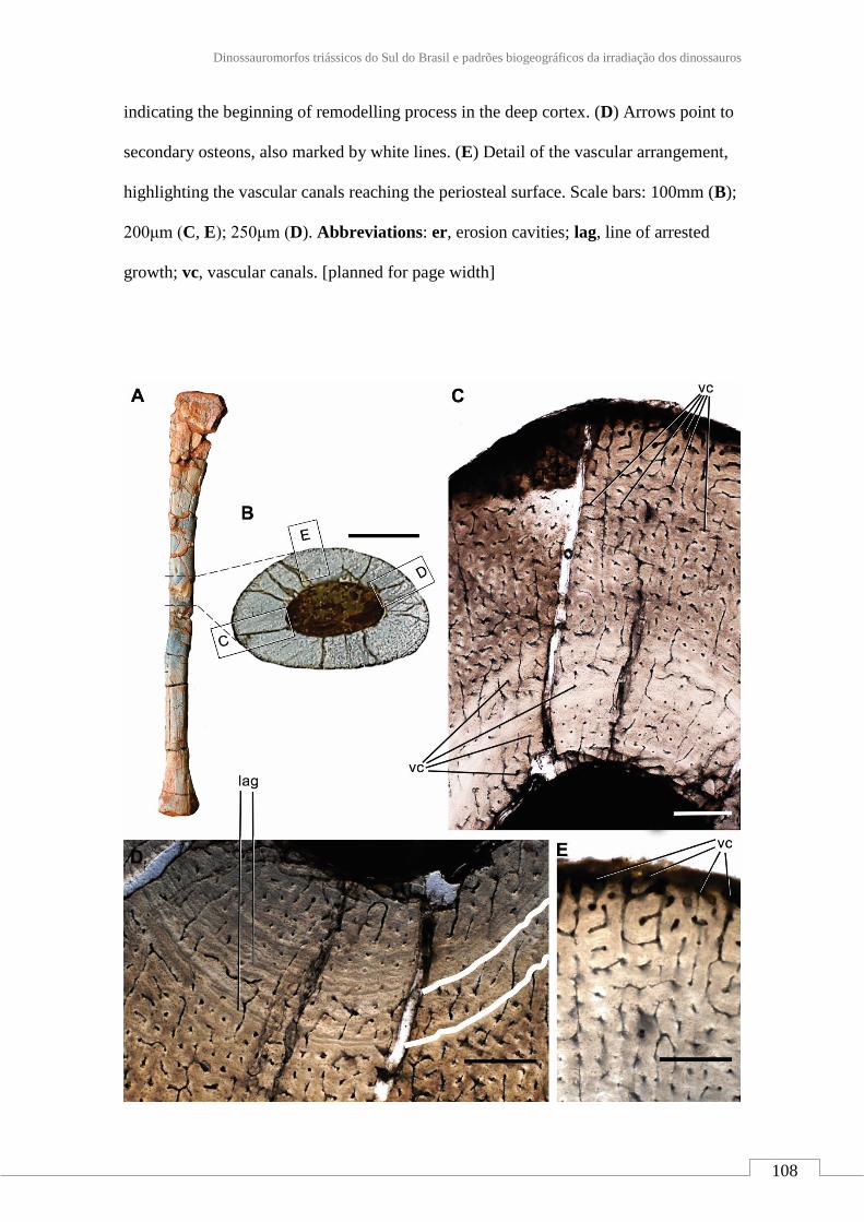
Dinossauromorfos triássicos do Sul do Brasil e padrões biogeográficos da irradiação dos dinossauros
108
indicating the beginning of remodelling process in the deep cortex. (D) Arrows point to
secondary osteons, also marked by white lines. (E) Detail of the vascular arrangement,
highlighting the vascular canals reaching the periosteal surface. Scale bars: 100mm (B);
200μm (C, E); 250μm (D). Abbreviations: er, erosion cavities; lag, line of arrested
growth; vc, vascular canals. [planned for page width]

JCA Marsola - 2018
109
FIGURE 15. General microstructural anatomy of the fibula of Nhandumirim
waldsangae gen. et sp. nov. (LPRP/USP 0651). (A) Right fibula indicating the sampled
area of the bone. (B) Cross section of the fibula with the square areas detailed in (C-E).
(C) View of the complete transect showing the osteohistological pattern of the fibula,
composed by plexiform arrange of the vascular network. (D) Lines of arrested growth in
the middle cortex marked by white lines. (E) Detail of the vascular arrangement,
highlighting the vascular canals reaching the periosteal surface. Scale bars: 150mm (B);
250μm (C); 300μm (D-E). Abbreviations: lag, line of arrested growth; vc, vascular
canals. [planned for page width]
FIGURE 16. Saturnalia tupiniquim (MCP-3845 PV). Articulated sacrum and ilia in
dorsal (A) view. B. Outline drawings represent the lateral aspect of the attachment scars
of the sacral vertebra transverse processes and ribs in the right ilium. Abbreviations:
1st, first primordial sacral vertebra; 2nd, second primordial sacral vertebra; ds, dorsal
vertebra incorporated to the sacrum. [planned for page width]

Dinossauromorfos triássicos do Sul do Brasil e padrões biogeográficos da irradiação dos dinossauros
110
FIGURE 17. Lateral view of several early dinosauromorph ilia, showing the depth of
the acetabulum. Dashed lines represent the possible limits of missing parts. A,
Asilisaurus kongwe (NMT RB159, after Peecook et al., 2013); B, Ixalerpeton
polesinensis (ULBRA-PVT059); C, Eocursor parvus (SAM-PK-K8025, after Butler,
2010); D, Herrerasaurus ischigualastensis (PVL 2566); E, Coelophysis bauri (AMNH
FARB 2708); F, Nhandumirim waldsangae gen. et sp. nov. (LPRP/USP 0651); G,
Saturnalia tupiniquim (MCP-3845 PV). Specimens scaled to the same acetabular
length. [planned for page width]

JCA Marsola - 2018
111
FIGURE 18. Phylogenetic relationships of Nhandumirim waldsangae gen. et sp. nov.
(LPRP/USP 0651) among early dinosauromorphs. A, Strict consensus of 48 MPTs
found in the analysis of the data matrix of Cabreira et al (2016); B, Strict consensus of
48 MPTs found in the analysis of the data matrix of Nesbitt and Ezcurra (2015). Values
at nodes are Bremer support and bootstrap proportions (above 50%). [planned for page
width]

Dinossauromorfos triássicos do Sul do Brasil e padrões biogeográficos da irradiação dos dinossauros
112
ANEXO 3
Materiais de sauropodomorfos e correlações bioestratigráficas da localidade tipo
de Sacisaurus, Triássico Superior da Formação Caturrita, sul do Brasil
Aceito para publicação após modificações: Marsola, J. C. A., Bittencourt, J. S., Da
Rosa, Á. A. S., Martinelli, A. G., Ribeiro, A. M. Ferigolo, J., and Langer, M. C.
Sauropodomorph remains and correlation of the Sacisaurus site, Late Triassic (Caturrita
Formation) of southern Brazil. Acta Palaeontologica Polonica.
Síntese do anexo 3
Sacisaurus agudoensis é o único silessaurídeo já reconhecido para a Supersequência
Santa Maria. Todavia, a correlação entre sua localidade-tipo com outros sítios triássicos
no Rio Grande do Sul é controversa. Em termos de idade, a ocorrência de S. agudoensis
nestes estratos tampouco permite uma inferência mais precisa que Triássico Superior. O
objetivo deste trabalho é a descrição de materiais de sauropodomorfos associados aos
abundantes restos de S. agudoensis, e avaliar se o seu sinal filogenético pode ajudar a
responder questão de correlação do sítio. De fato, a morfologia destes fósseis é mais
condizente com a de sauropodomorfos pós-carnianos como Pantydraco caducus e
Unaysaurus tolentinoi do que com espécies do Carniano, como Saturnalia tupiniquim e
Panphagia protos, o que sugere uma idade mais nova ao depósito. Da mesma maneira,
a anatomia dos dentes de cinodontes "brasilodontídeos" provenientes da mesma
localidade e estrato parecem coincidir com as correlações sugeridas pelos
sauropodomorfos descritos aqui.

JCA Marsola - 2018
113
Sauropodomorph remains and correlation of the Sacisaurus site, Late Triassic
(Caturrita Formation) of southern Brazil
JÚLIO C. A. MARSOLA, JONATHAS S. BITTENCOURT, ÁTILA A. S. DA ROSA,
AGUSTÍN G. MARTINELLI, ANA MARIA RIBEIRO, JORGE FERIGOLO, and
MAX C. LANGER
Abstract
Sacisaurus agudoensis is the only silesaurid known for the Triassic beds of the Santa
Maria Supersequence and the correlation of its type-locality to the other Triassic
deposits of south Brazil has always been controversial. In an attempt to improve this
situation, a handful of dinosaur remains found associated to S. agudoensis are here
described and compared. Their anatomy is more similar to that of Norian
sauropodomorphs such as Pantydraco caducus and Unaysaurus tolentinoi than to that
of Carnian taxa such as Saturnalia tupiniquim and Pampadromaeus barberenai. This
resemblance suggests a younger (perhaps Norian) age to the Sacisaurus site, as also
suggested by the record of Riograndia-like and brasilodontid cynodonts in the
assemblage and local stratigraphic correlation that positions the site in the Caturrita
Formation.
Key words: Dinosauria, Sauropodomorpha, Dinosauriformes, Santa Maria
Supersequence, Caturrita Formation, biostratigraphy, Norian, South America.

Dinossauromorfos triássicos do Sul do Brasil e padrões biogeográficos da irradiação dos dinossauros
114
Júlio C. A. Marsola [[email protected]] and Max C. Langer
[[email protected]], Departamento de Biologia, FFCLRP, Universidade de São
Paulo, Ribeirão Preto, SP, 14040-901, Brazil;
Jonathas S. Bittencourt [[email protected]], Departamento de Geologia,
Universidade Federal de Minas Gerais, Belo Horizonte, MG, 31270-901, Brazil;
Átila A. S. Da Rosa [[email protected]], Laboratório de Estratigrafia e
Paleobiologia, Departamento de Geociências, Universidade Federal de Santa Maria,
Santa Maria, RS, 97.105-900, Brazil;
Agustín G. Martinelli [[email protected]], Laboratório de
Paleontologia de Vertebrados, Departamento de Paleontologia e Estratigrafia, Instituto
de Geociências, Universidade Federal do Rio Grande do Sul, Porto Alegre, 91540-000,
RS;
Ana Maria Ribeiro [[email protected]] and Jorge Ferigolo
[[email protected]], Seção de Paleontologia, Museu de Ciências Naturais,
Fundação Zoobotânica do Rio Grande do Sul, Porto Alegre, RS, 90690-000, Brazil.
Introduction
The type-locality of the dinosauromorph Sacisaurus agudoensis Ferigolo and Langer,
2007, has been explored during 2000-2001 in a series of field works led by JF and the
crew of Fundação Zoobotânica do Rio Grande do Sul, in the context of the IDB (Inter-
American Development Bank) funded Pró-Guaíba Project. These unearthed the rich
material attributed to S. agudoensis, as well as cynodont (Ribeiro et al. 2011) and other
dinosaur remains (Langer and Ferigolo, 2013). The latter includes a handful of isolated
bones, which are too large to represent the kind of animal supposedly sampled at its
adult stage by the fossils attributed to S. agudoensis. The overlap of some of those

JCA Marsola - 2018
115
remains (i.e., ilium, femur) to bones of S. agudoensis indicates that they correspond to a
different taxon, with further non-duplicated larger elements (i.e. ectopterygoid, neck
vertebra, metatarsal I) also tentatively attributed to that taxon. This work aims at fully
describing such specimens, inferring their phylogenetic affinities and signal for faunal
correlation. Indeed, the correlation of the Sacisaurus site to other tetrapod-bearing
localities of the Santa Maria and Caturrita formations is not strongly constrained. As
such, we also attempt here to provide geological and biochronological data to more
strongly define the stratigraphic position of that site.
Institutional abbreviations.— BPI, Evolutionary Studies Institute, Johannesburg, South
Africa (formerly Bernard Price Institute); MB. R., Museum für Naturkunde, Berlin,
Germany; MCN, Museu de Ciências Naturais, Fundação Zoobotânica do Rio Grande do
Sul, Porto Alegre, Brazil; MCP, Museu de Ciências e Tecnologia, PUCRS, Porto
Alegre, Brazil; NHM, Natural History Museum, London, United Kingdom; PULR,
Universidad Nacional de La Rioja, La Rioja, Argentina; PVSJ, Museo de Ciencias
Naturales, San Juan, Argentina; SAM-PK, Iziko South African Museum, Cape Town,
South Africa; SMNS, Staatliches Museum für Naturkunde, Stuttgart, Germany;
UFRGS, Universidade Federal do Rio Grande do Sul, Porto Alegre, Brazil; UFSM,
Universidade Federal de Santa Maria, Santa Maria, Brazil; ULBRA, Museu de Ciências
Naturais, Universidade Luterana do Brasil, Canoas, Brazil.
Geological Settings
The Sacisaurus site is located at 19°43’12’’ S; 47°45’04’’ W, inside the western urban
area of city of Agudo, state of the Rio Grande do Sul, Brazil. Due to the urban
expansion, the outcrop it now located on the western margin of Independência Street,

Dinossauromorfos triássicos do Sul do Brasil e padrões biogeográficos da irradiação dos dinossauros
116
north to Concordia Avenue. Very few of the original outcrops remains, based on which
a sedimentary profile is herein provided (Fig. 1A).
The profile is more than eight meters deep and composed of an intercalation of
fluvial (CH) and overbank (CR) deposits. It starts with a fine sandstone where ex situ
Exaeretodon fragments were recorded. Still in the first meter of the profile, reddish
mudstones and a brownish fine sandstone are seen. The latter shows an upwards
coarsening trend, with mud intraclasts at the top, forming a millimetric intraformational
conglomerate. Above this conglomerate, there is an intercalation of reddish mudstones
and greenish fine sandstones that preserved most fossils in the site. The rest of the
profile is formed of yellowish to pinkish fine sandstones with millimetric intercalation
of brown mudstones. These present a lobe or tabular geometry, are generally massive,
but sometimes bear horizontal lamination and trough cross-bedding. Whereas the basal
most levels represent traction and suspension deposition probably in an oxbow lake, the
uppermost sandstones are linked to crevassing. These lithologies correspond to the
Caturrita Formation (sensu Andreis et al. 1980), i.e. upper portion of the Candelária
Sequence, Santa Maria Supersequence (sensu Horn et al. 2014).
Nearby outcrops allow for a better stratigraphic correlation (Fig. 1), as only
crevasse deposits are recorded at “Ki–Delicia” and “ASERMA” sites. The slightly more
distant, and much better known, Janner outcrop (Pretto et al. 2015) has equivalent
lithologies at its top, whereas the highly fossiliferous reddish mudstones of the middle
and lower part of the outcrop correspond to the Santa Maria Formation (Da Rosa, 2005,
2015).

JCA Marsola - 2018
117
Material
Bones of the larger dinosauromorph found at the Sacisaurus site were not recovered
articulated or closely associated, so that there is no clear evidence that they correspond
to a single individual. This is corroborated by the preserved right and left femora, which
share a very similar anatomy, but are of slightly different sizes. As a whole, the material
have a similar taphonomic signature and includes a right ectopterygoid (MCN PV
10049), one cervical vertebra (MCN PV 10027), a right ilium (MCN PV10026), right
(MCN PV10007) and left (MCN PV10008) femora, and a metatarsal I (MCN PV
10049). As mentioned above, the attribution of these specimens to the same taxon is
tentative; based on their similar phylogenetic signal (discussed below) added of
topotypic principles.
Comparative description
Ectopterygoid (MCN PV 10049).—The bone is nearly complete, missing only the
rostral tip of the lateral process and the medial contour of the medial process (Fig. 2),
precluding the proper assessment of its articulation with the pterygoid. As preserved,
the ectopterygoid is slightly longer (rostrocaudally) than lateromedially wide. The
lateral process is hook-shaped and arches rostrally, forming a pounded caudal margin
with the medial process. Its lateral half flattens lateromedially, and it is dorsoventrally
higher compared to its medial half. Its articulation with the jugal is marked by scars on
the caudolateral margin. Although missing its rostral-most tip, it is clear that such
articulation is tabular, differing from the T-shaped profile of Plateosaurus engelhardti
(Prieto-Márquez and Norell, 2011). The medial half of the lateral process expands
rostrocaudally towards the medial process, which is much longer rostrocaudally than the
former.

Dinossauromorfos triássicos do Sul do Brasil e padrões biogeográficos da irradiação dos dinossauros
118
The medial process is flange-like, dorsoventrally expanded, and bears a medially
directed dorsal margin. Its ventral surface is excavated by a semicircular depression,
which has been considered pneumatic and typical of theropods (e.g. Rauhut, 2003). Yet,
Nesbitt (2011) notices that such feature just marks the articulation with the pterygoid, as
seen in Eoraptor lunensis, Pantydraco caducus, Plateosaurus engelhardti, Liliensternus
liliensterni, and Coelophysis bauri. The medial process has a long rostral projection
resembling that of Pa. caducus (Yates, 2003; Galton and Kermack, 2010), and differing
from that of “Syntarsus” rhodesiensis (Raath, 1977), Allosaurus fragilis (Nesbitt, 2011),
Pl. engelhardti (Prieto-Márquez and Norell, 2011), and Lesothosaurus diagnosticus
(Porro et al. 2015).
Neck vertebra (MCN PV 10027).—The only preserved vertebra (Fig. 3) is somewhat
distorted and incomplete, missing the cranioventral portion of the centrum, the
zigapophyses, the neural spine, and most of the parapophyses and diapophyses.
Together, the dorsal position of the parapophyses in the centrum, the well-developed
diapophyses, the elongated centrum, and the presence of a ventral keel, indicate that
MCN PV 10027 represents a cervical vertebra, possibly from the caudal part (8th or 9th
element) of the neck. For descriptive purposes, the laminae and fossae nomenclature of
Wilson (1999, 2012) and Wilson et al. (2011) will be adopted.
The centrum has an elongated profile, at least three times longer than high, the
ventral portion of which bears a stout ventral keel; only the caudal half of which its
preserved. As such, the keel is mediolaterally thicker caudally, i.e. more than seven
times thicker in its caudal portion than cranially at the middle of the centrum (Fig. 3B).
That thicker portion projects further ventrally than the caudal articulation of the
centrum. The surface surrounding that articulation is heavily scarred, mainly on its

JCA Marsola - 2018
119
ventral margin. In ventral view, the centrum is spool-shaped, with its caudal rim c.1.75
times wider than its middle portion. The caudal articular face of the centrum is deeply
concave. In caudal view (Fig. 3C), it is exaggeratedly wider than high due taphonomic
distortion. In lateral view, its margin is oblique to the long axis of the centrum, so that
its ventral edge extents further caudally than the dorsal. The neurocentral suture is seen
mainly along the caudal portion of the vertebra, suggesting an advanced closure stage.
The dorsal surface of the centrum has a well-developed fossa (“fo” in Fig. 3), which is
remarkably deep, dorsoventrally narrow, and craniocaudally elongated. Most of both
parapophyses are missing due to breakages. Each is represented by a subtle and short
oblique (cranioventrally to caudodorsally oriented) ridge set at the dorsal part of the
cranial margin of the centrum, close to the neural arch, fading away caudodorsally in the
direction of the “anterior centrodiapophyseal lamina”.
The neural arch bears well developed laminae and fossae. The cranial portion of
the prezygodiapophyseal laminae is missing, but its well-developed caudal part is
clearly seen in left lateral view (“prdl” in Fig. 3). This lamina roofs a deep
prezygadiapophyseal centrodiapophyseal fossa and extends further laterally than the
“anterior centrodiapophyseal lamina” (“acdl” in Fig. 3), which is short and does not
reach the parapophysis. The centrodiapophyseal fossa is deep and craniocaudally
elongated. It is set caudal to the prezygadiapophyseal centrodiapophyseal fossa, with its
cranial half roofed by a lateroventrally directed, well-developed diapophysis, from the
dorsal surface of witch a well-developed postzygodiapophyseal lamina (“podl” in Fig.
3) arises. This lamina extends towards the base of the postzygapophysis (which is not
preserved), marking the craniodorsal margin of a deep postzygadiapophyseal
centrodiapophyseal fossa. The right of those fossae (Fig. 3E) is divided in two by a
subtle ridge that expands cranioventraly from the middle of the postzygodiapophyseal

Dinossauromorfos triássicos do Sul do Brasil e padrões biogeográficos da irradiação dos dinossauros
120
lamina. An equivalent ridge is, however absent from the left side of the vertebra. The
postzygadiapophyseal centrodiapophyseal fossa also overlaps the caudal half of the
centrodiapophyseal fossa dorsally. The “posterior centrodiapophyseal lamina” roofs the
dorsal and caudal portions of the centrodiapophyseal fossa, also forming the whole
ventral edge of the postzygadiapophyseal centrodiapophyseal fossa.
The dorsal surface of the neural arch (Fig. 3A) has several ridges, which are
tentatively associated to the laminae of later saurischians. The cranial surface of the
neural spine has a deep, dorsoventrally short and craniocaudally long “V”-shaped
spinoprezygapophyseal fossa (“sprf” in Fig. 3) at the base. Its lateral margins are
formed by subtle spinoprezygapophyseal laminae that reach the medial surface of the
prezygapophyses, where they bifurcate. Another pair of ridges (“rdg1” in Fig. 3) form
the lateral margin of the prezygapophyses, extending caudally along the dorsal surface
of the neural arch to reach the lateral base of the neural spine caudal to the
spinoprezygapophyseal fossa. Ventrolateral to that, there is another pair of faint
craniocaudally directed ridges (“rdg2” in Fig. 3). Also, subparallel to the neural spine, a
pair of inconspicuous low ridges (“eprl?” in Fig. 3) extend on the dorsal surface of the
neural arch. Although its caudal end is missing, it most probably corresponds to the
epipophyseal-prezygapophyseal lamina (Wilson, 2012).
Neck vertebrae with epipophyses have been regarded as a dinosaur
synapomorphy (Langer and Benton, 2006; Nesbitt, 2011). Although the epipophyses are
not preserved in MCN PV 10027, the ridge putatively related to the epipophyseal-
prezygapophyseal lamina is an indirect evidence of their presence. A similar condition
is seen in the 8th cervical vertebra of Panphagia protos (PVSJ 874), in which a
craniomedially directed incipient ridge comes from the epipophysis. This condition is
clearer in later saurischians, such as the sauropodomorphs Adeopapposaurus mognai

JCA Marsola - 2018
121
(PVSJ 610) and Massospondylus carinatus (BPI 4934; SAM PK K 388), and
neotheropods like Elaphrosaurus bambergi (Rauhut and Carrano, 2016). No
epipophyses are seen in the caudal cervical vertebrae of ornithischians (Sereno et al.
1993; Langer and Benton, 2006; Nesbitt, 2011), but these are seen in non-
dinosauromorph archosauromorphs, as Batrachotomus kupferzellensis and
Tanystropheus longobardicus (Langer and Benton, 2006; Nesbitt, 2011; Ezcurra, 2016).
Yet, the morphology of MCN PV 10027, with well-developed fossae and laminae,
stresses its closer resemblances to dinosaurs than to other archosaurs.
Carnian dinosaurs, such as Eoraptor lunensis (Sereno et al. 2012), Panphagia
protos (Martínez and Alcober, 2009), and Herrerasaurus ischigualastensis (Sereno and
Novas, 1993) lack the well-developed fossae and laminae seen in MCN PV 100027.
These are also mostly absent in pseudosuchians, although deep fossae and prominent
laminae are present in some poposaurids and crocodyliforms (Nesbitt, 2005, 2007;
Wedel, 2007). Among later saurischians, neotheropods are widely recognized by their
pneumatic vertebrae, as seen in early forms like Coelophysis bauri and Liliensternus
liliensterni (see Benson et al. 2012). This condition is characterized by cervical
vertebrae with well-develop laminae and deep fossae pierced by large foramina (Britt,
1993; O’Connor, 2006, 2007). On the other hand, early sauropodomorphs lack the deep
fossae seen in neotheropods (Wedel, 2007; Adeopapposaurus mognai, PVSJ 610) and in
MCN PV 100027, although the caudalmost vertebra of the cervical series of
Plateosaurus engelhardti (SMNS 13200) has well-developed fosssae and
prezygodiapophyseal, centrodiapophyseal, and postzygodiapophyseal laminae,
resembling the condition of MCN PV 100027. Neotheropods differs from MCN PV
100027 because they bear at least one pair of deep pleurocoels in the cranial portion of
the centrum. As observed by Tykoski (2005), those pleurocoels are set either

Dinossauromorfos triássicos do Sul do Brasil e padrões biogeográficos da irradiação dos dinossauros
122
caudodorsal or dorsal to the parapophyses. Although the cranial portion of the centrum
of MCN PV 100027 is damaged, the remaining morphology does not suggest the
presence of such a feature. Furthermore, a second pair of pleurocoels may be present
caudal to the aforementioned, as in coelophysoids (Tykoski, 2005) and in
Elaphrosaurus bambergi (Rauhut and Carrano, 2016), although not in L. liliensterni
(MB. R. 2175), but this is not seen in MCN PV 100027.
Ilium (MCN PV10026).—A right ilium (Fig. 4) is nearly complete, missing the cranial
tip of the preacetabular ala, as well as small portions of the cranial part of the
supracetabular crest, the cranioventral part of the acetabular medial wall, and the
cranioventral part of the postacetabular ala. The iliac length suggests an individual
smaller than those referred to Saturnalia tupiniquim, Guaibasaurus candelariensis,
Unaysaurus tolentinoi, and Pampadromaeus barberenai. The preacetabular ala is
subtriangular in lateral view and laterally directed due to the inwards arching of the
entire blade (Fig.4C). Its shape resembles that seen in sauropodomorphs such as
Riojasaurus incertus (PULR 56) and Efraasia minor (SMNS 12354), but clearly differs
from that of other sauropodomoph remains from the Caturrita Formation (Bittencourt et
al. 2012). The preacetabular ala is considerably shorter than the postacetabular ala and
its cranial tip is set well caudal to the cranialmost edge of the pubic peduncle. Although
the preacetabular ala of Pantydraco caducus (Yates, 2003) and Leonerasaurus
taquetrensis (Pol et al. 2011) extends cranial to the pubic peduncle, that structure is
shorter in most early sauropodomorphs (Cooper, 1981; Benton et al. 2000; Langer,
2003; Rowe et al. 2011), unlike ornithischians and neotheropods. The preacetabular ala
and the pubic peduncle are set at a right angle to one another in MCN PV10026. The
concave area between them seems to harbor an incipient preacetabular fossa (“prf” in

JCA Marsola - 2018
123
Fig. 4), but this cannot be confirmed due to breakages. Dorsally, the preacetabular ala
shows a slightly bulged lateral rugose muscle scar (“sc” in Fig. 4), related to the
insertion of M. iliotibialis (Hutchinson, 2001a; Langer, 2003), which spans caudally
along the dorsal edge of the iliac blade. It is caudaly connected to a broad muscle
attachment area at the caudal portion of the postacetabular ala.
The iliac blade is slightly higher than the height from the supracetabular crest to
the ventralmost level of the iliac acetabulum. Its lateral surface, caudal to the
preacetabular ala, bears two conspicuous depressions separated by a short, elevated
area. The dorsal depression (“dd” in Fig. 4) is rounded and craniocaudally elongated, as
well as larger than the lower one. It extends caudally as to almost reach the
postacetabular ala, but its deepest point is immediately dorsal to the ventral depression.
The latter (“vd” in Fig. 4) starts caudal to the preacetabular embayment, extending onto
its maximal transverse depth right above the supraacetabular crest.
The postacetabular ala is stout and caudodorsally projected, giving a slightly
sigmoidal aspect to the dorsal margin of the ilium in lateral/medial views. Its
caudoventral portion bears a pair of longitudinal crests restricted to its caudal half. The
medial of those (“vmc” in Fig. 4; “posteromedial lamina/shelf” of Ezcurra, 2010;
“medial lamina/blade” of Martinez and Alcober, 2009) extends along the medial surface
of the ilium, forming the dorsal margin of the attachment area for the second primordial
sacral rib. Its mid-length is right dorsomedial to the caudal tip of the ventral margin of
the postacetabular ala (“vmpoa” in Fig. 4), which extends caudally from the ischiadic
peduncle. The medial margin of the brevis fossa (“brfo” in Fig. 4) if formed by those
crests; the ventral margin of the postacetabular ala more cranially and the “medial crest”
more caudally. Its lateral margin is, on the other hand, formed by the second, more
lateral ridge (“vlc” in Fig. 4); i.e. the brevis shelf. The brevis fossa is shallow,

Dinossauromorfos triássicos do Sul do Brasil e padrões biogeográficos da irradiação dos dinossauros
124
transversely and longitudinally broad, but craniocaudally shorter than in earlier
sauropodomorphs like, Saturnalia tupiniquim (Langer, 2003), Chromogisaurus novasi
(Ezcurra, 2010), and Buriolestes scultzi (Cabreira et al. 2016), resembling that of other
sauropodomorphs, such as Plateosaurus engelhardti (SMNS 12950; 80664),
Massospondylus carinatus (Cooper, 1981), and Adeopapposaurus mognai (Martínez,
2009). Likewise, such a reduced fossa brevis also differs from those of neotheropods
(e.g. Liliensternus liliensteni MB. R. 2175) and early ornithischians (e.g. Baron et al.
2017).
The area between the supraacetabular crest and the postacetabular ala is concave,
unlike that of earlier sauropodomorphs like Chromogisaurus novasi (Ezcurra, 2010),
neotheropods (e.g. Liliensternus liliensteni MB. R. 2175), and some ornithischians (e.g.
Scelidosaurus harrisonii NHM R 1111), which bears a ridge connecting the brevis shelf
to the supraacetabular crest. The supraacetabular crest extends cranioventrally as a
continuous flange from a rugose area at the level of the ischiadic peduncle, along the
pubic peduncle, terminating near its articulation area. It is strongly expanded
lateroventrally, but not as much as in theropods. Its point of maximal transverse breadth
is above the center of the acetabulum, equally distant from the distal tips of the ischiadic
and pubic peduncles. At this point, the supraacetabular crest is nearly set at the mid
dorsoventral height of the ilium, unlike Saturnalia (MCP 3844-PV), Pampadromaeus
(ULBRA-PVT016) Panphagia (PVSJ 874), Chromogisaurus (PVSJ 845) and
Buriolestes (ULBRA-PVT280).
The acetabulum is as craniocaudally expanded as the length between the pre-
and postacetabular embayments, and relatively deeper dorsoventrally than in early
sauropodomorphs, e.g. Saturnalia tupiniquim, Panphagia protos, and Chromogisaurus
novasi (Marsola et al. in review). Its medial wall is ventrally projected, the ventral

JCA Marsola - 2018
125
margin of which levels with the ventral tip of both ischiadic and pubic peduncles. This
feature suggests a closed iliac acetabulum, unlike massospodylid and plateosaurid
sauropodomorphs, neotheropods, and most ornithischians (see Baron et al. 2017), but
similar to that of some non-dinosaur dinosauromorphs (Langer et al. 2013), S.
tupiniquim (Langer, 2003), P. protos (Martínez and Alcober, 2009), and Buriolestes
schultzi (Cabreira et al. 2016). There is a shallow ventral notch between the ischiadic
peduncle and the ventral margin of the acetabulum, which sets the ventral limit of the
ovoid dorsoventrally elongated antitrochanteric area of the acetabulum. The
cranioventral portion of the acetabulum has a short vertical ridge forming an angle of
45° to the long axis of the pubic peduncle. As described in other dinosaurs (Langer et al.
2010), this ridge is set medially to the supraacetabular crest, producing a subtriangular
ventral fossa (“vf” in Fig. 4) on the craniolateral corner of the acetabulum.
The pubic peduncle is as long as the extension between the pre- and
postacetabular embayments. Its distal portion is dorsoventrally deeper and transversely
narrower than the proximal. The craniodorsal surface bears a subtle ridge as seen in
Pampadromaeus barberenai (ULBRA-PVT016). Medially the peduncle is mostly flat,
with rugose distal areas for muscle attachment. Its articulation surface for the pubis
faces cranioventrally, is somewhat rounded in outline and craniocaudally elongated,
with the medial margin flatter than the lateral. The ischiadic peduncle is short and
subtriangular, similar to that of Saturnalia tupiniquim (Langer, 2003). It is laterally
bulged, producing a low mound-like process in ventral view that represents the
acetabular antitrochanter. The articular surface for the ischium is caudoventrally facing,
but lacks the caudal heel present in other sauropodomorphs, like P. barberenai
(ULBRA-PVT016), Riojasaurus incertus (PULR 56), Coloradisaurus brevis (Apaldetti
et al. 2013), and Efraasia minor (SMNS 12354).

Dinossauromorfos triássicos do Sul do Brasil e padrões biogeográficos da irradiação dos dinossauros
126
The medial surface of the ilium has a complex anatomy, encompassing the
concavities and crests that mark the articulation of the sacral vertebrae. Just caudal to
the preacetabular embayment, a dorsoventrally elongated concavity (“tsr” in Fig. 4) is
seen, cranially bounded by a vertical sharp ridge (“vr” in Fig. 4) formed by the medial
edge of the preacetabular fossa. Its dorsal, ventral and caudal margins are formed by a
gently elevated, continuous margin. Caudal to that, there is another concavity (“1st” in
Fig. 4), which is rounded and craniocaudally longer than the former. The dorsal part of
those concavities is formed by a continuous craniocaudally elongated depression that
probably received the sacral transverse processes. Its dorsal margin is formed by a
longitudinally oriented elevated margin, dorsal to which the medial iliac surface is
marked by a spread rugose surface serving for muscle attachment sites, as is also the
case of the surface bellow the articulation areas (“sc” in Fig. 4). More caudally, the
medial surface of the postacetabular ala is marked by the “medial crest”, which extends
cranially as a subtler ridge. This forms the medial margin of the caudal part of the brevis
fossa, which is equally projected in Thecodontosaurus antiqus (BRSUG 23613),
Efraasia minor (SMNS 12354), and Plateosaurus engelhardti (SMNS 12950), but less
marked in Massospondylus carinatus (BPI 5238) and Sarahsaurus aurifontanalis
(TMM 43646-2). Dorsal to the “medial crest”, there is a longitudinal groove (“tp2nd” in
Fig. 4) that received the transverse process of the second primordial sacral vertebra and
merged cranially to the aforementioned “elongated depression”. The attachment site for
the corresponding rib is a depressed subtriangular area (“r2nd” in Fig. 4) ventral to the
“medial crest”. This arrangement matches that of dinosaurs in which two vertebrae form
the bulk of the sacrum, as in Saturnalia tupiniquim (Langer, 2003) and Staurikosaurus
pricei (Bittencourt and Kellner, 2009). Yet, it is also possible that the cranial,
dorsoventrally elongate depression (“trs” in Fig. 4B) represents the articulation of the

JCA Marsola - 2018
127
rib of a trunk vertebra incorporated into the sacrum, as seen in one of the paratypes of S.
tupiniquim (MCP 3845-PV; Marsola et al. in review).
Femora (MCN PV10007, MCN PV10008).—Even though the femora do not overlap
(as they are from different sides) and share a very similar morphology, they probably
represent two individuals, as size estimation suggests the right element comes from a
considerably larger animal. In both femora, portions of the head and distal condyles are
missing. As preserved, the right element (Fig. 6) is 14,75 cm long, whereas the left is
12,25 (Fig. 5). As reconstructed, the right femur would be nearly 15 cm and the left
would measure between 13,5 – 14 cm. In any case, given their matching morphology, a
single description will be provided below for both femora.
The femur has a sinuous shape produced by the craniomedial projection of the
head and the cranial and medial bowing of the distal half of the shaft. The head
fragment associated to the left femur (Figs. 5E-F) is typically dinosaurian in its
transverse expansion, housing a rugose articular surface. Both the ligament sulcus and
the lateral tuber are not as pronounced as in other dinosauriforms, such as Sacisaurus
agudoensis (Langer and Ferigolo, 2013; MCN PV 10014), Saturnalia tupiniquim (MCP
3844-PV), Buriolestes schultzi (ULBRA-PVT280), and Eodromaeus murphi (PVSJ
562). The craniomedial tuber forms a rounded margin as seen perpendicular to the long
axis of the head (Figs. 5E-F). The head-shaft transition is rounded and contiguous in
those same views, its craniolateral surface being excavated by a subtle ventral
emargination (“ve” in Fig. 5). There is no evidence of a groove on the proximal surface
of the femur, and the caudal structures of the head, including the ‘greater trochanter’,
have not been preserved.

Dinossauromorfos triássicos do Sul do Brasil e padrões biogeográficos da irradiação dos dinossauros
128
The craniolateral surface of the proximal part of the femur has a rugose site for
ligament attachment right distal to the articular surface, which extends ventrally as the
intertrochanteric area. This area is laterally bound by the S-shaped dorsolateral
trochanter, whereas the cranial trochanter is a cranially bulged, roughly triangular
process, the rounded dorsal tip of which is well ventral to the femoral head. From the
medial margin of the cranial trochanter, the linea intermuscularis cranialis (“lic” in
Figs. 5-6) extends ventrally, bordering the medial and lateral surfaces of the shaft.
Between the base of the cranial and the dorsolateral trochanter, there is a rugose and
slightly elevated area (“mife” in Figs. 5-6) better seen in the left femur, which is
probably the insertion site of M. iliofemoralis externus (Hutchinson, 2001b) and
homologous to the trochanteric shelf.
As in herrerasaurids (Novas, 1994; Bittencourt and Kellner, 2009) and
sauropodomorphs (Langer, 2004; Langer and Benton, 2006), the fourth trochanter of
MCN PV10007 and PV10008 forms an asymmetrical flange, located on the caudal
surface of the proximal half of the shaft. In lateral/medial views, it has the shape of an
obtuse triangle, the longest edge of which projects caudoventrally from the shaft,
forming a rounded apex on that same direction. In ornithischians, such as Eocursor
parvus (Butler, 2010), the apex of the fourth trochanter is straighter and more distally
projected, as to acquire a pendant shape. Equally different is the symmetrical and lower
fourth trochanter of neotheropods, e.g. Dilophosaurus (Welles, 1984; UCMP 37302),
and non-dinosaurian dinosauromorphs (Langer et al. 2013). In caudal view, the fourth
trochanter has a slightly sinuous aspect and its lateral/medial outline shows that it is
more expanded distally than proximally. The tip of the fourth trochanter is rounded, and
its distal margin forms a nearly right angle to the long axis of the femur. In contrast, the
distal margin of the fourth trochanter in ornithischians is strongly concave. A

JCA Marsola - 2018
129
proximodistally elongated and transversely broad fossa (“cflf” in Figs. 5-6), medially
bound a ridge, is located medial to the fourth trochanter and represents the insertion of
M. caudofemoralis longus (Hutchinson, 2001b; Langer, 2003). The shaft surface
proximolateral to the fourth trochanter is also concave, encompassing a diagonally
oriented oval rugose area (“cfbf” in Figs. 5-6) for the origin of M. caudofemoralis
brevis (Hutchinson, 2001b; Griffin and Nesbitt, 2016). From the distal edge of the
fourth trochanter, another intermuscular line extends distally along the caudomedial
margin of the femoral shaft.
The femoral shaft expands transversely at the distal portion, but the exact
morphology of the condyles cannot be evaluated due to its poor preservation. The
popliteal fossa is enclosed by broad longitudinal ridges ate the caudal surface of the
distal part of the femur. The opposite (cranial) surface of the femur is not depressed, i.e.
the flexor fossa seen in some saurischians, like neotheropods, is absent. However, that
surface is scarred (“fdms” in Fig. 5) for muscle insertion as in Herrerasaurus (Novas,
1994) and Saturnalia (Langer, 2003).
Metatarsal I (MCN PV 10049).— Only the distal condyles of the left metatarsal I are
preserved along with a small portion of the shaft (Fig. 7). The preserved part of the shaft
is craniocaudally flattened, with a lateral margin wider than the medial, probably for
articulation with metatarsal II, resulting in a subtriangular cross-section. The condyles
expand lateromedially compared to the shaft, with the distal articulation being about one
third wider than the preserved portion of the latter. The lateral condyle is much larger
than the medial, also extending much more distally. It has a roughly triangular distal
outline, with rounded corners, formed by lateral, craniomedial, and caudomedial
margins. In the same view, the lateral margin of the condyle has a marked concavity,

Dinossauromorfos triássicos do Sul do Brasil e padrões biogeográficos da irradiação dos dinossauros
130
arising from a well-developed ligament pit (“llp” in Fig. 7), and a laterally projecting
caudolateral corner. The medial condyle is small, craniocaudally flattened, and
caudomedially directed in distal view, forming a 45º angle to the lateromedial axis of
the bone. This shape, along with the distal projection of the lateral condyle, produces a
medial displacement of the first digit, as typical pf sauropodomorphs as Unaysaurus
tolentinoi (UFSM 11069), Efraasia minor (SMNS 12354), Pantydraco caducus
(NHMUK RU P77/1), and Leonerasaurus taquetrensis (Pol et al., 2011), but not in
Carnian forms like Saturnalia tupiniquim (Langer, 2003) and Pampadromaeus
barberenai (ULBRA-PVT016). Although the metatarsal I is also known for
Guaibasaurus candelariensis, comparisons are hampered by its poor preservation
(Langer et al., 2010). In neotheropods, like Coelophysis bauri (e.g. NMMNH P-42200;
Rinehart et al. 2009), Dilophosaurus wetherilli (UCMP 37302), and Sinraptor dongi
(Currie and Zhao, 1993), the medial condyle of metatarsal I is not as medially projected
as in sauropodomorphs. This forms a craniocaudally deeper distal articular surface,
therefore differing from MCN PV 10049. A condition like that of neotheropods is found
in early ornithischians as Abrictosaurus concors (NHMUK RU B.54), Lesothosaurus
diagnosticus (NHMUK RU B.17), and Heterodontosaurus tucki (SAM-PK K 1332).
The craniomedial surface of the medial condyle bears a shallow ligament pit (“mlp” in
Fig. 7), the craniolateral rim of which borders the lateral condyle. The caudolateral
margin of the medial condyle forms the medial margin of a lateromedially wider than
deep flexor fossa (“ff” in Fig. 7). Unlike that of Plateosaurus engelhardti (SMNS 13200
Z), and the above cited neotheropods and ornithischians, an equally wider than deep
flexor fossa is present in the sauropodomorphs U. tolentinoi (UFSM 11069), E. minor
(SMNS 12354), Pan. caducus (NHMUK RU P77/1), and L. taquetrensis (Pol et al.
2011). Both medial and lateral ligament pits are heavily scarred for ligament insertion.

JCA Marsola - 2018
131
Discussion
Correlations of the Sacisaurus site
The geological review provided here indicates that all fossil-bearing levels of the
Sacisaurus site match the usual sandstone upward increase that regionally marks the
Caturrita Formation (sensu Andreis et al. 1980) and that the entire site is above the
neighboring mudstones of Janner site (Fig. 1). In palaeontological terms, although the
lower levels of the site, which record Exaeretodon-like cynodonts (Ribeiro et al. 2011),
have been roughly correlated to those of the Janner site (Langer et al., 2007; Langer and
Ferigolo, 2013), a younger age has been tentatively assigned to the type-stratum of S.
agudoensis, based on the presence of small, isolated postcanine teeth referred to the
probainognathians Riograndia guaibensis and Brasilitherium riograndensis (Langer et
al., 2007; Langer and Ferigolo, 2013). Indeed, their presence implies a correlation to the
“Riograndia Assemblage Zone” of Soares et al. (2011; see also Bonaparte et al. 2010),
which is younger than the “Hyperodapedon AZ” sampled from the Janner site. In this
section, we first provide a brief revaluation of the cynodont teeth recovered from the
same beds where all S. agudoensis specimens were found, followed by an attempt to
more comprehensively position that locality into the biostratigraphic schemes for the
Santa Maria Supersequence.
Cynodont teeth
The specimen previously referred to Riograndia guaibensis corresponds to an isolated
postcanine (MCN-PV 10204) here interpreted as a left lower element (Figs. 8A-B). It
has leaf-shaped crown with eight small cusps in mesiodistal line, which are the main
features it shares with R. guaibensis (Bonaparte et al. 2001; Soares et al. 2011). The
eight cusps have an asymmetrical distribution on the crown; the tallest cusp is preceded

Dinossauromorfos triássicos do Sul do Brasil e padrões biogeográficos da irradiação dos dinossauros
132
by three mesial cups and followed by four distal cusps. The cusps are separated by
shallow but long and curved (mesially convex) longitudinal (inter-cusp) grooves located
on both lingual and labial sides. Following the curvature of the grooves, the main cusp
and the four distal ones are slightly inclined backward. The cusps, especially the main
and the mesial ones, have a marked ridge on their lingual surface. The distalmost cusp is
located below the level of the mesialmost cusp and is slightly displaced lingually. The
labial surface of the crown is convex and the lingual is concave, with the mesiolingual
edge of the first cusp forming an elevated ridge. There is no conspicuous crown-root
constriction, but the distal border is more concave than the mesial in this area. The latter
is almost straight in labial/lingual views. The root is apically open and hollow, with a
circular cross-section along all its extension.
In fact, MCN-PV 10204 resembles postcanine teeth of Riograndia guaibensis
(Figs. 8C-D) in general aspect, but it is also reminiscent of the leaf-shaped crown
morphology seen in several Triassic archosauromorph taxa, such as Azendohsaurus,
Revueltosaurus, ornithischians, and sauropodomorphs (e.g., Flynn et al. 1999; Barret,
2000; Parker et al. 2005). Yet, none of the above archosauromorphs have teeth with the
unique set of features of MCN-PV 10204, e.g. long and mesially convex inter-cusp
grooves (= interdenticular sulci of Hendrickx et al. 2014) and crown with a concave
lingual surface. Although, the postcanine teeth of R. guaibensis (e.g., UFRGS-PV-833-
T; UFRGS-PV-1319-T) typically have more transversely narrow crowns, less
developed (i.e. small) cusps and inter-cusp grooves, a considerably less concave lingual
surface, and a more transversely flattened root with an incipient longitudinal groove,
more conspicuously in the labial side (Figure 8C-D). Thus, the morphology of MCN-
PV 10204 could fit to a more rostral R. guaibensis postcanine tooth, as more caudal
teeth increase the cusp number up to 11 (e.g., UFRGS-PV-1319-T). However, a new

JCA Marsola - 2018
133
Riograndia-like cynodont, sister-taxon to R. guaibensis, has been recently reported from
Janner site (Martinelli et al. 2016), which is typically included in the “Hyperodapedon
AZ”, showing the occurrence of leaf-shape toothed cynodonts in faunas older than the
“Riograndia AZ”. Therefore, the isolated tooth MCN-PV 10204 is here referred as a
Riograndia-like taxon and it provides no compelling evidence that the stratigraphic
level it comes from is younger than those corresponding to the “Hyperodapedon AZ” at
the Janner site.
The specimens originally referred to Brasilitherium riograndensis include two
isolated right lower postcanines (MCN-PV 10202, MCN-PV 10203). Their crown
morphology is similar, but MCN-PV 10202 (Figs. 8E-F) is larger than MCN-PV 10203
(Figs. 8G-H). Given the crown complexity, they seem to be from the middle to caudal
portions of the dental series, and MCN-PV 10202 is possibly from a more posterior
position in the series than MCN-PV 10203. They are asymmetrical with a main cusp “a”
followed by a large cusp “c” and a small cusp “d”. Cusp “b” is smaller than cusp “d”
and placed lower in the crown than the remaining cusps. Cusps “a” to “d” are aligned,
forming a sectorial crest, with conspicuous mesial and distal cutting edges connecting
cusps, whereas cusp “b” is separated from cusp “a” by a concave notch. Both teeth have
one mesiolabial and one mesiolingual accessory cusps (the latter possibly corresponding
to cusp “e” of Crompton, 1974), located below the notch between cusps “a” and “b”.
Consequently, in mesial view, cusp “b” is flanked by these two accessory cusps, as not
seen in any Brasilodon-Brasilitherium specimens. A distolingual cingulum bears three
cusps in MCN-PV 10102 and two discrete cusps in MCN-PV 10103. In MCN-PV
10102, the most distal cusp is broken at its base, but seems to be larger than the
remaining elements. The more mesial cusp, near which the cingulum ends, could
correspond to cusp “g”. In MCN-PV 10103, the distolingual cusps are considerably

Dinossauromorfos triássicos do Sul do Brasil e padrões biogeográficos da irradiação dos dinossauros
134
larger. Particularly, the more mesially placed (just below the notch between cusps “a”
and “c”) is acute and conspicuous, corresponding to cusp “g”. The root grooves are deep
both lingually and labially. Particularly in MCN-PV 10103, it divides the root in two
portions, with the distal one considerably larger mesiodistally and labiolingually, with a
strongly convex distal wall.
In general aspect, MCN-PV 10202 and MCN-PV 10203 resemble more
Brasilitherium riograndensis and Brasilodon quadrangularis than any other known
probainognathians from South America (e.g., Bonaparte and Barberena, 2001;
Martinelli et al. 2017a, b). They differ from the postcanine teeth of Alemoatherium
huebneri because these lack a mesiolabial accessory cusp, their lingual cusps and
cingulum are much less developed and cusp “d” is slightly smaller and lower positioned
than cusp “b” (Martinelli et al. 2017a). Besides, Prozostrodon brasiliensis and
Botucaraitherium belarminoi (Soares et al. 2014; Pacheco et al. 2017) postcanine teeth
have a more continuous lingual cingulum, with a higher number of accessory cusps than
MCN-PV 10202 and MCN-PV 10203. There are also subtle differences between these
teeth and those of Brasili. riograndensis and Brasilo. quadrangularis. The caudal lower
postcanine teeth of those two forms have an accessory distal cusp (distal to cusp “d”)
not seen in MCN-PV 10202 and MCN-PV 10203. The distolingual accessory cusps of
MCN-PV 10202 (at least three, including a larger distal cusp) and MCN-PV 10203 (two
large cusps, the mesial corresponding to cusp “g”) are usually absent in the more caudal
teeth of Brasili. riograndensis and Brasilo. quadrangularis and at least two cusps (“g”
and accessory cusp) are present in their middle postcanine teeth (e.g., UFRGS-PV-603-
T; UFRGS-PV-1043-T). Further, a mesiolabial cusp (in addition to mesial cusp “b” and
the accessory mesiolingual cusp = cusp “e”) is not seen in any specimen of Brasili.
riograndensis and Brasilo. quadrangularis, regardless the ontogenetic stage. Such

JCA Marsola - 2018
135
differences do not seem to represent intra-specific variations, as they are unknown in the
large available sample of both Brasili. riograndensis and Brasilo. quadrangularis
(Bonaparte et al. 2003, 2005; Martinelli et al. 2017a, b). Accordingly, although MCN-
PV 10202 and MCN-PV 10203 clearly represent derived prozostrodontians closely
related to the “brasilodontids” from the Riograndia AZ, the differences in their dentition
better indicate a new, still poorly sampled taxon, the biostratigraphical significance of
which it is still unknown.
The specimen MCN-PV 10205 includes an isolated left lower incisor 1 (Figure
9) similar to those of Exaeretodon spp (e.g., Abdala et al. 2002). The preserved portion
is 28 mm tall, including most of the crown and a small portion of the root. It has an oval
cross section at the base, longer labiolingually than mesiodistally. The labial surface is
apicobasally convex with a relatively thick layer of enamel that defines mesial and
distal cutting edges. The lingual surface is apicobasally concave, so that the tooth is
overall slightly curved lingually. That surface is also transversally convex, forming a
bulged central area along its entire length, which is separated from the distal cutting
edge by an apicobasally oriented groove. An enamel layer is not seen in the lingual
surface and the whole element exhibits evident postmortem weathering, with small,
irregular pits. Although, the referral of an isolated incisor to any specific taxon is hardly
feasible, the morphology of MCN-PV 10205 matches of that of gomphodontosuchine
traversodontid incisor, in particular the genus Exaeretodon, which is typical of the
Hyperodapedon AZ (Abdala et al. 2002; Soares et al. 2014) and very common at the
Janner site. Therefore, MCN-PV 10203 also provides no evidence that the Sacisaurus
type-stratum is younger than those referred to the “Hyperodapedon AZ” at the Janner
site.

Dinossauromorfos triássicos do Sul do Brasil e padrões biogeográficos da irradiação dos dinossauros
136
Sacisaurus site correlation
Sacisaurus agudoensis is the only silesaurid from the Santa Maria Supersequence
known to date based on a significant amount of material and does not help to correlate
its type-locality to other Triassic sites in Rio Grande do Sul. In general terms, core-
silesaurids (Cabreira et al. 2016) are known from both Carnian and Norian deposits
(Langer et al. 2013), so that the occurrence of S. agudoensis does not help refining the
age of the site more precisely than Late Triassic. As for the sauropodomorph remains
described here, the anatomy of the recovered bones (especially the neck vertebra, ilium,
and metatarsal) is more reminiscent of that seen in putatively younger (Norian)
members of the group, such as Pantydraco caducus and Unaysaurus tolentinoi, than in
Carnian sauropodomorphs such as Saturnalia tupiniquim and Panphagia protos. The
isolated Riograndia- and Exaeretodon-like cynodont teeth do not help correlating the
Sacisaurus site to either the top of the Alemoa Member or the base of the Caturrita
Formation, as similar forms are found in both geological settings (Abdala et al. 2002;
Ribeiro et al. 2011; Bittencourt et al. 2012; Martinelli et al. 2016). On the contrary, the
record of “brasilodontid” teeth seem to match that of the sauropodomorphs described
here, as these cynodonts are until now restricted to the younger (Norian) “Riograndia
AZ”. Yet, the resemblance of those same teeth to those of Alemoatherium huebneri,
along with their differences relative to those of Brasilitherium riograndensis and
Brasilodon quadrangularis precludes strong biostratigraphic inferences.
Put together, geological and palaeontological data indicate that the strata
identified in the Sacisaurus site, with its recovered fauna, including the Exaeretodon-
like gomphodontosuchinae traversodontids found in the sandstones below the type-
stratum of S. agudoensis, are likely younger than those recovered from the Janner site.
In lithostratigraphical terms, the fossil-bearing mudstones of the latter site are typical of

JCA Marsola - 2018
137
the Alemoa Member, matching those from other sites of the Santa Maria Supersequence
that typically yield “Hyperodapedon AZ” faunas. In particular, the abundance of
Exaeretodon riograndensis in the Janner site suggests that it belongs to younger faunas
within the “Hyperodapedon AZ” (Langer et al. 2007; Pretto et al. 2015). On the
contrary, the rocks exposed at the Sacisaurus site belong to the Caturrita Formation,
which regionally overlaps the Alemoa Member, matching the signal provided by the
sauropodomorph and “brasilodontid” fossils from the S. agudoensis type-stratum.
Accordingly, the available data indicates a younger age for the Sacisaurus site relative
of the entire “Hyperodapedon AZ”, so that the Riograndia-like teeth from the site could
actually belong to R. guaibensis, which characterizes the younger “Riograndia AZ”. On
the other hand, the Exaeretodon-like cynodonts found both at and below the S.
agudoensis type-stratum indicates that traversodontids similar to that genus have
extended their occurrence to strata younger than the “Hyperodapedon AZ”, as
previously suggested by the presence of a small-sized form in “Riograndia AZ”
deposits at the Poste site (Ribeiro et al. 2011), in the area of Candelaria (Bittencourt et
al. 2012).
Conclusions
The specimens described here reveal the presence of a larger dinosauromorph in the
type-stratum of Sacisaurus agudoensis, most probably corresponding to a
sauropodomorph dinosaur. Its anatomy resembles more that of Norian representatives of
the group, such as Pantydraco caducus and Unaysaurus tolentinoi than that of Carnian
taxa such as Saturnalia tupiniquim and Pampadromaeus barberenai. Together with
local stratigraphic correlation and the presence of brasilodontid teeth in the fossil
assemblage, this indicates a higher stratigraphic position and younger age for the

Dinossauromorfos triássicos do Sul do Brasil e padrões biogeográficos da irradiação dos dinossauros
138
Sacisaurus site relative to the better sampled fauna of the neighboring Janner site. The
fossiliferous layers of the latter correspond to the typical mudstones of the Santa Maria
Formation, which yield the Carnian the “Hyperodapedon AZ”. These are regionally
overlapped by the sandstones of the Caturrita Formation, which yield the Norian
“Riograndia AZ” and correspond to the entire sequence exposed at the Sacisaurus site.
Hence, a Norian age is, based on the available evidence, the best age estimate for
Sacisaurus agudoensis and coeval fauna.
Acknowledgements
JCAM is grateful to the following collection managers who provided access to the
specimens under their care: Alan Turner (Stony Brook University, Stony Brook, USA),
Alejandro Kramarz (Museo Argentino de Ciencias Naturales Bernardino Rivadavia,
Buenos Aires, Argentina), C. Mehling (American Museum of Natural History, New
York, USA), Caroline Buttler (National Museum of Wales, Cardiff, United Kingdom),
César Schultz (UFRGS), Claudia Hildebrandt (University of Bristol, Bristol, United
Kingdom), Deborah Hutchinson (Bristol Museum and Art Gallery, Bristol, United
Kingdom), Gabriela Cisterna (PURL), Ingmar Werneburg (Eberhard Karls Universität
Tübingen, Tübingen, Germany), Jaime Powell (Fundación Miguel Lillo, Tucumán,
Argentina), Jessica Cundiff (Museum of Comparative Zoology, Cambridge, USA),
Marco Brandalise de Andrade (MCP), Oliver Rauhut (Ludwig-Maximilians-Universität,
Munich, Germany), Rainer Schoch (SMNS), Ricardo Martínez (PVSJ), Sandra
Chapman (NHM), Sérgio Cabreira (ULBRA), Sifelani Jirah (BPI), Thomaz
Schossleitner (MB. R.), T. Sulej and M. Talanda (Institute of Paleobiology, Polish
Academy of Sciences, Warsaw, Poland), and Zaituna Erasmus (SAM-PK). The authors
also thank A. Marsh and B. Parker for sharing valuable photos for comparisons. The

JCA Marsola - 2018
139
following grants supported this research: FAPESP 2013/23114-1 and 2016/02473-1 to
JCAM; 2014/03825-3 to MCL; and FAPEMIG APQ-01110-15 to JSB.

Dinossauromorfos triássicos do Sul do Brasil e padrões biogeográficos da irradiação dos dinossauros
140
References
Abdala F., M. C. Barberena, and J. Dornelles. 2002. A new species of the
traversodontid cynodont Exaeretodon from the Santa Maria Formation
(Middle/Late Triassic) of southern Brazil. Journal of Vertebrate Paleontology
22:313–325.
Andreis, R. R., G. E. Bossi, and D. K. Montardo. 1980. O Grupo Rosário do Sul
(Triássico) no Rio Grande do Sul. Congresso Brasileiro de Geologia 31:659–673.
Apaldetti, C., D. Pol, and A. Yates. 2013. The postcranial anatomy of Coloradisaurus
brevis (Dinosauria: Sauropodomorpha) from the Late Triassic of Argentina and its
phylogenetic implications. Palaeontology 56:277–301.
Baron, M. G., D. B. Norman, and P. M. Barrett. 2017. Postcranial anatomy of
Lesothosaurus diagnosticus (Dinosauria: Ornithischia) from the Lower Jurassic of
southern Africa: implications for basal ornithischian taxonomy and systematics.
Zoological Journal of the Linnean Society 179:125–168.
Barret, P. M. 2000. Prosauropod dinosaurs and iguanas: speculations on the diets of
extinct reptiles. In: Sues, H.-D. (ed.), Evolution of Herbivory in Terrestrial
Vertebrates. Perspectives from the fossil record. Cambridge University Press,
London, pp. 42-78.
Benson, R. B., R. J. Butler, M. T. Carrano, and P. M. O'connor. 2012. Air‐filled
postcranial bones in theropod dinosaurs: physiological implications and the
‘reptile’–bird transition. Biological Reviews 87:168–193.
Benton, M. J., L. Juul, G. W. Storrs, and P. M. Galton. 2000. Anatomy and systematics
of the prosauropod dinosaur Thecodontosaurus antiquus from the Upper Triassic
of southwest England. Journal of Vertebrate Paleontology 20:77–108.

JCA Marsola - 2018
141
Bittencourt, J. S., and A. W. A. Kellner. 2009. The anatomy and phylogenetic position
of the Triassic dinosaur Staurikosaurus pricei Colbert, 1970. Zootaxa 2079:e56.
Bonaparte, J. F., and M. C. Barberena. 2001. On two advanced carnivorous cynodonts
from the Late Triassic of Southern Brazil. Bulletin of the Museum of Comparative
Zoology 156, 59–80.
Bonaparte, J. F., J. Ferigolo, and A. M. Ribeiro. 2001. A primitive Late Triassic
'ictidosaur' from Rio Grande do Sul, Brazil. Palaeontology 44:623–635.
Bonaparte, J. F., A. G. Martinelli, C. L. Schultz, and R. Rubert. 2003. The sister group
of mammals: small cynodonts from the Late Triassic of southern Brazil. Revista
Brasileira de Paleontologia 5:5–27.
Bonaparte, J. F., A. G. Martinelli, and C. L. Schultz. 2005. New information on
Brasilodon and Brasilitherium (Cynodontia, Probainognathia) from the Late
Triassic of southern Brazil. Revista Brasileira de Paleontologia 8:25–46.
Bonaparte, J. F., C. L. Schultz, M. B. Soares and A. Martinelli. 2010. La fauna local de
Faxinal do Soturno, Triásico Tardío de Rio Grande do Sul, Brazil. Revista
Brasileira de Paleontologia 13:1–14.
Britt, B. B. 1993. Pneumatic postcranial bones in dinosaurs and other archosaurs. PhD
dissertation, University of Calgary, Calgary, 383 pp.
Butler, R. J. 2010. The anatomy of the basal ornithischian dinosaur Eocursor parvus
from the lower Elliot Formation (Late Triassic) of South Africa. Zoological
Journal of the Linnean Society 160:648–684.
Cabreira, S. F., A. W. A. Kellner, S. Dias-da-Silva, L. R. da Silva, M. Bronzati, J. C. A.
Marsola, R. T. Müller, J. S. Bittencourt, B. J. Batista, T. Raugust, R. Carrilho, A.
Brodt, and M. C. Langer. 2016. A Unique Late Triassic Dinosauromorph

Dinossauromorfos triássicos do Sul do Brasil e padrões biogeográficos da irradiação dos dinossauros
142
Assemblage Reveals Dinosaur Ancestral Anatomy and Diet. Current Biology
26:3090–3095.
Cooper, M. R. 1981. The prosauropod Massospondylus carinatus Owen from
Zimbabwe: its biology, mode of life and phylogenetic significance. Occasional
Papers of the National Museums and Monuments 6:689–840.
Crompton, A.W. 1974. The dentitions and relationships of the southern African Triassic
mammals, Erythrotherium parringtoni and Megazostrodon rudnerae. Bulletin of
the British Museum (Natural History), Geology 24: 397–437.
Currie, P. J., and X. J. Zhao. 1993. A new carnosaur (Dinosauria, Theropoda) from the
Jurassic of Xinjiang, People's Republic of China. Canadian Journal of Earth
Sciences 30:2037–2081.
Da Rosa, Á. A. S. 2005. Paleoalterações em depósitos sedimentares de planícies
aluviais do Triássico Médio a Superior do sul do Brasil: caracterização, análise
estratigráfica e preservação fossilífera. PhD dissertation, Universidade do Vale
dos Sinos, São Leopoldo, 211 pp.
Da Rosa, Á. A. 2015. Geological context of the dinosauriform-bearing outcrops from
the Triassic of Southern Brazil. Journal of South American Earth Sciences
61:108–119.
Ezcurra, M. D. 2010. A new early dinosaur (Saurischia: Sauropodomorpha) from the
Late Triassic of Argentina: a reassessment of dinosaur origin and phylogeny.
Journal of Systematic Palaeontology 8:371–425.
Ezcurra, M. D. 2016. The phylogenetic relationships of basal archosauromorphs, with
an emphasis on the systematics of proterosuchian archosauriforms. PeerJ 4:e1778
https://doi.org/10.7717/peerj.1778.

JCA Marsola - 2018
143
Ferigolo, J., and Langer, M. C. (2007). A Late Triassic dinosauriform from south Brazil
and the origin of the ornithischian predentary bone. Historical Biology 19:23–33.
Flynn, J. J., M. Parrish, B. Rakotosamimanana, W. F. Simpson, R. L. Whatley, and A.
R. Wyss. 1999. A Triassic dauna from Madagascar, including early dinosaurs.
Science 286:763–765.
Galton, P. M., and Kermack, D. (2010). The anatomy of Pantydraco caducus, a very
basal sauropodomorph dinosaur from the Rhaetian (Upper Triassic) of South
Wales, UK. Revue de Paléobiologie 29:341–404.
Gauthier, J.A. 1986. Saurischian monophyly and the origin of birds. Memoirs of the
California Academy of Science 8:1–55.
Griffin, C. T., and S. J. Nesbitt. 2016a. The femoral ontogeny and long bone histology
of the Middle Triassic (? late Anisian) dinosauriform Asilisaurus kongwe and
implications for the growth of early dinosaurs. Journal of Vertebrate Paleontology
36:e1111224.
Horn, B. L. D., T. M. Melo, C. L. Schultz, R. P. Philipp, H. P. Kloss, and K. Goldberg.
2014. A new third-order sequence stratigraphic framework applied to the Triassic
of the Paraná Basin, Rio Grande do Sul, Brazil, based on structural, stratigraphic
and paleontological data. Journal of South American Earth Sciences 55:123–132.
Hutchinson, J. R. 2001a. The evolution of pelvic osteology and soft tissues on the line
to extant birds (Neornithes). Zoological Journal of the Linnean Society 131:123–
168.
Hutchinson, J.R. 2001b. The evolution of femoral osteology and soft tissues on the line
to extant birds (Neornithes). Zoological Journal of Linnean Society 131:169–197.
Langer, M. C. 2003. The pelvic and hind limb anatomy of the stem-sauropodomorph
Saturnalia tupiniquim (Late Triassic, Brazil). PaleoBios 23:1–40.

Dinossauromorfos triássicos do Sul do Brasil e padrões biogeográficos da irradiação dos dinossauros
144
Langer, M. C. 2004. Basal saurischia. The Dinosauria, 2:25–46.
Langer, M. C., and M. J. Benton. 2006. Early dinosaurs: a phylogenetic study. Journal
of Systematic Palaeontology 4:309–358.
Langer, M. C., and J. Ferigolo. 2013. The Late Triassic dinosauromorph Sacisaurus
agudoensis (Caturrita Formation; Rio Grande do Sul, Brazil): anatomy and
affinities. Geological Society, London, Special Publications 379:353–392.
Langer, M. C., A. M. Ribeiro, C. L. Schultz, and J. Ferigolo. 2007. The continental
tetrapod-bearing Triassic of south Brazil. New Mexico Museum of Natural
History and Science Bulletin 41:201–218.
Langer, M. C., J. S. Bittencourt, and C. L. Schultz. 2010. A reassessment of the basal
dinosaur Guaibasaurus candelariensis, from the Late Triassic Caturrita Formation
of south Brazil. Earth and Environmental Science Transactions of the Royal
Society of Edinburgh 101:301–332.
Langer, M.C., S. J. Nesbitt, J. S. Bittencourt, and R. B. Irmis. 2013. Non-dinosaurian
Dinosauromorpha. Geological Society, London, Special Publications 379:157–
186.
Marsola, J. C. A., J. S. Bittencourt, R. J. Butler, Á. A. S. Da Rosa, J. M. Sayão, and M.
C. Langer. In review. A new dinosaur with theropod affinities from the Late
Triassic Santa Maria Formation, South Brazil. Journal of Vertebrate Paleontology.
Martínez, R. N. 2009. Adeopapposaurus mognai, gen. et sp. nov. (Dinosauria:
Sauropodomorpha), with comments on adaptations of basal Sauropodomorpha.
Journal of Vertebrate Paleontology 29:142–164.
Martínez, R. N., and O. A. Alcober. 2009. A basal sauropodomorph (Dinosauria:
Saurischia) from the Ischigualasto Formation (Triassic, Carnian) and the early
evolution of Sauropodomorpha. PLoS One 4:e4397.

JCA Marsola - 2018
145
Martinelli, A. G., M. B. Soares, P. Rodrigues, and C. L. Schultz. 2016. The oldest
ictidosaur cynodont (Therapsida) from the late Carnian of southern Brazil and its
implication in probainognathian evolution. X Simpósio Brasileiro de
Paleontologia de Vertebrados, Rio de Janeiro. Boletim de Resumos, p. 111.
Martinelli, A. G., M. B. Soares, T. V. Oliveira, P. G. Rodrigues, and C. L. Schultz.
2017a. The Triassic eucynodont Candelariodon barberenai revisited and the early
diversity of stem prozostrodontians. Acta Palaeontologica Polonica 62(3):527-
542. Doi:10.4202/app.00344.2017
Martinelli, A. G., E. Eltink, Á. A. S. Da-Rosa, and M. C. Langer. 2017b. A new
cynodont (Therapsida) from the Hyperodapedon Assemblage Zone (upper
Carnian-Norian) of southern Brazil improves the Late Triassic probainognathian
diversity. Papers in Palaeontology 3: 401–423.
Nesbitt, S. J. 2005. Osteology of the Middle Triassic pseudosuchian archosaur
Arizonasaurus babbitti. Historical Biology 17:19–47.
Nesbitt, S. J. 2007. The anatomy of Effigia okeeffeae (Archosauria, Suchia), theropod-
like convergence, and the distribution of related taxa. Bulletin of the American
Museum of Natural History 302:1–84.
Nesbitt, S. J. 2011. The early evolution of archosaurs: relationships and the origin of
major clades. Bulletin of the American Museum of Natural History 352:1–292.
Novas, F. E. 1994. New information on the systematics and postcranial skeleton of
Herrerasaurus ischigualastensis (Theropoda: Herrerasauridae) from the
Novas, F. E. 1996. Dinosaur monophyly. Journal of vertebrate Paleontology 16:723–
741.

Dinossauromorfos triássicos do Sul do Brasil e padrões biogeográficos da irradiação dos dinossauros
146
O'Connor, P. M. 2006. Postcranial pneumaticity: An evaluation of soft‐tissue influences
on the postcranial skeleton and the reconstruction of pulmonary anatomy in
archosaurs. Journal of Morphology 267:1199–1226.
O'Connor, P. M. (2007. The postcranial axial skeleton of Majungasaurus crenatissimus
(Theropoda: Abelisauridae) from the Late Cretaceous of Madagascar. Journal of
Vertebrate Paleontology 27:127–162.
Parker, W. G., R. B. Irmis, S. J. Nesbitt, J. W. Martz, and L. S. Browne. 2005. The Late
Triassic pseudosuchian Revueltosaurus callenderi and its implications for the
diversity of early ornithischian dinosaurs. Proceedings of the Royal Society B 272
(1566): 963-969.
Pol, D., A. Garrido, and I. A. Cerda. 2011. A New Sauropodomorph Dinosaur from the
Early Jurassic of Patagonia and the Origin and Evolution of the Sauropod type
Sacrum. PLoS ONE 6:e14572. doi:10.1371/journal.pone.0014572.
Porro, L. B., Witmer, L. M., and Barrett, P. M. (2015). Digital preparation and
osteology of the skull of Lesothosaurus diagnosticus (Ornithischia: Dinosauria).
PeerJ 3:e1494.
Pretto, F. A., C. L. Schultz, and M. C. Langer. 2015. New dinosaur remains from the
Late Triassic of southern Brazil (Candelária Sequence, Hyperodapedon
Assemblage Zone). Alcheringa: An Australasian Journal of Palaeontology
39:264–273.
Prieto-Márquez, A., and Norell, M. A. (2011). Redescription of a nearly complete skull
of Plateosaurus (Dinosauria: Sauropodomorpha) from the Late Triassic of
Trossingen (Germany). American Museum Novitates 3727:58 p.

JCA Marsola - 2018
147
Ribeiro, A. M., F. Abdala, and R. S. Bertoni. 2011. Traversodontid cynodonts
(Therapsida-Eucynodontia) from two Upper Triassic localities of the Paraná
Basin, southern Brazil. Ameghiniana 48(Supplement): R111.
Raath, M. A. 1977. The anatomy of the Triassic theropod Syntarsus rhodesiensis
(Saurischia: Podokesauridae) and considerations of its biology. PhD dissertation,
Rhodes University, Salisbury, 233 pp.
Rauhut, O. W. M. 2003. The Interrelationships and Evolution of Basal Theropod
Dinosaurs. Special Papers in Palaeontology, 69:1–215.
Rauhut, O. W. M., and M. T. Carrano. 2016. The theropod dinosaur Elaphrosaurus
bambergi, from the Late Jurassic of Tendaguru, Tanzania. Zoological Journal of
the Linnean Society, 178:546–610.
Rinehart, L. F., S. G. Lucas, A. B. Heckert, J. A. Spielmann, and M. D. Celeskey. 2009.
The Paleobiology of Coelophysis bauri (Cope) from the Upper Triassic
(Apachean) Whitaker quarry, New Mexico, with detailed analysis of a single
quarry block. New Mexico Museum of Natural History and Science Bulletin 45.
Rowe, T. B., H. D. Sues, and R. R. Reisz. 2011. Dispersal and diversity in the earliest
North American sauropodomorph dinosaurs, with a description of a new taxon.
Proceedings of the Royal Society of London B: Biological Sciences 278:1044–
1053.
Sereno, P. C. 1993. The pectoral girdle and forelimb of the basal theropod
Herrerasaurus ischigualastensis. Journal of Vertebrate Paleontology 13:425–450.
Sereno, P. C., and F. E. Novas. 1993. The skull and neck of the basal theropod
Herrerasaurus ischigualastensis. Journal of Vertebrate Paleontology 13:451–476.
Sereno, P. C., R. N. Martínez, and O. A. Alcober. 2012. Osteology of Eoraptor lunensis
(Dinosauria, Sauropodomorpha). Journal of Vertebrate Paleontology 32:83–179.

Dinossauromorfos triássicos do Sul do Brasil e padrões biogeográficos da irradiação dos dinossauros
148
Soares, M. B., C. L. Schultz, and B. L. D. Horn. 2011. New information on Riograndia
guaibensis Bonaparte, Ferigolo and Ribeiro, 2001 (Eucynodontia,
Tritheledontidae) from the Late Triassic of southern Brazil: anatomical and
biostratigraphic implications. Anais da Academia Brasileira de Ciências, 83, 329–
354.
Soares, M. B., A. G. Martinelli, and T. V. Oliveira. 2014. A new prozostrodontian
cynodont (Therapsida) from the Late Triassic Riograndia Assemblage Zone
(Santa Maria Supersequence) of Southern Brazil. Anais da Academia Brasileira
de Ciências, 86, 1673–1691.
Tykoski, R. S. 2005. Anatomy, ontogeny, and phylogeny of coelophysoid theropods.
PhD dissertation, The University of Texas at Austin, Austin, 553 pp.
Wedel, M. J. 2007. What pneumaticity tells us about ‘prosauropods’, and vice versa.
Special Papers in Palaeontology 77:207–222.
Welles, S. P. 1984. Dilophosaurus wetherilli (Dinosauria, Theropoda). Osteology and
comparisons. Palaeontographica Abteilung A:85–180.
Wilson, J. A. 1999. A nomenclature for vertebral laminae in sauropods and other
saurischian dinosaurs. Journal of vertebrate Paleontology 19:639–653.
Wilson, J. A. 2012. New vertebral laminae and patterns of serial variation in vertebral
laminae of sauropod dinosaurs. Contributions from the Museum of Paleontology,
University of Michigan 32:91–110.
Wilson, J. A., D. D. Michael, T. Ikejiri, E. M. Moacdieh, and J. A. Whitlock. 2011. A
nomenclature for vertebral fossae in sauropods and other saurischian dinosaurs.
PLoS One 6:e17114.

JCA Marsola - 2018
149
Yates, A. M. (2003). A new species of the primitive dinosaur Thecodontosaurus
(Saurischia: Sauropodomorpha) and its implications for the systematics of early
dinosaurs. Journal of Systematic Palaeontology 1:1-42.

Dinossauromorfos triássicos do Sul do Brasil e padrões biogeográficos da irradiação dos dinossauros
150
Figure captions
Figure 1. Geologic location of the Sacisaurus agudoensis type-locality and its
correlations to neighboring sites. (A) Paraná Basin in South America. (B) Gondwanic
units present at Rio Grande do Sul State (modified from Da Rosa, 2015). (C) Location

JCA Marsola - 2018
151
and (D) Sedimentary profile of the Sacisaurus type-locality (star) and neighboring sites.
[planned for two-column width]
Figure 2. Right ectopterygoid MCN PV 10049 in (A) dorsal and (B) ventral views.
Abbreviations: ja, surface for jugal articulation; lp, lateral process; mp, medial process;
rp, rostral projection of the medial process; vd, ventral depression on the medial
process. Scale bar equals to 10 mm. [planned for one-column width]

Dinossauromorfos triássicos do Sul do Brasil e padrões biogeográficos da irradiação dos dinossauros
152
Figure 3: Cervical vertebra MCN PV 10027 in (A) dorsal, (B) ventral, (C) caudal, (D)
cranial, (E) right lateral and (F) left lateral views. The zoomed area in A shows the eprl.
Abbreviations: acdl, anterior centrodiapophyseal lamina; cdf, centrodiapophyseal fossa;
dp, diapophysis; eprl, epipophyseal-prezygapophyseal lamina; fo, fossa; ncs,
neurocentral suture; ns, neural spine; pcdl, posterior centrodiapophyseal lamina; pocdf,
postzygapophyseal centrodiapophyseal fossa; podl, postzygodiapophyseal lamina; pp,
parapophysis; ppr, parapophyseal ridge; prcdf, prezygadiapophyseal
centrodiapophyseal fossa; prdl, prezygodiapophyseal lamina; rdg, ridge; sc, muscle
scars; sprf, spinoprezygapophyseal fossa; sprl, spinoprezygapophyseal lamina; vk,
ventral keel. Scale bar equals to 10 mm. [planned for two-column width]

JCA Marsola - 2018
153
Figure 4. Right ilium MCN PV10026 in (A) lateral, (B) medial, (C) dorsal and (D)
ventrocaudal views. Abbreviations: 1st; attachment scars for the first primordial sacral
vertebra; ac, iliac acetabulum; amw, acetabular medial wall; an, acetabular
antitrochanter; brfo, brevis fossa; dd, dorsal depression; imr, iliac medial ridges; ip,
ischiadic peduncle; poa, postacetabular ala; pp, pubic peduncle; pra, preacetabular ala;
prf; preacetabular fossa; r2nd; attachment scars for the second primordial sacral
vertebra rib; sac, supracetabular crest; sc, scars for muscle attachment; tp2nd;
attachment scars for the second primordial sacral vertebra transverse process; trs,
attachment scars for the trunk-sacral vertebra; vd, ventral depression; vf, ventral fossa;
vlc, ventrolateral crest; vmc, ventromedial crest; vmpoa; ventral margin of the
postacetabular ala; vr, vertical ridge. Scale bar equals to 20 mm. [planned for two-
column width]

Dinossauromorfos triássicos do Sul do Brasil e padrões biogeográficos da irradiação dos dinossauros
154
Figure 5. Left femur MCN PV10008 in (A) medial, (B) cranial, (C) lateral and (D)
caudal views. F and G depict the head fragment in articulation with the rest of the bone
in craniolateral and caudomedial views, respectively. Abbreviations: 4th, fourth
trochanter; cmt, craniomedial tuber; cfbf, fossa for caudofemoralis brevis; cflf, fossa for
caudofemoralis longus; ct, cranial trochanter; dlt, dorsolateral trochanter; fdms, muscle
scar on laterocranial distal femur; lic, linea intermuscularis cranialis; ls, ligament sulcus;
mife: insertion site of M. iliofemoralis externus; pf, popliteal fossa; ve, ventral
emargination. Scale bar equals to 20 mm. [planned for two-column width]

JCA Marsola - 2018
155
Figure 6. Right femur MCN PV10007 in (A) medial, (B) cranial, (C) lateral and (D)
caudal views. Abbreviations: 4th, fourth trochanter; cfbf, fossa for caudofemoralis
brevis; cflf, fossa for caudofemoralis longus; ct, cranial trochanter; dlt, dorsolateral
trochanter; lic, linea intermuscularis cranialis; ls, ligament sulcus; mife: insertion site of
M. iliofemoralis externus; pf, popliteal fossa; ve, ventral emargination. Scale bar equals
to 20 mm. [planned for two-column width]

Dinossauromorfos triássicos do Sul do Brasil e padrões biogeográficos da irradiação dos dinossauros
156
Figure 7. Metatarsal I MCN PV 10049 in (A) cranial, (B) lateral, (C) caudal, (D)
mediodistal and (E) distal views. Abbreviations: ff, flexor fossa; lc, lateral condyle; llp,
lateral ligament pit; mc, medial condyle; mlp, medial ligament pit. Scale bar equals to
10 mm. [planned for one-column width]

JCA Marsola - 2018
157
Figure 8. Specimen MCN-PV 10204 (Agudo, Sacisaurus type-locality), isolated left
lower postcanine tooth originally interpreted as belonging to Riograndia in labial (A)
and lingual (B) views. Riograndia guaibensis from the Riograndia AZ (Faxinal do
Soturno, Linha São Luiz site), anterior postcanines of specimen UFRGS-PV-833-T in
lingual view (C) and of specimen UFRGS-PV-1319-T in labial view (D). Isolated right
lower postcanines MCN-PV 10102 (E, F) and MCN-PV 10103 (G, H) from Agudo
(Sacisaurus type-locality), originally referred to Brasilitherium riograndensis, in labial
(E, G) and lingual (F, H) views. Brasilodon quadrangularis, from the Riograndia AZ
(Faxinal do Soturno), detail of middle and posterior left lower postcanines (inverted for
the figure) of specimen UFRGS-PV-603-T in labial (I) and lingual (J) views. Arrows
indicate mesial side. Abbreviations: 4°, 8°, refer to cusp number; a, b, c, d, g, refer to
the name of the lower cups; cin/ac, cingulum/accessory cusp or cusps; gr, groove on
root; mla, mesiolabial cusp; mli (e), mesiolingual cusp; pc, postcanine tooth. [planned
for one-column width]

Dinossauromorfos triássicos do Sul do Brasil e padrões biogeográficos da irradiação dos dinossauros
158
Figure 9. Specimen MCN-PV 10205 from Agudo (Sacisaurus type-locality), isolated
left lower incisor 1 of Traversodontidae (cf. Exaeretodon sp.) in lingual (A) and distal
(B) views. [planned for one-column width]

JCA Marsola - 2018
159
ANEXO 4
Aumento da amostragem taxonômica sustenta a hipótese de que os dinossauros se
originaram ao sul do Gondwana
Submetido como: Marsola, J. C. A., Ferreira, G. S., Langer, M. C., Button, D. J., and
Butler, R. J. Increases in sampling support the southern Gondwanan hypothesis for the
origin of dinosaurs. Proceeding of the Royal Society: B.
Material suplementar: se encontra disponível no CD-ROM anexado ao final da Tese.
Síntese do anexo 4
Durante a maior parte do Mesozoico, os dinossauros dominaram os ecossistemas
terrestres por todo o mundo, e mesmo com a extinção de boa parte do grupo, ainda
persistem nos dias de hoje representados pelas aves. Esforços recentes para o melhor
entendimento da origem dos dinossauros resultaram na descoberta de diversas novas
espécies dos mais antigos memberso do grupo e de outros dinossauromorfos não-
dinossauros. Além disso, novas análises filogenéticas destacaram as incertezas quanto
às inter-relações das principais linhagens dinossaurianas (Sauropodomorpha, Theropoda
e Ornithischia), e questionaram a hipótese tradicional de que o grupo tenha se originado
na região sul do Gondwana, para então de dispersarem para todo Pangeia. Neste
trabalho foi considerado um panorama histórico da pesquisa sobre a origem dos
dinossauros para examinar o impacto de novas descobertas e de topologias divergentes
na construção de hipóteses biogeográficas ao longo de 20 anos. Ademais, os resultados
foram avaliados à luz de viézes de amostragem no registro fóssil. Nossos resultados

Dinossauromorfos triássicos do Sul do Brasil e padrões biogeográficos da irradiação dos dinossauros
160
consistentemente otimizam a porção sul do Gondwana como a área ancestral dos
Dinosauria, bem como de clados mais inclusivos, como Dinosauromorpha, e mostram
que essa hipótese é robusta mesmo com o aumento da amostragem taxonômica e
geográfica e com hipóteses filogenéticas conflitantes. Nossos resultados não
encontraram nenhum suporte para a origem laurasiana dos dinossauros, como
recentemente proposto, e sugerem que a origem do sul da Gondwana para os mesmos é
a mais plausível, dado o atual conhecimento da diversidade dos primeiros dinossauros e
dinossauromorfos não-dinossauros.

JCA Marsola - 2018
161
Increases in sampling support the southern Gondwanan hypothesis for the origin
of dinosaurs
Júlio C. A. Marsola1,2*, Gabriel S. Ferreira 1,3, Max C. Langer1, David J. Button4 and
Richard J. Butler2
1Laboratório de Paleontologia, FFCLRP, Universidade de São Paulo, Ribeirão Preto-SP,
14040-901, Brazil,
2School of Geography, Earth & Environmental Sciences, University of Birmingham,
Birmingham, B15 2TT, UK
3Fachbereich Geowissenschaften der Eberhard Karls Universität Tübingen,
Hölderlinstraße 12, 72074 Tübingen, Germany
4Department of Earth Sciences, The Natural History Museum, Cromwell Road, London
SW7 5DB, UK
*Corresponding author. E-mail: [email protected]
Key words: Dinosauria, sampling, biogeography, BioGeoBEARS, Triassic, Pangaea

Dinossauromorfos triássicos do Sul do Brasil e padrões biogeográficos da irradiação dos dinossauros
162
Abstract
Dinosaurs were ubiquitous in terrestrial ecosystems through most of the Mesozoic and
are still diversely represented in the modern fauna in the form of birds. Recent efforts to
better understand the origins of the group have resulted in the discovery of many new
species of early dinosaurs and their closest relatives (dinosauromorphs). In addition,
recent re-examinations of early dinosaur phylogeny have highlighted uncertainties
regarding the interrelationships of the main dinosaur lineages (Sauropodomorpha,
Theropoda and Ornithischia), and questioned the traditional hypothesis that the group
originated in South Gondwana and gradually dispersed over Pangaea. Here, we use a
historical approach to examine the impact of new fossil discoveries and changing
phylogenetic hypotheses on biogeographic scenarios for dinosaur origins over 20 years
of research time, and analyse the results in the light of different fossil record sampling
regimes. Our results consistently optimize South Gondwana as the ancestral area for
Dinosauria, as well as for more inclusive clades including Dinosauromorpha, and show
that this hypothesis is robust to increased taxonomic and geographic sampling and
divergent phylogenetic results. Our results do not find any support for the recently
proposed Laurasian origin of dinosaurs and suggest that a southern Gondwanan origin is
by far the most plausible given our current knowledge of the diversity of early dinosaurs
and non-dinosaurian dinosauromorphs.

JCA Marsola - 2018
163
1. Introduction
Dinosaurs dominated Mesozoic terrestrial ecosystems for more than 140 million years,
and remain highly diverse today, in the form of birds. As such, dinosaurs represent an
outstanding example of evolutionary success among terrestrial tetrapods, which is
reflected by the broad scientific interest in the group. Recently, there has been intense
debate over the origins, early evolutionary radiation, and rise to ecological dominance
of the group, stimulated by new discoveries of early dinosaurs and closely related taxa
[1-8], novel quantitative macroevolutionary analyses [9-12], and new geological data
[13-16].
The discovery of many of the earliest known fossils of dinosaurs and their close
relatives, non-dinosaurian dinosauromorphs, in South America and other southern
portions of the supercontinent Pangaea has led to the hypothesis that dinosaurs
originated in this region [2, 17-18]. However, a recent high-profile reassessment of the
early dinosaur evolutionary tree [19] not only challenged the long-standing
classification of the three main dinosaur lineages [20-21], but also questioned the
southern Gondwanan origin of the clade. Based solely on the observed
palaeogeographical distribution of some of the closest relatives of Dinosauria in their
phylogenetic hypothesis (i.e., the Late Triassic Saltopus elginensis and the Middle–Late
Triassic Silesauridae, which were recovered in a polytomy with Dinosauria), Baron et
al. [19, 22] proposed that dinosaurs may have originated in the northern part of Pangaea,
referred to as Laurasia. However, this was suggested in the absence of any formal
biogeographic analysis. Langer et al. [23] tested this hypothesis by running several
quantitative biogeographical analyses to reconstruct ancestral areas, the results of which
consistently recovered a southern Pangaean (or Gondwanan) origin for dinosaurs.
However, they only conducted these analyses for the Baron et al. [19] topology and did

Dinossauromorfos triássicos do Sul do Brasil e padrões biogeográficos da irradiação dos dinossauros
164
not consider alternative phylogenetic scenarios (e.g. [8]), or the long-term robustness of
these results to new fossil discoveries.
In this paper we aim to: (i) further test hypotheses about the ancestral
distribution of dinosaurs using a broader range of quantitative biogeographical models
and alternative phylogenetic hypotheses; (ii) test the stability of the biogeographic
results over 20 years of additional scientific discoveries and new research that have
dramatically changed our understanding of early dinosaur evolution; and (iii) discuss
how biased palaeogeographic sampling of the fossil record might impact our scenarios
for dinosaur origins.
2. Material and Methods
(a) Source trees and time scaling
We sampled trees from six independent phylogenetic analyses from the last 20 years,
each of which dealt with the major diversity of early dinosauromorphs at the time they
were published: (1) Sereno [24]; (2) Langer & Benton [25]; (3) Nesbitt et al. [2]; (4)
Cabreira et al. [8]; (5) Baron et al. [19]; and (6) Langer et al. [23] (Figure 1). For the
Baron et al. [19] dataset, we created three alternative topologies to explore the impact of
the uncertain relationships between Saltopus, Silesauridae and Dinosauria found by that
study. The three topologies differ in the following arrangements: A, Saltopus sister to
Silesauridae + Dinosauria; B, Saltopus sister to Silesauridae; and C, Saltopus sister to
Dinosauria. We pruned Cretaceous taxa from the chosen topologies, as their
biogeographical range is beyond the scope of our study. Supraspecific taxa were
replaced by specific representatives of the same clade in order to generate a more

JCA Marsola - 2018
165
explicit geographic distribution of terminal nodes. For example, in the topology of
Sereno [24] we replaced Diplodocidae with Diplodocus.
Since the biogeographic methods employed here require fully-solved, time-
calibrated topologies, we resolved all polytomies in the sampled trees according to the
following procedure. For hypotheses resulting from many most parsimonious trees
(MPTs; e.g. [23]), we first obtained a majority-rule consensus tree (cut-off = 50). The
remaining polytomies were manually resolved using a standardised procedure suggested
by previous studies, e.g. [26-27]. First, wherever possible we resolved polytomies to
minimise biogeographic changes. For example, in a polytomy (A,B,C) where A and B
share the same range, but C has a different range, we resolved A+B as sister-taxa to the
exclusion of C. We further resolved polytomies based on relationships recovered in
previous analyses. Finally, if polytomies remain, we chose the arrangement by
randomly selecting one of the possible MPTs of that analysis. The dichotomous trees
were then time-scaled using the R package strap [28], with branch lengths equally
divided [10], and a minimum branch length of 1 Ma. Time ranges were based on the
oldest and earliest dates of the stratigraphic stage (according to the International
Chronostratigraphic Chart v. 2017/02) in which a taxon occurs, the latter data being
gathered from the literature. For example, the first and last appearances of all Carnian
taxa were considered as 237 and 227 Ma, respectively.
(b) Biogeographical analyses
In order to investigate the influence of phylogenetic uncertainty and sampling on
ancestral distribution estimates for dinosaurs we conducted a series of stratified
biogeographic analyses with the R package BioGeoBEARS [29] using the
aforementioned phylogenetic trees. For each analysis, we ran two nested-models (M0

Dinossauromorfos triássicos do Sul do Brasil e padrões biogeográficos da irradiação dos dinossauros
166
and M1; see below) of the likelihood-based models DEC (Dispersal-Extinction
Cladogenesis [30-31]) and DIVALIKE (Dispersal-Vicariance Analysis [32]). Each
taxon was scored for four biogeographic provinces as defined by Langer et al. [33]:
South Gondwana (S), Equatorial Belt (B), Euramerica (A), and Trans-Uralian domains
(T). We set a maximum range size of two areas. Even though our analyses are
temporally restricted between the Middle Triassic to Middle Jurassic, a period during
which no drastic palaeobiogeographical changes between the considered areas are
supposed to have occurred, we conducted time-stratified analyses dividing the trees into
two discrete periods: Middle Triassic to Norian (247.2–208.6 Ma) and Rhaetian to
Middle Jurassic (208.5 Ma to the earliest tip of each tree). For each time stratum a
dispersal multiplier matrix was specified to model the arrangement between the defined
areas. To compare the effects of these assumptions, we followed the procedure of
Poropat et al. [34] and conducted analyses with ‘harsh’ and ‘relaxed’ versions of the
‘starting’ dispersal multiplier matrices (see Supplementary Material), and also set the
parameter w to be free in one of the models (M1; for M0 w is set to 1), in order to infer
optimal dispersal multipliers during the analyses. It is important to consider that distinct
models (e.g., DEC and DIVA) make specific assumptions about the biogeographic
processes of range change. For that reason, the maximum-likelihood approach of
BioGeoBEARS allowed us to test and choose the best fit model [35], using the
likelihood-ratio test (LRT) and the weighted Akaike information criterion (AICc).
3. Results and discussion
(a) The inferred ancestral area for dinosaurs
With the sole exception of the ‘starting’ analysis of the Langer & Benton [25] tree, for
which a joint distribution of South Gondwana and Euramerica was estimated for the

JCA Marsola - 2018
167
Dinosauria node, the best fit models (for LRT and AICc test results see the Electronic
Supplementary Material) obtained from all our analyses support a strictly southern
Gondwanan origin for dinosaurs (Table 1). Changing the dispersal multiplier matrices
did not yield distinct estimates. Similarly, our results yield high support for South
Gondwana as the ancestral area for other ornithodiran clades leading to the Dinosauria
node. Whereas all analyses of the Nesbitt et al. [2] dataset and the ‘starting’ version of
the Langer & Benton [25] dataset support a joint distribution of South Gondwana and
Euramerica as the ancestral area for Dinosauromorpha, the clades Dinosauromorpha and
Dinosauriformes are supported as originating in South Gondwana in all other analyses,
including in those datasets that have the most extensive sampling of non-dinosaurian
dinosauromorphs, e.g. [8, 19, 23]. South Gondwana is also inferred as the ancestral area
for the Silesauridae + Dinosauria clade in all analyses in which this sister-group relation
is present (i.e. not in Sereno [24] or iteration C of the Baron et al. [19] dataset), with the
exception of the ‘harsh’ analysis of the Langer & Benton [25] dataset. We note that the
results for the Langer & Benton [25] tree may not be reliable due to the low taxon
sampling of the tree and the short branches surrounding Dinosauria.
Our results do not therefore support the hypothesis of a Laurasian origin for
Dinosauria as proposed by Baron et al. [19], regardless of which of their three
alternative topologies ([19]: trees A, B and C) is employed. Although the problematic
taxon Saltopus elginensis is known from Laurasia (late Carnian Lossiemouth Sandstone
Formation of Scotland [36]), it is phylogenetically nested among South Gondwana taxa
in all alternative hypotheses and occurs stratigraphically 10–15 million years later than
the main splitting events along the dinosauromorph lineage leading up to the origin of
dinosaurs. Likewise, although Baron et al. [19] noted that the Laurasian Agnosphitys
cromhallensis was positioned as sister to other silesaurids in their results, this taxon is

Dinossauromorfos triássicos do Sul do Brasil e padrões biogeográficos da irradiação dos dinossauros
168
known from the Rhaetian fissure fill deposits of southwest England, i.e. some 35–40
million years after the inferred origin of Silesauridae. All known Middle Triassic non-
dinosaurian dinosauromorphs, as well as the only putative Middle Triassic dinosaur [4]
are from South Gondwana and only from the Carnian onwards does their range expand
into the northern hemisphere.
We conclude therefore, that the phylogenetic hypothesis proposed by Baron et
al. [19] does not provide any significant support for a Laurasian origin of dinosaurs
(Figure 2). Instead, all our results strongly support those of Nesbitt et al. [2] and Langer
et al. [23] (Figure 2), in which southern Gondwana (“southern Pangaea” and “South
America”, respectively, in their own terms) was also recovered as the ancestral area for
dinosaurs. Furthermore, our analyses show that Ornithoscelida and Saurischia would
also have originated in southern Gondwana in all possible versions of the Baron et al.
[19] phylogenetic hypothesis.
(b) Historical patterns
Palaeontologists frequently use ancestral-area reconstruction approaches, such as those
implemented by BioGeoBEARS, to infer ancestral ranges for clades and use these to
make inferences about evolutionary histories, e.g. [26-27, 34]. However, they much
more seldomly consider the robustness of those results to new fossil discoveries, which
may include taxa from areas in which they were previously unsampled, and changes in
phylogenetic hypothesis, which occur through the addition of more taxa and/or through
changing topologies that result from new datasets or analytical approaches. For an
ancestral range hypothesis to be considered well supported, it should be robust to such
changes in the source data.

JCA Marsola - 2018
169
Here, we have provided a unique historical perspective on early dinosaur
biogeography, by reconstructing ancestral areas for a series of alternative phylogenetic
topologies taken from the last 20 years of research effort. Our key result – a South
Gondwana origin for dinosaurs – has proved remarkably stable over two decades of new
fossil discoveries and extensive phylogenetic research. Since the work of Sereno [24],
23 new Triassic dinosaurs and non-dinosaurian dinosauromorphs have been discovered
and/or added to phylogenetic studies. This included new taxa from North America (e.g.
[1-2, 37]), Europe [36, 38-39] and North Africa [40]. Yet, this greatly increased
sampling has had few major impacts on models of early dinosaur biogeography, as the
southern Gondwanan origin for the group is invariably supported as the best model
throughout the research interval considered. We recommend using a similar historical
perspective when estimating ancestral distributions of other clades, as a way of
examining the support for biogeographical hypotheses.
Our results are also consistent despite highly divergent phylogenetic hypotheses
for early dinosaurs. For example, Cabreira et al. [8] recovered the majority of silesaurids
within Dinosauria, as a paraphyletic array of early ornithischians. Baron et al. [19, 22]
proposed the unconventional clade Ornithoscelida, with Ornithischia as the sister-taxon
of Theropoda, and herrerasaurids nested with sauropodomorphs within Saurischia,
whereas Langer et al. [23] reiterated support for a traditional Ornithischia-Saurischia
dichotomy at the base of Dinosauria. However, our results show that none of these
conflicting rearrangements of the three main dinosaurian lineages (Sauropodomorpha,
Theropoda, Ornithischia) and Silesauridae challenge the long-standing biogeographic
hypothesis of a southern Gondwanan origin for dinosaurs.

Dinossauromorfos triássicos do Sul do Brasil e padrões biogeográficos da irradiação dos dinossauros
170
(c) Sampling biases
A biogeographic hypothesis, such as the southern Gondwanan origin of Dinosauria,
may be well supported through research time and under alternative phylogenetic
topologies, but could still be flawed if fossil record sampling is highly heterogeneous.
For example, if dinosaurs actually originated in the late Middle–earliest Late Triassic in
Laurasia, and dispersed quickly across the globe, they might still be reconstructed as
ancestrally from South Gondwana if that region is the only one from which terrestrial
vertebrate fossils have been sampled in that time interval. Reconstructions of ancestral
areas for fossil taxa should therefore always be considered within an explicit
consideration of how the fossil record has been sampled spatially, and temporally, but
this is rarely the case. Here, we briefly discuss fossil record sampling through the
inferred origin and initial radiation of dinosaurs (Middle Triassic–early Late Triassic:
Anisian–Carnian), and the implications for the South Gondwana origins hypothesis.
The earliest dinosauromorph body fossils, as well as the oldest putative dinosaur
body fossil, are known from the Middle to earliest Late Triassic of South Gondwana,
most notably from the Manda Beds of Tanzania [3-5] and the Chañares Formation of
Argentina [41-44] (Figure 3). These represent two of the best-sampled stratigraphic
units for terrestrial tetrapods in this interval, but Laurasian tetrapods of broadly
comparable stratigraphic ages are known from various Laurasian localities, including
the USA (Moenkopi Formation; e.g. Nesbitt [45]), the UK (Helsby Sandstone
Formation; e.g. Coram et al. [46]), Russia (Donguz and Bukobay gorizonts; e.g. Gower
& Sennikov [47]), Germany (Erfurt Formation; e.g. Schoch & Sues [48]) and China
(Ermaying Formation; e.g. Sookias et al. [49]). To date, none of these Laurasian
deposits have yielded dinosauromorph body fossils (Figure 3). Putative dinosauromorph
footprint records have been reported from the Early–Middle Triassic of Laurasia [50],

JCA Marsola - 2018
171
but the taxonomic affinities of these occurrences remain controversial and difficult to
confirm.
Similarly, the earliest definitive dinosaur body fossils are from the early Late
Triassic (late Carnian) of Argentina and Brazil [6-8, 17-18,51-54] (Figure 3). Although
the dating of many Laurasian rock sequences of putatively similar age is controversial,
those in Germany (e.g. [55]), Poland (e.g. [56]), North America (e.g. [57]), and the UK
(e.g. [58]), have failed thus far to yield definite dinosaur remains, although the
silesaurid Silesaurus is known from Poland [39], and the problematic Saltopus from the
UK [36].
It remains possible that, as suggested by Baron et al. [22], better future sampling
of Middle–early Late Triassic localities from Laurasia will overturn the South
Gondwana hypothesis for dinosaur origins. However, these areas have been sampled by
palaeontologists for >150 years and have so far failed to yield body fossils of Middle
Triassic dinosauromorphs or early Late Triassic dinosaurs.
4. Conclusions
The last two decades have witnessed a great increase in the taxonomic sampling of
Triassic dinosaurs and non-dinosaurian dinosauromorphs. Unearthed from different
parts of the world, these new discoveries have helped palaeontologists to better
understand not only the morphology and diversity of early dinosaurs, but also to
develop new models for their rise. Along with these new finds, new phylogenetic
hypotheses for early dinosaurs have been proposed. These have challenged conventional
understanding of the relationships of the main dinosaurian lineages (e.g. [8, 19, 23]),
and questioned the long-standing hypothesis of a southern Gondwanan origin for the
clade [19, 23]. In this study, we have shown that even in the most divergent

Dinossauromorfos triássicos do Sul do Brasil e padrões biogeográficos da irradiação dos dinossauros
172
phylogenetic hypotheses of early dinosaurs, a southern Gondwanan origin is strongly
supported by quantitative biogeographic analyses. Additionally, we have demonstrated
that South Gondwana is consistently supported as ancestral area in a range of
phylogenies from the last 20 years, and has therefore been robust to increases in
taxonomic, geographic and phylogenetic sampling. Although Middle–Late Triassic rock
sequences worldwide have been sampled for decades, the oldest unequivocal dinosaur
body fossil remains are still clustered in southern Gondwanan deposits. Given the
present data, the South Gondwana hypothesis must therefore be considered the best-
supported interpretation of the ancestral area for the rise of dinosaurs.
Data accessibility
R scripts, data and all results of the biogeographic analyses [will be] available as online
supplementary information.
Authors’ contributions
RJB conceived the study. JCAM collected data. JCAM, GSF, and DJB conducted
analyses. JCAM, GSF and RJB wrote the paper. All authors revised and contributed
comments to the final manuscript.
Competing interests
We declare no competing interests.
Funding

JCA Marsola - 2018
173
This research was funded by the São Paulo Research Foundation (grants 2013/23114-1
and 2016/02473-1 to JCAM; 2014/03825-3 to MCL. RJB was supported by a Marie
Curie Career Integration Grant (630123).
Acknowledgements
We thank Paul Upchurch for providing training in the use of BioGeoBEARS.
References
1. Irmis RB, Nesbitt SJ, Padian K, Smith ND, Turner AH, Woody D, Downs A. 2007.
A late Triassic dinosauromorph assemblage from New Mexico and the rise of
dinosaurs. Science 317, 358–361. (doi: 10.1126/science.1143325
2. Nesbitt SJ, Smith ND, Irmis RB, Turner AH, Downs A, Norell MA. 2009. A
complete skeleton of a late Triassic saurischian and the early evolution of
dinosaurs. Science 326, 1530–1533. (doi: 10.1126/science.1180350)
3. Nesbitt SJ, Sidor CA, Irmis RB, Angielczyk KD, Smith RMH, Tsuji LA. 2010.
Ecologically distinct dinosaurian sister group shows early diversification of
Ornithodira. Nature 464, 95–98. (doi: 10.1038/nature08718)
4. Nesbitt SJ, Barrett PM, Werning S, Sidor CA, Charig AJ. The oldest dinosaur? A
Middle Triassic dinosauriform from Tanzania. 2013. Biol. Lett. 9, 1–5. (doi:
10.1098/rsbl.2012.0949)
5. Nesbitt SJ, Butler RJ, Ezcurra MD, Barrett PM, Stocker MR, Angielczyk KD,
Smith RMH, Sidor CA, Niedźwiedzki G, Sennikov AG, et al. 2017. The earliest
bird-line archosaurs and the assembly of the dinosaur body plan. Nature 544, 484–
487. (doi: 10.1038/nature22037)

Dinossauromorfos triássicos do Sul do Brasil e padrões biogeográficos da irradiação dos dinossauros
174
6. Martinez RN, Sereno PC, Alcober OA, Colombi CE, Renne PR, Montanez IP,
Currie BS. 2011. A basal dinosaur from the dawn of the dinosaur era in
southwestern Pangaea. Science 331, 206–210. (doi: 10.1126/science.1198467)
7. Cabreira SF, Schultz CL, Bittencourt JS, Soares MB, Fortier DC, Silva LR, Langer
MC. 2011. New stem-sauropodomorph (Dinosauria, Saurischia) from the Triassic
of Brazil. Naturwissenschaften 98, 1035–1040. (doi: 10.1007/s00114-011-0858-0)
8. Cabreira SF, Kellner AWA, Dias-da-Silva S, Roberto da Silva L, Bronzati M,
Marsola JCA, Müller RT, Bittencourt JS, Batista BJ, Raugust T. et al. 2016. A
unique late Triassic dinosauromorph assemblage reveals dinosaur ancestral
anatomy and diet. Curr. Biol. 26, 3090–3095. (doi: 10.1016/j.cub.2016.09.040)
9. Brusatte SL, Benton MJ, Ruta M, Lloyd GT. Superiority, competition, and
opportunism in the evolutionary radiation of dinosaurs. Science 321, 1485–1488.
(doi: 10.1126/science.1161833)
10. Brusatte SL, Benton MJ, Ruta M, Lloyd GT. The first 50 Myr of dinosaur
evolution: macroevolutionary pattern and morphological disparity. Biol. Lett. 23,
733–736. (doi 10.1098/rsbl.2008.0441)
11. Sookias RB, Butler RJ, Benson RBJ. Rise of dinosaurs reveals major body-size
transitions are driven by passive processes of trait evolution. Proc. R. Soc. B 279,
2180–2187. (doi 10.1098/rspb.2011.2441)
12. Benton MJ, Forth J, Langer MC. 2014. Models for the rise of the dinosaurs. Curr.
Biol. 24, R87–R95. (doi: 10.1016/j.cub.2013.11.063)
13. Whiteside JH, Lindström S, Irmis RB, Glasspool IJ, Schaller MF, Dunlavey M,
Nesbitt SJ, Smith ND, Turner AH. 2015. Extreme ecosystem instability suppressed
tropical dinosaur dominance for 30 million years. Proc. Natl. Acad. Sci. 112, 7909–
7913. (doi 10.1073/pnas.1505252112)

JCA Marsola - 2018
175
14. Marsicano CA, Irmis RB, Mancuso AC, Mundil R, Chemale F. 2016. The precise
temporal calibration of dinosaur origins. Proc. Natl. Acad. Sci. 113, 509–513. (doi:
10.1073/pnas.1512541112)
15. Bernardi M, Gianolla P, Petti FM, Mietto P, Benton MJ. 2018. Dinosaur
diversification linked with the Carnian Pluvial Episode. Nat. Comm. 9, 1–10. (doi:
10.1038/s41467-018-03996-1)
16. Langer MC, Ramezani J, Da Rosa ÁAS. 2018. U-Pb age constraints on dinosaur
rise from south Brazil. Gondwana Res. 57, 133–140. (doi:
10.1016/j.gr.2018.01.005)
17. Brusatte SL, Nesbitt SJ, Irmis RB, Butler RJ, Benton MJ, Norell MA. 2010. The
origin and early radiation of dinosaurs. Earth-Sci. Rev. 101, 68–100. (doi:
10.1016/j.earscirev.2010.04.001)
18. Langer MC, Ezcurra MD, Bittencourt JS, Novas FE. 2010. The origin and early
evolution of dinosaurs. Biol. Rev. 85, 55–110. (doi: 10.1111/j.1469-
185X.2009.00094.x)
19. Baron MG, Norman DB, Barrett PM. A new hypothesis of dinosaur relationships
and early dinosaur evolution. 2017. Nature 543, 501–506. (doi:
10.1038/nature21700)
20. Seeley HG. 1887. On the classification of the fossil animals commonly named
Dinosauria. Proc. R. Soc. London 43, 165–171.
21. Gauthier J. 1986. Saurischian monophyly and the origin of birds. Mem. Calif. Acad.
Sci. 8, 1–55.
22. Baron MG, Norman DB, Barrett PM. 2017. Baron et al. reply. Nature 551, E4–E5.
(doi: 10.1038/nature24012)

Dinossauromorfos triássicos do Sul do Brasil e padrões biogeográficos da irradiação dos dinossauros
176
23. Langer MC, Ezcurra MD, Rauhut OWM, Benton MJ, Knoll F, McPhee BW, Novas
FE, Pol D, Brusatte SL. 2018. Untangling the dinosaur family tree. Nature 551, E1–
E3. (doi: 10.1038/nature24011)
24. Sereno PC. 1999. The evolution of dinosaurs. Science 284, 2137–2147. (doi:
10.1126/science.284.5423.2137)
25. Langer MC, Benton MJ. 2006. Early dinosaurs: A phylogenetic study. J. Syst.
Palaeontol. 4, 309–358. (doi: 10.1017/S1477201906001970)
26. Upchurch P, Andres B, Butler RJ, Barrett PM. 2015. An analysis of pterosaurian
biogeography: implications for the evolutionary history and fossil record quality of
the first flying vertebrates. Hist. Biol. 27, 697–717. (doi:
10.1080/08912963.2014.939077)
27. Ferreira GS, Bronzati M, Langer MC, Sterli J. 2018. Phylogeny, biogeography and
diversification patterns of side-necked turtles (Testudines: Pleurodira). Royal Soc.
Open Sci. 5, 1–17. (doi: 10.1098/rsos.171773)
28. Bell MA, Lloyd GT. 2014. Strap: an R package for plotting phylogenies against
stratigraphy and assessing their stratigraphic congruence. Palaeontol. 58, 379–389.
(doi: 10.1111/pala.12142)
29. Matzke NJ. 2013 Probabilistic historical biogeography: new models for founder-
event speciation, imperfect detection, and fossils allow improved accuracy and
model-testing. Front. Biogeogr. 5, 242–248.
30. Ree RH. 2005. Detecting the historical signature of key innovations using
stochastic models of character evolution and cladogenesis. Evolution 59, 257–265.
(doi: 10.1554/04-369)
JCA Marsola - 2018
177
31. Ree RH, Smith SA. 2008. Maximum likelihood inference of geographic range
evolution by dispersal, local extinction, and cladogenesis. Syst. Biol. 57, 4–14. (doi:
10.1080/10635150701883881)
32. Ronquist F. 1997. Dispersal-Vicariance Analysis: A new approach to the
quantification of historical biogeography. Syst. Biol. 46, 195–203. (doi:
10.1093/sysbio/46.1.195)
33. Langer MC, Rincon AD, Ramezani J, Solorzano A, Rauhut OWM. 2014 New
dinosaur (Theropoda, stem-Averostra) from the earliest Jurassic of the La Quinta
Formation, Venezuelan Andes. Royal Soc. Open Sci. 1, 1–15. (doi:
10.1098/rsos.140184)
34. Poropat SF, Mannion PD, Upchurch P, Hocknull SA, Kear BP, Kundrát M,
Tischler TR, Sloan T, Sinapius GHK, Elliott, et al. 2016. New Australian
sauropods shed light on Cretaceous dinosaur palaeobiogeography. Sci. Rep 6, 1–12.
(doi: 10.1038/srep34467)
35. Matzke NJ. 2014. Model selection in historical biogeography reveals that founder-
event speciation is a crucial process in island clades. Syst. Biol. 63, 951–970. (doi:
10.1093/sysbio/syu056)
36. Benton MJ, Walker AD. Saltopus, a dinosauriform from the Upper Triassic of
Scotland. Earth Env. Sci. T. R. So. 101, 285–99. (doi:
10.1017/S1755691011020081)
37. Sues H-D, Nesbitt SJ, Berman DS, Henrici AC. 2011. A late-surviving basal
theropod dinosaur from the latest Triassic of North America. Proc. R. Soc. B 278,
3459–64. (doi: 10.1098/rspb.2011.0410)

Dinossauromorfos triássicos do Sul do Brasil e padrões biogeográficos da irradiação dos dinossauros
178
38. Fraser NC, Padian K, Walkden GM, Davis ALM. 2002. Basal dinosauriform
remains from Britain and the diagnosis of the Dinosauria. Palaeontology 45, 79–95.
(doi: 10.1111/1475-4983.00228)
39. Dzik J. 2003. A beaked herbivorous archosaur with dinosaur affinities from the
early Late Triassic of Poland. J. Vertebr. Paleontol. 23, 556–374. (doi:
10.1671/A1097)
40. Kammerer CF, Nesbitt SJ, Shubin NH. 2012. The first silesaurid dinosauriform
from the late Triassic of Morocco. Acta Palaeontol. Pol. 57, 277–284.
(10.4202/app.2011.0015)
41. Sereno PC, Arcucci AB. 1994. Dinosaurian precursors from the Middle Triassic of
Argentina: Lagerpeton chanarensis. J. Vertebr. Paleontol. 13, 385–399.
10.1080/02724634.1994.10011522)
42. Sereno PC, Arcucci AB. Dinosaurian precursors from the Middle Triassic of
Argentina: Marasuchus lilloensis, gen. nov. 1994. J. Vertebr. Paleontol. 14, 53–73.
(doi: 10.1080/02724634.1994.10011538)
43. Bittencourt JS, Arcucci AB, Marsicano CA, Langer MC. 2014. Osteology of the
Middle Triassic archosaur Lewisuchus admixtus Romer (Chañares Formation,
Argentina), its inclusivity, and relationships amongst early dinosauromorphs. J.
Syst. Palaeontol. 13, 189–219. (doi: 10.1080/14772019.2013.878758)
44. Ezcurra MD, Fiorelli LE, Martinelli AG, Rocher S, von Baczko MB, Ezpeleta M,
Taborda, JRA, Hechenleitner EM, Trotteyn MJ, Desojo JB. 2017. Deep faunistic
turnovers preceded the rise of dinosaurs in southwestern Pangaea. Nat. Ecol. Evol.
1, 1477–1483. (doi: 10.1038/s41559-017-0305-5)

JCA Marsola - 2018
179
45. Nesbitt, SJ. 2005. A new archosaur from the upper Moenkopi Formation (Middle
Triassic) of Arizona and its implications for rauisuchian phylogeny and
diversification. Neues Jahrb. Geol. Paläontol. 6, 332–346.
46. Coram RA, Radley JD, Benton MJ. 2018. The Middle Triassic (Anisian) Otter
Sandstone biota (Devon, UK): review, recent discoveries and ways ahead. Proc
Geol Assoc. (doi: 10.1016/j.pgeola.2017.06.007)
47. Gower, DJ, Sennikov, AG. 2000. Early archosaurs from Russia. In The age of
dinosaurs in Russia and Mongolia (eds MJ Benton, MA Shishkin, DM Unwin, EN
Kurochkin), pp. 140–159. Cambridge: Cambridge University Press.
48. Schoch RR, Sues H-D. 2015. A Middle Triassic stem-turtle and the evolution of the
turtle body plan. Nature 523, 584–587. (doi: 10.1038/nature14472)
49. Sookias RB, Sullivan C, Liu J, Butler RJ. 2014. Systematics of putative
euparkeriids (Diapsida: Archosauriformes) from the Triassic of China. PeerJ 2,
e658. (doi: 10.7717/peerj.658)
50. Brusatte SL, Niedzwiedzki G, Butler RJ. 2011. Footprints pull origin and
diversification of dinosaur stem lineage deep into Early Triassic. Proc. R. Soc. B
278, 1107–1113. (doi: 10.1098/rspb.2010.1746)
51. Alcober O, Martínez R. 2010. A new herrerasaurid (Dinosauria, Saurischia) from
the Upper Triassic Ischigualasto Formation of northwestern Argentina. ZooKeys 63,
55–81. (doi: 10.3897/zookeys.63.550)
52. Ezcurra MD. 2010. A new early dinosaur (Saurischia: Sauropodomorpha) from the
Late Triassic of Argentina: a reassessment of dinosaur origin and phylogeny. J.
Syst. Palaeontol. 8, 371–425. (doi: 10.1080/14772019.2010.484650)
53. Müller RT, Langer MC, Bronzati M, Pacheco CP, Cabreira SF, Dias-Da-Silva S.
2018. Early evolution of sauropodomorphs: anatomy and phylogenetic relationships

Dinossauromorfos triássicos do Sul do Brasil e padrões biogeográficos da irradiação dos dinossauros
180
of a remarkably well-preserved dinosaur from the Upper Triassic of southern
Brazil. Zool. J. Linn. Soc., 1–62. (doi: 10.1093/zoolinnean/zly009)
54. Pretto FA, Langer MC, Schultz CL. 2018. A new dinosaur (Saurischia:
Sauropodomorpha) from the Late Triassic of Brazil provides insights on the
evolution of sauropodomorph body plan. Zool. J. Linn. Soc., 1–29 (doi:
10.1093/zoolinnean/zly028)
55. Butler RJ, Rauhut OWM, Stocker MR, Bronowicz R. 2014. Redescription of the
phytosaurs Paleorhinus (‘Francosuchus’) angustifrons and Ebrachosuchus
neukami from Germany, with implications for Late Triassic biochronology. Zool. J.
Linn. Soc. 170, 155–208. (doi: 10.1111/zoj12094)
56. Dzik J, Sulej T. 2007. A review of the early Late Triassic Krasiejów biota from
Silesia, Poland. Palaeontol. Pol. 64, 3–27.
57. Sues H-D, Olsen PE. 2015. Stratigraphic and temporal context and faunal diversity
of Permian-Jurassic continental tetrapod assemblages from the Fundy rift basin,
eastern Canada. Atlantic Geology 51, 139–205.
58. Benton MJ, Walker AD. 1985. Palaeoecology, taphonomy, and dating of Permo-
Triassic reptiles from Elgin. Palaeontology 28, 207–234.

JCA Marsola - 2018
181
Figures and Legends
Figure 1: Three phylogenetic topologies of early dinosaurs, showing the increased
taxonomic and phylogenetic sampling of taxa since 1999. A. Sereno [24]. B. Langer &
Benton [25]. C. Langer et al. [23]. Names in blue represent Jurassic taxa. Names in
green represent taxa discovered from 1999–2009. Names in red represent taxa
discovered from 2010–2017.

Dinossauromorfos triássicos do Sul do Brasil e padrões biogeográficos da irradiação dos dinossauros
182
Figure 2: Ancestral area reconstruction for the time-calibrated trees of the best
biogeographical models of the ‘starting’ versions of (A) Baron et al. ([19]: topology C)

JCA Marsola - 2018
183
(DIVA M0) and (B) Langer et al. [23] (DIVA M1). Pie charts depict the probabilities
for ancestral areas of nodes. Rectangles next to the taxa indicate their temporal range
and the colours indicate their area.
Figure 3: Palaeogeographical distribution in continental deposits of non-
dinosauromorph Tetrapoda, non-dinosaur Dinosauromorpha and Dinosauria during the
(A) Middle Triassic/early Carnian and (B) late Carnian.

Dinossauromorfos triássicos do Sul do Brasil e padrões biogeográficos da irradiação dos dinossauros
184
Table 1: Best fit models for each analysed tree (all results are available in the
supplementary information).
Tree
Distance
multiplier
Best
model Ancestral Area for Dinosauria
Sereno, 1999
Starting DIVA M1 South Gondwana
Harsh DIVA M1 South Gondwana
Relaxed DIVA M1 South Gondwana
Langer & Benton,
2006
Starting DEC M0 South Gondwana and Euramerica
Harsh DEC M1 South Gondwana
Relaxed DEC M1 South Gondwana
Nesbitt et al., 2009
Starting DEC M0 South Gondwana
Harsh DEC M1 South Gondwana
Relaxed DEC M0 South Gondwana
Cabreira et al., 2016
Starting DIVA M0 South Gondwana
Harsh DIVA M1 South Gondwana
Relaxed DIVA M0 South Gondwana
Baron et al., 2017 A
Starting DIVA M1 South Gondwana
Harsh DIVA M1 South Gondwana
Relaxed DIVA M1 South Gondwana
Baron et al., 2017 B
Starting DIVA M1 South Gondwana
Harsh DIVA M1 South Gondwana
Relaxed DIVA M1 South Gondwana
Baron et al., 2017 C
Starting DIVA M0 South Gondwana
Harsh DIVA M1 South Gondwana

JCA Marsola - 2018
185
Relaxed DIVA M1 South Gondwana
Langer et al., 2017
Starting DIVA M1 South Gondwana
Harsh DIVA M1 South Gondwana
Relaxed DIVA M1 South Gondwana