universidade de sÃo paulo - teses.usp.br · produzidas mais de 40 bilhões de toneladas das 22...
TRANSCRIPT

1
UNIVERSIDADE DE SÃO PAULO
FACULDADE DE CIÊNCIAS FARMACÊUTICAS
Programa de Pós-Graduação em Ciência dos Alimentos
Área de Bromatologia
Caracterização e biodisponibilidade de derivados de ácido elágico da jabuticaba
(Myrciaria jaboticaba)
Marcela Roquim Alezandro
Tese para obtenção do grau de
DOUTOR
Orientador:
Prof. Dr. Maria Inés Genovese
São Paulo
2013

2
UNIVERSIDADE DE SÃO PAULO
FACULDADE DE CIÊNCIAS FARMACÊUTICAS
Programa de Pós-Graduação em Ciência dos Alimentos
Área de Bromatologia
Caracterização e biodisponibilidade de derivados de ácido elágico da jabuticaba
(Myrciaria jaboticaba)
Marcela Roquim Alezandro
Tese para obtenção do grau de
DOUTOR
Orientador:
Prof. Dr. Maria Inés Genovese
São Paulo
2013

3
Marcela Roquim Alezandro
Caracterização e biodisponibilidade de derivados de ácido elágico da jabuticaba
(Myrciaria jaboticaba)
Comissão Julgadora
da
Tese para obtenção do grau de Doutor
Prof. Dr. Maria Inés Genovese
orientador/presidente
____________________________
1o. examinador
____________________________
2o. examinador
____________________________
3o. examinador
____________________________
4o. examinador
São Paulo, __________ de _____.

4
Dedicatória
À minha família, meu exemplo,
minha força e meu refúgio.

5
Agradecimentos
A Prof. Maria Inés Genovese, por acreditar no meu potencial. Agradeço imensamente por ter
me ensinado tudo o que hoje sei, contribuindo para meu crescimento científico e intelectual.
Obrigada pela pela paciência para compreender as minhas limitações, pela oportunidade,
confiança e dedicação.
Aos companheiros de laboratórios, que não foram poucos ao longo de quatro anos: Alice
Fujita, Alexandre Pugliese, Any Elisa Gonçalves, Carlos Donadio, Cissa Sanches, Daniel
Daza, Diully Balisteiro, Flávia Beteto, Gabriella Pedrosa, Georgia Borges, Helena Barros,
Killian Colombo, Rafaela Rossi, Renata Araújo, Samires, Sandra Minei, Santiago Suarez,
Thiago Belchior, Wilson Júnior. Todos vocês são responsáveis pela realização deste trabalho!
Agradeço também pela convivência, pela troca diária de experiências e conhecimentos, pela
amizade e incentivo.
Aos professores do Bloco 14 e seus grupos de pesquisa, em especial, ao Prof. Flávio, Prof.
Inar, Prof. Silvia pela possibilidade de utilização de seus espaços e equipamentos, o que
permitiu que este trabalho fosse desenvolvido. Agradeço também ao pessoal da secretaria,
Cléo, Edilson, Mônica e Roberta, à Lurdinha, e a todos do departamento que, de alguma
forma, contribuíram para o trabalho.
Aos técnicos Alexandre Pimentel, Aline de Oliveira, Lúcia Helena Silva, Márcia Moraes,
Tânia Shiga e Tatiana Garofalo pelo auxílio que foi fundamental para a conclusão do trabalho.

6
Agradeço especialmente à Joana, pela iniciativa de nos ajudar, auxiliando na organização do
laboratório.
Aos professores do Instituto de Ciências Biomédicas (ICB-USP), Prof. Rui Curi e Prof.
Marilia Seelaender, por terem disponibilizado os espaços e equipamentos de seus laboratórios
para a realização de experimentos.
Ao pessoal do biotério, Flávia de Moura, Lívia Duarte, Renata Alves, Renata Spalutto, Roseni
Santana e Silvânia Neves, pela paciência e dedicação para ensinar aos alunos os cuidados e
procedimentos corretos com os animais.
Aos professores e colaboradores que participaram do exame de qualificação e auxiliaram com
sugestões valiosas: Prof. Deborah Bastos, Prof. Mário Maróstica, Dr. Camilo Lellis-Santos.
Ao Prof. Yves Desjardins, Pascal Dubé e Stéphanie Dudonne do Institut des Nutraceutiques
et des Aliments Fonctionnels e ao Prof. André Marette, Geneviève Pilon e Bruno Marcotte do
Centre de Recherche de l’Institut Universitaire de Cardiologie et de Pneumologie de Québec
da Université Laval, em Québec, Canadá. Agradeço aos que foram citados e também a todos
os envolvidos nos grupos de pesquisa pela oportunidade de trabalhar e aprender. Agradeço
ainda pelo carinho com que fui recebida, pela paciência para me ensinar e pela atenção
dedicada a mim no período em que estive neste lugar tão especial e diferente.
O doutorado sanduíche não foi apenas um período de trabalho árduo, foi também uma
oportunidade para conhecer outra cultura, aprender outra língua e conhecer pessoas especiais
que marcaram a minha vida. Agradeço aos amigos Amélia Bernardes, Danilo Bertholini,
7
Fulvio Toniato e Natalia Moreira que me acolheram e se tornaram a minha família, com quem
eu me diverti e dividi momentos incríveis. Agradeço também a tantas outras pessoas que
fizeram parte dessa experiência inesquecível.
À minha família, que sempre foi a motivação de tudo, o exemplo para a superação e o apoio
para as dificuldades. Agradeço pelo incentivo que esteve presente em todos os momentos e de
formas tão diversas. Agradeço pela compreensão nos momentos em que precisei estar
ausente, pelas palavras de carinho e conforto quando as coisas pareciam difíceis demais, pelas
orações e ensinamentos que sempre me acompanharam, onde quer que eu estivesse.
Aos amigos que mesmo sem entenderem muito bem os termos e conceitos, sempre
demonstraram interesse e orgulho pelo meu trabalho.
As minhas ex-companheiras de república Daniele Marques e Michele Gotelip, que hoje são
minhas amigas-irmãs, pela convivência, carinho, paciência, respeito, admiração e apoio.
Ao Departamento de Alimentos e Nutrição Experimental da Faculdade de Ciências
Farmacêuticas pela oportunidade de realização do doutorado.
À Companhia de Entrepostos e Armazéns Gerais, através do colaborador Henrique da Banca
Unidos, pela gentileza em ceder os frutos para os experimentos.
À Fundação de Amparo à Pesquisa do Estado de São Paulo, pela concessão da bolsa de
doutorado e pelo apoio financeiro para a realização desta pesquisa.
Obrigada a todos!

8
“Por vezes sentimos que aquilo que fazemos não é senão uma gota de água no
mar. Mas o mar seria menor se lhe faltasse uma gota”
Madre Teresa de Calcutá

9
RESUMO
ALEZANDRO, M. R. Caracterização e biodisponibilidade de derivados de ácido elágico
da jabuticaba (Myrciaria jaboticaba). 2013. 162 f. Tese (Doutorado) – Faculdade de
Ciências Farmacêuticas – Universidade de São Paulo, São Paulo, 2013.
O ácido elágico é um composto fenólico presente em algumas frutas e sementes. As maiores
fontes da dieta humana são as frutas conhecidas como berries, a romã e as nozes. Dentre as
frutas nativas brasileira, a jabuticaba apresenta teores de ácido elágico comparáveis aos das
berries. Além disso, a jabuticaba representa uma boa fonte de flavonoides e destaca-se pelo
sabor apreciado e pelo grande número de frutos que oferece a cada floração. Dessa forma, os
objetivos deste trabalho foram: caracterizar duas espécies de jabuticaba, Sabará e Paulista
(Myrciaria jaboticaba (Vell.) Berg and Myrciaria cauliflora (Mart.) O. Berg), em diferentes
estádios de maturação, assim como as frações polpa, casca e semente, quanto ao teor e perfil
de flavonoides, ácido elágico livre e total, elagitaninos, proantocianidinas e capacidade
antioxidante in vitro. Ainda, avaliar o efeito da administração de extrato bruto e/ou frações
fenólicas da jabuticaba Sabará sobre o status antioxidante e perfil bioquímico de ratos Wistar
diabéticos induzidos por estreptozotocina. As frações fenólicas de jabuticaba também foram
testados em modelo de prevenção de obesidade e diabetes tipo 2 induzidas por dieta
hiperlipídica em camundongos C57 Black 6. Também foi avaliado o efeito de extrato bruto e
frações fenólicas da jabuticaba em culturas celulares de hepatócitos FAO, macrófagos J774.1
e músculo L6. A biodisponibilidade de derivados do ácido elágico também foi estudada, tanto
em modelo in vitro de fermentação quanto in vivo em ratos Wistar. Os resultados
demonstraram que existem diferenças nos teores de compostos bioativos entre as espécies, e
entre os estádios de maturação. A variedade Sabará destacou-se em relação à capacidade
antioxidante, teor de proantocianidinas e ácido elágico total, e por ser mais cultivada e
consumida pela população, foi escolhida para continuar os estudos in vivo. Em culturas
celulares, o tratamento com os extratos de jabuticaba foi capaz de inibir a produção de óxido
nítrico em macrófagos e hepatócitos, e aumentou a captação de glicose em células
musculares. Os animais diabéticos tratados com a jabuticaba apresentaram alterações do perfil
lipídico plasmático, com reversão dos altos teores de colesterol total e triacilglicerídeos.
Outros efeitos como a redução da peroxidação lipídica e aumento da capacidade antioxidante
plasmática também foram observados. No modelo de prevenção de obesidade e diabetes tipo
2, o tratamento com os extratos fenólicos da jabuticaba melhorou a sensibilidade à insulina e a
tolerância à glicose, mesmo diante do consumo de dieta hiperlipídica e incremento ponderal
dos animais. O estudo da biodisponibilidade mostrou que os derivados do ácido elágico são
metabolizados especialmente pela microbiota intestinal e seus derivados foram detectados no
plasma, cólon, fígado, rins, músculo e cérebro dos animais. Estes resultados demonstraram
que a jabuticaba pode ser considerada uma excelente fonte de compostos bioativos e o seu
consumo pode ser associado à prevenção de alterações metabólicas causadas pelo diabetes e
obesidade, como a dislipidemia e a resistência à insulina.
PALAVRAS-CHAVE: ácido elágico, atividade biológica, biodisponibilidade, capacidade
antioxidante, compostos bioativos, elagitaninos, jabuticaba.
10
ABSTRACT
ALEZANDRO, M. R. Characterization and bioavailability of ellagic acid derivatives
from jaboticaba (Myrciaria jaboticaba). 2013. 162 f. Tese (Doutorado) – Faculdade de
Ciências Farmacêuticas – Universidade de São Paulo, São Paulo, 2013.
Ellagic acid is a phenolic compound present in several fruits and nuts. Walnuts and berries are
some known sources. Among the Brazilian native fruits, jaboticaba shows ellagic acid content
comparable to that of berries. In addition, jaboticaba is a good source of flavonoids and stands
out due to its appreciated flavor and the large number of fruits produced in each flowering. In
this way, this work aimed to characterize two species of jaboticaba, Sabará and Paulista
(Myrciaria jaboticaba (Vell.) Berg and Myrciaria cauliflora (Mart.) O. Berg), in different
ripening stages, as well as pulp, skin and seeds, in relation to the content and composition of
flavonoids, free and total ellagic acid, ellagitannins, proanthocyanidins and in vitro
antioxidant capacity. Besides, evaluate the effect of raw extract and solid-phase purified
phenolic fractions from Sabará jaboticaba administration on antioxidante status and
biochemical profile in streptozotocin-induced diabetic Wistar rats. The jaboticaba phenolic
fractions were also tested on high-fat-diet-induced obesity and type 2 diabetes in C57BL/6
mice. The effect of phenolic fractions on glucose transport in L6 muscle cells and nitric oxide
production in FAO hepatocytes and J774 macrophages were also assessed. The bioavailability
of polyphenols from jaboticaba were studied using in vitro and in vivo methods. The results
indicated that the phenolic compounds contents are different between the two species, and
among the different ripening stages. Sabará species presented the highest amounts of
proanthocyanidins and total ellagic acid, and the highest antioxidant capacity. For being the
most cultivated and consumed, this species was chosen for the in vivo studies. In cell cultures,
the jaboticaba extracts inhibited the nitric oxide production in macrophages and hepatocytes,
and increased glucose uptake in L6 muscle cells. In streptozotocin induced diabetic animals,
treatment with jaboticaba led to improvement in lipid profile, reducing the levels of total
cholesterol and triacylglycerol. Reduction in lipid peroxidation and increase in antioxidant
capacity were also observed. In high-fat-diet induced diabetic mice, the phenolic fractions
improved insulin sensitivity and glucose tolerance, even when mice become obese. The
bioavailability study revealed that the ellagic acid derivatives and other jaboticaba
polyphenols were metabolized, especially by the colonic microbiota, and their metabolites
were detected in plasma, colon, kidneys, liver, brain, muscle, and stomach. These results
demonstrated that jaboticaba may be considered an excellent source of bioactive compounds,
and its consumption can be related to reduced risk of metabolic disorders caused by diabetes
and obesity, such as dyslipidemia and insulin resistance.
KEYWORDS: ellagic acid, biological activity, bioavailability, antioxidante capacity,
bioactive compounds, ellagitannins, jaboticaba.

11
1. INTRODUÇÃO
O Brasil é o terceiro maior produtor de frutas do mundo, atrás apenas da China e da
Índia. De acordo com o Instituto Brasileiro de Geografia e Estatística (IBGE), em 2010 foram
produzidas mais de 40 bilhões de toneladas das 22 espécies de frutas, o que gerou uma receita
de US$ 20,6 bilhões. As cinco frutas de maior produção neste período foram laranja, banana,
uva, mamão e abacaxi, representando 70% de toda produção do setor frutícola. A receita é
16,47% a mais que o arrecadado em 2009. A área cultivada superou a marca dos três milhões
de hectares, um acréscimo de apenas 1,56% em relação ao ano anterior (IBGE, 2010). Entre
as frutas nativas brasileiras que apresentam maior dinâmica da produção, comercialização e
inserção nos mercados nacional e internacional estão o açaí (Euterpe oleracea Mart) e o
cupuaçu (Theobroma grandiflorum) (NOGUEIRA; SANTANA, 2009). Ainda, as condições
climáticas brasileiras favorecem uma grande diversidade de espécies frutíferas tropicais
nativas, como a jabuticaba, que cresce principalmente na mata pluvial e submatas de altitude,
e destaca-se entre as oito mil espécies de plantas nativas da Mata Atlântica. Ocorre desde
Mato Grosso do Sul e Minas Gerais até o Rio Grande do Sul, mas é mais cultivada no sudeste
brasileiro e São Paulo é o estado com maior produção (MATTOS, 1983).
A jabuticaba é uma fruta de cor roxo-escura ou negra, segundo a variedade da planta, e
polpa suculenta, mole e esbranquiçada envolvendo de uma a quatro sementes. As suas
qualidades incluem o sabor apreciado e a abundância de frutos que oferece a cada floração. É
uma fruteira comum em pomares caseiros ou de pequenas plantações, e seu cultivo vem
aumentando gradualmente. Segundo Donadio (2000), a Ceagesp (Companhia de Entrepostos
e Armazéns Gerais de São Paulo) comercializou cerca de 900000 kg de jabuticaba em 1980, e
em 1998 este valor subiu para mais de 4000000 kg. É pertencente à família Myrtaceae e a
principal espécie de jabuticabeira é a Myrciaria jaboticaba (Vell.) Berg, conhecida como

12
Sabará. Mas outras espécies, como a Myrciaria cauliflora (Mart.) O. Berg, ou jabuticaba
Paulista, também são encontradas no estado de São Paulo. A floração ocorre geralmente duas
vezes por ano, entre julho e agosto e entre novembro e dezembro, e os frutos maduros podem
ser obtidos em agosto, setembro e janeiro.
Entre as frutas nativas brasileiras, a jabuticaba é uma das mais apropriadas para o
consumo in natura, bem como para aproveitamento industrial, sendo utilizada na fabricação
de sucos, geleias, licores, vinhos, sorvetes e também como ingrediente de cosméticos
(DONADIO, 2000; LIMA et al., 2008). Destaca-se ainda pelos elevados teores de ácido
elágico, comparáveis aos das berries, e também por ser rica em flavonoides, principalmente
as antocianinas presentes de forma abundante na casca (ABE et al., 2012; LEITE et al., 2011).
1.1. Compostos bioativos de alimentos
A dieta habitual garante o fornecimento de macronutrientes e micronutrientes
necessários para o crescimento e desenvolvimento. Alguns desses nutrientes são considerados
essenciais e devem ser obtidos a partir do consumo regular de alimentos fontes, uma vez que
são fundamentais para a manutenção do funcionamento adequado do organismo e promoção
da saúde. É o caso dos aminoácidos lisina e metionina, e os ácidos graxos ômega 3, ácido
eicosapentaenoico (EPA) e ácido docosaexanóico (DHA) (WHO, 2003).
Recentemente, outros componentes dos alimentos ganharam importância devido às
evidências de que o seu consumo estaria relacionado a efeitos benéficos à saúde. São os
chamados compostos bioativos, sendo nutrientes ou não nutrientes que apresentam papel
metabólico ou fisiológico importante para a manutenção das funções normais do organismo
(BRASIL, 2002). O interesse de pesquisadores e profissionais da saúde sobre os compostos
bioativos teve início com a observação de dados epidemiológicos de baixa incidência de

13
doenças cardiovasculares e câncer em populações mediterrâneas e asiáticas. Buscou-se então
a explicação para tal fato junto aos hábitos alimentares e estilo de vida desses povos, e foi
constatado que, por exemplo, a dieta dos países mediterrâneos é baseada no alto consumo de
frutas e hortaliças, oleaginosas, peixes, vinho e azeite de oliva. Todos esses alimentos
consumidos moderadamente e de forma crônica fornecem compostos bioativos que auxiliam
na redução do risco de doenças crônicas (ORDOVAS et al., 2007).
A comprovação da eficiência e a garantia da segurança da ingestão desses compostos
bioativos exigem a realização de inúmeros estudos envolvendo não apenas ensaios in vitro,
mas também experimentos com animais e pesquisas clínicas em humanos. Por essa razão,
alguns compostos têm sido investigados cientificamente, mas ainda não apresentam
evidências suficientes que garantam sua aprovação pela Agência Nacional de Vigilância
Sanitária (Anvisa). Outros, entretanto, já estão registrados junto à agência como substância
bioativa com alegação de propriedades funcionais e ou de saúde. Entre eles, estão
carotenoides, fibras alimentares, fitoesteróis, proteína da soja e probióticos. Esses compostos
podem ser adicionados à matriz alimentar como um ingrediente, mas de forma geral ocorrem
naturalmente nos alimentos, como os frutooligossacarídeos presentes no yacon (Polymnia
sonchifolia) e o licopeno do tomate e seus subprodutos, como molhos e ketchup (BRASIL,
1999).
Entre as diferentes classes de compostos bioativos, destacam-se os compostos
fenólicos. Estes são substâncias produzidas a partir do metabolismo das plantas, o qual é
dividido em primário e secundário para fins didáticos. De fato, o metabolismo deve ser visto
de forma integral, uma vez que os compostos orgânicos, como açúcares, aminoácidos e ácidos
graxos, produzidos pela atividade metabólica primária apresentam distribuição universal e são
vitais para a planta. No entanto, algumas dessas substâncias são utilizadas como precursores
para a produção dos metabólitos secundários, como a eritrose 4-fosfato, proveniente da rota

14
da pentose fosfato, e o ácido fosfoenolpirúvico, resultante da glicólise (MANACH et al.,
2004).
Os compostos fenólicos estão amplamente distribuídos no reino vegetal e diversos
fatores podem influenciar a sua biossíntese pelas plantas, afetando diretamente o conteúdo
desses compostos no material vegetal. São eles: sazonalidade, índice pluviométrico, radiação
ultravioleta, ritmo circadiano, temperatura, altitude, ataque de patógenos, presença de
hervíboros, composição do solo e atmosférica. Nesse sentido, evidências apontam que a taxa
de produção de metabólitos secundários é aumentada à medida que a planta é submetida a
condições mais adversas durante o seu desenvolvimento. Deve ser enfatizado ainda, que há
um controle genético, especialmente sobre o perfil qualitativo dos compostos sintetizados
(GOBBO-NETO; LOPES, 2007).
As principais fontes de compostos fenólicos para a população brasileira são a laranja,
rica em naringenina, a alface, o tomate e a cebola, como fontes importantes de quercetina
(ARABBI et al., 2004). Outros alimentos também são ricos em fenólicos e contribuem para a
ingestão dietética da população, como as uvas e o vinho tinto, chá verde e café (PIMENTEL
et al., 2005).
A presença do grupo fenol é uma característica comum a todos os compostos
fenólicos. Entretanto, apresentam estruturas e funções diversas e dessa forma, podem ser
divididos em três classes: flavonoides, ácidos fenólicos e taninos. Os flavonoides são
divididos em oito subclasses, as quais compreendem flavona, chalcona, flavonol, flavanona,
flavan-3-ol, flavanonol, antocianidinas, isoflavonas (COOK; SAMMAN, 1996), com 15
átomos de carbono em seu esqueleto básico (C6-C3-C6) (TAPAS et al., 2008). Os ácidos
fenólicos contém um grupo carboxílico e sete átomos de carbono (C6-C1) e são subdivididos
em ácidos hidroxibenzoicos e os hidroxicinâmicos (D'ARCHIVIO et al., 2007). Alguns
fenólicos não se apresentam na forma livre em tecidos vegetais, mas na forma de polímeros,

15
como os taninos. Estes são divididos em hidrolisáveis e condensados, de acordo com a sua
estrutura química (HELDT, 1997).
O ácido elágico é um composto fenólico presente em algumas frutas e castanhas, tais
como: morango (Fragaria ananassa), framboesa (Rubus fruticosus), romã (Punica granatum)
e nozes (Juglans regia) (DANIEL et al., 1989). Pode ocorrer na forma livre, glicosilada ou
ligado como elagitaninos, esterificado com glicose (BATE-SMITH, 1972). Os elagitaninos
são compostos fenólicos solúveis em água de alto peso molecular e com capacidade de
precipitação de proteínas e alcaloides (SANTOS-BUELGA; SCALBERT, 2000). São ésteres
do ácido hexahidroxidifênico e um poliol, geralmente glicose e ácido quínico (HASLAM,
1989). Quando expostos a ácidos ou bases, a porção éster é hidrolisada e o ácido
hexahidroxidifênico se rearranja espontaneamente originando o ácido elágico, substância
insolúvel em água. Essa reação é a base para a detecção e quantificação indireta de
elagitaninos (CLIFFORD; SCALBERT, 2000).
Segundo Hakkinen et al. (1999), o ácido elágico representa mais de 50% do teor total
de fenólicos presentes em morangos e framboesas. O teor de ácido elágico livre varia bastante
quando comparamos framboesa (0,6 mg/100 g b.u.), morango (1,8 mg/100 g b.u.) e amora-
preta (8,8 mg/100 g b.u.) (AMAKURA et al., 2000). Porém, sabe-se que os teores de ácido
elágico livre são geralmente baixos, embora quantidades substanciais possam ser detectadas
após hidrólise ácida dos extratos, como resultado da quebra dos elagitaninos (BEATTIE et al.,
2005).
O morango representa a principal fonte de derivados de ácido elágico na dieta
brasileira (HAKKINEN et al., 2000). Pinto et al. (2008) analisaram sete cultivares de
morango (produzidas no mesmo local e sob as mesmas condições) comercializadas no Brasil
quanto ao teor de ácido elágico livre e total. O teor de ácido elágico livre variou de 0,6-2,6
(média de 1,6) mg/100 g (b.u.) e esses valores são similares ao encontrado em outro trabalho

16
para morango (1,8 mg/100 g b.u.) e framboesa (0,58 mg/100 g b.u.) (AMAKURA et al.,
2000). O conteúdo de ácido elágico total variou de 17-47 mg/100 g (b.u.), o que está de
acordo com resultados anteriores para morango (4-46 mg/100 g b.u.) (MAAS et al., 1991).
As fontes alimentares de ácido elágico são escassas, consistindo principalmente de
sementes e de frutas vermelhas tais como framboesa, morango e amora, cujo consumo no
Brasil ainda é restrito já que são frutas de países frios. Nesse contexto, a jabuticaba se
apresenta como uma fonte promissora desse composto em nossa dieta.
1.2. Atividade biológica dos compostos fenólicos
Os mecanismos pelos quais as doenças crônicas se desenvolvem, geralmente incluem
alterações oxidativas de moléculas consideradas críticas, o que engloba proteínas,
carboidratos, ácidos nucleicos, além das substâncias envolvidas na modulação da expressão
gênica e em respostas inflamatórias (KAWANISHI et al., 2002; LAGUERRE et al., 2007).
Adicionalmente, evidências científicas indicam que antioxidantes exógenos, obtidos a partir
dos alimentos, são fundamentais para a resposta do organismo ao estresse oxidativo
(SAURA-CALIXTO; GOÑI, 2009; VASCO et al., 2008).
No organismo humano, ocorrem diversos processos em células aeróbicas, como a
respiração e outras reações oxidativas, as quais são responsáveis pela formação de radicais
livres e espécies reativas de oxigênio. Estes, por sua vez, podem causar danos ao organismo e
contribuírem para o desenvolvimento de processos inflamatórios, doenças cardiovasculares e
tumores malignos (SIKORA et al., 2008). Contra esses danos, os tecidos apresentam um
eficiente sistema de defesa que compreende componentes enzimáticos (catalase, glutationa
peroxidase, superóxido dismutase) e substâncias de caráter hidrossolúvel, como o ácido
ascórbico, ou lipossolúvel, como o tocoferol (McLEAN et al., 2005).

17
A ingestão regular e em longo prazo de compostos fenólicos contribui para a defesa do
organismo e está relacionada à redução do risco de doenças crônicas. Estudos
epidemiológicos sugerem que uma dieta rica em vegetais tem efeito protetivo contra vários
tipos de câncer (HOLLMAN; KATAN, 1999), o que estaria relacionado ao potencial
antioxidante e ação anti-inflamatória destes compostos (HARBORNE; WILLIAMS, 2000).
Há um interesse particular na determinação dos teores de ácido elágico presente em
frutos devido a crescentes evidências de seus efeitos quimiopreventivos. Estudos com animais
utilizando carcinógenos químicos têm demonstrado que a administração do ácido elágico por
meio da dieta inibe o desenvolvimento de cânceres de esôfago, fígado e pulmão, dependendo
do tipo de composto utilizado. A aplicação tópica de ácido elágico também demonstrou
diminuir a incidência de câncer de pele induzido quimicamente, em camundongos
(HANNUM, 2004).
Estudos apontam os benefícios associados à ingestão de alimentos ricos em ácido
elágico. Algumas atividades biológicas importantes foram demonstradas: atividade
antiproliferativa e indução de apoptose em cultura de células carcinogênicas do epitélio
cervical (NARAYANAN et al., 1999); prevenção de câncer do trato gastrointestinal atribuída
ao acúmulo seletivo de ácido elágico em células epiteliais de rato (WHITLEY et al., 2003);
atividade antimicrobiana seletiva em microrganismos patogênicos para o homem
(PUUPPONEN-PIMIA et al., 2005); redução da incidência de morte por problemas cardíacos
(ANDERSON et al., 2001). Além dessas propriedades o ácido elágico e os elagitaninos são
alvo de muitas pesquisas por demonstrar alta atividade antioxidante in vitro (MEYER et al.,
1998).
Em ratos, o ácido elágico administrado pela dieta inibiu o desenvolvimento de câncer
de esôfago induzido por N-nitrosometilbenzilamina (NMBA) em 25 a 50% e as lesões
neoplásicas e pré-neoplásicas foram reduzidas (MANDAL; STONER, 1990; DANIEL;

18
STONER, 1991). Estes pesquisadores observaram que a inibição do tumor ocorria somente
quando o ácido elágico era administrado continuamente antes, durante e após a dose de
NMBA. Em estudo realizado por Kresty et al. (1998), a administração de framboesas
liofilizadas a ratos tratados previamente com NMBA inibiu eventos carcinogênicos de
iniciação e pós-iniciação como evidenciado pela diminuição na incidência e multiplicidade de
tumores, inibição da formação de adutos de DNA, redução dos índices proliferativos e
inibição da formação de lesões pré-neoplásicas.
Diversos estudos também já relataram as propriedades antioxidantes in vitro dos
elagitaninos e do ácido elágico (MULLEN et al., 2002; SRINIVASAN et al., 2002).
Priyadarsini et al. (2002) testaram a atividade antioxidante do ácido elágico através da medida
da habilidade de inibição da peroxidação lipídica induzida pela irradiação gama em
microssomos. O ácido elágico mostrou atividade antioxidante através do sequestro de ROS
(reactive oxygen species) e RNS (reactive nitrogen species), tais como radicais hidroxila,
peroxila, NO2 e peroxinitrila, com constantes de velocidade comparáveis aos de antioxidantes
conhecidos, tais como as vitaminas C e E. Elagitaninos como sanguiina H-6 e lambertianina C
(presentes em altas concentrações em framboesa, amora-preta e morango) apresentam alta
atividade antioxidante, podendo exercer efeitos protetores ao passarem pelo trato
gastrointestinal, onde também podem ser despolimerizados liberando ácido elágico, o qual
seria mais facilmente absorvido (BEATTIE et al., 2005). Cao et al. (1998) verificaram que a
ingestão de suco de morango fresco (240 g/copo) por mulheres aumentou a capacidade
antioxidante do plasma em 14-30% após quatro horas, indicando que os compostos
antioxidantes presentes eram absorvidos.
Apesar de mecanismos não esclarecidos e poucos estudos relatados, as hipóteses
salientam que a alta capacidade antioxidante de compostos fenólicos, principalmente dos
flavonoides, também pode ser efetiva na redução do estresse oxidativo e progressão do

19
diabetes mellitus (SONG et al., 2005). McDougall e Stewart (2005) destacam as antocianinas
como bons inibidores de α- glicosidase e relatam a importância dos polifenóis do chá verde
como potentes inibidores de enzimas proteolíticas envolvidas no desenvolvimento de
tumores. De modo similar, estudos mais recentes têm demonstrado que os compostos
fenólicos, além de apresentar alta capacidade antioxidante, possuem propriedades
terapêuticas, sendo estas antidiabética e antihipertensiva (KWON et al., 2006).
1.3. Biodisponibilidade dos compostos fenólicos
Para que um composto químico possa exercer a sua atividade biológica, deve atingir o
alvo fisiológico numa concentração mínima que determine tanto esse efeito biológico quanto
o mecanismo de ação (OLIVEIRA; BASTOS, 2011). Na dieta habitual, alguns gramas de
compostos fenólicos por dia são ingeridos. No entanto, as concentrações desses compostos no
organismo humano são muito baixas - na faixa de micromoles, o que está relacionado à sua
limitada absorção e biodisponibilidade (BASTOS et al., 2009).
O conceito de biodisponibilidade foi inicialmente proposto pela Food and Drug
Administration (FDA) para uso em farmacologia e mais tardiamente, passou a ser empregado
também por cientistas da nutrição, ao observarem que a presença do nutriente no alimento não
seria suficiente para garantir a sua utilização pelo organismo. Neste sentido,
biodisponibilidade refere-se à concentração de um determinado composto ou de seus
metabólitos na circulação, órgãos e tecidos em relação ao total ingerido (COZZOLINO,
2012).
A biodisponibilidade dos compostos fenólicos é uma característica extremamente
importante, já que esta varia amplamente entre os diversos compostos e não necessariamente
os mais abundantes em nossa dieta, ou os que apresentam maior capacidade antioxidante in

20
vitro, são os mais biodisponíveis. Um composto com alta atividade antioxidante intrínseca
pode, ao ser ingerido através da dieta, ser pobremente absorvido, extensamente metabolizado
ou rapidamente eliminado, resultando em baixa atividade biológica. Ainda, os metabólitos
produzidos pela atividade digestiva ou hepática podem ser completamente inativos ou
apresentar atividade maior que o composto original (MANACH et al., 2004).
Os compostos fenólicos apresentam baixa biodisponibilidade, especialmente quando
comparada à de macronutrientes. Sabe-se que o organismo não é capaz de distinguir se as
substâncias apresentam efeito benéfico ou potencialmente tóxico, mas somente se são ou não
nutrientes. Neste sentido, o organismo reconhece os compostos fenólicos como xenobióticos,
limitando sua absorção e estimulando mecanismos de detoxificação que visam controlar a
concentração fisiológica, prevenindo possíveis efeitos deletérios. De maneira similar ao que
ocorre com os micronutrientes, apenas uma parte dos compostos fenólicos é absorvida e
metabolizada. Estima-se que a absorção dos polifenóis esteja entre 1% e 60% do total
ingerido (JACOBS; TAPSELL, 2007).
A biodisponibilidade sofre a influência de diversos fatores, como a complexidade da
matriz alimentar, a forma química da substância presente naturalmente no alimento e a
ingestão concomitante de outros componentes alimentícios, os quais podem atuar como
ligantes, facilitar ou dificultar a absorção. Outros aspectos, como a massa e a integridade da
mucosa intestinal, o tempo de trânsito intestinal, a taxa de esvaziamento gástrico, o
metabolismo e o grau de conjugação, e ligação com as proteínas de transporte no sangue e nos
tecidos, correspondem às variações intra e interindividuais e afetam diretamente a
biodisponibilidade dos compostos fenólicos, que pode variar de 0 a 100% da dose ingerida
(JACOBS; TAPSELL, 2007; FRASETTO et al., 2001).
O primeiro passo após a ingestão de compostos fenólicos presentes na dieta é a
liberação dos mesmos de sua matriz. A deglicosilação de flavonoides, a clivagem de

21
proantocianidinas poliméricas e a hidrólise de ácidos fenólicos esterificados são consideradas
pré-requisitos para absorção destes compostos através da barreira intestinal (MANACH;
DONAVAN, 2004).
Apenas uma pequena parte de compostos fenólicos ingeridos é absorvida pelo
intestino delgado. Este processo ocorre através de difusão passiva e está associada com
hidrólise e liberação da aglicona pela ação da lactase floridzina hidrolase (LPH) presente nas
microvilosidades das células epiteliais do intestino (DONOVAN et al., 2006). Depois de
absorvida, a aglicona sofre metabolização no fígado, formando metabólitos sulfatados,
glicurônicos e/ou metilados através da ação respectiva das enzimas de fase II sulfotransferase
(SULT), uridina-50-difosfato glicuronosiltransferase (UGT) e catecol-O-metiltransferase
(COMT) (CROZIER et al., 2010).
Os produtos desta metabolização podem entrar na corrente sanguínea e serem
excretados através da urina, ou ainda, pela circulação enterohepática, uma fração considerável
pode ser excretada pelo fígado como componente da bile de volta para o intestino (MCBAIN;
MACFARLANE, 1997). Uma vez liberados no lúmen intestinal, estes conjugados podem ser
hidrolisados por enzimas bacterianas como as β-glicuronidases, sulfatases e glicosidases
(CROZIER et al., 2010; SELMA et al., 2009). Os compostos que não são absorvidos no
intestino delgado vão diretamente para o intestino grosso, onde são degradados pela
microbiota colônica a compostos mais simples, como ácidos fenólicos, e assim serem
absorvidos pelo sistema circulatório. Uma vez no intestino grosso, os flavonoides e seus
metabólitos podem apresentar benefícios à microbiota colônica por selecionar bactérias
probióticas ou inibir a proliferação de células cancerígenas (DEL RIO et al., 2010).
O esquema simplificado das diversas etapas envolvidas na absorção, metabolização e
excreção de compostos fenólicos é apresentado na Figura 1.

22
Figura 1. Esquema geral da biodisponibilidade de compostos fenólicos: absorção,
biotransformação hepática, excreção, reabsorção e formação de metabólitos pela ação da
microbiota (adaptado de KEMPERMAN et al., 2010).
Já se sabe que a maior parte dos compostos fenólicos da dieta é metabolizada no cólon
pela microbiota intestinal antes da absorção, e esta conversão é essencial na modulação do
efeito biológico dos mesmos. Está claro que estes efeitos observados são atribuídos
principalmente aos metabólitos formados (DEL RIO et al., 2010; SETCHELL et al., 2002; XU
et al., 1995).

23
Por muitos anos acreditou-se que a principal função do intestino era apenas de
reabsorção de água e sais minerais e uma rota simples de excreção. O intestino humano,
considerado um ecossistema microbiano altamente complexo, abriga uma concentração de
cerca de mil micro-organismos por grama de fezes, dentre bactérias e fungos, e é um sítio
ativo no processo de metabolização. Embora seja um número alto, a diversidade microbiana é
limitada em oito classes bacterianas, sendo duas delas mais predominantes, Firmicutes e
Bacteroides, que juntos somam 90% da microbiota intestinal, presentes principalmente no
cólon (POSSEMIERS et al., 2011).
O perfil qualitativo e quantitativo da formação de metabólitos a partir dos compostos
fenólicos é fortemente influenciado pelas variações interindividuais referentes à composição
da microbiota, a qual pode ser modificada por fatores genéticos, uso de medicamentos e
hábitos alimentares (RECHNER et al., 2004).
Mais recentemente, uma classe de polifenóis tem recebido a atenção da comunidade
científica em relação a sua biodisponibilidade, os elagitaninos. Sabe-se que são constituídos
por uma ou mais unidades hexahidroxidifênicas (HHDP) esterificadas a um açúcar,
usualmente glicose. Na hidrólise dos elagitaninos que pode ocorrer quimicamente no pH
fisiológico, a ligação éster é hidrolisada e os grupos HHDP se rearranjam espontaneamente,
formando ácido elágico. Uma série de derivados de ácido elágico existe entre as espécies
vegetais e eles são formados através da metilação, glicosilação ou metoxilação dos seus
grupos hidroxila. Alguns estudos sugerem que a microbiota colônica também poderia
participar da hidrólise dos elagitaninos, formando ácido elágico. A microbiota poderia ainda
agir sobre o ácido elágico e após extensivas transformações formar as urolitinas, metabólitos
capazes de atingir os tecidos periféricos, como a próstata, e exercer seu papel fisiológico. A
metabolização do ácido elágico é iniciada no jejuno e inicialmente é produzida a urolitina D,
em seguida a urolitina C e finalmente as urolitinas A e B (GONZALEZ-BARRIO et al., 2012;

24
LANDETE, 2011).
A via proposta para a metabolização dos elagitaninos pela microbiota do cólon
humano está esquematizada na Figura 2.
Figura 2. Via proposta para a metabolização colônica dos elagitaninos (Adaptado de
Gonzalez-Barrio et al., 2012 e Landete, 2011).
Após administração de elagitaninos provenientes de framboesa ou de romã a
camundongos (60-600 mg/kg de peso corpóreo, por gavagem), quantidades muito baixas de
ácido elágico foram detectadas na urina (0,05% da dose) e pulmões (0,01% da dose). No

25
entanto, em outro estudo realizado não foi detectada a presença de ácido elágico no sangue ou
tecidos dos camundongos alimentados durante uma semana com uma dieta contendo 1% de
ácido elágico (~1 mg/kg). Em ratos, após administração oral de ácido elágico, detectou-se o
metabólito (~10% da dose) 3,8-dihidróxi-6H-dibenzo[b,d]piran-6-ona na urina e fezes,
resultado da ação da microbiota (CERDÁ et al., 2005; CERDÁ et al., 2004).
Estudos demonstraram que apenas 3-6% dos elagitaninos consumidos são detectáveis,
seja como derivados do ácido elágico ou metabólitos (CERDÁ et al., 2003). Sabe-se que os
elagitaninos não são absorvidos como tal e devem ser metabolizados antes da absorção. No
entanto, a extensão da metabolização é dependente da habilidade da microbiota em formar
metabólitos a partir dos compostos fenólicos. O exemplo mais comum para o efeito da
microbiota sobre a formação de metabólitos é em relação ao equol, composto produzido a
partir das isoflavonas da soja e que apresenta propriedades estrogênica e antioxidante mais
fortes do que o composto original. Porém, apenas 30% a 50% dos indivíduos são capazes de
produzir o equol a partir das isoflavonas (YUAN; WANG; LIU, 2007).

26
2. OBJETIVOS
O objetivo geral deste trabalho foi caracterizar os compostos fenólicos do fruto da
jabuticaba e avaliar seu potencial biológico na promoção da saúde. Os objetivos específicos
são:
- Caracterizar duas variedades de jabuticaba (Myrciaria cauliflora e Myrciaria jaboticaba) em
relação ao conteúdo de fenólicos totais, flavonoides, ácido elágico livre e total,
proantocianidinas e capacidade antioxidante in vitro, e identificar taninos hidrolisáveis e
condensados;
- Verificar o efeito da administração de uma suspensão aquosa de jabuticaba Sabará sobre o
perfil bioquímico, capacidade antioxidante do plasma, e a atividade das enzimas antioxidantes
catalase, superóxido dismutase e glutationa peroxidase em plasma e tecidos de ratos Wistar
diabéticos induzidos por estreptozotocina;
- Determinar o efeito da administração do extrato bruto e extratos de compostos fenólicos
obtidos a partir da jabuticaba Sabará sobre o perfil bioquímico, enzimas antioxidantes
(catalase, glutationa peroxidase e superóxido dismutase), capacidade antioxidante do plasma,
e a atividade dessas enzimas em plasma e tecidos de ratos Wistar caquéticos;
- Verificar o efeito da administração dos extratos de compostos fenólicos obtidos a partir da
jabuticaba Sabará sobre o perfil lipídico, metabolismo de glicose, capacidade antioxidante do
plasma e ganho de peso de camundongos C57 Black 6 alimentados com dieta hiperlipídica;
27
- Verificar o efeito do extrato bruto e extratos de compostos fenólicos obtidos a partir da
jabuticaba Sabará em ensaios que mimetizam condições fisiopatológicas de diabetes e
inflamação em diferentes tipos de linhagens celulares: transporte de glicose em miócitos (L6),
produção de óxido nítrico (NO) em macrófagos (J774.1) e em hepatócitos (FaO);
- Verificar a produção de metabólitos a partir dos compostos fenólicos da jabuticaba em
modelo in vitro de fermentação fecal;
- Investigar a biodisponibilidade dos derivados de ácido elágico da jabuticaba em modelo in
vivo com ratos Wistar, através da identificação de metabólitos em plasma, urina e tecidos.

28
3. RESULTADOS
Os resultados foram divididos em quatro capítulos e foram apresentados na forma de
artigos científicos, conforme a seguir:
1. Comparative analysis of chemical and phenolic composition of two species of jaboticaba:
Myrciaria jaboticaba (Vell.) Berg and Myrciaria cauliflora (Mart.) O. Berg
2. In vitro and in vivo evaluation of the metabolism of polyphenols from jaboticaba
(Myrciaria jaboticaba (Vell.) Berg), a Brazilian native fruit
3. Jaboticaba (Myrciaria jaboticaba (Vell.) Berg), a Brazilian grape-like fruit, improves
plasma lipid profile in streptozotocin-mediated oxidative stress in diabetic rats

29
Comparative analysis of chemical and phenolic composition of two species of jaboticaba:
Myrciaria jaboticaba (Vell.) Berg and Myrciaria cauliflora (Mart.) O. Berg
Jaboticaba, a rich source of polyphenols among the Brazilian native fruits
MARCELA ROQUIM ALEZANDRO a, PASCAL DUBÉ
b, YVES DESJARDINS
b,
FRANCO MARIA LAJOLO a, MARIA INÉS GENOVESE
a*
* corresponding author (Tel: 55-11-30911525; Fax: 55-11-38154410; e-mail:
a Laboratório de Compostos Bioativos de Alimentos, Departamento de Alimentos e Nutrição
Experimental, FCF, Universidade de São Paulo, Av. Prof. Lineu Prestes 580, Bloco 14,
05508-900 São Paulo, SP, Brazil.
b Institut des Nutraceutiques et des Aliments Fonctionnels, Pavillon des Services, Université
Laval, 2440 Hochelaga Blvd, G1V 0A6, Québec, Québec Canada

30
ABSTRACT AND KEYWORDS PAGE
The two most important commercial species of jaboticaba, Sabará (Myrciaria
jaboticaba) and Paulista (Myrciaria cauliflora), were compared in relation to chemical
composition and in vitro antioxidant capacity (AC), considering the effect of ripening. Both
species presented similar mineral and centesimal composition, and were considered rich
sources of Mn (1.8-2.7 mg/100 g DW) and Cu (1.0 mg/100 g DW). Excepting for
anthocyanins, phenolic concentrations were much higher in Sabará compared to Paulista.
Ellagic acid derivatives (EA) contents varied according to ripening stage and also among the
fruit portion. The unripe stage showed the highest contents of proanthocyanidins (PAC) and
ellagitannins, and also AC. Ripening led to a decrease of 47% in total EA content, 43% in
PAC, 60-77% of AC. Among fractions, seeds showed the highest concentrations of
ellagitannins, proanthocyanidins and AC, meanwhile anthocyanins and quercetin derivatives
concentrated in the skin. Phenolics strongly inhibit carbohydrate digestive enzymes.
KEYWORDS: jaboticaba, ellagitannins, proanthocyanidins, flavonoids, antioxidant capacity.

31
1. INTRODUCTION
Brazil, with its continental dimension, has a very wide diversity of biomes, which
results in a huge variety of vegetal species, including some peculiar fruits. Brazilian native
fruits such as camu-camu, buriti and maná-cubiu were shown to display expressive amounts
of phenolic compounds and consequently high in vitro antioxidant capacity (Genovese, Pinto,
Gonçalves & Lajolo, 2008). The Atlantic Forest originally extended along the Brazilian coast
(92%), and is one of the richest and most varied groups of rainforest in South America
(Ribeiro, Metzger, Martensen, Ponzoni & Hirota, 2009). Jaboticaba is a native fruit from the
Atlantic Rainforest, whose economic importance has been continuously growing in Brazil due
to its attractive and distinctive flavor and potential for the food industry. The plant grows as a
large bushy tree and a single tree produces several thousand fruits (Donadio, 2000).
Jaboticaba (Myrciaria spp.) is a berry with smooth skin varying from bright green to
dark violet, depending on the ripening stage. It is much appreciated due to its sweet and
slightly acidic pulp and fruits contain between one and four small seeds (Donadio, 2000).
There is an enormous trading potential of this fruit, mainly because of its sensory
characteristics, being consumed in the raw form and also used by the food industry to produce
jam, juice, liqueur, ice cream and candy. Moreover, jaboticaba presents high levels of
minerals (2.8 - 3.8 % DW) and fiber (18-19%) (Lima, Corrêa, Alves, Abreu & Dantas-Barros,
2008). Two anthocyanins, cyanidin 3-glycoside (433 mg/ 100 g DW) and delphinidin 3-
glycoside (81 mg/ 100 g DW), were identified in the skin of jaboticaba (Leite, Malta, Riccio,
Eberlin, Pastore & Maróstica Júnior, 2011). Gallotannins, such as HHDP-galloyl-glucose,
casuariin, pedunculagin, di-HHDP-galloyl-glucose (casuarinin), HHDP-digalloyl-glucose
(tellimagrandin I), HHDP-trigalloyl-glucose (tellimagrandin II), and di-HHDP-galloyl-
glucose isomer (casuarictin), were detected for the first time in jaboticaba (Wu, Dastmalchi,
32
Long & Kennelly, 2012). Other compounds, like valoneic acid dilactone, an ellagic acid
derivative, and two depsides, 2-O-(3,4-dihydroxybenzoyl)-2,4,6-trihydroxyphenyl-acetic acid
and methyl 2-[(3,4-dihydroxybenzoyloxy)-4,6-dihydroxyphenyl] acetate (jaboticabin), were
also detected and reported earlier in Myrciaria species (Reynertson et al., 2006).
Ellagitannins are present in a few berries and nuts, and the main sources of ellagic acid
are blackberries, raspberries, pomegranates and walnuts (Clifford & Scalbert, 2000).
Recently, Abe, Lajolo & Genovese (2012) evaluated the total ellagic acid content of several
fruits commonly consumed in Brazil, and fruits belonging to the Myrtaceae family showed
the highest contents of ellagitannins, and jaboticaba the highest among those. Ripening was
shown to cause a change in the proportion of the different parts (skin, pulp and seeds), an
increase of anthocyanins level, and a reduction of tannins and total ellagic acid contents.
In this way, this work aimed to compare the two most important commercial species
of jaboticaba, cultivated in the same geographical region, in relation to the chemical
composition, identify and quantify the polyphenols, and measure the in vitro antioxidant
capacity, considering the ripening stages, as well as the fruit fractions pulp, seed and skin.
2. MATERIALS AND METHODS
2.1 Material
Ten kilograms of fresh jaboticaba species Sabará (Myrciaria jaboticaba), at different
ripening stages, and Paulista (Myrciaria cauliflora), fully ripened fruit, were obtained from a
local producer through the Central Market (Companhia de Entrepostos e Armazéns Gerais de
São Paulo - CEAGESP) in the Sao Paulo city, Brazil. Fruits of both species were from the
same origin, Jaboticabal (SP) at 21° 16’ S latitude and 48° 19’ W longitude. The fruits were

33
cleaned and part of the samples were separated into skin, seeds and pulp, freeze-dried and
stored at -20 ºC until analyses.
Initially, the two species of jaboticaba in the ripe stage were characterized and
compared in relation to the polyphenols composition. Afterwards, fruits of the Sabará species,
for being the most consumed and commercialized, were separated in five ripening stages
according to the size and skin color. Then, additional analysis were carried out in order to
characterize the different stages and the effect of ripening on the fruit composition.
2.2 Chemicals. The 2,2-diphenyl-1-picrylhydrazyl (DPPH), (+)-catechin, and the Folin-
Ciocalteu reagents were purchased from Sigma Co (St. Louis, MO). Polyamide SC6 columns
were obtained from Macherey-Nagel Gmbh and Co. (Düren, Germany). The hydroxy-2,5,7,8-
tetramethylchroman-2-carboxylic acid (Trolox) was from Aldrich (Milwaukee, WI). Ellagic
acid and quercetin were purchased from Sigma Chemical Co. (St Louis, MO, USA). The
anthocyanidins cyanidin and delphinidin and the respective 3-glucosides, as well as
procyanidin B2 were obtained from Extrasynthèse (Genay, France). All chemicals and
solvents used were analytical grade.
2.3 Physicochemical analysis. The pH determination was carried out by direct reading with a
digital potentiometer, model HM-26S, TOA Instruments, according to Association of Official
Analytical Chemists (AOAC, 2005). The titratable acidity (TA) was determined by titration
with 0.2 N NaOH and expressed in percentage of citric acid (AOAC, 2005). The content of
soluble solids (SS) was determined with a digital refractometer, model ATAGO N1, with
temperature compensation, expressed in °Brix.

34
2.4 Centesimal composition. The moisture, ash, protein, lipid and fibers contents were
determined according to AOAC (2005). The moisture was measured by freeze-drying under
vacuum, using Dura-Top MP, Bulk Tray Dryer, FST Systems®) during 96 hours. The ash
content was determined in muffle at 600 ºC. The protein and lipid contents were assessed by
Kjeldahl and Soxhlet methods, respectively. Determination of total, soluble and insoluble
dietary fiber was carried out by enzymatic-gravimetric method (AOAC, 2005). The total
carbohydrates content was calculated by difference.
2.5 Mineral composition. The mineral composition was assessed by determination of sodium,
nitrogen, phosphorus, potassium, calcium, magnesium, sulfur, boron, zinc, iron, manganese,
copper and selenium. The quantification of sodium and potassium was carried out by flame-
emission spectrophotometry, and others minerals by atomic absorption spectrophotometry
(AOAC, 2005).
2.6 Sugar content. The amount of glucose, fructose and sucrose were assessed by HPLC
coupled to a refractometer detector with a Sugar-Pak column (Waters, Milford, USA). The
samples were extracted in 80% ethanol and water. An aliquot was heated at 70 ºC in a water
bath and centrifuged. The supernatant was taken and the solvent was evaporated by heating at
40 ºC under nitrogen flow. The solid phase was resuspended in water, filtered and analyzed.
Identification was based on the retention time, and quantitation on external calibration.
2.7 Sample extraction for antioxidant capacity assays. Freeze-dried powders (1 g) were
extracted three times in a solvent mixture (100 mL the first time, 50 mL the next two times)
comprising methanol/water (70:30, v/v) or methanol/water/acetic acid (70:30:0.5, v/v/v) (for
samples containing anthocyanins), using a Brinkmann homogenizer (Polytron-Kinematica

35
GmbH, Kriens-Luzern, Sweden). The homogenate was filtered under reduced pressure
through filter paper (Whatman No 1) and it was stored at −20 °C until analysis (Genovese et
al., 2008). All extractions and subsequent assays were performed in triplicate.
2.7.1 Folin-Ciocalteu reducing capacity. The antioxidant capacity was assessed using the
Folin-Ciocalteu reagent, according to Singleton, Orthofer & Lamuela-Raventós (1999), with
some modifications. Results were expressed as mg of catechin equivalents (CE)/100 g of
sample dry weight (DW).
2.7.2 DPPH radical-scavenging ability. The antioxidant capacity was determined by the
DPPH (2,2-diphenyl-1-picrylhydrazyl) radical-scavenging method according to Brand-
Williams, Cuvelier & Berset (1995), with some modifications. Results were expressed as
mols Trolox equivalents (TE)/100 g of sample DW.
2.7.3 FRAP assay. The ferric reducing antioxidant power (FRAP) was determined using the
method of Benzie and Strain (1996). Results were expressed as mmols Trolox equivalents
(TE)/100 g of sample DW.
2.8 -amylase and -glucosidase inhibitory activity. Extracts were obtained by solid phase
extraction, according to Arabbi, Genovese and Lajolo (2004). The -amylase and -
glucosidase inhibitory assays were carried out as described by Gonçalves, Lajolo and
Genovese (2010).

36
2.9 Proanthocyanidins (PAC)
2.9.1 Quantification of proanthocyanidins by vanillin assay. The content of
proanthocyanidin was measured by vanillin/HCl assay (Burns, 1971). The extracts were
prepared using acidified methanol (methanol:acetic acid 99:1 v/v). Absorbances were
measured at 500 nm. Results were expressed as mg catechin equivalents (CE)/ 100 g of
sample DW.
2.9.2 Quantification of proanthocyanidins by Butanol-HCl method. The content of
proanthocyanidins was determined according to Porter, Hrstich and Chan (1986). Results
were expressed as mg quebracho tannin equivalents (QTE)/ 100 g of sample DW.
2.9.3 Quantification of proanthocyanidins by 4-dimethylaminocinnamaldehyde (DMAC)
assay. Total proanthocyanidins in jaboticaba samples were quantified using DMAC method,
according to Prior et al. (2010). The extracts were prepared using acetone/water/acetic acid
(AWA) (70:29.5:0.5 v/v/v). Total amount was calculated using commercially available
procyanidin B2 dimer as a standard, and the calibration curve was in the range of 5-50 μg/mL.
2.10 HPLC-DAD Analysis of Anthocyanins
2.10.1 Sample Preparation. The freeze-dried powders were extracted by
methanol/water/acetic acid (70:30:0.5, v/v) as described previously (item 2.7). The extracts
were filtered using a 0.22 m PTFE filter unit [poly(tetrafluoroethylene), Millipore Ltd.,
Bedford, MA] and analyzed by HPLC. The extractions were run in triplicate.
2.10.2 Identification and quantification of anthocyanins. Anthocyanin separation and
determination was performed according to Wu and Prior (2005) with a Develosil C18 (5 m,

37
25 cm x 4.6 mm id) column (Phenomenex, Torrance, CA, USA) and Agilent 1100 system
equipped with an ultraviolet (UV) detector and the software ChemStation (Palo Alto, USA).
Identification was based on the spectra and retention time, and quantification was based on
external calibration. The anthocyanins standards were purchased from Extrasynthèse (Lyon,
France). Results were expressed as mg/100 g of sample DW.
2.11 HPLC-DAD Analysis of Flavonoids and Ellagic Acid
2.11.1 Extraction procedure for flavonoids and free ellagic acid analysis. Extraction was
performed according to Arabbi, Genovese and Lajolo (2004) with some modifications. The
sum of eluted free ellagic acid and ellagic acid glycosides was considered as ‘free ellagic
acid’.
2.11.2 Total ellagic acid content. Total ellagic acid was determined after extraction and acid
hydrolysis according to Pinto, Lajolo and Genovese (2008). An aliquot (2 mL) of the raw
extracts in 80% acetone was dried under nitrogen, 2 N trifluoroacetic acid were added, and the
hydrolysis was performed at 120 °C for 90 min. The hydrolyzed samples were evaporated
under nitrogen, redissolved in methanol and filtered for HPLC analysis.
2.11.3 Identification and quantification of flavonoids and free ellagic acid. Identification
and quantification of flavonoids and phenolic acids were achieved using analytical reversed-
phase HPLC in a Hewlett-Packard 1100 system with autosampler and quaternary pump
coupled to a diode array detector controlled by the Chemstation software. The column used
was 250 × 4.6 mm, i.d., 5 μm, Prodigy ODS3 reversed-phase C18 (Phenomenex, Torrance,
CA, USA) and elution solvents were (A) water/tetrahydrofuran/trifluoroacetic acid (98:2:0.1,
v/v/v) and (B) acetonitrile. Solvent gradient elution was carried out according to Pinto, Lajolo
38
and Genovese (2008). Samples were injected in duplicate. For quercetin derivatives, results
were expressed as milligrams of aglycone, and ellagic acid derivatives were expressed as mg
of the respective standard. Results were expressed per 100 g of sample DW.
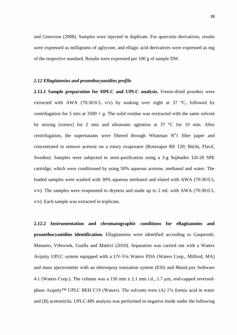
2.12 Ellagitannins and proanthocyanidins profile
2.12.1 Sample preparation for HPLC and UPLC analysis. Freeze-dried powders were
extracted with AWA (70:30:0.5, v/v) by soaking over night at 37
°C, followed by
centrifugation for 5 min at 3500 × g. The solid residue was rextracted with the same solvent
by mixing (vortex) for 2 min and ultrasonic agitation at 37
°C for 10 min. After
centrifugation, the supernatants were filtered through Whatman No1 filter paper and
concentrated to remove acetone on a rotary evaporator (Rotavapor RE 120; Büchi, Flavil,
Sweden). Samples were subjected to semi-purification using a 3-g Sephadex LH-20 SPE
cartridge, which were conditioned by using 50% aqueous acetone, methanol and water. The
loaded samples were washed with 30% aqueous methanol and eluted with AWA (70:30:0.5,
v/v). The samples were evaporated to dryness and made up to 2 mL with AWA (70:30:0.5,
v/v). Each sample was extracted in triplicate.
2.12.2 Instrumentation and chromatographic conditions for ellagitannins and
proanthocyanidins identification. Ellagitannins were identified according to Gasperotti,
Masuero, Vrhovsek, Guella and Mattivi (2010). Separation was carried out with a Waters
Acquity UPLC system equipped with a UV-Vis Waters PDA (Waters Corp., Milford, MA)
and mass spectrometer with an eletrospray ionization system (ESI) and MassLynx Software
4.1 (Waters Corp.). The column was a 150 mm x 2.1 mm i.d., 1.7 m, end-capped reversed-
phase Acquity™ UPLC BEH C18 (Waters). The solvents were (A) 1% formic acid in water
and (B) acetonitrila. UPLC-MS analysis was performed in negative mode under the following

39
conditions: capillary voltage 3 kV, source temperature 100 °C, desolvation temperature 350
°C, desolvation gas flow (N2) 650 L/h. The m/z range was 50-2000 Da. Ellagic acid and
elagitannins were identified using UV detection at 260 nm. Results were expressed as mg of
ellagic acid equivalents (EAE)/100 g sample DW.
Proanthocyanidins characterization was performed according to Robbins et al. (2009).
Samples were analyzed using a HPLC Agilent 1260 Infinity Series system equipped with
fluorescence detector. Solvents were (A) acetonitrile/acetic acid (98:2, v/v) and (B)
methanol/water/acetic acid (95:3:2, v/v/v). Fluorescence detection was conducted with an
excitation wavelength of 230 nm and an emission wavelength of 321 nm. Normal phase
separations were performed using Develosil Diol 100 Å (250 x 4.6 mm, 5 m particle size)
purchased from Phenomenex (Torrance, CA, USA). Results were expressed as mg
epicatechin equivalent per 100 g of sample DW.
2.13 Statistical Analysis. All analyses were run in triplicate and results were expressed as
mean standard deviation (SD). Initially, the results were checked for homogeneity of
variances by using the Levene test, and one-way ANOVA and the least significant difference
Fisher test was used to compare the means within group. P-values below 0.05 were regarded
as significant. All statistical analysis were performed by using the Statistica software package
version 11.0 (StatSoft, Inc., Tulsa, OK).

40
3. RESULTS AND DISCUSSION
3.1 Comparison between the ripe fruits of two species of jaboticaba
Among the Brazilian native fruits largely studied in the recent years, the most popular
is by far jaboticaba, whose production and commercialization have raised following the
increasing acceptance by the Brazilian population (Abe et al., 2012). Nowadays, Paulista and
Sabará are the only two species of commercial importance in the agronomic context, since the
others grow as a domestic crop (Lima et al., 2008).
Here, fruits of the two species, grown in a commercial plantation under the same
conditions (soil, climate and temperature) were collected and compared in relation to
chemical and mineral composition (Table 1).
The amount of protein (around 1.0%), fiber (17.9 – 19.3%) and carbohydrates (76.5 –
78.2%) was similar, presenting less than 8% of variation. Lipid content, however, was 27%
higher in the Paulista species (0.55%), and ashes were higher in the Sabará species (2.9%).
Protein and lipid contents are in accordance with previous reports (Lima et al., 2008),
but higher contents of fiber and carbohydrates were found by the other authors, which is 30%
and 12% higher than those values obtained in this work, respectively.

41
Table 1. Chemical and mineral composition of two species of jaboticaba, Paulista (Myrciaria
cauliflora) and Sabará (Myrciaria jaboticaba), in the ripe stage.
Composition (mg/100 g DW) Paulista Sabará
Protein 1.02 ± 0.01 a 0.94 ± 0.02
a
Lipid 0.55 ± 0.01 a 0.40 ± 0.01
b
Ashes 2.30 ± 0.08 b 2.90 ± 0.10
a
Fibers 17.9 ± 1.0 a 19.3 ± 0.9
a
Soluble 1.80 ± 0.15 a 2.30 ± 0.20
a
Insoluble 16.1 ± 0.7 a 17.0 ± 0.8
a
Carbohydrates* 78.2 a 76.5
a
Mineral (mg/100 g DW)
Nitrogen (N) 800 660
Phosphorus (P) 110 100
Potassium (K) 1320 1000
Calcium (Ca) 20 20
Magnesium (Mg) 120 100
Sulfur (S) 80 70
Boron (B) 0.7 0.8
Zinc (Zn) 2 2.9
Iron (Fe) 2.9 2.7
Manganese (Mn) 1.8 2.7
Copper (Cu) 1 1
Sodium (Na) 0.76 1.03
Selenium (Se) < LQ < LQ
*calculated by difference; Means in the same line with common letters are not significantly different (p<0,05).
LQ: Limit of quantification for Se = 0.002 mg/kg
The mineral composition of the two species of jaboticaba (Paulista and Sabará) was
assessed by determination of nitrogen, phosphorus, potassium, calcium, magnesium, sulfur,
boron, zinc, iron, manganese, copper, sodium and selenium (Table 1). The two species
presented similar contents of minerals. Nitrogenium and potassium contents were higher in
the Paulista species, but Sabará showed the highest concentrations of zinc, manganese and

42
sodium. Among the minerals analyzed, zinc, iron and manganese were the elements in highest
levels. The contents of phosphorus, iron, copper and calcium were similar between the
samples, and differences were less than 10%. The elements with the greatest discrepancy
between the two species were manganese and sodium. Environmental conditions during plant
development, such as seasonality, soil type, rainfall, ultraviolet radiation and temperature can
influence the mineral composition of fruits and vegetables (Siegler, 1998). Since the both
species were grown under the same conditions and collected at the same time, the differences
observed between Paulista and Sabará can be attributed to the genetic factors.
Lima, Corrêa, Dantas-Barros, Nelson and Amorim (2011) analyzed the mineral profile
of whole fruits of the same species Paulista and Sabará, and potassium was the main element
detected. Fruits are usually rich in potassium, which is concentrated especially in the skin.
Potassium is an element with high motility in plants, due to its low affinity to chelation, which
explains the high concentration in plant tissues. The presence of magnesium (100-120 mg/100
g DW) in jaboticaba fruits is due to the accumulation of this element in the skin, which
suggests an association with the concentration of anthocyanins, since magnesium has been
related to changes in the color of these pigments. The high concentration of phosphorus, in
turn, may be associated with phytin in the seeds, a compound which has a function as a
phosphorus reservoir for germination (Goodwin & Mercer, 1983).
The results presented here show that a serving of 100 g of jaboticaba, approximately
15 units, would provide from 10 to 15% of the recommended daily intake (RDI) of copper,
manganese and potassium. Considering the FDA’s (Food and Drug Administration’s)
definition, jaboticaba is a “good source” of these minerals, since one serving contains at least
10% of the RDI.
The two species of jaboticaba in the ripe stage, as well as its fractions skin, pulp and
seed were also evaluated in relation to the in vitro antioxidant capacity and the phenolic

43
content and composition (Figure 1). Sabará was the species that presented the highest
antioxidant capacity, approximately 20% higher than Paulista. Seeds were the most important
fraction contributing to the antioxidant properties of jaboticaba, followed by skin and pulp,
regardless of the method used (Folin-Ciocalteu reducing capacity, DPPH scavenging ability
or Ferric reducing antioxidant power).
Pulp showed the lowest total phenolic content and consequently the lowest antioxidant
capacity, which can be associated with several chemical and enzymatic modifications of
certain compounds during the ripening process. These alterations include hydrolysis of
glycosides by glycosidases, oxidation of phenols by phenoloxidases and polymerization of
free phenols. Moreover, the accumulation of soluble phenolics is higher in external plant
tissues, such as the skin, compared with the internal parts, like pulp (Robards, Prenzler,
Tucker, Swatsitang & Glover, 1999).
Here, the condensed tannins were evaluated by three different methods: the vanillin
method (Burns, 1971), the acidified butanol (Porter, Hrstich & Chan, 1986) and the DMAC
assay (Prior et al., 2010). The two first are colorimetric methods, and can suffer interferences
from sample components, such as anthocyanins, which are measured in the same wavelength
used for proanthocyanidins, leading to overestimated results. In this way, the DMAC method
developed recently has been considered more appropriate, since proanthocyanidins are
quantified at 640 nm. The mechanism of action of the DMAC reagent has not yet been
completely understood, although it seems to react with compounds having free hydroxyl
groups in a meta position and a single bond at the position 2,3 on the C ring (Prior et al.,
2010).

44
Figure 1. Comparison between Sabará and Paulista jaboticabas about: (A) Folin Ciocalteu
reducing ability, (B) DPPH scavenging ability, (C) Ferric reducing capacity (FRAP),
Proanthocyanidin content assessed by (D) Butanol-HCl assay, (E) Vanillin assay, (F) DMAC
assay, (G) Anthocyanidin content, (H) Quercetin derivatives content, (I) Free ellagic acid
content, (J) Total ellagic acid content. Different letters (a, b) represent significant differences
(p < 0.05) between the two species Paulista and Sabará.
A

45
Independent of the method used for quantification, the proanthocyanidin content was
higher in the Sabará jaboticaba. Among fruit fractions, seeds presented the highest amount,
followed by skin and pulp (Figure 1). Compared to other fruits, the amounts of
proanthocyanidins in strawberry, blueberry and cranberry, considered rich-PAC fruits, are
about 10 times higher than that of the ripe jaboticaba. Grapes, in turn, had 20 times more
proanthocyanidins, since the peel and seeds are rich in these compounds (Wang, Chung, Song
& Chun, 2011). Notwithstanding this, jaboticaba can be considered a good source of
proanthocyanidins, especially for the Brazilian population, for whom the availability of
blueberries and cranberries is limited.
Flavonoids contents were higher in the Paulista jaboticaba, which presented 25% more
anthocyanins and 42% more quercetin derivatives than Sabará. These two classes of
compounds were concentrated in the skin. A small amount of quercetin derivatives was
detected in the pulp. There was no flavonoids in the seeds (Figure 1).
The content of anthocyanins observed in the ripe jaboticaba is higher compared to
other fruits of the Myrtaceae family, such as camu-camu and pitanga (Abe et al., 2012). In a
previous study, cyanidin 3-glucoside and delphinidin 3-glucoside were identified in the skin
of the ripe jaboticaba, and the first one was also detected in small amounts in the pulp (Leite
et al., 2011; Lima et al., 2011).
Ellagic acid derivatives were detected in high concentrations in both species (Figure
1). The content of free ellagic acid found in the skin, pulp and seeds of both species of
jaboticaba was significantly low, varying from 2.2 mg/100 g DW (Paulista pulp) to 34
mg/100 g DW (Sabará seeds). The total ellagic acid content of jaboticaba was much higher
(56%) in the Paulista than in Sabará species. Among the fruit fractions, pulp presented the
lowest content (420-861 mg/100 g DW) and seeds the highest (3016-4180 mg/100 g DW)
(Figure 1).

46
Among several Brazilian native fruits, the highest amounts of ellagic acid derivatives
were found in the species of the Myrtaceae family. Jaboticaba, grumixama (Eugenia
brasiliensis) and cambuci (Campomanesia phaea) presented the highest contents of free and
total ellagic acid, comparable to fruits such as raspberry and blackberry, known as good
sources of these compounds (Abe et al., 2012).
3.2 Changes in the chemical composition during ripening of jaboticaba
Samples of Sabará jaboticaba were manually classified into five different ripening
stages, according to size and skin color. Physicochemical analyses and the sugar content and
composition are shown in Table 2. There was no significant difference in the moisture
content of the different ripening stages and the mean value was of approximately 83%. Fruits
showed a continuous increase in the diameter along the ripening process, as expected. Ready-
to-consume fruits presented an average diameter of 2.4 cm.
Total soluble solids are highly correlated with soluble sugars and organic acids
contents, and the concentration of these compounds is one of the most important variables in
fruit quality. For jaboticaba, there was an increase in soluble solids along the ripening, as
expected. The increase in pH was accompanied by a decrease in titratable acidity, due to a
reduction in the content of organic acids during fruit ripening (Table 2).
A high amount of sugar was detected in jaboticaba and as expected, the sugar content
was higher in the pulp and when ripe. In the ripe fruit, it was detected 3 times more fructose,
10 times more glucose and 18 times more sucrose than in the unripe. A significant sugar
content was also observed in the skin, which may be due to the presence of a remaining
portion of the pulp adhered to the skin (Lima et al. 2011). Seeds were the portion with the
lowest amount of sugar, from 4 to 16 times lower than pulp (Table 2).

47
Table 2. Moisture (%), diameter (cm), total soluble solids (°Brix), total titratable acidity (g
citric acid/100 g FW), pH, and sugar content (sucrose, glucose and fructose) (g/100 g DW) of
Sabará jaboticaba in different ripening stages.
Physicochemical parameters
Ripening stages Stage 1
(unripe)
Stage 2 Stage 3 Stage 4 Stage 5
(fully ripe)
Skin colour Green Light red Red Dark red Dark purple
Moisture 84 81 86 83 82
Diameter 1.6 ± 0.2 1.8 ± 0.2 1.9 ± 0.2 2.0 ± 0.2 2.4 ± 0.1
TSS 5.6 7.7 8.9 10.7 12.4
TTA 4.73 3.52 2.10 1.74 1.39
pH 2.67 2.84 3.03 3.28 4.08
Sugar content of ripening stages
Sucrose 0.92 na 6.61 na 16.62
Glucose 1.76 na 9.48 na 16.96
Fructose 7.33 na 15.99 na 21.42
Sugar content of fruit fractions Pulp Seed Skin
Sucrose na na na na 11.3 14.5 19.1
Glucose na na na na 18.6 15.9 22.9
Fructose na na na na 4.9 1.5 1.4
na: not analyzed
The antioxidant capacity also varied during ripening, and was assessed by three
different methods (Figure 2). The Folin-Ciocalteu reducing capacity decreased 67% from the
unripe to the ripe fruit. Similarly, there was a reduction of 60% by DPPH scavenging ability
and 77% by ferric reducing antioxidant power. A significant positive correlation was found
among the three methods used to evaluate antioxidant activity (0.90 ≤ r ≤ 0.95).

48
Figure 2. Antioxidant capacity of jaboticaba assessed by (A) Folin Ciocalteu reducing
capacity (mg catechin equivalent/100 g sample DW), (B) DPPH-radical scavenging ability
(mol Trolox equivalent/100 g sample DW) and (C) Ferric reducing antioxidant power
(FRAP) (mmol Trolox equivalent/100 g sample DW) of Sabará jaboticaba in different
ripening stages.

49
Many plant species and compounds isolated from plants have been experimentally
studied and used for reducing the postprandial glycemia (Grover, Yavad & Vats, 2002). In
order to evaluate the antidiabetic potential of jaboticaba, the in vitro assays for inhibitory
activity of enzymes involved in carbohydrate metabolism were carried out (Table 3). The in
vitro inhibitory activity of -amylase and -glucosidase was higher in the ripe fruits,
probably due to the highest concentration of anthocyanins in this stage. However, the unripe
jaboticaba also exhibited a significant inhibitory activity compared to other fruits, such as
tucumã and star fruit (Gonçalves, Lajolo & Genovese, 2010). The phenolic compounds from
seeds were the most potent inhibitor of -glucosidase, similar to results observed in cambuci
and cupuaçu (Gonçalves, Lajolo & Genovese, 2010).
Table 3. In vitro inhibitory activity of -amylase and -glucosidase of phenolic fractions
obtained from jaboticaba in diferente ripening stages.
Samples
-amylase -glucosidase
IC50
(mg sample DW
/L reaction)
IC50
(g CEa/
L reaction)
IC50
(mg sample DW
/L reaction)
IC50
(g CEa/
L reaction)
Ripening stages
Stage 1 2.9 1.2 1.7 1.3
Stage 2 1.9 1.8 1.6 1.0
Stage 3 1.0 0.4 1.6 0.9
Stage 4 1.3 0.6 1.8 1.6
Stage 5 0.6 0.5 0.7 0.5
Fruit portions of stage 5
Skin 1.7 1.3 0.7 0.8
Pulp 1.6 0.8 1.0 0.3
Seeds 1.1 0.5 0.6 0.2
aCE – catechin equivalent

50
It was previously reported that the most potent inhibitors of -glucosidase were
myricetin, epigallocatechin gallate, cyanidin, among 16 compounds belonging to six different
groups: flavone, flavonol, flavanone, isoflavone, flavan-3-ol and anthocyanidin (Tadera,
Minami, Takamatsu, & Matsuoka (2006). Therefore, anthocyanins present in the skin of
jaboticaba seem to have an important role in the inhibitory activity of the whole fruit.
Quercetin and anthocyanidin derivatives were the main flavonoids present in
jaboticaba, but they were detected only in skin and pulp, and not in seeds (Table 4). Ripening
caused an anthocyanin accumulation in the skin of the fruit, with a gradual increase in the
anthocyanin content of the whole fruit, from 5 to 147 mg/100 g of sample DW, comparing the
unripe with the ripe fruit. Regarding quercetin derivatives, they were not detected in the
unripe fruit. From the second stage (light red), however, 2 mg/100 g of sample (DW) were
found and this value did not change during maturation.
Table 4. Flavonoids, free and total ellagic acid content (mg/100 g sample DW) of Sabará
jaboticaba in different ripening stages.
Compounds Ripening stages
Stage 1 Stage 2 Stage 3 Stage 4 Stage 5
Free ellagic acid 19 ± 2c 31 ± 3b 31 ± 3b 39 ± 2a 40 ± 1 a
Total ellagic acid 9566 ± 812 a 8327 ± 129 b 6363 ± 629 c 6810 ± 606 c 5050 ± 99 d
Anthocyanins 4.9 ± 0.3 e 44 ± 1 d 65 ± 1 c 74 ± 3 b 147 ± 10 a
Delphinidin na na 7.1 ± 0.2 b na 23.5 ± 0.8 a
Cyanidin na na 58 ± 2 b na 123 ± 2 a
Quercetin derivatives nd 2.0 ± 0.1 a 2.1 ± 0.1 a 2.00 ± 0.05 a 2.1 ± 0.2 a
nd: not detected; na: not analyzed

51
Similarly, there was an increase in the anthocyanidins derivatives content during
ripening of grapes, while other classes of polyphenols exhibited a pattern of accumulation and
subsequent decline, which suggest a degradation or the utilization as a substrate in the
biosynthesis of other compounds (Adams, 2006).
Total ellagic acid decreased as jaboticaba ripened. The unripe fruit presented 9566 mg
of total ellagic acid/100 g of sample DW and 5050 mg/100 g DW were detected in the ripe
fruit, which indicates a decrease of 50% in the content of total ellagic acid during ripening
process (Table 4).
The same behavior was observed in strawberry, when five cultivars in three different
ripening stages were analyzed in relation to the total ellagic acid content. In the unripe stage,
amounts between 8.8 and 17.8 mg/100 g fresh weight (FW) were detected, in the intermediate
stage values varied from 4 to 9.4 mg/100 g FW and, from 1.6 to 4.5 mg/100 g FW in the ripe
stage. Ripening caused a decrease in the total ellagic acid content in all cultivars analyzed
(Williner, Pirovani & Güemes, 2003). Ellagic acid derivatives were the main compounds
found in the unripe strawberries, while pelagordin 3-glucoside, an anthocyanin derivative,
was the main compound present in the ripe fruits (Kosar, Kafkas, Paydas & Baser, 2004).
Proanthocyanidins contribute significantly to total polyphenols intake in the Western
diet, due to their wide distribution among different plant species (Santos-Buelga & Scalbert,
2000). However, there are few data available in the scientific literature on the qualitative and
quantitative profile of these compounds in foods, which is mainly due to the difficulty in
finding an appropriate method to characterize them.
Generally, tannins present in fruits in the non-polymerized form have tannic properties
more accentuated. During ripening, there is a polymerization of tannins and a reduction of
astringency (Clifford, 1997), which improves the sensory attributes of fruits.
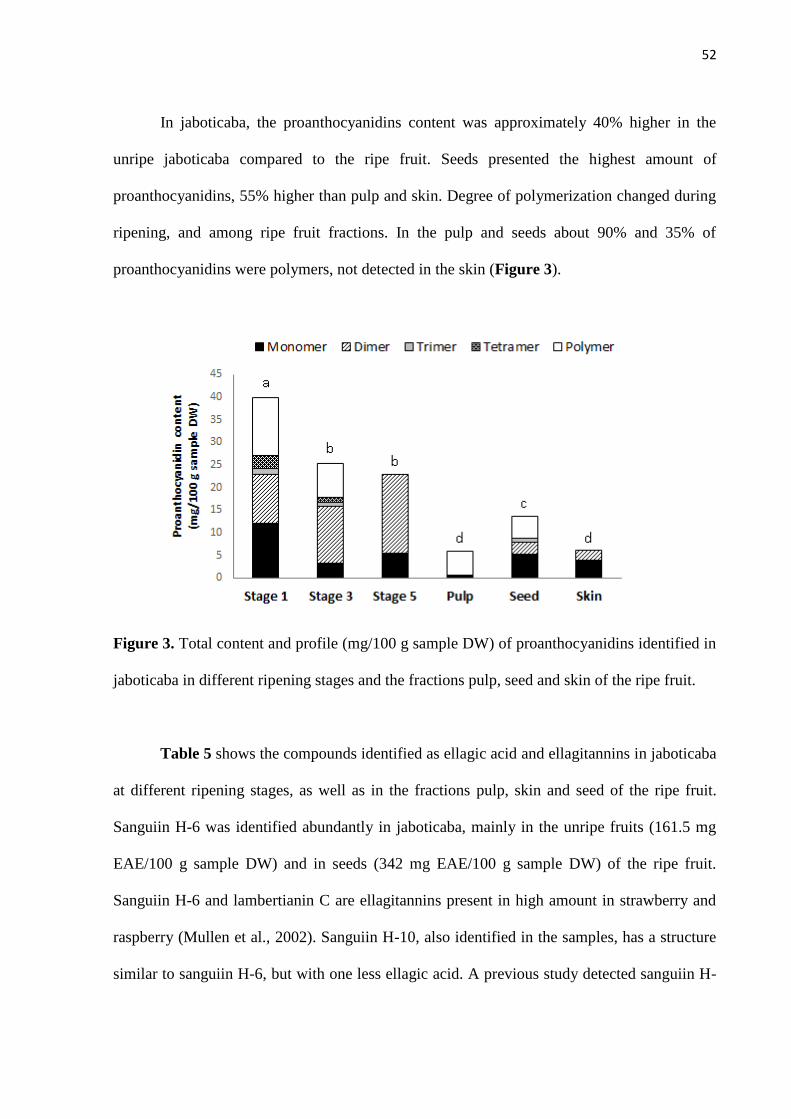
52
In jaboticaba, the proanthocyanidins content was approximately 40% higher in the
unripe jaboticaba compared to the ripe fruit. Seeds presented the highest amount of
proanthocyanidins, 55% higher than pulp and skin. Degree of polymerization changed during
ripening, and among ripe fruit fractions. In the pulp and seeds about 90% and 35% of
proanthocyanidins were polymers, not detected in the skin (Figure 3).
Figure 3. Total content and profile (mg/100 g sample DW) of proanthocyanidins identified in
jaboticaba in different ripening stages and the fractions pulp, seed and skin of the ripe fruit.
Table 5 shows the compounds identified as ellagic acid and ellagitannins in jaboticaba
at different ripening stages, as well as in the fractions pulp, skin and seed of the ripe fruit.
Sanguiin H-6 was identified abundantly in jaboticaba, mainly in the unripe fruits (161.5 mg
EAE/100 g sample DW) and in seeds (342 mg EAE/100 g sample DW) of the ripe fruit.
Sanguiin H-6 and lambertianin C are ellagitannins present in high amount in strawberry and
raspberry (Mullen et al., 2002). Sanguiin H-10, also identified in the samples, has a structure
similar to sanguiin H-6, but with one less ellagic acid. A previous study detected sanguiin H-

53
10 and sanguiin H-3 in Sanguisorba officinalis, which belongs to Rosaceae family (Tanaka,
Nonaka & Nishioka, 1985).
The ellagitannins composition identified in this work did not reproduce the profile
described previously (Reynertson et al., 2006; Wu et al. 2012). Here, 14 compounds were
identified as ellagic acid derivatives, while only three were detected in these two anterior
studies, along with other eight compounds identified as gallic acid derivatives. However, the
fruits of jaboticaba analyzed in the other studies were collected from the Botanical Garden in
the United States. We believe that those samples may not be representative of the species, and
our fruits were obtained from the main producer of jaboticaba in Brazil.
Quantification and structural characterization of ellagitannins and ellagic acid
derivatives in foods, such as the Brazilian native fruits, are starting points for the study of
biological effects, since these compounds have been associated with several healthy
properties, as antioxidant, antiviral and anticancer (Gasperotti et al., 2010).

54
Table 5. Ellagitannins profile of jaboticaba during ripening and fractions pulp, seed and skin of the ripe fruit (mg ellagic acid/100 g sample DW).
Compound Pulp Seed Skin Stage 1 Stage 3 Stage 5 Retention time (min)
Ellagic acid 9.4 nd 21.2 26.0 15.4 21.9 7.7 603; 449; 301
Sanguiin H-10 isomer (1) 1.6 12.6 19.5 61.8 24.0 9.3 3.7 783; 649
Sanguiin H-10 isomer (2) 4.8 35.6 35.0 96.0 77.0 17.2 3.9 783; 649
Sanguiin H-10 isomer (3) 5.4 38.6 32.3 75.8 42.0 17.9 5.0 783; 649
Lambertianin C without
ellagic acid moiety 1.8 2.8 10.4 24.7 21.0 6.9 8.0 1250
Sanguiin H-6 (1) 1.8 175.0 nd 84.8 37.0 25.7 3.4 934; 858; 1235; 469
Sanguiin H-6 (2) 6.6 145.0 3.6 nd 36.0 24.5 4.4 934; 858; 1235; 469
Sanguiin H-6 (3) nd 5.2 3.4 18.9 12.0 2.2 5.7 934; 858; 1235; 469
Sanguiin H-6 (4) 2.8 3.0 8.9 13.7 15.5 4.8 6.8 934; 858; 1235; 469
Sanguiin H-6 (5) nd 4.6 1.9 9.1 4.7 1.0 7.0 934; 858; 1235; 469
Sanguiin H-6 (6) 2.6 4.6 8.8 19.3 15.8 4.8 7.2 934; 858; 1235; 469
Sanguiin H-6 (7) nd 4.6 2.2 8.0 5.6 1.0 8.0 934; 858; 1235; 469
Sanguiin H-6 (8) nd nd nd 7.7 5.5 1.2 8.6 934; 858; 1235; 469
Unknown ellagitannin 2.2 7.5 96.4 nd nd nd 8.8 505; 1011
nd : not detected

55
4. CONCLUSIONS
This work reported, for the first time, a complete and detailed characterization of two
commercially important species of jaboticaba, as well as its fractions and ripening stages. The
two species of jaboticaba presented a similar centesimal and mineral composition, being
important sources of K, Mn and Cu. The Paulista jaboticaba presented the highest contents of
quercetin derivatives and anthocyanins. However, the major polyphenols of jaboticaba were
the ellagic acid derivatives, and these compounds were detected in higher amounts in the
Sabará jaboticaba. Skin was the portion of fruit with the highest content of flavonoids, but the
seeds presented the highest amount of ellagic acid derivatives. The unripe fruit showed the
highest antioxidant capacity and phenolic contents, and ripening led to a decrease in all these
compounds. Ready-to-consume fruits were shown to be very rich sources of polyphenols,
especially anthocyanins, and an important source of ellagitannins, comparable to the berries.
Phenolic compounds from jaboticaba strongly inhibited the in vitro activity of enzymes
involved in carbohydrate metabolism. This study showed that jaboticaba is an excellent
dietary source of polyphenols and that the most commercially relevant species is also the
richest phenolic source. Altogether, these results showed the importance of eating the whole
jaboticaba, without discarding the skin or seeds, in order to take advantages of all the
compounds present in different portions of the fruit.
Acknowledgements
This research was supported by Brazilian Government through Fundação de Amparo à
Pesquisa do Estado de São Paulo - Fapesp (2009/01775-0) and Canadian Government through
Emerging Leaders in the Americas Program (ELAP). We also thank Unidos Com. Imp. Exp.

56
Hortigranjeiros from Companhia de Entrepostos e Armazéns Gerais de São Paulo (Ceagesp)
for providing the samples.
REFERENCES
Abe, L. T., Lajolo, F. M., & Genovese, M. I. (2012). Potential dietary sources of ellagic acid
and other antioxidants among fruits consumed in Brazil: jaboticaba (Myrciaria jaboticaba
(Vell.) Berg). Journal of the Science of Food and Agriculture, 92, 1679-87.
Adams, D. O. (2006). Phenolics and ripening in grape berries. American Journal of Enology
and Viticulture, 57, 249-256.
AOAC. (2005). Official Methods of Analysis. (18th
ed.). Washington: Association of Official
Analytical Chemists.
Arabbi, P. R., Genovese, M. I., & Lajolo, F. M. (2004). Flavonoids in vegetable foods
commonly consumed in Brazil. Journal of Agricultural and Food Chemistry, 52, 1124-1131.
Benzie, I. F. F, & Strain, J. J. (1996). The Ferric Reducing Ability of Plasma (FRAP) as a
measure of ‘‘Antioxidant Power’’: The FRAP Assay. Analytical Biochemistry, 239, 70-76.
Brand-Williams, W., Cuvelier, M. E., & Berset, C. (1995). Use of a free radical method to
evaluate antioxidant activity. Food Science and Technology, 28, 25–30.
Burns, R. E. (1971). Method for estimation of tannin in grain sorghum. Agronomy Journal,
63, 511-512.
Clifford, M. N. (1997). Astringency. In: TOMÁS-BARBERÁN, F. A.; ROBINS, R. J.
Phytochemistry of fruit and vegetables (pp. 87-107). Oxford: Clarendon Press.
Clifford, M. N., & Scalbert, A. (2000). Ellagitannins - nature, occurrence and dietary burden.
Journal of the Science of Food and Agriculture, 80, 1118-1125.

57
Donadio, L. C. (2000). Jabuticaba (Myrciaria jaboticaba (Vell.) Berg. Jaboticabal: FUNEP,
55 pp.
Gasperotti, M., Masuero, D., Vrhovsek, U., Guella, G., & Mattivi, F. (2010). Profiling and
accurate quantification of Rubus ellagitannins and ellagic acid conjugates using direct UPLC-
Q-TOF HDMS and HPLC-DAD analysis. Journal of Agricultural and Food Chemistry, 58,
4602-4616.
Genovese, M. I., Pinto, M. S., Gonçalves, A. E. S. S., & Lajolo, F. M. (2008). Bioactive
compounds and antioxidant capacity of exotic fruits and commercial frozen pulps from Brazil.
Food Science and Technology International, 14, 207-214.
Gonçalves, A. E. S. S., Lajolo, F. M., & Genovese, M. I. (2010). Chemical Composition and
Antioxidant/Antidiabetic Potential of Brazilian Native Fruits and Commercial Frozen Pulps.
Journal of Agricultural and Food Chemistry, 58, 4666-4674.
Goodwin, T. W., & Mercer, E. I. (1983). Introduction to plant biochemistry. Pergamon:
Oxford. 677 p.
Kosar, M., Kafkas, E., Paydas, S., & Baser, K. H. C. (2004). Phenolic composition of
strawberry genotypes at different maturation stages. Journal of Agricultural and Food
Chemistry, 52, 1586-1589.
Leite, A. V., Malta, L. G., Riccio, M. F., Eberlin, M. N., Pastore, G. M., & Maróstica Júnior,
M. R. (2011). Antioxidant potential of rat plasma by administration of freeze-dried jaboticaba
peel (Myrciaria jaboticaba Vell Berg). Journal of Agricultural and Food Chemistry, 59,
2277-2283.
Lima, A. J. B., Corrêa, A. D., Alves, A. P. C., Abreu, C. M. P., & Dantas-Barros, A. M.
(2008). Caracterização química do fruto jabuticaba (Myrciaria cauliflora Berg) e de suas
frações. Archivos Latinoamericanos de Nutrición, 58, 416-421.

58
Lima, A. J. B., Corrêa, A. D., Dantas-Barros, A. M., Nelson, D. L., & Amorim, A. C.
L.(2011). Sugars, organic acids, minerals and lipids in jabuticaba. Revista Brasileira de
Fruticultura, 33, 540-550.
Mullen, W., Mcginn, J., Lean, M. E., Maclean, M. R., Gardner, P., Duthie, G. G., Yokota, T.,
& Crozier, A. (2002). Ellagitannins, flavonoids, and other phenolics in red raspberries and
their contribution to antioxidant capacity and vasorelaxation properties. Journal of
Agricultural and Food Chemistry, 50, 5191-5196.
Pinto, M. S., Lajolo, F. M., & Genovese, M. I. (2008). Bioactive compounds and
quantification of total ellagic acid in strawberries (Fragaria x ananassa Duch.). Food
Chemistry, 107, 1629-1635.
Porter, L. J., Hrstich, L. N., & Chan, B. G. (1986). The conversion of procyanidins and
prodelphinidins to cyanidin and delphinidin. Phytochemistry, 25, 223-230.
Prior, R. L., Fan, E., Ji, H., Howell, A., Nio, C., Payne, M. J., & Reed, J. (2010). Multi-
laboratory validation of a standard method for quantifying proanthocyanidins in cranberry
powders. Journal of the Science of Food and Agriculture, 90, 1473–1478.
Reynertson, K. A., Wallace, A. M., Adachi, S., Gil, R. R., Yang, H., Basile, M. J.,
D’Armiento, J., Weinstein, I. B., & Kennelly, E. J. (2006). Bioactive depsides and
anthocyanins from jaboticaba (Myrciaria cauliflora). Journal of Natural Product, 69, 1228-
1230.
Ribeiro, M. C., Metzger, J. P., Martensen, A. C., Ponzoni, F., & Hirota, M. M. (2009).
Brazilian Atlantic forest: how much is left and how is the remaining forest distributed?
Implications for conservation. Biological Conservation, 142, 1141–1153.
Robards, K., Prenzler, P. D., Tucker, G., Swatsitang, P., & Glover, W. (1999). Phenolic
compounds and their role in oxidative processes in fruits. Food Chemistry, 66, 401-436.

59
Robbins, R. J., Leonczak, J., Johnson, J. C., Li, J., Kwik-Uribe, C., Prior, R. L., & Gu, L.
(2009). Method performance and multi-laboratory assessment of a normal phase high pressure
liquid chromatography–fluorescence detection method for the quantitation of flavanols and
procyanidins in cocoa and chocolate containing samples. Journal of Chromatography A,
1216, 4831-4840.
Santos-Buelga, C., & Scalbert, A. (2000). Proanthocyanidins and tannin-like compounds –
nature, occurrence, dietary intake and effects on nutrition and health. Journal of the Science of
Food and Agriculture, 80, 1094-1117.
Seigler, D. S. (1998). Plant secondary metabolism. Dordrecht: Kluwer Academic. 759p.
Singleton, V. L., Orthofer, R., & Lamuela-Raventos, R. M. (1999). Analysis of total phenols
and other oxidation substrates and antioxidants by means of Folin–Ciocalteu reagent. Methods
in Enzymology, 299, 152–178.
Tadera, K., Minami, Y., Takamatsu, K., & Matsuoka, T. (2006). Inhibition of -glucosidase
and -amylase by flavonoids. Journal of Nutritional Science and Vitaminology, 52, 149-153.
Tanaka, T., Nonaka, G., & Nishioka, I. (1985). Tannins and related compounds. Part 28.
Revision of the structures of sanguiins H-6, H-2 and H-3, and isolation and characterization of
sanguiin H-11, a novel tetrameric hydrolysacle tannin, and seven related tannins, from
Sanguisorba officinalis. Journal of Chemical Research, 176-177.
Wang, Y.; Chung, S.-J.; Song, W. O., & Chun, O. K. (2011). Estimation of daily
proanthocyanidins intake and major food sources in the U.S. Diet. The Journal of Nutrition,
141, 447-452.
Williner, M. R., Pirovani, M.E., & Güemes, D.R. (2003). Ellagic acid content in strawberries
of different cultivars and ripening stages. Journal of the Science and Food and Agriculture,
83, 842-845.

60
Wu, S. B., Dastmalchi, K., Long, C., & Kennelly, E. J. (2012). Metabolite profiling of
jaboticaba (Myrciaria cauliflora) and other dark-colored fruit juices. Journal of Agricultural
and Food Chemistry, 60, 7513−7525.
Wu, X., & Prior, R. L. (2005). Systematic identification and characterization of anthocyanins
by HPLC-ESI-MS/MS in common foods in the United States: fruits and berries. Journal of
Agricultural and Food Chemistry, 53, 2589-2599.

61
In vitro and in vivo evaluation of the metabolism of polyphenols from jaboticaba
(Myrciaria jaboticaba (Vell.) Berg), a Brazilian native fruit
MARCELA ROQUIM ALEZANDRO 1, PASCAL DUBÉ
2, YVES DESJARDINS
2,
FRANCO MARIA LAJOLO 1, MARIA INÉS GENOVESE
1*
* corresponding author (Tel: 55-11-30911525; Fax: 55-11-38154410; e-mail:
1 Laboratório de Compostos Bioativos de Alimentos, Departamento de Alimentos e Nutrição
Experimental, FCF, Universidade de São Paulo, Av. Prof. Lineu Prestes 580, Bloco 14,
05508-900 São Paulo, SP, Brazil.
2 Institut des Nutraceutiques et des Aliments Fonctionnels, Pavillon des Services, Université
Laval, 2440 Hochelaga Blvd, G1V 0A6, Québec, Québec Canada
62
ABSTRACT
Ellagic acid derivatives are the main polyphenols found in jaboticaba (Myrciaria jaboticaba
Vell. Berg), a Brazilian native fruit. Those compounds are known due to their cancer
chemopreventive, cardioprotective and antioxidant potential. However, little is known about
their metabolites, which are the biologically active compounds indeed. Here, the aim was to
investigate the metabolism of different classes of polyphenols, especially ellagitannins and
ellagic acid derivatives, from jaboticaba in both in vitro and in vivo assays. The metabolites
formed from the jaboticaba polyphenols were identified in an in vitro fermentation model
using human feces. In addition, the fate of a wide variety of metabolites was monitored after
intragastric administration of jaboticaba extract (15 min – 8 h) in Wistar rats, using an UPLC-
MS. The in vitro experiment showed that the ellagic acid derivatives were metabolized by the
intestinal microbiota and degraded under testing conditions. Two compounds were identified
after fermentation with fecal inoculum, p-hydroxybenzoic and p-hydroxyphenylacetic acids.
In vivo, thirty eight metabolites were identified in plasma, stomach, liver, kidneys, brain,
muscle and colon, and most of them were formed from ellagic acid derivatives.
Keywords : Bioavailability / Ellagitannins / Jaboticaba
63
1. Introduction
Jaboticaba, Myrciaria jaboticaba (Vell.) Berg, is a grape-like in appearance and
texture and it is by far the best-known and the most consumed Brazilian native fruit. It is from
the Atlantic Rainforest and belongs to the Myrtaceae family. There is a continuously growing
interest in its production and commercialization, due to the sweet and slightly acidic flavor of
the pulp, which encourages its consumption in natura and as ingredient in food products, such
as jam, juice and liqueur (Donadio, 2000).
Jaboticaba is a rich source of phenolic compounds, which include quercetin
derivatives, proanthocyanidins and anthocyanins, that are concentrated in the purple to black
skin, when the fruit is ready-to-eat. In addition, recently it was demonstrated that ellagic acid
derivatives, such as ellagitannins, are the main compounds found in jaboticaba, being
sanguiin H-10, sanguiin H-6 and lambertianin C the most abundant (Alezandro, Dubé,
Desjardins, Lajolo & Genovese, 2013).
Chronic ingestion of phenolic compounds from fruits has been associated with
beneficial effects on human health, related to their antioxidant, cancer chemopreventive, anti-
inflammatory and cardioprotective properties (Mullen et al., 2002; Priyadarsini, Khopde,
Kumar & Mohan, 2002; Youdim, McDonald, Kalt, & Joseph, 2002). However, a chemical
compound must be further metabolized to exert its biological activity, being able to achieve
the physiological target in a minimum concentration needed to promote the biological effects
(Oliveira & Bastos, 2011).
Only a small portion of phenolic compounds ingested is absorbed by the small
intestine. This process can occur through passive diffusion and be associated with hydrolysis
and release of the aglycone by the action of an enzyme, such as lactase phloridzin hydrolase
(LPH), present in the microvilli of intestinal epithelial cells (Donovan, Manach, Faulks, &

64
Kroon, 2006). Once absorbed, the aglycone is metabolized in the liver, forming sulfated,
glucuronidated and/or methylated metabolites by the action of the respective phase II
enzymes sulfotransferase (SULT), uridine diphosphate-50-glicuronosiltransferase (UGT) and
catechol-O-methyltransferase (COMT) (Crozier, Del Rio, & Clifford, 2010).
The products of this metabolism may entry into the bloodstream and be excreted in the
urine, or a considerable fraction can be excreted by the liver into bile followed by entry into
the small intestine, through enterohepatic circulation (McBain & Macfarlane, 1997). Once
released into the intestinal lumen, these conjugates can be hydrolyzed by bacterial enzymes,
such as β-glucuronidases, sulfatases and glycosidases (Crozier, Del Rio, & Clifford, 2010;
Selma, Espín, & Thomás Barberán, 2009). Compounds that are not absorbed in the small
intestine pass directly to the large intestine, where they are degraded by the colonic
microbiota to simpler compounds, such as phenolic acids, and absorbed into the circulatory
system (Del Rio, Costa, Lean, & Crozier, 2010).
Several studies have demonstrated that most of the phenolic compounds are
metabolized in the colon by the microbiota intestinal before absorption, and this conversion is
essential for modulating their physiological actions. It is clear that these metabolites are the
main responsible for biological effects observed in vivo (Del Rio, Costa, Lean, & Crozier,
2010; Setchell, Brown, & Lydeking-Olsen, 2002; Xu, Harris, Wang, Murphy, & Hendrich,
1995).
However, nothing is known about the metabolites profile formed from the jaboticaba
polyphenols, as well as the distribution in different target tissues. These points are important
in order to elucidate the biological activity of these compounds. This study aimed to evaluate
the bioavailability of jaboticaba polyphenols in an in vitro fermentation model using human
feces and an in vivo assay in Wistar rats.

65
2. Material and methods
2.1. Chemicals. Resazurin, tryptone, MOPS, Na2EDTA, sodium dithionite, ascorbic acid,
ellagic acid, quercetin, (+)-catechin, and the Folin-Ciocalteu reagent were purchased from
Sigma Chemical Co. (St Louis, MO, USA). Ketamine chloride and xylidine chloride were
acquired from Bayer (Leverkusen, Germany). The anthocyanidins cyanidin and delphinidin
and the respective 3-glucosides, as well as procyanidin B2 were obtained from Extrasynthèse
(Genay, France). All chemicals/solvents were of analytical or HPLC grade, according to the
requirement, and obtained from Merck (Darmstadt, Germany).
2.2. Samples. Fully ripened jaboticaba Sabará (Myrciaria jaboticaba Vell. Berg) was kindly
provided by a local producer at the São Paulo Central Market (Companhia de Entrepostos e
Armazéns Gerais de São Paulo – CEAGESP, Brazil). Fruits were cleaned and immediately
freeze-dried and stored at -20 °C. Then, the dried fruits were ground to a fine powder in
a mortar and pestle, using liquid nitrogen to keep sample frozen.
2.3. Procedure for jaboticaba raw extract preparation. Extraction was performed according
to Arabbi, Genovese and Lajolo (2004) with some modifications. Freeze-dried powder was
extracted three times in a solvent mixture comprising methanol/water/acetic acid (70:30:0.5,
v/v/v), using a Brinkmann homogenizer (Polytron-Kinematica GmbH, Kriens-Luzern,
Sweden), at moderate speed for 1 min, while cooled in ice. The homogenate was filtered
under reduced pressure through filter paper (Whatman No 1). The extracts obtained were
concentrated until methanol elimination on a rotary evaporator (Rotavapor RE 120; Büchi,
Flavil, Sweden) at 40 °C and redissolved in water. The same procedure was used to prepare
extracts for the both in vivo and in vitro experiments, as well as for characterization analyses.

66
2.4. Characterization analyses
2.4.1. Total phenolics content. The determination of the total phenolic content was performed
using the Folin-Ciocalteu reagent, according to Singleton, Orthofer & Lamuela-Raventós
(1999), with some modifications. Catechin was used as the reference standard, and the results
were expressed as mg of catechin equivalents (CE)/100 g of sample dry weight (DW).
2.4.2. Proanthocyanidin content. Total proanthocyanidins in jaboticaba raw extract were
quantified using DMAC (4-dimethylaminocinnamaldehyde) method, according to Prior et al.
(2010). Total amount was calculated using commercially available procyanidin B2 dimer as a
standard, and the calibration curve was in the range of 5-50 μg/mL.
2.4.3. Total ellagic acid content. Total ellagic acid was determined after extraction and acid
hydrolysis according to Pinto, Lajolo and Genovese (2008). An aliquot (2 mL) of the raw
extracts was dried under nitrogen, 2 N trifluoroacetic acid was added, and the hydrolysis was
performed at 120 °C for 90 min. The hydrolyzed samples were evaporated under nitrogen,
redissolved in methanol and filtered for HPLC analysis.
2.4.4. Flavonoids composition. Identification and quantification of flavonoids, free and total
ellagic acid were achieved using analytical reversed-phase HPLC in a Hewlett-Packard 1100
system with autosampler and quaternary pump coupled to a diode array detector controlled by
the Chemstation software. The column used was 250 × 4.6 mm, i.d., 5 μm, Prodigy ODS3
reversed-phase C18 (Phenomenex, Torrance, CA, USA) and elution solvents were (A)
water/tetrahydrofuran/trifluoroacetic acid (98:2:0.1, v/v/v) and (B) acetonitrile. Solvent
gradient elution was carried out according to Pinto, Lajolo and Genovese (2008). Samples
were injected in duplicate. For quercetin derivatives, results were expressed as milligrams of

67
aglycone. Anthocyanins and ellagic acid derivatives were expressed as mg of the respective
standard. Results were expressed per 100 g of sample DW.
2.5. In vitro fermentation experiment
2.5.1. Subjects and experiment design. The in vitro fermentation model was based on fecal
incubation with conditions designed to simulate events taking place in the human colon,
according to Jaganath, Mullen, Lean, Edwards and Crozier (2009). Feces were collected from
five subjects, that were required to avoid all alcohol and food rich in polyphenols 48 h before
fecal collection. The volunteers were nonsmokers, age between 22 and 27, and had not
consumed antibiotics for at least 3 months before the study.
2.5.2. Fermentation medium. The fermentation medium was prepared by mixing 2 g of
tryptone in 400 mL of distilled water and 100 μL of micromineral solution (consisting of 13.2
g of CaCl2·2H2O, 10.0 g of MnCl2·4H2O, 1.0 g CoCl2·6H2O, FeCl3·6H2O, and distilled water
up to 100 mL). This solution was agitated to dissolve the chemicals and then 200 mL of
buffer solution (2 g of NH3·CO3, 17.5 g of Na2·2CO3, and 500 mL of distilled water), 200 mL
of macromineral solution (2.85 g of Na2HPO4·H2O, 3.1 g of KH2PO4·H2O, 0.3 g of
MgSO4·7H2O, and 500 mL of distilled water), and 1 mL of 0.1% (w/v) resazurin solution (a
redox indicator) were added. This medium was adjusted to pH 7 using HCl, after which it was
sterilized at 121 °C for 15 min. This also removed oxygen. Reducing solution (312.5 mg of
cysteine hydrochloride, 2 mL of 1 M NaOH, 312.5 mg of sodium sulfide, and 47.5 mL
distilled water) was added at 0.5 mL per 10 mL of medium after the solution was purged with
oxygen-free nitrogen (OFN) until anaerobic conditions were achieved as indicated by a color
change from pale indigo to colorless.
68
2.5.3. In vitro fermentation. Fresh feces from five volunteers were mixed and 6.4 g of fecal
sample was homogenized with 20 mL of phosphate buffer to obtain a 32% fecal slurry. Five
milliliters of the slurry was added to 44 mL of the prereduced fermentation medium and 1 mL
of jaboticaba raw extract (prepared as described in 2.3.). After the substrate was added, the
samples were purged with OFN and then, placed in a shaking water bath and incubated at 37
°C for 48 h. Aliquots of the fermented fecal samples (3 mL) were collected after 0, 2, 4, 6, 24,
30, and 48 h and stored immediately at −80°C.
2.5.4. Extraction of fecal incubates. Samples of fecal slurry (250 μL) were extracted twice
with 500 μL MeOH in 1% formic acid containing 20 mM sodium diethyldithiocarbamate.
Samples were centrifuged at 16,000 g for 10 min and supernatants were combined and
reduced to dryness under nitrogen flow. Extracts were resuspended in MeOH in 1% formic
acid and then analyzed by HPLC-DAD.
2.5.5. HPLC-DAD analysis. Samples were analyzed by HPLC-DAD and compounds were
identified based on retention time and spectral library. The chromatographic conditions were
the same described previously in 2.4.4.
2.6 In vivo bioavailability experiment
2.6.1. Animals and experimental design. The Faculty of Pharmaceutical Sciences/USP Ethical
Committee for Animal Research approved all the adopted procedures (Protocol
CEUA/FCF/USP no. 355). Fifty male Wistar rats (200 ± 10 g) were obtained from Animal
House of Faculty of Pharmaceutical Sciences and Chemistry Institute of University of São
Paulo. Animals were kept under standard laboratory conditions of temperature (23 ± 2 °C),

69
relative humidity (50 ± 5%), 12 h light-dark cycle. Chow diet and water were provided ad
libitum. Initially, animals were divided into two groups:
- Control: animals receiving water by gavage during 30 days;
- Jaboticaba: animals receiving 2.0 g/kg body weight of jaboticaba extract during 30 days;
In the euthanasia day, animals fasted overnight received water or jaboticaba raw extract (2
g/kg body weight) by intragastric administration, according to the group. Then, animals were
euthanized after 15, 45 min, 1 h 30 min, 2 h 15 min, 3 h, 4 h, 5 h, 6 h and 8 h after gavage.
Animals were anesthetized with ketamine chloride and xylidine chloride. Blood was collected
by cardiac puncture into tubes containing EDTA. The plasma was separated by centrifugation
at 2,000 g for 10 min at 4 °C. The tissues (liver, kidneys, stomach, gastrocnemius muscle,
brain and colon) were removed, weighed and immediately frozen under liquid nitrogen and
stored at -80 °C for further biochemical analysis.
2.6.2. Processing of biological samples
Plasma extraction. Plasma samples were extracted according to the method described by
Espín, González-Barrio, Cerdá, López-Bote, Rey and Tomás-Barberán (2007) with some
modifications. Aliquots of plasma were mixed with 4% phosphoric acid (1:1 v/v), and then
added in a solid phase cartridge Oasis HLB (Waters, Milford, Massachussets, USA),
previously conditioned with methanol and 0.2% acetic acid in water. After washing twice
with water and 0.2% acetic acid in water, compounds were eluted with acetone/water/acetic
acid (70:29.5:0.5 v/v/v), filtered through 0.22 m PVDF membrane syringe filters (Millipore
Ltd., Bedford, MA) and analyzed by UPLC-MS.

70
Tissues extraction. Tissues were extracted according to the method described by Oliveira,
Pinto, Sampaio, Yonekura, Catharino and Bastos (2013). The lyophilized samples were
mixed with 0.2% formic acid in methanol and 0.3 M sodium dithionite/0.1% (w/v) Na2EDTA.
The homogenate was centrifuged at 5,000 g for 10 min at 4 °C. The supernatant was collected
on a tube containing aqueous ascorbic acid (10 mg/mL). The residue from centrifugation was
rextracted with the same extractor solutions, and then, centrifuged under the same conditions
cited previously. The combined supernatants were partially vaccum evaporated for 40 min at
40 °C on a CentriVap concentrator (Labconco, Kansas City, USA). The collector tube was
washed with 625 mM MOPS buffer (pH 6.8) and this solution was mixed with the
concentrated supernatant. The samples were added in a solid phase cartridge Oasis HLB
(Waters, Milford, Massachussets, USA), previously conditioned with methanol and 0.2%
formic acid in water. After washing twice with 0.2% acetic acid in water, compounds were
eluted with 0.2% formic acid in methanol. The eluate was vaccum evaporated at 40 °C until
dryness, reconstituted with water/0.1% formic acid in acetonitrile (94:6 v/v), filtered through
0.22 m PVDF membrane syringe filters (Millipore Ltd., Bedford, MA) and analyzed by
UPLC-MS.
2.6.3. Metabolites identification. Samples were analyzed by UPLC-MS and metabolites were
identified based on retention time and MS spectra, according to Gasperotti, Masuero,
Vrhovsek, Guella and Mattivi (2010). Separation was carried out with a Waters Acquity
UPLC system equipped with a UV-Vis Waters PDA (Waters Corp., Milford, MA) and mass
spectrometer with an eletrospray ionization system (ESI) and MassLynx Software 4.1 (Waters
Corp.). The column was a 150 mm x 2.1 mm i.d., 1.7 m, end-capped reversed-phase
Acquity™ UPLC BEH C18 (Waters). The solvents were (A) 1% formic acid in water and (B)
acetonitrile. UPLC-MS analysis was performed in negative mode under the following
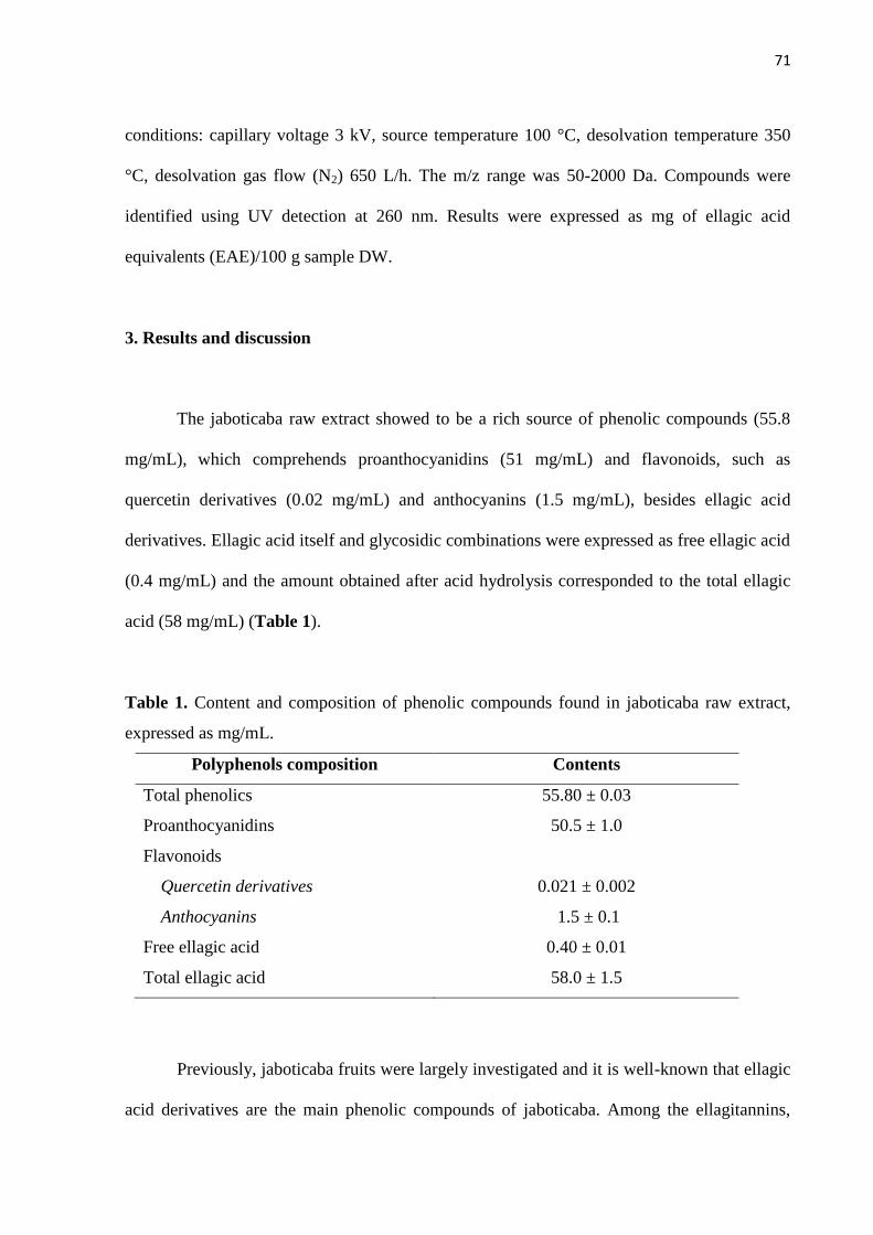
71
conditions: capillary voltage 3 kV, source temperature 100 °C, desolvation temperature 350
°C, desolvation gas flow (N2) 650 L/h. The m/z range was 50-2000 Da. Compounds were
identified using UV detection at 260 nm. Results were expressed as mg of ellagic acid
equivalents (EAE)/100 g sample DW.
3. Results and discussion
The jaboticaba raw extract showed to be a rich source of phenolic compounds (55.8
mg/mL), which comprehends proanthocyanidins (51 mg/mL) and flavonoids, such as
quercetin derivatives (0.02 mg/mL) and anthocyanins (1.5 mg/mL), besides ellagic acid
derivatives. Ellagic acid itself and glycosidic combinations were expressed as free ellagic acid
(0.4 mg/mL) and the amount obtained after acid hydrolysis corresponded to the total ellagic
acid (58 mg/mL) (Table 1).
Table 1. Content and composition of phenolic compounds found in jaboticaba raw extract,
expressed as mg/mL.
Polyphenols composition Contents
Total phenolics 55.80 ± 0.03
Proanthocyanidins 50.5 ± 1.0
Flavonoids
Quercetin derivatives 0.021 ± 0.002
Anthocyanins 1.5 ± 0.1
Free ellagic acid 0.40 ± 0.01
Total ellagic acid 58.0 ± 1.5
Previously, jaboticaba fruits were largely investigated and it is well-known that ellagic
acid derivatives are the main phenolic compounds of jaboticaba. Among the ellagitannins,

72
sanguiin H-10, sanguiin H-6 and lambertianin C were identified in this species. Cyanidin 3-
glucoside and delphinidin 3-glucoside were the anthocyanins detected, especially in the skin
of the fruits (Alezandro et al., 2013; Leite, Malta, Riccio, Eberlin, Pastore, Maróstica Júnior,
2011).
It is known that most polyphenols are metabolized by the colonic microbiota before
they can be absorbed, and these transformations are essential in modulating their biological
effects. It is clear that these metabolites are the main responsible for the physiological
response (Del Rio et al., 2010; Setchell et al., 2002). In order to investigate the bioavailability
of phenolic compounds from jaboticaba, an in vitro assay was performed using the human
fecal fermentation model, aiming for identification of their probable degradation products and
metabolites formed by the action of microbiota. In this study, the focus was on the ellagic acid
derivatives, as they are the main compounds found in jaboticaba fruits, although other
compounds were also identified. The data revealed that incubation led to degradation of
ellagic acid derivatives in both fermentation media, with and without feces. This finding
indicates that degradation may be related to the conditions inherent to the method, and not
only by the action of microbiota (Figure 1).
At the beginning, the content of ellagic acid derivatives was higher, approximately
20%, in the sample incubated without fecal inoculum. However, during the fermentation
process, degradation was observed in both samples. During the first two hours, there was a
reduction of 50% in the amount of ellagic acid derivatives detected in the sample without
feces, and 30% in the one with inoculum. After 48 hours, the content of ellagic acid
derivatives decreased 84% in the sample without inoculum and 78% in the one with feces
(Figure 1). The chromatographic profile of the fecal samples over time indicated smaller
ellagic acid peaks in the end of fermentation (T = 48 h) compared to those identified in the
beginning (T = 0) (Figure 2).

73
Figure 1. Fermentation profile of ellagic acid in samples incubated with and without fecal
inoculum, indicating the degradation over time.
The same degradation pattern was observed by Serra et al. (2011). There was a higher
degradation of procyanidins incubated with the fecal suspension in comparison to the samples
without feces. In addition, other authors also verified that the most compounds degraded in
the beginning of fermentation process. About 90% of quercetin present in the extracts
apparently disappeared during the first 15 minutes of incubation, and only 10% of residues
were identifiable after this period (Jaganath et al., 2009).
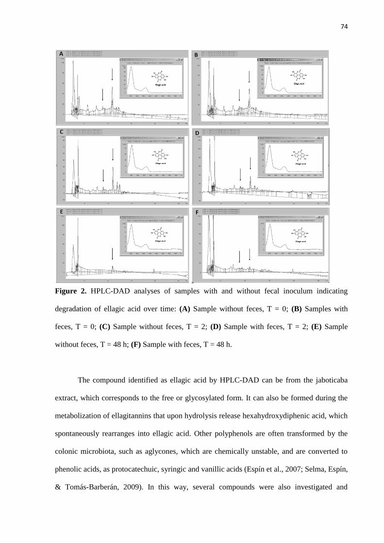
74
Figure 2. HPLC-DAD analyses of samples with and without fecal inoculum indicating
degradation of ellagic acid over time: (A) Sample without feces, T = 0; (B) Samples with
feces, T = 0; (C) Sample without feces, T = 2; (D) Sample with feces, T = 2; (E) Sample
without feces, T = 48 h; (F) Sample with feces, T = 48 h.
The compound identified as ellagic acid by HPLC-DAD can be from the jaboticaba
extract, which corresponds to the free or glycosylated form. It can also be formed during the
metabolization of ellagitannins that upon hydrolysis release hexahydroxydiphenic acid, which
spontaneously rearranges into ellagic acid. Other polyphenols are often transformed by the
colonic microbiota, such as aglycones, which are chemically unstable, and are converted to
phenolic acids, as protocatechuic, syringic and vanillic acids (Espín et al., 2007; Selma, Espín,
& Tomás-Barberán, 2009). In this way, several compounds were also investigated and
75
identified in the samples with and without fecal inoculum, which could be formed from
different polyphenols from jaboticaba (Table 2).
Analysis by HPLC-DAD indicated that many catabolic compounds and intermediate
products were found in the samples, being a result of the fission of the C6-C3-C6 skeleton
followed by the action of microbiota. Protocatechuic and syringic acid were identified in the
samples and may be derived from the anthocyanins metabolism. Protocatechuic acid,
however, is a phenolic acid that can be formed from different polyphenols, such as quercetin
and ellagic acid. Furthermore, after 2 hours, protocatechuic acid was not detected in the
samples, demonstrating that this compound may be transformed in other phenolic acids, such
as p-hydroxybenzoic acid. This last compound was detected only in the samples incubated
with feces, suggesting that the colonic microbiota is involved in the metabolization. Likewise,
p-hydroxyphenylacetic acid was found only in the samples incubated with fecal inoculum,
after 12 hours of incubation. The presence of p-hydroxyphenylacetic acid may be associated
with the microbial transformations of quercetin or proanthocyanidins, as well.
The colonic microbiota is the most important site of metabolism, especially for
hydroxycinnamic acids and flavonoids aglycones released from their conjugated forms by
cleavage of their glycosidic or ester bonds (Rechner et al., 2004). Recent studies revealed that
those metabolites are biologically more potent than their precursor compounds (Déprez et al.,
2000; Keppler & Humpf, 2005; Kim, Jung, Sohng, Han, Kim, & Han, 1998; Larrosa,
González-Sarrías, García-Conesa, Tomás-Barberán, Espín, 2006).

76
Table 2. Metabolites detected in the samples incubated with and without fecal inoculum,
during 48 hours of fermentation.
COLLECTION
TIME
WITH
INOCULUM
WITHOUT
INOCULUM
PRECURSOR
COMPOUNDS
T = 0
Protocatechuic acid Protocatechuic acid
Ellagic acid
Anthocyanin
Quercetin
Ellagic acid Ellagic acid Ellagitannin
Syringic acid Syringic acid Anthocyanin
T = 2 h
Protocatechuic acid Protocatechuic acid
Ellagic acid
Anthocyanin
Quercetin
Ellagic acid Ellagic acid Ellagitannin
Cinnamic acid Cinnamic acid -
p-hydroxybenzoic acid nd Protocatechuic acid
Quercetin
T = 4 h Ellagic acid Ellagic acid Ellagitannin
Cinnamic acid Cinnamic acid -
T = 6 h Ellagic acid Ellagic acid Ellagitannin
Cinnamic acid Cinnamic acid -
T = 12 h Ellagic acid
Cinnamic acid
p-hydroxyphenylacetic acid
Ellagic acid Ellagitannin
Cinnamic acid -
nd Proanthocyanidin
Quercetin
T = 24 h
Ellagic acid Ellagic acid Ellagitannin
Cinnamic acid Cinnamic acid -
p-hydroxyphenylacetic acid nd Proanthocyanidin
Quercetin
T = 48 h
Ellagic acid Ellagic acid Ellagitannin
Cinnamic acid Cinnamic acid -
p-hydroxyphenylacetic acid nd Proanthocyanidin
Quercetin
p-hydroxybenzoic acid nd
Protocatechuic acid
Proanthocyanidin
Quercetin
nd : not detected

77
It is known that other tissues are involved in the metabolism of phenolic compounds,
besides gut. The hepatic processing, for example, may be responsible for the formation of
different metabolites compared to the products obtained after intestinal metabolism. In this
way, the bioavailability of jaboticaba polyphenols was also evaluated in an in vivo model
using Wistar rats. In addition, the UPLC-MS was used to identify the compounds, since the
extraction of metabolites from biological fluids and tissues, and their characterization by other
spectroscopic means is complicated due to the very low concentrations of these compounds.
The fate of 38 metabolites were investigated in the present study (Table 3). Most of
them, 20 compounds, are conjugates of ellagic acid and their metabolites, known as
urolithins, or conjugated compounds. Besides ellagic acid (1), conjugates with methyl ether
and glucuronic acid (2 – 6) were identified. Ellagic acid methyl ether (2, m/z– 315) and ellagic
acid dimethyl ether (5, m/z– 329) were detected only in plasma. Compounds conjugated with
methyl ether and glucuronic acid (3, m/z– 329; 4, m/z
– 329; 6, m/z
– 329) were more widely
distributed, being detected in other tissues, as liver, stomach and colon, besides plasma.
Under the pH conditions of the small intestine, ellagic acid is released from
ellagitannins, and the colonic microbiota can also contribute to this transformation. The
metabolism of ellagic acid is initiated in the jejuno-ileal portion and urolithin D is the first
compound formed, followed by urolithin C and finally, urolithins A and B (González-Barrío
et al., 2012; Landete, 2011). Urolithin A (7, m/z– 227) was identified in all the biological
samples, unless in the brain. Urolithins B (8, m/z– 211), C (9, m/z
– 243) and D (10, m/z
– 259)
were also detected in all biological samples analyzed, but the amount varied among the
different tissues (Figure 3).

78
Table 3. Polyphenols metabolites detected in Wistar rats after chronic ingestion of jaboticaba raw extract.
Phenolic metabolites n° tR (min) MRM transition Occurence
ETs derivatives
ellagic acid 1 4.72 301 / 145 P, S, K
ellagic acid methyl ether 2 6.37 315 / 300 P
ellagic acid methyl ether glucuronide 3 9.85 491 / 315 P, L, S, K, M, C
ellagic acid methyl ether diglucuronide 4 6.79 667 / 491 P, S
ellagic acid dimethyl ether 5 6.72 329 / 315 P
ellagic acid dimethyl ether glucuronide 6 5.94 505 / 315 P, L
urolithin A 7 5.10 227 / 210 P, L, S, K, M, C
urolithin B 8 9.75 211 / 167 P, L, S, K, B, M, C
urolithin C 9 5.80 243 / 199 P, L, S, K, B, M, C
urolithin D 10 2.84 259 / 241 P, L, S, K, B, M, C
urolithin A glucuronide 11 7.55 403 / 227 P, L, S, K, C
urolithin A diglucuronide 12 7.89 579 / 227 P, L, S
urolithin B glucuronide 13 8.80 389 / 211 P, L, C

79
urolithin C glucuronide 14 4.53 419 / 243 P, L, S, K, C
urolithin C diglucuronide 15 3.55 595 / 243 P, L, S, K
urolithin C methyl ether glucuronide 16 7.65 433 / 257 P, L, S, K, M, C
urolithin C methyl ether glucuronide sulfate 17 8.80 513 / 337 P, L, S
urolithin D glucuronide 18 3.29 435 / 259 P, L, S, K
urolithin D methyl ether 19 3.38 273 / 258 P, S, K
urolithin D methyl ether glucuronide 20 6.90 449 / 273 P, L, S, K, C
hydroxyphenylpropionic acids
hydroxyphenylpropionic acid 21 6.93 165 / 121 P, L, S, K, B, M, C
dihydroxyphenylpropionic acid 22 5.55 181 / 137 P, L, S, K, B, M, C
hydroxyphenylacetic acids
hydroxyphenylacetic acid 23 3.50 151 / 107 P, L, S, K, B, M, C
dihydroxyphenylacetic acid 24 3.62 167 / 123 P, L, S, K, B, M, C
hydroxybenzoic acids
hydroxybenzoic acid 25 3.30 137 / 93 P, L, S, K, B, M, C
hydroxycinnamic acids

80
coumaric acid 26 4.72 163 / 119 P, L, S, K, B, M, C
caffeic acid 27 3.80 179 / 135 P, L, S, K, M, C
ferulic acid 28 4.85 193 / 134 P, L, S, K, C
flavan-3-ols
(+)-catechin 29 3.73 289 / 245 P, L, S, K, B, C
(–)-epicatechin 30 3.94 289 / 245 P, L, S, K, B, C
flavonols
quercetin-glucoside 31 4.74 463 / 301 P, L, S, C
quercetin-glucuronide 32 5.50 477 / 301 P, L, S, K, C
anthocyanins
delphinidin 3-glucoside 33 1.59 465 / 303 S
cyanidin 3-glucoside 34 1.91 449 / 287 S
cyanidin diglucuronide 35 1.89 639 / 287 S
delphinidin-acetylglucoside 36 2.96 507 / 303 S
cyanidin-acetylglucoside 37 2.28 491 / 287 S
delphinidin-coumaroylglucoside 38 3.00 611 / 303 S

81
Figure 3. UPLC-MS
analyses of metabolites in
plasma: (A) Urolithin A (7,
m/z– 227); (B) Urolithin B
(8, m/z– 211); (C) Urolithin
C (9, m/z– 243).

82
Urolithins were mainly detected in conjugated forms with methyl ether and glucuronic
acids, and rarely with sulfates. Urolithin C methyl ether glucuronide sulfate (17, m/z– 513)
was the only sulfated metabolite found in this study. Glucuronides of urolithin A (11, m/z–
403; 12, m/z– 579) were identified, as well as of urolithin B (13, m/z
– 389), C (14, m/z
– 419)
and D (18, m/z– 435). Different compounds were formed by different conjugate combinations
(16, m/z– 433; 20, m/z
– 449) and were largely distributed among the biological samples.
Quercetin is one of the most widely distributed phenolic compounds in human diet,
including vegetables, fruits, tea, and wine. Quercetin derivatives can be partly absorbed into
the body and accumulated in the circulation (Hollman, de Vries, Van Leeuwen, Mengelers, &
Katan, 1995; Hollman et al., 1997). In the gut, quercetin aglycone is released from glycosides
by the action of bacterial enzymes, as -glucosidases. Then, the portion that is not absorbed is
degraded to simpler products, as phenolic acid (hydroxyphenylpropionic, hydroxyphenyl-
acetic and hydroxybenzoic acids) (Serra, Macià, Romero, Reguant, Ortega, & Motilva, 2012).
After absorption, the aglycone is metabolized in the liver, producing metabolites conjugated
with methyl, glucuronate and sulfate groups, and this process is mediated by catecol-O-
methyltransferase (COMT), uridine diphosphate glucuronosyltransferase (UGT) and
sulfutransferase (SULT), respectively (Crozier, Del Rio, & Clifford, 2010). However, it was
demonstrated that glucuronide conjugates are the main quercetin metabolites (Manach et al.,
1998).
In this study, two compounds were identified as result of ingestion of quercetin
derivatives present in the raw jaboticaba extract. Quercetin-glucoside (31, m/z– 463) was
detected, especially in the stomach, but some animals presented small amount in plasma, liver
and colon. Quercetin-glucuronide (32, m/z– 477) was also found in plasma, liver, stomach,
kidneys and colon. Hydroxyphenylpropionic (21, m/z– 165), dihydroxyphenylpropionic (22,
m/z– 181), hydroxyphenylacetic (23, m/z
– 151), dihydroxyphenylacetic (24, m/z
– 167) and

83
hydroxylbenzoic acids (25, m/z– 137) were present in different biological tissues, and may be
derived from quercetin metabolism.
Jaboticaba showed to be a rich source of proanthocyanidins, which were metabolized
by the colon microbiota, releasing catechin (29, m/z– 289) and epicatechin (30, m/z
– 289).
Then, these monomeric units were converted in hydroxyphenylpropionic acid (21, 22),
hydroxyphenylacetic (23, 24), hydroxybenzoic (25) and hydroxycinnamic acids (28, m/z–
193). The presence of these compounds in biological fluids and tissues has been related to the
procyanidins metabolism (Aura, 2008; Serra, Macià, Romero, Anglés, Morelló, & Motilva,
2011).
Delphinidin 3-glucoside (33, m/z– 465) and cyanidin 3-glucoside (35, m/z
– 449) were
the anthocyanins previously identified in jaboticaba fruits (Alezandro et al., 2013) and were
also detected in the stomach of rats as the intact forms. One glucuronide conjugate of cyanidin
(35, m/z– 639) was found, as well. Apart from the conjugated metabolite, three anthocyanins
were identified in the stomach: delphinidin-acetylglucoside (36, m/z– 507), cyanidin-
acetylglucoside (37, m/z– 491) and delphinidin-coumaroylglucoside (38, m/z
– 611).
According to the literature, only 1–2% of anthocyanins were absorbed after ingestion
of high amounts (500 mg) of these compounds. However, when a lower quantity (100 mg) is
consumed, no products derived from their metabolism were found in biological fluids, as
plasma and urine (Clifford, 2000). This fact, along with the high unstability and susceptibility
to degradation, can explain why the anthocyanins metabolites were not detected in other
tissues, besides stomach.
The bioavailability of polyphenols is generally evaluated by means of acute
administration of one or more compounds. In this study, a long-term experiment (30 days)
was performed, as the scientific literature related that repeated oral doses of ellagitannins are
necessary to make the colonic microbiota able to metabolize ellagic acid derivatives and

84
produce urolithins (Cerdá, Llorach, Ceron, Espín, & Tomás-Barberán, 2003). Our findings
showed that metabolites formed from jaboticaba polyphenols were largely distributed in many
biological tissues, but the kind and fate of metabolites were not related with time of
euthanasia. Probably, the compounds found in the tissues may have been produced and
accumulated during all the supplementation period, and not only on the day of euthanasia.
4. Conclusion
The dietary polyphenols are effective substrates for the action of the microbiota in
human colon and are extensively metabolized, forming simpler phenolic or non-phenolic
compounds. The type and amount of compounds generated by metabolism are influenced by
the interindividual differences, which here was minimized by the use of a pool of feces. It is
known that the formation of metabolites is essential for the absorption and consequently, their
presence in target tissues in chemical form and concentration adequate to exert their
physiological role. Thirty eight compounds were identified as metabolites formed from
different classes of jaboticaba polyphenols and they were widely distributed in many
biological tissues and plasma. The data obtained in this study are the first step towards the
elucidation of the bioavailability of phenolic compounds from jaboticaba and provide
important information about the possible compounds that may be found in the human body
after consumption of this fruit.
Acknowledgements
We thank Daniela Moura de Oliveira for collaboration with analyses and Fundação de
Amparo à Pesquisa do Estado de São Paulo (Fapesp) (2009/01775-0) for financial support.

85
We also thank Unidos Com. Imp. Exp. Hortigranjeiros from Companhia de Entrepostos e
Armazéns Gerais de São Paulo (Ceagesp) for providing the samples.
References
Alezandro, M. R., Dubé, P., Desjardins, Y., Lajolo, F. M., & Genovese, M. I. (2013).
Comparative analysis of chemical and phenolic composition of two species of jaboticaba:
Myrciaria jaboticaba (Vell.) Berg and Myrciaria cauliflora (Mart.) O. Berg. Food Research
International, submitted.
Arabbi, P. R., Genovese, M. I., & Lajolo, F. M. (2004). Flavonoids in vegetable foods
commonly consumed in Brazil. Journal of Agricultural and Food Chemistry, 52, 1124-1131.
Aura, A. (2008). Microbial metabolism of dietary phenolic compounds in the colon.
Phytochemistry Reviews, 7, 407–429.
Cerdá, B., Llorach, R., Ceron, J. J., Espín, J. C., Tomás-Barberán, F. A. (2003). Evaluation of
the bioavailability and metabolism in the rat of punicalagin, an antioxidant polyphenol from
pomegranate juice. European Journal of Nutrition, 42, 18–28.
Clifford, M. N. (2000). Review. Anthocyanins – nature, occurrence and dietary burden.
Journal of the Science of Food and Agriculture, 80, 1063–1072.
Crozier, A., Del Rio, D., & Clifford, M. N. (2010). Bioavailability of dietary flavonoids and
phenolic compounds. Molecular Aspects of Medicine, 31, 446-467.

86
Del Rio, D., Costa, L. G., Lean, M. E. J., & Crozier, A. (2010). Polyphenols and health: What
compounds are involved? Nutrition, Metabolism and Cardiovascular Diseases, 20, 1-6.
Déprez, S., Brezillon, C., Rabot, S., Philippe, C., Mila, I., Lapierre, C., & Scalbert, A. (2000).
Polymeric proanthocyanidins are catabolized by human colonic microflora into low-
molecular-weight phenolic acids. Journal of Nutrition, 130, 2733–2738.
Donadio, L. C. (2000). Jabuticaba (Myrciaria jaboticaba (Vell.) Berg. Jaboticabal: FUNEP,
55 pp.
Espín, J. C., González-Barrio, R., Cerdá, B., López-Bote, C., Rey, A. I., & Tomás-Barberán,
F. A. (2007). Iberian pig as a model to clarify obscure points in the bioavailability and
metabolism of ellagitannins in humans. Journal of Agricultural and Food Chemistry, 55,
10476–10485.
Gasperotti, M., Masuero, D., Vrhovsek, U., Guella, G., & Mattivi, F. (2010). Profiling and
accurate quantification of Rubus ellagitannins and ellagic acid conjugates using direct UPLC-
Q-TOF HDMS and HPLC-DAD analysis. Journal of Agricultural and Food Chemistry, 58,
4602-4616.
González-Barrio, R., Truchado, P., García-Villalba, R., Hervas, G., Frutos, P., Espín, J. C., &
Tomás-Barberán, F. A. (2012). Metabolism of oak leaf ellagitannins and urolithin production
in beef cattle. Journal of Agricultural and Food Chemistry, 60, 12, 3068-3077.

87
Hollman, P. C., de Vries, J. H., van Leeuwen, S. D., Mengelers, M. J., Katan, M. B. (1995).
Absorption of dietary quercetin glycosides and quercetin in healthy ileostomy volunteers. The
American Journal of Clinical Nutrition, 62, 1276–1282.
Hollman, P. C., van Trijp, J. M., Buysman, M. N., van der Gaag, M. S., Mengelers, M. J., de
Vries, J. H., et al. (1997). Relative bioavailability of the antioxidant flavonoid quercetin from
various foods in man. FEBS Letters, 418, 152–156.
Jaganath, I. B., Mullen, W., Lean, M. E. J., Edwards, C. A., & Crozier, A. (2009). In vitro
catabolism of rutin by human fecal bacteria and the antioxidant capacity of its catabolites.
Free Radical Biology and Medicine, 47, 1180-1189.
Keppler, K., & Humpf, H. U. (2005). Metabolism of anthocyanins and their phenolic
degradation products by the intestinal microflora. Bioorganic & Medicinal Chemistry, 13,
5195–5205.
Kim, D. H., Jung, E. A., Sohng, I. S., Han, J. A., Kim, T. H., & Han, M. J. (1998). Intestinal
bacterial metabolism of flavonoids and its relation to some biological activities. Archives of
Pharmacal Research, 21, 17.
Landete, J. M. (2011). Ellagitannins, ellagic acid and their derived metabolites: A review
about source, metabolism, functions and health. Food Research International, 44, 1150-1160.

88
Larrosa, M.; González-Sarrías, A.; García-Conesa, M. T.; Tomás-Barberán, F. A.; Espín, J. C.
Urolithins, ellagic acid-derived metabolites produced by human colonic microflora, exhibit
estrogenic and antiestrogenic activities. J. Agric. Food Chem. 2006, 54, 1611–1620.
Leite, A. V., Malta, L. G., Riccio, M. F., Eberlin, M. N., Pastore, G. M., & Maróstica Júnior,
M. R. (2011). Antioxidant potential of rat plasma by administration of freeze-dried jaboticaba
peel (Myrciaria jaboticaba Vell Berg). Journal of Agricultural and Food Chemistry, 59,
2277-2283.
Manach, C., Morand, C., Crespy, V., Demigné, C., Texier, O., Régérat, F., et al. (1998).
Quercetin is recovered in human plasma as conjugated derivatives which retain antioxidant
properties. FEBS Letters, 426, 331–336.
McBain, A., & Macfarlane, G. (1997). Investigations of bifidobacterial ecology and
oligosaccharide metabolism in a three-stage compound continuous culture system.
Scandinavian journal of gastroenterology. Supplement, 222, 32.
Mullen, W., Mcginn, J., Lean, M. E., Maclean, M. R., Gardner, P., Duthie, G. G., Yokota, T.,
& Crozier, A. (2002). Ellagitannins, flavonoids, and other phenolics in red raspberries and
their contribution to antioxidant capacity and vasorelaxation properties. Journal of
Agricultural and Food Chemistry, 50, 5191-5196.
Oliveira, D. M., Bastos, D. H. M. (2011). Biodisponibilidade de ácidos fenólicos. Química
Nova, 34, 1051-1056.
89
Oliveira, D. M., Pinto, C. B., Sampaio, G. R., Yonekura, L., Catharino, R. R., & Bastos, D. H.
M. (2013). Method validation for extraction of phenolic acids from plasma, urine and liver
and analysis by ultra-performance liquid chromatography-mass spectrometry. Journal of
Agricultural and Food Chemistry, submitted.
Pinto, M. S., Lajolo, F. M., & Genovese, M. I. (2008). Bioactive compounds and
quantification of total ellagic acid in strawberries (Fragaria ananassa Duch.). Food
Chemistry, 107, 1629-1635.
Prior, R. L., Fan, E., Ji, H., Howell, A., Nio, C., Payne, M. J., & Reed, J. (2010). Multi-
laboratory validation of a standard method for quantifying proanthocyanidins in cranberry
powders. Journal of the Science of Food and Agriculture, 90, 1473–1478.
Priyadarsini, K. I., Khopde, S. M., Kumar, S. S., & Mohan, H. (2002). Free radical studies of
ellagic acid, a natural phenolic antioxidant. Journal of Agricultural and Food Chemistry, 50,
2200-2206.
Rechner, A. R., Smith, M. A., Kuhnle, G., Gibson, G. R., Debnam, E. S., Srai, S. K. S.,
Morre, K. P., & Rice-Evans, C. A. (2004). Colonic metabolism of dietary polyphenols:
influence of structure on microbial fermentation products. Free Radical Biology & Medicine,
36, 212-225.
Selma, M. A. V., Espín, J. C., & Tomás-Barberán, F. A. (2009). Interaction between
phenolics and gut microbiota: role in human health. Journal of Agricultural and Food
Chemistry, 57, 6485-6501.

90
Serra, A., Macià, A., Romero, M.-P., Anglés, N., Morelló, J.-R., & Motilva, M.-J. (2011).
Metabolic pathways of the colonic metabolism f procyanidins (monomers and dimers) and
alkaloids. Food Chemistry, 126, 1127–1137.
Serra, A., Macià, A., Romero, M.-P., Reguant, J., Ortega, N., & Motilva, M.-J. (2012).
Metabolic pathways of the colonic metabolism of flavonoids (flavonols, flavones and
flavanones) and phenolic acids. Food Chemistry, 130, 383-393.
Setchell, K. D. R., Brown, N. M., & Lydeking-Olsen, E. (2002). The clinical importance of
the metabolite equol—a clue to the effectiveness of soy and its isoflavones. The Journal of
Nutrition, 132, 3577-3584.
Singleton, V. L., Orthofer, R., & Lamuela-Raventos, R. M. (1999). Analysis of total phenols
and other oxidation substrates and antioxidants by means of Folin–Ciocalteu reagent. Methods
in Enzymology, 299, 152–178.
Xu, X., Harris, K. S., Wang, H.-J., Murphy, P. A., & Hendrich, S. (1995). Bioavailability of
soybean isoflavones depends upon gut microflora in women. The Journal of Nutrition, 125,
2307-2315.
Youdim, K. A., McDonald, J., Kalt, W., & Joseph, J. A. (2002). Potential role of dietary
flavonoids in reducing microvascular endothelium vulnerability to oxidative and
inflammatory insults. The Journal of Nutritional Biochemistry, 13, 282-288.
91
Jaboticaba (Myrciaria jaboticaba (Vell.) Berg), a Brazilian grape-like fruit, improves
plasma lipid profile in streptozotocin-mediated oxidative stress in diabetic rats
MARCELA ROQUIM ALEZANDRO1, DANIEL GRANATO
2,
MARIA INÉS GENOVESE1*
* corresponding author (Tel: 55-11-30911525; Fax: 55-11-38154410; e-mail:
1Laboratório de Compostos Bioativos de Alimentos, Departamento de Alimentos e Nutrição
Experimental, FCF, Universidade de São Paulo, Av. Prof. Lineu Prestes, 580, Bloco 14,
05508-900, São Paulo, SP, Brazil.
2Núcleo de Análise e Tratamento de Dados, Instituto Adolfo Lutz, Av. Dr. Arnaldo, 355,
01246-900, São Paulo, SP, Brazil.
92
ABSTRACT
Jaboticaba (Myrciaria jaboticaba Vell. Berg) is a Brazilian Atlantic rainforest fruit of the
Myrtaceae family. In this work, the effect of the daily intake (40 days) of jaboticaba (1.0 and
2.0 g DW/kg body weight) on oxidative stress and plasma lipid profile of streptozotocin
(STZ)-induced diabetic rats was evaluated. Jaboticaba was shown to be a good source of
phenolic compounds, proanthocyanidins and ellagitannins. Daily administration of jaboticaba
resulted in ameliorated water consumption and energy intake in STZ-diabetic rats. Plasma
total cholesterol levels were reduced in 32% and triacylglycerol decreased 50% when both
doses of jaboticaba were administered. This reduction of total cholesterol and triacylglycerol
levels seems to be associated to the strong in vitro inhibition of pancreatic lipase presented by
jaboticaba extracts. Plasma antioxidant capacity of diabetic rats assessed by FRAP assay
increased (2 to 2.5 times) after supplementation with both doses of jaboticaba along with a
decrease of lipid peroxidation in plasma (22%) and brain (10-17%). Diabetic rats consuming
jaboticaba presented higher activity of SOD in the brain, CAT and GPx in kidneys and liver,
and GPx in plasma, as compared to the control group. These results suggest that chronic
ingestion of jaboticaba may represent a dietary strategy for controlling oxidative stress in
pathological conditions.
KEYWORDS: Myrciaria jaboticaba; oxidative stress; streptozotocin-induced diabetes; lipid
profile.

93
1. Introduction
Jaboticaba, Myrciaria jaboticaba (Vell.) Berg, is a Brazilian native fruit from the
Atlantic Rainforest that belongs to the Myrtaceae family, grape-like in appearance and
texture. Its economic importance has been continuously growing in Brazil because of the
sweet and slightly acidic flavor of the pulp. Jaboticaba has a huge trading potential, since it
can be consumed in natura and also used by industry as ingredient to produce cosmetics and
food products (Donadio, 2000).
Jaboticaba fruits are a rich source of polyphenols, such as anthocyanins, which are
concentrated in the dark purple to almost black skin, when the fruit is ripe, besides quercetin
derivatives and proanthocyanidins. Furthermore, a recent study carried out with two species
of jaboticaba proved ellagic acid derivatives, such as ellagitannins, are the main compounds
detected in jaboticaba (Alezandro, Dubé, Desjardins, Lajolo & Genovese, 2013). Long-term
ingestion of fruits with high levels of flavonoids (anthocyanins) and tannins (ellagitannins)
has been associated with positive effects on human health, related to their antioxidant
potential and consequent inhibitory activity against lipid peroxidation, reducing the risk of
cardiovascular diseases (Mullen et al., 2002; Priyadarsini, Khopde, Kumar & Mohan, 2002;
Srinivasan, Vadhanam, Arif, & Gupta, 2002). Despite the unclear mechanisms and few
studies reported, several hypotheses indicate the high antioxidant capacity of phenolic
compounds, especially flavonoids, may also be effective in reducing oxidative stress
(Macedo, Rogero, Guimarães, Granato, Lobato, & Castro, 2013), progression of diabetes
mellitus (Song, Wang, Li & Cai, 2005), and hypertension (Kwon, Vattem, & Shetty, 2006).
Streptozotocin-induced diabetes is an animal model used to promote metabolic
dysfunctions related to oxidative stress (Raza & John, 2012). Oxidative stress occurs when
there is an overproduction of free radicals and the endogenous antioxidants are not enough to

94
buffer these unstable molecules (Mullarkey, Edelstein, & Brownlee, 1990), and leads to
modifications involved in the inflammation process and in the initiation and progression of
atherosclerosis (Strobel, Fassett, Marsh & Coombes, 2010), thus being intrinsically linked
with non-transmissible chronic diseases (Mullarkey, Edelstein, & Brownlee, 1990).
Studies about the potential beneficial effect on health of jaboticaba are sparse, but
research has shown that native fruits are promising sources of bioactive compounds. Herein,
the research available in the literature mostly focused on the role of anthocyanins present in
the skin of the fruit (Lenquiste, Batista, Dragano, Marineli & Maróstica Jr., 2012; Dragano et
al., 2013), and usually ignores the existence and importance of ellagitannins. Based on these
considerations, this work aimed to evaluate the effects of chronic administration of whole
jaboticaba on the oxidative stress related to streptozotocin-induced diabetes in an animal
model using Wistar rats.
2. Material and methods
2.1. Chemicals. The hydroxy-2, 5, 7, 8-tetramethylchroman-2-carboxylic acid (Trolox) was
obtained from Aldrich (Milwaukee, WI, USA). Malondialdehyde, fluorescein, 2,2-azobis(2-
methylpropionamide) dihydrochloride (AAPH), xanthine oxidase (from bovine milk),
cytochrome C (from horse heart) and xanthine were purchased from Sigma Chemical Co. (St
Louis, MO, USA). Glutathione, glutathione reductase and nicotinamide adenine dinucleotide
phosphate (NADPH) were obtained from Merck Chemical Co. (Darmstadt, Germany). TPTZ
(2, 4, 6-tripyridyl-S-triazine) was purchased from Fluka Chemie AG (Buchs, Switzerland).
Ketamine chloride and xylidine chloride were acquired from Bayer (Leverkusen, Germany).
All chemicals/solvents were of analytical or HPLC grade, according to the requirement.

95
2.2. Samples. Fully ripened jaboticaba Sabará (Myrciaria jaboticaba Vell. Berg) was
obtained from a local producer at the São Paulo Central Market (Companhia de Entrepostos e
Armazéns Gerais de São Paulo – CEAGESP, Brazil). Fruits were cleaned and immediately
freeze-dried and stored at -20 °C. Then, the dried fruits were ground to a fine powder in a
mortar and pestle, using liquid nitrogen to keep sample frozen. Protein, lipid, fiber and ashes
contents of jaboticaba were assessed according to AOAC (2005). Total phenolics (Singleton,
Orthofer, & Lamuela-Raventos, 1999), proanthocyanidins (Porter, Hrstich, & Chan, 1986),
ellagitannins (Pinto, Lajolo, & Genovese, 2008) and total tannins (Hagerman & Butler, 1978)
were also determined. Results were expressed as mg/g fresh weight (FW).
2.3. Sample extraction for lipase inhibition assay. Samples were extracted in a 70% aqueous
methanol solution for 2 h at room temperature, at a 1:20 (w/v) ratio of sample-to-solvent.
After filtration (Whatman N° 1), extracts were kept at -80 °C until analyses (Arabbi,
Genovese, & Lajolo, 2004).
2.4. Measurement of pancreatic lipase activity. The pancreatic lipase activity was measured
using 4-methylumbelliferyl oleate (4-MU oleate) as a substrate, according to Jacks and
Kircher (1967) with some modifications (You, Chen, Wang, Jiang, & Lin, 2012). Twenty-five
microliters of a sample solution dissolved in water and 50 L of a 0.1 mM 4-MU solution
dissolved in a buffer consisting of 13 mM Tris-HCl, 150 mM NaCl, and 1.3 mM CaCl2 (pH
8.0) were mixed in the well of a microtiter plate, and 25 L of the lipase solution (50 U/mL)
in the above buffer was then added to start the enzyme reaction. After incubation at 25 °C for
30 min, 0.1 mL of 0.1 M sodium citrate (pH 4.2) was added to stop the reaction. The amount
of 4-methylumbelliferone released by lipase was measured with a fluorometrical microplate
reader (Fluoroskan Ascent C LabSystems, Inc.) at an excitation wavelength of 355 nm and an

96
emission wavelength of 460 nm. Results were expressed as IC50 values, considering the
amount of sample (mg sample DW/mL reaction) and the content of phenolic compounds (mg
catechin equivalents (CE)/mL reaction), determined according to Singleton et al. (1999).
2.5. Animals and experimental design. The Faculty of Pharmaceutical Sciences/USP Ethical
Committee for Animal Research approved all the adopted procedures (Protocol
CEUA/FCF/USP no. 355). Thirty six male rats weighing 200 ± 10 g were obtained from
Animal House of Faculty of Pharmaceutical Sciences and Chemistry Institute of University of
São Paulo. Animals were kept under standard laboratory conditions of temperature (23 ± 2
°C), relative humidity (50 ± 5%), 12 h light-dark cycle. Chow diet and water were provided
ad libitum. For diabetes induction, overnight fasted rats received intraperitoneal injection
(i.p.) of STZ (65 mg/kg) in citrate buffer (pH 4.5), followed by an aqueous solution of 10%
glucose for eight hours. After three days the glycemia was measured and all rats presented
glucose levels upper than 200 mg/dL. The STZ-diabetic animals were divided into three
groups of 12 animals, as follows:
- Control: animals receiving water by gavage during 40 days;
- Group1: animals receiving 1.0 g/kg body weight of jaboticaba powder dispersed in water
during 40 days;
- Group2: animals receiving 2.0 g/kg body weight of jaboticaba powder dispersed in water
during 40 days.
The food and water consumptions were recorded daily. Every three days the animals
were weighted and the fast blood glucose was measured every five days in all animals.
Results were reported as average energy intake (Kcal/day), water consumption (mL/day),
plasma glucose levels (mg/dL) and body weight (g) for the 40 days period.

97
2.6 Blood and tissue samples. After 40 days the animals were anesthetized with ketamine
chloride and xylidine chloride. Blood was collected by cardiac puncture into tubes containing
EDTA. The plasma was separated by centrifugation at 2,000 g for 10 min at 4 °C.
Erythrocytes were washed three times with ice-cold 9 g/L NaCl solution and hemolyzed with
distilled water (1:4 v/v). The tissues were exhaustively perfused with sterilized ice-cold 9
g/L NaCl solution through heart puncture until the liver was uniformly pale. Kidneys, brain
and liver were removed, weighed and immediately frozen under liquid nitrogen and stored at -
80 °C for further biochemical analysis. At the time of analysis, the tissues homogenates were
prepared with ice-cold 50 mM phosphate buffer (pH 7.4) (1:4 w/v) and centrifuged at 10,000
g for 10 min at 4 °C.
2.7 Plasma antioxidant capacity. Antioxidant capacity of plasma towards peroxyl radicals
was evaluated by the ORAC method described by Huang, Ou, Hampsch-Woodill, Flanagan
and Prior (2002) and the ferric reducing ability of plasma (FRAP) assay was determined
according to Benzie and Strain (1996). Both methods were performed on a Synergy H1
Hybrid Multi-Mode microplate reader (BioTek Instruments, Winooski, VT) and the results
were expressed in μmols Trolox equivalents (TE)/mL plasma.
2.8 Lipid peroxidation levels. Thiobarbituric acid reactive substances (TBARS) levels in
plasma and tissues were measured according to Ohkawa, Ohishi, and Yagi (1979). An aliquot
of plasma or tissue homogenate was mixed with 8.1% sodium dodecyl sulphate (SDS), 20%
acetic acid, 0.67% thiobarbituric acid and water. The mixture was heated for 1 h at 95 °C and
the pink chromogen formed was extracted into 1.4 mL of n-butanol. The absorbance of the
organic phase was measured at 532 nm using a Synergy H1 Hybrid Multi-Mode microplate
reader (BioTek Instruments, Winooski, VT). Malondialdehyde (MDA) was used as a
standard. Results were expressed as mol MDA/mL plasma or mg protein.

98
2.9 Antioxidant enzymes. Antioxidant enzymes activities were measured in plasma, and
tissues (liver, brain and kidney). Briefly, catalase (CAT) activity was assayed at 25 °C by a
method based on the disappearance of 10 mM H2O2. The decomposition of H2O2 by CAT
contained in the samples follows a first-order kinetic and changes in absorbance were
measured 60 s after addition of H2O2, and then at 60 s intervals over 4 min (Hugo & Lester,
1984). Glutathione peroxidase (GPx) catalyses the oxidation of glutathione by terc-butyl
hydroperoxide. In the presence of glutathione reductase and NADPH, the oxidized glutathione
is converted to the reduced form with a concomitant oxidation of NADPH to NADP+, which
is reflected as a decrease in the absorbance at 340 nm (ε340 6.22 L/mmol/cm). Changes in
absorbance were measured at 60 s intervals over 6 min (Albrecht & William, 1981).
Superoxide dismutase (SOD) activity was measured by the decrease in the rate of cytochrome
c reduction in a xanthine/xanthine oxidase superoxide-generating system consisting of 10 mM
cytochrome c, 100 mM xanthine, 50 mM sodium phosphate buffer (pH 7.8) and the necessary
quantity of xanthine oxidase to yield a variation of 0.025 absorbance/min at 550 nm (Gunzler
et al., 1984). Enzymatic activities were expressed as units of activity (UA, corresponding to
0.1 absorbance changes for CAT and GPx and to 0.0125 absorbance change for SOD)
min−1
.g−1
.protein or mg−1
haemoglobin (Hb). Protein concentration in plasma samples was
determined by the method described by Lowry, Rosebrough, Farr and Randall, (1951).
Haemoglobin was measured using Drabkin’s reagent.
2.10 Biochemical analysis. The concentrations of total cholesterol (TC), triacylglycerol
(TAG), HDL cholesterol (HDL-c), glucose, urea and creatinine in plasma were determined
using commercial kits LABTEST (Lagoa Santa, MG, Brazil). The fast blood glucose was
determined by means of the Accu-Check Performa® system.

99
2.11 Statistical analysis. Data were presented as mean ± SD. Initially, the results were
checked for homogeneity of variances by using the Levene test while one-way ANOVA
(parametric) or the Welch test (non-parametric) was used to assess differences among the
three treatments. The least significant difference Fisher test or Kruscal-Wallis test was used to
compare the means within groups. P-values below 0.05 were regarded as significant. In order
to observe the experimental results simultaneously, nine biomarkers (triacylglycerol, total
cholesterol, GPx liver, TBARS brain, TBARS plasma, CAT liver, SOD plasma, SOD brain,
ORAC plasma) were submitted to a principal component analysis, adopting the biomarkers as
columns and Wistar rats as cases. Analyses were based on linear correlation, variances were
computed as Sum of Squares/(n-1) and a scatter plot that contained the variables and rats was
built adopting the factor-plane (1 x 2) (Granato, Katayama, & Castro, 2012). Statistical
standardization was performed to obtain relativized data to which the multivariate technique
was applied. The standardization of the variables was performed using the Equation (1):
Where Z is the standardized value for each value of the response, Xij represents the original
value for the object (i) of measured attribute (j), is the mean value of variable j, and sj is
the standard deviation for the attribute. All statistical analyses were performed with Statistica
v. 11 software (Statsoft Inc., Tulsa, OK, USA).

100
3. Results
Jaboticaba was shown to be a good source of phenolic compounds, which included
proanthocyanidins (10.4 mg/g FW), ellagitannins (9.1 mg/g FW) and total tannins (7.3 mg/g
FW). Besides being rich in polyphenols, jaboticaba provides fibers (34.7 mg/g FW) and ashes
(5.2 mg/g FW), important nutrients in maintaining health (Table 1).
Besides, jaboticaba raw extract showed a strong inhibitory activity against pancreatic
lipase (IC50 = 1.08 mg sample/mL reaction) and its polyphenols largely contributed to this
effect (IC50 = 0.06 mg CE/mL) (Table 1).

101
Table 1. Chemical composition (mg/g FW) and in vitro inhibitory activity of pancreatic lipase (IC50) of jaboticaba berry.
Chemical composition
Nutritional composition
Polyphenols composition
Carbohydrate
a Protein
a Lipid
a Ashes
a Fiber
a Total phenolic
b PACs
c ETs
d Total tannins
e
138 ± 5 1.7 ± 0.1 0.72 ± 0.02 5.2 ± 0.2 35 ± 2 6.4 ± 0.1 10.4 ± 0.3 9.1 ± 0.2 7.26 ± 0.05
Inhibitory activity of pancreatic lipase
IC50 (mg sample DW /mL reaction) IC50 (mg CE/ mL reaction)
1.08 0.06
Contents expressed as mg/g FW a; mg catechin equivalent/ g FW
b; mg quebracho tannin/g FW
c; mg ellagic acid/g FW
d; mg tannic acid/g FW

102
Due to this unique chemical composition and the antihyperlipidemic potential, we
believed that jaboticaba would be powerful against oxidative stress associated to diabetes.
Here, it was demonstrated that the daily administration of jaboticaba was effective for
controlling oxidative stress and hyperlipidemia in STZ-diabetic rats. Polyphagia and
polydipsia (Figure 1), common symptoms of diabetic animals, were also assuaged when the
diabetic rats received both doses of jaboticaba. The mean energy intake was significantly
reduced (p < 0.01) in 6%, after administration of the lowest dose, and 13% for the highest
dose. Similarly, significant (p < 0.01) reductions of 8% and 14% in water consumption were
observed after treatment with 1 and 2 g DW (dry weight)/kg of jaboticaba, respectively.
However, an increase (p = 0.03) in blood glucose levels was detected after administration of
the highest dose of jaboticaba (Figure 1), not physiologically relevant due to the already
elevated glycemia of the animals (> 680 mg/dL).
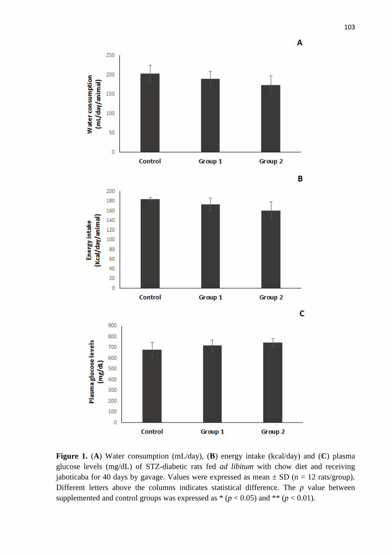
103
Figure 1. (A) Water consumption (mL/day), (B) energy intake (kcal/day) and (C) plasma
glucose levels (mg/dL) of STZ-diabetic rats fed ad libitum with chow diet and receiving
jaboticaba for 40 days by gavage. Values were expressed as mean ± SD (n = 12 rats/group).
Different letters above the columns indicates statistical difference. The p value between
supplemented and control groups was expressed as * (p < 0.05) and ** (p < 0.01).

104
The mean body weight of the diabetic rats treated with both doses of jaboticaba was
not significantly different from the control rats. However, the weight of the liver in proportion
to the body weight was lower (p < 0.01) in the treated groups (5.3-5.9%) compared to the
untreated animals (7.1%), a 21% decrease (Figure 2).
Figure 2. (A) Body weight (g) and tissues weight (% body weight), liver (B), brain (C) and
kidneys (D) of STZ-diabetic rats fed ad libitum with chow diet and receiving jaboticaba for 40
days by gavage. Values were expressed as mean ± SD (n = 12 rats/group). Different letters
above the columns indicates statistical difference. The p value between supplemented and
control groups was expressed as * (p < 0.01).
Lipid profile was also altered in the increased oxidative stress, but the administration
of jaboticaba was able to recover lipid levels in diabetic rats. Plasma TAG (p < 0.01) and TC
(p < 0.01) levels were reduced in the treated groups (Figure 3). TAG was reduced in 50% and
TC in 32% for both doses of jaboticaba.

105
Figure 3. Plasma lipid profile (mg/dL) of STZ-diabetic rats fed ad libitum with chow diet and
receiving jaboticaba for 40 days by gavage: (A) total cholesterol, (B) triacylglycerol, (C)
HDL-cholesterol. Values were expressed as mean ± SD (n = 12 rats/group). Different letters
above the columns indicates statistical difference. The p value between supplemented and
control groups was expressed as * (p < 0.01).

106
The two important indicators of renal health were evaluated. Creatinine varied from
0.73 to 0.97 mg/dL, and urea ranged between 61 and 67 mg/dL. No effect (p > 0.05) was
observed after administration of jaboticaba during 40 days, comparing the control with treated
groups (Figure 4).
Figure 4. Plasma creatinine (A) and urea (B) concentrations (mg/dL) of STZ-diabetic rats fed
ad libitum with chow diet and receiving jaboticaba for 40 days by gavage: (A) total
cholesterol, (B) triacylglycerol, (C) HDL-cholesterol. Values were expressed as mean ± SD (n
= 12 rats/group). Different letters above the columns indicates statistical difference.

107
Antioxidant capacity and lipid peroxidation are two of the most relevant parameters of
oxidative stress. Antioxidant capacity assessed by FRAP assay significantly increased (p <
0.01) from 4.5 mol TE/ mL plasma in the control rats to 12.1 mol TE/ mL plasma in the
treated rats from Group 1, which represents a 2.5 fold increase. For the group that received the
highest dose of jaboticaba, antioxidant capacity was 2-fold higher than in the control rats.
Conversely, no significant (p = 0.53) effect in the antioxidant capacity assessed by ORAC
was observed after treatment (Figure 5).
High concentration of MDA in tissues and/or in plasma is a known biomarker of
oxidative damage. Untreated diabetic rats showed elevated levels of MDA in both plasma and
brain, when compared to treated groups (Figure 5). Daily administration of jaboticaba
significantly decreased lipid peroxidation in plasma (p < 0.01) and brain (p = 0.01), although
no effect was observed in liver (p = 0.07) and kidney (p = 0.54). Our findings suggest that
polyphenols from jaboticaba protected the STZ-diabetic rats against oxidative damage.

108
Figure 5. Plasma antioxidant capacity (mol Trolox equivalent/mL plasma) assessed by
ORAC (A) and FRAP (B) assays, and lipid peroxidation (mol MDA/mg protein) of plasma
(C), brain (D), kidneys (E) and liver (F) of STZ-diabetic rats fed ad libitum with chow diet
and receiving jaboticaba for 40 days by gavage. Values were expressed as mean ± SD (n = 12
rats/group). Different letters above the columns indicates statistical difference. The p value
between supplemented and control groups was expressed as * (p < 0.01).
The activities of enzymatic antioxidants (SOD, CAT, GPx) were evaluated in plasma
and tissues. SOD activity in the brain of treated diabetic rats increased significantly (about
55%) as compared to the control rats (p = 0.001). Administration of jaboticaba to STZ-
induced diabetic rats significantly increased (p < 0.01) or restored the CAT and GPx activities

109
in kidney and liver. In plasma, GPx activity was 25% higher after treatment with the highest
dose of jaboticaba than observed in control rats (Figure 6).
Figure 6. Activity of antioxidant enzymes (UA/mg protein) CAT, SOD and GPx in plasma
(A), kidneys (B), brain (C) and liver (D) of STZ-diabetic rats fed ad libitum with chow diet
and receiving jaboticaba for 40 days by gavage. Values were expressed as mean ± SD (n = 12
rats/group). Different letters above the columns indicates statistical difference. The p value
between supplemented and control groups was expressed as * (p < 0.01) and ** (p < 0.001).
a a
b*
b* a
b* a a
b*
c*
a
b* b*
a a
b*
a
ab* b*
a
b*
a ab* b*
b**
a a

110
A multivariate approach using principal component analysis was proposed (Figure 7).
By analyzing the results, a total of 63.60% of data variability was explained by the first two
principal components. It is possible to observe that rats treated with jaboticaba (Group 1 and
Group 2) were separated from the control group using the nine selected oxidative stress
biomarkers as responses, indicating that the supplementation was effective and this in vivo
model was suitable to assess the oxidative stress and lipid profile of STZ-induced diabetic
Wistar rats treated with jaboticaba. The control group was separated from both Group 1 and
Group 2 due to differences (higher contents) in cholesterol, triacylglycerols, TBARS (brain
and plasma), and also because of low of activity of CAT (liver) and GPx (liver). In a
comparison between the two experimental groups, Group 1 was separated from Group 2
based on its lower activity of SOD (plasma) and CAT (liver) and higher antioxidant capacity
of plasma (ORAC) and activity of SOD (brain).

111
Control
Control
Control
Control
Control
Control
Group 1
Group 1
Group 1
Group 1
Group 1
Group 1
Group 2
Group 2
Group 2
Group 2
Group 2
Group 2
Principal Component 1: 44.86%
Pri
ncip
al C
om
po
ne
nt
2:
18
.74
%
Control
Control
Control
Control
Control
Control
Group 1
Group 1
Group 1
Group 1
Group 1
Group 1
Group 2
Group 2
Group 2
Group 2
Group 2
Group 2
Total cholesterolTriacylglycerolsTBARS brainTBARS plasmaGPx liverCAT liver
SOD plasmaCAT liverORAC plasmaSOD brain
Figure 7. Dispersion (PC1 vs PC2) of Wistar rats identified by the experimental groups
(Control, Group 1 and Group 2) using nine biomarkers of oxidative stress.
4. Discussion
Experimental models carried out with the use of diabetic animals have demonstrated
that oxidative stress, caused by persistent hyperglycemia, impairs the antioxidant defense
system and generates reactive oxygen species by auto-oxidation of glucose. In experiments
with streptozotocin-induced diabetes, both hyperglycemia and oxidative stress are involved in
the etiology and pathology of disease-related complications (Baynes & Thorpe, 1997).
Streptozotocin acts causing damage to DNA, after entering into the -cells via GLUT
2, a glucose transporter. The alkylation of DNA induces activation of poly ADP-ribosylation,

112
which is more relevant for diabetes induction than DNA damage itself. Poly ADP-
ribosylation brings on depletion of cellular NAD+ and ATP, providing a substrate for xanthine
oxidase, which results in the formation of superoxide radicals. Hydrogen peroxide and
hydroxyl radicals are also generated, thereafter. Moreover, streptozotocin releases high
amounts of nitric oxide, which is toxic and inhibits aconitase activity and participates in DNA
damage. -cells undergo the destruction by necrosis, as a consequence of the streptozotocin
action (Szkudelski, 2001).
Many fruits belonging to the Myrtaceae family have displayed an important role in
controlling the oxidative stress damage related to chronic diseases, such as diabetes, in animal
model. Organic extracts from Psidium guajava Linn. and Eugenia jambolana were shown to
protect against lipid peroxidation in tissue (islet β-cells), restore the activities of antioxidant
enzymes, including GPx, CAT, and SOD, as well as to ameliorate plasma lipid profile and
hyperglycemia (Huang, Yin & Chiu, 2011; Sharma, Balomajumder & Roy, 2008).
Jaboticaba also belongs to the Myrtaceae family and among the Brazilian native fruits
is the most popular and largely studied in the recent years. It is very attractive not only
because of the distinct flavor and high production and commercialization, but also due to its
singular chemical composition (Abe et al., 2012). Our findings showed that jaboticaba is rich
in phenolic compounds, mainly proanthocyanidins, ellagic acid derivatives and tannins.
Previous results demonstrated that a high concentration of anthocyanins is present in the skin
of the fruit (Alezandro et al., 2013). For being an important source of a wide variety of
polyphenols, the effects of chronic administration of this fruit on oxidative stress in this study
were evaluated.
Alterations caused by STZ are very well established (Lenzen, 2008; Szkudelski,
2001). Here the objectives were to evaluate the effect of jaboticaba under pathological stress,
and in this way only animals presenting glucose levels upper than 200 mg/dL were selected to

113
be supplemented. Results from the literature consistently demonstrate that, besides
hyperglycemia, there is an increase in triacylglycerol and total cholesterol levels in plasma,
high concentrations of thiobarbituric acid reactive substances in liver and kidneys. In these
tissues, the weight of the organ in proportion to the body weight is also increased (Silva,
Lima, Silva, & Pedrosa, 2011).
In diabetes, polyuria, polyphagia and polydipsia are usual symptoms, which are
evident since the beggining of the disease. Polyphagia is associated with the absence or
resistance to insulin action that hinders glucose entry into cells. Additionally, hyperglycemia
is responsible for blood hyperosmolarity, which causes an osmotic dieresis, known as
polyuria. Consequently, there is an excessive loss of water leading to a dehydration and
activation of thirst centre, resulting in polydipsia (Okon, Owo, Udokang, Udobang, &
Ekpenyong, 2012). The chronic administration of jaboticaba ameliorates those symptoms,
reducing the water and energy intakes after 40 days of supplementation.
However, since the cell injuries caused by STZ are irreversible, the treatment with
jaboticaba was not able to recover the -cells and insulin secretion, being difficult to control
hyperglycemia (Szkudelski, 2001). Moreover, diabetes also leads to a significant weight loss
related to the incapability of cells to produce energy from glucose. Once gluconeogenesis is
activated, muscle protein and fats are excessively mobilized for energy production, which
contributes for the weight loss, along with the dehydration caused by polyuria (Okun et al.,
2012). An increase in the weight of the liver in proportion to the body weight (liver to body
weight ratio) is also usual (Maritim, Dene, Sanders, & Watkins, 2003), and although
jaboticaba administration had no effect (p > 0.05) on weight during 40 days, this ratio was
reduced from 7.1% in the control groups to 5.3-5.9% in the treated rats, a 21% decrease.
Lipids play an important role in the development of diabetes, and increased
concentrations of lipids in plasma represent a risk factor for coronary heart diseases (He &

114
King, 2004). The reduction of total cholesterol and triacylglycerol levels could be related to
the biological activities of jaboticaba polyphenols, which were shown to be responsible for
inhibition of pancreatic lipase, a key enzyme for lipid absorption. It is well-known that
pancreatic lipase is responsible for dietary fat break down before it could be absorbed from
the intestine (McDougall, Kulkarni, & Stewart, 2009). Hypercholesterolemia in animals that
received streptozotocin is caused by a higher intestinal absorption and increased cholesterol
biosynthesis (Silva et al., 2011). The lipoproteins in diabetic rats are oxidized and may be
cytotoxic, which is reversed by treatment with antioxidants (Mathe, 1995). Our results
demonstrated that jaboticaba recovered lipid profile of diabetic rats, reducing both
triacylglycerol and total cholesterol concentrations in plasma.
Nephropathy is one of the most severe complications of diabetes and often leads to
end-stage chronic renal failure. Diabetic individuals usually have high contents of nitrogen
compounds in plasma and urine, such as creatinine and urea, as a result of decreased protein
synthesis and increased muscle proteolysis (Gray & Cooper, 2011). In this study, no effect
was observed in the concentrations of creatinine and urea in plasma, and these data are similar
to the results obtained by Oliveira and Genovese (2013) after treating STZ-diabetic rats with
cupuassu and cocoa liquors.
Indeed, antioxidant capacity is decreased in the plasma of untreated diabetic animals,
as a consequence of a higher requirement of antioxidants to regulate the ROS homeostasis
(Posuwan et al., 2013). However, increased plasma antioxidant capacity along with reduced
lipid peroxidation could be achieved after regular intake of rich sources of antioxidant
compounds (Torabian, Haddad, Rajaram, Banta & Sabaté, 2009).
Anthocyanins, such as cyanidin-3-O-glucoside and delphinidin-3-O-glucoside, found
in jaboticaba skin, were shown to be responsible for an increase in the antioxidant capacity of
plasma in an animal model (Leite, Malta, Riccio, Eberlin, Pastore & Maróstica-Júnior, 2011).

115
Ellagic acid, in addition, can inhibit ROS formation (Larrosa, García-Conesa, Espín &
Tomás-Barberán, 2010). Quercetin prevents oxidant injury and cell death by scavenging
oxygen radicals, protecting against lipid peroxidation and by chelating metal ions (Coskun,
Kanter, Korkmaz & Oter 2005).
ROS can be initially eliminated by essential scavenger enzymes, such as superoxide
dismutase (SOD), catalase (CAT) and glutathione peroxidase (GPx) (Posuwan et al., 2013).
When the activity of these antioxidant enzymes is reduced, the superoxide anion and
hydrogen peroxide are exceedingly available in biological systems, stimulating the ROS
production and the propagation of lipid peroxidation. Generally, the tissues of diabetic
individuals showed decreased CAT activity (Posuwan et al., 2013). Hepatic SOD and GPx are
also affected by increasing ROS generation and their activities are diminished, which were
confirmed by our results. The effect of exogenous antioxidants, such as polyphenols from
jaboticaba, would change according to the intervention period, in other words, if the
administration started before the diabetes induction, concomitantly or after well-established
diabetes (Maritim, Sanders & Watkins, 2003).
Results from the literature consistently demonstrate that the activities of antioxidant
enzymes can be differently affected by dietary administration of phenolics, depending on the
tissue considered and physiopathological condition. We previously showed that isoflavones
even caused a decrease in SOD activity in the liver, which could be a compensation for the
increased antioxidant capacity of the plasma, in normal condition (Barbosa, Lajolo, &
Genovese, 2011). Additionally, Maritim et al. (2003), in a review of the effects of STZ and
STZ plus antioxidant on the activity of antioxidant enzymes in animals (mice and rats), had
already reported that there is not total agreement about the effects of diabetes on the activities
of these enzymes.

116
Using one-way ANOVA it was possible to observe statistical differences among
diabetic rats that received jaboticaba and the control group, for each biomarker evaluated in
this study. However, when many responses are assessed, it is preferable to observe the results
for both oxidative stress biomarkers and animal groups simultaneously. This would facilitate
the visualization of the experimental results and inferences about the supplementation of
jaboticaba to STZ-induced diabetic rats could be easily drawn. In this work, a multivariate
statistical approach composed of principal component analysis was used to highlight
differences among groups receiving jaboticaba and the control using nine biomarkers of
oxidative stress. Using a two-dimensional plot (PC1 x PC2) more than 63% of the variability
in the experimental data could be explained by the proposed statistical approach, which is a
very interesting and desired fact once in vivo assays naturally present a high variability within
groups. Herein, the assessment of oxidative stress of diabetic Wistar rats supplemented with
jaboticaba using PCA was highly effective.
5. Conclusion
This study demonstrated that jaboticaba administration provided beneficial health
effects in diabetic rats by improving lipid profile and reducing oxidative stress. Besides
reducing water (8-14%) and energy intake (6-13%), both doses of jaboticaba were responsible
for decrease in total cholesterol (32%) and triacylglycerol (50%), increase in the antioxidant
capacity of plasma (2-2.5 times) along with diminished lipid peroxidation in plasma (22%)
and brain (10-17%). The activity of the antioxidant enzymes SOD was increased in brain,
CAT and GPx in kidneys and liver, and GPx in plasma. The findings obtained here support
the recommendations for including at least five portions of fruit and vegetables daily as part
of a healthy diet, preventing development or complications of non-transmissible chronic

117
diseases. However, further studies are essential to elucidate the exact mechanism of this
modulatory effect and also to evaluate the jaboticaba potential therapeutic effects.
Acknowledgements
We thank Thiago Belchior de Oliveira and Any Elisa de Souza Schmidt Gonçalves for
collaboration with analyses and Fundação de Amparo à Pesquisa do Estado de São Paulo
(Fapesp) (2009/01775-0) for financial support. We also thank Unidos Com. Imp. Exp.
Hortigranjeiros from Companhia de Entrepostos e Armazéns Gerais de São Paulo (Ceagesp)
for providing the samples.
References
Albrecht, W., & William, B. J. (1981). Glutathione peroxidase. Methods in Enzymology, 77,
(pp. 325–333): Academic Press.
Alezandro, M. R., Dubé, P., Desjardins, Y., Lajolo, F. M., & Genovese, M. I. (2013).
Comparative analysis of chemical and phenolic composition of two species of jaboticaba:
Myrciaria jaboticaba (Vell.) Berg and Myrciaria cauliflora (Mart.) O. Berg. Food Research
International, submitted.
AOAC. (2005). Official Methods of Analysis. (18th ed.). Washington: Association of Official
Analytical Chemists.

118
Arabbi, P. R., Genovese, M. I., & Lajolo, F. M. (2004). Flavonoids in vegetable foods
commonly consumed in Brazil. Journal of Agricultural and Food Chemistry, 52, 1124-1131.
Barbosa, A. C. L., Lajolo, F. M., & Genovese, M. I. (2011). Effect of free or protein-
associated soy isoflavones on the antioxidant status in rats. Journal of the Science of Food
and Agriculture, 91, 721-731.
Baynes, J. W., & Thorpe, S. R. (1997). The role of oxidative stress in diabetic complications.
Current Opinion in Endocrinology, Diabetes and Obesity, 3, 277-284.
Benzie, I. F. F, & Strain, J. J. (1996). The Ferric Reducing Ability of Plasma (FRAP) as a
measure of ‘‘Antioxidant Power’’: The FRAP Assay. Analytical Biochemistry, 239, 70-76.
Coskun, O., Kanter, M., Korkmaz, A., & Oter, S. (2005). Quercetin, a flavonoid antioxidant,
prevents and protects streptozotocin-induced oxidative stress and b-cell damage in rat
pancreas. Pharmacological Research, 51, 117–123.
Donadio, L. C. (2000). Jabuticaba (Myrciaria jaboticaba (Vell.) Berg. Jaboticabal: FUNEP,
55 pp.
Dragano, N. R. V., Marques, A. C., Cintra, D. E. C., Solon, C., Morari, J., Leite-Legatti, A.
V., Velloso, L. A., & Maróstica-Júnior, M. R. (2013). Freeze-dried jaboticaba peel powder
improves insulin sensitivity in high-fat-fed mice. British Journal of Nutrition, 1, 1-9.

119
Durackova, Z. (2010). Some current insights into oxidative stress. Physiological Research,
59, 459-469.
Granato, D., Katayama, F. C. U., & Castro, I. A. (2012). Characterization of red wines from
South America based on sensory properties and antioxidant activity. Journal of the Science of
Food and Agriculture, 92, 526-533.
Gray, S. P., & Cooper, M. E. (2011). Diabetic nephropathy in 2010: Alleviating the burden of
diabetic nephropathy. Nature Reviews Nephrology, 7, 71-73.
Gunzler, W. A., Steffens, G. J., Grossmann, A., Kim, S. M. A., Otting, F., Wendel, A., et al.
(1984). The amino-acid sequence of bovine glutathione peroxidase. Hoppe-Seyler Zeitschrift
fur Physiologische Chemie, 365, 195–212.
Hagerman, A. E., Butler, L. G. (1978). Protein precipitation method for the quantitative
determination of tannins. Journal of Agricultural and Food Chemistry, 26, 809–812.
He, Z., & King, G. L. (2004). Microvascular complications of diabetes. Endocrinology and
Metabolism Clinics of North America, 33, 215–238.
Huang, D., Ou, B., Hampsch-Woodill, M., Flanagan, J. A., & Prior, R. L. (2002). High-
throughput assay of oxygen radical absorbance capacity (ORAC) using a multichannel liquid
handling system coupled with a microplatefluorescence reader in 96-well format. Journal of
Agricultural and Food Chemistry, 50, 4437–4444.
120
Huang, C. S., Yin, M. C., Chiu, L. C. (2011). Antihyperglycemic and antioxidative potential
of Psidium guajava fruit in streptozotocin-induced diabetic rats. Food and Chemical
Toxicology, 49, 2189-2195.
Hugo, A., & Lester, P. (1984). Catalase in vitro. Methods in Enzymology, Vol. 105. (pp. 121–
126): Academic Press.
Jacks, T. J., Kircher, H. W. (1967). Fluorometric assay for the hydrolytic activity of lipase
using fatty acyl esters of 4-methylumbelliferone. Analytical Biochemistry, 21, 279-285.
Kwon, Y. I., Vattem, D. A., & Shetty, K. (2006). Evaluation of clonal herbs of Lamiaceae
species for management of diabetes and hypertension. Asia Pacific Journal of Clinical
Nutrition, 15, 107-18.
Larrosa, M., García-Conesa, M. T., Espín, J. C., Tomás-Barberán, F. A. (2010). Ellagitannins,
ellagic acid and vascular health. Molecular Aspects of Medicine, 31, 513-539.
Leite, A. V., Malta, L. G., Riccio, M. F., Eberlin, M. N., Pastore, G. M., & Maróstica Júnior,
M. R. (2011). Antioxidant potential of rat plasma by administration of freeze-dried jaboticaba
peel (Myrciaria jaboticaba Vell Berg). Journal of Agricultural and Food Chemistry, 59,
2277-2283.
Lenquiste, S. A., Batista, A. G., Marineli, R. S., Dragano, N. R. V., & Maróstica Júnior, M. R.
(2012). Freeze-dried jaboticaba peel added to high-fat diet increases HDL-cholesterol and
improves insulin resistance in obese rats. Food Research International, 49, 153,160.

121
Lowry, O. H., Rosebrough, N. J., Farr, A. L., & Randall, R. J. (1951). Protein measurement
with the Folin phenol reagent. Journal of Biological Chemistry, 193, 265–275.
Lenzen, S. (2008). The mechanisms of alloxan- and streptozotocin-induced diabetes.
Diabetologia, 51, 216-226.
Macedo, L. F. L., Rogero, M. M., Guimarães, J. P., Granato, D., Lobato, L. P., & Castro, I. A.
(2013). Effect of red wines with different in vitro antioxidant activity on oxidative stress of
high-fat diet rats. Food Chemistry, 137, 122-129.
Maritim, A., Dene, B. A., Sanders, R. A., & Watkins, J. B. (2003). Effects of pycnogenol
treatment on oxidative stress in streptozotocin-induced diabetic rats. Journal of Biochemical
and Molecular Toxicology, 17, 193–199.
Maritim, A. C., Sanders, R. A., & Watkins, J. B. (2003). Diabetes, oxidative stress, and
antioxidants: A review. Journal of Biochemistry and Molecular Toxicology, 17, 24–38.
Mathé, D. (1995). Dyslipidemia and diabetes: animal models. Diabetes & Metabolism, 21,
106-11.
McDougall, G. J., Kulkarni, N. & Stewart, D. (2009). Berry polyphenols inhibit pancreatic
lipase activity in vitro. Food Chemistry, 115, 193–199.
122
Mullarkey, C. J., Edelstein, D., & Brownlee, M. (1990). Free radical generation by early
glycation products: A mechanism for accelerated atherogenesis in diabetes. Biochemical and
Biophysical Research Communications, 173, 932–939.
Mullen, W., Mcginn, J., Lean, M. E., Maclean, M. R., Gardner, P., Duthie, G. G., Yokota, T.,
& Crozier, A. (2002). Ellagitannins, flavonoids, and other phenolics in red raspberries and
their contribution to antioxidant capacity and vasorelaxation properties. Journal of
Agricultural and Food Chemistry, 50, 5191-5196.
Ohkawa, H., Ohishi, N., & Yagi, K. (1979). Assay for lipid peroxides in animal tissues by
thiobarbituric acid reaction. Analytical Biochemistry, 95, 351–358.
Okon, U. A., Owo, D. U., Udokang, N. E., Udobang, J. A., & Ekpenyong, C. E. (2012). Oral
administration of aqueous leaf extract of ocimum gratissimum ameliorates polyphagia,
polydipsia and weight loss in streptozotocin-induced diabetic rats. American Journal of
Medicine and Medical Sciences, 2, 45-49.
Oliveira, T. B., & Genovese, M. I. (2013). Chemical composition of cupuassu (Theobroma
grandiflorum) and cocoa (Theobroma cacao) liquors and their effects on streptozotocin-
induced diabetic rats. Food Research International, 51, 929-935.
Pinto, M. S., Lajolo, F. M., & Genovese, M. I. (2008). Bioactive compounds and
quantification of total ellagic acid in strawberries (Fragaria ananassa Duch.). Food
Chemistry, 107, 1629-1635.

123
Porter, L. J., Hrstich, L. N., & Chan, B. G. (1986). The conversion of procyanidins and
prodelphinidins to cyanidin and delphinidin. Phytochemistry, 25, 223-230.
Posuwan, J., Prangthip, P., Leardkamolkarn, V., Yamborisut, U., Surasiang, R., Charoensiri,
R., & Kongkachuichai, R. (2013). Long-term supplementation of high pigmented rice bran oil
(Oryza sativa L.) on amelioration of oxidative stress and histological changes in
streptozotocin-induced diabetic rats fed a high fat diet; Riceberry bran oil. Food Chemistry,
138, 501-508.
Priyadarsini, K. I., Khopde, S. M., Kumar, S. S., & Mohan, H. (2002). Free radical studies of
ellagic acid, a natural phenolic antioxidant. Journal of Agricultural and Food Chemistry, 50,
2200-2206.
Raza, H., & John, A. (2012). Streptozotocin-induced cytotoxicity, oxidative stress and
mitochondrial dysfunction in human hepatoma HepG2 cells. International Journal of
Molecular Sciences, 13, 5751–5767.
Sharma, B., Balomajumder, C., & Roy, P. (2008). Hypoglycemic and hypolipidemic effects
of flavonoid rich extract from Eugenia jambolana seeds on streptozotocin induced diabetic
rats. Food and Chemical Toxicology, 46, 2376-2383.
Silva, M., Lima, W. G., Silva, M. E., & Pedrosa, M. L. (2011). Effect of streptozotocin on the
glycemic and lipid profiles and oxidative stress in hamsters. Arquivos Brasileiros de
Endocrinologia & Metabologia, 55, 46-53.

124
Singleton, V. L., Orthofer, R., & Lamuela-Raventos, R. M. (1999). Analysis of total phenols
and other oxidation substrates and antioxidants by means of Folin–Ciocalteu reagent. Methods
in Enzymology, 299, 152–178.
Song, Y., Wang, J., Li, X. K., & Cai, L. (2005). Zinc and the diabetic heart. Biometals, 18,
325-32.
Srinivasan, P., Vadhanam, M. V., Arif, J. M., & Gupta, R. C. (2002). A rapid screening assay
for antioxidant potential of natural and synthetic agents in vitro. Interntional Journal of
Oncology, 20, 983-986.
Strobel, N. A., Fassett, R. G., Marsh, S. A., & Coombes, J. S. (2010). Oxidative stress
biomarkers as predictors of cardiovascular disease. International Journal of Cardiology, 147,
191-201.
Szkudelski, T. (2001). The mechanism of alloxan and streptozotocin action in B cells of the
rat pancreas. Physiological Research, 50, 537-46.
Tas, S., Celikler, S., Ziyanok-Ayvalik, S., Sarandol, E., & Dirican, M. (2011). Ulva rigida
improves carbohydrate metabolism, hyperlipidemia and oxidative stress in streptozotocin-
induced diabetic rats. Cell Biochemistry and Function, 29, 108-113.
Torabian, S., Haddad, E., Rajaram, S., Banta, J., & Sabaté, J. (2009). Acute effect of nut
consumption on plasma total polyphenols, antioxidant capacity and lipid peroxidation.
Journal of Human Nutrition and Dietetics, 22, 64–71.

125
You, Q., Chen, F., Wang, X., Jiang, Y., & Lin, S. (2012). Anti-diabetic activities of phenolic
compounds in muscadine against alpha-glucosidase and pancreatic lipase. LWT – Food
Science and Technology, 46, 164-168.
Yu, Y. M., Chang, W. C., Wu, C. H., & Chiang, S.Y. (2005). Reduction of oxidative stress
and apoptosis in hyperlipidemic rabbits by ellagic acid. The Journal of Nutritional
Biochemistry, 16, 675–681.
Zafar, M., & Naqvi, S. N. (2010). Effects of STZ-induced diabetes on the relative weights of
kidney, liver and pancreas in albino rats: A comparative study. International Journal of
Morphology, 28, 135–142.

126
4. CONCLUSÕES
A composição centesimal e de minerais das duas variedades é similar. No entanto, a
jabuticaba Paulista apresenta teores de Na e K maiores, enquanto Zn e Mn estão presentes em
maior quantidade na Sabará;
Existem diferenças significativas nos teores de compostos bioativos entre as cultivares de
jabuticaba analisadas. Antocianinas e os derivados de ácido elágico são os principais
compostos fenólicos presentes, sendo a variedade Paulista a melhor fonte destes compostos;
A variedade Sabará apresentou os maiores valores para capacidade antioxidante e
proantocianidinas e a semente foi a porção do fruto com maior concentração de compostos
fenólicos;
O fruto verde apresentou a maior capacidade antioxidante e o maior teor de ácido elágico e
houve redução destes valores com a maturação. O fruto maduro é rico em antocianinas, as
quais estão concentradas na casca;
A jabuticaba apresentou capacidade inibitória das enzimas -amilase e -glicosidase,
sendo o fruto maduro e a semente as amostras com maior potencial;
As frações fenólicas da jabuticaba foram capazes de inibir a produção de óxido nítrico em
hepatócitos e macrófagos, e aumentar a captação de glicose pelo músculo, em culturas de
células;
A administração de jabuticaba aos ratos com diabetes induzida por estreptozotocina foi
eficiente no aumento da atividade de enzimas antioxidantes, especialmente nos rins e cérebro
dos animais, além de auxiliar no controle dos níveis plasmáticos de colesterol, o que sugere
127
que os compostos bioativos da jabuticaba promoveram um efeito benéfico sobre o estresse
oxidativo dos animais;
O ensaio de fermentação in vitro permitiu identificar a formação de metabólitos a partir dos
compostos fenólicos da jabuticaba pela ação da microbiota colônica. Os principais compostos
detectados após a fermentação com o inóculo fecal foram os ácidos p-hidroxibenzóico e p-
hidroxifenilacético;
No modelo de biodisponibilidade in vivo, foi possível verificar que os metabólitos e
compostos derivados dos polifenóis da jabuticaba foram absorvidos, atingiram o plasma e
foram detectados em tecidos como rins, fígado, músculo e cérebro;
Em resumo, os extratos de jabuticaba, por apresentar altos teores de compostos
antioxidantes, foram efetivos em combater o estresse oxidativo gerado tanto nos modelos
animais quanto em células. O consumo da fruta e/ou do suco de jabuticaba pode ser
considerado uma alternativa efetiva e promissora na proteção do organismo contra os danos
oxidativos, portanto, o seu consumo deve ser estimulado. Acredita-se que a redução no risco
de desenvolvimento de doenças crônicas se dá pela combinação de micronutrientes,
antioxidantes, fitoquímicos e fibras presentes nos alimentos.

128
REFERÊNCIAS BIBLIOGRÁFICAS
ABE, L. T.; LAJOLO, F. M.; GENOVESE, M. I. Potential dietary sources of ellagic acid and
other antioxidants among fruits consumed in Brazil: Jabuticaba (Myrciaria jaboticaba
(Vell.) Berg). Journal of the Science of Food and Agriculture, v. 92, p. 1679-87, 2012.
AMAKURA, Y.; OKADA, M.; TSUJI, S.; TONOGAI, Y. High-performance liquid
chromatographic determination with photodiode array detection of ellagic acid in fresh
and processed fruits. Journal of Chromatography A, v. 896, p. 87-93, 2000.
ANDERSON, K. J. ; TEUBER, S. S.; GOBEILLE, A.; CREMIN, P.; WATERHOUSE, A. L.;
STEINBERG, F. M. Walnut polyphenolics inhibit in vitro human plasma and LDL
oxidation. Journal of Nutrition, v. 131, p. 2837-2842, 2001.
ARABBI, P. R.; GENOVESE, M. I.; LAJOLO, F. M. Flavonoids in vegetable foods
commonly consumed in Brazil. Journal of Agricultural and Food Chemistry, v. 52,
p. 1124-1131, 2004.
BASTOS, D. H. M.; ROGERO, M. M.; ARÊAS, J. A. G. Mecanismos de ação de compostos
bioativos dos alimentos no contexto de processos inflamatórios relacionados à
obesidade. Arquivos Brasileiros de Endocrinologia & Metabologia, v. 53, p. 646-656,
2009.
BATE-SMITH, E. C. Detection and determination of ellagitannins. Phytochemistry, v. 11, p.
1153-1156, 1972.
BEATTIE, J.; CROZIER, A.; DUTHIE, G. G. Potential health benefits of berries. Current
Nutrition & Food Science, v. 1, p. 71-86, 2005.
BRASIL. Ministério da Saúde. Agência Nacional de Vigilância Sanitária. Legislação.
Resolução RDC n. 2, de 7 de janeiro de 2002. Aprova o regulamento técnico de
substâncias bioativas e probióticos isolados com alegação de propriedades funcional e
ou de asúde. Diário Oficial da União, Brasília, 17 julho 2002.
BRASIL. Ministério da Saúde. Agência Nacional de Vigilância Sanitária. Legislação.
Resolução n. 18, de 30 de abril de 1999. Aprova o Regulamento Técnico que estabelece
as diretrizes básicas para análise e comprovação de propriedades funcionais e ou de
saúde alegadas em rotulagem de alimentos, Diário Oficial da União, Brasília, 3 maio
1999.
CAO, G.; RUSSELL, R. M.; RISCHNER, N.; PRIOR, R. L. Serum antioxidant capacity is
increased by consumption of strawberry, spinach, red wine or vitamin C in elderly
woman. Journal of Nutrition, v. 128, p. 2383-90, 1998.
CERDÁ, B.; ESPÍN, J. C.; PARRA, A.; MARTÍNEZ, P.; TOMÁS-BARBERÁN, F. A. The
potent in vitro antioxidant ellagitannins from pomegranate juice are metabolized into
bioavailable but poor antioxidant hydroxy-6H-dibenzopyran-6-one derivatives by the
129
colonic microflora of healthy humans. European Journal of Nutrition, v. 43, p. 205–
220, 2004.
CERDÁ, B.; LLORACH, R.; CERON, J. J.; ESPIN, J. C.; TOMAS-BARBERAN, F. A.
Evaluation of the bioavailability and metabolism in the rat of punicalagin, an
antioxidant polyphenol from pomegranate juice. European Journal of Nutrition, v.
42, p. 18–28, 2003.
CERDÁ, B.; TOMÁS-BARBERÁN, F. A.; ESPÍN, J. C. Metabolism of chemopreventive and
antioxidant ellagitannins from strawberries, raspberries, walnuts and oak-aged wines in
humans: identification of biomarkers and individual variability. Journal of
Agricultural and Food Chemistry, v. 53, p. 227–235, 2005.
CLIFFORD, M. N.; SCALBERT, A. Ellagitannins - nature, occurrence and dietary burden.
Journal of the Science of Food and Agriculture, v. 80, p. 1118-1125, 2000.
COOK, N. C.; SAMMAN, S. Flavonoids: chemistry, metabolism, cardio-protective effects,
and dietary sources. Nutritional Biochemistry, v. 7, p. 66- 76, 1996.
COZZOLINO, S. M. F. Biodisponibilidade de Nutrientes, 4a ed., Manole: Barueri, 2012.
CROZIER, A.; DEL RIO, D.; CLIFFORD, M. N. Bioavailability of dietary flavonoids and
phenolic compounds. Molecular Aspects of Medicine, v. 31, p. 446-467, 2010.
DANIEL, E. M.; KRUPNICK, A. S.; HEUR, Y. H.; BLINZLER, J. A.; NIMS, R. W.;
STONER, G. D. Extraction, stability and quantitation of ellagic acid in various fruits and
nuts. Journal of Food Composition and Analysis, v. 2, p. 338-349, 1989.
DANIEL, E. M.; STONER, G. D. The effects of ellagic acid and 13-cis-retinoic acid on N-
nitrosobelzylmethylamine-induced esophageal tumorigenesis in rats. Letters, v. 56, p.
117-124, 1991.
D'ARCHIVIO, M.; FILESI, C.; DI BENDETTO, R.; GARGIULO, R.; GIOVANNINI, C.;
MASELLA, R. Polyphenols, dietary sources and bioavailability. Annali dell'Istituto
Superiore di Sanità, v. 43, p. 348-361, 2007.
DEL RIO, D.; COSTA, L. G.; LEAN, M. E. J.; CROZIER, A., Polyphenols and health: What
compounds are involved? Nutrition, Metabolism and Cardiovascular Diseases, v. 20,
p. 1-6, 2010.
DONADIO, L. C. Jabuticaba (Myrciaria jaboticaba (Vell.) Berg). Jaboticabal: Funep,
2000. 55p.(Serie Frutas Nativas, 3).
DONOVAN, J.L.; MANACH, C.; FAULKS, R.M.; KROON, P.A., Absorption and
metabolism of dietary secondary metabolites. In: Crozier, A., Clifford, M.N., Ashihara,
H. (Eds.), Plant Secondary Metabolites. Occurrence, Structure and Role in the Human
Diet. Blackwell Publishing, Oxford, New York, pp. 303–351, 2006.

130
FRASSETTO, L.; MORRIS, R. C.; SELLMEYER, D. E. ; TODD, K.; SEBASTIAN, A. Diet,
evolution and aging - The pathophysiologic effects of the post-agricultural inversion of
the potassium-to-sodium and base-to-chloride ratios in the human diet. European
Journal of Nutrition, v. 40, p. 200-213, 2001.
GOBBO-NETO, L.; LOPES, N. P. Plantas medicinais: fatores de influência no conteúdo de
metabólitos secundários. Quimica Nova, v. 30, p. 374-381, 2007.
GONZALEZ-BARRIO, R.; TRUCHADO, P.; GARCÍA-VILLALBA, R.; HERVAS, G.;
FRUTOS, P.; ESPÍN, J. C.; TOMAS-BARBERAN, F. A. Metabolism of oak leaf
ellagitannins and urolithin production in beef cattle. Journal of Agricultural and Food
Chemistry, v. 60, n. 12, p. 3068-3077, 2012.
HAKKINEN, S.; HEINONEN, M.; KARENLAMPI, S.; MYKKANEN, H.; RUUSKANEN,
J.;TORRONEN, R. Screening of selected flavonoids and phenolic acids in 19 berries.
Food Research International, v. 32, p. 345-353, 1999.
HAKKINEN, S. H.; KARENLAMPI, S. O.; MYKKANEN, H. M.; HEINONEN, M. I.;
TORRONEN, A. R. Ellagic acid content in berries: influence of domestic processing
and storage. European Food Research and Technology, v. 212, p. 75–80. 2000.
HANNUM, S. M. Potencial impact of strawberry on human health: a review of the science.
Critical Reviews in Food Science and Nutrition, v. 44, p. 1-17, 2004.
HARBORNE, J. B.; WILLIAMS, C. A. Advances in flavonoid research since 1992.
Phytochemistry 2000, 55, 481-504.
HASLAM, E. Plant polyphenols, vegetable tannins revisited, Cambridge University Press,
Cambridge, 1989.
HELDT, H.; Plant Biochemistry and Molecular Biology, University Press: Oxford, 1997.
522 p.
HOLLMAN, P. C.; KATAN, M. B. Dietary flavonoids: intake, health effects and
bioavailability. Food and Chemical Toxicology, v. 37, p. 937-942, 1999.
IBGE – Instituto Brasileiro de Geografia e Estatística. Produção Agrícola Municipal 2010.
Disponível em: http://www.ibge.gov.br/home/estatistica/economia/pam/2010/
default.shtm. Acesso em 20 setembro 2012.
JACOBS, D. R.; TAPSELL, L. C. Food, not nutrients, is the fundamental unit in nutrition.
Nutrition Reviews, v. 65, p. 439-50, 2007.
KAWANISHI, S.; MURATA, M.; TAKAHASHI, A.; SAITO, I. The role of metals in site-
specific DNA damage with reference to carcinogenesis. Free Radical Biology and
Medicine, v. 32, p. 822-32, 2002.
KEMPERMAN, R. A.; BOLCA, S.; ROGER, L. C.; VAUGHAN, E. E. Novel approaches for
analysing gut microbes and dietary polyphenols: challenges and opportunities.
Microbiology, v. 156, p. 3224-3231, 2010.

131
KRESTY, L. A.; MORSE, M. A.; ADAMS, C. A.; LU, J.; STONER, G. D. Inhibitory effect
of lyophilized black raspberries on esophageal tumorigenesis and O6-methylguanine
levels in the F344 rat. Proceedings of the Annual Meeting of the American
Association for Cancer Researc., A120, 1998.
KWON, Y.I.; VATTEM, D. A.; SHETTY, K. Evaluation of clonal herbs of Lamiaceae
species for management of diabetes and hypertension. Asia Pacific Journal of Clinical
Nutrition, v. 15, p. 107-18, 2006.
LAGUERRE, M.; LECOMTE, J., VILLENEUVE, P. Evaluation of the ability of antioxidants
to counteract lipid oxidation: Existing methods, new trends and challenges. Review.
Progress in Lipid Research, v. 46, p. 244-282, 2007.
LANDETE, J. M. Ellagitannins, ellagic acid and their derived metabolites: A review about
source, metabolism, functions and health. Food Research International, v. 44, p.
1150-1160, 2011.
LEITE, A. V.; MALTA, L. G.; RICCIO, M. F.; EBERLIN, M. N.; PASTORE, G. M.;
MARÓSTICA JÚNIOR, M. R. Antioxidant potential of rat plasma by administration of
freeze-dried jaboticaba peel (Myrciaria jaboticaba Vell Berg). Journal of Agricultural
and Food Chemistry, v. 59, p. 2277-2283, 2011.
LIMA, A. J. B.; CORRÊA, A. D.; ALVES, A. P. C.; ABREU, C. M. P.; DANTAS-BARROS,
A. M. Caracterização química do fruto jabuticaba (Myrciaria cauliflora Berg) e de suas
frações. Archivos Latinoamericanos de Nutrición, v. 58, p. 416-421, 2008.
MAAS, J. L.; WANG, S. Y.; GALLETTA, G. J. Evaluation of strawberry cultivars for ellagic
acid content. HortScience, v. 26, n.1, p. 66–68, 1991.
MANACH, C.; DONOVAN, J. L., Pharmacokinetics and metabolism of dietary flavonoids in
humans. Free Radical Research, v. 38, p.771-785, 2004.
MANACH, C.; SCALBERT, A.; MORAND, C.; RÉMÉSY, C.; JIMÉNEZ, L. Polyphenols:
food sources and bioavailability. American Journal of Clinical Nutrition, v. 79, p.
727-47, 2004.
MANDAL, S.; STONER, G. D. Inhibition of N-nitrosobenzymethylamine-induced
esophageal tumorigenesis in rats by ellagic acid". Carcinogenesis, v. 11, p. 55-61,
1990.
MATTOS, J. R. Frutíferas Nativas do Brasil: Jaboticabeiras; Porto Alegre, 1983; 92 p.
MCBAIN, A.; MACFARLANE, G. Investigations of bifidobacterial ecology and
oligosaccharide metabolism in a three-stage compound continuous culture system.
Scandinavian journal of gastroenterology. Supplement, v. 222, p. 32, 1997.
MCDOUGALL, G. J.; STEWART, D. The inhibitory activity effects of berry polyphenols on
digestive enzymes. Biofactors, v. 23, p. 189-195, 2005.

132
MCLEAN, J. A.; KARADAS, F.; SURAI, P.; McDEVITTI, R.; SPEAKE, B. Lipid-soluble
and water-soluble antioxidant activities of the avian intestinal mucosa at different sites
along the intestinal tract. Comparative Biochemistry and Physiology, v. 141, p. 366-
372, 2005.
MEYER, A. M. B. S.; HEINONEN, M.; FRANKEL, E. N. Antioxidant interactions of
catechin, cyanidin, caffeic acid, quercetin, and ellagic acid on human LDL oxidation.
Food Chemistry, 61, 71-75, 1998.
MULLEN, W.; MCGINN, J.; LEAN, M.E.; MACLEAN, M. R.; GARDNER, P.; DUTHIE,
G. G.; YOKOTA, T.; CROZIER, A. Ellagitannins, flavonoids, and other phenolics in
red raspberries and their contribution to antioxidant capacity and vasorelaxation
properties. Journal of Agricultural and Food Chemistry, v. 50, p. 5191-5196, 2002.
NARAYANAN, B. A.; GEOFFROY, O.; WILLINGHAM, M. C.; RE, G. G.; NIXON, D. W.
p53/p21(WAF1/CIP1) expression and its possible role in G1 arrest and apoptosis in
ellagic acid treated cancer cells. Cancer Letters, v. 136, p. 215-221, 1999.
NOGUEIRA, A. K. M.; SANTANA, A. C. Análise de sazonalidade de preços de varejo de
açai, cupuaçu e bacaba no estado do Pará. Revista de Estudos Sociais - ano 11, v. 21,
n. 1, 2009. 22p.
OLIVEIRA, D. M.; BASTOS, D. H. M. Biodisponibilidade de ácidos fenólicos. Química
Nova, v. 34, p. 1051-1056, 2011.
ORDOVAS, J. M.; KAPUT, J.; CORELLA, D. Nutrition in the genomics era: cardiovascular
disease risk and the Mediterranean diet. Molecular Nutrition and Food Research, v.
51, p. 1293-9, 2007.
PIMENTEL, C. V. M. B.; FRANCKI, V. M.; GOLLÜCKE, A. P. B. Alimentos funcionais:
introdução às principais substâncias bioativas em alimentos. São Paulo:
Varela; 2005.
PINTO, M. S.; LAJOLO, F. M.; GENOVESE, M. I. Bioactive compounds and quantification
of total ellagic acid in strawberry (Fragaria x ananassa Duch). Food Chemistry, v. 107,
p. 1629-1635, 2008.
POSSEMIERS, S.; BOLCA, S.; VERSTRAETE, W.; HEYERICK, A. The intestinal
microbiome: A separate organ inside the body with the metabolic potential to influence
the bioactivity of botanicals. Fitoterapia, v. 82, p. 53-66, 2011.
PRIYADARSINI, K. I.; KHOPDE, S. M.; KUMAR, S. S.; MOHAN, H.; Free radical studies
of ellagic acid, a natural phenolic antioxidant. Journal of Agricultural and Food
Chemistry, v. 50, p. 2200-2206, 2002.
PUUPPONEN-PIMIA, R.; NOHYNEK, L.; HARTMANN-SCHMIDLIN, S.; KAHKONEN,
M.; HEINONEN, M.; MAA TTA-RIIHINEN, K.; OKSMAN-CALDENTEY, K. M.
Berry phenolics selectively inhibit the growth of intestinal pathogens. Journal of
Applied Microbiology, v. 98, p. 991–1000, 2005.

133
RECHNER, A. R.; SMITH, M. A.; KUHNLE, G.; GIBSON, G. R.; DEBNAM, E. S.; SRAI,
S. K. S.; MORRE, K. P.; RICE-EVANS, C. A. Colonic metabolism of dietary
polyphenols: influence of structure on microbial fermentation products. Free Radical
Biology & Medicine, v. 36, p. 212-225, 2004.
SANTOS-BUELGA, C.; SCALBERT, A. Proanthocyanidins and tannin-like compounds –
nature, occurrence, dietary intake and effects on nutrition and health. Journal of the
Science of Food and Agriculture, v. 80, p. 1094-1117, 2000.
SAURA-CALIXTO, F.; GONI, I. Definition of the Mediterranean diet based on bioactive
compounds. Critical Review in Food Science and Nutrition, v. 49(2), p. 145-52,
2009.
SELMA, M. A. V.; ESPÍN, J. C.; TOMÁS-BARBERÁN, F. A. Interaction between phenolics
and gut microbiota: role in human health. Journal of Agricultural and Food
Chemistry, v. 57, p. 6485-6501, 2009.
SETCHELL, K. D. R.; BROWN, N. M.; LYDEKING-OLSEN, E. The clinical importance of
the metabolite equol—a clue to the effectiveness of soy and its isoflavones. The
Journal of Nutrition, v.132, p. 3577-3584, 2002.
SIKORA, E.; CIESLIK, E.; LESZCZYNSKA, T.; FILIPIAK-FLORKIWUACZ, A.;
PISULEWSKI, P. M. The antioxidant activity of selected cruciferous vegetables
subjected to aquathermal processing. Food Chemistry, v. 107, p. 50-55, 2008.
SONG, Y.; WANG, J.; LI, X. K. ; CAI, L. Zinc and the diabetic heart. Biometals, v. 18, p.
325-32, 2005.
SRINIVASAN, P.; VADHANAM, M. V.; ARIF, J. M.; GUPTA, R. C. A rapid screening
assay for antioxidant potential of natural and synthetic agents in vitro. Interntional
Journal of Oncology, v. 20, p. 983-986, 2002.
TAPAS, A. R.; SAKARKAR, D. M.; KAKDE, R. B. Flavonoids as Nutraceuticals: A
Review. Tropical Journal of Pharmaceutical Research, v. 7, p. 1089-1099, 2008.
VASCO, C.; RUALES, J.; KAMAL-ELDIN, A. Total phenolic compounds and antioxidant
capacities of major fruits from Ecuador. Food Chemistry, v. 111, p. 816-823, 2008.
WHITLEY, A. C.; STONER, G. D.; DARBY, M. V.; WALLE, T. Intestinal epithelial cell
accumulation of the cancer preventive polyphenol ellagic acid--extensive binding to
protein and DNA. Biochemical Pharmacology, v. 66, p. 907-915, 2003.
XU, X.; HARRIS, K. S.; WANG, H.-J.; MURPHY, P. A.; HENDRICH, S. Bioavailability of
soybean isoflavones depends upon gut microflora in women. The Journal of Nutrition,
v. 125, p. 2307-2315, 1995.
YUAN, J. P.; WANG, J. H.; LIU, X. Metabolism of dietary soy isoflavones to equol by
human intestinal microflora--implications for health. Molecular Nutrition and Food
Research, v. 51, p. 765-781, 2007.

134