pontifÍcia universidade catÓlica do rio grande do … · faculdade de medicina programa de...
TRANSCRIPT

PONTIFÍCIA UNIVERSIDADE CATÓLICA DO RIO GRANDE DO SUL
FACULDADE DE MEDICINA
PROGRAMA DE PÓS-GRADUAÇÃO EM MEDICINA E CIÊNCIAS DA SAÚDE
FARMACOLOGIA BIOQUÍMICA E MOLECULAR
RAQUEL DAL SASSO FREITAS
AVALIAÇÃO DOS EFEITOS IN VIVO E IN VITRO DE ÁCIDOS GRAXOS POLI-
INSATURADOS ÔMEGA-3 SOBRE OS EFEITOS DO QUIMIOTERÁPICO,
CICLOFOSFAMIDA.
Porto Alegre
2015

RAQUEL DAL SASSO FREITAS
AVALIAÇÃO DOS EFEITOS IN VIVO E IN VITRO DE ÁCIDOS GRAXOS POLI-
INSATURADOS ÔMEGA-3 SOBRE OS EFEITOS DO QUIMIOTERÁPICO,
CICLOFOSFAMIDA.
Dissertação apresentada como requisito
para obtenção do grau de Mestre pelo
Programa de Pós-Graduação em
Medicina e Ciências da Saúde, Área de
Concentração em Farmacologia
Bioquímica e Molecular, da Pontifícia
Universidade Católica do Rio Grande do
Sul.
Orientador: Profa. Dra. Maria Martha Campos
Porto Alegre
2015

RAQUEL DAL SASSO FREITAS
AVALIAÇÃO DOS EFEITOS IN VIVO E IN VITRO DE ÁCIDOS GRAXOS POLI-
INSATURADOS ÔMEGA-3 SOBRE OS EFEITOS DO QUIMIOTERÁPICO,
CICLOFOSFAMIDA.
Dissertação apresentada como requisito
para obtenção do grau de Mestre pelo
Programa de Pós-Graduação em
Medicina e Ciências da Saúde, Área de
Concentração em Farmacologia
Bioquímica e Molecular, da Pontifícia
Universidade Católica do Rio Grande do
Sul.
Aprovada em: _____ de _________________ de _________.
BANCA EXAMINADORA:
_____________________________________________________
Profa. Dra. Bartira Ercilia P. da Costa - PUCRS
_____________________________________________________
Prof. Dr. Jarbas Rodrigues de Oliveira – PUCRS
_____________________________________________________
Profa. Dra. Giselle Fazzioni Passos - UFRJ
_____________________________________________________
Suplente: Prof. Dr. Dyeison Antonow - PUCRS
Porto Alegre
2015

Dedico este trabalho à minha família

AGRADECIMENTOS
Primeiramente, gostaria de agradecer à minha orientadora, Profa. Maria Martha
Campos, por ter me acolhido e por ter acreditado em mim e no nosso trabalho.
Agradeço também por tudo que me ensinou nesses últimos dois anos e, sobretudo, pela
amizade.
À colega e amiga Kesiane Mayra da Costa, por enriquecer esse trabalho com as
suas habilidades técnicas e com sua experiência em hematologia. Sobretudo, agradeço
pela grande amizade construída ao longo desse trabalho e por me acompanhar sempre
que precisei.
À colega e amiga Natália Fontana Nicoletti, que entrou como colaboradora nos
“45 minutos do segundo tempo”. Agradeço pela excelente ajuda no Laboratório de
Cultura Celular, que com toda a sua experiência, também enriqueceu esse trabalho.
Também agradeço pela grande pequena amiga que ganhei.
Ao Prof. Maurício Reis Bogo e a Dra. Luiza Kist, por me abrirem as portas do
Laboratório de Genômica Molecular, possibilitando uma importante colaboração nesse
trabalho.
À Luciana Adolfo Ferreira, bióloga e técnica em histologia do Laboratório de
Patologia da Faculdade de Odontologia, pelo excelente trabalho realizado na preparação
das lâminas para a análise imunohistoquímica.
À Profa. Fernanda Morrone, por me acolher no Laboratório de Farmacologia
Aplicada e pela amizade.
Ao colega e amigo Rodrigo Braccini, por demonstrar extrema paciência ao me
ensinar o modelo da cistite, entre outros auxílios técnicos. Sobretudo, pela amizade.
Às meninas do laboratório: Natália Cignachi, Helena Filippini, Paula Juliana
Seadi, Fernanda Cruz, Camilla Tibúrcio, Cauana Tavares, Priscilla Pail e minhas duas
colaboradoras, Kesiane e Natália, meu muito obrigada pela amizade e pelos momentos
felizes regados à café que passei nesses últimos dois anos.

Aos meninos do laboratório: André Santos Jr, Gustavo Machado, Izaque Maciel,
Valnês Rodrigues Júnior e o já citado Rodrigo, meu muito obrigado pelo
companheirismo e pela amizade também regados a café.
Agradeço aos meus amigos de longa data, principalmente à Marta Loss
Drummond e à Júlia Bourscheidt Prates, por sempre me apoiarem em todas as minhas
decisões e estarem sempre presentes desde os meus 2 anos de idade.
Agradeço ao meu maior presente da época da graduação, Natali Nicolini, pela
amizade, pelo apoio e, sobretudo, pela paciência durante os meus sumiços devido a esse
trabalho.
Ao meu namorado, Roger Possap Silveira, por sempre, incondicionalmente, me
apoiar e acreditar em mim, principalmente, quando nem eu acreditei. Muito obrigada
pelo amor, amizade, companheirismo e paciência.
À minha irmã, Renata Dal Sasso Freitas, por além de ser minha irmã, é minha
melhor amiga. Por sempre valorizar o meu trabalho, demonstrando extremo interesse –
mesmo sendo de área completamente oposta – me apoiando, incondicionalmente.
Finalmente, aos meus pais, Carla e Thales, que fizeram da pesquisa um “negócio
familiar”, fazendo desse mundo minha casa. Acima de tudo, por todo o amor, carinho e
apoio incondicional.
Agradeço também à CAPES, ao CNPq, à PUCRS e à FINEP pelo apoio
financeiro e institucional.

“Science, my boy, is made up of mistakes, but they are mistakes which it is useful to
make, because they lead little by little to the truth.”
― Jules Verne, Journey to the Center of the Earth

RESUMO
A suplementação com ômega-3 é bastante utilizada como coadjuvante no
tratamento quimioterápico. Os efeitos benéficos das dietas ricas em ômega-3 estão
principalmente relacionados com a presença do ácido eicosapentaenoico (EPA) e do
ácido docosahexaenoico (DHA). A cistite hemorrágica é um efeito colateral que ocorre
em até 40% dos pacientes que utilizam o agente antitumoral, ciclofosfamida. Este
trabalho teve por objetivo avaliar os efeitos do ômega-3 na cistite hemorrágica induzida
por ciclofosfamida, in vivo, em camundongos. Ademais, o presente estudo também
investigou a influência da incubação in vitro com DHA sobre os efeitos antitumorais da
ciclofosfamida em células humanas de câncer de mama. Três tratamentos diferentes
foram realizados previamente à aplicação de uma única dose de ciclofosfamida (300
mg/kg i.p.) em camundongos machos Swiss: (i) suplementação com óleo de peixe rico
em ômega-3 (10% e 20%) por 21 dias, aplicação aguda (ii) i.p. e (iii) i.t de DHA, 1 hora
e 15 minutos antes, respectivamente. As dietas consideradas controle foram
suplementadas com óleo de milho nas mesmas concentrações. Animais tratados com
solução salina, por via i.p. ou i.t., foram utilizados como controles para o tratamento
com DHA. A ciclofosfamida causou nocicepção espontânea e alodínia mecânica na pata
traseira e no abdômen, sendo esses comportamentos revertidos pela suplementação de
óleo de peixe 20 % e pelo DHA, administrado por ambas as vias. A inflamação vesical
induzida pela ciclofosfamida, caracterizada por edema e hemorragia marcantes, não foi
revertida por nenhum dos tratamentos testados, corroborando com os resultados
negativos encontrados em relação à atividade da mieloperoxidade (MPO) em todos os
grupos. Em relação à contagem total de leucócitos, a ciclofosfamida aumentou
consideravelmente o número de neutrófilos, levando à redução do número de linfócitos.
Entretanto, tanto a suplementação com ômega-3, como o tratamento com DHA (i.p.)
reverteu parcialmente esse perfil leucocitário, após aplicação da ciclofosfamida. A
administração de ciclofosfamida resultou em aumento significativo dos níveis séricos de
interleucina-6 (IL-6) e da proteína quimiotática de monócitos-1 (MCP-1), sendo que o
tratamento com DHA produziu apenas uma leve redução da produção de IL-6. Em
relação à ativação de células da glia na medula espinhal, viu-se que a ciclofosfamida
não foi capaz de ativar as células da micróglia; porém, estimulou a ativação de
astrócitos. O tratamento com DHA sistêmico mostrou uma tendência em diminuir essa
ativação, mas de maneira não significativa. No que diz respeito à expressão do receptor

GPR40/FFAR1 na medula espinhal, a ciclofosfamida foi capaz de diminuir de forma
significativa sua expressão, sendo este parâmetro revertido pelo tratamento com DHA
i.p. De forma interessante, a incubação com DHA, de forma isolada, causou redução
significativa da viabilidade de células humanas de câncer de mama da linhagem MDA-
MB231. Além disso, o DHA potencializou o efeito antitumoral da ciclofosfamida,
quando testado em esquemas de combinação. Os resultados apresentados demonstram
que o ômega-3, tanto na forma de suplementação ou, administrado por via parenteral ou
central parece representar um tratamento coadjuvante promissor durante as terapias anti-
câncer. Ademais, os dados obtidos indicam que o DHA pode melhorar os efeitos
antitumorais de fármacos quimioterápicos, incluindo a ciclofosfamida.
Palavras-chaves: cistite hemorrágica, ciclofosfamida, ômega-3, nocicepção, inflamação,
GPR40, DHA

ABSTRACT
This study investigated the effects of the long-term dietary fish oil
supplementation or the acute administration of the omega-3 fatty acid docosahexaenoic
acid (DHA) in the mouse hemorrhagic cystitis (HC) induced by the anti-cancer drug
cyclophosphamide (CYP). HC was induced in mice by a single CYP injection (300
mg/kg i.p.). Animals received four different diets containing 10% and 20% of corn or
fish oil, during 21 days. Separated groups received DHA by i.p. (1 µmol/kg) or i.t. (10
µg/site) routes, 1 h or 15 min before CYP. The behavioral tests (spontaneous
nociception and mechanical allodynia) were carried out from 1 h to 6 h following CYP
injection. Bladder inflammatory changes, blood cell counts, and serum cytokines were
evaluated after euthanasia (at 6 h). Immunohistochemistry analysis was performed for
assessing spinal astrocyte and microglia activation, or GPR40/FFAR1 expression. In
vitro studies were conducted to determine the potential effects of DHA on CYP-induced
cytotoxicity in human breast cancer cells. Either fish oil supplementation or DHA
treatment (i.p. and i.t.) markedly prevented visceral pain, without affecting CYP-evoked
bladder inflammatory changes. Moreover, systemic DHA significantly prevented the
neutrophilia/lymphopenia caused by CYP, with a partial effect on serum IL-6 levels.
DHA also modulated the spinal astrocyte activation and the GPR40/FFAR1 expression.
Finally, the pre-incubation of DHA increased the anti-tumor effects of CYP in human
breast cancer cells.
The supplementation with omega-3 fatty acids-enriched fish oil or parenteral DHA
might be interesting nutritional approaches for cancer patients under chemotherapy
schemes with CYP.
Keywords: hemorrhagic cystitis, cyclophosphamide, omega-3, nociception,
inflammation, GPR40, DHA
LISTA DE ILUSTRAÇÕES
Figura 1. Síntese de mediadores lipídicos pró-inflamatórios e antiinflamatórios. .................. 16
Figura 2. Via de conversão dos ácidos graxos ômega-6 e ômega-3 ......................................... 18

LISTA DE ABREVIAÇÕES
AP-1 – Fator de transcrição ativador de proteína-1
NF-ΚB – Fator de transcrilção nuclear KB
TNF – Fator de necrose tumoral
IL-1β – Interleucina-1 β
IL-4 - Interleucina-4
IL-13 – Interleucina-13
COX-2 – Ciclooxigenase-2
5-LOX – Lipoxigenase-5
PGE2 – Prostaglandina E2
LTB4 – Leucotrieno-B4
PUFA – Ácidos Graxos Poli-Insaturados
EPA – Ácido Eicosapentaenoico
DHA – Ácido Docosahexaenoico

SUMÁRIO
INTRODUÇÃO ........................................................................................................................ 14
Cistite hemorrágica ......................................................................................................................... 14
O processo inflamatório ................................................................................................................. 15
Ácidos graxos poli- insaturados e dieta ........................................................................................ 18
OBJETIVOS ............................................................................................................................. 20
Objetivos Gerais .............................................................................................................................. 20
Objetivos Específicos ..................................................................................................................... 20
CONSIDERAÇÕES FINAIS ................................................................................................... 22
REFERÊNCIAS ....................................................................................................................... 24
ANEXOS .................................................................................................................................. 28
ANEXO A - CARTA DE APROVAÇÃO CEUA ................................................................... 29
ANEXO B - MANUSCRITO DO TRABALHO EXPERIMENTAL ..................................... 30
ANEXO C - COMPROVANTE DE SUBMISSÃO DO ARTIGO .......................................... 79

14
INTRODUÇÃO
Cistite hemorrágica
A cistite hemorrágica é um processo inflamatório com etiologia variada, tendo
como causa principal, o tratamento quimioterápico e radioterápico em regiões próximas
à bexiga. A cistite hemorrágica é caracterizada por hematúria, dor na região púbica,
frequência, urgência, incontinência e noctúria. Estes sintomas podem ser observados
durante o tratamento com doses baixas ou altas de quimioterápicos e podem persistir
por anos após o fim do tratamento. A cistite hemorrágica secundária à quimioterapia é
causada pelo grupo das oxazosforinas, mais precisamente, a ciclofosfamida e a
ifosfamida (Batista et al, 2006; Yoshida et al, 2008; Emadi et al., 2009; Korkmaz et al,
2012; Ribeiro et al, 2012). As medidas preventivas para pacientes que apresentam
cistite hemorrágica recebendo ciclofosfamida são diversas, como a co-administração de
2-mercaptoetanosulfonato de sódio (Mesna), hiper-hidratação com diurese forçada e,
utilização de cateter Foley para diminuir a exposição do urotélio ao agente tóxico. Em
casos mais graves, pode-se realizar uma cistectomia (McCarville et al, 2000).
A ciclofosfamida foi introduzida em 1958 como um agente antitumoral e, desde
então, é um dos fármacos mais utilizados no tratamento de diversos tipos de câncer.
Além disso, também é utilizada como imunossupressor, podendo ser administrada por
via endovenosa ou oral. É comumente empregada no tratamento de tumores sólidos,
leucemias, linfomas, neuroblastomas, retinoblastomas e carcinomas, juntamente com
outros agentes quimioterápicos. Também é utilizada no tratamento de doenças
autoimunes ou após transplantes. (de Jonge et al, 2005; Emadi et al., 2009)
A ciclofosfamida é ativada no fígado pelo sistema citocromo P450, sendo
transformada em 4-hidroxiciclofosfamida e seu tautômero, a aldofosfamida. Estes são
transportados pela corrente sanguínea até as células tumorais, onde a aldofosfamida
sofre clivagem e produz o metabólito ativo antitumoral mostarda de fosforamida e o
agente tóxico acroleína (de Jonge et al., 2005).

15
A acroleína é um dos aldeídos α-β-insaturados mais reativos que existem cujo
principal mecanismo de toxicidade é sua rápida ligação e depleção a glutationa. É
considerada tanto um produto, como um causador de peroxidação lipídica (Kehrer,
2000; Korkmaz et al., 2007). O ser humano está exposto à acroleína por diferentes
formas: este composto é usado como herbicida, está presente em alimentos, no vapor
formado pelo óleo de cozinha superaquecido e, em diferentes formas de fumaça,
incluindo a fumaça do cigarro (Kehrer et al., 2000). A cisitite hemorrágica ocorre em
função do contato direto da acroleína com o urotélio, causando o aumento da produção
de espécies reativas de oxigênio no epitélio da bexiga, induzindo a expressão de alguns
fatores de transcrição intracelulares, tais como NF-κB e AP-1 e, assim levando à
produção de citocinas pró-inflamatórias, como TNF e IL-1β. Porém, as reais causas da
urotoxicidade da acroleína não estão totalmente elucidadas (Batista et al., 2006;
Korkmaz et al., 2007). A toxicidade provocada pela acroleína pode ser prevenida com
Mesna, que forma um composto que é eliminado pela urina sem danificar o urotélio
(Brunton et al., 2010).
O processo inflamatório
A inflamação é um processo iniciado quando há lesão tecidual ou infecção por
algum microorganismo, sendo caracterizada por quatro sinais clássicos: tumor, calor,
rubor e dor. Estes sinais são decorrentes do aumento do fluxo sanguíneo, do aumento da
permeabilidade vascular – permitindo que moléculas maiores migrem da circulação para
o sítio inflamatório – e, do deslocamento de leucócitos para o tecido. Didaticamente, a
inflamação é dividida em duas fases principais: aguda e crônica, sendo a segunda,
consequência da primeira. Durante a fase de instalação da resposta inflamatória, as
primeiras células a serem recrutadas são os neutrófilos e os macrófagos, que liberam
citocinas pró-inflamatórias (IL-1β e TNF-α), eicosanoides, óxido nítrico, entre outros,
atraindo mais células do mesmo tipo para o local. Os neutrófilos também são
responsáveis pelo processo de fagocitose no sítio inflamatório, liberando espécies
reativas de oxigênio. Normalmente, quando os monócitos atingem o local da inflamação
e se diferenciam em macrófagos, os neutrófilos entram em apoptose. Os mediadores
inflamatórios produzidos anteriormente podem diminuir a velocidade do processo de

16
apoptose dos neutrófilos (Serhan, 2014; Serhan, 2010). Os macrófagos ativados
expressam citocinas pró-inflamatórias, quimiocinas e enzimas envolvidas na produção
de espécies reativas de oxigênio e de óxido nítrico. As citocinas IL-4 e a IL-13 liberadas
pelos macrófagos ativam a via da lipoxigenase em outros macrófagos, estimulando a
produção de mediadores anti-inflamatórios, como as lipoxinas, protectinas, resolvinas e
maresinas (Ariel et al., 2012; Buckley et al., 2014).
As prostaglandinas e os leucotrienos são eicosanoides predominantemente pró-
inflamatórios derivados do ácido araquidônico pelas vias da ciclooxigenase (COX-2) e
da lipoxigenase (5-LOX). A produção de prostaglandinas e leucotrienos, mais
especificamente da prostaglandina E2 (PGE2) e do leucotrieno B4 (LTB4), ocorre no
início do processo inflamatório pelos neutrófilos, sendo potentes mediadores da
inflamação. A PGE2 é essencial para o controle do fluxo sanguíneo e da dilatação do
endotélio vascular, sendo necessária para a adesão dos leucócitos à parede dos vasos e,
para a diapedese (Serhan et al., 2005). Sabe-se que a PGE2 também exerce funções anti-
inflamatórias, como inibição da produção de leucotrienos da série 4 e produção de
lipoxinas. Por outro lado, o LTB4 é somente pró-inflamatório, aumentando a
permeabilidade vascular, induzindo a liberação de espécies reativas de oxigênio pelos
neutrófilos, estimulando a produção de TNF-α, IL-1β e IL-6. (Calder, 2006)
Figura 1. Síntese de mediadores lipídicos pró-inflamatórios e anti-inflamatórios.
(retirado de Sehran et al., 2005).
As lipoxinas foram os primeiros mediadores lipídicos anti-inflamatórios a serem
descobertos, sendo produzidos a partir do ácido araquidônico. Resolvinas e protectinas
também são mediadores lipídicos prórresolução e são produzidos a partir dos ácidos
graxos poli-insaturados n-3, ácido eicosapentaenoico (EPA) e ácido docosahexaenoico

17
(DHA), tendo sido descobertos posteriormente. Existem também as maresinas,
mediadores anti-inflamatórios derivados do DHA com produção exclusiva nos
macrófagos. A principal função deles é a ativação de macrófagos, cujo papel é de
fagocitar os neutrófilos em apoptose, levando à resolução do processo inflamatório. Os
mesmos macrófagos são responsáveis pela produção de mediadores lipídicos anti-
inflamatórios; aparentemente, cada família de mediadores atua em diferentes momentos
da resolução da inflamação, embora todos sejam coletivamente responsáveis pelo
bloqueio da entrada de neutrófilos no sítio de inflamação (Serhan, 2005; Serhan et al.,
2008).
As resolvinas foram descobertas em função da identificação das lipoxinas e de
uma série de estudos com n-3, que mostraram resultados positivos relacionados à
resolução da inflamação. As resolvinas são produzidas a partir do EPA e do DHA pela
via da lipoxigenase, sendo as da série E a partir do EPA e, as da série D a partir do
DHA. As protectinas e as neutroprotectinas (estas recebem o prefixo neuro em função
da localização no organismo) são derivadas apenas do DHA. Quando estes mediadores
forma testados experimentalmente, foi observada uma redução de neutrófilos no sítio de
inflamação pelo bloqueio da sua migração via endotélio vascular e o controle do
deslocamento de leucócitos. Também foi observada inibição da produção de citocinas e
quimiocinas pró-inflamatórios e, indução do processo de fagocitose feito pelos
macrófagos em neutrófilos apoptóticos (Sehran et al., 2008; Stables et al., 2011).
Um dos principais sinais do processo inflamatório é o edema, que se deve ao
aumento da permeabilidade vascular. Quando esse processo ocorre, há liberação dos
ácidos graxos n-3, EPA e DHA, aumentando a disponibilidade para a produção de
protectinas e resolvinas. Altos níveis de EPA e DHA foram identificados em apenas 1
hora após o início do processo inflamatório, se mantendo elevados por até 48h. Ocorre
também, nos próprios neutrófilos, uma mudança das vias metabólicas que provoca o fim
da produção de leucotrienos e prostaglanidas e, inicia a produção de lipoxinas a partir
do ácido araquidônico, deflagrando a resolução da inflamação. Enquanto os macrófagos
fagocitam os neutrófilos apoptóticos, citocinas e quimiocinas produzem mais resolvinas,
protectinas e lipoxinas, estabelecendo um mecanismo de feedback negativo, a fim de
diminuir a permeabilidade vascular e o edema. Em função da importância da presença
de ácidos graxos n-3, observou-se que existia uma competição entre o ácido
18
araquidônico e os ácidos graxos n-3 pelas mesmas enzimas responsáveis pelas reações
de dessaturação e alongamento das moléculas, tendo uma maior afinidade com o n-3.
Logo, havendo maior quantidade de n-3 no organismo, menor é a produção de
mediadores lipídicos pró-inflamatórios derivados do ácido araquidônico, elevando a
produção de EPA e DHA e, consequentemente, de mediadores anti-inflamatórios,
estimulando a resolução da inflamação (Serhan et al., 2005; Calder, 2006; Serhan,
2010).
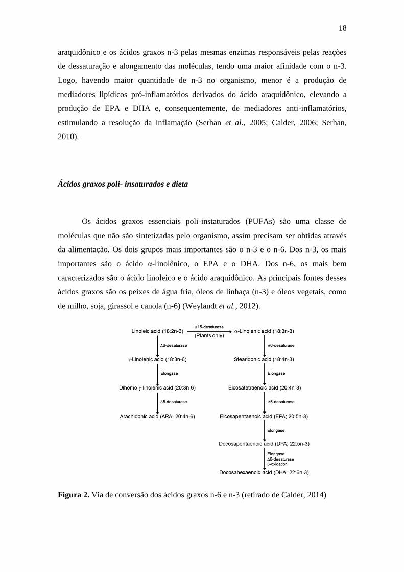
Ácidos graxos poli- insaturados e dieta
Os ácidos graxos essenciais poli-instaturados (PUFAs) são uma classe de
moléculas que não são sintetizadas pelo organismo, assim precisam ser obtidas através
da alimentação. Os dois grupos mais importantes são o n-3 e o n-6. Dos n-3, os mais
importantes são o ácido α-linolênico, o EPA e o DHA. Dos n-6, os mais bem
caracterizados são o ácido linoleico e o ácido araquidônico. As principais fontes desses
ácidos graxos são os peixes de água fria, óleos de linhaça (n-3) e óleos vegetais, como
de milho, soja, girassol e canola (n-6) (Weylandt et al., 2012).
Figura 2. Via de conversão dos ácidos graxos n-6 e n-3 (retirado de Calder, 2014)

19
O n-3 chamou atenção em estudos feitos com esquimós da Groelândia, em que
foi observada uma baixa prevalência de doenças cardiovasculares, asma, artrite
reumatoide e outras doenças autoimunes. Foi feito um estudo com amostras dos
alimentos consumidos por esquimós da Groelândia, sendo observado que os ácidos
graxos predominantes na alimentação eram os poli-insaturados, principalmente os da
classe n-3. A inflamação, sendo o denominador comum destas doenças, levou a pensar
em um efeito anti-inflamatório do n-3, em função da alimentação desta população ser
rica em ácidos graxos (Bang et al., 1980; Weylandt et al., 2012).
Os ácidos graxos n-3 provenientes dos vegetais, como a linhaça, estão na
forma de ácido α-linonênico, tendo que ser convertidos em EPA e DHA no organismo.
Por outro lado, no caso da suplementação com óleo de peixe, não há necessidade deste
processo (Calder, 2014).
Sabe-se que a suplementação alimentar com n-3 está relacionada com a
prevenção e melhora de doenças cardiovasculares, alterações inflamatórias crônicas,
doenças autoimunes e câncer (Laviano et al., 2013; De Caterina et al., 2011; Vaughan et
al., 2013). Existem evidências demonstrando os benefícios da suplementação desses
ácidos graxos durante o tratamento oncológico, pois além de potencializarem os efeitos
antitumorais das medicações quimioterápicas, auxiliam no aparecimento de algumas
reações adversas relacionadas à quimioterapia (Bourgnoux et al.,2009; Murphy et al.,
2012; Laviano et al, 2013). Além do mais, demonstrou-se que a suplementação diária
de n-3 previne o aparecimento da síndrome da anorexia-caquexia relacionado ao câncer,
proporcionando a manutenção do peso corporal, melhorando os parâmetros
inflamatórios e protegendo contra o estresse oxidativo (Murphy et al., 2011;
Finnochiaro et al., 2012; Hajjaji et al., 2012).
É possível que estas estratégias possam ser úteis como medidas preventivas
para pacientes submetidos a esquemas de quimioterapia e/ou radioterapia, evitando ou
amenizando os quadros de cistite hemorrágica após ou durante o tratamento com
ciclofosfamida.

20
OBJETIVOS
Objetivos Gerais
Avaliar os efeitos de ácidos graxos poli-insaturados n-3 sobre os efeitos adversos
e antitumorais do quimioterápico, ciclofosfamida, através de modelos in vivo e in vitro.
Objetivos Específicos
o Avaliar o efeito da suplementação com n-3 de óleo de peixe e da administração de
DHA sistêmico e espinhal, sobre as alterações inflamatórias vesicais (edema e
hemorragia), após a aplicação de ciclofosfamida em camundongos.
o Avaliar o efeito da suplementação com n-3 de óleo de peixe e da administração de
DHA sistêmico e espinhal, sobre o comportamento nociceptivo após a aplicação de
ciclofosfamida em camundongos.
o Verificar o efeito da suplementação com n-3 de óleo de peixe e da administração de
DHA sistêmico, sobre os níveis de citocinas periféricas após a aplicação de
ciclofosfamida em camundongos.
o Verificar o efeito da suplementação com n-3 de óleo de peixe e da administração de
DHA sistêmico, sobre a migração de neutrófilos na bexiga após a aplicação de
ciclofosfamida em camundongos.
o Analisar o efeito da suplementação com n-3 de óleo de peixe e da administração de
DHA sistêmico, sobre a contagem total de leucócitos circulantes após a aplicação de
ciclofosfamida em camundongos.
o Analisar o efeito da suplementação com n-3 de óleo de peixe e da administração de
DHA sistêmico sobre a expressão do mRNA do receptor GPR40/FFAR1 após
aplicação de ciclofosfamida em camundongos.
o Verificar o efeito da suplementação com n-3 de óleo de peixe e da administração de
DHA sistêmico, sobre a ativação das células da glia (GFAP, Iba-1) na medula
espinhal após a aplicação de ciclofosfamida em camundongos.
21
o Verificar o efeito da suplementação com n-3 de óleo de peixe e da administração de
DHA sistêmico, sobre a presença do receptor GPR40/FFAR1 na medula espinhal
após a aplicação de ciclofosfamida em camundongos.
o Analisar o efeito do DHA e da ciclofosfamida sobre a linhagem de célula tumoral de
mama MDA-MB-231.

22
CONSIDERAÇÕES FINAIS
O n-3 é utilizado como suplemento para pacientes oncológicos em função da sua
capacidade de diminuir o crescimento de tumores e de aumentar a eficácia de
tratamentos quimioterápicos. Também é utilizado para prevenir o aparecimento da
síndrome de anorexia-caquexia, prejudicando o tratamento do paciente de uma forma
global (Silva, 2006; Bougnoux et al. 2009).
A ciclofosfamida é amplamente utilizada na prática clínica como quimioterápico
e como imunossupressor em terapias pós-transplantes, tendo diversos efeitos colaterais
que prejudicam a qualidade de vida do paciente. Um destes efeitos é a cistite
hemorrágica, um processo inflamatório crônico, que pode ser prevenido pelo uso do
Mesna, mesmo não tendo 100% de efetividade (de Jonge et al., 2006).
Os dados apresentados neste trabalho reveleram outros efeitos exercidos pela
suplementação de óleo de peixe rico em n-3 ou pela administração parenteral de DHA,
em relação à analgesia em dor visceral associada à CYP. Surpreendentemente, nenhum
dos tratamentos propostos neste trabalho foi capaz de alterar a inflamação na bexiga
causada pela CYP.
Analizando os possíveis mecanismos dos efeitos analgésicos dos ácidos graxos
n-3 no nosso modelo experimental, podemos sugerir que há o envolvimento de
modulação periférica e central. Em relação às alterações periféricas, foram observadas
modificações nas contagens de neutrófilos e linfócitos circulantes no tratamento com
DHA. De forma intrigante, a ração que era, até então, considerada como controle – rica
em n-6 – também alterou as taxas de neutrófilos e linfócitos, de maneira semelhante à
dieta rica em n-3. Já na modulação central, viu-se a ativação de astrócitos e a regulação
da expressão do receptor GPR40/FFAR1 na medula espinhal. Este último resultado, em
relação ao receptor GPR40/FFAR1, abriu novas portas para essa investigação, já que
Nakamoto et al. (2012) demonstrou que esse receptor está diretamente relacionado com
analgesia e que ele está expresso de forma constitutiva em diversas porções do sistema
nervoso central.
Além de resultados interessantes ao nível de dor visceral, nosso trabalho também
gerou evidências demonstrando um efeito benéfico do DHA no tratamento

23
quimioterápico, aumentando a citoxicidade provocada pela CYP em uma linhagem de
células de tumor de mama.
De modo geral, nossos resultados apliam os efeitos benéficos da suplementação
de n-3 quando associada ao tratamento antitumoral.

24
REFERÊNCIAS
Ariel A, Serhan CN (2012). New lives given by cell death: macrophage differentiation
following their encounter with apoptotic leukocytes during the resolution of
inflammation. Front Immunol 3(4):1-6.
Batista CK, Brito GA, Souza ML, Leitao BT, Cunha FQ, Ribeiro RA (2006). A model
of hemorrhagic cystitis induced with acrolein in mice. Braz J Med Biol Res 39(11):
1475-1481.
Bang H0, Dyerberg J, Sinclair HM (1980). The composition of the Eskimo food in
north western Greenland. Am J Clin Nutri 33: 2657-2661.
Bougnoux P, Hajjaji N, Ferrasson MN, Giraudeau B, Couet C, Floch OL (2009).
Improving outcome of chemotherapy of metastatic breast cancer by docosahexaenoic
acid: a phase II trial. Br J Cancer 101:1978-1985.
Brunton LL, Lazo JS, Parker KL. Goodman & Gilman: manual de farmacologia e
terapêutica. Porto Alegre: McGraw-Hill; 2010.
Buckley CD, Gilroy DW, Serhan CN (2014). Proresolving Lipid Mediators and
Mechanisms in the Resolution o Acute Inflammation. Immunity 41(3):315-327.
Calder PC (2006). n-3 Polyunsaturated fatty acids, inflammation, and inflammatory
diseases. Am J Clin Nutr 83(6):1505S-1519S.
Calder PC (2014). Marine omega-3 fatty acids and inflammatory processes: Effects,
mechanisms and clinical relevance. Biochim Biophys Acta. doi:
10.1016/j.bbalip.2014.08.010
De Caterina R (2011). N-3 fatty acids in cardiovascular disease. N Engl J Med. 2011
Jun 23;364(25):2439-50.

25
de Jonge ME, Huitema AD, Rodenhuis S, Beijnen JH (2005). Clinical pharmacokinetics
of cyclophosphamide. Clin Pharmacokinet 44(11): 1135-1164.
Emadi A, Jones RJ, Brodsky RA (2009). Cyclophosphamide and cancer: golden
anniversary.
Nat Rev Clin Oncol. 6(11):638-47.
Finocchiaro C, Segre O, Fadda M, Monge T, Scigliano M, Schema M, Tinivella M,
Tiozzo E, Catalano MG, Pugliese M, Fortunati N, Aragno M, Muzio G, Maggiora M,
Oraldi M, Canuto RA (2012). Effect of n-3 fatty acids on patients with advanced lung
cancer: a double-blind, placebo-controlled study. Br J Nutrition 108:327-333.
Goldberg RJ, Katz J (2007). A meta-analysis of the analgesic effects of omega-3
polyunsaturated fatty acid supplementation for inflammatory joint pain. Pain 129(1-
2):210-223.
Gray KJ, Engelmann UH, Johnson EH, Fishman IJ (1986). Evaluation of misoprostol
cytoprotection of the bladder with cyclophosphamide (Cytoxan) therapy. J Urol 136(2):
497-500.
Hajjaji N, Couet C, Besson P, Bougnoux P (2012). DHA effect on chemotherapy-
induced body weight loss: as exploratory study in a rodent model of mammary tumors.
Nutr Cancer 64(7):1000-1007.
Kehrer JP, Biswal SS (2000). The Molecular Effects of Acrolein. Toxicol Sci 57(1):6-
15.
Korkmaz A, Ozturk M, Yildirim I (2012). Hemorrhagic cystitis; and old story with new
advancements. J Expl Integ Med 2(2):93-94.
Korkmaz A, Topal T, Oter S (2007). Pathophysiological aspects of cyclophosphamide
and ifosfamide induced hemorrhagic cystitis; implication of reactive oxygen and
nitrogen species as well as PARP activation. Cell Biol Toxicol 23(5): 303-312.

26
Kris-Etherton PM, Harris WS, Appel LJ (2001). Fish Consumption, Fish Oil, Omega-3
Fatty Acids, and Cardiovascular Disease. Circulation 106:2747-2757.
Laviano A, Riana S, Mofino A, Fanelli FR (2013). Omega-3 fatty acids in cancer. Cur
Opin Clin Nutr Metab Care 16:156-161.
Macedo FYB, Mourão LTC, Palheta Jr RC, Jucá DM, Lima Jr RCP, Neto JSC,
Magalhães PJC, Santos AA, Souza MHLP, Brito GAC, Ribeiro RA (2011).
Cyclooxygenase-2 contributes to functional changes seen on experimental hemorrhagic
cystitis induced by ifosfamide in rat urinary bladder. Cancer Chemother Pharmacol
67(4):935-943.
Martins JP, Silva RB, Coutinho-Silva R, Takiya CM, Battastini AM, Morrone FB, et al.
(2012). The role of P2X7 purinergic receptors in inflammatory and nociceptive changes
accompanying cyclophosphamide-induced haemorrhagic cystitis in mice. Br J
Pharmacol 165(1): 183-196.
McCarville MB, Hoffer FA, Gingrich JR, Jenkins JJ 3rd (2000). Imaging findings of
hemorrhagic cystitis in pediatric oncology patients. Pediatr Radiol 30(3):131-8.
Murphy RA, Mourtzakis M, Mazurak V (2012). N-3 polyunsaturated fatty acids: the
potential role for supplementation in cancer. Curr Opin Clin Nutr Metab Care
15(3):246-251.
Murphy RA, Mourtzakis M, Chu QSC, Baracos VE, Reiman T, Mazurak VC (2011).
Nutritional Intervention With Fish Oil Provides Over Standar of Care for Weight and
Skeletal Muscule Mass in Patients With Nonsmall Cell Lung Cancer Receiving
Chemotherapy. Cancer 117:1775-1782.
Nakamoto K , Nishinaka T, Matsumoto K, Kasuya F, Mankura M, Koyama Y,
Tokuyama S (2012). Involvement of the long-chain fatty acid receptor GPR40 as a
novel pain regulatory system. Brain Res 1432: 74-83.

27
Ribeiro RA, Lima-Junior RCP, Leite CAVG, Mota JMSC, Macedo FYB, Lima MVA,
Brito GAC (2012). Chemotherapy-induced hemorrhagic cystitis: pathogenesis,
pharmacological approaches and new insights. Journal of Experimental and Integrative
Medicine 2(2):95-112
Silva, MPN (2006). Síndrome da anorexia-caquexia em portadores de câncer. Rev Bras
Cancerologia 52(1):59-77.
Serhan CN, Savill J (2005). Resolution of inflammation: the beginning programs the
end. Nat Immunol 6(12):1191-1197
Serhan CN, Yacoubian S, Yang R (2008). Anti-Inflammatory and Proresolving Lipid
Mediators. Annu Rev Pathol Mech Dis 3:279-312.
Serhan C (2010). Novel Lipid Mediators and Resolution mechanisms in Acute
Inflammtion: to resolve or not? Am J Pathol 177(4):1576-91.
Stables MJ, Gilroy DW (2011). Old and new generation lipid mediators in acute
inflammation and resolution. Prog Lipid Res 50(1):35-51.
Vaughan VC, Hassing MR, Lewandowski PA (2013). Marine polyunsaturated fatty
acids and cancer therapy. Br J Cancer 108:486-492. DOI: 10.1038/bjc.2012.586.
Weylandt KH, Chiu CY, Gomolka B, Waechter SF, Wiedmenmann B (2012). Omega-3
fatty acids and their lipid mediators: Towards and understanding of resolvin and
protectin formation. Prostaglandins Other Lipid Mediat 97(3-4):73-82.
Yoshida T, Kawashima A, Ujike T, Uemura M, Nishimura K, Miyoshi S (2008).
Hyperbaric oxygen therapy for radiation-induced hemorrhagic cystitis. Int J Urol
15(7):639-641.

28
ANEXOS

29
ANEXO A - CARTA DE APROVAÇÃO CEUA

30
ANEXO B - MANUSCRITO DO TRABALHO EXPERIMENTAL
Os resultados do presente trabalho foram submetidos à revista The Journal of
Nutritional Biochemistry, fator de impacto 4.592 (JCR 2013).

31
Omega-3 fatty acids are able to modulate the painful symptoms associated to
cyclophosphamide-induced-hemorrhagic cystitis in mice
a,dRaquel D. S. Freitas,
a,c,dKesiane M. Costa,
b,dNatália F. Nicoletti,
a,cLuiza W. Kist,
a,b,c,dMaurício R. Bogo,
a,b,d,eMaria M. Campos
aPUCRS, Programa de Pós-graduação em Medicina e Ciências da Saúde, Porto
Alegre/RS, Brasil
bPUCRS, Programa de Pós-graduação em Biologia Celular e Molecular, Porto
Alegre/RS, Brasil
cPUCRS, Laboratório de Genômica e Biologia Molecular, Faculdade de Biociências,
Porto Alegre/RS, Brasil
dPUCRS, Instituto de Toxicologia e Farmacologia, Porto Alegre/RS, Brasil
ePUCRS, Faculdade de Odontologia, Porto Alegre/RS, Brasil
Corresponding author:
Maria Martha Campos
Instituto de Toxicologia e Farmacologia
Pontifícia Universidade Católica do Rio Grande do Sul,
Avenida Ipiranga, 6681, Prédio 12/D, Sala 101. Partenon - 90619-900 - Porto Alegre,
RS, Brasil.
Phone number: +55 51 3320 3562; Fax number: +55 51 3320 3626.
E-mail addresses: [email protected]; [email protected]

32
Abstract
This study investigated the effects of the long-term dietary fish oil
supplementation or the acute administration of the omega-3 fatty acid docosahexaenoic
acid (DHA) in the mouse hemorrhagic cystitis (HC) induced by the anti-cancer drug
cyclophosphamide (CYP). HC was induced in mice by a single CYP injection (300
mg/kg i.p.). Animals received four different diets containing 10% and 20% of corn or
fish oil, during 21 days. Separated groups received DHA by i.p. (1 µmol/kg) or i.t. (10
µg/site) routes, 1 h or 15 min before CYP. The behavioral tests (spontaneous
nociception and mechanical allodynia) were carried out from 1 h to 6 h following CYP
injection. Bladder inflammatory changes, blood cell counts, and serum cytokines were
evaluated after euthanasia (at 6 h). Immunohistochemistry analysis was performed for
assessing spinal astrocyte and microglia activation, or GPR40/FFAR1 expression. In
vitro studies were conducted to determine the potential effects of DHA on CYP-induced
cytotoxicity in human breast cancer cells. Either fish oil supplementation or DHA
treatment (i.p. and i.t.) markedly prevented visceral pain, without affecting CYP-evoked
bladder inflammatory changes. Moreover, systemic DHA significantly prevented the
neutrophilia/lymphopenia caused by CYP, with a partial effect on serum IL-6 levels.
DHA also modulated the spinal astrocyte activation and the GPR40/FFAR1 expression.
Finally, the pre-incubation of DHA increased the anti-tumor effects of CYP in human
breast cancer cells. The supplementation with omega-3 fatty acids-enriched fish oil or
parenteral DHA might be interesting nutritional approaches for cancer patients under
chemotherapy schemes with CYP.
Keywords: hemorrhagic cystitis, cyclophosphamide, omega-3, mice, GPR40, DHA
33
1. Introduction
The beneficial prophylactic effects of fish oil-derived omega-3 fatty acids,
namely eicosapentaenoic acid (EPA) and docosahexaenoic acid (DHA), have been
extensively demonstrated in metabolic disorders and cardiovascular diseases [1, 2, 3].
As a consequence, the dietary supplementation with omega-3 fatty acids for the general
adult population has been widely recommended during the last decade [4]. Compelling
evidence also indicates that omega-3 fatty acids might be useful for cancer patients,
even by enhancing the anti-tumor effects of chemotherapy agents, or by preventing the
related side effects [1, 5, 6]. Furthermore, it has been shown that dietary daily intake of
supplements enriched with EPA/DHA prevents cancer-induced cachexia and/or
sarcopenia, providing body weight maintenance in affected individuals [3]. For
instance, Murphy et al. [7] demonstrated that supplementation with 2.2-g fish oil per
day prevented the losses of body weight and muscle mass in patients with non-small
cell lung cancer under chemotherapy. In addition, Finnochiaro et al. [8] presented
similar results in a multi-center study conducted with the same cancer type, revealing a
reduction of inflammatory and oxidative parameters in patients receiving omega-3 fatty
acids. Interestingly, it was demonstrated that DHA-rich diet displayed protective effects
on the body weight loss in rats treated with the chemotherapy drug doxorubicin [9].
Hemorrhagic cystitis (HC) is an adverse effect of chemotherapy or radiotherapy
on the pubic region. The most common symptoms are dysuria, frequency, nocturia,
urgency, intense suprapubic pain and gross hematuria [10, 11]. Cyclophosphamide
(CYP) is an alkylating agent employed in chemotherapy schemes for a series of
different types of cancer, such as non-Hodgkin lymphoma, leukemia, breast cancer,
among other solid tumors [12]. Despite the potential anti-tumor effects of CYP, its use
is highly associated to the occurrence of HC, due to the generation of the urotoxic
34
metabolite acrolein, affecting 2 to 40 % of the treated patients. Preventive approaches,
including intense bladder irrigation and the co-administration of sodium-2-
mercaptoethane sulphonate (Mesna) have been used, although these strategies are not
totally effective in clinics, especially after long-term exposure to high doses of CYP
[13]. Therefore, it is reasonable to propose that omega-3 rich diets could be useful in
preventing CYP-induced HC. In fact, it was previously demonstrated that repeated
treatment with EPA (100 to 300 mg/kg) was able to reduce CYP-elicited genotoxicity
and oxidative stress in mice, although the inflammatory or painful urological alterations
have not been assessed in this study [14]. Notably, high levels of omega-3 fatty acids
have been positively correlated with disease remission rates in patients with non-
Hodgkin’s lymphoma that had been treated with different chemotherapy agents,
including CYP [15].
In the light of literature data, the present study investigated to what extent the
long-term supplementation with marine omega-3 fatty acids or the acute treatment with
DHA, might prevent the collateral effects associated with HC induced by CYP in mice,
aiming to assess the mechanisms related to the protective effects of omega-3 in this in
vivo experimental model. Efforts have also been made to evaluate whether DHA
interferes with the in vitro anti-tumor effects of CYP in a human breast cancer cell line.
2. Methods
2.1. Animals
Male Swiss mice (25 – 30g; total number of 200 mice) obtained from the
Universidade Federal de Pelotas (UFPEL) were used throughout the study. The animals
were housed in groups of five per cage and maintained in controlled temperature (22
2°C) and humidity (60–70%), under a 12 h light–dark cycle, with food and water ad

35
libitum. Experiments were conducted in accordance with current guidelines for the care
of laboratory animals, ethical guidelines for the investigation of experimental pain in
conscious animals and the ARRIVE Guidelines Checklist [16, 17]. All the experimental
procedures were approved by the Animal Ethics Committee of Pontifícia Universidade
Católica do Rio Grande do Sul (RS) (Protocol Number: CEUA 12/00303). The
experiments were performed between 8:00 and 12:00 AM to minimize the potential
circadian variations in the behavioral responses. The number of animals and the
intensity of noxious stimuli were the minimum necessary to demonstrate the consistent
effects of the treatments.
2.2. Drugs
The following drugs were used: cyclophosphamide (CYP; Genuxal - Baxter
Oncology GmbH; Halle/Westfalen, Germany) was purchased at Medilar (Porto Alegre,
Brazil), being diluted in distilled water. Docosahexaenoic acid in 1% ethanol (DHA)
was purchased from Cayman Chemicals (Michigan, USA) and was diluted in phosphate
buffered saline (PBS) until the desired concentration. The final ethanol concentration
never exceeded 0.1%.
2.3. Diets and treatments
Four different diets were prepared using Nuvilab® CR-1 chow, with the addition
of corn oil (50% of omega-6 fatty acids) or concentrated fish oil (55% of omega-3 fatty
acids); each oil was added at two distinct concentrations, 10% and 20%. The detailed
composition of diets is provided in the Supplementary Table 1. Dietary scheme started 1
month after birth, lasting 21 days, being HC induced at the 22th
day [18]. In a separate
series of experiments, the animals received DHA at different schedules of

36
administration. DHA was acutely dosed, 1 h or 15 min before the induction of HC, at 1
µmol/kg (intraperitoneal; i.p.) or 10 µg/site (intrathecal; i.t.), respectively [19, 20].
DHA-treated groups received only regular chow during all the experimental periods.
2.4. Induction of cystitis and nociception assessment
HC was induced by a single i.p. administration of CYP (300 mg/kg).
Immediately after the i.p. injection of CYP, mice were housed in individual plastic
cages, without sawdust bedding, and the spontaneous nociception behavior was
measured for 2 min, every 30 min, over a total period of 4 h. The behavioral alterations
were scored according to the following scale: 0 = normal; 1 = piloerection; 2 = strong
piloerection; 3 = labored breathing; 4 = licking of the abdomen; or 5 = stretching and
contractions of the abdomen, and the activity (walking, grooming, and rearing) was
recorded in seconds [21].
At the end of the 5th
h, Von Frey test was conducted to evaluate the mechanical
allodynia in the lower abdominal area. For this experimental set, 6-10 mice/group
(supplementation-treated animals) or 8 mice/group (DHA i.p. and i.t.) were used. Mice
were placed individually in clear Plexiglas boxes (9 x 7 x 11 cm) on elevated wire mesh
platforms to allow access to the abdomen. The withdrawal response frequency was
measured after 10 applications (duration of 1 s each) of 0.4 g von Frey hair (VFH)
(Stoelting, Chicago, IL), obtaining the percentage of frequency responses. The
following reactions were considered as a positive withdrawal response: sharp retraction
of the abdomen, immediate licking, scratching at the site of filament application, and/or
jumping [22].
In separate experimental groups, to evaluate the mechanical allodynia during all
the 6-h period after HC induction, the 0.4-g VFH was applied below to the plantar
37
surface of the right hind paw (to assess referred pain), or to the lower abdomen area, as
described by Meotti et al. [23], with slight modifications. The nociceptive responses
were evaluated at different time-points (1, 2, 4, 6 h) following CYP injection. The 0.4 g
VFH filament application to the hind paw and to the lower abdomen area produces a
mean withdrawal frequency of about 10% and 30%, respectively, which are adequate
values for the measurement of mechanical hypersensitivity. For this series of
experiments, 5-6 mice or 9-11 mice/group (10% and 20% dietary lipid concentration,
respectively), or 7-8 mice/group (DHA i.p. and i.t.) were used. In all the cases, after 6 h
of the CYP injection, the animals were killed by deep inhalation of sevoflurane for
further evaluation of inflammatory parameters.
2.5. Determination of bladder inflammatory parameters
This method was based on criteria established by Gray et al. [24]. Following
euthanasia (6 h after CYP application), all the bladders were dissected free from
connecting tissues, and transected at the bladder neck. Each bladder was
macroscopically evaluated, by an examiner unaware of the treatment groups. The edema
formation was categorized as severe (3), moderate (2), mild (1) or absent (0). Edema
was considered severe when fluid was seen externally in the walls of the bladder, as
well as internally. When edema was confined to the internal mucosa, it was reported as
moderate; when it was between normal and moderate, the edema was defined as mild.
Bladders were also examined for hemorrhage and categorized into four classes,
depending on the presence of intravesical clots (3), mucosal hematomas (2), dilatation
of the bladder vessels (1), or normal aspect (0). As an additional measure of bladder
edema, the wet weight of each bladder was recorded and expressed as mg per 100 g of
animal [24]. For this purpose, animals from the first set of behavioral tests were used.
38
2.6. Hematological parameters
After euthanasia, a small drop of blood was collected for the smear evaluation,
using Giemsa staining [25]. Differential cell counts (neutrophils, eosinophils, basophils,
lymphocytes, monocytes and immature cells) were estimated under an x40 objective, by
counting 100 cells [26]. Representative pictures were captured. For this analysis, the
animals from the second set of mechanical allodynia experiments were used.
2.7. Myeloperoxidase (MPO) activity
Neutrophil recruitment to the urinary bladder was measured by means of tissue
MPO activity, according to the method described by Martins et al. [21], with some
modifications. After euthanasia following 6 h of CYP injection, the bladders were
removed and stored at -80 °C. The tissues were homogenized in 5% (w/v) EDTA/NaCl
buffer (pH 4.7) and centrifuged at 4,000 rpm for 25 min, at 4°C. The pellet was
resuspended 0.5% hexadecyltrimethyl ammonium bromide buffer (pH 5.4), and the
samples were re-centrifuged (4,000 rpm, 25 min, 4°C). Twenty-five microliters of the
supernatant were used for the MPO assay. The enzymatic reaction was assessed with
1.6 mM tetramethylbenzidine, 80 mM NaPO4, and 0.3 mM hydrogen peroxide. The
absorbance was measured at 595 nm, and the results are expressed in optical density
(OD) per milligram of tissue. For MPO assay, 4-6 animals/groups were used for the
10% and 20% dietary lipid supplementation groups, whereas 4 animals/group were used
for DHA treatment group.

39
2.8. Analysis of GPR40/FFAR1 expression by quantitative real time RT-PCR (RT-
qPCR)
The spinal cords were collected 6 h after induction of HC by CYP. The total
RNA was isolated with Trizol® reagent (Invitrogen, Carlsbad, CA, USA) in accordance
with the manufacturer’s instructions. The total RNA was quantified (A260, A280,
A230) with the Quantifier spectrophotometer L-quant (Loccus Biotecnologia) and after
treated with Deoxyribonuclease I (Invitrogen) to eliminate genomic DNA
contamination in accordance with the manufacturer’s instructions. The cDNA was
synthesized with ImProm-II™ Reverse Transcription System (Promega) from 1 µg total
RNA, following the manufacturer´s instructions. Quantitative PCR was performed using
SYBR® Green I (Invitrogen) to detect double-strand cDNA synthesis. Reactions were
done in a volume of 25 µL using 12.5 µL of diluted cDNA, containing a final
concentration of 0.2x SYBR®
Green I (Invitrogen), 100 µM dNTP, 1x PCR Buffer, 3
mM MgCl2 0.25 U Platinum® Taq DNA Polymerase (Invitrogen), 0.5 M of betaine (for
Ffar-1), and 200 nM of each reverse and forward primers (Supplementary Table 2) The
PCR cycling conditions were: an initial polymerase activation step for 5 min at 95 °C,
40 cycles of 15 s at 95 °C for denaturation, 35 s at 60 °C for annealing and 15 s at 72 °C
for elongation. At the end of cycling protocol, a melting-curve analysis was included
and fluorescence measured from 60 to 99 °C and showed in all cases one single peak.
Hprt1, Ppia and Tbp were used as reference genes for normalization. Relative
expression levels were determined with 7500 Fast Real-Time Systems Software v.2.0.6
(Applied Biosystems). The efficiency per sample was calculated using LinRegPCR
2012.3 Software (http://LinRegPCR.nl). Relative mRNA expression levels were
determined using the 2-ΔΔCT
method. For this assay, the number of animals was 4-5
animals/group (i.p. saline/i.p. saline; i.p. saline/i.p. CYP, and i.p. DHA/i.p. CYP).
40
2.9. Evaluation of glia activation and GPR40/FFAR1 expression by
immunohistochemistry
The technique was performed as described by Maciel et al. [27], with
adaptations. After euthanasia at 6 h, the lumbar spinal cords (L3–L6 region) were
collected for immunohistochemistry analysis. Immunopositivity for activated astrocytes
or microglia, and GPR40/FFAR1 expression was assessed on paraffin tissue sections (3-
µm) by using the monoclonal rabbit anti-GFAP (1:250, Cat. #04-1062; Lot #2145973;
Merck Millipore, Darmstadt, Germany), monoclonal mouse anti-Iba1/AIF1 (1:300; Cat.
#MABN92; Lot #2172784; Merck Millipore, Darmstadt, Germany), and polyclonal
rabbit anti-GPR40/FFAR1 (1:100; Item no. 10007205; Cayman Chemicals, Michigan,
USA). High-temperature antigen retrieval was performed by immersion of the slides in
a water bath at 98–100 °C in 10 mM trisodium citrate buffer, pH 6.0 (anti-Iba-1), Tris-
EDTA buffer pH 9.0 (anti-GFAP and anti-GPR40) for 40 min. The peroxidase was
blocked by incubating the sections with perhidrol 5% for 30 min. The nonspecific
protein binding was blocked with milk serum solution 5% for 30 min. After overnight
incubation at 4 °C with primary antibodies, the slides were washed with PBS and
incubated with the secondary antibody HRP conjugate (Invitrogen), ready-to-use, for 20
min at room temperature. The sections were washed in PBS, and the visualization was
completed by using 3,3′-diaminobenzidine (Dako Cytomation) in chromogenic solution
and counterstained lightly with Harris’s Hematoxylin solution. Images were examined
with a Zeiss AxioImager M2 light microscope (Carl Zeiss, Gottingen, Germany). For
each section, 4-5 images were taken, in order to contemplate most areas of the spinal
cord. The images were captured in x200 magnification, and evaluated by using the
Image NIH Image J 1.36b Software (NIH, Bethesda, MD, USA). The number of GFAP-

41
positive astrocytes and Iba1-positive microglia cells was quantified by two independent
examiners in a blinded manner, in the following regions of the lumbar spinal cord: right
dorsal horn (RDH), left dorsal horn (LDH), right ventral horn (RVH), left ventral horn
(LVH) and central canal (CC). For the quantification of GPR40/FFAR1 positive
neurons, digitized 8-bit images were transferred to a computer, and the average pixel
intensity was calculated by using NIH ImageJ 1.36b Software, by analyzing the
following regions of the spinal cord: right ventral horn (RVH), left ventral horn (LVH),
right to the central canal (RCC), left to the central canal (LCC). For
immunohistochemistry analysis, 7–8 animals per group were used (i.p. saline/i.p. saline;
i.p. saline/i.p. CYP, and i.p. DHA/i.p. CYP).
2.10. Flow cytometry for serum cytokines
As a parameter of peripheral inflammation, the CBA mouse inflammation kit
(BD Biosciences) was used to determine the serum levels of interleukin-6 (IL-6), IL-10,
monocyte chemoattractant protein-1 (MCP-1), interferon-γ (IFN-γ), tumor necrosis
factor (TNF), and IL-12p70. The samples were analyzed using a flow cytometer
(FACSCanto II, BD Biosciences, San Jose, CA, USA) with a 488 nm laser and fitted
with a high-throughput sampler, as described previously by Nicoletti et al. [28]. Sample
data were acquired using BD FACSDiva V6.1.3 (BD Biosciences), and the results were
analyzed using FCAPArray v1.0.1 (BD Biosciences/Soft Flow HungaryLtd.) analysis
software. The data are expressed in pg/ml. For these experiments, 4-5 animals per group
were used (i.p. saline/i.p. saline; i.p. saline/i.p.CYP, and i.p. DHA/i.p. CYP).

42
2.11. Analysis of DHA effects on CYP-induced cytotoxicity
MDA-MB-231 human breast cancer cell lines were from American Type
Culture Collection (ATCC-Rockville, Maryland, USA). The cells were cultured in
Dulbecco’s Modified Eagle Medium with 10% fetal bovine serum (FBS) at a
temperature of 37°C, a minimum relative humidity of 95 %, and an atmosphere of 5 %
CO2 in air. The number of cells with metabolically active mitochondria was determined
based on the mitochondrial reduction of a tetrazolium bromide salt (MTT [3-(4,5-
imethylthia-zol-2-yl)-2,5-diphenyltetrazolium bromide] assay), according to the method
described by Gehring et al. [29]. In a first series of experiments, the cells were treated
with 4 different concentrations of CYP (0.1 mM, 1 mM, 10 mM, 50 mM) or DHA (25
µM, 50 µM, 75 µM, 100 µM) when incubated alone [30,31]. Secondly, the effects of a
subliminal and an effective concentration of DHA (75 µM and 100 µM) and CYP (CYP
1 mM and 10 mM) were tested in combination, in order to determine whether or not
DHA might affect the anti-tumor effects of CYP. In this case, DHA was added 30 min
before CYP incubation, and the cell viability was assessed as described previously.
2.12. Statistical analysis
Results are presented as mean ± standard error mean (SEM). The statistical
comparison of the data was performed by one-way analysis of variance (ANOVA),
followed by Bonferroni’s post-hoc test. The areas under the curves (AUC) were
calculated in the time-course experiments to evaluate hind paw or abdominal allodynia.
P values less than 0.05 (P< 0.05) were considered as indicative of significance
(GraphPad Prism 5.0, La Jolla, CA, USA).

43
3. Results
3.1. Effects of 21-day lipid dietary supplementation on painful and inflammatory
parameters in CYP-induced HC model
Extending previous literature data [32, 33], the present results show that acute
administration of CYP caused marked bladder inflammatory alterations, accompanied
by reduced locomotor activity, spontaneous nociception and regional mechanical
allodynia (Fig. 1-5). In this experimental model, the long-term dietary supplementation
with 10 % or 20 % fish oil produced a significant inhibition of the spontaneous
nociceptive behavior, when compared to the groups that received 10 % and 20 % of
corn oil diet (P < 0.05, P < 0.01, respectively; Fig. 1a). Furthermore, the diet
supplementation with both concentrations of fish oil (10 % and 20 %) also resulted in a
significant reduction of abdominal mechanical allodynia, in comparison to the same
concentrations of corn oil, when evaluated at the 5th
h (P < 0.05; Fig. 1b). Regarding the
inflammatory parameters, either 10 % or 20 % fish oil diet supplementation failed to
significantly alter the hemorrhage score (Fig. 1e) or the increased bladder wet weight
induced by CYP (Fig. 1f). Nevertheless, a slight inhibition of the edema score was
obtained with the 20 % fish oil diet supplementation, when compared to the 20 % corn
oil group (P < 0.05; Fig. 1d). Finally, the CYP-induced locomotor deficits were not
altered by any of the lipid dietary concentrations (Fig. 1c; P > 0.05).
3.2. Effects of 21-day lipid dietary supplementation on mechanical allodynia throughout
6-h period
Concerning the time-course evaluation of abdominal and hind paw mechanical
allodynia, 10% fish oil dietary supplementation was not able to reduce the
hypersensitivity, when VFH was applied to the right hind paw (Fig. 2a, b) or to the

44
lower abdomen (Fig. 2c, d), in comparison to the 10 % corn oil diet groups (P > 0.05),
according to the analysis of the AUC. In contrast, 20% fish oil dietary supplementation
significantly inhibited the abdominal mechanical allodynia, in relation to the 20% corn
oil diet group (P < 0.01; Fig. 2g,h), as indicated by the AUC evaluation. However, the
hind paw mechanical response in CYP-treated animals was not altered when compared
to the saline groups, with no difference between both lipids (P > 0.05; Fig. 2e, f).
3.3. Effects of i.p. and i.t. DHA on painful and inflammatory parameters in CYP-
induced HC model.
Based on earlier literature data demonstrating marked analgesic effects for DHA
in rodents [34], and also on the results described above for fish oil supplementation, we
decided to assess the effects of DHA administration, dosed by i.p. or i.t. routes, on the
adverse effects elicited by CYP (Fig 3-5). The systemic treatment with DHA (1
μmol/kg, i.p.), given 1 h before, significantly inhibited the spontaneous nociceptive
behavior caused by CYP (P < 0.01; Fig. 3a). In addition, the DHA i.p. treatment also
reduced the abdominal mechanical allodynia on the 5th
h after CYP injection (P < 0.05;
Fig. 3b). Conversely, the i.p. administration of DHA failed to inhibit the edema (P >
0.05; Fig. 3d), the hemorrhage (P > 0.05; Fig. 3e), the increased bladder wet weight (P
> 0.05; Fig. 3f), or the reduced locomotor activity, when compared to the control CYP-
treated groups (P > 0.05; Fig. 3c).
The effects of DHA administration were also tested when this fatty acid was
injected spinally. The i.t. administration of DHA (10 μg/site), given 15 min before CYP
administration, significantly lessened the spontaneous nociceptive behavior induced by
CYP (Fig. 4a; P < 0.05). Similarly, the abdominal mechanical hypersensitivity at the 5th
h was also reduced by the spinal application of DHA (Fig. 4b; P < 0.05). In relation to

45
the inflammatory changes, the i.t. application of DHA was not capable of altering the
edema (Fig. 4d; P > 0.05), or the hemorrhage (Fig. 4e; P > 0.05) associated to CYP
toxicity. However, the spinal treatment with DHA slightly inhibited CYP-induced
increase of the bladder wet weight (Fig. 4f; P < 0.05), whereas it did not affect CYP-
dependent reduced locomotor activity (Fig. 4c; P > 0.05).
3.4. Effects of i.p. and i.t. DHA on mechanical allodynia throughout 6-h period
The systemic administration of DHA displayed marked analgesic effects when
VFH was applied to the right hind paw (Fig. 5a,b; P < 0.05) or to the lower abdomen
(Fig. 5c,d; P < 0.05), according to the analysis of AUC. Nevertheless, the spinal
treatment with DHA caused a modest, although not significant reduction of hind paw
(Fig. 5e, f; P > 0.05) or abdominal mechanical allodynia elicited by CYP (Fig. 5g, h; P
> 0.05), as indicated by the AUC analysis.
3.6. Effects of 21-day lipid dietary supplementation or systemic DHA administration on
total blood cell counts.
Confirming the literature data [26], our results showed that saline-treated
animals display a predominance of lymphocytes. In contrast, the application of CYP
induced a marked reduction of lymphocyte counts, associated to a significant increase
of neutrophils in animals with a regular chow, an effect that was significantly reversed
by the i.p. administration of DHA (Fig. 6c, P < 0.05). On the other hand, the dietary
supplementation with 10 % (Fig. 6b) and 20 % (Fig. 6c) of either corn or fish oil led to
visible changes of lymphocyte/neutrophil rates in CYP-treated mice, and therefore the
statistical comparison for this set of experiments was not possible. Representative
images for hematological analysis are provided in the Supplementary Fig. 1.

46
3.7. Effects of 21-day lipid dietary supplementation or systemic DHA administration on
peripheral cytokine levels and on urinary bladder MPO activity.
Our data show that CYP injection was associated to increased MPO levels, but
this was not significantly changed by fish oil supplementation at 10 % (Fig. 6d) or 20 %
(Fig. 6e), or even by the i.p. treatment with DHA (Fig. 6f) (P > 0.05).
The urinary bladder inflammation induced by CYP involves the production of
local inflammatory cytokines, as demonstrated by Silva et al. [33] Nevertheless, in the
current study, we decided to evaluate the levels of circulating cytokines, based on the
differences of circulating leukocyte counts, as described above. In our experimental
model, the levels of IFN-γ and IL-12p70 were undetectable. Additionally, there was no
difference among the experimental groups when comparing the serum levels of TNF
(Fig. 7c) or IL-10 (Fig. 7d) (P > 0.05). The production of MCP-1 was significantly
augmented by CYP administration (P < 0.01), but the i.p. treatment with DHA did not
modify the levels of this cytokine (Fig. 7b). Regarding the serum production of IL-6, the
injection of CYP induced a marked increase of this cytokine (P > 0.01), and the
systemic DHA treatment was able to decrease IL-6 levels, but not in a significant
manner (Fig. 7a).
3.8. Effects of systemic administration of DHA on glial cells activation in the lumbar
spinal cord
Previous evidence demonstrates that administration of some chemotherapy
agents is associated with the spinal activation of glial cells in rats [35, 36]. Herein, the
administration of CYP led to astrocyte activation throughout distinct regions of the
mouse lumbar spinal cord, with significant differences at the right dorsal horn, in
relation to the saline control groups (Fig. 8a; P < 0.05). In this protocol, the i.p.

47
treatment with DHA caused a slight reduction in the number GFAP-positive astrocytes,
although this effect was not significant (P > 0.05). The total number of activated
astrocytes was calculated, showing no significant difference among the experimental
groups (Fig. 8b; P > 0.05). Representative images of the right dorsal horn of the lumbar
spinal cord are provided, indicating the presence of GFAP-positive astrocytes in the
following experimental groups: saline i.p. (Fig. 8c), CYP 300 mg/kg i.p. + saline i.p.
(Fig. 8d), and CYP 300 mg/kg i.p. + DHA 1 µmol/kg i.p (Fig. 8e). The microglial
activation was also evaluated by determining the positive immunolabelling for
Iba1/AIF1 in the mouse spinal cord. However, the immunopositivity for this marker
was sparsely observed throughout the different experimental groups (Supplementary
Fig. 2a-c).
3.9. Effects of systemic administration of DHA on GPR40/FFAR1 immunolabeling in
the lumbar spinal cord
The activation of the fatty acid receptor GPR40/FFAR1 by DHA binding has
been associated to analgesic effects in rodent models of pain [37]. Initially, we decided
to employ RT-qPCR to assess GPR40/FFAR1 expression in the mouse spinal cord.
However, it was not possible to detect transcripts in either experimental group (a
representative amplification plot is provided in the Supplementary Fig. 2d). Two sets of
primers were constructed and tested using 1µg of total RNA template per RT reaction,
and the effects of PCR-enhancing agents (betaine and DMSO) were also evaluated,
without successful amplification. Of note, all the reference genes used (Ppia, Tbp and
Hprt1) worked well under the conditions adopted, indicating that this method was not
suitable for GPR40/FFAR1 detection in our experiments. Therefore, an
immunohistochemistry analysis for GPR40/FFAR1 detection was carried out, and

48
analyzed throughout four different anatomical regions of the lumbar mouse spinal cord.
The administration of CYP led to a visible reduction of immunolabelled GPR40/FFAR1
positive neurons, when compared to the saline control group, an effect that was brought
to the control values by the i.p. treatment with DHA, with significant effects at the right
ventral horn (Fig. 9a; P < 0.05). The scatter dot plots did not reveal any significant
difference for the total quantification of positive GPR40/FFAR1 neurons among the
experimental groups, although it is possible to observe a similar profile with a reduction
of GPR40/FFAR1 immunopositivity in CYP-treated animals, and a reversion to the
saline control values in the DHA-treated group (Fig 9b, P > 0.05). Representative
images of the right ventral horn of the lumbar spinal cord show positive
immunolabelled neurons for GPR40/FFAR1, with evident dark-brown staining in the
three experimental groups (Fig. 9c-e).
3.10. In vitro effects of DHA treatment on CYP-induced cytotoxicity in MDA-MB-231
human breast cancer cells.
Initially, we analyzed different concentrations of CYP and DHA separately. The
treatment with CYP (10 mM and 50 mM) or DHA (100 µM) was able to reduce the
viability of MDA-MB-231 human breast cancer cells in a significant manner (Fig. 10a;
P < 0.01). The other tested concentrations of CYP (0.1 mM and 1mM) or DHA (25 µM,
50 µM, 75 µM) failed to alter the cell proliferation (Fig. 10a; P > 0.05). Next, we
assessed the effects of DHA and CYP, when these agents were tested in combination,
indicating that cytotoxic effects of CYP were not impaired by DHA incubation (Fig.
10b). Of note, the calculation of inhibition rates demonstrated that pre-incubation of a
subliminal concentration of DHA (75 µM) failed to potentiate the cytotoxic effects of a

49
low concentration of CYP (1 mM). However, additive significant effects were observed
by the treatment with DHA (100 µM) plus CYP (10 mM) (Fig. 10c; P < 0.01).
4. Discussion
The supplementation with fish oil-derived omega-3 fatty acids is frequently used
during chemotherapy, mostly to prevent the development of cancer cachexia [3]. HC is
a major adverse effect of the treatment with the antitumor agent CYP. HC is
characterized by a marked inflammation of the urinary bladder, causing severe lower
abdominal pain, among other symptoms [38, 39]. As an initial aim, this study evaluated
the protective effects of the dietary supplementation with fish oil-derived omega-3 fatty
acids on CYP-induced HC. Subsequently, we assessed the effects of the systemic or
spinal treatment with DHA in the same in vivo experimental parameters of CYP-caused
HC [40]. In addition, on the basis of the clinical trial carried out by Bougnoux et al. [5],
the effects of DHA were also tested in combination with CYP, by using an in vitro
assay for human breast cancer cell viability.
As demonstrated before, the acute administration of CYP is capable of inducing
marked spontaneous visceral nociception in rodents [41, 42]. In the current study, we
demonstrated an inhibition of the CYP-induced spontaneous nociceptive behavior in the
animals that received fish oil-enriched diets (10 and 20 %) during 21 days, when
compared to the corn oil diet groups. Of note, this inhibition was dependent on the
tested concentration of fish oil added to the chow. Supporting our data, a previous study
conducted by Nobre et al. [43]demonstrated marked analgesic effects for low doses of
fish oil-derived omega 3 fatty acids in rodent models of spontaneous nociception,
including the visceral pain elicited by acetic acid in mice. Furthermore, a meta-analysis

50
study published in 2007 suggested that the supplementation with fish oil derived-
omega-3 fatty acids represents an interesting adjunctive treatment for joint pain
associated with rheumatoid arthritis, inflammatory bowel diseases and dysmenorrhea
[44].
On the basis of recent literature data, it is tempting to suggest that analgesic
effects of fish-oil rich diets are likely related to the presence of DHA [45, 46].
Therefore, we decided to evaluate the effects of the systemic treatment with DHA in our
in vivo experimental paradigm. Notably, the single i.p. administration of DHA produced
a marked reduction of the nociceptive spontaneous behavior in the model of HC
induced by CYP in mice. Supporting these results, it was previously demonstrated that
oral administration of DHA was able to prevent either thermal or chemical nociception
in mice [47].
Altogether, our first series of data clearly indicate that either fish oil dietary
supplementation or systemic DHA display favorable analgesic effects in the mouse
model of CYP-evoked HC. This evidence prompted us to further assess the analgesic
effects of both approaches. As mentioned before, visceral pain is a major symptom of
CYP-induced HC [38, 39], and abdominal or hind paw mechanical sensitivity is
considered as indicative of this type of pain [32, 48]. In fact, Bon et al. [42]
demonstrated increased abdominal hypersensitivity at the 4th
h after dosing CYP (300
mg/kg), which is the same dose of CYP used in our experimental protocol, in different
mouse strains. In addition, a marked and time-related reduction of the abdominal or
hind paw mechanical threshold in CYP-treated rats was demonstrated before [23]. In
the present study, we also evaluated the abdominal mechanical allodynia at the 5th
h,
after the final assessment of spontaneous behavior, or in separate time-course
experiments from 1 to 6 h after CYP injection. In this case, only the 20 % fish oil

51
dietary supplementation reduced the abdominal mechanical allodynia, whereas the i.p.
administration of DHA inhibited both the abdominal and the hind paw mechanical
hypersensitivity, demonstrating the ability of this omega-3 fatty acid in preventing
referred pain.
It was previously demonstrated that spinal DHA was able to increase the paw
withdrawal threshold in the carrageenan-induced inflammatory pain or in CFA-evoked
heat hyperalgesia [19, 49]. To gain insights on the possible site of action of DHA, we
decided to assess the same set of behavioral tests as presented above, when DHA was
dosed i.t., 15 min before CYP. Our data indicate that DHA analgesic effects are, at least
partly, mediated by the modulation of spinal pathways related to pain transmission.
Accordingly, the i.t. administration of DHA displayed similar inhibitory actions on
either the spontaneous nociception or the mechanical allodynia, when compared to the
analgesic effects observed after the systemic DHA treatment, or the 21-day fish oil
dietary supplementation. Additionally, the i.t. treatment with DHA partially inhibited
the abdominal and the hind paw mechanical allodynia, when evaluated from 1 to 6 h
after CYP.
CYP-induced urinary bladder inflammation is characterized by the presence of
the severe edema and hemorrhage, accompanied by bladder neutrophil migration [21,
33, 50]. Although the intake of omega-3 fatty acids has been frequently associated with
anti-inflammatory effects [51], in our study, either the dietary supplementation with fish
oil or the treatment with DHA failed to display anti-inflammatory effects in the model
of HC caused by CYP. In fact, only a slight reduction of bladder edema was seen in the
groups that received 20 % fish oil or i.t. DHA. Thus, it is possible to conclude that
analgesic effects of marine-derived omega-3 fatty acids observed by us are not
dependent on the modulation of bladder inflammation.

52
A previous study demonstrated that repeated administration of low doses of CYP
(25 mg/kg) for three days resulted in a marked reduction of blood lymphocytes and
erythrocytes in mice, an effect that was prevented by the oral administration of Aloe
vera gel [52]. Extending this evidence, the present data revealed that a single i.p.
injection of a high dose of CYP (300 mg/kg) elicited a marked decrease of circulating
lymphocytes, associated to a significant increase of neutrophils, when compared to
saline-treated negative groups. Of note, the systemic administration of DHA modified
the lymphocyte/neutrophil rates in CYP-treated mice towards the saline control values.
Intriguingly, CYP-evoked alterations of lymphocytes and neutrophils were similarly
recovered by the dietary supplementation with omega-3-enriched fish oil, or even with
omega-6-containg corn oil. At this moment, we cannot explain this effect, but the
beneficial effects of corn oil supplementation in our model might be attributed to
omega-6-derived pro-resolution mediators, but this remains to be evaluated.
CYP-induced HC is accompanied by increased production of bladder or
circulating pro-inflammatory cytokines [21, 33, 53]. Considering the beneficial effects
observed for DHA on hematological parameters, we decided to investigate its actions on
serum production of cytokines. Our data revealed that CYP administration led to a
marked elevation of IL-6 and MCP-1 in the mouse serum, whereas the systemic
treatment with DHA caused a partial reduction of IL-6 levels. Thus, the favorable
effects on DHA on CYP-induced neutrophilia/lymphopenia could be explained, to some
extent, by the modulation of IL-6 production.
The use of chemotherapy agents has been associated with the development of
neuroinflammation, with marked activation of glia cells, such as astrocytes and
microglia [54]. In addition, both acute and chronic pain states are related to the
stimulation of glia cells in the central and peripheral nervous system [55]. Herein, we

53
show that a single injection of CYP was able to increase the number of activated
astrocytes in the right dorsal horn of the spinal cord, and the pretreatment with DHA
partially reversed this activation. Notwithstanding, it was not possible to detect any
change of microglia activation, in all the evaluated experimental groups. Previous
reports described a similar profile of astrocytic activation, without alteration of
microglia, in the neuropathic pain induced by the chemotherapy drugs, namely
paclitaxel, oxaliplatin or bortezomib in rats [35, 36]. Allied to literature data, our results
allow suggesting that analgesic effects displayed by DHA on CYP-induced visceral pain
are dependent on the modulation of astrocyte activation in the spinal cord.
It has been suggested that long-chain fatty acids exert their effects by interaction
with the G protein-coupled receptor GPR40/FFAR1 [56, 57]. Accordingly, convincing
evidence demonstrated that analgesic effects of DHA in the mouse model of pain
induced by global cerebral ischemia or by CFA injection involve the central activation
of GPR40/FFAR1 [45, 58]. The results of the present study extend this notion by clearly
showing that acute administration of CYP was associated with reduced
immunopositivity for GPR40/FFAR1 throughout neurons of the mouse spinal cord,
whilst the i.p. pre-treatment with DHA reversed this effect to the saline control levels.
Hence, the analgesic effects of DHA in CYP-treated mice probably rely on the neuronal
modulation of GPR40/FFAR1 receptors in the mouse spinal cord.
Our data extend the notion on the protective effects of omega-3 fatty acids in
chemotherapy-related adverse effects. In the case of the present study, we showed
marked analgesic effects for fish oil supplementation, and mainly DHA administration
against visceral pain associated to CYP-induced HC. To support our in vivo results, we
evaluated whether DHA might affect the cytotoxic effects of CYP. For this purpose, we
employed the MTT viability assay and the MDA-MB-231 human breast cancer cell

54
lineage. In this regard, it has been suggested that marine-derived omega-3 fatty acids,
such as DHA, are able to improve the effectiveness of cancer chemotherapy [59, 60].
For instance, it was demonstrated that DHA increased the cytotoxic in vitro effects of
the chemotherapy agent doxorubicin in MDA-MB-231 cells [61]. Herein, we provide
evidence indicating that isolated treatment with DHA or CYP led to concentration-
dependent reduction of MDA-MB-231 cell viability. Of note, the pre-incubation of
DHA resulted in increased effectiveness of antitumor effects displayed by CYP in this
cell line.
The supplementation with fish oil-derived omega-3 fatty acids is frequently
employed for cancer patients under chemotherapy, particularly to control cancer
cachexia. Our data shed new lights on the effects of omega-3 fatty acids, by revealing
the ability of fish oil-enriched diet or DHA administration to prevent the visceral pain
associated to CYP-induced HC. Surprisingly, either fish oil dietary supplementation or
parenteral DHA failed to alter the bladder inflammation caused by CYP. An analysis of
the possible mechanisms underlying the analgesic effects of omega-3 fatty acids in our
experimental model suggests the involvement of either peripheral or central
mechanisms, especially via modulation of circulating neutrophils, besides astrocytic
activation and GPR40/FFAR1 expression at the spinal level. One might suppose that
DHA would impair the anti-cancer effects of CYP; nonetheless, we also bring evidence
on the ability of this omega-3 fatty acid to sensitize human breast cancer cells to the
cytotoxic effects of CYP. Altogether, our results extend the notion about the beneficial
effects of omega-3 enriched diets to prevent the adverse effects caused by chemotherapy
drugs, or to improve their anti-tumor effects.
55
Conflict of interest
The authors declare that they have no conflict of interest.
Acknowledgments
This work was supported by grants from the Coordenação de Aperfeiçoamento
de Pessoal de Nível Superior (CAPES), Conselho Nacional de Desenvolvimento
Científico e Tecnológico (CNPQ), Fundação de Amparo à Pesquisa do Estado do Rio
Grande do Sul (FAPERGS) and FINEP Research Grant ‘‘Implantação, Modernização e
Qualificação de Estrutura de Pesquisa da PUCRS’’ (PUCRSINFRA) # 01.11.0014-00.
RDSF is a Msc. Postgraduate student in Medicine and Health Sciences: Molecular and
Biochemical Pharmacology, receiving grants from CAPES. KMC and NFN thanks
CAPES for financial support. The authors thank Mrs. Luciana Adolfo Ferreira for her
technical assistance in immunohistochemical technique.

56
References
[1] Laviano A, Riana S, Mofino A, Fanelli FR (2013). Omega-3 fatty acids in cancer.
Cur Opin Clin Nutr Metab Care 16:156-161. doi:10.1097/MCO.0b013e3285d2d99.
[2] De Caterina R (2011). N-3 fatty acids in cardiovascular disease. N Engl J Med Jun
23;364(25):2439-50. doi: 10.1056/NEJMra1008153
[3] Vaughan VC, Hassing MR, Lewandowski PA (2013). Marine polyunsaturated fatty
acids and cancer therapy. Br J Cancer 108:486-492. doi: 10.1038/bjc.2012.586.
[4] Bailey RL, Gahche JJ, Miller PE, Thomar PR, Dwyer JT (2013). Why US adults use
dietary supplements. JAMA Intern Med 173(5):355-361. doi:
10.1001/jamainternmed.2013.2299
[5] Bougnoux P, Hajjaji N, Ferrasson MN, Giraudeau B, Couet C, Floch OL (2009).
Improving outcome of chemotherapy of metastatic breast cancer by
docosahexaenoic acid: a phase II trial. Br J Cancer 101:1978-1985. doi:
10.1038/sj.bjc.6605441
[6] Murphy RA, Mourtzakis M, Mazurak V (2012). N-3 polyunsaturated fatty acids: the
potential role for supplementation in cancer. Curr Opin Clin Nutr Metab Care
15(3):246-251. doi: 10.1097/MCO.0b013e328351c32f.
[7] Murphy RA, Mourtzakis M, Chu QSC, Baracos VE, Reiman T, Mazurak VC
(2011). Nutritional Intervention With Fish Oil Provides Over Standard of Care for
Weight and Skeletal Muscle Mass in Patients With Nonsmall Cell Lung Cancer
Receiving Chemotherapy. Cancer 117:1775-1782. doi:10.1002/CNCR.25709.
[8] Finocchiaro C, Segre O, Fadda M, Monge T, Scigliano M, Schema M, Tinivella M,
Tiozzo E, Catalano MG, Pugliese M, Fortunati N, Aragno M, Muzio G, Maggiora
M, Oraldi M, Canuto RA (2012). Effect of n-3 fatty acids on patients with

57
advanced lung cancer: a double-blind, placebo-controlled study. Br J Nutrition
108:327-333. doi:10.1017/S0007114511005551
[9] Hajjaji N, Couet C, Besson P, Bougnoux P (2012). DHA effect on chemotherapy-
induced body weight loss: as exploratory study in a rodent model of mammary
tumors. Nutr Cancer 64(7):1000-1007. doi:10.1080/01635581.2012.714832
[10] Ribeiro RA, Lima-Junior RCP, Leite CAVG, Mota JMSC, Macedo FYB, Lima
MVA, Brito GAC (2012). Chemotherapy-induced hemorrhagic cystitis:
pathogenesis, pharmacological approaches and new insights. J Exp Integr Med
2:95-112. doi:10.5455/jeim.080312.ir.010
[11] Yoshida T, Kawashima A, Ujike T, Uemura M, Nishimura K, Miyoshi S (2008).
Hyperbaric oxygen therapy for radiation-induced hemorrhagic cystitis. Int J Urol
15(7):639-41. doi: 10.1111/j.1442-2042.2008.02053.x.
[12] Emadi A, Jones RJ, Brodsky RA (2009). Cyclophosphamide and cancer: golden
anniversary. Nat Rev Clin Oncol 6:638-647. doi:10.1038/nrclinonc.2009.146
[13] Mukhtar S, Woodhouse C (2010). The managment of cyclophosphamide-induced
haematuria. BJU Int 105:908-912. doi:10.1111/j.1464-410X.2009.09132.x.
[14] Li M, Zhu Q, Hu C, Giesy JP, Kong Z, Cui Y (2011). Protective Effects of
Eicosapentaenoic Acido n Genotoxicity and Oxidative Stress of Cyclophosphamide
in Mice. Environ Toxicol 26:217-223. doi: 10.1002/tox.20546.
[15] Cvetkovic, Vucic V, Cvetkovic B, Karadzic I, Ranic M, Glibetic M (2013).
Distribution of plasma fatty acids is associated with response to chemotherapy in
non-Hodgkin’s lymphoma patients. Med Oncol 30:741. doi:10.1007/s12032-013-
0741-2
[16] Zimmermann M (1983). Ethical guidelines for investigations of experimental pain
conscious animals. Pain 16:110-110.

58
[17] Kilkenny C, Browne WJ, Cuthill IC, Emerson M, Altman DG (2012). Improving
bioscience research reporting: the ARRIVE guidelines for reporting animal
research. Osteoarthritis Cartilage 20(4):256-60. doi: 10.1016/j.joca.2012.02.010
[18] de Matos OG, Amaral SS, Pereira da Silva PE, Perez DA, Alvarenga DM, Ferreira
AV, Alvarez-Leite J, Menezes GB, Cara DC (2012). Dietary supplementation with
omega-3-PUFA-rich fish oil reduces signs of food allergy in ovalbumin-sensitized
mice. Clin Dev Immunol 2012:236564. doi: 10.1155/2012/236564.
[19] Lu Y, Zhao LX, Cao DL, Gao YJ (2013). Spinal injection of docosahexaenoic acid
attenuate carrageenan-induced inflammatory pain through inhibition of microglia-
mediated neuroinflammation in the spinal cord. Neuroscience 241:22-31. doi:
10.1016/j.neuroscience.2013.03.003.
[20] Pan HC, Kao TK, Ou YC, Yang DY, Yen YJ, Wang CC, Chuang YH, Liao SL,
Ruang SL, Wu CW, Chianv AN, Chen CJ (2009). Protective effect of
docosahexaenoic acid against brain injury in ischemic rats. J Nutr Biochem 20:715-
725. doi: 10.1016/j.jnutbio.2008.06.014.
[21] Martins JP, Silva RBM, Coutinho-Silva R, Takiya CM, Battastini AMO, Morrone
FB, Campos MM (2012). The role of P2X7 purinergic receptor in inflammatory
and nociceptive changes accompanying cyclophosphamide-induced heamorhagic
cystitis in mice. Br J Pharmacol 165(1):183-96. doi: 10.1111/j.1476-
5381.2011.01535.x.
[22] Laird JM, Martinez-Caro L, Garcia-Nicas E, Cervero F (2001). A new model of
visceral pain and referred hyperalgesia in the mouse. Pain 92:335-342. doi:
10.1016/S0304-3959(01)00275-5.
[23] Meotti FC, Forner S, Lima-Garcia JF, Viana AF, Calixto JB (2013). Antagonism of
the transient receptor potential ankyrin 1 (TRPA1) attenuates hyperalgesia and
59
urinary bladder overactivity in cyclophosphamide-induced haemorrhagic cystitis.
Chem Bio Interact 203:440-447. doi: 10.1016/j.cbi.2013.03.008.
[24] Gray KJ, Engelmann UH, Johnson EH, Fishman IJ (1986). Evaluation of
misoprostol cytoprotection of the bladder with cyclophosphamide (Cytoxan)
therapy. J Urol 136: 497–500.
[25] Pilny AA (2008). Clinical hematology for rodent species. Vet Clin North Am Exot
Anim Pract 11:523-533, vi-vii. doi: 10.1016/j.cvex.2008.04.001.
[26] Costa KM, Maciel IS, Kist LW, Campos MM, Bogo MR (2014). Pharmacological
Inhibition of CXCR2 Chemokine Receptors Modulates Paraquat-Induced
Intoxication in Rats. PLoS ONE 9(8): e105740. doi:
10.1371/journal.pone.0105740.
[27] Maciel IS, Azevedo VM, Pereira TC, Bogo MR, Souza AH, Gomez MV, Campos
MM (2014). The spinal inhibition of N-type voltage-gated calcium channels
selectively prevents scratching behavior in mice. Neuroscience 277:794-805. doi:
10.1016/j.neuroscience.2014.07.065.
[28] Nicoletti NF, Rodrigues-Junior V, Santos AA Jr, Leite CE, Dias AC, Batista EL
Jr, Basso LA, Campos MM, Santos DS, Souto AA. (2014). Protective Effects of
Resveratrol on Hepatotoxicity Induced by Isoniazid and Rifampicin via SIRT1
Modulation. J Nat Prod 77(10):2190-5. doi: 10.1021/np5003143.
[29] Gehring MP, Pereira TCB, Zanin RF, Borges MC, Filho AB, Battastini AMO,
Bogo MR, Lenz G, Campos MM, Morrone FB (2012). P2X7 receptor activation
leads to increased cell death in a radiosenstive human glioma cell line. Purinergic
Signal 8:729-739. doi:10.1007/s11302-012-9319-2
[30] Pang H, Cai Li, Yang Y, Chen X, Sui G, Zhao C (2011). Knockdown of
Osteopontin Chemosensitizes MDA-MB-231 Cells to Cyclophosphamide by

60
Enhancing Apoptosis Through Activating p38 MAPK Pathway. Cancer Biother
Radiopharm. 26(2):165-173. doi :10.1089/cbr.2010.0838.
[31] Mouradian M, Kikawa KD, Drank BP, Komas SM, Kalyanaraman B, Pardini RS
(2014). Docosahexaenoic Acid Attenuates Breast Cancer Cell Metabolism and the
Warburg Pheotype by Targeting Bioenergetic Function. Mol Carcinog.
doi:10.1002/mc.22151
[32] Dornelles FN, Andrade EL, Campos MM, Calixto JB (2014). Role of CXCR2 and
TRPV1 in functional, inflammatory and behavioural changes in the rat model of
cyclophosphamide-induced haemorrhagic cystitis. Br J Pharmacol 171(2):452-67.
doi: 10.1111/bph.12467.
[33] Silva RBM, Sperotto NDM, Andrade EL, Pereira TCB, Leite CE, de Souza AH,
Bogo MR, Morrone FB, Gomez MV, Campos MM (2014). Spinal Blockage of
P/Q- OR N-type Voltage-gated Calcium Channels Modulates Functional and
Symptomatic Changes Related to Haemorrhagic Cystitis in Mice. Br J Pharmacol.
doi: 10.1111/bph.12966.
[34] Nakamoto K, Nishinaka T, Ambo A, Mankura M, Kasuya F, Tokuyama S (2011).
Possible involvement of β-endorphin in docosahexaenoic acid-induced
antinociception. Eur J Pharmacol 666(1): 100-104.
doi:10.1016/j.ejpharm.2011.05.047.
[36] Robinson CR, Zhan H, Dougherty PM (2014). Astrocytes, but not microglia, are
activated in oxaliplatin and bortezomib-induced peripheral neuropathy in the rat.
Neuroscience 274:308-317. doi: 10.1016/j.neuroscience.2014.05.051
[35] Zhang H, Yoon SY, Zhang H, Dougherty PM (2012). Evidence That Spinal
Astrocytes but Not Microglia Contribute for the Pathogenesis of Paclitaxel-Induced
Painful Neuropathy. J Pain 13(3):293-303. doi: 10.1016/j.jpain.2011.12.002.

61
[37] Nakamoto K , Nishinaka T, Matsumoto K, Kasuya F, Mankura M, Koyama Y,
Tokuyama S (2012). Involvement of the long-chain fatty acid receptor GPR40 as a
novel pain regulatory system. Brain Res 1432: 74-83.
doi:10.1016/j.brainres.2012.11.012.
[38] McCarville MB, Hoffer FA, Gingrich JR, Jenkins III JJ (2000). Imaging findings
of hemorrhagic cystitis in pediatric oncology patients. Pediatr Radiol 30:131-138.
[39] Augé C, Chene, G, Dubourdeau M, Desoubzadanne D, Corman B, Palea S, Lluel
P, Vergnolle N, Coelho AM (2013). Relevance of the cyclophosphamide-induced
cystitis model for the pharmacological studies targeting inflammation and pain of
the bladder. Eur J Pharmacol 707:32-40. doi: 10.1016/j.ejphar.2013.03.008
[40] Tokuyama S, Nakamoto K (2011). Unsaturated Fatty Acids and Pain. Bio Pharm
Bull 34(8): 1174-1178. doi: 10.1248/bpb.34.1174
[41] Boucher M, Meen M, Codron JP, Coudore F, Kemeny JL, Eschalier A (2000).
Cyclophosphamide-induced cystitis in freely-moving conscious rats: behavioral
approach to a new model of visceral pain. J Urol 164(1): 203-208.
doi:10.1016/S0022-5347(05)67495-2
[42] Bon K, Lichtensteiger CA, Wilson SG, Mogil JS (2003) Characterization of
cyclophosphamide cystitis, a model of visceral and referred pain, in the mouse:
species and strain differences. J Urol 170(3): 1008-1012. doi:
10.1097/01.ju.0000079766.49550.94
[43] Nobre MEP, Correia AO, Borges MB, Sampaio TMA, Chakraborty SA, Gonçalves
DO, Brito GAC, Leal LKAM, Felipe CFB, Lucetti DL, Arida RM, Viana GSB
(2013). Eicosapentaenoic acid and docosahexaenoic acid exert anti-inflammatory
and antinociceptive effects in rodents at low doses. Nut Res 33(5): 422-33. doi:
10.1016/j.nutres.2013.02.011.

62
[44] Goldberg RJ, Katz J (2007). A meta-analysis of the analgesic effects of omega-3
polyunsaturated fatty acid supplementation for inflammatory joint pain. Pain 129:
210-23. doi: 10.1016/j.pain.2007.01.020
[45] Nakamoto K, Nichinaka T, Sato N, Mankura M, Koyama Y, Kasuya F, Tokuyama
S (2013). Hypothalamic GPR40 Signaling Activated by Free Long Chain Fatty
Acids Suppresses CFA-Induced Inflammatory Chronic Pain. PLoS ONE
8(12):e81563. Doi: 10.1371/journal.pone.0081563.
[46] Torres-Guzman AM, Morado-Urbina CE, Alvarado-Vazquez P, Acosta-Gonzalez
RI, Chávez-Piña AE, Montiel-Ruiz RM, Jimenez-Andrade JM (2014). Chronic oral
or intraarticular administration of docosahexaenoic acid reduces nociception and
knee edema and improves functional outcomes in a mouse model of Complete
Freund's Adjuvant-induced knee arthritis. Arthritis Res Ther 16(2): R64. doi:
10.1186/ar4502.
[47] Nakamoto K, Nishinaka T, Mankura M, Fujita-Hamabe, W, Tokuyama S (2010).
Antinociceptive effects of docosahexaenoic acid against various pain stimuli in
mice. Bio Pharm Bull 33(6): 1070-1072. doi: 10.1248/bpb.33.1070.
[48] Cervero F, Laird JM (2004). Understanding the signaling and transmission of
visceral nociceptive events. J Neurobiol 61: 45–54.
[49] Xu ZZ, Zhang L, Liu T, Park JY, Berta T, Yang R, Serhan CN, Ji RR (2010).
Resolvins RvE1 and RvD1 attenuate inflammatory pain via central and peripheral
actions. Nat Med 16(5)592-595. doi: 10.1038/nm.2123.
[50] Golubeva AV, Zhdanov AV, Mallel G, Dinan TG, Cryan JF (2014). The mouse
cyclophosphamide model of bladder pain syndrome: tissue characterization,
immune profiling, and relationship to metabotropic glutamate receptors. Physiol
Rep 2(3):e00260. doi: 10.1002/phy2.260.

63
[51] Buckley, CD, Gilroy DW, Serhan CN (2014). Proresolving Lipid Mediators and
Mechanisms in the Resolution of Acute Inflammation. Immunity 40(3): 315-327.
doi: 10.1016/j.immuni.2014.02.009
[52] Im SA, Kim KH, Kim HS, Lee KH, Shin E, Do SG, Jo TH, Park Yi, Le CK (2014).
Processed Aloe vera Gel Ameliorates Cyclophosphamide-Induced Immunotoxicity.
Int J Mol Sci 15:19342-19354. doi: 10.3390/ijms151119342
[53] Dantas ACB, Batista-Junior FFB, Macedo LF, Mendes MNC, Azevedo IM,
Medeiros AC (2010). Protective effect of simvastatin in the cyclophosphamide
induced hemorrhagic cytisitis in rats. Acta Cirurg Brasileira 25(1):43-56. doi:
10.1590/S0102-86502010000100011
[54] Ji RR, Xu ZZ, Gao YJ (2014). Emerging targets in neuroinflammation-driven
chronic pain. Nature reviews. Drug discovery 13(7): 533-48. doi: 10.1038/nrd4334
[55] Ji RR, Berta T, Nedergaard M (2013). Glia and pain: Is chronic pain a gliopathy?
Pain 154: S10-S28. doi:/10.1016/j.pain.2013.06.022.
[56] Briscoe CP, tadayyon M, Andrews JL, Benson WG, Chambers JK, Eilert MM,
Ellis C, Eishourbagy NA, Goetz AS, Minnick DT, Murdock PR, Sauls HR, Shabon
U, Spinage LD, Strum JC, Szekeres PG, Tan KB, Way JM, Ignar DM, Wilson S,
Muir AI (2003). The orphan G protein-coupled receptor GPR40 is activated by
medium and long chain fatty acids. J Bio Chem 278(13):11303-11311.
doi:10.1074/jbc.M211495200
[57] Ma D, Tao B, Waraschina S, Kotani S, Lu L, Kaplamadzhiev DB, Mori Y,
Tonchev AB, Yamashima T (2007). Expression of free fatty acid receptor GPR40
in the central nervous system of adult monkeys. Neurosci Res 58(4):394-401.
doi:10.1016/j.neurores.2007.05.001

64
[58] Harada S, Haruna Y, Aizawa F, Matsuura W, Nakamoto K, Yamashita T, Kasuya
F, Tokuyama S (2014). Involvement of GPR40, a long-chain free fatty acid
receptor, in the production of central post-stroke pain after global cerebral
ischemia. Eur J Pharmacol 744:115-23. doi: 10.1016/j.ejphar.2014.09.036.
[59] Bougnoux P, Hajjaji N, Maheo K, Couet C, Chevalier S (2010). Fatty acids and
breast cancer: Sensitization to treatments and prevention of metastatic re-growth.
Prog Lipid Res 49(1):76-86. doi: 10.1016/j.plipres.2009.08.003
[60] Hajjaji N, Bougnoux P (2013). Selective sensitization of tumors to chemotherapy
by marine-derived lipids: A review. Cancer Treat Res 39(5):473-488. Doi:
10.1016/j.ctrv.2012.07.001
[61] Vibet S, Goupille C, Bougnoux P, Steghens JP, Goré J, Mahéo K (2008).
Sensitization by docosahexaenoic acid (DHA) of breast cancer cells to
anthracyclines through loss of gluthatione peroxidase (GPx1) response. Free Radic
Med Bio 44(7):1483-1491. doi: 10.1016/j.freeradbiomed.2008.01.009

65
Fig. 1. Effects of dietary supplementation with corn oil or fish oil (10 % and 20 %),
during 21 days, on painful and inflammatory parameters in the model of CYP-induced
HC. (A) Cumulative 4-h nociception score; (B) abdominal mechanical allodynia 5 h
after CYP injection; (C) cumulative 4-h locomotor activity; (D) edema and (E)
hemorrhage scores at the 6th
h; (F) bladder wet weight in grams. Each column
represents the mean ± SEM of 6 to 12 mice/group. ##
P < 0.01, #P < 0.05 significantly
different from saline-treated negative groups; **
P < 0.01, *P < 0.05 significantly
different from the respective corn oil CYP-treated group.

66
Fig. 2. Effects of dietary supplementation with corn oil or fish oil (10 % and 20 %),
during 21 days, on (A and E) hind paw or (C and G) abdominal mechanical allodynia
throughout 6-h period after CYP administration. The AUC for (B and F) hind paw or (D
and H) abdominal allodynia were calculated for both oils at 10 % and 20 %,
respectively. Each point of the curve or each column of the AUC graphs represents the
mean ± SEM of 6 to 11 mice/group. #P < 0.05 significantly different from saline-treated
negative groups; *P < 0.05 significantly different from the respective corn oil CYP-
treated group.
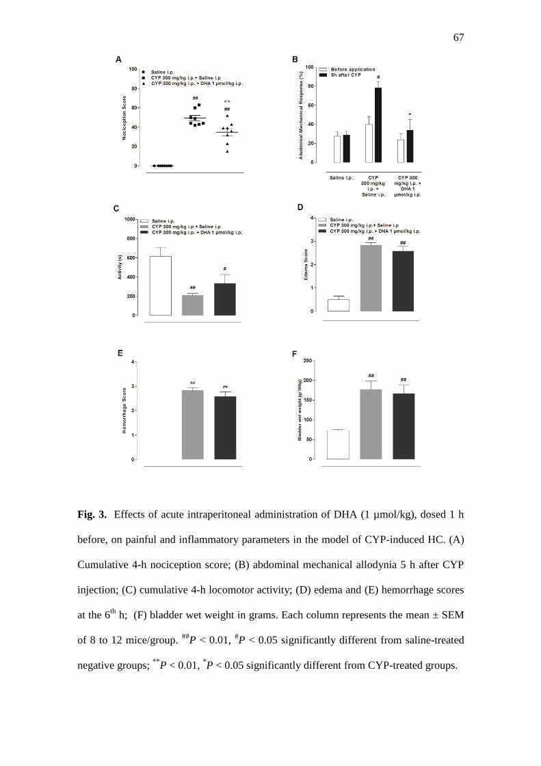
67
Fig. 3. Effects of acute intraperitoneal administration of DHA (1 µmol/kg), dosed 1 h
before, on painful and inflammatory parameters in the model of CYP-induced HC. (A)
Cumulative 4-h nociception score; (B) abdominal mechanical allodynia 5 h after CYP
injection; (C) cumulative 4-h locomotor activity; (D) edema and (E) hemorrhage scores
at the 6th
h; (F) bladder wet weight in grams. Each column represents the mean ± SEM
of 8 to 12 mice/group. ##
P < 0.01, #P < 0.05 significantly different from saline-treated
negative groups; **
P < 0.01, *P < 0.05 significantly different from CYP-treated groups.

68
Fig. 4. Effects of acute spinal administration of DHA (10 µg/site), injected 15 min
before, on painful and inflammatory parameters in the model of CYP-induced HC. (A)
Cumulative 4-h nociception score; (B) abdominal mechanical allodynia 5 h after CYP
injection; (C) cumulative 4-h locomotor activity; (D) edema and (E) hemorrhage scores
at the 6th
h; (F) bladder wet weight in grams. Each column represents the mean ± SEM
of 8 to 12 mice/group. ##
P < 0.01, #P < 0.05 significantly different from saline-treated
negative groups; *P < 0.05 significantly different from CYP-treated groups.

69
Fig. 5. Effects of (A, B, C and D) intraperitoneal administration of DHA (1 µmol/kg,
dosed 1 h before), or (E, F, G, H) spinal administration of DHA (10 µg/site, injected 15
min before), on (A and E) hind paw or (C and G) abdominal mechanical allodynia
throughout 6-h period after CYP administration. The AUC for (B and F) hind paw or (D
and H) abdominal allodynia were calculated for i.p. and i.t., respectively. Each point of
the curve or each column of the AUC graphs represents the mean ± SEM of 7 to 8
mice/group. ##
P < 0.01, #P < 0.05 significantly different from saline-treated negative
groups; *P < 0.05 significantly different from CYP-treated groups.

70
Fig. 6. Effects of 21-day dietary supplementation with corn oil or fish oil (10 % and 20
%), or effects of intraperitoneal administration of DHA (1 µmol/kg, dosed 1 h before),
on (A, B and C) total blood cell counts or (D, E and F) bladder MPO activity. The
scatter dot plots or the columns represent the mean ± SEM of 5 to 11 mice/group in
blood cell counts and 4 mice/group in MPO activity. ##
P < 0.01, #P < 0.05 neutrophil
counts or MPO activity significantly different from saline-treated negative groups; **
P <
0.01 lymphocyte counts significantly different from saline-treated negative groups; ¶P <
0.05 immature cells significantly different from saline-treated negative groups; §P <
0.05 lymphocyte counts significantly different from CYP-treated groups.

71
Fig. 7. Effects of acute intraperitoneal administration of DHA (1 µmol/kg), dosed 1 h
before, on serum levels of (A) IL-6; (B) MCP-1; (C) TNF; (D) IL-10. Each column
represents the mean ± SEM of 4 mice/group. ##
P < 0.01, #P < 0.05 significantly
different from saline-treated negative groups.
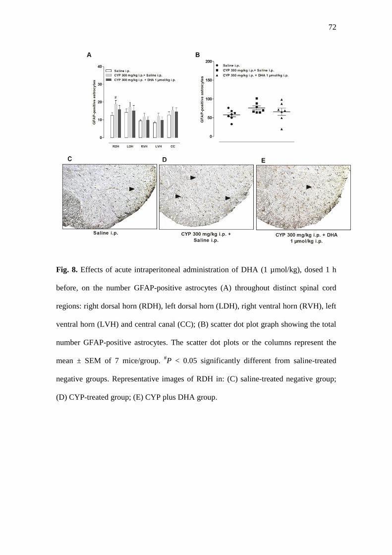
72
Fig. 8. Effects of acute intraperitoneal administration of DHA (1 µmol/kg), dosed 1 h
before, on the number GFAP-positive astrocytes (A) throughout distinct spinal cord
regions: right dorsal horn (RDH), left dorsal horn (LDH), right ventral horn (RVH), left
ventral horn (LVH) and central canal (CC); (B) scatter dot plot graph showing the total
number GFAP-positive astrocytes. The scatter dot plots or the columns represent the
mean ± SEM of 7 mice/group. #P < 0.05 significantly different from saline-treated
negative groups. Representative images of RDH in: (C) saline-treated negative group;
(D) CYP-treated group; (E) CYP plus DHA group.
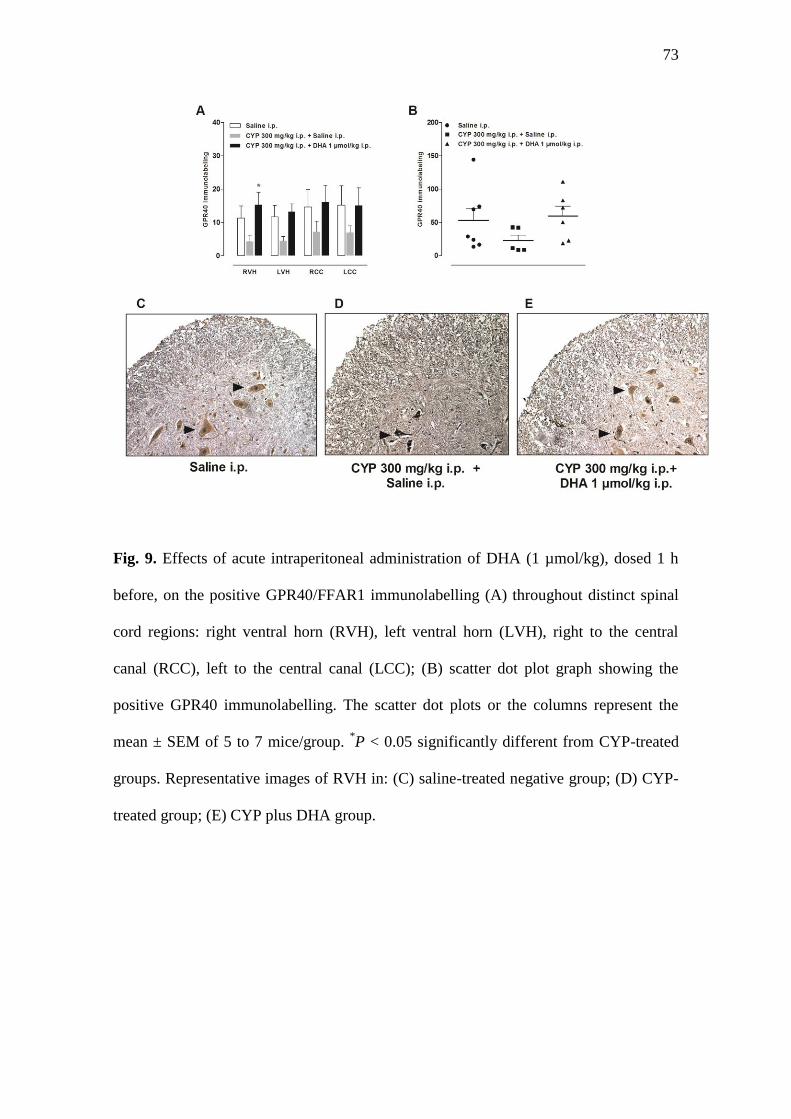
73
Fig. 9. Effects of acute intraperitoneal administration of DHA (1 µmol/kg), dosed 1 h
before, on the positive GPR40/FFAR1 immunolabelling (A) throughout distinct spinal
cord regions: right ventral horn (RVH), left ventral horn (LVH), right to the central
canal (RCC), left to the central canal (LCC); (B) scatter dot plot graph showing the
positive GPR40 immunolabelling. The scatter dot plots or the columns represent the
mean ± SEM of 5 to 7 mice/group. *P < 0.05 significantly different from CYP-treated
groups. Representative images of RVH in: (C) saline-treated negative group; (D) CYP-
treated group; (E) CYP plus DHA group.
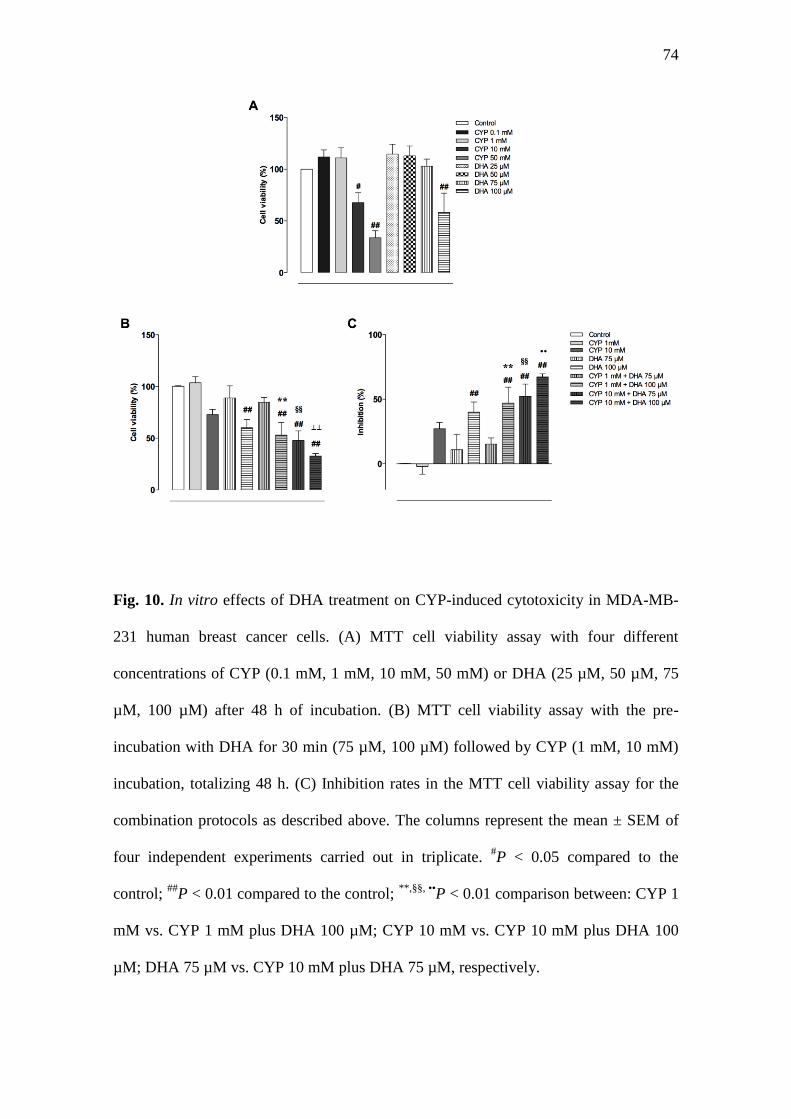
74
Fig. 10. In vitro effects of DHA treatment on CYP-induced cytotoxicity in MDA-MB-
231 human breast cancer cells. (A) MTT cell viability assay with four different
concentrations of CYP (0.1 mM, 1 mM, 10 mM, 50 mM) or DHA (25 µM, 50 µM, 75
µM, 100 µM) after 48 h of incubation. (B) MTT cell viability assay with the pre-
incubation with DHA for 30 min (75 µM, 100 µM) followed by CYP (1 mM, 10 mM)
incubation, totalizing 48 h. (C) Inhibition rates in the MTT cell viability assay for the
combination protocols as described above. The columns represent the mean ± SEM of
four independent experiments carried out in triplicate. #P < 0.05 compared to the
control; ##
P < 0.01 compared to the control; **,§§, ••
P < 0.01 comparison between: CYP 1
mM vs. CYP 1 mM plus DHA 100 µM; CYP 10 mM vs. CYP 10 mM plus DHA 100
µM; DHA 75 µM vs. CYP 10 mM plus DHA 75 µM, respectively.

75
Supplementary Table 1. Diet composition by weight and percentage of macronutrients
Constituents1
Regular
chow 10% CO 20% CO 10% FO 20% FO
g/kg
Protein 220 220 220 220 220
Total Fat 40 50 60 50 60
Fiber 80 80 80 80 80
Mineral mix* 100 100 100 100 100
Carbohydrates 560 560 560 560 560
*Mineral mix: Iron 50 mg; zinc 60 mg; copper 10 mg; iodine 2 mg; manganese 60 mg;
selenium 0.05 mg; cobalt 1.50 mg.

76
Supplementary Table 2. Reverse and forward primers
Gene Primer sequences (5’-3’)
Accession
number
(mRNA)
Amplicon
size (bp)
Hprt1*
F-
CTCATGGACTGATTATGGACAGGAC
R-
GCAGGTCAGCAAAGAACTTATAGCC
NM_013556 123
Tbp* F-CCGTGAATCTTGGCTGTAAACTTG
R-GTTGTCCGTGGCTCTCTTATTCTC NM_013684 118
Ppia* F-TATCTGCACTGCCAAGACTGAATG
R-CTTCTTGCTGGTCTTGCCATTCC NM_008907 127
Ffar-
1**
F-GCTCAGGCCTGAGCCACAAACG
R-
AGTACCACACTCCAGGCCCCTGTG
NM_194057.2 181
* According to Pernot et al. (2010).
** Designed by authors.

77
Supplementary Fig. 1. Representative images of the total blood cell counts. (A) 10 %
corn oil plus saline; (B) 10 % corn oil plus CYP; (C) 10 % fish oil plus saline; (D) 10 %
fish oil plus CYP; (E) 20 % corn oil plus saline; (F) 20 % corn oil plus CYP; (G) 20 %
fish oil plus saline; (H) 20 % corn oil plus CYP; (I) saline i.p. ; (J) CYP i.p. plus Saline
i.p.; (K) CYP i.p. plus DHA i.p. Head arrows indicated the neutrophils, and arrows
indicated the lymphocytes.

78
Supplementary Fig. 2. Immunohistochemistry for microglial activation, and RT-qPCR
for GPR40/FFAR1. Representative images of immunohistochemistry analysis showing
the absence of positive immunolabelling for Iba1/AIF1 in the right dorsal horn (RDH)
of the lumbar spinal cord in: (A) saline-treated negative group; (B) CYP-treated group;
(C) CYP plus DHA group. (D) Amplification plot of the expression of GPR40/FFAR1
receptor for mouse spinal cord samples from different experimental groups.
79
ANEXO C - COMPROVANTE DE SUBMISSÃO DO ARTIGO
Dear Dr. Raquel Freitas,
You have been listed as a Co-Author of the following submission:
Journal: Journal of Nutritional Biochemistry
Corresponding Author: Maria Martha Campos
Co-Authors: Raquel Freitas; Kesiane M Costa; Natália F Nicoletti; Luiza W Kist;
Maurício R Bogo;
Title: Omega-3 fatty acids are able to modulate the painful symptoms associated to
cyclophosphamide-induced-hemorrhagic cystitis in mice
If you did not co-author this submission, please contact the Corresponding Author of
this submission [email protected];[email protected]; do not follow
the link below.
An Open Researcher and Contributor ID (ORCID) is a unique digital identifier to which
you can link your published articles and other professional activities, providing a single
record of all your research.
We would like to invite you to link your ORCID ID to this submission. If the
submission is accepted, your ORCID ID will be linked to the final published article and
transferred to CrossRef. Your ORCID account will also be updated.
To do this, visit our dedicated page in EES. There you can link to an existing ORCID
ID or register for one and link the submission to it:
http://ees.elsevier.com/jnb/l.asp?i=24029&l=3FUG7OCF
More information on ORCID can be found on the ORCID
website, http://www.ORCID.org, or on our help
page:http://help.elsevier.com/app/answers/detail/a_id/2210/p/7923

80
Like other Publishers, Elsevier supports ORCID - an open, non-profit, community
based effort - and has adapted its submission system to enable authors and co-authors to
connect their submissions to their unique ORCID IDs.
Thank you,
Journal of Nutritional Biochemistry