new egzoluminal circular surgical stapler · 2011-01-03 · new egzoluminal circular surgical...
TRANSCRIPT

Acta of Bioengineering and Biomechanics Short comunicationVol. 12, No. 3, 2010
New egzoluminal circular surgical stapler
ROMUALD DROP1, STANISŁAW MAZURKIEWICZ2,*, ZENON WOŹNY3,PAWEŁ MAJCHER4, ADAM TABOR5
1 Department of General and Vascular Surgery, The Stefan Żeromski Specialist Hospital in Kraków, Poland.2 Department of Mechanical Engineering, Cracow University of Technology, Poland.
3 CERMET ISP Enterprise for Technical Progress and Implementation, Kraków, Poland.4 FAKRO PP Sp. z o.o., Department of Research and Development, Nowy Sącz, Poland.
5 Centre of Training and Organisation of Quality Systems, Cracow University of Technology, Kraków, Poland.
The study presents the design, prototype version and preliminary results of in vivo testing of the egzoluminal circular surgicalstapler for “end-to-end” anastomosis of bowels after the resection, where staples are put outside the intestinal lumen, and walls ofthe intestinal tube directly contact the intestinal mucous membrane. The adopted idea of an “end-to-end” intestinal anastomosisas well as a strong need for a device that would be re-usable have determined its specific design. The device is characterised bya kinematic mode of operation, different from the operating mode of commonly used staplers, and hence by a specific combina-tion of the individual components. The in vivo tests, successfully conducted on animals, have proved that this method of anasto-mosis as well as the device used for its practical performance are fully applicable in clinical practice, as best shown by patentapplication No. P 386369.
Key words: extraluminal circular stapler, mucous membrane, intestinal anastomosis, sterilisation
1. Introduction
The intraluminal circular staplers used currentlyare equipped with heads that are placed in the lumenof the bowel, the borders of which are folded to theinside. Staplers of this design are an excellent toolfor the anastomosis of an intestinal section of thealimentary tract, especially its rectal part [1]. Theapplication of intraluminal surgical staplers in theanastomosis of the remaining intestinal part of thealimentary tract is theoretically possible but not fullyconsistent with the well-known Halstead’s principleof surgical art [2]–[5].
The egzoluminal circular surgical stapler de-scribed in this study has been assigned for use insurgical anastomoses, especially in an “end-to-end”
intestinal anastomosis performed in practically allsections of the alimentary tract, where patency of thebowels is obtained by extraluminal folding (foldingto the outside) the borders of the bowel sections thatare to be put together. Similar to intraluminal sta-plers, also in this case, metal staples are used for theanastomosis.
2. Materials and methods
The main task of the designed versatile circularsurgical stapler is to perform an “end-to-end” interin-testinal anastomosis at each and every arbitrarily cho-sen section of the bowels, proceeding in accordancewith the Halstead’s principle of medical art (figure 1).
______________________________
* Corresponding author: Stanisław Mazurkiewicz, Chair of Experimental Mechanics and Biomechanics, Department of MechanicalEngineering, Cracow University of Technology, 31-864 Kraków, al. Jana Pawła II 37, Poland. Phone: +48 12 374 33 63, e-mail:[email protected]
Received: July 13rd, 2010Accepted for publication: September 30th, 2010

R. DROP et al.114
The said principle is fully satisfied using the newlydesigned stapler, where borders of the bowels arefolded in the direction opposite to the intestinal lu-men. This means the situation reverse to that faced ina traditional method using standard circular surgicalstaplers. The method itself forces the use of split ele-ments (head, anvil and cartridge holding the staples).The split design enables complete removal of all com-ponents of the circular surgical stapler from the pa-tient’s abdominal cavity after the anastomosis hasbeen completed.
Fig. 1. A versatile circular surgical stapler
The requirements of versatility and repeated use ofthe new stapler demanded some new solutions re-garding the design of its individual components interms of their geometry, kinematics, and operatingmode. Nearly all the components of the device havebeen designed and made in accordance with theadopted rules of simplicity, smooth surface, and ab-sence of recesses and threaded joints, to make thesterilisation easy, complete and reliable.
The new design includes only one element ofcomplex geometry, i.e. the staple cartridge. Its task isto feed staples during the operation of anastomosis.Because of the sterilisation regime, this is the onlyelement that is disposable and replaced before eachoperation. The design includes one threaded jointwhich plays double function, i.e. holds together allstapler elements to form one integral and useful wholeand enables the distance between the stapler head andanvil to be precisely calculated during the operation ofanastomosis. It also enables very quick assembly anddisassembly of the device. The threaded joint is, how-ever, located inside the device handle and, given itsposition, it never enters into direct contact with thestapled tissues.
All parts of the device are made from biotolerablematerials, resistant to sterilisation, both thermal andchemical (using gas).
2.1. Description of the device
The device is composed of a head with staplecartridge and an anvil. Both elements are of a splitdesign. At the stage of designing and later tests,different parting planes were chosen. Some of themleft the staple guides and seats untouched, but re-quired very intricate technology (figure 2), whileothers, simpler in performance, were “crossing andcutting” the guides and seats in two (figure 3). Thetests carried out on materials imitating bowels, aswell as the anastomoses carried out in vitro and invivo on animals have proved that each of the de-
Fig. 2. Complex split head
Fig. 3. Straight split head

New egzoluminal circular surgical stapler 115
signs proposed is equally efficient and ensures the re-quired quality and reproducibility of the stapled“suture”. When the anastomosis is performed, veryimportant is the design and operation of the stapleejector. The parting planes designed for the stapleroperating elements demanded the use of ejectors of twodifferent types, i.e. in the form of single plates and ofbridge-like construction. In the design of the partingplane that preserves the staple seats and guides un-touched, it was enough to use ejectors in the form ofsingle plates, while in the second design of the partingplane (crossing and splitting the guides in two) it wasnecessary to use, besides the ejecting plates, also theejecting bridge-like constructions. The bridges kept theejectors in position when the head was dismantled afterthe operation. The bridge-type ejectors were used onlyin these parts of the head parting plane where the stapleguides were “cut in two”.
Fig. 4. Ejecting ring with integral ejectors
Fig. 5. Ejecting ring with floating ejectors
Tests were carried out on both design variants, i.e.with integral ejectors (figure 4) and with “floating”ejectors (figure 5).
The variant with the “floating” ejectors gave verygood results as regards the “suture” quality and fail-ure-free operation during testing. A complete headwith the “floating” staples and bridges is shown infigure 6.
Fig. 6. A view of the head
2.2. Mode of functioning
The surgical stapler was designed for maximumease and simplicity of operation. The first step beforethe operation of anastomosis starts is to detach thehead with cartridge and the anvil from the device. Theborders of the bowels that are to be joined together areplaced in the respective internal parts of the joiningelements, to be rolled over next through these ele-ments. The bowels are already prepared for the anas-tomosis and provided with a primary suture (figure 7).The borders of the bowels placed in the device headand anvil, respectively, are then pulled off from thebowel lumen to rest on the external surfaces of thejoining elements. The primary suture is tied and thehead and anvil, now ready to start the operation, areconnected to the handle of the device. The borders ofboth bowel sections are moved close to each other,and the operation of anastomosis begins (figure 9).The operating parts of the device are pushed close toeach other by a screw-operated mechanism. The an-astomosis is done by pressing the starting lever whichactuates the mechanism, resulting in coupled forwardmovement of all the circumferentially arranged sta-ples. The releasing of the “stapled” bowel from the
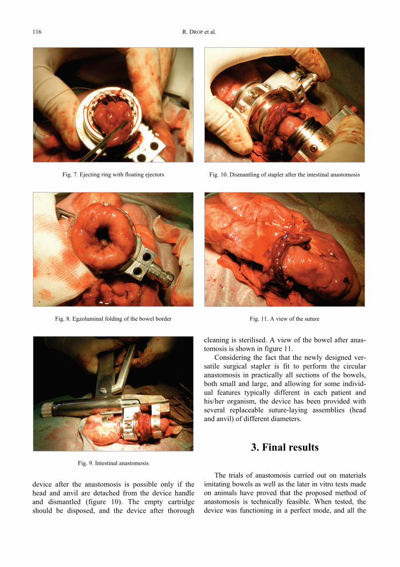
R. DROP et al.116
device after the anastomosis is possible only if thehead and anvil are detached from the device handleand dismantled (figure 10). The empty cartridgeshould be disposed, and the device after thorough
cleaning is sterilised. A view of the bowel after anas-tomosis is shown in figure 11.
Considering the fact that the newly designed ver-satile surgical stapler is fit to perform the circularanastomosis in practically all sections of the bowels,both small and large, and allowing for some individ-ual features typically different in each patient andhis/her organism, the device has been provided withseveral replaceable suture-laying assemblies (headand anvil) of different diameters.
3. Final results
The trials of anastomosis carried out on materialsimitating bowels as well as the later in vitro tests madeon animals have proved that the proposed method ofanastomosis is technically feasible. When tested, thedevice was functioning in a perfect mode, and all the
Fig. 7. Ejecting ring with floating ejectors
Fig. 8. Egzoluminal folding of the bowel border
Fig. 9. Intestinal anastomosis
Fig. 10. Dismantling of stapler after the intestinal anastomosis
Fig. 11. A view of the suture

New egzoluminal circular surgical stapler 117
performed operations of anastomosis offered the ex-pected high quality, reliability, and bowel patency. Theprocess of sterilisation, both chemical and thermal, hadno major effect on the performance efficiency of thedevice. So, in technical approach, the device describedin this study can be regarded as failure-free in operationand re-usable. Thus it has been demonstrated that it ispossible to design and manufacture a device that willperform an “end-to-end” anastomosis of the humanbowels in egzoluminal approach.
All operations of the anastomosis performed by thismethod in vivo on animals have ended in success. Theoperated animals have survived in good condition theperiod required for healing of the tissues, and re-operatedshowed complete and correctly done anastomosis of thebowels. Studies have also proved that the proposedmethod of anastomosis done with a circular stapler in the“mucous membrane-to-mucous membrane” arrangementensures effective healing and reconstruction of the tis-sues after surgery. In view of the fact that the preset goalhas been achieved, and all the technical as well as medi-cal steps have ended in full success, further studies toimplement this innovative method in clinical procedureseem both reasonable and justified.
Acknowledgements
The authors extend their sincere thanks to Professor JędrzejKrupiński, DSc., Director of the Institute of Zootechnics in
Kraków-Balice, for his kind consent to carry out the in vitro testsof intestinal anastomosis at the Institute.
The studies were financed from a research fund assigned foryears 2006–2008.
References
[1] DROP R., RZEŹNIK D., GACZOREK M., Ostra niedrożnośćprzewodu pokarmowego spowodowana chorobą nowot-worową, Nowotwory, Journal of Oncology, 2005, Vol. 55,Supl. 3.
[2] MŁYNARSKI T., ORLICKI P., MAJCHER P., Tendencje rozwo-jowe metod operacyjnego zespalania jelit, XVII OgólnopolskaKonferencja Naukowo-Dydaktyczna Teorii Maszyn i Mecha-nizmów, Warszawa-Jachranka, 2000.
[3] MŁYNARSKI T., ORLICKI P., MAJCHER P., Właściwości szwówchirurgicznych w procesie mechanicznego zespalania jelit,XVII Międzynarodowa Konferencja Biomechanika ’2001,Zakopane, 2001.
[4] TAKEYAMA H., SATO M., AKAMO Y., TANAKA M., HAYAKAWAT., HASEGAWA M., SAWAI H., YAMAMOTO M., OHARA E.,MANABE T., Keyhole procedure: a new technique for intesti-nal anastomosis with a large opening and less tissue trauma,using both circular and linear staplers, Surgery, 2003 Mar,133(3), 345–348.
[5] ALTOMARE D.F., RINALDI M., VEGLIA A., PETROLINO M.,De FAZIO M., SALLUSTIO P., Combined perineal and endo-rectal repair of rectocele by circular stapler: a novel surgi-cal technique, Dis Colon Rectum, 2002 Nov, 45(11), 1549–1552.