lise celular e ativação da trialisina, proteína citolítica
TRANSCRIPT

Rafael Miyazawa Martins
Lise Celular e Ativação da Trialisina,
Proteína Citolítica da Saliva de Triatoma infestans
Tese apresentada à Universidade
Federal de São Paulo - Escola Paulista de Medicina, para a obtenção do Título
de Doutor em Ciências
São Paulo
2007

Livros Grátis
http://www.livrosgratis.com.br
Milhares de livros grátis para download.

Rafael Miyazawa Martins
Lise Celular e Ativação da Trialisina,
Proteína Citolítica da Saliva de Triatoma infestans
Orientador: Prof. Dr. Sergio Schenkman Co-orientador: Prof. Dr. Rogério Amino
Tese apresentada à Universidade Federal de São Paulo - Escola Paulista de Medicina, para a obtenção do Título
de Doutor em Ciências
São Paulo 2007

Martins, Rafael Miyazawa
Lise Celular e Ativação da Trialisina, Proteína Citolítica da Saliva de
Triatoma infestans / Rafael Miyazawa Martins - São Paulo, 2007
viii, 115 fl.
Tese (Doutorado) Universidade Federal de São Paulo. Escola Paulista de
Medicina. Programa de Pós-Graduação em Microbiologia e Imunologia.
Título em inglês: Cell Lysis and Activation of Trialysin, Cytolytic Protein from
Triatoma infestans saliva.
1. Triatoma infestans; 2. trialisina; 3. saliva; 4. lise celular; 5. peptídeos
antimicrobianos; 6. ativação proteolítica

Trabalho realizado na Disciplina de Biologia Celular do
Departamento de Microbiologia, Imunologia e Parasitologia da
Universidade Federal de São Paulo - Escola Paulista de Medicina,
com auxílio financeiro concedido pela
Fundação de Amparo à Pesquisa do Estado de São Paulo (FAPESP).

"A LUZ NO FIM DO TÚNEL É SÓ A LUZ
DE UM TREM QUE VEM VINDO"
ROBERT LOWELL

AGRADECIMENTOS
À minha família, que me suporta e ampara em todos os momentos da minha vida: Mãe, Sazinha, Soni, Tiaha, Tiaki, Tiami, Tio Rômulo, Tio Issao e Tia Cecília; Alva, Edu, Rominho, Rê e especialmente aos meus lindos irmãos Rita e Frederico, pelos anos de convívio e aprendizado nem sempre fácil; por encararem bravamente meu Mr. Hyde que às vezes (!?) aflora e por darem o máximo para satisfazer o reizinho mandão! À minha namorada Taís pelo amor, companheirismo, paciência, alegria e dedicação; obrigado por cuidar de mim nos momentos mais críticos e também por me introduzir na sua família maravilhosa que me acolheu tão bem. Aos Fabulosos : Ao Sergio, por me aceitar no laboratório, me orientar, me dar toda a liberdade de trabalhar e exemplificar ética e dedicação à ciência. Obrigado pelo entusiasmo contagiante, disponibilidade e por me permitir trabalhar dentre pessoas especialíssimas. Ao Roti, que me guiou nos mistérios do laboratório desde 1998... Obrigado pela amizade, paciência, conselhos, bom-humor, disponibilidade e principalmente pelo exemplo espiritual e científico. Sinto muitíssimo não ter aproveitado seu ensinamento futebolístico mas quem sabe ainda haja tempo... Donizete Babinska, Júlio Monte, Lúcio Freitas-Júnior, Lys Guilbride, Marcel Ramirez, Marcella Faria, Marília Malta, Patrícia Godoy, Rafael Marques Porto, Tatiana Ejchel, Thaís Falcão e Vera Chioccola – obrigado pelo apoio nos momentos de desespero, quando o FPLC apita, as bolhas materializam no leitor de UV, o gel de prata cai na pia suja, os barbeiros tentam voar... Foi ótimo ter convivido com vocês, aprendi muito nesse período (mas sem socar centrífugas nem geladeiras)! - Carol Elias, adorável presença do lab, companheira de Coca-light, pão de queijo e capuccino no Fala! Valeu pelo auxílio freqüente e pelas conversas sobre futuro, Harry Potter, pós-docs, Jack Bouer, vida... - Evânia, a super! Valeu por toda a ajuda no dia-a-dia e nos momentos divertidíssimos de praia, forró e churras!!! - Carlos Massayuki, grande descascador de milho – lembro as aulas de cinética enzimática do 2o ano... Valeu pelas conversas, almoços às 11h, baladas, cervejas, clube da fumaça, pela família demais! - Fernando Dossin, (é, mister M...), caríssimo parceiro de corridas ocasionais, cervejinhas, bandejão, Sabedoria Chapa Quente, (algumas) baladas, papos cabeçudos e lições de vida (A gente faz o que a gente pode....heheheh)!

- Júlia Cunha, querida companheira de Unifesp/Bio32, uma pseudo-golfinho ímpar! Muito obrigado pela amizade, conselhos, espectrometria de massas, exemplo de arrumação de bancada (hehehe) e profissionalismo. - Teresa Jesus, exemplo de força e determinação, uma potência na bancada! Valeu pelas conversas, aulas de spinning, compras e passeios calóricos na Paulista! - Claudeci, sempre fazendo e falando 3000 coisas ao mesmo tempo, obrigado por nos dar mais do que a mão e me aturar. Lembre-se: queremos mais churrascos! - Ludmila, valeu pela amizade, pelo carinho e alegria no lab, conversas ótimas, momentos bidimensionais e espectroscópicos de massas... - Cláudio, valeu pela tremenda ajuda na cultura, na caça às presas dos barbeiros, por estar sempre disposto a auxiliar e aprender, tô esperando o preço justo... - Sheila Cristina, ponta firme de boteco. Valeu pela aromatização artificial de baunilha no laboratório e principalmente pela ampliação de meu vocabulário, guria. - Thiago, caiçara sofredor, solícito e dedicado, cuidado com a sua faca de diamante! - Alberto, chileno sensível, nervoso e divertido... Valeu pela companhia de corridas e bancada! - Tarsis Ferreira, agregado divertidíssimo, valeu pelas conversas de altíssimo teor, pelas análises açucaradas e discussões científicas. - Vivi Ha, coreana que parece quietinha mas quando desanda a falar... diversão garantida e presença constante nos encontros! - Rebecca Midory, cuja sensibilidade e inteligência me guiam nas escolhas quadrinhísticas!! - Carla, Erik, Ivan, Juca, Luana, Luciana e Rafão, alunos de iniciação que agitam o lab e dão sopro de vida e excitação ao laboratório! Obrigado por agitarem um pouco os velhíssimos pós-graduandos deste lab! - Finalmente, obrigado Bruno Mococa e Rogério Sacramento (o colaborador), que me testam todos os dias, sofrem comigo e de vez em quando geram resultados!!! Obrigado pela disposição, força de vontade e insistência! Espero que ambos continuem e cresçam na pesquisa; foi ótimo aprender com vocês! À Bio 32 e agregados, especialmente Aninha, Alisson, Bia, Cassiano, Cláudio, Dagot, Fabrício, Guto, Leila, Mari-bel, Ota, Paulinha, Renata-torredebabel, Rodrigo, Tati, Truz e Van. Vocês foram extremamente especiais e importantes desde minha chegada em São Paulo. Obrigado pelas conversas, mega-baladas, gorós homéricos, alegrias, tristezas e crises partilhadas!!! Obrigado, Juli-bel (Med 66) por também fazer parte dessa fase! Família Bel, obrigado por tudo o que vocês representam, adoro vocês! Aos habitantes do 8o andar, Disciplina de Biologia Celular; secretários: Márcia, Marcelo, Joseli e Antônia, por toda a ajuda na burocracia; Má, sem você cuidando das reservas técnicas, não sei o que seria de nós... Técnicos: Américo, Dona Sebá, Maria, Luizão, Seu Tonico, Zé Carlos, pela sabedoria (grande Dona Sebá!!) e apoio em todas as necessidades urgentíssimas nossas! Nossos vizinhos: Rosana Puccia, Zoilo Camargo, Olga Gompertz, Maria Lúcia Cardoso de Almeida, Luiz Travassos, Elaine Rodrigues e equipes por reagentes, discussões, desabafos, aparelhos... Especialmente Tânia Barros, Flávia Moraes, Kátia Cardoso, Antônio Augusto, Wagner Batista, Luciane Ganiko, Milene; Cris, Bel, Geisa, Rodrigão, Júnior, Sílvia; Fabiane, Clédja, Pato Godoy, Xandão; Rose, Carol Borsói, Mourisa, Maria Amélia, Ricardo e

Camila; Thaysa, Andrey, Rose, Sabrina, Carla (Querida Cuuuuunnnnn!), Flávia Hebeler, Fabi, Bianca, Luis Marchi, Fizão, Cambinão e Cintião (Arre égua!)! Obrigado pelos ótimos momentos juntos! À Bio 38 e agregados: Bárbara, Felipe, João, Joes, Karen, Leo, Marialice, Mineiro, Paula, Renata, Sílvia, Tato, Tim, Toddy, Clarissa, Chinezinho Cabeção, Débora, Mari Carioca, valeu pelas caminhadas nas nuvens juntos! Às meninas da Enf 64: Camila, Ju Fono, Lívia, Luana, Maris Morena e Loira, Pri, Uçu, Vivi; Bio 37: Artur, Bruno, Emília, Fefa, Gabi, Julien, Leo, Lipe, Mac, Marco. Valeu pela diversão!!!! Aos membros do Departamento: Parasitologia: secretárias Mércia e Regiane pela resolução dos problemas mais críticos em menor tempo possível, professores Nobuko Yoshida, Maurício Rodrigues, Clara Mestriner, José Franco da Silveira, Michel Rabinovitch, Renato Mortara; Microbiologia: Marcelo Briones (Bia Schnabel), Bia Castilho (Vivi-Xuxa, Carolzinha, Larissa, Nilce, as Spice Girls). Aos ressonantes do Laboratório Nacional de Luz Síncrotron: Thelma Pertinhez, Alberto Spisni, Mauríco Sforça, Leonardo Trabuco, Aline Oliveira, Rita Figueiredo, Sérgio Oyama; obrigado pela introdução ao mundo dos spins nucleares, seqüências de pulso e DYANAs!! À querida Profa. Cida Tanaka e equipe: Ricardo, Ivan, Serginho, Diogo, Cássia, Carla, Renato. Obrigado pela excelentíssima companhia, além disso pelos reagentes, equipamentos, discussões, conversas, festinhas, botecos, cafés. À Profa Iolanda Cuccovia e sua fantástica pós-doc Katia Daghastanli pela ajuda nos ensaios com lipossomos. Aos professores Maria Juliano e Luiz Juliano pelos peptídeos que me salvaram a tese. Izaura Hirata pelos seqüenciamentos e análises de aminoácidos e Iuri Gouvêa pelo uso do fluorímetro. Ao Prof. Magnus da Silva, pelas excelentes conversas, dicas e apoio nessa fase difícil de doutorado/pós-doutorado... To the special BoP 2006 attendants: Bruna, Lari, Angie, Nick, Mai, Najju, Katy, Markus, Tiff, Arnaud, Isabel, Jo, Beth, Paul, Kathryn, Megan, Dr. Patricia Johnson and Dr. Chris Hunter: because you know that Hips don’t lie… and all the terrific faculty and lecturers I met during this magical summer experience!! Ao Dr. Eduardo Aoki, Serviço de Saúde do Corpo Discente (Dra. Nair), Disciplina de Cirurgia de Cabeça e Pescoço (Drs. Roberto Takimoto, Marcelo Rosado, Macoto Kosugi, Murilo, Marcel) da Unifesp; às atenciosas enfermeiras da Ala de Internação de docentes, alunos e funcionários do Hospital São Paulo: obrigado por cuidarem da minha saúde na detecção, extirpação e eliminação do meu carcinominha... À Unifesp e Fapesp pela acolhida e suporte financeiro.

ÍNDICE
Lista de abreviaturas 1
Resumo 3
Summary 5
Introdução 7
1. Doença de Chagas 7
2. Os triatomíneos 8
3. A saliva dos triatomíneos 10
3.1. A saliva de Rhodnius prolixus 11
3.2. Glândulas salivares e saliva de Triatoma infestans 13
4. Polipeptídeos líticos de membranas 16
4.1. Citolisinas formadoras de poros 16
4.2. Peptídeos antimicrobianos e citolíticos 19
Objetivos 24
Capítulo 1: Purificação, clonagem e caracterização da trialisina 25
Resumo 26
Artigo 28
Capítulo 2: Estrutura de peptídeos sintéticos baseados no N-terminal da trialisina 35
Resumo 36
Artigo 40
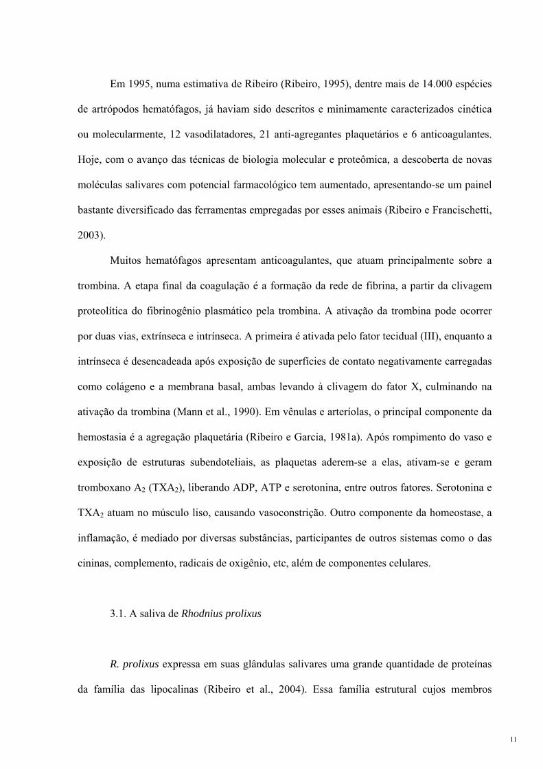
Capítulo 3: Expressão em sistema heterólogo da trialisina e pró-trialisina recombinantes 50
Resumo 51
Introdução 51
Materiais e métodos 54
Resultados 62
Capítulo 4: Inibição da lise da trialisina pela pró-região 70
Resumo 71
Artigo 74
Discussão Geral e Conclusões 97
Referências Bibliográficas 102

LISTA DE ABREVIATURAS
2X YT: meio de cultivo de bactérias com extrato de levedura e bacto-triptona (Yeast
extract, Tryptone).
ADP: 5'-difosfato de adenosina, adenosine 5'- diphosphate
AMPc: 3',5'-monofosfato de adenosina cíclico, cyclic adenosine 3',5'-monophosphate
APMSF: fluoreto de (4-amidino-fenil)-metano-sulfonil, (4-amidino-phenyl)-methane-
sulfonyl fluoride
ATP: 5'-trifosfato de adenosina, adenosine 5'-triphosphate
CAP: proteína ativadora por AMPc, cAMP activator protein
CD59: antígeno de diferenciação de grupo 59, Cluster of Differentiation 59
CDCs: citolisinas dependentes de colesterol
cDNA: ácido desoxirribonucléico complementar, complementary DNA
C-terminal: terminal carboxila
DTT: ditiotreitol
EDTA: ácido etilenodiaminatetraacético, ethylenediaminetetraacetic acid
GPI: glicosil-fosfatidilinositol
GST: glutationa S-transferase
HPLC: cromatografia líquida de alta performance, high performance liquid
chromatography
IPTG: isopropil-β-D-tiogalactopiranosídeo
L30: trialisina, clone 30 (Genbank AF427486)
LB: meio de cultivo de bactérias Luria-Bertani
Ni-NTA: ácido nitrilotriacético-níquel, nickel-nitriletriacetic acid
NL2: fragmento não-lítico, clone 2
NO: óxido nítrico
NP1-4, NP-7: nitroforina1-4, nitroforina-7
N-terminal: terminal amino
ORF: fase de leitura aberta, open reading frame
PCR: reação em cadeia da polimerase, polymerase chain reaction
PFO: perfringolisina O
pl5: pró-trialisina, clone 5 (Genbank AF427487)
RMN: ressonância magnética nuclear
RNA: ácido ribonucléico, ribonucleic acid
1

RPAI: inibidor de agregação plaquetária de Rhodnius, Rhodnius platelet aggregation
inhibitor
SDS: dodecil sulfato de sódio
SDS-PAGE: eletroforese em gel de poliacrilamida contendo dodecil-sulfato de sódio,
sodium dodecyl-sulfate polyacrylamide gel electrophoresis
TAE: solução contendo Tris-Acetato e EDTA
TFE: trifluoro-etanol
TMH: grampos transmembrânicos, transmembrane hairpins
TXA2: tromboxano A2
ufc: unidades formadoras de colônias
WHO: Organização Mundial da Saúde, World Health Organization
δ (ppm): deslocamento químico (partes por milhão)
2

RESUMO
A saliva de artrópodos hematófagos constitui um coquetel farmacológico que atua nos
sistemas hemostático, inflamatório e imunológico de seus hospedeiros para auxiliar a
aquisição do sangue. O inseto T. infestans, vetor da doença de Chagas, apresenta em sua
saliva uma série de moléculas e atividades biológicas já descritas. Dentre elas está a trialisina,
uma proteína formadora de poros em bicamadas lipídicas. A trialisina é capaz de
lisar/permeabilizar diversos tipos celulares e se propõe que a atividade lítica ocorra pela
inserção da porção N-terminal da molécula na membrana das células de tal forma a promover
a formação de um poro. A identificação do gene que codifica a trialisina mostrou que além de
ter uma seqüência sinal de secreção, a proteína conteria uma região ácida no seu N-terminal.
Este domínio está ausente na proteína madura purificada da saliva e poderia inibir a ação da
trialisina. O objetivo de nosso trabalho foi estudar o mecanismo de lise da trialisina e como se
daria a ativação do precursor da trialisina (pró-trialisina) durante a secreção da glândula
salivar até a ejeção da saliva.
Para entender o mecanismo de ação da trialisina, já que a expressão heteróloga tanto
da pró-trialisina quanto da trialisina apresentou várias dificuldades, utilizamos peptídeos
sintéticos baseados na região N-terminal da trialisina madura. Os peptídeos mostraram
diferentes especificidades para tripomastigotas de Trypanosoma cruzi, hemácias humanas e
Escherichia coli, mas de modo geral, aqueles que continham a maior parte da primeira hélice
anfipática predita na trialisina madura apresentaram maior atividade lítica. Todos os peptídeos
adquiriam estrutura secundária (α-hélices) na presença de agentes miméticos de membranas.
Determinamos as estruturas tridimensionais em solução por Ressonância Magnética Nuclear
dos três peptídeos mais ativos e de um pouco ativo e pudemos observar a importância de
segmentos distintos de alguns peptídeos nas especificidades dos alvos.
3

Utilizando um anti-soro gerado contra a porção C-terminal da trialisina recombinante
(fragmento não-lítico), identificamos a pró-trialisina em extratos de glândulas salivares
contendo APMSF, inibidor da ativação da trialisina. O precursor apresentou menor atividade
lítica contra tripomastigotas. Embora fosse previsto que o precursor teria uma porção pró de
33 resíduos, a diferença entre as bandas em SDS-PAGE da pró-trialisina e trialisina foi de
cerca de 10-15 aminoácidos. Um peptídeo de fluorescência apagada contendo 12 resíduos da
porção pró e 27 do N-terminal da trialisina madura foi sintetizado e observamos que sua
atividade lítica era aumentada quando a serino-protease salivar triapsina ou endoproteinase
Arg-C eram adicionadas, ao mesmo tempo em que o peptídeo alterava sua estrutura,
indicando que a porção acídica é responsável pela inibição da lise. Esses resultados
permitiram caracterizar o papel do N-terminal da trialisina no processo de lise, identificar o
precursor da trialisina na glândula salivar, verificar sua ativação durante a salivação e melhor
elaborar um mecanismo pelo qual a atividade é controlada durante o armazenamento da
proteína nas glândulas salivares.
4

SUMMARY
Hematophagous arthropods contain in their saliva a pharmacological cocktail that acts
on the hemostatic, inflammatory and immune systems of their hosts facilitating blood
acquisition. The saliva of the insect T. infestans, vector of Chagas' disease, contains several
described molecules and biological activities, among which lies trialysin, a protein that forms
pores in lipid bilayers. Trialysin can lyse/permeabilize different cell types and it is proposed
that lytic activity is achieved by insertion of the N-terminal portion of the molecule onto a cell
membrane forming a pore. The identification of the gene that codes for trialysin has shown
that besides a secretion signal sequence, the protein contains an acidic region upstream the
mature sequence that is not present in the N-terminus of trialysin purified from saliva and
could inhibit the activity of the molecule. Our goal was to study trialysin lytic mechanism and
the activation of its precursor (pro-trialysin) from secretion inside the salivary glands to saliva
ejection.
To understand trialysin action mechanism, since heterologous expression of either pro-
trialysin or trialysin was mostly unsuccessful, we used synthetic peptides based on the N-
terminal region of mature trialysin that presented different specificities against T. cruzi
trypomastigotes, human erythrocytes and E. coli, but those that encompassed most of the first
predicted amphipathic helix in trialysin were more active. All peptides folded into α-helices
in the presence of membrane mimetic agents. We solved the tridimensional solution structures
by Nuclear Magnetic Resonance of the three most active peptides and a less lytic one and
observed that distinct regions of some peptides determine target specificity.
Using an anti-serum against the recombinant C-terminal portion of trialysin (non-lytic
fragment), we identified pro-trialysin in salivary glands extracts containing APMSF, inhibitor
of trialysin activation. The lytic activity of the precursor against trypomastigotes is reduced.
5

Although it was predicted that the precursor would contain a pro-region 33-amino acids long,
the difference between the SDS-PAGE trialysin and pro-trialysin bands was around 10-15
residues. Using a fluorescence resonance emission transfer peptide encompassing 12 pro-
region amino acids and 27 mature N-terminal residues, after incubation with triapsin or
endoproteinase Arg-C, an increase in lytic activity was observed. At the same time, the
peptide changed its structure indicating this region can impair lysis inside salivary glands.
These results allowed us to characterize the role of trialysin N-terminus in the lysis process,
identify the trialysin precursor in the salivary gland, and determine the mechanism of
activation and lysis control during storage and saliva release.
6

INTRODUÇÃO
1. Doença de Chagas
A doença de Chagas é causada pelo protozoário Trypanosoma cruzi, sendo sua
transmissão ao homem realizada por insetos triatomíneos (Hemiptera: Reduviidae),
conhecidos popularmente como barbeiros (Chagas, 1909). As formas tripomastigotas
sangüíneas presentes no hospedeiro vertebrado são sugadas durante o repasto sangüíneo do
barbeiro. Essas formas, não-replicativas, diferenciam-se nas formas replicativas epimastigotas
no trato digestivo do inseto e, ao atingirem a ampola retal, diferenciam-se em tripomastigotas
metacíclicos, não-replicativos e infectivos aos hospedeiros mamíferos. A infecção pode se dar
pelo contato das formas metacíclicas com a lesão provocada pela picada ou com mucosas.
Dentro do hospedeiro vertebrado, o parasita infecta diferentes tipos celulares e,
intracelularmente, diferencia-se em amastigotas, formas replicativas, que por fim diferenciam-
se nos tripomastigotas sangüíneos, que continuam o ciclo, infectando outras células ou
barbeiros (figura 1).
Figura 1 - Ciclo da doença de Chagas. Retirado de WHO 2004 (WHO Expert Committee, 2002).
7

A doença se manifesta clinicamente em duas fases: aguda, que se inicia com a
infecção, apenas reconhecida em 1 a 2 % dos infectados e crônica, que persiste por décadas,
com baixa parasitemia e alterações em órgãos como coração, cólon, esôfago e sistema
nervoso autônomo. No último relatório da Organização Mundial da Saúde, de 2002, calculou-
se que cerca de 17,4 milhões de pessoas encontram-se infectadas e quase 93 milhões sob risco
de infecção (WHO Expert Committee, 2002).
2. Os triatomíneos
Dentro da ordem Hemiptera, família Reduviidae, a sub-família Triatominae reúne
insetos exclusivamente hematófagos dos quais cerca de 50% são capazes de transmitir o T.
cruzi para vertebrados (Monteiro et al., 2001). As 130 espécies dessa sub-família estão
distribuídas em 5 tribos, sendo de duas delas os representantes mais estudados dada sua
importância médica como vetores da Doença de Chagas, sua ampla distribuição e capacidade
de conviver com humanos, o Triatoma infestans e Panstrongylus megistus (tribo Triatomini)
e o Rhodnius prolixus (tribo Rhodniini) (Schofield, 2000).
A distribuição geográfica de Rhodnius é bastante diferente da de Triatoma: R. prolixus
é encontrado na região Amazônica, Venezuela, Colômbia e América Central enquanto T.
infestans, do nordeste do Brasil até o sul do continente Sul Americano (figura 2). As espécies
de Rhodnius são consideradas derivadas de populações arborícolas, dadas suas adaptações
para escalada e o hábito de colar os ovos a substratos, assim como infestação de ninhos de
pássaros e mamíferos em palmeiras e bromeliáceas. Diferentemente, os Triatoma infestam
ninhos de animais terrestres, covas e sob pedras, pondo ovos espalhados no solo (Schofield,
1988).
8

Figura 2 – Distribuição de espécies de Triatoma na América. Regiões pretas: R. prolixus, regiões
hachuradas, T. infestans. Modificado de (World Health Organization e Center for International Earth Science
Information Network, 1989).
O surgimento do hábito hematofágico em Triatominae é tido como recente, derivado
de reduvídeos predadores, com a intensificação da sua interação com mamíferos e aves em
seus ninhos (Schofield, 2000). Assim, diversas adaptações ocorreram, entre elas a suavização
das peças bucais (perfuração da epiderme vertebrada em contraposição ao exoesqueleto das
presas invertebradas), alteração da fisiologia digestiva (fatores hemolíticos, detoxificação do
heme sangüíneo, secreção de catepsinas, etc.) (Terra, 1988; Oliveira et al., 1999; Azambuja et
al., 1983) e da função e constituição salivar (Ribeiro e Garcia, 1981b).
Ao se alimentar, o barbeiro insere sua mandíbula na epiderme do hospedeiro e na
derme, sua maxila busca o sangue contido em um vaso, e não em hemorragias provocadas na
rede capilar como na alimentação de Diptera (Lavoipierre et al., 1959; Ribeiro, 1995) (figura
3A). Na primeira etapa, de sondagem, a maxila se movimenta aleatoriamente na procura de
um vaso, concomitantemente liberando saliva; segue-se então a fase de experimentação, em
que o inseto prova o líquido circundante e por fim, na sucção, quando um vaso é encontrado,
9

o sangue é bombeado para o intestino anterior do inseto. Recentemente, viu-se que R. prolixus
libera saliva em todas essas fases, inclusive na sucção, em que o bombeamento é interrompido
pela liberação de pequenos bolos salivares (Soares et al., 2006). Isto é possível graças à
separação dos canais salivar e alimentar por toda a maxila desses insetos (figura 3B)
(Lavoipierre et al., 1959).
Figura 3 – A picada do triatomíneo. A, o inseto insere na epiderme sua mandíbula e na derme a maxila
movimenta-se à procura de um vaso. B, corte transversal da maxila de T. infestans. Modificado de (Lavoipierre
et al., 1959).
A saliva então se torna uma ferramenta essencial à manutenção do hábito
hematofágico. Da constituição tóxica necessária à predação (neurotoxinas, proteases
digestivas), a saliva torna-se uma forma de contornar o sistema homeostático do hospedeiro
(agregação plaquetária, coagulação sangüínea, resposta do vaso, inflamação), que visa a
contenção da perda sangüínea, e a percepção da picada (Ribeiro, 1995).
3. A saliva dos triatomíneos
10
Em 1995, numa estimativa de Ribeiro (Ribeiro, 1995), dentre mais de 14.000 espécies
de artrópodos hematófagos, já haviam sido descritos e minimamente caracterizados cinética
ou molecularmente, 12 vasodilatadores, 21 anti-agregantes plaquetários e 6 anticoagulantes.
Hoje, com o avanço das técnicas de biologia molecular e proteômica, a descoberta de novas
moléculas salivares com potencial farmacológico tem aumentado, apresentando-se um painel
bastante diversificado das ferramentas empregadas por esses animais (Ribeiro e Francischetti,
2003).
Muitos hematófagos apresentam anticoagulantes, que atuam principalmente sobre a
trombina. A etapa final da coagulação é a formação da rede de fibrina, a partir da clivagem
proteolítica do fibrinogênio plasmático pela trombina. A ativação da trombina pode ocorrer
por duas vias, extrínseca e intrínseca. A primeira é ativada pelo fator tecidual (III), enquanto a
intrínseca é desencadeada após exposição de superfícies de contato negativamente carregadas
como colágeno e a membrana basal, ambas levando à clivagem do fator X, culminando na
ativação da trombina (Mann et al., 1990). Em vênulas e arteríolas, o principal componente da
hemostasia é a agregação plaquetária (Ribeiro e Garcia, 1981a). Após rompimento do vaso e
exposição de estruturas subendoteliais, as plaquetas aderem-se a elas, ativam-se e geram
tromboxano A2 (TXA2), liberando ADP, ATP e serotonina, entre outros fatores. Serotonina e
TXA2 atuam no músculo liso, causando vasoconstrição. Outro componente da homeostase, a
inflamação, é mediado por diversas substâncias, participantes de outros sistemas como o das
cininas, complemento, radicais de oxigênio, etc, além de componentes celulares.
3.1. A saliva de Rhodnius prolixus
R. prolixus expressa em suas glândulas salivares uma grande quantidade de proteínas
da família das lipocalinas (Ribeiro et al., 2004). Essa família estrutural cujos membros
11

encontram-se envolvidos nos mais diversos processos biológicos, tem a característica de
formar um barril-β; um bolso hidrofóbico no centro da estrutura serve de sítio de ligação a
pequenas moléculas, principalmente hidrofóbicas (Flower, 1996). As lipocalinas de R.
prolixus podem ser agrupadas em dois grupos, dependendo dos ligantes e suas funções
desempenhadas: 1) nitroforinas: NP1-4 e NP7, inicialmente purificadas das glândulas
salivares de Rhodnius, há pouco tempo, com o chamado sialoma, utilizando técnicas de
HPLC, degradação de Edman e seqüenciamento de bibliotecas de cDNA, descobriu-se uma
grande quantidade de transcritos de nitroforinas. Essas contêm uma molécula de heme cujo
íon férrico (Fe3+) liga-se ao óxido nítrico (NO) no lúmen das glândulas salivares, de pH ~5,0.
No momento da picada, o complexo nitroforina-NO é dissociado pela diferença de pH e o NO
atua na musculatura do vaso, relaxando-a. Sem NO, as nitroforinas captam a histamina
liberada por mastócitos, inibindo a inflamação (Montfort et al., 2000). A NP2 (também
conhecida como prolixina-S) ainda possui atividade anticoagulante, inibindo a ativação do
fator Xa, ligando-se ao fator IX e IXa, na via intrínseca (Zhang et al., 1998; Gudderra et al.,
2005). Já a recém descrita NP7, também anticoagulante, liga-se com alta afinidade a
membranas contendo fosfatidilserina, possivelmente impedindo a ligação do complexo pró-
trombinase a superfícies lipídicas (Andersen et al., 2004). Além disso, uma outra lipocalina,
ligadora de aminas biogênicas (norepinefrina, epinefrina e serotonina), 23% similar à NP2,
porém incapaz de ligar heme, foi descrita como anti-agregante plaquetária e vasodilatadora e
possivelmente anti-inflamatória (Andersen et al., 2003). 2) RPAIs: acrônimo de Rhodnius
Platelet Aggregation Inhibitors, as RPAIs são lipocalinas semelhantes à triabina, pallidipina e
procalina dos barbeiros Triatoma pallidipennis e Triatoma protracta (inibidores de trombina,
agregação plaquetária mediada por colágeno e ADP e alérgeno, respectivamente). As RPAIs
já caracterizadas, RPAI 1-3, ligam-se fortemente a nucleotídeos de adenosina (ADP),
impedindo a agregação plaquetária mediada por colágeno (Montfort et al., 2000; Francischetti
12

et al., 2002). Uma apirase, 5'-nucleotidase capaz de hidrolisar ADP em AMP, impedindo
agregação plaquetária (Sarkis et al., 1986) completa a lista de fatores descritos na saliva de R.
prolixus. Outras possíveis proteínas encontradas como transcritos no sialoma podem
completar o quadro, por exemplo, proteínas ligadoras de derivados do ácido aracdônico, uma
possível lectina e mucina (Ribeiro et al., 2004). Além de proteínas, há lipídeos na saliva,
inclusive lisofosfatidilcolina, em concentrações capazes de inibir agregação plaquetária e
aumentar a produção de óxido nítrico em células endoteliais (Golodne et al., 2003).
3.2. Glândulas salivares e saliva de Triatoma infestans
Em 1954, foi detalhadamente descrita a anatomia do complexo de glândulas salivares
de T. infestans acrescida de uma breve avaliação das atividades biológicas de seus conteúdos
luminares (Barth, 1954). Três pares de glândulas são responsáveis pela síntese da saliva
(figura 4). Todas elas são formadas por uma monocamada de células secretoras, envoltas por
uma lâmina basal, músculos e traquéolas. Na glândula D1, a secreção foi nomeada do tipo
merócrina (secreção de vesículas citoplasmáticas), na D2, apomerócrina (variando entre
merócrina e apócrina) e na D3, apócrina (secreção passiva do conteúdo citoplasmático)
(figura 5). No conteúdo da D1, descreveu-se uma atividade anticoagulante e proteolítica
(originalmente "destrói a hemoglobina"), na D2, uma hemolítica e a D3, considerada como
fonte do veículo para os conteúdos de D1 e D2. Em recente trabalho, as glândulas salivares de
T. infestans foram analisadas morfologicamente e citoquimicamente por microscopias
eletrônicas de transmissão e varredura (Reis et al., 2003). Viu-se uma grande quantidade de
microvilos em todas as três glândulas. Secreções ricas em lipídeos são encontradas em D1 e
aparentemente em maior quantidade na D2, onde a coloração intensa é observada em
vesículas claramente liberadas do citoplasma. As vilosidades são sugestivas da secreção
13

classicamente conhecida como merócrina (diferentemente da designação de Barth, acima), na
qual o conteúdo citoplasmático é exocitado passivamente, sem liberação de vesículas. A
secreção de grandes corpúsculos encontrados no lúmen contendo vesículas citoplasmáticas,
classicamente designada de apócrina, é observada em D1 e em maior escala em D2.
Figura 4 – Glândulas salivares de T. infestans. Micrografia eletrônica de varredura das glândulas D1 e
D2 (painel esquerdo, indicadas) e D3 (painel direito). O hilo está indicado por *, ducto acessório por AD e fibras
musculares, cabeças de seta. Retirado de (Reis et al., 2003).
Figura 5 – Esquema da secreção do epitélio glandular de T. infestans. A numeração crescente indica as
células nos diferentes estágios encontrados a partir do dito repouso (1), até a secreção final do maior volume de
saliva (5 em A, 4 em B e 3 em C). A, D1; B, D2; C, D3. Retirado de (Barth, 1954).
14

Décadas depois de Barth, com maior detalhamento bioquímico, a atividade
anticoagulante foi atribuída a dois componentes: um inibidor da via intrínseca e outro da via
comum da coagulação (Pereira et al., 1996). A atividade proteolítica é devida à presença de
diferentes enzimas, aparentemente representando uma cascata de ativação nas glândulas
salivares de T. infestans, sendo a protease majoritária purificada e denominada de triapsina
(Amino et al., 2001). A triapsina é semelhante à tripsina, com especificidade para clivagem
em resíduos de arginina e sua forma precursora parece ser clivada por uma aspartil-protease,
visto que pepstatina A, inibidor dessa classe de proteases, impede a ativação da triapsina. A
atividade hemolítica, sobre a qual esta tese versa, será abordada adiante. Além disso, na
glândula D2, foi caracterizada uma sialidase com especificidade para ácidos siálicos α(2-3)
ligados, possivelmente originária de um vírus por transferência horizontal. A sialidase, como
as proteases, é liberada na saliva no momento da picada. Essa sialidase poderia atuar
impedindo a inflamação no sítio da picada, inibindo a degranulação de mastócitos e/ou
impedindo a ancoragem (homing) de leucócitos pela dessialilação do antígeno de Lewis, que
medeia a ancoragem às selectinas do endotélio ativado (Amino et al., 1998).
Uma atividade anestésica, inibidora de canal de Na+, também foi encontrada na saliva
desses insetos, impedindo a propagação do sinal doloroso na picada. Com essa atividade, o T.
infestans diminui a percepção da dor do hospedeiro, podendo representar um dos fatores
contribuintes para o sucesso do inseto na aquisição de sangue, visto que o volume de sangue
ingerido é inversamente proporcional à irritação causada no momento da picada (Dan et al.,
1999).
O T. infestans também conta com atividade anti-agregante plaquetária devido à ação
de apirases secretadas na saliva (Faudry et al., 2004). Porém, este conta com cinco moléculas
distintas, pertencentes à família das 5'-nucleotidases. Recentemente, duas proteínas inibidoras
da agregação plaquetária induzida por colágeno, semelhantes à pallidipina foram
15

caracterizadas e denominadas triplatina-1 e –2. Diferentemente das RPAIs de R. prolixus, elas
não inibem a agregação plaquetária induzida por ADP (Morita et al., 2006).
Quanto à atividade hemolítica descrita por Barth em 1954 (Barth, 1954), essa foi
comprovada por Gregório e Ratcliffe em 1999 que também observaram uma atividade lítica
contra Trypanosoma rangeli, protozoário não patogênico ao homem (Gregorio e Ratcliffe,
1991). R. prolixus não apresenta atividade semelhante em suas glândulas salivares, mas sim
em seu intestino anterior (Azambuja et al., 1983). A molécula lítica foi identificada e
caracterizada em nosso laboratório, sendo batizada de trialisina. Toda esta tese trata da
caracterização dessa proteína formadora de poros em bicamadas lipídicas.
4. Polipeptídeos líticos de membranas
Como dito acima, a trialisina de T. infestans possui semelhanças com proteínas
formadoras de poros e peptídeos antimicrobianos. Na natureza, essas moléculas apresentam
papéis predominantemente defensivos ou ofensivos. Por exemplo, na imunidade inata de
diferentes organismos pluricelulares, após um desafio à homeostasia, diversos peptídeos
antimicrobianos são sintetizados e secretados a fim de impedir uma infecção massiva (Boman
et al., 1991; Hoffmann et al., 1993; Hancock e Lehrer, 1998; Lehrer e Ganz, 1999). Esses
peptídeos constituem o coquetel venenoso de diferentes animais e são expressos em bactérias
como uma forma de manutenção/conquista do nicho ecológico ou fatores de patogenicidade
(Bernheimer e Rudy, 1986; Kuhn-Nentwig, 2003; Cheigh e Pyun, 2005).
4.1. Citolisinas formadoras de poros
16

O modo de ação das proteínas bacterianas é bastante estudado na literatura, havendo
muitas com estrutura cristalográfica determinada. Há duas classes dessas proteínas, de acordo
com as estruturas constituindo seus poros: α-toxinas e β-toxinas formadas predominante
mente por α-hélices e folhas-β transmembrânicas, respectivamente (Gilbert, 2002). A família
das citolisinas ativadas por tiol, ou citolisinas dependentes de colesterol, (CDCs) compreende
diversas exotoxinas bacterianas como a pneumolisina de Streptococcus pneumoniae, a
estreptolisina O de Streptococcus spp. e a perfringolisina O de Clostridium perfringens, entre
diversas outras. Essas moléculas inserem-se em membranas contendo colesterol e
oligomerizam, formando poros de até 30 nm, contendo de 30 a 80 monômeros (Palmer, 2001;
Billington et al., 2000; Tweten, 2005; Gilbert, 2005). No caso da intermedilisina, o receptor é
a proteína ancorada por glicosil-fosfatidilinositol (GPI), CD59, um inibidor da formação do
complexo de ataque à membrana do sistema do complemento (Giddings et al., 2003). Após
contato de uma região de 11 resíduos de aminoácidos das CDCs com colesterol, as proteínas
difundem lateralmente na membrana iniciando a oligomerização em um pré-poro (Hotze et
al., 2001; Shepard et al., 2000). O pré-poro então sofre uma alteração conformacional em que
duas α-hélices tornam-se dois grampos-β anfipáticos, que formam a parede do poro
transmembrânico, um barril-β (Shatursky et al., 1999) (figura 6).
Figura 6 – Esquema da formação de poros de citolisinas dependentes de colesterol. Exemplo da
perfringolisina O (PFO). Em i, PFO se liga à membrana através do domínio D4 (não mostrado), oligomeriza (ii)
17

e se insere na membrana (iii) alterando a conformação das hélices em grampos transmembrânicos (TMH).
Retirado de (Ramachandran et al., 2004).
A α-hemolisina de Staphylococcus aureus é o protótipo de uma subclasse de β-
citolisinas da qual também fazem parte o antígeno protetor de Bacillus anthracis, a β-toxina
de Clostridium perfringens, leucocidinas e γ-hemolisinas. Elas são secretadas como
monômeros ou dois componentes monoméricos distintos que oligomerizam na membrana,
sem nenhum receptor específico identificado até o momento. Elas formam um barril-β hexa,
hepta (a forma do poro cristalizada e de estrutura resolvida) ou octamérico (Gouaux et al.,
1994; Song et al., 1996; Steinthorsdottir et al., 2000; Menestrina et al., 2001; Menestrina et
al., 2003).
Aerolisina, de Aeromonas spp. é secretada em sua forma precursora inativa, pró-
aerolisina, que necessita ser processada por proteases da célula-alvo como por exemplo a
furina (Abrami et al., 1998b; Abrami et al., 1998a). A ligação à membrana é via âncoras de
GPI na membrana e, após processamento, moléculas de aerolisina heptamerizam e então
inserem-se na membrana formando um barril-β semelhante à α-hemolisina de S. aureus
(Abrami et al., 1998b; Wilmsen et al., 1992). Entretanto, ainda não se sabe como a proteína
ganha acesso ao core sacarídico da âncora, havendo sugestões de que a interação seja
inicialmente dependente de oligossacarídeos N-ligados no glicocálice das células, além do
fato de que, em eritrócitos, a ligação é através da glicoforina (Rossjohn et al., 1998; Abrami et
al., 2000; Abrami et al., 2003). Embora experimentos in vitro mostrem que o poro permite o
vazamento de moléculas grandes como glucagon (Howard e Buckley, 1982), in vivo, o poro
apenas permite o fluxo de K+ e Ca2+ (Abrami et al., 1998b). Intracelularmente, há liberação de
Ca2+ e vacuolização não-apoptótica do retículo endoplasmático, ativação de proteína G e
produção de inositol (1, 4, 5)-trifosfato (Krause et al., 1998; Abrami et al., 1998b).
18

As α-toxinas são bem menos caracterizadas estruturalmente do que as β-toxinas
(Gilbert, 2002). A hemolisina E de E. coli forma oligômeros de 8 unidades, com 4 α-hélices
constituindo o poro (Tzokov et al., 2006; Wallace et al., 2000; Atkins et al., 2000) (figura 7).
A família das toxinas bacterianas AB5 é formada por várias proteínas de alta homologia
estrutural, envolvidas na patogênese de doenças: toxina colérica, shiga-toxina, enterotoxinas
de E. coli termolábeis e do tipo shiga e toxina pertussis. Essas são constituídas por cinco
monômeros que se ligam às membranas por oligossacarídeos e uma última subunidade
diferente, catalítica, com a atividade tóxica (por exemplo, ativação de proteína Gsα ativando
uma adenilato-ciclase, seguida de abertura de canais de cloreto e inibição da tradução), que
não apresenta atividade lítica (Lencer e Saslowsky, 2005). As colicinas são proteínas
formadoras de canais dependentes de voltagem na membrana plasmática de E. coli. Elas têm
três domínios: de ligação ao receptor, de translocação e de formação do canal. Ao se ligar a
porinas da membrana externa, segue-se sua translocação através das mesmas para o espaço
periplasmático e formação do canal na membrana interna, citoplasmática (Stroud et al., 1998;
Zakharov e Cramer, 2002).
Figura 7 – Modelo da hemolisina E de E. coli. Duas visões do modelo cristalográfico do octâmero de
hemolisinas E foi inserido num esquema de uma bicamada lipídica: em vermelho, cabeças polares e verde,
cadeias apolares lipídicas. Retirado de (Wallace et al., 2000).
4.2. Peptídeos antimicrobianos e citolíticos
19

Já os peptídeos líticos que participam da imunidade inata geralmente são expressos
após um trauma ou infecção (Boman et al., 1991; Lehrer e Ganz, 1999; Hancock e Lehrer,
1998). Hoje, uma variedade imensa de peptídeos antimicrobianos é descrita, nos mais
distintos organismos (Boman, 2003). Para fins didáticos, serão descritos e discutidos aqui
apenas os peptídeos clássicos, de estrutura de α-hélice e folhas-β, tidos como modelos de
antimicrobianos.
Muito se tem discutido sobre os mecanismos de ação tóxica dessas moléculas; sabe-se
que anfipaticidade é a característica fundamental que os permite interagir com membranas. A
grande maioria dos peptídeos é catiônica, havendo alguns poucos membros acídicos, algumas
vezes resultantes da clivagem de algumas proteínas, como o caso das pró-regiões de
zimogênios que após processamento apresentam atividade antimicrobiana (Brogden et al.,
1997).
O mecanismo de ação dos peptídeos antimicrobianos e citolíticos envolve inicialmente
a ligação à superfície celular, seja à parede celular ou ao glicocálice, antes de efetivamente
encontrar a membrana plasmática. Alguns estudos mostram que as moléculas se dispõem
paralelamente ao plano da membrana (efeito carpete), ligando-se aos lipídeos negativamente
carregados e permeabilizando a membrana (Steiner et al., 1988; Gazit et al., 1995), enquanto
outros mostram uma inserção transmembrânica das moléculas (Mchaourab et al., 1993).
Acredita-se que a interação da molécula paralelamente ao plano da membrana seja uma
primeira etapa na permeabilização, seguida por um aprofundamento dela na bicamada e
finalmente uma inserção acompanhada de oligomerização (Silvestro e Axelsen, 2000). Assim,
hoje, são postulados dois mecanismos distintos de inserção à membrana, de acordo com as
interações do peptídeo com ela e sua orientação: o mecanismo do tipo carpete e o do barril
(Shai, 1999; Brogden, 2005). No primeiro, os peptídeos atracam na membrana, interagindo
20

com as cabeças polares de fosfolipídeos. Então podem ou exercer uma permeabilização geral
da membrana, micelizando-a e destruindo-a como um detergente ou, ainda interagindo com as
cabeças polares lipídicas, formar poros chamados toroidais, em que a membrana é curvada na
periferia do poro. No tipo barril, os resíduos hidrofóbicos do peptídeo entram em contato com
a fase hidrofóbica da membrana, particularmente com as cadeias acila dos lipídeos, formando
poros (figura 8).
Figura 8 – Esquema representando os modelos de ação de peptídeos antimicrobianos. Retirado de (Papo
e Shai, 2005).
As cecropinas foram os primeiros antimicrobianos isolados da hemolinfa da mariposa
Hyalophora cecropia após infecção bacteriana (Steiner et al., 1981). Seu espectro de ação
cobre bactérias Gram positivas e negativas e a lise ocorre por formação de poros na
membrana (Christensen et al., 1988; Boman e Steiner, 1981). As cecropinas têm sido
encontradas constituindo a imunidade de outros insetos como Drosophila e Aedes (Imler e
Bulet, 2005; Lowenberger et al., 1999). Outras moléculas do tipo-cecropina têm sido
descobertas, como as sarcotoxinas (Okada e Natori, 1985), moricinas (Hara e Yamakawa,
1995) e também foi descrita uma molécula tipo-cecropina produzida pela bactéria
21

Helicobacter pylori com ação pró-inflamatória contribuindo para disfunção linfocitária e
desenvolvimento de gastrite crônica (Betten et al., 2001). Em solução aquosa, as cecropinas
assumem uma conformação não estruturada, porém, em soluções mistas
(orgânicas/parcialmente orgânicas) há a formação de α-hélices: no caso da cecropina A, duas
porções helicoidais (Holak et al., 1988).
Magaininas são peptídeos líticos de α-hélice isolados da secreção epitelial do sapo
Xenopus laevis (Zasloff, 1987) que possuem atividade antimicrobiana e anti-tumoral
(Cruciani et al., 1991; Baker et al., 1993). A magainina se liga preferencialmente a
membranas contendo lipídeos acídicos como fosfatidilglicerol (abundantes em membranas
bacterianas) (Op den Kamp, 1979; Cronan, 2003). Inicialmente (ou em baixa razão
peptídeo/lipídeo) o eixo longitudinal da maganinina posiciona-se paralelamente ao plano de
membrana, pouco se aprofundando na monocamada externa. Essa situação, quando
acompanhada de um aumento na razão peptídeo/lipídeo, força uma reorganização da
membrana, levando os peptídeos a se situar perpendicularmente a essa, porém mantendo
contato com as cabeças polares dos lipídios, formando o poro, multimérico, do tipo toroidal
(Zhao et al., 2001). O mesmo mecanismo parece se aplicar para a melittina, presente no
veneno da abelha Apis mellifera, porém, sua ação em lipídeos zwitteriônicos como
fosfatidilcolina (presentes em membranas de células de mamíferos) (Matsuzaki, 2001) é
semelhante ao das magaininas, enquanto em lipídeos acídicos ela é tipo-detergente (Ladokhin
e White, 2001; Yang et al., 2001).
Os maiores exemplos da classe de peptídeos antimicrobianos do tipo β são as
defensinas, que são peptídeos líticos encontrados em vertebrados, invertebrados e plantas cuja
formação de poro baseia-se em estruturas de folhas-β estabilizadas por 3 pontes dissulfeto
intramoleculares (Clarke e Campopiano, 2006). Elas são encontradas em grânulos
citoplasmáticos de neutrófilos e macrófagos, células epiteliais de mamíferos e hemolinfa de
22

artrópodos após infecção bacteriana, além de sementes de mono e dicotiledôneas (Kagan et
al., 1990; Hancock e Lehrer, 1998; Lehrer e Ganz, 1999; Bulet et al., 1999).
A idéia de se utilizar esses peptídeos na prática clínica é crescente, visto que gerar
resistência contra moléculas que atuam sobre a membrana plasmática é bastante difícil para
qualquer organismo. Além disso, tem-se cogitado seu uso quimioterápico em câncer (Papo e
Shai, 2005). Embora exista resistência à ação de peptídeos antimicrobianos (Peschel, 2002), a
diversidade e facilidade de se modular a atividade dessas moléculas tornam seu uso bastante
promissor, com alguns peptídeos já em ensaios clínicos de fase III (Zasloff, 2002).
Tem ganhado espaço na literatura a idéia de que a toxicidade de peptídeos ativos em
membranas deve-se não apenas à lise celular e sim por distúrbios intracelulares como, por
exemplo, inibição de síntese da parede celular, metabolismo de DNA, RNA e tradução
(Brogden, 2005). Sendo em sua maior parte peptídeos catiônicos, não é surpreendente a
capacidade desses em se ligar a moléculas poli-aniônicas como proteoglicanos, DNA e RNA,
por vezes encontrados no citoplasma ao invés de associados à membrana plasmática
(Sandgren et al., 2004; Park et al., 1998; Haukland et al., 2001).
23

OBJETIVOS
A trialisina mostra-se como um modelo bastante interessante, tanto do ponto de vista
estrutural, pelo fato da molécula ser única, quanto do aspecto de mecanismo de ação, reunindo
características de peptídeos antimicrobianos e proteínas formadoras de poros e também
quanto à sua função nas glândulas salivares de T. infestans e no momento da picada, no
hospedeiro vertebrado.
Dessa forma, nesta tese nos propusemos a:
- compreender o mecanismo da lise provocada pela trialisina em detalhes estruturais
usando como modelos peptídeos sintéticos baseados no N-terminal da molécula madura.
- analisar o controle da atividade lítica pela pró-região e ativação da pró-trialisina.
24

Capítulo 1: Purificação, clonagem e caracterização da trialisina.
Artigo: Trialysin, a novel pore-forming protein from saliva of hematophagous insects
activated by limited proteolysis
Rogério Amino, Rafael M. Martins, Joaquim Procopio, Izaura Yoshico Hirata, Maria
Aparecida Juliano e Sergio Schenkman
The Journal of Biological Chemistry 2002, Vol. 277 (8) 6207-6213
25

Desde 1954 é conhecida a existência da atividade citolítica da saliva de T. infestans
porém nenhuma análise bioquímica havia sido realizada. Neste artigo, realizado durante
minha iniciação científica e adicionado à tese por fundamentar todo o trabalho posterior,
purificamos e caracterizamos a molécula responsável pela lise celular da saliva, batizada de
trialisina.
A trialisina foi purificada da saliva de T. infestans e além de lisar tripomastigotas de
T. cruzi, permeabilizava células eucarióticas. Em ensaios de formação de condutância em
bicamada lipídica plana, a molécula purificada apresentou atividade formadora de poros
dependentes de voltagem. O N-terminal da proteína foi obtido por degradação de Edman e
baseados nessa seqüência, desenhamos um oligonucleotídeo degenerado para clonarmos o
cDNA correspondente utilizando uma biblioteca de glândulas salivares do barbeiro.
Amplificamos o cDNA da trialisina por PCR e a seqüência obtida mostrou uma proteína
predizendo ~22 kDa cujo N-terminal formaria duas α-hélices anfipáticas catiônicas,
características de peptídeos antimicrobianos. Um peptídeo sintético abrangendo a primeira das
hélices preditas apresentou atividade lítica e permeabilizante porém menor que a da trialisina.
Utilizando um oligonucleotídeo baseado no C-terminal da trialisina, amplificamos o
cDNA inteiro da molécula por PCR. A seqüência da pró-trialisina apresentava uma região
ausente na molécula madura apresentando um peptídeo sinal seguido por 33 resíduos de
aminoácidos contendo 45,5% de aminoácidos acídicos. Além disso, o processamento ocorre
em um resíduo de arginina, sugerindo que a protease salivar triapsina seria a responsável pela
ativação da trialisina. Realizando homogenatos de glândulas salivares na presença de APMSF,
inibidor da triapsina, observamos redução da atividade lítica contra tripomastigotas, o que nos
levou a propor o modelo mostrado na figura 8 do artigo. A trialisina é sintetizada na forma
precursora, na qual a pró-região acídica impede a atividade lítica interagindo com o N-
26

terminal básico. No momento da picada, há o processamento pela triapsina, permitindo que o
N-terminal lítico interaja com bicamadas lipídicas formando poros dependentes de voltagem.
Devido a sua atividade, a trialisina pode ter papel na manutenção da higiene das
glândulas salivares, uma vez que as glândulas salivares de insetos hematófagos infectadas por
vírus (Grimstad et al., 1980), Plasmodium (Wekesa et al., 1992; Rossignol et al., 1984) ou
Trypanosoma rangeli (Garcia et al., 1994) têm redução dos fatores anti-hemostáticos na
saliva, tornando maior a freqüência de picadas, aumentando a quantidade de saliva injetada e
aumentando a probabilidade de transmissão desses patógenos. O papel nutricional da trialisina
pode ser descartado uma vez que hemácias são mantidas intactas no estômago de T. infestans
por um longo período de tempo, sendo rompidas apenas no intestino posterior. Além disso,
sua capacidade de permeabilizar hemácias é menor do que outras células.
27

Trialysin, a Novel Pore-forming Protein from Saliva ofHematophagous Insects Activated by Limited Proteolysis*
Received for publication, October 12, 2001, and in revised form, December 2, 2001Published, JBC Papers in Press, December 19, 2001, DOI 10.1074/jbc.M109874200
Rogerio Amino‡, Rafael Miyazawa Martins‡, Joaquim Procopio§, Izaura Yoshico Hirata¶,Maria Aparecida Juliano¶, and Sergio Schenkman‡�
From the ‡Departamento de Microbiologia, Imunologia, e Parasitologia and the ¶Departamento de Biofısica,Escola Paulista de Medicina, UNIFESP, Sao Paulo, S.P. 04023-062 and §Departamento de Fisiologia e Biofısica,Instituto de Ciencias Biomedicas, USP, Sao Paulo, S.P. 05508-900, Brazil
We have characterized a pore-forming lytic proteinfrom the saliva of the hematophagous insect Triatomainfestans, a vector of Chagas disease. This protein,named trialysin, has 22 kDa and is present in the salivaat about 200 �g/ml. Purified trialysin forms voltage-de-pendent channels in planar lipid bilayers with conduct-ance of 880 � 40 pS. It lyses protozoan parasites andbacteria indicating that it has a role in the control ofmicroorganism growth in the salivary glands. At higherconcentrations, but below those found in saliva, trialy-sin can also permeabilize and lyse mammalian cells, sug-gesting that it might also facilitate insect blood feedingby interfering with the cell response of the host. Thetranslated cDNA sequence of trialysin shows a basic,lysine-rich protein in which the N-terminal region ispredicted to form an amphipathic �-helical structurewith positive charges on one side and hydrophobicamino acids on the opposite side. A synthetic peptidecorresponding to this cationic amphipathic �-helix in-duces protozoan lysis and mammalian cell permeabili-zation, showing that this region is involved in lytic ac-tivity. However, the lytic peptide G6V32 is 10-fold lessefficient than trialysin in lysing parasites and 100-foldless efficient in permeabilizing mammalian cells. Tria-lysin activity is about 10-fold reduced in salivary glandhomogenates prepared in the presence of an irreversi-ble serine-protease inhibitor. Since trialysin precursorcontains an anionic pro-sequence of 33 amino acids con-tiguous to the cationic amphipathic putative �-helix, wepropose that removal of the acidic pro-sequence by lim-ited proteolysis activates trialysin by exposing this lyticbasic amphipathic motif.
Molecules able to form pores in biological membranes havebeen evolutionarily selected in prokaryotic and eukaryotic or-ganisms where the balance between permeabilization and lyticeffect dictates their functions. Over the last decade, severalpore-forming peptides, ranging from 12 to 45 amino acid resi-dues, have been identified in animals and plants (1, 2). Most of
them are part of an innate defense mechanism against micro-organisms. Despite differences in the primary and secondarystructure (�-helices and �-sheets), they share a highly basicand amphipathic feature with a higher selectivity to lyse bac-teria than host cells (3).
Another well studied class of pore-forming molecules in-cludes proteins like bacterial toxins (4, 5), which require oli-gomerization for pore formation and specific membrane targetssuch as cholesterol (6), glycosylphosphatidylinositol (7), andintegrins (8). Crystallographic data showed that �-hemolysinfrom Staphylococcus aureus is formed by the oligomerization of33-kDa monomers in a heptameric �-barrel pore (9). Octamericand pentameric pores were found in HlyE hemolysin of Gram-negative bacteria (10) and in Vibrio cholerae cytolysin (11),respectively. In cholesterol-dependent cytolysins produced bymore than 20 species of Gram-positive bacteria, the pores canbe larger and formed by monomer assembling into rings of30–50 subunits (12, 13). These cytolysins are bacterial viru-lence factors, affecting host immune cell function and cytokineinduction (14).
Here we describe a new type of pore-forming molecule in thesalivary glands of Triatoma infestans (Hemiptera, Reduviidae),an obligate hematophagous insect that transmits the humanpathogenic protozoan Trypanosoma cruzi, the agent of Chagasdisease, through its feces. It is a 22-kDa protein that shares theproperties of the two classes of lytic molecules. It possesses abasic amphipathic lytic motif in the N-terminal region contain-ing 27 amino acid residues, similar to antimicrobial lytic pep-tides, and a protein portion that increases the lytic specificitytoward eukaryotic cells such as bacterial toxins. This protein,named trialysin, was purified from the salivary glands of T.infestans. It forms negative voltage-dependent pores in planarlipid bilayers and induces lysis of bacteria, Trypanosoma, andmammalian cells. In addition, we provide evidence that trialy-sin is synthesized as a precursor and is processed by limitedproteolysis.
EXPERIMENTAL PROCEDURES
Insects and Saliva—T. infestans were reared at the Laboratorio deXenodiagnostico, Instituto Dante Pazzanese de Cardiologia, Sao Paulo,Brazil, in glass cylinders at 28 °C. The insects fed on ducks betweeneach molt. Male adults were used in this study. One week after feeding,the collector held the insects ventrally and gently blew air at theirrostrum to liberate the maxilla with a drop of saliva. This small dropwas collected immediately using a glass capillary and stored at �20 °C.
Lytic and Permeabilization Assays—The assays were performed withtrypomastigote forms of T. cruzi (Y strain) obtained from an infectedtissue culture of LLC-MK2 cells maintained in Dulbecco’s modifiedEagle’s medium with 10% fetal bovine serum at 37 °C with 5% CO2. Theparasites were collected by centrifugation and resuspended to the indi-cated concentrations with Dulbecco’s modified Eagle’s medium contain-ing 10% fetal bovine serum. After incubation with lytic material, the
* This work was supported by grants from Fundacao de Amparo aPesquisa do Estado de Sao Paulo and Conselho Nacional de Desenvolvi-mento Cientıfico e Tecnologico, Brazil. The costs of publication of thisarticle were defrayed in part by the payment of page charges. Thisarticle must therefore be hereby marked “advertisement” in accordancewith 18 U.S.C. Section 1734 solely to indicate this fact.
The nucleotide sequence(s) reported in this paper has been submittedto the GenBankTM/EBI Data Bank with accession number(s) AF427486and AF427487.
� To whom correspondence should be addressed: R. Botucatu 862-8a,EPM-UNIFESP, 04023-062 Sao Paulo, S.P. Brazil. Tel.: 55-11-55751996; Fax: 55-11-55715877; E-mail: [email protected].
THE JOURNAL OF BIOLOGICAL CHEMISTRY Vol. 277, No. 8, Issue of February 22, pp. 6207–6213, 2002© 2002 by The American Society for Biochemistry and Molecular Biology, Inc. Printed in U.S.A.
This paper is available on line at http://www.jbc.org 6207 28

parasites were placed on ice, and the number of live parasites wascounted in a Neubauer chamber using a phase microscope. Hemolysiswas determined by hemoglobin release from human erythrocytes bymeasuring the absorbance at 405 nm after centrifugation. A 100% rateof hemolysis was achieved by treatment with 2% Triton X-100. Bacte-riolytic activity was quantified after plating bacteria incubated with thelytic fractions onto LB agar. Permeabilization was detected by incubat-ing bacteria in suspension or mammalian cells attached to glass cover-slips with the lytic molecules in the presence of 10 �g/ml ethidiumbromide. Fluorescence and phase contrast images were acquired with adigital camera (MX12P, Adimec) connected to a fluorescence microscopeand processed with Leica Qwin version 2.3 software.
Purification of Trialysin—Trialysin was purified by diluting 1 vol-ume of saliva with 2 volumes of MilliQ/H2O. A precipitate was formedand removed by centrifugation at 14,000 � g for 10 min. The superna-tant was chromatographed on a 1.0-ml Hitrap Q column (AmershamBiosciences) equilibrated with 20 mM Tris-HCl, pH 8.0. The unboundmaterial was collected and mixed with 1 volume of 3.4 M (NH4)2SO4.The sample was further purified through a 1.0-ml phenyl-Superosecolumn equilibrated with 20 mM Tris-HCl, pH 8.0, containing 1.7 M
(NH4)2SO4. The column was eluted with a linear gradient of 20 ml of theabove buffer to 20 mM Tris-HCl, pH 8.0, at 0.5 ml/min. The fractionscontaining the lytic activity were pooled and applied to a 1.0-ml Mono S
column equilibrated with 20 mM MES,1 pH 6.0. Bound proteins wereeluted with a linear gradient to 1 M NaCl in the same buffer at 0.5ml/min. All purification steps were performed using a fast protein liquidchromatography system (Amersham Biosciences) at room temperature.The samples were analyzed by 15% SDS-PAGE after Coomassie Blue orsilver nitrate staining.
cDNA Library and Cloning Procedures—A cDNA library from T.infestans salivary glands was constructed with poly(A)� mRNA ex-tracted from D1/D2 salivary glands using the QuickPrep Micro mRNAPurification Kit (Amersham Biosciences). The cDNA was prepared from3 �g of mRNA using the SuperScriptTM Lambda System for cDNASynthesis and inserted into the Lambda ZipLox vector (Lambda Clon-ing System, Invitrogen) according to the manufacturer’s instructions.The library was packed with the Ready to Go Lambda Packing System(Amersham Biosciences) and amplified in Escherichia coli Y1090 (ZL).
The library was used as a template for PCR containing a primerbased on the N terminus of trialysin, the degenerated oligonucleo-tide-NT (5�-TTYAARATHAARCCNGGNAARG) and the SP6 promoterprimer located downstream of the � vector cloning site. The oligonucle-otides-CT (5�-CGGGATCCTTAATCAATTTCAACTTCATC) and the T7promoter primer were used to amplify the full-length cDNA. Amplifiedproducts were purified from agarose gels and cloned into pGEM T-easy(Promega). Plasmids were inserted into E. coli DH5�, and clones weresequenced using the Applied Biosystems Inc. 377 SequencingApparatus.
Lipid Bilayer Measurements—Planar lipid bilayers were formed us-ing a solution of 2.5% azolecithin (Sigma) in n-decane according to the
1 The abbreviations used are: MES, 4-morpholineethanesulfonic acid;APMSF, aminophenylmethylsulfonyl fluoride; G6V32, the peptide cor-responding to the segment from glycine 6 to valine 32 of trialysin.
FIG. 1. Detection of a lytic and pore forming activity in insectsaliva. Two aliquots of T. infestans saliva were mixed with samplebuffer and fractionated by non-reducing 12.5% SDS-PAGE. A, the gelcontaining one sample (1 �l of saliva) was stained with CoomassieR-250. B, the other piece of gel, containing 2.5 �l of saliva, was treatedwith 15% methanol for 1 h and with 0.1 M Tris-HCl, pH 7.4, for another1 h. The gel was cut into slices that were incubated with 1 � 106 T. cruzitrypomastigotes/ml in 50 �l of Dulbecco’s modified Eagle’s medium,10% fetal bovine serum. The remaining parasites after 105 min at 37 °Cwere counted in a Neubauer chamber. Means of triplicate measure-ments � S.D. are shown in the figure. This experiment was repeatedtwice with similar results. The molecular size standards are as follows:bovine serum albumin, 66 kDa; ovalbumin, 45 kDa; carbonic anhy-drase, 29 kDa; �-lactoglobulin, 18 kDa; and lysozyme, 14 kDa. C, ablack planar lipid bilayer (2.5% azolecithin in n-decane) was formed inan orifice 1.0 mm in diameter in 5 mM Tris-HCl and 5 mM KCl, pH 7.4,with a Vclamp � Vtrans � �20 mV. After base-line stabilization, salivawas diluted about 25,000-fold in the cis compartment, and data wererecorded.
FIG. 2. Purification of trialysin. A–C respectively show the elutionprofile of the Hitrap Q, phenyl-Superose, and Mono S columns of atypical purification from 1.2 ml of water-diluted saliva. The trypanolyticactivity is represented as open circles, the protein as solid lines, and thesalt gradients as dashed lines. To detect the lytic activity the fractionswere dialyzed against phosphate-buffered saline and incubated at 37 °Cwith 1 � 106 trypomastigotes/ml. The number of parasites lysed permin is shown in each graph. Insets show silver-stained SDS-PAGE oftotal saliva (lane a, 0.1% of the total sample) and of each pool, asindicated by the upper trace. The amount of material loaded onto the gelwas 0.08% of Hitrap flow through lane b, 0.3% of the phenyl-Superosepool (lane c), and 0.5% of the Mono S pool (lane d). The standards wereas in Fig. 1.
Salivary Pore-forming Protein6208
29

technique of Mueller et al. (15). The membranes for macroscopic con-ductance measurements were 0.8–1.0 mm in diameter, and those forsingle-channel recordings were 300–500 �m wide. Conductance wasmeasured using a patch clamp amplifier (Dagan Instruments, model8900) configured in a voltage clamp mode. The data were acquired usingthe Axotape 2.0.2 software (Axon Instruments) and analyzed with anAxoScope 8.0 (Axon Instruments). The voltage was simultaneouslymonitored in a digital oscilloscope (Tektronix TDS 340A).
Peptide Synthesis, Purification, and Quantitation—Peptides weresynthesized by the Fmoc (N-(9-fluorenyl)methoxycarbonyl) methodol-ogy as described by Hirata et al. (16) using an automated bench topsimultaneous multiple solid-phase peptide synthesizer (PSSM 8 systemfrom Shimadzu, Tokyo). Peptides were purified to homogeneity by highpressure liquid chromatography on a Vydac C18 analytical column, andthe sequence was confirmed by Edman degradation (PPSQ 23 systemfrom Shimadzu, Tokyo). Peptide concentration was determined fromstock solutions by amino acid analysis.
RESULTS
We found that saliva of T. infestans killed the infective(trypomastigotes) and replicative (epimastigotes) insect stagesof the protozoan T. cruzi. Lysis was abolished at 0 °C or whenthe saliva was heated to 85 °C, suggesting that a protein wasinvolved in parasite lysis (not shown). When the saliva wasseparated by non-reducing SDS-PAGE, the parasite lytic activ-ity was detected in a gel position corresponding to proteins of30 kDa (Fig. 1, A and B). Likewise, diluted saliva induced anexponential increase in the conductance of the planar lipidbilayer until disruption of the membrane after 10–20 min (Fig.1C), suggesting that a pore-forming activity was responsible forthe lytic activity.
To purify the lytic molecule, ejected saliva from 800 insects(400 �l) was diluted in water, and the soluble fraction wasapplied to an anion exchange column (Hitrap Q). Unboundfractions containing the lytic activity (Fig. 2A) were pooled andadjusted to 1.7 M ammonium sulfate, and the pool was chro-matographed through a phenyl-Superose column. The lytic ac-tivity eluted with �750 mM ammonium sulfate (Fig. 2B). Frac-tions with lytic activity were pooled and applied to a cationexchange column (Mono S). The lytic material was eluted with800 mM NaCl and was separated from most contaminants thatdid not bind to the column (Fig. 2C). The bound-active fractions
contained a protein that migrated as a single band of 22 kDa inreducing SDS-PAGE stained with silver nitrate (Fig. 2C, inset).
Part of the lytic activity did not bind to the Mono S columnwhen the sample was loaded in the presence of 750 mM ammo-nium sulfate (Fig. 2C). This lytic activity may be trialysin-bound to other proteins because when we dialyzed the phenyl-Superose pool, all the lytic activity bound to Mono S and elutedwith 800 mM NaCl, exactly as trialysin. In this case, however,several other proteins bound to the column, contaminating thetrialysin fraction. The final fraction yielded a single silver-stained band by SDS-PAGE of a similar size to the original lyticactivity found in saliva. From 400 �l of saliva, about 80 �g oftrialysin were obtained in a typical purification procedure,indicating that the concentration of this protein in saliva is atleast 200 �g/ml or 9 �M. This purification protocol was repeatedseveral times with similar results.
Purified trialysin induced lysis of T. cruzi trypomastigotesand human red blood cells at 8 �g/ml (0.4 �M). However, par-asite lysis occurred much more rapidly than hemolysis (Fig.3A). This kinetic difference is probably due to the differentmembrane composition because both types of cells have similarsurface sizes. Trialysin was able to permeabilize (Fig. 3B) andkill E. coli K12 at 8 �g/ml (not shown). Adherent LLC-MK2
epithelial cells were also permeabilized but required high con-centrations of trialysin (1.5 �M), as shown in Fig. 3C.
In an initial attempt to elucidate the mechanism of lysis, wefound that saliva generates macroscopic currents in large bi-layers. The pore-forming activity of trialysin was then tested insmall bilayers where evidence of pore-forming events was ob-tained, as shown in Fig. 4A. Trialysin added to the cis aqueoussolution at a concentration of 0.2 �g/ml generated a predomi-nant mean current state of 43.77 � 2.33 pA (n � 100; Vclamp �Vtrans � �50 mV) with a Po of �0.5 and a second current levelof 84.82 � 2.85 pA, indicative of the insertion of two pore-forming complexes in the lipid membrane (Fig. 4, A and B). Atpositive trans voltages (�30 to �100 mV), previous unitarycurrents were switched off, with reversal to unitary currentsupon reclamping to negative trans voltages (Fig. 4C).
Purified trialysin was submitted to N-terminal sequencing,
FIG. 3. Effect of trialysin on bacteria, protozoa, and mammalian cells. A, trypomastigotes and human erythrocytes (5.0 � 10 6 cells/ml)were washed in Hanks’ glucose containing 0.2% bovine serum albumin and incubated with purified trialysin (8 �g/ml) at 37 °C with shaking. Thepanels show T. cruzi trypomastigote and human erythrocyte. Bars, 5 �m. This experiment was repeated independently three times with similarresults. B, E. coli K12 (5.0 � 106 bacteria/ml) was incubated with ethidium bromide for 40 min at 37 °C in the presence (�) or absence (�) oftrialysin (8 �g/ml). Viability of bacteria was assessed by solid medium growth. Bar, 10 �m. C, confluent LLC-MK2 cells were incubated withethidium bromide for 50 min at 37 °C in the presence (�), or absence (�) of trialysin (34 �g/ml). After washing the cells, the images were acquiredunder phase contrast (Phase), and by fluorescence using a Nikon UV2A filter (EtBr). Bars, 20 �m.
Salivary Pore-forming Protein 6209
30

and the first 20 amino acids were determined. A single se-quence was generated (Fig. 5A, bold letters), indicating that theprotein was highly purified. From the first eight N-terminalamino acids (underlined in Fig. 5A), a degenerated oligonucleo-tide primer was designed. This primer and the SP6 promoterprimer, located downstream of genes cloned in a directional �ZipLox library of T. infestans salivary glands, were used toamplify the trialysin cDNA by PCR. Several clones were ob-tained and sequenced. Seven clones showed the translatedsequence of the first 20 amino acids found by protein sequenc-ing. These clones were fully sequenced, presenting similarDNA sequences and almost the same amino acid sequence of205 amino acids, as shown for clone 30 in Fig. 3A. Some cloneshad conserved amino acid changes (A28L, clone 31; G45S,M123T, and D183E, clones 17 and 22; F12L and I169V, clone1), which might be attributed to the fact that we used a popu-lation of insects to prepare the cDNA library. Nevertheless, wecannot exclude the possibility that more than one gene encodestrialysin.
The amino acid sequence predicts a protein with a pI of 9.46,compatible with the high retention in Mono S, and a molecularmass of 22.2 kDa, similar to those obtained by reducing SDS-PAGE. No similarity to other proteins from the data bases was
found. One striking feature of this molecule was the high per-cent of lysine residues (16%), even in comparison to the otherpositively charged amino acids, i.e. arginine (2%) and histidine(0.5%) residues. The repetitive distribution of these lysines inthe N-terminal portion suggested that these initial amino acidsform an amphipathic �-helical structure. The region betweenGly6 and Val32 predicts an amphipathic �-helix region by GOR(17) and Eisenberg methods (18) (see Fig. 5B). By plotting thisregion as a helical wheel (19), the charged amino acids werefound predominantly on one side of the helix, whereas thehydrophobic amino acids lay on the opposite side (Fig. 5C). Thisconfiguration is similar to that of several other antimicrobialpore-forming lytic peptides such as magainin from frog skin(20), cecropin (21), and sarcotoxin (22) found in insect hemo-lymph. The G6V32 sequence of trialysin also has a cluster ofglycines between the charged and hydrophobic amino acids, asseen in magainin and sarcotoxin.
To verify whether the region between the Gly6 and Val32
amino acids of trialysin was involved in the lytic activity, theG6V32 peptide was synthesized and tested for lytic activity. Asshown in Fig. 6, the peptide lysed T. cruzi trypomastigotes andpermeabilized mammalian cells. The lysis was specific sincethe peptide was ineffective at 0 °C, and the purified peptidewith blocked lysine residues was unable to induce lysis (notshown). Comparatively, trialysin was more efficient thanG6V32 in both assays. However, trialysin-induced parasite ly-sis was about 10-fold higher, and trialysin-induced mammaliancell permeabilization was about 100-fold higher. This resultshows that the remaining amino acid residues of the protein,excluding the cationic amphipathic lytic region, increased theactivity of trialysin mainly toward mammalian cells.
Since trialysin was efficient in permeabilizing mammaliancells, it could also produce damage in the insect salivary glandif synthesized with full lytic activity. Therefore, we searched fora precursor molecule by amplifying the cDNA library with ananchor primer corresponding to a site upstream of the vectorcloning site and a primer corresponding to the C terminus oftrialysin. We obtained a PCR product with about 1100 bp, 300bp longer than the product amplified based on purified trialy-sin. Its DNA sequence predicts an amino acid sequence thatcontains a typical signal peptide (23) followed by a 33-aminoacid sequence rich in negatively charged amino acids (7 Aspand 8 Glu) separated by 1 arginine located upstream of theN-terminal sequence of the purified trialysin (Fig. 7A). Thisfinding suggests that the mature protein is produced by limitedproteolysis and that these acidic amino acids might control thelytic activity by neutralizing the basic amino acids in the ad-jacent region corresponding to the lytic amphipathic �-helix.
Since we have reported previously (24) the presence of aserine protease in salivary glands of T. infestans that is acti-vated at the time of the bite and specifically cleaves arginines,we tested the lytic activity of salivary gland homogenates pre-pared in the presence or absence of an irreversible serine pro-tease inhibitor. As shown in Fig. 7B, a 10-fold reduction in lyticactivity was obtained in the presence of APMSF, showing thata serine protease is involved in the processing and activation oftrialysin. APMSF did not affect T. cruzi lysis when pre-acti-vated saliva was used, excluding the participation of serineproteases in parasite lysis.
DISCUSSION
In the present study we have characterized the cytolyticsalivary activity of T. infestans. It is a pore-forming protein,activated by limited proteolysis, that permeabilizes and lysesprokaryotic and eukaryotic cells. This protein was purified tohomogeneity, cloned, and named trialysin (from Triatoma in-festans cytolysin). We obtained several trialysin cDNA se-
FIG. 4. Trialysin generates single channels in bilayers. Blackplanar lipid bilayers (2.5% azolecithin in n-decane) were formed in anorifice 0.5 mm in diameter in phosphate-buffered saline. A, trialysinwas added to the cis compartment (0.2 �g/ml), and data were recordedwith a Vclamp � Vtrans � �50 mV. B, distribution of current events (�30ms, n � 100) of data recorded in an experiment similar to that shown inA. The numbers above the bars represent the mean � S.D. currentduring the respective intervals. C, trialysin was added to the cis com-partment (0.2 �g/ml), and data were recorded with a Vclamp � Vtrans ��50 mV. Similar results were obtained with Vclamp � �30, �75, �90,and �100 mV.
Salivary Pore-forming Protein6210
31

quences, but all of them predict for proteins with conservedmodifications in amino acid sequence.
No significant similarity was found between trialysin andother sequences in data banks. However, the region delimitedby Gly6 and Val32 (30% lysine) is a suitable cationic am-phipathic lytic motif and structurally resembles several anti-microbial lytic peptides. The synthetic peptide G6V32, whichcorresponds to this domain, lyses trypomastigotes and perme-abilizes mammalian cells. However, the entire protein is moreefficient than the peptide in inducing parasite lysis and partic-ularly in permeabilizing mammalian cells. Compared with tria-lysin, larger amounts of G6V32 are required to permeabilizeadherent epithelial cells, whereas only a 10-fold molar excess isrequired to lyse T. cruzi. These findings indicate that the re-maining 178 residues of the protein have a role in increasingthe activity of trialysin-mediated parasite killing and, to a
greater extent, the activity of permeabilizing mammalian cells.This relative mammalian cellular specificity has probablyevolved from an early predatory ancestor molecule used to lyseinvertebrate cells, once triatomines evolved from predatoryfamily of reduvids insects. Nevertheless, protozoan parasitesand bacteria are more susceptible to trialysin and G6V32 com-pared with mammalian cells, a fact that could be related todifferences in their membrane composition and structure. Sa-liva generates an exponential increase in conductance leadingto disruption of artificial lipid membranes. This effect may bedue to the trialysin pore forming activity and not to a deter-gent-like activity. First, trialysin is found at high concentrationin saliva, and in its purified state forms single channels. Sec-ond, the lipid membrane remained stable in the presence oftrialysin and in absence of a negative potential, and no lyticactivity could be detected at 0 °C. Finally, trialysin generatedconductance only at negative trans potentials with the chan-nels reversibly closing at positive trans potentials. Events atpositive trans voltages were rarely detected, probably due toinsertion of trialysin in the trans side of the membrane.
Cytolytic activities need to be tightly regulated in order toavoid self-damage, mainly in storage compartments such as T.infestans salivary glands, where they reach high concentra-tions (9 �M). One way of controlling the lytic activity is to relyon differential specificity of molecules, as seen with bacterialtoxins targeted to specific membrane components of host cells.Another way depends on proteolytic processing, as shown fordefensins (25), aerolysin (26), and vibriolysin (27). Our datastrongly support the notion that trialysin is synthesized as aprecursor molecule that is activated by limited proteolysis. Theprecursor of trialysin contains a signal peptide sequence fol-lowed by an acidic 33-amino acid pro-sequence (45% of Glu andAsp), not found in the mature protein. This anionic region isadjacent to the cationic amphipathic lytic motif (30% of Lys),which is located at the N terminus of the mature protein. Thissuggests that the anionic pro-sequence controls the trialysinactivity by neutralizing the adjacent cationic lytic motif. Inaddition, the hair pin formed by electrostatic interaction ofthese opposite charged sequences may organize the site ofprotein processing (Arg56) in an exposed loop for a proper
FIG. 5. Predicted sequence of trialysin. A, amino acid sequence based on the translated DNA sequence of clone 30 obtained by PCRamplification in a cDNA library of T. infestans salivary glands (GenBankTM accession number AF427486). The bold letters represent the amino acidsequence obtained by Edman degradation of purified trialysin. The underlined sequence corresponds to the primer position used to amplify thecDNA clones. B, the figure shows plot of �-helical regions using the GOR prediction method with no decision constants (�-region), plot of �-helicalamphipathic regions using Eisenberg hydropathic moment (�-amphipathic), and charge (Charge) distribution of the molecule at pH 7.0. C, thefigure shows amphipathic helical wheels of trialysin (open box in B) and the indicated pore-forming peptides (45) obtained with the softwarepackage Protean 4.0 (DNASTAR).
FIG. 6. Comparison of trialysin and G6V32-induced lysis of T.cruzi and mammalian cells. Trialysin (open circles) and G6V32(closed circles) were incubated for 50 min at 37 °C with 3.0 � 106
trypomastigotes/ml. Parasite lysis was determined using a Neubauerchamber. Alternatively, trialysin (open triangles) and G6V32 (closedtriangles) were incubated for 50 min at 37 °C with attached and con-fluent LLC-MK2 cells in the presence of 10 �g/ml ethidium bromide.The percentage of permeabilized cells was determined after digital dataanalysis. Each point represents the mean � S.D. of triplicateexperiments.
Salivary Pore-forming Protein 6211
32

proteolytic attack. A similar anionic pro-sequence of 45 aminoacid residues (18% of Glu and Asp) is found in defensin, whichis a 30-amino acid peptide that forms cationic amphipathic�-sheets and contains 13% of arginine (28). Interestingly, theratio of negatively and positively charged amino acids is about1.5 in both molecules, a finding that suggests a general mech-anism of activation control.
We have described previously a serine protease called triap-sin, which is present in the salivary glands of T. infestans andis released in the ejected saliva (24). Based on the present data,it appears that triapsin is responsible for trialysin activation.First, triapsin is the major proteolytic activity in the salivaryglands of T. infestans. Second, triapsin is highly specific forcleavage of the arginine bond, and this is the amino acid foundbefore the N terminus of the mature trialysin. Third, APMSF,an irreversible specific inhibitor of triapsin, significantly im-pairs the activation of trialysin in salivary gland homogenates.Finally, triapsin is activated at the time of saliva ejection, acondition that would prevent self-damage by trialysin.
We propose a simplified model of activation and pore forma-tion, as shown in Fig. 8. The pro-trialysin is stored in theinactive form in salivary glands. At the time of the bite a serineprotease is activated (Fig. 8A) and cleaves the exposed Arg56 ofpro-trialysin separating the anionic peptide from the maturepore-forming protein, thus exposing the cationic amphipathicmotif of trialysin. This cationic domain of trialysin then bindsto the negative head group of membrane phospholipids (Fig.8B) and inserts the amphipathic �-helix into the lipid bilayerforming an aqueous pore, which has a cis-trans asymmetry(Fig. 8, C and D), being opened by negative trans voltages.
In hematophagous insects, lytic activities have been associ-ated with a digestive hemolytic role (29). However, it is un-likely that trialysin has a nutritional role, since blood is storedin the stomach for weeks without hemolysis, which is per-formed in the posterior midgut of T. infestans. A possible role oftrialysin may be related to its antimicrobial activity, as shownfor cationic amphipathic peptides found in insects (30). Indeed,
we observed that trialysin prevents microbial colonization ofsalivary glands of T. infestans because there is no bacterialgrowth when saliva from 10 insects is plated onto LB agar(data not shown). Consequently, trialysin could impair thetransmission of pathogens through the insect saliva. This is thecase of Trypanosoma rangeli, a protozoan genetically related toT. cruzi (31), which is transmitted by saliva of the triatominevector Rhodnius prolixus but is not transmitted by the bite of T.infestans (32–34). Differently from T. infestans, R. prolixus hasno lytic activity in the salivary glands (35) but expresses a lyticpeptide in the stomach (36). This basic peptide from the stom-ach of R. prolixus also lyses T. cruzi and may provide insectprotection against some parasite strains (37). We also foundthat in some T. infestans feeding on mice infected with T. cruzi,the recovery of parasites in the gut is low, suggesting thattrialysin might also impair the transmission of T. cruzi in somecases.2 No lytic activity is detected in the stomach extracts ofstarved T. infestans.
A role of trialysin in blood feeding on the mammalian hostshould also be considered. Saliva is the only interface that T.infestans possesses to counteract immune, inflammatory, andhemostatic host responses to obtain successfully its blood meal(38). Thus, trialysin may also act during blood feeding since it isable to permeabilize mammalian cells at concentrations lowerthan those found in saliva. In fact, diluted saliva permeabilizescells present in rat mesentery and lymphocytes (data not shown).When injected into vertebrate skin, saliva encounters several celltypes, such as nerve fibers, mast cells, platelets, smooth musclecells, and endothelial cells, which depend on controlled ionic fluxto exert their physiological roles. It has been demonstrated thatpore-forming molecules inhibit platelet function (39, 40), induceapoptosis in leukocytes (41, 42) and in endothelial cells (43),promote shedding of L-selectin in leukocytes, inhibit the lympho-
2 R. Amino, R. M. Martins, J. Procopio, I. Y. Hirata, M. A. Juliano,and S. Schenkman, manuscript in preparation.
FIG. 7. Proteolytic activation of the trialysin precursor. A shows the deduced amino acid sequence of pro-trialysin, the amino acid numbersof the precursor protein, and the charge density at pH 7.0 (Protean 4.0, DNASTAR) (GenBankTM accession number AF427487). The arrowheadindicates the site of proteolysis, and the ribbon indicates the position of the G6V32 peptide. B shows the trypomastigote lysis induced by T.infestans salivary gland homogenates (SGH) prepared in the absence (open circles) or presence (closed circles) of 100 �g/ml APMSF. The extractscontained 133 glands/ml in phosphate-buffered saline. The salivary gland homogenates were incubated for 50 min at 37 °C with 3.0 � 106
trypomastigotes/ml. Parasite lysis was determined by counting with a Neubauer chamber. This experiment was repeated independently threetimes with similar results.
FIG. 8. Hypothetical model of trialysin activation and pore-forming activity. A, activation of a molecule by a serine protease occurs bycleavage at Arg56 of the trialysin precursor. The proteolytic processing releases the anionic pro-sequence exposing the cationic amphipathic lyticmotif of trialysin. B, binding of the activated molecule to membrane phospholipids. The negative charge represents the phosphate group ofmembrane lipids. C, pore formation under a negative membrane potential (trans side negative) with the organization of G6V32 in an �-helicalstructure. D, reversible closure of the pore under a positive membrane potential (trans side positive).
Salivary Pore-forming Protein6212
33

cyte proliferative response, and immunoglobulin production (44)among other cellular activities (14).
In conclusion, our results place trialysin between the lyticpore-forming peptides and the lytic pore-forming bacterial pro-teins. The cationic amphipathic pore-forming peptides are clas-sically characterized as antibacterial molecules because of theresistance of mammalian cells to peptide lysis and are found inthe immune system of several unrelated species. This resist-ance could be clearly seen when comparing the permeabilizingactivity of peptide G6V32 and the entire protein. On the otherhand, there are pore-forming proteins from bacteria that pos-sess target specificity for eukaryotic host cells and in manycases interfere with cell function, thus being important deter-minants of bacterial virulence. Trialysin is able to kill micro-organisms like a lytic peptide and permeabilize mammaliancells like a bacterial toxin. This dual role may allow it to act onblood feeding by interacting with host cells and on microbialgrowth control in salivary glands. In addition, the lytic mole-cule is activated by proteolytic removal of the anionic pro-sequence contiguous to the cationic lytic motif. Understandinghow lytic molecules control their activities and specificities isdesirable in order to design cell-specific regulated cytolyticproteins.
Acknowledgments—We thank Dr. Abilio Fragata and Dr. LauraMainfrino (Instituto Dante Pazzanese de Cardiologia, SP, Brazil) forproviding the insects, Luciana Honda and Evania Azevedo for technicalassistance, Dr. Vitor Marcel Faca for the amino acid analysis, and Dr.Luiz Juliano and Dr. Jose Marcos Ribeiro for helpful suggestions andfor reading the manuscript.
REFERENCES
1. Lehrer, R. I., and Ganz, T. (1999) Curr. Opin. Immunol. 11, 23–272. Garcia-Olmedo, F., Molina, A., Alamillo, J. M., and Rodriguez-Palenzuela, P.
(1998) Biopolymers 47, 479–4913. Matsuzaki, K. (2001) Biochem. Soc. Trans. 29, 598–6014. Tweten, R. K., Parker, M. W., and Johnson, A. E. (2001) Curr. Top. Microbiol.
Immunol. 257, 15–335. Welch, R. A. (2001) Curr. Top. Microbiol. Immunol. 257, 85–1116. Watson, K. C., Rose, T. P., and Kerr, E. J. (1972) J. Clin. Pathol. 25, 885–8917. Diep, D. B., Nelson, K. L., Raja, S. M., Pleshak, E. N., and Buckley, J. T. (1998)
J. Biol. Chem. 273, 2355–23608. Lally, E. T., Kieba, I. R., Sato, A., Green, C. L., Rosenbloom, J., Korostoff, J.,
Wang, J. F., Shenker, B. J., Ortlepp, S., Robinson, M. K., and Billings, P. C.(1997) J. Biol. Chem. 272, 30463–30469
9. Song, L., Hobaugh, M. R., Shustak, C., Cheley, S., Bayley, H., and Gouaux,J. E. (1996) Science 274, 1859–1866
10. Wallace, A. J., Stillman, T. J., Atkins, A., Jamieson, S. J., Bullough, P. A.,Green, J., and Artymiuk, P. J. (2000) Cell 100, 265–276
11. Zitzer, A., Zitzer, O., Bhakdi, S., and Palmer, M. (1999) J. Biol. Chem. 274,
1375–138012. Olofsson, A., Hebert, H., and Thelestam, M. (1993) FEBS Lett. 319, 125–12713. Sekiya, K., Satoh, R., Danbara, H., and Futaesaku, Y. (1993) J. Bacteriol. 175,
5953–596114. Billington, S. J., Jost, B. H., and Songer, J. G. (2000) FEMS Microbiol. Lett.
182, 197–20515. Mueller, P., Rudin, H. T., Tien, T., and Wescott, W. C. (1963) J. Phys. Chem.
67, 534–53516. Hirata, I. Y., Cezari, M. H. S., Nakaie, C. R., Boschov, P., Ito, A. S., Juliano,
M. A., and Juliano, L. (1994) Lett. Pept. Sci. 1, 299–30817. Garnier, J., Osguthorpe, D. J., and Robson, B. (1978) J. Mol. Biol. 120, 97–12018. Eisenberg, D., Weiss, R. M., and Terwilliger, T. C. (1984) Proc. Natl. Acad. Sci.
U. S. A. 81, 140–14419. Schiffer, M., and Edmundson, A. B. (1967) Biophys. J. 7, 121–13520. Zasloff, M. (1987) Proc. Natl. Acad. Sci. U. S. A. 84, 5449–545321. Steiner, H., Hultmark, D., Engstrom, A., Bennich, H., and Boman, H. G. (1981)
Nature 292, 246–24822. Okada, M., and Natori, S. (1985) J. Biol. Chem. 260, 7174–717723. Nielsen, H., Engelbrecht, J., Brunak, S., and von Heijne, G. (1997) Protein
Eng. 10, 1–624. Amino, R., Tanaka, A. S., and Schenkman, S. (2001) Insect Biochem. Mol. Biol.
31, 465–47225. Wilson, C. L., Ouellette, A. J., Satchell, D. P., Ayabe, T., Lopez-Boado, Y. S.,
Stratman, J. L., Hultgren, S. J., Matrisian, L. M., and Parks, W. C. (1999)Science 286, 113–117
26. Abrami, L., Fivaz, M., Decroly, E., Seidah, N. G., Jean, F., Thomas, G., Leppla,S. H., Buckley, J. T., and van der Goot, F. G. (1998) J. Biol. Chem. 273,32656–32661
27. Hall, R. H., and Drasar, B. S. (1990) Infect. Immun. 58, 3375–337928. Valore, E. V., Martin, E., Harwig, S. S., and Ganz, T. (1996) J. Clin. Invest. 97,
1624–162929. Lehane, M. J. (1994) Int. J. Parasitol. 24, 27–3230. Bulet, P., Hetru, C., Dimarcq, J. L., and Hoffmann, D. (1999) Dev. Comp.
Immunol. 23, 329–34431. Stevens, J. R., Teixeira, M. M., Bingle, L. E., and Gibson, W. C. (1999) Int. J.
Parasitol. 29, 749–75732. D’Alessandro, A. (1972) J. Med. Entomol. 9, 187–19533. D’Alessandro, A., and de Hincapie, O. (1986) Am. J. Trop. Med. Hyg. 35,
512–51434. Cuba, C. A. (1998) Rev. Soc. Bras. Med. Trop. 31, 207–22035. Gregorio, E. A., and Ratcliffe, N. A. (1991) Mem. Inst. Oswaldo Cruz 86,
181–18636. Azambuja, P., Guimaraes, J. A., and Garcia, E. S. (1983) J. Insect Physiol. 29,
833–83737. Mello, C. B., Azambuja, P., Garcia, E. S., and Ratcliffe, N. A. (1996) Exp.
Parasitol. 82, 112–12138. Ribeiro, J. M. C. (1995) Infect. Agent Dis. 4, 143–15239. Walch, M., Ziegler, U., and Groscurth, P. (2000) Ultramicroscopy 82, 259–26740. Ashmarin, I. P., Tkachenko, S. B., Rud’ko, I. A., Korneva, E. A., Kokriakov,
V. N., and Kubatiev, A. A. (1993) Bull. Exp. Biol. Med. 115, 23–2541. Wang, J. F., Kieba, I. R., Korostoff, J., Guo, T. L., Yamaguchi, N., Rozmiarek,
H., Billings, P. C., Shenker, B. J., and Lally, E. T. (1998) Microb. Pathog. 25,317–331
42. Korostoff, J., Wang, J. F., Kieba, I., Miller, M., Shenker, B. J., and Lally, E. T.(1998) Infect. Immun. 66, 4474–4483
43. Menzies, B. E., and Kourteva, I. (2000) FEMS Immunol. Med. Microbiol. 29,39–45
44. Mitchell, T. J., and Andrew, P. W. (1997) Microb. Drug Resist. 3, 19–2645. Kini, R. M., and Evans, H. J. (1989) Int. J. Pept. Protein Res. 34, 277–286
Salivary Pore-forming Protein 6213
34

Capítulo 2: Estrutura de peptídeos sintéticos baseados no N-terminal
da trialisina.
Artigo: Lytic Activity and Structural Differences of Amphipathic Peptides Derived
from Trialysin
Rafael M. Martins, Maurício L. Sforça, Rogério Amino, Maria Aparecida Juliano, Sérgio
Oyama, Jr.,Luiz Juliano, Thelma A. Pertinhez, Alberto Spisni e Sergio Schenkman
Biochemistry 2006, 45, 1765-1774
35

Neste artigo, utilizamos peptídeos sintéticos baseados no N-terminal da trialisina para
entendermos o mecanismo de ação da trialisina.
Testamos 7 peptídeos em ensaios de lise de tripomastigotas de T. cruzi, E. coli e
eritrócitos humanos. Interessantemente, observamos atividades bastante díspares dependendo
de pequenas variações (de 5 resíduos de aminoácidos) dentro dos primeiros 52 aminoácidos
do N-terminal da trialisina madura. Decidimos então analisar essas diferenças
estruturalmente. O perfil de dicroísmo circular de todos indicava a formação de α-hélices
quando da adição de dodecil-sulfato de sódio (SDS) ou trifluoro-etanol (TFE). Em água, todos
apresentavam estrutura randômica. Inicialmente, pretendíamos resolver as estruturas
tridimensionais dos peptídeos adotadas num ambiente semelhante ao de uma bicamada
lipídica, ou seja, em micelas de SDS, condição em que sabíamos que os peptídeos adquiriam
estrutura.
Coletamos dados de Ressonância Magnética Nuclear (RMN) unidimensional do
próton 1H para avaliarmos preliminarmente as condições experimentais. Utilizando o peptídeo
P6, que apresentava maior atividade lítica contra tripomastigotas e melhor representava o N-
terminal da trialisina (cobrindo 32 resíduos de aminoácidos do N-terminal), pudemos observar
que os picos correspondentes aos prótons amídicos dispunham-se bastante sobrepostos, o que
tornaria a resolução estrutural muito difícil no assinalamento seqüencial dos prótons.
Alterando as condições do experimento (figura 9), os picos eram melhor resolvidos porém
apenas com o cossolvente orgânico TFE, tido como estabilizador de estrutura (Sonnichsen et
al., 1992), foi que obtivemos a melhor dispersão desses prótons, representando a estrutura do
peptídeo monomérico. Isso sugeria que em SDS os peptídeos adquiriam estruturas
possivelmente agregadas.
36

Figura 9 - Espectro unidimensional de RMN do próton 1H do peptídeo P6 (1,2 mM) em água contendo
5% D2O, 100 mM SDS ou 30% TFE. As diferentes condições são indicadas; região dos prótons amídicos
(fingerprinting region) está mostrada. Eixo das abscissas representando, em partes por milhão (ppm), os
deslocamentos químicos (δ).
Desta forma, P5, P6 e P7 tiveram suas estruturas resolvidas em 30% TFE, condição na
qual apresentavam máxima estruturação, comparável com SDS. A estrutura de P2 foi
resolvida a fim de compararmos os três peptídeos mais líticos com um menos ou não lítico. A
análise das estruturas mostrou que P5/6/7 apresentavam uma região helicoidal comum central
que se estendia ao C-terminal, compartilhado por P5 e P6 enquanto os N-terminais comuns de
P6 e P7 aparentavam desorganização. Quando comparamos separadamente cada região,
pudemos observar uma alta similaridade entre essas regiões de P6/P7 e P5/P6 que se estendia
37

às cadeias laterais dos resíduos envolvidos, sugerindo principalmente no par P6/P7 que uma
diferente conformação era mantida no N-terminal.
As superfícies eletrostáticas dos peptídeos mostraram uma clara anfipaticidade gerada
pelas cadeias laterais e, no caso de P2, embora contivesse uma região anfipática predita do N-
terminal da trialisina, sua superfície não era. Anfipaticidade helicoidal é uma característica
muito importante em se tratando da eficácia de peptídeos antimicrobianos. O aumento da
helicidade e do momento hidrofóbico tendem a um aumento da atividade hemolítica
(Giangaspero et al., 2001; Chen et al., 2005). Para comprovar que a região N-terminal era
realmente importante, utilizamos dois peptídeos baseados no P6 com modificações: P6_1
continha Pro5 e Gly6 trocados por Ala e Ser e P6_2, cujas Lys 2 e 4 foram trocadas por Ser.
Esses peptídeos tiveram praticamente a mesma atividade contra T. cruzi (no caso da troca de
cargas, em P6_2, houve uma diminuição), porém em E. coli e eritrócitos, a retirada da região
envolvida na dobra N-terminal, Pro5-Gly6 aumentou muito a eficiência lítica para essas
células.
Os microdomínios identificados nos terminais dos peptídeos puderam então explicar
suas diferenças de especificidade. A estrutura formada no N-terminal de P6/P7, que
representa o N-terminal da trialisina madura correlaciona com a atividade lítica preferencial
contra tripomastigotas de T. cruzi. É interessante que uma pequena região pode ter um papel
tão diferencial contra um tipo celular específico. Certamente, constituintes específicos da
superfície de T. cruzi devem aumentar a afinidade dos peptídeos por suas membranas. Embora
não tenhamos abordado essas questões, a alta especificidade tanto da trialisina quanto dos
peptídeos dela derivados podem dar indícios da função da molécula.
Uma forma excelente encontrada para compararmos as atividades dos peptídeos foi a
adoção do chamado "índice de segurança", que leva em consideração a atividade anti-
microbiana/anti-parasitária em relação à hemólise: P6 e P7, que contém os cinco primeiros
38

aminoácidos do N-terminal da trialisina madura são os mais potentes anti-parasitários. É
também interessante observar que em geral todos são melhor anti-parasitários do que anti-
microbianos, sugerindo que estudos visando aumentar a estabilidade desses compostos e
possíveis utilizações terapêuticas em conjunto com a quimioterapia tradicional podem ser
bastante promissores. Dados de nosso laboratório já mostraram que Trypanosoma brucei
brucei pode ser lisado em sangue de camundongos (não publicado); testes em Acanthamoeba
castellanii, protozoário causador de ceratite na córnea, podendo infectar inclusive o sistema
nervoso central em pacientes imunocomprometidos, mostram um potente efeito quando as
proteases secretadas no meio de cultura são inibidas (Sacramento RS, tese em preparação).
Visto que a trialisina evoluiu num ambiente pressionado a não ser reconhecido pelo
hospedeiro mamífero, os peptídeos nela baseados podem ser de grande valor no combate a
infecções.
39

Lytic Activity and Structural Differences of Amphipathic Peptides Derived fromTrialysin†,‡
Rafael M. Martins,§ Maurıcio L. Sforca,| Rogerio Amino,⊥ Maria Aparecida Juliano,+ Sergio Oyama, Jr.,|
Luiz Juliano,+ Thelma A. Pertinhez,|,@ Alberto Spisni,*,|,# and Sergio Schenkman*,§
Departamento de Microbiologia, Imunologia e Parasitologia, Departamento de Bioquı´mica, and Departamento de Biofı´sica,UniVersidade Federal de Sa˜o Paulo, R. Botucatu 862, Sa˜o Paulo 04023-062, Brazil, Centro de Biologia Molecular Estrutural,Laboratorio Nacional de Luz Sı´ncrotron, Campinas, Sa˜o Paulo 13083-100, Brazil, and Department of Experimental Medicine,
UniVersity of Parma, Via Volturno, 39, Parma 43100, Italy
ReceiVed July 24, 2005; ReVised Manuscript ReceiVed NoVember 29, 2005
ABSTRACT: Trialysin is a pore-forming protein found in the saliva ofTriatoma infestans(Hemiptera,Reduviidae), the insect vector of Chagas’ disease. The protein is active against a broad range of cell typesfrom bacteria to eukaryotic cells. Recognizing that the N-terminus of trialysin harbors the lytic motif[Amino, R., Martins, R. M., Procopio, J., Hirata, I. Y., Juliano, M. A., and Schenkman, S. (2002)J. Biol.Chem. 277, 6207-6213], we designed a set of peptides scanning this region to investigate the structuralbasis of its biological function. Peptides encompassing residues 1-32 (P6), 1-27 (P7), and 6-32 (P5)efficiently induced lysis of the protozoan parasiteTrypanosoma cruziandEscherichia coliin the 0.4-9.0µM range, while much higher concentrations were required to cause hemolysis. Other more internal peptides,including peptide P2 (residues 21-47) and others up to residue 52, were less effective. P6 turned out tobe the most active of all. P7 has a significantly higher activity than P5 againstE. coli, while P5 has ahemolytic activity comparable to that of P6. CD spectroscopy showed that all tested peptides acquire acomparable helical content in solvent mixtures or in detergent micelles. The solution structure of P2 andP5-P7 was determined in a 30% trifluoroethanol/water mixture by nuclear magnetic resonance. All peptidesexhibit a structure characterized by a central helical fold, and except for P2, which does not show acontinuous hydrophobic surface, they are amphipathic. The structural models show that P5 and P7 extendtheir structural similarities with the most active peptide, P6, in either the C-terminus or the N-terminus.Amino acid substitutions in the N-terminus of P6 improved hemolysis but did not change the activityagainstT. cruzi. These results suggest that while amphipathicity is essential for the lytic activity, theselectivity of the active peptides for specific organisms appears to be associated with the structural featuresof their N- and C-termini.
To obtain its blood meal,Triatoma infestans, the insectvector of Chagas’ disease, needs to overcome several barriersraised by the host. The first is physical, represented by theskin, and the second biochemical, represented by thehemostatic and inflammatory systems (1). For this reason,the salivary glands of these insects developed an ensembleof compounds aimed at this task, including, besides a
sialidase (2), proteases (3), an anesthetic compound (4),anticoagulant proteins (5), and a pore-forming protein namedtrialysin (6). Since trialysin exerts its potent cytolytic activityon a large variety of cell types, from bacteria to mammaliancells, it has been hypothesized that it favors the maintenanceof the salivary conduct free of microorganisms and parasites,among which isTrypanosoma cruzi, the agent of Chagas’disease.
The N-terminal region of trialysin has been predicted toform an amphipathicR-helix, and in a previous work (6),we have found that the peptide encompassing residues Gly6-Val32, P5 (Figure 1), possesses lytic activity, although it is∼10 and∼100 times less effective than the whole proteinagainst trypanosomes and eukaryotic cells, respectively. Thelytic domain of trialysin is reminiscent of the class of cationiclytic peptides found in the immune system and in the venomsof several animal species (7-13). Their mechanism of actionis thought to occur either through pore formation, in whichthe peptides insert perpendicular to the plane of the mem-brane interacting with the lipid acyl chains, or through acarpet-like mode, in which they lie parallel to the membraneand act as a detergent (14). In either case, the peptides
† This work was supported by grants from Fundac¸ ao de Amparo aPesquisa do Estado de Sao Paulo (SMOLBnet) and Conselho Nacionalde Desenvolvimento Cientıfico e Tecnologico, Brazil.
‡ The proton chemical shifts of P2 and P5-P7 have been depositedin the BMRB (http://www.bmrb.wisc.edu/) as entries 6859, 6616, 6618,and 6619, respectively.
* To whom correspondence should be addressed. S.S.: phone, (5511)5575-1996; fax, (5511) 5571-5877; e-mail, [email protected]. A.S.:phone, (39 0521) 033807; fax, (39 0521) 033802; e-mail, [email protected].
§ Departamento de Microbiologia, Imunologia e Parasitologia, Uni-versidade Federal de Sao Paulo.
| Laboratorio Nacional de Luz Sıncrotron.⊥ Departamento de Bioquımica, Universidade Federal de Sao Paulo.+ Departamento de Biofısica, Universidade Federal de Sao Paulo.@ Present address: Department of Experimental Medicine, Section
of Chemistry and Structural Biochemistry, University of Parma, Parma,Italy.
# University of Parma.
1765Biochemistry2006,45, 1765-1774
10.1021/bi0514515 CCC: $33.50 © 2006 American Chemical SocietyPublished on Web 01/24/2006
40

structural features and charge distribution as well as thecomposition of the target membrane are key determinantsfor lysis specificity (15,16).
Aiming to understand the lytic mechanism of trialysin, wehave been prompted to search for its active motif. Here wepresent a study in which synthetic peptides encompassingportions of the N-terminus of trialysin have been tested fortheir lytic properties against the infective stage ofT. cruzi,as well as againstEscherichia coliand human erythrocytes.We found that the most active ones, independent of the targetcell, are those encompassing the first 32 residues of theN-terminus of trialysin, in particular P6 (residues Phe1-Val32). As CD1 spectroscopy indicated that all peptidesacquire anR-helical fold both in the presence of detergentmicelles and in TFE solutions, to highlight the possibleexistence of structural determinants that could justify theirlytic specificity we selected the three most active onestogether with one devoid of any activity and determined theirstructures in a 30/70 (v/v) TFE/water mixture by NMRspectroscopy.
EXPERIMENTAL PROCEDURES
Peptide Synthesis, Purification, and Quantitation.Peptideswere synthesized by the Fmoc [N-(9-fluorenyl)methoxycar-bonyl] methodology as previously described (17) using anautomated benchtop simultaneous multiple solid-phase pep-tide synthesizer (PSSM 8 system from Shimadzu, Tokyo,Japan) and were purified to homogeneity by high-pressureliquid chromatography on a Vydac C18 analytical column.The molecular mass and sequence were checked by aminoacid analysis, MALDI-TOF (TOFSpec E instrument fromMicromass, Manchester, U.K.), and Edman degradation(PPSQ 23 system from Shimadzu). Peptide concentrationswere determined from stock solutions by acid hydrolysis andamino acid analysis.
Trypanolytic Assay.Trypomastigote forms ofT. cruzi(Ystrain) were obtained from infected tissue cultures of LLC-MK2 cells maintained at 37°C in 5% CO2 in Dulbecco’s
modified Eagle’s medium supplemented with 10% fetalbovine serum. Serial dilutions of peptides were incubatedwith 3 × 10⁶ culture trypomastigotes/mL at 37°C for 1 h.The number of surviving (motile) parasites was determinedin a Neubauer hemacytometer using a phase microscope (6).
Bactericidal Assay. E. coliDH5R was grown in Luria-Bertani liquid medium to an OD600 of 0.1 (∼1.0× 10⁸ cells/mL) and incubated in the growth medium with each peptideat various concentrations for 1 h at 37°C. The samples werethen serially diluted in cold medium and plated. Afterovernight growth at 37°C, colony-forming units werecounted and plotted.
Hemolytic Assay.Erythrocytes were obtained from thecellular fraction of human blood collected with citrate andcentrifuged at 2000g for 10 min, washed twice, andresuspended in Hanks solution containing 1% BSA. Serialdilutions of peptides were incubated with 3× 10⁷ erythrocytes/mL in U-bottomed 96-well plates at 37°C for 2.5 h, underagitation. Plates were centrifuged at 2000g for 5 min, andthe supernatants were collected. The amount of releasedhemoglobin was measured by reading the absorbance at 405nm and compared to the total extent of lysis obtained with0.2% Triton X-100.
CD Spectroscopy.Circular dichroism (CD) experimentswere performed using a Jasco J-810 spectropolarimeter(JASCO International Co. Ltd., Tokyo, Japan), coupled to aPeltier Jasco PFD-425S system for temperature control.Samples were prepared by diluting a stock aliquot of thepeptides to different concentrations in deionized water orTFE/H2O mixtures or SDS micelles with the pH kept at 4.0.In the experiments carried out in the presence of SDSmicelles, the SDS concentration was increased by additionof the required volume of a stock solution (400 mM SDS atpH 4.0). CD measurements were carried out using a 1 mmcell in the spectral range of 190-260 nm, at 37°C. Eachspectrum is the average of four consecutive scans. Afterbaseline correction, concentration adjustment for dilutioneffects (in the case of SDS addition), and posterior confirma-tion of peptide concentration by amino acid analysis, theobserved ellipticity,θ (millidegrees), was converted to themean residue molar ellipticity, [θ] (degrees square centimeterper decimole). The percentage ofR-helix was calculated asdescribed previously (18).
NMR Spectroscopy.The samples for NMR measurementswere prepared by dissolving P2 and P5-P7 in 600µL of aTFE/H2O (30/70, v/v) mixture, containing 5% D2O, to yielda peptide concentration of approximately 1.2 mM at pH 4.0.
1 Abbreviations: BMRB, BioMagResBank; CD, circular dichroism;cmc, critical micelle concentration; DQF-COSY, double-quantum-filtered correlation spectroscopy; LC50, peptide concentration producing50% lysis; LPC, palmitoyllysophosphatidylcholine; NMR, nuclearmagnetic resonance; NOE, nuclear Overhauser effect; NOESY, nuclearOverhauser enhancement spectroscopy; rmsd, root-mean-square devia-tion; ROESY, rotating frame Overhauser spectroscopy; SDS, sodiumdodecyl sulfate; TFE, trifluoroethanol; TOCSY, total correlationspectroscopy.
FIGURE 1: Synthetic peptides corresponding to the N-terminus of trialysin. The top sequence shows the N-terminal portion of maturetrialysin. The boxes represent predicted amphiphilicR-helical regions (53). The sequences of peptides synthesized are aligned with respectto the protein sequence. Underlined residues correspond to mutated amino acids.
1766 Biochemistry, Vol. 45, No. 6, 2006 Martins et al.
41

All two-dimensional1H NMR experiments were carried outin a Varian INOVA AS500 spectrometer operating at499.730 MHz for the1H frequency at 37°C. The protonchemical shifts were referenced to 4,4-dimethyl-4-silapen-tane-1-sulfonate (0.00 ppm). To assign the peptide resonancepeaks, standard methods were used, including double-quantum-filtered correlation spectroscopy (DQF-COSY) (19),total correlation spectroscopy (TOCSY) (20), nuclear Over-hauser enhancement spectroscopy (NOESY) (21,22), androtating frame Overhauser spectroscopy (ROESY) (23)experiments. The TOCSY spectra were acquired using aDIPSI spin-lock sequence at a field strength of 10 kHz anda spin-lock evolution time of 75 ms. NOESY spectra withmixing times of 150, 200, 300, and 400 ms were recordedto evaluate the spin diffusion. ROESY experiments wererecorded with mixing times of 100 and 150 ms.
All two-dimensional experiments were acquired in thephase-sensitive mode using the method of States (24). Thespectral width was typically 6000 Hz, and 256τ1 increments,with 32 transients of 2048 complex points for each freeinduction decay, were recorded, except for the DQF-COSYexperiment. It was collected with 1024τ1 experiments with32 transients of 8192 complex points. Water suppression wasachieved by low-power continuous wave irradiation duringthe relaxation delay. Solvent suppression in ROESY experi-ments was achieved using the WATERGATE method (25).Data were processed on a Silicon Graphics Octane 2workstation using the nmrPIPE/nmrVIEW software (26).Prior to Fourier transformation, the time domain data werezero-filled in both dimensions to yield a 4K× 2K datamatrix. When necessary, a fifth-order polynomial baselinecorrection was applied after transformation and phasing. Toobtain distance constraints, cross-peak volumes were esti-mated from the 300 ms (P2 and P6) or 400 ms (P5 and P7)NOESY spectra and 100 ms (P6) or 150 ms (P2, P5, andP7) ROESY spectra. To relate them to interproton distances,a calibration was made using the distance of 1.8 Å for thewell-defined geminalâ-protons, and the NOE volumes wereclassified as strong, medium, and weak, corresponding tothe upper bound constraints of 2.8, 3.5, and 5.0 Å, respec-tively. Lower bounds were taken to be the sum of the vander Waals radii (1.8 Å) for the interacting protons in all cases.
Molecular Modeling.The three-dimensional structures ofall peptides were computed using the simulated annealingmethods in the DYANA refine module (27). The modelingprotocol was based on the method implemented by Pristovsˇeket al. (28). Each round of refinement started with 20 randomconformers, from which the 10 lowest-target functionstructures were used to analyze constraint violations andassign additional NOE constraints to be included in thesubsequent calculation. This process was repeated until allviolations were eliminated. In the final round of refinement,a total of 200 structures were calculated and the 40conformers with the lowest target function were consideredfor analysis. After simulated annealing, these 40 structureswith a target function smaller than 0.11( 0.010 (P2), 0.28( 0.016 (P5), 0.20( 0.020 (P6), and 0.17( 0.016 Å2 (P7),with no distance violation larger than 0.2 Å and no dihedralangle violation greater than 5°, were energy minimized witha full consistent valence force field (29, 30) (Morse andLennard-Jones potentials, Coulombic term) using, in the earlystages of refinement, 1500 steps per structure and steepest
descents until the maximum derivative was less than 1000kcal/Å. In the final round, 15 000 steps were carried out usingconjugated gradients until the maximum derivative was lessthan 0.001 kcal/Å. All calculations were carried out on aSilicon Graphics Octane 2 workstation using the DISCOVERsoftware package, together with INSIGHT II as the graphicinterface (Accelrys Inc., San Diego, CA). The quality of thefinal structures was analyzed using PROCHECK-NMR (31)and CNS (32).
RESULTS
Peptide Design.Secondary structure prediction indicatesthat the N-terminal portion of the mature trialysin shouldform two amphiphilic R-helices extending from Gly6 toVal32 and from Met34 to Val46 (Figure 1). As peptide P5was shown to induce lysis ofT. cruziand erythrocytes (6),to search for the minimal lytic motif in this region, weselected and synthesized the peptides, in their amidated form,as shown in Figure 1. Table 1 reports the chemical andphysical properties of the peptides.
Biological ActiVities.The peptide’s lytic activity has beentested after incubation for 1 h at 37 °C against thetrypomastigote form ofT. cruzi (Figure 2A) andE. coli(Figure 2B) and after incubation for 2.5 h with humanerythrocytes (Figure 2C). Parasite lysis was observed at 0.2µM P6, with 100% lysis at 2µM. Bacterial lysis started at2 µM, reaching total lysis at∼10 µM, while higherconcentrations of P6 (from 50 to 200µM) were required tolyse erythrocytes. Other peptides were less efficient. Tocompare the lytic activity of the various peptides, weestimated their LC50 values (Table 2) and considered thevalue obtained for the most active peptide (P6) to be 100%.As reported in Figure 2D-F, the peptides encompassing thetrialysin region, residues Phe1-Val32 (P5-P7; see Figure1), are the most active ones. Interestingly, however, whileP6 is the most effective of all againstT. cruzi, in the case ofE. coli, we found that P7 exhibits an activity that is only50% lower and that P5 is as lytic as P6 against erythrocytes.Peptides P1-P3 encompassing regions of trialysin fartherfrom the N-terminus, between residues Val16 and Lys52(Figure 1), exhibit much lower activity againstT. cruzi, andthey are practically inactive against bacteria and erythrocytes.P4, residues Lys11-Gly37, has an intermediate activitytoward all targets.
To quantify the specificity of an antimicrobial agent, it iscommon to use the therapeutic index. This parameter is the
Table 1: Physical Properties of Trialysin-Derived C-TerminallyAmidated Peptides
chargea mean hydrophobicityb
peptidemolecularmassa (Da) pH 7.0 pH 4.0 CSSc Kyte-Doolittled Eisenberge
P1 2782.5 +5.9 +6.1 -0.95 -0.23 -0.24P2 2869.6 +7.9 +8.1 -2.07 -0.71 -0.41P3 2822.6 +6.9 +7.1 -1.35 -0.45 -0.32P4 2899.6 +7.9 +8.1 -1.76 -0.78 -0.39P5 2896.6 +5.9 +6.3 -1.31 -0.72 -0.33P6 3510.4 +7.9 +8.3 -1.15 -0.76 -0.35P7 3041.8 +6.9 +7.3 -1.22 -0.9 -0.4P6_1 3517.4 +8.9 +9.5 -1.24 -0.61 -0.35P6_2 3431.2 +6.9 +7.5 -0.8 -0.53 -0.25
a Predictions made by Protean from the DNAstar package.b Calcu-lated using the analysis tool of Tossi and Sandri (see http://www.bbc-m.units.it/∼tossi/HydroCalc/HydroMCalc.html).c As defined in ref51.d As defined in ref52. e As defined in ref53.
Trialysin Lytic Peptides Biochemistry, Vol. 45, No. 6, 20061767
42

ratio of the minimal hemolytic concentration (MHC) and theminimal inhibitory concentration (MIC), the concentrationthat totally inhibits bacterial growth (33). Here, the analysisof the concentration dependence of the lytic activity reportedin Figure 2 suggests that another possible way to effectivelyhighlight the antimicrobial efficiency and the associatedreduced hemolytic damage is the ratio between the LC50 forerythrocytes and the one for bacteria or parasites, which wecall the “safety index”. The larger the value, the better it isfrom a therapeutical point of view. Table 2 reports thesevalues for the studied peptides. As can be seen, peptides P6and P7 appear to be the ones with the best features, havinga very low LC50 and a high safety index.
CD Analysis.All peptides in water exhibit a random coilconformation, and no peptide or salt concentration depen-dence has been detected (data not shown). However, in thepresence of both SDS at concentrations above 5 mM (SDScmc∼ 3-4 mM) and a TFE/water mixture above 20% TFE(v/v), we found that they all fold into anR-helical conforma-tion as indicated by the two negative bands at 208 and 222nm and by the positive one at 194 nm. Panels A and B ofFigure 3 report, as an example, the titration of P6 with SDSand TFE, respectively. Panels C and D of Figure 3 reportthe CD spectra of P2 and P5-P7 in 100 mM SDS and in a30% (v/v) TFE/water mixture, respectively. A similar CDpattern was observed also for P1, P3, and P4 (data notshown). CD spectra of P2 and P5-P7 indicate that, also inthe presence of LPC micelles, the peptides acquire anR-helical fold (data not shown).
NMR Spectroscopy.The initial attempts to determine thesolution structure of P6 in SDS or LPC micelles were notsuccessful. In fact, the resonance lines of the amide protonsin the monodimensional1H NMR experiments turned out tobe broad under all the experimental conditions that weretested (data not shown), thus suggesting that the peptide, atthe concentration used for the NMR experiments, has apropensity to aggregate. The similarity of the peptides CD
Table 2: Lytic Activities of the Synthetic Peptides
LC50 [µM (µg/mL)]a safety indexb
peptide T. cruzi E. coli erythrocyteserythrocytes/
T. cruzierythrocytes/
E. coli
P1 15 (41.7) 33 (91.8) >900 (>2500) - -P2 6 (17.2) >50 (>143) 450 (1290) 75 -P3 19 (53.6) >50 (>141) >900 (>2500) - -P4 6 (17.3) 7.6 (22.0) 274 (794) 46 36P5 1.6 (4.6) 9 (26.1) 117 (338) 73 13P6 0.4 (1.4) 2.6 (9.1) 90 (316) 225 35P7 1.3 (4.0) 5.2 (15.8) 296 (900) 227 57P6_1 0.4 (1.4) 0.9 (3.2) 12.6 (44.3) 31.5 14.7P6_2 0.9 (3.1) 1.7 (5.8) 28.1 (96.4) 31.2 16.3
a The LC50 was taken from the curves of Figure 2 as the peptide concentration required to lyse 50% of the target cells.b Safety indexes werecalculated using the LC50 (micromolar) reported in this table when precisely determined.
FIGURE 2: Peptide lytic activity. Peptides were incubated at 37°Cwith trypomastigote forms ofT. cruzi(A), E. coli (B), and humanerythrocytes (C), at the indicated concentrations. Lysis was assessedafter 1 h for T. cruzi and E. coli or 2.5 h for erythrocytes asdescribed in Experimental Procedures. The LC50 values obtainedfrom the lysis curves were used to compare peptide activities forT. cruzi (D), E. coli (E), and erythrocytes (F). The LC50 of themost active peptide (P6) is indicated as a white bar, and it hasbeen taken to be 100%. The LC50 values of the others peptides(black bars) have been plotted accordingly.
FIGURE 3: Peptide CD spectra. P6 was diluted to 70µM in theindicated concentrations of SDS (A) or TFE (B). Panels C and Dshow the spectra of 70µM P2 and P5-P7 in 100 mM SDS and30% TFE. All experiments were carried out at 37°C and pH 4.0,unless otherwise stated.
1768 Biochemistry, Vol. 45, No. 6, 2006 Martins et al.
43

spectra in TFE and in the presence of detergent micelles(Figure 3) suggest they acquire a comparable helicalconformation, and because at this stage our goal was todetermine the solution structure of the peptides in theirmonomeric state, the NMR experiments have been carriedout in a TFE/H2O mixture (30/70, v/v). Peptides P5-P7 wereselected as they are the most active and because their lyticactivity evidenced a diverse selectivity for each of the chosentargets. We also determined the solution structure of P2because it is practically devoid of activity against all targets.
The1H resonance sequential assignment was achieved onthe basis of TOCSY, COSY, NOESY, and ROESY two-dimensional spectra, and the chemical shifts for each peptidehave been deposited in the BMRB as accession numbers6859, 6616, 6618, and 6619 for P2 and P5-P7, respectively.
Secondary Structure.The summary of the inter-residueNOEs is reported in Figure 4. Peptides P5-P7 present strongdNN(i,i+1) NOEs together with a dense pattern ofdRN(i,i+3),dRâ(i,i+3), anddRN(i,i+4) NOEs involving residues Lys11-Leu23, indicating they all possess anR-helical fold in thatregion. However, while for P6 theR-helix extends back toGly6 and up to Ser27, for P5 the helix extends only fromLys24 to Val29. In the case of P7, in addition to the centralR-helical region common to the other two peptides, there isinstead a slight indication of helical secondary structureinvolving residues Val8-Asp10. For P2, the intensedNN-(i,i+1) and the very densedRN(i,i+3), dRâ(i,i+3), anddRN-(i,i+4) pattern indicate the presence of anR-helix encom-passing most of the peptide, residues Lys25-Met44. For allpeptides, the weakerdRN(i,i+1), as compared todNN(i,i+1)connectivities, and a well-defined pattern of medium-rangeNOEs indicate that their central helical fold is quite stable.
Molecular Modeling.A total of 321, 384, 305, and 398distance restraints, including 73, 74, 59, and 126 medium-range, for P5-P7 and P2, respectively, were used forstructure calculations using DYANA (27).After energyminimization, 15 models for P5-P7 and 18 models for P2were chosen on the basis of the lowest rmsd and used torepresent the solution three-dimensional structure of eachpeptide. Table 3 summarizes the structural statistics of thesefour peptides. The Ramachandran plot analysis shows thatall the æ andψ angles are in allowed regions. In addition,there are no significant violations of molecular geometryparameters. Figure 5A shows the superimposition of theselected models for the three peptides in the Lys11-Leu23region where they all exhibit anR-helical fold as alreadysuggested by the NOE pattern (Figure 4). The rmsd’s forthe backbone and for the heavy atoms are quite low (Table3), confirming the good definition of the helix. As indicatedby the NOE pattern (Figure 4), theR-helical region extendsdifferently in P5 with respect to P6 and P7. The rmsd’sobtained for the superposition of residues Lys11-Val29 forP5 and Val8-Leu23 for P6 and P7 are still quite satisfactory,indicating that the helical fold is well-defined also in theseextended regions (Table 3). A visual inspection of Figure5A, however, evidences that the three peptides differ in thedegree of conformational flexibility of their N- and C-termini,with P5 exhibiting a reduced spread. Figure 5B shows thatpeptide P2 exhibits a regular and well-definedR-helicalstructure involving most of the peptide sequence, Lys25-Gly45, as indicated by the low rmsd (Table 3).
Recognizing that P5-P7 present a structurally similarcentral region and that, perhaps, the origin of their differenti-ated lytic activity is associated with their N- or C-termini,we selected the minimum energy model of each peptide andsuperimposed their backbones. Figure 6A reports the resultsof this comparison. As seen here, besides the central region,P6 and P5 show a good superimposition in the Lys24-Ala30region, with a rmsd for the backbone as low as 0.37 Å (Table3). On the other hand, P6 and P7 exhibit a quite satisfactorysuperposition in the Ile3-Asp10 region, where the rmsd)1.14 Å (Table 3). Overall, a good superposition is found inthe Lys11-Ala30 and Lys2-Leu23 regions for P6 and P5,and P6 and P7, respectively (Table 3). Interestingly, a goodmatch is found also for the side chains of the N- andC-terminal regions (Figure 6B,C).
FIGURE 4: Summary of the sequential and medium-range NOEconnectivities for P5 (A), P6 (B), P7 (C), and P2 (D) in a TFE/H2O (30/70, v/v) mixture at 37°C and pH 4.0. Only the sequencesof peptides P5-P7 are aligned. The intensities of the observedNOEs are represented by the thicknesses of lines and are classifiedas strong, medium, and weak, corresponding to upper boundconstraints of 2.8, 3.5, and 4 Å, respectively.
Trialysin Lytic Peptides Biochemistry, Vol. 45, No. 6, 20061769
44

DISCUSSION
In a previous work, we showed that the N-terminus oftrialysin contains regions that must be involved with theprotein antimicrobial/lytic activity (6). The data presentedhere evidence that, of the seven peptides that were tested,encompassing selected regions of the first 52 N-terminalresidues, three of them, P5-P7, that map the trialysin Phe1-Val32 region exhibit remarkable lytic activity againstT. cruzitrypomastigotes,E. coli, and, to a lesser extent, erythrocytes.P4, corresponding to residues Lys11-Gly37, turns out tobe poorly active against the parasiteT. cruzi, although itslytic activity is comparable to that of P5 and P7 againstbacteria and erythrocytes, respectively. P1-P3, covering thetrialysin Val16-Lys52 region, are much less active againstall targets.
These peptides have a comparable mass, and their totalcharges range between 5.9 and 7.9 at physiological pH.Moreover, their mean hydrophobicities, calculated using thevarious methods available at http//www.bbcm.it/∼tossi/HydroCalc/HydroMCalc.html, do not evidence any signifi-cant difference that can be correlated to their diversebiological activity (Tables 1 and 2). Since the CD dataindicate that they are all capable of acquiring a comparablehelical fold both in TFE and in the presence of detergentmicelles (Figure 3), we can infer that the peptide functionaldifferences may be related to other conformational features.
Peptide antimicrobial activity is generally associated withtheir ability to acquire an amphipathicR-helical conformationand to possess a positively charged surface. Indeed, as shownin Figure 7A, the hydrophobic/hydrophilic surfaces obtainedusing Insight II confirm that peptides P5-P7 acquireamphipathic structures. Interestingly, unlike P5, the nonlyticpeptide P2 does not present a clear amphipathic nature
(Figure 8), which explains its poor lytic activity. Overall,these results confirm that the presence of an amphipathichelix is a requirement for expressing significant lytic activity(34). Despite its lytic activity againstE. coli and erythrocytes,peptide P4, which has part of the amphipathic helix of P5,was not investigated because it is practically devoid of anylytic effect against trypanosomes.
By comparing the lytic efficiency of P5-P7, peptide P6is the most potent of all with an LC50 ranging from 0.4µMagainstT. cruzi up to 90µM against erythrocytes, a trendcommon to the three peptides. Interestingly, however, whileP5 and P7 are much less effective than P6 againstT. cruzi,P7 exhibits a significant activity againstE. coli, being onlyhalf that observed for P6, and P5 is as active as P6 againsterythrocytes. From a structural point of view, though theyall share the same well-defined helical fold in their centralpart (Figure 5), it is worth noticing that the NMR data revealthat P6 and P5 exhibit the same structure in the C-terminuswhile P6 and P7 possess the same conformational organiza-tion in the N-terminus (Figure 6). In other words, P6 andP5 present a similar conformation in their central andC-terminal regions (rmsd for backbone residues Lys11-Ala30 ) 1.00 Å), while P6 and P7 exhibit a superimposablebackbone in their N-terminal and central regions (rmsd forbackbone residues Lys2-Leu23) 1.67 Å). These structuralsimilarities at the N- and C-termini also involve the aminoacid side chains (Figure 6B,C).
Recently, it has been proposed that the increasing peptidepositive charge and amphipathicity are important for bacteriallysis while an increase in hydrophobicity correlates witherythrocyte lysis (33). Indeed, our results corroborate thesereports as the analysis of the electrostatic surface of P5-P7using MOLMOL (35) evidences they have large positively
Table 3: Structural Statistics of the NMR-Derived Structures of P2 and P5-P7
P2 P5 P6 P7
no. of structures 18 15 15 15no. of NOE-based distance constraints
total distance 398 321 384 305intraresidue 190 172 235 132sequential 82 76 75 114medium-range 126 73 74 59
rmsd from ideal covalent geometrybonds (Å) - 0.0046 0.0036 0.0036angles (deg) - 0.5661 0.5123 0.4815impropers (deg) - 0.3147 0.1953 0.2411dihedrals (deg) - 26.6225 27.7814 27.7273
Ramachandran plot analysismost favored region (%) 86.5 87.5 82.7 85.1additionally allowed region (%) 13.5 12.5 17.3 14.9generously allowed region (%) 0 0 0 0disallowed region (%) 0 0 0 0
rmsd (Å)backbone (residues)a 1.17( 0.31 (all) 0.43( 0.07 (11-29) 0.44( 0.20 (8-23) 0.20( 0.07 (8-23)
0.40( 0.11 (25-45) 0.28( 0.06 (11-23) 0.28( 0.10 (11-23) 0.14( 0.04 (11-23)heavy atoms (residues)a 2.16( 0.53 (all) 1.17( 0.15 (11-29) 1.18( 0.38 (8-23) 1.12( 0.20 (8-23)
1.17( 0.18 (25-45) 0.96( 0.13 (11-23) 1.16( 0.37 (11-23) 1.18( 0.22 (11-23)
rmsd (Å) (minimum energy)
P5 and P6 P6 and P7
backbone (residues) 1.00 (11-30) 1.67 (2-23)0.37 (24-30) 1.14 (2-10)
heavy atoms (residues) 2.47 (11-30) 3.47 (2-23)1.78 (24-30) 2.08 (2-10)
a Values in parentheses correspond to the residues used for superimposition.
1770 Biochemistry, Vol. 45, No. 6, 2006 Martins et al.
45

charged patches (Figure 7B), and in fact, they are more activeagainst bacteria and parasites than against erythrocytes. Inaddition, inconsistent with the hypothesis of the existenceof a direct correlation between peptide helicity and hemolyticactivity (14), here we find that, despite the comparablehelicity (Figure 5) and hydrophobicity (Table 1), P5-P7exhibit diverse hemolytic activities as well as a diverseselectivity for bacteria and parasites, thus suggesting otherfactors must be considered. P2, on the other hand, is notamphipathic (Figure 8A). It is mainly neutral (Figure 8B)and, as a result, is devoid of any significant lytic activityagainst all targets.
Thus, we propose that the diversified lytic activity againstthe selected targets may be associated with the presence ofspecific structural details. The relatively higher antimicrobial/antiparasitic effectiveness (as revealed by the combinedevaluation of the LC50 and of the safety index) of P6 andP7, compared to that of P5, might be due to the presence ofan N-terminal segment that folds into some nonconventionalstructural elements (bends or hydrophobic clusters), as it hasbeen found for an antimicrobial peptide derived from bovinehemoglobin (36). In P5, the absence of this type ofN-terminus and the concomitant presence of a more extendedwell-definedR-helical arrangement in the C-terminus, present
also in P6, could justify their similar high lytic activityagainst erythrocytes. The fact that P6 contains all thesestructural features would explain its overall higher lyticefficiency against all targets. Moreover, results showing thatmodifications introduced into the N-terminus of P6 reducedthe safety index further support the notion of the role playedby this peptide region. Interestingly, replacement of Pro5and Gly6 with Ala and Ser, respectively, enhances thepeptide’s hemolytic activity, without affecting its trypanolysisefficiency (Table 2). Though proline is known to have a rolein antimicrobial peptides when located in the middle of thepeptide (37,38), the effect observed in this case cannot beexplained simply on the basis of a reduced backboneflexibility. In fact, substitution of Lys2 and Lys4 with Serproduces a comparable effect (Table 2).
Recognizing that the cell membrane is the main target ofantimicrobial peptides, we may hypothesize that the diverselipid composition and distribution or the presence of otherscomponents in the cell membrane of the various organismsplay an important role in the modulation of peptide-membrane interaction, thus leading to the differentiation oftheir action. Indeed, whileT. cruzimembrane compositionis characterized mainly by zwitterionic/neutral lipids (39),the erythrocyte cell membrane contains a considerableamount of cholesterol, which confers rigidity to the mem-brane, and small amounts of negative lipids. In contrast, theE. coli plasma membrane contains a significant amount ofnegatively charged phospholipids, particularly 20% phos-phatidylglycerol (40). More than that, while the surface ofT. cruzi is covered by sialylated mucin-like glycoproteins(41), bacteria present lipopolysaccharides and erythrocytesglycoproteins. Indeed, it is well-known that peptides suchas melittin, which has affinity for both anionic and zwitte-rionic lipids, exhibit a broad range of activity, from bacteriato erythrocytes (11), while magainin, which interacts selec-tively with the anionic lipids on the outer leaflet of the plasmamembrane, is highly effective against microorganisms anda poor hemolytic agent (42). Thus, if we combine theseobservations with the fact that C-terminally amidated pep-
FIGURE 5: Peptide solution structure. (A) Backbone superpositionof the final 15 minimum-energy models of P5-P7 in a TFE/H2O(30/70, v/v) mixture at 37°C and pH 4.0. The horizontal linesindicate the position of residues Lys11 and Leu23 of peptides. (B)Backbone superposition of the final 18 minimum-energy modelsof P2 in a TFE/H2O (30/70, v/v) mixture at 37°C and pH 4.0.
FIGURE 6: (A) Selective backbone superposition of P5-P7. (B)Superposition of the backbone and side chains, residues 3-10, ofpeptides P6 and P7. (C) Superposition of the backbone and sidechains, residues 24-30, of peptides P5 and P6. The color codingis as follows: P6 colored green, P7 blue, and P5 red.
Trialysin Lytic Peptides Biochemistry, Vol. 45, No. 6, 20061771
46

tides exhibit an enhanced antimicrobial activity due to areduced structural flexibility of the peptide C-terminal region(ref 12 and a manuscript in preparation), it is conceivable toenvisage that the existence of a synergistic effect betweenthe presence of structural details involving the peptide terminiand the diverse lipid composition of the membrane thatcharacterize the various antimicrobial peptide target organ-isms will lead to a differentiation of their selectivity andefficiency.
Overall, in view of the increasing bacterial resistance toconventional antibiotics, antimicrobial peptides have beenidentified as a promising alternative (43) as they act on a
target against which it is difficult to generate resistance, theplasma membrane. So far, they have been tested against awide range of microorganisms, even cancer cells, butprotozoa have often been overlooked with the exception ofsome applications in parasitic infections such asLeishmaniaspp. and the malaria-causing parasites,Plasmodiumspp. (44).Melittin (45), magainins (46), cecropins (47), dermaseptins(48), phylloseptins (49), and even a peptide from NK-lysin,a T lymphocyte protein (50), are active againstT. cruzi atconcentrations ranging from 2.5µM (NK-2 peptide) to 20mM (magainins). Since the goal is to devise molecules activeagainst parasites and bacteria at very low concentrations that
FIGURE 7: (A) Hydrophobic/hydrophilic surface obtained using Insight II (30) for the minimum-energy structure of P5-P7 in a TFE/H2O(30/70, v/v) mixture at 37°C and pH 4.0. (B) The electrostatic surface of the peptides was obtained using MOLMOL (35). The bottomstructures represent the peptides rotated 180° around their longitudinal axis.
1772 Biochemistry, Vol. 45, No. 6, 2006 Martins et al.
47

are harmless to mammalian host cells, on the basis of thiswork, it appears that P7 and P6, due to their very low LC50
against parasites and bacteria as well as their high safetyindex (Table 2), are promising peptides for biomedicalapplication.
ACKNOWLEDGMENT
We thank Evania Barbosa Azevedo for technical support,Izaura Yoshico Hirata (Departamento de Biofısica, UNIFESP)for the peptide amino acid analysis, and Bruno dos SantosPascoalino and Gabriel Andrade Alves for help with thebacterial lysis assays.
REFERENCES
1. Ribeiro, J. M. (1995) Blood-feeding arthropods: Live syringesor invertebrate pharmacologists?Infect. Agents Dis. 4, 143-152.
2. Amino, R., Porto, R. M., Chammas, R., Egami, M. I., andSchenkman, S. (1998) Identification and characterization of asialidase released by the salivary gland of the hematophagousinsectTriatoma infestans,J. Biol. Chem. 273, 24575-24582.
3. Amino, R., Tanaka, A. S., and Schenkman, S. (2001) Triapsin,an unusual activatable serine protease from the saliva of thehematophagous vector of Chagas’ diseaseTriatoma infestans(Hemiptera: Reduviidae),Insect Biochem. Mol. Biol. 31, 465-472.
4. Dan, A., Pereira, M. H., Pesquero, J. L., Diotaiuti, L., and Beirao,P. S. (1999) Action of the saliva ofTriatoma infestans(Het-eroptera: Reduviidae) on sodium channels,J. Med. Entomol. 36,875-879.
5. Pereira, M. H., Souza, M. E., Vargas, A. P., Martins, M. S., Penido,C. M., and Diotaiuti, L. (1996) Anticoagulant activity ofTriatomainfestansandPanstrongylus megistussaliva (Hemiptera/Triatomi-nae),Acta Trop. 61, 255-261.
6. Amino, R., Martins, R. M., Procopio, J., Hirata, I. Y., Juliano, M.A., and Schenkman, S. (2002) Trialysin, a novel pore-formingprotein from saliva of hematophagous insects activated by limitedproteolysis,J. Biol. Chem.277, 6207-6213.
7. Lehrer, R. I., and Ganz, T. (1999) Antimicrobial peptides inmammalian and insect host defence,Curr. Opin. Immunol. 11,23-27.
8. Bulet, P., Tocklin, R., and Menin, L. (2004) Anti-microbialpeptides: From invertebrates to vertebrates,Immunol. ReV. 198,169-184.
9. Kuhn-Nentwig, L. (2003) Antimicrobial and cytolytic peptides ofvenomous arthropods,Cell. Mol. Life Sci. 60, 2651-2668.
10. Hultmark, D., Steiner, H., Rasmuson, T., and Boman, H. G. (1980)Insect immunity. Purification and properties of three induciblebactericidal proteins from hemolymph of immunized pupae ofHyalophora cecropia,Eur. J. Biochem. 106, 7-16.
11. Dempsey, C. E. (1990) The actions of melittin on membranes,Biochim. Biophys. Acta 1031, 143-161.
12. Sforca, M. L., Oyama, S., Jr., Canduri, F., Lorenzi, C. C., Pertinhez,T. A., Konno, K., Souza, B. M., Palma, M. S., Ruggiero, N. J.,Azevedo, W. F., Jr., and Spisni, A. (2004) How C-terminalcarboxyamidation alters the biological activity of peptides fromthe venom of the eumenine solitary wasp,Biochemistry 43, 5608-5617.
13. Zasloff, M. (1987) Magainins, a class of antimicrobial peptidesfrom Xenopusskin: Isolation, characterization of two active forms,and partial cDNA sequence of a precursor,Proc. Natl. Acad. Sci.U.S.A. 84, 5449-5453.
14. Shai, Y. (1999) Mechanism of the binding, insertion and desta-bilization of phospholipid bilayer membranes byR-helical anti-microbial and cell non-selective membrane-lytic peptides,Biochim.Biophys. Acta 1462, 55-70.
15. Dathe, M., and Wieprecht, T. (1999) Structural features of helicalantimicrobial peptides: Their potential to modulate activity onmodel membranes and biological cells,Biochim. Biophys. Acta1462, 71-87.
16. Sitaram, N., and Nagaraj, R. (1999) Interaction of antimicrobialpeptides with biological and model membranes: Structural andcharge requirements for activity,Biochim. Biophys. Acta 1462,29-54.
17. Hirata, I. Y., Cezari, M. H. S., Nakaie, C. R., Boschov, P., Ito, A.S., Juliano, M. A., and Juliano, L. (1994) Internally quenchedfluorogenic protease substrates: Solid-phase synthesis and fluo-rescence spectroscopy of peptides containingortho-aminobenzoyl/dinitrophenyl groups as donor-acceptor pairs,Lett. Pept. Sci. 1,299-308.
18. Chen, Y. H., Yang, J. T., and Chau, K. H. (1974) Determinationof the helix andâ form of proteins in aqueous solution by circulardichroism,Biochemistry 13, 3350-3359.
19. Piantini, U., Sorensen, O. W., and Ernst, R. R. (1982) MultipleQuantum Filters for Elucidating NMR Coupling Networks,J. Am.Chem. Soc. 104, 6800-6801.
20. Braunschweiler, L., and Ernst, R. R. (1983) Coherence Transferby Isotropic Mixing: Application to Proton Correlation Spec-troscopy,J. Magn. Reson. 53, 521-528.
21. Kumar, A., Ernst, R. R., and Wuthrich, K. (1980) A two-dimensional nuclear Overhauser enhancement (2D NOE) experi-ment for the elucidation of complete proton-proton cross-relaxation networks in biological macromolecules,Biochem.Biophys. Res. Commun. 95, 1-6.
22. Macura, S., and Ernst, R. R. (1980) Elucidation of Cross Relaxationin Liquids by Two-Dimensional Nmr-Spectroscopy,Mol. Phys.41, 95-117.
23. Bax, A., and Davis, D. G. (1985) Practical Aspects of Two-Dimensional Transverse NOE Spectroscopy,J. Magn. Reson. 63,207-213.
24. States, D. J., Haberkorn, R. A., and Ruben, D. J. (1982) A Two-Dimensional Nuclear Overhauser Experiment with Pure Absorp-tion Phase in 4 Quadrants,J. Magn. Reson. 48, 286-292.
25. Piotto, M., Saudek, V., and Sklenar, V. (1992) Gradient-tailoredexcitation for single-quantum NMR spectroscopy of aqueoussolutions,J. Biomol. NMR 2, 661-665.
FIGURE 8: Minimum-energy NMR-derived solution structure ofpeptide P2. (A) The hydrophobic/hydrophilic surface was obtainedusing Insight II (30). (B) The electrostatic surface was obtainedusing MOLMOL (35). The bottom structures represent the peptidesrotated 180°around their longitudinal axis.
Trialysin Lytic Peptides Biochemistry, Vol. 45, No. 6, 20061773
48

26. Delaglio, F., Grzesiek, S., Vuister, G. W., Zhu, G., Pfeifer, J.,and Bax, A. (1995) NMRPipe: A multidimensional spectralprocessing system based on UNIX pipes,J. Biomol. NMR 6, 277-293.
27. Guntert, P., Mumenthaler, C., and Wuthrich, K. (1997) Torsionangle dynamics for NMR structure calculation with the newprogram DYANA,J. Mol. Biol. 273, 283-298.
28. Pristovsek, P., Ruterjans, H., and Jerala, R. (2002) Semiautomaticsequence-specific assignment of proteins based on the tertiarystructuresthe program st2nmr,J. Comput. Chem. 23, 335-340.
29. Dauber-Osguthorpe, P., Roberts, V. A., Osguthorpe, D. J., Wolff,J., Genest, M., and Hagler, A. T. (1988) Structure and energeticsof ligand binding to proteins:Escherichia colidihydrofolatereductase-trimethoprim, a drug-receptor system,Proteins 4, 31-47.
30. INSIGHT II User Guide(1995) Biosym Technologies, San Diego.31. Laskowski, R. A., Rullmannn, J. A., MacArthur, M. W., Kaptein,
R., and Thornton, J. M. (1996) AQUA and PROCHECK-NMR:Programs for checking the quality of protein structures solved byNMR, J. Biomol. NMR 8, 477-486.
32. Brunger, A. T., Adams, P. D., Clore, G. M., DeLano, W. L., Gros,P., Grosse-Kunstleve, R. W., Jiang, J. S., Kuszewski, J., Nilges,M., Pannu, N. S., Read, R. J., Rice, L. M., Simonson, T., andWarren, G. L. (1998) Crystallography & NMR system: A newsoftware suite for macromolecular structure determination,ActaCrystallogr. D54(Part 5), 905-921.
33. Chen, Y., Mant, C. T., Farmer, S. W., Hancock, R. E., Vasil, M.L., and Hodges, R. S. (2005) Rational design ofR-helicalantimicrobial peptides with enhanced activities and specificity/therapeutic index,J. Biol. Chem. 280, 12316-12329.
34. van’t Hof, W., Veerman, E. C. I., Helmerhorst, E. J., andAmerongen, A. V. N. (2001) Antimicrobial peptides: Propertiesand applicability,Biol. Chem. 382, 597-619.
35. Koradi, R., Billeter, M., and Wuthrich, K. (1996) MOLMOL: Aprogram for display and analysis of macromolecular structures,J. Mol. Graphics 14, 31-32.
36. Sforca, M. L., Machado, A., Figueredo, R. C., Oyama, J. S., Silva,F. D., Miranda, A., Daffre, S., Miranda, M. T., Spisni, A., andPertinhez, T. A. (2005) The Micelle-Bound Structure of anAntimicrobial Peptide Derived from theR-Chain of BovineHemoglobin Isolated from the TickBoophilus microplus,Bio-chemistry 44, 6440-6451.
37. Pukala, T. L., Brinkworth, C. S., Carver, J. A., and Bowie, J. H.(2004) Investigating the importance of the flexible hinge in caerin1.1: Solution structures and activity of two synthetically modifiedcaerin peptides,Biochemistry 43, 937-944.
38. Kobayashi, S., Chikushi, A., Tougu, S., Imura, Y., Nishida, M.,Yano, Y., and Matsuzaki, K. (2004) Membrane translocationmechanism of the antimicrobial peptide buforin 2,Biochemistry43, 15610-15616.
39. Uhrig, M. L., Couto, A. S., Alves, M. J., Colli, W., and deLederkremer, R. M. (1997)Trypanosoma cruzi: Nitrogenous-base-containing phosphatides in trypomastigote formssisolation andchemical analysis,Exp. Parasitol. 87, 8-19.
40. Cronan, J. E. (2003) Bacterial membrane lipids: Where do westand?Annu. ReV. Microbiol. 57, 203-224.
41. Chioccola, V. L. P., Acosta-Serrano, A., Almeida, I. C., Ferguson,M. A. J., Souto-Padron, T., Rodrigues, M. M., Travassos, L. R.,and Schenkman, S. (2000) Mucin-like molecules form a negativelycharged coat that protectsTrypanosoma cruzitrypomastigotesfrom killing by human anti-R-galactosyl antibodies,J. Cell Sci.113, 1299-1307.
42. Papo, N., and Shai, Y. (2003) Can we predict biological activityof antimicrobial peptides from their interactions with modelphospholipid membranes?Peptides 24, 1693-1703.
43. Hancock, R. E. (2001) Cationic peptides: Effectors in innateimmunity and novel antimicrobials,Lancet Infect. Dis. 1, 156-164.
44. Vizioli, J., and Salzet, M. (2002) Antimicrobial peptides versusparasitic infections?Trends Parasitol. 18, 475-476.
45. Azambuja, P., Mello, C. B., D’Escoffier, L. N., and Garcia, E. S.(1989) In vitro cytotoxicity ofRhodnius prolixushemolytic factorand mellitin towards different trypanosomatids,Braz. J. Med. Biol.Res. 22, 597-599.
46. Huang, C. M., Chen, H. C., and Zierdt, C. H. (1990) Magaininanalogs effective against pathogenic protozoa,Antimicrob. AgentsChemother. 34, 1824-1826.
47. Barr, S. C., Rose, D., and Jaynes, J. M. (1995) Activity of lyticpeptides against intracellularTrypanosoma cruziamastigotes invitro and parasitemias in mice,J. Parasitol. 81, 974-978.
48. Brand, G. D., Leite, J. R., Silva, L. P., Albuquerque, S., Prates,M. V., Azevedo, R. B., Carregaro, V., Silva, J. S., Sa, V. C.,Brandao, R. A., and Bloch, C., Jr. (2002) Dermaseptins fromPhyllomedusa oreadesandPhyllomedusa distincta. Anti-Trypa-nosoma cruziactivity without cytotoxicity to mammalian cells,J. Biol. Chem. 277, 49332-49340.
49. Leite, J. R., Silva, L. P., Rodrigues, M. I., Prates, M. V., Brand,G. D., Lacava, B. M., Azevedo, R. B., Bocca, A. L., Albuquerque,S., and Bloch, J. (2005) Phylloseptins: A novel class of anti-bacterial and anti-protozoan peptides from thePhyllomedusagenus,Peptides 26, 565-573.
50. Jacobs, T., Bruhn, H., Gaworski, I., Fleischer, B., and Leippe, M.(2003) NK-lysin and its shortened analog NK-2 exhibit potentactivities againstTrypanosoma cruzi, Antimicrob. Agents Chemoth-er. 47, 607-613.
51. Fauchere, J. L., Charton, M., Kier, L. B., Verloop, A., and Pliska,V. (1988) Amino acid side chain parameters for correlation studiesin biology and pharmacology,Int. J. Pept. Protein Res. 32, 269-278.
52. Kyte, J., and Doolittle, R. F. (1982) A simple method for displayingthe hydropathic character of a protein,J. Mol. Biol. 157, 105-132.
53. Eisenberg, D., Weiss, R. M., and Terwilliger, T. C. (1984) Thehydrophobic moment detects periodicity in protein hydrophobicity,Proc. Natl. Acad. Sci. U.S.A. 81, 140-144.
BI0514515
1774 Biochemistry, Vol. 45, No. 6, 2006 Martins et al.
49

Capítulo 3: Expressão em sistema heterólogo da trialisina e pró-
trialisina recombinantes.
50

Resumo
Para entender o mecanismo de ação da trialisina seria imprescindível obtê-la na forma
de proteína recombinante. Isto não só permitiria estudar melhor sua função, como obter dados
estruturais que permitissem entender em maior detalhe como esta proteína, além de sua
porção N-teminal induziria a permeabilização de células. Neste capítulo, descreveremos os
resultados de tentativas de expressão, a maioria delas sem sucesso.
Introdução
A cristalização e resolução estrutural de proteínas de membranas são um dos maiores
desafios hoje na Biologia Estrutural. Cerca de 20 a 25% das fases abertas de leitura (Open
Reading Frames: ORFs) em genomas seqüenciados correspondem a proteínas de membrana
(Wallin e von Heijne, 1998) porém, numa estimativa de 2003, apenas 0,3 % das estruturas
com resolução atômica depositadas no Protein Data Bank representavam proteínas integrais
de membrana (Grisshammer e Tate, 2003). A maior parte dessas proteínas teve suas estruturas
resolvidas devido às suas ocorrências naturalmente abundantes (Kiefer, 2003). A trialisina não
possui nenhuma estrutura similar em banco de dados e possivelmente apresenta um tipo de
dobramento inédito.
O sistema bacteriano foi nossa primeira escolha, pois a rapidez e rendimento obtidos
eram fundamentais para que pudéssemos realizar os posteriores ensaios de cristalização,
difração de raios-X e resolução da estrutura. Utilizamos diversos vetores e cepas de bactérias
disponíveis, principalmente aqueles baseados na T7 RNA polimerase. Brevemente
descreveremos os sistemas, as construções e os resultados obtidos:
- Sistema pET e T7 RNA Polimerase (Novagen)
51

Neste sistema, a bactéria hospedeira de um plasmídeo contendo a seqüência exógena a
ser expressa possui um lisógeno viral (λDE3) em seu genoma expressando a T7 RNA
polimerase sob controle do promotor lacUV5. O plasmídeo inserido deverá então conter o
gene de escolha sob controle de um promotor dessa enzima. A cepa de E. coli utilizada em
nossos estudos foi a comercialmente disponível BL21, deficiente na expressão das proteases
lon e ompT, contendo o lisógeno λDE3. Em níveis basais, a BL21(DE3) conta com inibição
da expressão da T7 RNA polimerase pelo repressor lac, produto do gene lacI. Para um
controle mais rígido, utilizam-se os plasmídeos pLysS e pLysE, que codificam para a T7
lisozima, inibidora natural da T7 RNA polimerase expressa em níveis basais. Adicionando-se
isopropil-β-D-tiogalactopiranosídeo (IPTG) na cultura bacteriana, o repressor lac é inibido,
acarretando expressão da T7 RNA polimerase em níveis tão altos que escapam do controle da
T7 lisozima, havendo então a expressão do gene de escolha.
Os vetores pET contêm o promotor da T7 RNA polimerase para transcrição do gene
exógeno inserido nas bactérias. Utilizamos o pET-14b e o pET-44a. O primeiro insere no N-
terminal da proteína recombinante a ser expressa uma cauda de poli-histidinas (6 resíduos),
facilitando posteriormente sua purificação em colunas de afinidade a níquel e um sítio para
clivagem pela protease trombina (para efetuar a clivagem da cauda poli-Hys). O segundo
insere a chaperonina bacteriana NusA no N-terminal da proteína recombinante, visando
solubilizar a proteína a ela fusionada, três caudas de poli-histidinas: no N-terminal e no C-
terminal da NusA, outra no C-terminal da proteína de interesse, um sítio de trombina e outro
de enteroquinase para clivagem da proteína de fusão.
Outras cepas de E. coli utilizadas foram a Rosetta(DE3) e a BL21trxB(DE3). A
Rosetta(DE3) possui um plasmídeo contendo sete seqüências de tRNAs contendo códons
raros de E. coli (AGA, AGG, AUA, CUA, GGA, CCC e CGG), facilitando a expressão de
52

proteínas que contêm esses códons em suas seqüências. BL21trxB(DE3) possui uma mutação
na tiorredoxina redutase, facilitando a formação de pontes dissulfeto intracelularmente.
-Sistema pBAD/gIII e o operon da arabinose (Invitrogen)
No operon da arabinose, AraC é uma proteína que pode reprimir ou ativar a
transcrição do promotor araBAD. Na presença de L-arabinose, AraC se liga ao açúcar
aumentando sua afinidade aos sítios I1 e I2 do promotor, ativando a transcrição dos genes sob
seu controle. A ativação é aumentada quando os níveis de 3'-5' monofosfato cíclico de
adenosina (AMPc) são aumentados: essas moléculas se ligam à proteína ativadora por AMPc
(CAP, acrônimo de cAMP activator protein), que aumenta a afinidade de AraC pelos sítios I1
e I2 do promotor. Na ausência do indutor, AraC se liga a dois outros sítios do promotor, O1 e
O2, reprimindo a transcrição. Quando glicose é adicionada ao meio de cultura, há redução nos
níveis de AMPc, impedindo a ação da CAP, aumentando a repressão transcricional.
Utilizamos o vetor pBAD/gIII, que adiciona no N-terminal da proteína recombinante a
seqüência de secreção periplasmática pIII, do bacteriófago fd e uma cauda de poli-histidinas
no C-terminal.
- Sistema pGEX (Amersham)
Nos plasmídeos da série pGEX, a glutationa S-transferase (GST) é fusionada no N-
terminal da proteína de interesse. Assim, a proteína de fusão gerada pode ser purificada em
cromatografia de afinidade, utilizando uma resina contendo radicais tióis (-SH), eluindo-se
com gradiente de glutationa reduzida. Testamos dois plasmídeos da série: pGEX-6P3 e
pGEX-2T, cujas diferenças estão nos sítios de clivagem de proteases. No primeiro, há um
sítio para a protease PreScission (Amersham), enquanto no segundo há um sítio para trombina
entre GST e a proteína recombinante. O promotor utilizado é o Ptac, uma modificação do
53

promotor lac, portanto não há necessidade de se utilizar uma bactéria hospedeira expressando
T7 RNA polimerase. O controle da expressão basal não é tão rígido quanto no sistema pET.
- Levedura Pichia pastoris (Invitrogen)
O sistema de expressão heteróloga de P. pastoris baseia-se na recombinação do gene
de interesse no locus da enzima álcool oxidase (AOX), que é induzida quando a única fonte
nutricional de carbono fornecida é metanol. Para isso, é necessário clonar o gene de interesse
em vetores que posteriormente serão digeridos com enzimas de restrição apropriadas,
liberando os fragmentos que recombinarão no locus aox. Clonamos nossas construções em
dois vetores diferentes, que utilizam duas diferentes vias de secreção para o meio de cultura:
pPIC9 e pHIL-S1. Assim, as leveduras corretamente recombinadas expressariam a proteína
recombinante quando induzidas na presença de metanol. A seleção de recombinantes é
realizada pela marca biossintética da his4: apenas existente nas leveduras que incorporaram o
DNA exógeno.
Materiais e métodos
- Lista de oligonucleotídeos iniciadores (utilizados em clonagens e seqüenciamentos)
Tabela 1 – oligonucleotídeos utilizados nas clonagens e seqüenciamentos das
construções realizadas.
Nome Seqüência Vetor Função
NdeLys30 5’ GCC ATA TGT TCA
AGA TAA AAC CGG GGA
AAG 3’
pET-14b Adicionar sítio para Nde I no
terminal 5’ da trialisina madura
54

BamLys30 5’ CGG GAT CCT TAA
TCA ATT TCA ACT TCA
TC 3’
pET-14b Adicionar sítio para Bam HI no
terminal 3’ da trialisina madura
Nde-PL5
(AEYELDE)
5' GCC ATA TGG CTG
AAT ATG AAC TTG ATG
AAA C 3'
pET-14b Adicionar sítio para Nde I no
terminal 5’ da pró-trialisina
NDEPL5-99 5' CAT ATG GGT GTG
AAG ATC AGC CC 3’
pET-14b Adicionar sítio Nde I no terminal
5’ do fragmento não lítico
NcoPL5-23 5' CCA TGG CTG AAT
ATG AAC TTG ATG AAA
C 3’
pBAD/gIII C Adicionar sítio Nco I no terminal
5’ da pró-trialisina
Not5PL 5' TGC GGC CGC TTA
ATC AAT TTC AAC TTC
ATC 3'
pBAD/gIII C
pPIC9K/ pPIC9
Adicionar sítio Not I no terminal
3’ da pró-trialisina
BAMH15L30 5' CGG ATC CTT TAA
GAT TAA ACC TGG 3'
pGEX inserção de sítio Bam HI no 5’ da
trialisina madura
BAMH15P15 5' CGG ATC CGC TGA
ATA TGA ACT TG 3'
pGEX/ pHIL-S1 inserção de sítio Bam HI no 5’ da
pró-trialisina
ECOR13LYS 5' CGA ATT CTT AAT
CAA TTT CAA CTT C 3'
pGEX inserção de sítio Eco RI no 3’ da
trialisina
Eco5PL
pPic9k
5' TAG AAT TCG CTG
AAT ATG AAC TTG ATG
3'
pPIC9K/ pPIC9/
pHIL-S1
Inserir sítio Eco RI no 5’ da pró-
trialisina
EcoRIFWDHYS 5' GGA ATT CAT GGG
CAG CAG CC 3'
pPIC9/ pHIL-S1 Inserir sítio Bam HI no 5' da
cauda de histidinas de pET-14b
NdeP2 5' GCA TAT GAA ACA
ATT AAA GAA AGT TAG
pET-14b Inserir sítio Nde I no 5' da
trialisina iniciando no peptídeo
55

3' P2 (resíduo 11)
NdeP3 5' GCA TAT GGT TGG
TAA AGT TCT AAA AC 3'
pET-14b Inserir sítio Nde I no 5' da
trialisina iniciando no peptídeo
P3 (resíduo 16)
NdeP4 5' GCA TAT GAA ATT
TGG G 3'
pET-14b Inserir sítio Nde I no 5' da
trialisina iniciando no peptídeo
P4 (resíduo 21)
Nde32 5' GCA TAT GGT GGC
TAT GAA GAA AGG 3'
pET-14b Inserir sítio Nde I no 5' da
trialisina iniciando no resíduo 32
T7 promoter 5’ GTA ATA CGA CTC
ACT ATA 3
pET-14b/
pGEM-T Easy
Seqüenciar a partir do terminal 5’
T7 terminator 5’ CTA GTT ATT GCT
CAG CGG 3’
pET-14b Seqüenciar a partir do terminal 3’
SP6 promoter 5’ ATT TAG GTGACA
CTA TAG 3’
pGEM-T Easy Seqüenciar a partir do terminal 3’
M13 forward 5’ GTA AAA CGA CGG
CCA G 3’
pCR 2.1 TOPO Seqüenciar a partir do terminal 5’
M13 reverse 5’ CAG GAA ACA GCT
ATG AC 3’
pCR 2.1 TOPO Seqüenciar a partir do terminal 3’
PBAD forward 5' ATG CCA TAG CAT
TTT TAT CC 3'
pBAD/ gIII Seqüenciar a partir do terminal 5’
5’AOX 5’ GAC TGG TTC CAA
TTG ACA AGC 3’
pPIC9K Seqüenciar a partir do terminal 5’
do gene inserido
3’AOX 5’ GCA AAT GGC ATT
CTG ACA TCC 3’
pPIC9K Seqüenciar a partir do terminal 3’
do gene inserido
- Clonagens
56

Todas as clonagens e métodos descritos são bem estabelecidos e se baseiam nos
métodos listados em (Sambrook et al., 1989). Utilizamos os clones L30 ou pl5
(respectivamente da trialisina madura e precursora, números de acesso Genbank: AAL82380
e AAL82381) obtidos em nosso laboratório previamente (Amino et al., 2002) nos plasmídeos
pGEM-T Easy (Promega) como moldes em reações de cadeia da polimerase (Polymerase
Chain Reaction, PCR) e diferentes oligonucleotídeos iniciadores para inserção de diferentes
sítios de enzimas de restrição, dependendo dos plasmídeos de expressão a serem utilizados,
para amplificar os fragmentos desejados. Esses foram purificados a partir de géis de agarose
1% em solução tampão TAE (40 mM Tris-Acetato 1 mM EDTA) utilizando Sephaglas
BandPrep kit (Amersham) ou QIAquick Gel Extraction Kit (Qiagen) e ligados nos vetores de
clonagem pGEM-T Easy ou pCR 2.1-TOPO (Invitrogen). A ligação foi utilizada para
transformar bactérias E. coli DH5α competentes pelo método do CaCl2 (Sambrook et al.,
1989), plaqueadas nos meios de cultura contendo os antibióticos apropriados. Clones com
padrão de restrição esperado foram selecionados, seqüenciados, crescidos para obtermos
DNA plasmidial em quantidade, digeridos com as enzimas de restrição necessárias
(Fermentas) e os insertos liberados foram purificados para ligação nos plasmídeos desejados
previamente digeridos com as enzimas de restrição apropriadas. As ligações foram realizadas
com o Rapid DNA Ligation Kit (Promega) e utilizadas para transformar bactérias DH5α.
Clones esperados foram identificados por seqüenciamento e o DNA extraído de um deles,
utilizado para transformar bactérias de expressão. No caso da levedura P. pastoris, as
construções corretas foram digeridas com as enzimas apropriadas e a digestão total
monitorada por eletroforese em géis de agarose. A quantidade de DNA necessária para
eletroporação das leveduras foi precipitada por isopropanol/NaCl (Sambrook) e o DNA obtido
foi ressuspendido em 10 μl de H2O desionizada e estéril para transfecção (de acordo com o
fabricante, Invitrogen).
57

- Reação de seqüenciamento em ABI-Prism 377
Todas as construções foram checadas para detecção de possíveis erros da DNA
polimerase ou mesmo de mutações ocorridas nas transformações. Cerca de 100 ng das
construções obtidas por lise alcalina ou fervura (Sambrook et al., 1989) foram submetidas a
reações de PCR com apenas um oligonucleotídeo iniciador para cada reação e BigDye
Sequencing Reagent (Applied Biosystems). Após as reações, o DNA era precipitado com
etanol 80% e submetido à eletroforese em ABI-Prism 377. Os resultados foram analisados
com o software DNAStar.
- Expressão de proteínas bacterianas
Alguns clones eram escolhidos para análise de expressão das proteínas recombinantes:
as colônias isoladas eram crescidas em 2 ml dos meios apropriados (Luria-Bertani, Terrific
Broth, 2X YT, RM [para pBAD- E. coli LMG194]) por cerca de 8 h a 37oC sob agitação e
alíquotas eram reservadas para congelamento com 20% glicerol. Após crescimento durante a
noite, as culturas eram inoculadas em novos meios de cultura (10% volume) e crescidas até
atingirem densidade óptica (λ = 600 nm) de 0,6. Os indutores eram então adicionados e as
condições de cultura alteradas, se necessário (mudança de temperatura: 30o, 25oC, variação de
concentração de indutor). IPTG era utilizado até 1 mM e L-arabinose até 0,2%. Após
diferentes tempos de incubação, alíquotas eram retiradas para análise de expressão em géis de
SDS-PAGE comparando-se com culturas não induzidas.
- Expressão em P. pastoris
O protocolo do fabricante (Invitrogen) foi seguido. Resumidamente, após
eletroporação das leveduras GS115 e seleção de recombinantes por auxotrofia de histidina
58

(gene his4), diversos clones obtidos foram verificados quanto à presença dos genes desejados
por reações de PCR. Aqueles recombinados eram então crescidos em meio sem metanol a
30oC sob agitação e depois induzidos com a adição do álcool 1% e retirada de demais fontes
de carbono do meio por até 7 dias com constante adição de metanol. Análise dos
sobrenadantes das culturas foi realizada em SDS-PAGE.
- SDS-PAGE e imunoblot
A análise de expressão era realizada em géis de SDS-PAGE em concentrações de
acrilamida variando de 10% (fusão com NusA) a 15%. A preparação dos géis era feita de
acordo com Sambrook e corada com Coomassie Brilliant Blue (0,02% CBB R250 em
etanol:ácido acético:água 4:1:4). Para imunoblots realizados com soros anti-trialisina, os géis
foram transferidos para membranas de nitrocelulose Hybond-C Extra (GE Healthcare) e as
reações imunológicas foram realizadas de acordo com Harlow (Harlow e Lane, 1988).
- Obtenção da pró-trialisina recombinante em fusão com GST (GST-pl5)
Para obter a pró-trialisina em fusão com GST no vetor pGEX-6P3, a bactéria BL21
(DE3) pLysE transformada com o plasmídeo foi crescida a 37oC durante a noite a 200 rpm em
200 ml de meio LB contendo 100 μg/ml ampicilina, 34 μg/ml cloranfenicol e 10 μg/ml
kanamicina. No dia seguinte, essa cultura foi utilizada para inocular 2 l do mesmo meio com
os mesmos antibióticos e crescida a 37oC por 5 h. A indução era feita com 0,1 mM IPTG por
3 h a 23oC. A cultura era então centrifugada a 3000 g por 10 min, o precipitado de células era
ressuspendido em 100 ml de tampão de lise (20 mM Tris-HCl, pH 8,0 e 5 mM EDTA) e
sonicado no gelo utilizando 10 ciclos de 15 s na máxima potência com intervalos de 1 min
cada em um sonicador Branson Sonifier 450. A seguir, 0,1 % Triton X-100 (vol/vol) era
adicionado e o lisado, submetido a três ciclos de congelamento/descongelamento (-
59

80oC/37oC). O sobrenadante obtido por centrifugação a 15000 g por 20 min era aplicado em
uma coluna de 1ml de resina de Glutathione-Sepharose 4B (Amersham), pré-equilibrada no
tampão de lise contendo 0,1 % Triton X-100. A coluna era lavada com 50 volumes do mesmo
tampão e a eluição realizada durante a noite a 4oC com 3 volumes de tampão de lise contendo
20 mM glutationa reduzida.
A GST-pl5 produzida por bactérias DH5α transformadas com a construção em pGEX-
2T foi expressa a partir de um pré-inóculo de 2 ml crescidos durante a noite em LB contendo
100 μg/ml ampicilina a 37oC a 200 rpm, inoculado em 200 ml do mesmo meio de cultura. A
cultura era crescida nas mesmas condições até atingir densidade óptica (λ = 600 nm) de 1,5,
quando eram adicionados 0,1 mM IPTG. A indução transcorria por uma noite a 30oC sob
agitação de 200 rpm. O processamento da cultura e purificação da proteína foram realizados
como descrito acima.
- Obtenção da pró-trialisina em fusão com NusA (NusA-pl5)
Um clone de E. coli BL21 (DE3) transformada com a construção da pró-trialisina no
vetor pET-44a foi crescido em 20 ml de meio LB contendo 100 μg/ml ampicilina a 37oC a
200 rpm por uma noite. Essa cultura foi utilizada para inocular 200 ml do mesmo meio até
atingir densidade óptica (λ = 600 nm) de 1,5. A indução com 0,5 mM IPTG foi realizada por
5 h. A cultura foi então centrifugada por 10 min a 3000 g e o precipitado ressuspendido em
100 ml 20 mM Tris-HCl pH 8,0 0,3 M NaCl e sonicado (idem à GST-pl5). O sobrenandante
foi obtido após centrifugação a 15000 g por 20 min e aplicado em 1 ml da resina Ni-NTA
(Qiagen) pré-equilibrada na mesma solução utilizada para ressuspender as células. O
sobrenadante eluído foi reaplicado à coluna por mais duas vezes e então a coluna foi lavada
com 200 volumes da mesma solução contendo 5 mM imidazol. A eluição foi realizada com a
mesma solução contendo 300 mM imidazol em frações de 1 ml.
60

- Clivagem das proteínas de fusão
GST-pl5: A protease PreScission (20 unidades/ml) foi adicionada na resina
Glutathione-Sepharose contendo a proteína recombinante obtida com o vetor pGEX-6P3. A
protease era também retida na coluna que foi lavada com tampão de digestão contendo 50
mM Tris-HCl pH 7,0, 0,15 M NaCl, 1 mM EDTA e 1 mM DTT e incubada a temperatura
ambiente por 12 a 15 h, de acordo com o fabricante (Amersham). A proteína recombinante
sem a GST era então eluída com o tampão de digestão e detectada por SDS-PAGE. Para
verificar a proteína total, 5 μl da resina eram também analisados por SDS-PAGE.
Na construção GST-pl5 em pGEX-2T e NusA-pl5 em pET-44a, a proteína eluída foi
incubada com trombina em diferentes proporções de enzima e substrato em tampão de
clivagem de trombina que continha 20 mM Tris-HCl pH 8,4, 0,15 M NaCl e 2,5 mM CaCl2,
como sugerido pelo fabricante da protease (Novagen). As clivagens foram feitas entre as
temperaturas de 20oC e 37oC por até 20 h. A análise da clivagem foi realizada por SDS-PAGE
e imunoblot.
- Ativação das proteínas recombinantes com triapsina
A triapsina da saliva de T. infestans foi parcialmente purificada a partir de 100 μl da
saliva coletada dos insetos, de acordo com Amino (Amino et al., 2001). Apenas o último pico
de atividade, correspondente à triapsina eluído da coluna HiTrap Q contendo atividade
amidolítica usando como substrato o cromógeno S-2288 (Cromogenix) foi utilizado para
clivar as proteínas recombinantes, com ou sem as proteínas de fusão. A incubação era
realizada durante a noite a 37oC e a clivagem, analisada por imunoblot e em ensaios de
atividade lítica contra tripomastigotas de T. cruzi.
61

Resultados
- Sumário das tentativas de expressão
Na tabela II apresentamos um sumário das diferentes tentativas de expressão de
trialisina e seu precursor.
Tabela 2 – Vetores, insertos e bactérias hospedeiras utilizados para expressão de
diferentes formas da trialisina. Resultados e observações relevantes estão narrados.
Vetor Características Inserto Bactéria Resultado Observações
BL21 (DE3)
pLysS
Não transforma Trialisina
madura
BL21 (DE3)
pLysE
Poucos
transformantes
Sem expressão
BL21 (DE3)
pLysS
Não transforma
BL21 (DE3)
pLysE
Alguns
transformantes
Produto sem
atividade
Pró-trialisina
BL21 trxB
(DE3) pLysE
Quantidade maior
de transformantes
Bactérias morrem
após indução,
recombinante não
detectado
BL21 (DE3)
pLysS
Expressão Não utilizado Fragmento não
lítico (NL2)
BL21 (DE3)
pLysE
Expressão maior
que pLysS
Recombinante
insolúvel, utilizada
para imunização
pET-14b proteína
recombinante com
cauda de histidinas
no N-terminal e
sítio de clivagem
por trombina
Fragmento
Tria32
(iniciando no
32o resíduo da
trialisina
madura)
BL21 (DE3)
pLysE
Não transforma
62

Fragmento
TriaP4
(iniciando no
21o resíduo da
trialisina
madura)
BL21 (DE3)
pLysE
Não transforma
Fragmento
TriaP3
(iniciando no
16o resíduo da
trialisina
madura)
BL21 (DE3)
pLysE
Não transforma
Fragmento
TriaP2
(iniciando no
11o resíduo da
trialisina
madura)
BL21 (DE3)
pLysE
Não transforma
Trialisina
madura
BL21 (DE3) Expressão Recombinante
solúvel, inativo pET-44a proteína
recombinante
fusionada à NusA
com caudas de
histidinas e sítio de
trombina
Pró-trialisina BL21 (DE3) Expressão Recombinante
solúvel, ativável
pela triapsina,
inativa
pBAD/gIII C proteína
recombinante
periplasmática,
cauda de histidinas
no C-terminal,
Pró-trialisina LMG194 Transformantes Após indução,
morte e não
detecção de
recombinante
pPIC9/
pPIC9K/
pHIL-S1
recombinante
secretada Pró-trialisina P. pastoris
GS115
Transformantes,
expressão de
proteína
Proteína inativa,
instável e
recombinação do
gene
Trialisina
madura
BL21 (DE3)
pLysS
Transformantes Não detectada
proteína
recombinante
pGEX-6P3 recombinante
fusionada à GST,
sítio da protease
PreScission Pró-trialisina BL21 (DE3)
pLysE
Transformantes Proteína expressa,
instável.
63

pGEX-2T recombinante
fusionada à GST,
sítio da protease
trombina
Pró-trialisina DH5α,
Rosetta(DE3)
Expressão Recombinante
solúvel, instável,
clivado pela
triapsina, inativo
- Expressão da trialisina e pró-trialisina com cauda de poli-histidinas no N- e C-
terminais e formas truncadas
Utilizando os vetores pET-14b e pBAD/gIII C, pretendíamos obter as proteínas
recombinantes na forma mais simples possível: apenas com uma cauda de poli-histidinas para
facilitar posterior purificação. Entretanto, embora obtivéssemos clones de bactérias de
expressão transformados com as construções (com exceção da trialisina madura, em que não
se obtiveram transformantes), em diversas condições de cultura não detectamos as proteínas
recombinantes. Observamos que as bactérias, ao induzirmos a cultura a expressar ambas as
proteínas, sofriam lise, como mostrado nos gráficos 1A e B.
Gráfico 1 – Bactérias morrem ao serem induzidas a expressar a pró-trialisina. Bactérias BL21 (DE3)
pLysE contendo a construção pET-14b-pl5 (A) e LMG194 contendo pBAD/gIII C-pl5 (B) foram crescidas e
induzidas com IPTG e L-arabinose, respectivamente. A partir da indução, alíquotas das culturas foram retiradas
e plaqueadas nos meios apropriados para determinação do número de bactérias vivas (ufc = unidades formadoras
de colônias). As placas foram incubadas durante a noite a 37oC e as colônias resultantes foram contadas.
64

Já que tínhamos evidências do papel do N-terminal da trialisina madura na lise,
realizamos uma série de construções no vetor pET-14b suprimindo diferentes regiões do N-
terminal da molécula. Foram feitas construções que continham a seqüência a partir dos
aminoácidos Lys11, Val16, Lys21, Val32 e Met44 da trialisina madura (triaP2, triaP3, triaP4,
tria32 e NL2 respectivamente). Interessantemente, apenas a construção tendo removidas todas
as duas regiões preditas de formar α-hélices anfipáticas é que pode ser expressa, porém
totalmente insolúvel (construção NL2, fragmento não-lítico).
- Expressão de fragmento não-lítico e da pró-trialisina em fusão com GST e NusA
A expressão e utilização do fragmento não-lítico da trialisina (NL) são descritos no
capítulo seguinte. Utilizamos essa proteína para a obtenção de um soro policlonal de coelhos
anti-trialisina e essa ferramenta foi utilizada em muitos experimentos, inclusive na detecção
das proteínas de fusão com GST e NusA.
A construção da pró-trialisina em fusão com GST foi obtida no vetor pGEX-6P3, que
contém o sítio para clivagem da protease PreScission. Essa protease é a protease do rinovírus
3C fusionada a GST, permitindo que clivemos a proteína de fusão ainda ligada à coluna,
liberando em solução apenas a proteína de interesse. Porém, a clivagem da pró-trialisina-GST
(GST-pl5) não foi eficiente, levando-nos a utilizar o pGEX-2T, que possui sítio para
trombina. Essa construção foi bem sucedida, como mostrado na figura 10A. Inicialmente
notamos que a proteína de fusão degradava facilmente, porém, adição de EDTA na
purificação impedia esse processo, como mostrado por imunoblot com soro anti-trialisina na
figura 10B. O EDTA apenas impediu a degradação, possivelmente proteolítica da proteína
recombinante, porém, a molécula continuava instável, agregando e precipitando mesmo na
presença de Triton X-100 e outros detergentes, glicerol e uréia (até 2 M).
65

Figura 10 – Expressão da GST-pl5 em bactérias e estabilização por EDTA. Em A, a amostra eluída da
coluna Glutathione-Sepharose 4B analisada por SDS-PAGE e corada com Coomassie. A mesma amostra foi
incubada por 38 h a temperatura ambiente na presença dos inibidores de proteases indicados e após
centrifugação, os sobrenadantes foram submetidos a análise por imunoblot com soro anti-trialisina. Padrão de
peso molecular Benchmark, (Invitrogen).
Tanto a pró-trialisina quanto a trialisina foram expressas no vetor pET-44a (figura 11A
e B). Decidimos utilizar a construção da pró-trialisina (NusA-pl5) para posteriores estudos da
ativação da molécula. A purificação foi realizada em coluna de níquel e a eluição resultante é
mostrada na figura 11C. Após digestão com trombina, a proteína foi liberada da NusA e
permaneceu solúvel durante semanas. O problema neste caso é que aparentemente diversos
produtos de origem bacteriana, provavelmente contendo NusA, mas sem a trialisina
contaminavam a preparação, dificultando a sua purificação para uma análise posterior.
A B
66
Figura 11 – Expressão e purificação da proteína recombinante em fusão com NusA. A, extratos de
bactérias induzidas com diferentes concentrações de IPTG (indicado) submetidas a imunoblot. NusA-L30
(trialisina madura), indicada na seta. B, extratos de bactérias expressando NusA-pl5 (ponta de seta). Todas foram
induzidas por 5 h a 28oC. Em C, purificação da NusA-pl5, fração eluída da coluna Ni-NTA, submetida a SDS-
PAGE e corada com Coomassie. NusA-pl5 indicada na ponta de seta. Padrão de peso molecular Benchmark,
indicados nas figuras, à esquerda.
- Clivagem da GST-pl5 por proteases salivares
Obtendo a proteína recombinante, o passo seguinte foi ativá-la utilizando proteases
salivares. Para que pudéssemos, além de ativar a pró-trialisina, testar sua atividade lítica
contra células, necessitávamos utilizar a triapsina purificada pois a saliva total contém muita
trialisina, dificultando tanto a detecção da molécula ativada quanto a determinação de
atividade. Para isso, preparamos uma fração parcialmente purificada da triapsina
cromatografando a saliva total na coluna de troca aniônica HiTrap Q (Amersham). Nessa
etapa de purificação da saliva, a atividade de trialisina não é retida enquanto a proteolítica,
sim (Amino et al., 2001; Amino et al., 2002). Desta forma as proteínas recombinantes (GST-
pl5, NusA-pl5) foram incubadas com a triapsina e em ambas pudemos notar o aparecimento
da trialisina com o tamanho encontrado na saliva (figuras 12A e B). A adição de APMSF
impediu a ativação ocasionada pela triapsina. Em um único experimento pudemos detectar
atividade da trialisina obtida em fusão em GST, mas não foi possível encontrar condições em
67

que a proteína ficasse estável. Em diversas outras tentativas, as proteínas geradas em ambas as
fusões não apresentavam atividade lítica e em todos os casos elas precipitavam rapidamente.
Figura 12 - Ativação da pró-trialisina recombinante por triapsina. A, a proteína GST-pl5 foi digerida
com trombina, liberando a pró-trialisina da GST. Essa amostra foi incubada com a fração de triapsina
parcialmente purificada e as reações foram analisadas em imunoblot. Na presença do inibidor da triapsina,
APMSF, a recombinante não é processada. B, a NusA-pl5 foi clivada com trombina, gerando a pró-trialisina que,
quando incubada com saliva numa diluição tal que a trialisina não é detectada no imunoblot, é clivada em
trialisina. Ponta de seta, pró-trialisina, seta, trialisina madura. Padrão de peso molecular Benchmark (Invitrogen).
- Expressão em P. pastoris
Decidimos testar se o sistema da levedura P. pastoris permitiria expressar a trialisina
recombinante nos sobrenadantes de cultura, uma vez que se tratava de uma célula eucariótica,
com mecanismos de síntese protéica e sorting intracelular semelhantes aos encontrados nas
células do barbeiro. Dois plasmídeos com seqüências sinais para distintas vias de secreção
foram utilizados. As leveduras foram transfectadas com construções contendo a trialisina
madura e a pró-trialisina com e sem cauda de histidinas. Obtivemos diversos clones que
apresentaram a marca de seleção (crescimento em meio sem histidina) porém, ao tentarmos
identificar clones recombinados com os genes desejados, encontramos raros positivos em
mais de 100 clones de leveduras. Induzimos esses clones positivos a expressar as proteínas
recombinantes, porém em diferentes condições, não pudemos detectá-las.
68

Estes resultados sugerem que a trialisina é bastante tóxica tanto no sistema bacteriano
como no sistema de P. pastoris e que mesmo a expressão de formas precursoras da trialisina
estariam sendo ativados e levando as células à lise. Também mostram que a expressão de
fusões de proteínas no N-terminal dos precursores da trialisina permitem a expressão, mas
atrapalham de maneira considerável o folding da proteína.
69

Capítulo 4: Inibição da lise da trialisina pela pró-região.
Artigo: Presence of acidic amino acids in the N-terminus of pro-trialysin found in the
salivary glands impairs lytic activity of trialysin
Rafael M. Martins, Kátia R. Daghastanli, Maria Aparecida Juliano, Iolanda M. Cuccovia,
Rogério Amino e Sergio Schenkman
Manuscrito em preparação
70

Neste artigo em preparação, descrevemos a ativação da trialisina e o mecanismo de
controle da atividade lítica.
Utilizamos o fragmento não-lítico da trialisina expresso em sistema bacteriano para
obter um soro capaz de reconhecer a trialisina. O fragmento foi expresso na fração de
proteínas insolúveis de E. coli e purificado na forma desnaturada com 8 M de uréia. O soro
obtido reconheceu tanto a proteína recombinante quanto a trialisina da saliva em imunoblots.
Dessa forma, pudemos detectar que a trialisina é ejetada na saliva numa forma cerca de 1 a 2
kDa maior do que sua forma madura. Após poucos minutos a pró-trialisina é processada e em
até uma hora, degradada, com surgimento de bandas protéicas de tamanho menor. Em
glândulas salivares recém dissecadas encontramos a pró-trialisina também com 1 a 2 kDa
maior do que sua forma madura.
O cDNA da pré-pró-trialisina prediz um peptídeo sinal de 22 resíduos de aminoácidos
seguido por um segmento de 33 resíduos rico em aminoácidos negativamente carregados
(45,5% dos resíduos). Baseado nesta previsão, uma forma da trialisina foi expressa em fusão
com GST (descrita no capítulo 3). Essa forma migrava em SDS-PAGE com cerca de 30 kDa,
portanto maior do que a pró-trialisina estocada nas glândulas salivares de T. infestans.
Baseados nesses achados, supomos que a pró-região da forma da trialisina acumulada na
glândula deveria conter no máximo 15 aminoácidos (figura 13).
Figura 13 - Esquema do processamento da trialisina. PS, peptídeo sinal. A escala indica as posições dos
resíduos de aminoácidos considerando +1 o primeiro aminoácido da trialisina madura; números negativos
representam as regiões de seqüência sinal e pré-pró-região. À direita, estão os tamanhos dos polipeptídeos
preditos e aqueles observados por migração em SDS-PAGE.
71

Verificamos que o processamento da pró-trialisina em trialisina madura é realizado
pela protease triapsina pois a adição do seu inibidor (APMSF, inibidor de serino-proteases)
em extratos de glândulas salivares impede a atividade lítica (em tripomastigotas de T. cruzi e
eritrócitos) e sua clivagem (a banda de 26 kDa é encontrada). Além disso, uma fração
enriquecida para a triapsina é capaz de processar os precursores recombinantes e ativar um
peptídeo sintético que mimetizaria o precursor da trialisina (veja abaixo).
Tentamos obter a seqüência do pró-peptídeo através de degradação de Edman e
espectrometria de massas porém embora tenhamos obtido outros peptídeos da trialisina,
nenhum da pró-região foi encontrado.
Para confirmar que bastaria uma pequena pró-região acídica para impedir a atividade
da trialisina, utilizamos um peptídeo sintético denominado proP7 contendo 12 resíduos de
aminoácidos da pró-região e o peptídeo P7 (descrito no capítulo 2), que cobre os primeiros 27
aminoácidos do N-terminal da trialisina madura e tem atividade lítica. Além disso, o peptídeo
foi sintetizado contendo um fluoróforo no N-terminal (ácido o-aminobenzóico) e um
supressor de fluorescência no C-terminal {Gln-[N-(2,4-dinitrofenil)etilenodiamina]}.
Tratando o peptídeo com triapsina (ou endoprotease Arg-C, que cliva especificamente no C-
terminal de argininas - no caso do proP7, a região de processamento), a pró-região é liberada
e o peptídeo P7 resultante foi capaz de lisar células em concentrações mais baixas quando
comparado com o peptídeo não processado. Ao mesmo tempo, notamos um aumento de
fluorescência após o tratamento com protease. Este aumento de fluorescência sugere que a
ativação do peptídeo é acompanhada de alterações conformacionais já que o apagamento da
fluorescência é inversamente proporcional à distância entre o fluoróforo e o apagador que
ocorre para peptídeos com este tamanho. Além disso, o proP7, diferentemente do peptídeo P7,
altera sua estrutura em tampão fosfato contendo NaCl. O espectro de dicroísmo circular é
72

alterado, havendo uma transição de estrutura randômica para um perfil semelhante ao de α-
hélices.
Em paralelo, pudemos constatar um aumento da lise de lipossomos de fosfatidilcolina
contendo 20% cardiolipina quando proP7 é clivado por frações parcialmente purificadas de
triapsina ou Arg-C.
Existem duas possibilidades para explicar o controle da lise pela pró-região: na
primeira, o pró-peptídeo acídico interage intramolecularmente com as cargas positivas do N-
terminal da trialisina madura, bloqueando a atividade lítica da α-hélice anfipática. A presença
do pró-peptídeo deve afetar a estrutura dita não-convencional dos primeiros 11 resíduos da
trialisina madura, impedindo a ligação desses com membranas. Uma segunda possibilidade
seria que o bloqueio da lise é realizado por interações intermoleculares, do pró-peptídeo de
uma molécula com a hélice catiônica de outra molécula. Esse tipo de dimerização
bloqueadora de atividade lítica é visto no caso das catelicidinas, em que uma região de um
precursor interage com outro precursor, bloqueando a exposição da região contendo o
peptídeo antimicrobiano.
Alguns peptídeos antimicrobianos e proteínas formadoras de poros são sintetizados
como zimogênios inativos. O caso da pró-trialisina é intrigante pois o bloqueio da lise é
parcial e poderia se tornar um problema para sua estocagem nas glândulas salivares. Porém,
durante a secreção nas glândulas salivares de T. infestans, há a liberação de grandes vesículas
muitas vezes contendo gotículas de lipídeos e até pequenas vesículas citoplasmáticas. Nesse
ambiente, a pró-trialisina pode interagir com essas estruturas e moléculas, competindo sua
ligação às membranas plasmáticas glandulares. Além disso, sua clivagem proteolítica é
realizada pela triapsina, secretada em outra glândula, tornando a trialisina bastante letal
apenas no momento da picada, em que os conteúdos de todas as glândulas são reunidos.
73

Presence of acidic amino acids in the N-terminus of pro-trialysin found in the salivary glands
impairs lytic activity of trialysin.
Abstract
Triatoma infestans (Hemiptera:Reduviidae) is an hematophagous insect that transmits the
protozoan parasite Trypanosoma cruzi, the etiological agent of Chagas' disease. Its saliva
contains several anti-hemostatic weapons, including the protein named trialysin that forms
pores in membranes. Peptides based on the N-terminus of trialysin lyse cells and fold into α-
helical amphipathic segments resembling antimicrobial peptides. Since it is active against a
broad range of cell types, its storage/secretion/activation should be strictly controlled. By
using a specific antiserum against trialysin we show here that a precursor larger than the
mature protein is found in the salivary glands. This precursor form of trialysin is less active
than trialysin and is converted into mature trialysin after saliva secretion by limited
proteolysis. A synthetic peptide flanked by a fluorophore and a quencher including the acidic
portion of the pro-region and the N-terminus of the protein is also less active against cells and
artificial membranes. Upon proteolysis by triapsin, the most abundant protease from T.
infestans saliva, or Arg-C endoproteinase, the peptide unfolds and increases its lytic activity.
This mechanism of activation could provide a way to impair the toxic effects of trialysin
inside the salivary glands, restricting lytic activity to the bite site.
Introduction
Hematophagous animals need to counteract physical and molecular barriers such as the
epidermis and the inflammatory, hemostatic and immune systems of the hosts to fulfill their
74

nutritional needs (1). Saliva evolved for the specific task of exploiting these biochemical
cascades facilitating blood acquisition. The triatomine insects are exclusive blood-feeders that
transmit Chagas’ disease. They acquire the parasite when ingesting blood of infected
mammalian hosts and transmit the parasites combined with their feces, instead of injecting the
protozoan parasites during the bite like anophelines, sand-flies or tsetse flies (2). Several
molecules present in the salivary secretion of triatomine bugs that participate in the blood
acquisition have been described. For example, Rhodnius prolixus saliva contains nitrophorins
(NPs), which are heme-binding lipocalin proteins with multiple functions. They can release
NO in the bite site, increasing vasodilation, or capture histamine, inhibiting inflammation.
Nitrophorins also bind factor IXa and anionic phospholipids inhibiting blood coagulation (in
the case of NP2 and NP7) (3;4). An apyrase depletes ADP and ATP in the bite site, acting in
concert with lipocalin RPAI1-4, which inhibit platelet aggregation (4). Another lipocalin that
does not bind heme but biogenic amines inhibits platelet aggregation and vasoconstriction (5).
T. infestans developed a different set of molecules. It expresses three different anticoagulant
activities (6), proteases (7), a sialidase (8), apyrases (9), an inhibitor of platelet aggregation
(10), a Na+ channel blocker that acts as a local anaesthetics (11), and a cytolytic protein
named trialysin (12). Trialysin is a pore-forming protein that promotes lysis of several cell
types, from bacteria to mammalian cells. This protein contains an amphipathic region at the
N-terminus. Synthetic peptides based on the mature N-terminus of trialysin displayed lytic
activities and their solution structures showed characteristics of cationic amphipathic
antimicrobial peptides (13).
The lytic activities presented by trialysin as well as by other pore forming proteins and
peptides must be well controlled in order to avoid destroying the membrane compartments
75

during their synthesis. Precursors are often synthesized as larger sequences containing an N-
terminal signal peptide followed by a pro-region and the lytic peptide. The antimicrobial
peptides cecropins, melittins, and magainins are synthesized as polypeptides, which are
processed by proteolytic cleavage resulting in one antimicrobial molecule (cecropin and
melittin) (14;15) or six (one prepromagainin polypeptide generates one magainin I (PGS) and
five magainin II (PGS-Gly10-Lys22) molecules (16). The pro-regions of magainin and
melittin are composed of negatively charged residues that may inhibit their action. Aureins
(17), α-defensins (cryptdins) (18), dermaseptins (19), latarcins are also examples of peptides
containing acidic pro-regions (20). In contrast, apolar residues form the pro-region of
cecropin. A large pro-region of approximately 100 residues is present in cathelicidins, which
are mammal proteins with multiple functions including antimicrobial activity (21). There has
been structural evidence that the processing of cathelicidin is performed in dimeric, domain-
swapped structures that expose the cleavage site and at the same time control their activities
impairing the antimicrobial activity of the C-terminus (22).
Trialysin cDNA predicts a secretion signal followed by an acidic domain composed of 33
negatively charged residues after the predicted signal sequence, not found in the mature
protein. The activation of pro-trialysin into mature trialysin was suggested to occur by limited
proteolysis as it is prevented by APMSF, which inhibited a major serine protease of T.
infestans saliva (7). However, the precursor form was not previously identified, and it is
unknown when and by which mechanism it occurs. Therefore, here we describe the precursor
form of trialysin using an antiserum reactive against salivary trialysin and present evidence
that activation occurs after saliva ejection. Using an internally quenched fluorescent peptide
based on the predicted pro- and lytic N-terminal regions of pro-trialysin, we provide evidence
76

that the activation mechanism of the protein involves conformational changes of this segment
of the protein.
Materials and methods
Insects and cells- T. infestans (male and females) were maintained at room temperature and
fed twice weekly in mice anaesthetized with 0.2% (w/v) ketamine chloridrate, 0.12% (w/v)
xylazine chloridrate in PBS. Trypomastigote forms of Y strain of T. cruzi, and erythrocytes
were obtained as previously described (12).
Saliva extraction and salivary gland extracts - Saliva was collected as described (12) from
both male and female insects two days after feeding. Salivary glands were obtained by
dissecting the insects by pulling off the rostrum, exposing the thoracic viscera. The glands
were separated from the esophagus and ducts and kept in ice-cold phosphate saline buffer
(PBS). For SDS-PAGE analysis, glands were readily homogenized in SDS-PAGE loading
buffer containing 1% 2-mercaptoethanol, and boiled for 5 min before electrophoresis.
Otherwise, glands were mechanically disrupted in ice-cold PBS containing or not 200 μg of
the serine-protease inhibitor (4-amidino-phenyl)-methane-sulfonyl fluoride (APMSF) per ml,
centrifuged for 5 min at 14000 g. The collected supernatants were used for activity assays and
SDS-PAGE analysis. Protein concentration was determined by Bradford technique using
bovine serum albumin as standard (23).
Activity assays - Lysis of trypomastigotes and permeabilization assays of erythrocytes were
performed as described (12;13) using twofold dilution of salivary glands extracts, or stock
solutions of peptide proP7 (see below).
77

Recombinant protein expression and purification - A Non-Lytic region of pre-pro-trialysin
(spanning C-terminal region between amino acid residues Met89 - Asp260, NL2) was
amplified by PCR using primers NDE15P15 89 (5' CCA TAT GAA GAA AGG AGC AGC
3') and Bam-LYS30 reverse (5' CGG GAT CCT TAA TCA ATT TCA ACT TCA TC 3'), and
the pro-trialysin cDNA as template previously cloned in pGEM-T Easy (Promega) (12) in
order to insert Nde I and Bam HI restriction sites at 5' and 3' termini. The amplified fragment
was inserted in the cloning vector pCR 2.1-TOPO (Invitrogen) and the reaction was used to
transform chemically competent E. coli DH5α. After sequence confirmation the obtained
plasmid was digested with restriction enzymes Nde I and Bam HI (Fermentas), purified from
agarose gel and inserted into pET-14b (Novagen) previously digested with the same
restriction enzymes using Rapid DNA Ligation Kit (Promega). The ligation reaction was used
to transform E. coli DH5α, and the recovered plasmid (pET14b-NL2) was used to transform
BL21 (DE3) pLysE. The recombinant protein expression was induced in 300 ml LB medium
cultures at 37oC at OD600nm ≅ 0.6 with 0.6 mM isopropyl β-D-thioglucopyranoside (Sigma)
and bacteria was collected after overnight incubation by centrifugation at 3000 x g for 10 min.
The bacterial cell pellet was resuspended in 20 mM Tris-HCl pH 8.0, 6 mM MgCl2, 0.1%
Triton X-100, and lysis obtained by three freeze-thawing cycles. The lysate was centrifuged
(15000 g, 15 min, at 4oC), and the insoluble pellet was extracted with 100 mM sodium
phosphate, 10 mM Tris-HCl, 8 M urea, pH 8.0. The insoluble material was removed by
centrifugation and soluble NL2 was purified by chromatography in Ni-NTA agarose resin
(Qiagen) by elution at pH 4.3. NL2-containing fractions were pooled and the recombinant
protein was further purified by gel-filtration in a Superdex HR200 colunm (Amersham)
equilibrated with 20 mM Tris-HCl pH 8.0, 300 mM NaCl, 8 M urea, and 1 mM DTT by using
an Åkta Purifier system. The purified protein was visualized by SDS-PAGE and selected
78
samples were dialyzed twice against 1 L of PBS at 4oC to remove urea. The recombinant
protein was recovered in the insoluble form.
Pro-trialysin fusioned with glutathione S-transferase (GST) was obtained in DH5α
cells transformed with the vector pGEX-2T containing pro-trialysin cDNA. This construction
was obtained after inserting by PCR the restriction sites at the flanking regions of trialysin.
The template was the same as above and the reaction included oligonucleotides BAMH15P15
(5' CGG ATC CGC TGA ATA TGA ACT TG 3') and ECOR13LYS (5' CGA ATT CTT AAT
CAA TTT CAA CTT C 3'). Cells were grown at 37oC in LB medium and at OD600nm = 1.5
induced with 0.1 mM IPTG with subsequent growth overnight at 30oC at 200 rpm.
Afterwards, the culture was centrifuged at 3000 g for 10 min and the cell pellet was lysed by
sonication in 20 mM Tris-HCl, pH 8.0, 5 mM EDTA containing 0.1% Triton X-100 (vol/vol).
Soluble proteins were obtained after centrifugation at 15000 g for 20 min and the resulting
supernatant incubated with Glutathione-Sepharose 4B (Amersham) previously equilibrated in
the buffer used for cell lysis. The column was washed with 50 ml lysis buffer and bound
proteins were eluted with the same buffer containing 20 mM reduced glutathione after an
overnight incubation at 4oC.
Antiserum production and immunoblotting – A suspension containing 100 μg NL2 in 300 μl
PBS was emulsified with the same volume of complete Freund's adjuvant (Sigma) and
subcutaneously injected throughout the dorsum of a female rabbit. Two consecutive boosts in
incomplete Freund's adjuvant at 3 weeks intervals were performed and blood was collected
from the ear marginal vein. For immunoblots, SDS-PAGEs were wet-transferred to
nitrocellulose membranes (Hybond C-extra, Amersham) and total blotted proteins were
visualized by Ponceau S staining. The membrane was incubated for 1 h in PBS containing 5%
non-fat dry milk and 1 h with the antiserum diluted 1:5000 in the same solution. After three
79

10 min washes in PBS, bound antibodies were detected after 1 h incubation with peroxidase-
conjugated goat anti-rabbit IgG, (Santa Cruz), three 10 min PBS washes and ECL (Pierce)
using Hyperfilm-ECL (GE Biosciences).
Peptide synthesis and purification - The fluorescence resonance energy transfer peptide based
on the presumptive N-terminus of pro-trialysin found in the salivary glands including the P7
region of mature trialysin (13) was synthesized by solid-phase synthesis (24). An automated
benchtop simultaneous multiple solid-phase peptide synthesizer (PSSM 8 system, Shimadzu,
Japan) was used to synthesize peptides using the Fmoc-procedure. All peptides obtained were
purified by semipreparative HPLC on an ecosil C-18 column. The molecular mass and purity
of synthesized peptides were checked by amino acid analysis and MALDI-TOF mass
spectrometry using a Tof-Spec-E from Micromass, Manchester, U.K. Further purification in a
μRPC C2/C18 reverse phase columns in an Åkta Purifier system (Amersham) was performed.
Stock solutions of peptides were prepared in DMSO:water (20:80) and the peptide
concentrations were determined spectrophotometrically using the molar extinction coefficient
of 17.300 M-1.cm-1 at 365 nm.
Fluorimetric measurements - The proteolytic cleavage of proP7 peptide was monitored by
measuring the fluorescence at λem = 420 nm after excitation at λexc = 320 nm in a plate-reader
spectrofluorimeter (Microtek). Stock solutions of the peptide were diluted in the indicated
buffer solutions at 37oC incubated with partially purified triapsin (step 2 of (7)), or with 1
mg/ml trypsin (type VI, bovine, Sigma) or 5 μg/ml Arg-C endoproteinase (Calbiochem).
Liposome preparation and carboxyfluorescein leakage assay - Large unilamellar vesicles
(LUVs) were prepared from egg phosphatidylcholine and bovine heart cardiolipin (80:20,
80

weight) dissolved in ethanol and dried under N2 flow. The lipid film on the tube was hydrated
in 10 mM Tris-HCl pH 8.0 and 50 mM carboxyfluorescein. This suspension was extruded
through 11 rounds in a LiposoFast (Avestin Inc.) system containing two polycarbonate
membranes and submitted to a Sephadex G-25 colunm equilibrated in 10 mM Tris-HCl pH
8.0 and 0.3 M NaCl to remove free carboxyfluorescein from LUVs. Phospholipid content was
determined according to Rouser (25). LUVs were diluted in 1 ml of 10 mM Tris-HCl, pH 8.0,
0.3 M NaCl, and fluorescence was measured in a Hitachi F-2000 spectrofluorimeter (λex= 490
nm and λem = 512 nm) after adding peptide solutions. At the end of each experiment, total
carboxyfluorescein fluorescence was recorded by addition of 0.1% Triton X-100.
Results
Generation of specific anti-trialysin rabbit serum
In order to assess the mechanism of activation of T. infestans pro-trialysin, we raised specific
antibodies capable of recognizing salivary trialysin. As the N-terminus of trialysin was shown
to be the lytic motif, and attempts to obtain the full-length precursor and mature forms of the
recombinant protein in heterologous systems were unsuccessful, we subcloned the sequence
corresponding to the C-terminal non-lytic (NL) fragment of trialysin into the pET-14b
plasmid and expressed it in BL21 (DE3) pLysE E. coli cells (Figure 1A). The protein was
purified in by Ni-affinity and gel-exclusion chromatography (Figure 1B) and used to
immunize rabbits. Immunoblots using T. infestans saliva showed that the obtained serum
specifically recognized a protein with molecular size similar to that of purified trialysin
besides the recombinant protein used to immunize the animals (Figure 1C).
81

Pro-trialysin is processed after saliva is secreted
As this protein was purified from pooled stocks maintained at -20oC from multiple rounds of
saliva acquisition from the insects, we could not be sure when the pro-trialysin was processed.
Therefore we first asked if mature trialysin is already processed when saliva is ejected. For
this, small amounts of saliva were collected in ice and analyzed by immunolbot. As shown in
figure 2, a protein with 26 kDa is detected. After incubation at room temperature for 10 min,
the 26 kDa protein is converted to the size of mature trialysin (~24 kDa). At longer
incubations it is further processed as judged by the lower molecular weight bands that appear
at 45 and 60 min. This result indicates that trialysin is processed after saliva ejection.
The precursor form released in saliva is found in the salivary glands
To detect if the secreted trialysin is already present in the salivary glands, and which of the
processed forms would have lytic activity, gland extracts were analyzed by immunoblots.
Salivary glands D1 and D2 were homogenized in the presence, or absence, of APMSF,
previously shown to inhibit triapsin activity, a protease found in the saliva proposed to be
responsible for trialysin activation (12). As shown in figure 3A, the 26 kDa precursor was
detected when the glands were prepared in the presence of APMSF, or when the glands were
directly boiled in SDS-PAGE loading buffer, while the 24 kDa form was observed in the
absence of the inhibitor. In parallel, the lytic activity against trypanosomes was considerably
inhibited in the glands pre-treated with APMSF (figure 4A), although the inhibitory effect
was less intense for erythrocyte lysis (figure 4B). Activity inhibition did not occur by the
APMSF itself, as glands previously prepared in the absence of APMSF had the same lytic
activity if the inhibitor was added further on.
82

To identify which one represents the active form of trialysin a GST-pro-trialysin fusion
protein was expressed in E. coli. This protein contained GST, a thrombin cleavage site and the
pro-trialysin starting from the amino acid –33 to the C-terminus (Figure 1A). It was obtained
from soluble E. coli extracts and purified by glutathione-Sepharose column. The fusion
protein was largely unstable, becoming insoluble as concentration increased. It did not show
lytic activity. It could be processed by thrombin generating a 30 kDa protein band in SDS-
PAGE. The processed recombinant protein was also lytically inactive. When partially purified
triapsin was added to the GST-fusion protein or to the 30 kDa protein they were both
processed to a 24 kDa band (figure 3B). All these data indicate that the 24 kD found as the
processed form in the saliva is the active form of trialysin and the presence of 10 to 15 amino
acids of the pro-region of pro-trialysin stocked in the glands is sufficient to inhibit lytic
activity.
A synthetic peptide containing the pro-region and the N-terminus of trialysin is activated by
proteolysis
We attempted at identifying the true sequence of pro-trialysin using Edman degradation,
bidimensional SDS-PAGE and mass spectroscopy but we did not obtain any possible
sequence from the pro-region. So, we decided to check if a segment as small as the difference
observed between mature and precursor forms could decrease lytic activity. For this, a
synthetic peptide encompassing 12 residues upstream the N-terminus of mature trialysin and
27 residues downstream was synthesized. The 27 mer segment was previously characterized
as peptide P7 and shown to retain lytic activity. The new peptide (proP7) is represented in
figure 5A and was prepared containing at the N-terminus the fluorescent probe o-
aminobenzoic acid (Abz), and at the C-terminus a quencher group Gln-N-(2,4-
83

dinitrophenyl)ethylenediamine (Q-EDDnp). The quenching depends on the distance between
the donor and the acceptor (26). When proP7 was treated with endoproteinase Arg-C, which
cleaves the unique arginine of proP7, and releases the precursor from the P7 portion, the
fluorescence increases with time (figure 5B). Similar increase in fluorescence was observed
by adding triapsin or saliva (not shown). As quenching of fluorogenic peptides longer than 40
amino acids is minimal, unless they are folded, this increase in fluorescence suggests that
proP7 is at certain extent structured. Moreover, this peptide acquires structure in buffered
solution as seen by circular dichroism analysis (Figure 5C), differently from the mature
trialysin N-terminal peptides (13).
Lysis increases after clevage of proP7
Next, we compared the lytic activity of proP7 and Arg-C processed peptide. For this, artificial
liposome membranes (20:80 cardiolipin:phosphatidylcholine) containing 6-
carboxyfluorescein, which is a fluorophore that auto-suppresses its fluorescence was used.
Upon permeabilization these lipossomes release the 6-carboxyfluorescein and the
fluorescence increases. As expected, the processed peptide is more effective in promoting
liposome permeabilization than proP7 (figure 6A and B). Similar results were obtained
comparing the lysis of trypanosomes, although the non-processed forms presented lytic
activity against this parasite (figure 6C). These results indicate that a small acidic portion of
the pro-region decreases lysis efficiency of trialysin in a peptide model.
Discussion
84

Here we describe trialysin processing from its accumulation in the salivary glands of T.
infestans until its release during saliva ejection, when the full lytic capacity of the protein is
achieved. As predicted by its cDNA sequence, trialysin must be synthesized containing a 22
amino acids signal sequence that should be post-translationally removed. The resulting
polypeptide would have 30 kDa. However, it is not detected in the salivary glands. Instead, a
shorter protein, with no more than 2 kDa of the mature trialysin (24 kDa) accumulates in the
gland and is released during salivation. This 26 kDa precursor is much less active and part of
this decrease in activity can be attributed to the presence of no more than 15 amino acid
residues in the pro-region. By introducing 12 amino acids from this short sequence upstream
of a synthetic peptide corresponding to the N-terminus of trialysin, we observed a significant
decrease in the lytic activity. This region promotes folding of the peptide in buffered solution,
which does not happen with P7 alone, as seen by circular dichroism. We propose that this
sequence can promote folding of trialysin N-terminus hiding the positive charges that may
interact with the membrane to cause lysis. Proteolytic processing releases the precursor and
activates lysis.
It is interesting that such a small region can modulate lytic activity of a powerful cytolysin.
Larger forms of trialysin were not detected raising the question whether another protease
acting after the signal peptide release would be involved. This process could happen very
quickly, after synthesis and only the 26 kDa molecule would be stocked in the gland.
Unfortunately, the exact cleavage point was not detected as the N-terminus, or a
corresponding peptide was not found. Nevertheless, our data using the peptide indicate that an
acidic peptide as short as 12 amino acids already inhibited lysis. It is possible that this acidic
region can interact with the basic residues in the amphipathic N-terminus. The observed
residual lytic activity for proP7, and to a less extent to pro-trialyisn might occur because the
acidic segment does not block all the cationic surface available. Alternatively, lysis would be
85

inhibited by formation of a dimer in which the pro-region of one molecule would interact with
the cationic surface of another molecule. It has been shown in the case of cathelicidins (stored
in neutrophil granules) that impairment of activity can be accomplished by domain-swapping,
in which two molecules fold themselves together as dimers (22). In the case of trialysin, we
have no evidence for occurrence of either this mechanism or any other oligomerization
control of lytic activity. Against that, the precursor molecule isolated from the gland in the
presence of APMSF behaves as a monomer in gel exclusion chromatography (not shown). We
cannot exclude, however, this possibility in the case of proP7.
Structural data on the N-terminus of trialysin shows that the very first amino acids are folded
in a nonrigid structure that could be part of the bending region of the hairpin in the proposed
model (13). This flexibility could allow the acidic domain to interact with positive charges in
the context of the protein, but not in the case of a short synthetic peptide. This also suggests
that the protein structure might have addition roles in the inactivation of pro-trialysin.
We have previously observed that small variations in the sequence of the N-terminus of
trialysin can modify its specificity to target cells. The mobility of the N-terminus seems to
prevent lysis of erythrocytes as substitution of Gly and Pro residues increases activity of this
cells, but not of trypanosomes (13). This could explain why the acidic portion of trialysin is
less effective in the case of erythrocytes as compared to trypanosomes. Perhaps the presence
of negative charges in the membrane would compete for the basic domain and the protein
would end inserting in the target.
Expression of either the full length predicted pro-trialysin or trialysin itself in bacteria, or in
the yeast P. pastoris have come unsuccessful up to this moment. Results so far indicate that
both the predicted pro-region and the whole N-terminus (the first two predicted amphipathic
α-helices) need to be ablated or fused to a bigger protein (NusA protein or GST; Hys-tags are
86

insufficient) in order to be translated (data not shown). On the other hand, every recombinant
protein expressed (GST-pro-trialysin or NL fragment) was unstable or quickly precipitated
during purification. These findings suggest that pro-trialysin might require a proper
environment to fold into a lytically inactive protein. The protective action of the molecule
could be based on the existence of distinct components in higher-eukaryotes membranes, or a
different inhibitor that is degraded, or lost after dilution of the salivary glands homogenate in
vitro.
R. prolixus contains a molecule (nitrophorin-7) that binds to phospholipids in the bite site,
decreasing prothrombin complex formation (27). Besides, it has been shown that R. prolixus
saliva contains lysophosphatidylcholine in enough concentration to affect platelet function in
the host (28). Trialysin activity could also be controlled by interacting with lipids present in T.
infestans salivary glands. Since 1954, it is known that the lytic activity of T. infestans saliva
resides in the D2 gland and the proteolytic activity, in the D1 (29). Cytochemical analysis (30)
have also shown that the lipid content in the D2 secretion is higher and that great amounts of
cytoplasmic contents (including vesicles) are expelled in this gland. When the insect bites,
proteolytic D1 content mixes with D2 secretion forming the saliva and trialysin is activated by
removal of the acidic propiece.
The presence of an acidic portion is also found in other cytolytic protein toxins. For example,
some pore-forming bacterial toxins are synthesized as precursors with acidic propieces:
proaerolysin is synthesized and secreted by Aeromonas hydrophilla as a dimer that binds the
glycan core of GPI-anchored proteins on the cell surface and it is processed by host proteases,
releasing a small C-terminal peptide, thus enabling the toxin to oligomerize into the
heptameric channel (31). The El Tor hemolysin from Vibrio cholerae is also processed by
many proteases in different sites in the acidic/apolar propiece (32).The fact that a synthetic
peptide reproduces, though partially, lysis inhibition and is suitable for proteolytic activation
87

should be useful to understand how these processes occur at structural levels. In addition, it
can help to design regulated antimicrobial compounds.
References
1. Ribeiro, J. M. Blood-feeding arthropods: live syringes or invertebrate pharmacologists? (1995) Infect. Agents Dis. 4, 143-152.
2. Chagas, C. Nova tripanozomiase humana: Estudos sobre a morfolojia e o ciclo evolutivo do Schizotrypanum cruzi n. gen., n. sp., ajente etiolojico de nova entidade morbida do homem. (1909) Mem. Inst. Oswaldo Cruz 1, 159-218.
3. Montfort, W. R., Weichsel, A., and Andersen, J. F. Nitrophorins and related antihemostatic lipocalins from Rhodnius prolixus and other blood-sucking arthropods (2000) Biochim. Biophys. Acta 1482, 110-118.
4. Ribeiro, J. M., Andersen, J., Silva-Neto, M. A., Pham, V. M., Garfield, M. K., and Valenzuela, J. G. Exploring the sialome of the blood-sucking bug Rhodnius prolixus (2004) Insect Biochem. Mol. Biol. 34, 61-79.
5. Andersen, J. F., Francischetti, I. M., Valenzuela, J. G., Schuck, P., and Ribeiro, J. M. Inhibition of hemostasis by a high affinity biogenic amine-binding protein from the saliva of a blood-feeding insect (2003) J. Biol. Chem. 278, 4611-4617.
6. Pereira, M. H., Souza, M. E., Vargas, A. P., Martins, M. S., Penido, C. M., and Diotaiuti, L. Anticoagulant activity of Triatoma infestans and Panstrongylus megistus saliva (Hemiptera/Triatominae) (1996) Acta Trop. 61, 255-261.
7. Amino, R., Tanaka, A. S., and Schenkman, S. Triapsin, an unusual activatable serine protease from the saliva of the hematophagous vector of Chagas' disease Triatoma infestans (Hemiptera: Reduviidae) (2001) Insect Biochem. Mol. Biol. 31, 465-472.
8. Amino, R., Porto, R. M., Chammas, R., Egami, M. I., and Schenkman, S. Identification and characterization of a sialidase released by the salivary gland of the hematophagous insect Triatoma infestans (1998) J. Biol. Chem. 273, 24575-24582.
9. Faudry, E., Lozzi, S. P., Santana, J. M., D'Souza-Ault, M., Kieffer, S., Felix, C. R., Ricart, C. A., Sousa, M. V., Vernet, T., and Teixeira, A. R. Triatoma infestans apyrases belong to the 5'-nucleotidase family (2004) J. Biol. Chem. 279, 19607-19613.
10. Morita, A., Isawa, H., Orito, Y., Iwanaga, S., Chinzei, Y., and Yuda, M. Identification and characterization of a collagen-induced platelet aggregation inhibitor, triplatin, from salivary glands of the assassin bug, Triatoma infestans (2006) FEBS J. 273, 2955-2962.
11. Dan, A., Pereira, M. H., Pesquero, J. L., Diotaiuti, L., and Beirao, P. S. Action of the saliva of Triatoma infestans (Heteroptera: Reduviidae) on sodium channels (1999) J. Med. Entomol. 36, 875-879.
88

12. Amino, R., Martins, R. M., Procopio, J., Hirata, I. Y., Juliano, M. A., and Schenkman, S. Trialysin, a novel pore-forming protein from saliva of hematophagous insects activated by limited proteolysis (2002) J. Biol. Chem. 277, 6207-6213.
13. Martins, R. M., Sforca, M. L., Amino, R., Juliano, M. A., Oyama S Jr, Juliano, L., Pertinhez, T. A., Spisni, A., and Schenkman, S. Lytic activity and structural differences of amphipathic peptides derived from trialysin (2006) Biochemistry 45, 1765-1774.
14. van Hofsten, P., Faye, I., Kockum, K., Lee, J. Y., Xanthopoulos, K. G., Boman, I. A., Boman, H. G., Engstrom, A., Andreu, D., and Merrifield, R. B. Molecular cloning, cDNA sequencing, and chemical synthesis of cecropin B from Hyalophora cecropia (1985) Proc. Natl. Acad. Sci. U. S. A 82, 2240-2243.
15. Kreil, G. Biosynthesis of melittin, a toxic peptide from bee venom. Amino-acid sequence of the precursor (1973) Eur. J. Biochem. 33, 558-566.
16. Terry, A. S., Poulter, L., Williams, D. H., Nutkins, J. C., Giovannini, M. G., Moore, C. H., and Gibson, B. W. The cDNA sequence coding for prepro-PGS (prepro-magainins) and aspects of the processing of this prepro-polypeptide (1988) J. Biol. Chem. 263, 5745-5751.
17. Chen, T., Scott, C., Tang, L., Zhou, M., and Shaw, C. The structural organization of aurein precursor cDNAs from the skin secretion of the Australian green and golden bell frog, Litoria aurea (2005) Regul. Pept. 128, 75-83.
18. Wilson, C. L., Ouellette, A. J., Satchell, D. P., Ayabe, T., Lopez-Boado, Y. S., Stratman, J. L., Hultgren, S. J., Matrisian, L. M., and Parks, W. C. Regulation of intestinal alpha-defensin activation by the metalloproteinase matrilysin in innate host defense (1999) Science 286, 113-117.
19. Amiche, M., Seon, A. A., Pierre, T. N., and Nicolas, P. The dermaseptin precursors: a protein family with a common preproregion and a variable C-terminal antimicrobial domain (1999) FEBS Lett. 456, 352-356.
20. Kozlov, S. A., Vassilevski, A. A., Feofanov, A. V., Surovoy, A. Y., Karpunin, D. V., and Grishin, E. V. Latarcins, antimicrobial and cytolytic peptides from the venom of the spider Lachesana tarabaevi (Zodariidae) that exemplify biomolecular diversity (2006) J. Biol. Chem. 281, 20983-20992.
21. Boman, H. G. Antibacterial peptides: basic facts and emerging concepts (2003) J. Intern. Med. 254, 197-215.
22. Sanchez, J. F., Hoh, F., Strub, M. P., Aumelas, A., and Dumas, C. Structure of the cathelicidin motif of protegrin-3 precursor: structural insights into the activation mechanism of an antimicrobial protein (2002) Structure. 10, 1363-1370.
23. Bradford, M. M. A rapid and sensitive method for the quantitation of microgram quantities of protein utilizing the principle of protein-dye binding (1976) Anal. Biochem. 72, 248-254.
24. Hirata, I. Y., Cezari, M. H. S., Nakaie, C. R., Boschov, P., Ito, A. S., Juliano, M. A., and Juliano, L. Internally quenched fluorogenic protease substrates: solid-phase synthesis and
89

fluorescence spectroscopy of peptides containing ortho-aminobenzoyl/dinitrophenyl groups as donor-acceptor pairs. (1994) Lett. Peptide Sci. 1, 299-308.
25. Rouser, G., Fkeischer, S., and Yamamoto, A. Two dimensional then layer chromatographic separation of polar lipids and determination of phospholipids by phosphorus analysis of spots (1970) Lipids 5, 494-496.
26. Pimenta, D. C., Nantes, I. L., de Souza, E. S., Le Bonniec, B., Ito, A. S., Tersariol, I. L., Oliveira, V., Juliano, M. A., and Juliano, L. Interaction of heparin with internally quenched fluorogenic peptides derived from heparin-binding consensus sequences, kallistatin and anti-thrombin III (2002) Biochem. J. 366, 435-446.
27. Andersen, J. F., Gudderra, N. P., Francischetti, I. M., Valenzuela, J. G., and Ribeiro, J. M. Recognition of anionic phospholipid membranes by an antihemostatic protein from a blood-feeding insect (2004) Biochemistry 43, 6987-6994.
28. Golodne, D. M., Monteiro, R. Q., Graca-Souza, A. V., Silva-Neto, M. A., and Atella, G. C. Lysophosphatidylcholine acts as an anti-hemostatic molecule in the saliva of the blood-sucking bug Rhodnius prolixus (2003) J. Biol. Chem. 278, 27766-27771.
29. Barth, R. Estudos Anatômicos e Histológicos sôbre a Subfmaília Triatominae (Heteroptera, Reduviidae) (1954) Mem. Inst. Oswaldo Cruz 52, 517-590.
30. Reis, M. M., Meirelles, R. M., and Soares, M. J. Fine structure of the salivary glands of Triatoma infestans (Hemiptera: Reduviidae) (2003) Tissue Cell 35, 393-400.
31. Fivaz, M., Abrami, L., Tsitrin, Y., and van der Goot, F. G. Not as simple as just punching a hole (2001) Toxicon 39, 1637-1645.
32. Nagamune, K., Yamamoto, K., and Honda, T. Intramolecular chaperone activity of the pro-region of Vibrio cholerae El Tor cytolysin (1997) J. Biol. Chem. 272, 1338-1343.
90

-55 -33 -1MSKFWLLLLLVAAFQFAHSYPAAEYELDETTNDEVRQFIGDGYFEDEGDDGDEER
+1FKIKPGKVLDKFGKIVSKVLKQLKKVSAVAKVAMKKGAALLKKMGVKISPLKCEENT
CKSCVIFKIPTENSFCLTIRFMKTNIATYLVVAGEINRKSKFEEKLKLGNMPRCVNVE
GFIGKVCMKGIEGHAKSSSGQANVNFCLGLVAEKFGVGAKLCGIYANKKVRVKISP
QLFPGATSLDGDIVKLDDNGEDATTLDVDEVEID
Figure 1 – Sequence, expression and purification of the non-lytic fragment of trialysin. A) The translated cDNA coding for pre-pro-trialysin. The leader peptide sequence is shown in bold letters, the pro-region, in italics and the non-lytic fragment, underlined. Arrow indicates the N-terminus of the proP7 peptide. Numbers show the positions of amino acids according to the N-terminus of mature trialysin (+1), the predicted signal sequence (-55), the acidic pro-region (-33) and the cleavage site (- 1). B) The non-lytic sequence (NL2) was expressed in E. coli BL21 (DE3) pLysE; the insoluble protein fraction was solubilized in 8 M urea and purified using Ni-affinity chromatography. SDS- PAGE profile of eluate fractions from Ni-NTA and Superdex 200 HR columns (Ni-NTA and SPDX, respectively). C) Imunoblot with anti-NL2 serum; eluate fraction from Ni-NTA colunm (NL2) and T. infestans saliva (sal). Protein molecular weight markers in kDa (Benchmark, Invitrogen).
A
91

Time (min): 0 5 10 20 30 45 60
30 kDa-
25 kDa-
20 kDa-
Figure 2 – Pro-trialysin ejected in saliva is smaller than predicted and is quickly processed. Saliva was collected and kept in ice. Aliquots were taken at different time points (0, 5, 10, 20, 30, 45, 60 min) at room temperature incubation, submitted to SDS-PAGE and immunoblotted using anti- trialysin serum. Molecular weights are indicated on the left. Arrow indicates protrialysin and the arrowhead, mature trialysin.
92

Figure 3 – Trialysin precursor found in the T. infestans salivary gland. Panel A shows an immunoblot using anti-trialysin of samples containing salivary glands of T. infestans dissected and solubilized in the absence (-), or presence of APMSF (+), and directly boiled in SDS-PAGE sample buffer (bo). The lane (rec) contained a GST-pro-trialysin recombinant pre-treated with thrombin. Panel B shows a immunoblot of recombinant pro-trialysin (from GST-pro-trialysin) untreated (-) and treated (+) with purified triapsin. Molecular weights are indicated on the left. Open arrow indicates the predicted recombinant pro-trialysin size, thicker arrow indicates the protrialysin found in the salivary gland and the arrowhead the size of mature trialysin.
50 kDa -
30 kDa -25 kDa -
20 kDa -
- + bo rec
30 kDa –
25 kDa -
Triapsin: - +A B
APMSF
93

Figure 4 – Lytic activities of T. infestans salivary gland homogenates depends on proteolytic activation. Salivary gland homogenates prepared in the presence ( ) or absence ( ) of APMSF were incubated at 37oC with 3 x 10⁶ /ml trypomastigote forms of T. cruzi (A), or 3 x 10⁷ /ml human erythrocytes (B) for 1 h, and 2.5 h respectively. In A, the number of surviving cells was determined in a hemacytometer and the percentage of lysis was calculated relative to the control. In B, permeabilization percentage was obtained relative to hemoglobin release of control by treating the cells with 0.2% Triton X-100.
Salivary gland homogenate protein concentration (μg/ml)0,1 1 10 100 1000
Per
mea
biliz
atio
n (%
)
0
10
20
30
40
50+ APMSF - APMSF
Salivary gland homogenate protein concentration (μg/ml)0,1 1 10 100 1000
Try
pom
astig
ote
lysi
s (%
)
0
20
40
60
80
100
120+ APMSF- APMSF
A
B
94

Figure 5 – ProP7 design and activation. Panel A schematically shows proP7 peptide. It was synthesized as a fluorescence resonance emission transfer peptide containing at the N-terminal fluorescence donor o-aminobenzoic acid and at the C-terminus the quencher Gln-[N-(2,4- dinitrophenyl)ethylenediamine. It contained 12 amino acid residues from the pro-region and the 27 residues corresponding to the N-terminal peptide from mature trialysin (P7). Panel B shows the time course of fluorescence increase of proP7 (3.6 μM) after incubation with 5 mg/ml endoproteinase Arg-C at 37oC. Panel C shows circular dichroism profile of 51 μM proP7 in water and after addition of 10 mM sodium phosphate pH 7.0 containing indicated concentrations of NaCl.
C-.................................-NH-CH2 -CH2 -NH- -NO2
NO2
NH2
O
FEDEGDDGDEERFKIKPGKVLDKFGKIVSKVLKQLKKVSQ
A
Pro P7
time (min)
0 10 20 30 40 50 60 70
RFU
(rel
ativ
e flu
ores
cenc
e un
its)
400
500
600
700
800
900
1000
1100
1200
+ ArgC- ArgC
λ (nm)
200 220 240 260
[θ] (
mde
g.cm
2 .dm
ol-1
)
-8000
-6000
-4000
-2000
0
2000
4000
6000
water5 mM NaCl10 mM NaCl20 mM NaCl40 mM NaCl100 mM NaCl
B C
95

Figure 6 – Endoproteinase Arg-C treatment increases proP7 lytic activity. Leakage of carboxyfluorescein from liposomes prepared using phosphatidylcholine:cardiolipin (80:20 w:w) incubated with proP7 (A) or Arg-C-treated proP7 (B). (C) T. cruzi trypomastigotes were incubated with the indicated concentrations o proP7 pretreated ( ) or not ( ) with Arg-C.
proP7 concentration (μM)
0,1 1 10
Tryp
omas
tigot
e ly
sis
%
0
20
40
60
80
100+ ArgC- ArgC
C
time (s)
0 500 1000 1500
carb
oxyf
luor
esce
in le
akag
e %
0
20
40
60
80
100 0 μM proP7 0.7 μM proP7 - Arg-C 1.4 μM proP7 - Arg-C
time (s)
0 500 1000 1500
carb
oxyf
luor
esce
in le
akag
e %
0
20
40
60
80
1000 μM proP70.7 μM proP7 + Arg-C1.4 μM proP7 + Arg-C
A B
96

DISCUSSÃO GERAL E CONCLUSÕES
Nesta tese analisamos a estrutura do N-terminal da trialisina, região da molécula
responsável pela lise de células e a ativação de sua forma precursora. Utilizando peptídeos
sintéticos baseados no N-terminal da trialisina madura, observamos diferentes atividades
líticas contra tripanossomas, hemácias e bactérias. Apenas os peptídeos que continham a
região da primeira α-hélice anfipática apresentaram proeminente atividade lítica. Através da
resolução das estruturas dos três peptídeos mais ativos por RMN, descobrimos que o N-
terminal da trialisina se organiza como uma α-hélice cuja superfície eletrostática se mostra
perfeitamente anfipática a partir do aminoácido 11 até o 30. Os 10 aminoácidos no N-terminal
formam uma estrutura não convencional que modifica a especificidade de lise de peptídeos
sintéticos frente às diferentes membranas biológicas. A partir do resíduo 30, a estrutura
continua a poder formar α-hélice mas esta passa a ser menos anfipática e tem menor atividade
lítica. É possível no entanto que no contexto da proteína essas porções se organizem de
maneira diferente. Isto porque, quando tentamos expressar proteínas recombinantes sem as
porções do N-terminal cujos peptídeos apresentavam atividade lítica, ou seja, contendo como
N-terminais apenas as regiões sabidamente helicoidais porém inativas, as bactérias
hospedeiras dos plasmídeos contendo essas construções não puderam ser transformadas.
Apenas retirando-se totalmente a região das duas hélices anfipáticas (cerca de 50 resíduos de
aminoácidos) é que conseguimos obter a tradução de um polipeptídeo contendo parte da
seqüência da trialisina (fragmento não-lítico).
97

Figura 14 – Esquema dos resultados obtidos sobre a região lítica da trialisina.
Na figura 14, essas diferentes regiões estão esquematizadas ficando evidente que toda
esta porção da trialisina deve ter um papel na lise celular desempenhada pela trialisina. É
possível que a porção restante da proteína seja responsável pela sua oligomerização na
membrana, visto seu caráter insolúvel quando produzido em bactéria. Além disso, todos os
resultados em que foi possível obter recombinantes, e mesmo após purificação da trialisina,
ela se mostra altamente instável, agregando e precipitando em solução. Também verificamos
que após a incubação da saliva com células, a trialisina forma agregados de alta massa
molecular.
Para estudar o processamento da trialisina, soros foram obtidos de coelho imunizado
contra a parte não lítica e insolúvel da trialisina. Utilizando esse soro, descobrimos que a
forma precursora da trialisina que é acumulada na glândula salivar e é depois liberada na
salivação é menor do que o previsto baseado na seqüência de seu cDNA. Enquanto a
seqüência teórica predizia 55 resíduos de aminoácidos a mais do que o primeiro aminoácido
da forma purificada da saliva, a forma encontrada na glândula tinha um tamanho que não deve
corresponder a mais de 15 aminoácidos na pró-região. É interessante notar (figura 13, capítulo
4) que o precursor teórico tem também uma extensa região acídica, mas ele não está presente
na proteína estocada. Talvez ele seja rapidamente processado, uma vez que dentro desta
região se encontra uma lisina.
98

Sabíamos que quando a serino-protease triapsina, a mais abundante da saliva, era
inibida por APMSF, a atividade lítica também era. De fato, os homogenatos das glândulas
preparados na presença de APMSF, ou fervidos em SDS, contêm a forma da trialisina
estocada nas glândulas. Porém, verificamos que a atividade lítica desta forma não é bloqueada
totalmente, fato este confirmado quando usamos um peptídeo sintético contendo uma pequena
região acídica de 12 aminoácidos (proP7). Estes resultados são interessantes pois em geral
proteínas líticas produzidas como zimogênios não têm atividade se não forem processadas.
Exemplos são a proteína C9 do sistema do complemento e a aerolisina, que devem ser
processadas, para apresentar alguma atividade (Wang et al., 2000; Abrami et al., 1998a).
Além disso, os nossos estudos mostraram pela primeira vez o papel de uma porção
encontrada na forma precursora de uma proteína lítica. Aparentemente, a região acídica
interage com a parte básica. Os nossos dados de supressão de fluorescência indicam que o
peptídeo proP7 está estruturado, evitando assim, que a parte do peptídeo P7 possa interagir
com a superfície de membranas. Esta interação é necessária para promover penetração dos
peptídeos nas membranas e promover sua ação lítica. Esta estruturação pode ocorrer pela
formação de uma espécie de grampo, mas não podemos descartar que no caso do peptídeo
estejam ocorrendo dimerização ou outro tipo de agregação dos peptídeos. A figura 15 mostra
uma representação deste mecanismo de controle da lise pela pró-região. No caso da trialisina,
não encontramos evidências de que ela dimerize no estado ativo, indicando que talvez na
proteína intacta um grampo se forme realmente.
99

Figura 15 - Hipóteses de controle da lise da trialisina. A, pró-região impede intramolecularmente a lise
interagindo com superfície catiônica do N-terminal. B, impedimento da lise ocorre por interações
intermoleculares, formando dímeros. Azul, região negativamente carregada; região verde, positiva e vermelha,
hidrofóbica.
Baseado nos nossos resultados a trialisina seria secretada pelas glândulas D2 na forma
precursora e ao se encontrar com o conteúdo proteolítico presente na secreção da D1, ela é
rapidamente ativada no sítio da picada. Nas secreções das duas glândulas, encontram-se
muitas vesículas encapsulando a saliva (Reis et al., 2003). Desta forma seria possível que a
baixa atividade da pró-trialisina nesse ambiente poderia auxiliar no rompimento dessas
cápsulas de forma pouco drástica, solubilizando e homogeneizando o conteúdo salivar.
Lipídeos presentes na saliva de T. infestans poderiam interagir com a pró-trialisina,
impedindo o encontro dela com as membranas plasmáticas das células no órgão, evitando
dano irreparável às funções da glândula (figura 16).
Figura 16 - Esquema da síntese e secreção da trialisina. Modificação de Barth.
100

No local da picada, com os conteúdos das glândulas reunidos e depois diluídos, a pró-
trialisina é processada na forma madura, mais ativa. Nesse ambiente, a trialisina pode
interagir com membranas diversas, rompendo plaquetas e permeabilizando endotélio e
mastócitos. O efeito de permeabilização nestas células precisa ser demonstrado, além do que
seria importante saber o que isto poderia acarretar como vantagem ao inseto durante a sua
alimentação. Um papel nutricional pode ser descartado uma vez que os principais
constituintes do sangue, as hemácias, permanecem intactas no estômago do inseto por muitas
horas antes de serem rompidas. Além disso, muito pouco da saliva deve ser ingerido no
repasto e, dadas as características agregantes da trialisina, sua retenção no sítio da picada é
bastante provável. O desenvolvimento de insetos deficientes em trialisina, hoje factível com
técnicas de interferência de RNA, poderá ajudar nessas questões.
101

REFERÊNCIAS BIBLIOGRÁFICAS
Abrami,L., Fivaz,M., Decroly,E., Seidah,N.G., Jean,F., Thomas,G., Leppla,S.H.,
Buckley,J.T., e van der Goot,F.G. (1998a). The pore-forming toxin proaerolysin is activated
by furin. J. Biol. Chem. 273, 32656-32661.
Abrami,L., Fivaz,M., Glauser,P.E., Parton,R.G., e van der Goot,F.G. (1998b). A pore-forming
toxin interacts with a GPI-anchored protein and causes vacuolation of the endoplasmic
reticulum. J. Cell Biol. 140, 525-540.
Abrami,L., Fivaz,M., Glauser,P.E., Sugimoto,N., Zurzolo,C., e van der Goot,F.G. (2003).
Sensitivity of polarized epithelial cells to the pore-forming toxin aerolysin. Infect. Immun. 71,
739-746.
Abrami,L., Fivaz,M., e van der Goot,F.G. (2000). Adventures of a pore-forming toxin at the
target cell surface. Trends Microbiol. 8, 168-172.
Amino,R., Martins,R.M., Procopio,J., Hirata,I.Y., Juliano,M.A., e Schenkman,S. (2002).
Trialysin, a novel pore-forming protein from saliva of hematophagous insects activated by
limited proteolysis. J. Biol. Chem. 277, 6207-6213.
Amino,R., Porto,R.M., Chammas,R., Egami,M.I., e Schenkman,S. (1998). Identification and
characterization of a sialidase released by the salivary gland of the hematophagous insect
Triatoma infestans. J. Biol. Chem. 273, 24575-24582.
Amino,R., Tanaka,A.S., e Schenkman,S. (2001). Triapsin, an unusual activatable serine
protease from the saliva of the hematophagous vector of Chagas' disease Triatoma infestans
(Hemiptera: Reduviidae). Insect Biochem. Mol. Biol. 31, 465-472.
102

Andersen,J.F., Francischetti,I.M., Valenzuela,J.G., Schuck,P., e Ribeiro,J.M. (2003).
Inhibition of hemostasis by a high affinity biogenic amine-binding protein from the saliva of a
blood-feeding insect. J. Biol. Chem. 278, 4611-4617.
Andersen,J.F., Gudderra,N.P., Francischetti,I.M., Valenzuela,J.G., e Ribeiro,J.M. (2004).
Recognition of anionic phospholipid membranes by an antihemostatic protein from a blood-
feeding insect. Biochemistry 43, 6987-6994.
Atkins,A., Wyborn,N.R., Wallace,A.J., Stillman,T.J., Black,L.K., Fielding,A.B.,
Hisakado,M., Artymiuk,P.J., e Green,J. (2000). Structure-function relationships of a novel
bacterial toxin, hemolysin E. The role of alpha G. J. Biol. Chem. 275, 41150-41155.
Azambuja,P., Guimarães,J.A., e Garcia,E.S. (1983). Haemolytic factor from the crop of
Rhodnius prolixus: Evidence and partial characterization. Journal of Insect Physiology 29,
833-837.
Baker,M.A., Maloy,W.L., Zasloff,M., e Jacob,L.S. (1993). Anticancer efficacy of Magainin2
and analogue peptides. Cancer Res. 53, 3052-3057.
Barth,R. (1954). Estudos Anatômicos e Histológicos sôbre a Subfmaília Triatominae
(Heteroptera, Reduviidae). Mem. Inst. Oswaldo Cruz 52, 517-590.
Bernheimer,A.W. e Rudy,B. (1986). Interactions between membranes and cytolytic peptides.
Biochim. Biophys. Acta 864, 123-141.
Betten,A., Bylund,J., Christophe,T., Boulay,F., Romero,A., Hellstrand,K., e Dahlgren,C.
(2001). A proinflammatory peptide from Helicobacter pylori activates monocytes to induce
lymphocyte dysfunction and apoptosis. J. Clin. Invest 108, 1221-1228.
Billington,S.J., Jost,B.H., e Songer,J.G. (2000). Thiol-activated cytolysins: structure, function
and role in pathogenesis. FEMS Microbiol. Lett. 182, 197-205.
103

Boman,H.G. (2003). Antibacterial peptides: basic facts and emerging concepts. J. Intern.
Med. 254, 197-215.
Boman,H.G., Faye,I., Gudmundsson,G.H., Lee,J.Y., e Lidholm,D.A. (1991). Cell-free
immunity in Cecropia. A model system for antibacterial proteins. Eur. J. Biochem. 201, 23-
31.
Boman,H.G. e Steiner,H. (1981). Humoral Immunity in Cecropia Pupae. Current Topics in
Microbiology and Immunology 94-5, 75-91.
Brogden,K.A. (2005). Antimicrobial peptides: pore formers or metabolic inhibitors in
bacteria? Nat. Rev. Microbiol. 3, 238-250.
Brogden,K.A., Ackermann,M., e Huttner,K.M. (1997). Small, anionic, and charge-
neutralizing propeptide fragments of zymogens are antimicrobial. Antimicrob. Agents
Chemother. 41, 1615-1617.
Bulet,P., Hetru,C., Dimarcq,J.L., e Hoffmann,D. (1999). Antimicrobial peptides in insects;
structure and function. Dev. Comp Immunol. 23, 329-344.
Chagas,C. (1909). Nova tripanozomiase humana: Estudos sobre a morfolojia e o ciclo
evolutivo do Schizotrypanum cruzi n. gen., n. sp., ajente etiolojico de nova entidade morbida
do homem. Mem. Inst. Oswaldo Cruz 1, 159-218.
Cheigh,C.I. e Pyun,Y.R. (2005). Nisin biosynthesis and its properties. Biotechnol. Lett. 27,
1641-1648.
Chen,Y., Mant,C.T., Farmer,S.W., Hancock,R.E., Vasil,M.L., e Hodges,R.S. (2005). Rational
design of alpha -helical antimicrobial peptides with enhanced activities and
specificity/therapeutic index. J. Biol. Chem. 280, 12316-12329.
104

Christensen,B., Fink,J., Merrifield,R.B., e Mauzerall,D. (1988). Channel-forming properties
of cecropins and related model compounds incorporated into planar lipid membranes. Proc.
Natl. Acad. Sci. U. S. A 85, 5072-5076.
Clarke,D.J. e Campopiano,D.J. (2006). Structural and functional studies of defensin-inspired
peptides. Biochem. Soc. Trans. 34, 251-256.
Cronan,J.E. (2003). Bacterial membrane lipids: Where do we stand? Annual Review of
Microbiology 57, 203-224.
Cruciani,R.A., Barker,J.L., Zasloff,M., Chen,H.C., e Colamonici,O. (1991). Antibiotic
magainins exert cytolytic activity against transformed cell lines through channel formation.
Proc. Natl. Acad. Sci. U. S. A 88, 3792-3796.
Dan,A., Pereira,M.H., Pesquero,J.L., Diotaiuti,L., e Beirao,P.S. (1999). Action of the saliva of
Triatoma infestans (Heteroptera: Reduviidae) on sodium channels. J. Med. Entomol. 36, 875-
879.
Faudry,E., Lozzi,S.P., Santana,J.M., D'Souza-Ault,M., Kieffer,S., Felix,C.R., Ricart,C.A.,
Sousa,M.V., Vernet,T., e Teixeira,A.R. (2004). Triatoma infestans apyrases belong to the 5'-
nucleotidase family. J. Biol. Chem. 279, 19607-19613.
Flower,D.R. (1996). The lipocalin protein family: structure and function. Biochem. J. 318 ( Pt
1), 1-14.
Francischetti,I.M., Andersen,J.F., e Ribeiro,J.M. (2002). Biochemical and functional
characterization of recombinant Rhodnius prolixus platelet aggregation inhibitor 1 as a novel
lipocalin with high affinity for adenosine diphosphate and other adenine nucleotides.
Biochemistry 41, 3810-3818.
105

Garcia,E.S., Mello,C.B., Azambuja,P., e Ribeiro,J.M. (1994). Rhodnius prolixus: salivary
antihemostatic components decrease with Trypanosoma rangeli infection. Exp. Parasitol. 78,
287-293.
Gazit,E., Boman,A., Boman,H.G., e Shai,Y. (1995). Interaction of the mammalian
antibacterial peptide cecropin P1 with phospholipid vesicles. Biochemistry 34, 11479-11488.
Giangaspero,A., Sandri,L., e Tossi,A. (2001). Amphipathic alpha helical antimicrobial
peptides. Eur. J. Biochem. 268, 5589-5600.
Giddings,K.S., Johnson,A.E., e Tweten,R.K. (2003). Redefining cholesterol's role in the
mechanism of the cholesterol-dependent cytolysins. Proc. Natl. Acad. Sci. U. S. A 100,
11315-11320.
Gilbert,R.J. (2002). Pore-forming toxins. Cell Mol. Life Sci. 59, 832-844.
Gilbert,R.J. (2005). Inactivation and activity of cholesterol-dependent cytolysins: what
structural studies tell us. Structure. 13, 1097-1106.
Golodne,D.M., Monteiro,R.Q., Graca-Souza,A.V., Silva-Neto,M.A., e Atella,G.C. (2003).
Lysophosphatidylcholine acts as an anti-hemostatic molecule in the saliva of the blood-
sucking bug Rhodnius prolixus. J. Biol. Chem. 278, 27766-27771.
Gouaux,J.E., Braha,O., Hobaugh,M.R., Song,L., Cheley,S., Shustak,C., e Bayley,H. (1994).
Subunit stoichiometry of staphylococcal alpha-hemolysin in crystals and on membranes: a
heptameric transmembrane pore. Proc. Natl. Acad. Sci. U. S. A 91, 12828-12831.
Gregorio,E.A. e Ratcliffe,N.A. (1991). The distribution of agglutinins and lytic activity
against Trypanosoma rangeli and erythrocytes in Rhodnius prolixus and Triatoma infestans
tissue extracts and haemolymph. Mem. Inst. Oswaldo Cruz 86, 181-186.
106

Grimstad,P.R., Ross,Q.E., e Craig,G.B., Jr. (1980). Aedes triseriatus (Diptera: Culicidae) and
La Crosse virus. II. Modification of mosquito feeding behavior by virus infection. J. Med.
Entomol. 17, 1-7.
Grisshammer,R. e Tate,C. (2003). Preface: overexpression of integral membrane proteins.
Biochim. Biophys. Acta 1610, 1.
Gudderra,N.P., Ribeiro,J.M., e Andersen,J.F. (2005). Structural determinants of factor IX(a)
binding in nitrophorin 2, a lipocalin inhibitor of the intrinsic coagulation pathway. J. Biol.
Chem. 280, 25022-25028.
Hancock,R.E. e Lehrer,R. (1998). Cationic peptides: a new source of antibiotics. Trends
Biotechnol. 16, 82-88.
Hara,S. e Yamakawa,M. (1995). Moricin, a novel type of antibacterial peptide isolated from
the silkworm, Bombyx mori. J. Biol. Chem. 270, 29923-29927.
Harlow,E. e Lane,D. (1988). Antibodies: a Laboratory Manual. Cold Spring Harbor
Laboratory).
Haukland,H.H., Ulvatne,H., Sandvik,K., e Vorland,L.H. (2001). The antimicrobial peptides
lactoferricin B and magainin 2 cross over the bacterial cytoplasmic membrane and reside in
the cytoplasm. FEBS Lett. 508, 389-393.
Hoffmann,J.A., Hetru,C., e Reichhart,J.M. (1993). The humoral antibacterial response of
Drosophila. FEBS Lett. 325, 63-66.
Holak,T.A., Engstrom,A., Kraulis,P.J., Lindeberg,G., Bennich,H., Jones,T.A.,
Gronenborn,A.M., e Clore,G.M. (1988). The solution conformation of the antibacterial
peptide cecropin A: a nuclear magnetic resonance and dynamical simulated annealing study.
Biochemistry 27, 7620-7629.
107

Hotze,E.M., Wilson-Kubalek,E.M., Rossjohn,J., Parker,M.W., Johnson,A.E., e Tweten,R.K.
(2001). Arresting pore formation of a cholesterol-dependent cytolysin by disulfide trapping
synchronizes the insertion of the transmembrane beta-sheet from a prepore intermediate. J.
Biol. Chem. 276, 8261-8268.
Howard,S.P. e Buckley,J.T. (1982). Membrane glycoprotein receptor and hole-forming
properties of a cytolytic protein toxin. Biochemistry 21, 1662-1667.
Imler,J.L. e Bulet,P. (2005). Antimicrobial peptides in Drosophila: structures, activities and
gene regulation. Chem. Immunol. Allergy 86, 1-21.
Kagan,B.L., Selsted,M.E., Ganz,T., e Lehrer,R.I. (1990). Antimicrobial defensin peptides
form voltage-dependent ion-permeable channels in planar lipid bilayer membranes. Proc.
Natl. Acad. Sci. U. S. A 87, 210-214.
Kiefer,H. (2003). In vitro folding of alpha-helical membrane proteins. Biochim. Biophys.
Acta 1610, 57-62.
Krause,K.H., Fivaz,M., Monod,A., e van der Goot,F.G. (1998). Aerolysin induces G-protein
activation and Ca2+ release from intracellular stores in human granulocytes. J. Biol. Chem.
273, 18122-18129.
Kuhn-Nentwig,L. (2003). Antimicrobial and cytolytic peptides of venomous arthropods. Cell
Mol. Life Sci. 60, 2651-2668.
Ladokhin,A.S. e White,S.H. (2001). 'Detergent-like' permeabilization of anionic lipid vesicles
by melittin. Biochim. Biophys. Acta 1514, 253-260.
Lavoipierre,M.M., Dickerson,G., e Gordon,R.M. (1959). Studies on the methods of feeding of
blood-sucking arthropods. I. The manner in which triatomine bugs obtain their blood-meal, as
observed in the tissues of the living rodent, with some remarks on the effects of the bite on
human volunteers. Ann. Trop. Med. Parasitol. 53, 235-250.
108

Lehrer,R.I. e Ganz,T. (1999). Antimicrobial peptides in mammalian and insect host defence.
Curr. Opin. Immunol. 11, 23-27.
Lencer,W.I. e Saslowsky,D. (2005). Raft trafficking of AB5 subunit bacterial toxins.
Biochim. Biophys. Acta 1746, 314-321.
Lowenberger,C., Charlet,M., Vizioli,J., Kamal,S., Richman,A., Christensen,B.M., e Bulet,P.
(1999). Antimicrobial activity spectrum, cDNA cloning, and mRNA expression of a newly
isolated member of the cecropin family from the mosquito vector Aedes aegypti. J. Biol.
Chem. 274, 20092-20097.
Mann,K.G., Gaffney,D., Bovill,E.G. (1990). Williams Hematology, McGraw-Hill, Inc.
Matsuzaki,K. (2001). Why and how are peptide-lipid interactions utilized for self defence?
Biochem. Soc. Trans. 29, 598-601.
Mchaourab,H.S., Hyde,J.S., e Feix,J.B. (1993). Aggregation state of spin-labeled cecropin
AD in solution. Biochemistry 32, 11895-11902.
Menestrina,G., Dalla,S.M., Comai,M., Coraiola,M., Viero,G., Werner,S., Colin,D.A.,
Monteil,H., e Prevost,G. (2003). Ion channels and bacterial infection: the case of beta-barrel
pore-forming protein toxins of Staphylococcus aureus. FEBS Lett. 552, 54-60.
Menestrina,G., Serra,M.D., e Prevost,G. (2001). Mode of action of beta-barrel pore-forming
toxins of the staphylococcal alpha-hemolysin family. Toxicon 39, 1661-1672.
Monteiro,F.A., Escalante,A.A., e Beard,C.B. (2001). Molecular tools and triatomine
systematics: a public health perspective. Trends Parasitol. 17, 344-347.
Montfort,W.R., Weichsel,A., e Andersen,J.F. (2000). Nitrophorins and related antihemostatic
lipocalins from Rhodnius prolixus and other blood-sucking arthropods. Biochim. Biophys.
Acta 1482, 110-118.
109

Morita,A., Isawa,H., Orito,Y., Iwanaga,S., Chinzei,Y., e Yuda,M. (2006). Identification and
characterization of a collagen-induced platelet aggregation inhibitor, triplatin, from salivary
glands of the assassin bug, Triatoma infestans. FEBS J. 273, 2955-2962.
Okada,M. e Natori,S. (1985). Primary structure of sarcotoxin I, an antibacterial protein
induced in the hemolymph of Sarcophaga peregrina (flesh fly) larvae. J. Biol. Chem. 260,
7174-7177.
Oliveira,M.F., Silva,J.R., Dansa-Petretski,M., de Souza,W., Lins,U., Braga,C.M., Masuda,H.,
e Oliveira,P.L. (1999). Haem detoxification by an insect. Nature 400, 517-518.
Op den Kamp,J.A. (1979). Lipid asymmetry in membranes. Annu. Rev. Biochem. 48, 47-71.
Palmer,M. (2001). The family of thiol-activated, cholesterol-binding cytolysins. Toxicon 39,
1681-1689.
Papo,N. e Shai,Y. (2005). Host defense peptides as new weapons in cancer treatment. Cell
Mol. Life Sci. 62, 784-790.
Park,C.B., Kim,H.S., e Kim,S.C. (1998). Mechanism of action of the antimicrobial peptide
buforin II: buforin II kills microorganisms by penetrating the cell membrane and inhibiting
cellular functions. Biochem. Biophys. Res. Commun. 244, 253-257.
Pereira,M.H., Souza,M.E., Vargas,A.P., Martins,M.S., Penido,C.M., e Diotaiuti,L. (1996).
Anticoagulant activity of Triatoma infestans and Panstrongylus megistus saliva
(Hemiptera/Triatominae). Acta Trop. 61, 255-261.
Peschel,A. (2002). How do bacteria resist human antimicrobial peptides? Trends Microbiol.
10, 179-186.
110

Ramachandran,R., Tweten,R.K., e Johnson,A.E. (2004). Membrane-dependent
conformational changes initiate cholesterol-dependent cytolysin oligomerization and
intersubunit beta-strand alignment. Nat. Struct. Mol. Biol. 11, 697-705.
Reis,M.M., Meirelles,R.M., e Soares,M.J. (2003). Fine structure of the salivary glands of
Triatoma infestans (Hemiptera: Reduviidae). Tissue Cell 35, 393-400.
Ribeiro,J.M. (1995). Blood-feeding arthropods: live syringes or invertebrate pharmacologists?
Infect. Agents Dis. 4, 143-152.
Ribeiro,J.M., Andersen,J., Silva-Neto,M.A., Pham,V.M., Garfield,M.K., e Valenzuela,J.G.
(2004). Exploring the sialome of the blood-sucking bug Rhodnius prolixus. Insect Biochem.
Mol. Biol. 34, 61-79.
Ribeiro,J.M. e Francischetti,I.M. (2003). Role of arthropod saliva in blood feeding: sialome
and post-sialome perspectives. Annu. Rev. Entomol. 48, 73-88.
Ribeiro,J.M. e Garcia,E.S. (1981a). Platelet antiaggregating activity in the salivary secretion
of the blood sucking bug Rhodnius prolixus. Experientia 37, 384-386.
Ribeiro,J.M. e Garcia,E.S. (1981b). The role of the salivary glands in feeding in Rhodnius
prolixus. J. Exp. Biol. 94, 219-230.
Rossignol,P.A., Ribeiro,J.M., e Spielman,A. (1984). Increased intradermal probing time in
sporozoite-infected mosquitoes. Am. J. Trop. Med. Hyg. 33, 17-20.
Rossjohn,J., Feil,S.C., McKinstry,W.J., Tsernoglou,D., van der,G.G., Buckley,J.T., e
Parker,M.W. (1998). Aerolysin--a paradigm for membrane insertion of beta-sheet protein
toxins? J. Struct. Biol. 121, 92-100.
Sambrook,J., Fritsch,E.F., e Maniatis,T. (1989). Molecular Cloning - A Laboratory Manual.
Cold Spring Harbor Laboratory Press).
111

Sandgren,S., Wittrup,A., Cheng,F., Jonsson,M., Eklund,E., Busch,S., e Belting,M. (2004).
The human antimicrobial peptide LL-37 transfers extracellular DNA plasmid to the nuclear
compartment of mammalian cells via lipid rafts and proteoglycan-dependent endocytosis. J.
Biol. Chem. 279, 17951-17956.
Sarkis,J.J., Guimaraes,J.A., e Ribeiro,J.M. (1986). Salivary apyrase of Rhodnius prolixus.
Kinetics and purification. Biochem. J. 233, 885-891.
Schofield,C.J. (1988). Biosystematics of the Triatominae. In Biosystematics of
Haematophagous Insects, M.W.Service, ed. (Oxford: Claredon Press), pp. 284-312.
Schofield,C.J. (2000). Biosystematics and evolution of the Triatominae. Cad. Saude Publica
16 Suppl 2, 89-92.
Shai,Y. (1999). Mechanism of the binding, insertion and destabilization of phospholipid
bilayer membranes by alpha-helical antimicrobial and cell non-selective membrane-lytic
peptides. Biochim. Biophys. Acta 1462, 55-70.
Shatursky,O., Heuck,A.P., Shepard,L.A., Rossjohn,J., Parker,M.W., Johnson,A.E., e
Tweten,R.K. (1999). The mechanism of membrane insertion for a cholesterol-dependent
cytolysin: a novel paradigm for pore-forming toxins. Cell 99, 293-299.
Shepard,L.A., Shatursky,O., Johnson,A.E., e Tweten,R.K. (2000). The mechanism of pore
assembly for a cholesterol-dependent cytolysin: formation of a large prepore complex
precedes the insertion of the transmembrane beta-hairpins. Biochemistry 39, 10284-10293.
Silvestro,L. e Axelsen,P.H. (2000). Membrane-induced folding of cecropin A. Biophys. J. 79,
1465-1477.
Soares,A.C., Carvalho-Tavares,J., Gontijo,N.F., dos,S., V, Teixeira,M.M., e Pereira,M.H.
(2006). Salivation pattern of Rhodnius prolixus (Reduviidae; Triatominae) in mouse skin. J.
Insect Physiol 52, 468-472.
112

Song,L., Hobaugh,M.R., Shustak,C., Cheley,S., Bayley,H., e Gouaux,J.E. (1996). Structure of
staphylococcal alpha-hemolysin, a heptameric transmembrane pore. Science 274, 1859-1866.
Sonnichsen,F.D., Van Eyk,J.E., Hodges,R.S., e Sykes,B.D. (1992). Effect of trifluoroethanol
on protein secondary structure: an NMR and CD study using a synthetic actin peptide.
Biochemistry 31, 8790-8798.
Steiner,H., Andreu,D., e Merrifield,R.B. (1988). Binding and action of cecropin and cecropin
analogues: antibacterial peptides from insects. Biochim. Biophys. Acta 939, 260-266.
Steiner,H., Hultmark,D., Engstrom,A., Bennich,H., e Boman,H.G. (1981). Sequence and
specificity of two antibacterial proteins involved in insect immunity. Nature 292, 246-248.
Steinthorsdottir,V., Halldorsson,H., e Andresson,O.S. (2000). Clostridium perfringens beta-
toxin forms multimeric transmembrane pores in human endothelial cells. Microb. Pathog. 28,
45-50.
Stroud,R.M., Reiling,K., Wiener,M., e Freymann,D. (1998). Ion-channel-forming colicins.
Curr. Opin. Struct. Biol. 8, 525-533.
Terra,W.R. (1988). Physiology and biochemistry of insect digestion: an evolutionary
perspective. Braz. J. Med. Biol. Res. 21, 675-734.
Tweten,R.K. (2005). Cholesterol-dependent cytolysins, a family of versatile pore-forming
toxins. Infect. Immun. 73, 6199-6209.
Tzokov,S.B., Wyborn,N.R., Stillman,T.J., Jamieson,S., Czudnochowski,N., Artymiuk,P.J.,
Green,J., e Bullough,P.A. (2006). Structure of the hemolysin E (HlyE, ClyA, and SheA)
channel in its membrane-bound form. J. Biol. Chem. 281, 23042-23049.
113

Wallace,A.J., Stillman,T.J., Atkins,A., Jamieson,S.J., Bullough,P.A., Green,J., e
Artymiuk,P.J. (2000). E. coli hemolysin E (HlyE, ClyA, SheA): X-ray crystal structure of the
toxin and observation of membrane pores by electron microscopy. Cell 100, 265-276.
Wallin,E. e von Heijne,G. (1998). Genome-wide analysis of integral membrane proteins from
eubacterial, archaean, and eukaryotic organisms. Protein Sci. 7, 1029-1038.
Wang,Y., Bjes,E.S., e Esser,A.F. (2000). Molecular aspects of complement-mediated
bacterial killing. Periplasmic conversion of C9 from a protoxin to a toxin. J. Biol. Chem. 275,
4687-4692.
Wekesa,J.W., Copeland,R.S., e Mwangi,R.W. (1992). Effect of Plasmodium falciparum on
blood feeding behavior of naturally infected Anopheles mosquitoes in western Kenya. Am. J.
Trop. Med. Hyg. 47, 484-488.
WHO Expert Committee. Control of Chagas Disease. 905. 2002. World Health Organization.
Wilmsen,H.U., Leonard,K.R., Tichelaar,W., Buckley,J.T., e Pattus,F. (1992). The aerolysin
membrane channel is formed by heptamerization of the monomer. EMBO J. 11, 2457-2463.
World Health Organization e Center for International Earth Science Information Network.
Geographical distribution of arthropod-borne diseases and their principal vectors. 1989.
Yang,L., Harroun,T.A., Weiss,T.M., Ding,L., e Huang,H.W. (2001). Barrel-stave model or
toroidal model? A case study on melittin pores. Biophys. J. 81, 1475-1485.
Zakharov,S.D. e Cramer,W.A. (2002). Insertion intermediates of pore-forming colicins in
membrane two-dimensional space. Biochimie 84, 465-475.
Zasloff,M. (1987). Magainins, a class of antimicrobial peptides from Xenopus skin: isolation,
characterization of two active forms, and partial cDNA sequence of a precursor. Proc. Natl.
Acad. Sci. U. S. A 84, 5449-5453.
114

Zasloff,M. (2002). Antimicrobial peptides of multicellular organisms. Nature 415, 389-395.
Zhang,Y., Ribeiro,J.M., Guimaraes,J.A., e Walsh,P.N. (1998). Nitrophorin-2: a novel mixed-
type reversible specific inhibitor of the intrinsic factor-X activating complex. Biochemistry
37, 10681-10690.
Zhao,H., Mattila,J.P., Holopainen,J.M., e Kinnunen,P.K. (2001). Comparison of the
membrane association of two antimicrobial peptides, magainin 2 and indolicidin. Biophys. J.
81, 2979-2991.
115

Livros Grátis( http://www.livrosgratis.com.br )
Milhares de Livros para Download: Baixar livros de AdministraçãoBaixar livros de AgronomiaBaixar livros de ArquiteturaBaixar livros de ArtesBaixar livros de AstronomiaBaixar livros de Biologia GeralBaixar livros de Ciência da ComputaçãoBaixar livros de Ciência da InformaçãoBaixar livros de Ciência PolíticaBaixar livros de Ciências da SaúdeBaixar livros de ComunicaçãoBaixar livros do Conselho Nacional de Educação - CNEBaixar livros de Defesa civilBaixar livros de DireitoBaixar livros de Direitos humanosBaixar livros de EconomiaBaixar livros de Economia DomésticaBaixar livros de EducaçãoBaixar livros de Educação - TrânsitoBaixar livros de Educação FísicaBaixar livros de Engenharia AeroespacialBaixar livros de FarmáciaBaixar livros de FilosofiaBaixar livros de FísicaBaixar livros de GeociênciasBaixar livros de GeografiaBaixar livros de HistóriaBaixar livros de Línguas

Baixar livros de LiteraturaBaixar livros de Literatura de CordelBaixar livros de Literatura InfantilBaixar livros de MatemáticaBaixar livros de MedicinaBaixar livros de Medicina VeterináriaBaixar livros de Meio AmbienteBaixar livros de MeteorologiaBaixar Monografias e TCCBaixar livros MultidisciplinarBaixar livros de MúsicaBaixar livros de PsicologiaBaixar livros de QuímicaBaixar livros de Saúde ColetivaBaixar livros de Serviço SocialBaixar livros de SociologiaBaixar livros de TeologiaBaixar livros de TrabalhoBaixar livros de Turismo