joana maria freitas carvalho - universidade do minho ... · ‘que a força do medo que tenho não...
TRANSCRIPT

Joana Maria Freitas Carvalho
Setembro 2008
UM
inho
|200
8
Development and Characterization ofDextrin Based HydrogelsUse of Non-catalityc Domains for theModification of Polysaccharides
Dev
elo
pm
en
t a
nd
Ch
ara
cte
riza
tio
n o
f D
extr
in B
ase
d H
ydro
ge
lsU
se o
f N
on
-ca
talit
yc D
om
ain
s fo
r th
e M
od
ific
ati
on
of
Po
lysa
cch
ari
de
sJo
ana
Mar
ia F
reita
s C
arva
lho
Universidade do MinhoEscola de Engenharia
Doutoramento em Engenharia Química e Biológica
Universidade do MinhoEscola de Engenharia
Joana Maria Freitas Carvalho
Setembro 2008
Development and Characterization ofDextrin Based HydrogelsUse of Non-catalityc Domains for theModification of Polysaccharides
Trabalho efectuado sob a orientação doDoutor Francisco Miguel Portela GamaDoutora Lucília Maria Alves Ribeiro Domingues
É AUTORIZADA A REPRODUÇÃO INTEGRAL DESTA TESE APENAS PARA EFEITOS DE INVESTIGAÇÃO,
MEDIANTE DECLARAÇÃO ESCRITA DO INTERESSADO, QUE A TAL SE COMPROMETE;
Universidade do Minho, ___/___/______
Assinatura: ________________________________________________
Development and Characterization of Dextrin Based Hydrogels
Use of Non-catalityc Domains for the Modification of Polysaccharides
Carvalho, JMF | 2008 ii
Aos meus Pais, à minha Irmã e ao António
‘Que a força do medo que tenho não me impeça de ver o que anseio
que a morte de tudo o que acredito não me tape os ouvidos e a boca
pois metade de mim é o que eu grito mas a outra metade é silêncio (…)
Que essa minha vontade de ir embora se transforme na calma e na paz que eu mereço
que essa tensão que me corrói por dentro seja um dia recompensada
porque metade de mim é o que penso e a outra metade um vulcão.
Que o medo da solidão se afaste, que o convívio comigo mesmo se torne ao menos suportável
que o espelho reflita em meu rosto um doce sorriso que me lembro ter dado na infância
porque metade de mim é a lembrança do que fui e a outra metade não sei
Que não seja preciso mais que uma simples alegria pra me fazer aquietar o espírito
e que o silêncio me fale cada vez mais
porque metade de mim é abrigo mas a outra metade é cansaço!
Que a arte nos aponte uma resposta mesmo que ela não saiba
e que ninguém a tente complicar pois é preciso simplicidade pra fazê-la florescer
porque metade de mim é plateia e a outra metade é a canção
E que a minha loucura seja perdoada
porque metade de mim é amor e a outra metade também.’
Oswaldo Montenegro

Development and Characterization of Dextrin Based Hydrogels
Use of Non-catalityc Domains for the Modification of Polysaccharides
Carvalho, JMF | 2008 iv
Agradecimentos
Sendo que se encerra esta jornada, agradeço a todos aqueles que, de uma forma ou de outra, contribuiram para a sua concretização.
Ao Prof. Miguel, antes de tudo, agradeço pelo apoio científico, mas não menos importante, pela constância e pela paciência que me dedicou, mesmo nas alturas (não raras) em que lhe esgotei todos os limites. Obrigada Prof.
À Prof. Lucília pelo apoio científico e pelo incentivo e motivação.
A todos os meus colegas do LTEB, de uma forma especial Catarina (Goncinha), Susana (Su Margarida) e David (Tirando onda na’s Europa), por serem exemplos de determinação e trabalho e claro…pela indulgência para com o meu dandismo. O dandismo não é um deleite excessivo com roupas ou elegância material. Disse Baudelaire. Para o dandi perfeito, essas coisas nada mais são do que o símbolo da superioridade aristocrática de sua mente.
À Monica, porque sim. Do Benfica, às solitárias sopas de brócolos! Dos grandes amores platónicos, aos planos combinados com precisão atómica. Do Realismo do Eça, ao Romantismo decadente e demodè do Bocage. EU SEI, TU SABES...NÓS SABEMOS!
Ao Tiago, porque sonha os meus sonhos. De facto, nem todos os Deuses sumiram e se transformaram em prosaicas figuras humanas. Sobrou UM.
À Fundação para a Ciência e Tecnologia (FCT), pelo apoio financeiro através da bolsa de investigação SFRH/BD/17482/2004.
A essência da Ciência é a remissão do pecado de ser
frágil a condição humana. Assim me turba de
compensação a paixão que lhe tenho e à sua
inerente complexidade…Oscilando em privilégio e
pesar de estar ao mesmo tempo dentro e fora d’Ela.
Cumpre-se O sonho, o Meu sonho, o meu Fado
Nem tudo fica por fazer JC

Development and Characterization of Dextrin Based Hydrogels
Use of Non-catalityc Domains for the Modification of Polysaccharides
Carvalho, JMF | 2008 vi
Abstract
Tissue engineering (TE) has emerged as a promising approach to circumvent the limitations of the
existing therapies for the treatment of tissue loss or organ failure. In a parallel route, continuous advances
in biotechnology led to the availability of complex natural molecules for the treatment of the 20th Century
diseases, such as AIDS, Alzheimer and cancer. These molecules, far more challenging to deliver than the
classical therapeutic agents, were the driven force for the development of a new frontier research – the
controlled drug delivery (CDD). TE and CDD have soon become interdisciplinary branches of science,
gathering concepts from engineering, material and life sciences to develop new generations of biomedical
tools which allowed overcoming clinical limitations such as donor scarcity, immunological rejection or
drawbacks associated with surgery, thereby increasing patient compliance.
The development of biomedical devices has focused on the design of three-dimensional structures made
from natural or synthetic materials, termed scaffolds. Hydrogels are a class of hydrophilic polymeric
scaffolds, with appealing features from the perspective of biological mimicking. They have a good
biocompatibility, degradability and appropriate mechanical properties, allowing for a favorable
controlled interaction with living systems. Hydrogels can be used in TE, as scaffolds to support and
promote tissue regeneration, and as attractive systems for the controlled release of pharmaceutically
active molecules.
The goal of this thesis was to functionalize the biomaterial – Dextrin – to produce a hydrogel, as a
potential alternative to the commonly used polymers for biomedical applications, namely as controlled
release devices. Dextrin is a polymer composed of α-(1→4) D-glucose units, produced by partial
hydrolysis of starch. The transesterification of dextrin with vinyl acrylate (VA) was carried out in
anhydrous dimethylsulfoxide (DMSO), being C2 and C3 the preferred acylation positions, as revealed by
solid state-NMR (nuclear magnetic resonance) analysis. Different degrees of substitution (DS) ranging
from ca. 10 to 70% were obtained by controlling the molar ratio of VA to dextrin and gels were obtained
by free radical polymerization of aqueous solutions of dextrin-VA. A preliminary analysis on the
potential of these hydrogels for the controlled release of bioactive molecules was carried out. The protein
(bovine serum albumin-BSA) diffusion coefficients on the hydrogels were calculated using the lag-time
analysis. Values in range 10-7 cm2/s were obtained for DS 20 and DS 40 and a smaller value of 10-8 cm2/s
arised upon DS increasing to 70%, revealing the dependence of the diffusivity on the crosslinking
density. Further investigation has shown that the degradation is very slow under physiological conditions.
However, the hydrogels could be rendered degradable through the incorporation of the enzyme
amyloglucosidase, which prove to be an effective route to modulate the release profiles. Nevertheless, an
alternative approach, which included the functionalization of the polymer with a methacrylate ester
(HEMA), was also performed. It was possible to obtain hydrogels with distinct mechanical properties,
resulting in more desirable degradation kinetics, as revealed through a rheologic analysis of the
viscoelastic behavior. Finally, the biocompatibility of the hydrogels has been evaluated in vitro, using
mouse embryo fibroblasts. The adhesion, proliferation and morphology of the cells on the hydrogel were
Development and Characterization of Dextrin Based Hydrogels
Use of Non-catalityc Domains for the Modification of Polysaccharides
Carvalho, JMF | 2008 vii
studied. The extracts obtained from the hydrogels, only slightly reduced the proliferation of fibroblasts
(~15%). It was possible to observe that the direct seeding of the cells onto the hydrogels surfaces resulted
in a reduction in the proliferation rate, as compared to tissue culture polystyrene plate. However, the
results show that, although with a delay, cells are effectively able to grow, indicating that no deleterious
effects are produced by dextrin hydrogels.
Cellulose is the most abundant polysaccharide on Earth. Its hydrolysis is handled by a variety of different
enzymes, known as cellulases. Cellulases, hemicellulases and other polysaccharide-degrading enzymes
are widely used in a variety of applications, namelly in pulp and paper industries. Despite its wide
utilization, several drawbacks result from enzyme utilization. Taking the paper treatment as an example,
the drawbacks include the extensive hydrolysis of polysaccharides that leads to a reduction of both fiber
strength and mass. In this context, the application of carbohydrate-binding modules (CBMs) allows
overcoming the limitations associated with the enzyme technology. CBMs are non-catalytic modules
present in several cellulases and hemicellulases. Several studies indicated that treatment of cellulose
fibers with CBMs alters the interfacial properties of the fibers.
In this work, the effect of recombinant cellulose-binding domains (CBD) on the properties of secondary
paper fiber was evaluated. Two recombinant family 3 CBDs, from Clostridium thermocellum scaffoldin
protein CipA (CBDCipA) and Cellobiohydrolase A (CbhA) were used. The CbhA CBD was used either
alone (CBDCbhA) or fused with the internal fibronectin (FN31,2) module (FN31-FN32-CBDCbhA).
Additionally, the CBDs were chemically conjugated with an activated polyethylene glycol (PEG). The
data showed that the CBDCipA-PEG conjugate leads to a change on the properties of secondary fibers, as
revealed by the improvement in both pulp drainage (Shopper-Riegler degree (ºSR) decreased up to 15%)
and paper tensile strength. This effect is attributed to the presence of the PEG molecule, since CBDs
lacking PEG were unable to modify pulp and paper properties. It is suggested that PEG mimetizes the
glycosidic fraction of fungal CBDs, which is absent in the highly purified bacterial modules used here. It
is concluded that the improved drainability of the pulp is attributed to the hydration and stabilization of
the fibers.

Development and Characterization of Dextrin Based Hydrogels
Use of Non-catalityc Domains for the Modification of Polysaccharides
Carvalho, JMF | 2008 viii
Resumo
A engenharia de tecidos (TE) surgiu como uma forma promissora de contornar as limitações das terapias
existentes, utilizadas no tratamento do mau funcionamento ou perda total de funções de um órgão ou
tecido. Numa linha de investigação paralela, os contínuos avanços biotecnológicos, conduziram ao
aparecimento de uma vasta gama de moléculas complexas para o tratamento de emergentes doenças do
Século XX, tais como SIDA, Alzheimer e cancro. A forma de libertação destas novas moléculas no
organismo constituiu um desafio, que acabaria por ser a força impulsionadora de uma nova fronteira de
investigação – a libertação controlada de fármacos (CDD). TE e CDD cedo se tornaram ramos científicos
interdisciplinares. Aplicando conceitos de engenharia e ciências da vida, uniram esforços no sentido de
desenvolver novas gerações de produtos biomédicos que permitissem ultrapassar alguns dos urgentes
problemas associados à prática clínica, como a escassez de dadores, a rejeição imunológica ou as
desvantagens da cirurgia, melhorando os cuidados de saúde.
O desenvolvimento dos novos produtos biomédicos foi direccionado no sentido da produção de
estruturas tridimensionais a partir de materiais naturais ou sintéticos, denominados scaffolds. Neste
contexto surgem os hidrogéis, como uma classe de scaffolds poliméricos e hidrofilicos, com
características apelativas da perspectiva do mimetismo de condições biológicas naturais, das quais se
destacam a biocompatibilidade, a degradabilidade e as propriedades mecânicas, permitindo uma
interacção favorável e controlada com os sistemas vivos. Os hidrogéis são utilizados em TE como
suportes para promover a regeneração de tecidos, podendo também ser usados como atractivos sistemas
de libertação controlada de fármacos.
Um dos principais objectivos deste trabalho consistia na funcionalização de um biomaterial – Dextrino –
para a produção de um hidrogel, como alternativa aos polímeros actualmente utilizados em aplicações
biomédicas, nomeadamente como sistema de libertação controlada. O dextrino é um polímero de
unidades de α-(1→4) D-glucose, produzido pela hidrólise parcial do amido. A sua transesterificação com
vinil acrilato (VA) foi efectuada em dimetilsulfoxido anidro (DMSO), sendo as posições C2 e C3, os
locais preferenciais de acilação, revelados por ressonância magnética nuclear (NMR) de sólidos.
Diferentes graus de substituição (DS) (entre 10 e 70%) foram obtidos através da alteração da razão molar
VA/dextrino e os hidrogéis foram obtidos por polimerização radicalar de soluções aquosas de dextrino-
VA. A avaliação preliminar do potencial destes hidrogéis como sistemas de libertação controlada, foi
efectuada utilizando a proteína albumina sérica de bovino (BSA), tendo os coeficientes de difusão sido
calculado por análise do lag-time. Valores na ordem de 10-7 cm2/s foram obtidos para géis DS 20 e DS
40. Verificou-se, no entanto, uma diminuição para 10-8 cm2/s, aquando do aumento do DS para 70%,
assinalando a dependência da difusão na densidade de reticulação do hidrogel. Apesar de investigação
subsequente ter revelado que a degradação dos hidrogéis ocorre de forma lenta em condições
fisiológicas, foi possível torná-los degradáveis através da incorporação da enzima amiloglucosidase,
sendo uma forma efectiva de modular os perfis de libertação. Não obstante, foi realizada uma abordagem
alternativa, passando pela utilização do ester metacrilato (HEMA) na funcionalização do polímero. A
Development and Characterization of Dextrin Based Hydrogels
Use of Non-catalityc Domains for the Modification of Polysaccharides
Carvalho, JMF | 2008 ix
avaliação reológica do comportamento visco-elástico revelou ser possível a obtenção de hidrogéis com
propriedades mecânicas distintas, resultando em cinéticas de degradação mais apropriadas. Finalmente, a
biocompatibilidade dos hidrogéis foi avaliada in vitro, em fibroblastos embrionários de rato, através da
análise da adesão, proliferação e morfologia celulares. Os resultados demonstraram que os extractos
obtidos a partir dos hidrogéis induziram apenas uma ligeira redução da proliferação celular (~15%). Foi
ainda possível observar que o cultivo directo das células na superfície dos hidrogéis, resulta numa
redução na taxa de proliferação quando comparada com a cultura controlo. No entanto, foi demonstrado
que as células são efectivamente capazes de crescer, indicando que a presença do hidrogel não produz
efeitos deletérios.
A celulose é o polissacarídeo mais abundante na Terra. A sua hidrólise é levada a cabo por diferentes
enzimas, conhecidas como celulases. As celulases, hemicelulases e outras enzimas responsáveis pela
degradação de polissacarídeos, têm uma vasta aplicação, nomeadamente na indústria de polpa e papel.
Apesar de amplamente utilizadas, vários inconvenientes resultam da acção das enzimas. No caso do
tratamento de papel destaca-se a hidrólise extensiva dos polissacarídeos, que resulta numa redução de
massa e resistência das fibras. Neste contexto, a aplicação de módulos de ligação a carbohidratos
(CBMs), surge como uma alternativa viável, evitando as desvantagens da tecnologia enzimática. Os
CBMs consistem em módulos não-catalíticos, presentes em várias celulases e hemicelulases. Vários
estudos indicam que o tratamento de fibras de celulose com CBMs provoca alterações nas propriedades
interfaciais das mesmas.
Neste trabalho foi avaliado o efeito de domínios de ligação a celulose (CBDs) recombinantes nas
propriedades de fibras de papel secundárias. Foram utilizados dois CBDs (família 3) recombinantes de
Clostridium thermocellum, pertencentes a dois complexos enzimáticos: scaffoldin protein A
(CipA/CBDCipA) e Cellobiohydrolase A (CbhA/CBDCbhA). O CBDCipA foi utilizado isolado ou em fusão
com o módulo interno de fibronectina (FN31-FN32-CBDCbhA). Procedeu-se ainda à conjugação química
dos CBDs com uma molécula activada de polietileno glicol (PEG). Os resultados obtidos demonstraram
que o conjugado CBDCipA-PEG provoca alterações nas fibras secundárias, que resultam no melhoramento
da drenabilidadedas polpas (diminuição do grau de Shopper-Riegler (ºSR) até 15%), bem como da
resistência à tensão do papel. Este efeito é atribuído à presença da molécula de PEG, uma vez que na
ausência deste, os CBDs isolados não são capazes de provocar alterações nas propriedades da polpa e do
papel, sugerindo que a molécula de PEG mimetiza o efeito da fracção glicosídica dos CBDs fungícos,
que está ausente nos módulos bacterianos puros. Conclui-se que o melhoramento na drenabilidade da
polpa está relacionado com a hidratação e estabilização das fibras.
Development and Characterization of Dextrin Based Hydrogels
Use of Non-catalityc Domains for the Modification of Polysaccharides
Carvalho, JMF | 2008 x
Table of Contents
LIST OF ABBREVIATIONS xiii
LIST OF NOMENCATURE xv
LIST OF FIGURES xvi
LIST OF TABLES xx
CHAPTER 1 | INTRODUCTION 1
Summary 1
SECTION I | DEVELOPMENT AND CHARACTERIZATION OF DEXTRIN BASED
HYDROGELS
3
Tissue Engineering – An Overview to the Hybrid Technology 3
Controlled Drug Delivery – A New Research Frontier 6
Biomaterials – The Foundations 11
From Biomaterial to Scaffold 12
Scaffold Characteristics 13
Polymers in Biomedical Applications 14
Natural Polymers 15
Synthetic Polymers 18
Hydrogel as a Scaffold 20
Physical, Chemical and Toxicological Properties of Hydrogels 22
Mechanical Properties 22
Degradation Behavior 22
Swelling 23
Diffusion Characteristics and Release Profiles 23
Biocompatibility 24
Improving Hydrogel Properties 25
Dextrin 27
Chemical Modification of Dextrin 28
Aims 30
References 31
Development and Characterization of Dextrin Based Hydrogels
Use of Non-catalityc Domains for the Modification of Polysaccharides
Carvalho, JMF | 2008 xi
SECTION II | USE OF NON-CATALYTIC DOMAINS FOR THE MODIFICATION OF
POLYSACCHARIDES
49
Polysaccharides 49
Cellulose 40
Cellulosome of Clostridium thermocellum 51
Carbohydrate-binding Modules (CBM) 52
Modification of Polysaccharides 55
Use of Non-catalytic Domains – An Emerging Field 55
CBM Pegylation 57
Aims 58
References 59
CHAPTER 2 | PRODUCTION AND CHARACTERIZATION OF A NEW DEXTRIN
BASED HYDROGEL
65
Summary 65
Introduction 67
Materials and Methods 68
Results and Discussion 72
Conclusions 82
References 82
CHAPTER 3 | NEW DEXTRIN-VINYLACRYLATE HYDROGEL: STUDIES ON
PROTEIN DIFFUSION AND RELEASE
85
Summary 85
Introduction 87
Materials and Methods 88
Results and Discussion 92
Conclusions 99
References 100
Development and Characterization of Dextrin Based Hydrogels
Use of Non-catalityc Domains for the Modification of Polysaccharides
Carvalho, JMF | 2008 xii
CHAPTER 4 | IN VITRO BIOCOMPATIBILITY ASSESSMENT OF DEXTRIN-BASED
HYDROGELS
103
Summary 103
Introduction 105
Materials and Methods 106
Results and Discussion 109
Conclusions 118
References 118
CHAPTER 4 | DEGRADATION KINETICS AND RHEOGICAL PROPERTIES OF NEW
CLASS OF DEGRADABLE DEXTRIN-BASED HYDROGELS
121
Summary 121
Introduction 123
Materials and Methods 124
Results and Discussion 128
Conclusions 135
References 135
CHAPTER 6 | MODIFICATION OF PULP AND PAPER USING CBD-PEG
CONJUGATES
139
Summary 139
Introduction 141
Materials and Methods 142
Results and Discussion 147
Conclusions 154
References 155
CHAPTER 7 | CONCLUSIONS | PERSPECTIVES 159
Conclusions 161
PerspectivesPerspectivesPerspectivesPerspectives 163
Development and Characterization of Dextrin Based Hydrogels
Use of Non-catalityc Domains for the Modification of Polysaccharides
Carvalho, JMF | 2008 xiii
List of Abbreviations
AMG Amyloglucosidase
APS Ammonium persulfate
ASC Acid-swollen cellulose
BMP Bone morphogenetic protein
CBD Cellulose-binding domain
CbhA Cellobiohydrolase A
CBM Carbohydrate-binding module
CBS Calf bovine serum
CDD Controlled drug delivery
CDI N,N’-Carbonyldiimidazole
CipA Cellulosome integrating protein A
CP-MAS Cross-Polarization and Magic-Angle-Spinning
2D Two dimensional
3D Three dimensional
DMEM Dulbecco’s modified Eagle medium
DMAP 4-(N,N-Dimethylamine)pyridine
DMSO Dimehyl sulfoxide
DNS Dinitrosalicylic acid
ECM Extracellular matrix
FDA Food and Drug Administration
FN Fibronectin
GH9 Family 9 glycosyl hydrolase
GPS Gel Permeation Chromatography
HA Hyaluronic acid
HAP Hydroxyapatite
IgG Immunoglobulin G
IKVAV Ile-Lys-Val-Ala-Val (signaling domain)
IPTG Isopropyl-D-thiogalactopyranoside
MALDI-TOF Matrix-Assisted Laser Desorption/ Ionization-Time Of Flight
MTT 3-[4,5-dimethylthiazol-2-yl]-2,5-diphenyl-tetrazolium bromide
Development and Characterization of Dextrin Based Hydrogels
Use of Non-catalityc Domains for the Modification of Polysaccharides
Carvalho, JMF | 2008 xiv
MWCO Molecular weight cutoff
Ni-NTA Ni-Nitrilotriacetic acid
NMR Nuclear Magnetic Resonance
PBS Phosphate buffer saline
PCL Poly(ε-caprolactone)
PEG Poly(ethylene glycol) and copolymers
PHEMA Poly(hydroxyethyl mehacrylate)
PLA Poly(lactic acid)
PNIPAM Poly(N- isopropylacrylamide)
PVA Poly(vinyl alcohol)
REDV Arg-Glu-Asp-Val (signaling domain)
RGD Arg-Gly-Asp (signaling domain)
SBM Starch binding modules
SDS-PAGE Sodium Dodecyl Sulfate Polyacrylamide Gel Electrophoresis
SEM Scanning Electron Microscopy
SS-mPEG Succinimidyl succinate-PEG
TCP β-tricalcium phosphate
TCPS Cell culture polystyrene plate
TE Tissue engineering
TEMED N,N,N’,N’-Tetramethylenethylenediamine
THF Tetrahydrofuran
TNBSA 2,4,6-Trinitrobenzene sulfonic acid
VA Vinyl acrylate
YIGSR Tyr-Ile- Gly-Ser-Arg (signaling domain)
Development and Characterization of Dextrin Based Hydrogels
Use of Non-catalityc Domains for the Modification of Polysaccharides
Carvalho, JMF | 2008 xv
List of Nomenclature
Greek Symbols
A Area (cm2)
aw Water activity (dimensionless)
C Concentration (g cm-3)
CBDBound Molar amount of CBD adsorbed (µmol g-1)
CBDFree CBDs in the liquid phase at the adsorption equilibrium (µmol g-1)
CBDInitial Initial CBD concentration (µmol g-1)
CBDMax Maximum molar amount of CBD adsorbed (µmol g-1)
CPII Cell Proliferation Inhibition Index (%)
DR Degree of reticulation (%)
DS Degree of substitution (%)
G’ Storage modulus (Pa)
G’’ Viscous modulus (Pa)
Ka Adsorption equilibrium constant (L µmol-1)
l Membrane thickness (cm)
mloss Mass loss during degradation (%)
Q Solute transferred through the membrane (g)
ºSR Shopper-Riegler degree (º)
SR Swelling Ratio (dimensionless)
t Time (seconds)
ts Lag time (seconds)
θ Diffraction angle (º)
δ 1NMR peak intensities (ppm)
Tan (α) Tangent of phase degree (dimensionless)
Development and Characterization of Dextrin Based Hydrogels
Use of Non-catalityc Domains for the Modification of Polysaccharides
Carvalho, JMF | 2008 xvi
List of Figures
CHAPTER 1 | SECTION I
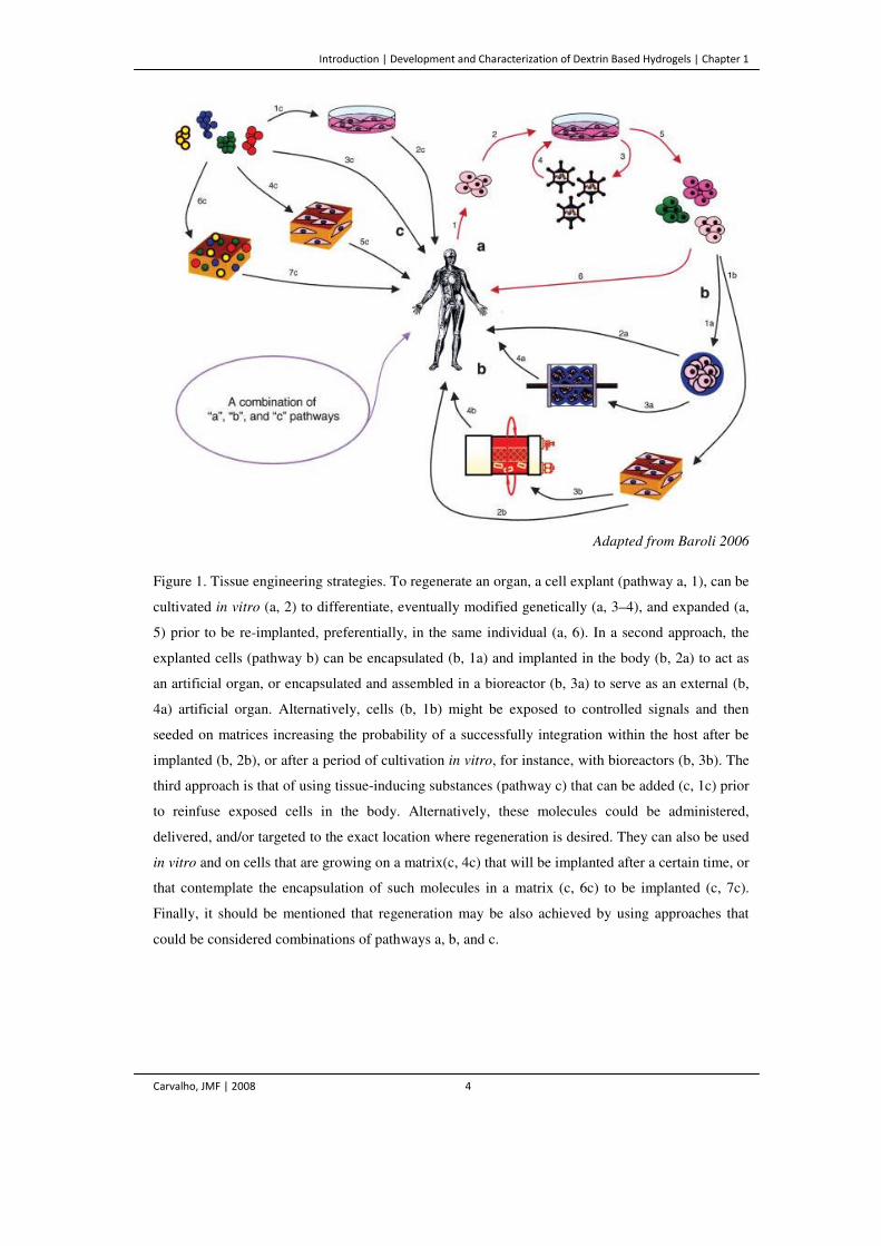
Figure 1. Tissue engineering strategies. To regenerate an organ, a cell explant (pathway a,
1), can be cultivated in vitro (a, 2) to differentiate, eventually modified genetically
(a, 3–4), and expanded (a, 5) prior to be re-implanted, preferentially, in the same
individual (a, 6). In a second approach, the explanted cells (pathway b) can be
encapsulated (b, 1a) and implanted in the body (b, 2a) to act as an artificial organ,
or encapsulated and assembled in a bioreactor (b, 3a) to serve as an external (b,
4a) artificial organ. Alternatively, cells (b, 1b) might be exposed to controlled
signals and then seeded on matrices increasing the probability of a successfully
integration within the host after be implanted (b, 2b), or after a period of
cultivation in vitro, for instance, with bioreactors (b, 3b). The third approach is
that of using tissue-inducing substances (pathway c) that can be added (c, 1c) prior
to reinfuse exposed cells in the body. Alternatively, these molecules could be
administered, delivered, and/or targeted to the exact location where regeneration is
desired. They can also be used in vitro and on cells that are growing on a matrix(c,
4c) that will be implanted after a certain time, or that contemplate the
encapsulation of such molecules in a matrix (c, 6c) to be implanted (c, 7c). Finally,
it should be mentioned that regeneration may be also achieved by using
approaches that could be considered combinations of pathways a, b, and c.
(Adapted from Baroli 2006)
4
Figure 2. Drug levels in the blood with (a) traditional drug dosing and (b) controlled-
delivery. With traditional tablets or injections, the drug level rises after each
administration, decreasing until the next one. The blood level of the agent should
remain between a maximum value (which may be toxic), and a minimum value
(below which the drug is no longer effective). In controlled drug delivery the drug
level in the blood remains constant, between the desired maximum and minimum,
for an extended period of time.
8
Figure 3. Structural formula of a α-D-glucopyranose residue (A), amylopectin (B) and
amylose (C).
16
Figure 4. Schematic representation of a functionalized hydrogel surface. The RGD ligands
mediate cell adhesion through the interaction with the integrins of the cells.
26
Development and Characterization of Dextrin Based Hydrogels
Use of Non-catalityc Domains for the Modification of Polysaccharides
Carvalho, JMF | 2008 xvii
Figure 5. Schematic representation of a dextrin molecule highlighting the predominant α-
(1→4) linkages, also evidencing the presence of α-(1→6) branch point.
27
Figure 6. Generic representation of a transesterification reaction (a). Synthesis of dextrin-
vinyl acrylate (VA), evidencing the reactive double bond on the final product. 30
CHAPTER 1 | SECTION II
Figure 1. Cell wall structure evidencing cellulose fibers. Schematic representation of a
cellulose molecule highlighting the β-(1→4) linkages. (Adapted from Sticklen
2008)
50
Figure 2. Simplified schematic view of the interaction between C. thermocellum
cellulosome and its substrate. CBM – Cellulose-Binding Module.
51
Figure 3. Structure of SS-mPEG, showing the activating succinimidyl succinate terminal
group, highly reactive toward nucleophile amine-containing molecules.
58
CHAPTER 2
Figure 1. Dextrin-VA synthesis and formation of the hydrogel networks.
70
Figure 2. Proteolytic activity in 0.2 M phosphate buffer pH 8 (▲) or DMSO (■) using
casein as substrate. The proteolytic activity is expressed as the rate of NH2
formation.
72
Figure 3. Weight variation of the samples containing DMSO, VA and enzyme, with
different initial water content, after equilibrium with saturated salt solutions.
Insertion shows the aw calculation (aw=0.3, for y=0) for an initial water content of
20%.
76
Figure 4. 13C CP-MAS NMR spectra of dextrin mixed with VA. Bands indicated with * are
spinning side bands. Number of scans (NS) =16729.
78
Figure 5. Downfield expansions of the 13C CP-MAS NMR spectra of (a) dextrin mixed with
vinyl acrylate and dextrin-VA with different degrees of substitution (DS) (b)
6.6%, NS=6228 (c) 8.7%, NS=6706 (d) 15.3%, NS=18827; the degrees of
substitution were determined by liquid state NMR of the solubilised dextrin-VA
sample. Peaks indicated with * are spinning side bands.
79
Figure 6. Full 13C CP-MAS NMR spectra and lowfield expansions for (a) dextrin-VA DS
15%, NS=18827 (b) dextrin-VA DS 15% hydrogel, NS=62916.
80
Figure 7. Full 13C CP-MAS NMR spectra and lowfield expansions for (a) dextrin-VA
DS=67%, NS=20821 (b) dextrin-VA DS=67% hydrogel, NS=20898. The signals
81
Development and Characterization of Dextrin Based Hydrogels
Use of Non-catalityc Domains for the Modification of Polysaccharides
Carvalho, JMF | 2008 xviii
indicated with * correspond to spinning sidebands.
Figure 8. X-ray powder diffraction patterns obtained from (a) dextrin-VA DS 67% (b)
dextrin-VA DS 67% hydrogel.
81
CHAPTER 3
Figure 1. 1H-NMR spectra of dextrin-VA in D2O. DS= 20%. 90
Figure 2. Diagram of diffusion chamber (Lateral view). 91
Figure 3. Structure representative of the dextrin molecules used to form dextrin-VA
hydrogels. A. Linear structure. B. Branched structure.
93
Figure 4. Protein and glucose transferred through the membrane (DS=20%), vs time, in the
diffusivity experiments.
94
Figure 5. SEM micrographs of hydrogel inner (A) and outer (B) surfaces with different DS,
ranging from 10 to70%.
96
Figure 6. Cumulative release of BSA (2 g/L) from dextrin-VA hydrogels, DS 20 (A), 40 (B)
and 70 (C). The data points are averages of at least three repeats that deviated less
than 5% of the total amount of protein in the hydrogels.
97
CHAPTER 4
Figure 1. Fluorescence labelling of mouse embryo fibroblasts. Micrographs show the
control culture, the cell layers under the positive (latex rubber) and negative (agar)
controls, and cell layers under (B) and around (A) the DS 20 hydrogel, after 24
hours of incubation (magnification 10 and 40x).
111
Figure 2. Cell proliferation inhibition index (mean% ± SD) after 72 h for the polymers
dextrin, acrylated dextrin (dextrin-VA), and for the initiators used in the
polymerization reaction APS and TEMED. Data are from a representative
experiment, performed in triplicate.
113
Figure 3. Mouse embryo fibroblasts, cultured for 24 h in the presence of 100 µg/mL of APS
(A), 300 mg/mL DS 20 (also representative for the other DS values) (B) and
control culture (C). Highly cytotoxic effects of APS are shown in micrograph A,
the cells are dead and floating in the medium. When compared to control cultures,
dextrin-VA-treated cells contained intracellular granula (see arrows) and have
spider-like structures (magnification 10x).
114
Figure 4. Fluorescence labelling of mouse embryo fibroblasts. Micrographs show control
culture and cells seeded on the surface of a DS 20 and DS 70 hydrogels, after 5, 24
116
Development and Characterization of Dextrin Based Hydrogels
Use of Non-catalityc Domains for the Modification of Polysaccharides
Carvalho, JMF | 2008 xix
and 72 hours of incubation (magnification 10x). A, B and C graphics correspond
to the MTS absorbance values at 490 nm through time course of the assay.
Figure 5. Cell proliferation inhibition index (CPII) (mean% ± SD) after 72 h for different
dextrin hydrogel discs. CPII obtained using MTT, MTS and MTS after cell
detachment. Data are from two representative experiments, each one performed in
triplicate.
117
CHAPTER 5
Figure 1. Synthesis of dextrin-HEMA. 126
Figure 2. Dextrin-HEMA 1H NMR spectrum. 126
Figure 3. Degradability of dextrin-based hydrogels. Swelling ratio (A) and mass loss as a
function of time, when immersed in PBS pH 7.4, at 37 ºC (average ±SD, n=3). (*
from day 40 the hydrogel structure was severely damaged, preventing the SR
measurements).
130
Figure 4. Rheological measurements of the different hydrogel samples. Frequency is 1 Hz
and stress varies in order to keep the response in the linear viscoelastic regime.
134
CHAPTER 6
Figure 1. Structure of SS-mPEG, showing the activating succinimidyl succinate terminal
group, highly reactive toward nucleophile amines.
145
Figure 2. SDS/PAGE analysis of unpurified and purified extracts of CBDCbhA, FN31-FN32-
CBDCbhA and CBDCipA.. Proteins were purified through nickel-affinity
chromatography as described in Materials and Methods.
147
Figure 3. Adsorption isotherms of CBDCbhA and FN31-FN32-CBDCbhA (a) and CBDCipA and
CBDCipA-PEG (b) for cellulose CF11.
150
Figure 4. MALDI-TOF analysis of the CBDCipA-PEG conjugated molecules, highlighting
the size distribution. Each peak with different molecular weight corresponds to
conjugates with multiple PEG molecules grafted.
152
Figure 5. Effect of CBDs on the physical properties of recycled (secondary) fibers. 153
Development and Characterization of Dextrin Based Hydrogels
Use of Non-catalityc Domains for the Modification of Polysaccharides
Carvalho, JMF | 2008 xx
List of Tables
CHAPTER 1 | SECTION I
Table 1. Characteristics of a drug delivery system. 9
Table 2. Important biomaterials employed in medicine. 12
Table 3. Biomedical applications of hydrogels. 21
CHAPTER 2
Table 1. Degree of substitution (DS) obtained with different VA concentrations obtained
enzymatic and chemically, with dextrin and dextran as substrates.
73
Table 2. 13C and 1H NMR assignments of the glucopyranosyl ring (δ, ppm) on dextrin-
VA with DS 20%.
74
Table 3. Water activity of reaction mixtures with different amounts of water and DS (the
VA to dextrin ratio correspond to a theoretical DS of 20%) obtained after
transesterification reaction.
76
CHAPTER 3
Table 1. Glycosidic-linkage analysis of the dextrin used to prepare the hydrogel. 93
Table 2. Diffusivity coefficients of BSA and glucose for different hydrogel reticulation
degrees and hydrogel mass loss values after 20 days.
95
CHAPTER 4
Table 1. Cell proliferation inhibition index of mouse embryo fibroblasts, cultured in the
presence of extracts obtained from dextrin-VA hydrogels with different DS and
initial water content.
110
Table 2. Mouse embryo fibroblasts adhesion onto dextrin-VA hydrogels with different
DS values. (% estimated by cell counting after trypsinization).
115
CHAPTER 6
Table 1. Oligonucleotides used for cloning. 144
Development and Characterization of Dextrin Based Hydrogels
Use of Non-catalityc Domains for the Modification of Polysaccharides
Carvalho, JMF | 2008 xxi
Table 2. Hydrolysis of ASC and filter paper by truncated forms of CbhA and
combinations of GH9 and Cel5B with CBDCbhA and FN31-FN32-CBDCbhA.
148
Table 3. Effect of CBDs in the Shopper-Riegler Index (each one performed with
triplicates) and permeability of paper and pulp fibers.
153
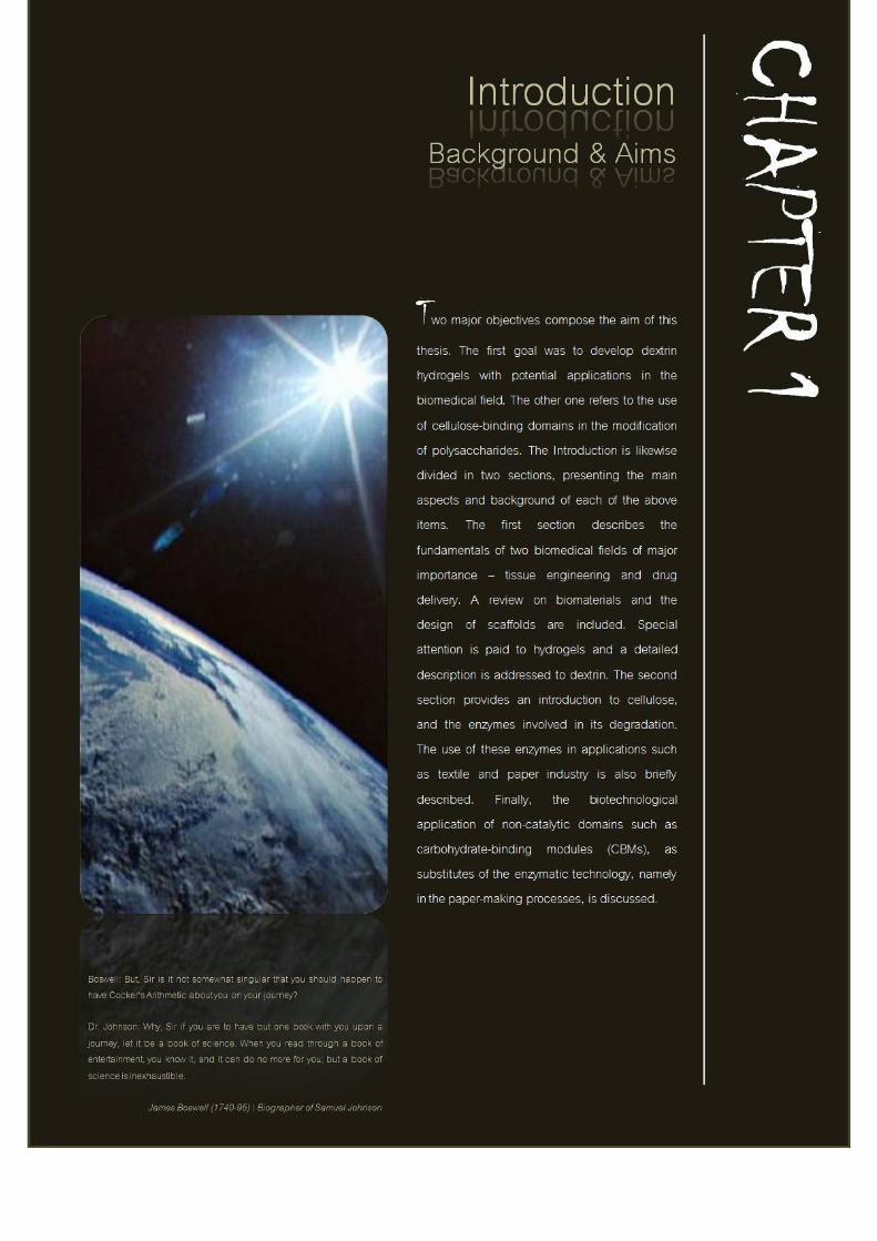

Introduction | Development and Characterization of Dextrin Based Hydrogels | Chapter 1
Carvalho, JMF | 2008 3
SECTION I
Development and Characterization of Dextrin
Based Hydrogels
Tissue Engineering – An Overview to the Hybrid Technology
The advent of tissue engineering (TE) has been motivated by the challenge of
producing tissues substitutes that might restore, maintain or improve the structural
features and physiological functions of natural living tissues (Thomson et al. 1995,
Freed and Vunjak-Novakovic 1998). Tissue engineering has been described in the
early 90s by Langer and Vacanty (1993) as “the persuasion of the body to heal
itself, through the delivery at the appropriate sites of molecular signals, cells, and
supporting structures”. Initial strategies for the creation of new tissues combined
three basic principles: (1) The isolation and cultivation of cells, (2) the use of
tissue-inducing substances, and (3) the placement of cells within suitable matrices
to support their three dimensional growth (Langer and Vacanty 1993, Vacanty and
Langer 1999) (Figure 1).
The direct in vivo implantation of isolated cells, allows overcoming the
complications of surgery, as an invasive technique. Additionally, it is possible to
replace only those cells that supply the needed function and also manipulate cells
before infusion. In this approach there is very little of engineering. Its potential
limitation include failure of infused cells in maintaining their function and it must
be considered the possibility of immunological rejection (Langer and Vacanty
1993, Freed and Vunjak-Novakovic 1998) (Figure 1, pathway a).
Introduction | Development and Characterization of Dextrin Based Hydrogels | Chapter 1
Carvalho, JMF | 2008 4
Adapted from Baroli 2006
Figure 1. Tissue engineering strategies. To regenerate an organ, a cell explant (pathway a, 1), can be
cultivated in vitro (a, 2) to differentiate, eventually modified genetically (a, 3–4), and expanded (a,
5) prior to be re-implanted, preferentially, in the same individual (a, 6). In a second approach, the
explanted cells (pathway b) can be encapsulated (b, 1a) and implanted in the body (b, 2a) to act as
an artificial organ, or encapsulated and assembled in a bioreactor (b, 3a) to serve as an external (b,
4a) artificial organ. Alternatively, cells (b, 1b) might be exposed to controlled signals and then
seeded on matrices increasing the probability of a successfully integration within the host after be
implanted (b, 2b), or after a period of cultivation in vitro, for instance, with bioreactors (b, 3b). The
third approach is that of using tissue-inducing substances (pathway c) that can be added (c, 1c) prior
to reinfuse exposed cells in the body. Alternatively, these molecules could be administered,
delivered, and/or targeted to the exact location where regeneration is desired. They can also be used
in vitro and on cells that are growing on a matrix(c, 4c) that will be implanted after a certain time, or
that contemplate the encapsulation of such molecules in a matrix (c, 6c) to be implanted (c, 7c).
Finally, it should be mentioned that regeneration may be also achieved by using approaches that
could be considered combinations of pathways a, b, and c.
Introduction | Development and Characterization of Dextrin Based Hydrogels | Chapter 1
Carvalho, JMF | 2008 5
In the second strategy (Figure 1, pathway b), a temporary support is required to
serve as an adhesive substrate for the implanted cells and a physical support to
guide the formation of the new tissues. Transplanted cells adhere to the support or
are encapsulated in a biomaterial, proliferate and produce the extracellular matrix
(ECM), stimulating the new tissue formation. During this process, the support
matrix gradually degrades and might, eventually, be eliminated (Langer and
Vacanty 1993).
The third approach contemplates the use of tissue-inducing substances such as
growth factors, or cytokines, before cell reinfusion (Figure 1, pathway c). One of
the major disadvantages of this approach is the purification and large-scale
production of the inducing substances, and also the need of a system to deliver the
bioactive molecule to the desired location (Langer and Vacanty 1993, Langer
1999).
These principles and strategies are still valid and encouraging research has been
carried out over the last decade, reporting on the application of tissue engineering
products and strategies in clinical use (Lysaght et al. 1998, Bonassar and Vacanty
1998, Lysaght and Hazelhurst 2000). However, it must be recognized that many
unforeseen problems must be overcome to achieve the ambitious goals of providing
fully functional tissue replacement.
Having in mind these developments, it seems obvious that the original support
materials must be tailored to provide additional functionality, besides the ability to
withstand mechanical loads or to possess suitable degradation kinetics (Tessmar
and Gopferich 2007). They should act as a synthetic extracellular matrix. ECM is
the natural medium in which cells proliferate, differentiate and migrate, and
therefore is the gold standard for tissue regeneration (Meredith et al. 1993, Bosman
et al. 2003). Natural ECM is a condensed matrix mainly composed of locally
secreted proteins, proteoglycans and polysaccharides, arranged as a molecular
network. The homeostatic dynamic state of ECM is controlled by proteoglycans,
and a number of signaling molecules, such as growth factors, which mediate cell-
ECM and cell-cell interactions. All these molecules are embedded in an
Introduction | Development and Characterization of Dextrin Based Hydrogels | Chapter 1
Carvalho, JMF | 2008 6
amorphous, fundamental substance represented by glycosaminoglycan chains,
which form the highly hydrated gel structure imbibing the matrix (Meredith et al.
1993). Thus, a suitable support for tissue engineering should mimic some of the
ECM characteristics, namely providing mechanical integrity to tissues, supporting
their growth, providing an environment for the host cell survival as well as the
means to deliver nutrients, growth and differentiation factors for long term support
of the proliferation. Additionally, they should guide cell adhesion and even recruit
desirable cells, promoting the dynamic interaction with surrounding tissues (Yang
et al. 2001, Drury and Mooney 2003). Furthermore, biocompatibility is an essential
issue regarding their pharmaceutical and biomedical applicability, to avoid any
adverse foreign host response (Hutmacher 2001).
Controlled Drug Delivery – A New Research Frontier
Despite the great attention paid to the promising developments in TE, a parallel
extensive research has also been continually performed over the past two decades
in another challenging field – the controlled drug delivery (CDD). CDD strategies
have made a dramatic impact in medicine. Advances in biotechnology led to the
availability of complex natural molecules for disease treatment (growth factors,
interferons, cytokines, response modifiers, etc). In form of complex carbohydrates,
proteins or DNA, these materials are, sometimes, larger in size, less stable, less
soluble and much harder to deliver than the classical therapeutic agents. The
development of CDD was a critical step in the treatment of diseases such as AIDS,
Alzheimer and cancer, and as challenging as the new drug discovery itself (Davis
and Illum 1998).
Although initially following two distinct scientific routes, TE and CDD have soon
converged. The need for deliver growth and differentiation factors for long term
support of the proliferating cells, turned the controllable release systems into the
Introduction | Development and Characterization of Dextrin Based Hydrogels | Chapter 1
Carvalho, JMF | 2008 7
fusion step in the development of new generations of biomedical tools and products
based on TE principles.
Controlled drug delivery occurs when a polymer is combined with a drug or other
bioactive molecule in such a way that the molecule is released from the material in
a predesigned manner (Robinson and Lee 1997, Langer 1988). The release of the
active agent may be constant, cyclic or triggered by an external event. The goal of
controlling the delivery is to achieve more effective therapies, eliminating at the
same time, the possibility for both under and overdosing. The ability to maintain
the drug levels within a desired range of concentration in a targeted way, enhancing
the ability to use highly toxic, poorly soluble or relatively unstable drugs, the need
for fewer administrations and increasing in patient compliance are among the
advantages of using controlled-delivery systems (Figure 2). Additionally, their use
to minimize drug degradation and loss, to prevent harmful side-effects and to
increase the drug bioavailability, has also been successfully applied (Kost and
Langer 1992, Peppas 1997, Charman et al. 1999, Soppimath 2001).
The ideal drug delivery system should be inert, biocompatible, mechanically
strong, comfortable for the patient, capable of high levels of drug loading, safe
from accidental release, simple to administer and remove, and easy to fabricate and
sterilize. Among drug carriers are polymers in different formulations (hydrogels,
micro and nanoparticles), microcapsules, lipoproteins, liposomes, micelles and
others. (Santini 2000, Soppimath et al. 2001, Torchilin 2001, Bae et al. 2003,
Kopecek 2003, Haag 2003, Muller-Goymann 2004, Packhaeuser 2004).
Introduction | Development and Characterization of Dextrin Based Hydrogels | Chapter 1
Carvalho, JMF | 2008 8
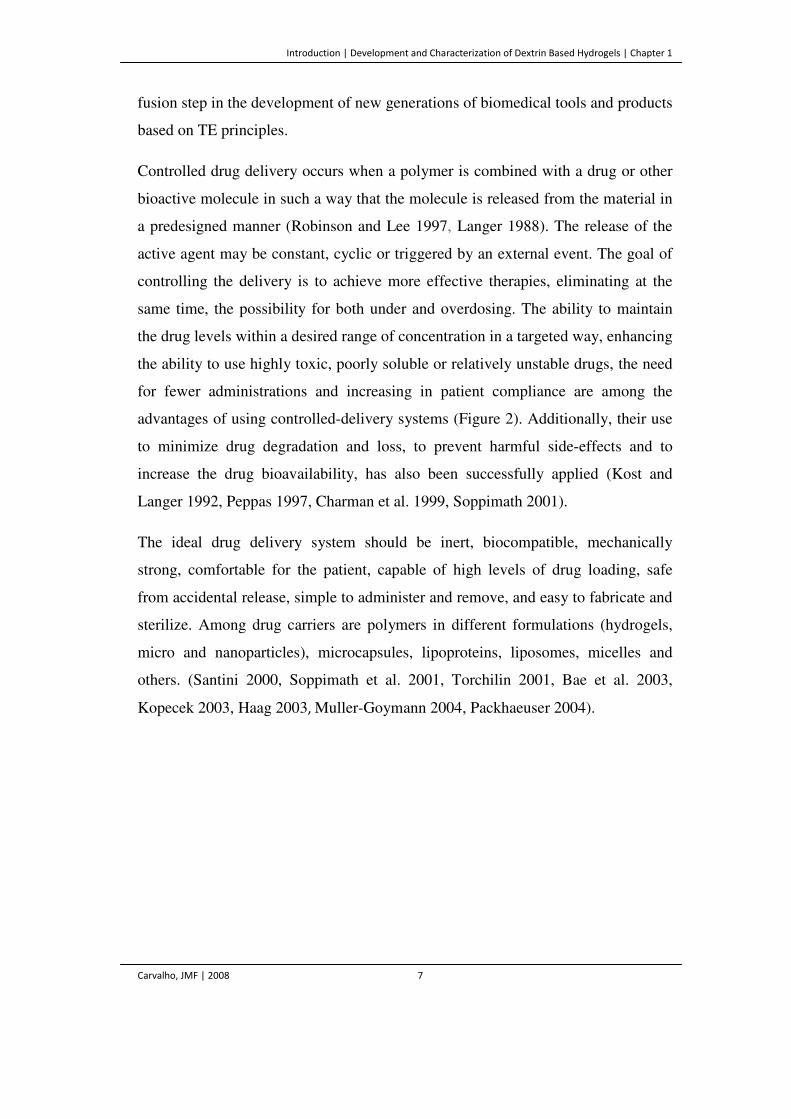
Figure 2. Drug levels in the blood with (a) traditional drug dosing and (b) controlled-delivery. With
traditional tablets or injections, the drug level rises after each administration, decreasing until the
next one. The blood level of the agent should remain between a maximum value (which may be
toxic), and a minimum value (below which the drug is no longer effective). In controlled drug
delivery the drug level in the blood remains constant, between the desired maximum and minimum,
for an extended period of time.
In recent years, controlled delivery formulations became much more sophisticated,
with the ability to do more than simply extend the effective release period for a
bioactive agent. Current controlled release devices are capable to respond to
changes in the biological environment and delivering/stop delivering the drugs
based on these changes. Moreover, the delivery system can be targeted to the
specific cell, tissue, or local where the drug it contains is to be delivered. The
release rates are therefore determined by the design of the system, which in turn
depends on the requirements of its final application (Peppas 1997, Tessmar and
Göpferich 2007).
Table 1 summarizes the key characteristics of a controlled delivery system. The
mode of delivery and subsequent degradation are important features in developing
successful formulations. Sustained (or continuous) release of a drug involves the
release at a controlled rate due to the diffusion out of the polymer or by its
degradation over time. Pulsatile release is achieved by using carrier polymers that
respond to specific stimuli (e.g., exposure to light, changes in pH or temperature)
and is often the preferred method of delivery, as it resembles the naturally
Introduction | Development and Characterization of Dextrin Based Hydrogels | Chapter 1
Carvalho, JMF | 2008 9
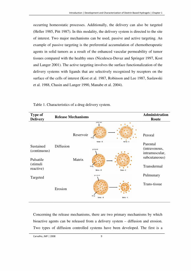
occurring homeostatic processes. Additionally, the delivery can also be targeted
(Heller 1985, Pitt 1987). In this modality, the delivery system is directed to the site
of interest. Two major mechanisms can be used, passive and active targeting. An
example of passive targeting is the preferential accumulation of chemotherapeutic
agents in solid tumors as a result of the enhanced vascular permeability of tumor
tissues compared with the healthy ones (Niculescu-Duvaz and Springer 1997, Kost
and Langer 2001). The active targeting involves the surface functionalization of the
delivery systems with ligands that are selectively recognized by receptors on the
surface of the cells of interest (Kost et al. 1987, Robinson and Lee 1987, Saslawski
et al. 1988, Chasin and Langer 1990, Manabe et al. 2004).
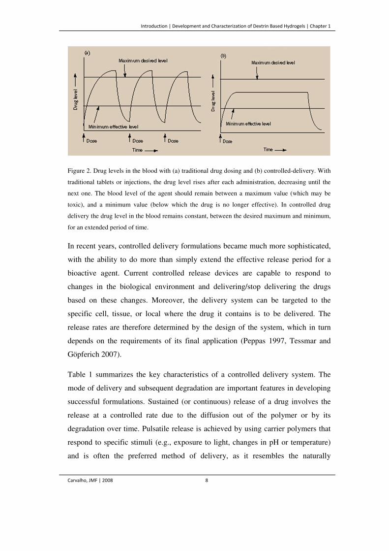
Table 1. Characteristics of a drug delivery system.
Type of
Delivery Release Mechanisms
Administration
Route
Sustained (continuous) Pulsatile (stimuli reactive) Targeted
Diffusion
Reservoir
Peroral Parental (intravenous, intramuscular, subcutaneous) Transdermal Pulmunary Trans-tissue
Matrix
Erosion
Concerning the release mechanisms, there are two primary mechanisms by which
bioactive agents can be released from a delivery system – diffusion and erosion.
Two types of diffusion controlled systems have been developed. The first is a

Introduction | Development and Characterization of Dextrin Based Hydrogels | Chapter 1
Carvalho, JMF | 2008 10
reservoir device in which the bioactive agent forms a core surrounded by a
polymeric barrier. These systems include membranes, capsules, microcapsules,
liposomes, and hollow fibers. The second type is a polymeric matrix device in
which the active agent is dispersed or dissolved. In these two systems, drug
diffusion through the polymer is the rate-limiting step. Likewise, release rates
depend on the polymer characteristics and its consequent effect on the diffusion
and partition coefficient of the biomolecule to be released (Kost et al. 1987,
Robinson and Lee 1987, Langer 1988, Chasin and Langer 1990).
In chemically controlled systems, the erosion is the mechanism underlying release
control. Bioerodible (or biodegradable) systems offer the advantage of being
absorbed by the body, avoiding the drawbacks of the surgical removal. In a
bioerodible system the drug is ideally uniformly distributed throughout a polymer
as in matrix systems (Kost 1987, Kost and Langer 1992). As the polymer
surrounding the drug is eroded, the drug is release. The pendant chain system is
another chemically controlled system. The drug is chemically attached to a
polymer backbone chain and the release takes place by hydrolytic or enzymatic
cleavage (Heller 1988, Jeong et al. 1999).
Finally, the choice of the administration route is another feature of major
importance in the application of CDD technology. The delivery route is driven by
patient acceptability, the properties of the drug, such as its solubility, the access to
a disease location, or effectiveness in dealing with the specific disease. The most
important drug delivery route is the peroral, however, parental, transdermal,
pulmonary and trans-tissue routes are also very important (Davis and Illum 1998).
Introduction | Development and Characterization of Dextrin Based Hydrogels | Chapter 1
Carvalho, JMF | 2008 11
Biomaterials – The Foundations
Originating from their application in the biological environment, the designation of
biomaterials is due to their ability to replace or restore biological functions and
exhibit a pronounced compatibility with the biological environment (Langer et al.
1990, Hoecker 1998). Biomaterials play a major role in most tissue engineering
strategies and apart from this original use, they have been increasingly applied as
the pivotal substrate for cell attachment and migration and also as implantable
systems for drug and cell carrying (McCulloch and Shalaby 1998, Zhou et al. 2003,
Kanjickal and Lopina 2004), to activate specific cellular function in a localized
region (Langer 1995, James and Khon 1996, Hutmacher et al. 2001). The
development of biomaterials for medical applications has recently focused on the
design of biomimetic materials that might be able to interact with surrounding
tissues by biomolecular recognition, to make them capable of eliciting specific
cellular responses, mediated by specific interactions (Klee and Hoecker 1999,
Lucke et al. 2000). Biomaterials have been employed to conduct and accelerate
otherwise naturally occurring phenomena, such as tissue regeneration in wound
healing. They can also be used to induce cellular responses that might not be
normally present, such as healing in a diseased subject and to block natural
phenomena, such as the immune rejection of cell transplants (Langer 1995).
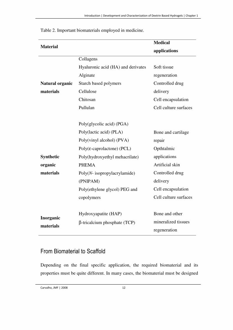
A wide variety of biomaterials, both synthetic and natural, is available (Table 2).
Naturally derived and recombinant biomaterials that combine the beneficial aspects
of both natural and many of the desirable features of synthetic materials have been
designed and produced. In general, some authors claim that the latter offer some
advantages, since they can be tailored to give a wide range of properties and more
predictable and reproducible results that the materials derived from natural sources
(Doylan and Cameron1990, Cascone et al. 1993, Hanein et al. 1995). However,
natural biomaterials are more likely to induce the appropriate biological response
which is fundamental in biomedical applications.
Introduction | Development and Characterization of Dextrin Based Hydrogels | Chapter 1
Carvalho, JMF | 2008 12
Table 2. Important biomaterials employed in medicine.
Material Medical
applications
Natural organic
materials
Collagens
Soft tissue
regeneration
Controlled drug
delivery
Cell encapsulation
Cell culture surfaces
Hyaluronic acid (HA) and derivates
Alginate
Starch based polymers
Cellulose
Chitosan
Pullulan
Synthetic
organic
materials
Poly(glycolic acid) (PGA)
Bone and cartilage
repair
Opthtalmic
applications
Artificial skin
Controlled drug
delivery
Cell encapsulation
Cell culture surfaces
Poly(lactic acid) (PLA)
Poly(vinyl alcohol) (PVA)
Poly(ε-caprolactone) (PCL)
Poly(hydroxyethyl mehacrilate)
PHEMA
Poly(N- isopropylacrylamide)
(PNIPAM)
Poly(ethylene glycol) PEG and
copolymers
Inorganic
materials
Hydroxyapatite (HAP) Bone and other
mineralized tissues
regeneration
β-tricalcium phosphate (TCP)
From Biomaterial to Scaffold
Depending on the final specific application, the required biomaterial and its
properties must be quite different. In many cases, the biomaterial must be designed
Introduction | Development and Characterization of Dextrin Based Hydrogels | Chapter 1
Carvalho, JMF | 2008 13
not to interact with individual cells, but rather with multiple cells, or even whole
tissues. This means that the materials must be processed into devices exceeding the
dimensions of a single cell in a two (2D) or three (3D) dimensional architecture. In
the late 1970s and 1980s, cells on sheets of collagen (2D), or collagen–
glycosaminoglycan composites, were used in tissue regeneration in an attempt to
create new skin (Bell et al. 1981, Burke et al. 1991). However, the pressure of the
constant attempts to engineer virtually every human tissue (including cartilage,
bone, muscle, heart valves, nerves, among others), drove the research to 3D
structures that enable large numbers of cells to be housed. The term scaffold arose.
The scaffold serves as 3D template for initial cell attachment and subsequent tissue
formation both in vitro and in vivo. The scaffold provides the necessary support for
cells to attach, proliferate, maintaining their differentiated state and can even define
the overall shape of the tissue-engineered transplant (Langer 2000, Hutmacher
2001).
Scaffold Characteristics
The challenge for scaffold systems is to arrange an appropriate 3D configuration
that shall be able to fulfill the needs of the final application, which, in general,
requires balancing temporary mechanical function with mass transport to aid
biological delivery and/or tissue regeneration, and do so in a way that can be
carried out reproducibly, economically, and on a large scale. The requirements for
a scaffold to be considered suitable in the biomedical field are complex; however,
the following basic characteristics must be addressed to bring about the desired
biologic response (Hutmacher 2001). (1) The scaffold should be biocompatible.
Neither it nor its degradation products should induce any adverse response or
toxicity. (2) The scaffold should possess the appropriate mechanical properties, to
provide the correct environment, matching the intended site of implantation. (3)
Additionally the scaffold should be made from material with controlled
biodegradability or bioresorbability so that tissue will eventually replace the
Introduction | Development and Characterization of Dextrin Based Hydrogels | Chapter 1
Carvalho, JMF | 2008 14
scaffold. (4) It should have an interconnected pore network, enhancing the
diffusion rates, improving oxygen and nutrient supply and waste removal, thereby
facilitating the vascularization. (5) Furthermore an appropriate surface chemistry
should favor cellular attachment, differentiation and proliferation. (6) Finally the
scaffold should be easily processed into a variety of shapes and sizes as well as
easily sterilized (Freed et al. 1994, Thomson et al. 1995, Lu and Mikos 1996,
Agrawal et al. 1997, Piskin 1997, Shapiro and Cohen 1997, Simske 1997, Maquet
and Jerome 1997, Kim and Mooney 1998, Middleton and Tipton 2000, Chapekar
2000, Hutmacher 2000).
Polymers in Biomedical Applications
The selection of a scaffold material is both critical and difficult. There are many
biomaterials available, such as metals, ceramics and polymers. Metals and ceramics
have contributed to major advances in medicine, particularly in orthopedic tissue
replacements. Typical implant metals were made of cobalt and titanium, and
typical ceramics included alumina, zirconia, calcium phosphate, and bioglass
(Hench 1996). However, the requirement of biodegradability totally excludes the
use of metals and almost excludes the use of ceramics as scaffolds (Thomson et al.
1995, Maquet and Jerome 1997). Nevertheless, inorganic compounds, such as
hydroxyapatite (HAP), β-tricalcium phosphate (TCP) and combinations of both can
be successfully applied in orthopedic applications for the regeneration of bones and
other mineralized tissues (LeGeroz 2002, Paul and Sharma 2003). The drawbacks
include the difficulty in processing complex shapes and the non-degradability. For
these reasons, polymeric materials have received an increasing attention.
Biodegradable polymers can be broadly classified into natural and synthetic, based
on their origin.
Introduction | Development and Characterization of Dextrin Based Hydrogels | Chapter 1
Carvalho, JMF | 2008 15
Natural Polymers
Natural polymers, such as polysaccharides and proteins have been shown to have
wide application as scaffolds. Its major advantage is that they often have an
organized structure, at both molecular and macroscopic levels, thereby resulting in
some favorable characteristics, such as the ability to induce tissue ingrowth and to
most closely simulate the native cellular milieu. In fact, they usually contain
domains that can send important signals to guide cell development at various
stages. However, poor mechanical performances and large batch-to-batch
variations can limit their wide applications. Also, the degradation of naturally
occurring polymers almost relies on enzymatic processes, which will introduce
some inevitable patient-to-patient variation in the degradation rates, affecting the
predictability and reproducibility as it was already stated (Thomson et al. 1995).
Hyaluronic acid (HA) and collagen were considered as the materials of choice for
scaffolds for a long time. They are two ubiquitous biopolymers found in the
mammalian body and two of the principal components of ECM (McPherson et al.
1986, Laurent et al. 1996). They have been used especially for regeneration of soft
tissues either alone or in combination with other agents (Lee et al. 2001, Pachence
1996). Although having, in general, poor mechanical properties, naturally derived
polymers are interesting because they do not induce a host response, and may
enhance the biological recognition encouraging the normal cellular functions. For
the replacement of soft tissues, there are many strategies employing a combination
of collagen and hyaluronic acid, since they strongly resemble the organization of
ECM (Brun et al. 1999, Xin et al. 2004, Tang et al. 2007).
Alginate is a well known polysaccharide widely used due to its gelling properties in
aqueous solutions. It can be extracted from marine brown algae or produced by
bacteria (Kim and Mooney 1998). Due to the intrinsic properties of alginate
calcium gels (biocompatibility, adhesion, porosity, and ease of manipulation) much
attention has recently been focused on the delivery of proteins, cell encapsulation,
and tissue regeneration. However, alginate is not biodegradable in the human body
Introduction | Development and Characterization of Dextrin Based Hydrogels | Chapter 1
Carvalho, JMF | 2008 16
and the gelation dependence on the calcium ions is also a drawback, since they can
be lost following implantation (Kim and Mooney 1998).
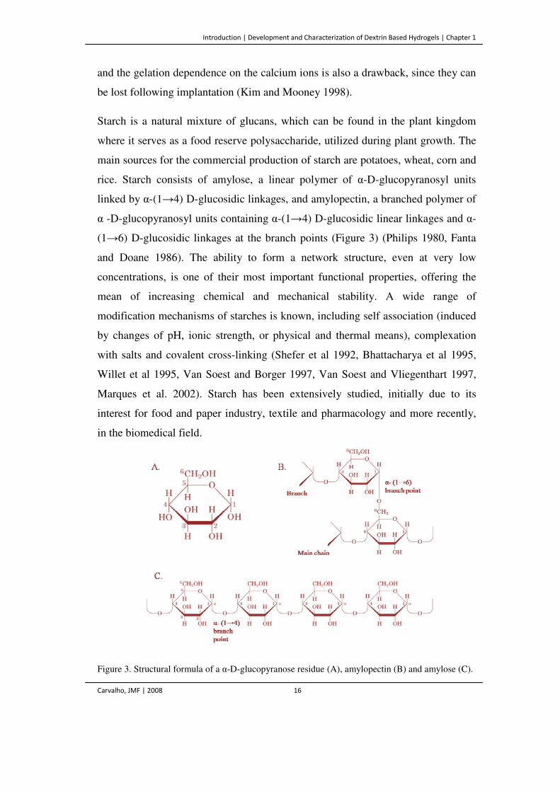
Starch is a natural mixture of glucans, which can be found in the plant kingdom
where it serves as a food reserve polysaccharide, utilized during plant growth. The
main sources for the commercial production of starch are potatoes, wheat, corn and
rice. Starch consists of amylose, a linear polymer of α-D-glucopyranosyl units
linked by α-(1→4) D-glucosidic linkages, and amylopectin, a branched polymer of
α -D-glucopyranosyl units containing α-(1→4) D-glucosidic linear linkages and α-
(1→6) D-glucosidic linkages at the branch points (Figure 3) (Philips 1980, Fanta
and Doane 1986). The ability to form a network structure, even at very low
concentrations, is one of their most important functional properties, offering the
mean of increasing chemical and mechanical stability. A wide range of
modification mechanisms of starches is known, including self association (induced
by changes of pH, ionic strength, or physical and thermal means), complexation
with salts and covalent cross-linking (Shefer et al 1992, Bhattacharya et al 1995,
Willet et al 1995, Van Soest and Borger 1997, Van Soest and Vliegenthart 1997,
Marques et al. 2002). Starch has been extensively studied, initially due to its
interest for food and paper industry, textile and pharmacology and more recently,
in the biomedical field.
Figure 3. Structural formula of a α-D-glucopyranose residue (A), amylopectin (B) and amylose (C).
Introduction | Development and Characterization of Dextrin Based Hydrogels | Chapter 1
Carvalho, JMF | 2008 17
Dextran is a glucose homopolysaccharide that feature a substantial number of
consecutive α-(1→6) linkages in their main chains, usually more than 50% of the
total linkages. They also possess side chains stemming from α-(1→2), α-(1→3), or
α-(1→4) branch linkages. Just like starch, there are many reports on the use of
dextran in the pharmaceutical field, being the drug delivery a very promising one.
One of the first approaches was to introduce reactive double bonds by
functionalizing the polymer with glycidyl acrylate (Kamath and Park 1995). This
study was the precursor for numerous others. Dextran-MA, dextran-HEMA,
microparticles of functionalized dextran are being used for the delivery of
imunoglobulin G (IgG), native or recombinant human bone morphogenetic protein
(BMP), DNA, among others (Franssen et al. 1997, Hennink et al. 1997, Ferreira et
al. 2002, Pitarresi et al. 2003, Ferreira et al. 2004, Maire et al. 2005, Ferreira et al.
2005, DeGest et al. 2006).
Cellulose is a linear polymer with repeating units consisting of cellobiose. It can
undergo enzymatic degradation resulting in the formation of D-glucose units.
Although cellulose is insoluble in common solvents, the hydroxyl groups are
reactive and can be easily functionalized. Several derivatives of cellulose in the
form of ethers, esters, and acetals, such as methyl cellulose, hydroxypropylmethyl
cellulose and carboxymethyl cellulose have been investigated as candidates for
various applications. All of these cellulose derivatives are soluble in a variety of
solvents and can be easily processed into various forms such as membranes,
sponges and fibers (Baeyer et al. 1988, Hou et al. 1991). Additionally, the good
mechanical properties of cellulose make it very attractive for biomedical
applications as dressings in treating surgical incisions, burns, wounds, and various
dermatological disorders, and also as matrices for drug delivery applications
(Swarbrick and Boyan 1991, Takahashi et al. 2001, Tate et al. 2001, Li et al. 2002,
Stabenfeldt et al. 2006).
Chitosan is the deacylate derivate of chitin. Chitin is a naturally wide occurring
polysaccharide that can be extracted from crustacean exoskeletons (e.g. crabs and
shrimps), also existing in fungal cell walls. The biodegradation rate is determined
Introduction | Development and Characterization of Dextrin Based Hydrogels | Chapter 1
Carvalho, JMF | 2008 18
by the amount of residual acetyl content, a parameter which can easily be varied
(Hayashi 1994, Pachence and Khon 1997). Like HA, chitosan is not antigenic and
is a well-tolerated implantable material, showing biostimulating activity in the
healing process of various tissues. Chemical modification of chitosan produces
materials with a variety of physical and mechanical properties, very useful in
applications as encapsulation, inhibition of blood coagulation, controlled drug
delivery, among others (Madihally and Mathew 1999).
Pullulan is a linear bacterial homopolysaccharide originating from Aurebasidium
pullulans. The backbone is formed by glycosidic linkages of α-(1→6) D-
glucopyranose and α-(1→4) D-glucopyranose units in a 1/2 ratio (Leathers 2002).
The backbone structure of pullulan resembles dextran, being also easily derivatized
in order to impart new physico-chemical properties, e.g. to increase the solubility in
organic solvents or to introduce reactive groups, leading to a polymeric system
capable of forming hydrogels. The unique linkage pattern of pullulan endows the
polymer with distinctive physical traits, including adhesive properties and the
capacity to form fibers, compression moldings, and strong, oxygen-impermeable
films (Leathers 2003). Several studies refer pullulan as a promising polymeric
carrier for many drugs (Shingel 2004). Additionally, recent works reported that
pullulan allow for smooth muscle cell adhesion, spreading, and proliferation and
hold promises as scaffolds for vascular engineering (Autissier et al. 2007).
Synthetic polymers
The main advantage of synthetic polymers, over the natural ones, is that they are
chemically synthesized, being therefore possible to control their molecular weight
and molecular weight distribution (with variable degrees of accuracy, depending on
e.g. the type of polymerization reaction), which ultimately causes profound effects
on the physical characteristics of the polymer, such as strength and degradation
rates (Thomson et al. 1995, Maquet and Jerome 1997). They can, thus, be
Introduction | Development and Characterization of Dextrin Based Hydrogels | Chapter 1
Carvalho, JMF | 2008 19
developed to meet the requirements of its final applications. Additionally, although
enzymatic processes can assist the degradation, it is, in general, brought up by
simple hydrolysis, which is desirable to minimize the person-to-person variations.
Among synthetic materials, biodegradable polyesters approved by the Food and
Drug Administration (FDA) are rapidly gaining recognition in the field of TE.
Poly (α-hydroxy acids), such as poly(glycolic acid) (PGA), poly(lactic acid) (PLA)
and their copolymer poly(lactic-co-glycolic acid) (PLGA) are currently the most
widely investigated and most commonly used synthetic biodegradable polymers
and have been widely applied in bone and cartilage repair (Athanasiou et al. 1998,
Sherwood et al 2002, Uematsu et al 2005).
Poly(ε-caprolactone) (PCL), which degrades at significantly lower rates than PLA,
PGA and PLGA, is less attractive for tissue engineering applications. However,
PCL-based scaffolds have been studied for skin replacement and other tissue
engineering applications (Ng et al. 2001, Htai et al. 2004, Li et al. 2005).
Furthermore, novel degradable PCL networks, PLGA-PCL-PLGA tri-block
copolymers and PCL-chitosan matrices are more hydrophilic, degrade faster and
possess desirable mechanical properties as compared to PCL (Choi and Park 2002,
Kweon et al. 2003 Sarasam and Madihally 2005).
Poly(N-isopropylacrylamide) (pNiPAAm) and its copolymers belong to the most
intensively investigated thermoreversible materials. Recent developments on
pNiPAAm-based scaffolds include their use for drug delivery (Liu et al. 2006, Yin
et a. 2006, Nakayama et al. 2006, Coughlan et al 2006), cell encapsulation and
delivery (Na et al. 2006) and cell culture surfaces (Hatakeyama et al. 2006).
Poly(ethylene glycol)s (PEGs) are very popular synthetic polymers frequently used
in tissue engineering and drug delivery applications. Although PEG derivatives
provide only end-groups for chemical modification, they are frequently used,
because they are non-toxic and non-adhesive towards proteins, resulting in suitable
model systems (Drury and Mooney 2003, Eiselt et al. 1999). The copolymerization
of PEG with biodegradable and biocompatible polyesters yielded some interesting
Introduction | Development and Characterization of Dextrin Based Hydrogels | Chapter 1
Carvalho, JMF | 2008 20
solutions that may be applied as delivery systems in patches, creams, gels,
injectables and implants (Jeong et al. 2000, Zitzmann and Nieschlag 2000, Chen
and Singh 2005, Lee et al. 2006).
Hydrogel as a Scaffold
Among the scaffolds, hydrogels are receiving an increasing attention. They can fill
irregular shaped defects and incorporate cells and other bioactive materials (Drury
and Mooney 2003). Several biomedical applications require materials that possess
a jelly consistency, which can set and be molded into a desired shape under
physiological conditions.
The term hydrogel, refers to aqueous (water-containing) gels, polymer networks
that are insoluble in water, where they swell to an equilibrium volume, retaining
their shapes (Chen et al. 1997). The importance of hydrogels in biomedical
applications was first reported in the late 1950s, with the development of PHEMA
gels as a soft contact lens material (Wichterle and Lim 1960). Nowadays they are
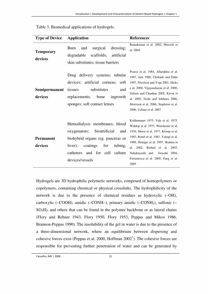
used in numerous biomedical applications (Table 3) including ophthalmic devices,
biosensors, biomembranes, and as delivery systems (Andrade 1976, Peppas 1986).
Hydrogels have appealing features from the perspective of biological mimicking.
They are able to retain a great quantity of water, producing a soft consistency,
which resembles natural living tissues. Hydrogels have a good biocompatibility
and, in general, they show low minimal mechanical and frictional irritation (Chen
et al. 1997, Hoffman 2002). Furthermore they can be processed under relatively
mild conditions and can be delivered in a minimally invasive manner, increasing
patient compliance (Lee and Mooney 2001, Nguyen and West 200, Baroli 2006).
Introduction | Development and Characterization of Dextrin Based Hydrogels | Chapter 1
Carvalho, JMF | 2008 21
Table 3. Biomedical applications of hydrogels.
Type of Device Application References
Temporary
devices
Burn and surgical dressing;
degradable scaffolds; artificial
skin substitutes; tissue barriers
Ramakumar et al. 2002, Mazzoli et
al. 2004
Semipermanent
devices
Drug delivery systems; tubular
devices; artificial corneas; soft
tissues substitutes and
replacements; bone ingrowth
sponges; soft contact lenses
Pearce et al. 1984, Allarakhia et al.
1987, Jain 1988, Chehade and Elder
1997, Nicolson and Vogt 2001, Hicks
e al. 2000, Vijayasekaran et al. 2000,
Gulsen and Chauhan 2005, Kwon et
al. 2005, Goda and Ishihara 2006,
Morrison et al. 2006, Stapleton et al.
2006, Uchino et al. 2007
Permanent
devices
Hemodialysis membranes; blood
oxygenators; bioartificial and
biohybrid organs (eg. pancreas or
liver); coatings for tubing,
catheters and for cell culture
devices/vessels
Kolthammer 1975, Vale et al. 1975,
Widdop et al. 1975, Winchester et al.
1976, Moise et al. 1977, Klomp et al.
1983, Ronel et al. 1983, Yanagi et al.
1989, Honiger et al. 1997, Brahim et
al. 2002, Risbud et al. 2003,
Nakabayashi and Iwasaki 2004,
Ferramosca et al. 2005, Garg et al.
2005
Hydrogels are 3D hydrophilic polymeric networks, composed of homopolymers or
copolymers, containing chemical or physical crosslinks. The hydrophilicity of the
network is due to the presence of chemical residues as hydroxylic (–OH),
carboxylic (–COOH), amidic (–CONH–), primary amidic (–CONH2), sulfonic (–
SO3H), and others that can be found in the polymer backbone or as lateral chains
(Flory and Rehner 1943, Flory 1950, Flory 1953, Peppas and Mikos 1986,
Brannon-Peppas 1990). The insolubility of the gel in water is due to the presence of
a three-dimensional network, where an equilibrium between dispersing and
cohesive forces exist (Peppas et al. 2000, Hoffman 20021). The cohesive forces are
responsible for preventing further penetration of water and can be generated by
Introduction | Development and Characterization of Dextrin Based Hydrogels | Chapter 1
Carvalho, JMF | 2008 22
covalent bonds between the chains of the polymer network (chemical crosslink) or
by cooperative and associative forces such as hydrogen bonds or Van der Waals
interactions (Peppas and Mikos 1986). Hydrogels can be either neutral or ionic,
depending on the ionization of the side groups. In addition to the ability of
imbibing large amounts of water, certain hydrogels are sensitive to the
physiological or biological environment in which they are inserted. These
responsive hydrogels can exhibit swelling changes due to the external pH,
temperature, ionic strength or electromagnetic radiation (Qiu and Park 2001, Jeong
et al. 2002).
Hydrogel networks can be classified as natural or synthetic (based on the origin of
the starting material), degradable or nondegradable, and according to the
preparation methods, they can also be divided in physical or chemical. Physical
hydrogels can be prepared in various manners including ionic or hydrogen bond
interaction, crystallization, protein interaction, self-assembly in a supramolecular
structure or micellar packing. Chemical hydrogels are produced by crosslinking
materials through chemical or polymerization reaction (Peppas et al. 2000). This
thesis will focus on the chemical hydrogels, specially produced by polymerization
reaction. In this case, the synthetic or natural starting materials (monomer,
polymers) should have, or should be modified to have at least a vinyl residue (e.g.
acrylic acid, methacrylic acid, hydroxyethylmethacrylate, cinnamic acid, among
others). The functionalized monomers are then polymerized in the presence of
small quantities of crosslinking agents, bearing multi-vinyl residues, in a so-call
chain-reaction polymerization, commonly known as radical polymerization.
However, starting materials possessing different functional groups such as –OH, –
COOH, –CONH or –CONH2, can also be crosslinked through covalent linkages
between the polymer chains and complementary reactivity, such as amine-
carboxylic acid (–NH–/–COOH, isocyanate-hydroxyl (–NCO/–OH) or by Schiff
base formation (Hennink and van Nostrum 2002).
Introduction | Development and Characterization of Dextrin Based Hydrogels | Chapter 1
Carvalho, JMF | 2008 23
Physical, Chemical and Toxicological Properties of Hydrogels
Selection or synthesis of the appropriate hydrogel is governed by the physical
properties, the mass transport properties and the biological interaction requirements
that are best suited for a given application.
Mechanical Properties
Mechanical properties of hydrogels are very important for pharmaceutical
applications. For example, the integrity of a drug delivery device during the
lifetime of the application is crucial. The system must protect a sensitive
therapeutic agent, such as protein, maintaining its integrity until it is released out of
the system. Changes in the crosslinking density of the hydrogels have been
routinely applied to achieve the desired mechanical properties. Increasing the
crosslinking will result in a stronger gel with a more brittle structure, which
sometimes is not desirable, since it shall interfere with the release profiles. Hence,
there is a commitment between a relatively strong but yet elastic structure (Anseth
et al. 1996).
Degradation Behavior
Another important feature of the hydrogel is the degradation behavior. The desired
kinetics for hydrogel degradation depends on the final application. Degradation is
essential in many small and large molecule release applications and in functional
tissue regeneration. Ideally, the rate of scaffold degradation should be adequate for
the controlled release of bioactive molecules or reflect the rate of new tissue
formation. Degradation of hydrogel may occur according to different pathways.
There are three basic degradation mechanisms: hydrolysis, enzymatic cleavage and
dissolution. Backbone degradation will generate low molecular weight, and
generally soluble units. This type of degradation is typical of hydrophilic polymer
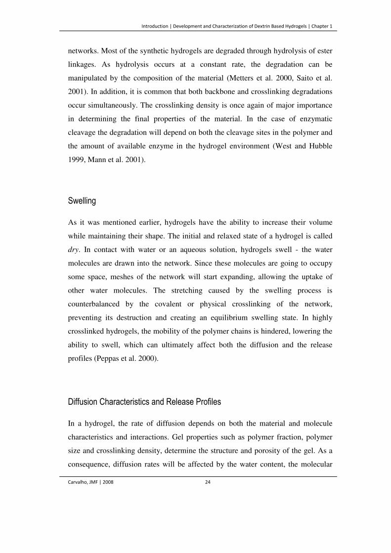
Introduction | Development and Characterization of Dextrin Based Hydrogels | Chapter 1
Carvalho, JMF | 2008 24
networks. Most of the synthetic hydrogels are degraded through hydrolysis of ester
linkages. As hydrolysis occurs at a constant rate, the degradation can be
manipulated by the composition of the material (Metters et al. 2000, Saito et al.
2001). In addition, it is common that both backbone and crosslinking degradations
occur simultaneously. The crosslinking density is once again of major importance
in determining the final properties of the material. In the case of enzymatic
cleavage the degradation will depend on both the cleavage sites in the polymer and
the amount of available enzyme in the hydrogel environment (West and Hubble
1999, Mann et al. 2001).
Swelling
As it was mentioned earlier, hydrogels have the ability to increase their volume
while maintaining their shape. The initial and relaxed state of a hydrogel is called
dry. In contact with water or an aqueous solution, hydrogels swell - the water
molecules are drawn into the network. Since these molecules are going to occupy
some space, meshes of the network will start expanding, allowing the uptake of
other water molecules. The stretching caused by the swelling process is
counterbalanced by the covalent or physical crosslinking of the network,
preventing its destruction and creating an equilibrium swelling state. In highly
crosslinked hydrogels, the mobility of the polymer chains is hindered, lowering the
ability to swell, which can ultimately affect both the diffusion and the release
profiles (Peppas et al. 2000).
Diffusion Characteristics and Release Profiles
In a hydrogel, the rate of diffusion depends on both the material and molecule
characteristics and interactions. Gel properties such as polymer fraction, polymer
size and crosslinking density, determine the structure and porosity of the gel. As a
consequence, diffusion rates will be affected by the water content, the molecular
Introduction | Development and Characterization of Dextrin Based Hydrogels | Chapter 1
Carvalho, JMF | 2008 25
weight and size of the diffusion species, the swelling and the degradability of the
material. The actual mechanism of drug release depends on whether diffusion or
erosion is the rate-controlling step. For erodible systems, polymer dissolution after
crosslink cleavage plays a critical role in determining overall release profiles.
Degradation of crosslinks increases the mesh size of the gel, allowing for diffusion
to be facilitated and therefore, the release is erosion-controlled. On contrary, the
erosion of the matrix can be much slower than the diffusion of the molecule
through the polymer. The release is then considered diffusion-controlled (Dijk-
Wolthuis et al. 1997, Amsden 1999). While either diffusion or erosion can be the
predominant factor for a specific type of polymer, in most cases, the release
kinetics is a result of a combination of these two mechanisms (Sujja-areevath et al.,
1998; Lee, 1981; Tahara et al., 1995; Efentakis and Buckton, 2002).
Biocompatibility
Apart from favorable physico-chemical and mechanical properties, the most
important requirement for a hydrogel to be used in medical applications is its
biocompatibility in a specific environment, together with the non-cytoxicity of its
degradation products. Most of the toxicity problems associated with hydrogels are
the unreacted monomers, oligomers and initiators, that leach out during application.
These leachables can exhibit varying levels of reactivity and consequently toxicity.
In addition, it is also necessary to consider the potential toxicity of the degradation
products. Therefore, the knowledge of the degradation processes and the effects
that the by-products might have is crucial for long-term success of the hydrogel
application (Kirkpatrick 1992, Del Guerra et al. 1995, Wallin 1995, Del Guerra et
al. 1996, Kohn and Langer 1996, Ratner 1997).
Introduction | Development and Characterization of Dextrin Based Hydrogels | Chapter 1
Carvalho, JMF | 2008 26
Improving Hydrogel Properties
Recent advances in the development of hydrogels include the incorporation of cell
recognition molecules on the surface or the bulk of the materials. Early studies
reported on the use of long chains of ECM proteins such as fibronectin (FN),
vitronectin and laminin for surface modification, with successful promotion of cell
adhesion and proliferation (Humphries et al. 1986). More recent trends leave
behind the long chain proteins, using instead, short peptide fragments as signaling
domains, which are more stable during the modification process and can be
massively synthesized. The most commonly used peptide for modification is Arg-
Gly-Asp (RGD) (Figure 4), the signaling domain derived from fibronectin and
laminin, Tyr-Ile- Gly-Ser-Arg (YIGSR), Arg-Glu-Asp-Val (REDV) and Ile-Lys-
Val-Ala-Val (IKVAV) (Hubbell et al. 1991, Ranieri et al. 1995).
Figure 4. Schematic representation of a functionalized hydrogel surface. The RGD ligands mediate
cell adhesion through the interaction with the integrins of the cells.
The surface coating with these bioactive molecules, serving the purpose of
mimicking ECM, allows for the modulation of cellular functions such as cell
attachment, proliferation, and differentiation. Additionally, the cell-signaling
peptides are incorporated into the hydrogel backbone and not limited to the surface.
Another feature includes the incorporation of enzymatically degradable sequences,
allowing the degradation to be modulated by the presence of enzymes that
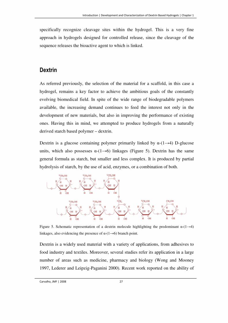
Introduction | Development and Characterization of Dextrin Based Hydrogels | Chapter 1
Carvalho, JMF | 2008 27
specifically recognize cleavage sites within the hydrogel. This is a very fine
approach in hydrogels designed for controlled release, since the cleavage of the
sequence releases the bioactive agent to which is linked.
Dextrin
As referred previously, the selection of the material for a scaffold, in this case a
hydrogel, remains a key factor to achieve the ambitious goals of the constantly
evolving biomedical field. In spite of the wide range of biodegradable polymers
available, the increasing demand continues to feed the interest not only in the
development of new materials, but also in improving the performance of existing
ones. Having this in mind, we attempted to produce hydrogels from a naturally
derived starch based polymer – dextrin.
Dextrin is a glucose containing polymer primarily linked by α-(1→4) D-glucose
units, which also possesses α-(1→6) linkages (Figure 5). Dextrin has the same
general formula as starch, but smaller and less complex. It is produced by partial
hydrolysis of starch, by the use of acid, enzymes, or a combination of both.
Figure 5. Schematic representation of a dextrin molecule highlighting the predominant α-(1→4)
linkages, also evidencing the presence of α-(1→6) branch point.
Dextrin is a widely used material with a variety of applications, from adhesives to
food industry and textiles. Moreover, several studies refer its application in a large
number of areas such as medicine, pharmacy and biology (Wong and Mooney
1997, Lederer and Leipzig-Paganini 2000). Recent work reported on the ability of
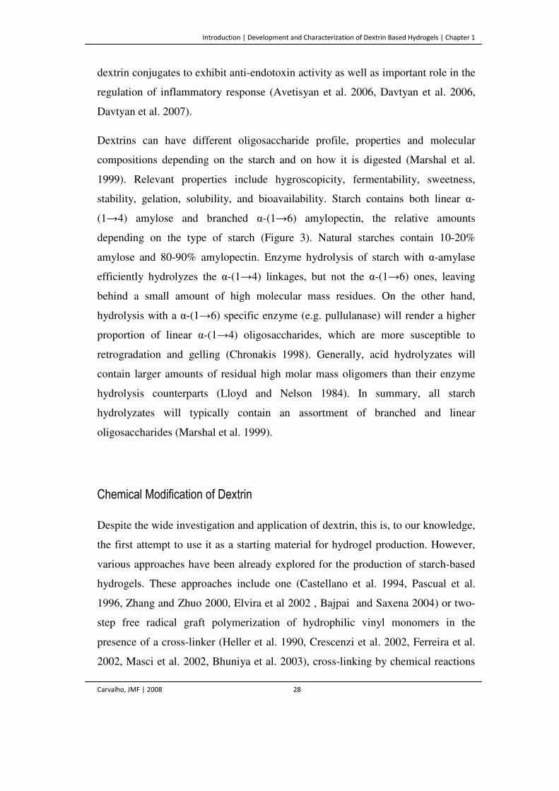
Introduction | Development and Characterization of Dextrin Based Hydrogels | Chapter 1
Carvalho, JMF | 2008 28
dextrin conjugates to exhibit anti-endotoxin activity as well as important role in the
regulation of inflammatory response (Avetisyan et al. 2006, Davtyan et al. 2006,
Davtyan et al. 2007).
Dextrins can have different oligosaccharide profile, properties and molecular
compositions depending on the starch and on how it is digested (Marshal et al.
1999). Relevant properties include hygroscopicity, fermentability, sweetness,
stability, gelation, solubility, and bioavailability. Starch contains both linear α-
(1→4) amylose and branched α-(1→6) amylopectin, the relative amounts
depending on the type of starch (Figure 3). Natural starches contain 10-20%
amylose and 80-90% amylopectin. Enzyme hydrolysis of starch with α-amylase
efficiently hydrolyzes the α-(1→4) linkages, but not the α-(1→6) ones, leaving
behind a small amount of high molecular mass residues. On the other hand,
hydrolysis with a α-(1→6) specific enzyme (e.g. pullulanase) will render a higher
proportion of linear α-(1→4) oligosaccharides, which are more susceptible to
retrogradation and gelling (Chronakis 1998). Generally, acid hydrolyzates will
contain larger amounts of residual high molar mass oligomers than their enzyme
hydrolysis counterparts (Lloyd and Nelson 1984). In summary, all starch
hydrolyzates will typically contain an assortment of branched and linear
oligosaccharides (Marshal et al. 1999).
Chemical Modification of Dextrin
Despite the wide investigation and application of dextrin, this is, to our knowledge,
the first attempt to use it as a starting material for hydrogel production. However,
various approaches have been already explored for the production of starch-based
hydrogels. These approaches include one (Castellano et al. 1994, Pascual et al.
1996, Zhang and Zhuo 2000, Elvira et al 2002 , Bajpai and Saxena 2004) or two-
step free radical graft polymerization of hydrophilic vinyl monomers in the
presence of a cross-linker (Heller et al. 1990, Crescenzi et al. 2002, Ferreira et al.
2002, Masci et al. 2002, Bhuniya et al. 2003), cross-linking by chemical reactions
Introduction | Development and Characterization of Dextrin Based Hydrogels | Chapter 1
Carvalho, JMF | 2008 29
with complementary groups (Lenaerts et al. 1991, Shiftan et al. 2000, Seidel et al.
2001, Seidel et al. 2004), radiation-induced polymerization and cross-linking
(Rosiak and Ulanski 1999, Ceresh et al. 2002, Zhai et al. 2002, Yoshii et al. 2003)
and physical self-assembly processes (Akiyoshi et al. 1993, Akiyoshi et al. 1996,
Akiyoshi et al. 1997, Janes et al. 2002, Simon et al. 2003, Duval-Terrié et al. 2003).
Dextrin is not capable of gelling. As it was described, to produce chemical
hydrogels, the starting raw materials should contain a functional group (Radley
1976, Wurzburg 1986, Riedel and Nickel 1999).
Chemical derivatization of polysaccharides has profound effects on macroscopic
behavior (e.g. solubility, stability and viscosity characteristics). Among the many
possibilities of modification, the creation of reactive double bonds through a
transesterification reaction is the principal process by which it is possible to
functionalize sugars, producing sugar esters (Figure 6a). This process has been
studied extensively with regard to the chemical phenomena involved (Park et al.
2003, Vargha and Truter 2005, Jeong et al. 2006). The final product of this reaction
has side chains (vinylic groups) attached to the polysaccharide backbone and
polymerizes through well-established free radical methods (Mark and Mehltretter
1972, De Graaf et al. 1998).
The goal of this work was to use the ester, vinyl acrylate (VA), as the acyl donor to
the esterification of the hydroxyl groups of dextrin (acyl acceptor) (Figure 6b). The
double bonds allow further chemical reactions such as secondary crosslinking of
the final product. The degree of substitution (DS) refers to the average number of
substituted polysaccharide hydroxyl group and is of major importance in
determining overall properties of the resulting hydrogel, such as the degradation
behavior or the release profiles.
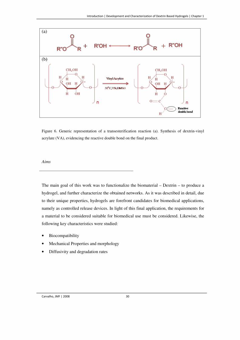
Introduction | Development and Characterization of Dextrin Based Hydrogels | Chapter 1
Carvalho, JMF | 2008 30
(a)
(b)
Figure 6. Generic representation of a transesterification reaction (a). Synthesis of dextrin-vinyl
acrylate (VA), evidencing the reactive double bond on the final product.
Aims
The main goal of this work was to functionalize the biomaterial – Dextrin – to produce a
hydrogel, and further characterize the obtained networks. As it was described in detail, due
to their unique properties, hydrogels are forefront candidates for biomedical applications,
namely as controlled release devices. In light of this final application, the requirements for
a material to be considered suitable for biomedical use must be considered. Likewise, the
following key characteristics were studied:
• Biocompatibility
• Mechanical Properties and morphology
• Diffusivity and degradation rates
References | Chapter 1
Carvalho, JMF | 2008 31
References
Agrawal CM, Athanasiou KA and Heckman JD. (1997) Biodegradable PLA-PGA polymer for
tissue engineering orthopaedics. Mater Sci Forum 250: 115–128.
Akiyoshi K, Deguchi S, Moriguchi N, Yamaguchi S and Sunamoto J. (1993) Self-aggregates of
hydrophobized polysaccharides in water – formation and characteristics of pH-responsive behaviors
of graft copolymer and blend nanoparticles. Macromolecules 26: 3062–3068.
Akiyoshi K, Taniguchi I, Fukui H and Sunamoto J. (1996) Hydrogel nanoparticle formed by self-
assembly of hydrophobized polysaccharide. Stabilization of adriamycin by complexation. Eur J
Pharm Biopharm 42: 286–290.
Akiyoshi K, Deguchi S, Tajima H, Nishikawa T and Sunamoto J. (1997) Microscopic structure and
thermoresponsiveness of a hydrogel nanoparticle by self-assembly of a hydrophobized
polysaccharide. Macromolecules 30: 857–861.
Allarakhia L, Knoll RL and Lindstrom RL. (1987) Soft intraocular lenses. J Cataract Refract Surg
13: 607–620.
Andrade JD. (1976) Hydrogels for medical and related applications. Vol 31. .American Chemical
Society, Washington, DC, USA.
Anseth KS, Bowman CN and Brannon-Peppas L. (1996) Mechanical properties of hydrogels and
their experimental determination. Biomaterials 17: 1647–1657.
Athanasiou KA, Agrawal CM, Barber FA and Burkhart SS. (1998) Orthopedic applications for
PLA-PGA biodegradable polymers. Arthroscopy 14: 726–737.
Autissier A, Letourneur D, Le Visage C. (2007) Pullulan-based hydrogel for smooth muscle cell
culture. J Biomed Mat Res Part A 82: 336–342.
Avetisyan SA, Hakobyan GS and Davtyan TK. (2006) Modulation of endotoxin-induced respiratory
splash of granulocytes and monocytes in patients with familial Mediterranean fever by iodine-
lithium-α-dextrin and sodium thiosulfate. Patol Fiziol Eksp Ter 1: 11–13.
Bae Y, Fukushima S, Harada A and Kataoka K. (2003) Design of environment-sensitive
supramolecular assemblies for intracellular drug delivery: polymeric micelles that are responsive to
intracellular pH change. Angew Chem Int Ed 42: 4640–4643.
References | Chapter 1
Carvalho, JMF | 2008 32
Baeyer HV, Lajous-Petter A, Debrandt W, Hampl H, Kochinke F and Herbst R. (1988) Surface
reactions on blood contact during hemodialysis and haemofiltration with various membrane types. J
Membr Sci 36: 215–228.
Bajpai SK and Saxena S. (2004) Enzymatically degradable and pH-sensitive hydrogels for colon-
targeted oral drug delivery. I. Synthesis and characterization. J Appl Polym Sci 92: 3630–3643.
Baroli B. (2006) Photopolymerization in drug delivery, tissue engineering and cell encapsulation:
Issues and potentialities. J Chem Tech Biotech 81: 491–499.
Bhattacharya M, Vaidya U, Zhang D and Narayan R. (1995) Properties of bends of starch and
synthetic polymers containing anhydride groups. II. Effect of amylopectin to amylose ratio in
starch. J App Pol Sci 57: 539–554.
Bhuniya SP, Rahman S, Satyanand AJ, Gharia MM and Dave AM. (2003) Novel route to synthesis
of allyl starch and biodegradable hydrogel by copolymerizing allyl-modified starch with
methacrylic acid and acrylamide. J Polym Sci Part A: Polym Chem 41: 1650–1658.
Bell E, Ehrlich P, Buttle DJ and Nakatsuji T. (1981) Living tissue formed in vitro and accepted as
skin-equivalent of full thickness. Science 221: 1052–1054.
Bonassar LJ and Vacanti CA (1998) Tissue engineering: the first decade and beyond. J Cell
Biochem 30: 297–303.
Bosman FT and Stamenkovic I. (2003) Functional structure and composition of extracellular matrix. J Pathol 200: 423–428.
Brahim S, Narinesingh D and Guiseppi-Elie A. (2002) Bio-smart hydrogels: Co-joined molecular recognition and signal transduction in biosensor fabrication and drug delivery. Biosens Bioelectron 17: 973–981.
Brannon-Peppas L. (1990) Preparation and characterization of crosslinked hydrophilic networks. In
Absorbent polymer technology. Brannon-Peppas L and Harland RS (eds). Elsevier, Amsterdam,
Netherlands, pp 45–66.
Brun P, Cortivo R, Zavan B, Vecchiato N and Abatangelo G. (1999) In vitro reconstructed tissues
on hyaluronan-based temporary scaffolding. J Mater Sci Mater Med 10: 683–688.
Burke JF, Yannas IV, Quimby WC, Bondoc CC and Jung WK (1981). Successful use of a
physiologically acceptable artificial skin in the treatment of extensive burn injury. Ann Surg 194:
413–448.
References | Chapter 1
Carvalho, JMF | 2008 33
Cascone MG, Tricoli M, Cerrai P and Sbarbati Del Guerra R. (1993) Cell cultures in the
biocompatibility study of synthetic materials. Cytotechnology 11: 137–139.
Castellano I, Pascual B, Vazquez B, Goñi I and Gurruchaga M. (1994) Graft copolymerization of
different mixtures of acrylic monomers on amylopectin: swelling behavior. J Appl Polym Sci 54:
577–584.
Ceresh S, Gilboa Y, Peisahov-Korol J, Gdalevsky G, Voorspoels J, Remon JP and Kost J. (2002)
Preparation and characterization of bioadhesive grafted starch copolymers as platforms for
controlled drug delivery. J Appl Polym Sci 86: 1157–1162.
Chapekar MS. (2000) Tissue engineering: challenges and opportunities. J Biomed Mater Res 53:
617–620.
Charman WN, Chan H-K, Finnin BC and Charman SA. (1999) Drug delivery: a key factor in
realising the full therapeutic potential of drugs. Drug Dev Res 46: 316–327.
Chasin M and Langer R (1990) Biodegradable polymers as drug delivery systems. Marcel Dekker,
New York, USA.
Chehade M and Elder MJ. (1997) Intraocular lens materials and styles: a review. Aust N Z J
Ophthalmol 25: 255–263.
Chen S and Singh J. (2005) Controlled delivery of testosterone from smart polymer solution based
systems: in vitro evaluation. Int J Pharm 295: 183–190.
Chen J, Jo S and Park K. (1997). Degradable hydrogels. In Handbook of biodegradable polymers.
Domb AJ, Kost J and Wiseman DM (eds). Overseas Publishers Association, Amsterdam,
Netherlands, pp 203–230.
Choi SH and Park TG. (2002) Synthesis and characterization of elastic PLGA/PCL/PLGA triblock
copolymers. J Biomater Sci Polym Ed 13: 1163–1173.
Chronakis IS. (1998) On the molecular characteristics, compositional properties, and structural-
functional mechanisms of maltodextrins: a review. Crit Rev Food Sci 38: 599–637.
Coughlan DC, Quilty FP and Corrigan OI. (2006) Effect of drug physicochemical properties on
swelling/deswelling kinetics and pulsatile drug release from thermoresponsive poly(N-
isopropylacrylamide) hydrogels. J Control Release 98: 97–114.
Crescenzi V, Dentini M, Bontempo D and Masci G. (2002) Hydrogels based on pullulan derivatives
crosslinked via a ‘‘living’’ free-radical process. Macromol Chem Phys 203: 1285–1291.
References | Chapter 1
Carvalho, JMF | 2008 34
Davis SS and Illum L. (1998) Drug delivery systems for challenging molecules. Int J Pharm 176: 1–
8.
Davtyan TK, Mkrtchyan NR and Manukyan HA. (2006) Dexamethasone, colchicine and iodine-
lithium-α-dextrin act differentially on the oxidative burst and endotoxin tolerance induction in vitro
in patients with Behcet’s disease. Int Immunopharm 6: 396–407.
Davtyan TK, Hakobyan IS, Muradyan RE, Hovhannisyan HG and Gabrielyan ES. (2007)
Evaluation of amino acids as mediators for the antibacterial activity of iodine-lithium-α -dextrin in
vitro and in vivo. J Antimicrob Chemother 59: 1114–1122.
De Gest BG, Stubbe BG, Jonas AM, Van Thienen T, Hinrichs WLJ, Demeester J and De Smedt
SC. (2006) Self-exploding lipid-coated microgels. Biomacromolecules 7: 373–379.
De Graaf RA, Broekroelofs A and Janssen LPBM. (1998) The acetylation of starch by reactive
extrusion. Starch/Stärke 50: 198–205.
Del Guerra R, Gazzetti P, Lazzerini G, Cerrai P, Guerra G, Tricoli M and Cristallini C. (1995)
Degradation products of poly(ester-esther-ester) block copolymers do not alter endothelial
metabolism in vitro. J Mater Sci Mater Med 6: 824–828.
Del Guerra R, Cascone M, Ricci D, Martinoia S, Parodi M, AhluwaliaA, Van Mourk J and
Grattarola M. (1996) Optimization of the interaction between ethylene-vinyl alcohol copolymers
and human endothelial cells. J Mater Sci Mater Med 7: 8–12.
Doillon CJ and Cameron K. (1990) New approaches for biocompatibility testing using cell culture.
Int J Artif Organs 13: 517–520.
Drury JL and Mooney DJ. (2003) Hydrogels for tissue engineering: scaffold design variables and
applications. Biomaterials 24: 4337–4351.
Duval-Terrié C, Huguet J and Muller G. (2003) Self-assembly and hydrophobic clusters of
amphiphilic polysaccharides, colloids and surfaces. A Physicochem Eng Aspects 220: 105–115.
Eiselt P, Lee YK and Mooney JD (1999) Rigidity of two-component hydrogels prepared from
alginate and poly(ethylene glycol) – diamines. Macromolecules 32: 5561–5566.
Elvira C, Mano JF, Roman JS and Reis RL. (2002) Starch-based biodegradable hydrogels with
potential biomedical applications as drug delivery systems. Biomaterials 23: 1955–1966.
Fanta GF and Doane WM. (1986) In Modified starches: properties and uses. Wurzburg OB (ed).
CRC Press, Boca Raton, FL, USA, pp 149–178.
References | Chapter 1
Carvalho, JMF | 2008 35
Ferramosca E, Burke S, Chasan-Taber S, Ratti C, Chertow GM and Raggi P. (2005) Potential
antiatherogenic and anti-inflammatory properties of sevelamer in maintenance hemodialysis
patients. Am Heart J 149: 820–825.
Ferreira L, Carvalho R, Gil MH and Dordick JS. (2002) Enzymatic synthesis of dextran-containing
hydrogels. Biomaterials 23: 3957–3967.
Ferreira L, Rafael A, Lamghari M, Barbosa MA, Gil MH, Cabrita MAS and Dordick JS. (2004)
Biocompatibility of chemoenzymatically derived dextran-acrylate hydrogels. J Biomed Mater Res
68: 584–96.
Ferreira L, Gil MH, Cabrita MAS and Dordick JS. (2005) Biocatalytic synthesis of highly ordered
degradable dextran-based hydrogels. Biomaterials 26: 4707–4716.
Flory PJ and Rehner J. (1943) Statistical mechanics of cross-linked polymer networks. II. Swelling.
J Chem Phys 11: 521–526.
Flory PJ. (1950) Statistical mechanics of swelling networks structures. J Chem Phys 18: 108–111.
Flory PJ. (1953) Principles of polymer chemistry. Cornell University Press, Ithaca, NY, USA.
Franssen O, Vos OP and Hennink WE. (1997) Delayed release of a model protein from
enzymatically-degrading dextran hydrogels. J Control Release 44: 237–245.
Freed LE, Vunjak-Novakovic G, Biron RJ, Eagles DB, Lesnoy DC, Barlow SK and Langer R.
(1994) Biodegradable polymer scaffolds for tissue engineering. Bio/Technology 12: 689–693.
Freed LE and Vunjak-Novakovic G. (1998) Culture of organized cell communities. Adv Drug Deliv
Rev 33: 15–30.
Garg JP, Chasan-Taber S, Blair A, Plone M, Bommer J, Raggi P and Chertow GM. (2005) Effects
of sevelamer and calcium-based phosphate binders on uric acid concentrations in patients
undergoing hemodialysis: A randomized clinical trial. Arthritis Rheum 52: 290–295.
Goda T and Ishihara K. (2006) Soft contact lens biomaterials from bioinspired phospholipid
polymers. Expert Rev Med Devices 3: 167–174.
Gulsen D and Chauhan A. (2005) Dispersion of microemulsion drops in HEMA hydrogel: A
potential ophthalmic drug delivery vehicle. Int J Pharm 292: 95–117.
Haag R. (2004) Supramolecular drug–delivery systems based on polymeric core-shell architectures.
Angew Chem Int Ed 43: 278–282.
References | Chapter 1
Carvalho, JMF | 2008 36
Hanein D, Geiber B and Addadi L. (1995) Cell adhesion to crystal surfaces: a model for initial
stages in the attachment of cells to solid substrates. Cells and Mater 5: 197–210.
Hatakeyama H, Kikuchi A, Yamato M and Okano T. (2006) Bio-functionalized thermoresponsive
interfaces facilitating cell adhesion and proliferation. Biomaterials 27: 5069–5078.
Hayashi T. (1994) Biodegradable polymers for biomedical uses. Prog Polym Sci 19: 663–672.
Heller J. (1985) Self–regulated drug delivery systems. Med Devices Diag Ind 7: 32–41.
Heller J. (1988) Chemically self–regulated drug delivery systems. J Control Release 8: 111–124.
Heller J, Pangbum SH and Roskos KV. (1990) Development of enzymatically degradable protective
coatings for use in triggered drug delivery systems: derivatized starch hydrogels. Biomaterials 11:
345–350.
Hennink WE, Franssen O, Van Dijk-Wolthuis WNE and Talsma H. (1997) Dextran hydrogels for
the controlled release of proteins. J Control Release 48:107–114.
Hennink WE and van Nostrum CF. (2002) Novel crosslinking methods to design hydrogels. Adv
Drug Deliv Rev 54: 13–36.
Hench LL. (1996) Ceramics, glasses, and glass-ceramics. In Science: An Introduction to Materials
in Medicine. Biomaterials. Ratner BD, Hoffman AS, Schoen FJ et al. (eds). Academic Press, NY,
USA, pp 73–83.
Hicks C, Crawford G, Chirila T, Wiffen S, Vijayasekaran S, Lou X, Fitton J, Maley M, Clayton A,
Dalton P, Platten S, Ziegelaar B, Hong Y, Russo A and Constable I. (2000) Development and
clinical assessment of an artificial cornea. Prog Retin Eye Res 19: 149–170.
Hoecker H. (1998) Polymeric materials as biomaterials under particular consideration of
biodegradable polymers. Macromol Symp 130: 161–168.
Hoffman AS. (2002) Hydrogels for biomedical applications. Adv Drug Deliv Rev 43: 3–12.
Honiger J, Couturier C, Goldschmidt P, Maillet F, Kazatchkine MD and Laroche L. (1997) New
anionic polyelectrolyte hydrogel for corneal surgery. J Biomed Mater Res 37: 548–553.
Hou KC, Zaniewski R and Roy S. (1991) Protein A immobilized affinity. Cartridge for
immunoglobulin purification. Biotechnol Appl Biochem 13: 257–268.
References | Chapter 1
Carvalho, JMF | 2008 37
Htay AS, Teoh SH and Hutmacher DW. (2004) Development of perforated microthin poly(epsilon-
caprolactone) films as matrices for membrane tissue engineering. J Biomater Sci Polym Ed 15:
683–700.
Hubbell JA, Massia SP, Desai NP and Drumheller PD. (1991) Endothelial cell-selective materials
for tissue engineering in the vascular graft via a new receptor. Biotechnology 9: 568–572.
Humphries MJ, Akiyama SK, Komoriya A, Olden K and Yamada KM. (1986) Identification of an
alternatively spliced site in human plasma fibronectin that mediates cell type specific adhesion. J
Cell Biol 103: 2637–2647.
Hutmacher DW. (2000) Scaffolds in tissue engineering bone and cartilage. Biomaterials 21: 2529–
2543.
Hutmacher DW. (2001) Scaffold design and fabrication technologies for engineering tissues – state
of the art and future perspectives. J Biomater Sci Polymer Ed 12: 107–124.
Hutmacher DW, Goh JC and Teoh SH. (2001) An introduction to biodegradable materials for tissue
engineering applications. Ann Acad Med Singapore 30: 183–191.
Jain MR. (1988) Drug delivery through soft contact lenses. Br J Ophthalmol 72: 150–154.
James K and Kohn J. (1996) New biomaterials for tissue engineering. MRS Bull 22–26.
Janes KA, Calvo P and Alonso MJ. (2002) Polysaccharide colloidal particles as delivery systems for
macromolecules. Adv Drug Deliv Rev 54: 13–36.
Jeong BM, Choi YK, Bae YH, Zentner G and Kim SW. (1999) New biodegradable polymers for
injectable drug delivery systems. J Control Release 62: 109–114.
Jeong BM, Lee DS, Bae YH and Kim SW. (2000) In situ gelation of PEG-PLGA-PEG triblock
copolymer aqueous solutions and degradation thereof. J Biomed Mater Res 50: 171–177.
Jeong BM, Kim SW and Bae YH. (2002) Thermosensitive sol-gel reversible hydrogels. Adv Drug
Deliv Rev 54: 37–51.
Jeong G-T , Byun K-Y, Lee W-T and Ryu H-W. (2006) Enzymatic synthesis of sorbitan
methacrylate: Comparison of methacrylic acid and vinyl methacrylate. Biochem Eng J 29: 69–74.
Kamath KR and Park K. (1995) Study on the release of invertase from enzymatically degradable
dextran hydrogels. Polym Gels Netw 3: 243–254.
References | Chapter 1
Carvalho, JMF | 2008 38
Kanjickal DG and Lopina ST. (2004) Modeling of drug release from polymeric delivery systems.
Crit Rev Ther Drug Carrier Syst 21: 345–386.
Kim BS and Mooney DJ. (1998) Development of biocompatible synthetic extracellular matrices for
tissue engineering. TIB TECH 16: 224–230.
Kirkpatrick CJ. (1992) A critical view of current and proposed methodologies for biocompatibility
testing: cytotoxicity in vitro. Regulatory Affairs 4: 13–32.
Klee D and Hoecker H. (1999) Polymers for biomedical applications: improvement of the interface
compatibility. Adv Polym Sci 149: 1–57.
Klomp GF, Hashiguchi H, Ursell PC, Takeda Y, Taguchi T and Dobelle WH. (1983) Macroporous
hydrogel membranes for a hybrid artificial pancreas. II. Biocompatibility. J Biomed Mater Res 17:
865–871.
Kohn J and Langer R. (1996) In Biomterials Science. Ratner B, Hoffman A, Schoen F and Lemons
J (eds). Academic Press, New York, USA pp 54–72.
Kolthammer J. (1975) The in vitro adsorption of drugs from horse serum onto carbon coated with
an acrylic hydrogel. J Pharm Pharmacol 27: 801–805.
Kopecek J. (2003) Smart and genetically engineered biomaterials and drug delivery systems. Euro J
Pharma Sci 20: 1–16.
Kost J, Wolfrum J and Langer R. (1987) Magnetically enhanced insulin release in diabetic rats. J
Biomed Mater Res 21: 1367–1373.
Kost J and Langer R. (1992) Responsive polymer systems for controlled delivery of therapeutics.
Trends Biotech 10: 127–131.
Kost J and Langer R. (2001) Responsive polymeric delivery systems. Adv Drug Deliv Rev 46: 125–
148.
Küdela V. (1989) Hydrogels. In Polymers: Biomaterials and medical applications. Kroschwitz JI
(ed). John Wiley and Sons, Inc, New York, USA, pp 228–252.
Kweon H, Yoo MK, Park IK, Kim TH, Lee HC, Lee HS, Oh JS, Akaike T and Cho CS. (2003) A
novel degradable polycaprolactone networks for tissue engineering. Biomaterials 24: 801–808.
Kwon JW, Han YK, Lee WJ, Cho CS, Paik SJ, Cho DI, Lee JH and Wee WR. (2005)
Biocompatibility of poloxamer hydrogel as an injectable intraocular lens: A pilot study. J Cataract
Refract Surg 31: 607–613.
References | Chapter 1
Carvalho, JMF | 2008 39
Langer R. (1998) Drug delivery and targeting. Nature 392: 5–22.
Langer R, Cima LG, Tamada JA and Wintermantel E. (1990) Future directions in biomaterials.
Biomaterials 11: 738–745.
Langer R and Vacanti JP. (1993) Tissue engineering. Science 260: 920–926.
Langer R. (1995) Biomaterials and biomedical engineering. Chem Eng Sci 50: 4109–4121.
Langer R. (2000) Tissue engineering. Mol Ther 1: 12–15.
Leathers TD. (2002) Pullulan. In Biopolymers. Vol 6. De Baets S, Vandamme EJ and Steinbüchel A
(eds). Wiley-VCH, Weinheim, pp 1–35.
Leathers TD. (2003) Biotechnological production and applications of pullulan. Appl Microbiol
Biotechnol 62: 468–473.
Lederer M and Leipzig-Paganini E. (2000) Dextrin as excipient in pharmaceutical preparations.
Pharmazie 55: 378–379.
Lee CH, Singla A and Lee Y. (2001) Biomedical applications of collagen. Int J Pharm 221: 1–22.
Lee J, Bae YH, Sohn YS and Jeong BM. (2006) Thermogelling aqueous solutions of alternating
multiblock copolymers of poly(L-lactic acid) and poly(ethylene) glycol. Biomacromolecules 7:
1729–1734.
Lee KY and Mooney DJ. (2001) Hydrogels for tissue engineering. Chem Rev 101: 1869–1877.
Lenaerts V, Dumoulin Y and Mateescu MA. (1991) Controlled-release of theophylline from cross-
linked amylose tablets. J Control Release 15: 39–46.
Li L, Shan H, Yue CY, Lam YC, Tam KC and Hu X. (2002) Thermally induced association and
dissociation of methylcellulose in aqueous solutions. Langmuir 18: 7291–7298.
Li WJ, Tuli R, Okafor C, Derfoul A, Danielson KG, Hall DJ and Tuan RS. (2005) A
threedimensional nanofibrous scaffold for cartilage tissue engineering using human mesenchymal
stem cells. Biomaterials 26: 599–609.
Liu YY, Shao YH and Lü J. (2006) Preparation, properties and controlled release behaviors of pH-
induced thermosensitive amphiphilic gels. Biomaterials 27: 4016–4024.
Lloyd NE and Nelson WI. (1974) In Whistler RL, Bemiller JN and Paschall EF (eds). Starch
chemistry and technology, Academic Press, Orlando, Florida, USA, pp 614.
References | Chapter 1
Carvalho, JMF | 2008 40
Lu L and Mikos A. (1996) The importance of new processing techniques in tissue engineering.
MRS Bulletin 21: 28–32.
Lucke A, Tessmar J, Schnell E, Schmeer G and Gopferich A. (2000) Biodegradable poly(D,L-lactic
acid)-poly(ethylene glycol)-monomethyl ether diblock copolymers: structures and surface properties
relevant to their use as biomaterials. Biomaterials 21: 2361–2370.
Lysaght MJ, Nguy NA and Sullivan K. (1998) An economic survey of the emerging tissue
engineering industry. Tissue Eng 4: 231–238.
Lysaght MJ and Hazlehurst AL. (2000) Tissue engineering: the end of the beginning. Tissue Eng
10: 309–320.
Madihally S and Mathew H. (1999) Porous chitosan scaffolds for tissue engineering. Biomaterials
20: 1133–1142.
Maire M, Chaubet F, Mary P, Blanchat C, Meunier A and Logeart-Avramoglou D. (2005) Bovine
BMP osteoinductive potential enhance by functionalized dextran-derived hydrogels. Biomaterials
26: 5085–5092.
Manabe T, Okino H, Maeyama R, Mizumoto K, Nagai E, Tanaka M and Matsuda T. (2004) Novel
strategic therapeutic approaches for prevention of local recurrence of pancreatic cancer after
resection: trans–tissue, sustained local drug delivery systems. J Control Release 100: 317–330.
Mann BK, Gobin AS, Tsai AT, Schmedlen RH and West JL. (2001) Smooth muscle growth in
photopolymerized hydrogels with cell adhesive and proteolytically degradable domains: synthetic
ECM analogs for tissue engineering. Biomaterials 22: 3045–3051.
Maquet V and Jerome R. (1997) Design of macroporous biodegradable polymer scaffolds for cell
transplantation. Mater Sci Forum 250: 15–42.
Marchal LM, Beeftink HH and Tramper J. (1999) Towards a rational design of commercial
maltodextrins. Trends Food Sci Technol 10: 345–355.
Mark AM and Mehltretter CL. (1972) Facile preparation of starch triacetates. Die Stärke 24: 73–
100.
Marques AP, Reis RL and Hunt JA. (2002) The biocompatibility of novel starch-based polymers
and composites: in vitro studies. Biomaterials 23: 1471–1478.
Masci G, Bontempo D and Crescenzi V. (2002) Synthesis and characterization of thermoresponsive
N-isopropylacrylamide/methacrylated pullulan hydrogels. Polymer 43: 5587–5593.
References | Chapter 1
Carvalho, JMF | 2008 41
Mazzoli RA, Raymond WR 4th, Ainbinder DJ, Hansen EA. (2004) Use of self-expanding,
hydrophilic osmotic expanders (hydrogel) in the reconstruction of congenital clinical anophthalmos.
Curr Opin Ophthalmol 15: 426–431.
McCulloch I and Shalaby SW (1998). In Tailored polymeric materials for controlled delivery
systems. American Chemical Society, Washington DC, USA.
McPherson JM, Sawamura S and Amstrong R. (1986). An examination of the biologic response to
injectable, glutaraldehyde cross-linked collagen implants. J Biomed Mater Res 20: 93–107.
Meredith JE, Fateli B and Schwartz MA. (1993) The extracellular matrix as a cell survival factor.
Mol Cell Biol 9: 953–961.
Metters AT, Anseth KS and Bowman CN. (2000) Fundamental studies of a novel, biodegradable
PEG-b-PLA hydrogel. Polymer 41: 3993–4004.
Middleton JC and Tipton AJ. (2000) Synthetic biodegradable polymers as orthopedic devices.
Biomaterials 21: 2335–2346.
Moise O, Sideman S, Hoffer E, Rousseau I and Better OS. (1977) Membrane permeability for
inorganic phosphate ion. J Biomed Mater Res 11: 903–913.
Morrison DA, Gridneva Z, Chirila TV and Hicks CR. (2006) Screening for drug-induced spoliation
of the hydrogel optic of the AlphaCor artificial cornea. Cont Lens Anterior Eye 29: 93–100.
Muller-Goymann CC. (2004) Physicochemical characterization of colloidal drug delivery systems
such as reverse micelles, vesicles, liquid crystals and nanoparticles for topical administration. Euro J
Pharma Biopharma 58: 343–56.
Na K, Park JH, Kim SW, Sun BK, Woo DG, Chung HM and Park KH. (2006) Delivery of
dexamethasone, ascorbate, and growth factor (TGF b-3) in thermo-reversible hydrogel constructs
embedded with rabbit chondrocytes. Biomaterials 27: 5951–5957.
Nakabayashi N and Iwasaki Y. (2004) Copolymers of 2-methacryloyloxyethyl phosphorylcholine
(MPC) as biomaterials. Biomed Mater Eng 14: 345–354.
Nakayama M, Okano T, Miyazaki T, Kohori F, Sakai and Yokoyama M. (2006) Molecular design
of biodegradable polymeric micelles for temperature-responsive drug release. J Control Release
115: 46–56.
References | Chapter 1
Carvalho, JMF | 2008 42
Ng KW, Hutmacher DW, Schantz JT, Ng CS, Too HP, Lim TC, Phan TT and Teoh SH. (2001)
Evaluation of ultra-thin poly(epsilon-caprolactone) films for tissue-engineered skin. Tissue Eng 7:
441–455.
Nguyen KT and West JL. (2002) Photopolymerizable hydrogels for tissue engineering applications.
Biomaterials 23: 4307–4314.
Nicolson PC and Vogt J. (2001) Soft contact lens polymers: An evolution. Biomaterials 22: 3273–
3283.
Niculescu–Duvaz I and Springer CJ. (1997) Andibody-directed enzyme prodrug therapy (ADEPT):
a review. Adv Drug Deliv Rev 26: 151–72.
Pachence JM. (1996) Collagen-based devices for soft tissue repair. J Biomed Mater Res 33: 35–40.
Pachence JM and Kohn J. (1996) Biodegradable polymers for tissue engineering. In Lanza R,
Langer R, Chick W (ed). Principles of tissue Engineering. Academic Press, New York, USA, pp
273–293.
Packhaeuser CB, Schnieders J, Oster CG and Kissel T. (2004) In situ forming parenteral drug
delivery systems: an overview. Euro J Pharma Biopharma 58: 445–455.
Park HG, Do JH and Chang HN. (2003) Regioselective enzymatic acylation of multi-hydroxyl
compounds in organic synthesis. Biotechnol Bioprocess Eng 8: 1–8.
Pascual B, Castellano I, Vazquez B, Gurruchaga M and Goñi I. (1996) Hydrogels based on graft
copolymerization of 2-hydroxypropyl methacrylate/acrylate mixtures on amylose: swelling
behavior. Polymer 37: 1005–1011.
Paul W and Sharma CP. (2003) Ceramic drug delivery: a perspective. J Biomater Appl 17: 253–
264.
Pearce RS, West LR, Rodeheaver GT and Edlich RF. (1984) Evaluation of a new hydrogel coating
for drainage tubes. Am J Surg 148: 687–691.
Peppas LB. (1997) Polymers in controlled drug delivery. Med Plast Biomaterials Mag.
Peppas NA. (1986) Hydrogels in medicine and pharmacy. Vols 1,2. CRC, Boca Raton, FL, USA, pp
171–180.
Peppas NA and Mikos AG (1986) Preparation methods and structure of hydrogels. In Hydrogels in
medicine and pharmacy. Vol 1. Peppas NA (ed). CRC Press, Boca Raton, Florida, USA, pp 1–27.
References | Chapter 1
Carvalho, JMF | 2008 43
Peppas NA, Bures P, Leobandung W, Ichikawa H. (2000) Hydrogels in pharmaceutical
formulations. Eur J Pharm Biopharm 50: 27–46.
Peppas NA, Huang Y, Torres-Lugo M, Ward JH and Zhang J. (2000) Physicochemical foundations
and structural design of hydrogels in medicine and biology. Annu Rev Biomed Eng 2: 9–29.
Piskin E. (1997) Biomaterials in different forms tissue engineering - an overview. Mater Sci Forum
250: 1–14.
Pitarresi G, Palumbo FS, Giammona G, Casadei MA and Moracci FM. (2003) Biodegradable
hydrogels obtained by photocrosslinking of dextran and polyaspartamide derivatives. Biomaterials
24: 4301–4313.
Pitt CG. (1986) Self–regulated and triggered drug delivery systems. Pharm Int 13: 88–92.
Phillips GO (1980). The Carbohydrate. In Chemistry and Biochemistry. Pigman WW and Horton D.
(eds). Academic Press, New York.
Qiu Y and Park K. (2001). Environment-sensitive hydrogels for drug delivery. Adv Drug Deliv Rev
53: 321–339.
Radley JA. (1976) Industrial uses of starch and its derivatives. Kluwer Academic Publishers,
Netherlands.
Ramakumar S, Roberts WW, Fugita OE, Colegrove P, Nicol TM, Jarrett TW, Kavoussi LR and
Slepian MJ. (2002) Local hemostasis during laparoscopic partial nephrectomy using biodegradable
hydrogels: Initial porcine results. J Endourol 16: 489–494.
Ranieri JP, Bellamkonda R, Bekos EJ, Vargo Jr. TG and Aebischer JAG. (1995) Neuronal cell
attachment to fluorinated ethylene propylene films with covalently immobilized laminin
oligopeptides YIGSR and IKVAV. II. J Biomed Mater Res 29: 779–85.
Ratner BD. (1997) Biomaterials science: overview and opportunities with special reference to
organic and polymeric glow discharge plasma treatments. In Plasma processing of polymers.
Agostino R et al. (ed). Kluwer Academic Publishers, pp 137–139.
Riedel U and Nickel J. (1999) Natural fibre-reinforced biopolymers as construction materials – new
discoveries. Angew Makromol Chem 272: 34–40.
Risbud MV, Karamuk E, Schlosser V and Mayer J. (2003) Hydrogel-coated textile scaffolds as
candidate in liver tissue engineering: II. Evaluation of spheroid formation and viability of
hepatocytes. J Biomater Sci Polym Ed 14: 719–731.
References | Chapter 1
Carvalho, JMF | 2008 44
Robinson JR and Lee VHL. (1987) Controlled drug delivery: fundamentals and applications (2nd
ed), Marcel Dekker, New York, USA.
Ronel SH, D’Andrea MJ, Hashiguchi H, Klomp GF and Dobelle WH. (1983) Macroporous
hydrogel membranes for a hybrid artificial pancreas. I. Synthesis and chamber fabrication. J Biomed
Mater Res 17: 855–864.
Rosiak JM and Ulanski P. (1999) Synthesis of hydrogels by irradiation of polymer in aqueous
solution. Rad Phys Chem 55: 139–151.
Saito N, Okada T, Horiuchi H, Murakami N, Takahashi J, Nawata M, Ota H, Nozaki K and
Takaoka K. (2001) A biodegradable polymer as a cytokine delivery system for inducing bone
formation. Nat Biotech 12: 332–335.
Santini Jr JT, Richards AC, Scheidt R, Cima MJ and Langer R. (2000) Microchips as controlled
drug–delivery devices. Angew Chem Int Ed 39: 2396–3407.
Sarasam A and Madihally SV. (2005) Characterization of chitosan-polycaprolactone blends for
tissue engineering applications. Biomaterials 26: 5500–5508.
Saslawski O, Weigarten C, Beoit JP and Couvreur P. (1988) Magnetically responsive microspheres
for pulsed delivery of insulin. Life Sci 42: 1521–1235.
Seidel C, Kulicke WM, Heß C, Hartmann B, Lechner MD and Lazik W. (2001) Influence of the
cross-linking agent on the gel structure of starch derivatives. Starch/Stärke 53: 305–310.
Seidel C, Kulicke, WM, Heß C, Hartmann B, Lechner MD and Lazik W. (2004) synthesis and
characterization of cross-linked carboxymethyl potato starch ether gels. Starch/Stärke 56: 157–166.
Simon S, Dugast JY, Le Cerf D, Picton L and Muller G. (2003) Amphiphilic polysaccharides.
Evidence for a competition between intra and intermolecular associations in dilute system. Polymer
44: 7917–7924.
Simske SJ, Ayers RA and Bateman TA. (1997) Porous materials for bone engineering. Mater Sci
Forum 250: 15–42.
Shapiro L and Cohen S. (1997) Novel alginate sponges for cell culture transplantation. Biomaterials
18: 583–590.
Shefer A, Shefer S, Kost J and Langer R. (1992) Structural characterization of starch networks in
the solid state by cross-polymerization magic-angle-spinnng13C NMR spectroscopy and wide angle
X-ray diffraction. Macromolecules 25: 6756–6760.
References | Chapter 1
Carvalho, JMF | 2008 45
Sherwood JK, Riley SL, Palazzolo R, Brown SC, Monkhouse DC, Coates M, Griffith LG, Landeen
LK and Ratcliffe A. (2002) A three-dimensional osteochondral composite scaffold for articular
cartilage repair. Biomaterials 23: 4739–4751.
Shiftan D, Ravenelle F, Mateescu MA and Marchessault RH. (2000) Change in the V/B polymorph
ratio and t1 relaxation of epichlorohydrin crosslinked high amylose starch excipient. Starch/Stärke
52: 186–195.
Shingel KI. (1994) Current knowledge on biosynthesis, biological activity, and chemical
modification of the exopolysaccharide, pullulan. Carbohydr Res 339: 447–460.
Soppimath KS, Aminabhavi TM, Kulkarni AR and Rudzinski WE. (2001) Biodegradable polymeric
nanoparticles as drug delivery devices. J Control Release. 70: 1–20.
Stabenfeldt SE, Garcia AJ and LaPlaca MC. (2006) Thermoreversible laminin-functionalized
hydrogel for neural tissue engineering. J Biomed Mater Res A 77: 718–725.
Stapleton F, Stretton S, Papas E, Skotnitsky C and Sweeney DF. (2006) Silicone hydrogel contact
lenses and the ocular surface. Ocul Surf 4: 24–43.
Swarbrick J and Boyan HC. (1991) Gels and jellies. In Encyclopedia of pharmaceutical technology.
Vol 6. Marcel Dekker, NY, USA pp 415–436.
Takahashi M, Shimazaki M and Yamamoto J. (2001) Thermoreversible gelation and phase
separation in aqueous methyl cellulose solutions. J Polym Sci B 39: 91–100.
Tang S, Vickers SM, Hsu HP and Spector M. (2007) Fabrication and characterization of porous
hyaluronic acid-collagen composite scaffolds. J Biomed Mater Res A 82: 323–335.
Tate MC, Shear DA, Hoffman SW, Stein DG and LaPlaca MC. (2001) Biocompatibility of
methylcellulose-based constructs designed for intracerebral gelation following experimental
traumatic brain injury. Biomaterials 22: 1113–1123.
Tessmar JK and Gopferich AM. (2007) Matrices and scaffolds for protein delivery in tissue
engineering. Adv Drug Del Rev 59: 274–291.
Thomson RC, Wake MC, Yaszemski M and Mikos AG. (1995) Biodegradable polymer scaffolds to
regenerate organs. Adv Polym Sci 122: 247–274.
Thomson RC, Yasemski MJ, Powers JM and Mikos AG. (1995) Fabrication of biodegradable
scaffolds to engineer trabecular bone. J Biomater Sci Polymer Ed 7: 23–38.
References | Chapter 1
Carvalho, JMF | 2008 46
Torchilin VP. (2001) Structure and design of polymeric surfactant–based drug delivery systems. J
Control Release 73: 137–172.
Uematsu K, Hattori K, Ishimoto Y, Yamauchi J, Habata T, Takakura Y, Ohgushi H, Fukuchi T and
Sato M. (2005) Cartilage regeneration using mesenchymal stem cells and a three-dimensional poly-
lactic-glycolic acid (PLGA) scaffold. Biomaterials 26: 4273–4279.
Uchino Y, Shimmura S, Miyashita H, Taguchi T, Kobayashi H, Shimazaki J, Tanaka J and Tsubota
K. (2007) Amniotic membrane immobilized poly(vinylalcohol) hybrid polymer as an artificial
cornea scaffold that supports a stratified and differentiated corneal epithelium. J Biomed Mater Res
B: Appl Biomater 81: 201–206.
Vacanti JP and Langer R. (1999) Tissue engineering: The design and fabrication of living
replacement devices for surgical reconstruction and transplantation. Lancet 354: 32–34.
Vale JA, Rees AJ, Widdop B and Goulding R. (1975) Use of charcoal haemoperfusion in the
management of severely poisoned patients. Br Med J 1: 5–9.
Van Soest J and Borger D. (1997) Structure and properties of compression-molded thermoplastic
starch materials from normal and amylase maize starch. J Appl Polym Sci 64: 631–644.
Van Soest J and Vliegenthart J. (1997) Cristallinity in starch plastics: consequences for material
properties. TIBTECH 15: 208–213.
Vargha V and Truter P. (2005) Biodegradable polymers by reactive blending trans-esterification of
thermoplastic starch with poly(vinyl acetate) and poly(vinyl acetate-co-butyl acrylate). Eur Polym J
41: 715–726.
Vijayasekaran S, Chirila TV, Robertson TA, Lou X, Fitton JH, Hicks CR and Constable IJ. (2000)
Calcification of poly(2-hydroxyethyl methacrylate) hydrogel sponges implanted in the rabbit
cornea: A 3-month study. J Biomater Sci Polym Ed 11: 599–615.
Wallin R. (1995) Global biocompatibility. Med Dev Tech 34–38.
West JL and Hubbell JA. (1999) Polymeric biomaterials with degradation sites for proteases
involved in cell migration. Macromolecules 32: 241–244.
Wichterle O and Lim D. (1960) Hydrophilic gels for biological use. Nature 185: 117–118.
Widdop B, Medd RK, Braithwaite RA, Rees AJ and Goulding R. (1975) Experimental drug
intoxication: Treatment with charcoal haemoperfusion. Arch Toxicol 34: 27–36.
References | Chapter 1
Carvalho, JMF | 2008 47
Willet J, Jasberg B and Swanson C. (1995) Rheology of thermoplastic starch, effects of
temperature, moisture content and additives on melt viscosity. Poly Eng and Sci 35: 202–210.
Winchester JF, Apiliga MT, Mackay JM and Kennedy AC. (1976) Haemodialysis with charcoal
haemoperfusion. Proc Eur Dial Transplant Assoc 12: 526–533.
Wong WH and Mooney DJ. (1997) In Synthetic biodegradable polymer scaffolds. Synthesis and
properties of biodegradable polymers used as synthetic matrices for tissue engineering, Birkhaüser,
Boston, USA, pp 51–82.
Wurzburg OB. (1986) Modified starches: properties and uses. CRC Press, Inc Boca Raton, Florida,
USA.
Xin X, Borzacchiello A, Netti PA, Ambrosio L and Nicolais L. (2004) Hyaluronic-acid-based semi-
interpenetrating materials. J Biomater Sci Polym Ed 15: 1223–1236.
Yang S, Leong K-F, Du Z and Chua C-K. (2001) The design of scaffolds for use in tissue
engineering. Part I. Traditional factors. Tissue Eng 7: 679–689.
Yin X, Hoffman AS and Stayton PS. (2006) Poly(N-isopropylacrylamide-copropylacrylicacid)
copolymers that respond sharply to temperature and pH. Biomacromolecules 7: 1381–1385.
Yanagi K, Ookawa K, Mizuno S and Ohshima N. (1989) Performance of a new hybrid artificial
liver support system using hepatocytes entrapped within a hydrogel. ASAIO Trans 35:570–572.
Yoshii F, Zhao L, Wach RA, Nagasawa N, Mitomo H and Kume T. (2003) Hydrogels of
polysaccharide derivatives crosslinked with irradiation at paste-like condition. Nuclear Inst Meth
Phys Res B 208: 320–324.
Zhai M, Yoshii F, Kume T and Hashim K. (2002) Syntheses of PVA/starch grafted hydrogels by
irradiation. Carbohydr Pol 50: 295–303.
Zhang, X. and Zhuo, R. (2000) Synthesis of temperature-sensitive Poly(N- isopropylacrylamide)
hydrogel with improved surface property. J Col Interf Sci 223: 311–313.
Zhou S, Liao X, Li X, Deng X and Li H. (2003) Poly-D,L-lactide-copoly(ethylene glycol)
microspheres as potential vaccine delivery systems. J Control Release 86: 195–205.
Zitzmann M and Nieschlag E. (2000) Hormone substitution in male hypogonadism. Mol Cell
Endocrinol 161:73–88.

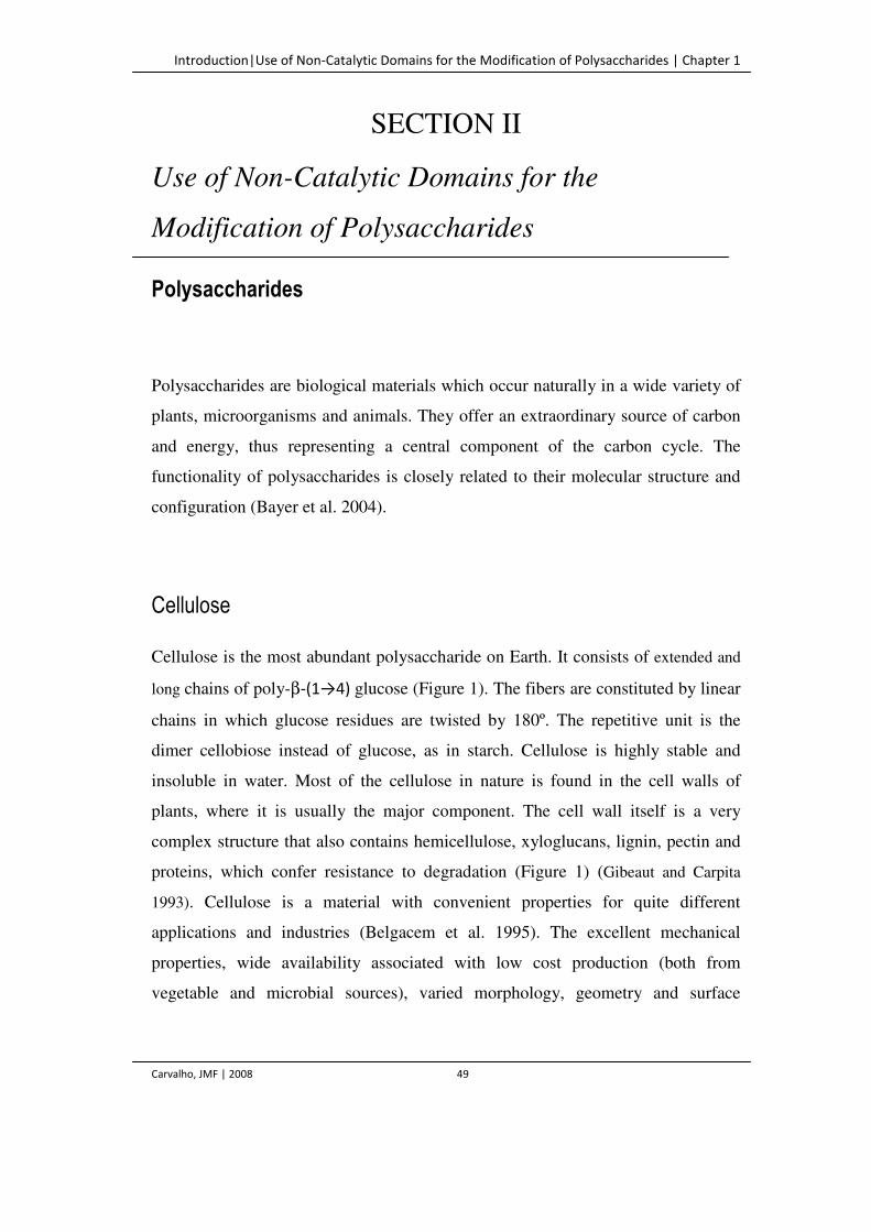
Introduction|Use of Non-Catalytic Domains for the Modification of Polysaccharides | Chapter 1
Carvalho, JMF | 2008 49
SECTION II
Use of Non-Catalytic Domains for the
Modification of Polysaccharides
Polysaccharides
Polysaccharides are biological materials which occur naturally in a wide variety of
plants, microorganisms and animals. They offer an extraordinary source of carbon
and energy, thus representing a central component of the carbon cycle. The
functionality of polysaccharides is closely related to their molecular structure and
configuration (Bayer et al. 2004).
Cellulose
Cellulose is the most abundant polysaccharide on Earth. It consists of extended and
long chains of poly-β-(1→4) glucose (Figure 1). The fibers are constituted by linear
chains in which glucose residues are twisted by 180º. The repetitive unit is the
dimer cellobiose instead of glucose, as in starch. Cellulose is highly stable and
insoluble in water. Most of the cellulose in nature is found in the cell walls of
plants, where it is usually the major component. The cell wall itself is a very
complex structure that also contains hemicellulose, xyloglucans, lignin, pectin and
proteins, which confer resistance to degradation (Figure 1) (Gibeaut and Carpita
1993). Cellulose is a material with convenient properties for quite different
applications and industries (Belgacem et al. 1995). The excellent mechanical
properties, wide availability associated with low cost production (both from
vegetable and microbial sources), varied morphology, geometry and surface
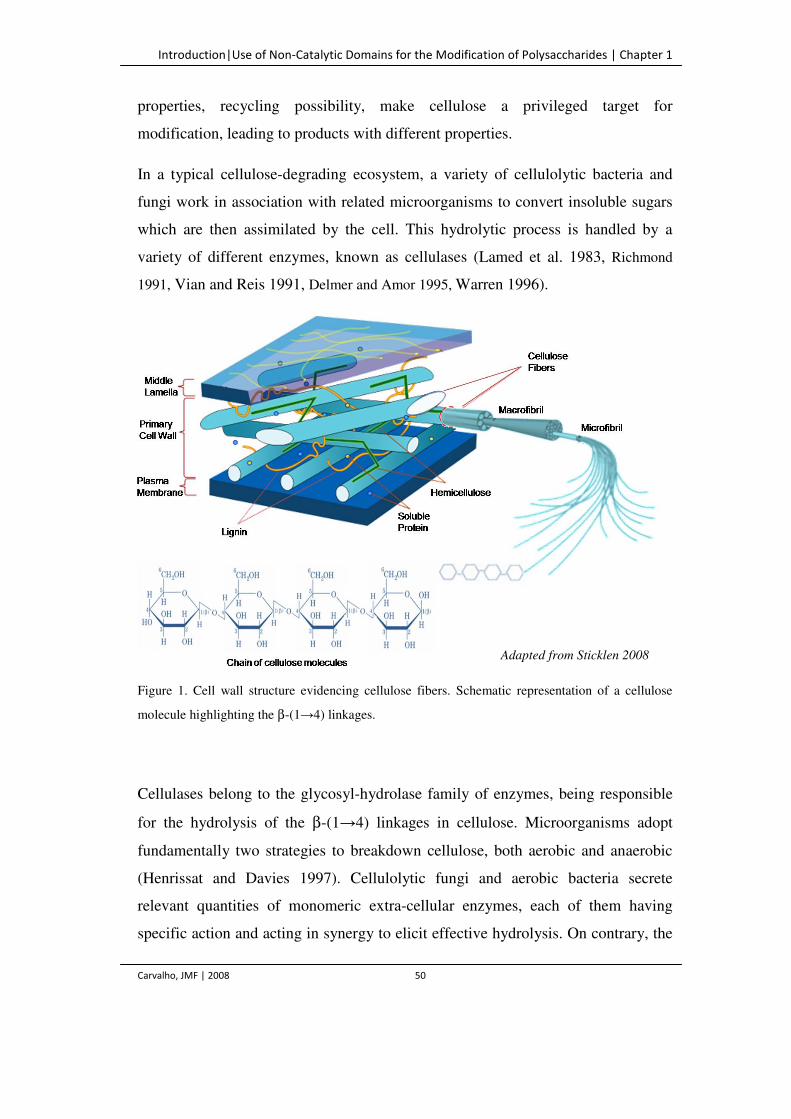
Introduction|Use of Non-Catalytic Domains for the Modification of Polysaccharides | Chapter 1
Carvalho, JMF | 2008 50
properties, recycling possibility, make cellulose a privileged target for
modification, leading to products with different properties.
In a typical cellulose-degrading ecosystem, a variety of cellulolytic bacteria and
fungi work in association with related microorganisms to convert insoluble sugars
which are then assimilated by the cell. This hydrolytic process is handled by a
variety of different enzymes, known as cellulases (Lamed et al. 1983, Richmond
1991, Vian and Reis 1991, Delmer and Amor 1995, Warren 1996).
Figure 1. Cell wall structure evidencing cellulose fibers. Schematic representation of a cellulose
molecule highlighting the β-(1→4) linkages.
Cellulases belong to the glycosyl-hydrolase family of enzymes, being responsible
for the hydrolysis of the β-(1→4) linkages in cellulose. Microorganisms adopt
fundamentally two strategies to breakdown cellulose, both aerobic and anaerobic
(Henrissat and Davies 1997). Cellulolytic fungi and aerobic bacteria secrete
relevant quantities of monomeric extra-cellular enzymes, each of them having
specific action and acting in synergy to elicit effective hydrolysis. On contrary, the
Adapted from Sticklen 2008
Introduction|Use of Non-Catalytic Domains for the Modification of Polysaccharides | Chapter 1
Carvalho, JMF | 2008 51
anaerobic bacteria produce enzymatic complexes called cellulosomes,
multienzymatic systems present in the surface of the cells, which adsorbs and
degrade cellulose (Coughlan and Ljungdahl 1988, Shoham et al. 1999, Bayer et al.
2004). Up to 11 different enzymes are aligned, thus ensuring a high local
concentration, together with the correct ratio and order of the components. For
these reasons, it is believed that the anaerobic system may be metabolically
advantageous over the aerobic one (individual enzymes). Besides the higher
absorption efficacy of the degradation products, due to the positioning of
cellulosomes on the cell surface, the close proximity between the cells and
enzymes allow for a better control of the metabolic activity (Boisset et al. 1999,
Schwarz WH 2001).
Cellulosome of Clostridium thermocellum
Clostridium thermocellum (C. thermocellum) is an anaerobic thermophilic
bacterium, considered to have the highest rate of cellulose utilization (Johnson et
al. 1982, Arai et al. 2001, Lynd et al. 2002, Demain et al. 2005). The hydrolysis is
mediated by a large multi-enzyme complex termed cellulosome (Figure 2).
Figure 2. Simplified schematic view of the interaction between C. thermocellum cellulosome and its
substrate. CBM – Cellulose-binding Module.
Introduction|Use of Non-Catalytic Domains for the Modification of Polysaccharides | Chapter 1
Carvalho, JMF | 2008 52
The principal component of the cellulosome is a scaffoldin subunit, a large
enzyme-integrating protein (CipA – Cellulosome integrating protein) that contains
cohesin modules, normally in multiple copies, for incorporation of the catalytic
domains, including β-(1→4)-endoglucanases, cellobiohydrolases, hemicellulases
and other cellulosomal components (Figure 2) (Bayer et al. 1994, Béguin and
Lemaire 1996). The enzymes contain a complementary module, the dockerin
domain that binds to the cohesin modules, mediating the integration of the enzymes
into the complex of the scaffoldin subunit. The scaffoldin also frequently includes a
carbohydrate-binding module (i.e., a cellulose-binding domain), commonly
referred to as CBM, through which the complex usually recognizes and binds to the
substrate. The cellulosome is anchored to the plant cell wall primarily through the
action of the internal family 3 CBM located in CipA. Cellobiohydrolase A (CbhA)
is one of the most important catalytic components of the cellulosome. It is
composed of an N-terminal family 4 CBD, an immunoglobulin-like domain, a
family 9 glycosyl hydrolase catalytic domain (GH9), two fibronectin like repeats
(FN31,2), a family 3 CBD and a C-terminal dockerin domain (Felix and Ljungdahl
1993, Bayer et al. 1998, Béguin and Alzari 1998, Zverlov et al. 1998, Shoham et al.
1999, Blum et al. 2000, Kurokawa et al. 2001, Kataeva et al. 2002, Kataeva and
Ljungdahl 2003, Kataeva et al. 2003).
Carbohydrate-binding Modules (CBM)
Not only cellulases, but also other enzymes involved in the hydrolysis of insoluble
polysaccharides, are typically modular enzymes with a distinct carbohydrate-
binding domain joined to a catalytic domain through a linker – highly glycosilated
peptide chain. Due to their high specificity and substrate recognition mechanisms,
these modules have a fundamental role in the enzymatic degradation of plants and
in polysaccharide storage.
Introduction|Use of Non-Catalytic Domains for the Modification of Polysaccharides | Chapter 1
Carvalho, JMF | 2008 53
A carbohydrate-binding module (CBM) is defined as a contiguous amino acid
sequence within a carbohydrate-active enzyme, having carbohydrate-binding
activity. CBMs were previously classified as cellulose-binding domains (CBDs)
based on the initial discovery of several modules that bound cellulose as their
primary ligand (Gilkes et al. 1988, Tomme et al. 1988). However, new modules
meeting the CBM criteria, which are capable to bind carbohydrates other than
cellulose, are continually being found.
CBMs are organized in 52 families (CAZY server,
http://www.cazy.org/fam/acc_CBM.html). This classification of carbohydrate-
binding domains is based on amino acid sequence similarity and is expected to aid
in the identification of both CBMs and its functional residues, to predict binding
specificity and polypeptide folding and even reveal evolutionary relationships.
Family 1 to 13 corresponds to CBMs which have a least one cellulose-binding
domain (Tomme et al. 1995, Boraston et al. 2004).
Modification of Polysaccharides
The adoption of industrial environmentally friendly processes is finally turning
mandatory, bringing biotechnological processes to the forefront of research. Under
its broad definition, biotechnology encompasses activities such as fermentation,
cell immobilization, and enzyme technology. Extensive basic and applied research
on cellulases, hemicellulases and other polysaccharide-degrading enzymes has not
only generated significant scientific knowledge but has also revealed their potential
in biotechnology. At present, they are used in food, brewery and wine production,
animal feed, textile and laundry, pulp and paper industries, as well as in agriculture,
in wastewater treatment, systems to remove oxygen-demanding substances and
suspended solids and for research purposes. The demand for these enzymes is
Introduction|Use of Non-Catalytic Domains for the Modification of Polysaccharides | Chapter 1
Carvalho, JMF | 2008 54
growing rapidly and has become the driving force for intensifying its research
(Bhat and Bhat 1997, Bhat 2000).
Enzymes are among the most promising examples of new technologies designed to
help the textile industry meeting the challenges of strict environmental regulations,
global competition and new-market driven demands for specific fabric properties.
Cellulases are used in the treatment of cotton fibers, namely in the stone wash
process, substituting the use of stones to produce used-like aspect of the garments
(Miettinen-Oinonen 2004). They are also used in the de-pilling process, which
consists in remove the small fibers – pills – present in the surface of cotton
(Heikinheimo 2002).
Enzymes have also several uses in the paper industry and have long been applied in
paper production, for the modification of both fiber and paper properties (Bajpai
1999). These enzymes include xylanases, endoglucanases, cellulases and
hemicellulases (Suurnäki et al. 1994, Senior and Hamilton 1993, Gen and Li 2003,
Seo et al. 2000, Jackson at al. 1993, Pala et al. 2001). Currently, the most
important application of enzymes includes the use of xylanases in biobleaching
pre-treatment of kraft pulp (Vikarii et al. 1994). This process allows for the
reduction, or even elimination of the hypochloride used in the bleaching process for
the lignin oxidation. Endoglucanases have found their function in two important
processes, de-watering and de-inking. One of the most important stages in paper
production is the de-watering step, in which water is removed from the formed
sheet using a combination of vacuum and heat. Cellulases can assist in this step by
solubilizing small particles that are strong water holders (Pommier et al. 1989,
Pommier et al. 1990, Jackson et al. 1993, Woodward et al. 1994, Bhardwaj et al.
1996). The same mechanism is responsible for lowering the energy consumption in
mechanical pulp production by improving the beatability, drainage and solubility of
the pulps. Additionally, the use of cellulases for glue removal has proved to be
efficient, even more than the conventional alkaline chemicals (Sykes and
Klungness 1997).
Introduction|Use of Non-Catalytic Domains for the Modification of Polysaccharides | Chapter 1
Carvalho, JMF | 2008 55
Use of Non-catalytic Domains – An Emerging Field
The attractiveness of biotechnology lies in its potential to increase specificity on
the reactions to save energy, lowering the associated cost in reactions, keeping in
mind the development of environmentally friendly processes. Despite its wide
utilization, well established in the above discussion, several drawbacks result from
enzyme utilization. Taking the paper treatment as an example, the drawbacks
include the extensive hydrolysis of polysaccharides that leads to a reduction of both
fiber strength and mass. In this context, the application of CBMs allows
overcoming the limitations of the enzyme technology.
Nowadays, CBMs are widely used. They can be used on the purification of
recombinant proteins (Jiang and Radford 2000). Textile applications include fabric
targeting of recombinant enzymes that do not possess a native affinity for the
cellulosic fibers, such as amylases, proteases and lipases (von der Osten 2000). Other
substances can also be targeted to cellulosic fabrics. Fragrance-bearing particles
conjugated to CBMs can be added to laundry powder, hence reducing the amount
of fragrance needed in the product (Berry 2001). Another application consists in
the expression of CBMs at the cells surface, such that they may be immobilized in
cellulosic materials (Nam et al. 2002, Lethio et al. 2003). CBMs are also
successfully applied as analytical tool in research. An example is the development
of a system based on CBMs that allows for a rapid detection of pathogenic
microbes in food samples (Shoseyov et al. 1999).
Due to its ability to adsorb to cellulose without degrading it, CBMs have great
biotechnology potential. The ability to fuse CBDs with other molecules of different
sizes, charges, hydrophobicities, catalytic activities or bioactivities, allow these
molecules to be potentially interesting and useful on the activation and
modification of cellulose fibers. Inspiring research into CBM-cellulose fiber
interactions is continuously being carried out (Kitaoka and Tanaka 2001, Levy and
Shoseyov 2002). Several studies have indicated that treatment of cellulose fibers
with CBMs alters the interfacial properties of the fibers. This phenomenon was first
Introduction|Use of Non-Catalytic Domains for the Modification of Polysaccharides | Chapter 1
Carvalho, JMF | 2008 56
observed by Cavaco-Paulo et al. (1999), who demonstrated that treatment of cotton
fibers with a CBM alters their affinity to dye. Later on, Suürnakki et al. (2000)
showed that treatment of bleached chemical pulp with endoglucanases,
cellobiohydrolases, and the catalytic domains from Trichoderma reesei could also
change the interfacial properties, having a beneficial effect on pulp properties such
as viscosity and strength after refinement. Another study demonstrated that
treatment of fibers recycled from old paperboard containers with CBMs improve
both the tensile and burst indexes, as well as increase the pulp drainage rate (Pala et
al. 2001, Pala et al. 2002). Following studies described an increase in the surface
area of cellulosic fibers as a consequence of cellulosic aggregate disruption (Din et
al. 1991, Kitaoka and Tanaka 2001, Pinto et al. 2004). Kataeva et al. (2002) have
also demonstrated the non hydrolytic disruption of cellulose fibers by the family 3
CBM of CbhA of C. thermocellum. This non-hydrolytic disruptive activity has also
been ascribed to the Fn31,2 module. Additionally, it was suggested that Fn31,2
affects hydrolysis more significantly when it is attached to its appended catalytic
domain, in what seems to be a synergistic process. This thesis will focus on the
potential of two recombinant family 3 CBDs (belonging to CipA and CbhA of the
C. thermocellum) and a fusion protein with the FN31,2 module (also from CbhA) to
modify secondary paper fibers.
As described earlier, CBMs are produced by a large number of fungi and bacteria
and have the ability to potentiate the catalytic efficiency of the associated catalytic
domains. They may be obtained through the proteolysis of cellulases, providing
large amounts of peptides in a rather rapid procedure (Pinto et al. 2004, Pinto et al.
2006). In addition they can be produced using the recombinant DNA technology
(Creagh et al. 1996, Bothwell et al. 1997, Linder et al. 1999, Bayer et al. 2000). In
fact, recombinant forms of these proteins containing his-tags, provide a flexible
system which allows overcoming the purity limitations of the proteolysis approach.
However, CBMs obtained through these methods have not the highly glycosilated
peptide sequence (Pinto et al. 2004, Pinto et al. 2006). Thus, in current work,
Introduction|Use of Non-Catalytic Domains for the Modification of Polysaccharides | Chapter 1
Carvalho, JMF | 2008 57
recombinant CBM were chemically modified with PEG, expected to mimetize the
properties of the native CBM glycoconjugates.
Pegylation
First reports of the use of PEG to modify the interfacial/biological properties of
different molecules, remount to the late 70s and the studies on the alteration of
immunological properties of BSA modified with PEG (Abuchowski et al. 1977).
The coupling of PEG to other molecules can be used to attain various ends. It can
be used to alter solubility characteristics both in aqueous or organic solvents (Inada
et al. 1986), to increase the stability of proteins in solution (Berger and Pizzo
1988), for modulation of the immune response (Delgado et al. 1992), to extend the
half life of the modified substances, among others. The PEG molecule consists of
repeating units of ethylene oxide that terminate in hydroxyl groups on either end of
a linear chain. In solution PEG is a highly mobile molecule that creates a large
exclusion volume for its molecular weight, far larger than proteins of comparable
size. It can be conjugated to other molecules through two principle approaches; (1)
direct reaction with the hydroxyl groups, (2) reaction of the polymer with a
functional molecule – activation. The most common activation methods, the one
that has been used in this work, is the creation of amine-reactive derivates that can
be linked with proteins and other amine-containing molecules through a carboxilate
end, producing a stable conjugate (Figure 3) (Zalipsky et al. 1991, Miron and
Wilchek 1993).
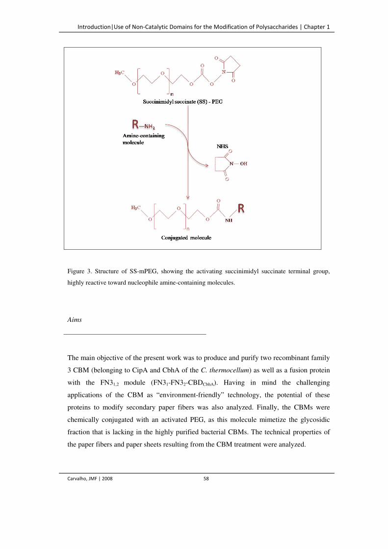
Introduction|Use of Non-Catalytic Domains for the Modification of Polysaccharides | Chapter 1
Carvalho, JMF | 2008 58
Figure 3. Structure of SS-mPEG, showing the activating succinimidyl succinate terminal group,
highly reactive toward nucleophile amine-containing molecules.
Aims
The main objective of the present work was to produce and purify two recombinant family
3 CBM (belonging to CipA and CbhA of the C. thermocellum) as well as a fusion protein
with the FN31,2 module (FN31-FN32-CBDCbhA). Having in mind the challenging
applications of the CBM as “environment-friendly” technology, the potential of these
proteins to modify secondary paper fibers was also analyzed. Finally, the CBMs were
chemically conjugated with an activated PEG, as this molecule mimetize the glycosidic
fraction that is lacking in the highly purified bacterial CBMs. The technical properties of
the paper fibers and paper sheets resulting from the CBM treatment were analyzed.
References | Chapter 1
Carvalho, JMF | 2008 59
References
Abuchowski A, McCoy JR, Palczuk NC, Van Es T and Davis FF. (1977) Effect of covalent
attachment of poly(ethylene glycol) on immunogenicity and circulating life of bovine liver catalase.
J Biol Chem 252: 3582–3586.
Arai T, H Ohara S, Karita T, Kimura KS and Ohmiya K. (2001) Sequence of celQ and properties of
CelQ, a component of the Clostridium thermocellum cellulosome. Appl Microbiol Biotechnol 57:
660–666.
Bailon P and Berthold W. (1998) Polyethylene glycol-conjugated pharmaceutical proteins. Pharm
Sci Tech Today 1: 352–356.
Bajpai P. (1999) Application of Enzymes in the Pulp and Paper Industry. Biotechnol Prog 15: 147–
157.
Bayer EA, Morag E and Lamed R. (1994) The cellulosome – a treasure-trove for biothechnology.
Trends Biotechnol 12: 379–386.
Bayer EA, Shoham Y and Lamed R. (2000) Cellulose decomposing prokaryotes and their enzyme
systems. In The Prokaryotes: An Involving Electronic Resource for the Microbial. 3rd edn,
Springer, New York, USA pp 1–4.
Bayer EA, Belaich JP, Shoham Y and Lamed R. (2004) The cellulosomes: multienzyme machines
for degradation of plant cell wall polysaccharides. Annu Rev Microbiol 58: 521–554.
Béguin P and Lemaire M. (1996) The cellulosome: as exocellular, multiprotein complex specialized
incellulose degradation. Crit Rev Biochem Mol Biol 31: 201–236.
Beguin P and Alzari P. (1998) The cellulosome of Clostridium thermocellum. Biochem Soc Trans
26: 178–185.
Belgacem MN, Czeremuskin G, Sapieha S and Gandini A. (1995) Surface characterization of
cellulose fibres by XPS and inverse gas chromatography. Cellulose 2:145-157.
Berger H and Pizzo SV. (1988) Preparation of polyethylene glycol-tissue plasminogen activator
adducts that retain functional activity: characteristics and behavior in three animal species. Blood
71: 1641-1647.
References | Chapter 1
Carvalho, JMF | 2008 60
Berry MJ, Davis PJ and Gidley MJ. (2001) Conjugated polysaccharide fabric detergent and
conditioning products. US patent 6225462.
Bhardwaj NK, Bajpai P and Bajpai PK. (1996) Use of enzymes in modification of fibres for
improved beatability. J Biotechnol 51: 21–26.
Bhat MK and Bhat S. (1997) Cellulose degrading enzymes and their potential industrial
applications. Biotechnol Adv 15: 583–620.
Bhat MK. (2000) Cellulases and related enzymes in biotechnology Biotechnol Adv 18: 355–383.
Boisset C, Chanzy, H, Henrissat B, Lamed R, Shoham Y and Bayer EA. (1999) Digestion of
crystalline cellulose substrates by the Clostridium thermocellum cellulose: structural and
morphological aspects. Biochem J 340: 829–835.
Boraston AB, Bolam DN, Gilbert HJ and Davies GJ. (2004) Carbohydrate-binding modules: fine
tuning polysaccharide recognition. Biochem J 382: 769–781.
Bothwell MK, Daughhetee SD, Chaua GY, Wilson DB and Walker LP. (1997) Binding capacities
for Thermomonospora fusca E3, E4 and E5, the E3 binding domain, and Thichoderma reesei CBHI
on avicel and bacterial microcrystalline cellulose. Bioresource Technol 60: 169 –178.
Coughlan MP and Ljungdahl LG. (1988) Comparative biochemistry of fungal and bacterial
cellulolytic enzyme systems. FEMS Symp 43: 11–30.
Creagh A, Ong E, Jervis E, Kilburn D and Haynes C. (1996) Binding of the cellulose-binding
domain of exoglucanase Cex from Cellulomonas fimi to insoluble microcrystalline cellulose is
entropically driven. Proc Natl Acad Sci USA 93: 12229 –12234.
Delgado C, Francis GE and Fisher D. (1992) The uses and properties of PEG-linked proteins Crit
Rev Ther Drug Carrier Syst 9: 249–304.
Delmer DP and Amor Y (1995) Cellulose Biosynthesis. Plant Cell 7: 987–1000.
Demain AL, Newcomb M and Wu JHD. (2005) Cellulase, clostridia, and ethanol. Microbiol Mol
Biol Rev 69: 124–154.
Din N, Gilkes NR, Tekant B, Miller RC, Warren RAJ and Kilburn DG. (1991) Non-hydrolytic
disruption of cellulose fibers by the binding domain of a bacterial cellulose. Bio Technology
9:1096–1099.
References | Chapter 1
Carvalho, JMF | 2008 61
Gibeaut DM and Carpita NC. (1993) Structural models of primary cell wall in flowering plants:
consistency of molecular structure with the physical properties of the walls during growth. Plant J 3:
1–30.
Gilkes NR, Warren RA, Miller RCJ and Kilburn DG. (1988) Precise excision of the cellulose-
binding domains from two Cellulomonas fimi cellulases by a homologous protease and the effect on
catalysis. J Biol Chem 263: 10401–10407.
Heikinheimo L. (2002) Trichoderma reesei cellulases in processing cotton. Tampere University of
Technology pp 82.
Henrissat B and Davies G. (1997) Structural and sequence-based classification of glycoside
hydrolases. Curr Opin Struct Biol 7: 637–644.
Inada Y, Matsuswma A, Kodera Y and NishimuraH. (1990) Polyethylene glycol(peg)-protein
conjugates: application to biomedical and biotechnological processes. J Bioact Comp Pol 5: 343–
364.
Jackson LS, Heitmann JA and Joyce TW. (1993) Enzymatic modification of secondary fibers. Tappi
J 76: 147–154.
Johnson EA, Sakajoh M, Halliwell G, Madia A and Demain AL. (1982) Saccharification of
complex cellulosic substrates by the cellulase system from Clostridium thermocellum. Appl Environ
Microbiol 43: 1125–1132.
Kitaoka T and Tanaka H. (2001) Novel paper strength additive containing cellulose-binding domain
of cellulose. J Wood Sci 47: 322–324.
Kataeva IA, Seidel III RD, Shah A, West LT, Li X-L and Ljungdahl LG. (2002) The fibronectin
type 3 like repeat from the Clostridium thermocellum cellobiohydrolase CbhA promotes hydrolysis
of cellulose by modifying its surface. Appl Environ Microbiol 68: 4292–4300.
Kataeva IA and Ljungdahl LG. (2003) The Clostridium thermocellum cellulosome: a multi-protein
complex of module composed components. Uversky VN. (ed). In Protein Structures: Kaleidoscope
of Structural Properties and Functions. Research Singpost, Kerala, India pp 661–666.
Kurokawa J, Hemjinda E, Arai T, Karita S, Kimura T, Sakka K and Ohmiya K. (2001) Sequence of
the Clostridium thermocellum mannanase gene man26B and characterization of the translated
product. Biosci Biotechnol Biochem 65: 548–554.
References | Chapter 1
Carvalho, JMF | 2008 62
Lamed R, Setter E, Kenig R and Bayer EA. (1983) The cellulosome - a discrete cell surface
organelle of Clostridium thermocellum which exhibits separate antigenic, cellulose-binding and
various cellulolytic activities. Biotechnol Bioeng 13:163–181.
Levy I and Shoseyov O. (2002) Cellulose-binding domains biotechnological applications. Biotech
Adv 20: 191–213.
Lehtio J, Sugiyama J, Gustavsson M, Fransson L, Linder and Teeri TT. (2003) The bonding
specificity and affinity determinants of family 1 and family 3 cellulose-binding modules.
Proceedings of the National Academy of Sciences of USA 100: 484–489.
Linder I, Nevanen T and Teeri TT. (1999) Design of a pH-dependent cellulose-binding domain.
FEBS Lett 447: 13–16.
Lynd LR, Weimer PJ, van Zyl WH and Pretorius IS. (2002) Microbial cellulose utilization:
fundamentals and biotechnology. Microbiol Mol Biol Rev 66: 506–577.
Miettinen-Oinonen A. (2004) Trichoderma reesei strains for production of cellulases for the textile
industry. University of Helsinky pp 100.
Miron T and Wilchek M. (1993) A simplified method for the preparation of succinimidyl carbonate
polyethylene glycol for coupling to protein Bioconjugate Chem 4: 568–569.
Nam JM, Fujita Y, Arai T, Kondo A, Morikawa Y, Okada H, Ueda M and Tanaka A. (2002)
Construction of engineered yeast with ability of binding to cellulose. J Mol Catal 17: 197–202.
Pala H, Lemos MA, Mota M and Gama FM. (2001) Enzymatic upgrade of old paperboard
containers. Enzyme Microb Technol 29: 274–279.
Pala H, Mota M and Gama FM. (2002) Enzymatic modification of paper fibres. Biocat Biotransf 20:
353–361.
Pinto R, Moreira S, Mota M and Gama FM. (2004) Studies on the Cellulose-Binding Domains
adsorption to cellulose. Langmuir 20: 1409–1413.
Pinto R, Carvalho J, Mota M and Gama FM. (2006) Large-Scale production of cellulose-binding
domains. Adsorption studies using CBD-FITC conjugates. Cellulose 13: 557–569.
Pommier JC, Fuentes JL and Goma G. (1989) Using enzymes to improve the process and quality in
the recycled. paper industry. Tappi J 72: 187–191.
References | Chapter 1
Carvalho, JMF | 2008 63
Pommier JC, Goma G, Fuentes JL, Rousset C and Jokinen O. (1990) Using enzymes to improve the
process and quality in the recycled paper industry. Part 2: industrial applications. Tappi J 73: 197–
202.
Richmond PA. (1991) In Biosynthesis and biodegradation of cellulose. Haigler CH and Weiner CJ.
(eds). Marcel Dekker, New York, USA pp 5–25.
Schwarz WH. (2001) The cellulosome and cellulose degradation by anaerobic bacteria. Appl
Microbiol Biotechnol 56: 634–649.
Senior DJ and Hamilton J. (1993) Xylanase treatment for the bleaching of softwood kraft pulps: the
effect of chlorine dioxide substitution. TAPPI J 76: 200–206.
Seo YB, Shin IC and Jeon Y. (2000) Enzymatic and mechanical treatment on chemical pulp. TAPPI
J 83: 1–9.
Shoham Y, Lamed R and Bayer EA. (1999) The cellulossome concept as an efficient microbial
strategy for the degradation of polysaccharides. Trends Microbiol 7: 275–281.
Shoseyov O, Shpigel I, Goldstein MM and Doi RH. (1999) Methods of detection using a cellulose-
binding domain fusion product. US patent 5856201.
Sticklen MB. (2008) Plant genetic engineering for biofuel production: towards affordable cellulosic
ethanol. Nature Rev Gen 9: 433–443.
Sykes MS and Klungness JH. (1997) Enzymatic removal of stickie contaminants. In Proceedings of
the 4th international refining conference 687–691.
Suurnäki A, Kantelinen A, Buchert J and Viikari L. (1994) Enzyme-aided bleaching of industrial
softwood kraft pulps. TAPPI J 77: 111–116.
Tomme P, Warren RA, Miller RC Jr, Kilburn DG and Gilkes NR. (1995) In Enzymatic degradation
of insoluble polysaccharides. Saddler JN and Penner M (eds). Cellulose-binding domains:
classification and properties. American Chemical Society, Washington, USA pp 142–163.
Tomme P, Van Tilbeurgh H, Pettersson G, Van Damme J, Vandekerckhove J, Knowles J, Teeri T
and Claeyssens M. (1988) Studies of the cellulolytic system of Trichoderma reesei QM 9414.
Analysis of domain function in two cellobiohydrolases by limited proteolysis. Eur J Biochem 170:
575–581.
Vian B and Reis D. (1991) In Biosynthesis and biodegradation of cellulose. Haigler CH and Weiner
CJ. (eds). Marcel Dekker, New York, USA pp 25–51.
References | Chapter 1
Carvalho, JMF | 2008 64
Viikari L, Kantelinen A, Sundquist J and Linko M. (1994) Xylanases. in bleaching: from an idea to
the industry. FEMS Microbiol Rev 13: 335–350.
Von der Osten C, Bjornvad ME, Vind and Rasmussen MD. (2000) Process and composition for
desizing cellulosic fabric with an enzyme hybrid. US patent 6017751.
Von der Osten C, Bjornvad ME, Vind J and Rasmussen MD. (2000) Process for removal or
bleaching of soiling or stains from cellulosic fabric. US patent 6015783.
Warren RAJ. (1996) Microbial hydrolysis of polysaccharides. Annu Rev Microb 50:183–212.
Woodward J, Stephan LM, Koran LJ, Wong KKY and Saddler JN. (1994) Enzymatic separation of
high−quality uninked pulp fibers from recycled newspaper. Bio/Technology 12: 905–908.
Zalipsky S, Seltzer R and Menon-Rudolph S. (1992) Evaluation of a new reagent for covalent
attachment of polyethylene glycol to proteins. 15: 100–114.
Zverlov V V, Velikodvorskaya GV, Schwarz WH, Bronnenmeier K, Kellerman J and Staudenbauer
WL (1998). Multidomain structure and cellulosomal localization of the Clostridium thermocellum
cellobiohydrolase CbhA. J Bacteriol 180: 3091–3099.
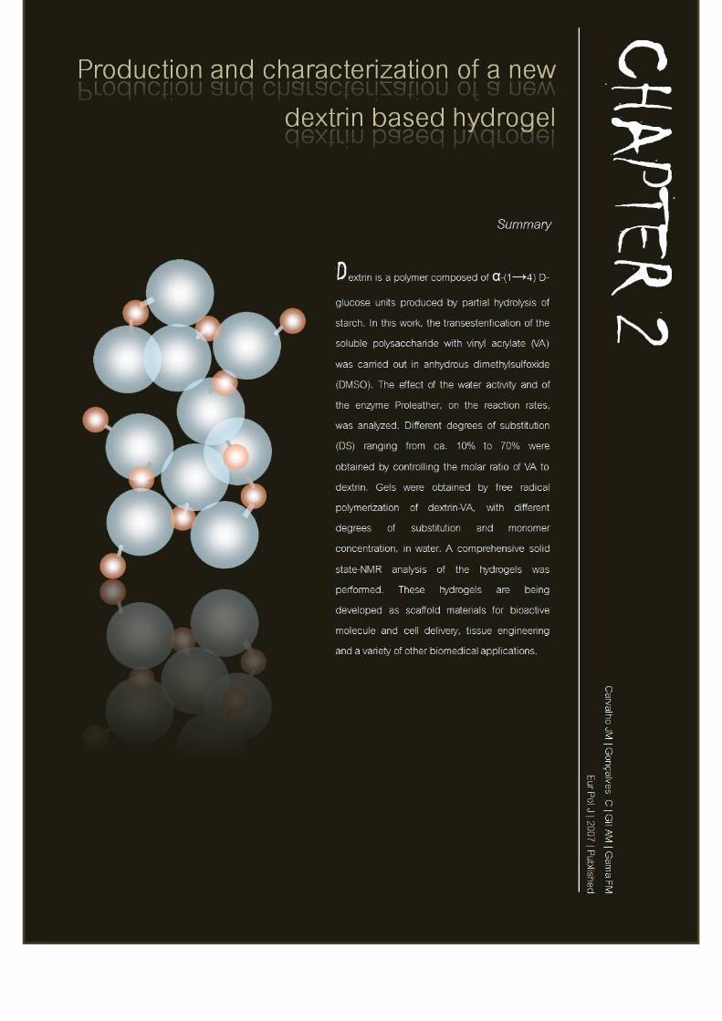

Production and Characterization of a New Dextrin Based Hydrogel | Chapter 2
Carvalho, JMF | 2008 67
Introduction
Hydrogels are a class of three-dimensional, highly hydrated polymeric networks
(Peppas et al. 2000). They are composed of hydrophilic polymer chains, which can
be either synthetic or natural. The structural integrity of hydrogels depends on
crosslinks established between the polymer chains, by chemical bonds and physical
interactions. Hydrogels are providing new opportunities for the development of a
variety of medical applications (Peppas et al. 1996), including drug delivery
systems (McCulloch and Shalaby 1998), scaffold materials to organize cells into a
three-dimensional architecture, tissue replacements (Kane et al. 1996) wound
dressings (Ishihara et al. 2002, Chen et al. 2003) and immobilization of proteins
and cells, among others. Since the clinical use of hydrogels is increasing,
considerable efforts have been made in order to develop new hydrogels from a
variety of synthetic and natural materials (Drury and Mooney 2003). In this work,
we report a procedure to prepare dextrin hydrogels. Dextrin is a glucose-containing
saccharide polymer linked by α-(1→4) D-glucose units, having the same general
formula as starch, but smaller and less complex. This polysaccharide is produced
by partial hydrolysis of starch, which can be accomplished by the use of acid,
enzymes, or a combination of both. Dextrin is a widely used material with a variety
of applications, from adhesives to food industry and textiles. Moreover, because of
the biocompatibility and degradability, starch-based materials are presently being
used to prepare biodegradable hydrogels with technological applications in a large
number of areas such as medicine, pharmacy and biology (Wong and Mooney
1997, Marques et al. 2002). Various approaches have been already explored for the
production of starch hydrogels. These approaches include one or two-step free
radical graft polymerization of hydrophilic vinyl monomers in the presence of a
cross-linker, crosslinking by chemical reactions with complementary groups,
radiation-induced polymerization and crosslinking, and novel self-assembly
processes (Zhang et al. 2005) However, compared with our approach, some of
these chemical strategies show disadvantages, such as the use of catalysts (e.g. 4-
Production and Characterization of a New Dextrin Based Hydrogel | Chapter 2
Carvalho, JMF | 2008 68
dimethylaminopyridine (4-DMAP) or pyridoxine) (Ferreira et al. 2002) which are
difficult to remove from the reaction mixture and could dramatically increase the
toxicity of the material. To prepare the hydrogels, dextrin was modified with vinyl
acrylate (VA) using two different approaches, chemical or enzymatic. Chemical
synthesis is broadly used, however presenting several drawbacks, such as the need
for protection and deprotection of functional groups to prevent unwanted
racemization, as well as the lack of stereoselectivity. The use of enzymes under
mild conditions has allowed, in some cases, overcoming these limitations, also
enabling modification of molecules that could not otherwise be altered (Cooney
and Heuter 1974, Zacks and Klibanov 1985, Klibanov 1997, Klibanov 2001).
Apolar solvents are generally considered to better preserve the enzymatic activity
that polar ones (Sergeeva et al. 1997, Akkara et al. 1999). However, Ferreira et al.
(2002) have recently reported the used of the Bacillus subtilis protease Proleather
FG-F to catalyze the transesterification of dextran with VA, in anhydrous
dimethylsulfoxide (DMSO). The enzyme-catalyzed system developed by Ferreira
and colleagues, using dextran and inulin, was in this work used for dextrin
modification. The starch polysaccharide functionalized with reactive double bonds
was cross-linked by free radical polymerization in aqueous solution. The structure
and morphology of starch networks in the hydrogel were characterized by cross-
polarization magic-angle-spinning 13C NMR spectroscopy (CP-MAS 13C NMR).
Materials and Methods
Materials
Proleather FG-F, a protease from Bacillus subtilis, was obtained from Amano
Enzyme Co. Dextrin - Koldex 60 (Mw= 45700 Da, as determined by gel
permeation chromatography (GPC) analysis) was a generous gift from Tate and
Lyle. Vinyl acrylate (VA) was from Aldrich, N,N,N’,N’-
tetramethylenethylenediamine (TEMED) and ammonium persulfate (APS) were
purchased from BioRad, dimethylsulfoxide (DMSO) and acetone were from
Production and Characterization of a New Dextrin Based Hydrogel | Chapter 2
Carvalho, JMF | 2008 69
AppliChem and polyethyleneglicol (PEG 200 g/mol) was obtained from Riedel-d
Haën. DMSO was dried with 0.4 nm molecular sieves, at least overnight, before
use. Regenerated cellulose tubular membranes, with 3500 MWCO, were obtained
from Membrane Filtration Products.
Pretreatment of Proleather and determination of proteolytic activity in DMSO
Proleather FG-F was “pH adjusted” as described by Ferreira et al. (2002). The
enzyme was resuspended in 20 mM phosphate buffer at pH 8. After freezing, the
sample was lyophilized on a CHRIST ALPHA2-4 lyophization unit (B. Braun
Biotech International) for 48 h. The percentage of active enzyme in commercial
powder was 8.9 % (w/w) as obtained by BCA Protein Assay (Pierce) using BSA as
protein standard. The proteolytic activity of Proleather FG-F in either 0.2 M
phosphate buffer pH 8 or DMSO was determined with casein as substrate. Briefly,
in the presence of a protease the casein is cleaved, exposing primary amines. The
compound TNBSA (2,4,6-Trinitrobenzene sulfonic acid) reacts with these exposed
amines thereby producing an orange-yellow colour that can be quantified as 450
nm absorbance (Abs450). Control blanks for each sample were included in the assay
to correct the effect of primary amines on proteins in the protease sample. The
absorbance values were then converted to equivalent subtilisin (a Bacillus subtilis
protease) concentration using a calibration curve. One unit of protease activity (U)
is defined as the production of 1 mM of free amine per minute per milligram of
enzyme.
Enzymatic and chemical synthesis of dextrin-VA
The enzyme-catalyzed modification of dextrin was performed as described by
Ferreira et al. (2002) with few modifications. Briefly, dextrin (4 g) and an amount
of VA ranging from 382 to 2600 µL were dissolved in anhydrous DMSO (60 mL).
The reaction was started by adding 600 mg of “pH adjusted” Proleather FG-F. The
reaction mixtures were then incubated at 50 ºC, with magnetic stirring, for 72 h.
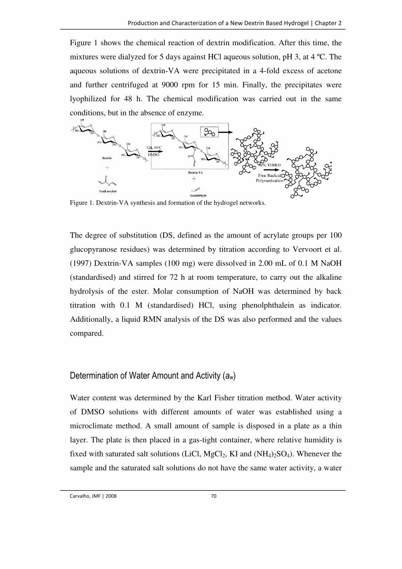
Production and Characterization of a New Dextrin Based Hydrogel | Chapter 2
Carvalho, JMF | 2008 70
Figure 1 shows the chemical reaction of dextrin modification. After this time, the
mixtures were dialyzed for 5 days against HCl aqueous solution, pH 3, at 4 ºC. The
aqueous solutions of dextrin-VA were precipitated in a 4-fold excess of acetone
and further centrifuged at 9000 rpm for 15 min. Finally, the precipitates were
lyophilized for 48 h. The chemical modification was carried out in the same
conditions, but in the absence of enzyme.
Figure 1. Dextrin-VA synthesis and formation of the hydrogel networks.
The degree of substitution (DS, defined as the amount of acrylate groups per 100
glucopyranose residues) was determined by titration according to Vervoort et al.
(1997) Dextrin-VA samples (100 mg) were dissolved in 2.00 mL of 0.1 M NaOH
(standardised) and stirred for 72 h at room temperature, to carry out the alkaline
hydrolysis of the ester. Molar consumption of NaOH was determined by back
titration with 0.1 M (standardised) HCl, using phenolphthalein as indicator.
Additionally, a liquid RMN analysis of the DS was also performed and the values
compared.
Determination of Water Amount and Activity (aw)
Water content was determined by the Karl Fisher titration method. Water activity
of DMSO solutions with different amounts of water was established using a
microclimate method. A small amount of sample is disposed in a plate as a thin
layer. The plate is then placed in a gas-tight container, where relative humidity is
fixed with saturated salt solutions (LiCl, MgCl2, KI and (NH4)2SO4). Whenever the
sample and the saturated salt solutions do not have the same water activity, a water
Production and Characterization of a New Dextrin Based Hydrogel | Chapter 2
Carvalho, JMF | 2008 71
transfer among the samples occurs, quantified by weighting. The equilibrium is
reached when constant mass is observed, by sorption or desorption. The water
activity of the sample is determined according to the procedure described by
Multon and Bizot (1978) and shown in figure 2.
Preparation of dextrin-VA hydrogels
Dextrin-VA hydrogels were obtained by free radical polymerization of aqueous
solution of dextrin-VA, with different degrees of substitution and monomer
concentration (Figure 1). Two different amounts of dextrin-VA (300 and 400 mg)
were dissolved in 900 µL of 0.2 M phosphate buffer, pH 8.0, and bubbled with
nitrogen for 2 min. The gelation reactions were initiated by adding 90 µL APS (80
mg/ml in 0.2 M phosphate buffer, pH 8.0) and 90 µL TEMED (13.6 % (v/v) in
water, pH adjusted to 8.0 with HCl) and allowed to occur for 30 min at room
temperature. All the recipients in which gelation took place were coated with a
polyethyleneglicol solution (PEG 200 g/mol) to simplify the removal of the
hydrogel following polymerization.
Solid state NMR Spectroscopy
The CP-MAS 13C NMR technique is a convenient solid-state analytical method for
the characterization of hydrogels due to the insolubility of their networks. The 13C
NMR spectra were recorded on a 400 MHz Bruker FT NMR (Fourier transform
nuclear magnetic resonance) spectrometer equipped with CP-MAS (cross-
polarization and magic-angle-spinning) accessories. The dry samples were placed
in a 7 mm rotor and spun at a rate of 4.5 kHz. Spectra were acquired with a contact
time of 5 ms, spectral width 300 ppm, 2 K time domain points and the number of
transients indicated in the figure captions. All spectra were processed with a
linebroadening (LB) factor of 30 Hz in order to improve signal to noise ratio.
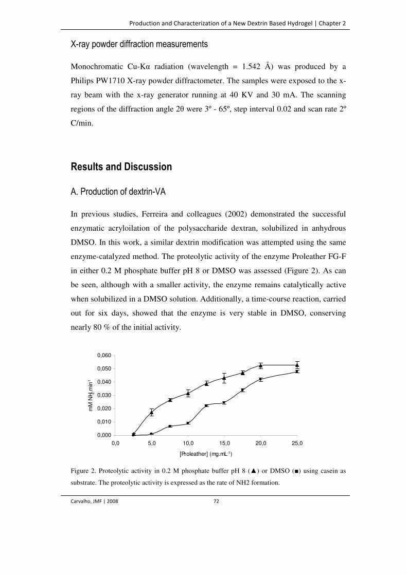
Production and Characterization of a New Dextrin Based Hydrogel | Chapter 2
Carvalho, JMF | 2008 72
X-ray powder diffraction measurements
Monochromatic Cu-Kα radiation (wavelength = 1.542 Å) was produced by a
Philips PW1710 X-ray powder diffractometer. The samples were exposed to the x-
ray beam with the x-ray generator running at 40 KV and 30 mA. The scanning
regions of the diffraction angle 2θ were 3º - 65º, step interval 0.02 and scan rate 2º
C/min.
Results and Discussion
A. Production of dextrin-VA
In previous studies, Ferreira and colleagues (2002) demonstrated the successful
enzymatic acryloilation of the polysaccharide dextran, solubilized in anhydrous
DMSO. In this work, a similar dextrin modification was attempted using the same
enzyme-catalyzed method. The proteolytic activity of the enzyme Proleather FG-F
in either 0.2 M phosphate buffer pH 8 or DMSO was assessed (Figure 2). As can
be seen, although with a smaller activity, the enzyme remains catalytically active
when solubilized in a DMSO solution. Additionally, a time-course reaction, carried
out for six days, showed that the enzyme is very stable in DMSO, conserving
nearly 80 % of the initial activity.
Figure 2. Proteolytic activity in 0.2 M phosphate buffer pH 8 (▲) or DMSO (■) using casein as
substrate. The proteolytic activity is expressed as the rate of NH2 formation.
0,000
0,010
0,020
0,030
0,040
0,050
0,060
0,0 5,0 10,0 15,0 20,0 25,0
[Proleather] (mg.mL-1)
mM
NH 2
.min
-1
Production and Characterization of a New Dextrin Based Hydrogel | Chapter 2
Carvalho, JMF | 2008 73
The synthesis of dextrin-VA was performed both in the presence or absence of 10
mg/mL Proleather FG-F, for 72 h at 50 ºC in DMSO. The results in Table 1 show
that dextrin-VA with different DS (ranging from ca.10 to 70 %) can be obtained,
both in the presence or absence of the enzyme, by varying the concentration of the
acyl donor. The modified polymer was successfully recovered by precipitation with
an excess of acetone, after dialysis. The obtained reaction yields, in the range 41-
62%, were superior to values referred in other similar works (Ferreira et al. 2002).
Table 1. Degree of substitution (DS) obtained with different VA concentrations
obtained enzymatic and chemically, with dextrin and dextran as substrates.
Dextrin Dextran
With Proleather Non-catalysed reaction (Without Proleather)
With Proleather Non-catalysed reaction (Without Proleather)
DS (%)a
Efficiencyb DS (%)a Efficiencyb DS (%)a Efficiencyb DS (%)a Efficiencyb
11 73 10 73 10 67 0 0
14 70 13 70 13 65 0 0
19 76 20 76 16 64 0 0
67 67 59 59 44 44 5 5
a Determined by titration of the molar consumption of NaOH (alkaline hydrolysis of the ester)
b Ratio of the experimental to the theoretical DS (Calculated as molar ratio of VA to dextrin glucopyranose residues in the reaction mixtures)
A comparative study, using dextrin and dextran, was carried out in the presence
and absence of the enzyme. Dextrans are soluble polysaccharides characterized by
a predominance (higher than 95%) of α-(1→6) backbone linkages and varying
proportions of α-(1→2), α-(1→3) and α-(1→4) linkages typically at branch points.
Dextrins are partially hydrolyzed glucose homopolymers composed exclusively of
α-(1→4) backbone linkages. As already stated, Ferreira and colleagues showed in
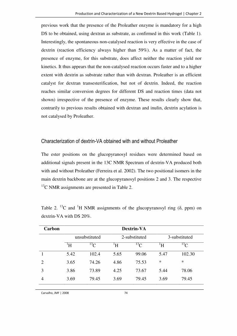
Production and Characterization of a New Dextrin Based Hydrogel | Chapter 2
Carvalho, JMF | 2008 74
previous work that the presence of the Proleather enzyme is mandatory for a high
DS to be obtained, using dextran as substrate, as confirmed in this work (Table 1).
Interestingly, the spontaneous non-catalysed reaction is very effective in the case of
dextrin (reaction efficiency always higher than 59%). As a matter of fact, the
presence of enzyme, for this substrate, does affect neither the reaction yield nor
kinetics. It thus appears that the non-catalysed reaction occurs faster and to a higher
extent with dextrin as substrate rather than with dextran. Proleather is an efficient
catalyst for dextran transesterification, but not of dextrin. Indeed, the reaction
reaches similar conversion degrees for different DS and reaction times (data not
shown) irrespective of the presence of enzyme. These results clearly show that,
contrarily to previous results obtained with dextran and inulin, dextrin acylation is
not catalysed by Proleather.
Characterization of dextrin-VA obtained with and without Proleather
The ester positions on the glucopyranosyl residues were determined based on
additional signals present in the 13C NMR Spectrum of dextrin-VA produced both
with and without Proleather (Ferreira et al. 2002). The two positional isomers in the
main dextrin backbone are at the glucopyranosyl positions 2 and 3. The respective 13C NMR assignments are presented in Table 2.
Table 2. 13C and 1H NMR assignments of the glucopyranosyl ring (δ, ppm) on
dextrin-VA with DS 20%.
Carbon Dextrin-VA
unsubstituted 2-substituted 3-substituted 1H 13C 1H 13C 1H 13C
1 5.42 102.4 5.65 99.06 5.47 102.30
2 3.65 74.26 4.86 75.53 * *
3 3.86 73.89 4.25 73.67 5.44 78.06
4 3.69 79.45 3.69 79.45 3.69 79.45
Production and Characterization of a New Dextrin Based Hydrogel | Chapter 2
Carvalho, JMF | 2008 75
5 3.99 76.05 3.99 76.05 3.99 76.05
6 3.88 63.18 3.88 63.18 3.88 63.18
*Signals not identified.
Bi-dimensional (HSQC) NMR experiments were carried out in order to evaluate
the isomer ratio in dextrin-VA. The area of the three peaks, C1, C1-S2 and C1-S3,
was determined using a Lorentzian adjust, and the isomer ratios calculated. For
dextrin-VA with DS 20% obtained by chemical reaction, an isomer ratio of 70:30,
at positions 2 and 3, was found. For the enzymatic reaction a similar substitution
pattern was observed (the ratio of isomers at positions 2 and 3 was 76:24).
Additionally, the regioisomer at position 2 was more highly favored (two times).
Again, it appears that the enzyme does not influence the reaction, since the isomers
obtained are the same, irrespective of the presence of the enzyme. Thus, this work
demonstrates that it is possible to regioselectively modified dextrin without using
enzymes.
Effect of water amount and activity on the transesterification reaction
The transesterification reaction was carried out using mixtures of DMSO with
different amounts of water, corresponding to different values of aw (Figure 3). The
values of aw and the corresponding dextrin DS obtained are show in and 2 and table
3. Table 3 shows that the reaction is switched towards synthesis for aw <0.3. Low
amounts of water (in the range 0 to 20%) correspond to similar aw, and lead to
similar DS. Higher values of aw (≥0.4) result in a dramatic reduction of dextrin
modification. These results are clearly different from the effect of aw described by
Ferreira and colleagues for the catalysed reaction, with dextran. In this case, a very
low amount of water is mandatory for the enzymatic reaction to be carried out,
while in our experiment (with dextrin) as much as 20% of water (aw≈0.3) may be
present without loss of synthesis reaction.
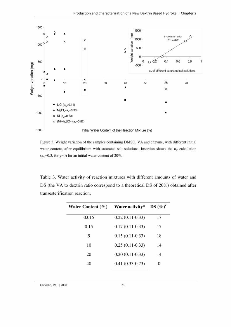
Production and Characterization of a New Dextrin Based Hydrogel | Chapter 2
Carvalho, JMF | 2008 76
Figure 3. Weight variation of the samples containing DMSO, VA and enzyme, with different initial
water content, after equilibrium with saturated salt solutions. Insertion shows the aw calculation
(aw=0.3, for y=0) for an initial water content of 20%.
Table 3. Water activity of reaction mixtures with different amounts of water and
DS (the VA to dextrin ratio correspond to a theoretical DS of 20%) obtained after
transesterification reaction.
Water Content (%) Water activity* DS (%)c
0.015 0.22 (0.11-0.33) 17
0.15 0.17 (0.11-0.33) 17
5 0.15 (0.11-0.33) 18
10 0.25 (0.11-0.33) 14
20 0.30 (0.11-0.33) 14
40 0.41 (0.33-0.73) 0
-1500
-1000
-500
0
500
1000
1500
0 10 20 30 40 50 60 70
Initial Water Content of the Reaction Mixture (%)
Weig
ht
variation (
mg)
Série2
Série3
Série4
Série5
LiCl (aw=0.11)
MgCl2 (aw=0.33)
KI (aw=0.73)
(NH4)2SO4 (aw=0.82)
y = 2068,6x - 615,1
R2 = 0,9899
-500
0
500
1000
1500
0 0,2 0,4 0,6 0,8 1
aw of different saturated salt solutions
We
igh
t va
ria
tio
n
(mg
)-1500
-1000
-500
0
500
1000
1500
0 10 20 30 40 50 60 70
Initial Water Content of the Reaction Mixture (%)
Weig
ht
variation (
mg)
Série2
Série3
Série4
Série5
LiCl (aw=0.11)
MgCl2 (aw=0.33)
KI (aw=0.73)
(NH4)2SO4 (aw=0.82)
y = 2068,6x - 615,1
R2 = 0,9899
-500
0
500
1000
1500
0 0,2 0,4 0,6 0,8 1
aw of different saturated salt solutions
We
igh
t va
ria
tio
n
(mg
)
Production and Characterization of a New Dextrin Based Hydrogel | Chapter 2
Carvalho, JMF | 2008 77
60 0.95 (0.82-1) 0
80 ~1 0
100 1 0
*aw estimated as shown in figure 2. The actual aw value is in the range between brackets.
B. Hydrogel production and NMR characterization
The acrylate groups in dextrin-VA were polymerized using APS and TEMED as
free radical initiators. Aqueous solutions of dextrin-VA with different DS (ranging
from ca. 10 to 70%) and with two concentrations (33% and 44% (w/v)), were
polymerized at room temperature, and gelation occurred within less than 5 min.
The obtained hydrogel slabs were almost transparent, the opacity increasing with
decreasing in DS values, which is likely related to the better solubility of the more
highly substituted material. The hydrogels were characterized using the solid state
NMR technique of 13C CP-MAS. This analysis, as described ahead, allowed the
detection of VA adsorbed to dextrin, covalently linked to dextrin, and finally of the
VA reticulating the hydrogel.
In the 13C CP/MAS spectra of dextrin or similar samples, the peak chemical shifts
are sufficiently similar to those observed in the solution state to allow peak
assignment to be made. In order to identify the environments of non-linked dextrin
and VA moieties, a sample of dextrin wetted with vinyl acrylate was prepared and
analysed. The spectrum obtained (Figure 4) clearly shows the glucopyranosyl
carbons in the 58-110 ppm and the acrylate carbons in the 120-175 ppm region.
Signals at 90-105 ppm and 58-65 ppm are attributed to C1 and C6 carbons in
hexapyranoses, respectively. The overlapping signal at about 68-78 ppm is
associated with C2, C3 and C5. The resonance that appears at 82 ppm is thought to
arise from C4 carbons in noncrystalline material (Langer et al. 1992). Regarding
VA carbons, the peak resonating at 128 ppm arises from the carbons engaged in the
double bond whereas the peak at 163 ppm corresponds to the carbonyl carbons
(arrow in Figure 4). Both these positions should thus be characteristic of the VA
environments when simply adsorbed to the dextrin. And 5 shows the downfield
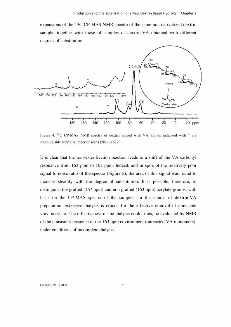
Production and Characterization of a New Dextrin Based Hydrogel | Chapter 2
Carvalho, JMF | 2008 78
expansions of the 13C CP-MAS NMR spectra of the same non derivatized dextrin
sample, together with those of samples of dextrin-VA obtained with different
degrees of substitution.
Figure 4. 13C CP-MAS NMR spectra of dextrin mixed with VA. Bands indicated with * are
spinning side bands. Number of scans (NS) =16729.
It is clear that the transesterification reaction leads to a shift of the VA carbonyl
resonance from 163 ppm to 167 ppm. Indeed, and in spite of the relatively poor
signal to noise ratio of the spectra (Figure 5), the area of this signal was found to
increase steadily with the degree of substitution. It is possible, therefore, to
distinguish the grafted (167 ppm) and non grafted (163 ppm) acrylate groups, with
basis on the CP-MAS spectra of the samples. In the course of dextrin-VA
preparation, extensive dialysis is crucial for the effective removal of unreacted
vinyl acrylate. The effectiveness of the dialysis could, thus, be evaluated by NMR
of the consistent presence of the 163 ppm environment (unreacted VA monomers),
under conditions of incomplete dialysis.
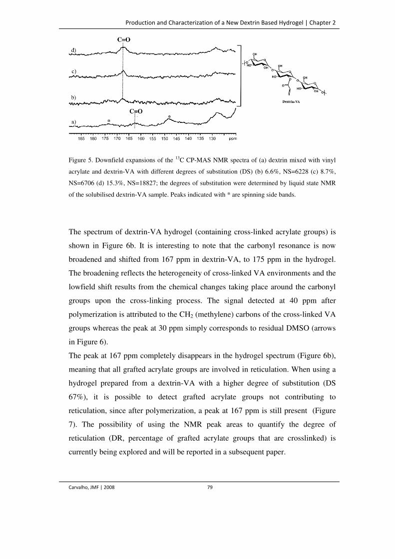
Production and Characterization of a New Dextrin Based Hydrogel | Chapter 2
Carvalho, JMF | 2008 79
Figure 5. Downfield expansions of the 13C CP-MAS NMR spectra of (a) dextrin mixed with vinyl
acrylate and dextrin-VA with different degrees of substitution (DS) (b) 6.6%, NS=6228 (c) 8.7%,
NS=6706 (d) 15.3%, NS=18827; the degrees of substitution were determined by liquid state NMR
of the solubilised dextrin-VA sample. Peaks indicated with * are spinning side bands.
The spectrum of dextrin-VA hydrogel (containing cross-linked acrylate groups) is
shown in Figure 6b. It is interesting to note that the carbonyl resonance is now
broadened and shifted from 167 ppm in dextrin-VA, to 175 ppm in the hydrogel.
The broadening reflects the heterogeneity of cross-linked VA environments and the
lowfield shift results from the chemical changes taking place around the carbonyl
groups upon the cross-linking process. The signal detected at 40 ppm after
polymerization is attributed to the CH2 (methylene) carbons of the cross-linked VA
groups whereas the peak at 30 ppm simply corresponds to residual DMSO (arrows
in Figure 6).
The peak at 167 ppm completely disappears in the hydrogel spectrum (Figure 6b),
meaning that all grafted acrylate groups are involved in reticulation. When using a
hydrogel prepared from a dextrin-VA with a higher degree of substitution (DS
67%), it is possible to detect grafted acrylate groups not contributing to
reticulation, since after polymerization, a peak at 167 ppm is still present (Figure
7). The possibility of using the NMR peak areas to quantify the degree of
reticulation (DR, percentage of grafted acrylate groups that are crosslinked) is
currently being explored and will be reported in a subsequent paper.
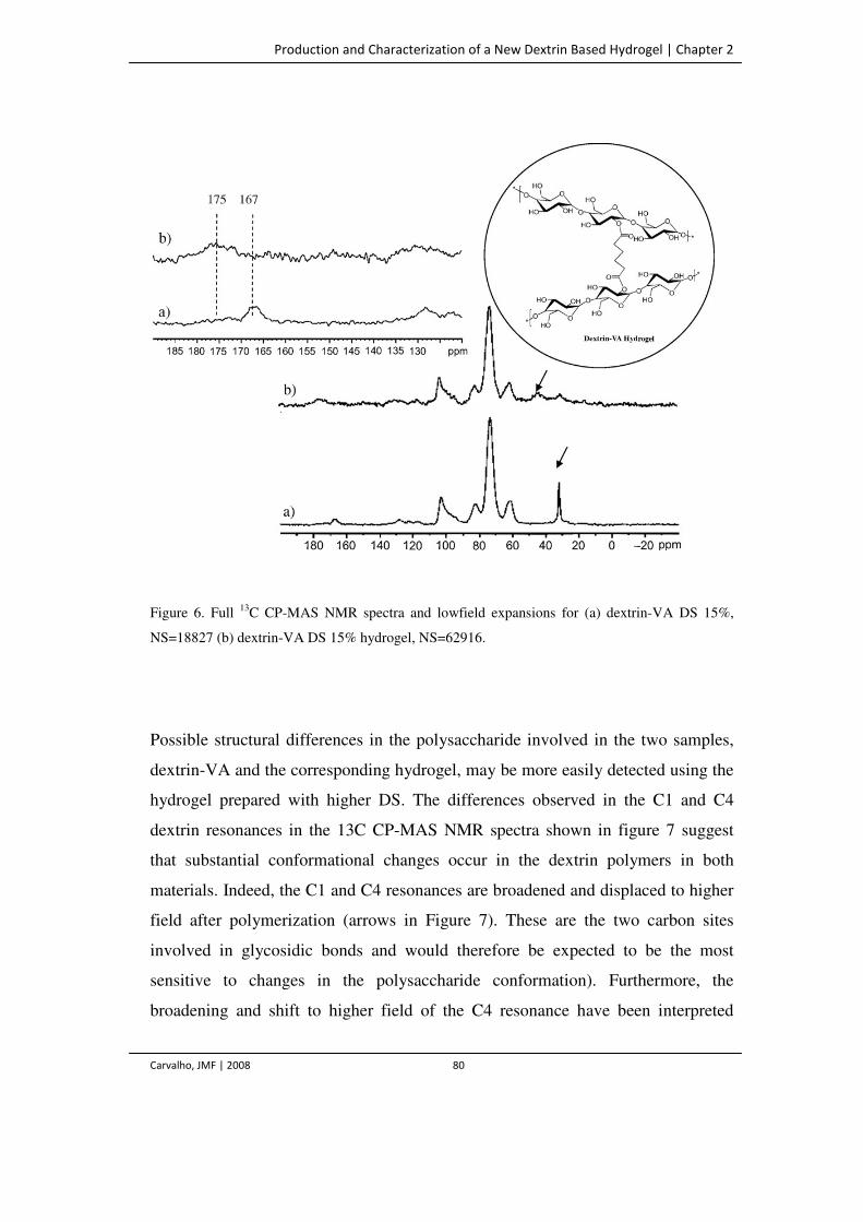
Production and Characterization of a New Dextrin Based Hydrogel | Chapter 2
Carvalho, JMF | 2008 80
Figure 6. Full 13C CP-MAS NMR spectra and lowfield expansions for (a) dextrin-VA DS 15%,
NS=18827 (b) dextrin-VA DS 15% hydrogel, NS=62916.
Possible structural differences in the polysaccharide involved in the two samples,
dextrin-VA and the corresponding hydrogel, may be more easily detected using the
hydrogel prepared with higher DS. The differences observed in the C1 and C4
dextrin resonances in the 13C CP-MAS NMR spectra shown in figure 7 suggest
that substantial conformational changes occur in the dextrin polymers in both
materials. Indeed, the C1 and C4 resonances are broadened and displaced to higher
field after polymerization (arrows in Figure 7). These are the two carbon sites
involved in glycosidic bonds and would therefore be expected to be the most
sensitive to changes in the polysaccharide conformation). Furthermore, the
broadening and shift to higher field of the C4 resonance have been interpreted
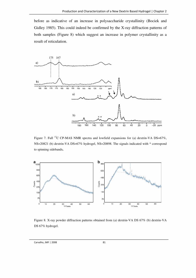
Production and Characterization of a New Dextrin Based Hydrogel | Chapter 2
Carvalho, JMF | 2008 81
before as indicative of an increase in polysaccharide crystallinity (Bociek and
Gidley 1985). This could indeed be confirmed by the X-ray diffraction patterns of
both samples (Figure 8) which suggest an increase in polymer crystallinity as a
result of reticulation.
Figure 7. Full 13C CP-MAS NMR spectra and lowfield expansions for (a) dextrin-VA DS=67%,
NS=20821 (b) dextrin-VA DS=67% hydrogel, NS=20898. The signals indicated with * correspond
to spinning sidebands.
Figure 8. X-ray powder diffraction patterns obtained from (a) dextrin-VA DS 67% (b) dextrin-VA
DS 67% hydrogel.
Production and Characterization of a New Dextrin Based Hydrogel | Chapter 2
Carvalho, JMF | 2008 82
Conclusions
A method previously developed by Ferreira and colleagues was in this work
applied in the production of dextrin hydrogels. Surprisingly, it was verified that,
although Proleather effectively catalyses the transesterification of acrylate
molecules to dextran and inulin, dextrin is not a substrate for this enzyme.
Nevertheless, it has been possible to produce dextrin with varying amounts of
grafted acrylate groups, by varying the VA concentration. Also surprising was the
finding that the regioselectivity of the non-catalysed reaction is comparable to the
one obtained with the enzyme-catalysed reaction, C2 and C3 being the preferred
acylation positions. Derivatized dextrin can be polymerized in aqueous solution by
radical polymerization with APS and TEMED.
Combining the ability to tailor the hydrogel properties to fit a wide range of
characteristics, with the biocompatibility of the starch-based materials, it is
possible to obtain dextrin crosslinked networks which might be very attractive for
biomedical applications. Although starch hydrogels have been reported by other
authors, the approach used in this work is different, and the basis for a
comprehensive NMR structural analysis has also been developed. Current work is
in progress with the purpose of quantifying, through solid NMR analysis, the
several kinds of VA present in the hydrogels. Rheological and biocompatibility
studies have been performed and will be described elsewhere.
References
Akkara JA, Ayyagari MSR and Bruno FF. (1999) Enzymatic synthesis and modification of
polymers in nonaqueous solvents. TIBTECH Reviews 17: 67–73.
Bociek SM and Gidley MJ. (1985) Molecular Organization in Starches: A 13C CP/MAS NMR.
Study J Am Chem Soc 107: 7040–7044.
Chen T, Embree HD, Brown EM, Taylor MM and Payne GF. (2003) Enzyme-catalyzed gel
formation of gelatine and chitosan: potential for in situ applications. Biomaterials 24: 2831–2841.
Production and Characterization of a New Dextrin Based Hydrogel | Chapter 2
Carvalho, JMF | 2008 83
Cooney CL and Hueter J. (1974) Enzyme catalysis in the presence of nonaqueous solvents using
chloroperoxidase. Biotechnol Bioeng 16: 1044–1053.
Drury JL and Mooney DJ. (2003) Hydrogels for tissue engineering: scaffold variables and
applications. Biomaterials 24: 4337-4351.
Ferreira L, Gil MH and Dordick JS. (2002) Enzymatic synthesis of inulin-containing hydrogels.
Biomacromolecules 3: 333–341.
Ferreira L, Carvalho R, Gil MH and Dordick JS. (2002) Enzymatic synthesis of dextran-containing
hydrogels. Biomaterials 23: 3957–3967.
Ishihara M, Nakanishi K, Ono K, Sato M, Saito Y, Yura H, Matsui T, Hattori H, Uenoyama M,
Kikuchi M and Kurita A. (2002) Photocrosslinkable chitosan as a dressing wound occlusion and
accelerator in healing process. Biomaterials 23: 833–840.
Kane JB, Tompkins RG, Yarmush ML and Burke JF. (1996) Burn dressings. In Biomaterials
science: an introduction to materials in medicine. Academic Press, San Diego, USA, pp 360–370.
Klibanov AM. (1997) Why are enzymes less active in organic solvents than in water? Trends
Biotechnol 15: 97–101.
Klibanov AM. (2001) Improving enzymes by using them in organic solvents. Nature 409: 241–246.
Langer R, Kost J, Shefer S and Shefer A. (1992) Structural characterization of starch networks in
the solid state by cross-polarization magic-angle-spinning 13C NMR spectroscopy and wide angle x-
ray diffraction. Macromolecules 25: 6756–6760.
Marques AP, Reis RL and Hunt JA. (2002) The biocompatibility of novel starch-based polymers
and composites: in vitro studies. Biomaterials 23: 1471– 1478.
McCulloch I and Shalaby SW. (1998) In Tailored polymeric materials for controlled delivery
systems. American Chemical Society, Washington DC, USA.
Multon JL and Bizot H. (1978) Intermediate moisture foods and water activity determination. Ann
Nutr Aliment. 32: 631–654.
Peppas NA, Bures P, Leobandung W and Ichikawa H. (2000) Hydrogels in pharmaceutical
formulations. Eur J Pharm Biopharm 50: 27– 46.
Peppas NA. (1996) Hydrogels. In Biomaterials science: an introduction to materials in medicine.
Academic Press, San Diego, USA, pp 60–64.
Production and Characterization of a New Dextrin Based Hydrogel | Chapter 2
Carvalho, JMF | 2008 84
Sergeeva MV, Paradkar VM and Bordick JS. (1997) Peptide synthesis using proteases dissolved in
organic solvents. Enz Microb Technol 20: 263–268.
Vervoort L, van den Mooter G, Augustijns P, Busson R, Toppet S and Kinget R. (1997) Inulin
hydrogels as carriers for colonic drug targeting: I Synthesis and characterization of methacrylated
insulin and hydrogel formation. Pharm Res 14: 1730–1737.
Wong WH and Mooney DJ. (1997) Synthesis and properties of biodegradable polymers used as
synthetic matrices for tissue engineering. In Synthetic biodegradable polymer scaffolds. Birkhaüser,
Boston, USA, pp 51–82.
Zacks A and Klibanov AM. (1985) Enzyme-catalyzed processes in organic solvents. Proc Natl
Acad Sci USA 82: 8192–3196.
Zhang L-M, Yang C and Yan L. (2005) Perspectives on: strategies to fabricate starch based
hydrogels with potential biomedical applications. J Bioact Compat Pol 20: 297–314.
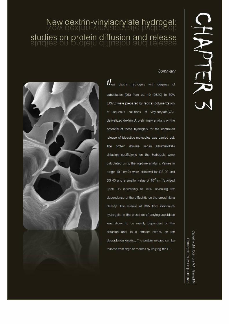

New Dextrin Vinylacrylate Hydrogel: Studies on Protein Diffusion and Release | Chapter 3
Carvalho, JMF | 2008 87
Introduction
Biodegradable hydrogels are useful for a variety of medical applications (Dordick
et al. 1994, Park and Park 1996, Han et al. 2000). In general, hydrogels have a
good biocompatibility and the soft rubbery consistence resemble natural tissues,
allowing for a favorable controlled interaction with living systems (Vyavahare and
Kohn, 1994). Hydrogels may be used in the field of tissue engineering, as scaffolds
to support and promote tissue regeneration and also as attractive systems for the
controlled release of pharmaceutically active molecules (Peppas 1996, Kane et al.
1996, McCulloch and Shalaby 1998, Peppas et al. 2000). A drug delivery vehicle
positioned in the proximity of the site of disease or injury can release the drug in
the desired location, this way reducing the side effects that usually result from
systemic administration. Additionally, depending on the crosslinking density, the
hydrogels structure can restrict the diffusion of macromolecules, being able to
deliver the therapeutic agent over extended periods of time. Indeed, improving in
the patient compliance and extension of product life are major advantages of the
drug delivery systems (Hubbell 1995, Ratner 2002). Ideal systems for drug delivery
are degradable, permeable, porous and capable of maintaining a desired shape. In
previous work, we have described a new dextrin based hydrogel, which could
possibly match these properties (Carvalho et al. 2007). Dextrin is a glucose
containing polymer linked by α-(1→4) D-glucose units, having the same general
formula as starch, but smaller and less complex.
In this study, we aim at evaluating the diffusion of two different molecules in the
hydrogel matrixes, glucose and a model protein, bovine serum albumin (BSA).
This will allow achieving a second goal, to evaluate the potential of the hydrogels
as controlled release systems. For these purposes, the enzyme amyloglucosidase
was used to modulate the release of proteins entrapped in the hydrogel. Thus, the
degradation rate of the network may be controlled through the crosslink density of
the hydrogels and the amount of enzyme present.
New Dextrin Vinylacrylate Hydrogel: Studies on Protein Diffusion and Release | Chapter 3
Carvalho, JMF | 2008 88
Materials and Methods
Materials
Dextrin - Koldex 60 starch was a generous gift from Tate & Lyle (Decatur, IL,
USA). Vinyl acrylate (VA) was from Aldrich, N,N,N’,N’-
tetramethylenethylenediamine (TEMED), ammonium persulfate (APS) and Protein
Assay were purchased from BioRad, dimethylsulfoxide (DMSO) and acetone were
from AppliChem and polyethyleneglicol (PEG 200 g/mol) was obtained from
Riedel-d Haën. DMSO was dried with 0.4 nm molecular sieves at least overnight
before use. BSA was from Sigma (Mw= 67 kDa). Regenerated cellulose tubular
membranes with 3500 molecular weight cutoff (MWCO) were obtained from
Membrane Filtration Products (Seguin, USA)
Carbohydrate analysis
Dextrin sugars were released by 1 M sulfuric acid hydrolysis at 100 ºC for 2.5 h
and analysed as their alditol acetates by gas chromatography using a Carlo Erba
6000 with a split injector (split ratio 1:60) and a flame ionisation detector as
described by Mafra et al. (2001). For determination of the linkage glycosidic
composition, the dextrin sample was activated with powdered NaOH in dry
dimethylsuphoxide and methylated with CH3I (Ciucanu and Kerek 1984). The
methylated material was dissolved in dichloromethane:water (1:1, v/v), and the
organic phase was recovered, washed several times with water, evaporated, and
freeze dried. The methylated polysaccharides were hydrolyzed with 2 M TFA at
121 ºC for 1 h, reduced by NaBD4 and acetylated in the presence of acetic
anhydride and 1-methylimidazole. The partially methylated alditol acetates were
analysed by GC-MS using an OV-1 capillary column as described by Cardoso et al.
(2007). Triplicates were made of all analyses.
New Dextrin Vinylacrylate Hydrogel: Studies on Protein Diffusion and Release | Chapter 3
Carvalho, JMF | 2008 89
Preparation of dextrin-VA hydrogels
Dextrin-VA monomers were synthesized from dextrin in DMSO in the presence of
different amounts of vinyl acrylate. The transesterification reaction was carried out
at 50ºC for 72 h. The degree of acrylate substitution (DS) was determined by
proton nuclear resonance spectroscopy (1H-NMR) in D2O as previously described
(Carvalho et al. 2007). Briefly, 1H-NMR spectra were recorded in D2O (10 mg in 1
mL) and DS was calculated using equation 1:
where, in the NMR spectra, x is the sum of the peak intensities corresponding to
the protons from vinyl group (δ in the range of 6.00-7.00 ppm) and y is the sum of
the peak intensities of all dextrin protons (δ in the range of 0.00-6.00 ppm),
excluding the water peak (δ around 4.80 ppm) (Figure 1). The hydrogel slabs with
different DS (from ca. 10 to 70%) were prepared by radical polymerization of
aqueous solution of Dextrin-VA. These solutions were prepared by dissolving
dextrin-VA (300 or 400 mg) in buffer (900 µL) (phosphate buffer 0.2 M, pH 8.0
with 0.02% sodium azide) and bubbling with nitrogen for 2 min. For the release
experiments, before gelation, a BSA solution (in phosphate buffer) was added
yielding a final concentration of 2 g/L. The gelation reactions were initiated by
adding 90 µL APS (80 mg/ml in phosphate buffer) and 90 µL TEMED (13.6 %
(v/v) in water, pH adjusted to 8.0 with HCl) and allowed to occur for 30 min at
room temperature. For the preparation of dextrin-VA/amyloglucosidase (AMG)
hydrogels, different amounts of enzyme solution (diluted to 10 U/mL in phosphate
buffer) was added to the dextrin-VA solution before the addition of the gelation
reagents, as described above.
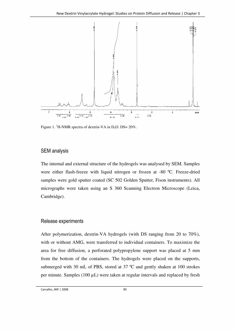
New Dextrin Vinylacrylate Hydrogel: Studies on Protein Diffusion and Release | Chapter 3
Carvalho, JMF | 2008 90
Figure 1. 1H-NMR spectra of dextrin-VA in D2O. DS= 20%.
SEM analysis
The internal and external structure of the hydrogels was analysed by SEM. Samples
were either flash-freeze with liquid nitrogen or frozen at -80 ºC. Freeze-dried
samples were gold sputter coated (SC 502 Golden Sputter, Fison instruments). All
micrographs were taken using an S 360 Scanning Electron Microscope (Leica,
Cambridge).
Release experiments
After polymerization, dextrin-VA hydrogels (with DS ranging from 20 to 70%),
with or without AMG, were transferred to individual containers. To maximize the
area for free diffusion, a perforated polypropylene support was placed at 5 mm
from the bottom of the containers. The hydrogels were placed on the supports,
submerged with 30 mL of PBS, stored at 37 ºC and gently shaken at 100 strokes
per minute. Samples (100 µL) were taken at regular intervals and replaced by fresh
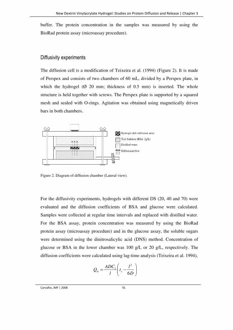
New Dextrin Vinylacrylate Hydrogel: Studies on Protein Diffusion and Release | Chapter 3
Carvalho, JMF | 2008 91
buffer. The protein concentration in the samples was measured by using the
BioRad protein assay (microassay procedure).
Diffusivity experiments
The diffusion cell is a modification of Teixeira et al. (1994) (Figure 2). It is made
of Perspex and consists of two chambers of 60 mL, divided by a Perspex plate, in
which the hydrogel (Ø 20 mm; thickness of 0.5 mm) is inserted. The whole
structure is held together with screws. The Perspex plate is supported by a squared
mesh and sealed with O-rings. Agitation was obtained using magnetically driven
bars in both chambers.
Figure 2. Diagram of diffusion chamber (Lateral view).
For the diffusivity experiments, hydrogels with different DS (20, 40 and 70) were
evaluated and the diffusion coefficients of BSA and glucose were calculated.
Samples were collected at regular time intervals and replaced with distilled water.
For the BSA assay, protein concentration was measured by using the BioRad
protein assay (microassay procedure) and in the glucose assay, the soluble sugars
were determined using the dinitrosalicylic acid (DNS) method. Concentration of
glucose or BSA in the lower chamber was 100 g/L or 20 g/L, respectively. The
diffusion coefficients were calculated using lag-time analysis (Teixeira et al. 1994),
−=
D
lt
l
ADCQ sts 6
21
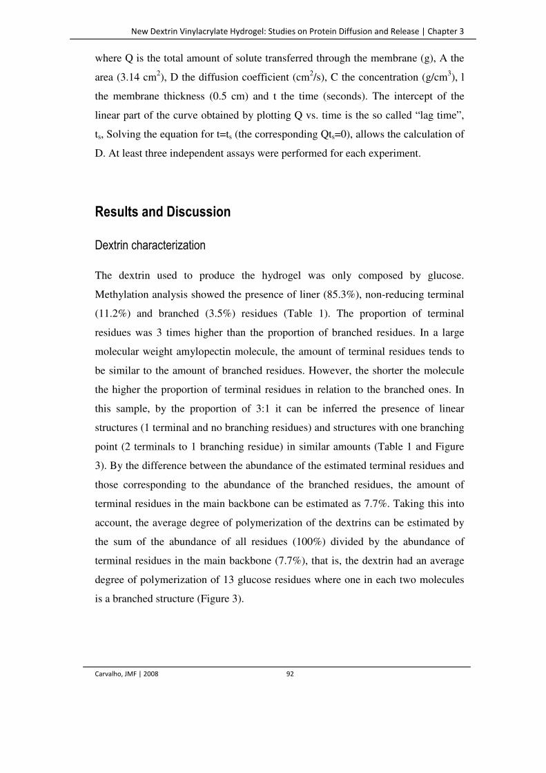
New Dextrin Vinylacrylate Hydrogel: Studies on Protein Diffusion and Release | Chapter 3
Carvalho, JMF | 2008 92
where Q is the total amount of solute transferred through the membrane (g), A the
area (3.14 cm2), D the diffusion coefficient (cm2/s), C the concentration (g/cm3), l
the membrane thickness (0.5 cm) and t the time (seconds). The intercept of the
linear part of the curve obtained by plotting Q vs. time is the so called “lag time”,
ts, Solving the equation for t=ts (the corresponding Qts=0), allows the calculation of
D. At least three independent assays were performed for each experiment.
Results and Discussion
Dextrin characterization
The dextrin used to produce the hydrogel was only composed by glucose.
Methylation analysis showed the presence of liner (85.3%), non-reducing terminal
(11.2%) and branched (3.5%) residues (Table 1). The proportion of terminal
residues was 3 times higher than the proportion of branched residues. In a large
molecular weight amylopectin molecule, the amount of terminal residues tends to
be similar to the amount of branched residues. However, the shorter the molecule
the higher the proportion of terminal residues in relation to the branched ones. In
this sample, by the proportion of 3:1 it can be inferred the presence of linear
structures (1 terminal and no branching residues) and structures with one branching
point (2 terminals to 1 branching residue) in similar amounts (Table 1 and Figure
3). By the difference between the abundance of the estimated terminal residues and
those corresponding to the abundance of the branched residues, the amount of
terminal residues in the main backbone can be estimated as 7.7%. Taking this into
account, the average degree of polymerization of the dextrins can be estimated by
the sum of the abundance of all residues (100%) divided by the abundance of
terminal residues in the main backbone (7.7%), that is, the dextrin had an average
degree of polymerization of 13 glucose residues where one in each two molecules
is a branched structure (Figure 3).
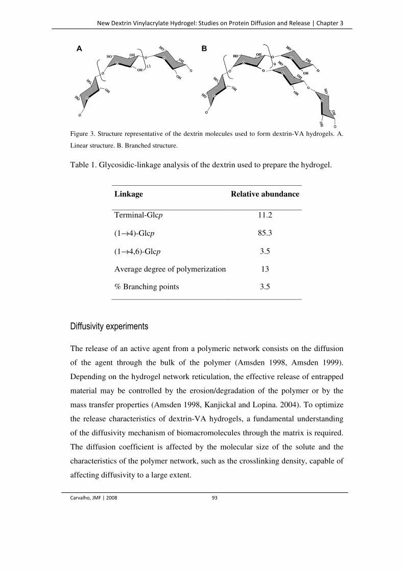
New Dextrin Vinylacrylate Hydrogel: Studies on Protein Diffusion and Release | Chapter 3
Carvalho, JMF | 2008 93
Figure 3. Structure representative of the dextrin molecules used to form dextrin-VA hydrogels. A.
Linear structure. B. Branched structure.
Table 1. Glycosidic-linkage analysis of the dextrin used to prepare the hydrogel.
Linkage Relative abundance
Terminal-Glcp 11.2
(1→4)-Glcp 85.3
(1→4,6)-Glcp 3.5
Average degree of polymerization 13
% Branching points 3.5
Diffusivity experiments
The release of an active agent from a polymeric network consists on the diffusion
of the agent through the bulk of the polymer (Amsden 1998, Amsden 1999).
Depending on the hydrogel network reticulation, the effective release of entrapped
material may be controlled by the erosion/degradation of the polymer or by the
mass transfer properties (Amsden 1998, Kanjickal and Lopina. 2004). To optimize
the release characteristics of dextrin-VA hydrogels, a fundamental understanding
of the diffusivity mechanism of biomacromolecules through the matrix is required.
The diffusion coefficient is affected by the molecular size of the solute and the
characteristics of the polymer network, such as the crosslinking density, capable of
affecting diffusivity to a large extent.
New Dextrin Vinylacrylate Hydrogel: Studies on Protein Diffusion and Release | Chapter 3
Carvalho, JMF | 2008 94
Figure 4 shows the different diffusion behavior of BSA (67 kDa) and glucose.
After a lag-phase, the total amount of solute transferred through the hydrogel is
proportional to the time. The diffusivity coefficients were determined using
hydrogels with different DS, ranging from 20 to 70% (Table 2). The diffusion
coefficient of BSA on the DS 20 hydrogel was 4.2 ± 1.6 × 10⁻⁷ cm2/s. This value is
only slightly lower than the one obtained by Lévesque et al. (2005), for BSA
diffusivity in water (5.9 × 10⁻⁷ cm2/s) and in the order of magnitude of the value
obtained with Dex-MA hydrogels (3.1 ± 0.2 × 10⁻⁷ cm2/s). The diffusivity in water
and in the hydrogel is thus in the same range, suggesting that these matrices
probably have continuous water and polymer phases with interconnected pores. In
fact, SEM analysis of three-dimensional hydrogel slabs revealed a highly porous
structure (Figure 5).
Figure 4. Protein and glucose transferred through the membrane (DS=20%), vs time, in the
diffusivity experiments.
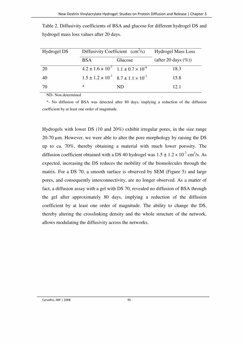
New Dextrin Vinylacrylate Hydrogel: Studies on Protein Diffusion and Release | Chapter 3
Carvalho, JMF | 2008 95
Table 2. Diffusivity coefficients of BSA and glucose for different hydrogel DS and
hydrogel mass loss values after 20 days.
Hydrogel DS Diffusivity Coefficient (cm2/s) Hydrogel Mass Loss
(after 20 days (%)) BSA Glucose
20 4.2 ± 1.6 × 10⁻⁷ 1.1 ± 0.7 × 10⁻⁶ 18.3
40 1.5 ± 1.2 × 10⁻⁷ 8.7 ± 1.1 × 10⁻⁷ 15.8
70 * ND 12.1
ND- Non determined
*- No diffusion of BSA was detected after 80 days, implying a reduction of the diffusion
coefficient by at least one order of magnitude.
Hydrogels with lower DS (10 and 20%) exhibit irregular pores, in the size range
20-70 µm. However, we were able to alter the pore morphology by raising the DS
up to ca. 70%, thereby obtaining a material with much lower porosity. The
diffusion coefficient obtained with a DS 40 hydrogel was 1.5 ± 1.2 × 10⁻⁷ cm2/s. As
expected, increasing the DS reduces the mobility of the biomolecules through the
matrix. For a DS 70, a smooth surface is observed by SEM (Figure 5) and large
pores, and consequently interconnectivity, are no longer observed. As a matter of
fact, a diffusion assay with a gel with DS 70, revealed no diffusion of BSA through
the gel after approximately 80 days, implying a reduction of the diffusion
coefficient by at least one order of magnitude. The ability to change the DS,
thereby altering the crosslinking density and the whole structure of the network,
allows modulating the diffusivity across the networks.
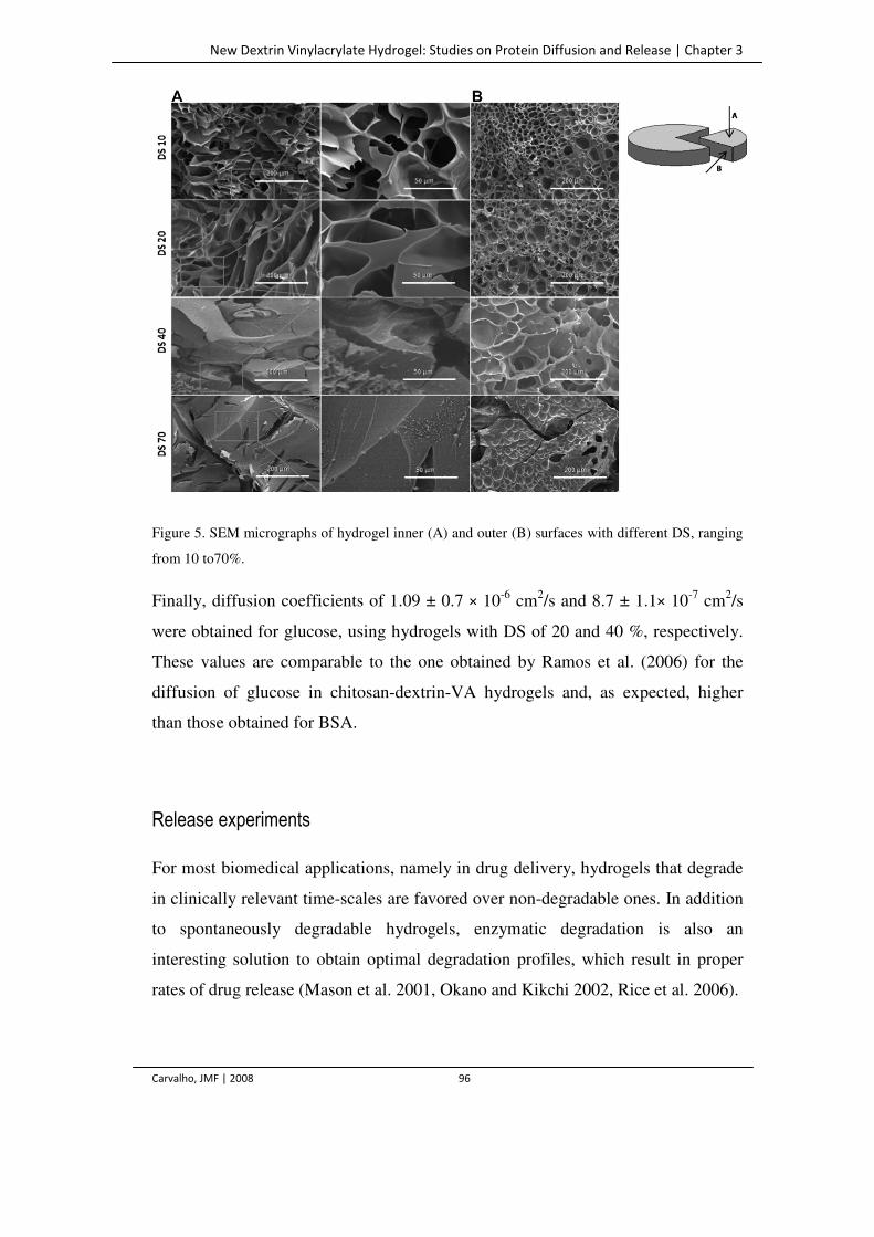
New Dextrin Vinylacrylate Hydrogel: Studies on Protein Diffusion and Release | Chapter 3
Carvalho, JMF | 2008 96
Figure 5. SEM micrographs of hydrogel inner (A) and outer (B) surfaces with different DS, ranging
from 10 to70%.
Finally, diffusion coefficients of 1.09 ± 0.7 × 10⁻⁶ cm2/s and 8.7 ± 1.1× 10⁻⁷ cm2/s
were obtained for glucose, using hydrogels with DS of 20 and 40 %, respectively.
These values are comparable to the one obtained by Ramos et al. (2006) for the
diffusion of glucose in chitosan-dextrin-VA hydrogels and, as expected, higher
than those obtained for BSA.
Release experiments
For most biomedical applications, namely in drug delivery, hydrogels that degrade
in clinically relevant time-scales are favored over non-degradable ones. In addition
to spontaneously degradable hydrogels, enzymatic degradation is also an
interesting solution to obtain optimal degradation profiles, which result in proper
rates of drug release (Mason et al. 2001, Okano and Kikchi 2002, Rice et al. 2006).
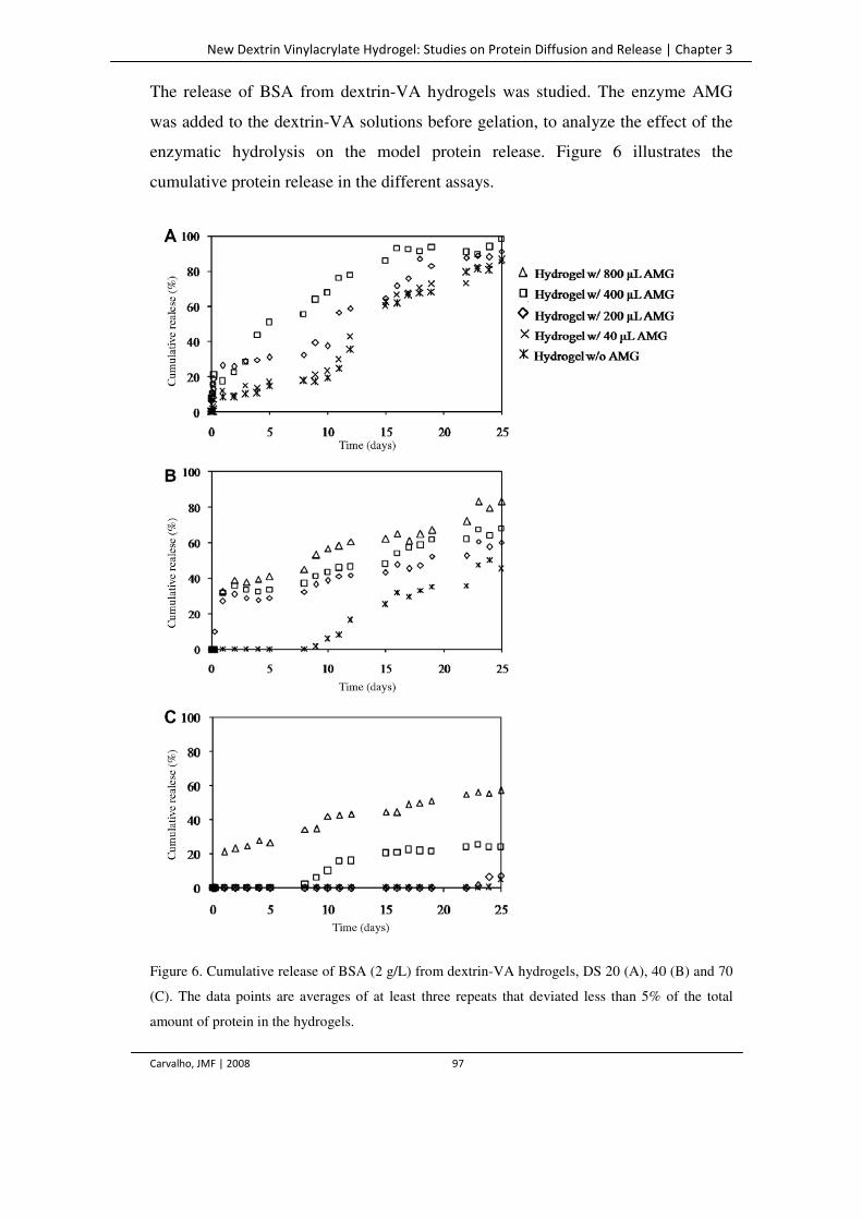
New Dextrin Vinylacrylate Hydrogel: Studies on Protein Diffusion and Release | Chapter 3
Carvalho, JMF | 2008 97
The release of BSA from dextrin-VA hydrogels was studied. The enzyme AMG
was added to the dextrin-VA solutions before gelation, to analyze the effect of the
enzymatic hydrolysis on the model protein release. Figure 6 illustrates the
cumulative protein release in the different assays.
Figure 6. Cumulative release of BSA (2 g/L) from dextrin-VA hydrogels, DS 20 (A), 40 (B) and 70
(C). The data points are averages of at least three repeats that deviated less than 5% of the total
amount of protein in the hydrogels.
New Dextrin Vinylacrylate Hydrogel: Studies on Protein Diffusion and Release | Chapter 3
Carvalho, JMF | 2008 98
For the DS 20 hydrogel (Figure 6A), a two stage release of protein is observed in
all hydrogel samples, the rate slowing down through the time course of the
experiment. In the initial incubation period (from 0 to 6 h), only a small amount of
BSA was released from all samples, corresponding to between 10 and 25% of the
total amount of protein in the hydrogels. After approximately 20 days, most of the
protein was effectively released from the gel, in all assays.
As it is well known, diffusion might not be the only factor determining the delivery
rate, degradation playing also a critical role in determining overall release profiles.
Degradation of crosslinks increases the mesh size of the gel, allowing for diffusion
to be facilitated. Thus, the release of a molecule entrapped in a hydrogel network is
controlled by (1) the diffusion through the matrix or (2) by hydrolysis of the
network crosslinks, followed by an erosion process with mass loss (Dijk-Wolthuis
et al. 1997, Amsden 1999). While either diffusion or dissolution can be the
predominant factor for a specific type of polymer, in most cases, the release
kinetics is a result of a combination of these two mechanisms (Sujja-areevath et al.
1998, Lee 1981, Tahara et al. 1995, Efentakis and Buckton 2002).
As can be seen in Table 2, after 20 days of incubation, for DS 20, only
approximately 20% of the hydrogel had undergone solubilization, according to dry
weight measurements. Such mass loss reflects the release of non-reticulated
material, since no significant degradation take place in the absence of the enzyme.
As a matter of fact, the incubation of the hydrogel in water for up to 3 months did
not lead to further mass release. Diffusion, rather than the enzymatic degradation,
is the governing factor for the release in these hydrogels (DS 20). Indeed, all of the
protein is released even in the case where no AMG is used, which again shows that
the protein is able to freely diffuse in the hydrogel network. The presence of
increasing amounts of enzyme leads to higher BSA release rate, an effect that may
be classified as moderate. As a matter of fact, as shown in the previous section, the
protein diffusion in this hydrogel compares with the diffusion in water, hence the
moderate effect observed in the presence of AMG.
New Dextrin Vinylacrylate Hydrogel: Studies on Protein Diffusion and Release | Chapter 3
Carvalho, JMF | 2008 99
As can also be seen in figure 6, for higher DS values the total amount released from
degrading hydrogels (i.e., with AMG) was always much larger compared with the
control (without enzyme), a process that seems to occur in an enzyme-
concentration dependent manner. These results reveal that the presence of the
enzyme, by hastening the degradation of the hydrogels, significantly affect the
release rate (Figure 6, B and C). As can be seen, namely for the DS 70 hydrogel
sample, in the absence of the enzyme, almost no release is detected, showing that,
contrarily to the DS 20 gel, in which the release is mostly diffusional, for hydrogels
with a rather high DS, the release behavior is driven by the enzyme concentration.
In these cases, the higher crosslinking density prevents the free diffusion of the
molecules. The network mesh is tighter, with subsequent reduced mobility.
Likewise, the enzymatic cleavage of polymer chains raises the degradation process,
reducing the time scale of the slow natural bulk erosion, being thereby responsible
for the control of the release rate.
These results show that the control of DS and of enzyme concentration allow for
the establishment of release systems, controlled over different ranges of time, from
days (low DS, higher enzyme) to months (higher DS, no enzyme). As reported by
Meyvis et al. (2001) with dextran-MA hydrogels, more recently by Vlugt-Wensink
et al. (2006) and confirmed with our diffusivity results, an increase in DS of the
hydrogel, thereby increasing the crosslinking density is an expeditious method to
lengthen the duration of protein delivery. Taken together, all these variables can
provide a versatile method of fine tuning the protein release rate.
Conclusions
The BSA release from the dextrin-VA hydrogel with DS 20 is mainly controlled by
the diffusion rate. For hydrogels with higher DS, the enzymatic hydrolysis/erosion
governs the release rate. By using hydrogels with different DS and enzyme
New Dextrin Vinylacrylate Hydrogel: Studies on Protein Diffusion and Release | Chapter 3
Carvalho, JMF | 2008 100
concentration, it is possible to control the release rate from days to weeks, or even
months.
Acknowledgements
This research was supported by Fundação para a Ciência e a Tecnologia under
grant SFRH / BD / 17482 / 2004 and FUNCARB POCTI/BIO/45356/2002.
References
Amsden B. (1998) Solute diffusion within hydrogels: mechanisms and models. Macromolecules 31:
8382–8395.
Amsden B. (1999) An obstruction-scaling model for diffusion in homogeneous hydrogels.
Macromolecules 32: 874–879.
Cardoso SM, Ferreira JÁ, Mafra I, Silva AMS and Coimbra MA. (2007) Structural ripening-related
changes of the arabinan-rich pectic polysaccharides from olive pulp cell walls. J Agr Food Chem
55: 7124–7130.
Carvalho J, Gonçalves C, Gil AM and Gama FM. (2007) Production and characterization of a new
dextrin based hydrogel. Eur Polym J 43: 3050–3059.
Ciucanu I and Kerek F. (1984) A simple and rapid method for permethylation of carbohydrates.
Carbohydr Res 131: 209–217.
Dijk-Wolthuis WNE, Hoogeboom JAM, van Steenbergen MJ, Tsang SKY and Hennink WE. (1997)
Degradation and release behavior of dextran-based hydrogels. Macromolecules 30: 4639–4645.
Dordick JS, Linhardt RJ and Rethwisch DG. (1994) Chemical and biological catalysis to make
swellable polymers. CHEMTECH 33–39.
Efentakis M and Buckton G. (2002) The effect of erosion and swelling on the dissolution of
theophylline from low and high viscosity sodium alginate matrices. Pharm Dev Tech 7: 69–77.
Han JH, Krochta JM, Kurth MJ and Hsieh Y-L. (2000). Lactitol based poly(ether polyol) hydrogels
for controlled-release chemical and drug delivery systems. J Agr Food Chem 48: 5278 –5282.
New Dextrin Vinylacrylate Hydrogel: Studies on Protein Diffusion and Release | Chapter 3
Carvalho, JMF | 2008 101
Hubbell JA. (1995) Biomaterials in tissue engineering. Biotech 13: 565–576.
Kane JB, Tompkins RG, Yarmush ML and Burke JF. (1996) In Biomaterials science: an
introduction to materials in medicine. Academic Press, San Diego, USA, pp 360–370.
Kanjickal DG and Lopina ST. (2004) Modeling of drug release from polymeric delivery systems.
Crit Rev Ther Drug Carrier Syst 21: 345–386.
Lee PI. (1981) In Controlled release of pesticides and pharmaceuticals. Plenum Press, New York,
USA, pp 39–48.
Lévesque SG, Lima RM and Shoicheta MS. (2005) Macroporous interconnected dextran scaffolds
of controlled porosity for tissue-engineering applications. Biomaterials 26: 7436–7446.
Mafra I, Lanza B, Reis A, Marsilio V, Campestre C, Angelis M and Coimbra MA. (2001) Effect of
ripening on texture, microstructure and cell wall polysaccharide composition of olive fruit. Physiol
Plantarum, 111: 439–447.
Mason MN, Metters AT, Bowman CN and Anseth KS. (2001) Predicting controlled-release
behaviour of degradable PLA-b-PEG-b-PLA hydrogels. Macromolecules 34: 4630–4635.
McCulloch I and Shalaby SW. (1998) In Tailored polymeric materials for controlled delivery
systems. American Chemical Society, Washington DC, USA.
Meyvis T, Smedt DS, Stubbe B, Hennink W and Demeester J. (2001) On the release of proteins
from degrading dextran methacrylate hydrogels and the correlation with the rheologic properties of
the hydrogels. Pharma Res 18: 1593–1599.
Okano T and Kikuchi A. (2002) Pulsatile drug release control using hydrogels. Adv Drug Deliv Rev
54: 53–77.
Park H and Park K. (1996) Hydrogels in bioapplications. In Hydrogels and biodegradable polymers
for bioapplications. American Chemical Society, Ottenbrite, Washington DC, USA, pp 2–10.
Peppas NA, Bures P, Leobandung W and Ichikawa H. (2000) Hydrogels in pharmaceutical
formulations. Euro J Pharm 50: 27– 46.
Peppas NA. (1996) In Biomaterials science: an introduction to materials in medicine. Academic
Press, San Diego, USA, pp 60–64.
Ramos R, Carvalho V and Gama FM. (2006) Novel hydrogel obtained by chitosan and dextrin–VA
copolymerization. Biotech Lett 28: 1279–1284.
New Dextrin Vinylacrylate Hydrogel: Studies on Protein Diffusion and Release | Chapter 3
Carvalho, JMF | 2008 102
Ratner BD. (2002) Reducing capsular thickness and enhancing angiogenesis around implant drug
release systems. J Control Release 78: 211–218.
Rice MA, Sanchez–Adams J and Anseth KS. (2006) Exogeneously triggered, enzymatic
degradation of photopolymerized hydrogels with polycaprolactone subunits: experimental
observations and modelling of mass loss behaviour. Biomacromolecules 7: 1968–1975.
Sujja-areevath J, Munday DL, Cox PJ and Khan KA. (1998) Relationship between swelling, erosion
and drug release in hydrophilic natural gum mini-matrix formulations. Euro J Pharma Sci 6: 207–
217.
Tahara K, Yamamoto K and Nishihata T. (1995) Overall mechanism behind matrix sustained
release (SR) tablets prepared with hydroxypropyl methylcellulose 2910. J Control Release 35: 59–
66.
Teixeira JA, Mota M and Venâncio A. (1994) Model identification and diffusion coefficients
determination of glucose and malic acid in calcium alginate membranes. Chem Eng J 56: 9–14.
Vlugt-Wensink KDK, Vlugt TJH, Wim JW, Crommelin DJA, Verrijk R and Hennink WE. (2006).
Modeling the release of proteins from degrading crosslinked dextran microspheres using kinetic
Monte Carlo simulations. J Control Release 111: 117–127.
Vyavahare N and Kohn J. (1994) Photocrosslinked hydrogels based on copolymers of poly(ethylene
glycol) and lysine. J Polym Sci 32: 1271–1281.
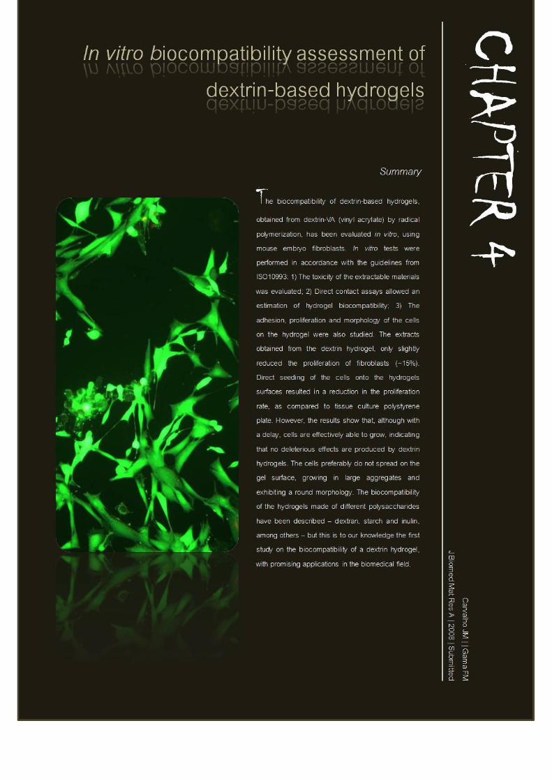

In Vitro Biocompatibility Assessment of Dextrin-Based Hydrogels | Chapter 4
Carvalho, JMF | 2008 105
Introduction
Hydrogels are three-dimensional networks composed of hydrophilic polymers,
capable of absorbing substantial amounts of water, thus resembling natural living
tissues. Their specific chemical and physical properties make hydrogels potentially
suitable for a wide variety of biomedical purposes, including contact lenses,
membranes, biosensors and bioactive scaffolds for tissue engineering (e.g. linings
for artificial hearts or material for artificial skin) (Sgouras and Duncan 1990,
Peppas 1996, Kane et al. 1996, McCulloch and Shalaby 1998, Peppas et al. 2000,
Ishihara et al. 2002, Chen et al. 2003, Drury and Mooney 2003). Hydrogels can
also be applied as implantable delivery systems and as protein- and cell-resistant
coatings (Levy and Andry 1991, Schacht et al. 1993, Di Silvio et al. 1994, Draye et
al. 1998).
The biocompatibility of the hydrogels is an essential issue regarding their
pharmaceutical and biomedical applicability. A material is considered
biocompatible when it does not damage adjacent cells, does not lead to significant
tissue scarring, nor originates a side response that decreases its function (Hansen et
al. 1989, Behl et al. 1994, Berridge et al. 1996). In general, hydrogels have a good
biocompatibility. Since their hydrophilic surface has a high interfacial free energy,
proteins and cells from body fluids have a low tendency to adhere (Peppas 1996,
Hern and Hubble 1998, Hoffman 2002). Moreover, the soft and rubbery
consistence of hydrogel matrixes minimizes irritation of the surrounding tissues.
Recently, we have reported the production of a new dextrin hydrogel (Carvalho et
al. 2007). Dextrin polymers with a varying number of functional groups (VA)
attached to the macromolecular structure (degree of acrylate substitution, DS), may
be produced. These polymers reticulate by radical polymerization, hence allowing
the control of network properties like the swelling ratio, erosion and mass loss
profiles. Herein, we report the results of in vitro cytotoxicity tests performed with
dextrin-VA hydrogels and its extracts, through direct and indirect contact assays,
following the guidelines from ISO10993. Additionally, observations using both
In Vitro Biocompatibility Assessment of Dextrin-Based Hydrogels | Chapter 4
Carvalho, JMF | 2008 106
fluorescence and phase-contrast inverted light microscopy were also carried out as
indicators for cell viability and morphology.
Materials and Methods
Materials
Dextrin was a generous gift from Tate & Lyle. Dimethylsulfoxide, N,N,N’,N’-
tetramethylenediamine (TEMED), and ammonium persulfate (APS) were
purchased from Aldrich (Milwauke, WI). Dextrin acrylates (dextrin-VA) with
different DS were synthesized as described previously (Carvalho et al. 2007).
Dulbecco’s modified Eagle medium (DMEM) with 4500 mg glucose/L, L-
glutamine, 110 mg sodium piruvate/L and NaHCO3 (Sigma) was supplemented
with 10% calf bovine serum (CBS) (Gibco, UK) and 1% of a
Penicillin/Streptomycin solution (Sigma). This medium is further referred to as
DMEM complete medium. MTT (3-[4,5-dimethylthiazol-2-yl]-2,5-diphenyl-
tetrazolium bromide) was obtained from Sigma and MTS CellTiter 96 was from
Promega. Transwell plates (96 and 24-wells) were purchased from Orange
Scientific (Belgium). For the fluorescence microscopy assays, the LIVE/DEAD ®
Viability/Cytotoxicity Kit for mammalian cells, from Molecular Probes (USA),
was used. Positive and negative controls, latex rubber and high-density
polyethylene were from U.S. Pharmacopeia. All other chemicals and solvents used
in this work were of the highest purity commercially available.
Cell culture
Mouse embryo fibroblasts 3T3 (ATCC CCL-164) were grown in DMEM complete
medium, at 37°C, in a fully humidified air containing 5% CO2 (IR auto Flow). The
cells were fed every 2–3 days. When cells reached confluence, the culture medium
was discarded and the cells were detached with 2 mL of 0.25% (w/v) trypsin-
EDTA [1:250, from porcine pancreas (Sigma)] solution for 15 min at 37°C, and 6
In Vitro Biocompatibility Assessment of Dextrin-Based Hydrogels | Chapter 4
Carvalho, JMF | 2008 107
mL of DMEM complete medium was added to inactivate the trypsin after cell
detachment. The cells were then centrifuged (10 min, 1000 rpm) and resuspended
in culture medium before use.
Solutions of dextrin, dextrin-VA, TEMED, and APS
All solutions were prepared on the day of application. Solutions of 30-40% of
dextrin or dextrin-VA (DS 10, 15, 20, 40 or 70%) per millilitre of DMEM
complete medium and solutions of 2.5, 10, 50, and 100 µL of APS or TEMED per
millilitre of DMEM complete medium were sterilized through a 0.22-µm filter
(Orange Scientific, Belgium), with the exception of 400 mg/mL dextrin-VA
solutions for which a 0.45-µm filter (Orange Scientific) was used because of the
higher viscosity. Before cell seeding, hydrogels obtained after polymerization were
rinsed with PBS (3x) and then washed twice with DMEM complete medium.
Mitochondrial metabolic activity assay
MTT solution (0.45 mg/mL in DMEM complete medium without phenol red) in an
amount equal to 10% of the culture volume was added to each well. After 3 h of
incubation at 37°C, the MTT solution was removed and the insoluble formazan
crystals formed in the bottom of the wells were dissolved in a dimethylsulfoxide
volume equal to the original culture volume (Mosmann 1983). The absorbance was
measured at 570 nm using a plate reader (SynergyHT, BioTek). MTS test was
additionally carried out. In this method, the absorbance of the formazan at 490 nm
can be measured directly from the assay plates without additional processing.
Direct contact assay
A fibroblast suspension (3 mL) containing 3x10⁴ cells/mL was plated into each
well of a six-well plate, to yield a final density of 104 cells/cm2. After reaching a
state of sub-confluence (after 24 hours), hydrogel discs (Ø 4 mm, 2 mm thickness)
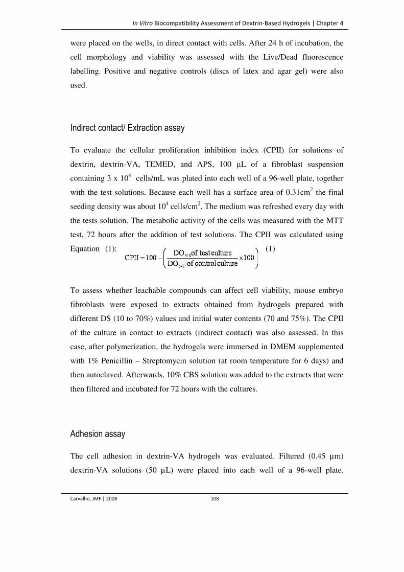
In Vitro Biocompatibility Assessment of Dextrin-Based Hydrogels | Chapter 4
Carvalho, JMF | 2008 108
were placed on the wells, in direct contact with cells. After 24 h of incubation, the
cell morphology and viability was assessed with the Live/Dead fluorescence
labelling. Positive and negative controls (discs of latex and agar gel) were also
used.
Indirect contact/ Extraction assay
To evaluate the cellular proliferation inhibition index (CPII) for solutions of
dextrin, dextrin-VA, TEMED, and APS, 100 µL of a fibroblast suspension
containing 3 x 10⁴ cells/mL was plated into each well of a 96-well plate, together
with the test solutions. Because each well has a surface area of 0.31cm2 the final
seeding density was about 104 cells/cm2. The medium was refreshed every day with
the tests solution. The metabolic activity of the cells was measured with the MTT
test, 72 hours after the addition of test solutions. The CPII was calculated using
Equation (1):
(1)
To assess whether leachable compounds can affect cell viability, mouse embryo
fibroblasts were exposed to extracts obtained from hydrogels prepared with
different DS (10 to 70%) values and initial water contents (70 and 75%). The CPII
of the culture in contact to extracts (indirect contact) was also assessed. In this
case, after polymerization, the hydrogels were immersed in DMEM supplemented
with 1% Penicillin – Streptomycin solution (at room temperature for 6 days) and
then autoclaved. Afterwards, 10% CBS solution was added to the extracts that were
then filtered and incubated for 72 hours with the cultures.
Adhesion assay
The cell adhesion in dextrin-VA hydrogels was evaluated. Filtered (0.45 µm)
dextrin-VA solutions (50 µL) were placed into each well of a 96-well plate.
In Vitro Biocompatibility Assessment of Dextrin-Based Hydrogels | Chapter 4
Carvalho, JMF | 2008 109
Polymerization started by the addition of filtered APS (5 µL) and TEMED (5 µL)
solutions, under sterile conditions. After 2 h of polymerization, the hydrogels
formed at the bottom of the wells were washed, first with PBS and secondly with
DMEM complete medium (3x200 µL); then, 100 µL of a fibroblast suspension
containing 3x10⁴ cells/mL was plated into each well, to yield a final density of 104
cells/cm2. The culture medium was refreshed (100 µL) every two days. For the
control assays, cells were grown directly in the bottom of the wells. After 72 hours
of incubation, the hydrogels were washed with PBS (3x100 µL) to remove floating
cells, and the cell layer was trypsinized and counted using a cell counting chamber
(Neubauer, Germany). Additionally, the cell proliferation was estimated using the
mitochondrial metabolic activity of the cells grown onto the hydrogel surface, in a
parallel experiment. Two methods were used, MTT and MTS, (applied directly on
the cell layers grown on top of the hydrogel). The discrepancy between the results
obtained, led us to addopt a slightly changed protocol: the cells were detached from
the hydrogel surface, before conducting both the colorimetric and counting assays,
an approach that resulted in more accurate and reliable results.
Microscopy assays
At regular time intervals, cell growth and morphology were evaluated by
observation with a phase-contrast inverted light microscope. Cytotoxic effects were
ranked in cellular death, cell adhesion ability and induction of morphological
changes. Additionally, cells were labelled with LIVE/DEAD Viability/Cytotoxicity
Kit and observed by fluorescence microscopy.
Results and Discussion
In order to evaluate the cytotoxicity of dextrin, acrylate derivatized dextrin, dextrin
hydrogels and their leachable products, we have studied the effects of these
In Vitro Biocompatibility Assessment of Dextrin-Based Hydrogels | Chapter 4
Carvalho, JMF | 2008 110
materials in a mouse embryo fibroblast culture, namely in the cell proliferation and
adhesion on the hydrogel, morphological features and cell death.
Extraction Assay
As previously described, upon exposure to aqueous solutions, hydrogels can
release leachable products and, therefore, the cytotoxicity of hydrogel extracts must
be evaluated. For that purpose, fibroblast cultures were exposed to extracts of
hydrogels, prepared using dextrin-VA with different DS and initial water contents.
The cell viability was quantified using the MTS assay. The results are shown in
Table 1. The extracts induced a reduction inferior to 17% in the mitochondrial
metabolic activity of fibroblasts, as compared with the control. No significant
differences were found in hydrogels with different DS. The initial water content, 70
and 75%, (corresponding to initial monomer concentrations of 44 and 33%,
respectively) of the hydrogel network also does not produce major differences in
the CPII.
Table 1. Cell proliferation inhibition index of mouse embryo fibroblasts, cultured
in the presence of extracts obtained from dextrin-VA hydrogels with different DS
and initial water content.
DS (%)a
CPII (%)c
Initial Water Content (%)b
70 75
10 12.4 ± 2.3 14.8 ± 3.1
15 14.2 ± 4.5 11.9 ± 1.7
20 15.4 ± 1.6 13.1 ± 1.9
40 14.8 ± 2.1 15.5 ± 1.2
70 17.3 ± 2.7 14.9 ± 4.2
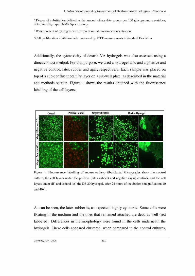
In Vitro Biocompatibility Assessment of Dextrin-Based Hydrogels | Chapter 4
Carvalho, JMF | 2008 111
a Degree of substitution defined as the amount of acrylate groups per 100 glucopyranose residues, determined by liquid NMR Spectroscopy
b Water content of hydrogels with different initial monomer concentration
c Cell proliferation inhibition index assessed by MTT measurements ± Standard Deviation
Additionally, the cytotoxicity of dextrin-VA hydrogels was also assessed using a
direct contact method. For that purpose, we used a hydrogel disc and a positive and
negative control, latex rubber and agar, respectively. Each sample was placed on
top of a sub-confluent cellular layer on a six-well plate, as described in the material
and methods section. Figure 1 shows the results obtained with the fluorescence
labelling of the cell layers.
Figure 1. Fluorescence labelling of mouse embryo fibroblasts. Micrographs show the control
culture, the cell layers under the positive (latex rubber) and negative (agar) controls, and the cell
layers under (B) and around (A) the DS 20 hydrogel, after 24 hours of incubation (magnification 10
and 40x).
As can be seen, the latex rubber is, as expected, highly cytotoxic. Some cells were
floating in the medium and the ones that remained attached are dead as well (red
labbeled). Differences in the morphology were found in the cells underneath the
hydrogels. These cells appeared clustered, when compared to the control cultures,
A B
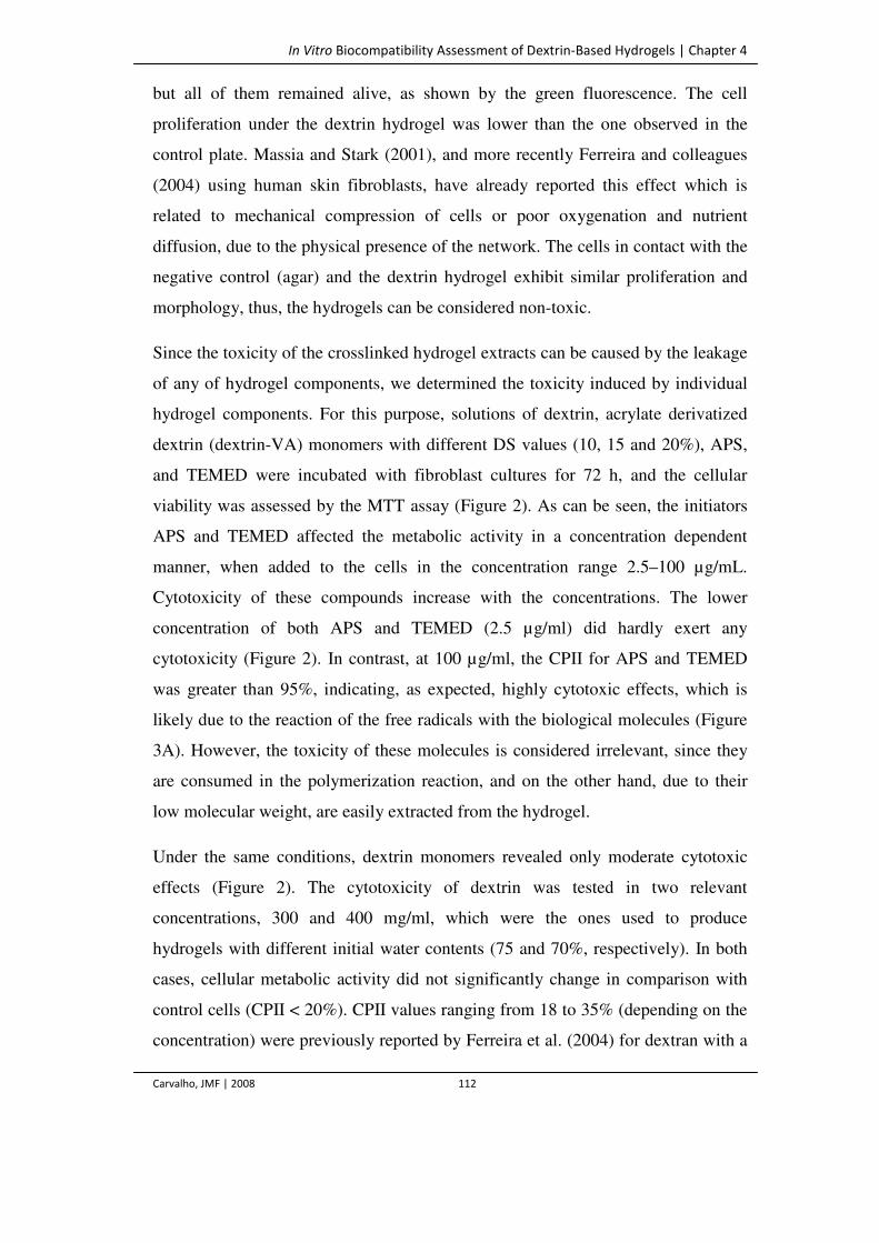
In Vitro Biocompatibility Assessment of Dextrin-Based Hydrogels | Chapter 4
Carvalho, JMF | 2008 112
but all of them remained alive, as shown by the green fluorescence. The cell
proliferation under the dextrin hydrogel was lower than the one observed in the
control plate. Massia and Stark (2001), and more recently Ferreira and colleagues
(2004) using human skin fibroblasts, have already reported this effect which is
related to mechanical compression of cells or poor oxygenation and nutrient
diffusion, due to the physical presence of the network. The cells in contact with the
negative control (agar) and the dextrin hydrogel exhibit similar proliferation and
morphology, thus, the hydrogels can be considered non-toxic.
Since the toxicity of the crosslinked hydrogel extracts can be caused by the leakage
of any of hydrogel components, we determined the toxicity induced by individual
hydrogel components. For this purpose, solutions of dextrin, acrylate derivatized
dextrin (dextrin-VA) monomers with different DS values (10, 15 and 20%), APS,
and TEMED were incubated with fibroblast cultures for 72 h, and the cellular
viability was assessed by the MTT assay (Figure 2). As can be seen, the initiators
APS and TEMED affected the metabolic activity in a concentration dependent
manner, when added to the cells in the concentration range 2.5–100 µg/mL.
Cytotoxicity of these compounds increase with the concentrations. The lower
concentration of both APS and TEMED (2.5 µg/ml) did hardly exert any
cytotoxicity (Figure 2). In contrast, at 100 µg/ml, the CPII for APS and TEMED
was greater than 95%, indicating, as expected, highly cytotoxic effects, which is
likely due to the reaction of the free radicals with the biological molecules (Figure
3A). However, the toxicity of these molecules is considered irrelevant, since they
are consumed in the polymerization reaction, and on the other hand, due to their
low molecular weight, are easily extracted from the hydrogel.
Under the same conditions, dextrin monomers revealed only moderate cytotoxic
effects (Figure 2). The cytotoxicity of dextrin was tested in two relevant
concentrations, 300 and 400 mg/ml, which were the ones used to produce
hydrogels with different initial water contents (75 and 70%, respectively). In both
cases, cellular metabolic activity did not significantly change in comparison with
control cells (CPII < 20%). CPII values ranging from 18 to 35% (depending on the
concentration) were previously reported by Ferreira et al. (2004) for dextran with a
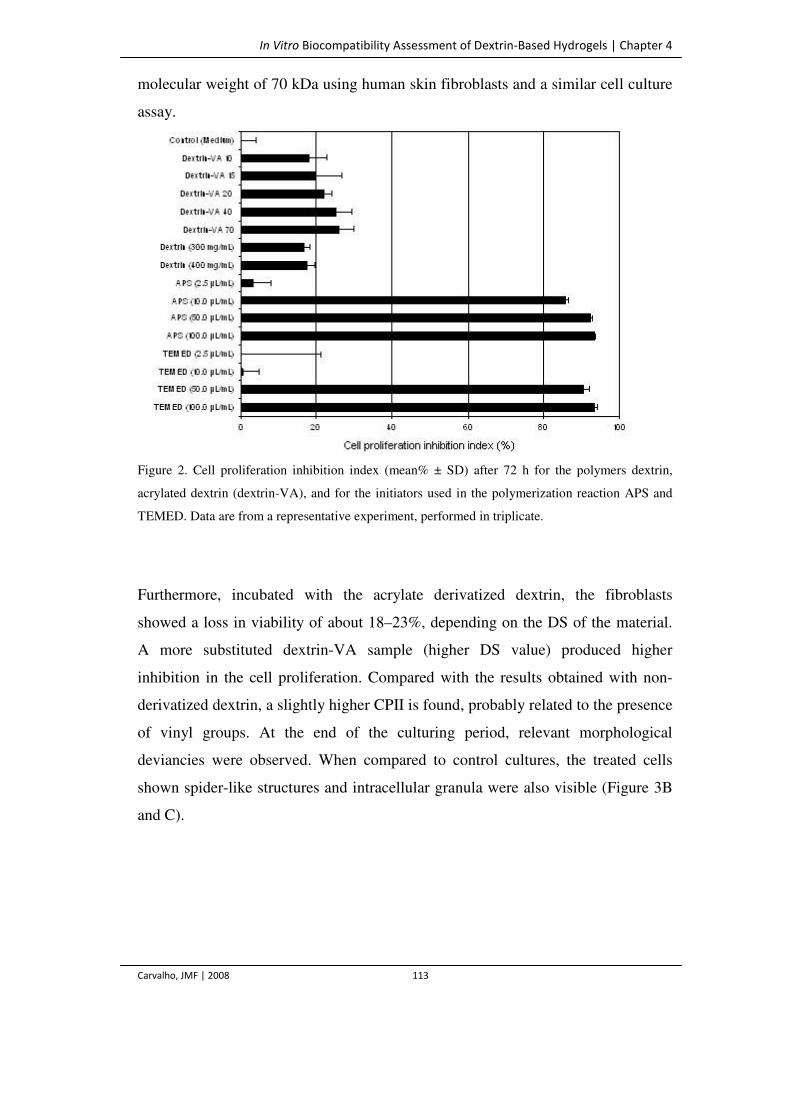
In Vitro Biocompatibility Assessment of Dextrin-Based Hydrogels | Chapter 4
Carvalho, JMF | 2008 113
molecular weight of 70 kDa using human skin fibroblasts and a similar cell culture
assay.
Figure 2. Cell proliferation inhibition index (mean% ± SD) after 72 h for the polymers dextrin,
acrylated dextrin (dextrin-VA), and for the initiators used in the polymerization reaction APS and
TEMED. Data are from a representative experiment, performed in triplicate.
Furthermore, incubated with the acrylate derivatized dextrin, the fibroblasts
showed a loss in viability of about 18–23%, depending on the DS of the material.
A more substituted dextrin-VA sample (higher DS value) produced higher
inhibition in the cell proliferation. Compared with the results obtained with non-
derivatized dextrin, a slightly higher CPII is found, probably related to the presence
of vinyl groups. At the end of the culturing period, relevant morphological
deviancies were observed. When compared to control cultures, the treated cells
shown spider-like structures and intracellular granula were also visible (Figure 3B
and C).
In Vitro Biocompatibility Assessment of Dextrin-Based Hydrogels | Chapter 4
Carvalho, JMF | 2008 114
Figure 3. Mouse embryo fibroblasts, cultured for 24 h in the presence of 100 µg/mL of APS (A),
300 mg/mL DS 20 (also representative for the other DS values) (B) and control culture (C). Highly
cytotoxic effects of APS are shown in micrograph A, the cells are dead and floating in the medium.
When compared to control cultures, dextrin-VA-treated cells contained intracellular granula (see
arrows) and have spider-like structures (magnification 10x).
Cell Adhesion and Proliferation on Dextrin Hydrogels
The cell adhesion on the hydrogel surfaces was evaluated using samples with
different DS (20 and 70%). The results were expressed as a percentage of the
adhesion on cell culture polystyrene plates (TCPP), estimated by cell counting.
Additionally, the mitochondrial activity of the adherent cells was also assessed in a
parallel experiment. This study showed that the hydrogels are, as expected, less
adhesive than TCPP. The resistance of the hydrogel surfaces to cell adhesion is, as
described for other polysaccharides such as dextran, presumably due to poor
protein adsorption onto the hydrophilic and nonionic polymer (Osterberg et al.
1995, Frazier et al. 2000, De Groot et al. 2001). Indeed, cellular adhesion on
hydrophilic materials such as hyaluronic acid and PEG-based hydrogels was
reported to be extremely sparse (Mann et al. 2001, Ramamurthi and Vesely 2002).
In other cases, as recently described in pullulan hydrogels using smooth muscle
cells cell adhesion and moderate growth is observed (Autissier et al. 2007).
Viability tests indicated that fibroblasts adhered to some extent (50 to 60%) as
compared to the adhesion on TCPP and are capable to grow on dextrin hydrogels.
In the first stage of the culture, the cells appear round and then attach to the gel
surface. As can be seen in Table 2, after one hour, less than 30% of the cells were
attached to the hydrogel surface, irrespective of the DS tested; after 4 hours, more
than 50% of the initial cells remained associated to the gels.
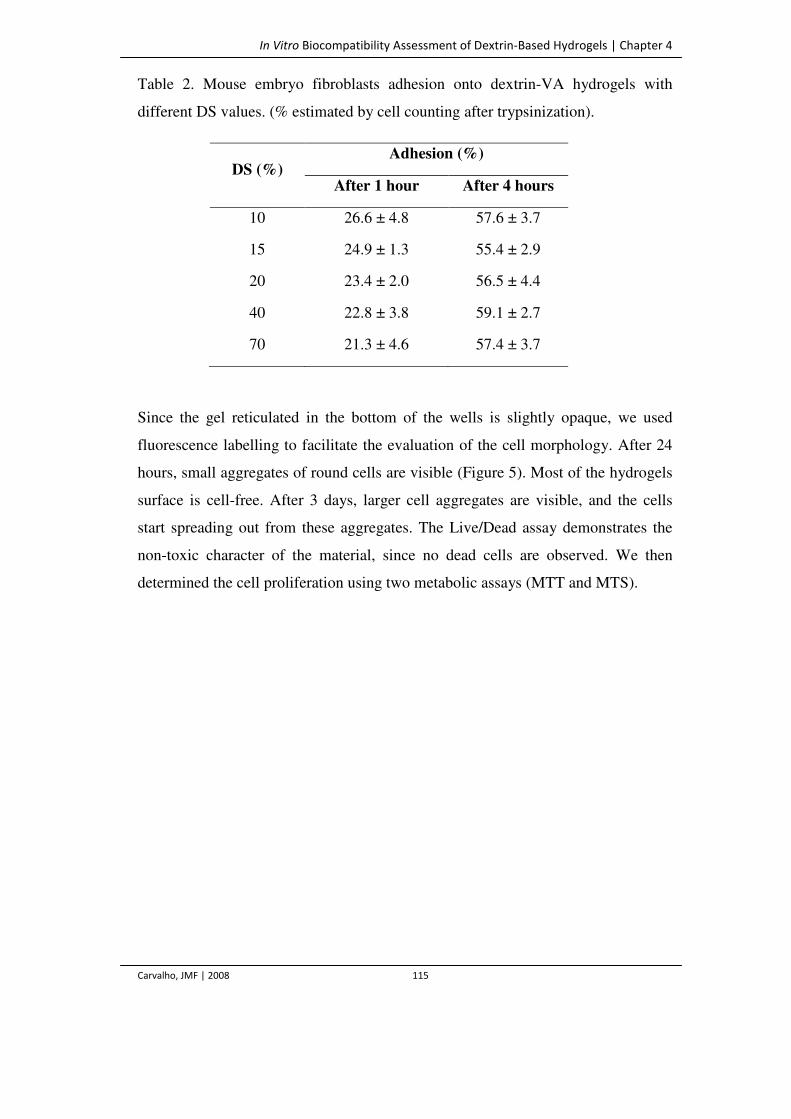
In Vitro Biocompatibility Assessment of Dextrin-Based Hydrogels | Chapter 4
Carvalho, JMF | 2008 115
Table 2. Mouse embryo fibroblasts adhesion onto dextrin-VA hydrogels with
different DS values. (% estimated by cell counting after trypsinization).
DS (%) Adhesion (%)
After 1 hour After 4 hours
10 26.6 ± 4.8 57.6 ± 3.7
15 24.9 ± 1.3 55.4 ± 2.9
20 23.4 ± 2.0 56.5 ± 4.4
40 22.8 ± 3.8 59.1 ± 2.7
70 21.3 ± 4.6 57.4 ± 3.7
Since the gel reticulated in the bottom of the wells is slightly opaque, we used
fluorescence labelling to facilitate the evaluation of the cell morphology. After 24
hours, small aggregates of round cells are visible (Figure 5). Most of the hydrogels
surface is cell-free. After 3 days, larger cell aggregates are visible, and the cells
start spreading out from these aggregates. The Live/Dead assay demonstrates the
non-toxic character of the material, since no dead cells are observed. We then
determined the cell proliferation using two metabolic assays (MTT and MTS).
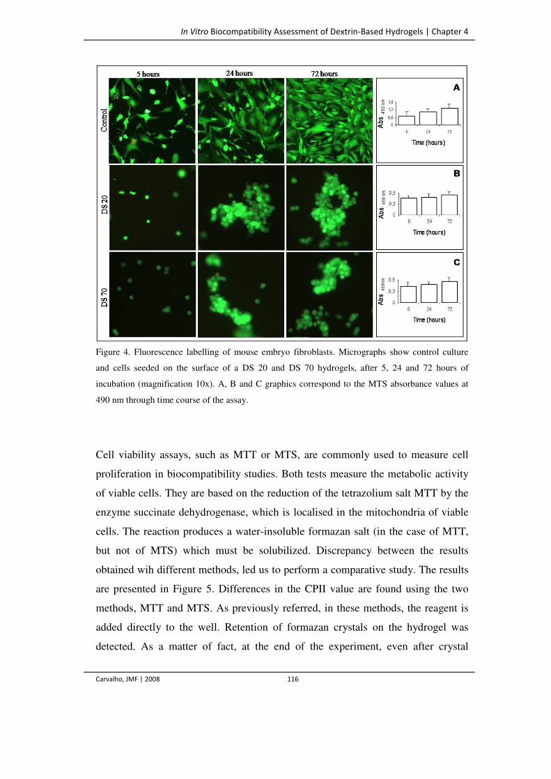
In Vitro Biocompatibility Assessment of Dextrin-Based Hydrogels | Chapter 4
Carvalho, JMF | 2008 116
Figure 4. Fluorescence labelling of mouse embryo fibroblasts. Micrographs show control culture
and cells seeded on the surface of a DS 20 and DS 70 hydrogels, after 5, 24 and 72 hours of
incubation (magnification 10x). A, B and C graphics correspond to the MTS absorbance values at
490 nm through time course of the assay.
Cell viability assays, such as MTT or MTS, are commonly used to measure cell
proliferation in biocompatibility studies. Both tests measure the metabolic activity
of viable cells. They are based on the reduction of the tetrazolium salt MTT by the
enzyme succinate dehydrogenase, which is localised in the mitochondria of viable
cells. The reaction produces a water-insoluble formazan salt (in the case of MTT,
but not of MTS) which must be solubilized. Discrepancy between the results
obtained wih different methods, led us to perform a comparative study. The results
are presented in Figure 5. Differences in the CPII value are found using the two
methods, MTT and MTS. As previously referred, in these methods, the reagent is
added directly to the well. Retention of formazan crystals on the hydrogel was
detected. As a matter of fact, at the end of the experiment, even after crystal
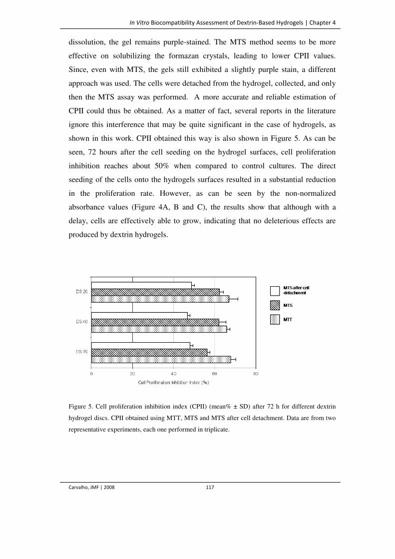
In Vitro Biocompatibility Assessment of Dextrin-Based Hydrogels | Chapter 4
Carvalho, JMF | 2008 117
dissolution, the gel remains purple-stained. The MTS method seems to be more
effective on solubilizing the formazan crystals, leading to lower CPII values.
Since, even with MTS, the gels still exhibited a slightly purple stain, a different
approach was used. The cells were detached from the hydrogel, collected, and only
then the MTS assay was performed. A more accurate and reliable estimation of
CPII could thus be obtained. As a matter of fact, several reports in the literature
ignore this interference that may be quite significant in the case of hydrogels, as
shown in this work. CPII obtained this way is also shown in Figure 5. As can be
seen, 72 hours after the cell seeding on the hydrogel surfaces, cell proliferation
inhibition reaches about 50% when compared to control cultures. The direct
seeding of the cells onto the hydrogels surfaces resulted in a substantial reduction
in the proliferation rate. However, as can be seen by the non-normalized
absorbance values (Figure 4A, B and C), the results show that although with a
delay, cells are effectively able to grow, indicating that no deleterious effects are
produced by dextrin hydrogels.
Figure 5. Cell proliferation inhibition index (CPII) (mean% ± SD) after 72 h for different dextrin
hydrogel discs. CPII obtained using MTT, MTS and MTS after cell detachment. Data are from two
representative experiments, each one performed in triplicate.
In Vitro Biocompatibility Assessment of Dextrin-Based Hydrogels | Chapter 4
Carvalho, JMF | 2008 118
Conclusions
Dextrin-based hydrogels constitute a promising biomaterial that may be used for a
variety of applications. The results suggest that the dextrin hydrogel have
negligible cell toxicity, allowing cell adhesion and proliferation. Additionally, the
modification of the surface of the hydrogels with RGD peptides, using recombinant
starch binding modules (SBM) fused to the bioactive peptides, was proved to
significantly improve the cell adhesion and surface spreading (ongoing work). This
is a significant advantage on using a starch derived hydrogel (in this case, dextrin),
since the availability of SBMs allows a wide variety of bioactive peptides to be
driven to the hydrogels surfaces. Furthermore, the interaction of this hydrogel with
different cell lines and living tissues – subcutaneous implants – is currently being
analysed.
References
Autissier A, Letourneur D and Le Visage C. (2007) Pullulan-based hydrogel for smooth muscle cell
culture. J Biomed Mater Res A 82: 336–342.
Behl C, Davis JB, Lesley R and Schubert D. (1994) Hydrogen peroxide mediates amyloid β-protein
toxicity. Cell 77: 817–827.
Berridge MV, Tan AS, McCoy KD and Wang R. (1996) The biochemical and cellular basis of cell
proliferation assays that use tetrazolium salts. Biochemica 4: 15–20.
Carvalho J, Gonçalves C, Gil AM and Gama FM. (2007) Production and characterization of a new
dextrin based hydrogel. Eur Pol J 43: 3050–3059.
Chen T, Embree HD, Brown EM, Taylor MM and Payne GF. (2003) Enzyme-catalyzed gel
formation of gelatine and chitosan: potential for in situ applications. Biomaterials 24: 2831–2841.2
In Vitro Biocompatibility Assessment of Dextrin-Based Hydrogels | Chapter 4
Carvalho, JMF | 2008 119
De Groot CJ, Van Luyn MJA, Van Dijk-Wolthuis WNE, Cadée JA, Plantinga JA, Den Otter W and
Hennink WE. (2001) In vitro biocompatibility of biodegradable dextran-based hydrogels tested with
human fibroblasts. Biomaterials 22: 1197–1203.
Di Silvio L, Gurav N, Kayser MV, Braden M and Downes S. (1994) Biodegradable microspheres: a
new delivery system for growth hormone. Biomaterials 15: 931–936.
Draye J-P, Delaey B, van de Voorde A, van Den Bulcke A, Bogdanov and Schacht E. (1998) In
vitro release characteristics of bioactive molecules from dextran dialdehyde cross-linked gelatin
hydrogel films. Biomaterials 19: 99–107.
Drury JL and Mooney DJ. (2003) Hydrogels for tissue engineering: scaffold variables and
applications. Biomaterials 24: 4337–4351.
Ferreira L, Rafael A, Lamghari M, Barbosa MA, Gil MH, Cabrita MAS and Dordick JS. (2004)
Biocompatibility of chemoenzymatically derived dextran-acrylate hydrogels. Biomed Mater Res A
68: 584–596.
Frazier RA, Matthijs G, Davies MC, Roberts CJ, Schacht E and Tendler SJB. (2000)
Characterization of protein-resistant dextran monolayers. Biomaterials 21: 957–966.
Hansen MB, Nielsen SE and Berg K. (1989) Re-examination and further development of a precise
and rapid method for measuring cell growth/cell killing. J Immunol Meth 119: 203–210.
Hern DL and Hubbell JA. (1998) Incorporation of adhesion peptides into non-adhesive hydrogels
useful for tissue resurfacing. J Biomed Mater Res 39: 266–276.
Hoffman AS. (2002) Hydrogels for biomedical applications. Adv Drug DelivRev 54: 3–12.
Ishihara M, Nakanishi K, Ono K, Sato M, Saito Y, Yura H, Matsui T, Hattori H, Uenoyama M,
Kikuchi M and Kurita A. (2002) Photocrosslinkable chitosan as a dressing wound occlusion and
accelerator in healing process. Biomaterials 23: 833–840.
Kane JB, Tompkins RG, Yarmush ML and Burke JF. (1996) Burn dressings. In Biomaterials
science: an introduction to materials in medicine. Academic Press, San Diego, USA, pp 360–370.
Levy MC and Andry M-C. (1991) Mixed-walled microcapsules made of cross-linked proteins and
polysaccharides: preparation and properties. J Microencapsul 8: 335–347.
Mann BK, Gobin AS, Tsai AT, Schmedlen RH and West JL. (2001) Smooth muscle cell growth in
photopolymerized hydrogels with cell adhesive and proteolytically degradable domains: synthetic
ECM analogs for tissue engineering. Biomaterials 22: 3045–3051.
In Vitro Biocompatibility Assessment of Dextrin-Based Hydrogels | Chapter 4
Carvalho, JMF | 2008 120
Massia SP and Stark J. (2001) Immobilized RGD peptides on surface grafted dextran promote
biospecific cell attachment. J Biomed Mater Res 56: 390–399.
McCulloch I and Shalaby SW. (1998) In Tailored polymeric materials for controlled delivery
systems. American Chemical Society, Washington DC, USA.
Mosmann T. (1983) Rapid colorimetric assay for cellular growth and survival: application to
proliferation and cytotoxicity assays. J Immunol Methods 65: 55–63.
Osterberg E, Bergstrom K, Holmberg K, Schuman TP, Riggs JA, Burns NL, Van Alstine JM and
Harris JM. (1995) Protein-rejecting ability of surface-bound dextran in end-on and side-on
configurations: comparison to PEG. J Biomed Mater Res 29: 741–747.
Peppas NA. (1996) Hydrogels. In Biomaterials science: an introduction to materials in medicine.
Academic Press, San Diego, USA, pp 60–64.
Peppas NA, Bures P, Leobandung W and Ichikawa H. (2000) Hydrogels in pharmaceutical
formulations. Eur J Pharm Biopharm 50: 27–46.
Ramamurthi A and Vesely I. (2002) Smooth muscle cell adhesion on crosslinked hyaluronan gels. J
Biomed Mater Res 60: 195–205.
Schacht EH, Nobels M, 3 S, Demeester J, Fransen J and Lemahieu A. (1993) Cross-linkage of
gelatin by dextran dialdehyde. Pol Gels Networks 1:213–224.
Sgouras D and Duncan R. (1990) Methods for the evaluation of biocompatibility of soluble
synthetic polymers which have potential for biomedical use: 1-use of the tetrazolium-based
colorimetric assay (MTT) as a preliminary screen for evaluation of in vitro cytotoxicity. J Mater
Sci: Mater Med 1: 61–68.

Degradation Kinetics and Rheological Properties of New Class of Degradable Dextrin-Based Hydrogels | Chapter 5
Carvalho, JMF | 2008 123
Introduction
Hydrogels are a class of three-dimensional, hydrophilic polymeric networks that
can absorb substantial amounts of water. In general, hydrogels have a good
biocompatibility and are capable of resembling natural tissues, allowing for a
favourable controlled interaction with living systems (Vyavahare and Kohn 1994).
For this reason, these materials have found widespread application in the
biomedical, biotechnological, and pharmaceutical fields. Hydrogels can be used in
tissue engineering, as scaffolds to support and promote tissue regeneration (Peppas
1996, Kane et al. 1996, McCulloch et al. 1998, Peppas et al. 2000). In addition,
hydrogels are gaining increasing attention as attractive systems for the controlled
release of pharmaceutically active molecules (Okay and Gurun 1992, Walther et al.
1995). An important class of hydrogels is obtained by the polymerization of
acrylate esters (e.g. vinyl acrylate, methacrylate, hydroxyethyl methacrylate,
HEMA) through the polymerization of the water-soluble functionalized polymers
(Van Dijk-Wolthuis et al. 1995, Hennink et al. 1996, Van Dijk-Wolthuis et al.
1997a, Ferreira et al. 2002 (a), Ferreira et al. 2002 (b)). In previous work, we have
described a new dextrin based hydrogel in which the polysaccharide functionalized
with reactive double bonds was cross-linked by free radical polymerization in
aqueous solution (Carvalho et al. 2007). In these polymeric networks, ester bonds
are present in the cross-links and may undergo hydrolytic cleavage, which can
ultimately affect both the diffusion and the release profiles (Peppas et al. 2000).
Recently, we reported on the diffusion and release of two different molecules from
dextrin hydrogels (Carvalho et al. 2008). Further investigation has shown that the
hydrolysis of the ester groups is very slow under physiological conditions.
However, the hydrogels could be rendered degradable through the incorporation of
the enzyme amyloglucosidase, which could prove to be an effective route to
modulate the release profiles. An alternative approach is the functionalization of
the polymer with a methacrylate ester (HEMA). As shown in several studies,
hydrogels obtained by polymerization of this compound, have desirable
degradation kinetics (Van Dijk-Wolthuis et al. 1997b, c, d, Stubbe et al. 2002).
Degradation Kinetics and Rheological Properties of New Class of Degradable Dextrin-Based Hydrogels | Chapter 5
Carvalho, JMF | 2008 124
In this study we report on the production of dextrin-HEMA hydrogels and perform
a comparative study of the stability of the two different acrylated dextrins (dextrin-
vinylacrylate (VA) and dextrin-HEMA). It is reasonable to expect that distinct
mechanical properties resulted from the functionalization with the two ester
functions. In this context, a rheologic analysis allowed exploring the time evolution
of the viscoelastic behavior of the hydrogels, providing a qualitative
characterization of the crosslinking processes, as well as the structural changes
occurring in the network during degradation. Moreover, as the erosion process can
play an important role in determining hydrogel properties such as the release
profiles, a mass loss evaluation was also performed.
Materials and Methods
Materials
Dextrin - Koldex 60 starch was a generous gift from Tate & Lyle (Decatur, IL,
USA). Vinyl acrylate (VA) was from Aldrich, N,N,N’,N’-
tetramethylenethylenediamine (TEMED) and ammonium persulfate (APS) were
purchased from BioRad, dimethylsulfoxide (DMSO) and acetone were from
AppliChem. 2-hydroxyethyl methacrylate (HEMA), hydroquinone monomethyl
ether (>98%), 4-(N,N-dimethylamino)pyridine (DMAP, 99%), N,N’-
carbonyldiimidazole (CDI, 98%) and tetrahydrofuran (THF) were from Fluka. Both
DMSO and THF were dried with 0.4 nm molecular sieves at least overnight before
use. Regenerated cellulose tubular membranes with 3500 molecular weight cutoff
(MWCO) were obtained from Membrane Filtration Products (Seguin, USA).
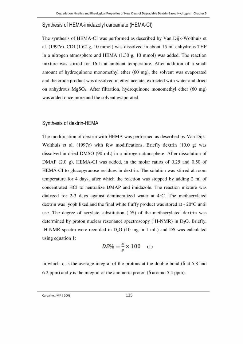
Degradation Kinetics and Rheological Properties of New Class of Degradable Dextrin-Based Hydrogels | Chapter 5
Carvalho, JMF | 2008 125
Synthesis of HEMA-imidazolyl carbamate (HEMA-CI)
The synthesis of HEMA-CI was performed as described by Van Dijk-Wolthuis et
al. (1997c). CDI (1.62 g, 10 mmol) was dissolved in about 15 ml anhydrous THF
in a nitrogen atmosphere and HEMA (1.30 g, 10 mmol) was added. The reaction
mixture was stirred for 16 h at ambient temperature. After addition of a small
amount of hydroquinone monomethyl ether (60 mg), the solvent was evaporated
and the crude product was dissolved in ethyl acetate, extracted with water and dried
on anhydrous MgSO4. After filtration, hydroquinone monomethyl ether (60 mg)
was added once more and the solvent evaporated.
Synthesis of dextrin-HEMA
The modification of dextrin with HEMA was performed as described by Van Dijk-
Wolthuis et al. (1997c) with few modifications. Briefly dextrin (10.0 g) was
dissolved in dried DMSO (90 mL) in a nitrogen atmosphere. After dissolution of
DMAP (2.0 g), HEMA-CI was added, in the molar ratios of 0.25 and 0.50 of
HEMA-CI to glucopyranose residues in dextrin. The solution was stirred at room
temperature for 4 days, after which the reaction was stopped by adding 2 ml of
concentrated HCl to neutralize DMAP and imidazole. The reaction mixture was
dialyzed for 2-3 days against demineralized water at 4°C. The methacrylated
dextrin was lyophilized and the final white fluffy product was stored at - 20°C until
use. The degree of acrylate substitution (DS) of the methacrylated dextrin was
determined by proton nuclear resonance spectroscopy (1H-NMR) in D2O. Briefly, 1H-NMR spectra were recorded in D2O (10 mg in 1 mL) and DS was calculated
using equation 1:
in which x, is the average integral of the protons at the double bond (δ at 5.8 and
6.2 ppm) and y is the integral of the anomeric proton (δ around 5.4 ppm).
(1)
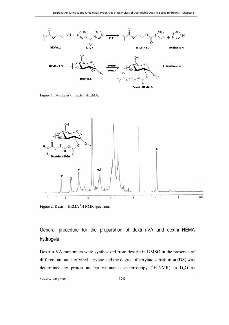
Degradation Kinetics and Rheological Properties of New Class of Degradable Dextrin-Based Hydrogels | Chapter 5
Carvalho, JMF | 2008 126
Figure 1. Synthesis of dextrin-HEMA.
Figure 2. Dextrin-HEMA 1H NMR spectrum.
General procedure for the preparation of dextrin-VA and dextrin-HEMA
hydrogels
Dextrin-VA monomers were synthesized from dextrin in DMSO in the presence of
different amounts of vinyl acrylate and the degree of acrylate substitution (DS) was
determined by proton nuclear resonance spectroscopy (1H-NMR) in D2O as
Degradation Kinetics and Rheological Properties of New Class of Degradable Dextrin-Based Hydrogels | Chapter 5
Carvalho, JMF | 2008 127
previously described (Carvalho et al. 2007). The hydrogel slabs with different DS
(20 and 40%) were prepared by radical polymerization of aqueous solution of
either dextrin-VA or dextrin-HEMA (300 mg/mL). The gelation reactions were
initiated by adding 90 µL APS (80 mg/ml in phosphate buffer) and 90 µL TEMED
(13.6 % (v/v) in water, pH adjusted to 8.0 with HCl) and allowed to occur for 30
min at room temperature.
Standard degradation conditions
Two sets of hydrogels were used. After the polymerization reaction, gels were
removed from the casting recipients. One set of hydrogels was immediately
liofilized to determine the initial dry weight (Wi). The other one was immersed in
50 mL of phosphate buffer saline (PBS) pH 7.4 and kept in a thermostated room at
37 °C. At set time intervals, gels were removed, blotted with filter paper to remove
water excess, weighted (Ws – weight in the swollen state) and lyophilised to
determine the dry weight, Wd. The swelling ratio (SR) and the mass loss (mloss)
were calculated according to equation 2 and 3, respectively:
(2) (3)
The hydrogel degradation time is defined as the time needed for complete
degradation (W=0).
Rheological measurements
The rheological measurements were performed in a controlled-stress Rheologica
rheometer at a controlled temperature (25ºC). A plate-plate measuring system was
used (Ø 25 mm) and the gap between the plates was 1 mm. For the experiments,
polymerized hydrogels with 25 mm diameter and 1 mm of thickness, were
immersed in 50 mL of PBS at pH 7.4 and kept in a thermostated room at 37 °C to
Degradation Kinetics and Rheological Properties of New Class of Degradable Dextrin-Based Hydrogels | Chapter 5
Carvalho, JMF | 2008 128
allow degradation, with the exception of the initial time measurements (t=0), in
which hydrogels were used immediately after polymerization. At set time
intervals, gels were removed from the containers and placed between the plates of
the measuring system. Two sets of experiments were performed:
a) Stress sweep experiments were carried out to optimize the applied stress
used in the frequency-oscillation experiments. The lower limit of the
applied stress is determined by the sensitivity of the instrument. On the
other hand, the upper limit of the applied stress will be influenced by the
linear viscoelastic range and the presence of slip effects.
b) Once the range of linear viscoelastic response was determined, experiments
were performed in Small Amplitude Oscillatory Shear, SAOS, at a fixed
frequency of 1 Hz and a stress known to yield a good quality signal in the
linear viscoelastic regime.
Results and Discussion
In previous work, we have demonstrated the production of a new dextrin hydrogel,
through the transesterification of the polymer with vinyl acrylate (VA) (Carvalho et
al. 2007). However, these hydrogels were shown to degrade slowly under
physiological conditions, which is a limitation in light of their application as
controlled delivery systems. Several studies reported the successful
functionalization of dextran with a methacrylate ester (HEMA) and it was shown
that hydrogels obtained by polymerization of this compound, are able to degrade in
desirable timeframes (Van Dijk-Wolthuis et al. 1997b, c, d, Stubbe et al. 2002). In
this work, a similar dextrin modification was attempted using the same chemistry.
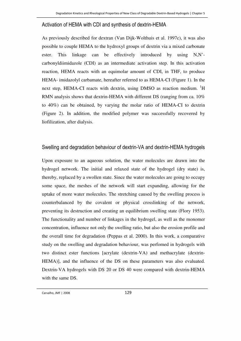
Degradation Kinetics and Rheological Properties of New Class of Degradable Dextrin-Based Hydrogels | Chapter 5
Carvalho, JMF | 2008 129
Activation of HEMA with CDI and synthesis of dextrin-HEMA
As previously described for dextran (Van Dijk-Wolthuis et al. 1997c), it was also
possible to couple HEMA to the hydroxyl groups of dextrin via a mixed carbonate
ester. This linkage can be effectively introduced by using N,N’-
carbonyldiimidazole (CDI) as an intermediate activation step. In this activation
reaction, HEMA reacts with an equimolar amount of CDI, in THF, to produce
HEMA- imidazolyl carbamate, hereafter referred to as HEMA-CI (Figure 1). In the
next step, HEMA-CI reacts with dextrin, using DMSO as reaction medium. 1H
RMN analysis shows that dextrin-HEMA with different DS (ranging from ca. 10%
to 40%) can be obtained, by varying the molar ratio of HEMA-CI to dextrin
(Figure 2). In addition, the modified polymer was successfully recovered by
liofilization, after dialysis.
Swelling and degradation behaviour of dextrin-VA and dextrin-HEMA hydrogels
Upon exposure to an aqueous solution, the water molecules are drawn into the
hydrogel network. The initial and relaxed state of the hydrogel (dry state) is,
thereby, replaced by a swollen state. Since the water molecules are going to occupy
some space, the meshes of the network will start expanding, allowing for the
uptake of more water molecules. The stretching caused by the swelling process is
counterbalanced by the covalent or physical crosslinking of the network,
preventing its destruction and creating an equilibrium swelling state (Flory 1953).
The functionality and number of linkages in the hydrogel, as well as the monomer
concentration, influence not only the swelling ratio, but also the erosion profile and
the overall time for degradation (Peppas et al. 2000). In this work, a comparative
study on the swelling and degradation behaviour, was perfomed in hydrogels with
two distinct ester functions [acrylate (dextrin-VA) and methacrylate (dextrin-
HEMA)], and the influence of the DS on these parameters was also evaluated.
Dextrin-VA hydrogels with DS 20 or DS 40 were compared with dextrin-HEMA
with the same DS.
Degradation Kinetics and Rheological Properties of New Class of Degradable Dextrin-Based Hydrogels | Chapter 5
Carvalho, JMF | 2008 130
Figure 3. Degradability of dextrin-based hydrogels. Swelling ratio (A) and mass loss as a function
of time, when immersed in PBS pH 7.4, at 37 ºC (average ±SD, n=3). (* from day 40 the hydrogel
structure was severely damaged, preventing the SR measurements).
As can be seen in figure 3A, for dextrin-VA, at physiological pH, in the initial
period of the incubation (around 5 days), a very slight increase in the swelling ratio
is detected for both degrees of substitution (DS 20 or DS 40). Nevertheless, neither
gel showed a significant increase in swelling over 100 days, indicating that no
significant hydrolysis of the polymerized acrylate esters occurred. As expected,
greater swelling values (in the range 2.5 to 3) were found for the DS 20 networks,
compared with those (in the range 1.6 to 2.1) obtained with the higher DS, since in
highly crosslinked hydrogels the mobility of the polymer chains is hindered,
lowering the ability to swell.
Contrarily to the dextrin-VA hydrogels, in which the SR remains almost constant
during the time course of the experiment, in the case of dextrin-HEMA, a
progressive swelling occurred over the time, until the complete dissolution of the
hydrogels is accomplished. The increased swelling is likely a consequence of the
hydrolysis of the methacrylate bonds in the crosslinked structure. The cleavage of
the ester bonds leads to a decrease in the crosslinking density, allowing for the
network to expand and swell, absorbing more water.
Degradation Kinetics and Rheological Properties of New Class of Degradable Dextrin-Based Hydrogels | Chapter 5
Carvalho, JMF | 2008 131
As can be seen in figure 3B, the swelling ratio values reach a maximum at the time
that the gels starts to dissolve, and dextrin chains most likely bear few crosslinks
per chain. As also expected, while for a DS 20 dextrin-HEMA hydrogel, the
dissolution took place in approximately 35 days, in case of DS 40 hydrogels,
further time was needed to accomplish dissolution (60 days).
The mass loss of dextrin-VA/HEMA hydrogels with different DS (DS 20 and DS
40) was also monitored as a function of time. Figure 3B summarizes the
degradation times of all dextrin gels investigated. This figure shows that for both
gel systems (dextrin-VA or dextrin-HEMA), the degradation time increased with
an increasing DS. It seems obvious that an increasing DS at a fixed initial water
content (all the hydrogels have the same initial water content of about 70%), results
in a network with a higher crosslinking density. In these cases, to accomplish the
network dissolution, more cross-links have to be hydrolyzed, which requires more
time. When comparing the two hydrogels systems, a linear mass loss profile can be
observed in dextrin-VA, until day 20, with a rate of roughly 0.5% of gel solubilized
per day. However, following this initial period, the rate slows down, and at day
100, only about 25% and 40% of mass loss is detected for DS 20 and DS 40
hydrogels, respectively. Probably, the mass losses observed correspond to the
release of non-polymerized material.
Contrarily, as shown in figure 3B, for dextrin-HEMA hydrogels, the mass loss was
dramatic, reaching about 30% for DS 20 and 50% for DS 40, after 20 days of
incubation. In this case, hydrogels start to disintegrate quickly, until a complete
dissolution is achieved. These results suggest that, in the case of dextrin-HEMA
hydrogels, the degradation occurs via a homogeneous bulk erosion process. In this
case the swelling ratio would be expected to increase with the incubation time,
because of the loosened polymer network since, although hydrolyzed, the polymer
chains remain in the matrix, allowing the network to become looser and to
continuously swell, until the dissolution is complete (Wong and Mooney 1997,
Suggs et al. 1998, Martens et al. 2002). In the case of dextrin-VA hydrogels, it
Degradation Kinetics and Rheological Properties of New Class of Degradable Dextrin-Based Hydrogels | Chapter 5
Carvalho, JMF | 2008 132
seems that the initial mass loss can be attributed to the release of a fraction of
unbound molecules which were physically entrapped in the matrix at the time of
polymerization. As diffusion occurs, the small molecules are released out of the
network. A stabilization of the swelling values is, thus, observed at the time that all
the unbound molecules are released. Contrarily to dextrin-HEMA, which are
hydrolytically labile, dextrin-VA hydrogels are very stable under physiological
conditions.
Mechanical Properties
Mechanical properties of hydrogels are very important for pharmaceutical
applications. For example, the integrity of a drug delivery device during the
lifetime of the application is crucial. The system must protect a sensitive
therapeutic agent, such as protein, maintaining its integrity until it is released out of
the system. Changes in the crosslinking density of the hydrogels have been
routinely applied to achieve the desired mechanical properties. Increasing the
crosslinking will result in a stronger gel with a more brittle structure, which
sometimes is not desirable since it shall interfere with the release profiles. Hence, a
compromise must be achieved between a relatively strong but yet elastic structure
(Anseth et al. 1996). It is well established that the release of the proteins from a
hydrogel is clearly influenced by its degradation rates (Meyvis et al. 2000).
Dynamic mechanical analysis, performed on the hydrogel, provide quantitative
information on the viscoelastic and rheological properties of the material, by
measuring the mechanical response of the samples as they are deformed. The
elastic (also real or storage) modulus G’ and the viscous (also imaginary or loss)
modulus G’’ are presented. The storage modulus (G’, Figure 4A) of a hydrogel is
proportional to the amount of elastic chains within the polymer network (Ferry
1980). Figure 3 shows the rheological results for all the hydrogels and it is clear
that the results are in good agreement with the degradability studies above.
Dextrin-HEMA hydrogels show lower G’ values, as compared with the dextrin-VA
ones, indicating that physical structuring is less pronounced when dextrin is
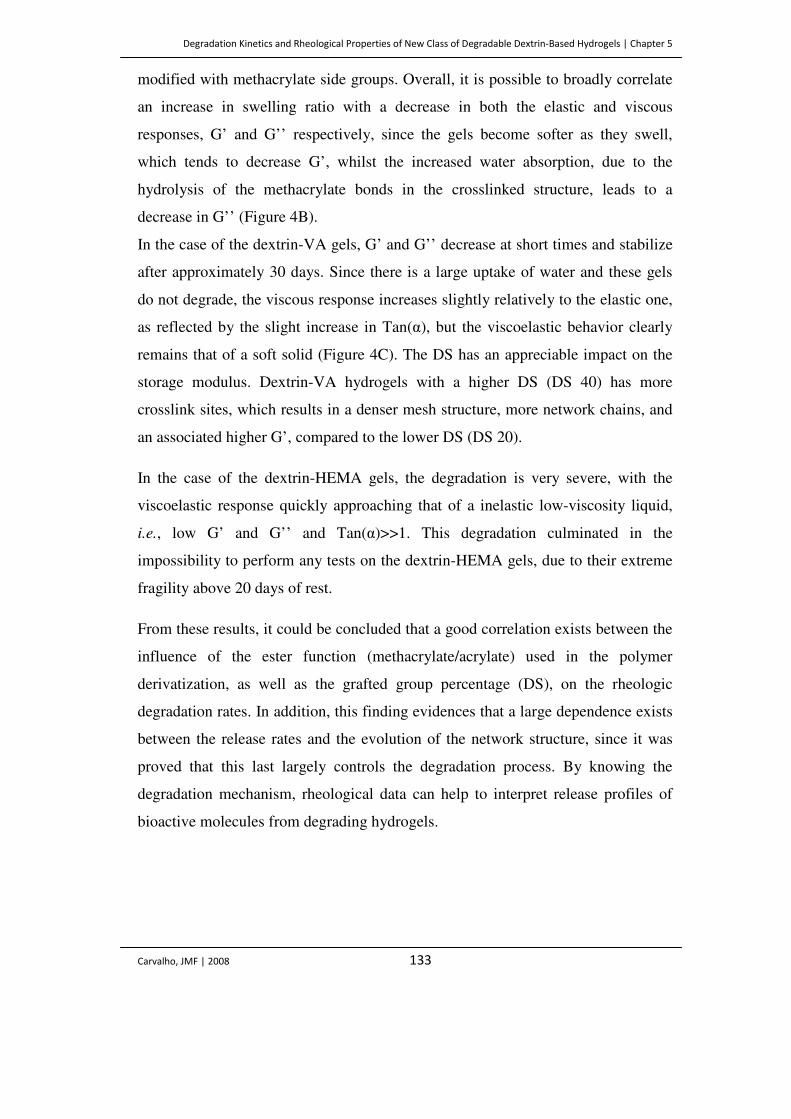
Degradation Kinetics and Rheological Properties of New Class of Degradable Dextrin-Based Hydrogels | Chapter 5
Carvalho, JMF | 2008 133
modified with methacrylate side groups. Overall, it is possible to broadly correlate
an increase in swelling ratio with a decrease in both the elastic and viscous
responses, G’ and G’’ respectively, since the gels become softer as they swell,
which tends to decrease G’, whilst the increased water absorption, due to the
hydrolysis of the methacrylate bonds in the crosslinked structure, leads to a
decrease in G’’ (Figure 4B).
In the case of the dextrin-VA gels, G’ and G’’ decrease at short times and stabilize
after approximately 30 days. Since there is a large uptake of water and these gels
do not degrade, the viscous response increases slightly relatively to the elastic one,
as reflected by the slight increase in Tan(α), but the viscoelastic behavior clearly
remains that of a soft solid (Figure 4C). The DS has an appreciable impact on the
storage modulus. Dextrin-VA hydrogels with a higher DS (DS 40) has more
crosslink sites, which results in a denser mesh structure, more network chains, and
an associated higher G’, compared to the lower DS (DS 20).
In the case of the dextrin-HEMA gels, the degradation is very severe, with the
viscoelastic response quickly approaching that of a inelastic low-viscosity liquid,
i.e., low G’ and G’’ and Tan(α)>>1. This degradation culminated in the
impossibility to perform any tests on the dextrin-HEMA gels, due to their extreme
fragility above 20 days of rest.
From these results, it could be concluded that a good correlation exists between the
influence of the ester function (methacrylate/acrylate) used in the polymer
derivatization, as well as the grafted group percentage (DS), on the rheologic
degradation rates. In addition, this finding evidences that a large dependence exists
between the release rates and the evolution of the network structure, since it was
proved that this last largely controls the degradation process. By knowing the
degradation mechanism, rheological data can help to interpret release profiles of
bioactive molecules from degrading hydrogels.
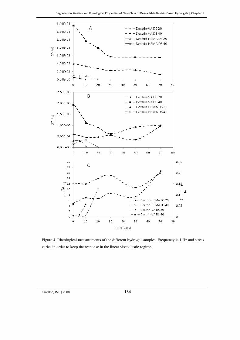
Degradation Kinetics and Rheological Properties of New Class of Degradable Dextrin-Based Hydrogels | Chapter 5
Carvalho, JMF | 2008 134
Figure 4. Rheological measurements of the different hydrogel samples. Frequency is 1 Hz and stress
varies in order to keep the response in the linear viscoelastic regime.
B
C
Degradation Kinetics and Rheological Properties of New Class of Degradable Dextrin-Based Hydrogels | Chapter 5
Carvalho, JMF | 2008 135
CONCLUSIONS
It was possible to obtain a new class of polymerizable dextrins (dextrin-HEMA) by
using a method based on the activation of the hydroxyl groups of HEMA with CDI,
followed by their coupling with the polymer, yielding a derivatized material that
can be polymerized in aqueous solution to form hydrogels. The comparative study
of the stability of these new hydrogels and the previously described non-degradable
dextrin-VA ones revealed that the firsts are effectively hydrolyzed under
physiological conditions. Contrarily to the dextrin-VA hydrogels, in which the SR
remains almost constant during storage, in the case of dextrin-HEMA, a
progressive swelling accompanied by a severe mass loss process, occurred over the
time, culminating in the complete dissolution of the hydrogels. Accordingly, the
comparative study, extended to the rheological analysis of the viscoelastic
behavior, revealed that dextrin-HEMA hydrogels have lower storage module (G’)
values, compared with the dextrin-VA ones, indicating that physical structuring is
less pronounced when dextrin is modified with methacrylate side groups.
It can be concluded that the dextrin functionalization with HEMA, may be an
effective route to modulate the hydrogel characteristics, namely the degradation
kinetics, tailoring the release profiles to fit the purpose of the controlled delivery.
References
Anseth KS, Bowman CN and Brannon-Peppas L. (1996) Mechanical properties of hydrogels and
their experimental determination. Biomaterials 17: 1647–1657.
Carvalho J, Gonçalves C, Gil AM and Gama FM. (2007) Production and characterization of a new
dextrin based hydrogel. Eur Pol J 43: 3050-3059.
Carvalho J, Coimbra, MA and Gama FM. (2008) New dextrin-vinylacrylate hydrogel: studies on
protein diffusion and release. Carbohydr Pol doi:10.1016/j.carbpol.2008.07.033. In Press.
Degradation Kinetics and Rheological Properties of New Class of Degradable Dextrin-Based Hydrogels | Chapter 5
Carvalho, JMF | 2008 136
Ferry JD. (1980) Viscoelastic Properties of Polymers, John Wiley & Sons, New York, USA.
Ferreira L, Gil MH and Dordick JS. (2002) Enzymatic synthesis of inulin-containing hydrogels.
Biomacromolecules 3: 333–341. (a)
Ferreira L, Carvalho R, Gil MH and Dordick JS. (2002) Enzymatic synthesis of dextran-containing
hydrogels. Biomaterials 23: 3957–3967. (b)
Flory PJ. (1953) In Principles of polymer chemistry. Cornell University Press, Ithaca, NY, USA, pp
577.
Hennink WE, Talsma H, Borchert JCH, de Smedt SC and Demeester J. (1996) Controlled release of
proteins from dextran hydrogels. J Control Release 39: 47–55.
Kane JB, Tompkins RG, Yarmush ML and Burke JF. (1996) In Biomaterials science: an
introduction to materials in medicine. Academic Press, San Diego, USA, pp 360–370.
Martens P, Holland T and Anseth KS. (2002) Synthesis and characterization of degradable
hydrogels formed from acrylate modified poly(vinyl alcohol) macromers. Polymer 43: 6093–6100.
Meyvis TKL, De Smedt SC, Hennink WE and Demeester J. (2000) Influence of the degradation
mechanism of hydrogels on their elastic and swelling properties during degradation.
Macromolecules 33: 4717–4725.
McCulloch I and Shalaby SW. (1998) In Tailored polymeric materials for controlled delivery
systems. American Chemical Society, Washington DC, USA.
Okay O and Gurun C. (1992) Synthesis and formation mechanism of porous 2-hydroxyethyl
methacrylate-ethylene glycol dimethacrylate copolymer beads. J Appl Polym Sci 46: 401–410.
Peppas NA. (1996) In Biomaterials science: an introduction to materials in medicine. Academic
Press, San Diego, USA, pp 60–64.
Peppas NA, Bures P, Leobandung W and Ichikawa H. (2000) Hydrogels in pharmaceutical
formulations. Eur J Pharm 50: 27– 46.
Stubbe BG, Braeckmans K, Horkay F, Hennink WE, De Smedt SC and Demeester J. (2002)
Swelling pressure observations on degrading dex-HEMA hydrogels, Macromolecules 35: 2501–
2505.
Suggs LJ, Krishnan RS, Garcia CA, Peter SJ, Anderson JM and Mikos AG. (1998) In vitro and in
vivo degradation of poly(propylene fumarate-co-ethylene glycol) hydrogels. J Biomed Mater Res
42: 312–320.
Degradation Kinetics and Rheological Properties of New Class of Degradable Dextrin-Based Hydrogels | Chapter 5
Carvalho, JMF | 2008 137
Van Dijk-Wolthuis WNE, Franssen O, Talsma H, Van Steenbergen MJ, Kettenes-van den Bosch JJ
and Hennink WE. (1995) Synthesis, characterization, and polymerization of glycidyl methacrylate
derivatized dextran. Macromolecules 28: 6317–6322.
Van Dijk-Wolthuis WNE, Kettenes-van den Bosch JJ, Van der Kerkvan Hoof A and Hennink WE.
(1997) Reaction of dextran with glycidyl methacrylate: an unexpected transesterification.
Macromolecules 30: 3411–3413. (a)
Van Dijk-Wolthuis WNE, Van Steenbergen MJ, Underberg WJM and Hennink WE. (1997)
Degradation kinetics of methacrylated dextrans in aqueous solution. J Pharm. Sci 86: 413–417. (b)
Van Dijk-Wolthuis WNE, Tsang SKY, Kettenes-van den Bosch JJ and Hennink WE. (1997) A new
class of polymerizable dextran with hydrolyzable groups: hydroxyethyl methacrylated dextran with
and without oligolactate spacer. Polymer 38: 6235–6242. (c)
Van Dijk-Wolthuis WNE, Hoogeboom JAM, van Steenbergen MJ, Tsang SKY and Hennink WE.
(1997) Degradation and release behavior of dextran-based hydrogels. Macromolecules 30: 4639–
4645. (d)
Vyavahare N and Kohn J. (1994) Photocrosslinked hydrogels based on copolymers of poly(ethylene
glycol) and lysine. J Pol Sci 32: 1271–1281.
Walther DH, Sin GH, Blanch HW and Prausnitz JM. (1995) Pore-size distributions of cationic 2-
hydroxyethyl methacrylate (HEMA) hydrogels. Polym. Gels Networks 3: 29–45.
Wong WH and Mooney DJ. (1997) Synthesis and properties of biodegradable polymers used as
synthetic matrices for tissue engineering. In Synthetic biodegradable polymer scaffolds. Atala A and
Mooney DJ. (eds.) Birkhaüser, Boston, USA, pp 49–82.


Modification of Pulp and Paper Using CBD-PEG Conjugates | Chapter 6
Carvalho, JMF | 2008 141
Introduction
Enzymes have long been applied in paper production, for the modification of both
fiber and paper properties. These enzymes include xylanases, endo-glucanases,
cellulases and hemicellulases (Suurnäki et al. 1994, Senior and Hamilton 1993,
Gen and Li 2003, Seo et al. 2000, Jackson at al. 1993, Pala et al. 2001). However,
several drawbacks result from enzyme utilization in paper treatment, such as the
extensive hydrolysis of polysaccharides that leads to a reduction of both fiber
strength and mass. To avoid these side effects that result from enzyme utilization,
recently the use of cellulose-binding domains (CBDs) in paper treatment has been
evaluated. CBDs are non-catalytic modules present in several cellulases and
hemicellulases and exhibit high affinity for cellulose. These modules are produced
by a large number of fungi and bacteria and potentiate the catalytic efficiency of
the associated catalytic domains. The ability to fuse CBDs with other molecules of
different sizes, charges, hydrophobicities, catalytic activities or bioactivities, allows
these molecules to be potentially interesting and useful on the activation and
modification of cellulose fibers (Kitaoka and Tanaka 2001, Levy and Shoseyov
2002, Pinto et al. 2004, Pinto et al. 2006). CBDs may be obtained through the
proteolysis of cellulases, as described earlier, providing large amounts of peptides
in a rather rapid procedure, or using the recombinant DNA technology (Creagh et
al. 1996, Bothwell et al. 1997, Linder et al. 1999, Bayer et al. 2000). This last
method allows overcoming the purity limitations of the proteolysis approach,
although the CBDs obtained through these methods are not glycosylated (Pinto et
al. 2006).
Cellobiohydrolase A (CbhA) is one of the most important catalytic components of
the multi-enzyme complex produced by the anaerobic thermophilic bacterium
Clostridium thermocellum, which was termed cellulosome. This complex is
specialized in the hydrolysis of insoluble plant cell wall polysaccharides. It is
composed of a cellulosome-integrating protein, termed CipA, containing nine
repetitive cohesin domains which bind to the dockerin domains located in the
Modification of Pulp and Paper Using CBD-PEG Conjugates | Chapter 6
Carvalho, JMF | 2008 142
cellulosomal catalytic subunits that can express glycoside hydrolase, carbohydrate
esterase or pectate lyase activities (Bayer et al. 1998, Beguin and Alzari 1998,
Blum et al. 2000, Felix and Ljungdahl 1993, Kataeva et al. 2002, Kataeva and
Ljungdahl 2003, Kurokawa et al. 2001, Shoham et al. 1999, Zverlov et al. 1998).
The cellulosome is anchored to the plant cell wall primarily through the action of
the internal family 3 CBM located in CipA. C. thermocellum cellulosomal CbhA is
composed of an N-terminal family 4 CBD, an immunoglobulin-like domain, a
family 9 glycosyl hydrolase catalytic domain (GH9), two Fn3-like repeats (Fn31,2),
a family 3 CBD and a C-terminal dockerin domain (Kataeva et al. 2002). Previous
work reported the ability of CbhA family 3 CBD to enhance the surface
modification and hydrolysis of cellulose fibers (Kataeva et al. 2002). Additionally,
it has been shown that CBDs obtained through the proteolysis of Trichoderma
reesei cellulases, are capable of modifying the drainability of recycled paper pulps
(Pinto et al. 2004). In the present work, we analyzed the potential of two
recombinant family 3 CBDs (belonging to CipA and CbhA of the Clostridium
thermocellum) and a fusion protein with the FN31,2 module (FN31-FN32-CBDCbhA)
to modify secondary paper fibers. Furthermore, the CBDs were chemically
conjugated with an activated PEG, as this molecule mimetize the glycosidic
fraction that is lacking in the highly purified bacterial CBDs. Finally, the technical
properties of the fiber papers and paper sheets resulting from the CBD treatment
were analyzed.
Materials and Methods
Chemicals
Sodium acetate was obtained from Panreac (Barcelona, Spain) and acid-swollen
cellulose (ASC) was prepared by treatment of Avicel PH-105 (Fluka, Philadelphia)
with phosphoric acid (Klyosov and Sinitsyn 1981). Whatman No. 3 filter paper and
dinitrosalicylic acid (DNS) were from Sigma (St. Louis, Mo.). SS-m Polyethylene
glycol (PEG) (SUNBRIGHT MW5000) was purchased from NOF Corporation
Modification of Pulp and Paper Using CBD-PEG Conjugates | Chapter 6
Carvalho, JMF | 2008 143
(Belgium). Paper fibers (Kraftline Paper) were gently supplied by Portucel Viana
company. All chemicals were of the highest purity available.
Bacterial strains, culture conditions, and plasmids.
C. thermocellum JW20 was used as a source of genomic DNA. The bacterium was
grown anaerobically under a nitrogen atmosphere at 60°C in a prereduced medium
with 1% (w v-1) cellobiose as described earlier (Kataeva et al. 1999). E. coli
BL21(DE3)pLys (Stratagene Cloning Systems, La Jolla, Calif.) was used as the
cloning host for the T7 RNA polymerase expression vector pET-21a (Novagen,
Madison, Wisc.). It was grown in Luria-Bertani medium supplemented with
ampicillin (100 µg ml-1). Isolation of genomic DNA from C. thermocellum was
performed through the method described by Marmur (Marmur 1961).
Primer design, PCR, and cloning.
Flanking primers containing restriction sites were designed according to the DNA
sequence of the gene encoding CbhA (Table 1). DNA fragments were amplified
through PCR using purified genomic DNA from C. thermocellum as a template.
The PCR reactions were carried out with Taq polymerase (New England Biolabs,
Beverly, Mass.). PCRs were performed as follows: preheating at 95 ºC for 2 min,
40 cycles at 95 ºC for 45 s, 53 ºC for 45 s and 72 ºC for 45s, followed by an
elongation at 72 ºC for 10 min. PCR products were separated by 1% agarose gel
electrophoresis. The extracted DNA fragments were digested with the appropriated
restriction enzymes and ligated into the pET-21a vector digested similarly. The
ligation products were used to transform BL21(DE3)pLys competent cells. The
integrity of each construct was verified by both restriction analysis and DNA
sequencing.
Modification of Pulp and Paper Using CBD-PEG Conjugates | Chapter 6
Carvalho, JMF | 2008 144
Table 1. Oligonucleotides used for cloning.
Nº and primer Sequence Restriction
Site Direction
1.CBDCbhA 5´- CTC GCT AGC GAT GTA AAA GTA GAG TAC TTG - 3´ Nhe I Forward
2.CBDCbhA 5´- CAC CTC GAG GGT TCC CCA AAC AAG TTC TCC - 3´ Xho I Reverse
3.FN31-FN32-CBDCbhA 5´- CTC GCT AGC ACA GAC AGT GAA ACC GAT AAG - 3´ Nhe I Forward
4.FN31-FN32-CBDCbhA 5´- CAC CTC GAG GGT TCC CCA AAC AAG TTC TCC - 3´ Xho I Reverse
5.CBDCipA 5´- CTA GCT AGC AGA GGT GAT ACA CCG ACC AAG GGA G - 3´ Nhe I Forward
6.CBDCipA 5´- CAC CTC GAG TTC TTT ACC CCA TAC AAG AAC - 3´ Xho I Reverse
Protein purification
All recombinant proteins were six-Histagged at the C terminus and were purified
from BL21(DE3)pLys cultures (1 liter) harboring the appropriated pET-21a
derivative. The cells were harvested 5 h after induction with 1 mM isopropyl-D-
thiogalactopyranoside (IPTG) at 37ºC. All purification steps were performed at
4°C. After collection, the cells were resuspended with 20 mM Na-phosphate buffer,
pH 7.5, containing 0.5 M NaCl, and disintegrated with a sonicator. Cell debris was
removed by centrifugation. Clear supernatant was applied onto a Ni-nitrilotriacetic
acid (Ni-NTA) agarose (HisTrap, Amersham) column equilibrated with the 20 mM
Na-phosphate, pH 7.5, containing 0.5 M NaCl. The column was washed with 20
mM Na-phosphate–0.5 M NaCl, pH 7.4. Proteins were eluted by a gradient of 0 to
0.3 M imidazole, pH 7.4. Fractions containing recombinant proteins were
combined and dialyzed against PBS. Dialyzed proteins were further used in the
adsorption assays. Additionally, the glycohydrolase GH9 from CbhA and the
endoglucanase Cel5B from Cellvibrio mixtus were produced as described
previously (Fontes et al. 1998).
CBD-PEG Conjugation
Briefly, the protein to be pegylated was dissolved in 0.1 M Sodium Phosphate, pH
7.5, at a concentration of 2 mg mL-1. A designated quantity of succinimidyl
succinate (SS)-mPEG (Figure 1) (at the molar ratio of polymer to primary amines
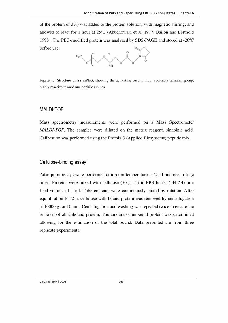
Modification of Pulp and Paper Using CBD-PEG Conjugates | Chapter 6
Carvalho, JMF | 2008 145
of the protein of 3%) was added to the protein solution, with magnetic stirring, and
allowed to react for 1 hour at 25ºC (Abuchowski et al. 1977, Bailon and Berthold
1998). The PEG-modified protein was analyzed by SDS-PAGE and stored at -20ºC
before use.
Figure 1. Structure of SS-mPEG, showing the activating succinimidyl succinate terminal group,
highly reactive toward nucleophile amines.
MALDI-TOF
Mass spectrometry measurements were performed on a Mass Spectrometer
MALDI-TOF. The samples were diluted on the matrix reagent, sinapinic acid.
Calibration was performed using the Promix 3 (Applied Biosystems) peptide mix.
Cellulose-binding assay
Adsorption assays were performed at a room temperature in 2 ml microcentrifuge
tubes. Proteins were mixed with cellulose (50 g L-1) in PBS buffer (pH 7.4) in a
final volume of 1 ml. Tube contents were continuously mixed by rotation. After
equilibration for 2 h, cellulose with bound protein was removed by centrifugation
at 10000 g for 10 min. Centrifugation and washing was repeated twice to ensure the
removal of all unbound protein. The amount of unbound protein was determined
allowing for the estimation of the total bound. Data presented are from three
replicate experiments.
Modification of Pulp and Paper Using CBD-PEG Conjugates | Chapter 6
Carvalho, JMF | 2008 146
Enzyme assays
The cellulolytic activity of the truncated variants CBDCbhA and FN31-FN32-
CBDCbhA were assayed isolated and in the presence of the enzymes GH9 and Cel5B
(at the optimum enzyme temperature, 60 and 50°C for GH9 and Cel5B,
respectively) was determined in a 50 mM sodium citrate buffer, pH 6.0, containing
5 % of the substrate. Control assays were performed in the same conditions, only
with buffer. The activity was monitored by measuring the release of reducing
sugars. Reducing sugars were determined with DNS (Miller 1959). Activity was
expressed as micromoles of product (cellobiose equivalent) released per minute per
milligram of enzyme. Efficiency of cellulose hydrolysis was determined with ASC
and filter paper as substrates.
Treatment with CBDs and CBD-PEG conjugate
Cellulose fibers, at 10% (w w-1) consistency, were beated in a PFI mill for 3000
revolutions. Afterwards, CBDs (CBDCipA, CBDCipA-PEG, CBDCbhA and FN31-
FN32-CBDCbhA) were adsorbed by mixing the protein at different concentrations
(0.1, 1 and 2 mg per gram of fiber, dry weight) with 30 g of fibers (dry weight) in
acetate buffer (50 mM, pH 5.0 and final volume of 2 L) for 30 minutes at room
temperature. Control experiments were performed by adding buffer without the
protein.
Analysis of the Pulp and Paper Properties
The pulp and handsheet properties were analyzed using standard procedures: ISO
5267/1 (Shopper-Riegler); ISO 5636/3 1992 (F) (permeability); ISO 1924/2 1985
(F) (tensile strength and break length); ISO 1947 (F) (tearing); ISO 2758 1983 (F)
(bursting strength).
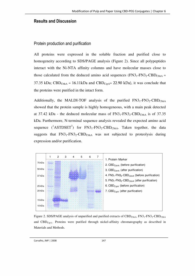
Modification of Pulp and Paper Using CBD-PEG Conjugates | Chapter 6
Carvalho, JMF | 2008 147
2 3 41 5 761. Protein Marker
2. CBDCbhA (before purification)
3. CBDCbhA (after purification)
4. FN31-FN32-CBDCbhA (before purification)
5. FN31-FN32-CBDCbhA (after purification)
6. CBDCipA (before purification)
7. CBDCipA (after purification)
75 kDa
50 kDa
37 kDa
25 kDa
20 kDa
15 kDa
10 kDa
Results and Discussion
Protein production and purification
All proteins were expressed in the soluble fraction and purified close to
homogeneity according to SDS/PAGE analysis (Figure 2). Since all polypeptides
interact with the Ni-NTA affinity columns and have molecular masses close to
those calculated from the deduced amino acid sequences (FN31-FN32-CBDCbhA ≈
37.35 kDa; CBDCbhA ≈ 16.11kDa and CBDCipA≈ 22.90 kDa), it was conclude that
the proteins were purified in the intact form.
Additionally, the MALDI-TOF analysis of the purified FN31-FN32-CBDCbhA
showed that the protein sample is highly homogeneous, with a main peak detected
at 37.42 kDa - the deduced molecular mass of FN31-FN32-CBDCbhA is of 37.35
kDa. Furthermore, N-terminal sequence analysis revealed the expected amino acid
sequence (1ASTDSET7) for FN31-FN32-CBDCbhA. Taken together, the data
suggests that FN31-FN32-CBDCbhA was not subjected to proteolysis during
expression and/or purification.
Figure 2. SDS/PAGE analysis of unpurified and purified extracts of CBDCbhA, FN31-FN32-CBDCbhA
and CBDCipA.. Proteins were purified through nickel-affinity chromatography as described in
Materials and Methods.
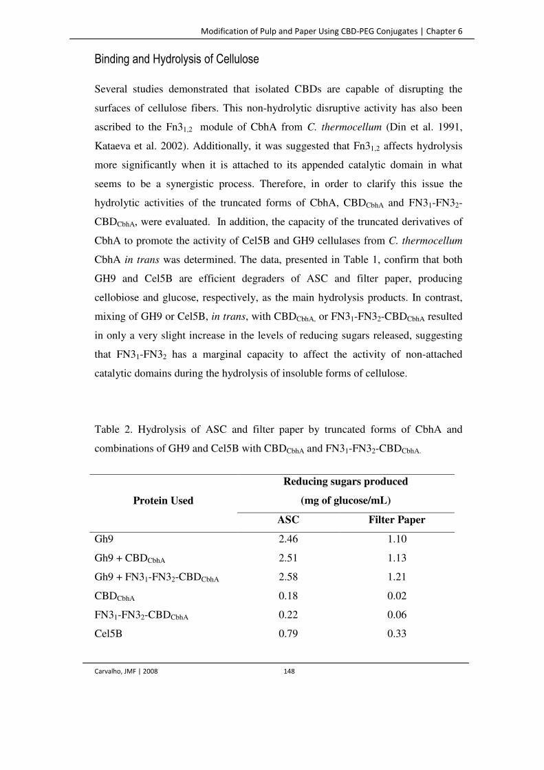
Modification of Pulp and Paper Using CBD-PEG Conjugates | Chapter 6
Carvalho, JMF | 2008 148
Binding and Hydrolysis of Cellulose
Several studies demonstrated that isolated CBDs are capable of disrupting the
surfaces of cellulose fibers. This non-hydrolytic disruptive activity has also been
ascribed to the Fn31,2 module of CbhA from C. thermocellum (Din et al. 1991,
Kataeva et al. 2002). Additionally, it was suggested that Fn31,2 affects hydrolysis
more significantly when it is attached to its appended catalytic domain in what
seems to be a synergistic process. Therefore, in order to clarify this issue the
hydrolytic activities of the truncated forms of CbhA, CBDCbhA and FN31-FN32-
CBDCbhA, were evaluated. In addition, the capacity of the truncated derivatives of
CbhA to promote the activity of Cel5B and GH9 cellulases from C. thermocellum
CbhA in trans was determined. The data, presented in Table 1, confirm that both
GH9 and Cel5B are efficient degraders of ASC and filter paper, producing
cellobiose and glucose, respectively, as the main hydrolysis products. In contrast,
mixing of GH9 or Cel5B, in trans, with CBDCbhA, or FN31-FN32-CBDCbhA resulted
in only a very slight increase in the levels of reducing sugars released, suggesting
that FN31-FN32 has a marginal capacity to affect the activity of non-attached
catalytic domains during the hydrolysis of insoluble forms of cellulose.
Table 2. Hydrolysis of ASC and filter paper by truncated forms of CbhA and
combinations of GH9 and Cel5B with CBDCbhA and FN31-FN32-CBDCbhA.
Protein Used
Reducing sugars produced
(mg of glucose/mL)
ASC Filter Paper
Gh9 2.46 1.10
Gh9 + CBDCbhA 2.51 1.13
Gh9 + FN31-FN32-CBDCbhA 2.58 1.21
CBDCbhA 0.18 0.02
FN31-FN32-CBDCbhA 0.22 0.06
Cel5B 0.79 0.33

Modification of Pulp and Paper Using CBD-PEG Conjugates | Chapter 6
Carvalho, JMF | 2008 149
The capacity of the proteins expressed in this study to bind insoluble forms of
cellulose was evaluated. The data, presented in figure 3a, revealed that both
CBDCbhA and FN31-FN32-CBDCbhA present a low affinity to cellulose, as shown by
the analysis of the adsorption isotherms. Non-linear regression analysis was used to
calculate the parameters of the adsorption isotherm, using the expression:
where CBDBound is the molar amount of protein adsorbed, per unit weight of
cellulose, [CBD]Free is the molar protein concentration in the liquid phase at the
adsorption equilibrium, CBDMax and Ka are the maximum molar amount of protein
adsorbed, per unit weight of cellulose, and the adsorption equilibrium constant,
respectively.
Both proteins bound weakly to cellulose, since the values of CBDMax and Ka were,
respectively, 0.27 µmol g-1 and 0.01 L µmol-1 (0.94 L g-1) for CBDCbhA and 0.14
µmol g-1 and 0.07 L µmol-1 (1.83 L g-1) for FN31-FN32-CBDCbhA. These values
are lower than those previously referred for CBDCbhA, by Kataeva and colleagues
(2002), which described the domain as a cellulose binder with a CBDMax of 2.45
µmol g-1 and a Ka of 1.12 L g-1. Taken together, the data suggest that, although
surprisingly, the FN31-FN32-CBDCbhA exhibits poor affinity for cellulose fibers,
and its lower affinity cannot be explained by proteolysis, as described above. The
family 3 CBDs are found associated with GH9, which are known to act in
cellulosic substrates (Bayer et al. 2000). There are three major subgroups in the
family 3 CBDs; a, b and c, based on sequence similarity (Morag et al. 1990). In
biochemical studies, the CBDb subgroup (to which belongs the CBDCbhA used in
this work), have been shown to bind strongly to the surface of microcrystalline
cellulose and were proposed to promote the cellulolytic activity by concentrating
the enzyme near the cellulose surface (Tormo et al. 1996, Gilad et al. 2003).
Cel5B + CBDCbhA 0.83 0.34
Cel5B + FN31-FN32-CBDCbhA 0.84 0.36
Control 0.19 0.02
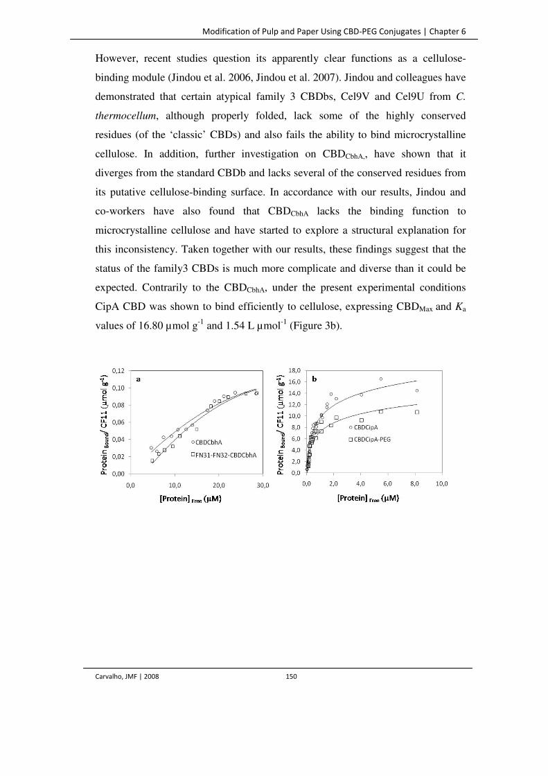
Modification of Pulp and Paper Using CBD-PEG Conjugates | Chapter 6
Carvalho, JMF | 2008 150
However, recent studies question its apparently clear functions as a cellulose-
binding module (Jindou et al. 2006, Jindou et al. 2007). Jindou and colleagues have
demonstrated that certain atypical family 3 CBDbs, Cel9V and Cel9U from C.
thermocellum, although properly folded, lack some of the highly conserved
residues (of the ‘classic’ CBDs) and also fails the ability to bind microcrystalline
cellulose. In addition, further investigation on CBDCbhA,, have shown that it
diverges from the standard CBDb and lacks several of the conserved residues from
its putative cellulose-binding surface. In accordance with our results, Jindou and
co-workers have also found that CBDCbhA lacks the binding function to
microcrystalline cellulose and have started to explore a structural explanation for
this inconsistency. Taken together with our results, these findings suggest that the
status of the family3 CBDs is much more complicate and diverse than it could be
expected. Contrarily to the CBDCbhA, under the present experimental conditions
CipA CBD was shown to bind efficiently to cellulose, expressing CBDMax and Ka
values of 16.80 µmol g-1 and 1.54 L µmol-1 (Figure 3b).

ERROR: stackunderflow
OFFENDING COMMAND: ~
STACK: