francisco pereira gonçalves - repositorio-aberto.up.pt · trabalho organizado de acordo com as...
TRANSCRIPT
2016/2017
Francisco Pereira Gonçalves
Acute and subacute functional
effects of relaxin-2 on human
mammary artery/
Efeitos funcionais agudos e
subagudos da relaxina-2 na artéria
mamária humana
março, 2017
Mestrado Integrado em Medicina
Área: Medicina básica (FOS 3.1)
Fisiologia
Tipologia: Dissertação
Trabalho efetuado sob a Orientação de:
Professor Doutor Paulo Castro Chaves
E sob a Coorientação de:
Professor Doutor Adelino Leite Moreira
Trabalho organizado de acordo com as normas da revista: Peptides
Francisco Pereira Gonçalves
Acute and subacute functional effects of
relaxin-2 on human mammary artery/
Efeitos funcionais agudos e subagudos da
relaxina-2 na artéria mamária humana
março, 2017

Projeto de Opção do 6º ano - DECLARAÇÃO DE INTEGRIDADE
Eu, Francisco Pereira Gonçalves, abaixo assinado, nº mecanográfico 201107394, estudante do 6º ano
do Ciclo de Estudos Integrado em Medicina, na Faculdade de Medicina da Universidade do Porto,
declaro ter atuado com absoluta integridade na elaboração deste projeto de opção.
Neste sentido, confirmo que NÃO incorri em plágio (ato pelo qual um indivíduo, mesmo por omissão,
assume a autoria de um determinado trabalho intelectual, ou partes dele). Mais declaro que todas as
frases que retirei de trabalhos anteriores pertencentes a outros autores, foram referenciadas, ou
redigidas com novas palavras, tendo colocado, neste caso, a citação da fonte bibliográfica.
Faculdade de Medicina da Universidade do Porto, 09/03/2017
Assinatura conforme cartão de identificação:
Projecto de Opção do 6º ano – DECLARAÇÃO DE REPRODUÇÃO
NOME
Francisco Pereira Gonçalves
NÚMERO DE ESTUDANTE DATA DE CONCLUSÃO
201107394 09-03-2017
DESIGNAÇÃO DA ÁREA DO PROJECTO
Medicina Básica (FOS 3.1) - Fisiologia
TÍTULO DISSERTAÇÃO
Acute and subacute functional effects of relaxin-2 on human mammary artery
ORIENTADOR
Paulo Castro Chaves
COORIENTADOR
Adelino Leite Moreira
ASSINALE APENAS UMA DAS OPÇÕES:
É AUTORIZADA A REPRODUÇÃO INTEGRAL DESTE TRABALHO APENAS PARA EFEITOS DE INVESTIGAÇÃO,
MEDIANTE DECLARAÇÃO ESCRITA DO INTERESSADO, QUE A TAL SE COMPROMETE.
É AUTORIZADA A REPRODUÇÃO PARCIAL DESTE TRABALHO (INDICAR, CASO TAL SEJA NECESSÁRIO, Nº
MÁXIMO DE PÁGINAS, ILUSTRAÇÕES, GRÁFICOS, ETC.) APENAS PARA EFEITOS DE INVESTIGAÇÃO, MEDIANTE
DECLARAÇÃO ESCRITA DO INTERESSADO, QUE A TAL SE COMPROMETE.
DE ACORDO COM A LEGISLAÇÃO EM VIGOR, (INDICAR, CASO TAL SEJA NECESSÁRIO, Nº MÁXIMO DE PÁGINAS,
ILUSTRAÇÕES, GRÁFICOS, ETC.) NÃO É PERMITIDA A REPRODUÇÃO DE QUALQUER PARTE DESTE TRABALHO.
X
Faculdade de Medicina da Universidade do Porto, 09/03/2017
Assinatura conforme cartão de identificação:
Acute and subacute functional effects of
relaxin-2 on human mammary artery
Francisco Gonçalves1,2, Rafael Martins,1,2, Luís Mendonça1,2, Tiago Laundos
Santos3,4, Mariana Pintalhão1,2,5, Francisco Vasques Nóvoa1,2,5, Mário Jorge
Amorim6, Paulo Pinho6, Diana Nascimento 3,4, Adelino Leite Moreira1,2,6, Paulo
Castro Chaves1,2,5
1 - Departamento de Cirurgia e Fisiologia, Universidade do Porto, Portugal.
2 - Unidade de Investigação Cardiovascular, Portugal.
3 - Instituto de Engenharia Biomédica (INEB), Universidade do Porto, Portugal.
4 - Instituto de Investigação e Inovação em Saúde (i3S), Universidade do Porto, Portugal.
5 - Internal Medicine Department, São João Hospital Centre, Porto, Portugal.
6 - Cardiothoracic Surgery Department, São João Hospital Centre, Porto, Portugal.
Endereço para correspondência:
Professor Doutor Paulo Castro-Chaves
Departamento de Cirurgia e Fisiologia,
Faculdade de Medicina da Universidade do Porto
Alameda Professor Hernâni Monteiro; 4200-319 Porto, Portugal
Tel.: +351 225508452; Fax: +351 225519194; E.mail: [email protected]

Acute and subacute functional effects of Francisco Gonçalves relaxin-2 on human mammary artery March 2017
ABSTRACT
Introduction: Despite classically associated with pregnancy, relaxin is a hormone with pleiotropic
properties and an increasingly unveiled role in the pathophysiology of several cardiovascular
diseases. Previous clinical trials have explored its therapeutic role on acute heart failure. However,
the exact actions of relaxin on the human vascular territory are scarcely known.
Purposes: To study the acute and subacute functional effects of relaxin-2 on the vasoreactivity of
the human mammary artery (HMA). To assess the expression of RXFP1, its main receptor, in this
vascular territory.
Methods: HMA from 37 patients subjected to coronary artery bypass graft surgery (average age 69.3
years; 7 female) were sectioned into 2mm rings and mounted on a myograph (DMT myograph
system). On one protocol, the vessels were exposed to increasing concentrations of relaxin (10-10-
10-7M) or vehicle after pre-contraction with phenylephrine (10-5M), n=5. On a different protocol,
the rings were treated for 24h with relaxin (10-7M) or vehicle and afterwards subjected to
increasing concentrations of vasoconstrictors (10-9-10-5 M) - phenylephrine (n=8), endothelin-1
(n=6) and angiotensin II (n=11) – and of vasodilators (10-10-10-5 M) - acetylcholine (n=8) and sodium
nitroprusside (n=11). Vessel rings were also incubated for 24h with relaxin or vehicle after
endothelium removal, and their response to nitroprusside evaluated (10-10-10-5 M; n=7).
Immunofluorescence labelling of RXFP1 was performed along with labelling for CD31 (endothelial
cells) and α smooth muscle actin (smooth muscle cells). Functional responses are expressed as
mean±standard error (%).
Results: After acute exposure to relaxin, no differences in the developed active tension were
observed between relaxin or vehicle treated groups. Following 24h treatment with relaxin or
vehicle, vascular viability and the vasoconstrictor effects of phenylephrine, endothelin-1 and
angiotensin II were similar between groups. However, vessel rings treated with relaxin showed
higher relaxation when compared to vehicle treated rings, both in response to acetylcholine
(59.1±6.1% vs 46.2±6.2%; p<0.01) and nitroprusside (128.9±4.8% vs 118.7±5.1% p<0.05). The
higher response to nitroprusside was preserved when treatment with relaxin followed
endothelium removal (143.3±4.1% vs 132.8±3.3%; p<0.01). Immunofluorescence showed labelling
for RXFP1 on smooth muscle and endothelial cells.
Conclusion: Treatment with relaxin-2 for 24h potentiates HMA vasodilatation without interfering
with the effects of several vasoconstrictors, and this effect is at least partially independent of the
endothelium. We have also identified for the first time the presence of RXFP1 on the
endothelium and smooth muscle cells of HMA. Besides shedding further light on relaxin’s role on
human vascular physiology, the present work may have implications in the understanding of
HMA’s physiology, an important conduct for coronary revascularization procedures.
Acute and subacute functional effects of Francisco Gonçalves relaxin-2 on human mammary artery March 2017
INTRODUCTION
Relaxin-2 is a peptide hormone initially described for its effects on the female reproductive
tract during pregnancy, and is responsible as well for mediating the cardiovascular adaptations
that occur during this period, inclusively through its actions on the systemic and renal vasculature.
[1-3]
In the past two decades, growing evidence has attributed relaxin pleiotropic effects on the
cardiovascular system. This hormone may play an import role on the pathophysiology of heart
failure, hypertension and cardiac ischemic disease. [4-6] Effectively, several studies point towards
compensatory production and release of relaxin by the myocardium during heart failure, and
indeed plasmatic levels of relaxin correlate with clinical and echocardiographic parameters of right
side heart overload. [7]
Additionally, relaxin has been evaluated as a possible therapeutic weapon in conditions
such as heart failure, hypertension, pre-eclampsia and peripheral vascular disease. [2] The RELAX-
AHF randomized clinical trial has demonstrated superiority of relaxin on dyspnoea relief and
mortality reduction at 180 days in relation to placebo, in an acute heart failure setting. [8] In the
context of cardiac ischemic disease, the demonstration of beneficial effects on the myocardial
ischemia-reperfusion lesion both on a pig model subject to anterior descent artery occlusion [9]
and on rat cardiomyocytes culture [10], may signal further therapeutic usefulness on coronary
heart disease and acute myocardial infarction.
Laboratory studies on animal models attribute relaxin angiogenic, antioxidant, anti-
inflammatory, antifibrotic and vasodilator actions [4, 5], that may constitute the basis of its
cardioprotective potential. The vascular effects of relaxin, mediated by its main receptor, RXFP1,
seem to occur through a rise in nitric oxide, with a probable involvement of metalloproteinases,
ET-B receptor signalling, extracellular matrix modifications and possibly VEGF signalling. [11]
However, the effects and mechanisms of action are markedly heterogenous between different
vascular territories and studies in human are scarce. Indeed, several aspects of relaxin’s action on
human vascular tissue await full characterization. [12]
Internal thoracic artery, or mammary artery (MA), is the preferred conduct for coronary
artery bypass graft (CABG) surgery [13, 14], and is characterized by its relative resistance to
atherosclerosis and endothelial dysfunction. [15, 16] These characteristics together with its clinical
relevance justify the pertinence of further unravelling the factors that modulate its vasoreactivity.
The present work aims to contribute for the enlightenment of relaxin’s role in human
vascular physiology, with the main goals of characterizing its acute and subacute effects on MA
vasoreactivity, the relevance of several clinical characteristics for the said effects and lastly to
assess the presence of RXFP1, relaxin’s main receptor, in this vascular territory.

Acute and subacute functional effects of Francisco Gonçalves relaxin-2 on human mammary artery March 2017
METHODS
Sample collection and dissection of vascular rings
Spare MA segments were collected by the Cardiothoracic Surgery Team during 37 CABG
procedures. Following collection, the segments were transported to the laboratory within 15
minutes in a physiological Krebs solution (T: 4ºC), with the following composition (mM): 130 NaCl;
4.7 KCl; 14.9 NaHCO3; 1.18 KH2PO4; 5.5 glucose; 1.17 MgSO4·7H2O e 1.6 CaCl2·2H2O. In the
laboratory the adjacent connective tissue was removed and the vessel was sectioned into rings
with 2 mm of length each. The patients included in the study were characterized in terms of
demographic characteristics, past medical history and medication prescribed. The project was
approved by the local Ethical Committee. The vascular rings obtained were used for both the
functional and immunofluorescence protocols described below.
Functional Protocols
Experimental Preparation
Each functional protocol proceeded using 4 vascular rings obtained from a single patient.
The rings were horizontally mounted in a myograph (Multi Wire Myograph System Model 620M -
DMT®) in Krebs solution (32ºC, pH 7.40), bubbling with a gaseous mixture (95% O2; 5% CO2). After
normalization to an effective transmural pressure of 100 mmHg, per a previously described
protocol [17], the rings stabilized during 60 min. Following this period, the vessel viability was
assessed through inciting the maximum contractile response to KCl (0,1M). After washing and new
stabilization for 30 min, the response to several vasoactive peptides was determined for each ring.
Data was continuously acquired and digitally stored (PowerLab 4/30 ADInstruments). The
following parameters were recorded: dimensions (segment and rings lengths and internal
circumference, in μm), tension (T, mN/mm) and effective transmural pressure (ERTP, kPa).
1- Influence of the acute treatment with relaxin on MA’s vasoreactivity
To study the immediate action of relaxin on MA, the rings obtained from the MA of 5
different patients were pre-contracted with phenylephrine (10-5M) and exposed to increasing
concentrations (10-10 -10-7M) of relaxin-2 (SRP3147, Sigma-Aldrich®) or vehicle. (Figure 1 - 1)
2 – Influence of the subacute treatment with relaxin on MA’s vasoreactivity
For this subset of protocols, which aimed to assess the influence of the preceding
treatment with relaxin on MA’s response to several vasoactive peptides, the vascular rings
obtained from 30 different patients were randomized to incubation for 24 hours with relaxin (10-
7M) or vehicle in DMEM culture medium (37ºC; 5% CO2), preceding its assembly in the myograph.
Following the normalization and contraction in response to KCl, described above, the rings were
Acute and subacute functional effects of Francisco Gonçalves relaxin-2 on human mammary artery March 2017
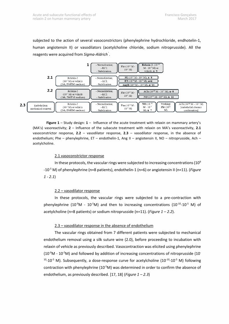
subjected to the action of several vasoconstrictors (phenylephrine hydrochloride, endhotelin-1,
human angiotensin II) or vasodilators (acetylcholine chloride, sodium nitroprusside). All the
reagents were acquired from Sigma-Aldrich®.
Figure 1 – Study design: 1 – Influence of the acute treatment with relaxin on mammary artery’s
(MA’s) vasoreactivity; 2 – Influence of the subacute treatment with relaxin on MA’s vasoreactivity, 2.1
vasoconstrictor response, 2.2 – vasodilator response, 2.3 – vasodilator response, in the absence of
endothelium; Phe – phenylephrine, ET – endothelin-1, Ang II – angiotensin II, NO – nitroprusside, Ach –
acetylcholine.
2.1 vasoconstrictor response
In these protocols, the vascular rings were subjected to increasing concentrations (109
-10-5 M) of phenylephrine (n=8 patients), endothelin-1 (n=6) or angiotensin II (n=11). (Figure
1 - 2.1)
2.2 – vasodilator response
In these protocols, the vascular rings were subjected to a pre-contraction with
phenylephrine (10-9M - 10-5M) and then to increasing concentrations (10-10-10-5 M) of
acetylcholine (n=8 patients) or sodium nitroprusside (n=11). (Figure 1 – 2.2).
2.3 – vasodilator response in the absence of endothelium
The vascular rings obtained from 7 different patients were subjected to mechanical
endothelium removal using a silk suture wire (2.0), before proceeding to incubation with
relaxin of vehicle as previously described. Vasocontraction was elicited using phenylephrine
(10-9M - 10-5M) and followed by addition of increasing concentrations of nitroprusside (10-
10-10-5 M). Subsequently, a dose-response curve for acetylcholine (10-10-10-5 M) following
contraction with phenylephrine (10-5M) was determined in order to confirm the absence of
endothelium, as previously described. [17, 18] (Figure 1 – 2.3)
Acute and subacute functional effects of Francisco Gonçalves relaxin-2 on human mammary artery March 2017
Immunofluorescence protocols
Vascular rings (see “Sample collection and dissection of vascular rings” above) were fixed
overnight in 10% neutral buffered formalin and embedded in paraffin. Histological 3 μm sections
were rehydrated and subjected to an antigen retrieval protocol with Tris/EDTA (pH=9.0; 96ºC; 35
min) and to treatment with Sudan Black B (0.5% (m/V) in ethanol 70%(V/V); room temperature; 5
min). Lastly, the sections were submitted to permeabilization with Triton X-100 (0.2%; room
temperature; 5 min).
RXFP1 location
Two MA rings from different patients were used for identification of RXFP1. Following
treatment described above, the sections were incubated overnight with a primary antibody for the
RXFP1 receptor (1:200; HPA027067, rabbit polyclonal; Sigma-Aldrich), primary antibody for CD31
(1:250; SC-1506, goat polyclonal, Santa Cruz Biotechnology) and primary antibody for smooth
muscle actin (1:400; A5228, mouse monoclonal, Sigma-Aldrich), at 4º C. Following washing, they
were incubated with the corresponding secondary antibodies for 1 hour (A31573 donkey anti-
rabbit; A21202 donkey anti-mouse; A11057 donkey anti-goat, respectively) and incubated in a 4’,6-
diamidino-2-phenylindole medium (DAPI) prior to cover slip placement. The specificity of the
immunostaining was assessed by omission of the primary antibodies. The images were obtained
with wide-field microscopy (DMI6000B Microscope; Leica, Heidelberg, Germany), with a 63x
amplification (1.3 NA).
Endothelium removal confirmation
Two out of all the rings subjected to mechanical endothelium removal were randomly
selected for an immunofluorescence protocol. This protocol followed the steps described
previously, but was performed with only the primary antibodies for CD31 and sma. Two rings not
subjected to endothelium removal were used as positive controls.
Statistical analysis
The tensions achieved with vasoconstriction in response to KCl were expressed in Active
Tension (AT, mN/mm). For all other vasoconstrictor responses, the instantaneous active tension
was expressed as the percentage of the response to KCl (AT vasoconstrictor/KCl, %). For the
vasodilator responses, the instantaneous active response was expressed as the percentage of the
active tension generated in response to phenylephrine (AT vasodilator/PheMax, %). The results are
presented as mean±standard error. Two-way repeated measures ANOVA was used for the
comparison between the tension developed in the relaxin treated group and the vehicle treated
one. P<0,05 was considered statistically significant.
Acute and subacute functional effects of Francisco Gonçalves relaxin-2 on human mammary artery March 2017
RESULTS
Functional protocols
Population characterization
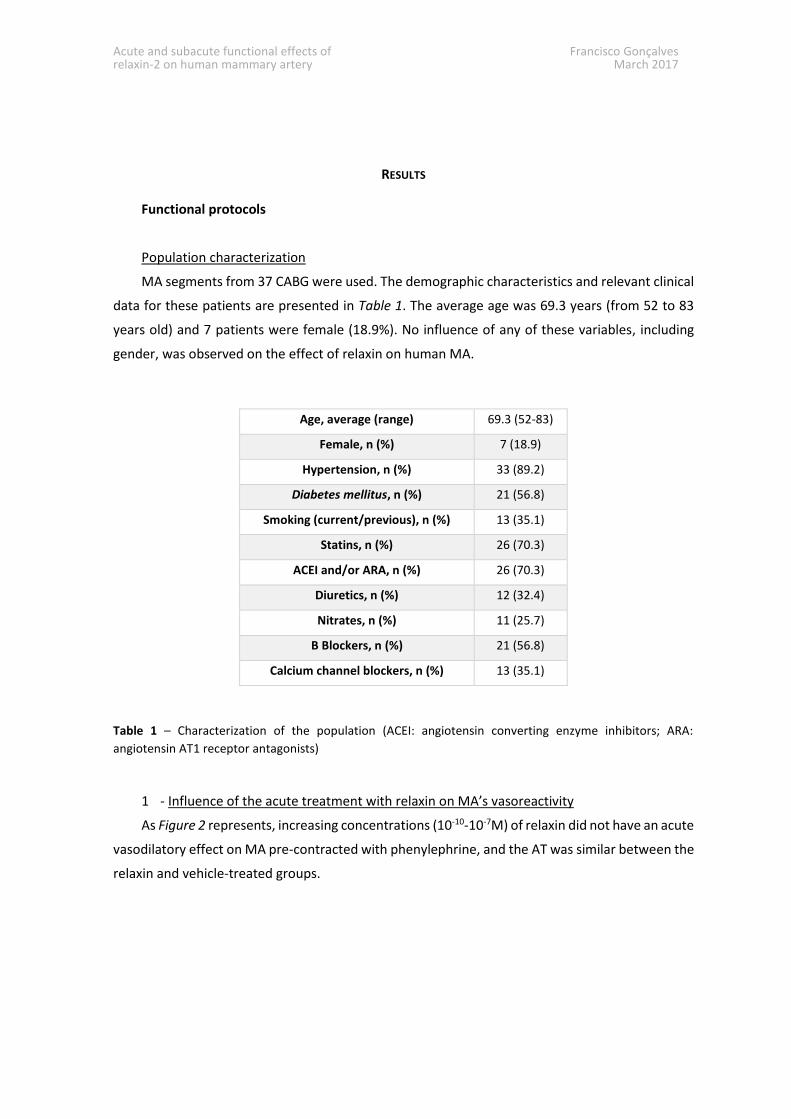
MA segments from 37 CABG were used. The demographic characteristics and relevant clinical
data for these patients are presented in Table 1. The average age was 69.3 years (from 52 to 83
years old) and 7 patients were female (18.9%). No influence of any of these variables, including
gender, was observed on the effect of relaxin on human MA.
Table 1 – Characterization of the population (ACEI: angiotensin converting enzyme inhibitors; ARA:
angiotensin AT1 receptor antagonists)
1 - Influence of the acute treatment with relaxin on MA’s vasoreactivity
As Figure 2 represents, increasing concentrations (10-10-10-7M) of relaxin did not have an acute
vasodilatory effect on MA pre-contracted with phenylephrine, and the AT was similar between the
relaxin and vehicle-treated groups.
Age, average (range) 69.3 (52-83)
Female, n (%) 7 (18.9)
Hypertension, n (%) 33 (89.2)
Diabetes mellitus, n (%) 21 (56.8)
Smoking (current/previous), n (%) 13 (35.1)
Statins, n (%) 26 (70.3)
ACEI and/or ARA, n (%) 26 (70.3)
Diuretics, n (%) 12 (32.4)
Nitrates, n (%) 11 (25.7)
Β Blockers, n (%) 21 (56.8)
Calcium channel blockers, n (%) 13 (35.1)

Acute and subacute functional effects of Francisco Gonçalves relaxin-2 on human mammary artery March 2017
Figure 2 – Evaluation of the influence of the
acute treatment with relaxin-2 (10-10 M a 10-5 M)
or vehicle in MA pre-contracted with
phenylephrine (10-9 - 10-5 M), n=5; AT – active
tension: percentage (%) of the response to 0,1 M
KCl, expressed as average ± standard error.
2 – Influence of the subacute treatment with relaxin on MA’s vasoreactivity
Globally, the AT (mN/mm) generated in response to KCl (0,1M) was similar for the vessels in
the relaxin or vehicle-treated groups (15.75±2.32 mN vs 16.85±3.33 mN, respectively; n=32),
reflecting comparable vascular viability between groups (Figure 3 – A).
2.1 vasoconstrictor response
No differences between the active tension developed by relaxin or vehicle treated
groups was found for any of the studied vasoconstrictors. The average AT generated in
response to the maximum concentration of phenylephrine (10-5 M; n=8; Figure 3- B) was 59.0±
5.4% for the relaxin treated rings and 55.6±4,9% for vehicle treated group. Only the maximum
used concentration of endothelin generated vasoconstriction (10-5 M; n=6; Figure 3 - C),
corresponding to an AT for the relaxin treated rings of 36.8±4.7% and for the vehicle treated
rings of 34.2±5.8%. The dose-response curves for the relaxin or vehicle treated groups were
similar, with no significantly different AT values for any of the used concentrations (Figure 3-
D).

Acute and subacute functional effects of Francisco Gonçalves relaxin-2 on human mammary artery March 2017
Figure 3 – Vasoconstrictor response after incubation for 24h with either relaxin-2 (10-7 M) or vehicle; AT
(active tension - average ± standard error) expressed as mN/mm (graphic A) or as percentage (%) of the
response to 0,1 M KCl (graphics B, C and D); A – Response to 0,1M KCl, n=19; B – Response to phenylephrine
(Phe - 10-9 - 10-5 M), n=8; C – Response to endothelin-1 (ET - 10-5 M), n=6; D – Response to angiotensin II
(AngII - 10-9 - 10-5 M), n=11.
2.2 – vasodilator response
The relaxin pre-treated vascular rings showed a superior vasodilatory response to
acetylcholine when compared with the vehicle treated group. Effectively, when the rings were
incubated with relaxin, the total AT decrease following exposure to the maximum dose of
acetylcholine (10-5 M) was 59.1±6.1%, compared to 46.2±6.2% for the rings incubated with
vehicle (p<0.01; Figure 4 - A). Similarly, the vasodilator response to nitroprusside was higher
in the relaxin treated rings, with a total AT decrease of 128.9±4.8% after addition of the
highest dose of nitroprusside (10-5M), compared to only 118.7±5.1% for the vehicle treated
rings (p<0.05; Figure 4 - B).

Acute and subacute functional effects of Francisco Gonçalves relaxin-2 on human mammary artery March 2017
Figure 4 – Vasodilator response after incubation for 24h with either relaxin-2 (10-7 M) or vehicle; AT (active
tension - average ± standard error) expressed as percentage (%) of the response to phenylephrine (10-5 M);
A – Response to acetylcholine (Ach - 10-10 M - 10-5 M), n=8; B – Response to nitroprusside (NO - 10-10 M - 10-
5 M), n=11; * p<0.05, vs vehicle.
2.3 – vasodilator response in the absence of endothelium
Vasodilator response to concentrations of 10-7 M, 10-6 M and 10-5 M of nitroprusside
was higher after incubation with relaxin with prior endothelium removal, when compared to
vehicle. For the maximum concentration of nitroprusside, the total decrease in AT for the
relaxin treated rings was 143.3±4.1%, with a decrease of only 132.8±3.3% by the vehicle
treated rings (p<0.01; Figure 5).
Figure 5 - Vasodilator response to nitroprusside (NO - 10-10 M - 10-5 M), n=7, after endothelium removal
followed by incubation for 24h with either relaxin-2 (10-7 M) or vehicle; AT (active tension - average ±
standard error) expressed as percentage (%) of the response to phenylephrine (10-5 M); * p<0.05, vs vehicle.
Acute and subacute functional effects of Francisco Gonçalves relaxin-2 on human mammary artery March 2017
Immunofluorescence protocols
Endothelium removal confirmation
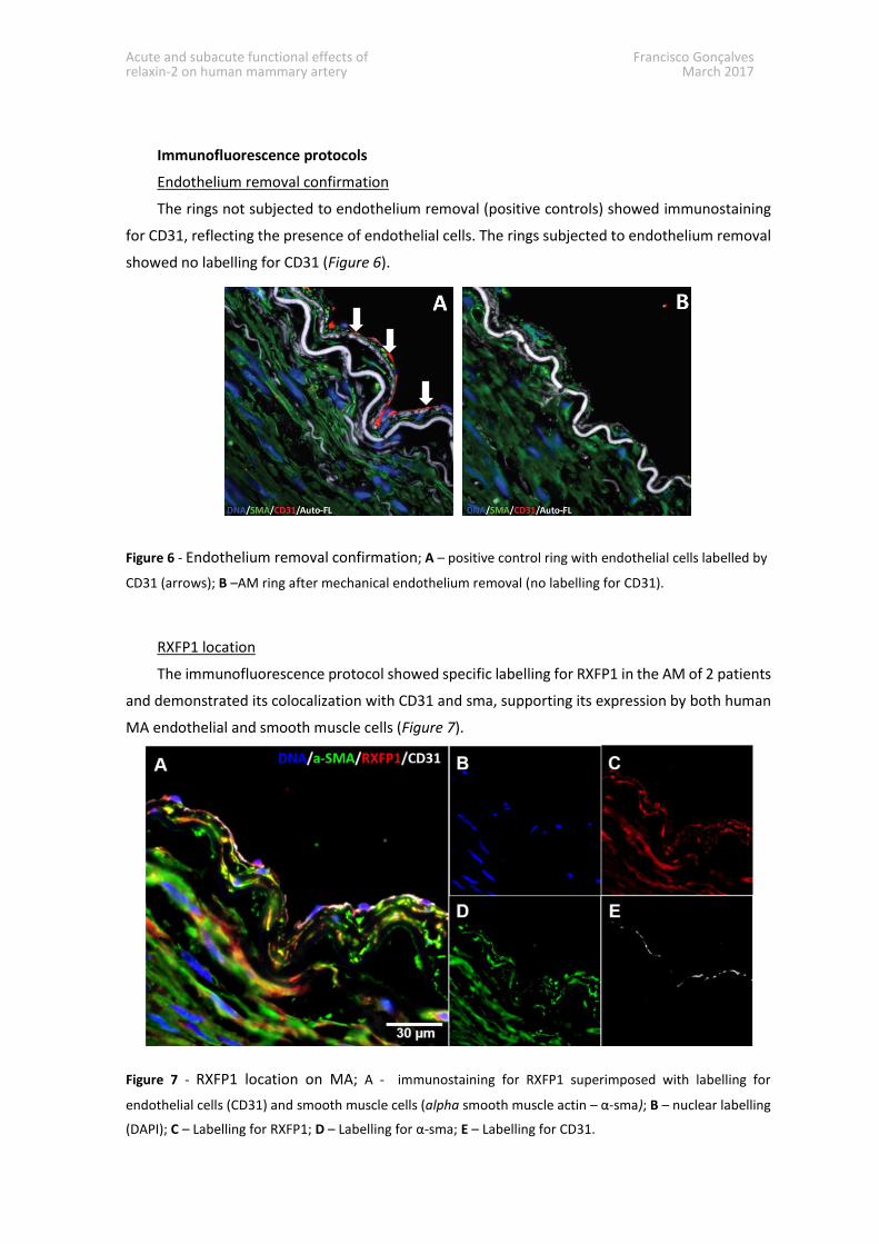
The rings not subjected to endothelium removal (positive controls) showed immunostaining
for CD31, reflecting the presence of endothelial cells. The rings subjected to endothelium removal
showed no labelling for CD31 (Figure 6).
Figure 6 - Endothelium removal confirmation; A – positive control ring with endothelial cells labelled by
CD31 (arrows); B –AM ring after mechanical endothelium removal (no labelling for CD31).
RXFP1 location
The immunofluorescence protocol showed specific labelling for RXFP1 in the AM of 2 patients
and demonstrated its colocalization with CD31 and sma, supporting its expression by both human
MA endothelial and smooth muscle cells (Figure 7).
Figure 7 - RXFP1 location on MA; A - immunostaining for RXFP1 superimposed with labelling for
endothelial cells (CD31) and smooth muscle cells (alpha smooth muscle actin – α-sma); B – nuclear labelling
(DAPI); C – Labelling for RXFP1; D – Labelling for α-sma; E – Labelling for CD31.
Acute and subacute functional effects of Francisco Gonçalves relaxin-2 on human mammary artery March 2017
DISCUSSION AND CONCLUSION
This study constitutes the first description of relaxin’s functional effects on the human MA, a
crucial vascular territory for the surgical approach to ischemic cardiomyopathy. In the present
work, MA’s response to relaxin was assessed in two different settings. In the first protocol, we
showed that relaxin was not able to induce, per se, a vasodilatory response by MA in the in vitro
conditions described. However, in the second set of protocols, we observed that previous
incubation for 24h with relaxin potentiated MA’s vasodilation in response both to acetylcholine,
which effects are mediated by the endothelium, and to nitroprusside, which has a direct effect on
the smooth muscle, without an apparent concomitant increase in the vasoconstrictor response to
either phenylephrine, endothelin-1 or angiotensin II. In this study we also describe, for the first
time, RXFP1’s expression on the endothelial and smooth muscle cells of this artery.
RXFP1, the human relaxin-2 receptor, is distributed in a similar way to its ligand, compatible
with a predominantly paracrine or autocrine actions of relaxin [11] and with the usually low or
even undetectable relaxin plasmatic levels in healthy individuals. In rat, this receptor is distributed
in a heterogenous way between the smooth muscle cells and the endothelial cells accordingly to
the vascular territory considered: it is mainly localized in the smooth muscle cells in femoral artery
and vein and pulmonary arterioles and mainly in endothelial cells in aorta, cava vein and
mesenteric artery and veins. [19] The expression of RXFP1’s mRNA was identified in subcutaneous
human arteries [12] and in several human vascular cells, namely human umbilical artery and vein
endothelial and smooth muscle cells, even though the protein was not expressed in the membrane
of umbilical artery endothelial cells. [20] RXFP1 probably mediates the clear majority of relaxin’s
vasodilatory actions [11] and this study demonstrates its unequivocal expression on the
endothelial and smooth muscle layers, both of which are thus putative targets for relaxin.
Currently, information is lacking regarding relaxin’s action and respective mechanisms of
signalling on human tissues with cardiovascular significance that endogenously express RXFP1.
Despite this, the acute modulation of the vascular function by relaxin is already described in vivo
in the human. In the clinical trial RELAX-AHF the group of patients treated with an intravenous
infusion of relaxin showed a higher decrease of the systolic arterial pressure [8] and a previous
clinical trial revealed hemodynamic changes mostly within 30 minutes of infusion of relaxin in
patients with acute heart failure, namely reduction of the pulmonary capillary wedge pressure and
pulmonary artery pressure, accompanied by a mild decrease of the systolic and diastolic arterial

Acute and subacute functional effects of Francisco Gonçalves relaxin-2 on human mammary artery March 2017
pressures. [21] Studies in animal models and isolated arteries have demonstrated the specificity of
relaxin’s acute vasodilator effect depending on the vascular territory considered. In human, the
subcutaneous [12] and gluteal arteries showed vasodilation dependent on endothelium [22] in
response to relaxin, but this effect was absent in myometrial and uteroplacentary arteries [23] and
in resistance pulmonary arteries. [22] Our results have showed that relaxin does not constitute a
potent acute vasodilator in the specific case of human MA. These findings additionally support the
existence of local systems responsible for the action of this peptide, which might explain the
variability of the short-term effects of relaxin in different settings.
Pharmacotherapy seems to be able to influence the acute response to relaxin, as evidenced
by the attenuation of the vasodilation in response to relaxin in patients medicated with ACEIs
(angiotensin-converting-enzyme inhibitors), which was further exacerbated when the patients
were also on indomethacin. [22] Our population, in accordance to the usual characteristics of
patients subjected to CABG, is prominently elderly, polymedicated and with several comorbidities.
Effectively, a large proportion of patients was on ACEIs and/or ARAs (angiotensin II receptor
antagonists) (70,3%) and the study characteristics does not allow us to exclude a possible
interference of these drugs on the acute effect of relaxin. The expansion of the in vitro studies with
human arteries and veins aiming to explore possible interactions will further clarify the relevance
of concomitant presence of pharmacologic substances on the vascular effects of relaxin.
Alongside the acute effects, relaxin seems able to induce sustained vasodilation in human [8,
21], compatible with the modulation of MA’s vascular function by a 24h incubation with relaxin
that we observed. This prolonged effect may be partially explained by the apparent absence of
classical regulation of RXFP1, which is not subjected to phosphorylation, desensitization or
internalization with exposure to high relaxin concentrations [24]. The downstream signalling also
seems to be different depending with the exposure duration. Relaxin activates metalloproteinases
(MMPs) with the ability to induce endothelin-1 functional antagonism through an increase in
endothelial ET-B signalling, with the MMP-9 being most relevant in an acute setting and MMP-2
when the exposure is prolonged several days [25, 26]. In a prolonged exposure setting the vascular
remodelling promoted by relaxin has an essential role, despite occurring only in certain vascular
territories, in rat and mice [19, 27, 28]. Relaxin’s subacute and chronic vasodilatory effects seem
to indeed rely on mechanisms distinct from those responsible for their acute effects and to vary
depending on the specific vessel considered.
Acute and subacute functional effects of Francisco Gonçalves relaxin-2 on human mammary artery March 2017
Studies in human tissues that corroborate the mechanisms that, in animal models and cell
culture, are responsible for the non-acute effects of relaxin are scarce. The study of the
mechanisms behind the chronic vascular actions of relaxin in humans bears intrinsic problems. We
focused on the subacute effects of relaxin on a human vascular territory selected for its clinical
relevance. It is important to note that endothelium removal did not abolish the influence of the
treatment with relaxin on MA, which corroborates in an original way the presence of endothelium-
independent mechanisms as part of relaxin’s action on MA. Effectively, despite most experimental
evidence both in human and in animal models pointing towards endothelium-dependent relaxin
vascular effects, most of these studies are specifically analysing relaxin’s role in an acute setting.
Furthermore, our study reveals no influence on vasoconstriction in response to
phenylephrine, endothelin-1 and angiotensin II by previous incubation with relaxin, going against
studies in rat, in which infusion of relaxin throughout 6h promoted a diminished response of
mesenteric and aorta arteries to endothelin-1 [29] and in vivo antagonism of angiotensin II effects
on peripheral vascular resistance [30], justifying the importance of further characterizating the
vascular structural and molecular changes induced by relaxin in a context of subacute exposure.
In conclusion, the present study demonstrates that previous exposure to relaxin is able to
modulate, at least partially through endothelium independent mechanisms, MA’s vasoreactivity,
potentiating its vasodilation with no concomitant effects on vasoconstriction. This allows us to
state that relaxin has an interesting profile of functional effects on human mammary artery, the
preferred conduct for myocardial revascularization.
Acute and subacute functional effects of Francisco Gonçalves relaxin-2 on human mammary artery March 2017
REFERENCES
1. Leo, C.H., et al., Vascular actions of relaxin: nitric oxide and beyond. Br J Pharmacol, 2016.
2. Raleigh, J.M., et al., Relaxin' the Heart: A Novel Therapeutic Modality. J Cardiovasc
Pharmacol Ther, 2016. 21(4): p. 353-62.
3. Cernaro, V., et al., Relaxin: new pathophysiological aspects and pharmacological
perspectives for an old protein. Med Res Rev, 2014. 34(1): p. 77-105.
4. Sarwar, M., et al., The actions of relaxin on the human cardiovascular system. Br J
Pharmacol, 2016.
5. Du, X.J., et al., Cardiovascular effects of relaxin: from basic science to clinical therapy. Nat
Rev Cardiol, 2010. 7(1): p. 48-58.
6. Conrad, K.P. and S.G. Shroff, Effects of relaxin on arterial dilation, remodeling, and
mechanical properties. Curr Hypertens Rep, 2011. 13(6): p. 409-20.
7. Pintalhao, M., et al., Relaxin serum levels in acute heart failure are associated with
pulmonary hypertension and right heart overload. Eur J Heart Fail, 2016.
8. Teerlink, J.R., et al., Serelaxin, recombinant human relaxin-2, for treatment of acute heart
failure (RELAX-AHF): a randomised, placebo-controlled trial. The Lancet, 2013. 381(9860):
p. 29-39.
9. Perna AM, M.E., et al., Novel drug development opportunity for relaxin in acute myocardial
infarction: evidences from a swine model. FASEB J, 2005. 19(11): p. 1525-7.
10. Boccalini, G., et al., Relaxin protects cardiac muscle cells from hypoxia/reoxygenation
injury: involvement of the Notch-1 pathway. FASEB J, 2015. 29(1): p. 239-49.
11. Bathgate, R.A., et al., Relaxin family peptides and their receptors. Physiol Rev, 2013. 93(1):
p. 405-80.
12. McGuane, J.T., et al., Relaxin induces rapid dilation of rodent small renal and human
subcutaneous arteries via PI3 kinase and nitric oxide. Endocrinology, 2011. 152(7): p. 2786-
96.
13. Umakanthan, J., et al., Barriers to the universal adoption of bilateral internal mammary
artery grafting. Int J Surg, 2015. 16(Pt B): p. 179-82.
14. Papakonstantinou, N.A. and N.G. Baikoussis, Total arterial revascularization: A superior
method of cardiac revascularization. Hellenic J Cardiol, 2016. 57(3): p. 152-156.
15. Grapow, M.T., et al., Human internal thoracic arteries from diabetic patients are resistant
to endothelial dysfunction. Fundam Clin Pharmacol, 2009. 23(5): p. 567-72.
16. Fonseca, D.A., P.E. Antunes, and M.D. Cotrim, Endothelium-dependent vasoactivity of the
human internal mammary artery. Coron Artery Dis, 2014. 25(3): p. 266-74.
17. Mendonca, L., et al., Angiotensin-(1-7) modulates angiotensin II-induced vasoconstriction
in human mammary artery. Cardiovasc Drugs Ther, 2014. 28(6): p. 513-22.

Acute and subacute functional effects of Francisco Gonçalves relaxin-2 on human mammary artery March 2017
18. Yu, J., et al., Dexmedetomidine-Induced Contraction in the Isolated Endothelium-Denuded
Rat Aorta Involves PKC-delta-mediated JNK Phosphorylation. Int J Med Sci, 2015. 12(9): p.
727-36.
19. Jelinic, M., et al., Localization of relaxin receptors in arteries and veins, and region-specific
increases in compliance and bradykinin-mediated relaxation after in vivo serelaxin
treatment. Faseb j, 2014. 28(1): p. 275-87.
20. Sarwar, M., et al., Serelaxin-mediated signal transduction in human vascular cells: bell-
shaped concentration-response curves reflect differential coupling to G proteins. Br J
Pharmacol, 2015. 172(4): p. 1005-19.
21. Ponikowski, P., A randomized, double-blind, placebo-controlled, multicentre study to
assess haemodynamic effects of serelaxin in patients with acute heart failure. Eur J Heart
Fail, 2014. 35.
22. Fisher, C., et al., Is the pregnancy hormone relaxin also a vasodilator peptide secreted by
the heart? Circulation, 2002. 106(3): p. 292-5.
23. Petersen, L.K., et al., Effects of human relaxin on isolated rat and human myometrium and
uteroplacental arteries. Obstet Gynecol, 1991. 78(5 Pt 1): p. 757-62.
24. Callander, G.E., W.G. Thomas, and R.A. Bathgate, Prolonged RXFP1 and RXFP2 signaling
can be explained by poor internalization and a lack of beta-arrestin recruitment. Am J
Physiol Cell Physiol, 2009. 296(5): p. C1058-66.
25. Jeyabalan, A., et al., Matrix metalloproteinase-2 activity, protein, mRNA, and tissue
inhibitors in small arteries from pregnant and relaxin-treated nonpregnant rats. J Appl
Physiol (1985), 2006. 100(6): p. 1955-63.
26. Jeyabalan, A., et al., Vascular matrix metalloproteinase-9 mediates the inhibition of
myogenic reactivity in small arteries isolated from rats after short-term administration of
relaxin. Endocrinology, 2007. 148(1): p. 189-97.
27. Debrah, D.O., et al., Relaxin regulates vascular wall remodeling and passive mechanical
properties in mice. J Appl Physiol (1985), 2011. 111(1): p. 260-71.
28. Xu, Q., et al., Relaxin therapy reverses large artery remodeling and improves arterial
compliance in senescent spontaneously hypertensive rats. Hypertension, 2010. 55(5): p.
1260-6.
29. Dschietzig, T., et al., Relaxin, a pregnancy hormone, is a functional endothelin-1 antagonist:
attenuation of endothelin-1-mediated vasoconstriction by stimulation of endothelin type-
B receptor expression via ERK-1/2 and nuclear factor-kappaB. Circ Res, 2003. 92(1): p. 32-
40.
30. Debrah, D.O., et al., Relaxin increases cardiac output and reduces systemic arterial load in
hypertensive rats. Hypertension, 2005. 46(4): p. 745-50.
Acute and subacute functional effects of Francisco Gonçalves relaxin-2 on human mammary artery March 2017
ABSTRACT
Normas da revista selecionada
PEPTIDES
An International Journal
GUIDE FOR AUTHORS
We now differentiate between the requirements for new and revised submissions.
You may choose to submit your manuscript as a single Word or PDF file to be used
in the refereeing process. Only when your paper is at the revision stage, will you be
requested to put your paper in to a 'correct format' for acceptance and provide the
items required for the publication of your article.
To find out more, please visit the Preparation section below.
PREPARATION
NEW SUBMISSIONS
Submission to this journal proceeds totally online and you will be guided stepwise
through the creation and uploading of your files. The system automatically converts
your files to a single PDF file, which is used in the peer-review process. As part of
the Your Paper Your Way service, you may choose to submit your manuscript as a
single file to be used in the refereeing process. This can be a PDF file or a Word
document, in any format or layout that can be used by referees to evaluate your
manuscript. It should contain high enough quality figures for refereeing. If you prefer
to do so, you may still provide all or some of the source files at the initial submission.
Please note that individual figure files larger than 10 MB must be uploaded
separately.
References
There are no strict requirements on reference formatting at submission. References
can be in any style or format as long as the style is consistent. Where applicable,
author(s) name(s), journal title/book title, chapter title/article title, year of publication,
volume number/book chapter and the pagination must be present. Use of DOI is
highly encouraged. The reference style used by the journal will be applied to the
Acute and subacute functional effects of Francisco Gonçalves relaxin-2 on human mammary artery March 2017
accepted article by Elsevier at the proof stage. Note that missing data will be
highlighted at proof stage for the author to correct.
Formatting requirements
There are no strict formatting requirements but all manuscripts must contain the
essential elements needed to convey your manuscript, for example Abstract,
Keywords, Introduction, Materials and Methods, Results, Conclusions, Artwork and
Tables with Captions. If your article includes any Videos and/or other
Supplementary material, this should be included in your initial submission for peer
review purposes. Divide the article into clearly defined sections.
Figures and tables embedded in text
Please ensure the figures and the tables included in the single file are placed next
to the relevant text in the manuscript, rather than at the bottom or the top of the file.
The corresponding caption should be placed directly below the figure or table.