faculdade de odontologia -...
TRANSCRIPT

FACULDADE DE ODONTOLOGIA
EXPRESSÃO IMUNOISTOQUÍMICA DOS RECEPTORES
TOLL-LIKE 5 E 9 E DO FATOR DE TRANSCRIÇÃO FOXP3
EM LESÕES ORAIS DE LÍQUEN PLANO E LÚPUS
ERITEMATOSO
VICTORIA MARTINA TRUCCI
2013
PONTIFÍCIA UNIVERSIDADE CATÓLICA DO RIO GRANDE DO SUL
FACULDADE DE ODONTOLOGIA
VICTORIA MARTINA TRUCCI
EXPRESSÃO IMUNOISTOQUÍMICA DOS RECEPTORES TOLL-LIKE 5 E 9 E
DO FATOR DE TRANSCRIÇÃO FOXP3 EM LESÕES ORAIS DE LÍQUEN
PLANO E LÚPUS ERITEMATOSO
IMMUNOHISTOCHEMICAL EXPRESSION OF TOLL-LIKE RECEPTORS 5
AND 9 AND FOXP3 TRANSCRIPTION FACTOR IN ORAL LESIONS OF
LICHEN PLANUS AND LUPUS ERYTHEMATOSUS
Porto Alegre
2013

VICTORIA MARTINA TRUCCI
EXPRESSÃO IMUNOISTOQUÍMICA DOS RECEPTORES TOLL-LIKE 5 E 9 E
DO FATOR DE TRANSCRIÇÃO FOXP3 EM LESÕES ORAIS DE LÍQUEN
PLANO E LÚPUS ERITEMATOSO
Dissertação apresentada como
parte dos requisitos necessários
para a obtenção do título de
Mestre em Odontologia, Área
de Concentração: Estomatologia
Clínica
Orientadora: Profa. Dra. Karen Cherubini
Porto Alegre
2013
DADOS INTERNACIONAIS DE CATALOGAÇÃO NA PUBLICAÇÃO (CIP)
Trucci, Victoria Martina
Expressão dos receptores toll-like 5 e 9 e do fator de
transcrição Foxp3 em lesões orais de líquen plano e
lúpus eritematoso / Victoria Martina Trucci – Porto
Alegre, 2013. 94 f. : il. Diss. (Mestrado) - Fac. de Odontologia. Programa de Pós-Graduação em
Odontologia. Área de concentração: Estomatologia Clínica, PUCRS, 2013.
Orientador: Profª. Drª. Karen Cherubini.
1. Odontologia. 2. Estomatologia Clínica. 3.Líquen plano. 4. Lupus
eritematoso. 5.Toll like receptor 5. 6.Toll like receptor 9. 7.Foxp3. 8. Lesões
orais. I. Cherubini, Karen. Título.

Epígrafe

Si se es fiel al llamado de la vocación, los
sacrificios y dificultades que uno pueda atravesar serán
irrelevantes a la hora del balance final.
César Milstein (1927 - 2002)

Dedicatória

Aos meus pais, que também são merecedores,
ainda que simbólicos, deste título.

Agradecimentos

Agradeço, primeiramente, a Deus, que me deu provas concretas de sua existência, indicando-
me sempre os melhores caminhos a serem seguidos.
Aos meus pais, Gerardo Raúl Trucci e Denise Terezinha Bruschi Trucci, a quem devo
eterna gratidão por todo o sacrifício e amor com que me criaram.
Aos meus irmãos, Christian Raúl Trucci e Gerardo Raúl Trucci Filho, pela amizade
fraternal e verdadeiramente sincera.
À minha avó paterna, “la abuela Jovita”, pelo exemplo do que é lutar na vida. E também ao
“abuelo Raúl” (in memorian), pelas belas recordações de momentos únicos que marcaram
minha infância.
À professora Dra. Karen Cherubini, pela dedicação no exercício de sua função de
professora orientadora.
À Dra. Márcia Fava, cuja participação foi fundamental para o esboço e subsequente
concretização deste estudo.
Às demais professoras do Serviço de Estomatologia e Prevenção do Câncer Bucomaxilofacial
do Hospital São Lucas da PUCRS, Dra. Maria Antonia Zancanaro de Figueiredo, Dra.
Fernanda Salum e Dra. Liliane Soares Yurgel, e à secretária Cristiane Carlotto, pelo bom
desenvolvimento dos atendimentos nos ambulatórios, fazendo dos mesmos um ambiente de
aprendizado diário.
Ao Dr. Vinicius Duval da Silva, Patologista do Hospital São Lucas e Professor da Faculdade de
Medicina da PUCRS, por permitir o desenvolvimento da pesquisa em seu Laboratório.
À professora Dra. Maria Martha Campos e às professoras da Disciplina de Radiologia
Odontológica da Faculdade de Odontologia da PUCRS, Dra. Márcia Rejane Brücker, Dra.
Maria Ivete Rockenbach, Dra. Aline Rose Cantarelli Morosolli, Dra. Helena Wilhelm de
Oliveira e Dra. Rejane Maria Holderbaum, pelo estímulo para que eu viesse a trilhar o
caminho da Estomatologia na Pós-Graduação. Aqui também devo um agradecimento especial às

professoras doutoras Nilza Pereira da Costa e Elaine Bauer Veeck, minhas constantes
incentivadoras.
Aos Professores Dr. Marcos Túlio Mazzini Carvalho e Dr. José Antonio Poli de
Figueiredo, respectivamente Diretor da Faculdade de Odontologia e Coordenador do
Programa de Pós-Graduação durante o período do Mestrado, pelo excelente trabalho na
promoção da expansão acadêmico-científica da Odontologia na Instituição e fora dela.
Ao Professor Dr. Alexandre Bahlis, atual Diretor da Faculdade de Odontologia, e à
Professora. Dra. Ana Maria Spohr, atual Coordenadora do Programa de Pós-Graduação,
pelo empenho em assumirem o desafio inerente aos cargos a que se dispuseram.
Aos professores com os quais cursei disciplinas teóricas neste Curso de Pós-Graduação
Dr. Eraldo Luiz Batista Junior, Dra. Maria Antonia Zancanaro de Figueiredo,
Dr. Rogério Miranda Pagnoncelli e Dra. Luciana Mayumi Hirakata, cujos conteúdos
transmitidos foram importantes para a construção do meu conhecimento.
A todos os funcionários da Faculdade de Odontologia da PUCRS, bem como os do Hospital
São Lucas, com os quais tive contato e que, de forma prestativa, sempre procuraram
colaborar.
À cirurgiã-dentista Marcelei Regina de Brito, minha colega de trabalho, pela oportunidade
brindada, bem como pelo espaço para o exercício também da Estomatologia em sua clínica.
Aos meus colegas da Pós-Graduação, com os quais convivi e criei laços de amizade ao longo
desses dois anos. Um agradecimento especial a Márcia Payeras, pelo coleguismo e
generosidade dispendidos durante a fase experimental de minha pesquisa.
Agradeço a meus amigos e pessoas próximas queridas por acreditarem sempre em mim e na
minha capacidade em vencer mais este desafio.
.

Resumo

RESUMO
O presente estudo teve por objetivo avaliar o padrão de expressão dos receptores toll-like
(TLR) 5 e 9 e do fator de transcrição forkhead box p3 (Foxp3) de células T regulatórias em
lesões orais de líquen plano e lúpus eritematoso. Vinte e um espécimes de biópsias de
líquen plano, 21 de lúpus eritematoso e 21 de hiperplasia fibrosa inflamatória (grupo-
controle) foram submetidos a processamento imunoistoquímico com os marcadores anti-
TLR5, anti-TLR9 e anti-Foxp3, e a quantificação da expressão desses marcadores foi
realizada por meio do software Image Pro Plus 4.5.1 (Media Cybernetics). O padrão de
expressão do Foxp3 diferiu significativamente entre os três grupos, sendo os maiores
níveis observados no grupo líquen plano, e os menores, no grupo-controle. A expressão do
TLR5 não diferiu significativamente entre os grupos lúpus eritematoso e controle, mas foi
maior nesses grupos quando comparados ao grupo líquen plano. Ao discriminarem-se os
subtipos de lúpus eritematoso sistêmico e discoide, não foi verificada diferença
significativa na expressão do TLR5 entre líquen plano e lúpus sistêmico, mas foram
verificados maiores níveis desse marcador no lúpus discoide e no grupo-controle quando
comparados ao grupo líquen plano. O padrão de expressão do TLR9 não diferiu
significativamente entre os grupos. Foxp3 e TLR5 apresentaram correlação negativa fraca
(r= -0.279). As demais variáveis não apresentaram correlação, mesmo quando analisadas
intragrupo. Os resultados do presente estudo permitem concluir que a expressão de Foxp3
está aumentada em lesões orais de líquen plano e lúpus eritematoso, enquanto TLR5 e
TLR9 não exibem maior expressão nessas lesões; Foxp3 e TLR5 estão inversamente
correlacionados. Novas investigações sobre células T regulatórias, receptores toll-like e
citocinas pró-inflamatórias fazem-se necessárias para o melhor entendimento de doenças
como líquen plano e lúpus eritematoso.
Palavras-chave: líquen plano, lúpus eritematoso, doenças do sistema imunológico,
receptores toll-like, forkhead box p3, expressão imunoistoquímica

Summary

SUMMARY
The present study aimed at analyzing the expression of toll-like receptors (TLR) 5 and 9
and forkhead box p3 regulatory T cells transcription factor (Foxp3) in oral lesions of lichen
planus and lupus erythematosus. Twenty-one biopsy specimens of lichen planus, 21 of
lupus erythematosus and 21 of inflammatory fibrous hyperplasia (control group) were
subjected to immunohistochemical staining with anti-TLR5, anti-TLR-9 and anti-Foxp3,
whose expressions were quantified using Image Pro Plus 4.5.1 software (Media
Cybernetics). Immunostaining for Foxp3 differed significantly between the three groups,
where the highest levels were shown by lichen planus and the lowest ones by controls.
TLR5 expression did not differ significantly between lupus erythematosus and control
group, but it was significantly greater in these two groups than in lichen planus. When we
considered the two types of lupus separately, there was no significant difference of TLR5
between lichen planus and systemic lupus erythematosus, but discoid lupus erythematosus
as well as the control group showed significantly greater values than lichen planus. TLR9
immunostaining did not differ significantly between the groups analyzed. A weak negative
correlation was observed between Foxp3 and TLR5 (r= -0.279), while there was no other
correlation. When this analysis was performed within each group, no correlation was
observed between the variables. In conclusion, Foxp3 expression is increased in oral
lesions of lichen planus and lupus erythematosus, whereas TLR5 and TLR9 do not show
increased expression in these lesions. Foxp3 and TLR5 are inversely correlated. Further
investigations analyzing Treg cells and proinflammatory cytokines in these diseases are
warranted.
Keywords: lichen planus, lupus erythematosus, immune system diseases, toll-like
receptors, forkhead box p3, immunohistochemical expression

Sumário

SUMÁRIO
1 INTRODUÇÃO ………………………………………………………………………. 16
2 ARTIGO 1………………………………………………………………….………….. 21
2.1 Introduction…………………………………………………………………………… 23
2.2 Dendritic cells………………………………………………………….………………. 24
2.3 Type 1 interferon system................................................................................................. 25
2.4 Regulatory T cells............................................................................................................ 26
2.5 Toll-like receptors….………………………………………………………..……......... 31
2.6 Dendritic cells, type 1 IFN, TLRs and Tregs in autoimmunity…...……...….……… 33
2.7 Final considerations......................................................................................................... 39
2.8 Acknowledgments……………………................................................................................... 40
2.9 References…………………………..…………………………………………............. 40
3 ARTIGO 2………………………………………………………….…………………… 48
3.1 Introduction…………………………………………………………………………….. 51
3.2 Materials and methods………………………………………………………………… 53
3.3 Results.............................................................................................................................. 56
3.4 Discussion......................................................................................................................... 60
3.5 Acknowledgments……………………................................................................................... 65
3.6 References....................................................................................................................... 65
4 DISCUSSÃO GERAL................................................................................................... 71
5 REFERÊNCIAS............................................................................................................ 78
6 ANEXOS ........................................................................................................................ 89

Introdução

16
1 INTRODUÇÃO
Doenças autoimunes podem desenvolver-se como resultado de falhas na
tolerância imunológica, o que determina a ativação de células T autorreativas. Padrões
moleculares exógenos microbianos ou endógenos ligam-se aos receptores toll-like
(TLR) e ativam vias de sinalização em células do sistema imunológico inato, o que
resulta na produção de citocinas pró-inflamatórias e ativação de células T que, até o
momento, são consideradas o principal fator envolvido no desenvolvimento da
autoimunidade (Mills, 2011). Os TLRs apresentam papel fundamental na resposta
imunológica inata a agentes microbianos invasores. Tais receptores são expressos em
monócitos, macrófagos, linfócitos, células dendríticas e granulócitos, bem como no
epitélio das vias aéreas e na pele, importantes sítios de interação do organismo com
patógenos (McInturff et al., 2005).
Além da ativação de células T efetoras, ligantes de TLRs podem modular direta
e indiretamente a função das células T regulatórias (Tregs). Existem evidências de que a
ativação de TLRs pode bloquear a resposta das células Treg, levando à quebra da
tolerância a antígenos próprios (Pasare; Medzhitov, 2003; Yang et al., 2004; Peng et al.,
2005; Mills, 2011). Diferentes estímulos extracelulares e diferentes moléculas
sinalizadoras intracelulares controlam o desenvolvimento e a função das Tregs por meio
da regulação da transcrição gênica do fator de transcrição forkhead box p3 (Foxp3)
(Lal; Bromberg, 2009). Sob a influência de determinadas condições inflamatórias ou
ligantes específicos, a ativação de certos TLRs resulta em reduzida expressão de Foxp3
com menor geração de Tregs capazes de suprimir a proliferação de células T
autorreativas (Pasare; Medzhitov, 2003; Yang et al., 2004; Lal; Bromberg, 2009; Hackl
et al., 2011).

17
Estudos recentes têm destacado que os TLRs estariam envolvidos na patogênese
do lúpus eritematoso sistêmico (LES) (Nakano et al., 2008; Wong et al., 2010; Midgley
et al., 2012). O LES representa o protótipo da doença autoimune, caracterizado por um
amplo espectro de manifestações clínicas e produção de autoanticorpos (Midgley et al.,
2012). As primeiras manifestações clínico-patológicas são consequência de uma
inflamação tecidual local que tem início com a deposição de imunocomplexos nos
diversos tecidos do corpo, uma vez que ocorre a produção de autoanticorpos contra
componentes nucleares das células (Munoz et al., 2008). Uma falha na eliminação de
células apoptóticas, principalmente neutrófilos, levaria ao acúmulo de autoantígenos
nucleares (proteínas nucleares, DNA, RNA) que são modificados por clivagem
enzimática durante o próprio processo de apoptose celular. Esse atraso na eliminação do
material nuclear modificado permite que o mesmo seja reconhecido por TLRs em
células dendríticas com consequente aumento da produção de interferon alfa (IFN-alfa),
um potente mediador na patogênese do lúpus eritematoso sistêmico. Tal processo
resulta na formação de autoanticorpos contra os autoantígenos nucleares (Munoz et al.,
2008; Midgley et al., 2012). O IFN produzido medeia a maturação e a apresentação
antigênica por células dendríticas convencionais e macrófagos. Além disso, ativa a
geração de células B e T efetoras e de memória, enquanto Tregs são suprimidas, o que
colabora para o estabelecimento do quadro de autoimunidade observado no lúpus
(Rönnblom et al., 2011).
A etiologia do líquen plano ainda não foi totalmente esclarecida. No entanto,
alguns fatores têm sido associados ao seu surgimento, entre os quais estão ansiedade,
diabetes, doenças autoimunes, doenças intestinais, drogas, estresse, hipertensão,
infecções bacterianas e virais, materiais dentários restauradores, neoplasias e
predisposição genética (Canto et al., 2010; Roopashree et al., 2010). Ainda, acredita-se

18
na existência de uma possível susceptibilidade individual para o desenvolvimento da
doença, que estaria na dependência da natureza do antígeno, da habilidade do indivíduo
para apresentação de antígenos, da presença de células T capazes de reconhecer esse
antígeno (receptores) e da possibilidade de herdar um perfil de citocinas e
polimorfismos genéticos que estimulem resposta mediada por células ao antígeno
(Thornhill, 2001). Há evidências de que uma desregulação imunológica mediada pelo
componente celular do sistema imunológico estaria envolvida na patogênese do líquen
plano oral, já que o infiltrado inflamatório nessa doença é composto predominantemente
por células T e macrófagos, enquanto plasmócitos são observados raramente, e
depósitos de imunocomplexos não são característicos (Lodi et al., 2005).
As lesões de líquen plano e lúpus eritematoso que se desenvolvem na mucosa
oral podem apresentar aspecto clínico variado e semelhante, sendo necessária a
realização de biópsia para o estabelecimento do diagnóstico diferencial (Neville et al.,
2009). As lesões orais de líquen plano geralmente são bilaterais e simétricas, afetando a
mucosa jugal, a gengiva e as porções lateral e dorsal da língua. São representadas
tipicamente por estrias reticulares esbranquiçadas denominadas estrias de Wickham, que
podem coalescer e formar pápulas e placas. As formas atrófica e erosiva são observadas
frequentemente e, nos casos mais graves, a mucosa pode estar ulcerada. As lesões orais
típicas do lúpus eritematoso ocorrem primeiramente nas mucosas jugal e labial, rebordo
alveolar e vermelhão de lábio e são caracterizadas por uma área atrófico-ulcerada
central, pequenas placas brancas ceratóticas com bordas elevadas e estrias brancas
irradiadas. No entanto, o aspecto clínico é variado, o que pode dificultar o diagnóstico
diferencial com o líquen plano (Farthing; Speight, 2006). Considerando-se que
alterações na expressão da Foxp3, por meio da ativação de TLRs específicos, podem ter
papel preponderante no desenvolvimento de doenças autoimunes, bem como a

19
semelhança clínica entre lesões orais de líquen plano e lúpus eritematoso, o presente
estudo teve por objetivo avaliar, por meio de imunoistoquímica, o padrão de expressão
dos toll-like receptors 5 e 9 e do fator de transcrição Foxp3 em lesões orais dessas duas
doenças. O trabalho apresenta-se sob a forma de dois artigos científicos: o primeiro
consiste em um artigo de revisão da literatura sobre o tema em questão, e o segundo
compreende o experimento realizado.

Artigo 1

21
2 ARTIGO 1
O artigo a seguir intitula-se Interrelationship of dendritic cells, system type 1
interferon, regulatory T cells and toll-like receptors in immune response and
autoimmune diseases – a literature review e foi formatado de acordo com as normas do
periódico Archives of Oral Biology (Anexos A e B).

22
Interrelationship of dendritic cells, system type 1 interferon, regulatory T cells and
toll-like receptors in immune response and autoimmune diseases – a literature
review
Victoria Martina Trucci
Fernanda Gonçalves Salum
Maria Antonia Figueiredo
Karen Cherubini *
Postgraduate Program, Dental College, Pontifical Catholic University of Rio Grande do
Sul - PUCRS
Running title: Important factors in autoimmune diseases
* Corresponding author
Karen Cherubini
Serviço de Estomatologia, Hospital São Lucas PUCRS
Av Ipiranga, 6690, sala 231
Porto Alegre RS Brazil
CEP 90610-000
Telephone/fax: 55(51)33203254
E-mail: [email protected]

23
Abstract
The mechanisms involved in immune disturbances that lead to autoimmune diseases are
not completely understood. Many studies focusing on this issue have been published,
clearly indicating the great interest of the scientific community in improving the
management and treatment of these diseases. There is evidence that the activation of
some receptors of the toll-like family (TLRs) of the innate immune system, and also
changes in expression levels of forkhead box p3 (Foxp3) protein, which is found in
regulatory T cells (Tregs), could be involved in the development of autoimmunity. We
present here a literature review focusing on the interrelationship of dendritic cells,
TLRs, Tregs and type 1 interferon in the immune response and autoimmune diseases,
with special interest in lupus erythematosus and lichen planus. Understanding the
specific role of each of these factors would help elucidate the obscure etiology of such
diseases and open new perspectives for their management and treatment.
Keywords: autoimmune diseases; TLRs; Foxp3; lichen planus; lupus erythematosus
Introduction
Autoimmune diseases are chronic conditions in which the immune system develops an
inflammatory response to self cells and tissue components in the human body. This
leads to inflammation and abnormal infiltration of lymphocytes and other leukocytes in
the tissues, although they are free of infection.1 Included among these conditions are
some diseases such as lupus erythematosus,2 which is a classic autoimmune disease
where multiple autoantibodies are produced.3 Nevertheless, there are some other
immune disorders such as lichen planus, whose autoimmune nature has not yet been
definitely established, as their initial triggers and pathogenesis are unknown.4

24
Therefore, lichen planus has been classified as either an autoimmune5-9
or immune-
mediated disease.4,7,10-14
Regardless of being a classic or a possible autoimmune disorder, the
etiopathogenesis of these diseases needs to be elucidated, which has generated growing
interest in investigating their relationship with some specific immune mechanisms.15
Plasmacytoid dendritic cells, forkhead box p3+
regulatory T cells (Foxp3+
Tregs), toll-
like receptors (TLRs) and type 1 interferon system (type 1 IFN), especially interferon-
alpha (IFN-alpha), have been recognized as key elements involved in the process. An
imbalance in the interrelationship between them could be the cause of the development
of autoimmunity.16-23
Also, the interrelationship between environmental stimuli and the exacerbation
of a self-reactive lymphocytic response, which culminates in lichen lesions, has been
investigated. Studies have evidenced that plasmacytoid dendritic cells are present in
skin lesions of patients with autoimmune diseases but absent in controls.15
The constant
presence of an endogenous or exogenous component recognized as an antigen in the
body could persistently stimulate TLRs of plasmacytoid dendritic cells with consequent
production of IFN and inhibition of Foxp3+ Treg cells, which would establish a vicious
circle and a consequent autoimmune state. We present here a literature review focusing
on these important aspects of the immune system and their role in the immune response
and autoimmune diseases, with special interest in lupus erythematosus and lichen
planus.
Dendritic cells
Dendritic cells are immune cells originating from hematopoietic tissue, which, besides
having the function of antigen-presentation, are also involved in the immune tolerance
process.24,25
These cells circulate in peripheral tissues capturing pathogens and/or

25
apoptotic cells and presenting them as antigens to T cells. T cells then proliferate and
differentiate into Th1 (cellular immune response), Th2 (humoral immune response) and
regulatory T cells, where the last are involved in the suppression mechanisms of
immune response. Cytokines produced by dendritic cells contribute to the induction of
T cell differentiation. Interleukin-6 (IL-6) induces TCD4+ cells to differentiate into Th2
cells and suppresses Treg activity; interleukin-10 (IL-10) inhibits Th1 but induces Th2
responses; interleukin-18 (IL-18, IFN-gamma-inducing factor) interacts with IL-12 and
induces naïve T cell proliferation and differentiation into IFN-gamma-producing Th1
cells.19
Moreover, some subtypes of dendritic cells are highly specialized in the
induction and differentiation of Treg cells, where a failure in this function can result in
autoimmunity.22
Basically, dendritic cells are divided into myeloid and plasmacytoid.
Plasmacytoid dendritic cells are found in blood and secondary lymphoid organs
expressing blood dendritic cell antigen 2 (BDCA2), immunoglobulin-like transcript 7
(ILT-7) and interleukin-3 receptor (also known as CD123 antigen) on the cell
membrane and are great producers of type 1 IFN.21,24,26
Once activated by either
endogenous or exogenous ligands, they secret considerable amounts of IFN-alpha21,24
and IL-6, which stimulates plasma cells to produce antibodies.24
Plasmacytoid dendritic
cells express TLR7 and TLR9 in the endosomal membranes and can be activated by
pathogens that invade them through endocytosis. In addition, these cells express
interferon regulatory factors 5 (IRF 5) and 7 (IRF 7), which increase their capacity of
producing IFN and other inflammatory cytokines.21
Type 1 interferon system
Type 1 interferon system (type 1 IFN), which includes interferon alpha (IFN-alpha) and
interferon beta (IFN-beta), is produced against viral infections and can activate innate

26
and adaptive immune cells, thereby playing a role in cellular and humoral responses.21
It has important immunomodulatory effects: maturation and migration of dendritic cells
to secondary lymphoid organs; increase in antigen presentation and increase in its own
production, especially in plasmacytoid dendritic cells. In natural killer (NK) cells, it
increases cytotoxicity and cytokine production; in macrophages, it stimulates the
intracellular destruction of pathogens; in T-lymphocytes, it increases IL-12 expression
and generation of memory cells, as well as promoting suppression of Tregs.21
Regulatory T cells
Potentially pathogenic CD4+ self-reactive T cells are found in peripheral tissues of
normal individuals, and their activation/expansion is controlled by other CD4+ cells
called regulatory T cells (Tregs). These two populations of CD4+ cells can be
discriminated by the expression of some specific surface molecules. Treg cells, also
called suppressor T cells, are a subtype of lymphocytes involved in the induction and
maintenance of immune tolerance and in the control of the immune response as well. 27
Tregs are induced by dendritic cells through several mechanisms that are
dependent on indoleamine 2,3-dioxygenase, IL-10, IL-27, retinoic acid, vitamin D and
transforming growth factor beta (TGF-beta). They comprise IL-10 secreting type 1
Tregs (Tr1), natural and adaptive Foxp3+ Tregs, Th3 cells and double-negative Tregs.
22
Natural Tregs (nTregs) are induced in the thymus, show high CD25 (IL-2 alpha chain
receptor) expression on the cell membrane, and are generally called CD4+CD25
+
Tregs.22,28,29
They suppress other T cells through cell-cell contact in a cytokine-
independent mode, and it is believed that they play an important role in central tolerance
to self-antigens.28,30
Adaptive Tregs (aTregs), in turn, develop from mature CD4+CD25
-
T cells in peripheral tissues under particular conditions or antigen-specific stimulation.
nTregs and aTregs were recently defined also by the intracellular expression of Foxp3

27
(CD4⁺CD25⁺Foxp3+), which is a transcription factor needed for the differentiation and
normal function of Tregs.22,28,29
Upon activation, nTregs predominantly express granzyme A, while aTregs
express granzyme B. Both subtypes of Tregs exhibit perforin-dependent cytotoxicity
against a variety of autologous target cells, including CD4+ and CD8
+ T cells, CD14
+
monocytes, and dendritic cells.31
Classic human Tregs (CD4+CD25
+Foxp3
+) inhibit Th
CD4+CD25
- cell proliferation through local action via cell-cell contact and induce them
to assume an aTreg phenotype, which exerts its regulatory function by systemic action
via TGF-beta. The secretion of this soluble suppressor mediator by aTregs is important
for the systemic control of self-reactive effector T cells (Fig.1).32
To become suppressor cells, Tregs need to have their T-cell receptors (TCR)
activated in the presence of IL-2, whose membrane receptor component is CD25 (de la
Rosa et al., 2004). In naïve mice, CD25 protein is restricted to Tregs, whereas in
humans just cells expressing large amounts of CD25 (CD4+CD25
high T cells) can be
designated as Tregs. This is because activated T cells express intermediate levels of
CD25, even though they do not show any suppressor function.27,33,34
Foxp3+Treg cells play an important role in the maintenance of peripheral
immune tolerance17,22
and control of intense chronic inflammatory and allergic
responses.22
They are capable of blocking immune responses as well as inflammation
and tissue destruction through functional suppression of many cells, including TCD4+
and TCD8+ lymphocytes, monocytes, NK cells,
34 and antigen-presenting cells, as well
as antibody production by B lymphocytes.17,34
Although it has not been confirmed, the
site of Tregs action may be represented by draining lymph nodes or by the respective
target organ or both.35
Despite their importance, suppressor molecular mechanisms of

28
Tregs are still not completely known.34 However, it is believed that their major function
is to suppress the activation and expansion of naïve conventional T cells.33
It has been observed that proliferation and cytokine production of conventional
CD4+CD25
- T cells, either from rats
36 or humans,
37 can be directly inhibited by Tregs,
even in the absence of antigen-presenting cells. Different suppressor mechanisms may
be used by Tregs depending on their state of activation, inflammatory site, and type and
activation state of the suppressed target cell. Basically these mechanisms can be direct
or indirect. Nevertheless, how the type of suppressor mechanism to be used is
determined has not been identified.34
Direct suppression of effector T cells by Tregs can involve soluble immune
suppressor factors or cell contact, whereas indirect suppression involves antigen-
presenting cells.34
The stimulation of effector T cells leads to the generation of a Ca2+
signal and a cascade of protein phosphorylation and dephosphorylation events, which
culminates in the activation of transcription factors that stimulate T cell proliferation
and cytokine expression. The most important transcription factors for induction of
cytokine expression are nuclear factor kappa B (NF-κB), nuclear factor of activated T-
cells (NFAT) and activator protein 1 transcription factor (AP-1), which mediate gene
activation of some cytokines such as IL-2 and IFN-gamma.34,38,39
Tregs can rapidly
suppress this cytokine expression in effector T cells through inhibition of Ca2+ signals
and consequently reduced NFAT and NF-κB activation.34
IL-10 and TGF-beta are inhibitory cytokines secreted by TCR-activated Tregs
and are involved in in vivo and in vitro suppression of both proliferation and cytokine
production of many cells such as T CD8+, B, NK, and other innate immune cells.
35,37,40
Tregs can secret and express large amounts of soluble and membrane-bound TGF-beta,
and the blockade of this protein production can partly nullify the suppression of T cell

29
proliferation, which suggests that TGF-beta produced by Tregs play an important role in
autoimmunity control.41,42
The indirect pathway through which Tregs suppress the activation of
conventional T cells occurs by means of impairing the stimulatory function of antigen-
presenting cells.43
Tregs induce an immunosuppressive pattern of cytokine production
through reduced synthesis of IL-6 and increased levels of IL-10 by dendritic cells. In
contact to CD4+ T cells, dendritic cells would increase IL-6 and decrease IL-10
production; however, if in contact with CD4+CD25
+ Tregs, the opposite would occur,
with increase in IL-10 levels in detriment of IL-6 (Fig.1).44
Also, Tregs can inhibit
conventional T cell activation by reducing stable cell contacts between them and
dendritic cells. Less time of contact between these cells has been observed in the
presence of Tregs.43
Associated with the complex suppressor mechanism mediated by Tregs through
IL-10, it was recently demonstrated that Tregs need IL-10 and not IL-35 and TGF-beta
to control IFN-gamma production by T cells in inflamed skin, whereas in lymph nodes
IL-10 is dispensable for control of IFN-gamma and T cell expansion. T cell cultures are
capable of secreting IFN-gamma in the absence of Tregs, but incapable in their
presence.34,45

30
Figure 1 - Mechanisms of Foxp3+ Treg-mediated T cell supression: Regulatory T cell
(Treg) express granzymes (nTreg = granzyme A; aTreg = granzyme B) which induce
citotoxicity against autologous target cells in a perphorin-dependent manner. Through
production of antiinflamatory cytokines [transforming growth factor beta (TGF-beta)
and interleukin 10 (IL-10)], Tregs induce increased production of IL-10 and decresead
production of interleukin 6 (IL-6) by dendritic cells. This would inhibit antigen
presenting dendritic cell function.
nTreg= natural Treg; aTreg= adaptive Treg; DC= dendritic cell
Factors interfering with Foxp3 expression and Tregs stability
Foxp3 has unstable expression, and there are environmental or extrinsic factors that
probably affect its stability. Therefore, depending on the type of stimulus and ligands,
the natural history of inflammatory events can be marked by more or less Treg
suppressor activity.20
Different regulatory elements of the Foxp3 locus respond to

31
various signals. However, it is unknown how different extracellular signals affect the
chromatin structure of these regulatory elements.20,46
IL-2 deficiency and the presence of IL-1, IL-4, IL-6, IL-23 and IFN-gamma can
induce alterations in the chromatin of the gene locus of Foxp3, which impairs its
transcription and thus promotes a decrease in its expression and suppressor effect.17,20
Lal et al.20
demonstrated that the two independent pathways of IL-6/IL-6
receptor/activator of transcription-3 (IL-6/IL-6R/STAT3) and
peptidoglycan/TLR2/Myd88/IRF-1 resulted in inhibition of Foxp3 expression in nTregs
and aTregs. These pathways lead to profoundly different changes in the structure of
Foxp3 chromatin, with subsequent effects on T cell function. In the presence of TLR 7
and TLR9 ligands, cocultures of dendritic and T cells have shown greater production of
IL-6, IL-12 and IL-17, with consequent reduced percentage of Foxp3+ cells. That is, IL-
6 as well as IFN-gamma and IL-4 produced in cocultures of dendritic and T cells in the
presence of TLR7 ligands are critical factors for the decrease in Foxp3 expression.47
Toll-like receptors
Toll-like receptors (TLRs) are a group of glycoproteins that work as transmembrane
receptors of the innate immune system, allowing antigen recognition and subsequent
immune activation. They are located on the cell membrane surface (TLRs 1, 2, 4, 5, 6,
10) and in intracellular compartments such as the endoplasmic reticulum and
endosomes (TLRs 3, 7, 8, 9), where they are more common in cells that initiate the
primary immune response. Either immune cells such as monocytes, macrophages,
dendritic cells, granulocytes, NK cells, and B and T lymphocytes, or non-immune cells
such as keratinocytes and fibroblasts express TLRs,23,48,49
where these receptors play
different functions depending on the type of cell and tissue where they are expressed.18
They stimulate an adaptive immune response through the activation of dendritic cells

32
and also increase the production of costimulating molecules and inflammatory
cytokines such as TNF-alpha, IL-6 and IL-12.50
The ten human TLRs so far described are capable of recognizing a wide variety
of molecules, ranging from those of bacterial origin such as lipoproteins (TLRs 1, 2 and
6), lipopolisaccharides (TLR4) and flagellin (TLR5) to viral RNA (TLRs 3, 7, and 8)
and DNA (TLR9).18,48
TLR 7 plays an important role in autoimmunity and antiviral
defense,47
and TLR9 contributes to the production of anti-DNA antibodies,24
whereas
TLR10 function is not completely known. TLR11 has been identified in mice and
reported as capable of recognizing molecules of Toxoplasma gondii. 23
The function of
recognizing infections and inducing signaling pathways that result in the expression of
inflammatory mediators and immune response is common to different TLRs.18
The
presence of TLR ligands results in cytokine and chemokine secretion, which leads to
immune activation against pathogens. TLRs 3, 7 and 9, for instance, respond to viral
RNA and DNA with IFN-alpha production,51
which exerts many immunomodulatory
effects.21
The potent inflammatory response induced by TLRs is protective in most cases,
since pathogens are destroyed before they can cause injury to the host. Exogenous
ligands derived from viral or bacterial infections activate TLRs, thereby cause
inflammation. This inflammation, in turn, induced by either endogenous or exogenous
ligands, can cause cell necrosis and extracellular matrix damage, as well as
proinflammatory cytokine production, which enables the release of new endogenous
TLR ligands, making the process chronic.18
All TLRs share a conserved intracellular domain, the toll-interleukin-1 receptor
(TIR) domain, which is responsible for signal transduction and adaptor recruitment. The
extracellular domain, in turn, is called a leucine-rich repeat (LRR), which is responsible
33
for recognizing TLR ligands.18
Until now, five TLR adaptor molecules have been
described: myeloid differentiation factor-88 (MyD88),52
TIR domain-containing
adaptor protein (TIRAP),53
TIR domain-containing adaptor inducing IFN-beta (TRIF or
TICAM-1),54
TRIF related adaptor molecule (TRAM or TICAM-2),55
and sterile alpha
and HEAT/Armadillo motif (SARM).56
Once TLR is activated by an exogenous or
endogenous ligand, its TIR domain interacts with the adaptor molecule, initiating a
cascade of intracellular signaling pathways that lead to the activation of transcription
factors.23,57,58
The activation of these transcription factors results in the induction of
gene expression of inflammatory cytokines such as TNF-alpha and type 1 IFN.48,59,60
While most TLRs depend on the intracellular MyD88 signaling pathway, TLRs 3 and 4
use the TRIF pathway, which induces IFN-beta. The TNF receptor associated factor 6
(TRAF 6) pathway is considered an additional transducer for TLRs 7 and 9.48,59,60
The expression and function of TLR5 and TLR9 have been investigated in
human keratinocytes. In normal epithelium, TLR5 is expressed in basal keratinocytes in
the first and second layers. TLR9 is expressed in more external layers of the epidermis
comprising the top two to three layers of cells. It is observed that transforming growth
factor alpha (TGF-alpha), which is involved in epithelial healing, increases the
expression of both these TLRs. Besides increasing the expression of TLRs, TGF-alpha
stimulates their function by means of increasing IL-8 proinflammatory cytokine and
human beta-defensin 2 (hBD-2) antimicrobial peptide production.61
Dendritic cells, type 1 IFN, TLRs and Tregs in autoimmunity
Jin et al.19
observed that, in systemic lupus erythematosus (SLE) patients, plasmacytoid
dendritic cells have a greater capacity of stimulating inflammatory T cells than in
controls. These authors isolated dendritic cells from peripheral blood of 58 lupus
patients and 62 controls and kept them in culture with or without apoptotic
34
polymorphonuclear cells and, afterwards, with T cells. In the presence of apoptotic
polymorphonuclear cells, the plasmacytoid dendritic cells from controls induced the
differentiation of T cells into CD4+CD25
+ Tregs with high expression of Foxp3,
reduced IL-6 production, and increased TLR9 mRNA transcription. On the contrary,
plasmacytoid dendritic cells from lupus patients were functionally abnormal, since they
induced inflammatory T cell proliferation even in the absence of apoptotic
polymorphonuclear cells. They also lost TLR9 expression and were less efficacious in
inducing Tregs differentiation after interaction with apoptotic polymorphonuclear cells.
Therefore, in SLE patients, plasmacytoid dendritic cells are more effective in
stimulating inflammatory T cells than in inducing Treg development. Such finding
suggests that only healthy plasmacytoid dendritic cells can induce the development of
suppressive Tregs.19
In this way, these cells seem to be abnormal in lupus showing a
lower capacity to induce Treg cell differentiation.19,21
Deficiency or dysfunction of Tregs may result in autoimmunity. Elimination of
the peripheral population of CD4+CD25
+ T cells in rats promotes a wide spectrum of
organ-specific and systemic autoimmune diseases, as well as graft-versus-host
disease,27,33
whereas reconstitution of this cell population prevents the development of
autoimmunity.27
The alterations in number and function of CD4⁺CD25⁺Foxp3+
Tregs
were recently determined in autoimmune and inflammatory diseases such as psoriasis,
multiple sclerosis, autoimmune polyglandular syndrome type II, rheumatoid arthritis,
myasthenia gravis and type I diabetes.62
High and stable expression of Foxp3 is needed
for Treg suppressor activity, whereas its decrease favors loss of suppressor ability.63
Also, similar alterations were observed in lichen planus. Using immunohistochemistry
and real time RT-PCR, Tao et al.62
found a greater number of Foxp3+ cells in oral
lesions of lichen planus patients than in controls. Reticular lesions of lichen in turn

35
showed a higher frequency of Foxp3+ cells compared to erosive ones. In flow
cytometry, they showed a significant increase in these cells in the peripheral blood of
patients after treatment. Such findings indicate that Foxp3+ Treg cells are involved in
the etiopathogenesis of oral lichen planus, and they show an inverse correlation with
disease activity.62
Disclosing global defects in Treg cells of autoimmune disease patients has been
a challenging task because of the complexity of this kind of disturbance. There are
many precipitating factors, genetic and environmental, contributing to the susceptibility
to autoimmune diseases. Therefore, the failure of Tregs to function could be a
consequence of a lack of Tregs with the specificity needed to suppress inflammation in
a particular organ, cell-intrinsic defects, or extrinsic factors that inhibit the function of
these cells.29
Excessive levels of tumor necrosis factor (TNF) and reduced IL-2 production
commonly observed in SLE can contribute to a functional defect in CD4+CD25
+ Treg
cells, which start expressing lower levels of Foxp3 mRNA and lose their suppressive
function.64
Moreover, in SLE patients, the inability of plasmacytoid dendritic cells to
induce Treg development contributes to immune tolerance breakdown and
autoimmunity onset, with reduction of suppressor function of CD4+CD25
+ Tregs and
low expression of Foxp3 mRNA and protein.19
Valencia et al.64
observed a significant reduction in suppressor function of
CD4+CD25
high Tregs isolated from the peripheral blood of active SLE patients, which
was confirmed by the expression of low levels of Foxp3 (mRNA and protein) and
reduced inhibition of in vitro cytokine production and proliferation of TCD4+. The same
was not observed in Tregs isolated from patients with inactive disease, where Foxp3
expression and suppression of TCD4+ proliferation were similar to that in controls. On
36
the other hand, in vitro activation of CD4+CD25
high Treg cells from active SLE patients,
in culture with plate-bound anti-CD3 and high doses of IL-2, increased Foxp3
expression (mRNA and protein) and restored their suppressor function. To determine
whether loss of suppressor function in active SLE would result from an intrinsic defect
of CD4+CD25
high Tregs or from an increase in effector CD4
+CD25
- T cell resistance,
Valencia et al.64
mixed cells from active SLE patients with cells of normal controls.
They found that Tregs from active SLE patients failed to suppress the proliferation of
both autologous and control effector T cells, whereas Tregs from controls were capable
of suppressing the proliferation of effector T cells of active SLE patients.
TGF-beta is important to Treg development and function because it increases
Foxp3 levels.65
Nevertheless, if in the presence of IL-21 and especially IL-6, it can also
evoke a Th17-type inflammatory response, where Foxp3 levels are reduced allowing
Tregs to assume a Th17 phenotype,65
which is associated with many autoimmune and
inflammatory pathologies.17,66
Noteworthy is the higher frequency of IL-17 producing
Th cells (Th17 cells) in these pathologies.19,21,66
TLRs are involved in the pathogenesis of autoimmune diseases, where they can
be activated by endogenous ligands.18,23,24
TLR ligands can induce the secretion of
cytokines capable of interfering with Treg plasticity and inhibiting their suppressor
function.20
Exogenous and endogenous TLR7 ligands, respectively found in viral
infections and autoimmune diseases, reduce Foxp3 expression leading to lower numbers
of Tregs and, therefore, lower capacity to suppress self-reactive T cell proliferation. The
activation of dendritic cells through TLR7, for example, leads to the production of
soluble factors that decrease the generation of Tregs, which probably favors their
differentiation into proinflammatory effector Th cells and consequently promotes
autoimmunity.47

37
Patients with rheumatologic autoimmune diseases, especially lupus
erythematosus, have a higher production of type 1 IFN.21
High serum levels of IFN-
alpha are considered a heritable risk factor for SLE,51
and are associated with higher
disease activity, more severe clinical manifestations and immune activation signals.21,67
IRFs can be activated by circulating immune complexes containing nucleic acids, which
are recognized by endosomal TLRs. In addition, genetic variants of IRF-5 and IRF-7
have been associated with susceptibility to the disease, because they induce IFN-alpha
production.51
There is some evidence that activated plasmacytoid dendritic cells are
responsible for the continuous production of IFN-alpha in lupus,21,67
and the plasma of
lupus patients has interferogenic immune complexes which are able to specifically
activate these cells.21,68
In lupus, self-antigens derived from nucleic acids can be generated from
apoptotic or necrotic cells, because of higher frequency of apoptosis or lack of clearance
of apoptotic cells, and the immune complexes that activate TLR7 are potent inducers of
IFN-alpha. Still, environmental factors such as ultraviolet light and demethylating
agents increase the amount of autoantigens. This promotes greater production of
interferogenic immune complexes and type 1 IFN, which promotes autoimmunity in a
vicious circle.21
The effect of TLR2 stimulation on human Tregs has also been studied. It was
observed that Treg suppressor activity is inhibited by TLR2 Pam3Cys ligand (synthetic
bacterial lipoprotein), leading to a greater proliferation of effector T cells. TLR2
activation induces Tregs to secret IL-6, which in turn reduces Treg suppressor function
and induces their differentiation into effector Th17 cells.40
On the other hand, the
activation of other TLRs such as TLR4 and TLR5 seems to have no effect on the
generation of Tregs.47
Nevertheless, in psoriasis, TLR5 and TLR9 are expressed in

38
larger amounts and show diffuse distribution in epithelium, with TLR5 expressed in
three to eight of the deepest layers, and TLR9 occurring throughout all the epithelium.61
Also, although evidence points to an important role of TLRs in lichen planus
development, published studies are a little conflicting in this respect. Some have
reported upregulation of TLR915,23,48
and TLR269
in lichen lesions, whereas another
study has reported a significantly lower rate of TLR 1 and TLR2 expressions in lichen
planus than in controls.70
The result of autoimmunity versus the immune tolerance process depends on the
balance between stimulatory signals such as binding of ligands to TLRs, and antigen
concentration, as well as on inhibitory signals mediated by Tregs.35
This balance breaks
down in SLE, where autoantibodies can bind to either native DNA or nuclear RNA-
binding proteins such as Ro, La, Sm and RNP, and the immune complexes formed
stimulate cells through endosomal TLRs, activating antiviral immunity and amplifying
an autoimmune response. This abnormal process results in IFN-alpha production.51
Moreover, persistent activation of TLRs involved in the induction of Th1/Th2
inflammatory cytokines such as IFN-gamma, type 1 IFN and other interleukins capable
of downregulating Foxp3, can inhibit Treg activity.19,20,29,47,71
Therefore, the release of
TLR endogenous ligands during inflammatory processes and the consequent activation
of their signaling pathways can represent a trigger mechanism of autoimmune diseases
(Fig.2).18

39
Figure 2 - Mechanism of immune activation by TLRs and their relationship with
immune disorders. TLRs= toll-like receptors; Foxp3=forkhead box p3; IRFs=
interferon regulatory factors; IFN= interferon
Final considerations
Despite the large number of published studies about autoimmunity and immune-
mediated diseases, there is still no consensus on theories and concepts about how the
dysregulation of the immune response actually occurs and which are the triggering
factors. Although the Foxp3-mediated relationship between TLRs and Tregs has been
extensively investigated,20,35,47,50,72-78
it is known that many other factors and
mechanisms are also involved in the process such as intracellular signaling pathways,
gene polymorphisms and pro-inflammatory cytokine patterns, as well as different
immune activation pathways.3,20,46,54,76,79,80-86
The involvement of some exogenous antigen such as viruses and bacteria
invading host tissues has been pointed out in the etiology of autoimmune diseases.87,88
Likewise, an association between hepatitis C virus (HCV) infection and oral lichen
40
planus has been reported. HCV is the infection most associated with immune
manifestations.88
There is evidence that HCV can subvert the immune system and
induce autoimmunity through molecular mimicry. That is, T and B cells start
recognizing self-antigens that share molecular similarities with HCV.87
It is possible
that the same process occurs with many other microorganisms in many other diseases,
which would involve the mechanisms examined in this review. Therefore, considering
the complexity of the immune response and the tremendous microbiota harbored by
humans, it would be crucial in each specific case of autoimmune and immune-mediated
disease to investigate and rule out first the involvement of exogenous agents, especially
microorganisms, as the triggering factor.
Acknowledgments
We thank Dr. A. Leyva (U.S.A.) for English editing of the manuscript.
References
1 Parham P. The immune system. Porto Alegre: Artes Médicas, 2001.
2 Gonçalves LM, Bezerra Júnior JR, Cruz MC. Clinical evaluation of oral lesions
associated with dermatologic diseases. An Bras Dermatol 2010; 85: 150-6.
3 Perl A. Pathogenic mechanisms in systemic lupus erythematosus. Autoimmunity
2010; 43: 1–6.
4 Lodi G, Scully C, Carrozzo M, Griffiths M, Sugerman PB, Thongprasom K.
Current controversies in oral lichen planus: report of an international consensus
meeting. Part 1. Viral infections and etiopathogenesis. Oral Surg Oral Med Oral
Pathol Oral Radiol Endod 2005; 100: 40-51.
5 Lukac J, Brozović S, Vucicević-Boras V, Mravak-Stipetić M, Malenica B, Kusić
Z. Serum autoantibodies to desmogleins 1 and 3 in patients with oral lichen
planus. Croat Med J 2006; 47: 53-8.
6 Thornhill MH, Sankar V, Xu XJ, Barrett AW, High AS, Odell EW, et al. The role
of histopathological characteristics in distinguishing amalgam-associated oral
lichenoid reactions and oral lichen planus. J Oral Pathol Med 2006; 35: 233-40.

41
7 Ismail SB, Kumar SK, Zain RB. Oral lichen planus and lichenoid reactions:
etiopathogenesis, diagnosis, management and malignant transformation. J Oral
Sci 2007; 49: 89-106.
8 Scully C, Carrozzo M. Oral mucosal disease: Lichen planus. Br J Oral Maxillofac
Surg 2008; 46: 15-21.
9 Canto AM, Müller H, Freitas RR, Santos PS. Oral lichen planus (OLP): clinical
and complementary diagnosis. An Bras Dermatol 2010; 85: 669-75.
10 Scully C, Beyli M, Ferreiro MC, Ficarra G, Gill Y, Griffiths M, et al. Update on
oral lichen planus: etiopathogenesis and management. Crit Rev Oral Biol Med
1998; 9: 86-122.
11 Sugerman PB, Savage NW, Walsh LJ, Zhao ZZ, Zhou XJ, Khan A, et al. The
pathogenesis of oral lichen planus. Crit Rev Oral Biol Med 2002; 13: 350-65.
12 Bermejo-Fenoll A, López-Jornet P. Familial oral lichen planus: presentation of six
families. Oral Surg Oral Med Oral Pathol Oral Radiol Endod 2006; 102: e12-5.
13 Roopashree MR, Gondhalekar RV , Shashikanth MC, George J, Thippeswamy
SH, Shukla A. Pathogenesis of oral lichen planus – a review. J Oral Pathol Med
2010; 39: 729–34.
14 Pereira JS, Monteiro BV, Nonaka CF, Silveira ÉJ, Miguel MC. Foxp3(+) T
regulatory cells in oral lichen planus and its correlation with the distinct clinical
appearance of thelesions. Int J Exp Pathol 2012; 93: 287-94.
15 Li J, Chen J, Tan Z, Liu H, Liu Z. Expression of TLR9 and its mRNA in the
lesions of lichen planus. J Huazhong Univ Sci Technolog Med Sci 2007; 27: 203-
5.
16 Wenzel J, Scheler M, Proelss J, Bieber T, Tüting T. Type I interferon-associated
cytotoxic inflammation in lichen planus. J Cutan Pathol 2006; 33: 672-8.
17 Zhou X, Bailey-Bucktrout S, Jeker LT, Bluestone JA. Plasticity of CD4+ FoxP3+
T cells. Curr Opin Immunol 2009; 21: 281–5.
18 Drexler SK, Foxwell BM. The role of toll-like receptors in chronic inflammation.
Int J Biochem Cell Biol 2010; 42: 506-18.
19 Jin O, Kavikondala S, Mok MY, Sun L, Gu J, Fu R, et al. Abnormalities in
circulating plasmacytoid dendritic cells in patients with systemic lupus
erythematosus. Arthritis Res Ther 2010; 12: R137.
20 Lal G, Yin N, Xu J, Lin M, Schroppel S, Ding Y, et al. Distinct inflammatory
signals have physiologically divergent effects on epigenetic regulation of Foxp3
expression and Treg function. Am J Transplant 2011; 11: 203–14.
21 Rönnblom L, Alm GV, Eloranta ML. The type I interferon system in the
development of lupus. Semin Immunol 2011; 23: 113–21.

42
22 Kushwah R, Hu J. Role of dendritic cells in the induction of regulatory T cells.
Cell Biosci 2011; 1: 20.
23 Ermertcan AT, Öztürk F, Gündüz K. Toll-like receptors and skin. J Eur Acad
Dermatol Venereol 2011; 25: 997-1006.
24 Blanco P, Paluckad AK, Pascuald V, Banchereaud J. Dendritic cells and cytokines
in human inflammatory and autoimmune diseases. Cytokine Growth Factor Rev
2008; 19: 41–52.
25 Kushwah R, Hu J. Complexity of dendritic cell subsets and their function in the
host immune system. Immunology 2011; 133: 409-19.
26 Liu YJ. IPC: professional type 1 interferon producing cells and plasmacytoid
dendritic cell precursors. Annu Rev Immunol 2005; 23: 275-306.
27 Sakaguchi S, Sakaguchi N, Asano M, Itoh M, Toda M. Pillars article:
immunologic self-tolerance maintained by activated T cells expressing IL-2
receptorα-chains (CD25). Breakdown of a single mechanism of self-tolerance
causes various autoimmune diseases. J Immunol 1995. J Immunol 2011; 186:
3808-21.
28 de Boer OJ, van der Loos CM, Teeling P, van der Wal AC, Teunissen MB.
Immunohistochemical analysis of regulatory T cell markers Foxp3 and GITR on
CD4⁺CD25⁺ T cells in normal skin and inflammatory dermatoses. J Histochem Cytochem 2007; 55: 891–8.
29 Long SA, Buckner JH. CD4+Foxp3
+ T regulatory cells in human autoimmunity:
more than a numbers game. J Immunol 2011; 187: 2061-6.
30 Bacchetta R, Gregori S, Roncarolo MG. CD4+ regulatory T cells: mechanisms of
induction and effector function. Autoimmun Rev 2005; 4: 491-6.
31 Grossman WJ, Verbsky JW, Barchet W, Colonna M, Atkinson JP, Ley TJ. Human
T regulatory cells canuse the perforin pathway to cause autologous target cell
death. Immunity 2004; 21: 589-601.
32 Jonuleit H, Schmitt E, Kakirman H, Stassen M, Knop J, Enk AH. Infectious
tolerance: human CD25(+) regulator T cells convey suppressor activity to
conventional CD4(+) T helper cells. J Exp Med 2002; 196: 255-60.
33 Baecher-Allan C, Brown JA, Freeman GJ, Hafler DA. CD4+CD25high regulatory
cells in human peripheral blood. J Immunol 2001; 167: 1245-53.
34 Schmidt A, Oberle N, Krammer PH. Molecular mechanisms of Treg-mediated T
cell suppression. Front Immunol 2012; 3: 51.
35 Suri-Payer E, Fritzsching B. Regulatory T cells in experimental autoimmune
disease. Springer Semin Immunopathol 2006; 28: 3-16.

43
36 Ermann J, Szanya V, Ford GS, Paragas V, Fathman CG, Lejon K.
CD4(+)CD25(+) T cells facilitate the induction of T cell anergy. J Immunol 2001;
167: 4271-5.
37 Dieckmann D, Plottner H, Berchtold S, Berger T, Schuler G. Ex vivo isolation
and characterization of CD4(+)CD25(+) T cells with regulatory properties from
human blood. J Exp Med 2001; 193: 1303-10.
38 Lin J, Weiss A. T cell receptor signalling. J Cell Sci 2001; 114(Pt 2): 243-4.
39 Smith-Garvin JE, Koretzky GA, Jordan MS. T cell activation. Annu Rev Immunol
2009; 27: 591-619.
40 Nyirenda MH, Sanvito L, Darlington PJ, O'brien K, Zhang GX, Constantinescu
CS, et al. TLR2 stimulation drives human naive and effector regulatory T cells
into a Th17-like phenotypewith reduced suppressive function. J Immunol 2011;
187: 2278-90.
41 Nakamura K, Kitani A, Fuss I, Pedersen A, Harada N, Nawata H, et al. TGF-beta
1 plays an important role in the mechanism of CD4+CD25
+ regulatory T cell
activity in both humans and mice. J Immunol 2004; 172: 834-42.
42 Levings MK, Sangregorio R, Sartirana C, Moschin AL, Battaglia M, Orban PC, et
al. Human CD25+CD4
+ T suppressor cell clones produce transforming growth
factor beta, but not interleukin 10, and are distinct from type 1 T regulatory cells.
J Exp Med 2002; 196: 1335-46.
43 Tadokoro CE, Shakhar G, Shen S, Ding Y, Lino AC, Maraver A, et al. Regulatory
T cells inhibit stable contacts between CD4+ T cells and dendritic cells in vivo. J
Exp Med 2006; 203: 505-11.
44 Veldhoen M, Moncrieffe H, Hocking RJ, Atkins CJ, Stockinger B. Modulation of
dendritic cell function by näive and regulatory CD4+ T cells. J Immunol 2006;
176: 6202-10.
45 Sojka DK, Fowell DJ. Regulatory T cells inhibit acute IFN-γ synthesis without
blockingT-helper cell type 1 (Th1) differentiation via a compartmentalized
requirement for IL-10. Proc Natl Acad Sci U S A 2011; 108: 18336-41.
46 Lal G, Bromberg JS. Epigenetic mechanisms of regulation of Foxp3 expression.
Blood 2009; 114: 3727-35.
47 Hackl D, Loschko J, Sparwasser T, Reindl W, Krug AB. Activation of dendritic
cells via TLR7 reduces Foxp3 expression and suppressive function in induced
Tregs. Eu J Immunol 2011; 41: 1334–43.
48 Hari A, Flach TL, Shi Y, Mydlarski PR. Toll-like receptors: role in
dermatological disease. Mediators Inflamm 2010; 2010: 437246. doi:
10.1155/2010/437246.

44
49 Valins W, Amini S, Berman B. The expression of Toll-like receptors in
dermatological diseases and the therapeutic effect of current and newer topical
Toll-like receptor modulators. J Clin Aesthet Dermatol 2010; 3: 20-9.
50 Mudd PA, Teague BN, Farris AD. Regulatory T cells and systemic lupus
erythematosus. Scand J Immunol 2006; 64: 211-8.
51 Salloum R, Niewold TB. Interferon regulatory factors in human lupus
pathogenesis. Transl Res 2011; 157: 326–31.
52 Burns K, Martinon F, Esslinger C, Pahl H, Schneider P, Bodmer JL, et al.
MyD88, an adapter protein involved in interleukin-1 signaling. J Biol Chem 1998;
273: 12203-9.
53 Horng T, Barton GM, Medzhitov R. TIRAP: an adapter molecule in the Toll
signaling pathway. Nat Immunol 2001; 2: 835-41.
54 Yamamoto M, Sato S, Mori K, Hoshino K, Takeuchi O, Takeda K, et al. Cutting
edge: a novel Toll/IL-1 receptor domain-containing adapter that preferentially
activates the IFN-beta promoter in the Toll-like receptor signaling. J Immunol
2002; 169: 6668-72.
55 Fitzgerald KA, Rowe DC, Barnes BJ, Caffrey DR, Visintin A, Latz E, et al. LPS-
TLR4 signaling to IRF-3/7 and NF-kappaB involves the toll adapters TRAM and
TRIF. J Exp Med 2003; 198: 1043-55.
56 Carty M, Goodbody R, Schröder M, Stack J, Moynagh PN, Bowie AG. The
human adaptor SARM negatively regulates adaptor protein TRIF-dependent Toll-
like receptor signaling. Nat Immunol 2006; 7: 1074-81.
57 Miller LS, Modlin RL. Toll-like receptors in the skin. Semin Immunopathol 2007;
29: 15-26.
58 Kawai T, Akira S. The role of pattern-recognition receptors in innate immunity:
update on Toll-like receptors. Nat Immunol 2010; 11: 373-84.
59 Kaisho T, Akira S. Toll-like receptor function and signaling. J Allergy Clin
Immunol 2006; 117: 979-87.
60 Loniewski KJ, Patial S, Parameswaran N. Sensitivity of TLR4- and -7-induced
NF kappa B1 p105-TPL2-ERK pathway to TNF-receptor-associated-factor-6
revealed by RNAi in mouse macrophages. Mol Immunol 2007; 44: 3715-23.
61 Miller LS, Sorensen OE, Liu PT, Jalian HR, Eshtiaghpour D, Behmanesh BE, et
al. TGF-alpha regulates TLR expression and function on epidermal keratinocytes.
J Immunol 2005; 174: 6137-43.
62 Tao XA, Xia J, Chen XB, Wang H, Dai YH, Rhodus NL, et al. Foxp3 T
regulatory cells in lesions of oral lichen planus correlated with disease activity.
Oral Dis 2010; 16: 76-82.

45
63 Sakaguchi S, Miyara M, Costantino CM, Hafler DA. Foxp3+ regulatory T cells in
the human immune system. Nat Rev Immunol 2010; 10: 490-500.
64 Valencia X, Yarboro C, Illei G, Lipsky PE. Deficient CD4+CD25high T
regulatory cell function in patients with active systemic lupus erythematosus. J
Immunol 2007; 178: 2579-88.
65 Yang XO, Nurieva R, Martinez GJ, Kang HS, Chung Y, Pappu BP, et al.
Molecular antagonism and plasticity of regulatory and inflammatory T cell
programs. Immunity 2008; 29: 44-56.
66 Crispín JC, Tsokos GC. Interleukin-17-producing T cells in lupus. Curr Opin
Rheumatol 2010; 22: 499-503.
67 Bengtsson AA, Sturfelt G, Truedsson L, Blomberg J, Alm G, Vallin H, et al.
Activation of type I interferon system in systemic lupus erythematosus correlates
with disease activity but not with antiretroviral antibodies. Lupus 2000; 9: 664-71.
68 Vallin H, Blomberg S, Alm GV, Cederblad B, Rönnblom L. Patients with
systemic lupus erythematosus (SLE) have a circulating inducer of interferon-alpha
(IFN-alpha) production acting on leucocytes resembling immature dendritic cells.
Clin Exp Immunol 1999; 115: 196-202.
69 Ohno S, Tateishi Y, Tatemoto Y, Morishita K, Sasabe E, Yamamoto T. Enhanced
expression of Toll-like receptor 2 in lesional tissues and peripheral blood
monocytes of patients with oral lichen planus. J Dermatol 2011; 38: 335-44.
70 Salem SA, Abu-Zeid RM, Nada OH. Immunohistochemical study of toll-like
receptors 1 and 2 expression in cutaneous lichen planus lesions. Arch Dermatol
Res 2012; doi 10.1007/s00403-012-1267-8
71 Chang JH, Kim YJ, Han SH, Kang CY. IFN-gamma-STAT1 signal regulates the
differentiation of inducible Treg: potential role for ROS-mediated apoptosis. Eur
J Immunol 2009; 39: 1241-51.
72 Pasare C, Medzhitov R. Toll pathway-dependent blockade of CD4+CD25+ T cell-
mediated suppression by dendritic cells. Science 2003; 299: 1033-6.
73 Crellin NK, Garcia RV, Hadisfar O, Allan SE, Steiner TS, Levings MK. Human
CD4+ T cells express TLR5 and its ligand flagellin enhances the suppressive
capacity and expression of FOXP3 in CD4+CD25
+ T regulatory cells. J Immunol
2005; 175: 8051-9.
74 Peng G, Guo Z, Kiniwa Y, Voo KS, Peng W, FuT, et al. Toll-like receptor 8-
mediated reversal of CD4+ regulatory T cell function. Science 2005; 309: 1380-4.
75 Kabelitz D. Expression and function of Toll-like receptors in T lymphocytes. Curr
Opin Immunol 2007; 19: 39-45.
76 Miyara M, Sakaguchi S. Natural regulatory T cells: mechanisms of suppression.
Trends Mol Med 2007; 13: 108-16.

46
77 van Maren WW, Jacobs JF, de Vries IJ, Nierkens S, Adema GJ. Toll-like receptor
signaling on Tregs: to suppress or not to suppress? Immunology 2008; 124: 445-
52.
78 Oberg HH, Juricke M, Kabelitz D, Wesch D. Regulation of T cell activation by
TLR ligands. Eur J Cell Biol 2011; 90: 582-92.
79 Liu B, Dai J, Zheng H, Stoilova D, Sun S, Li Z. Cell surface expression of an
endoplasmic reticulum resident heat shock protein gp96 triggers MyD88-
dependent systemic autoimmune diseases. Proc Natl Acad Sci U S A. 2003; 100:
15824-9.
80 Means TK, Latz E, Hayashi F, Murali MR, Golenbock DT, Luster AD. Human
lupus autoantibody DNA complexes activate DCs through cooperation of CD32
and TLR9. J Clin Invest 2005; 115: 407-17.
81 Pisitkun P, Deane JA, Difilippantonio MJ, Tarasenko T, Satterthwaite AB,
Bolland S. Autoreactive B cell responses to RNA-related antigens due to TLR7
gene duplication. Science 2006; 312: 1669-72.
82 Demirci FY, Manzi S, Ramsey-Goldman R, Kenney M, Shaw PS, Dunlop-
Thomas CM, et al. Association study of Toll-like receptor 5 (TLR5) and Toll-like
receptor 9 (TLR9) polymorphisms in systemic lupuserythematosus. J Rheumatol
2007; 34: 1708-11.
83 Dardalhon V, Awasthi A, Kwon H, Galileos G, Gao W, Sobel RA, et al. IL-4
inhibits TGF-beta-induced Foxp3+ T cells and, together with TGF-beta, generates
IL-9+ IL-10+ Foxp3(-) effector T cells. Nat Immunol 2008; 9: 1347-55.
84 Kalogerakou F, Albanidou-Farmaki E, Markopoulos AK, Antoniades DZ.
Detection of T cells secreting type 1 and type 2 cytokines in the peripheral blood
of patients with oral lichen planus. Hippokratia 2008; 12: 230-5.
85 Samanta A, Li B, Song X, Bembas K, Zhang G, Katsumata M, et al. TGF-beta
and IL-6 signals modulate chromatin binding and promoter occupancy by
acetylated Foxp3. Proc Natl Acad Sci U S A. 2008; 105: 14023-7.
86 Tao XA, Li CY, Xia J, Yang X, Chen XH, Jian YT, et al. Differential gene
expression profiles of whole lesions from patients with oral lichen planus. J Oral
Pathol Med 2009; 38: 427-33.
87 Paroli M, Iannucci G, Accapezzato D. Hepatitis C virus infection and autoimmune
diseases. Int J Gen Med 2012; 5: 903-7.
88 Vergani D, Mieli-Vergani G. Autoimmune manifestations in viral hepatitis. Semin
Immunopathol 2012. doi: 10.1007/s00281-012-0328-6.

Artigo 2

48
3 ARTIGO 2
O artigo a seguir intitula-se Immunohistochemical expression of toll-like
receptors 5 and 9 and Foxp3 transcription factor in oral lesions of lichen planus and
lupus erythematosus e foi formatado de acordo com as normas do periódico Journal of
Oral Pathology and Medicine (Anexos C e D).
49
Immunohistochemical expression of forkhead box p3 transcription factor and toll-
like receptors 5 and 9 in oral lesions of lichen planus and lupus erythematosus
Victoria Martina Trucci1
Márcia Fava1
Fernanda Gonçalves Salum1
Maria Antonia Figueiredo1
Vinicius Duval da Silva2
Karen Cherubini1
1 Postgraduate Program, Dental College, Pontifical Catholic University of Rio Grande
do Sul - PUCRS
2 Department of Pathology, School of Medicine, Hospital São Lucas, Pontifical Catholic
University of Rio Grande do Sul – PUCRS
Running title: TLR5, TLR9 and Foxp3 in oral lesions
Keywords: lichen planus, lupus erythematosus, toll-like receptors, forkhead box p3,
immunohistochemical expression
Corresponding author
Karen Cherubini
Serviço de Estomatologia, Hospital São Lucas - PUCRS
Av. Ipiranga, 6690/231
Porto Alegre, RS, Brazil 90610-000
Telephone/Fax: 55 51 33203254
Email: [email protected]

50
Abstract
Background: This study aimed at analyzing the expression of forkhead box p3
transcription factor (Foxp3) and toll-like receptors (TLR) 5 and 9 in oral lesions of
lichen planus and lupus erythematosus.
Materials and methods: Biopsy specimens of lichen planus (n=21), lupus
erythematosus (n=21) and inflammatory fibrous hyperplasia (n=21; control group) were
subjected to immunohistochemical staining with anti-Foxp3, anti-TLR5 and anti-TLR-
9, whose expressions were quantified.
Results: Immunostaining for Foxp3 differed significantly between lichen planus, lupus
erythematosus and control group, where the highest expression levels were shown by
lichen planus and the lowest ones by controls. TLR5 expression did not differ
significantly between lupus erythematosus and control group, but it was significantly
greater in these two groups than in lichen planus. When we considered the two types of
lupus separately, there was no significant difference in TLR5 between lichen planus and
systemic lupus erythematosus, but discoid lupus erythematosus as well as the control
group showed significantly greater values than lichen planus. TLR9 immunostaining did
not differ significantly between the groups analyzed. A weak negative correlation was
observed between Foxp3 and TLR5 (r=-0.279), while there was no other correlation.
When this analysis was performed within each group, no correlation was observed
between the variables.
Conclusions: Foxp3 expression is increased in oral lesions of lichen planus and lupus
erythematosus, whereas TLR5 and TLR9 do not show increased expression in these
lesions. Foxp3 and TLR5 are inversely correlated. Further investigations analyzing Treg
cells and proinflammatory cytokines in these diseases are warranted.

51
Introduction
Lupus erythematosus and lichen planus are immune-mediated disturbances whose
specific etiologies are unknown (1-3). Both diseases determine the occurrence of oral
lesions that are similar in appearance (4) and present as white striated lesions and white
plaques, as well as erythematous and erosive areas with white striae (5). While lupus is
considered the prototype of autoimmune disease, showing a variety of autoantibodies, in
lichen planus no autoantibody has yet been identified. Nor has it been determined if an
endogenous or exogenous antigen is the pivotal clue in the development of lichen
planus (2,6,7). Nevertheless, the involvement of CD4 and CD8 lymphocytes and
dendritic cells, which cause damage to the basal cell layer of the epithelium in oral
mucosa is recognized in both diseases (3,8). Many other complex immune mechanisms
are also involved in these processes (9), and recently, the role of regulatory T cells
(Tregs), forkhead box p3 transcription factor (Foxp3) and toll-like receptors (TLRs) has
been discussed in the field of autoimmune diseases (10-20).
Tregs are suppressor T lymphocytes that control peripheral self-reactive T
lymphocytes and play an important role in the homeostasis of the immune response.
Foxp3, in turn, is a transcription factor required for the normal development and
function of Tregs, and it is also responsible for the phenotype definition of these cells
(13,17,21-23). IL-2 production by activated effector T cells expands and sustains Treg
cells, which in turn causes a feedback to suppress the effector T cell (Teff) response,
maintaining normal immune homeostasis. Disruption of this cross-talk can lead to
dysregulation of the Treg/Teff balance and contribute to the development of
autoimmune diseases. It has been demonstrated that intra-islet Treg cell dysfunction
secondary to defective IL-2 production is a major cause of progressive breakdown of
self-tolerance (22).

52
TLRs are members of a family of signaling receptors of the innate immune
system that recognize conserved pathogen-associated molecular patterns and play
important functions in host defense in either innate or adaptive immunity. There are
several known endogenous and exogenous TLR ligands that can be found in lesional
tissues or infections, stimulating specific TLRs in T cells or in antigen-presenting cells
and inducing them to secrete proinflammatory cytokines (14,17). Accordingly, TLR
ligands can induce the secretion of cytokines capable of interfering with the plasticity of
Tregs through epigenetic mechanisms regulating Foxp3 expression, and inhibiting their
suppressive function (17,23,24).
Recent studies have reported that TLRs are capable of recognizing endogenous
molecules released due to cell damage and necrosis, and they have also been found in
numerous autoimmune diseases (14). For instance, DNA-immune complexes from
systemic lupus erythematosus patients induce cytokine production by plasmacytoid
dendritic cells in a pathway dependent on TLR9 (25), and TLR9 stimulation activates
transitional B cells to differentiate into antibody-secreting cells (26). Thus, the constant
presence of TLR9 endogenous ligands could continuously stimulate antibody-secreting
B cells (26) and inhibit Tregs by inducing plasmacytoid dendritic cells to produce
cytokines that block Foxp3 expression (24,27). This would establish a vicious circle,
and a consequent autoimmune state. TLR9 is considered the major receptor related to
autoimmune disturbances such as lupus erythematosus (12,14,28-30).
Like TLR9, TLR5 is expressed in epidermis (31), but it is also found on the
surface of isolated T cells, and its mRNA is expressed at high levels by Tregs (32). This
receptor is activated by flagellin, a bacterial protein (6,31,33), and it does not react with
endogenous ligands such as DNA or RNA immune complexes (29). Therefore, TLR5
seems to be associated with exogenous ligands of microbial origin and has an important
53
role in maintaining homeostasis and regulating host defense against bacterial infection
(31,33). TLR5 and its ligand flagellin modulate the suppressive activity of naturally
occurring CD4+CD25
high Tregs by interfering with Foxp3 expression (32,34,35).
According to the type of the stimulus and ligands, the course of inflammatory
events can be characterized by higher or lower suppressor Tregs activity, which
interferes with the immune response. Therefore, knowing Tregs plasticity in different
inflammatory conditions and their associated mechanisms is crucial to the development
of immune therapies, in either the induction of tolerance of transplanted tissues or
prevention of autoimmune diseases (17). Since changes in Foxp3 expression induced by
the activation of specific TLRs can play a major role in the development of autoimmune
diseases, this study aimed at analyzing the immunohistochemical expression of Foxp3,
TLR5 and TLR9 in oral lesions of lichen planus and lupus erythematosus.
Materials and Methods
This study was approved by the Research Ethics Committee of Pontifical Catholic
University of Rio Grande do Sul. The sample comprised biopsy specimens of oral
lesions, which were allocated to 3 groups: group 1 (lichen planus) was composed of 21
specimens of oral lichen planus, where the diagnosis was confirmed through
hematoxylin and eosin (H&E) and direct immunofluorescence staining; group 2 (lupus
erythematosus) comprised 21 biopsy specimens of lupus erythematosus (12 discoid
lupus erythematosus and 9 systemic lupus erythematosus), where the diagnosis was also
confirmed by H&E and direct immunofluorescence; and group 3 (control group) was
composed of 21 specimens of inflammatory fibrous hyperplasia of the oral mucosa from
patients without any other disease. All the biopsied patients were female adults whose
ages ranged from 26 to 85 years old.

54
Inclusion criteria
Inflammatory fibrous hyperplasias provided the control group, where specimens with
minimum inflammatory infiltrate and no secondary infection were selected. Clinical
criteria for oral lichen planus were the presence of white striated lesions in the buccal
mucosa bilaterally and negative serology for antinuclear antibody (ANA), as well as for
anti-DNA, anti-SSA, anti-SSB, anticardiolipins (IgG and IgM) and VDRL. For discoid
lupus erythematosus, oral lesions were to have an erythematous or atrophic central area
surrounded by delicate and irradiating white striae, with asymmetric distribution in the
oral cavity (36) in patients who did not match the oral lichen planus criteria. The
diagnosis of systemic lupus erythematosus was established when oral lesions occurred
along with 4 or more criteria from the American Association of Rheumatology,
simultaneously or periodically, in any time interval (37,38) and was confirmed by a
rheumatologist’s assessment. Patients whose lesions were in contact with amalgam
dental fillings were excluded from the study.
On the H&E examination, the inclusion criteria for lichen planus were: (a)
orthokeratosis and/or parakeratosis, (b) sharp epithelial crests, (c) basal cell layer
degeneration, (d) band subepithelial infiltrate of lymphocytes. For systemic lupus
erythematosus diagnosis, the criteria were (a) hyperkeratosis, (b) epithelial atrophy or
atrophy alternating with hyperplasia, (c) basal cell layer degeneration, and (d)
subepithelial, deep and perivascular lymphocytic infiltrate (5,39,40). Discoid lupus
erythematosus was diagnosed by (a) basal cell layer degeneration, (b)
pseudoepitheliomatous hyperplasia and/or epithelial atrophy, and (c) focal or diffuse
and/or perivascular dense infiltrate of lymphocytes, with other chronic inflammatory
cells (36,41,42). On direct immunofluorescence examination, lichen planus should show
fibrinogen and C3 positivity in the basal membrane zone, whereas lupus erythematosus

55
should show negativity for fibrinogen and positivity for IgM or IgG or C3 also in the
basal membrane zone. The sample size was calculated using WinPepi 5.6 software,
based on Tao et al. (43), with a power of 80% and assuming a significance level of 5%.
Immunohistochemical processing
Three-micrometer-thick sections were obtained, deparaffinized and subjected to
immunohistochemical processing. Antigen retrieval was performed in a 96oC water
bath for 20 min, using Tris-EDTA, pH 9. Endogenous peroxidase was blocked with 3%
hydrogen in methanol. The antibodies used were anti-Foxp3 (clone 236A/E7, dilution
1:300, Abcam, Cambridge, UK), anti-TLR5 (clone 19D759.2, dilution 1:300, Abcam),
and anti-TLR9 (clone 26C593, dilution 1:4000, Abcam). The sections were incubated
with the specific antibody for 12 to 14 h at 4ºC. The detection system used was Picture
Max, HRP Polymer Conjugate Broad Spectrum (Invitrogen, Carlsbad, CA, USA).
Color development was with the chromogen 3,30-diaminoazobenzidine and phosphate
buffer solution containing 0.002% hydrogen peroxide, and slides were then stained
with hematoxylin, dehydrated, cleared, and coverslipped. Tonsil, prostate and pancreas
specimens provided the positive controls respectively for Foxp3, TLR5 and TLR9,
whereas the negative controls were performed by processing the specimens in the
absence of the primary antibodies.
Histological analysis
The images were captured with a Zeiss Axioskop 40 light microscope (Zeiss,
Goettingen, Germany), connected to a CoolSnap Pro videocamera (Media
Cybernetics, Bethesda, MD, USA) linked to a computer with Image Pro Capture Kit
Platform (Media Cybernetics). The images were captured using a 20x objective, and
stored in TIFF (Tagged Image File Format). In each of the immunohistochemical

56
slides, 6 fields were captured in a standardized manner. The digital images were
analyzed with Image Pro Plus 4.5.1 software (Media Cybernetics).
Histological analysis was performed by a calibrated observer blinded to the
study. The calibration consisted of an evaluation of a series of 30 histological images
for each marker at two different times. The agreement between these two evaluations
was tested by using the intraclass correlation coefficient, which showed a strong
correlation (r > 0.8).
Images were subjected to quantitative and qualitative analyses. The quantitative
analysis of immunohistochemical expression of Foxp3, TLR5 and TLR9 was carried out
using the semiautomated segmentation technique in Image Pro Plus 4.5.1 software
(Media Cybernetics). In this technique, an immunostained point is selected by the
observer and the software automatically selects and calculates all the stained area.
Statistical analysis
Immunohistochemical expression of Foxp3, TLR5 and TLR9 was compared between
the lichen planus, lupus erythematosus and control groups by the Kruskal-Wallis and its
multiple comparisons test. Correlation between these variables was analyzed using the
Spearman correlation coefficient. Data were analyzed in SPSS 17.0 (Statistical Package
for the Social Sciences Inc., Chicago, IL, USA) considering a significance level of 5%.
Results
Quantitative analysis
Foxp3
Immunostaining for Foxp3 differed significantly between the three groups evaluated,
where the highest levels were shown by lichen planus followed by lupus erythematosus,
and the lowest levels occurred in the control group (Table 1, Kruskal-Wallis, multiple

57
comparisons test, α=0.05). These results were also observed when systemic lupus
erythematosus and discoid lupus erythematosus were considered separately (Fig. 1,
Kruskal-Wallis, multiple comparisons test, α=0.05).
TLR5
Lupus erythematosus and the control group showed greater expression of TLR5 than
did lichen planus, but the two former groups did not significantly differ between each
other (Table 1, Kruskal-Wallis, multiple comparisons test, α=0.05). When we
considered the two types of lupus separately, there was no significant difference in
TLR5 between lichen planus and systemic lupus erythematosus, but discoid lupus
erythematosus as well as the control group showed greater values compared to lichen
planus (Fig. 1, Kruskal-Wallis, multiple comparisons test, α=0.05).
TLR9
The levels of TLR9 immunostaining did not significantly differ between the groups
analyzed, even when systemic and discoid lupus erythematosus were considered
separately (Table 1 and Fig.1, Kruskal-Wallis, α=0.05).
Table 1 – Immunostaining (µm2) for forkhead box p3 (Foxp3), toll-like receptor 5
(TLR5) and toll-like receptor 9 (TLR9) in oral lesions of lichen planus, lupus
erythematosus and control group
Lichen planus Lupus erythematosus Control
Marker MD P25 P75 MD P25 P75 MD P25 P75
Foxp3 1761.3A
549.4 2140.2 296.8B
119.2 1065.2 43.0C
28.2 86.9
TLR 5 2431.8B
1334.5 7129.1 7171.1A
4270.4 10895.5 11898.0A
4489.2 14527.1
TLR 9 8264.5A
3539.5 16474.9 9627.6A
4076.4 16749.8 8585.7A
2473.7 12225.6
MD=median; P25=25th percentile; P75=75
th percentile
Medians followed by different letters differed significantly
Kruskal-Wallis complemented by its multiple comparisons test, at a significance level of 5%
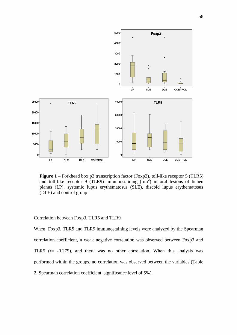
58
Figure 1 – Forkhead box p3 transcription factor (Foxp3), toll-like receptor 5 (TLR5)
and toll-like receptor 9 (TLR9) immunostaining (µm2) in oral lesions of lichen
planus (LP), systemic lupus erythematosus (SLE), discoid lupus erythematosus
(DLE) and control group
Correlation between Foxp3, TLR5 and TLR9
When Foxp3, TLR5 and TLR9 immunostaining levels were analyzed by the Spearman
correlation coefficient, a weak negative correlation was observed between Foxp3 and
TLR5 (r= -0.279), and there was no other correlation. When this analysis was
performed within the groups, no correlation was observed between the variables (Table
2, Spearman correlation coefficient, significance level of 5%).

59
Table 2 - Correlation between forkhead box p3 (Foxp3), toll-like receptor 5 (TLR5) and
toll-like receptor 9 (TLR9) analyzed by the Spearman correlation coefficient
General Groups
Lichen planus Lupus
erythematosus Control
FOXP3 and TLR5 -0.279* 0.100 0.197 -0.224
FOXP3 and TLR9 0.152 0.310 -0.088 0.077
TLR5 and TLR9 0.217 0.236 0.356 0.003
*Significant correlation at significance level of 5%
Qualitative analysis
The three groups evaluated (lichen planus, lupus erythematosus and control) showed a
strong and focal immunostaining pattern for Foxp3 predominantly in connective tissue.
TLR5 immunostaining occurred in either epithelial or connective tissue in all groups.
Nevertheless, in connective tissue it showed a predominantly weak and focal pattern,
whereas in epithelium it was strong and diffuse, with higher intensity in the zone of the
epithelial basal layer. In the lichen planus and lupus erythematosus groups, TLR 9
immunostaining pattern ranged from intermediate to strong, with diffuse distribution in
epithelium and focal in connective tissue. In the control group, TLR9 exhibited strong
staining with diffuse distribution in the epithelium and focal distribution in the
connective tissue. Figure 2 depicts the immunostaining for Foxp3, TLR5 and TLR9 in
the groups analyzed.

60
Figure 2 – Forkhead box p3 transcription factor (Foxp3), toll-like receptor 5
(TLR5) and toll-like receptor 9 (TLR9) immunostaining in oral lesions of lichen
planus, systemic lupus erythematosus (SLE), discoid lupus erythematosus (DLE)
and control group (x 200)
Discussion
In our results, immunostaining for Foxp3 and TLR5 differed significantly between the
groups evaluated, whereas TLR9 did not show any significant difference between them.
The highest Foxp3 expression occurred in the lichen planus group, followed by lupus
erythematosus, and the lowest values occurred in the control group. The results for
lichen planus and lupus erythematosus, showing expression of Foxp3 significantly

61
greater than the controls are in agreement with reports that Foxp3 is increased in
inflammatory processes if compared to controls (11). Also, the lower expression of
Foxp3 in lupus erythematosus if compared to lichen is corroborated by the autoimmune
profile of lupus, where Tregs dysfunction seems to occur (44). It is important to recall
here that the difference between lupus and the control group was smaller than that
observed between lichen and lupus, which can indicate some failure in immune
regulatory mechanisms mediated by Tregs occurring in lupus erythematosus (44,45). In
this way, we could also infer that lichen planus would not be an autoimmune
disturbance, or if considering it as autoimmune, its pathogenesis would be associated
with factors other than Tregs failure (8,46,47). Another point in this respect is the
different phases we can find in systemic lupus erythematosus, alternating with periods
of higher and lower disease activity (48), which could also have interfered with our
results. Because of the small size of our sample, we did not classify the cases of
systemic lupus erythematosus according to SLEDAI (systemic lupus erythematosus
disease activity index). Anyway, when we analyzed discoid and systemic lupus
separately, no change was observed in these results. Maybe further studies with larger
samples could clarify this issue. Also important is the high predominance of T CD4
lymphocytes, which express Foxp3, in lichen planus oral lesions, while in lupus lesions
inflammatory infiltrate also comprises other cells besides lymphocytes, such as
macrophages, mastocytes, plasma cells, and neutrophils in considerable amounts (5).
de Boer et al. (11) did not find any significant difference in Foxp3 expression
between skin lesions of lichen planus and normal skin, which disagrees with our results.
Nevertheless, these authors reported finding considerable variation in Foxp3 between
the specimens within each group, including the control group, which was also observed
in our study. On the other hand, one could point out that our control group was
62
composed of fibrous hyperplasias, which are also inflammatory conditions, and for this
reason would show increased levels of Foxp3. However, besides using the inclusion
criterion of minimal or no inflammatory infiltrate for control group, our results agree
with those reported by Pereira et al. (49), who also demonstrated greater expression of
Foxp3 in oral lesions of lichen planus than in fibrous hyperplasias.
Although Foxp3 is considered the best marker for Tregs (50,51), it is difficult to
precisely evaluate Tregs number and function, as non-Treg CD4 or CD8 cells can
sometimes express Foxp3 as well. The increased frequency of these cells with low
levels of Foxp3 is associated with a defective suppressor function in vitro and with the
development of specific anti-DNA antibodies in patients with active lupus (20). The
possible presence of these non-Treg Foxp3+
cells could have interfered with our results.
Considering the existence of these distinct populations of Foxp3+ cells, it would be
interesting to find out which types of them actually occur in our sample and at what
rates. Still, it is possible that in the lupus erythematosus group, the levels of Foxp3 are
not sufficient to achieve the balance of immune response and therefore leading to the
establishment of autoimmunity (44).
The expression of TLRs in epithelial cells confers protection against pathogens
(31). In our results, TLR5 expression did not significantly differ between lupus
erythematosus and the control group, but these two groups showed significantly greater
values than did lichen planus. TLR5 is expressed in epithelial and dendritic cells, and it
mediates the dynamic interaction between microbiota and the host immune system,
showing positive correlation with microbiota density (33). Similar expression of TLR5
in lupus erythematosus and control group suggests that lupus etiopathogenesis is not
related to flagellated bacterial agents. Anyway, the participation of some fungal or viral
pathogen or even other bacterial types has not been ruled out, as TLR5 is not activated

63
by them. On the other hand, its lower expression in lichen could indicate some failure in
this group to properly react against flagellated microbiota, even the commensals that
invade the epithelium, with consequent loss of local homeostasis (33).
A former study reported greater expression of TLR4 and TLR9 in oral lichen
planus lesions suggesting, respectively, a role of Gram-negative bacteria and human
papillomavirus (HPV) infection in this disease (19). However, it is worth recalling that
the sample of that study comprised either striated/hyperkeratotic or erosive-ulcerated
lesions (19), where the latter are much more likely to harbor microorganisms (49). Our
study, on the other hand, used only striated lesions, which apparently would diminish
the possibility of a secondary infection of the specimens.
TLR9 immunostaining did not significantly differ between the groups analyzed,
even when systemic and discoid lupus erythematosus were considered separately.
Considering that TLR9 is activated mainly by viral agents (10,29), but also by
unmethylated DNA sequences (CpG dinucleotides) found in bacteria (31), we could
infer that this kind of pathogen would not be related to lichen planus or lupus
erythematosus. Our results disagree with those of Li et al. (10) and Siponen et al. (19)
who found greater immunohistochemical expression of TLR9 in lichen than in controls
(normal mucosa and normal skin). Nevertheless, recent studies have reported that TLRs
are also capable of recognizing endogenous molecules released due to cell damage and
necrosis, and they have been found in many autoimmune diseases (14). TLR9 is
considered the major receptor related to autoimmune disturbances such as lupus
erythematosus (12,14,28-30). Accordingly, we expected higher levels of TLR9 in the
lupus erythematosus group, which did not happen. A possibility to consider is that
transforming growth factor-alpha (TGF-alpha), which regulates the expression and
function of TLR5 and TLR9 in human keratinocytes (31) would also be increased in our

64
control group (fibrous hyperplasia). From this point of view, we would have increased
levels of TLR5 and TLR9 also in controls. According to Miller et al. (31), the effect of
TGF-alpha on the expression of TLR9 is more pronounced, which may be due to the
growth and differentiation properties of TGF-alpha, which promotes the maturation of
keratinocytes into the upper and most differentiated layers of the epidermis, where
TLR9 is almost exclusively expressed. If we consider that all the groups of our study
can show epithelial hyperplasia, especially the control group, and that TLR9 is more
affected by TGF-alpha and also has a wider distribution in this tissue if compared to
TLR5 (31), our lack of significant difference in TLR9 between the groups seems
reasonable.
A weak negative correlation was observed between Foxp3 and TLR5, even
though it was not found in the analysis within the groups. Accordingly, a former study
reported that TLR5-/-
rats showed higher levels of Foxp3 than did wild type rats (33). It
seems that TLR5 activation promotes downregulation of Foxp3 expression as a pathway
to defend the host against pathogens (33). This explains our result of negative
correlation between these two markers.
When comparing our results for Foxp3, TLR5 and TLR9 with previously
reported studies (10,11,19), some disagreements are observed. Regarding this, it is
important to consider the rigorous criteria adopted in our study to define the diagnosis
of lichen planus and lupus erythematosus, using strict clinical and histopathological
criteria, including a direct immunofluorescence test. It is known how difficult the
correct diagnosis of these lesions is, where misdiagnosis can result if rigorous criteria
are not respected. Regarding the possible association of both lichen planus and lupus
erythematosus with pathogens, including fungal and viral ones, not just other TLRs
such as TLR2, TLR3 and TLR4 (19,52-56) should be tested in further studies, but also

65
the presence of microorganisms through specific techniques such as flow cytometry,
PCR and RT-PCR (57-59). Such studies should especially focus on molecular
mimicry/immunological cross-reactivity mechanisms by comparing cytometric features
and protein sequences of known autoimmune immunogens with those of
microorganisms commonly involved in human infections (60,61). Still, the profile and
interaction of inflammatory cytokines such as IL2, IFN type I, IFN-gamma, IL6, and
IL17 with Foxp3 and TLRs need to be investigated in these diseases.
Acknowledgments
We thank Dr. A. Leyva (U.S.A.) for English editing of the manuscript.
References
1 Scully C, Beyli M, Ferreiro MC, et al. Update on oral lichen planus:
etiopathogenesis and management. Crit Rev Oral Biol Med 1998; 9: 86-122.
2 Lodi G, Scully C, Carrozzo M, Griffiths M, Sugernab PB, Thongprasom K.
Current controversies in oral lichen planus: Report of an international consensus
meeting. Part 1. Viral infections and etiopathogenesis. Oral Surg Oral Med Oral
Pathol Oral Radiol Endod 2005; 100: 40-51.
3 Perl A. Pathogenic mechanisms in systemic lupus erythematosus. Autoimmunity
2010; 43: 1–6.
4 Neville BW, Damm DD, Allen CM, Bouquot JE. Oral and maxillofacial
pathology. Rio de Janeiro: Elsevier, 2009.
5 Farthing PM, Speight PM. Problems and pitfalls in oral mucosal pathology. Curr
Diagn Pathol 2006; 12: 66–74.
6 Demirci FY, Manzi S, Ramsey-Goldman R, et al. Association study of toll-like
receptor 5 (TLR5) and toll-like receptor 9 (TLR9) polymorphisms in systemic
lupus erythematosus. J Rheumatol 2007; 34: 1708-11.
7 Reich A, Marcinow K, Bialynicki-Birula R. The lupus band test in systemic lupus
erythematosus patients. Ther Clin Risk Manag 2011; 7: 27–32.
8 Roopashree MR, Gondhalekar RV, Shashikanth MC, George J, Thippeswamy SH,
Shukla A. Pathogenesis of oral lichen planus—a review. J Oral Pathol Med 2010;
39: 729-34.

66
9 Vincent FB, Bourke P, Morand EF, Mackay F, Bossingham D. Focus on systemic
lupus erythematosus in indigenous Australians: toward a better understanding of
autoimmune diseases. Intern Med J 2012. doi: 10.1111/imj.12039
10 Li J, Chen J, Tan Z, Liu H, Liu Z. Expression of TLR9 and its mRNA in the
lesions of lichen planus. J Huazhong Univ Sci Technolog Med Sci 2007; 27: 203-
5.
11 de Boer OJ, van der Loos CM, Teeling P, van der Wal AC, Teunissen MB.
Immunohistochemical analysis of regulatory T cell markers FOXP3 and GITR on
CD4⁺CD25⁺ T cells in normal skin and inflammatory dermatoses. J Histochem
Cytochem 2007; 55: 891–8.
12 Blanco P, Paluckad AK, Pascuald V, Banchereaud J. Dendritic cells and cytokines
in human inflammatory and autoimmune diseases. Cytokine Growth Factor Rev
2008; 19: 41–52.
13 Zhou X, Bailey-Bucktrout S, Jeker LT, Bluestone JA. Plasticity of CD4+ FoxP3
+
T cells. Curr Opin Immunol 2009; 21: 281–5.
14 Drexler SK, Foxwell BM. The role of toll-like receptors in chronic inflammation.
Int J Biochem Cell Biol 2010; 42: 506-18.
15 Salloum R, Niewold TB. Interferon regulatory factors in human lupus
pathogenesis. Transl Res 2011; 157: 326–31.
16 Hackl D, Loschko J, Sparwasser T, Reindl W, Krug AB. Activation of dendritic
cells via TLR7 reduces Foxp3 expression and suppressive function in induced
Tregs. Eu J Immunol 2011; 41: 1334–43.
17 Lal G, Yin N, Xu J, et al. Distinct inflammatory signals have physiologically
divergent effects on epigenetic regulation of Foxp3 expression and Treg function.
Am J Transplant 2011; 11: 203–14.
18 Ermertcan AT, Öztürk F, Gündüz K. Toll-like receptors and skin. J Eur Acad
Dermatol Venereol 2011; 25: 997-1006.
19 Siponen M, Kauppila JH, Soini Y, Salo T. TLR4 and TLR9 are induced in oral
lichen planus. J Oral Pathol Med 2012; 41: 741-7.
20 Pan X, Yuan X, Zheng Y, et al. Increased CD45RA+ FoxP3(low) regulatory T
cells with impaired suppressive function in patients with systemic lupus
erythematosus. PLoS One 2012; 7: e34662.
21 Wan YY, Flavell RA. Regulatory T-cell functions are subverted and converted
owing to attenuated Foxp3 expression. Nature 2007; 445: 766-70.
22 Tang Q, Adams JY, Penaranda C, et al. Central role of defective interleukin-2
production in the triggering of islet autoimmune destruction. Immunity 2008; 28:
687-97.

67
23 Lal G, Zhang N, van der Touw W, et al. Epigenetic regulation of Foxp3
expression in regulatory T cells by DNA methylation. J Immunol 2009; 182: 259-
73.
24 Lal G, Bromberg JS. Epigenetic mechanisms of regulation of Foxp3 expression.
Blood 2009; 114: 3727-35.
25 Means TK, Luster AD. Toll-like receptor activation in the pathogenesis of
systemic lupus erythematosus. Ann N Y Acad Sci 2005; 1062: 242-51.
26 Guerrier T, Youinou P, Pers JO, Jamin C. TLR9 drives the development of
transitional B cells towards the marginal zone pathway and promotes
autoimmunity. J Autoimmun 2012; 39: 173-9.
27 Pasare C, Medzhitov R. Toll pathway-dependent blockade of CD4+CD25
+ T cell-
mediated suppression by dendritic cells. Science 2003; 299: 1033-6.
28 Barrat FJ, Meeker T, Gregorio J, et al. Nucleic acids of mammalian origin can act
as endogenous ligands for toll-like receptors and may promote systemic lupus
erythematosus. J Exp Med 2005; 202: 1131–9.
29 Marshak-Rothstein A. Toll-like receptors in systemic autoimmune disease. Nat
Rev Immunol 2006; 6: 823-35.
30 Richez C, Blanco P, Rifkin I, Moreau JF, Schaeverbeke T. Role for toll-like
receptors in autoimmune disease: the example of systemic lupus erythematosus.
Joint Bone Spine 2011; 78: 124-30.
31 Miller LS, Sørensen OE, Liu PT, et al. TGF-alpha regulates TLR expression and
function on epidermal keratinocytes. J Immunol 2005; 174: 6137-43.
32 Crellin NK, Garcia RV, Hadisfar O, Allan SE, Steiner TS, Levings MK. Human
CD4+ T cells express TLR5 and its ligand flagellin enhances the suppressive
capacity and expression of Foxp3 in CD4+CD25
+ T regulatory cells. J Immunol
2005; 175: 8051-9.
33 Feng T, Cong Y, Alexander K, Elson CO. Regulation of toll-like receptor 5 gene
expression and function on mucosal dendritic cells. PLoS One 2012; 7: e35918.
34 Kabelitz D. Expression and function of toll-like receptors in T lymphocytes. Curr
Opin Immunol 2007; 19: 39-45.
35 Oberg HH, Juricke M, Kabelitz D, Wesch D. Regulation of T cell activation by
TLR ligands. Eur J Cell Biol 2011; 90: 582-92.
36 Nico MM, Vilela MA, Rivitti EA, Lourenço SV. Oral lesions in lupus
erythematosus: correlation with cutaneous lesions. Eur J Dermatol 2008; 18: 376-
81.
37 Tan EM, Cohen AS, Fries JF, et al. The 1982 revised criteria for the classification
of systemic lupus erythematosus. Arthritis Rheum 1982; 25: 1271-7.

68
38 Hochberg MC. Updating the American College of Rheumatology revised criteria
for the classification of systemic lupus erythematosus. Arthritis Rheum 1997; 40:
1725.
39 López-Labady J, Villarroel-Dorrego M, González N, Pérez R, Mata de Henning
M. Oral manifestations of systemic and cutaneous lupus erythematosus in a
Venezuelan population. J Oral Pathol Med 2007; 36: 524-7.
40 Sepehr A, Wenson S, Tahan SR. Histopathologic manifestations of systemic
diseases: the example of cutaneous lupus erythematosus. J Cutan Pathol 2010; 37:
112-24.
41 Schiodt M. Oral discoid lupus erythematosus. III. A histopathologic study of
sixty-six patients. Oral Surg Oral Med Oral Pathol 1984; 57: 281-93.
42 Baltaci M, Fritsch P. Histologic features of cutaneous lupus erythematosus.
Autoimmun Rev 2009; 8: 467-73.
43 Tao XA, Xia J, Chen XB, et al. Foxp3 T regulatory cells in lesions of oral lichen
planus correlated with disease activity. Oral Dis 2010; 16: 76-82.
44 Long SA, Buckner JH. CD4+Foxp3
+ T regulatory cells in human autoimmunity:
more than a numbers game. J Immunol 2011; 187: 2061-6.
45 Mudd PA, Teague BN, Farris AD. Regulatory T cells and systemic lupus
erythematosus. Scand J Immunol 2006; 64: 211-8.
46 Korn T, Oukka M, Kuchroo V, Bettelli E. Th17 cells: effector T cells with
inflammatory properties. Semin Immunol 2007; 19: 362-71.
47 Bettelli E, Korn, T; Oukka, M; Kuchroo, VK. Induction and effector functions of
Th17 cells. Nature 2008; 453: 1051-7.
48 Papp K, Végh P, Hóbor R, et al. Immune complex signatures of patients with
active and inactive SLE revealed by multiplex protein binding analysis on antigen
microarrays. PLoS One 2012; 7: e44824.
49 Pereira JS, Monteiro BV, Nonaka CF, Silveira ÉJ, Miguel MC. FoxP3(+) T
regulatory cells in oral lichen planus and its correlation with the distinct clinical
appearance of the lesions. Int J Exp Pathol 2012; 93: 287-94.
50 Hori S, Nomura T, Sakaguchi S. Control of regulatory T cell development by the
transcription factor Foxp3. Science 2003; 299: 1057–61.
51 Khattri R, Cox T, Yasayko SA, Ramsdell F. An essential role for Scurfin in
CD41CD251 T regulatory cells. Nat Immunol 2003; 4: 337–42.
52 Johnson GB, Brunn GJ, Platt JL. Cutting edge: an endogenous pathway to
systemic inflammatory response syndrome (SIRS)-like reactions through toll-like
receptor 4. J Immunol 2004; 172:20-4.

69
53 Brentano F, Schorr O, Gay RE, Gay S, Kyburz D. RNA released from necrotic
synovial fluid cells activates rheumatoid arthritis synovial fibroblasts via toll-like
receptor 3. Arthritis Rheum 2005; 52: 2656-65.
54 McInturff JE, Modlin RL, Kim J. The role of toll-like receptors in the
pathogenesis and treatment of dermatological disease. J Invest Dermatol 2005;
125: 1-8.
55 Hari A, Flach TL, Shi Y, Mydlarski PR. Toll-like receptors: role in
dermatological disease. Mediators Inflamm 2010; 2010:437246. doi:
10.1155/2010/437246
56 Ohno S, Tateishi Y, Tatemoto Y, Morishita K, Sasabe E, Yamamoto T. Enhanced
expression of Toll-like receptor 2 in lesional tissues and peripheral blood
monocytes of patients with oral lichen planus. J Dermatol 2011; 38: 335–44.
57 Jonuleit H, Schmitt E, Stassen M, Tuettenberg A, Knop J, Enk AH. Identification
and functional characterization of human CD4+CD25
+ T cells with regulatory
properties isolated from peripheral blood. J Exp Med 2001; 193: 1285–94.
58 Dardalhon V, Awasthi A, Kwon H, et al. IL-4 inhibits TGF-beta-induced Foxp3+
T cells and, together with TGF-beta, generates IL-9+ IL-10
+ Foxp3(-) effector T
cells. Nat Immunol 2008; 9: 1347-55.
59 Chang JH, Kim YJ, Han SH, Kang CY. IFN-gamma-STAT1 signal regulates the
differentiation of inducible Treg: potential role for ROS-mediated apoptosis. Eur
J Immunol 2009; 39: 1241-51.
60 Westall FC. Molecular mimicry revisited: gut bacteria and multiple sclerosis. J
Clin Microbiol 2006; 44: 2099-104.
61 Sebastiani GD, Galeazzi M. Infection--genetics relationship in systemic lupus
erythematosus. Lupus 2009; 18: 1169-75.

Discussão Geral
71
4 DISCUSSÃO GERAL
As doenças autoimunes e as desordens imunológicas que acometem o organismo
humano são alvo de incessante investigação, haja vista a morbidade e a mortalidade que
implicam. Muitos dos mecanismos etiopatogênicos envolvidos nessas enfermidades
ainda não são completamente conhecidos. Entretanto, a compreensão efetiva dos fatores
que contribuem para o desencadeamento das alterações imunológicas é imprescindível
para a prevenção e para o melhor manejo dessas doenças.
O papel da imunidade inata, mais especificamente, dos receptores da família toll
(toll-like receptors, TLRs) na ativação imunológica e em processos autoimunes, por
meio do reconhecimento de antígenos próprios (Drexler; Foxwell, 2010; Lal et al.,
2011), tem sido alvo de destaque e investigação nos principais centros de Imunologia.
TLRs têm como função primordial defender o organismo por meio do reconhecimento
de patógenos e ativação da resposta imunológica adaptativa (Hari et al., 2010; Drexler;
Foxwell, 2010; Valins et al., 2010; Ermertcan et al., 2011). Ainda nesse campo, tem
sido investigada a relação desses receptores com a modulação da atividade supressora
das células T regulatórias (Tregs) por meio de alterações da expressão de um fator de
transcrição muito importante para o exercício de sua função, o forkhead box p3
(Foxp3). Falhas na expressão de Foxp3 por parte das Tregs estariam associadas a
defeitos funcionais dessas células, com consequente desequilíbrio da resposta
imunológica. Tal desequilíbrio, representado por uma disfunção do componente
regulatório (supressão), permitiria o estabelecimento da autoimunidade (Lal; Bromberg,
2009; Lal et al., 2009; Lal et al., 2011; Long; Buckner, 2011).
Considerando-se que o líquen plano e o lúpus eritematoso são enfermidades
autoimunes ou imunologicamente mediadas (Scully et al., 1998; Lodi et al., 2005; Perl,
2010) que exibem, frequentemente, manifestações orais (Farthing; Speight, 2006), o

72
presente estudo investigou, nessas lesões, o padrão de expressão imunoistoquímica dos
TLRs 5 e 9 e do Foxp3. O TLR9, um receptor toll-like especializado no reconhecimento
de partículas de DNA de origem bacteriana e viral, tem sido associado ao
desenvolvimento de uma resposta autoimune por meio do reconhecimento de partículas
de DNA do próprio organismo, além de ser capaz, mediante constante estimulação, de
promover contínua baixa expressão de Foxp3 em células Treg com comprometimento
de sua função na manutenção da homeostase do sistema imunológico (Pasare;
Medzhitov, 2003; Means; Luster, 2005; Barrat et al., 2005; Marshak-Rothstein, 2006;
Blanco et al., 2008; Drexler; Foxwell, 2010; Richez et al., 2011; Guerrier et al., 2012).
O TLR5, por sua vez, tem como ligante principal a flagelina, que é expressa
especificamente por bactérias portadoras de flagelo (Miller et al., 2005; Marshak-
Rothstein, 2006; Demirci et al., 2007; Feng et al., 2012).
Os resultados da presente pesquisa evidenciaram diferença significativa do
padrão de expressão imunoistoquímica do Foxp3 e do TLR5 entre os grupos avaliados,
enquanto essa diferença não foi observada para o marcador TLR9. Além disso, foi
observada correlação negativa entre Foxp3 e TLR5. O grupo líquen plano apresentou os
maiores níveis de expressão do Foxp3, seguido do grupo lúpus eritematoso, e os
menores níveis ocorreram no grupo-controle. A maior expressão de Foxp3 nos grupos
líquen e lúpus em comparação ao grupo-controle estão de acordo com o estudo de De
Boer et al. (2007) que demonstrou maiores níveis desse marcador em amostras de
processos inflamatórios do que em controles. Por outro lado, a menor expressão de
Foxp3 no grupo lúpus eritematoso, quando comparado ao grupo líquen plano, pode
estar associada ao perfil autoimune da doença, que envolveria uma disfunção das
células Treg (Mudd et al., 2006; Long; Buckner, 2011). Dessa forma, pode-se sugerir
que o líquen plano não seja uma doença de natureza autoimune ou, se assim for, sua

73
patogênese estaria associada a outros fatores que não falhas na população de Tregs
(Korn et al., 2007; Bettelli et al., 2008; Roopashree et al., 2010).
Além da análise da expressão de células Treg Foxp3+ nas lesões de líquen e de
lúpus, o presente estudo avaliou a expressão de dois TLRs, o 5 e o 9. A princípio,
informações sobre o envolvimento do TLR5 em tais enfermidades são escassas na
literatura (Demirci et al., 2007). Por outro lado, tem-se especulado que o TLR9
participaria do desencadeamento de reações autoimunes que culminariam no
desenvolvimento de lúpus eritematoso sistêmico (Marshak-Rothstein, 2006; Richez et
al., 2011; Ghaly et al., 2012; Guerrier et al., 2012) e líquen plano (Li et al., 2007).
Assim, investigou-se se o padrão de expressão desses TLRs em lesões orais de líquen
plano teria alguma semelhança com o das lesões de lúpus, visto que isso forneceria
alguma ideia quanto à etiopatogênese de ambas as doenças, bem como outras
informações acerca dos TLRs 5 e 9. Nesse sentido, talvez fosse possível inferir que o
líquen plano seria, de fato, uma enfermidade autoimune, o que ainda não está bem
definido na literatura. A análise imunoistoquímica dos TLRs em lesões de hiperplasia
fibroepitelial, por sua vez, possibilitaria o estudo do padrão de expressão dos mesmos
em um processo sabidamente não autoimune, estabelecendo um meio de comparação
com as lesões de líquen e lúpus. Sendo o TLR9 considerado o principal receptor toll-
like associado a distúrbios autoimunes como o lúpus (Barrat et al., 2005; Marshak-
Rothstein, 2006; Blanco et al., 2008; Drexler; Foxwell, 2010; Richez et al., 2011),
esperava-se que seus níveis de expressão tivessem sido maiores no grupo lúpus
eritematoso. No entanto, não foi observada diferença significativa em sua expressão
entre os três grupos avaliados. Considerando-se que todos os grupos do presente estudo,
em especial o grupo-controle, podem apresentar hiperplasia epitelial, que se trata de um
processo proliferativo, tal resultado parece justificável. Processos proliferativos do

74
epitélio exibem maiores concentrações do fator de transformação do crescimento alfa
(TGF-alfa), o qual regula a expressão e a função dos TLRs 5 e 9 elevando seus níveis
nos queratinócitos humanos (Miller et al., 2005). Isso justificaria a ausência de
diferença significativa da expressão do TLR9 entre os grupos.
Também não foi observada diferença significativa da expressão do TLR5 entre o
grupo lúpus eritematoso e o grupo-controle. Entretanto, tais grupos exibiram maior
expressão desse marcador quando comparados ao grupo líquen plano. O TLR5 tem
correlação positiva com a densidade da microbiota no tecido (Feng et al., 2012) e,
portanto, sua menor expressão no grupo líquen plano poderia indicar uma deficiência da
capacidade de reagir adequadamente à microbiota específica, com consequente
desequilíbrio da homeostase local (Feng et al., 2012).
Ainda, foi observada correlação negativa, embora fraca, entre a expressão do
Foxp3 e do TLR5. No estudo de Feng et al. (2012), ratos TLR5-/- apresentaram maiores
níveis de Foxp3 do que os wild type, o que sugere que a ativação desse TLR promova a
expressão de menores níveis de Foxp3 como um mecanismo de defesa do organismo
contra patógenos. Isso explicaria a correlação negativa observada entre TLR5 e Foxp3
no presente estudo.
Tanto no líquen plano quanto no lúpus eritematoso, uma cascata de eventos
inflamatórios ocorre nos sítios onde se desenvolvem as lesões. Todos esses eventos,
marcados pela produção de uma gama de citocinas, quimiocinas e ativação de inúmeras
vias de sinalização inter e intracelular, são, de certa forma, responsáveis pelo padrão de
expressão dos componentes-alvo do presente estudo, bem como pelo próprio aspecto
clínico das lesões. O resultado do processo “tolerância versus autoimunidade” pode
culminar no aparecimento de tais lesões e depende do equilíbrio entre sinais
estimulatórios, como a presença de ligantes de TLRs e a concentração de um dado

75
antígeno, seja ele exógeno ou endógeno, bem como de sinais inibitórios mediados por
células Treg (Suri-Payer; Fritzsching, 2006). A quebra desse equilíbrio é observada no
lúpus eritematoso sistêmico, em que autoanticorpos ligam-se ao DNA ou às proteínas
conjugadas ao DNA/RNA provenientes de células apoptóticas (Munoz et al., 2008;
Rönnblom et al., 2011), gerando imunocomplexos que podem estimular células por
meio de TLRs, o que resulta na produção de citocinas inflamatórias como o IFN-alfa
(Salloum; Niewold, 2011). A contínua ativação de TLRs envolvidos na indução de
citocinas inflamatórias Th1/Th2, capazes de inibir a expressão de Foxp3, pode ser
responsável pelo bloqueio da função de células Treg (Chang et al., 2009; Jin et al.,
2010; Hackl et al., 2011; Lal et al., 2011; Long; Buckner, 2011). Assim, a presença de
ligantes endógenos que, supostamente, poderiam estar sendo gerados em doenças como
lúpus e líquen, teria papel preponderante no estabelecimento e na manutenção da
autoimunidade (Drexler; Foxwell, 2010).
Nesse sentido, da mesma forma que ligantes endógenos poderiam estar
persistentemente ativando todo esse círculo vicioso que resulta na cronicidade das
doenças em questão, poder-se-ia especular que determinados agentes infecciosos
constantemente presentes nos tecidos, como partículas virais, poderiam exercer esse
mesmo papel. Além disso, é possível que tais agentes apresentem semelhanças
moleculares com estruturas do próprio organismo, o que induziria a uma reação cruzada
por permitir o reconhecimento de antígenos próprios pelo sistema imunológico e, com
isso, o desencadeamento da autoimunidade (Westall, 2006; Sebastiani; Galeazzi, 2009).
A complexidade dos distúrbios autoimunes e imunologicamente mediados, em
que tanto fatores genéticos/individuais quanto ambientais contribuem para a
susceptibilidade à doença, exige que novas pesquisas continuem sendo conduzidas
nessa área. Parece pertinente a investigação da relação de tais enfermidades com células

76
Treg, citocinas e quimiocinas pró-inflamatórias, bem como com alguns microrganismos
possivelmente envolvidos no desencadeamento de reações autoimunes em função das
semelhanças moleculares que guardam em relação a componentes do organismo
humano. Até hoje, a maioria das doenças autoimunes e imunologicamente mediadas
apresentam-se como enfermidades controláveis, porém incuráveis. Os pacientes
resignam-se a uma vida inteira de tratamentos medicamentosos e controles clínicos.
Tratamentos esses capazes apenas de inibir ou atenuar as manifestações da doença e, em
geral, acompanhados de importantes efeitos colaterais. Somente a elucidação desses
intrincados caminhos do sistema imunológico propiciará novas perspectivas de
tratamento, cura e prevenção dessas enfermidades. Daí a necessidade constante de
novas pesquisas nesse campo.

Referências
78
5 REFERÊNCIAS
Bacchetta R, Gregori S, Roncarolo MG. CD4+ regulatory T cells: mechanisms of
induction and effector function. Autoimmun Rev 2005; 4(8): 491-6.
Baecher-Allan C, Brown JA, Freeman GJ, Hafler DA. CD4+CD25
high regulatory cells in
human peripheral blood. J Immunol 2001; 167(3): 1245-53.
Baltaci M, Fritsch P. Histologic features of cutaneous lupus erythematosus. Autoimmun
Rev 2009; 8(6): 467-73.
Barrat FJ, Meeker T, Gregorio J, Chan JH, Uematsu S, Akira S, Chang B, Duramad O,
Coffman RL. Nucleic acids of mammalian origin can act as endogenous ligands for
Toll-like receptors and may promote systemic lupus erythematosus. J Exp Med 2005;
202(8): 1131–9.
Bengtsson AA, Sturfelt G, Truedsson L, Blomberg J, Alm G, Vallin H, Rönnblom L.
Activation of type I interferon system in systemic lupus erythematosus correlates with
disease activity but not with antiretroviral antibodies. Lupus 2000; 9(9): 664-71.
Bermejo-Fenoll A, López-Jornet P. Familial oral lichen planus: presentation of six
families. Oral Surg Oral Med Oral Pathol Oral Radiol Endod 2006; 102(2): e12-5.
Bettelli E, Korn T, Oukka M, Kuchroo VK. Induction and effector functions of TH17
cells. Nature 2008; 453 (7198): 1051-7.
Blanco P, Paluckad AK, Pascuald V, Banchereaud J. Dendritic cells and cytokines in
human inflammatory and autoimmune diseases. Cytokine Growth Factor Rev 2008;
19(1): 41–52.
Brentano F, Schorr O, Gay RE, Gay S, Kyburz D. RNA released from necrotic synovial
fluid cells activates rheumatoid arthritis synovial fibroblasts via Toll-like receptor 3.
Arthritis Rheum 2005; 52(9): 2656-65.
Burns K, Martinon F, Esslinger C, Pahl H, Schneider P, Bodmer JL, Di Marco F,
French L, Tschopp J. MyD88, an adapter protein involved in interleukin-1 signaling. J
Biol Chem 1998; 273(20): 12203-9.
Canto AM, Müller H, Freitas RR, Santos PS. Oral lichen planus (OLP): clinical and
complementary diagnosis. An Bras Dermatol 2010; 85(5): 669-75.
Carty M, Goodbody R, Schröder M, Stack J, Moynagh PN, Bowie AG. The human
adaptor SARM negatively regulates adaptor protein TRIF-dependent Toll-like receptor
signaling. Nat Immunol 2006; 7(10): 1074-81.
Chang JH, Kim YJ, Han SH, Kang CY. IFN-gamma-STAT1 signal regulates the
differentiation of inducible Treg: potential role for ROS-mediated apoptosis. Eur J
Immunol 2009; 39(5): 1241-51.
Crellin NK, Garcia RV, Hadisfar O, Allan SE, Steiner TS, Levings MK. Human CD4+
T cells express TLR5 and its ligand flagellin enhances the suppressive capacity and
79
expression of Foxp3 in CD4+CD25
+ T regulatory cells. J Immunol 2005; 175(12): 8051-
9.
Crispín JC, Tsokos GC. Interleukin-17-producing T cells in lupus. Curr Opin
Rheumatol 2010; 22(5): 499-503.
Dardalhon V, Awasthi A, Kwon H, Galileos G, Gao W, Sobel RA, Mitsdoerffer M,
Strom TB, Elyaman W, Ho IC, Khoury S, Oukka M, Kuchroo VK. IL-4 inhibits TGF-
beta-induced Foxp3+ T cells and, together with TGF-beta, generates IL-9
+ IL-10
+
Foxp3(-) effector T cells. Nat Immunol 2008; 9(12): 1347-55.
de Boer OJ, Van der Loos CM, Teeling P, van der Wal AC, Teunissen MB.
Immunohistochemical analysis of regulatory T cell markers Foxp3 and GITR on
CD4⁺CD25⁺ T cells in normal skin and inflammatory dermatoses. J Histochem
Cytochem 2007; 55(9): 891–8.
de la Rosa M, Rutz S, Dorninger H, Scheffold A. Interleukin-2 is essential for
CD4+CD25
+ regulatory T cell function. Eur J Immunol 2004; 34(9): 2480-8.
Demirci FY, Manzi S, Ramsey-Goldman R, Kenney M, Shaw PS, Dunlop-Thomas CM,
Kao AH, Rhew EY, Bontempo F, Kammerer C, Kamboh MI. Association study of Toll-
like receptor 5 (TLR5) and Toll-like receptor 9 (TLR9) polymorphisms in systemic lupus erythematosus. J Rheumatol 2007; 34(8): 1708-11.
Dieckmann D, Plottner H, Berchtold S, Berger T, Schuler G. Ex vivo isolation and
characterization of CD4(+)CD25(+) T cells with regulatory properties from human
blood. J Exp Med 2001; 193(11): 1303-10.
Drexler SK, Foxwell BM. The role of toll-like receptors in chronic inflammation. Int J
Biochem Cell Biol 2010; 42(4): 506-18.
Ermann J, Szanya V, Ford GS, Paragas V, Fathman CG, Lejon K. CD4(+)CD25(+) T
cells facilitate the induction of T cell anergy. J Immunol 2001; 167(8): 4271-5.
Ermertcan AT, Öztürk F, Gündüz K. Toll-like receptors and skin. J Eur Acad Dermatol
Venereol 2011; 25(9): 997-1006.
Farthing PM, Speight PM. Problems and pitfalls in oral mucosal pathology. Curr Diagn
Pathol 2006; 12 (1): 66-74.
Feng T, Cong Y, Alexander K, Elson CO. Regulation of Toll-like receptor 5 gene
expression and function on mucosal dendritic cells. PLoS One 2012; 7(4): e35918.
Fitzgerald KA, Rowe DC, Barnes BJ, Caffrey DR, Visintin A, Latz E, Monks B, Pitha
PM, Golenbock DT. LPS-TLR4 signaling to IRF-3/7 and NF-kappaB involves the toll
adapters TRAM and TRIF. J Exp Med 2003; 198(7): 1043-55.
Ghaly NR, Kotb NA, Nagy HM, Rageh ES. Toll-like receptor 9 in systemic lupus
erythematosus, impact on glucocorticoid treatment. J Dermatolog Treat 2012.
doi:10.3109/09546634.2012.697110

80
Gonçalves LM, Bezerra Júnior JR, Cruz MC. Clinical evaluation of oral lesions
associated with dermatologic diseases. An Bras Dermatol 2010; 85(2): 150-6.
Grossman WJ, Verbsky JW, Barchet W, Colonna M, Atkinson JP, Ley TJ. Human T
regulatory cells can use the perforin pathway to cause autologous target cell death.
Immunity 2004; 21(4): 589-601.
Guerrier T, Youinou P, Pers JO, Jamin C. TLR9 drives the development of transitional
B cells towards the marginal zone pathway and promotesautoimmunity. J Autoimmun
2012; 39(3): 173-9.
Hackl D, Loschko J, Sparwasser T, Reindl W, Krug AB. Activation of dendritic cells
via TLR7 reduces Foxp3 expression and suppressive function in induced Tregs. Eu J
Immunol 2011; 41(5): 1334–43.
Hari A, Flach TL, Shi Y, Mydlarski PR. Toll-like receptors: role in dermatological
disease. Mediators Inflamm 2010; 2010:437246. doi: 10.1155/2010/437246
Hochberg MC. Updating the American College of Rheumatology revised criteria for the
classification of systemic lupus erythematosus. Arthritis Rheum 1997; 40(9): 1725.
Hori S, Nomura T, Sakaguchi S. Control of regulatory T cell development by the
transcription factor Foxp3. Science 2003; 299(5609): 1057-61.
Horng T, Barton GM, Medzhitov R. TIRAP: an adapter molecule in the Toll signaling
pathway. Nat Immunol 2001; 2(9): 835-41.
Ismail SB, Kumar SK, Zain RB. Oral lichen planus and lichenoid reactions:
etiopathogenesis, diagnosis, management and malignant transformation. J Oral Sci
2007; 49(2): 89-106.
Jin O, Kavikondala S, Mok MY, Sun L, Gu J, Fu R, Chan A, Yeung J, Nie Y, Lau CS.
Abnormalities in circulating plasmacytoid dendritic cells in patients with systemic lupus
erythematosus. Arthritis Res Ther 2010; 12(4): R137.
Johnson GB, Brunn GJ, Platt JL. Cutting edge: an endogenous pathway to systemic
inflammatory response syndrome (SIRS)-like reactions through Toll-like receptor 4. J
Immunol 2004; 172(1):20-4.
Jonuleit H, Schmitt E, Kakirman H, Stassen M, Knop J, Enk AH. Infectious tolerance:
human CD25(+) regulator cells convey suppressor activity to conventionalCD4(+) T
helper cells. J Exp Med 2002; 196(2): 255-60.
Jonuleit H, Schmitt E, Stassen M, Tuettenberg A, Knop J, Enk AH. Identification and
functional characterization of human CD4+CD25
+ T cells with regulatory properties
isolated from peripheral blood. J Exp Med 2001; 193(11): 1285–94.
Kabelitz D. Expression and function of Toll-like receptors in T lymphocytes. Curr Opin
Immunol 2007; 19(1): 39-45.

81
Kaisho T, Akira S. Toll-like receptor function and signaling. J Allergy Clin Immunol
2006; 117(5): 979-87.
Kalogerakou F, Albanidou-Farmaki E, Markopoulos AK, Antoniades DZ. Detection of
T cells secreting type 1 and type 2 cytokines in the peripheral blood of patients with oral
lichen planus. Hippokratia 2008; 12(4): 230-5.
Kawai T, Akira S. The role of pattern-recognition receptors in innate immunity: update
on Toll-like receptors. Nat Immunol 2010; 11(5): 373-84.
Khattri R, Cox T, Yasayko SA, Ramsdell F. An essential role for Scurfin in
CD4+CD25
+ T regulatory cells. Nat Immunol 2003; 4(4): 337-42.
Korn T, Oukka M, Kuchroo V, Bettelli E. Th17 cells: effector T cells with
inflammatory properties. Semin Immunol 2007; 19(6): 362-71.
Kushwah R, Hu J. Role of dendritic cells in the induction of regulatory T cells. Cell
Biosci 2011a; 1(1):20.
Kushwah R, Hu J. Complexity of dendritic cell subsets and their function in the host
immune system. Immunology 2011b; 133(4): 409-19.
Lal G, Bromberg JS. Epigenetic mechanisms of regulation of Foxp3 expression. Blood
2009; 114(18): 3727-35.
Lal G, Yin N, Xu J, Lin M, Schroppel S, Ding Y, Marie I, Levy DE, Bromberg JS.
Distinct inflammatory signals have physiologically divergent effects on epigenetic
regulation of Foxp3 expression and Treg function. Am J Transplant 2011; 11(2): 203–
14.
Lal G, Zhang N, van der Touw W, Ding Y, Ju W, Bottinger EP, Reid SP, Levy DE,
Bromberg JS. Epigenetic regulation of Foxp3 expression in regulatory T cells by DNA
methylation. J Immunol 2009; 182(1): 259-73.
Levings MK, Sangregorio R, Sartirana C, Moschin AL, Battaglia M, Orban PC,
Roncarolo MG. Human CD25+CD4
+ T suppressor cell clones produce transforming
growth factor beta, but not interleukin 10, and are distinct from type 1 T regulatory
cells. J Exp Med 2002; 196(10): 1335-46.
Li J, Chen J, Tan Z, Liu H, Liu Z. Expression of TLR9 and its mRNA in the lesions of
lichen planus. J Huazhong Univ Sci Technolog Med Sci 2007; 27(2): 203-5.
Lin J, Weiss A. T cell receptor signalling. J Cell Sci 2001; 114(Pt 2): 243-4.
Liu B, Dai J, Zheng H, Stoilova D, Sun S, Li Z. Cell surface expression of an
endoplasmic reticulum resident heat shock protein gp96 triggers MyD88-dependent
systemic autoimmune diseases. Proc Natl Acad Sci U S A. 2003; 100(26): 15824-9.
Liu YJ. IPC: professional type 1 interferon producing cells and plasmacytoid dendritic
cell precursors. Annu Rev Immunol 2005; 23: 275-306.

82
Lodi G, Scully C, Carrozzo M, Griffiths M, Sugerman PB, Thongprasom K. Current
controversies in oral lichen planus: report of an international consensus meeting. Part 1.
Viral infections and etiopathogenesis. Oral Surg Oral Med Oral Pathol Oral Radiol
Endod 2005; 100(1): 40-51.
Long SA, Buckner JH. CD4+Foxp3
+ T regulatory cells in human autoimmunity: more
than a numbers game. J Immunol 2011; 187(5): 2061-6.
Loniewski KJ, Patial S, Parameswaran N. Sensitivity of TLR4- and -7-induced NF
kappa B1 p105-TPL2-ERK pathway to TNF-receptor-associated-factor-6 revealed by
RNAi in mouse macrophages. Mol Immunol 2007; 44(15): 3715-23.
López-Labady J, Villarroel-Dorrego M, González N, Pérez R, Mata de Henning M. Oral
manifestations of systemic and cutaneous lupus erythematosus in a Venezuelan
population. J Oral Pathol Med 2007; 36(9): 524-7.
Lukac J, Brozović S, Vucicević-Boras V, Mravak-Stipetić M, Malenica B, Kusić Z.
Serum autoantibodies to desmogleins 1 and 3 in patients with oral lichen planus. Croat
Med J 2006; 47(1): 53-8.
Marshak-Rothstein A. Toll-like receptors in systemic autoimmune disease. Nat Rev
Immunol 2006; 6(11): 823-35.
McInturff JE, Modlin RL, Kim J. The role of toll-like receptors in the pathogenesis and
treatment of dermatological disease. J Invest Dermatol 2005; 125(1): 1-8.
Means TK, Latz E, Hayashi F, Murali MR, Golenbock DT, Luster AD. Human lupus
autoantibody DNA complexes activate DCs through cooperation of CD32 and TLR9. J
Clin Invest 2005; 115(2): 407-17.
Means TK, Luster AD. Toll-like receptor activation in the pathogenesis of systemic
lupus erythematosus. Ann N Y Acad Sci 2005; 1062: 242-51.
Midgley A, Thorbinson C, Beresford MW. Expression of Toll-like receptors and their
detection of nuclear self-antigen leading to immuneactivation in JSLE. Rheumatology
(Oxford) 2012; 51(5): 824-32.
Miller LS, Modlin RL. Toll-like receptors in the skin. Semin Immunopathol 2007;
29(1): 15-26.
Miller LS, Sørensen OE, Liu PT, Jalian HR, Eshtiaghpour D, Behmanesh BE, Chung
W, Starner TD, Kim J, Sieling PA, Ganz T, Modlin RL. TGF-alpha regulates TLR
expression and function on epidermal keratinocytes. J Immunol 2005; 174(10): 6137-
43.
Mills KH. TLR-dependent T cell activation in autoimmunity. Nat Rev Immunol 2011;
11(12): 807-22.
Miyara M, Sakaguchi S. Natural regulatory T cells: mechanisms of suppression. Trends
Mol Med 2007; 13(3): 108-16.

83
Mudd PA, Teague BN, Farris AD. Regulatory T cells and systemic lupus
erythematosus. Scand J Immunol 2006; 64(3): 211-8.
Munoz LE, Van Bavel C, Franz S, Berden J, Herrmann M, Van Der Vlag J. Apoptosis
in the pathogenesis of systemic lupus erythematosus. Lupus 2008; 17(5): 371-5.
Nakamura K, Kitani A, Fuss I, Pedersen A, Harada N, Nawata H, Strober W. TGF-beta
1 plays an important role in the mechanism of CD4+CD25
+ regulatory T cell activity in
both humans and mice. J Immunol 2004; 172(2): 834-42.
Nakano S, Morimoto S, Suzuki J, Nozawa K, Amano H, Tokano Y, Takasaki Y. Role
of pathogenic auto-antibody production by Toll-like receptor 9 of B cells in active
systemic lupus erythematosus. Rheumatology (Oxford) 2008; 47(2): 145-9.
Neville BW, Damm DD, Allen CM, Bouquot JE. Patologia Oral e Maxilofacial. 3.ed.
Rio de Janeiro: Elsevier, 2009.
Nico MM, Vilela MA, Rivitti EA, Lourenço SV. Oral lesions in lupus erythematosus:
correlation with cutaneous lesions. Eur J Dermatol 2008; 18(4): 376-81.
Nyirenda MH, Sanvito L, Darlington PJ, O'brien K, Zhang GX, Constantinescu CS,
Bar-Or A, Gran B. TLR2 stimulation drives human naive and effector regulatory T cells
into a Th17-like phenotype with reduced suppressive function. J Immunol 2011; 187(5):
2278-90.
Oberg HH, Juricke M, Kabelitz D, Wesch D. Regulation of T cell activation by TLR
ligands. Eur J Cell Biol 2011; 90(6-7): 582-92.
Ohno S, Tateishi Y, Tatemoto Y, Morishita K, Sasabe E, Yamamoto T. Enhanced
expression of Toll-like receptor 2 in lesional tissues and peripheral blood monocytes of
patients with oral lichen planus. J Dermatol 2011; 38(4): 335–44.
Pan X, Yuan X, Zheng Y, Wang W, Shan J, Lin F, Jiang G, Yang YH, Wang D, Xu D,
Shen L. Increased CD45RA+ FoxP3(low) regulatory T cells with impaired suppressive
function in patients with systemic lupus erythematosus. PLoS One 2012; 7(4): e34662.
Papp K, Végh P, Hóbor R, Szittner Z, Vokó Z, Podani J, Czirják L, Prechl J. Immune
complex signatures of patients with active and inactive SLE revealed by multiplex
protein binding analysis on antigen microarrays. PLoS One 2012; 7(9): e44824.
Parham P. O sistema imune. Porto Alegre: Artes Médicas, 2001.
Paroli M, Iannucci G, Accapezzato D. Hepatitis C virus infection and autoimmune
diseases. Int J Gen Med 2012; 5: 903-7.
Pasare C, Medzhitov R. Toll pathway-dependent blockade of CD4+CD25
+ T cell-
mediated suppression by dendritic cells. Science 2003; 299(5609): 1033-6.
Peng G, Guo Z, Kiniwa Y, Voo KS, Peng W, Fu T, Wang DY, Li Y, Wang HY, Wang
RF. Toll-like receptor 8-mediated reversal of CD4+ regulatory T cell function. Science
2005; 309(5739): 1380-4.

84
Pereira JS, Monteiro BV, Nonaka CF, Silveira ÉJ, Miguel MC. Foxp3(+) T regulatory
cells in oral lichen planus and its correlation with the distinct clinical appearance of the
lesions. Int J Exp Pathol 2012; 93(4): 287-94.
Perl A. Pathogenic mechanisms in systemic lupus erythematosus. Autoimmunity 2010;
43(1): 1–6.
Pisitkun P, Deane JA, Difilippantonio MJ, Tarasenko T, Satterthwaite AB, Bolland S.
Autoreactive B cell responses to RNA-related antigens due to TLR7 gene duplication.
Science 2006; 312(5780): 1669-72.
Reich A, Marcinow K, Bialynicki-Birula R. The lupus band test in systemic lupus
erythematosus patients. Ther Clin Risk Manag 2011;7: 27–32.
Richez C, Blanco P, Rifkin I, Moreau JF, Schaeverbeke T. Role for toll-like receptors in
autoimmune disease: the example of systemic lupus erythematosus. Joint Bone Spine
2011; 78(2): 124-30.
Roopashree MR, Gondhalekar RV, Shashikanth MC, George J, Thippeswamy SH,
Shukla A. Pathogenesis of oral lichen planus – a review. J Oral Pathol Med 2010;
39(10): 729–34.
Rönnblom L, Alm GV, Eloranta ML. The type I interferon system in the development
of lupus. Semin Immunol 2011; 23(2): 113–21.
Sakaguchi S, Miyara M, Costantino CM, Hafler DA. Foxp3+ regulatory T cells in the
human immune system. Nat Rev Immunol 2010; 10(7): 490-500.
Sakaguchi S, Sakaguchi N, Asano M, Itoh M, Toda M. Pillars article: immunologic
self-tolerance maintained by activated T cells expressing IL-2 receptorα-chains (CD25).
Breakdown of a single mechanism of self-tolerance causes various autoimmune
diseases. J. Immunol. 1995. J Immunol 2011; 186(7): 3808-21.
Salem SA, Abu-Zeid RM, Nada OH. Immunohistochemical study of toll-like receptors
1 and 2 expression in cutaneous lichen planus lesions. Arch Dermatol Res 2012; doi
10.1007/s00403-012-1267-8
Salloum R, Niewold TB. Interferon regulatory factors in human lupus pathogenesis.
Transl Res 2011; 157(6): 326–31.
Samanta A, Li B, Song X, Bembas K, Zhang G, Katsumata M, Saouaf SJ, Wang Q,
Hancock WW, Shen Y, Greene MI. TGF-beta and IL-6 signals modulate chromatin
binding and promoter occupancy by acetylated Foxp3. Proc Natl Acad Sci U S A. 2008;
105(37): 14023-7.
Schiodt M. Oral discoid lupus erythematosus. III. A histopathologic study of sixty-six
patients. Oral Surg Oral Med Oral Pathol 1984; 57(3): 281-93.
Schmidt A, Oberle N, Krammer PH. Molecular mechanisms of Treg-mediated T cell
suppression. Front Immunol 2012; 3: 51.

85
Scully C, Beyli M, Ferreiro MC, Ficarra G, Gill Y, Griffiths M, Holmstrup P, Mutlu S,
Porter S, Wray D. Update on oral lichen planus: etiopathogenesis and management. Crit
Rev Oral Biol Med 1998; 9(1): 86-122.
Scully C, Carrozzo M. Oral mucosal disease: Lichen planus. Br J Oral Maxillofac Surg
2008; 46(1): 15-21.
Sebastiani GD, Galeazzi M. Infection--genetics relationship in systemic lupus
erythematosus. Lupus 2009; 18(13): 1169-75.
Sepehr A, Wenson S, Tahan SR. Histopathologic manifestations of systemic diseases:
the example of cutaneous lupus erythematosus. J Cutan Pathol 2010; 37(Suppl 1): 112-
24.
Siponen M, Kauppila JH, Soini Y, Salo T. TLR4 and TLR9 are induced in oral lichen
planus. J Oral Pathol Med 2012; 41(10): 741-7.
Smith-Garvin JE, Koretzky GA, Jordan MS. T cell activation. Annu Rev Immunol 2009;
27: 591-619.
Sojka DK, Fowell DJ. Regulatory T cells inhibit acute IFN-γ synthesis without blocking
T-helper cell type 1 (Th1) differentiation via a compartmentalized requirement for IL-
10. Proc Natl Acad Sci USA 2011; 108(45): 18336-41.
Sugerman PB, Savage NW, Walsh LJ, Zhao ZZ, Zhou XJ, Khan A, Seymour GJ, Bigby
M. The pathogenesis of oral lichen planus. Crit Rev Oral Biol Med 2002; 13(4): 350-65.
Suri-Payer E, Fritzsching B. Regulatory T cells in experimental autoimmune disease.
Springer Semin Immunopathol 2006; 28(1): 3-16.
Tadokoro CE, Shakhar G, Shen S, Ding Y, Lino AC, Maraver A, Lafaille JJ, Dustin
ML. Regulatory T cells inhibit stable contacts between CD4+ T cells anddendritic cells
in vivo. J Exp Med 2006; 203(3): 505-11.
Tan EM, Cohen AS, Fries JF, Masi AT, McShane DJ, Rothfield NF, Schaller JG, Talal
N, Winchester RJ. The 1982 revised criteria for the classification of systemic lupus
erythematosus. Arthritis Rheum 1982; 25(11): 1271-7.
Tang Q, Adams JY, Penaranda C, Melli K, Piaggio E, Sgouroudis E, Piccirillo CA,
Salomon BL, Bluestone JA. Central role of defective interleukin-2 production in the
triggering of islet autoimmune destruction. Immunity 2008; 28(5): 687-97.
Tao XA, Li CY, Xia J, Yang X, Chen XH, Jian YT, Cheng B. Differential gene
expression profiles of whole lesions from patients with oral lichen planus. J Oral Pathol
Med 2009; 38(5): 427-33.
Tao XA, Xia J, Chen XB, Wang H, Dai YH, Rhodus NL, Cheng B. Foxp3 T regulatory
cells in lesions of oral lichen planus correlated with disease activity. Oral Dis 2010;
16(1): 76-82.
Thornhill MH. Immune mechanisms in oral lichen planus. Acta Odontol Scand 2001;
59(3): 174 –7.
86
Thornhill MH, Sankar V, Xu XJ, Barrett AW, High AS, Odell EW, Speight PM,
Farthing PM. The role of histopathological characteristics in distinguishing amalgam-
associated oral lichenoid reactions and oral lichen planus. J Oral Pathol Med 2006;
35(4): 233-40.
Valencia X, Yarboro C, Illei G, Lipsky PE. Deficient CD4+CD25
high T regulatory cell
function in patients with active systemic lupus erythematosus. J Immunol 2007; 178(4):
2579-88.
Vallin H, Blomberg S, Alm GV, Cederblad B, Rönnblom L. Patients with systemic
lupus erythematosus (SLE) have a circulating inducer of interferon-alpha (IFN-alpha)
production acting on leucocytes resembling immature dendritic cells. Clin Exp Immunol
1999; 115(1): 196-202.
Valins W, Amini S, Berman B. The expression of Toll-like receptors in dermatological
diseases and the therapeutic effect of current and newer topical Toll-like receptor
modulators. J Clin Aesthet Dermatol 2010; 3(9): 20-9.
van Maren WW, Jacobs JF, de Vries IJ, Nierkens S, Adema GJ. Toll-like receptor
signaling on Tregs: to suppress or not to suppress? Immunology 2008; 124(4): 445-52.
Veldhoen M, Moncrieffe H, Hocking RJ, Atkins CJ, Stockinger B. Modulation of
dendritic cell function by naïve and regulatory CD4+ T cells. J Immunol 2006; 176(10):
6202-10.
Vergani D, Mieli-Vergani G. Autoimmune manifestations in viral hepatitis. Semin
Immunopathol 2012. doi: 10.1007/s00281-012-0328-6.
Vincent FB, Bourke P, Morand EF, Mackay F, Bossingham D. Focus on systemic lupus
erythematosus in indigenous Australians: toward a better understanding of autoimmune
diseases. Intern Med J 2012. doi: 10.1111/imj.12039
Wan YY, Flavell RA. Regulatory T-cell functions are subverted and converted owing to
attenuated Foxp3 expression. Nature 2007; 445(7129): 766-70.
Wenzel J, Scheler M, Proelss J, Bieber T, Tüting T. Type I interferon-associated
cytotoxic inflammation in lichen planus. J Cutan Pathol 2006; 33(10): 672-8.
Westall FC. Molecular mimicry revisited: gut bacteria and multiple sclerosis. J Clin
Microbiol 2006; 44(6): 2099-104.
Wong CK, Wong PT, Tam LS, Li EK, Chen DP, Lam CW. Activation profile of Toll-
like receptors of peripheral blood lymphocytes in patients with systemic lupus
erythematosus. Clin Exp Immunol 2010; 159(1): 11-22.
Yamamoto M, Sato S, Mori K, Hoshino K, Takeuchi O, Takeda K, Akira S. Cutting
edge: a novel Toll/IL-1 receptor domain-containing adapter that preferentially activates
the IFN-beta promoter in the Toll-like receptor signaling. J Immunol 2002; 169(12):
6668-72.

87
Yang Y, Huang CT, Huang X, Pardoll DM. Persistent Toll-like receptor signals are
required for reversal of regulatory T cell-mediated CD8 tolerance. Nat Immunol 2004;
5(5): 508-15.
Yang XO, Nurieva R, Martinez GJ, Kang HS, Chung Y, Pappu BP, Shah B, Chang SH,
Schluns KS, Watowich SS, Feng XH, Jetten AM, Dong C. Molecular antagonism and
plasticity of regulatory and inflammatory T cell programs. Immunity 2008; 29(1): 44-56.
Zhou X, Bailey-Bucktrout S, Jeker LT, Bluestone JA. Plasticity of CD4+ Foxp3
+ T
cells. Curr Opin Immunol 2009; 21(3): 281–5.

Anexos

89
ANEXO A

90
ANEXO B
Normas para sumbissão de manuscritos ao periódico Archives of Oral Biology
http://www.elsevier.com/journals/archives-of-oral-biology/0003-9969/guide-for-
authors
91
ANEXO C

92
ANEXO D
Normas para sumbissão de manuscritos ao periódico Journal of Oral Pathology and
Medicine
http://onlinelibrary.wiley.com/journal/10.1111/(ISSN)1600-
0714/homepage/ForAuthors.html

93
ANEXO E
94
ANEXO F