elevaÇÃo da mucosa sinusal associada À inserÇÃo … · foram obtidos nos tempos 0 e 6 meses...
TRANSCRIPT

MESTRADO EM ODONTOLOGIA
ÁREA DE CONCENTRAÇÃO EM PERIODONTIA
FÁBIO LUÍS BORGES
ELEVAÇÃO DA MUCOSA SINUSAL ASSOCIADA À
INSERÇÃO DE IMPLANTES OSSEOINTEGRADOS
SEM A UTILIZAÇÃO DE ENXERTO AUTÓGENO:
AVALIAÇÃO CLÍNICA E RADIOGRÁFICA
Guarulhos
2009
2
FÁBIO LUÍS BORGES
ELEVAÇÃO DA MUCOSA SINUSAL ASSOCIADA À
INSERÇÃO DE IMPLANTES OSSEOINTEGRADOS
SEM A UTILIZAÇÃO DE ENXERTO AUTÓGENO:
AVALIAÇÃO CLÍNICA E RADIOGRÁFICA
Dissertação apresentada à Universidade
Guarulhos para obtenção do título de
Mestre em Odontologia, Área de
Concentração em Periodontia.
Orientador: Prof. Dr. Jamil Awad Shibli
Co-orientador: Profa. Dra. Poliana Duarte
Guarulhos
2009
3
DEDICATÓRIA
Dedico este trabalho aos meus pais, Roberto e Cleide (in
memoriam), pela formação e educação de seus filhos.
Aos meus avós, Wilson e Rosária, que nos momentos
difíceis souberam nos dar o amparo necessário.
A minha irmã Rita, pelo companheirismo e
reconhecimento de minhas conquistas.
A minha esposa, Adriana, e aos meus filhos, Mariana e
Matheus, pelo apoio e compreensão nos momentos em que me
fiz ausente.
4
AGRADECIMENTOS
Ao meu orientador e amigo, Prof. Dr. Jamil Awad Shibli, pela
credibilidade e confiança em mim depositadas, pela dedicação, competência,
e constantes ensinamentos e por sua importante contribuição em minha vida
profissional.
Aos professores do Programa de Pós-Graduação em Odontologia da
Universidade Guarulhos, Profs. Drs. Magda Feres, Luciene Figueiredo,
Marcelo de Faveri, Claudia Ota-Tsuzuki, André F. Reis, Alessandra Cassoni,
Marta F. Bastos, José Augusto Rodrigues, César Augusto G. Arrais, pelos
ensinamentos e, em especial, a minha co-orientadora Profa. Dra. Poliana
Duarte, pelo carinho e preocupação incondicional dada a nossa formação
profissional.
Aos colegas da Pós-Graduação e, em especial, Alexandre Morais,
Camila M. Esteves, Rafael de Oliveira Dias, Felipe Brilhante, Fernando
Feitosa, Alline Kasaz, Flávio França, Eduardo Leonetti, pelos momentos de
descontração e de prazeroso convívio.
Ao amigos Munir Salomão, Tatiana Onuma, Luciana Ap. G. de
Cardoso, Daiana S.Correia, pela amizade e imensurável ajuda durante todo o
estudo.
Aos amigos Gustavo M. Lopes e Raphael A. Dias, pelo auxílio durante
a coleta dos dados.
Ao Sistema INP®, pelo fornecimento dos implantes Conus®,
cicatrizadores, barreiras de polipropileno Bone Heal® e complementação do
material cirúrgico utilizado.
A Sra. Elisabete Velasco, representando o Sistema INP®, pela
credibilidade no projeto e toda a confiança em mim depositada.
Ao Prof. Dr. Eduardo Ayub, por ceder gentilmente o equipamento de
análise de frequência (OSSTELL®) para este estudo.

5
Às funcionárias Cíntia Lobo e Cristina Zoucas, pelos atenciosos
préstimos.

6
“Não está na natureza das coisas que o homem realize um descobrimento
súbito e inesperado; a ciência avança passo a passo e cada homem depende
do trabalho de seus predecessores.”
Ernest Rutherford
7
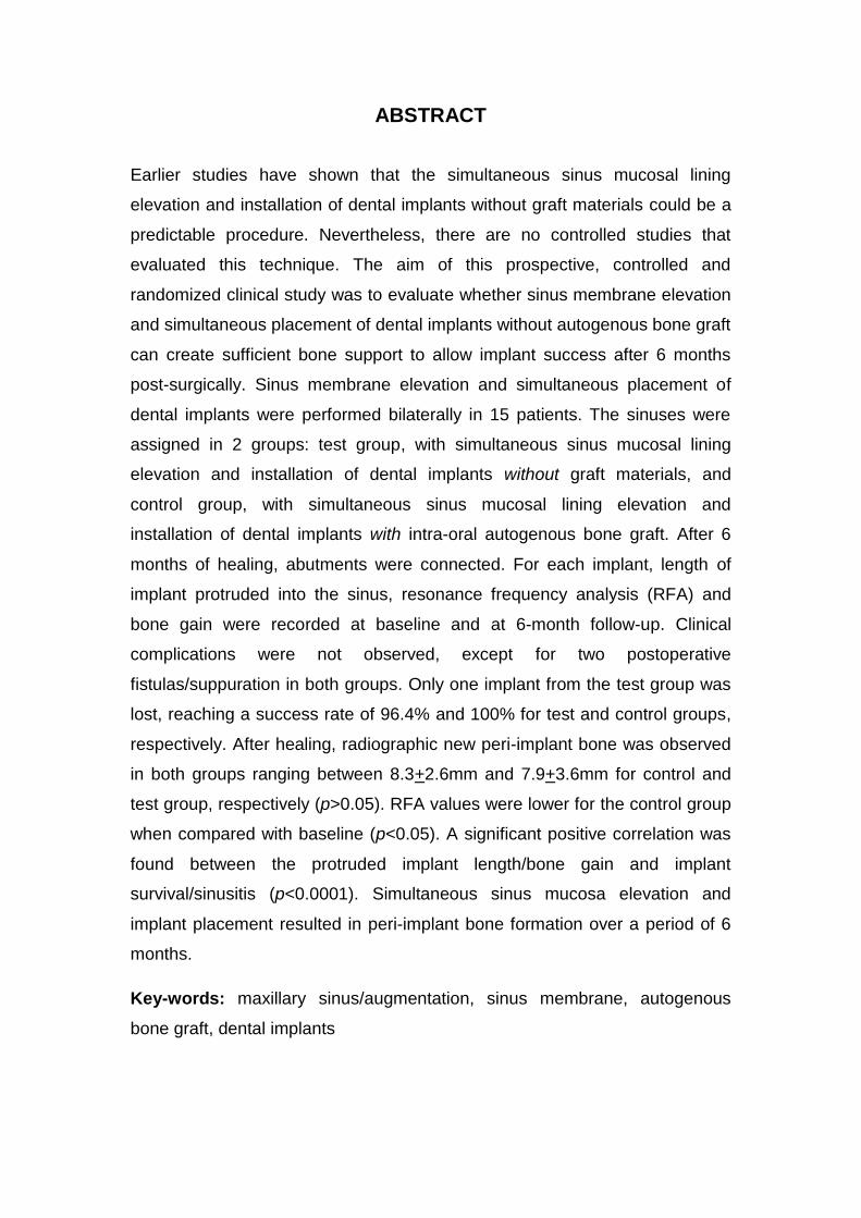
RESUMO
Estudos prévios mostraram que a elevação da mucosa sinusal
concomitantemente à instalação de implantes dentais osseointegráveis sem a
utilização de materiais de enxerto pode ser um procedimento previsível. No
entanto, não existem estudos controlados que avaliaram esta técnica. O
objetivo deste estudo clínico, prospectivo, controlado e randomizado foi
avaliar se a elevação da membrana sinusal e a simultânea inserção de
implantes dentais osseointegráveis sem enxerto de osso autógeno pode criar
suporte ósseo alveolar para permitir o sucesso do implante após um período
de 6 meses. A elevação da membrana sinusal e a inserção de implantes
dentais osseointegráveis foram realizadas bilateralmente em 15 pacientes.
Os seios maxilares foram distribuídos em 2 grupos: grupo teste, com
elevação da mucosa sinusal e inserção simultânea de implantes dentais
osseointegráveis sem a adição de material de enxerto, e grupo controle, com
elevação da mucosa sinusal e inserção simultânea de implantes dentais
osseointegráveis com a adição de material de enxerto autógeno intra-oral.
Decorridos 6 meses do pós-cirúrgico, os pilares de cicatrização foram
instalados. Para cada implante, comprimento do implante inserido no rebordo
remanescente, análise de freqüência de ressonância (AFR) e ganho ósseo
foram obtidos nos tempos 0 e 6 meses após cirurgia. Complicações clínicas
não foram observadas, com exceção de duas fístulas/supurações no período
pós-operatório para ambos os grupos. Apenas um implante do grupo teste foi
perdido, obtendo-se, assim, um índice de sucesso de 96,4% e 100% para os
grupos teste e controle, respectivamente. Após cicatrização, a neoformação
óssea radiográfica peri-implantar foi observada para ambos os grupos,
variando entre 8,3+2,6mm e 7,9+3,6mm para os grupos controle e teste,
respectivamente (p>0,05). Os valores de AFR aos 6 meses foram
significativamente menores para o grupo controle quando comparados ao
tempo 0 (p<0,05). Correlações positivas foram encontradas entre o
comprimento do implante inserido no seio maxilar/ganho ósseo e
sobrevivência do implante/sinusite (p<0,0001). A técnica de implantes

8
inseridos simultaneamente à elevação da mucosa sinusal sem enxertos
resultou em formação óssea peri-implantar após um período de 6 meses.
Palavras–chave: seio maxilar/elevação, membrana do seio maxilar, enxerto
de osso autógeno, implantes dentários
9
ABSTRACT
Earlier studies have shown that the simultaneous sinus mucosal lining
elevation and installation of dental implants without graft materials could be a
predictable procedure. Nevertheless, there are no controlled studies that
evaluated this technique. The aim of this prospective, controlled and
randomized clinical study was to evaluate whether sinus membrane elevation
and simultaneous placement of dental implants without autogenous bone graft
can create sufficient bone support to allow implant success after 6 months
post-surgically. Sinus membrane elevation and simultaneous placement of
dental implants were performed bilaterally in 15 patients. The sinuses were
assigned in 2 groups: test group, with simultaneous sinus mucosal lining
elevation and installation of dental implants without graft materials, and
control group, with simultaneous sinus mucosal lining elevation and
installation of dental implants with intra-oral autogenous bone graft. After 6
months of healing, abutments were connected. For each implant, length of
implant protruded into the sinus, resonance frequency analysis (RFA) and
bone gain were recorded at baseline and at 6-month follow-up. Clinical
complications were not observed, except for two postoperative
fistulas/suppuration in both groups. Only one implant from the test group was
lost, reaching a success rate of 96.4% and 100% for test and control groups,
respectively. After healing, radiographic new peri-implant bone was observed
in both groups ranging between 8.3+2.6mm and 7.9+3.6mm for control and
test group, respectively (p>0.05). RFA values were lower for the control group
when compared with baseline (p<0.05). A significant positive correlation was
found between the protruded implant length/bone gain and implant
survival/sinusitis (p<0.0001). Simultaneous sinus mucosa elevation and
implant placement resulted in peri-implant bone formation over a period of 6
months.
Key-words: maxillary sinus/augmentation, sinus membrane, autogenous
bone graft, dental implants
10
SUMÁRIO
1. INTRODUÇÃO JUSTIFICADA............................................................11
2. PROPOSIÇÃO.....................................................................................16
3. ARTIGO CIENTÍFICO..........................................................................17
4. CONSIDERAÇÕES FINAIS.................................................................43
5. REFERÊNCIAS BIBLIOGRÁFICAS....................................................43
11
1. INTRODUÇÃO JUSTIFICADA
Após a comprovação científica da osseointegração no final da década
de 70, definida como “uma conexão direta e estrutural entre osso vivo
ordenado e a superfície de um implante submetido a carga funcional”,
grandes avanços foram realizados para que cirurgias de implantes dentários
se tornassem uma alternativa viável na substituição dos elementos dentários
ausentes.
Modificações no desenho estrutural dos implantes e o tratamento
mecânico e químico de sua superfície possibilitaram um aumento substancial
dos índices de sucesso quando utilizados no tratamento reabilitador oral, fato
este amplamente documentado na literatura (ROOS-JÄNSAKER et al.,
2006). Tal sucesso sustenta-se em duas condições fundamentais: a
existência de um volume ósseo que possibilite a instalação dos implantes em
sua posição protética ideal e de um número de implantes em tamanhos
variados que faça frente às forças mastigatórias (MISCH, 2000).
Porém, usualmente o que se encontra são reabsorções ósseas que se
caracterizam pela atrofia do tecido ósseo em altura e espessura, podendo
variar entre as diferentes regiões da cavidade oral com padrões de
reabsorção distintos. A causa mais frequente e responsável por grandes
atrofias relaciona-se àquela que ocorre após a perda do elemento dental por
ausência de estímulo ao tecido ósseo (CAWOOD & HOWELL, 1988). As
reabsorções causadas pela doença periodontal também merecem ser
mencionadas, já que podem ocasionar, em seus estágios mais avançados,
uma grande redução do volume ósseo, o que impossibilita muitas vezes o
uso de implantes osseointegrados sem que manobras de enxertia sejam
executadas previamente (CAWOOD & HOWELL, 1991).
Também é importante considerar as características do osso nesta
região, geralmente de cortical fina e trabeculado pouco denso, o que contribui
para uma menor taxa de sucesso na reabilitação por meio de implantes. A
região posterior da maxila é a que possui a menor densidade entre todas as
regiões dos maxilares, e essa tende a diminuir com o avanço da idade do
12
indivíduo, o que torna a região comumente acometida por importantes
reabsorções. A este fato, soma-se a pressão positiva exercida nos seios
maxilares, que ocasiona o avanço de seus limites com conseqüente
diminuição do volume ósseo nesta região. Este processo é denominado de
pneumatização dos seios maxilares (CHAVANAZ, 1990).
Neste contexto, torna-se bastante desafiador a reabilitação de maxilares
atrofiados em sua região posterior. Tais ocorrências reforçam a necessidade
de realizar cirurgias reparadoras que aumentem o volume ósseo da região e
que permitam, posteriormente, a instalação de implantes osseointegrados
adequados.
Inicialmente, na década de 80, os procedimentos de reconstrução
óssea previamente à instalação de implantes osseointegráveis foram
descritos com a utilização de enxertos autógenos, ou seja, um fragmento de
tecido ósseo do próprio paciente é retirado de uma região denominada área
doadora, e colocado em uma região receptora onde há a necessidade de
reconstrução (BOYNE & JAMES, 1980). Nesse procedimento, um retalho
mucoperiostal e uma abertura em formato de janela na parede lateral do seio
maxilar permitiam o acesso para o descolamento da membrana de
Schneider, criando-se um espaço para a aplicação do enxerto de origem
autógena. Esses procedimentos apresentaram altos índices de sucesso, em
torno de 90%, e são utilizados até hoje com frequência, com altos graus de
previsibilidade (WALLACE, 2006; NKENKE & STELZLE, 2009).
Até a presente data, o uso do enxerto autógeno é considerado como o
gold standard por conter características osteogênicas, osteocondutoras e
osteoindutoras (HALLMAN et al., 2001). Vários estudos surgiram com a
proposta de tornar esse procedimento menos invasivo, quer pela diminuição
e facilidade do acesso cirúrgico, como na técnica proposta por SUMMERS,
1994, quer pela substituição parcial ou total de osso autógeno. Sua utilização
representa um segundo leito cirúrgico, ocasionando um aumento do tempo e
risco cirúrgico, além dos desconfortos pós-operatórios inerentes a esses
procedimentos. Esses desconfortos estão proporcionalmente relacionados à
quantidade de reconstrução necessária para a reabilitação do caso clínico, o
que define a fonte doadora de enxerto, que pode ser intra-oral, cujas áreas
mais utilizadas são sínfise mentoniana e o ramo mandibular (CLAVERO &
13
LUNDGREN, 2003). Nas grandes reconstruções do tecido ósseo utilizam-se
fontes doadoras extra-orais, sendo que as mais relatadas pela literatura são a
crista ilíaca, por acesso anterior ou posterior, a tíbia e a calota craniana
(CHIAPASCO et al., 2009).
A necessidade de usar fontes extra-orais implica em realizar o
procedimento em ambiente hospitalar, com equipe médica auxiliar,
procedimentos realizados sob anestesia geral e médicos ortopedistas ou
neurologistas para coleta dos enxertos, o que aumenta consideravelmente as
custas e a morbidade do tratamento. Por isso, pesquisadores vêm buscando
alguma técnica ou algum material que possa substituir os enxertos
autógenos, sem que haja comprometimento dos resultados (MANGANO et
al., 2009).
O material de enxertia parece ser de fundamental importância para o
prognóstico de enxertos. Estudos mostram que diversos biomateriais como
osso liofilizado humano, liofilizado bovino, sulfato de cálcio e as
hidroxiapatitas possuem limitações específicas em diferentes graus, o que
gera incertezas quando ao prognóstico desses enxertos, e que apenas o
osso autógeno possui propriedades verdadeiramente osteogênicas, com
menor tempo de cicatrização (DEGIDI et al.2006, BÖECK-NETO et al., 2009).
As proteínas morforgenéticas (ou bone morphogenetic protein - BMP),
por suas propriedades exclusivas de osseoindução dentre todos os
biomateriais, vêm merecendo atenção especial (MANGANO et al., 2009;
NKENKE & STELZLE, 2009). Entretanto, seu alto custo ainda inviabiliza sua
aplicação.
Já a engenharia tecidual, através do cultivo de células em laboratório,
também poderá, em um futuro próximo, ser uma alternativa aos enxertos
autógenos (MANGANO et al., 2009)
Com os estudos publicados sobre regeneração tecidual guiada (RTG),
verificou-se a possibilidade de se utilizar essa técnica também na região dos
seios maxilares. Assim, BRUSCHI et at., 1998, demonstraram a possibilidade
de formação de osso ao redor de implantes inseridos dentro do seio maxilar
sem qualquer material de enxerto.
14
Em um achado de formação óssea espontânea, LUNDGREN et al.,
2003, relataram a formação óssea ocorrida após a remoção de cisto dentro
da cavidade do seio maxilar. No ano seguinte, LUNDGREN et al., 2004,
publicaram estudo mostrando a possibilidade dessa técnica em um estudo
que utilizou 19 implantes inseridos em 12 seios maxilares. Após o
levantamento da mucosa do seio maxilar, os implantes foram inseridos e a
janela óssea, que fora removida para se obter o acesso à cavidade sinusal,
foi recolocada em sua posição original. Os autores discutiram a neoformação
óssea ao redor dos implantes segundo o processo de regeneração tecidual
guiada na qual a presença de coágulo sanguíneo alojado em um
compartimento ósseo auxiliado pela manutenção mecânica da membrana
sinusal pelos implantes, formando uma “tenda”, resultou em formação de
tecido ósseo peri-implantar. Os autores comentaram, ainda, que a reposição
da parede óssea removida para o acesso à cavidade sinusal funcionava
como uma barreira rígida para evitar o crescimento de tecido mole para
dentro da cavidade sinusal.
Em um estudo utilizando macacos, PALMA et al, 2006, avaliaram
histologicamente a formação óssea ao redor de implantes inseridos na
cavidade sinusal enxertada com osso autógeno e apenas coágulo sanguíneo.
Nesse estudo, os autores compararam a formação óssea ao redor de
implantes, sendo que essa neoformação foi significantemente maior nos
implantes de superfície anodizada (p<0.05), embora a neoformação
ocorresse também ao redor de implantes de superfície lisa.
Posteriormente, THOR et al, 2007, observaram um ganho médio de
6,51 mm ao redor de implantes inseridos no seio maxilar sem qualquer
material de enxerto e somente com a presença de coágulo sanguíneo. Nesse
estudo, houve 41% de perfuração da membrana de Schneider, com apenas
um (1) implante perdido dentre os 44 instalados. Os autores mostraram,
ainda, que a porção apical do implante apresentava ausência de
neoformação óssea, provavelmente devido à movimentação pneumática da
cavidade sinusal, que empurrava a mucosa sinusal elevada contra a porção
apical dos implantes.
Embora a técnica de utilização de implantes inseridos
concomitantemente à elevação da mucosa sinusal com preenchimento com

15
coágulo sanguíneo apresente ótimos resultados, ainda não há, na literatura,
estudos controlados e randomizados que avaliem, de maneira sistemática,
esta técnica clínica de elevação de seio maxilar.

16
2. PROPOSIÇÃO
O objetivo deste estudo é avaliar, clínica e radiograficamente, a
neoformação óssea ao redor de implantes osseointegrados inseridos em
seios maxilares sem a inserção de enxerto ósseo.
17
3. Artigo Científico
Simultaneous sinus membrane elevation and dental implant placement
without autogenous bone graft: a 6-month follow-up study
(artigo preparado segundo as normas do Clinical Implant Dentistry and
Related Research)
Fabio L. Borges, Rafael O. Dias*, Tatiana Onuma*, Luciana Ap. Gouveia
Cardoso*, Munir Salomão†, Eduardo Ayub‡, Jamil Awad Shibli§.
Correspondence to:
Prof. Jamil Awad Shibli
Centro de Pós-Graduação e Pesquisa – CEPE, Universidade Guarulhos
Praça Tereza Cristina, 229 – Centro
07011-040 Guarulhos, SP - Brazil
e-mail: [email protected]
FAX: +55 11 24641758
Running title: Sinus mucosal lining elevation without bone graft
Graduate Student, Department of Periodontology, Dental Research Division,
Guarulhos University, Guarullhos, SP, Brazil.
† Private Practice, São Paulo, SP, Brazil
‡ Private Practice, Campo Grande, MS, Brazil
§ Assistant Professor, Department of Periodontology and Head of Oral
Implantology Clinic, Guarulhos University, Guarulhos, SP, Brazil.
18
ABSTRACT
Background: Earlier studies have shown that the simultaneous sinus
mucosal lining elevation and installation of dental implants without graft
materials could be a predictable procedure. Nevertheless, there are no
prospective, controlled and randomized studies that evaluated this technique.
Purpose: The aim of this prospective, controlled and randomized clinical
study was to evaluate whether sinus membrane elevation and simultaneous
placement of dental implants without autogenous bone graft can create
sufficient bone support to allow implant success after 6 months post-
surgically.
Material and Methods: Sinus membrane elevation and simultaneous
placement of dental implants were performed bilaterally in 15 patients in a
split-mouth design. The sinuses were assigned in 2 groups: test group, with
simultaneous sinus mucosal lining elevation and installation of dental implants
without graft materials, and control group, with simultaneous sinus mucosal
lining elevation and installation of dental implants with intra-oral autogenous
bone graft. After 6 months of healing, abutments were connected. For each
implant, length of implant protruded into the sinus, resonance frequency
analysis (RFA) and bone gain were recorded at baseline and 6 months follow-
up.
Results: Clinical complications were not observed, except for two
postoperative fistulas/suppuration in both groups. Only one implant of test
19
group was lost, reaching a success rate of 96.4% and 100% for test and
control groups, respectively. After healing, radiographic new peri-implant bone
was observed in both groups ranging between 8.3+2.6mm and 7.9+3.6mm for
control and test group, respectively (p>0.05). RFA values were lower for the
control group when compared with baseline (p<0.05). A significant positive
correlation was found between the protruded implant length/bone gain and
implant survival/sinusitis (p<0.0001). The technique applied (placing implants
simultaneously to sinus membrane elevation without graft material) resulted in
bone formation over a period of 6 months.
Conclusions: Implants placed simultaneously to sinus membrane elevation
without graft material resulted in bone formation over a period of 6 months.
Key-words: maxillary sinus/augmentation, sinus membrane, autogenous
bone graft, dental implants.

20
INTRODUCTION
Dental implant therapy has become an excellent and safe treatment
modality for a conservative and esthetic alternative to solve partial and total
edentulism. When the patient presents deficient alveolar ridges, however, this
deficiency could jeopardize the placement of dental implants, mainly in the
posterior maxilla, due to loss of alveolar bone and increased maxillary sinus
pneumatization.
The maxillary sinus grafting procedure has been used for occlusal
rehabilitation with prosthetic appliances placed over dental implants in the
posterior maxilla. A plethora of researchers (for review see1,2) have evaluated
different bone grafting materials inserted in the maxillary sinus cavity.
However, recent studies3-9 have shown that the simple elevation of the
Schneiderian membrane can induce bone formation at the maxillary sinus.
This technique was based on the concept that the lifting of the sinus
membrane and the establishment of a compartment with a blood clot could
result in new bone around the inserted implants in similar way that bone graft
materials maintain the augmented space and promote osteogenesis.4
Maxillary sinus augmentation as well as bone regenerative procedures share
similarities and both are coordinated processes involving various biological
factors.10 Blood supply and angiogenesis play an important role in guided
bone formation.11,1Indeed, the blood clot contain many growth factors, such
as fibroblast growth factor (FGF), transforming growth factor (TGF), bone
morphogenetic proteins (BMP), insulin-like growth factor (IGF), platelet-
derived growth factor (PDGF), and vascular endothelial growth factor (VEGF),
21
that are expressed during skeletal development and induced in response to
injury. These factors are believed to regulate the repair of bone tissue.13,14
Some of these molecules are also involved in angiogenesis (i.e. FGF, TFG,
VEGF).14 Complementary, it was shown that cells derived from explants of
Schneiderian membrane can express markers of osteoprogenitor cells.15
In addition, the contact of the whole blood with the titanium surface
generates thrombin.16 Thrombin that is generated by coagulation cascade not
only cleaves fibrinogen but also contributes to activation of osteoblasts via
proteinase-activated receptors, which, with the platelets may have several
effects on bone growth.
Together, these observations show that the simultaneous elevation of
sinus membrane and implant placement could be a feasible clinical
procedure. However, to date, there are not controlled human studies that
evaluated this technique. Therefore, the aim of this prospective, controlled
randomized study was to evaluate the simultaneous sinus membrane
elevation and implant placement without autogenous bone grafts after a 6-
month follow-up.
MATERIAL AND METHODS
Patient Population
Seventeen subjects (11 females and 6 males, mean age 57.9 years)
presenting bilateral edentulous area in posterior maxilla were enrolled in this
study. The sinuses, in a split mouth design, were assigned in 2 groups: a
control group consisting of n = 17 sinuses that received simultaneous sinus
22
membrane elevation, autogenous bone graft and implant placement, and a
test group consisting of n = 17 sinuses that received simultaneous sinus
membrane elevation and implant placement without graft material. Tossing a
coin was used to determine which sinus was assigned as control or test sinus
side.
Calculation of the sample size was based on a series of previous
studies.7,17 A difference of 20% or 1mm in bone reformation (height of new
bone formed around implants placed into maxillary sinus), with a common
standard deviation of 3mm between sinus lifting approaches was set, as the
present split mouth study design (with or without graft materials in) is not
available in the literature. With an of 0.05 and 1- of 0.80, a sample of at
least 14 subjects was considered desirable.
The study protocol was explained to each subject and a signed
informed consent was obtained. The Institutional Clinical Research Ethics
Committee of Guarulhos University approved this study protocol (# 152/09).
Exclusion Criteria
Subjects were excluded if they were smokers and if they had residual
sinus floor of less than 3mm height, maxillary sinus pathology, a chronic
medical disease or condition that would contraindicate dental surgery (e.g.,
diabetes, uncontrolled hypertension, history of head and neck radiation),
moderate to severe chronic periodontitis in the remaining teeth (i.e.,
suppuration, bleeding on probing in more than 30% of the subgingival sites or
any site with probing depth > 5mm), absence of primary stability of the
23
inserted implant in the residual bone and large sinus membrane perforation
( > 3mm) during mucosal sinus elevation procedure.
Sinus membrane elevation
All subjects received oral prophylaxis treatment before surgery.
Panoramic radiographs and dental computer tomography scans - CT (I-Cat,
KaVo Dental GmbH, Biberach, Germany) were taken of all patients. The
residual alveolar bone height was measured before surgery. All patients
received antibiotics (amoxicilin 875mg and sulbactan 125mg) and steroidal
anti-inflammatory (dexamethasone 8mg) prior to the surgery. The bilateral
maxillary sinus augmentation was performed under local anesthesia on the
same day. According to the CT of the patient and anatomical landmarks, a
horizontal crestal incision and two vertical incisions extending beyond the
mucogingival junction were performed. A full-thickness flap was reflected in
order to expose the maxillary sinus lateral bone wall. Under constant irrigation
with sterile saline solution, an osseous window of approximately 15mm x
10mm was demarked, using a round diamond coated bur. The bone in the
center of the window was left attached to the sinus membrane. The
Schneiderian membrane was carefully dissected and elevated using specially
designed elevators, and the bony wall was gently pushed inside the sinus
cavity forming the roof to the secluded compartment. The sinus membrane
was released without any tension to provide an adequate compartment for the
autogenous bone graft (control side) or blood clot (test side). Two trained
surgeons (FLB and JAS) performed all surgeries.
24
Autogenous bone graft
Autogenous bone grafts from the symphysis area or the mandibular
ramus, depending on the volume of maxillary sinus and availability of donor
area, were obtained via an intraoral incision. A modified 8.0mm length and
6mm diameter trephine bur (INP, São Paulo, SP, Brazil), under constant
sterile saline irrigation, were used to harvest the donor site and provided a
milled bone. The bone grafts were stored in saline solution until they were
placed inside the sinus of the control group.
Implant placement
Screw-shaped implants with sandblasted acid-etched surface, 4.00 mm
diameter and 15 to 18mm length (Conus, INP, São Paulo, SP, Brazil) were
used in this study. Implants sites were marked using a surgical template. The
templates were based on the diagnostic waxing with perforations on the
longitudinal axis, on the premolar and molar regions, according to ideal
position of final implant supported restorations.
Initial implant stability was optimized by using an under-preparation
technique: drilling through the residual bone using a 2.0mm twist drill followed
by a 2.8mm and 3mm drill was performed, just enough to enable the initial
insertion of implant in the surgical site.
The autogenous bone, in the control group, was placed at the superior
aspect of the sinus against the medial aspect of the grafted compartment
created in the sinus cavity. The graft was condensed at each stage. The
dental implants were placed to half of their total length. Then, following the
condensation of the graft, the dental implants were seated in their final

25
positions, to avoid empty spaces in the sinus cavity. Any remaining graft
material was placed over the exposed implant surfaces.
Once the coagulum was observed underneath the elevated sinus
mucosa of the test group (without autogenous bone graft), the implants were
finally placed, as shown in figure 1.
Figure 1: Clinical view of a) lateral bone wall pushed into the sinus cavity;
b) simultaneous sinus membrane elevation and implant placement without
autogenous bone graft; c) simultaneous sinus membrane elevation and
implant placement; d) autogenous bone graft inserted over the implants.
Following the implant placement, a polypropylene membrane (INP, São
Paulo, SP, Brazil) was applied to cover the lateral wall osteotomy of the
sinuses of control and test groups. The membrane was adjusted to extend
26
circumferentially 5 to 8mm over the adjacent alveolar bone, avoiding
ingrowths of the soft connective tissue. To allow flap apposition and closure
after placement, incisions were made buccally and palatally after membrane
placement. Primary wound closure was achieved with horizontal mattress
sutures alternated with interrupted sutures to ensure a submerged healing
procedure in dental implants.
Postoperative care
Postoperative care consisted of a 0.12% chlorhexidine mouthrinse
twice a day for 14 days without mechanical cleaning at the surgical areas.
Anti-inflammatory medication (dexamethasone, 4 mg) was administered once
a day and appropriate analgesia (paracetamol, 750 mg) for 3 days following
surgery in order to reduce postoperative swelling and pain. A postoperative
antibiotic regimen with amoxicilin and sulbactan was prescribed during 7
days. Nylon sutures were removed 14 days after surgery. The existing upper
removable prosthesis was adapted with soft tissue conditioner and was worn
after a healing period of 4 weeks. Occlusal adjustments and soft tissue
conditioner were performed when necessary. Professional plaque control
supplemented this healing phase every month, during 6 months.
Postsurgically events as membrane exposure, sinusitis and
paresthesia were recorded in each recall visit.
Implant stability and CT measurements
Immediately after the implant placement (baseline) and at second
stage surgery (6 months after maxillary sinus augmentation), the resonance
27
frequency analysis-RFA (Osstell, Integration Diagnostics, Savadaled,
Sweden) was used to measure the primary stability of the implant. The
transducer (smartpeg type 1) was hand-screwed into the implant body. For
every series of RFA measurements, the ISQ values (unit of RFA) were
recorded. An ISQ value between 1 and 100 was given where 1 is the lowest
and 100 the highest. A mean of ISQ value was calculated for each implant
based on one measurement of each implant, and then of each group. The
RFA was measured at baseline and 6 months after therapy.
Three computed tomography (CT) datasets were acquired for every
patient, at baseline, 14 days and 6 months after maxillary sinus augmentation
procedures. The CT data were transferred in the DICOM format to specific
implant navigation software (I-Cat Vision, Kavo Dental). This format allows a 3D
reconstruction of the maxilla. Moreover, this software enables, through
segmentation tools, to measure bone crest height along transversal sections,
corresponding to the longitudinal axis of the implant, before and after maxillary
augmentation.
A single trained examiner performed all measurements in order to
evaluate the changes in height of maxillary sinus floor for each implant. The
sinuses were evaluated in order to assess the radiographic parameters:
1) average of residual sinus floor measured in the initial CT (A1+A2/2); 2) the
height of endosinus bone gain, defined as the mean height of new bone
(B1+B2/2); 3) the linear distance of the buccal and palatal sinus wall (C1+C2/2);
4) the length of the implant protruded into the sinus after surgery (D1+D2/2).
These measurements were taken and then averaged per implant, and then per
group, as shown in figure 2.

28
Figure 2: Schematic drawing of an implant inserted into the sinus cavity. To
evaluate the changes in height of maxillary sinus floor for each implant, the
sinuses were evaluated in order to assess the radiographic parameters:
1) average of residual sinus floor measured in the initial CT (AM=A1+A2/2);
2) the mean height of endosinus bone gain, defined as the mean height of
new bone (BM=B1+B2/2); 3) the average of linear distance of the buccal and
palatal sinus wall (CM=C1+C2/2); 4) the mean length of the implant protruded
into the sinus after surgery (DM=D1+D2/2)
Statistical analysis
The mean and standard deviation of the value of RFA and radiographic
data were calculated for each implant and then for each group. Mann-Whitney
U test was used to calculate the differences between groups for the
radiographic and RFA variables. Wilcoxon’s rank test was used to evaluate
the intra-groups differences between RFA values at baseline and 6 months
post-therapy. The 2 test was used to calculate the dichotomously variables,
i.e., presence or absence of suppuration, membrane exposure, lateral window
closure and implant survival. Spearman correlation was used to evaluate the
29
possible correlations among the clinical and radiographic variables. The unit
of analysis was the patient and the level of significance was 0.05.
RESULTS
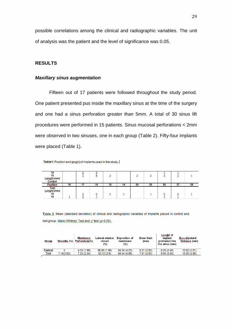
Maxillary sinus augmentation
Fifteen out of 17 patients were followed throughout the study period.
One patient presented pus inside the maxillary sinus at the time of the surgery
and one had a sinus perforation greater than 5mm. A total of 30 sinus lift
procedures were performed in 15 patients. Sinus mucosal perforations < 2mm
were observed in two sinuses, one in each group (Table 2). Fifty-four implants
were placed (Table 1).
30
Postoperative control
Two postoperative wound infections, one in each group, occurred 3-4
weeks after the maxillary sinus augmentation. Both exhibited suppuration, and
they were solved with membrane removal and irrigation with 0.12%
chloredixine. Additional surgery was not needed. Membrane exposure was
present in more than 50% of the cases and they were removed without
surgical intervention (Table 2). These exposures happened after a period of 2-
4 months after surgery.
In addition, no patient presented any paresthesia or altered sensation
in the donor area. Oral function was not affected in all treated patients.
Re-entry surgery and implant survival
At abutment surgery, the remaining membranes were removed and
visual evaluation of the lateral window of the maxillary osteotomy was
performed. Four sinuses presented an incomplete closing of the lateral
window: one in the control group and three in the test group (Table 2).
One implant in the test group was removed due to a lack of
osseointegration. This loss was observed in the patient that presented
sinusitis. The 56 remaining implants, in both groups, were clinically stable.
The implant survival was 96.4% and 100% to test and control, respectively.

31
CT evaluation
Table 2 presents the radiographic variables. No difference was found
between groups. The CT images showed that implant protruding, on average,
8 mm into the sinus (p>0.05). In all patients, radiographic evidence of new
bone formation in the elevated sinus area was seen. Both sides of implants, in
a varied range, were covered with new bone, independently of the evaluated
group (Figures 3 and 4). The new bone formation was 8.3+2.6 mm and
7.9+3.6mm in the control and test groups, respectively (p>0.05). In some
cases, mainly in the test group, the new bone tissue was not seen at the
apical implant area. The distance between the buccal and palatal bone wall
(DM, Figure 2) was also similar in both groups (p>0.05).
Positive correlations were detected to length of implant protruded into
the sinus and bone gain (p<0.0001; r2=0.635) and lateral window closure and
bone gain (p<0.05, r2=0.551) for both groups. In addition, sinusitis was
correlated with implant survival (p<0.0001, r2=0.704)
32
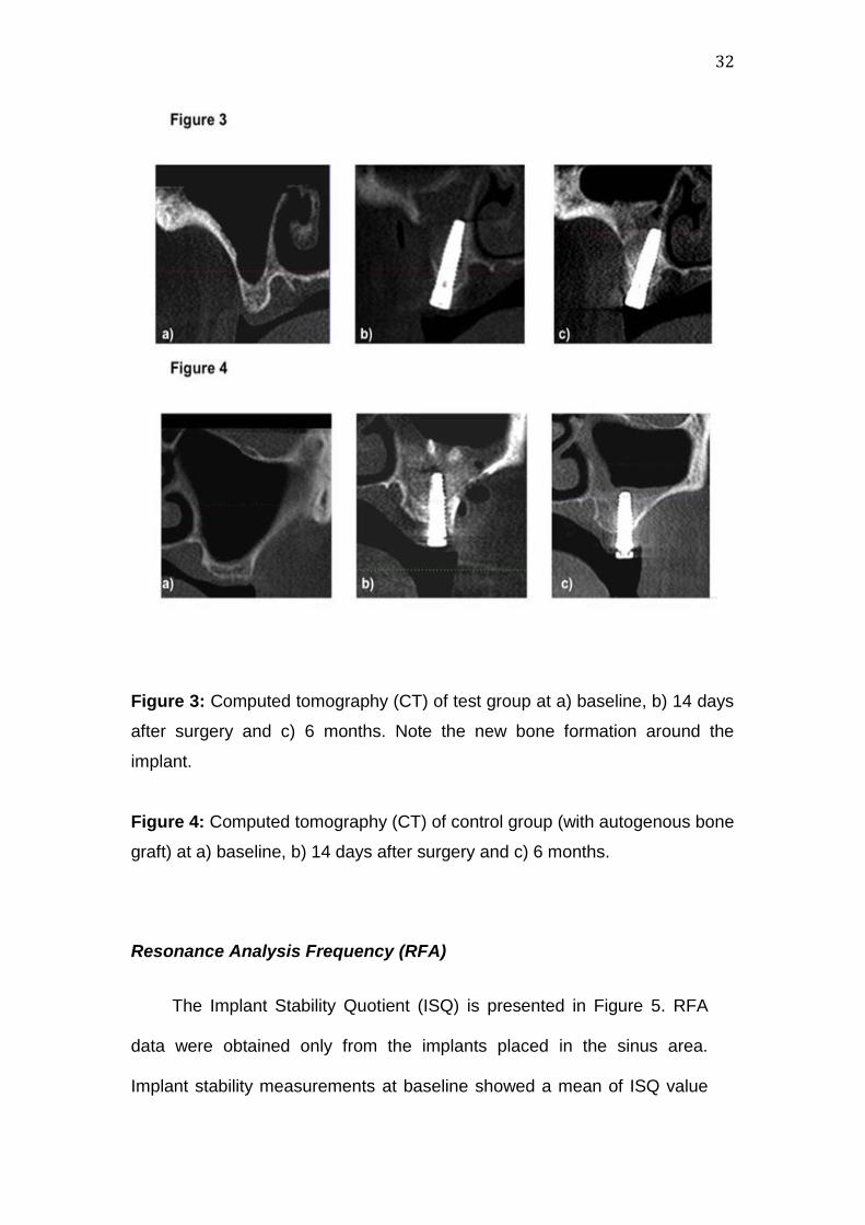
Figure 3: Computed tomography (CT) of test group at a) baseline, b) 14 days
after surgery and c) 6 months. Note the new bone formation around the
implant.
Figure 4: Computed tomography (CT) of control group (with autogenous bone
graft) at a) baseline, b) 14 days after surgery and c) 6 months.
Resonance Analysis Frequency (RFA)
The Implant Stability Quotient (ISQ) is presented in Figure 5. RFA
data were obtained only from the implants placed in the sinus area.
Implant stability measurements at baseline showed a mean of ISQ value
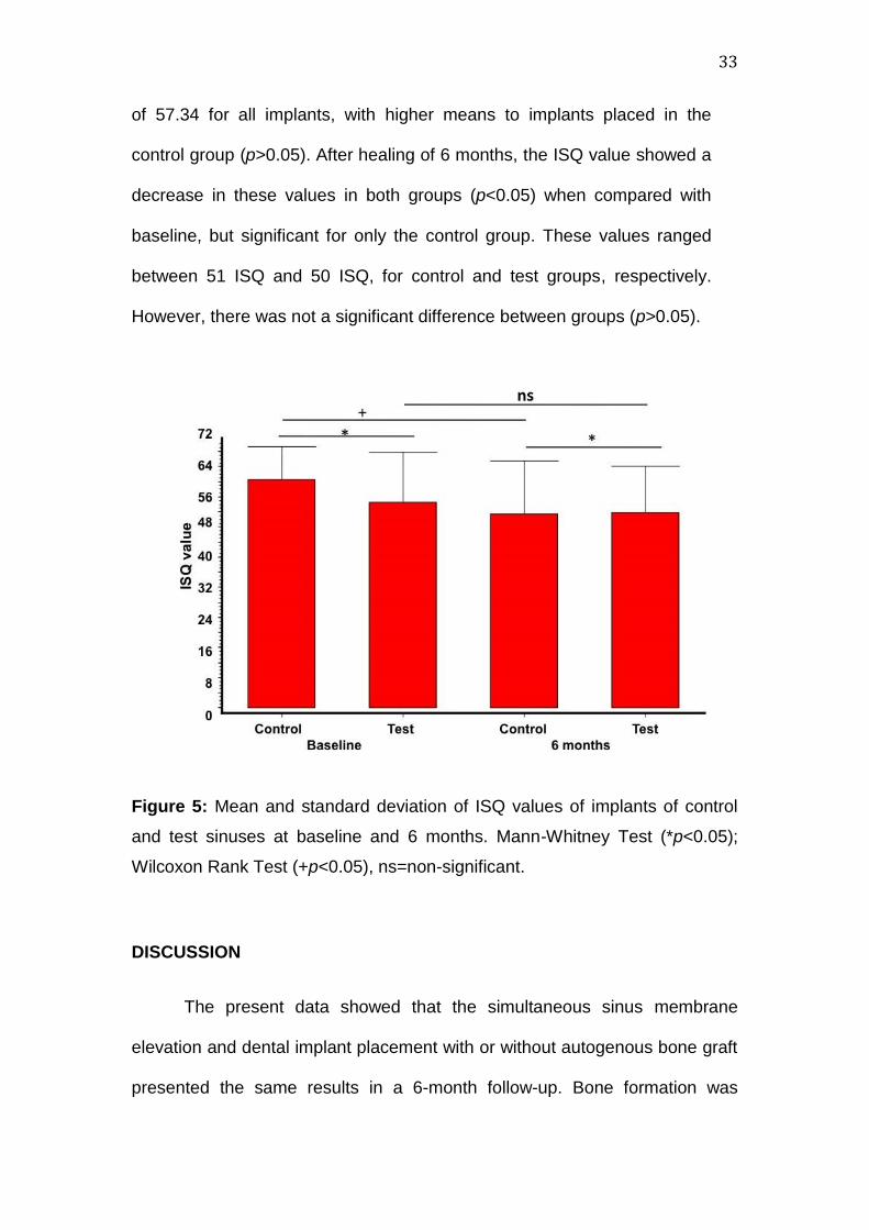
33
of 57.34 for all implants, with higher means to implants placed in the
control group (p>0.05). After healing of 6 months, the ISQ value showed a
decrease in these values in both groups (p<0.05) when compared with
baseline, but significant for only the control group. These values ranged
between 51 ISQ and 50 ISQ, for control and test groups, respectively.
However, there was not a significant difference between groups (p>0.05).
Figure 5: Mean and standard deviation of ISQ values of implants of control
and test sinuses at baseline and 6 months. Mann-Whitney Test (*p<0.05);
Wilcoxon Rank Test (+p<0.05), ns=non-significant.
DISCUSSION
The present data showed that the simultaneous sinus membrane
elevation and dental implant placement with or without autogenous bone graft
presented the same results in a 6-month follow-up. Bone formation was
34
evident in all patients, except in that patient that presented an acute post-
surgery sinusitis. This patient also lost one implant during the initial healing
period in the test group. These results agree with previous studies in humans
4,6,7 and animals8,9, who also obtained, in a varied range, new bone formation
in the maxillary sinus augmentation without bone grafts.
Although these results are based on recent studies,4,7 the idea of
placing implants inside the maxillary sinus without bone grafts is not new.
Previous studies, developed in the early 80’s by BRÄNEMARK et al.18 and
BOYNE & JAMES19, reported bone formation at apical portion of dental
implants placed in maxillary sinus after carefully raising of the sinus
membrane.
Thereafter, BRUSCHI et al.20 and SUMMERS,21 also showed that the
careful lining of the sinus membrane allowed new bone formation around the
implant placed in maxillary sinus cavity, through remaining alveolar bone crest
approach. However, these techniques have bone formation limited to 4 to 5
mm.
The simultaneous Schneiderian membrane elevation and implant
placement performed in our study showed better results, compared with the
aforementioned studies.18,20,21 An extensive bone formation around implants
was observed, almost covering all the apical portion of the implant. The bone
gain ranged between 8.3mm and 7.9mm for control and test groups,
respectively. Previous studies17,22 that evaluated different graft materials in
maxillary sinus augmentation and simultaneous implant placement reached
similar results.
35
It must be pointed out that maxillary sinus pneumatization could be
caused by positive intra-sinus air pressure due to respiration and this
pressure might promote resorption and new pneumatization after maxillary
sinus augmentation.17,23 However, in the present study, both sinus groups do
not present resorption, at least after a 6-month follow-up. This finding may be
supported, in part, by two alterations made in the technique advocated by
LUNDGREN et al.5 Firstly, the present study pushed the lateral bone window
inside the sinus cavity, using this thin bone as “roof” of the secluded cavity.
This bone wall was mechanically supported by the dental implants as a space
maker for guided bone regeneration. Secondly, utilizing membrane that
avoided the soft tissue ingrowths in the sinus cavity allowed not only a better
blood clot organization but also stabilization for closure of the lateral window,
as shown in previous studies.22,23 These alterations could, together, establish
proper pneumatic conditions, different from the earlier data,7 where the apical
portion of some implants were not covered by new bone.
In addition, this technique do not use bone grafts inside the sinus
cavity. Autogenous bone is the gold standard, but its use is limited by donor-
site morbidity, sparse availability, and uncontrolled resorption.24,25,26 Another
advantage of this sinus lifting technique was the use of sandblasted acid
etched implants with 15 and 18 mm length. Previous studies have shown the
importance of implant surface topography at micrometer scale on trombogenic
activity16 as well as the length of implants in success rate.27 This trombogenic
activation results in the recruitment, migration and differentiation of progenitor
osteogenic cells. These cells are provided by the Schneiderian membrane
and exposed bone in the sinus cavity. The VEGF is probably the most
36
important player in the vascular formation during angiogenesis.28 VEGF is an
endothelial-specific growth factor that promotes angiogenesis by stimulating
endothelial cell differentiation, proliferation, and migration,29 and plays an
important role in bone remodeling by attracting endothelial cells and
osteoclasts, and by stimulating osteoblast differentiation.30 The involvement
of VEGF in bone formation is also suggested by its interaction with humoral
factors that regulate bone homeostasis31 and by its role, not only in bone
angiogenesis but also in different aspects of bone development, including
chondrocyte differentiation, and osteoblast and osteoclast recruitment.29
Moreover, osteoblasts and osteoblast-like cells have been shown to be able
to produce VEGF.30 Bone formation is closely linked to blood vessel invasion
and therefore, the angiogenesis plays a pivotal role in all regenerative
processes.13,14,28-30 VEGF may act indirectly or directly to increase
recruitment of mesenchymal stem cells through an enhancement of vascular
permeability, which may facilitate migration of host mesenchymal stem cells to
the bone regeneration site.13 VEGF activity is essential for normal
angiogenesis and appropriate callus formation and mineralization in response
to bone injury.
Complementary, it is reported that RFA can provide objective
evaluation of implant stability, possibly demonstrating evidence for extending
of implant osseointegration.32 Therefore, the present data demonstrated that
the use of ISQ values ranged between 54.2 and 60.6 to implants placed in
test and control sinus, values very similar to HALLMAN et al.33 that found a
ISQ value of 66.2 (range from 53 to 76) in implants placed in grafted sinus.
However, it could be speculated that the difference between ISQ values of
37
control and test sides at baseline (p<0.05) was due to the presence of
autogenous bone graft in the control sinuses. As the bone graft must be
added before the dental implant placement to allow a proper graft
condensation, this fact might have influenced the results, instead, after a 6-
month healing, there was no difference between groups. In addition, the lower
means of ISQ values after the 6-month period could be related with the
initiation of the new bone formation.34 The present study also demonstrated a
high survival rate for simultaneous implant placement in both groups. The
success rate ranged between 96.4 and 100%, similar to previous
reports.1,2,24,28
In conclusion, simultaneous sinus membrane elevation and implant
placement, with or without bone graft reach a comparably bone gain and
implant survival at 6 months follow-up. However, more long-term clinical data
are needed to draw a more definitive conclusion.
ACKNOWLEDGMENTS
The authors are indebted to INP Implants, São Paulo, Brazil, for
providing the dental implants and membranes. The authors also declare they
have no financial interest in any of the materials related in this study.
38
REFERENCES
1. Nkenke E, Stelzle F. Clinical outcomes of sinus floor augmentation for
implant placement using autogenous bone or bone substitutes: a
systematic review. Clin Oral Implants Res. 2009;20 Suppl 4:124-33
2. Del Fabbro M, Rosano G, Taschieri S. Implant survival rates after
maxillary sinus augmentation. Eur J Oral Sci. 2008;116:497-506.
3. Lundgren S, Andersson S, Sennerby L. Spontaneous bone formation in
the maxillary sinus after removal of a cyst: coincidence or
consequence? Clin Implant Dent Relat Res. 2003;5:78-81.
4. Lundgren S, Cricchio G, Palma VC, Salata LA, Sennerby L. Sinus
membrane elevation and simultaneous insertion of dental implants: a
new surgical technique in maxillary sinus floor augmentation.
Periodontol 2000. 2008;47:193-205.
5. Lundgren S, Andersson S, Gualini F, Sennerby L. Bone reformation
with sinus membrane elevation: a new surgical technique for maxillary
sinus floor augmentation. Clin Implant Dent Relat Res. 2004;6:165-73.
6. Hatano N, Sennerby L, Lundgren S. Maxillary sinus augmentation
using sinus membrane elevation and peripheral venous blood for
implant-supported rehabilitation of the atrophic posterior maxilla: case
series. Clin Implant Dent Relat Res. 2007;9:150-5.
7. Thor A, Sennerby L, Hirsch JM, Rasmusson L. Bone formation at the
maxillary sinus floor following simultaneous elevation of the mucosal
lining and implant installation without graft material: an evaluation of
20 patients treated with 44 Astra Tech implants. J Oral Maxillofac
Surg. 2007;65(7 Suppl 1):64-72

39
8. Palma VC, Magro-Filho O, de Oliveria JA, Lundgren S, Salata LA,
Sennerby L. Bone reformation and implant integration following
maxillary sinus membrane elevation: an experimental study in
primates. Clin Implant Dent Relat Res. 2006;8:11-24.
9. Cricchio G, Palma VC, Faria PEP, Oliveira JA, Lundgren S, Sennerby
L, Salata LA. Histological outcomes on the development of new
space-making devices for maxillary sinus floor augmentation. Clin
Implant Dent Relat Res 2009; 11:e14-e22.
10. Huang YC, Kaigler D, Rice KG, Krebsbach PH, Mooney DJ.
Combined angiogenic and osteogenic delivery enhances bone marrow
stromal cell-driven bone regeneration. J Bone Miner Res.
2005;20:848-857.
11. Boëck-Neto RJ, Artese L, Piattelli A, Shibli JA, Perrotti V, Piccirilli M,
Marcantonio E Jr. VEGF and MVD expression in sinus augmentation
with autologous bone and several graft materials. Oral Dis.
2009;15:148-54.
12. Degidi M, Artese L, Rubini C, Perrotti V, Iezzi G, Piattelli A.
Microvessel density and vascular endothelial growth factor expression
in sinus augmentation using Bio-Oss. Oral Dis. 2006;12:469-475.
13. Bayliss PE, Bellavance KL, Whitehead GG, Abrams JM, Aegerter S,
Robbins HS, Cowan DB, Keating MT, O'Reilly T, Wood JM, Roberts
TM, Chan J. Chemical modulation of receptor signaling inhibits
regenerative angiogenesis in adult zebrafish. Nat Chem Biol.
2006;2:265-273.
40
14. Dai J, Rabie AB. VEGF: an essential mediator of both angiogenesis
and endochondral ossification. J Dent Res. 2007;86:937-950.
15. Srouji S, Kizhner T, Ben David D, Riminucci M, Bianco P, Livne E. The
Schneiderian membrane contains osteoprogenitor cells: in vivo and in
vitro study. Calcif Tissue Int. 2009;84:138-45
16. Thor A, Rasmusson L, Wennerberg A, Thomsen P, Hirsch JM, Nilsson
B, Hong J. The role of whole blood in thrombin generation in contact
with various titanium surfaces. Biomaterials. 2007;28:966-74
17. Hatano N, Shimizu Y, Ooya K. A clinical long-term radiographic
evaluation of graft height changes after maxillary sinus floor
augmentation with a 2:1 autogenous bone/xenograft mixture and
simultaneous placement of dental implants. Clin Oral Implants Res.
2004;15:339-45.
18. Brånemark PI, Adell R, Albrektsson T, Lekholm U, Lindström J,
Rockler B. An experimental and clinical study of osseointegrated
implants penetrating the nasal cavity and maxillary sinus. J Oral
Maxillofac Surg. 1984;42(8):497-505
19. Boyne PJ, James RA. Grafting of the maxillary sinus floor with
autogenous marrow and bone. J Oral Surg. 1980;38:613-6.
20. Summers RB. A new concept in maxillary implant surgery: the
osteotome technique. Compendium. 1994;15:152-158.
21. Bruschi GB, Scipioni A, Calesini G, Bruschi E. Localized management
of sinus floor with simultaneous implant placement: a clinical report. Int
J Oral Maxillofac Implants. 1998;13:219-2
41
22. Wallace SS, Froum SJ, Cho SC, Elian N, Monteiro D, Kim BS, Tarnow
DP. Sinus augmentation utilizing anorganic bovine bone (Bio-Oss)
with absorbable and nonabsorbable membranes placed over the
lateral window: histomorphometric and clinical analyses. Int J
Periodontics Restorative Dent. 2005; 25:551-559.
23. Tarnow DP, Wallace SS, Froum SJ, Rohner MD, CHO SC. Histologic
and clinical comparison of bilateral sinus floor elevations with and
without barrier membrane placement in 12 patients: Part 3 of an
ongoing prospective study. Int J Periodontics Restorative Dent, 2000;
20: 117-125.
24. Peleg M, Mazor Z, Garg AK. Augmentation grafting of the maxillary
sinus and simultaneous implant placement in patients with 3 to 5 mm
of residual alveolar bone height. Int J Oral Maxillofac Implants.
1999;14:549-56.
25. Hürzeler MB, Kirsch A, Ackermann KL, Quiñones CR. Reconstruction
of the severely resorbed maxilla with dental implants in the augmented
maxillary sinus: a 5-year clinical investigation. Int J Oral Maxillofac
Implants. 1996;11:466-75.
26. Clavero J, Lundgren S. Ramus or chin grafts for maxillary sinus inlay
and local onlay augmentation: comparison of donor site morbidity and
complications. Clin Implant Dent Relat Res. 2003;5:154-60.
27. Wallace SS. Maxillary sinus augmentation: evidence-based decision
making with a biological surgical approach. Compend Contin Educ
Dent. 2006;27:662-8; quiz 669, 680.

42
28. Byun JH, Park BW, Kim JR, Lee JH Expression of Vascular
Endothelial Growth Factor and its receptors after mandibular
distraction osteogenesis Int J Oral Maxillifac Surg 2007;36:338-344
29. Mattuella LG, Westphalen Bento L, De Figueiredo JAP, Nor JE, De
Araujo FB, Fossati ACM. Vascular Endothelial Growth Factor and its
relationship with the dental pulp. J Endod 2007;33:524-530.
30. Eriksson C, Nygren H, Ohlson K. Implantation of hydrophilic and
hydrophobic titanium discs in rat tibia: cellular reactions on the
surfaces during the first 3 weeks in bone. Biomaterials
2004;25:4759–4766.
31. Peng H, Wright V, Usas A, Gearhart B, Shen HC, Cummins J, Huard
J. Synergistic enhancement of bone formation and healing by stem
cell-expressed VEGF and bone morphogenetic protein-4. J Clin
Invest. 2002;110:751-759.
32. Sennerby L, Meredith N. Implant stability measurements using
resonance frequency analysis: biological and biomechanical aspects
and clinical implications. Periodontol 2000. 2008;47:51-66.
33. Hallman M, Sennerby L, Zetterqvist L, Lundgren S. A 3-year
prospective follow-up study of implant-supported fixed prostheses in
patients subjected to maxillary sinus floor augmentation with a 80:20
mixture of deproteinized bovine bone and autogenous bone Clinical,
radiographic and resonance frequency analysis. Int J Oral Maxillofac
Surg. 2005;34:273-80.
34. Barewal RM, Oates TW, Meredith N, Cochran DL. Resonance
frequency measurement of implant stability in vivo on implants with a
43
sandblasted and acid-etched surface. Int J Oral Maxillofac Implants.
2003;18:641-51.
4. CONSIDERAÇÕES FINAIS
A reabilitação de áreas posteriores da maxila, principalmente após a
perda do elemento dental, torna-se complexa devido à pneumatização do
seio maxilar. Embora sejam vários os tratamentos e tipos de enxerto para a
regeneração desta região, ainda não há um consenso sobre qual técnica
apresenta melhor previsibilidade e melhor taxa de sobrevivência dos
implantes. A técnica do preenchimento da cavidade sinusal com coágulo
sanguíneo e inserção simultânea de implantes mostrou-se eficaz na
neoformação óssea peri-implantar, pelo menos, 6 meses após a terapia
cirúrgica. No entanto, apesar dos resultados apresentados serem
animadores, são necessários estudos longitudinais que possam fornecer
maiores detalhes sobre a taxa de sobrevivência destes implantes após
função. Outros estudos que possam avaliar as macro e micro estruturas dos
implantes, bem como sua eficácia em pacientes fumantes, poderão
contribuir para predizer seu sucesso clínico.
5. REFERÊNCIAS BIBLIOGRÁFICAS
Boëck-Neto RJ, Artese L, Piattelli A, Shibli JA, Perrotti V, Piccirilli M,
Marcantonio E Jr. VEGF and MVD expression in sinus augmentation with
autologous bone and several graft materials. Oral Dis. 2009;15:148-54.
44
Boyne PJ, James RA. Grafting of the maxillary sinus floor with autogenous
marrow and bone. J Oral Surg. 1980;38:613-6.
Bruschi GB, Scipioni A, Calesini G, Bruschi E. Localized management of
sinus floor with simultaneous implant placement: a clinical report. Int J Oral
Maxillofac Implants. 1998;13:219-2
Cawood JL, Howell RA. A classification of the edentulous jaws. Int J Oral
Maxillofac Surg. 1988; 17: 232-236.
Cawood JL, Howel RA. Reconstructive preprosthetic surgery. I. Anatomical
considerations. Int J Oral Maxillofac Surg. 1991; 20: 75-82.
Chavanaz M. Maxillary sinus: anatomy, physiology, surgery and bone grafting
related to implantology. Eleven years of surgical experience (1979-1990). J
Oral Implantol. 1990; 16:199-209.
Chiapasco M, Casentini P, Zaniboni M. Bone augmentation procedures in
implant dentistry. Int J Oral Maxillofac Implants. 2009;24 Suppl:237-59.
Clavero J, Lundgren S. Ramus or chin grafts for maxillary sinus inlay and
local onlay augmentation: comparison of donor site morbidity and
complications. Clin Implant Dent Relat Res. 2003;5:154-60.
Degidi M, Artese L, Rubini C, Perrotti V, Iezzi G, Piattelli A. Microvessel
density and vascular endothelial growth factor expression in sinus
augmentation using Bio-Oss. Oral Dis. 2006;12:469-475.
Hallman M, Cederlund A, Lindskog S, Lundgren S, Sennerby L. A clinical
histologic study of bovine hydroxyapatite in combination with autogenous
bone and fibrin glue for maxillary sinus floor augmentation. Results after 6 to 8
months of healing. Clin Oral Implants Res. 2001;12:135-43.
45
Mangano C, Piattelli A, Mangano A, Mangano F, Mangano A, Iezzi G, Borges
FL, d’Avila S, Shibli JA. Combining scaffolds and ostegenic cells in
regenerative bone surgery: a preliminary histologic report in human maxillary
sinus augmentation. Clin Implant Dent Relat Res. 2009, 11: 34e-40e.
Lundgren S, Andersson S, Gualini F, Sennerby L. Bone reformation with sinus
membrane elevation: a new surgical technique for maxillary sinus floor
augmentation. Clin Implant Dent Relat Res. 2004;6:165-73.
Lundgren S, Andersson S, Sennerby L. Spontaneous bone formation in the
maxillary sinus after removal of a cyst: coincidence or consequence? Clin
Implant Dent Relat Res. 2003;5:78-81.
Lundgren S, Cricchio G, Palma VC, Salata LA, Sennerby L. Sinus membrane
elevation and simultaneous insertion of dental implants: a new surgical
technique in maxillary sinus floor augmentation. Periodontol 2000.
2008;47:193-205.
Nkenke E, Stelzle F. Clinical outcomes of sinus floor augmentation for implant
placement using autogenous bone or bone substitutes: a systematic review.
Clin Oral Implants Res. 2009;20 Suppl 4:124-33
Palma VC, Magro-Filho O, de Oliveria JA, Lundgren S, Salata LA, Sennerby
L. Bone reformation and implant integration following maxillary sinus
membrane elevation: an experimental study in primates. Clin Implant Dent
Relat Res. 2006;8:11-24.
Roos-Jansåker AM. Long time follow up of implant therapy and treatment of
peri-implantitis. Swed Dent J Suppl. 2007;(188):7-66.
Summers RB. A new concept in maxillary implant surgery: the osteotome
technique. Compendium. 1994;15:152-158.
Thor A, Sennerby L, Hirsch JM, Rasmusson L. Bone formation at the
maxillary sinus floor following simultaneous elevation of the mucosal lining
and implant installation without graft material: an evaluation of 20 patients

46
treated with 44 Astra Tech implants. J Oral Maxillofac Surg. 2007;65(7 Suppl
1):64-72
Wallace SS. Maxillary sinus augmentation: evidence-based decision making
with a biological surgical approach. Compend Contin Educ Dent.
2006;27:662-8; quiz 669, 680.