efeito da administraÇÃo intraestriatal aguda de
TRANSCRIPT

UNIVERSIDADE FEDERAL DO RIO GRANDE DO SUL
INSTITUTO DE CIÊNCIAS BÁSICAS DA SAÚDE
PROGRAMA DE PÓS-GRADUAÇÃO EM CIÊNCIAS BIOLÓGICAS -
BIOQUÍMICA
EFEITO DA ADMINISTRAÇÃO INTRAESTRIATAL AGUDA DE
ÁCIDO QUINOLÍNICO SOBRE METABOLISMO ENERGÉTICO EM
RATOS JOVENS
CÉSAR AUGUSTO JOÃO RIBEIRO
ORIENTADOR: Prof. Dr. MOACIR WAJNER
Dissertação de mestrado apresentada ao Programa de Pós-Graduação em
Ciências Biológicas - Bioquímica da Universidade Federal do Rio Grande do
Sul como requisito parcial à obtenção do grau de Mestre em Bioquímica.
Porto Alegre, 2006

II
“Ser imperador sobre si mesmo é a primeira
condição para imperar sobre os demais.”
José Ortega y Gasset

III
Àquela que logo estará conosco
e que me transformará de filho em pai.

IV
AGRADECIMENTOS
À UFRGS, pública e gratuita, juntamente com este Departamento, que me acolhem
desde 1999 e me proporcionaram uma formação de altíssima qualidade.
Aos meus amigos Ângelo e Cedric, colegas e parceiros na faculdade e amigos para
a vida inteira.
Aos funcionários deste Departamento, em especial à Cléia, pois sem ela
seguramente as coisas aqui não funcionariam tão bem como funcionam.
Aos professores deste Departamento, pelos ensinamentos transmitidos durante
todos esses anos.
Em especial aos professores do Grupo de Erros Inatos do Metabolismo, Clóvis,
Dutra e Ângela, pelo companheirismo, carinho e experiências transmitidas nesses anos.
Aos colegas dos Laboratórios 34 e 36, em especial à Carolina, Ângela e Cuca, boas
amigas e companheiras em alguns experimentos.
À trupe do Laboratório 38, o melhor laboratório desde Departamento,
companheiros nos experimentos (que deram certo e que não funcionaram) e nos
churrascos lá em casa.
Às bolsistas IC Anna Laura, Anelise, Carol, Karina Scussiato e Juliana, pela ajuda
em todas as necessidades, e à Vanessa, que teve papel importante no desenvolvimento
desse trabalho;
Aos colegas Alexandra, Rafael, Gustavo, Denis e Letícia, e em especial ao
Guilhian, à Karina Dalcin e à Patrícia, companheiros de faculdade, bons amigos os bons e
maus momentos.

V
Ao Professor Moacir, meu orientador desde sempre, não só pela experiência
profissional e de pesquisa, e jamais pela oportunidade, mas principalmente pelo carinho e
orientação de vida nesses anos.
À minha família, meus pais Brás e Lourdes, pela formação de caráter e por todo o
suporte que vêm me dando durante a vida inteira, e à minha irmã Valéria, pelo carinho,
apesar das brigas pelo uso do computador.
E por último, e definitivamente não menos importante, à Luciana, minha
namorada, mulher, amada, amante, que vem me apoiando desde o início da faculdade e
compreendendo que às vezes os experimentos requerem trabalhos que se estendem até
fora do horário normal de trabalho, pelo amor, carinho, compreensão e companheirismo
nesses anos, e que traz consigo o melhor do amor de um casal - o nosso bebê.
Obrigado!
VI
ÍNDICE
PARTE I ..................................................................................................................... 01
RESUMO .............................................................................................................. 02
ABSTRACT ......................................................................................................... 03
LISTA DE ABREVIATURAS ............................................................................ 04
1- INTRODUÇÃO ............................................................................................... 05
1.1- Metabolismo do Triptofano - Via das Quinureninas .......................... 05
1.2-Ácido Quinolínico e Doenças Neurodegenerativas ............................... 08
1.3- Metabolismo Energético Cerebral ........................................................ 10
1.3.1- Fosforilação Oxidativa .................................................................. 11
1.3.2- Creatina Quinase ........................................................................... 12
1.4- Metabolismo Energético nas Doenças Neurodegenerativas ............... 13
2- OBJETIVOS .................................................................................................... 17
2.1- Objetivo Geral ........................................................................................ 17
2.2- Objetivos Específicos ............................................................................. 17
PARTE II ................................................................................................................... 18
Artigo a ser Submetido à Revista “Journal of Neurochemistry” .................... 19
PARTE III .................................................................................................................. 71
1- DISCUSSÃO .................................................................................................... 72
2- CONCLUSÕES ............................................................................................... 82
REFERÊNCIAS BIBLIOGRÁFICAS .................................................................... 83

1
PARTE I

2
RESUMO
O ácido quinolínico (AQ) é um metabólito do metabolismo do triptofano produzido na via das quinureninas. Demonstrou-se ser um fraco agonista de receptores NMDA, porém capaz de produzir morte em neurônios susceptíveis via ativação à sua ação excitatória. O envolvimento do AQ tem sido sugerido como um mecanismo patológico em diversas doenças neurodegenerativas, incluindo-se as doenças de Huntington, Alzheimer e no complexo de demência induzida pela síndrome da imunodeficiência adquirida. As propriedades neurotóxicas do AQ têm sido atribuídas principalmente à excitotoxicidade e ao estresse oxidativo, enquanto muito pouco se sabe sobre seus efeitos sobre o metabolismo energético cerebral. Assim, no presente estudo investigamos o efeito da administração intraestriatal de AQ (150 nmol) em ratos de 30 dias de vida sobre as atividades de enzimas fundamentais do metabolismo energético, tais como os complexos I-IV da cadeia respiratória, creatina quinase e citrato sintase, além da produção de 14CO2 a partir de [1-14C]acetato 3, 6 ou 12 horas após a injeção de AQ. Observamos que a injeção de AQ inibiu significativamente a atividade dos complexos II (50%), III (46%) e II-III (35%), bem como da enzima creatina quinase (27%), mas não alterou as atividades dos complexos I e IV em estriados preparados 12 horas após a injeção de AQ. Além disso, não observamos quaisquer alterações nessas atividades enzimáticas 3 ou 6 horas após o tratamento com AQ. A produção de 14CO2 a partir de [1-14C]acetato também foi inibida significativamente (27 %) pelo AQ somente em estriados preparados 12 horas após a injeção. As atividades dos complexos da cadeia respiratória e da creatina quinase foram também medidas em homogeneizados de estriado expostos a 100 µM de AQ. Não foram observadas alterações nessas atividades na presença de AQ. Essas observações in vitro juntamente com os achados ex vivo sugerem que a ação do AQ comprometendo o metabolismo energético em estriado de ratos é provavelmente indireta. Novos experimentos foram realizados para elucidar os mecanismos envolvidos nas inibições das atividades das enzimas do metabolismo energético induzidas pelo AQ. O pré-tratamento dos ratos com MK-801, um antagonista de receptores NMDA, ou com creatina preveniu completamente os efeitos inibitórios induzidos pelo AQ. Além disso, os seqüestradores de radicais livres α-tocoferol mais ascorbato e o inibidor da óxido nítrico sintase L-NAME preveniram completamente as inibições provocadas pelo AQ nas atividades do complexo III e da creatina quinase, indicando que esses efeitos inibitórios são provavelmente devido à geração de espécies reativas. Por outro lado, o pré-tratamento com piruvato bloqueou completamente os efeitos inibitórios causados pela injeção de AQ na atividade do complexo II e parcialmente a inibição da atividade da creatina quinase indicando que esses efeitos foram provavelmente devido à formação de espécies reativas Tomadas em conjunto, esses resultados indicam fortemente que a fosforilação oxidativa e a transferência energética celular estão comprometidas por altas concentrações de AQ no estriado de ratos jovens e que os efeitos inibitórios em enzimas chave do metabolismo energético causados pela injeção de AQ são provavelmente devido à ativação de receptores NMDA.

3
ABSTRACT Quinolinic acid (QA), an endogenous metabolite produced in kynurenine pathway of tryptophan metabolism, is a weak agonist at the NMDA receptor, and selectively kills susceptible neurons via activation of the NMDA receptors The neurotoxic capacity of QA in vivo has implicated it in a variety of neurodegenerative disorders, including Huntington’s disease, Alzheimer’s disease and AIDS dementia complex. Most of QA toxic properties have been attributed to excitotoxicity and oxidative stress, whereas very little is known about its effects on brain energy metabolism. Therefore, in the present study we investigated the ex vivo effect of intrastriatal administration of 150 nmol QA to 30-day-old rats on critical enzyme activities of energy metabolism, including the respiratory chain complexes I-IV, creatine kinase and citrate synthase, as well as on 14CO2 production from [1-14C]acetate at distinct periods after QA injection. We observed that QA injection significantly inhibited complexes II (50%), III (46%) and II-III (35%), as well as creatine kinase (27 %), but not complexes I and IV activities in striatum prepared 12 hours after QA treatment. In contrast, no alterations of these enzyme activities were observed 3 or 6 hours after QA treatment. 14CO2 production from [1-14C]acetate was also significantly inhibited (27 %) by QA only in rat striatum prepared 12 hours after injection. The respiratory chain complexes and creatine kinase activities were also measured in striatum homogenates exposed to 100 µM QA. No alterations of these activities were observed in the presence of QA. These in vitro observations allied to the ex vivo findings suggest that QA compromises energy metabolism in the rat striatum indirectly rather than due to a direct action of QA on the enzymes. New experiments were therefore designed to elucidate the involved mechanisms of QA-induced inhibition on the enzymatic activities of energy metabolism. Pretreatment with the NMDA receptor antagonist MK-801 and with creatine totally prevented the inhibitory effects elicited by QA. In addition, the free-radical scavengers α-tocopherol plus ascorbate and the nitric oxide synthase inhibitor L-NAME completely abolished the inhibitions provoked by QA on CK and complex III, indicating that these effects were probably due to generation of reactive species. On the other hand, pyruvate pretreatment totally blocked the inhibitory effects of QA injection on complex II activity and partially prevented QA-induced CK inhibition. Taken together, these observations strongly indicate that oxidative phosphorylation and cellular energy transfer are compromised by high concentrations of QA in the striatum of young rats and that the inhibitory effects caused by QA injection on critical steps of energy metabolism were probably mediated by NMDA stimulation.
4
LISTA DE ABREVIATURAS
• AQ, ácido quinolínico
• ADP, difosfato de adenosina
• ATP, trifosfato de adenosina
• CK, creatina quinase
• CoQ, coenzima Q
• CTE, cadeia de transporte de elétrons
• DH, doença de Huntington
• DNA, ácido desoxirribonucléico
• FADH2, flavina adenina dinucleotídeo, forma reduzida
• GABA, ácido γ-aminobutírico
• GSH, glutationa reduzida
• L-NAME, éster metílico da Nω-nitro-L-arginina
• MK-801, maleato de dizocilpina
• NAD+, nicotinamida adenina dinucleotídeo, forma oxidada
• NADP, nicotinamida adenina dinucleotídeo fosfato
• NMDA, N-metil-D-aspartato
• NO, óxido nítrico
• PCr, fosfocreatina
• SIDA, síndrome da imunodeficiência adquirida
• SNC, sistema nervoso central

5
1- INTRODUÇÃO
1.1- Metabolismo do Triptofano – Via das Quinureninas
O triptofano é um aminoácido essencial, sendo usado por todas as formas de vida.
Em seres humanos, aproximadamente 30% do triptofano da dieta é incorporado em
proteínas. Além disso, é substrato para a produção de diversas moléculas com atividade
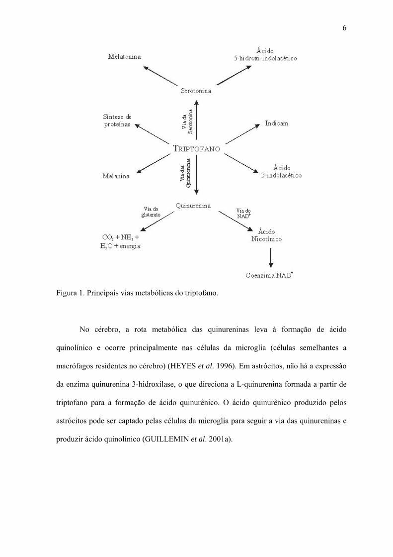
biológica (ALLEGRI et al. 2003), conforme esquematizado na Figura 1. No sistema
nervoso central e no intestino, o triptofano é substrato para síntese de serotonina, enquanto
na glândula pineal é utilizado para síntese de melatonina. Em situações em que o conteúdo
de niacina na dieta é insuficiente para as necessidades metabólicas, o triptofano é utilizado
na síntese do cofator essencial nicotinamida adenina dinucleotídeo (NAD+) (MOFFETT et
al. 2003). O triptofano também é intermediário na via das quinureninas. Nesse particular, a
via das quinureninas é a principal rota de catabolismo do triptofano, resultando na
produção de NAD+ e outros intermediários neuroativos. Entre elas, destacam-se a
quinurenina, o ácido quinurênico, a 3-hidroxiquinurenina e os ácidos picolínico e
quinolínico (Figura 2).
6
Figura 1. Principais vias metabólicas do triptofano.
No cérebro, a rota metabólica das quinureninas leva à formação de ácido
quinolínico e ocorre principalmente nas células da microglia (células semelhantes a
macrófagos residentes no cérebro) (HEYES et al. 1996). Em astrócitos, não há a expressão
da enzima quinurenina 3-hidroxilase, o que direciona a L-quinurenina formada a partir de
triptofano para a formação de ácido quinurênico. O ácido quinurênico produzido pelos
astrócitos pode ser captado pelas células da microglia para seguir a via das quinureninas e
produzir ácido quinolínico (GUILLEMIN et al. 2001a).

7
L-Triptofano
Formilquinurenina
L-Quinurenina
Triptofano 2,3-dioxigenase
Indolamina 2,3-dioxigenase
Quinurenina formamidase
Ácido Antranílico
QuinureninacetoglutaratotransaminaseÁcido
Quinurênico
3-hidroxiquinurenina
Ácido3-hidroxiantranílico
QuinureninacetoglutaratotransaminaseÁcido
Xanturênico
Semialdeído2-amino-3-carboximucônico
ÁcidoQuinolínico
Semialdeído2-aminomucônico
ÁcidoPicolínico
ÁcidoNicotínico
Glutaril-CoA
Acetil-CoA
Espontaneamente Descarboxilase do
minocarboximucônicosemialdeídoa
Espontaneamente
Espontaneamente
3-hidroxiantranilato3,4-dioxigenase
Quinureninase
Quinureninase
Quinurenina 3-hidroxilase
Figura 2. Metabolismo do triptofano: via das quinureninas. A conversão de triptofano em
quinurenina se dá no fígado por ação da enzima triptofano 2,3-dioxigenase e nos outros
tecidos por ação da enzima indolamina 2,3-dioxigenase.
Nos últimos anos a via das quinureninas tem despertado considerável interesse,
visto que diversos dos seus metabólitos apresentam atividades biológicas (ALLEGRI et al.
2003), interferindo no comportamento, no sono, na termo-regulação e na gestação
(STONE, 1993; CURZON, 1996). Essa rota está bastante relacionada à resposta
inflamatória, sendo que seus intermediários são capazes de induzir a expressão de várias
8
citocinas, quimiocinas e seus receptores em astrócitos (GUILLEMIN et al. 2000, 2001a),
promovendo a inflamação. Além disso, encontram-se aumentados em diversas condições
neuropatológicas (STONE, 2001a).
Dentre os intermediários da via das quinureninas, de fundamental importância é o
ácido quinolínico (AQ). Seu potencial fisiológico e significância farmacológica foram
reconhecidos com a descoberta da sua capacidade de ativação seletiva de receptores
glutamatérgicos excitatórios sensíveis ao N-metil-D-aspartato (NMDA) (STONE, 2001a).
Também foi sugerido recentemente que o acúmulo de intermediários da rota das
quinureninas estaria envolvido no processo patogênico presente na acidemia glutárica tipo
I (HEYES, 1987; VARADKAR E SURTEES, 2004).
1.2- Ácido Quinolínico e Doenças Neurodegenerativas
O envolvimento do AQ em diversas desordens neurodegenerativas, tais como
doenças de Alzheimer (WIDNER et al. 2000), Huntington (SCHWARCZ et al. 1984;
WHETSELL & SCHWARCZ, 1989; STONE, 2001b) e demência associada à síndrome da
imunodeficiência adquirida (SIDA) tem sido sugerido (HEYES et al. 1989, 1991)
A concentração de AQ no cérebro de pacientes afetados por essas doenças está
elevado. Embora essas concentrações raramente excedam 1 µM, esses níveis podem ser
suficientes para causar dano neuronal significante por ativação de receptores NMDA ou
através da liberação de glutamato endógeno (CONNICK & STONE, 1986, 1988). Nesse
particular, a excitotoxicidade produzida pela constante ativação de receptores NMDA
causada pelo AQ está relacionada com o aumento das concentrações citosólicas de cálcio,
depleção de ATP e GABA e morte específica de neurônios colinérgicos e GABAérgicos
(FOSTER et al. 1983, SCHWARCZ et al. 1984, STONE, 1993).

9
Fortes evidências indicam que a ativação de receptores NMDA é crítica para a
produção do dano cerebral na demência associada à SIDA (LIPTON, 1998). Nesses
pacientes os níveis de AQ no líquido cefalorraquidiano estão aumentadas em até 20 vezes,
sendo correlacionados com a severidade das disfunções motora e cognitiva apresentadas
pelos mesmos (HEYES et al. 1991, MARTIN et al. 1992, STONE, 2001a). Os níveis de
AQ no cérebro encontram-se aumentados em até 60 vezes em pacientes com várias formas
de disfunção neurológica associada com inflamação quando comparados com outros
pacientes em que o dano cerebral não envolve inflamação (HEYES et al. 1992).
Recentemente, foi demonstrado que o peptídeo β-amilóide, cujo acúmulo na forma
de placas (conhecidas como placas senis) está associado à doença de Alzheimer, induz a
produção de ácido quinolínico por macrófagos e, principalmente, pela microglia
(GUILLEMIN et al. 2001b). Diminuição nos níveis de ácido quinurênico e elevações nas
concentrações de triptofano e seu metabólito L-quinurenina foram encontradas nessa
doença, sendo o aumento desses níveis proporcional ao grau do déficit cognitivo (BARAN
et al. 1999; WIDNER et al. 1999, 2000, HEYES et al. 1992).
Além disso, diversos estudos demonstraram recentemente que a formação de
espécies reativas e a redução das defesas antioxidantes cerebrais também estão envolvidas
nas ações neurotóxicas do AQ (RIOS & SANTAMARIA, 1991; BEHAN et al. 1999;
SANTAMARIA et al. 2001a,b, LEIPNITZ et al. 2005).
Por outro lado, o AQ tem sido usado como modelo animal de indução química da
doença de Huntington (DH), visto que as alterações neuroquímicas e degeneração neuronal
induzidas pela infusão de AQ são similares àquelas observadas no cérebro de pacientes
com essa doença (SCHWARCZ et al. 1983, 1984; BEAL et al. 1986; FERRANTE et al.
1993).
A DH é uma desordem neurodegenerativa autossômica dominante, caracterizada
por depleção neuronal progressiva, particularmente dos gânglios da base, cujos

10
mecanismos envolvidos ainda não estão completamente elucidados. Entretanto, evidências
recentes têm relacionado excitotoxicidade, estresse oxidativo e alterações no metabolismo
energético em cérebro de pacientes com DH (BRENNAN et al. 1985; MANN et al. 1990;
GU et al. 1996, BEAL, 2000). Outras evidências mostram que a anormalidade
mitocondrial no cérebro de pacientes com DH é confinada ao estriado (BROWNE et al.
1997) e similar àquela induzida por malonato ou ácido 3-nitropropiônico (BEAL et al.
1993a; LUDOLPH et al. 1992).
1.3- Metabolismo Energético Cerebral
O cérebro possui uma intensa atividade metabólica, porém suas reservas
energéticas são extremamente pequenas em relação à sua demanda. Assim, há necessidade
contínua de substratos energéticos para o cérebro de mamíferos, sendo a glicose o principal
deles, onde, em contraste com outros tecidos, não necessita de insulina para ser captada e
oxidada (DICKINSON, 1996). No entanto, o padrão de utilização deste nutriente varia
conforme a etapa de desenvolvimento do sistema nervoso central (SNC), o estado
nutricional do indivíduo e o destino de sua cadeia de átomos de carbono (MARKS et al.
1996). Situações de jejum prolongado fazem com que o SNC passe a utilizar corpos
cetônicos para a obtenção de energia, a fim de poupar o organismo de um catabolismo
protéico exacerbado resultante da necessidade da manutenção da glicemia via
gliconeogênese (MARKS et al. 1996). A glicose captada pelo cérebro é, também, fonte de
carbono para a síntese de diversas outras biomoléculas essenciais (por exemplo,
neurotransmissores), o que reforça a idéia de que a utilização de glicose não está atrelada
somente à produção de energia.
Mitocôndrias de mamíferos são organelas intracelulares ubíquas, responsáveis pela
produção de ATP pelo metabolismo aeróbico, mas também desempenham outras funções

11
intracelulares além da produção de ATP, tendo um papel crítico no processo de apoptose e
servindo como um tampão de cálcio. Tecidos com alta atividade aeróbica, tais como
cérebro, músculos esquelético e cardíaco, apresentam altas concentrações de mitocôndrias
(ORTH & SCHAPIRA, 2001).
O ciclo do ácido cítrico é a via comum de oxidação dos glicídios, aminoácidos e
ácidos graxos. O metabolismo energético cerebral se mostra essencialmente aeróbico,
sendo a glicose o principal substrato utilizado (CLARK et al. 1993), entrando no ciclo sob
a forma de acetil-CoA, que é então oxidada completamente a CO2. Quando não há hipóxia,
a fosforilação oxidativa é dependente da concentração de ATP, ADP e fosfato inorgânico
(Pi) e da razão mitocondrial de NADH/NAD+, que é determinada pela atividade da cadeia
transportadora de elétrons e pela transferência de elétrons provenientes de reações
catalisadas por enzimas mitocondriais. A cadeia transportadora de elétrons oxida o NADH
e o FADH2 e bombeia prótons para o espaço intermembrana da mitocôndria, formando
assim um gradiente de prótons que através da passagem pela ATP-sintase, produz ATP na
fosforilação oxidativa (ERECINSKA & SILVER, 1994).
1.3.1- Fosforilação Oxidativa
A fosforilação oxidativa é o processo principal da produção de energia celular.
Todos os passos oxidativos na degradação de carboidratos, gorduras e aminoácidos
convergem a esse estágio final da respiração celular, em que a energia da oxidação,
provida pelo fluxo de elétrons através das enzimas da cadeia respiratória mitocondrial,
promove a síntese de ATP (NELSON & COX, 2004). A cadeia de transporte de elétrons
(CTE) mitocondrial é composta por quatro complexos enzimáticos (complexos I-IV),
recebe elétrons das coenzimas NADH e FADH2, e os transfere através de uma série de
reações de oxidação-redução até o oxigênio molecular e simultaneamente acopla essa

12
reação exoergônica à translocação de prótons através da membrana mitocondrial interna
(WALLACE, 1999). O fluxo de prótons (gradiente eletroquímico de prótons) gerado
durante o transporte de elétrons na CTE leva à formação de ATP a partir de ADP e Pi pelo
complexo V (ATP sintase) (BABCOCK & WIKSTROM, 1992; WALLACE, 1999).
Os complexos da cadeia de transporte de elétrons são complexos protéicos: NADH
desidrogenase (complexo I), sucinato: ubiquininona oxirredutase (complexo II), complexo
citocromo b-c1 (complexo III) e citocromo c oxidase (complexo IV), além de elementos
móveis que se localizam entre os complexos. São eles a coenzima Q (CoQ), um
componente não protéico lipossolúvel que carreia elétrons entre os complexos I e III, e o
citocromo c, uma proteína localizada na face externa da membrana que transfere os
elétrons do complexo III para o complexo IV (MARKS et al. 1996).
1.3.2- Creatina Quinase
A creatina quinase (CK) consiste de um grupo de isoenzimas com um papel central
no metabolismo energético, principalmente para tecidos com alta demanda energética,
como cérebro, músculo cardíaco e esquelético, onde funciona como um efetivo sistema de
tampão para os níveis celulares de ATP. A CK catalisa a transfosforilação reversível entre
ATP e creatina a ADP e fosfocreatina [MgATP- + creatina ↔ (fosfocreatina)- + MgADP- +
H+], ajudando a manter os níveis dos substratos fosforilados. Sabe-se que durante a
excitação nervosa e neuromuscular ocorre um aumento de dez vezes no turnover celular de
ATP, e que durante essas mudanças rápidas, o sistema creatina/fosfocreatina é necessário
tanto como um tampão energético quanto como um sistema de transporte entre os locais de
produção e consumo de ATP pelas ATPases para evitar grandes flutuações nos níveis de
ATP/ADP celulares nesses tecidos excitáveis (BESSMAN & CARPENTER, 1985;
SCHNYDER et al. 1991; WALLIMANN et al. 1992).

13
As isoformas da CQ estão localizadas em sítios de demanda e produção energética.
A isoforma citosólica (Ci-CQ) consiste de dímeros e é expressa de uma maneira tecido-
específica, isto é, cérebro-específica (BB-CQ), músculo esquelético-específica (MM-CQ) e
um heterodímero músculo cardíaco-específico (MB-CQ) (SCHNYDER et al. 1991;
WALLIMANN et al. 1992; O’GORMAN et al. 1996; HAMMAN et al. 1995). As formas
mitocondriais da CQ (Mi-CQ) são dispostas em octâmeros e são compostas da isoforma
sarcomérica músculo-específica Mib-CQ e da forma ubíqua Mia-CQ, que é encontrada nas
mitocôndrias do tecido cerebral (SAKS et al. 1985; SCHELEGEL et al. 1988;
WALLIMANN et al. 1992; GROSS et al. 1996).
Devido à sua localização próxima a sítios onde se dá a geração de energia e
transporte de íons através de membranas, o sistema CQ/fosfocreatina desempenha um
papel fundamental na homeostase energética neuromuscular. Assim, é presumível que
alterações na função da CQ levem ao desenvolvimento de vários estados patológicos
envolvendo o cérebro, músculo esquelético e músculo cardíaco (HAMMAN et al. 1995;
DAVID et al. 1998, AKSENOV et al. 1999; AKSENOV et al. 2000).
1.4- Metabolismo Energético nas Doenças Neurodegenerativas
Numerosas hipóteses têm sido propostas para explicar a fisiopatologia das doenças
de Alzheimer, Huntington e Parkinson, sem, no entanto, se obter até o momento uma
explicação satisfatória para o dano cerebral causado por essas doenças. No entanto,
acredita-se que possíveis mecanismos envolvam deficiência no metabolismo energético,
estresse oxidativo e neurotoxicidade mediada por receptores glutamatérgicos do tipo
NMDA, ou, possivelmente, um somatório desses fatores (ROSE & HENNEBERRY,
1994). Uma das hipóteses é de que alterações na cadeia transportadora de elétrons seria o

14
evento etiológico primário na maioria dessas doenças (PARKER, 1989; PARKER et al.
1989, 1990, 1994; SWERDLOW et al. 1996, 1997).
O cérebro é altamente dependente de energia para seu funcionamento normal
(ROSSEN et al. 1943) e a mitocôndria é a estrutura intracelular que mantém os
suprimentos de energia para o cérebro. Uma alteração funcional nessa estrutura poderá
levar, portanto, a alterações patológicas nos neurônios e astrócitos (BEAL et al. 1993b;
BEAL, 1995; BOWLING & BEAL, 1995; DAVIS et al. 1995). Mutações no DNA
mitocondrial e reações envolvendo geração de espécies reativas podem danificar a
mitocôndria e diminuir a atividade dos complexos da cadeia respiratória. Um prejuízo no
transporte de elétrons, além de causar um prejuízo na produção de ATP, leva a uma
dispersão dos elétrons na forma de radicais livres potencialmente danosos à célula. Visto
que os sistemas de defesa antioxidantes, como as enzimas superóxido dismutase, glutationa
peroxidase, glutationa redutase e catalase, são relativamente deficientes no cérebro
(MARKLUND et al. 1982; MARTILLA et al. 1988), um ciclo vicioso pode ocorrer,
aumentando o dano oxidativo a neurônios e levando à morte neuronal característica de
doenças neurodegenerativas. Um prejuízo na fosforilação oxidativa também reduz o
bombeamento de prótons através da membrana mitocondrial, diminuindo o potencial da
membrana e, conseqüentemente, levando à abertura do poro de transição da
permeabilidade mitocondrial. Evidências sugerem que uma diminuição no potencial de
membrana mitocondrial seja um evento primário na iniciação do processo apoptótico
(WADIA et al. 1998).
Por outro lado, dano neuronal e estresse oxidativo podem levar a mudanças reativas
em astrócitos situados nas proximidades, levando a um aumento na produção de citocinas
pró-apoptóticas e pró-inflamatórias, bem como a uma alteração na homeostase de íons
extracelulares e de aminoácidos neurotransmissores (MACCIONI et al. 2001). Além disso,
mecanismos patogênicos das doenças neurodegenerativas podem estimular células da

15
microglia, levando à produção de substâncias potencialmente neurotóxicas (MACCIONI et
al. 2001).
Numerosas evidências associam doenças neurodegenerativas a uma diminuição no
metabolismo energético. Estudos demonstraram uma diminuição na atividade do complexo
I da cadeia respiratória postmortem em cérebros de pacientes portadores de doença de
Parkinson (GU et al. 1998; JANETZKY et al. 1994; SCHAPIRA et al. 1989, 1990a,b).
Também há relatos de defeitos nos complexos II e III da cadeia respiratória e na enzima α-
cetoglutarato desidrogenase, importante enzima do ciclo do ácido cítrico, nessa doença
(MIZUNO et al. 1990, 1994).
Na doença de Alzheimer, a mais comum dentre as doenças neurodegenerativas, é
encontrada principalmente uma redução na atividade do complexo IV da cadeia
respiratória (MAURER et al. 2000). Estudos de imagem in vivo demonstraram uma
redução no metabolismo da glicose em pacientes portadores da doença de Alzheimer, e
essa diminuição aparece precocemente, precedendo os sintomas clínicos, e se acentua com
a severidade da doença (JAGUST et al. 1988; SMITH et al. 1992; KENNEDY et al. 1995;
SMALL et al. 1995). Estudos postmortem em cérebros também demonstraram uma
diminuição na atividade do complexo enzimático da piruvato desidrogenase e na atividade
da enzima α-cetoglutarato desidrogenase na doença de Alzheimer (PERRY et al. 1980;
GIBSON et al. 1988; MASTROGIACOMO et al. 1993).
Na esclerose lateral amiotrófica, observou-se uma diminuição na atividade dos
complexos I, III e IV da cadeia respiratória em mitocôndrias de células híbridas
(SWERDLOW et al. 1998).
Por outro lado, vários estudos têm demonstrado uma diminuição na utilização de
glicose em estriado e córtex cerebral de pacientes portadores da doença de Huntington
(KUHL et al. 1982; HAYDEN et al. 1986; YOUNG et al. 1986; GRAFTON et al. 1992;
MARTIN et al. 1992; KUWERT et al. 1993), além de um aumento nos níveis cerebrais de

16
lactato (JENKINS et al. 1993). Também se observou uma redução na utilização de
oxigênio, bem como na atividade dos complexos II, III e IV da cadeia respiratória no
núcleo caudato de pacientes portadores dessa doença (BRENNAN et al. 1985; GU et al.
1996).

17
2- OBJETIVOS
2.1- Objetivo Geral
Este trabalho tem por objetivo avaliar o efeito da administração aguda intraestriatal
de ácido quinolínico (AQ) sobre o metabolismo energético em estriado de ratos jovens
bem como os mecanismos envolvidos na disfunção energética estriatal induzida pela
administração intraestriatal de AQ.
2.2- Objetivos Específicos
* Avaliar o efeito da administração intraestriatal de AQ sobre a produção de 14CO2
a partir de [1-14C]acetato em homogeneizado de estriado de ratos 3, 6 ou 12 horas após a
injeção.
* Avaliar o efeito da administração intraestriatal de AQ sobre a atividade dos
complexos I, II, III e IV da cadeia respiratória em homogeneizado de estriado de ratos 3, 6
ou 12 horas após a injeção.
* Avaliar o efeito da administração intraestriatal de AQ sobre a atividade da enzima
creatina quinase em homogeneizado de estriado de ratos 3, 6 ou 12 horas após a injeção.
* Estudar os mecanismos envolvidos nas possíveis alterações do metabolismo
energético estriatal causada pela administração intraestriatal de AQ através da:
- pré-administração intraperitoneal de antioxidantes, tais α-tocoferol (100 mg/kg)
mais ácido ascórbico (40 mg/kg) ou da pré-administração intraperitoneal de L-NAME (2
mg/kg);
- avaliação do efeito da pré-administração intraperitoneal de substratos energéticos,
tais como ácido pirúvico (500 mg/kg) ou creatina (40 mg/kg).
- pré-administração intraperitoneal do antagonista de receptores NMDA, MK-801
(0,25 mg/kg).

18
PARTE II
19
Artigo a ser submetido à revista Journal of Neurochemistry

20
EVIDENCE THAT QUINOLINIC ACID SEVERELY IMPAIRS ENERGY
METABOLISM THROUGH ACTIVATION OF THE NMDA RECEPTOR IN RAT
STRIATUM
César A. J. Ribeiro1, Vanessa Grando1, Moacir Wajner1,2,3
1 Departamento de Bioquímica, Instituto de Ciências Básicas da Saúde, Universidade
Federal do Rio Grande do Sul, Porto Alegre, RS, Brasil
2 Serviço de Genética Médica, Hospital de Clínicas de Porto Alegre, Porto Alegre, RS,
Brasil
3 Universidade Luterana do Brasil, Canoas, RS, Brasil
Address correspondence and reprint requests to
Prof. Dr. Moacir Wajner
Departamento de Bioquímica, Instituto de Ciências Básicas da Saúde, Universidade
Federal de Rio Grande do Sul.
Rua Ramiro Barcelos N° 2600 – Anexo, CEP: 90035-003, Porto Alegre, RS – Brasil
Phone: +55 51 3316-5571, fax: +55 51 3316-5535
E-mail address: [email protected]
Running title: Quinolinic acid and brain energy metabolism
Abbreviations used:
CK, creatine kinase; HD, Huntington’s disease; L-NAME, Nω-nitro-L-arginine methyl
esther; MK-801, (5R,10S)-(+)-5-methyl-10,11-dihydro-5H-dibenzo[a,d]cyclohepten-5,10-
imine hydrogen maleate; NO, nitric oxide; NOS, nitric oxide synthase; PCr,
phosphocreatine; QA, quinolinic acid

21
Abstract
Most of quinolinic acid (QA) toxic properties have been attributed to excitotoxicity
and oxidative stress, whereas very little is known about its effects on brain energy
metabolism. Therefore, in the present study we investigated the in vivo effect of
intrastriatal administration of 150 nmol QA to 30-day-old rats on critical enzyme activities
of energy metabolism, including the respiratory chain complexes I-IV, creatine kinase and
citrate synthase, as well as on 14CO2 production from [1-14C]acetate at distinct periods
after QA injection. We observed that QA injection significantly inhibited complexes II
(50%), III (46%) and II-III (35%), as well as creatine kinase (27 %), but not complexes I
and IV activities in striatum prepared 12 hours after QA treatment. In contrast, no
alterations of these enzyme activities were observed 3 or 6 hours after QA treatment. 14CO2
production from [1-14C]acetate was also significantly inhibited (27 %) by QA only in rat
striatum prepared 12 hours after injection. The respiratory chain complexes and creatine
kinase activities were also measured in striatum homogenates exposed to 100 µmol QA.
No alterations of these activities were observed in the presence of QA. These in vitro
observations allied to the ex vivo findings suggest that QA compromises energy
metabolism in the rat striatum indirectly rather than due to a direct action of QA on the
enzymes. New experiments were therefore designed to elucidate the involved mechanisms
of QA-induced inhibition on the enzymatical activities of energy metabolism. Pretreatment
with the NMDA receptor antagonist MK-801 and with creatine totally prevented the
inhibitory effects elicited by QA. In addition, the free-radical scavengers α-tocopherol plus
ascorbate and the nitric oxide synthase inhibitor L-NAME completely abolished the
inhibitions provoked by QA on CK and complex III, indicating that these effects were
probably due to generation of reactive species. On the other hand, pyruvate pretreatment
totally blocked the inhibitory effects of QA injection on complex II activity and partially
prevented QA-induced CK inhibition. Taken together, these observations strongly indicate

22
that oxidative phosphorylation and cellular energy transfer are compromised by high
concentrations of QA in the striatum of young rats and that the inhibitory effects caused by
QA injection on critical steps of energy metabolism were probably mediated by NMDA
stimulation.
Key words: quinolinic acid, respiratory chain, creatine kinase, energy metabolism
23
Introduction
Quinolinic acid (QA; 2,3-pryridine dicarboxylic acid), an endogenous tryptophan
metabolite produced at the kynurenine pathway, is an excitotoxin acting as an agonist at
the N–methyl-D-aspartate (NMDA) receptor (Stone, 1993). Experiments in rodents have
demonstrated that intrastriatal injection of QA mimics many of the neurochemical and
histological features of Huntington´s disease (HD) (Beal et al. 1986), and therefore this
neurotoxin has been used as a chemically-induced animal model of this disease (Schwarz
et al. 1983, 1984; Beal et al. 1986; Ferrante et al. 1993).
HD is a degenerative neurological disorder in which there is progressive neuronal
depletion, particularly in the basal ganglia, whose underlying mechanisms are still poorly
established. However, recent evidence has implicated excitotoxicity, oxidative stress and
metabolic impairment as important factors of the neuronal degeneration in HD and in other
neurodegenerative diseases (Beal et al. 2000). In this context, various studies have
demonstrated abnormal energy metabolism in postmortem HD brain, most notably reduced
activity of succinate-linked oxidation in the caudate nucleus and severe deficiency in
activities of the respiratory chain complexes (Brennan et al. 1985; Mann et al. 1990; Gu et
al. 1996). Further evidence has shown that the mitochondrial abnormality in HD brain is
confined to the striatum (Browne et al. 1997), being similar to that induced by the complex
II inhibitors of the respiratory chain malonate and 3-nitropropionic acid (Beal et al. 1993;
Ludolph et al. 1992).
Most studies performed to examine the in vitro and in vivo effect of QA on brain
tissue demonstrated excitotoxicity and oxidative damage caused by this neurotoxin (Stone,
1993, Tavares et al. 2000, Santamaria et al. 2001; Leipnitz et al. 2005). It has been
demonstrated that excitotoxicity produced by sustained activation of NMDA receptors by
QA is related to increased cytosolic Ca2+ concentrations, ATP and GABA depletion, and
specific GABAergic and cholinergic neuronal death (Foster et al. 1983, Schwartz et al.

24
1984, Stone, 1993). In addition, oxidative stress produced by free radical formation has
been also documented as an important mechanism of QA toxicity in the brain (Rios and
Santamaria, 1991; Santamaria and Rios, 1993; Perez-Severiano et al. 1998, 2004; Leipnitz
et al. 2005). In this context, not only oxygen-derived radical species, but also nitrogen-
derived species have been shown to be induced by QA in the rodent brain (Noack et al.
1998, Ryu et al. 2004). Surprisingly, very few studies were designed to examine the role of
quinolinic acid on brain energy metabolism, despite the fact that QA has been found at
increased concentrations in brain of HD patients and energy dysfunction and more
specifically deficiency of the activities of the respiratory chain complexes were also
observed in these patients. To our knowledge, only two works investigated the influence of
QA on striatum energetics in adult rats. It was demonstrated that intrastriatal injection of
QA provokes a progressive time-dependent mitochondrial dysfunction reflected by a low
respiratory chain ratio (decreased respiration) and a reduction of ATP, NAD+, aspartate
and glutamate concentrations (Bordelon et al. 1997). Disturbances in neuronal activity and
ion gradients secondarily to metabolic impairment have been also attributed to QA
(Bordelon et al. 1998). Although these studies revealed that electrical activity and cellular
respiration are reduced by QA in the striatum, they did not evaluate the underlying
mechanisms.
Therefore, the present study was undertaken to investigate the influence of QA
intrastriatal injection to young rats on important parameters of energy metabolism,
including the activities of the various respiratory chain complexes, as well as on creatine
kinase and citrate synthase activities and 14CO2 production from [1-14C]acetate at distinct
periods after QA injection. We also examined the ability of a variety of administered
compounds (energetic substrates, antioxidants and an NMDA-receptor antagonist) to
modify the energetic deficit induced by intrastriatal QA injection in order to clarify the
mechanisms involved.

25
Material and Methods
Animals and Reagents
A total of 140 30-day-old Wistar rats obtained from the Central Animal House of
the Department of Biochemistry, Instituto de Ciências Básicas da Saúde, Universidade
Federal do Rio Grande do Sul, Porto Alegre, Brazil, were used in the in vitro and ex vivo
studies. The animals were maintained on a 12:12 h light/dark cycle in an air-conditioned
constant temperature (22ºC + 1ºC) colony room, with food and water ad libitum. All
reagents used were of analytical grade and purchased from Sigma Co. (St Louis, MO,
USA), except for dizocilpine maleate (MK-801), which was purchased from Tocris
(Ballwin, MO, USA).
Quinolinic Acid Administration
The rats were deeply anesthetized with sodium thiopental (30 mg/kg i.p.) and
placed in a stereotaxic apparatus. A small hole was drilled in the skull for microinjection,
and 0.5 µL of 300 mM quinolinic acid (150 nmol, pH 7.4 adjusted with NaOH) or NaCl
(controls), at the same concentration, was slowly injected over 4 min into the left striatum
via a needle connected by a polythene tube to a 10 µL Hamilton syringe. The needle was
left in place for another 1 min before been softly removed, so that the total procedure
lasted 5 min. The coordinates for injection were as follows: 0.6 mm posterior to bregma,
2.6 mm lateral to midline and 4.5 mm ventral from dura (Paxinos and Watson, 1986). The
correct position of the needle was tested by injecting 0.5 µL of methylene blue injection
(4% in saline solution) and carrying out histological analysis. The dose and method of QA
administration were based in previous works (Qin et al. 1992). The experimental protocol
was approved by the Ethics Committee for animal research of the Universidade Federal do
Rio Grande do Sul, Porto Alegre, Brazil and followed the NIH Guide for the Care and Use

26
of Laboratory Animals (NIH publication 85-23, revised 1985). All efforts were made to
minimize the number of animals used and their suffering.
In some experiments the animals were pretreated during 7 days with daily i.p.
administration of α-tocopherol (40 mg/kg) plus ascorbic acid (100 mg/kg) or with NaCl
(0.9% 10 mL/kg, i.p.), as described previously (Franzon et al. 2003). Some animals were
also pretreated with Nω-nitro-L-arginine methyl esther (L-NAME, 2 mg/kg) or NaCl (0.9%
10 mL/kg, i.p.) 30 minutes before QA intrastriatal injection. The L-NAME dose used was
shown to inhibit cerebral nitric oxide synthase (NOS) by more than 70% (Nishikawa et al.
1993), without significantly altering blood flow (Buisson et al. 1992). In another
experiment, rats were pretreated with sodium pyruvate i.p. at a dose of 500 mg/kg or NaCl
(0.9% 10 mL/kg, i.p.)1 hour before QA injection (Ryu et al. 2003). The effect of creatine
administration on QA-induced energetic dysfunction was also evaluated by preinjecting the
animals for 7 days, two injections per day, with creatine (50 mg/kg, i.p.) or NaCl (0.9% 10
mL/kg, i.p.), after which the animals received one intrastriatal QA injection (Vasques et al.
2005). Finally, in the experiments designed to evaluate the participation of glutamatergic
mechanisms mediated by NMDA-glutamate receptors, the animals received MK-801 (0.25
mg/kg, i.p.) or NaCl (0.9% 10 mL/kg, i.p.) 30 min before they were injected with QA.
Tissue Preparation
Animals were sacrificed by decapitation 3, 6 or 12 hours after intrastriatal injection
of either quinolinic acid or NaCl, the brain was rapidly excised on a Petri dish placed on
ice. The olfactory bulb, pons, medulla, cerebral cortex and cerebellum were discarded, and
the ipsilateral and contralateral striata were dissected, weighed and kept chilled until
homogenization with a ground glass type Potter-Elvehjem homogenizer in the specific
buffer used for each technique. For the determination of the electron transfer chain
complexes and creatine kinase activities, contralateral and ipsilateral striata were

27
homogenized in 20 volumes of SETH buffer, pH 7.4 (250 mM sucrose, 2 mM EDTA, 10
mM Trizma base, and 50 IU/mL heparin). The homogenates were centrifuged at 800 X g
for 10 min at 4ºC, the pellet was discarded and the supernatants kept at -70°C until enzyme
activity determination (Fischer et al. 1985). The maximal period between homogenate
preparation and enzyme analysis was always less than 7 days.
Mitochondria from striatum were also purified for measurement of complex I
activity. Briefly, the cerebral cortex was homogenized with a ground glass type Potter-
Elvehjem homogenizer in 10 volumes of phosphate buffer pH 7.4 containing 0.3M
sucrose, 5mM MOPS, 1mM EGTA and 0.1% bovine serum albumin. The homogenates
were centrifuged at 1500×g for 10 min at 4°C and the pellet was discarded. The
supernatant was centrifuged at 15,000×g in order to isolate mitochondria present in the
pellet, which was finally dissolved in the same buffer (Latini et al. 2005)
For the experiments carried out to measure 14CO2 production, striatum was
homogenized (1:10, w/v) in Krebs-Ringer bicarbonate buffer pH 7.4 using an ice-chilled
glass homogenizing vessel at 900 rpm and total homogenates were used in these
experiments.
For the in vitro studies, striatum from non-treated rats was dissected and
homogenized in a similar manner as that for the ex vivo experiments to obtain supernatants
and purified mitochondrial fractions. The supernatants or mitochondrial preparations were
them incubated in the presence of 100 µM QA.
Respiratory Chain Complexes Activities
Mitochondrial respiratory chain enzyme activities were measured in striatum
supernatants 3, 6 or 12 hours after QA or NaCl intrastriatal injection. The activity of
NADH dehydrogenase (Complex I) was assessed as described by Cassina and Radi (1996)
in mitochondrial preparations. The activities of succinate: DCIP-oxidoreductase (complex

28
II) and succinate:cytochrome c oxidoreductase (complex II-III) were determined according
to the method of Fischer et al (1985). The activity of ubiquinol:cytochrome c oxireductase
(complex III) was assayed according to the method described by Birch-Machin et al.
(1995) and that of cytochrome c oxidase (complex IV) according to Rustin et al. (1994).
The methods described to measure these activities were slightly modified, as described in
details in previous reports (Brusque et al. 2002, da Silva et al. 2002). The activities of the
respiratory chain complexes were expressed as nmol/(min mg protein). For the in vitro
studies, 100 µM QA was supplemented to the incubation medium, whereas the control
group did not contain the metabolite.
Creatine Kinase activity (CK) determination
CK activity was measured in striatum homogenates 3, 6 or 12 hours after QA or
NaCl intrastriatal injection in a reaction mixture consisting of 60 mM Tris-HCl, pH 7.5,
containing 7 mM phosphocreatine, 9 mM MgSO4, 0.625 mM lauryl maltoside and
approximately 0.4-1.2 µg protein in a final volume of 100 µL. For the in vitro studies, 100
µM QA was supplemented to the incubation medium, whereas the control group did not
contain the metabolite. After 10 min of pre-incubation at 37ºC, the reaction was started by
the addition of 0.3 µmol ADP. The reaction was stopped after 10 min by the addition of 1
µmol of p-hydroxymercuribenzoic acid. The creatine formed was estimated according to
the colorimetric method of Hughes (1962) with slight modifications as described
previously (da Silva et al. 2004). The color was developed by the addition of 100 µL 2%
α-naphtol and 100 µL 0.05% diacetyl in a final volume of 1 mL and read
spectrophotometrically at 540 nm after 20 min. Results were expressed as percentage of
controls as µmol creatine formed/(min mg protein).

29
Citrate Synthase activity
The activity of citrate synthase, a marker enzyme of mitochondrial viability, was
assayed according to Shepherd and Garland (1973) in a medium containing 75 mM Tris-
HCl pH 8.0, 0.01% Triton X-100, 0.1 mM DTNB, 0.5 mM oxaloacetic acid and 20 µg
protein. The reaction was started with the addition of 0.42 mM acetyl-CoA and monitored
spectrophotometrically at 412 nm for 3 min. Results were expressed as nmol TNB
formed/(min mg protein).
14CO2 production from [1-14C]acetate
For the experiments designed to evaluate 14CO2 production, the animals were
sacrificed 3 or 12 hours after intrastriatal QA or NaCl (0.9 %) injection, the contralateral
and ipsilateral striata were dissected and homogenized (1:10, w/v) in Krebs-Ringer
bicarbonate buffer pH 7.4 using an ice-chilled glass homogenizing vessel at 900 rpm. Total
striatal homogenates were added to small flasks (11 cm3). Flasks were pre-incubated in a
metabolic shaker at 37°C for 15 min. After pre-incubation, 0.1 μCi [1-14C]-acetate and 0.5
mM of unlabeled acetate were added to the incubation medium. The flasks were gassed
with a O2:CO2 (95:5) mixture and sealed with rubber stoppers and Parafilm M. Glass
center wells containing a folded 65 mm/5 mm piece of Whatman 3 filter paper were hung
from the stoppers. After 60 min of incubation at 37°C, 0.1 mL of 50% trichloroacetic acid
was added to the medium and 0.1 mL of benzethonium hydroxide was added to the center
wells with needles introduced through the rubber stopper. The flasks were left to stand for
30 min to complete 14CO2 trapping and then opened. The filter papers were removed and
added to vials containing scintillation fluid, and radioactivity was measured (Dutra-Filho et
al. 1995).

30
Protein determination
Protein was measured by the method of Lowry et al (1951), using bovine serum
albumin as standard. The protein concentration in the supernatants varied from 1.5-4.0 mg
protein/mL in all experiments.
Statistical analysis
Data were expressed as means ± SEM for absolute values or percentage of control.
Assays were performed in duplicate or triplicate and the mean was used for statistical
analysis. Results were assessed with a computerized statistical program (Statistical
Package for the Social Sciences, SPSS) on an IBM-compatible PC. Comparisons between
values were calculated using the unpaired and paired Student’s t-tests. Differences between
the groups were considered statistical significant when P < 0.05.
Results
The activities of the respiratory chain complexes were determined in homogenates
from the left striatum of rats sacrificed 3, 6 or 12 hours after QA or NaCl administration. It
can be seen in Table 1 that QA did not affect these enzymatic activities when rats were
killed 3 and 6 hours after intrastriatal injection, as compared to NaCl-injected animals. In
contrast, significant inhibitions on complex II (50%, P < 0.05), complex III (46%, P <
0.05) and complex II-III (35%, P < 0.01) activities, but not of complex I (P = 0.273), and
complex IV (P = 0.945) activities, were observed in striatum prepared 12 hours after QA
treatment (Figure 1).
The activity of citrate synthase was also determined in striatum homogenates
prepared 12 hours after QA or NaCl injection. No significant difference was found
between groups, indicating that the number of mitochondria and/or the mitochondrial
integrity was not affected by intrastriatal injection of QA (P = 0.456). These data

31
corroborate our findings of no alteration of complexes I and IV activities of the respiratory
chain at 12 hours after QA intrastriatal administration.
The activities of creatine kinase (CK) in striatum homogenates taken 3, 6 or 12
hours after QA or NaCl administration are shown in Figure 2. No significant differences
between QA and NaCl-injected striata were found in CK activity at 3 or 6 hours after QA
injection. However, CK activity was significantly reduced by about 27% after 12 hours of
QA intrastriatal administration (P < 0.05).
We then examined the influence of QA treatment on CO2 production from [1-
14C]acetate in total homogenates prepared from striatum of rats that received QA or NaCl
3, 6 or 12 hours after injection. No differences in 14CO2 synthesis were found 3 or 6 hours
after QA-injection. However, CO2 production from acetate was found significantly reduced
by about 30% in striatum of QA-injected animals sacrificed 12 hours after QA
administration (P < 0.05) (Figure 3).
We then examined the in vitro effect of QA (100 µM) on the respiratory chain
complexes and creatine kinase activities in striatal homogenates of young rats. Table 2
shows that no significant changes were found when QA was added to the incubation
medium, as compared to controls. Taken together the in vitro and the ex vivo observations,
it may be presumed that QA-induced impairment of energy production was mediated by
indirect mechanisms, rather due to a direct action of this organic acid on the enzymes.
Therefore, the next set of experiments was designed to elucidate the mechanisms
involved in impairment of striatum bioenergetics caused by QA administration. The
animals were pretreated with antioxidants, energetic substrates or an NMDA receptor
antagonist and sacrificed 12 hours after QA injection. In these experiments, the activities
of the respiratory chain complexes and of creatine kinase were evaluated in striatum
homogenates prepared from the ipsilateral and the contralateral striatum (controls) in order
to minimize the number of animals used. Preliminary experiments revealed no significant

32
changes in these activities between the NaCl-injected ipsilateral and the noninjected
contralateral striatum prepared 12 hours after injection (data not shown), indicating that
NaCl injection per se did not alter the energy metabolism parameters evaluated.
Furthermore, we also observed that QA injection in the ipsilateral striatum did not change
the energy metabolism parameters in the contralateral striatum (data not shown).
We initially pretreated the animals with daily i.p. injections of the free-radical
scavengers α-tocopherol (40 mg/kg) plus ascorbate (100 mg/kg) for 7 days, while controls
received saline solution. As depicted in Figure 4, pretreatment with these drugs totally
prevented the inhibitory effect of QA on CK (P < 0.01 vs saline) and complex III (P <
0.05 vs saline) activities, but did not alter the inhibition provoked by this neurotoxin on
complexes II and II-III.
We then pretreated the animals with one i.p. injection of the NOS inhibitor L-
NAME (2 mg/kg) 30 min prior to QA intrastriatal administration (Figure 5). This drug was
administered in a dose that inhibits cerebral nitric oxide synthase up to 70% (Nishikawa et
al. Stroke 1993) without altering blood flow (Buisson et al. Br. J. Pharmacol, 1992). L-
NAME pretreatment only abolished the inhibitory effect of QA administration on CK
activity (P < 0.01 vs saline) (Figure 5), indicating that NO or/and peroxynitrite generation
is probably involved in such inhibitory effect. Taken together, these results indicate that
CK and complex III inhibition induced by QA injection is probably mediated by the
generation of reactive species.
Animals were also pretreated with the energetic substrates pyruvate and creatine,
Pretreatment of rats 1 hour prior to intrastriatal injection of QA with 500 mg/kg pyruvate, a
dose previously reported to be efficient to reduce QA-induced striatum toxicity (Ryu et al.
2004), did not alter the inhibitory pattern of complex II-III and III, but partially reduced (P
< 0.05 vs contralateral; P < 0.05 vs saline) the inhibitory effect of QA on CK activity and
completely prevented the inhibitory effect of this neurotoxin on complex II activity (P <

33
0.05 vs saline) (Figure 6).
The effect of i.p. creatine (50 mg/kg) or saline (0.9 % NaCl) pretreatment for 7
days, twice a day, on the inhibitory properties of QA was examined. As shown in figure 7,
creatine pretreatment completely prevented all inhibitory effects of QA. Moreover, the
activity of CK of animals receiving creatine was even higher (25%, P < 0.01) in the
ipsilateral, as compared with the contralateral striatum.
Finally, we observed that intraperitoneal preadministration of the NMDA
antagonist MK-801 (0.25 mg/kg) fully prevented the inhibitory effects elicited by QA
intrastriatal injection on all parameters evaluated (Figure 8). These results indicate that the
inhibitions of enzymatic activities caused by QA administration were probably mediated
by NMDA overstimulation.
Discussion
Quinolinic acid is presumably involved in the pathogenesis of neurodegenerative
disorders (Schwarcz et al. 1984, Whetsell and Schwarcz, 1989; Widner et al. 2000a,b;
Obrenovitch, 2001; Stone, 2001), infectious, inflammatory and non-inflammatory diseases
(Heyes et al. 1991, 1995, Halperin and Heyes, 1992; Brouwers et al. 1993) It has been
generally considered that the toxic actions of QA are predominantly related with NMDA
receptor overactivation (Stone et al. 1993; Susel et al. 1989), although other investigators
do not support the presumption that this mechanism is responsible for most of the brain
damage exerted by QA (Obrenovitch et al. 2001). In this context, QA is capable to induce
oxidative stress through reactive oxygen and nitrogen species formation by NMDA-
dependent and NMDA-independent manners, therefore involving a pattern of toxicity
distinct from excitotoxicity (Behan et al. 1999, Santamaria et al. 2001; Leipnitz et al.
2005). Furthermore, quinolinic acid (QA) and the inhibitors of complex II of the
respiratory chain malonate and 3-nitropropionic acid reproduce the neurochemical and

34
histopathological features of Huntington´s disease characterized by basal ganglia lesions
and QA has been used as an animal model of because it this disorder (Schwarcz et al.
1983; Beal et al. 1986; Beal et al. 1993; Greene et al. 1993; Brouillet et al. 1995).
On the other hand, intracerebral administration of QA has been shown to cause
energetic dysfunction in striatum of adult rats but the involved mechanisms were not
unraveled (Bordelon et al. 1997, 1998).The present investigation demonstrates that in vivo
intrastriatal administration of QA to young rats decreases aerobic respiration, by
significantly blocking the respiratory chain and the citric acid cycle, and also compromises
energy transfer inside the cell, by inhibiting creatine kinase (CK) activity, in a time-
dependent manner. The alterations of energy metabolism were observed 12 hours, but not
3 or 6 hours after QA injection, suggesting that these effects could be mediated by indirect
mechanisms such as NMDA receptor activation and/or oxidative stress. This assumption is
reinforced by the fact that doses of 100 µmol QA were unable to inhibit in vitro the
activities of the respiratory chain complexes and of creatine kinase, indicating that no
direct effect of QA on these enzymes occurred.
Significant reductions of the activities of complexes II (50 %), II-III (35 %), III (46
%) of the respiratory chain and of CK (27 %) were detected in the striatum 12 hours after
QA injection. Labeled CO2 production from [1-14C]acetate, which reflects the activity of
the Krebs cycle, was also found reduced (27 %) only at 12 hours after QA infusion. Taken
together, these results strongly indicate that the striatum aerobic capacity was
compromised by QA.
Young animals (30 day-old rats) were used in our experiments because very little is
known on the effects of QA on intermediary metabolism at an early animal age. In this
context, changes in temporal and spatial expression of NMDA receptor subtypes (QA is an
agonist of these subtype of glutamate receptors) occur during brain development (Monyer
et al. 1994, Portera-Cailliau et al 1996, Wenzel et al 1997). Thus, it could be presumed

35
that differential age-specific effects may be achieved in case QA acts on a specific subtype
of NMDA receptor.
We could hypothetically attribute our results of reduced activities of crucial
enzymatic steps of energy production as due to tissue damage and cell death caused by QA
infusion since reduced ATP production or utilization can result in loss of cell viability by
disrupting energy-dependent vital cellular processes, and it has been shown that QA
provokes lower ATP synthesis in the striatum (Bordelon et al. 1997). In this regard, ATP is
essential to fuel ionic pumps that generate and maintain ionic and voltage gradients across
neuronal membranes, including Na+,K+,ATPase pump that controls the resting membrane
potential and other ATPases that regulate intracellular Ca2+ levels (Novelli et al. 1988,
Haneberry et al. 1989, Zeevalk et al. 1991). Increased intracellular Ca2+ may lead to
reactive species formation and in turn attack important enzyme activities of the respiratory
chain and of the Krebs cycle (Dykens, 1994,). Therefore, impaired energy metabolism and
oxidative stress may form a self-amplifying cycle of cell toxicity. However, this was
probably not the case i.e. the changes in energy metabolism parameters as reported here are
not likely to be due to cell loss, since the activities of NADH dehydrogenase (complex I),
complex IV and citrate synthase, other enzyme activities that reflect the mitochondrial
content in the samples, were not altered by QA injection. This is in agreement with
previous studies showing that there is no significant decrease in cell number after similar
injections of this toxin (Bordelon et al. 1994).
The animal data obtained in the current study agree to a certain extend to other
reports performed in humans and showing that various activities of the respiratory chain
complexes, particularly complex II, are markedly and selectively reduced in postmortem
striatum from HD patients (Tabrizi et al. 1999, Gu et al. 1996). Thus, our results may
provide further support for the usefulness of intrastriatal QA injection as an animal model
of HD.

36
Considering that oxidative stress can be induced by QA in vitro and in vivo
(Leipnitz et al. 2005; Rios and Santamaria, 1991, Perez-Severiano et al. 2004) and that the
respiratory chain complexes II and III and CK can be attacked by free radicals (Cardoso et
al. 1999; Wendt et al. JBC 2003, Stachowiack JBC 1998), we evaluated whether free
radical generation could be involved in the energetic impairment provoked by QA
intrastriatal injection by pretreating animals with the free radical scavengers α-tocopherol
plus ascorbate or the NOS inhibitor L-NAME and then assaying the energetic parameters.
We observed that pretreatment with α-tocopherol plus ascorbate was able to fully prevent
CK and complex III inhibitions induced by QA infusion, indicating that these inhibitory
effects were mediated by reactive species. In this particular, it is well established that α-
tocopherol is a lipophilic antioxidant that acts as a free radical scavenger, donating
hydrogen to radicals usually peroxyl and alkoxyl radicals, thus preventing the propagation
of lipid peroxidation. Because it is localized into the hydrocarbon core of the phospholipids
bilayer, it interacts with cell membrane, protecting polyunsaturated fatty acids and thiol
groups of membrane proteins, keeping the integrity and function of the biomembranes
(Trappel, 1973; Ames et al. 1993; Burton et al. 1990). During this process, α-tocopherol is
converted to the tocopheryl radical, requiring ascorbate for its regeneration back to reduced
tocopherol (Frei et al. 1990; McCay, 1985; Carr and Frei, 1999). Regarding complex III, it
has been recently reported that this complex contains tightly bound cardiolipin molecules
that appear to be essential for its catalytic function (Gomez Jr and Robinson, 1999; Lange
et al. 2001) and that reactive oxygen species generated by the mitochondrial respiratory
chain affect complex III activity via cardiolipin peroxidation (Paradies et al. 2001). Our
results also demonstrated lack of preventive effect of α-tocopherol on complex II activity,
and this is in accordance with other study reporting an inability of α-tocopherol to protect
complex II in an oxidative stress situation (Cardoso et al. 1999). Therefore, it is likely that
a direct damage of free radicals towards the complex proteins of this complex occurs,

37
rather than an indirect action due to lipid peroxidation of membrane proteins in which this
complex is embbebed. Further experiments using other antioxidants, such as glutathione
esters should be carried out to unravel the involved mechanisms.
Nitric oxide (NO) has been implicated in the neurotoxicity associated with
glutamate receptor stimulation (Dawson et al. 1991) and more recently with QA-induced
neurotoxicity in the striatum (Peres-Severiano et al. 1998). NMDA receptor activation
induces NOS (Ayata et al. 1997) by calcium influx, leading to increase of NO and its
highly toxic reactive nitrogen species derivative peroxynitrite. In this context, increase in
microglia-derived iNOS and neuronal NOS have been reported in QA-injected striatum
(Ryu, 2004, Schmidt et al. 1995). Our results revealed that pretreatment with the NOS
inhibitor L-NAME was able to totally prevent the inhibitory effect of QA infusion on CK
activity, without altering the other parameters of energy metabolism, indicating therefore
that CK activity is especially vulnerable to NO and/or peroxynitrite attack . Our findings
are in agreement with previous reports showing that CK activity is inhibited by
peroxynitrite (Wendt et al. 2003, Stachowiack JBC 1998). The absence of a preventive
effect of L-NAME on QA-induced inhibition of the respiratory chain complexes II, III and
II-III indicates that inhibition of these complexes were probably not mediated by nitrogen
reactive species. Taken together, these results indicate that the impairment of striatum
bioenergetics induced by QA injection is partially mediated by generation of reactive
species.
Since pyruvate serves as a cellular energy substrate (Tsacopoulos and Magistretti,
1996), exhibits antioxidant activity (Giandomenico et al. 1997) and protects against
NMDA-mediated cell death by enhancing energy metabolism (Maus et al. 1999), in this
work we also tested the effect of pyruvate pretreatment on the energetic dysfunction
caused by QA intrastriatal infusion. Pyruvate completely prevented the inhibitory effect on
complex II activity and attenuated the inhibition of CK activity provoked by QA. These

38
results agree with previous reports showing that this compound is neuroprotective against
QA-induced damage in different types of striatal neurons (Ryu et al. 2003, 2004). Ryu and
coworkers (2004) also reported that pyruvate treatment decreased by about 50% iNOS
expression in astrocytes, a fact that may explain the partial prevention of CK activity
inhibition and reinforces our present data indicating that this inhibition is at least in part
mediated by formation of NO or its metabolites. In addition, since pyruvate can directly
neutralize H2O2 and lipid peroxides in a non-enzymatic reaction (Constantopoulos and
Barranger, 1984; DeBoer et al. 1993; Crestanello et al. 1995), it may be presumed that
these properties may also be related to the inhibition of complex II and CK elicited by QA.
Regarding complex II, it has been shown that pyruvate induces in vitro synthesis of citrate,
NADPH and GSH (Tejero-Taldo et al. 1999) and that GSH protects complex II against
inhibition induced by oxidative stress (Cardoso et al. 1999). Although we did not measure
GSH levels after pyruvate pretreatment, it may be presumed that pyruvate may have
prevented the inhibitory QA-induced inhibition of complex II of the respiratory chain by
increasing GSH levels. This is in agreement with the observations that inhibitions of
mitochondrial activities are related to the deficient regeneration of the endogenous
naturally-occurring antioxidant GSH that depend on the supply of hydrogen from
respiratory substrates (Glinka and Youdim, 1994).
Moreover, considering that various studies have shown neuroprotective effects of
creatine supplementation against energy deficit caused by malonate and 3-nitropropionic
acid toxicity in vivo (Matthews et al. 1998; Malcon et al. 2000) and in cultivated striatal
and hippocampal neurons (Brustovetsky et al. 2001), we examined whether creatine
administration could change the pattern of energy dysfunction provoked by intrastriatal
injection of QA. We found that pretreatment with creatine completely abolished all
inhibitory effects induced by QA injection on striatum bioenergetics.

39
Despite its broad neuroprotective properties in vivo and in vitro, the mechanisms of
action of creatine-mediated neuroprotection are not well understood yet. Current
hypotheses include enhanced energy storage, as well as stabilization of the mitochondrial
permeability transition pore by the octameric conformation of CK (O’Gorman et al. 1997;
Brdiczka et al. 1998; Wyss and Kaddurah-Daouk, 2000). Thus, it is well established that
creatine is an excellent stimulant of mitochondrial respiration, resulting in the generation
of phosphocreatine (PCr) and secondarily of ATP (Kernec et al. 1996; O’Gorman et al.
1996). Furthermore, rise in PCr/ATP ratio may serve to power ion pumps, especially the
energetically demanding sarcoplasmic-endoplasmic reticulum Ca2+-ATPase pump
(Wallimann and Hemmer, 1994), whose activities are required to maintain ion
homeostasis, excitability, and Ca2+ signaling. These ion pumps are coupled to CK and
require a high local ATP/ADP ratio for efficient function. In addition, it should be noted
that creatine exposure may upregulate transcription of the brain cytosolic CK
(Kuzhikandathil and Molloy, 1994), as has been observed by protein kinase A upregulation
of the brain isozyme of CK (Kuzhikandahtil and Molloy, 1994). Taken together, these
effects may act synergistically in order to improve the overall bioenergetic status of the
cell, making it more resistant to injury (Zhu et al. 2004).
Creatine also protects against NMDA overstimulation, which may be attributed to
its capacity to rise PCr levels and the ability of PCr to stimulate synaptic glutamate uptake
and thereby to reduce extracellular glutamate (Xu et al. 1996). This hypothesis is
reinforced by the fact that QA was previously shown to stimulate synaptosomal glutamate
release and to inhibit astrocyte glutamate uptake (Tavares, et al. 2002).
Previous reports have demonstrated that QA administration and NMDA excitotoxic
lesions are associated with reduction of energy reserves (Mitani, 1994; Tsujii et al. 1994;
Bordelon et al. 1997). Excitotoxicity is accompanied by Ca2+ surge (Khodorov et al. 1996,
Stout et al. 1998), which leads to a deterioration of the cellular energy status, if energy
40
reserves are insufficient for Ca2+ pumping (Brewer and Wallimann, 2000). Bioenergetics
failure could be the result of reactive species-induced oxidation and inactivation of several
of the mitochondrial transport chain complexes (Zhang et al. 1990), mitochondrial
aconitase (Li et al. 2001) and/or ATP synthase (Beal et al. 1986). Therefore, we tested
whether the noncompetitive NMDA antagonist MK-801 could block QA effect on striatum
bioenergetics. We observed that this glutamate antagonist was able to fully prevent all QA-
induced inhibitory effects on energy metabolism. These findings are in agreement with a
report describing that MK-801 blocks QA-induced degeneration in rat striatum (Woodruff
et al. 1987) and strongly support the hypothesis that the blockage of the respiratory chain
and CK caused by QA intrastriatal injection was secondary to NMDA receptor stimulation.
As regards to the pathophysiological importance of our findings, it should be
emphasized that QA concentrations that provoked the effects here observed were similar to
those found during neuroinflammatory processes (Beagles et al. 1998, Obrenovitch, 2001;
Obrenovitch and Urenjak, 2003). Furthermore, although we did not measure ATP levels, a
previous experiment using a similar approach demonstrated that ATP synthesis was
markedly blocked by QA intrastriatal administration (Bordelon et al. 1997). In this
scenario, our present investigation extended this study and unraveled some of the involved
mechanisms of energy depletion, showing also that the Krebs cycle (CO2 production) was
also blocked by QA, as well as CK activity, indicating that energy transfer is also
compromised by intrastriatal high levels of QA.
In conclusion, our present results indicate that energy dysfunction must be also
considered as a QA-inducing mechanism of cell damage. Since creatine is well tolerated in
man, the present findings are of particular interest because use of oral treatment with
creatine may represent a potential novel adjuvant therapeutic strategy against QA toxicity,
particularly in neurodegenerative diseases in which brain QA concentrations are increased
and/or defects in energy metabolism are implicated (Beal, 1992).

41
Acknowledgements
This work was supported by FAPERGS, PROPESQ/UFRGS, CAPES and CNPq.
References
Ames B.N., Shigenaga M.K. and Hagen T.M. (1993) Oxidants, antioxidants and the
degenerative diseases of aging. Proc. Natl Acad. Sci. U.S.A. 90, 7915–7922.
Ayata C., Ayata G., Hara H., et al. (1997) Mechanisms of reduced striatal NMDA
excitotoxicity in type I nitric oxide synthase knock-out mice. J. Neurosci. 17, 6908-6917.
Beagles K.E., Morrison P.F., and Heyes M.P. (1998) Quinolinic acid in vivo synthesis
rates, extracellular concentrations, and intercompartmental distributions in normal and
immune-activated brain as determined by multiple-isotope microdialysis. J. Neurochem.
70, 281-291.
Beal M.F., Kowall N.W., Ellison D.W., Mazurek M.F., Swartz K.J. and Martin J.B. (1986)
Replication of the neurochemical characteristics of Huntington's disease by quinolinic acid.
Nature 321, 168-171.
Beal M.F. (1992) Does impairment of energy metabolism result in excitotoxic neuronal
death in neurodegenerative illnesses?. Ann. Neurol. 31, 119-130.
Beal M.F., Brouillet E., Jenkins G.G., Henshaw R., Rosen B. and Hyman B.T. (1993) Age-
dependent striatal excitotoxic lesions produced by the endogenous mitochondrial inhibitor
malonate. J. Neurochem. 61, 1147-1150.

42
Beal M.F. (2000) Energetics in the pathogenesis of neurodegenerative diseases. Trends
Neurosci. 23, 298-304.
Behan W.M., McDonald M., Darlington L.G. and Stone T.W. (1999) Oxidative stress as a
mechanism for quinolinic acid-induced hippocampal damage: protection by melatonin and
deprenyl. Br. J. Pharmacol, 128, 1754-1760.
Birch-Machin M.A., Briggs H.L., Saborido A.A., Bindoff L.A. and Turnbull D.M. (1994)
An evaluation of the measurement of the activities of complexes I-IV in the respiratory
chain of human skeletal muscle mitochondria. Biochem. Med. Metab. Biol. 51, 35-42.
Bordelon Y., MacKenzie L., McLaughlin B.-A. and Chesselet M.-F. (1994) Excitotoxic
lesions of rat striatum: time course of DNA damage assessed by in situ nick translation.
Soc. Neurosci., Abstr., 20, 988.
Bordelon Y.M., Chesselet M.F., Erecinska M. and Silver I.A. (1998) Effects of intrastriatal
injection of quinolinic acid on electrical activity and extracellular ion concentrations in rat
striatum in vivo. Neuroscience 83, 459-469.
Bordelon Y.M., Chesselet M.F., Nelson D., Welsh F. and Erecinska M. (1997) Energetic
dysfunction in quinolinic acid-lesioned rat striatum. J. Neurochem. 69, 1629-1639.
Brdiczka D., Beutner G., Ruck A., Dolder M. and Wallimann T. (1998) The molecular
structure of mitochondrial contact sites. Their role in regulation of energy metabolism and
permeability transition. Biofactors 8, 235-242.
43
Brennan W.A., Bird. E.D. and Aprille J.R. (1985) Regional mitochondrial respiratory
activity in Huntington’s disease brain. J. Neurochem. 44, 1948-1950.
Brewer G.J. and Wallimann T.W. (2000) Protective effect of the energy precursor creatine
against toxicity of glutamate and beta-amyloid in rat hippocampal neurons. J. Neurochem.
74,:1968-1978.
Brouillet E., Hantraye P., Ferrante R.J., Dolan R., Leroy-Willig A., Kowall N.W. and Beal
M.F. (1995) Chronic mitochondrial energy impairment produces selective striatal
degeneration and abnormal choreiform movements in primates. Proc. Natl Acad. Sci. USA
92, 7105-7109.
Brouwers P., Heyes M.P., Moss H.A., Wolters P.L., Poplack D.G., Markey S.P. and Pizzo
P.A. (1993) Quinolinic acid in the cerebrospinal fluid of children with symptomatic human
immunodeficiency virus type 1 disease: relationships to clinical status and therapeutic
response. J. Infect. Dis. 168, 1380-1386.
Browne S.E., Bowling A.C., MacGravey U, Baik M.J., Berger S.C., Muqit M.M., Bird
E.D. and Beal M.F. (1997) Oxidative damage and metabolic dysfunction in Huntington’s
disease: selective vulnerability of the basal ganglia. Ann. Neurol. 41, 646-653.
Brusque A.M., Borba Rosa R., Schuck P.F. et al. (2002). Inhibition of the mitochondrial
respiratory chain complex activities in rat cerebral cortex by methylmalonic acid.
Neurochem. Int. 40, 593-601.
44
Brustovetsky N., Brustovetsky T.and Dubinsky J.M. (2001) On the mechanisms of
neuroprotection by creatine and phosphocreatine. J. Neurochem. 76, 425-434.
Buisson A., Plotkine M. and Boulu R.G. (1992) The neuroprotective effect of a nitric oxide
inhibitor in a rat model of focal cerebral ischaemia, Br. J. Pharmacol. 106, 766-767.
Burton G.W., Wronska U., Stone L., Foster D.O. and Ingold, K.U. (1990) Biokinetics of
dietary RRR-α-tocopherol in the male guinea pig at three dietary levels of Vitamin C and
two levels of Vitamin E. Lipids 25, 199–210.
Cardoso S.M., Pereira C. and Oliveira R. (1999) Mitochondrial function is differentially
affected upon oxidative stress. Free Radic. Biol. Med. 26, 3-13.
Carr A. and Frei B., (1999) Does Vitamin C act as a pro-oxidant under physiological
conditions?. FASEB J. 13, 1007–1024.
Cassina A. and Radi R. (1996) Differential inhibitory action of nitric oxide and
peroxynitrite on mitochondrial electron transport. Arch. Biochem. Biophys. 328, 309-316
Constantopoulos G. and Barranger J.A. (1984) Non enzymatic decarboxylation of
pyruvate. Anal. Biochem. 139, 353–358.
Crestanello J.A., Kamelgard J. and Whitman G.J.R. (1995) The cumulative nature of
pyruvate's dual mechanism for myocardial protection. J. Surg. Res. 59, 198–204.

45
da Silva C.G., Ribeiro C.A., Leipnitz G., Dutra-Filho C.S., Wyse A.T., Wannmacher C.M.,
Sarkis J.J., Jakobs C. and Wajner M. (2002) Inhibition of cytochrome c oxidase activity in
rat cerebral cortex and human skeletal muscle by D-2-hydroxyglutaric acid in vitro.
Biochim. Biophys. Acta 1586, 81-91.
da Silva C.G., Bueno A.R., Schuck P.F. et al. (2004) Inhibition of creatine kinase activity
from rat cerebral cortex by D-2-hydroxyglutaric acid in vitro. Neurochem. Int. 44, 45-52.
Dawson V.L., Dawson T.M., London E.D., Bredt D.S. and Snyder S.H. (1991) Nitric oxide
mediates glutamate neurotoxicity in primary cortical cultures. Proc. Natl Acad. Sci. USA
88, 6368-6371.
DeBoer L.W.V., Bekx P.A., Han L. and Steinke L. (1993) Pyruvate enhances recovery of
rat hearts after ischemia and reperfusion by preventing free radical generation. Am. J.
Physiol. 265,H1571–H1576
Dutra-Filho C.S., Wajner M., Wannmacher C.M., Gassen E., Candiago R.H., Wilhelms
A.M. and de Malfussi H.F. (1995) 2-hydroxybutyrate and 4-hyddroxybutyrate inhibit CO2
formation from labeled substrates by rat cerebral cortex. Biochem. Soc. Trans. 23, 228S.
Dykens J.A. (1994) Isolated cerebral and cerebellar mitochondria produce free radicals
when exposed to elevated Ca2+ and Na: implications for neurodegeneration. J. Neurochem.
63, 584-591.

46
Ferrante R.J., Kowall N.W., Cipolloni P.B., Storey E. and Beal M.F. (1993) Excitotoxin
lesions in primates as a model for Huntington’s disease: histophatologic and
neurochemical characterization. Exp. Neurol. 119, 46-71.
Fischer J.C., Ruitenbeek W., Berden J.A., Trijbels J.M., Veerkamp J.H., Stadhouders
A.M., Sengers R.C. and Janssen A.J. (1985) Differential investigation of the capacity of
succinate oxidation in human skeletal muscle. Clin. Chim. Acta 153, 23-26.
Foster A.C., Collins J.F. and Schwarcz R. (1983) On the excitotoxic properties of
quinolinic acid, 2,3-piridine dicarboxylic acid and structurally related compounds.
Neuropharmacology 22, 1331-1342.
Franzon R., Lamers M.L., Stefanello F.M., Wannmacher C.M., Wajner M. and Wyse A.T.
(2003) Evidence that oxidative stress is involved in the inhibitory effect of praline on
Na+,K+-ATPase activity in synaptic plasma membrane of rat hippocampus. Int. J. Dev.
Neurosci. 21, 303-307.
Frei B., Stocker R., England L. and Ames, B.N. (1990) Ascorbate: the most effective
antioxidant in the blood. Adv. Exp. Med. Biol. 264, 155–163.
Giandomenico A.R., Cerniglia G.E., Biaglow J.E., Stevens C.W. and Koch C.J. (1997) The
importance of sodium pyruvate in assessing damage produced by hydrogen peroxide. Free
Radic. Biol. Med. 23, 426–434.
Glinka Y.Y. and Youdim M.B. (1995) Inhibition of mitochondrial complexes I and IV by
6-hydroxydopamine. Eur. J. Pharmacol. 292, 329-332.
47
Gomez Jr. B. and Robinson N.C. (1999) Phospholipase digestion of bound cardiolipin
reversibly inactivates bovine cytochrome bc1. Biochemistry 38, 9031-9038.
Greene J.G., Porter R.H., Eller R.V. and Greenamyre J.T. (1993) Inhibition of succinate
dehydrogenase by malonic acid produces an "excitotoxic" lesion in rat striatum. J.
Neurochem. 61, 1151-1154.
Gu M., Gash M.T., Mann V.M., Javoy-Agid F., Cooper J.M. and Schapira A.H.V., (1996)
Mitochondrial defect in Huntington’s disease caudate nucleus. Ann. Neurol. 39, 385-389.
Halperin J.J. and Heyes M.P. (1992) Neuroactive kynurenines in Lyme borreliosis.
Neurology 42, 43-50.
Henneberry R.L., Novelli A., Cox J.A. and Lysko P.G. (1989) Neurotoxicity at the N-
methyl-D-aspartate recetor in energy-compromised neurons: An hypothesis for cell death
in aging and disease. Ann. NY Acad. Sci. 568, 225-233.
Heyes M.P., Brew B.J., Martin A. et al. (1991) Quinolinic acid in cerebrospinal fluid and
serum in HIV-1 infection: relationship to clinical and neurological status. Ann. Neurol. 29,
202-209.
Heyes M.P., Saito K., Milstien S. and Schiff S.J. (1995) Quinolinic acid in tumors,
hemorrhage and bacterial infections of the central nervous system in children. J. Neurol.
Sci. 133, 112-118.

48
Hughes B.P. (1962) A method for estimation of serum creatine kinase and its use in
comparing creatine kinase and aldolase activity in normal and pathological sera. Clin.
Chim. Acta. 7, 597-603.
Kernec F., Le Tallec N., Nadal L., Begue J.M. and Le Rumeur E. (1996) Phosphocreatine
synthesis by isolated rat skeletal muscle mitochondria is not dependent upon external ADP:
a 31P NMR study. Biochem. Biophys. Res. Commun. 225, 819-825
Khodorov B., Pinelis V., Vergun O., Storozhevykh T. and Vinskaya, N. (1996)
Mitochondrial deenergization underlies neuronal calcium overload following a prolonged
glutamate challenge. FEBS Lett. 397, 230-234.
Kuzhikandathil E.V. and Molloy G.R. (1994) Transcription of the brain creatine kinase
gene in glial cells is modulated by cyclic AMP-dependent protein kinase. J. Neurosci. Res.
39, 70-82.
Lange C., Nett J.H., Trumpower B.L. and Hunte C. (2001) Specific roles of protein-
phospholipid interactions in the yeast cytochrome bc1 complex structure. EMBO J. 20,
6591-6600.
Latini A, Rodriguez M, Borba Rosa R, et al. (2005) 3-Hydroxyglutaric acid moderately
impairs energy metabolism in brain of young rats. Neuroscience 135, 111-120.
Leipnitz G., Schumacher C., Scussiato K., Dalcin K.B., Wannmacher C.M., Wyse A.T.,
Dutra-Filho C.S., Wajner M. and Latini A. (2005) Quinolinic acid reduces the antioxidant
defenses in cerebral cortex of young rats. Int. J. Dev. Neurosci. 23, 695-701.

49
Li Q.Y., Pedersen C., Day B.J and Patel M. (2001) Dependence of excitotoxic
neurodegeneration on mitochondrial aconitase inactivation. J. Neurochem. 78, 746-755.
Lowry O.H., Rosebrough N.J., Farr, A.L. and Randall R.J. (1951) Protein measurement
with the Folin phenol reagent. J. Biol. Chem. 193, 265-275.
Ludolph A.C., Seelig M., Ludolph A., Novitt P., Allen C.N., Spencer P. and Sabri, M.I.
(1992) 3-Nitropropionic acid decreases cellular energy levels and causes neuronal
degeneration in cortical explants. Neurodegeneration 1, 155-161.
Malcon C., Kaddurah-Daouk R. and Beal M.F. (2000) Neuroprotective effects of creatine
administration against NMDA and malonate toxicity. Brain Res. 860, 195-198.
Mann V.M., Cooper J.M., Javoy-Agid F., Agid Y., Jenner P. and Schapira A.H. (1990)
Mitochondrial function and parental sex effect in Huntington's disease. Lancet 336, 749.
Matthews R.T., Yang L., Jenkins B.G. et al. (1999) Pyruvate and lactate protect striatal
neurons against N-methyl-D-aspartate-induced neurotoxicity. Eur. J. Neurosci. 11, 3215–
3224.
McCay P.B. (1985) Vitamin E: interactions with free radical and ascorbate. Annu. Rev.
Nutr. 5, 323–340.

50
Mitani A., Takeyasu S., Yanase H., Nakamura Y. and Kataoka K. (1994) Changes in
intracellular Ca2+ and energy levels during in vitro ischemia in the gerbil hippocampal
slice. J. Neurochem. 62, 626-634.
Monyer H., Burnashev N., Laurie D.J., Sakmann B. and Seeburg P.H. (1994)
Developmental and regional expression in the rat brain and functional properties of four
NMDA receptors. Neuron 12, 529-540.
Nishikawa T., Kirsch J.R., Koehler R.C., Bredt S.D., Snyder S.H. and Traystman R.J.
(1993) Effect of nitric oxide synthase inhibition on cerebral blood flow and injury volume
during focal ischemia in cats. Stroke 24, 1718-1724.
Noack H., Lindenau J., Rothe F., Asayama K. and G. Wolf G. (1998) Differential
expression of superoxide dismutase isoforms in neuronal and glial compartments in the
course of excitotoxically mediated neurodegeneration relation to oxidative and nitrergic
stress. Glia 23, 285–297.
Novelli A., Reilly J.A., Lysko P.G. and Henneberry R.C. (1998) Glutamate becomes
neurotoxic via the N-methyl-D-aspartate receptor when intracellular levels are reduced.
Brain Res. 452, 205-212.
Obrenovitch T.P. (2001) Quinolinic acid accumulation during neuroinflammation. Does it
imply excitotoxicity? Ann. NY Acad. Sci. 939, 1-10.
Obrenovitch T.P. and Urenjak J. (2003) Accumulation of quinolinic acid with
neuroinflammation: does it mean excitotoxicity? Adv. Exp. Med. Biol. 527, 147-154.

51
O'Gorman E., Beutner G., Wallimann T. and Brdiczka D. (1996) Differential effects of
creatine depletion on the regulation of enzyme activities and on creatine-stimulated
mitochondrial respiration in skeletal muscle, heart, and brain. Biochim. Biophys. Acta
1276, 161-170.
O'Gorman E., Beutner G., Dolder M., Koretsky A.P., Brdiczka D. and Wallimann T.
(1997) The role of creatine kinase in inhibition of mitochondrial permeability transition.
FEBS Lett. 414, 253–257.
Paradies G., Petrosillo G., Pistolese M. and Ruggiero F.M. (2001) Reactive oxygen species
generated by the mitochondrial respiratory chain affect the complex III activity via
cardiolipin peroxidation in beef-heart submitochondrial particles. Mitochondrion 1, 151-
159.
Paxinos G. and Watson C. (1986) The rat brain in stereotaxic coordinates (2nd edition),
Academic Press, San Diego.
Perez-Severiano F., Escalante B. and Rios C. (1998) Nitric oxide synthase inhibition
prevents acute quinolinate-induced striatal neurotoxicity, Neurochem. Res. 23, 1297–1302.
Perez-Severiano F., Rodriguez-Perez M., Pedraza-Chaverri J. et al. (2004) S-allylcysteine,
a garlic-derived antioxidant, ameliorates quinolinic acid-induced neurotoxicity and
oxidative damage in rats, Neurochem. Int. 45, 1175–1183.

52
Portera-Cailliau C., Price D.L. and Martin L.J. (1996) N-methyl-D-aspartate receptor
proteins NR2A and NR2B are differentially distributed in the developing rat central
nervous system as revealed by subunit-specific antibodies. J. Neurochem. 66, 692-700.
Qin Y, Soghomonian J.-J. and Chesselet M.-F. (1992) Effects of quinolinic acid on
messenger RNAs encoding somatostatin and glutamic acid decarboxylases in the striatum
of adult rats. Exp. Neurol. 115, 200-211.
Rios C. and Santamaria A. (1991) Quinolinic acid is a potent lipid peroxidant in rat brain
homogenates, Neurochem. Res. 16, 1139–1143.
Rustin P., Chretien D., Bourgeron T., Gerard B., Rotig A., Saudubray J.M. and Munnich
A. (1994) Biochemical and molecular investigations in respiratory chain deficiencies. Clin.
Chim. Acta 228, 35-51.
Ryu J.K., Kim S.U. and McLarnon J.G. (2003) Neuroprotective effects of pyruvate in the
quinolinic acid rat model of Huntington's disease. Exp. Neurol. 183, 700-704.
Ryu J.K., Kim S.U. and McLarnon J.G. (2004) Blockade of quinolinic acid-induced
neurotoxicity by pyruvate is associated with inhibition of glial activation in a model of
Huntington’s disease. Exp. Neurol. 187, 150-159.
Santamaria A. and Rios C. (1993) MK-801, an N-methyl-D-aspartate receptor antagonist,
blocks dquinolinic acid-induced lipid peroxidation in rat corpus striatum. Neurosci. Lett.
159, 51-54.

53
Santamaria A., Jiménez-Capdeville M.E., Camacho A., Rodríguez-Martínez E., Flores A.
and Galván-Arzate, S. (2001) In vivo hydroxyl radical formation after quinolinic acid
infusion into rat corups striatum. Neuroreport 12, 2693-2696.
Schmidt W., Wolf G., Calka J. and Schmidt H.H. (1995) Evidence for bidirectional
changes in nitric oxide synthase activity in rat striatum after excitotoxically (quinolinic
acid) induced degeneration. Neuroscience 67, 345-356.
Schwarcz R., Whetsell Jr. W.O. and Mangano R.M. (1983) Quinolinic acid: an
endogenous metabolite that produces axon-sparing lesions in rat brain. Science 219, 316-
318.
Schwarcz R., Foster A.C., French E.D., Whetsell Jr. W.O. and Kohler, C. (1984)
Excitotoxic models for neurodegenerative disorders. Life Sci. 35, 19-32.
Shepherd D. and Garland P.B., (1973) Citrate synthase from rat liver. Methods Enzymol.
13, 11-16.
Stachowiak O., Dolder M., Wallimann T. and Richter C. (1998) Mitochondrial creatine
kinase is a prime target of peroxynitrite-induced modification and inactivation. J. Biol.
Chem. 273, 16694-16699.
Stone T.W., (1993) Neuropharmacology of quinolinic and kynurenic acids. Pharmacol.
Rev., 45, 309-379, 1993

54
Stone T.W. (2001) Kynurenines in the CNS: from endogenous obscurity to therapeutic
importance. Prog Neurobiol, 64 185-218.
Stout A.K., Raphael H.M., Kanterewicz B.I., Klann E. and Reynolds I.J. (1998)
Glutamate-induced neuron death requires mitochondrial calcium uptake. Nat. Neurosci. 1,
366–373.
Susel Z., Engber T.M and Chase T.N. (1989) Behavioral evaluation of the anti-excitotoxic
properties of MK-801: comparison with neurochemical measurements. Neurosci. Lett. 104,
125-129.
Tabrizi S.J., Cleeter M.W., Xuereb J., Taanman J.W., Cooper J.M. and Schapira A.H.
(1999) Biochemical abnormalities and excitotoxicity in Huntington's disease brain. Ann.
Neurol. 45, 25-32.
Tappel A.L. (1973) Lipid peroxidation damage to cell components. Fed. Proc. 32, 1870-
1874.
Tavares R.G., Tasca C.I., Santos C.E., Wajner M., Souza D.O. and Dutra-Filho C.S. (2000)
Quinolinic acid inhibits glutamate uptake into synaptic vesicles from rat brain.
Neuroreport 11, 249-253.
Tavares R.G., Tasca C.I., Santos C.E., Alves L.B., Porciuncula L.O., Emanuelli T. and
Souza D.O. (2002) Quinolinic acid stimulates synaptosomal glutamate release and inhibits
glutamate uptake into astrocytes. Neurochem. Int. 40, 621-627.

55
Tejero-Taldo M.I., Caffrey J.L., Sun J. and Mallet R.T. (1999) Antioxidant properties of
pyruvate mediate its potentiation of beta- adrenergic inotropism in stunned myocardium. J.
Mol. Cell. Cardiol. 31, 1863-1872.
Tsacopoulos M. and Magistretti P.J. (1996) Metabolic coupling between glia and neurons.
J. Neurosci. 16, 877–885.
Tsuji K., Nakamura Y., Ogata T., Shibata T. and Kataoka K. (1994) Rapid decrease in
ATP content without recovery phase during glutamate-induced cell death in cultured spinal
neurons. Brain Res. 662, 289–292.
Vasques V. de C., Brinco F. and Wajner M. (2005) Intrahippocampal administration of the
branched-chain alpha-hydroxy acids accumulating in maple syrup urine disease
compromises rat performance in aversive and non-aversive behavioral tasks. J. Neurol. Sci.
232, 11-21.
Wallimann T. and Hemmer W. (1994) Creatine kinase in non-muscle tissues and cells.
Mol. Cell. Biochem. 133-134, 193-220.
Wendt S., Schlattner U. and Wallimann T. (2003) Differential effects of peroxynitrite on
human mitochondrial creatine kinase isoenzymes. Inactivation, octamer destabilization,
and identification of involved residues. J. Biol. Chem. 278, 1125-1130.
Wenzel A., Fritschy J.M., Mohler H. and Benke D. (1997) NMDA receptor heterogeneity
during postnatal development of the rat brain: differential expression of the NR2A, NR2B,
and NR2C subunit proteins, J. Neurochem. 68, 469-478.

56
Whetsell Jr. W.O. and Schwarcz R. (1989) Prolonged exposure to submicromolar
concentrations of quinolinic acid causes excitotoxic damage in organotypic cultures of rat
corticostriatal system. Neurosci. Lett. 97, 271-275.
Widner B., Leblhuber F., Walli J., Tilz G.P., Demel U. and Fuchs D. (1999) Degradation
of tryptophan in neurodegenerative disorders. Adv. Exp. Med. Biol. 467, 133-138.
Widner B., Leblhuber F., Walli J., Tilz G.P., Demel U. and Fuchs D. (2000) Tryptophan
degradation and immune activation in Alzheimer's disease. J. Neural Transm. 107, 343-
353.
Woodruff G.N., Foster A.C., Gill R., Kemp J.A., Wong E.H. and Iversen L.L. (1987) The
interaction between MK-801 and receptors for N-methyl-D-aspartate: functional
consequences. Neuropharmacology 26, 903-909.
Wyss M. and Kaddurah-Daouk R. (2000) Creatine and creatinine metabolism. Physiol.
Rev. 80, 1107-1213.
Xu C.J, Klunk W.E., Kanfer J.N., Xiong Q., Miller G. and Pettegrew J.W. (1996)
Phosphocreatine-dependent glutamate uptake by synaptic vesicles. J. Biol. Chem. 271,
13435-13440.
Zeevalk G.D. and Nicklas W.J. (1989) Mechanisms underlying initiation of excitotoxicity
associated with metabolic inhibition, J. Pharm. Exp. Ther. 257, 870-878.
57
Zhang Y., Marcillat O., Giulivi C., Ernster L. and Davies K.J. (1990) The oxidative
inactivation of mitochondrial electron transport chain components and ATPase. J. Biol.
Chem. 265, 16330-16336.
Zhu S., Li M., Figueroa B.E. et al. (2004) Prophylactic creatine administration mediates
neuroprotection in cerebral ischemia in mice. J. Neurosci. 24, 5909-5912.

58
Table 1: Respiratory chain complex activities in striatum homogenates (complexes II, II-
III, III and IV) or in striatum mitochondrial preparations (complex I) 3 or 6 hours after
intrastriatal administration of 150 nmol QA.
Time after intrastriatal injection
3 h 6h
NaCl QA NaCl QA
Complex I 257.4 ± 24.3
(n = 3)
305.8 ± 49.1
(n = 3)
261.2 ± 13.9
(n = 3)
242.1 ± 14.7
(n = 3)
Complex II 4.59 ± 0.58
(n = 4)
4.35 ± 0.29
(n = 4)
5.30 ± 0.40
(n = 5)
5.64 ± 0.68
(n = 4)
Complex II- III 19.76 ± 1.89
(n = 4)
25.16 ± 1.86
(n = 4)
23.55 ± 0.80
(n = 5)
25.43 ± 3.04
(n = 4)
Complex IV 148.1 ± 36.1
(n = 5)
174.5 ± 13.8
(n = 3)
114.5 ± 15.6
(n = 5)
121.3 ± 11.7
(n = 4)
Data represent mean ± SEM for 4-6 experiments performed in duplicate and are expressed
as nmol/min mg protein. Activities are expressed as nmol.min-1.mg protein-1 No significant
differences were found between the various groups (Student´s t test for unpaired samples).
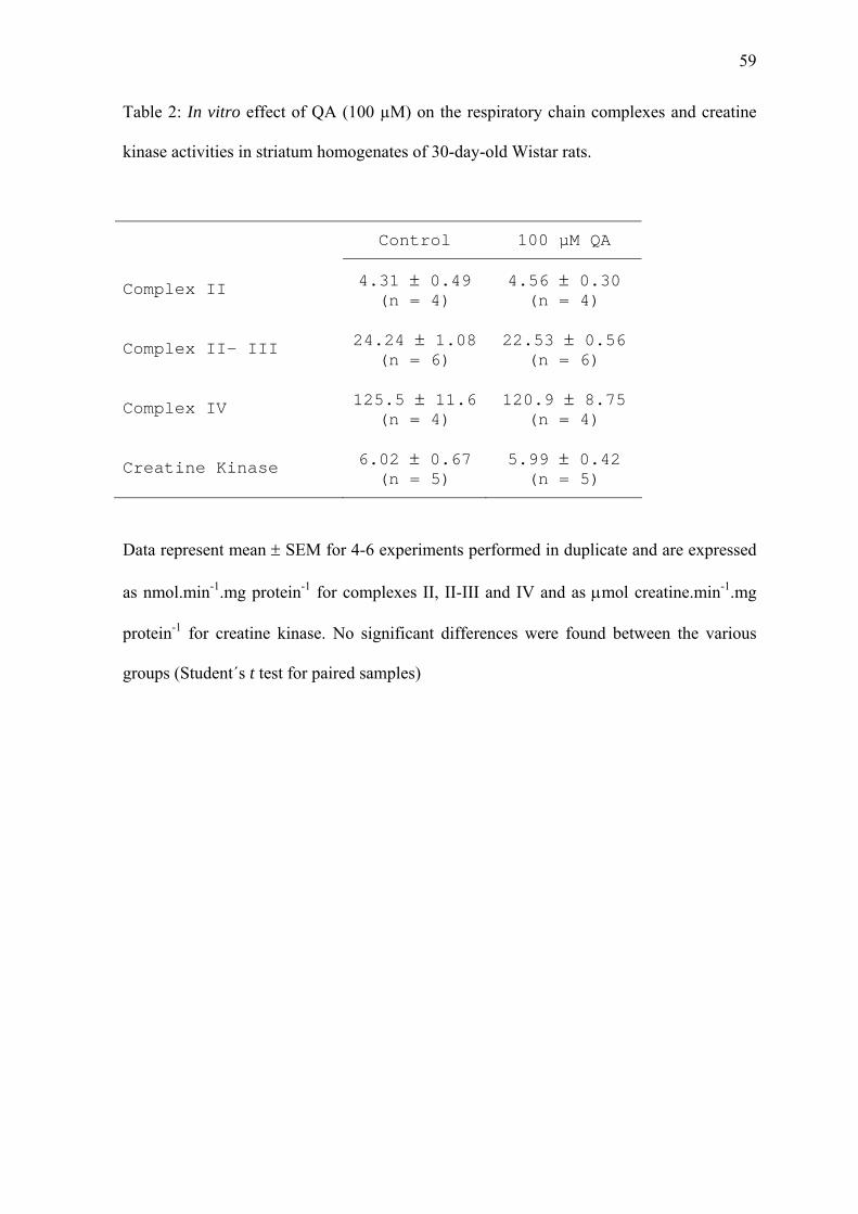
59
Table 2: In vitro effect of QA (100 µM) on the respiratory chain complexes and creatine
kinase activities in striatum homogenates of 30-day-old Wistar rats.
Control 100 µM QA
Complex II 4.31 ± 0.49 (n = 4)
4.56 ± 0.30 (n = 4)
Complex II- III 24.24 ± 1.08 (n = 6)
22.53 ± 0.56 (n = 6)
Complex IV 125.5 ± 11.6 (n = 4)
120.9 ± 8.75 (n = 4)
Creatine Kinase 6.02 ± 0.67 (n = 5)
5.99 ± 0.42 (n = 5)
Data represent mean ± SEM for 4-6 experiments performed in duplicate and are expressed
as nmol.min-1.mg protein-1 for complexes II, II-III and IV and as μmol creatine.min-1.mg
protein-1 for creatine kinase. No significant differences were found between the various
groups (Student´s t test for paired samples)

60
Legend to Figures
Figure 1: Respiratory chain complex activities in striatum 12 hours after intrastriatal
administration of 150 nmol QA. Animals received NaCl or QA in the left striatum as
described in Material and Methods. Data are represented as mean ± SEM for 3-6
independent experiments performed in duplicate and are expressed as nmol.min-1.mg
protein-1 * P < 0.05; ** P < 0.01 compared to NaCl (Student’s t test for unpaired samples).
Figure 2: Creatine kinase activity in striatum obtained at different periods (3-12 hours)
after intrastriatal administration of 150 nmol QA or NaCl. Animals received NaCl or QA
in the left striatum as described in Material and Methods. Data are represented as mean ±
SEM for 4-8 independent experiments performed in triplicate and are expressed as µmol
creatine.min-1.mg protein-1. ** P < 0.01 compared to NaCl (Student’s t test for unpaired
samples).
Figure 3: 14CO2 production from [1-14C]acetate in striatum obtained at different periods (3-
12 hours) after intrastriatal administration of 150 nmol QA or NaCl. Animals received
NaCl or QA in the left striatum as described in Material and Methods. Data are represented
as mean ± SEM for 4-5 independent experiments performed in triplicate and are expressed
as percentage of controls. * P < 0.05 compared to NaCl (Student’s t test for unpaired
samples).
Figure 4: Effects of α-tocopherol (40 mg/kg) plus ascorbic acid (100 mg/kg) on QA striatal
energy metabolism inhibition. Animals were treated with saline or α-tocopherol plus
ascorbic acid i.p. for 7 days prior to QA injection. Data are represented as mean ± SEM for

61
5 independent experiments (animals) performed in duplicate and are expressed as
percentage of controls. * Significantly different from contralateral striatum (control) (* P <
0.05; ** P < 0.001). Significantly different from ipsilateral striatum of rats that
received saline ( P < 0.05, P < 0.01). (Student’s t test for unpaired samples).
Figure 5: Effects of nitric oxide synthase inhibitor (L-NAME, 2 mg/kg) on QA striatal
energy metabolism inhibition. Animals were treated with saline or L-NAME i.p. 30 min
prior to QA injection. Data are represented as mean ± SEM for 6 independent experiments
(animals) performed in duplicate and are expressed as percentage of controls. *
Significantly different from contralateral striatum (control) (* P < 0.05; ** P < 0.01).
Significantly different from ipsilateral striatum of rats that received saline ( P <
0.01). (Student’s t test for unpaired samples).
Figure 6: Effects of pyruvate (500 mg/kg) on QA striatal energy metabolism inhibition.
Animals were treated with saline or pyruvate i.p. 1 hour prior to QA injection. Data are
represented as mean ± SEM for 4-7 independent experiments (animals) performed in
duplicate and are expressed as percentage of controls. * Significantly different from
contralateral striatum (control) (* P < 0.05; ** P < 0.01). Significantly different from
ipsilateral striatum of rats that received saline ( P < 0.05). (Student’s t test for unpaired
samples).
Figure 7: Effects of creatine (50 mg/kg) on QA striatal energy metabolism inhibition.
Animals were treated with saline or creatine i.p. for 7 days, twice a day, prior to QA
injection. Data are represented as mean ± SEM for 4-6 independent experiments (animals)
performed in duplicate and are expressed as percentage of controls. * Significantly
different from contralateral striatum (control) (* P < 0.05; ** P < 0.01). Significantly

62
different from ipsilateral striatum of rats that received saline ( P < 0.05, P <
0.01). (Student’s t test for unpaired samples).
Figure 8: Effects of pretreatment with MK-801 (0.25 mg/kg) prior to QA striatal energy
metabolism inhibition. Animals were treated with saline or MK-801 i.p. 30 min before QA
injection. Data are represented as mean ± SEM for 5 independent experiments (animals)
performed in duplicate and are expressed as percentage of controls. * Significantly
different from contralateral striatum (control) (* P < 0.05; ** P < 0.01). Significantly
different from ipsilateral striatum of rats that received saline ( P < 0.05). (Student’s t
test for unpaired samples).

63
Figure 1
NaCl QA0
100
200
300
400
500
(nm
ol /
min
/ m
g pr
otei
n)Complex I Complex II
NaCl QA0
2
4
6
8
(nm
ol /
min
/ m
g pr
otei
n)
*
NaCl QA0
5
10
15
20
25
30
**
(nm
ol /
min
/ m
g pr
otei
n)
NaCl QA0
5
10
15
20
25
30
35
40
Complex IV
Complex III
*
Complex II-III
NaCl QA0
20
40
60
80
100
120
(nm
ol /
min
/ m
g pr
otei
n)
(nm
ol /
min
/ m
g pr
otei
n)
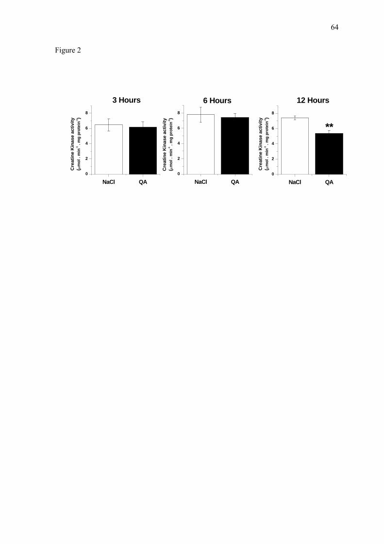
64
Figure 2
NaCl QA0
2
4
6
8
Cre
atin
e K
inas
e ac
tivity
( μm
ol .
min
-1 .
mg
prot
ein-1
)
Cre
atin
e K
inas
e ac
tivity
( μm
ol .
min
-1 .
mg
prot
ein-1
)
NaCl QA0
2
4
6
8
12 Hours6 Hours
Cre
atin
e K
inas
e ac
tivity
( μm
ol .
min
-1 .
mg
prot
ein-1
)
3 Hours
NaCl QA0
2
4
6
8
**

65
Figure 3
NaCl QA0
20
40
60
80
100
120
14C
O2 p
rodu
ctio
n fr
om
[1-14
C]a
ceta
te (%
of c
ontr
ol)
14C
O2 p
rodu
ctio
n fr
om
[1-14
C]a
ceta
te (%
of c
ontr
ol)
NaCl QA0
20
40
60
80
100
120
12 Hours6 Hours3 Hours
NaCl QA0
20
40
60
80
100
120
14C
O2 p
rodu
ctio
n fr
om
[1-14
C]a
ceta
te (%
of c
ontr
ol)
*
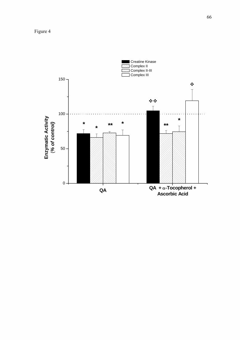
66
Figure 4
Salina Vit.E+C0
50
100
150
**
Enzy
mat
ic A
ctiv
ity(%
of c
ontr
ol) * ****
*
Creatine Kinase Complex II Complex II-III Complex III
QA QA + α-Tocopherol +Ascorbic Acid

67
Figure 5
Salina L-NAME0
50
100
150
** **** **
Enzy
mat
ic A
ctiv
ity
(% o
f con
trol
) *
Creatine Kinase Complex II Complex II-III Complex III
QA QA + L-NAME

68
Figure 6
Salina Piruvato0
50
100
150
***
Creatine Kinase Complex II Complex II-III Complex III
Enzy
mat
ic A
ctiv
ity
(% o
f con
trol
) ** **
*
QA QA + Pyruvate

69
Figure 7
Salina Creatina0
50
100
150
QA + Creatine
Enzy
mat
ic A
ctiv
ity(%
of c
ontr
ol)
**
* ****
Creatine Kinase Complex II Complex II-III Complex III
QA
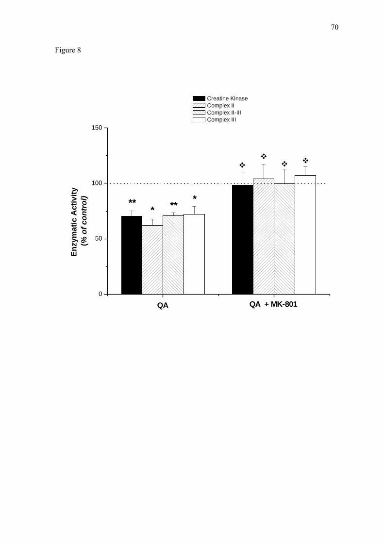
70
Figure 8
Salina MK-8010
50
100
150
Creatine Kinase Complex II Complex II-III Complex III
** **
Enzy
mat
ic A
ctiv
ity
(% o
f con
trol
)
**
QA QA + MK-801

71
PARTE III

72
1- DISCUSSÃO
O ácido quinolínico (AQ) é um agonista NMDA que exerce ações excitotóxicas no
sistema nervoso central (SNC) (STONE, 1993). No entanto, uma vez que o AQ é um
agonista relativamente fraco desses receptores (SCHWARCZ et al. 1983), os mecanismos
de toxicidade do AQ sobre o SNC continuam parcialmente esclarecidos. Neste particular
foi recentemente relatado que, apesar das concentrações de AQ aumentarem em até 10-100
vezes em células ativadas da microglia e em macrófagos invasores do SNC durante
processos neuroinflamatórios, essas quantidades de AQ ainda são insuficientes para
resultar em atividade excessiva dos receptores NMDA e morte neuronal (OBRENOVICH,
2001). Assim novos experimentos são necessários para verificar porque o AQ é tão
neurotóxico se todos os seus efeitos não podem ser atribuídos à superestimulação do SNC.
Por outro lado, tem sido demonstrado que o AQ induz lesões estriatais semelhantes
às encontradas na doença de Huntington, uma vez que o dano produzido por esse ácido
poupa os neurônios somatostatina-neuropeptídeo Y (BEAL et al. 1986). Daí o fato do AQ
estar sendo usado como modelo animal químico da DH para que se possa estudar a
patogênese dessa doença com a finalidade de se descobrir agentes terapêuticos para a
mesma. Interessante também é a constatação de que as concentrações do AQ estão
aumentadas postmortem no cérebro de pacientes afetados pela doença de Huntington e
também pela doença de Alzheimer (STONE, 2001). Recentemente a patogênese do dano
neurológico dessas patologias tem sido associada à excitotoxicidade, estresse oxidativo e
distúrbio metabólico (SALINZKA et al. 2005, ZARKOVIC, 2003, BEAL et al. 2000).
A presente investigação teve por objetivo estudar o efeito da administração
intraestriatal do AQ sobre vários parâmetros do metabolismo energético, com ênfase
particular sobre a produção e a transferência celular de energia. Utilizamos em nossos
experimentos animais jovens (30 dias de vida) visto que pouco se sabe sobre os efeitos do

73
AQ em animais dessa idade, sendo a maior parte dos trabalhos com o AQ realizada em
ratos de 3 ou mais meses de idade.
Verificamos inicialmente que o AQ não alterou a atividade dos complexos da
cadeia respiratória (fosforilação oxidativa) ou da enzima creatina quinase (CK)
(transferência de energia) em estriado obtido 3 ou 6 horas após a injeção intraestriatal de
AQ. No entanto, 12 horas após a injeção de AQ as atividades dos complexos II, II-III, III e
da CK estavam significativamente reduzidos em 50, 35, 46 e 27%, respectivamente. Tais
resultados indicam que a cadeia respiratória estava marcadamente bloqueada com reflexos
negativos sobre a produção de ATP. Tendo em vista que a CK é fundamental para a
transferência do ATP mitocondrial para o citosol, podemos concluir que a produção e a
transferência de energia intracelular foram comprometidas pela injeção intraestriatal do
AQ. Testamos também a atividade do ciclo do ácido cítrico pela medida da produção de
14CO2 a partir de [1-14C]acetato. Observamos que essa medida foi significativamente
reduzida 12 horas após a infusão de AQ. Esses resultados, tomados em seu conjunto,
indicam que o metabolismo energético no estriado dos animais foi marcadamente afetado
pelas concentrações elevadas de AQ após a sua administração intracerebral, mas não
informa se esse efeito ocorreu diretamente, ou secundariamente à estimulação de
receptores NMDA e/ou à geração aumentada de espécies ativas (RIOS & SANTAMARIA,
1991; BEHAN et al. 1999; SANTAMARIA et al. 2001a,b, LEIPNITZ et al. 2005). O fato
de que as alterações no metabolismo energético somente foram observadas após 12 horas
da infusão do AQ, podem levantar a suspeita de que esse efeito foi secundário a outras
alterações. Então resolvemos testar o efeito in vitro do AQ, na concentração de 100 μM,
sobre as atividades dos complexos da cadeia respiratória e da CK. Verificamos que o AQ,
na concentração de 100 μM, não foi capaz de alterar quaisquer das atividades
determinadas, o que sugere que o efeito do mesmo comprometendo o metabolismo
energético celular após a administração intraestriatal tenha sido secundário a outros efeitos.

74
Estudos anteriores demonstraram que lesões excitotóxicas via receptores NMDA
estão associadas com redução dos níveis de ATP e fosfocreatina (BORDELON, 1997,
MITANI, 1994). A redução da produção ou utilização de ATP pode levar à morte celular
via processos dependentes de energia. Assim, o ATP é essencial para várias bombas
iônicas que geram e mantêm os gradientes iônicos e elétricos nas membranas neuronais e
que são essenciais para o funcionamento das células neurais. Dentre essas bombas,
salientamos a bomba Na+,K+-ATPase que controla o potencial de membrana e também
outras ATPases que regulam os níveis intracelulares de Ca2+. (NOVELLI et al. 1988;
HENEBERRY et al. 1989, ZEEVALK et al. 1991).
A redução do metabolismo energético associada com estresse oxidativo podem
formar um ciclo de auto-amplificação de toxicidade celular, o que pode explicar o fato de
que somente encontramos efeitos 12 horas após a injeção de AQ.
É importante enfatizar que, apesar de não termos analisado histopatologicamente as
prováveis lesões provocadas pela administração intraestriatal do AQ, é provável que as
alterações no metabolismo energético aqui reportadas não tenham sido devidas à
diminuição do número de células neurais, visto que as atividades dos complexos I e IV da
cadeia respiratória e da enzima citrato sintase não foram alteradas pela injeção de AQ.
Nesse particular, enfatize-se os resultados de BORDELON e colaboradores (1994),
demonstrando que não houve diminuição significativa do número de células após horas de
injeções similares de AQ.
Assim, nossos dados são consistentes com achados de que as atividades dos
complexos da cadeia respiratória são marcada e seletivamente reduzidos no estriado de
pacientes com DH (TABRIZI et al. 1999, GU et al. 1996). Portanto, nossos resultados dão
suporte para a utilização de injeção intraestriatal de AQ como modelo químico animal para
indução de doenças neurodegenerativas.
Por outro lado, espécies reativas estão envolvidas em vários processos patológicos,

75
tais como no envelhecimento (BOKOV et al., 2004), na hipóxia (CHEN et al. 2003) e em
várias doenças neurodegenerativas, incluindo a doença de Huntington (STOY et al. 2005).
Além disso, diversos estudos demonstraram que o AQ pode induzir estresse oxidativo in
vitro e in vivo (LEIPNITZ et al. 2005, RIOS e SANTAMARIA, 1991; SANTAMARÍA e
RIOS, 1993). Assim, para avaliar o papel da geração de espécies reativas na disfunção
energética estriatal encontrada 12 horas após a injeção de AQ, os animais foram pré-
tratados com seqüestradores de radicais livres (α-tocoferol mais ascorbato) ou com um
inibidor da enzima óxido nítrico sintase (NOS, éster metílico de Nω-nitro-L-arginina, L-
NAME) e os parâmetros energéticos foram avaliados.
Observamos que o pré-tratamento com α-tocoferol e ascorbato administrados
simultaneamente foi capaz de prevenir a inibição da CK e do complexo III induzida pela
injeção de AQ, indicando que esses efeitos inibitórios foram mediados pela geração de
espécies reativas. O α-tocoferol interage com as membranas celulares, captura radicais
livres e interrompe a cadeia de reações oxidativas que causam dano às células. (AMES et
al. 1993; BURTON et al. 1990) Durante esse processo, α-tocoferol é convertido a radical
tocoferil, necessitando de ascorbato para sua regeneração a tocoferol reduzido (FREI et al.
1990; McCAY, 1985; CARR & FREI, 1999). Neste contexto, enfatize-se resultados
anteriores demonstrando que o complexo III da cadeia respiratória é inibido por espécies
ativas de oxigênio (CARDOSO et al. 1999), sendo que a CK mitocondrial pode ser atacada
por espécies ativas de nitrogênio e de oxigênio (GROSS et al. 1996; WENDT, et al. 2003;
STACHOWIACK et al. 1998), sendo que essas atividades foram completamente
prevenidas pelo pré-tratamento com α-tocoferol mais ascorbato.
Já o óxido nítrico (NO) está envolvido com a neurotoxicidade causada pela
estimulação de receptores glutamatérgicos (DAWSON et al. 1991), sendo verificado que a
estimulação de receptores glutamatérgicos e a conseqüente geração de NO são fatores

76
importantes na patogênese de várias doenças neurodegenerativas (MELDRUM &
GARTHWAITE, 1990, DAWSON et al. 1992). Neste contexto, está bem estabelecido que
a ativação de receptores NMDA resulta na ativação da oxido nítrico sintase (iNOS) pela
entrada intracelular de cálcio (AYATA et al. 1997), o que estimula a síntese de espécies
reativas de nitrogênio, como o NO e o seu derivado tóxico peroxinitrito.
Além disso, aumento na iNOS derivada de microglia e NOS neuronal também
foram demonstrados em estriado de ratos após a injeção de AQ (RYU et al. 2004,
SCHMIDT et al. 1995).
Nossos resultados mostraram que o pré-tratamento com L-NAME, um inibidor da
NOS, foi capaz de prevenir o efeito inibitório da atividade da CK causado pela injeção de
AQ, o que está de acordo com resultados prévios de que a atividade da CK é inibida por
peroxinitrito (WENDT et al. 2003; STACHOWIACK et al. 1998). Por outro lado, o pré-
tratamento com L-NAME não inibiu a diminuição de atividade dos vários complexos da
cadeia respiratória, indicando que as espécies ativas de nitrogênio não estão envolvidas
com a inibição da cadeia respiratória. Tomados os resultados em seu conjunto, é possível
que a entrada de cálcio na célula secundária à superestimulação dos receptores NMDA
pelo AQ possa ter induzido a produção de espécies ativas de oxigênio por parte da
mitocôndria que poderiam ser responsáveis pela inibição da cadeia respiratória.
Por outro lado, se o início da cascata patológica é a depleção das reservas
energéticas celulares, então agentes que mantêm os estoques energéticos celulares podem
atuar como agentes neuroprotetores.
Testamos também o efeito do pré-tratamento com substratos energéticos após a
injeção intraestriatal de AQ com o intuito de verificar se esses agentes, que podem ser
administrados de forma oral, poderiam prevenir ou atenuar o déficit energético verificado
no estriado através das medidas das atividades da cadeia respiratória e da creatina quinase.
O ácido pirúvico, metabólito final da glicólise, que é um substrato energético celular

77
(TSACOPOULOS & MAGISTRETTI, 1996) e também apresenta atividade antioxidante
(GIANDOMENICO et al. 1997), foi o primeiro substrato a ser testado. Estudos in vitro
mostraram capacidade neuroprotetora do piruvato sobre morte celular mediada por
ativação de receptores NMDA por estimular o metabolismo energético (MAUS et al.
1999). Em nosso estudo, demonstramos que o pré-tratamento com 500 mg/kg de piruvato
foi capaz de prevenir completamente os efeito inibitório causado pela injeção de AQ sobre
a atividade do complexo II e parcialmente sobre a atividade da CK. Nossos resultados
concordam com aqueles de RYU e colaboradores (2003, 2004), que observaram efeitos
neuroprotetores in vivo do piruvato sobre o dano estriatal induzido por AQ. É possível que
esses efeitos sejam devidos à capacidade do ácido pirúvico de neutralizar os efeitos do
H2O2 e de peróxidos lipídicos através de uma reação não-enzimática
(CONSTANTOPOULOS & BARRANGER, 1984; DEBOER et al. 1993;
CRESTANELLO et al. 1995), bem como de aumentar as concentrações dos intermediários
do ciclo do ácido cítrico (SUNDQVIST et al. 1989; RUSSELL & TAEGTMEYER, 1991).
Uma vez elevado, o citrato inibe a enzima fosfofrutoquinase (GARLAND et al. 1963) e
direciona o fluxo glicolítico para a via da hexose monofosfato, a principal fonte de
NADPH necessária para regenerar glutationa reduzida (HILTUNEN & DAVIS, 1981). O
aumento do fluxo nessa via aumenta a razão NADPH/NADP+ servindo para direcionar a
reação da glutationa redutase para a formação de glutationa reduzida, o principal agente
antioxidante do SNC. TEJERO-TALDO e colaboradores (1999) confirmaram essa hipótese
em um modelo de isquemia/reperfusão cardíaca seguida de tratamento com piruvato, onde
encontraram aumento dos níveis de citrato, NADPH e GSH. Embora não tenhamos medido
as concentrações de GSH após a administração de piruvato em nosso modelo, e se o
piruvato de fato aumenta os níveis de GSH em nosso modelo experimental, isso
concordará com os resultados de CARDOSO e colaboradores (1999), que demonstraram
que GSH é capaz de proteger a inibição induzida por estresse oxidativo na atividade do
78
complexo II.
Tendo em vista que estudos prévios demonstraram que GSH é capaz de proteger a
inibição da atividade do complexo II induzida por estresse oxidativo (CARDOSO et al.
1999), é possível que em nosso estudo o aumento de GSH provocado por piruvato tenha
prevenido o efeito inibitório do AQ sobre o complexo II da cadeia respiratória. Além disso,
considerando outros resultados, demonstrando que o tratamento com piruvato diminui em
aproximadamente 50% a expressão da iNOS em astrócitos, podemos presumir que essa
ação possa estar envolvida na prevenção parcial da inibição da atividade da CK provocada
pelo piruvato, que demonstramos ser provavelmente devida à formação de NO ou seus
metabólitos.
Por outro lado, considerando a gama crescente de evidências indicando efeitos
neuroprotetores da creatina sobre a toxicidade induzida por NMDA e malonato in vivo
(MALCON et al. 2000), por glutamato e peptídeo β-amilóide em neurônios hipocampais
cultivados (BREWER & WALLIMANN, 2000), e por 3-nitropropionato tanto in vivo
(MATTHEWS et al. 1998) quanto em culturas de neurônios estriatais e hipocampais
(BRUSTOVETSKY et al. 2001), testamos também o efeito do pré-tratamento com creatina
sobre o déficit energético provocado pela administração intraestriatal de AQ. Verificamos
que o pré-tratamento com creatina completamente preveniu os efeitos inibitórios sobre o
metabolismo energético induzidos pela injeção de AQ. Convém enfatizar que a creatina é
um excelente estimulante da respiração mitocondrial, resultando na formação de
fosfocreatina (PCr, KERNEC et al. 1996; O’GORMAN et al. 1996). Além disso, o
aumento na relação PCr/ATP estimula as bombas iônicas, especialmente as Ca2+-ATPases
do retículo sarcoplasmático-endoplasmático que têm alta demanda energética
(WALLIMANN & HEMMER, 1994), necessárias para manter a homeostase iônica,
excitabilidade e sinalização de Ca2+. Essas bombas estão acopladas à CK e necessitam uma
alta relação local de ATP/ADP para função eficiente. É sabido também que a creatina

79
aumenta o estado bioenergético celular de uma maneira geral, tornando a célula mais
resistente à injúria (ZHU et al. 2004).
A capacidade da PCr de aumentar o estado energético celular e, portanto, de
estimular a captação sináptica de glutamato, reduzindo a concentração extracelular de
glutamato, também pode ser considerado como um fator importante nos efeitos
neuroprotetores da creatina (XU et al. 1996). Neste particular, foi verificado em nosso
laboratório que o AQ estimula a liberação sinaptossomal de glutamato e inibe a captação
astrocitária deste neurotransmissor in vitro potencialmente levando à excitotoxicidade
(TAVARES et al. 2002). A excitotoxicidade, por sua vez, é acompanhada por um aumento
do influxo intracelular de Ca2+ (KHODOROV et al. 1996; STOUT et al. 1998), levando a
um ciclo vicioso por aumento crônico de Ca2+ e posterior deterioração do estado energético
celular, no caso das reservas energéticas serem insuficientes para o bombeamento de Ca2+
(BREWER & WALLIMANN, 2000).
Foi também demonstrado que a exposição por longo tempo com Cr induz a
transcrição da CK citosólica (KUZHIKANDATHIL & MOLLOY, 1994). Esse fato,
associado com o aumento da demanda energética induzida pela injeção de AQ, pode
explicar o aumento das atividades do complexo IV e da CK observados no estriado dos
ratos pré-tratados com creatina e que receberam injeção intraestriatal de AQ. Esse
resultado não é devido ao aumento da quantidade de mitocôndrias visto que a atividade da
citrato sintase permaneceu inalterada.
Além das observações acima referidas quanto às propriedades neuroprotetoras in
vivo e in vitro da creatina, também se tem constatado a estabilização do poro de transição
de permeabilidade mitocondrial pela conformação octamérica da CK (O’GORMAN et al.
1997, WYSS & KADDURAH-DAOUK, 2000). Assim, alguns dos efeitos da creatina
podem ser devidos à estabilização e proteção do poro de transição de permeabilidade e, por
conseguinte, da integridade mitocondrial (BRDICZKA et al. 1998).
80
Como o AQ é um agonista de receptores NMDA, resolvemos finalmente testar o
efeito do bloqueio desses receptores sobre o déficit energético causado pelo AQ. Neste
particular, o MK-801 é um antagonista de receptores NMDA não-competitivo que foi
descoberto na triagem de compostos com atividade anticonvulsivante (WONG et al. 1986),
bloqueando o influxo de cálcio mediado por NMDA (MURPHY et al. 1987). No presente
estudo, verificamos que o pré-tratamento sistêmico com MK-801 foi capaz de bloquear
completamente a toxicidade e todas as alterações do metabolismo energético induzidas
pela injeção de AQ. Esses achados estão de acordo com os achados de FOSTER e
colaboradores (1987 a,b), em que o MK-801 foi capaz de bloquear a degeneração do
estriado e do hipocampo induzida por NMDA, bem como a neurotoxicidade induzida por
AQ em estriado de ratos (WOODRUFF et al. 1987).
Tomadas essas observações em seu conjunto e aliadas aos resultados do presente
trabalho, podemos concluir que a inibição do ciclo do ácido cítrico, da cadeia respiratória e
da creatina quinase observadas no presente trabalho foram provavelmente secundárias à
superestimulação dos receptores NMDA por AQ. A prevenção da inibição de alguns
parâmetros por seqüestradores de radicais livres ou por L-NAME deveu-se possivelmente
ao impedimento da formação secundária de espécies reativas devidas à entrada de cálcio
conseqüente à superestimulação de receptores NMDA pelo AQ. Enfatize-se que, quando o
AQ foi adicionado (experimentos in vitro) ao meio de incubação, não houve nenhum efeito
do mesmo sobre os parâmetros de metabolismo energético analisados, o que está de acordo
com as conclusões de que o déficit energético provocado pela injeção intraestriatal in vivo
do AQ sobre o estriado é indireto e mediado por estímulo de receptores NMDA.
Apesar das bases moleculares da toxicidade glutamatérgica ainda não estarem
completamente elucidadas, há um consenso geral de que é dependente da entrada de cálcio
intracelular (CHOI, 1985, 1987, 1995). Além disso, sabe-se que os receptores NMDA são
importantes na mediação da neurotoxicidade glutamatérgica, possivelmente devido à sua

81
alta permeabilidade a cálcio (CHOI, 1988; TYMIANSKI, 1996). Nesse particular, há
várias evidências demonstrando a participação de íons cálcio no processo citotóxico.
Assim, o aumento do cálcio intracelular leva à ativação de várias enzimas, incluindo-se as
calpainas e outras proteases, proteínas quinases, oxido nítrico sintase, calcineurinas e
endonucleases. Há também aumento na produção de radicais livres, alterações na
organização do citoesqueleto, e ativação de sinais genéticos levando à morte celular
(SATTLER & TYMIANSKI, 2000).
82
2- CONCLUSÕES
Em conclusão, o presente estudo demonstra que a injeção intraestriatal de AQ
resulta em uma redução marcada das atividades dos complexos da cadeia respiratória e da
enzima CK no SNC de ratos jovens de maneira tempo-dependente. Além disso, nossos
achados mostram que os efeitos inibitórios causados pela administração intraestriatal de
AQ são totalmente dependentes da ativação de receptores NMDA. Nossos resultados
também demonstram que os efeitos inibitórios nas atividades da CK e do complexo III
podem ser devidos à geração de espécies reativas, já que tais efeitos inibitórios foram
prevenidos quando da pré-administração de seqüestradores de espécies reativas e/ou de um
inibidor da síntese de óxido nítrico. O aumento do estado energético pela pré-
administração de piruvato parcialmente preveniu os efeitos inibitórios causados pela
injeção de AQ, enquanto a pré-administração de creatina foi capaz de prevenir totalmente
os efeitos causados pelo AQ. Visto que ações neuroprotetoras da creatina têm sido
relatadas, tanto por aumentar o estado energético celular, como por sua capacidade
antioxidante, e também por ser bem tolerada em seres humanos, sendo rapidamente
absorvida quando administrada por via oral (BALSON et al. 1994, DAWSON et al. 1995,
HAGENFELDT et al. 1994), nossos achados sugerem que a suplementação com creatina
pode ser uma nova estratégia terapêutica em doenças neurodegenerativas em que ocorre
aumento das concentrações intracerebrais do AQ ou durante processos neuroinflamatórios
caracterizados quimicamente pelo aumento nas concentrações intracelulares dessa
neurotoxina (BEAL, 1992).

83
REFERÊNCIAS BIBLIOGRÁFICAS
84
• AKSENOV, M., AKSENOVA, M. V, PAYNE, R. M., TROJANOVSKI, J. Q., SCHMIDT, K. L., CARNEY, J. M., BUTTERFIELD, D. A., MARKESBERY, W. R. Oxidation of cytosolic proteins and expression of creatine kinase BB in frontal lobes of Neurodegenerative disorders. Dement. Geriatr. Cogn. Disord. 10, 158-165, 1999.
• AKSENOV, M., AKSENOVA, M. V., BUTTERFIELD, A. D., MARKESBERY,
W. R. Oxidative modification of creatine kinase BB in Alzheimer’s disease brain. J. Neurochem. 74, 2520-2527, 2000.
• ALLEGRI, G.; COSTA, C. V. L.; BERTAZZO, A., BISAIOLO, M., RAGAZZI, E.
Enzyme activities of tryptophan metabolism along the kynurenine pathway in various species of animals. Il Farmaco 58, 829-836, 2003
• AMES, B.N., SHIGENAGA, M.K., HAGEN, T.M., Oxidants, antioxidants and the
degenerative diseases of aging. Proc. Natl. Acad. Sci. U.S.A. 90, 7915–7922, 1993.
• AYATA C., AYATA G., HARA H., MATTHEWS, R.T., BEAL, M.F., FERRANTE, R.J., ENDRES, M., KIM, A., CHRISTIE, R.H., WAEBER, C., HUANG, P.L., HYMAN, B.T., MOSKOWITZ, M.A. Mechanisms of reduced striatal NMDA excitotoxicity in type I nitric oxide synthase knock-out mice. J. Neurosci. 17, 6908-6917, 1997.
• BABCOCK, G.T., WIKSTROM, M. Oxygen activation and the conservation of
energy in cell respiration. Nature 356, 301-309 1992.
• BALSOM, P.D., SODERLUND, K., EKBLOM, B. Creatine in humans with special reference to creatine supplementation. Sports Med. 18, 268-280, 1994.
• BARAN, H.; JELLIGER, K.; DEECKE, L. Kynurenine metabolism in Alzheimer’s
disease. J. Neural. Trasm. 106, 165-181, 1999.
• BEAL, M. F. Aging, energy, and oxidative stress in neurodegenerative disease. Ann. Neurol. 38, 357-366, 1995.
• BEAL, M. F.; HYMAN, B. T.; KOROSHETZ, W. Do defects in mitochondrial
energy metabolism underlie the pathology of neurodegenerative diseases? Trends Neurosci. 16, 125-131, 1993b.
• BEAL, M. F.; KOWALL, N. W.; ELLISON, D. W.; MAZUREK, M.F.; SWARTZ,
K.J.; MARTIN, J.B. Replication of the neurochemical characteristics of Huntington’s disease by quinolinic acid. Nature 321, 168-171, 1986.
• BEAL, M.F. Energetics in the pathogenesis of neurodegenerative diseases. Trends
Neurosci. 23, 298-304, 2000.
• BEAL, M.F.; BROUILLET, E.; JENKINS. B.; HENSHAW, R.; ROSEN, B.; HYMAN, B.T. Age-dependent striatal excitotoxic lesions produced by the endogenous mitochondrial inhibitor malonate. J. Neurochem. 61, 1147-1150, 1993a.

85
• BEHAN W.M., MCDONALD M., DARLINGTON L.G., STONE T.W. Oxidative
stress as a mechanism for quinolinic acid-induced hippocampal damage: protection by melatonin and deprenyl. Br. J. Pharmacol. 128, 1754-1760, 1999.
• BESSMAN, S. P., CARPENTER, C. L. The creatine–creatine phosphate energy
shuttle. Ann. Rev. Biochem. 54: 831-862, 1985.
• BOKOV, A., CHAUDHURI, A., RICHARDSON, A. The role of oxidative damage and stress in aging. Mech. Ageing Dev. 125, 811-826, 2004.
• BORDELON Y., MACKENZIE L., MCLAUGHLIN B.-A. CHESSELET M.-F.
Excitotoxic lesions of rat striatum: time course of DNA damage assessed by in situ nick translation. Soc. Neurosci., Abstr., 20, 988, 1994.
• BORDELON, Y.M., CHESSELET, M.F., NELSON, D., WELSH, F.,
ERECINSKA, M. Energetic dysfunction in quinolinic acid-lesioned rat striatum. J. Neurochem. 69, 1629-1639, 1997.
• BOWLING, A.C.; BEAL, M.F. Bioenergetic and oxidative stress in
neurodegenerative diseases. Life Sci. 56, 1151-1171, 1995.
• BRDICZKA, D., BEUTNER, G., RUCK, A., DOLDER, M., WALLIMANN, T. The molecular structure of mitochondrial contact sites. Their role in regulation of energy metabolism and permeability transition. Biofactors 8, 235-242, 1998.
• BRENNAN, W. A.; BIRD, E. D.; APRILLE, J. R. Regional mitochondrial
respiratory activity in Huntington’s disease brain. J. Neurochem. 44,. 1948-1950, 1985.
• BREWER, G.J., WALLIMANN, T.W. Protective effect of the energy precursor creatine against toxicity of glutamate and beta-amyloid in rat hippocampal neurons. J. Neurochem. 74, 1968-1978, 2000.
• BROWNE, S.E., BEAL, M.F. The energetics of Huntington's disease. Neurochem.
Res. 29, 531-546, 2004.
• BROWNE, S.E., BOWLING, A.C., MacGARVEY, U., BAIK, M.J., BERGER, S.C., MUQIT, M.M., BIRD, E.D., BEAL, M.F. Oxidative damage and metabolic dysfunction in Huntington's disease: selective vulnerability of the basal ganglia. Ann. Neurol. 41, 646-653, 1997.
• BRUSTOVETSKY, N., BRUSTOVETSKY, T., DUBINSKY, J.M. On the
mechanisms of neuroprotection by creatine and phosphocreatine. J. Neurochem. 76, 425-434, 2001.
• BURTON, G.W., WRONSKA, U., STONE, L., FOSTER, D.O., INGOLD, K.U.
Biokinetics of dietary RRR-α-tocopherol in the male guinea pig at three dietary levels of Vitamin C and two levels of Vitamin E. Lipids 25, 199–210, 1990.

86
• CARDOSO, S.M., PEREIRA, C., OLIVEIRA, R. Mitochondrial function is differentially affected upon oxidative stress. Free Radic. Biol. Med. 26, 3-13, 1999.
• CARR, A., FREI, B. Does Vitamin C act as a pro-oxidant under physiological
conditions?. FASEB J. 13, 1007–1024, 1999.
• CHEN, Q., VAZQUEZ, E.J., MOGHADDAS, S., HOPPEL, C.L., LESNEFSKY, E.J. Production of reactive oxygen species by mitochondria: central role of complex III. J. Biol. Chem. 278, 36027-36031, 2003.
• CHOI, D.W. Calcium: still center-stage in hypoxic-ischemic neuronal death.
Trends Neurosci. 18, 58-60, 1995.
• CHOI, D.W. Calcium-mediated neurotoxicity: relationship to specific channel types and role in ischemic damage. Trends Neurosci. 11, 465-469, 1988.
• CHOI, D.W. Glutamate neurotoxicity in cortical cell cultures is calcium dependent.
Neurosci. Lett. 58, 293-297, 1985.
• CHOI, D.W. Ionic dependence of glutamate neurotoxicity. J. Neurosci. 7, 369-379, 1987.
• CLARK, J.B., BATES, T.E., CULLINGFORD, T, Land, J.M. Development of
enzymes of energy metabolism in the neonatal mammalian brain. Dev. Neurosci-Basel. 17, 174-180, 1993.
• CONNICK, J.H., STONE, T.W. Quinolinic acid effects on amino acid release from
the rat cerebral cortex in vitro and in vivo. Br. J. Pharmacol. 93, 868-876, 1988.
• CONNICK, J.H., STONE, T.W. The effect of kainic, quinolinic and beta-kainic acids on the release of endogenous amino acids from rat brain slices. Biochem. Pharmacol. 35, 3631-3635, 1986.
• CONSTANTOPOULOS, G., BARRANGER, J.A. Non enzymatic decarboxylation
of pyruvate. Anal. Biochem. 139, 353–358, 1984.
• CRESTANELLO, J.A., KAMELGARD, J., WHITMAN, G.J.R. The cumulative nature of pyruvate's dual mechanism for myocardial protection. J. Surg. Res. 59, 198–204, 1995.
• CURZON, G. Brain tryptophan. Normal and disturbed control. Adv. Exp. Med.
Biol. 398, p. 27-34, 1996.
• DAVID, S.S., SHOEMAKER, M., HALEY, B.E. Abnormal properties of creatine kinase in Alzheimer’s disease brain: correlation of reduced enzyme activity and active site photolabelling with aberrant cytosol-membrane partitioning. Mol. Brain Res. 54, 276-287, 1998.
• DAVIS, J.N.; HUNNICUTT, J.C. Jr.; CHISHLOM, A. A mitochondrial bottleneck
hypothesis of Alzheimer’s disease. Mol. Med. Today 1, 240-247, 1995.
87
• DAWSON, B., CUTLER, M., MOODY, A., LAWRENCE, S., GOODMAN, C.,
RANDALL, N. Effects of oral creatine loading on single and repeated maximal short sprints. Aust. J. Sci. Med. Sport. 27, 56-61, 1995.
• DAWSON, T.M., DAWSON, V.L., SNYDER, S.H. A novel neuronal messenger
molecule in brain: the free radical, nitric oxide. Ann. Neurol. 32, 297-311, 1992.
• DAWSON, V.L., DAWSON, T.M., LONDON, E.D., BREDT, D.S., SNYDER S.H. Nitric oxide mediates glutamate neurotoxicity in primary cortical cultures. Proc. Natl Acad. Sci. USA 88, 6368-6371, 1991.
• DeBOER, L.W.V., BEKX, P.A., HAN, L., STEINKE, L. Pyruvate enhances
recovery of rat hearts after ischemia and reperfusion by preventing free radical generation. Am. J. Physiol. 265, H1571–H1576, 1993.
• DICKINSON, C.J. Cerebral oxidative metabolism in hypertension. Clin. Sci. 91,
539-550, 1996.
• ERECINSKA, M.; SILVER, I. A. Íons and energy in mammalian brain. Prog. Neurobiol. 43, 37-71, 1994
• FERRANTE, R.J., KOWALL, N.W., CIPOLLONI, P.B., STOREY, E., BEAL,
M.F., Excitotoxin lesions in primates as a model for Huntington’s disease: histophatologic and neurochemical characterization. Exp. Neurol. 119, 46-71, 1993.
• FOSTER, A.C., COLLINS, J.F., SCHWARCZ, R., On the excitotoxic properties of
quinolinic acid, 2,3-piridine dicarboxylic acid and structurally related compounds. Neuropharmacology 22, 1331-1342, 1983.
• FOSTER, A.C., GILL, R., KEMP, J.A., WOODRUFF, G.N. Systemic
administration of MK-801 prevents N-methyl-D-aspartate-induced neuronal degeneration in rat brain. Neurosci. Lett. 76, 307-311, 1987a.
• FOSTER, A.C., WONG, E.H. The novel anticonvulsant MK-801 binds to the
activated state of the N-methyl-D-aspartate receptor in rat brain. Br. J. Pharmacol. 91, 403-409, 1987b.
• FREI, B., STOCKER, R., ENGLAND, L. AMES, B.N. Ascorbate: the most
effective antioxidant in the blood. Adv. Exp. Med. Biol. 264, 155–163, 1990.
• GARLAND, P.B., RANDLE, P.J., NEWSHOLME, E.A. Citrate as an intermediary in the inhibition of phosphofructokinase in rat heart muscle by fatty acids, ketone bodies, pyruvate, diabetes and starvation. Nature 200, 169–170, 1963.
• GIBSON, G. F.; SHEU, K. F. R.; BLASS, J.P., BAKER, A., CARLSON, K.C.,
HARDING, B., PERRINO, P. Reduced activities of thiamine-dependent enzymes in the brains and peripheral tissues of patients with Alzheimer’s disease. Arch. Neurol. 45, 836-840, 1988.

88
• GRAFTON, S.T., MAZZIOTTA, J.C., PAHL, J.J., ST GEORGE-HYSLOP, P., HAINES, J.L., GUSELLA, J., HOFFMAN, J.M., BAXTER, L.R., PHELPS, M.E. Serial changes of cerebral glucose metabolism and caudate size in persons at risk for Huntington’s disease. Arch. Neurol. 49, 1161-1167, 1992.
• GROSS, W. L., BAK, M. I., INGWALL, J. S., ARSTALL, M. A., SMITH, T. W.,
BALLIGAND, J. L., KELLY, R. Nitric oxide inhibits creatine kinase and regulates rat heart contractile reserve. Proc. Natl Acad. Sci. USA 93: 5604-5609, 1996.
• GU, M., GASH, M.T., MANN, V.M. JAVOY-AGID F., COOPER J.M.,
SCHAPIRA A.H.V., Mitochondrial defect in Huntington’s disease caudate nucleus. Ann. Neurol. 39, 385-389, 1996.
• GU, M.; OWEN, A. D.; TOFFA, S. E. K. COOPER, J.M., DEXTER, D.T.,
JENNER, P., MARSDEN, C.D., SCHAPIRA, A.H. Mitochondrial function, GSH and iron in neurodegenerative and Lewy body diseases. J. Neurol. Sci. 158, 24-29, 1998.
• GUILLEMIN, G., CROITORU, J., DORMONT, D. et al. Involvement of
quinolinic acid in chemokine production and chemokine receptor expression in astrocytes. J. Neurovirol. 6, 242, 2000.
• GUILLEMIN, G., CROITORU, J., SMITH, D. G. et al. Quinolinic acid up-
regulates chemokine production and chemokine recptor expression in astrocytes: implications for brain inflammation and AIDS. J. Neuroimmunol. 118, 119, 2001a.
• GUILLEMIN, G. J., SMITH, D.G., WILLIAMS, K. et al. β-Amyloid peptide 1-42
induces human macrophages to produce the neurotoxin quinolinic acid. J. Neuroimmunol. 118, 336, 2001b.
• HAGENFELDT, L., von DOBELN, U., SOLDERS, G., KAIJSER, L. Creatine
treatment in MELAS. Muscle Nerve. 17, 1236-1237, 1994.
• HAGLUND, M.M., STAHL, W.L., KUNKEL, D.D., SCHWARTZKROIN, P.A. Developmental and regional differences in the localization of Na+,K+-ATPase activity in the rabbit hippocampus. Brain Res. 343, 198-203, 1985.
• HAMMAN, B.L., BITTL, J.A., JACOBUS, W.E., ALLEN, P.D., SPENCER, R.S.,
TIAN, R., INGWALL, J.S. Inhibition of the creatine kinase reaction decreases the contractile reserve of isolated rat hearts. Am. J. Physiol. 269, H1030–H1036, 1995.
• HAYAISHI, O. My life with tryptophan – never a dull moment. In: Ishiguro, I. Et
al. (Eds.) Advances in tryptophan research. Toyoake: Fujita Health University Press, p. 3-16, 1992.
• HAYDEN, M. R.; MARTIN, W. R. W.; STOESSL, A. J. et al. Positron emission
tomography in the early diagnosis of Huntington’s disease. Neurology 36, 888-894, 1986.
89
• HENNEBERRY, R.L., NOVELLI, A., COX, J.A., LYSKO, P.G. Neurotoxicity at the N-methyl-D-aspartate recetor in energy-compromised neurons: An hypothesis for cell death in aging and disease. Ann. NY Acad. Sci. 568, 225-233, 1989.
• HEYES, M.P. Hypothesis: a role for quinolinic acid in the neuropathology of
glutaric aciduria type I. Can. J. Neurol. Sci. 14, 441–443, 1987.
• HEYES, M.R., BREW, B.J., MARTIN, A., PRICE, R.W., SALAZAR, A.M., SIDTIS, J.J., YERGEY, J.A., MOURDIAN, M.M., SADLER, A.E., KEILP, J., RUBINOW, D., MARKEY, S.P. Quinolinic acid in cerebrospinal fluid and serum in HIV-1 infection: relationship to clinical and neurologic status. Ann. Neurol. 29, 202–209, 1991.
• HEYES, M.P., SAITO, K., CROWLEY, J.S., DAVIS, L.E., DEMITRACK, M.A.,
DER, M., DILLING, L.A., ELIA, J., KRUESI, M.J.P., LACKNER, A., LARSEN, S.A., LEE, K., LEONARD, H.L., MARKEY, S.P., MARTIN, A., MILSTEIN, S., MOURADIAN, M.M., PRANZATELLI, M.R., QUEARRY, B.J., SALAZAR, A., SMITH, M., STRAUS, S.E., SUNDERLAND, T., SWEDO, S.E., TOURTELLOTTE, W.W. Quinolinic acid and kynurenine pathway metabolism in inflammatory and non-inflammatory neurologic disease. Brain 115, 1249–1273, 1992.
• HEYES, M.P.; ACHIM, C. L.; WILEY, C. A., MAJOR, E.O., SAITO, K.,
MARKEY, S.P. Human microglia convert L-tryptophan into the neurotoxin quinolinic acid. Biochem. J. 320, 595-597, 1996.
• HILTUNEN, J.K., DAVIS, E.J. The disposition of citric acid cycle intermediates
by isolated rat mitochondria. Biochim. Biophys. Acta 678, 115–121, 1981.
• JAGUST, W. J.; FRIEDLAND, R. P.; BUDINGER, T. F., KOSS, E., OBER, B. Longitudinal studies of regional cerebral metabolism in Alzheimer’s disease. Neurology 38, 909-912, 1988.
• JANETZKY, B., HAUCK, S., YOUDIM, M.B.H., RIEDERER, P., JELLINGER,
K., PANTUCEK, F., ZOCHLING, R., BOISSL, K.W., REICHMANN, H. Unaltered aconitase activity but decreased complex I activity in substantia nigra pars compacta of patients with Parkinson’s disease. Neurosci. Lett. 169, 126-128, 1994.
• JENKINS, B.G., KOROSHETZ, W.J., BEAL, M.F., ROREN, B.R. Evidence for
impairment of energy metabolism in vivo in Huntington’s disease using localized 1H NMR spectroscopy. Neurology. 43, 2689-2695, 1993.
• KENNEDY, A.M., FRACKOWIAK, R.S., NEWMAN, S.K., BLOOMFIELD,
P.M., SEAWARD, J., ROQUES, P., LEWINGTON, G., CUNNINGHAM, V.J., ROSSOR, M.N. Deficits in cerebral glucose metabolism demonstrated by position emission tomography in individuals at risk of familial Alzheimer’s disease. Neurosci. Lett. 186, 17-20, 1995.

90
• KHODOROV B., PINELIS V., VERGUN O., STOROZHEVYKH T., VINSKAYA N. Mitochondrial deenergization underlies neuronal calcium overload following a prolonged glutamate challenge. FEBS Lett. 397 230–234, 1996.
• KUHL, D. E.; PHELPS, M. E.; MARKHAM, C. H. METTER, E.J., RIEGE, W.H.,
WINTER, J. Cerebral metabolism and atrophy in Huntington’s disease determined by 18FDG and computed tomographic scan. Ann. Neurol. 12, 425-434, 1982.
• KUWERT, T., LANGE, H.W., BOECKER, H., TITZ, H., HERZOG, H., AULICH,
A., WANG, B.C., NAYAK, U., FEINENDEGEN, L.E. Striatal glucose consumption in chorea-free subjects at risk of Huntington's disease. J. Neurol. 241, 31-36, 1993.
• KUZHIKANDATHIL, E.V., MOLLOY, G.R. Transcription of the brain creatine
kinase gene in glial cells is modulated by cyclic AMP-dependent protein kinase. J. Neurosci. Res. 39 70–82, 1994.
• LEIPNITZ, G., SCHUMACHER, C., SCUSSIATO, K., DALCIN, K.B.,
WANNMACHER, C.M., WYSE, A.T., DUTRA-FILHO, C.S., WAJNER, M., LATINI, A. Quinolinic acid reduces the antioxidant defenses in cerebral cortex of young rats. Int. J. Dev. Neurosci. 23, 695-701. 2005.
• LIAO, R., NASCIMBEN, L., FRIEDRICH, J., GAWTHMEY, J.K., INGWALL,
J.S. Decrease energy reserve in an animal model of dilated cardiomyopathy. Relationship to contractile performance. Cir. Res. 78, 893-902, 1996.
• LIPTON, S.A. Neuronal injury associated with HIV-1: approaches to treatment.
Ann. Rev. Pharmacol. 38, pp. 159–177, 1998.
• LUDOLPH, A.C., SEELIG, M., LUDOLPH, A., NOVITT, P., ALLEN, C.N., SPENCER, P., SABRI, M.I. 3-Nitropropionic acid decreases cellular energy levels and causes neuronal degeneration in cortical explants. Neurodegeneration 1, 155-161, 1992.
• MACCIONI, R.B.; MUNÕZ, P.J.; BARBEITO, L. The molecular bases of
Alzheimer’s disease and other neurodegenerative disorders. Arch. Med. Res. 32, 367-381, 2001.
• MALCON, C., KADDURAH-DAOUK, R., BEAL, M.F. Neuroprotective effects
of creatine administration against NMDA and malonate toxicity. Brain Res. 860, 195-198, 2000.
• MANN, V.M., COOPER, J.M., JAVOY-AGID, F., AGID, Y., JENNER, P.,
SCHAPIRA, A.H. Mitochondrial function and parental sex effect in Huntington's disease. Lancet 336, 749, 1990.
• MARKLUND, S. L., WESTMAN, N.G., LUNDGREN, E., ROOS, G. Copper- and
zinc-containing superoxide dismutase, manganese-containing superoxide dismutase, catalase, and glutathione peroxidase in normal and neoplastic human cell lines and normal human tissues. Cancer Res. 42, 1955-1961, 1982.

91
• MARKS, D. B.; MARKS, A. D.; SMITH, C. M. Basic Medical Biochemisty, A Clinical Approach. Lippincott Willians & Wilkins, 1996.
• MARTILLA, R. J.; ROYTTA, M.; LORENTZ, U. K. Rinne, U.K. Oxygen toxicity
protecting enzymes in the human brain. J. Neural. Tras. 74, 87-95, 1988.
• MARTIN, A., HEYES, M.P., SALAZAR, A.M., KAMPEN, D.L., WILLIAMS, J., LAW, W.A., COATS, M.E., MARKEY, S.P. Progressive slowing of reaction time and increasing cerebrospinal fluid concentrations of quinolinic acid in HIV-infected individuals. J. Neuropsychiat. Clin. Neurosci. 4, 270–279, 1992.
• MASTROGIACOMO, F.; BERGERON, C.; KISH, S. J. Brain alpha ketoglutarate
dehydrogenase complex activity in Alzheimer’s disease. J. Neurochem. 61, 2007-2014, 1993.
• MATTHEWS, R.T., YANG, L., JENKINS, B.G., FERRANTE, R.J., ROSEN,
B.R., KADDURAH-DAOUK, R., BEAL, M.F. Neuroprotective effects of creatine and cyclocreatine in animal models of Huntington's disease. J Neurosci. 18, 156-163, 1998.
• MAURER, I.; ZIERZ, S.; MÖLLER, H. J. A selective defect of cytochrome c
oxidase is present in brain of Alzheimer disease patients. Neurobiol. Aging 21, 455-462, 2000.
• McCAY, P.B. Vitamin E: interactions with free radical and ascorbate. Annu. Rev.
Nutr. 5, 323–340, 1985.
• MELDRUM, B. GARTHWAITE, J. Excitatory amino acid neurotoxicity and neurodegenerative disease. Trends Pharmacol. Sci. 11, 379-387, 1990.
• MITANI, A., TAKEYASU, S., YANASE, H., NAKAMURA, Y., KATAOKA, K.
Changes in intracellular Ca2+ and energy levels during in vitro ischemia in the gerbil hippocampal slice. J. Neurochem. 62, 626-634, 1994.
• MIZUNO, Y.; MATUDA, S.; YOSHIRO, H., MORI, H., HATTORI, H., IKEGE,
S. An immunohistochemical study on α-ketoglutarate dehydrogenase complex in Parkinson’s disease. Ann. Neurol. 35, 201-210, 1994.
• MIZUNO, Y.; SUZUKI, K.; OHTA, S. Postmortem changes in mitochondrial
respiratory enzymes in brain and a preliminary observation in Parkinson’s disease. J. Neurol. Sci. 96, 49-57, 1990.
• MOFFETT, J.R., NAMBOODIRI, M.A., Tryptophan and the immune response.
Imunology and Cell Biology, 81, 247-265, 2003.
• NELSON, D. L.; COX, M. M. Lehninger, Principles of Biochemistry. 4a ed. New York: Worth Publishers, 2004.
92
• NOVELLI, A., REILLY, J.A., LYSKO, P.G., HENNEBERRY, R.C. Glutamate becomes neurotoxic via the N-methyl-D-aspartate receptor when intracellular levels are reduced. Brain Res. 452, 205-212, 1988.
• O’GORMAN, E., BEUTNER, G., WALLIMANN, T., BRDICZKA, D. Differential
effects of creatine depletion on the regulation of enzyme activities and on creatine-stimulated mitochondrial respiration in skeletal muscle. Biochim. Biophys. Acta 1276, 161-170, 1996.
• OBRENOVITCH, T.P. Quinolinic acid accumulation during neuroinflammation.
Does it imply excitotoxicity? Ann. NY Acad. Sci. 939, 1-10, 2001.
• O'GORMAN, E., BEUTNER, G., WALLIMANN, T., BRDICZKA, D. Differential effects of creatine depletion on the regulation of enzyme activities and on creatine-stimulated mitochondrial respiration in skeletal muscle, heart, and brain. Biochim. Biophys. Acta 1276, 161-170, 1996.
• O'GORMAN, E., BEUTNER, G., DOLDER, M., KORETSKY, A.P., BRDICZKA,
D., WALLIMANN, T. The role of creatine kinase in inhibition of mitochondrial permeability transition. FEBS Lett. 414, 253–257, 1997.
• ORTH, M., SCHAPIRA, A.H.V. Mitochondria and degenerative disorders. Am. J.
Med. Gen. 106, 27-36, 2001.
• PARKER, W.D. Jr. Sporadic neurologic disease and the electron transport chain: a hypothesis. In: Pascuzzi (Ed.) Proceedings of the 1989 scientific meeting of the American Society for Neurological Investigation: new developments in neuromuscular disease. Bloomington: Indiana University Printing Services. 1990, 59-64, 1989.
• PARKER, W. D.; BOYSON, S. J.; LUDER, A. S. PARKS, J.K. Evidence for a
defect in NADH: ubiquinone oxidoreductase (complex I) in Huntington’s disease. Neurology 40, 1231-1234, 1990.
• PARKER, W.D. Jr.; PARKS J.K..; FILLEY, C.M., KLEINSCHMIDT-
DEMASTERS, B.K. Electron transport defects in Alzheimer’s disease brain. Neurology 44, 1090-1096, 1994.
• PERRY, E. K.; PERRY, R. I. I.; TOMLINSON, B. E., BLESSED, G., GIBSON,
P.H. Coenzyme A-acetylating enzymes in Alzheimer’s disease: possible cholinergic “compartment” of pyuvate dehydrogenase. Neurosci. Lett. 18, 105-110, 1980.
• REYNOLDS, I.J., MURPHY, S.N., MILLER, R.J. 3H-labeled MK-801 binding to
the excitatory amino acid receptor complex from rat brain is enhanced by glycine. Proc. Natl Acad. Sci. USA 84, 7744-7748, 1987.
• RIOS, C.; SANTAMARIA, A. Quinolinic acid is a potent lipid peroxidant in rat
brain homogenates. Neurochem. Res. 16, 1139-1143, 1991.

93
• ROSE, C.D.; HENNEBERRY, R.C. Etiology of the neurodegenerative disorders: a critical analysis. Neurobiol. Aging. 15, 233-234, 1994.
• ROSSEN, R.; KABAT, H.; ANDERSON, J. P. Acute arrest of cerebral circulation
in man. Arch. Neurol. Psychiatr. 50, 510-528, 1943.
• RUSSELL, R.R. III, TAEGTMEYER, H. Pyruvate carboxylation prevents the decline in contractile function of rat hearts oxidizing acetoacetate. Am. J. Physiol 261, H1756–H1762, 1991.
• RYU, J.K., KIM, S.U., MCLARNON, J.G. Blockade of quinolinic acid-induced
neurotoxicity by pyruvate is associated with inhibition of glial activation in a model of Huntington's disease. Exp. Neurol. 187, 150-159, 2004.
• SAKS, V.A, KUZNETSOV, A.V., KUPRIANOV, V.V., MICELI, M.V.,
JACOBUS, W.E. Creatine kinase of rat heart mitochondria. The demonstration of functional coupling to oxidative phosphorylation in an inner membrane-matrix preparation. J. Biol. Chem. 260, 7757-7764, 1985.
• SALINSKA, E., DANYSZ, W., LAZAREWICZ, J.W. The role of excitotoxicity in
neurodegeneration. Folia Neuropathol, 43, 322-339, 2005.
• SANTAMARÍA A., RIOS C. MK-801, an N-methyl-D-aspartate receptor antagonist, blocks dquinolinic acid-induced lipid peroxidation in rat corpus striatum. Neurosci. Lett. 159, 51-54, 1993.
• SANTAMARIA, A., GALVAN-ARZATE, S., LISY, V., ALI, S.F., DUHART,
H.M., OSORIO-RICO, L., RIOS, C., ST'ASTNY, F. Quinolinic acid induces oxidative stress in rat brain synaptosomes. Neuroreport 12, 871-874, 2001a.
• SANTAMARÍA, A., JIMENEZ-CAPDEVILLE, M.E., CAMACHO, A.,
RODRIGUEZ-MARTINEZ, E., FLORES, A., GALVAN-ARZATE, S. In vivo hydroxyl formation after quinolinic acid infusion into rat corpus striatum. Neuroreport 12, 2693-2696, 2001b.
• SATTLER, R., TYMIANSKI, M. Molecular mechanisms of calcium-dependent
excitotoxicity. J. Mol. Med. 78, 3-13, 2000.
• SCHAPIRA, A.H., COOPER, J.M., DEXTER, D., JENNER, P., CLARK, J.B., MARSDEN, C.D. Mitochondrial complex I deficiency in Parkinson’s disease. Lancet 1, 1269, 1989.
• SCHAPIRA, A.H., COOPER, J.M., DEXTER, D., CLARK, J.B., JENNER, P.,
MARSDEN, C.D. Mitochondrial complex I deficiency in Parkinson’s disease. J. Neurochem. 54, 823-827, 1990a.
• SCHAPIRA, A.H., MANN, V.M., COOPER, J.M., DEXTER, D., DANIEL, S.E.,
JENNER, P., CLARK, J.B., MARSDEN, C.D. Anatomic and disease specificity of NADH CoQ1reductase (complex I) deficiency in Parkinson’s disease. J. Neurochem. 55, 2142-2145, 1990b.

94
• SCHLEGEL, J., ZURBRIGGEN, B. , WEGMANN, G., WYSS, M.,
EPPENBERGER, H., WALLIMANN, T. Native mitochondrial creatine kinase forms octameric structures. I. Isolation of two interconvertible mitochondrial creatine kinase forms, dimeric and octameric mitochondrial creatine kinase: characterization, localization, and structure-function relationships. J. Biol. Chem. 263, 16942-53, 1988.
• SCHNYDER, T., GROSS, H., WINKLERM H., EPPENBERGER, H. M.,
WALLIMANN, T. Structure of the mitochondrial creatine kinase octamer: high-resolution shadowing and image averaging of single molecules and formation of linear filaments under specific staining conditions. J. Cell Biol. 112, 95-101, 1991.
• SCHWARCZ, R., FOSTER, A.C., FRENCH, E.D., WHETSELL Jr., W.O.,
KOHLER, C., Excitotoxic models for neurodegenerative disorders. Life Sci. 35, 19-32, 1984.
• SCHWARCZ. R,, WHETSELL, W.O. Jr., MANGANO, R.M. Quinolinic acid: an
endogenous metabolite that produces axon-sparing lesions in rat brain. Science 219, 316-318, 1983.
• SMALL, G. W.; MAZZIOTTA, J. C.; COLLINS, M. T. et al. Apolipoprotein E
type 4 allele and cerebral glucose metabolism in relatives at risk for familial Alzheimer’s disease. JAMA 273, 942-947, 1995.
• SMITH, G. S.; DE LEON, M.J.; KLUGER, A. Topography of cross sectional and
longitudinal glucose metabolic deficits in Alzheimer’s disease: Pathophysiologic implications. Arch. Neurol. 49, 1142-1150, 1992.
• STACHOWIAK, O., DOLDER, M., WALLIMANN, T., RICHTER, C..
Mitochondrial creatine kinase is a prime target of peroxynitrite-induced modification and inactivation. J. Biol. Chem. 273, 16694-16699, 1998.
• STONE, T. W. Neuropharmacology of quinolinic and kynurenic acids. Pharmacol.
Rev. 45, 309-379, 1993.
• STONE, T.W. Kynurenines in the CNS: from endogenous obscurity to therapeutic importance. Prog. Neurobiol. 64, 185-218, 2001a.
• STONE, T.W. Endogenous neurotoxins from tryptophan. Toxicon, 39, 61-73,
2001b.
• STOUT, A.K., RAPHAEL, H.M., KANTEREWICZ, B.I., KLANN, E., REYNOLDS, I.J. Glutamate-induced neuron death requires mitochondrial calcium uptake. Nat. Neurosci. 1 366–373, 1998.
• STOY, N., MACKAY, G.M., FORREST, C.M., CHRISTOFIDES, J., EGERTON,
M., STONE, T.W. Darlington LG. Tryptophan metabolism and oxidative stress in patients with Huntington's disease. J. Neurochem. 93, 611-623, 2005.

95
• SUNDQVIST, K.E., HILTUNEN, J.K., HASSINEN, I.E. Pyruvate carboxylation in the rat heart. Role of biotin-dependent enzymes. Biochem. J. 257, 913-916, 1989.
• SWERDLOW, R.H., PARKS, J.K., CASSARINO, D.S., MAGUIRE, D.J.,
MAGUIRE, R.S., BENNETT, J.P. JR, DAVIS, R.E., PARKER, W.D. Jr. Cybrids in Alzheimer’s disease: a celular model of the disease? Neurobiology 49, 918-925, 1997.
• SWERDLOW, R.H., PARKS, J.K., MILLER, S.W., TUTTLE, J.B., TRIMMER,
P.A., SHEEHAN, J.P., BENNETT, J.P. Jr, DAVIS, R.E., PARKER, W.D. Jr. Origin and functional consequences of the complex I defect in Parkinson’s disease. Ann. Neurol. 40, 663-671, 1996.
• SWERDLOW, R.H., PARKS, J.K., CASSARINO, D.S., TRIMMER, P.A.,
MILLER, S.W., MAGUIRE, D.J., SHEEHAN, J.P., MAGUIRE, R.S., PATTEE, G., JUEL, V.C., PHILLIPS, L.H., TUTTLE, J.B., BENNETT, J.P. Jr, DAVIS, R.E., PARKER, W.D. Jr. Mitochondria in sporadic amyotrophic lateral sclerosis. Exper. Neurol. 153, 135-142, 1998.
• TABRIZI, S.J., CLEETER, M.W., XUEREB, J., TAANMAN, J.W., COOPER,
J.M., SCHAPIRA, A.H. Biochemical abnormalities and excitotoxicity in Huntington's disease brain. Ann. Neurol., 45, 25-32, 1999.
• TAVARES, R.G., TASCA, C.I., SANTOS, C.E., ALVES, L.B., PORCIÚNCULA,
L.O., EMANUELLI, T., SOUZA, D.O. Quinolinic acid stimulates synaptosomal glutamate release and inhibits glutamate uptake into astrocytes. Neurochem. Int. 40, 621-627, 2002.
• TEJERO-TALDO, M.I., CAFFREY, J.L., SUN, J., MALLET, R.T. Antioxidant
properties of pyruvate mediate its potentiation of beta-adrenergic inotropism in stunned myocardium. J. Mol. Cell. Cardiol. 31, 1863-1872, 1999.
• TYMIANSKI, M. Cytosolic calcium concentrations and cell death in vitro. In:
Stiesjo, B.K., Wieloch, T. (eds). Advances in neurology. Lippincott-Raven, Philadelphia, 85-105, 1996.
• VARADKAR, S., SURTEES, R. Glutaric aciduria type I and kynurenine pathway
metabolites: a modified hypothesis. J. Inherit. Metab. Dis. 27, 835-842, 2004.
• WADIA, J.S., CHALMERS-REDMAN, R.M., JU, W.J., CARLILE, G.W., PHILLIPS, J.L., FRASER, A.D, TATTON, W.G. Mitochondrial membrane potential and nuclear changes in apoptosis caused by trophic withdrawal: time course and modification by (-) deprenyl. J. Neurosci. 18, 932-947, 1998.
• WALLACE, D. C. Mitochondrial diseases in man and mouse. Science 283, 1482-
1487, 1999.
• WALLIMANN, T., HEMMER, W. Creatine kinase in non-muscle tissues and cells. Mol. Cell. Biochem. 133-134, 193-220, 1994.

96
• WALLIMANN, T., WYSS, M., BRDICZKA, D., NICOLAY, K, EPPENBERGER, H.M.. Intracellular compartmentation, structure and function of creatine kinase isoenzymes in tissues with high and fluctuating energy demands: the 'phosphocreatine circuit' for cellular energy homeostasis. Biochem. J. 281, 21-40, 1992.
• WENDT, S., SCHLATTNER, U., WALLIMANN, T. Differential effects of
peroxynitrite on human mitochondrial creatine kinase isoenzymes. Inactivation, octamer destabilization, and identification of involved residues. J. Biol. Chem. 278, 1125-30, 2003.
• WHETSELL, W.O. Jr., SCHWARCZ, R. Prolonged exposure to submicromolar
concentrations of quinolinic acid causes excitotoxic damage in organotypic cultures of rat corticostratal system. Neurosci. Lett. 97, 271-275, 1989.
• WIDNER, B.; LEBLHUBER, F.; WALLI, J. TILZ, G.P., DEMEL, U., FUCHS, D.
Degradation of tryptophan in neurodegenerative disorders. Adv. Exp. Med. Biol. 467, 133-138, 1999.
• WIDNER, B., LEBLHUBER, F., WALLI, J., TILZ, G.P., DEMEL, U., FUCHS, D.
Tryptophan degradation and immune activation in Alzheimer’s disease. J. Neural Transm. 107, 343-353, 2000.
• WONG, E.H., KEMP, J.A., PRIESTLEY, T., KNIGHT, A.R., WOODRUFF, G.N.,
IVERSEN, L.L. The anticonvulsant MK-801 is a potent N-methyl-D-aspartate antagonist. Proc. Natl Acad. Sci. USA. 83, 7104-7108, 1986.
• WOODRUFF, G.N., FOSTER, A.C., GILL, R., KEMP, J.A., WONG, E.H.,
IVERSEN, L.L. The interaction between MK-801 and receptors for N-methyl-D-aspartate: functional consequences. Neuropharmacology 26, 903-909, 1987.
• WYSS, M., Kaddurah-Daouk, R. Creatine and creatinine metabolism. Physiol. Rev.
80, 1107-213, 2000.
• XU, C.J., KLUNK, W.E., KANFER, J.N., XIONG, Q., MILLER, G., PETTEGREW, J.W. Phosphocreatine-dependent glutamate uptake by synaptic vesicles. A comparison with atp-dependent glutamate uptake. J. Biol. Chem. 271, 13435-13440, 1996.
• YOUNG, A.B., PENNEY, J.B., STAROSTA-RUBINSTEIN, S., MARKEL, D.S.,
BERENT, S., GIORDANI, B., EHRENKAUFER, R., JEWETT, D., HICHWA, R. PET scan investigations of Huntington’s disease: cerebral metabolic correlates of neurological features and functional decline. Ann. Neurol. 20, 296-303, 1986.
• ZARKOVIC, K. 4-hydroxynonenal and neurodegenerative diseases. Mol. Aspects
Med. 24, 293-303, 2003
• ZEEVALK, G.D., NICKLAS, W.J. Mechanisms underlying initiation of excitotoxicity associated with metabolic inhibition, J. Pharm. Exp. Ther., 257, 870-878, 1989.