dinÂmica molecular dos vÍrus influenza a (h1n1 ... · nossos resultados revelaram a circulação...
TRANSCRIPT

MINISTÉRIO DA SAÚDE FUNDAÇÃO OSWALDO CRUZ INSTITUTO OSWALDO CRUZ
Doutorado Biologia Celular e Molecular
DINÂMICA MOLECULAR DOS VÍRUS INFLUENZA A (H1N1) PANDÊMICO EM CINCO ANOS DE CIRCULAÇÃO NO BRASIL
PAOLA CRISTINA RESENDE SILVA
Rio de Janeiro Junho de 2015

ii
INSTITUTO OSWALDO CRUZ Programa de Pós-Graduação em Biologia Celular e Mol ecular
PAOLA CRISTINA RESENDE SILVA
DINÂMICA MOLECULAR DOS VÍRUS INFLUENZA A (H1N1) PAN DÊMICO EM CINCO ANOS DE CIRCULAÇÃO NO BRASIL
Tese apresentada ao Instituto Oswaldo Cruz
como parte dos requisitos para obtenção do título
de Doutor em Ciências pela Pós-graduação em
Biologia Celular e Molecular
Orientadora: Dra. Marilda Mendonça Teixeira de Agudo Siqueira
RIO DE JANEIRO Junho de 2015

Ficha catalográfica elaborada pela
Biblioteca de Ciências Biomédicas/ ICICT / FIOCRUZ - RJ
S586 Silva, Paola Cristina Resende
Dinâmica molecular dos vírus Influenza A (H1N1) pandêmico em cinco anos de circulação no Brasil / Paola Cristina Resende Silva. – Rio de Janeiro, 2015.
xviii,176 f. : il. ; 30 cm.
Tese (Doutorado) – Instituto Oswaldo Cruz, Pós-Graduação em
Biologia Celular e Molecular, 2015. Bibliografia: f. 158-172
1. Influenza. 2. H1N1. 3. Pandemia. 4. Sequenciamento. 5. Hemaglutinina. 6. Neuraminidase. 7. Resistência antiviral. I. Título.
CDD 614.518

iv
INSTITUTO OSWALDO CRUZ Programa de Pós-Graduação em Biologia Celular e Mol ecular
AUTOR: PAOLA CRISTINA RESENDE SILVA
DINÂMICA MOLECULAR DOS VÍRUS INFLUENZA A (H1N1) PAN DÊMICO EM CINCO ANOS DE CIRCULAÇÃO NO BRASIL
ORIENTADORA: Dra. Marilda Mendonça Teixeira de Agu do Siqueira Aprovada em: 22 de julho de 2015 EXAMINADORES: Prof. Dra. Ana Maria Bispo de Filippis – Pesquisadora no Laboratório de Flavivirus do IOC - FIOCRUZ Prof. Dra. Nancy Cristina Junqueira Bellei – Pesquisadora na Universidade Federal de São Paulo - UNIFESP Prof. Dr. Edson Elias da Silva – Pesquisador no Laboratório de Enterovirus do IOC - FIOCRUZ Prof. Dr. Gonzalo José Bello Bentacor – Pesquisador no Laboratório de AIDS e Imunologia Molecular do IOC – FIOCRUZ Prof. Dra. Glória Regina da Silva e Sá – Pesquisadora na Universidade Federal do Estado do Rio de Janeiro
Rio de Janeiro, 22 de junho de 2015.

v
Dedic o esta tese aos meus amados pais Edna Maria Carvalho Resende Silva e Dilson de Oliveira Silva.

vi
AGRADECIMENTOS
Agradecer não é tarefa fácil, não pelo ato em si, mas pelo receio de não mencionar alguns daqueles que me inspiraram e me ajudaram nessa caminhada de altos e baixos, mas que trilhei com muito amor pelo que eu faço.
Agradeço à Deus por me amparar nos momentos difíceis, me dar força interior para superar as dificuldades e mostrar os caminhos nas horas incertas.
À minha querida orientadora e amiga Dra. Marilda Mendonça Siqueira, por me dar a oportunidade de trabalhar ao seu lado, e especialmente por me apoiar e me ensinar com o seu enorme coração a crescer profissional e pessoalmente. Hoje não tenho dúvidas que fiz a escolha certa. Obrigada por todo empenho, amizade, sabedoria e compreensão no decorrer deste trabalho.
Ao amigo Dr. Fernando Couto Motta pelo companheirismo, incentivo e imenso profissionalismo. Obrigada por me ensinar a trabalhar com biologia molecular e especialmente por toda ajuda ao longo desses anos.
Ao amigo Dr. Thiago Moreno Lopes e Souza pelo carinho, por me apoiar e direcionar diversas vezes que eu precisei ao longo da minha trajetória acadêmica.
Às minhas queridas amigas Priscila Born, Daniela Bandeira, Sharon Carney e Milene Mesquita que me aguentaram e compartilharam comigo diversos momentos ao longo dessa tese.
À toda equipe do Laboratório de Vírus Respiratórios e do Sarampo (LVRS), não só os que fazem parte do laboratório hoje, mas todos os que fizeram parte ao longo destes quatro anos. Vocês são a minha segunda família! Muito obrigada pelo convívio diário, palavras de apoio e incentivo, lições de vida e carinho.
À minha revisora Dra. Braulia Caetano, que fez um excelente trabalho, teve toda paciência e dedicação durante a revisão.
À minha família, especialmente aos meus pais, Edna e Dilson, por terem sido o contínuo apoio nestes quatro anos, ensinando-me, principalmente, a importância da construção e coerência de meus próprios valores.
Ao curso de pós-graduação em Biologia Celular e Molecular, coordenação, secretaria e todo o corpo docente, que confiaram no meu trabalho e me deram a oportunidade de crescimento, aprendizado e realização profissional.
A todos os meus colegas da pós-graduação do Instituto Oswaldo Cruz (IOC) que estiveram comigo em várias disciplinas, fóruns discentes e congressos.

vii
À banca por terem aceitado o convite de avaliar o meu trabalho e terem dado excelentes sugestões.
Ao Conselho Nacional de Desenvolvimento Científico e Tecnológico (CNPq), pela bolsa concedida durante os quatro anos do curso e ao Departamento da Ciência e Tecnologia (DECIT) pelo financiamento concedido para realização desta tese.
E finalmente, a todos que mesmo não sendo mencionados contribuíram para a conclusão desta tese. Muito obrigada!

viii
“ Se enxerguei mais longe, foi porque me apoiei sobre os ombros de gigantes. ” (Isaac Newton)

ix
INSTITUTO OSWALDO CRUZ DINÂMICA MOLECULAR DOS VÍRUS INFLUENZA A (H1N1) PAN DÊMICO EM CINCO ANOS DE
CIRCULAÇÃO NO BRASIL
RESUMO
TESE DE DOUTORADO EM BIOLOGIA CELULAR E MOLECULAR
Paola Cristina Resende Silva
A primeira detecção do vírus Influenza A (H1N1)pdm09 no Brasil aconteceu em maio de
2009, e foi seguida de uma extensa disseminação por toda a população brasileira, com grande impacto em morbidade e mortalidade. Para entender a dinâmica molecular do Influenza A (H1N1)pdm09 no país, a presente tese reuniu sete trabalhos que abordaram a análise filogenética deste agente viral durante e após o período pandêmico (2009 a 2014) e buscou indentificar polimorfismos virais associados à virulência e à resistência ao antiviral Oseltamivir (OST). Para isso, as metodologias realizadas foram o sequenciamento dos genes de hemaglutinina (HA) e neuraminidase (NA) utilizando a metodologia de Sanger e a metodologia de pirosequenciamento para detectar polimorfismos de base única (SNPs).
Nossos resultados revelaram a circulação de nove grupos filogenéticos ao longo dos cinco anos do estudo, indicando uma substituição temporal dos grupos e ocasionalmente uma estratificação geográfica. No entanto, nenhum dos grupos filogenéticos identificados foram associados com um pior prognóstico da infecção por influenza. Ao contrário do que foi observado em estudos anteriores, as mutações K-15E e Q310H no gene HA não se associaram ao aumento de virulência, mesmo na infecção de indivíduos imunocomprometidos. Por outro lado, polimorfismos no resíduo 222 da HA, que caracterizaram a presença de quasispecies virais, mostraram uma forte associação com a gravidade da infecção, especialmente em gestantes. Nesta tese, também realizamos a vigilância de marcadores de resistência no gene NA. Entre as amostras analisadas encontramos sete vírus com a mutação H275Y e dois com S247N, esses marcadores estão relacionados com a diminuição de sensibilidade ao antiviral OST. Entre as amostras resistentes, a grande maioria foi detectada na região Sul do Brasil, em pacientes que não receberam OST. Isto sugere uma possível transmissão sustentada do vírus resistentes no país. Embora variantes H275Y resistentes apresentem baixa aptidão viral, a propagação deste vírus pode ocorrer com o ganho de mutações permissivas nos genes HA ou NA. No Brasil, o vírus que circulam desde 2011 apresentaram mutações V241I e N369K no gene NA, que foram, teoricamente, associadas a uma maior aptidão viral da variante resistente.
Diante disso, cinco anos após a pandemia observamos que o vírus A (H1N1)pdm09 está estabelecido na população humana com várias substituições genéticas quando comparado com a sequencia da cepa vacinal A/California/07/2009. A maioria destas substituições parecem não ocasionar uma maior gravidade da infecção, e esta pode ser atribuída a outros fatores, tais como fatores genéticos do hospedeiro. Por fim, considerando o risco de surgimento e disseminação de vírus resistentes é importante reforçar o monitoramento viral e também realizar estudos para novas drogas antivirais.

x
INSTITUTO OSWALDO CRUZ
MOLECULAR DYNAMIC OF INFLUENZA A (H1N1) PANDEMIC VI RUSES FIVE YEARS OF CIRCULATION IN BRAZIL
ABSTRACT
THESIS IN CELLULAR AND MOLECULAR BIOLOGY
Paola Cristina Resende Silva
The Influenza A (H1N1)pdm09 virus was first detected in May 2009 in Brazil and later resulted in an extensive spread throughout the Brazilian population with a severe impact on morbidity and mortality. To understand the molecular dynamic of (H1N1)pdm09 virus in Brazil this thesis grouped seven papers which approached the phylogenetic reconstruction of the virus during and after the pandemic period (2009 to 2014) and the genomic identification of viral polymorphisms associated with virulence or antiviral resistance to Oseltamivir (OST). For this, we performed genome sequencing, focusing especially on the hemagglutinin (HA) and neuraminidase (NA) genes using conventional Sanger sequencing and PyroMark 96ID to detect single nucleotide polymorphisms (SNPs).
Our results showed that in Brazil nine (H1N1)pdm09 phylogenetic groups circulated along the five years of the study, indicating a temporal replacement of groups and ocasionally a geographic stratification. However, no phylogenetic group seemed to be associated with a worse clinical outcome. The increased virulence observed in previous studies with a 2009 group bearing the genetic markers K-15E and Q310H was not confirmed in our analyses, even evaluating an immunocompromised population. On the other hand, polymorphysms at position 222 of HA gene, which characterized the presence of viral quasispecies, showed an association with increased virulence in brazilian samples, especially in pregnant women. In this study we also performed surveillance of resistance markers at the NA gene. From the analysed samples we found seven viruses with H275Y and two with S247N mutation, that diminish the sensibility to oseltamivir (OST). Among the resistant samples, the large majority was detected in the Southern region of Brazil in patients that did not receive OST. This suggests a possible sustained transmission of resistant virus in the country. Although resistant H275Y variants have low viral fitness, the spread of this virus can occur with the gain of permissive mutations in the HA or NA genes. In Brazil, viruses circulating since 2011 presented mutations V241I and N369K in the NA gene, which were theoretically associated with greater viral fitness.
Five years after the pandemic we observed that A (H1N1)pdm09 is established in the population with genetic substitutions in all gene segments when compared to the vaccine strain A/California/07/2009. We demonstrated that the majority of those substitutions did not increase the severity of infection. The worst clinical outcomes may be attributed to other factors, such as genetic host factors. Finally, considering the risk of emergence and spread of resistant viruses it is important to strengthen viral surveillance and also carry out studies for new antiviral drugs.

xi
ÍNDICE
Lista de Figuras .................................. ........................................................... xiv
Lista de Tabelas .................................. ........................................................... xvi
Lista de Siglas e Abreviaturas .................... ................................................. xvii
1 INTRODUÇÃO 19
1.1 Propriedades gerais dos vírus Influenza A ......... ................................. 19
1.1.1 Taxonomia e nomenclatura ......................................................... 19
1.1.2 Morfologia e estrutura genômica ................................................. 21
1.1.3 Ciclo replicativo dos vírus Influenza A ......................................... 26
1.1.4 Mecanismos evolutivos dos vírus Influenza ................................. 31
1.2 Histórico e aspectos moleculares das principais pan demias e
epidemias ocasionadas pelos vírus Influenza A ..... ............................ 34
1.2.1 Pandemias causadas pelos vírus Influenza A ............................. 34
1.2.2 Importantes surtos e epidemias de variantes de Influenza
A entre humanos ......................................................................... 40
1.3 Epidemiologia dos vírus Influenza A (H1N1)pdm09 ... ......................... 44
1.3.1 Sazonalidade dos vírus influenza A ............................................. 46
1.3.2 Impacto do vírus A (H1N1)pdm09 no Brasil ................................. 47
1.3.3 Epidemiologia molecular e evolução dos vírus A
(H1N1)pdm09 .............................................................................. 49
1.4 Resposta imune contra a infecção pelos vírus Influe nza ................... 54
1.5 Patogênese dos vírus Influenza A (H1N1)pdm09 ...... .......................... 59
1.5.1 Fatores de patogenicidade virais intrínsecos ............................... 59
1.5.2 Fatores do hospedeiro ................................................................. 60
1.6 Diagnóstico e vigilância laboratorial dos vírus Inf luenza ................... 63
1.6.1 Diagnóstico dos vírus Influenza ................................................... 63
1.6.2 Métodos para caracterização molecular dos vírus
Influenza ...................................................................................... 64
1.6.3 Rede de vigilância dos vírus Influenza......................................... 66
1.7 Controle dos vírus Influenza A .................... .......................................... 69
1.7.1 Vacinação anti-influenza .............................................................. 69
1.7.2 Antivirais e resistência ................................................................. 71
2 JUSTIFICATIVA 77

xii
3 OBJETIVOS 78
3.1 Objetivo Geral .................................... ..................................................... 78
3.2 Objetivos Específicos ............................. ............................................... 78
CAPÍTULO I: FILOGENIA DO GENE DA HEMAGLUTININA DOS VÍRUS
INFLUENZA A (H1N1)PDM09 DETECTADOS NO BRASIL E ASSO CIAÇÕES
COM A CEPA VACINAL A/CALIFORNIA/7/2009 79
Documento I – Molecular findings from influenza A ( H1N1)pdm09
detected in patients from a Brazilian equatorial re gion during
the pandemic period ............................... ............................................... 82
Documento II – Phylogenetic analyzes of Influenza A (H1N1)pdm09
hemagglutinin gene during and after the pandemic ev ent in
Brazil ............................................ ............................................................ 90
CAPÍTULO II: IDENTIFICAÇÃO DE MARCADORES DE VIRULÊN CIA
ASSOCIADOS AO AUMENTO DA GRAVIDADE DA INFECÇÃO PELO VÍRUS
INFLUENZA A (H1N1)PDM09 111
Documento III – Polymorphism at residue 222 of the hemaggutinin of
pandemic influenza A (H1N1)pdm09: Association of qu asi-
species to morbidity and mortality in diferente ris k categories ....... 113
Documento VI – Detection of the Influenza A (H1N1)p dm09 virus
carrying the K-15E, P83S and Q293H mutations in pat ients who
have undergone bone marrow transplant ............. ............................. 119
CAPÍTULO III: IDENTIFICAÇÃO DE MARCADORES GENÉTICOS
ASSOCIADOS A RESISTÊNCIA AO ANTIVIRAL OSELTAMIVIR 1 26
Documento V: Antiviral resistance surveillance for influenza A virus
in Brazil: investigation on 2009 pandemic influenza A (H1N1)
resistance to oseltamivir ......................... ............................................ 128
Documento VI: Detection of oseltamivir-resistant pa ndemic Influenza
A (H1N1)pdm2009 in Brazil: can community transmissi on be
ruled out? ........................................ ...................................................... 131
Documento VII: Oseltamivir-resistant influenza A (H 1N1)pdm2009
strains found in Brazil are endowed with permissive mutations,
which compensate the loss of fitness imposed by ant iviral
resistance ........................................ ...................................................... 138

xiii
4 DISCUSSÃO 144
4.1 Dinâmica molecular do vírus Influenza A (H1N1)pdm09 no Brasil
de 2009 a 2014 ...................................................................................... 144
4.2 Polimorfismos no gene de HA e associações com o des fecho
clínico da infecção ............................... ................................................ 148
4.3 Marcadores genéticos no gene da NA associados a res istência
ao antiviral oseltamivir .......................... ............................................... 151
5 PERSPECTIVAS 154
6 CONCLUSÕES 156
7 REFERÊNCIAS BIBLIOGRÁFICAS 158
8 ANEXO 173
Anexo I - Whole-genome sequences of influenza A(H3N 2) viruses
isolated from Brazilian patients with mild illness during the 2014
season ............................................ ....................................................... 173

xiv
LISTA DE FIGURAS
Figura 1.1 – Vírions de Influenza A subtipo H3N2 ap resentando formas
esféricas e filamentosas visualizados por criomicro scopia eletrônica. ............ 21
Figura 1.2 – Estrutura esquemática da partícula vir al e suas proteínas
estruturais e não estruturais. .................... ............................................................. 22
Figura 1.3 – Representação esquemática da organizaç ão linear de um
monômero da HA do subtipo H1 e sua estrutura tridim ensional. ....................... 24
Figura 1.4 - Representação esquemática do tetrâmero da NA visualizado de
cima com destaque para os resíduos altamente conser vados de um dos quatro
sítios ativos da molécula. ........................ ............................................................... 26
Figura 1.5 – Distribuição global de receptores de á cido siálico α2,6 e α2,3 nas
células do trato respiratório de humanos, furões, s uínos e no sistema
disgetório e respiratório de aves. ................ .......................................................... 27
Figura 1.6 – Representação esquemática do ciclo rep licativo do vírus Influenza.
.................................................................................................................................. 28
Figura 1.7 – Geração da diversidade genética e os p rocessos de drift e shift
antigênico. ....................................... ........................................................................ 32
Figura 1.8 – Impacto das pandemias de Influenza, os rearranjos e eventos de
adaptação em humanos, suínos e aves. .............. ................................................. 37
Figura 1.9 – Evolução da glicoproteína Hemaglutinin a dos vírus Influenza
A/H1N1 circulantes de 1918 a 2009. ................ ....................................................... 39
Figura 1.10 - Comparação do genoma do vírus Influen za A H1N1 detectados em
surtos de suinos e humanos nos EUA de dezembro de 2 005 a dezembro de
2009 e o genoma do vírus Influenza A (H1N1)pdm09. . ........................................ 42
Figura 1.11 - Comparação dos padrões de influenza s azonal nos países
temperados e tropicais nas Américas. .............. .................................................... 46
Figura 1.12 - Representação das curvas de circulaçã o do vírus Influenza A
(H1N1)pdm09 durante e após o período pandêmico (sem ana epidemiológica 1
2009 a semana epidemiológica 46 de 2014). ......... ................................................ 49
Figura 1.13 - Modelo teórico de dispersão de varian tes dos vírus Influenza
mostranto a região tropical como o principal reserv atório anual destes vírus. 50
Figura 1.14 - Publicações cientificas com sequencia mento de nova geração
para os vírus Influenza. A. Quantitativo do número de publicações por ano; B.
Porcentagem de publicações de Influenza por área. . .......................................... 65

xv
Figura 1.15 - Distribuição global da rede de vigilâ ncia de Influenza da OMS
atualizado em 28 de novembro de 2014. ............. .................................................. 68
Figura 1.16 - Dispersão do vírus resistente com a m utação H275Y e
importância das mutações permissivas nesse processo .................................... 76
xvi
LISTA DE TABELAS
Tabela 1.1 – Segmentos gênicos dos vírus Influenza A e uma breve descrição
das funções das proteínas que estes genes codificam . ...................................... 23
Tabela 1.2 - Variações de aminoácidos que caracteri zaram os grupos
filogenéticos do virus A (H1N1)pdm09 desde sua emer gência até o ano de
2014. ......................................................................................................................... 53
Tabela 1.3 - Principais diferenças entre as 4 plata formas de sequenciamento de
nova geração mais utilizadas para virologia ....... ................................................. 66

xvii
LISTA DE SIGLAS E ABREVIATURAS
AS Ácido siálico
CDC Centre for Disease Prevention and Control - Centro para Controle e
Prevenção de Doenças
ECDC European Centre for Disease Prevention and Control - Centro
Europeu para Controle e Prevenção de Doenças
EUA Estados Unidos da América
FIOCRUZ Fundação Oswaldo Cruz
GOF Gain-of-function – estudos de ganho de função
HA Hemaglutinina
HI Hemagglutination inhibition - Inibição da hemaglutinação
HPAI High pathogenic avian influenza - Influenza aviário de alta
patogenicidade
IAL Instituto Adolfo Lutz
IEC Instituto Evandro Chagas
IFITM3 Interferon-induced transmembrane protein 3 - proteína
transmembrana 3 induzida por Interferon
IL Interleucina
iNA inibidores de neuraminidase
IFN Interferon
IOC Instituto Oswaldo Cruz
ISG Interferon-stimulated genes – genes estimulados pelo interferon
LACEN Laboratórios Centrais
LPAI Low pathogenic avian influenza - Influenza aviário de baixa
patogenicidade
LVRS Laboratório de Vírus Respiratórios e do Sarampo
M Gene da matriz
M1 Matrix protein – proteína de Matriz
M2 Proteína constituinte do canal de prótons M2
MDCK Madin-Darby Canine Kidney Epithelial Cells
MS Ministério da Saúde
NA Neuraminidase
NEP Nucleoproteína de exportação
NIC National Influenza Center – Centro Nacional para Influenza

xviii
NGS New Generation Sequencing – Sequenciamento de nova geração
NP Nucleoproteína
NS1 Proteína não estrutural 1
OMS Organização Mundial da Saúde
OST Oseltamivir
PA Polimerase ácida
PAMPs Pathogen-associated molecular patterns – padrões moleculares
associados a patógenos
PB1 Polimerase básica 1
PB2 Polimerase básica 2
PCR Polymerase Chain Reaction – Reação de polimerase em cadeia
PE Pernambuco
PSQ Pirosequenciamento
RB Receptor binding – Receptor de ligação
RNA Ribonucleic acid – Ácido Ribonucleico
RNAc Ácido ribonucleico complementar
RNAfd Ácido ribonucleico fita dupla
RNAfs Ácido ribonucleico fita simples
RNAm Ácido ribonucleico mensageiro
RNAv Ácido ribonucleico viral
RNP Ribonucleoproteína
RT Reverse Trancription – Transcrição Reversa
SNG Sequenciamento de nova geração
SLN Sinal de localização nuclear
SNF Secreção de nasofaringe
SNP Single nucleotide polymorphism – Polimorfismo de base única
SVS Secretaria de Vigilância e Saúde
TLR Toll-like receptors – receptores do tipo Toll
ZAN Zanamivir

19
1 INTRODUÇÃO
Os vírus Influenza A são importantes patógenos humanos responsáveis por
epidemias sazonais e ocasionais pandemias que levam a taxas substanciais de
morbidade e mortalidade e a um considerável impacto financeiro a cada ano
(Molinari et al., 2007, Simonsen, 1999). A gripe, como é popularmente conhecida a
infecção aguda causada pelos vírus Influenza, é uma zoonose e em humanos
provoca desde infecções assintomáticas até doenças respiratórias graves, podendo
levar à morte (World Health Organization, 2014a). A doença gripal tem como
característica epidemiológica uma alta taxa de recorrência e a capacidade de
acometer indivíduos em todas as faixas etárias (World Health Organization, 2014a).
Os sintomas mais comuns são febre alta (≥ 38oC), tosse, congestão nasal, coriza,
cefaléia, mialgia e artralgia, que podem incapacitar temporariamente os indivíduos
(World Health Organization, 2014a). Não é incomum, principalmente nos extremos
de faixa etária, a necessidade de intervenção médica para evitar complicações,
como infecções respiratórias secundárias, pneumonia ou até mesmo insuficiência
respiratória, com necessidade de ventilação mecânica (Cox & Subbarao, 1999).
Estima-se que ocorram de 3 a 5 milhões de infecções por Influenza ao ano, e que o
número de óbitos esteja em torno de 250 a 500 mil em todo o mundo (World Health
Organization, 2014a).
1.1 Propriedades gerais dos vírus Influenza A
1.1.1 Taxonomia e nomenclatura
Os vírus Influenza pertencem a família Orthomyxoviridae, que inclui além dos
três gêneros Influenzavirus A, B e C, outros dois gêneros: Thogotovirus e Isavirus
(International Committee on Taxonomy of Viruses, 2010). Os gêneros dos vírus
Influenza são divididos de acordo com as variações antigênicas na nucleoproteína

20
(NP) e na proteína de matriz (M1) (Webster & Bean, 1978). Entre os três gêneros
dos vírus Influenza, o gênero A é o responsável pelos grandes eventos pandêmicos
e epidemias sazonais, seus representantes são considerados mais virulentos e
geneticamente mais variáveis, possuindo a capacidade de atingir um grande
espectro de reservatórios animais, entre mamíferos e aves (Cardona et al., 2009,
Hardelid et al., 2011, Tong et al., 2013, World Health Organization, 2014a). Por sua
vez, o gênero B contém, quase que exclusivamente, vírus que causam infecções em
humanos (World Health Organization, 2014a). Os vírus do gênero C são os menos
comuns e frequentemente causam somente uma infecção leve em crianças (Salez et
al., 2014, World Health Organization, 2014a).
O vírus Influenza A pode ser ainda classificado em subtipos através das
propriedades antigênicas das duas glicoproteínas de superfície viral, Hemaglutinina
(HA) e Neuraminidase (NA). Atualmente são conhecidos 18 subtipos de HA (H1 –
H18) e 11 de NA (N1 – N11) (Fouchier et al., 2005, Tong et al., 2012, Tong et al.,
2013). Isso faz com que existam 198 formas diferentes de combinações, entretanto
nem todas parecem ser viáveis na natureza e algumas são restritas a alguns
hospedeiros (Cardona et al., 2009). Historicamente, três subtipos de HA (H1, H2 e
H3) adquiriram a abilidade de serem transmitidos eficientemente entre humanos, co-
circulando atualmente com maior frequência os subtipos sazonais H1 e H3
(Schrauwen et al., 2013, World Health Organization, 2014a). Entretanto, outros
subtipos, como H5, H6, H7 e H9, ocasionalmente acometem humanos e são
considerados possíveis ameaças para uma futura pandemia (Beigel et al., 2005,
Gao et al., 2013, Shen et al., 2015, Wang et al., 2014).
O sistema atual de nomenclatura dos isolados do vírus influenza A é dado na
seguinte ordem: tipo (A, B ou C); hospedeiro (omite-se no caso dos humanos); local
de isolamento; número da amostra; ano de isolamento; seguido pela descrição entre
parênteses do subtipo de HA e NA (Wright et al., 2007). Como exemplo, podemos
citar o isolado A/goose/Guangdong/1/96 (H5N1) - vírus influenza tipo A, subtipo
H5N1 – isolado de um ganso em Guangdong (China) no ano de 1996; ou ainda a

21
cepa isolada de humanos, A/California/07/2009 (H1N1) – vírus influenza tipo A,
subtipo H1N1, detectada na Califórnia no ano de 2009.
Os subtipos de origem aviária ainda recebem uma classificação adicional em
relação a patogenicidade da cepa, sendo denominados como influenza aviário de
baixa patogenicidade (LPAI, low pathogenic avian influenza) ou como, influenza
aviário de alta patogenicidade (HPAI, high pathogenic avian influenza) baseado na
estrutura do sítio de clivagem da HA viral (Schrauwen et al., 2013).
Além da clássica nomenclatura, algumas terminologias especiais na descrição
dos subtipos H1N1 e H3N2 foram adotadas recentemente para se referir e
diferenciar algumas importantes variantes, como: o vírus pandêmico do subtipo
H1N1, que ficou estabelecido como A (H1N1)pdm09; e a variante do subtipo H3N2
que emergiu em 2011, a qual denomina-se A(H3N2)v por conter o gene da matriz
(M) do vírus pandêmico A (H1N1)pdm09 (Centers for Disease Control and
Prevention, 2012, World Health Organization, 2011a).
1.1.2 Morfologia e estrutura genômica
Em relação a sua morfologia, as partículas do vírus Influenza A apresentam-
se como estruturas pleomórficas (Figura 1.1), podendo variar de um formato esférico
a filamentoso, e medem cerca de 100 a 120 nanômetros de diâmetro (Calder et al.,
2010, Itoh et al., 2009).
Figura 1.1 – Vírions de Influenza A subtipo H3N2 ap resentando formas esféricas e filamentosas visualizados por criomicro scopia eletrônica. Adaptado de Calder et al, 2010.

22
Estes vírus são envelopados, com membrana proveniente da membrana
celular do hospedeiro (Palese & Shaw, 2007). Como mostra esquematicamente a
Figura 1.2, no envelope viral estão inseridas as glicoproteínas de superfície HA e
NA, e ainda canais de prótons formados pela proteína M2 (Palese & Shaw, 2007).
Abaixo do envelope viral encontramos um arranjo de proteínas M1 que delimitam a
porção interna da partícula viral e reveste o material genético fragmentado em 8
complexos de ribonucleoproteinas (RNPs) (Eisfeld et al., 2015). As RNPs são
estruturas compostas pelo complexo de polimerase viral – polimerase básica 2
(PB2), polimerase básica 1 (PB1) e polimerase ácida (PA), ligado a NP e a cada um
dos segmentos do genoma viral (Eisfeld et al., 2015). O genoma viral
octasegmentado é composto por ácido ribonucleico (RNA – ribonucleic acid) de fita
simples e polaridade negativa com cerca de 13.5 kilobases, sendo que os
segmentos individuais variam de 890 a 2341 nucleotídeos (Palese & Shaw, 2007).
Estes podem codificar até 16 proteínas estruturais e não estruturais que
desempenham diferentes funções na arquitetura e na dinâmica do cliclo replicativo
viral (Eisfeld et al., 2015, Medina & Garcia-Sastre, 2011, Schrauwen et al., 2013,
Wise et al., 2009).
Figura 1.2 – Estrutura esquemática da partícula vir al e suas proteínas estruturais e não estruturais. Adaptado de Medina et al, 2011.

23
Na Tabela 1.1 estão listados os oito segmentos gênicos dos vírus Influenza A
e uma breve descrição das funções das proteínas que estes genes codificam.
Tabela 1.1 – Segmentos gênicos dos vírus Influenza A e uma breve descrição das funções das proteínas que estes genes codificam . Gene Função da proteína viral
(1) PB2
PB2: Componente do complexo da polimerase viral; participa do processo de transcrição, com o reconhecimento e captura de Cap e também no processo de replicação.
(2) PB1 PB1: Componente do complexo da polimerase viral, possui atividade de endonuclease e alongamento
PB1-F2: Fator de virulência; indução de apoptose N40: proteína não essencial, mas interage com PB2 e melhora a eficiência da replicação
viral (3) PA PA: Componente do complexo da polimerase viral e participa no processo de transcrição
e replicação PA-X: Regula o desligamento da célula do hospedeiro PA-N155: Função ainda desconhecida PA-N182: Função ainda desconhecida (4) HA HA: importante fator de tropismo viral; receptor de ligação; atividade de fusão; e maior
antígeno viral (5) NP NP: Ligação e síntese de RNA viral (6) NA NA: Glicoproteína de superfície; atividade de sialidase (7) M M1: Proteína da matriz; interação com as RNPs virais e glicoproteínas
M2: Proteína de membrana; forma o canal iônico e responsável pela montagem
(8) NS NS1: Proteína não-estrurural multifunctional, antagonista de Interferon
NEP/NS2: Proteína não-estrurural de exportação nuclear dos RNPs virais
Entre estas proteínas, as glicoproteínas de superfície HA e NA são os
principais antígenos virais e são estas partículas que compõem a vacina anti-
influenza reformulada anualmente (Carrat & Flahault, 2007, Yewdell et al., 1986).
A glicoproteína HA é uma proteína trimérica distribuida em grande quantidade
(aproximadamente 80%) na superfície viral (Nayak et al., 2009). Ela é codificada
pelo quarto segmento gênico dos vírus Influenza com aproximadamente 1778
nucleotídeos, o qual codifica uma proteína de 550 aminoácidos (Palese & Shaw,
2007). Após sua síntese no retículo endoplasmático rugoso, ocorre o seu
processamento que envolve a adição de açúcares (glicosilação) e ácidos graxos e a
clivagem do precursor HA0 por proteases celulares em duas subunidades
monoméricas HA1 (328 aa) e HA2 (222 aa) que permanecem unidas por pontes

24
dissulfeto (importante na infecciosidade e patogenicidade do vírus) (Gamblin &
Skehel, 2010, Schrauwen et al., 2013, Sriwilaijaroen & Suzuki, 2012). Como mostra
a Figura 1.3, a subunidade HA1 é a extremidade amínica e hidrofílica, onde ocorrem
as glicosilações, e nela estão localizados os domínios de ligação aos receptores
celulares e os cinco sítios antigênicos, Sa (resíduos 128-129, 156-160, 162-167), Sb
(resíduos 187-198), Ca1 (resíduos 169-173, 206-208, 238-240), Ca2 (resíduos 140-
145, 224-225) e Cb (resíduos 74-79) (Sriwilaijaroen & Suzuki, 2012). A subunidade
HA2, parte hidrofóbica da HA, constitui a maior parte da haste contendo o domínio
fusogênico (Bouvier & Palese, 2008, Sriwilaijaroen & Suzuki, 2012).
A. Cadeia linear de HA0, composta por HA1 e HA2, mostrando os três domínios: domínio de fusão (F) com F` terminal de HA1 e HA2; domínio de esterase (E); e domínio de ligação com os receptores (RB). B. Estrutura tridimensional da HA mostrando a localização dos cinco sítios antigênicos e o sítio de ligação com os receptores celulares.
Figura 1.3 – Representação esquemática da organizaç ão linear de um monômero da HA do subtipo H1 e sua estrutura tridim ensional. Adaptado de Bouvier & Pelese (2008) e Sriwilaijaroen & Suzuki (2012).
A glicoproteína HA apresenta duas funções primordiais para o processo
replicativo: ela reconhece e se liga aos receptores de ácido siálico (AS) da superfície

25
celular e permite a fusão do envelope viral com a membrana interna do endossomo,
possibilitando a liberação final das RNPs no citoplasma celular (Gamblin & Skehel,
2010, Palese & Shaw, 2007). Anticorpos neutralizantes são direcionados a esta
estrutura protéica (Yewdell et al., 1986), que apresenta consequentemente
apresenta uma grande variabilidade genética (Wiley & Skehel, 1987). Desta forma,
mutações na HA é a peça chave para a emergência de novas variantes virais (Wiley
& Skehel, 1987).
Por sua vez, a glicoproteína NA tem 454 aa, é codificada pelo sexto segmento
gênico dos vírus influenza e é a segunda proteína mais abundante
(aproximadamente 17%) do envelope viral (Nayak et al., 2009). Ela se organiza em
espículas tetraméricas em forma de cogumelo que ficam ancoradas no envelope
viral pela extremidade hidrofóbica (29 aminoácidos) (Bouvier & Palese, 2008,
Gamblin & Skehel, 2010). Os sítios ativos da NA estão localizados no centro de cada
uma das 4 subunidades protéicas e são conservados para manter a atividade da
molécula (Figura 1.4) (Nguyen et al., 2012). A NA, além de ser uma estrutura
antigênica, participa na liberação de novos vírions da superfície da célula
hospedeira, permitindo assim a disseminação viral devido a sua atividade sialidásica
(Bouvier & Palese, 2008, Nayak et al., 2009).

26
Figura 1.4 - Representação esquemática do tetrâmero da NA visualizado de cima com destaque para os resíduos altamente conser vados de um dos quatro sítios ativos da molécula. Adaptado de Nguyen et al, 2012.
1.1.3 Ciclo replicativo dos vírus Influenza A
Os receptores primários dos vírus influenza no organismo humano estão
localizados nas células epiteliais do trato respiratório superior e inferior (Palese &
Shaw, 2007). A membrana citoplasmática destas células é rica em ácido N-acetil
neuraminico, também conhecido como resíduos de AS, e tanto a HA quanto a NA
viral possuem alta afinidade com estes resíduos (Gamblin & Skehel, 2010, Palese &
Shaw, 2007). O tecido do trato respiratório superior humano é revestido
predominantemente por células contendo AS com ligações do tipo α2,6 (AS-α2,6),
enquanto as células do trato respiratório inferior possuem majoritariamente AS-α2,3
como pode ser observado na Figura 1.5 (de Graaf & Fouchier, 2014, Medina &
Garcia-Sastre, 2011).

27
Diferentes padrões de expressão de receptores de ácido siálico (AS) em diferentes hospedeiros. Residuos de AS-α2,6 (estrelas vermelhas), AS-α2,3 baseado no reconhecimento de Maackia amurensis agglutinin isotipo I, MAA-I (estrelas azuis), e Maackia amurensis agglutinin isotipo II, MAA-II (estrelas amarelas), no epitélio do trato respiratório de humanos (cavidade nasal, traqueia, brônquios, bronquíolos e alvéolos), furões, suínos e aves (sistema digestório e respiratório). O tamanho das estrelas corresponde a abundancia de determinado receptor.
Figura 1.5 – Distribuição global de receptores de á cido siálico α2,6 e α2,3 nas células do trato respiratório de humanos, furões, s uínos e no sistema disgetório e respiratório de aves. Adaptado de Graaf & Fouchier, 2014.
O processo replicativo viral se inicia com o reconhecimento dos resíduos de
AS pela HA viral, o que permite o desencadeamento de diferentes etapas até a
liberação da partícula viral recém-formada. Na Figura 1.6 podemos observar através
de uma representação esquemática um resumo das etapas do ciclo replicativo dos
vírus Influenza A.

28
A interação do vírus influenza à célula se dá por resíduos de ácido siálicos (AS) presentes na superfície celular. Então, ocorre a internalização do vírus por endocitose. O baixo pH no endossomo desencadeia a fusão das membranas viral e endossomal, liberando as ribonucleoproteínas (RNPs) virais para o citoplasma. Estas são importadas para o núcleo, onde servem como modelo para a tradução e transcrição do genoma viral. Novas proteínas são sintetizadas a partir do RNA mensageiro (RNAm) viral e o genoma viral é replicado por meio de um RNA complementar (RNAc) de sentido positivo. As RNPs recém-sintetizadas são exportadas do núcleo para o local de montagem no citoplasma, então protegidas pela proteína M1 migram até a membrana celular, onde são recobertos por um envelope lipídico contendo as glicoproteínas da superfície virais HA e NA e a proteína M2. A NA é responsável pela liberação da partícula viral recém sintetizada, pois cliva os AS da superfície celular, impedindo assim sua aglomeração ou retenção na superfície da célula devido à interação da HA com os resíduos de AS.
Figura 1.6 – Representação esquemática do ciclo rep licativo do vírus Influenza. Adaptado de Medina et al, 2011.
Após o reconhecimento, ocorre o processo de endocitose que pode ser
mediado através de quatro mecanismos, como: 1) vesículas revestidas por clatrina;
2) através de caveolina; 3) através de vias sem clatrina e sem caveolina; ou 4)
através de macropinocitose (Edinger et al., 2014). Contudo, a endocitose mediada
por clatrina parece ser o modelo mais comum para a entrada dos vírus Influenza na
célula (Edinger et al., 2014). A fusão das membranas viral e endossomal é pH-
dependente. O baixo pH permite que ocorra uma alteração conformacional na HA
viral e que o percursor HA0 seja clivado em duas subunidades, HA1 e HA2
(Sriwilaijaroen & Suzuki, 2012). O domínio fusogênico HA2 é inserido na membrana

29
endossomal, criando um poro por onde o conteúdo viral passa para o citoplasma. O
processo de desnudamento da partícula viral depende também da proteína M2, que
atua como um canal de prótons, possibilitando uma maior acidificação do meio.
Assim, ocorre a desestabilização da matriz, e consequentemente a liberação das
RNPs no citoplasma (Palese & Shaw, 2007).
Os vírus influenza são uma exceção ente os vírus de RNA, pois seu material
genético é replicado e transcrito no núcleo celular (Palese & Shaw, 2007) pela ação
do complexo das polimerases virais (PB2, PB1 e PA). Assim, as RNPs ao serem
liberadas no citoplasma precisam ser direcionadas ao núcleo. Todo complexo de
RNP possui um sinal de localização nuclear (SLN) na porção NP que interage com a
maquinaria de importação nuclear. O transporte de proteínas através do poro
nuclear ocorre por um mecânismo dependente de energia com reconhecimento do
SLN pela importina-α, a qual se liga a importina-β, que por sua vez permite a
interação com proteínas do poro nuclear e a internalização da molécula (Palese &
Shaw, 2007).
Uma vez no núcleo celular, o RNAv sentido negativo é transcrito em RNAm
por um mecanismo dependente de iniciador. Esses produtos de RNAm são cópias
incompletas do RNAv, pois são capeados e poliadenilados em suas extremidades 5`
e 3`, respectivamente. A replicação, por sua vez, ocorre em duas etapas: na primeira
é produzida uma cópia completa do molde de RNAv, tendo como produto um RNAc
sentido positivo e, na segunda, esse RNAc é utilizado como molde para a produção
de novos RNAv. Todas as reações RNAv-RNAm, RNAv-RNAc e RNAc-RNAv são
catalizadas pelo mesmo complexo de polimerses virais, com distintas funções para
cada subunidade (Palese & Shaw, 2007).
A síntese do RNAm viral depende da atividade da RNA polimerase celular do
tipo II e de um iniciador capeado 5`, o qual é capturado da célula hospedeira. O
início da transcrição se da pela ligação da extremidade 5` do RNAv com a
subunidade PB1. Isso induz uma mudança conformacional na polimerase e permite
que a proteína PB2 reconheça e se ligue a estrutura Cap do pré-RNAm do
hospedeiro. A mudança na polimerase também aumenta sua afinidade pela

30
extremidade 3` do RNAv, que por sua vez também se liga em PB1. A ligação na
extremidade 3` estabiliza o complexo da polimerase e serve para ativar a função de
endonuclease de PB1, que cliva a ligação pré-RNA 10 a 13 nucleotideos após a
ligação com Cap. O resíduo de guanina da sequência ligada ao Cap se liga ao
penúltimo nucleotídeo da extremidade 3` do molde do RNAv iniciando, assim, o
alongamento da cadeia de RNA catalizado pela PB1. Segue-se a extensão do
RNAm, que continua até o sinal de poliadenilação localizado 16 nucleotídeos antes
da extremedade 5` do RNAv. A poliadenilação é vital para a expressão gênica,
mutações introduzidas na extremidade 5` do RNAv podem inibir o processo
poliadenilação, impedindo a exportação dos RNAm do núcleo para o citoplasma
(Palese & Shaw, 2007).
Vale ressaltar que alguns genes dos vírus Influenza A e B, como os
segmentos 7 e 8 (Influenza A) e 8 (influenza B) codificam duas proteínas a partir de
uma única sequencia, e que a expressão de tais proteínas é possível via um
mecanismo de splicing alternativo do RNAm utilizando a maquinaria celular (Palese
& Shaw, 2007).
Diferente da síntese do RNAm (dependente do mecanismo de iniciador Cap),
na primeira etapa da replicação, a extremidade 5` do RNAv, ao se ligar a PB1, ativa
a enzima PA. Essa ativação conduz a uma fosforilação que inicia a síntese da
cadeia de RNAc (antigenoma, polaridade positiva). Esse processo, no qual são
obtidas cópias completas da molécula de RNAv, depende da NP recém-sintetizada.
Na segunda etapa do processo de replicação o RNAc serve como molde para
síntese de novas cópias de RNAv sentido negativo, após a ligação da sua
extremidade 5` ao PB1, fosforilação e ativação de PA, num mecanismo também
dependente de NP (Palese & Shaw, 2007).
A montagem dos componentes virais inicia-se no núcleo, onde é formado um
complexo de RNAv recém sintetizado com o complexo de polimerase, NP e M1, que
após a tradução nos ribossomos, apresentam um SLN estrutural que permitem que
estas sejam direcionadas para o núcleo. O processo de exportação do complexo
RNA-polimerases-NP-M1-NEP/NS2 para o citoplasma celular requer o envolvimento

31
direto de duas proteínas virais, M1 e NEP/NS2. Existem evidências que M1 interage
com nucleossomos, o que leva a hipótese de que a interação de M1 causa a
dissociação da RNP da matriz nuclear. NEP/NS2 interage com o receptor de
exportação e várias nucleoporinas, e ainda forma o complexo RNP-M1-NEP/NS2 no
núcleo, sendo NEP/NS2 o responsável por recrutar a maquinaria e direcionar o
complexo de exportação (Palese & Shaw, 2007).
Após a síntese de outras proteínas específicas do vírus, oligomerização e
glicosilação de algumas delas no retículo endoplasmático e aparelho de Golgi,
respectivamente, é formada uma estrutura que se aproxima da membrana celular
para que a proteína M1 interaja com os domínios internos da espícula HA. A
partícula viral é liberada por brotamento, glicoproteínas de superfície HA e NA são
encaminhadas para a membrana citoplasmática e nelas se ancoram por ligações
carboxi-terminal e amino-terminal, juntamente com a proteina M2. Durante o
brotamento a enzima NA exece sua função de sialidase, levando a clivagem dos AS
presentes na superfície celular. A NA impede assim a interação da HA com qualquer
AS, ou seja, a reinfecção da célula e a formação de aglomerados de partículas virais
na superfície celular, permitindo a liberação dos vírions (Palese & Shaw, 2007).
1.1.4 Mecanismos evolutivos dos vírus Influenza
Sob a perspectiva da genética de populações ou da evolução dos vírus de
RNA, as mudanças evolutivas encontram-se relacionadas a diferentes processos:
mutações, recombinação ou rearranjo gênico, seleção, deriva gênica e migração.
Estes processos são interativos e delineiam a estrutura das populações virais (Duffy
et al., 2008). Para garantir a sua sobrevivência, o vírus Influenza depende de dois
importantes mecanismos evolutivos, o drift antigênico e o shift antigênico (Figura
1.7), os quais o permitem a evadir o sistema imune dos indivíduos e se adaptar a
novos hospedeiros (Webster & Bean, 1978).

32
Distintos subtipos dos vírus Influenza A (vírus azuis e amarelos) circulando na em uma população de hospedeiros susceptíveis. A diversidade genética da população viral circulante aumenta através dos processos de mutação (drift antigênico) e rearranjo (shift antigênico). As setas azuis indicam as relações entre os vírus ancestrais e descendentes. As marcas brancas nos segmentos indicam as mutações neutras, e as vermelhas, indicam as mutações que afetam as regiões antigênicas nas proteínas de superfície. As setas laranja indicam a geração de vírus oriundos de rearranjo, contendo segmentos dos dois ancestrais. Conforme estes vírus permanecem circulando, a imunidade da população aumenta, sendo representada pelo afunilamento em forma de gargalo. Paralelamente, os vírus contendo mudanças antigênicas nas proteínas de superfície se acumulam expandindo a população viral. Em algum momento, ocorrerá novamente a emergência da nova variante antigênica, menos afetada pela imunidade populacional e esta será capaz de causar novas infecções e fundar um novo cluster, composto por variantes similares.
Figura 1.7 – Geração da diversidade genética e os p rocessos de drift e shift antigênico. Adaptado de McHardy e Adams (2009).
O mecanismo de drift antigênico resulta de mutações pontuais inseridas
durante o processo replicativo, ocasionadas pela baixa fidelidade da enzima RNA-
polimerase RNA-dependente facilitando assim a rápida evolução viral (Duffy et al.,
2008, McHardy & Adams, 2009, Medina & Garcia-Sastre, 2011). Esse processo
permite a evolução antigênica viral pela seleção de novas variantes contendo
alterações de aminoácidos nas proteínas HA e NA (Medina & Garcia-Sastre, 2011).
Essas mudanças superam a imunidade pré-existente em humanos, e estas novas
cepas se dispersam pela população sendo responsáveis por uma nova epidemia

33
sazonal (Medina & Garcia-Sastre, 2011). A taxa de mutação é diferente para cada
gene, sendo que aqueles segmentos expressos na superfície do vírion apresentam
taxas maiores do que aqueles que expressam proteínas não estruturais (Christman
et al., 2011, Rambaut et al., 2008).
Alterações mais drásticas no genoma viral podem ocorrer, como o rearranjo
de segmentos gênicos (shift antigênico), resultando na emergência de uma nova
variante com potencial pandêmico, como ocorreu com o vírus A (H1N1)pdm09
(Shinde et al., 2009, Smith et al., 2009). O mecanismo de shift antigênico ocorre
devido ao rearranjo entre os fragmentos gênicos de vírus de origens distintas
durante a infecção simultânea de duas partículas virais numa mesma célula.

34
1.2 Histórico e aspectos moleculares das principais pandemias e
epidemias ocasionadas pelos vírus Influenza A
1.2.1 Pandemias causadas pelos vírus Influenza A
No ano de 412 a.C., Hipócrates já descrevia epidemias com sintomatologia
respiratória semelhantes às ocasionadas atualmente pelo vírus Influenza. Atribui-se,
ainda, ao vírus Influenza a responsabilidade por variados surtos de doenças
respiratórias ao longo da história, como a devastadora doença que ocorreu em
Atenas durante a Guerra Peloponesa; vários surtos de tosse e febre que ocorreram
na Idade Média; assim como a doença que debilitou os soldados ingleses e
franceses durante a Primeira Guerra Mundial (Nicholson, 1998).
Logo após a Primeira Guerra Mundial, altíssimas taxas de infecção,
morbidade e mortalidade foram observadas no mundo. Estima-se que metade da
população mundial da época tenha sido infectada e cerca de 50 milhões de pessoas
tenham morrido em consequência da doença. Embora existam diversas teorias, a
origem do vírus que assolou o mundo ainda continua sendo um enigma. A hipótese
mais aceita é que este vírus seria originário do surto notificado pelo médico norte-
americano Dr. L. Miner em 1918 no Kansas, EUA, entre soldados que entraram em
contato com suínos (Crosby, 1989). Essa devastadora pandemia, como ficou
estabelecido posteriormente, ocorreu em três ondas entre o mês de setembro de
1918 a março de 1919. A primeira onda surgiu como uma doença respiratória leve
entre a população, já a segunda onda ocasionou alta morbidade, mas baixa
mortalidade, mas recebeu pouca atenção na maior parte do mundo, devido à guerra.
Contudo, foi devido à grande repercussão do surto ocorrido na Espanha que essa a
pandemia ficou conhecida como Gripe Espanhola. A terceira onda - de similar
impacto à anterior - acometeu muitas pessoas devido à pneumonia e falência
respiratória, sendo que a maioria dos óbitos foi ocasionada por infecções
secundárias oportunistas após a infecção primária pelo vírus Influenza A do subtipo
H1N1 (Wright et al., 2007).

35
A passagem desta pandemia pelo Brasil foi marcante, com os primeiros
registros datando de setembro de 1918, com o adoecimento de quatro tripulantes do
navio Piauí no porto de Recife. Entre outubro a dezembro de 1918 - período
conhecido oficialmente como pandêmico, estima-se que 65% da população
brasileira adoeceu. Foram registradas cerca de 2 mil mortes em São Paulo e, no Rio
de Janeiro, capital do Brasil na ocasião, foram notificadas 14.348 mortes. Um trecho
da descrição feita pelo historiador Pedro Nava, que presenciou os acontecimentos
da época, é transcrito a seguir e revela a situação ocasionada pela pandemia de
1918 no Brasil.
“...aterrorizava a velocidade do contágio e o número de pessoas que estavam sendo
acometidas. Nenhuma de nossas calamidades chegara aos pés da moléstia
reinante: o terrível não era o número de casualidades – mas, não haver quem
fabricasse caixões, quem os levasse ao cemitério, quem abrisse covas e enterrasse
os mortos. O espantoso já não era a quantidade de doentes, mas o fato de estarem
quase todos doentes, a impossibilidade de ajudar, tratar, transportar comida, vender
gêneros, aviar receitas, exercer, em suma, os misteres indispensáveis à vida
coletiva...” (Nava, 1989, Teixeira, 1993)
Devido ao grande impacto causado pela pandemia da Gripe Espanhola no
mundo, as autoridades da Inglaterra priorizaram a pesquisa em virologia. Assim,
Wilson Smith e colaboradores do Instituto Nacional para Pesquisas Médicas em
Londres, no ano de 1933, iniciaram experimentos, nos quais inoculavam
intranasalmente em furões lavados de nasofaringe de pessoas com sintomas de
infecção respiratória aguda (Smith et al., 1933). Sendo possível então, observar que
os animais infectados desenvolviam os sintomas característicos da doença. Desta
forma, concluíram que o patógeno era um agente filtrável, passível de transmissão
de um animal a outro e capaz de levar ao desenvolvimento de uma resposta imune
por parte do hospedeiro, ficando este vírus conhecido posteriormente como vírus
Influenza tipo A (Smith et al., 1986). A descoberta do vírus influenza A motivou
novas pesquisas, resultando no desenvolvimento da técnica de replicação do vírus
em ovos embrionados no ano de 1934 (Burnet & Ferry, 1934). Hoje, esta é uma das

36
principais técnicas utilizadas para o isolamento viral e para a produção de vacinas
anti-influenza (Carrat & Flahault, 2007, WHO Global Influenza Network, 2011). No
ano de 1940, foi detectado um novo vírus que se caracterizava por ser
antigenicamente distinto do vírus Influenza A mas apresentava características
estruturais semelhantes. Este novo agente foi denominado de B/Lee/40, e sua
descoberta culminou na descrição de um novo gênero, denominado Influenzavirus
tipo B (Francis, 1940). Em 1941, foi comprovada a capacidade hemaglutinante viral
e foi desenvolvido o primeiro protocolo de inibição da hemaglutinação (HI,
Hemagglutination inhibition), possibilitando a identificação sorológica dos isolados e,
por conseguinte, a presença de anticorpos no soro de pacientes suspeitos (Hirst,
1941). Por fim, foi descoberto no ano de 1951 um novo gênero, denominado
Influenzavírus tipo C (Taylor, 1951).
Baseado em estudos evolutivos do segmento gênico da HA, acredita-se que
os três gêneros dos vírus Influenza tenham um ancestral comum, sendo que os vírus
influenza C divergiram primeiro (há aproximadamente 8.000 anos), e os vírus
Influenza A e B, respectivamente, há cerca de 2.000 e 4.000 anos atrás (Suzuki &
Nei, 2002). Esse ancestral comum explica as similaridades encontradas entre os
gêneros Influenza A e B, quando comparados ao gênero Influenza C.
Após a primeira pandemia do século XX e as descobertas científicas que esta
pandemia possibilitou, outros eventos pandêmicos ocorreram: dois no século XX
(Gripe Asiática e Gripe de Hong Kong); e um no início do século XXI (pandemia do
vírus Influenza A (H1N1)pdm09). A Figura 1.8 mostra esquematicamente a origem
dos vírus que ocosionaram as quatro pandemias e o impacto ocasionado por elas na
população humana.

37
Figura 1.8 – Impacto das pandemias de Influenza, os rearranjos e eventos de adaptação em humanos, suínos e aves. Adaptado de Sorrell et al, 2011.
Trinta e oito anos após a “Gripe espanhola”, entre os anos de 1957-58, uma
nova cepa emergente do vírus influenza, o subtipo A/H2N2 foi detectado na
província de Yunan, na China (Scholtissek et al., 1978). Esta cepa dispersou-se para
Hong Kong e, então, rapidamente atingiu Singapura, Taiwan e Japão. Em seis
meses, o vírus havia se espalhado por todo o globo, ficando a pandemia conhecida
como Gripe Asiática. A maioria dos óbitos ocorreu devido à pneumonia bacteriana
secundária, sendo os indivíduos muito jovens e idosos, os mais atingidos (Wright et
al., 2007).

38
No ano de 1968, o subtipo de A/H2N2 foi substituído pelo subtipo A/H3N2,
após rearranjo entre os segmentos gênicos HA e PB1 (provenientes de patos
selvagens) e os outros seis segmentos do próprio subtipo A/H2N2 (Scholtissek et al.,
1978). Esta nova pandemia ficou conhecida como Gripe de Hong Kong e foi
caracterizada por apresentar baixos índices de mortalidade, quando comparada com
as duas pandemias anteriores. Possivelmente, em decorrência da modificação em
apenas uma estrutura na superfície viral (o segmento gênico HA deste vírus era
muito similar ao do vírus que circulou entre 1889-1900) e pela presença de
anticorpos nos indivíduos com mais de sessenta anos na época (Wright et al., 2007).
No ano de 2009, ocorreu a emergência de uma nova linhagem de influenza A
subtipo H1N1, caracterizando a primeira pandemia do século XXI. O vírus - produto
de rearranjo gênico - é composto por: dois genes (PA e PB2) da linhagem aviária
norte-americana; um gene (PB1) derivado da linhagem sazonal H3N2; três genes
(HA, NP e NS) da linhagem suína clássica norte-americana; e dois genes (NA e M)
da linhagem suína euro-asiática (Garten et al., 2009, Itoh et al., 2009, Smith et al.,
2009). Este vírus parece ter sido introduzido em um único evento na população
mexicana e norte-americana (Dawood et al., 2009), com alta transmissibilidade entre
humanos, desafiando toda a preparação mundial para a esperada pandemia de
influenza aviária (Belshe, 2009).
O diferencial entre o vírus A (H1N1)pdm09 e o A H1N1 sazonal, que circulou
na população humana desde sua reemergência em 1977 (Nakajima et al., 1978) até
2009, está especialmente localizado na HA viral e deve-se à rápida mudança
antigênica ocorrida nesta molécula (Settembre et al., 2010). A Figura 1.9 mostra as
alterações antigênicas ocorridas na HA dos vírus que circularam entre humanos e
reservatórios animais de 1918 a 2009.
39
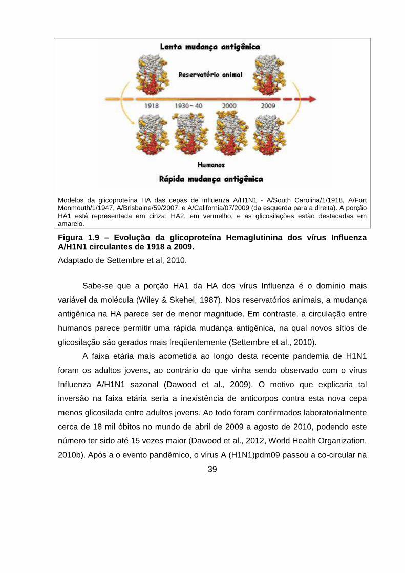
Modelos da glicoproteína HA das cepas de influenza A/H1N1 - A/South Carolina/1/1918, A/Fort Monmouth/1/1947, A/Brisbaine/59/2007, e A/California/07/2009 (da esquerda para a direita). A porção HA1 está representada em cinza; HA2, em vermelho, e as glicosilações estão destacadas em amarelo.
Figura 1.9 – Evolução da glicoproteína Hemaglutinin a dos vírus Influenza A/H1N1 circulantes de 1918 a 2009.
Adaptado de Settembre et al, 2010.
Sabe-se que a porção HA1 da HA dos vírus Influenza é o domínio mais
variável da molécula (Wiley & Skehel, 1987). Nos reservatórios animais, a mudança
antigênica na HA parece ser de menor magnitude. Em contraste, a circulação entre
humanos parece permitir uma rápida mudança antigênica, na qual novos sítios de
glicosilação são gerados mais freqüentemente (Settembre et al., 2010).
A faixa etária mais acometida ao longo desta recente pandemia de H1N1
foram os adultos jovens, ao contrário do que vinha sendo observado com o vírus
Influenza A/H1N1 sazonal (Dawood et al., 2009). O motivo que explicaria tal
inversão na faixa etária seria a inexistência de anticorpos contra esta nova cepa
menos glicosilada entre adultos jovens. Ao todo foram confirmados laboratorialmente
cerca de 18 mil óbitos no mundo de abril de 2009 a agosto de 2010, podendo este
número ter sido até 15 vezes maior (Dawood et al., 2012, World Health Organization,
2010b). Após a o evento pandêmico, o vírus A (H1N1)pdm09 passou a co-circular na

40
população juntamente com o H3N2, apresentando um menor impacto em
mortalidade. Assim, como um virus sazonal, o A (H1N1)pdm09 passou a ser incluído
na composição da vacina a partir de 2010/2011 no hemisfério norte e 2011 no
hemisfério sul (World Health Organization, 2014b).
1.2.2 Importantes surtos e epidemias de variantes de Influenza A entre
humanos
O subtipo de Influenza A que poderá ocasionar uma nova pandemia sempre
foi um assunto bem debatido pela comunidade científica. Alguns acreditam que
apenas os subtipos H1, H2 e H3 podem realmente ocasionar os eventos
pandêmicos entre humanos. Outros apostam que os subtipos H5, H6, H7 e H9,
responsáveis por infecções zoonóticas, são mais propensos a causar a próxima
pandemia (Sorrell et al., 2011). Isso é incerto, mas importantes surtos e epidemias
com diferentes subtipos de Influenza são reportados esporadicamente ao longo da
história.
Em 1977, o subtipo H1N1 similar ao que ocasionou a pandemia de 1918
reapareceu na Rússia, tornando-se epidêmico (Wright et al., 2007). Entretanto, não
substituiu o subtipo H3N2, que circulava desde a pandemia de 1968, passando
esses dois subtipos de Influenza A, juntamente com o vírus Influenza B, a circular
concomitantemente na população humana mundial. Normalmente, um destes vírus
sazonais predomina, tornando-se epidêmico em determinado ano (Hilleman, 2002).
Além destes vírus sazonais outros subtipos ocasionalmente infectam humanos
causando surtos e epidemias locais.
No ano de 1997, o subtipo HPAI H5N1 causou um grande surto em aves
domésticas em Hong Kong resultando no primeiro caso de transmissão
documentada de aves para humanos, com seis casos fatais entre os 18 indivíduos
infectados (de Jong et al., 1997). Análises filogenéticas evidenciaram a transmissão
direta da cepa circulante de H5N1 entre galinhas para o homem eliminando a etapa
intermediária da adaptação viral em suínos (Webster, 1998). A agilidade na
obtenção dos resultados permitiu a rápida identificação e eliminação dos plantéis de
41
aves contaminadas em Hong Kong. Em geral, em países do continente asiático os
mercados de animais vivos são comuns e constituem uma ótima oportunidade para
o rearranjo entre diferentes subtipos de Influenza. Após 2004, o vírus HPAI H5N1 se
espalhou pela Ásia, Europa e África causando vários surtos em aves domésticas e
ocasionalmente sendo isolado de mamíferos. Desde 2003, 694 casos foram
relatados com 402 óbitos (World Health Organization, 2015a). A transmissão
sustentada humano-humano do HPAI H5N1 não parece ser muito eficiente, dado
que os casos de transmissão entre pessoas são raros (Bender et al., 1999).
Entretanto, não é descartada a possibilidade deste subtipo se tornar pandêmico,
pois o mesmo pode desenvolver adaptações que garantiriam a transmissão
humano-humano, através de mutações ou aquisição de um novo segmento gênico
de vírus influenza humano clássicos, representando assim uma grande ameaça.
Outro subtipo atípico para o homem, o H7, tem sido responsável por vários
surtos entre aves que resultam ocasionalmente em infecções humanas (Gao et al.,
2013, Koopmans et al., 2004, Puzelli et al., 2014). Algumas infecções são isoladas,
sem maior importância epidemiológica, como casos de conjuntivite e infecções
respiratórias pelos subtipos A/H7N2, A/H7N3 e A/H7N7 reportados na Europa e
Estados Unidos de 1997 a 2014 (Belser et al., 2009, Puzelli et al., 2014). Entretanto,
em março de 2013 na China, o subtipo aviário A/H7N9 detectado apenas em
pássaros no passado emergiu causando uma significativa taxa de infecções em
humanos e aves (Gao et al., 2013). Neste importante surto, foram reportados pela
OMS 453 casos laboratoriais de infecção humana com A/H7N9, incluindo 175
mortes (World Health Organization, 2014c). Apesar do grande número de casos
observados este vírus não parece ser transmitido facilmente de pessoa-pessoa, e a
transmissão sustentada não foi reportada (World Health Organization, 2014c).
Antes da emergência da variante pandêmica, de dezembro de 2005 a
fevereiro de 2009, cerca de 10 casos de transmissão de Influenza A de suínos para
humanos foram reportados nos EUA (Dawood et al., 2009, Shinde et al., 2009).
Estes vírus eram dos subtipos H1N1 e H1N2 e possuiam segmentos gênicos muito

42
semelhantes aos vírus A (H1N1)pdm09 (Figura 1.10), e foram apontados como
possíveis precursores do vírus pandêmico (Shinde et al., 2009)
Figura 1.10 - Comparação do genoma do vírus Influen za A H1N1 detectados em surtos de suinos e humanos nos EUA de dezembro de 2 005 a dezembro de 2009 e o genoma do vírus Influenza A (H1N1)pdm09. Adaptado de Dawood et al, 2009.
O vírus A (H1N1)pdm09 também sofreu rearranjo com o vírus H3N2 sazonal
e, em julho de 2011, foi detectada pela primeira vez em humanos a variante H3N2v,
que possui o gene M do vírus influenza A (H1N1)pdm09 (Centers for Disease
Control and Prevention, 2014a). Estes vírus foram previamente observados em
suínos no ano anterior, e a infecção humana foi associada com a exposição
prolongada aos porcos em feiras agrícolas. A transmissão humano-humano foi
limitada, sem a observação de uma transmissão comunitária. De agosto de 2011 a
outubro de 2014, um total de 343 casos humanos foram registrados, causando

43
aparentemente uma infecção leve (Centers for Disease Control and Prevention,
2014a).
Os casos de rearranjo e surtos humanos apresentados acima mostram um
pouco da complexa dinâmica dos vírus Influenza A na natureza e confirmam a
necessidade de vigilância e controle destes surtos com potencial pandêmico. Diante
do impacto em saúde pública que a emergência de uma variante pandêmica
significa, estudos que visam prever o impacto de uma nova pandemia são
importantes, mas também polêmicos, e dividem opiniões do ponto de vista da
biossegurança (Davis et al., 2014, Duprex et al., 2015, Fouchier et al., 2012). Alguns
estudos de “ganhos de função” (GOF, gain-of-function), os quais permitem adaptar
uma variante viral potencialmente pandêmica, tornando-a mais transmissível e
contagiosa, foram realizados com o subtipo H5N1 com o objetivo de melhorar a
identificação de mutações perigosas. Contudo, alguns grupos são contrários a
realização desses tipos de estudo, pois não existe maneira de se descartar
totalmente o risco de liberação acidental das variantes altamente patogênicas de
Influenza geradas em laboratório. Sob esta ótica, afirmam os estudiosos, pesquisas
dessa natureza, envolvendo modificações virais para GOF, trariam mais ameaças
que benefícios a população humana.

44
1.3 Epidemiologia dos vírus Influenza A (H1N1)pdm09
No início de 2009, as diferentes organizações de saúde e grupos de pesquisa
do mundo que vinham se preparando para uma potencial pandemia de H5N1, foram
surpreendidas pela emergência do subtipo A (H1N1)pdm09. Entre os meses de
março e abril de 2009 foram diagnosticados os primeiros casos de infecção
respiratória causada por essa variante no México e nos EUA, mas acredita-se que a
transmissão tenha iniciado alguns meses antes do reconhecimento do primeiro surto
(Smith et al., 2009). Durante as primeiras semanas de vigilância, este vírus
espalhou-se pelo mundo rapidamente, sendo que em 11 de junho de 2009 a OMS
declarou que a infecção causada pela nova variante tinha atingido a porporção
pandêmica (World Health Organization, 2009b).
De acordo com a OMS, para caracterização de um evento pandêmico existem
seis fases ou níveis de alerta (World Health Organization, 2010a). Na primeira fase,
os vírus que circulam entre animais não são detectados em humanos, sendo
encontrados na população apenas os vírus sazonais. Na fase dois um virus
Influenza conhecido por ter causado infecções em humanos é detectado circulando
entre animais domésticos ou selvagens, sem infecção em humanos. Contudo, o
potencial pandêmico do patógeno circulante é considerado nesta etapa. Na fase
três, ocorrem casos esporádicos em humanos, através da transmissão animal-
humano. Contudo, esta transmissão é limitada, não ocorrendo a transmissão
humano-humano. Quando o vírus rompe essa barreira e passa a ocorrer a
transmissão sustentada entre humanos caracteriza-se a fase quatro, com
surgimento de surtos comunitários. A fase cinco é caracterizada pela propagação do
vírus em pelo menos dois países, sendo iminente a ocorrência de uma pandemia. A
fase seis caracteriza a pandemia, propriamente dita, com a ocorrência de surtos em
vários países simultaneamente. Após a fase pandêmica, atinge-se o período pós-
pico, no qual há uma redução significativa no número de casos. Entretanto, existe a
possibilidade de eventos recorrentes na população. Ao término do período pós-pico,

45
ocorre o período pós-pandêmico, no qual os vírus passam a circular normalmente,
como um vírus sazonal (World Health Organization, 2010a).
Em relação às outras pandemias já registradas, o impacto da pandemia de
influenza A (H1N1)pdm09 foi moderado, com cerca de 18 mil óbitos notificados no
mundo entre abril de 2009 a agosto de 2010. Estima-se, entretanto, que o número
de mortes pode ter chegado a 284 mil se forem contados óbitos não notificados
(Dawood et al., 2012, World Health Organization, 2010b).
Uma das características da pandemia de 2009 foi a forma desproporcional
com que afetou crianças e adultos jovens em relação aos grupos relativamente,
mais velhos (Bautista et al., 2010). A excasses de indivíduos sintomáticos com idade
superior a 60 anos (Dawood et al., 2009), se deve possivelmente a uma imunidade
cruzada adquirida previamente por possível exposição a variantes similares que
circularam entre 1940 e 1950 (Settembre et al., 2010). Alguns estudos fortaleceram
essa hipótese, como o realizado por Garten e colaboradores (2009) que mostraram
que cerca de 33% de indivíduos acima de 60 anos possuíam anticorpos
neutralizantes de reação cruzada para influenza A (H1N1)pdm09 através de testes
de HI e de inibição da neutralização (Garten et al., 2009). Outro estudo de exposição
domiciliar ao influenza A (H1N1)pdm09, revelou que indivíduos abaixo de 50 anos
eram 2 vezes mais susceptíveis ao vírus (Cauchemez et al., 2009).
Estima-se que 25 a 50% dos pacientes infectados com A (H1N1)pdm09 que
foram hospitalizados ou morreram apresentavam alguma comorbidade (doenças
crônicas, imunossupressão, obesidade, entre outras) ou algum fator de risco para
complicações clínicas (Bautista et al., 2010, Punpanich & Chotpitayasunondh, 2012).
Um grupo afetado de forma importante durante a pandemia foi o de gestantes
(Mosby et al., 2011), cuja maior sensibilidade não ocorreu somente a falta de
anticorpos prévios, mas também a uma variedade de mudanças no sistema
cardíaco, respiratório, hormonal e imunológico que possivelmente contribuíram para
o aumento da morbidade e mortalidade neste grupo (Jamieson et al., 2006).
As características clínicas e complicações da infecção por influenza A
(H1N1)pdm09 foram similares às da influenza sazonal. O espectro clínico variou

46
desde infecções assintomáticas à pneumonia viral resultando em falência
respiratótia, disfunção respiratória aguda, falência múltipla de órgãos e morte
(Bautista et al., 2010).
1.3.1 Sazonalidade dos vírus influenza A
O vírus influenza A apresenta uma sazonalidade complexa, com influência de
um conjunto de fatores populacionais (nível de imunidade, interações sociais,
comportamentais e culturais), virais (contínuo processo de geração e seleção de
novas linhagens) e ecológicos/ambientais (Lofgren et al., 2007). Os vírus influenza
têm seu pico epidêmico nos meses de maio a setembro nas regiões de clima
temperado do Hemisfério Sul, entre dezembro e março nas regiões de clima
temperado do Hemisfério Norte, e durante todo o ano (com maior incidência no
período chuvoso) nas regiões tropicais e subtropicais (Tamerius et al., 2011, Viboud
et al., 2006) (Figura 1.6).
Figura 1.11 - Comparação dos padrões de influenza s azonal nos países temperados e tropicais nas Américas. Adaptado de (Viboud et al, 2006).

47
Devido às dimensões continentais do Brasil, estendendo-se por áreas
temperadas, subtropicais e equatoriais, é possível identificar distintos padrões
sazonalidade dos vírus influenza nas diferentes regiões do país (Mello et al., 2009,
Motta et al., 2006, Moura et al., 2009, SVS, 2009). A região Sul apresenta uma
sazonalidade similar à observada nos países de clima temperado, com epidemias
com pico no inverno (junho-julho) (Straliotto et al., 2002). A região Norte apresenta
dois picos, sendo o maior em associação ao período chuvoso (março-abril), como
observado em países tropicais da Ásia (Moura et al., 2009). Já nas outras regiões do
Brasil, temos uma situação intermediária, com casos detectados ao longo de todo
ano e picos menos acentuados no inverno (Alonso et al., 2007). Entretanto, a
descontinuidade e a falta de homogeneidade dos serviços de epidemiologia nas
diferentes regiões do país dificultam muito a comparação entre os dados de
circulação de influenza.
1.3.2 Impacto do vírus A (H1N1)pdm09 no Brasil
Devido as variações de densidade populacional e do acesso a saúde entre os
diferentes países, o impacto da pandemia de Influenza de 2009 foi diferenciado nas
diversas regiões do globo (Boni et al., 2009). No Brasil, os primeiros resultados
positivos para o vírus A (H1N1)pdm09 foram divulgados no início de maio de 2009
(Secretaria de Vigilância em Saúde, 2010). As amostras analisadas eram
provenientes de quatro adultos jovens, dois deles do Estado de São Paulo, um do
Rio de Janeiro e o outro de Minas Gerais, sendo que todos apresentavam algum
histórico de viagem recente ao México ou aos EUA (Secretaria de Vigilância em
Saúde, 2010).
No período inicial da circulação viral no país (semanas epidemiológicas 16 a
28) os casos detectados estavam relacionados a viajantes que haviam estado fora
do país. Assim, medidas de contenção foram adotadas, especialmente a vigilância
intensificada em pontos de entrada no país como portos, aeroportos e fronteiras
terrestres na tentativa de identificar a maioria dos casos suspeitos. A fim de reduzir

48
os riscos de transmissão comunitária foram adotadas medidas como: internação e
isolamento de todos os casos com sindrome gripal, tratamento com o antiviral
Oseltamivir e busca ativa e quarentena dos contactantes (Secretaria de Vigilância
em Saúde, 2010).
Contudo, apesar dos esforços de contenção, no dia 16 de julho de 2009 foi
declarada a transmissão sustentada do vírus A (H1N1)pdm09 no Brasil, após a
conclusão da investigação epidemiológica dos primeiros casos suspeitos e da
notificação do primeiro caso de transmissão da doença sem o vínculo com viagens
ou contato com viajantes internacionais. Assim iniciou-se a fase de mitigação, na
qual foram adotadas medidas para reduzir a gravidade e mortalidade pela a doença,
através do diagnóstico rápido e tratamento dos casos que apresentavam risco para
a doença grave ou fatal (Secretaria de Vigilância em Saúde, 2010). O
monitoramento de casos de SRAG e de individuos que apresentavam algum fator de
risco para complicação da doença foram intesificados a fim de atenuar os casos
graves e diminuir os casos de óbitos (Secretaria de Vigilância em Saúde, 2010).
Assim, no ano de 2009 (semanas epidemiológicas 16 a 52) foram registrados
no Brasil 2.051 óbitos ocasinados pelo vírus A (H1N1)pdm09, sendo a taxa de
mortalidade nacional de 1,1/100 mil habitantes. Quando analisada separadamente
por região geográfica, a mortalidade foi maior no Sul do país, atingindo 3/100 mil
habitantes (Secretaria de Vigilância em Saúde, 2010).
Nas epidemias dos anos subsequentes o vírus A (H1N1)pdm09 passou a
circular de forma sazonal e o número de casos e de óbitos foi reduzido, como mostra
a Figura 1.12.

49
Figura 1.12 - Representação das curvas de circulaçã o do vírus Influenza A (H1N1)pdm09 durante e após o período pandêmico (sem ana epidemiológica 1 2009 a semana epidemiológica 46 de 2014). Adaptado de imagem cedida pela pelo Grupo de trabalho de Influenza da SVS.
1.3.3 Epidemiologia molecular e evolução dos vírus A (H1N1)pdm09
A dinâmica evolutiva e epidemiológica dos vírus Influenza está relacionada
diretamente com o padrão de migração global destes vírus. Diversos modelos são
propostos para explicar seu processo migratório, entretanto, apesar de diferentes
estudos, ainda sabemos muito pouco. Um destes modelos teóricos defende que os
trópicos abrigam populações virais endêmicas por todo ano, e que estas regiões
atuam como um reservatório que, sazonalmente, reintroduz isolados virais nas

50
regiões temperadas como mostra a Figura 1.13 (Rambaut et al., 2008). Alguns
estudos filogenéticos sustentam este modelo e apontam as regiões tropicais como o
centro da diversidade e distribuição do vírus influenza A humano para outras regiões
do mundo (Nelson et al., 2007, Rambaut et al., 2008). Ainda dentro desse modelo,
acredita-se também que, durante as migrações sazonais para dentro e fora dos
trópicos (com elevada biodiversidade), as aves transmitem influenza para outros
hospedeiros (aves e mamíferos) e que os os humanos garantem a persistência viral
na comunidade e sua rápida disseminação entre países ou regiões (Nelson et al.,
2007, Reed et al., 2003).
Figura 1.13 - Modelo teórico de dispersão de varian tes dos vírus Influenza mostranto a região tropical como o principal reserv atório anual destes vírus. Adaptado de Rambaut et al, 2008.
Um outro molelo aponta o leste e sudeste asiático como as regiões
disseminadoras de variantes de vírus Influenza para o restante do mundo, e ainda
mostra a América do Sul como o fim de linha para a circulação de variantes de
influenza A H3N2 (Bedford et al., 2010, Russell et al., 2008). Finalmente, opondo-se
a todos os outros modelos migratórios, Bahl e colaborares sugerem que as
populações virais migram constantemente em todos os sentidos e evoluem durante

51
a trajetória de migração, e que todas as regiões são potenciais fontes para
emergência de novas variantes virais (Bahl et al., 2011).
Independente da direção de dispersão das populações virais, a migração e a
evolução do vírus influenza têm como importantes veículos a movimentação de
pessoas pelo mundo (especialmente pelo trafego aéreo) e o comércio internacional
de aves domésticas e de suínos (Khan et al., 2009, Tamerius et al., 2011). Estudos
realizados com amostras A (H1N1)pdm09 do início da pandemia demonstraram o
impacto da movimentação internacional de pessoas, especialmente por rotas
aéreas, na rápida disseminação do vírus pelo mundo (Khan et al., 2009, Lycett et al.,
2012), sendo relatadas em vários países múltiplas introduções de variantes virais na
primeira fase da pandemia (Lycett et al., 2012, Zehender et al., 2012).
Análises evolutivas do genoma completo do vírus A (H1N1)pdm09 durante o
período pandêmico (abril de 2009 a agosto de 2010) revelaram a circulação de dois
grandes grupos (Christman et al., 2011). O Cluster I com origem no México, Texas e
Califórnia e o Cluster II com origem em Nova York (Fereidouni et al., 2009, Shiino et
al., 2010). Este o período inicial da introdução e adaptação do vírus A (H1N1)pdm09
em humanos mostrou um baixo número de mutações, sendo que a maioria delas
(cerca de 98%) ocorria randomicamente, resultando num baixo sinal filogenético
entre os dois grupos ao analisar juntas todas as sequências mundiais (Christman et
al., 2011). Entretanto, uma micro-evolução pode ser observada quando foram
analisadas amostras locais e com um restrito período de tempo (Nelson et al., 2009,
Shiino et al., 2010).
Estudos filogenéticos realizados nos primeiros meses (abril e julho de 2009)
de circulação mundial do vírus A (H1N1)pdm09 revelaram uma inicial diversidade
viral nos EUA, o que resultou na caracterização de pelo menos 7 grupos
filogenéticos, G1 a G7-pdm (Tabela 1.2), com assinaturas distintas no genoma viral
(Nelson et al., 2009). Isolados de A (H1N1)pdm09 dos grupos G5, G6 e G7-pdm
foram também observados na India de maio a setembro de 2009 e em amostras
brasileiras (Goni et al., 2012, Potdar et al., 2010). Evidências de uma evolução

52
adaptativa dentro G7-pdm foram observadas e este se tornou o grupo predominante
(Mullick et al., 2011, Nelson et al., 2009, Potdar et al., 2010).
O grupo de vigilância do Centro Europeu para Controle e Prevenção de
Doenças (ECDC, European Centre for Disease Prevention and Control) é uma das
diversas entidades ao redor do globo que reportam regularmente as variantes
emergentes nos anos subsequentes ao evento pandêmico (European Centre for
Disease Prevention and Control, 2011, 2013). O ECDC reportou a circulação na
Europa de pelo menos 6 grupos filogenéticos (G2 - G7) e 3 subgrupos (6A - 6C) no
período pós-pandêmico 2011 a 2014, como mostra a Tabela 1.2 (European Centre
for Disease Prevention and Control, 2011, 2013). Este achado foi confirmado em
amostras detectadas na Tunísia (El Moussi et al., 2013). Entretanto, mesmo com o
aumento na diversidade viral, a vacina anti-influenza para o subtipo H1N1 continua
sendo a cepa A/California/7/2009, isso porque títulos de HI de isolados dos grupos
identificados atualmente ainda apresentam boa reatividade com o anti-soro
produzido contra o vírus da vacina (European Centre for Disease Prevention and
Control, 2011, 2013).

53
Tabela 1.2 - Variações de aminoácidos que caracteri zaram os grupos filogenéticos do virus A (H1N1)pdm09 desde sua emer gência até o ano de 2014.
Grupo filogenético (Representante)
Substituições de amino ácidos por proteína
Referência
G1 (A/California/7/2009-like)
PA: S224P*; HA: S83P*, A197T*, V321I*
Nelson et al, 2009
G2-período pandêmico PA: M581L* NP: T373I
G3-período pandêmico
G4-período pandêmico PB2: V649I*; PB1: I667T; NP: V100I; NA: V106I; NS2: E63K
G5-período pandêmico NP: V100I*; NA: V106I*, N248D
G6-período pandêmico
HA: K-15E; Q293H*; NP: V100I; NA: V106I, N248D
G7-período pandêmico HA: S203T; NP: V100I*; NA: V106I, N248D*; NS1: I123V
G2 (A/Czech Republic/32/2011-like)
HA: N31D, A186T, S162N ECDC, 2011
G3 (A/Umea/1/2011-like)
HA: A134T, A141S*, V152A*, S183P, I295V*
G4 (A/Christchurch/16/2010-like)
HA: N125D
G5 (A/Astrakhan/1/2011-like)
HA: D97N, R205K, I216V, V249L
G6 (A/St Petersburg/27/2011-like)
HA: D97N, S124N, S185T
G6A (A/Hong Kong/5659/2012-like)
HA: H138R, V249L ECDC, 2013
G6B (A/Norway/2417/2013-like)
HA: K163Q, E172K, A256T, K283E
G6C (A/Estonia/76677/2013-like)
HA: E172K, V234I, K283E
G7 (A/St Petersburg/100/2011-like)
HA: S124N, S143G, S185T, A197T ECDC, 2011
A numeração de HA utilizada para esta tabela desconta o peptídeo sinal; *variações mais frequentes em cada grupo

54
1.4 Resposta imune contra a infecção pelos vírus In fluenza
Durante o curto período de incubação entre a infecção e os sintomas clínicos
da gripe (2 a 3 dias) inicia-se o reconhecimento e ativação da resposta imune inata.
Nesta etapa, células efetoras, e mediadores solúveis como citocinas, quimiocinas e
outros fatores atuam no controle da infecção. Ainda, durante a resposta imune inata
são ativados os mecanismos de apresentação de antígenos, que em conjunto com
as citocínas, são responsáveis pelo desencadeamento da resposta adaptativa
(Iwasaki & Pillai, 2014, van de Sandt et al., 2012).
Uma vez que células do trato respiratório são infectadas, inicia-se uma
complexa série de eventos intracelulares que estimulam o início de uma resposta
antiviral, levando a produção de quimiocinas, interferon (IFN) e outras citocinas. O
reconhecimento viral por padrões moleculares associados a patógenos (PAMPs,
pathogen-associated molecular patterns) e a coordenação inicial da resposta a
infecção são mediados por três grandes famílias de receptores inatos, denominados
receptores de padrões moleculares (PRRs, Pattern Recognition Receptors): 1)
receptores do tipo Toll (TLRs, Toll-like receptors), como TLR3, TLR7, TLR4 e TLR2;
2) receptores do tipo NOD (nucleotide oligomerization domain; NLRs, Nod-like
receptors) da família NLRP3; e 3) receptores RIG-I (retinoic acid inducible gene-I;
RLR, RIG-like receptors) (Kreijtz et al., 2011, van de Sandt et al., 2012).
TLR3 e TLR7 estão localizados em vesículas endossomais e são capazes de
reconhecer RNA viral dupla fita (RNAfd) e RNA viral fita simples (RNAfs),
respectivamente. Ambos induzem a produção de IFN tipo I, e ainda TLR7 produz
citocinas inflamatórias, como as interleucinas (IL), IL-12 e IL-6. Por sua vez, TLR2 e
TLR4 estão localizados na superfície celular e reconhecem glicoproteínas de
superfície viral HA e NA (Kreijtz et al., 2011, van de Sandt et al., 2012).
O receptor NLRP3 do tipo NOD se torna ativado pela infecção viral em
resposta a RNAfd e pela atividade do canal iônico M2. Por um mecanismo ainda não
totalmente elucidado, a ativação do receptor NLRP3 leva a sua oligomerização,
formando uma estrutura denominada Inflamossomo no citoplasma. O Inflamossomo

55
serve como plataforma para recrutamento e ativação da pró-Caspase I e sua
conversão em Caspase I ativa. Esta, por sua vez, processa as pró-citocinas IL-1β,
IL-18 e afins até sua forma biologicamente ativa. A IL-1β está envolvida em uma
série de respostas pró-inflamatórias, inclusive expansão de clones de células T
CD4+ antígeno-específicas e seu direcionamento para o perfil Th17. Em uma
diferente classe de NLRs, NOD2 parece mediar o reconhecimento de RNAfs em
modelos virais de RSV e Influenza, levando a produção de IFN do tipo I (Kreijtz et
al., 2011, van de Sandt et al., 2012).
RIG-I, por sua vez, é ativado pela ligação do RNA 5` trifosfato ou do RNAfd
viral ao domínio repressor de RIG-I. Este sofre uma mudança conformacional,
expondo os domínios de recrutamento e ativação de caspases. Estes domínios são
ubiquitinados por E3 ubiquitina ligase induzida por IFN, denominada TRIM25
(tripartite motify 25). RIG-I pode se associar a adaptadores de sinalização virais nas
mitocondrias (MAVS, mitocondrial antiviral signaling adaptor; também conhecido
com IPS-1, VISA ou Cardif), iniciando a cascata de sinalização que leva a ativação
dos fatores de transcrição IRF3 (Interferon Regulatory Factor 3) e NF-κB (Nuclear
Factor Kappa B) (Kreijtz et al., 2011, van de Sandt et al., 2012).
Todas essas vias de sinalização eventualmente resultam na transcrição de
citocinas pró-inflamatórias, quimiocinas e IFNs que ativam a resposta antiviral e o
recrutamento de neutrófilos, ativação de macrófagos e maturação de células
dendríticas (Kreijtz et al., 2011).
A indução de IFNs é essencial na resposta protetora inicial contra a infecção
por influenza. Os IFNs do tipo I, incluindo IFN-α e IFN-β, tem um importante papel na
limitação da replicação viral. Estes também estimulam células dendíticas, resultando
no aumento de apresentação de antígenos para células T CD4+ e T CD8+, o que
contribui para iniciar a resposta adaptativa. IFN-β é produzido via feedback positivo
regulado pelo IRF7, estimulando na própria célula ou células vizinhas a produção de
IFN-α e IFN-β.
Os IFNs do tipo I possuem um papel fundamental na limitação da replicação
viral, sendo produzido por macrófagos, pneumócitos e células dendríticas (Iwasaki

56
and Pillai 2014). Eles promovem um estado antiviral celular por estimular a
expressão de centenas de genes, conhecidos como genes estimulados por
interferon (ISG, Interferon stimulated genes). Os ISG podem controlar a patogenese
viral por meio de uma variedade de mecanismos e seus efeitos são geralmente
específicos a cada tipo de vírus, célula ou tecido (Iwasaki and Pillai 2014; Schoggins
2014). Já foram identificados mais de 400 ISGs, entretanto, as funções biológicas da
grande maioria deles não estão bem estabelecidas (Iwasaki & Pillai, 2014). Por outro
lado, algumas das proteínas codificadas por ISGs apresentam um papel bem
definido no controle da infecção por influenza, como as proteínas transmembrana
induzidas por interferon (IFITMs, Interferon-induced transmembrane proteins),
capazes de bloquear a infecção de vários vírus envelopados que são internalizados
nas células pela via endossomal, incluindo o vírus influenza A (Brass et al., 2009). O
bloqueio ou alteração no perfil da expressão dos ISGs podem induzir uma
diminuição da resistência antiviral natural do hospedeiro (Iwasaki & Pillai, 2014,
Schoggins, 2014).
A segunda linha de defesa contra a infecção pelos vírus Influenza A é a
resposta imune adaptativa (humoral e celular). No geral, esta resposta é altamente
específica e lenta no primeiro encontro com o patógeno. No entanto, a partir do
segundo encontro com o patógeno, quando já se tem uma memória imunológica, a
resposta se mostra mais rápida e forte (Noh & Kim, 2013, van de Sandt et al., 2012).
A resposta imune adaptativa humoral leva a produção de anticorpos
neutralizantes específicos contra o vírus Influenza, especialmente contra a proteína
HA. Após a infecção primária (cerca de 3 dias), células dendríticas carregadas com
antígenos virais migram para linfonodos do mediastino, onde ocorre reconhecimento
dos epítopos de HA, NA e outras proteínas pelos linfócitos B residentes (van de
Sandt et al., 2012). Estes, por sua vez, passam por rápida proliferação e maturação
em células produtoras de anticorpos (van de Sandt et al., 2012). Os anticorpos vírus-
específicos presentes no soro ou, de preferência, depositados na superfície da
mucosa das vias aéreas, podem bloquear a entrada viral, e o subsequente
estabelecimento da infecção, por meio do impedimento da interação da HA com os

57
resíduos de AS das células hospedeiras (Kreijtz et al., 2011). Esse, inclusive, é o
principal mecanismo de proteção das vacinas sazonais licenciadas para uso em
humanos contra Influenza (Noh & Kim, 2013).
A maioria dos anticorpos neutralizantes anti-HA são direcionados contra
epítopos do domínio globular da proteína. Como vimos anteriormente, essa região é
altamente variável e seus epítopos mudam de composição entre cepas virais. Assim,
anticorpos produzidos contra uma determinada sequência de HA não são capazes
de neutralizar os epítopos de uma linhagem variante, mesmo se produzidos em altos
títulos. Esse é um dos mecanismos de escape imunológicos mais importantes do
Influenza, que explica também a necessidade de revisão frequente de composição
das vacinas sazonais. Os anticorpos dirigidos a NA não neutralizam diretamente o
vírus, mas inibem sua atividade enzimática limitando a dispersão viral. Estes
também facilitam o processo de citotoxicidade celular dependente de anticorpo e
podem contribuir para remoção da célula infectada (Kreijtz et al., 2011, van de Sandt
et al., 2012).
Por sua vez, a resposta imune celular consiste no recrutamento de células T
CD4+ e CD8+ específicas para o vírus. Estas células desempenham um papel
importante na regulação da resposta imunológica e eliminação viral,
respectivamente (Kreijtz et al., 2011, van de Sandt et al., 2012). As células T CD4+
são ativadas após o reconhecimento de peptídeos virais associados ao MHC de
classe II e moléculas co-estimulatórias na superfície de células apresentadoras de
antígenos (Kreijtz et al., 2011). A principal função das T CD4+ é produzir citocinas
que regulam a ativação e função de outras células imunes. De acordo com o perfil
de citocinas, as T CD4+ podem ser divididas em diversos perfis. Os perfis clássicos
são os das células T auxiliares (Th, T helper cell) do tipo Th1, as quais produzem IL-
2 e IFN-gamma as quais favorecem a ativação de macrófagos e células T CD8+ com
perfil citotóxico e células Th2, que produzem IL-4 e IL13 e regulam produção de
anticorpos. Mais recentemente foram descritos outros perfis, como os das células
Th17 e células T reguladoras (Tregs), as quais atuam na modulação da resposta
imune, controlando o processo inflamatório (van de Sandt et al., 2012).

58
Células citotóxicas T CD8+ (CTLs) desempenham um importante papel pois
realizam a eliminação direta de células infectadas pelos vírus. São ativadas no
tecido linfoide e recrutadas pra o local da infecção, onde reconhecem e eliminam a
célula infectada prevenindo a produção da progenie (Kreijtz et al., 2011, van de
Sandt et al., 2012)..
59
1.5 Patogênese dos vírus Influenza A (H1N1)pdm09
As causas de patogenicidade podem ser atribuídas a vários fatores como a
patogenicidade intrínseca do vírus, fatores adquiridos do hospedeiro (comorbidades,
gestação ou imunodepressão) e, ainda, a susceptibilidade genética do hospedeiro
ao vírus (Fukuyama & Kawaoka, 2011, Horby et al., 2013, Iwasaki & Pillai, 2014,
Punpanich & Chotpitayasunondh, 2012, Schrauwen et al., 2013).
1.5.1 Fatores de patogenicidade virais intrínsecos
As proteínas virais possuem um importante papel na virulência e alterações
nestas moléculas podem levar ao aumento da especificidade do vírus a
determinados receptores, ao rompimento de barreiras entre hospedeiros, ou ainda, à
potencialização da capacidade replicativa e da apitidão viral (Schrauwen et al.,
2013).
Uma das proteínas virais responsáveis pelo aumento de patogenicidade é a
HA, glicoproteína de superfície responsável pelo início da infecção e que atua como
o principal fator de tropismo do virion (Schrauwen et al., 2013). Alterações em sítios
específicos da molécula podem mudar a estrutura conformacional da proteína
alterando a sua preferência de ligação ao tipo de receptor de AS (Pan et al., 2012).
Uma destas alterações é a mutação D222G na HA dos vírus influenza A
H1N1pdm09, indicado como um potencial marcador de virulência da cepa durante a
pandemia de 2009 (World Health Organization, 2010c). A mudança no resíduo 222
confere uma maior interação da HA viral com receptores de AS-α2,3 presentes nas
superfícies das células do trato respiratório inferior (ver Figura 1.3, página 24) e,
consequentemente, leva a uma maior gravidade da doença (Belser et al., 2011,
Chutinimitkul et al., 2010, Liu et al., 2010).
Outras mutações na HA e em outras proteínas dos vírus A (H1N1)pdm09
foram sugeridas como indicadoras de um pior prognóstico, contudo mais estudos

60
são necessários para comprovar essa associação com a gravidade. Por exemplo, o
conjunto de mutações K-2E, P83S e Q293H (grupo filogenético G6-pdm) apresenta
interpretações controversas da sua associação com a morbidade e mortalidade
(Glinsky, 2010, Potdar et al., 2010). Alguns autores associam esse grupo a um maior
numero de óbitos (Glinsky, 2010, Potdar et al., 2010), enquanto outros associam
esse grupo a casos brandos de Influenza (Graham et al., 2011, Lee et al., 2010,
Nelson et al., 2009).
No Reino Unido um outro estudo analisando o genoma de vírus A
(H1N1)pdm09 que circularam durante as três primeiras ondas da pandemia revelou
que mutações detectadas durante a terceira onda, nas glicoproteínas de superficie
HA e NA, no componente do complexo da polimerase PA (N321K), e na proteína
antagonista de interferon NS1 aumentaram a habilidade do vírus replicar nas células
epiteliais do trato respiratório humano (Elderfield et al., 2014).
Outras proteínas como PB1-F2 contribuem para a virulência dos vírus
Influenza A por induzirem a apoptose de células infectadas. A mutação N66S em
PB1-F2 reduz a produção de IFN. O vírus A (H1N1)pdm09 não codifica para PB1-
F2, devido a três códons de parada prematuros, mas parece que quando este vírus
ganham essa capacidade a virulência é baixa ao afetar camundongos e furões
(Schrauwen et al., 2013).
1.5.2 Fatores do hospedeiro
Dados da literatura apontam que alguns grupos de indivíduos são mais
propensos a apresentarem quadros mais graves de infecção por Influenza, como por
exemplo, pessoas nos extremos de faixas etárias (menores que 5 anos e maiores
que 60), gestantes, imunocomprometidos, portadores de doenças crônicas,
fumantes, obesos, entre outros (Punpanich & Chotpitayasunondh, 2012). Estes
indivíduos são considerados grupos de risco por apresentarem maior pré-disposição
ao desenvolvimento da doença grave, e a estes é recomendado a vacinação anual e
o uso da droga antiviral para suprimir a replicação viral (Ministério da Saúde, 2013).
Entretanto, alguns grupos de risco, como os imunocomprometidos, não respondem

61
bem ao tratamento antiviral e apresentam muitas vezes uma excreção viral
prolongada, o que pode levar a emergência de variantes resistentes ao tratamento
antiviral (Souza et al., 2010, van der Vries et al., 2013).
Apesar de raros, estudos relacionando os fatores genéticos do hospedeiro
associados a uma maior patogenicidade no curso da infecção por influenza vem
apontando alguns potenciais marcadores humanos de gravidade (Horby et al., 2013,
Keynan et al., 2013).
Uma alteração na produção de IFITM3 apresenta fortes evidências de
associação com a infecção grave por influenza em humanos (Everitt et al., 2012).
Esta proteína tem função de bloquear a fusão do envelope viral com a membrana
endossomal celular dos hospedeiros, impedindo que as ribonucleoproteínas virais
sejam liberadas no citoplasma (Desai et al., 2014). Experimentos conduzidos por
Everitt e colaboradores revelaram que em camundongos deficientes para a síntese
de IFITM3, a replicação viral é persistente e está associada ao aumento de morte
celular e exagerada resposta pró-inflamatória (Everitt et al., 2012). Neste mesmo
estudo, os autores também reportam a possível associação do polimorfismo de base
única (SNP, single nucleotide polymorphism) rs12252-C (C/C ao invés de T/T ou
T/C) na susceptibilidade da infecção grave pelos vírus A (H1N1)pdm09 em pacientes
do Reino Unido (Everitt et al., 2012). A homogozigoze C/C para o alelo rs12252
altera o sítio aceptor de splicing e modifica o aminoácido 21 N-terminal da proteína
IFITM3, afetando a sua localização no endossoma (Everitt et al., 2012, Jia et al.,
2012). Além do estudo europeu, esse SNP também foi relatado em pacientes graves
hospitalizados com os vírus influenza A (H1N1)pdm09 ou H7N9 na China (Wang et
al., 2013, Zhang et al., 2013). Nestes estudos foi possível observar que, dependendo
da herança genética da população a frequência destes marcadores podem sofrer
variações consideráveis, pois a frequência do alelo rs12252-C parece ser bem maior
na população chinesa que na população européia (Everitt et al., 2012, Wang et al.,
2013, Zhang et al., 2013). Recentemente, um estudo controverso realizado por Mills
e colaboradores não encontrou a associação do SNP rs12252-C com a gravidade da
infecção (Mills et al., 2014). De acordo com estes estudos, por se tratar de um alelo

62
com baixa expressividade na população humana em geral, é necessário um número
de amostras suficientemente robusto para afirmar a associação ou não destes
marcadores com a gravidade da infecção (Horby et al., 2013, Mills et al., 2014).
Além deste, outros polimorfismos genéticos também foram associados com
uma maior susceptibilidade e gravidade da infecção, como: a deficiência no receptor
CCR5 (alelo CCR5Δ32), receptores de imunoglobulina com ligantes 2DL2/L3 e ainda
uma deficiência na imunoglobulina de classe G2 (Keynan et al., 2013). Entretanto,
mais estudos nessa área são necessários, pois em geral o número amostral é
pequeno nos estudos que indicam esses potenciais marcadores genéticos humanos
de gravidade (Keynan et al., 2013).
A investigação de marcadores genéticos humanos associados a um aumento
na gravidade da infecção é um grande desafio devido a necessidade de um grande
número amostral para o estudo, entretanto é um ponto importante a ser investigado
a fim de identificar potenciais grupos de risco e sugerir melhor conduta de profilaxia
para estes indivíduos (Pepin et al., 2010).
63
1.6 Diagnóstico e vigilância laboratorial dos vírus Influenza
1.6.1 Diagnóstico dos vírus Influenza
O sítio primário de replicação dos vírus Influenza são as células epiteliais do
trato respiratório superior, portanto, o material de escolha para a detecção destes
vírus é a secreção de nasofaringe (SNF), podendo os espécimes clínicos serem o
swab combinado nasal e orofaríngeo ou o aspirado de nasofaringe (WHO Global
Influenza Network, 2011). A coleta oportuna do material clínico deve ser realizada
até o quinto dia (preferencialmente, até o terceiro dia), a partir do início dos sintomas
(WHO Global Influenza Network, 2011). Sendo a qualidade da coleta,
acondicionamento e transporte das amostras clínicas imprescindíveis para a
realização do diagnóstico e caracterização viral com resultados fidedignos.
Diferentes métodos podem ser utilizados para o diagnóstico das infecções por
Influenza. Contudo, o isolamento viral e a RT-PCR são consideradas técnicas
primordiais para a caracterização viral (Centers for Disease Control and Prevention,
2009, WHO Global Influenza Network, 2011).
O isolamento viral constitui uma das técnicas clássicas para o diagnóstico dos
vírus influenza. Este pode ser realizados cultivando o vírus em ovos embrionados,
linhagens de células de mamíferos (MDCK - Madin-Darby Canine Kidney Epithelial
Cells) ou células imortalizadas de adenocarcinoma humano (células HeLa) (Burnet &
Ferry, 1934). Nas culturas celulares, os vírus se replicam causando efeito citopático,
caracterizado pela formação de placas (WHO Global Influenza Network, 2011).
A replicação viral é detectada pela técnica de hemaglutinação, que se baseia
na capacidade aglutinante viral. Uma vez que existem outros patógenos com
capacidade hemaglutinante, a identificação de vírus Influenza se dá pelo uso da
técnica de HI, onde são utilizados anticorpos específicos (WHO Global Influenza
Network, 2011). Esta última técnica também pode ser utilizada para a subtipagem e
caracterização antigênica dos vírus Influenza (Cox & Subbarao, 1999, Zambon,
1998).

64
Além da imunofluorescência indireta, um teste amplamente utilizado no
diagnóstico de diversos vírus respiratórios, testes rápidos têm sido desenvolvidos e
são capazes de detectar a presença de infecção por Influenza em menos de meia-
hora. Estes testes podem ser empregados para auxiliar o diagnóstico e manejo
clínico de indivíduos com síndrome gripal em consultórios e hospitais ou em estudos
epidemiológicos. Contudo, não são adequados para fins de diagnóstico, dada a sua
menor sensibilidade (50-70%) quando comparados às técnicas convencionais
(Centers for Disease Control and Prevention, 2014b).
Atualmente, a técnica mais utilizada para o diagnóstico de influenza dos
gêneros A e B e seus subtipos é a RT-PCR em tempo real, um método realizado em
etapa única (WHO Global Influenza Network, 2011). Ele se baseia na formação do
DNA complementar (cDNA) a partir do RNA genômico viral, seguido da amplificação
exponencial das fitas de cDNA, específicas para as proteínas virais M, HA e NP.
Esta amplificação com primers e sondas gera um sinal que é detectado pelos filtros
do equipamento. Sendo possível, após uma a duas horas de reação, identificar os
tipos e subtipos dos vírus Influenza com alta confiabilidade.
1.6.2 Métodos para caracterização molecular dos vírus Influenza
O método padrão para a caracterização molecular dos vírus influenza é o
sequenciamento convencional de Sanger, sendo este utilizado como rotina nos
laboratórios realizam a vigilância molecular dos vírus Influenza (World Health
Organization, 2009a). Entretanto, novas tecnologias de sequenciamento de nova
geração (SNG) vem sendo utilizadas com uma maior frequência e tem revolucionado
o campo da pesquisa genômica, devido ao baixo custo por base e alta cobertura
(Quinones-Mateu et al., 2014). Análises de metagenomica utilizando SNG tem
levado a descoberta de novos patógenos (Tong et al., 2013), e tem sido também
utilizado para caracterização de amostras de pacientes infectados por influenza
sazonal e pandêmico (Greninger et al., 2010, Kuroda et al., 2010). A vantagem de
utilização desta metodologia é especialmente a alta cobertura, que esta relacionado
ao sequenciamento de milhões de cópias de um único segmento gênico (Quinones-

65
Mateu et al., 2014). Isso agregou novas informações no campo das doenças
infecciosas, em especial em virologia. O SNG tem sido utilizado com sucesso, por
exemplo, na identificação e quantificação de variantes virais em baixas frequências
(ou quasispecies virais) em indivíduos infectados pelo vírus da imunodeficiência
humana (HIV) e pelos vírus hepatite B e C (HBV ou HCV), para estimar o impacto de
quasispecies virais na resposta ao tratamento antiviral (Cortes et al., 2014, Hoffmann
et al., 2007, Mitsuya et al., 2008). Nos estudos com os vírus Influenza, o SNG tem
sido aplicado para detectar a diversidade intra-hospedeiro (Kuroda et al., 2010,
Ramakrishnan et al., 2009), investigar mutações de potenciais marcadores de
virulência (Selleri et al., 2012), observar dinâmica viral frente ao tratamento com o
antiviral OST (Ghedin et al., 2012, Ghedin et al., 2011, Tellez-Sosa et al., 2013),
entre outras aplicações (Figura 1.14).
Figura 1.14 - Publicações cientificas com sequencia mento de nova geração para os vírus Influenza. A. Quantitativo do número de publicações por ano; B. Porcentagem de publicações de Influenza por área. Adapatado de Quinones-Mateu et al. 2014.
Existem alguns tipos de plataformas de SNG, sendo que quatro delas
dominam no campo da virologia: 454 (Roche) (Margulies et al., 2005), Illumina
(Illumina, Inc.) (Bennett, 2004), Ion Torrent (Life Technologies) (Rothberg et al.,
2011), and PacBio (Pacific Biosciences) (Eid et al., 2009). Entretanto, estas

66
plataformas apresentam diferenças significantes em relação a quantidade e
qualidade de sequencias geradas, e aplicações que cada sistema pode suportar
(Quinones-Mateu et al., 2014). A seleção de uma plataforma de SNG depende de
vários fatores como o tipo de pesquisa a ser realizado, o custo dos reagentes, a
infraestrutura existente e experiência profissional. Na Tabela 1.3 podemos observar
as principais diferenças entres as quatro plataformas de SNG.
Tabela 1.3 - Principais diferenças entre as 4 plata formas de sequenciamento de nova geração mais utilizadas para virologia
FLX 454 Illumina Ion Torrent PacBio
Método de amplificação
PCR de emulsão com beads
Pontes de PCR in situ
PCR de emulsão com
beads
Nenhum PCR
Química Síntese (pirosequenciamento)
Síntese (terminação reversível)
Síntese (detecção de
H+)
Sequenciamento de única molécula
por tempo real Tamanho da leitura (pb)
400 a 700 125 a 250
200 a 400 8.500
Cobertura por corrida
(GB)
0.05 a 0.09 8 a 1.000 2 a 10 0.15
Erro primário Indel Substituição Indel Indel
Taxa de erro ~1% ~1% ~1% ~13%
Tempo de corrida (horas)
10 a 20 39 a 276 4 a 7 2
Número de publicações
187 129 13 6
Vantagens Leituras de fragmentos longos
Fácil manipulação Baixo custo, corridas rápidas
Leituras de fragmentos longos
Desvata gens Homopolímeros e alto custo por Mb
Fragmentos curtos e longas corridas
Homopolímeros Alto custo e alta taxa de erro
Informações extraídas de Quinones-Mateu et al. 2014.
1.6.3 Rede de vigilância dos vírus Influenza
Devido às características evolutivas dos vírus influenza, o impacto anual em
morbidade e mortalidade das suas epidemias e o seu potencial pandêmico, é de
fundamental importância a constante vigilância destes vírus, bem como a rápida
identificação de novas cepas emergentes.

67
Desde a década de 1940 a necessidade de um monitoramento contínuo já
era observada, visto que o agente etiológico da gripe não se tratava de um vírus
estável, que as epidemias eram provocadas por cepas que sofriam mutações (o que
lhes conferia vantagem seletiva sobre as cepas antecessoras) e que as aves
migratórias funcionavam como um reservatório para o vírus. Assim, em 1952, a OMS
estabeleceu um programa de vigilância que opera através de uma rede mundial de
laboratórios coordenados pela OMS como mostra a Figura 1.15 (World Health
Organization, 2015b).
Atualmente, esta rede consiste de seis Centros Colaboradores de Influenza,
sendo quatro deles laboratórios de regulação essenciais da OMS. Esses centros
estão localizados em Musashi-Murayamashi no Japão, em Londres na Inglaterra, em
Beijing na China, em Melbourne na Austrália, e outros dois nos EUA, um em Atlanta
e outro em Memphis (World Health Organization, 2015b). Além destes, um total de
142 instituições, localizadas em 112 paises, são reconhecidas como centros
nacionais para Influenza (NIC, National Influenza Center) (World Health
Organization, 2015b). No Brasil o LVRS do IOC - FIOCRUZ, localizado no Rio de
Janeiro, juntamente com o Instituto Adolfo Lutz (IAL), localizado em São Paulo e o
Instituto Evandro Chagas (IEC), localizado em Belém no Pará, fazem parte da Rede
nacional de vigilância do vírus Influenza para OMS e para o MS (Secretaria de
Vigilância em Saúde, 2009). As informações geradas através desta rede de
laboratórios mundiais são importantes vigilância de variantes emergentes e para a
definição das cepas vacinais que irão compor a vacina anti-influenza reformulada
anualmente.

68
Figura 1.15 - Distribuição global da rede de vigilâ ncia de Influenza da OMS atualizado em 28 de novembro de 2014. Adaptada de World Health Organization 2015b.

69
1.7 Controle dos vírus Influenza A
1.7.1 Vacinação anti-influenza
A vacinação é a principal medida aplicada para prevenção e controle das
complicações ocasionadas pela infecção dos vírus Influenza (WHO Global Influenza
Network, 2011). Porém, não existe uma vacina duradoura contra esses vírus. Isso
porque os anticorpos desenvolvidos em exposições anteriores, seja por infecção
natural ou por vacinação, podem não reconhecer os alvos expressos pelas novas
variantes virais e, assim, não conferir uma proteção total (Carrat & Flahault, 2007).
Assim, os indivíduos estão susceptíveis a terem várias infecções por influenza ao
longo da vida.
As mudanças antigênicas freqüentes combinadas com a alta taxa de
transmissibilidade viral geram a necessidade de re-avaliação anual da vacina contra
Influenza. A decisão sobre a composição vacinal é baseada em análises
filogenéticas e experimentais e em dados epidemiológicos gerados pela rede
mundial de laboratórios da OMS, que mostram quais linhagens virais circulantes são
representativas e epidemiologicamente relevantes em cada temporada de gripe
(Hampson, 2008, WHO Global Influenza Network, 2011). Para escolha da vacina,
são realizadas duas reuniões anuais, uma para escolha da vacina a ser aplicada no
hemisfério sul e outra para decisão sobre a vacina para o hemisfério norte.
As vacinas anti-influenza podem ser atenuadas, recombinantes ou inativadas
(Houser & Subbarao, 2015, Noh & Kim, 2013). Todas os três tipos de vacinas são
multivalentes, ou seja, contem componentes dos dois subtipos de Influenza A, H1N1
e H3N2, e uma das linhagens do vírus Influenza B que tiveram importancia
epidemiológica na última temporada de gripe (Centers for Disease Control and
Prevention, 2010, Houser & Subbarao, 2015, Noh & Kim, 2013).
As vacinas atenuadas têm sido produzidas há mais de 50 anos nos EUA e na
Rússia (WHO Global Influenza Network, 2011). Este tipo de vacina é composto por
cepas virais atenuadas que causam infecções assintomáticas ou produzem sintomas

70
leves. Entretanto, devido a possibilidade destes vírus sofrerem reversões e
causarem infecções graves, essa vacina é administrada via intranasal (vantagem por
poder estimular as imunidades local e sistémica) em indivíduos saudáveis nas faixas
etárias de 2 a 49 anos (nos EUA), ou de 3 a 15 anos (na Rússia), não sendo
recomendadas para indivíduos portadores de problemas crônicos de saúde e
mulheres grávidas (Houser & Subbarao, 2015, WHO Global Influenza Network,
2011). Os vírus constituintes na vacina são termo-sensíveis, o que limita a sua
capacidade replicativa na temperatura de 38 a 39°C, podendo implicar baixa
eficiência replicativa nas vias aéreas inferiores. Porém, estão adaptados a
temperaturas mais baixas, replicando bem a 25°C, temperatura que restringe a
replicação de vírus tipo selvagem (Houser & Subbarao, 2015, WHO Global Influenza
Network, 2011, Wright et al., 2007).
A vacina recombinante é composta pela proteína de HA que é expressa em
células de insetos por vetores de baculovírus, sendo o tempo de produção desta
vacina curto, o que poderia ser aplicado na resposta rápida a um evento pandêmico
(Houser & Subbarao, 2015). Uma vacina recombinante FluBlok foi licenciada para
uso nos EUA em indivíduos na faixa etária de 18 a 49 anos e pode ser utilizada em
pessoas alégicas aos componentes do ovo (Houser & Subbarao, 2015).
Por sua vez, com o uso mais abrangente em todo o mundo, as vacinas
inativadas são administradas via parenteral em indivíduos de todas idades a partir
dos 6 meses de idade (mesmo apresentando algumas complicações de saúde) e
gera imunidade humoral e celular, principalmente contra a proteína HA (Houser &
Subbarao, 2015, WHO Global Influenza Network, 2011). Para sua produção, os vírus
são cultivados em ovos embrionados, purificados para reduzir reações alérgicas,
concentrados e inativados quimicamente com formalina ou β-propiolactona (Houser
& Subbarao, 2015, Wright et al., 2007). Uma vacina quadrivalente foi recentemente
licenciada para uso nos EUA a qual inclui as duas linhagens de Influenza B (Victoria-
87 e Yamagata-88) e os dois subtipos de Influenza A. A cepa vacinal
A/California/7/2009 escolhida como representante do vírus pandêmico para compor

71
a vacina até o memento não foi alterada, sendo a mesma desde 2010 até 2015 para
ambos os hemisférios (World Health Organization, 2014b).
No Brasil, a vacina inativada anti-influenza é oferecida gratuitamente para a
população por meio de campanhas anuais, num período anterior ao período
epidêmico do vírus no país. Esta vacina é indicada para crianças de seis meses a
cinco anos de idade, para idosos (acima dos 60 anos de idade), e para alguns
grupos de risco, como portadores de cardiopatias, nefropatias, diabetes mellitus
insulinodependente, cirrose hepática, hemoglobulinopatias, doença pulmonar
obstrutiva crônica (DPOC), imunocomprometidos (transplantados, pacientes em
tratamento de câncer, asmáticos, portadores de HIV e miopatias) e familiares que
estejam em contato com os referidos pacientes, e aos profissionais de saúde. Os
demais indivíduos adquirem imunidade contra os vírus por infecção natural durante
as epidemias anuais (Secretaria de Vigilância em Saúde, 2009).
Estudos baseados em partes mais conservadas dos vírus influenza vem
sendo conduzidos a fim de ser formulada uma vacina universal (Hampson, 2008,
Houser & Subbarao, 2015). Duas candidatas a vacinas universais incluem como
antígenos a região da haste da HA (região altamente conservada) e a proteína M2
(Houser & Subbarao, 2015). Entretanto, o desenvolvimento de uma vacina que
confere proteção contra todos os subtipos do virus influenza A ainda é um grande
desafio, mas a obtenção desta vacina seria uma grande oportunidade para reduzir a
ameaça de uma pandemia e o impacto do vírus sazonal (Hampson, 2008, Houser &
Subbarao, 2015).
1.7.2 Antivirais e resistência
As drogras anti-influenza são classificadas, de acordo com seus alvos no sítio
replicativo viral, como inibidores de HA, que impedem os processos de
reconhecimento de receptores e endocitose; bloqueadores do canal de prótons M2,
que inibem fusão do envelope viral às membranas celulares; inibidores das
polimerases virais, que interrompem a replicação e transcrição do genoma viral, e
consequentemente, a síntese de proteínas virais; ou ainda, inibidores da NA (iNA),

72
que bloqueiam a atividade de sialidase da NA e impedem a liberação de novas
partículas virais (Loregian et al., 2014). Existem ainda alguns antivirais que tem
como alvo os fatores celulares do hospedeiro, entretanto existem receios sobre o
efeito de toxicidade desta classe de antivirais in vivo (Loregian et al., 2014, Muller et
al., 2012). Entretanto, atualmente apenas duas classes de drogas antivirais são
aprovadas e utilizadas mundialmente no controle da infecção causada pelos vírus
influenza, são elas: os bloqueadores dos canais de prótons M2 representados pelas
Adamantanas, Amantadina e Rimantadina; e os inibidores de NA (iNA),
representados pelo Oseltamivir (OST) e Zanamivir (ZAN) (Ison, 2011). Com isso, são
inúmeros os estudos em busca de novos alvos e novas drogas antivirais, pois é
necessário aumentar nosso armamento de drogas para o controle do vírus Influenza
(Wathen et al., 2012).
As adamatanas foram descritas na década de 1960, sendo as primeiras
drogas anti-influenza a serem utilizadas (Davies et al., 1964, World Health
Organization, 1985). Esses agentes atuam inibindo a replicação viral nas fases
iniciais da infecção por influenza, através do bloqueio eletrostático do canal iônico
formado pela proteína M2, devido a seu grupamento amino carregado positivamente
(Pielak & Chou, 2011). As drogas disponíveis desta classe só atuam no canal M2
dos vírus influenza A, não sendo observado até o momento um bloqueador eficaz do
canal M2 de influenza B (BM2) devido a sua diferença na estrutura conformacional
da proteína (Pinto & Lamb, 2006). A rápida emergência de variantes resistentes à
esta classe de antivirais é um fator significante, que tem afetado o uso clínico das
adamantanas, tanto para medidas terapêuticas quanto para profiláticas (Ison, 2011).
A resistência ao antiviral surge em média 2 a 5 dias após o início da terapia com
adamantanas, tanto em indivíduos imunodeprimidos quanto em imunocopetentes
(Ison, 2011). O aumento da taxa de resistência às adamantanas vem sendo
observado progressivamente nos últimos 10-15 anos entre os subtipos de Influenza
A sazonais H1N1 e H3N2 (Saito et al., 2008). E ainda em 2009, a variante A
(H1N1)pdm09 emergiu naturalmente resistente a esta classe de antivirais, devido a
mutação S31N no gene que codifica para a proteína M2 (Dawood et al., 2009).

73
Assim, esta classe de antivirais esta sendo gradativamente abandonada e
substituida pelos iNA para o tratamento e profilaxia das infecções por influenza
(Ison, 2011, Pizzorno et al., 2011).
Os iNA inibem a dispersão de novas partículas virais formadas e impedem a
infecção de novas células (Ison, 2011). As drogas representantes desta classe são
análogos de AS, e seu mecanismo de ação é baseado na competição com o
substrato natural de NA, resultando no bloqueio do sítio ativo da enzima (Nguyen et
al., 2012). Estes compostos são utilizados amplamente contra infecções pelos vírus
influenza por duas razões: a primeira é que eles possuem um amplo spectro de
ação, pois são ativos para os vírus Influenza A e B, uma vez que o sítio ativo da NA
nestes vírus é altamente conservado (Gamblin & Skehel, 2010); e a segunda é que
ainda o desenvolvimento de resistência contra os iNA é menos comum do que para
as outras drogas (Ison, 2011).
O ZAN foi o primeiro iNA a ser desevolvido e foi licenciado em 1999. Dada
sua polaridade, ele não é muito bem absorvido e deste modo ele é fornecido como
um agente inalante. Após a inalação, 15% da droga se deposita dentro do trato
respiratório inferior (Kamali & Holodniy, 2013). Portanto, pode precipitar causando
broncoespasmo em pacientes com doença pulmonar e não pode ser usado em
pacientes que necessitam de ventilação mecânica. ZAN também pode ser
encontrado numa formulação para uso intravenoso, mas no momento essa forma
está disponível apenas para uso compassivo (situações em que os medicamentos
são administrados a um doente por razões humanitárias mesmo antes de ter a
aprovação por agencias reguladoras). Atualmente, o ZAN intravenoso está
passando por testes clínicos para determinar a dosagem ideal (Kamali & Holodniy,
2013).
O OST foi desenvolvido baseado na estrutura do sítio ativo de ZAN e é
administrado como uma pró-droga que é convertida para sua forma ativa no fígado
(Kamali & Holodniy, 2013). Como o ZAN disponível para administração intravenosa
e esta licenciado para profilaxia e tratamento compassivo de pacientes que não
toleram a dose oral (Kamali & Holodniy, 2013). Em geral, a maior parte dos casos de

74
resistência tem sido observada para o OST e poucas cepas resistentes ao ZAN
foram encontradas. Isso pode ser devido a maior proximidade do ZAN com o
substrato natural. Apesar da resistência, o OST é utilizado com muito mais
frequência por ser de administração oral (Kelso & Hurt, 2012).
A resistência ao OST ocorre pela mutação H275Y (numeração N1) da NA,
localizada próximo ao sítio ativo da enzima. Tal mutação causa uma sutil mudança
estrutural no sítio ativo da proteína e um reposicionamento de uma cadeia lateral, o
que enfraquece a ligação do OST (Nguyen et al., 2012). A mutação H275Y não
interfere na ligação com o ZAN devido a sua maior afinidade com o substrato
natural. Além da H275Y, mutações como S247N e I223X também são associadas
com uma redução de sensibilidade ao antiviral OST, sendo que estas em conjunto
podem potencializar a resistência ao antiviral (Hurt et al., 2011b).
O vírus A (H1N1)pdm09 apresenta uma taxa de resistência antiviral mundial
de 1 a 2%, apesar do uso extensivo de iNA para tratamento e profilaxia em vários
países (Nguyen et al., 2012). Entretanto, preocupações sobre a circulação
comunitária dessas variantes têm sido levantadas (Fry & Gubareva, 2012, Hurt et al.,
2012). Desde sua emergência, são observadas mutações esporádicas de resistência
ao OST, sendo que 25% a 76% destes vírus resistentes foram detectados em
pacientes imunocomprometidos após o tratamento com o antiviral (Inoue et al.,
2010, World Health Organization, 2011b).
Sabe-se que a mutação H275Y diminui drásticamente a aptidão viral de
variantes H1N1 e impede sua dispersão pela população humana (Herlocher et al.,
2004, Ives et al., 2002). Entretanto, a partir de 2010, vírus A (H1N1)pdm09
resistentes ao OST (H275Y) foram detectados em clusters comunitários, nos quais
os pacientes infectados não receberam tratamento antiviral e nem tiveram contato
pacientes em tratamento (Hurt et al., 2011a, Lackenby et al., 2011, Storms et al.,
2012, Takashita et al., 2014, Takashita et al., 2015a, World Health Organization,
2011b). Um destes casos ocorreu na cidade de Newcastle na Austrália onde, entre
maio e agosto de 2011, cerca de 31 casos de vírus resistentes foram registrados.
Devido a alta similaridade entre as sequências, especulou-se o espalhamento viral a

75
partir de uma única variante (Hurt et al., 2011a, Hurt et al., 2012). Contudo, esse
cluster resistente parou de circular, e ao seu desparecimento foram atribuídas
algumas hipósteses como: a emergência de uma nova variante viral do novo período
sazonal, ou ainda, por causa da falta de aptidão dessa variante viral circulando na
mesma região que vírus susceptíveis ao OST (Kelso & Hurt, 2012). Outro grande
cluster ocorreu também recentemente no Japão entre setembro de 2013 a julho de
2014 (Takashita et al., 2015a). A emergência de clusters cada vez mais frequente
nos deixa em alerta para a emergência de uma variante totalmente resistente capaz
de se disseminar pela população.
Algo semelhante com o que ocorre hoje com a variante A (H1N1)pdm09,
ocorreu com a variante A H1N1 sasonal entre os anos de 2007 e 2009 (Hurt et al.,
2009, Kelso & Hurt, 2012, Meijer et al., 2009). Em 2007, variantes H1N1 sazonais
com a mutação H275Y começaram a se espalhar globalmente, chegando perto a
fixação da mutação na NA durante a epidemia de 2008-2009 em ambos os
hemisférios (Hurt et al., 2009, Meijer et al., 2009). Essa disperção de resistência foi
precedida por mutações permissivas no gene da NA, que garantiram possivelmente
um aumento da aptidão de variantes com a mutação H275Y (Bloom et al., 2010,
Bloom et al., 2011, Bouvier et al., 2012). A Figura 1.16 mostra o papel das mutações
permissivas na dispersão de variantes A H1N1 sazonal resistentes com a mutação
H275Y.
Com o auxílio de ferramentas de modelagem molecular alguns grupos vêm
tentando predizer algumas possíveis mutações permissivas (V241I, N369K e
N386S) que poderiam ocorrer concomitantemente com H275Y na variante A
(H1N1)pdm09 resistente ao OST e possibilitar sua dispersão e dominância entre a
populçao humana (Bloom et al., 2011, Hurt et al., 2012, Kelso & Hurt, 2012,
Takashita et al., 2015a).
A emergência de variantes resistentes causa um grande impacto na saúde
pública e na prática clínica. Diante dessa potencial perda de uma das mais
importantes armas para conter a dispersão e o impacto clínico do vírus influenza
(Kelso & Hurt, 2012), é necessário intensificar a vigilância de variantes resistentes

76
detectando sua emergência e evitando a disperção comunitária dessa variante;
minimizar a transmissão viral entre pacientes hospitalizados que estão recebendo
tratamento com OST; e ainda, concentrar esforços para estudos com novas drogas
anti-influenza que atuem em alvos diferentes para obtermos assim mais opções de
tratamento.
1) O vírus resistente ainda não emergiu e os vírus selvagens são transmitidos a outros indivíduos susceptíveis. 2) O tratamento com oseltamivir de uma pessoa infectada com o vírus selvagem pode conduzir ao aparecimento de uma subpopulação de vírus resistentes (H275Y) ao antiviral no gene de NA. Neste caso, a aptidão da variante resistente é muito baixa para configurar a sua transmissão para outo indivíduo na ausência da droga. 3) No entanto, o tratamento de uma pessoa infectada com um vírus contendo as mutações permissivas no gene de NA leva ao aparecimento de uma subpopulação vírus resistente sem aptidão comprometido que são capazes de estendida a transmissão subsequente para indivíduos não tratados. 4) A mutação H275Y ocorreu anteriormente em um fundo de mutações permissivas no NA e outros genes (incluindo mutações no gene HA causando deriva antigênica), resultando em variantes H275Y que pode ser facilmente transmitido entre os indivíduos não tratados, o que lhe permite tornar-se a linhagem do vírus dominante.
Figura 1.16 - Dispersão do vírus resistente com a m utação H275Y e importância das mutações permissivas nesse processo . Adaptado de Kelso e Hurt, 2012

77
2 JUSTIFICATIVA
A grande diversidade genética e o rápido processo evolutivo característico
dos vírus influenza são determinantes para a adaptação viral a novos hospedeiros,
ao aumento da patogenicidade, a diminuição de susceptibilidade aos antivirais ou
escape à imunidade vacinal, impactando negativamente na saúde humana. O vírus
Influenza A (H1N1)pdm09 emergiu abril em 2009, dispersou-se rapidamente e
causou um grande impacto mundial em morbidade e mortalidade. Mesmo após ser
declarado o fim do período pandêmico, em agosto de 2010, e de ter ocorrido uma
redução no número de casos, este vírus continuou a circular entre a população
humana causando epidemias sazonais a cada ano.
No Brasil, durante o período pandêmico o vírus A (H1N1)pdm09 foi
responsável por 44.544 casos de síndrome respiratória aguda grave (SRAG) e 2.051
óbitos. O número de casos de infecções por este vírus diminuiu nas epidemias dos
anos seguintes, mas sua circulação continuou com consideráveis taxas de
morbidade e mortalidade anual entre as epidemias de 2011 e 2014. Contudo, pouco
se sabe sobre o perfil genômico do vírus A (H1N1)pdm09 que circula no país.
Assim, considerando o impacto mundial ocasionado pela circulação do vírus
A (H1N1)pdm09, sua diversificação genética observada nos últimos cinco anos e a
falta de conhecimento molecular do vírus que circula no Brasil, tornou-se importante
analisar a dinâmica filogenética do vírus no país. Assim, com este estudo nos
propomos investigar as relações filogenéticas dos vírus pandêmico desde sua
introdução na população brasileira até a epidemia de 2014 e correlacionar
polimorfismos virais associados a virulência e a resistência ao antiviral OST com os
dados epidemiolócos dos pacientes.

78
3 OBJETIVOS
3.1 Objetivo Geral
Identificar variantes virais de Influenza A (H1N1)pdm09 que circularam no
Brasil de 2009 a 2014, bem como correlacionar possíveis polimorfismos no genoma
viral com alterações na virulência e na resistência ao antiviral OST.
3.2 Objetivos Específicos
• Capitulo I: Descrever filogeneticamente a relação da HA dos vírus Influenza A
(H1N1)pdm09 detectados em diferentes regiões geográficas do Brasil e
correlacionar estes com o vírus vacinal e os vírus que circularam mundialmente
no período de abril de 2009 a julho de 2014;
• Capítulo II: Identificar polimorfismos nos genes da HA do vírus Influenza A
(H1N1)pdm09 e correlacionar com os desfechos clínicos da infecção, buscando
detectar potenciais marcadores de virulência e fatores de risco associados; e
• Capítulo III: Investigar nos vírus Influenza A (H1N1)pdm09 marcadores ge néticos
de resistência ao OST, e observar presença de mutações permissivas que
podem estar relacionadas ao aumento da aptidão viral.

79
Capítulo I: Filogenia do gene da hemaglutinina d os
vírus Influenza A (H1N1)pdm09 detectados no Brasil e
associações com a cepa vacinal A/California/7/2009
Neste capítulo apresentamos dois trabalhos, nos quais observamos as
relações filogenéticas através da análise do gene da HA dos vírus A
(H1N1)pdm09 detectados em diferentes regiões geográficas do Brasil com a
cepa vacinal A/California/7/2009 e com os vírus detectados em outras partes
do mundo.
O primeiro trabalho “Molecular findings from influenza A (H1N1)pdm09
detected in patients from a Brazilian equatorial region during the pandemic
period” foi publicado no ano de 2013 na revista Memórias do Instituto Oswaldo
Cruz (Documento I) e foi realizado em colaboração com o Instituto Evandro
Chagas de Belém no Pará e com a Universidade Federal de Pernambuco. Este
trabalho teve como objetivo entender um pouco da dinâmica molecular do vírus
pandêmico no estado de Pernambuco (PE) durante o período pandêmico
(junho de 2009 a abril de 2010). Cerca de 30 amostras tiveram o segmento
gênico de HA sequenciados pela metodologia Sanger e 118 amostras foram
pirosequenciadas para investigação do SNP H275Y no segmento gênico da
NA. As 118 amostras rastreadas para o SNP H275Y apresentaram histidina (H)
neste resíduo, característico de variantes selvagens dos vírus influenza, não
sendo detectada portanto nenhuma amostra com o marcador de resistência ao
antiviral OST. Em relação as análises filogenéticas do gene HA, as amostras
de PE se agruparam no G6-pdm (7 amostras detectadas de julho a outubro de
2009) e no G7-pdm (23 amostras detectadas de julho de 2009 a abril de 2010),
revelando assim uma predominância de variantes virais do G7-pdm entre as
amostras analisadas. Tomando como referência a sequência vacinal
A/Califórnia/7/2009, as substituições de aminoácido encontradas nas amostras

80
de PE foram comparadas com o desfecho clínico do paciente a fim de observar
possíveis mutações associadas a uma maior gravidade da infecção. Seis
substituições (G39E, S71F, D222NG, N260K, Y351F e D472G) foram
encontradas uma ou duas vezes somente entre os pacientes hospitalizados ou
que foram a óbito pela infecção por A (H1N1)pdm09, podendo estas serem
sugeridas como possíveis marcadores virais relacionados a gravidade da
infecção. Entretanto, entre estas substituições destacamos o polimorfismo
misto N/G no resíduo 222 do gene da HA viral, sendo este vírus isolado de um
paciente com infecção grave por A (H1N1)pdm09.
O segundo trabalho Phylogenetic analyzes of Influenza A (H1N1)pdm09
hemagglutinin gene during and after the pandemic event in Brazil (Documento
II) esta em fase de preparação e será submetido em breve para publicação.
Neste trabalho sequenciamos por Sanger e realizamos a análise filogenética do
gene da HA de 220 amostras brasileiras do vírus A (H1N1)pdm09. Estas
amostras foram detectadas em três diferentes regiões geográficas do Brasil,
nordeste (n = 38), sudeste (n = 37) e sul (n = 145), no período pandêmico (n =
65) e nos períodos epidêmicos dos anos de 2011 (n = 10), 2012 (n = 49), 2013
(n = 74) e 2014 (n = 23). Os pacientes, dos quais as amostras foram coletadas,
apresentavam uma idade mediana de 26 anos (variando de 0 a 78 anos), entre
estes 54.1% eram mulheres. Entre os 220 pacientes somente 138 (62,7%)
apresentavam informações clínicas, sendo 44 casos brandos, 67 casos graves
e 27 casos fatais. A análise filogenética das 220 amostras revelou a circulação
de pelo menos nove grupos genéticos virais no Brasil. Três destes grupos
genéticos (G1, G6-pdm e G7-pdm) co-circularam durante o período pandêmico,
mostrando o estágio inicial de diversificação da variante A (H1N1)pdm09 com
uma pequena distância genética em comparação com a sequência vacinal.
Assim como o trabalho realizado com amostras de PE, este trabalho também
mostrou uma predominância do G7-pdm e confirmou ainda que foi a partir
deste grupo genético que novos grupos filogenéticos (G5, G6, incluindo os

81
subgrupos 6B, 6C e 6D, e G7) emergiram sendo detectados nas epidemias
subsequentes de 2011 a 2014. A co-circulação de grupos filogenéticos foi
observada durante as epidemias de 2011 (G5 e G6), 2012 (G5, G6 e G7) e
2013 (G6 e G7), entretanto as amostras da epidemia de 2014 foram detectadas
somente no G6B. As variantes virais detectadas após a pandemia exibiram
uma grande distância genética em relação a vacina com várias substituições
especialmente em sítios antigênicos (até 5 substituições por amostra). Isso
permitiu a observação que teoricamente a eficácia da vacina pode ter sido
reduzida. Além disso, nós observamos ainda a possibilidade de grupos
filogenéticos ou mutações pontuais estarem associadas ao aumento da
gravidade da infecção. Realizando uma análise estatística baseada na
topologia da árvore Bayesiana nenhum grupo apresentou uma associação
maior com a gravidade da infecção. Entretanto, os polimorfismos (D222DGNA,
D222DG, D222G e D222N) no resíduo 222 no gene da HA foram os únicos que
estatisticamente apresentaram uma associação significante com a gravidade
da infecção. Alterações neste resíduo não apresentam um perfil de
agrupamento filogenético, sendo este localizado nos grupos G6-pdm, G7-pdm,
e nos subgrupos G6B e G6D.
82
DOCUMENTO I – MOLECULAR FINDINGS FROM
INFLUENZA A (H1N1)PDM09 DETECTED IN PATIENTS
FROM A BRAZILIAN EQUATORIAL REGION DURING
THE PANDEMIC PERIOD

912 Mem Inst Oswaldo Cruz, Rio de Janeiro, Vol. 109(7): 912-917, November 2014
online | memorias.ioc.fiocruz.br
Molecular findings from influenza A(H1N1)pdm09 detected in patients from a Brazilian equatorial region during the pandemic period
Maria José Couto Oliveira1,2, Fernando do Couto Motta3/+, Marilda M Siqueira3, Paola Cristina Resende3, Priscilla da Silva Born3, Thiago Moreno L Souza3, Milene Mesquita3,
Maria de Lourdes Aguiar Oliveira3, Sharon Carney3, Wyller Alencar de Mello4, Vera Magalhães2
1Setor de Virologia, Laboratório Central de Saúde Pública de Pernambuco, Recife, PE, Brasil 2Programa de Pós-Graduação em Medicina Tropical, Universidade Federal de Pernambuco, Recife, PE, Brasil 3Laboratório de Vírus Respiratórios e do Sarampo,
Instituto Oswaldo Cruz-Fiocruz, Rio de Janeiro, RJ, Brasil 4Instituto Evandro Chagas, Belém, PA, Brasil
After the World Health Organization officially declared the end of the first pandemic of the XXI century in August 2010, the influenza A(H1N1)pdm09 virus has been disseminated in the human population. In spite of its sustained circulation, very little on phylogenetic data or oseltamivir (OST) resistance is available for the virus in equato-rial regions of South America. In order to shed more light on this topic, we analysed the haemagglutinin (HA) and neuraminidase (NA) genes of influenza A(H1N1)pdm09 positive samples collected during the pandemic period in the Pernambuco (PE), a northeastern Brazilian state. Complete HA sequences were compared and amino acid changes were related to clinical outcome. In addition, the H275Y substitution in NA, associated with OST resistance, was investigated by pyrosequencing. Samples from PE were grouped in phylogenetic clades 6 and 7, being clustered together with sequences from South and Southeast Brazil. The D222N/G HA gene mutation, associated with sever-ity, was found in one deceased patient that was pregnant. Additionally, the HA mutation K308E, which appeared in Brazil in 2010 and was only detected worldwide the following year, was identified in samples from hospitalised cases. The resistance marker H275Y was not identified in samples tested. However, broader studies are needed to establish the real frequency of resistance in this Brazilian region.
Key words: influenza A(H1N1)pdm09 - haemagglutinin - oseltamivir resistance - pyrosequencing
Influenza viruses are well recognised for their viral properties such as sialic-acid receptor specificity, gly-cosylation, virulence, pathogenicity and development of resistance to drug therapy as well as for their high mutation rates, with the ability to generate antigenically distinct variants (Kilander et al. 2010, Ghedin et al. 2011, Sun et al. 2011). The haemagglutinin (HA) gene pres-ents the highest mutation rates in the influenza genome and its encoded glycoprotein is responsible for the ini-tial steps of successful infection (Wagner et al. 2002). In April 2009, the World Health Organization (WHO) iden-tified the occurrence of novel human influenza cases in Mexico and the United States of America (USA), which were caused by influenza A(H1N1) of swine origin (Gar-ten et al. 2009), thereafter referred to as A(H1N1)pdm09. Within months, the virus had spread worldwide, provok-ing the first pandemic of the XXI century, accounting for more than 18,000 deaths (WHO 2010). In Brazil, the first cases were confirmed by sequencing and real-time reverse transcription-polymerase chain reaction (RT-PCR) on 7 May 2009 in the Southeast Region from
doi: 10.1590/0074-0276140210Financial support: DECIT/MoH, CNPq + Corresponding author: [email protected] 11 June 2014Accepted 8 September 2014
travellers arriving from Mexico. Three weeks later, the local transmission of A(H1N1)pdm09 influenza virus was announced by the Brazilian Ministry of Health. In Pernambuco (PE), a northeast state located in an equato-rial region (geographic coordinates of Recife municipal-ity, the state’s capital: 08º03’14’’S 34º52’52’’W), the first case was confirmed at the beginning of June 2009.
Since its establishment in the human population, many authors have monitored polymorphisms of the in-fluenza A(H1N1)pdm09 HA gene, which allowed early phylogenetic classification of the virus into seven dis-tinct clades (Potdar et al. 2010), with clades 6 and 7 being the most prevalent in Brazil and worldwide (Nelson et al. 2009). Mixed viral populations bearing the D222G/N change have been more frequently reported among se-vere/fatal cases (Ledesma et al. 2011, Wedde et al. 2013, Resende et al. 2014). The changes in this residue are as-sociated with an expansion in viral tropism, allowing linkage of the particles to both α2,6 and α2,3 sialic-acid cell receptors, with the latter being found in the major-ity in the lower respiratory tract of humans (Liu et al. 2010). In addition, mutations in the neuraminidase (NA) can lead to a loss of sensitivity to oseltamivir (OST), as observed for seasonal A(H1N1) viruses in different countries before the emergency of the 2009 pandemic virus (Dharan et al. 2009, Eshaghi et al. 2009, Moscona 2009). The worldwide dissemination of A(H1N1)pdm09 influenza virus associated with the broad use of OST has demanded increasing surveillance of viral resistance to NA inhibitors. To our knowledge, data published from Brazil’s equatorial region are mainly clinical and epide-

Influenza in Brazil’s equatorial region • Maria José Couto Oliveira 913
miological surveys (Barrau et al. 2012, Schuck-Paim et al. 2012, Pires Neto et al. 2013, Silva et al. 2014), without molecular or phylogenetic data. Taking into account the impact of influenza A(H1N1)pdm09, the unique charac-teristics of influenza viruses circulating worldwide and the lack of published data on molecular features of the virus from this equatorial region, we seek to investigate the presence of the H275Y change in NA gene and carry out the molecular characterisation of the HA gene from circulating influenza A(H1N1)pdm09 viruses in PE.
SUBJECTS, MATERIALS AND METHODS
Clinical samples and molecular diagnosis - Clini-cal specimens (combined nose and throat swabs or lung tissue fragments of deceased subjects) were collected from outpatients and hospitalised patients in Recife and its metropolitan area from April 2009-April 2010. These samples were collected in the scope of the Bra-zilian Influenza Surveillance Network within seven days of onset of influenza-like illness. In hospitalised cases where OST was used, the sample collection was performed before drug administration. After collection, both swabs and tissue fragments were placed in standard virus transport medium at the Pernambuco State Central Laboratory. The samples were then sent frozen to the Evandro Chagas Institute, in PA, for influenza A(H1N1)pdm09 detection by a real-time RT-PCR protocol de-veloped by Centers for Disease Control and Prevention (USA) (WHO 2009b). The samples confirmed positive for influenza A(H1N1)pdm09 were sent to the Respi-ratory Viruses Reference Laboratory at Oswaldo Cruz Foundation, Rio de Janeiro, for further molecular char-acterisation. This study was approved by the Ethics in Research Committee of the Federal University of Per-nambuco (protocol CEP/CCS/UFPE 219/10).
Sequencing of HA genes - The genetic characterisa-tion of the HA gene from 30 samples from June 2009-April 2010 was performed by the Sanger method. In summary, the HA gene was amplified by RT-PCR us-ing SuperScript III One-step RT-PCR with Platinum Taq-DNA Polymerase (Invitrogen, USA) following the protocol released by WHO (2009a). The amplicons were directly sequenced using Big Dye Terminator Cycle Se-quencing Ready Reaction v.3.1 (Applied Biosystems, USA), purified (Motta et al. 2006) and run on an ABI 3130XL Capillary Automatic DNA Analyzer, as estab-lished by the manufacturer. Sequences of complete HA genes were edited using the Sequencher software, v.5.1 (Gene Codes, USA).
Phylogenetic analysis - The data set used for phylo-genetic analysis was composed of the 30 HA sequences obtained in this study as well as other Brazilian and worldwide sequences available at GenBank (ncbi.nlm.nih.gov) collected over the period of this study. The complete dataset (5,471 sequences) was split in Brazil-ian (157, 2.87%) and non-Brazilian small ones. The CD-HIT software (weizhong-lab.ucsd.edu/cd-hit/) was used to select representative sequences of each data set, re-ducing then to a reasonable size. The identity cut-offs were 99.9% and 99.5% for Brazilian and non-Brazilian groups, respectively. The final data set contained 70 se-quences, including two egg-isolated vaccine strains A/
California/07/09 (FJ969540) and A/California/04/09 (FJ966082). The complete HA gene sequences were aligned using the CLUSTALW tool integrated within the MEGA 5.2 software (Tamura et al. 2011). A maximum-likelihood (ML) phylogenetic tree was inferred with the PhyML program (atcg-montpellier.fr/phyml/), using the best-fitting nucleotide substitution model, Hasegawa-Kishino-Yano plus gamma distribution among the sites, which was estimated by means of jModelTest v.2.1.1 software. ML reliability of branches was evaluated using approximate likelihood-ratio test and an interior branch cut-off value was ≥ 0.9. The tree was rooted with two vaccine representative sequences, which fell outside all clades, being used as a basis for comparison.
Pyrosequencing analysis of NA gene - A total of 118 influenza A(H1N1)pdm09 positive samples (65 hospital-ised and 53 outpatients) were pyrosequenced in order to check for the presence of the resistance marker H275Y in the NA gene, according to a protocol developed previous-ly (Deyde et al. 2010). After purification, the amplicons were analysed on the Pyromark Q-96 ID (Qiagen, Ger-many), using the single nucleotide polymorphism (SNP) mode. To carry out the quantitative analysis of H275Y mutation, we investigated the codon change CAC-TAC by pyrosequencing. All samples presenting less than 10% of “T” at the first position of the codon were con-sidered wild type. Conversely, all values above 10% were considered as a mixture of wild and mutant types, with potentially reduced sensitivity to OST treatment.
RESULTS
The details of the 30 samples sequenced in this study, namely strain name, GenBank accession, date and epi-demiologic week (EW) of collection and patient type, are summarised in Table I. The samples sequenced covered the beginning of the regional circulation of the pandemic virus in PE in June 2009-April 2010. The period with the largest amount of sequences was August-September, between EW 31-38, accounting for 19 sequences. Twenty out of 30 HA sequences came from hospitalised patients, with two fatal cases. Theoretically translated amino acid sequences obtained from these patients held a total of 16 changes in comparison to A/California/07/2009. The changes stratified by patient type and their association to HA epitope regions are listed in Table II. The changes P83S and I321V were identified immediately after the introduction of the virus in the human population, be-ing common to all samples outside phylogenetic clade 1 (Nelson et al. 2009). The second most frequent change was S203T, the signature residue of phylogenetic clade 7 (Potdar et al. 2010). This change was found in 23 sam-ples (77%) collected from both outpatients or hospital-ised subjects and only seven samples were similar to the vaccine prototype for this residue. Out of 30 samples, the Q293H change, characteristic of clade 6, was found in eight sequences in this study; three in outpatient sub-jects and five in samples from hospitalised patients. This change was combined with a K-15E mutation (in the HA signal peptide) in seven out of eight samples, the unique exception being A/Pernambuco/175/2009 (CY103914) from the outpatient group. Residue changes S84N, D97N and P182Q were found both in outpatient and hospital-

914 Mem Inst Oswaldo Cruz, Rio de Janeiro, Vol. 109(7), November 2014
ised groups and the mutation K308E was identified in one sample from 2009 (CY103928) and three from 2010 (CY103938, CY103939 and CY103940), all from hospita-lised patients. A further six changes were identified once or twice in this study, also in hospitalised or deceased patients, namely: G39E, S71F, D222N/G, N260K, Y351F and D472G. The sequences in this study were also anal-ysed for the presence of putative HA virulence mark-ers D222G (WHO 2009c) and Q293H (Glinsky 2010). A mixed viral population D222N/G, presenting as a double peak on the Sanger electropherogram (Supplementary data), was identified in sample A/Pernambuco/643/2009 (CY103935) from a deceased patient. On the other hand, the change Q239H was detected in eight samples, all from 2009, including a sample from a deceased pregnant woman bearing the mixture N/G at residue 222. The ML phylogenetic analysis of complete HA sequences from PE was carried out along with global representative se-quences available in GenBank (Figure). PE sequences were divided in two major groups on the ML tree, repre-
sented by red branches and the most frequent amino acid changes are represented at the branches. Seven samples were allocated to clade 6 and 23 to clade 7. No chrono-logical or geographic segregation was detected between PE and south-southeastern Brazilian samples, being clustered altogether in both clades. The pyrosequencing assay was performed in order to detect for the anti-viral resistance genetic marker H275Y of the NA gene. A total of 118 samples from hospitalised and outpatients patients positive for influenza A(H1N1)pdm09 by RT-PCR were assayed using pyrosequencing methodology and none presented a tyrosine at residue 275.
DISCUSSION
It is noteworthy that whereas HA mutations S84N, D97N and P182Q were found in both outpatient and hos-pitalised groups, the change K308E was found only in severe cases. The K308E change was detected in one sample collected on 4 September 2009 from a hospita-lised patient (CY103928) and thereafter in three other
TABLE IInfluenza A(H1N1)pdm09 positive samples sequenced in this study
Strain identificationGenBank accession
EW collection Collection date
Patient type
A/Pernambuco/42/2009 CY103923 25 27 June 2009 OA/Pernambuco/66/2009 CY103936 26 29 June 2009 HA/Pernambuco/82/2009 CY103941 26 2 July 2009 OA/Pernambuco/107/2009 CY103911 28 17 July 2009 OA/Pernambuco/117/2009 CY103912 29 23 July 2009 OA/Pernambuco/126/2009 CY103913 29 25 July 2009 OA/Pernambuco/175/2009 CY103914 31 5 August 2009 OA/Pernambuco/177/2009 CY103915 31 5 August 2009 OA/Pernambuco/181/2009 CY103916 31 5 August 2009 OA/Pernambuco/231/2009 CY103917 31 9 August 2009 HA/Pernambuco/247/2009 CY103918 32 11 August 2009 HA/Pernambuco/402/2009 CY103920 33 19 August 2009 HA/Pernambuco/397/2009 CY103919 34 24 September 2009 HA/Pernambuco/414/2009 CY103922 34 25 August 2009 HA/Pernambuco/431/2009 CY103924 34 27 August 2009 OA/Pernambuco/447/2009 CY103925 34 30 August 2009 HA/Pernambuco/458/2009 CY103926 34 28 August 2009 HA/Pernambuco/513/2009 CY103927 35 3 September 2009 HA/Pernambuco/518/2009 CY103928 35 4 September 2009 HA/Pernambuco/565/2009 CY103929 36 11 September 2009 HA/Pernambuco/567/2009 CY103930 36 12 September 2009 HA/Pernambuco/590/2009 CY103931 37 18 September 2009 HA/Pernambuco/592/2009 CY103932 37 18 September 2009 HA/Pernambuco/609/2009 CY103933 37 22 September 2009 HA/Pernambuco/624/2009 CY103934 38 24 September 2009 HA/Pernambuco/643/2009 CY103935 39 3 October 2009 DA/Pernambuco/688/2009 CY103937 44 4 November 2009 OA/Pernambuco/732/2010 CY103938 2 14 January 2010 HA/Pernambuco/744/2010 CY103939 9 6 March 2010 HA/Pernambuco/768/2010 CY103940 15 15 April 2010 D
D: deceased; EW: epidemiologic week; H: hospitalised; O: outpatient.

Influenza in Brazil’s equatorial region • Maria José Couto Oliveira 915
samples from the beginning of 2010, collected from two hospitalised (CY103938, CY103939) and a deceased pa-tient (CY103940) (Table II). Based on sequences avail-able in GenBank for this period, the K308E change first appeared in the Brazilian equatorial area and only in the next year in other tropical and temperate countries such as Singapore, England, Spain and USA. It is also worth noting that for Brazilian samples this change was not associated with V47I or E172K as observed in sam-ples from the genetic group represented by A/Madrid/SO8171/10 identified in Spain (Ledesma et al. 2012). It remains to be seen if such changes are responsible for the severity of the cases and conversely if these changes were provoked by a longer period of viral replication in the lower respiratory tract. Scattered residue changes have been identified in severely immunosuppressed cancer patients and were associated to long shedding periods (Souza et al. 2010). In our study the patients were sampled once therefore it was not possible follow the evolution of the virus during the course of illness. Nevertheless, a greater diversity in the HA gene was distinguished in the hospitalised group when compared with the outpatient group, as observed by the amount of single mutations in hospitalised subject sequences.
In addition, we studied the possible association be-tween the patient’s clinical status and the presence of HA virulence markers D222G and Q293H. A mixed vi-ral population D222N/G (a double peak on the Sanger electropherogram) was identified in one sample only
from this study. The amino acid residue 222 is located in the receptor binding site of HA and alterations in this site can affect the cellular tropism of the virus, rendering particles more prone to infecting the lower respiratory tract, thus, provoking severe disease. This hypothesis is in agreement with a fatal outcome from a pregnant woman that developed pneumonia with pulmonary con-densation on chest radiography. The other putative viru-lence marker Q293H was detected in a similar propor-tion both in outpatient samples as well in hospitalised samples; therefore, we could not associate this muta-tion with more severe or fatal cases. Such findings cor-roborate the results previously reported from Brazilian and Indian samples (Lee et al. 2010, Potdar et al. 2010). Phylogenetic relationships between PE HA sequences, south-southeastern Brazilian and other worldwide iso-lates can be seen in Figure. PE sequences were classified in co-circulating clades 6 and 7, as observed in distinct regions of the world (Nelson et al. 2009). The majority of PE sequences are in clade 7, grouped together with other sequences from the same period from the states of Santa Catarina and Rio Grande do Sul, in southern Brazil, geographically distant from PE. Both clusters enclosed minor sub-clusters, most of them characterised by single amino acid changes, however, all samples were similar to the vaccine prototype, as observed by other authors (Barr et al. 2010, Ikonen et al. 2010). All sam-ples in clade 6 presented the signature mutation Q293H, whereas this mutation was found only in two samples in clade 7: A/Pernambuco/175/09 (CY103914) and A/Pernambuco/414/09 (CY103922).
The H275Y marker was not detected in the 118 sam-ples evaluated in this study. This finding is in agreement with previous data, that suggest a very low frequency of resistance in influenza A(H1N1)pdm09 in comparison to seasonal viruses (Souza et al. 2010, Yang et al. 2013) dur-ing the period the samples were collected. However, the expected levels of resistance detected in south-southeast-ern states in Brazil (Souza et al. 2013) and in Argentina (Barrero et al. 2011) since 2011, as well as the increasing number of resistant strains reported worldwide (Hurt et al. 2012, Souza et al. 2013) indicate the need for broader monitoring of resistance in equatorial areas of Brazil.
In this study, some important features of influenza A(H1N1)pdm09 HA and NA genes were described from samples collected in PE during the 2009-2010 biennium. There was no significant difference in PE HA sequenc-es when compared with those from other Brazilian re-gions as samples from distinct regions were extensively grouped together in the phylogenetic tree. These region-al sub-clusters, formed by samples collected between July/2009-April/2010, are indicative of co-circulation of lineages, as previously observed (Galiano et al. 2011). In addition, the NA resistance marker H275Y was not identified by pyrosequencing in any of the 118 samples tested, confirming the stability of the NA of the pan-demic virus during the pandemic year. However, con-tinuous monitoring of viral evolution and the emergence of OST resistance are pivotal for worldwide influenza surveillance and the findings depicted here contribute to a better understanding of the genetic characteristics of the influenza A(H1N1)pdm09 virus that circulated dur-ing the pandemic period in the Northeast Brazil.
TABLE II Mutations in complete neuraminidase gene segments
associated with patient characteristics and antigenic sites
Amino acid changesa
HAepitope
Patient type(n)
Total(n)
Outpatient (n = 10)
Hospitalised (n = 18)
Deceased (n = 2)
K-15E - 2 4 1 7G39E - - 1 - 1S71F Cb - 1 - 1S74N Cb 1 2 - 3P83S - 10 18 2 30S84N - 1 1 - 2D97N - 3 2 - 5P182Q - 1 4 - 5S203T Ca1 8 14 1 23D222N/Gb Ca2 - - 1 1N260K - - 1 - 1Q293H - 3 4 1 8K308E - - 3 1 4I321V - 10 18 2 30Y351Fc - - - 1 1D472Gc - - 1 1 7
a: numbering according to H1 subtype without the signal pep-tide; b: N/G: a double peak (A/G) at the first base of the 222 codon; c: HA2 region; HA: haemagglutinin.

916 Mem Inst Oswaldo Cruz, Rio de Janeiro, Vol. 109(7), November 2014
Maximum-likelihood (ML) phylogenetic tree of complete neuraminidase gene representing influenza A(H1N1)pdm09 viruses circulating in the state of Pernambuco (PE) from June 2009-April 2010. PE sequence names are in red to differentiate from other representative sequences from the equivalent period available in the GenBank database. ML reliability of branches was evaluated using an approximate likelihood-ratio test and the interior branch cut-off value was ≥ 0.9 indicated by an asterisk. All samples sequenced in this study are classified in clades 6 and 7 that co-circulate. Major associated amino acid changes are indicated in the branches using H1 numbering without signal peptide. The tree is rooted with egg-isolated vaccine strain A/California/07/09 (FJ969540). †: deceased patients.
REFERENCES
Barr IG, Cui L, Komadina N, Lee RT, Lin RT, Deng Y, Caldwell N, Shaw R, Maurer-Stroh S 2010. A new pandemic influenza A(H1N1) genetic variant predominated in the winter 2010 in-fluenza season in Australia, New Zealand and Singapore. Euro Surveill 15: 19692.
Barrau M, Larrieu S, Cassadou S, Chappert JL, Dussart P, Najioul-lah F, Matheus S, Renner J, Gasc C, Quenel P, Ledrans M 2012. Hospitalized cases of influenza A(H1N1)pdm09 in the French territories of the Americas, July 2009-March 2010. Rev Panam Salud Publica 32: 124-130.
Barrero PR, Viegas M, Valinotto LE, Mistchenko AS 2011. Genetic and phylogenetic analyses of influenza A(H1N1)pdm virus in Buenos Aires, Argentina. J Virol 85: 1058-1066.
Deyde VM, Sheu TG, Trujillo AA, Okomo-Adhiambo M, Garten R, Klimov AI, Gubareva LV 2010. Detection of molecular markers of drug resistance in 2009 pandemic influenza A(H1N1) viruses by pyrosequencing. Antimicrob Agents Chemother 54: 1102-1110.
Dharan NJ, Gubareva LV, Meyer JJ, Okomo-Adhiambo M, McClinton RC, Marshall SA, St George K, Epperson S, Brammer L, Klimov
AI, Bresee JS, Fry AM, Oseltamivir-Resistance Working G 2009. Infections with oseltamivir-resistant influenza A(H1N1) virus in the United States. JAMA 301: 1034-1041.
Eshaghi A, Bolotin S, Burton L, Low DE, Mazzulli T, Drews SJ 2009. Genetic microheterogeneity of emerging H275Y influenza virus A(H1N1) in Toronto, Ontario, Canada from the 2007-2008 respi-ratory season. J Clin Virol 45: 142-145.
Galiano M, Agapow PM, Thompson C, Platt S, Underwood A, Ellis J, Myers R, Green J, Zambon M 2011. Evolutionary pathways of the pandemic influenza A(H1N1) 2009 in the UK. PLoS ONE 6: e23779.
Garten RJ, Davis CT, Russell CA, Shu B, Lindstrom S, Balish A, Ses-sions WM, Xu X, Skepner E, Deyde V, Okomo-Adhiambo M, Gubareva L, Barnes J, Smith CB, Emery SL, Hillman MJ, Rivailler P, Smagala J, de Graaf M, Burke DF, Fouchier RA, Pappas C, Al-puche-Aranda CM, Lopez-Gatell H, Olivera H, Lopez I, Myers CA, Faix D, Blair PJ, Yu C, Keene KM, Dotson Jr PD, Boxrud D, Sambol AR, Abid SH, St George K, Bannerman T, Moore AL, Stringer DJ, Blevins P, Demmler-Harrison GJ, Ginsberg M, Kri-ner P, Waterman S, Smole S, Guevara HF, Belongia EA, Clark PA, Beatrice ST, Donis R, Katz J, Finelli L, Bridges CB, Shaw M,

Influenza in Brazil’s equatorial region • Maria José Couto Oliveira 917
Jernigan DB, Uyeki TM, Smith DJ, Klimov AI, Cox NJ 2009. An-tigenic and genetic characteristics of swine-origin 2009 A(H1N1) influenza viruses circulating in humans. Science 325: 197-201.
Ghedin E, Laplante J, DePasse J, Wentworth DE, Santos RP, Lepow ML, Porter J, Stellrecht K, Lin X, Operario D, Griesemer S, Fitch A, Halpin RA, Stockwell TB, Spiro DJ, Holmes EC, St George K 2011. Deep sequencing reveals mixed infection with 2009 pan-demic influenza A(H1N1) virus strains and the emergence of os-eltamivir resistance. J Infect Dis 203: 168-174.
Glinsky GV 2010. Genomic analysis of pandemic (H1N1) 2009 re-veals association of increasing disease severity with emergence of novel hemagglutinin mutations. Cell Cycle 9: 958-970.
Hurt AC, Hardie K, Wilson NJ, Deng YM, Osbourn M, Leang SK, Lee RT, Iannello P, Gehrig N, Shaw R, Wark P, Caldwell N, Givney RC, Xue L, Maurer-Stroh S, Dwyer DE, Wang B, Smith DW, Levy A, Booy R, Dixit R, Merritt T, Kelso A, Dalton C, Durrheim D, Barr IG 2012. Characteristics of a widespread com-munity cluster of H275Y oseltamivir-resistant A(H1N1)pdm09 influenza in Australia. J Infect Dis 206: 148-157.
Ikonen N, Haanpaa M, Ronkko E, Lyytikainen O, Kuusi M, Ruutu P, Kallio-Kokko H, Mannonen L, Lappalainen M, Ziegler T, Julkunen I 2010. Genetic diversity of the 2009 pandemic influ-enza A(H1N1) viruses in Finland. PLoS ONE 5: e13329.
Kilander A, Rykkvin R, Dudman SG, Hungnes O 2010. Observed as-sociation between the HA1 mutation D222G in the 2009 pandem-ic influenza A(H1N1) virus and severe clinical outcome, Norway 2009-2010. Euro Surveill 15: e19498.
Ledesma J, Pozo F, Perez Ruiz M, Navarro JM, Pineiro L, Montes M, Perez Castro S, Suarez Fernandez J, Garcia Costa J, Fernandez M, Galan JC, Cuevas MT, Casas I, Perez Brena P 2011. Substitu-tions in position 222 of haemagglutinin of pandemic influenza A(H1N1) 2009 viruses in Spain. J Clin Virol 51: 75-78.
Ledesma J, Pozo F, Reina G, Blasco M, Rodriguez G, Montes M, Lopez-Miragaya I, Salvador C, Reina J, Ortiz de Lejarazu R, Egido P, Lopez Barba J, Delgado C, Cuevas MT, Casas I, Span-ish Influenza Surveillance S 2012. Genetic diversity of influenza A(H1N1) 2009 virus circulating during the season 2010-2011 in Spain. J Clin Virol 53: 16-21.
Lee RT, Santos CL, de Paiva TM, Cui L, Sirota FL, Eisenhaber F, Maur-er-Stroh S 2010. All that glitters is not gold - founder effects compli-cate associations of flu mutations to disease severity. Virol J 7: 297.
Liu Y, Childs RA, Matrosovich T, Wharton S, Palma AS, Chai W, Dan-iels R, Gregory V, Uhlendorff J, Kiso M, Klenk HD, Hay A, Feizi T, Matrosovich M 2010. Altered receptor specificity and cell tropism of D222G hemagglutinin mutants isolated from fatal cases of pan-demic A(H1N1) 2009 influenza virus. J Virol 84: 12069-12074.
Moscona A 2009. Global transmission of oseltamivir-resistant influ-enza. N Engl J Med 360: 953-956.
Motta FC, Siqueira MM, Lugon AK, Straliotto SM, Fernandes SB, Krawczuk MM 2006. The reappearance of Victoria lineage influ-enza B virus in Brazil, antigenic and molecular analysis. J Clin Virol 36: 208-214.
Nelson M, Spiro D, Wentworth D, Beck E, Fan J, Ghedin E, Halpin R, Bera J, Hine E, Proudfoot K, Stockwell T, Lin X, Griesemer S, Ku-mar S, Bose M, Viboud C, Holmes E, Henrickson K 2009. The early diversification of influenza A/H1N1pdm. PLoS Curr 1: RRN1126.
Pires Neto RJ, Lemos DR, Cavalcanti LP, Ramos Junior AN, Alencar CH, Facanha MC, Barroso MI, Vilar DC, Fonseca Neto MD 2013. Pandemic influenza A(H1N1) 2009: epidemiological analysis of cases in a tropical/semi-arid region of Brazil. Rev Soc Bras Med Trop 46: 141-146.
Potdar VA, Chadha MS, Jadhav SM, Mullick J, Cherian SS, Mishra AC 2010. Genetic characterization of the influenza A pandemic (H1N1) 2009 virus isolates from India. PLoS ONE 5: e9693.
Resende PC, Motta FC, Oliveira ML, Gregianini TS, Fernandes SB, Cury AL, do Carmo DRM, Souza TM, Siqueira MM 2014. Poly-morphisms at residue 222 of the hemagglutinin of pandemic influ-enza A(H1N1)pdm09: association of quasi-species to morbidity and mortality in different risk categories. PLoS ONE 9: e92789.
Schuck-Paim C, Viboud C, Simonsen L, Miller MA, Moura FE, Fer-nandes RM, Carvalho ML, Alonso WJ 2012. Were equatorial re-gions less affected by the 2009 influenza pandemic? The Brazil-ian experience. PLoS ONE 7: e41918.
Silva RC, Siqueira MAM, Martins Netto E, Bastos JS, Nascimento-Carvalho CM, Vilas-Boas AL, Bouzas ML, Motta FC, Brites C 2014. Epidemiological aspects of influenza A related to climatic conditions during and after a pandemic period in the city of Salva-dor, northeastern Brazil. Mem Inst Oswaldo Cruz 109: 229-235.
Souza TM, Resende PC, Fintelman-Rodrigues N, Gregianini TS, Ikuta N, Fernandes SB, Cury AL, Rosa MC, Siqueira MM 2013. Detection of oseltamivir-resistant pandemic influenza A(H1N1)pdm2009 in Brazil: can community transmission be ruled out? PloS ONE 8: e80081.
Souza TM, Salluh JI, Bozza FA, Mesquita M, Soares M, Motta FC, Pitrowsky MT, Oliveira ML, Mishin VP, Gubareva LV, Whitney A, Rocco SA, Gonçalves VM, Marques VP, Velasco E, Siqueira MM 2010. H1N1pdm influenza infection in hospitalized cancer pa-tients: clinical evolution and viral analysis. PLoS ONE 5: e14158.
Sun S, Wang Q, Zhao F, Chen W, Li Z 2011. Glycosylation site altera-tion in the evolution of influenza A(H1N1) viruses. PLoS ONE 6: e22844.
Tamura K, Peterson D, Peterson N, Stecher G, Nei M, Kumar S 2011. MEGA5: molecular evolutionary genetics analysis using maxi-mum likelihood, evolutionary distance and maximum parsimony methods. Mol Biol Evol 28: 2731-2739.
Wagner R, Matrosovich M, Klenk HD 2002. Functional balance be-tween haemagglutinin and neuraminidase in influenza virus in-fections. Rev Med Virol 12: 159-166.
Wedde M, Wahlisch S, Wolff T, Schweiger B 2013. Predominance of HA-222D/G polymorphism in influenza A(H1N1)pdm09 viruses associated with fatal and severe outcomes recently circulating in Germany. PLoS ONE 8: e57059.
WHO - World Health Organization 2009a. WHO CDC protocol for in-fluenza A(H1N1) genome sequencing. Available from: who.int/csr/resources/publications/swineflu/GenomePrimers_20090512.pdf.
WHO - World Health Organization 2009b. WHO CDC protocol of real-time RT-PCR for influenza A(H1N1) 2009. Available from: who.int/csr/resources/publications/swineflu/CDCRealtimeRT-PCR_SwineH1Assay2009_20090430.pdf.
WHO - World Health Organization 2009c. Preliminary review of D222G amino acid substitution in the haemagglutinin of pan-demic influenza A(H1N1) 2009 viruses. Available from: who.int/csr/resources/publications/swineflu/h1n1_d222g/en/.
WHO - World Health Organization 2010. WHO pandemic (H1N1) 2009. Available from: who.int/csr/don/2010_08_06/en/index.html.
Yang JR, Huang YP, Chang FY, Hsu LC, Huang HY, Pan YT, Lin YC, Wu HS, Liu MT 2013. Characterization of oseltamivir-resistant influenza A(H1N1)pdm09 viruses in Taiwan in 2009-2011. J Med Virol 85: 379-387.
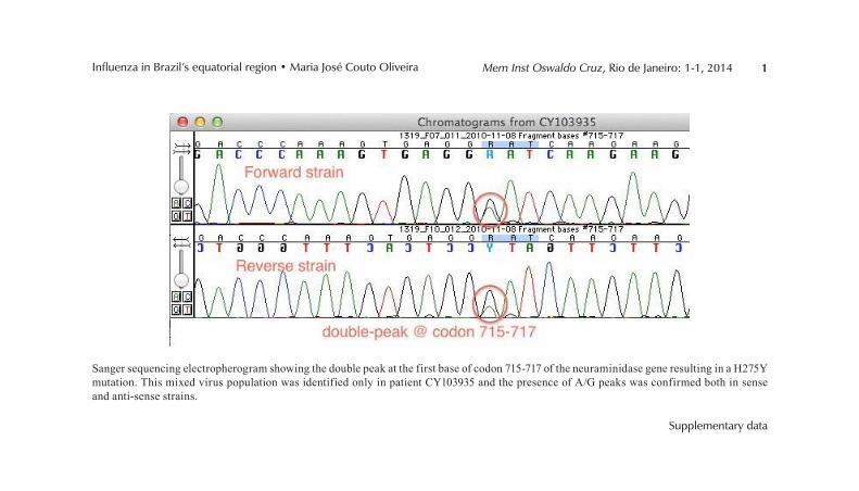
1Mem Inst Oswaldo Cruz, Rio de Janeiro: 1-1, 2014
Supplementary data
Influenza in Brazil’s equatorial region • Maria José Couto Oliveira
Sanger sequencing electropherogram showing the double peak at the first base of codon 715-717 of the neuraminidase gene resulting in a H275Y mutation. This mixed virus population was identified only in patient CY103935 and the presence of A/G peaks was confirmed both in sense and anti-sense strains.

90
DOCUMENTO II – PHYLOGENETIC ANALYZES OF
INFLUENZA A (H1N1)PDM09 HEMAGGLUTININ GENE
DURING AND AFTER THE PANDEMIC EVENT IN
BRAZIL

Documento II – Manuscrito em fase de preparação
91
Phylogenetic analyzes of Influenza A (H1N1)pdm09 hemagglutinin gene during and after the pandemic event in Brazil Paola Cristina Resende*1, Fernando do Couto Motta1, Priscila da Silva Born1, Daniela Machado1,2, Braulia Costa Caetano1, David Brown1, Marilda Mendonça Siqueira1.
1 – Laboratory of Respiratory Viruses and Measles – Oswaldo Cruz Institute – FIOCRUZ – Rio de Janeiro – Brazil 2 – Fondation Mérieux, Emerging Pathogens Laboratory – Centre International de Recherche en Infectologie (CIRI) *Corresponding author Paola Cristina Resende E-mail: [email protected] Address: Av. Brasil, 4365 - Manguinhos - Rio de Janeiro - RJ – Brasil – Pav. HPP – B104 Zip Code: 21040-360 Abstract
Pandemic influenza A H1N1 [A(H1N1)pdm09] was first detected in Brazil
in May 2009, and spread extensively throughout the country causing a peak of infection during June to August 2009. Since then, it has continued to circulate with a seasonal pattern, causing high rates of morbidity and mortality. Over this period, the virus has continually evolved with the accumulation of new mutations. In this study we analyze the phylogenetic relationship in a collection of 220 A(H1N1)pdm09 hemagglutinin (HA) gene sequences collected during and after the pandemic period (2009 to 2014) in Brazil. In addition, we have looked for evidence of viral polymorphisms associated with severe disease and compared the range of viral variants with the vaccine strain (A/California/7/2009) used throughout this period.
The phylogenetic analyzes in this study revealed the circulation of at least nine genetic groups in Brazil. Three (G1, G6-pdm and G7-pdm) co-circulated during the pandemic period, showing an early pattern of viral diversification with a low genetic distance from vaccine strain. Other phylogenetic groups, G5, G6 (including 6B, 6C and 6D subgroups), and G7 were found in the subsequent epidemic seasons from 2011 to 2014. These viruses exhibited more amino acid differences from the vaccine strain with several substitutions at the antigenic sites. This is associated with a theoretical decrease in the vaccine efficacy. Furthermore, we observed a polymorphism strongly associated with severe/fatal cases at residue 222 of the HA gene. This was significantly associated with severe disease reinforcing previous reports that described this residue as a potential virulence marker.

Documento II – Manuscrito em fase de preparação
92
This study provides new information about the circulation of some viral variants in Brazil, follows up potential genetic markers associated with virulence and allows infer if the efficacy of the current vaccine against more recent A(H1N1)pdm09 strains may be reduced. Keywords: Influenza, A(H1N1)pdm09, hemagglutinin, phylogenetic analyzes
1. Introduction
Influenza viruses are important human pathogens, causing seasonal epidemics and occasional pandemics with high rates of morbidity and mortality annually (World Health Organization, 2014a). During April 2009 a new Influenza A subtype H1N1 virus emerged simultaneously in Mexico and the United States, which rapidly spread worldwide, causing the first pandemic of the 21st century (Dawood et al., 2009; Smith et al., 2009). This virus was the product of multiple reassortment events involving viruses from swine, birds and humans and was responsible for more than 18,000 deaths worldwide (Smith et al., 2009; World Health Organization, 2010a). In Brazil, pandemic Influenza A (H1N1) virus [A(H1N1)pdm09] was first detected in May 2009 and it spread rapidly across the country. Despite public health containment measures being adopted to control viral transmission, a mortality rate of 4,6% among severe acute respiratory infections (SARI) cases were reported (Secretaria de Vigilância em Saúde, 2010). After the pandemic period, A(H1N1)pdm09 began to circulate as a seasonal virus together with influenza A/H3N2 and influenza B virus lineages, causing annual epidemic seasons during winter and considerable morbidity and mortality (Secretaria de Vigilância em Saúde, 2012, 2013a, b, 2014).
Influenza viruses have a segmented single stranded RNA genome and exhibit an evolutionary plasticity. During replication process, gradual genetic changes (antigenic drift) occur due to the lack of proof reading of the RNA polymerase complex (Carrat and Flahault, 2007; Medina and Garcia-Sastre, 2011). It contributes to the ability of the virus to evade recognition by the immune system and ensure its consistent circulation within human populations (Carrat and Flahault, 2007; Medina and Garcia-Sastre, 2011). Surveillance of emerging viral variants is necessary to inform decisions on the vaccine composition. Currently, the vaccine used in Brazil contains two subtypes of Influenza A, H1N1 and H3N2 and one influenza B lineage. Since the emergence of A(H1N1)pdm09, the H1N1 vaccine component has remained the same in the Northern and Southern Hemisphere (World Health Organization, 2014b). The anti-influenza vaccine effectiveness is rarely elevated due to the high mutation rate of influenza virus. Depending on antigenic match effectiveness of 30-60% against influenza have been reported (Gupta et al., 2006). Viral mutations may also cause an increase in viral fitness leading to a greater transmissibility, pathogenicity and to the emergence of viral variants resistant to antiviral drugs (Glinsky, 2010; Lee et

Documento II – Manuscrito em fase de preparação
93
al., 2010; Mesquita et al., 2014; Resende et al., 2014; Souza et al., 2013). The HA gene is the major viral antigen and is responsible for entry in the host cells. Analyzes of this gene provides important data of virulence, and temporal and geographical distribution of strains (Smith et al., 2004). Some mutations in the HA of A(H1N1)pdm09 virus have been suggested as potential virulence marker, as: K-2E and Q293H (Glinsky, 2010; Potdar et al., 2010); D222G (World Health Organization, 2010b). However only the substitution of aspartic acid (D) to glycine (G) at residue 222 of the HA gene (D222G) presented a strong association with severe and fatal cases (Resende et al., 2014). Regarding the phylogenetic dynamic of the A(H1N1)pdm09 virus, previous studies showed an early diversification of this virus into seven phylogenetic groups during pandemic period, based on specific mutations in the viral genome (Nelson et al., 2009; Potdar et al., 2010). After the pandemic, a major diversification of these strains occurred and new phylogenetic groups were identified based on mutations at HA gene (European Centre for Disease Prevention and Control, 2011, 2013).
In this study, we investigated genetic characteristics of A(H1N1)pdm09 strains which circulated in Brazil from 2009 to 2014 in different geographical regions in the country. Based on HA gene viral variants, we observed amino acid substitutions associations with severe clinical outcomes and analyzed important divergences in comparison to vaccine strain.
2. Material and methods
2.1 Samples
Using the Brazilian Influenza Surveillance Network, nasopharyngeal aspirates (NPAs) were collected from patients with Influenza-like illness (ILI) and hospitalized patients suffering from severe acute respiratory infection (SARI). These samples were sent to the Laboratory of Respiratory Viruses at FIOCRUZ in Rio de Janeiro, a National Influenza Center (NIC) for World and Health Organization (WHO) and for the Ministry of Healthy (MoH), where this study were conducted.
A total of 220 representative Brazilian A(H1N1)pdm09 strains were selected representing three Brazilian geographic/climatic regions, namely: Northeastern (n = 38), Southeastern (n = 37) and Southern (n = 145), covering the pandemic period (n = 65) and the following epidemic seasons 2011 (n = 10), 2012 (n = 49), 2013 (n = 74) and 2014 (n = 23). These samples were collected from patients with a median age of 26 years (ranging from 0 to 78 years), 54.1% were females. Among these patients only 138 (62,7%) provided detailed information about clinical outcomes, 44 cases were mild, 67 severe and 27 were from fatal cases.
Samples were manipulated anonymously. Patient identifiers including personal information (name, address) and hospitalization number were removed to protect patient confidentiality. This study was conducted within the scope of

Documento II – Manuscrito em fase de preparação
94
the National Influenza Epidemiological Surveillance Program/MoH, as part of a global and Federal public health policy for Influenza control and prevention in Brazil. For the reasons, a formal approval from our Ethical Committee was not required.
2.2 RNA extraction, amplification and sequencing of the HA gene
Viral RNA was extracted from NPAs using QIAmp viral RNA mini kit (Qiagen, Hilden, Germany), according to manufacturer’s protocol. cDNA was synthesized using Super Script III enzyme (Life technologies). Thereafter whole HA sequence was obtained by amplification of four PCR products using Taq Platinum DNA polymerase enzyme (Life technologies) and primers sets described previously by CDC Influenza division (World Health Organization, 2009). DNA products were purified with QIAquick PCR Purification kit (Qiagen) and sequenced using an automated ABI 3130XL Genetic Analyzer (Applied Biosystems). Sequences were assembled and the comparisons with the reference sequence (A/California/7/2009 – EPI516528) were performed with Sequencher software 5.1v (Gene Codes). Brazilian sequences were submitted to the Global Initiative on Sharing Avian Influenza Data (GISAID) database and are available with the accession numbers: EPI178726 to EPI 178945.
2.3 Data set construction, genetic and phylogenetic analyzes
A data set with 265 HA sequences was constructed, including Brazilian
sequences (n = 220) and other representative sequences (n = 45) obtained at GISAID database. These representative sequences have specific amino acid signatures in the HA gene that defined some phylogenetic groups, from pandemic (G1, G2-pdm to G7-pdm) and post pandemic periods (G2 to G7) (European Centre for Disease Prevention and Control, 2011, 2013; Nelson et al., 2009; Potdar et al., 2010). Sequences from unclassified phylogenetic groups found in this study were submitted to Basic Local Alignment Search Tool (BLAST) available at the National Center for Biotechnology Information (NCBI) database to obtain similar sequences circulating worldwide. The data set alignment was generated with Clustal W algorithm (integrated tool within Geneious 7.0 software) and edited manually for optimization. Maximum Likelihood (ML) trees were inferred with the on-line PhyML platform (http://www.atcg-montpellier.fr/phyml), using the best-fitting nucleotide substitution model, Hasegawa-Kishino-Yano (HKY) + gamma distribution among the sites, which was estimated by means of Modeltest, integrated tool within the MEGA 6.0 software (Tamura et al., 2013). ML reliability of branches was evaluated using approximate likelihood-ratio test (aLRT) with an interior branch cut-off value ≥ 0.9 and the tree was rooted with the vaccine strain A/California/7/2009.

Documento II – Manuscrito em fase de preparação
95
2.4 Analyzes of substitutions and prediction of vaccine efficacy Amino acid substitutions from Brazilian HA sequences were compared
with vaccine strain using Sequencher v5.1 software (Gene Codes). Amino acid distances among phylogenetic groups to vaccine strain were calculated in Mega 6.0 software (Tamura et al., 2013) using the p-distance method. In order to estimate vaccine efficacy (E), we used the equation E = 0.47 – 2.47 x Pepitope described by Deem and Pan (Deem and Pan, 2009). Pepitope means the largest p-distance value, the proportion of different amino acids for each epitope between two strains. We considered the five epitopes Sa, Sb, Ca1, Ca2, and Cb of the HA1 globular domain, against which the majority of neutralizing antibodies are directed (Igarashi et al., 2010). Antigenic distances of five HA epitopes found amongst the Brazilian strains from the vaccine strain A/California/7/2009 were calculated using the method p-distance in MEGA 6.0 software. 2.5 Statistical analysis
The software BaTS (Bayesian Tip-Significance testing) was used to test for significant phylogeny-trait correlations between virus sequences from mild and severe/fatal cases. It takes into account the statistic tests of Association Index (AI), the Parsimony Score (PS) and the Maximum Clade (MC), they provided 95% confidence intervals (CI) and significance estimation for these whilst accounting for phylogenetic uncertainty by the use of the posterior sets of trees obtained through earlier Bayesian phylogenetic analysis obtained by MrBayes software (Parker et al., 2008). Other statistical analyzes were calculated by Fisher`s exact test available at Graph Pad software an on-line tool (http://www.graphpad.com/quickcalcs/contingency1/). Statistical significance was considered only for p-values < 0.05.
3. Results
3.1 Phylogenetic analyzes of the hemagglutinin gene Molecular analyses of HA gene sequences obtained from 220 Brazilian
A(H1N1)pdm09 strains revealed that at least nine genetic groups circulated in Brazil from 2009 to 2014 (Figure 1). The ML tree topology showed a clear temporal structure with co-circulation of phylogenetic groups and subgroups with replacement of genetic groups over time. Based only on the HA gene, we identified two phylogenetic groups that circulated early in the pandemic: group 6 from pandemic period (G6-pdm), whose sequences clustered closely with A/New York/4735/2009-like with K-15E and Q293H substitutions; and group 7 from pandemic period (G7-pdm), similar to A/Shangai/1/2009-like with S203T

Documento II – Manuscrito em fase de preparação
96
substitution. Three Brazilian strains were more similar to the A/California/7/2009-like group (G1). These three early phylogenetic groups were detected during May to August 2009. Of 65 strains analyzed from this period, the large majority (67,7%) was G7-pdm, followed by the G6-pdm (26,2%). The G7-pdm, was the origin of later phylogenetic groups (G2 to G7) identified in the post-pandemic period, they are shown collapsed at the ML tree (Figure 1, arrows). Post-pandemic Brazilian HA sequences clustered in group 5 (G5), group 6 (G6), and group 7 (G7).
Another ML tree was constructed with the reference strains and our sequences from 2011 to 2014 epidemic seasons (Figure 1 A to D). This ML tree showed co-circulation of G5 and G6 during epidemic season 2011, of G5, G6 and G7 in 2012 and of G6 and G7 in 2013 (Figure 1 B and C). Only the G6 subgroup 6B were detected in 2014 epidemic season (Figure 1D). G5 and G7 variants were less common than G6. The G5 A/Astrakhan/1/2011-like sequences were characterized by D97N, R205K, I216V and V249L substitutions. Twenty-two Brazilian strains clustered in this group, were collected from May 2011 to August 2012 (Fig. 2B). Seventeen G7 Brazilian sequences detected in 2012 and 2013 seasons were A/St.Petersburg/100/2011-like, being characterized by S143G, S185T and A197T signature substitutions. In addition to these signatures, strains from 2013 exhibited the L-9M, S84G and K163I substitutions, which constituted a new cluster inside the group (Fig. 1C). Regarding G6, represented by strains A/St.Petersburg/27/2011-like with the D97N and S185T substitutions, it has circulated widely in Brazil since 2012 (Fig 2D), and more than half of Brazilian viruses analyzed in this study (n = 116) belonged to it. This group also shows the wider diversity, being divided in four subgroups, 6A to 6D. The subgroup 6B was the most populated enclosing 79 Brazilian sequences from 2013 and 2014 and the signature substitutions K163Q and A256T. The 6D phylogenetic subgroup (n= 33), was defined by the N260T mutation. Only two samples were classified as subgroup 6C at this collection and no one sample was clustered as subgroup 6A.
3.2 Amino acid substitutions and its association with clinical outcomes
In this analysis we identified changes in the HA gene sequences attributed to genetic evolution of A(H1N1)pdm09 circulating in Brazil from 2009 to 2014. Amino acid translations of the HA gene, from Brazilian viral sequences were compared with those from the vaccine strain A/California/7/2009 to verify possible associations with severe clinical outcomes (Table 1). Clinical outcome information was available for 138 patients; 44 patients presenting ILI were classified as mild; 67 cases were severe and were designated as SARI, and 27 were fatal cases. We constructed Bayesian trees using MrBayes software to show the lack of association of mild and severe/fatal cases inside each phylogenetic groups (Table 2). Reviewing the main amino acid substitutions associated with an increase of severity in the literature we observe that in our

Documento II – Manuscrito em fase de preparação
97
cases only the presence of any polymorphisms at residue 222 was associated with severe/fatal cases (Table 3) (p-value < 0.05). The mutations at residue 222 were not associated to any specific phylogenetic cluster, being found in several branches and groups throughout the tree.
3.3 Amino acid substitutions in antigenic sites and vaccine efficacy prediction We identified 22 amino acid changes compared to the vaccine strain
located at five different antigenic sites (1 to 5 mutations per sample). They were distributed in all phylogenetic groups, except G1. Groups from the pandemic period had 1 to 3 changes and all strains in groups from post-pandemic period contained 2 to 5 changes (Table 2).
The large majority of these mutation were found at low frequency (0,5% to 0,9%) scattered throughout the years covered by our sampling, namely: T72I, A73S and S75T in Cb; P137S, A139S and A141R in Ca2; N137D; S174T and S179N in Sa; I183V, G187R and E235D in Ca1; A203T, D204Y, Q189K and S190N in Sb. However, other mutations at the five antigenic sites appeared in high proportions in the viral population. The S203T and R205K substitutions located at antigenic site Ca1, were found in 90,5% and 12,3% of strains respectively. In Sb 60,5% of strains showed substitutions at residue 185, S185T (n = 126) or S185A (n = 7). In Sa we observed K163Q or K163I substitutions in 42,7% of strains. H183Q was found in 6,8% and D222G, D222N or mixed viral populations D222D/G or D222D/G/N/A had a frequency of 5,5%.
The mean of global amino acid p-distance (number of amino acid substitutions per site) of each phylogenetic group were calculated in comparison to the vaccine strain (Table 4). It revealed that phylogenetic groups from the pandemic period had a low number of amino acid substitutions in comparison to phylogenetic groups from the post-pandemic period. We also calculated the antigenic distances of five HA epitopes from all sequences in relation to those from the vaccine strain A/California/7/2009 in order to estimate the theoretical vaccine efficacy (E) against Brazilian viruses using the Pepitope method (Deem and Pan, 2009). Phylogenetic groups from pandemic period presented a perfect vaccine match (Pepitope = 0) with an E value of 0.470, which means 100% of predict efficacy against the vaccine. However, the phylogenetic groups with mutations in antigenic sites presented a decrease at the predict vaccine match (Table 4). The G5 presented the worst match with the vaccine with 4,5%, of agreement, followed by G6C with 38,5% and others with 51,2%. Interestingly, we observed amongst our study population 13 cases infected with A(H1N1)pdm09, who had received vaccine prior to illness, 11 in 2013 and 2 in 2014, as reported on the epidemiological form. That they had received vaccine some months before illness and their virus strains had a predicted vaccine match of 52,1% (E = 0.245).

Documento II – Manuscrito em fase de preparação
98
4. Discussion This study describes amino acid changes in the HA of 220 A(H1N1)pdm09
viruses circulating in Brazil during the pandemic period (April, 2009 to August, 2010) and in the following epidemic seasons (2011 to 2014). Phylogenetic analyzes indicate that at least nine distinct genetic groups defined by specific mutations circulated in Brazil during this period. Previous evolutionary studies, conducted with A(H1N1)pdm09 that circulated worldwide during the initial phase of pandemic period, showed an early diversification based on molecular signatures of the whole genome, which characterized the seven initial phylogenetic groups (G1, G2-pdm to G7-pdm) (Nelson et al., 2009; Potdar et al., 2010). We identified two of these early phylogenetic groups, namely G6-pdm and G7-pdm, as well as sequences associated to G1 circulating in Brazil. As this previous classification was based on whole genome sequence, because we have only HA sequences we need more information about the other genes to know exactly where the four sequences closer to G1 are clustering. In turn G6-pdm and G7-pdm co-circulated among Brazilian population confirming previous data (Goni et al., 2011; Oliveira et al., 2013; Sant'Anna et al., 2014). As observed worldwide, G7-pdm was also the most common phylogenetic group detected in Brazil during the pandemic period (Nelson et al., 2009; Potdar et al., 2010). The global dominance of G7-pdm variant may be attributed to its initial foothold in New York city, which could have facilitated its rapid global spread due to its high international interconnectivity (Nelson et al., 2011). The end of pandemic period was declared on August, 2010 (World Health Organization, 2010a) and from mid-2010 to early 2011, ILI cases reduced in Brazil (Secretaria de Vigilância em Saúde, 2012). We speculate that this may have been associated with increase of herd immunity following the mass vaccination campaign against A(H1N1)pdm09, together with the inter seasonality period in the Southern Hemisphere. Since then, A(H1N1)pdm09 has circulated as a seasonal virus together with Influenza A/H3N2 and the two lineages of influenza B viruses causing annual epidemic seasons during the winter in Brazil with a lesser impact on morbidity and mortality than during the pandemic period (Secretaria de Vigilância em Saúde, 2012, 2013a, b, 2014).
However, after the pandemic period (2011 to 2014 epidemic seasons) new genetic groups emerged from G7-pdm (European Centre for Disease Prevention and Control, 2011, 2013). At least six new phylogenetic groups were detected circulating worldwide and they were numbered from group 2 to 7 (G2 to G7), since vaccine strain A/California/7/2009 reminded as G1 (European Centre for Disease Prevention and Control, 2011, 2013). Brazilian samples were observed in three of these groups G5, G6 and G7 (Fig. 1). Additionally, strains from epidemic seasons 2012/13 in Europe were classified in one of the G6 subgroups, 6A to 6C (European Centre for Disease Prevention and Control, 2013). Using the same approach, we describe a subgroup, with phylogentic signature N260T, called 6D. This mutation, detected among 2012 Brazilian samples seems to be

Documento II – Manuscrito em fase de preparação
99
observed mostly in HA sequences in North and South America. Group co-circulation during epidemics in 2011 (G5 and G6), 2012 (G5, G6 and G7) and 2013 (G6 and G7) were observed, however samples of 2014 epidemic season were detected only the G6B. According to data from Brazilian MoH, circulation of A(H1N1)pdm09 as a seasonal virus were more extensive during 2012 and 2013 epidemic seasons compared with the 2011 and 2014 epidemics (Secretaria de Vigilância em Saúde, 2012, 2013a, b, 2014). The circulation of multiple phylogenetic groups in these periods may had contributed to increase of the viral circulation. Some of these genetic groups were observed previously in Northern hemisphere before their detection in Brazil. Viruses collected during epidemic season 2010-2011 in European countries fall into the post-pandemic phylogenetic groups, and in Brazil, it occurred after May 2011 (European Centre for Disease Prevention and Control, 2011; Ledesma et al., 2012). It may suggest that the previous epidemic of North Hemisphere may predict and potentially seed the next epidemic season in Brazil, however more data is necessary to confirm this hypothesis.
As a RNA virus, Influenza viruses have a high rate of genetic variation, which ensures its survival and constant circulation in the human population (Carrat and Flahault, 2007). The rate of change of influenza means that the antigens included in the annual vaccination have to be changed on a regular basis to provide protection against current strains. The selection of strains is based on the detection of new variants and assessment of antigenic change using ferret antisera and sera from vaccine. Studies have attempted to evaluate the use of sequence based amino acid changes to predict these antigenic changes. (Glinsky, 2010; Lee et al., 2010; Mesquita et al., 2014; Resende et al., 2014; Souza et al., 2013).
In our study, sequences from post-pandemic strains showed more amino acid differences than pandemic strains from the vaccine and some of the amino acid substitutions occurred in antigenic sites (Ca1, Ca2, Cb, Sa and Sb) of the HA molecule (Sriwilaijaroen and Suzuki, 2012). Although the genetic groups identified in this study showed specific changes in different epitopes of HA, which contributed to a theoretical decrease in vaccine efficacy, the results obtained by formal antigenic characterization performed with similar variant viruses did not show a consistent marked change in antigenicity compared to vaccine virus A/California/7/2009 (European Centre for Disease Prevention and Control, 2011, 2013). Around 99,1% of Brazilian samples contained up to four mutations in HA antigenic sites. However, previous report indicates that such mutations did not contribute to a significant change in antigenicity (Dapat et al., 2012), indicating that the algorithms to evaluate genetic changes alone, needs future refinement before it can be used to infer vaccine choices.
None of the phylogenetic groups found in Brazil had a strong relationship with severe or fatal influenza disease, and our findings corroborates the lack of association with severity of K-15E and Q293H substitutions (G6-pdm) found previously (Graham et al., 2011; Lee et al., 2010; Mesquita et al., 2014).

Documento II – Manuscrito em fase de preparação
100
Otherwise, we observed a point mutation at residue 222 of the HA gene associated with an increase of severity illness in humans (World Health Organization, 2010b). This mutation was detected across variant viral groups and in different seasons, which suggests it as a potential virulence marker. Associations of amino acid changes in the HA with a possible increase of virulence has been reported (El Moussi et al., 2013; Glinsky, 2010; Kilander et al., 2010; Maurer-Stroh et al., 2010; Melidou et al., 2010; Resende et al., 2014). The association of polymorphisms and the presence of quasispecies at residue 222 of the HA was widely explored in samples from pandemic period (Resende et al., 2014). This amino acid residue is located in the antigenic site Ca2 near from of the receptor-binding domain and local changes may influence binding preferences of the virus (Liu et al., 2010). The substitutions of aspartic acid (D) to glycine (G), asparagine (N) and/or Alanine (A) at residue 222 of the HA gene were found in 11 cases, of which, 10 were fatal cases and one severe case. Among these patients, five were pregnant women. Our group had previously reported a curious relation between this residue in samples from pregnant women and disease’s severity (Resende et al., 2014). Polymorphisms at residue 222 in Brazilian strains did not form a phylogenetic cluster, sequences with this mutation were observed in strains from two phylogenetic groups from the pandemic period, G6-pdm and G7-pdm, and it was observed in recent cases due to G6B and G6D viruses.
5. Conclusions
Five years after pandemic the A(H1N1)pdm09 continues to circulate in Brazilian population, nine influenza A(H1N1)pdm09 genetic groups defined by specific amino acid changes have been circulated in Brazil from 2009 to 2014. We have seen a regular replacement of variants by new phylogenetic groups over time and the presence of mutations in antigenic sites have been increasing. In accordance with antigenic results worldwide, none of these genetic groups seems to show significant antigenic differences with the vaccine strain A/California/7/2009, but theoretically the vaccine efficacy have been decreased due to changes in antigenic sites of these strains. This study reinforce the importance of continued laboratory based-surveillance to detect the emergence of new variants, which essential for reformulation of influenza vaccine, since the closer the vaccine strain is to the dominant circulating viral variant, the more effective is the vaccine is likely to be. Acknowledgments We would like to thank the DECIT, CNPq and FAPERJ for financial support to conduct this research and the Brazilian surveillance influenza network for collection of samples.

Documento II – Manuscrito em fase de preparação
101
References Carrat, F., Flahault, A., 2007. Influenza vaccine: the challenge of antigenic drift. Vaccine 25, 6852-6862. Dapat, I.C., Dapat, C., Baranovich, T., Suzuki, Y., Kondo, H., Shobugawa, Y., Saito, R., Suzuki, H., 2012. Genetic characterization of human influenza viruses in the pandemic (2009-2010) and post-pandemic (2010-2011) periods in Japan. PLoS One 7, e36455. Dawood, F.S., Jain, S., Finelli, L., Shaw, M.W., Lindstrom, S., Garten, R.J., Gubareva, L.V., Xu, X., Bridges, C.B., Uyeki, T.M., 2009. Emergence of a novel swine-origin influenza A (H1N1) virus in humans. N Engl J Med 360, 2605-2615. Deem, M.W., Pan, K., 2009. The epitope regions of H1-subtype influenza A, with application to vaccine efficacy. Protein Eng Des Sel 22, 543-546. El Moussi, A., Ben Hadj Kacem, M.A., Pozo, F., Ledesma, J., Cuevas, M.T., Casas, I., Slim, A., 2013. Genetic diversity of HA1 domain of heammaglutinin gene of influenza A(H1N1)pdm09 in Tunisia. Virol J 10, 150. European Centre for Disease Prevention and Control, 2011. Influenza Virus Characterisation - Summary Europe, August and September 2011 Technical Document European Centre for Disease Prevention and Control, 2013. Influenza Virus Characterisation - Summary Europe, September 2013. Surveillance Report. Glinsky, G.V., 2010. Genomic analysis of pandemic (H1N1) 2009 reveals association of increasing disease severity with emergence of novel hemagglutinin mutations. Cell Cycle 9, 958-970. Goni, N., Moratorio, G., Ramas, V., Coppola, L., Chiparelli, H., Cristina, J., 2011. Phylogenetic analysis of pandemic 2009 influenza A virus circulating in the South American region: genetic relationships and vaccine strain match. Arch Virol 156, 87-94. Graham, M., Liang, B., Van Domselaar, G., Bastien, N., Beaudoin, C., Tyler, S., Kaplen, B., Landry, E., Li, Y., 2011. Nationwide molecular surveillance of pandemic H1N1 influenza A virus genomes: Canada, 2009. PLoS One 6, e16087. Gupta, V., Earl, D.J., Deem, M.W., 2006. Quantifying influenza vaccine efficacy and antigenic distance. Vaccine 24, 3881-3888. Igarashi, M., Ito, K., Yoshida, R., Tomabechi, D., Kida, H., Takada, A., 2010. Predicting the antigenic structure of the pandemic (H1N1) 2009 influenza virus hemagglutinin. PLoS One 5, e8553. Kilander, A., Rykkvin, R., Dudman, S.G., Hungnes, O., 2010. Observed association between the HA1 mutation D222G in the 2009 pandemic influenza A(H1N1) virus and severe clinical outcome, Norway 2009-2010. Euro Surveill 15. Ledesma, J., Pozo, F., Reina, G., Blasco, M., Rodriguez, G., Montes, M., Lopez-Miragaya, I., Salvador, C., Reina, J., Ortiz de Lejarazu, R., Egido, P., Lopez Barba, J., Delgado, C., Cuevas, M.T., Casas, I., 2012. Genetic diversity of influenza A(H1N1)2009 virus circulating during the season 2010-2011 in Spain. J Clin Virol 53, 16-21. Lee, R.T., Santos, C.L., de Paiva, T.M., Cui, L., Sirota, F.L., Eisenhaber, F., Maurer-Stroh, S., 2010. All that glitters is not gold--founder effects complicate associations of flu mutations to disease severity. Virol J 7, 297. Liu, Y., Childs, R.A., Matrosovich, T., Wharton, S., Palma, A.S., Chai, W., Daniels, R., Gregory, V., Uhlendorff, J., Kiso, M., Klenk, H.D., Hay, A., Feizi, T., Matrosovich, M., 2010. Altered receptor specificity and cell tropism of D222G hemagglutinin mutants isolated from fatal cases of pandemic A(H1N1) 2009 influenza virus. J Virol 84, 12069-12074. Maurer-Stroh, S., Lee, R.T., Eisenhaber, F., Cui, L., Phuah, S.P., Lin, R.T., 2010. A new common mutation in the hemagglutinin of the 2009 (H1N1) influenza A virus. PLoS Curr 2, RRN1162. Medina, R.A., Garcia-Sastre, A., 2011. Influenza A viruses: new research developments. Nat Rev Microbiol 9, 590-603.

Documento II – Manuscrito em fase de preparação
102
Melidou, A., Gioula, G., Exindari, M., Chatzidimitriou, D., Diza, E., Malisiovas, N., 2010. Molecular and phylogenetic analysis of the haemagglutinin gene of pandemic influenza H1N1 2009 viruses associated with severe and fatal infections. Virus Res 151, 192-199. Mesquita, M., Resende, P., Marttorelli, A., Machado, V., Sacramento, C.Q., Fintelman-Rodrigues, N., Abrantes, J.L., Tavares, R., Schirmer, M., Siqueira, M.M., Souza, T.M., 2014. Detection of the Influenza A(H1N1)pdm09 Virus Carrying the K-15E, P83S and Q293H Mutations in Patients Who Have Undergone Bone Marrow Transplant. PLoS One 9, e94822. Nelson, M., Spiro, D., Wentworth, D., Beck, E., Fan, J., Ghedin, E., Halpin, R., Bera, J., Hine, E., Proudfoot, K., Stockwell, T., Lin, X., Griesemer, S., Kumar, S., Bose, M., Viboud, C., Holmes, E., Henrickson, K., 2009. The early diversification of influenza A/H1N1pdm. PLoS Curr 1, RRN1126. Nelson, M.I., Tan, Y., Ghedin, E., Wentworth, D.E., St George, K., Edelman, L., Beck, E.T., Fan, J., Lam, T.T., Kumar, S., Spiro, D.J., Simonsen, L., Viboud, C., Holmes, E.C., Henrickson, K.J., Musser, J.M., 2011. Phylogeography of the spring and fall waves of the H1N1/09 pandemic influenza virus in the United States. J Virol 85, 828-834. Oliveira, M.J., Motta, F.D., Siqueira, M.M., Resende, P.C., Born, P.D., Souza, T.M., Mesquita, M., Oliveira, M.D., Carney, S., Mello, W.A., Magalhaes, V., 2013. Molecular findings from influenza A(H1N1)pdm09 detected in patients from a Brazilian equatorial region during the pandemic period. Mem Inst Oswaldo Cruz 0, 0. Parker, J., Rambaut, A., Pybus, O.G., 2008. Correlating viral phenotypes with phylogeny: accounting for phylogenetic uncertainty. Infect Genet Evol 8, 239-246. Potdar, V.A., Chadha, M.S., Jadhav, S.M., Mullick, J., Cherian, S.S., Mishra, A.C., 2010. Genetic characterization of the influenza A pandemic (H1N1) 2009 virus isolates from India. PLoS One 5, e9693. Resende, P.C., Motta, F.C., Oliveira Mde, L., Gregianini, T.S., Fernandes, S.B., Cury, A.L., do Carmo, D.R.M., Souza, T.M., Siqueira, M.M., 2014. Polymorphisms at Residue 222 of the Hemagglutinin of Pandemic Influenza A(H1N1)pdm09: Association of Quasi-Species to Morbidity and Mortality in Different Risk Categories. PLoS One 9, e92789. Sant'Anna, F.H., Borges, L.G., Fallavena, P.R., Gregianini, T.S., Matias, F., Halpin, R.A., Wentworth, D., d'Azevedo, P.A., Veiga, A.B., 2014. Genomic analysis of pandemic and post-pandemic influenza A pH1N1 viruses isolated in Rio Grande do Sul, Brazil. Arch Virol 159, 621-630. Secretaria de Vigilância em Saúde, 2010. Influenza Pandêmica (H1N1) 2009 – Análise da situação epidemiológica e da resposta no ano de 2009. Ministério da Saúde, Brasilia, p. 21. Secretaria de Vigilância em Saúde, 2012. Informe técnico de Influenza - Vigilânia de Sindrome Respiratória Aguda Grave (SRAG), de Sindrome Gripal (SG) e de internações por CID J09 e J18. Ministério da Saúde, Brasília, p. 15. Secretaria de Vigilância em Saúde, 2013a. Boletim epidemiológico - Influenza (gripe) - Semana Epidemiológica (SE) 52 (atualizado em 08/01/2013). Ministério da Saúde, Brasília, p. 11. Secretaria de Vigilância em Saúde, 2013b. Boletim epidemiológico - Influenza: Monitoramento até a Semana Epidemiológica 52 de 2013. Ministério da Saúde, Brasília, p. 10. Secretaria de Vigilância em Saúde, 2014. Boletim epidemiológico - Influenza: Monitoramento até a Semana Epidemiológica 46 de 2014. Ministério da Saúde, Brasília, p. 11. Smith, D.J., Lapedes, A.S., de Jong, J.C., Bestebroer, T.M., Rimmelzwaan, G.F., Osterhaus, A.D., Fouchier, R.A., 2004. Mapping the antigenic and genetic evolution of influenza virus. Science 305, 371-376. Smith, G.J., Vijaykrishna, D., Bahl, J., Lycett, S.J., Worobey, M., Pybus, O.G., Ma, S.K., Cheung, C.L., Raghwani, J., Bhatt, S., Peiris, J.S., Guan, Y., Rambaut, A., 2009. Origins and evolutionary genomics of the 2009 swine-origin H1N1 influenza A epidemic. Nature 459, 1122-1125.
Documento II – Manuscrito em fase de preparação
103
Souza, T.M., Resende, P.C., Fintelman-Rodrigues, N., Gregianini, T.S., Ikuta, N., Fernandes, S.B., Cury, A.L., Rosa Mdo, C., Siqueira, M.M., 2013. Detection of Oseltamivir-Resistant Pandemic Influenza A(H1N1)pdm2009 in Brazil: Can Community Transmission Be Ruled Out? PLoS One 8, e80081. Sriwilaijaroen, N., Suzuki, Y., 2012. Molecular basis of the structure and function of H1 hemagglutinin of influenza virus. Proc Jpn Acad Ser B Phys Biol Sci 88, 226-249. Tamura, K., Stecher, G., Peterson, D., Filipski, A., Kumar, S., 2013. MEGA6: Molecular Evolutionary Genetics Analysis version 6.0. Mol Biol Evol 30, 2725-2729. World Health Organization, 2009. Sequencing primers and protocol. WHO Collaborating Centre for Influenza at CDC, Atlanta, GA. World Health Organization, 2010a. Pandemic (H1N1) 2009 - update 112. http://www.who.int/csr/don/2010_08_06/en/index.html, Geneva. World Health Organization, 2010b. Preliminary review of D222G amino acid substitution in the haemagglutinin of pandemic influenza A (H1N1) 2009 viruses. Wkly Epidemiol Rec 85, 21-28. World Health Organization, 2014a. Influenza (Seasonal), Fact sheet N°211., March 2014 ed. http://www.who.int/mediacentre/factsheets/fs211/en/index.html. World Health Organization, 2014b. WHO recommendations on the composition of influenza virus vaccines.

Documento II – Manuscrito em fase de preparação
104
Figure 1 - Maximum-Likelihood phylogenetic tree of the HA whole gene (1701 nucleotides) of 220 Brazilian influenza A(H1N1)pdm09 viruses detected from 2009 (blue), 2010 (cyan), 2011(purple), 2012 (pink), 2013 (red) and 2014 (orange) seasons and 46 representative sequences (black) of the groups G1, G2-pdm to G7-pdm and G2 to G7 collected on GISAID database. Amino acid changes associated with these specific groups were indicated in the nodes using H1 numbering without signal peptide. The best-fitting nucleotide substitution model used was Hasegawa-Kishino-Yano (HKY) + gamma distribution among the sites. ML reliability of branches was evaluated using approximate likelihood-ratio test (aLRT) and an interior branch cut-off value was ≥ 0.9. The figure 1A shows groups from pandemic period. The collapsed branches represent sequences from post pandemic period and arrows in red indicate where the Brazilian sequences were found. The figures 1B, 1C and 1D, shows the G5, G7 and G6 and its subgroups respectively.

G6-pdm
G7-pdm
A.
D97N
*
*
D97N; R205K;I216V; V249L
**
*
*
*
*
**
S203T
K-15E;Q310H
A134T; S183P
N125D
N31D, S162N, A186T
G1 - vaccine
*
*
*
*
*

B.
G5
C.
G7
*
*
*
*
*
*
*
*
*
*
*
*
D97N; R205K;I216V; V249L
H138Q;E356A;
K488R
A-4T;A-2T
E491K
V30I
T15N; A139S
*
*
*
*
*
*
*
*
S143G; A197T
S84G;K163I
S69T;N260D
R205K
T120A; E499K
*
*

D.
G6
6D
6C
6B
6A
K163Q;A256T
V234I
E172K;K283E
*
*
*
**
*
*N260TD97N;
S124N;S185T
H138R; V249L
*
*
*
**
*
*
*
*
A-2T

Documento II – Manuscrito em fase de preparação
Table 1 - Relation of amino acid changes at the hem agglutin of the Brazilian strains to clinical outco me. Phylogenetic group Period of circulation in Brazil Number of
mild cases (%)
Number of severe
and fatal cases (%)
Presence of
mutations at 222
residue of HA
G1 Jun 2009 to Aug 2009 1 (2,3%) 2 (2,1%)
G6-pdm (K-15E; Q310H)
May 2009 to Mar 2010 3 (6,8%) 8 (8,5%) D222DGNA (2);
D222G (2)
G7-pdm (S203T)
Jun 2009 to Jun 2010 6 (13,6%) 31 (33,0%) D222DGNA (4);
D222DG (2)
D222G (1)
G5 (D97N; R205K; I216V; V249L)
May 2011 to Aug 2012 10 (22,7%) 7 (7,4%)
G6 (D97N)
May 2011 to Jun 2011 0 (0,0%) 1 (1,1%)
G6B (K163Q; A256T)
Mar 2013 to Jun 2014 5 (11,4%) 23 (24,5%) D222N (1)
G6C (V234I)
Mar 2013 to Jun 2013 0 (0,0%) 4 (4,3%)
G6D (N260T)
Apr 2012 to Jan 2013 15 (34,1%) 11 (11,7%) D222G (1)
G7 (S143G; A197T)
Mar 2012 to Jun 2013 4 (9,1%)
7 (7,4%)

Documento II – Manuscrito em fase de preparação
Table 2 – Comparison about the clustering of mild c ases versus severe plus fatal cases using Bayesian Tip-Significance testing (BaTS).
Statistic test Observed mean (95% CI) Null hypotesis mean (95% CI) Significance (p-value)
AI 3,9 (3,0 – 4,9) 5,6 (4,6 – 6,6) 0,00
PS 30,6 (28,0 – 32,0) 37,4 (34,0 – 40,3) 0,00
MC (Mild cases) 7,0 (7,0 – 7,0) 2,6 (2,1 – 3,4) 0,00
MC (Severe + Fatal cases) 8,3 (7,0 – 13,0) 6,4 (4,5 – 9,9) 0,27
PS (parsimony score) statistic quantifies phylogeny–trait correlation; AI (association index) statistic takes into account the shape of the phylogeny by measuring the imbalance of internal phylogeny nodes; MC (monophyletic clade).
Table 3 - Comparison of polymorphisms at 222 residu e of the influenza A (H1N1)pdm09 hemagglutin gene i n 138 Brazilian in patients with mild versus severe/fatal clinical outcomes.
HA position Clinical outcome (a)
p-value (c) Mild Severe/Fatal
D222G 0 % (0/44) 4.3 % (4/94) 0.299
D222N 0 % (0/44) 1.1 % (1/94) 1.0
D222D/G 0 % (0/44) 1.1 % (1/94) 1.0
D222D/G/N/A 0 % (0/44) 5.3 % (5/94) 0.168
X222 (b) 0 % (0/44) 11.7 % (11/94) 0.016 (d) (a) Only 138 from 220 patients included in this study had information about clinical outcome; (b) X222 means any mutation at the residue 222 (c) The statistical significance between the number of mild and severe/fatal cases was calculated by Fisher's exact test;; (d) Residues with statistical significance p-value < 0.05

Documento II – Manuscrito em fase de preparação
Table 4 - Amino acid distance of the nine A(H1N1)pd m09 phylogenetic groups to the vaccine strain A/Cal ifornia/7/2009 and the substitutions frequency in antigenic sites into each group.
Phylogenetic Group
(amino acid signatures)
Number of
sequences
Period of circulation in
Brazil
Mean of amino acid
p-distance to
A/California/7/2009
Mean of predicted
vaccine efficacy (E)
Number of
changes in
antigenic sites
G1 4 Jun 2009 to Aug 2009 0.005 0.470 0
G6-pdm (K-15E; Q310H)
17 May 2009 to Mar 2010 0.008 0.470 0 to 1
G7-pdm (S203T)
44 Jun 2009 to Jun 2010 0.008 0.470 1 to 3
G5 (D97N; R205K; I216V; V249L)
22 May 2011 to Aug 2012 0.021 0.021 2 to 4
G6 (D97N)
2 May 2011 to Jun 2011 0.017 0.245 2
G6B (K163Q; A256T)
79 Mar 2013 to Jun 2014 0.021 0.245 3 to 5
G6C (V234I)
2 Mar 2013 to Jun 2013 0.019 0.181 2 to 3
G6D (N260T)
33 Apr 2012 to Jan 2013 0.018 0.245 2 to 3
G7 (S143G; A197T)
17 Mar 2012 to Jun 2013 0.025 0.245 2 to 5
H1 numbering without signal peptide; p-distance: proportion of different amino acids for each HA1 epitope between the strain of interest and the vaccine strain A/California/7/2009; Vaccine efficacy (E) per each phylogenetic group.

111
Capítulo II: Identificação de marcadores de virulên cia
associados ao aumento da gravidade da infecção pelo
vírus Influenza A (H1N1)pdm09
Neste capítulo apresentamos dois artigos, nos quais abordamos a análise de
polimorfismos no gene da HA dos vírus A (H1N1)pdm09 associados a um possível
aumento na gravidade da infecção pelo vírus.
O primeiro trabalho foi publicado na revista Plos One em março de 2014 com
o título: “Polymorphism at residue 222 of the hemaggutinin of pandemic influenza A
(H1N1)pdm09: Association of quasi-species to morbidity and mortality in different risk
categories” (Documento III). Neste estudo utilizamos a metodologia de
pirosequenciamento para detecção de SNPs na posição 222 da HA viral. Foram
analisadas um total de 401 amostras de pacientes com manifestações clínicas
brandas, graves e fatais coletadas entre 2009 e 2011. Isso possibilitou detectar a
presença do polimorfismo D222G ou de populações mistas, DG222, DN222, GN222
e DGN222 em 70 amostras, sendo estas majoritariamente de pacientes graves ou
que foram a óbito no ano de 2009.
Em abril de 2014 publicamos um segundo trabalho também na revista Plos
One, “Detection of the Influenza A (H1N1)pdm09 virus carrying the K-15E, P83S and
Q293H mutations in patients who have undergone bone marrow transplant”
(Documento IV). Neste trabalho estudamos o impacto de variantes contendo as
mutações K-15E, P83S e Q293H em uma população de indivíduos que sofreram
transplante de medula óssea. Alguns estudos associaram essa variante viral com
uma maior gravidade e fatalidade, outros discordam dessa associação. Nossos
resultados mostraram que os indivíduos transplantados mesmo sendo mais
propensos a apresentar piores resultados clínicos, por serem imunocomprometidos,
apresentaram casos leves de infecção. Esses resultados não corroboram estudos

112
que relacionaram as substituições K-15E, P83S e Q293H com o aumento da
gravidade da doença.

113
DOCUMENTO III – POLYMORPHISM AT RESIDUE 222 OF
THE HEMAGGUTININ OF PANDEMIC INFLUENZA A
(H1N1)PDM09: ASSOCIATION OF QUASI-SPECIES TO
MORBIDITY AND MORTALITY IN DIFERENTE RISK
CATEGORIES

Polymorphisms at Residue 222 of the Hemagglutinin ofPandemic Influenza A(H1N1)pdm09: Association ofQuasi-Species to Morbidity and Mortality in DifferentRisk CategoriesPaola Cristina Resende1*, Fernando C. Motta1, Maria de Lourdes A. Oliveira1, Tatiana S. Gregianini2,
Sandra B. Fernandes3, Ana Luisa F. Cury4, Maria do Carmo D. Rosa5, Thiago Moreno L. Souza1,
Marilda M. Siqueira1
1 Laboratorio de Vırus Respiratorios e do Sarampo, Instituto Oswaldo Cruz/ FIOCRUZ, Rio de Janeiro, Rio de Janeiro, Brazil, 2 Laboratorio Central de Saude Publica do
Estado do Rio de Grande do Sul -Fundacao estadual de producao e pesquisa em saude secao de virologia, Porto Alegre, Rio Grande do Sul, Brazil, 3 Laboratorio Central de
Saude Publica do Estado de Santa Catarina, Florianopolis, Santa Catarina, Brazil, 4 Laboratorio Central de Saude Publica do Estado de Minas Gerais/Instituto Octavio
Magalhaes e Fundacao Ezequiel Dias, Belo Horizonte, Minas Gerais, Brazil, 5 Laboratorio Central de Saude Publica do Estado do Parana, Curitiba, Parana, Brazil
Abstract
The D222G substitution in the hemagglutinin (HA) gene of the pandemic influenza A(H1N1)pdm09 virus has been identifiedas a potential virulence marker, because this change allows for virus invasion deeper into the respiratory tract. In this study,we analyzed D, G and N polymorphisms at residue 222 by pyrosequencing (PSQ). We initially analyzed 401 samples fromBrazilian patients. These were categorized with respect to clinical conditions due to influenza infection (mild, serious or fatal)and sub-stratified by risky factors. The frequency of mixed population of virus, with more than one polymorphism at residue222, was significantly higher in serious (10.6%) and fatal (46.7%) influenza cases, whereas those who showed mild influenzainfections were all infected by D222 wild-type. Mixtures of quasi-species showed a significant association of mortality,especially for those with risk factors, in special pregnant women. These results not only reinforce the association betweenD222G substitution and influenza A(H1N1)pdm09-associated morbidity and mortality, but also add the perspective that aworse clinical prognosis is most likely correlated with mixtures of quasi-species at this HA residue. Therefore, quasi-speciesmay have a critical and underestimated role in influenza-related clinical outcomes.
Citation: Resende PC, Motta FC, Oliveira MdLA, Gregianini TS, Fernandes SB, et al. (2014) Polymorphisms at Residue 222 of the Hemagglutinin of PandemicInfluenza A(H1N1)pdm09: Association of Quasi-Species to Morbidity and Mortality in Different Risk Categories. PLoS ONE 9(3): e92789. doi:10.1371/journal.pone.0092789
Editor: Toru Takimoto, University of Rochester Medical Center, United States of America
Received October 18, 2013; Accepted February 25, 2014; Published March 25, 2014
Copyright: � 2014 Resende et al. This is an open-access article distributed under the terms of the Creative Commons Attribution License, which permitsunrestricted use, distribution, and reproduction in any medium, provided the original author and source are credited.
Funding: Brazilian Ministry of Health and Ministry of Technology and Science, CNPq and FAPERJ provided funding to conduct this research. The funders had norole in study design, data collection and analysis, decision to publish, or preparation of the manuscript.
Competing Interests: The authors have declared that no competing interests exist.
* E-mail: [email protected]
Introduction
The pandemic influenza A(H1N1)pdm09 virus was responsible
for over 18,449 deaths worldwide from 2009 to 2010 [1].
Although increased mortality is generally associated with novel
influenza viruses due to the lack of a preexisting immunity in the
general population, several groups of patients at higher risk for
complications due to the influenza infection were reinforced or
identified in 2009, such as pregnant women, obese individuals,
immunocompromised patients and, surprisingly, otherwise healthy
young adults, among others [2].
In addition to hosts’ predisposed clinical conditions, some
specific polymorphisms in the viral genome may enhance its
virulence [3]. Circulating influenza A(H1N1)pdm09 viruses from
2009 to the present are virtually similar, and no major antigenic
variation has been found in viral hemagglutinin (HA), which is the
viral surface glycoprotein responsible for attachment and a major
target of the host’s immune response [2]. However, mutations,
such as D222G/E/N (H1 numbering without signal peptide),
which are located within the HA receptor-binding domain (RBD),
have been positively associated with worse clinical outcomes as
early as during the 2009 pandemics; being therefore, a potential
virulence marker [2,3]. The D222G mutation increases viral
invasion to deeper areas of the respiratory tract because it
amplifies viral tropism by allowing viral binding to sialic acid
residues with the a2,6- (a2,6) and a2,3-linkages (a2,3) found in the
upper and lower respiratory tract, respectively [4,5,6].
For RNA viruses, viral pathogenesis and evolution are two
related processes because high mutation rates lead to the
generation of quasi-species [7]. Although many of these novel
variants may not be viable, others may evade hosts’ immune
responses and/or be endowed with enhanced virulence [7]. Thus,
by studying the heterogeneity of quasi-species at residue 222 of
A(H1N1)pdm09 HA and its association with a worse clinical
outcome, insights may be made as to what extent the active
process of virus replication evolution influences A(H1N1)pdm09-
related morbidity and mortality. Therefore, the heterogeneity at
residue 222 was analyzed and found to be associated with poor
PLOS ONE | www.plosone.org 1 March 2014 | Volume 9 | Issue 3 | e92789

clinical outcomes of the Brazilian population. In our study, we
found that A(H1N1)pdm09 mutants at any distribution at residue
222G/D/N are significantly associated with worse clinical
conditions due to influenza and to risk factors than the D222G
mutation alone. Moreover, we also found the heterogeneity of
amino acids at residue 222 in a representative proportion of
A(H1N1)pdm09-infected individuals in Brazil.
Materials and Methods
Data collection and ethical statementOur laboratory is the National Reference Laboratory for
influenza and Respiratory Diseases for the Brazilian Ministry of
Health (MoH) and we also integrate the World Health Organi-
zation (WHO) network for Influenza surveillance, as a National
Influenza Center (NIC). As part of these surveillance systems, we
continuously receive samples of suspected cases for virological
surveillance, which includes different laboratory testing, such as
subtyping, antigenic and genetic analyses. Samples are collected
from individuals with respiratory influenza-like illness or severe
acute respiratory infection (SARI), according to WHO and MoH
case definitions [8]. Sociodemographic, clinical and epidemiolog-
ical data are also collected by the epidemiological surveillance
teams in the different Brazilian states, which includes basic
information, such as gender, age, city/state of case notification and
residence and dates of symptoms onset and sample collection.
Personal information such as name and address are confidential
and only assessed by the head of Laboratory. At the entrance,
samples and forms are coded, in such a way that all analyses
remain anonymous for all staff (maintaining confidentiality, in
accordance to our ethical protocol declared in the scope of our
quality system; ISO 15189).
The majority of samples used in this investigation were collected
during Influenza A(H1N1)pdm09 pandemics, a global public
health emergency. After pandemics, virtually all samples were
collected in the scope of the National Influenza Epidemiological
Surveillance Program/MoH, as part of a global and federal public
health policy for Influenza control and prevention. For the reasons
stated above, a formal approval from our Ethical Committee is not
required.
Patient and sample characteristicsWe analyzed samples from 401 A(H1N1)pdm09-confirmed
patients, as judged using the WHO/CDC RT-PCR protocol [8].
These samples were nasopharyngeal aspirates (NPAs) (n = 392)
and lung tissue fragments (n = 9). Nine positive NPAs from 3
immunocompromised patients previously studied by our group
and with prolonged viral shedding were also analyzed [9]. The
samples were selected from our laboratory database. Clinical data
from these patients were classified as follows: 1) fatal cases,
individuals who died due to influenza-related complications; 2)
serious infections, individuals with severe symptoms of respiratory
infection, such as SARI, pneumonia or who underwent hospital-
ization due to influenza; 3) mild cases, from patients with no
reports of any complications cited. Further classification of our
patients was also used, based on comorbidities. These include the
following categories: chronic pulmonary disorder, subjects with
chronic obstructive pulmonary disease (COPD), asthma, chronic
lung disease; immunossupression, individuals with HIV, cancer
and who underwent transplantation; metabolic disorder, diabetes,
obesity and other disorders; cardiovascular disorder included those
reporting hypertension group and chronic heart disease; and a
group with any comorbidity was also considered. Subjects with
incomplete clinical information were excluded from the classifica-
tion as having mild, serious or fatal outcomes due to influenza
infection. Nevertheless, if clinical information was incomplete, but
data from risk factor available, such sample was included in the
study.
RNA extraction, RT-PCR and Pyrosequencing (PSQ) assayViral RNA was extracted from NPAs using a QIAmp viral
RNA mini kit (Qiagen, Hilden, Germany). Lung tissue fragments
were macerated using a TissueRuptor (Qiagen), and RNA was
extracted using an RNeasy mini kit (Qiagen). The RT-PCR
protocol and PSQ assay were performed as described elsewhere
[10]. For PSQ assay, customized dispensation order ATGTAT(-
CAGT)6 was conducted in the PyroMark Q96 ID pyrosequencer
(Qiagen) [10]. The analyses were performed based on the quality
of the pyrograms and algorithm to calculate the proportion of each
variant has been described elsewhere [10]. The PSQ protocol may
allow for the detection of approximately 5.0% of the quasi-species.
To be more conservative, we raised this cutoff by 50%. Thus, viral
sub-populations with mixtures above 7.5% were considered in this
study.
Statistical analysesDescriptive and bivariate analyses (chi-square/Fisher’s exact test
for categorical variables and t-test for means) were employed to
assess putative associations between variables of interest and
outcomes. Multiple logistic regressions were carried out, consid-
ering variables of epidemiological relevance and plausibility.
Significance was set when p value ,0.05. Analyses were performed
using SPSS for windows, version 19 (SPSS Inc.)
Results
Associations among the heterogeneity of amino acids atresidue 222 of the HA with epidemiological data andclinical outcomes
We focused in studying samples from the period when changes
at 222 residue were more common in the literature, from 2009 to
2011 [11,12]. We performed PSQ analyses for 401 samples among
these 70 samples presented any type of mutation in this residue, 9
(12.8%) were D222G alone and 61 (87.2%) were mixed viral
populations. The presence of polymorphisms was significantly
higher during the pandemic period (from April 2009 to August
2010), when compared to 2011 (Table 1). The large majority of
the samples were collected from June to September/2009
(epidemiological weeks 26 to 35) and from May to August/2011
(epidemiological weeks 22 to 35) (Table 1) – which represents the
autumn/winter in Brazil.
Although Brazil is a continental-wide country which crosses the
equator line, all our samples were from its Southern Hemisphere
portion. Samples from 4 out of the 5 regions of Brazil were
studied, which includes the southern (56.6%), southeastern
(37.4%), northeastern (5.3%) and northern (0.7%) regions. As
most of the samples were collected in the southern and
southeastern regions, where influenza A(H1N1)pdm09 activity
was more intense [13], naturally the heterogeneity at HA residue
222 from samples of these regions were more frequent, ranging
from 11 to 17% (Table 1). Although we found the higher
frequency of quasi-species at 222 residue in northeastern region,
the sampling of this region is very small, 21 specimens (Table 1).
Besides, as our laboratory receives convenience samples, 12 out of
these 21 cases were from patients that deceased and, therefore,
were more likely to be infected with mutant viruses.
The mean age of the patients was 27.4 (6 18.2) years (ranging
from 0 to 88 years old). There was no statistically significant
Polymorphisms at Residue 222 of the Pandemic Influenza A(H1N1)pdm09
PLOS ONE | www.plosone.org 2 March 2014 | Volume 9 | Issue 3 | e92789

correlation between the emergence of quasi-species at influenza
HA 222 residue with patient’s gender (Table 1). Although no
statistical significance was observed for the heterogeneity at 222
residue of influenza HA with respect to the origin of the samples,
the very small number of specimens analyzed from the lower
respiratory tract may have impaired the statistics.
In virtually all cases of mild infection (n = 52), the wild-type
(WT) D222 residue was present in the entire viral population
(Table 1). Importantly, the frequency of WT D222 residue
significantly diminished as influenza A(H1N1)pdm09-related
illness severity increased – being observed in 87.0% and 47.8%
of the patients with serious and fatal infections (Table 1),
respectively. As previously published [14], our data reinforce a
statistically significant association between the D222G mutation
with serious and fatal outcomes (Table 1). Most importantly, we
found a correlation between mixed amino acid residues, a
consequence of quasi-species emergence, with both serious
infections and fatal outcomes (Table 1). Of note, and as mentioned
above, our data are minimally biased by the use of 9 samples from
the lower respiratory tract (lung tissues), which are richer in a2,3
and provide an environment more likely to allow the emergence of
mutant viruses at the residue 222.
Impact of quasi-species at residue 222 in individuals withrisk factors, especially pregnant women
Although studies on the D222G mutation have associated it
with enhanced A(H1N1)pdm09-related morbidity and mortality
[2] for the general population, information for risk categories
remains scarce. We stratified our data to analyze putative
associations between influenza-associated outcomes and heteroge-
neity at 222 residue in different risk exposure categories. Naturally,
the stratification of our cases decreased the number of specimens
studied in the different categories. Nevertheless, among individuals
with any risk factor, only 16.0% (20/125) of subjects infected with
D222 progressed to death whereas 51.7% (15/29) of those infected
by 222G or mixed populations deceased (p,0.001) (Table 2).
Remarkably, similar finding were met among pregnant woman
who deceased, where 20.5% and 61.5% had WT D222 and G or
mixed infections, respectively (p,0.013), in contrast with other risk
categories where none significant differences were found. Of note,
among the analyzed samples from pregnant woman, 30 individ-
uals were at the second gestation trimester. From the three patients
who reported abortions, two had mixtures of N/G/D at residue
222. Next, we looked at the groups of patients with predisposing
conditions as a function of the heterogeneity at residue 222, even
though all samples sizes were too small (Table 2). Although no
statistical significant correlation was found, an important propor-
tion of individuals with chronic cardiovascular, pulmonary,
metabolic and renal disorders, immunodepression or under
tabagism who died had quasi-species mixtures with respect to
222 residue.
Prolonged viral shedding and mutants at residue 222Because we have previously studied immunocompromised
patients with prolonged A(H1N1)pdm09 shedding [9], we verified
if mutant viruses and their sub-populations would emerge during
the course of infection in these patients. We analyzed samples from
patients with 9, 31 and 56 days of viral shedding. Positive
association between prolonged viral replication and quasi-species
emergence were not found. In fact, one patient had a mixture of
Table 1. Sociodemographic, clinical and epidemiological characteristics according to Influenza A (H1N1)pdm09 HA 222polymorphisms.
Variables N/Total (%) Polymorphisms at 222 HA residue p value*
D (WT) G Mixed1
Collection period , 0.001
Pandemic period 301/401 (75.1) 232/301 (77.1) 8/301 (2.2) 61/301 (15.2)
Post pandemic period 100/401 (24.9) 99/100 (99.0) 1/100 (1.0) 0/100 (0.0)
Geographical regions , 0.001
South 227/401 (56.6) 197/227 (86.8) 5/227 (2.2) 25/227 (11.0)
Southeast 150/401 (37.4) 120/150 (80.0) 4/150 (2.7) 26/150 (17.3)
Northeast 21/401 (5.3) 11/21 (52.4) 0/21 (0.0) 10/21 (47.6)
North 3/401 (0.7) 3/3 (100.0) 0/3 (0.0) 0/3 (0.0)
Gender 0.605
Female 221/401 (55.1) 179/221 (81.0) 6/221 (2.7) 36/221 (16.3)
Male 180/401 (44.9) 152/180 (84.4) 3/180 (1.7) 25/180 (13.9)
Origin of samples 0.093
Upper respiratory tract 392/401 (97.8) 326/392 (83.2) 9/392 (14.5) 57/392 (14.5)
Lower respiratory tract 9/401 (2.2) 5/9 (55.6) 4/9 (44.4) 0/9 (0.0)
Clinical condition , 0.001
Mild 52/305 (17.0) 52/52 (100.0) 0/52 (0.0) 0/52 (0.0)
Serious 161/305 (52.8) 140/161 (87.0) 4/161 (2.5) 17/161 (10.6)
Fatal 92/305 (30.2) 44/92 (47.8) 5/92 (5.4) 43/92 (46.7)
WT, wild type D222; Pandemic period (EW 17/2009 to 9/2010); Post pandemic period (EW 22/2011 to 35/2011); Serious cases were defined as those who presented SARIor pneumonia and/or demanded hospitalization; EW, epidemiological week. 1G222 or mixed populations - regarded as the polymorphisms G/N; G/D; N/D or N/G/D atthe 222 HA residue. *Associations were assessed by Pearson Chi-square tests and p value was considered significant when ,0.05.doi:10.1371/journal.pone.0092789.t001
Polymorphisms at Residue 222 of the Pandemic Influenza A(H1N1)pdm09
PLOS ONE | www.plosone.org 3 March 2014 | Volume 9 | Issue 3 | e92789

N/G/D in their viral population at the initial sampling; however,
the WT D222 residue had been fixed by day 29 after the initial
sampling. All other samples had the WT D222 residue in the
entire viral population. Despite these observations, associations
between prolonged virus shedding and mixed populations at
residue 222 may exist [14]; however, the very small number of
patients available for this study did not allow us to make strong
statistical inferences.
Discussion
Considering that the D222G polymorphism of the HA gene has
been positively associated with disease severity [2,3] and that the
importance of quasi-species has been highlighted in the context of
this residue [11,14,15,16,17,18,19], in this study, we investigated
whether any diversity of quasi-species at the 222 residue were
associated with morbidity and mortality in Brazil, where about
10% of the global deaths by Influenza A(H1N1)pdm09 infections
had occurred [13]. In addition, we also explored the role of
comorbidities/underlying diseases in this context. Besides D222G,
our results reinforce the importance of HA 222 mixed populations
in disease severity and progression to death, especially among
pregnant women.
It has been suggested that the sustained transmission of viral
strains with the complete D222G mutation is virtually nonexistent
[20]. Variants containing the G222 residue occur sporadically,
and their presence does not contribute to the formation of
phylogenetic groups [19]. In this study, mutants with the complete
D222G mutation and different mixtures of quasi-species could be
detected in specimens from severely ill and deceased patients.
Although our data reinforce the association between the D222G
mutation and mortality, our results also indicate that such an
association is even stronger for mixtures of quasi-species. In
addition, mixtures of quasi-species appear to have an important
positive correlation with influenza-related severity.
Viral populations with residue D222 interact more efficiently
with a2-6, whereas G222 and N222 strains have expanded
tropisms, as these viruses also bind a2-3 [4,5]. Consequently,
mutant viruses are also able to cause infection in the lower
respiratory tract, potentially increasing the clinical severity of
influenza infection [19,20]. Interestingly, a large number of
mixtures containing WT and mutant viruses were found in NPAs.
Because these are specimens from the upper respiratory tract, the
detection of diverse viral sub-populations and associations between
quasi-species with influenza-related severity and fatality indicate
the spread of viral replication throughout the respiratory tract. In
other words, the emergence of quasi-species at residue 222 might
be more dynamic during the course of influenza infection than the
relatively stable detection of the completely changed D222G
residue. In a lung autopsy sample from patient who died of viral
pneumonia, viral sub-populations with 222D/G/N were found
[17], as a high prevalence of polymorphisms at residue 222 were
found in cases of fatal pneumonia, during the second wave of the
A(H1N1)pdm09 pandemic in Mexico [11]. Thus, the combination
of N222, G222 and D222 forming viral sub-populations may be
critical, such as has been observed in the Americas where D222N
variants were associated with mortality [21]. In accordance with
these observations, mixed populations were found in 4 from 9 lung
tissue samples from deceased subjects here analyzed [14,19].
Whereas other studies highlight that the most important change
for morbidity and mortality in A(H1N1)pdm09 HA is the D222G
mutation, we call special attention to the emergence of quasi-
species and, thus, to other amino acid residues less well-associated
with worse clinical outcomes. These differences may have
occurred because we employed PSQ assays, which we believe
lead to more precise results. Other reports on the role of D222G
mutation in morbidity and mortality context are based in Sanger
sequencing method [20,22,23,24,25,26], which is able to identify
the predominant viral population. The use of PSQ, allowed us to
explore the heterogeneity and contribution of other amino acid
substitutions (and their combination) in clinical severity of disease,
at the quasi-species level. In our study, 17.5% of the whole sample
showed infection by HA-222 mutant viruses, from which G or
mixed populations (D/G, G/N, D/N, D/G/N) were found in
12.8% and 87.2%, respectively. Those infected by a mutant virus/
mixed population were about 10 times more likely to die from
Influenza A(H1N1)pdm09 infection than those infected by the WT
virus (p,0.001). These results suggest that besides D222G,
infections with mixed populations may be critical. Differences in
the frequencies of quasi-species mixtures at residue 222 found by
us and others [10,11,14,15,17] may be due to methodologies and/
or to the characteristics of the samples that were analyzed in
different countries, such as the abundance of specimens from the
lower respiratory tract. In Brazil, we detected mixtures containing
residue N222, which has been found and positively associated with
influenza severity in other regions of the Americas [21].
Table 2. Distribution of polymorphisms at 222 HA residue in Influenza A(H1N1)pdm09 fatal cases, according to comorbidity orunderlying disease.
Risk factors N fatal/ Total Polymorphisms at 222 HA residue p value*
D G G/D N/D N/G N/G/DG or Mixedpopulation
Any risk factor 35/154 20/125 3/6 1/9 1/2 3/3 7/9 15/29 , 0.001
Pregnancy 15/47 7/34 1/2 3/4 0/0 3/3 1/4 8/13 0.013
Chronic cardiovascular disorder 11/33 7/26 0/0 0/3 1/1 0/0 3/3 4/7 0.186
Chronic pulmonary disorder 2/24 1/22 0/1 0/0 0/0 0/0 1/1 1/2 0.163
Metabolic disorder 11/27 6/19 1/2 0/1 0/1 1/1 3/3 5/8 0.206
Imunodepression 5/33 4/29 0/0 1/3 0/0 0/0 0/1 1/4 0.500
Tabagism 5/20 4/16 1/2 0/1 0/0 0/0 0/1 1/4 1.00
Chronic renal disorder 3/7 2/6 0/0 1/1 0/0 0/0 0/0 1/1 0.429
* Associations were assessed by Pearson Chi-square tests and p value was considered significant when ,0.05.doi:10.1371/journal.pone.0092789.t002
Polymorphisms at Residue 222 of the Pandemic Influenza A(H1N1)pdm09
PLOS ONE | www.plosone.org 4 March 2014 | Volume 9 | Issue 3 | e92789

The identification of D222G viruses in mild cases [20,24] have
been used to refute/alleviate the importance of this mutation in
disease severity [14,20]. In contrast to these studies, only WT
viruses (D222) were found among mild cases (p,0.001), empha-
sizing the relevant role of HA-222 mutations in the clinical
outcome. This was particularly important among pregnant women
– a population severely affected by the 2009 pandemic [27]. In this
subsample, D222G and/or mixtures of quasi-species were
significantly associated with death, suggesting that identification
of mutant viruses may be an important proxy of clinical outcome.
Increased levels of sialic acid and expression of a2,3 linkage in
respiratory tract have been described during pregnancy [28], what
could possibly contribute to the selection of mutant viruses. In an
experimental study, D222G mutant virus was also more virulent in
pregnant mice [29]; however, mixtures of viruses representing
heterogeneous compositions of amino acids at residue 222 were
not used as a reference. For other exposure categories, composed
of subjects with underlying chronic diseases (cardiovascular, renal,
metabolic) or immunodepression, none association between
clinical outcome and the presence of HA 222 mutations could
be established. Maybe due to a lack of statistical power, since each
of those strata was composed by a small subsample.
Finally, our work emphasize the emergence of mixtures of
quasi-species at residue 222 in A(H1N1)pdm09 HA in Brazil, a
country severely impacted by the 2009 pandemics. We not only
reinforce the association of the D222G mutation with influenza
severity and fatality but also add the perspective that a poor
clinical prognosis is more strongly associated with mixtures of
quasi-species at this residue. Therefore, heterogeneous quasi-
species may have a critical and underestimated role in influenza-
related clinical outcomes.
Author Contributions
Conceived and designed the experiments: PCR TMLS MMS. Performed
the experiments: PCR TMLS. Analyzed the data: PCR FCM MLAO
TMLS MMS. Contributed reagents/materials/analysis tools: TSG SBF
ALFC MCDR MMS. Wrote the paper: PCR TMLS MMS.
References
1. World Health Organization (2010) Pandemic (H1N1) 2009 - update 112.
Geneva. Available: http://www.who.int/csr/don/2010_08_06/en/index.html.
2. Cheng VC, To KK, Tse H, Hung IF, Yuen KY (2012) Two years after
pandemic influenza A/2009/H1N1: what have we learned? Clin Microbiol Rev
25: 223–263.
3. World Health Organization (2010) Preliminary review of D222G amino acid
substitution in the haemagglutinin of pandemic influenza A (H1N1) 2009
viruses. Wkly Epidemiol Rec 85: 21–28.
4. Chutinimitkul S, Herfst S, Steel J, Lowen AC, Ye J, et al. (2010) Virulence-
associated substitution D222G in the hemagglutinin of 2009 pandemic influenza
A(H1N1) virus affects receptor binding. J Virol 84: 11802–11813.
5. Liu Y, Childs RA, Matrosovich T, Wharton S, Palma AS, et al. (2010) Altered
receptor specificity and cell tropism of D222G hemagglutinin mutants isolated
from fatal cases of pandemic A(H1N1) 2009 influenza virus. J Virol 84: 12069–
12074.
6. Belser JA, Jayaraman A, Raman R, Pappas C, Zeng H, et al. (2011) Effect of
D222G Mutation in the Hemagglutinin Protein on Receptor Binding,
Pathogenesis and Transmissibility of the 2009 Pandemic H1N1 Influenza Virus.
PLoS One 6: e25091.
7. Lauring AS, Andino R (2010) Quasispecies theory and the behavior of RNA
viruses. PLoS Pathog 6: e1001005.
8. WHO Global Influenza Network (2011) Manual for the laboratory diagnosis
and virological surveillance of influenza. pp. 153.
9. Souza TM, Salluh JI, Bozza FA, Mesquita M, Soares M, et al. (2010) H1N1pdm
influenza infection in hospitalized cancer patients: clinical evolution and viral
analysis. PLoS One 5: e14158.
10. Levine M, Sheu TG, Gubareva LV, Mishin VP (2011) Detection of
hemagglutinin variants of the pandemic influenza A (H1N1) 2009 virus by
pyrosequencing. J Clin Microbiol 49: 1307–1312.
11. Vazquez-Perez JA, Isa P, Kobasa D, Ormsby CE, Ramirez-Gonzalez JE, et al.
(2013) A (H1N1) pdm09 HA D222 variants associated with severity and
mortality in patients during a second wave in Mexico. Virol J 10: 41.
12. Yasugi M, Nakamura S, Daidoji T, Kawashita N, Ramadhany R, et al. (2012)
Frequency of D222G and Q223R hemagglutinin mutants of pandemic (H1N1)
2009 influenza virus in Japan between 2009 and 2010. PLoS One 7: e30946.
13. Ministerio da Saude (2010) Influenza Pandemica (H1N1) 2009 – Analise da
situacao epidemiologica e da resposta no ano de 2009. In: Saude Sdve, editor.
Brasilia. pp. 21.
14. Wedde M, Wahlisch S, Wolff T, Schweiger B (2013) Predominance of HA-
222D/G Polymorphism in Influenza A(H1N1)pdm09 Viruses Associated with
Fatal and Severe Outcomes Recently Circulating in Germany. PLoS One 8:
e57059.
15. Selleri M, Piralla A, Rozera G, Giombini E, Bartolini B, et al. (2012) Detection
of haemagglutinin D222 polymorphisms in influenza A(H1N1)pdm09-infected
patients by ultra-deep pyrosequencing. Clin Microbiol Infect.
16. Wang B, Dwyer DE, Soedjono M, Shi H, Matlho K, et al. (2011) Evidence ofthe circulation of pandemic influenza (H1N1) 2009 with D222D/G/N/S
hemagglutinin polymorphisms during the first wave of the 2009 influenza
pandemic. J Clin Virol.17. Kuroda M, Katano H, Nakajima N, Tobiume M, Ainai A, et al. (2010)
Characterization of quasispecies of pandemic 2009 influenza A virus (A/H1N1/2009) by de novo sequencing using a next-generation DNA sequencer. PLoS
One 5: e10256.
18. Chen H, Wen X, To KK, Wang P, Tse H, et al. (2010) Quasispecies of theD225G substitution in the hemagglutinin of pandemic influenza A(H1N1) 2009
virus from patients with severe disease in Hong Kong, China. J Infect Dis 201:1517–1521.
19. Rykkvin R, Kilander A, Dudman S, Hungnes O (2013) Within-patientemergence of the influenza A(H1N1)pdm09 HA1 222G variant and clear
association with severe disease, Norway. Euro Surveill 18.
20. Puzelli S, Facchini M, Spagnolo D, De Marco MA, Calzoletti L, et al. (2010)Transmission of hemagglutinin D222G mutant strain of pandemic (H1N1) 2009
virus. Emerg Infect Dis 16: 863–865.21. Houng HS, Garner J, Zhou Y, Lyons A, Kuschner R, et al. (2012) Emergent
2009 influenza A(H1N1) viruses containing HA D222N mutation associated
with severe clinical outcomes in the Americas. J Clin Virol 53: 12–15.22. Baldanti F, Campanini G, Piralla A, Rovida F, Braschi A, et al. (2010) Severe
outcome of influenza A/H1N1/09v infection associated with 222G/Npolymorphisms in the haemagglutinin: a multicentre study. Clin Microbiol
Infect 17: 1166–1169.23. Kilander A, Rykkvin R, Dudman SG, Hungnes O (2010) Observed association
between the HA1 mutation D222G in the 2009 pandemic influenza A(H1N1)
virus and severe clinical outcome, Norway 2009-2010. Euro Surveill 15.24. Ledesma J, Pozo F, Ruiz MP, Navarro JM, Pineiro L, et al. (2011) Substitutions
in position 222 of haemagglutinin of pandemic influenza A (H1N1) 2009 virusesin Spain. J Clin Virol 51: 75–78.
25. Mak GC, Au KW, Tai LS, Chuang KC, Cheng KC, et al. (2010) Association of
D222G substitution in haemagglutinin of 2009 pandemic influenza A (H1N1)with severe disease. Euro Surveill 15.
26. Miller RR, MacLean AR, Gunson RN, Carman WF (2010) Occurrence ofhaemagglutinin mutation D222G in pandemic influenza A(H1N1) infected
patients in the West of Scotland, United Kingdom, 2009–10. Euro Surveill 15.
27. Mosby LG, Rasmussen SA, Jamieson DJ (2011) 2009 pandemic influenza A(H1N1) in pregnancy: a systematic review of the literature. Am J Obstet Gynecol
205: 10–18.28. Orczyk-Pawilowicz M, Augustyniak D, Hirnle L, Katnik-Prastowska I (2012)
Lectin-based analysis of fucose and sialic acid expressions on human amnioticIgA during normal pregnancy. Glycoconj J.
29. Chan KH, Zhang AJ, To KK, Chan CC, Poon VK, et al. (2010) Wild type and
mutant 2009 pandemic influenza A (H1N1) viruses cause more severe diseaseand higher mortality in pregnant BALB/c mice. PLoS One 5: e13757.
Polymorphisms at Residue 222 of the Pandemic Influenza A(H1N1)pdm09
PLOS ONE | www.plosone.org 5 March 2014 | Volume 9 | Issue 3 | e92789

119
DOCUMENTO VI – DETECTION OF THE INFLUENZA A
(H1N1)PDM09 VIRUS CARRYING THE K-15E, P83S AND
Q293H MUTATIONS IN PATIENTS WHO HAVE
UNDERGONE BONE MARROW TRANSPLANT

Detection of the Influenza A(H1N1)pdm09 Virus Carryingthe K-15E, P83S and Q293H Mutations in Patients WhoHave Undergone Bone Marrow TransplantMilene Mesquita1, Paola Resende1, Andressa Marttorelli1, Viviane Machado1, Carolina Q. Sacramento1,
Natalia Fintelman-Rodrigues1, Juliana L. Abrantes1, Rita Tavares2, Marcelo Schirmer2,
Marilda M. Siqueira1, Thiago Moreno L. Souza1*
1 Measles and Respiratory viruses Laboratory, WHO/NIC, Oswaldo Cruz Institute, Fiocruz, Rio de Janeiro, RJ, Brazil, 2 Center for Bone Marrow Transplantation (CEMO),
National Cancer Institute (INCa), Rio de Janeiro, RJ, Brazil
Abstract
The 2009 pandemic influenza A(H1N1)pdm09 virus emerged and caused considerable morbidity and mortality in the thirdworld, especially in Brazil. Although circulating strains of A(H1N1)pdm09 are A/California/04/2009-like (CA-04-like) viruses,various studies have suggested that some mutations in the viral hemagglutinin (HA) may be associated with enhancedseverity and fatality. This phenomenon is particularly challenging for immunocompromised individuals, such as those whohave undergone bone marrow transplant (BMT), because they are more likely to display worse clinical outcomes toinfluenza infection than non-immunocompromised individuals. We studied the clinical and viral aspects of post-BMTpatients with confirmed A(H1N1)pdm09 diagnosis in the largest cancer hospital in Brazil. We found a viral strain with K-15E,P83S and Q293H polymorphisms in the HA, which is presumably more virulent, in these individuals. Despite that, thesepatients showed only mild symptoms of infection. Our findings complement the discovery of mild cases of infection withthe A(H1N1)pdm09 virus with the K-15E, P83S and Q293H mutations in Brazil and oppose other studies that have linkedthese changes with increased disease severity. These results could be important for a better comprehension of the impactof the pandemic influenza in the context of BMT.
Citation: Mesquita M, Resende P, Marttorelli A, Machado V, Sacramento CQ, et al. (2014) Detection of the Influenza A(H1N1)pdm09 Virus Carrying the K-15E, P83Sand Q293H Mutations in Patients Who Have Undergone Bone Marrow Transplant. PLoS ONE 9(4): e94822. doi:10.1371/journal.pone.0094822
Editor: Fausto Baldanti, Fondazione IRCCS Policlinico San Matteo, Italy
Received December 5, 2013; Accepted March 20, 2014; Published April 16, 2014
Copyright: � 2014 Mesquita et al. This is an open-access article distributed under the terms of the Creative Commons Attribution License, which permitsunrestricted use, distribution, and reproduction in any medium, provided the original author and source are credited.
Funding: The work was supported by Instituto Oswaldo Cruz/Fiocruz (www.ioc.fiocruz.br) and Brazilian Ministry of Health/Decit and SVS (http://portal2.saude.gov.br/sisct/??)??? . Dr. Siqueira was supported in part by individual research grants from CNPq (www.cnpq.br). Dr. Siqueira and Souza used, in part, individualresearch grants from FAPERJ (www.faperj.br) and CNPq. Thanks are also due to IOC/Fiocruz for providing PCR and NFR fellowships. The funders had no role in study design, data collection and analysis, decision to publish, or preparation of the manuscript.
Competing Interests: The authors have declared that no competing interests exist.
* E-mail: [email protected]
Introduction
Influenza A(H1N1)pdm09 emerged in April 2009, reaching
developing countries more dramatically [1]. In Brazil, where
influenza surveillance is neglected, more than 2000
A(H1N1)pdm09-associated deaths occurred in 2009 [2]. Under-
standing the impact of A(H1N1)pdm09 is critically important for
patients at higher risk of infection, such as immunocompromised
individuals [3–7]. Cancer and transplant patients have atypical
clinical presentations of influenza-like illness (ILI), which may
delay adequate clinical interventions [8], leading to worse clinical
outcomes [5,9–12]. In Brazil, over 7% of A(H1N1)pdm09-related
deaths occurred in immunocompromised individuals [2].
In addition to predisposing conditions in the host, specific
polymorphisms in the viral genome could enhance viral virulence
[13–18]. Although no major antigenic variations in the hemag-
glutinin (HA) of circulating strains of A(H1N1)pdm09 have been
found, several polymorphic strains have been described, consti-
tuting seven clades [17], based concatenated viral genomes
[17,19,20]. Sequences from the HA segment reproduce the
phylogenetic topology for, the presumably more virulent, clade 6
and 7 viruses [17]. In South America, viruses from clades 5, 6 and
7 co-circulated [21]. Polymorphisms found in clade 6 strains have
been identified and include the K-15E, P83S and Q293H
mutations (ESH strain) [5,13–15,17]. Controversial interpretations
of ESH strain-related morbidity and mortality [5,13–15,17] have
been raised. Potdar et al. [17] found a positive association between
clade 6 and 7 viruses and deaths. Glinsky et al. [13] stated that
more than 40% of the individuals who died due to
A(H1N1)pdm09 were infected by a variant carrying the Q293H
mutation. However, the ESH strain has been found in some mild
cases [14,15,20], reinforcing the fact that host-specific factors
could also account for influenza-related clinical outcomes [22].
Considering that most of the information suggests a severe impact
of ESH strains on the general population [13], it may be expected
that this strain could be potentially life-threatening to immuno-
compromised individuals. We studied phylogenetically relevant
changes in the HA of the A(H1N1)pdm09 virus in individuals that
had undergone bone-marrow transplantation (BMT). Despite that,
we found benign clinical outcomes, strengthening previous
findings for the Brazilian population infected by the ESH strain
[15].
PLOS ONE | www.plosone.org 1 April 2014 | Volume 9 | Issue 4 | e94822

Material and Methods
Ethics statementOur study has been approved by the Ethics Committee (Comite
de Etica em Pesquisa; CEP; http://www.inca.gov.br/
conteudo_view.asp?id = 2380) at the Instituto Nacional de Cancer
(INCa), Rio de Janeiro, Brazil. This institutional review board is
led by Dr. Adriana Scheliga. Protocol was submitted under the
number #18/2010 and the need for informed consent has been
waived.
Patients and data collectionsSamples and clinical data from the 2009 pandemic from the
National Reference Center for BMT (Centro de Transplante de
Medula Ossea; CEMO) at the National Cancer Institute (INCa) in
Rio de Janeiro, Brazil were sent to our laboratory as part of the
influenza surveillance (Brazilian National Influenza Center/
WHO). The minimal requirements for inclusion in this retrospec-
tive analysis of convenience samples were fever (.37.8uC), ILI and
a previous history of BMT. From June to August 2009,
nasopharyngeal swabs (NPAs) from 11 patients were collected.
Of these patients, 9 had a confirmed diagnosis of A(H1N1)pdm09
(Table 1). Besides the cases under analysis, two control groups of
patients were included. These were individuals with confirmed
diagnosis of influenza A(H1N1)pdm09 and with date onset of
illness from epidemiological weeks 24/2009 to 32/2010 (which
represent the period of time from the mitigation phase of the
pandemic to August 10th, 2010, when WHO issued recommen-
dations for the post-pandemic period). As these samples were
collected during the pandemics, numerous clinical-epidemiological
forms were not entirely fulfilled. Nevertheless, we had 73
sequenced samples with completed clinical information, such as
presence of comorbidities, immune status, presence of severe acute
respiratory infection (SARI), requirement for hospitalization and
clinical outcomes.
Sample collection and diagnosisNasopharyngeal Dacron swabs or aspirates (NPAs) were
collected, and RNA was extracted using a viral RNA mini kit
(Qiagen, CA), according to the manufacturer’s instructions. RNA
was eluted in 10 mM Tris-HCl, pH 8.0, with 1 mM EDTA (TE
buffer) and stored at 270uC. This RNA was used for one-step
Real-time RT-PCR assays for influenza subtyping according to
the World Health Organisation (WHO) recommendations
[23,24]. Diagnosis for a range of other respiratory pathogens,
such as coronavirus (229, 43 and 63), parainfluenza (1, 2, 3 and 4),
human metapneumovirus, parechovirus, rhinovirus, RSV A/B,
adenovirus and enterovirus, has been performed according to
manufacturer’s instructions (Fast Track Diagnosis, Luxemburg,
Luxemburg).
Cells and virus isolationMadin-Darby canine kidney (MDCK) cells were cultured in
Dulbecco’s modified Eagle’s medium (DMEM; GIBCO, Grand
Island, NY) supplemented with 10% fetal bovine serum (FBS;
Hyclone, Logan, Utah), 100 U/mL penicillin and 100 mg/mL
streptomycin and were incubated at 37uC in 5% CO2 [23]. Virus
isolation was performed in either 9-day-old embryonated eggs or
in MDCK cells, as previously described [23]. We confirmed viral
isolation using hemagglutination, neuraminidase activity or real-
time RT-PCR assays [23–25]. Viruses were passaged no more
than three times.
Influenza HA sequencingThe influenza HA gene was sequenced by Sanger protocol, as
described elsewhere [17,24]. In Brief, RNA from patient’s NPAs
was extracted (viral RNA mini kit; Qiagen, CA) and subjected to
one-step RT-PCR using Superscript III and Platinum Taq with
previously described primers [23]. Amplicons were purified and
sequenced by the Sanger method (Big Dye Terminator Cycle
Sequencing Ready Reaction kit, Applied Biosystems, CA).
Fragments were then analyzed using an automatic sequencer
(ABI PRISMTM 3100-avant Genetic Analyzer; PE, Applied
Biosystems). Consensus sequences for HA were generated in
SeqEd (Applied Biosystems) and aligned to other sequences
deposited in GenBank using the ClustalW algorithm in Megalign
(Mega software 4.1). The products were analysed in an ABI Prism
3130XL genetic analyser (Life technologies). The dataset gener-
ated were assembled in Sequencher 5.0 software (GeneCodes
Corporation, Michigan, USA) with a HA reference sequence, A/
California/4/2009 (GenBank accession number: FJ966082). Of
note, H1 numbering was used for HA throughout this study.
Sequences were analyzed using neighbor-joining with bootstrap
(1,000 times) and the Mega 4.1 software. Of note, H1 numbering
was used for HA throughout this study. GenBank accession
number for the sequences generated from this article are the
following: KC967083-KC967090, CY052046-CY052050,
CY052346-CY052350, CY054283, CY060444, CY060450,
CY072074, CY072076, CY072082, CY072085-CY072088 and
KJ417954-KJ417961. The amplified RT-PCR products were
purified using the QIAquick PCR Purification kit (QIAGEN,
Valencia, CA) and sequenced using a BigDye Terminator v3.1
Cycle Sequencing kit (Life technologies, CA).
Statistical analysisStandard descriptive statistics were used to describe the study
population. Continuous variables were reported as the mean 6
standard deviation or median (range) as appropriate. Comparative
analyses between post-BMT patients and controls groups, non-
imunocompromised individuals with mild outcome or otherwise
healthy adults with severe/fatal outcomes were performed using
OpenEpi software [26]. Significances were accessed through
Fischer’s exact test when P values ,0.05. Odds ratio (OR) and
95%confidence intervals (CI) were registered when appropriate.
Results
Sampling and clinical dataThe post-BMT patients in our study had a median age of 22
years (ranging from 9 to 40 years). Males accounted for 67% of the
individuals. Patients who underwent BMT before or within 6
months of A(H1N1)pdm09 diagnosis were stratified because this
time frame is critical for immunosuppression after BMT [27]. Two
immunosuppressed patients, one with graft-versus-host disease
(GVHD) and the other with chronic lung injury (CLI), received
BMT 6 months prior to influenza diagnosis (Table 1). The other
seven patients received BMT more than 6 months prior to
influenza infection (Table 1). However, two patients in this last
group were immunocompromised, due to remission of cancer or
chronic neutropenia (,1.500 cells/mm3) (Table 1). Viruses
infecting these patients were infectious, because they have been
isolated in cell culture. Although we have searched for other
respiratory viruses infection, all patients were negative (Fast-Track
Diagnostic, Luxembourg, Luxembourg).
Influenza in Post-Bone-Marrow-Transplant Patients
PLOS ONE | www.plosone.org 2 April 2014 | Volume 9 | Issue 4 | e94822

Ta
ble
1.
Clin
ical
-an
dvi
ral-
asso
ciat
ed
char
acte
rist
ics
of
pat
ien
tsw
ho
un
de
rwe
nt
BM
Tan
dh
ada
con
firm
ed
dia
gn
osi
so
fA
(H1
N1
)pd
m0
9in
fect
ion
.
Pa
tie
nt
Nu
mb
er
Ty
pe
of
Ca
nce
rP
eri
od
of
BM
TS
am
ple
Co
lle
ctio
nD
ate
Be
gin
nin
go
fth
eS
ym
pto
ms
Po
lym
orp
his
ms
inIn
flu
en
za
AC
o-m
orb
idit
yR
em
issi
on
Ca
nce
rD
ece
ase
d
1A
LLM
ore
than
6m
on
ths
15
/Ju
l/0
91
4/J
ul/
09
K-1
5E,
P8
3S
and
Q2
93
HN
oN
o
2A
ML
Mo
reth
an6
mo
nth
s2
3/J
ul/
09
23
/Ju
l/0
9K
-15
,P
83
San
dQ
29
3N
o2
9/J
un
/10
3C
ML
Wit
hin
6m
on
ths
24
/Ju
l/0
91
8/J
ul/
09
K-1
5E,
P8
3S
and
Q2
93
RG
VH
DN
o1
8/J
un
/10
4A
ML
Mo
reth
an6
mo
nth
s2
7/J
ul/
09
22
/Ju
l/0
9K
-15
E,P
83
San
dQ
29
3H
Ye
sN
o
5M
Mo
reth
an6
mo
nth
s2
9/J
ul/
09
27
/Ju
l/0
9K
-15
E,P
83
San
dQ
29
3H
Ne
utr
op
en
iaN
oN
o
6H
LW
ith
in6
mo
nth
s2
9/J
ul/
09
22
/Ju
l/0
9K
-15
E,P
83
San
dQ
29
3H
CLI
No
No
7SM
Mo
reth
an6
mo
nth
s2
9/J
ul/
09
22
/Ju
l/0
9K
-15
E,P
83
San
dQ
29
3H
No
No
8C
ML
Mo
reth
an6
mo
nth
s2
6/J
ul/
09
25
/Ju
l/0
9K
-15
E,P
83
San
dQ
29
3H
No
No
9C
ML
Mo
reth
an6
mo
nth
s1
1/A
ug
/09
05
/Au
g/0
9K
-15
E,P
83
San
dQ
29
3H
No
No
ALL
–ac
ute
lym
ph
ob
last
icle
uke
mia
,A
ML
–ac
ute
mye
loid
leu
kem
ia,
CM
L–
chro
nic
mye
log
en
ou
sle
uke
mia
,M
–m
yelo
fib
rosi
s,H
L–
Ho
dg
kin
’sly
mp
ho
ma,
GV
HD
–g
raft
-ve
rsu
s-h
ost
dis
eas
e,
CLI
–ch
ron
iclu
ng
inju
ry,
NS
–n
ot
seq
ue
nce
d.
do
i:10
.13
71
/jo
urn
al.p
on
e.0
09
48
22
.t0
01
Influenza in Post-Bone-Marrow-Transplant Patients
PLOS ONE | www.plosone.org 3 April 2014 | Volume 9 | Issue 4 | e94822

Detection of mutant viruses and case-control analysesThe A(H1N1)pdm09 HA was sequenced from all clinical
samples. In eight cases, mutant viruses were found. The ESH
strain was found in 7 patients, while one sample had the K-15E,
P83S and Q293R mutations (Figure 1). In another patient, a strain
with wild-type (WT) residues K-15 and Q293, and mutant P83S,
was detected (KSQ strain; patient 2 in Table 1). The predisposing
conditions of the patients indicate that they should be more likely
to have worse clinical outcomes. Additionally, confirmed diagnosis
with a presumably more virulent strain of A(H1N1)pdm09 could
be even more critical for these individuals. Despite that, these
patients displayed mild symptoms of infection, and hospitalization
was not required. Comparisons of the influenza A(H1N1)pdm09
HA sequences from post-BMT patients with those from Lee et al
[15], and other control groups (otherwise healthy individuals with
severe/fatal outcomes or non-immunocompromised individuals
with mild infection) are shown (Figure 1). As we can see, there is
no specific pattern to cluster sequences together (Figure 1). In fact,
this phylogenetic tree has low bootstrap values for the different
branches (Figure 1); suggesting that independently of the clinical
outcomes and/or predisposing condition, the different virus strains
from this period looked alike.
Next, we compared the exposure to the virus carrying the three
polymorphisms, ESH strain, as a risk factor to lead to different
outcomes in post-BMT individuals and other control groups of
patients. Of note, another required control would be a viral strain
without the three polymorphisms. In our dataset, viruses
genetically closer to the A/California/04/2009, also carry the
change P83S. Mutation P83S is found in influenza
A(H1N1)pdm09 viruses from clades 6 and 7 [17], and in Brazilian
strains from the mitigation phase of the pandemics and afterwards.
Since mutations K-15E and Q293H are the main ones associated
with increased influenza virulence [13,17], comparisons between
ESH and KSQ strains correlation with disease severity could still
be feasible. By selecting Brazilian samples from the pandemic
period from our dataset, we found 73 sequenced samples from
potential control groups. Among these, 21 cases were from healthy
individuals, with registered information about absence of any
comorbidity, and 51 cases were from non-immunocompromised
individuals. Among healthy individuals, 3 had mild infection (no
ESH virus detected), 13 had severe infection (5 ESH viruses) and 5
deceased (1 ESH virus). Out of the non-immunocompromised
individuals, 11 had mild outcome (5 ESH viruses), 22 had severe
infection (7 ESH viruses) and 18 deceased (7 ESH viruses). In
Table 2, we compared ESH as a risk factor, over the KSQ strain,
for post-BMT patients and other groups. Although ESH is
presumably more lethal, the mild clinical outcome is more likely to
occur in post-BMT patients than severe or fatal outcomes in
otherwise healthy individuals (OR = 14.32, 95% CI 1.4–767.3; P,
0.05) (Table 2, upper half). When the exposure to ESH is analyzed
by comparing two groups with mild outcomes, post-BMT vs. non-
immunocompromised patients, no significant difference is ob-
served (Table 2, lower half). Despite that, there is a greater
tendency of ESH-infected post-BMT patients progress to mild
Figure 1. Phylogenetic tree of A(H1N1)pdm09 viruses se-quenced from patients who underwent BMT. The scale barindicates the number of amino acid changes per site. This tree is rootedby the HA sequence from the California/04/2009 strain. Bootstrapvalues below 10 are hidden. Influenza A(H1N1)pdm09 HA sequencesare highlighted: post-BMT patients (red circles), Lee’s work [15] (pinksquare), non-immunocompromised individuals with mild infection(green triangle) and otherwise healthy individuals with severe/fataloutcomes (blue triangle).doi:10.1371/journal.pone.0094822.g001
Influenza in Post-Bone-Marrow-Transplant Patients
PLOS ONE | www.plosone.org 4 April 2014 | Volume 9 | Issue 4 | e94822

clinical infection than non-immunocompromised individuals
(OR = 8.53 and 95% CI 0.71–495.4). Altogether, data from
phylogenetic tree and contingency table, suggest that ESH strain
correlation with severity or fatality may be questionable.
Ours findings oppose the previously observed impact of the
ESH strain on the general population [13,15,17]. Our patients
were easily distinguishable in terms of their specific genetic
backgrounds, ethnicity and previous history of treatments and
clinical interventions. Therefore, it seems unlikely that an impaired
virus replication could have occurred as a consequence of any
specific cancer- or BMT-related interventions. Although the works
on influenza infection in post-BMT patients is, in general, limited
by the small number of patients (around a dozen individuals) [28],
contributions such as this are important for further meta-analysis
investigations – which may strengthen the isolated findings
displayed in the literature.
Discussion
The association of clinical data and viral sequences is important
for the identification of novel virulence markers [22]. Although
such an approach has been used for surveillance, it takes the host’s
genetic background for granted. We believe that surveillance
focused on immunocompromised patients could, in many cases,
yield more pragmatic insights for public health concerns. Because
these post-BMT patients tend to have worse clinical outcomes due
to influenza infection, the identification of mild symptoms for a
presumably more virulent virus could mean that enhanced
pathogenicity of this variant is questionable or that some specific
factor associated with this population may impair virus infection/
replication.
With respect to the ESH strain, our findings complement the
studies by Graham et al. and Lee et al. [14,15] and oppose those
by Glinsky et al. and Potdar et al. [13,17]. ESH strain may be
included into the clade 6 of influenza A(H1N1)pdm09, which has
been associated with morbidity and mortality [17]. Nevertheless,
clade-6 viruses possess mutations in genes other than HA, which
could naturally influence its virulence. Glinsky et al [13] found an
exceedingly high prevalence of Q293H mutation in individuals
that deceased. Based solely on this last work, association between
Q293H and mortality would be even more significant than the one
presented by the change D222G in viral HA [16,18,29,30]. It
should be taken into account that, ESH strain may have simply
emerged during the peak of pandemic influenza circulation
[15,31]. As so, ESH strain has been detected in mild cases of
2009 pandemic virus infection, by us and others in different
countries [15,31]. By studying Brazilian samples from the same
period, Lee and colleagues [15] suggested that rather than real
phenotypic changes that could increase influenza A(H1N1)pdm09
virulence, the ESH strain was emerged as a stochastic event during
the peak of virus circulation during the Brazilian winter.
Therefore, we agree with this interpretation that increased
mortality and hospitalizations in Brazil during the epidemiological
weeks 31 to 35 of 2009 were due to enhanced influenza circulation
through predisposed hosts than to the emergence of the ESH
strain. As it has been demonstrated, major event that led to 2009
influenza morbidity and mortality, including in otherwise healthy
individuals, was the susceptible of the human population to
influenza viruses with very low glycosylated HA, such as influenza
A(H1N1)pdm09 [32,33]. Nevertheless, accumulation of global
data from different cohorts/groups of patients is needed to reach
more decisive conclusions on the importance of these mutations.
Finally, our results could stimulate a debate among investigators
working on the molecular epidemiology/surveillance of influenza
about critical mutations in the A(H1N1)pdm09 strain and could be
interesting for physicians working on BMT.
Acknowledgments
We are indebted to Instituto Oswaldo Cruz/Fiocruz (?www.ioc.fiocruz.br)
and Centro de Transplantes de Medula Ossea (CEMO)/INCA (?http://
www1.inca.gov.br/conteudo_view.asp?id = 59) for their contribution.
Author Contributions
Conceived and designed the experiments: MM PR AM VM CQS NFR.
Performed the experiments: MM PR AM VM CQS NFR. Analyzed the
data: MM PR MMS MS TMLS JLA. Contributed reagents/materials/
analysis tools: RT MS. Wrote the paper: TMLS MM PR MMS.
References
1. Perez Velasco R, Praditsitthikorn N, Wichmann K, Mohara A, Kotirum S, et al.
(2012) Systematic review of economic evaluations of preparedness strategies and
interventions against influenza pandemics. PLoS One 7: e30333.2. SdVeS-Md S (2010) Influenza Pandemica (H1N1) 2009 – Analise da situacao
epi demiologica e da resposta no ano de 2009. Boletim Eletronico Epidemiologico. pp.1–21.
3. Lapinsky SE (2010) H1N1 novel influenza A in pregnant and immunocompro-
mised patients. Crit Care Med 38: e52–57.
4. Kunisaki KM, Janoff EN (2009) Influenza in immunosuppressed populations: a
review of infection frequency, morbidity, mortality, and vaccine responses.
Lancet Infect Dis 9: 493–504.
5. Souza TM, Salluh JI, Bozza FA, Mesquita M, Soares M, et al. (2010) H1N1pdm
influenza infection in hospitalized cancer patients: clinical evolution and viral
analysis. PLoS One 5: e14158.
Table 2. Comparisons between post-BMT patients and controls groups with respect to strains of influenza A(H1N1)pdm09.
Groups of patients and their respective outcome
Influenza A(H1N1)pdm09 strains (residues -15, 83 and293)
Post-BMT with mildoutcome
healthy individuals with severeor fatal outcomes
*P values
ESH 8 6 0.0088
KSQ 1 12
Influenza A(H1N1)pdm09 strains (residues -15, 83 and293)
Post-BMT withmild outcome
non-immunocompromisedwith mild otcome
*P values
ESH 8 5 0.058
KSQ 1 6
*P values were determined by Fischer’s exact test, values below 0.05 were considered to be statistically significant.doi:10.1371/journal.pone.0094822.t002
Influenza in Post-Bone-Marrow-Transplant Patients
PLOS ONE | www.plosone.org 5 April 2014 | Volume 9 | Issue 4 | e94822

6. Cordero E, Aydillo T, Farinas MC, Pano-Pardo JR, Pachon J, et al. (2012)
Immunosuppressed patients with pandemic influenza A 2009 (H1N1) virusinfection. Eur J Clin Microbiol Infect Dis 31: 547–556.
7. Cordero E, de la Torre-Cisneros J, Moreno A, Perez-Romero P, Riera M (2012)
The impact of influenza A(H1N1)pdm09 infection on immunosuppressedpatients. Enferm Infecc Microbiol Clin 30 Suppl 4: 38–42.
8. Casper C, Englund J, Boeckh M (2010) How I treat influenza in patients withhematologic malignancies. Blood 115: 1331–1342.
9. (2009) Oseltamivir-resistant novel influenza A (H1N1) virus infection in two
immunosuppressed patients - Seattle, Washington, 2009. MMWR Morb MortalWkly Rep 58: 893–896.
10. Cao B, Li XW, Mao Y, Wang J, Lu HZ, et al. (2009) Clinical features of theinitial cases of 2009 pandemic influenza A (H1N1) virus infection in China.
N Engl J Med 361: 2507–2517.11. Seville MT, Blair JE, Vikram HR, Kusne S (2010) 2009 H1N1 influenza in
hospitalized transplant recipients. Transplantation 90: 571–574.
12. Redelman-Sidi G, Sepkowitz KA, Huang CK, Park S, Stiles J, et al. (2010) 2009H1N1 influenza infection in cancer patients and hematopoietic stem cell
transplant recipients. J Infect 60: 257–263.13. Glinsky GV (2010) Genomic analysis of pandemic (H1N1) 2009 reveals
association of increasing disease severity with emergence of novel hemagglutinin
mutations. Cell Cycle 9: 958–970.14. Graham M, Liang B, Van Domselaar G, Bastien N, Beaudoin C, et al. (2011)
Nationwide molecular surveillance of pandemic H1N1 influenza A virusgenomes: Canada, 2009. PLoS One 6: e16087.
15. Lee RT, Santos CL, de Paiva TM, Cui L, Sirota FL, et al. (2010) All that glittersis not gold—founder effects complicate associations of flu mutations to disease
severity. Virol J 7: 297.
16. Liu Y, Childs RA, Matrosovich T, Wharton S, Palma AS, et al. (2010) Alteredreceptor specificity and cell tropism of D222G hemagglutinin mutants isolated
from fatal cases of pandemic A(H1N1) 2009 influenza virus. J Virol 84: 12069–12074.
17. Potdar VA, Chadha MS, Jadhav SM, Mullick J, Cherian SS, et al. (2010)
Genetic characterization of the influenza A pandemic (H1N1) 2009 virus isolatesfrom India. PLoS One 5: e9693.
18. WHO (2010) Preliminary review of D222G amino acid substitution in thehaemagglutinin of pandemic influenza A (H1N1) 2009 viruses. Wkly Epidemiol
Rec 85: 21–22.19. Brockwell-Staats C, Webster RG, Webby RJ (2009) Diversity of influenza viruses
in swine and the emergence of a novel human pandemic influenza A (H1N1).
Influenza Other Respir Viruses 3: 207–213.
20. Nelson M, Spiro D, Wentworth D, Beck E, Fan J, et al. (2009) The early
diversification of influenza A/H1N1pdm. PLoS Curr 1: Rrn1126.
21. Goni N, Moratorio G, Ramas V, Coppola L, Chiparelli H, et al. (2011)
Phylogenetic analysis of pandemic 2009 influenza A virus circulating in the
South American region: genetic relationships and vaccine strain match. Arch
Virol 156: 87–94.
22. Pepin KM, Lass S, Pulliam JR, Read AF, Lloyd-Smith JO (2010) Identifying
genetic markers of adaptation for surveillance of viral host jumps. Nat Rev
Microbiol 8: 802–813.
23. WHO Global Influenza Network (2011) Manual for the laboratory diagnosis
and virological surveillance of influenza. pp. 153.
24. WHO (2009) World Health Organization. CDC protocol of realtime RTPCR
for influenza A(H1N1).
25. Szretter KJ, Balish AL, Katz JM (2006) Influenza: propagation, quantification,
and storage. Curr Protoc Microbiol Chapter 15: Unit 15G.11.
26. Dean AG, Sullivan KM, Soe MM (2014) OpenEpi: Open Source Epidemiologic
Statistics for Public Health. Version. Available: www.OpenEpi.com, updated
2013/04/06 ed.
27. Scales DC, Thiruchelvam D, Kiss A, Sibbald WJ, Redelmeier DA (2008)
Intensive care outcomes in bone marrow transplant recipients: a population-
based cohort analysis. Crit Care 12: R77.
28. Mohty B, Thomas Y, Vukicevic M, Nagy M, Levrat E, et al. (2012) Clinical
features and outcome of 2009-influenza A (H1N1) after allogeneic hematopoietic
SCT. Bone Marrow Transplant 47: 236–242.
29. Piralla A, Pariani E, Rovida F, Campanini G, Muzzi A, et al. (2011) Segregation
of virulent influenza A(H1N1) variants in the lower respiratory tract of critically
ill patients during the 2010–2011 seasonal epidemic. PLoS One 6: e28332.
30. Kilander A, Rykkvin R, Dudman SG, Hungnes O (2010) Observed association
between the HA1 mutation D222G in the 2009 pandemic influenza A(H1N1)
virus and severe clinical outcome, Norway 2009–2010. Euro Surveill 15.
31. Graham M, Liang B, Van Domselaar G, Bastien N, Beaudoin C, et al. (2011)
Nationwide molecular surveillance of pandemic H1N1 influenza A virus
genomes: Canada, 2009. PLoS One 6: e16087.
32. Wei CJ, Boyington JC, Dai K, Houser KV, Pearce MB, et al. (2010) Cross-
neutralization of 1918 and 2009 influenza viruses: role of glycans in viral
evolution and vaccine design. Sci Transl Med 2: 24ra21.
33. Xu R, Ekiert DC, Krause JC, Hai R, Crowe JE, et al. (2010) Structural basis of
preexisting immunity to the 2009 H1N1 pandemic influenza virus. Science 328:
357–360.
Influenza in Post-Bone-Marrow-Transplant Patients
PLOS ONE | www.plosone.org 6 April 2014 | Volume 9 | Issue 4 | e94822

126
Capítulo III: Identificação de marcadores genéticos
associados a resistência ao antiviral oseltamivir
Neste capítulo apresentamos três estudos relacionados a análise de variantes
do vírus A (H1N1)pdm09 resistentes aos antiviral OST no Brasil. Estes trabalhos
tiveram como objetivos: a implementação da vigilância de variantes resistentes ao
antiviral OST, a identificação de marcadores genéticos associados a resistência e a
correlação de variantes resistentes com dados clínico e epidemiológicos dos
pacientes a fim de observar a dinâmica de transmissão destas variantes virais no
país.
O primeiro estudo, “Antiviral resistance surveillance for influenza A virus in
Brazil: investigation on 2009 pandemic influenza A (H1N1) resistance to oseltamivir”,
foi publicado na revista Diagnostic Microbiology and Infections Disease no ano de
2011 (Documento V). Neste trabalho analisamos 315 amostras positivas para o vírus
A (H1N1)pdm09 detectadas entre os anos de 2009 e 2010. Apesar de não termos
detectado neste primeiro momento cepas resistentes ao antiviral OST, este estudo
marcou o início da vigilância de variantes de Influenza resistentes aos antivirais no
país. Neste estudo inicial implementamos a utilização da metodologia de
pirosequenciamento para detecção de polimorfismos de base única (SNPs, single
nucleotide polymorphysm) H275Y no gene da NA para a rápida triagem de variantes
resistentes, anteriormente realizado apenas pela metodologia convencional de
Sanger.
Em novembro de 2013 publicamos na revista Plos One o segundo trabalho
com o título: Detection of oseltamivir-resistant pandemic Influenza A(H1N1)pdm2009
in Brazil: can community transmission be ruled out? (Documento VI). Este trabalho
esta relacionado a vigilância de variantes resistentes ao OST no Brasil com
amostras detectadas nos anos de 2009, 2011 e 2012. Neste estudo encontramos
sete vírus mutantes, sendo dois com a mutação S247N e outros 5 com a mutação
H275Y. A maioria destes vírus foram de amostras coletadas na região Sul do Brasil.
Vários destes vírus resistentes foram detectados antes do início do tratamento

127
antiviral, sugerindo que a transmissão comunitária destes vírus poderia existir.
Entretanto, nós mostramos que um dos vírus A (H1N1)pdm09 resistentes ao OST
(H275Y) foi detectado na região de tríplice fronteira entre Brasil, Argentina e
Paraguai, ressaltando que esta variante poderia também ser encontrada em outros
países da América Latina.
Visando uma melhor investigação de uma possível transmissão sustentada da
variante A (H1N1)pdm09 no Brasil publicamos em fevereiro de 2015 na revista
Memórias do Instituto Oswaldo Cruz o trabalho: “Oseltamivir-resistant influenza
A(H1N1)pdm2009 strains found in Brazil are endowed with permissive mutations,
which compensate the loss of fitness imposed by antiviral resistance” (Documento
VII). Neste estudo destacamos, além das duas amostras detectadas em 2012, mais
duas amostras resistentes do ano de 2013 detectadas em pacientes que também
não fizeram uso do antiviral OST. A circulação destas variantes resistentes foi
observada em cidades da região sul e sudeste do país com alta densidade
populacional aumentando o risco para uma potencial dispersão destes vírus. Isso
porque as mutações permissivas V241I e N369K foram detectadas na NA viral.
Essas mutações caracterizadas previamente garantem um aumento na aptidão de
variantes com a mutação H275Y podendo facilitar a dispersão dessa variante.
Até o momento as taxas de circulação de variantes resistentes no Brasil
continuam de 1 a 2% similares as observadas mundialmente, mesmo com a
circulação de variantes com mutações permissivas. Entretanto as informações
contidas nestes documentos são importantes, pois contribuem para o monitoramento
de variantes resistentes no país.

128
DOCUMENTO V: ANTIVIRAL RESISTANCE SURVEILLANCE
FOR INFLUENZA A VIRUS IN BRAZIL: INVESTIGATION ON
2009 PANDEMIC INFLUENZA A (H1N1) RESISTANCE TO
OSELTAMIVIR

Available online at www.sciencedirect.com
Letter
Diagnostic Microbiology and Infect
ious Disease 71 (2011) 98–99www.elsevier.com/locate/diagmicrobioEzequiel Dias Minas Gerais, Brazil
Antiviral resistance surveillance for influenza A virus inBrazil: investigation on 2009 pandemic influenza A(H1N1) resistance to oseltamivir
Dear Editor,
The 2009 pandemic influenza A (H1N1) virus (pH1N1)challenged the world's preparedness to this threat (Dawoodet al., 2009). Since this novel virus emerged with adamantaneresistance, neuraminidase inhibitors (NAIs) were used forantiviral therapy (Dawood et al., 2009). Monitoring antiviralresistance toNAIs is critical in order to define, plan, and reviewpublic health policies — from drug stockpile to healthinterventions. In Brazil, oseltamivir was the drug of choiceand no report on zanamivir use has been made (MoH, 2010).We monitored antiviral resistance during the first wave of the2009 pandemic (April to December 2009) and also in sporadiccases of pH1N1 in 2010. A total of 315 sampleswere analyzed,including 215 patients from 2009 (61 fatal cases) and 100infected individuals from 2010. The median age of the studiedindividuals was 26 years (ranging from 0 to 78 years); malesaccounted for 37% of the patients. The number of analyzedsamples was proportional to the temporal and geographicaldistribution of the pH1N1-confirmed cases throughout theepidemiologicweeks of 2009–2010. About 90%of 2009 caseswere from July to September (winter) and approximately 79%of 2010 samples were from March to May (autumn). Themajority of the specimens were from the southern (55%) andsoutheastern (30%) regions of Brazil. About one third ofpatients had been hospitalized (37%) and 36% reported acomorbidity. These individuals had immunosuppression(43%; either by cancer, organ transplant, or HIV/AIDS),heart and/or lung diseases (36%), or pregnancy (13%), amongothers. About 30% of our cohort was treated with oseltamivir.Among the deceased patients, only 8 had record of oseltamiviruse and about half had some comorbidity.We isolated the virusfrom 96 cases after at least 2 passages in Madin–Darby caninekidney cells. The neuraminidase activity from these isolates wastitered and IC50 values determined using the NA-star assay kit(Applied Biosystems, CA). A mean IC50 value of 0.7 ±0.4 nmol/L (SD) (ranging from 0.3 to 1.7 nmol/L) was observedfor oseltamivir carboxylate (kindly donated by F. Hoffmann-LaRoche, F.HOFFMANN-LARCOHELTD,Basel, Switzerland.)We also found, by pyrosequencing, that all samples from2009 and 2010 had the WT H275 amino acid residue in more
0732-8893/$ – see front matter © 2011 Elsevier Inc. All rights reserved.
than 90% of the quasispecies population. Our analysis shedslight on the influenza strains circulating in Brazil and does notsupport the detection of resistant strains of pH1N1 virus inBrazil during the periods of this virus' circulation in 2009–2010. Overall, our observations reinforce a considerablestability of the neuraminidase gene from the pH1N1 virus(WHO, 2010).
Acknowledgments
Thanks are due to the BrazilianMinistry of Health (DECIT/MS and SVS/MS), CNPq, FAPERJ (FAPERJ/SESDEC/CNPQ-PPSUS-DECIT/SCTIE/MS), and HPA GlobalHealth Fund for providing funding to conduct this research.This letter was revised by a native English speaker, Ms.Sharon Carney.
Thiago Moreno L. SouzaMilene MesquitaPaola Resende
Viviane MachadoLaboratório de vírus respiratórios e do sarampo
Instituto Oswaldo Cruz/FiocruzRio de Janeiro, Brazil
E-mail address: [email protected]
Tatiana Schaffer GregianiniLaboratório Central de Saúde Pública do Estado do Rio deGrande do Sul-Fundação Estadual de Produção e Pesquisa
Em Saúde Seção de VirologiaRio Grando do Sul, Brazil
Sandra Bianchini FernandesLaboratório Central de Saúde Pública do
Estado de Santa CatarinaSanta Catarina, Brazil
Imaculada Campos Dias OliveiraLaboratório Central de Saúde Pública do Estado de Minas
Gerais/Instituto Octávio Magalhães e Fundação

99Letter / Diagnostic Microbiology and Infectious Disease 71 (2011) 98–99
Maria do Carmo Debur RosaLaboratório Central de Saúde Pública do Estado
do Paraná, Paraná, Brazil
Rosane MarinelliLaboratório Central de Saúde Pública
do Estado do Rio de Janeiro Noel NutelsRio de Janeiro, Brazil
Carlos Henrique de Azeredo-LimaFernando C. Motta
Maria de Lourdes Aguiar-OliveiraMarilda M. Siqueira
Laboratório de vírus respiratórios e do sarampoInstituto Oswaldo Cruz/Fiocruz
Rio de Janeiro, Brazil
doi:10.1016/j.diagmicrobio.2011.05.006
References
Novel Swine-Origin Influenza A (H1N1) Virus Investigation Team,Dawood FS, Jain S, Finelli L, Shaw MW, Lindstrom S, Garten RJ,Gubareva LV, Xu X, Bridges CB, Uyeki TM (2009) Emergence of anovel swine-origin influenza A (H1N1) virus in humans. N Engl JMed 360:2605–2615.
Ministry of Health Brazil (MoH) (2010) Diretrizes para o enfrentamentoà pandemia de influenza A (H1N1): ações da atenção primária àsaúde. 25. Available: http://portal.saude.gov.br/portal/arquivos/pdf/protocolo_influenzaa_aps_atualizado.pdf2010 (Accessed 2010January).
World Health Organization (WHO) (2009) Update on oseltamivir resistantpandemic A (H1N1) 2009 influenza virus: January 2010. WklyEpidemiol Rec 85:37–40.

131
DOCUMENTO VI: DETECTION OF OSELTAMIVIR-
RESISTANT PANDEMIC INFLUENZA A (H1N1)PDM2009 IN
BRAZIL: CAN COMMUNITY TRANSMISSION BE RULED
OUT?

Detection of Oseltamivir-Resistant Pandemic InfluenzaA(H1N1)pdm2009 in Brazil: Can Community TransmissionBe Ruled Out?Thiago Moreno L. Souza1*, Paola C. Resende1, Natalia Fintelman-Rodrigues1, Tatiana SchafferGregianini2, Nilo Ikuta6, Sandra Bianchini Fernandes3, Ana Luisa Furtado Cury4, Maria do Carmo DeburRosa5, Marilda M. Siqueira1
1 Laboratório de Vírus Respiratórios e do Sarampo, Instituto Oswaldo Cruz/Fiocruz, Rio de Janeiro, Rio de Janeiro, Brazil, 2 Laboratório Central de SaúdePública do Estado do Rio de Grande do Sul, Fundação Estadual de Produção e Pesquisa em Saúde Seção de Virologia, Porto Alegre, Rio Grando do Sul,Brazil, 3 Laboratório Central de Saúde Pública do Estado de Santa Catarina, Florianópolis, Santa Catarina, Brazil, 4 Laboratório Central de Saúde Pública doEstado de Minas Gerais, Instituto Octávio Magalhães Fundação Ezequiel Dias, Belo Horizonte, Minas Gerais, Brazil, 5 Laboratório Central de Saúde Pública doEstado do Paraná, Curitiba, Paraná, Brazil, 6 Universidade Luterana do Brasil, Porto Alegre, Rio Grande do Sul, Brazil
Abstract
Although surveillance efforts that monitor the emergence of drug-resistant strains of influenza are critical, systematicanalysis is overlooked in most developing countries. We report on the occurrence of strains of pandemic influenzaA(H1N1)pdm09 with resistance and decreased susceptibility to oseltamivir (OST) in Brazil in 2009, 2011 and 2012.We found 7 mutant viruses, 2 with the mutation S247N and other 5 with the mutation H275Y. Most of these viruseswere from samples concentrated in the southern region of Brazil. Some of these resistant viruses were detected priorto the initiation of OST treatment, suggesting that community transmission of mutant viruses may exist. Moreover, weshow that one of these OST-resistant (H275Y) strains of A(H1N1)pdm09 was discovered in the tri-border regionbetween Brazil, Argentina and Paraguay, highlighting that this strain could also be found in other Latin Americancountries. Our findings reinforce the importance of enhanced antiviral resistance surveillance in Brazil and in otherLatin American countries to confirm or rule out the community transmission of OST-resistant strains ofA(H1N1)pdm09.
Citation: Souza TML, Resende PC, Fintelman-Rodrigues N, Gregianini TS, Ikuta N, et al. (2013) Detection of Oseltamivir-Resistant Pandemic InfluenzaA(H1N1)pdm2009 in Brazil: Can Community Transmission Be Ruled Out? PLoS ONE 8(11): e80081. doi:10.1371/journal.pone.0080081
Editor: Hiroshi Nishiura, The University of Tokyo, Japan
Received April 29, 2013; Accepted October 9, 2013; Published November 11, 2013
Copyright: © 2013 Souza et al. This is an open-access article distributed under the terms of the Creative Commons Attribution License, which permitsunrestricted use, distribution, and reproduction in any medium, provided the original author and source are credited.
Funding: The work was supported by Instituto Oswaldo Cruz/Fiocruz (www.ioc.fiocruz.br) and Brazilian Ministry of Health/Decit and SVS (http://dtr2001.saude.gov.br/sctie/decit/index.htm). MMS was supported in part by individual research grants from CNPq (www.cnpq.br). MMS and TLMS used, inpart, individual research grants from FAPERJ (www.faperj.br) and CNPq. Thanks are also due to IOC/Fiocruz for providing PCR and NFR fellowships. Thefunders had no role in study design, data collection and analysis, decision to publish, or preparation of the manuscript.
Competing interests: The authors have declared that no competing interests exist.
* E-mail: [email protected]
Introduction
Influenza causes respiratory tract infection and is associatedwith high rates of morbidity and mortality, which can be moresevere in pandemic periods. Although vaccines againstinfluenza are available, changes in their antigenic compositionare a necessary consequence of viral escape from immuneresponse – leading to mismatch between the vaccine andcirculating strains [1]. Influenza vaccination is generallyrecommended for individuals at higher risk of influenza-associated complications [2]. Therefore, anti-influenza drugsare essential for prophylaxis and therapeutic interventions [3].Neuraminidase inhibitors (NAIs), such as oseltamivir (OST),are the main anti-influenza drugs in clinical use [3], as
resistance to adamantanes has become common [3]. Beforethe 2009 pandemic, OST-resistant strains of seasonal humaninfluenza A(H1N1) viruses had already been detected [3].Since the 2009 pandemic, the use of OST has grown,potentially most importantly imposing selective pressures onthe pandemic influenza A(H1N1)pdm09 virus [3]. Therefore,enhanced surveillance capacity to detect the emergence ofNAI-resistant strains of A(H1N1)pdm09 should be developed,especially in developing countries, where the scenario is evenmore challenging due to limited resources and/or laboratory/epidemiological capacities for surveillance. The globalcirculation of OST-resistant strains of A(H1N1)pdm09 isapproximately 1 % [4,5], and concerns about communitycirculation of these agents have been raised [4,5]. It is
PLOS ONE | www.plosone.org 1 November 2013 | Volume 8 | Issue 11 | e80081

unknown for most of the developing countries if there iscommunity circulation of these agents. In 2009 and 2010, ourstudy on immunocompromised, hospitalised or deceasedpatients did not detect the circulation of OST-resistant strainsof A(H1N1)pdm09 in Brazil [6,7]. Nevertheless, our morerecent data demonstrated that community transmission ofmutant viruses might occur in Brazil. In this article, we discusspossibilities of community transmission in the discovery contextof H275Y and S247N viruses.
Material and Methods
Ethics statementEthic committee approval and need for informed consent
have been waived for this study because influenza surveillanceis covered by Brazilian public health policies and all data wereanalysed in an anonymous fashion. This is in compliance withDecree 05 of February 21, 2006 the Secretariat of HealthSurveillance of the ministry of health [8]. This states that: “Theresults of laboratory tests of disease of immediate notificationlisted in Annex III of this Decree shall be notified by the nationalreference laboratories”. Within Annex III, in sub-item II, humaninfluenza is listed as a disease for which of results of samplesfrom research outbreaks should be notified. The mentionedDecree is update from time to time according to needs imposedby public health and preceded by the Brazilian Law No. 6,259,of October 30, 1975 [9]. The second article of this law statesthat: “the action of epidemiological surveillance comprehendsthe information, investigations and necessary surveys forplanning and evaluation the measures of disease control andsituations of health problems”, which is the case whencommunity transmission of OST-resistant strains of influenzaseems to be occurring.
Patients and data collectionOur laboratory is the National Influenza Center (NIC) in
Brazil, we continuously receive a sub-set of samples from theinfluenza surveillance system from individuals with fever(>37.8°C) and respiratory influenza-like illness [10], who havebeen treated according to Brazilian guidelines [11]. Thesamples were accompanied by clinical-epidemiological formscontaining at least some basic information, such as patientinitials, gender, age, city/state of onset of illness and the datesof the beginning of the symptoms and sample collection. Otherclinical characteristics of the patients were collected non-systematically. It is important to note that we used conveniencesamples for our study, which makes our analysis moreconvenient for research purposes than as final epidemiologicaldata.
Sample collection and diagnosisNasopharyngeal Dacron swabs or aspirates (NPAs) were
collected, and RNA was extracted using a viral RNA mini kit(Qiagen, CA), according to the manufacturer’s instructions.RNA was eluted in 10 mM Tris-HCl, pH 8.0, with 1 mM EDTA(TE buffer) and stored at -70°C. This RNA was used for one-step Real-time RT-PCR assays for influenza subtyping
according to the World Health Organisation (WHO)recommendations [10].
Cells and virus isolationMadin-Darby canine kidney (MDCK) cells were cultured in
Dulbecco’s modified Eagle’s medium (DMEM; GIBCO, GrandIsland, NY) supplemented with 10 % foetal bovine serum (FBS;Hyclone, Logan, Utah), 100 U/mL penicillin and 100 µg/mLstreptomycin and were incubated at 37 °C in 5 % CO2 [10].Virus isolation was performed in either 9-day-old embryonatedeggs or in MDCK cells, as previously described [10]. Weconfirmed viral isolation using haemagglutination,neuraminidase activity or real-time RT-PCR assays [10,12,13].Viruses were passaged no more than three times.
Functional antiviral assayTo determine the IC50 values of our samples to OST
carboxylate, we performed functional antiviral assays using theNA-StarTM assay kit (Life Technologies, CA), according to themanufacturer’s instructions. Briefly, the viral isolates obtainedfrom MDCKs or embryonated eggs were centrifuged at 800 x gfor 10 min to remove cellular debris. Next, the neuraminidase(NA) activities in these samples were titred as a two-folddilution to determine the working dilution of the virus. After that,the NA activity of the viral isolates was measured in thepresence of different concentrations of OST carboxylate. Forcomparison, assays with wild-type and resistant standardstrains of influenza A(H1N1)pdm09 were run in parallel tovalidate the IC50 values of our isolates, as recommended [10].The WT (H275) and resistant (H275Y) strains ofA(H1N1)pdm09 virus A/Perth/265/2009 and A/Perth/261/2009,respectively, were kindly donated by isirv-AVG (formally theNISN group).
Molecular antiviral assaysSingle nucleotide polymorphisms (SNP) in the NA gene were
analysed by pyrosequencing, as described previously [14]. TheRT-PCR was performed using biotinylated primers andSuperScript III/ Platinum Taq DNA Polymerase in a one-stepreaction (Invitrogen, CA). The biotinylated RT-PCR productswere then mixed with streptavidin and washed to obtain abiotinylated single-stranded DNA. This DNA was used as atemplate for hybridisation to residue-specific sequencingprimers [14].
Alternatively, the whole NA gene was sequenced by Sangersequencing according to a protocol described elsewhere [15].The amplified RT-PCR products were purified using theQIAquick PCR Purification kit (QIAGEN, Valencia, CA) andsequenced using a BigDye Terminator v3.1 Cycle Sequencingkit (Life technologies, CA). The products were analysed in anABI Prism 3130XL genetic analyser (Life technologies). Thedataset generated were assembled in Sequencher 5.0 software(GeneCodes Corporation, Michigan, USA) with a NA referencesequence, A/California/4/2009 (GenBank accession number:FJ966084). Of note, N1 numbering was used for NAthroughout this study.
Oseltamivir-Resistant A(H1N1)pdm09 in Brazil
PLOS ONE | www.plosone.org 2 November 2013 | Volume 8 | Issue 11 | e80081

Results
Sampling and clinical dataFrom 2011 forward, our laboratory-based influenza antiviral
resistance surveillance became broader than in previous years[7] because every current A(H1N1)pdm09-positive NPAreceived by our laboratory, from patients in 4 out of 5 regions ofBrazil (498 cases in total: 163 from 2011 and 335 from 2012),was screened for H275Y and S247N mutations in theneuraminidase (NA) gene. These mutations were chosen forinitial investigation because H275Y can lead to resistance toOST [16], and S247N was described early in the pandemic asdecreasing viral susceptibility to OST [16,17] and, morerecently, as a potentiator of H275Y-imposed resistance againstOST [18]. In line with the World Health Organization (WHO),we considered the detection of H275Y and S247N as predictiveof strains with resistance and decreased susceptibility to OST,respectively [16,17]. We managed to successfully performpyrosequencing analysis of 325 and 300 samples to detect theH275Y and S247N mutations, respectively.
The studied samples belonged to patients with a median ageof 20 years old [ranging from 0 to 79 years old (min. and max.)or from 3 to 37 years old (quartile deviation)] – representing themajority of individuals not covered by Brazilian policy to receiveinfluenza vaccination [2]. Approximately 47 % of the individualswere males. The temporal and geographical distribution of oursamples was representative of influenza activity in Brazil in2011 and 2012. Samples collected from May to August(autumn to winter in Brazil) accounted for 70 % of thespecimens. Approximately 80 % and 8 % of the samples werefrom the southern and southeastern regions, respectively (theremaining 12 % were from northern and northeastern regions).Of the patients with a completed standardised data collection(231 A(H1N1)pdm09-confirmed patients), 47 % displayedsevere acute respiratory infection (SARI) during the onset ofillness, 21 % had been vaccinated against influenza, 20 %were hospitalised, 11 % had other predisposing conditions(HIV/AIDS, cancer, Down syndrome and heavy drug users)and 6.6 % were deceased. Systematic information about theuse of OST therapy was rarely collected (5.6 %), which mayreflect the absence of this specific field in the standardised datacollection form and negligence concerning a critical issue ofinfluenza surveillance.
Detection of mutant virusesAs we mentioned above, our sampling was orientated by the
screening of H275Y and S247N mutations by pyrosequencing.Among these successfully analysed samples, Sangersequencing was performed with a representative number ofspecimens, 56 (GenBank accession numbers: KC984872-KC984935). This subset of specimens had WT amino acidresidues at positions V116, I117, E119, Q136, K150, D151,D199 and I223. Seven mutant viruses were detected, by bothmethodologies, 2 S247N and 5 H275Y viruses (Table 1).Functional assays were successfully performed with 56 virusisolates. The IC50 values for sensitive and resistant viruseswere 0.8 ± 0.7 nM and 89 ± 23 nM (mean ± SD), respectively(Figure 1).
The S247N mutation was identified in viruses from 2 patients(Table 1), including a 37-year-old female from Minas Gerais(MG) with SARI and diffuse lung infiltrates who was admitted tothe intensive care unit (ICU). She had OST therapy initiated 4days after the onset of illness, and her NPA sample wascollected before such treatment. She passed away as acomplication of influenza infection. The other patient was athree-month-old male with Down syndrome and pulmonarycomplications, from the state of RS. He required hospitalisationand died due to influenza infection. No information about OSTuse was recorded.
The H275Y A(H1N1)pdm09 virus was found in four otherpatients (Table 1), from the Santa Catarina (SC), Parana (PR)and RS states. The patient from SC, a 36-year-old male,required ICU admission but had a benign clinical outcome.Importantly, OST was administered to him 7 days after theonset of illness, and NPA was collected before that. The patientfrom PR, a 12-year-old male, rapidly received OST treatment,which had been initiated within 24 h after the beginning of thesymptoms. His sample was collected before treatment. He hada benign clinical outcome. The patients from RS were 28- and34-year-old males displaying pneumonia. Both patientssurvived. These two patients received OST before samplecollection, and 100 % of H275Y quasi-species were observedin samples from these individuals, both by pyrosequencing andSanger sequencing (Table 1). This rapid emergence is veryunusual, suggesting that they might have been primarilyinfected by the resistant virus. Of note, all these sevenindividuals had not been vaccinated against influenza.
Table 1. Clinical and virological aspects of patients in which OST-resistant strains of influenza were detected.
Patient Mutations Region State Age a Gender Symptoms date Collection date Oseltamivir (Beginning date) Deceased Oseltamivir before sampling1 H275Y Southern RS 26 M 24-Aug-09 25-Aug-09 NI NI NI
2 S247N Southern RS 3* M NI 2-Jul-11 NI Yes NI
3 H275Y Southern SC 36 M 14-May-12 20-May-12 21-Jul-12 No No
4 H275Y Southern PR 12 M 21-Jun-12 22-Jun-12 22-Jul-12 No No
5 H275Y Southern RS 34 M 28-Jun-12 2-Jul-12 1-Jul-12 No Yes
6 H275Y Southern RS 28 M 22-Jun-12 27-Jun-12 25-Jun-12 No Yes
7 S247N Southeast MG 37 F 1-Jul-12 5-Jul-12 5-Jul-12 Yes NIa Years old; * months old; NI – not informeddoi: 10.1371/journal.pone.0080081.t001
Oseltamivir-Resistant A(H1N1)pdm09 in Brazil
PLOS ONE | www.plosone.org 3 November 2013 | Volume 8 | Issue 11 | e80081

Considering that the state of RS was more severely affectedduring the pandemic [19] and has a temperate climate, we alsostudied an additional 100 samples from this state from 2009 tofurther evaluate the previous existence of H275Y virus inBrazil. We indeed found one sample from animmunocompromised 28-years-old male with the H275Ymutation. Information on the use of OST or clinical outcomewas not available for this patient. In fact, he passed away amonth after the onset of illness, and reasons other thaninfluenza cannot be ruled out.
Discussion
In this work, some of the OST-resistant viruses and viruseswith decreased susceptibility to this drug were detected afterthe beginning of antiviral treatment, such as for the two patientsfrom RS and for another from MG (Table 1, patients 5 to 7).Although these results may suggest the emergence of mutantvirus due to selective pressures imposed by the treatment, the
absence of any sample prior to treatment makes it difficult toconfirm. Moreover, we detected in patients 5 to 7 100 % ofquasi-species in their samples (by both pyrosequencing andSanger) at zero to two days after OST initiation. This short timeframe may also jeopardise the conclusion that mutant viruseswere generated by selective pressure. Thus, the suggestion ofcommunity transmission of H275Y virus may be not limited topatients from PR and SC states, for whom resistant viruseswere detected before antiviral therapy initiation occurred (Table1, patients 3 and 4) and to the resistant virus found in 2009 in apatient with scarce medical records. Thus, continuous andsystematic studies on the circulation of NAI-resistant strains ofinfluenza in Brazil should be conducted to confirm or rule outthis suggestion. In fact, community transmission of H275Yviruses has been described in other parts of the world [4,5] andis possible due to a new balance, which favours viral fitness,between NA and haemagglutinin (HA) activities in this strain[20].
Figure 1. IC50 values for Brazilian isolates. A total of 56 A(H1N1)pdm09 isolates (at maximum three passages in MDCKs) weretitred, and their IC50 values were determined using NA-StarTM assay (Brazilian). For comparison, the WT (H275) and resistant(H275Y) strains of A(H1N1)pdm09 virus A/Perth/265/2009 and A/Perth/261/2009, respectively. The data represent the mean ±maximum and minimum ranges.doi: 10.1371/journal.pone.0080081.g001
Oseltamivir-Resistant A(H1N1)pdm09 in Brazil
PLOS ONE | www.plosone.org 4 November 2013 | Volume 8 | Issue 11 | e80081

Moreover, considering the continental-wide distribution ofBrazil from the equatorial line to sub-tropical areas, theseasonality and impact of respiratory viruses may be greatlyvariable throughout Brazilian territory. The southern states ofBrazil, RS, SC and PR, generally have a temperate climate andmore marked seasonality of influenza than other states. As aresult of that, during the pandemic for example, the mortalityratio of A(H1N1)pdm09-confirmed deaths for Brazil as a wholewas 1.1 % (1.1 case per 100,000 inhabitants), whereas theratio for the southern region was 3.0 % [19]. Thus, highinfluenza activities in these states could lead to more rapid viralevolution, which in the presence of antiviral drugs would lead tothe selection of resistant strains that may be endowed with theability for sustained transmission [20]. Considering samplesanalysed in this study and in our previous publication [7], over913 samples of 6303 A(H1N1)pdm09-confirmed cases (> 95 %confidence interval; CI) were interrogated for the presence ofH275Y mutation from 2009 to 2012 (Table 2). This is thelargest laboratory-based influenza-related study performed inBrazil in terms of number of cases investigated. We found 4 outof 5 OST-resistant H275Y in 2012, when nearly allA(H1N1)pdm09-confirmed cases (> 97 %) were studied forrresistance emergence (Table 2). In 2012, an incidence ofapproximately 1.19 % of OST-resistant strains can beestimated in Brazil, which is similar to what have been found inothers parts of the world [4,5]. Of note, our study was carriedout with convenience samples received by the NationalInfluenza Center, rather than being a classical epidemiologicaldesigned investigation.
Another highlight from our study would be the detection ofOST-resistant viruses in Foz do Iguaçu city (Iguaçu Falls) in
the state of PR. This city forms a tri-border with the Argentineand Paraguayan cities of Puerto Iguazu and Ciudad de Leste,respectively. Foz do Iguaçu is a cosmopolitan city with over200,000 inhabitants and more than 1,700,000 inbound andoutbound flights and buses annually at the city’s internationalairport and bus station [21]. The flow of people between Foz doIguaçu and Ciudad de Leste is estimated to be over 16,500persons daily [21]. Therefore, it seems plausible to believe thatOST-resistant A(H1N1)pdm09 could also be found inParaguay. To our knowledge, there are no scientific studies oninfluenza resistance to NAIs in Paraguay. In Argentina, fewcases of OST-resistant strains of pandemic virus have beendescribed in its capital [22]. No information about H275Y virusstrains in the northeastern region of Argentina was found.
Therefore, our data reinforce the importance of enhancedsurveillance on the emergence of NAI-resistant strains ofinfluenza in developing countries because such emergencemay drive critical decisions in public health, ranging from drugstockpiling to health interventions.
Author Contributions
Conceived and designed the experiments: TMLS PCR NFRTSG NI SBF ALFC MCDR MMS. Performed the experiments:PCR NFR. Analyzed the data: TMLS PCR NFR TSG NI SBFALFC MCDR MMS. Contributed reagents/materials/analysistools: TMLS PCR NFR TSG NI SBF ALFC MCDR MMS. Wrotethe manuscript: TMLS PCR NFR MMS. Analyzed the data andperformed laboratory-based assays: TMLS PCR NFR MMS.Performed influenza surveillance data collection: TSG SBFALFC NI MCDR.
References
1. Carrat F, Flahault A (2007) Influenza vaccine: the challenge ofantigenic drift. Vaccine 25: 6852-6862. doi:10.1016/j.vaccine.2007.07.027. PubMed: 17719149.
2. Ministerio da Saúde (2012) Informe técnico: Campanha Nacional deVacinação contra a Influenza 2012. Brasília. Available: http://portal.saude.gov.br/portal/arquivos/pdf/informe_tecnico_campanha_influenza_2012.pdf. Accessed 2012December.
3. Pizzorno A, Abed Y, Boivin G (2011) Influenza drug resistance. SeminRespir Crit Care Med 32: 409-422.
4. Hurt AC, Hardie K, Wilson NJ, Deng YM, Osbourn M et al. (2012)Characteristics of a widespread community cluster of H275Y
oseltamivir-resistant A(H1N1)pdm09 influenza in Australia. J Infect Dis206: 148-157. doi:10.1093/infdis/jis337. PubMed: 22561367.
5. Fry AM, Gubareva LV (2012) Understanding influenza virus resistanceto antiviral agents; early warning signs for wider community circulation.J Infect Dis 206: 145-147. doi:10.1093/infdis/jis338. PubMed:22561368.
6. Souza TM, Salluh JI, Bozza FA, Mesquita M, Soares M et al. (2010)H1N1pdm influenza infection in hospitalized cancer patients: clinicalevolution and viral analysis. PLOS ONE 5: e14158. doi:10.1371/journal.pone.0014158. PubMed: 21152402.
7. Souza TM, Mesquita M, Resende P, Machado V, Gregianini TS et al.(2011) Antiviral resistance surveillance for influenza A virus in Brazil:investigation on 2009 pandemic influenza A (H1N1) resistance to
Table 2. Summary of samples tested for the detection of H275Y mutation in A(H1N1)pdm09 viruses from 2009 to 2012.
% Confidence Intervals
YearsSuspected cases sent to NIC-Brazil
A(H1N1)pdm09-confirmedcases 95 90 99
Studied cases for A(H1N1)pdm09-carrying H275Y A(H1N1)pdm09-carrying H275Y
2009 16936 5655 360 259 594 315* 12010 1789 140 103 93 116 100* 02011 1431 163 114 102 130 163 02012 2016 345 183 152 228 335 4
Total 22172 6303 760 606 1068 913 5*. The numbers represent the sum of the cases presented in this study and in our previous publication [7].doi: 10.1371/journal.pone.0080081.t002
Oseltamivir-Resistant A(H1N1)pdm09 in Brazil
PLOS ONE | www.plosone.org 5 November 2013 | Volume 8 | Issue 11 | e80081

oseltamivir. Diagn Microbiol Infect Dis 71: 98-99. doi:10.1016/j.diagmicrobio.2011.05.006. PubMed: 21784597.
8. Secretaria de Vigilância em Sáude (SVS) (2006). PORTARIA No 5, DE21 DE FEVEREIRO DE 2006. Available: http://portal.saude.gov.br/portal/arquivos/pdf/portaria_5_2006.pdf. Accessed 2013 April
9. Ministerio da Saúde (1975) Available: http://portal.saude.gov.br/portal/arquivos/pdf/lei6259.pdf. Accessed 2013 April
10. WHO Global Influenza Network (2011) anual for the laboratorydiagnosis and virological surveillance of influenza. p. 153.
11. Ministerio da Saúde (2009) Protocolo de manejo clínico e vigilânciaepidemiológica da influenza. Available: http://portal.saude.gov.br/portal/arquivos/pdf/protocolo_de_manejo_clinico_05_08_2009.pdf. Accessed2012 November
12. Szretter KJ, Balish AL, Katz JM (2006) Influenza: propagation,quantification, and storage. Curr Protoc Microbiol Chapter 15: Unit 15G11
13. World Health Organization (2009) CDC protocol of realtime RTPCR forinfluenza A(H1N1). Available: http://www.who.int/csr/resources/publications/swineflu/CDCRealtimeRTPCR_SwineH1Assay-2009_20090430.pdf. Accessed2012 December
14. Deyde VM, Sheu TG, Trujillo AA, Okomo-Adhiambo M, Garten R et al.(2010) Detection of molecular markers of drug resistance in 2009pandemic influenza A (H1N1) viruses by pyrosequencing. AntimicrobAgents Chemother 54: 1102-1110. doi:10.1128/AAC.01417-09.PubMed: 20028826.
15. Baillie GJ, Galiano M, Agapow PM, Myers R, Chiam R et al. (2012)Evolutionary dynamics of local pandemic H1N1/2009 influenza viruslineages revealed by whole-genome analysis. J Virol 86: 11-18. doi:10.1128/JVI.05347-11. PubMed: 22013031.
16. World Health Organization (2012) Laboratory methodologies for testingthe antiviral susceptibility of influenza viruses: Neuraminidase inhibitor(NAI). November, 2012 ed: WHO. Available: http://www.who.int/influenza/gisrs_laboratory/antiviral_susceptibility/nai_overview/en/index.html. Accessed 2012 December
17. Hurt AC, Lee RT, Leang SK, Cui L, Deng YM et al. (2011) Increaseddetection in Australia and Singapore of a novel influenza A(H1N1)2009variant with reduced oseltamivir and zanamivir sensitivity due to aS247N neuraminidase mutation. Euro Surveill 16: ([MedlinePgn:])PubMed: 21679678.
18. Seibert CW, Rahmat S, Krammer F, Palese P, Bouvier NM (2012)Efficient transmission of pandemic H1N1 influenza viruses with high-level oseltamivir resistance. J Virol 86: 5386-5389. doi:10.1128/JVI.00151-12. PubMed: 22345446.
19. Ministerio da Saúde (2010) Influenza Pandêmica (H1N1) 2009 –Análise da situação epidemiológica e da resposta no ano de 2009. In:S Sdve. Brasilia. Available: http://portal.saude.gov.br/portal/arquivos/pdf/boletim_eletronico_influenza_25_03.pdf. Accessed 2012December
20. Rameix-Welti MA, Enouf V, Cuvelier F, Jeannin P, van der Werf S(2008) Enzymatic properties of the neuraminidase of seasonal H1N1influenza viruses provide insights for the emergence of naturalresistance to oseltamivir. PLOS Pathog 4: e1000103. PubMed:18654625.
21. Roseira AM (2006) Foz do Iguaçu: cidade rede sul-americana. SãoPaulo: Universidade de São Paulo. p. 171.
22. Barrero PR, Viegas M, Valinotto LE, Mistchenko AS (2011) Geneticand phylogenetic analyses of influenza A H1N1pdm virus in BuenosAires, Argentina. J Virol 85: 1058-1066. doi:10.1128/JVI.00936-10.PubMed: 21047959.
Oseltamivir-Resistant A(H1N1)pdm09 in Brazil
PLOS ONE | www.plosone.org 6 November 2013 | Volume 8 | Issue 11 | e80081

138
DOCUMENTO VII: OSELTAMIVIR-RESISTANT INFLUENZA
A (H1N1)PDM2009 STRAINS FOUND IN BRAZIL ARE
ENDOWED WITH PERMISSIVE MUTATIONS, WHICH
COMPENSATE THE LOSS OF FITNESS IMPOSED BY
ANTIVIRAL RESISTANCE

Mem Inst Oswaldo Cruz, Rio de Janeiro, Vol. 110(1): 101-105, February 2015 101
online | memorias.ioc.fiocruz.br
Oseltamivir-resistant influenza A(H1N1)pdm2009 strains found in Brazil are endowed with permissive mutations, which compensate
the loss of fitness imposed by antiviral resistance
Thiago Moreno Lopes e Souza1,2,3/+, Natalia Fintelman-Rodrigues1, Paola Cristina Resende1, Milene Mesquita1, Tatiana Schaffer Gregianini4, Fernando A Bozza5, Ana Carla Pecego5,
Sandra Bianchini Fernandes6, Ana Luisa Furtado Cury7, Irina Nastassja Riediger8, Marilda M Siqueira1
1Laboratório de Vírus Respiratórios e do Sarampo 2Laboratório de Imunofarmacologia, Instituto Oswaldo Cruz 5Laboratório de Medicina Intensiva, Instituto de Pesquisas Clínicas Evandro Chagas 3Centro de Desenvolvimento Tecnológico em Saúde,
Fundação Oswaldo Cruz, Rio de Janeiro, RJ, Brasil 4Laboratório Central de Saúde Pública do Estado do Rio de Grande do Sul, Seção de Virologia, Fundação Estadual de Produção e Pesquisa em Saúde, Porto Alegre, RS, Brasil 6Laboratório Central de Saúde Pública
do Estado de Santa Catarina, Florianópolis, SC, Brasil 7Laboratório Central de Saúde Pública do Estado de Minas Gerais, Instituto Octávio Magalhães, Fundação Ezequiel Dias, Belo Horizonte, MG, Brasil 8Laboratório Central de Saúde Pública do Estado do Paraná, Curitiba, PR, Brasil
The 2009 pandemic influenza A virus outbreak led to the systematic use of the neuraminidase (NA) inhibitor oseltamivir (OST). Consequently, OST-resistant strains, carrying the mutation H275Y, emerged in the years after the pandemics, with a prevalence of 1-2%. Currently, OST-resistant strains have been found in community settings, in un-treated individuals. To spread in community settings, H275Y mutants must contain additional mutations, collectively called permissive mutations. We display the permissive mutations in NA of OST-resistant A(H1N1)pdm09 virus found in Brazilian community settings. The NAs from 2013 are phylogenetically distinct from those of 2012, indicating a ten-dency of positive selection of NAs with better fitness. Some previously predicted permissive mutations, such as V241I and N369K, found in different countries, were also detected in Brazil. Importantly, the change D344N, also predicted to compensate loss of fitness imposed by H275Y mutation, was found in Brazil, but not in other countries in 2013. Our results reinforce the notion that OST-resistant A(H1N1)pdm09 strains with compensatory mutations may arise in an independent fashion, with samples being identified in different states of Brazil and in different countries. Systematic circulation of these viral strains may jeopardise the use of the first line of anti-influenza drugs in the future.
Key words: influenza A(H1N1)pdm09 - oseltamivir - antiviral - resistance - H275Y - permissive mutation
doi: 10.1590/0074-02760140330Financial support: FIOCRUZ, MS/DECIT-SVSMMS was supported in part by individual research from CNPq, MMS and TMLS used in part individual research from FAPERJ and CNPq.+ Corresponding author: [email protected] 8 September 2014Accepted 12 January 2015
Anti-influenza drugs are essential for prophylactic and therapeutic interventions. Since antiviral resistance to adamantanes is very common (Santesso et al. 2013), neuraminidase inhibitors (NAI) have become the main class of anti-influenza drugs in clinical use. Among NAIs, oseltamivir (OST) use has grown since the emer-gence of 2009 pandemic influenza A [A(H1N1)pdm09]. Consequently, the pressure imposed by OST on A(H1N1)pdm09 has led to the selection of OST-resistant mutants, with a prevalence of 1-2% in different countries (Dixit et al. 2013). Remarkably, community transmission of OST-resistant influenza A(H1N1)pdm09 has drawn special attention because mutation H275Y in the neuraminidase (NA) may decrease viral fitness (Kelso & Hurt 2012). Nevertheless, the H275Y change may emerge in a NA endowed with permissive mutations (Kelso & Hurt
2012), which compensates for the decrease in viral fit-ness imposed by OST resistance. Viral strains carry-ing predicted permissive mutations have been circulat-ing (Hurt et al. 2011, 2012, Kelso & Hurt 2012, CDC 2013, Takashita et al. 2014, Zaraket et al. 2014, Correia et al. 2015) and may have been responsible for A(H1N1)pdm09 OST-resistant outbreaks in community settings (Hurt et al. 2011, 2012, Lackenby et al. 2011, Kelso & Hurt 2012, Storms et al. 2012, CDC 2013, Souza et al. 2013, Takashita et al. 2014, Zaraket et al. 2014, Correia et al. 2015). The largest cluster of community spread of OST-resistant A(H1N1)pdm09 occurred in Australia, with cases detected in cities 4,000 km apart (Hurt et al. 2011, 2012). In Brazil during 2012, we found an overall incidence of 1.19% of OST-resistant strains of influenza A(H1N1)pdm09 (Souza et al. 2013), with community spread in cities 535 km apart. In the current study, 2013 influenza A(H1N1)pdm09 surveillance data reveals the circulation of OST-resistant strains with predicted per-missive mutations, detected by Sanger sequencing, in community settings in Brazil.
SUBJECTS, MATERIALS AND METHODS
Ethics - Since influenza surveillance is covered by Brazilian public health policies and all data were ana-lysed in an anonymous fashion, ethical committee ap-proval and need for informed consent have been waived, as previously described (Souza et al. 2013).

Oseltamivir resistance in Brazil • Thiago Moreno Lopes e Souza et al.102
Patients and data collection - A sub-set of samples from patients displaying acute symptoms of respiratory infection (fever, > 37.8ºC and respiratory influenza-like illness) (WHO 2011) were collected and sent to the Na-tional Influenza Centre (NIC) in Brazil. Patients were treated according to Brazilian guidelines for influenza management (MS 2009). Patients’ information, such as name initials, gender, age, city/state of onset of illness and the dates of the beginning of the symptoms and sam-ple collection, were registered.
Sample collection and diagnosis - Nasopharyngeal aspirates or Dacron swabs were collected and RNA was extracted using a viral RNA mini kit (QIAGEN, USA), according to the manufacturer’s instructions. One-step real-time reverse transcription-polymerase chain re-action (RT-PCR) assays for influenza subtyping were performed according to the World Health Organization (WHO) recommendations (WHO 2011).
Cells and virus isolation - Madin-Darby canine kid-ney cells (London line) were kindly donated by the Cent-ers of Disease Control and Prevention (CDC), Influenza Reagent Resources (FR-58). These cells were cultured in Dulbecco’s modified Eagle’s medium (GIBCO, USA) supplemented with 10% foetal bovine serum (Hyclone, USA), 100 U/mL penicillin and 100 µg/mL streptomycin and were incubated at 37ºC in 5% CO2 (WHO 2011). The virus isolation was done according to WHO internation-al protocol (WHO 2011). We confirmed viral isolation by NA activity (Szretter et al. 2006, WHO 2009, 2011). Viruses were passaged no more than two times.
Functional antiviral assay - To determine the half maximum inhibitory concentration (IC50) values of our samples to OST carboxylate, we performed functional antiviral assays using the NA-StarTM assay kit (Life Technologies, USA), according to the manufacturer’s instructions (Souza et al. 2013). Assays with wild-type and resistant strains of influenza A(H1N1)pdm09, A/Perth/265/2009 and A/Perth/261/2009, respectively, were performed as a control. These control strains were kindly donated by International Society for Influenza and other Respiratory Viruses Diseases-Antiviral Group, Neuraminidase Inhibitor Susceptibility Network.
Molecular antiviral assays - Single nucleotide poly-morphisms in the NA gene were analysed by pyrose-quencing, as described previously (Deyde et al. 2010).
The NA gene was sequenced by Sanger sequencing
according to a protocol described elsewhere (Baillie et al. 2012). The amplified RT-PCR products were purified using the QIAquick PCR Purification kit (QIAGEN) and sequenced using a BigDye Terminator v.3.1 Cycle Sequencing kit (Life Technologies). The products were analysed in an ABI Prism 3130XL genetic analyser (Life Technologies). Sequences with the mutation H275Y found in our analysis were deposited in GenBank (acces-sions KC984901, KC984933, KJ493404 and KJ493405). The data generated were assembled in Sequencher 5.0 software (GeneCodes Corporation, USA) with an NA reference sequence, A/California/4/2009 (GenBank accession FJ966084). N1 numbering was used for NA throughout this study.
RESULTS AND DISCUSSION
In 2013, the Brazilian NIC received 1,498 speci-mens from individuals with acute respiratory infection, encompassing samples from three out of five Brazilian Regions. Among these, 310 were positive for influenza virus A(H1N1)pdm09. Most of the cases were concen-trated in the southern (52.9%) and southeastern (31.3%) regions of Brazil. The analysed samples were collected mainly during autumn (28.4%) and winter (36.1%). Based on clinical-epidemiological forms completed, symptoms of severe acute respiratory infection were found in 2% of the patients, vaccines accounted for 13.5% of the in-dividuals, comorbidities were registered in 13.9% of the patients and 7.7% deceased. OST-treated patients ac-counted for 15.9% of the individuals analysed. Among all confirmed cases of A(H1N1)pdm09, 208 samples presented reliable pyrograms, with respect to screening for the H275Y mutation (Deyde et al. 2010). Two speci-mens contained A(H1N1)pdm09 virus with the H275Y amino acid substitution indicative for OST resistance were found (Table). These were from individuals with no registered use of OST (Table), as occurred in 2012 (Souza et al. 2013). The IC50 values for sensitive strains (n = 206) isolated in 2013 were 0.5 ± 0.4 nM (mean ± standard deviation) and the IC50 values for resistant strains were 102 and 116 nM (Table).
In general, viral strains found in Brazilian commu-nity settings from 2013 clustered in a different branch than those from 2012 (Fig. 1) (H275Y viruses in red). This temporal segregation of NA sequences may suggest a tendency of positive selection of A(H1N1)pdm09 NAs. It is therefore likely that viruses with these NAs are bet-ter adapted to propagate in their hosts than the predeces-
TABLEViral characteristics and clinical aspects of patients in which oseltamivir (OST)-resistant samples were detected
Patients MutationsIC50
(nM) Region State Age GenderSymptoms onset
(date)Sample collection
(date) OST use Deceased
1 H275Y 102 Southeast RJ 40 F 2 April 2013 5 April 2013 No No2 H275Y 116 South RS 26 F 26 March 2013 5 April 2013 No No
IC50: the half maximum inhibitory concentration; OST: oseltamivir; RJ: Rio de Janeiro; RS: Rio Grande do Sul.

103Mem Inst Oswaldo Cruz, Rio de Janeiro, Vol. 110(1), February 2015
sor strains. The NA from Brazilian strains from 2012 and 2013 had the changes V241I and N369K in common. These mutations have been found in Brazil since 2011 (Fig. 1) and are predicted to compensate for the nega-tive effects of H275Y change (Hurt et al. 2012, Butler et al. 2014). The V241I and N369K enhance NA expression and activity in in vitro studies and this effect improve A(H1N1)pdm09 fitness (Butler et al. 2014). These two changes have also been found in the community cluster of OST-resistant A(H1N1)pdm09 in the United States of America (USA), Australia and Japan (Hurt et al. 2012, CDC 2013, Takashita et al. 2014). The permissive muta-
tion N386K, on the other hand, was found in Australia and Japan (Hurt et al. 2012, Takashita et al. 2014), but not in the Americas (neither in USA or Brazil). Differently than Australian samples, Japanese, North American and Brazilian samples from 2013 had the change N200S (Storms et al. 2012, Takashita et al. 2014). Remarkably, one Brazilian sample from 2013 had the change D344N, which is predicted to occur in A(H1N1)pdm09 NA by in silico data and capable of compensating for the reduc-tion in NA activity by H275Y (Bloom et al. 2011). In addition, other changes of apparently minor importance occurred in the NA sequences shown in red in the phy-
Fig. 1: phylogenetic tree neuraminidase (NA) sequences from Brazil. Maximum-likelihood (ML) phylogenetic tree with NA from influenza A(H1N1)pdm09 viruses circulating in Brazil from 2009-2013, rooted by NA sequences from A/California/4/2009 and A/California/7/2009. Sequences with the mutation H275Y are shown in red. Amino acid changes associated with these specific clusters are indicated in the nodes using N1 numbering. ML reliability of branches was evaluated using approximate likelihood-ratio test and the interior branch cut-off values ≥ 0.9 are represented by asterisks.

Oseltamivir resistance in Brazil • Thiago Moreno Lopes e Souza et al.104
logenetic tree (Fig. 1) (GenBank accessions KC984901, KC984933, KJ493404 and KJ493405). For example, the mutations N42S (2012) and N44S (2013) may create new glycosylation sites (Hurt et al. 2012). Moreover, we also found mutations in the A(H1N1)pdm09 NA close to their equivalent amino acid residues in the widely disseminat-ed OST-resistant seasonal H1N1 (Meijer et al. 2007, Hurt et al. 2009, 2012); such as S79P, I188T and N225D.
For public health concerns, H275Y viruses from 2012 were found in the cities of Foz do Iguaçu (state of Paraná) and Florianópolis (state of Santa Catarina) (Souza et al. 2013), whereas the two samples from 2013 were collected in Nova Iguaçu (NIG) (state of Rio de Janeiro) and Porto Alegre (POA) (state of Rio Grande do Sul) (Fig. 2). NIG and POA are cities around 1,490 km apart and have popu-lation densities of 1,527.60 and 2,837.52 inhabitant/km2
(IBGE 2010), respectively. High population densities, such as these, increase the risk of respiratory virus infection and highlight the potential to spread OST-resistant vari-ants. Considering the data from 2012 and 2013, an area of over 271,817 km2 could be exposed to OST-resistant A(H1N1)pdm09 found in community settings. Remark-ably, this area is within the southern and southeastern Brazilian regions, in which influenza activities are higher due to transition from a temperate to tropical climate.
Our data not only suggests that OST-resistant strains may be present in an even broader area of Brazil or South America, than previously thought (Souza et al. 2013), but also draws special attention to the community detection of influenza A(H1N1)pdm09 H275Y in highly populous developing countries in which antiviral resistance sur-veillance may be neglected. Although low prevalence of H275Y viruses with permissive mutations in the NA
Fig. 2: geographic detection of oseltamivir (OST)-resistant influenza A(H1N1)pdm09 strains in community settings. This map shows the South-ern Cone of Brazil and neighbour Latin American countries. OST-resistant influenza A(H1N1)pdm09 detected in community settings in 2012 and 2013 are highlighted in red and yellow, respectively. This map has been generated with the R-program.
is comparable to what is found in other countries, this information is critical for further drug stockpiling and pandemic preparedness.
ACKNOWLEDGEMENTS
To IOC/Fiocruz, for providing PCR and NFR fellowships, and to Ms Sharon Carney, for reviewing the paper.
REFERENCES
Baillie GJ, Galiano M, Agapow PM, Myers R, Chiam R, Gall A, Pals-er AL, Watson SJ, Hedge J, Underwood A, Platt S, McLean E, Pebody RG, Rambaut A, Green J, Daniels R, Pybus OG, Kellam P, Zambon M 2012. Evolutionary dynamics of local pandemic H1N1/2009 influenza virus lineages revealed by whole-genome analysis. J Virol 86: 11-18.
Bloom JD, Nayak JS, Baltimore D 2011. A computational-experi-mental approach identifies mutations that enhance surface ex-pression of an oseltamivir-resistant influenza neuraminidase. PLoS ONE 6: e22201.
Butler J, Hooper KA, Petrie S, Lee R, Maurer-Stroh S, Reh L, Guar-naccia T, Baas C, Xue L, Vitesnik S, Leang SK, McVernon J, Kelso A, Barr IG, McCaw JM, Bloom JD, Hurt AC 2014. Estimat-ing the fitness advantage conferred by permissive neuraminidase mutations in recent oseltamivir-resistant A(H1N1)pdm09 influ-enza viruses. PLoS Pathog 10: e1004065.
CDC - Centers for Disease Control and Prevention 2013. Weekly U.S. Influenza Surveillance Report. FluView. A weekly influenza sur-veillance report prepared by the Influenza Division. 2013-2014 influenza season week 51 ending December 21, 2013. Available from: cdc.gov/flu/weekly/.
Correia V, Santos LA, Giria M, Almeida-Santos MM, Rebelo-de-An-drade H 2015. Influenza A(H1N1)pdm09 resistance and cross-decreased susceptibility to oseltamivir and zanamivir antiviral drugs. J Med Virol 87: 45-56.

105Mem Inst Oswaldo Cruz, Rio de Janeiro, Vol. 110(1), February 2015
Deyde VM, Sheu TG, Trujillo AA, Okomo-Adhiambo M, Garten R, Klimov AI, Gubareva LV 2010. Detection of molecular markers of drug resistance in 2009 pandemic influenza A (H1N1) viruses by pyrosequencing. Antimicrob Agents Chemother 54: 1102-1110.
Dixit R, Khandaker G, Ilgoutz S, Rashid H, Booy R 2013. Emergence of oseltamivir resistance: control and management of influenza before, during and after the pandemic. Infect Disord Drug Targets 13: 34-45.
Hurt AC, Ernest J, Deng YM, Iannello P, Besselaar TG, Birch C, Buchy P, Chittaganpitch M, Chiu SC, Dwyer D, Guigon A, Harrower B, Kei IP, Kok T, Lin C, McPhie K, Mohd A, Olveda R, Panayotou T, Rawlinson W, Scott L, Smith D, D’Souza H, Komadina N, Shaw R, Kelso A, Barr IG 2009. Emergence and spread of oseltamivir-resistant A(H1N1) influenza viruses in Oceania, South East Asia and South Africa. Antiviral Res 83: 90-93.
Hurt AC, Hardie K, Wilson NJ, Deng YM, Osbourn M, Gehrig N, Kelso A 2011. Community transmission of oseltamivir-resistant A(H1N1)pdm09 influenza. N Engl J Med 365: 2541-2542.
Hurt AC, Hardie K, Wilson NJ, Deng YM, Osbourn M, Leang SK, Lee RT, Iannello P, Gehrig N, Shaw R, Wark P, Caldwell N, Givney RC, Xue L, Maurer-Stroh S, Dwyer DE, Wang B, Smith DW, Levy A, Booy R, Dixit R, Merritt T, Kelso A, Dalton C, Durrheim D, Barr IG 2012. Characteristics of a widespread com-munity cluster of H275Y oseltamivir-resistant A(H1N1)pdm09 influenza in Australia. J Infect Dis 206: 148-157.
IBGE - Instituto Brasileiro de Geografia e Estatística 2010. Censo 2010. Available from: censo2010.ibge.gov.br.
Kelso A, Hurt AC 2012. The ongoing battle against influenza: drug-resistant influenza viruses: why fitness matters. Nat Med 18: 1470-1471.
Lackenby A, Gilad JM, Pebody R, Miah S, Calatayud L, Bolotin S, Vipond I, Muir P, Guiver M, McMenamin J, Reynolds A, Moore C, Gunson R, Thompson C, Galiano M, Bermingham A, Ellis J, Zambon M 2011. Continued emergence and changing epide-miology of oseltamivir-resistant influenza A(H1N1)2009 virus, United Kingdom, winter 2010/11. Euro Surveill 16: 19784.
Meijer A, Lackenby A, Hay A, Zambon M 2007. Influenza antiviral susceptibility monitoring activities in relation to national anti-
viral stockpiles in Europe during the winter 2006/2007 season. Euro Surveill 12: E3-E4.
MS - Ministério da Saúde Brasil 2009. Protocolo de manejo clíni-co e vigilância epidemiológica da influenza, versão III, MS, Brasília, 32 pp.
Santesso N, Hsu J, Mustafa R, Brozek J, Chen YL, Hopkins JP, Che-ung A, Hovhannisyan G, Ivanova L, Flottorp SA, Saeterdal I, Wong AD, Tian J, Uyeki TM, Akl EA, Alonso-Coello P, Smaill F, Schünemann HJ 2013. Antivirals for influenza: a summary of a systematic review and meta-analysis of observational studies. Influenza Other Respir Viruses 7 (Suppl. 2): 76-81.
Souza TM, Resende PC, Fintelman-Rodrigues N, Gregianini TS, Ikuta N, Fernandes SB, Cury AL, Rosa MC, Siqueira MM 2013. Detection of oseltamivir-resistant pandemic influenza A(H1N1)pdm2009 in Brazil: can community transmission be ruled out? PLoS ONE 8: e80081.
Storms AD, Gubareva LV, Su S, Wheeling JT, Okomo-Adhiambo M, Pan CY, Reisdorf E, George KS, Myers R, Wotton JT, Robinson S, Leader B, Thompson M, Shannon M, Klimov A, Fry AM 2012. Oseltamivir-resistant pandemic (H1N1) 2009 virus infections, United States, 2010-11. Emerg Infect Dis 18: 308-311.
Szretter KJ, Balish AL, Katz JM 2006. Influenza: propagation, quan-tification and storage. Curr Protoc Microbiol 3: C15-U15G.1.
Takashita E, Ejima M, Itoh R, Miura M, Ohnishi A, Nishimura H, Odagiri T, Tashiro M 2014. A community cluster of influenza A(H1N1)pdm09 virus exhibiting cross-resistance to oseltamivir and peramivir in Japan, November to December 2013. Euro Sur-veill 19: 20666.
WHO - World Health Organization 2009. CDC protocol of realtime RTPCR for influenza A(H1N1). Available from: who.int/csr/re-sources/publications/swineflu/realtimeptpcr/en/.
WHO - World Health Organization 2011. WHO Global Influenza Net-work 2011. Manual for the laboratory diagnosis and virological surveillance of influenza, WHO, Geneva, 139 pp.
Zaraket H, Dapat C, Ghanem S, Ali Z, Lteif M, Kondo H, Dapat IC, Saito K, Kayali G, Suzuki H, Dbaibo G, Saito R 2014. Characteri-zation of human influenza viruses in Lebanon during 2010-2011 and 2011-2012 post-pandemic seasons. Intervirology 57: 344-352.

144
4 DISCUSSÃO
4.1 Dinâmica molecular do vírus Influenza A (H1N1)p dm09 no
Brasil de 2009 a 2014
A dinâmica evolutiva do vírus influenza A é formada por uma complexa rede
de interações entre a capacidade de mutação rápida (drift antigênico) e rearranjo
frequente entre os segmentos gênicos (shift antigênico), gerando uma diversidade
viral que é modulada pela seleção natural, pelos padrões globais de circulação do
vírus e pela biologia da população hospedeira (Rambaut et al., 2008). No entanto, a
dinâmica evolutiva de eventos pandêmicos desde sua emergência inicial até seu
estabelecimento na população humana não é bem compreendida. A emergência do
vírus A (H1N1)pdm09 concomitantemente no México e EUA entre março e abril de
2009 (Smith et al., 2009) e sua posterior disseminação global nos trouxe uma
oportunidade única de observação dessa dinâmica, especialmente no Brasil, um
país de dimensões continentais e clima diferenciado nas suas diferentes regiões
geográficas.
As análises filogenéticas conduzidas neste estudo revelaram uma evolução
temporal dos vírus A (H1N1)pdm09 que circularam no Brasil de 2009 a 2014, sendo
que as amostras de 2011 a 2014 acumularam um maior número de mutações nos
segmentos gênicos de HA e NA em comparação com sequências do período
pandêmico 2009 e 2010. Esse achado também foi observado em diversas partes do
mundo, como Turquia, Índia, Japão e Tunísia (Dakhave et al., 2013, Dapat et al.,
2012, El Moussi et al., 2013, Guldemir et al., 2013). Esse acúmulo de mutações
pontuais no genoma dos vírus Influenza já era esperado devido a característica de
baixa fidelidade do complexo de RNA polimerase viral, o que garante uma rápida
evolução ao vírus (Duffy et al., 2008, Medina & Garcia-Sastre, 2011). Essa
diversidade genética também foi reportada por diversos trabalhos e alguns grupos
filogenéticos com assinaturas específicas foram previamente definidos (El Moussi et

145
al., 2013, European Centre for Disease Prevention and Control, 2011, 2013, Nelson
et al., 2009, Potdar et al., 2010). De acordo com essa classificação, identificamos no
Brasil nove grupos genéticos que circularam no período de 2009 a 2014.
Na fase inicial da pandemia, pelo menos sete grupos filogeneticamente
distintos (G1, G2-pdm ao G7-pdm) circulavam globalmente, com co-circulação em
determinadas regiões geográficas (Nelson et al., 2009, Potdar et al., 2010). No
Brasil, durante o período pandêmico identificamos a circulação de pelo menos três
destes grupos, G6-pdm, G7-pdm e sequências muito próximas ao G1. Os grupos
virais, G6-pdm e G7-pdm, co-cicularam entre a população brasileira nas três regiões
geográficas analisadas confirmando dados prévios encontrados na literatura (Goni et
al., 2011, Oliveira et al., 2013, Sant'Anna et al., 2014). Como observado em várias
partes do mundo, o G7-pdm também foi o grupo filogenético mais comum no Brasil,
sendo este detectado durante o período pandêmico (Nelson et al., 2009, Potdar et
al., 2010). O predomínio desse grupo pode estar relacionado à sua circulação inicial
na cidade de Nova Iorque, uma cidade com grande interconectividade, facilitando
assim a rápida dispersão global dessa variante (Nelson et al., 2011). Além disso, foi
observado que o G7-pdm parece ter se adaptado melhor na população humana se
mantendo nas epidemias dos anos subsequentes ao evento pandêmico, dando
origem a novos grupos filogenéticos (European Centre for Disease Prevention and
Control, 2013, Maurer-Stroh et al., 2010). Dentro deste grupo predominante novas
mutações parecem ter sido adquiridas já em 2010, como a mutação E391K, descrita
por Maurer-Stroh e colaboradores (2010), que estavam presentes em amostras de
Alagoas coletadas no fim da pandemia em 2010.
O fim do período pandêmico foi declarado em agosto de 2010 (World Health
Organization, 2010b) e desde o início 2010 até 2011, os casos de influenza
reduziram drasticamente no país (Secretaria de Vigilância em Saúde, 2012). Nós
especulamos que essa redução no número de casos possa ter ocorrido devido a
imunidade adquirida após a grande circulação viral na pandemia e a campanha de
vacinação em massa contra o A (H1N1)pdm09 realizada em 2010, adicionada ao
período de Inter sazonalidade no hemisfério Sul. Desde então, nas epidemias

146
subsequentes, o vírus A (H1N1)pdm09 tem circulado como um vírus sazonal
juntamente com o vírus Influenza A/H3N2 e as duas linhagens de Influenza B
(Victoria e Yamagata) causando epidemias anuais no Brasil com um menor impacto
em morbidade e mortalidade do que no período pandêmico (Secretaria de Vigilância
em Saúde, 2012, 2013a, 2013b, 2014).
As análises filogenéticas revelaram que, após o período pandêmico
(epidemias de 2011 a 2014) novas variantes virais emergiram a partir do G7-pdm
(European Centre for Disease Prevention and Control, 2011, 2013). Pelo menos seis
novos grupos genéticos foram detectados circulando ao redor do mundo, G2 ao G7,
uma vez que a vacina A/California/7/2009 é reconhecida como G1 (European Centre
for Disease Prevention and Control, 2011, 2013). As amostras brasileiras foram
observadas em três destes grupos, G5, G6 e G7. Contudo, novas assinaturas
genéticas dentro destes grupos foram identificadas, sendo que após a epidemia de
2012/13 na Europa foram classificados dentro do G6 três subgrupos (6A ao 6C)
(European Centre for Disease Prevention and Control, 2013). Adicionalmente,
observamos um grupo genético com a substituição N260T o qual foi denominado
neste estudo como subgrupo 6D. Esta mutação apareceu em cepas detectadas no
Brasil no ano de 2012, mas desapareceu na epidemia do ano seguinte, sendo
observada majoritariamente em sequências de HA nas Américas, principalmente na
América latina, sugerindo um possível padrão filogeográfico (dados não publicados).
Assim como durante a pandemia, nós observamos co-circulação de grupos
genéticos durante as epidemias de 2011 (G5 e G6), 2012 (G5, G6 e G7) e 2013 (G6
e G7). De acordo com o MS, a circulação do A (H1N1)pdm09 como um vírus sazonal
foi mais expressiva durante as epidemias de 2012 e 2013 com um alto número de
casos quando comparados com as epidemias de 2011 e 2014 (Secretaria de
Vigilância em Saúde, 2012, 2013a, 2013b, 2014). A co-circulação de múltiplos
grupos genéticos durante esse período pode ter contribuído para o aumento da
circulação viral. Entretanto, as amostras de 2014 foram agrupadas somente no
subgrupo 6B, como observado também em outros países da Europa e Ásia
(European Centre for Disease Prevention and Control, 2015, Fang et al., 2015). A

147
circulação predominante deste grupo parece ser um novo gargalo para posterior
emergência de novos grupos filogenéticos, como observado pelo grupo G7-pdm em
2009/2010, para verificarmos essa hipótese um continuo acompanhamento genético
de variantes virais das próximas epidemias se torna necessário.
Vários dos grupos genéticos identificados neste estudo foram observados
anteriormente no hemisfério norte antes de sua detecção no Brasil. Vírus coletados
durante a epidemia de 2010-2011 em países da Europa deram origem aos grupos
filogenéticos do período pós-pandêmico, e a detecção deste vírus no Brasil
ocorreram após o mês de maio de 2011 (European Centre for Disease Prevention
and Control, 2011, Ledesma et al., 2012). Isso sugere que a epidemia do ano
anterior no hemisfério norte pode prever e potencialmente dar origem a próxima
epidemia no Brasil, entretanto dados mais robustos são necessários para confirmar
esta hipótese.
As taxas de variação genética dos vírus Influenza garantem a sobrevivência e
sua constante circulação na população humana (Carrat & Flahault, 2007). Alterações
genéticas na HA e NA dos vírus influenza podem significar que os antígenos
incluídos na vacina anual tenham que ser alterados para proporcionar proteção
contra as cepas circulantes (Carrat & Flahault, 2007). A seleção destas cepas é
baseada em diversos critérios entre eles, a detecção de novas variantes e avaliação
da mudança antigênica utilizando antissoros e soros de furões infectados com
determinada variantes (WHO Global Influenza Network, 2011). Contudo, estudos
têm tentado avaliar o uso das mudanças de aminoácidos como base para prever
essas mudanças antigênicas (Bedford et al., 2014, Smith et al., 2004). Nossas
análises revelaram que amostras do período pós-pandêmico apresentaram um
maior número de substituições de aminoácidos do que as amostras da pandemia
quando comparadas com a vacina. Muitas dessas substituições ocorreram nos sítios
antigênicos (Ca1, Ca2, Cb, Sa e Sb) da HA (Sriwilaijaroen & Suzuki, 2012). A grande
maioria (99,1%) das amostras brasileiras apresentaram até quatro mutações nos
sítios antigênicos de HA, no entanto estudos anteriores indicam que até 4 mutações

148
em sítios antigênicos não contribuem para uma mudança significativa na
antigenicidade (Dapat et al., 2012).
Embora os grupos genéticos identificados neste estudo mostraram alterações
em diferentes epítopos de HA, o que poderia contribuir uma diminuição teórica da
eficácia da vacina (Deem & Pan, 2009, Gupta et al., 2006), os resultados obtidos
pela caracterização antigênica, realizada com variantes semelhantes não mostram
uma mudança marcada consistente na antigenicidade em comparação com o vírus
da vacina A/California/7/2009 (European Centre for Disease Prevention and Control,
2011, 2013). Isso mostra a importância do teste fenotípico para se avaliar a eficácia
da vacina e indica que os algoritmos para avaliar as alterações genéticas precisam
de refinamento antes que possam ser utilizados para inferir na escolha da vacina.
4.2 Polimorfismos no gene de HA e associações com o desfecho
clínico da infecção
A associação entre dados clínicos e informações genéticas virais é importante
para a identificação de novos marcadores de virulência (Pepin et al., 2010). Um dos
resíduos frequentemente apontados pela literatura como um potencial marcador de
virulência é o aminoácido localizado na posição 222 do gene da HA (Cheng et al.,
2012, World Health Organization, 2010c). Sua localização é no sítio de ligação ao
receptor celular e alterações genéticas nesta posição podem levar a um tropismo
viral diferenciado (Chutinimitkul et al., 2010, Liu et al., 2010). A substituição D222G
aumenta a afinidade da HA por AS-α2,3 (presente majoritariamente no trato
respiratório inferior) e favorece, consequentemente, infecção respiratória baixa, cujo
prognóstico é geralmente pior (Belser et al., 2011, Chutinimitkul et al., 2010, Liu et
al., 2010, World Health Organization, 2010c). Alguns trabalhos apontam ainda a
importância da presença de subpopulações virais mistas (quasispecies virais) devido
a polimorfismos localizados no resíduo 222 (Chen et al., 2010, Kuroda et al., 2010,

149
Rykkvin et al., 2013, Selleri et al., 2012, Vazquez-Perez et al., 2013, Wang et al.,
2011, Wedde et al., 2013).
Considerando estes dados nós investigamos se a diversidade de
subpopulações virais que circularam no Brasil apresentava associação com
morbidade e mortalidade (Resende et al., 2014). Para detecção de quasispecies
empregamos a metodologia PSQ, a qual apresentou maior sensibilidade do que
outras técnicas convencionais (Levine et al., 2011). De fato, nossos resultados
apresentaram uma forte associação da presença de quasispecies no resíduo 222
com infecções graves e fatais. Esta associação de populações heterogêneas se
mostrou mais forte que a presença do polimorfismo D222G sozinho na população
viral, como observado por outros autores (Kuroda et al., 2010, Levine et al., 2011,
Selleri et al., 2012, Vazquez-Perez et al., 2013, Wedde et al., 2013). Isso nos
permitiu investigar a existência de outros tipos de associações, como de
polimorfismos no resíduo 222 com alguns fatores de risco (gestação, sintoma
respiratório grave, imunocomprometimento, doenças crônicas entre outros). E os
dados mostraram uma forte correlação de mortalidade, especialmente em variantes
detectadas em gestantes e pacientes com sintomas graves de infecção respiratória,
com a presença de quasispecies virais.
As gestantes de fato foram gravemente afetadas pela pandemia (Mosby et al.,
2011). Uma possível explicação para o perfil heterogêneo viral ter sido observado
principalmente em gestantes pode ser a natural exacerbação da expressão de AS
durante o período gestacional, com consequente aumento nos níveis de AS-α2,3 no
trato respiratório (Orczyk-Pawilowicz et al., 2012). Desse modo parece plausível
especular que os níveis aumentados de AS-α2,3 podem auxiliar na seleção de vírus
mutantes, por facilitar sua fixação e invasão de células hospedeiras. Portanto, não é
surpreendente que: encontramos uma associação significativa de quasispecies no
resíduo 222 e fatalidade para as gestantes; e as altas taxas de hospitalização devido
a infecção por influenza têm sido descritas para este grupo de pacientes (Mosby et
al., 2011). Em um trabalho experimental, o vírus mutante D222G também foi mais
virulento para ratas em período gestacional (Chan et al., 2010), no entanto, as

150
misturas de vírus que representam composições heterogêneas de aminoácidos no
resíduo 222 não foram testadas neste estudo.
O segundo grupo mais afetado pela presença de quasispecies relacionadas
ao resíduo 222 foram os pacientes com sintomas graves de infecção respiratória
(SRAG, dispneia ou pneumonia). De fato, esses indivíduos têm piores resultados
clínicos devido a associações anteriores com a mutação D222G e até mesmo
subpopulações virais com 222DGN (Kuroda et al., 2010, Vazquez-Perez et al.,
2013). Apesar das associações destes polimorfismos com os casos de mortalidade,
permanece incerto se o impacto do tropismo viral dessas variantes para o trato
respiratório inferior seria a causa ou a consequência de doenças graves
(Chutinimitkul et al., 2010, Weinheimer et al., 2012). No entanto, o potencial
patogênico da infecção por influenza não pode ser atribuído apenas ao tropismo
viral, mas também está relacionado com a capacidade viral de se replicar em
pneumócitos do tipo II. Isso permite o escape da resposta imune adaptativa e um
retardo na eliminação do vírus, o que propicia um ambiente favorável para acumular
variantes mutantes no resíduo 222 (Chutinimitkul et al., 2010, Weinheimer et al.,
2012).
Por sua vez, os polimorfismos K-15E, P83S e Q293H, que correspondem ao
G6-pdm, foram algumas vezes associadas à morbidade e mortalidade de pacientes
infectados com essa variante (Glinsky, 2010, Potdar et al., 2010). Entretanto, alguns
estudos se revelaram controversos, pois estes mesmos polimorfismos foram
encontrados também em vírus coletados de diversos casos brandos (Graham et al.,
2011, Lee et al., 2010, Nelson et al., 2009), reforçando a noção de que fatores
específicos do hospedeiro também podem contribuir para o desfecho clínico da
infecção (Mossad, 2009). Considerando o componente viral e o componente
hospedeiro no desfecho clínico da infecção, especulamos se a infecção por vírus
com o perfil de mutações do grupo G6-pdm poderia ser potencialmente fatal para
indivíduos imunocomprometidos. Assim, analisamos sequências de HA de amostras
de A (H1N1)pdm09 obtidas de pacientes que receberam transplante de medula
óssea (grupo clássico de indivíduos imunossuprimidos). Contudo, nossos resultados

151
não revelaram associação entre variantes virais e a gravidade da doença, já que os
transplantados infectados com uma variante apresentando os polimorfismos K-15E,
P83S e Q293H não apresentaram uma sintomatologia grave. Isso corroborou aos
dados obtidos por Graham e col. e Lee e col. (Graham et al., 2011, Lee et al., 2010)
e discordou dos achados de Glinsky e col. e Potdar e col. (Glinsky, 2010, Potdar et
al., 2010). Entretanto, o acúmulo de informações globais de diferentes grupos de
pacientes ainda é necessário para obter conclusões sobre a importância destas
mutações na gravidade da infecção.
Diante dos potenciais marcadores de virulência apresentados vale ressaltar
que fatores os genéticos dos hospedeiros devem ser considerados e talvez sejam os
mais importantes para o desfecho clínico da infecção, uma vez que uma mesma
variante potencialmente patogênica pode ocasionar desfechos clínicos distintos.
4.3 Marcadores genéticos no gene da NA associados a resistência
ao antiviral oseltamivir
Os iNA, tais como o fosfato de OST e o ZAN, são as principais drogas em uso
clínico contra infecções causadas pelo vírus Influenza (Bautista et al., 2010,
Ministério da Saúde, 2013, Pizzorno et al., 2011). Com o advento da pandemia de
2009, o uso de OST cresceu, impondo uma pressão seletiva para estes vírus. A
despeito disso, a maioria dos vírus Influenza A que circulam atualmente na
população humana, Influenza A (H1N1)pdm09 e (H3N2) e as duas linhagens de
Influenza B, permanecem susceptíveis ao OST (Ison, 2011). A circulação global de
vírus A (H1N1)pdm09 resistentes ao OST está em torno de 1 a 2%, entretanto
preocupações sobre a circulação comunitária dessas variantes têm sido levantadas
(Fry & Gubareva, 2012, Hurt et al., 2012, Takashita et al., 2015b). Especialmente se
levarmos em consideração que os dados globais são limitados e que muitos dos
países em desenvolvimento desconhecem o perfil da circulação de variantes
resistentes.
152
No Brasil, detectamos alguns marcadores genéticos no gene da NA que são
apontados pela literatura como redutores de sensibilidade ao OST (Deyde et al.,
2010, Ison, 2011). Durante o período pandêmico (2009-2010) investigamos a
ocorrência de variantes apresentando marcadores de resistência em amostras de
pacientes imunocomprometidos, e em amostras de indivíduos hospitalizados e que
foram a óbito, no entanto, não encontramos a circulação destas variantes (Souza et
al., 2011). Apesar de não termos detectado a presença de variantes resistentes ao
OST este estudo serviu de base para implementação de um sistema de vigilância
laboratorial de variantes de Influenza resistentes no país.
Em um segundo estudo realizado com amostras das epidemias dos anos
seguintes, 2011 e 2012, em conjunto com a análise retrospectiva de amostras de
2009 foi possível detectar detectar as primeiras variantes resistentes ao OST no
Brasil (Souza et al., 2013). Cinco amostras apresentaram o marcador de resistência
H275Y e duas o marcador de diminuição de sensibilidade S247N (Deyde et al.,
2010, Hurt et al., 2011b), sendo grande parte destas amostras detectadas na região
Sul do país, que faz fronteira com outros países da América Latina, como Argentina
e Paraguai. Assim parece plausível especular que vírus potencialmente resistentes
também podem estar circulando nos países vizinhos, onde nenhum caso foi
reportado. Na capital da Argentina, poucos registros destas variantes foram
descritos em pacientes que fizeram uso do antiviral (Barrero et al., 2011, Cane et al.,
2010), contudo nenhuma informação sobre estas variantes foi observada na região
nordeste da Argentina, a qual faz fronteira com o Brasil. Detectamos ainda, duas
amostras que possuíam marcadores de resistência para o OST coletadas de
pacientes que não fizeram uso do antiviral. Além destas na epidemia do ano
seguinte mais duas amotras resistentes foram detectadas com o mesmo perfil
(Souza et al., 2013, Souza et al., 2015). Assim, a especulação de uma transmissão
comunitária desta variante não pode ser descartada. De fato, a transmissão
comunitária de vírus com a substituição H275Y tem sido descrita em outras partes
do mundo (Fry & Gubareva, 2012, Hurt et al., 2012, Takashita et al., 2015b). Como,
31 casos de vírus resistentes registrados entre maio e agosto de 2011 em

153
Newcastle, Austrália (Hurt et al., 2011a, Hurt et al., 2012) e 104 casos registrados
entre setembro de 2013 a julho de 2014 em Sapporo/Hokkaido no Japão (Takashita
et al., 2015a). Acredita-se que esse ganho de adaptabilidade, transmissão e
replicação de vírus resistentes H275Y se deve a presença de mutações permissivas
como V241I e N369K na NA viral (Bloom et al., 2011, Hurt et al., 2012, Takashita et
al., 2015a), que estão presentes nas amostras que circulam no Brasil desde a
epidemia de 2011. Isso reforça a possibilidade de transmissão comunitária da
variante resistesnte e alerta para a emergência de uma variante totalmente
resistente as classes de antivirais disponíveis atualmente. Este cenário mostra a
importância da continua vigilância da emergência de variantes ao OST nos países
em desenvolvimento, como o Brasil, uma vez que este fato que pode conduzir
decisões críticas em saúde pública, como o estoque de drogas para as intervenções
terapêuticas na população.

154
5 PERSPECTIVAS
Diante dos trabalhos expostos nesta tese apontamos algumas perspectivas
de possíveis linhas de pesquisa a serem abordadas e sugestões para alguns
projetos a serem realizados a partir dos dados de dinâmica molecular, análise de
virulência e resistência antiviral.
Uma destas linhas sugere o estudo mais aprofundado da dinâmica evolutiva
dos vírus Influenza A (H1N1)pdm09 no país. O Brasil é um país com dimensões
continentais, está geograficamente localizado entre o Equador e os trópicos e
consequentemente possui climas diferenciados, isso propicia a realização de um
estudo de dinâmica populacional viral que visa propor uma hipótese de introdução e
migração viral dentro do país. Entretanto, para um projeto deste porte ser realizado é
necessário um maior número de amostras coletadas, com boa representatividade
temporal e geográfica, representando as cinco regiões geográficas do país. A
representatividade amostral é uma dificuldade que estamos tentando solucionar,
especialmente no que se diz respeito a obtenção de amostras das regiões Norte e
Centro Oeste do país, onde os dados moleculares de Influenza são escassos.
Outra perspectiva é a necessidade de investigar marcadores genéticos
humanos associados ao aumento da gravidade da infecção. Isso porque
observamos com este estudo que apesar de existirem indícios que algumas
mutações virais podem estar associadas a gravidade da infecção por Influenza,
muitos casos graves não puderam ser explicados pela virulência da cepa.
Observamos que alguns indivíduos infectados pela mesma variante viral possuem
um desfecho clínico distinto. Isso indica que existem outros fatores não virais, como
os fatores genéticos do hospedeiro, que poderiam estar influenciando na gravidade
da infecção.
Em relação a investigação de marcadores de resistência aos antivirais este
estudo contribuiu bastante para o início da vigilância de variantes resistentes no
Brasil. Detectamos as primeiras variantes resistentes circulando no país e ainda

155
identificamos potenciais mutações permissivas, que garantem ao vírus resistente
uma maior adaptabilidade na população, sugerindo uma transmissão comunitária
desta variante. Contudo, mesmo a circulação do vírus resistente ainda está em torno
de 1 a 2%, a aquisição de mutações permissivas indica um possível risco de
circulação comunitária de variantes resistentes as classes de antivirais disponíveis
para uso atualmente. Isso instiga a investigação de novos alvos antivirais e o teste
de novas drogas antivirais para o controle destes vírus.
Por fim, com a pandemia o interesse para conhecer melhor as características
do genoma dos vírus influenza aumentou significativamente e a necessidade de
aprimoramento a aplicação de novas metodologias para caracterização viral, como o
SNG, se tornou evidente a fim de extrair uma informação de melhor qualidade do
genoma de maneira rápida e precisa. Com isso, recentemente iniciamos estudos
utilizando distintos protocolos de SNG, um utilizando a plataforma de
sequenciamento GS FLX 454 abordando a estratégia de amplicons para os genes
de HA e NA e um segundo trabalho utilizando a plataforma de sequenciamento Ion
Torrent abordando a estratégia de sequenciamento do genoma completo dos vírus
Influenza A. Utilizando essa última estratégia publicamos o trabalho “Whole-genome
sequences of influenza A(H3N2) viruses isolated from Brazilian patients with mild
illness during the 2014 season”, na revista Memórias do Instituto Oswaldo Cruz
(Anexo I). Este trabalho foi pioneiro no que diz respeito ao sequenciamento do
genoma dos vírus Influenza A utilizando a metodologia de SNG no Brasil. Apesar
deste subtipo viral não ser o foco desta tese é importante mencionar essa
publicação, pois o protocolo utilizado para a amplificação e sequenciamento do
genoma viral é o mesmo que utilizaremos para a obtenção de genomas completos
do subtipo A (H1N1)pdm09 futuramente.
156
6 CONCLUSÕES
• Cinco anos após a emergência da variante A (H1N1)pdm09, o vírus continua a
circular na população brasileira como um vírus sazonal, causando epidemias
anuais, com considerável morbidade e mortalidade.
• A análise filogenética revelou que, pelo menos, nove grupos genéticos circularam
no país de 2009 a 2014. Foi observada uma constante substituição de variantes
formando novos grupos filogenéticos, com co-circulação destes grupos em
alguns anos.
• A emergência de novos grupos filogenéticos parece ocorrer primeiramente em
países do hemisfério norte, chegando ao Brasil na epidemia seguinte. Entretanto,
análises mais específicas de filodinâmica são necessárias para confirmar esta
hipótese.
• Embora observado um aumento de mutações em sítios antigênicos da HA viral,
com redução na eficácia teórica da vacina, os dados da rede de vigilância da
OMS demonstram que a cepa vacinal A/California/7/2009 ainda confere proteção
contra os grupos genéticos que circulam mundialmente.
• Diante do aumento da diversidade dos vírus A (H1N1)pdm09, reforçamos a
importância da continua vigilância para detectar a emergência de novas
variantes, pois esta é essencial para a reformulação da vacina anual anti-
influenza.
• Correlacionando as características genéticas virais com os dados clínicos dos
pacientes, nossos resultados sugerem que o vírus A (H1N1)pdm09 não
apresenta outros marcadores de virulência, além do polimorfismo nos resíduos
222 da HA viral;
• As amostras brasileiras que apresentaram a mutação D222G ou populações
virais mistas (quasispecies) D222D/G/N foram significativamente associadas à
infecção mais grave e óbitos por Influenza.

157
• Uma vez que vários casos graves e óbitos não puderam ser explicados apenas
por alterações genética virais, a gravidade da infecção pode também estar
associada a outros fatores. Entre eles, fatores adquiridos (doenças crônicas,
imunossupressão entre outros fatores de risco) ou ainda a fatores genéticos do
hospedeiro.
• A vigilância de variantes resistentes aos antivirais é extremamente importante e
necessária para a definição e planejamento das políticas de saúde pública
voltadas à intervenção terapêutica e ao estoque de antivirais no Brasil.
• Com o constante monitoramento ao longo destes cinco anos, identificamos a
circulação de 1,1% de variantes resistentes no Brasil conforme mundialmente
observado.
• A identificação das mutações permissivas (V241I e N369K) no gene da NA, que
conferem maior aptidão viral às variantes resistentes, favorece a possibilidade de
transmissão comunitária destes vírus.
• Considerando o potencial de emergência e dispersão de variantes virais
resistentes ao OST no país, é importante fortalecer a vigilância viral no Brasil,
bem como nos países vizinhos, onde existem poucas informações sobre a
circulação de variantes resistentes.

158
7 REFERÊNCIAS BIBLIOGRÁFICAS
1. Alonso WJ, Viboud C, Simonsen L, Hirano EW, Daufenbach LZ, Miller MA. Seasonality of Influenza in Brazil: A Traveling Wave from the Amazon to the Subtropics. American Journal of Epidemiology. 2007;165(12):1434-42. 2. Bahl J, Nelson MI, Chan KH, Chen R, Vijaykrishna D, Halpin RA, et al. Temporally structured metapopulation dynamics and persistence of influenza A H3N2 virus in humans. Proc Natl Acad Sci U S A. 2011;108(48):19359-64. 3. Barrero PR, Viegas M, Valinotto LE, Mistchenko AS. Genetic and phylogenetic analyses of influenza A H1N1pdm virus in Buenos Aires, Argentina. J Virol. 2011;85(2):1058-66. 4. Bautista E, Chotpitayasunondh T, Gao Z, Harper SA, Shaw M, Uyeki TM, et al. Clinical aspects of pandemic 2009 influenza A (H1N1) virus infection. N Engl J Med. 2010;362(18):1708-19. 5. Bedford T, Cobey S, Beerli P, Pascual M. Global migration dynamics underlie evolution and persistence of human influenza A (H3N2). PLoS Pathog. 2010;6(5):e1000918. 6. Bedford T, Suchard MA, Lemey P, Dudas G, Gregory V, Hay AJ, et al. Integrating influenza antigenic dynamics with molecular evolution. Elife. 2014;3:e01914. 7. Beigel JH, Farrar J, Han AM, Hayden FG, Hyer R, de Jong MD, et al. Avian influenza A (H5N1) infection in humans. N Engl J Med. 2005;353(13):1374-85. 8. Belser JA, Bridges CB, Katz JM, Tumpey TM. Past, present, and possible future human infection with influenza virus A subtype H7. Emerg Infect Dis. 2009;15(6):859-65. 9. Belser JA, Jayaraman A, Raman R, Pappas C, Zeng H, Cox NJ, et al. Effect of D222G Mutation in the Hemagglutinin Protein on Receptor Binding, Pathogenesis and Transmissibility of the 2009 Pandemic H1N1 Influenza Virus. PLoS One. 2011;6(9):e25091. 10. Belshe RB. Implications of the emergence of a novel H1 influenza virus. N Engl J Med. 2009;360(25):2667-8. 11. Bender C, Hall H, Huang J, Klimov A, Cox N, Hay A, et al. Characterization of the surface proteins of influenza A (H5N1) viruses isolated from humans in 1997-1998. Virology. 1999;254(1):115-23. 12. Bennett S. Solexa Ltd. Pharmacogenomics. 2004;5(4):433-8. 13. Bloom JD, Gong LI, Baltimore D. Permissive secondary mutations enable the evolution of influenza oseltamivir resistance. Science. 2010;328(5983):1272-5. 14. Bloom JD, Nayak JS, Baltimore D. A computational-experimental approach identifies mutations that enhance surface expression of an oseltamivir-resistant influenza neuraminidase. PLoS One. 2011;6(7):e22201. 15. Boni MF, Manh BH, Thai PQ, Farrar J, Hien TT, Hien NT, et al. Modelling the progression of pandemic influenza A (H1N1) in Vietnam and the opportunities for reassortment with other influenza viruses. BMC Med. 2009;7:43.

159
16. Bouvier NM, Palese P. The biology of influenza viruses. Vaccine. 2008;26 Suppl 4:D49-53. 17. Bouvier NM, Rahmat S, Pica N. Enhanced mammalian transmissibility of seasonal influenza A/H1N1 viruses encoding an oseltamivir-resistant neuraminidase. J Virol. 2012;86(13):7268-79. 18. Brass AL, Huang IC, Benita Y, John SP, Krishnan MN, Feeley EM, et al. The IFITM proteins mediate cellular resistance to influenza A H1N1 virus, West Nile virus, and dengue virus. Cell. 2009;139(7):1243-54. 19. Burnet FM, Ferry JD. The differentiation of the viruse of fowl plague e newcastle disesase: experiments using the techinique of choroallantoic inoculation of the developing egg. . British Journal of Experimental Pathology. 1934;15:56-64. 20. Calder LJ, Wasilewski S, Berriman JA, Rosenthal PB. Structural organization of a filamentous influenza A virus. Proc Natl Acad Sci U S A. 2010;107(23):10685-90. 21. Cane A, Casanueva E, Iolster T, Sticco N, Richards L, Sosa P, et al. First isolation of a oseltamivir-resistant influenza A (H1N1) strain in Argentina. Pediatr Infect Dis J. 2010;29(4):384. 22. Cardona CJ, Xing Z, Sandrock CE, Davis CE. Avian influenza in birds and mammals. Comp Immunol Microbiol Infect Dis. 2009;32(4):255-73. 23. Carrat F, Flahault A. Influenza vaccine: the challenge of antigenic drift. Vaccine. 2007;25(39-40):6852-62. 24. Cauchemez S, Donnelly CA, Reed C, Ghani AC, Fraser C, Kent CK, et al. Household transmission of 2009 pandemic influenza A (H1N1) virus in the United States. N Engl J Med. 2009;361(27):2619-27. 25. Centers for Disease Control and Prevention. Influenza Symptoms and Laboratory Diagnostic Procedures2009 15/08/2010. Available from: http://www.cdc.gov/flu/professionals/diagnosis/labprocedures.html. 26. Centers for Disease Control and Prevention. Key Facts About Seasonal Flu Vaccine2010. 27. Centers for Disease Control and Prevention. Update: Influenza A (H3N2)v transmission and guidelines - five states, 2011. MMWR Morb Mortal Wkly Rep. 2012;60(51-52):1741-4. 28. Centers for Disease Control and Prevention. Influenza A (H3N2) Variant Virus 2014a [cited 2015 February 28]. Available from: http://www.cdc.gov/flu/swineflu/h3n2v-cases.htm. 29. Centers for Disease Control and Prevention. Rapid Diagnostic Testing for Influenza: Information for Clinical Laboratory Directors 2014b [cited 2015]. Available from: http://www.cdc.gov/flu/professionals/diagnosis/rapidlab.htm. 30. Chan KH, Zhang AJ, To KK, Chan CC, Poon VK, Guo K, et al. Wild type and mutant 2009 pandemic influenza A (H1N1) viruses cause more severe disease and higher mortality in pregnant BALB/c mice. PLoS One. 2010;5(10):e13757. 31. Chen H, Wen X, To KK, Wang P, Tse H, Chan JF, et al. Quasispecies of the D225G substitution in the hemagglutinin of pandemic influenza A(H1N1) 2009 virus from patients with severe disease in Hong Kong, China. J Infect Dis. 2010;201(10):1517-21.

160
32. Cheng VC, To KK, Tse H, Hung IF, Yuen KY. Two years after pandemic influenza A/2009/H1N1: what have we learned? Clin Microbiol Rev. 2012;25(2):223-63. 33. Christman MC, Kedwaii A, Xu J, Donis RO, Lu G. Pandemic (H1N1) 2009 virus revisited: An evolutionary retrospective. Infect Genet Evol. 2011;11(5):803-11. 34. Chutinimitkul S, Herfst S, Steel J, Lowen AC, Ye J, van Riel D, et al. Virulence-associated substitution D222G in the hemagglutinin of 2009 pandemic influenza A(H1N1) virus affects receptor binding. J Virol. 2010;84(22):11802-13. 35. Cortes KC, Zagordi O, Perlejewski K, Laskus T, Maroszek K, Bukowska-Osko I, et al. Deep sequencing of hepatitis C virus hypervariable region 1 reveals no correlation between genetic heterogeneity and antiviral treatment outcome. BMC Infect Dis. 2014;14:389. 36. Cox NJ, Subbarao K. Influenza. Lancet. 1999;354(9186):1277-82. 37. Crosby A. America's Forgotten Pandemic: The Influenza of 1918. Cambridge, UK1989. 38. Dakhave M, Khirwale A, Patil K, Kadam A, Potdar V. Whole-Genome Sequence Analysis of Postpandemic Influenza A(H1N1)pdm09 Virus Isolates from India. Genome Announc. 2013;1(5). 39. Dapat IC, Dapat C, Baranovich T, Suzuki Y, Kondo H, Shobugawa Y, et al. Genetic characterization of human influenza viruses in the pandemic (2009-2010) and post-pandemic (2010-2011) periods in Japan. PLoS One. 2012;7(6):e36455. 40. Davies WL, Grunert RR, Haff RF, McGahen JW, Neumayer EM, Paulshock M, et al. Antiviral Activity of 1-Adamantanamine (Amantadine). Science. 1964;144(3620):862-3. 41. Davis CT, Chen LM, Pappas C, Stevens J, Tumpey TM, Gubareva LV, et al. Use of highly pathogenic avian influenza A(H5N1) gain-of-function studies for molecular-based surveillance and pandemic preparedness. MBio. 2014;5(6). 42. Dawood FS, Iuliano AD, Reed C, Meltzer MI, Shay DK, Cheng PY, et al. Estimated global mortality associated with the first 12 months of 2009 pandemic influenza A H1N1 virus circulation: a modelling study. Lancet Infect Dis. 2012;12(9):687-95. 43. Dawood FS, Jain S, Finelli L, Shaw MW, Lindstrom S, Garten RJ, et al. Emergence of a novel swine-origin influenza A (H1N1) virus in humans. N Engl J Med. 2009;360(25):2605-15. 44. de Graaf M, Fouchier RA. Role of receptor binding specificity in influenza A virus transmission and pathogenesis. EMBO J. 2014;33(8):823-41. 45. de Jong JC, Claas EC, Osterhaus AD, Webster RG, Lim WL. A pandemic warning? Nature. 1997;389(6651):554. 46. Deem MW, Pan K. The epitope regions of H1-subtype influenza A, with application to vaccine efficacy. Protein Eng Des Sel. 2009;22(9):543-6. 47. Desai TM, Marin M, Chin CR, Savidis G, Brass AL, Melikyan GB. IFITM3 Restricts Influenza A Virus Entry by Blocking the Formation of Fusion Pores following Virus-Endosome Hemifusion. PLoS Pathog. 2014;10(4):e1004048. 48. Deyde VM, Sheu TG, Trujillo AA, Okomo-Adhiambo M, Garten R, Klimov AI, et al. Detection of molecular markers of drug resistance in 2009 pandemic influenza

161
A (H1N1) viruses by pyrosequencing. Antimicrob Agents Chemother. 2010;54(3):1102-10. 49. Duffy S, Shackelton LA, Holmes EC. Rates of evolutionary change in viruses: patterns and determinants. Nat Rev Genet. 2008;9(4):267-76. 50. Duprex WP, Fouchier RA, Imperiale MJ, Lipsitch M, Relman DA. Gain-of-function experiments: time for a real debate. Nat Rev Microbiol. 2015;13(1):58-64. 51. Edinger TO, Pohl MO, Stertz S. Entry of influenza A virus: host factors and antiviral targets. J Gen Virol. 2014;95(Pt 2):263-77. 52. Eid J, Fehr A, Gray J, Luong K, Lyle J, Otto G, et al. Real-time DNA sequencing from single polymerase molecules. Science. 2009;323(5910):133-8. 53. Eisfeld AJ, Neumann G, Kawaoka Y. At the centre: influenza A virus ribonucleoproteins. Nat Rev Microbiol. 2015;13(1):28-41. 54. El Moussi A, Ben Hadj Kacem MA, Pozo F, Ledesma J, Cuevas MT, Casas I, et al. Genetic diversity of HA1 domain of heammaglutinin gene of influenza A(H1N1)pdm09 in Tunisia. Virol J. 2013;10:150. 55. Elderfield RA, Watson SJ, Godlee A, Adamson WE, Thompson CI, Dunning J, et al. Accumulation of human-adapting mutations during circulation of A(H1N1)pdm09 influenza virus in humans in the United Kingdom. J Virol. 2014;88(22):13269-83. 56. European Centre for Disease Prevention and Control. Influenza Virus Characterisation - Summary Europe, August and September 2011 Technical Document [Internet]. 2011 07/07/2013 [cited 2013 July, 07]. Available from: http://www.ecdc.europa.eu/en/healthtopics/seasonal_influenza/epidemiological_data/Pages/Influenza_virus_characterisation.aspx. 57. European Centre for Disease Prevention and Control. Influenza Virus Characterisation - Summary Europe, September 2013. Surveillance Report. 2013. 58. European Centre for Disease Prevention and Control. Influenza Virus Characterisation - Summary Europe, March 2015. Surveillance Report. 2015. 59. Everitt AR, Clare S, Pertel T, John SP, Wash RS, Smith SE, et al. IFITM3 restricts the morbidity and mortality associated with influenza. Nature. 2012;484(7395):519-23. 60. Fang Q, Gao Y, Chen M, Guo X, Yang X, Wei L. Molecular epidemiology and evolution of influenza A and B viruses during winter 2013-2014 in Beijing, China. Arch Virol. 2015;160(4):1083-95. 61. Fereidouni SR, Beer M, Vahlenkamp T, Starick E. Differentiation of two distinct clusters among currently circulating influenza A(H1N1)v viruses, March-September 2009. Euro Surveill. 2009;14(46). 62. Fouchier RA, Garcia-Sastre A, Kawaoka Y. The pause on Avian H5N1 influenza virus transmission research should be ended. MBio. 2012;3(5). 63. Fouchier RA, Munster V, Wallensten A, Bestebroer TM, Herfst S, Smith D, et al. Characterization of a novel influenza A virus hemagglutinin subtype (H16) obtained from black-headed gulls. J Virol. 2005;79(5):2814-22. 64. Francis T, Jr. A New Type of Virus from Epidemic Influenza. Science. 1940;92(2392):405-8.

162
65. Fry AM, Gubareva LV. Understanding influenza virus resistance to antiviral agents; early warning signs for wider community circulation. J Infect Dis. 2012;206(2):145-7. 66. Fukuyama S, Kawaoka Y. The pathogenesis of influenza virus infections: the contributions of virus and host factors. Curr Opin Immunol. 2011;23(4):481-6. 67. Gamblin SJ, Skehel JJ. Influenza hemagglutinin and neuraminidase membrane glycoproteins. J Biol Chem. 2010;285(37):28403-9. 68. Gao R, Cao B, Hu Y, Feng Z, Wang D, Hu W, et al. Human infection with a novel avian-origin influenza A (H7N9) virus. N Engl J Med. 2013;368(20):1888-97. 69. Garten RJ, Davis CT, Russell CA, Shu B, Lindstrom S, Balish A, et al. Antigenic and genetic characteristics of swine-origin 2009 A(H1N1) influenza viruses circulating in humans. Science. 2009;325(5937):197-201. 70. Ghedin E, Holmes EC, DePasse JV, Pinilla LT, Fitch A, Hamelin ME, et al. Presence of oseltamivir-resistant pandemic A/H1N1 minor variants before drug therapy with subsequent selection and transmission. J Infect Dis. 2012;206(10):1504-11. 71. Ghedin E, Laplante J, DePasse J, Wentworth DE, Santos RP, Lepow ML, et al. Deep sequencing reveals mixed infection with 2009 pandemic influenza A (H1N1) virus strains and the emergence of oseltamivir resistance. J Infect Dis. 2011;203(2):168-74. 72. Glinsky GV. Genomic analysis of pandemic (H1N1) 2009 reveals association of increasing disease severity with emergence of novel hemagglutinin mutations. Cell Cycle. 2010;9(5):958-70. 73. Goni N, Moratorio G, Coppola L, Ramas V, Comas V, Sonora M, et al. Bayesian coalescent analysis of pandemic H1N1 influenza A virus circulating in the South American region. Virus Res. 2012;170(1-2):91-101. 74. Goni N, Moratorio G, Ramas V, Coppola L, Chiparelli H, Cristina J. Phylogenetic analysis of pandemic 2009 influenza A virus circulating in the South American region: genetic relationships and vaccine strain match. Arch Virol. 2011;156(1):87-94. 75. Graham M, Liang B, Van Domselaar G, Bastien N, Beaudoin C, Tyler S, et al. Nationwide molecular surveillance of pandemic H1N1 influenza A virus genomes: Canada, 2009. PLoS One. 2011;6(1):e16087. 76. Greninger AL, Chen EC, Sittler T, Scheinerman A, Roubinian N, Yu G, et al. A metagenomic analysis of pandemic influenza A (2009 H1N1) infection in patients from North America. PLoS One. 2010;5(10):e13381. 77. Guldemir D, Kalaycioglu AT, Altas AB, Korukluoglu G, Durmaz R. Monitoring genetic diversity of influenza A(H1N1)pdm09 virus circulating during the post-pandemic period in Turkey. Jpn J Infect Dis. 2013;66(4):299-305. 78. Gupta V, Earl DJ, Deem MW. Quantifying influenza vaccine efficacy and antigenic distance. Vaccine. 2006;24(18):3881-8. 79. Hampson AW. Vaccines for pandemic influenza. The history of our current vaccines, their limitations and the requirements to deal with a pandemic threat. Ann Acad Med Singapore. 2008;37(6):510-7.

163
80. Hardelid P, Andrews N, Pebody R. Excess mortality monitoring in England and Wales during the influenza A(H1N1) 2009 pandemic. Epidemiol Infect. 2011;139(9):1431-9. 81. Herlocher ML, Truscon R, Elias S, Yen HL, Roberts NA, Ohmit SE, et al. Influenza viruses resistant to the antiviral drug oseltamivir: transmission studies in ferrets. J Infect Dis. 2004;190(9):1627-30. 82. Hilleman MR. Realities and enigmas of human viral influenza: pathogenesis, epidemiology and control. Vaccine. 2002;20(25-26):3068-87. 83. Hirst GK. The Agglutination of Red Cells by Allantoic Fluid of Chick Embryos Infected with Influenza Virus. Science. 1941;94(2427):22-3. 84. Hoffmann C, Minkah N, Leipzig J, Wang G, Arens MQ, Tebas P, et al. DNA bar coding and pyrosequencing to identify rare HIV drug resistance mutations. Nucleic Acids Res. 2007;35(13):e91. 85. Horby P, Nguyen NY, Dunstan SJ, Baillie JK. An updated systematic review of the role of host genetics in susceptibility to influenza. Influenza Other Respir Viruses. 2013;7 Suppl 2:37-41. 86. Houser K, Subbarao K. Influenza Vaccines: Challenges and Solutions. Cell Host Microbe. 2015;17(3):295-300. 87. Hurt AC, Ernest J, Deng YM, Iannello P, Besselaar TG, Birch C, et al. Emergence and spread of oseltamivir-resistant A(H1N1) influenza viruses in Oceania, South East Asia and South Africa. Antiviral Res. 2009;83(1):90-3. 88. Hurt AC, Hardie K, Wilson NJ, Deng YM, Osbourn M, Gehrig N, et al. Community transmission of oseltamivir-resistant A(H1N1)pdm09 influenza. N Engl J Med. 2011a;365(26):2541-2. 89. Hurt AC, Hardie K, Wilson NJ, Deng YM, Osbourn M, Leang SK, et al. Characteristics of a widespread community cluster of H275Y oseltamivir-resistant A(H1N1)pdm09 influenza in Australia. J Infect Dis. 2012;206(2):148-57. 90. Hurt AC, Lee RT, Leang SK, Cui L, Deng YM, Phuah SP, et al. Increased detection in Australia and Singapore of a novel influenza A(H1N1)2009 variant with reduced oseltamivir and zanamivir sensitivity due to a S247N neuraminidase mutation. Euro Surveill. 2011b;16(23). 91. Inoue M, Barkham T, Leo YS, Chan KP, Chow A, Wong CW, et al. Emergence of oseltamivir-resistant pandemic (H1N1) 2009 virus within 48 hours. Emerg Infect Dis. 2010;16(10):1633-6. 92. International Committee on Taxonomy of Viruses. Orthomyxoviridae family 2010 [updated August 29; cited 2015 February 25]. Available from: http://ictvdb.bio-mirror.cn/Ictv/fs_ortho.htm. 93. Ison MG. Antivirals and resistance: influenza virus. Curr Opin Virol. 2011;1(6):563-73. 94. Itoh Y, Shinya K, Kiso M, Watanabe T, Sakoda Y, Hatta M, et al. In vitro and in vivo characterization of new swine-origin H1N1 influenza viruses. Nature. 2009;460(7258):1021-5. 95. Ives JA, Carr JA, Mendel DB, Tai CY, Lambkin R, Kelly L, et al. The H274Y mutation in the influenza A/H1N1 neuraminidase active site following oseltamivir

164
phosphate treatment leave virus severely compromised both in vitro and in vivo. Antiviral Res. 2002;55(2):307-17. 96. Iwasaki A, Pillai PS. Innate immunity to influenza virus infection. Nat Rev Immunol. 2014;14(5):315-28. 97. Jamieson DJ, Theiler RN, Rasmussen SA. Emerging infections and pregnancy. Emerg Infect Dis. 2006;12(11):1638-43. 98. Jia R, Pan Q, Ding S, Rong L, Liu SL, Geng Y, et al. The N-terminal region of IFITM3 modulates its antiviral activity by regulating IFITM3 cellular localization. J Virol. 2012;86(24):13697-707. 99. Kamali A, Holodniy M. Influenza treatment and prophylaxis with neuraminidase inhibitors: a review. Infect Drug Resist. 2013;6:187-98. 100. Kelso A, Hurt AC. The ongoing battle against influenza: Drug-resistant influenza viruses: why fitness matters. Nat Med. 2012;18(10):1470-1. 101. Keynan Y, Malik S, Fowke KR. The role of polymorphisms in host immune genes in determining the severity of respiratory illness caused by pandemic H1N1 influenza. Public Health Genomics. 2013;16(1-2):9-16. 102. Khan K, Arino J, Hu W, Raposo P, Sears J, Calderon F, et al. Spread of a novel influenza A (H1N1) virus via global airline transportation. N Engl J Med. 2009;361(2):212-4. 103. Koopmans M, Wilbrink B, Conyn M, Natrop G, van der Nat H, Vennema H, et al. Transmission of H7N7 avian influenza A virus to human beings during a large outbreak in commercial poultry farms in the Netherlands. Lancet. 2004;363(9409):587-93. 104. Kreijtz JH, Fouchier RA, Rimmelzwaan GF. Immune responses to influenza virus infection. Virus Res. 2011;162(1-2):19-30. 105. Kuroda M, Katano H, Nakajima N, Tobiume M, Ainai A, Sekizuka T, et al. Characterization of quasispecies of pandemic 2009 influenza A virus (A/H1N1/2009) by de novo sequencing using a next-generation DNA sequencer. PLoS One. 2010;5(4):e10256. 106. Lackenby A, Moran Gilad J, Pebody R, Miah S, Calatayud L, Bolotin S, et al. Continued emergence and changing epidemiology of oseltamivir-resistant influenza A(H1N1)2009 virus, United Kingdom, winter 2010/11. Euro Surveill. 2011;16(5). 107. Ledesma J, Pozo F, Reina G, Blasco M, Rodriguez G, Montes M, et al. Genetic diversity of influenza A(H1N1)2009 virus circulating during the season 2010-2011 in Spain. J Clin Virol. 2012;53(1):16-21. 108. Lee RT, Santos CL, de Paiva TM, Cui L, Sirota FL, Eisenhaber F, et al. All that glitters is not gold--founder effects complicate associations of flu mutations to disease severity. Virol J. 2010;7:297. 109. Levine M, Sheu TG, Gubareva LV, Mishin VP. Detection of hemagglutinin variants of the pandemic influenza A (H1N1) 2009 virus by pyrosequencing. J Clin Microbiol. 2011;49(4):1307-12. 110. Liu Y, Childs RA, Matrosovich T, Wharton S, Palma AS, Chai W, et al. Altered receptor specificity and cell tropism of D222G hemagglutinin mutants isolated from fatal cases of pandemic A(H1N1) 2009 influenza virus. J Virol. 2010;84(22):12069-74.

165
111. Lofgren E, Fefferman NH, Naumov YN, Gorski J, Naumova EN. Influenza Seasonality: Underlying Causes and Modeling Theories. Journal of Virology. 2007;81(11):5429-36. 112. Loregian A, Mercorelli B, Nannetti G, Compagnin C, Palu G. Antiviral strategies against influenza virus: towards new therapeutic approaches. Cell Mol Life Sci. 2014. 113. Lycett S, McLeish NJ, Robertson C, Carman W, Baillie G, McMenamin J, et al. Origin and fate of A/H1N1 influenza in Scotland during 2009. J Gen Virol. 2012;93(Pt 6):1253-60. 114. Margulies M, Egholm M, Altman WE, Attiya S, Bader JS, Bemben LA, et al. Genome sequencing in microfabricated high-density picolitre reactors. Nature. 2005;437(7057):376-80. 115. Maurer-Stroh S, Lee RT, Eisenhaber F, Cui L, Phuah SP, Lin RT. A new common mutation in the hemagglutinin of the 2009 (H1N1) influenza A virus. PLoS Curr. 2010;2:RRN1162. 116. McHardy AC, Adams B. The role of genomics in tracking the evolution of influenza A virus. PLoS Pathog. 2009;5(10):e1000566. 117. Medina RA, Garcia-Sastre A. Influenza A viruses: new research developments. Nat Rev Microbiol. 2011;9(8):590-603. 118. Meijer A, Lackenby A, Hungnes O, Lina B, van-der-Werf S, Schweiger B, et al. Oseltamivir-resistant influenza virus A (H1N1), Europe, 2007-08 season. Emerg Infect Dis. 2009;15(4):552-60. 119. Mello WAd, Paiva TMd, Ishida MA, Benega MA, Santos MCd, Viboud C, et al. The Dilemma of Influenza Vaccine Recommendations when Applied to the Tropics: The Brazilian Case Examined Under Alternative Scenarios. PLoS ONE. 2009;4(4):e5095. 120. Mills TC, Rautanen A, Elliott KS, Parks T, Naranbhai V, Ieven MM, et al. IFITM3 and Susceptibility to Respiratory Viral Infections in the Community. J Infect Dis. 2014;209(7):1028-31. 121. Ministério da Saúde Sdves. Protocolo de tratamento de Influenza 20132013; 1(1):[36 p.]. Available from: http://bvsms.saude.gov.br/bvs/publicacoes/protocolo_tratamento_influenza.pdf. 122. Mitsuya Y, Varghese V, Wang C, Liu TF, Holmes SP, Jayakumar P, et al. Minority human immunodeficiency virus type 1 variants in antiretroviral-naive persons with reverse transcriptase codon 215 revertant mutations. J Virol. 2008;82(21):10747-55. 123. Molinari NA, Ortega-Sanchez IR, Messonnier ML, Thompson WW, Wortley PM, Weintraub E, et al. The annual impact of seasonal influenza in the US: measuring disease burden and costs. Vaccine. 2007;25(27):5086-96. 124. Mosby LG, Rasmussen SA, Jamieson DJ. 2009 pandemic influenza A (H1N1) in pregnancy: a systematic review of the literature. Am J Obstet Gynecol. 2011;205(1):10-8. 125. Mossad SB. The resurgence of swine-origin influenza A (H1N1). Cleve Clin J Med. 2009;76(6):337-43.

166
126. Motta FC, Siqueira MM, Lugon AK, Straliotto SM, Fernandes SB, Krawczuk MM. The reappearance of Victoria lineage influenza B virus in Brazil, antigenic and molecular analysis. Journal of clinical virology : the official publication of the Pan American Society for Clinical Virology. 2006;36(3):208-14. 127. Moura FEA, Perdigão ACB, Siqueira MM. Seasonality of Influenza in the Tropics: A Distinct Pattern in Northeastern Brazil. The American Journal of Tropical Medicine and Hygiene. 2009;81(1):180-3. 128. Muller KH, Kakkola L, Nagaraj AS, Cheltsov AV, Anastasina M, Kainov DE. Emerging cellular targets for influenza antiviral agents. Trends Pharmacol Sci. 2012;33(2):89-99. 129. Mullick J, Cherian SS, Potdar VA, Chadha MS, Mishra AC. Evolutionary dynamics of the influenza A pandemic (H1N1) 2009 virus with emphasis on Indian isolates: Evidence for adaptive evolution in the HA gene. Infect Genet Evol. 2011;11(5):997-1005. 130. Nakajima K, Desselberger U, Palese P. Recent human influenza A (H1N1) viruses are closely related genetically to strains isolated in 1950. Nature. 1978;274(5669):334-9. 131. Nava P. Chão de Ferro: memórias 3. . Rio de Janeiro: Nova Fronteira; 1989. 132. Nayak DP, Balogun RA, Yamada H, Zhou ZH, Barman S. Influenza virus morphogenesis and budding. Virus Res. 2009;143(2):147-61. 133. Nelson M, Spiro D, Wentworth D, Beck E, Fan J, Ghedin E, et al. The early diversification of influenza A/H1N1pdm. PLoS Curr. 2009;1:RRN1126. 134. Nelson MI, Simonsen L, Viboud C, Miller MA, Holmes EC. Phylogenetic analysis reveals the global migration of seasonal influenza A viruses. PLoS Pathog. 2007;3(9):1220-8. 135. Nelson MI, Tan Y, Ghedin E, Wentworth DE, St George K, Edelman L, et al. Phylogeography of the spring and fall waves of the H1N1/09 pandemic influenza virus in the United States. J Virol. 2011;85(2):828-34. 136. Nguyen HT, Fry AM, Gubareva LV. Neuraminidase inhibitor resistance in influenza viruses and laboratory testing methods. Antivir Ther. 2012;17(1 Pt B):159-73. 137. Nicholson KG. Human influenza. In: Nicholson KG, Webster RG, Hay AJ, editors. Textbook of Influenza. London: Blackwell Science; 1998. p. 219-64. 138. Noh JY, Kim WJ. Influenza vaccines: unmet needs and recent developments. Infect Chemother. 2013;45(4):375-86. 139. Oliveira MJ, Motta FD, Siqueira MM, Resende PC, Born PD, Souza TM, et al. Molecular findings from influenza A(H1N1)pdm09 detected in patients from a Brazilian equatorial region during the pandemic period. Mem Inst Oswaldo Cruz. 2013;0:0. 140. Orczyk-Pawilowicz M, Augustyniak D, Hirnle L, Katnik-Prastowska I. Lectin-based analysis of fucose and sialic acid expressions on human amniotic IgA during normal pregnancy. Glycoconj J. 2012. 141. Palese P, Shaw M. Orthomyxoviridae: The viruses and their replication. In: Knipe DM, Howley PM, editors. Fields Virology. 2. 5th ed. Philadelphia: Lippincott Williams & Wilkins.; 2007. p. 1647–90.

167
142. Pan D, Xue W, Wang X, Guo J, Liu H, Yao X. Molecular mechanism of the enhanced virulence of 2009 pandemic Influenza A (H1N1) virus from D222G mutation in the hemagglutinin: a molecular modeling study. J Mol Model. 2012;18(9):4355-66. 143. Pepin KM, Lass S, Pulliam JR, Read AF, Lloyd-Smith JO. Identifying genetic markers of adaptation for surveillance of viral host jumps. Nat Rev Microbiol. 2010;8(11):802-13. 144. Pielak RM, Chou JJ. Influenza M2 proton channels. Biochim Biophys Acta. 2011;1808(2):522-9. 145. Pinto LH, Lamb RA. The M2 proton channels of influenza A and B viruses. J Biol Chem. 2006;281(14):8997-9000. 146. Pizzorno A, Abed Y, Boivin G. Influenza drug resistance. Semin Respir Crit Care Med. 2011;32(4):409-22. 147. Potdar VA, Chadha MS, Jadhav SM, Mullick J, Cherian SS, Mishra AC. Genetic characterization of the influenza A pandemic (H1N1) 2009 virus isolates from India. PLoS One. 2010;5(3):e9693. 148. Punpanich W, Chotpitayasunondh T. A review on the clinical spectrum and natural history of human influenza. Int J Infect Dis. 2012. 149. Puzelli S, Rossini G, Facchini M, Vaccari G, Di Trani L, Di Martino A, et al. Human infection with highly pathogenic A(H7N7) avian influenza virus, Italy, 2013. Emerg Infect Dis. 2014;20(10):1745-9. 150. Quinones-Mateu ME, Avila S, Reyes-Teran G, Martinez MA. Deep sequencing: becoming a critical tool in clinical virology. J Clin Virol. 2014;61(1):9-19. 151. Ramakrishnan MA, Tu ZJ, Singh S, Chockalingam AK, Gramer MR, Wang P, et al. The feasibility of using high resolution genome sequencing of influenza A viruses to detect mixed infections and quasispecies. PLoS One. 2009;4(9):e7105. 152. Rambaut A, Pybus OG, Nelson MI, Viboud C, Taubenberger JK, Holmes EC. The genomic and epidemiological dynamics of human influenza A virus. Nature. 2008;453(7195):615-9. 153. Reed KD, Meece JK, Henkel JS, Shukla SK. Birds, migration and emerging zoonoses: west nile virus, lyme disease, influenza A and enteropathogens. Clin Med Res. 2003;1(1):5-12. 154. Resende PC, Motta FC, Oliveira Mde L, Gregianini TS, Fernandes SB, Cury AL, et al. Polymorphisms at Residue 222 of the Hemagglutinin of Pandemic Influenza A(H1N1)pdm09: Association of Quasi-Species to Morbidity and Mortality in Different Risk Categories. PLoS One. 2014;9(3):e92789. 155. Rothberg JM, Hinz W, Rearick TM, Schultz J, Mileski W, Davey M, et al. An integrated semiconductor device enabling non-optical genome sequencing. Nature. 2011;475(7356):348-52. 156. Russell CA, Jones TC, Barr IG, Cox NJ, Garten RJ, Gregory V, et al. The global circulation of seasonal influenza A (H3N2) viruses. Science. 2008;320(5874):340-6. 157. Rykkvin R, Kilander A, Dudman S, Hungnes O. Within-patient emergence of the influenza A(H1N1)pdm09 HA1 222G variant and clear association with severe disease, Norway. Euro Surveill. 2013;18(3).

168
158. Saito R, Suzuki Y, Li D, Zaraket H, Sato I, Masaki H, et al. Increased incidence of adamantane-resistant influenza A(H1N1) and A(H3N2) viruses during the 2006-2007 influenza season in Japan. J Infect Dis. 2008;197(4):630-2; author reply 2-3. 159. Salez N, Melade J, Pascalis H, Aherfi S, Dellagi K, Charrel RN, et al. Influenza C virus high seroprevalence rates observed in 3 different population groups. J Infect. 2014;69(2):182-9. 160. Sant'Anna FH, Borges LG, Fallavena PR, Gregianini TS, Matias F, Halpin RA, et al. Genomic analysis of pandemic and post-pandemic influenza A pH1N1 viruses isolated in Rio Grande do Sul, Brazil. Arch Virol. 2014;159(4):621-30. 161. Schoggins JW. Interferon-stimulated genes: roles in viral pathogenesis. Curr Opin Virol. 2014;6C:40-6. 162. Scholtissek C, Rohde W, Von Hoyningen V, Rott R. On the origin of the human influenza virus subtypes H2N2 and H3N2. Virology. 1978;87(1):13-20. 163. Schrauwen EJ, de Graaf M, Herfst S, Rimmelzwaan GF, Osterhaus AD, Fouchier RA. Determinants of virulence of influenza A virus. Eur J Clin Microbiol Infect Dis. 2013;33(4):479-90. 164. Secretaria de Vigilância em Saúde. Influenza Pandêmica (H1N1) 2009 – Análise da situação epidemiológica e da resposta no ano de 2009. Brasilia: Ministério da Saúde; 2010. p. 21. 165. Secretaria de Vigilância em Saúde. Informe técnico de Influenza - Vigilânia de Sindrome Respiratória Aguda Grave (SRAG), de Sindrome Gripal (SG) e de internações por CID J09 e J18. Brasília: Ministério da Saúde; 2012. p. 15. 166. Secretaria de Vigilância em Saúde. Boletim epidemiológico - Influenza (gripe) - Semana Epidemiológica (SE) 52 (atualizado em 08/01/2013). Brasília: Ministério da Saúde; 2013a. p. 11. 167. Secretaria de Vigilância em Saúde. Boletim epidemiológico - Influenza: Monitoramento até a Semana Epidemiológica 52 de 2013. Brasília: Ministério da Saúde; 2013b. p. 10. 168. Secretaria de Vigilância em Saúde. Boletim epidemiológico - Influenza: Monitoramento até a Semana Epidemiológica 46 de 2014. Brasília: Ministério da Saúde; 2014. p. 11. 169. Secretaria de Vigilância em Saúde MS. Influenza. Guia de Vigilância Epidemiológica. 2009;Caderno 1:1 - 24. 170. Selleri M, Piralla A, Rozera G, Giombini E, Bartolini B, Abbate I, et al. Detection of haemagglutinin D222 polymorphisms in influenza A(H1N1)pdm09-infected patients by ultra-deep pyrosequencing. Clin Microbiol Infect. 2012. 171. Settembre EC, Dormitzer PR, Rappuoli R. H1N1: can a pandemic cycle be broken? Sci Transl Med. 2010;2(24):24ps14. 172. Shen HQ, Yan ZQ, Zeng FG, Liao CT, Zhou QF, Qin JP, et al. Isolation and phylogenetic analysis of hemagglutinin gene of H9N2 influenza viruses from chickens in South China, 2012 to 2013. J Vet Sci. 2015. 173. Shiino T, Okabe N, Yasui Y, Sunagawa T, Ujike M, Obuchi M, et al. Molecular evolutionary analysis of the influenza A(H1N1)pdm, May-September, 2009: temporal and spatial spreading profile of the viruses in Japan. PLoS One. 2010;5(6):e11057.

169
174. Shinde V, Bridges CB, Uyeki TM, Shu B, Balish A, Xu X, et al. Triple-reassortant swine influenza A (H1) in humans in the United States, 2005-2009. N Engl J Med. 2009;360(25):2616-25. 175. Simonsen L. The global impact of influenza on morbidity and mortality. Vaccine. 1999;17 Suppl 1:S3-10. 176. Smith DJ, Lapedes AS, de Jong JC, Bestebroer TM, Rimmelzwaan GF, Osterhaus AD, et al. Mapping the antigenic and genetic evolution of influenza virus. Science. 2004;305(5682):371-6. 177. Smith FI, Parvin JD, Palese P. Detection of single base substitutions in influenza virus RNA molecules by denaturing gradient gel electrophoresis of RNA-RNA or DNA-RNA heteroduplexes. Virology. 1986;150(1):55-64. 178. Smith GJ, Vijaykrishna D, Bahl J, Lycett SJ, Worobey M, Pybus OG, et al. Origins and evolutionary genomics of the 2009 swine-origin H1N1 influenza A epidemic. Nature. 2009;459(7250):1122-5. 179. Smith W, Andrewes CH, Laidlaw PP. A virus obtained from Influenza patients. Lancet. 1933;222(5732):66-8. 180. Sorrell EM, Schrauwen EJ, Linster M, De Graaf M, Herfst S, Fouchier RA. Predicting 'airborne' influenza viruses: (trans-) mission impossible? Curr Opin Virol. 2011;1(6):635-42. 181. Souza TM, Mesquita M, Resende P, Machado V, Gregianini TS, Fernandes SB, et al. Antiviral resistance surveillance for influenza A virus in Brazil: investigation on 2009 pandemic influenza A (H1N1) resistance to oseltamivir. Diagn Microbiol Infect Dis. 2011;71(1):98-9. 182. Souza TM, Resende PC, Fintelman-Rodrigues N, Gregianini TS, Ikuta N, Fernandes SB, et al. Detection of Oseltamivir-Resistant Pandemic Influenza A(H1N1)pdm2009 in Brazil: Can Community Transmission Be Ruled Out? PLoS One. 2013;8(11):e80081. 183. Souza TM, Salluh JI, Bozza FA, Mesquita M, Soares M, Motta FC, et al. H1N1pdm influenza infection in hospitalized cancer patients: clinical evolution and viral analysis. PLoS One. 2010;5(11):e14158. 184. Souza TML, Fintelman-Rodrigues N, Resende PC, Mesquita M, Gregianini TS, Bozza FA, et al. Oseltamivir-resistant influenza A(H1N1)pdm2009 strains found in Brazil are endowed with permissive mutations, which compensate the loss of fitness imposed by antiviral resistance. Mem Inst Oswaldo Cruz. 2015;110(1):101-5. 185. Sriwilaijaroen N, Suzuki Y. Molecular basis of the structure and function of H1 hemagglutinin of influenza virus. Proc Jpn Acad Ser B Phys Biol Sci. 2012;88(6):226-49. 186. Storms AD, Gubareva LV, Su S, Wheeling JT, Okomo-Adhiambo M, Pan CY, et al. Oseltamivir-resistant pandemic (H1N1) 2009 virus infections, United States, 2010-11. Emerg Infect Dis. 2012;18(2):308-11. 187. Straliotto SM, Siqueira MM, Muller RL, Fischer GB, Cunha MLT, Nestor SM. Viral etiology of acute respiratory infections among children in Porto Alegre, RS, Brazil. Revista da Sociedade Brasileira de Medicina Tropical. 2002;35:283-91. 188. Suzuki Y, Nei M. Origin and evolution of influenza virus hemagglutinin genes. Mol Biol Evol. 2002;19(4):501-9.

170
189. SVS. Influenza. Guia de Vigilância Epidemiológica / Ministério da Saúde, Secretaria de Vigilância em Saúde, Departamento de Vigilância Epidemiológica. Série A. Normas e Manuais Técnicos. 7 ed. Brasília/DF: Ministério da Saúde; 2009. p. 816. 190. Takashita E, Ejima M, Itoh R, Miura M, Ohnishi A, Nishimura H, et al. A community cluster of influenza A(H1N1)pdm09 virus exhibiting cross-resistance to oseltamivir and peramivir in Japan, November to December 2013. Euro Surveill. 2014;19(1). 191. Takashita E, Kiso M, Fujisaki S, Yokoyama M, Nakamura K, Shirakura M, et al. Characterization of a large cluster of influenza A(H1N1)pdm09 virus cross-resistant to oseltamivir and peramivir during the 2013/2014 influenza season in Japan. Antimicrob Agents Chemother. 2015a. 192. Takashita E, Meijer A, Lackenby A, Gubareva L, Rebelo-de-Andrade H, Besselaar T, et al. Global update on the susceptibility of human influenza viruses to neuraminidase inhibitors, 2013-2014. Antiviral Res. 2015b;117C:27-38. 193. Tamerius J, Nelson MI, Zhou SZ, Viboud C, Miller MA, Alonso WJ. Global influenza seasonality: reconciling patterns across temperate and tropical regions. Environ Health Perspect. 2011;119(4):439-45. 194. Taylor RM. A further note on 1233 influenza C virus. Arch Gesamte Virusforsch. 1951;4(4):485-500. 195. Teixeira LA. Medo e morte: Sobre a epidemia de gripe espanhola de 1918. . Série Estudos em Saúde Coletiva [Internet]. 1993; 59:[32 p.]. 196. Tellez-Sosa J, Rodriguez MH, Gomez-Barreto RE, Valdovinos-Torres H, Hidalgo AC, Cruz-Hervert P, et al. Using High-Throughput Sequencing to Leverage Surveillance of Genetic Diversity and Oseltamivir Resistance: A Pilot Study during the 2009 Influenza A(H1N1) Pandemic. PLoS One. 2013;8(7):e67010. 197. Tong S, Li Y, Rivailler P, Conrardy C, Castillo DA, Chen LM, et al. A distinct lineage of influenza A virus from bats. Proc Natl Acad Sci U S A. 2012;109(11):4269-74. 198. Tong S, Zhu X, Li Y, Shi M, Zhang J, Bourgeois M, et al. New world bats harbor diverse influenza A viruses. PLoS Pathog. 2013;9(10):e1003657. 199. van de Sandt CE, Kreijtz JH, Rimmelzwaan GF. Evasion of influenza A viruses from innate and adaptive immune responses. Viruses. 2012;4(9):1438-76. 200. van der Vries E, Stittelaar KJ, van Amerongen G, Veldhuis Kroeze EJ, de Waal L, Fraaij PL, et al. Prolonged influenza virus shedding and emergence of antiviral resistance in immunocompromised patients and ferrets. PLoS Pathog. 2013;9(5):e1003343. 201. Vazquez-Perez JA, Isa P, Kobasa D, Ormsby CE, Ramirez-Gonzalez JE, Romero-Rodriguez DP, et al. A (H1N1) pdm09 HA D222 variants associated with severity and mortality in patients during a second wave in Mexico. Virol J. 2013;10(1):41. 202. Viboud C, Alonso WJ, Simonsen L. Influenza in Tropical Regions. PLoS Med. 2006;3(4):e89. 203. Wang B, Dwyer DE, Soedjono M, Shi H, Matlho K, Ratnamohan M, et al. Evidence of the circulation of pandemic influenza (H1N1) 2009 with D222D/G/N/S

171
hemagglutinin polymorphisms during the first wave of the 2009 influenza pandemic. J Clin Virol. 2011. 204. Wang G, Deng G, Shi J, Luo W, Zhang G, Zhang Q, et al. H6 influenza viruses pose a potential threat to human health. J Virol. 2014;88(8):3953-64. 205. Wang Z, Zhang A, Wan Y, Liu X, Qiu C, Xi X, et al. Early hypercytokinemia is associated with interferon-induced transmembrane protein-3 dysfunction and predictive of fatal H7N9 infection. Proc Natl Acad Sci U S A. 2013;111(2):769-74. 206. Wathen MW, Barro M, Bright RA. Antivirals in seasonal and pandemic influenza--future perspectives. Influenza Other Respir Viruses. 2012;7 Suppl 1:76-80. 207. Webster RG. Influenza: an emerging disease. Emerg Infect Dis. 1998;4(3):436-41. 208. Webster RG, Bean WJ, Jr. Genetics of influenza virus. Annu Rev Genet. 1978;12:415-31. 209. Wedde M, Wahlisch S, Wolff T, Schweiger B. Predominance of HA-222D/G Polymorphism in Influenza A(H1N1)pdm09 Viruses Associated with Fatal and Severe Outcomes Recently Circulating in Germany. PLoS One. 2013;8(2):e57059. 210. Weinheimer VK, Becher A, Tonnies M, Holland G, Knepper J, Bauer TT, et al. Influenza A viruses target type II pneumocytes in the human lung. J Infect Dis. 2012;206(11):1685-94. 211. WHO Global Influenza Network. Manual for the laboratory diagnosis and virological surveillance of influenza. 2011. p. 153. 212. Wiley DC, Skehel JJ. The structure and function of the hemagglutinin membrane glycoprotein of influenza virus. Annu Rev Biochem. 1987;56:365-94. 213. Wise HM, Foeglein A, Sun J, Dalton RM, Patel S, Howard W, et al. A complicated message: Identification of a novel PB1-related protein translated from influenza A virus segment 2 mRNA. J Virol. 2009;83(16):8021-31. 214. World Health Organization. Current status of amantadine and rimantadine as anti-influenza-A agents: memorandum from a WHO meeting. Bull World Health Organ. 1985;63(1):51-6. 215. World Health Organization. Sequencing primers and protocol: WHO Collaborating Centre for Influenza at CDC, Atlanta, GA.; 2009a [cited 2009]. Available from: http://www.who.int/csr/resources/publications/swineflu/GenomePrimers_20090512.pdf. 216. World Health Organization. World now at the start of 2009 influenza pandemic 2009b [cited 2009 November, 2009]. Available from: http://www.who.int/mediacentre/news/statements/2009/h1n1_pandemic_phase6_20090611/en/. 217. World Health Organization. Current WHO phase of pandemic alert 2010a. Available from: http://www.who.int/csr/disease/avian_influenza/phase/en/index.html. 218. World Health Organization. Pandemic (H1N1) 2009 - update 1122010b; 2010(August 6). Available from: http://www.who.int/csr/don/2010_08_06/en/index.html.

172
219. World Health Organization. Preliminary review of D222G amino acid substitution in the haemagglutinin of pandemic influenza A (H1N1) 2009 viruses. Wkly Epidemiol Rec. 2010c;85(4):21-8. 220. World Health Organization. Standardization of terminology of the pandemic A(H1N1)2009 virus. 2011a [cited 2015 January 27]. Available from: http://www.who.int/influenza/gisrs_laboratory/terminology_ah1n1pdm09/en/. 221. World Health Organization. Update on oseltamivir resistance in influenza A(H1N1)2009 viruses 01 June 2011 2011b [cited 2015 12 March]. Available from: http://www.who.int/influenza/2011_06_03_weekly_web_update_oseltamivir_resistance.pdf. 222. World Health Organization. Influenza (Seasonal). Fact sheet N°211 [Internet]. 2014a January 21, 2014 January 21, 2014]. Available from: http://www.who.int/mediacentre/factsheets/fs211/en/index.html. 223. World Health Organization. WHO recommendations on the composition of influenza virus vaccines 2014b [cited 2014 January 21]. Available from: http://www.who.int/influenza/vaccines/virus/recommendations/en/. 224. World Health Organization. WHO risk assessment of human infection with avian influenza A(H7N9) virus - WHO Risk Assessment as of 02 October 2014 2014c [updated January 26, 2015]. Available from: http://www.who.int/influenza/human_animal_interface/influenza_h7n9/riskassessment_h7n9_2Oct14.pdf. 225. World Health Organization. Cumulative number of confirmed human cases for avian influenza A(H5N1) reported to WHO, 2003-2015 2015a [cited 2015 February 27]. Available from: http://www.who.int/influenza/human_animal_interface/EN_GIP_20150106CumulativeNumberH5N1cases_corrected.pdf. 226. World Health Organization. Global Influenza Surveillance and Response System (GISRS) 2015b [cited 2015 March 04]. Available from: http://www.who.int/influenza/gisrs_laboratory/en/. 227. Wright PF, Neumann G, Kawaoka Y. Orthomyxoviruses. In: Knipe DM, Howley PM, editors. Fields Virology. 2. 5th ed. Philadelphia: Lippincott Williams & Wilkins; 2007. p. 1691-730. 228. Yewdell JW, Caton AJ, Gerhard W. Selection of influenza A virus adsorptive mutants by growth in the presence of a mixture of monoclonal antihemagglutinin antibodies. J Virol. 1986;57(2):623-8. 229. Zambon M. Laboratory diagnosis of influenza. In: Science B, editor. Textbook of Influenza. London: Blackwell Science; 1998. p. 300-5. 230. Zehender G, Pariani E, Piralla A, Lai A, Gabanelli E, Ranghiero A, et al. Reconstruction of the evolutionary dynamics of the A(H1N1)pdm09 influenza virus in Italy during the pandemic and post-pandemic phases. PLoS One. 2012;7(11):e47517. 231. Zhang YH, Zhao Y, Li N, Peng YC, Giannoulatou E, Jin RH, et al. Interferon-induced transmembrane protein-3 genetic variant rs12252-C is associated with severe influenza in Chinese individuals. Nat Commun. 2013;4:1418.

173
8 ANEXO
ANEXO I - WHOLE-GENOME SEQUENCES OF INFLUENZA
A(H3N2) VIRUSES ISOLATED FROM BRAZILIAN PATIENTS
WITH MILD ILLNESS DURING THE 2014 SEASON

Mem Inst Oswaldo Cruz, Rio de Janeiro: 1-3, 2015 1
online | memorias.ioc.fiocruz.br
Whole-genome sequences of influenza A(H3N2) viruses isolated from Brazilian patients with mild illness during the 2014 season
Paola Cristina Resende, Fernando Couto Motta/+, Priscila Silva Born, Milene Miranda, Marilda M Siqueira
Laboratório de Vírus Respiratórios e do Sarampo, Instituto Oswaldo Cruz-Fiocruz, Rio de Janeiro, RJ, Brasil
The influenza A(H3N2) virus has circulated worldwide for almost five decades and is the dominant subtype in most seasonal influenza epidemics, as occurred in the 2014 season in South America. In this study we evaluate five whole genome sequences of influenza A(H3N2) viruses detected in patients with mild illness collected from January-March 2014. To sequence the genomes, a new generation sequencing (NGS) protocol was performed using the Ion Torrent PGM platform. In addition to analysing the common genes, haemagglutinin, neuraminidase and matrix, our work also comprised internal genes. This was the first report of a whole genome analysis with Brazilian influenza A(H3N2) samples. Considerable amino acid variability was encountered in all gene segments, demonstrating the importance of studying the internal genes. NGS of whole genomes in this study will facilitate deeper virus charac-terisation, contributing to the improvement of influenza strain surveillance in Brazil.
Key words: influenza - H3N2 - Ion Torrent - NGS
Influenza A(H3N2) viruses have circulated world-wide since 1968 causing high rates of morbidity and mortality (WHO 2014a). During the 2014 season in the Southern Hemisphere, especially in South America’s South Cone and Brazil, the subtype A(H3N2) was pre-dominant among circulating influenza subtypes (PAHO 2014, Silva et al. 2014). Due to its important impact on public health, the constant improvement of method-ologies for virus characterisation and evaluation of the match between circulating viruses and vaccine strains is necessary. As part of the efforts to characterise influen-za A(H3N2) viruses circulating in Brazil, we sequenced the whole genome of five samples with a new generation sequencing (NGS) protocol. This allowed for a compari-son among the Brazilian sequences and the World Health Organization (WHO) vaccine recommended strains for the 2013-2014 period in addition to implementing a new genomic tool in the Brazilian surveillance network.
The Brazilian samples were collected from patients with mild infection from January-March 2014 in the southern [state of Paraná (PR), lat: -25.252089/long: -52.021542, and the state Santa Catarina, lat: -27.242339/long: -50.218856] and southeastern (state of Rio de Ja-neiro, lat: -22.908300/long: -43.197077) regions. Viral RNA was extracted directly from one clinical sample and from four isolates after one passage in Madin-Darby canine kidney (MDCK) cells using QIAmp Vi-ral RNA Mini Kit (Qiagen, Germany) following the
doi: 10.1590/0074-02760140412+ Corresponding author: [email protected] Received 3 November 2014Accepted 16 December 2014
manufacturer’s instructions. All eight influenza gene segments were amplified at Respiratory and Measles Viruses Laboratory (Oswaldo Cruz Foundation) with PathAmp™ FluA Reagents (Life Technologies, USA), a two steps multiplex reverse transcription-polymerase chain reaction (PCR) rendering a six-band profile on a standard 2% agarose gel. All subsequent bench work for massive parallel sequencing was carried out at Thermo Fisher Scientific Brazil’s headquarters laboratory in the state of São Paulo. In summary, the quality, quantity and integrity of total DNA pools were evaluated using Qubit® dsDNA HS Assay Kit and Qubit 2.0 fluorometer (Thermo Fisher Scientific, DE). After that, the genome segments from each sample were enzymatically cleaved in fragments of approximately 250 base pairs (bp) and a barcode was linked to each pool of genome fragments, forming the libraries. After the emulsion PCR, DNA-positive beads were recovered, enriched and subjected to ion semiconductor sequencing using a 314 chip on the Ion Torrent sequencer (Thermo Fisher Scientific, DE). The reads obtained were subjected to quality filtering using a specific plug-in available from <ioncommunity.lifetechnologies.com/docs/DOC-7384>. All reads with Q score ≥ 20 were aligned with reference sequences, A/Texas/50/2012 (GISAID EPI ISL 164401), used in the 2014 epidemic, and A/Switzerland/9715293/2013 (GISAID EPI ISL 166310), the vaccine strain elected for 2015 season (WHO 2013, 2014b). The comparison with reference genomes and posterior analysis were conduct-ed using Geneious software v.6.1.6 (Biomatters).
The concatenated whole genome of influenza A(H3N2) strains was 13,138 bp in length, with a 43.33% G+C content. The length of segments ranged from ~2,300 for the polymerase basic (PB)2 gene to ~900 bases for the non-structural (NS) gene. The sequencing protocol gen-erated high coverages for each sample and the average coverage for each genomic segment ranged from 144-968 reads. The quality of sequencing was high for the major-

Whole-genome sequences of influenza A • Paola Cristina Resende et al.2
ity of the genomic segments, however, two samples pre-sented low coverage for the polymerase acidic (PA) and PB1 genes, yielding partially sequenced fragments.
Analysing the theoretical translation of whole genom-es, we detected an average of 23 amino acid substitutions in the 2014 Brazilian A(H3N2) sequences in comparison to the genome of the A/Texas/50/2012 vaccine strain. Amino acid substitutions were found in all genes, espe-cially in those that code for the surface glycoproteins, haemagglutinin (HA) and neuraminidase (NA). The HA gene accumulated more substitutions (5-8) in comparison to the other genes, such as N144A, R158G, N161S, P214S, V363M. No differences were observed between the clini-cal and isolated viruses from the A/RS/23/2014 sample, however it was the virus that presented the most substitu-tions among analysed samples, bearing the changes N22S, A388S and A492V, which were not observed in the other samples. Phylogenetically, the HA gene sequences from the Brazilian samples were included inside the group 3C.3, a distinct group from A/Texas/50/2012 at group 3C.2 according to the report sent to WHO for Vaccine Recommendation meeting (SVS 2014). The NA gene sub-stitution H150R was detected in all samples when com-pared to the A/Texas/50/2012 vaccine strain. In addition, the isolated samples (A/PR/87/2014 and A/PR/88/2014) from PR presented I222V, N329T and D339N. The mu-tations found in the NA genes were not associated with decreased sensitivity to the antiviral oseltamivir. It was noted that when the NA sequences from isolated and non-isolated A/RS/23/2014 samples were compared, the resi-due D151 was observed in the non-isolated sample while the polymorphism D/N151 was observed in the MDCK isolated virus. In addition, virus from the isolated sample also presented the polymorphism I73I/T. These find-ings suggest that viruses isolated in MDCK cells might promote a viral pressure to adapt the virus as in agree-ment with a previous study (Tamura et al. 2013). The matrix gene only presented the substitution E290K when compared to the vaccine strain. Assessing the internal genes, we found several mutations, namely PB2 (V122I,
L384F, E472D, R630K and D740N), PB1 (A83V, R215K and D464D), PA (V323I, V602I, N614S and I621V), NS (E26K, R44K, K67R, Q193H, K217R, R227G and the R248 stop codon) and for the nucleoprotein gene (S69S/P, V100I, V197I and T472A).
Concerning the WHO vaccine recommendations for the 2015 influenza season (WHO 2014b), the new pro-totype A/Switzerland/9715293/2013 (H3N2)-like virus was genetically closer to the Brazilian sequences than A/Texas/50/2012. This assumption is based mainly on the changes N144A, R158G and P214S of the HA epitopes A, B and D, respectively, diminishing the distance among the vaccine prototype and the more recent circulating strains. The information obtained with whole-genome sequencing of the A(H3N2) isolates in this study is im-portant for strain surveillance, forecasts the emergence of new variants and corroborates the decision to change the vaccine strain for the 2015 epidemic season.
Nucleotide sequence accessions - The whole-genome sequences of the Brazilian influenza A(H3N2) samples from 2014 were deposited in the GISAID database (Global Initiative on Sharing All Influenza Data) under the accessions listed in Table.
ACKNOWLEDGEMENTS
To the states of Paraná, Rio Grande do Sul and Rio de Janeiro laboratories from National Influenza Network, for sampling, to the Thermo Fisher Scientific team (São Paulo, Brazil), for tech-nical support, and to Sharon Carney, for the English review.
REFERENCES
PAHO - Pan American Health Organization 2014. Regional Update EW 36 - Influenza and other respiratory viruses. Available from: paho.org/hq/index.php?option=com_docman&task=doc_view&Itemid=270&gid=27288&lang=es.
Silva RC, Siqueira MAM, Netto EM, Bastos JS, Nascimento-Carval-ho CM, Vilas-Boas AL, Bouzas ML, Motta FC, Brites C 2014. Epidemiological aspects of influenza A related to climatic condi-tions during and after a pandemic period in the city of Salvador, northeastern Brazil. Mem Inst Oswaldo Cruz 109: 229-235.
TABLEAccessions in the Global Initiative on Sharing All Influenza Data database
Gene
Accessions
A/RS/23/2014a A/RS/23/2014 A/RJ/34/2014 A/PR/87/2014 A/PR/88/2014
PB2 EPI543520 EPI543528 EPI543542 EPI543548 EPI543556PB1 EPI543521 EPI543529 - EPI543549 EPI543557PA EPI543522 EPI543530 - EPI543550 -HA EPI543523 EPI543531 EPI543543 EPI543551 EPI543558NP EPI543524 EPI543532 EPI543544 EPI543552 EPI543559NA EPI543525 EPI543533 EPI543545 EPI543553 EPI543560M EPI543526 EPI543534 EPI543546 EPI543554 EPI543561NS EPI543527 EPI543535 EPI543547 EPI543555 EPI543562
a: original specimen; HA: haemagglutinin; M: matrix; NA: neuraminidase; NP: nucleoprotein; NS: non-structural; PA: polymerase acidic; PB: polymerase basic; PR: state of Paraná; RJ: state of Rio de Janeiro; RS: state of Rio Grande do Sul.

3Mem Inst Oswaldo Cruz, Rio de Janeiro, 2015
SVS - Secretaria de Vigilância em Saúde 2014. Influenza Working Group 2014. Summary of Influenza virus activity in Brazil - Jan-uary to September 2014, SVS, Brasília, 23 pp.
Tamura D, Nguyen HT, Sleeman K, Levine M, Mishin VP, Yang H, Guo Z, Okomo-Adhiambo M, Xu X, Stevens J, Gubareva LV 2013. Cell culture-selected substitutions in influenza A(H3N2) neuraminidase affect drug susceptibility assessment. Antimicrob Agents Chemother 57: 6141-6146.
WHO - World Health Organization 2013. Recommended composition
of influenza virus vaccines for use in the 2014 Southern Hemi-sphere influenza season. Available from: who.int/influenza/vac-cines/virus/recommendations/2014_south/en/.
WHO - World Health Organization 2014a. Influenza (seasonal). Available from: who.int/mediacentre/factsheets/fs211/en/.
WHO - World Health Organization 2014b. Recommended compo-sition of influenza virus vaccines for use in the 2015 Southern Hemisphere influenza season. Available from: who.int/influen-za/vaccines/virus/recommendations/2015_south/en/.