departamento de ciÊncias da vida - estudo geral: home · departamento de ciÊncias da vida ......
TRANSCRIPT


DEPARTAMENTO DE CIÊNCIAS DA VIDA
FACULDADE DE CIÊNCIAS E TECNOLOGIA UNIVERSIDADE DE COIMBRA
Mitochondrial fusion and
fission regulation in
Parkinson’s Disease
Dissertação apresentada à Universidade de Coimbra para cumprimento dos requisitos necessários à obtenção do grau de Mestre em Biologia Celular e Molecular, realizada sob a orientação científica da Professora Doutora Sandra Morais Cardoso (Universidade de Coimbra) e supervisão da FCTUC pela Professora Doutora Emília Duarte (Universidade de Coimbra).
Daniel Ferreira dos Santos
2011


Agradecimentos
À Professora Doutora Sandra Morais Cardoso quero agradecer por me ter
acolhido no seu grupo de trabalho e ter confiado em mim para a realização deste
trabalho. Quero agradecer sobretudo à orientação e o conhecimento transmitido ao
longo deste ano.
Agradeço a todos os colegas do grupo “Molecular Mechanisms of Disease” pelo
apoio e boa disposição que permitiram um ambiente agradável para a realização deste
trabalho, e sobretudo aos colegas que mais contribuíram para a minha aprendizagem
laboratorial, as quais me ensinaram tudo o que hoje sei. Daniela, Raquel e Diana Silva,
um muito obrigado.
À Professora Doutora Emília Duarte agradeço todo o apoio fornecido nos
momentos cruciais de escolhas que se deram ao longo destes dois anos
Aos meus colegas de Mestrado pelos bons momentos de disposição, e pela
resposta aos pedidos de ajuda que nunca foram recusados.
Quero agradecer especialmente aos meus pais, a minha irmã, família e amigos
por todo o apoio fornecido ao longo deste ano, e pela compreensão nos momentos mais
difíceis.


8
Contents
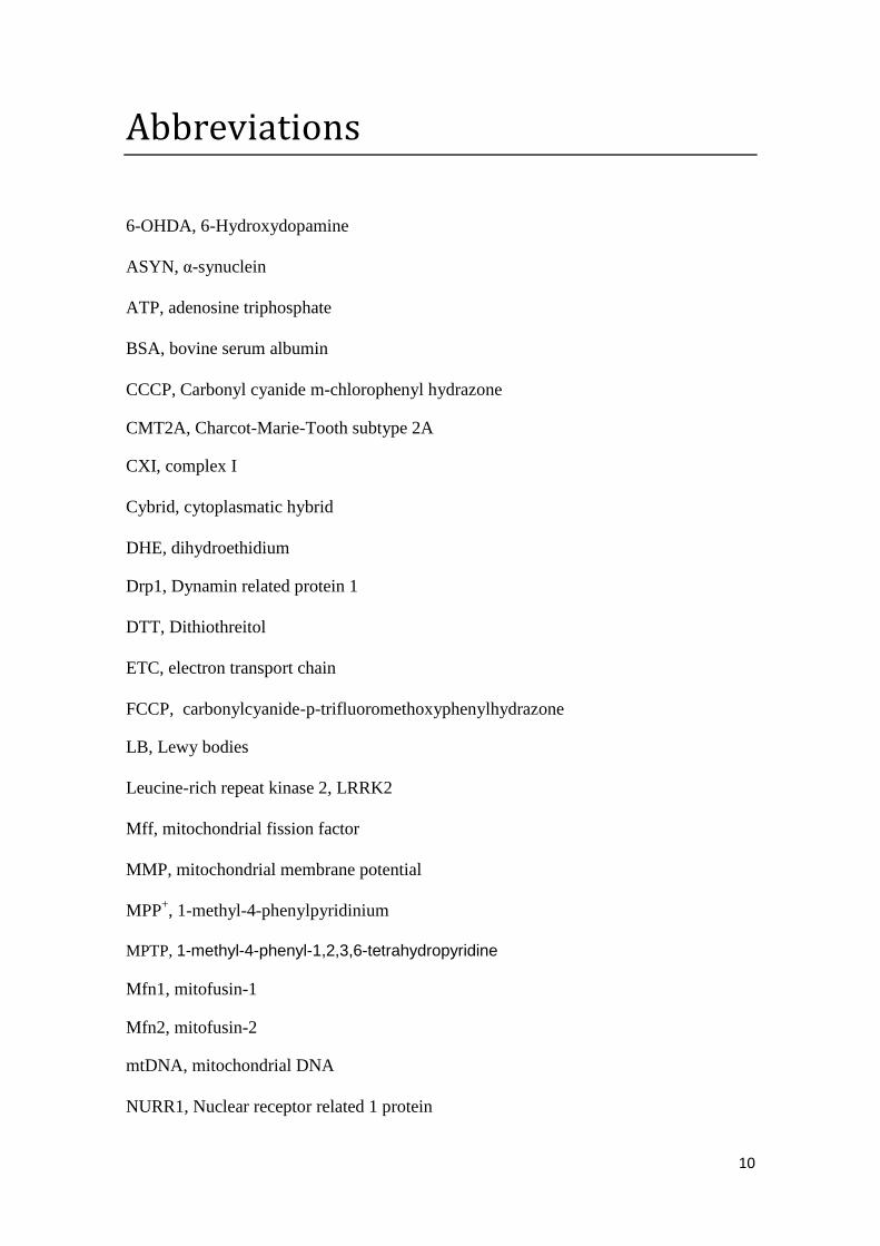
Abbreviations .............................................................................................................................. 10
Resumo ........................................................................................................................................ 12
Abstract ....................................................................................................................................... 14
Chapter 1 ..................................................................................................................................... 16
Introduction ................................................................................................................................ 16
1.1. Parkinson’s disease and mitochondrial involvement ....................................................... 18
1.1.1 Critical role of OXPHOS dysfunction in PD ................................................................. 18
1.1.2. Oxidative stress and PD ............................................................................................. 19
1.1.3. The role of mitochondrial DNA in PD ........................................................................ 20
1.2. Familial PD genes and mitochondrial homeostasis .......................................................... 21
1.2.1. Parkin ......................................................................................................................... 23
1.2.2. PINK1 ......................................................................................................................... 24
1.2.3. α-synuclein ................................................................................................................ 25
1.3. Mitochondrial dynamics ................................................................................................... 27
1.3.1. Mitochondria as an organelle with a tightly controlled dynamic ............................. 27
1.3.1.1 Mitochondrial fission........................................................................................... 27
1.3.1.2 Mitochondrial fusion ........................................................................................... 29
1.3.1.3. Mitophagy .......................................................................................................... 30
1.4. Why is mitochondrial dynamics important in neurons? .................................................. 31
1.4.1. Mitochondrial dynamics in neurodegenerative disorders rather than PD ............... 34
1.4.2. Mitochondrial dynamics in Parkinson’s disease ....................................................... 35
1.4.2.1. Available PD models and mitochondrial dynamics ............................................ 35
1.4.2.2 Role of PINK1 and Parkin in mitochondrial dynamics in PD ................................ 37
1.4.2.2.1 Modulating mitochondrial shape ................................................................. 37
1.4.2.2.2. Governing mitochondrial fate ..................................................................... 39
1.4.2.3. α-synuclein and mitochondrial morphology ...................................................... 40
1.5. Objectives ......................................................................................................................... 41
Chapter 2 ..................................................................................................................................... 43
Materials & Methods .................................................................................................................. 43
2.1 Biological Material ............................................................................................................. 44
2.1.1 Cell lines ..................................................................................................................... 44
2.1.1.1 mtDNA depleted NT2 ρ0 cells ............................................................................. 44
2.1.1.2 NT2 cybrid cell lines creation .............................................................................. 44

9
2.1.1.3 Human neuroblastoma cell line SH-SY5Y ............................................................ 45
2.1.1.4 SH-SY5Y cell line inducible expressing WT α-synuclein ....................................... 45
2.1.1.5 Human HEK293T cell line .................................................................................... 45
2.2 Chemicals and Cell media .................................................................................................. 45
2.3 Subcellular proteome extraction in cultured cells ............................................................ 46
2.4 Lymphocytes isolation ....................................................................................................... 47
2.5. Immunoblotting ............................................................................................................... 47
2.6 Immunocytochemistry ...................................................................................................... 49
2.6 HEK293T transfection procedure ...................................................................................... 49
2.5 Fluorometric reactive oxygen species production ............................................................ 50
Chapter 3 ..................................................................................................................................... 51
Results ......................................................................................................................................... 51
3.1. Mitochondrial fragmentation and altered distribution in different PD models .............. 52
3.2. MPP+ modulates fission and fusion proteins levels in CT cybrids in a distinct manner
from the observed in basal PD cybrids.................................................................................... 53
3.3. PD cybrids exhibit decreased mitochondrial levels of the pro-fusion protein Opa1 ....... 55
3.4. Protein expression in PD patients’ lymphocytes .............................................................. 56
3.5. Mitochondrial fragmentation in mitochondrial DNA depleted cells, associated with
aberrant protein expression ................................................................................................... 57
3.6 Abnormal fusion and fission in ASYN SH-SY5Y overexpressing cells ................................. 60
3.7 MPP+-induced ROS production is independent of OPA1 mitochondrial levels ................. 64
Chapter 4 ..................................................................................................................................... 66
Discussion .................................................................................................................................... 66
References ................................................................................................................................... 72
10
Abbreviations
6-OHDA, 6-Hydroxydopamine
ASYN, α-synuclein
ATP, adenosine triphosphate
BSA, bovine serum albumin
CCCP, Carbonyl cyanide m-chlorophenyl hydrazone
CMT2A, Charcot-Marie-Tooth subtype 2A
CXI, complex I
Cybrid, cytoplasmatic hybrid
DHE, dihydroethidium
Drp1, Dynamin related protein 1
DTT, Dithiothreitol
ETC, electron transport chain
FCCP, carbonylcyanide-p-trifluoromethoxyphenylhydrazone
LB, Lewy bodies
Leucine-rich repeat kinase 2, LRRK2
Mff, mitochondrial fission factor
MMP, mitochondrial membrane potential
MPP+, 1-methyl-4-phenylpyridinium
MPTP, 1-methyl-4-phenyl-1,2,3,6-tetrahydropyridine
Mfn1, mitofusin-1
Mfn2, mitofusin-2
mtDNA, mitochondrial DNA
NURR1, Nuclear receptor related 1 protein

11
OMI/HTRA2, Heat transfer requirement 2
Opa1, Optic atrophy 1
OXPHOS, Oxidative phosphorylation
PBS, phosphate-buffered saline
PINK1, PTEN-induced putative kinase 1
PD, Parkinson’s disease
ROS, reactive oxygen species
SNpc, substantia nigra pars compacta

12
Resumo
A doença de Parkinson é a segunda doença neurodegenerativa mais comum e a
doença neurodegenerativa associada ao movimento mais comum. É causada pela perda
dos neurónios dopaminérgicos na substantia nigra pars compacta levando a um défice
de dopamina no estriado. Perceber a base molecular da doença de Parkinson tem se
revelado um grande desafio no campo das doenças neurodegenerativas. Apesar de terem
sido propostas várias hipóteses para explicar os mecanismos subjacentes a patogenia da
doença de Parkinson, um crescente corpo de evidencias tem enfatizado o papel da
disrupção da dinâmica mitocôndrial como um grande contribuidor para a etiopatogenia
da doença de Parkinson. Tem se vindo a acumular dados que sugerem que uma
dinâmica mitocondrial anormal se encontra envolvida na disfunção mitocôndrial ou
medeia a morte neuronal em diferentes modelos da doença de Parkinson. Aliás, a
integração da fissão, fusão e autofagia mitocondrial forma um mecanismo de
manutenção de qualidade mitocôndrial da homeostase mitocôndrial na qual defeitos na
função mitocôndrial têm sido associados à doença de Parkinson. A maioria dos casos
surge como condição esporádica e os restantes são herdados com mutações em vários
genes que tem sido ligados a essas formas genéticas da doença. Os estudos de duas das
proteínas ligadas as formas familiares da doença, nomeadamente a PINK1 e a Parkin,
forneceram evidências que estas duas proteínas actuam na mesma via regulando a fissão
e fusão mitocôndria e a mitofagia. Uma vez que uma dinâmica mitocôndrial anormal
tem sido cada vez mais implicada na patogenia da doença de Parkinson, nesta tese,
investigámos a regulação da dinâmica mitocôndrial em diferentes modelos celulares da
doença de Parkinson. Nos observamos que uma diminuta localização mitocôndrial da
OPA1 leva a um dano da dinâmica mitocôndrial na maioria dos modelos celulares de
13
Parkinson. Para além do mais, a clivagem das isoformas longas da Opa1 parecem ser
responsáveis pelo padrão de fragmentação observado nos modelos celulares de PD
esporádicos derivados da mitocôndria. A sobrexpressão da alpha-synucleina induz a
tubulação da rede mitocondrial com estruturas elongadas que são o oposto do observado
em modelos celulares de PD. Inesperadamente, a sobrexpressao da Opa1 não resgatou o
aumento de produção de espécies reactivas de oxigénio induzida pelo MPP+.
Globalmente, as nossas observações sugerem que uma fusão mitocondrial dependente
da Opa1 desempenha um papel crucial na mediação nas anormalidades mitocôndriais e
disfunção celular induzida MPP+/mtDNA.
Estes estudos sugerem que a dinâmica mitocôndrial desempenha um papel
importante na patogenia da doença de Parkinson, e um melhor entendimento destes
mecanismos podem levar à descoberta de novos alvos terapêuticos para esta doença.

14
Abstract
Parkinson’s disease (PD) is the second most common neurodegenerative
disorder and the most common neurodegenerative movement disorder. It is caused by
the loss of dopaminergic neurons in the substantia nigra pars compacta leading to a
dopamine deficit in the striatum. Understanding the molecular basis of PD has proven to
be a major challenge in the field of neurodegenerative diseases. Although several
hypotheses have been proposed to explain the molecular mechanisms underlying the
pathogenesis of PD, a growing body of evidence has highlighted the role of
mitochondrial dynamics disruption as a major contributor to PD etiopathogenesis.
Accumulating data suggests that abnormal mitochondrial dynamics is involved in
mitochondrial dysfunction or mediates neuronal death in different PD models.
Moreover, integration of mitochondrial fusion, fission and mitochondrial autophagy
forms a quality maintenance mechanism of mitochondrial homeostasis that defects in
mitochondrial function have been associated with PD. Most of the cases arise as
sporadic conditions and the others are inherited with mutations in several genes being
linked to these genetic forms of PD. Studies of two of the proteins linked to the familial
forms of the disease, namely PINK1 and Parkin, provided evidence that these two
proteins act in the same pathway regulating mitochondrial fusion, fission and
mitophagy. Because abnormal mitochondrial dynamics are increasingly implicated in
the pathogenesis of PD, in this thesis, we investigated the regulation mitochondrial
dynamics in different PD cellular models. We observed that a decreased OPA1
mitochondrial localization-drive mitochondrial dynamics impairment in most cellular
PD models. Moreover, OPA1 long isoforms cleavage seems to be responsible for
mitochondrial fragmented pattern observed in sporadic mitochondrial-driven cellular
PD models. Alpha-synuclein overexpression induces a tubular mitochondrial network

15
with elongated structures that is the opposite of what was observed in mitochondrial PD
cellular models. Unexpectedly, OPA1 overexpression did not rescued MPP+-induced
increase in reactive oxygen species (ROS). Overall, our findings suggest that OPA1-
dependent mitochondrial fusion plays a crucial role in mediating MPP+/mtDNA
induced mitochondria abnormalities and cellular dysfunction.
These studies suggest that mitochondrial dynamics can play an important role in
PD pathogenesis, and a better understanding of these mechanisms can lead to the
discover of new therapeutical targets for this disease.
Keywords: Parkinson’s disease; mitochondrial dysfunction; mitochondrial dynamics;
mitochondrial fission; mitochondrial fusion; mitophagy

16
Chapter 1 Introduction
17
Mitochondria, the “power house” of living cells as described before (Banerjee et
al., 2009), are found virtually in every eukaryotic cell. These are especially complex
organelles that are involved in a number of cellular functions and are essential for both
life and death. The main of those functions include: the production of cellular ATP;
participation in the synthesis of key metabolites; the regulation of apoptosis; calcium
buffering; and the primary source of endogenous reactive oxygen species (ROS)
(Delettre et al., 2000, Benard et al., 2007). As there are several assaults, either generated
in situ or those imposed from extracellular environment, mitochondria are easy targets
and their functions could be compromised. Therefore, a mitochondrial dysfunction
results in a smaller supply of cellular energy, a failure in maintaining cellular
homeostasis, and activation of cell death pathways which could underlie selective
dopaminergic neurodegeneration in Parkinson’s disease (PD) (Beal, 2005).
PD is the second most common neurodegenerative disorder after Alzheimer’s
disease (AD), and the most common neurodegenerative movement disorder.
Pathologically, it is classically characterized by the loss of dopaminergic neurons in the
substantia nigra pars compacta (SNpc), leading to a dopamine deficit in the striatum,
and the presence of proteinaceous cytoplasmatic inclusions called Lewy bodies (LBs) in
surviving neurons (Forno, 1996). Clinically, PD patients manifest symptoms of
progressive rigidity, bradykinesia, tremor, and postural instability, as well as symptoms
involving non-motor brain functions, including cognitive and autonomic functions
(Lang and Lozano, 1998a, b, Weintraub and Stern, 2005, Barbas, 2006). In PD it is
known that the loss of the nigral dopaminergic neurons and terminals are responsible for
movement alterations, although there are indications that several other neuronal
populations throughout the brain are also affected in this disease (Braak et al., 2004).

18
Nowadays there is no cure for PD, as little is known about the etiopathogenesis of PD,
and so the pursuit to define disease mechanisms continues.
1.1. Parkinson’s disease and mitochondrial involvement
In almost 90-95% of all occurrences PD arises essentially as a sporadic
condition, i.e. in absence of any apparent genetic linkage, and, over the years, several
theories have been proposed for the etiology of the disease in an attempt to explain the
why and the how of neurodegeneration in PD. There are several evidences for the
relation between mitochondrial dysfunction and PD pathogenesis however it is
debatable if it is the cause or an effect. Even though, it was recently proposed the
mitochondrial cascade hypothesis for PD (Cardoso, in press).
1.1.1 Critical role of OXPHOS dysfunction in PD
The discovery that exposure to MPTP (1-methyl-4-phenyl-1,2,3,6-
tetrahydropyridine), a selective inhibitor of mitochondrial complex I (CXI) of the
electron transport chain (ETC), led to a development of progressive and irreversible
parkinsonism provided strong evidence that mitochondrial dysfunction may be involved
in the pathogenesis of PD (Langston et al., 1983). This direct relation was further
established when it was (Schapira et al., 1989) described a CXI deficiency in the
substantia nigra of PD patients postmortem brains, which was also seen in skeletal
muscle and platelets (Bindoff et al., 1989, Parker et al., 1989). In agreement, some data
suggests that sporadic PD is characterized by a systemic decrease in CXI (Penn et al.,
1995, Keeney et al., 2006). Indeed, these authors were able to see that there was an
19
increase in the levels of protein carbonyls (a marker of an oxidative modification of
proteins) in several catalytic subunits of CXI, which was in agreement with reduced
electron transfer rates, indicating that the oxidative damage of CXI subunits may lead
its misassembly and dysfunction. The reason why substantia nigra is more vulnerable to
this impairment of CXI activity is possible due to increased generation of ROS from
dopamine metabolism and iron content from dopaminergic neurons (Chinta and
Andersen, 2008).
1.1.2. Oxidative stress and PD
Mitochondria are the main source for endogenous ROS production (reviewed in
(Starkov, 2008, Murphy, 2009) and there are evidence of a linkage between oxidative
stress and neurodegeneration in PD. In addition, the levels of antioxidants and oxidized
targets are known to be altered in PD, as reviewed before (Jenner, 2003). Together with
the fact that inhibition of mitochondrial complexes increases free radicals production
(Orth and Schapira, 2002, Turrens, 2003), it is becoming clear a relation between
mitochondrial dysfunction and PD. Indeed, an unbalance between exacerbated ROS
production and/or defective ROS removal results in oxidative damage to mitochondrial
DNA (mtDNA), proteins and lipids. According to the mitochondrial protein expression
and/or abundance, it was reported that subunits of CXI was differentially expressed in
mitochondria enriched fractions from post mortem PD substantia nigra, when compared
to control (Jin et al., 2006). A reduction of the immunostaining for a mitochondrial
protein alpha-ketoglutarate was also noted in post mortem PD brain (Mizuno et al.,
1994). Oxidative damage to mtDNA may compromise respiratory chain subunits, which
are encoded by mtDNA, establishing a vicious circle of oxidative stress and

20
bioenergetic failure, which has been the rationale for the mitochondrial theory of aging
(Linnane et al., 1989).
1.1.3. The role of mitochondrial DNA in PD
Mitochondria house their own DNA, and it is strongly believed that the
proximity of mtDNA to ROS generation as a consequence of normal respiratory chain
function could increase mtDNA mutations (Richter et al., 1988, Ozawa, 1997). Thus,
the alleged CXI defect in PD brain, and consequent increase in ROS production, may
affect the mtDNA which encodes 7 of the 46 protein subunits of CXI, making mutations
in the mtDNA an obvious trigger of PD pathology. Studies with cytoplasmatic hybrids
(cybrids) cell lines supported this idea of a relation between mtDNA encoded defects
and PD, showing that CXI defect from PD platelets is transferable into mtDNA
deficient cell lines (Swerdlow et al., 1996, Gu et al., 1998). Such approach, the use of
cybrids to investigate the potential of mtDNA from PD to determine the origin of the
CXI defect, led the authors to suggest that mtDNA in those patients cause the CXI
deficiency through inherited or somatic mutations. Indeed, these defects were associated
with increased free radical production, impaired mitochondrial calcium buffer and
increase susceptibility to MPP+ (Sheehan et al., 1997). With these results it could be
hypothesized that CXI deficit in PD are inherited either from mitochondrial genome or
from alteration on somatic mtDNA. Moreover, Trimer et al., (2004) using a cybrid
model of PD reported the formation of fibrillar and vesicular inclusions that replicated
most antigenic and structural features of LBs (Trimmer et al., 2004). It is important to
underline that there was no need in this experiment for exogenous protein expression or
21
inhibition of mitochondrial or proteasomal function. In PD, the maternal inheritance
pattern of mtDNA mutation is rare and may be a mere coincidence (Wooten et al., 1997,
Swerdlow et al., 1998, Thyagarajan et al., 2000). Although, some studies indicated that
mtDNA abnormality may contribute to PD, as a mitochondrial 12SrRNA point mutation
was found in a pedigree with Parkinsonism, deafness and neuropathy (Thyagarajan et
al., 2000). Another evidence of mtDNA relevance in PD arose when the ablation of the
mitochondrial transcription factor A gene, which regulates transcription of mtDNA and
copy number, in nigrostriatal dopaminergic neurons of mice caused the slow
progressive degeneration of dopaminergic neurons seen in PD (Ekstrand et al., 2007).
Moreover, this mouse model showed reduced mtDNA expression, respiratory chain
deficit and neuronal death, leading to progressive, L-dopa-responsive impairment of
motor functions. Therefore, the role of mtDNA cannot be overruled and further
investigations are warranted to better understand its role in PD.
1.2. Familial PD genes and mitochondrial homeostasis
As mentioned above, 90-95% of the cases of PD arise as sporadic conditions.
The others 5-10% of the cases are inherited, and linkage analysis has identified a
number of PD-associated genes. They usually, but not exclusively, show an early onset
of symptoms and are in essence clinically indistinguishable from sporadic PD (Bonifati,
2007, Gasser, 2009). Inherited forms of PD have been linked to 16 familial PD-linked
genetic loci (PARK1-16), including a–syn, parkin, DJ-1, PINK1 (PTEN-induced kinase
1), NURR1 (Nuclear receptor related 1 protein), OMI/HTRA2 (Heat transfer
requirement 2), LRRK2 (Leucine-rich repeat kinase 2), were shown to locate in or

22
interact with mitochondria under certain conditions (Fig. 1) (Arduino et al., 2011,
Esteves et al., 2011).
Figure 1 - Products of PD-associated genes and their role in maintaining mitochondrial
homeostasis through modulation of mitochondrial dynamics and oxidative stress. Adapted from
(Henchcliffe and Beal, 2008).
The study of the function and dysfunction of PD genes confirms us the relevance
of the biochemical alterations found in sporadic PD, i.e., mitochondrial dysfunction,
oxidative stress and an imbalance in protein homeostasis characterized by an increase in
protein misfolding and aggregation accompanied by an impaired removal of misfolded
proteins. Indeed, several of the proteins encoded by them carry out important function
within mitochondria and/or act to reduce oxidative stress, providing strong evidences
for a casual involvement of mitochondrial dysfunction and oxidative stress in PD
pathogenesis. Thus, insight into the function of PD genes can promote our
understanding of the molecular causes of PD and help to focus research on key
23
biochemical pathways. This also suggests that the mechanisms underlying sporadic and
familial PD are similar. We will briefly focus on α-synuclein, Parkin and PINK1
proteins due to their relevance to mitochondrial dynamics.
1.2.1. Parkin
Mutations in the Parkin gene (PARK2) were reported to cause early onset
juvenile form of autosomal recessive parkinsonism (Kitada et al., 1998). It is suggested
that Parkin is a RING finger containing protein that acts as an E3 ubiquitin protein
ligase in the proteasome-mediated degradation of several proteins in vitro (Shimura et
al., 2000). The loss of the E3 ligase activity has been suggested to result in
accumulation of toxic substrates leading to the autosomal recessive form of PD
(Dawson, 2006). It was seen in a Parkin knockout mouse line an increase in striatal
extracellular dopamine concentrations, reduced synaptic excitability, and a mild, non-
progressive motor deficit at 2–4 months (Goldberg et al., 2003). Surprisingly, no loss of
dopaminergic neurons was described and, like in most patients with Parkin mutations,
no inclusion formations were noted in these mice (Goldberg et al., 2003). In a study
conducted by Palacino and co-workers (2004) Parkin-deficient mice revealed decreased
levels of several subunits of complexes I and IV. In addition, striatal cells from the mice
exhibited reduced mitochondrial respiratory capacity and decreased antioxidant capacity
(Palacino et al., 2004). Recently, it was proposed the hypothesis that Parkin could be a
key player in the turnover of damaged mitochondria by mitophagy (McBride, 2008), as
it was shown that Parkin can be recruited to depolarized mitochondria targeting them to
the destruction by autophagy (Narendra et al., 2008).
24
1.2.2. PINK1
Mutation in phosphatase and tensin homologue (PTEN)-induced putative kinase
1 (PINK1) are associated with hereditary early-onset of PD (Valente et al., 2004a), as
certain heterozygous PINK1 mutations may pre-dispose to the development of sporadic
early-onset PD (Valente et al., 2004b). PINK1 is a serine/threonine kinase which is
translated in the cytoplasm and imported into mitochondria through an N-terminal
mitochondrial targeting sequence (Valente et al., 2004a, Silvestri et al., 2005). PINK1
mutations cause PD possibly due to impairment on the phosphorylation of its substrates,
probably in mitochondria. These mutations have been reported within and outside the
kinase domain (Hatano et al., 2004, Valente et al., 2004a), however, the localization of
PINK1 to the mitochondria is not affected by these mutations (Zhou et al., 2008b).
Although it is known that PINK1 is imported to mitochondria, as said before, its sub-
mitochondrial localization remains intensely debatable. This topic is crucial for
identifying its physiological substrates and its mode of action in the context of PD
pathogenesis. It seems that the majority of the studies demonstrates that PINK1
localizes in the inner mitochondrial membrane (Silvestri et al., 2005, Gandhi et al.,
2006, Pridgeon et al., 2007), while other suggest that it associates with the
intermembrane space (Silvestri et al., 2005, Plun-Favreau et al., 2007, Pridgeon et al.,
2007) or even the outer mitochondrial membrane (Gandhi et al., 2006) with the kinase
domain facing the cytoplasm (Zhou et al., 2008b). The depletion of PINK1 in cultured
cells results in abnormal mitochondrial morphology and membrane depolarization.
Similar mitochondrial alterations are present in primary cells from patients with PINK1

25
mutations (Exner et al., 2007) suggesting that PINK1 plays an important role in
mitochondrial maintenance. There is also a study in which seems to exists an important
role for PINK1 protecting mitochondria against oxidative stress by phosphorylating the
mitochondrial chaperone protein TRAP1 (Pridgeon et al., 2007). There is no doubt that
more studies in this area are needed, although there is a certainly that both proteins
modulate mitochondrial dynamics including fusion/fission which will be further
mentioned in detail concerning PD pathogenesis.
1.2.3. α-synuclein
Three different missense mutation in α-synuclein (ASYN) gene (PARK1& 4
locus), a duplication and triplication of the ASYN gene locus lead to dominantly
inherited early-onset of PD (Polymeropoulos et al., 1997, Kruger et al., 1998, Singleton
et al., 2003, Zarranz et al., 2004). Although its physiological function is still quite
unknown, it is known that ASYN is a presynaptic protein that is present in an
aggregated form in LB’s in PD due to the presence of its hydrophobic non-amyloid beta
component domain. It has been described that familial mutation and over-expression of
this protein accelerates this so-called ASYN protofibrilis (Conway et al., 1998, Conway
et al., 2000, Fredenburg et al., 2007). The selective toxicity of ASYN to dopamine
neurons (Xu et al., 2002) maybe in part explained by the stabilization of the toxic
ASYN protofibrilis by dopamine adducts (Conway et al., 2001). There are various
mechanisms proposed to explain ASYN toxicity, one of those includes mitochondrial
dysfunction. However both are hallmarks implicated in PD pathogenesis it was not clear
until recently that these two processes are interrelated and complement each other in
26
disease pathogenesis. Indeed, recent evidences suggests this interplay in which mice
harboring human A53T ASYN mutant shown mitochondrial accumulation of human
ASYN and exhibit mitochondrial degeneration associated with increased mtDNA
damage and impaired activity of the electron transport chain complex IV cytochrome
oxidase (Martin et al., 2006). Studies in yeast, where abrogation of mtDNA inhibited α-
synuclein-induced ROS formation and apoptosis (Buttner et al., 2008), highlighted the
requirement of mitochondria in mediating the toxicity of α-synuclein. Mitochondrial
association of ASYN in cells was also linked to oxidation of mitochondrial proteins and
increased levels of calcium and nitric oxide (Parihar et al., 2008). It was interesting to
note that mice lacking ASYN exhibited alterations in membrane lipids and reduced
activities of the electron transport complexes I and III in mitochondria (Ellis et al.,
2005), suggesting that ASYN has a physiological role in mitochondria, and that the
mitochondrial deficits observed in transgenic animals may be due to toxic gain-of-
function of over-expressed or mutated ASYN present in mitochondria. Recently, is was
shown that human ASYN N-terminal 32 amino acids contain a cryptic mitochondrial
targeting signal, which is important for mitochondrial targeting of ASYN (Devi et al.,
2008). It was also seen, in human dopaminergic neurons, that accumulation of wild-type
ASYN in mitochondria caused reduced mitochondrial CXI activity and increased ROS
production. Although, these defects occurred in an early time point in dopaminergic
neurons expressing familial with A53T mutation as compared with wild-type ASYN
(Devi et al., 2008). In this work it was also shown that ASYN interacts with CXI in the
inner mitochondrial membrane. In fact, the content of ASYN in purified mitochondria
from substantia nigra were several-fold higher in PD compared to healthy individual,
suggesting that mitochondrial accumulation of ASYN may contribute to the CXI defects
of the disease (Devi et al., 2008). Therefore, the interaction process of ASYN with

27
mitochondria is quite complex and impacts mitochondrial physiology to affect normal
functioning of dopaminergic neurons.
1.3. Mitochondrial dynamics
1.3.1. Mitochondria as an organelle with a tightly controlled
dynamic
What was once thought to be an isolated and rigid structure organelle,
mitochondria became now highly dynamic organelle that constantly divides (fission)
and fuse (fusion) with each other (Chan, 2006). Indeed, this highly dynamic and
orchestrated process of mitochondrial fusion and fission affects, besides morphology,
the size, number, length and even mitochondrial function and distribution. This
phenomenon regulated by a delicate balance between these two opposing processes is
important in order to maintain the integrity of mitochondria, electrical and biochemical
connectivity, in mitochondrial turnover, and in aggregation, stabilization, and protection
of mtDNA (Westermann, 2002).
1.3.1.1 Mitochondrial fission
For the mitochondrial division, both the inner and outer membrane need to fise
and the mtDNA and other critical mitochondrial contents need to be redistributed to the
daughter mitochondria, so that the parental gives rise to two isolated, small, round
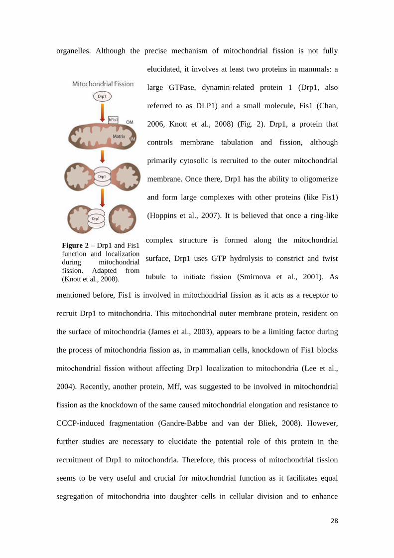
28
organelles. Although the precise mechanism of mitochondrial fission is not fully
elucidated, it involves at least two proteins in mammals: a
large GTPase, dynamin-related protein 1 (Drp1, also
referred to as DLP1) and a small molecule, Fis1 (Chan,
2006, Knott et al., 2008) (Fig. 2). Drp1, a protein that
controls membrane tabulation and fission, although
primarily cytosolic is recruited to the outer mitochondrial
membrane. Once there, Drp1 has the ability to oligomerize
and form large complexes with other proteins (like Fis1)
(Hoppins et al., 2007). It is believed that once a ring-like
complex structure is formed along the mitochondrial
surface, Drp1 uses GTP hydrolysis to constrict and twist
tubule to initiate fission (Smirnova et al., 2001). As
mentioned before, Fis1 is involved in mitochondrial fission as it acts as a receptor to
recruit Drp1 to mitochondria. This mitochondrial outer membrane protein, resident on
the surface of mitochondria (James et al., 2003), appears to be a limiting factor during
the process of mitochondria fission as, in mammalian cells, knockdown of Fis1 blocks
mitochondrial fission without affecting Drp1 localization to mitochondria (Lee et al.,
2004). Recently, another protein, Mff, was suggested to be involved in mitochondrial
fission as the knockdown of the same caused mitochondrial elongation and resistance to
CCCP-induced fragmentation (Gandre-Babbe and van der Bliek, 2008). However,
further studies are necessary to elucidate the potential role of this protein in the
recruitment of Drp1 to mitochondria. Therefore, this process of mitochondrial fission
seems to be very useful and crucial for mitochondrial function as it facilitates equal
segregation of mitochondria into daughter cells in cellular division and to enhance
Figure 2 – Drp1 and Fis1
function and localization
during mitochondrial
fission. Adapted from
(Knott et al., 2008).

29
distribution of mitochondria along cytoskeletal tracks. It also has an important role in
the targeting of damaged segments of mitochondria to the autophagic process, playing a
housekeeping role in the cell, as it will be mentioned later.
1.3.1.2 Mitochondrial fusion
On the other side, mitochondrial fusion, in mammalian cells, is mediated in part
by the outer membrane dynamin-like GTPases mitofusin-1 and -2 (Mfn1 and Mfn2) and
the inner membrane optic atrophy protein (Opa1)
(Fig. 3) (Cerveny et al., 2007, Detmer and Chan,
2007b). Although both Mfn1 and Mfn2 have the
same location and appear to play similar roles in
mitochondrial fusion, they function independently
of each other, even having different rates of GTP
hydrolysis (Ishihara et al., 2004) (Fig. 4). Indeed, it
was reported that these two proteins could form
homo-oligomeric and hetero-oligomeric
complexes, during mitochondrial fusion, and thus
tether outer membranes of neighboring
mitochondria together (Ishihara et al., 2004, Zuchner et al., 2004). The inner membrane
protein OPA1, that faces intermembrane space, has been propose to be primarily
involved in the inner membrane fusion in a process that requires Mfn1, but not Mfn2
(Cipolat et al., 2004). Like mitochondrial fission, fusion seems to be required for a
normal mitochondrial function as this process is likely to protect function by providing
Figure 3 – Mitochondrial outer and
inner membrane fusion controlled by
Mfn1/2 and OPA1 respectively.
Adapted from (Knott et al., 2008)

30
a chance for mitochondria to mix their contents, thus enabling protein complementation,
mtDNA repair and equal distribution of metabolites.
Figure 4 – Proteins reponsable for the modulation of mitochondrial dynamics and cellular
localization of the same. Adapted from (Winklhofer and Haass, 2010)
1.3.1.3. Mitophagy
As mentioned before, mitochondrial dysfunction can be a key determinant in cell
death as this organelle provides critical functions for cell survival. Indeed, it is
necessary an orchestrated mechanism capable of selectively remove damaged
mitochondria that are functionally impaired or have accumulated mtDNA mutation that
potentiates ROS generation. The process responsible for this is denominated autophagy.
It is a process in which cellular components are isolated by engulfment into
autophagosomes. Further these autophagosomes fuse with lysosomes which contain
hydrolytic enzymes that break down cellular components. Generally this process is
activated during nutrient deprivation so that the products can be recycled into more
urgently needed molecules. However, autophagy can have a housekeeping role in

31
maintaining quality control by turning over organelles, like mitochondria, and
degrading protein aggregates. So mitochondria can be targeted for degradation in a
process called mitophagy. Although this process is not new, it had remained unclear
whether mitophagy can selectively degrade dysfunctional mitochondria. In a study
performed by Kanti and Klionsky it was reported that selective mitophagy was
regulated independently from bulk autophagy (Kanki and Klionsky, 2008). It was also
described that mitochondrial fission could target mitochondria to degradation. Indeed, it
was shown in pancreatic b-cells and COS7 cells that mitochondrial fission can yield
uneven products in which one of the daughter mitochondrion was depolarized, much
less like to fuse and had reduced levels of OPA1 protein, and eventually was
autophagocytosed (Twig et al., 2008). Supporting this find it was also stated that to
mitophagy occur there must be a loss of fusion and the presence of fission. In the base
of this theory is the fact that the overexpression of OPA1, Fis1 RNAi, and Drp1
dominant-negative expression all reduced level of mitophagy. Although mitochondrial
fragmentation is suitable for mitophagy, it is not a sufficient signal for this process
(Narendra et al., 2008, Twig et al., 2008).
1.4. Why is mitochondrial dynamics important in neurons?
Changes in the mechanisms that mediate mitochondrial dynamics can lead to
different defects in neurons (Fig. 5). Some of the reasons for the particular importance
of mitochondria fusion and fission in neurons possible rely on their unique features such
as their post-mitotic state and their long processes with higher energy requirements.
Indeed, this high energy demands to support their survival and specialized function may
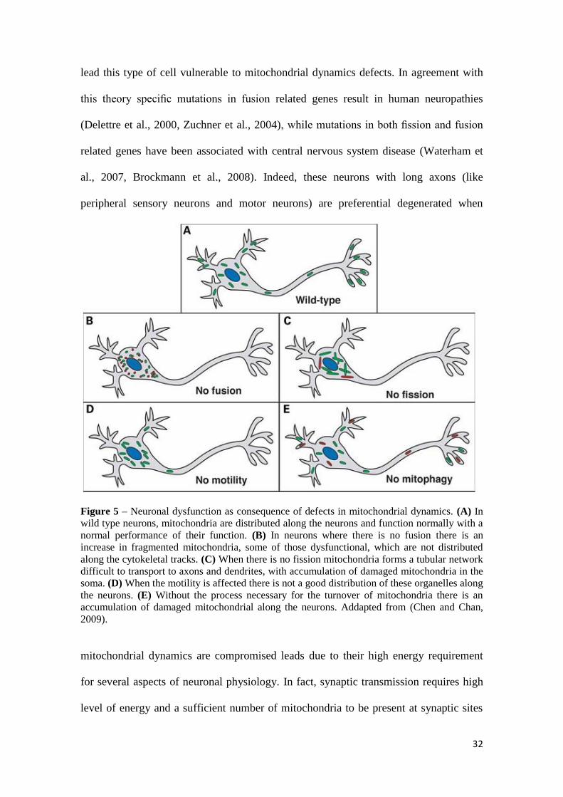
32
lead this type of cell vulnerable to mitochondrial dynamics defects. In agreement with
this theory specific mutations in fusion related genes result in human neuropathies
(Delettre et al., 2000, Zuchner et al., 2004), while mutations in both fission and fusion
related genes have been associated with central nervous system disease (Waterham et
al., 2007, Brockmann et al., 2008). Indeed, these neurons with long axons (like
peripheral sensory neurons and motor neurons) are preferential degenerated when
Figure 5 – Neuronal dysfunction as consequence of defects in mitochondrial dynamics. (A) In
wild type neurons, mitochondria are distributed along the neurons and function normally with a
normal performance of their function. (B) In neurons where there is no fusion there is an
increase in fragmented mitochondria, some of those dysfunctional, which are not distributed
along the cytokeletal tracks. (C) When there is no fission mitochondria forms a tubular network
difficult to transport to axons and dendrites, with accumulation of damaged mitochondria in the
soma. (D) When the motility is affected there is not a good distribution of these organelles along
the neurons. (E) Without the process necessary for the turnover of mitochondria there is an
accumulation of damaged mitochondrial along the neurons. Addapted from (Chen and Chan,
2009).
mitochondrial dynamics are compromised leads due to their high energy requirement
for several aspects of neuronal physiology. In fact, synaptic transmission requires high
level of energy and a sufficient number of mitochondria to be present at synaptic sites

33
so that the plasma-membrane-potential could be maintained, synaptic neurotransmitter
could be released and reuptake, and to build-up a reserve pool of vesicles for prolonged
or high-frequency firing. The dynamic processes of mitochondrial fission/fusion are
intimately and critically involved in the formation of synapses and dendritic spines,
where exist a high demand of ATP. While the unbalance of mitochondrial fission/fusion
towards fission leads to an increase in synapse formation, a decrease in the same leads
to a loss of mitochondria from dendritic spines and a reduction of synapse formation (Li
et al., 2004). The absence of the mitochondrial fission protein Drp1 has been shown to
prevent mitochondria from distributing to synapses and to lead to synaptic dysfunction
(Verstreken et al., 2005).This might be linked to the need of smaller mitochondrial units
which presumably are more easily transported over long distances as, e.g., required in
axonal transport. Other mitochondrial feature that is very important for normal neuronal
physiology is the Ca2+
buffering that prevents excitotoxicity by an excessive free-Ca2+
load in neurons. On the other hand, the process of fusion is crucial for the maintenance
of mtDNA that cannot take place without this process (Rapaport et al., 1998). In fact,
fusion of mitochondria has been directly implicated in preventing the accumulation of
damaged mtDNA (Nakada et al., 2001, Ono et al., 2001). As it was shown recently,
mitochondrial fission appears to be critical to mtDNA maintenance (Parone et al., 2008)
and so we can find that in mitochondrial dynamics a delicate balance is very important
as the coordination of these processes maintain and protect mtDNA. This is very
important in neurons and neurodegenerative diseases as it was shown that mtDNA
mutations accumulate in the brain with age (Corral-Debrinski et al., 1992). Neurons are
also particularly sensitive to the impairment of the transport of mitochondria through
the axons and dendrites so that these could reach sites of increased energy requirement.
They are transported both in anterograde and retrograde directions, in a process

34
regulated in part by cytoskeletal and mitochondrial proteins, and several intrinsic
component of mitochondrial bioenergetics, like ATP synthesis or changes in
mitochondrial membrane potential, can impair this transport (Rintoul et al., 2003, Miller
and Sheetz, 2004). This suggests a link between mitochondrial bioenergetic functions
and mitochondrial transport and distribution. In addition, disruption of mitochondrial
movement has been noted in excitotoxicity and oxidative stress conditions (Rintoul et
al., 2003), two factors implicated in cellular stress and neuronal death.
1.4.1. Mitochondrial dynamics in neurodegenerative disorders
rather than PD
There are several mutations in mitochondrial fission/fusion genes in humans that
result in very specific neurodegenerative diseases. Regarding the mitochondrial fusion
proteins, mutations in Mfn2 cause Charcot-Marie-Tooth subtype 2A (CMT2A), which
involves axonal degeneration of motor and sensory neurons (Zuchner et al., 2004,
Kijima et al., 2005). Mutations in Opa1, other mitochondrial fusion protein, cause the
most common form of optic atrophy, autosomal dominant optic atrophy (ADOA)
(Alexander et al., 2000). Concerning the mutations in mitochondrial fission proteins, a
dominant negative mutation in the human Drp1 gene resulted in elongated and tangled
mitochondria concentrated at perinuclear region (Waterham et al., 2007). The patient
carrying this mutation died shortly after birth and displayed some symptoms resembling
those of ADOA and CMT2A, emphasizing the importance of the fission machinery in
neuronal maintenance.
It is interesting to note that there are some human genetic mutations that do not
affect mitochondrial morphogenesis, as the others described above, but cause disease

35
through a fission/fusion-independent manner. Examples of this are some Mfn2 mutants
that retain the ability to promote mitochondrial fusion (Detmer and Chan, 2007a). This
could indicate that Mfn2 has other pathogenic roles not related to mitochondrial fusion.
There are several studies suggesting other functions for this protein, like mitochondrial
trafficking (Baloh et al., 2007) and regulating Ca2+
uptake and signaling by tethering
mitochondria to endoplasmatic reticulum (de Brito and Scorrano, 2008), which
disruption could lead to peripheral axon degeneration and neuronal death. Although, it
remains necessary the creation of animal models so it could be distinguishable the
contribution of the various functions of the fission/fusion genes in disease pathogenesis.
1.4.2. Mitochondrial dynamics in Parkinson’s disease
One of the most accepted reasons for the vulnerability of the dopaminergic
neurons from substantia nigra to changes in mitochondrial dynamics is the lower basal
mitochondrial content compared to other midbrain neurons (Liang et al., 2007). This
evidence could contribute to the selective vulnerability of these neurons to external or
internal toxic stimuli that affect mitochondrial fission, fusion and mitophagy,
compromising mitochondrial homeostasis during PD pathogenesis.
1.4.2.1. Available PD models and mitochondrial dynamics
Through the widely used models to mimic PD pathogenesis it was already
described the existence mitochondrial dynamics imbalance. Indirect observations using
a cytoplasmatic hybrid (cybrid) model of PD showed that PD cybrid cells contained a

36
significantly increased percentage of mitochondria that were enlarged or swollen and
had a pale matrix with few remaining cristae (Trimmer et al., 2000). This mitochondrial
morphology in PD cybrids, whose mitochondria are proceeding from platelets of PD
patients, was also seen by Esteves and coworkers in a similar cybrid model, suggesting
that there could be impairment in mitochondrial fission (Esteves et al., 2008).
The first evidence demonstrating mitochondrial fragmentation induction by
toxins used to study PD, was the finding that both CXI inhibitors rotenone and MPP+
induced mitocondrial fission in rat dopaminergic cell line N27 (Barsoum et al., 2006).
Following studies continued to report mitochondrial fragmentation following
administration of CXI inhibitors like 6-Hydroxydopamine (6-OHDA)l(Gomez-Lazaro
et al., 2008), MPP+ (Meuer et al., 2007, Endo et al., 2009, Wang et al., 2011c) and
rotenone (Benard et al., 2007, Mortiboys et al., 2008, Plecita-Hlavata et al., 2008,
Sandebring et al., 2009, Thomas et al., 2011). Interestingly, some of these authors went
further and showed that the CXI inhibition-induced mitochondrial fragmentation was
Drp1-dependent, as Drp1 silencing or its dominant-negative mutant expression
prevented the mitochondrial morphological changes and the cell death induced by the
toxins (Benard et al., 2007, Meuer et al., 2007, Gomez-Lazaro et al., 2008, Wang et al.,
2011c). Together, these results highlight mitochondrial fragmentation as an early and
upstream event mediating CXI inhibition-induced toxicity. However, it was reported
that chronic exposure to rotenone significantly increased mitochondrial branching and
length in human skin fibroblasts (Koopman et al., 2005). The discrepancy between these
results may rely in the different duration of the toxic stimuli, e.g. acute vs. chronic,
toxin concentration, or different cell lines, which may affect differently mitochondrial
morphology. Therefore, in further necessary studies using PD-linked toxins to study
mitochondrial morphology, careful experimental design should be performed in order to

37
accurately mimic PD pathology and enable a proper analysis of the mitochondrial
network in this disease.
1.4.2.2 Role of PINK1 and Parkin in mitochondrial dynami cs in PD
Until now, the strongest evidence of the role of mitochondrial dynamics in PD
aroused from studies in which the manipulation of some genes associated with familial
PD affected mitochondrial dynamics in several models and tissues. As previously
described, PINK1 and Parkin are proteins that have an important role in mediating
mitochondrial function and morphology.
1.4.2.2.1 Modulating mitochondrial shape
Back in 2003, the first studies describing mitochondrial structural alterations
associated with parkin mutants arouse. Using Drosophila Parkin mutant flies it was
reported that muscle mitochondria had abnormal morphology, including swollen or
fragmented mitochondria with disrupted cristae, a phenotype that was rescued by
overexpression of wild-type Parkin (Greene et al., 2003, Pesah et al., 2004).
Surprisingly, no major morphological abnormalities in striatal mitochondria of parkin -
/- mice were later reported (Palacino et al., 2004), an observation that would serve as a
template for the discrepancies of the results arising from the models to study PINK1 and
Parkin effect on mitocondrial morphology, that would then emerge.
The first detailed studies unraveling PINK1 and Parkin role in mitochondrial
morphology regulation were preformed through genetic manipulations in Drosophila.
Initially, loss of PINK1 or Parkin function in Drosophila resulted in similar
38
mitochondrial morphological abnormalities, including enlarged mitochondria with
fragmented cristae. Surprisingly, Parkin overexpression was able to rescue PINK1
mutant mitochondrial morphology defects, but not vice versa, suggesting that PINK1
and Parkin take part in the same pathway in which PINK1 acts upstream of Parkin in
regulation of mitochondrial morphology (Clark et al., 2006, Park et al., 2006, Yang et
al., 2006). Latter findings, still in Drosophila, revealed how PINK1/Parkin pathway
exerts its effect in mitochondrial morphology. It was shown that the Drp1 loss of
function was lethal in PINK1 or Parkin mutants, as Drp1 overexpression suppressed the
mitochondrial morphological abnormalities described before (Deng et al., 2008, Poole
et al., 2008, Yang et al., 2008, Park et al., 2009). Interestingly, loss of function of the
proteins involved in the mitochondrial fusion process, like Mfn and Opa1, were also
able to restore mitochondrial morphology in PINK1 and Parkin mutants, suggesting that
PINK1/Parkin pathway stimulates mitochondrial fission and/or inhibits mitochondrial
fusion.
However, conformity between the results reported in Drosophila and findings in
vertebrates was not achieved. In HeLa cells PINK1 knockdown induced mitocondrial
fragmentation and reduced/altered mitochondrial cristae in mammalian cells,
abnormalities that were rescued by wild type PINK1 and Parkin overexpression, but not
Parkin mutant’s (Exner et al., 2007). Observations in primary fibroblasts from patients
with PINK1 mutations have also shown mitochondrial fragmentation. Moreover, PINK1
or Parkin down-regulation in mammalian cells was again reported to induce comparable
mitochondrial fragmentation (Dagda et al., 2009, Lutz et al., 2009). As Drp1
knockdown was able to reverse this phenotype, demonstrating that increased
mitochondrial fission is associated with PINK1/parkin depletion, Mfn2 and Opa1
overexpression could also rescue mitochondrial phenotype (Lutz et al., 2009). Further
39
studies reported again that PINK1 loss of function increased mitochondrial fission
(Wood-Kaczmar et al., 2008, Cui et al., 2010, Heeman et al., 2011, Wang et al., 2011b),
as human wild type PINK1 overexpression produces a pro-fusion effect in the
mitochondrial network (Cui et al., 2010).
Strikingly, even between mammalian models there’s still some controversy as in
recent studies PINK1 or Parkin overexpression resulted in mitochondrial fragmentation,
and inactivation of these proteins resulted in elongated mitochondria (Gautier et al.,
2008, Sandebring et al., 2009, Cui et al., 2011, Pacelli et al., 2011, Yu et al., 2011), in
agreement with the former Drosophila studies. Overexpression of Drp1 or the
knockdown of Opa1 reversed the mitochondrial elongation induced by PINK1 RNAi, as
overexpression of PINK1 or Parkin were also suppressed mitochondrial elongation
induced by Drp1 knockdown (Yu et al., 2011).
1.4.2.2.2. Governing mitochondrial fate
Beyond the “simple” regulation of mitochondrial morphology, through
interactions with the mitochondrial fission/fusion machinery, an important role for the
PINK1/Parkin pathway has been attributed at the mitochondrial quality control level.
Upon mitochondrial damage, PINK/Parkin pathway have been reported to
promote poly-ubiquitination Mfn1 and Mfn2, through Parkin interaction, leading to its
turnover by UPS, independently of the autophagic pathway (Chan et al., 2011, Glauser
et al., 2011). The degradation of these mitochondrial outer membrane fusion proteins
leads to an impairment of mitochondrial fusion which, upon physiological conditions,
can exert a selective removal of damaged mitochondria by autophagy (mitophagy).
Indeed, Parkin was suggested to prevent refusion of depolarized mitochondria, product

40
of a division process, through elimination of the proteins involved in the outer
mitochondrial membrane fusion, Mfn1 and Mfn2 (Tanaka et al., 2010).
The dependence on this pathway to promote mitophagy has been extensively
described as PINK1 and Parkin silencing leads to lower ubiquitination of the
mitochondrial proteins Mfn1 and Mfn2 upon mitochondrial damage, leading to
accumulation of defective mitochondria (Gegg et al., 2010). Mutations in PINK1 and
Parkin also impairs ubiquitination of mitofusins in human fibroblasts (Rakovic et al.,
2011). Moreover, a study in Drosophila has shown that Parkin requires functional
PINK1 to correctly be recruited to dysfunctional mitochondria and promote their
degradation, through Mfn1/2 degradation (Ziviani et al., 2010).
The parkin-mediated autophagy was shown to be prevented upon inhibition of
Drp1-mediated mitochondrial fission, emphasizing the importance of these
fission/fusion proteins in mitochondrial quality control (Tanaka et al., 2010).
Importantly, it was recently revealed that Parkin also interacts and ubiquitinates Drp1,
signaling this protein for degradation at the UPS (Wang et al., 2011a)
1.4.2.3. α-synuclein and mitochondrial morphology
A relation between mitochondrial morphology and ASYN was early described
as overexpression of ASYN resulted in abnormal large mitochondria displaying
vacuolization of the cristae (Hsu et al., 2000). Still, at the time, no direct interaction was
established between them. Nowadays it is clear that ASYN interacts with mitochondria.
It is extensively described its transport to this organelle and its accumulation in the
same (Martin et al., 2006, Li et al., 2007, Cole et al., 2008, Devi et al., 2008, Nakamura
et al., 2008, Parihar et al., 2008, Shavali et al., 2008, Liu et al., 2009, Chinta et al., 2010,
Zhu et al., 2011). Some of these reports also highlight ASYN ability to associate with

41
proteins like complex I and the adenylate translocator, impairing their activity. ASYN
transgenic mice developed mitochondrial pathology, as mitochondria contained ASYN
and were shrunken, swollen, or vacuolated (Martin et al., 2006). Moreover it has been
recently suggested that this association between ASYN and mitochondria has a critical
role in the regulation of mitochondrial morphology. Indeed, according to two recent
studies, it seems that this association drives mitochondrial fission by inhibition of the
fusion process (Kamp et al., 2010, Nakamura et al., 2011). This ability of ASYN was
demonstrated to be independent of the fusion/fission machinery and seems to rely
simply in its direct interaction with mitochondrial membranes. In other study,
overexpression of ASYN has been reported to induce mitochondrial morphological
deformation as swollenness, cristae loss and vacuolation (Zhu et al., 2011).
1.5. Objectives
Taken together, the studies reviewed here provide several evidences that
perturbations in mitochondrial dynamics could underline the selective degeneration
observed in PD pathogenesis. However it is important to note that the processes that
mediate mitochondrial dynamics - fission, fusion and mitophagy - are not isolated and
independent, and there are several aspects of these machinery that need to be elucidated
in order to get a better understanding in the mechanisms that are responsible for the
imbalanced mitochondrial dynamics that are known to occur in PD. In this thesis we
performed several studies in order to get a clear understanding of this pathway. We used
different cellular PD models that mimic sporadic (cybrids), familial PD (-synuclein
overexpressing cells), or PD patients lymphocytes in order shade a light and elucidate
the conflicting data in PD. We also direct manipulated the fusion machinery (OPA1

42
overexpression) in a way that could rescue mitochondrial function in a PD model. Our
work allowed us to gain a better understanding of the pathway mediating mitochondrial
dynamics in PD.

43
Chapter 2 Materials & Methods

44
2.1 Biological Material
2.1.1 Cell lines
2.1.1.1 mtDNA depleted NT2 ρ0 cells
NT2 human neuroblastoma cell line containing mtDNA (ρ+), purchased from
Stratagene (La Jolla, CA) was depleted from mtDNA by long-term ethidium bromide
exposure as described by Swerdlow and co-workers (1997). Cells were grown in 75
cm2 tissue culture flasks maintained in a humidified incubator at 37ºC and 5% CO2.
2.1.1.2 NT2 cybrid cell lines creation
To create the cybrid cell lines for this study, we used the teratocarcinoma cell
line lacking mtDNA (NT2-Rho0 cell line) that does not possess a functional ETC and is
autotrophic for pyruvate and uridine (Swerdlow et al., 1997). In brief, NT2-Rho0 cells
were repopulated with platelet mitochondria from either healthy individuals or PD
patients (CT cybrids and PD cybrids, respectively). Seven days after fusion, cybrid cells
were placed in selection medium containing 10% dialyzed fetal calf serum and lacking
pyruvate and uridine, as previously described (Swerdlow et al., 1997, Cardoso et al.,
2004).These conditions result in the selective death of Rho0 cells that have not been
repopulated with donor mitochondria. After selection was complete, cybrid cells were
switched to cybrid expansion medium. Cells were grown in 75 cm2 tissue culture flasks
maintained in a humidified incubator at 37ºC and 5% CO2.
45
2.1.1.3 Human neuroblastoma cell line SH -SY5Y
SH-SY5Y human neuroblastoma cells were purchased from ATCC. Cells were
grown in 75 cm2 tissue culture flasks maintained in a humidified incubator at 37ºC and
5% CO2.
2.1.1.4 SH-SY5Y cell line inducible expressing WT α-synuclein
The generation of a stable cell line inducible expressing WT ASYN was
described previously (Vekrellis et al., 2009). Briefly, naïve SHSY-5Y cells were
transfected with the Tet-Off vector and selected with 500 µg/mL G418. Determination
of the inducibility of the resistant clones was determined by transient transfection of a
pTRE-LUC vector, in the presence or absence of dox (2µg/mL) in TET-Off-approved
medium, and one clone (2-22) was further used for generation of stable pTRE-ASYN
expression.
2.1.1.5 Human HEK293T cell line
Human HEK293T cells were obtained from ATCC. Cells were grown in 75 cm2
tissue culture flasks maintained in a humidified incubator at 37ºC and 5% CO2.
2.2 Chemicals and Cell media
MPP+ was purchased from Sigma. DMEM medium was obtained from Gibco-
Invitrogen. Optimem medium was obtained from Gibco-Invitrogen. Non-dialyzed and

46
dialyzed fetal bovine serum was obtained from Gibco-Invitrogen. NT2 ρ0 cell growth
medium consisted of Optimem supplemented with 10% heat inactivated fetal bovine
serum, 200 µg/ml sodium pyruvate, 150 µg/ml uridine, and 100 IU/ml penicillin and 50
µg/ml streptomycin. NT2 cybrid selection medium consisted of Optimem supplemented
with 10% non-dialyzed heat inactivated fetal bovine serum and penicillin–streptomycin.
Cybrid expansion medium consisted of Optimem supplemented with 10% heat
inactivated fetal bovine serum and penicillin–streptomycin. MPP+ medium consisted of
cybrid expansion medium with 1 mM MPP+. SHSY-5Y cell growth medium consisted
in DMEM and Ham´s F12 medium with 10% supplemental fetal bovine serum and 100
IU/ml penicillin and 50 μg/ml streptomycin. HEK283 cells growth consisted in DMEM
supplemented with 10% fetal bovine serum and penicillin–streptomycin. SHSY-5Y
cells were cultured in RPMI 1640, 10% fetal bovine serum. Stable cells inducible over-
expressing WT ASYN were maintained in 250 lg/mL G418 and 50 lg/mL Hygromycin
B. ASYN expression was switched off with dox (2 µg/mL). Stock cultures were kept in
the presence of dox. Analysis in stable cells inducible over-expressing WT ASYN was
performed 4 days after removal of dox from cell’s medium.
2.3 Subcellular proteome extraction in cultured cells
In order to obtain pure mitochondrial and cytosolic protein extracts, protein
subcellular extraction was performed in cell culture using ProteoExtract® Subcellular
Proteome Extraction Kit from Calbiochem®, according to the manufacturer's protocol.
Briefly, each extraction buffer is sequentially incubated with cell pellet taking
advantage of the differential solubility of certain subcellular compartments in the
specific buffer. After incubation, the subcellular compartment solubilized in the

47
respective extraction buffer is separated from cell pellet by appropriate centrifugation
force.
2.4 Lymphocytes isolation
Venous blood was collected by venipuncture in K2EDTA-containing tubes.
Lymphomonocytes were isolated by Ficoll-Histopaque 1077 density gradient
centrifugation. Briefly, blood samples were carefully layered on Ficoll-Histopaque 1077
and centrifuged for 20 min at 2500rpm at room temperature, in a swing-out rotor,
without brake. Lymphomonocytes were collected from the interface between serum and
Ficoll-Histopaque 1077 gradients and washed once in PBS. The solution was then
centrifuged to pellet the cells for 10 min at 1500rpm, at room temperature. Cells were
the lysated with in buffer containing 25 mM HEPES, 1 mM EDTA, 1 mM EGTA, 2
mM MgCl2, protease inhibitors (commercial protease inhibitor cocktail from Sigma),
0.1 M PMSF (Sigma), 0.2 M DTT (Sigma), 1% Triton X-100 , 2mM ortovanadate and
50 mM sodium fluoride.
2.5. Immunoblotting
For the analysis of total protein levels, individual cybrid cell lines were scraped
in buffer containing 25 mM HEPES, 1 mM EDTA, 1 mM EGTA, 2 mM MgCl2,
protease inhibitors (commercial protease inhibitor cocktail from Sigma), 0.1 M PMSF
(Sigma), 0.2 M DTT (Sigma), 1% Triton X-100 , 2mM ortovanadate and 50 mM
sodium fluoride. Cell suspensions were frozen three times in liquid nitrogen and

48
centrifuged at 20,000 g for 10 min at 4 ºC. The resulting supernatants were removed and
stored at −80°C. Protein concentrations were determined by the Bradford method and
equal amounts of protein (50 µg) were used for immunoblotting. For the SDS-PAGE
experiments samples were resolved by electrophoresis in SDS polyacrylamide gels and
transferred to PVDF membranes (Millipore, Billerica, MA, USA). Non-specific binding
was blocked by gently agitating the membranes in 5% non-fat milk or 5% BSA, for
phosphorilated proteins, and 0.1% Tween in TBS for 1h at room temperature. The blots
were subsequently incubated with the respective primary antibodies overnight at 4°C
with gentle agitation (1:1000 Polyclonal Rabbit anti-phospho-Drp1 (Ser616) antibody
from Cell Signaling Technology; 1:750 Polyclonal Rabbit anti-Fis1 antibody from
Imgenex; 1:520 Rabbit Polyclonal anti-Opa1 antibody from Abcam; 1:10,000
monoclonal anti-alpha-tubulin antibody from SIGMA; 1:200 Polyclonal Rabbit anti-
Drp1 antibody from Santa Cruz Biotechnology, Inc.; 1:200 Polyclonal Rabbit anti-Mfn1
antibody from Santa Cruz Biotechnology, Inc.; 1:200 Polyclonal Rabbit anti-Tom20
antibody from Santa Cruz Biotechnology, Inc.; 1:500 Monoclonal Mouse anti-Mfn2
antibody from Abnova; 1:1000 mouse; 1:500 Monoclonal Mouse anti-glyceraldehyde-
3-phosphate dehydrogenase from Millipore). Blots were washed with TBS containing
0.1% non fat milk and 0.1% Tween three times (each time for 10 min), and then
incubated with the appropriate horseradish peroxidase- conjugated secondary antibody
for 2 h at room temperature with gentle agitation. After three washes specific bands of
interest were detected by developing with an alkaline phosphatase enhanced chemical
fluorescence reagent (ECF from GE Healthcare). Fluorescence signals were detected
using a Biorad Versa-Doc Imager, and band densities were determined using Quantity
One Software.

49
2.6 Immunocytochemistry
Cells were all plated at density of 0.05x10⁶ and after incubation periods cells
were washed twice with PBS and fixed for 30 min at room temperature using 4%
paraformaldehyde. The fixed cells were washed again with PBS, permeabilized with
0.2% Triton X-100, and blocked with 3% BSA. The permeabilized cells were incubated
with primary antibody (1:100 polyclonal rabbit anti-Tom20 from Santa Cruz
Biotechnology; and 1:400 mouse monoclonal anti-α-sunuclein from Santa Cruz
Biotechnology). Afterward cells were incubated 1h with appropriate secondary antibody
(1:200 alexa fluor 594 and 488 from Molecular probes, Eugene, OR, USA). Finally
cells were washed in PBS, incubated for 5 minutes with Hoechst 33342 (15mg/L in
PBS, pH 7.4) in the dark. Cells were visualized by confocal microscopy.
2.6 HEK293T transfection procedure
HEK293T cells were transfected using FuGENE® 6 Transfection Reagent
according to manufacturer’s protocol. The Reagent:DNA ratio used was 6:1 (µl, for
transfection reagent, and µg for DNA, respectively). The Opa1 (WT) construct was a
kind gift from Xiongwei Zhu, Case Western Reserve University, Cleveland, OH 44106,
USA. The expression vector for Myc-tagged wild-type OPA1 was based on pCMV-Tag
vector (Stratagene).

50
2.5 Fluorometric reactive oxygen species production
ROS production was measured using dihydroethidium (DHE), a dye that in
cytoplasm exhibits blue fluorescence, however, once this probe is oxidized to ethidium,
it intercalates within DNA, staining the cell nucleus a bright fluorescent red. HEK293
cell were plated at the density of 0.05x10⁶ in 48-well plates and transfected the next day
for 48h. After the 24h treatment for the last 24h of transfection, cells were washed once
with PBS (at 37ºC), and the incubated with 7.5 µM DHE (from a stock solution of 1
mM in DMSO) in Krebs medium, at 37ºC, and put in the incubator in light-protected
conditions, for 1h at 37ºC.
Fluorescence was monitored every 5 min for 1h, at 37ºC, by using a Spectramax
GEMINI EM fluorocytometer (Molecular Devices), with excitation and emission
wavelengths corresponding to 518 and 605 nm, respectively. After the measure, Krebs
medium was removed and the cells were cells were scraped in buffer containing 25 mM
HEPES, 1 mM EDTA, 1 mM EGTA, 2 mM MgCl2, and protease inhibitors (0.1 M
PMSF, 0.2 M DTT, 1% Triton X-100 and 1:1,000 dilution of a protease-inhibitor
cocktail) for posterior protein quantification by the Bradford method.

51
Chapter 3 Results

52
3.1. Mitochondrial fragmentation and altered distribution in
different PD models
In order to investigate the morphology and distribution of the mitochondrial
network in the cybrids it was preformed an immunostaining against the mitochondrial
protein Tom20. PD cybrids exhibited a more perinuclear distribution of the
mitochondrial network when compared to control cybrids, with and apparent loss of
mitochondrial interconnectivity (Fig. 6). This inference is based on the appearance of
fragmented mitochondria nearby nucleus with disorganized mitochondrial network that
no longer connect with each other. It was also tested the effect of acute MPP+
exposure,
a CXI inhibitor. Incubation with 1mM of MPP+ for 24 hours fragmented mitochondrial
network in a great extent, resulting in the appearance of round-shaped disconnected
perinuclear mitochondria. Taken together, PD cybrids exhibit an intermediary
mitochondrial network between CT cybrids and MPP+ treated CT cybrids.

53
Figure 6 – Mitochondrial fragmentation and altered distribution in PD cellular models.
Representative immunofluorescence pictures evidencing mitochondrial network in CT and PD
Cybrids, under basal conditions, and in MPP+-treated CT cybrids (1mM, 24 hours).
Immunostaining was performed against the mitochondrial protein Tom20. PD cybrids
mitochondrial network morphology is placed between the pronounced mitochondrial
fragmentation and perinuclear distribution in MPP+-treated CT cybrids and the interconnected
well distributed CT cybrids mitochondrial network. Green: Tom20, Blue: Hoechst.
(Magnification x63)
3.2. MPP+ modulates fission and fusion proteins levels in CT
cybrids in a distinct manner from the observed in basal PD
cybrids
Observed the abnormal mitochondrial network in PD cybrids and MPP+ treated
CT cybrids we next asked whether there was a disturbance of the proteins involved in
the mitochondrial fission and fusion process. The immunoblot performed from total
cellular lysates revealed a balance of the protein levels between CT and PD cybrids
except in levels of the pro-fusion protein Mfn2 that were increased in PD cybrids (Fig.
7). Treatment of CT cybrids with the CXI inhibitor MPP+ resulted in a different

54
modulation of protein levels, significantly decreasing the fusion-stimulatory
phosphorylation of Drp1 at Ser616 and Mfn2 levels, when compared to basal CT
Cybrids. Strikingly, MPP+ treatment modulates the conversion of the isoforms of the
mitochondrial protein Opa1 involved in the fusion process. Indeed, it is observed a
conversion of the denominated long isoforms (LI) into the short isoforms (SI), 24h after
treatment with MPP+. Summarizing, the acute CXI inhibition appear to regulate the
levels of the proteins involved in mitochondrial fission fusion process differently from a
chronic CXI inhibition.
Figure 7 – Accute MPP+ treatment modulates mitochondrial fission/fusion proteins levels
unlike the chronic pathological state, the PD cybrid. Representative immunoblot (A,C) and
quantification analysis (B,D) revealed that Mfn2 levels are increased in PD cybrids when
compared to CT cybrids. MPP+ treatment of CT cybrids reduces the levels of the fusion protein
Mfn2 and the phosphorylation of the fission protein Drp1 (Ser616) (B). MPP+ also modulates
the conversion of Opa1 long isoforms (LI) into short isoforms (SI) (C). Data represent mean ±
SEM values derived from, at least, three independent determinations. (*p < 0.05; **p < 0.01;
***p <0.001, significantly different when compared to CT cybrid group; Student’s t-test).
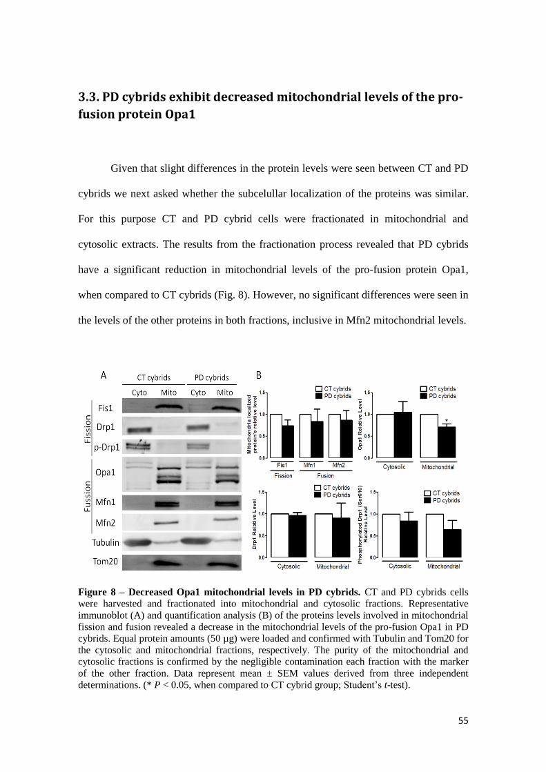
55
3.3. PD cybrids exhibit decreased mitochondrial levels of the pro-
fusion protein Opa1
Given that slight differences in the protein levels were seen between CT and PD
cybrids we next asked whether the subcelullar localization of the proteins was similar.
For this purpose CT and PD cybrid cells were fractionated in mitochondrial and
cytosolic extracts. The results from the fractionation process revealed that PD cybrids
have a significant reduction in mitochondrial levels of the pro-fusion protein Opa1,
when compared to CT cybrids (Fig. 8). However, no significant differences were seen in
the levels of the other proteins in both fractions, inclusive in Mfn2 mitochondrial levels.
Figure 8 – Decreased Opa1 mitochondrial levels in PD cybrids. CT and PD cybrids cells
were harvested and fractionated into mitochondrial and cytosolic fractions. Representative
immunoblot (A) and quantification analysis (B) of the proteins levels involved in mitochondrial
fission and fusion revealed a decrease in the mitochondrial levels of the pro-fusion Opa1 in PD
cybrids. Equal protein amounts (50 µg) were loaded and confirmed with Tubulin and Tom20 for
the cytosolic and mitochondrial fractions, respectively. The purity of the mitochondrial and
cytosolic fractions is confirmed by the negligible contamination each fraction with the marker
of the other fraction. Data represent mean ± SEM values derived from three independent
determinations. (* P < 0.05, when compared to CT cybrid group; Student’s t-test).

56
3.4. Protein expression in PD patients’ lymphocytes
In order to get a better insight from how the mitochondrial network morphology
is regulated in vivo, mitochondrial fission/fusion proteins were analyzed in PD patients,
comparing to age-matched healthy subjects. Total lysates from lymphocytes, isolated
from venous blood, were immunobloted and no significant statistical variations were
seen between PD and CT subjects (Fig. 9), although we could see an increase in pDRP1
levels and a decrease in OPA1 levels, which point towards an increase in mitochondrial
fragmentation.
Figure 9 – Basal expression of mitochondrial fission and fusion proteins in PD
lymphocytes. Lymphocytes were isolated form PD and age-matched healthy subjects and total
lysates were immnunobloted. Representative immunoblot (A) and quantification analysis (B) of
the proteins levels involved in mitochondrial fission and fusion revealed no statistical
significant differences in the total protein levels of PD subjects, relative to controls. Equal
protein amounts (50 µg) were loaded and confirmed with Gapdh. Data represent mean ± SEM
values derived from three independent determinations.

57
3.5. Mitochondrial fragmentation in mitochondrial DNA depleted
cells, associated with aberrant protein expression
To gain a better understand of the contribution of mtDNA to the mitochondrial
network, immunocytochemistry against the mitochondrial protein Tom20 was
performed in mtDNA depleted NT2 cell line. Upon basal condition NT2 ρ0 cell display
a pronounced fragmented mitochondrial network with very small round mitochondria
instead of a reticulated network seen in NT2 CT cybrids (Fig. 10, 6).
Figure 10 – Mitochondrial fragmentation in mitochondrial DNA depleted cells. Representative immunofluorescence pictures evidencing mitochondrial network in NT2 cells, containing or not mtDNA, under basal conditions, or treated with FCCP (10µM, 2 hours). Immunostaining was performed against the mitochondrial protein Tom20. NT2 ρ0 cells exhibit mitochondrial fragmentation when compared to NT2 CT cybrids. Green: Tom20.
Indeed, an impaired fusion or increased fission may cause fragmentation of the
mitochondrial network in the absence of mtDNA. To distinguish between these
possibilities, we evaluated in NT2 ρ0 cells the proteomics of the proteins involved in the
mitochondrial fission and fusion process. In agreement with previous observations in
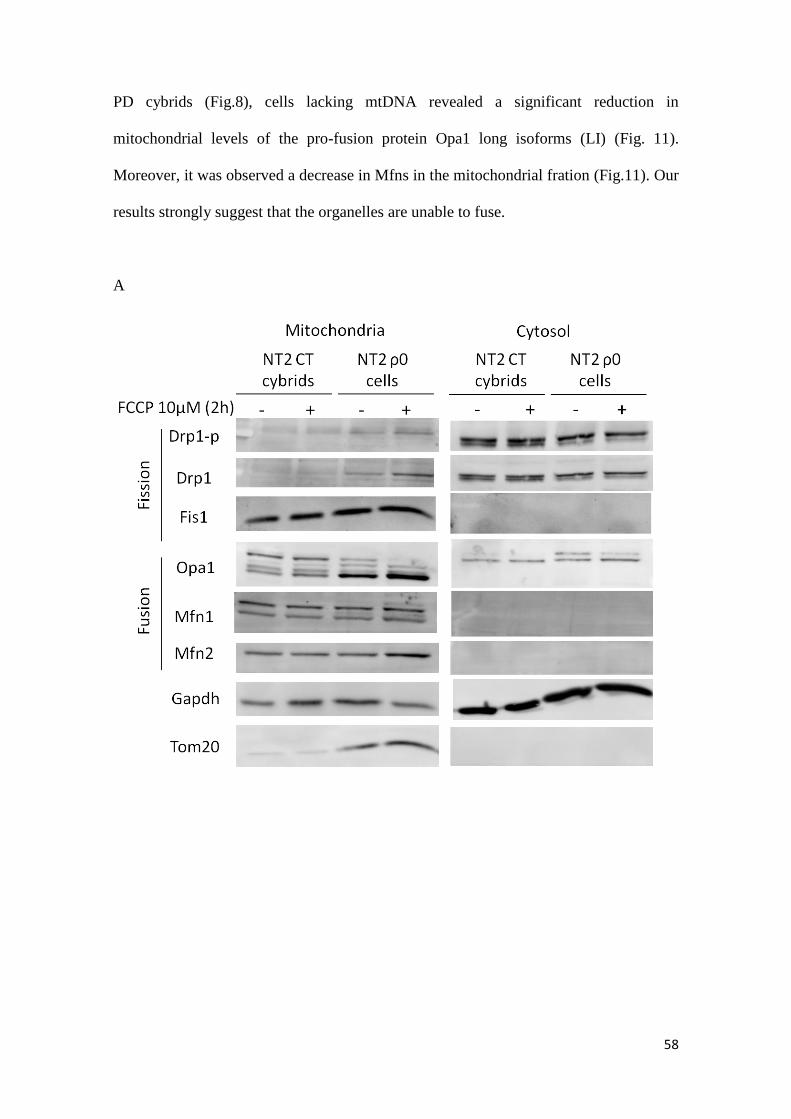
58
PD cybrids (Fig.8), cells lacking mtDNA revealed a significant reduction in
mitochondrial levels of the pro-fusion protein Opa1 long isoforms (LI) (Fig. 11).
Moreover, it was observed a decrease in Mfns in the mitochondrial fration (Fig.11). Our
results strongly suggest that the organelles are unable to fuse.
A
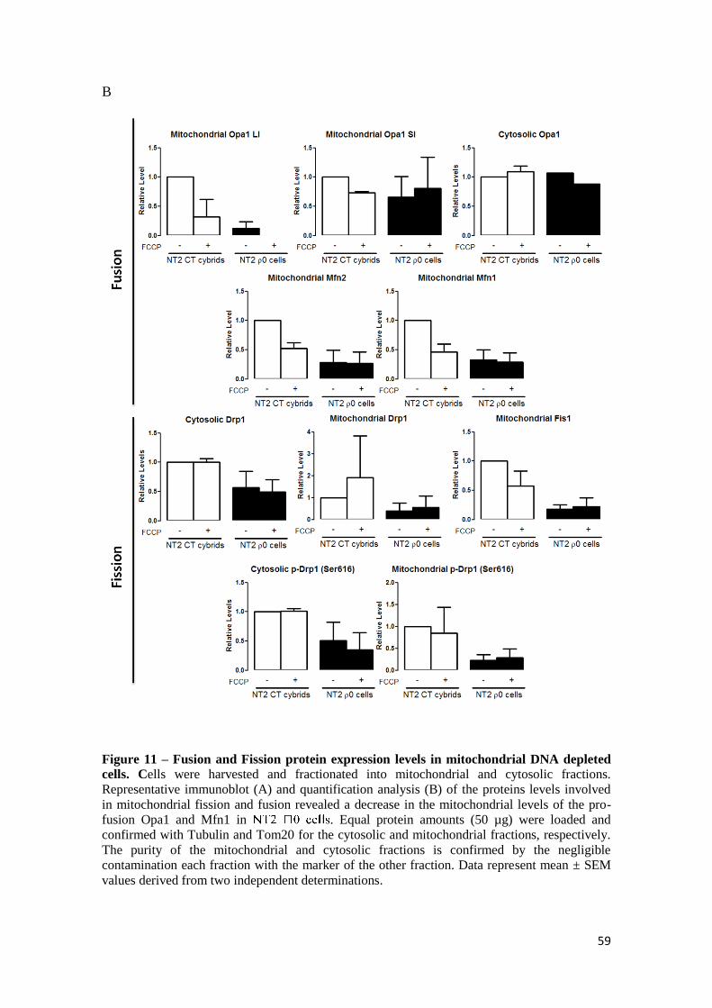
59
B
Figure 11 – Fusion and Fission protein expression levels in mitochondrial DNA depleted
cells. Cells were harvested and fractionated into mitochondrial and cytosolic fractions.
Representative immunoblot (A) and quantification analysis (B) of the proteins levels involved
in mitochondrial fission and fusion revealed a decrease in the mitochondrial levels of the pro-
fusion Opa1 and Mfn1 in . Equal protein amounts (50 µg) were loaded and
confirmed with Tubulin and Tom20 for the cytosolic and mitochondrial fractions, respectively.
The purity of the mitochondrial and cytosolic fractions is confirmed by the negligible
contamination each fraction with the marker of the other fraction. Data represent mean ± SEM
values derived from two independent determinations.

60
3.6 Abnormal fusion and fission in ASYN SH-SY5Y overexpressing
cells
To further verify the physiological relevance of the involvement of ASYN in
modulating mitochondrial morphology, mitochondrial morphology were monitored by
imaging of cells, incubated with Tom20. When wild-type ASYN was overexpressed in
SH-SY5Y cells, a decreased mitochondrial fragmentation was observed (Figure 12). In
order to validate our results and experimental conditions (4 days overexpressing ASYN)
we treated both cells with FCCP, and observed an increase in the mitochondrial
fragmentation pattern.
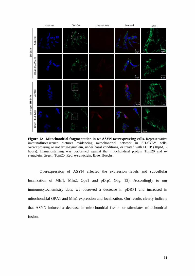
61
Figure 12 –Mitochondrial fragmentation in wt ASYN overexpressing cells. Representative
immunofluorescence pictures evidencing mitochondrial network in SH-SY5Y cells,
overexpressing or not wt α-synuclein, under basal conditions, or treated with FCCP (10µM, 2
hours). Immunostaining was performed against the mitochondrial protein Tom20 and α-
synuclein. Green: Tom20, Red: α-synuclein, Blue: Hoechst.
Overexpression of ASYN affected the expression levels and subcellular
localization of Mfn1, Mfn2, Opa1 and pDrp1 (Fig. 13). Accordingly to our
immunocytochemistry data, we observed a decrease in pDRP1 and increased in
mitochondrial OPA1 and Mfn1 expression and localization. Our results clearly indicate
that ASYN induced a decrease in mitochondrial fission or stimulates mitochondrial
fusion.
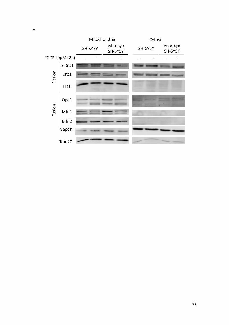
62
A

63
B
Figure 13 – Fusion and Fission protein expression levels in wt ASYN overexpressing cells.
Cells were harvested and fractionated into mitochondrial and cytosolic fractions. Representative
immunoblot (A) and quantification analysis (B) of the proteins levels involved in mitochondrial
fission and fusion revealed a decrease in the mitochondrial levels of the pro-fusion Opa1 and
and Tom20 for the cytosolic and mitochondrial fractions, respectively. The purity of the
mitochondrial and cytosolic fractions is confirmed by the negligible contamination each fraction
with the marker of the other fraction. Data represent mean ± SEM values derived from two
independent determinations.

64
3.7 MPP+-induced ROS production is independent of OPA1
mitochondrial levels
Mitochondria are the primary source for endogenous ROS, and a significant
increase in ROS levels could be detected in HEK293T cells after 24 h of treatment with
MPP+ (Fig. 14). MPP
+-treated HEK293T cells show an increase in superoxide radical
production. When challenged with rotenone, a complex I inhibitor, HEK293T cells
increase significantly superoxide radical production. Interestingly, MPP+ and rotenone
co-treatments decreased the detection of ROS levels, probably due to the loss of cell
numbers after this trigger.
To explore the relationship between MPP+-induced mitochondrial fragmentation
and ROS production, we also measured ROS levels in OPA1 overexpressing HEK293T
cells treated with MPP+ for 24h. Basal level of ROS was similar in nontreated OPA1
overexpressing cells compared with control cells. Unexpectedly, MPP+-induced ROS
production was not dependent on mitochondrial OPA1 levels. Cells treated with MPP+
that overexpressed OPA1 showed an increase in ROS production (Fig. 14). These
findings indicate Opa1 overexpression doesn’t rescue MPP+ induced ROS production in
HEK293T cells.
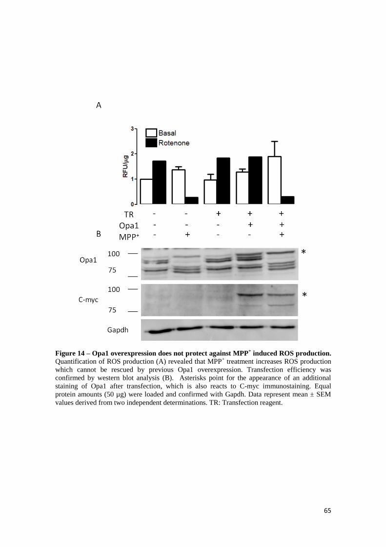
65
Figure 14 – Opa1 overexpression does not protect against MPP+ induced ROS production.
Quantification of ROS production (A) revealed that MPP+ treatment increases ROS production
which cannot be rescued by previous Opa1 overexpression. Transfection efficiency was
confirmed by western blot analysis (B). Asterisks point for the appearance of an additional
staining of Opa1 after transfection, which is also reacts to C-myc immunostaining. Equal
protein amounts (50 µg) were loaded and confirmed with Gapdh. Data represent mean ± SEM
values derived from two independent determinations. TR: Transfection reagent.

66
Chapter 4 Discussion
67
The data presented in this work supports the idea that an abnormal mitochondrial
morphology and distribution is a consequence of OXPHOS deficiency. Mitochondria in
our PD cellular model exhibit an intermediary phenotype between healthy state and
acute CXI inhibition-induced severe mitochondrial fragmentation (Fig. 6). Although
MPP+ treatment of our CT cybrids (1mM, 24h) resembles CXI activity deficiency of our
PD cybrids (Esteves et al., 2008), fission/fusion proteins’ levels modulation is very
different. Indeed, our PD cybrids exhibits a reduction of mitochondrial Opa1 levels and
accumulation of the Mfn2 levels, both proteins involved in the mitochondrial fusion
process (Fig. 7, 8). Treatment of CT cybrids with MPP+ also modulates Opa1 levels,
however it significantly reduces Mfn2 levels and the fission-stimulatory
phosphorylation of Drp1 at Ser616.
Several CXI inhibitors were described to induce mitochondrial fragmentation in
a Drp1- dependent manner (Benard et al., 2007, Gomez-Lazaro et al., 2008, Wang et al.,
2011c). Indeed, Wang and colleagues have recently shown that MPP+ mediates Drp1-
dependent mitochondrial fragmentation in SH-SY5Y cells as the attenuation of
mitochondrial fragmentation by Drp1 knockdown almost prevents the decrease of
mitochondrial membrane potential, calcium handling disruption, increased ROS
generation and deficient mitophagy, placing mitochondria fragmentation as an upstream
event mediating MPP+-induced toxicity (Wang et al., 2011c). Our results with MPP
+
treatment of CT cybrids are, in some extent, in agreement with the previous study. They
had described that at 24h after MPP+ treatment, during which they had seen a biphasic
increase in Drp1 levels, Drp1 levels are similar to control, which is also seen in our
MPP+ treated CT cybrids (Fig. 7). Although it was shown that Drp1 plays an important
role in cell death, it might not be the only player, and therefore Wang and co-workers

68
study lacks information regarding mitochondrial proteins involved in mitochondrial
fusion.
Increasing studies have been highlighting a tight regulation of the proteins
involved in the mitochondrial fusion process in neurodegeneration, namely Opa1 and
Mfn1 & 2. Indeed, decreased expression of these proteins have been described in
neurodegenerative diseases, namely in AD and HD (Wang et al., 2008, Wang et al.,
2009, Manczak et al., 2011, Shirendeb et al., 2011).
Between our chronic and acute mitochondrial model of PD it appears to be a
nexus at Opa1 and Mfn’s levels modulation. Indeed, our PD cybrids exhibits reduced
mitochondrial Opa1 isoforms levels (Fig. 7, 8) and MPP+ treatment modulates Opa1
levels by converting Opa1 long isoforms into the short ones (SI) (Fig. 7). Along with
the fragmented mitochondrial phenotype observed here (Fig. 6), disorganization of
mitochondrial cristae structure, mitochondrial membrane potential reduction and
mitochondrial cytochrome c release were also described in our PD cybrids (Esteves et
al., 2008), in agreement with results observed upon Opa1 siRNA-mediated knockdown
in HeLa cells (Olichon et al., 2003, Griparic et al., 2004). Indeed, it was suggested that
loss of mitochondrial Opa1 accelerates cytochrome c release and causes a block in
mitochondrial fusion, placing apoptotic mitochondrial fragmentation downstream of
these events (Arnoult et al., 2005).
On the other side, whereas in our acute MPP+-model there is a propensity for a
reduction in Mfns levels (Fig. 7), Mfns levels in our PD cybrids remain identical, or
higher (Fig. 7, 8). In the field of Mfns’ levels regulation it was been extensively
described that upon mitochondrial damage Mfn1 & 2 are ubiquitinated by Parkin and
degraded in a proteasome dependent manner (Tanaka et al., 2010, Chan et al., 2011).
Therefore, it is possible that upon MPP+ induced damage in CT cybrids mainly Mfn2 is

69
ubiquitin tagged and degraded by proteasome leading to the decreased Mfn2 levels.
However, in our PD cybrids impairment in the UPS (Martins-Branco unpublished work)
may either prevent ubiquitination of Mfns or inhibit their proteasome mediated
degradation, culminating in the accumulation of defective fragmented mitochondria.
Future experiments accessing Mfns ubiquitination can quickly elucidate this hypothesis.
Moreover, our results in PD patients lymphocytes, show a trendline towards an
increase in pDRP1 and a decrease in OPA1 levels (Fig. 9), although no statistical
significance was obtained with such small sample. These results are in accordance with
what we observed in PD cybrids, obtained after fusion of PD patients platelets with
mtDNA depleted cells.
Interestingly, Opa1 was shown to be crucial for mtDNA replication and
distribution as silencing of Opa1 variants led to mtDNA depletion and to altered
mtDNA distribution throughout the mitochondrial network (Elachouri et al., 2011). As
the already described cellular deficits of our PD cybrid cell lines are consequences of
the inheritance of PD patient mtDNA (Esteves et al., 2008), targeting Opa1 function
seems appropriate to maintenance of mitochondrial genome integrity in a cellular model
of a disease. In order to address this issue we performed experiments in cells without
mtDNA, and observed a similar pattern of mitochondrial morphology and intracellular
localization as in PD cybrids (Fig. 10 and 6). Although, we observed a comparable
decrease in mitochondrial OPA1 levels, these cells have more dramatic proteomic
alterations, like a decrease in mitochondrial Mfn 2 and 1 (Fig. 11). Indeed, NT2 0 cells
FCCP, what could be explained by a dramatic alteration towards mitochondrial
fragmentation under basal conditions (Fig. 10).
Studies using dominant optic atrophy (DOA) patients fibroblasts with inhibited
mitochondrial fusion, caused by mutation in the OPA1 gene, showed disturbed

70
oxidative metabolism with impaired mitochondrial ATP synthesis after treatment with
CXI inhibitor (Zanna et al., 2008).
Since familial PD can also be caused ASYN gene duplication/triplication, which
leads to enhanced protein levels of ASYN (Singleton et al, 2003) and patients with
sporadic PD show an increase of ASYN mRNA and oligomers as was observed in our
sporadic PD model, PD cybrids (Esteves et al., 2011) we wanted to access the effect of
wt ASYN in mitochondrial fragmentation/elongation. It was previously described that
ASYN mediated mitochondrial fragmentation independently of mitochondrial fusion
and fission proteins (Kamp et al 2010). Our results, using the same cell line, but with a
stable tranfection of wt ASYN, show a decrease of mitochondrial fragmentation (Fig.
12) with a decreased expression of the fission protein pDRP1 and increases in
mitochondrial fusion proteins, OPA1, Mfn1. Our findings suggest that ASYN gene
duplications or triplications may lead to increased amounts of ASYN inside the
mitochondria (Esteves et al., unpublished work), which may promote mitochondrial
fusion in order to dilute toxic ASYN oligomers. More studies with cells overexpressing
ASYN need to be done in order to fully understand the contradictory results obtained by
Kamp and co-workers (2010) showing that increased amounts of ASYN bind to
mitochondria inhibiting mitochondrial fusion and therefore trigger disease pathology.
While it remains to be determined whether and how mtDNA-induced changes in
bioenergetic function cause mitochondrial fragmentation, we demonstrated that a
compromised OXPHOS causes an excessive mitochondrial fragmentation triggered by a
cleavage of the OPA1 LI (Fig. 7 and 8). However, the involvement of mitochondrial
fragmentation and perinuclear distribution in OXPHOS-mediated impairments in
mitochondrial membrane potential, calcium handling, ROS generation and mitophagy
as well as eventual cell death are still undisclosured. Indeed, the attenuation of

71
mitochondrial fragmentation by OPA1overexpression did not prevent MPP+-induced
ROS production (Fig. 14). It was previously described that DRP1 knockdown prevented
MPP+-mediated ROS production (Wang et al., 2011). Indeed, Opa1 up-regulation
already shown to restore mitochondrial morphology and to protect neurons against
excitotoxicity (Jahani-Asl et al., 2011). Although no changes in Opa1 levels and
distribution were seen upon exposure to NMDA, this protein was required for
calpastatin-mediated neuroprotection, placing Opa1 downstream of calpain activation
following an excitotoxic stimulus. Our cybrid cellular model also presents show an
exacerbate calpain activation, which may be mediating OPA1 clevadge (Esteves et al.,
2011).
Although we still need to address OPA1/mitochondrial fusion involvement in
OXPHOS-mediated mitochondrial membrane potential depletion, mitochondrial
calcium buffering, and mitophagy, we strongly believe that changes in mitochondrial
dynamics and turnover might render neurons susceptible to degeneration in PD.

72
References
Alexander C, Votruba M, Pesch UE, Thiselton DL, Mayer S, Moore A, Rodriguez M, Kellner U, Leo-Kottler B, Auburger G, Bhattacharya SS, Wissinger B (OPA1, encoding a dynamin-related GTPase, is mutated in autosomal dominant optic atrophy linked to chromosome 3q28. Nat Genet 26:211-215.2000).
Arduino DM, Esteves AR, Cardoso SM (Mitochondrial fusion/fission, transport and autophagy in Parkinson's disease: when mitochondria get nasty. Parkinsons Dis 2011:767230.2011).
Arnoult D, Grodet A, Lee YJ, Estaquier J, Blackstone C (Release of OPA1 during apoptosis participates in the rapid and complete release of cytochrome c and subsequent mitochondrial fragmentation. J Biol Chem 280:35742-35750.2005).
Baloh RH, Schmidt RE, Pestronk A, Milbrandt J (Altered axonal mitochondrial transport in the pathogenesis of Charcot-Marie-Tooth disease from mitofusin 2 mutations. J Neurosci 27:422-430.2007).
Banerjee R, Starkov AA, Beal MF, Thomas B (Mitochondrial dysfunction in the limelight of Parkinson's disease pathogenesis. Biochim Biophys Acta 1792:651-663.2009).
Barbas NR (Cognitive, affective, and psychiatric features of Parkinson's disease. Clin Geriatr Med 22:773-796, v-vi.2006).
Barsoum MJ, Yuan H, Gerencser AA, Liot G, Kushnareva Y, Graber S, Kovacs I, Lee WD, Waggoner J, Cui J, White AD, Bossy B, Martinou JC, Youle RJ, Lipton SA, Ellisman MH, Perkins GA, Bossy-Wetzel E (Nitric oxide-induced mitochondrial fission is regulated by dynamin-related GTPases in neurons. EMBO J 25:3900-3911.2006).
Beal MF (Mitochondria take center stage in aging and neurodegeneration. Ann Neurol 58:495-505.2005).
Benard G, Bellance N, James D, Parrone P, Fernandez H, Letellier T, Rossignol R (Mitochondrial bioenergetics and structural network organization. J Cell Sci 120:838-848.2007).
Bindoff LA, Birch-Machin M, Cartlidge NE, Parker WD, Jr., Turnbull DM (Mitochondrial function in Parkinson's disease. Lancet 2:49.1989).
Bonifati V (Genetics of parkinsonism. Parkinsonism Relat Disord 13 Suppl 3:S233-241.2007). Braak H, Ghebremedhin E, Rub U, Bratzke H, Del Tredici K (Stages in the development of
Parkinson's disease-related pathology. Cell Tissue Res 318:121-134.2004). Brockmann K, Dreha-Kulaczewski S, Dechent P, Bonnemann C, Helms G, Kyllerman M, Bruck W,
Frahm J, Huehne K, Gartner J, Rautenstrauss B (Cerebral involvement in axonal Charcot-Marie-Tooth neuropathy caused by mitofusin2 mutations. J Neurol 255:1049-1058.2008).
Buttner S, Bitto A, Ring J, Augsten M, Zabrocki P, Eisenberg T, Jungwirth H, Hutter S, Carmona-Gutierrez D, Kroemer G, Winderickx J, Madeo F (Functional mitochondria are required for alpha-synuclein toxicity in aging yeast. J Biol Chem 283:7554-7560.2008).
Cardoso SM, Santana I, Swerdlow RH, Oliveira CR (Mitochondria dysfunction of Alzheimer's disease cybrids enhances Abeta toxicity. J Neurochem 89:1417-1426.2004).
Cardoso SM. The Mitochondrial Cascade Hypothesis for Parkinson’s Disease Current Pharmaceutical Design (in press)).
Cerveny KL, Tamura Y, Zhang Z, Jensen RE, Sesaki H (Regulation of mitochondrial fusion and division. Trends Cell Biol 17:563-569.2007).
Chan DC (Mitochondria: dynamic organelles in disease, aging, and development. Cell 125:1241-1252.2006).

73
Chan NC, Salazar AM, Pham AH, Sweredoski MJ, Kolawa NJ, Graham RL, Hess S, Chan DC (Broad activation of the ubiquitin-proteasome system by Parkin is critical for mitophagy. Hum Mol Genet 20:1726-1737.2011).
Chen H, Chan DC (Mitochondrial dynamics--fusion, fission, movement, and mitophagy--in neurodegenerative diseases. Hum Mol Genet 18:R169-176.2009).
Chinta SJ, Andersen JK (Redox imbalance in Parkinson's disease. Biochim Biophys Acta 1780:1362-1367.2008).
Chinta SJ, Mallajosyula JK, Rane A, Andersen JK (Mitochondrial alpha-synuclein accumulation impairs complex I function in dopaminergic neurons and results in increased mitophagy in vivo. Neurosci Lett 486:235-239.2010).
Cipolat S, Martins de Brito O, Dal Zilio B, Scorrano L (OPA1 requires mitofusin 1 to promote mitochondrial fusion. Proc Natl Acad Sci U S A 101:15927-15932.2004).
Clark IE, Dodson MW, Jiang C, Cao JH, Huh JR, Seol JH, Yoo SJ, Hay BA, Guo M (Drosophila pink1 is required for mitochondrial function and interacts genetically with parkin. Nature 441:1162-1166.2006).
Cole NB, Dieuliis D, Leo P, Mitchell DC, Nussbaum RL (Mitochondrial translocation of alpha-synuclein is promoted by intracellular acidification. Exp Cell Res 314:2076-2089.2008).
Conway KA, Harper JD, Lansbury PT (Accelerated in vitro fibril formation by a mutant alpha-synuclein linked to early-onset Parkinson disease. Nat Med 4:1318-1320.1998).
Conway KA, Lee SJ, Rochet JC, Ding TT, Williamson RE, Lansbury PT, Jr. (Acceleration of oligomerization, not fibrillization, is a shared property of both alpha-synuclein mutations linked to early-onset Parkinson's disease: implications for pathogenesis and therapy. Proc Natl Acad Sci U S A 97:571-576.2000).
Conway KA, Rochet JC, Bieganski RM, Lansbury PT, Jr. (Kinetic stabilization of the alpha-synuclein protofibril by a dopamine-alpha-synuclein adduct. Science 294:1346-1349.2001).
Corral-Debrinski M, Horton T, Lott MT, Shoffner JM, Beal MF, Wallace DC (Mitochondrial DNA deletions in human brain: regional variability and increase with advanced age. Nat Genet 2:324-329.1992).
Cui M, Tang X, Christian WV, Yoon Y, Tieu K (Perturbations in mitochondrial dynamics induced by human mutant PINK1 can be rescued by the mitochondrial division inhibitor mdivi-1. J Biol Chem 285:11740-11752.2010).
Cui T, Fan C, Gu L, Gao H, Liu Q, Zhang T, Qi Z, Zhao C, Zhao H, Cai Q, Yang H (Silencing of PINK1 induces mitophagy via mitochondrial permeability transition in dopaminergic MN9D cells. Brain Res 1394:1-13.2011).
Dagda RK, Cherra SJ, 3rd, Kulich SM, Tandon A, Park D, Chu CT (Loss of PINK1 function promotes mitophagy through effects on oxidative stress and mitochondrial fission. J Biol Chem 284:13843-13855.2009).
Dawson TM (Parkin and defective ubiquitination in Parkinson's disease. J Neural Transm Suppl 209-213.2006).
de Brito OM, Scorrano L (Mitofusin 2 tethers endoplasmic reticulum to mitochondria. Nature 456:605-610.2008).
Delettre C, Lenaers G, Griffoin JM, Gigarel N, Lorenzo C, Belenguer P, Pelloquin L, Grosgeorge J, Turc-Carel C, Perret E, Astarie-Dequeker C, Lasquellec L, Arnaud B, Ducommun B, Kaplan J, Hamel CP (Nuclear gene OPA1, encoding a mitochondrial dynamin-related protein, is mutated in dominant optic atrophy. Nat Genet 26:207-210.2000).
Deng H, Dodson MW, Huang H, Guo M (The Parkinson's disease genes pink1 and parkin promote mitochondrial fission and/or inhibit fusion in Drosophila. Proc Natl Acad Sci U S A 105:14503-14508.2008).
Detmer SA, Chan DC (Complementation between mouse Mfn1 and Mfn2 protects mitochondrial fusion defects caused by CMT2A disease mutations. J Cell Biol 176:405-414.2007a).
74
Detmer SA, Chan DC (Functions and dysfunctions of mitochondrial dynamics. Nat Rev Mol Cell Biol 8:870-879.2007b).
Devi L, Raghavendran V, Prabhu BM, Avadhani NG, Anandatheerthavarada HK (Mitochondrial import and accumulation of alpha-synuclein impair complex I in human dopaminergic neuronal cultures and Parkinson disease brain. J Biol Chem 283:9089-9100.2008).
Ekstrand MI, Terzioglu M, Galter D, Zhu S, Hofstetter C, Lindqvist E, Thams S, Bergstrand A, Hansson FS, Trifunovic A, Hoffer B, Cullheim S, Mohammed AH, Olson L, Larsson NG (Progressive parkinsonism in mice with respiratory-chain-deficient dopamine neurons. Proc Natl Acad Sci U S A 104:1325-1330.2007).
Elachouri G, Vidoni S, Zanna C, Pattyn A, Boukhaddaoui H, Gaget K, Yu-Wai-Man P, Gasparre G, Sarzi E, Delettre C, Olichon A, Loiseau D, Reynier P, Chinnery PF, Rotig A, Carelli V, Hamel CP, Rugolo M, Lenaers G (OPA1 links human mitochondrial genome maintenance to mtDNA replication and distribution. Genome Res 21:12-20.2011).
Ellis CE, Murphy EJ, Mitchell DC, Golovko MY, Scaglia F, Barcelo-Coblijn GC, Nussbaum RL (Mitochondrial lipid abnormality and electron transport chain impairment in mice lacking alpha-synuclein. Mol Cell Biol 25:10190-10201.2005).
Endo R, Saito T, Asada A, Kawahara H, Ohshima T, Hisanaga S (Commitment of 1-methyl-4-phenylpyrinidinium ion-induced neuronal cell death by proteasome-mediated degradation of p35 cyclin-dependent kinase 5 activator. J Biol Chem 284:26029-26039.2009).
Esteves AR, Arduino DM, Silva DF, Oliveira CR, Cardoso SM (Mitochondrial Dysfunction: The Road to Alpha-Synuclein Oligomerization in PD. Parkinsons Dis 2011:693761.2011).
Esteves AR, Domingues AF, Ferreira IL, Januario C, Swerdlow RH, Oliveira CR, Cardoso SM (Mitochondrial function in Parkinson's disease cybrids containing an nt2 neuron-like nuclear background. Mitochondrion 8:219-228.2008).
Exner N, Treske B, Paquet D, Holmstrom K, Schiesling C, Gispert S, Carballo-Carbajal I, Berg D, Hoepken HH, Gasser T, Kruger R, Winklhofer KF, Vogel F, Reichert AS, Auburger G, Kahle PJ, Schmid B, Haass C (Loss-of-function of human PINK1 results in mitochondrial pathology and can be rescued by parkin. J Neurosci 27:12413-12418.2007).
Forno LS (Neuropathology of Parkinson's disease. J Neuropathol Exp Neurol 55:259-272.1996). Fredenburg RA, Rospigliosi C, Meray RK, Kessler JC, Lashuel HA, Eliezer D, Lansbury PT, Jr. (The
impact of the E46K mutation on the properties of alpha-synuclein in its monomeric and oligomeric states. Biochemistry 46:7107-7118.2007).
Gandhi S, Muqit MM, Stanyer L, Healy DG, Abou-Sleiman PM, Hargreaves I, Heales S, Ganguly M, Parsons L, Lees AJ, Latchman DS, Holton JL, Wood NW, Revesz T (PINK1 protein in normal human brain and Parkinson's disease. Brain 129:1720-1731.2006).
Gandre-Babbe S, van der Bliek AM (The novel tail-anchored membrane protein Mff controls mitochondrial and peroxisomal fission in mammalian cells. Mol Biol Cell 19:2402-2412.2008).
Gasser T (Mendelian forms of Parkinson's disease. Biochim Biophys Acta 1792:587-596.2009). Gautier CA, Kitada T, Shen J (Loss of PINK1 causes mitochondrial functional defects and
increased sensitivity to oxidative stress. Proc Natl Acad Sci U S A 105:11364-11369.2008).
Gegg ME, Cooper JM, Chau KY, Rojo M, Schapira AH, Taanman JW (Mitofusin 1 and mitofusin 2 are ubiquitinated in a PINK1/parkin-dependent manner upon induction of mitophagy. Hum Mol Genet 19:4861-4870.2010).
Glauser L, Sonnay S, Stafa K, Moore DJ (Parkin promotes the ubiquitination and degradation of the mitochondrial fusion factor mitofusin 1. J Neurochem.2011).
Goldberg MS, Fleming SM, Palacino JJ, Cepeda C, Lam HA, Bhatnagar A, Meloni EG, Wu N, Ackerson LC, Klapstein GJ, Gajendiran M, Roth BL, Chesselet MF, Maidment NT, Levine MS, Shen J (Parkin-deficient mice exhibit nigrostriatal deficits but not loss of dopaminergic neurons. J Biol Chem 278:43628-43635.2003).

75
Gomez-Lazaro M, Bonekamp NA, Galindo MF, Jordan J, Schrader M (6-Hydroxydopamine (6-OHDA) induces Drp1-dependent mitochondrial fragmentation in SH-SY5Y cells. Free Radic Biol Med 44:1960-1969.2008).
Greene JC, Whitworth AJ, Kuo I, Andrews LA, Feany MB, Pallanck LJ (Mitochondrial pathology and apoptotic muscle degeneration in Drosophila parkin mutants. Proc Natl Acad Sci U S A 100:4078-4083.2003).
Griparic L, van der Wel NN, Orozco IJ, Peters PJ, van der Bliek AM (Loss of the intermembrane space protein Mgm1/OPA1 induces swelling and localized constrictions along the lengths of mitochondria. J Biol Chem 279:18792-18798.2004).
Gu M, Cooper JM, Taanman JW, Schapira AH (Mitochondrial DNA transmission of the mitochondrial defect in Parkinson's disease. Ann Neurol 44:177-186.1998).
Hatano Y, Li Y, Sato K, Asakawa S, Yamamura Y, Tomiyama H, Yoshino H, Asahina M, Kobayashi S, Hassin-Baer S, Lu CS, Ng AR, Rosales RL, Shimizu N, Toda T, Mizuno Y, Hattori N (Novel PINK1 mutations in early-onset parkinsonism. Ann Neurol 56:424-427.2004).
Heeman B, Van den Haute C, Aelvoet SA, Valsecchi F, Rodenburg RJ, Reumers V, Debyser Z, Callewaert G, Koopman WJ, Willems PH, Baekelandt V (Depletion of PINK1 affects mitochondrial metabolism, calcium homeostasis and energy maintenance. J Cell Sci 124:1115-1125.2011).
Henchcliffe C, Beal MF (Mitochondrial biology and oxidative stress in Parkinson disease pathogenesis. Nat Clin Pract Neurol 4:600-609.2008).
Hoppins S, Lackner L, Nunnari J (The machines that divide and fuse mitochondria. Annu Rev Biochem 76:751-780.2007).
Hsu LJ, Sagara Y, Arroyo A, Rockenstein E, Sisk A, Mallory M, Wong J, Takenouchi T, Hashimoto M, Masliah E (alpha-synuclein promotes mitochondrial deficit and oxidative stress. Am J Pathol 157:401-410.2000).
Ishihara N, Eura Y, Mihara K (Mitofusin 1 and 2 play distinct roles in mitochondrial fusion reactions via GTPase activity. J Cell Sci 117:6535-6546.2004).
Jahani-Asl A, Pilon-Larose K, Xu W, MacLaurin JG, Park DS, McBride HM, Slack RS (The mitochondrial inner membrane GTPase, optic atrophy 1 (Opa1), restores mitochondrial morphology and promotes neuronal survival following excitotoxicity. J Biol Chem 286:4772-4782.2011).
James DI, Parone PA, Mattenberger Y, Martinou JC (hFis1, a novel component of the mammalian mitochondrial fission machinery. J Biol Chem 278:36373-36379.2003).
Jenner P (Oxidative stress in Parkinson's disease. Ann Neurol 53 Suppl 3:S26-36; discussion S36-28.2003).
Jin J, Hulette C, Wang Y, Zhang T, Pan C, Wadhwa R, Zhang J (Proteomic identification of a stress protein, mortalin/mthsp70/GRP75: relevance to Parkinson disease. Mol Cell Proteomics 5:1193-1204.2006).
Kamei S, Chen-Kuo-Chang M, Cazevieille C, Lenaers G, Olichon A, Belenguer P, Roussignol G, Renard N, Eybalin M, Michelin A, Delettre C, Brabet P, Hamel CP (Expression of the Opa1 mitochondrial protein in retinal ganglion cells: its downregulation causes aggregation of the mitochondrial network. Invest Ophthalmol Vis Sci 46:4288-4294.2005).
Kamp F, Exner N, Lutz AK, Wender N, Hegermann J, Brunner B, Nuscher B, Bartels T, Giese A, Beyer K, Eimer S, Winklhofer KF, Haass C (Inhibition of mitochondrial fusion by [alpha]-synuclein is rescued by PINK1, Parkin and DJ-1. EMBO J 29:3571-3589.2010).
Kanki T, Klionsky DJ (Mitophagy in yeast occurs through a selective mechanism. J Biol Chem 283:32386-32393.2008).
Keeney PM, Xie J, Capaldi RA, Bennett JP, Jr. (Parkinson's disease brain mitochondrial complex I has oxidatively damaged subunits and is functionally impaired and misassembled. J Neurosci 26:5256-5264.2006).

76
Kijima K, Numakura C, Izumino H, Umetsu K, Nezu A, Shiiki T, Ogawa M, Ishizaki Y, Kitamura T, Shozawa Y, Hayasaka K (Mitochondrial GTPase mitofusin 2 mutation in Charcot-Marie-Tooth neuropathy type 2A. Hum Genet 116:23-27.2005).
Kitada T, Asakawa S, Hattori N, Matsumine H, Yamamura Y, Minoshima S, Yokochi M, Mizuno Y, Shimizu N (Mutations in the parkin gene cause autosomal recessive juvenile parkinsonism. Nature 392:605-608.1998).
Knott AB, Perkins G, Schwarzenbacher R, Bossy-Wetzel E (Mitochondrial fragmentation in neurodegeneration. Nat Rev Neurosci 9:505-518.2008).
Koopman WJ, Verkaart S, Visch HJ, van der Westhuizen FH, Murphy MP, van den Heuvel LW, Smeitink JA, Willems PH (Inhibition of complex I of the electron transport chain causes O2-. -mediated mitochondrial outgrowth. Am J Physiol Cell Physiol 288:C1440-1450.2005).
Kruger R, Kuhn W, Muller T, Woitalla D, Graeber M, Kosel S, Przuntek H, Epplen JT, Schols L, Riess O (Ala30Pro mutation in the gene encoding alpha-synuclein in Parkinson's disease. Nat Genet 18:106-108.1998).
Lang AE, Lozano AM (Parkinson's disease. First of two parts. N Engl J Med 339:1044-1053.1998a).
Lang AE, Lozano AM (Parkinson's disease. Second of two parts. N Engl J Med 339:1130-1143.1998b).
Langston JW, Ballard P, Tetrud JW, Irwin I (Chronic Parkinsonism in humans due to a product of meperidine-analog synthesis. Science 219:979-980.1983).
Lee YJ, Jeong SY, Karbowski M, Smith CL, Youle RJ (Roles of the mammalian mitochondrial fission and fusion mediators Fis1, Drp1, and Opa1 in apoptosis. Mol Biol Cell 15:5001-5011.2004).
Li WW, Yang R, Guo JC, Ren HM, Zha XL, Cheng JS, Cai DF (Localization of alpha-synuclein to mitochondria within midbrain of mice. Neuroreport 18:1543-1546.2007).
Li Z, Okamoto K, Hayashi Y, Sheng M (The importance of dendritic mitochondria in the morphogenesis and plasticity of spines and synapses. Cell 119:873-887.2004).
Liang CL, Wang TT, Luby-Phelps K, German DC (Mitochondria mass is low in mouse substantia nigra dopamine neurons: implications for Parkinson's disease. Exp Neurol 203:370-380.2007).
Linnane AW, Marzuki S, Ozawa T, Tanaka M (Mitochondrial DNA mutations as an important contributor to ageing and degenerative diseases. Lancet 1:642-645.1989).
Liu G, Zhang C, Yin J, Li X, Cheng F, Li Y, Yang H, Ueda K, Chan P, Yu S (alpha-Synuclein is differentially expressed in mitochondria from different rat brain regions and dose-dependently down-regulates complex I activity. Neurosci Lett 454:187-192.2009).
Lutz AK, Exner N, Fett ME, Schlehe JS, Kloos K, Lammermann K, Brunner B, Kurz-Drexler A, Vogel F, Reichert AS, Bouman L, Vogt-Weisenhorn D, Wurst W, Tatzelt J, Haass C, Winklhofer KF (Loss of parkin or PINK1 function increases Drp1-dependent mitochondrial fragmentation. J Biol Chem 284:22938-22951.2009).
Manczak M, Calkins MJ, Reddy PH (Impaired mitochondrial dynamics and abnormal interaction of amyloid beta with mitochondrial protein Drp1 in neurons from patients with Alzheimer's disease: implications for neuronal damage. Hum Mol Genet 20:2495-2509.2011).
Martin LJ, Pan Y, Price AC, Sterling W, Copeland NG, Jenkins NA, Price DL, Lee MK (Parkinson's disease alpha-synuclein transgenic mice develop neuronal mitochondrial degeneration and cell death. J Neurosci 26:41-50.2006).
McBride HM (Parkin mitochondria in the autophagosome. J Cell Biol 183:757-759.2008). Meuer K, Suppanz IE, Lingor P, Planchamp V, Goricke B, Fichtner L, Braus GH, Dietz GP, Jakobs
S, Bahr M, Weishaupt JH (Cyclin-dependent kinase 5 is an upstream regulator of mitochondrial fission during neuronal apoptosis. Cell Death Differ 14:651-661.2007).
77
Miller KE, Sheetz MP (Axonal mitochondrial transport and potential are correlated. J Cell Sci 117:2791-2804.2004).
Mizuno Y, Matuda S, Yoshino H, Mori H, Hattori N, Ikebe S (An immunohistochemical study on alpha-ketoglutarate dehydrogenase complex in Parkinson's disease. Ann Neurol 35:204-210.1994).
Mortiboys H, Thomas KJ, Koopman WJ, Klaffke S, Abou-Sleiman P, Olpin S, Wood NW, Willems PH, Smeitink JA, Cookson MR, Bandmann O (Mitochondrial function and morphology are impaired in parkin-mutant fibroblasts. Ann Neurol 64:555-565.2008).
Murphy MP (How mitochondria produce reactive oxygen species. Biochem J 417:1-13.2009). Nakada K, Inoue K, Ono T, Isobe K, Ogura A, Goto YI, Nonaka I, Hayashi JI (Inter-mitochondrial
complementation: Mitochondria-specific system preventing mice from expression of disease phenotypes by mutant mtDNA. Nat Med 7:934-940.2001).
Nakamura K, Nemani VM, Azarbal F, Skibinski G, Levy JM, Egami K, Munishkina L, Zhang J, Gardner B, Wakabayashi J, Sesaki H, Cheng Y, Finkbeiner S, Nussbaum RL, Masliah E, Edwards RH (Direct membrane association drives mitochondrial fission by the Parkinson Disease-associated protein {alpha}-synuclein. J Biol Chem.2011).
Nakamura K, Nemani VM, Wallender EK, Kaehlcke K, Ott M, Edwards RH (Optical reporters for the conformation of alpha-synuclein reveal a specific interaction with mitochondria. J Neurosci 28:12305-12317.2008).
Narendra D, Tanaka A, Suen DF, Youle RJ (Parkin is recruited selectively to impaired mitochondria and promotes their autophagy. J Cell Biol 183:795-803.2008).
Olichon A, Baricault L, Gas N, Guillou E, Valette A, Belenguer P, Lenaers G (Loss of OPA1 perturbates the mitochondrial inner membrane structure and integrity, leading to cytochrome c release and apoptosis. J Biol Chem 278:7743-7746.2003).
Ono T, Isobe K, Nakada K, Hayashi JI (Human cells are protected from mitochondrial dysfunction by complementation of DNA products in fused mitochondria. Nat Genet 28:272-275.2001).
Orth M, Schapira AH (Mitochondrial involvement in Parkinson's disease. Neurochem Int 40:533-541.2002).
Ozawa T (Oxidative damage and fragmentation of mitochondrial DNA in cellular apoptosis. Biosci Rep 17:237-250.1997).
Pacelli C, De Rasmo D, Signorile A, Grattagliano I, di Tullio G, D'Orazio A, Nico B, Comi GP, Ronchi D, Ferranini E, Pirolo D, Seibel P, Schubert S, Gaballo A, Villani G, Cocco T (Mitochondrial defect and PGC-1alpha dysfunction in parkin-associated familial Parkinson's disease. Biochim Biophys Acta.2011).
Palacino JJ, Sagi D, Goldberg MS, Krauss S, Motz C, Wacker M, Klose J, Shen J (Mitochondrial dysfunction and oxidative damage in parkin-deficient mice. J Biol Chem 279:18614-18622.2004).
Parihar MS, Parihar A, Fujita M, Hashimoto M, Ghafourifar P (Mitochondrial association of alpha-synuclein causes oxidative stress. Cell Mol Life Sci 65:1272-1284.2008).
Park J, Lee G, Chung J (The PINK1-Parkin pathway is involved in the regulation of mitochondrial remodeling process. Biochem Biophys Res Commun 378:518-523.2009).
Park J, Lee SB, Lee S, Kim Y, Song S, Kim S, Bae E, Kim J, Shong M, Kim JM, Chung J (Mitochondrial dysfunction in Drosophila PINK1 mutants is complemented by parkin. Nature 441:1157-1161.2006).
Parker WD, Jr., Boyson SJ, Parks JK (Abnormalities of the electron transport chain in idiopathic Parkinson's disease. Ann Neurol 26:719-723.1989).
Parone PA, Da Cruz S, Tondera D, Mattenberger Y, James DI, Maechler P, Barja F, Martinou JC (Preventing mitochondrial fission impairs mitochondrial function and leads to loss of mitochondrial DNA. PLoS One 3:e3257.2008).

78
Penn AM, Roberts T, Hodder J, Allen PS, Zhu G, Martin WR (Generalized mitochondrial dysfunction in Parkinson's disease detected by magnetic resonance spectroscopy of muscle. Neurology 45:2097-2099.1995).
Pesah Y, Pham T, Burgess H, Middlebrooks B, Verstreken P, Zhou Y, Harding M, Bellen H, Mardon G (Drosophila parkin mutants have decreased mass and cell size and increased sensitivity to oxygen radical stress. Development 131:2183-2194.2004).
Plecita-Hlavata L, Lessard M, Santorova J, Bewersdorf J, Jezek P (Mitochondrial oxidative phosphorylation and energetic status are reflected by morphology of mitochondrial network in INS-1E and HEP-G2 cells viewed by 4Pi microscopy. Biochim Biophys Acta 1777:834-846.2008).
Plun-Favreau H, Klupsch K, Moisoi N, Gandhi S, Kjaer S, Frith D, Harvey K, Deas E, Harvey RJ, McDonald N, Wood NW, Martins LM, Downward J (The mitochondrial protease HtrA2 is regulated by Parkinson's disease-associated kinase PINK1. Nat Cell Biol 9:1243-1252.2007).
Polymeropoulos MH, Lavedan C, Leroy E, Ide SE, Dehejia A, Dutra A, Pike B, Root H, Rubenstein J, Boyer R, Stenroos ES, Chandrasekharappa S, Athanassiadou A, Papapetropoulos T, Johnson WG, Lazzarini AM, Duvoisin RC, Di Iorio G, Golbe LI, Nussbaum RL (Mutation in the alpha-synuclein gene identified in families with Parkinson's disease. Science 276:2045-2047.1997).
Poole AC, Thomas RE, Andrews LA, McBride HM, Whitworth AJ, Pallanck LJ (The PINK1/Parkin pathway regulates mitochondrial morphology. Proc Natl Acad Sci U S A 105:1638-1643.2008).
Pridgeon JW, Olzmann JA, Chin LS, Li L (PINK1 protects against oxidative stress by phosphorylating mitochondrial chaperone TRAP1. PLoS Biol 5:e172.2007).
Rakovic A, Grunewald A, Kottwitz J, Bruggemann N, Pramstaller PP, Lohmann K, Klein C (Mutations in PINK1 and Parkin impair ubiquitination of Mitofusins in human fibroblasts. PLoS One 6:e16746.2011).
Rapaport D, Brunner M, Neupert W, Westermann B (Fzo1p is a mitochondrial outer membrane protein essential for the biogenesis of functional mitochondria in Saccharomyces cerevisiae. J Biol Chem 273:20150-20155.1998).
Richter C, Park JW, Ames BN (Normal oxidative damage to mitochondrial and nuclear DNA is extensive. Proc Natl Acad Sci U S A 85:6465-6467.1988).
Rintoul GL, Filiano AJ, Brocard JB, Kress GJ, Reynolds IJ (Glutamate decreases mitochondrial size and movement in primary forebrain neurons. J Neurosci 23:7881-7888.2003).
Rosenfeld M, Brenner-Lavie H, Ari SG, Kavushansky A, Ben-Shachar D (Perturbation in mitochondrial network dynamics and in complex I dependent cellular respiration in schizophrenia. Biol Psychiatry 69:980-988.2011).
Sandebring A, Thomas KJ, Beilina A, van der Brug M, Cleland MM, Ahmad R, Miller DW, Zambrano I, Cowburn RF, Behbahani H, Cedazo-Minguez A, Cookson MR (Mitochondrial alterations in PINK1 deficient cells are influenced by calcineurin-dependent dephosphorylation of dynamin-related protein 1. PLoS One 4:e5701.2009).
Schapira AH, Cooper JM, Dexter D, Jenner P, Clark JB, Marsden CD (Mitochondrial complex I deficiency in Parkinson's disease. Lancet 1:1269.1989).
Shavali S, Brown-Borg HM, Ebadi M, Porter J (Mitochondrial localization of alpha-synuclein protein in alpha-synuclein overexpressing cells. Neurosci Lett 439:125-128.2008).
Sheehan JP, Swerdlow RH, Parker WD, Miller SW, Davis RE, Tuttle JB (Altered calcium homeostasis in cells transformed by mitochondria from individuals with Parkinson's disease. J Neurochem 68:1221-1233.1997).
Shimura H, Hattori N, Kubo S, Mizuno Y, Asakawa S, Minoshima S, Shimizu N, Iwai K, Chiba T, Tanaka K, Suzuki T (Familial Parkinson disease gene product, parkin, is a ubiquitin-protein ligase. Nat Genet 25:302-305.2000).

79
Shirendeb U, Reddy AP, Manczak M, Calkins MJ, Mao P, Tagle DA, Reddy PH (Abnormal mitochondrial dynamics, mitochondrial loss and mutant huntingtin oligomers in Huntington's disease: implications for selective neuronal damage. Hum Mol Genet 20:1438-1455.2011).
Silvestri L, Caputo V, Bellacchio E, Atorino L, Dallapiccola B, Valente EM, Casari G (Mitochondrial import and enzymatic activity of PINK1 mutants associated to recessive parkinsonism. Hum Mol Genet 14:3477-3492.2005).
Singleton AB, Farrer M, Johnson J, Singleton A, Hague S, Kachergus J, Hulihan M, Peuralinna T, Dutra A, Nussbaum R, Lincoln S, Crawley A, Hanson M, Maraganore D, Adler C, Cookson MR, Muenter M, Baptista M, Miller D, Blancato J, Hardy J, Gwinn-Hardy K (alpha-Synuclein locus triplication causes Parkinson's disease. Science 302:841.2003).
Smirnova E, Griparic L, Shurland DL, van der Bliek AM (Dynamin-related protein Drp1 is required for mitochondrial division in mammalian cells. Mol Biol Cell 12:2245-2256.2001).
Starkov AA (The role of mitochondria in reactive oxygen species metabolism and signaling. Ann N Y Acad Sci 1147:37-52.2008).
Swerdlow RH, Parks JK, Cassarino DS, Maguire DJ, Maguire RS, Bennett JP, Jr., Davis RE, Parker WD, Jr. (Cybrids in Alzheimer's disease: a cellular model of the disease? Neurology 49:918-925.1997).
Swerdlow RH, Parks JK, Davis JN, 2nd, Cassarino DS, Trimmer PA, Currie LJ, Dougherty J, Bridges WS, Bennett JP, Jr., Wooten GF, Parker WD (Matrilineal inheritance of complex I dysfunction in a multigenerational Parkinson's disease family. Ann Neurol 44:873-881.1998).
Swerdlow RH, Parks JK, Miller SW, Tuttle JB, Trimmer PA, Sheehan JP, Bennett JP, Jr., Davis RE, Parker WD, Jr. (Origin and functional consequences of the complex I defect in Parkinson's disease. Ann Neurol 40:663-671.1996).
Tanaka A, Cleland MM, Xu S, Narendra DP, Suen DF, Karbowski M, Youle RJ (Proteasome and p97 mediate mitophagy and degradation of mitofusins induced by Parkin. J Cell Biol 191:1367-1380.2010).
Thomas KJ, McCoy MK, Blackinton J, Beilina A, van der Brug M, Sandebring A, Miller D, Maric D, Cedazo-Minguez A, Cookson MR (DJ-1 acts in parallel to the PINK1/parkin pathway to control mitochondrial function and autophagy. Hum Mol Genet 20:40-50.2011).
Thyagarajan D, Bressman S, Bruno C, Przedborski S, Shanske S, Lynch T, Fahn S, DiMauro S (A novel mitochondrial 12SrRNA point mutation in parkinsonism, deafness, and neuropathy. Ann Neurol 48:730-736.2000).
Trimmer PA, Borland MK, Keeney PM, Bennett JP, Jr., Parker WD, Jr. (Parkinson's disease transgenic mitochondrial cybrids generate Lewy inclusion bodies. J Neurochem 88:800-812.2004).
Trimmer PA, Swerdlow RH, Parks JK, Keeney P, Bennett JP, Jr., Miller SW, Davis RE, Parker WD, Jr. (Abnormal mitochondrial morphology in sporadic Parkinson's and Alzheimer's disease cybrid cell lines. Exp Neurol 162:37-50.2000).
Turrens JF (Mitochondrial formation of reactive oxygen species. J Physiol 552:335-344.2003). Twig G, Elorza A, Molina AJ, Mohamed H, Wikstrom JD, Walzer G, Stiles L, Haigh SE, Katz S, Las
G, Alroy J, Wu M, Py BF, Yuan J, Deeney JT, Corkey BE, Shirihai OS (Fission and selective fusion govern mitochondrial segregation and elimination by autophagy. EMBO J 27:433-446.2008).
Valente EM, Abou-Sleiman PM, Caputo V, Muqit MM, Harvey K, Gispert S, Ali Z, Del Turco D, Bentivoglio AR, Healy DG, Albanese A, Nussbaum R, Gonzalez-Maldonado R, Deller T, Salvi S, Cortelli P, Gilks WP, Latchman DS, Harvey RJ, Dallapiccola B, Auburger G, Wood NW (Hereditary early-onset Parkinson's disease caused by mutations in PINK1. Science 304:1158-1160.2004a).

80
Valente EM, Salvi S, Ialongo T, Marongiu R, Elia AE, Caputo V, Romito L, Albanese A, Dallapiccola B, Bentivoglio AR (PINK1 mutations are associated with sporadic early-onset parkinsonism. Ann Neurol 56:336-341.2004b).
Vekrellis K, Xilouri M, Emmanouilidou E, Stefanis L (Inducible over-expression of wild type alpha-synuclein in human neuronal cells leads to caspase-dependent non-apoptotic death. J Neurochem 109:1348-1362.2009).
Verstreken P, Ly CV, Venken KJ, Koh TW, Zhou Y, Bellen HJ (Synaptic mitochondria are critical for mobilization of reserve pool vesicles at Drosophila neuromuscular junctions. Neuron 47:365-378.2005).
Wang H, Song P, Du L, Tian W, Yue W, Liu M, Li D, Wang B, Zhu Y, Cao C, Zhou J, Chen Q (Parkin ubiquitinates Drp1 for proteasome-dependent degradation: implication of dysregulated mitochondrial dynamics in Parkinson disease. J Biol Chem 286:11649-11658.2011a).
Wang HL, Chou AH, Wu AS, Chen SY, Weng YH, Kao YC, Yeh TH, Chu PJ, Lu CS (PARK6 PINK1 mutants are defective in maintaining mitochondrial membrane potential and inhibiting ROS formation of substantia nigra dopaminergic neurons. Biochim Biophys Acta 1812:674-684.2011b).
Wang X, Su B, Lee HG, Li X, Perry G, Smith MA, Zhu X (Impaired balance of mitochondrial fission and fusion in Alzheimer's disease. J Neurosci 29:9090-9103.2009).
Wang X, Su B, Liu W, He X, Gao Y, Castellani RJ, Perry G, Smith MA, Zhu X (DLP1-dependent mitochondrial fragmentation mediates 1-methyl-4-phenylpyridinium toxicity in neurons: implications for Parkinson's disease. Aging Cell.2011c).
Wang X, Su B, Siedlak SL, Moreira PI, Fujioka H, Wang Y, Casadesus G, Zhu X (Amyloid-beta overproduction causes abnormal mitochondrial dynamics via differential modulation of mitochondrial fission/fusion proteins. Proc Natl Acad Sci U S A 105:19318-19323.2008).
Waterham HR, Koster J, van Roermund CW, Mooyer PA, Wanders RJ, Leonard JV (A lethal defect of mitochondrial and peroxisomal fission. N Engl J Med 356:1736-1741.2007).
Weintraub D, Stern MB (Psychiatric complications in Parkinson disease. Am J Geriatr Psychiatry 13:844-851.2005).
Westermann B (Merging mitochondria matters: cellular role and molecular machinery of mitochondrial fusion. EMBO Rep 3:527-531.2002).
Winklhofer KF, Haass C (Mitochondrial dysfunction in Parkinson's disease. Biochim Biophys Acta 1802:29-44.2010).
Wood-Kaczmar A, Gandhi S, Yao Z, Abramov AY, Miljan EA, Keen G, Stanyer L, Hargreaves I, Klupsch K, Deas E, Downward J, Mansfield L, Jat P, Taylor J, Heales S, Duchen MR, Latchman D, Tabrizi SJ, Wood NW (PINK1 is necessary for long term survival and mitochondrial function in human dopaminergic neurons. PLoS One 3:e2455.2008).
Wooten GF, Currie LJ, Bennett JP, Harrison MB, Trugman JM, Parker WD, Jr. (Maternal inheritance in Parkinson's disease. Ann Neurol 41:265-268.1997).
Xu J, Kao SY, Lee FJ, Song W, Jin LW, Yankner BA (Dopamine-dependent neurotoxicity of alpha-synuclein: a mechanism for selective neurodegeneration in Parkinson disease. Nat Med 8:600-606.2002).
Yang Y, Gehrke S, Imai Y, Huang Z, Ouyang Y, Wang JW, Yang L, Beal MF, Vogel H, Lu B (Mitochondrial pathology and muscle and dopaminergic neuron degeneration caused by inactivation of Drosophila Pink1 is rescued by Parkin. Proc Natl Acad Sci U S A 103:10793-10798.2006).
Yang Y, Ouyang Y, Yang L, Beal MF, McQuibban A, Vogel H, Lu B (Pink1 regulates mitochondrial dynamics through interaction with the fission/fusion machinery. Proc Natl Acad Sci U S A 105:7070-7075.2008).
81
Yu W, Sun Y, Guo S, Lu B (The PINK1/Parkin pathway regulates mitochondrial dynamics and function in mammalian hippocampal and dopaminergic neurons. Hum Mol Genet.2011).
Zanna C, Ghelli A, Porcelli AM, Karbowski M, Youle RJ, Schimpf S, Wissinger B, Pinti M, Cossarizza A, Vidoni S, Valentino ML, Rugolo M, Carelli V (OPA1 mutations associated with dominant optic atrophy impair oxidative phosphorylation and mitochondrial fusion. Brain 131:352-367.2008).
Zarranz JJ, Alegre J, Gomez-Esteban JC, Lezcano E, Ros R, Ampuero I, Vidal L, Hoenicka J, Rodriguez O, Atares B, Llorens V, Gomez Tortosa E, del Ser T, Munoz DG, de Yebenes JG (The new mutation, E46K, of alpha-synuclein causes Parkinson and Lewy body dementia. Ann Neurol 55:164-173.2004).
Zhou C, Huang Y, Przedborski S (Oxidative stress in Parkinson's disease: a mechanism of pathogenic and therapeutic significance. Ann N Y Acad Sci 1147:93-104.2008a).
Zhou C, Huang Y, Shao Y, May J, Prou D, Perier C, Dauer W, Schon EA, Przedborski S (The kinase domain of mitochondrial PINK1 faces the cytoplasm. Proc Natl Acad Sci U S A 105:12022-12027.2008b).
Zhu Y, Duan C, Lu L, Gao H, Zhao C, Yu S, Ueda K, Chan P, Yang H (alpha-Synuclein overexpression impairs mitochondrial function by associating with adenylate translocator. Int J Biochem Cell Biol 43:732-741.2011).
Ziviani E, Tao RN, Whitworth AJ (Drosophila parkin requires PINK1 for mitochondrial translocation and ubiquitinates mitofusin. Proc Natl Acad Sci U S A 107:5018-5023.2010).
Zuchner S, Mersiyanova IV, Muglia M, Bissar-Tadmouri N, Rochelle J, Dadali EL, Zappia M, Nelis E, Patitucci A, Senderek J, Parman Y, Evgrafov O, Jonghe PD, Takahashi Y, Tsuji S, Pericak-Vance MA, Quattrone A, Battaloglu E, Polyakov AV, Timmerman V, Schroder JM, Vance JM (Mutations in the mitochondrial GTPase mitofusin 2 cause Charcot-Marie-Tooth neuropathy type 2A. Nat Genet 36:449-451.2004).