curso de formación para el professorado de ensañanza ...w3.impa.br/~jair/impa2001.doc · web...
TRANSCRIPT
Movimento Biológico
IMPA/Curso de Verão
15 a 26 de janeiro 2001
Jair Koiller
Laboratório Nacional de Computação Científica, Brasil
[email protected], [email protected] ,
15/1 Movimentos biológico: um panorama17/1 Microorganismos I19/1 Biocinética dos peixes e pássaros – uma introdução22/1 Microorganismos II24/1 Motores moleculares26/1 Sugestões de artigos para estudo.
Dedicado a Rosalia
Agradecimentos
Jorge ZubelliManuel de Leon, Jose Luis FernandezAlexandre Roma, Joyce Bevilacqua
Marcos Farina, Henrique Lins de Barros, Darci Esquivel
Colaboradores:
Richard Montgomery (U.California Santa Cruz)Kurt Ehlers (Nevada State University)
Joaquin Delgado (UAM, Mexico)Marco A.Raupp (LNCC)
Alexandre Cherman (Fundação Planetário) Fernando Duda (UFRJ)
1
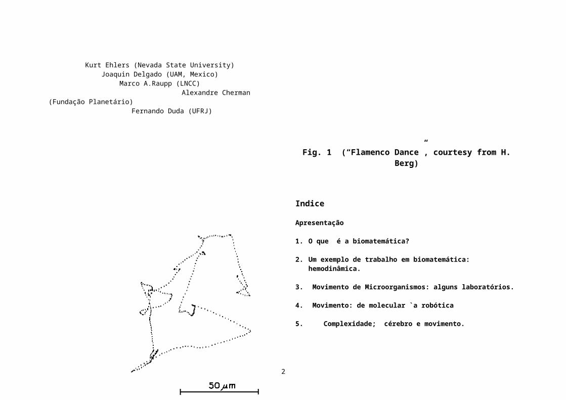
Fig. 1 (“Flamenco Dance”, courtesy from H. Berg)
Indice
Apresentação
1. O que é a biomatemática?
2. Um exemplo de trabalho em biomatemática: hemodinâmica.
3. Movimento de Microorganismos: alguns laboratórios.
4. Movimento: de molecular `a robótica 5. Complexidade; cérebro e movimento.
6. Biomecânica e Robótica.
7. Roteiro de estudos em Biologia Molecular. Biologia celular do Citoesqueleto
8. Motores Moleculares.
9. Nanotecnologia e sua interface biológica.
Extras (em formato eletrônico; napagina do curso ou me solicitando)
10. On the gait of Animals (Aristotle). There's Plenty of Room at the Bottom (Richard P. Feynman) 11. Coleção de artigos; visualização: “galeria” de imagens e filmes
12. Movimento de Microorganismos: alguns aspectos matemáticos(Jair Koiller)
2
Apresentação
"Biology is wet and dynamic" (Howard Berg )
Este minicurso tem dois objetivos. O primeiro é apresentar um rápido (portanto extremamente incompleto…) panorama de temas em movimento biológico que tiveram um extraordinário desenvolvimento nos últimos dez anos, em particular os “motores moleculares”. Assim, nossa meta será cumprida se for possivel motivar, complementarmente, o minicurso do Prof. Marcelo Magnasco "Problemas matematicos em Bioinformática". Recomendamos neste sentido ver antecipadamente seu web site www.asterion.rockefeller.edu/marcelo/marcelo.html,www.rockefeller.edu/labheads/magnasco/magnasco.htmOutras informações sobre bioinformática podem ser vistas no site de Laura Landweber http://www.molbio.princeton.edu/faculty/landweber.html:
Hoje em dia os mecanismos moleculares do movimento biológico começam a ser entendidos. Como um matemático poderia iniciar-se nestes temas? O Prof. Berg nos recomendou: Molecular Cell Biology by Darnell, Lodish e Baltimore (Alberts et al., Essential Cell Biology, Garland 1998 é um pouco menos enciclopédico. Stryer's Biochemistry, 4th Ed., Freeman, 1995 também é excelente).
O segundo objetivo do minicurso é divulgar alguns resutados sobre o movimento de microorganismos (limite Stokesiano) e da zoo-biofluidinâmica (limite Euleriano). A biofluidinâmica (cujo maior expoente foi o Professor James Lighthill, recentemente falecido) também rege o movimento dos fluidos internos (sangue, secreções) nos animais. Diremos algo sobre o impacto da biologia na robótica (“zoobótica”). E’ extraordinário que, do ponto de vista matemático, a modelagem da natação e do vôo utiliza uma mesma equação diferencial parcial, a “equação de Navier-Stokes”. Um parâmetro importante nesta equação é o número de Reynolds ( Re ), que mede a razão entre as forças inerciais e as forças viscosas. Para os microorganismos, Re é tão pequeno que pode ser tomado = 0 e obtemos a “equação de Stokes”, uma equação linear bem mais simples. Poderosos métodos matemáticos permitem explicar os movimentos de ciliados e flagelados de vários formatos. No movimento de pássaros e peixes, que já fascinava Leonardo
da Vinci, Re é muito alto. Mas não podemos tomar diretamente a viscosidade nula, Re = infinito, o que forneceria a “equação de Euler”. Isto porque na vizinhança do corpo, devido a seus movimentos, aparecem efeitos de “camada limite” (boundary layer), criando vórtices e provocando turbulência, fenômenos de enorme complexidade. Eis uma bibliografia básica:
David B. Dusenbery, Life at small scale – the behavior of microbes, Scientific American Library, 1996.
Hans J. Lugt, Vortex flow in nature and technology, J. Wiley, 1983.
Stephen Childress, Mechanics of swimming and flying, Cambr. University Press, 1981.
James Lighthill, Mathematical Biofluiddynamics, Siam, 1975.
Howard Berg, Random walks in Biology, Princeton Univ. Press, 1993.
Muitas outras fontes você encontrará em minhas notas:
Microbiofluidinamica jair1.dvi jairdos3.dvi
Problems and progress in Microswimming, com Richard Montgomery eKurt Ehlers, J.Nonlinear Science 6, 507-541, 1996. probprog.dvi
Spectral methods for Stokes flows, com M.A.Raupp, J.D.Fernandez, K. Ehlers e R. Montgomery, Computational and Applied Mathematics, 17 :3, 343-371, 1998.
Efficiencies of nonholonomic locomotion problems, com J.D.Fernandez,
3

Reports on Mathematical Physics, 42: 1/2, 165-183, 1998.
Movimiento de microorganismos, La Gaceta de la Real Sociedad Matem\'atica Espanola, 2:3 1999, 423-445, 1999.
Low Reynolds number Swimming in two dimensions, com Kurt Ehlers, Alexandre Cherman, Joaquin Delgado, Richard Montgomery e Fernando Duda, Proc. HAMSYS98, World Scientific, editado por Ernesto Lacomba, 2000.mexico.dvi
A quantidade de temas biológicos que permitirão novos aportes matemáticos é enerme. Um tema não tratado aqui é a morfogênese, que permite abordagens clássicas tão diferentes quanto as de Turing, D'Arcy Thompson, e Thom.Se for preciso mencionar apenas um site com os principais temas da Biologia Matemática na atualidade, recomendo o do ano especial na Universidade de Minnesota (www.ima.umn.edu; olhar workshops 98-99) Para se procurar bibliografia em biociencias, o site mais detalhado é o Medline, em www.cos.com (Community of Science). Como fonte geral, o site da Enciclopédia Britanica, www.britannica.com .
Fig.3 (Mysterious swimmer)
The cyanobacteria Synecococcus is 2 microns long and 1 micron wide, swims fast at 25 microns/sec. Kurt Ehlers’ model is deceivingly simple. Waves propagate tangentially along the membrane. The resulting motion of the organism is (counterintuitively) in the same direction. This hypothesis is presently being under investigation.
Cyanobacteria are important in photosynthesis. Some can produce toxic substances in drinking water supplies (See http://bilbo.bio.purdue.edu/www-cyanosite.)
4

“Esto tenia de ser por oposiciones?”
O que é a biomatemática?
Para termos uma idéia das muitas areas de atuação da Matemática na Biologia, podemos examinar o programa de alguns workshops no ano espacial realizado entre 1998 e 1999 no Institute for Mathematics and its Applications, University of Minnesota ( www.ima.umn.edu ; ver em “Program for 1998-99”)
Os worshops 5,6,12 são de especial interesse para nosso curso.
Os organizadores foram: Lisa Fauci , Tulane University; Simon A. Levin, Princeton University; James D. Murray, University of Washington; Alan Perelson (Chair), Los Alamos Natl. Lab.; Michael Reed, Duke University. “Significant applications of mathematics to biology have occurred for nearly a century, starting from the early work of Vito Volterra and Alfred Lotka oninteracting populations, and maturing through fundamental work in population genetics (Haldane, Fisher, and Wright), epidemiology (Ross, Kermach and MacKendrick), development (Turing) and neurobiology (Hodgkin and Huxley,
Fitzhugh and Nagumo, McCulloch and Pitts). Much of this research stimulated important contributions by other mathematicians (Kolmogorov, Petrovsky, Piscunox, Karlin, etc.); in general, however, until the past 10--20 years, communication between mathematicians and biologists remained problematical; much work in mathematical biology was relatively sterile, unsullied by contact with data, while experimental work suffered from a lack of theoretical generality.
The situation has changed dramatically in the past decade or so. Today's biologists are, in many areas, very sophisticated mathematically; mathematicians have learned the importance of becoming immersed in data; and the spectrum of practitioners has filled in, providing a continuum of highly mathematical work to collaborations. New and exciting areas (e.g. molecular biology, epidemiology and immunology) have opened up to mathematical investigations. A century of research has elucidated fundamental mechanisms in evolution, collective phenomena and pattern formation, and laid the foundations for more specialized modeling; and the development of new computational tools has greatly expanded the potential both for fundamental studies and for communications.
Thus the time is right for this special year at the IMA, built upon a selected series of workshops highlighting some of the mathematical challenges emerging from the consideration of biological issues, and endeavoring to show how the mathematics can be applied to the resolution of those issues. This program focuses on some particularly rich areas of investigation, complementing activities which have been carried out at the IMA in MRI, molecular biology and neurobiology in earlier years.”
September - December, 1998: Theoretical Problems In Developmental Biology and ImmunologyTutorial: Mathematical and Computational Issues in Pattern Formation, September 3-4, 1998 Workshop 1: Pattern Formation and Morphogenesis: The Basic Process, September 8-12, 1998 Workshop 2: Pattern Formation and Morphogenesis: Model Systems, September 14-18, 1998
5
Tutorial: Immunology, Cell Signaling, the Physiology of the Immune System and the Dynamics of the Immune Response, October 8-9, 1998 Workshop 3: Immune System Modeling & Cell Signaling, October 12-16, 1998 Period of Concentration: Forging an Appropriate Immune Response as a Problem in Distributed Artificial Intelligence, October 19-23, 1998 Tutorial: Mathematical Models of AIDS, November 6, 1998 Workshop 4: Dynamics and Control of AIDS, November 9-13, 1998 Minisymposium: Cancer, November 15-19, 1998
January - March, 1999 Mathematical Problems in Physiology***** Workshop 5: Cell Adhesion and Motility, January 4-8, 1999 ***** Workshop 6: Computational Modeling in Biological Fluid Dynamics, January 25-29, 1999 Workshop 7: Membrane Transport and Renal Physiology, February 8-12, 1999 Tutorial: Endocrinology: Mechanism of Hormone Secretion and Control, February 14, 1999 Workshop 8: Endocrinology: Mechanism of Hormone Secretion and Control, February 15-19, 1999 Tutorial: Audition, March 5, 1999 Workshop 9: Audition, March 8-12, 1999
April - June, 1999 Dynamic Models of Ecosystems and EpidemicsWorkshop 10: Local Interaction and Global Phenomena in Vegetation and Other Systems, April 19-23, 1999 "HOT TOPICS" Workshop: Challenges and Opportunities in Genomics: Production, Storage, Mining and Use April 24-27, 1999 Tutorial: Introduction to Epidemiology and Immunology, May 13-14, 1999 Workshop 11: Mathematical Approaches for Emerging and Reemerging Infectious Diseases, May 17-21, 1999 ***** Workshop 12: From Individual to Aggregation: Modeling Animal Grouping, June 7-11, 1999 "HOT TOPICS" Workshop: Decision Making Under Uncertainty:Energy and Environmental Models, July 20-24, 1999.
Links para Biologia Matemática
Sociedades Científicas
Sugiro uma visita inicial ao site da Society for Mathematical Biology ( http://www.smb.org).Em http://www.smb.org/sites.shtml você encontrará uma grande quantidade de links!
“Our society is committed to attracting members from all countries, and from a wide spectrum of interests. We hope to be able to provide a forum for discussion of research in biology, mathematical biology, and mathematics applied to or motivated by biology. Interdisciplinary research such as biophysics, computational biology, and many other similar realms are a growing part of our mandate. We hope that you will help broaden our horizons with your particular approach or research interests by joining thesociety and participating in our meetings.”
No site da Biophysics Society, você encontrará todas as informações biológicas de interesse.http://www.biophysics.org/biophys/society/biohome.htmEm particular recomendamos a coleção de monografias “on line”http://biosci.umn.edu/biophys/OLTB/Textbook.htmlEis alguns dos temas que se pode encontrar: Bioenergetics and Photosynthesis , Cell Biophysics , Channels, Receptors and Transporters, Computational Biology, Electrophysiology, Intermolecular Forces, Kinetics of Biological Systems , Membranes, Muscle and Cell, Contractility, Nucleic Acids, Photobiophysics, Proteins, Supramolecular Assemblies, Diffraction and Scattering, EPR, NMR , Separations and Hydrodynamics, Sequence Analysis, Single Molecule Techniques, Spectroscopy, Thermodynamics, Becoming a Biophysicist, Teaching Biophysics - published in Biophysical Journal , BJ Supplements and Computer Programs
6
Outras sociedades científicas das mais importantes são: American Physics Society www.aps.org, o American Institute of Physics, www.aip.org, American Mathematical Society, www.ams.org e a Society for Industrial and Applied Mathematics www.siam.org. Visitando estes sites, ficará claro o enorme interesse que a biologia tem despertado.
A http://www.biolinks.com/ uma organização que se define como uma “Internet Search Engine Designed by Scientists, for Scientists”.
Sites em Universidades podem ser vistos diretamente. Para mencionar apenas um, sugerimos http://www.mcb.harvard.edu/BioLinks.html, em Harvard.
Para não omitimos todos os sites nas universidades brasileiras e espanholas, mencionamos o departamento de Biomatemática da Universidade Complutense de Madrid: http://www.ucm.es/info/matbio/
Outros Links educacionais
Um site diretamente accessivel a alunos e professores de secundário, é “Mathematical Biology pages de Brandeis University” (evolução, resposta de sinapse, predador-presa, e muito mais!)www.bio.brandeis.edu/biomath/top/html http://www.bio.brandeis.edu/biomath/menu.html
No UTK Mathematical Life Sciences Archives home page: http://archives.math.utk.edu/mathbio/ você encontrará vários cursos preparados.
E’ uma coleção de “Life Science Pages” organizada por E. Dobos, L. Gross and C. Zimmermann na University of Tennessee, com o apoio da National Science Foundation Undergraduate Course and Curriculum Program.
Tutoriais em Biologia estrutural, produzidos pelo National Institute of Health, USA: http://cmm.info.nih.gov/modeling/tutorials.html
Para história da Física, recomendamos: http://www.aip.org/history/Em http://www.aip.org/history/web-link.htm há links para história de outras ciências.
Em http://www.nobelprizes.com/ e em www.nobel.se você encontrará biografias dos detendores de premios Nobel.
Para história da Matemática: http://www-history.mcs.st-andrews.ac.uk/history/Mathematicians
http://www.petersons.com/ (site geral em assuntos educacionais)
Sites adicionais sobre a vida marinha e terrestre
Site do Marine Biological Laboratory: catálogo da zoologia marinha.http://www.mbl.edu/MRC/CATALOG/
Para quem está interessado em fazer um insetos e animais polizadores, veja o site do Smithsonian Institute, com lindas fotos de insetos e aves participantes deste importante aspecto da ecologia.web2.si.edu/pollinatorswww.si.edu/whatsnew/body_whatsnew.htm
7
www.smithsonianmag.si.edu/smithsonian/issues00/apr00/pollen.htmlwww.pollinators.com/http://www.geocities.com/~pollinators/
“The coevolution of flowering plants and their pollinators has filled the earth with a diversity of life-forms: a quarter-million species of plants, and almost as many animal pollinators, including at least 1,200 vertebrates. The range of pollinators is staggering — in addition to birds, bees and bats, plants rely on such creatures as beetles, butterflies, ants, spiders, earthworms, parrots, even a New Zealand gecko and the pygmy gliding possum of Australia.”
Um exemplo de trabalho em biomatemática: hemodinâmica
Um tema tradicional da biomatemática é o estudo do fluxo sanguíneo (hemodinâmica). Não trataremos deste tema nestas notas, mas apenas mencionamos o o trabalho do grupo do Prof. Charles S. Peskin, do and Courant Institute of Mathematical Sciences, com quem colabora meu colega Alexandre Roma, da Universidade de São Paulo. Sites relevantes são (incluindo outras áreas de trabalho):http://www.cns.nyu.edu/associates/Peskin.htmlhttp://www.psc.edu/science/Peskin/CWSA.htmlhttp://www.psc.edu/science/Peskin/Boundary.html
“ Charles Peskin, David McQueen, and their group at the Courant Institute of Mathematical Sciences of New York University are well known for their heart modeling. Their model, originally in 2D but now 3D, combines tissue and fluid mechanics and permits the examination of the performance of modeled artificial heart valves for any of the four valves of the human heart. The two-dimensional model, in fact, led to a patent on such a valve.
It was the advent of the NSF supercomputer centers, Peskin says, that permitted his group to extend their computations from two to three dimensions. The calculation and examination of a single heartbeat in the present 3D model has taken approximately one week of CPU time on a single PSC C90 processor. When
the code was parallelized recently to use all 16 processors, Peskin and McQueen obtained a speedup of a factor of 10. The group is currently reorganizing the code to take advantage of domain decomposition methods and to make use of the powerful IBM SP-2, Cray T3D, and similar machines.
Interestingly, Peskin notes, both the original 2D model and the first 3D model had defects corresponding to human mitral valve prolapse, a common but non-life-threatening heart murmur, and the models had to be corrected to simulate a nondefective heart. "That the models got 'sick' the way humans do is a kind of validation of the approach," Peskin says.
An important application of the model will be to simulate valve action, and a complete run of several heartbeats will be used to determine whether a given valve design creates any regions of stagnation (low-velocity flow) in the circulating blood. Such regions can nucleate the formation of clots, and clot formation can threaten both the valve action and (if the clot breaks free) other regions of the circulation. Clot formation is, in fact, the chief risk associated with artificial heart valves.
High resolution is also needed to make the Reynolds number (Re) realistic. To use a realistic Re for blood flow in the heart direction. Fortunately, the flow pattern of blood in the heart is not very sensitive to the Reynolds number, and improvements in numerical methodology, such as local mesh refinement near boundaries or the use of entirely grid-free methods, may make it possible to avoid the extreme computational requirements implied by such a refinement of a uniform grid. Nevertheless, a fully satisfactory computation of blood flow in the heart will require a substantial increase in computer power, Peskin says--possibly as great or greater than the increase that was needed to move from two to three dimensions.
Increased computer power is needed not only to do the current computation more correctly, but also to bring in additional phenomena that are highly relevant to blood flow in the heart. Two examples of such phenomena are the electrical activity that coordinates and controls the heartbeat and the dynamics of the blood clotting process, which is important in evaluating the function of prosthetic cardiac valves. Models of these phenomena are being developed separately from
8

the model of cardiac mechanics described here. Microscopic and macroscopic models of the clotting process are being developed by Aaron Fogelson, a former student of Peskin's who's now at the University of Utah. Ultimately, Peskin and McQueen hope to combine such models with their mechanical model to increase its realism and predictive power. Again, a dramatic increase in computer power would be required.
"My situation is absolutely typical," Peskin insists. "Almost any time-dependent problem in three space dimensions pushescurrent computational capabilities to their limits. Inadequate resolution can lead to incorrect conclusions and to inappropriate attempts to 'fix' a model that isn't really broken."
Movimento de Microorganismos
Para dar uma pequena idéia desta área, começamos um um artigo que nós escrevemos para o livro World Mathematical Year, editorial Carrogio, Barcelona, a convite do Prof. Manuel de Leon, CSIC/Madrid, a aparecer em breve.
“Four decades ago, Richard Feynman anticipated research on Micro-robots in an amusing talk called “There’s plenty of room in the bottom”. He was not joking that time. Engineers, Mathematicians and Biologists are teaming together to make them (see Science, April 7 2000).
Industrial Robots or Automatons are machines controlled by primitive brains, whose software uses the theory of Cellular Automata. There are many lessons to be learned from microscopic living beings, which we can call “cellular robots”. We chose a confusing title on purpose because “celulas automatas” are also “automatas celulares”.
How does one describe the motile behavior of microorganisms? How much do they move on average? Professor Howard Berg, from Harvard, is one of the leaders in this field of study. For him and many others these questions are as fascinating today as they where to Anton van Leeuwenhoek in 1676. A bacterium’s cycle lasts about 20min. As in a James Bond movie, it’s an adventurous life: they are subject to strong Brownian forces in the aqueous environment and enemies are everywhere. But there are compensations: their sexual life is active and interesting. As Stephen J. Gould says, for bacteria, we humans are just some big mountains full of goodies. They have been around for almost 4 billion years. Humans appeared about 1 million years ago (civilization exists for 10 thousand years only).
My coworkers (Kurt Ehlers and Richard Montgomery) and I have some mathematical models for microorganism locomotion (see our article Movimiento de microorganismos, Gaceta Matematica, Noviembre 1999). Viscosity dominates over inertia (“zero Reynolds number”): only geometry matters. Motion of the body in the fluid is an indirect result of shape changes. It is a “gauge theory” – like the theory a cat needs to know to fall upright (see Richard Montgomery’s chapter). Fix a particular configuration of an isolated organism, and first consider a trial “ boundary condition” V along the located boundary of the body, corresponding to a given shape deformation. There is an unavoidable ambiguity: V plus an infinitesimal rigid motion X (consisting of infinitesimal translation and rotation) is as good as V because the intrinsic shape deformation is the same. In self-propulsion, the correct V is picked out of the collection of V(trial)
9

+ X by imposing the constraint that it induces no net force or torque on the fluid. The “scallop paradox” states that a one degree of freedom organism or robot swimming at zero Reynolds number goes nowhere. We extended the theory to “social life” at low Reynolds number. Then two can swim fine by cooperating.
Says Berg: “biology is wet and dynamic”. Subcellular organelles of eukaryotes, just as the whole cell, are immersed in an aqueous environment. Everything is subject to very strong thermal fluctuations. Inside the cell there is a “railroad system” formed by microtubules runnig in all directions. Proteins are transported along by specialized molecules, kinesins, acting as “cargo vans”. Kinesin uses the ATP (adenosine tri-phosfate, the biological fuel) hydrolysis to drive powerstrokes. Such “molecular motors” are being intensively studied. Other motions, like protein translocation across membranes may be driven by a “ratchet” mechanism (Feynman would like this idea). Here Brownian energy is “tamed” (rectified) with the help of chemical reactions at favorite places.”
A seguir, descrevemos o trabalho de três grupos de pesquisa nesta área.
Grupo do Prof. Howard C. Berg, (Professor, Molecular and Cellular Biology, Harvard University, http://www.mcb.harvard.edu/Faculty/Berg.html e pesquisador do Rowland Intitute of Science.
O Prof. Berg é o nosso “guru”. No site www.rowland.org, você encontrará informações sobre o trabalho do grupo do Prof. Berg em comportamento e motilidade das bactérias; e filmes em QuickTime.
“ We study bacteria, the simplest free-living single-celled organisms. We are interested in how they sense changes in their environment, analyze sensory data, and respond in a purposeful manner. Our quest is an understanding of behavior at the molecular level. Our primary subject is the peritrichously-flagellated organism
Escherichia coli. We are trying to learn how its flagellar motors work, how their directions of rotation are controlled during responses to chemical stimuli (chemotaxis), and what effect that rotation has on modes of flagellar propulsion. We have worked on other bacteria that swim without flagella (a cyanobacterium, Synechococcus) and are beginning work on bacteria that glide across surfaces (Mycoplasma, which moves by an unknown mechanism, and Pseudomonas aeruginosa, which uses thin fibers called type IV pili).”
Estas são portanto as áreas de trabalho: “Bacterial Motility and Behavior: Motion of fluorescent flagellar filaments, Bleaching of components of fluorescent flagellar motors, Directions of rotation of the flagellar rotary motor at high speed, Gliding motility in Mycoplasma, Gliding motility generated by type IV pili”.
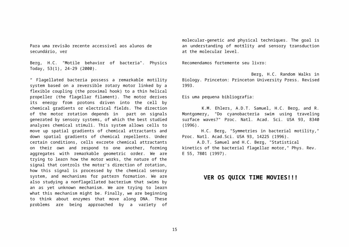
Para uma revisão recente accessivel aos alunos de secundário, ver Berg, H.C. "Motile behavior of bacteria". Physics Today, 53(1), 24-29 (2000).
“ Flagellated bacteria possess a remarkable motility system based on a reversible rotary motor linked by a flexible coupling (the proximal hook) to a thin helical propeller (the flagellar filament). The motor derives its energy from protons driven into the cell by chemical gradients or electrical fields. The direction of the motor rotation depends in part on signals generated by sensory systems, of which the best studied analyzes chemical stimuli. This system allows cells to move up spatial gradients of chemical attractants and down spatial gradients of chemical repellents. Under certain conditions, cells excrete chemical attractants on their own and respond to one another, forming aggregates with remarkable geometric order. We are trying to learn how the motor works, the nature of the signal that controls the motor's direction of rotation, how this signal is processed by the chemical sensory system, and mechanisms for pattern formation. We are also studying a nonflagellated bacterium that swims by an as yet unknown mechanism. We are trying to learn what this mechanism might be. Finally, we are beginning to think about enzymes that move along DNA. These problems are being approached by a variety of molecular-genetic and physical techniques. The goal is an understanding of motility and sensory transduction at the molecular level.”
10

Recomendamos fortemente seu livro:
Berg, H.C. Random Walks in Biology. Princeton: Princeton University Press. Revised 1993.
Eis uma pequena bibliografia:
K.M. Ehlers, A.D.T. Samuel, H.C. Berg, and R. Montgomery, "Do cyanobacteria swim using traveling surface waves?" Proc. Natl. Acad. Sci. USA 93, 8340 (1996). H.C. Berg, "Symmetries in bacterial motility," Proc. Natl. Acad.Sci. USA 93, 14225 (1996). A.D.T. Samuel and H.C. Berg, "Statistical kinetics of the bacterial flagellar motor," Phys. Rev. E 55, 7801 (1997).
VER OS QUICK TIME MOVIES!!!
Grupo do Prof. Charles J. Brokaw, Professor of Biology, Caltech: Computer simulation of biological movement
http://broccoli.caltech.edu/~biology/brochure/faculty/brokaw.html
“My work seeks to understand the mechanisms of motility of eukaryotic flagella and cilia. Specifically, I want to understand how the activity of the motor enzymes (dyneins) that cause sliding between the flagellar microtubules is regulated in order to produce particular patterns of bending.
Experimental work is carried out at the Kerckhoff Marine Laboratory, operated by the Division of Biology in Corona del Mar, about 60 miles from the Pasadena campus. This laboratory has facilities for maintaining sea urchins as sources of spermatozoa on a year-round basis, as well as access to spermatozoa from other marine invertebrates such as the tunicate, Ciona. We mostly use spermatozoa that have been demembranated with detergent and then reactivated with MgATP. This in vitro system provides an excellent system for precise measurements of the parameters of flagellar movement in response to various experimental manipulations. The measurements are made using high-speed photographic recording and computerized image analysis.”
Recomendo o filme que se encontra emhttp://www.cco.caltech.edu/~brokawc/Demo1/BeadExpt.html
“Demonstration of microtubule sliding in a beating flagellum. This sea urchin spermatozoon was treated with detergent, Triton X-100, to remove the cell membrane, and then transferred to a solution containing a relatively low concentration of MgATP, to reactivate the bending of its flagellum at a frequency of about 1.5 cycles per second. This spermatozoon was swimming freely, but the photographs have been repositioned to eliminate movement and rotation of the sperm head.”
Entre os links recomendados por Brokaw ,http://numbat.murdoch.edu.au/spermatology/spermhp.html, a “Spermatology home page”, onde se encontra alguma informação histórica sobre a percepção de Leeuwenhoek: http://zygote.swarthmore.edu/fert1.html
Entre os principais grupos desta comunidade, mencionamos http://www.bio.ph.kcl.ac.uk/
“The Biophysics Group is part of the Department of Physics at King's College London. It aims to establish the molecular mechanisms involved in sperm motility using an original approach based on computer modelling techniques with
11
corresponding experimental studies. The flagellum used to propel the sperm is a long slender organelle. The forces required to bend it are generated by molecular motors called dynein. Cilia, like flagella, are microscopic motile organelles which propel individual cells, and are also present in large numbers in the human lung to remove particulate matter. Malfunction of cilia and flagella can lead to respiratory and reproductive disorders, e.g. cystic fibrosis, primary ciliary dyskinesis and Kartagener's syndrome. Some diseases of commercially important crops, such as cocoa and potatoes are spread by flagellated cells. An understanding of the motor mechanisms of these organelles is therefore of medical and biological significance, as well as having commercial implications. The successful use of computer modelling in the study of dynein will also lead to its application in the investigation of the other families of biological molecular motors, myosin (involved in muscle contraction) and kinesin (used to transport cargo from one part of a cell to another), and to the study of molecular structure and function in a range of medical and biological systems”.
VER O FILME DO FLAGELO EM MOVIMENTO!!
Grupo do Prof. Raymond Goldstein (Arizona State University, Biophysics)
Você encontrará vários filmes interessantes na página: http://biophys.physics.arizona.edu/~gold
Este grupo trabalha na elastohidrodinamica de filamentos e membranas biológicas. Trabalham também em efeitos coletivos na chemotaxia. Sobre esta última área:
“We want to understand various macroscopic phenomena associated with morphogenesis in cellular populations. Using the prototype system {\it
Dictyostelium discoideum}, our goal is to develop quantitative and controlled experiments on the interplay between chemical signaling and collective behavior such as organized chemotaxis. It is known from a large amount of earlier experimental work on this system that collective chemotaxis occurs in response to coherent chemical waves either in the form of target patterns driven by autonomous pacemakers or as rotating spiral waves. Little quantitative information exists on the origin of the transition from signaling to collective chemotaxis, the symmetry-breaking which produces the pacemakers, or the factors that influence the selection of one of these patterns over another. Our initial experiments have shown that by varying the overall cell population density there is a transition from target-dominated to spiral-dominated behavior, suggesting a crucial role for the diffusive coupling of cells in the selection process. These studies also reveal an interesting entrainment dynamics of spirals and pacemakers. Experiments underway seek to understand more clearly the biochemical basis for the selection of wave patterns, and to probe the instabilities associated with chemotaxis in the presence of periodic waves.
Na página http://www.physics.arizona.edu/physics/newsletter/fall97/biophys.htmlEncontramos um depoimento do Prof. Goldstein:
“A Perspective on Biological Physics”: “In his classic work On Growth and Form, the great natural scientist D'Arcy Wentworth Thompson said, "Cell and tissue, shell and bone, leaf and flower, are so many portions of matter, and it is in obedience to the laws of physics that their particles have been moved, moulded and conformed... Their problems of form are in the first instance mathematical problems, their problems of growth are essentially physical problems, and the morphologist is, ipso fact, a student of physical science."
D'Arcy Thompson's attempts to understand the world of living things through physical reasoning anticipated by decades the current explosion of interest in the physics community of things biological. As we in the department begin our own search this year for a new faculty member in experimental biological physics, I thought it would be useful to give an overview of the whole field and to describe
12
some newly initiated projects in my group. The intellectual threads that are important in biological physics include statistical mechanics, kinetic theory, hydrodynamics, continuum mechanics, nonlinear dynamics and colloidal physics. As such, the field is not unlike astrophysics, where many different disciplines meet. Emerging techniques involving micromanipulation (e.g. optical traps), microlithography (to create structured environments), fluorescence methods (for direct visualization of living processes), and the like will all play an important role. An important question is: What can the discipline of physics bring to this area? Clearly, it is not productive for physicists to move into biology simply to copy the methods and adopt the paradigms of the biologists already there. To a much greater extent than is found in the biology or chemistry communities, physics is deeply connected with "model building" and testing, in which one makes contact with reality through a series of progressively more detailed physical and mathematical descriptions. At each stage, there is emphasis on consistency with established laws, introduction of dimensionless control parameters (related to time, length, energy, and force) to delineate the space of possible behaviors, establishment of scaling laws (expressing commonality or even universality of behavior), and some degree of logical and mathematical rigor regarding the results of those models.
On the experimental side, lessons from the study of phase transitions and critical phenomena as well as nonlinear dynamics are important: the identification of fundamental control parameters(even if this entails bringing the experimental system far from physiological conditions), development of in vitro model systems for living processes (e.g. the study of artificial lipid vesicle shapes and dynamics in order to understand cell membranes), and of course the development of new methods and technologies for these experiments. Finally, let me outline briefly some of the experimental research I have begun here on form and motion in biological systems. The first project is aimed at understanding the elastic and dynamic properties of bacterial filaments that undergo iterated supercoiling instabilities. These filaments are formed by mutants that exhibit failure of the separation of daughter cells, and supercoil as a consequence of twisting stresses built up in the cell walls through this process of growth. Many years of experimentation by Mendelson and collaborators established remarkableproperties of these filaments, whose handedness and dynamics can be tuned through external
solution conditions. Yet, no fundamental understanding of the origins of this phenomenon exists, and there is little quantification of the forces involved in the conformational evolution. Besides the biological relevance of this work, this
system serves as a "laboratory" for the study of very general problems in nonlinear dynamics and differential geometry.
To begin an investigation of these issues, we have built laser tweezers to manipulate the filaments in order to measure basic elastic properties and have a preliminary measurement of the elasticity of a bacterial wall that we hope will help us understand this phenomenon in detail.
The second effort, in collaboration with Prof. John Kessler (Physics) involves the study of the biofluiddynamics of individual and collective bacterial motion. We seek to understand fundamental features of low Reynolds number hydrodynamics relevant to organismal motion. One phenomenon of interest is the bundling of bacterial flagella in cells with multiple flagellae. In E. coli, coherent rotation of the helical flagella in one of the two possible directions results in the formation of a tight bundle; rotation in the opposite sense makes them fly apart. These two modes of operation produce the so-called "run-and-tumble" dynamics of bacterial
13

chemotaxis. Yet there is very little understanding of this bundling process and the hydrodynamic forces involved. There is also scant attention paid to the possible relevance of long-range fluid stirring from flagellar motion on nutrient uptake. We propose experiments using high-speed video microscopy and particle tracking methods to address these issues as well as long-range hydrodynamic interactions that may produce collective swimming behavior.
A third project will explore the physics of actin-based motility. In many cells motility is achieved through motion of the cytoskeleton, a cross-linked network of actin filaments. The extension of pseudopods and lamellipods proceed apparently through inhomogeneous polymerization and depolymerization of the actin network, but there is little quantification of the resultant forces; I propose to develop an in-vitro model system involving actin encapsulated in synthetic lipid vesicles to attempt to understand the essential aspects of this process. By controlling the polymerization and depolymerization of the actin network with fine spatial resolution, it may be possible to probe the motive forces generated by inhomogeneous polymerization coupled to an elastic membrane.
Fig.4 (Patterns obtained by Elena Budrene in H. Berg’s laboratory.) Under certain conditions, bacteria excrete chemical attractants on their own and respond to one another, forming aggregates with remarkable geometric order. What are the mechanisms of pattern formation?.
Movimento: de molecular `a robótica
Número especial de Science, vol 288, Number 5463, 7 Apr 2000 , “Movement: molecular to robotic”
On the Move, Lisa Chong, Elizabeth Culotta, and Andrew Sugden
“ From the trafficking of intracellular cargoes to the flight of the wandering albatross, movement is one of life's central attributes. All organisms have a three-dimensional spatial context that is both internal and external; directed, organized motion, which creates as well as operates within this context, has been a prime target of natural selection from the dawn of evolutionary time. This special issue examines the principles of movement as expressed at scales ranging from the subcellular to that of individual organisms, and in robots as well as in living creatures.
At the root of all existence lies movement at the subcellular and molecular levels. Recent studies of molecular motors such as the proteins myosin and kinesin, which are not only responsible for muscle movement but are also involved in processes such as cell division and membrane transport, have revealed how underlying similarities at the molecular structural level can generate a diversity of functional properties and mechanisms of converting chemical to mechanical energy. These findings are reviewed on page 88 by Vale and Milligan. These molecular analogs to motors are not the only way of controlling and organizing the movement of material within cells. As reviewed by Mahadevan and Matsudaira (p. 95), much has been learned about the polymeric analogs to other mechanical systems, especially springs and ratchets. Such systems mediate another sizable cross section of cellular activities, including fertilization, which can be governed by the dynamics of actin filaments in some instances.
Of course, whole cells move too, with profound consequences for a host of physiologic processes, including development, when cells traverse the embryo to find their correct places. In a News story (p. 86), Gretchen Vogel reports that developmental biologists, long focused on genetic signals rather than cell movements, are at last catching glimpses of the molecules that promptcells to move or stay still at crucial moments in development.
At the level of the individual animal, the wealth of solutions to the problem of getting from point A to point B--swimming, running, jumping, flying, diving--has been keenly explored by natural historians and biomechanicists for centuries. Yet the past decade has been a particularly vigorous period for biomechanics,
14
propelled not only by an unprecedented harnessing of computing power and the runaway development of robotics, but also by a growing appreciation of the feedbacks between animal sensory and locomotory systems and of the parallels between different types of locomotion (see the Review by Dickinson et al., p. 100). Studies at the interfaces between behavior, physiology, and motion, employing ever more ingenious methods, are also yielding remarkable new insights. For example, the Report by Williams et al. (p. 133) reveals the stop-and-start behavior of dolphins and other marine mammals, including the world's largest animal, the enigmatic blue whale. A related News story (p. 83) by Elizabeth Pennisi discusses these findings, which are among the most dramatic of a series of recent discoveries concerning the diverse advantages gained by moving intermittently.
Mechanical creatures may also help illuminate the principles of motion, as reported in the News story by Gary Taubes (p. 80). As biologists learn from a new generation of robotic lobsters, moths, flies, and fish, roboticists are deliberately modeling their creations on natural principles. Not since the bird's wing gave us the airfoil has the application of natural knowledge in this area looked so promising.
The study of motion is one of those rare and challenging disciplines that draws together the biological, the chemical, and the physical, and it holds endless fascination for the pure and applied scientist alike. We can predict many fruitful collaborations in the future and expect many surprises.”
Este artigo, que abre a seção especial sobre Movimento Biológico, despertou vários comentários posteriores. Eis alguns:
“ The interesting array of articles for the special issue "Movement: Molecular to Robotic" (7 Apr., pp. 79 106) examines the biology of movement for animals with endoskeletons, animals with exoskeletons, robots, and molecules, but movement in an entire kingdom--the plant kingdom—is overlooked. Movement in plants is based on physical mechanisms that are very different from most animal movements, and movements have been a central factor in the evolution of many
plant adaptations. Mechanisms include hydraulic shifts operating by means of osmotic engines (1), differential growth, fracturing of structures dueto localized desiccation, and cell separations or dissolution leading to projectile actions.
Examples of movements based on hydraulic shifts include the cyclic flapping of leaves and the opening and closing of some petals and flowers. Cells on one side of a petiole, pulvinus, stem, or pedicel will lose water, whereas cells on the opposite side will maintain or even increase turgidity, and movement of leaf, stem, or flower will occur. These osmotic engines are driven principally by ionic pumps across the cell membrane that usually transport K+ or Ca2+ (or both), and in some cases, such as the leaf folding in the sensitive mimosa plant, an action potential is involved.
Examples of movement by differential growth on opposite sides of a plant organ include phototropism, gravitropism, the opening and closing of some flowers, and the coiling of stems and tendrils. In the case of the stilt palm (Iriartea gigantea) (2), the trunk is held aloft on supporting brace roots, and movement of the tree toward a light-gap can occur by differential growth of the brace roots on the lighted side and abandonment of such roots on the shaded side.
Another group of movements involves localized cell degradation to produce a throwing action. Such movements include the explosive projection of pollen, spores, or seeds, which occur when tension is released in a cluster of cell walls that have degraded, resulting in a catapult-like action.
Movement can also occur by negative growth. Below-ground bulbs or stems can move down to a desirable depth by means of contractile roots. These roots have been found in more than 90% of the plants studied (3). They become thickened and develop extensive wrinkles as the root contracts. The irreversible contraction effect is achieved by a combination of thickening and shortening of individual cells along the root axis, combined with programmed cell death of layers of cells in the root cortex.
15

And finally, there is a large array of thigmo movements, movements stimulated by touch (mechanical perturbation) (4). The coiling of tendrils around an object involves two component movements (5): an initial movement (involving a contractile adenosine triphosphatase) resulting in hydraulic changes that lead to rapid coiling, followed by differential growth on opposing sides of the tendril, which allows the tendril to continue coiling around the support. There is evidence that such thigmo coiling movements involve the classic stimulus-response syndrome, including the opening of calcium channels and a probable sequence of phosphorylations.
Another example of thigmo movement is the motor pulvinus of the sensitive mimosa plant (Mimosa pudica) (6). When a leaf is touched, the signal is transmitted to the base of the leaf by a lever action. An action potential is generated. Special structures called tannin vacuoles in the motor cells at the base of the leaf, analogous to the sarcoplasmic reticulum in muscle sarcomeres, release Ca2+ into the cytoplasm. This causes actomyosin microfilaments to contract, opening putative potassium channels in the plasma membrane. The K+ effluxes, drawing water after it by the osmotic engine mechanism. The turgor of these cells thus decreases, causing the leaf to bend downward as a result of the turgor differential between the upper and lower sides. Action potentials then travel up and down the stem, activating motor cells at the bases of other leaves.
In conclusion, the various movements performed by plants are special in that they are primarily based not on contractile proteins, but on physical and cellular alterations. The wide array of repositioning movements and adaptations for reproductive effectiveness has evolved principally as components of photosynthetic and reproductive strategies.
A. Carl Leopold, Mordecai J. Jaffe, Boyce Thompson Institute of Plant Research, Ithaca, NY 14853, USA
1. S. Vogel, Life's Devices: The Physical World of Animals and Plants (Princeton Univ. Press, Princeton, NJ, 1988), pp. 255-263.
2. P. H. Allen, The Rain Forests of Golfo Dulce (Stanford Univ. Press, Stanford, CA, 1977). 3. H. De Vries, Land. Jahrbuch. (Ver. Wiegandt, Hempel & Pasrey, 1880); A. Rimbach, Beit. Wissenschasft. Botanik 2, 1 (1898); Ber. Deuts. Bot. Ges. 44, 328 (1926). 4. M. J. Jaffe, Encycl. Plant Physiol. 11, 444 (1985). 5.------ and A. W. Galston, Plant Physiol. 41, 1014 (1966); Plant Physiol. 42, 845 (1967); Plant Physiol. 43, 537 (1968). 6. H. Toriyama, Cytologia 20, 367 (1955); ------ and M. J. Jaffe, Plant Physiol. 49, 72 (1972); H. M. Turnquist et al., Protoplasma 176, 91 (1993); T. Sibaoka, Symp. Soc. Exp. Biol. 20, 49 (1966).
In the "Movement" special issue, the News article "In nature, animals that stop and start with the race" by Elizabeth Pennisi (7 Apr., p. 83) misses one of the neatest examples. Once fish discovered the energetic advantages of "burst-and-coast" swimming (D. Weihs, J. Theor. Biol. 48, 215,1974), they also discovered the advantage of coasting above the surface of the water, to minimize drag and increase the distance covered by the coasting phase--hence, "flying fish." This was also discovered (independently) by flying squids, which inherently have a burst-and-coast mode of locomotion. Charles J. Brokaw, Division of Biology, California Institute of Technology.
I was writing up notes from Gary Taubes' News article, "Biologists and engineers create a new generation of robots that imitate life" (Science, 7 Apr., p. 80). Taking the notes was a bit troublesome because of the lack of a convenient term to categorize robots based on cockroaches, spiders, snakes, fish, and so on. The descriptive phrase "zoologically inspired robots" worked, but was a bit clumsy. The answer was then obvious--"zoobots” (Greg Goebel) .
16
Fig. 5. Muybridge, Eadweard (1830-1904) http://www.kingston.ac.uk/muytext0.htm Muybridge was a young man when he left Kingston for America. The exact year of his departure is uncertain, but by 1856 he was established as a bookseller and a publisher's agent in San Francisco, trading under the name EJ Muygridge. In 1860 he was injured in a stage coach crash whilst travelling overland from San Francisco to New York for a visit to Europe. Muybridge returned to America in about 1866 to become a professional photographer.
Complexidade
Science, vol. 284, 2 de abril de 1999, “Sistemas Complexos” , p. 79-109.p.80:Células virtuais (artificial life) : www.e-cell.org, Masanu Tomita e seu grupo de bioinformática da Keio University, Leslie Loew U.Connecticut, Farmington; www.nrcam.uchc.edu .
p.82. “Unraveling Bacteria's dependable homing system”: o sistema contem diversas sub-unidades: i) para "cheirar" a fonte dos nutrientes; ii) para mover os flagelos; iii) para transmitir os sinais. Como este sistema é integrado? Como garantir sua robustez num meio variável? Há um padrão independente das características dos diferentes organismos? Stanislas Leibler (Dept Biol. Molecular) e Naama Barkai, (Princeton) : equações diferenciais para a taxia randomica da E.coli.
p. 99-101 J.K. Parrish (Zoologia, U. Washington Seattle) e L.Edelstein-Keshet (Math, U. British Columbia, Canada), “Complexity, pattern, and evolutionary trade- offs in animal agregation”: modelos matemáticos de agregação, estratégias para a proteção do coletivo contra predadores.
Science, vol 284, 21/05/99 Microbe management “Sinfonia das bacterias”, pg. 1302, por E.Strauss. As bactérias podem comunicar-se com membros de sua e de outros espécies, permitindo-lhes coordenarem suas atividades. Algumas requerem um grupo para que possam ter impacto: se voce está sozinho num grande auditório, não é escutado; mas se está num coro, as vozes se juntam. Os micróbios sabem que há poder na união. Algumas vezes os microbios se tornam virulentos. Outras vezes, estas atividades são benéficas. Segundo Bonnie Bassler (http://www.molbio.princeton.edu/faculty/bassler.html), “ as bactérias querem fazer algumas coisas quando estão sozinhas e outras quando estão numa comunidade … Respondendo a uma maior densidade populacional lhes permite mudar para um novo conjunto de tarefas micróbios quanto para as criaturas que eles habitam. A bactéria luminescente Vibrio Fischeri vive num órgão especializado em animais como o Hawaian bobtail squid. Quando há um númerosuficiente de bactérias, a luz é “ligada”, eliminando a sombra e a lula nada a luz lua.
17

Movimento e Cérebro
Nestas notas, evidentemente não poderemos abordar (principalemente por sermos meros curiosos ) a parte mais fascinante (a nosso ver) da biologia atual, os processos de enorme complexidade no sistema nervoso central. Em particular, as atividades do cérebro ligadas ao movimento. Neste tema, apenas mencionamos dois exemplos.
De Susan A. Greenfield, The Human Brain, Basic Books, 1997. “Charles Sherrington, um dos grandes pioneiros da fisiologia durante a primeira metade do século 20, resumiu a contribuição pervasiva do movimento em nossas vidas: ‘desde um sopro na floresta até a queda de uma árvore, tudo é movimento’. Das sutilezas da linguagem corporal, à precisão da palavra, e à total desambiguidade de um abraço, virtualmente toda comunicação depende do movimento.
Embora as plantas podem se mover pois voltam-se para a luz, elas não podem gerar movimento como nós. For a da ficção cientifica, nenhuma planta se locomove de um lugar para outro. Em contraste clarissimo, todos os animais estão em movimento – ou seja, eles são animados. Interessantemente a palavra Latina animus significa `consciencia’.
Se voce é um organismo multicelular e está em movimento, então você tem, pelo menos um tipo primitivo de cérebro. A importância para as criaturas que se movem terem um cérebro é muito bem ilustrada por uma observação feita inicialmente pelo falecido Imperador Hirohito do Japão, para quem a vida marinha era um hobby apaixonante. O unicado em questão se chama sea squirt.
Quando ainda é uma larva imatura, o sea squirt passa o tempo nadando por aí: não apenas é capaz de fazer movimentos coordenados, como também possui um sensor primitivo de vibrações, grosseiramente comparável com um opuvido, e um sensor primitivo de luz, grosseiramente comparável com um olho. De fato, o pode se dizer que o sea squirt possui um cérebro modesto. Porém, quando se torna
maduro, o sea squirt muda de estilo de vida e se prende a uma pedra. Ele não precisa mais de nadar por aí, porque ele agora vive filtrando a água do mar. Webster: sea squirt any tunicate, esp. a sessile ascidian, so called from its habit of contracting its body and ejecting streams of water when disturbed. 1. tunicate Zool. any sessile marine chordate of the subphylum Tunicata (Urochordata), having a saclike body enclosed in a thick membrane or tunic and two openings or siphons for the ingress and egress of water.
2. sessile: l. permanently attached; not freely moving.[1715–25; < L sessilis fit for sitting on, low enough to sit on, dwarfish (said of plants), equiv. to sess(us) (ptp. of sedre� to SIT1) + -ilis -ILE]
Nesta etapa, o sea squirt na verdade realiza um ato notável: consome seu próprio cérebro!
A pista para a finalidade do cérebro dada por esta estória é que você só precisa de um cérebro se voce está se movendo. Para formas de vida estacionárias, um cérebro não é mais necessário. O ponto todo é que para um animal se movendo, existe uma interação com o meio que está mudando incessantemnete. Voce precisa de um equipamento para lhe dizer muito ràpidamente o que está acontecendo e, o que é mais importante, que lhe permita responder ao que está acontecendo, para escapar dos predadores ou para caçar as suas presas. Portanto o cérebro, de qualquer forma, tamanho e grau de sofisticação que seja, é ligado de forma fundamental para garantir a sobrevivência, tanto como consequencia tal como causa do movimento.”
Cérebro e sexo (ver Scientific American, vol. 283 nb 2, August 2000, : Pg. 56-61 Male Sexual Circuitry, Irwin Goldstein, Boston University)
“Quinhentos anos atrás, Leonardo da Vinci fez uma observação sobre o pênis que parece correta ainda hoje para muitos homens e suas companheiras. O scientista da Renascença – inventor e artista – um numa longa linha de investigadores que tentaram resolver a charada da rigidez peniana – observou que este orgão por vezes traiçoeiro parece ter vontade própria. “O pênis não obedece as ordens do seu mestre, que tenta enrigecê-lo ou encolhe-lo, ao contrário, o pênis se enrigece
18

livremente enquanto seu dono dorme. Pode-se dizer que o pênis tem sua própria mente”, escreveu ele.
Da Vinci, que dissecou inúmeros penis de cadáveres de homens executados por enforcamento, foi o primeiro cientista a reconhecer que durante a erecção, o pênis enche-se de sangue. Porém, na sua percepção de que o pênis possui vontde própria, este estudioso de muitos talentos estava equivocado.
Muito ao contrário de ter sua própria mente, como pensava da Vinci, sabe-se hoje que o pênis está sob completo controle do sistema nervoso central – o cérebro e a espinha dorsal…. As pesquisas tem demonstrado muitas semelhanças entre os sexos no papel do sistema nervoso central no controle da excitação, orgasmo e várias outras funções” .
Em resumo, o orgão sexual mais importante é … o cérebro.
Fig 6. No brain, no sex.
Biomecânica e Robótica
1. Informações históricas
Ao que se sabe, os primeiros estudos de biomecânica foram escritos cerca de 350 A.C por Aristoteles ( “On the Gait of Animals”, “The History of Animals” e “On the Motion of Animals” ; traduções para inglês podem ser encontradas em http://classics.mit.edu ). Do primeiros deles, extraimos o trecho inicial:
“We have now to consider the parts which are useful to animals for movement in place (locomotion); first, why each part is such as it is and to what end they possess them; and second, the differences between these parts both in one and the same creature, and again by comparison of the parts of creatures of different species with one another. First then let us lay down how many questions we have to consider. The first is what are the fewest points of motion necessary to animal progression, the second why sanguineous animals have four points and not more, but bloodless animals more than four, and generally why some animals are footless, others bipeds, others quadrupeds, others polypods, and why all have an even number of feet, if they have feet at all; why in fine the points on which progression depends are even in number.
Next, why are man and bird bipeds, but fish footless; and why do man and bird, though both bipeds, have an opposite curvature of the legs. For man bends his legs convexly, a bird has his bent concavely; again, man bends his arms and legs in opposite directions, for he has his arms bent convexly, but his legs concavely. And a viviparous quadruped bends his limbs in opposite directions to a man's, and in opposite directions to one another; for he has his forelegs bent convexly, his
19

hind legs concavely. Again, quadrupeds which are not viviparous but oviparous have a peculiar curvature of the limbs laterally away from the body. Again, why do quadrupeds move their legs criss-cross?
We have to examine the reasons for all these facts, and others cognate to them; that the facts are such is clear from our Natural History, we have now to ask reasons for the facts.”
Com certeza aborreceremos os que conhecem melhor que nós a história da ciência, ao passarmos direto a Leonardo da Vinci (estaremos esquecendo as contribuições importantes das civilizações do oriente e do oriente médio). Informações na web, podem ser encontradas em muitos sites (a máquina de busca www.google.com deu 205,000 em 0.05 segundos). Recomendamos os seguintes, para as contribuições cientificas de Leonardo:
Museo Nazionale della Scienza e della Tecnica - Milano – Italia : http://www.museoscienza.org/. Ali se pode encontrar muitos links para Leonardo na web : http://www.museoscienza.org/english/leonardo/Default.htm.
O Istituto e Museo di Storia della Scienza, Florence, Italy e o Science Museum, Londres acabam (10/99-08/2000) de fazer uma exibição importante sobre Leonardo e os engenheiros da Renascença:http://galileo.imss.firenze.it/news/mostra/index.htmlhttp://www.sciencemuseum.org.uk/on-line/leonardohttp://galileo.imss.firenze.it/news/mostra/6/index.htmlEm http://www.imss.fi.it/~tsettle/index.html você encontrará muitos links para a história da ciencia, medicina e tecnologia.
Voce deverá olhar nesta linda exposição virtual as idéias de Leonardo sobre biomecânica, robótica, e propulsão na água e no ar. IMPERDIVEL
Curiosamente, há ainda o interesse de curiosos em imitar o voo das aves, ver http://www.catskill.net/evolution/flight/home.html (flapping flight website)
Eis algumas informações sobre os manuscritos científicos de Leonardo:
“After Leonardo's death in 1519 Francesco Melzi, his favourite pupil, brought many of his manuscripts and drawings back to Italy. This is confirmed by a note written by an agent of the Duke of Ferrara, dated 1523, referring to: "those little books by Leonardo about the anatomy, and many other interesting things. But this huge mass of writings, undoubtedly the largest collection of the entire Renaissance, has endured many vicissitudes following Leonardo's death. It was Melzi's heirs who, after his death in 1579, began to scatter the material. Having no idea of their importance, they initially stored Leonardo's drawings and manuscripts in a loft, later giving parts of it away or selling sheets cheaply to friends and collectors. Already in 1630, the Barnabite Antonio Mazenta speaks of the dispersal of the Leonardo manuscripts, and singles out Pompeo Leoni, a sculptor at the court of the King of Spain, as one of those chiefly responsible not only for losing part of the collection, but even worse, for rearranging the order of its contents. Indeed, in an effort to sort the artistic drawings from the technical ones, and to put together the scientific notes, he split up the original manuscripts, cut and pasted pages and created two separate collections. One is now called the "Codex Atlanticus", the other the Windsor collection, which contains some six hundred drawings. Using the same method, Leone went on to create at lest four more volumes. Upon Leoni's death, his heirs brought part of the manuscriptsback to Italy, where they were purchased by Count Galeazzo Arconati who, in 1637, donated them to the Biblioteca Ambrosiana where they remained until 1796, the year of Napoleon Bonaparte's arrival in Milan. Napoleon ordered the manuscripts to betransferred to Paris, but in 1851 the Austrian government requested their return. Only the Codex Atlanticus was actually returned, while the other twelve manuscripts, marked with the letters A to M, remained in Paris, and were regarded as lost. Other manuscripts stayed in Spain and then went their different ways. Others remained undiscovered until 1966, when they were found quite by chance in the archives of the National Library of Madrid”.
Dos dez diferentes manuscritos que existem, os que nos interessam aqui são:
Codex Atlanticus
20

“This Codex, kept in the Biblioteca Ambrosiana in Milan, contains a number of drawings, most of which can be dated in the period 1480 to 1518. Various themes are touched on, from mathematics to geometry, astronomy, botany, zoology and the military arts. Today it consists of twelve leather-bound volumes, comprising 1,119 supports which gather together pages of different sizes. The name "Codex Atlanticus" derives from the fact that originally all the sheets were contained in a single large-sized volume, rather like an atlas in fact. The Codex Atlanticus was created around the end of the sixteenth century by the sculptor Pompeo Leoni who - in a disastrous operation - dismembered the original Leonardo manuscripts which had came into his possession. Leoni separated all the scientific and technical drawings, today contained in the Codex, from the naturalistic and anatomical ones, many of which are today part of the Royal Windsor collection.” Codex 'On the Flight of Birds'
Held in the Biblioteca Reale of Turin, this collection includes 17 pages (measuring 21 x 15 cm) out of the original 18. It deals primarily with the flight of birds, which Leonardo analysed with a very rigorous approach, paying particular attention to the mechanics of flight, as well as to air resistance, winds and currents. The pages can be dated to approximately 1505.
Codices of the Institut de France
These documents are to be found at the Institut de France in Paris, and comprise twelve paper manuscripts, some bound in parchment, others in leather, and others still in cardboard. They are in a variety of sizes, the smallest being Codex M (10 x 7 cm) and the largest Codex C (31 x 22 cm). They are conventionally identified by a letter of the alphabet, from A to M. Various subjects are covered: military art, optics, geometry, the flight of birds, hydraulics. The majority of the pages can be dated, presumably, to the period between 1492 and 1516.
Codex Forster
These manuscripts are in London, at the Victoria and Albert Museum. The paper manuscripts are parchment-bound and are known as: "Forster I" (14 x 10 cm), "Forster II" (10 x 7 cm) and "Forster III" (9 x 7 cm). They include studies on geometry, weights and hydraulic machine which Leonardo carried out in different periods, between 1490 and 1496 for "Forster III", between 1495 and 1497 for "Forster II" and between 1487 and 1490-1505 for "Forster I". Codex Leicester
This Codex was purchased by Bill Gates in 1995. It is a paper manuscript, bound in leather and comprising 64 sheets measuring 30 x 22 cm, dedicated for the most part to studies in hydraulics and the movement of water; the manuscripts can be dated to the period between 1504 and 1506. Several studies in geology and astronomy are also included.
The Madrid Codices
These manuscripts are in the National Library of Madrid, where they were rediscovered only in 1966. The two paper manuscripts are bound in red morocco leather. For fast identification purposes, they were named "Madrid I" and "Madrid II". Most of the pages contained in "Madrid I" - 192 sheets (21 x 15 cms. in size) - prevalently concern studies in mechanics and can be dated to between 1490 and 1496, whilst those in "Madrid II" are dedicated to studies in geometry, and date to between 1503 and 1505.
2. Robótica e Biomecânica na atualidade.
Para uma (longa) viagem ao mundo atual da Biomecânica, ver http://www.per.ualberta.ca/biomechanics/
Existe um grande aporte da biomecânica na medicina de reabilitação. Entre os muitos sites, ver por exemplo,http://www.gait.com (The Derby Gait Analysis Laboratory, Bioengineering Research Centre, England)
21

“We offer the very latest diagnostic information for informed treatment planning and care management. Objective, quantitative data on limb movement and rotation, joint moments and energy expenditure, and muscle activity involved in movement”. Na Johns Hopkins University, http://www.biomech.jhu.edu/ . Ver os projetos ali desenvolvidos e os muitos links.
Entre os grupos de pesquisas na biomatemática do movimento, com os quais tivemos contato, apontamos os seguintes:
Grupo de Andy Ruina, em Cornell (http://www.tam.cornell.edu/~ruina/)
IMPERDIVEL!!!
Vale a pena olhar com detalhe o seu maravilhoso site. Apartir dos links ali encontrados você poderá ter uma idéia do estado da arte. Uma apreciação do seu trabalho, para o leitor geral, foi feita na revista the Economist (20/12/1997).
“ Except perhaps after parties, most grown-up humans take the ability to walk on two legs forgranted. Yet with bipedal walking, evolution has pulled off an impressive feat of engineering,one that human engineers have not been able to reproduce in a robot. This may be because engineers have been looking in the wrong place.
How humans walk has been pondered in great detail. A better understanding of the process could lead not just to more nimble robots but also to better prosthetic limbs and new treatments of muscular diseases that impair walking. But most research in the field has focused on the complicated interactions between the nervous system and the muscles. However, this may only be part of the story.
In the late 1980s, Tad McGeer, a mechanical engineer, built a pair of leg-like objects with no control mechanism, and showed that they were capable of marching down shallow slopes all by themselves, powered only by gravity. This suggested that the design of the body might matter as much as the signals the legs receive from the brain.
Since then, more people have become interested in the mechanics of walking. With his colleagues, Andy Ruina, an engineer at Cornell University in Ithaca, New York, has been building a range of legs that can walk by themselves. The simplest model has two straight rods, joined at a ``hip'', with two semi-circular ``feet'' attached to the ends of the rods. These can walk down slopes without signals from any brain and without falling over. But making toys that can walk is only part of what the group does to understand walking. A lot of the walking is ``virtual''. Mariano Garcia and Michael Coleman, graduate students in the group, have written a number of computer simulations to see if a model of walking fits what legs do in practice. The equations that describe the motion of the walker take into account the mass of the feet and hips, the angle of the slope and the force of gravity. Cranking through the calculations, the computer looks for motions that will be stable.
The results show that even a simple pair of legs is capable of a diverse array of gaits. There is the standard one leg in front of the other motion, called ``period one'' motion because it repeats itself after each step. Change the angle of the incline, and you get ``period two'' motion, which looks like limping: the same motion is repeated only after two steps. Make the slope even steeper, and the period of the motion keeps doubling-after limping comes staggering, and finally chaotic walking, where the legs take short and long steps at random never settling down into a pattern but not falling down either. On the steepest slopes the legs finally succumb, and simply fall over.
The latest toy from Dr Ruina's laboratory, however, is a walker with more complex behaviour. Named Mr FancyPants (owing to the pair of trousers it sports), it has no knees, but is stabilised with weights around its ankles. Mr FancyPants has the distinction of being the first walker that can walk, but cannot stand still.
This is important, because Mr FancyPants does not have a big, wide base, which is what makes most things stable. Rather, some aspect of the motion keeps it from falling over. In many ways, Mr FancyPants moves like a person who has stumbled, but instinctively knows where to put his leg to avoid taking a spill. Remarkably, Mr FancyPants seems to put its legs in exactly the right place to stop
22
falling, just because of the way it is constructed, not because of any complicated signals from a control mechanism like a brain.
The computer model that attempts to describe Mr FancyPants's motion shows that it walks in an unstable way. Unstable walking is only possible under the most ideal conditions-even the slightest change will cause the walker to topple over. But Mr FancyPants walks in the real world, where conditions are never ideal. How it does so while remaining stable is something of a mystery.
For a system to be stable, small disturbances must not affect it. For instance, friction acts to stabilise the motion of a pendulum through the air. But stability can also arise from what are known as ``non-holonomic constraints''. An ice-skater is nonholonomically constrained, for example, because he can move back and forth, but not from side to side. So the skater is constrained in a way that a person on ice without skates, who can slip and slide where he pleases, is not. What Dr Ruina and Mr Coleman are proposing in a forthcoming paper in Physical Review Letters is that Mr FancyPants may be constrained in a similarly non-holonomic way: Mr FancyPants cannot move by slipping and sliding, only by lifting its leg and making contact with the ground as it steps.
A complete model of how two-legged creatures walk will eventually have to include the muscular system, nerves, brain and skeleton. But Dr Ruina and his colleagues have shown that two legs can walk a long way alone, without the guidance of an active brain. Be glad of that after the party.”
Os aspectos de simetria são destacados no estudos de Martin Golubitsky, Ian Stewart, Luciano Buono and James J. Collins.
Ver o site de Martin Golubitsky, University of Houston (http://www.math.uh.edu/~mg): Animal Gaits“Joint research with Ian Stewart, Luciano Buono and James J. Collins: Collins and Stewart noted that many quadruped gaits can be described by spatio-temporal symmetries. For example, when a horse paces it moves both left legs in unison and then both right legs and so on. The motion is described by two symmetries: Interchange front and back legs, and swap left and right legs with a half-period
phase shift. Biologists postulate the existence of a central pattern generator (CPG) in the neural system that sends periodic signals to the legs. CPGs can be thought of as electrical circuits that produce periodic signals and can be modeled by coupled systems of differential equations with symmetries based on leg permuation. In this lecture we discuss animal gaits; describe how periodic solutions with prescribed spatio-temporal symmetry can be formed in symmetric systems; construct a CPG architecture that naturally produces quadrupedal gait rhythms; and make several testable predictions about gaits”.
Roteiro de estudos em Biologia Molecular
Eis algumas palavras chaves importantes em Biologia Molecular:
DNA, Recombinant Enzymes Gene Expression Regulation Genetic Screening Genetic Techniques Molecular Biology Nucleic Acids Oncogenes
Obtivemos um “dicionário” de Biologia Molecular no site da American Society of Hematology (http://www.hematology.org/education/index.html), elaborado pelo Dr.Kenneth Kaushansky (Professor of Medicine, Adjunct Professor of
23
Biochemistry, University of Washington School of Medicine). Está em formato pdf. Este trabalho foi republicado, Kaushansky K, Glossary of molecular biology terminology., Biologicals 24: 3, 157-75, Sep, 1996.
Para um estudo mais detalhado, a nivel universitário, o Prof. Howard Berg sugere as seguintes referências:
1. Molecular Cell Biology, Harvey Lodish, David Baltimore, Arnold Berk, S. Lawrence Zipursky, Paul Matsudaira, 3rd edition (March 1995), W H Freeman & Co.; ISBN: 0716736861
Molecular Cell Biology Hardcover Cd-Rom edition (February 1996) , W H Freeman & Co.; ISBN: 0716727110
A Student's Companion in Molecular Cell Biology, H. Lodish, J. E. Darnell, 3rd edition (December 2000) W H Freeman & Co.; ISBN: 0716726726
Answer Book: Molecular Cell Biology, David Scicchitano, H. Lodish, J. E. Darnell, 3rd edition (December 1998) , W H Freeman & Co.; ISBN: 071672703X
2. Molecular Biology of the Cell , Bruce Alberts (Editor), Bray Alberts, 3rd Bk&cdr edition (June 1999) Garland Pub; ISBN: 0815336233
Essential Cell Biology : An Introduction to the Molecular Biology of the Cell, Bruce Alberts, Dennis Bray, Alexander Johnson, Julian Lewis, Peter Walter, Keith Roberts, Martin Raff, Garland Pub; ISBN: 0815320450
3. Biochemistry (4E & CDR Media) , Lubert Stryer, 4th Bk&cdr edition (March 1995), W H Freeman and Co; ISBN: 071673687X
Student Companion for Stryer's Biochemistry , Lubert Stryer, Richard I. Gumport4th edition (February 1996), W H Freeman & Co.; ISBN: 0716725606
Mencionamos agora um número especial da revista Science, sobre a biologia celular do citoesqueleto.
Cell Biology of the Cytoskeleton (Science, volume 279, Number 5350 Issue of 23 Jan 1998, p 459.)
“Cells come in a huge variety of shapes and sizes, from the almost spherical lymphocyte, to amoeboid cells such as macrophages, to flattened spindle-shaped fibroblasts or polygonal epithelial cells, to neuronal cells with the complex branching extensions the dendrites and the very long extension the axon. Such cellular architecture is constructed and maintained by the cytoskeleton, a dynamic network of intracellular proteinaceousstructural elements. The cytoskeleton is responsible for cell shape, motility, migration, and polarity, and for establishing intercellular contacts to produce tissue architecture. In addition, the cytoskeleton plays many roles inside the cell. For example, in cell division it forms the scaffold on which chromosomes are segregated to daughter cells and separates the daughter cells after mitosis. Like the vertebrate skeleton, certain types of cytoskeletal elements are more or less permanent features of cells, including the actin and myosin filament bundles in muscle cells and the microtubule arrays in cilia and flagellae. Other cytoskeletal structures are very dynamic, continuously assembling and disassembling like the tracks of a child's train set as part of their functional cycle or for use in various cellular processes. One particularly radical example of cytoskeletal dynamics is the complete remodeling of the microtubule array of a cell during mitosis--it changes from a network radiating throughout the cell to the compact, bipolar, mitotic spindle.
In this special issue of Science, some of the emerging areas of research on cytoskeletal dynamics are examined. The basic building blocks of the cytoskeleton include actin microfilaments (about 7 nm in diameter), tubulin microtubules (about 24 nm in diameter), and a variety of intermediate filaments (about 10 nm in diameter). Each filament type is composed of linear polymers of globular protein subunits, which are assembled and disassembled by the cell in a carefully regulated fashion, sometimes at astonishing rates. One of the classical images of the cytoskeleton is the molecular machinery of muscle tissue, in which microfilament arrays are linked by myosin motor filaments, forming sliding filaments that expand and contract in generating force. Mermall and colleagues (p. 527) review the current state of knowledge about the roles of nonmuscle myosins,
24
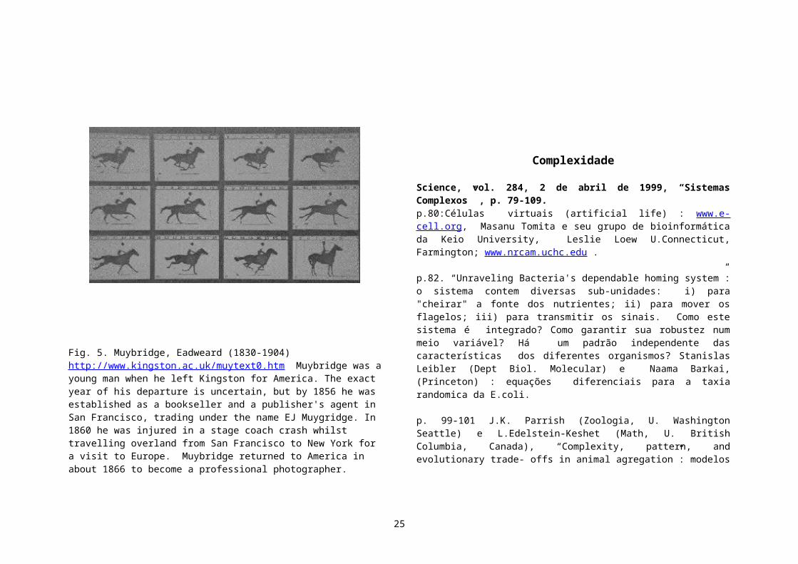
which do not form filamentous structures, in various cellular processes, including membrane traffic, cell movement, and signal transduction. Microtubules play fundamental roles in the formation of complex cellular geometries such as axons, the extremely elongated processes of neurons. Microtubule-based motors use microtubule tracks to move a variety of cargoes around cells--the movement of chromosomes along the mitotic spindle during mitosis is an example. Hirokawa (p. 519) describes the large number of microtubule-based motors, encoded by the kinesin and dynein multigene families, and elaborates on their roles in intracellular transport. A kinesin Web site has details on many of the aspects of the cell biology and biophysics of this important intracellular motor protein (www.blocks.fhcrc.org/~kinesin/). The actin cytoskeleton also shapes cellular processes; for example, in the formation of filopodia or cell contact sites. Hall (p. 509) looks at the interplay between actin architecture and the signal transduction machinery of the cell in promoting profound changes in cell shape and motility in response to extracellular signals. Less is known about the role of intermediate filaments in cells, mainly because of a lack of tools with which to study their assembly and disassembly. Fuchs and Cleveland (p. 514) summarize recent advances in our understanding of the roles of multiple types of intermediate filaments, which have become clear through the discovery of several diseases linked to intermediate filament pathology. A Research News story by Elizabeth Pennisi (p. 477) focuses on the kinetochore, the part of the chromosome that interacts with the microtubules of the mitotic spindle. Finally, Echard and co-workers (p. 580) describe a putative new motor that is likely to play a role in intracellular transport through the secretory pathway.
Problems with the cytoskeleton can cause disorders of the skin, the nervous system, and the muscles. Changes in the cytoskeleton are key, and even diagnostic, in the pathology of some diseases, including cancer. Understanding the basic cell biology of the cytoskeleton has contributed to our understanding of the pathology of some of these disorders and will continue to affect approaches to understanding, diagnosis, and therapy for various conditions. (Stella M. Hurtley).”
Na próxima seção, você encontrará muitas outras fontes sobre biologia molecular que se pode obter pela www.
Motores Moleculares
“Navegar é preciso” (Fernando Pessoa)
É possivel encontrar informações maravilhosas na internet, ainda que não possamos escapar da sensação de nos perdermos num dos labirintos de Jorge Luis Borges… Os sites mencionados abaixo podem ajudar o professor a complementar seu trabalho em sala de aula. Hoje em dia muitos colocam em suas páginas, tanto as informações sobre seu trabalho como também informações educacionais em geral. Esta lista é obviamente muito incompleta, e com certeza deixamos de fora muitos sites importantes, inclusive da Espanha. Não há tãopouco uma
organização muito coerente, devido a premência do tempo que tivemos para a preparação do texto. Alguns trechos em inglês foram simplesmente copiados dos sites para dar uma idéia prévia dos assuntos. Para dar uma idéia do número de publicações, basta ver que usando fazendo uma busca em Science, obtivemos há algum tempo, para “anywhere in article: molecular motors”: 3810 !
Se você deseja imediatamente se informar sobre o estado da arte, olhe a home page do congresso recentemente realizado na Universidade de Alberta:http://www.phys.ualberta.ca/~biophys/banff2000
25
Mas é melhor talvez começarmos bem devagar… Um site educacional maravilhoso para biologia celular, accessivel para crianças inclusive, é http://www.cellsalive.net/ ou http://www.cellsalive.com/Em particular, veja como bactérias se movem: http://www.cellsalive.net/animabug.htm
Um lindo tutorial sobre biologia celeular, onde apendemos todas as informações básicas (você conhece os nomes “ microtubulos, cilia, flagella, etc, …”?) para alunos (e professores) em final de ensino secundário se encontra em:http://cellbio.utmb.edu/CELLBIO/ (University of Texas Medical Branch., Galveston, Texas)Veja por exemplo as páginashttp://cellbio.utmb.edu/CELLBIO/microtub.htm#Menuhttp://cellbio.utmb.edu/CELLBIO/microtubule_structure.htmAli há links para outros tutoriais, históricos, etc.: http://cellbio.utmb.edu/Links.htm
A “home page da Kinesin” - com filmes, material informativo muito completo é http://www.blocks.fhcrc.org/~kinesin/Entre os links ali mencionados, veja http://www.scripps.edu/milligan/research/movies/kinesin_text.html animações do laboratório de Ron Vale ([email protected]) Ron Milligan ([email protected]) Graham Johnson ([email protected])
Para outros links em motores moleculares, ver http://motility.york.ac.uk:85/(dali em diante, a navegação prosseguirá de vento em popa, incluindo a home page da miosina)
Comecemos com a descrição do trabalho do grupo da Universidade de York (as pessoas estão listadas no site) http://motility.york.ac.uk:85/“The Problem. It is well known that the breakdown of ATP powers the generation of force by myosin (and most other molecular motors). The biochemistry and kinetics of this process have been extensively studied, and the mechanics of
muscle contraction are well documented. Recently, mechanical data for single molecules has been obtained. However, we still lack a full understanding of the way that the biochemical and mechanical events are coupled. Questions in Coupling We know that the energy released by the breakdown of a single ATP molecule is ~10-19 J. Recent optical tweezer measurements have shown that skeletal muscle myosin moves in steps of 4-5nm, and can generate a force of approximately 2pN. This would be equivalent to ~10-20J of mechanical work per step. However there remain many questions about coupling: How many ATP molecules are consumed for each mechanical step? Which steps of the biochemical cycle produce force? Are all the bound states of myosin mechanically equivalent? What are the properties of mutant myosins and non-muscle myosins (e.g. myosin I)?
Our Approach We believe that the best way to resolve these issues is to make direct observations of the ATP hydrolysis and mechanical events on a single myosin head. This can be achieved by combining twotechniques: Optical Tweezers and Total Internal Reflection Fluorescence Microscopy (TIRFM). We are currently building the apparatus to perform these experiments.”
Site do Grupo da Universidade de Munique IMPERDIVELhttp://www.med.uni-muenchen.de/phychem/zellbio/Schliwa Lab: centrosome structure and dynamics using Dictyostelium amebae as a model system; molecular motors, in particular kinesins of fungi; optical tweezersSchleicher Lab: actin-binding proteins of Dictyostelium
O site do grupo do Prof. Manfred Schliwa é fantástico. Eis algumas informações:Cell Biology of intracellular motility - http://www.med.unimuenchen.de/phychem/zellbio/tI/resIK2.html“Eukaryotic cells are highly compartmentalized. Localization of, and communication between, these compartments is mediated by cytoskeletal polymers such as microtubules and molecular motors such as kinesin or dynein. We are interested in the molecular basis of organelle transport and have chosen to study the motor kinesin using Neurospoa crassa as a model system.”
26

(Ver uma grande quantidade de links em http://www.med.uni-muenchen.de/phychem/zellbio/tO/linO.html)
Site de Julie Therriot (Stanford) IMPERDIVELhttp://cmgm.stanford.edu/theriot/http://cmgm.stanford.edu/theriot/movies.htm
Este é outro site fantástico, de uma excelente pesquisadora, que também tem a preocupação de divulgar sua área ao público geral. Há filmes maravilhosos!
“Actin-Based Motility for the Non-Biologist. Imagine that you're mostly made up of water. Well, you are, so you don't have to try very hard. But now imagine that, instead of keratin-rich skin, the only thing that holds you together is a thin bilayer of lipids--molecules which are most familiar to most people as components of fat. Finally, imagine that you're only 10 microns (.000010 meters) wide. Now you have a problem. You're much, much smaller than most water droplets. Water droplets form because of surface tension, which you're familiar with if you've ever watched rain on the window. The water molecules organize themselves in such a way that they can rise above the glass. These forces are so strong that if you fill a straw with water and put your finger over the top, the water won't run out the bottom, even though the water column is exerting a lot of pressure on the tiny hemisphere of surface-tension-secured water at the bottom. The narrower the straw, the higher the water column it can hold.
So if you're only 10 microns wide, mostly water, and held together by a flexible lipid-lined bag, you have a serious problem which cells have confronted since their appearance in the primordial soup. Surface tensionwill easily force you to collapse into a sphere. But cells are rarely spherical: they can be cylindrical (intestine lining), amoeboid (immune cells), long and slender (muscle cells), covered with protrusions (neurons) or more. Moreover, cells can crawl, drastically changing their shape in
order to pull themselves along. Our job is to ask them how they do it.
The answer starts with the cell's skeleton, a fibrous network made up of filaments of varying thickness. Motility is mostly the jurisdiction of actin filaments, whose remarkable strength helps resist the forces ofsurface tension. Actin filaments are made up of protein monomers--individual building blocksthat can be linked together like Legos to form long or short chains. Other proteins in the cell bind actin filaments to link them together, stop their growth, accelerate their growth, or chop them up. Yet other proteins bind to actin monomers to regulate the biochemical equilibrium between monomers and filaments. In other words, a cell's skeleton is nothing like our skeleton in that it changes structure very quickly.
Our two favorite pathogenic bacteria--Listeria monocytogenes and Shigella flexneri are excellent cell biologists. They understand actin-based motility so well that when they invade into the interior of cells, they hijack the cell's actin-based motility system to facilitate their own spread into adjacent cells. Since their motility is easier to measure than that of a cell, we spy on them as they've spied on cells, trying to learn what they know.
Here's what we and other labs have figured out. Each bacterium produces a single protein (ActA for Listeria or IcsA for Shigella) which encourages the growth of new actin filaments at the bacterial surface. As these filaments grow (polymerize), they exert force on the bacterium, pushing it forward from the outside, much as actin filaments push their cells forward from the inside. The bacterium rockets forward, leaving behind a comet tail of short, highly networked actin-filaments which shrink (depolymerize) overtime. You can see one of these tails in the movie at the top of the page.
Think of it like a jet contrail (800k file). Addition of new material only occurs at the back of the 'organism,' and the organism causes that addition. The tail is stationary with respect to its environment (the cytoplasm or the atmosphere). And finally, that environment governs how fast the tail falls apart (host proteins or wind).
27

In an incredible example of pathogenic evolution, these bacteria become the terrorists of the cell, hijacking it's transportation mode to ram them into the cell's membrane. The bacterium then protrudes into an adjacent cell, and, upon escaping into the cell's neighbor, continues dividing and spreading to other cells. By never exiting cytoplasm, the bacterium avoids the commandos of the immune system: large, crawling cells called macrophages which are constantly policing the body for harmful intruders to eat.
By studying how these pathogens move, we deepen our understanding of many biological problems. It's a two-for-one deal. On the bacterial side, we can learn about the relationship between host and pathogen, helping modern medicine combat the diseases caused by these organisms. Effects of listeriosis and shigellosis include diarrhea, meningitis, spontaneous abortions, and even death for immunocompromised individuals (children, the elderly, AIDS patients). On the host cellular side, we can learn how cells use their skeletons to change shape in developing tissues, how they move in the development of a new organism, how wounds are healed by migrating cells, how metastatic tumor cells crawl into the blood, and many otherphysiological processes.
How cells hold or change their shape may seem at first like an esoteric question. Yet to the cell, it's anything but. And since cells make up every living thing on this planet, we think listening to them is worth the effort. Grupo de George Oster, Professor of Cell & Developmental Biology, Berkeley, http://nature.berkeley.edu/~goster/home.html Ver a “gallery” com as animações da ATP Synthase e outros motores moleculares.“My research involves construction and testing of theoretical models of molecular, cellular and developmental processes. Current projects include investigations into the basic physics and chemistry of protein motors, cell motility and membrane organization.” LINDOS MODELOS MATEMATICOS !
Site dos Profs. S. G. Boxer e A. van Oudenaarden (Geometrical Brownian ratchets). Contém informações básicas sobre o mecanismo dos ratchets, além de explicações qualitativas sobre os ratchets geométricos estudados por eles.http://www.stanford.edu/group/boxer/ http://web.mit.edu/biophysics/research.html“Recently interest in Brownian ratchets and fluctuation-driven transport has increased enormously because of the relevance of these concepts for biological systems such as molecular motors, ion pumps, and for thedesign of biomolecular sieves. Brownian ratchets have the unique ability to exploit thermal fluctuations to drive directional transport. A 2 dimensional fluid phospholipid bilayer membrane on a patterned surface is an experimental realization of a particular type of Brownian ratchet called a geometrical Brownian ratchet. This system has many of the properties of interest in the general area of Brownian ratchets, can be described quantitatively, and has potential applications in the separation of membrane-associated molecules and assemblies. Actin Propulsion Engines. Actin filaments are biopolymers that can both polymerize (grow) and depolymerize (shrink). These highly dynamical molecules are capable of exerting significant mechanical forces. A complex of many actin filaments that is randomly growing and shrinking is used by living cells to change shape and to move. How this inherently stochastic process self-organizes in a well-defined propulsion engine is a challenging problem that involves knowledge from both physics and biology.”
Site do grupo do Prof. Eberhard Unger em Jena:http://www.imb-jena.de/www_elmi/molcyto_cyto.htmlhttp://www.imb-jena.de/www_elmi/molcyto_members.html#ungerna página do Prof. K.Boehm, há muitos links ótimos para outros grupos, um tutorial, e “applets” sobre a kinesina.http://www.imb-jena.de/~kboehm/http://www.imb-jena.de/~kboehm/Kinesin.htmlhttp://www.imb-jena.de/~kboehm/Tubulin.html otimo review didatico
Site de Lawrence S.B. Goldstein (University of California San Diego)http://medicine.ucsd.edu/pharmaco/lsgoldstein.html(o site do Departamento de Famacologia é interessante de ser ver,
28
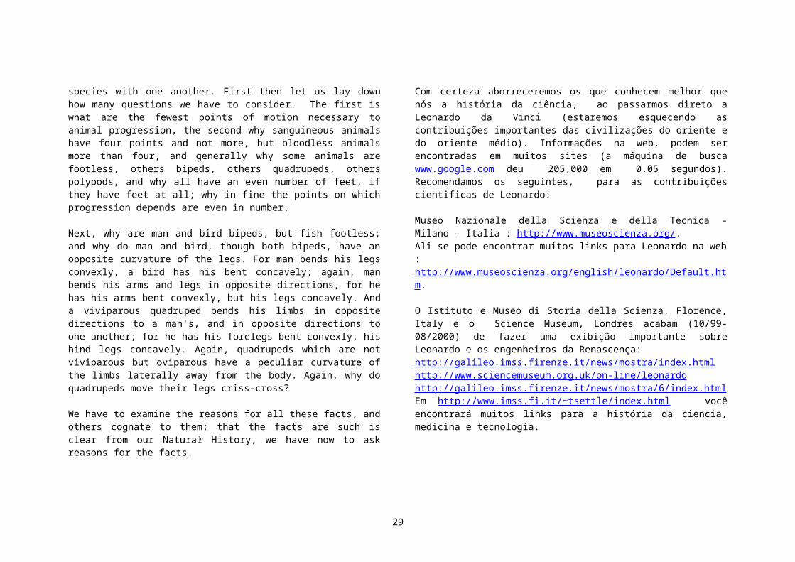
http://medicine.ucsd.edu/pharmaco/e o de biologia http://www-biology.ucsd.edu/http://biosciences.sdsc.edu/http://www-biology.ucsd.edu/“My laboratory is interested in understanding the molecular mechanisms of intracellular movement in neurons and the role of transport dysfunction in neurodegenerative diseases. Our focus is on the attachment, function, and regulation of kinesin and dynein microtubule motor proteins. The major questions we are addressing are: 1) What role(s) do these motors play in axonal transport, transport of visual system components in photoreceptors, and transport of informational signaling molecules? 2) Does motor-driven transport dysfunction play a major role in neurodegenerative diseases such as retinitis pigmentosum and Alzheimer's disease? 3) How are kinesins and dyneins coupled to intracellular cargoes and regulated? 4) How are appropriate destinations in the neuron found (e.g., axons versus dendrites)? 5) Do intracellular transport processes play important roles in neuronal cell polarization, signaling, growth, and pathfinding? Technologically, our work utilizes molecular and classical genetics, cell biology, and biochemistry in D.melanogaster and M. musculus. Thus, we are making mutants in defined motor proteins and inferring function by phenotypic analysis. We are also using genetic screens to identify novel proteins that couple motors to cargo and regulate their function.”
Site de E. Frey (Physics Department, Harvard) e colaboradores ; ver os Java applets de Motores Moleculares)http://cmtw.harvard.edu/~frey/ver também de seus colaboradores:http://WWW.Physik.TU-Muenchen.DE:81/~avilfan/ecmm/ (elastically coupled molecular motors)http://www.ph.tum.de/~avilfan/relax/ (1-dim dimer absorption)
Site de Joe Howard, U. Washington http://depts.washington.edu/pbiopage/faculty/howard.html
Ali podemos ver "applets" da kinesina andando, e modelo como um ‘motor de combustão”.
“Our laboratory is interested in the mechanical properties of cells and molecules. How do cells detect and effect changes in their mechanical environment? How do they establish and change shape? How do they move? To answer these questions, we havedeveloped highly sensitive techniques that allow us to visualize and manipulate individualmolecules, and to measure directly the influence of forces on their structural conformations. Using these techniques, we have measured the force required to open asingle ion channel, we have determined the elasticity of individual cytoskeletal filaments, and we have characterized the mechanical output - the force, displacement and work - of single motor proteins. Current projects in the lab include the following: (i) The molecular mechanisms of force generation by motor proteins. By combining single-molecule techniques with biochemical and protein-engineering methods, we hope to identify, at the amino acid level, the moving parts that make up motor proteins- the springs, shafts, and axles - and to understand how the motion of these parts is coupled to the hydrolysis of ATP. (ii) The structural basis for the regulation of motor proteins. We are trying to understand how motor proteins recognize and bind to their cellular cargoes, and how the binding to the cargo leads to the switching on of the motor activity. (iii) Mechanoelectrical transduction by cutaneous sensory receptors. We are studying the cellular and molecular mechanisms underlyingthe sensation of touch to the skin: which cells are mechanoreceptive, what molecules and structures are initially perturbed by the mechanical stimuli, and which ion channels are ultimately gated by these mechanical forces? (iv) Remodeling of the extracellular matrix. How do cells such fibroblasts reorient in response to forces in tissues and how, in turn, does this lead to a reorientation and polarization of the extracellular matrix.
Site de Frank Julicher (Instituto Curie) http://perso.curie.fr/Frank.Julicher/“The main focus of our research is the physics of nonequilibrium processes in biological systems on the scale of the cell. Important examples are the force and motion generation in cells by motor enzymes or assemblies of such motors as well as the mechanical and dynamical properties of membranes, filaments and the cytoskeleton. Some examples are: Sound Detection and Sensory Systems (Auditory Sensitivity by Critical Oscillations of Hair Cells); Active Processes in Biological Systems (Beating and Swimming of Flagella and Cilia; A Stochastic
29

Model for RNA Polymerase Motion along DNA); Molecular Motors (Modelling Molecular Motors; Molecular Motors: From Individual to Collective Behavior ); Energy Transduction and Efficiency of Molecular Motors (Elastic Properties and Dynamics of the Cytosceleton; Pulling on a Filament )” (Há vários artigos que se pode fazer os downloads, com muitas figuras e a matemática é razoàvelmente accessivel. )
Finalmente, tudo pode ser sumarizado no site da “Superfamilia kinesina”:
Grupo do Prof. Nobutaka Hirokawa (Department of Cell Biology and Anatomy, Graduate School of Medicine, University of Tokyo). cb.m.u-tokyo.ac.jp/profile-hirokawa.html, cb.m.u-tokyo.ac.jp/ Ver a excelente revisão: “the neurnal cytosqueleton” . O Prof. Hirokawa e seus colaboradores estão classificando as famílias de kinesianas e seus tipos de locomoção. Ver http://cb.m.u-tokyo.ac.jp/KIF/index.html (Kinesin Superfamily Protein (KIF) Home Page) . Ó”timo review Ā
Outros exemplos de processos celulares envolvendo movimento
R. Bruce Nicklas (Duke University; http://www.dcmb.duke.edu/faculty/nicklas.htmHow Cells Get the Right Chromosomes? When cells divide, the chromosomes must be delivered flawlessly to the daughter cells. Missing or extra chromosomes can result in birth defects and cancer. Chance events are the starting point for chromosome delivery, which makes the process prone to error. Errors are avoided by diverse uses of mechanical tension from mitotic forces. Tension stabilizes the proper chromosome configuration, controls a cell cycle checkpoint, and changes chromosome chemistry. Every time a cell divides, the daughter cells must get the right chromosomes. For example, in humans, Down syndrome occurs when an error in meiosis results in a child with an extra copy of chromosome 21. Beyond the cost in human terms, Down syndrome has an estimated annual economic cost of $3.6 billion. Cells with missing or extra chromosomes can be equally ruinous
inadults, by fueling the development of malignant cancer cells (2). Given the cost of errors, it is not surprising that cells take pains to avoid them.
Site do Prof. Andrew Maniotis, U. Iowa (Movimento envolvido em tumores )http://www.anatomy.uiowa.edu/pages/directory/research/maniotis.html“Tumor cells are like other cells in that they reqire food, and a route by which they can eliminate waste. Recent observations have suggested that a critical regulatory step in the primary growth and spread of melanoma involves solid state mechanical changes that determine the vascular architecture within the tumor microenvironment. Therefore, we are defining how these solid state signaling pathways within human tumor biopsies, and within various three-dimensional reconstituted experimental systems control the responses of both host and tumor cells when tumor cells of differing invasive, metastatic, or dormant potential interact with host cells. In addition, we have discovered both in vivo and in vitro that invasive tumor cells themselves have vasoformative potential which is distinctly different than what is typically observed in normal tissues or in wounded tissues. In normal cells, we have previously defined how tensionally continuous molecular networks spanning between the extracellular matrix, the cytoskeleton, and the genome control cell growth, and have now turned our attention toward how tumor cells might use these mechanisms to control tumor vessel or sinusoid formation, and host cell apoptosis. In addition, we have discovered that certain classes of intermediate filaments and endothelial-specific epitopes are uniquely expressed in aggressive tumor cells specifically along vascular structures. To study how cells might control non-linear genetic responses to differences in the mechanical environment, we are now utilizing a method we have developed in which we microsurgically remove nucleoplasm or chromosomes from living cells under isotonic conditions, without the use of detergents or fixatives. Using this approach, we have learned to induce spontaneous decondensation and recondensation of chromosome form and position throughout an entire genome, and by manipulating proteases and certain exogenously added proteins, we can reconstitute the complete human genome in normal host endothelium and are applying this approach to tumor cells to identify unknown condensing or decondensing proteins (histone H1, topoisomerases) or other highly charged molecules. It is hoped that these approaches will provide an
30
experimental basis to understand and perhaps one day control oncodevelopment, and aneuploidy.”
Site de Stanilas Leibler (Princeton) Mecanismos moleculares regulando a quimiotaxia das bactérias.http://www.molbio.princeton.edu/faculty/leibler.htmlIn recent years, it has become clear that molecular biology is facing a new challenge: to move from the description of individual components and their mutual interactions toward system analysis. Such an analysis would describe the functioning of systems of interacting biomolecules on a more global level, much closer to a phenotypic, rather than a genetic, description. In physics and chemistry, one often studies systems in which many components interact with one another, giving rise to new “collective phenomena.” We are interested in such collective phenomena taking place in biological systems. In particular, it is attractive to think that a detailed study of the best known prototype systems could uncover the underlying general “design principles.” Even the simplest unicellular organisms, such as bacteria, perform a sophisticated kind of “information processing.” For instance, Escherichia coli can direct itself in space by measuring the temporal gradients of chemicals. The enzymatic network responsible for chemotaxis is relatively simple (with a small number of components) and is extremely well characterized at the molecular level. Yet the “system analysis” of this signal transduction network is far from complete. We do not understand the source of the sensitivity to small signals in chemotaxis, the response to different competing signals, or the structural stability of the circuit. Nor do we understand the reasons why common building blocks such as phosphorylation cascades appear in these and other signal transduction systems, or the evolutionary significance of the observed network architectures. We have been studying, both theoretically and experimentally, the chemotaxis network. Through quantitative analysis of bacterial behavior and modifications of the intracellular biochemistry, we have been able to demonstrate that this network presents some properties (such as adaptation precision) that are “robust” with respect to variations of its individual components. Such robustness is the consequence of the network’s connectivity, and it may become one of the “design principles” for a large class of biological networks. In addition, we are applying fluorescence correlation spectroscopy to monitor the concentrations of the network’s components, while
simultaneously analyzing the behavior of individual bacteria. This allows us to determine the source of the observed “nongenetic individuality,” namely the large variations in the behavior of genetically identical bacteria. We plan further to pursue our studies of simple, prototype systems in search of a quantitative, “systemic” description of their functioning. We have started preliminary experiments and theoretical work on the following genetic and biochemical phenomena: (1) Circadian rhythms in microorganisms. We are particularly interested in the phenomenon of temperature compensation (independence of the internal clock of temperature) and its physical/biochemical basis. (2) Epigenetic cellular phenomena. Even the simplest gene regulation networks present multiple steady-states which can be transmitted for many generations. We plan to extend classical observations in the series of quantitative experiments inbacterial systems. (3) Evolvability of simple networks. We are now trying to build simple artificial networks from natural components (e.g., from the networks of bacteria and phages), which would perform a predefined function in vivo (e.g., an oscillator). Such experiments could become a starting point for a more general study of networks-their classification, compatibility, and evolvability. These are just a few examples of phenomena governed by networks with a small number of known components. Over the past decades, researchers have been gathering an enormous volume of data (such as genomic DNA sequence information, transcript levels, etc.) across a wide spectrum of biological systems. We are now trying to develop mathematical and physical tools to extract information about signal transduction networks based on the analysis of transcription levels under different conditions.
Nanotecnologia e sua interface biológica
Reproduzimos aqui o artigo de Robert F. Service, “Nanotechnology: Borrowing From Biology to Power the Petite, Science, volume 283, Number 5398 Issue of 1 Jan 1999, pp. 27 – 28:
Nanotechnology researchers are harvesting molecular motors from cells
31

in hopes of using them to drive nano-sized devices
“If you received a molecule-sized car, snowmobile, or jet ski for Christmas, you've probably realized by now that the thing is totally useless. It just sits there on your microscope slide like an inert dust speck, incapable of going for a spin around the cover slip. Okay, so molecular vehicles are pure fantasy. But their immobility is a problem that's all too real for would-be builders of nano-sized devices. Such devices are so small, there's no obvious way to power them. Now, researchers are turning to biology for what may be a possible solution: molecular motors from living things.
Cells are packed with protein-based motors powered by the chemical fuel of life, adenosine triphosphate, or ATP. These motors ferry cargo, flex muscles, and even copy DNA. And at a recent meeting,* two groups, one led by Carlo Montemagno of Cornell University in Ithaca, New York, and the other by Viola Vogel of the University of Washington in Seattle, reported taking the first baby steps toward harnessing these motors to power nanotechnology devices. Like molecular mechanics, the researchers have unbolted the motors from their cellular moorings, remounted them on engineered surfaces, and demonstrated that they can in fact perform work, such as twirling microscopic plastic beads. "What we're really trying to do is makeengineered systems that tap into the energy system of life," says Montemagno.
The effort still has a long way to go. But the early work is already generating enthusiasm in the community. "I think it's a very productive path to follow," says Al Globus, a nanotechnology expert at the National Aeronautics and Space Administration's Ames Research Center, Moffat Field, California. If the effort does pan out, it could help researchers make everything from tiny pumps that release lifesaving drugs when needed to futuristic materials that heal themselves when damaged.
For their molecular motor, Montemagno and his colleagues turned to one of the cell's heavy lifters: ATPase, a complex of nine types of proteins that work together to generate ATP. While tiny--it measures just 12 nanometers across and 12 high—this cellular motor is remarkably sophisticated, containing a cylinder of
six proteins surrounding a central shaft. ATPase converts the movement of protons within the cell's energy powerhouse, the mitochondrion, into a mechanical rotation of the shaft, a motion that helps catalyze the formation of ATP. But the motor can also run in reverse, burning ATPs to rotate the shaft and move protons.
Last year, Hiroyuki Noji and his colleagues at the Tokyo Institute of Technology and Keio University in Yokohama, Japan, captured this rotational motion on camera for the first time (see Science, 4 December, p. 1844). They dangled a fluorescent-tagged molecule off the end of the shaft, fed the motor ATP, then put it through a microscope and took sequential pictures of the shaft as it rotated in circles around the cylinder.
Based on the number of rotations produced by a given amount of ATP, the researchers calculated that the motor operates at near 100% efficiency-- "well above the efficiency of motors we're capable of building," says Montemagno. "If the motor was as big as a person, it would be able to spin a telephone pole about 2 kilometers long about one revolution per second."
That result inspired Montemagno and his Cornell colleagues--George Bachand, Scott Stelick, and Marlene Bachand--to see if they could use the ATPase rotary motor to move man-made objects. They started by genetically engineering two changes into ATPase proteins, one to stick the motors to metal surfaces and the other to provide an attachment site for the beads that they wanted the motor to move.
To make the first change, the team added an amino acid sequence loaded with histidine, which binds tightly to metals, to the base of the proteins that form the motor's cylinder. Next they used electron beam lithography to pattern an array of nickel islands--each roughly 40 nanometers across--atop a glass microscope cover slip. When they then spritzed water on top to keep the proteins happy and added the motors, the base of the cylinders bound to the nickel islands, causing the motors tostand upright.
32

To attach the beads, which were made of plastic or a plastic/iron composite and coated with a small organic molecule called biotin, Montemagno and his colleagues added cystine, a sulfur-containing amino acid, to the top of the central shaft. That allowed the shaft to grab a small sulfur-binding protein called streptavidin, which could in turn bind the biotin-coated beads. When the researchers then added ATP fuel to the solution atop the slide and used a laser-based interferometer to track the beads' movement, they could see their array of motors twirling in endless loops, like a dance floor of nano-sized dervishes. "I had the thing running for well over 2 hours at a time," says Montemagno. "It was seriously cool."
But whirling beads--impressive as they may be--are still a long way from nanorobots rooting through the body. So Montemagno's team is pressing ahead. They're currently working on replacing the beads with tiny magnetic bars. If the motors spin the bars, the researchers will be able to measure precisely how strong the motors are by applying an outside magnetic field: By increasing the field until the motors can no longer spin, they will be able to probe the limit of the motor's power.
What's more, the spinning bars should generate an electrical current that might eventually be used to power devices, such as chip-based drug delivery pumps or chemical weapons sensors implanted in the body. But these uses, Montemagno says, are just the beginning. "There's 100,000 different things you could do with these motors," he says.
Washington's Vogel says much the same thing about her team's contraption, a nanoscale monorail in which a collection of molecular motors all lined up on a surface pass a tiny tube hand over hand down the line. Vogel based her monorail on one of the cell's own transport systems, which consists of tracks made of microtubules, tube-shaped assemblies of a protein called tubulin, and small motors made of another protein, kinesin. In cells, the kinesin motors latch onto the fixed microtubules and churn like steam engines from one end of the line to the other, ferrying molecular cargo such as proteins and lipids. But for their experiment, Vogel and her colleagues John Dennis and Jonathan Howard reversed
these roles, fastening kinesin motors to a surface and having them shuttle microtubules down the line from one motor to the next.
Biophysicists studying kinesin motors had done related experiments in the past. But in those, Vogel says, the kinesins were in random locations on surfaces. When microtubules and ATP were then added, the kinesins shuttled microtubules in all directions. To control the transport, the Washington team had to line up the kinesins. Here, the researchers took a low-tech approach. They simply rubbed a block of polytetrafluoroethylene, or PFTE, across a glass slide, causing molecules of the chainlike polymers to rub off and coat it. The scraping acted something like a hair brush, getting all the PFTE chains to line upon the surface, creating a series of grooves running for micrometers along the slide.
After submerging the slides in water and coating them with a small protein called casein, to protect overlying proteins, they added the kinesin motors, which settled into the grooves. They then sprinkled on a few microtubules, which were tagged with fluorescent compounds so they could be seen, and dropped some ATP fuel into the solution.
By turning on a xenon lamp to set the microtubules aglow and letting their cameras roll, Vogel and her colleagues could see the kinesins push their tubular cargo in one direction, moving it hand over hand down the parallel grooves. "Even though kinesins move on the nanoscale, we could watch the microtubules move on the micron scale," says Vogel.
For now, the team is using the monorail to study the performance of their motors. But down the road, Vogel says that the tinyrail lines could be used to transport replacement components for self-healing biomaterials for medical implants. If this and other efforts to motorize the nanoworld are successful, those microscope slides may soon see their first traffic jams.
Sites na internet
33
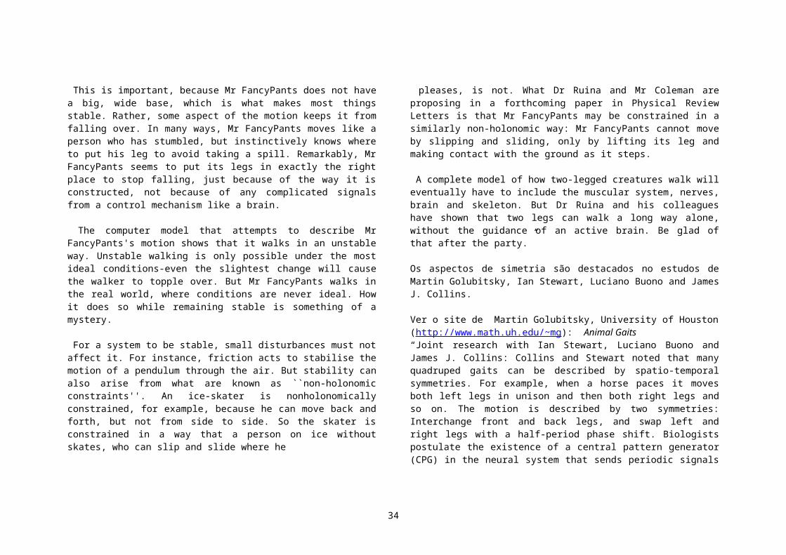
Podemos começar com o home page da nanotecnologia. http://www.zyvex.com/nano/ e recomendamos a leitura do artigo de Feynman, “There’s plenty of room in the bottom”.
“Legos” em escala atomica. (Science vol. 284, 21/05/99 pg. 1231,Net Watch)Máquinas em escala atômica estão se tornando realidade. Ver as imagens e videos da NASA Ames em http://www.nas.nasa.gov/Groups/Nanotechnology/galleryPor exemplo as, “Fullerene Gears” (14,0) tube based gear: gear.jpg; largeSystem.jpg .A idéia é usar padrões de fluor e hidrogenio numa superficie de carbono para guardar bits de dados. Para ler os dados, voce passa um buckytubo de carbono preso a um microscopio de varredura (scanning probe). Este esquema permitiria ler 10.000.000 mais dados por área do que os atuais discos óticos.
A Universidade de Cornell tem um grande centro em nanotecnologia. Verhttp://www.nbtc.cornell.edu/ http://www.cnf.cornell.edu/ Sugerimos ver a página do Prof. Carlo Montemagno (Agricultural and Biological Engineering)
Dr. Montemagno's research is focused on the application of nanotechnology to biological systems. His current projects are directed at the development of biomolecular motor powered nanoelectromechanical devices and the engineering of on-chip detectors for pathogens. “My current and near term investigations focus upon the development of numeric and experimental techniques to explore: 1) methods of integrating single molecule biological motors with nanoscale silicon devices; 2) the influence of interfaces in multiphase fluid flow through and within biological materials; 3) the role of fluid-fluid and biological interfaces in the transport of nutrients and chemicals between the physical and biological domains; 4) fracture/pore geometry and its contribution to the transport of fluids and microorganisms in porous media; 5) the role of the cytoskeleton in processing intracellular information; and 6) biological processing of organic wastes.”
Para alguns recursos para professores, ver a página educacional nestes sites e tambémhttp://www.nanospace.systems.org/ns_2000/NS00_Sessions.htm
Outros reviews em nanotecnologia:
http://www.resonance-pub.com/nanotech.htm (este é um portal em divulgação da ciencia)Micro/Nanotechnology Conference -- http://www.aero.org/conferences/micro-nano/
No ETH Zurich, http://www.nanotechnology.ethz.ch/
A Duke University tem também um centro integrado em Bioengenharia: verhttp://bme-www.mc.duke.edu/Research/Cellsurf/faculty.html(Cellular and Biosurface Engineering):Although centered in the school of engineering, the CBE faculty members have primary and/or secondary appointments in one of seven degree granting entities - the Departments of Biochemistry, Biomedical Engineering, Cell Biology, Chemistry, Electrical Engineering, and Mechanical Engineering and Materials Science - or in clinical departments training program provide strong expertise in biomaterials, material property characterizations, functional evaluation, surface modifications, cell culture, and tissue and cell biomechanics. Chemistry faculty affiliated with the training program provide expertise in patterned surface, protein immobilization, and separations. Faculty from the biomedical sciences provide expertise in cellular physiology, biophysics, protein engineering, protein structure and function, tumor oncology, and cell culture. The research interests of the participating departments cover a wide range of fundamental and applied topics, many of which are important to biotechnology.
Ainda em Duke, http://www.zoology.duke.edu/crenshaw/ , Grupo do Prof. Crenshaw. Distributed Robotics: The Micro-Hunter project is a collaborative research effort between the Crenshaw Laboratory and Nekton Technology Incorporated, sponsored by DARPA. The goal of the project is to construct small, cheap,
34

reliable aquatic robots, whose primary objective will be to gather underwater data. The project is well underway, and is expected to deliver a maneuverable robot within the next three years. Our contribution to this project is twofold: (1) design of orientation algorithms, and (2) analysis of the motion of robots.Locomotion and Orientation of Micro-organisms: The locomotion and orientation of micro-organisms has been studied since people first observed them with microscopes. Nevertheless, these processes remain poorly understood. The Crenshaw lab has examined the orientation of free-swimming single cells (spermatozoa, flagellates, and ciliates) for 10 years. We have developed new techniques for collecting and analyzing the 3D trajectories of freely swimming cells to examine the responses of these cells to external stimuli, such as beams of light and concentration gradients of chemicals. Our primary objective has been to test the theory of helical klinotaxis (references below) to determine if these cells use this mechanism of orientation. Our research has in more recent years turned to different levels of biological organization, studying both molecular mechanisms of signal transduction in protists and the ramifications of orientation behaviors on the distributions of protists in natural environments.
Fig. 7 (Celular Automata) Paramecia, protozoa about 100 microns length, are very sophisticated ``Celulas Automatas’’. Ray Fearing, from Berkeley Electrical Engineering, showed that they can learn to perform tasks such us doing a baseball run If completely domesticated they could to become micro-robotic workhorses. They do not need to be fueled and their fabrication is ridiculously inexpensive.
35