avaliaÇÃo da atividade da proteÍna quinase … · na maturaÇÃo in vitro de oÓcitos caninos...
TRANSCRIPT
Ciência Animal, 27 (1): 41-54, 2017.
AVALIAÇÃO DA ATIVIDADE DA PROTEÍNA QUINASE P34CDC2
NA MATURAÇÃO IN VITRO DE OÓCITOS CANINOS
(Evaluation of p34cdc2 kinase activity in the in vitro maturation of canine oocytes)
Leda Maria Costa Pereira1*; Paulo Ricardo de Oliveira Bersano2, Maria Denise Lopes1
1Dpto de Reprodução Animal e Radiologia Veterinária, Faculdade de Medicina Veterinária e Zootecnia da
Universidade Estadual Paulista - UNESP, Botucatu, SP; 2Laboratório de Patologia e Medicina Legal
Veterinária, Faculdade de Veterinária da Universidade Estadual do Ceará, Fortaleza, CE, Brazil.
ABSTRACT
The control of the cell cycle is regulated by a cascade of coordinated events that can act
by influencing the expression or repression of the activity of proteins related to meiosis
resumption. Studies have indicated that the activity of these proteins is time-dependent
in the in vitro maturation process (IVM). This work aimed to evaluate the kinetics of the
p34cdc2 kinase activity during IVM of canine oocytes. Ovaries were obtained from 40
bitches submitted to elective ovary-salpingo-hysterectomy (OSH). After OSH, ovaries
were immediately transported at temperature of 4 °C. In the laboratory, ovaries were
sliced for the release of cumulus-oocyte complexes (COCs). Only grade 1 COCs were
selected and placed in maturation medium for a period of 24, 48 and 72 h of maturation.
After culturing, COCs were plated of 0.2% hyaluronidase solution for complete removal
of cumulus cells. The activity of the p34cdc2 protein was detected by ELISA. Based on
the results, it was observed that the activity of the protein is time-dependent, peaking
after 48 hours of IVM (p 0.01). After 72 hours, activity declined. Based on this study, it
could be concluded that the p34cdc2 protein kinase plays a very important role in the
meiosis progression in bitches. Thus, a better understanding of this protein as well as of
others that participate in the maturation process may contribute to the establishment of
more adequate media to significantly improve maturation rates.
Keywords: Protein kinase, oocytes, p34cdc2, maturation, bitches.
RESUMO
O controle do ciclo celular é regulado por uma cascata de eventos coordenados que
podem atuar influenciando na expressão ou repressão da atividade de proteínas
relacionadas a retomada da meiose. Estudos indicam que a atividade dessas proteínas
mostra-se tempo-dependente no processo de maturação in vitro (MIV). Esse trabalho
teve o objetivo de avaliar a cinética da atividade quinase p34cdc2 durante a MIV de
oócitos caninos. *Endereço para correspondência:
Ciência Animal 27(1), 2017
Os ovários foram obtidos de 40 cadelas submetidas à ovário-salpingo-histerectomia
(OSH) eletiva. Após a OSH, os ovários foram imediatamente transportados a uma
temperatura de 4 ºC. No laboratório, os ovários foram seccionados em fatias finas
("slicing"), para a liberação dos complexos cumulus-oócito (COCs). Apenas os COCs
grau 1 foram selecionados e colocados em meio de maturação por um período de 24, 48
e 72 h de maturação. Após o cultivo, os COCs foram colocados em placas contendo
solução de hialuronidase 0.2% para a retirada completa das células do cumulus. A
atividade da proteína p34cdc2 foi detectada por ELISA. Com base nos resultados verifica-
se que a atividade da proteína mostra-se tempo-dependente, atingindo o pico após 48h
de MIV (p<0,01). Após 72h, a atividade demonstrou um decréscimo. Com base neste
estudo pode-se concluir que a proteína quinase p34cdc2 desempenha uma função de suma
importância na progressão da meiose em cadelas. Dessa forma, a compreensão melhor
dessa proteína assim como de outras que participam do processo de maturação poderá
contribuir para o estabelecimento de meios mais adequados que melhorem
significativamente as taxas de maturação.
Palavras chave: Proteína quinase, oócitos, p34cdc2, maturação, cadela.
INTRODUCTION
In mammals, when removed from
the follicular environment, oocytes no
longer receive the signal that maintains
meiotic block and resume meiosis. In
bitches, although the oocyte may
spontaneously resume in vitro meiosis,
maturation rates are very low and the
percentage of oocytes remaining in the
germinal vesicle (GV) is high. During
ovogenesis, the information required for
the initial embryonic development is
transcribed and stored as mRNA or
translated and stored as proteins
(ALLARD et al., 2005). Any alteration in
the synthesis and consequently in the
stock of these proteins can have serious
consequences in oocyte development.
The resumption of the meiotic
cell cycle is mainly influenced by the
meiosis promoter factor (MPF), which is
one of the main regulators of
morphological changes that occur during
oocyte maturation, regulating
chromosome condensation, nuclear
envelope rupture and the reorganization of
microtubules (MOTLIK, 1998).
MPF is a heterodimeric protein
belonging to the family of kinases,
consisting of a catalytic subunit, the
cyclin-dependent kinase called cdk1 or
p34cdc2, which controls cell division and a
regulatory subunit, cyclin B1
(MERMILLOD, 2000). Meiosis
progression requires both cyclin B
synthesis and its cytoplasmic shift to the
42

43
Ciência Animal 27(1), 2017
nucleus, along with dephosphorylation of
the catalytic subunit.
Kinases and phosphatases are
involved in resumption and complete
meiotic maturation of the oocyte. In the
growth phase, oocytes have very low
p34cdc2 levels and are not able to progress
from the G2 phase (interval between DNA
synthesis and cell division) to the M phase
(cell division, meiosis). The acquisition of
meiotic competence is associated with the
activation of p34cdc2 at the end of oocyte
development and with the presence of
adequate amount of cyclin B1 to provide
sufficient raw material for pre-MPF
activation (DE VANTERY et al., 1996).
In its inactive form, pre-MPF, the catalytic
subunit is phosphorylated into Thr14
(threonine 14) and Tyr15 (tyrosine 15)
residues (GAUTIER et al., 1988). MPF is
activated when the two subunits, p34cdc2
and cyclin B, are associated and Thr14
and Tyr15 residues are dephosphorylated,
whose reaction is catalyzed by cdc 25
phosphatase enzyme. Dephosphorylation
of these residues as well as the subsequent
conversion of MPF-inactive to MPF-
active is the key to G2/M passage
(CURCIO et al., 2006). However, it is still
unclear which of the heterodimeric
subunits of mammalians MPF plays a
determinant role in the control of MPF
activity (LEDAN et al., 2001).
Kanatsu-Shinohara et al. (2000)
compared the concentrations of p34cdc2
and cyclin B1 in incompetent and
competent mouse oocytes to resume
meiosis and found that the concentrations
of both are approximately three times
higher in competent oocytes when
compared to oocytes unable to resume
meiosis. However, the cyclin B1
concentration was up to seven times
higher than that of p34cdc2 in both stages.
According to these authors, since cyclin
B1 is in excess in both immature and
mature oocytes, p34cdc2 would play a more
central role in the regulation of the MPF
activation.
The aim of this study was to
evaluate the influence of p34cdc2 kinase
activity at different moments of the in
vitro maturation of canine oocytes.
MATERIAL AND METHODS
Animals
Ovaries were obtained from 40
healthy adult domestic bitches of different
breeds that underwent ovariohysterectomy
at the Small Animals Reproduction
Service of the Department of Animal
Reproduction and Veterinary Radiology,
Faculty of Veterinary Medicine and
Animal Science, UNESP. The study was
accepted by the Ethics Research
Committee of FMVZ – UNESP, campus
44
Ciência Animal 27(1), 2017
of Botucatu, under protocol number
176/2011.
Obtaining and classifying oocytes
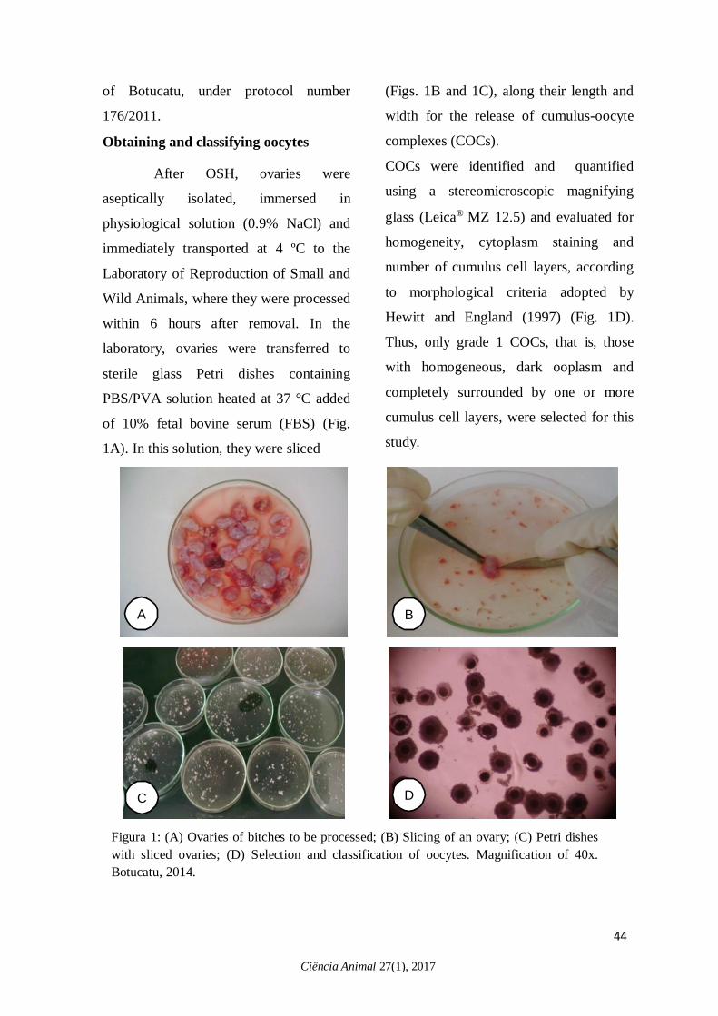
After OSH, ovaries were
aseptically isolated, immersed in
physiological solution (0.9% NaCl) and
immediately transported at 4 ºC to the
Laboratory of Reproduction of Small and
Wild Animals, where they were processed
within 6 hours after removal. In the
laboratory, ovaries were transferred to
sterile glass Petri dishes containing
PBS/PVA solution heated at 37 °C added
of 10% fetal bovine serum (FBS) (Fig.
1A). In this solution, they were sliced
(Figs. 1B and 1C), along their length and
width for the release of cumulus-oocyte
complexes (COCs).
COCs were identified and quantified
using a stereomicroscopic magnifying
glass (Leica® MZ 12.5) and evaluated for
homogeneity, cytoplasm staining and
number of cumulus cell layers, according
to morphological criteria adopted by
Hewitt and England (1997) (Fig. 1D).
Thus, only grade 1 COCs, that is, those
with homogeneous, dark ooplasm and
completely surrounded by one or more
cumulus cell layers, were selected for this
study.
A
B
C
D
Figura 1: (A) Ovaries of bitches to be processed; (B) Slicing of an ovary; (C) Petri dishes
with sliced ovaries; (D) Selection and classification of oocytes. Magnification of 40x.
Botucatu, 2014.
45
Ciência Animal 27(1), 2017
Ciência Animal, 27 (1); 41-54, 2017
During the selection process, COCs were
washed three times in TCM-199 washing
medium supplemented with 25mM
HEPES, 0.2mM sodium pyruvate, 5mM
sodium bicarbonate and 75 μg / mL
gentamicin.
In vitro maturation (IVM)
After selected and washed, grade
1 COCs were divided into groups of up to
20 oocytes and transferred to 4-well
culture plates containing 500 μL of
maturation medium. The medium used
was TCM 199 supplemented with 25 mM
HEPES, 50 μg / ml gentamicin, 26 mM
sodium bicarbonate, 1.5 mM sodium
pyruvate, 2.9 mM sodium lactate
pentahydrate, 0.6 mM cysteine, 0.03 IU /
mL hCG, 0.5 μg / ml FSH, 20 μg / mL E2
and 10 ng / mL epidermal growth factor
(EGF) (SUZUKAMO et al., 2009). The
cultures were perfomed at 38 °C in a
humidified atmosphere of 5% CO2 for a
period of 24, 48 or 72 hours.
At the end of each culture period,
COCs were placed in plates containing
200 μL 0.2% hyaluronidase solution for 5
minutes and repeatedly aspirated with the
50 μL pipette for complete removal of
cumulus cells. After complete removal,
oocytes were then washed in PBS/PVA
for hyaluronidase inactivation. Oocytes
were then transferred to PBS solution
supplemented with 3.7%
paraformaldehyde, washed again in PBS
and stained with 10 μg / ml Hoechst
33342. Oocytes were placed between slide
and coverslip, and evaluated under light
and fluorescence microscopy (Leica®
DFC 310 FX) for evaluation of nuclear
maturation.
Oocyte extract preparation
Oocyte extract was prepared
according to methodology described by
Suzukamo et al. (2009) at times 0, 24, 48
and 72 hours. After completion of each
maturation time, oocytes were washed
several times in PBS/PVA and transferred
to a graduated conical glass tube
containing a buffer solution (lysing
solution) consisting of TRIS-20 mM, 150
mM NaCl, 1.0 mM EDTA, 1.0 mM
EGTA, 1% Triton X-100, 2.5 mM sodium
phosphate, 1.0 mM β-glycerophosphate,
1.0 mM Na3VO4, 1.0 mg / mL leupeptin
and 1.0 mM phenylmethylsulfonyl
fluoride (PMSF), to prepare oocyte
extract. The proportion of 5μL of buffer
solution for five oocytes was used. After
this process, the glass tube was placed in
styrofoam containing liquid nitrogen for
three minutes. Then, this tube was
transferred to Sonifier (Branson Digital
Sonifier®) for complete oocyte
fragmentation and consequent release of
46
Ciência Animal 27(1), 2017
its proteins. The oocyte extract was
sonicated five times for 25 seconds, with
an interval of one minute. At the end of
this procedure, the oocyte extract was
transferred to an identified microtube and
stored at -80 °C.
Measurement of the p34cdc2 kinase
activity
The extract of lysed oocytes (5
oocytes / 5 μL buffer solution) was mixed
with 45 μL kinase-A buffer solution
composed of 25 mM HEPES buffer (pH =
7.5), 10 mM MgCl2 (MBL), 0.1 mM ATP
and 10% mouse vimentin peptide solution.
This mixture was incubated at 30 °C for
30 minutes. The reaction was terminated
with the addition of 200 μL PBS
containing 50 mM EGTA (MBL).
Subsequently, the extract was centrifuged
for 15 seconds and the mouse vimentin
peptide phosphorylation was detected by
ELISA (MESACUP® cdc2 Kinase Assay
Kit).
Statistical analysis
Logistic regression models (SAS
Institute, 2011) were constructed in order
to estimate the chances of oocytes to be
observed at germinal vesicle (GV),
germinal vesicle breakdown (GVBD),
metaphase I (IM), metaphase II and
degenerate (DEG) maturation stages in
different growing times (24h, 48h and
72h). The ELISA technique was
performed three times and each
experiment was replicated four times. The
measurement of p34cdc2 kinase activity
during times 0, 24, 48 and 72 hours of
canine oocyte was performed by analysis
of variance in a completely randomized
design, followed by Tukey's test for
multiple comparisons between means. The
significance level used to reject H0 (null
hypothesis) was 5%, that is, for p<0.05
(significance level less than 0.05).
RESULTS AND DISCUSSION
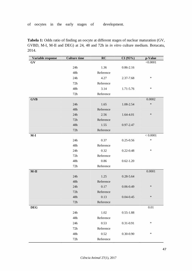
The chances of observing an
oocyte at different times of in vitro
maturation are presented in Tab.1. This
tab shows that the chances of finding an
oocyte in the GV stage in time of 24 h
were 3.15 times greater than in 72h
(p<0.0001). Similarly, the chances of
finding an oocyte at the VG stage were
2.4 times greater at 48 h when compared
to 72 h (p<0.0001). In the time of 24h,
there were 1.26 times more chances of
having an oocyte at GVBD than in 48h (p
= 0.0002) and 1.42 times more chance
than in 72 hours (p = 0.0002). Thus, these
results showed that earlier stagies (GV
and GVBD) are most present in the first
hours of culture in vitro and in the dog,
even after 48 hours, there is the presence
Ciência Animal 27(1), 2017
of oocytes in the early stages of development.
Tabela 1: Odds ratio of finding an oocyte at different stages of nuclear maturation (GV,
GVBD, M-I, M-II and DEG) at 24, 48 and 72h in in vitro culture medium. Botucatu,
2014.
Variable response Culture time RC CI (95%) p-Value
GV <0.0001
24h 1.36 0.86-2.16
48h Reference
24h 4.27 2.37-7.68 *
72h Reference
48h 3.14 1.71-5.76 *
72h Reference
GVB 0.0002
24h 1.65 1.08-2.54 *
48h Reference
24h 2.56 1.64-4.01 *
72h Reference
48h 1.55 0.97-2.47
72h Reference
M-I < 0.0001
24h 0.37 0.25-0.56 *
48h Reference
24h 0.32 0.22-0.48 *
72h Reference
48h 0.86 0.62-1.20
72h Reference
M-II 0.0001
24h 1.25 0.28-5.64
48h Reference
24h 0.17 0.06-0.49 *
72h Reference
48h 0.13 0.04-0.45 *
72h Reference
DEG 0.01
24h 1.02 0.55-1.88
48h Reference
24h 0.53 0.31-0.91 *
72h Reference
48h 0.52 0.30-0.90 *
72h Reference
47
48 Ciência Animal 27(1), 2017
In vertebrates, oocytes remain for
several weeks, months or years in the
prophase of the first meiotic division.
During this long period, they gradually
accumulate mRNA, protein, lipid and
sugar molecules as they grow in size. This
essential stock for the supply of energy
and information during oocyte growth is
paramount for the final stage of oogenesis,
i.e., oocyte maturation. Oocytes that do
not acquire competence do not complete
cytoplasmic maturation (MOUROT et al.,
2006).
During meiosis resumption to the
maternal-zygotic phase, transcription
occurs at a very low level, so, oocytes
need to have protein and mRNA supplies
to meet demand during maturation,
fertilization and early embryonic
development (RACEDO et al. 2008). If
this information is absent or insufficient,
there may be failures in nuclear or
cytoplasmic maturation, or both,
impairing subsequent development.
Studies have shown that mRNA levels
encoding such proteins are related to
maturation time and oocyte quality
(CALDER et al., 2005). Thus, it is of
paramount importance knowing which
proteins are most important in meiosis
control; which may be acting in an
antagonistic or synergistic way,
accelerating or delaying the meiotic
process; or even if proteins identified in
other species as responsible for acquiring
competence are being expressed in
bitches.
The culture media used for the
maturation of canine oocytes are
developed based on adaptations of media
used for other animals. In spite of the
numerous studies aiming to develop a
maturation medium based on the
endocrine and metabolic needs of bitches,
the oocyte indexes that reach the final
stages of maturation are very low, making
it impossible to develop reproductive
biotechnologies. Thus, the study of
proteins, such as p34cdc2, that are involved
in the meiosis control, can elucidate what
maintains the majority of oocytes at the
GV stage in this species.
According to Kovo et al. (2006),
the MPF concentration oscillates among
cell divisions. MPF shows a marked
activity in oocytes during stages of
meiotic division resumption, reaching its
highest level at meiosis I. The decrease in
the concentration of this factor is observed
during the transition from anaphase to
telophase, before the first polar corpuscle
is released. A study in rats demonstrated
that MPF is high in the meiosis
resumption before GVBD - nucleolus
dissolution, chromosomal condensation,
microtubular reorganization and
Ciência Animal 27(1), 2017
dissolution of the nuclear envelope,
reaching maximum level at M-I
(metaphase plate formation), declining
before formation of the first polar
corpuscle and increasing again before M-
II. According to the present study, it was
observed that the p34cdc2 protein kinase, as
well as MPF, oscillates among the
different times of in vitro maturation,
exerting different function at each stage of
the cell cycle. Before maturation and in
the first 24 hours, an increase is observed,
but not significant, which shows that in
the early stages of development, this
protein is still in the process of being
translated. In the first 24 hours, it is
possible to observe, according to the
results found, the highest oocyte index in
GV, since this period showed 3.15 times
more chances of having an oocyte at this
stage than the time of 72 hours and 2.4
times more chances than the time of 48h.
Thus, synthesis of the p34cdc2 protein in
the first 24 hours of in vitro maturation
may not be sufficient to promote meiosis
resumption in the canine species.
In a study carried out with mice
through the treatment of oocytes with
protein synthesis inhibitor,
cyclohexamide, it was suggested that the
increase in cyclin synthesis may be
responsible for the regulation of MPF due
to the decrease in the activation of MPF
and to meiosis resumption (HAMPL and
EPPIG, 1995). In this research, oscillation
of p34cdc2 protein kinase at different
maturation times, as well as changes in
MPF levels during the different
maturation stages have been reported in
literature. Thus, as reported by these
authors, it could be inferred that this
protein may exert influence on the
regulation of MPF and consequently on
cell cycle control.
According to Josefsberg et al.
(2003), the decline of MPF activity
between meiosis I and II divisions occurs
due to the cyclin B degradation process. In
this study, there was a significant increase
(p<0.001) in p34cdc2 protein kinase
between 24h and 48h, reaching peak in
48h and then there is a decline up to 72h.
In view of these data, it may be suggested
that this protein may play a fundamental
role in different stages of the cell cycle,
such as in meiosis resumption, transition
from GV to germinal vesicle breakdown
(GVBD), or as a determinant for the
progression for metaphase I (MI) and
metaphase II (M-II) stages. Higher
chances of obtaining oocytes at VG and
GVBD were observed for time of 24 h.
However, the time of 48h showed 2.7
times more chances of obtaining oocyte at
M-I stage compared to time of 24h
(p<0.0001). The chances of obtaining
oocytes at M-I and M-II stages increase
after 72 h of culture. As after 48h, there is
49

Ciência Animal 27(1), 2017
a decline in the activity of p34cdc2 protein
kinase, and it can be assumed that it can
act as a raw material at crucial moments
during cell cycle. The functions of this
protein can be observed in different ways:
directly controlling meiosis, mutually
interacting with cyclin B1 to act in
meiosis resumption or even serving as raw
material for MPF activation.
A study conducted by Suzukamo
et al. (2009) demonstrated increase of
p34cdc2 protein kinase during maturation,
observing a peak of its activity at 72 h of
culture and a decline after that time.
According to these authors, the time of
72h would be ideal for the in vitro
maturation of canine oocytes. In this
present study, changes in the p34cdc2
kinase activity during times of 0, 24, 48
and 72 h of canine oocyte IVM are shown
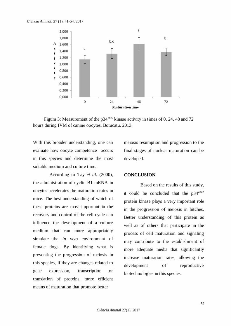
in Fig.3. The p34cdc2 kinase activity at
time 24h when compared to 48h showed a
highly significant difference (p<0.001).
This difference was also observed
comparing times of 48h and 72h. It was
observed that the p34cdc2 kinase activity
peaked at 48h and decreased at 72h of
maturation time. In this way, the time of
48h could be indicated as recommended
for in vitro culture. However, according to
Tsutsui et al. (1989) and Reynaud et al.
(2005), oocytes require more than 48
hours to complete nuclear maturation
within the oviduct. There is controversy
over culture time but it is evident that for
oocyte competence to occur, the medium
must provide nutrients, growth factors and
proteins that simulate the in vivo
environment. Unlike Suzukamo et al.
(2009), this study used epidermal growth
factor (EGF) in the maturation medium.
This factor has been associated to
cascades of cellular signaling. Studies on
porcine oocytes have demonstrated that
EGF-induced maturation promotes MAPK
phosphorylation, raising the hypothesis
that gonadotropin-induced maturation
occurs due to the action of EGFR and
MAPK. EGF regulates MAPK activity by
binding to EGF (EGFR) receptors and
EGFR inhibition is responsible for
blocking meiosis resumption induced by
LH and FSH. Thus, EGF, acting on
MAPK phosphorylation, may have
exerted influence on the phosphorylation
of protein kinases during maturation
progression.
Thus, it is suggested that only the
evaluation of the p34cdc2 protein kinase
activity is not enough to understand the
modifications that occur during the cell
cycle, requiring further studies on other
signaling pathways that interfere in
meiosis, as well as the evaluation of the
phosphorylation of proteins involved in
this process.
50
51
Ciência Animal 27(1), 2017
Ciência Animal, 27 (1); 41-54, 2017
Figura 3: Measurement of the p34cdc2 kinase activity in times of 0, 24, 48 and 72
hours during IVM of canine oocytes. Botucatu, 2013.
With this broader understanding, one can
evaluate how oocyte competence occurs
in this species and determine the most
suitable medium and culture time.
According to Tay et al. (2000),
the administration of cyclin B1 mRNA in
oocytes accelerates the maturation rates in
mice. The best understanding of which of
these proteins are most important in the
recovery and control of the cell cycle can
influence the development of a culture
medium that can more appropriately
simulate the in vivo environment of
female dogs. By identifying what is
preventing the progression of meiosis in
this species, if they are changes related to
gene expression, transcription or
translation of proteins, more efficient
means of maturation that promote better
meiosis resumption and progression to the
final stages of nuclear maturation can be
developed.
CONCLUSION
Based on the results of this study,
it could be concluded that the p34cdc2
protein kinase plays a very important role
in the progression of meiosis in bitches.
Better understanding of this protein as
well as of others that participate in the
process of cell maturation and signaling
may contribute to the establishment of
more adequate media that significantly
increase maturation rates, allowing the
development of reproductive
biotechnologies in this species.
52
Ciência Animal 27(1), 2017
FUNDING SOURCES
São Paulo Research Foundation –
FAPESP, number 2013/21667-3 and
2014/19776-1.
REFERENCES
ALLARD, P.; YANG, Q.; MARZLUFF,
W.F.; CLARKE, H.J.; 2005: The stem-
loop binding protein regulates translation
of histone mRNA during mammalian
oogenesis. Developmental Biology, v.286,
n.1, 2005.
CALDER, M.D.; CAVENEY, A.N.;
SIRARD, M.A.; WATSON, A.J. Effect of
serum and cumulus cell expansion on
marker gene transcripts in bovine
cumulus-oocytes complexes during
maturation in vitro. Fertility and Sterility,
v.83, p.1077-1085, 2005.
CURCIO, B.R.; LEON, P.M.M.;
JUNIOR, F.F.; NOGUEIRA, C.E.W.;
DESCHAMPS, J.C. Equinos: oogênese,
foliculogênese e maturação. Revista
Brasileira de Reprodução Animal, v.20,
p.28-35, 2006.
DE VANTERY, C.; GAVIN, A.C.;
VASSAKKI, J.D.; SCHORDERET-
SLATKINE, S. An accumulation of 34
cdc2 at the end of mouse oocyte growth
correlates with the acquisition of meiotic
competence. Developmental Biology,
v.174, p.335-344, 1996.
GAUTIER, J.; NORBURRY, C.;
LOHKA, M.; NURSE, P.; MALLER, J.
Purified maturation promoting factor
contains the product of a Xenopus
homolog of the fission yeast cell cicle
control gene p34cdc2. Cell, v.54, p.433-
439, 1998.
HAMPL, A.; EPPIG, J.J. Analysis of the
mechanism of metaphase I arrest in
maturating mouse oocytes. Development,
v.121, p.925- 933, 1995.
HEWITT, D.A.; ENGLAND, G.C.W.
1997: The effect of pre-ovulatory
endocrine events upon maturation of
oocytes of the domestic bitch. Journal of
Reproduction and Fertility, v.51, p.83-91,
1997.
JOSEFSBERG, L.B.; GALIANI, D.;
LAZAR, S.; KAUFAMAN, O.; SEGER,
R.; DEKEL N. MPF governs MAPK
activation and interphase suppression
during meiosis of rat oocytes. Biology of
Reproduction, v.68, p.1282-1290, 2003.
KANATSU-SHINOHARA, M.;
SCHULTZ, R.M.; KOPF, G.S.
Acquisition of meiotic competence in
mouse oocytes: Absolute amounts of
53
Ciência Animal 27(1), 2017
p34(cdc2), cyclin b1, cdc25C, and weel in
meiotically incompetent and competent
oocytes. Biology of Reproduction, v.63,
p.1610-1616, 2000.
KOVO, M.; KANDLI-COHEN, M.;
BEN-HAIM, M. An activeprotein kinase
A (PKA) is involved in meiotic arrest of
rat growing oocytes. Society for
Reproduction and Fertility, v.132, p.33-
43, 2006.
LEDAN, E.; POLANSKI, Z.; TERRET,
M-E.; MARO, B. Meiotic maturation of
the mouse oocyte requires an equilibrium
between cyclin B synthesis and
degradation. Developmental Biology,
v.232, p.400-413, 2001.
MERMILLOD, P.; TOMANEK, M.;
MARCHAL, R.; MEIJER, L. High
developmental competence of cattle
oocytes maintained at the germinal vesicle
stage for 24 hours in culture by specific
inhibition of MPF kinase activity.
Molecular Reproduction and
Development, v.55, p.89-95, 2000.
MOTLIK, J.; PAVLOK, A.; KUBELKA,
M.; KALOUS, J.; KALAB, P. Interplay
between cdc2 kinase and MAP kinase
pathway during maturation of mammalian
oocytes. Theriogenolgy, v.49, 461-469,
1998.
MOUROT, M.; DUFORT, I.; GRAVEL,
C.; ALGRIANY, O.; DIELEMAN, S.;
SIRARD, M, A. The influence of follicle
size, FSH-enriched maturation medium
and early cleavage on bovine oocyte
maternal mRNA levels. Molecular
Reproduction and Development, v.73,
n.11, p.1367-1379, 2006.
RACEDO, S.E.; WRENZYCKI, C.;
HERRMANN, D.; SALAMONE, D.;
NIEMANN, H. Effects of follicle size and
stage of maturation on mRNA expression
in bovine in vitro matured oocytes.
Molecular Reproduction and
Development, v.75, p.17-25, 2008.
REYNAUD, K.; FONTBONNE, A.;
MARSELOO, N.; THOUMIRE, S.;
CHEBROUT, M.; VIARIS DE
LESEGNO, C.; CHASTANT-
MAILLARD, S. In vivo meiotic
resumption, fertilization and early
embrionary development in the bitch.
Reproduction, v.130, p.193-201, 2005.
SUZUKAMO, C.; HOSHINA, M.;
MORIYA, H.; HISHIYAMA, N.;
NAKAMURA, S.; KAWAI, F.; SATO,
H.; ARIGA, M.; ITO, J.;
KASHIWAZAKI, N. Kinetics of nuclear
status and kinase activities during in vitro
maturation of canine oocytes. Journal of
54
Ciência Animal 27(1), 2017
Reproduction and Development, v.55, n.2,
p.116-120, 2009.
TAY, J.; HODGMAN, R.; RICHTER, D.
The control of cyclin B1 mRNA
translation during mouse oocyte
maturation. Developmental Biology,
v.221, p.1-9, 2000.
TSUTSUI, T. Gamete physiology and
timing of ovulation and fertilization in
dogs. Journal of Reproduction and
Fertility, v.39, p.269-275, 1989.