arachnoid cyst spontaneous rupture · gemelar em que há uma mola hidatiforme completa e um feto...
TRANSCRIPT

CA
SO C
LÍN
ICO
Revista Científica da Ordem dos Médicos www.actamedicaportuguesa.com 137
gemelar em que há uma mola hidatiforme completa e um feto viável, o casal tem de optar entre interromper a gravi-dez de um feto vivo, sem patologia, ou deixar prosseguir a gestação, enfrentando os riscos de morte fetal e de com-plicações maternas graves. Se o casal optar por prosse-
guir a gravidez, a grávida deve ser referenciada para um centro perinatal diferenciado, e as complicações maternas devem ser rastreadas, com a vigilância da função tiroideia, hemorragia vaginal, sinais e sintomas sugestivos de pré--eclâmpsia e parto pré-termo. Alguns autores9 sugerem a realização de radiografia torácica trimestral para pesquisar metastização pulmonar. O casal deve ser informado que as hipóteses de ter um parto de um recém-nascido vivo e saudável são inferiores a 50%, e que entre 16 a 50% dos casos desenvolvem doença trofoblástica gestacional per-sistente.2,3
CONFLITOS DE INTERESSE Os autores declaram a inexistência de conflitos de inte-resse na realização do presente trabalho.
FONTES DE FINANCIAMENTO Não existiram fontes externas de financiamento para a realização deste artigo.
Figura 4 - Produto de concepção: feto, placenta e mola hidatiforme(fotografia cortesia de Artur Costa e Silva)
REFERÊNCIAS1. Altieri A, Franceschi S, Ferlay J, Smith J, La Vecchia C. Epidemiology
and aetiology of gestational throphoblastic diseases. Lancet Oncol. 2003;4:670-8.
2. Sebire NJ, Foskett M, Paradinas FJ, Fisher RA, Francis RJ, Short D, et al. Outcome of twin pregnancies with complete hydatiform mole and healthy co-twin. Lancet. 2002;359:2165-6.
3. Matsui H, Sekiya S, Hando T, Wake N, Tomoda Y. Hydatiform mole co-existent with a twin live fetus: a national collaborative study in Japan. Hum Reprod. 2000;15:608-11.
4. Taylor JH. Twin pregnancy: Well-formed hydatiform mole associated with normal viable child. BMJ. 1957;1:1103.
5. Piura B, Rabinovich A, Hershkovitz R, Maor E, Mazor M. Twin pregnan-cy with a complete hydatiform mole and a surviving co-existent fetus. Arch Gynecol Obstet. 2008;278:377-82.
6. Marcorelles P, Audrezet MP, Le Bris MJ, Laurent Y, Chabaud JJ, Ferec C, et al. Diagnosis and outcome of complete hydatiform mole coexisting with a live twin fetus. Eur J Obstet Gynecol Reprod Biol 2005;118:21-7.
7. Malhotra N, Deka D, Takkar D, Kochar S, Goel S, Sharma MC. Hydati-form mole with a coexisting live fetus in dichorionic twin gestation. Eur J Obstet Gynecol Reprod Biol. 2001;94:301-3.
8. Choi-Hong SR, Genest DR, Crum CP, Berkowitz E, Goldstein DP, Scho-field DH. Twin pregnancies with complete hydatiform mole and coexist-ing fetus: Use of fluorescent in situ hybridization to evaluate placental X- and Y- cromossomal content. Hum Pathol. 1995;26:1175-80.
9. Wee L, Jauniaux E. Prenatal diagnosis and management of twin preg-nancies complicated by a co-existing molar pregnancy. Prenat Diagn. 2005;25:772-6.
Arachnoid Cyst Spontaneous Rupture
Rotura Espontânea de Quisto Aracnóide
Inês Brás MARQUES1, J. VIEIRA BARBOSA1
Acta Med Port 2014 Jan-Feb;27(1):137-141
1. Serviço de Neurologia. Centro Hospitalar e Universitário de Coimbra. Coimbra. Portugal.Recebido: 14 de Fevereiro de 2013 - Aceite: 15 de Junho de 2013 | Copyright © Ordem dos Médicos 2014
anos35
35 anos a promover as ciências biomédicas
ACTA M
ÉDIC
A PO
RTUG
UESA
1979 - 2014
ABSTRACTArachnoid cysts are benign congenital cerebrospinal fluid collections, usually asymptomatic and diagnosed incidentally in children or adolescents. They may become symptomatic after enlargement or complications, frequently presenting with symptoms of intracranial hypertension. We report an unusual case of progressive refractory headache in an adult patient due to an arachnoid cyst spontaneous rupture. Although clinical improvement occurred with conservative treatment, the subdural hygroma progressively enlarged and surgi-cal treatment was ultimately needed. Spontaneous rupture is a very rare complication of arachnoid cysts. Accumulation of cerebro-spinal fluid accumulation in the subdural space causes sustained intracranial hypertension that may be life-threatening and frequently requires surgical treatment. Patients with arachnoid cysts must be informed on their small vulnerability to cyst rupture and be aware that a sudden and severe headache, especially if starting after minor trauma or a Valsalva manoeuvre, always requires medical evaluation. Keywords: Arachnoid Cysts/complications; Rupture, Spontaneous.
Marques IB, et al. Arachnoid cyst spontaneous rupture, Acta Med Port 2014 Jan-Feb;27(1):137-141
CA
SO C
LÍNIC
O
138Revista Científica da Ordem dos Médicos www.actamedicaportuguesa.com
Marques IB, et al. Arachnoid cyst spontaneous rupture, Acta Med Port 2014 Jan-Feb;27(1):137-141
INTRODUCTION Arachnoid cysts (ACs) are benign congenital collec-tions of cerebrospinal fluid (CSF) surrounded by histologi-cally normal layers of the arachnoid membrane. They are believed to result from a dysgenesis in embryological de-velopment with anomalous splitting of the normally fused inner and outer layers of the arachnoid membrane followed by secretion of fluid by the arachnoid cells into the resulting cleft.1-3 ACs represent about 1% of all intracranial space- -occupying lesions, being a common incidental neuro- imaging finding.1,4,5 They are usually diagnosed during childhood and adolescence, with male gender predomi-nance. Their most common location is the middle cranial fossa, with predilection for the left side.1,2,5-8
In most cases, ACs remain small and asymptomatic.2,9 Occasionally, they become manifest, especially when large and located in the middle cranial fossa.6 Signs and symp-toms usually follow cyst enlargement with compression or irritation of adjacent structures, intracranial mass effect and/or CSF circulation disturbance.1
Clinical manifestations depend on various factors, in-cluding age and cyst location and size. Headache is the most common presentation, but they may also present with seizures, intracranial hypertension or focal motor deficits. In children they may cause macrocephaly and delayed psy-chomotor development.1,7,9
Although infrequent, more serious and potentially life-threatening complications may occur, making ACs not so benign as frequently believed.2,6,7 Head trauma can origin rupture of blood vessels around the cyst wall or of the un-supported bridging veins in cyst cavity, producing intracystic and/or subdural hemorrhage.1-3,5,7 Traumatic or spontane-
ous cyst rupture has been reported rarely, mainly in chil-dren.1-11 In this article we report an unusual case of ara- chnoid cyst spontaneous rupture in an adult patient.
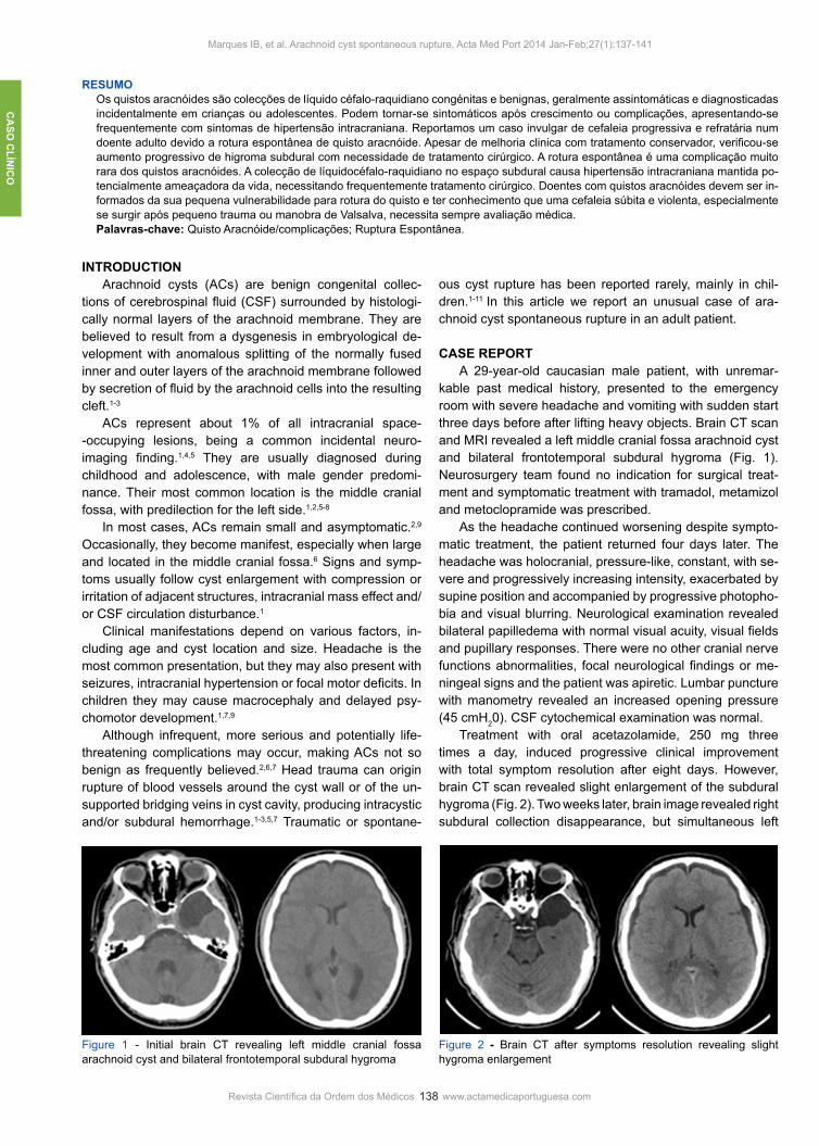
CASE REPORT A 29-year-old caucasian male patient, with unremar- kable past medical history, presented to the emergency room with severe headache and vomiting with sudden start three days before after lifting heavy objects. Brain CT scan and MRI revealed a left middle cranial fossa arachnoid cyst and bilateral frontotemporal subdural hygroma (Fig. 1). Neurosurgery team found no indication for surgical treat-ment and symptomatic treatment with tramadol, metamizol and metoclopramide was prescribed. As the headache continued worsening despite sympto-matic treatment, the patient returned four days later. The headache was holocranial, pressure-like, constant, with se-vere and progressively increasing intensity, exacerbated by supine position and accompanied by progressive photopho-bia and visual blurring. Neurological examination revealed bilateral papilledema with normal visual acuity, visual fields and pupillary responses. There were no other cranial nerve functions abnormalities, focal neurological findings or me-ningeal signs and the patient was apiretic. Lumbar puncture with manometry revealed an increased opening pressure (45 cmH20). CSF cytochemical examination was normal. Treatment with oral acetazolamide, 250 mg three times a day, induced progressive clinical improvement with total symptom resolution after eight days. However, brain CT scan revealed slight enlargement of the subdural hygroma (Fig. 2). Two weeks later, brain image revealed right subdural collection disappearance, but simultaneous left
RESUMOOs quistos aracnóides são colecções de líquido céfalo-raquidiano congénitas e benignas, geralmente assintomáticas e diagnosticadas incidentalmente em crianças ou adolescentes. Podem tornar-se sintomáticos após crescimento ou complicações, apresentando-se frequentemente com sintomas de hipertensão intracraniana. Reportamos um caso invulgar de cefaleia progressiva e refratária num doente adulto devido a rotura espontânea de quisto aracnóide. Apesar de melhoria clinica com tratamento conservador, verificou-se aumento progressivo de higroma subdural com necessidade de tratamento cirúrgico. A rotura espontânea é uma complicação muito rara dos quistos aracnóides. A colecção de líquidocéfalo-raquidiano no espaço subdural causa hipertensão intracraniana mantida po-tencialmente ameaçadora da vida, necessitando frequentemente tratamento cirúrgico. Doentes com quistos aracnóides devem ser in-formados da sua pequena vulnerabilidade para rotura do quisto e ter conhecimento que uma cefaleia súbita e violenta, especialmente se surgir após pequeno trauma ou manobra de Valsalva, necessita sempre avaliação médica.Palavras-chave: Quisto Aracnóide/complicações; Ruptura Espontânea.
Figure 1 - Initial brain CT revealing left middle cranial fossa arachnoid cyst and bilateral frontotemporal subdural hygroma
Figure 2 - Brain CT after symptoms resolution revealing slight hygroma enlargement

CA
SO C
LÍN
ICO
Revista Científica da Ordem dos Médicos www.actamedicaportuguesa.com 139
Figure 3 - Brain CT after three weeks of acetazolamide treatment revealing enlargement of the left subdural hygroma
Figure 4 - Brain CT after two months, revealing left subdural hygro-ma enlargement with mass effect and midline shift
Figure 5 - Post-operative brain CT scan with subdural hygroma resolution and reduction of arachnoid cyst volume
subdural hygroma enlargement with minor midline shift (Fig. 3). Neurosurgery team decided to maintain a conservative approach with watchful waiting, as the patient was asymp-tomatic and clinically stable. A control CT scan scheduled for two months later identi-fied enlargement of the left subdural hygroma with mass effect and larger contralateral displacement of midline struc-tures (Fig. 4). Despite the absence of symptoms, urgent craniotomy with evacuation of subdural hygroma and cyst fenestration was performed with satisfactory imagiologic outcome (Fig. 5) and without clinical complications.
DISCUSSION Rupture is a very uncommon complication of arachnoid cysts, with less than fifty cases of either traumatic or spon-taneous rupture reported in the literature, the vast majority in children and adolescents.2-17 The rarity of this entity, especially in adulthood, probably explains the frequent lack of clinical suspicion in adults with progressive refractory headache. We present a summary of the patients with spontaneous ACs rupture reported in literature in Table 1.6,7,9-16 All cases occurred in children or adolescents (age range: 5-16 years), with our patient being the first adult reported with spontane-ous AC rupture. In the totality of the cases with spontaneous rupture, cysts were located in the middle cranial fossa and intracranial hypertension manifestations were present with headache being a constant symptom. The most common finding in examination was bilateral papilledema and a fo-cal neurological deficit (right hemiparesis) was present in one patient. Subdural hygroma was ipsilateral to the cyst in all but one of the cases, a patient with bilateral subdural collections in similarity to our patient. Surgical treatment was performed in all cases, except in one patient who responded to conservative treatment. Good clinical and radiological outcome was reported in the totality of cases. Two mechanisms have been proposed to explain ACs rupture.10 The first one would be a minor head injury able to create a tear in the subarachnoid cyst wall and a commu-nication with subarachnoid space. A flap-valve mechanism, only allowing CSF entering into the cyst, progressively increases cyst internal pressure with later disruption into the subdural space. In the second mechanism, spontane-
ous rupture of the cyst into the subdural space follows a sudden transient intracranial pressure increase during a Valsalva manoeuvre. CSF accumulation in the subdural space results in subdural hygroma and subsequent intracranial hypertension.1-17
In clinically stable patients, a good response to conser-vative treatment with acetazolamide has been reported with resolution of hygroma and intracranial hypertension mani-festations, albeit not affecting the cyst dimension.9,17 Al-though our patient symptoms resolved with acetazolamide, the hygroma progressively enlarged and surgical treatment was ultimately necessary. The best treatment approach for ACs remains contro-versial.4,5,8 An expectant attitude is advised for small and asymptomatic cysts, while surgical treatment is indicated for symptomatic cysts, enlarging cysts, cysts with mass effect and after cyst complications.1,5,9
The main objective of surgical treatment is cyst decom-pression. The currently preferred surgical approach is cyst fenestration which establishes an adequate communica-tion between the cyst cavity and the normal CSF flow path-way.1,2,4 This procedure can be performed by craniotomy or endoscopically, both being safe and effective.1,2,4,6 The sur-gical procedure choice depends mainly on surgeon experi-ence and preference, as surgical complications are essen-tially related to cyst location and size rather than the method used.1 Cystoperitoneal shunt is an alternative that reduces potential complications from sudden decompression of the surrounding brain, but has inconveniences as dependence on a valvular system and risk of obstruction or infection.1,5
Marques IB, et al. Arachnoid cyst spontaneous rupture, Acta Med Port 2014 Jan-Feb;27(1):137-141

CA
SO C
LÍNIC
O
140Revista Científica da Ordem dos Médicos www.actamedicaportuguesa.com
Table 1 – Summary of patients with spontaneous arachnoid cyst rupture
Reference Gender Age (years)
Cyst Location
Clinical Presentation
Neurological Examination
Subdural Collections Treatment Outcome
Cullis P, Gilroy J, 198310 M 11 Left MCF
Headache and vomiting
Fundoscopy with “capillary blush and blurred disk margins”
Ipsilateral SD hygroma
Craniotomy and cyst fenestration
NR
Albuquerque FC, Giannotta SL 19977
NR 10 Left MCFHeadache, nausea and
vomitingNR
Ipsilateral SD hygroma
Subdural-peritoneal shunt
Symptoms and hygroma resolution; Cyst reduction
Ergun et al 199711 M 14 Left MCF
Headache, nausea, vomiting and right
hemiparesis
Bilateral papilledema, mild right hemiparesis
Ipsilateral SD hygroma
Craniotomy and cyst fenestration
Symptoms resolution; Hygroma and cyst reduction
Sener 199712 M 12 Left MCF“signs and
symptoms of increased IP”
“signs and symptoms of increased IP”
Ipsilateral SD hygroma
NR NR
Sener 199712 M 16 Left MCF“signs and
symptoms of increased IP”
“signs and symptoms of increased IP”
Ipsilateral SD haematoma
NR NR
Choong et al 19989 F 9 Left MCF
Headache and vomiting
Bilateral papilledema
Bilateral SD hygroma
Conservative (Acetazolamide)
Symptoms and hygroma resolution
Cayli et al 200013 F 12 Left MCF
Headache, nausea and
vomiting
Bilateral papilledema
Ipsilateral SD hygroma
Craniotomy and cyst fenestration
Symptoms resolution; Hygroma and cyst reduction
Cakir et al 200314 M 9 Right MCF
Headache, nausea and
vomitingNormal
Ipsilateral SD hygroma
Subdural-peritoneal shunt
Symptoms resolution; Hygroma and cyst reduction
Poirrier et al 200415 M 15 Right MCF
Headache, blurred vision,
nausea, vomiting, anorexia,
epigastric pain and diaphoresis
NormalIpsilateral SD hygroma
Subdural-peritoneal shunt
Symptoms and hygroma resolution
Slaviero et al 20086 M 5 Left MCF
Headache and drowsiness
NormalIpsilateral SD hygroma
Hygroma evacuation and endoscopic cyst fenestration
Symptoms and hygroma resolution; Cyst reduction
Gil-Gouveia et al 201016 F 16 Left MCF
Headache, nausea
photophobia and diplopia
NormalIpsilateral SD hygroma
Subdural-peritoneal shunt
Symptoms and hygroma resolution
M – male; F – female; MCF –Middle cranial fossa; NR – not reported; SD – subdural; IP – intracranial pressure
Marques IB, et al. Arachnoid cyst spontaneous rupture, Acta Med Port 2014 Jan-Feb;27(1):137-141

CA
SO C
LÍN
ICO
Revista Científica da Ordem dos Médicos www.actamedicaportuguesa.com 141
A recent study about ACs complicated with subdural collec-tions described good clinical results after subdural effusions evacuation, without cyst intervention.5
In our patient, additionally to subdural collection evacu-ation, surgical cyst fenestration was performed as a preven-tive measure of further cyst complications. Arachnoid cyst rupture is a very rare complication of ACs and an extremely uncommon cause of headache. Patients must be informed on their small vulnerability to cyst rupture or hemorrhage and the relationship of those rare events with activities with increased risk for cranial trauma or with extreme increase in intracranial pressure. It is also important to explain these patients that a sudden and violent headache, especially after trauma or manoeuvres
increasing intracranial pressure, always requires medical evaluation.
CONFLICTS OF INTEREST None stated. This clinical case was previously delive-red at the following scientific meetings: Autumn Meeting of the Sociedade Portuguesa de Cefaleias, November 18 and 19, 2011, Coimbra, Portugal (Oral Communication); 16th Congress of the European Federation Neurological Socie-ties (EFNS), September 8 to 11, 2012; Stockholm, Sweden (Poster).
FUNDING SOURCES None stated.
REFERENCES1. Vega-Sosa A, de Obieta-Cruz E, Hernández-Rojas MA. Intracranial ara-
chnoid cyst. Cir Cir. 2010;78:551-6. 2. Hamada H, Hayashi N, Umemura K, Kurosaki K, Endo S. Middle cranial
fossa cyst presenting with subdural effusion and endoscopic detection of tear of the cyst – case report. Neurol Med Chir. 2010;50:512-4.
3. Offiah C, St Clair Forbes W, Thorne J. Non-haemorrhagic subdural col-lection complicating rupture of a middle cranial fossa arachnoid cyst. Br J Radiol. 2006;79:79-82.
4. Gelabert-González M, Fernández-Villa J, Cutrín-Prieto J, Garcìa Allut A, Martínez-Rumbo R. Arachnoid cyst rupture with subdural hygroma: re-port of three cases and literature review. Childs Nerv Syst. 2002;18:609-13.
5. Sprung C, Armbruster B, Koeppen D, Cabraja M. Arachnoid cysts of the middle cranial fossa accompanied by subdural effusions—experience with 60 consecutive cases. Acta Neurochir. 2011;153:75-84.
6. Slaviero F, Frighetto L, Azambuja Júnior ND, Martins LS, Annes RD, Vanzin JR. Middle cranial fossa arachnoid cysts complicated with sub-dural collections. Arq Neuropsiquiatr. 2008;66:913-5.
7. Albuquerque FC, Giannotta SL. Arachnoid cyst rupture producing sub-dural hygroma and intracranial hypertension: case reports. Neurosur-gery. 1997;41:951-6.
8. Gupta R, Vaishya S, Mehta VS. Arachnoid cyst presenting as subdural hygroma. J Clin Neurosci. 2004; 11:317-8.
9. Choong CT, Lee SH. Subdural hygroma in association with middle fossa arachnoid cyst: acetazolamide therapy. Brain Dev. 1998;20:319-22.
10. Cullis P, Gilroy J. Arachnoid cyst with rupture into the subdural space. J Neurol Neurosurg Psychiatry. 1983;46:454-6.
11. Ergun R, Ökten AI, Beșkonakli E, Anasiz H, Ergüngör F, Tașkin Y. Unu-sual complication of arachnoid cyst: Spontaneous rupture into the sub-dural space. Acta Neurochir. 1997;139:692-4.
12. Sener RN. Arachnoid cysts associated with post-traumatic and spon-taneous rupture into the subdural space. Comput Med Imaging Graph. 1997;21:341-4.
13. Cayli SR. Arachnoid cyst with spontaneous rupture into the subdural space. Br J Neurosurg 2000;14:568-70.
14. Cakir E, Kayhankuzeyli; Sayin OC, Peksoylu B, Karaarslan G. Arach-noid cyst rupture with subdural hygroma: case report and literature re-view. Neurocirurgía. 2003;14:72-5.
15. Poirrier AL, Ngosso-Tetanye I, Mouchamps M, Misson JP. Spontaneous arachnoid cyst rupture in a previously asymptomatic child: a case report. Eur J Paediatr Neurol. 2004;8:247-51
16. Gil-Gouveia R, Miguens J, Coiteiro D. Rupture of middle fossa arachnoid cyst simulating new-onset migraine with aura. Headache. 2010;50:314-9.
17. Longatti P, Marton E, Billeci D. Acetazolamide and corticosteroid ther-aphy in complicated arachnoid cyst. Childs Nerv Syst. 2005;21:1061-4.
Protocolo de Actuação Hospitalar na Abordagem de Mordedura de Ofídio
Hospital Protocol for Snakebite Victims Management
Pedro MARQUES DA COSTA1,2, Rodrigo SOUSA1, Maria LOBO ANTUNES1, Sara AZEVEDO1,2, Gabriela ARAÚJO E SÁ1,2, Maria do Céu MACHADO1,2
Acta Med Port 2014 Jan-Feb;27(1):141-145
1. Departamento de Pediatria. Hospital de Santa Maria. Centro Hospitalar Lisboa Norte. Lisboa. Portugal.2. Faculdade de Medicina. Universidade de Lisboa. Lisboa. Portugal.Recebido: 18 de Fevereiro de 2013 - Aceite: 08 de Setembro de 2013 | Copyright © Ordem dos Médicos 2014
anos35
35 anos a promover as ciências biomédicas
ACTA M
ÉDIC
A PO
RTUG
UESA
1979 - 2014
RESUMOA mordedura de ofídio venenoso é uma situação rara em Portugal. Quando ocorre em idade pediátrica apresenta maior gravidade e risco de complicações fatais. A actuação protocolada constitui o gold standard of care nos centros internacionais. Neste artigo apresentamos dois casos clínicos de mordedura de ofídio venenoso em idade pediátrica. Baseados na literatura actual, discutimos a actuação clínica tomada e expomos uma proposta de protocolo de actuação hospitalar que visa uma intervenção multidisciplinar e actualizada das equipas médicas envolvidas.Palavras-chave: Antivenenos; Mordeduras de Serpentes; Portugal.
Marques da Costa P, et al. Protocolo actuação hospitalar na abordagem de mordedura de ofídio, Acta Med Port 2014 Jan-Feb;27(1):141-145

Inês Brás MARQUES, J. VIEIRA BARBOSA
Arachnoid Cyst Spontaneous RuptureActa Med Port 2014:27:137-141
Publicado pela Acta Médica Portuguesa, a Revista Científica da Ordem dos Médicos
Av. Almirante Gago Coutinho, 151 1749-084 Lisboa, Portugal.
Tel: +351 218 428 215 E-mail: [email protected]
www.actamedicaportuguesa.comISSN:0870-399X | e-ISSN: 1646-0758