universidade estadual do ceará - portal da uece · 2011-02-10 · machado luz, isadora machado...
TRANSCRIPT

0
UNIVERSIDADE ESTADUAL DO CEARÁ
PRÓ-REITORIA DE PÓS-GRADUAÇÃO E PESQUISA
FACULDADE DE VETERINÁRIA
PROGRAMA DE PÓS-GRADUAÇÃO EM CIÊNCIAS VETERINÁRIAS
JAMILY BEZERRA BRUNO
CULTIVO IN VITRO DE FOLÍCULOS PRÉ-ANTRAIS CAPRINOS
UTILIZANDO ANGIOTENSINA II (ANG II), FATOR DE
CRESCIMENTO DO ENDOTÉLIO VASCULAR (VEGF) E PEPTÍDEO
INTESTINAL VASOATIVO (VIP)
FORTALEZA
2010

1
JAMILY BEZERRA BRUNO
CULTIVO IN VITRO DE FOLÍCULOS PRÉ-ANTRAIS CAPRINOS
UTILIZANDO ANGIOTENSINA II (ANG II), FATOR DE
CRESCIMENTO DO ENDOTÉLIO VASCULAR (VEGF) E PEPTÍDEO
INTESTINAL VASOATIVO (VIP)
FORTALEZA
2010
Tese apresentada ao Programa de Pós-Graduação em
Ciências Veterinárias da Faculdade de Veterinária da
Universidade Estadual do Ceará, como requisito parcial
para obtenção do título de Doutor em Ciências
Veterinárias.
Área de Concentração: Reprodução e Sanidade Animal.
Linha de pesquisa: Reprodução e sanidade de pequenos
ruminantes.
Orientador: Prof. Dr. José Ricardo de Figueiredo

2
B898c Bruno, Jamily Bezerra
Cultivo in vitro de folículos pré-antrais caprinos
utilizando angiotensina II (ANG II), fator de crescimento do
endotélio vascular (VEGF) e peptídeo intestinal vasoativo
(VIP) / Jamily Bezerra Bruno. — Fortaleza, 2010.
222 p. ; il.
Orientador: Prof. Dr. José Ricardo de Figueiredo.
Tese (Programa de Pós-Graduação em Ciências
Veterinárias – Doutorado em Ciências Veterinárias) –
Universidade Estadual do Ceará, Faculdade de Veterinária.
1. Caprino. 2. Reprodução animal. 3. Folículos ovarianos. I.
Universidade Estadual do Ceará, Faculdade de Veterinária.
CDD: 636.08

3
JAMILY BEZERRA BRUNO
CULTIVO IN VITRO DE FOLÍCULOS PRÉ-ANTRAIS CAPRINOS
UTILIZANDO ANGIOTENSINA II (ANG II), FATOR DE
CRESCIMENTO DO ENDOTÉLIO VASCULAR (VEGF) E PEPTÍDEO
INTESTINAL VASOATIVO (VIP)
Aprovada em: 14/12/2010
Conceito obtido: Satisfatório
Nota: 9,5
BANCA EXAMINADORA
___________________________________
Prof. Dr. José Ricardo de Figueiredo
Universidade Estadual do Ceará
Orientador
___________________________________
Profa. Dra. Maria Helena Tavares de Matos
Universidade Federal do Vale do São Francisco
Co-orientadora / Examinadora
___________________________________
Prof. Dr. José Roberto Viana Silva
Universidade Federal do Ceará
Co-orientador/ Examinador
___________________________________
Profa. Dra. Sônia Nair Báo
Universidade de Brasília
Examinadora
___________________________________
Profa. Dra. Luciana Relly Bertolini
Universidade de Fortaleza
Examinadora
___________________________________
Dr. Cláudio Afonso Pinho Lopes
Universidade Estadual do Ceará
Examinador
Tese apresentada ao Programa de Pós-Graduação em
Ciências Veterinárias da Faculdade de Veterinária da
Universidade Estadual do Ceará, como requisito
parcial para obtenção do título de Doutor em Ciências
Veterinárias.

4
Aos meus pais,
ao meu marido,
à minha filha,
dedico.

5
Agradecimentos
Agradeço à Universidade Estadual do Ceará (UECE), pela oportunidade de realização
do Curso de Doutorado em Ciências Veterinárias.
À Fundação Cearense de Apoio ao Desenvolvimento Científico e Tecnológico
(FUNCAP) e a Coordenação de Aperfeiçoamento do Pessoal de Nível Superior (CAPES),
pela concessão da bolsa de estudos durante a realização do curso de doutorado, fato este que
muito contribuiu para a viabilização desta tese.
Ao Conselho Nacional de Desenvolvimento Científico e Tecnológico (CNPq), à Rede
Nordeste de Biotecnologia (RENORBIO) e à Financiadora de Estudos e Projetos (FINEP),
pelo suporte finaceiro.
Ao Laboratório de Manipulação de Oócitos e Folículos Ovarianos Pré-Antrais
(LAMOFOPA) e ao Laboratório de Virologia (LABOVIR), pelo suporte técnico.
Agradeço a DEUS por tudo, em especial por mais esse presente que me deste: minha
filha!
Aos meus pais, Marcos Antônio Campos Bezerra e Marta Maria de Sousa Bezerra,
pelo amor, dedicação, educação, pelo incentivo e apoio que têm me dado durante todo esse
tempo. Obrigada pela presença constante em minha vida e pela união que nos torna cada vez
mais ligados. Amo vocês!
Ao meu marido, Affonso Bruno Neto, e minha filha, Sarah Bezerra Bruno, pelo amor
e carinho. Não existem palavras que sejam capazes de expressar todo o meu amor por vocês.
Aos meus irmãos, Marcos Antônio Campos Bezerra Júnior, Bruna de Sousa Bezerra e
Emmanuelle de Sousa Bezerra, por tudo que passamos juntos, e às minhas sobrinhas Mikaelle
Sabatinne de Souza Medeiros e Sarah Mirelle de Souza Zuza, pelas grandes alegrias vividas.
6
Agradeço em especial a Bruna, mesmo que a palavra ―obrigada‖ signifique tanto, não
expressará por inteiro o quanto seu gesto atencioso e delicado foi importante para mim.
Ao Dr. José Ricardo de Figueiredo, por ter acreditado em mim para assumir
responsabilidades de sua confiança. Eu o agradeço pela orientação, incentivo e paciência.
À Dra. Maria Helena Tavares de Matos, pela co-orientação e amizade. Você foi
essencial durante essa caminhada, não sei se teria conseguido sem a sua ajuda. Obrigada por
nunca ter me faltado quando eu precisei de você. Obrigada pelas palavras, ensinamentos,
incentivo e atenção dispensada no decorrer desse período. É difícil expressar em palavras o
meu carinho, amizade e a admiração por você!
Ao Dr. José Roberto Viana Silva e Dr. Cláudio Cabral Campello, pela disponibilidade,
prestatividade, colaboração e pelos conhecimentos repassados.
À Dra. Ana Paula Ribeiro Rodrigues e à Dra. Liliam Mara Trevisan Tavares, pelo
carinho e amizade e pelo exemplo de pesquisadoras.
À Dra. Sônia Nair Báo e à Dra Cristina Alves Peixoto, por colocarem o Laboratório de
Microscopia Eletrônica de Transmissão, à disposição da equipe LAMOFOPA, bem como suas
alunas, Khesller Patrícia Olázia Name e Mariana Aragão Matos Donato, pela valiosa ajuda.
À banca examinadora, por ter aceitado prontamente o convite e, sobretudo pela sua
valiosa colaboração ao corrigir o presente trabalho.
Aos professores do Programa de Pós-Graduação em Ciências Veterinárias (PPGCV),
pelo conhecimento e experiência compartilhados e ao coordenador do programa Marcos Fábio
Gadelha Rocha pelo excelente trabalho desenvolvido.
7
Às secretárias, Adriana Maria Sales Albuquerque, Ana Cristina Sabóia Nascimento e
Alzenira de Andrade, e o funcionário, Antônio César Camelo, pela valiosa ajuda, atenção e
respeito sempre dispensados. César seu carinho e ―cafezinhos‖ ajudaram muito nessa
caminhada.
Às minhas amigas Cristiane de Farias Mendes, Cristina Moreiro Ribeiro, Iara Tersia
Freitas Macedo, Isabel Bezerra Lima Verde, Kamila Marques Andrade, Maria Liduína Maia
de Oliveira, Neyva Torres de Souza Cartaxo e Sanely Lourenço, pela amizade sincera, pelas
energias positivas transmitidas e pelos momentos inesquecíveis.
À minha amiga Juliana Jales de Hollanda Celestino, que desde o primeiro dia do
doutorado me acompanhou, contribuiu, ensinou, incentivou de diferentes maneiras e apoiou
em muitos momentos de dificuldade. Juliana, esta jornada teria sido muito mais difícil sem
você.
Aos meus amigos do LAMOFOPA, Anderson Pinto Almeida, Cleidson Manoel
Gomes da Silva, Deborah de Melo Magalhães, Giovanna Quintino Rodrigues, Hiédely Kenia
Machado Luz, Isadora Machado Teixeira Lima, Ívina Rocha Brito, Leonardo Correia Pinto,
Lívia Schell Wanderley, Luciana Rocha Faustino, Marcella Moreira Clemente de Mello Pinto,
Rafael Rossetto de Sousa, Rebeca Magalhães Pedrosa Rocha, Valdevane Rocha Araújo,
Valesca Barreto Luz, e especialmente ao Fabrício Sousa Martins, Márcia Viviane Alves
Saraiva e Roberta Nogueira Chaves, que participaram de toda essa jornada, acompanhando
momentos de dificuldade e outros de intensa alegria. Agradeço pela amizade, pelo incentivo,
pelo apoio, pela ajuda, pela paciência, pelo entusiasmo, pelos ensinamentos e pelos momentos
de descontração. Obrigada por todos os momentos que passamos juntos!
Meus agradecimentos especiais para minhas ―pupilas‖: Laritza Ferreira de Lima,
Anelise Maria Costa V. Alves e meu ―filhote‖ Márcio Breno Sampaio Mororó, pela amizade e
inestimável ajuda no decorrer desse período.

8
Aos alunos de iniciação científica, Emmanuel Teles Sales e Mirlla Baracho Ferreira,
pela ajuda prestada para tese.
Aos demais integrantes do LAMOFOPA, pelo espírito de cooperação.
A todos que não foram aqui mencionados, mas que direta ou indiretamente me
ajudaram durante a realização deste trabalho. Os meus mais sinceros agradecimentos.

9
Lembre-se de que você mesmo é:
o melhor secretário de sua tarefa, o mais eficiente propagandista de seus ideais, a mais clara
demonstração de seus princípios e o mais alto padrão de ensino superior que seu espírito abraça.
Não se esqueça igualmente de que: o maior inimigo de suas realizações mais nobres, a completa
ou incompleta negação do idealismo sublime que você apregoa, o arquiteto de suas aflições e o
destruidor de suas oportunidades de elevação – é você mesmo.
Francisco Cândido Xavier

10
Resumo
Os objetivos do presente estudo foram avaliar o efeito de diferentes concentrações da
angiotensina II (ANG II), fator de crescimento do endotélio vascular (VEGF) e peptídeo
intestinal vasoativo (VIP) no desenvolvimento in vitro de folículos pré-antrais caprinos e
verificar a localização do receptor 2 do VEGF (VEGFR-2) e a expressão dos receptores da
ANG II (AGTR1 e AGTR2) em ovários caprinos. Para investigar a localização do VEGFR-2
e expressão do AGTR1 e AGTR2 no ovário caprino, foram empregadas as técnicas de
imunohistoquímica e PCR em tempo real, respectivamente. Para avaliar a eficiência das
substâncias supracitadas sobre a sobrevivência e o crescimento folicular, fragmentos de córtex
ovariano foram cultivados in vitro por um ou sete dias em meio essencial mínimo
suplementado (MEM+) adicionado de diferentes concentrações das referidas substâncias.
Antes e após o período de cultivo, os fragmentos de córtex ovariano foram fixados e
processados para histologia clássica. Os folículos foram classificados quanto ao grau de
desenvolvimento em primordiais ou em desenvolvimento, e quanto à sobrevivência em
normais ou degenerados. O diâmetro folicular foi avaliado antes e após o cultivo. As técnicas
de microscopia de fluorescência e microscopia eletrônica de transmissão (MET) foram
utilizadas para melhor avaliar a viabilidade e morfologia desses folículos, respectivamente.
Após definição das melhores concentrações de VIP, foi realizado um último experimento a
fim de quantificar a expressão dos RNAm para o VIP em ovários caprinos através da técnica
de PCR em tempo real, e avaliar a influência do VIP isoladamente ou em associação ao FSH
sobre a sobrevivência e o desenvolvimento de folículos secundários caprinos isolados, bem
como sobre a expressão do RNAm para o VIP e receptor de FSH (FSH-R) nesses folículos
cultivados por seis dias. Os resultados do cultivo in vitro mostraram que após sete dias, 10
ng/mL de ANG II promoveu a manutenção da viabilidade e ultraestrutura de folículos pré-
antrais caprinos. Os RNAm para AGTR1 e AGTR2 foram detectados em todas categorias e
tipos foliculares estudados. No que se refere ao VEGF, as concentrações de 10 e 200 ng/mL
foram mais eficientes para promover o crescimento e manter a viabilidade, respectivamente.
Além disso, o receptor VEGFR-2 foi encontrado em oócitos de folículos em todos os estágios
de desenvolvimento e em células da granulosa de folículos em crescimento. O VIP foi mais
efetivo em promover o crescimento e manutenção da ultraestrutura folicular após sete dias de

11
cultivo quando utilizado na concentração de 10 ng/mL. A presença de RNAm para o VIP foi
demonstrada em todas categorias foliculares em ovários de cabras, mostrando um aumento em
seus níveis durante a transição de folículo primordial para secundário. No entanto, a utilização
de VIP e/ou FSH não afetou o desenvolvimento de folículos secundários e não alterou os
níveis de RNAm para FSH-R embora tenha reduzido os níveis de RNAm para o VIP após
cultivo in vitro de folículos pré-antrais por seis dias. Diante disso, concluiu-se que ANG,
VEGF e VIP apresentam importantes funções no desenvolvimento in vitro de folículos pré-
antrais caprinos.
Palavras-chave: ANG II. VIP. VEGF. Pré-antral. Cultivo in vitro.
12
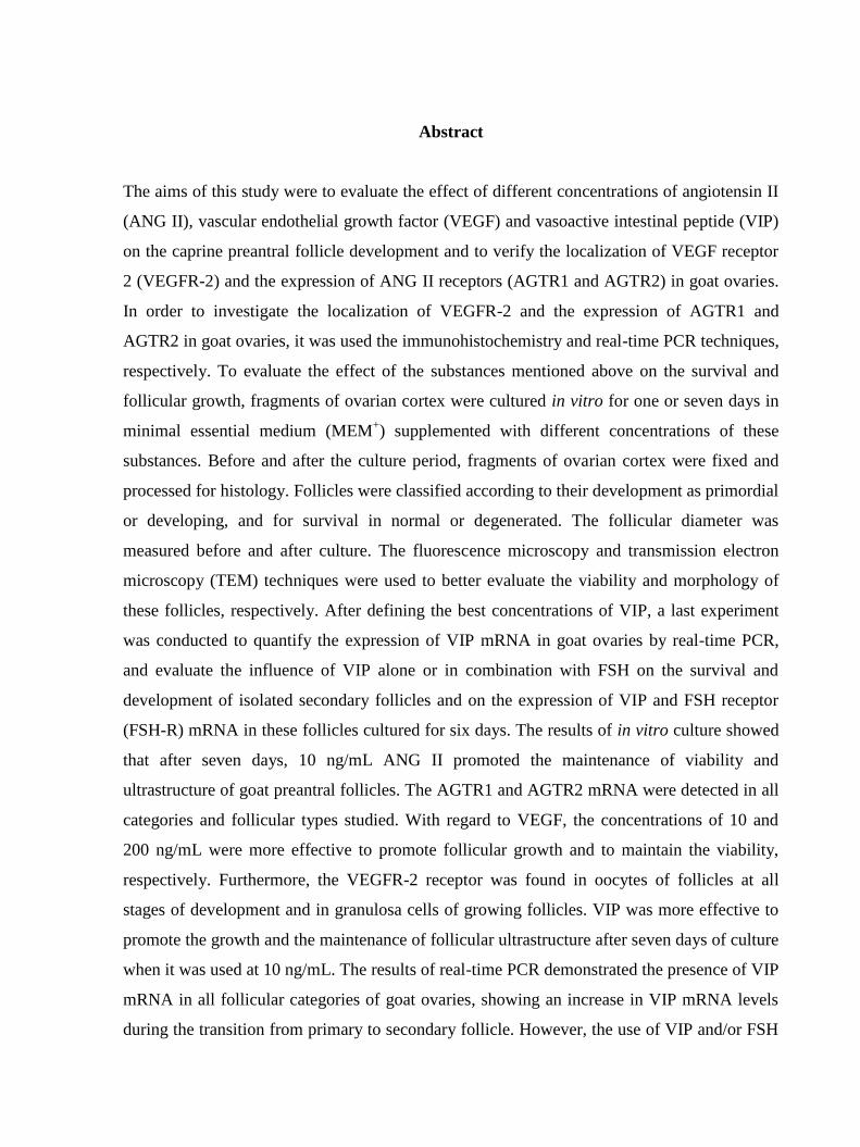
Abstract
The aims of this study were to evaluate the effect of different concentrations of angiotensin II
(ANG II), vascular endothelial growth factor (VEGF) and vasoactive intestinal peptide (VIP)
on the caprine preantral follicle development and to verify the localization of VEGF receptor
2 (VEGFR-2) and the expression of ANG II receptors (AGTR1 and AGTR2) in goat ovaries.
In order to investigate the localization of VEGFR-2 and the expression of AGTR1 and
AGTR2 in goat ovaries, it was used the immunohistochemistry and real-time PCR techniques,
respectively. To evaluate the effect of the substances mentioned above on the survival and
follicular growth, fragments of ovarian cortex were cultured in vitro for one or seven days in
minimal essential medium (MEM+) supplemented with different concentrations of these
substances. Before and after the culture period, fragments of ovarian cortex were fixed and
processed for histology. Follicles were classified according to their development as primordial
or developing, and for survival in normal or degenerated. The follicular diameter was
measured before and after culture. The fluorescence microscopy and transmission electron
microscopy (TEM) techniques were used to better evaluate the viability and morphology of
these follicles, respectively. After defining the best concentrations of VIP, a last experiment
was conducted to quantify the expression of VIP mRNA in goat ovaries by real-time PCR,
and evaluate the influence of VIP alone or in combination with FSH on the survival and
development of isolated secondary follicles and on the expression of VIP and FSH receptor
(FSH-R) mRNA in these follicles cultured for six days. The results of in vitro culture showed
that after seven days, 10 ng/mL ANG II promoted the maintenance of viability and
ultrastructure of goat preantral follicles. The AGTR1 and AGTR2 mRNA were detected in all
categories and follicular types studied. With regard to VEGF, the concentrations of 10 and
200 ng/mL were more effective to promote follicular growth and to maintain the viability,
respectively. Furthermore, the VEGFR-2 receptor was found in oocytes of follicles at all
stages of development and in granulosa cells of growing follicles. VIP was more effective to
promote the growth and the maintenance of follicular ultrastructure after seven days of culture
when it was used at 10 ng/mL. The results of real-time PCR demonstrated the presence of VIP
mRNA in all follicular categories of goat ovaries, showing an increase in VIP mRNA levels
during the transition from primary to secondary follicle. However, the use of VIP and/or FSH

13
did not affect the development of secondary follicles and did not alter the FSH-R mRNA
levels, although it reduced the VIP mRNA levels after in vitro culture of preantral follicles for
six days. According to the results, it was concluded that ANG, VEGF and VIP have important
roles in the in vitro development of caprine preantral follicles.
Keywords: ANG II. VIP. VEGF. Preantral. In vitro culture.

14
LISTA DE FIGURAS
Revisão de Literatura
Figura 1. Desenho esquemático ilustrando os fatores envolvidos na formação e ativação
folicular. Adaptado de Knight and Glister (2006)....................................................................34
Figura 2. Desenho esquemático ilustrando os fatores envolvidos no crescimento folicular.
Adaptado de Knight and Glister (2006)…………………………………………………...….39
Capítulo 1
Figura 1. Angiogenic growth factors act in different stages of follicular development...……59
Capítulo 2
Figure 1. Schematic illustration of the signaling pathways for the effect of VIP in granulosa
cells. VIP binds through two G-protein-coupled receptors (VPAC1-R and VPAC2-R).
Binding of the peptide to the receptors in the cell membrane results in activation of adenylate
cyclase (AC) with increased intracellular formation of cAMP, which in turn activates protein
kinase A (PKA). The reactions culminate in activation of cAMP response element binding
protein (CREB) via its phosphorylation, followed by upregulation of the StAR and
antiapoptotic protooncogene Bcl-2……………………………..………………………….....84
Capítulo 3
Figure 1. Steady-state levels of AGTR1 and AGTR2 mRNAs in goat ovarian follicles (means
+ SEM). A, B) Primordial, primary and secondary follicles. C, E) COCs from small and large
antral follicles. D, F) Granulosa/theca cells from small and large antral follicles. G, I) COCs
and granulosa/theca cells from small antral follicles. H, J) COCs and granulosa/theca cells
from large antral follicles. a,b
(P<0.05)…………………………………………………..…102

15
Figure 2. Histological sections, stained with periodic-acid-Schiff hematoxylin, showing
primordial (A), intermediate (B) and primary (C) normal follicles after culture in 10-ng/mL
ANG II for 7 days. o: oocyte; n: oocyte nucleus; gc: granulosa cells. Magnification was at
400x………………………………………………………………………………………….104
Figure 3. Percentage (mean S.E.M.) of morphologically normal caprine preantral follicles in
the fresh control and after in vitro culture for one or seven days in the absence or presence of
ANG II……………………………………….……………………………………………...104
Figure 4. Viability of caprine preantral follicles as determined using fluorescent probes.
Isolated preantral follicles after culture in 10 (A) and 50 (C) ng/mL ANG II were classified as
viable if cells were labeled by calcein-AM (green fluorescence) (B, D) (B). Scale bars=50
μm…………………………………………………………………………………………...105
Figure 5. Ultrastructural analysis of non-cultured (fresh control) (A) and cultured caprine
preantral follicles (B), which were cultured for seven days in medium containing 10 ng/mL
ANG II. o, oocyte; n, oocyte nucleus; gc, granulosa cells; m, mitochondria; arrow, oocyte
membrane. (A, B: bar=2 µm; C: bar=1 µm). Three to five follicles per group were examined,
and the photomicrographs are representative examples……………………………………..106
Figure 6. Percentage (mean S.E.M.) of primordial (A) and growing (B) follicles in the fresh
control and after in vitro culture for one or seven days in the absence or presence of ANG
II………………………………………………..……………………………………………107
Capítulo 4
Figure 1. Percentage (mean ± s.e.m.) of healthy caprine preantral follicles in the fresh control
and after in vitro culture for 1 or 7 days in the absence or presence of vascular endothelial
growth factor (VEGF)……………………………………………………….………………125

16
Figure 2. Ultrastructural analysis of (a) non-cultured (fresh control) and (b) cultured caprine
preantral follicles, cultured for 7 days in medium containing 200 ngmL−1 vascular
endothelial growth factor (VEGF). o, oocyte; n, oocyte nucleus; gc, granulosa cells; m,
mitochondria; v, vesicles; er, endoplasmic reticulum; mv, microvilli; arrow, oocyte
membrane. Scale bars=2μm………………………………………...……………………….128
Figure 3. Viability of caprine preantral follicles, as determined using fluorescent probes. An
isolated preantral follicle after culture in 200 ngmL−1 vascular endothelial growth factor
(VEGF) that was classified as viable (a) because cells were labelled by calcein-AM (green
fluorescence; b). Scale bars=25μm…………………………………………………...……..129
Figure 4. Vascular endothelial growth factor receptor-2 immunoreactivity in different
structures found within goat ovaries: (a) primordial follicle, (b) primary follicle, (c) secondary
follicle, (d) antral follicle, (e) mural granulosa and theca cells from an antral follicle and (f)
negative control. o, oocyte; gc, granulose cells; mgc, mural granulosa cells; cc, cumulus cells;
tc, theca cells. Scale bars=50μm (a, c, d, f); 25μm (b, e)........................................................130
Capítulo 5
Figure 1. Histological section of caprine tissue cultured for 7 days in 10 ng/ml of VIP
showing normal (n) and degenerated (d) follicles after staining with periodic acid Schiff–
hematoxylin………………………………………………………………………………….145
Figure 2. Percentage (mean ± SEM) of morphologically normal preantral follicles in control
(noncultured) ovaries and ovaries cultured in vitro for 1 or 7 days, in the absence or presence
of VIP……...………………………………………………………………………………...145
Figure 3. Percentage (mean 8 SEM) of primordial ( a ) and growing ( b ) follicles in control
(non-cultured) ovaries and ovaries cultured in vitro for 1 or 7 days in the absence or presence
of VIP…………...…………………………………………………………………………...147

17
Figure 4. Ultrastructural analysis of non-cultured preantral follicle ( a ) and follicle cultured
for seven days in medium containing 10 ng/ml VIP ( b , c ). n = Nucleus; gc = granulosa cell;
nc = nucleulus; m = mitochondria; v = vesicles; arrow = oocyte membrane……………….149
Capítulo 6
Figure 1. Steady-state levels of VIP mRNA in goat ovarian follicles (means + SEM). A)
Primordial, primary and secondary follicles. B) COCs from small and large antral follicles. C)
Granulosa/theca cells from small and large antral follicles. D) COCs and granulosa/theca cells
from small antral follicles. E) COCs and granulosa/theca cells from large antral follicles. a,b
(P<0.05)……………………………………………………………………………………...166
Figure 2. Preantral follicles from goats at day 0 (A) and antral follicles after 6 days of in vitro
culture with 10 ng/mL VIP (B)………………………………………………...……………168
Figure 3. Steady state levels of VIP mRNA in goat preantral follicles cultured for 6 days in α-
MEM+ supplemented with FSH, VIP or both.
a,b (P<0.05)…………………………….……169
Figure 4. Steady state levels of FSHR mRNA in goat preantral follicles cultured for 6 days in
α-MEM+ supplemented with FSH, VIP or both………………………………………..……169

18
LISTA DE TABELAS
Capítulo 1
Tabela 1. Summary of the effects of angiogenic factors on ovarian follicular development..59
Capítulo 3
Table 1. Primer pairs used for real-time PCR analyses………………………………...….…97
Table 2. Caprine oocyte and follicle diameters (mean SEM) in the fresh control (non-
cultured) and after culture for one or seven days at different concentrations of ANG II…...108
Capítulo 4
Table 1. Caprine oocyte and follicle diameters (mean±s.e.m.) in fresh control (non-cultured)
and after culture for 1 or 7 days in the absence or presence of vascular endothelial growth
factor………………………………………………...………………………………………127
Table 2. Relative intensity of immunohistochemical staining for vascular endothelial growth
factor receptor-2 in goat ovarian follicles (−) absent; (+) weak; (++) moderate; NA, not
applicable……………………………………………………...…………………………….129
Capítulo 5
Table 1. Oocyte and follicle diameters (mean ± SEM) in control (noncultured) and treatments
after in vitro culture for 1 or 7 days in the absence or presence of VIP……………………..148
Capítulo 6
Table 1. Primer pairs used for real-time PCR analyses…………………………………......163

19
Table 2: Survival (%), antrum formation (%) and diameter (µm) of goat preantral follicles
cultured for 6 days with VIP and/or FSH……………………...............................................168

20
LISTA DE ABREVIATURAS E SIGLAS
AC : Adenylate cyclase (Adenilato ciclase)
AGTR 1 : Receptor 1 para ANG
AGTR 2 : Receptor 2 para ANG
AMH : Anti-müllerian hormone (Hormônio anti-mülleriano)
ANG II : Angiotensin II (Angiotensina II)
ANPT : Angiopoietin (Angiopoietina)
ANOVA : Analysis of variance (Análise de variância)
as : antisense (anti senso)
ATP : Adenosine-5'-triphosphate (Adenosina-5‘- trifosfato)
bFGF : Basic fibroblast growth factor (Fator de crescimento fibroblástico básico)
BMP-4, -7, -15:
: Bone morphogenetic protein-4, -7, -15 (Proteína morfogenética do osso-4, -
7, -15)
BSA : Bovine serum albumin (albumina sérica bovina)
Ca++
: Íon cálcio
cAMP :Cyclic adenosine-3',5'-monophosphate (Adenosina-3',5'-monofosfato
cíclico)
CAPES : Coordenação de Aperfeiçoamento do Pessoal de Nível Superior
cc : células do cúmulus
cDNA : DNA complementar
CG : Células da granulosa
CGP : Primordial germ cells (Células germinativas primordiais)
c-Kit : Receptor para kit ligand
CL : Corpo lúteo
CNPq : Conselho Nacional de Desenvolvimento Científico e Tecnológico
CO2 : Dióxido de Carbono
COCs : Complexos cumulus-oócitos
CPqAM : Centro de Pesquisa Aggeu Magalhães
CREB : Element binding protein
CT : Threshold cycle

21
d : degenerated (degenerado)
DAB : Diaminobenzidina
DNA : Deoxyribonucleic acid (Ácido desoxirribonucléico)
dNTP : Deoxy-nucleotide-triphophates (Desoxinucleotídeo trifosfato)
EGF : Epidermal growth factor (Fator de crescimento epidermal)
EG-VEGF :Endocrine gland-derived vascular endotelial growth factor (Fator de
crescimento do endotélio vascular derivado da glândula endócrina)
er : endoplasmic reticulum (retículo endoplasmático)
ET-1 : Endothelin-1 (Endotelina-1)
FGF-2 , -7, -10 : Fibroblast growth factor-2, -7, -10 (Fator de crescimento fibroblástico-2, -7,
-10)
FGFR2 : Receptor 2 para FGF
Fig α : Factor in the germline alpha (Fator de linha germinal α)
Fig. : Figura
FINEP : Financiadora de Estudos e Projetos
FIOCRUZ : Fundação Oswaldo Cruz
FSH : Follicle stimulating hormone (Hormônio folículo estimulante)
FSHR : Receptor de FSH
FSHr : FSH recombinante
g : gravidade
G : Proteína G
GAPDH :Glyceraldehydes-2-phophate dehydrogenase (Gliceraldeído-2-fosfato
desidrogenase)
gc : granulosa cell (células da granulosa)
GDF-9 : Growth differentiation factor-9 (Fator de crescimento e diferenciação-9)
GH : Growth hormone (Hormônio do crescimento)
GLM : General linear models
GnRH : Gonadotropin-releasing hormone (Hormônio liberador de gonadotrofinas)
h : horas
HC : Histologia clássica
HE : Hematoxilina-eosina
22
IAA : Indol 3-acetic acid (Ácido 3-indol acético)
IGF-1 : Insulin like growth factor-1 (Fator de crescimento semelhante à insulina-1)
IGFR1 : Receptor para IGF-1
IgG : Immunoglobulin G (Imunoglobulina G)
ITS : Insulin, tranferrin and selenium (Insulina, transferrina e selênio)
IU : International units (Unidades internacionais)
K+ : Íon potássio
kDa : Quilodaltons
KDR : Kinase insert domain receptor
KL : Kit ligand
L : Litro
LABOVIR : Laboratório de Virologia
LAMOFOPA : Laboratório de Manipulação de Oócitos e Folículos Ovarianos Pré-Antrais
LH : Luteinizing hormone (Hormônio luteinizante)
LIF : Leukemia inhibitory factor (Fator inibidor de leucemia)
LM : Light microscopy (Microscopia de luz)
MEM : Meio essencial mínimo
MEM+ : Meio essencial mínimo suplementado
MET : Microscopia eletrônica de transmissão
m : mitocôndrias
M : Molar
mg : miligrama
mgc : mural granulosa cells (células da granulosa mural)
min. : minutos
mL : mililitro
mm : milímetros
mM : milimolar
MOIFOPA : Manipulação de oócitos inclusos em folículos ovarianos pré-antrais
mOsm/L : miliosmol/litro
mv : microvilli (microvilo)
n. : número
23
n : núcleo
n : normal
Na+ : Íon sódio
nc : nucleolus (nucléolo)
ng : nanograma
NGF : Nerve growth factor (Fator de crescimento do nervo)
nm : nanômetros
Nu : Núcleo
NUBIS : Núcleo de Biotecnologia de Sobral
O : Oocyte (Oócito)
P < 0.05 : Probabilidade de erro menor do que 5%
P > 0.05 : Probabilidade de erro maior do que 5%
p. : página
PACAP : Pituitary adenylate cyclase-activating polypeptide (Polipeptídeo ativador da
adenilato-ciclase pituitária)
PAS : Periodic acid Schiff (Ácido periódico de Schiff)
PBS : Phosphate buffer solution (Tampão fosfato salino)
PCR : Polimerase chain reaction (Reação em cadeia de polimerase)
pH : potencial hidrogeniônico
PK : Protein kinase
PPGCV : Programa de Pós-Graduação em Ciências Veterinárias
RENORBIO : Rede Nordeste de Biotecnologia
RNA : Ribonucleic acid (Ácido ribonucléico)
RNAase : Enzima ribonuclease
RNAm : Ribonucleic acid messenger (Ácido ribonucléico mensageiro
RT-PCR :Reverse transcription- polimerase chain reaction (Reação em cadeia
polimerase - transcriptase reversa)
s : sense (senso)
SAS : Statistical analysis system
SBAC : Solução à base de água de coco
SEM : Standard error of means (Erro padrão da média)

24
StAR :Steroidogenic acute regulatory protein (Proteína reguladora aguda
esteroidogênica)
tc : theca cells (células da teca)
TEM : Transmission electron microscopy (Microscopia eletrônica de transmissão)
TGF- : Transforming growth factor- (Fator de crescimento transformante-)
UBQ : Ubiquitin (Ubiquitina)
UECE : Universidade Estadual do Ceará
UFC : Universidade Federal do Ceará
UnB : Universidade de Brasília
USA : Estados Unidos da América
v : vesicles (vesículas)
VEGF :Vascular endotelial growth factor (Fator de crescimento do endotélio
vascular)
VEGFR-1 : Receptor 1 para VEGF
VEGFR-2 : Receptor 2 para VEGF
VIP : Vasoactive intestinal peptide (Peptídeo intestinal vasoativo)
VPAC1-R : Receptor 1 para VIP
VPAC2-R : Receptor 2 para VIP
VPF : Vascular permeability factor (Fator de permeabilidade vascular)
X : Eixo das abscissas
Y : Eixo das ordenadas
α-MEM : Alpha minimal essential medium (Meio essencial mínimo alfa)
α-MEM+ : Supplemented alpha minimal essential medium (Meio essencial mínimo
alfa suplementado)
g : Microgramas
L : Microlitro
m : Micrômetro
μM : Micromolar
% : Porcentagem
~ : Aproximadamente
°C : Graus Celsius
25
SUMÁRIO
1. INTRODUÇÃO...................................................................................................................27
2. REVISÃO BIBLIOGRÁFICA...........................................................................................29
2.1. O ovário mamífero.............................................................................................................29
2.2. Foliculogênese e classificação dos folículos ovarianos.....................................................29
2.3. Regulação da foliculogênese inicial ou fase pré-antral......................................................30
2.3.1. Formação de folículos primordiais.................................................................................30
2.3.2. Ativação de folículos primordias para primário..............................................................30
2.3.3. Progressão de folículo primário para secundário............................................................34
2.3.4. Progressão de folículo secundário para antral.................................................................37
2.4. Biotécnica de MOIFOPA...................................................................................................40
2.5. Cultivo in vitro de folículos pré-antrais.............................................................................40
2.6. Cultivo in vitro de folículos pré-antrais caprinos in situ....................................................42
2.7. Cultivo in vitro de folículos pré-antrais caprinos isolados................................................44
2.8. Técnicas para análise folicular...........................................................................................46
2.8.1. Histologia clássica...........................................................................................................46
2.8.2. Microscopia eletrônica de transmissão...........................................................................46
2.8.3. Microscopia de fluorescência..........................................................................................47
2.8.4. Biologia molecular..........................................................................................................47
2.8.5. Imunohistoquímica..........................................................................................................48
3. JUSTIFICATIVA................................................................................................................49
4. HIPÓTESES CIENTÍFICAS.............................................................................................50
5.OBJETIVOS.........................................................................................................................51
5.1. Objetivo geral.....................................................................................................................51
5.2. Objetivos específicos.........................................................................................................51
6. CAPÍTULO I
Fatores angiogênicos e o desenvolvimento folicular ovariano.................................................53
7. CAPÍTULO II
Envolvimento do peptídeo intestinal vasoativo na fisiologia ovariana.....................................75

26
8. CAPÍTULO III
Expressão dos receptores de angiotensina II em ovários caprinos e melhoria da viabilidade
folicular in vitro........................................................................................................................91
9. CAPÍTULO IV
Expressão do receptor do fator de crescimento do endotélio vascular (VEGF) em ovários de
cabras e melhoria da sobrevivência e crescimento de folículos pré-antrais caprinos com
VEGF......................................................................................................................................116
10. CAPÍTULO V
Peptídeo intestinal vasoativo melhora a sobrevivência e desenvolvimento de folículos pré-
antrais caprinos após cultivo de tecido in vitro.......................................................................138
11. CAPÍTULO VI
Níveis de RNAm do peptídeo intestinal vasoativo em ovários de cabras e seu efeito sobre o
desenvolvimento in vitro de folículos pré-antrais isolados.....................................................156
12. CONCLUSÕES...............................................................................................................179
13. PERSPECTIVAS............................................................................................................180
14. REFERÊNCIAS BIBLIOGRÁFICAS..........................................................................181
27
1. INTRODUÇÃO
Diversas tecnologias de reprodução assistida, tais como a fecundação in vitro, a
transferência de embriões, a transgenia e a clonagem, vêm sendo desenvolvidas com o intuito
de aumentar a utilização do potencial reprodutivo em fêmeas mamíferas. Entretanto, a
utilização em larga escala destes procedimentos depende da disponibilidade de oócitos
maturos, que constituem apenas uma pequena porção dos oócitos presentes no ovário
(MURUVI et al., 2005).
Considerando que os folículos pré-antrais contém a grande maioria dos oócitos
presentes nos ovários (cerca de 90%), o cultivo, a maturação e a fecundação in vitro dos
oócitos inclusos nestes folículos abrem inúmeras possibilidades para a produção de milhares
de embriões, bem como para a compreensão das funções ovarianas. No entanto, a maioria
desses folículos morre durante a vida pré- ou pós-natal, enquanto estão ainda em quiescência,
e nunca entram na complexa via de desenvolvimento, que pode, ou não, culminar com a
ovulação (KNIGHT; GLISTER, 2006).
Diante disso, surgiu a biotécnica de Manipulação de Oócitos Inclusos em Folículos
Ovarianos Pré-antrais (MOIFOPA), com o objetivo de recuperar um grande número de
oócitos inclusos nesses folículos e cultivá-los in vitro até sua completa maturação,
prevenindo-os assim da morte (atresia) que ocorre in vivo. A MOIFOPA, também conhecida
como ―ovário artificial‖, visa mimetizar in vitro os eventos in vivo que culminam com a
formação do folículo pré-ovulatório. Essa biotécnica poderá contribuir, decisivamente, no
sentido de oferecer condições para a conservação e multiplicação de animais de alto valor
genético e/ou em via de extinção (FIGUEIREDO et al., 2008).
Neste contexto, o desenvolvimento de um sistema de cultivo in vitro eficiente é um
ponto crucial para permitir a obtenção de um grande número de oócitos inclusos em folículos
pré-antrais. Entretanto, os mecanismos precisos que controlam o início e a progressão do
crescimento folicular ainda estão sendo investigados. Dessa forma, uma melhor compreensão
das diferentes substâncias que regulam o desenvolvimento normal dos oócitos e suas células
foliculares circundantes é de suma importância para compreender a foliculogênese inicial e
para o sucesso dos sistemas de cultivo in vitro, contribuindo assim para a posterior produção
in vitro de embriões.

28
Dentre os fatores que parecem exercer influência na foliculogênese, podem-se destacar
substâncias angiogênicas, tais como angiotensina II (ANG II) e o fator de crescimento do
endotélio vascular (VEGF), bem como substâncias presentes em fibras nervosas, como o
peptídeo intestinal vasoativo (VIP). Estudos têm demonstrado que esses fatores exercem
importantes papéis no controle da foliculogênese (GIOMETTI et al., 2005; ABRAMOVICH;
PARBOREL; TESONE, 2006; HULSHOF, 1995). Entretanto, a maioria das pesquisas
realizadas com esses fatores é com grandes ruminantes, sendo ainda escassas as pesquisas em
caprinos. Visto que os caprinos são considerados animais economicamente atrativos por
serem importantes fontes de carne, leite e pele, especialmente na região Nordeste do país, o
estudo da expressão e do efeito desses fatores no cultivo in vitro de folículos pré-antrais é de
fundamental importância para aumentar o potencial reprodutivo desses animais.
Para uma melhor compreensão da importância deste trabalho, a revisão de literatura a
seguir aborda aspectos relacionados ao ovário mamífero, foliculogênese e classificação dos
folículos ovarianos, regulação da foliculogênese, MOIFOPA, com ênfase no cultivo in vitro
de folículos pré-antrais caprinos e técnicas para análise folicular.

29
2. REVISÃO DE LITERATURA
2.1. O ovário mamífero
O ovário mamífero é o órgão principal do sistema reprodutivo das fêmeas e exerce
duas funções fisiológicas importantes, sendo responsável pela: 1) diferenciação e liberação do
oócito maturo para posterior fecundação (função exócrina); 2) síntese de hormônios e
diversos peptídeos (função endócrina) que são essenciais para o desenvolvimento folicular,
ciclicidade e manutenção da gestação (BARNETT et al., 2006). Ele é composto por vários
tipos celulares diferenciados, é circundado por uma superfície epitelial, comumente conhecida
como epitélio germinal e possui duas regiões: cortical e medular. A medula ovariana é
localizada na porção mais interna do ovário, com exceção dos eqüídeos, e consiste de um
arranjo irregular de tecido conjuntivo fibroelástico e um extenso sistema nervoso e vascular.
A região cortical contém folículos ovarianos em vários estágios de desenvolvimento ou
regressão, bem como corpos lúteos (LIU et al., 2006). O tecido conjuntivo do córtex consiste
de fibroblastos, colágeno e fibras reticulares (SILVA, 2005).
2.2. Foliculogênese e classificação dos folículos ovarianos
A foliculogênese, evento iniciado na vida pré-natal na maioria das espécies, pode ser
definida como o processo de formação, crescimento e maturação folicular, iniciando-se com a
formação do folículo primordial e culminando com o estágio de folículo pré-ovulatório (VAN
DEN HURK; ZHAO, 2005).
O folículo é considerado a unidade morfológica e funcional do ovário mamífero, cuja
função é proporcionar um ambiente ideal para o crescimento e maturação do oócito
(CORTVRINDT; SMITZ, 2001a), bem como produzir hormônios e peptídeos (BARNETT et
al., 2006). O folículo é composto por um oócito circundado por células somáticas (granulosa e
tecais), todavia, durante a foliculogênese, a morfologia folicular é alterada uma vez que o
oócito cresce e as células da granulosa circundantes se diferenciam (BRISTOL-GOULD;
WOODRUFF, 2006). Os folículos ovarianos podem ser classificados de acordo com o grau de
evolução em pré-antrais (primordiais, intermediários, primários e secundários) ou antrais
(terciários e pré-ovulatórios) (FIGUEIREDO et al., 2008).

30
2.3. Regulação da foliculogênese inicial ou fase pré-antral
2.3.1. Formação de folículos primordiais
Ainda na vida pré-natal, na maioria das espécies, os oócitos são circundados por
células somáticas (células da pré-granulosa), formando os folículos primordiais (PEPLING;
SPRADLING, 1998). Estes por sua vez, são constituídos por um oócito quiescente, esférico
ou oval, circundado por células da pré-granulosa de formato pavimentoso. O núcleo do oócito
é relativamente grande e ocupa uma posição de central a excêntrica. Nos folículos
primordiais, a zona pelúcida ainda não é observada, verificando-se apenas uma justaposição
do oócito e células da granulosa, sem nenhuma junção específica (LUCCI et al., 2001).
Um fator que parece ser crucial para o início da formação de folículos primordiais é o
Fig-α (BARNETT et al., 2006), um fator transcricional derivado do oócito. Neurotrofinas
podem também estar envolvidas na sinalização entre células somáticas e células germinativas
no momento da formação dos folículos primordiais. Tal informação é baseada nas mudanças
no padrão de expressão da neurotrofina-4 ao longo de toda a formação de folículos
primordiais e a presença de seus receptores nos oócitos de ovários humanos e de ratos
(ANDERSON et al., 2002). Estudos in vitro sugerem que o fator de crescimento do nervo
(NGF) pode estar envolvido não apenas na inervação ovariana, mas também na formação
folicular em ratas e camundongas (OJEDA et al., 2000). Além disso, o NGF participa na
diferenciação de células mesenquimais para formar folículos primordiais em camundongas
(DISSEN et al., 2001).
2.3.2. Ativação de folículos primordiais para primário
A ativação dos folículos primordiais é um processo que se dá pela transição dos
folículos do ―pool‖ de reserva (folículos quiescentes) para o "pool" de folículos em
crescimento (primário, secundário, terciário e/ou pré-ovulatório) (RÜSSE, 1983). Durante a
ativação o folículo primordial, o qual se apresenta circundado por uma camada de células da
granulosa de morfologia pavimentosa, transforma-se em folículo primário, circundado por
somente uma camada de células cubóides (VAN DEN HURK; BEVERS; BECKERS,1997).
31
Além da mudança da morfologia das células da granulosa, os volumes citoplasmático e
nuclear do oócito aumentam consideravelmente (HIRSHFIELD, 1991).
O conhecimento sobre os fatores e mecanismos envolvidos na ativação dos folículos
primordiais é escasso, o que pode ser devido às dificuldades de isolamento destes folículos
(CAMPBELL, 2009). No entanto, sabe-se que um balanço entre fatores inibitórios e
estimulatórios sistêmicos ou de origem local provavelmente regula a ativação dos folículos
primordiais.
Em relação aos fatores inibitórios, estudos têm demonstrado que o hormônio anti-
mileriano (AMH), membro da superfamília do fator de crescimento transformante- (TGF-),
promove a inibição, mas não o bloqueio completo, do recrutamento de folículos primordiais
para o "pool" de folículos em crescimento (DURLINGER; VISSER; THEMMEN, 2002).
Camundongas com nocaute para o gene AMH mostraram um aumento na taxa de
recrutamento de folículos primordiais, resultando em uma depleção prematura da reserva
ovariana dos animais (DURLINGER et al., 1999). O papel inibitório do AMH sobre os
folículos primordiais tem sido reportado também em vacas (GIGLI et al., 2005) e mulheres
(CARLSSON et al., 2006). Além disso, a observação de que a expressão de AMH é ausente
em folículos primordiais, mas detectada nas células da granulosa de folículos primários até
folículos antrais iniciais (DURLINGER et al., 1999), sugere que folículos em crescimento
podem exercer uma influência inibitória sobre folículos primordiais quiescentes.
Estudos in vitro com bovinos (BRAW-TAL; YOSSEFI, 1997), caprinos (SILVA et
al., 2004a, CHAVES et al., 2010a), primatas (FORTUNE et al., 1998) e humanos
(HOVATTA et al., 1997) demonstraram que a ativação de folículos primordiais ocorre
espontaneamente, isto é, sem a adição de hormônios ou fatores de crescimento, o que pode
ser devido à liberação de fatores estimulatórios ou prevenção da produção de fatores
inibitórios pelo oócito ou estroma, células da granulosa ou células pré-tecais dentro do tecido
(SILVA et al., 2006a).
No que se refere aos fatores estimulatórios, um candidato promissor para promover o
início do crescimento de folículos primordiais é o kit ligand (KL). YOSHIDA et al. (1997)
demonstraram que o bloqueio do receptor do KL (c-Kit) com anticorpos afeta o
desenvolvimento de folículos primordiais. Estudos têm demonstrado que ambos o RNAm e a
proteína do KL (SILVA et al., 2006b) e c-Kit (MOTRO; BERNSTEIN, 1993; CLARK et al.,

32
1996) são encontrados em oócitos e células da granulosa, respectivamente, de todos os
estágios de desenvolvimento folicular. Quando adicionado ao meio de cultivo, o KL
promoveu a ativação folicular, ou seja, passagem dos folículos do estágio primordial para
primário em diferentes espécies (ratas: PARROT; SKINNER, 1999; NILSSON; SKINNER,
2004; DOLE; NILSSON; SKINNER, 2008; camundongas: HUTT; MCLAUGHLIN;
HOLLAND, 2006; cabras: CELESTINO et al., 2010).
Da mesma forma, as proteínas morfogenéticas ósseas (BMPs), pertecentes à
superfamília TGF-, têm um papel importante na ativação folicular, uma vez que seu efeito
tem sido demonstrado sobre a transição de folículos primordiais para primários em
camundongas (LEE et al., 2001; MOORE; SHIMASAKI, 2005) e cabras (CELESTINO et al.,
dados não publicados). Ovelhas com mutações inativadoras no gene BMP-15 são
completamente inférteis, tendo o desenvolvimento bloqueado em estágio de folículo
primordial (JUENGEL et al. 2002; HANRAHAN et al., 2004). Além disso, a expressão da
BMP-15 (ovelhas: MERY et al., 2007; vacas: BODENSTEINER et al., 1999; mulheres:
SHIMASAKI et al., 2004) e dos seus receptores (ratas: ERICKSON; SHIMASAKI, 2003;
cabras: SILVA et al., 2004b; vacas: FATEHI et al., 2005) em folículos primordiais sugere que
esse fator possua um papel na ativação folicular. Em roedores, a BMP-15 estimulou o
desenvolvimento in vitro de folículos primordiais e primários (OTSUKA et al., 2000;
FORTUNE, 2003). Outros tipos de BMPs, como a BMP-4 e -7, também podem estar
envolvidas na ativação folicular. Células tecais/intersticiais produzem BMP-4 e a exposição
de ovários a anticorpos neutralizantes para BMP-4 resultou em redução do volume dos
ovários, acompanhada da perda progressiva de folículos primordiais, e aumento da apoptose
celular (NILSSON; SKINNER, 2003). Além disso, estudos têm mostrado que injeções de
BMP-7 na bursa ovariana de ratas reduziram o número de folículos primordiais, e
aumentaram de forma concomitante, o número de folículos primários, secundários e antrais
(LEE et al., 2001; 2004), suportando uma ação parácrina positiva da BMP-7 derivada da teca
sobre as células da granulosa de folículos pré-antrais em crescimento.
O fator de crescimento fibroblástico-2 (FGF-2), também pode influenciar
positivamente a transição de folículos primordiais para primários. Os oócitos produzem FGF-
2 que agem sobre células da granulosa e células da teca (NILSSON ; PARROTT; SKINNER,
2001). O FGF-2 está expresso em oócitos de folículos primordiais (NILSSON; SKINNER,
33
2004) e estudos in vitro têm mostrado que esse fator é eficiente em estimular a ativação de
folículos primordiais de ratas (NILSSON; PARROTT; SKINNER, 2001), mulheres (GAROR
et al., 2009) e cabras (MATOS et al., 2007a).
O fator inibitório de leucemia (LIF) também pode promover o desenvolvimento de
folículos primordiais. Células da granulosa produzem LIF que age sobre o oócito, bem como
sobre outras células da granulosa (NILSSON; KEZELE; SKINNER, 2002), A presença do
LIF e de seus receptores foi detectada em folículos primordiais, além de folículos primários e
secundários (ratas: NILSSON; KEZELE; SKINNER, 2002; mulheres: ABIR et al., 2004). A
exposição de ovários ao anticorpo neutralizante do LIF diminuiu o desenvolvimento folicular
espontâneo (NILSSON; KEZELE; SKINNER, 2002). O LIF promoveu um aumento na
ativação folicular após cultivo in vitro de ovários de ratas (NILSSON; KEZELE; SKINNER,
2002) e de mulheres (ABIR et al., 2004).
Recentemente, estudos realizados com camundongas transgênicas demonstraram que a
mutação de genes que controlam a expressão das gonadotrofinas, bem como de seus
receptores, afeta diretamente não só a ovulação e formação de corpo lúteo, mas também o
processo de formação de folículos primordiais, crescimento folicular e atresia (BARNETT et
al., 2006). Em caprinos, o FSH promoveu a ativação folicular após cultivo de folículos
primordiais inclusos em pequenos fragmentos de córtex ovariano (MATOS et al., 2007b;
MAGALHÃES et al., 2009a,b). Apesar dos receptores para FSH serem expressos a partir de
folículos primários (O‘SHAUGHNESSY; DUDLEY; RAJAPAKSHA, 1996; SARAIVA et
al., 2010a), esse hormônio pode agir indiretamente estimulando a síntese e secreção de fatores
parácrinos nos grandes folículos. Além disso, trabalhos reportaram que o FSH estimula a
expressão do KL (EPPIG, 2001), o qual é importante na regulação da foliculogênese inicial.
Por fim, algumas substâncias podem ainda estimular a expressão de outros fatores
importantes para ativação folicular. Por exemplo, a insulina estimula o efeito do LIF e do KL
e pode, portanto, ser um co-regulador na via de sinalização, controlando a transição de
folículos primordiais, ou pode fornecer um suporte trófico e permitir que eles respondam ao
máximo à sinalização do LIF (KEZELE; NILSSON; SKINNER 2002). Além disso, o LIF e o
FGF-2 estimulam a expressão do RNAm do KL em células da granulosa (NILSSON;
NILSSON; SKINNER, 2002; NILSSON; SKINNER, 2004).
34
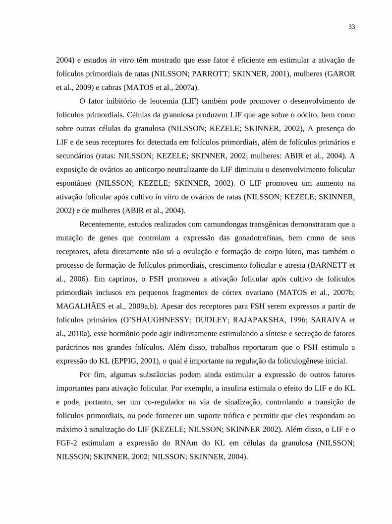
A Figura 1 ilustra resumida e esquematicamente os diferentes fatores envolvidos na
formação de folículos primordias e ativação folicular.
Figura 1. Desenho esquemático ilustrando os fatores envolvidos na formação e ativação
folicular. CG: Células da granulosa; CT: Células da teca; (+): efeito estimulatório; (-): efeito
inibitório. Adaptado de Knight and Glister (2006).
2.3.3. Progressão de folículo primário para secundário
A passagem de folículo primário para secundário é caracterizada pela proliferação das
células da granulosa e um aumento no tamanho e conteúdo protéico do oócito (VAN DEN
HURK; ZHAO, 2005). Os folículos secundários são formados por um oócito circundado por
duas ou mais camadas de células da granulosa de morfologia cubóide. O núcleo do oócito
assume uma posição excêntrica e as organelas começam a mover-se para a periferia. Com o
desenvolvimento dos folículos, também aumenta o número de microvilos e inicia-se a
formação da zona pelúcida (LUCCI et al., 2001). Neste estágio, o oócito entra em fase de
crescimento extensivo, as células da granulosa ao redor se tornam mais proliferativas e uma
camada de células da teca se desenvolve em torno das células da granulosa, a partir de células

35
do estroma intersticial. Após a formação da camada da teca, já se inicia a formação de vasos
sanguíneos. Além disso, ocorre a formação de uma extensiva rede de junções do tipo gap, que
são canais membranários que permitem a passagem de nutrientes, íons inorgânicos, segundo
mensageiros e pequenos metabólitos entre as células (KIDDER; MHAWI, 2002). Uma
complexa organização citoplasmática é necessária e depende tanto da produção de produtos
de genes novos e organelas, como da modificação e redistribuição daquelas já existentes
(PICTON; BRIGGS; GOSDEN, 1998). Dessa forma, como consequência do aumento da
síntese de RNAm e de proteínas, o número de ribossomos, mitocôndrias e outras organelas
celulares aumentam nos oócitos em crescimento. Além disso, muitas organelas mudam sua
aparência e movem-se para a periferia do oócito (VAN DEN HURK; ZHAO, 2005). Essa
progressão parece ser controlada, tanto por peptídeos e hormônios esteróides, bem como por
fatores de crescimento intraovarianos (FORTUNE, 2003; VAN DEN HURK; ZHAO, 2005).
O crescimento de folículos pré-antrais in vivo ocorre independentemente das
gonadotrofinas, uma vez que folículos pré-antrais de animais e humanos desenvolvem até o
estágio antral com níveis mínimos de gonadotrofinas circulantes (GULYAS et al., 1977;
HALPIN et al., 1986; HILLIER, 1994), sendo dessa forma atuante os fatores de crescimento.
Entretanto, vários estudos in vitro têm sugerido que o FSH desempenha um importante papel
no desenvolvimento de folículos pré-antrais (HIRAO et al., 1994; CAIN; CHATTERJEE;
COLLINS, 1995; CORTVRINDT; SMITZ; VAN STEIRTEGHEM, 1997; SPEARS et al.,
1998; WU; BENJAMIN; CARRELL, 2001). Mais recentemente, alguns autores têm
demonstrado que o FSH aumenta o diâmetro de folículos pré-antrais bovinos (ITOH et al.,
2002), suínos (WU; TIAN, 2007) e caprinos (MATOS et al., 2007b; MAGALHÃES et al.,
2009a) após cultivo in vitro.
O fator de crescimento e diferenciação-9 (GDF-9) é um fator essencial para a
formação de folículos secundários, uma vez que em ratas com nocaute de gene para o GDF-9,
o desenvolvimento folicular além do estágio primário não foi observado (DONG et al., 1996).
De forma semelhante, ovelhas com mutações espontâneas inativadoras do gene GDF-9
(HANRAHAN et al., 2004) e ovelhas imunizadas ativamente contra o GDF-9 não
apresentaram folículos pré-antrais desenvolvidos no estágio de folículos secundários
(JUENGEL et al., 2002). Em cabras, SILVA et al. (2004b) localizaram a proteína GDF-9 em
oócitos de todos tipos foliculares e em células da granulosa de folículos primários,

36
secundários e antrais, mas não em folículos primordiais. A exposição in vitro de tecido
ovariano ao GDF-9 em roedores (NILSSON; SKINNER, 2002, 2003; WANG; ROY, 2004),
cabras (MARTINS et al., 2008) e mulheres (HREINSSON et al., 2002) promoveu a
progressão do desenvolvimento de folículos primários. Além disso, outro estudo mostrou que
100 ng/mL de GDF-9 promoveu o crescimento de folículos pré-antrais in vitro e suprimiu a
apoptose de células da granulosa (ORISAKA et al., 2006).
A BMP-15, outro fator derivado do oócito, parece também ser importante para o
crescimento de folículos primários (JUENGEL et al., 2004), sendo as células da granulosa, as
células alvo para o ligante BMP-15 (OTSUKA et al., 2000; GALLOWAY et al., 2000). A
expressão dos genes que codificam a proteína BMP-15 são essenciais para os estágios iniciais
de crescimento folicular e em particular para transição de folículos primários para secundários
(MERY et al., 2007). Além disso, alguns autores têm descrito uma alta expressão de RNAm
e/ou proteína BMP-15 em oócitos em crescimento ou completamente crescidos
(SHIMASAKI et al., 2004; JUENGEL; MCNATTY, 2005; LI et al., 2008). TEIXEIRA et al.
(2002) relataram que a expressão desse fator aumenta em uma correlação direta com o
crescimento folicular. Em cabras, foi demonstrado um aumento significativo nos níveis de
RNAm para BMP-15 durante a transição de folículos primários para secundários, bem como
um aumento no número desses folículos após cultivo in vitro (CELESTINO et al., dados não
publicados).
A expressão da ativina, receptores de ativina, e folistatina (proteína ligada à ativina)
tem sido detectada em folículos primários e secundários (RABINOVICI, 1991; MCNATTY,
2000; PANGAS et al., 2002; DRUMMOND et al., 2002; SILVA et al., 2006a). Sob
condições in vitro, a ativina estimula o crescimento de folículos uni e/ou multilaminares de
vacas (HULSHOF et al., 1997), ratas (ZHAO et al., 2001), camundongas (SMITZ et al.,
1998) e cabras (SILVA et al., 2006a).
O fator de crescimento epidermal (EGF) é conhecido como um potente fator
mitogênico, podendo estimular a proliferação de diferentes tipos celulares (TOYODA et al.,
2007). Estudos têm demonstrado a expressão da proteína e RNAm para o EGF e seus
receptores em oócitos e células da granulosa de folículos pré-antrais e antrais de ratas, vacas,
mulheres, porcas, camundongas e cabras (FENG; KNECHT; CATT, 1987; LONERGAN et
al., 1996; QU et al., 2000; SINGH; RUTLEDGE; ARMSTRONG, 1995; HILL et al., 1999;
37
SILVA et al., 2006c). Outros estudos com cultivo in vitro de folículos pré-antrais mostraram
que o EGF promove a proliferação de células da granulosa de porcas, roedores e mulheres
(MORBECK; FLOWERS; BRITT, 1993; GOSPODAROWICZ; BIALECKI, 1979) e
aumenta o diâmetro folicular de vacas, roedores, mulheres e cabras (GUTIERREZ et al.,
2000; ROMANO et al., 1994; ROY; KOLE, 1998; SILVA et al., 2004c).
Outro fator mitogênico também importante para a transição de folículos primários para
secundários é o FGF-2. Tal fator é importante no controle de uma ampla variedade de funções
ovarianas, incluindo mitose de células da granulosa (ROBERTS; ELLIS, 1999),
esteroidogênese (VERNON; SPICER,1994), diferenciação (ANDERSON; LEE, 1993) e
apoptose (TILLY et al., 1992). Já foi observado que o FGF-2 está expresso em oócitos de
folículos primordiais e células da granulosa e da teca de folículos em crescimento de vacas
(VAN WEZEL et al., 1995; YAMAMOTO et al., 1997; NILSSON; PARROTT; SKINNER,
2001). Além disso, receptores para a proteína e RNAm para o FGF-2 têm sido demonstrados
em folículos em crescimento de vacas (WANDJI; FORTIER; SIRARD, 1992) e ratas
(SHIKONE; YAMOTO; NAKANO, 1992; ASAKAI et al., 1993, 1995). Wandji et al. (1996)
mostraram que a utilização de 50 ng/mL de FGF-2 aumentou o diâmetro folicular e estimulou
a proliferação de células da granulosa em folículos bovinos cultivados por 6 dias. Outros
autores também observaram que o FGF-2 promoveu aumento no diâmetro folicular e a
proliferação das células da granulosa de vacas (NUTTINCK et al., 1996), gatas
(JEWGENOW, 1996) e ratas (NILSSON; PARROTT; SKINNER, 2001).
2.3.4. Progressão de folículo secundário para antral
Após o crescimento dos folículos secundários e organização das células da granulosa
em várias camadas, ocorre a formação de uma cavidade repleta de líquido denominada antro.
A partir deste estágio, os folículos passam a ser denominados terciários ou antrais. O fluido
folicular que preenche esta cavidade contém água, eletrólitos, proteínas séricas e alta
concentração de hormônios esteróides secretados pelas células da granulosa (BARNETT et
al., 2006). Durante o desenvolvimento folicular, a produção de fluido antral é intensificada
pelo aumento da vascularização folicular e permeabilidade dos vasos sangüíneos, os quais
estão fortemente relacionados com o aumento do folículo antral. O desenvolvimento dos

38
folículos antrais é caracterizado por uma fase de crescimento, recrutamento, seleção e
dominância (VAN DEN HURK; ZHAO, 2005) sendo a formação de folículos pré-ovulatórios
um pré-requisito para a ovulação e formação do corpo lúteo (DRUMMOND, 2006).
Vários experimentos in vivo e in vitro têm mostrado que o número de folículos
secundários, seu tamanho, número de células e taxa de atresia são influenciados pelas
gonadotrofinas, das quais o FSH é o fator de sobrevivência predominante (VAN DEN HURK;
BEVERS; BECKERS,1997; VAN DEN HURK et al., 2000). O LH também parece ser
importante para o desenvolvimento de folículos secundários, uma vez que, através de seus
receptores, que podem ser demonstrados na camada da teca de folículos secundários de ratas,
ele desencadeia a biossíntese de andrógenos os quais são capazes de estimular a formação de
receptores para FSH nas células da granulosa e assim, podem ampliar os efeitos do FSH em
folículos secundários (VAN DEN HURK; BEVERS; DIELEMAN, 1999; VAN DEN HURK
et al., 2000). A síntese de andrógenos estimulada pelo LH é amplificada por neurotrofinas
(VAN DEN HURK; ZHAO, 2005).
O hormônio do crescimento (GH) é outro fator endócrino que pode promover o
desenvolvimento de folículos secundários. A presença de RNAm para o receptor de GH tem
sido detectada em folículos pré-antrais de ratas (ZHAO et al., 2002). Estudos in vitro usando
GH têm demonstrado que esse fator desempenha um importante papel controlando as fases
iniciais do desenvolvimento folicular. A adição de GH ao meio de cultivo in vitro de folículos
pré-antrais estimulou a produção de estradiol e a proliferação das células da granulosa e da
teca em murinos (KOBAYASHI et al., 2000), além de promover um aumento no diâmetro de
folículos pré-antrais de camundongas cultivados in vitro (LIU et al., 1998; KIKUCHI et al.,
2001).
Além dos hormônios citados, a família FGF, que é composta por um grupo de fatores,
é potencialmente importante para o crescimento folicular (BURATINI et al., 2005). McGEE
et al. (1999) demonstraram uma supressão dose-dependente da apoptose de células da
granulosa e estímulo do crescimento de folículos secundários de ratas cultivadas com o FGF-
7. O FGF-2, um potente fator angiogênico (REYNOLDS; REDMER, 1998; PLENDL, 2000),
exerce um importante papel na foliculogênese, uma vez que a função ovariana é dependente
do estabelecimento e contínuo remodelamento de um complexo sistema vascular, que
possibilita os folículos receberem suprimento adequado de nutrientes, oxigênio e suporte
39
hormonal (ROBINSON et al., 2009). Além do FGF-2, outros fatores angiogênicos também
parecem ser importantes para o desenvolvimento folicular nessa fase, dos quais podemos
destacar o VEGF e a ANG II. No primeiro Capítulo desta tese, será mostrada uma revisão
detalhada acerca da importância de fatores angiogênicos durante o desenvolvimento folicular,
incluindo o FGF-2, VEGF e ANG II.
Neurotrofinas e neurotransmissores, como por exemplo, NGFs e VIP respectivamente,
também parecem ser bons candidatos para estimular o desenvolvimento folicular precoce,
inclusive de folículos secundários. Ovários de camundongas com mutações deletérias ao NGF
exibiram acentuada redução do número de folículos primários e secundários, enquanto a
proliferação de células da granulosa foi visivelmente reduzida após o cultivo in vitro
(DISSEN et al., 2001). Já o VIP tem sido apontado como um importante regulador no
desenvolvimento de folículos secundários de ratas, bovinos e primatas (VAN DEN HURK et
al., 2000; MCGHEE; HSUEH, 2000) e pode estar envolvido na função esteroidogênica das
células da granulosa de roedores (VAN DEN HURK; ZHAO, 2005). O segundo Capítulo
desta tese refere-se a uma revisão acerca do envolvimento do VIP na fisiologia ovariana.
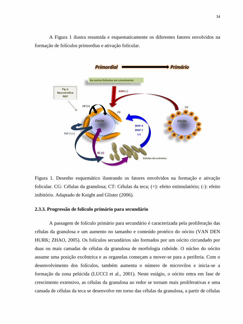
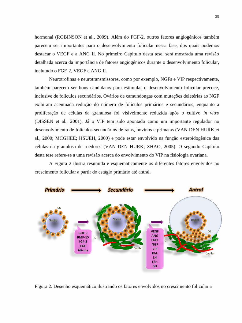
A Figura 2 ilustra resumida e esquematicamente os diferentes fatores envolvidos no
crescimento folicular a partir do estágio primário até antral.
Figura 2. Desenho esquemático ilustrando os fatores envolvidos no crescimento folicular a

40
partir do estágio primário até antral. CG: Células da granulosa; CT: Células da teca. Adaptado
de Knight and Glister (2006).
2.4. Biotécnica de MOIFOPA
Tendo em vista que o ovário mamífero contém milhares de oócitos inclusos em
folículos pré-antrais e que poucos destes folículos desenvolvem-se até o estágio de folículo
pré-ovulatório, e na tentativa de utilizar de maneira eficiente o potencial de gametas
femininos no futuro, vem sendo desenvolvida a biotécnica de MOIFOPA.
Esta biotécnica visa o resgate de um grande número de oócitos inclusos nos folículos
pré-antrais, seguido das etapas de conservação (resfriamento e/ou criopreservação) e/ou
cultivo in vitro até o estágio de maturação folicular (FIGUEIREDO et al., 2008),
representando uma alternativa para o fornecimento de uma população homogênea de oócitos
para as biotécnicas de fecundação in vitro e clonagem (TELFER, 1996).
Nesse contexto, o cultivo in vitro de folículos pré-antrais vem sendo largamente
empregado com o intuito de promover a multiplicação e a posterior diferenciação das células
da granulosa e assegurar o crescimento e a maturação de seus oócitos, para que os mesmos
estejam aptos à produção de embriões (FIGUEIREDO et al., 2008). A secção a seguir
abordará os diversos sistemas de cultivo in vitro empregados.
2.5. Cultivo in vitro de folículos pré-antrais
No que se refere aos sistemas de cultivo in vitro, os folículos ovarianos podem ser
cultivados inclusos no próprio tecido ovariano (in situ) ou na forma isolada. O cultivo in situ é
considerado um modelo prático uma vez que a manipulação é rápida e nesse cultivo, a
manutenção da integridade tridimensional dos folículos e a interação destes com células do
estroma são mantidas. Já o cultivo de folículos isolados permite uma melhor perfusão dos
nutrientes e o acompanhamento individual dos folículos.
Em roedores, a pequena dimensão dos ovários possibilita o cultivo do órgão inteiro, o
que tem sido bastante útil para o estudo da foliculogênese inicial em pequenos mamíferos
(FORTUNE, 2003). Por outro lado, em animais domésticos de médio e grande porte, devido
às grandes dimensões dos ovários, não é possível utilizar este modelo. Para estes animais, o

41
cultivo de pequenos fragmentos de córtex ovariano, rico em folículos primordiais, tem sido
realizado para o estudo da ativação e crescimento de folículos primários (caprinos: SILVA et
al., 2004a; bovinos: BRAW-TAL; YOSSEFI, 1997; babuínos: WANDJI et al., 1997;
humanos: HOVATTA et al.,1997). O cultivo de pequenos fragmentos de córtex ovariano tem
a vantagem de manter a integridade celular e facilitar a perfusão do meio para o tecido
ovariano (TELFER, 1996).
Grande progresso já tem sido observado no cultivo in vitro de folículos pré-antrais em
diferentes espécies animais. Em felinos (JEWGENOW; STOLTE, 1996) e marsupiais
(BUTCHER; ULLMAN, 1996), observou-se o crescimento de folículos pré-antrais isolados,
porém, sem a formação de antro. Em humanos, bovinos e caninos, folículos secundários
isolados cresceram in vitro o estágio antral (ROY; TREACY, 1993; GUTIERREZ et al.,
2000; SERAFIM et al., 2010). Resultados mais satisfatórios foram obtidos com suínos (WU;
TIAN, 2007), bubalinos (GUPTA et al., 2008), ovinos (ARUNAKUMARI;
SHANMUGASUNDARAM; RAO, 2010) e caprinos (SARAIVA et al., 2010b;
MAGALHÃES et al., 2011), em que se alcançou a produção de embriões após cultivo in vitro
de grandes folículos secundários. Entretanto, os melhores resultados do cultivo folicular
foram obtidos em roedores, sendo observada a obtenção de crias viáveis a partir do cultivo de
oócitos provenientes de folículos pré-antrais de camundongas, nos quais o oócito adquiriu
competência para maturação, fertilização e desenvolvimento embrionário (EPPIG;
SCHROEDER, 1989; O‘BRIEN; PENDOLA; EPPIG, 2003). Tal crescimento foi obtido
através do sistema de cultivo em dois passos: (1) cultivo de ovários inteiros para obtenção da
transição de folículo primordial para primário e (2) isolamento e posterior cultivo de folículos
primários e secundários.
Como pode-se observar, um grande número de sistemas de cultivo in vitro têm sido
desenvolvidos para promover o crescimento de folículos de diferentes espécies. Além das
diferenças dos sistemas de cultivo, existem também muitas diferenças espécies-específicas,
como: i) o período para a conclusão da foliculogênese e oogênese; ii) o tamanho dos folículos
ovulatórios e oócitos maduros, e iii) as diferenças na natureza, concentrações e efeitos dos
fatores de crescimento que influenciam o folículo e produção de oócitos in vivo. Essas
diferenças são altamente relevantes no desenvolvimento de sistemas que suportam o
crescimento in vitro completo e maturação do oócito (PICTON et al., 2008). No entanto, em

42
todos os protocolos, algumas características são vitais para otimizar o crescimento in vitro: o
fornecimento de nutrientes, eletrólitos, antioxidantes, aminoácidos, substratos energéticos,
vitaminas, hormônios e fatores de crescimento (PICTON et al., 2008). Diante disso, nossa
equipe vem testando uma série de substâncias no cultivo in vitro de folículos pré-antrais
caprinos.
2.6. Cultivo in vitro de folículos pré-antrais caprinos in situ
Os primeiros estudos do LAMOFOPA foram realizados com o intuito de se obter um
meio de base para o cultivo in vitro de folículos pré-antrais caprinos. SILVA et al. (2004a) ao
comparar a solução à base de água de coco (SBAC) com o Meio Essencial Mínimo (MEM),
verificaram que o MEM apresentou melhores taxas de sobrevivência e ativação folicular.
Esses resultados foram reforçados por MARTINS et al. (2005), em que foi observado uma
elevada taxa de degeneração folicular após utilização de SBAC. Além disso, SILVA et al.
(2004a) observaram que a adição de suplementos ao meio de base MEM, como hipoxantina e
substratos energéticos, tais como piruvato e glutamina, e ITS (Insulina, Transferrina e
Selênio) foram considerados importantes para manter a sobrevivência de folículos pré-antrais
caprinos cultivados in vitro.
Existiu ainda uma clara necessidade de verificar a temperatura e tempo de transporte
ideal para ovários caprinos para posterior cultivo in vitro. Então, CHAVES et al. (2008) ao
testar diferentes tempos e temperaturas de transporte, verificaram que os fragmentos
ovarianos resfriados a 4º C por 4 h durante o transporte são melhores para manter a
viabilidade e aumentar o crescimento folicular durante o cultivo in vitro.
Nossa equipe testou ainda o efeito de alguns hormônios. No que se refere à utilização
do FSH (MATOS et al., 2007b; MAGALHÃES et al., 2009a,b) e do estradiol (LIMA-
VERDE et al., 2010a), estes apresentaram efeitos positivos na sobrevivência, isto é,
percentagens de folículos pré-antrais morfologicamente normais superiores ao controle
cultivado (MEM+), bem como na manutenção da integridade ultraestrutural. Tais hormônios
tiveram ainda um efeito adicional sobre a ativação e o crescimento folicular. No que se refere
ao LH, embora ele tenha mantido a integridade ultraestrutural dos folículos quando
adicionado ao meio de cultivo, as percentagens de folículos pré-antrais morfologicamente

43
normais foram similares ao controle cultivado após sete dias (SARAIVA et al., 2008). Dentre
os hormônios testados, o GH na concentração de 10 ng/mL, além de apresentar efeito
benéfico para sobrevivência, também foi eficiente para a transição de folículos primordiais
para primários (MARTINS et al., 2010). Por outro lado, a androstenediona não apresentou
nenhum efeito adicional sobre a sobrevivência de folículos pré-antrais caprinos (LIMA-
VERDE et al., 2010b).
Quando os fatores de crescimento foram testados, foi obsevado que dois membros da
família FGF, o FGF-10 (CHAVES et al., 2010b) e o FGF-2 (MATOS et al., 2007a),
aumentaram a percentagem de folículos normais e promoveram a ativação folicular em
relação ao controle cultivado, respectivamente. No entanto, fatores como o KL e a BMP-15,
não apenas se destacaram em relação ao controle cultivado, como também apresentaram
valores de sobrevivência similares ao controle fresco (CELESTINO et al., 2010;
CELESTINO et al., dados não publicados). Todos esses fatores foram também eficientes em
manter a integridade ultraestrutural dos folículos após sete dias de cultivo. No que se refere ao
crescimento, a ativina A (SILVA et al., 2006a), o FGF-10 (CHAVES et al., 2010b) e a BMP-
15 (CELESTINO et al., dados não publicados) aumentaram o diâmetro de folículos pré-
antrais caprinos após cultivo in vitro. Alguns fatores de crescimento, tais como EGF, KL,
BMP-15 e IGF-1 aumentaram a percentagem de folículos primários (CELESTINO et al.,
2009; 2010; dados não publicados; MARTINS et al., 2010). No entanto, apenas a BMP-15 e o
GDF-9 aumentaram a percentagem de folículos secundários após sete dias de cultivo
(CELESTINO et al., dados não publicados; MARTINS et al., 2008), porém com taxas
relativamente baixas (~ 20%).
Dentre os antioxidantes testados, pode-se citar o α-tocoferol e a ternatina, no entanto,
quando utilizados nas concentrações de 5, 10 ou 15 µM, não foram eficientes para o
desenvolvimento de folículos pré-antrais caprinos (LIMA-VERDE et al., 2009). Por outro
lado, o ácido ascórbico (50 µg/mL) associado ao FSH (50 ng/mL) obteve elevadas
percentagens de folículos pré-antrais normais, os quais ainda mantiveram a integridade
ultraestrutural, bem como promoveram a ativação e o crescimento de folículos pré-antrais
caprinos (ROSSETTO et al., 2009). Outras interações também foram benéficas, tais como o
FSH com FGF-2; e FSH com EGF, as quais aumentaram o diâmetro folicular (MATOS et al.,
44
2007c; SILVA et al., 2004c) e IGF-1 com GH, os quais promoveram a transição de folículos
primordiais para intermediários (MARTINS et al., 2010).
Por outro lado, outros fatores testados (BMP-7: ARAÚJO et al., 2010a; NGF:
CHAVES et al., 2010a), não foram eficazes em melhorar a percentagem de folículos pré-
antrais morfologicamente normais em relação ao controle cultivado, apesar de terem mantido
a ultraestrutura dos folículos após análise por microscopia eletrônica de transmissão (MET).
Substâncias, tais como o ácido 3-indol-acético (IAA) (MATOS et al., 2006) e soro fetal
bovino (BRUNO et al., 2008), nem mesmo foram capazes de manter a ultraestrutura dos
folículos cultivados.
Como se pode observar, apesar de folículos inclusos em tecido ovariano serem
mantidos saudáveis, ativarem e crescerem, poucos desses folículos progridem até o estágio de
primário e secundário. Na tentativa de melhorar tais resultados, outras substâncias,
isoladamente ou em associação, vêm sendo testadas.
2.7. Cultivo in vitro de folículos pré-antrais caprinos isolados
Simultaneamente aos experimentos com cultivo in vitro de fragmentos ovarianos,
experimentos voltados para o cultivo de folículos pré-antrais isolados vêm sendo realizados,
com o objetivo de manter a viabilidade, promover a formação da cavidade antral, estimular o
crescimento e capacitar os oócitos inclusos nesses folículos a serem maturados e fecundados
in vitro.
Outros pesquisadores, trabalhando com essa mesma espécie, desenvolveram um
sistema de cultivo de folículos secundários isolados, capaz de promover a formação do antro
(ZHOU; ZHANG, 2000), bem como manter a sobrevivência e aumentar o diâmetro folicular
(ZHOU; ZHANG, 2005). Nosso grupo também foi capaz de obter tais resultados, uma vez
que a utilização do meio de base foi capaz de manter a sobrevivência e atingir estágio antral, e
a adição de FSH no meio ainda promoveu um aumento na taxa de crescimento folicular
(RODRIGUES et al., 2010).
Um grande progresso foi alcançado por nosso grupo, o qual além de estabelecer um
sistema de cultivo capaz de estimular o crescimento e formação antral assegurou a retomada
da meiose (SILVA et al., 2010). Nesse experimento foram testadas duas concentrações de
oxigênio (5 e 20%), em que a concentração de 20% foi mais eficiente em promover o

45
crescimento folicular e a retomadada da meiose (16,7%) de oócitos oriundos de folículos pré-
antrais crescidos in vitro.
MAGALHÃES et al. (2010), ao avaliar o efeito de diferentes intervalos de troca de
meio (troca a cada 2 ou 6 dias) sobre o desenvolvimento de folículos pré-antrais caprinos
isolados, verificaram que sob as mesmas condições, o intervalo de troca de meio a cada 2 dias
foi mais eficiente sobre a viabilidade folicular e ainda proporcionou a retomada da meiose
(12,5%).
Ainda no que se refere à troca de meio, ARAÚJO et al (2010b) avaliaram a influência
de três diferentes protocolos de troca de meio. Nesse trabalho, a adição periódica de meio (5
μl de meio fresco a cada dois dias) foi recomendada por ser mais prática, manter a viabilidade
e promover o desenvolvimento in vitro de folículos pré-antrais caprinos.
Outras formas de cultivo também foram testadas, como o cultivo de folículos de forma
individual ou em grupos. DUARTE et al. (2010) observaram que o co-cultivo de folículos
antrais com folículos pré-antrais afetou negativamente o desenvolvimento folicular. Por outro
lado, o cultivo de folículos pré-antrais em grupo mostrou taxas superiores de sobrevivência e
crescimento em relação aos folículos cultivados individualmente, além de estimular a
retomada da meiose em 9,09%. No entanto, essa percentagem também foi muito baixa, o que
refletiu a necessidade de melhorar tal sistema de cultivo.
Na tentativa de melhorar tal sistema, e sabendo que os folículos possuem diferentes
níveis de exigências nos diferentes estágios de desenvolvimento, foi desenvolvido um meio de
cultivo seqüencial utilizando FSH. Esse hormônio foi utilizado em concentrações aumentadas
ao longo do cultivo (FSH 100 ng/mL do dia 0 a 6; FSH 500 ng/mL do dia 6 a 12; FSH 1000
ng/mL do dia 12 a 18), promovendo altas percentagens de retomada da meiose
(aproximadamente 62%) (SARAIVA et al., 2010a). Diante desse resultado promissor, novos
experimentos foram realizados utilizando tal sistema de cultivo.
De fato, a partir da utilização de um meio de base que continha FSH seqüencial
suplementado com LH e EGF (SARAIVA et al., 2010b) ou GH (MAGALHÃES et al., 2011),
foi possível a obtenção dos primeiros embriões caprinos oriundos de folículos secundários
isolados cultivados in vitro.
Apesar dos grandes avanços obtidos pela nossa equipe, o rendimento referente à
produção de oócitos maturos, bem como a obtenção de embriões a partir de folículos pré-
46
antrais caprinos ainda é extremamente baixo, o que reforça a necessidade de melhorar tal
sistema de cultivo.
2.8 Técnicas para análise folicular
Diferentes técnicas podem ser utilizadas para análise folicular após o cultivo in vitro
de folículos pré-antrais. Dentre elas, podemos destacar aquelas que permitem a avaliação da
qualidade folicular, como por exemplo, a histologia clássica (HC), a microscopia eletrônica de
transmissão (MET) e a microscopia de fluorescência. Além disso, existem ainda as técnicas
de biologia molecular que permitem o estudo da expressão de genes que codificam ligantes
e/ou receptores de diferentes substâncias importantes para a foliculogênese, contribuindo para
uma melhor elucidação desse processo. A seguir, será abordada brevemente cada uma dessas
técnicas.
2.8.1 Histologia Clássica
A HC é uma técnica importante para avaliação de folículos pré-antrais após cultivo in
vitro, pois além de permitir uma análise quantitativa, ou seja, de um grande número de
folículos cultivados, permite ainda verificar a mudança na morfologia das células da
granulosa de pavimentosa para cúbica, por ocasião da ativação folicular, além de analisar a
integridade morfológica do oócito e das células da granulosa. Tal técnica permite, portanto a
classificação dos folículos quanto ao seu estágio de desenvolvimento (primordial,
intermediário, primário ou secundário), e ainda quanto às suas características morfológicas
(normais ou atrésicos). Entretanto, a HC possui como desvantagens não permitir a avaliação
da integridade de membranas e das organelas citoplasmáticas. Vale salientar que tal técnica
pode ser realizada tanto em folículos isolados, como naqueles inclusos em fragmentos de
córtex ovariano (MATOS et al., 2007d).
2.8.2 Microscopia Eletrônica de Transmissão
A MET é considerada uma técnica qualitativa e acurada, capaz de permitir a avaliação
47
da integridade de membranas celulares e organelas citoplasmáticas (SALEHNIA;
MOGHADAM; VELOJERDI, 2002). Ela se mostra como uma ferramenta valiosa para
detectar modificações morfológicas iniciais devido à atresia, as quais podem ser observadas
apenas em nível ultraestrutural, antes de se tornarem mais visíveis e possíveis de serem
identificadas por microscopia óptica. Tal técnica é, portanto, capaz de verificar a qualidade do
oócito e das células da granulosa (LUCCI et al., 2001; LOPES et al., 2009). Com esta técnica,
é possível ainda avaliar a atresia, ou seja, a morte do folículo tanto por apoptose
(STALDEMANN; LASSMAN, 2000) como pelo processo degenerativo de necrose
(MARTINEZ-MADRID et al., 2007).
2.8.3 Microscopia de Fluorescência
Outra técnica empregada para avaliar a viabilidade folicular após o cultivo in vitro é a
microscopia de fluorescência, a qual utiliza marcadores fluorescentes, que quando excitados
com radiação de baixo comprimento de onda, absorvem energia e emitem luz de comprimento
de onda maior (JUNQUEIRA; CARNEIRO, 2005). A microscopia de fluorescência é
considerada uma técnica confiável, prática e rápida (CORTVRINDT; SMITZ, 2001b; LOPES
et al., 2009). Recentemente, estudos vêm sendo realizados empregando a técnica de
fluorescência para avaliar a viabilidade folicular após o cultivo in vitro (ROSSETTO et al.,
2009; SILVA et al., 2010; MAGALHÃES et al., 2010). Nesses trabalhos, os folículos foram
analisados por fluorescência com base na detecção simultânea de células vivas e mortas
marcadas por calceína-AM e pelo etídio homodímero-1, respectivamente. Enquanto a
primeira sonda detecta atividade da esterase intracelular, enzima característica de células
viáveis, a outra cora ácidos nucléicos em células não-viáveis, com ruptura na membrana
plasmática (LOPES et al., 2009).
2.8.4 Biologia Molecular
A identificação e quantificação dos níveis de RNAm de diferentes substâncias
(ligantes e receptores) que atuam durante a foliculogênese também é considerada uma
importante ferramenta para auxiliar na compreensão desse processo, uma vez que permite
48
detectar alterações nos padrões de expressão gênica que ocorrem em resposta a fenômenos
relacionados à sobrevivência, ao crescimento e à diferenciação celular. Desta forma, a
capacidade de quantificar os níveis de transcrição de genes específicos é fundamental para
garantir a completa investigação das funções foliculares (ZAMORANO; MAHESH; BRANN,
1996). Dentre as técnicas de biologia molecular, podem ser citadas aquelas que detectam,
localizam ou identificam os ácidos nucléicos (hibridização in situ Southern e Northern
Blotting) ou proteínas (Western Blotting e imunohistoquímica), as que podem efetuar a
quantificação do DNA (Reação em Cadeia de Polimerase - PCR) ou do RNA (Reação de
Transcriptase Reversa de Cadeia da Polimerase - RT-PCR), as que permitem a quantificação
da expressão do RNAm mesmo em uma mistura complexa de RNA total (ensaios de proteção
de ribonuclease) ou que possibilitem a análise da expressão de milhares de genes
simultaneamente (Microarranjos de DNA).
Atualmente, a técnica mais utilizada para quantificar a expressão de RNAm é a RT-
PCR em tempo real (qRT-PCR) (KREUZER; MASSEY, 2002). A qPCR, uma variante da
RT-PCR convencional, permite uma análise precisa da quantificação da expressão gênica em
determinado tecido ou amostra biológica. Esse método utiliza um sistema fluorescente em
plataforma, capaz de detectar a luz oriunda da reação de amplificação de um determinado
gene no momento real da amplificação (BUSTIN, 2002).
2.8.5 Imunohistoquímica
A imunohistoquímica também vem sendo utilizada com freqüência para avaliar a
expressão de fatores de crescimento e hormônios, bem como seus receptores, presentes nos
ovários, além de ser utilizada também para verificar a proliferação das células da granulosa.
Esta técnica representa um conjunto de procedimentos diagnósticos que utilizam anticorpos
(policlonais ou monoclonais) como reagentes de grande especificidade para a detecção de
antígenos que marcam estruturas em cortes histológicos ou em células cultivadas (ABIR et al.,
2006).
49
3. JUSTIFICATIVA
Os folículos primordiais representam cerca de 90% de todos os folículos presentes no
ovário. Entretanto, os mecanismos responsáveis pelo seu crescimento ainda não são
completamente elucidados. Além disso, cerca de 99,9% dos folículos ovarianos sofrem
atresia, o que reduz significativamente o potencial reprodutivo das fêmeas. Neste contexto,
com o intuito de evitar a grande perda folicular que ocorre in vivo, surge a biotécnica de
MOIFOPA que permite a recuperação de dezenas a milhares de folículos primordiais a partir
de um único ovário. Tais folículos poderiam ser criopreservados para posterior utilização e/ou
cultivados in vitro.
Dessa forma, sabendo-se da grande relevância social e econômica que a espécie
caprina representa para várias regiões do mundo, especialmente o Nordeste Brasileiro, e da
escassez de informações sobre as condições necessárias e os fatores envolvidos no
crescimento e na maturação oocitária in vitro, é de extrema importância o desenvolvimento de
um sistema de cultivo in vitro capaz de manter a sobrevivência folicular, ativar esses folículos
e assegurar o seu posterior crescimento, otimizando o aproveitamento do potencial oocitário
desses animais e incrementando a eficiência da reprodução animal.
Alguns estudos têm investigado o efeito de diversos hormônios e fatores de
crescimento no cultivo in vitro de folículos pré-antrais de animais de laboratórios e animais
domésticos. Entretanto, os efeitos de diferentes concentrações de ANG II, VEGF e VIP ainda
não foram avaliados no cultivo in vitro de folículos pré-antrais caprinos. Para este fim, as
técnica de histologia clássica, microscopia de fluorescência e MET foram empregadas para
determinar a qualidade de folículos pré-antrais caprinos cultivados in vitro e,
conseqüentemente, melhor avaliar a eficiência dos meios de cultivo testados. Além disso,
foram utilizados ensaios imunohistoquímicos e ferramentas da biologia molecular, com
intuito de se conhecer a expressão dessas substâncias no ovário caprino, bem como após
cultivo in vitro.
50
4. HIPÓTESE CIENTÍFICA
Diante do exposto, formularam-se as seguintes hipóteses:
1) O receptor para VEGF, bem como o RNAm para os receptores da ANG II (AGTR1 e
AGTR2) e para o VIP são expressos de forma diferenciada em folículos ovarianos
caprinos;
2) A ANG II, o VEGF e o VIP influenciam a sobrevivência, a ativação e o crescimento in
vitro de folículos pré-antrais caprinos inclusos em tecido ovariano;
3) O VIP afeta a sobrevivência, a formação de antro, o crescimento e a expressão do RNAm
para VIP e receptor de FSH (FSHR) em folículos pré-antrais caprinos isolados.

51
5. OBJETIVOS
5.1. Objetivo geral
1) Verificar a localização da proteína para o receptorde VEGF- 2 (VEGFR-2) e a expressão
do RNAm para os receptores da ANG II (AGTR1 e AGTR2) e para o VIP em folículos
ovarianos caprinos.
2) Avaliar o efeito da ANG II, VEGF e VIP sobre o desenvolvimento in vitro de folículos pré-
antrais caprinos inclusos em tecido ovariano.
3) Avaliar o efeito do VIP sobre o desenvolvimento in vitro de folículos pré-antrais caprinos
isolados, bem como sobre a expressão do RNAm para o VIP e receptor de FSH (FSHR) em
folículos pré-antrais caprinos isolados após cultivo in vitro.
5.2. Objetivos específicos
1) Investigar nos compartimentos foliculares (oócitos, células da granulosa, células da teca e
células do cumulus) a localização do receptor de VEGF (VEGFR-2), bem como a expressão
do RNAm para os receptores da ANG II (AGTR1 e AGTR2) e para o VIP, em folículos
caprinos;
2) Avaliar os efeitos da ANG II, VEGF e VIP sobre a sobrevivência, a ativação e o
crescimento de folículos pré-antrais caprinos durante o cultivo in vitro de fragmentos de
córtex ovariano por 1 ou 7 dias;
3) Avaliar o efeito do VIP sobre o a sobrevivência, formação de antro e crescimento de
folículos pré-antrais caprinos isolados, bem como sobre a expressão do RNAm para VIP e
receptor de FSH (FSHR) em folículos pré-antrais caprinos isolados após cultivo in vitro.
Nas páginas seguintes, serão apresentados os resultados desta tese na forma de seis

52
capítulos referentes a dois artigos de revisão e quatro artigos técnicos. Vale salientar que uma
revisão foi publicada em periódico incluso no Qualis CAPES ―B3‖, dois artigos técnicos já foram
publicados em periódicos Qualis ―A1‖ e outros três encontram-se em fase de julgamento.
53
6. CAPÍTULO 1
Fatores angiogênicos e o desenvolvimento folicular ovariano
(Angiogenic factors and ovarian follicle development)
Periódico: Animal Reproduction v. 6, n. 2, p. 371-379, 2009

54
Resumo
Os folículos requerem um suprimento sanguíneo adequado de oxigênio, nutrientes e
hormônios, além de remover o excesso de CO2 e outros metabólitos. A aquisição de uma
oferta vascular adequada é provavelmente um passo limitante na seleção e maturação do
folículo dominante. Desta forma, há um progressivo interesse no estudo dos fatores de
crescimento envolvidos no processo de angiogênese. Além disso, uma melhor compreensão
sobre os mecanismos que regulam a expressão e a ação desses fatores poderia ser um ponto
chave para aumentar o desempenho reprodutivo das fêmeas. Portanto, esta revisão visa
resumir os dados atuais sobre a importância dos fatores de crescimento pró e anti-angiogênico
que regulam a angiogênese no desenvolvimento folicular ovariano.
Palavras-chave: Angiogênese. Folículo. Fatores de crescimento. Ovário.

55
Angiogenic factors and ovarian follicle development
J.B. Bruno1, M.H.T. Matos, R.N. Chaves, J.J.H. Celestino, M.V.A. Saraiva,
I.B. Lima-Verde, V.R. Araújo, J.R. Figueiredo
Faculty of Veterinary Medicine, Laboratory of Manipulation of Oocytes and Preantral
Follicles (LAMOFOPA),
State University of Ceara, Fortaleza, CE, Brazil.
Abstract
Ovarian follicles require an adequate blood supply for oxygen, nutrients and
hormones, in addition to eliminating CO2 and other metabolites. Acquisition of an adequate
vascular supply is probably a limiting step in the selection and maturation of the dominant
follicle. In this way, there is a progressive interest in the study of the growth factors involved
in the angiogenic process. In addition, a better understanding about the mechanisms that
regulate the expression and action of these factors could be a key point to increase the
reproductive performance in females. Therefore, this review aims to summarize current data
on the importance of the pro- and anti-angiogenic growth factors which regulate angiogenesis
in ovarian follicle development.
Keywords: angiogenesis, follicle, growth factors, ovary.
Introduction
During embryo development, blood vessels differentiate from endothelial precursors
by a process called vasculogenesis. Angiogenesis is the process of new blood vessel
development from pre-existing vasculature that occurs in embryos and adults (Stouffer et al.,
2001). In the last two decades, there was a progressive interest in the study of angiogenesis
due to the association of this process with pathological conditions in adult tissues, such as
tumoral growth and inflammation (Smith, 2001). In addition, several aspects of human and
animal reproduction, such as clinical alterations that occur in the ovary and in the female
56
reproductive tract depend on angiogenesis (Acosta et al., 2003). This process occurs
throughout follicular development, allowing adequate nutritional and hormonal supply for
ovarian follicle growth and oocyte development, as well as corpus luteum formation (Fraser
and Lunn, 2000).
A wide range of growth factors have been identified that promote (pro-angiogenic) or
inhibit (anti-angiogenic) angiogenesis (Stouffer et al., 2001). However, modulation of the
expression and action of these factors can be a key point to increase female reproductive
performance. In this review, aspects related to the importance of angiogenesis in ovarian
follicle development, as well as the role of the regulatory pro- and anti-angiogenic growth
factors, will be discussed.
Blood vessels formation
The vascular system is developed based on two distinct processes: vasculogenesis and
angiogenesis. While blood vessels differentiate from endothelial precursors by a process
called vasculogenesis during embryo development, in adults further vessel development from
pre-existing vasculature occurs by intussusception or sprouting by a process called
angiogenesis. Angiogenesis is characterized by a cascade of events that starts with capillary
proliferation and culminates with the formation of a new microcirculation composed of
arterioles, capillaries and venules (Redmer and Reynolds, 1996). During the development of
new blood vessels, some features can be observed such as enzymatic degradation of the basal
membrane of the pre-existing vessels, migration of endothelial cells marked by angiogenic
stimulus and finally, endothelial cell proliferation (Redmer et al., 2001). This
neovascularization is completed with the formation of a capillary network and differentiation
of new capillaries into arterioles and veins (LeCouter et al., 2002).
Studies have demonstrated that circulating endothelial precursor cells, i.e., originated
from bone marrow, may contribute to angiogenesis in adults (Carmeliet and Jair, 2000). In
addition, recent studies indicated the presence of mitogenic endothelial cells, which are the
primary components of capillaries, in specific organs, modulating angiogenic responses in a
variety of organs (LeCouter et al., 2002).

57
It is important to note that in several adult tissues, capillary growth rarely occurs and
the vascular endothelium represents a stable population of cells with low mitogenic rates
(Klagsbrun and D`Amore, 1996). An exception is the rapid growth and regression that occurs
in the female genital organs, associated with equivalent changes in its vascular network
(Reynolds and Redmer, 1995). The mature ovary shows a highly developed vasculature,
reflecting its high metabolic rate, which turns this organ into a unique model for studies of
angiogenesis regulation during growth, differentiation and regression of normal tissues in
adults (Redmer and Reynolds, 1996).
Follicular angiogenesis
The ovarian follicle is the structural and functional unit of the mammalian ovary,
which supplies the necessary environment for oocyte growth and maturation (Telfer, 1996).
The follicles are surrounded by somatic cells (granulosa and theca cells) and can be classified
in preantral (primordial, primary and secondary) and antral (tertiary and preovulatory)
follicles (Hulshof et al., 1994). It is known that vasculature is not equally distributed among
the population of follicles of the adult ovary, since only theca cell layers, present in later
follicular stages, have vessels. Quiescent primordial follicles and slow growing preantral
follicles do not have a vascular supply of their own, but instead rely on vessels in the
surrounding stroma. Thus, Martelli et al. (2009) showed that an autonomous vascular supply
starts to be evident in preantral follicles with diameter from 110 μm. However, as a follicular
antrum develops, the thecal layer acquires a vascular sheath consisting of two capillary
networks located in the theca interna and externa, respectively (Stouffer et al., 2001).
Acquisition of an adequate vascular supply is probably a limiting step in the selection
and maturation of the dominant follicle destined to ovulate (Stouffer et al., 2001). Some
studies have shown that angiogenesis is followed by a vasodilatation, a functional adaptation
for the occurrence of ovulation, as well as by the development of theca endocrine function
(Jiang et al., 2003). Thus, there are evidences that theca cells angiogenesis have a primary
role in follicular development (Tamanini and De Ambrogi, 2004).
The development and growth of the theca vascular network are probably controlled by
paracrine and angiogenic factors produced by granulosa cells. In addition to those factors,
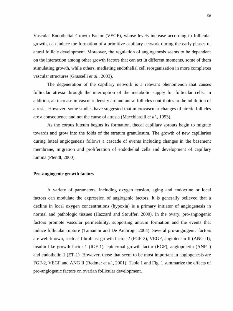
58
Vascular Endothelial Growth Factor (VEGF), whose levels increase according to follicular
growth, can induce the formation of a primitive capillary network during the early phases of
antral follicle development. Moreover, the regulation of angiogenesis seems to be dependent
on the interaction among other growth factors that can act in different moments, some of them
stimulating growth, while others, mediating endothelial cell reorganization in more complexes
vascular structures (Grasselli et al., 2003).
The degeneration of the capillary network is a relevant phenomenon that causes
follicular atresia through the interruption of the metabolic supply for follicular cells. In
addition, an increase in vascular density around antral follicles contributes to the inhibition of
atresia. However, some studies have suggested that microvascular changes of atretic follicles
are a consequence and not the cause of atresia (Macchiarelli et al., 1993).
As the corpus luteum begins its formation, thecal capillary sprouts begin to migrate
towards and grow into the folds of the stratum granulosum. The growth of new capillaries
during luteal angiogenesis follows a cascade of events including changes in the basement
membrane, migration and proliferation of endothelial cells and development of capillary
lumina (Plendl, 2000).
Pro-angiogenic growth factors
A variety of parameters, including oxygen tension, aging and endocrine or local
factors can modulate the expression of angiogenic factors. It is generally believed that a
decline in local oxygen concentrations (hypoxia) is a primary initiator of angiogenesis in
normal and pathologic tissues (Hazzard and Stouffer, 2000). In the ovary, pro-angiogenic
factors promote vascular permeability, supporting antrum formation and the events that
induce follicular rupture (Tamanini and De Ambrogi, 2004). Several pro-angiogenic factors
are well-known, such as fibroblast growth factor-2 (FGF-2), VEGF, angiotensin II (ANG II),
insulin like growth factor-1 (IGF-1), epidermal growth factor (EGF), angiopoietin (ANPT)
and endothelin-1 (ET-1). However, those that seem to be most important in angiogenesis are
FGF-2, VEGF and ANG II (Redmer et al., 2001). Table 1 and Fig. 1 summarize the effects of
pro-angiogenic factors on ovarian follicular development.
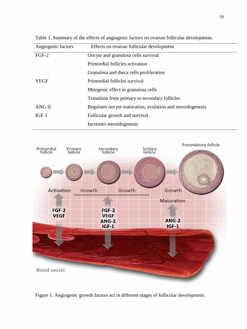
59
Table 1. Summary of the effects of angiogenic factors on ovarian follicular development.
Angiogenic factors Effects on ovarian follicular development
FGF-2 Oocyte and granulosa cells survival
Primordial follicles activation
Granulosa and theca cells proliferation
VEGF Primordial follicles survival
Mitogenic effect in granulosa cells
Transition from primary to secondary follicles
ANG II Regulates oocyte maturation, ovulation and steroidogenesis
IGF-1 Follicular growth and survival
Increases steroidogenesis
Figure 1. Angiogenic growth factors act in different stages of follicular development.

60
Fibroblast growth factor-2 (FGF-2)
FGF-2, also known as basic FGF (bFGF), was the first angiogenic factor indentified in
the ovary (Gospodarowicz et al., 1985). The localization of FGF-2 in endothelial cells
suggests that it is an important factor for endothelial growth (Gospodarowicz et al., 1985).
FGF-2 is also found in ovarian follicles (rat: Nilsson et al., 2001; human: Ben-haroush
et al., 2005) and corpus luteum (rat: Asakai et al., 1993; bovine: Schams et al., 1994), while
its receptors are expressed in growing follicles (Wandji et al., 1995). In medium and large
swine follicles, mRNA for FGF-2 and its receptor FGFR-2 was detected in granulosa and
theca cells, respectively. In the bovine ovary, the expression of the mRNA for FGF-2 in inner
theca significantly increases during final follicular growth; however this expression was weak
in granulosa cells (Shimizu et al., 2002). This factor exerts an antiapoptotic effect in
granulosa cells, favoring the production of other angiogenic factors (Grasselli et al., 2002).
Some in vitro studies have shown that addition of FGF-2 to the culture medium
promoted primordial and primary follicles growth (Nilsson et al., 2001), granulosa and theca
cell proliferation (Wandji et al., 1996), as well as oocyte survival (Zhou and Zhang, 2005).
Recently, Matos et al. (2007) demonstrated that FGF-2 at 50 ng/mL, stimulated goat
primordial follicle activation after 5 days of in vitro culture.
Vascular Endothelial Growth Factor (VEGF)
VEGF, also known as vascular permeability factor (VPF), is a potent mitogenic factor
that stimulates endothelial cell migration. It has also a role in the structural maintenance,
increase of capillary permeability (Redmer et al., 2001) and a survival factor for endothelial
cells of microvessels (Stouffer et al., 2001). The VEGF family is composed of at least six
members (VEGF A, B, C, D, E and F), and the human VEGF-A gene is organized in eight
exons, separated by seven introns. Alternative exon splicing results in the generation of four
different isoforms, having 121, 165, 189, and 206 amino acids, respectively, after signal
sequence cleavage (VEGF121, VEGF165, VEGF189, VEGF206; Stouffer et al., 2001;
Ferrara, 2004). VEGF A expression was demonstrated in preantral follicles. The protein
VEGF A has been identified in oocytes of human primordial follicles (Otani et al., 1999;

61
Harata et al., 2006) and human and rat primary follicles (Celik- Ozenci et al., 2003). In swine
and bovine follicles, VEGF A is weakly expressed during early development and this
expression becomes higher in granulosa and theca cells of dominant follicles (Barboni et al.,
2000; Greenaway et al., 2005). Expression of mRNA for VEGF in the granulosa and theca
cells, as well as the protein for VEGF in all follicle compartments and follicular fluid
significantly increase according to the stage of follicular development (Yamamoto et al.,
1997; Greenaway et al., 2004; Taylor et al., 2004). Recently, VEGF A expression in rats was
occasionally observed in early preantral follicles and was always detected in preantral
follicles during the late stages of development (Abramovich et al., 2009; Martelli et al.,
2009). Furthermore, there are two known VEGF receptors (VEGFR-1 and VEGFR-2) that
bind to VEGF-A. VEGFR-1 is expressed in quiescent and proliferative endothelial cells
(Berisha et al., 2000) and induced the formation of vessels by VEGF (Boonyaprakob et al.,
2003). VEGFR-2 is expressed specially in angiogenic endothelial cells and regulates the
effects of VEGF on the proliferation and migration of these cells (Celik- Ozenci et al., 2003).
High VEGF concentrations cause a destabilization of the blood vessels, resulting in a
new vascular network development, while VEGF deficiency results in blood vessel regression
(Hanahan, 1997). Furthermore, Hazzard et al. (1999) demonstrated that gonadotropins
stimulated VEGF secretion in primate preovulatory follicles and can act as regulatory factors
of VEGF production. In this way, modulation of the hormones that influence VEGF
expression, such as human (hCG) and equine (eCG) chorionic gonadotrophin, luteinizing
hormone (LH) and follicle stimulating hormone (FSH), as well as their levels in the follicle,
are possibly one of the keys to control ovarian follicular angiogenesis. There are also in vitro
(Pepper et al., 1992) and in vivo (Asahara et al., 1995) indications of synergistic effects
between angiogenic growth factors. In bovine, association between VEGF and FGF-2 induced
an in vitro angiogenic response, which was stronger and faster than the effect produced by
these two factors individually.
Recent in vitro studies suggested that VEGF has a mitogenic effect in granulosa cells
and can stimulate follicular growth in rats (Otani et al., 1999). In this species, Kezele et al.
(2005) identified that the gene encoding for VEGF-A is an important regulator of primordial
follicle development. In addition, Danforth et al. (2003) showed that VEGF increases the
number of primary and secondary follicles in rat ovaries. Recently, a study verified that
62
VEGF promoted the transition from primary to secondary follicles in bovine (Yang and
Fortune, 2007). Furthermore, a study associated VEGF production and the increase of blood
vessel content to follicular activation, i.e., the transition from the primordial to primary
follicle stage (Mattioli et al., 2001). Another study showed that endogenous VEGF is essential
to rodent primordial follicle survival (Roberts et al., 2007). Moreover, the inhibition of VEGF
activity produced an increase in ovarian apoptosis through an unbalance in the pro and
antiapoptotic protein rate, leading to a great number of atretic follicles (Abramovich et al.,
2006). Other authors observed that the direct injection of VEGF into the ovary increases
vasculature (Shimizu, 2006), the number of antral follicles and inhibits apoptosis (Quintana et
al., 2004).
Endocrine gland-derived vascular endothelial growth factor (EG-VEGF)
Endocrine gland-derived vascular endothelial growth factor (EG-VEGF) was
identified as a novel human endothelial cell mitogen, through a bioassay assessing the ability
of a library of purified human secreted proteins to promote the growth of primary adrenal
cortex capillary endothelial cells (LeCouter et al., 2001). EG-VEGF does not belong to the
VEGF family or other known families of endothelial mitogens but instead is a member of a
structurally related class of peptides including the digestive enzyme colipase, the Xenopus
headorganizer, Dickkopf (Glinka et al., 1998), venom protein A (VPRA; Joubert and
Strydom, 1980) or mamba intestinal toxin-1 ‗‗MIT-1‘‘ (Schweitz et al., 1999), a non-toxic
component of Dendroaspis polylepis polylepis venom, and the secreted proteins from
Bombina variegata designated Bv8 (Mollay et al., 1999). Its receptors were designated EG-
VEGFR-1 and EG-VEGFR-2 (Masuda et al., 2002). EG-VEGF selectively promoted
proliferation, survival and chemotaxis of endothelial cells isolated from steroidogenic tissues
(Lin et al., 2002). Indeed, exogenous EG-VEGF in the ovary (LeCouter et al., 2001) or testis
(LeCouter et al., 2003) can dramatically affect vascular leakage. Northern blot analysis of a
panel of RNAs from a variety of human tissues revealed EG-VEGF expression in ovary,
testis, adrenal and placenta (LeCouter et al., 2001). In situ hybridization analysis
demonstrated that steroidogenic cells within these glands are the source of EG-VEGF
(LeCouter et al., 2001). Granulosa cells in primordial and primary follicles express EG-VEGF

63
strongly. As the secondary follicle matures, EG-VEGF expression in granulose cells declines
(Ferrara et al., 2003). At approximately 5 days post-ovulation, both VEGF-A and EG-VEGF
are strongly expressed in a portion of granulosa lutein cells, whereas 8 days post-ovulation
EG-VEGF expression is intense in the theca lutein cells, while VEGF expression has
diminished to the point where only weak signal remains in the peripheral thecal cells (Corner,
1956).
Angiotensin II (ANG II)
ANG II is a potent vasoactive peptide, which is converted from ANG I by angiotensin
conversion enzyme, and induces neovascularization in rabbit retina (Fernandez et al., 1985),
mouse and human endometrium (Hu et al., 1996; Li and Ahmed, 1996; Walsh et al., 1997), as
well as in the bovine corpus luteum (Walsh et al., 1997). ANG II acts by its binding to a
group of receptors, classified as ANG receptor type 1 (AT-1) and 2 (AT-2). Both receptors are
expressed in the ovary, and their presence and distribution differ significantly among the
species and follicular development stages (Pucell et al., 1991; Obermüller et al., 1998). In
rabbit ovaries, AT-2 receptor is expressed predominantly in granulosa cells of preovulatory
follicles, while AT-1 is located in theca and stroma cells (Yoshimura et al., 1996). In mouse,
AT-2 is expressed exclusively in granulosa cells of large atretic follicles, while AT-1 is
expressed in all structures of ovarian follicles (Pucell et al., 1991). The presence of ANG II
receptors in the ovary suggests a possible role of this peptide on this organ. Mitsube et al.
(2003) reported that only the blockage of AT-2 increases blood flow in the mouse ovary,
suggesting that vasoconstriction occurs specifically through this receptor. In addition, another
study observed high ANG II levels during proestrous in the mouse, and this fact may be
associated with the high vascular proliferation that occurs in this phase of the estrous cycle
(Costa et al., 2003). Moreover, ANG II improves angiogenic activity of VEGF in bovine
microvascular cells (Otani et al., 2000).
ANG II regulates oocyte maturation (Kuo et al., 1991), ovulation (Yoshimura et al.,
1996) and steroidogenesis (Yoshimura et al., 1993) through the modulation of ovarian blood
flow (Mitsube et al., 2003). ANG II is well known for its vasoconstrictor action; however this
effect was not observed in the ovary after treatment with ANG II (Costa et al., 2003). The

64
vasodilatation was observed in rabbit ovaries perfused in vitro with ANG II and this effect can
be due to the release of gonadotropins stimulated by ANG II (Kuo et al., 1991). In addition,
Shuttleworth et al. (2002) suggested a possible role of ANG II in swine early folliculogenesis
and steroidogenesis. In bovine, ANG II restored the inhibitory effect of follicular cells on
oocyte maturation (Giometti et al., 2005; Stefanello et al., 2006) and stimulated nuclear and
cytoplasmic maturation of swine oocytes (Li et al., 2004). In addition, ANG II appears to
regulate the induction of several autocrine growth factors, such as platelet derived growth
factor, transforming growth factor-β, FGF-2 and IGF-1, and induces the angiogenic activity
through the paracrine function of VEGF in microvascular cells (Otani et al., 2000).
Insulin like growth factor-1 (IGF-1)
The IGF system appears to have indirect effects on angiogenesis through stimulatory
action for VEGF production in luteal cells, as well as through the stimulus of endothelial cell
proliferation and differentiation (Schams et al., 2001). In bovine, a high expression of IGF-1
in theca interna was observed before the phase of follicular selection, while the expression
increased in granulosa cells after this phase. In addition, mRNA for IGF-1 receptor (IGFR-1)
was present in theca interna and granulosa cells with increased levels during final follicular
development (Schams et al., 2002).
In association with FSH, addition of IGF-1 to the in vitro culture medium of preantral
follicles stimulated follicular growth in several species (human: Louhio et al., 2000; bovine:
Gutierrez et al., 2000; rats: Zhao et al., 2001; mouse: Liu et al., 1998). Experiments
performed by Zhou and Zhang (2005) showed that IGF- 1 promoted the growth and
maintained the viability of oocytes from caprine preantral follicles. In swine, 50 ng/mL of
IGF-1 promoted follicular growth, stimulated granulosa cell proliferation and prevented
apoptosis of preantral follicles cultured for 4 days in the presence of serum (Mao et al., 2004).
Furthermore, different concentrations of IGF-1 (10, 50 and 100 ng/mL) increased follicular
diameter and steroidogenesis of mouse preantral follicles cultured in vitro for 6, 10 and 12
days (Demeestere et al., 2004). In a recent study, Thomas et al. (2007) showed that follicular
diameter was increased over control levels by addition of 50 ng/ml of IGF-I during 6 days of
culture.
65
Anti-angiogenic growth factors
Angiogenesis is also modulated by inhibitory factors, such as thrombospodin,
angiostatin, endostatin, 2-metoxiestradiol, hyaluronic acid, platelet factor-4, tumoral necrose
factor α and interferon γ. These substances blocked endothelial cell proliferation and
migration, as well as in vitro capillary formation (Espinosa-Cervantes and Rosado-Garcia,
2002).
Regarding thrombospodin 1 and 2, they bind to their receptor CD36 and inhibit
angiogenesis, inducing endothelial cell apoptosis. In rat ovaries, the expression of
thrombospondin 1 and CD36 mRNA is high in granulosa cells of preantral and antral follicles
and is limited in theca cells. Nevertheless, there is no expression of mRNA for thrombospodin
2 in ovarian follicles. Furthermore, thrombospodin 1 is strongly expressed in small follicles
where the vascularization is absent, showing that expression of thrombospodins decreases
during follicular maturation together with the increase in VEGF levels (Petrik et al., 2002).
Platelet factor-4 inhibits angiogenesis both in vivo and in vitro and the inhibitory
effects are due to the formation of complexes with FGF-2, inhibiting FGF-2 binding to its
receptors (Perollet et al., 1998).
Final considerations
Increasing evidence suggests that physiological angiogenesis in ovarian follicles and
corpus luteum are fundamental features of mammalian reproduction. Failures in vascular
development in these structures may be the reason for several ovarian dysfunctions observed
during the estrous cycle and pregnancy. Therefore, it is necessary to evaluate both in vivo and
in vitro influences of the angiogenic factors, alone or in association, on the survival (anti-
apoptotic effects) of ovarian cells in different species. This information will provide novel
opportunities for therapeutic interventions and improvement of the efficiency of assisted
reproduction in humans and animals in the future.
Acknowledgments
J.B. Bruno is a recipient of a grant from FUNCAP/CAPES (Brazil).
66
References
Abramovich D, Parborel F, Tesone M. 2006. Effect of a vascular endothelial growth factor
(VEGF) inhibitory treatment on the folliculogenesis and ovarian apoptosis in gonadotropin-
treated prepubertal rats. Biol Reprod, 75:434-441.
Abramovich D, Celin AR, Hernandez F, Tesone M, Parborel F. 2009. Spatiotemporal
analysis of the protein expression of angiogenic factors and their related receptors during
folliculogenesis in rats with and without hormonal treatment. Reproduction, 137:309- 320.
Acosta TJ, Hayashi KG, Ohtani M, Miyamoto A. 2003. Local changes in blood flow
within the preovulatory follicle wall and early corpus luteum in cows. Reproduction, 125:759-
767.
Asahara T, Bauters C, Zheng LP, Takeshita S, Bunting S, Ferrara N. 1995. Synergistic
effect of vascular endothelial growth factor and basic fibroblast growth factor on angiogenesis
in vivo. Circulation, 91:365-371.
Asakai R, Tamura K, Eishi Y, Iwamoto M, Kato Y, Okamoto R. 1993. Basic fibroblast
growth factor (bFGF) receptors decrease with luteal age in rat ovarian luteal cells:
colocalization of bFGF receptors and bFGF in luteal cells. Endocrinology, 133:1074-1084.
Barboni B, Turriani M, Galeati G, Spinaci M, Bacci ML, Forni M, Mattioli M. 2000.
Vascular endothelial growth factor production in growing pig antral follicles. Biol Reprod,
63:858-864.
Ben-haroush A, Abir R, Ao A, Jin S, Kessler-Icekson G, Feldberg D, Fisch B. 2005.
Expression of basic fibroblast growth factor and its receptors in human ovarian follicles from
adults and fetuses. Fertil Steril, 84:1257-1268.
Berisha B, Schams D, Kosmann M, Amselgruber W, Einspanier R. 2000. Expression and
tissue concentration of vascular endothelial growth factor, its receptors and localization in the
bovine corpus luteum during estrous cycle and pregnancy. Biol Reprod, 63:1106-1114.
Boonyaprakob U, Gadsby JE, Hedgpeth V, Routh P, Almond GW. 2003. Expression and
localization of vascular endothelial growth factor and its receptors in pig corpora lutea during
the oestrous cycle. Reproduction, 126:393-405.
Carmeliet P, Jair RK. 2000. Angiogenesis in cancer and other diseases. Nature, 407:249-
257.

67
Celik-Ozenci C, Akkoyunhlu G, Kayisli UA, Arici A, Demir R. 2003. Localization of
vascular endothelial growth factor in the zona pellucida of developing ovarian follicles in the
rat: a possible role in destiny of follicles. Histochem Cell Biol, 120:383-390.
Corner GWJ. 1956. The histological dating of the human corpus luteum of menstruation. Am
J Anat, 9:377-401.
Costa APR, Fagundes-Moura CR, Pereira VM, Silva LFM, Vieira AR, Santos RAS, Reis
AM. 2003. Angiotensin-(1–7): A novel peptide in the ovary. Endocrinology, 144:1942-1948.
Danforth DR, Arbogast LK, Ghosh S, Dickerman A, Rofagha R, Friedman CI. 2003.
Vascular endothelial growth factor stimulates preantral follicle growth in the rat ovary. Biol
Reprod, 68:1736-1741.
Demeestere I, Gervy C, Centner J, Devreker F, Englert Y, Delbaere A. 2004. Effect of
insulin-like growth factor-I during preantral follicular culture on steroidogenesis, in vitro
oocyte maturation, and embryo development in mice. Biol Reprod, 70:1664-1669.
Espinosa-Cervantes MC, Rosado-Garcia A. 2002. Angiogenesis in reproductive
physiology: follicular development, formation and maintenance of the corpus luteum. Ginecol
Obstet Mex, 70:17-27.
Fernandez LA, Twickler J, Mead A. 1985. Neovascularization produced by angiotensin II.
J Lab Clin Med, 105 141-145.
Ferrara N, Frantz G, LeCouter J, Dillart-Telm L, Pham T, Draksharapu A, Giordano
T, Peale F. 2003. Differential expression of the angiogenic factor genes VEGF and EG-
VEGF in normal and polycystic human ovaries. Am J Pathol, 162:1881-1893.
Ferrara N. 2004. Vascular endothelial growth factor: basic science and clinical progress.
Endocr Rev, 25:581- 611.
Fraser HM, Lunn SF. 2000. Angiogenesis and its control in the female reproductive system.
Br Med Bull, 56: 787-797.
Giometti IC, Bertagnolli AC, Ornes RC, Costa LFS, Carambula SF, Reis AM, Oliveira
JF, Emanuelli IP, Gonçalves PB. 2005. Angiotensin II reverses the inhibitory action
produced by theca cells on bovine oocyte nuclear maturation. Theriogenology, 63:1014-1025.
Glinka A, Wu W, Delius H, Monaghan AP, Blumenstock C, Niehrs C. 1998. Dickkopf-1
is a member of a new family of secreted proteins and functions in head induction. Nature,
391:357-362.

68
Gospodarowicz D, Cheng J, Lui GM, Baird A, Esch F, Bohlen P. 1985. Corpus luteum
angiogenic factor is related to fibroblast growth factor. Endocrinology, 117:2383-2391.
Grasselli F, Basini G, Bussolati S, Tamanini C. 2002. Effects of VEGF and bFGF on
proliferation and production of steroids and nitric oxide in porcine granulosa cells. Reprod
Domest Anim, 37:362-368.
Grasselli F, Basini G, Tirelli M, Cavalli V, Bussolati S, Tamanini C. 2003. Angiogenic
activity of porcine granulosa cells cocultured with endothelial cells in a microcarrier based
three-dimensional fibrin gel. J Physiol Pharmacol, 54:361-370.
Greenaway J, Connor K, Pedersen HG, Coomber BL, Lamarre J, Petrik J. 2004.
Vascular endothelial growth factor and its receptor, Flk-1/KDR, are cytoprotective in the
extravascular compartment of the ovarian follicle. Endocrinology, 145:2896-2905.
Greenaway J, Centry PA, Feige J-J, Lamarre J, Petrik JJ. 2005. Thrombospondin and
vascular endothelial growth factor are cyclically expressed in an inverse pattern during bovine
ovarian follicle development. Biol Reprod, 72:1071-1078.
Gutierrez CG, Ralph JH, Telfer EE, Wilmut I, Webb R. 2000. Growth and antrum
formation of bovine preantral follicles in long-term culture in vitro. Biol Reprod, 62:1322-
1328.
Hanahan D. 1997. Signaling vascular morphogenesis and maintenance. Science, 277:55-60.
Harata T, Ando H, Iwase A, Nagasaka T, Mizutani S, Kikkawa F. 2006. Localization of
angiotensin II, the AT1 receptor, angiotensin-converting enzyme, aminopeptidase A,
adipocyte-derived leucine aminopeptidase, and vascular endothelial growth factor in the
human ovary throughout the menstrual cycle. Fertil Steril, 86:433-439.
Hazzard TM, Molskness TA, Chaffin CL, Stouffer RL. 1999. Vascular endothelial growth
factor (VEGF) and angiopoietin regulation by gonadotropin and steroids in macaque
granulosa cells during the periovulatory interval. Mol Hum Reprod, 5:1115-1121.
Hazzard TM, Stouffer RL. 2000. Angiogenesis in ovarian follicular and luteal development.
Baillieres Best Pract Res Clin Obstet Gynaecol, 4:883-900.
Hu DE, Hiley CR, Fan TP. 1996. Comparative studies of angiogenic activity of vasoactive
intestinal peptide, endothelin-1 and -3 and angiotensin II in a rat sponge model. Br J
Pharmacol, 117:545-551.

69
Hulshof SCJ, Figueiredo JR, Bekers JF, Bevers MM, Van Den Hurk R. 1994. Isolation
and Characterization of preantral follicles from foetal bovine ovaries. Vet Q, 2:78-80.
Jiang JY, Macchiarelli G, Tsang BK, Sato E. 2003. Capillary angiogenesis and
degeneration in bovine ovarian antral follicles. Reproduction, 125:211-223.
Joubert FJ, Strydom DJ. 1980. Snake venom. The amino acid sequence of protein A from
Dendroaspis polylepis polylepis (black mamba) venom. Hoppe Seylers Z Physiol Chem,
361:1787-1794.
Kezele PR, Ague JM, Nilsson E, Skinner MK. 2005. Alteration in the ovarian transcriptome
during primordial follicle assembly and development. Biol Reprod, 72:241-255.
Klagsbrun M, D`Amore PA. 1996. Vascular endothelial growth factor and its receptors.
Cytokine Growth Factor Rev, 7:259-270.
Kuo TC, Endo K, Dharmarajan AM, Miyazaki T, Atlas SJ, Wallach EE. 1991. Direct
effect of angiotensin II on in vitro perfused rabbit ovary. J Reprod Fertil, 92:469-474.
LeCouter J, Kowalski J, Foster J, Hass P, Zhang Z, Dillard-Telm L, Frantz G, Rangell
L, DeGuzman L, Keller G-A, Peale F, Gurney A, Hillan KJ, Ferrara N. 2001.
Identification of an angiogenic mitogen selective for endocrine gland endothelium. Nature,
412:877-884.
LeCouter J, Ferrara N. 2002. Endocrine gland-derived VEGF and the emerging hypothesis
of organ-specific regulation of angiogenesis. Nat Med, 8:913-917.
LeCouter J, Lin R, Frantz G, Tejada M, Peale F, Hillan KJ, Ferrara N. 2003. The EG-
VEGF homologue, Bv8, is a potent angiogenic factor. Localization of Bv8 receptors to
endothelial cells. Proc Natl Acad Sci USA, 100:2685-2690.
Li XF, Ahmed A. 1996. Dual role of angiotensin II in the human endometrium. Hum Reprod,
11:95-108.
Li YH, Jiao LH, Liu RH, Chen XL, Wang H, Wang WH. 2004. Localization of
angiotensin II in pig ovary and its effects on oocyte maturation in vitro. Theriogenology,
61:447-459.
Lin R, LeCouter J, Kowalski J, Ferrara N. 2002. Characterization of EGVEGF signaling in
adrenal cortex capillary endothelial cells. J Biol Chem, 277:8724-8729.

70
Liu X, Andoh K, Yokota H, Kobayashi J, Abe Y, Yamada K, Mizunuma H, Ibuki Y.
1998. Effects of growth hormone, activin, and follistatin on the development of preantral
follicle from immature female mice. Endocrinology, 139:2342-2347.
Louhio H, Hovatta O, Sjöberg J, Tuuri T. 2000. The effects of insulin, and insulin-like
growth factors I and II on human ovarian follicles in long-term culture. Mol Hum Reprod,
6:694-698.
Macchiarelli G, Nottola SA, Vizza E, Familiari G, Kikuta A, Murakami T, Motta PM.
1993. Microvasculature of growing and atretic follicles in the rabbit ovary: a SEM study of
corrosion casts. Arch Histol Cytol, 56:1-12.
Mao J, Smith MF, Rucker EB, Wu GM, McCauley TC, Cantley JTC, Prather RS,
Didion BA, Day BN. 2004. Effect of epidermal growth factor and insulin-like growth factor I
on porcine preantral follicular growth, antrum formation, and stimulation of granulosal cell
proliferation and suppression of apoptosis in vitro. J Anim Sci, 82:1967-1975.
Masuda Y, Takatsu Y, Terao Y, Kumano S, Ishibashi Y, Suenaga M, Abe M, Fukusumi
S, Watanabe T, Shintani Y, Yamada T, Hinuma S, Inatomi N, Ohtaki T, Onda H,
Fujino M. 2002. Isolation and identification of EG-VEGF/prokineticins as cognate ligands
for two orphan G-protein-coupled receptors. Biochem Biophys Res Commun, 293:396-402.
Martelli A, Bernabò N, Berardinelli P, Russo V, Rinaldi C, Di Giacinto O, Mauro A,
Barboni B. 2009. Vascular supply as a discriminating for pig preantral follicle selection.
Reproduction, 137:45-58.
Matos MHT, Van den Hurk R, Lima-verde IB, Luque MCA, Santos KDB, Martins FS,
Báo SN, Lucci CM, Figueiredo JR. 2007. Effects of fibroblast growth factor-2 on the in
vitro culture of caprine preantral follicles. Cell Tiss Organs, 186:112-120.
Mattioli M, Barboni B, Turriani M, Galeati G, Zannoni A, Castellani G, Berardinelli P,
Scapolo PA. 2001. Follicle activation involves vascular endothelial growth factor production
and increased blood vessel extension. Biol Reprod, 65:1014-1019.
Mitsube K, Mikuni M, Matousek M, Zackrisson U, Brannstrom M. 2003. Role of the
angiotensin II system in regulation of ovulation and blood flow in the rat ovary.
Reproduction, 125: 425-435.
71
Mollay C, Wechselberger C, Mignogna G, Negri L, Melchiorri P, Barra D, Kreil G.
1999. Bv8, a small protein from frog skin and its homologue from snake venom induce
hyperalgesia in rats. Eur J Pharmacol, 374: 189-196.
Nilsson E, Parrot JA, Skinner MK. 2001. Basic fibroblast growth factor induces primordial
follicle development and initiates folliculogenesis. Mol Cell Endocrinol, 175:123-130.
Obermüller N, Schlamp D, Hoffmann S, Gentili M, Inagami T, Gretz N, Weigel M.
1998. Localization of the mRNA for the angiotensin II receptor subtype 2 (AT2) in follicular
granulosa cells of the rat ovary by nonradioactive in situ hybridization. J Histochem
Cytochem, 46:865-870.
Otani N, Minami S, Yamoto M, Shikone T, Otani H, Nishiyama R, Otani T, Nakano R.
1999. The vascular endothelial growth factor/fms-like tyrosine kinase system in human ovary
during the menstrual cycle and early pregnancy. J Clin Endocrinol Metab, 84:3845- 3851.
Otani A, Takagi H, Oh H, Suzuma K, Matsumura M, Ikeda E, Honda Y. 2000.
Angiotensin II stimulated vascular endothelial growth factor expression in bovine retinal
pericytes. Invest Ophthalmol Vis Sci, 41:1192- 1199.
Pepper MS, Ferrara N, Orci L, Montesano R. 1992. Potent synergism between vascular
endothelial growth factor and basic fibroblast growth factor in the induction of angiogenesis
in vitro. Biochem Biophys Res Commun, 189:824-831.
Perollet C, Han ZC, Savona C, Caen JP, Bikfalvi A. 1998. Platelet factor 4 modulates
fibroblast growth factor 2 (FGF-2) activity and inhibits FGF-2 dimerization. Blood, 91:3289-
3299.
Petrik JJ, Gentry PA, Feige JJ, Lamarre J. 2002. Expression and localization of
thrombospondin-1 and -2 and their cell surface receptor, CD36, during rat follicular
development and formation of the corpus luteum. Biol Reprod, 67:1522-1531.
Plendl J. 2000. Angiogenesis and vascular regression in the ovary. Anat Histol Embryol,
29:257-266.
Pucell AG, Hodges JC, Sen I, Bumpus FM, Husain A. 1991. Biochemical properties of the
ovarian granulosa type 2- angiotensin II receptor. Endocrinology, 128:1947-1959.
Quintana R, Kopcow L, Sueldo C, Marconi G, Rueda NG, Barañao RI. 2004. Direct
injection of vascular endothelial growth factor into the ovary of mice promotes follicular
development. Fertil Steril, 82:1101-1105.

72
Redmer DA, Reynolds LP. 1996. Angiogenesis in the ovary. Rev Reprod, 1:182-192.
Redmer DA, Doraiswamy V, Bortnem BJ, Fisher K, Jablonka-Shariff A, Grazul-Bilska
AT, Reynolds LP. 2001. Evidence for a role of capillary pericytes in vascular growth of the
developing ovine corpus luteum. Biol Reprod, 65:879-889.
Reynolds LP, Redmer DA. 1995. Utero-placental vascular development and placental
function. J Anim Sci, 73:1839-1851.
Roberts AE, Arbogast LK, Friedman CI, Cohn DE, Kaumaya PT, Danforth D.R. 2007.
Neutralization of endogenous vascular endothelial growth factor depletes primordial follicles
in the mouse ovary. Biol Reprod, 76:218-223.
Schams D, Amselgruber W, Einspanier R, Sinowatz F, Gospodarowicz D. 1994.
Localization and tissue concentration of basic fibroblast growth factor in the bovine corpus
luteum. Endocrine, 2:907-912.
Schams D, Kosmann M, Berisha B, Amselgruber WM, Miyamoto A. 2001. Stimulatory
and synergistic effects of luteinising hormone and insulin-like growth factor 1 on the secretion
of vascular endothelial growth factor and progesterone of cultured bovine granulose cells. Exp
Clin Endocrinol Diabetes, 109:155-162.
Schams D, Berisha B, Kosmann M, Amselgruber W. 2002. Expression and localization of
IGF family members in bovine antral follicles during final growth and in luteal tissue during
different stages of estrous cycle and pregnancy. Domest Anim Endocrinol, 22:51- 72.
Schweitz H, Pacaud P, Diochot P, Moinier D, Ladzunski M. 1999. MIT(1), a black mamba
intestinal toxin with a new and highly potent activity on intestinal contraction. FEBS Lett,
461:183-188.
Shimizu T, Jiang J-Y, Sasada H, Sato E. 2002. Changes of messenger RNA expression of
angiogenic factors and related receptors during follicular development in gilts. Biol Reprod,
67:1846-1852.
Shimizu T. 2006. Promotion of ovarian follicular development by injecting vascular
endothelial growth factor (VEGF) and growth differentiation factor 9 (GDF-9) genes. J
Reprod Dev, 52:23-32.
Shuttleworth G, Pipkin FB, Hunter MG. 2002. In vitro development of pig preantral
follicles cultured in a serum-free medium and the effect of angiotensin II. Reproduction,
123:807-818.

73
Smith SK. 2001. Angiogenesis and reproduction. Br J Obstet Gynaecol, 108:777-783.
Stefanello JR, Barreta MH, Porciuncula PM, Arruda JN, Oliveira JF, Oliveira MA,
Gonçalves PB. 2006. Effect of angiotensin II with follicle cells and insulin-like growth
factor-I or insulin on bovine oocyte maturation and embryo development. Theriogenology,
66:2068-2076.
Stouffer RL, Martínez-Chequer JC, Molskness TA, Xu F, Hazzard TM. 2001. Regulation
and action of angiogenic factors in the primate ovary. Arch Med Res, 32:567-575.
Tamanini C, De Ambrogi M. 2004. Angiogenesis in developing follicle and corpus luteum.
Reprod Dom Anim, 39:206-216.
Taylor PD, Hillier SG, Fraser HM. 2004. Effects of GnRH antagonist treatment on
follicular development and angiogenesis in the primate ovary. J Endocrinol, 183:1-17.
Telfer EE. 1996. The development of methods for isolation and culture of preantral follicles
from bovine and porcine ovaries. Theriogenology, 45:101-110.
Thomas FH, Campbell BK, Armstrong DG, Telfer EE. 2007. Effects of IGF-I
bioavailability on bovine preantral follicular development in vitro. Reproduction, 133:1121-
1128.
Walsh DA, Hu DE, Wharton J, Catravas JD, Blake DR, Fan TP. 1997. Sequencial
development of angiotensin receptors and angiotensin I converting enzyme during
angiogenesis in the rat subcutaneous sponge granuloma. Br J Pharmacol, 120:1302-1311.
Wandji SA, Eppig JJ, Fortune JE. 1995. FSH and growth factors affect the growth and
endocrine function in vitro of granulosa cells of bovine preantral follicles. Theriogenology,
45:817-832.
Wandji SA, Srsen V, Voss AK, Eppig JJ, Fortune JE. 1996. Initiation in vitro of growth of
bovine primordial follicles. Biol Reprod, 55:942-948.
Yamamoto S, Konishi I, Tsuruta Y, Nanbu K, Mandai M, Kuroda H, Matsushita K,
Hamid AA, Yura Y, Mori T. 1997. Expression of vascular endothelial growth factor
(VEGF) during folliculogenesis and corpus luteum formation in the human ovary. Gynecol
Endocrinol, 11:371-381.
Yang MY, Fortune JE. 2007. Vascular endothelial growth factor stimulates the primary to
secondary follicle transition in bovine in vitro. Mol Reprod Dev, 74:1095-1104.

74
Yoshimura Y, Karube M, Oda T, Koyama N, Shiokawa S, Akiba M, Yoshinaga A,
Nakamura Y. 1993. Locally produced angiotensin II induces ovulation by stimulating
prostaglandin production in in vitro perfused rabbit ovaries. Endocrinology, 133:1609-1616.
Yoshimura Y, Karube M, Aoki H, Oda T, Koyama N, Nagai A, Akimoto Y, Hirano H,
Nakamura Y. 1996. Angiotensin II induces ovulation and oocyte maturation in rabbit ovaries
via the AT2 receptor subtype. Endocrinology, 137:1204-1211.
Zhao J, Taverne MAM, Van Der Weijden GC, Bevers MM, Van Den Hurk R. 2001.
Insulin-like growth factor-I (IGF-I) stimulates the development of cultured rat preantral
follicle. Mol Reprod Dev, 58:287- 296.
Zhou H, Zhang Y. 2005. Regulation of in vitro growth of preantral follicles by growth
factors in goats. Domest Anim Endocrinol, 28:235-242.
75
7. CAPÍTULO 2
Envolvimento do peptídeo intestinal vasoativo na fisiologia ovariana
(Involvement of vasoactive intestinal peptide (VIP) on ovarian physiology)
Periódico: Animal Reproduction (Submetido em outubro de 2010)
76
Resumo
Além do sistema nervoso central, fibras nervosas contendo peptídeo intestinal vasoativo
(VIP), foram descritas ao longo do trato genital feminino. Existem crescentes evidências de
que o VIP e seus receptores desempenham um papel importante na regulação local da
fisiologia ovariana via AMPc. Tem sido relatado que o VIP regula a sobrevivência e
crescimento do folículo ovariano, maturação do oócito, ovulação e a esteroidogênese.
Portanto, esta revisão visa resumir os dados atuais sobre a importância do VIP na fisiologia
ovariana.
Palavras-chave: VIP. Folículos ovarianos. Foliculogênese. Ovulação. Esteroidogênese.

77
Involvement of vasoactive intestinal peptide (VIP) on ovarian physiology
J.B. Brunoa; M.H.T. Matos
b; R.N.Chaves
a; J.R. Figueiredo
a
aFaculty of Veterinary, LAMOFOPA, PPGCV, State University of Ceará, Fortaleza-CE, Brazil.
bNucleus of Biotechnology Applied to Ovarian Follicle Development, Federal University of São Francisco
Valley, Petrolina-PE, Brazil.
*Correspondence should be addressed to:
Programa de Pós-Graduação em Ciências Veterinárias (PPGCV)
Laboratório de Manipulação de Oócitos e Folículos Pré-Antrais (LAMOFOPA)
Universidade Estadual do Ceará (UECE)
Av. Paranjana, 1700, Campus do Itaperi.
Fortaleza – CE – Brasil. CEP: 60740 903
Tel.: +55.85. 3101.9852; Fax: +55.85.3101.9840
E-mail address: [email protected] (J.B.Bruno)

78
Abstract
In addition to the central nervous system, vasoactive intestinal peptide (VIP) containing
nerves have been described throughout the female genital tract. There is growing evidence
that VIP and their receptors play important roles in the local regulation of ovarian physiology
through cAMP pathway. It has been reported that VIP regulates the ovarian follicle survival
and growth, oocyte maturation, ovulation and steroidogenesis. Therefore, this review aims to
summarize current data on the importance of VIP on ovarian physiology.
Keywords: VIP, ovarian follicles, ovulation, steroidogenesis.
Introduction
Ovarian activity is regulated not only by gonadotropins and steroids, but also by a
number of neural inputs and paracrine regulatory mechanisms. The mammalian ovary is
innervated by extrinsic nerves, which are both catecholaminergic and peptidergic in nature
(Burden, 1985; Ojeda and Lara, 1989; Ojeda et al., 1989). Peptidergic innervation of the
ovary was verified, among other ways, by the presence of vasoactive intestinal peptide (VIP)
(Ahmed et al., 1986).
In addition to the central nervous system, VIP containing nerves have been described
throughout the human female genital tract being most abundant in the vagina, cervix and
clitoris and less numerous in the uterine body, oviduct and ovary (Ottesen and Fahrenkrug,
1995), which suggests that such peptides may not play an exclusively neuroendocrine role.
There is growing evidence that VIP plays an important role in the female reproductive system
by acting as a potential local regulator of ovarian physiology, such as regulation of
steroidogenesis, cAMP accumulation, plasminogen activator production and oocyte
maturation (Ahmed et al., 1986; Tornell et al., 1988; Johnson and Tilly, 1988). This review
will focus on the role of VIP and their receptors in ovarian physiology.

79
Expression of VIP and their receptors in the ovary
VIP is a member of the structurally related neuropeptide family including pituitary
adenylate cyclase-activating polypeptide (PACAP)/secretin/glucagons (Arimura, 1992; Gozes
et al., 1999; Miyata et al., 1989). VIP and PACAP act by binding to three types of G protein-
coupled VIP/PACAP receptors. PAC1-R binds specifically to both PACAPs and VIP, though
with very low affinity; VPAC1-R and VPAC2-R bind to PACAP and VIP with equal affinity
(Lutz et al., 1993).
In ovaries, VIP is reported to be produced by nerves fibers innervating follicles at all
stages of development of rodent (Ahmed et al., 1986) and avian (Johnson et al., 1994).
Hulshof et al. (1994) showed that bovine ovary is innervated by VIP-positive nerve fibers,
beginning at the onset of follicular development and increasingly with age. Furthermore, VIP
and their receptors immunoreactivity has been found in close association with the ovarian
vasculature (Hulshof et al., 1994; Vaccari et al., 2006).
Despite the presence of mRNA for VIP has been detected in ovaries of rats (Gozes and
Tsafriri, 1986), its presence was not observed in mice (Barberi et al., 2007). These authors
suggested such results due to the fact that this peptide has been originated in extrinsic
innervations. In fact, the lack of radioimmunoassayable VIP levels following the transection
of the ovarian nerves indicates that ovarian VIP derives mostly from extrinsic innervation of
the gland. However, in goats, the presence of mRNA for VIP was detected in all follicular
categories (primordial, primary, secondary and antral follicles) and cellular types studied
(granulosa and theca cells) (Bruno et al., unpublished data), suggesting a local synthesis of
this peptide.
Barberi et al. (2007) showed that the three VIP receptors are expressed in mouse
granulosa cells in vivo. Furthermore, other authors have shown that VPAC1-R is the most
abundant receptor in ovaries from juvenile mice, and that VPAC2-R levels are lower, while
those of PAC1-R are very low (Cecconi et al., 2004). In the rat preovulatory follicles, PAC1-
R and VPAC2-R are expressed by granulosa cells, whereas theca/interstitial cells exclusively
express VPAC1-R e VPAC2-R (Vaccari et al., 2006). PAC1-R was the main receptor present
in the ovary of rat, followed by VPAC2-R and VPAC1-R (Latini et al., 2010).
80
Signaling pathways and effects of VIP on follicle survival
VIP has been shown to protect several cell types from apoptosis, including
thymocytes, prostate cancer and neural cells (Delgado et al., 1996; Said, 1996; Gutierrez-
Canas, et al., 2003; Sastry et al., 2006). In the ovary, previous in vitro studies also
demonstrated that VIP inhibits apoptosis of rat follicles (Flaws et al., 1995; Lee et al. 1999).
Moreover, it has been demonstrated that PACAP and VIP were able to prevent granulosa cell
apoptosis of follicles cultured in serum free medium (Vaccari et al., 2006; Barberi et al.,
2007). Recently, VIP maintained the ultrastructural integrity of goat preantral follicles after
seven days of ovarian tissue culture (Bruno et al., 2010).
It is possible that VIP maintained cell viability through the cAMP pathway. The
stimulation of cAMP formation by VIP is probably a transient response that occurs early after
culture initiation. In support of this, the ability of VIP to suppress apoptosis was mimicked by
treatment of follicles with the adenylyl cyclase activator (forskolin), and this substance
increased cAMP production and accumulation within cultured follicles (Flaws et al., 1995).
These findings along with previous reports on the role of the cAMP-protein kinase A (PKA)
second messenger system in mediating the effects of VIP in target cells (Ojeda et al., 1989;
Johnson and Tilly, 1988; Davoren and Hsueh, 1986) provide evidence that VIP, potentially
acting at least in part via the cAMP pathway, functions as a novel inhibitor of apoptosis.
Other studies have shown that the neuroprotective effects of VIP are mediated
partially via activation of PKA through PKC (Vaudry et al., 2000) and cAMP response
element binding protein phosphorylation (Walton and Dragunow, 2000). The effect or
mechanisms include inhibition of caspase-3 activity, upregulation of Bcl-2 and suppression of
cytoplasmic cytochrome c translocation (reviewed by Filippatos et al., 2001). On the other
hand, it is plausible that mechanisms independent of PKA activity also partly mediate the
actions of VIP on follicle survival. This possibility is supported by previous studies using a
mouse pituitary cell line that demonstrated activation of voltage-sensitive calcium channels
after VIP stimulation (Schecterson and McKnight, 1991).
81
In vitro effects of VIP on follicle growth
There are only few papers dealing with VIP effects on the ovarian follicles. In vitro
studies revealed that VIP stimulates the development of isolated bovine primary and early
secondary follicles (Hulshof, 1995). We have recently demonstrated that VIP is an important
factor for the growth of small preantral follicles enclosed in caprine ovarian tissue (Bruno et
al., 2010). In addition to the increase of VIP immunoreactivity with the appearance of
secondary and antral follicles in bovine ovaries (Hulshof et al., 1994), the contemporary
presence of VIP and its receptors around preantral and antral follicles (Vaccari et al., 2006)
may explain the role of VIP in follicle growth. Nevertheless, in mice, VIP did not affect
follicular development and caused inhibition of follicular growth, antrum formation,
granulosa cell proliferation, as well as estradiol production of follicles stimulated by FSH
(Cecconi et al., 2004). These conflicting results may be due to study design, differences
related to species, culture conditions and different follicular stages analyzed.
Influence of VIP on maturation and ovulation
Studies have shown that VIP stimulates maturation in follicle-enclosed oocytes, but
could transiently inhibit or not affect spontaneous maturation of cumulus-enclosed oocytes
(Tornell et al., 1988; Apa et al., 1997). Culture of bovine cumulus oocyte complexes in the
presence of VIP did not affect nuclear maturation or cumulus expansion, but it retarded
cytoplasmic maturation as reflected by delayed cortical granule migration (Beker et al., 2000).
Moreover, the addition of VIP to the culture medium did not improve in vitro maturation and
fertilization of sheep (Ledda et al., 1996) and buffalo (Nandi et al., 2003) oocytes.
Two possible explanations for the limited response to VIP in events of maturation can
be suggested: First, depending on the experimental conditions, cAMP can either stimulate or
inhibit meiosis (Hillensjo et al., 1978). It is possible that high doses of VIP sustain high levels
of cAMP in the oocyte and prevent meiosis. Such a phenomenon is believed to occur in the
presence of forskolin (Ekholm et al., 1984), dbcAMP, or phosphodiesterase inhibitors
(Hillensjo et al., 1978). A second explanation for the limited response could be that VIP
affects only one subpopulation of follicles or subpopulation of granulosa cells within each

82
follicle. In fact, Kasson et al. (1985) have shown one predominantly VIP-sensitive and one
FSH-sensitive subpopulation of granulosa cells.
Nevertheless, although VIP did not influence maturation, it has been shown to
stimulate ovulation in perfused rat ovaries (Schmidt et al., 1990). In mammalian system, VIP
or LH, acting via the adenylyl cyclase system, induce an increase in plasminogen activator
activity (Beers et al., 1975; Wang and Leung, 1983; Liu et al., 1987), a serine protease that
has been implicated in the process of follicular rupture at the time of ovulation (Beers, 1975;
Reich et al., 1985) and in the process of follicular cumulus cell expansion and dispersion (Liu
et al., 1986).
In addition, the localization of VIP and its receptors in association with blood vessels
(Hulshof et al., 1994; Vaccari et al., 2006) suggests that this neuropeptide might be involved
in the regulation of ovarian blood flow. In fact, VIP contributes to the increase in blood flow
around preovulatory follicles observed after the LH surge (Acosta et al., 2003). This increased
ovarian stromal blood flow may, in turn, lead to a greater delivery of gonadotrophins to the
granulosa cells of preovulatory follicles (Redmer and Reynolds, 1996), which will be
important for the generation of a normal follicle and competent oocyte. The gonadotrophin
would consequently stimulate the production of PACAP in the preovulatory follicle. The fact
that PACAP induces genes related to ovulation and luteinization, and mediates some of the
effects of LH on granulosa cell differentiation at the time of ovulation (Gras et al., 1999; Lee
et al., 1999, Park et al., 2000), suggests that PACAP may serve as an ovarian physiological
mediator of gonadotrophins in the ovulatory process. This is in agreement with previous
demonstration that provides direct evidence of the presence of PAC1-R and the absence of
VPAC1-R and VPAC2-R on germinal vesicles oocytes (Vaccari et al., 2006). Further studies
are warranted to evaluate the respective roles of PACAP and VIP in ovulatory process.
VIP stimulates ovarian steroidogenesis
Studies have shown that in the periphery, the denervation of ovaries during the early
luteal phase of the estrus cycle leads to changes in their morphology and impairs
steroidogenic activity in pigs (Jana et al., 2005). Similarly, inhibition of ovarian secretory
function and delayed pubertal onset were observed in rats after denervation (Ojeda et al.,

83
1983; Lara et al., 1990; Forneris and Aguado, 2002). The alterations in gonadal endocrine
function are attributed to the loss of the peptidergic supply (for example, VIP) of neuronal
fibers (Jana et al., 2005; Ojeda et al., 1983; Lara et al., 1990; Forneris et al., 2002). Therefore,
these studies show that VIP is implicated in ovarian steroidogenesis.
In addition, VIP is involved in the regulation of steroidogenic activity, stimulating
estradiol and progesterone release from cultured granulosa cells and whole ovaries in vitro
(Davoren and Hsueh, 1985; Ahmed et al., 1986; Parra et al., 2007; Kowalewski et al., 2010),
progesterone release in vivo (Fredericks et al., 1983), and androgen release from ovarian
fragments in vitro (Ahmed et al., 1986). These effects may be related to the ability of VIP to
enhance the synthesis of the cholesterol side-chain cleavage enzyme complex (Trzeciak et al.,
1986), the rate-limiting step in progesterone biosynthesis and the activity of the aromatase
enzyme complex (George and Ojeda, 1987). Studies have provided evidence that VIP is
capable of inducing aromatase activity before the ovary became responsive to gonadotropins
(George and Ojeda, 1987; Mayerhofer et al., 1997). A possibility to consider is that VIP
induction of estradiol release during the estrus cycle may also come from immature follicles.
If true, this would suggest that VIP plays a complementary role to that of FSH, the primary
mediator of estradiol biosynthesis, in determining the magnitude of estradiol increase under
varying physiologic conditions (Parra et al., 2007). These findings strongly suggest a
physiological role for this peptide in ovarian steroid biosynthesis.
Little is known about the molecular mechanisms of VIP-mediated steroidogenesis. It is
known that VIP increases the levels of cAMP (Tornell et al., 1988, Apa et al., 1997; Vaccari
et al., 2006) and subsequently leads to PKA activation, which in turn induces steroidogenesis
in granulosa cells. The cAMP/PKA pathway is the major route in the trophic hormone-
stimulated regulation of steroidogenic acute regulatory protein (StAR) expression and
function. StAR mediates the rate-limiting step in steroidogenesis, the transfer of cholesterol
from the outer to the inner mitochondrial membrane, and it is the hormonal regulation of
StAR expression and activity that allows tissues to accurately control their steroid production
(Kowalewski et al., 2010). A reduction of StAR and 3β-hydroxysteroid dehydrogenase
expression, enzyme converting pregnenolone to progesterone, accompanied by decreased
serum levels of FSH was demonstrated in young VIP knockout mice (Lacombe et al., 2007).

84
Additional signaling pathways may be involved because the receptors activate multiple
intracellular pathways (Spengler et al., 1993; Lutz et al., 1993).
Figure 1. Schematic illustration of the signaling pathways for the effect of VIP in granulosa
cells. VIP binds through two G-protein-coupled receptors (VPAC1-R and VPAC2-R).
Binding of the peptide to the receptors in the cell membrane results in activation of adenylate
cyclase (AC) with increased intracellular formation of cAMP, which in turn activates protein
kinase A (PKA). The reactions culminate in activation of cAMP response element binding
protein (CREB) via its phosphorylation, followed by upregulation of the StAR and
antiapoptotic protooncogene Bcl-2.
Final considerations
Collectively, these results underline the crucial role of VIP and their receptors in the
control ovarian folliculogenesis. VIP and its receptors, through cAMP/PKA pathway, regulate
the follicle survival and growth, oocyte maturation and ovulation, as well as ovarian
steroidogenesis. Further research in this field will greatly advance our understanding of
ovarian physiology.
85
References
Acosta TJ, Hayashi KG, Ohtani M, Miyamoto A. 2003. Local changes in blood flow within
the preovulatory follicle wall and early corpus luteum in cows. Reproduction, 125:759-767.
Ahmed CE, Dees WL, Ojeda SR. 1986. The immature rat ovary is innervated by vasoactive
intestinal peptide (VIP)-containing fibers and responds to VIP with steroid secretion.
Endocrinology, 118:1682-1689.
Apa R, Lanzone A, Mastrandrea M, Miceli F, Macchione E, Fulghesu AM, Caruso A,
Canipari R. 1997. Effect of pituitary adenylate cyclaseactivating peptide on meiotic
maturation in follicle-enclosed, cumulus-enclosed, and denuded rat oocytes. Biol Reprod,
57:1074-1079.
Arimura A. 1992. Pituitary adenylate cyclase activating polypeptide (PACAP): discovery and
current status of research. Regul Pept, 37:287-303.
Barberi M, Muciaccia B, Morelli MB, Stefanini M, Cecconi S, Canipari R. 2007. Expression
localisation and functional activity of pituitary adenylate cyclaseactivating polypeptide,
vasoactive intestinal polypeptide and their receptors in mouse ovary. Reproduction, 134:281-
292.
Beers WH. 1975. Follicular plasminogen and plasminogen activator and the effect of plasmin
on ovarian follicle wall. Cell, 6:379-386.
Beker ARCL, Izadyar F, Colenbrander B, Bevers MM. 2000. Effect of growth hormone
releasing hormone (GHRH) and vasoactive intestinal peptide (VIP) on in vitro bovine oocyte
maturation. Theriogenology, 53:1771-1782.
Bruno JB, Celestino JJH, Lima-Verde IB, Matos MHT, Lima LF, Name KPO, Araújo VR,
Saraiva MVA, Martins FS, Campello CC, Silva JRV, Báo SN, Figueiredo JR. 2010.
Vasoactive intestinal peptide improves the survival and development of caprine preantral
follicles after in vitro tissue culture. Cells Tissue Organs, 191:414-21.
Burden HW. 1985. The adrenergic innervations of mammalian ovaries. In Ben-Jonathan N,
Bahr MM, Weiner RI (Ed.). Catecholamines as Hormone Regulators. New York, Raven
Press, pp. 261-278.
86
Cecconi S, Rossi G, Barberi M, Scaldaferri L, Canipari R. 2004. Effect of pituitary adenylate
cyclase-activating polypeptide and vasoactive intestinal polypeptide on mouse preantral
follicle development in vitro. Endocrinology, 145:2071-2079.
Davoren JJ, Hsueh AJW. 1985. Vasoactive intestinal peptide: a novel stimulator of
steroidogenesis by cultured rat granulosa cells. Biol Reprod, 33:37-52.
Delgado M, Garrido E, Martinez C, Leceta J, Gomariz RP. 1996. Vasoactive intestinal
peptide and pituitary adenylate cyclase-activating polypeptides (PACAP27) and (PACAP38)
protect CD4+CD8+ thymocytes from glucocorticoid-induced apoptosis. Blood, 87:5152-
5161.
Ekholm C, Hiilensjö T, Magnusson C, Rosberg S. 1984. Stimulation and inhibition of rat
oocyte meiosis by forskolin. Biol Reprod, 30:537-43.
Filippatos GS, Gangopadhyay N, Lalude O, Parameswaran N, Said SI, Spielman W, Uhal
BD. 2001. Regulation of apoptosis by vasoactive peptides. Am. J. Physiol. Lung. Cell Mol
Physiol, 281:749-761.
Flaws JA, Desanti A, Tilly KI, Javid RO, Kugu K, Johnson AL, Hirshfield AN, Tilly JL.
1995. Vasoactive intestinal peptidemediated suppression of apoptosis in the ovary: potential
mechanisms of action and evidence of a conserved antiatretogenic role through evolution.
Endocrinology, 136:4351-4359.
Forneris ML, Aguado LI. 2002. Neonatal superior nerve transection disturbs the cyclic
activity of the female rats. J Steroids Biochem Mol Biol, 82:75-82.
Fredericks CM, Lundquist LE, Mathur RS, Ashton SH, Landgrebe SC. 1983. Effects of
vasoactive intestinal polypeptide upon ovarian steroids, ovum transport and fertility in the
rabbit. Biol Reprod, 28:1052-1060.
George FW, Ojeda SR. 1987. Vasoactive intestinal peptide induces aromatase activity before
development of primary follicles or responsiveness to follicle-stimulating hormone. Proc Natl
Acad Sci USA, 84:5803-5807.
Gozes I, Tsafriri A. 1986. Detection of vasoactive intestinal peptideencoding messenger
ribonucleic acid in the rat ovaries. Endocrinology, 119:2606-2610.
Gozes I, Fridkinb M, Hill JM, Brenneman DE. 1999. Pharmaceutical VIP: prospects and
problems. Curr Med Chem, 6:1019-1034.

87
Gras S, Hannibal J, Fahrenkrug J. 1999. Pituitary adenylate cyclaseactivating polypeptide is
an auto/paracrine stimulator of acute progesterone accumulation and subsequent luteinization
in cultured periovulatory granulosa/lutein cells. Endocrinology, 140:2199-2205.
Gutierrez-Cañas I, Rodriguez-Henche N, Bolanos O, Carmena MJ, Prieto JC, Juarranz MG.
2003. VIP and PACAP are autocrine factors that protect the androgen-independent prostate
cancer cell line PC-3 from apoptosis induced by serum withdrawal. Br J Pharmacol,
139:1050-1058.
Hillensjö T, Ekhohn C, Ahrén K. 1978. Role of cyclic AMP in oocyte maturation and
glycolysis in the pre-ovulatory rat follicle. Acta Endocrinol, 87:377-388.
Hulshof SCJ, Dijkstra G, Van Der Beek EM, Bevers MM, Figueiredo JR, Beckers JF, Van
Den Hurk R. 1994. Immunocytochemical localization of vasoactive intestinal peptide and
neuropeptide y in the bovine ovary. Biol Reprod, 50:553-560.
Hulshof SCJ. 1995. Bovine preantral follicles and their development in vitro; Utrecht, Thesis.
Jana B, Dzienis A, Pańczyszyn J, Rogozińska A, Wojtkiewicz J, Skobowiat C, Majewski M.
2005. Denervation of the porcine ovaries performed during the early luteal phase influenced
morphology and function of the gonad. Reprod Biol, 5:69-82.
Johnson AL, Tilly JL. 1988. Effects of vasoactive intestinal peptide on steroid secretion and
plasminogen activator activity in granulose cells of the hen. Biol Reprod, 38:296-303.
Johnson AL, Li Z, Gibney JA, Malamed S. 1994. Vasoactive intestinal peptide-induced
expression of cytochrome p450 cholesterol side-chain cleavage and 17a-hydroxylase enzyme
activity in hen granulosa cells. Biol Reprod, 51:327-333.
Kasson BG, Meidan R, Davoren JB, Hsueh AJW. 1985. Identification of subpopulations of
rat granulosa cells: sedimentation properties and hormonal responsiveness. Endocrinology,
7:1027-1034.
Kowalewski MP, Dyson MT, Boos A, Stocco DM. 2010. Vasoactive intestinal peptide (VIP)-
mediated expression and function of steroidogenic acute regulatory protein (StAR) in
granulosa cells. Mol Cell Endocrinol, 328:93-103.
Lacombe A, Lelievre V, Roselli CE, Muller JM, Waschek JA, Vilain E. 2007. Lack of
vasoactive intestinal peptide reduces testosterone levels and reproductive aging in mouse
testis. J Endocrinol, 194:153-160.

88
Lara HE, McDonald JK, Ahmed CE, Ojeda SR. 1990. Guanethidine-mediated destruction of
ovarian sympathetic nerves disrupts ovarian development and function in rats. Endocrinology,
127:2199-2209.
Latini S, Chiarpotto M, Muciaccia B, Vaccari S, Barberi M, Guglielmo MC, Stefanini M,
Cecconi S, Canipari R. 2010. Inhibitory effect of pituitary adenylate cyclase activating
polypeptide on the initial stages of rat follicle development. Mol Cell Endocrinol, 320:34-44.
Ledda S, Bogliolo L, Leoni G, Calvia P, Naitana S. 1996. Influence of vasoactive intestinal
peptide (VIP), atrial natriuretic peptide (ANP) and insulin-like growth factor-I (IGF-I) on in
vitro maturation of prepubertal and adult sheep oocytes. Zygote, 4:343-348.
Lee J, Park HJ, Choi HS, Kwon HB, Arimura A, Lee BJ, Choi WS, Chun SY. 1999.
Gonadotropin stimulation of pituitary adenylate cyclase-activating polypeptide (PACAP)
messenger ribonucleic acid in the rat ovary and the role of PACAP as a follicle survival
factor. Endocrinology, 140:818-826.
Liu YX, Ny T, Sarkar D, Loskutoff D, Hsueh AJW. 1986. Identification and regulation of
tissue plasminogen activator activity in rat cumulus-oocyte complexes. Endocrinology,
119:1578-1587.
Liu YX, Kasson BG, Dahl KD, Hsueh AJ. 1987. Vasoactive intestinal peptide stimulates
plasminogen activator activity by cultured rat granulosa cells and cumulus-oocyte complexes.
Peptides, 8:29-33.
Lutz EM, Sheward WJ, West KM, Morrow JA, Fink G, Harmar AJ. 1993. The VIP2 receptor:
molecular characterisation of a cDNA encoding a novel receptor for vasoactive intestinal
peptide. FEBS Lett, 334:3-8.
Mayerhofer A, Dissen GA, Costa M, Ojeda SR. 1997. A role for neurotransmitters in early
follicular development: induction of functional follicle-stimulating hormone receptors in
newly formed follicles of the rat ovary. Endocrinology, 138:3320-3329.
Miyata A, Arimura A, Dahl RR, Minamino N, Uehara A, Jiang L, Culler MD, Coy DH. 1989.
Isolation of a novel 38 residue-hypothalamic polypeptide which stimulates adenylate cyclase
in pituitary cells. Biochem Biophys Res Commun, 164:567-574.
Nandi S, Ravindranatha BM, Gupta PSP, Raghu HM, Sarma PV. 2003. Developmental
competence and post-thaw survivability of buffalo embryos produced in vitro: effect of
89
growth factors in oocyte maturation medium and of embryo culture system Theriogenology,
60:1621-1631.
Ojeda SR, White SS, Aguado LI, Advis JP, Andersen JM. 1983. Abdominal vagotomy delays
the onset of puberty and inhibits ovarian function in the female rat. Neuroendocrinology,
36:261-267.
Ojeda SR, Lara HE. 1989. The role of the sympathetic nervous system in the regulation of
ovarian function. In Pirke KM, Wuttke W, Schweiger U (Ed.). The Menstrual Cycle and Its
Disorders, Berlin, Springer-Verlag, pp. 26-32.
Ojeda SR, Lara HE, Ahmed CE. 1989. The potential relevance of vasoactive intestinal peptide
to ovarian physiology. In Adashi E (Ed.). Putative Intraovarian Regulators, New York,
Thieme Inc., pp. 52-60.
Ottesen B, Fahrenkrug J. 1995. Vasoactive intestinal polypeptide and other preprovasoactive
intestinal polypeptide-derived peptides in the female and male genital tract: localization,
biosynthesis, and functional and clinical significance. Am J Obstet Gynecol, 172:1615–1631.
Park HJ, Lee J, Wang L, Park JH, Kwon HB, Arimura A, Chun SY. 2000. Stage-specific
expression of pituitary adenylate cyclase-activating polypeptide type I receptor messenger
ribonucleic acid during ovarian follicle development in the rat. Endocrinology, 141:702-709.
Parra C, Fiedler JL, Lunal SL, Greiner M, Padmanabhan V, Lara HE. 2007. Participation of
vasoactive intestinal polypeptide in ovarian steroids production during the rat estrous cycle
and in the development of estradiol valerate-induced polycystic ovary. Reproduction,
133:147-154.
Redmer DA, Reynolds LP. 1996. Angiogenesis in the ovary. Rev Reprod, 1:182-192.
Reich R, Miskin R, Tsafriri A. 1985. Follicular plasminogen activator: involvement in
ovulation. Endocrinology, 116:516-521.
Said SI. 1996. Molecules that protect: the defense of neurons and other cells. J Clin Invest,
97:2163-2164.
Sastry KSR, Smith AJ, Karpova Y, Datta SR, Kulik G. 2006. Diverse antiapoptotic signaling
pathways activated by vasoactive intestinal polypeptide, epidermal growth factor, and
phosphatidylinositol 3-kinase in prostate cancer cells converge on bad. J Biol Chem,
281:20891-20901.

90
Schecterson LC, McKnight GS. 1991. Role of cyclic adenosine 3‘,5‘- monophosphate-
dependent protein kinase in hormone-stimulated /3-endorphin secretion in AtT20 cells. Mol
Endocrinol, 5:170-178.
Schmidt G, Jörgensen J, Kannisto P, Liedberg F, Ottesen B, Owman C. 1990. Vasoactive
intestinal polypeptide in the PMSG-primed immature rat ovary and its effect on ovulation in
the isolated rat ovary perfused in vitro. J Reprod Fertil, 90:465-472.
Spengler D, Waeber C, Pantaloni C, Holsboer F, Bockaert J, Seeburg PH, Journot L. 1993.
Differential signal transduction by five splice variants of the PACAP receptor. Nature,
365:170-175.
Tornell J, Carlsson B, Hillensjo T. 1988. Vasoactive intestinal peptide stimulates oocyte
maturation, steroidogenesis, and cyclic adenosine 3, 5 -monophosphate production in isolated
preovulatory rat follicles. Biol Reprod, 39:213-220.
Trzeciak WH, Schmid CE, Simpson E, Ojeda SR. 1986. Vasoactive intestinal peptide induces
the synthesis of the cholesterol side-chain cleavage enzyme complex in cultured rat ovarian
granulosa cells. Proc Natl Acad Sci USA, 83:7490-7494.
Vaccari S, Latini S, Barberi M, Teti A, Stefanini M, Canipari R. 2006. Characterization and
expression of different pituitary adenylate cyclase-activating polypeptide/vasoactive intestinal
polypeptide receptors in rat ovarian follicles. J Endocrinol, 191:287-299.
Vaudry D, Gonzalez BJ, Magali B, Pamantung TF, Fontaine M, Fournier A, Vaudry H. 2000.
The neuroprotective effect of pituitary adenylate cyclase-activating polypeptide on cerebellar
granule cells is mediated through inhibition of the CED3-related cysteine protease caspase-
3/CPP32. Proc Natl Acad Sci USA, 97:13390-13395.
Walton MR, Dragunow M. 2000. Is CREB a key to neuronal survival?. Trends Neurosci,
23:48-53.
Wang C, Leung A. 1983. Gonadotropins regulate plasminogen activator production by rat
granulosa cells. Endocrinology, 112:1201-1207.

91
8. CAPÍTULO 3
Expressão dos receptores de angiotensina II em ovários caprinos e melhoria da
viabilidade folicular in vitro
(Expression of angiotensin II receptors in the caprine ovary and improvement of follicular
viability in vitro)
Periódico: Reproduction, Fertility and Development (Submetido em novembro de 2010).

92
Resumo
Esse estudo investigou os níveis de RNAm para os receptores de ANG II (AGTR1 e AGTR2)
em ovários de cabras e avaliou o efeito da ANG II sobre a sobrevivência e o crescimento de
folículos pré-antrais caprinos. Fragmentos ovarianos foram cultivados por um ou sete dias em
α-meio essencial mínimo (αMEM+) com ANG II (0, 1, 5, 10, 50 ou 100 ng/mL) e analisados
por histologia, microscopia de fluorescência e microscopia eletrônica de transmissão. Os
níveis de RNAm para AGTR1 foram similares nos diferentes tipos de folículos pré-antrais;
folículos primordiais apresentaram maiores níveis de RNAm para AGTR2 que folículos
secundários. Células da granulosa/teca de pequenos ou grandes folículos antrais apresentaram
níveis significativamente superiores de RNAm para AGTR1 que seus respectivos COCs. Por
outro lado, COCs de grandes folículos antrais tinham níveis de RNAm para AGTR2 que
foram significativamente maiores que suas respectivas células da granulosa/teca. ANG II (10
and 50 ng/mL) aumentou a percentagem de folículos pré-antrais normais após sete dias de
cultivo comparado com αMEM+. Estudos de fluorescência e ultra-estrutural confirmaram a
integridade folicular após sete dias em ANG II (10 ng/mL). Em conclusão, RNAm para
AGTR1 e AGTR2 foram detectados em todas categorias foliculares. ANG II (10 ng/mL)
manteve a viabilidade de folículos pré-antrais caprinos por sete dias.
Keywords: Angiotensina II. Folículo pré-antral. Cultivo. Caprinos.

93
Expression of angiotensin II receptors in the caprine ovary and improvement of
follicular viability in vitro
J.B. Brunoa, I.B. Lima-Verde
b, J.J.H. Celestino
a, L.F. Lima
a, M.H.T. Matos
c, L.R. Faustino
a,
C.M.G Silvaa, R. Rossetto
a, M.V.A. Saraiva
a, C.A.P. Lopes
a, M.A.M. Donato
d, C. A.
Peixotod, C.C. Campello
a, J.R.V. Silva
e, J.R. Figueiredo
a
aFaculty of Veterinary Medicine, LAMOFOPA, PPGCV, State University of Ceara, Fortaleza-CE, Brazil,
bInstitute for Technology and Research, Tiradentes University, Aracaju-SE, Brazil
cNucleus of Biotechnology Applied to Ovarian Follicle Development, Federal University of São Francisco
Valley, Petrolina-PE, Brazil
dLaboratory of Ultrastructure, CPqAM/Fiocruz, Federal University of Pernambuco, Recife-PE, Brazil
eBiotechnology Nucleus of Sobral (NUBIS), Federal University of Ceara, Sobral-CE, Brazil
Running head: ANG II and follicular survival
*Correspondence should be addressed to:
Programa de Pós-Graduação em Ciências Veterinárias (PPGCV)
Laboratório de Manipulação de Oócitos e Folículos Pré-Antrais (LAMOFOPA)
Universidade Estadual do Ceará (UECE)
Av. Paranjana, 1700, Campus do Itaperi.
Fortaleza – CE – Brasil. CEP: 60740 903
Tel.: +55.85. 3101.9852; Fax: +55.85.3101.9840
E-mail address: [email protected] (J.B. Bruno)

94
Abstract
This study investigated the levels of ANG II receptor (AGTR1 and AGTR2) mRNA in goat
ovaries and evaluated the effect of ANG II on the in vitro survival and growth of caprine
preantral follicles. Ovarian fragments were cultured for one or seven days in α-Minimum
Essential Medium (αMEM+) with ANG II (0, 1, 5, 10, 50 or 100 ng/mL) and analyzed by
histology, fluorescent microscopy and transmission electron microscopy. The levels of
AGTR1 mRNA were similar in the different types of preantral follicles; primordial follicles
had higher levels of AGTR2 mRNA than secondary follicles. Granulosa/theca cells from
either small or large antral follicles had significantly higher levels of AGTR1 mRNA than
their respective COCs. Conversely, COCs from large antral follicles had AGTR2 mRNA
levels that were significantly higher than their respective granulosa/theca cells. ANG II (10
and 50 ng/mL) increased the percentages of normal preantral follicles after seven days of
culture compared with αMEM+. Fluorescence and ultrastructural studies confirmed follicular
integrity after seven days in ANG II (10 ng/mL). In conclusion, AGTR1 and AGTR2 mRNAs
were detected in all follicular categories. ANG II (10 ng/mL) maintained caprine preantral
follicle viability for seven days.
Keywords: angiotensin II; preantral follicles; culture; caprine.
1. Introduction
The formation, growth and maturation process of ovarian follicles is known as
folliculogenesis and is influenced by various substances, including hormones and growth
factors (Figueiredo et al. 2008). Of these substances, Angiotensin II (ANG II) plays an
important role in folliculogenesis. ANG II and its receptors are part of a system known as the
renin-angiotensin system (RAS) (Yoshimura, 1997). The RAS plays important roles in the
synthesis and secretion of prostaglandins and estrogen, the regulation of ovarian follicle
development, ovulation and atresia (Pellicer et al. 1988; Yoshimura et al. 1993).
ANG II acts through two main types of membrane receptors, AGTR1 and AGTR2.
ANG II receptors have been detected in the granulosa and inner theca cells of rat antral

95
follicles (Husain et al. 1987). Furthermore, rabbit granulosa cells express high levels of
AGTR2 receptors in preovulatory follicles (Yoshimura et al. 1996). In bovines, Schauser et
al. (2001) observed that only theca cells have ANG II receptors, and a predominance of
AGTR2 was noted in the outer theca. Recently, Portela et al. (2008) detected AGTR1 and
AGTR2 mRNA in both the theca and granulosa cells of bovine antral follicles. However,
quantification of the steady-state level of AGTR1 and AGTR2 mRNAs during different stages
of goat follicular development has not yet been performed.
The activity of ANG II has been related to oocyte maturation, ovulation and
steroidogenesis in rabbits (Yoshimura et al. 1993; Yoshimura et al. 1992; Feral et al. 1995)
and to meiosis resumption (Giometti et al. 2005; Stefanello et al. 2006), follicular growth
(Nielsen et al. 1994) and steroidogenesis (Acosta et al. 1999) in cows. Li et al. (2004)
observed that 100 ng/ml of ANG II stimulated nuclear and cytoplasmic maturation of porcine
oocytes. Giometti et al. (2005) found that oocytes cultured for 12 h in medium conditioned
with follicular cells had inhibited nuclear maturation, and the majority of oocytes remained in
the germinal vesicle stage. However, after the addition of ANG II to the medium, this effect
was reversed.
The role of ANG II in late folliculogenesis, especially in the processes of ovulation
and maturation, is of great interest. However, the effect of this substance on early
folliculogenesis is not currently known. Thus, the present study was designed to investigate
the influence of different concentrations of ANG II (1, 5, 10, 50 or 100 ng/ml) on the survival,
activation (transition from primordial to primary follicles) and growth of preantral follicles
enclosed in caprine ovarian tissue cultured for one or seven days.
2. Materials and methods
2.1 Steady-state level of AGTR1 and AGTR2 mRNA in goat ovarian follicles
To evaluate the steady-state level of mRNA, 30 ovaries from 15 goats (Capra hircus)
were collected at a local slaughterhouse and rinsed in Minimum Essential Medium (MEM)
containing antibiotics (100 µg/mL penicillin and 100 µg/mL streptomycin). After this
preparation, 10 ovaries from 5 goats were used to isolate primordial, primary and secondary

96
follicles. The remaining ovaries were used to collect cumulus–oocyte complexes (COCs),
mural granulosa cells, and theca cells from small and large antral follicles. Primordial,
primary, and secondary follicles were isolated using a previously described mechanical
procedure (Lucci et al. 1999). After isolation, these follicles were washed several times to
completely remove the stromal cells and were then placed by category into separate
Eppendorf tubes in groups of 10. This procedure was completed within 2 h, and all samples
were stored at -80C until the RNA was extracted. From a second group of ovaries (n=15),
COCs aspirated from small (1–3 mm) and large (3–6 mm) antral follicles were recovered.
Compact COCs were selected from the follicle content as described by van Tol and Bevers
(1998). Thereafter, groups of 10 COCs were stored at -80C until RNA extraction. To collect
the mural granulosa and theca cell complex, small (n=10) and large antral follicles (n=10)
were isolated from the ovaries (n=5) and dissected free from the stromal tissue with forceps as
previously described (van Tol and Bevers, 1998). The follicles were then bisected, and the
granulosa and theca cell complexes were collected and stored at -80C.
Total RNA isolation was performed using a Trizol Plus RNA Purification Kit
(Invitrogen, São Paulo, Brazil). According to the manufacturer‘s instructions, 1 mL of Trizol
solution was added to each frozen sample, and the lysate was aspirated through a 20-gauge
needle before centrifugation at 10,000 x g for 3 min at room temperature. All lysates were
then diluted 1:1 with 70% ethanol and subjected to a mini-column. After the binding of the
RNA to the column, DNA digestion was performed using RNAse-free DNAse (340 Kunitz
units/mL) for 15 min at room temperature. After washing the column three times, the RNA
was eluted with 30 µL RNAse-free water.
Prior to reverse transcription, the eluted RNA samples were incubated for 5 min at
70C and chilled on ice. Reverse transcription was then performed in a total volume of 20 µl,
which comprised 10 µl of sample RNA, 4 µl 5X reverse transcriptase buffer (Invitrogen), 8 U
RNAseOUT, 150 U Superscript III reverse transcriptase, 0.036 U random primers
(Invitrogen), 10 mM DTT, and 0.5 mM of each dNTP. The mixture was incubated for 1 h at
42C, followed by 5 min at 80C, then it was stored at -20C. Negative controls were
prepared under the same conditions but without the inclusion of the reverse transcriptase.
Quantification of the AGTR1 and AGTR2 mRNAs was performed using SYBR
Green. PCR reactions were composed of 1 µL cDNA as a template in 7.5 µL of SYBR Green

97
Master Mix (PE Applied Biosystems, Foster City, CA), 5.5 µL of ultra-pure water, and 0.5
µM of each primer. The primers were designed to amplify AGTR1 and AGTR2 mRNA.
Glyceraldehyde-2-phosphate dehydrogenase (GAPDH) and β-actin were used as endogenous
controls to normalize the steady-state mRNA level of the genes (Table 1). The thermal
cycling profile for the first round of PCR was as follows: initial denaturation and activation of
the polymerase for 15 min at 94C, followed by 40 cycles of 15 s at 94C, 30 s at 60C, and
45 s at 72C. The final extension was for 10 min at 72C. All reactions were performed in a
real-time PCR Mastercycler (Eppendorf, Germany). The delta-delta-CT method was used to
transform CT values into normalized relative steady-state mRNA levels.
Table 1. Primer pairs used for real-time PCR analyses.
Target
gene
Primer sequence (5´ 3´) Sense Position GenBank accession nº
GAPDH TGTTTGTGATGGGCGTGAACCA
s 287-309 GI:27525390
ATGGCGTGGACAGTGGTCATAA
as 440-462
β-actin ACCACTGGCATTGTCATGGACTCT
s 187-211 GI:28628620
TCCTTGATGTCACGGACGATTTCC
as 386-410
AT1
AT2
AGCATTGACCGCTACCTGGCTATT
TAGTTGGCAAACTGGCCAAACCTG
s
as
367-390
490-513
GI: 57619242
GI: 148277605
TACATCTTCAACCTCGCTGTGGCT
TCACAGGTCCAAAGAGCCAGTCAT
s
as
244-267
346-369
s,sense; as, antisense
2.2 In vitro culture of preantral follicles
Ovarian cortical tissues (n=8 ovaries) were collected at a local slaughterhouse from 4
adult (1-3 years old) mixed-breed goats (Capra hircus). Immediately postmortem, the ovaries
were washed once in 70% alcohol for 10 s and twice in MEM supplemented by HEPES and
antibiotics (100 µg/ml penicillin and 100 µg/ml streptomycin). Ovary pairs were transported
within 1 h to the laboratory in MEM at 4°C (Chaves et al. 2008). Unless otherwise stated, the

98
culture media, synthetic human ANG II, and other chemicals used in the present study were
purchased from Sigma Chemical Co. (St Louis, MO, USA).
The organ culture system used herein was previously described in detail (Matos et al.
2007). In the laboratory, ovarian cortex tissue samples from each ovarian pair were cut into 13
slices (approximately 3 x 3 mm with a 1-mm thickness) using a needle and scalpel under
sterile conditions. The tissue pieces were then either directly fixed for histological and
ultrastructural analysis (fresh control) or placed in culture for one or seven days. Ovarian
pieces were incubated in 1 ml of culture media consisting of MEM supplemented with ITS
(10 µg/mL insulin, 5.5 µg/mL transferrin and 5 ng/mL selenium), 2 mM glutamine, 2 mM
hypoxanthine and 1.25 mg/mL bovine serum albumin (BSA) in the absence (cultured control)
or presence of 1, 5, 10, 50 or 100 ng/mL ANG II in 24-well plates at 39C in 5% CO2. Each
treatment was repeated four times, and the ovaries of four different animals were
consequently used. The culture media were replenished every other day.
2.3 Morphological analysis and assessment of in vitro follicular growth
Before culture (fresh control) and after one or seven days in culture, all of the pieces
cultured with or without ANG II were fixed in Carnoy‘s solution for 12 h and then
dehydrated in increasing concentrations of ethanol. After paraffin embedding (Synth, São
Paulo, Brazil), the caprine tissue pieces were cut into 7-m sections, and each section was
mounted on a glass slide and stained by periodic acid-Schiff and hematoxylin. Follicle stage
and survival were assessed microscopically on serial sections. Coded slides were blindly
examined using a microscope (Nikon, Japan) under 400X magnification.
The developmental stages of follicles have been defined previously (Silva et al.
2004a) as primordial (one layer of flattened granulosa cells around the oocyte) or growing
(intermediate: one layer of flattened to cuboidal granulosa cells; primary: one layer of
cuboidal granulosa cells; secondary: two or more layers of cuboidal granulosa cells around
the oocyte). Individual follicles were further classified as histologically normal when an
intact oocyte surrounded by well-organized granulosa cells in one or more layers without a
pyknotic nucleus was observed. Atretic follicles were defined as those with a retracted
oocyte, pyknotic nucleus, and/or disorganized granulosa cells that were detached from the

99
basement membrane. Overall, 120 follicles were evaluated for each treatment (30 follicles per
treatment for one repetition x four repetitions=120 follicles).
To evaluate follicular activation, the percentages of healthy primordial and growing
follicles were calculated before (fresh control) and after culture in each medium. In addition,
follicle and oocyte diameters were measured only in morphologically normal follicles. The
follicle diameter was recorded as the length from edge to edge of the basement membrane or
from the outside edge of the theca cell layer when present. Oocyte diameter was recorded as
the length from edge to edge of the oocyte membrane. Two perpendicular diameters were
recorded for each parameter, and the average of these two values was reported. Furthermore,
each follicle was examined in every section in which it appeared and matched with the same
follicle on adjacent sections to avoid double counting. This ensured that each follicle was
only counted once, regardless of its size.
2.4 Viability assessment of follicles cultured in vitro
Based on the morphological analysis, the viability of the follicles cultured with the
ANG II concentrations that promoted the highest percentages of normal follicle growth was
further analyzed using fluorescent probes. Additional pairs of goat ovaries (n=2) were
collected from a slaughterhouse and cut into fragments at the laboratory. One of these
fragments was immediately processed for follicle isolation, and the remaining fragments were
cultured for seven days with ANG II (10 and 50 ng/mL) as described above. After the culture
period, fragments were processed for mechanical isolation using the methods described by
Lucci et al. (1999).
Thereafter, the viability of isolated preantral follicles was assessed through a two-color
fluorescence cell viability assay based on the simultaneous determination of live and dead
cells using calcein-AM and ethidium homodimer-1, respectively. The first probe detected the
intracellular esterase activity of viable cells, and the second labeled nucleic acids of non-
viable cells that had plasma membrane disruption. The test was performed by adding 4 μM
calcein-AM and 2 μM ethidium homodimer-1 (Molecular Probes, Invitrogen, Karlsruhe,
Germany) to the suspension of isolated follicles and incubating at 37°C for 15 min. After
labeling, follicles were washed once by centrifugation at 100 × g for 5 min, resuspended in

100
MEM and mounted on a glass microscope slide in 5 mL anti-fading medium (DABCO,
Sigma, Deisenhofen, Germany) to prevent photobleaching. Finally, follicles were examined
using a DMLB fluorescence microscope (Leica, Germany). The emitted fluorescence signals
of calcein-AM and ethidium homodimer-1 were collected at 488 and 568 nm, respectively.
Oocytes and granulosa cells were considered alive if the cytoplasm stained positively with
calcein-AM (green) and the chromatin was not labeled with ethidium homodimer-1 (red).
2.5 Ultrastructural analysis of caprine preantral follicles
For a more detailed evaluation of follicular morphology after histological analysis,
ultrastructural studies were performed on the fragments of fresh control and treated with the
lowest concentration that maintained their viability (10 ng/mL). A section with a maximum
dimension of 1 mm3 was cut from each fragment of the ovarian tissue and fixed in Karnovsky
solution (4% paraformaldehyde and 2.5% glutaraldehyde in 0.1 M sodium cacodylate buffer
at pH 7.2) for 3 h at room temperature (approximately 25°C). After three washes in sodium
cacodylate buffer, specimens were post-fixed in 1% osmium tetroxide, 0.8% potassium
ferricyanide and 5 mM calcium chloride in 0.1 M sodium cacodylate buffer for 1 h at room
temperature. The samples were then dehydrated using a gradient of acetone solutions and then
embedded in SPIN-PON resin (Sigma Company, St Louis, MO). Subsequently, semi-thin
sections (3 µm) were cut, stained with toluidine blue and analyzed using light microscopy at
400 × magnification. Ultra-thin sections (60–70 nm) were obtained from preantral follicles,
which were classified as morphologically normal in semi-thin sections according to the
criteria adopted in histology. Subsequently, the ultra-thin sections were contrasted with uranyl
acetate and lead citrate and examined under a Morgani-FEI transmission electron microscope
operating at 80 kV. The following parameters were evaluated: the density and the integrity of
ooplasmic and granulosa cell organelles, vacuolization and the integrity of the basement and
nuclear membranes.
2.6 Statistical analysis

101
mRNA expression data from primordial, primary and secondary follicles were
analyzed using the Kruskal-Wallis test, and the t test was used for paired comparisons of
mRNA expression in small and large antral follicles (P<0.05). Percentages of the surviving
follicles at both stages (i.e., primordial and developing) and follicle and oocyte diameters
were calculated for one or seven days of the various treatments. These data were initially
analyzed using Kolmogorov–Smirnov and Bartlett‘s tests to confirm a normal distribution and
homogeneity of variance, respectively. Analysis of variance was then performed using the
GLM procedure of SAS (1999), and Dunnett‘s test was applied to compare ANG II-treated
groups with the control and αMEM+
groups (Steel et al. 1997). To avoid type II errors that
may result from the high coefficient of variation, Duncan‘s test was applied to compare
results for cultures with different ANG II concentrations, and Student‘s t-test was used to
compare the means between one and seven days of culture. Data obtained from fluorescence
microscopy, which was used to validate the histological findings, were also pooled and
analyzed by a Chi-square test. Differences were considered to be significant when P<0.05,
and results are expressed as means ± standard error of the mean (SEM).
3. Results
3.1. Steady-state level of AGTR1 and AGTR2 mRNA in goat ovarian follicles
The quantification of AGTR1 receptor mRNA expression did not demonstrate
differences among the preantral follicles, i.e., primordial, primary and secondary follicles
(P>0.05 – Figure 1A). Primordial follicles had significantly higher levels of AGTR2 receptor
mRNA than the secondary follicle stages (P<0.05), but the levels did not differ significantly
from those of primary follicles (P>0.05 – Fig. 1B).
No differences were observed in the AGTR1 and AGTR2 receptor mRNA levels for
COCs or granulosa/theca cells collected from small and large antral follicles (P>0.05 – Figure
1C, D, E, F). Granulosa/theca cells from either small or large antral follicles had significantly
higher levels of AGTR1 mRNA than their respective COCs (P<0.05 – Figure 1G, H).
Conversely, COCs from large antral follicles had AGTR2 mRNA levels that were
significantly higher than their respective granulosa/theca cells (P<0.05 – Figure 1I, J).
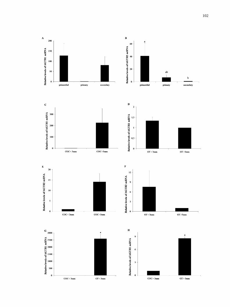
102

103
Figure 1. Steady-state levels of AGTR1 and AGTR2 mRNAs in goat ovarian follicles (means
+ SEM). A, B) Primordial, primary and secondary follicles. C, E) COCs from small and large
antral follicles. D, F) Granulosa/theca cells from small and large antral follicles. G, I) COCs
and granulosa/theca cells from small antral follicles. H, J) COCs and granulosa/theca cells
from large antral follicles. a,b
(P<0.05).
3.2 Effect of ANG II on follicular survival after in vitro culture
The present study analyzed a total of 1,560 caprine preantral follicles. Figure 2 shows
histological sections of primordial (A), intermediate (B) and primary (C) morphologically
normal follicles after seven days of culture in 10 ng/mL ANG II. As shown in Figure 3, a
significant reduction (P<0.05) in the percentage of morphologically normal follicles was
observed in all treatments when compared to the fresh control (90.82%), except when the
fragments were cultured for one day in 10 ng/mL ANG II (81.67%; P>0.05). Furthermore,
after seven days, a significantly higher percentage of normal follicles (P<0.05) was observed
in tissues that were cultured with 10 or 50 ng/mL ANG II than those cultured with 100 ng/mL
ANG II or the control (αMEM+). The percentage of follicles with normal morphology was
similar on days 1 and 7 of culture for all treatments, except for the αMEM+ and 100 ng/ml
ANG II treatments (P<0.05).
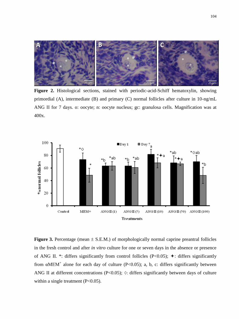
104
Figure 2. Histological sections, stained with periodic-acid-Schiff hematoxylin, showing
primordial (A), intermediate (B) and primary (C) normal follicles after culture in 10-ng/mL
ANG II for 7 days. o: oocyte; n: oocyte nucleus; gc: granulosa cells. Magnification was at
400x.
Figure 3. Percentage (mean S.E.M.) of morphologically normal caprine preantral follicles
in the fresh control and after in vitro culture for one or seven days in the absence or presence
of ANG II. *: differs significantly from control follicles (P<0.05); : differs significantly
from αMEM+ alone for each day of culture (P<0.05); a, b, c: differs significantly between
ANG II at different concentrations (P<0.05); : differs significantly between days of culture
within a single treatment (P<0.05).

105
3.3 Assessment of preantral follicle viability by fluorescence
Based on the results of the morphological analysis, a viability trial was performed
using the treatments that promoted a higher percentage of normal follicles. Caprine preantral
follicles (n=30) were analyzed after seven day culture in αMEM+ supplemented with 10
(Figure 4A, B) or 50 ng/mL ANG II (Figure 4C, D). The results of this quantitative analysis
showed that the percentage of viable follicles was similar in the fresh control group (85%) and
those cultured in 10 and 50 ng/mL ANG II (93.33% and 90%, respectively) (P>0.05).
Figure 4. Viability of caprine preantral follicles as determined using fluorescent probes.
Isolated preantral follicles after culture in 10 (A) and 50 (C) ng/mL ANG II were classified as
viable if cells were labeled by calcein-AM (green fluorescence) (B, D) (B). Scale bars=50
μm.

106
3.4 Ultrastructural analysis of caprine preantral follicles
TEM studies were performed in non-cultured follicles (fresh control) and in follicles
that were cultured for seven days in the lowest concentration of ANG II (10 ng/mL), which
showed the best results for follicular survival and viability. The ultrastructural characteristics
of follicles from the fresh control (Figure 5A) and those cultured with ANG II were similar
(Figure 5B). The follicles showed intact basement and nuclear membranes and a large oocyte
nucleus. In addition, vesicles and organelles were uniformly distributed in the ooplasm,
especially the mitochondria. Granulosa cells were ultrastructurally normal and showed an
elongated and large nucleus with an irregular membrane.
Figure 5. Ultrastructural analysis of non-cultured (fresh control) (A) and cultured caprine
preantral follicles (B), which were cultured for seven days in medium containing 10 ng/mL
ANG II. o, oocyte; n, oocyte nucleus; gc, granulosa cells; m, mitochondria; arrow, oocyte
membrane. (A, B: bar=2 µm; C: bar=1 µm). Three to five follicles per group were examined,
and the photomicrographs are representative examples.
3.5 Follicular activation and growth after culturing with ANG II
Figure 6 (A and B) shows the percentage of primordial and growing follicles,
respectively, in ovarian cortical tissue before and after in vitro culture. After one or seven
days of culture, a significant reduction (P<0.05) in the percentage of primordial follicles was

107
observed in all treatments and was associated with an increase (P<0.05) in the percentage of
growing follicles compared to the fresh control.
Figure 6. Percentage (mean S.E.M.) of primordial (A) and growing (B) follicles in the
fresh control and after in vitro culture for one or seven days in the absence or presence of
ANG II. *: differs significantly from control follicles (P<0.05).
Table 2 shows the diameter of follicles and oocytes after one or seven days culture in
the presence or absence of ANG II. Regarding the follicular diameter, tissues cultured with

108
ANG II for seven days showed values similar to the fresh control and the cultured control
(P>0.05). Furthermore, with progression of the culture period, only the treatments that
contained ANG II maintained their follicular diameter (P>0.05). This result was not found
with αMEM+, in which there was a significant decrease in diameter (P<0.05). Oocyte
diameter was similar between 10 and 50 ng/mL ANG II and the fresh control. This was not
observed in the other treatments, which demonstrated a significant decrease in diameter
(P<0.05). Similar to follicular diameter, αMEM+ alone decreased the oocyte diameter with
the progression of culture (P<0.05).
Table 2. Caprine oocyte and follicle diameters (mean SEM) in the fresh control (non-
cultured) and after culture for one or seven days at different concentrations of ANG II.
Follicular diameter (m) Oocyte diameter (m)
Control 54.04 4.09 43.47 6.12
Treatments Day 1 Day 7 Day 1 Day 7
αMEM+ 57.84 6.38 49.03 8.08
43.10 6.21 35.41 5.27
*
ANG II 1 51.04 7.84 50.89 7.20 40.23 5.14 38.00 6.43 *
ANG II 5 49.23 7.42 48.09 7.71 38.37 5.89* 36.43 5.65*
ANG II 10 52.09 6.24 55.10 7.75 39.95 4.14 39.76 5.65
ANG II 50 54.51 7.62 52.01 8.30 39.67 5.08 38.74 6.30
ANG II 100 52.02 6.76 51.71 7.66 40.58 6.45 37.35 4.93*
*: differs significantly from control follicles (P<0.05); : differs significantly from αMEM+
alone for each day culture (P<0.05); : differs significantly between days of culture within a
single treatment (P<0.05).
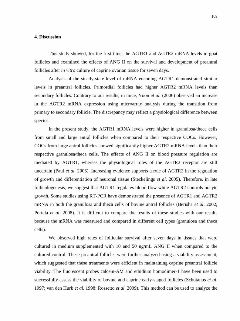
109
4. Discussion
This study showed, for the first time, the AGTR1 and AGTR2 mRNA levels in goat
follicles and examined the effects of ANG II on the survival and development of preantral
follicles after in vitro culture of caprine ovarian tissue for seven days.
Analysis of the steady-state level of mRNA encoding AGTR1 demonstrated similar
levels in preantral follicles. Primordial follicles had higher AGTR2 mRNA levels than
secondary follicles. Contrary to our results, in mice, Yoon et al. (2006) observed an increase
in the AGTR2 mRNA expression using microarray analysis during the transition from
primary to secondary follicle. The discrepancy may reflect a physiological difference between
species.
In the present study, the AGTR1 mRNA levels were higher in granulosa/theca cells
from small and large antral follicles when compared to their respective COCs. However,
COCs from large antral follicles showed significantly higher AGTR2 mRNA levels than their
respective granulosa/theca cells. The effects of ANG II on blood pressure regulation are
mediated by AGTR1, whereas the physiological roles of the AGTR2 receptor are still
uncertain (Paul et al. 2006). Increasing evidence supports a role of AGTR2 in the regulation
of growth and differentiation of neuronal tissue (Steckelings et al. 2005). Therefore, in late
folliculogenesis, we suggest that AGTR1 regulates blood flow while AGTR2 controls oocyte
growth. Some studies using RT-PCR have demonstrated the presence of AGTR1 and AGTR2
mRNA in both the granulosa and theca cells of bovine antral follicles (Berisha et al. 2002;
Portela et al. 2008). It is difficult to compare the results of these studies with our results
because the mRNA was measured and compared in different cell types (granulosa and theca
cells).
We observed high rates of follicular survival after seven days in tissues that were
cultured in medium supplemented with 10 and 50 ng/mL ANG II when compared to the
cultured control. These preantral follicles were further analyzed using a viability assessment,
which suggested that these treatments were efficient in maintaining caprine preantral follicle
viability. The fluorescent probes calcein-AM and ethidium homodimer-1 have been used to
successfully assess the viability of bovine and caprine early-staged follicles (Schotanus et al.
1997; van den Hurk et al. 1998; Rossetto et al. 2009). This method can be used to analyze the

110
viability of follicles, thus offering a new approach for investigating the metabolic and
developmental aspects of folliculogenesis in vitro. Furthermore, after the ultrastructural
analysis, important structures in addition to the basement and nuclear membranes, such as the
mitochondria and granulosa cells, were preserved even after seven days of culture in the
presence of 10 ng/ml ANG II. The beneficial effect of ANG II on follicular viability may have
occurred directly through receptor binding or indirectly through an interaction with other
substances. As previously mentioned, we did not verify differences in AGTR1 receptor
expression in preantral follicles in this work. However, AGTR2 mRNA levels were
significantly higher in primordial follicles than in secondary follicles, but they did not differ
from the primary stage. The ovarian fragments consisted mostly of early preantral follicles
(i.e., primordial and intermediate). Thus, it is possible that ANG II mediates its effect on
viability through AGTR2. A recent study showed that AGTR2 expression in the granulosa
cells of bovine antral follicles is also related to the maintenance of follicular viability because
ANG II concentration and AGTR2 expression were higher in healthy follicles compared to
atretic follicles (Portela et al. 2008). Moreover, Stefanello et al. (2006) found that when ANG
II was associated with insulin-like growth factor-1 (IGF-1), it reversed the inhibitory effect of
follicular cells on the nuclear maturation of bovine oocytes, resulting in a higher rate of
embryo development. In addition to IGF-I, ANG II appears to regulate the induction of other
autocrine growth factors, such as fibroblast growth factor-2 (FGF-2) (Itoh et al. 1993). Thus,
IGF-1 and/or FGF-2 may have functioned as a survival factor for the follicles in the current
study. The importance of IGF-1 (unpubl. data) and FGF-2 (Matos et al. 2007) in maintaining
the morphology of caprine preantral follicles after in vitro culture for seven days has also been
demonstrated.
After seven days of culture in the current study, a high percentage of growing follicles
was observed in all treatments when compared to the fresh control. However, no difference
between ANG II-treated and αMEM+ follicles was found, which indicates that ANG II did not
affect caprine follicular activation. These results agree with those obtained by Harata et al.
(2006) in humans. This study found ANG II immunolocalization to be weak in oocytes of
primordial, primary and secondary follicles. Nevertheless, medium composition is known to
be an important factor for success during the in vitro culture of preantral follicles. The αMEM
that was used in our study is one of the richest formulations of medium for cellular culture
111
and is composed of 21 essential amino acids, B-vitamin complexes, vitamins C and D,
inorganic salts and pyruvate. This medium was also supplemented with glutamine,
hypoxanthine, BSA and ITS, which are substances essential for follicular development (Silva
et al. 2004b). The supplements made the medium more complex and nutrient-rich, which may
be responsible for the efficiency of follicular activation that was observed in all treatments. In
other studies, spontaneous activation was also described after culturing preantral follicles in
αMEM+ without the addition of hormones and growth factors (Lima-Verde et al. in press;
Chaves et al. 2010).
Although ANG II maintained follicle and oocyte diameters throughout the culture
period, it did not affect follicle or oocyte diameter. Shuttleworth et al. (2002) also found no
significant effect of ANG II (10-10
mol/l) on the diameter of pig preantral follicles or antrum
formation after 16 or 30 days of culture. Furthermore, Portela et al. (2008) observed that the
addition of ANG II to bovine granulosa cells in culture did not affect cell proliferation or
estradiol secretion, but it inhibited mRNA that encoded serine protease inhibitor E2, a protein
that is involved in tissue remodeling. We believe that the lack of effect on diameter was
associated with a low mitogenic stimulation caused by ANG II during the initial categories of
follicular development.
In conclusion, this study demonstrated that only AGTR2 was expressed differently in
the preantral follicle categories and that AGTR1 and AGTR2 were differently expressed in
the different compartments of the antral follicles. In addition, 10 ng/mL ANG II improved the
survival of caprine preantral follicles after seven days of in vitro culture. These findings
suggest that ANG II plays an important role in early follicular viability and that it could have
a significant application in improving the quality of oocytes that are used for in vitro
maturation and fertilization.
Acknowledgments
This work was supported by CNPQ (RENORBIO: grant number 554812/2006-1).
Jamily Bezerra Bruno is a recipient of a grant from FUNCAP/CAPES (Brazil). The authors
thank José Leandro da Silva Neto for his technical support in classical histology.

112
Disclosures
The authors declare that there is no potential conflict of interest that can be perceived
as prejudicing the impartiality of the research reported.
References
Acosta, T. J., Berisha, B., Ozawa, T., Sato, K., Schams, D., and Miyamoto, A. (1999).
Evidence for a local endothelin-angiotensin-atrial natriuretic peptide systemin bovine
mature follicles in vitro: effects on steroid hormones and prostaglandin secretion. Biol.
Reprod. 61, 1419-1425.
Berisha, B., Schams, D., Miyamoto, A. (2002). The mRNA Expression of Angiotensin and
Endothelin System Members in Bovine Ovarian Follicles During Final Follicular
Growth. J. Reprod. Developm.48, 573-582.
Chaves, R. N., Alves, A. M. C. V., Duarte, A. B. G., Araújo, V. R., Celestino, J. J. H., Matos,
M. H. T., Lopes, C. A. P., Campello, C. C., Name, K. P. O., Báo, S. N., and Figueiredo,
J. R. (2010). Nerve growth factor promotes the survival of goat preantral follicles
cultured in vitro. Cells. Tissues. Organs. 192, 272-82.
Chaves, R. N., Martins, F. S., Saraiva, M. V. A., Celestino, J. J. H., Lopes, C. A. P., Correia,
J. C., Lima-Verde, I. B., Matos, M. H. T., Báo, S. N., Name, K. P. O., Campello, C. C.,
Silva, J. R. V., and Figueiredo, J. R. (2008). Chilling ovarian fragments during
transportation improves viability and growth of goat preantral follicles cultured in vitro.
Reprod. Fertil. Dev. 20, 640-647.
Feral, C., LeGall, S., and Leymarie, P. (1995). Angiotensin II modulates steroidogenesis in
granulosa and theca in the rabbit ovary: Its possible involvement in atresia. Eur. J.
Endocrinol. 133, 747-753.
Figueiredo, J. R., Rodrigues, A. P. R., Amorim, C. A., Silva, J. R. V. (2008). Manipulação de
Oócitos Inclusos em Folículos Ovarianos Préantrais- MOIFOPA. In ‗Biotécnicas
Aplicadas à Reprodução Animal‘. (Eds P. B. D. Gonçalves, J. R. Figueiredo and V. J. F.
Freitas) pp. 303-327. (Editora Roca: São Paulo, SP.)

113
Giometti, I. C., Bertagnolli, A. C., Ornes, R. C., Da Costa, L. F. S., Carambula, S. F., Reis, A.
M., De Oliveira, J. F. C., Emanuelli, I. P., and Gonçalves, P. B. D. (2005). Angiotensin
II reverses the inhibitory action produced by theca cells on bovine oocyte nuclear
maturation. Theriogenology 63, 1014-1025.
Harata, T., Ando, H., Iwase, A., Nagasaka, T., Mizutani, S., and Kikkawa, F. (2006).
Localization of angiotensin II, the AT1 receptor, angiotensin-converting enzyme,
aminopeptidase A, adipocyte-derived leucine aminopeptidase, and vascular endothelial
growth factor in the human ovary throughout the menstrual cycle. Fertil. Steril. 86, 433-
439.
Husain, A., Bumpus, F. M., Silva, P., and Speth, R. C. (1987). Localization of angiotensin II
receptors in ovarian follicles and the identification of angiotensin II in rat ovaries. Proc.
Natl. Acad. Sci. U.S.A. 84, 2489-2493.
Itoh, H., Mukoyama, M., Pratt, R. E., Gibbons, G. H., and Dzau, V. J. (1993). Multiple
autocrine growth factors modulate vascular smooth muscle cell growth response to
angiotensin II. J. Clin. Invest. 91, 2268-2274.
Li, Y. H., Jiao, L. H., Liu, R. H., Chen, X. L., Wang, H., and Wang, W. H. (2004).
Localization of angiotensin II in pig ovary and its effects on oocyte maturation in vitro.
Theriogenology 61, 447-459.
Lima-Verde, I. B., Rossetto, R., Matos, M. H. T., Celestino, J. J. H., Bruno, J. B., Silva, C. M.
G., Faustino, L. R., Mororó, M. B. S., Araújo, V. R., Campello, C. C., and Figueiredo, J.
R. (in press). Androstenedione and follicle stimulating hormone involvement in the
viability and development of goat preantral follicles in vitro. Anim. Reprod.
Lucci, C. M., Amorim, C. A., Báo, S. N., Figueiredo, J. R., Rodrigues, A. P. R., Silva, J. R.,
and Gonçalves, P. B. D. (1999). Effect of the interval of serial sections of ovarian in the
tissue chopper on the number of isolated caprine preantral follicles. Anim. Reprod. Sci.
56, 39-49.
Matos, M. H. T., van den Hurk, R., Lima-Verde, I. B., Luque, M. C. A., Santos, K. D. B.,
Martins, F. S., Báo, S. N., Lucci, C. M., Figueiredo, J. R. (2007). Effects of fibroblast
growth factor-2 on the in vitro culture of caprine preantral follicles. Cells Tissues
Organs. 186, 112-120.
114
Nielsen, A. H., Hagemann, A., Svenstrup, B., Nielsen, J., and Poulsen, K. (1994)
Angiotensin-II receptor density in bovine ovarian follicles relates to tissue renin and
follicular size. Clin. Exp. Pharmacol. Physiol. 21, 463-469.
Paul, M., Mehr, A. P., Kreutz, R. (2006). Physiology of Local Renin-Angiotensin Systems.
Physiol. Rev. 86, 747-803.
Pellicer, A., Palumbo, A., DeCherney, A. H., and Naftolin, F. (1988). Blockage of ovulation
by an angiotensin antagonist. Science 240, 1660-1661.
Portela, V. M., Gonçalves, P. B. D., Veiga, A. M., Nicola, E., Buratini, J. J., and Price, C. A.
(2008). Regulation of angiotensin type 2 receptor in bovine granulosa cells.
Endocrinology 149, 5004-5011.
Rossetto, R., Lima-Verde, I. B., Matos, M. H. T., Saraiva, M. V. A., Martins, F. S., Faustino,
L. S., Araújo, V. R., Silva, C. M., Name, K. P., Báo, S. N., Campello, C. C., Figueiredo,
and J. R., Blume, H. (2009). Interaction between ascorbic acid and follicle-stimulating
hormone maintains follicular viability after long-term in vitro culture of caprine
preantral follicles. Domest. Anim. Endocrinol. 37, 112-123.
Schauser, K. H., Nielsen, A. H., Winther, H., Dantzer, V., and Poulsen, K. (2001).
Localization of the renin-Angiotensin system in the bovine ovary: cyclic variation of the
angiotensin II receptor expression. Biol. Reprod. 65, 1672-1680.
Schotanus, K., Hage, W. J., Vanderstichele, H., and van den Hurk, R. (1997). Effects of
conditioned media from murine granulosa cell lines on the growth of isolated bovine
preantral follicles. Theriogenology 48, 471-83.
Shuttleworth, G., Broughton Pipkin, F. B., and Hunter, M. G. (2002). In vitro development of
pig preantral follicles cultured in a serum-free medium and the effect of angiotensin II.
Reproduction 123, 807-818.
Silva, J. R. V., van den Hurk, R., Costa, S. H. F., Andrade, E. R., Nunes, A. P. A., Ferreira, F.
V. A., Lôbo, R. N. B., and Figueiredo, J. R. (2004b). Survival and growth of goat
primordial follicles after in vitro culture of ovarian cortical slices in media containing
coconut water. Anim. Reprod. Sci. 81, 273-286.
Silva, J. R. V., van den Hurk, R., Matos, M. H. T., Santos, R. R., Pessoa, C., Moraes, M. O.,
and Figueiredo, J. R. (2004a). Influences of FSH and EGF on primordial follicles during
in vitro culture of caprine ovarian cortical tissue. Theriogenology 61, 1691-1704.
115
Steckelings, U. M., Kaschina, E., Unger, Th. (2005). The AT2 receptor- A matter of love and
hate. Peptides 26, 1401-1409.
Steel, R. G. D., Torrie, J. H., and Dickey, D. A. (1997). ‗Principles and procedures of
statistics: a biometrical approach.‘ (McGraw-Hill: New York, NY).
Stefanello, J. R., Barreta, M. H., Porciuncula, P. M., Arruda, J. N., Oliveira, J. F., Oliveira, M.
A., and Gonçalves, P. B. (2006). Effect of angiotensin II with follicle cells and insulin-
like growth factor-I or insulin on bovine oocyte maturation and embryo development.
Theriogenology 66, 2068-2076.
van den Hurk, R., Spek, E. R., Hage, W. J., Fair, T., Ralph, J. H., and Schotanus, K. (1998).
Ultrastructure and viability of isolated bovine preantral follicles. Hum. Reprod. Update.
4, 833-841.
van Tol, H.T., and Bevers, M. M. (1998). Theca cells and theca-cell conditioned medium
inhibit the progression of FSH-induced meiosis of bovine oocytes surrounded by
cumulus cells connected to membrane granulosa. Mol. Reprod. Dev. 51, 315-321.
Yoon, S-J., Kim, K-H., Chung, H-M., Choi, D-H., Lee, W-S., Cha, K-Y., and Lee, K-A.
(2006). Gene expression profiling of early follicular development in primordial,
primary, and secondary follicles. Fert. Sterility. 85, 193-203.
Yoshimura, Y. (1997). The ovarian renin-angiotensin system in reproductive physiology.
Front. Neuroendocrinol. 18, 247-291.
Yoshimura, Y., Karubc, M., Koyama, N., Shiokawa, S., Nanno, T., and Nakamura, Y. (1992).
Angiotensin II directly induces follicle rupture and oocyte maturation in the rabbit.
FEBS. Lett. 307, 305-308.
Yoshimura, Y., Karube, M., Aoki, H., Oda, T., Koyama, N., Nagai, A., Akimoto, Y., Hirano.
H., and Nakamura, Y. (1996). Angiotensin II induces ovulation and oocyte maturation
in rabbit ovaries via the AT2 receptor subtype. Endocrinology 137, 1204-1211.
Yoshimura, Y., Karube, M., Oda, T., Koyama, N., Shiokawa, S., Akiba, M., Yoshinaga, A.,
and Nakamura, Y. (1993). Locally produced angiotensin II induces ovulation by
stimulating prostaglandin production in vitro perfused rabbit ovaries. Endocrinology
133, 1609-1616.

116
9. CAPÍTULO 4
Expressão do receptor do fator de crescimento do endotélio vascular (VEGF) em ovários
de cabras e melhoria da sobrevivência e crescimento de folículos pré-antrais caprinos
com VEGF
(Expression of vascular endothelial growth factor (VEGF) receptor in goat ovaries and
improvement of in vitro caprine preantral follicle survival and growth with VEGF)
Periódico: Reproduction, Fertility and Development v. 21, p.679-687, 2009

117
Resumo
Os objetivos do presente estudo foram avaliar o efeito do fator de crescimento endotelial
vascular (VEGF) sobre a sobrevivência e crescimento de folículos pré-antrais caprinos após
cultivo in vitro, bem como verificar a expressão de receptor do VEGF (VEGFR)-2 em ovários
caprinos. Fragmentos ovarianos foram cultivadas por um dia ou 7 dias em meio essencial
mínimo (MEM) isoladamente ou suplementado com diferentes concentrações de VEGF (1,
10, 50, 100 e 200 ng mL-1). Tecidos não-cultivados (controle fresco) e cultivados foram
processados para estudos histológico e ultraestrutural. Os resultados mostraram que 200 ng
mL-1 de VEGF resultou em percentagem similar de folículos pré-antrais normais após 1 e 7
dias de cultivo comparado com o controle. Comparado ao meio base de cultivo sozinho, um
aumento nos diâmetros folicular e oocitário foi observado na presença de 10 ng mL–1
VEGF
após 7 dias de cultivo. A análise ultraestrutural confirmou a integridade folicular após 7 dias
de cultivo na presença de 200 ng mL–1
de VEGF. Estudo imunohistoquímico demonstrou a
expressão de VEGFR-2 em oócitos e células da granulosa de todos estágios foliculares,
exceto em células da granulosa de folículos primordiais. Em conclusão, o presente estudo
mostrou que o VEGF manteve a integridade ultraestrutural folicular e promoveu o
crescimento folicular. Além disso, VEGFR-2 foi detectado em oócitos de folículos ovarianos
caprinos em todos estágios de desenvolvimento e em células da granulosa de folículos em
desenvolvimento.
Palavras-chave: VEGF. Cultivo in vitro. Folículos pré-antrais. Caprinos.

118
Expression of vascular endothelial growth factor (VEGF) receptor in goat ovaries and
improvement of in vitro caprine preantral follicle survival and growth with VEGF
J. B. BrunoAD
, J. J. H. CelestinoA, I. B. Lima-Verde
A, L. F. Lima
A, M. H. T. Matos
A, V. R.
AraújoA, M.V. A. Saraiva
A, F. S. Martins
A, K. P.O. Name
B, C. C. Campello
A, S. N. Báo
B, J. R.
V. SilvaC and J. R. Figueiredo
A
AFaculty of Veterinary Medicine, LAMOFOPA, PPGCV, State University of Ceara, Av. Paranjana, 1700,
Campus do Itaperi, Fortaleza, CE 60.740-000, Brazil.
BLaboratory of Electron Microscopy, Department of Cell Biology, University of Brasilia, Brasilia, DF 70.919-
970, Brazil.
CBiotechnology Nucleus of Sobral (NUBIS), Federal University of Ceara, Av. Geraldo Rangel, Sobral, CE
180/186,60.041-040, Brazil.
DCorresponding author. Email: [email protected]
Abstract. The aim of the present study was to evaluate the effect of vascular endothelial
growth factor (VEGF) on the survival and growth of goat preantral follicles after in vitro
culture and to verify the expression of VEGF receptor (VEGFR)-2 in goat ovaries. Ovarian
fragments were cultured for 1 or 7 days in minimal essential medium (MEM) with different
concentrations of VEGF (1, 10, 50, 100 or 200 ng mL−1
). Non-cultured (fresh control) and
cultured tissues were processed for histological and ultrastructural studies. The results showed
that 200 ng mL−1
VEGF resulted in a similar percentage of normal preantral follicles after 1
and 7 days of culture compared with control. Compared with basic culture medium alone, an
increase in follicular and oocyte diameters was observed in the presence of 10 ng mL−1
VEGF
after 7 days culture. Ultrastructural analysis confirmed follicular integrity after 7 days culture
in the presence of 200 ng mL−1
VEGF. Immunohistochemical studies demonstrated the
expression of VEGFR-2 in oocytes and granulosa cells of all follicular stages, except in
granulosa cells of primordial follicles. In conclusion, the present study has shown that VEGF
maintains follicular ultrastructural integrity and promotes follicular growth. In addition,
VEGFR-2 is expressed in oocytes of caprine ovarian follicles at all developmental stages and
in granulosa cells of developing follicles.
Additional keyword: culture.
119
Introduction
Preantral follicles, which consist of primordial, primary and secondary follicles, have
no vascular supply of their own and depend on vessels in the surrounding stroma (Stouffer et
al. 2001). However, during antrum development, the thecal layer acquires a vascular sheath
consisting of two capillary networks located in the theca interna and externa. These newly
formed ovarian blood vessels provide an increased supply of gonadotropins, growth factors,
oxygen and steroid precursors, as well as other substances, to the growing follicle. The
acquisition of an adequate vascular supply is possibly a rate-limiting step in the selection and
maturation of the dominant follicle destined to ovulate (Stouffer et al. 2001). Both the ovarian
follicle and corpus luteum have been shown to produce several angiogenic factors. Of these
factors, vascular endothelial growth factor (VEGF) is thought to play a pivotal role in the
regulation of angiogenesis in the ovary (Tamanini and De Ambrogi 2004).
In addition to being a potent mitogenic factor, VEGF (also known as ‗vascular
permeability factor‘) has an important role in regulating vascular structure and increasing
capillary permeability (Redmer et al. 2001). Moreover, there is evidence that VEGF is a
survival factor for endothelial cells in certain types of microvessels (Stouffer et al. 2001). In
the ovary, VEGF appears to be involved in several processes, including follicular survival and
growth (Danforth et al. 2003; Quintana et al. 2004; Roberts et al. 2007). Some studies have
demonstrated that in the follicle and follicular fluid, the expression of VEGF protein increases
significantly according to the stage of follicular development (Barboni et al. 2000; Greenaway
et al. 2005). Others have demonstrated the expression of VEGF in oocytes of primordial
follicles (in humans; Otani et al. 1999; Harata et al. 2006), in primary follicles of rats (Celik-
Ozenci et al. 2003) and humans (Harata et al. 2006) and in granulosa and theca cells of
secondary follicles (in the bovine; Yang and Fortune 2007). It has been shown that VEGF acts
via two tyrosine kinase family receptors, namely flt-1 (VEGF receptor (VEGFR)-1) and flk-
1/KDR (VEGFR-2; Shibuya 1995; Ferrara and Davis-Smyth 1997), which are expressed in
theca cells of swine ovaries (Shimizu et al. 2003). The intraovarian VEGF/VEGFR-2 pathway
seems to be critical for follicular development because blockade of VEGFR-2 function alters
the secretion of follicular hormones, indicating that the intraovarian effects of VEGF are
mediated by this receptor (Zimmermann et al. 2002). The mRNAs encoding VEGF and its
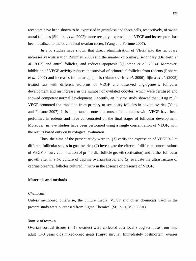
120
receptors have been shown to be expressed in granulosa and theca cells, respectively, of swine
antral follicles (Shimizu et al. 2002); more recently, expression of VEGF and its receptors has
been localised to the bovine fetal ovarian cortex (Yang and Fortune 2007).
In vivo studies have shown that direct administration of VEGF into the rat ovary
increases vascularisation (Shimizu 2006) and the number of primary, secondary (Danforth et
al. 2003) and antral follicles, and reduces apoptosis (Quintana et al. 2004). Moreover,
inhibition of VEGF activity reduces the survival of primordial follicles from rodents (Roberts
et al. 2007) and increases follicular apoptosis (Abramovich et al. 2006). Iijima et al. (2005)
treated rats with different isoforms of VEGF and observed angiogenesis, follicular
development and an increase in the number of ovulated oocytes, which were fertilised and
showed competent normal development. Recently, an in vitro study showed that 10 ng mL−1
VEGF promoted the transition from primary to secondary follicles in bovine ovaries (Yang
and Fortune 2007). It is important to note that most of the studies with VEGF have been
performed in rodents and have concentrated on the final stages of follicular development.
Moreover, in vivo studies have been performed using a single concentration of VEGF, with
the results based only on histological evaluation.
Thus, the aims of the present study were to: (1) verify the expression of VEGFR-2 at
different follicular stages in goat ovaries; (2) investigate the effects of different concentrations
of VEGF on survival, initiation of primordial follicle growth (activation) and further follicular
growth after in vitro culture of caprine ovarian tissue; and (3) evaluate the ultrastructure of
caprine preantral follicles cultured in vitro in the absence or presence of VEGF.
Materials and methods
Chemicals
Unless mentioned otherwise, the culture media, VEGF and other chemicals used in the
present study were purchased from Sigma Chemical (St Louis, MO, USA).
Source of ovaries
Ovarian cortical tissues (n=18 ovaries) were collected at a local slaughterhouse from nine
adult (1–3 years old) mixed-breed goats (Capra hircus). Immediately postmortem, ovaries

121
were washed once in 70% alcohol for 10 s and twice in minimum essential medium (MEM)
supplemented with 100 μg mL−1
penicillin and 100 μg mL−1
streptomycin. Pairs of ovaries
were transported within 1 h to the laboratory in MEM at 4◦C.
Experimental protocol
The organ culture system used in the present study has been described in detail elsewhere
(Matos et al. 2007). Briefly, ovarian tissue samples from each ovarian pair were cut into 13
slices (3×3×1 mm) using a needle and scalpel under sterile conditions. The tissue pieces were
then either directly fixed for histological and ultrastructural analysis (fresh control) or placed
in culture for 1 or 7 days. Caprine tissues were transferred to 24-well culture dishes
containing 1 mL culture medium. Culture was performed at 39◦C in 5% CO2 in a humidified
incubator and all media were incubated for 1 h before use. The basic culture medium
(cultured control) was called MEM+ and consisted of MEM (pH 7.2–7.4) supplemented with
ITS (insulin 6.25 ng mL−1
, transferrin 6.25 ng mL−1
and selenium 6.25 ng mL−1
), 0.23 mm
pyruvate, 2 mm glutamine, 2 mm hypoxanthine and 1.25 mg mL−1
bovine serum albumin
(BSA). For the different experiments, this medium was supplemented with different
concentrations of recombinant human VEGF (1, 10, 50, 100 or 200 ng mL−1
). Each treatment
was repeated four times and the culture medium was replenished every other day.
Morphological analysis and assessment of in vitro follicular growth
Before culture (fresh control) and after 1 or 7 days in culture, all tissue pieces were fixed in
Carnoy‘s solution for 12 h and then dehydrated in increasing concentrations of ethanol. After
paraffin embedding (Synth, São Paulo, Brazil), the tissues pieces were cut into 7 μm sections
and each section was mounted on glass slides and stained with periodic acid-Schiff (PAS)–
haematoxylin. Follicle stage and survival were assessed microscopically on serial sections.
Coded slides were examined under a microscope (Nikon, Japan) at ×400 magnification with
the observer blinded to treatment group.
The developmental stages of the follicles have been defined previously (Silva et al.
2004) as primordial (one layer of flattened granulosa cells around the oocyte) or growing
(intermediate, one layer of flattened to cuboidal granulosa cells; primary, one layer of
cuboidal granulosa cells; and secondary, two or more layers of cuboidal granulosa cells

122
around the oocyte). These follicles were classified individually as histologically normal when
an intact oocyte was present and was surrounded by granulosa cells that were well organised
in one or more layers and had no pyknotic nuclei. Atretic follicles were defined as those with
a retracted oocyte, pyknotic nucleus and/or disorganised granulosa cells detached from the
basement membrane. Overall, 120 follicles were evaluated for each treatment (30 follicles per
treatment in one repetition × 4 repetitions = 120 follicles).
To evaluate follicular activation, the percentage of healthy primordial and growing
follicles was calculated before (fresh control) and after culture in each medium. In addition,
follicle and oocyte diameters were measured in healthy follicles only. Follicle diameter was
recorded as the length from edge to edge of the basement membrane or from the outside edge
of the theca cell layer when present. Oocyte diameter was recorded as the length from edge to
edge of the oocyte membrane. Two perpendicular diameters were recorded for each parameter
and the average of these two values was reported. Care was taken to count each follicle only
once, as we have done in our earlier studies (Matos et al. 2007). Each follicle was examined
in every section in which it appeared and matched with the same follicle on adjacent sections
to avoid double counting, thus ensuring that each follicle was only counted once, regardless of
its size.
Ultrastructural analysis
For better evaluation of follicular morphology, ultrastructural studies were performed on
fragments of fresh control tissues (non-cultured) and those with the best results during the
histological analysis after 7 days culture. Briefly, small pieces (1 mm3) of caprine ovarian
tissues were fixed in 2% paraformaldehyde and 2.5% glutaraldehyde in 0.1 m sodium
cacodylate buffer (pH 7.2) for 4 h at room temperature. After fixation, fragments were post-
fixed in 1% osmium tetroxide, 0.8% potassium ferricyanide and 5 mm calcium chloride in 0.1
m sodium cacodylate buffer for 1 h. Samples were dehydrated through an acetone gradient
and tissues were embedded in Spurr‘s resin. Semithin sections (3 μm) were cut on an
ultramicrotome (Reichert Supernova, Heidelberg, Germany) for light microscopy studies and
stained with Toluidine blue. The ultrathin sections (60–70 nm) were contrasted with uranyl
acetate and lead citrate and examined under a Jeol 1011 (Jeol, Tokyo, Japan) transmission

123
electron microscope (TEM). Parameters such as the density and integrity of ooplasmic and
granulosa cell organelles, vacuolisation and basement membrane integrity were evaluated.
Viability assessment of follicles cultured in vitro
Based on the results of histological analysis, the viability of follicles cultured with the
concentration of VEGF that provided the best outcome was further analysed using a more
accurate method of assessment based on fluorescent probes.
Caprine preantral follicles from ovarian fragments cultured for 7 days with VEGF
(200 ng mL−1
) were isolated by the mechanical method of Lucci et al. (1999). Briefly, using a
tissue chopper (Mickle Laboratory Engineering, Gomshal, Surrey, UK) adjusted to a
sectioning interval of 75 μm, samples were cut into small pieces, which were placed in MEM
and suspended 40 times using a large Pasteur pipette (diameter approximately 1600 μm) and
then subsequently 40 times with a smaller Pasteur pipette (diameter approximately 600 μm) to
dissociate preantral follicles from the stroma. The material obtained was passed through 500-
and 100-μm nylon mesh filters, resulting in a suspension containing preantral follicles smaller
than 100 μm in diameter. This procedure was performed within 10 min at room temperature.
Thereafter, the viability of preantral follicles was assessed through a two-colour
fluorescence cell viability assay based on the simultaneous determination of live and dead
cells using calcein-AM and ethidium homodimer-1, respectively. Whereas the first probe
detected the intracellular esterase activity of viable cells, the second probe labelled nucleic
acids of non-viable cells with plasma membrane disruption. The test was performed by adding
4 μm calcein-AM and 2 μm ethidium homodimer-1 (Molecular Probes, Invitrogen, Karlsruhe,
Germany) to the suspension of isolated follicles and incubating them at 37◦C for 10 min.
After being labelled, follicles were washed three times in MEM and mounted on a glass
microscope slide in 5 μL antifading medium (DABCO; Sigma, Deisenhofen, Germany) to
prevent photobleaching and were finally examined using a DMLB fluorescence microscope
(Leica, Wetzlar, Germany). The fluorescent signals of calcein-AM and ethidium homodimer
emitted were collected at 488 and 568 nm, respectively. Oocytes and granulosa cells were
considered live if the cytoplasm was stained positively with calcein-AM (green) and the
chromatin was not labelled with ethidium homodimer (red).

124
Immunohistochemical localisation of VEGFR 2
Localisation of VEGFR-2 was performed on serial 5-μm sections cut from the ovaries of five
different goats. These sections were mounted on poly-L-lysine-coated slides, dried overnight
at 37◦C, deparaffinized in xylene and rehydrated in a graded ethanol series. Endogenous
peroxidase was blocked by incubating the deparaffinized sections in 3% (v/v) hydrogen
peroxide in methanol for 10 min. The sections were then washed with 1× phosphate-buffered
saline (PBS) and the epitopes were activated by microwaving the sections for 7 min at 900 W
in 0.01 m citrate buffer (pH 6.0). After microwave treatment, the sections were washed in
PBS–0.05% (v/v) Tween (PBS-T) before being incubated for 30 min with 5% normal goat
serum in PBS to minimize non-specific binding. Then, sections were incubated with mouse
monoclonal anti-VEGFR-2 antibodies (clone KDR-1) diluted 1 : 20. After incubation with the
antibody, sections were washed three times with PBS and incubated for 45 min with
biotinylated secondary antibody (anti-mouse IgG; Santa Cruz Biotechnology, Santa Cruz, CA,
USA) diluted 1 : 200 in PBS containing 5% normal goat serum. Next, sections were washed
three times in PBS-T before being incubated for 45 min with an avidin–biotin complex (1 :
600; Vectastain Elite ABC kits; Vector Laboratories, Burlingame, CA, USA). The sections
were then washed three times in PBS and stained with diaminobenzidine (DAB; 0.05% (w/v)
DAB in 10 mm TRIS-HCl, pH 7.6, 0.03% (v/v) H2O2) for a maximum of 10 min. The stained
sections were rinsed in PBS and water and counterstained for 10 s in Mayer‘s haematoxylin.
Finally, the sections were washed for 10 min under running tap water and subsequently
dehydrated in a graded ethanol series. This was followed by xylene treatment and mounting in
Pertex (Cellpath, Hemel, Hempstead, UK). Staining intensity was scored as absent (−), weak
(+), moderate (++) or strong (+++). Controls for non-specific staining were performed by: (1)
replacing the primary antibody with IgGs from the same species in which the specific
antibody was raised, at the same concentration; and (2) incubation with DAB reagent alone to
exclude the possibility of non-suppressed endogenous peroxidase activity. For
immunohistochemical analysis, follicles were classified as described earlier for culture.
Statistical analysis
The percentage of surviving follicles at both stages (i.e. primordial and developing) obtained
after 1 or 7 days of various treatments, as well as data regarding follicle and oocyte diameters,
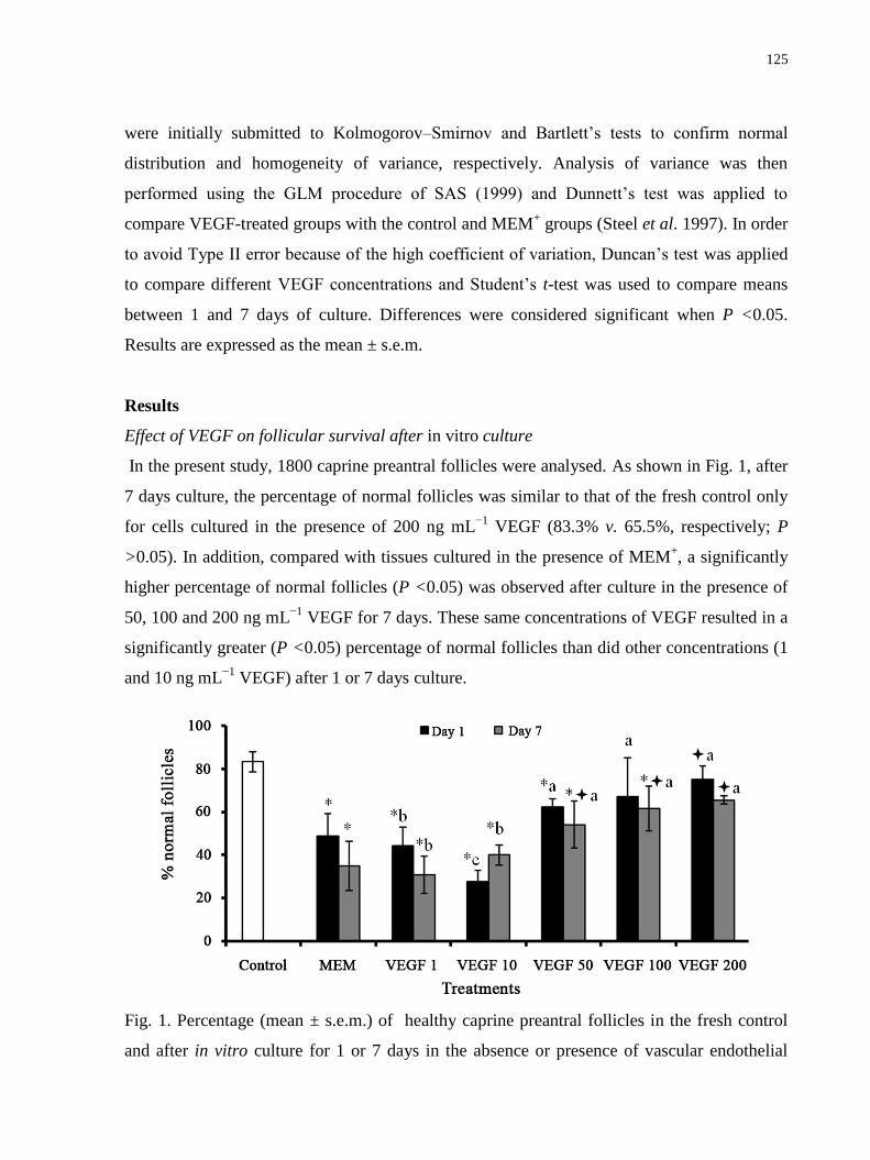
125
were initially submitted to Kolmogorov–Smirnov and Bartlett‘s tests to confirm normal
distribution and homogeneity of variance, respectively. Analysis of variance was then
performed using the GLM procedure of SAS (1999) and Dunnett‘s test was applied to
compare VEGF-treated groups with the control and MEM+ groups (Steel et al. 1997). In order
to avoid Type II error because of the high coefficient of variation, Duncan‘s test was applied
to compare different VEGF concentrations and Student‘s t-test was used to compare means
between 1 and 7 days of culture. Differences were considered significant when P <0.05.
Results are expressed as the mean ± s.e.m.
Results
Effect of VEGF on follicular survival after in vitro culture
In the present study, 1800 caprine preantral follicles were analysed. As shown in Fig. 1, after
7 days culture, the percentage of normal follicles was similar to that of the fresh control only
for cells cultured in the presence of 200 ng mL−1
VEGF (83.3% v. 65.5%, respectively; P
>0.05). In addition, compared with tissues cultured in the presence of MEM+, a significantly
higher percentage of normal follicles (P <0.05) was observed after culture in the presence of
50, 100 and 200 ng mL−1
VEGF for 7 days. These same concentrations of VEGF resulted in a
significantly greater (P <0.05) percentage of normal follicles than did other concentrations (1
and 10 ng mL−1
VEGF) after 1 or 7 days culture.
Fig. 1. Percentage (mean ± s.e.m.) of healthy caprine preantral follicles in the fresh control
and after in vitro culture for 1 or 7 days in the absence or presence of vascular endothelial

126
growth factor (VEGF). *P <0.05 compared with control follicles; P <0.05 compared with
minimum essential medium (MEM) alone on each day of culture. Different superscript letters
indicate significant differences between different concentrations of VEGF (P <0.05).
Follicular activation and growth after culture with VEGF
The percentage of primordial and growing follicles in fresh tissues was 35.1% and 64.9%,
respectively. In all treatments, after 1 or 7 days culture, there was no significant effect of
VEGF on follicular activation (i.e. we did not observe any significant reduction in the
percentage of primordial follicles or any increase in the percentage of growing follicles; P
>0.05).
With regard to follicular growth, tissues cultured in the presence of 200 ng mL−1
VEGF had significantly larger (after 1 day) and smaller (after 7 days) follicular and oocyte
diameters than the fresh control (non-cultured). When compared with MEM+, a significant
increase (P <0.05) in follicular and oocyte diameters was observed following treatment with
10 ng mL−1
VEGF for 7 days. In addition, over the same period, 10 ng mL−1
VEGF was more
efficient than 50, 100 and 200 ng mL−1
VEGF in increasing oocyte diameter (P <0.05; Table
1).
Ultrastructural analysis of caprine preantral follicles cultured with VEGF
The ultrastructural features of follicles from fresh control and 200 ng mL−1
VEGF-treated
groups were similar. These follicles showed intact basement and nuclear membranes and a
large oocyte nucleus. In addition, the organelles were uniformly distributed in the ooplasm,
especially the mitochondria. Both the smooth and rough endoplasmic reticula were present,
either as isolated aggregations or as complex associations with mitochondria and vesicles.
Granulosa cells were ultrastructurally normal and well organised around the oocyte, showing
an elongated and large nucleus with an irregular membrane (Fig. 2). Microvillar extensions of
the plasma membranes of oocyte and granulose cells were visible in the space between these
compartments.

127
Table 1. Caprine oocyte and follicle diameters (mean ± s.e.m.) in fresh control (non-cultured)
and after culture for 1 or 7 days in the absence or presence of vascular endothelial growth
factor.
Follicular diameter (m) Oocyte diameter (m)
Control 66.90 9.26 53.92 8.88
Treatments Day 1 Day 7 Day 1 Day 7
MEM+ 69.22 8.99 57.32 11.45
* 55.77 5.97 45.58 9.71
*
VEGF 1 65.66 13.85 b
64.43 10.90 a 52.36 9.86
b 50.52 9.26
ab
VEGF 10 66.90 12.04 b 68.96 11.52
a 52.68 8.53
b 55.29 9.93
a
VEGF 50 73.70 8.87 b 63.19 11.58
a 57.44 8.54
ab 48.05 7.40
bc
VEGF 100 67.36 15.52 b 63.52 12.59
a 52.37 10.44
b 45.62 7.37
* bc
VEGF 200 82.35 12.73*a
55.93 5.75 *b
62.26 6.89 *a
42.49 6.86 *c
VEGF1, 10, 50, 100 or 200 represent VEGF at 1, 10, 50, 100 or 200 ng mL−1
, respectively.
MEM+, minimum essential medium (pH 7.2–7.4) supplemented with ITS (insulin 6.25 ng
mL−1
, transferrin 6.25 ng mL−1
and selenium 6.25 ng mL−1
), 0.23 mm pyruvate, 2 mm
glutamine, 2 mm hypoxanthine and 1.25 mg mL−1
bovine serum albumin. *P <0.05
compared with control follicles; P <0.05 compared with MEM+ alone on each day of
culture. Different superscript letters indicate significant differences between different
concentrations of VEGF (P <0.05)

128
Fig. 2. Ultrastructural analysis of (a) non-cultured (fresh control) and (b) cultured caprine
preantral follicles, cultured for 7 days in medium containing 200 ng mL−1
vascular endothelial
growth factor (VEGF). o, oocyte; n, oocyte nucleus; gc, granulosa cells; m, mitochondria; v,
vesicles; er, endoplasmic reticulum; mv, microvilli; arrow, oocyte membrane. Scale bars =
2μm.
Viability of follicles cultured with VEGF
In the present study, five caprine preantral follicles were analysed after 7 days culture with
200 ng mL−1
VEGF. After this qualitative analysis, all follicles (100%) remained viable, as
assessed by calcein-AM–ethidium homodimer assays, the percentage of normal follicles was
similar to that seen in the fresh control group (Fig. 3).
Immunohistochemical localisation of VEGFR-2 in caprine preantral follicles
Immunohistochemical analysis was performed to localize VEGFR-2 in caprine preantral and
antral follicles. The results demonstrated a moderate reaction for VEGFR-2 in oocytes of
follicles from all developmental stages. Granulosa cells of primordial follicles showed a
negative response for VEGFR-2, but a weak and moderate immunoreaction was observed in
granulose cells of primary and secondary follicles, respectively. In addition, cumulus and
mural granulose cells showed a weak reaction for VEGFR-2. Nevertheless, theca cells did not

129
show a positive reaction for VEGFR-2. No staining was observed in negative controls
incubated with VEGFR-2 (Table 2; Fig. 4).
Fig. 3. Viability of caprine preantral follicles, as determined using fluorescent probes. An
isolated preantral follicle after culture in 200 ng mL−1
vascular endothelial growth factor
(VEGF) that was classified as viable (a) because cells were labelled by calcein-AM (green
fluorescence; b). Scale bars = 25μm.
Table 2. Relative intensity of immunohistochemical staining for vascular endothelial growth
factor receptor-2 in goat ovarian follicles (−) absent; (+) weak; (++) moderate; NA, not
applicable
Follicular categories
Follicular
compartments
Primordial Primary Secondary Antral
Oocyte ++ ++ ++ ++
Granulosa - + ++ +
Theca NA NA - -
Cumulus NA NA NA +
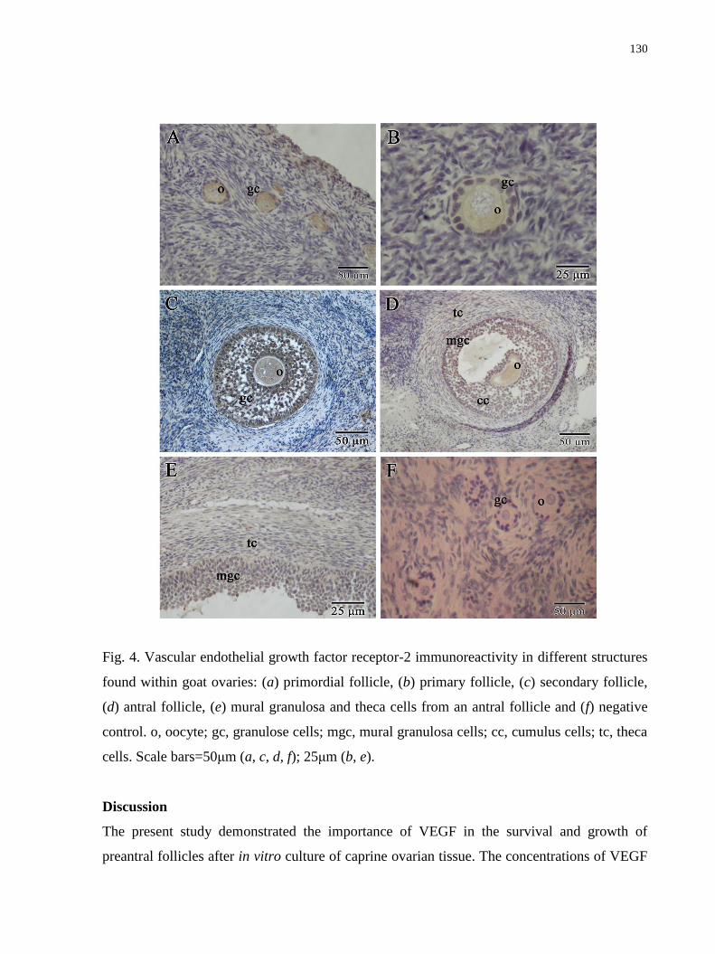
130
Fig. 4. Vascular endothelial growth factor receptor-2 immunoreactivity in different structures
found within goat ovaries: (a) primordial follicle, (b) primary follicle, (c) secondary follicle,
(d) antral follicle, (e) mural granulosa and theca cells from an antral follicle and (f) negative
control. o, oocyte; gc, granulose cells; mgc, mural granulosa cells; cc, cumulus cells; tc, theca
cells. Scale bars=50μm (a, c, d, f); 25μm (b, e).
Discussion
The present study demonstrated the importance of VEGF in the survival and growth of
preantral follicles after in vitro culture of caprine ovarian tissue. The concentrations of VEGF
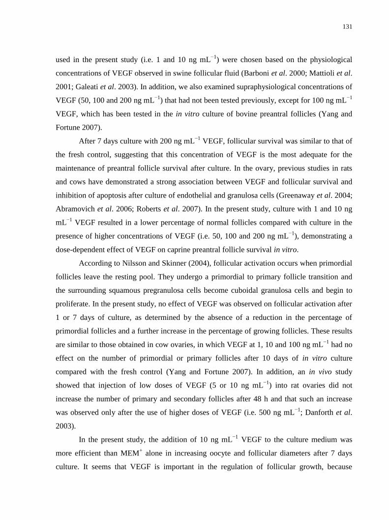
131
used in the present study (i.e. 1 and 10 ng mL−1
) were chosen based on the physiological
concentrations of VEGF observed in swine follicular fluid (Barboni et al. 2000; Mattioli et al.
2001; Galeati et al. 2003). In addition, we also examined supraphysiological concentrations of
VEGF (50, 100 and 200 ng mL−1
) that had not been tested previously, except for 100 ng mL−1
VEGF, which has been tested in the in vitro culture of bovine preantral follicles (Yang and
Fortune 2007).
After 7 days culture with 200 ng mL−1
VEGF, follicular survival was similar to that of
the fresh control, suggesting that this concentration of VEGF is the most adequate for the
maintenance of preantral follicle survival after culture. In the ovary, previous studies in rats
and cows have demonstrated a strong association between VEGF and follicular survival and
inhibition of apoptosis after culture of endothelial and granulosa cells (Greenaway et al. 2004;
Abramovich et al. 2006; Roberts et al. 2007). In the present study, culture with 1 and 10 ng
mL−1
VEGF resulted in a lower percentage of normal follicles compared with culture in the
presence of higher concentrations of VEGF (i.e. 50, 100 and 200 ng mL−1
), demonstrating a
dose-dependent effect of VEGF on caprine preantral follicle survival in vitro.
According to Nilsson and Skinner (2004), follicular activation occurs when primordial
follicles leave the resting pool. They undergo a primordial to primary follicle transition and
the surrounding squamous pregranulosa cells become cuboidal granulosa cells and begin to
proliferate. In the present study, no effect of VEGF was observed on follicular activation after
1 or 7 days of culture, as determined by the absence of a reduction in the percentage of
primordial follicles and a further increase in the percentage of growing follicles. These results
are similar to those obtained in cow ovaries, in which VEGF at 1, 10 and 100 ng mL−1
had no
effect on the number of primordial or primary follicles after 10 days of in vitro culture
compared with the fresh control (Yang and Fortune 2007). In addition, an in vivo study
showed that injection of low doses of VEGF (5 or 10 ng mL−1
) into rat ovaries did not
increase the number of primary and secondary follicles after 48 h and that such an increase
was observed only after the use of higher doses of VEGF (i.e. 500 ng mL−1
; Danforth et al.
2003).
In the present study, the addition of 10 ng mL−1
VEGF to the culture medium was
more efficient than MEM+ alone in increasing oocyte and follicular diameters after 7 days
culture. It seems that VEGF is important in the regulation of follicular growth, because

132
inhibition of VEGF activity with neutralizing antibodies or a soluble form of its receptor has
been reported to interrupt follicular growth and the proliferation of monkey granulosa cells
(Danforth et al. 2003). Wulff et al. (2001) demonstrated that blockade of VEGF results in a
reduction of theca cell proliferation in the secondary and tertiary follicles of primates.
However, in the present study, there was a reduction in oocyte and follicular diameter after 7
days in the presence of 200 ng mL−1
VEGF compared with the fresh control. It is known that
granulosa cells are the main cell type responsible for VEGF production (Mattioli et al. 2001;
Galeati et al. 2003). Thus, the VEGF produced by these cells in addition to the VEGF added
to the culture medium at high concentration (200 ng mL−1
) may have promoted accelerated
follicular growth. Late preantral follicles, which have an elevated metabolism and increased
uptake of nutrients, were not able to maintain their viability. Then, after 7 days, only the
morphology of small preantral follicles, which have a low metabolism and are less sensitive to
degeneration (Hirshfield 1991; Wandji et al. 1996; Silva et al. 2002), was preserved.
Using TEM, it was observed that in addition to the basal and nuclear membranes,
important structures, such as the mitochondria, endoplasmic reticulum and granulosa cells
were preserved even after 7 days culture in the presence of 200 ng mL−1
VEGF. In the present
study, we observed microvilli, which are important in the communication between oocytes
and granulosa cells, allowing the exchange of substances and, consequently, enabling
adequate follicular development. In addition to ultrastructural analysis, the viability of
preantral follicles cultured for 7 days in the presence of 200 ng mL−1
VEGF was further
assessed, with the results indicating that this treatment is efficient in maintaining caprine
preantral follicle viability. The fluorescent probes calcein-AM and ethidium homodimer-1
have been used successfully to assess the viability of bovine early stage follicles (Schotanus et
al. 1997; van den Hurk et al. 1998). This method can be used to analyse the viability of
follicles, thereby offering a new approach for investigating metabolic and developmental
aspects of folliculogenesis in vitro.
The present study demonstrated the expression of VEGFR-2 in oocytes and granulosa
cells of all follicular stages, except in granulosa cells from primordial follicles. This latter
finding may be explained by the fact that VEGF did not promote changes in granulosa cell
morphology and further granulosa cell proliferation, which characterises follicular activation.
Although absent in granulosa cells of primordial follicles, VEGFR-2 was expressed in the

133
oocytes of these follicles. Some authors have demonstrated the expression of VEGFR-1 and
VEGFR-2 mRNA, particularly in the thecal cell layer (Shimizu et al. 2002, 2003), from the
late follicular stages onward. In addition, on the basis of experiments in which rats were
treated with antibodies that bind toVEGFR-2, it has been reported that VEGFR-2 is important
for primordial follicle survival (Roberts et al. 2007). Based on the results of the present study,
in other types of follicles (i.e. primary and secondary), stimulation with VEGF may increase
oocyte diameter, which may or may not be associated with granulosa cell proliferation, thus
promoting follicular growth. Zimmermann et al. (2003) verified that in hypophysectomised
primates VEGFR- 2 activity is necessary to increase the gonadotropin-dependent proliferation
of granulosa cells.
Conclusion
The present study showed that different concentrations of VEGF have different effects on
caprine preantral follicle survival and development. Specifically, to obtain more viable
follicles and to promote preantral follicle growth, 200 and 10 ng mL−1
VEGF, respectively,
should be used. Moreover, the expression of VEGFR-2 in oocytes of caprine ovarian follicles
in all developmental stages and in granulosa cells of developing follicles suggests that VEGF
may have a physiological function in mammalian reproduction.
Acknowledgements
This work was supported by CNPQ (RENORBIO: grant no. 554812/2006– 1). J.B.B. is a
recipient of a grant from FUNCAP/CAPES (Brazil). The authors thank José Leandro da Silva
Neto for his technical support with the histological examinations.
References
Abramovich, D., Parborel, F., and Tesone, M. (2006). Effect of a vascular endothelial growth
factor (VEGF) inhibitory treatment on the folliculoggenesis and ovarian apoptosis in
gonadotropin-treated prepubertal rats. Biol. Reprod. 75, 434–441.
doi:10.1095/BIOLREPROD.106. 051052

134
Barboni, B., Turriani, M., Galeati, G., Spinaci, M., Bacci, M. L., Forni, M., and Mattiolo, M.
(2000). Vascular endothelial growth factor production in growing pig antral follicles.
Biol. Reprod. 63, 858–864. doi:10.1095/ BIOLREPROD63.3.858
Celik-Ozenci, C., Akkoyunhlu, G., Kayisli, U. A., Arici, A., and Demir, R. (2003).
Localization of vascular endothelial growth factor in the zona pellucida of developing
ovarian follicles in the rat: a possible role in destiny of follicles. Histochem. Cell Biol.
120, 383–390. doi:10.1007/ S00418-003-0586-4
Danforth, D. R., Arbogast, L. K., Ghosh, S., Dickerman, A., Rofagha, R., and Friedman, C. I.
(2003).Vascular endothelial growth factor stimulates preantral follicle growth in the rat
ovary. Biol. Reprod. 68, 1736–1741. doi:10.1095/BIOLREPROD.101.000679
Ferrara, N., and Davis-Smyth, T. (1997). The biology of vascular endothelial growth factor.
Endocr. Rev. 18, 4–25. doi:10.1210/ER.18.1.4
Galeati, G., Spinaci, M.,Govoni,N., Zannoni, A., Fantinati, P., Seren, E., and Tamanini, C.
(2003). Stimulatory effects of fasting on vascular endothelial growth factor (VEGF)
production by growing pig ovarian follicles. Reproduction 126, 647–652.
doi:10.1530/REP.0.1260647
Greenaway, J., Connor, K., Pedersen, H. G., Coomber, B. L., Lamarre, J., and Petrik, J.
(2004).Vascular endothelial growth factor and its receptor, Flk-1/KDR, are
cytoprotective in the extravascular compartment of the ovarian follicle. Endocrinology
145, 2896–2905. doi:10.1210/EN.2003- 1620
Greenaway, J., Centry, P. A., Feige, J.-J., Lamarre, J., and Petrik, J. J. (2005).
Thormbospondin and vascular endothelial growth factor are cyclically expressed in an
inverse pattern during bovine ovarian follicle development. Biol. Reprod. 72, 1071–
1078. doi:10.1095/ BIOLREPROD.104.031120
Harata, T., Ando, H., Iwase, A., Nagasaka, T., Mizutani, S., and Kikkawa, F. (2006).
Localization of angiotensin II, the AT1 receptor, angiotensin-converting enzyme,
aminopeptidase A, adipocyte-derived leucine aminopeptidase, and vascular endothelial
growth factor in the human ovary throughout the menstrual cycle. Fertil. Steril. 86,
433–439. doi:10.1016/J.FERTNSTERT.2006.01.041
Hirshfield,A. N. (1991). Development of follicles in mammalian ovary. Int. Rev. Cytol. 124,
43–101. doi:10.1016/S0074-7696(08)61524-7

135
Iijima, K., Jiang, J.-Y., Shimizu,T., Sasada, H., and Sato, E. (2005). Acceleration of follicular
development by administration of vascular endothelial growth factor in cycling female
rats. J. Reprod. Dev. 51, 161–168. doi:10.1262/JRD.51.161
Lucci, C. M.,Amorim, C.A., Báo, S.N., Figueiredo, J. R., Rodrigues,A. P. R., Silva, J. R., and
Gonçalves, P. B. D. (1999). Effect of the interval of serial sections of ovarian in the
tissue chopper on the number of isolated caprine preantral follicles. Anim. Reprod. Sci.
56, 39–49. doi:10.1016/S0378- 4320(99)00031-7
Matos, M. H. T., Van Den Hurk, R., Lima-Verde, I. B., Luque, M. C. A., Santos, K. D. B.,
Martins, F. S., Báo, S. N., Lucci, C. M., and Figueiredo, J. R. (2007). Effects of
fibroblast growth factor-2 on the in vitro culture of caprine preantral follicles. Cells
Tissues Organs 186, 112–120. doi:10.1159/000103016
Mattioli, M., Barboni, B., Turriani, M., Galeati, G., Zannoni, A., Castellani, G., Berardinelli,
P., and Scapolo, P. A. (2001). Follicle activation involves vascular endothelial growth
factor production and increased blood vessel extension. Biol. Reprod. 65, 1014–1019.
doi:10.1095/ BIOLREPROD65.4.1014
Nilsson, E. E., and Skinner, M. K. (2004). Kit ligand and basic fibroblast growth factor
interactions in the induction of ovarian primordial to primary follicle transition. Mol.
Cell. Endocrinol. 214, 19–25. doi:10.1016/ J.MCE.2003.12.001
Otani, N., Minami, S., Yamoto, M., Shikone, T., Otani, H., Nishiyama, R., Otani,T., and
Nakano, R. (1999).The vascular endothelial growth factor/ fms-like tyrosine kinase
system in human ovary during the menstrual cycle and early pregnancy. J. Clin.
Endocrinol. Metab. 84, 3845–3851. doi:10.1210/JC.84.10.3845
Quintana, R., Kopcow, L., Sueldo, C., Marconi, G., Rueda, N. G., and Barañao, R. I. (2004).
Direct injection of vascular endothelial growth factor into the ovary of mice promotes
follicular development. Fertil. Steril. 82, 1101–1105.
doi:10.1016/J.FERTNSTERT.2004.03.036
Redmer, D. A., Doraiswamy, V., Bortnem, B. J., Fisher, K., Jablonka- Shariff, A., Grazul-
Bilska, A. T., and Reynolds, L. P. (2001). Evidence for a role of capillary pericytes in
vascular growth of the developing ovine corpus luteum. Biol. Reprod. 65, 879–889.
doi:10.1095/ BIOLREPROD65.3.879
136
Roberts, A. E., Arbogast, L. K., Friedman, C. I., Cohn, D. E., Kaumaya, P. T., and Danforth,
D. R. (2007). Neutralization of endogenous vascular endothelial growth factor depletes
primordial follicles in the mouse ovary. Biol. Reprod. 76, 218–223.
doi:10.1095/BIOLREPROD. 106.050880
Schotanus, K., Hage, W. J., Vanderstichele, H., and van den Hurk, R. (1997). Effects of
conditioned media from murine granulosa cell lines on the geowth of isolated bovine
preantral follicles. Theriogenology 48, 471–483. doi:10.1016/S0093-691X(97)00256-2
Shibuya, M. (1995). Role of VEGF–Flt receptor system in normal and tumor angiogenesis.
Adv. Cancer Res. 67, 281–316. doi:10.1016/S0065- 230X(08)60716-2
Shimizu,T. (2006). Promotion of ovarian follicular development by injecting vascular
endothelial growth factor (VEGF) and growth differentiation factor 9 (GDF-9) genes. J.
Reprod. Dev. 52, 23–32. doi:10.1262/ JRD.17072
Shimizu, T., Jiang, J.-Y., Sasada, H., and Sato, E. (2002). Changes of messenger RNA
expression of angiogenic factors and related receptors during follicular development in
gilts. Biol. Reprod. 67, 1846–1852. doi:10.1095/BIOLREPROD.102.006734
Shimizu, T., Jiang, J.-Y., Iijima, K., Miyabayashi, K., Ogawa,Y., Sasada, H., and Sato, E.
(2003). Induction of follicular development by direct single injection of vascular
endothelial growth factor gene fragments into the ovary of miniature gilts. Biol. Reprod.
69, 1388–1393. doi:10.1095/ BIOLREPROD.103.016311
Silva, J. R., Ferreira, M.A. L., Costa, S. H. F., Santos, R. R., Carvalho, F. C.A., Rodrigues,A.
P. R., Lucci, C. M., Báo, S.N., and Figueiredo, J. R. (2002). Degeneration rate of
preantral follicles in the ovaries of goats. Small Ruminant Res. 43, 203–209.
doi:10.1016/S0921-4488(02)00017-2
Silva, J. R., Van Den Hurk, R., Matos, M. H. T., Santos, R. R., Pessoa, C., Moraes, M. O.,
and Figueiredo, J. R. (2004). Influences of FSH and EGF on primordial follicles during
in vitro culture of caprine ovarian cortical tissue. Theriogenology 61, 1691 1704.
doi:10.1016/ J.THERIOGENOLOGY.2003.09.014
Statistical Analysis System (SAS) (1999). ‗SAS/STAT User‘s Guide.‘ (SAS Institute: Cary,
NC.)
Steel, R. G. D., Torrie, J. H., and Dickey, D. A. (1997). ‗Principles and Procedures of
Statistics: A Biometrical Approach, 3rd edn.‘ (McGraw- Hill: NewYork.)
137
Stouffer, R. L., Martínez-Chequer, J. C., Molskness, T. A., Xu, F., and Hazzard, T. M. (2001).
Regulation and action of angiogenic factors in the primete ovary. Arch. Med. Res. 32,
567–575. doi:10.1016/S0188-4409(01)00323-X
Tamanini, C., and De Ambrogi, M. (2004). Angiogenesis in developing follicle and corpus
luteum. Reprod. Domest. Anim. 39, 206–216. doi:10.1111/J.1439-0531.2004.00505.X
van den Hurk, R., Spek, E. R., Hage, W. J., Fair, T., Ralph, J. H., and Schotanus, K. (1998).
Ultrastructure and viability of isolated bovine preantral follicles. Hum. Reprod. Update
4, 833–841. doi:10.1093/ HUMUPD/4.6.833
Wandji, S. A., Srsen, V., Voss, A. K., Eppig, J. J., and Fortune, J. E. (1996). Initiation in vitro
of growth of bovine primordial follicles. Biol. Reprod. 55, 942–948.
doi:10.1095/BIOLREPROD55.5.942
Wulff, C., Wiegand, S. J., Saunders, P. T., Scobie, G. A., and Fraser, H. M. (2001).
Angiogenesis during follicular development in the primate and its inhibition by
treatment with truncated Flt-1-Fc (vascular endothelial growth factor Trap(A40)).
Endocrinology 142, 3244–3254. doi:10.1210/EN.142.7.3244
Yang, M. Y., and Fortune, J. E. (2007). Vascular endothelial growth factor stimulates the
primary to secundary follicle transition in bovine in vitro. Mol. Reprod. Dev. 74, 1095–
1104. doi:10.1002/MRD.20633
Zimmermann, R. C., Xiao, E., Bohlen, P., and Ferin, M. (2002). Administration of anti
vascular endothelial growth factor receptor 2 antibody in the early follicular phase
delays follicular selection and development in the rhesus monkey. Endocrinology 143,
2496–2502. doi:10.1210/EN.143.7.2496
Zimmermann, R. C., Hartman, T., Kavic, S., Pauli, S. A., Bohlen, P., Sauer, M.V., and
Kitajewski, J. (2003).Vascular endothelial growth factor receptor 2-mediated
angiogenesis is essential for gonadotropin-dependent follicle development. J. Clin.
Invest. 112, 659–669.

138
10. CAPÍTULO 5
Peptídeo intestinal vasoativo melhora a sobrevivência e desenvolvimento de folículos
pré-antrais caprinos após cultivo de tecido in vitro
(Vasoactive intestinal peptide improves the survival and development of caprine preantral
follicles after in vitro tissue culture)
Periódico: Cells Tissues Organs v. 191, n.52, p. 414-421, 2010

139
Resumo
O objetivo deste estudo foi avaliar o efeito do peptídeo intestinal vasoativo (VIP) na
sobrevivência, ativação e crescimento de folículos pré-antrais após o cultivo in vitro. O córtex
ovariano foi dividido em pequenas peças e um fragmento foi imediatamente fixado (controle).
Os fragmentos restantes foram cultivados in vitro por um ou sete dias a 39oC e 5% de CO2,
em Meio Essencial Mínimo suplementado (MEM+) com ou sem diferentes concentrações de
VIP (1, 10, 50, 100 e 200 ng / ml). Fragmentos ovarianos não-cultivados (controle fresco) e
cultivados foram processados para análise histológica e Microscopia Eletrônica de
Transmissão (MET). Os folículos foram classificados como primordiais ou em
desenvolvimento, bem como normais ou degenerados. Nossos resultados indicam que quando
comparado ao controle, todas as concentrações de VIP, exceto 200 ng / ml, resultaram em
percentagens similares de folículos pré-antrais normais após um e sete dias de cultivo. O
cultivo de tecido do córtex ovariano por um ou sete dias aumentou a percentagem de ativação
folicular em todos os tratamentos, quando comparado ao controle, exceto com 1 ng / ml de
VIP após um dia. No entanto, nenhuma diferença foi observada entre os folículos tratados-
VIP e tratados-MEM+. Além disso, após sete dias de cultivo, maiores diâmetros foliculares e
oócitários foram observados em folículos cultivados com 10 ng / ml VIP em relação ao
MEM+ sozinho. A MET mostrou integridade ultraestrutural de folículos após sete dias de
cultivo em 10 ng / ml VIP. Em conclusão, este estudo demonstrou que o VIP manteve a
integridade folicular e estimulou o crescimento de folículos pré-antrais caprinos.
Palavras-chave: VIP. Folículo pré-antral. Cultivo. Caprino.

140
Vasoactive Intestinal Peptide Improves the Survival and Development of Caprine
Preantral Follicles after in vitro Tissue Culture
J.B. Bruno a J.J.H. Celestino
a I.B. Lima-Verde
a M.H.T. Matos
a L.F. Lima
a K.P.O. Name
b
V.R. Araújo a M.V.A. Saraiva
a F.S. Martins
a C.C. Campello
a J.R.V. Silva
c S.N. Báo
b J.R.
Figueiredo a
a Faculty of Veterinary Medicine, LAMOFOPA, PPGCV, State University of Ceara, Fortaleza ,
b Laboratory of
Electron Microscopy, Department of Cell Biology, University of Brasilia, Brasilia , and c Biotechnology
Nucleus of Sobral (NUBIS), Federal University of Ceara, Sobral , Brazil
Abstract
The aim of this study was to evaluate the effect of vasoactive intestinal peptide (VIP) on the
survival, activation and growth of goat preantral follicles after in vitro culture. The ovarian
cortex was divided into small pieces and one fragment was immediately fixed (control). The
remaining fragments were cultured in vitro for 1 or 7 days at 39 ° C and 5% CO2, in
supplemented minimum essential medium (MEM+) with or without different concentrations
of VIP (1, 10, 50, 100 or 200 ng/ml). Noncultured (fresh control) and cultured ovarian
fragments were processed for histological analysis and transmission electron microscopy.
Follicles were classified as primordial or developing, and as normal or degenerated. Our
findings indicate that when compared with control, addition of all concentrations of VIP
except 200 ng/ml resulted in similar percentages of normal preantral follicles after 1 and 7
days of culture. Culture of ovarian cortex tissue for 1 and 7 days increased the percentage of
follicular activation in all treatments when compared with control, except with 1 ng/ml of VIP
after 1 day. However, no difference was observed between VIP-treated and MEM+-treated
follicles. In addition, after 7 days of culture, the highest follicular and oocyte diameters were
observed in follicles cultured with 10 ng/ml VIP relative to MEM+ alone. Transmission
electron microscopy showed ultrastructural integrity of follicles after 7 days of culture in 10
ng/ml VIP. In conclusion, this study demonstrates that VIP maintains follicular integrity and
stimulates caprine preantral follicle growth.
Key Words: Vasoactive intestinal peptide, Preantral follicles, Culture, Caprine.

141
Introduction
Ovarian activity is regulated not only by gonadotropins and steroids, but also by a
number of neural inputs and paracrine regulatory mechanisms. The mammalian ovary is
innervated by extrinsic nerves, which are both catecholaminergic and peptidergic in nature
[Burden, 1985; Ojeda and Lara, 1989; Ojeda et al., 1989]. Peptidergic innervation of the
ovary was verified, among other ways, by the presence of vasoactive intestinal peptide (VIP)
[Ahmed et al., 1986]. This neuropeptide was originally isolated from porcine duodenum and
is involved in smooth muscle relaxation and vasodilation [Said and Mutt, 1970]. VIP has been
found in the central nervous system as well as in a wide variety of organs and peripheral
tissues, including lung, testis, adrenal and ovary [Cecconi et al., 2004].
Additional findings, summarized below, suggest VIP may play a vital role in the
acquisition of cyclic ovarian function. VIP has been shown to stimulate in vitro androgen and
estradiol release [Davoren and Hsueh, 1985; Parra et al., 2007]. In addition, VIP stimulation
induced aromatase and follicle-stimulating hormone (FSH) receptor expression at both the
transcript and protein level in the rat neonatal ovary several days before the ovary became
responsive to gonadotropins [Mayerhofer et al., 1997]. In bovine tissue, primary follicle
formation is accompanied by the outgrowth of VIP-containing nerves [Hulshof et al., 1994].
Furthermore, this neuropeptide is reported to be produced by nerve fibers innervating follicles
at all stages of development [Ahmed et al., 1986; Hulshof et al., 1994; Johnson et al., 1994].
Other in vitro studies revealed that VIP stimulates the development of isolated bovine
primary and early secondary follicles [Hulshof, 1995] and promotes avian granulose cell
survival by inhibiting apoptosis in preovulatory follicles [Flaws et al., 1995]. Moreover, many
of the effects of VIP on ovarian granulosa cells are similar to the action of gonadotropins
[Ahmed et al., 1986; Johnson and Tilly, 1988; Ojeda et al., 1989; Johnson et al., 1994],
suggesting that VIP may be important for regulating gonadotropin independent development
and ovarian follicle survival. These data confirm previous findings concerning the
participation of sympathetic innervation in the control of follicular development. However,
there are no reports on the possible role of VIP in caprine preantral follicle viability and
development in vitro. The aim of this study, therefore, was to evaluate whether different

142
concentrations of VIP influence the survival, activation and growth of caprine preantral
follicles in vitro cultured for 1 or 7 days.
Materials and Methods
Chemicals
Unless mentioned otherwise, the culture media, VIP and other chemicals used in the
present study were purchased from Sigma Chemical Co. (St. Louis, Mo., USA).
Source of Ovaries
Ovarian cortical tissues were obtained from 4 mixed-breed goats (n = 8 ovaries)
collected at a local slaughterhouse. Immediately postmortem, the ovaries were washed first in
70% alcohol and then twice in minimum essential medium (MEM) supplemented with 100
µg/ml penicillin and 100 µg/ml streptomycin (Vetec, Rio de Janeiro, Brasil). Ovary pairs
were transported within 1 h to the laboratory in MEM at 33 ° C.
Experimental Protocol
Our organ culture system was described in detail previously [Silva et al., 2004; Matos
et al., 2007a]. Ovarian tissue samples from the same ovarian pair were cut in slices (3 x 3 x 1
mm) using a needle and scalpel under sterile conditions. The tissue pieces were then either
directly fixed for histological and ultrastructural analysis (control – day 0) or placed in culture
for 1 or 7 days. Caprine tissues were transferred to 24-well culture dishes containing 1 ml of
culture media. Culture was performed at 39°C in 5% CO2 in a humidified incubator and all
media were incubated for 1 h prior to use. The basic culture medium (cultured control) was
called MEM+ and consisted of MEM (pH 7.2 7.4) supplemented with ITS (insulin 6.25 ng/ml,
transferrin 6.25 ng/ml and selenium 6.25 ng/ml), 0.23 m M pyruvate, 2 mM glutamine, 2 mM
hypoxanthine, 1.25 mg/ml of bovine serum albumin. To test the effects of VIP, different
concentrations of this peptide (0, 1, 10, 50, 100 or 200 ng/ml) were added to the culture
medium. The experiment was carried out in 4 replicates and the culture media was
replenished every other day.
143
Morphological Analysis and Assessment of in vitro Follicular Growth
Fresh (noncultured) control and cultured tissues were fixed in 10% formalin for 12 h
and then dehydrated in increasing concentrations of ethanol. After paraffin (Synth, São Paulo,
Brasil) embedding, the caprine tissues pieces were cut into 7 - µm sections and stained by
periodic acid Schiff and hematoxylin. Sections were examined by microscopy (Nikon, Tokyo,
Japan) under 400 x magnifications.
Preantral follicles were classified as described previously [Gougeon and Chainy,
1987], as primordial (1 layer of flattened granulosa cells around the oocyte), intermediate (1
layer of flattened to cuboidal granulosa cells), primary (1 layer of cuboidal granulosa cells)
and secondary (2 or more layers of cuboidal granulose cells around the oocyte). Individual
follicles were further classified as histologically normal when an intact oocyte, surrounded by
well organized granulosa cells in 1 or more layers, with no pyknotic nucleus, was present.
Atretic follicles were defined as those with a retracted oocyte, pyknotic nucleus and/or
disorganized granulosa cells detached from the basement membrane. Overall, 120 follicles
were evaluated for each treatment (30 follicles per animal).
To evaluate follicular activation, the percentages of healthy primordial and growing
follicles were calculated before (fresh control) and after culture in each medium. In addition,
follicle and oocyte diameters were measured only in healthy follicles with the aid of an ocular
micrometer. Both diameters, from the basement membrane, at right angles to each other in the
largest crosssection of each growing oocyte and follicle, were measured and averaged. To
minimize the possibility of counting more than once, only follicles with a visible oocyte
nucleus were recorded. Further, each follicle was examined in every section in which it
appeared and matched with the same follicle on adjacent sections to avoid double counting,
regardless of its size.
Ultrastructural Analysis of Caprine Preantral Follicles
For more in-depth evaluation of follicular morphology, ultrastructural studies were
performed on fragments of fresh control and treatment groups that maintained follicular
morphology during histological analysis. Small pieces (1 mm3) of caprine ovarian tissues
were fixed in 2% paraformaldehyde and 2.5% glutaraldehyde in 0.1 M sodium cacodylate
buffer (pH 7.2) for 4 h at room temperature. After fixation, fragments were postfixed in 1%

144
osmium tetroxide, 0.8% potassium ferricyanide and 5 mM calcium chloride in 0.1 M sodium
cacodylate buffer for 1 h. Subsequently, the samples were dehydrated in a gradient of acetone
solutions and the tissues were embedded in Spurr‘s resin. Semithin sections (3 µm) were cut
on an ultramicrotome (Reichert Supernova, Heidelberg, Germany) for light microscopy
studies and stained with toluidine blue. Ultrathin sections (60–70 µm) were contrasted with
uranyl acetate and lead citrate, and examined under a Jeol 1011 (Jeol, Tokyo, Japan)
transmission electron microscope. Parameters such as density and integrity of ooplasmic and
granulosa cell organelles, vacuolization and basement membrane integrity were evaluated.
Statistical Analysis
Means of surviving follicles at all stages (primordial and developing – intermediate,
primary or secondary) determined after 1 or 7 days of culture in the various treatments were
subjected to analysis of variance using the GLM procedure of the SAS software and
Dunnett‘s test to compare VIP-treated groups against control and MEM+; Duncan test was
used to compare differences among VIP concentrations [Steel et al., 1997]. Student‘s t test
was used to compare means between 1 and 7 days of culture. Differences were considered to
be significant when p < 0.05 and data were expressed as mean ± standard error of means
(SEM).
Results
Effect of VIP on the Follicular Survival after Culture
In the present study, a total of 1,800 caprine preantral follicles were analyzed. Figure 1
shows a histological section of caprine ovarian tissue cultured for 7 days in MEM+
supplemented with 10 ng/ml of VIP. Present in this figure are morphologically normal
follicles with a round oocyte, a nucleus without pyknosis and well-organized granulose cells
around the oocyte. In addition, degenerated follicles show oocytes with pycnotic nucleus and
disorganized granulosa cells around the oocyte. After 1 or 7 days of in vitro culture, it was
verified that percentages of normal preantral follicles were similar (p > 0.05) to fresh control
in all treatment groups, except for MEM+ alone or with the addition of 200 ng/ml of VIP (p <
0.05) (fig. 2).

145
nn
dd
dd nn
Fig. 1. Histological section of caprine tissue cultured for 7 days in 10 ng/ml of VIP showing
normal (n) and degenerated (d) follicles after staining with periodic acid Schiff–hematoxylin.
Fig. 2. Percentage (mean ± SEM) of morphologically normal preantral follicles in control
(noncultured) ovaries and ovaries cultured in vitro for 1 or 7 days, in the absence or presence
of VIP.
Furthermore, after 1 or 7 days, treatment with all concentrations of VIP significantly
increased (p < 0.05) the percentage of normal follicles when compared to MEM+ alone,
except 200 ng/ml of VIP (p < 0.05). In fact, treatment with 200 ng/ml VIP resulted in a
aA
aA aA
aA
*bB
aA
aA *aA
* *
aA
aA
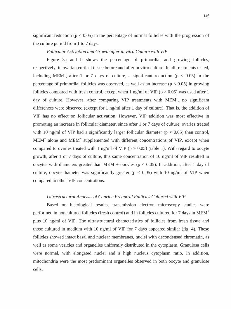
146
significant reduction (p < 0.05) in the percentage of normal follicles with the progression of
the culture period from 1 to 7 days.
Follicular Activation and Growth after in vitro Culture with VIP
Figure 3a and b shows the percentage of primordial and growing follicles,
respectively, in ovarian cortical tissue before and after in vitro culture. In all treatments tested,
including MEM+, after 1 or 7 days of culture, a significant reduction (p < 0.05) in the
percentage of primordial follicles was observed, as well as an increase (p < 0.05) in growing
follicles compared with fresh control, except when 1 ng/ml of VIP (p > 0.05) was used after 1
day of culture. However, after comparing VIP treatments with MEM+, no significant
differences were observed (except for 1 ng/ml after 1 day of culture). That is, the addition of
VIP has no effect on follicular activation. However, VIP addition was most effective in
promoting an increase in follicular diameter, since after 1 or 7 days of culture, ovaries treated
with 10 ng/ml of VIP had a significantly larger follicular diameter (p < 0.05) than control,
MEM+ alone and MEM
+ supplemented with different concentrations of VIP, except when
compared to ovaries treated with 1 ng/ml of VIP (p > 0.05) (table 1). With regard to oocyte
growth, after 1 or 7 days of culture, this same concentration of 10 ng/ml of VIP resulted in
oocytes with diameters greater than MEM + oocytes (p < 0.05). In addition, after 1 day of
culture, oocyte diameter was significantly greater (p < 0.05) with 10 ng/ml of VIP when
compared to other VIP concentrations.
Ultrastructural Analysis of Caprine Preantral Follicles Cultured with VIP
Based on histological results, transmission electron microscopy studies were
performed in noncultured follicles (fresh control) and in follicles cultured for 7 days in MEM+
plus 10 ng/ml of VIP. The ultrastructural characteristics of follicles from fresh tissue and
those cultured in medium with 10 ng/ml of VIP for 7 days appeared similar (fig. 4). These
follicles showed intact basal and nuclear membranes, nuclei with decondensed chromatin, as
well as some vesicles and organelles uniformly distributed in the cytoplasm. Granulosa cells
were normal, with elongated nuclei and a high nucleus cytoplasm ratio. In addition,
mitochondria were the most predominant organelles observed in both oocyte and granulose
cells.

147
Fig. 3. Percentage (mean ± SEM) of primordial ( a ) and growing ( b ) follicles in control
(non-cultured) ovaries and ovaries cultured in vitro for 1 or 7 days in the absence or presence
of VIP.
*bA *bA *A
*aB *aB
aA
*aA
*bA *bA *A
*aA *aA
*aB *aA
bA
*aA *A *aA *aB *aA
*aA *aA *A
*aA

148
Table 1. Oocyte and follicle diameters (mean ± SEM) in control (noncultured) and treatments
after in vitro culture for 1 or 7 days in the absence or presence of VIP.
Follicular diameter (µm) Oocyte diameter (µm)
Control 51.2 6.0 38.6 6.1
Treatments Day 1 Day 7 Day 1 Day 7
MEM 50.8 8.1 50.5 8.4 37.0 6.7 37.2 4.0
1 ng/ml VIP 54.3 11.0 c,d
54.5 7.1 c,d
39.2 7.3 d 40.0 5.6
c,d
10 ng/ml VIP 58.7 9.6 a,b,c
57.7 7.7 a,b,c
44.1 7.4a,b,c
42.9 5.8 b,c
50 ng/ml VIP 48.9 7.5 d 50.0 6.6
d 36.4 5.3
d 36.6 5.9
d
100 ng/ml VIP 50.6 7.3 d 51.9 6.6
d 38.3 5.7
d 39.0 6.2
d
200 ng/ml VIP 50.1 7.5 d 51.2 4.9
d 39.5 7.4
d 37.0 3.4
d
a p < 0.05: differs significantly from control follicles;
b p < 0. 05: differs significantly from
MEM alone in each day culture. Superscript letters c and d indicate significant differences (p
< 0.05) between VIP at different concentrations. There were no significant differences
between days 1 and 7 of culture (p > 0.05).
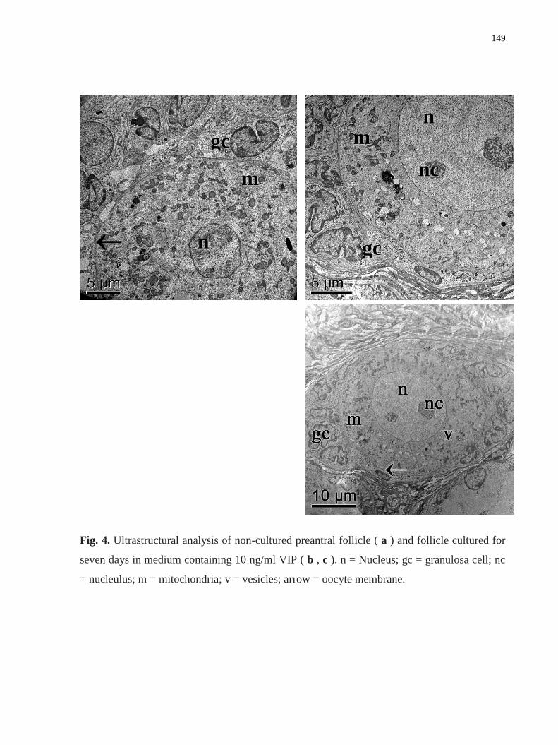
149
Fig. 4. Ultrastructural analysis of non-cultured preantral follicle ( a ) and follicle cultured for
seven days in medium containing 10 ng/ml VIP ( b , c ). n = Nucleus; gc = granulosa cell; nc
= nucleulus; m = mitochondria; v = vesicles; arrow = oocyte membrane.
ggcc
mm
nn
nn
nncc
mm
ggcc

150
Discussion
The present study demonstrated, for the first time, the effects of different
concentrations of VIP on caprine follicular viability and early development in vitro. Several
studies have demonstrated that VIP influences important ovarian functions, such as regulation
of steroidogenesis [Ahmed et al., 1986], cAMP accumulation [Tornell et al., 1988],
plasminogen activator production [Johnson and Tilly, 1988] and oocyte maturation [Tornell et
al., 1988].
In our study, ovarian fragments from fresh control ovaries contained 85%
histologically normal preantral follicles. Silva et al. [2002] reported that approximately 12%
of goat preantral follicles in fresh ovaries were degenerated. In previous studies characterizing
ovarian follicular populations, 4.8 and 0.20% of caprine and ovine preantral follicles were
degenerated, respectively [Lucci et al., 1999; Amorim et al., 2000]. Additionally, follicular
survival rates (approximately 85%) in fresh control ovaries similar to our results have been
previously described by other authors [Matos et al., 2007a; Chaves et al., 2008].
The addition of different concentrations of VIP (1, 10, 50 or 100 ng/ml) to the culture
medium was important to maintain preantral follicle viability after 1 or 7 days of culture. VIP
has been shown to protect several cell types from apoptosis, including thymocytes, prostate
cancer and neural cells [Flaws et al., 1995; Delgado et al., 1996; Said, 1996; Gutierrez-Canas
et al., 2003; Sastry et al., 2006]. Reduction in lung injury by VIP is associated with inhibition
of caspase activation and upregulation of bcl- 2, both of which suppress cell death and
promote cell survival [Fraser and Evan, 1996; Said and Dickman, 2000]. In the ovary,
previous in vitro studies also demonstrate that VIP inhibits apoptosis of rat follicles [Flaws et
al., 1995; Vaccari et al., 2006] as well as avian granulosa cells [Flaws et al., 1995]. In our
study, however, the highest concentration of VIP (that is, 200 ng/ml) promoted a decrease in
follicular survival, indicating this VIP concentration may be toxic to the early stages of
caprine follicular development. More studies are necessary to determine the molecular
mechanism of VIP action on caprine follicular survival.
According to Nilsson and Skinner [2004], follicular activation occurs when primordial
follicles leave the resting pool, undergo a primordial to primary follicle transition, and
surrounding squamous pregranulosa cells become cuboidal and begin to proliferate. It is
151
known that follicular activation also occurs due to the increase in oocyte diameter. In the
present study, all treatments tested, including MEM+, increased the percentage of growing
follicles after 7 days of culture in relation to fresh control. However, there was no difference
between VIP-treated and MEM+ follicles, which indicates that VIP did not have an effect on
follicular activation. Nevertheless, VIP addition was effective in promoting an increase in
oocyte and follicular diameter after both 1 and 7 days of culture. The presence of VIP
receptors in granulosa, theca and interstitial cells [Vaccari et al., 2006] may explain the effect
of VIP on the increase in follicular diameter. Another possibility is that VIP probably acts by
increasing the oocyte diameter and/or number of granulosa cells but not interfering in the
changing of granulosa cell morphology (flat to cuboidal). It is likely that other growth factors,
such as fibroblast growth factor-2 or epidermal growth factor [Matos et al., 2007b; Celestino
et al., 2009], may be necessary in the culture medium to promote caprine follicular activation.
In addition, VIP immunoreactivity is reported to increase with the appearance of secondary
and antral follicles in bovine ovaries, which suggests a role for this peptide in follicular
growth [Hulshof et al., 1994]. Furthermore, VIP stimulated in vitro development of isolated
primary and secondary bovine follicles [Hulshof, 1995]. In accordance with our results, a
recent study in buffalo showed that 50 ng/ml VIP increased preantral follicle diameter [Gupta
et al., 2002]. Nevertheless, in mice, VIP did not affect follicular development and caused
inhibition of follicular growth, antrum formation, granulosa cell proliferation as well as
estradiol production of follicles stimulated by FSH [Cecconi et al., 2004]. These conflicting
results may be due to study design, differences related to species, culture conditions and
different follicular stages analyzed.
In the present work, treatment with 10 ng/ml VIP and the noncultured control
maintained the ultrastructural integrity of preantral follicles after 7 days of culture. Recently,
Matos et al. [2007a] showed similar results in the culture of caprine ovarian tissue in medium
containing FSH. Normal follicles had an ultrastructure similar to that previously described in
the goat [Lucci et al., 2001] and other species: swine [Greenwald and Moor, 1989]; bovine
[Van Wezel and Rodgers, 1996]; and human [Oktay et al., 1997].
In conclusion, this study showed 10 ng/ml VIP promotes follicular survival and
caprine preantral follicle growth after 7 days of in vitro culture and not the number of growing
follicles. These findings suggest that VIP plays an important role in early follicular

152
development and could have significant utility in improving the quality of oocytes used for in
vitro maturation and fertilization.
Acknowledgments
This work was supported by CNPq, CAPES, FINEP and Fundação Cearense de Apoio
à Pesquisa (FUNCAP). J.B.B. is the recipient of a grant from FUNCAP/CAPES (Brazil).
References
Ahmed, C.E., W.L. Dees, S.R. Ojeda (1986) The immature rat ovary is innervated by
vasoactive intestinal peptide (VIP)-containing fibers and responds to VIP with steroid
secretion. Endocrinology 118: 1682–1689.
Amorim, C.A., C.M. Lucci, A.P.R. Rodrigues, F.C.A. Carvalho, J.R. Figueiredo, D. Rondina,
R. Cecchi, A. Giorgetti, A. Martini, P. B.D. Gonçalves (2000) Quantitative and
qualitative analysis of the effectiveness of the mechanical method for the isolation of
preantral follicles from ovines ovaries. Theriogenology 53: 1251–1262.
Burden, H.W. (1985) The adrenergic innervations of mammalian ovaries in: Ben Jonathan,
N., M.M. Bahr, R.I. Weiner (eds): Catecholamines as Hormone Regulators. New York,
Raven Press, pp 261–278.
Cecconi, S., G. Rossi, M. Barberi, L. Scaldaferri, R. Canipari, (2004) Effect of pituitary
adenylate cyclase-activating polypeptide and vasoactive intestinal polypeptide on mouse
preantral follicle development in vitro. Endocrinology 145: 2071–2079.
Celestino, J.J.H., J.B. Bruno, I.B. Lima-Verde, M.H.T. Matos, M.V.A. Saraiva, R.N. Chaves,
F.S. Martins, L.F. Lima, K.P.O. Name, C.C. Campello, J.R.V. Silva, S.N. Báo, J.R.
Figueiredo (2009) Recombinant epidermal growth factor maintains follicular
ultrastructure and promotes the transition to primary follicles in caprine ovarian tissue
cultured in vitro. Reprod Sci 16: 239–246.
Chaves, R.N., F.S. Martins, M.V.A. Saraiva, J.J.H. Celestino, C.A.P. Lopes, J.C. Correia, I.B.
Lima-Verde, M.H.T. Matos, S.N. Báo, K.P.O. Name, C.C. Campello, J.R.V. Silva, J.R.

153
Figueiredo (2008) Chilling ovarian fragments during transportation improves viability
and growth of goat preantral follicles cultured in vitro. Reprod Fertil Dev 20: 640–647.
Davoren, J.B., A.J.W. Hsueh (1985) Vasoactive intestinal peptide: a novel stimulator of
steroidogenesis by cultured rat granulosa cells. Biol Reprod 33: 37–52.
Delgado, M., E. Garrido, C. Martinez, J. Leceta, R.P. Gomariz (1996) Vasoactive intestinal
peptide and pituitary adenylate cyclase-activating polypeptides (PACAP27) and
(PACAP38) protect CD4+CD8+ thymocytes from glucocorticoid-induced apoptosis.
Blood 87: 5152–5161.
Flaws, J.A., A. Desanti, K.I. Tilly, R.0. Javid, K. Kugu, A.L. Johnson, A.N. Hirshfield, J.L.
Tilly (1995) Vasoactive intestinal peptide mediated suppression of apoptosis in the
ovary: potential mechanisms of action and evidence of a conserved antiatretogenic role
through evolution. Endocrinology 136: 4351–4359.
Fraser, A., G. Evan (1996) A license to kill. Cell 85: 781–784.
Gougeon, A., G.B.N. Chainy (1987) Morphometric studies of small follicles in ovaries of
women at different ages. J. Reprod Fert 81: 433–442.
Greenwald, G.S., R.M. Moor (1989) Isolation and preliminary characterization of pig
primordial follicles. J Reprod Fertil 87: 561–571.
Gupta, P.S.P., S. Nandi, B.M. Rvindranatha, P.V. Sarma (2002) In vitro culture of buffalo
(Bubalus bubalis) preantral follicles. Theriogenology 57: 1839–1854.
Gutierrez-Cañas, I., N. Rodriguez-Henche, O. Bolanos, M.J. Carmena, J.C. Prieto, M.G.
Juarranz (2003) VIP and PACAP are autocrine factors that protect the androgen
independent prostate cancer cell line PC-3 from apoptosis induced by serum
withdrawal. Br J Pharmacol 139: 1050–1058.
Hulshof, S.C.J., G. Dijkstra, E.M. Van Der Beek, M.M. Bevers, J.R. Figueiredo, J.F. Beckers,
R. Van Den Hurk (1994) Immunocytochemical localization of vasoactive intestinal
peptide and neuropeptide y in the bovine ovary. Biol Reprod 50: 553–560.
Hulshof, S.C.J. (1995) Bovine preantral follicles and their development in vitro; thesis,
Utrecht. Johnson, A.L., J.L. Tilly (1988) Effects of vasoactive intestinal peptide on
steroid secretion and plasminogen activator activity in granulose cells of the hen. Biol
Reprod 38: 296–303.
154
Johnson, A.L., Z. Li, J.A. Gibney, S. Malamed (1994) vasoactive intestinal peptide induced
expression of cytochrome p450 cholesterol side-chain cleavage and 17a hydroxylase
enzyme activity in hen granulosa cells. Biol Reprod 51: 327–333.
Lucci, C.M., C.A. Amorim, A.P.R. Rodrigues, J.R. Figueiredo, S.N. Báo, J.R.V. Silva, P.B.D.
Gonçalves (1999) Study of preantral follicles population in situ and after mechanical
isolation from caprine ovaries at different reproductive stages. Anim Reprod Sci 56:
223–236.
Lucci, C.M., R.V. Silva, C.A. Carvalho, R. Figueiredo, N. Báo (2001) Light microscopical
and ultrastrutural characterization of goat preantral follicles. Small Rum Res 41: 61–69.
Matos, M.H.T., I.B. Lima-Verde, M.C.A. Luque, J.E. Maia Jr, J.R. Silva, J.J.H. Celestino,
F.S. Martins, S.N. Báo, C.M. Lucci, J.R. Figueiredo (2007a) Essential role of follicle
stimulating hormone in the maintenance of caprine preantral follicle viability in vitro.
Zygote 15: 173–182.
Matos, M.H.T., R. van den Hurk, I.B. Lima- Verde, M.C.A. Luque, K.D.B. Santos, F.S.
Martins, S.N. Báo, C.M. Lucci, J.R. Figueiredo (2007b) Effects of fibroblast growth
factor-2 on the in vitro culture of caprine preantral follicles. Cells Tissues Organs 186:
112–120.
Mayerhofer, A., G.A. Dissen, M. Costa, S.R. Ojeda (1997) A role for neurotransmitters in
early follicular development: induction of functional follicle-stimulating hormone
receptors in newly formed follicles of the rat ovary. Endocrinology 138: 3320–3329.
Nilsson, E.E., M.K. Skinner (2004) Kit ligand and basic fibroblast growth factor interactions
in the induction of ovarian primordial to primary follicle transition. Mol Cell Endocrinol
214: 19–25.
Ojeda, S.R., H.E. Lara (1989) The role of the sympathetic nervous system in the regulation of
ovarian function; in Pirke, K.M., W. Wuttke, U. Schweiger (eds): The Menstrual Cycle
and Its Disorders. Berlin, Springer-Verlag, pp 26–32.
Ojeda, S.R., H.E. Lara, C.E. Ahmed (1989) The potential relevance of vasoactive intestinal
peptide to ovarian physiology; in Adashi, E. (ed) Putative Intraovarian Regulators. New
York, Thieme Inc., pp 52–60.

155
Oktay, K., D. Briggs, R.G. Gosde (1997) Ontogeny of follicle-stimulating hormone receptor
gene expression in isolated human ovarian follicles. J Clinic Endocrinol Metabol 82:
3748–3751.
Parra, C., J.L. Fiedler, S.L. Lunal, M. Greiner, V. Padmanabhan, H.E. Lara (2007)
Participation of vasoactive intestinal polypeptide in ovarian steroids production during
the rat estrous cycle and in the development of estradiol valerate-induced polycystic
ovary. Reproduction 133: 147–154.
Said, S.I. (1996) Molecules that protect: the defense of neurons and other cells. J Clin Invest
97: 2163–2164.
Said, S.I., K. Dickman (2000) Pathways of inflammation and cell death in the lung:
modulation by vasoactive intestinal peptide. Regul Pept 93: 21–29.
Sastry, K.S.R., A.J. Smith, Y. Karpova, S.R. Datta, G. Kulik (2006) Diverse antiapoptotic
signaling pathways activated by vasoactive intestinal polypeptide, epidermal growth
factor, and phosphatidylinositol 3-kinase in prostate cancer cells converge on bad. J Biol
Chem 281: 20891–20901.
Silva, J.R.V., M.A.L. Ferreira, S.H.F. Costa, R.R. Santos, F.C.A. Carvalho, A.P.R. Rodrigues,
C.M. Lucci, S.N. Báo, J.R. Figueiredo (2002) Degeneration rate of preantral follicles in
the ovaries of goats. Small Rum Res 43: 203–209.
Silva, J.R., R. Van Den Hurk, M.H.T. Matos, R.R. Santos, C. Pessoa, M.O. Moraes, J.R.
Figueiredo (2004) Influences of FSH and EGF on primordial follicles during in vitro
culture of caprine ovarian cortical tissue. Theriogenology 61: 1691–1704.
Steel, R.G.D., J.H. Torrie, D. Dickey (1997) Principles and Procedures of Statistics: A
Biometrical Approach, ed 3. New York, Mc-Graw-Hill.
Tornell, J., B. Carlsson, T. Hillensjo (1988) Vasoactive intestinal peptide stimulates oocyte
maturation, steroidogenesis, and cyclic adenosine 3‘ , 5‘ –monophosphate production in
isolated preovulatory rat follicles. Biol Reprod 39: 213–220.
Vaccari, S., S. Latini, M. Barberi, A. Teti, M. Stefanini, R. Canipari (2006) Characterization
and expression of different pituitary adenylate cyclase-activating polypeptide/vasoactive
intestinal polypeptide receptors in rat ovarian follicles. J Endocrinol 191: 287–299.
Van Wezel, I., R.J. Rodgers (1996) Morphological characterization of primordial follicles and
their environment in vivo. Biol Reprod 55: 1003–1010.

156
11. CAPÍTULO 6
Níveis de peptídeo intestinal vasoativo em ovários de cabras e seu efeito sobre o
desenvolvimento in vitro de folículos pré-antrais isolados
(Steady-state level of Vasoactive Intestinal Peptide in goat ovaries and its effect on in
vitro development of isolated preantral follicles)
Periódico: Molecular and Cellular Endocrinology (Submetido em outubro de 2010).

157
Resumo
Os objetivos deste estudo foram investigar o nível de RNAm para Peptídeo Intestinal
Vasoativo (VIP) em ovários de cabra e avaliar os efeitos do VIP e / ou FSH sobre o
desenvolvimento in vitro e os níveis de mRNA para o ligante VIP e receptor do FSH (FSHR).
Para esse fim, folículos primordiais, primários e secundários de cabras, bem como folículos
antrais pequenos e grandes foram obtidos e os níveis de mRNA para VIP foram quantificados
por PCR em tempo real. Os efeitos do VIP e / ou FSH sobre o desenvolvimento dos folículos
pré-antrais e sobre a expressão de mRNA para VIP e FSHR foram avaliados após 6 dias de
cultivo em meio de controle (α-MEM) ou em α-MEM suplementado com FSH (100 ng / mL),
VIP (10 ng / mL) ou VIP+FSH. A RT-PCR demonstrou que os níveis de mRNA para VIP em
folículos secundários foi significativamente maior do que no estágio de folículo primordial.
Em pequenos e grandes folículos antrais, níveis de mRNA para VIP em complexo cumulus
oócito (CCOs) foram significativamente maiores do que suas respectivascélulas da granulosa /
teca. A adição do VIP e / ou FSH ao meio de cultivo não teve efeito sobre a sobrevivência, a
formação de antro e diâmetro folicular. No entanto, a presença de FSH, VIP ou ambos, em
meio de cultivo reduziu significativamente os níveis de mRNA para a VIP, mas não alterou os
níveis de mRNA para FSHR. Em conclusão, o mRNA para a VIP foi detectado em todas as
categorias e tipos celulares de cabra. Além disso, VIP e / ou FSH não afetou o
desenvolvimento dos folículos secundários e reduziu os níveis de mRNA para VIP após curto
período de cultivo de folículos pré-antrais caprinos.
Palavras-chave: VIP. FSH. Folículos pré-antrais. Caprino. Cultivo in vitro.

158
Steady-state levels of Vasoactive Intestinal Peptide mRNA in goat ovaries and its
effect on the in vitro development of isolated preantral follicles
J.B. Brunoa*, J.J.H. Celestino
a, M.V.A. Saraiva
a, R.M.P. Rocha
a, I.R. Brito
a, A.B.G. Duarte
a,
V.R. Araújoa, C.M.G Silva
a, I.M.T. Lima
a, M.H.T. Matos
b, C.C. Campello
a, J.R.V. Silva
c,
J.R. Figueiredoa
aFaculty of Veterinary Medicine, LAMOFOPA, PPGCV, State University of Ceara, Fortaleza-CE, Brazil,
bNucleus of Biotechnology Applied to Ovarian Follicle Development, Federal University of São Francisco
Valley, Petrolina-PE, Brazil
cBiotecnology Nucleus of Sobral (NUBIS), Federal University of Ceara, Sobral-CE, Brazil
*Correspondence should be addressed to:
Programa de Pós-Graduação em Ciências Veterinárias (PPGCV)
Laboratório de Manipulação de Oócitos e Folículos Pré-Antrais (LAMOFOPA)
Universidade Estadual do Ceará (UECE)
Av. Paranjana, 1700, Campus do Itaperi.
Fortaleza – CE – Brasil. CEP: 60740-930
Tel.: +55.85. 3101.9852; Fax: +55.85.3101.9840
E-mail address: [email protected] (J.B.Bruno)

159
Abstract
The aims of this study were to investigate the levels of vasoactive intestinal peptide (VIP)
mRNA in goat ovaries and to evaluate the effects of VIP and/or follicle-stimulating hormone
(FSH) on follicular development and mRNA levels of VIP and FSH receptor (FSHR).
Preantral follicles were cultured for 6 days in α-MEM+ or in α-MEM
+ supplemented with
FSH and/or VIP. RT-PCR demonstrated that levels of VIP mRNA in secondary follicles were
significantly higher than in primordial follicle. The addition of VIP and/or FSH to the culture
medium had no effect on follicular development. However, the presence of FSH and/or VIP
in the culture medium significantly reduced VIP mRNA levels, but did not alter FSHR
mRNA levels. In conclusion, VIP mRNA was detected in all goat follicular categories and
cellular types, VIP and/or FSH did not affect the development of secondary follicles and
reduce the expression of VIP mRNA levels.
Keywords: VIP, FSH, Preantral Follicles, Culture, Caprine.
1. Introduction
Ovarian activity is regulated not only by gonadotropins and steroids but also by a
number of neural inputs and paracrine regulatory mechanisms. The mammalian ovary is
innervated by extrinsic nerves, which are both catecholaminergic and peptidergic in nature
(Burden, 1985; Ojeda and Lara, 1989; Ojeda et al., 1989). Peptidergic innervation of the
ovary was verified, among other ways, by the presence of vasoactive intestinal peptide (VIP)
(Ahmed et al., 1986).
Some findings suggest that VIP may play a vital role in several ovarian functions, such
as the regulation of steroidogenesis (Ahmed et al., 1986; Tornell et al., 1988), cAMP
accumulation (Tornell et al., 1988, Vaccari et al., 2006), plasminogen activator production
(Johnson and Tilly, 1988) and oocyte maturation (Tornell et al., 1988). VIP has been shown to
stimulate androgen and estradiol release in vitro (Davoren and Hsueh, 1985; Parra et al.,
2007). Hulshof et al. (1994) demonstrated that the bovine ovary is innervated by VIP-positive
nerve fibers beginning at the onset of follicular development and increasingly with age.

160
Furthermore, this neuropeptide is produced by nerve fibers innervating follicles at all stages
of development in rodent (Ahmed et al., 1986) and avian (Johnson et al., 1994) ovaries.
However, VIP mRNA levels in goat ovarian follicles have not yet been described. Previous in
vitro studies also demonstrated that VIP inhibits the apoptosis of rat and mice follicles (Flaws
et al., 1995; Vaccari et al., 2006) and stimulates the development of isolated bovine primary
and early secondary follicles (Hulshof, 1995). Moreover, we recently reported that the
addition of VIP to the in vitro culture medium maintained the survival and increases the
diameter of the early preantral follicles enclosed in caprine ovarian tissue (Bruno et al., 2010).
Follicle-stimulating hormone (FSH) acts through binding to the FSH receptor (FSHR),
a G protein-coupled receptor superfamily member that is located exclusively in granulosa
cells (Gudermann et al., 1995), from the primary follicle developmental stage onwards
(Méduri et al., 2002). FSHR mRNA was detected in the preantral and antral follicles of goats
(Saraiva et al., unpublished data). FSH increased the in vitro survival and proliferation of
granulosa cells, antrum formation and steroidogenesis in preantral follicles isolated from
several species (mouse: Cortvrindt et al., 1997, sheep: Cecconi et al., 1999; buffalo: Sharma et
al., 2009). In caprine species, FSH maintained the ultrastructural integrity, promoted the
activation of primordial follicles and furthered the growth of preantral follicles enclosed in
ovarian tissue (Matos et al., 2007; Magalhães et al., 2009). Moreover, the addition of FSH to
the culture medium in a sequential way maintained viability, stimulated antrum formation and
reduced oocyte extrusion in isolated caprine secondary follicles (Saraiva et al., unpublished
data). Finally, FSH interacts with several intraovarian growth factors, which mediate its effect
in regulating cellular interactions by autocrine and paracrine mechanisms, thus inducing
follicular growth (Erickson and Shimasaki, 2001). Therefore, several growth and endocrine
factors locally produced by the ovary are able to amplify or attenuate FSH action. However,
among these factors, it is not known whether VIP, with or without an association with FSH,
has an effect on the in vitro development of isolated caprine preantral follicles, as well as on
VIP and FSHR mRNA levels.
Therefore, this study aimed to accomplish the following: (1) to determine the steady-
state levels of VIP mRNA during different follicular stages in goat ovaries; (2) to investigate
the influence of VIP and/or FSH on the development of isolated caprine preantral follicles

161
after 6 days of in vitro culture; and (3) to verify the effects of VIP and/or FSH on VIP and
FSHR mRNA levels in isolated caprine preantral follicles cultured for 6 days.
2. Material and Methods
2.1. Source of ovaries
Ovaries (n=70) from 35 adult mixed-breed goats (Capra hircus, one to three years old)
were collected at a local slaughterhouse, with 30 ovaries used for experiment 1 and 40 ovaries
used for experiment 2. Immediately postmortem, the ovaries were washed in 70% alcohol
followed by two washes in minimum essential medium (MEM). The ovaries were placed into
tubes containing 15 ml of MEM plus HEPES (MEM HEPES) supplemented with 100 µg/ml
penicillin and 100 µg/ml streptomycin, and they were transported to the laboratory at 4°C
(Chaves et al., 2008) within 1 h. Unless mentioned otherwise, the culture media and other
chemicals used in the present study were purchased from Sigma Chemical Co. (St Louis,
USA).
2.2. Experiment 1: Steady-state levels of VIP mRNA in goat ovarian follicles
To evaluate steady-state mRNA levels, ovaries (n=10) were used for the isolation of
primordial, primary and secondary follicles using a mechanical procedure, as previously
described (Lucci et al., 1999). After isolation, these follicles were washed twice to completely
remove the stromal cells, and the follicles were then placed by category into separate
Eppendorf tubes in groups of 10. This procedure was completed within 2 h, and all samples
were stored at –80C until the RNA was extracted.
The remaining ovaries were used for the collection of COCs, mural granulosa cells
and thecal cells from small and large antral follicles. Compact COCs aspirated from small (1–
3 mm) and large (3–6 mm) antral follicles were recovered from the ovaries (n=15).
Thereafter, groups of 10 COCs were stored at –80C until RNA extraction. To collect mural
granulosa and theca cell complexes, 10 small and large antral follicles were isolated from
ovaries (n=5) and dissected from stromal tissue as previously described (Van Tol and Bevers,

162
1998). The follicles were then bisected and granulosa and theca cell complexes were collected
and stored at –80C.
Isolation of total RNA was performed using Trizol plus a purification kit (Invitrogen,
São Paulo, Brazil). According to the manufacturer‘s instructions, 1 mL of Trizol solution was
added to each frozen sample, and the lysate was aspirated through a 20-gauge needle before
centrifugation at 10,000 g for 3 min at room temperature. Thereafter, all lysates were diluted
1:1 with 70% ethanol and subjected to a mini-column. After binding the RNA to the column,
DNA digestion was performed using RNAse-free DNAse (340 Kunitz units/mL) for 15 min at
room temperature. After washing the column three times, the RNA was eluted with 30 µL
RNAse-free water.
Prior to reverse transcription, the eluted RNA samples were incubated for 5 min at
70C and then chilled on ice. Reverse transcription was then performed in a total volume of
20 µl, which was comprised of 10 µl of sample RNA, 4 µl 5X reverse transcriptase buffer
(Invitrogen), 8U RNAseout, 150U Superscript III reverse transcriptase, 0.036U random
primers (Invitrogen), 10 mM DTT, and 0.5 mM of each dNTP. The mixture was incubated for
1 h at 42C, for 5 min at 80C, and then stored at -20C. Negative controls were prepared
under the same conditions but without the inclusion of the reverse transcriptase.
The quantification of VIP mRNA levels was performed using SYBR Green. PCR
reactions were composed of 1 µl cDNA as a template in 7.5 µl of SYBR Green Master Mix
(PE Applied Biosystems, Foster City, CA), 5.5 µl of ultra-pure water, and 0.5 µM of each
primer. The primers were designed to amplify VIP mRNA. Glyceraldehyde-2-phosphate
dehydrogenase (GAPDH) and β-actin (Table 1) were used as endogenous controls for the
normalization of the steady-state mRNA levels of the genes. The thermal cycling profile for
the first round of PCR was as follows: initial denaturation and activation of the polymerase
for 15 min at 94C, followed by 40 cycles of 15 sec at 94C, 30 sec at 60C, and 45 sec at
72C. The final extension was for 10 min at 72C. All reactions were performed in a real time
PCR Mastercycler (Eppendorf, Germany). The delta-delta-CT method was used to transform
CT values into normalized relative steady-state mRNA levels.
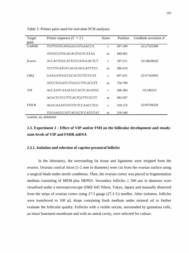
163
Table 1: Primer pairs used for real-time PCR analyses.
Target
gene
Primer sequence (5´ 3´) Sense Position GenBank accession nº
GAPDH TGTTTGTGATGGGCGTGAACCA
s 287-309 GI:27525390
ATGGCGTGGACAGTGGTCATAA
as 440-462
β-actin ACCACTGGCATTGTCATGGACTCT
s 187-211 GI:28628620
TCCTTGATGTCACGGACGATTTCC
as 386-410
UBQ GAAGATGGCCGCACTCTTCTGAT
s 607-631 GI:57163956
ATCCTGGATCTTGGCCTTCACGTT as 756-780
VIP
FSH-R
ACCAATCAAACGCCACTCAGATGC
AGACTCTCCTTCACTGCTTCGCTT
s
as
360-384
483-507
GI:340253
GI:95768228
AGGCAAATGTGTTCTCCAACCTGC
TGGAAGGCATCAGGGTCGATGTAT
s
as
250-274
316-340
s,sense; as, antisense
2.3. Experiment 2 - Effect of VIP and/or FSH on the follicular development and steady-
state levels of VIP and FSHR mRNA
2.3.1. Isolation and selection of caprine preantral follicles
In the laboratory, the surrounding fat tissue and ligaments were stripped from the
ovaries. Ovarian cortical slices (1-2 mm in diameter) were cut from the ovarian surface using
a surgical blade under sterile conditions. Then, the ovarian cortex was placed in fragmentation
medium consisting of MEM plus HEPES. Secondary follicles ≥ 200 μm in diameter were
visualized under a stereomicroscope (SMZ 645 Nikon, Tokyo, Japan) and manually dissected
from the strips of ovarian cortex using 27.5 gauge (27.5 G) needles. After isolation, follicles
were transferred to 100 µL drops containing fresh medium under mineral oil to further
evaluate the follicular quality. Follicles with a visible oocyte, surrounded by granulosa cells,
an intact basement membrane and with no antral cavity, were selected for culture.

164
2.3.2. Caprine preantral follicle culture
After selection, follicles were individually cultured in 100 µL drops of culture medium
under mineral oil in Petri dishes (60x15 mm, Corning, USA). Control culture medium, called
α-MEM+
, consisted of α-MEM (pH 7.2-7.4) supplemented with 3.0 mg/mL bovine serum
albumin (BSA), ITS (insulin 10 µg/mL, transferrin 5.5 µg/mL and selenium 5 ng/mL), 2 mM
glutamine, 2 mM hypoxantine and 50 μg/mL of ascorbic acid under mineral oil. Incubation
was conducted at 39°C with 5% CO2 in the air for 6 days. Fresh culture medium was prepared
and incubated for 1 h prior to use. Preantral follicles obtained from each animal were
randomly distributed in the following treatments: α-MEM+ alone or
supplemented with 100
ng/mL of recombinant FSH (rFSH: Tecnopec, Brazil), 10 ng/mL of VIP or both, constituting
the treatments α-MEM+, FSH, VIP and VIP+FSH, respectively. Every other day, 60 μL of the
culture media were replaced with fresh medium. The culture was replicated four times and a
minimum of 35 follicles was used per treatment.
2.3.3. Morphological evaluation of follicle development
Follicles were classified according to their morphology, and those showing
morphological signs of degeneration, such as darkness of oocytes and surrounding cumulus
cells or those with misshapen oocytes, were classified as degenerated. Follicular diameter was
measured only in healthy follicles in the x and y dimensions (90o), by using an ocular
micrometer (100 x magnification) inserted into a stereomicroscope (SMZ 645 Nikon, Tokyo,
Japan) every other day of culture. To measure follicular growth, the mean increase in
follicular diameter was calculated as follows: the diameter of viable follicles at day 6 minus
the diameter of viable follicles at day 0 divided by the total number of viable follicles at day
6. In addition, the percentages of secondary follicles that reached the antrum formation in
vitro were determined. Antral cavity formation was defined as a visible translucent cavity
within the granulosa cell layers.

165
2.3.4. Steady-state levels of VIP and FSHR mRNA in goat ovarian follicles cultured in
vitro
To evaluate the effect of VIP on the expression of VIP and FSHR mRNA after a 6 day
culture period, groups of 10 follicles were collected at the end of culture period and stored at -
80°C until the extraction of total RNA. Quantification of mRNA was performed as previously
described, and the primers for VIP and FSHR are shown in Table 1. β-actin and ubiquitin
(UBQ) were used as endogenous controls to normalize gene expression (Table 1).
2.4 Statistical analysis
mRNA expression data from primordial, primary and secondary follicles were
analyzed by the Kruskal-Wallis non-parametric test, while the t test was used for paired
comparisons of mRNA expression in the small and large antral follicles (P<0.05). For the cell
culture experiments, follicles were considered to be the experimental unit, following the same
approach as reported by Silva et al. (2010). Data from the follicular survival and antrum
formation for each treatment were compared using the Chi-square test, with the results
expressed as percentages. Follicular diameter data were submitted to the Kolmogorov-
Smirnov and Bartlett tests to confirm normal distribution and homoscedasticity,
respectively. Because of the heterogeneity of the variances, the follicular diameter, growth
rate, and VIP and FSHR mRNA levels after culture were compared using the Kruskal-Wallis
non-parametric test (SAS, 1999). The results were expressed as the mean ± standard error of
the mean (SEM), and differences were considered to be significant when P<0.05.
3. Results
3.1. Experiment 1 - Steady-state levels of VIP mRNA in goat ovarian follicles
Quantification of mRNA demonstrated that secondary follicles had significantly
higher levels of VIP mRNA compared to the primordial follicle (P<0.05). When the levels of
VIP mRNA in primordial and primary follicles were compared, no significant difference was
observed (P>0.05; Figure 1 A). In addition, no significant difference was observed between

166
COCs collected from small and large antral follicles (P>0.05; Figure 1 B), but granulosa/theca
cells from large antral follicles had higher levels of VIP mRNA than small antral follicles
(P<0.05; Figure 1 C). Real time PCR showed that COCs from both small and large antral
follicles had significantly higher levels of VIP mRNA than their respective granulosa/theca
cells (P<0.05; Figure 1 D, E).
Figure 1. Steady-state levels of VIP mRNA in goat ovarian follicles (means + SEM). A)
Primordial, primary and secondary follicles. B) COCs from small and large antral follicles. C)

167
Granulosa/theca cells from small and large antral follicles. D) COCs and granulosa/theca cells
from small antral follicles. E) COCs and granulosa/theca cells from large antral follicles. a,b
(P<0.05).
3.2. Experiment 2 - Effect of VIP and/or FSH on follicular development and steady-state
levels of VIP and FSHR mRNA
3.2.1. Follicular survival and growth after in vitro culture
The rates of follicular survival were greater than 90% at day 6 of culture in all
treatments tested. However, no significant differences were observed among the treatments
(Table 2; P>0.05). Figure 2 shows normal follicles before and after 6 days of in vitro culture.
From as early as day 2 of culture, all treatments had follicles with an antral cavity.
Furthermore, as the culture progressed from day 0 to day 6, a significant increase in the
percentage of antral follicles was observed (data not shown; P<0.05). However, when the
treatments were compared with each other, no significant differences were observed (P>0.05).
The follicles in the in vitro culture showed an initial mean diameter of 221.59±0.22,
237.73±0.21, 223.94±0.21 and 245.34±0.22 µm for the α-MEM+, FSH, VIP and VIP+FSH
treatments, respectively (Table 2; P>0.05). With the progression of the culture, an increase in
follicular diameter was observed from day 0 to day 6 in all treatment groups (P<0.05);
however, no significant differences were observed among the treatments (P>0.05). In
addition, follicles cultured in α-MEM+, FSH, VIP and VIP+FSH increased 16.94±0.09,
19.22±0.09, 18.89±0.09 and 20.89±0.08 µm/day, respectively. Similar to the diameter, no
significant differences were observed when the treatments were compared to each other
(P>0.05).
3.2.2. Steady-state levels of VIP and FSHR mRNA in goat ovarian follicles cultured in
vitro
Figure 3 shows VIP mRNA levels after 6 days of culture with the different treatments
that were tested. Culture with FSH, VIP and VIP+FSH promoted a significant reduction in

168
VIP mRNA levels in caprine preantral follicles after 6 days when compared with the control
(P<0.05). With regard to FSHR mRNA levels, no significant differences were observed
among the various treatments (P>0.05; Figure 4).
Table 2: Survival (%), antrum (%) e diameter (µm) of goat preantral follicles cultured for 6
days in VIP and/or FSH.
Treatments Survival (%)
Day 6
Antrum (%)
Day 6
Diameter ± SEM
Day 0 Day 6
α-MEM 100 (37/37) 72.97 (27/37) 221.59 ± 66.43 b 323.22 ± 101.30 a
FSH 91.89 (34/37) 72.97 (27/37) 237.73 ± 58.12 b 358.04 ± 100.61 a
VIP 97.30 (36/37) 75.68 (28/37) 223.94 ± 60.47 b 338.72 ± 84.88 a
VIP + FSH 100 (35/35) 85.71 (30/35) 245.34 ± 68.66 b 370.70 ± 91.96 a
Values with different letters between a column differ significantly (a, b; P<0.05)
Figure 2. Preantral follicles from goats at day 0 (A) and antral follicles after 6 days of in vitro
culture with 10 ng/mL VIP (B).

169
Figure 3. Steady state levels of VIP mRNA in goat preantral follicles cultured for 6 days in α-
MEM+ supplemented with FSH, VIP or both.
a,b (P<0.05).
Figure 4. Steady state levels of FSHR mRNA in goat preantral follicles cultured for 6 days in
α-MEM+ supplemented with FSH, VIP or both.

170
4. Discussion
The current study demonstrates the presence of VIP mRNA in all follicular categories
studied in goat ovaries, showing an increase in VIP mRNA levels during the transition from
the primordial to secondary follicle. The presence of VIP mRNA and its receptors was also
detected in rat and mouse ovaries (Gozes and Tsafriri, 1986; Cecconi et al., 2004; Vaccari et
al., 2006; Barberi et al., 2007). VIP protein was also reported to increase with the appearance
of secondary and antral follicles in bovine ovaries (Hulshof et al., 1994). These results suggest
a role for this peptide in follicular growth.
Granulosa/theca cells from large antral follicles had higher levels of VIP mRNA than
small antral follicles, suggesting that VIP acts in a stage-dependent way because the mRNA
expression levels increase with the progression of follicular development. In addition, COCs
from either small or large antral follicles had higher VIP mRNA levels than their respective
granulosa/theca cells. In rats, some studies have shown that VIP stimulates maturation in
follicle-enclosed oocytes but could transiently inhibit, or not affect, the spontaneous
maturation of cumulus-enclosed oocytes (Tornell et al., 1988; Apa et al., 1997). Furthermore,
other studies have shown that VIP increases the levels of cAMP, which regulates oocyte
maturation (Eppig and Downs, 1984) in preovulatory follicles rats (Tornell et al., 1988, Apa
et al., 1997) and stimulates ovulation in perfused rat ovaries (Schmidt et al., 1990). In
addition, VIP stimulates the production of plasminogen activator (Johnson and Tilly, 1988),
which converts plasminogen into plasmin that acts in the follicular wall releasing the COCs.
In the current study, high rates of survival and antrum formation were observed after
the in vitro culture period, and there was an increase in the follicular diameter after all
treatments. These results occurred independently of the addition of VIP and/or FSH to the
culture medium. Previous in vitro studies in different species have demonstrated that VIP (rat:
Flaws et al., 1995; Vaccari et al., 2006; bovine: Hulshof, 1995) inhibits apoptosis in preantral
follicles and stimulates the development of isolated primary and/or secondary follicles.
Recently, a study performed by our research team has demonstrated that VIP is an important
factor that promotes the growth of small preantral follicles enclosed in caprine ovarian tissue
(Bruno et al., 2010). However, despite the fact that VIP mRNA that was detected in caprine
secondary follicles, no additional effect of this factor was observed after the culture of
171
isolated follicles in this category when compared to the follicles cultured in α-MEM+. This
result may be due to the fact that α-MEM+ is a medium that is rich in nutrients (amino acids,
carbohydrates, B-vitamin complexes, vitamins C and D, inorganic salts and pyruvate) and
DNA precursors, all of which promote cell division (Hartshorne, 1997). Another possible
explanation for this finding is that VIP may need to interact with other substances and in other
concentrations to promote the in vitro growth of isolated secondary follicles in the caprine
species. Cecconi et al. (2004) demonstrated that VIP did not affect mouse follicular
development and caused the inhibition of follicular growth, antrum formation, granulosa cell
proliferation, and estradiol production. Moreover, other studies demonstrated that FSH
inhibits follicle apoptosis (murine: Cortvrindt et al., 1998; bovine: Itoh et al., 2002), maintains
the ultrastructural integrity of goat preantral follicles after culturing cortical tissue (Saraiva et
al., 2008), and promotes survival and antrum formation after 18 days culture of isolated
caprine secondary follicles (Saraiva et al., unpublished data). However, in the present study,
no additional influence of this hormone was observed after 6 days of culture when compared
with the control medium. It is likely that the use of the different concentrations of FSH
associated with a longer culture period of isolated follicles could improve the beneficial
effects of FSH on follicular survival and viability.
In this study, the presence of VIP in the culture medium reduces the mRNA levels for
itself. It is possible that the use of exogenous VIP, associated with its endogenous production
during the culture, might have caused a down-regulation in the expression of VIP mRNA.
Similarly, the addition of FSH to the culture medium reduces VIP mRNA levels. Flaws et al
(1995) verified that VIP can prevent follicular atresia in the absence of the influence of
gonadotropin. Analysis of VIP receptor (VPAC1-R) mRNA levels in whole mouse ovaries
showed that transcripts were present in untreated 22-day-old immature animals and were
significantly down-regulated after gonadotropin stimulation (Barberi et al., 2007). These
findings raise the possibility that VIP can promote the survival of follicles that have not yet
acquired dependence upon gonadotropins for continued development. This proposal would be
consistent with the fact that VIP can induce aromatase activity in immature follicles that are
not yet responsive to FSH (George and Ojeda, 1987). Many of the effects of VIP on ovarian
granulosa cells are similar to the action of gonadotropins (Ahmed et al., 1986; Johnson and

172
Tilly, 1988; Ojeda et al., 1989; Johnson et al., 1994), suggesting that VIP may be important
for regulating gonadotropin-independent development and ovarian follicle survival.
Although VIP stimulates the synthesis of cAMP, a key intracellular messenger
involved in the formation of FSHR (Knecht et al., 1983; Tano et al., 1997), in the present
study, the presence of VIP had no influence on FSHR mRNA levels in caprine preantral
follicles cultured in vitro. Evidence also suggests the existence of cAMP-independent
mechanisms, such as activin, that regulate the formation of FSHR in immature granulosa
cells (Tano et al., 1997; Nakamura et al., 1995; Xiao et al., 1992). In contrast to our
findings, VIP stimulation induced aromatase and FSHR expression at both the transcript
and protein levels in rat neonatal ovaries several days before the ovaries became responsive
to gonadotropins (Mayerhofer et al., 1997). In addition, there was no increase in FSHR
mRNA expression when the medium was supplemented with FSH. It is possible that
gonadotropins are not required for their own receptor expression during initial
folliculogenesis, as shown in hpg/hpg mice, in which the development of FSHR mRNA
levels was normal (O‘Shaughnessy et al., 1997). Moreover, Saraiva et al. (unpublished data)
used a sequential culture system with FSH and showed no increase in the expression of
FSHR after 6 and 12 days of isolated caprine secondary preantral follicle culture. However,
most studies show that gonadotropins stimulate the expression of their own receptors
(Smith and Ojeda, 1986; Minegishi et al., 1997; Guglielmo et al., in press). A second
possible explanation for the limited response could be that FSH may have promoted a
transcriptional down-regulation or decreased the stability of receptor mRNA induced by
high FSH concentrations (Tilly et al., 1992; Tisdall et al., 1995; Xu et al., 1995).
In conclusion, the present study demonstrated that VIP mRNA was detected in all
follicular categories and cellular types in caprine species. In the culture conditions used here,
the presence of VIP and/or FSH did not affect follicular survival and development after 6 days
of in vitro culture, in addition to reduces the mRNA levels for VIP, however without
influences the mRNA levels for FSHR. More detailed studies of the mechanisms of action of
VIP on follicular development in vivo and in vitro are essential to better understand
folliculogenesis. Furthermore, future applications of in vitro follicle culture systems would
improve fertility preservation for humans and rare animal species.

173
Acknowledgments
This work was supported by CNPq, CAPES, FINEP and Fundação Cearense de Apoio
à Pesquisa (FUNCAP). Jamily Bezerra Bruno is a recipient of a grant from FUNCAP (Brazil).
References
Ahmed, C.E., Dees, W.L., Ojeda, S.R., 1986. The immature rat ovary is innervated by
vasoactive intestinal peptide (VIP)-containing fibers and responds to VIP with steroid
secretion. Endocrinology 118, 1682–1689.
Apa, R., Lanzone, A., Mastrandrea, M., Miceli, F., Macchione, E., Fulghesu, A.M., Caruso,
A., Canipari, R., 1997. Effect of pituitary adenylate cyclase activating peptide on meiotic
maturation in follicle-enclosed, cumulus-enclosed, and denuded rat oocytes. Biol. Reprod.
57, 1074-1079.
Barberi, M., Muciaccia, B., Morelli, M.B., Stefanini, M., Cecconi, S., Canipari, R., 2007.
Expression localisation and functional activity of pituitary adenylate cyclase activating
polypeptide, vasoactive intestinal polypeptide and their receptors in mouse ovary.
Reproduction 134, 281-292.
Bruno, .JB., Celestino, J.J.H., Lima-Verde, I.B., Matos, M.H.T., Lima, L.F., Name, K.P.O.,
Araújo, V.R., Saraiva, M.V.A., Martins, F.S., Campello, C.C., Silva, J.R.V., Báo, S.N.,
Figueiredo, J.R., 2010. Vasoactive intestinal peptide improves the survival and
development of caprine preantral follicles after in vitro tissue culture. Cells Tissues Organs
191, 414-21.
Burden, H.W., 1985. The adrenergic innervations of mammalian ovaries, in: Ben-Jonathan,
N., Bahr, M.M., Weiner, R.I. (Eds.), Catecholamines as Hormone Regulators. Raven Press,
New York, pp. 261-278.
Cecconi, S., Barboni, B., Coccia, M., Mattioli, M., 1999. In vitro development of sheep
preantral follicles. Biol. Reprod. 60, 594-601.
Cecconi, S., Rossi, G., Barberi, M., Scaldaferri, L., Canipari, R., 2004. Effect of pituitary
adenylate cyclase-activating polypeptide and vasoactive intestinal polypeptide on mouse
preantral follicle development in vitro. Endocrinology 145, 2071-2079.
174
Chaves, R.N., Martins, F.S., Saraiva, M.V.A., Celestino, J.J.H., Lopes, C.A.P., Correia, J.C.,
Lima-Verde, I.B., Matos, M.H.T., Báo, S.N., Name, K.P.O., Campello, C.C., Silva, J.R.V.,
Figueiredo, J.R., 2008. Chilling ovarian fragments during transportation improves viability
and growth of goat preantral follicles cultured in vitro. Reprod. Fertil. Dev. 20, 640-647.
Cortvrindt, R., Smitz, J., Steirteghem, A.C., 1997. Assessment of the need for follicle
stimulating hormone in early pre antral mouse follicle culture in vitro. Hum. Reprod. 12,
759-768.
Cortvrindt, R.G., Hu, Y., Liu, J., Smitz, J.E., 1998. Timed analysis of the nuclear maturation
of oocytes in early preantral mouse follicle culture supplemented with recombinant
gonadotrophin. Fertil. Steril. 70, 1114-1125.
Davoren, J.B., Hsueh, A.J., 1985. Vasoactive intestinal peptide: a novel stimulator of
steroidogenesis by cultured rat granulosa cells. Biol. Reprod. 33, 37-52.
Eppig, J.J., Downs, S.M., 1984. Chemical signals that regulate mammalian oocyte maturation.
Biol. Reprod. 30, 1-11.
Erickson, G.F., Shimasaki, S., 2001. The physiology of folliculogenesis: the role of novel
growth factors. Fertil. Steril. 76, 943-949.
Flaws, J.A., Desanti, A., Tilly, K.I., Javid, R.0., Kugu, K., Johnson, A.L., Hirshfield, A.N.,
Tilly, J.L., 1995. Vasoactive intestinal peptide mediated suppression of apoptosis in the
ovary: potential mechanisms of action and evidence of a conserved antiatretogenic role
through evolution. Endocrinology 136, 4351-4359.
George, F.W., Ojeda, S.R., 1987. Vasoactive intestinal peptide induces aromatase activity
before development of primary follicles or responsiveness to follicle-stimulating hormone.
Proc. Natl. Acad. Sci. U.S.A. 84, 5803-5807.
Gozes, I., Tsafriri, A., 1986. Detection of vasoactive intestinal peptide encoding messenger
ribonucleic acid in the rat ovaries. Endocrinology 119, 2606-2610.
Gudermann, T., Nürnberg, B., Schultz, G., 1995. Receptors and G proteins as primary
components of transmembrane signal transduction. I. G-protein-coupled receptors:
structure and function. J. Mol. Med. 73, 51-63.
Guglielmo, M.C., Ricci, G., Catizone, A., Barberi, M., Galdieri, M., Stefanini, M., Canipari,
R., in press. The effect of hepatocyte growth factor on the initial stages of mouse follicle
development. J. Cell. Physiol.

175
Hartshorne, G., 1997. In vitro culture of ovarian follicles. Rev. Reprod. 2, 94-104.
Hulshof, S.C.J., Dijkstra, G., Van der Beek, E.M., Bevers, M.M., Figueiredo, J.R., Beckers,
J.F., Van den Hurk, R., 1994. Immunocytochemical localization of vasoactive intestinal
peptide and neuropeptide Y in the bovine ovary. Biol. Reprod. 50, 553-560.
Hulshof, S.C.J., 1995. Bovine preantral follicles and their development in vitro; thesis,
Utrecht.
Itoh, T., Kacchi, M., Abe, H., Sendai, Y., Hoshi, H., 2002. Growth, antrum formation, and
estradiol production of bovine preantral follicles cultured in a serum-free medium. Biol.
Reprod. 67, 1099-1105.
Johnson, A.L., Tilly, J.L., 1988. Effects of vasoactive intestinal peptide on steroid secretion
and plasminogen activator activity in granulose cells of the hen. Biol. Reprod. 38, 296-303.
Johnson, A.L., Li, Z., Gibney, J.A., Malamed, S., 1994. Vasoactive intestinal peptide-induced
expression of cytochrome p450 cholesterol side-chain cleavage and 17a-hydroxylase
enzyme activity in hen granulosa cells. Biol. Reprod. 51, 327-333.
Knecht, M., Ranta, T., Catt, K.J., 1983. Granulosa cell differentiation in vitro: induction and
maintenance of follicle-stimulating hormone receptors by adenosine 3_,5_-
monophosphate. Endocrinology 113, 949-956.
Lucci, C.M., Amorim, C.A., Rodrigues, A.P.R., Figueiredo, J.R., Báo, S.N., Silva, J.R.V.,
Gonçalves, P.B.D., 1999. Study of preantral follicles population in situ and after
mechanical isolation from caprine ovaries at different reproductive stages. Anim. Reprod.
Sci. 56, 223-236.
Magalhães, D.M., Araujo, V.R., Lima-Verde, I.B., Matos, M.H.T, Silva, R.C., Lucci, C.M.,
Báo, S.N., Campello, C.C., Figueiredo, J.R., 2009. Different Follicle-Stimulating Hormone
(FSH) sources influence caprine preantral follicle viability and development in vitro Braz.
J. Vet. Res. Anim. Sci. 46, 378-386.
Matos, M.H.T., Lima-Verde, I.B., Luque, M.C.A., Maia Jr, J.E., Silva, J.R.V., Celestino,
J.J.H., Martins, F.S., Báo, S.N., Lucci, C.M., Figueiredo, J.R., 2007. Essential role of
follicle stimulating hormone in the maintenance of caprine preantral follicle viability in
vitro. Zygote 15, 173-182.
176
Mayerhofer, A., Dissen, G.A., Costa, M., Ojeda S.R., 1997. A role for neurotransmitters in
early follicular development: induction of functional follicle-stimulating hormone
receptors in newly formed follicles of the rat ovary. Endocrinology 138, 3320-3329.
Méduri, G., Charnaux, N., Driancourt, M.A., Combettes, L., Granet, P., Vannier B, Loosfelt,
H., Migrom, E., 2002. Follicle-stimulating hormone receptors in oocytes? J. Clin.
Endocrinol. Metab. 87, 2266-2276.
Minegishi, T., Tano, M., Kishi, H., Kameda, T., Miyamoto, K., 1997. Follicle-stimulating
hormone regulation on its receptor messenger ribonucleic acid levels in cultured rat
granulosa cells. Biochim. Biophys. Acta. 1359, 165-173.
Nakamura, M., Nakamura, K., Igarashi, S., Tano, M., Miyamoto, K., 1995. Interaction
between activin A and cAMP in the induction of FSH receptor in cultured rat granulosa
cells. J. Endocrinol. 147, 103-110.
Ojeda, S.R., Lara, H.E., 1989. The role of the sympathetic nervous system in the regulation of
ovarian function, in: Pirke, K.M., Wuttke, W., Schweiger, U. (Eds.), The Menstrual Cycle
and Its Disorders. Springer-Verlag, Berlin, pp. 26-32.
Ojeda, S.R., Lara, H.E., Ahmed, C.E., 1989. The potential relevance of vasoactive intestinal
peptide to ovarian physiology, in: Adashi, E. (Ed.), Putative Intraovarian Regulators.
Thieme Inc., New York, pp. 52-60.
O‘Shaughnessy, P.J., McLelland, D., McBride, M.W., 1997. Regulation of luteinizing
hormone-receptor and follicle stimulating hormone-receptor messenger ribonucleic acid
levels during development in the neonatal mouse ovary. Biol. Reprod. 57, 602-608.
Parra, C., Fiedler, J.L., Luna, S.L., Greiner, M., Padmanabhan, V., Lara, H.E., 2007.
Participation of vasoactive intestinal polypeptide in ovarian steroids production during the
rat estrous cycle and in the development of estradiol valerate-induced polycystic ovary.
Reproduction 133, 147–154.
Saraiva, M.V.A., Celestino, J.J.H., Chaves, R.N., Martins, F.S., Bruno, J.B., Lima-Verde,
I.B., Matos, M.H.T., Silva, G.M., Porfirio, E.P., Báo, S.N., Campello. C.C., Silva, J.R.V.,
Figueiredo, J.R., 2008. Influence of different concentrations of LH and FSH on in vitro
caprine primordial ovarian follicle development. Small Rum. Res. 78, 87-95.
Schmidt, G., Jörgensen, J., Kannisto, P., Liedberg, F., Ottesen, B., Owman, C., 1990.

177
Vasoactive intestinal polypeptide in the PMSG-primed immature rat ovary and its effect on
ovulation in the isolated rat ovary perfused in vitro. J. Reprod. Fertil. 90, 465-472.
Sharma, G.T., Dubey, P.K., Meur, S.K., 2009. Survival and developmental competence of
buffalo preantral follicles using three dimensional collagen gel culture system. Anim.
Reprod. Sci. 114, 115-124.
Silva, C.M.G., Matos, M.H.T., Rodrigues, G.Q., Faustino, L.R., Pinto, L.C., Chaves, R.N.,
Araújo, V.R., Campello, C.C., Figueiredo J.R., 2010. In vitro survival and development of
goat preantral follicles in two different oxygen tensions. Anim. Reprod. Sci. 117, 83-89.
Smith, S.S., Ojeda, S.R., 1986. Neonatal release of gonadotropins is essential for development
of ovarian follicle-stimulating hormone receptor. Biol. Reprod. 34. 219-227.
Tano, M., Minegishi, T., Nakamura, K., Karino, S., Ibuki, Y., Miyamoto, K., 1997.
Transcriptional and post-transcriptional regulation of FSH receptor in rat granulosa cells
by cyclic AMP and activin. J. Endocrinol. 153, 465-473.
Tilly, J.L., Kowalski, K.I., Schomberg, D.W., Hsueh, A.J.W., 1992. Apoptosis in atretic
ovarian follicles is associated with selective decreases in messenger ribonucleic acid
transcripts for gonadotropin receptors and cytochrome P450 aromatase. Endocrinology
131, 1670-1676.
Tisdall, D.J., Watanabe, K., Hudson, N.L., Smith, P., McNatty, K.P., 1995. FSH receptor
gene expression during ovarian follicle development in sheep. J. Endocrinol. 15, 273-281.
Tornell, J., Carlsson, B., Hillensjo, T., 1988. Vasoactive intestinal peptide stimulates oocyte
maturation, steroidogenesis, and cyclic adenosine 3 _ , 5 _ -monophosphate production in
isolated preovulatory rat follicles. Biol. Reprod. 39, 213-220.
Vaccari, S., Latini, S., Barberi, M., Teti, A., Stefanini, M., Canipari, R., 2006.
Characterization and expression of different pituitary adenylate cyclase-activating
polypeptide/vasoactive intestinal polypeptide receptors in rat ovarian follicles. J.
Endocrinol. 191, 287–299.
Van Tol HT, Bevers MM, 1998. Theca cells and theca-cell conditioned medium inhibit the
progression of FSH-induced meiosis of bovine oocytes surrounded by cumulus cells
connected to membrana granulosa. Mol Reprod Dev 51, 315–321.

178
Xiao, S., Robertson, D.M., Findlay, J.K., 1992. Effects of activin and follicle-stimulating
hormone (FSH)-suppressing protein/follistatin on FSH receptors and differentiation of
cultured rat granulosa cells. Endocrinology 131, 1009-1016.
Xu, Z., Garverick, H.A., Smith, G.W., Smith, M.F., Hamilton, S.A., Youngquist, R.S., 1995.
Expression of follicle-stimulating hormone and luteinizing hormone receptor messenger
ribonucleic acids in bovine follicles during the first follicular wave. Biol. Reprod. 53, 951-
957.

179
12. CONCLUSÕES
A adição de 10 ng/mL de ANG II manteve a viabilidade e a ultraestrutura de folículos
pré-antrais caprinos após sete dias de cultivo in vitro. Os RNAm para os receptores
AGTR1 e AGTR2 foram detectados em todas as categorias foliculares, sendo
expressos de forma diferenciada nos diferentes compartimentos de folículos antrais.
O VEGF na concentração de 200 ng/mL manteve a viabilidade e a ultraestrutura e na
concentração de 10 ng/mL promoveu o crescimento de folículos pré-antrais caprinos
após sete dias de cultivo in vitro. Além disso, o VEGFR-2 foi expresso em oócitos de
folículos ovarianos caprinos em todos os estágios de desenvolvimento e em células da
granulosa de folículos em desenvolvimento.
A utilização de 10 ng/mL VIP assegurou a manutenção da integridade folicular e
estimulou o crescimento in vitro de folículos pré-antrais caprinos após sete dias de
cultivo in vitro.
O RNAm para a VIP foi detectado em todas as categorias e tipos celulares de folículos
caprinos. Além disso, o VIP e/ou o FSH não afetaram o desenvolvimento dos folículos
secundários e reduziram os níveis de RNAm para VIP após 6 dias de cultivo de
folículos pré-antrais caprinos.

180
13. PERSPECTIVAS
A eficiência dos programas de fecundação in vitro e transferência de embriões visando
à multiplicação de animais de alto valor zootécnico ou em via de extinção a partir de oócitos
provenientes dos folículos pré-antrais, depende ainda da completa compreensão dos
mecanismos que controlam a foliculogênese inicial, uma vez que os folículos primordiais
representam a grande reserva dos oócitos que poderão ser destinados para as diferentes
biotécnicas reprodutivas.
Os resultados deste projeto evidenciaram a participação da ANG II, VEGF e VIP no
desenvolvimento de folículos pré-antrais caprinos cultivados in situ. Entretanto, estudos
complementares poderão ser realizados visando à utilização destas substâncias sobre o
desenvolvimento de folículos pré-antrais isolados após ativação e crescimento destes folículos
in situ.
Diante das conclusões deste trabalho, as informações obtidas poderão ser utilizadas
para aperfeiçoar a elaboração e o fornecimento de meios de cultivo capazes de propiciar
ótimas condições para um completo crescimento folicular, preservando a viabilidade celular e
revolucionando a produção in vitro de embriões.

181
14. REFERÊNCIAS BIBLIOGRÁFICAS
ABIR, R.; FISCH, B.; JIN, S.; BARNNET, M.; FREIMANN, S.; VAN DEN HURK, R.;
FELDBERG, D.; NITKE, S.; KRISSI, H.; AO, A. Immunocytochemical detection and RT-
PCR expression of leukaemia inhibitory factor and its receptor in human fetal and adult
ovaries. Molecular Human Reproduction, v. 10, n.5, p. 313-319, 2004.
ABIR, R.; NITKE, A.; BEN-HAROUSH, A.; FISCH, B. In vitro maturation of primordial ovarian
follicles: Clinical significance, progress in mammals, and methods for growth evaluation.
Histology and Histopatology, v. 21, p. 887-898, 2006.
ABRAMOVICH, D.; CELIN, A. R.; HERNANDEZ, F.; TESONE, M.; PARBOREL, F.
Spatiotemporal analysis of the protein expression of angiogenic factors and their related
receptors during folliculogenesis in rats with and without hormonal treatment. Reproduction,
v. 137, p. 309- 320, 2009.
ABRAMOVICH, D.; PARBOREL, F.; TESONE, M. Effect of a vascular endothelial growth
factor (VEGF) inhibitory treatment on the folliculogenesis and ovarian apoptosis in
gonadotropin-treated prepubertal rats. Biology of Reproduction, v. 75, p. 434-441, 2006.
ACOSTA, T. J.; HAYASHI, K. G.; OHTANI, M.; MIYAMOTO, A. Local changes in blood
flow within the preovulatory follicle wall and early corpus luteum in cows. Reproduction, v,
p. 125:759-767, 2003.
ACOSTA, T. J.; BERISHA, B.; OZAWA, T.; SATO, K.; SCHAMS, D.; MIYAMOTO, A.
Evidence for a local endothelin-angiotensin-atrial natriuretic peptide system in bovine mature
follicles in vitro: effects on steroid hormones and prostaglandin secretion. Biology of
Reproduction, v. 61, n. 6, p. 1419-1425, 1999.
AHMED, C. E.; DEES, W. L.; OJEDA, S. R. The immature rat ovary is innervated by
vasoactive intestinal peptide (VIP)-containing fibers and responds to VIP with steroid
secretion. Endocrinology, v. 118, p. 1682–1689, 1986.
AMORIM, C. A.; LUCCI, C. M.; RODRIGUES, A. P. R.; CARVALHO, F. C. A.;
FIGUEIREDO, J. R.; RONDINA, D.; CECCHI, R.; GIORGETTI, A.; MARTINI, A.;
GONÇALVES, P. B. D. Quantitative and qualitative analysis of the effectiveness of the
mechanical method for the isolation of preantral follicles from ovines ovaries.
Theriogenology, v. 53, p. 1251–1262, 2000.

182
ANDERSON, E.; LEE, G. Y. The participation of growth factors in stimulating the quiescent,
proliferative, and differentiative stages of rat granulosa cells grown in a serum-free medium.
Tissue Cell, v. 25, p. 49-72, 1993.
ANDERSON, R. A.; ROBINSON, L. L. L.; BROOKS, J.; SPEARS N. Neurotropins and their
receptors are expressed in the human fetal ovary. Journal of Clinical Endocrinology and
Metabolism, v. 87, p. 890-897, 2002.
APA, R.; LANZONE, A.; MASTRANDREA, M.; MICELI, F.; MACCHIONE, E.;
FULGHESU, A.M.; CARUSO, A.; CANIPARI, R. Effect of pituitary adenylate cyclase
activating peptide on meiotic maturation in follicle-enclosed, cumulus-enclosed, and denuded
rat oocytes. Biology of Reproduction, v. 57, p. 1074-1079, 1997.
ARAÚJO, V. R.; CHAVES, R. N.; DUARTE, A. B. G.; CELESTINO, J. J. H.; SILVA, G.
M.; FERNANDES, D. D.; MATOS, M. H. T.; CAMPELLO, C. C.; FIGUEIREDO, J. R.
Effect of culture medium replacement protocol on the in vitro development of isolated caprine
secondary follicles. Small Ruminant Research, 2010b. [no prelo].
ARAÚJO, V. R.; SILVA, C. M. G.; MAGALHÃES, D. M.; SILVA, G. M.; BÁO, S. N.;
SILVA, J. R. V.; FIGUEIREDO, J. R.; RODRIGUES, A. P. R. Effect of Bone Morphogenetic
Protein-7 (BMP-7) on in vitro survival of caprine preantral follicles. Pesquisa Veterinária
Brasileira, v. 30, n. 4, p. 305-310, 2010a.
ARIMURA, A. Pituitary adenylate cyclase activating polypeptide (PACAP): discovery and
current status of research. Regulatory Peptides, v. 37, p. 287-303, 1992.
ARUNAKUMARI, G.; SHANMUGASUNDARAM, N.; RAO, V. H. Development of
morulae from the oocytes of cultured sheep preantral follicles. Theriogenology, v. 74, n. 5, p.
884-94, 2010.
ASAHARA, T.; BAUTERS, C.; ZHENG, L.P.; TAKESHITA, S.; BUNTING, S.;
FERRARA, N. Synergistic effect of vascular endothelial growth factor and basic fibroblast
growth factor on angiogenesis in vivo. Circulation, v. 91, p. 365-371, 1995.
ASAKAI, R.; SONG, S.; ITOH, N.; YAMAKUNI, T.; TAMURA, K.; OKAMOTO, R.
Differential gene expression of fibroblast growth factor receptor isoforms in rat ovary.
Molecular and Cellular Endocrinology, v. 114, p. 223, 1995.
183
ASAKAI, R.; TAMURA, K.; EISHI, Y.; IWAMOTO, M.; KATO, Y.; OKAMO, R. Basic
fibroblast growth factor (bFGF) receptors decrease with luteal age in rat ovarian luteal cells:
colocalization of bFGF receptors and bFGF in luteal cells. Endocrinology, v. 133, p. 1074-
1084, 1993.
BARBERI, M.; MUCIACCIA, B.; MORELLI, M. B.; STEFANINI, M.; CECCONI, S.;
CANIPARI, R. Expression localization and functional activity of pituitary adenylate cyclase
activating polypeptide, vasoactive intestinal polypeptide and their receptors in mouse ovary.
Reproduction, v.134, p. 281-292, 2007.
BARBONI, B.; TURRIANI, M.; GALEATI, G.; SPINACI, M.; BACCI, M. L.; FORNI, M.;
MATTIOLI, M. Vascular endothelial growth factor production in growing pig antral follicles.
Biology of Reproduction, v. 63, p. 858-864, 2000.
BARNETT, K. R.; SCHILLING, C.; GREENFELD, C. R.; TOMIC, D.; FLAWS, J. A.
Ovarian follicle development and transgenic mouse models. Human Reproduction Update, v.
12, p. 537-55, 2006.
BEERS W. H. Follicular plasminogen and plasminogen activator and the effect of plasmin on
ovarian follicle wall. Cell, v. 6, p. 379-386. 1975.
BEKER, A. R. C. L.; IZADYAR, F.; COLENBRANDER, B.; BEVERS, M. M. Effect of
growth hormone releasing hormone (GHRH) and vasoactive intestinal peptide (VIP) on in
vitro bovine oocyte maturation. Theriogenology, v, 53, p. 1771-1782. 2000.
BEN-HAROUSH, A.; ABIR, R.; AO, A.; JIN, S.; KESSLER-ICEKSON, G.; FELDBERG,
D.; FISCH, B. Expression of basic fibroblast growth factor and its receptors in human ovarian
follicles from adults and fetuses. Fertility and Sterility, v.84, p. 1257-1268, 2005.
BERISHA, B.; SCHAMS, D.; KOSMANN, M.; AMSELGRUBER, W.; EINSPANIER, R.
Expression and tissue concentration of vascular endothelial growth factor, its receptors and
localization in the bovine corpus luteum during estrous cycle and pregnancy. Biology of
Reproduction, v. 63, p. 1106-1114, 2000.
BERISHA, B.; SCHAMS, D.; MIYAMOTO, A. The mRNA Expression of Angiotensin and
Endothelin System Members in Bovine Ovarian Follicles During Final Follicular Growth.
Journal Reproduction and Development, v. 48, p. 573-582, 2002.
184
BODENSTEINER, K. J.; CLAY, C. M.; MOELLER, C. L.; SAWYER, H. R. Molecular
cloning of the ovine growth/differentiation factor-9 gene and expression of
growth/differentiation factor-9 in ovine and bovine ovaries. Biology of Reproduction, v. 60, p.
381-386, 1999.
BOONYAPRAKOB, U.; GADSBY, J. E.; HEDGPETH, V.; ROUTH, P.; ALMOND, G. W.
Expression and localization of vascular endothelial growth factor and its receptors in pig
corpora lutea during the oestrous cycle. Reproduction, v. 126, p. 393-405, 2003.
BRAW-TAL, R.; YOSSEFI, S. Studies in vivo and in vitro on the initiation of follicle growth
in the bovine ovary. Journal of Reproduction Fertility, v. 109, p.165-71, 1997.
BRISTOL-GOULD, S.; WOODRUFF, T. K. Folliculogenesis in the domestic cat (Felis
catus). Theriogenology, v. 66, p. 5-13, 2006.
BRUNO, J. B.; CELESTINO, J. J. H.; LIMA-VERDE, I. B.; MATOS, M. H. T.; LIMA, L.
F.; NAME, K. P. O.; ARAÚJO, V. R.; SARAIVA, M. V. A.; MARTINS, F. S.; CAMPELLO,
C. C.; SILVA, J. R. V.; BÁO, S. N.; FIGUEIREDO, J. R. Vasoactive intestinal peptide
improves the survival and development of caprine preantral follicles after in vitro tissue
culture. Cells Tissue Organs, v. 191, n. 5, p. 414-21, 2010.
BRUNO, J. B.; LIMA-VERDE, I. B.; MARTINS, F. S.; MATOS, M. H. T.; LOPES, C. A. P.;
MAIA-JR, J. E.; BÁO, S. N.; NOBRE-JUNIOR, H. V.; MAIA F. D.; PESSOA, C.;
MORAES, M. O.; SILVA, J. R. V., FIGUEIREDO, J. R.; RODRIGUES, A.P.R.
Característica histológica, ultra-estrutural e produção de nitrito de folículos pré-antrais
caprinos cultivados in vitro na ausência ou presença de soro. Arquivo Brasileiro de Medicina
Veterinária e Zootecnia, v. 60, n. 6, p. 1329-1337, 2008.
BURATINI, J. J.; GLAPINSK, V. F.; GIOMETTI, I. C.; TEIXEIRA, A. B.; COSTA, I. B.
Expression of fibroblast growth factor-8 and its cognate receptor, fibroblast growth factor
receptor (FGFR) -3c and -4, in fetal bovine preantral follicles. Molecular Reproduction and
Development, v. 70, p. 255-261, 2005.
BURDEN, H. W. The adrenergic innervations of mammalian ovaries. In: BEN-JONATHAN,
N.; BAHR, M. M.; WEINER, R. I. (Eds.), Catecholaminesas Hormone Regulators, New
York: Raven Press, 1985, p. 261–278.
BUSTIN, S. A. Quantification of mRNA using real-time reverse transcription PCR (RT-
PCR): trends and problems. Journal of Molecular Endocrinology. v. 29, p. 23-39, 2002.

185
BUTCHER, L.; ULLMANN, S. L. Culture of Preantral Ovarian Follicles in the Grey,
Shorttailed Opossum, Monodelphis domestica. Reproduction, Fertility and Development, v. 8,
p. 535-539, 1996.
CAIN, L.; CHATTERJEE, S.; COLLINS, T. J. In vitro folliculogenesis of rat preantral
follicles. Endocrinology, v. 136, p. 3369-3377, 1995.
CAMPBELL, B. K. The endocrine and local control of ovarian follicle development in the
ewe. Animal Reproduction, v. 6, n. 1, p. 159-171, 2009.
CARLSSON, I. B.; SCOTT J. E.; VISSER, J. A.; RITVOS, O.; THEMMEN, A. P. N.;
HOVATTA, O. Anti-Mullerian hormone inhibits initiation of growth of human primordial
ovarian follicles in vitro. Human Reproduction, v. 21, p. 2223-27, 2006.
CARMELIET, P.; JAIR, R. K. Angiogenesis in cancer and other diseases. Nature, v. 407, p.
249-257, 2000.
CECCONI, S.; BARBONI, B.; COCCIA, M.; MATTIOLI, M. In vitro development of sheep
preantral follicles. Biology of Reproduction. v. 60, p. 594-601, 1999.
CECCONI, S.; ROSSI, G.; BARBERI, M.; SCALDAFERRI, L.; CANIPARI, R. Effect of
pituitary adenylate cyclase-activating polypeptide and vasoactive intestinal polypeptide on
mouse preantral follicle development in vitro. Endocrinology, v. 145, p. 2071–2079, 2004.
CELESTINO, J. J. H.; BRUNO, J. B.; LIMA-VERDE I. B.; MATOS, M. H. T.; SARAIVA,
M. V. A.; CHAVES, R. N.; MARTINS, F. S.; LIMA, L. F.; NAME, K. P. O.; CAMPELLO,
C. C.; SILVA, J. R. V.; BÁO, S. N.; FIGUEIREDO, J. R. Recombinant Epidermal Growth
Factor maintains follicular ultrastructure and promotes the transition to primary follicles in
caprine ovarian tissue cultured in vitro. Reproductive Sciences. v. 16, n. 3, p. 239-246, 2009.
CELESTINO, J. J. H.; BRUNO, J. B.; LIMA-VERDE, I. B.; MATOS, M. H. T.; SARAIVA,
M. V. A.; CHAVES, R. N.; MARTINS, F. S.; ALMEIDA, A. P.; CUNHA, R. M. S.; LIMA,
L. F.; NAME, K. P. O.; CAMPELLO, C. C.; SILVA, J. R. V.; BAO, S. N.; FIGUEIREDO, J.
R. Steady-State Level of Kit Ligand mRNA in Goat Ovaries and the Role of Kit Ligand in
Preantral Follicle Survival and Growth In Vitro. Molecular Reproduction & Development, v.
77, p. 231-240, 2010.
186
CELIK-OZENCI, C.; AKKOYUNHLU, G.; KAYISLI, U. A.; ARICI, A.; DEMIR, R.;
Localization of vascular endothelial growth factor in the zona pellucida of developing ovarian
follicles in the rat: a possible role in destiny of follicles. Histochemistry and Cell Biology, v.
120, p. 383-390, 2003.
CHAVES, R. N.; ALVES, A. M. C. V.; DUARTE,
A. B. G.; ARAÚJO,
V. R.; CELESTINO,
J. J. H.; MATOS, M. H. T.; LOPES,
C. A. P.; CAMPELLO,
C. C.; NAME,
K. P. O.; BÁO,
S.
N.; FIGUEIREDO, J. R. Nerve growth factor promotes the survival of goat preantral follicles
cultured in vitro. Cells Tissues Organs, v.192, n. 4, p. 272-82, 2010a.
CHAVES, R. N.; LIMA-VERDE, I. B.; CELESTINO, J. J. H.; DUARTE, A. B. G.; ALVES,
A. M. C. V.; MATOS, M. H. T.; CAMPELLO, C. C.; NAME, K. P. O.; BÁO, S. N.;
BURATINI JR, J.; FIGUEIREDO, J. R. Fibroblast growth factor-10 maintains the survival
and promotes the growth of cultured goat preantral follicles. Domestic Animal Endocrinology,
v. 39, p. 249-258, 2010b.
CHAVES, R. N.; MARTINS, F. S.; SARAIVA, M. V. A.; CELESTINO, J. J. H.; LOPES, C.
A. P.; CORREIA, J. C.; LIMA-VERDE, I. B.; MATOS, M. H. T.; BÁO, S. N.; NAME, K. P.
O.; CAMPELLO, C. C.; SILVA, J. R. V.; FIGUEIREDO, J. R. Chilling ovarian fragments
during transportation improves viability and growth of goat preantral follicles cultured in
vitro. Reproduction Fertility and Development, v. 20, p. 640–647, 2008.
CLARK, D. E.; TISDALL, D. J.; FIDLER, A. E.; MCNATTY, K. P. Localization of mRNA
encoding c-kit during the initiation of folliculogenesis in ovine fetal ovaries. Journal of
Reproduction and Fertility, v. 106, p. 329-335, 1996.
CORNER, G. W. J. The histological dating of the human corpus luteum of menstruation. The
American Journal of Anatomy, v.9, p. 377-401, 1956.
CORTVRINDT, R. G.; HU, Y.; LIU, J.; SMITZ, J. E. Timed analysis of the nuclear
maturation of oocytes in early preantral mouse follicle culture supplemented with
recombinant gonadotrophin. Fertility and Sterility, v. 70, p.1114-1125, 1998.
CORTVRINDT, R. G.; SMITZ, J. E. J. Fluorescent probes allow rapid and precise recording
of follicle density and staging in human ovarian cortical biopsy samples. Fertility and
Sterility, v. 75, p. 588-593, 2001b.
CORTVRINDT, R.; SMITZ, J. E. J. In vitro follicle growth: Achievements in mammalian
species. Reproduction in Domestic Animals, v. 36, p. 3-9, 2001a.

187
CORTVRINDT, R.; SMITZ, J. E.; VAN STEIRTEGHEM, A. C. Assesment of the need for
follicle stimulating hormone in early preantral mouse follicle culture in vitro. Human
Reproduction, v. 12, p. 759-768, 1997.
COSTA, A. P. R.; FAGUNDES-MOURA, C. R.; PEREIRA, V. M.; SILVA, L. F. M.;
VIEIRA, A. R.; SANTOS, R. A. S; REIS, A. M. Angiotensin-(1–7): A novel peptide in the
ovary. Endocrinology, v. 144, p. 1942-1948, 2003.
DANFORTH, D. R.; ARBOGAST, L. K.; GHOSH, S.; DICKERMAN, A.; ROFAGHA, R.;
FRIEDMAN, C. I. Vascular endothelial growth factor stimulates preantral follicle growth in
the rat ovary. Biology of Reproduction, v. 68, p. 1736-1741, 2003.
DAVOREN, J. B.; HSUEH, A. J. W. Vasoactive intestinal peptide: a novel stimulator of
steroidogenesis by cultured rat granulosa cells. Biology of Reproduction, v. 33, p. 37–52,
1985.
DELGADO, M.; GARRIDO, E.; MARTINEZ, C.; LECETA, J.; GOMARIZ, R. P.
Vasoactive intestinal peptide and pituitary adenylate cyclase-activating polypeptides
(PACAP27) and (PACAP38) protect CD4+CD8+ thymocytes from glucocorticoid-induced
apoptosis. Blood, v. 87, p. 5152–5161, 1996.
DEMEESTERE, I.; GERVY, C.; CENTNER, J.; DEVREKER, F.; ENGLERT, Y.;
DELBAERE, A. Effect of insulin-like growth factor-I during preantral follicular culture on
steroidogenesis, in vitro oocyte maturation, and embryo development in mice. Biology of
Reproduction, v. 70, p. 1664-1669, 2004.
DISSEN, G. A.; ROMERO, C.; HIRSHFIELD, A. N.; OJEDA, S. R. Nerve growth factor is
required for early follicular development in the mammalian ovary. Endocrinology, v. 142, p.
2078-2086, 2001.
DOLE, G.; NILSSON, E. E.; SKINNER, M. K. Glial-derived neurotrophic factor promotes
ovarian primordial follicle development and cell–cell interactions during folliculogenesis.
Reproduction, v. 135, p. 671-682, 2008.
DONG, J.; ALBERTINI, D. F.; NISHIMORI, K.; KUMAR, T. R.; LU, N.; MATZUK, M. M.
Growth differentiation factor-9 is required during early ovarian folliculogenesis. Nature, v.
383, p. 531-535, 1996.

188
DRUMMOND, A. E. The role of steroids in follicular growth. Reproductive Biology and
Endocrinology, v. 4, p. 1-11, 2006.
DRUMMOND, A. E.; LE, M. T.; ETHIER, J. F.; DYSON, M.; FINDLAY, J. K. Expression
and localization of activin receptors, Smads, and beta glycan to the postnatal rat ovary.
Endocrinology, v. 143, p. 1423-1433, 2002.
DUARTE, A. B. G.; CHAVES, R. N.; ARAÚJO, V. R.; CELESTINO. J. J. H.; SILVA, G.
M.; LOPES C. A. P.; TAVARES L. M. T.; CAMPELLO, C. C.; FIGUEIREDO, J. R.
Follicular interactions affect the in vitro development of isolated goat preantral follicles.
Zygote, 2010. [no prelo].
DURLINGER, A. L. L.; VISSER, J. A.; THEMMEN, A. P. N. Regulation of ovarian
function: the role of anti-Mullerian hormone. Reproduction, v. 124, p. 601-609, 2002.
DURLINGER, A. L.; KRAMER, P.; KARELS, B.; DE JONG, F. H.; UILENBROEK, J. T.;
GROOTEGOED, J. A.; THEMMEN, A. P. Control of primordial follicle recruitment by anti-
Mullerian hormone in the mouse ovary. Endocrinology, v. 140, p. 5789-5796, 1999.
EKHOLM, C.; HIILENSJÖ, T.; MAGNUSSON, C.; ROSBERG, S. Stimulation and
inhibition of rat oocyte meiosis by forskolin. Biology of Reproduction, v, 30, p. 537-43. 1984.
EPPIG, J. J.; SCHROEDER, A. C. Capacity of mouse oocytes from preantral follicles to
undergo embryogenesis and development to live young after growth, maturation, and
fertilization in vitro. Biology of Reproduction, v. 41, p. 268-276, 1989.
EPPIG, J. J.; DOWNS, S. M. Chemical signals that regulate mammalian oocyte maturation.
Biology of Reproduction, v. 30, p. 1-11, 1984.
EPPIG, J. J. Oocyte control of ovarian follicular development and function in mammals.
Reproduction, v. 122, n. 6, p. 829-838, 2001.
ERICKSON, G. F.; SHIMASAKI, S. The spatiotemporal expression pattern of the
bonemorphogenetic proteinfamily inratovary cell types during the estrous cycle. Reproductive
Biology and Endocrinology, v. 1, p. 9, 2003.
ERICKSON, G. F.; SHIMASAKI, S. The physiology of folliculogenesis: the role of novel
growth factors. Fertility and Sterility, v. 76, p. 943-949, 2001.

189
ESPINOSA-CERVANTES, M. C.; ROSADO-GARCIA, A. Angiogenesis in reproductive
physiology: follicular development, formation and maintenance of the corpus luteum.
Ginecología y Obstetricia de México, v. 70, p. 17-27, 2002.
FATEHI, A. N.; VAN DEN HURK, R.; COLENBRANDER, B.; DAEMEN, A. J. J. M.;
VAN TOL, H. T. A.; MONTEIRO, R. M.; ROELEN, B. A. J.; BEVERS, M. M. Expression
of bone morphogenetic protein2 (BMP2), BMP4 and BMP receptors in the bovine ovary but
absence of effects of BMP2 and BMP4 during IVM on bovine oocyte nuclear maturation and
subsequent embryo development. Theriogenology, v. 63, p. 872-889, 2005.
FENG, P.; KNECHT, M.; CATT, K. Hormonal control of epidermal growth factor receptors
by gonadotropins during granulosa cell differentiation. Endocrinology, v. 120, p. 1121-1126,
1987.
FERAL, C.; LEGALL, S.; LEYMARIE, P. Angiotensin II modulates steroidogenesis in
granulosa and theca in the rabbit ovary: Its possible involvement in atresia. European Journal
of Endocrinology, v. 133, p. 747-753, 1995.
FERNANDEZ, L.A.; TWICKLER, J., MEAD, A. Neovascularization produced by
angiotensin II. The Journal of Laboratory and Clinical Medicine, v. 105, p. 141-145, 1985.
FERRARA, N. Vascular endothelial growth factor: basic science and clinical progress.
Endocrine Reviews, v. 25, p. 581-611, 2004.
FERRARA, N.; DAVIS-SMYTH, T. The biology of vascular endothelial growth factor.
Endocrine Reviews, v. 18, p. 4–25, 1997.
FERRARA, N.; FRANTZ, G.; LECOUTER, J.; DILLART-TELM, L.; PHAM, T.;
DRAKSHARAPU, A.; GIORDANO, T.; PEALE, F. Differential expression of the angiogenic
factor genes VEGF and EG-VEGF in normal and polycystic human ovaries. The American
Journal of Pathology, v. 162, p. 1881-1893, 2003.
FIGUEIREDO, J.R.; RODRIGUES, A. P. R.; AMORIM, C. A.; SILVA, J. R. V.
Manipulação de oócitos inclusos em folículos ovarianos pré-antrais – MOIFOPA. In:
GONÇALVES, P. B. D.; FIGUEIREDO, J. R.; FREITAS, V. J. F (Eds.) Biotécnicas
aplicadas à reprodução animal, São Paulo: Editora Roca, 2008, p. 303-327.

190
FILIPPATOS, G. S.; GANGOPADHYAY, N.; LALUDE, O.; PARAMESWARAN, N.;
SAID, S. I.; SPIELMAN, W.; UHAL, B. D. Regulation of apoptosis by vasoactive peptides.
American Journal of Physiology. Lung Cellular and Molecular Physiology, v, 281 p. 749-
761. 2001.
FLAWS, J. A.; DESANTI, A.; TILLY, K. I.; JAVID, R. 0.; KUGU, K.; JOHNSON, A. L.;
HIRSHFIELD, A. N.; TILLY, J. L. Vasoactive intestinal peptide mediated suppression of
apoptosis in the ovary: potential mechanisms of action and evidence of a conserved
antiatretogenic role through evolution. Endocrinology, v. 136, p. 4351–4359, 1995.
FORNERIS, M. L.; AGUADO, L. I. Neonatal superior nerve transection disturbs the cyclic
activity of the female rats. The Journal of Steroid Biochemistry and Molecular Biology, v, 82
p. 75-82. 2002.
FORTUNE, J. E. The early stages of follicular development: activation of primordial follicles
and growth of preantral follicles. Animal Reproduction Science, v. 78, p. 135-163, 2003.
FORTUNE, J. E.; KITO, S.; WANDJI, S. A.; SRSEN, V. Activation of bovine and baboon
primordial follicles in vitro. Theriogenology, v. 49, p. 441-449, 1998.
FRASER, A.; EVAN, G. A license to kill. Cell, v. 85, p. 781–784, 1996.
FRASER, H. M.; LUNN, S. F. Angiogenesis and its control in the female reproductive
system. British Medical Bulletin, v. 56, p. 787-797, 2000.
FREDERICKS, C. M.; LUNDQUIST, L. E.; MATHUR, R. S.; ASHTON, S. H.;
LANDGREBE, S. C. Effects of vasoactive intestinal polypeptide upon ovarian steroids, ovum
transport and fertility in the rabbit. Biology of Reproduction, v, 28 p. 1052-1060. 1983.
GALEATI, G.; SPINACI, M.; GOVONI,N.; ZANNONI, A.; FANTINATI, P.; SEREN, E.;
TAMANINI, C. Stimulatory effects of fasting on vascular endothelial growth factor (VEGF)
production by growing pig ovarian follicles. Reproduction, v. 126, p. 647–652, 2003.
GALLOWAY, S. M.; MCNATTY, K. P.; CAMBRIDGE, L. M.; LAITINEN, M. P. E.;
JUENGEL, J. L.; JOKIRANTA, T. S.; MCLAREN, R. J.; LUIRO, K.; DODDS, K. G.;
MONTGOMERY, G. W.; BEATTIE, A. E.; DAVIS, G. H.; RITVOS, O. Mutations in an
oocyte-derived growth factor gene (BMP-15) cause increased ovulation rate and infertility in
a dosagesensitive manner. Nature Genetics, v. 25, p. 279-83, 2000.
191
GAROR, R.; ABIR, R.; ERMAN, A.; FELZ, C.; NITKE, S.; FISCH, B. Effects of basic
fibroblast growth factor on in vitro development of human ovarian primordial follicles.
Fertility and Sterility, v. 91, p. 1967-1975, 2009.
GEORGE, F. W.; OJEDA, S. R. Vasoactive intestinal peptide induces aromatase activity
before development of primary follicles or responsiveness to follicle-stimulating hormone.
Proceedings of the National Academy of Sciences of the United States of America. v. 84, p.
5803-5807, 1987.
GIGLI, I.; CUSHMAN, R. A.; WAHL, C. M.; FORTUNE, J. E. Evidence for a role for anti-
Mullerian hormone in the suppression of follicle activation in mouse ovaries and bovine
ovarian cortex grafted beneath the chick chorioallantoic membrane. Molecular Reproduction
and Development, v. 71, p. 480-488, 2005.
GIOMETTI, I. C.; BERTAGNOLLI, A. C.; ORNES, R. C.; COSTA, L. F. S.;
CARAMBULA, S. F.; REIS, A. M,; OLIVEIRA, J. F.; EMANUELLI, I. P.; GONÇALVES,
P. B. Angiotensin II reverses the inhibitory action produced by theca cells on bovine oocyte
nuclear maturation. Theriogenology, v. 63, p. 1014-1025, 2005.
GLINKA, A.; WU, W.; DELIUS, H.; MONAGHAN, A. P.; BLUMENSTOCK, C.; NIEHRS,
C. Dickkopf-1 is a member of a new family of secreted proteins and functions in head
induction. Nature, v. 391, p. 357-362, 1998.
GOSPODAROWICZ, D.; BIALECKI, H. Fibroblast and epidermal growth factors are
mitogenic agents for cultured granulose cells of rodent, porcine and human origin.
Endocrinology, v. 104, p. 757-764, 1979.
GOSPODAROWICZ, D.; CHENG, J.; LUI, G.M.; BAIRD, A.; ESCH, F.; BOHLEN, P.
Corpus luteum angiogenic factor isrelated to fibroblast growth factor. Endocrinology, v. 117,
p. 2383-2391, 1985.
GOUGEON, A.; CHAINY, G. B. N. Morphometric studies of small follicles in ovaries of
women at different ages. Journal of Reproduction and Fertility, v. 81, p. 433–442, 1987.
GOZES, I.; FRIDKINB, M.; HILL, J. M.; BRENNEMAN, D. E. Pharmaceutical VIP:
prospects and problems. Current Medicinal Chemistry, v. 6, p. 1019-34. 1999.

192
GOZES, I.; TSAFRIRI, A. Detection of vasoactive intestinal peptide encoding messenger
ribonucleic acid in the rat ovaries. Endocrinology, v. 119, p. 2606-2610; 1986.
GRAS, S.; HANNIBAL, J.; FAHRENKRUG, J. Pituitary adenylate cyclaseactivating
polypeptide is an auto/paracrine stimulator of acute progesterone accumulation and
subsequent luteinization in cultured periovulatory granulosa/lutein cells. Endocrinology, v,
140 p. 2199-2205, 1999.
GRASSELLI, F.; BASINI, G.; BUSSOLATI, S.; TAMANINI, C. Effects of VEGF and bFGF
on proliferation and production of steroids and nitric oxide in porcine granulosa cells.
Reproduction in Domestic Animals, v. 37, p. 362-368, 2002.
GRASSELLI, F.; BASINI, G.; TIRELLI, M.; CAVALLI, V.; BUSSOLATI, S.; TAMANINI,
C. Angiogenic activity of porcinegranulosa cells cocultured with endothelial cells in
amicrocarrier based three-dimensional fibrin gel. Journal of Physiology and Pharmacology:
an Official Journal of the Polish Physiological Society, v. 54, p. 361-370, 2003.
GREENAWAY, J.; CENTRY, P. A.; FEIGE, J. J.; LAMARRE, J.; PETRIK, J. J.
Thrombospondin and vascular endothelial growth factor are cyclically expressed in an inverse
pattern during bovine ovarian follicle development. Biology of Reproduction, v. 72, p. 1071-
1078, 2005.
GREENAWAY, J.; CONNOR, K.; PEDERSEN, H. G.; COOMBER, B. L.; LAMARRE, J.;
PETRIK, J. Vascular endothelial growth factor and its receptor, Flk-1/KDR, are
cytoprotective in the extravascular compartment of the ovarian follicle. Endocrinology, v.
145, p. 2896-2905, 2004.
GREENWALD, G. S.; MOOR, R. M. Isolation and preliminary characterization of pig
primordial follicles. Journal of Reproduction and Fertility, v. 87, p. 561–571, 1989.
GUDERMANN, T.; NÜRNBERG, B.; SCHULTZ, G. Receptors and G proteins as primary
components of transmembrane signal transduction. I. G-protein-coupled receptors: structure
and function. Journal of Molecular Medicine. v. 73, p. 51-63, 1995.
GUGLIELMO, M. C.; RICCI, G.; CATIZONE, A.; BARBERI, M.; GALDIERI, M.;
STEFANINI, M.; CANIPARI, R. The effect of hepatocyte growth factor on the initial stages
of mouse follicle development. Journal of Cellular Physiology, 2010.[no prelo].

193
GULYAS, B. J.; HODGEN, G. D.; TULLNER, W. W.; ROSS, G. T. Effects of fetal or
maternal hypophysectomy on endocrine organs and bodyweight in infant rhesusmonkeys
(Macaca mulatta): with particular emphasis on oogenesis. Biology of Reproduction, v. 16, p.
216-27, 1977.
GUPTA, P. S. P.; NANDI, S.; RVINDRANATHA, B. M.; SARMA, P. V. In vitro culture of
buffalo(Bubalus bubalis) preantral follicles. Theriogenology, v. 57, p. 1839–1854, 2002.
GUPTA, P. S. P.; RAMESH, H. S.; MANJUNATHA, B. M.; NANDI, S.; RAVINDRA, J. P.
Production of buffalo embryos using oocytes from in vitro grown preantral follicles. Zygote,
v. 16, p. 57-63, 2008.
GUTIERREZ, C. G.; RALPH, J. H.; TELFER, E. E.; WILMUT, I.; WEBB, R. Growth and
antrum formation of bovine preantral follicles in long-term culture in vitro. Biology of
Reproduction, v. 62, p. 1322-1328, 2000.
GUTIERREZ-CAÑAS, I.; RODRIGUEZ-HENCHE, N.; BOLANOS, O.; CARMENA, M. J.;
PRIETO, J. C.; JUARRANZ, M. G. VIP and PACAP are autocrinefactors that protect the
androgen-independentprostate cancer cell line PC-3 fromapoptosis induced by serum
withdrawal. British journal of pharmacology, v. 139, p. 1050–1058, 2003.
HALPIN, D. M.; JONES, A.; FINK, G.; CHARLTON, H. M. Postnatal ovarian follicle
development in hypogonadal (hpg) and normal mice and associated changes in the
hypothalamic-pituitary ovarian axis, Journal of Reproduction and Fertility, v. 77, p. 287-296,
1986.
HANAHAN, D. Signaling vascular morphogenesisand maintenance. Science, v. 277, p. 55-
60, 1997.
HANRAHAN, J. P.; GREGAN, S. M.; MULSANT, P.; MULLEN, M.; DAVIS, G. H.;
POWELL, R.; GALLOWAY, S. M. Mutations in the Genes for Oocyte-Derived Growth
Factors GDF9 and BMP15 Are Associated with Both Increased Ovulation Rate and Sterility
in Cambridge and Belclare Sheep (Ovis aries). Biology of Reproduction, v. 70, p. 900-909,
2004.
HARATA, T.; ANDO, H.; IWASE, A.; NAGASAKA, T.; MIZUTANI, S.; KIKKAWA, F.
Localization of angiotensin II, theAT1 receptor, angiotensin-converting enzyme,
aminopeptidase A, adipocyte-derived leucineaminopeptidase, and vascular endothelial growth

194
factorin the human ovary throughout the menstrual cycle. Fertility and Sterility, v. 86, p. 433-
439, 2006.
HARTSHORNE, G. In vitro culture of ovarian follicles. Reviews of Reproduction, v. 2, p. 94-
104, 1997.
HAZZARD, T. M.; MOLSKNESS, T. A.; CHAFFIN, C. L.; STOUFFERRL. Vascular
endothelial growth factor (VEGF) and angiopoiet in regulation by gonadotropin and steroids
in macaque granulosa cells during the periovulatory interval. Molecular Human
Reproduction, v.5, p.1115-1121, 1999.
HAZZARD, T. M.; STOUFFER, R. L. Angiogenesis in ovarian follicular and luteal
development. Baillière's best practice & research. Clinical Obstetrics & Gynaecology, v. 4,
p. 883-900, 2000.
HILL, J. L.; HAMMAR, K.; SMITH, P. J.; GROSS, D. J.; Stage-dependent effects of
epidermal growth factor on Ca2þ efflux in mouse oocytes. Molecular Reproduction and
Development, v. 53, p. 244-253, 1999.
HILLENSJÖ, T.; EKHOHN, C.; AHRÉN, K. Role of cyclic AMP in oocyte maturation and
glycolysis in the pre-ovulatory rat follicle. Acta Endocrinologica, v, 87 p. 377-88. 1978.
HILLIER, S. G. Current concepts of the roles of follicle stimulating hormone and luteinizing
hormone in folliculogenesis. Human Reproduction, v. 9, p. 188–91, 1994.
HIRAO, Y.; NAGAI, T.; KUBO, M.; MIYANO, T.; MIYAKE, M.; KATO, S. In vitro
growth and maturation of pig oocytes. Journal of Reproduction and Fertility, v. 100, p. 333–
339, 1994.
HIRSHFIELD, A. N. Development of follicles in mammalian ovary. International Review of
Cytology, v. 124, p. 43–101, 1991.
HOVATTA, O.; SILYE, R.; ABIR, R.; KRAUSZ, T.; WINSTON, R. M. L. Extracellular
matriz improves survival of both stored and fresh human primordial and primary ovarian
follicles in long-term culture. Human Reproduction, v. 12, p. 1032-1036, 1997.
HREINSSON, J. G.; SCOTT, J. E.; RASMUSSEN, C.; SWAHN, M. L.; HSUEH, A. J.;
HOVATTA, O. Growth differentiation factor-9 promotes the growth, development, and

195
survival of human ovarian follicles in organ culture. Journal of Clinical Endocrinology and
Metabolism, v. 87, p. 316-321, 2002.
HU, D. E.; HILEY, C. R.; FAN, T. P. Comparative studies of the angiogenic activity of
vasoactive intestinal peptide, endothelins-1 and -3 and angiotensin II in a rat sponge model.
British Journal of Pharmacology, v. 117, n. 3, p. 545-551, 1996.
HULSHOF, S. C. J.; FIGUEIREDO, J. R.; BECKERS, J. F.; BEVERS, M. M.;
VANDERSTICHELE, H.; VAN DEN HURK, R. Bovine preantral follicles and activin:
immunohistochemistry for activin and activin receptor and the effect of bovine activin A in
vitro. Theriogenology, v. 48, p. 133–142, 1997.
HULSHOF, S. C. J. Bovine preantral follicles and their development in vitro; thesis, Utrecht,
1995.
HULSHOF, S. C. J.; DIJKSTRA, G.; VAN DER BEEK, E. M.; BEVERS, M. M.;
FIGUEIREDO, J. R.; BECKERS, J. F.; VAN DEN HURK, R. Immunocytochemical
localization of vasoactive intestinal peptide and neuropeptide y in the bovine ovary. Biology
of Reproduction, v. 50, p. 553–560, 1994.
HULSHOF, S. C. J.; FIGUEIREDO, J. R.; BEKERS, J. F.; BEVERS, M. M.;VAN DEN
HURK, R. Isolation and Characterization of preantral follicles from foetal bovine ovaries. The
Veterinary Quarterly, v. 2, p. 78-80, 1994.
HUSAIN, A.; BUMPUS, F. M.; SILVA, P.; SPETH, R. C. Localization of angiotensin II
receptors in ovarian follicles and the identification of angiotensin II in rat ovaries.
Proceedings of the National Academy of Sciences of the United States of America, v. 84, n. 8,
p. 2489-2493, 1987.
HUTT, K. J.; MCLAUGHLIN, E. A.; HOLLAND, M. K. KIT/KIT ligand in mammalian
oogenesis and folliculogenesis: roles in rabbit and murine ovarian follicle activation and
oocyte growth. Biology of Reproduction, v. 75, p. 421–433, 2006.
IIJIMA, K.; JIANG, J. Y.; SHIMIZU,T.; SASADA, H.; SATO, E. Acceleration of follicular
development by administration of vascular endothelial growth factor in cycling female rats.
The Journal of Reproduction and Development, v. 51, p. 161–168, 2005.

196
ITOH, H.; MUKOYAMA, M.; PRATT, R. E.; GIBBONS, G. H.; DZAU, V. J. Multiple
autocrine growth factors modulate vascular smooth muscle cell growth response to
angiotensin II. The Journal of Clinical Investigation, v. 91, p. 2268-2274, 1993.
ITOH, T.; KACCHI, M.; ABE, H.; SENDAI, Y.; HOSHI, H. Growth, antrum formation, and
estradiol production of bovine preantral follicles cultured in a serum-free medium. Biology of
Reproduction, v. 67, p. 1099-1105, 2002.
JANA, B.; DZIENIS, A.; PAŃCZYSZYN, J.; ROGOZIŃSKA, A.; WOJTKIEWICZ, J.;
SKOBOWIAT, C.; MAJEWSKI, M. Denervation of the porcine ovaries performed during the
early luteal phase influenced morphology and function of the gonad. Reproductive Biology, v.
5, p. 69-82. 2005.
JEWGENOW, K. Impact of peptide growth factors on the culture of small preantral follicles
of domestic cats. Theriogenology, v. 45, p. 889-895, 1996.
JEWGENOW, K.; STOLTE, M. Isolation of preantral follicles from nondomestic cats -
viability and ultrastructural investigations. Animal Reproduction Science, v. 44, p. 183-193,
1996.
JIANG, J. Y.; MACCHIARELLI, G.; TSANG, B. K.; SATO, E. Capillary angiogenesis and
degeneration in bovineovarian antral follicles. Reproduction, v. 125, p. 211-223, 2003.
JOHNSON, A. L.; LI, Z.; GIBNEY, J. A.; MALAMED, S. Vasoactive intestinal peptide-
induced expression of cytochrome p450 cholesterol side-chain cleavage and 17a-hydroxylase
enzyme activity in hen granulosa cells. Biology of Reproduction, v. 51, p. 327–333, 1994.
JOHNSON, A. L.; TILLY, J. L. Effects of vasoactive intestinal peptide on steroid secretion
and plasminogen activator activity in granulose cells of the hen. Biology of Reproduction, v.
38, p. 296–303, 1988.
JOUBERT, F. J.; STRYDOM, D. J. Snake venom. Theamino acid sequence of protein A from
Dendroaspis polylepis polylepis (black mamba) venom. Hoppe-Seyler’s Zeitschrift für
Physiologische Chemie, v. 361, p. 1787-1794, 1980.
JUENGEL, J. L.; BODENSTEINER, K. J.; HEATH, D. A.; HUDSONA, N. L.; MOELLER,
C. L.; SMITH, P.; GALLOWAY, S. M.; DAVIS, G. H.; SAWYER, H. R.; MCNATTY, K. P.

197
Physiology of GDF9 and BMP15 signalling molecules. Animal Reproduction Science, v. 82-
83, p. 447-460, 2004.
JUENGEL, J. L.; HUDSON, N. L.; HEATH, D. A.; SMITH, P.; READER, K. L.;
LAWRENCE, S. B.; O'CONNELL, A. R.; LAITINEN, M. P.; CRANFIELD, M.; GROOME,
N. P.; RITVOS, O.; MCNATTY, K. P. Growth differentiation factor 9 and bone
morphogenetic protein 15 are essential for ovarian follicular development in sheep. Biology of
Reproduction, v. 67, p. 1777-1789, 2002.
JUENGEL, J. L.; MCNATTY, K. P. The role of proteins of the transforming growth factor-b
superfamily in the intraovarian regulation of follicular development. Human Reproduction
Update, v. 1, p. 144-161, 2005.
JUNQUEIRA, L. C.; CARNEIRO, J. Biologia celular e molecular. Oitava edição. Rio de
Janeiro: Guanabara Koogan 332p, 2005.
KASSON, B. G.; MEIDAN, R.; DAVOREN, J. B.; HSUEH, A. J. W. Identification of
subpopulations of rat granulosa cells: sedimentation properties and hormonal responsiveness.
Endocrinology, v, 7 p. 1027-1034. 1985.
KEZELE, P.; NILSSON, E. E.; SKINNER, M. K. Insulin but not insulin-like growth factor-1
promotes the primordial to primary follicle transition. Molecular and Cellular Endocrinology,
v. 192, p. 37-43. 2002.
KEZELE, P. R.; AGUE, J. M.; NILSSON, E.; SKINNER, M. K. Alteration in the ovarian
transcriptome during primordial follicle assembly and development. Biology of Reproduction,
v. 72, p. 241-255, 2005.
KIDDER, G. M.; MHAWI, A. A. Gap junctions and ovarian folliculogenesis. Reproduction,
v. 123, p. 613-620, 2002.
KIKUCHI, N.; ANDOH, K.; ABE, Y.; YAMADA, K.; MIZUNUMA, H.; IBUKI, Y.
Inhibitory action of leptin on early follicular growth differs in immature and adult female
mice. Biology of Reproduction, v. 65, p. 66–71, 2001.
KLAGSBRU, N. M.; D`AMORE, P. A. Vascular endothelial growth factor and its receptors.
Cytokine & Growth Factor Reviews, v. 7, p. 259-270, 1996.

198
KNECHT, M.; RANTA, T.; CATT, K. J. Granulosa cell differentiation in vitro: induction and
maintenance of follicle-stimulating hormone receptors by adenosine 3_,5_-monophosphate.
Endocrinology, v.113, p. 949-956, 1983.
KNIGHT, P. G.; GLISTER, C. TGF-b superfamily members and ovarian follicle
development. Reproduction, v. 132, p. 191-206, 2006.
KOBAYASHI, J.; MIZUNUMA, H.; KIKUCHI, N.; LIU, X.; ANDOH, K.; ABE, Y.;
YOKOTA, H.; YAMADA, K.; IBUKI, Y.; HAGIWARA, H. Morphological assessment of
the effect of growth hormone on preantral follicles from 11-day-old mice in an in vitro
culture system. Biochemical and Biophysical Research Communications, v. 268, p. 36–41,
2000.
KOWALEWSKI, M. P.; DYSON, M. T.; BOOS, A.; STOCCO, D. M. Vasoactive intestinal
peptide (VIP)-mediated expression and function of steroidogenic acute regulatory protein
(StAR) in granulosa cells. Molecular and Cellular Endocrinology, v, 328, p. 93-103. 2010.
KREUZER, H.; MASSEY, A. Engenharia genética e biotecnologia, 434p. 2. ed. Porto
Alegre: Artmed, 2002.
KUO, T. C.; ENDO, K.; DHARMARAJAN, A. M.; MIYAZAKI, T.; ATLAS, S. J.;
WALLACH, E. E. Direct effect of angiotensin II on in vitro perfused rabbit ovary. Journal of
Reproduction and Fertility, v. 92, p. 469-474, 1991.
LACOMBE, A.; LELIEVRE, V.; ROSELLI, C. E.; MULLER, J. M.; WASCHEK, J. A.;
VILAIN, E. Lack of vasoactive intestinal peptide reduces testosterone levels and reproductive
aging in mouse testis. The Journal of Endocrinology, v, 194, n. 1, p. 153-160. 2007.
LARA, H. E.; MCDONALD, J. K.; AHMED, C. E.; OJEDA, S. R. Guanethidine-mediated
destruction of ovarian sympathetic nerves disrupts ovarian development and function in rats.
Endocrinology, v, 127 p. 2199-2209. 1990.
LATINI, S.; CHIARPOTTO, M.; MUCIACCIA, B.; VACCARI, S.; BARBERI, M.;
GUGLIELMO, M. C.; STEFANINI, M.; CECCONI, S.; CANIPARI, R. Inhibitory effect of
pituitary adenylate cyclase activating polypeptide on the initial stages of rat follicle
development. Molecular and Cellular Endocrinology, v, 320 p. 34-44. 2010.

199
LECOUTER, J.; FERRARA, N. Endocrine gland-derivedVEGF and the emerging hypothesis
of organ-specific regulation of angiogenesis. Nature Medicine, v. 8, p. 913-917, 2002.
LECOUTER, J.; KOWALSKI, J.; FOSTER, J.; HASS, P.; ZHANG, Z.; DILLARD-TELM,
L.; FRANTZ, G.; RANGELL, L.; DEGUZMAN, L.; KELLER, G. A.; PEALE, F.;
GURNEY, A.; HILLAN, K. J.; FERRARA, N. Identification of an angiogenic mitogen
selective for endocrine gland endothelium. Nature, v. 412, p. 877-884, 2001.
LECOUTER, J.; LIN, R.; FRANTZ, G.; TEJADA, M.; PEALE, F.; HILLAN, K.J.;
FERRARA, N. The EG-VEGF homologue, Bv8, is a potent angiogenic factor. Localization of
Bv8 receptors to endothelial cells. Proceedings of the National Academy of Sciences of the
United States of America, v. 100, p. 2685-2690, 2003.
LEDDA, S.; BOGLIOLO, L.; LEONI, G.; CALVIA, P.; NAITANA, S. Influence of
vasoactive intestinal peptide (VIP), atrial natriuretic peptide (ANP) and insulin-like growth
factor-I (IGF-I) on in vitro maturation of prepubertal and adult sheep oocytes. Zygote, v, 4 p.
343-348, 1996.
LEE, J.; PARK, H. J.; CHOI, H. S.; KWON, H. B.; ARIMURA, A.; LEE, B. J.; CHOI, W. S.;
CHUN, S. Y. Gonadotropin stimulation of pituitary adenylate cyclase-activating polypeptide
(PACAP) messenger ribonucleic acid in the rat ovary and the role of PACAP as a follicle
survival factor. Endocrinology, v, 140 p. 818-826, 1999.
LEE, W. S.; OTSUKA, F.; MOORE, R. K.; SHIMASAKI, S. Effect of bone morphogenetic
protein-7 on folliculogenesis and ovulation in the rat. Biology of Reproduction, v. 65, p. 994-
999, 2001.
LEE, W. S.; YOON, S. J.; YOON, T. K.; CHA, K. Y.; LEE, S. H.; SHIMASAKI, S.; LEE, S.;
LEE, K. A. Effects of bone morphogenetic protein-7 (BMP-7) on primordial follicular growth
in the mouse ovary. Molecular Reproduction and Development, v. 69, p. 159-163, 2004.
LI, H. K.; KUO, T. Y.; YANG, H. S.; CHEN, L. R.; LI, S. S.; HUANG, H. W. Differential
gene expression of bone morphogenetic protein 15 and growth differentiation factor 9 during
in vitro maturation of porcine oocytes and early embryos. Animal Reproduction Science, v.
103, p. 312-322, 2008.
LI, X. F.; AHMED, A. Dual role of angiotensin II in the human endometrium. Human
Reproduction, v. 11, p. 95-108, 1996.

200
LI, Y. H.; JIAO, L. H.; LIU, R. H.; CHEN, X. L.; WANG, H.; WANG, W. H. Localization of
angiotensin II in pig ovary and its effects on oocyte maturation in vitro. Theriogenology, v.
61, p. 447-459, 2004.
LIMA-VERDE, I. B.; MATOS, M. H. T.; BRUNO, J. B.; MARTINS, F. S.; SANTOS R. R.;
BÁO, S. N.; LUQUE, M. C. A.; VIEIRA, G. A. B.; SILVEIRA, E. R.; RODRIGUES, A. P.
R.; FIGUEIREDO, J. R.; OLIVEIRA, M. A. L.; LIMA, P. F. Effects of α-tocopherol and
ternatin antioxidants on morphology and activation of goat preantral follicles in vitro cultured.
Arquivo Brasileiro de Medicina Veterinária e Zootecnia, v. 61, n. 1, p. 57-65, 2009.
LIMA-VERDE, I. B.; ROSSETTO, R.; MATOS, M. H. T.; CELESTINO, J. J. H.; BRUNO,
J. B.; SILVA, C. M. G.; FAUSTINO, L. R.; MORORÓ, M. B. S.; ARAÚJO, V. R.;
CAMPELLO, C. C.; FIGUEIREDO, J. R. Androstenedione and follicle stimulating hormone
involvement in the viability and development of goat preantral follicles in vitro. Animal
reproduction, v. 7, p. 80-89, 2010b.
LIMA-VERDE, I. B.; SARAIVA, M. V. A.; MATOS, M. H. T.; BRUNO, J. B.; TENÓRIO,
S. B.; MARTINS, F. S.; CUNHA, L. D.; NAME, K. P. O.; BÁO, S. N., CAMPELLO, C. C.;
FIGUEIREDO, J. R. Interaction between estradiol and follicle stimulating hormone promotes
in vitro survival and development of caprine preantral follicles. Cells Tissues Organs, v. 191,
p. 240-247, 2010a.
LIN, R.; LECOUTER, J.; KOWALSKI, J.; FERRARA, N. Characterization of EGVEGF
signaling in adrenal cortex capillary endothelial cells. The Journal of Biological Chemistry, v.
277, p. 8724-8729, 2002.
LIU, K.; RAJAREDDY, S.; LIU, L.; JAGARLAMUDI, K.; BOMAN, K.; SELSTAM, G.;
REDDY, P. Control of mammalian oocyte growth and early follicular development by the
oocyte PI3 kinase pathway: new roles for an old timer. Developmental Biology, v. 299, p. 1-
11, 2006.
LIU, X.; ANDO, H. K.; YOKOTA, H.; KOBAYASHI, J.; ABE, Y.; YAMADA, K.;
MIZUNUMA, H.; IBUKI, Y.. Effects of growth hormone, activin, and follistatin on the
development of preantral follicle from immature female mice. Endocrinology, v. 139, p.
2342-2347, 1998.
LIU, Y. X.; KASSON, B. G.; DAHL, K. D.; HSUEH, A. J. Vasoactive intestinal peptide
stimulates plasminogen activator activity by cultured rat granulosa cells and cumulus-oocyte
complexes. Peptides, v, 8 p. 29-33. 1987.

201
LIU, Y. X.; NY, T.; SARKAR, D.; LOSKUTOFF, D.; HSUEH, A. J. W. Identification and
regulation of tissue plasminogen activator activity in rat cumulus-oocyte complexes.
Endocrinology, v, 119 p. 1578-1587. 1986.
LONERGAN, P.; CAROLAN, C.; VAN LANGENDONCKT, A.; DONNAY, I.; KHATIR,
H.; MERMILLOD, P. Role of epidermal growth factor in bovine oocyte maturation and
preimplantation embryo development in vitro. Biology of Reproduction, v. 54, p. 1420-1429,
1996.
LOPES, C. A. P.; SANTOS, R. R.; CELESTINO, J. J. H.; MELO, M. A.; CHAVES, R. N.;
CAMPELLO, C. C.; SILVA, J. R.; BÁO, S. N.; JEWGENOW, K.; FIGUEIREDO, J. R.;
Short-term preservation of canine preantral follicles: Effects of temperature, medium and
time. Animal Reproduction Science, v. 115, p. 201–14, 2009.
LOUHIO, H.; HOVATTA, O.; SJÖBERG, J.; TUURI, T. The effects of insulin, and insulin-
like growth factors I and II on human ovarian follicles in long-term culture. Molecular Human
Reproduction, v. 6, p. 694-698, 2000.
LUCCI, C. M.; AMORIM, C. A.; BÁO, S. N.; FIGUEIREDO, J. R.; RODRIGUES, A. P. R.;
SILVA, J. R.; GONÇALVES, P. B. D. Effect of the interval of serial sections of ovarian in
the tissue chopper on the number of isolated caprine preantral follicles. Animal Reproduction
Science, v. 56, p.39–49, 1999.
LUCCI, C. M.; AMORIM, C. A.; RODRIGUES, A. P. R.; FIGUEIREDO, J. R.; BÁO, S. N.;
SILVA, J. R. V.; GONÇALVES, P. B. D. Study of preantral follicles population in situ and
after mechanical isolation from caprine ovaries at different reproductive stages. Animal
Reproductive Sciences, v. 56, p. 223–236, 1999.
LUCCI, C. M.; SILVA, R. V.; CARVALHO, C. A.; FIGUEIREDO, R.; BÁO, N. Light
microscopical and ultrastrutural characterization of goat preantral follicles. Small Ruminant
Research : the Journal of the International Goat Association, v. 41, p. 61–69, 2001.
LUTZ, E. M.; SHEWARD, W. J.; WEST, K. M.; MORROW, J. A.; FINK, G.; HARMAR, A.
J. The VIP2 receptor: molecular characterisation of a cDNA encoding a novel receptor for
vasoactive intestinal peptide. FEBS Letters, v. 334, p. 3-8, 1993.
MACCHIARELLI, G.; NOTTOLA, S. A.; VIZZA, E.; FAMILIARI, G.; KIKUTA, A.;
MURAKAMI, T.; MOTTA, P. M. Microvasculature of growing and atretic follicles in the
202
rabbit ovary: a SEM study of corrosion casts. Archives of Histology and Cytology, v. 56, p. 1-
12, 1993.
MAGALHÃES, D. M.; ARAÚJO, V. R.; LIMA-VERDE, I. B.; MATOS, M. H. T.; SILVA,
R. C.; LUCCI, C. M.; BÁO, S. N.; CAMPELLO, C. C.; FIGUEIREDO, J. R.. Different
Follicle-Stimulating Hormone (FSH) sources influence caprine preantral follicle viability and
development in vitro. Brazilian Journal of Veterinary Research and Animal Science, v. 46, p.
378-386, 2009a.
MAGALHÃES, D. M.; ARAÚJO, V. R.; LIMA-VERDE, I. B.; MATOS, M. H. T.; SILVA,
R. C.; LUCCI, C. M.; BÁO, S. N.; CAMPELLO, C. C.; FIGUEIREDO, J. R., Impact of
pituitary FSH purification on in vitro early folliculogenesis in goats. Biocell, v. 33, p. 91-97,
2009b.
MAGALHÃES, D. M.; DUARTE, A. B. G.; ARAÚJO, V. R.; BRITO, I. R.; SOARES, T. G.;
LIMA, I. M. T.; LOPES, C. A. P.; CAMPELLO, C. C.; RODRIGUES, A. P. R.;
FIGUEIREDO, J. R. In vitro production of a caprine embryo from a preantral follicle cultured
in media supplemented with growth hormone. Theriogenology, v. 75, p. 182-188, 2011.
MAGALHÃES, D. M.; FERNANDES; D. D., MORORÓ, M. B. S.; SILVA, C. M. G.;
RODRIGUES, G. Q.; BRUNO, J. B.; MATOS, M. H. T.; CAMPELLO, C. C.;
FIGUEIREDO, J. R. Effect of the medium replacement interval on the viability, growth and
in vitro maturation of isolated caprine and ovine pre-antral follicles. Reproduction in
Domestic Animals, 2010. [no prelo].
MAO, J.; SMITH, M. F.; RUCKER, E. B.; WU, G. M.; MCCAULEY, T. C.; CANTLEY, J.
T. C.; PRATHER, R. S.; DIDION, B. A.; DAY, B. N. Effect of epidermal growth factor and
insulin-like growth factor I on porcine preantral follicular growth, antrum formation, and
stimulation of granulosal cell proliferation and suppression of apoptosis in vitro. Journal of
animal science, v. 82, p. 1967-1975, 2004.
MARTELLI, A.; BERNABÒ, N.; BERARDINELLI, P.; RUSSO, V.; RINALDI, C.; DI
GIACINTO, O.; MAURO, A.; BARBONI, B. Vascular supply as a discriminating for pig
preantral follicle selection. Reproduction, v. 137, p. 45-58, 2009.
MARTINS, F. S.; CELESTINO, J. J. H.; SARAIVA, M. V. A.; CHAVES, R. N.;
ROSSETTO, R.; SILVA, C. M. G.; LIMA-VERDE, I. B.; LOPES, C. A. P.; CAMPELLO, C.
C.; FIGUEIREDO, J. R. Interaction between growth differentiation factor 9, insulin-like
growth factor I and growth hormone on the in vitro development and survival of goat

203
preantral follicles. Brazilian Journal of Medical and Biological Research, v. 43, p. 728-736,
2010.
MARTINEZ-MADRID, B.; CAMBONI, A.; DOLMANS, M. M.; NOTTOLA, S.;
LANGENDONCKT, A. V.; DONNEZ, J. Apoptosis and ultrstructural assessment after
cryopreservation of whole human varies with their vascular pedicle. Fertility and Sterility, v.
87, p.1153-1165, 2007.
MARTINS, F. S.; CELESTINO, J. J. H.; SARAIVA, M. V. A.; MATOS, M. H. T.; BRUNO,
J. B.; ROCHA-JUNIOR, C. M. C.; LIMA-VERDE, I. B.; LUCCI, C. M.; BÁO, S. N.;
FIGUEIREDO, J. R. Growth and differentiation factor-9 stimulates activation of goat
primordial follicles in vitro and their progression to secondary follicles. Reproduction
Fertility and Development, v. 20, p. 916-924, 2008.
MARTINS, F. S.; VAN DEN HURK, R.; SANTOS, R. R.; SILVA, J. R. V.; MATOS, M. H.
T.; CELESTINO, J. J. H.; RODRIGUES, A. P. R.; PESSOA, C.; FERREIRA, F. V. A.;
FIGUEIREDO, J. R. Development of goat primordial follicles after in vitro culture of ovarian
tissue in Minimal Essential Medium supplemented with coconut water. Animal Reproduction,
v. 2, n. 2, p. 106-113, 2005.
MASUDA, Y.; TAKATSU, Y.; TERAO, Y.; KUMANO, S.; ISHIBASHI, Y.; SUENAGA,
M.; ABE, M.; FUKUSUMI, S.; WATANABE, T.; SHINTANI, Y.; YAMADA, T.;
HINUMA, S.; INATOMI, N.; OHTAKI, T.; ONDA, H.; FUJINO, M. Isolation and
identification of EG-VEGF/ prokineticins as cognate ligands for two orphan G-protein-
coupled receptors. Biochemical and Biophysical Research Communications, v. 293, p. 396-
402, 2002.
MATOS, M. H. T.; LIMA-VERDE, I. B.; BRUNO, J. B.; LOPES, C. A. P.; MARTINS, F. S.;
SANTOS, K. D. B.; ROCHA, R. M. P.; SILVA, J. R. V.; BÁO, S. N.; FIGUEIREDO, J. R.
Follicle stimulating hormone and fibroblast growth factor-2 interact and promote goat
primordial follicle development in vitro. Reproduction Fertility and Development, v. 19, p.
677-684, 2007c.
MATOS, M. H. T.; LIMA-VERDE, I. B.; LUQUE, M. C. A.; MAIA JR, J. E.; SILVA, J. R.;
CELESTINO, J. J. H.; MARTINS, F. S.; BÁO, S. N.; LUCCI, C. M.; FIGUEIREDO, J. R.
Essential role of follicle stimulating hormone in the maintenance of caprine preantral follicle
viability in vitro. Zygote, v. 15, p. 173–182, 2007a.

204
MATOS, M. H. T.; SILVA, J. R. V.; RODRIGUES, A. P. R.; FIGUEIREDO, J. R.; Técnicas
para avaliação da qualidade de folículos ovarianos pré-antrais cultivados in vitro. Revista
Brasileira de Reprodução Animal, v.31, n.4, p.433-442, 2007d.
MATOS, M. H. T.; VAN DEN HURK, R.; LIMA-VERDE, I. B.; LUQUE, M. C. A.;
SANTOS, K. D. B.; MARTINS, F. S.; BÁO, S. N.; LUCCI, C. M.; FIGUEIREDO, J. R.
Effects of fibroblast growth factor-2 on the in vitro culture of caprine preantral follicles. Cell
Tissues Organs, v. 186, p. 112-120, 2007b.
MATOS,
M. H. T.; VAN DEN HURK, R.; MARTINS, F. S.; SANTOS, R. R.; LUQUE, M.
C. A.; SILVA,
J. R. V.; CELESTINO, J. J. H.; BÁO,
S. N.; FIGUEIREDO, J. R. Histological
and ultrastructural features of caprine preantral follicles after in vitro culture in the presence
or absence of indole-3-acetic acid. Animal Reproduction, v. 3, n. 4, p. 415-422, 2006.
MATTIOLI, M.; BARBONI, B.; TURRIANI, M.; GALEATI, G.; ZANNONI, A.;
CASTELLANI, G.; BERARDINELLI, P.; SCAPOLO, P. A. Follicle activation involves
vascular endothelial growth factor production and increased blood vessel extension. Biology
of Reproduction, v. 65, p. 1014-1019, 2001.
MAYERHOFER, A.; DISSEN, G. A.; COSTA, M.; OJEDA, S. R. A role for
neurotransmitters in early follicular development: induction of functional follicle-stimulating
hormone receptors in newly formed follicles of the rat ovary. Endocrinology, v. 138, p. 3320–
3329, 1997.
MCGEE, E. A.; CHUN, S. Y.; LAI, S.; HE, Y.; HSUEH, A. J. Keratinocyte growth factor
promotes the survival, growth, and differentiation of preantral ovarian follicles. Fertility and
Sterility, v. 71, p. 732-738, 1999.
MCGHEE, E. A.; HSUEH, A. J. Initial and cyclic recruitment of ovarian follicles. Endocrine
Reviews, v. 21, p. 200–214, 2000.
MCNATTY, K. P.; FIDLER, A. E.; JUENGEL, J. L.; QUIRKE, L. D.; SMITH, P. R.;
HEATH, D. A.; LUNDY, T.; OCONNELL, A.; TIDDALL, D. J. Growth and paracrine
factors regulating follicular formation and cellular function. Molecular and Cellular
Endocrinology, v. 163, p. 11–20, 2000.
MÉDURI, G.; CHARNAUX, N.; DRIANCOURT, M. A.; COMBETTES, L.; GRANET, P.;
VANNIER, B.; LOOSFELT, H.; MIGROM, E. Follicle-stimulating hormone receptors in
oocytes? Journal of Clinical Endocrinology & Metabolism, v. 87, p. 2266-2276, 2002.

205
MERY, L.; LEFEVRE, A.; BENCHAIB, M.; DEMIRCI, B.; SALLE, B.; GUERIN, J-F.;
LORNAGE, J. Follicular growth in vitro: Detection of growth differentiation factor 9 (GDF9)
and Bone morphogenetic protein 15 (BMP15) during in vitro culture of ovine cortical slices.
Molecular Reproduction and Development, v. 74, p. 767-774, 2007.
MINEGISHI, T.; TANO, M.; KISHI, H.; KAMEDA, T.; MIYAMOTO, K. Follicle-
stimulating hormone regulation on its receptor messenger ribonucleic acid levels in cultured
rat granulosa cells. Biochimica et Biophysica Acta, v. 1359, p. 165-173, 1997.
MITSUBE, K.; MIKUNI, M.; MATOUSEK, M.; ZACKRISSON, U.; BRANNSTROM, M.
Role of the angiotensin II system in regulation of ovulation and blood flow in the rat ovary.
Reproduction, v. 125, p. 425-435, 2003.
MIYATA, A.; ARIMURA, A.; DAHL, R. R.; MINAMINO, N.; UEHARA, A.; JIANG, L.;
CULLER, M. D.; COY, D. H. Isolation of a novel 38 residue-hypothalamic polypeptide
which stimulates adenylate cyclase in pituitary cells. Biochemical and Biophysical Research
Communications, v, 164 p. 567-574. 1989.
MOLLAY, C.; WECHSELBERGER, C.; MIGNOGNA, G.; NEGRI, L.; MELCHIORRI, P.;
BARRA, D.; KREIL, G. Bv8, a small protein from frog skin and its homologue from snake
venom induce hyperalgesia in rats. European Journal of Pharmacology, v. 374, p. 189-196,
1999.
MOORE, R. K.; SHIMASAKI, S. Molecular biology and physiological role of the oocyte
factor, BMP-15. Molecular and Cellular Endocrinology, v. 234, p. 67-73, 2005.
MORBECK, D. E.; FLOWERS, W. L.; BRITT, J. H. Response of porcine granulosa cells
isolated from primary and secondary follicles to FSH, 8-bromo-cAMP and EGF in vitro.
Journal of Reproduction and Fertility, v. 99, p. 577-584, 1993.
MOTRO, B.; BERNSTEIN, A. Dynamic changes in ovarian c-kit and syeel expression during
the estrous reproductive cycle. Developmental Dynamics, v. 197, p. 69-79, 1993.
MURUVI, W.; PICTON, H. M.; RODWAY, R. G.; JOYCE, I. M. In vitro growth of oocytes
from primordial follicles isolated from frozen-thawed lamb ovaries. Theriogenology, v. 64, p.
1357-1370, 2005.

206
NAKAMURA, M.; NAKAMURA, K.; IGARASHI, S.; TANO, M.; MIYAMOTO, K.
Interaction between activin A and cAMP in the induction of FSH receptor in cultured rat
granulosa cells. Journal of Endocrinology, v. 147, p. 103-110, 1995.
NANDI, S.; RAVINDRANATHA, B. M.; GUPTA, P. S. P.; RAGHU, H. M.; SARMA, P. V.
Developmental competence and post-thaw survivability of buffalo embryos produced in vitro:
effect of growth factors in oocyte maturation medium and of embryo culture system.
Theriogenology, v. 60, p. 1621-1631, 2003.
NIELSEN, A. H.; HAGEMANN, A.; SVENSTRUP, B.; NIELSEN, J.; POULSEN, K.
Angiotensin-II receptor density in bovine ovarian follicles relates to tissue renin and follicular
size. Clinical and Experimental Pharmacology & Physiology, v. 21, p. 463-469, 1994.
NILSSON, E. E.; KEZELE, P.; SKINNER, M. K. Leukemia inhibiting factor (LIF) promotes
the primordial to primary follicle transition in rat ovaries. Molecular and Cellular
Endocrinology, v. 188, p. 65–73, 2002.
NILSSON, E. E.; PARROTT, J. A.; SKINNER, M. K. Basic fibroblast growth factor induces
primordial follicle development and initiates folliculogenesis. Molecular and Cellular
Endocrinology, v. 175, p. 123–130, 2001.
NILSSON, E. E.; SKINNER, M. K. Bone morphogenetic protein-4 acts as an ovarian follicle
survival factor and promotes primordial follicle development. Biology of Reproduction, v. 69,
p. 1265-1272, 2003.
NILSSON, E. E.; SKINNER, M. K. Growth and differentiation factor-9 stimulates
progression of early primary but not primordial rat ovarian follicle development. Biology of
Reproduction, v. 67, p. 1018–1024, 2002.
NILSSON, E. E.; SKINNER, M. K. Kit ligand and basic fibroblast growth factor interactions
in the induction of ovarian primordial to primary follicle transition. Molecular and Cellular
Endocrinology, v. 214, p. 19–25, 2004.
NUTTINCK, F.; COLLETTE, L.; MASSIP, A.; DESSY, F. Histologic and autoradiographic
study of the in vitro effects of FGF-2 and FSH on isolated bovine preantral follicles:
preliminary investigation. Theriogenology, v. 45, p. 1235-1245, 1996.

207
O‘BRIEN, M. J.; PENDOLA, J. K.; EPPIG, J. J. A revised protocol for in vitro development
of mouse oocytes from primordial follicles dramatically improves their developmental
competence. Biology of Reproduction, v. 68, p. 1682-1686, 2003.
O‘SHAUGHNESSY, P. J.; DUDLEY, K.; RAJAPAKSHA, W. R. Expression of follicle
stimulating hormone-receptor mRNA during gonadal development. Molecular and Cellular
Endocrinology, v. 125, 169-175, 1996.
O‘SHAUGHNESSY, P. J.; MCLELLAND, D.; MCBRIDE, M. W. Regulation of luteinizing
hormone-receptor and follicle stimulating hormone-receptor messenger ribonucleic acid
levels during development in the neonatal mouse ovary. Biology of Reproduction, v. 57, p.
602-608, 1997.
OBERMÜLLER, N.; SCHLAMP, D.; HOFFMANN, S.; GENTILI, M.; INAGAMI, T.;
GRETZ, N.; WEIGEL, M. Localization of the mRNA for the angiotensin II receptor subtype
2 (AT2) in follicular granulosa cells of the rat ovary by nonradioactive in situ hybridization.
The Journal of Histochemistry and Cytochemistry: Official Journal of the Histochemistry
Society, v. 46, p. 865-870, 1998.
OJEDA, S. R.; LARA, H. E. The role of the sympathetic nervous system in the regulation of
ovarian function. In: PIRKE, K. M.; WUTTKE, W.; SCHWEIGER, U. (Eds), The Menstrual
Cycle and Its Disorders. Berlin: Springer-Verlag, 1989, p.26–32.
OJEDA, S. R.; LARA, H. E.; AHMED, C. E. The potential relevance of vasoactive intestinal
peptide to ovarian physiology. In: ADASHI, E. (Eds), Putative Intraovarian Regulators.
NewYork: Thieme Inc., 1989, p 52–60.
OJEDA, S. R.; ROMERO, C.; TAPIA, V.; DISSEN, G. A. Neurotrophic and cell-cell
dependent control of early follicular development. Molecular and Cellular Endocrinology, v.
163, p. 67-71, 2000.
OJEDA, S. R.; WHITE, S. S.; AGUADO, L. I.; ADVIS, J. P.; ANDERSEN, J. M. Abdominal
vagotomy delays the onset of puberty and inhibits ovarian function in the female rat.
Neuroendocrinology, v, 36 p. 261-267. 1983.
OKTAY, K.; BRIGGS, D.; GOSDE, R.G. Ontogeny of follicle-stimulating hormone receptor
gene expression in isolated human ovarian follicles. The Journal of Clinical Endocrinology
and Metabolism, v. 82, p. 3748–3751, 1997.
208
ORISAKA, M.; ORISAKA, S. J.; JIN-YI, C. J.;WANG,Y.; KOTSUJI, F.; ANDTSANG, B.
K. Growth differentiation factor-9 is anti-apoptotic during follicular development from
preantral to early antral stage. Molecular Endocrinology, v. 20, p. 2456–2468, 2006.
OTANI, A.; TAKAGI, H.; OH, H.; SUZUMA, K.; MATSUMURA, M.; IKEDA, E.;
HONDA, Y. Angiotensin II stimulated vascular endothelial growth factor expression in
bovine retinal pericytes. Investigative Ophthalmology & Visual Science, v. 41, p. 1192- 1199,
2000.
OTANI, N.; MINAMI, S.; YAMOTO, M.; SHIKONE, T.; OTANI, H.; NISHIYAMA, R.;
OTANI, T.; NAKANO, R. The vascular endothelial growth factor/fms-like tyrosine kinase
system in human ovary during the menstrual cycle and early pregnancy. The Journal of
Clinical Endocrinology and Metabolism, v. 84, p. 3845- 3851, 1999.
OTSUKA, F.; YAO, Z.; LEE, T. H.; YAMAMOTO, S.; ERICKSON, G. F.; SHIMASAKI, S.
Bone morphogenetic protein-15: identification of target cells and biological functions. The
Journal of Biological Chemistry, v. 275, p. 39523-39528, 2000.
OTTESEN, B; FAHRENKRUG, J. Vasoactive intestinal polypeptide and other
preprovasoactive intestinal polypeptide-derived peptides in the female and male genital tract:
localization, biosynthesis, and functional and clinical significance. American journal of
Obstetrics and Gynecology, v, 172 p. 1615–1631. 1995.
PANGAS, S. A.; RADEMAKER, A. W.; FISHMAN, D. A.; WOODRUFF, T. K.
Localization of the activin signal transduction components in normal human ovarian follicles:
implications for autocrine and paracrine signaling in the ovary. Journal of Clinical
Endocrinology and Metabolism, v. 87, p. 2644–2657, 2002.
PARK, H. J.; LEE, J.; WANG, L.; PARK, J. H.; KWON, H. B.; ARIMURA, A.; CHUN, S.
Y. Stage-specific expression of pituitary adenylate cyclase-activating polypeptide type I
receptor messenger ribonucleic acid during ovarian follicle development in the rat.
Endocrinology, v. 141, p. 702-709, 2000.
PARRA, C.; FIEDLER, J. L.; LUNAL, S. L.; GREINER, M.; PADMANABHAN, V.;
LARA, H. E. Participation of vasoactive intestinal polypeptide in ovarian steroids production
during the rat estrous cycle and in the development of estradiol valerate-induced polycystic
ovary. Reproduction, v. 133, p. 147–154, 2007.
209
PARROT, J. A.; SKINNER, M. K. Kit-ligand/stem cell factor induces primordial follicle
development and initiates folliculogenesis. Endocrinology, v. 140, p. 4262-4271, 1999.
PAUL, M.; MEHR, A. P.; KREUTZ, R. Physiology of Local Renin-Angiotensin Systems.
Physiological Reviews, v. 86, p. 747-803, 2006.
PELLICER, A.; PALUMBO, A.; DECHERNEY, A. H.; NAFTOLIN, F. Blockage of
ovulation by an angiotensin antagonist. Science, v. 240, p. 1660-1661, 1988.
PEPLING, M. E.; SPRADLING, A. C. Female mouse germ cells form synchronously
dividing cysts. Development, v. 125, p. 3323-3328, 1998.
PEPPER, M. S.; FERRARA, N.; ORCI, L.; MONTESANO, R. Potent synergism between
vascular endothelial growth factor and basic fibroblast growth factor in the induction of
angiogenesis in vitro. Biochemical and Biophysical Research Communications, v. 189, p.
824-831, 1992.
PEROLLET, C.; HAN, Z.C.; SAVONA, C.; CAEN, J.P.; BIKFALVI, A. Platelet factor 4
modulates fibroblast growth factor 2 (FGF-2) activity and inhibits FGF-2 dimerization. Blood,
v. 91, p. 3289-3299, 1998.
PETRIK, J. J.; GENTRY, P. A.; FEIGE, J. J.; LAMARRE, J. Expression and localization of
thrombospondin-1 and -2 and their cell surface receptor, CD36, during rat follicular
development and formation of the corpus luteum. Biology of Reproduction, v. 67, p. 1522-
1531, 2002.
PICTON, H. M.; HARRIS, S. E.; MURUVI, W.; CHAMBERS, E. L. The in vitro growth and
maturation of follicles. Reproduction, v. 136, p. 703–715, 2008.
PICTON, H.; BRIGGS, D.; GOSDEN, R. The molecular basis of oocyte growth and
development. Molecular and Cellular Endocrinology, v. 145, p. 27–37, 1998.
PLENDL, J. Angiogenesis and vascular regression in the ovary. Anatomia, Histologia,
Embryologia, v. 29, p. 257–66, 2000.

210
PORTELA, V. M.; GONÇALVES, P. B. D.; VEIGA, A. M.; NICOLA, E.; BURATINI, J. J.;
PRICE, C. A. Regulation of angiotensin type 2 receptor in bovine granulosa cells.
Endocrinology, v. 149, p. 5004–5011, 2008.
PUCELL, A. G.; HODGES, J. C.; SEN, I.; BUMPUS, F. M.; HUSAIN, A. Biochemical
properties of the ovarian granulosa type 2- angiotensin II receptor. Endocrinology, v. 128, p.
1947-1959, 1991.
QU, J.; GODIN, P. A.; NISOLLE, M.; DONNEZ, J. Distribution of epidermal growth factor
receptor expression of primordial follicles in human ovarian tissue before and after
cryopreservation. Human Reproduction, v. 15, p. 302-310, 2000.
QUINTANA, R.; KOPCOW, L.; SUELDO, C.; MARCONI, G.; RUEDA, N. G.;
BARAÑAO, R. I. Direct injection of vascular endothelial growth factor into the ovary of mice
promotes follicular development. Fertility and Sterility, v. 82, p. 1101-1105, 2004.
RABINOVICI, J.; GOLDSMITH, P. C.; ROBERTS, V. J.; VAUGHAN, J.; VALE, W.;
JAFFE, R. B. Localization and secretion of inhibin/activin subunits in the human and
subhuman primate fetal gonads. Journal of Clinical Endocrinology and Metabolism, v. 73, p.
1141–1149, 1991.
REDMER, D. A.; DORAISWAMY, V.; BORTNEM, B. J.; FISHER, K.; JABLONKA-
SHARIFF, A.; GRAZUL-BILSKA, A. T.; REYNOLDS, L. P. Evidence for a role of capillary
pericytes in vascular growth of the developing ovine corpus luteum. Biology of Reproduction,
v. 65, p. 879-889, 2001.
REDMER, D.A.; REYNOLDS, L.P. Angiogenesis in the ovary. Reviews of Reproduction, v.
1, p. 182-192, 1996.
REICH, R.; MISKIN, R.; TSAFRIRI, A. Follicular plasminogen activator: involvement in
ovulation. Endocrinology, v, 116 p. 516-521, 1985.
REYNOLDS, L. P.; REDMER, D. A. Expression of the angiogenic factors, basic fibroblast
growth factor and vascular endothelial growth factor, in the ovary. Journal of animal science,
v. 76, p. 1671–81, 1998.
REYNOLDS, L.P.; REDMER, D.A. Utero-placental vascular development and placental
function. Journal of Animal Science, v. 73, p. 1839-1851, 1995.

211
ROBERTS, A.E.; ARBOGAST, L.K.; FRIEDMAN, C.I.; COHN, D.E.; KAUMAYA, P.T.;
DANFORTH, D.R. Neutralization of endogenous vascular endothelial growth factor depletes
primordial follicles in the mouse ovary. Biology of Reproduction, v. 76, p. 218-223, 2007.
ROBERTS, R. D.; ELLIS, R. C. L. Mitogenic effects of fibroblast growth factors on chicken
granulosa and theca cells in vitro. Biology of Reproduction, v. 61, p. 1387-92, 1999.
ROBINSON, R. S.; WOAD, K. J.; HAMMOND, A. J.; LAIRD, M.; HUNTER, M. G.;
MANN, G. E. Angiogenesis and vascular function in the ovary Focus on Vascular Function in
Female Reproduction. Reproduction, v. 138, p. 869–881, 2009.
RODRIGUES, G. Q.; SILVA, C. M. G.; FAUSTINO, L. R.; BRUNO, J. B.; PINTO, L. C.;
LOPES, C. A. P.; CAMPELLO, C. C.; FIGUEIREDO, J. R. Efeito de diferentes
concentrações de hormônio folículo-estimulante recombinante sobre o desenvolvimento in
vitro de folículos pré-antrais caprinos e ovinos isolados. Acta Veterinaria Brasilica, v.4, p.
144-152, 2010.
ROMANO, M.; KRAUS, E. R.; BOLAND, C. R.; COFFEY, R. J. Comparison between
transforming growth factor alpha and epidermal growth factor in the protection of rat gastric
mucosa against drug-induced injury. The Italian Journal of Gastroenterology, v. 26, p. 223-
228, 1994.
ROSSETTO, R.; LIMA-VERDE, I. B.; MATOS, M. H. T.; SARAIVA, M. V. A.;
MARTINS, F. S.; FAUSTINO, L. R.; ARAÚJO, V. R.; SILVA, C. M. G.; NAME, K. P. O.;
BÁO, S. N.; CAMPELLO, C. C.; FIGUEIREDO, J. R.; BLUME, H. Interaction between
ascorbic acid and follicle-stimulating hormone maintains follicular viability after long-term in
vitro culture of caprine preantral follicles. Domestic Animal Endocrinology, v. 37, p. 112-123,
2009.
ROY, S. K.; KOLE, A. R. Ovarian transforming growth factor-beta (TGF-beta) receptors: in
vitro effects of follicle stimulating hormone, epidermal growth factor and TGF beta on
receptor expression in human preantral follicles. Molecular Human Reproduction, v. 4, p.
207-214, 1998.
ROY, S. K.; TREACY, B. J. Isolation and long-term culture of human preantral follicles.
Fertility and Sterility, v.59, p. 783-791, 1993.
RÜSSE, I. Oogenesis in cattle and sheep. Bibliotheca Anatomica, v. 24, p. 77-92, 1983.

212
SAID, S.I. Molecules that protect: the defenseof neurons and other cells. The Journal of
Clinical Investigation, v. 97, p. 2163–2164, 1996.
SAID, S.I.; DICKMAN, K. Pathways of inflammation and cell death in the lung: modulation
by vasoactive intestinal peptide. Regulatory Peptides, v. 93, p. 21–29, 2000.
SALEHNIA, M.; MOGHADAM, E. A.; VELOJERDI, M. R. Ultrastructure of follicles after
vitrification of mouse ovarian tissue. Fertility and Sterility, v.78, p.644-645, 2002.
SARAIVA, M. V. A.; CELESTINO, J. J. H.; ARAÚJO, V. R.; CHAVES, R. N.; ALMEIDA,
A. P.; LIMA-VERDE, I. B.; DUARTE, A. B. G.; SILVA, G. M.; MARTINS, F. S.; BRUNO,
J. B.; MATOS, M. H. T.; CAMPELLO, C. C.; SILVA, J. R. V.; FIGUEIREDO, J. R.
Expression of follicle-stimulating hormone receptor (FSH-R) in goat ovarian follicles and the
impact of sequential culture medium on in vitro development of caprine preantral follicles.
Zygote, 2010a. [no prelo].
SARAIVA, M. V. A.; CELESTINO, J. J. H.; CHAVES, R. N.; MARTINS, F. S.; BRUNO, J.
B.; LIMA-VERDE, I. B.; MATOS, M. H. T.; SILVA, G. M.; PORFIRIO, E. P.; BÁO, S. N.;
CAMPELLO, C. C.; SILVA, J. R. V.; FIGUEIREDO, J. R.. Influence of different
concentrations of LH and FSH on in vitro caprine primordial ovarian follicle development.
Small Ruminant Research, v. 78, p. 87-95, 2008.
SARAIVA, M. V. A.; ROSSETTO, R.; BRITO, I. R.; CELESTINO, J. J. H.; SILVA, C. M.
G.; FAUSTINO. L. R.; ALMEIDA, A. P.; BRUNO, J. B.; MAGALHÃES, D. M.;
RODRIGUES, G. Q.; MATOS, M. H. T.; CAMPELLO, C. C.; SILVA, J. R. V.;
FIGUEIREDO, J. R. Dynamic medium produces caprine embryo from preantral follicles
grown in vitro. Reproductive Sciences, v. 17, p. 1135-1143, 2010b.
SASTRY, K. S. R.; SMITH, A. J.; KARPOVA, Y.; DATTA, S. R.; KULIK, G. Diverse
antiapoptotic signaling pathways activated by vasoactive intestinal polypeptide, epidermal
growth factor, and phosphatidylinositol 3-kinase in prostate cancer cells converge on bad. The
Journal of Biological Chemistry, v. 281, p. 20891–20901, 2006.
SCHAMS, D.; AMSELGRUBER, W.; EINSPANIER, R.; SINOWATZ, F.;
GOSPODAROWICZ, D. Localization and tissue concentration of basic fibroblast growth
factor in the bovine corpus luteum. Endocrine, v. 2, p. 907-912, 1994.
SCHAMS, D.; BERISHA, B.; KOSMANN, M.; AMSELGRUBER, W. Expression and
localization of IGF family members in bovine antral follicles during final growth and in luteal

213
tissue during different stages of estrous cycle and pregnancy. Domestic Animal
Endocrinology, v. 22, p. 51-72, 2002.
SCHAMS, D.; KOSMANN, M.; BERISHA, B.; AMSELGRUBER, W. M.; MIYAMOTO, A.
Stimulatory and synergistic effects of luteinising hormone and insulin-like growth factor 1 on
the secretion of vascular endothelial growth factor and progesterone of cultured bovine
granulose cells. Experimental and Clinical Endocrinology & Diabetes: Official Journal,
German Society of Endocrinology and German Diabetes Association, v. 109, p. 155-162,
2001.
SCHAUSER, K. H.; NIELSEN, A. H.; WINTHER, H.; DANTZER, V.; POULSEN, K.
Localization of the renin-angiotensin system in the bovine ovary: cyclic variation of the
angiotensin II receptor expression. Biology of Reproduction, v. 65, p. 1672-1680, 2001.
SCHECTERSON, L. C.; MCKNIGHT, G. S. Role of cyclic adenosine 3‘,5‘- monophosphate-
dependent protein kinase in hormone-stimulated /3-endorphin secretion in AtT20 cells.
Molecular Endocrinology, v. 5, p. 170-178. 1991.
SCHMIDT, G.; JÖRGENSEN, J.; KANNISTO, P.; LIEDBERG, F.; OTTESEN, B.;
OWMAN, C. Vasoactive intestinal polypeptide in the PMSG-primed immature rat ovary and
its effect on ovulation in the isolated rat ovary perfused in vitro. Journal of Reproduction and
Fertility, v. 90, p. 465-472, 1990.
SCHOTANUS, K.; HAGE, W. J.; VANDERSTICHELE, H.; VAN DEN HURK, R. Effects
of conditioned media from murine granulosa cell lines on the growth of isolated bovine
preantral follicles. Theriogenology, v. 48, p. 471–483, 1997.
SCHWEITZ, H.; PACAUD, P.; DIOCHOT, P.; MOINIER, D.; LADZUNSKI, M. MIT(1), a
black mamba intestinal toxin with a new and highly potent activity on intestinal contraction.
FEBS Letters, v. 461, p. 183-188, 1999.
SERAFIM, M. K.; ARAUJO, V. R.; SILVA, G. M.; DUARTE, A. B.; ALMEIDA, A. P.;
CHAVES, R. N.; CAMPELLO, C. C.; LOPES, C. A.; FIGUEIREDO, J. R.; SILVA, L. D.
Canine preantral follicles cultured with various concentrations of follicle-stimulating hormone
(FSH). Theriogenology, v. 74, n. 5, p. 749-55, 2010.
SHARMA, G. T.; DUBEY, P. K.; MEUR, S. K. Survival and developmental competence of
buffalo preantral follicles using three dimensional collagen gel culture system. Animal
Reproduction Science, v. 114, p. 115-124, 2009.
214
SHIBUYA, M. Role of VEGF–Flt receptor system in normal and tumor angiogenesis.
Advances in Cancer Research, v. 67, p. 281–316, 1995.
SHIKONE, T.; YAMOTO, M.; NAKANO, R. Follicle-stimulating hormone induces
functional receptors for basic fibroblast growth factor in rat granulosa cells. Endocrinology, v.
131, p. 1063-1068, 1992.
SHIMASAKI, S.; MOORE, R. K.; OTSUKA, F.; ERICKSON, G. F. The bone
morphogenetic protein system in mammalian reproduction. Endocrine Reviews, v. 25, p. 72-
101, 2004.
SHIMIZU, T. Promotion of ovarian follicular development by injecting vascular endothelial
growth factor (VEGF) and growth differentiation factor 9 (GDF-9) genes. The Journal of
Reproduction and Development, v. 52, p. 23-32, 2006.
SHIMIZU, T.; JIANG, J. Y.; IIJIMA, K.; MIYABAYASHI, K.; OGAWA,Y.; SASADA, H.;
SATO, E. Induction of follicular development by direct single injection of vascular
endothelial growth factor gene fragments into the ovary of miniature gilts. Biology of
Reproduction, v. 69, p. 1388–1393, 2003.
SHIMIZU, T.; JIANG, J. Y.; SASADA, H.; SATO, E. Changes of messenger RNA
expression of angiogenic factors and related receptors during follicular development in gilts.
Biology of Reproduction, v. 67, p. 1846-1852, 2002.
SHUTTLEWORTH, G.; PIPKIN, F. B.; HUNTER, M. G. In vitro development of pig
preantral follicles cultured in a serum-free medium and the effect of angiotensin II.
Reproduction, v. 123, p. 807-818, 2002.
SILVA, C. M. G.; MATOS, M. H. T.; RODRIGUES, G. Q.; FAUSTINO, L. R.; PINTO, L.
C.; CHAVES, R. N.; ARAÚJO, V. R.; CAMPELLO, C. C.; FIGUEIREDO, J. R. In vitro
survival and development of goat preantral follicles in two different oxygen tensions. Animal
Reproduction Science, v.117, p. 83-89, 2010.
SILVA, J. R. V. Growth factors in gota ovarios and the role of ativina-A in the development
of esrly-staged follicles. Phd Thesis. Utrecht University, Faculty of Veterinary Medicine, 142,
2005.
SILVA, J. R. V.; THARASANIT, T.; TAVERNE, M. A. M.; VAN DER WEIJDEN, G. C.;
SANTOS, R. R.; FIGUEIREDO, J. R.; VAN DEN HURK, R. The activin-follistatin system

215
and in vitro early follicle development in goats. Journal of Endocrinology, v. 189, p. 113-125,
2006a
SILVA, J. R. V.; VAN DEN HURK, R.; COSTA, S. H. F.; ANDRADE, E. R.; NUNES, A. P.
A.; FERREIRA, F. V. A.; LÔBO, R. N. B.; FIGUEIREDO, J. R. Survival and growth of goat
primordial follicles after in vitro culture of ovarian cortical slices in media containing coconut
water. Animal Reproduction Science, v. 81, p. 273-286, 2004a.
SILVA, J. R. V.; VAN DEN HURK, R.; FIGUEIREDO, J. R. Expression of mRNA and
protein localization of epidermal growth factor and its receptor in goat ovaries. Zygote, v. 14,
p. 107-117, 2006c.
SILVA, J. R. V.; VAN DEN HURK, R.; MATOS, M. H. T.; SANTOS, R. R.; PESSOA, C.;
MORAES, M. O.; FIGUEIREDO, J. R. Influences of FSH and EGF on primordial follicles
during in vitro culture of caprine ovarian cortical tissue. Theriogenology, v. 61, p. 1691–1704,
2004c.
SILVA, J. R. V.; VAN DEN HURK, R.; VAN TOL, H. T. A.; ROELEN, B. A. J.;
FIGUEIREDO, J. R. The Kit ligand/c-Kit receptor system in goat ovaries: gene expression
and protein localization. Zygote, v. 14, p. 317–328, 2006b.
SILVA, J. R. V.; VAN DEN HURK, R.; VAN TOL, H. T. A.; ROELEN, B. A. J.;
FIGUEIREDO, J. R. Expression of Growth Differentiation Factor 9 (GDF9), Bone
Morphogenetic Protein 15 (BMP15), and BMP Receptors in the Ovaries of Goats. Molecular
Reproduction and Development, v. 70, p. 11–19, 2004b.
SILVA, J. R.; FERREIRA, M. A. L.; COSTA, S. H. F.; SANTOS, R. R.; CARVALHO, F. C.
A.; RODRIGUES, A. P. R.; LUCCI, C. M.; BÁO, S. N.; FIGUEIREDO, J. R. Degeneration
rate of preantral follicles in the ovaries of goats. Small Ruminant Research: the Journal of the
International Goat Association, v. 43, p. 203–209, 2002.
SINGH, B.; RUTLEDGE, J. M.; ARMSTRONG, D. T. Epidermal growth factor and its
receptor gene expression and peptide localization in porcine ovarian follicles. Molecular
Reproduction and Development, v. 40, p. 391-399, 1995.
SMITH, S. K. Angiogenesis and reproduction. British Journal of Obstetrics and
Gynaecology, v. 108, p. 777-783, 2001.

216
SMITH, S. S.; OJEDA, S. R. Neonatal release of gonadotropins is essential for development
of ovarian follicle-stimulating hormone receptor. Biology Reproduction v. 34. p. 219-227,
1986.
SMITZ, J.; CORTVRINDT, R.; HU, Y.; VANDERSTICHELE, H. Effects of recombinant
activin A on in vitro culture of mouse preantral follicles. Molecular Reproduction and
Development, v. 50, p. 294–304. 1998.
SPEARS, N.; MURRAY, A. A.; ALISSON, V.; BOLAND, N. I.; GOSDEN, R. G. Role of
gonadotrofins and ovarian steroids in the development of mouse follicles in vitro. Journal of
Reproduction and Fertility, v. 113, p. 19–26, 1998.
SPENGLER, D.; WAEBER, C.; PANTALONI, C.; HOLSBOER, F.; BOCKAERT, J.;
SEEBURG, P. H.; JOURNOT, L. Differential signal transduction by five splice variants of
the PACAP receptor. Nature, v. 365, p. 170-175, 1993.
STALDEMANN, C.; LASSMANN, H. Detection of apoptosis in tissue sections. Cell and
Tissue Research, v. 301, p. 19-31, 2000
STECKELINGS, U. M.; KASCHINA, E.; UNGER, Th. The AT2 receptor- A matter of love
and hate. Peptides, v. 26, p. 1401-1409, 2005.
STEEL, R. G. D.; TORRIE, J. H.; DICKEY, D. A. ‗Principles and Procedures of Statistics: A
Biometrical Approach, 3rd edn. NewYork: McGraw- Hill, 1997.
STEFANELLO, J. R.; BARRETA, M. H.; PORCIUNCULA, P. M.; ARRUDA, J. N.;
OLIVEIRA, J. F.; OLIVEIRA, M. A.; GONÇALVES, P. B. Effect of angiotensin II with
follicle cells and insulin-like growth factor-I or insulin on bovine oocyte maturation and
embryo development. Theriogenology, v. 66, p. 2068-2076, 2006.
STOUFFER, R. L.; MARTÍNEZ-CHEQUER, J. C.; MOLSKNESS, T. A.; XU, F.
HAZZARD, T. M. Regulation and action of angiogenic factors in the primate ovary. Archives
of Medical Research, v.32, p. 567-575, 2001.
TAMANINI, C.; DE AMBROGI, M. Angiogenesis in developing follicle and corpus luteum.
Reproduction in Domestic Animals, v.39, p. 206-216, 2004.
217
TANO, M.; MINEGISHI, T.; NAKAMURA, K.; KARINO, S.; IBUKI, Y.; MIYAMOTO, K.
Transcriptional and post-transcriptional regulation of FSH receptor in rat granulosa cells by
cyclic AMP and activin. Journal of Endocrinology, v. 153, p. 465-473, 1997.
TAYLOR, P. D.; HILLIER, S. G.; FRASER, H. M. Effects of GnRH antagonist treatment on
follicular development and angiogenesis in the primate ovary. The Journal of Endocrinology,
v.183, p. 1-17, 2004.
TEIXEIRA FILHO, F. L.; BARACAT, E. C.; LEE, T. H.; SUH, C. S.; MATSUI, M.;
CHANG, R. J.; SHIMASAKI, S.; ERICKSON, G. F. Aberrant expression of growth
differentiation factor-9 in oocytes of women with polycystic ovary syndrome. The Journal of
Clinical Endocrinology and Metabolism, v. 87, p. 1337-1344, 2002.
TELFER, E. E. The development of methods for isolation and culture of preantral follicles
from bovine and porcine ovaries. Theriogenology, v. 45, p. 101–110, 1996.
THOMAS, F. H.; CAMPBELL, B. K.; ARMSTRONG, D. G.; TELFER, E. E. Effects of IGF-
I bioavailability on bovine preantral follicular development in vitro. Reproduction, v.133, p.
1121-1128, 2007.
TILLY, J. L.; BILLIG, H.; KOWALSKI, K. I.; HSUEH, A. J. Epidermal growth factor and
basic fibroblast growth factor supress the spontaneous onset of apoptosis in cultured rat
ovarian granulosa cells and follicles by a tyrosine-kinase-depedent mechanism. Molecular
Endocrinology, v. 6, p. 1942-1950, 1992.
TILLY, J. L.; KOWALSKI, K. I.; SCHOMBERG, D. W.; HSUEH, A. J. W. Apoptosis in
atretic ovarian follicles is associated with selective decreases in messenger ribonucleic acid
transcripts for gonadotropin receptors and cytochrome P450 aromatase. Endocrinology, v.
131, p. 1670-1676, 1992.
TISDALL, D. J.; WATANABE, K.; HUDSON, N. L.; SMITH, P.; MCNATTY, K. P. FSH
receptor gene expression during ovarian follicle development in sheep. Journal of
Endocrinology. v. 15, p. 273-281, 1995.
TORNELL, J.; CARLSSON, B.; HILLENSJO, T. Vasoactive intestinal peptide stimulates
oocyte maturation, steroidogenesis, and cyclic adenosine 3',5'-monophosphate production in
isolated preovulatory rat follicles. Biology of Reproduction, v. 39, p. 213–220, 1988.
218
TOYODA, T.; NAKAMURA, K.; YAMADA, K.; THANSEEM, I.; ANITHA, A.; SUDA, S.;
TSUJII, M.; IWAYAMA, Y.; HATTORI, E.; TOYOTA, T.; MIYACHI, T.; IWATA, Y.;
SUZUKI, K.; MATSUZAKI, H.; KAWAI, M.; SEKINE, Y.; TSUCHIYA, K.; SUGIHARA,
G.; OUCHI, Y.; SUGIYAMA. T.; TAKEI, N.; YOSHIKAWA, T.; MORI, N. SNP analyses
of growth factor genes EGF, TGF-b1, and HGF reveal haplotypic association of EGF with
autism. Biochemical and Biophysical Research Communications, v. 360, p. 715-720, 2007.
TRZECIAK, W. H.; SCHMID, C. E.; SIMPSON, E.; OJEDA, S. R. Vasoactive intestinal
peptide induces the synthesis of the cholesterol side-chain cleavage enzyme complex in
cultured rat ovarian granulosa cells. Proceedings of the National Academy of Sciences of the
United States of America, v. 83, p. 7490-7494. 1986.
VACCARI, S.; LATINI, S.; BARBERI, M.; TETI, A.; STEFANINI, M.; CANIPARI, R.
Characterization and expression of different pituitary adenylate cyclase-activating
polypeptide/vasoactive intestinal polypeptide receptors in rat ovarian follicles. The Journal of
Endocrinology, v. 191, p. 287–299, 2006.
VAN DEN HURK, R.; ABIR, R.; TELFER, E. E.; BEVERS, M. M. Primate and bovine
immature oocytes and follicles as sources of fertilizable oocytes. Human Reproduction
Update, v. 6, p. 457–74, 2000.
VAN DEN HURK, R.; BEVERS, M. M.; BECKERS, J. F. In vivo and in vitro development
of preantral follicles. Theriogenology, v. 47, p.73–82, 1997.
VAN DEN HURK, R.; BEVERS, M. M.; DIELEMAN, S. J. In: JOY, K. P.; KRISHNA, A.;
HALDAR C. (Eds.), Comparative endocrinology and reproduction. New Delhi: Narosa
Publishing House, 1999, p. 296–312.
VAN DEN HURK, R.; SPEK, E. R.; HAGE, W. J.; FAIR, T.; RALPH, J. H.; SCHOTANUS,
K. Ultrastructure and viability of isolated bovine preantral follicles. Human Reproduction
Update, v. 4, p. 833–841, 1998.
VAN DEN HURK, R.; ZHAO, J. Formation of mammalian oocytes and their growth,
differentiation and maturation within ovarian follicles. Theriogenology, v. 63, p. 1717-1751,
2005.
VAN TOL, H. T.; BEVERS, M. M. Theca cells and theca-cell conditioned medium inhibit the
progression of FSH-induced meiosis of bovine oocytes surrounded by cumulus cells

219
connected to membrana granulosa. Molecular Reproduction and Development, v. 51, p. 315–
321, 1998.
VAN WEZEL, I. L.; UMAPATHYSIVAM, K.; TILLEY, W. D.; RODGERS, R. J.
Immunohistochemical localization of basic fibroblast growyh factor in bovine ovarian
follicles. Molecular and Cellular Endocrinology, v. 115, p. 133-140, 1995.
VAN WEZEL, I.; RODGERS, R. J. Morphological characterization of primordial follicles
and their environment in vivo. Biology of Reproduction., v. 55, p. 1003–1010, 1996.
VAUDRY, D.; GONZALEZ, B. J.; MAGALI, B.; PAMANTUNG, T. F.; FONTAINE, M.;
FOURNIER, A.; VAUDRY, H. The neuroprotective effect of pituitary adenylate cyclase-
activating polypeptide on cerebellar granule cells is mediated through inhibition of the CED3-
related cysteine protease caspase-3/CPP32. Proceedings of the National Academy of Sciences
of the United States of America, v. 97, p. 13390-13395, 2000.
VERNON, R. K.; SPICER, L. J. Effects of basic fibroblast growth factor and heparin on
follicle-stimulating hormone-induced streroidogenesis by bovine granulosa cells. Journal of
Animal Science, v. 72, p. 2696-702, 1994.
WALSH, D. A.; HU, D. E.; WHARTON, J.; CATRAVAS, J. D.; BLAKE, D. R.; FAN, T. P.
Sequencial development of angiotensin receptors and angiotensin I converting enzyme during
angiogenesis in the rat subcutaneous sponge granuloma. British Journal of Pharmacology, v.
120, p. 1302-1311, 1997.
WALTON, M. R.; DRAGUNOW, M. Is CREB a key to neuronal survival?. Trends Neurosci,
v. 23, p. 48-53, 2000.
WANDJI, S. A.; EPPIG, J. J.; FORTUNE, J. E. FSH and growth factors affect the growth and
endocrine function in vitro of granulosa cells of bovine preantral follicles. Theriogenology, v.
45, p. 817-832, 1995.
WANDJI, S. A.; SRSEN, V.; NATHANIELSZ, P. W.; EPPIG, J. J.; FORTUNE, J. E.
Initiation of growth of baboon primordial follicles in vitro. Human Reproduction, v. 12, p.
1993- 2001, 1997.
WANDJI, S. A.; SRSEN, V.; VOSS, A. K.; EPPIG, E. E.; FORTUNE, J. E. Initiation in vitro
of growth of bovine primordial follicles. Biology of Reproduction, v. 55, p. 942-948, 1996.

220
WANDJI, S.; FORTIER, M. A.; SIRARD, M. Differential response to gonadotrophins and
prostaglandin E2 in ovarian tissue during prenatal and postnatal development in cattle.
Biology of Reproduction, v. 46, p. 1034-1041, 1992.
WANG, C.; LEUNG, A. Gonadotropins regulate plasminogen activator production by rat
granulosa cells. Endocrinology, v, 112 p. 1201-1207. 1983.
WANG, J.; ROY, S. K. Growth differentiation factor-9 and stem cell factor promote
primordial follicle formation in the hamster: modulation by follicle-stimulating hormone.
Biology of Reproduction, v. 70, p. 577–585, 2004.
WU, J.; BENJAMIN, R. E.; CARRELL, D. T. In vitro growth, maturation, fertilization, and
embryonic development of oocytes from porcine preantral follicles. Biology of Reproduction,
v. 64, p. 375-381, 2001.
WU, J.; TIAN, Q. Role of follicle stimulating hormone and epidermal growth factor in the
development of porcine preantral follicle in vitro. Zygote, v. 15, p. 233–240, 2007.
WULFF, C.; WIEGAND, S. J.; SAUNDERS, P. T.; SCOBIE, G. A.; FRASER, H. M..
Angiogenesis during follicular development in the primate and its inhibition by treatment with
truncated Flt-1-Fc (vascular endothelial growth factor Trap(A40). Endocrinology, v. 142, p.
3244–3254, 2001.
XIAO, S.; ROBERTSON, D. M.; FINDLAY, J. K. Effects of activin and follicle-stimulating
hormone (FSH)-suppressing protein/follistatin on FSH receptors and differentiation of
cultured rat granulosa cells. Endocrinology, v. 131, p. 1009-1016, 1992.
XU, Z.; GARVERICK, H. A.; SMITH, G. W.; SMITH, M. F.; HAMILTON, S. A.;
YOUNGQUIST, R. S. Expression of follicle-stimulating hormone and luteinizing hormone
receptor messenger ribonucleic acids in bovine follicles during the first follicular wave.
Biology Reproduction, v. 53, p. 951-957, 1995.
YAMAMOTO, S.; KONISHI, I.; NANDU, K.; KOMATSU, T.; MANDAI, M. KURODA,
H.; MATSUSHITA, K.; MORI, T. Immunohistochemical localization of basic fibroblast
growth factor (bFGF) during folliculogenesis in the human ovary. Gynecological
Endocrinology, v. 11, p. 223-230, 1997.
YAMAMOTO, S.; KONISHI, I.; TSURUTA, Y.; NANBU, K.; MANDAI, M.; KURODA,
H.; MATSUSHITA, K.; HAMID, A.A.; YURA, Y.; MORI, T. Expression of vascular

221
endothelial growth factor (VEGF) during folliculogenesis and corpus luteum formation in the
human ovary. Gynecological endocrinology: the Official Journal of the International Society
of Gynecological Endocrinology, v. 11, p. 371-381, 1997.
YANG, M. Y.; FORTUNE, J. E. Vascular endothelialgrowth factor stimulates the primary to
secondary follicle transition in bovine in vitro. Molecular Reproduction and Development, v.
74, p. 1095-1104, 2007.
YOON, S-J.; KIM, K-H.; CHUNG, H-M.; CHOI, D-H.; LEE, W-S.; CHA, K-Y.; LEE, K-A.
Gene expression profiling of early follicular development in primordial, primary, and
secondary follicles. Fertility and Sterility, v. 85, p. 193-203, 2006.
YOSHIDA, H.; TAKAKURA, N.; NATAOKA, H.; KUNISADA, T.; OKAMURA, H.;
NISHIKAWA, S. I. Stepwise requirement of c-Kit tyrosine kinase in mouse ovarian follicle
development. Developmental Biology, v. 184, p. 122-137, 1997.
YOSHIMURA, Y. The ovarian renin-angiotensin system in reproductive physiology.
Frontiers in Neuroendocrinology, v. 18, n. 3, p. 247-291, 1997.
YOSHIMURA, Y.; KARUBC, M.; KOYAMA, N.; SHIOKAWA, S.; NANNO, T.;
NAKAMURA, Y. Angiotensin II directly induces follicle rupture and oocyte maturation in
the rabbit. FEBS Letters, v. 307, n. 3, p. 305-308, 1992.
YOSHIMURA, Y.; KARUBE, M.; AOKI, H.; ODA, T.; KOYAMA, N.; NAGAI, A.;
AKIMOTO, Y.; HIRANO, H.; NAKAMURA, Y. Angiotensin II induces ovulation and
oocyte maturation in rabbit ovaries via the AT2 receptor subtype. Endocrinology, v. 137, p.
1204-1211, 1996.
YOSHIMURA, Y.; KARUBE, M.; ODA, T.; KOYAMA, N.; SHIOKAWA, S.; AKIBA, M.;
YOSHINAGA, A.; NAKAMURA, Y. Locally produced angiotensin II induces ovulation by
stimulating prostaglandin production in vitro perfused rabbit ovaries. Endocrinology, v. 133,
p. 1609-1616, 1993.
ZAMORANO, P. L.; MAHESH, V. B.; BRANN, D. W. Quantitative RT-PCR for
neuroendocrine studies. A minireview. Neuroendocrinology, v. 63, p. 397–407, 1996.
ZHAO, J.; TAVERNE, M. A. M.; VAN DER WEIJDEN, G. C.; BEVERS, M. M.; VAN
DEN HURK, R. Effect of activin A on in vitro development of rat preantral follicles and
localisation of activin A and activin receptor II. Biology of Reproduction, v. 65, p. 967–977,
2001.
222
ZHAO, J.; TAVERNE, M. A.; VAN DE WEIJDEN, G. C.; BEVERS, M. M.; VAN DEN
HURK, R. Immunohistochemical localisation of growth hormone (GH), GH receptor (GHR),
insulin-like growth factor I (IGF-I) and type I IGF-I receptor, and gene expression of GH and
GHR in rat pre-antral follicles. Zygote, v. 10, p.85–94, 2002.
ZHAO, J.; TAVERNE, M. A. M.; VAN DER WEIJDEN, G. C.; BEVERS, M. M.; VAN
DEN HURK, R. Insulin-like growth factor-I (IGF-I) stimulates the development of cultured
rat preantral follicle. Molecular Reproduction and Development, v. 58, p. 287- 296, 2001.
ZHOU, H.; ZHANG, Y. In vitro development of caprine ovarian preantral follicles.
Theriogenology, v. 54, p. 641-650, 2000.
ZHOU, H.; ZHANG, Y. Regulation of in vitro growth of preantral follicles by growth factors
in goats. Domestic Animal Endocrinology, v. 28, p.235– 242, 2005.
ZIMMERMANN, R. C.; HARTMAN, T.; KAVIC, S.; PAULI, S. A.; BOHLEN, P.; SAUER,
M. V.; KITAJEWSKI, J. Vascular endothelial growth factor receptor 2-mediated
angiogenesis is essential for gonadotropin-dependent follicle development. The Journal of
Clinical Investigation, v. 112, p. 659–669, 2003.
ZIMMERMANN, R. C.; XIAO, E.; BOHLEN, P.; FERIN, M. Administration of anti-vascular
endothelial growth factor receptor 2 antibody in the early follicular phase delays follicular
selection and development in the rhesus monkey. Endocrinology, v. 143, p. 2496–2502, 2002.