universidade do minho · É autorizada a reproduÇÃo integral desta tese/trabalho apenas para...
TRANSCRIPT
Escola de Ciências
Rui Miguel Soares Armada
Human, mouse, fly and yeast GUP1
orthologues in Candida albicans.
Tese de Mestrado
Mestrado em Genética Molecular
Trabalho efectuado sob a orientação de:
Doutora Célia Ferreira
Doutora Cândida Lucas
Outubro de 2011
Universidade do Minho
ii
DECLARAÇÃO
Nome: Rui Miguel Soares Armada
Endereço electrónico: [email protected] Telefone:
Número do Bilhete de Identidade: 13187326
Título tese mestrado:
Human, mouse, fly and yeast GUP1 orthologues in Candida albicans.
Orientador(es):
Professora Doutora Célia Ferreira
Professora Doutora Cândida Lucas
Ano de conclusão: 2011
Designação do Mestrado:
Mestrado em Genetica Molecular
É AUTORIZADA A REPRODUÇÃO INTEGRAL DESTA TESE/TRABALHO APENAS PARA EFEITOS DE INVESTIGAÇÃO, MEDIANTE DECLARAÇÃO ESCRITA DO INTERESSADO, QUE A TAL SE COMPROMETE; Universidade do Minho, ___/___/______
Assinatura: ________________________________________________
iii
Agradecimentos
Em 1º lugar gostaria de agradecer à Doutora Célia Ferreira e à Doutora Cândida Lucas
por toda a ajuda e conhecimentos fornecidos durante a realização deste trabalho.
Aos colegas de laboratório: Fábio, Joana Tulha, Joana Carvalho e Alain pela troca de
conhecimentos e pela excelente companhia que foram.
Aos meus pais por todo o apoio que me deram ao longo da minha vida e que permitiu a
realização deste trabalho.
Por fim a todos os meus amigos, por todo o apoio nos bons e maus momentos
partilhados durante o curso.
iv
Abstract
In C. albicans the deletion of GUP1 causes drastic alterations at both morphologic and
physiologic levels, compromising several virulence factors, such as (i) peculiar colony
morphology, (ii) filamentous growth defects, (iii) impairment on the ability of cells to
adhere, invade and form biofilms, and (iv) resistance to the antifungal agents.
Gup1 is a yeast O-acyltransferase with close homologues in higher Eukaryotes, which
has been implicated in the regulation of the Hedgehog pathway in mouse. Based on that,
a primarily aim for the present work concerned the creation of a strain collection of C.
albicans ∆gup1 mutant harboring available GUP1 homologues from well known model
organisms/higher Eukaryotes: yeast Saccharomyces cerevisiae (ScGUP1), Fly
Drosophila melanogaster (DmGUP1), Mouse Mus musculus (MmGUP1), and Man
Homo sapiens (HsGUP1). These transformed C. albicans strains were tested for several
virulence factors, such morphology and differentiation, agar adherence and invasion
capacities, and resistance to antifungals agents. Another important purpose of this study
was to obtain for the first time a detailed characterization of the proteins secreted onto
the C. albicans extracellular matrix. For that we imitated biofilm formation on plate and
compare the wt, ∆gup1 mutant and transformants.
Our results suggest an apparent initial development of hyphae/pseudohyphae cells in
liquid cultures, when the GUP1 homologues are inserted into ∆gup1 mutant strain, yet
the same strains are unable to give continuity to that differentiation. In agreement, on
solid cultures those transformant strains, though displaying a colony morphology closer
to wt strain were powerless to differentiate into hyphas. Moreover all the
Ca∆gup1GUP1 homologues showed the same impairment displayed by ∆gup1mutant
strain, in what regards adherence and agar invasion capacities. Concerning antifungal
resistance, it seems that only DmGUP1 gene complements ∆gup1 mutation.
Additionally, the analysis of ∆gup1 mutant extracellular matrix protein profile revealed
lack of some proteins, presence of other that were not on wt profile and different
concentration of some of the common proteins, suggesting that the lack of Gup1p
triggers C. albicans to produce or to excrete distinct proteins. In conclusion, our results
open a field of options to future research, mainly regarding proteomics of ECM and its
relation to the virulence of C. albicans.
v
Resumo
A deleção do gene GUP1 em C. albicans provoca-lhe alterações drásticas tanto a nível
morfológico como fisiológico, comprometendo vários factores de virulência tais como a
capacidade de desenvolver hifas, a capacidade de aderir invadir e formar biofilmes,
alterações de morfologia nas colónias bem com um aumento da resistência a
antifúngicos.
A proteína Gup1p é uma O-aciltransferase presente em leveduras e que partilha uma
elevada homologia com os eucariotas superiores, tendo sido descrita como essencial na
regulação da via Hedgehog, em ratinhos. Com base nestes conhecimentos, foi proposto
como 1° objectivo deste trabalho a criação de uma colecção de estirpes de C. albicans
mutada no gene GUP1 complementada com vários homólogos do GUP1 de eucariotas
superiores: levedura Saccharomyces cerevisiae (ScGUP1), mosca Drosophila
melanogaster (DmGUP1), ratinho - Mus musculus (MmGUP1), humano -Homo sapiens
(HsGUP1). Nestas novas estirpes foram testados vários factores de virulência:
morfologia, capacidade de diferenciação em filamentos, aderência e invasão ao agar, e
ainda a resistência a antifúngicos. Um outro objectivo importante deste trabalho
consistiu na caracterização detalhada das proteínas secretadas para a matriz extracelular
da C. albicans. Para tal simulou-se a formação de biofilmes em placas comparando as
estirpes wt e mutada no GUP1.
Os resultados obtidos mostram que os vários homólogos do GUP1 quando inseridos na
estirpe mutada tiveram a capacidade de iniciar diferenciação em hifas/pseudohifas, em
meio líquido. No entanto, não foram aptas para dar continuidade à referida
diferenciação. Do mesmo modo, em meio sólido as colónias destas estirpes foram
morfologicamente mais semelhantes à wt que ao mutante GUP1, sem no entanto
apresentarem hifas. Para além disso, nenhum dos transformantes mostrou capacidade de
aderir ou invadir o agar. Quando testada a sua resistência a antifúngicos apenas a estirpe
complementada com o DmGUP1 (da mosca) reverteu o fenótipo do mutante GUP1. Por
fim, a análise das proteínas da matriz extracelular da wt e do mutante GUP1 revelou
perfis proteicos muito distintos, presença de diferentes proteínas e também em
diferentes quantidades, o que sugere que a ausência do GUP1 provoca uma alterada
excreção de proteínas.

vi
Table of contents
Acknowledgements ....................................................................................................................iii
Abstract ..................................................................................................................................... iv
Resumo ....................................................................................................................................... v
Table of contents ........................................................................................................................ vi
Background ............................................................................................................................... 1
1. Candida and Candidosis ....................................................................................................... 2
1.1. Virulence factors/morphogenesis in C. albicans ............................................................ 3
1.2. C. albicans biofilms ...................................................................................................... 6
1.2.1. Formation and features of C. albicans biofilms ....................................................... 6
1.2.2. Morphogenic conversion in C. albicans biofilms ................................................... 7
1.2.3. Biofilms resistance to antifungals........................................................................... 8
1.3. Extracellular matrix (ECM) ......................................................................................... 9
2. GUP1 ................................................................................................................................. 11
2.1. GUP1 homologues .................................................................................................... 12
2.2. Hedgehog pathway .................................................................................................... 13
Aims ......................................................................................................................................... 16
Materials and Methods ........................................................................................................... 18
1. Strains and growth conditions............................................................................................. 19
2. DNA manipulation ............................................................................................................. 20
2.1. Transformation of C. albicans∆gup1 mutant strain...................................................... 21
2.1.1. Transformation in E. coli XL1Blue ...................................................................... 21
2.1.2. Plasmid isolation from E. Coli XL1Blue cells ...................................................... 21
2.1.3. Enzymatic digestion of plasmid DNA.................................................................. 22
2.1.4. Extraction of DNA fragments by electrophoresis ................................................. 23
2.1.5. Ligation reaction ................................................................................................. 23
2.1.6. Yeast transformation .......................................................................................... 24
3. Virulence assays ................................................................................................................. 25
3.1. Morphology and hyphal formation............................................................................... 25
3.2. Adherence to agar and invasion capacities ................................................................... 25
vii
3.3. Resistance to antifungals drugs .................................................................................... 26
4. Proteins secreted to ECM ................................................................................................... 26
Results ..................................................................................................................................... 28
1. Strains harbouring GUP1 homologues ............................................................................... 29
2. Virulence assays with the C. albicans complemented strains .............................................. 38
2.1. Colony morphology and hyphal formation capacity .................................................... 39
2.2. Adherence to agar and invasion capacities................................................................... 40
2.3. Resistance to antifunfals.............................................................................................. 41
3. Extracellular matrix profiles ............................................................................................... 43
Discussion ................................................................................................................................ 46
I .............................................................................................................................................. 47
II ............................................................................................................................................ 48
III ........................................................................................................................................... 49
IV ........................................................................................................................................... 50
V ............................................................................................................................................ 51
Final comments ...................................................................................................................... 52
Future work ............................................................................................................................ 54
References ............................................................................................................................... 56

Background
2
1. Candida and Candidosis
Since the beginning of the 80s, fungi, mainly Candida yeast species have emerged as
important causes of serious human infections (Blumberg et al, 2001). This is
particularly true among the immuno-compromised patients and those hospitalized with
serious sickness (Rees et al, 1998). The rise of infections caused by Candida species
(candidosis) are largely due to a limited number of sources: increasing number of AIDS
seropositives, transplants, novel cancer treatments, and a widespread use of large-
spectrum antibiotics and indwelling medical devices (Pfaller et al, 2007). In the early
90s, the introduction of triazoles like fluconazole appeared like a promising option to
treat systemic fungal infections (Trick et al, 2002). Still, the incidence of mortality rates
associated with this infectious disease remains the same for more than a decade. This
has been explained not only by the growing resistance of Candida species to the
antifungal agents, as to delays in the administration of appropriate antifungal therapy
(Pfaller et al, 2007). Moreover, despite the fact that Candida albicans is the most
common species causing candidosis, approximately 66%, other non albicans Candida
species have emerged as potential human pathogens: C. glabrata (≈12%), C.
parapsilosis (≈11%), C. tropicalis (≈10%) and C. krusei (≈1%) (Trick et al, 2002).
C. albicans is a normal commensal of human microflora, residing at the gastrointestinal
tract, the oral cavity, the vaginal and urinary environment, yet this organism is also an
opportunistic pathogen (Pfaller et al, 2007). This diploid yeast, thought until recently to
be asexual (Bennett et al, 2003), causes infections such as denture stomatitis, thrush and
urinary tract-infections, but can also provoke more severe systemic infections (Pfaller et
al, 2007). The severity of such infections depends on the immune mechanisms of the
host that are impaired (Calderone et al, 1997). These can be generally separated in: (i)
mucosal infections and (ii) blood stream infections. Mucosal infections, medically
called pseudomembranes candidosis are characterized by white spots and affect vaginal,
esophageal, oral and gastrointestinal mucosae. Blood stream infections, also known as
candidemia, affect individuals who have deficient number of neutrophils i.e. immuno-
compromised. Candidemia of the internal organs it’s called disseminated candidosis,
and it is a serious medical condition that leads to mortality rates between 30% and 50%
(Sudbery et al, 2011).
3
The pathogenicity of C. albicans is based on several attributes of this species,
denominated virulence factors, and on the development of specific strategies that assist
in the yeast ability to colonize host tissues, cause disease, and overcome host defences
(Naglik et al, 2003). These include (i) the production of certain proteins-secreted
hydrolytic enzymes, (ii) the dimorphic transition (morphogenetic conversion from
budding yeast into filamentous yeast), the ability to switch between different cell
phenotypes commonly named as phenotypic switching, (iii) the production of invasive
biomolecules such proteases and phospholiases, which enable C. albicans to adhere and
invade different inert and biological substrates, (iv) the ability to form biofilms, (iv) and
the immunomodulation of the host defense and the antigenic variability (Martinez et at,
1998; Calderone et al, 1997). This complexity, agrees with an opportunistic infectious
agent life cycle, and therefore with C. albicans considerable genome plasticity (Sudbery
et al, 2011). An example is the appearance of an isochromossome V that carries genes
involved on the expression of ergosterol pathway enzymes targeted by azole drugs
(Selmecki et al, 2006).
1.1 Virulence factors/morphogenesis in C. albicans
C. albicans morphogenic transition is defined as the switch between unicellular yeast
cells and a filamentous growth form (Calderone et al, 2001). In the yeast form, the cells
reproduce by budding-off daughter cells that typically disassociate from the mother cell.
In the filamentous or hyphal mode, the cells continually grow at a tip which leads to an
elongated tube-like structure – hyphae – made of multiple contiguous cells separated by
septae (Whiteway et al, 2007). In addition this fungus can naturally occur in other
morphological forms characteristic of specific cellular functions, namely the opaque
form - described as characteristic of mating-competent cells (Soll, 2004), and the
chlamydospore form - characterized by a ticked cell wall typically formed under
suboptimal growth conditions (Fabry et al, 2003), and the pseudohyphal form consisting
of the state between yeast and hyphal form, which coexists with the yeast and hyphal
forms during infections (Sudbery et al, 2004).
Fungal invasion has been described to depend on these changes in morphology
especially hyphae growth, which tip extension can generate significant tip pressures for
4
tissue penetration (Gow et al, 2002). Different morphological forms confer distinct
properties that have advantages at different stages and sites of infection. During the
earliest stages of infection, C. albicans forms hyphae. Once infection is established
these are replaced by both yeast cells and pseudohyphae (Gow et al, 2002).
The first studies on C. albicans filamentous growth ignored the pseudohyphae state, or
tended to consider it as an intermediate stage between yeast and true hyphal growth
forms. Sudbery and collaborators work (Sudbery et al, 2004) described a systematic
evaluation of these morphological states, having into account the next parameters;
- Cell shape: hyphae have no constrictions at the neck of mother cell and have
parallel side in all entire length; pseudo-hyphae have constrictions at neck of
mother cell and the length and width vary enormously.
- Cell cycle: is fundamental for the mode of growth and organization, and is
related to the location of the first mitoses within the germ tube (hyphae) or
across the bud neck (pseudo-hyphae).
It has been established also that the environmental conditions that favor hyphae, yeast
or pseudohyphae growth are different (Odds, 1988). Hyphal growth is promoted at
conditions such as: growth at 37°C, presence of serum, neutral pH, high CO2
concentrations (Sudbery et al, 2011) and presence of N-acetylglucosamine (Cassone et
al, 1985). On the other hand, yeast growth is favored by a growth at lower temperatures
30°C (or under) and acidic pH (4.0).
Hyphal development in C. albicans is regulated by at least five different pathways: i)
the conserved pH signaling pathway, that activates hyphal growth in response to
environment pH, (ii) the morphogenetic pathway induced by the transcription factor
Cph2p, (iii) the hyphal formation pathway, stimulated by Czf1p, (iv) the basic helix-
loop-helix Efg1p transcription factor, involved in the Ras-cAMP signaling pathway that
responds to nutrient deprivation and, (v) the mitogen activated protein (MAP) kinase
pathway that activates hyphal development in response to nutrient limitation (reviewed
by Gow et al, 2002).
5
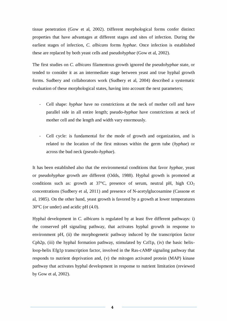
Hyphae and pseudo-hyphae formation is the ultimate phenotype of polarized growth. In
the yeast form, polarized growth is defined by a crescent-shaped polarisome at the tip of
the growing bud (Fig. 1) (Whiteway et al, 2007). The processes of polarized growth in
C. albicans yeast form and pseudo-hyhae formation in S. cerevisiae are similar. It
occurs through formation of secretory vesicles that are transported to a specific site of
polarized growth, which is marked by cortical markers (Sudbery et al, 2011). These
activate Cdc42, a protein involved in the regulation of the cell cycle, resulting in the
formation of the polarisome, the exocyst and a septing ring. The septin ring organizes
the arrangement of the primary and the secondary septae between the mother and
daughter cells. The polarisome initiates the formation of actin filaments along which
post-Golgi secretory vesicles are delivered to the sites of polarized growth, and where
they dock with the exocyst before fusing with the plasma membrane (Fig. 1) (Sudbery
et al, 2011). In hyphae forms, the polarized growth is considerably different. There is a
structure rich in secretory vesicles formed at the apex, called Spitzenkörper or the tip
body (Sudbery et al, 2011).
Fig. 1. Cell cycle of yeast and the first cell cycle of hyphae and pseudohyphae. Extract from Sudbery et
al, 2004.
6
1.2 C. albicans biofilms
The ability to form biofilms it is intimately associated with the ability to cause infection
and is considered an important virulence determinant during candidosis. Candidosis are
frequently seeded from biofilms developed on medical devices (Nobile et al, 2009). The
most common Candida associated with biofilm formation is C. albicans (Ramage et al,
2005). C. albicans can colonize and develop a biofilm on almost any medical device
(Uppuluri et al, 2009), including prostheses, implants, several types of catheters, etc.
Furthermore, the formation of biofilms in medical devices besides serving as a source
for infections, can also cause the failure of the device (Nett et al, 2006).
1.2.1 Formation and features of C. albicans biofilms
Biofilm formation is a process in which microorganisms irreversibly attach and grow on
a surface producing and secreting polymers that facilitate the attachment and a matrix
formation. This corresponds to a change in the microorganism growth rate and gene
transcription profile (Donlan, 2001).
The progression to a mature biofilm depends of three factors: cell adhesion,
extracellular matrix production and transition yeast-to-hyphae (Nett et al, 2006). In
biofilm formation cell initially adhere to biomaterial surfaces (Li et al, 2003). The
characteristics of the substratum could affect the rate and extend of the attachment. In
general, biofilms develop more quickly in rougher and more hydrophobic materials
(Donlan, 2001). The attachment of Candida to biomaterials is mediated by nonspecific
factors like hydrophobicity and electrostatic forces, and by specific adhesins that
recognize ligands in the conditioning films, like serum or salivary proteins (Ramage et
al, 2005). A mature biofilm of Candida species is composed by a complex three-
dimensional structure and displays extensive spatial heterogeneity (Ramage et al, 2005).
The biofilm acts like a “filter” to entrap, among others, nutrients and water and facilitate
their influx as well as the disposal of waste products throughout the many layers of cells
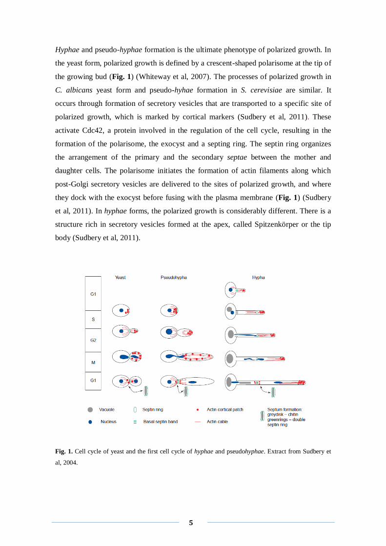
(Ramage et al, 2005). Observations through Scanning Electron Microscopy (SEM)
reveal the fully mature C. albicans biofilms as a dense network of yeasts, hyphae,
7
pseudohyphae, as well as extracellular polymeric material, the matrix (Ramage et al,
2005; Hawser et al, 1994).
Fig. 2. SEM image of mature C. albicans biofilm. Bar corresponds to 10 µm. Extracted from Ramage et
al, 2005.
1.2.2 Morphogenic conversion in C. albicans biofilm
It is widely recognized that morphogenesis plays a pivotal role in the development of C.
albicans biofilms (Ramage et al, 2005). Baillie and collaborators demonstrated that
hyphae are essential elements of the architecture and structural integrity of mature
biofilm (Baillie et al, 1999). In this study they compare the biofilms produced by two
wild-type (wt) strains with those formed by two mutants, incapable of yeast and hyphal
growth, respectively. An analysis using SEM revealed that the biofilms of the wt strains
consists in two different layers: a basal yeast layer and a thicker hyphal layer. On the
other hand, the hyphae-mutant produced only the basal layer and the yeast mutant
formed only a thicker layer. The biofilms of the yeast mutants were more easily
detached that the ones formed by wt strains, which suggested that the basal yeast layer
is crucial for the anchoring of the biofilm to the surface (Baillie et al, 1999). More
recently our group found that Cagup1Δ null mutant strain was not able to form typical
biofilm structures, presenting much less hyphae/pseudo-hyphae comparing to wt
8
(Ferreira et al, 2010). Still, dimorphism may not be an absolute prerequisite for biofilm
formation, since biofilms harboring only yeast cells have been described (Ramage et al,
2010).
Very little is still known about the regulation of biofilm formation, yet, some genetic
studies have been made. The regulator Efg1 protein was described as a key factor in the
formation and subsequent development of mature C. albicans biofilms in a variety of
biological and artificial surfaces (Ramage et al, 2002) and Zap1p, a zinc-responsive
regulatory protein as a negative regulator of biofilm maturation (Nobile et al, 2009).
C. albicans quorum sensing modulates all stages of biofilm life cycles: attachment,
maturation and dispersal (Donlan, 2002; Deveau et al, 2011). Farnesol, a C. albicans
autoregulatory molecule that regulates the transition between yeast and hyphae form, is
the best characterized quorum sensing molecule, and leads to a reduction on the biofilm
sizes (Hornby et al, 2001; Deveau et al, 2011).
1.2.3 Biofilms resistance to antifungals
The increased resistance of biofilm-associated infections to antimicrobial agents is a
serious problem for public health (Donlan, 2001). Presently, there are four groups of
drugs available to treat fungal infections: (i) the antifungal agents that inhibit
macromolecule synthesis (e.g. flucytosine), (ii) the ones that impair membrane barrier
function (nystatin and amphotericin), (iii) the inhibitors of ergosterol synthesis (azoles
derivates such as clotrimazole, ketoconazole or fluconazole) and, (iv) drugs that interact
with microtubules (e.g. Griseofulvin) (Bossche, 1997).
Hitherto, it looks like that no class of antifungal agent is immune to the development of
resistance. Therefore it becomes necessary to consider the development of a systematic
program of in vitro susceptibility testing, in order to take therapeutic decisions (Pfaller
et al, 2007). Several works showed Candida biofilms high levels of resistance to
antifungal agents and proposed explanations. It has been suggested, for instance that the
matrix of extracellular polymeric material would exclude or limit the access of the drug
(Al-Fattani et al, 2006). Still, in another study, Al-Fattani and collaborators (Al-Fattani
et al, 2006) found that the matrix did not constitute a barrier for 5 clinically used
antifungals of different chemical structure, suggesting that the matrix plays a minor role
9
in drug resistance. In their study, they compared biofilms incubated without agitation
(that have little matrix) with biofilms incubated with gentle shaking (produce more
matrix material) and these did not show significant differences in susceptibility to any
of the drugs tested (Al-Fattani et al, 2006). Other mechanism pointed to have a role on
Candida biofilms resistance was the nutrient limitation and growth impairment, since
mature biofilms are more resistant to antifungals (Ramage et al, 2005).
1.3 Extracellular matrix (ECM)
One of the most important features of biofilms is the extracellular matrix. ECM is a
three-dimensional highly hydrated gel-like environment where the microorganims are
mainly immobilized (Flemming et al, 2000).
The composition of the matrix varies according to the nature of the organisms present.
For instances, exopolysaccharides are the most abundant matrix polymers of bacterial
biofilms. These can be attached to carboxyl, sulphate or phosphate groups becoming
negatively charged. Nucleic acids, lipids, uronic acid and proteins can also be present
but in smaller amounts (Al-Fattani et al, 2006). The ECM of mammalian cells is mainly
constituted by a large variety of fibrous proteins (such as glycoproteins and
proteoglycans) and glycosaminoglycans, although it varies from tissue to tissue
(Mecham, 2011). In terms of functions, mammalian ECM is much more sophisticated
and diverse than the ECM of yeasts, being for instance essential on the repair of the
tissue (Mecham, 2011). In C. albicans ECM were identified structures called “towers”,
which are microcolonies of the matrix, separated by water channels that allow an
efficient nutrient circulation within the biofilm (Donlan et al, 2002).

10
Fig. 3. C. albicans forms dense biofilms composed of yeast cells, hyphae, and extracellular matrix.
Images produced using SEM. (ref:http://hsc.unm.edu/som/medicine/id/msclinic.shtml).
More recently, it was proposed that extracellular DNA is also a component of C.
albicans ECM and that it may play a crucial role in biofilms structural role, based on the
observation that the addition of exogenous DNA increases the biofilm mass and
conversely, when treated with DNase the biofilm mass decreases (Martins et al, 2010).
Some extracellular polysaccharides and proteins have been reported to be essential
components of the matrix (Sutherland, 2001). Also dead cells have been observed in
some biofilms, which suggests that cell debris can accumulate in the ECM (Webb et al,
2003). Possibly the extracellular DNA derives from these dead cells. Furthermore, it
was recently described that the matrix has a function of cellular protection from
surrounding environment, not only by preventing the uptake of drugs, by being a shield
for other kinds of stress, but also by acting as a maintainer of the architectural stability
of the biofilm (Nobile et al, 2009). Matrix production is described in close link to
biofilm formation (Nobile et al, 2009). It is known that the ECM abundance in C.
albicans can be regulated positively or negatively by glucoamylases and alcohol
dehydrogenases, for instance Gca1p, Gca2p and Adh5p which act as positive regulators,
while Cshp and Ifd6p act as negative regulators (Nobile et al, 2009). In the study
mentioned above, Al-Fattani and collaborators (Al-Fattani et al, 2006) showed that the
synthesis of ECM material is dramatically increased during the biofilm development,
which is subjected to a liquid flow and shaking, in opposition to minimal ECM
synthesis under static incubation conditions. Despite this understanding, still very little
is known about its regulation or production mechanisms (Nobile et al, 2009).
11
2. GUP1
GUP1 gene was firstly described in S. cerevisiae encoding a multi-membrane-spanning
protein essential for glycerol active uptake by proton symport (Holst et al, 2000). This
was based on the fact that the deletion of GUP1 resulted (i) on a slow grow on glycerol
as sole carbon and energy source, (ii) on a diminished ability to use extracellular
glycerol for rescuing form osmotic stress and (iii) on a defect on active uptake of
glycerol and metabolism (Holst et al, 2000).
Soon after, Gup1p was included on the MBOAT (membrane-bound O-acetyl
transferases) family (Hoffman, 2000; Neves et al, 2004), and involved in several
processes connected with cell structure organization and biogenesis. It was described as
having a role in the bipolar site selection, and several proteins sorting (Ny et al, 2001;
bonangelino et al, 2002), cytoskeleton polarization (Ny et al, 2001), vacuole
morphology (Bonangelino et al, 2002) telomere length maintenance (Askree et al, 2004)
and plasma membrane composition evidenced by a reduction in phospholipids and an
increase in triacylglycerols (Oelkers et al, 2000).
This pleiotropism suggested the role of this protein in cellular processes to be of a
complex nature. Further ahead, GUP1 was associated to cell wall morphology,
synthesis and integrity (Ferreira et al, 2006). This is reinforced by the described GUP1
genetic interactions with genes associated with chitin and β1,6-glucan synthesis,
including SKT5 (activator of a chitin synthase involved in cytokinesis), RSV161 and
RSV167 (both involved in actin polarizaton) (Ferreira et al, 2006). GUP1 gene interferes
in lipid metabolism leading to deep alterations on sphingolipid-sterol ordered domains
integrity/assembly as well as an abnormal sterol distribution at the level of plasma
membrane (Ferreira et al, 2008).
Ergosterol is an important constituent of membranes being target of common
antifungals like azoles and polyenes (Pasrija et al, 2005). Several important roles have
been attributed to sterol/sphingolipid-rich domains in dynamics processes such as
protein sorting during exo- and endocytoses, cell polarity and signaling (Pasrija et al,
2005). Ferreira and Lucas (Ferreira et al, 2008) showed Scgup1Δ mutant moderate
sensitivity to sphingolipids biosynthesis inhibitors (SBIs), but a high resistance to
ergosterol biosynthesis inhibitors (EBIs), including azoles. Following, Gup1p was
12
directly implicated in GPI anchors remodeling (Bosson et al, 2006). The authors
showed that in ∆gup1 mutants the incorporation of ceramide in the anchors was
disturbed, consequently the transport of the GPI protein Gas1p from endoplasmatic
reticulum to Golgi was slower and part of it was lost into the medium (Bosson et al,
2006).
All these observations clearly associating ScGup1p with lipids metabolism, with
consequences on the resistance to antifungals (Ferreira et al, 2008), as well as with cell
wall constitution, morphology and assembly (Ferreira et al, 2006), stress a putative
importance for the biology of C. albicans, in particular, the switch from commensal to
pathogenic nature and the subsequent increased resistance to antifungal drugs.
In the last years, our group started to study GUP1 gene in C. albicans. We have
showed that GUP1 gene clearly has a role in C. albicans virulence (Ferreira et al, 2010).
The deletion of GUP1 in C. albicans changes ergosterol plasma membrane
constitution/distribution, presenting an increased resistance to azoles. More importantly,
CaGup1p strongly interferes with the capacity of cells to develop hyphae, to adhere, to
invade and to form biofilms, all of which considered significant virulence factors.
Moreover, when treated with macrophages, the mutant Ca∆gup1 was much easily
phagocytosed than wt strain and its survival was clearly impaired (Ferreira et al, 2010).
2.1 GUP1 homologues
Recently GUP1 gene attracted a great deal of attention because its mouse homologue
was described as a negative regulator of the N-palmilotylation of Sonic hedgehog
pathway from higher Eukaryotes (Abe et al, 2008), which controls morphogenesis,
differentiation and patterning during embryogenesis, including proliferation and cell
fate. This highly stresses the roles of Gup1 in cellular morphology and division control
and comes into accordance with the results in yeast suggesting the existence of a
putative Hh-like pathway in lower Eukaryotes.
13
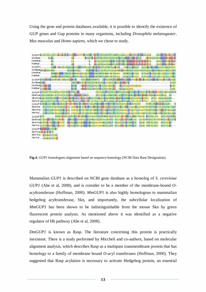
Using the gene and protein databases available, it is possible to identify the existence of
GUP genes and Gup proteins in many organisms, including Drosophila melanogaster,
Mus musculus and Homo sapiens, which we chose to study.
Fig.4. GUP1 homologues alignment based on sequence homology (NCBI Data Base Designation).
Mammalian GUP1 is described on NCBI gene database as a homolog of S. cerevisiae
GUP1 (Abe et al, 2008), and is consider to be a member of the membrane-bound O-
acyltransferase (Hoffman, 2000). MmGUP1 is also highly homologous to mammalian
hedgehog acyltransferase, Skn, and importantly, the subcellular localization of
MmGUP1 has been shown to be indistinguishable from the mouse Skn by green
fluorescent protein analysis. As mentioned above it was identified as a negative
regulator of Hh pathway (Abe et al, 2008).
DmGUP1 is known as Rasp. The literature concerning this protein is practically
inexistent. There is a study performed by Miccheli and co-authors, based on molecular
alignment analysis, which describes Rasp as a multipass transmembrane protein that has
homology to a family of membrane bound O-acyl transferases (Hoffman, 2000). They
suggested that Rasp acylation is necessary to activate Hedgehog protein, an essential
14
component of Hedgehog pathway (Miccheli et al, 2002). An in silico analysis, revealed
that the Rasp ORF (CG11495) used on the above mentioned work (Hoffman, 2000)
presents a high homology with ScGUP1 gene (this study, Fig. 4).
As for DmGUP1, there is very little information on Human GUP1. In fact, no studies
with this gene have been made or published in the literature. Human GUP1 genomic
sequence, available at NCBI database presents a high homology with MmGUP1 (this
study, Fig. 4).
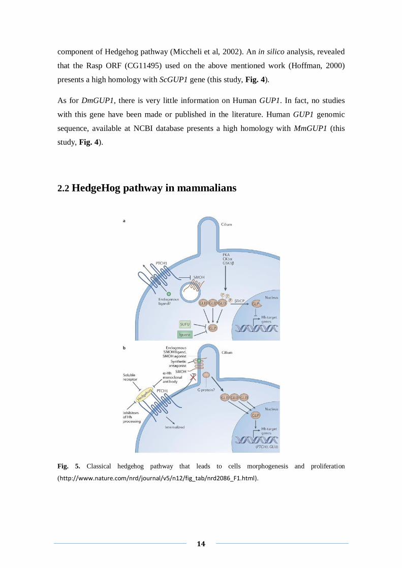
2.2 HedgeHog pathway in mammalians
Fig. 5. Classical hedgehog pathway that leads to cells morphogenesis and proliferation
(http://www.nature.com/nrd/journal/v5/n12/fig_tab/nrd2086_F1.html).
15
The glycoprotein Sonic Hedgehog (SHH) is essential for the development of several
tissues during embryogenesis (Britto et al, 2000) and is also involved in organogenesis
(Abe et al, 2008). SHH has additional functions in cell survival and cell proliferation
(Ahlgren et al, 2002). Recent pieces of evidence suggest a major role for the activation
of Sonic Hedgehog (SHH) signaling pathway in tumor development (Mao et al, 2009a;
Mao et al, 2009b). Namely, SHH was involved in pathogenesis of neuroblastoma due to
its high expression level in this pathology cells lines. Additionally, SHH inhibition
induced apoptosis and stopped proliferation, abolishing the tumorgenecity of
neuroblastoma (Mao et al, 2009a).
Aims
17
Aims
Gup1 is a yeast O-acyltransferase with close homologues in higher Eukaryotes
(Hoffman, 2000; Neves et al, 2004) which has been implicated in the regulation of the
Hedgehog pathway in mouse (Abe et al, 2008).
In C. albicans the deletion of GUP1 causes drastic alterations both at morphologic and
physiologic levels (Ferreira et al, 2010), such as (i) changes in the constitution and
distribution of ergosterol in the plasma membrane, (ii) resistance to antifungals (azoles),
(iii) aberrant colony morphology, (iv) impairment on the capacity of cells to develop
hyphae, and (v) defects on the ability of cells to adhere, invade and form biofilms.
Furthermore, when a copy of CaGUP1 gene is re-introduced to ∆gup1 mutant these
phenotypes are reversed, indicating that the above phenotypes are directly connected to
Gup1p function or its absence (Ferreira et al, 2010).
Based on that, it was proposed as aim for the present work, to create and study several
strains of C. albicans ∆gup1 mutant harboring available GUP1 homologues from well
known model organisms/higher Eukaryotes:
- Yeast - Saccharomyces cerevisiae (ScGUP1)
- Fly - Drosophila melanogaster (DmGUP1)
- Mouse - Mus musculus (MmGUP1)
- Man - Homo sapiens (HsGUP1)
These newly transformed C. albicans strains were tested for several virulence factors,
such as: (i) morphology and differentiation; (ii) adherence and invasion capacities; and
(iii) resistance to antifungals agents.
Another important purpose of this study was to obtain a first characterization of the
proteins secreted onto the C. albicans extracellular matrix, for which it was chosen to
mimic biofilm formation on plate and compare the wt, ∆gup1 mutant and transformants
for that matter.
Material and Methods
19
1. Strains & growth conditions
In this study C. albicans strains used were: wild type (wt) BWP17
(ura3Δ::λimm434/ura3Δ::λimm434his1::hisG/his1::hisGarg4 ::hisG/arg4::hisG)
(Wilson et al, 1999), several clones of homozygous C. albicans Δgup1/ Δgup1 (isogenic
to BWP17 but gup1::URA3-dpl200/gup1::ARG4) named as Δgup1 mutant (Kayingo et
al, 2009) and Δgup1 mutant complemented with GUP1 homologues from S. cerevisiae
(ScGUP1), D. melanogaster (DmGUP1), Mus musculus (MmGUP1) and H. sapiens
(HsGUP1), generally referred below as CaΔgup1GUP1 homologues.
Yeast strains were batch-grown on rich medium (YPD - yeast extract (1% w/v), peptone
(2% w/v), glucose (2%, w/v)), minimal medium (YNBD – yeast nitrogen base (0.67%
w/v), glucose (2% w/v)) or Spider´s medium (Nutrient broth (1% w/v), manitol (1%
w/v), K2HPO4 (0.2% w/v)), at 30ºC (YPD; YNBD) or 37ºC (YPD; Spider), with orbital
shaking, at 200 rpm. Solid cultures media were supplemented with 2% agar (YPD,
YNBD) or 1.5% agar (Spider) and identically incubated at 30 and 37°C. Transformants
were grown at 30ºC on YNBD supplemented with 200 µg/mL geneticin (G418), until
colonies were observed. Strains were kept at 4ºC on solid media.
Escherichia coli XL1Blue strain (endA1 gyrA96 (nalR) thi-1 recA1 relA1 lac glnV44
F'[::Tn10 proAB+ lacIq Δ(lacZ)M15] hsdR17(rK- mK+) was used for DNA
propagation. Bacterial cells were grown in Luria-Bertani (LB) medium (yeast extract
(0.5%, w/v); tryptone (1%, w/v); NaCl (1%, w/v)) at 37ºC in an orbital shaker. Strains
were maintained in LB medium supplemented with 2% agar, previously grown
overnight at 37ºC and kept at 4ºC. Bacterial transformants were grown overnight on LB
medium with ampicilin (100 mg/ml) at 37ºC, until colonies were observed.
20
2. DNA manipulation
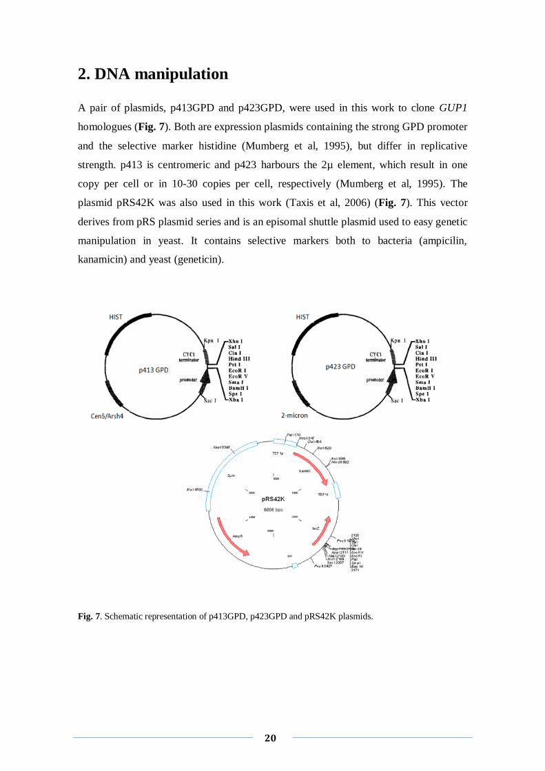
A pair of plasmids, p413GPD and p423GPD, were used in this work to clone GUP1
homologues (Fig. 7). Both are expression plasmids containing the strong GPD promoter
and the selective marker histidine (Mumberg et al, 1995), but differ in replicative
strength. p413 is centromeric and p423 harbours the 2µ element, which result in one
copy per cell or in 10-30 copies per cell, respectively (Mumberg et al, 1995). The
plasmid pRS42K was also used in this work (Taxis et al, 2006) (Fig. 7). This vector
derives from pRS plasmid series and is an episomal shuttle plasmid used to easy genetic
manipulation in yeast. It contains selective markers both to bacteria (ampicilin,
kanamicin) and yeast (geneticin).
Fig. 7. Schematic representation of p413GPD, p423GPD and pRS42K plasmids.

21
2.1 Transformation of C. albicans∆gup1 mutant strain
The referred vectors, p413GPD, p423GPD and pRS42K were used to clone the several
GUP1 homologues, ScGUP1, DmGUP1, MmGUP1 and HsGUP1 in C. albicans. These
constructions were previously propagated in E. coli XL1Blue strain.
2.1.1 Transformation in E. coli XL1Blue
E. coli XL1Blue transformation was made following standard protocols and using
prepared competent E. coli XL1Blue cells (Ausubel et al, 1996). The transformation
selection was performed based on the autotrophic mark ampicillin present in both
p4X3GPD and pRS42K plasmids. The procedure is next described:
E. coli XL1Blue competent cells preparation was made using the Simple and Efficient
Method (Inoue et al, 1990). The method basically consists on inducing membrane
alterations that make cells more permeable to macromolecules, in the present case DNA
on plasmids, which crosses the membrane more easily.
To transform E. coli XL1Blue, a volume of 5 µl plasmid DNA (≈50 µg/µl) was added to
200 µl of competent cells. These were maintained on ice for 40 minutes, subjected to a
heat shock by incubation at 42ºC for 2 minutes and then incubated on ice for 2 minutes.
In order to allow cells to recover, 500 µl of LB medium was added and the cell
suspension further incubated at 37ºC for 1 hour. A volume of 100 µl of this suspension
was then platted on solid LB medium supplemented with 100 µg/ml of ampicillin. Cells
were allowed to grow overnight at 37ºC.
2.1.2 Plasmid isolation from E. coli XL1Blue cells
In order to isolate plasmidic DNA in large scale, several colonies of E. coli XL1Blue
transformed with GUP1 homologues were inoculated in 5ml of LB-amp liquid medium
and grown overnight at 37ºC. The plasmid extraction was performed with GeneEluteTM
Plasmid Miniprep Kit [Sigma], following the protocol of the manufacturer.
22
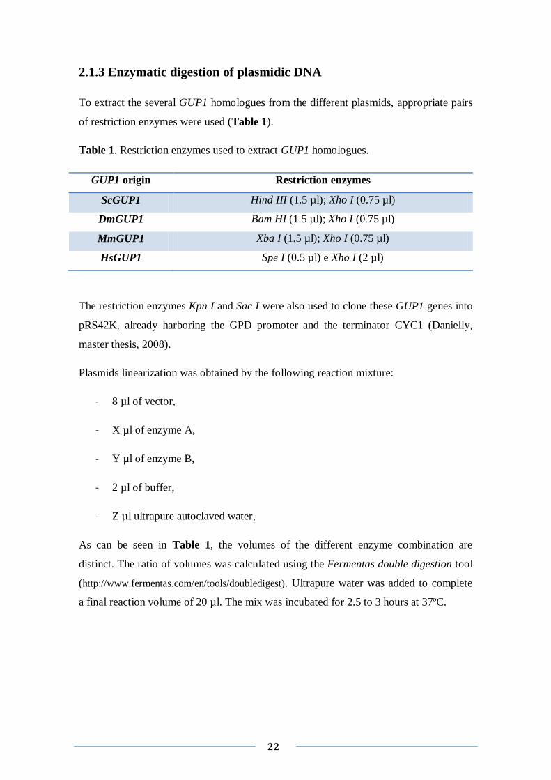
2.1.3 Enzymatic digestion of plasmidic DNA
To extract the several GUP1 homologues from the different plasmids, appropriate pairs
of restriction enzymes were used (Table 1).
Table 1. Restriction enzymes used to extract GUP1 homologues.
GUP1 origin Restriction enzymes
ScGUP1 Hind III (1.5 µl); Xho I (0.75 µl)
DmGUP1 Bam HI (1.5 µl); Xho I (0.75 µl)
MmGUP1 Xba I (1.5 µl); Xho I (0.75 µl)
HsGUP1 Spe I (0.5 µl) e Xho I (2 µl)
The restriction enzymes Kpn I and Sac I were also used to clone these GUP1 genes into
pRS42K, already harboring the GPD promoter and the terminator CYC1 (Danielly,
master thesis, 2008).
Plasmids linearization was obtained by the following reaction mixture:
- 8 µl of vector,
- X µl of enzyme A,
- Y µl of enzyme B,
- 2 µl of buffer,
- Z µl ultrapure autoclaved water,
As can be seen in Table 1, the volumes of the different enzyme combination are
distinct. The ratio of volumes was calculated using the Fermentas double digestion tool
(http://www.fermentas.com/en/tools/doubledigest). Ultrapure water was added to complete
a final reaction volume of 20 µl. The mix was incubated for 2.5 to 3 hours at 37ºC.
23
2.1.4 Extraction of DNA fragments of agarose gel electrophoresis
In order to separate, identify and purify DNA fragments, 1% agarose gel electrophoresis
was performed. The DNA was visualized by UV illumination (245nm) at Eagle Eye II
(Stratagene), previously stained with Gel RED (Biotium) dye. The molecular weight
marker was Lambda DNA/Eco471 [Fermentas]. The electrophoresis ran for
approximately 1 hour at 70 volts. The DNA fragments of interest were extracted with
QIAquick Gel Extraction Kit (QUIAGEN) system following the manufacture
recommendations.
2.1.5 Ligation reaction
Prior to ligation reaction the plasmid was treated with the enzyme Shrimp Alkaline
Phosphatase (SAP) in order to remove phosphate 5’ ends of the vectors. This
elimination prevents the auto-ligation of the vector and reduces the frequency of
plasmid circularization during the ligation reaction (Sambrook et al, 1989). For that, the
vector was incubated with SAP enzyme for 10 minutes at 37ºC. The mix was further
incubated 15 minutes at 65ºC in order to a complete inactivation of the enzyme.
The ligation reaction was performed using the following conditions:
- 1 µl ligation buffer 10x,
- 1 µl plasmid,
- 7 µl of purified digestion product,
- 1 µl of T4 DNA ligase,
- 10 µl ultrapure water to achieve final volume of 20 µl,
The reaction mixture was incubated overnight at 4ºC. For DNA propagation, the
ligation product was used to transform E. coli XL1Blue competent cells, as described
above.
24
2.1.6 Yeast transformation
C. albicans ∆gup1 mutant strain was transformed following a protocol adapted from
Gietz R.D. and collaborators, 1995 and Walther A. and collaborators, 2003. The
procedure is next described:
Grow of ∆gup1 mutant strain in ≈5ml of YPD, overnight at 30ºC until
stationary phase,
Ressuspend the cells on fresh YPD to OD600= 0.3,
Incubate for 4-5h at 30ºC,
Harvest the cells by centrifugation 5 min at 5000 rpm. Wash the pellet
once with water. Carefully remove the remaining water,
Ressuspend the pellet in 1.5ml of LiAc/TE solution* (0.5 ml 10xTE; 0.5
ml 10x LiAc; 4 ml water),
Set up transformation tubes as follows:
. 1-5 µl DNA
. 10 µl ssDNA (DNA carrier)
. 80 µl cells
. 600 µl PEG4000/LiAc solution* (2.5g PEG4000, 0.5 ml
10x LiAc, 4.5 ml water)
Vortex briefly, and incubate overnight at 30ºC,
Heat shock for 15 minutes at 44ºC,
Spin down cells at 13000 rpm for 15 seconds and pour off PEG;
Spin down again and tip off remaining PEG,
Ressuspend the pellet in 100-200 µl water and plate the suspension on
selective plates (YNB w/o aminoacids supplemented with geneticin (200
µg/ml));
25
Incubate for 24-48h at 30ºC.;
*Both solutions were sterilized by filtration.
3. Virulence assays
3.1 Morphology and hyphal formation
In order to observe different colony morphology/differentiation, the cultures were
diluted and inoculated in both liquid and solid medium, under hyphal non-inducing
conditions (YPD at 30ºC) and under hyphal-inducing conditions (Spider medium, YPD
at 37º and also YPD supplemented with 10% Fetal Bovine Serum (FBS) at 37º C).
Solid media: Cultures were allowed to grow for 2-3 weeks. During this period the
colonies were several times observed at light microscopy to inspect hyphal
development.
Liquid media: To discriminate between hyphae and pseudo-hyphae, young cultures
were diluted to 1×10⁷
cells/ml (OD600=1), and incubated in the presence of FBS 10%
(Fetal Bovine Serum) at 37ºC. At several time points a sample was collected and cells
were stained with CFW (Calcofluor White) by adding to 3 ml of cell suspension 300 μl
of CFW (300 μg/ml). Samples were incubated at room temperature for 5 min, after
which 5 μl of the suspension was placed on microscopic slide, visualized by light
microscopy (LM) and photographed. CFW dye binds primarily to chitin, but also in less
extent to glucans, staining cell wall and septa of the cells (Ferreira et al, 2010).
Microscopy assessments were done in a Leica Microsystems DM-5000B
epifluorescence microscope, with appropriate filter settings. Images were acquired by a
Leica DCF350FX digital camera and processed with LAS AF Leica Microsystems
software.
3.2 Adherence to agar and invasion capacities
Equal volumes of young cultures of each strain were diluted to 1 × 10⁷ cells/ml
(OD600=1), and 1 ml of cells suspension was spotted into YPD and Spider medium agar

26
plates. Cultures were allowed to grow at 37°C for 10 days. The cells on the surface were
then removed by washing under running water (Guo et al, 2000; Hube et al, 2001) and
visualized by LM.
3.3 Resistance to antifungal drugs
To attest Ca∆gup1GUP1 homologues sensibility/resistance to antifungal drugs, YPD-
grown cultures were used for drop tests, containing approximately 1×10⁷ cells/ml
(OD600=1) as described before (Ferreira et al, 2008; Ferreira et al, 2010). Six-fold serial
dilutions were made, and 5 μl of each suspension was applied on selective media.
Results were scored after 3-5 days of incubation at 30°C. YPD medium was
complemented as follow: clotrimazole (68.8 μg/ml), ketoconazole (86.1 μg/ml),
fluconazole (30.6 μg/ml). All chemicals were obtained at the highest available grade
from Sigma Aldrich (Ferreira et al, 2010).
4. Proteins secreted to extracellular matrix (ECM)
The extraction of proteins secreted to the extracellular matrix (ECM) was performed
using an optimized protocol develop by our group (Joana Carvalho, unpublished
results):
In order to obtain a biofilm-like structure and ensure relevant quantities of secretome, 1
ml of cells (OD600= 1) were spread on different mediums (YPD, Spider and YNB
without aminoacids). The overlays were allowed to grow 7 days at 30°C and 37°C. The
biomass of at least 5 plates (90mm) of each strain was scraped and immersed in PBS
buffer containing a cocktail of proteases inhibitors (PMSF 0.2 µg/ml; Aprotinin 0.32
µg/ml; Pepstatin 1µg/ml; Leupeptin 1µg/ml) in which cells were washed for 10 minutes
on a roller under constant rotation, at 4°C. The suspension was then spun down for 5
minutes at 5000 rpm. The supernatant was collected and stored at 4ºC for further protein
analysis.
To obtain a maximum protein recovery, proteins were precipitated overnight with a mix
of TCA (Trichloroacetic acid) and DTT (Dithiothreitol) at 4°C. DTT has a protein
protector effect that increases the yield of proteins recovery. A crucial step to perform
27
efficient protein extraction is the protein solubilization. Two alternative buffers were
tested, urea and a modification of Laemmli buffer consisting on the withdraw of the
bromophenol blue and the addition of urea (6M of final concentration), to previous
Laemmli buffer (Joana C., unpublished results). This last one was chosen according to
ressuspension efficiency determined by electrophoretic profile, as well as because it is
compatible with the utilization of Bradford protein quantification method. To enhance
the performance of the protein precipitation, two further steps of vortex and sonication
were performed.
SDS-PAGE was made in 9% of polyacrylamide in order to allow the use of a broad
range of molecular weight marker. Electrophoretic run was performed during 1 hour at
140 volts. The gel was stained with AgNO3. A formaldehyde fixation step, which
enhances the staining sensitivity but precludes any further use of the stained bands, was
introduced to the staining process.

Results
29
1.Strains harboring GUP1 homologues
The cloning of several of the GUP1 homologues (ScGUP1; DmGUP1; MmGUP1 and
HsGUP1) was initially performed into a pair of p4X3 series plasmids, with different
expression strength (Mumberg et al, 1995). These plasmids have different origins of
replication, a centromeric (p413) and a 2µ (p423) that vary in the number of copies
produced (1 and 10-30 copies per cell respectively) (Mumberg et al, 1995). Yet, both
have a strong promoter (GPD) and also share the same terminator (CYC1).
In general, the strategy applied to clone the GUP1 homologues consisted in extracting
the gene from the original plasmid (all GUP1 homologues were obtained already cloned
in several plasmids) and cloned them into p4X3GPD plasmids. A restriction map was
performed for the genomic DNA coding sequence of each GUP1 homologue in order to
select the appropriate restriction enzymes to be used on the cloning steps, considering
both the plasmidic Multi-Cloning-Site (MCS) and the restriction analysis. Each
construction, p4X3GPD-GUP1 homologues, was then transformed in E. coli XL1Blue
in order to obtain a larger stock of DNA. The E. coli XL1Blue transformants were
selected in LB medium supplemented with 100 µg/ml of ampicilin. The positive clones,
those who carry the GUP1 gene fragment, were verified through electrophoresis of the
enzymatic digestion confirming the expected bands size. These positive clones were
then frozen and stored at -80°C in glycerol stocks.
To otain the ScGUP1-GFP chimera a previous pYES2-GUP1-GFP construction
available at the collaborators Franscesco Grieco and Gianluca Bleeve (Istituto di
Scienze Delle Produzioni Alimentari del Consiglio Nazionale Delle Ricerche (ISPA),
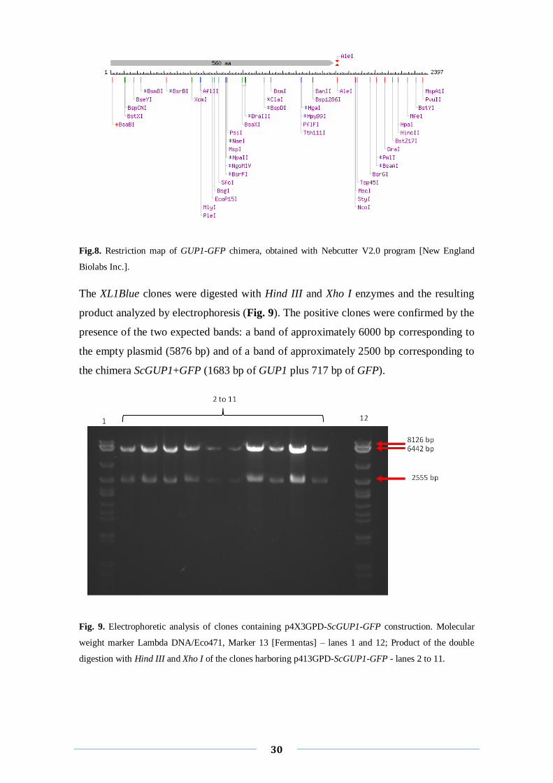
Lecce, Italy) was used. The in silico restriction map was studied, covering the chimera
GUP1-GFP and not only ScGUP1 gene (Fig. 8). Hind III and Xho I enzymes were
chosen to extract the fragment form pYES2 and to clone it in p4X3GPD, once these
were included simultaneously into the MCS (Multiple Cloning Site) of both plasmids
and do not cut the chimera ScGUP1-GFP (Fig. 8).
30
Fig.8. Restriction map of GUP1-GFP chimera, obtained with Nebcutter V2.0 program [New England
Biolabs Inc.].
The XL1Blue clones were digested with Hind III and Xho I enzymes and the resulting
product analyzed by electrophoresis (Fig. 9). The positive clones were confirmed by the
presence of the two expected bands: a band of approximately 6000 bp corresponding to
the empty plasmid (5876 bp) and of a band of approximately 2500 bp corresponding to
the chimera ScGUP1+GFP (1683 bp of GUP1 plus 717 bp of GFP).
Fig. 9. Electrophoretic analysis of clones containing p4X3GPD-ScGUP1-GFP construction. Molecular
weight marker Lambda DNA/Eco471, Marker 13 [Fermentas] – lanes 1 and 12; Product of the double
digestion with Hind III and Xho I of the clones harboring p413GPD-ScGUP1-GFP - lanes 2 to 11.
31
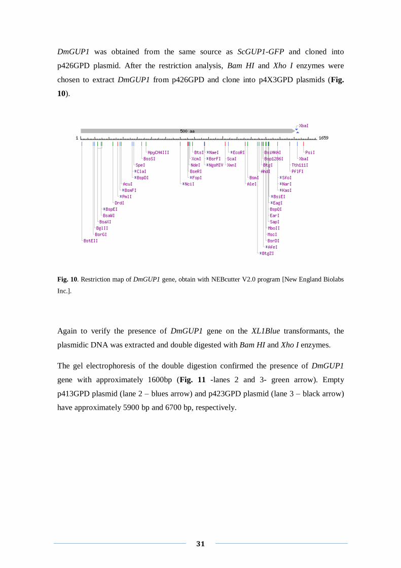
DmGUP1 was obtained from the same source as ScGUP1-GFP and cloned into
p426GPD plasmid. After the restriction analysis, Bam HI and Xho I enzymes were
chosen to extract DmGUP1 from p426GPD and clone into p4X3GPD plasmids (Fig.
10).
Fig. 10. Restriction map of DmGUP1 gene, obtain with NEBcutter V2.0 program [New England Biolabs
Inc.].
Again to verify the presence of DmGUP1 gene on the XL1Blue transformants, the
plasmidic DNA was extracted and double digested with Bam HI and Xho I enzymes.
The gel electrophoresis of the double digestion confirmed the presence of DmGUP1
gene with approximately 1600bp (Fig. 11 -lanes 2 and 3- green arrow). Empty
p413GPD plasmid (lane 2 – blues arrow) and p423GPD plasmid (lane 3 – black arrow)
have approximately 5900 bp and 6700 bp, respectively.
32
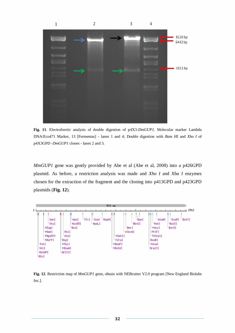
Fig. 11. Electroforetic analysis of double digestion of p4X3-DmGUP1. Molecular marker Lambda
DNA/Eco471 Marker, 13 [Fermentas] - lanes 1 and 4; Double digestion with Bam HI and Xho I of
p4X3GPD -DmGUP1 clones - lanes 2 and 3.
MmGUP1 gene was gently provided by Abe et al (Abe et al, 2008) into a p426GPD
plasmid. As before, a restriction analysis was made and Xho I and Xba I enzymes
chosen for the extraction of the fragment and the cloning into p413GPD and p423GPD
plasmids (Fig. 12).
Fig. 12. Restriction map of MmGUP1 gene, obtain with NEBcutter V2.0 program [New England Biolabs
Inc.].

33
Three XL1Blue transformants were double digested with the same enzymes and
analyzed by electrophoresis (Fig. 13). Two clones displayed two bands, one of
MmGUP1 gene (≈1500bp) and the other corresponding to the empty p413GPD
(≈5900bp) (Fig. 13. lanes 3 and 4). On the other hand, in the clone of lane 2 only the
presence of empty plasmid was visualized, meaning that the MmGUP1 gene was not
incorporated into this clone. Therefore, only clones 3 and 4 were considered positive.
Fig. 13. Electrophoretic analysis of the double digestion of p413-MmGUP1. Molecular Marker - lane 1;
Double digestion with Xba I and Xho I of p413-MmGUP1 clones- lanes 2, 3 and 4;
A turning point on this work arose when we tried to clone ∆gup1 mutant strain with the
p4X3GPD constructions harboring the GUP1 homologues (from ScGUP1; DmGUP1;
MmGUP1). Despite of the several attempts and protocols used, CaΔgup1GUP1
homologues clones were never achieved. We hypothesized that this difficulty could be
related with the aminoacid selection of the p4X3GPD plasmids. Apparently the marker
gene, histidine, was not stringent enough as selection force, preventing us to obtain
positive clones. These complications have been described by other researchers
(Danielly, master thesis, 20008; Pacheco, PhD thesis, 2008).
We decided to clone all GUP1 homologues into a new plasmid, the pRS42K, an
integrative epissomal plasmid, whose selective force arises as more reliable since is an
antibiotic (Taxis et al, 2006). However, this vector is not an expression plasmid, since it
lacks the promoter and terminator (Taxis et al, 2006). The strategy thus to extract from
the p4X3GPD plasmids constructions previously build, the fragment containing the
GPD promoter, the GUP1 homologues and the CYC1 terminator and insert the whole
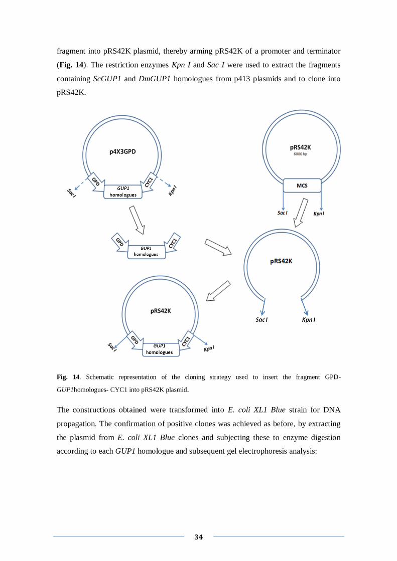
34
fragment into pRS42K plasmid, thereby arming pRS42K of a promoter and terminator
(Fig. 14). The restriction enzymes Kpn I and Sac I were used to extract the fragments
containing ScGUP1 and DmGUP1 homologues from p413 plasmids and to clone into
pRS42K.
Fig. 14. Schematic representation of the cloning strategy used to insert the fragment GPD-
GUP1homologues- CYC1 into pRS42K plasmid.
The constructions obtained were transformed into E. coli XL1 Blue strain for DNA
propagation. The confirmation of positive clones was achieved as before, by extracting
the plasmid from E. coli XL1 Blue clones and subjecting these to enzyme digestion
according to each GUP1 homologue and subsequent gel electrophoresis analysis:
35
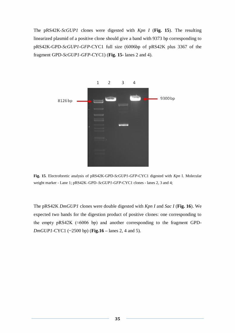
The pRS42K-ScGUP1 clones were digested with Kpn I (Fig. 15). The resulting
linearized plasmid of a positive clone should give a band with 9373 bp corresponding to
pRS42K-GPD-ScGUP1-GFP-CYC1 full size (6006bp of pRS42K plus 3367 of the
fragment GPD-ScGUP1-GFP-CYC1) (Fig. 15- lanes 2 and 4).
Fig. 15. Electroforetic analysis of pRS42K-GPD-ScGUP1-GFP-CYC1 digested with Kpn I. Molecular
weight marker - Lane 1; pRS42K–GPD–ScGUP1-GFP-CYC1 clones - lanes 2, 3 and 4;
The pRS42K DmGUP1 clones were double digested with Kpn I and Sac I (Fig. 16). We
expected two bands for the digestion product of positive clones: one corresponding to
the empty pRS42K (≈6006 bp) and another corresponding to the fragment GPD-
DmGUP1-CYC1 (~2500 bp) (Fig.16 – lanes 2, 4 and 5).
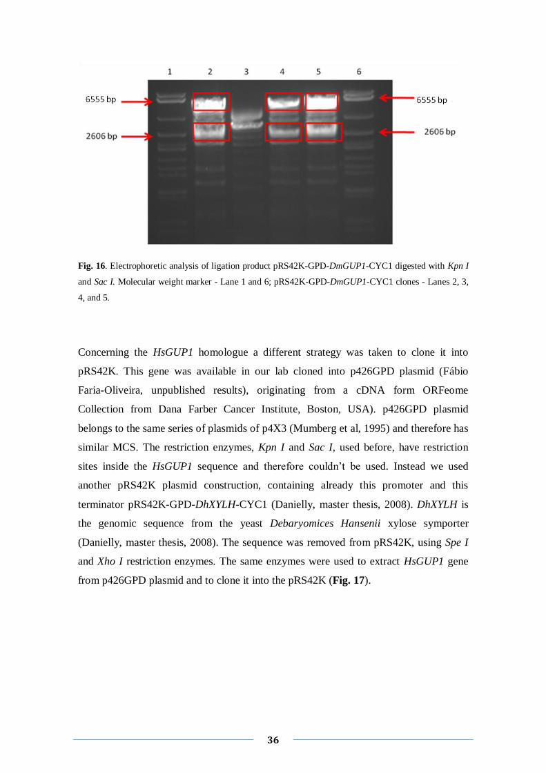
36
Fig. 16. Electrophoretic analysis of ligation product pRS42K-GPD-DmGUP1-CYC1 digested with Kpn I
and Sac I. Molecular weight marker - Lane 1 and 6; pRS42K-GPD-DmGUP1-CYC1 clones - Lanes 2, 3,
4, and 5.
Concerning the HsGUP1 homologue a different strategy was taken to clone it into
pRS42K. This gene was available in our lab cloned into p426GPD plasmid (Fábio
Faria-Oliveira, unpublished results), originating from a cDNA form ORFeome
Collection from Dana Farber Cancer Institute, Boston, USA). p426GPD plasmid
belongs to the same series of plasmids of p4X3 (Mumberg et al, 1995) and therefore has
similar MCS. The restriction enzymes, Kpn I and Sac I, used before, have restriction
sites inside the HsGUP1 sequence and therefore couldn’t be used. Instead we used
another pRS42K plasmid construction, containing already this promoter and this
terminator pRS42K-GPD-DhXYLH-CYC1 (Danielly, master thesis, 2008). DhXYLH is
the genomic sequence from the yeast Debaryomices Hansenii xylose symporter
(Danielly, master thesis, 2008). The sequence was removed from pRS42K, using Spe I
and Xho I restriction enzymes. The same enzymes were used to extract HsGUP1 gene
from p426GPD plasmid and to clone it into the pRS42K (Fig. 17).
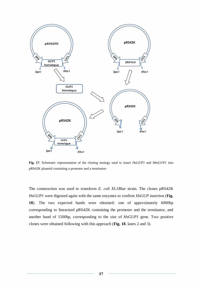
37
Fig. 17. Schematic representation of the cloning strategy used to insert HsGUP1 and MmGUP1 into
pRS42K plasmid containing a promoter and a terminator.
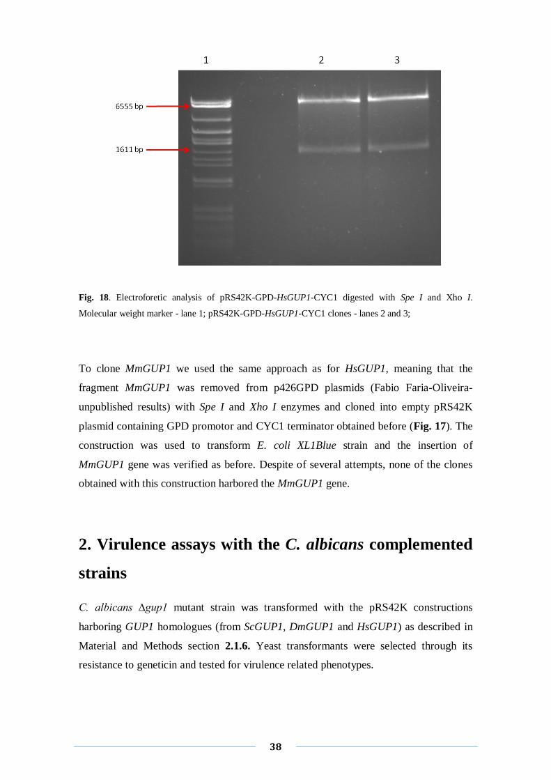
The construction was used to transform E. coli XL1Blue strain. The clones pRS42K
HsGUP1 were digested again with the same enzymes to confirm HsGUP insertion (Fig.
18). The two expected bands were obtained: one of approximately 6000bp
corresponding to linearized pRS42K containing the promoter and the terminator, and
another band of 1500bp, corresponding to the size of HsGUP1 gene. Two positive
clones were obtained following with this approach (Fig. 18. lanes 2 and 3).
38
Fig. 18. Electroforetic analysis of pRS42K-GPD-HsGUP1-CYC1 digested with Spe I and Xho I.
Molecular weight marker - lane 1; pRS42K-GPD-HsGUP1-CYC1 clones - lanes 2 and 3;
To clone MmGUP1 we used the same approach as for HsGUP1, meaning that the
fragment MmGUP1 was removed from p426GPD plasmids (Fabio Faria-Oliveira-
unpublished results) with Spe I and Xho I enzymes and cloned into empty pRS42K
plasmid containing GPD promotor and CYC1 terminator obtained before (Fig. 17). The
construction was used to transform E. coli XL1Blue strain and the insertion of
MmGUP1 gene was verified as before. Despite of several attempts, none of the clones
obtained with this construction harbored the MmGUP1 gene.
2. Virulence assays with the C. albicans complemented
strains
C. albicans ∆gup1 mutant strain was transformed with the pRS42K constructions
harboring GUP1 homologues (from ScGUP1, DmGUP1 and HsGUP1) as described in
Material and Methods section 2.1.6. Yeast transformants were selected through its
resistance to geneticin and tested for virulence related phenotypes.
39
2.1 Colony morphology and hyphal formation capacity
The colony morphology and capacity of cells to develop hyphae of the several
Ca∆gup1GUP1homologues were tested in both solid and liquid media.
Solid media: wt, ∆gup1 mutant and Ca∆gup1GUP1 homologues strains were grown for
2 weeks on YPD agar plates, at 30°C (non-hyphae-inducing conditions) and on YPD
and Spider medium at 37°C (hyphae-inducing conditions), before colony morphology
inspection. As described before, ∆gup1 mutant exhibit different colony morphology
when compare with wt (Ferreira et al, 2010). On the other hand, the colonies of
Ca∆gup1GUP1 homologues strains didn’t display the spaghetti/flower kind
morphology characteristic of ∆gup1 mutant and in that sense, were closer to the wt
colony morphology (Ferreira et al, 2010). Yet, neither of the Ca∆gup1GUP1
homologues presents hyphae differentiation and wt strain has that capacity (Ferreira et
al, 2010). Thus, in solid media the GUP1 homologues were not able to revert ∆gup1
mutant defect to develop hyphae, although all affected colony morphology (not shown).
Liquid media: Time-course of hyphae formation induced by FBS (fetal bovine serum)
in liquid medium was checked. Calcofluorwhite (CFW) binds mainly to chitin (Herth et
al, 1980), one of the cell wall compounds, allowing us to see the cell wall, the budding
scars and the septas (Hoch et al, 2005). Contrarily to ∆gup1 mutant, the
Ca∆gup1GUP1homologues harboring the different homologues ScGUP1, DmGUP1
and HsGUP1 were able to differentiate into pseudo-hyphae/hyphae cells, very soon
after induction (5 min.) as shown in Fig. 19. This, however, stabilizes/stops after 30
minutes of incubation with FBS (Fig. 19). Our statement is based on the the observation
that after the refered period is not possible to see alterations on the size of
hyphae/pseudo-hyphae cells. Furthermore, the stained wall indicate that the filamentous
cells developed by these strains are more likely to correspond to pseudo-hyphae rather
than true hyphae, since i) first septum is close to the mother neck, ii) present
constrictions at the septa junctions and iii) the side walls are not parallel (Fig. 19 - white
arrows). This is in clear contrast to wt cells, which form true hyphae, once the first
septum of these filamentous cells is distant from the mother neck and the other septa do
not present constrictions (Fig. 19 - red arrows). It is further noteworthy that
Ca∆gup1GUP1homologues cells were significantly smaller than wt.

40
Fig. 19. Time-course pseudo-hyphae/hyphae formation in wt, ∆gup1 mutant and
Ca∆gup1GUP1homologues, in liquid hyphae inducing medium (FBS). Cells were stained with CFW and
observed by LM with the adquated filters.
2.2 Adherence to agar and invasion capacities
In order to evaluate the Ca∆gup1GUP1homologues strains capacity to adhere and to
invade agar, cells were grown for ten days on both YPD and Spider agar plates. Ended
41
this period the plates were washed under running water. In a previous publication it was
described how the deletion of GUP1 interferes in the capacity of the strain to both
adhere and invade these surfaces (Ferreira et al, 2010). All the transformants revealed
the same lack of adherence capacity/level to YPD and Spider plates, since they were
washed out as easy as ∆gup1 mutant from the agar surface.
Replicas of these plates were further inspected for agar invasion as described in
Materials and Methods- section 3.2. In a previous work we showed that ∆gup1
mutant,was deficient on the ability to invade these surfaces (Ferreira et al, 2010). Here,
we observed that the complementation of the ∆gup1 mutant with the mentioned
homologues did not reverted this phenotype (not shown). In fact, none of the
Ca∆gup1GUP1homologue strains were able to invaded the agar, at all the conditions
tested.
2.3 Resistance to antifungals
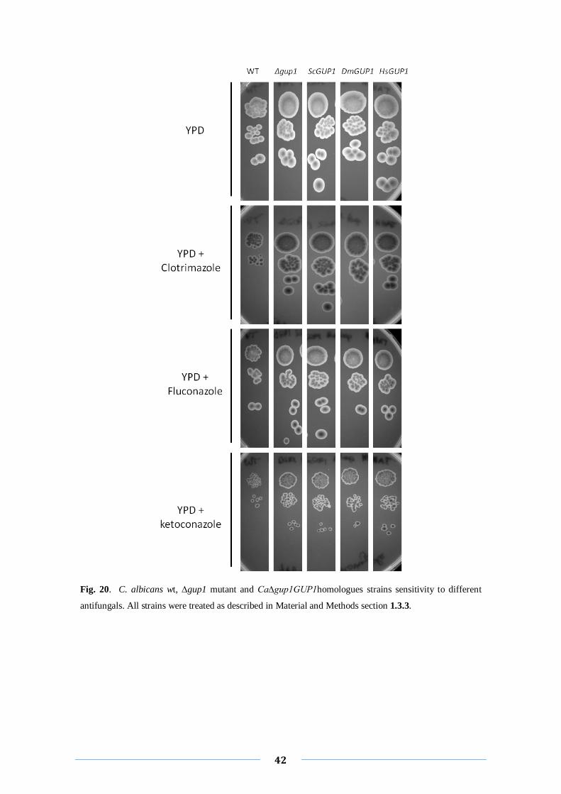
The O-acyltransferase Gup1p acts on lipids metabolism affecting the assembly and
integrity of the plasma membrane, as well as the sphingolipids-sterol order domains
(Ferreira et al, 2008). An association between altered lipid-ordered domains and
antifungal resistance has been described before as influencing the susceptibility to
antifungal drugs (Mukhopadhyay et al, 2004; Ferreira et al, 2008). Regarding
Ca∆gup1GUP1homologues strains, the two harboring the ScGUP1 and the HsGUP1
genes seem to be more resistant to these antifungals than the wt strain, showing a
similar behavior to ∆gup1 mutant strain (Fig. 20, 3th
and 5th
columns). However the
strain complemented with DmGUP1 gene was as sensitive to these drugs as wt strain
(Fig. 20. 4th colunm), differing from the other complemented strains.
42
Fig. 20. C. albicans wt, ∆gup1 mutant and Ca∆gup1GUP1homologues strains sensitivity to different
antifungals. All strains were treated as described in Material and Methods section 1.3.3.
43
3. Extracellular matrix profiles
The C. albicans ability to invade tissues and form biofilms is related to the formation of
an ECM yet uncharacterized in detail (Douglas, 2003). Similarly to what happens in
higher Eukaryotes, it is likely that the constituents of the matrix influence and relation
to the morphological switch or, more broadly, control the multicellular aggregates
morphology. Moreover, the GUP1 gene from mammals was implicated in the regulation
of the morphogenic Hedgehog pathway (Abe et al, 2008). This prompted us to attempt a
first approach to the C. albicans ECM using the present constructions. The analysis of
ECM proteins from C. albicans was performed using a novel methodology developed to
efficiently extract the yeast ECM (Joana Carvalho, unpublished results).
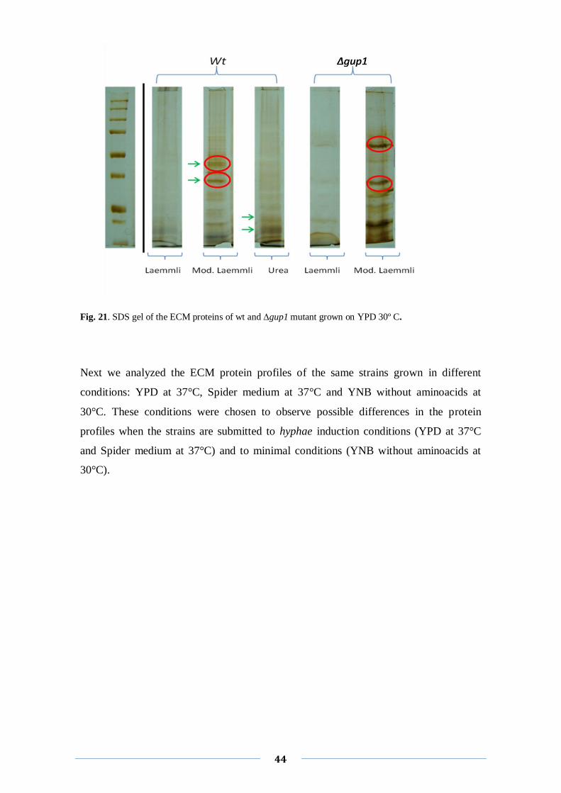
We started to compare the ECM proteins of wt and ∆gup1 mutant strains grown on ideal
conditions (YPD at 30°C). For that, 5 plates (with 1 ml of culture per plate) of each
strain were grown during 5 days, in order to obtain a mature biofilm-like structure. This
was collected, the protein and sugar fractions separated and the proteins precipitated as
described in Materials and Methods- section 4. The SDS revealed that different buffers
ressuspend different proteins, as is mirroned by the different protein profiles (Fig. 21-
green arrows) and modified Laemmli appears as the best ressuspension buffer since the
bands are better defined and the protein pattern is more easily visualized (Fig. 21- lane 3
and 6). Furthermore, regarding the comparison of the protein profile of ∆gup1 mutant
strain with wt strain, the differences in the protein pattern are notorious, independently
of the ressuspension buffer used. This suggests that ∆gup1 mutant excrete different
proteins to the extracellular matrix of those excreted by wt, but also excrete proteins in
distinct quantities than wt (Fig. 21. red circles). This is not unexpected since it was also
observed in S. cerevisiae liquid cultures (Ferreira et al, 2006).
44
Fig. 21. SDS gel of the ECM proteins of wt and ∆gup1 mutant grown on YPD 30º C.
Next we analyzed the ECM protein profiles of the same strains grown in different
conditions: YPD at 37°C, Spider medium at 37°C and YNB without aminoacids at
30°C. These conditions were chosen to observe possible differences in the protein
profiles when the strains are submitted to hyphae induction conditions (YPD at 37°C
and Spider medium at 37°C) and to minimal conditions (YNB without aminoacids at
30°C).
45
Fig. 22. SDS gel of the ECM proteins wt and ∆gup1 mutant grown on YPD at 30º C (column A); at 37º
C (column B) and on Spider medium at 37º C (column C); All proteins were ressuspended in modified
Laemmli buffer.
The ECM protein profile of ∆gup1 mutant was again very distinct from the one
displayed by wt (Fig. 22. red arrows). However, probably the most significant result
was that under induction hyphae conditions the protein profiles displayed a considerable
reduced quantity of proteins, independently of the strain used (Fig. 22. columns B and
C).

Discussion
47
I
S. cerevisiae Gup1p is an acyltransferase involved in lipids metabolism/rafts stability
and on the resistance to antifungals (Ferreira et al, 2008), as well as in the cell wall
constitution, morphology and assembly (Ferreira et al, 2006). These are important
features to be considered when regarding both C. albicans switch from commensal to
pathogen and its increased resistance to antifungal drugs. Moreover, in 2008, Abe and
collaborators (Abe et al, 2008) described that mammalian GUP1 as a negative regulator
of N-terminal palmitoylation of Sonic hedgehog of Hedgehog pathway, which among
other functions is involved in morphogenesis in mammalian cells (Abe et al, 2008). In a
previous work, our group has shown that C. albicans virulence and drug-resistance
requires Gup1p (Ferreira et al, 2010). Our assumptions were based on the following
observations. First, gup1 mutant strain was resistant to common antifungals. Second,
CaGUP1 deletion incited an aberrant evenly ergosterol distribution at the level of
plasma membrane. Third, the ability to switch from yeast-form to hyphae-growth
required CaGUP1. Fourth, a distinct growth orientation elicited by the deletion of
CaGUP1 leaded to colonies with remarkable distinct/aberrant morphology i.e. a flower,
spaghetti, irregular and wrinkled shape. Fifth, gup1 mutant strain adherence and
invasion abilities were strongly reduced. Sixth, biofilm formation, another important
indicator of C. albicans virulence, was strongly impaired by the deletion of CaGUP1.
Finally, the introduction of the GUP1 gene copy into the gup1 mutant strain was able
to revert all these phenotypes, symptomatic of the GUP1 gene accountability.
All taken, and considering the fact that MmGUP1 gene was able to complemented the
hypha morphogenetic defects on solid media of gup1 null mutant (Ferreira et al,
2010), we anticipated that Gup1p might be part of a yeast morphogenic pathway,
parallel to the mammalian Hedgehog. Several approaches are being developed by our
group to unveil and characterize this pathway, both in S. cerevisiae and C. albicans. The
present thesis focused on C. albicans approach. For that, we clone several GUP1
homologues from lower to higher eukaryotes (S. cerevisiae, D. melanogaster and H.
sapiens) and evaluated the level of complementation of each homologue concerning
some of the virulence factors affected by GUP1 deletion, namely hyphae development,
adherence and agar invasion but also regarding antifungals resistance.
48
II
The initial approach to clone GUP1 homologues was performed using p4X3GPD
plasmids series (Mumberg et al, 1995). p413GPD and p423GPD are considered useful
tools for cloning procedures in yeast. The only distinction between them is the different
expression strength, p423GPD ensures high expression, while p413GD only produces a
copy of the recombinant protein per cell (Mumberg et al, 1995). Although the
constructions p4X3GDP-GUP1 homologues were efficiently cloned in E. coli, when
transformed into C. albicans no clones were achieved regardless of the genomic
sequence cloned. We were not able to obtain colonies from these transformations.
Instead an undifferentiated amount of biomass (background) was attained on the
selective media. This suggests the lost of the insertion and therefore, a high level of
instability of these constructions. Although heterologous expression of plasma
membrane proteins are traditionally considered very tricky, and consequently the
structural instability of the expression vector may result into the lost or rearrangement
of the plasmid, we first considered that the main reason could be associated with a weak
(not stringent enough) selective force of the aminoacid marker, histidine. This problem
has been described before by other groups (Danielly, master thesis, 2008; Pacheco, PhD
thesis, 2008). For instance, a plasmid from the same series of p4X3GPD, the p416GPD,
was utilized to express the gene HSP12 in S. cerevisiae revealing genetic instability.
The same happened on the heterologous expression of LGT1 in Torulaspora delbrueckii
(Pacheco, PhD thesis, 2008) and on the heterologous expression of D. hansennii xilose
symporter DhXYLH in S. cerevisiae (Danielly, master thesis, 2008).
GUP1 homologues were then cloned into a cloning vector, pRS42K, along with the
GPD promoter and CYC1 terminator. This plasmid has a strong selection marker, the
antibiotic geneticin (G418). Furthermore, it has been described to enhance the flexibility
of genetic manipulations and gene expression in yeast (Taxis et al, 2006), increasing the
possibilities of the gene be expressed in larger quantities (Futcher et al, 1983). Using
this plasmid the cloning of ScGUP1, DmGUP1 and HsGUP1 in C. albicans ∆gup1
mutant strain was successful.
III
49
The ability to switch rapidly between yeast-to-hyphae forms of growth is a defining
characteristic of C. albicans cells. Nevertheless, each form of growth provides critical
functions required for pathogenicity/virulence. Namely, hyphae form is thought to
facilitate host tissues invasion and escape from phagocytic destruction (Whiteway et al,
2007; Sudbery et al, 2004; Slutsky et al, 1985). According to previous works, ∆gup1
mutant is not able to develop hyphae or pseudo-hyphae in solid media, and presents a
great delay in doing so in liquid media (Ferreira et al, 2010). Moreover, ∆gup1 mutant
displays distinct colony morphology (spaghetti/flower shape) (Ferreira et al, 2010). The
insertion of GUP1 homologues into ∆gup1 mutant reverted this morphology pattern,
turning colonies smooth and round. Yet, this reversion was not complete, since
Ca∆gup1 transformants were not able to develop the hyphae or pseudo-hyphae
characteristic of wt strain in any tested media (YPD and Spider at 37ºC) (not shown).
This suggests the possible existence of some steps during the process of differentiation
and that these homologues are only able to complement some of those stages, leading to
an intermediated morphology between the sphagetti-like form of ∆gup1 mutant and the
filamentous one of wt. On the other hand, macroscopic morphology may not entirely
depend on cellular differentiation/hyphae formation, and be under the control of other
signaling via, which GUP1 homologues were not able to complement.
In agreement, Ca∆gup1GUP1homologues transformant strains when submitted to
filament development induction with FBS in liquid cultures were only capable of
differentiate into hyphae and pseudo-hyphae during the first moments of induction. In
fact, these strains form filamentous cells as soon as induction is applied (at a minutes
scale), whereas ∆gup1 mutant has a big delay to start developing hyphae and pseudo-
hyphae (Ferreira et al, 2010). After 30 min of incubation with FBS, all the transformants
stop the hyphae elongation/growth, being for that reason all the hyphae cells composed
only by two or at most three cells. This is quite interesting since, although ∆gup1
mutant requires 2.5 h more to start the filamentous development than wt, after 4 or 5 h,
the length of ∆gup1 mutant filamentous cells are quite similar to wt ones (Ferreira et al,
2010). On the other hand, C. albicans invasiveness requires that cells differentiate into
hyphae and/or pseudo-hyphae (Sudbery et al, 2004). Accordingly, none of the
transformant strains were able to invade the agar. It seems that the signaling for starting
filamentous development is complemented by ScGUP1, DmGUP1 and HsGup1 genes,
but the following steps are not. Discover the events that are behind this interruption
constitute a stimulating/interesting future project.
50
Other crucial features on C. albicans pathogenicity are the adherence and biofilm
formation abilities. In this work we did not evaluate the biofilm formation aptitude of
the transformant strains, yet we did assess the adhesion of those strains to YPD and
Spider medium. ∆gup1 mutant has a reduced potential to adhere the agar, which is
mirrored by the easiness with which this strain is washed out from the agar (Ferreira et
al, 2010). The introduction of the GUP1 homologues into gup1 mutant did not cause
any amend to its adherence ability, which lead us to extrapolate that possibly also the
biofilm formation skill will most probably be defective on those strains.
IV
Modifications on the membrane lipid composition (phospholipids and ergosterol), on its
fluidity and asymmetry can be important determinants of yeast cells susceptibility to
antifungal drugs (Pasrija et al, 2005). Both S. cerevisiae and C. albicans ∆gup1 mutants
present an altered sterol distribution across the plasma membrane, accompanied by an
increased resistance to antifungal agents such fluconazole, ketoconazole and
clotrimazole (Ferreira et al, 2008; Ferreira et al, 2010). These antifungals belong to the
class of the azoles and are commonly used to treat Candida infections. The pathways
involved in C. albicans lipid bionsynthesis are not well documented, yet in S. cerevisiae
it is known that these drugs operate on the biosynthesis of ergosterol at the C-14
demmethylation stage (Song et al, 2007), causing a depletion on ergosterol and
accumulation of lanosterol. In our study the Ca∆gup1GUP1 homologues with ScGUP1
and HsGUP1 had a growth behavior very similar to ∆gup1 mutant, meaning that these
strains were as resistant to EBIs as the mutant. This suggests that the resistance to
antifungals phenotype of ∆gup1 mutant was not complemented by ScGUP1 and
HsGUP1 homologues. On the other hand, DmGUP1 gene seems to revert ∆gup1 mutant
phenotype, showing a similar susceptibility to EBIs as wt strain. The observation of
filipin stained sterols at the plasma membrane of these transformant strains would be of
great interest. We would expect an even sterol distribution characteristic of Δgup1
mutant for the strains harboring ScGUP1 and HsGUP1 and a normal punctuated plasma
membrane sterol distribution for the strain carrying DmGUP1 gene.
V
51
The characterization C. albicans ECM proteins is of enormous significance concerning
the discovery and identification of the main players of cell-cell communication (Joana
Carvalho, unpublished results). While some few studies were published, namely on C.
albicans ECM that connects the cells within the biofilm structures (Ramage et al, 2005;
Mukherjee et al, 2004; Martins et al, 2010; Sutherland, 2001; Al-Fattani et al, 2006),
and on wild S. cerevesiae strains colonies (Stovicek et al, 2010; Váchová et al, 2011),
this subject has been largely neglected. Thomas and co-workers (Thomas et al, 2006)
paper was one of the very few that addressed the identification of C. albicans ECM
proteins in relation with virulence. These authors performed the proteomic comparative
analysis of cell surface-associated proteins and secreted components (among other
subcellular fractions) of both liquid and biofilm C. albicans cultures. They found some
differences that could be associated with the role of biofilm on the Candida infections.
Bearing in mind the fact that the GUP1 homologues from higher Eukaryotes interfere in
the morfogenic Hedgehog pathway that controls and is controlled through the ECM
components, and in view of all the results above, we considered pertinent to explore the
protein composition of the C. albicans ∆gup1 mutant and the above-mentioned
transformants, eager to find differences on the yeast ECM protein profiles. In fact, the
ECM protein profile of ∆gup1 mutant was clearly distinct in all conditions tested. The
∆gup1 mutant SDS-PAGE profile not only showed proteins that were not apparent on
the wt profile, but also some of the common proteins were present at different
concentrations. Interestingly, when either strain was cultivated at high temperatures
(37C), the ECM proteins profiles exhibited considerably less proteins.
If we bear in mind the phenotypes of ∆gup1 mutant in S. cerevisiae, a possible
rationalization for such events could be the instability that the absence of Gup1p causes
on the cell wall and membrane composition, assembly, and stability (Ferreira et al,
2006; Ferreira et al, 2008) but also on the protein endocytosis and exocytosis processes
(Bosson et al, 2006; Bonangelino et al, 2002). However, one can speculate that some of
the proteins excreted by ∆gup1 mutant present on our profiles may be associated to the
adherence, biofilm formation and hyphae development deficiencies of this strain, as
well as with its increased resistance to the antifungal drugs. The future identification
and characterization of the interesting proteins present on those profiles will definitely
provide important information in this regard.
52
Final comments
Considering all the results obtained in this work, we may conclude that ScGUP1,
DmGUP1 and HsGUP1 only partially complement Ca∆gup1 mutant defects. In what
concerns antifungal resistance, only DmGUP1 which is actually more similar to Gup2
(unpublished results from Fábio faria-Oliveira), was able to revert ∆gup1 mutant
phenotype, showing a similar susceptibility to EBIs as wt strain. Regarding hyphae
differentiation on liquid media, all the GUP1 homologues were able to start developing
filamentous cells, but unable to give continuity to that differentiation. In agreement, on
solid cultures, although displaying a colony morphology closer to wt strain, the
transformants were unable to differentiate into hyphae, exception made of MmGUP1
that complemented hyphae differentiation defect of ∆gup1 mutant in solid media
(Ferreira et al, 2010). This strain was nevertheless not further tested. It is well known
that heterologous expression of proteins despite of the system used, E. coli, yeast or
even mammalian or plant cells, results in lower expression levels than on the original
cells (Grisshammer et al, 1995; Sarramegna et al, 2003). This is even more critical if the
protein is a plasma membrane resident (Hamacher et al, 2002; Vilalba et al, 1992), as is
the case of Gup1p. Further studies are required to conclude about GUP1 homologues
complementation efficiency of ∆gup1 mutant phenotypes in relation to expressed
protein amounts, and consequently infer on their common functions.

Future work
54
Future work
Our results, although preliminary open a field of options for future research, mainly
regarding proteomics of ECM and its relation to the virulence of C. albicans. To
consolidate our observations, namely concerning the hyphae/pseudo-hyphae
development and the resistance to antifungals agents of these GUP1 homologues,
further assays are needed:
1. Test new clones of these transformants to attest if the results obtained were due
to different expression levels of the proteins.
2. Test the strains with the empty plasmid to confirm that the events are in fact
owed only to GUP1 homologues insertion.
3. Clone MmGUP1 in the same plasmid (pRS42k) and test it regarding: (i)
adherence and agar invasion; (ii) hyphal formation ability in liquid cultures; and
(iii) resistance to antifungals;
4. Complete the virulence assays namely study the ergosterol distribution and the
biofilm formation ability of these transformant strains.
5. Analyze the protein profile of wt, ∆gup1 mutant as well as of the transformant
strains in 2D-electrophoresis to further identification of key proteins present in
the strains.
References
56
References
Abe Y., Yoshiko K., Niikura T.. 2008. Mammalian Gup1, a homologue of Saccharomyces
cerevisiae glycerol uptake/transporter 1, acts as a negative regulator for N- terminal
palmitoylation of Sonic hedgehog. FEBS J., vol.275, pp. 318-331.
Ahlgren S.C., Thakur V., Bronner-Fraser M.. 2002. Sonic hedgehog rescues cranial neural crest
from cell death induced by ethanol exposure. Proc Natl Acad Sci U S A, pp.10476-81.
Al-Fattani M.A. and Douglas L.J.. 2006. Biofilm matrix of Candida albicans and Candida
tropicalis: chemical composition and role in drug resistance. Med Microbiol, vol. 55, no. 8, pp.
999-1008.
Askree S.H., Yehuda T., Smolikov S., Gurevich R., Hawk J., Coker C., Krauskopf A., Kupiec
M. and McEachern M.J.. 2004. A genome-wide screen for Saccharomyces cerevisiae deletion
mutants that affect telomere length. Proc Natl Acad Sci U S A , vol. 101, pp. 8658-63.
Ausubel F.M., Struhl K., Smith J.A., Seidman J.G., Moore D.D., Kingston R.E. and Brent R..
1996. Current protocols in molecular biology.
Baillie G.S. and Douglas L.J.. 1999. Role of dimorphism in the development of Candida
albicans biofilms. J. Med. Microbiol., vol. 48, pp. 671–679.
Bennett R., Jonhson A. D.. 2003. Completion of a parasexual cycle in Candida albicans by
induced chromosome loss in tetraploid strains. EMBO Journal, vol. 22, No. 10, pp. 2505-2515.
Blumberg H. M., Jarvis W.R., Soucie J.M., Edwards J.E., Patterson J. E.,. M. A. Pfaller,
Rangel-Frausto M.S., Rinaldi M.G., Saiman L., Wiblin R.T., Wenzel R. P., and the NEMIS
Study Group. 2001. Risk factors for candidal bloodstream infections in surgical intensive care
unit patients: the NEMIS prospective multicenter study. Clin. Infect. Dis., vol. 33; pp.177–186
Bonangelino C.J., Chavez E.M., Bonifacio J.S.. 2002. Genomic screen for vacuolar protein
sorting genes in Saccharomyces cerevisiae. Mol Biol Cell, vol. 13, pp. 2486-2501.
Bossche H.V.. 1997. Mechanisms of antifungal resistance. Rev Iberoam Micol, vol. 14, pp. 44-
49.
Bosson R., Jaquenoud M., Conzelmann A.. 2006. GUP1 of Saccharomyces cerevisiae encodes
an O-acyltransferase involved in remodeling of the GPI anchor. Mol. Biol. Cell, vol. 17, pp.
2636-2645.
57
Britto J.M., Tannahill D. and Keynes R.J.. 2000. Life, death and Sonic hedgehog. Bioessays,
vol. 22, no. 6, pp. 499-502.
Calderone R. and Fonzi W.. 2001. Virulence factors of Candida albicans.Trends in
Microbiology, Vol.9, No.7, pp. 327-335.
Calderone R., Sturtevant J.. 1997. Candida albicans adhesins: biochemical aspects and
virulence. Iberoam Micol, vol. 14, pp. 90-97.
Cassone A., Sullivan P.A. and Shepherd M.G.. 1985. N-acetyl-D-glucosamine-induced
morphogenesis in Candida albicans. Microbiologica, pp. 85-99.
Deveau A. and Hogan D.A.. 2011. Linking quorum sensing regulation and biofilm formation by
Candida albicans, Quorum Sensing. Methods in molecular biology, vol. 692, Part 2, pp. 219-
233.
Donlan R. M. and Costerton J. W.. 2002. Biofilms: survival mechanismsof clinically relevant
microorganisms. Clin Microbiol Rev, vol. 15, pp. 167–193.
Donlan R.M. 2001) Biofilm formation: a clinically relevant microbiological process. Clin Infect
Dis, vol. 33, pp. 1387–1392.
Donlan R.M.. 2002. Biofilms: microbial life on surfaces. Emerg. Infect. Dis, vol. 8, pp. 881–
890.
Douglas, L.J.. 2003. Candida biofilms and their role in infection. Trends in Microbiology, vol.
11 pp. 30-36.
Fabry W., Schmid E.N., Schraps M. and Ansorg R.. 2003. Isolation and purification of
chlamydosporesof Candida albicans. Med. Mycol. Vol. 41, pp. 53–58.
Ferreira C., Silva S., von Voorst F., Aguiar C., Kielland-Brandt M.C, Brandt A Lucas C.. 2006.
Absence of Gup1p in Saccharomyces cerevisiae results in a defective cell wall composition,
assembly, stability and morphology. FEMS Yeast Res., vol. 6, pp. 1027-1038.
Ferreira C. and Lucas C.. 2008. The yeast O-acyltransferase Gup1p interferes in lipid
metabolism with direct consequences on the sphingolipid-sterol-ordered domains
integrity/assembly. Biochim. Biophys. Acta, pp. 2648-2653.
Ferreira, Danielly. Cloning and functional charactherization of the putative xylose proton
symport from the halotolerant yeast Debaryomices hansenii in S. cerevisiae. 2008. Thesis
(Molecular Genetics) – University of Minho, Braga, Portugal.
58
Ferreira C., Silva S., Faria-oliveira F., Pinho E., Henriques M., Lucas C. 2010. The role of
CaGup1p O-acyltransferase in azoles resistance, hyphal development, adhesion, invasion and
biofilm formation of Candida albicans. BMC Microbiology, vol.10..
Flemming H.C., Wingender J., Mayer C., Korstgens V. and Borchard W. 2000. Cohesiveness in
biofilm matrix polymers, In: Community Structure and Co-operation in Biofilms,. Allison D.
G., Gilbert P., Lappin-Scott H. M. and Wilson M. (Ed.), pp. 87–105. Edited by Cambridge
University Press.
Futcher A.B., Cox B.S.. 1983. Maintenance of the 2 micron circle plasmid in populations of S.
cerevisiae. Journal Bacteorology, vol. 154, pp. 612-622.
Gietz R.D., Schiestl R.H., Willems A.R., Woods R.A.. 1995. Studies on the transformation of
intact yeast cells by the LiAc/SS-DNA/ PEG procedure. Yeast, vol. 11, pp. 355–360.
Gow N., Brown A. and Odds F.. 2002. Fungal morphogenesis and host invasion. Current
Opinion in Microbiology, vol. 5, pp. 366–371.
Grisshammer R. and Tate C.G.. 1995. Overexpression of integral membrane proteins for
structural studies. Quartely reviews of biophysics, vol. 35, pp. 315-422.
Guo B., Styles C.A., Feng Q., Fink G.R.. 2000. A Saccharomyces gene family involved in
invasive growth, cell-cell adhesion, and mating. Proc Natl Acad Sci USA, vol. 97, pp. 12158-
12163.
Hamacher T., Becker J. Gardonyi M., Hahn-Hagerdal B. and Boles E.. 2002. Characterization
of the xylose-transporting properties of yeast hexose transporters and their influence on xylose
utilization. Microbiology, vol. 148, pp. 2783-2788.
Hawser S. P. and Douglas L. J.. 1994. Biofilm formation by Candida species on the surface of
catheter materials in vitro. Infect. Immun, vol. 62, pp. 915–921.
Herth W., Schnepf E.. 1980.The fluorochrome, calcofluor white, binds oriented to structural
polysaccharide fibrils. Protoplasma, vol. 105, pp. 129–133.
Hoch H.C., Galvani C.D., Szarowski D.H. and Turner J.N.. 2005.Two new fluorescent dyes
applicable for visualization of fungal cell walls. Mycologia, vol. 97, pp. 580-588.
Hofmann K.. 2000. A superfamily of membrane-bound O-acyltransferases with implications for
Wnt signaling. Trends Biochem. Sci., vol. 25, pp. 111-112.
59
Holst B., Lunde C. and Lages F.. 2000. GUP1 and its close homologue GUP2, encoding
multimembrane-spanning proteins involved in active glycerol uptake in Saccharomyces
cerevisiae. Mol. Microbiol. Vol. 37, pp. 108-124.
Hornby J. M., Jensen E. C., Lisec A. D., Tasto J. J., Jahnke B., Shoemaker R., Dussault P., and
Nickerson K. W.. 2001. Quorum sensing in the dimorphic fungus Candida albicans is mediated
by farnesol. Appl. Environ. Microbiol, vol. 67, pp. 2982–2992.
Hube B., Hess D., Baker C.A., Schaller M., Schafer W., Dolan J.W.. 2001. The role and
elevance of phospholipase D1 during growth and dimorphism of Candida albicans. Microbiol,
vol. 147, pp. 879-889.
Inoue H., Nojima H. and Okayama H.. 1990. High efficiency transformation of Escherichia coli
with plasmids. Gene, vol. 96, Issue 1, pp 23-28.
Kayingo G., Martins A. and Andrie R.. 2009. A permease encoded by STL1 is required for
active glycerol uptake by Candida albicans. Microbiol., vol. 155, pp. 1547-1557.
Li X., Yan Z. and Xu J.. 2003. Quantitative variation of biofilms among strains in natural
populations of Candida albicans. Microbiology, vol. 149, pp. 353–362.
Mao L., Xia Y.P., Zhou Y.N., Dai R.L., Yang X., Duan S.J., Qiao X., Mei Y.W., Hu B. and Cui
H.. 2009. A critical role of Sonic Hedgehog signaling in maintaining the tumorigenicity of
neuroblastoma cells. Cancer Sci, vol. 100, no. 10, pp. 1848-55.(a)
Mao L., Xia Y.P., Zhou Y.N., Dai R.L., Yang X., Wang Y.J., Duan S.J., Qiao X., Mei Y.W., Hu
B.. 2009. Activation of sonic hedgehog signaling pathway in olfactory neuroblastoma.
Oncology, vol. 77, pp. 231-43. (b)
Martinez J.P., Gozalbo D., Casanova M., Lopez-Ribot J.L., Chaffin W.L.. 1998. Cell wall and
secreted proteins of Candida albicans: Identification, function and expression. Microbiol Mol
Biol, vol. 62, No. 1, pp. 130-180, 1092-2172.
Martins M., Uppuluri P., Thomas D.P., Cleary I.A., Henriques M., Lopez-Ribot J.L. and
Oliveira R.. 2010. Presence of extracellular DNA in the Candida albicans biofilm matrix and its
contribution to biofilms. Mycopathologia, Vol. 169, no. 5, pp. 323-33.
Mecham R.P. 2011. An overview of extracellular matrix structure and function, the extracellular
matrix: an overview. 0887-3224, Heidelberg.
Micchelli C.A., The I., Selva E., Mogila V. and Perrimon N.. 2002. Rasp, a putative
transmembrane acyltransferase, is required for Hedgehog Signaling. Development, vol. 129, pp.
843-851.
60
Mukherjee P.K., Chandra J.. 2004. Candida biofilm resistance. Drug Resist Updat, vol. 7, pp.
301-9.
Mukhopadhyay K., Prasad T., Saini P., Pucadyil J., Chattopadhyay A. and Prasad R. 2004.
Membrane sphingolipid-ergosterol interactions are important determinants of multidrug
resistance in Candida albicans. Antimicrob Agents Chemother, vol. 48, pp. 1778-1787.
Mumberg D., Muller R. and Funk M. 1995. Yeast vectors for the controlled expression of
heterologous proteins in different genetic backgrounds. Gene, vol. 156, pp. 119-122.
Naglik J.R., Challacombe S.J. and Hube B. 2003. Candida albicans secreted aspartyl
proteinases in virulence and pathogenesis. Microbiol. Mol. Biol, vol. 67, pp. 400-428.
Nett J. and Andes D.. 2006. Candida albicans biofilm development, modeling a host-pathogen
interaction. Current opinion in microbiology, vol. 9, pp 1-6, 1369-5274.
Neves L., Oliveira R. and Lucas C.. 2004. Yeast orthologues associated with glycerol transport
and metabolism. Yeast research, vol. 5, issue 1, pp. 51-62.
Ni L. and Snyder M.. 2001. A genomic study of the bipolar bud site selection pattern in
Saccharomyces cerevisiae. Mol. Biol. Cell, vol. 12, pp. 2147-2170.
Nobile C.J., Nett J.E., Hernday A.D., Homann O.R., Deneault J.S. and Nantel A.. 2009. Biofilm
matrix regulation by Candida albicans Zap1. PLoS Biol.
Odds F.C.. 1988. Candida and Candidosis. Balliere Tindall, London
Oelkers P., Tinkelenberg A., Erdeniz N., Cromley D., Billheimer J. and Sturley S.. 2000. A
lecithin cholesterol acyltransferase-like gene mediates diacylglycerol esterification in yeast. J
Biol Chem, vol. 275, pp. 15609–15612.
Pacheco A., Sousa M.J.. Baker’s yeasts for use in frozen-dough technology: sugar utilization in
freeze tolerant Torulaspora delbrueckii strains and elucidation of cryo-resistance mechanisms.
2008. PhD thesis – University of Minho, Braga, Portugal.
Pasrija R., Prasad T., Prasad R.. 2005. Membrane raft lipid constituents affect drug
susceptibilities of Candida albicans. Biochem. Soc. Trans., vol. 33, pp. 1219-1223.
Pfaller M.A., Diekema D.J.. 2007. Epidemiology of invasive candidiasis: a persistent public
health problem. Clin. Microbiol. Rev., vol. 20, pp. 133-163.
Ramage G., Saville S.P., Thomas D.P., Lopez-Ribot J.L.. 2005. Candida biofilms: an update.
Eukaryot Cell, vol. 4, pp 633–638.
61
Ramage G.,. VandeWalle K, Lopez-Ribot J.L. and. Wickes B.L.. 2002. The filamentation
pathway controlled by the Efg1 regulator protein is required for normal biofilm formation and
development in Candida albicans. FEMS Microbiol., Vol. 214, pp. 95–100.
Rees J. R., Pinner R.W., Hajjeh R.A, Brandt M.E., and Reingold A.L.. 1998. The
epidemiological features of invasive mycotic infections in the San Francisco Bay Area, 1992–
1993: results of a population-based laboratory active surveillance. Clin. Infect. Dis., vol. 27, pp.
1138–1147.
Sambrook J., Fritish E.F. and Maniatis T.. 1989. In molecular cloning: A laboratory Manual,
Cold spring Harbor, NY. Cold Spring harbor Laboratory Press.
Sarramegna V., Talmont F., Demange P. and Milnon A.. 2003. Heterologous expression of G-
protein-coupled receptors: comparison of expression systems fron the standpoint of large scale
production and purification. Cellular and Molecular Life Science, vol. 60, pp. 1529-1546.
Selmecki A., Forche A., and Berman J.. 2006. Aneuploidy and isochromosome formation in
drug-resistant Candida albicans. Science, vol. 313, pp. 367-370.
Slutsky B., Buffo J. and Soll D.R.. 1985. High-frequency switching of colony morphology in
Candida albicans. Science, vol. 230, pp. 666-669.
Soll D.R. 2004. Mating-type locus homozygosis, phenotypic switching and mating: a unique
sequence of dependencies in Candida albicans. Bioessays, vol. 26, pp.10–20.
Song Z. and Nes W.D.. 2007. Sterol biosynthesis inhibitors: potential for transition state analogs
and mechanism-based inactivators targeted at sterol methyltransferase. Lipids, vol. 42, pp. 15-
33.
Stovícek V., Váchová L.,. Kuthan M, Palková Z.. 2010. General factors important for the
formation of structured biofilm-like yeast colonies. Fungal Genetics and Biology, vol. 47, pp.
1012-1022.
Sudbery P., Gow N., Berman J.. 2004. The distinct morphogenic states of Candida albicans.
Trends Microbiol.vol. 12, pp. 317–24.
Sudbery P., Kim J.. 2011. Candida albicans, a major fungal pathogen. The journal of
microbiology, vol. 49, No. 2, pp. 171-177.
Sutherland I.W.. 2001. The biofilm matrix–an immobilized but dynamic microbial
environment. Trends Microbiol., vol. 9, pp. 222–227.
Taxis C. and Knop M.. 2006. System of centromeric, epissomal and integrative vectors based on
drug resistance markers for Saccharomyces cerevisiae. BioTechniques, vol. 40, no. 1, pp. 73-78.
62
Thomas D.P., Pitarch A., Monteoliva L., Gil C., Lopez-Ribot J.L.. 2006. Proteomics to Study
Candida albicans Biology and Pathogenicity. Infect Disord Drug Targets, vol. 6, pp. 335-41.
Trick W. E., Fridkin S. K., Edwards J. R., Hajjeh R. A., Gaynes R. P., and the National
Nosocomial Infections Surveillance System Hospitals. 2002. Secular trend of hospital-acquired
candidemia among intensive care unit patients in the United States during 1989–1999. Clin.
Infect. Dis., vol. 35, pp. 622–630.
Uppuluri P., Chaturvedi A.K. and Lopez-Ribot J.L.. 2009. Design of a simple model of Candida
albicans biofilms formed under conditions of flow: development, architecture and drug
resistance. Mycopathologia, vol. 168, No. 3, pp. 101-109.
Váchová L., Šťovíček V., Hlaváček O., Chernyavskiy O., Štěpánek L., Kubínová L., and
Palková Z.. 2011. Flo11p, drug efflux pumps, and the extracellular matrix cooperate to form
biofilm yeast colonies. JCB, vol. 194, no. 5, pp. 679-687.
Vilalba J.M., Palmagre M.G., Berberian G.E., Ferguson C. and Serrano R.. 1992. Functional
expression of plant plasma membrane H+-ATPase in yeast endoplasmatic reticulum. Journal of
biological chemistry, vol. 267, pp. 12341-12349.
Walther A. and Wendland J. 2003. An improved transformation protocol for the human fungal
pathogen Candida albicans, Current genetics, vol. 42, no. 6, pp. 339-343.
Webb J.S., Thompsom L.S., James S., Charlton T., Tolker-Nielsen T., Koch B., Givskov M.
and Kjelleberg S.. 2003. Cell death in Pseudomonas aeruginosa biofilm development. J.
Bacteriol., vol. 185, pp. 4585–4592.
Whiteway M. and Bachewich C.. 2007. Morphogenesis in Candida albicans. Annu. Rv.
Microbiol, vol. 61, pp. 529-53.
Wilson R.B., Davis D., Mitchell A.P.. 1999. Rapid hypothesis testing in Candida albicans
through gene disruption with short homology regions. J. Bacteriol., vol. 181, pp. 868-874.