universidade do brasil ufrj centro de ciências da saúde ... · ... reinoldo e lorena, pela vida...
TRANSCRIPT

Universidade do Brasil – UFRJ
Centro de Ciências da Saúde
Faculdade de Odontologia
ASSOCIAÇÃO ENTRE A ESTABILIDADE PRIMÁRIA DE MINI-
IMPLANTES ORTODÔNTICOS E A QUALIDADE DOS
SUBSTRATOS ÓSSEOS RECEPTORES
Mariana Marquezan
CD, MO
Tese submetida ao corpo docente da Faculdade de
Odontologia da Universidade do Brasil - UFRJ, como parte
dos requisitos, para a obtenção do Título de Doutor em
Odontologia (Ortodontia).
Rio de Janeiro
2013

ASSOCIAÇÃO ENTRE A ESTABILIDADE PRIMÁRIA DE MINI-IMPLANTES
ORTODÔNTICOS E A QUALIDADE DOS SUBSTRATOS ÓSSEOS
RECEPTORES
MARIANA MARQUEZAN, CD, MO
Orientadores: Profa. Dra. MARGARETH MARIA GOMES DE SOUZA
Prof. Dr. EDUARDO FRANZOTTI SANT’ANNA
Tese submetida ao corpo docente da Faculdade de
Odontologia da Universidade do Brasil - UFRJ, como parte dos requisitos
para obtenção do Título de Doutor em Odontologia (Ortodontia).
Comissão Examinadora:
_____________________________________ Prof. Dr. Vilmar Antônio Ferrazzo
Rio de Janeiro
2013
____________________________________ Prof. Dr. Eduardo Franzotti Sant’Anna
____________________________________
Profa. Dra. Margareth Maria Gomes de Souza
_____________________________________ Prof. Dr. Gláucio Serra Guimarães
_____________________________________ Profa. Dra. Mônica Tirre de Souza Araújo

ii
Ficha Catalográfica
MARQUEZAN, Mariana
Associação entre a estabilidade primária de mini-implantes
ortodônticos e a qualidade dos substratos ósseos receptores. Rio de
Janeiro: UFRJ/ Faculdade de Odontologia, 2013.
xxii, 110f.
Tese: Doutorado em Odontologia (Ortodontia) – Universidade Federal
do Rio de Janeiro, Faculdade de Odontologia, 2013.
1 Mini-implante 2 Osso
3 Ortodontia 4 Teses
I Título
II Tese (Doutorado - UFRJ/Faculdade de Odontologia)
iii
AGRADECIMENTOS
Agradeço a Deus que me guiou até aqui e permitiu mais essa conquista.
Aos meus pais, Reinoldo e Lorena, pela vida que me foi proporcionada,
pelo amor recebido, e pelo exemplo de estudo, trabalho, dedicação e
perseverança.
Às minhas irmãs Marcela e Mirela, e suas respectivas famílias, Bruno e
Mateus, Eduardo e Estela, que estiveram sempre ao meu lado, seja de maneira
presencial ou via msn e sms. Obrigada por serem minhas irmãs, amigas e
parceiras.
Ao meu namorado, Mateus Saccol de Oliveira, pelo apoio nessa segunda
metade de curso de doutorado, pelo amor e compreensão na minha ausência.
Agradeço à família UFRJ por esses sete anos que estivemos juntos. Foi
uma experiência maravilhosa ter sido aluna de mestrado e doutorado e ter tido
como mestres os doutores Ana Maria Bolognese, Antônio Carlos de Oliveira
Ruellas, Eduardo Franzotti Sant’Anna, José Fernando Stangler Brazalle,
Lincoln Issamu Nojima, Margareth Maria Gomes de Souza, Matilde da Cunha
Gonçalves Nojima, Mônica Tirre de Souza Araújo, Teresa Cristina Moreira (in
memoriam), Lucianne Cople Maia, Ronir Raggio Luiz e Sandra Torres.
iv
Aos meus orientadores, Margareth Maria Gomes de Souza e Eduardo
Franzotti Sant’Anna, agradeço pelos ensinamentos, incentivo, confiança e
disponibilidade.
À aluna de mestrado Amanda Cunha, que iniciou seus trabalhos comigo
ainda na Iniciação científica, e que esteve sempre disposta a auxiliar nos
experimentos. Agradeço a disponibilidade, gentileza e amizade.
Agradeço aos meus colegas de turma Amanda Osório Ayres de Freitas,
Giselle Naback Lemes Vilani, Luiz Felipe de Miranda Costa e Rodrigo Cesar
Santiago pela amizade e convivência harmônica. Aos também colegas de
doutorado Carolina Mascarenhas Baratieri, Cláudia Trindade Mattos, Matheus
Alves Júnior e Thiago Chon Leon Lau pela amizade, troca de conhecimento e
por terem nos aceitado na sala do doutorado por período estendido.
Agradeço também aos funcionários Bruno Marques Machado de
Carvalho, Diane Esteves de Souza Dores, Fernanda Ribeiro da Silva, Mônica
Mello do Nascimento Gonçalves, Robson Antônio de França, Terezinha de
Souza Lopes, Vanilda Antônio Saturnino e Waltencir Silva Ferreira pela
dedicação ao curso e amizade.
Agradeço ainda aos professores: Maria Lucia Fleiuss Farias e Laura
Maria Carvalho de Mendonça pela parceria na realização das análises de
densitometria óssea; Inayá Lima e Ricardo Tadeu Lopes pela parceria na
realização das análises de microtomografia; Sérgio de Souza Camargo Jr e
Emanuel Santos Jr pela ajuda e disponibilidade na tentativa de realizarmos a
nanoindentação.
Aos meus professores da graduação em Odontologia da Universidade
Federal de Santa Maria (UFSM), em especial aos Professores Estela Maris
v
Jurach, Leandro Berni Osório, Milton Meri Benitz Farret, Renésio Armindo
Gress e Vilmar Ferrazo que me introduziram no universo da Ortodontia,
agradeço a amizade, incentivo e ensinamentos.
Aos professores de Ortodontia da Universidade Federal do Rio Grande do
Sul (UFRGS), Carlos Alberto Mundstock, Eduardo Silveira Ferreira, Ênio
José Ferreira, José Renato Prietsch, Karina Mundstock e Telmo Bandeira
Berthold, pela oportunidade de cursar o Internato em Ortodontia Preventiva e
Interceptativa, pelos ensinamentos e amizade.
Aos colegas do Centro de Estudos Odontológicos Meridional (CEOM) -
Anamaria Estacia, Andrea Becker Oliveira, Giovana Casaccia, João Batista
Correa e Marcel Farret - e da UNIFRA - Maurício Mezomo, Marcel Farret,
Simone Antoniazi, Luiz Felipe Durand de Oliveira e Micéli Guimarães Blaya -
pela amizade e pela troca de conhecimentos.
À família Osório Ayres de Freitas, nas pessoas da Amanda e Simone,
que me receberam com muito carinho e desprendimento em sua casa. Obrigada
pela amizade, generosidade e confiança.
Às amigas Ediane Maria Ribeiro e Monika Kim, minhas primeiras colegas
de apartamento, pela calorosa recepção em sua casa, pela harmoniosa
convivência e pelos muitos favores.
À Empresa INP pela doação dos mini-implantes utilizados no projeto piloto.
Ao Frigorífico Silva, na pessoa da médica veterinária Flávia Stefanello,
pela doação dos ossos bovinos, pela seriedade com que trataram a solicitação e
pela atenção dispensada.
Ao tio Onei Oliveira, que muito auxiliou com sua habilidade no manejo das
peças, agradeço a ajuda e o tempo dispensado.

vi
À Coordenação de Aperfeiçoamento de Pessoal de Nível Superior
(CAPES), pelo incentivo à qualificação profissional através de bolsa de estudos
concedida.
À Fundação de Amparo à Pesquisa do Estado do Rio de Janeiro (FAPERJ)
pelo auxílio financeiro concedido para execução do projeto que deu origem a essa
tese.
Meu sincero agradecimento!
Mariana
vii
Charlie Brown – criação de Charles Schulz

viii
RESUMO
MARQUEZAN, Mariana. Associação entre a estabilidade primária de mini-
implantes ortodônticos e a qualidade dos substratos ósseos receptores.
Orientadores: Dra. Margareth Maria Gomes de Souza e Dr. Eduardo Franzotti
Sant’Anna. Rio de Janeiro: UFRJ/Faculdade de Odontologia, 2013. Tese
(Doutorado em Odontologia – Ortodontia). xxii, 110f.
O objetivo dos autores foi verificar a associação da estabilidade primária
de mini-implantes ortodônticos (MI) e a qualidade do sítio ósseo receptor. Duas
revisões sistemáticas e um experimento foram realizados. A primeira revisão
avaliou a associação entre densidade mineral óssea (BMD) e a estabilidade de
implantes dentários. A segunda revisão sistemática e meta-análise verificou a
associação entre a espessura de cortical e a estabilidade de MI. O experimento
teve como objetivo comparar a estabilidade primária de MI inseridos em blocos
ósseos com dois tipos de BMD, com e sem cobertura cortical, e investigar se
propriedades do osso trabecular podem influenciar na estabilidade. Cinquenta e
dois blocos ósseos foram extraídos de ossos pélvicos bovinos frescos. Quatro
grupos foram delineados considerando o tipo de osso (ilíaco ou púbico) e a
presença ou ausência de cortical. Os espécimes foram escaneados através de

ix
microtomografia computadorizada a fim de avaliar espessura trabecular (Tb.Th),
número trabecular (Tb.N), separação trabecular (Tb.S), densidade trabecular
(BV/TV), BMD e espessura cortical. MI com 1,4 mm de diâmetro e 6 mm de
comprimento foram inseridos nos blocos ósseos e estabilidade primária foi
avaliada através de torque de inserção (IT), mobilidade do MI (PTV) e teste de
tração (PS). A comparação intergrupos mostrou menor nível de estabilidade
primária quando a BMD de osso trabecular foi menor e na ausência de cortical
(P≤0,05). Teste de correlação de Pearson mostrou que Tb.N, Tb.Th, BV/TV, BMD
trabecular e BMD total, foram correlacionados com IT, PTV e PS. A espessura
cortical apresentou correlação positiva com TI e PS (P≤0,05). Com essa tese foi
possível concluir que: 1) existe associação positiva entre a estabilidade primária
de implantes dentários e a BMD do sítio receptor; 2) há também associação
positiva entre a estabilidade primária de MI e espessura cortical do sítio receptor;
e 3) o osso trabecular desempenha um papel importante na estabilidade primária
de MI na presença ou ausência de osso cortical.

x
SUMMARY
MARQUEZAN, Mariana. Associação entre a estabilidade primária de mini-
implantes ortodônticos e a qualidade dos substratos ósseos receptores.
Orientadores: Dra. Margareth Maria Gomes de Souza e Dr. Eduardo Franzotti
Sant’Anna. Rio de Janeiro: UFRJ/Faculdade de Odontologia, 2013. Tese
(Doutorado em Odontologia – Ortodontia). xxii, 110f.
The aim of the authors was to investigate the association between primary
stability of orthodontic mini-implants (MI) and the quality of the bone site receiver.
Two systematic reviews and a trial were conducted. The first review examined the
association between bone mineral density (BMD) and the stability of dental
implants. The second systematic review and meta-analysis examined the
association between cortical thickness and stability of MI. The experiment aimed
to compare the primary stability of miniscrews inserted into bone blocks of different
BMD with and without cortical bone, and investigates whether some trabecular
properties could influence the primary stability. Fifty-two bone blocks were
extracted from fresh bovine pelvic bone. Four groups were designed considering
the bone type (iliac or pubic) and presence or absence of cortical. Specimens
were microCT imaged to evaluate trabecular thickness (Tb.Th), trabecular number

xi
(Tb.N), trabecular separation (Tb.S), bone volume density (BV/TV), BMD and
cortical thickness. MI 1.4 mm in diameter and 6 mm long were inserted into the
bone blocks and primary stability was evaluated by insertion torque (IT), MI
mobility (PTV) and pullout strength (PS). Intergroup comparison showed lower
level of primary stability when BMD of trabecular bone was smaller and in the
absence of cortical (P≤.05). Pearson's correlation test showed Tb.N, Tb.Th,
BV/TV, trabecular BMD and total BMD were correlated to IT, PTV and PS. Cortical
thickness was correlated to IT and PS (P≤.05). This thesis concluded that: 1) there
is a positive association between the primary stability of dental implants and BMD
of the receptor site; 2) there is also a positive association between the primary
stability of MI and cortical thickness of the receptor site; and 3) cancellous bone
plays an important role in primary stability of mini-implants in the presence or
absence of cortical bone.

xii
RESUMEN
MARQUEZAN, Mariana. Associação entre a estabilidade primária de mini-
implantes ortodônticos e a qualidade dos substratos ósseos receptores.
Orientadores: Dra. Margareth Maria Gomes de Souza e Dr. Eduardo Franzotti
Sant’Anna. Rio de Janeiro: UFRJ/Faculdade de Odontologia, 2013. Tese
(Doutorado em Odontologia – Ortodontia). xxii, 110f.
El objetivo de los autores fue investigar la asociación de la estabilidad primaria de
mini-implantes ortodónticos (MI) y la calidad del sitio óseo receptor. Se realizaron
dos revisiones sistemáticas y un experimento. La primera revisión examinó la
asociación entre la densidad mineral ósea (BMD) y la estabilidad de los implantes
dentales. La segunda revisión sistemática y meta-análisis examinó la asociación
entre el espesor cortical y la estabilidad de MI. El experimento tuvo como objetivo
comparar la estabilidad primaria de MI insertados en bloques de hueso con dos
tipos de BMD con y sin cobertura de cortical, y investigar si las propiedades del
hueso trabecular pueden influir en la estabilidad. Cincuenta y dos bloques de
hueso fueron extraídos de los huesos pélvicos de bovinos frescos. Cuatro grupos
fueron diseñados teniendo en cuenta el tipo de hueso (ilíaco o púbico) y la
presencia o ausencia de cortical. Las muestras fueron analizadas por

xiii
microtomografía computarizada para evaluar el grosor trabecular (Tb.Th), el
número trabecular (Tb.N), la separación trabecular (Tb.S), la densidad ósea
trabecular (BV/TV), BMD y el espesor cortical. Fueron insertados MI con 1,4 mm
de diámetro y 6 mm de longitud en los bloques de hueso y la estabilidad primaria
se evaluó por medio del inserción de torción (TI), movilidad del MI (PTV) y la
prueba de depullouT (PS). La comparación entre los grupos mostró una
estabilidad primaria más baja cuando la BMD del hueso trabecular fue menos y en
la ausencia de hueso cortical (P≤0,05). La prueba de correlación de Pearson
mostró que Tb.N, Tb.Th, BV/TV, BMD trabecular y BMD total se correlacionaron
con la IT, PTV e PS. El espesor cortical se correlacionó positivamente con la TI y
PS (P≤0,05). Con esta tesis se concluyó que: 1) existe una asociación positiva
entre la estabilidad primaria de los implantes dentales y BMD del sitio receptor; 2)
también hay una asociación positiva entre la estabilidad primaria de los MI y el
espesor cortical del sitio del receptor; y 3) el hueso trabecular hace un papel
importante en la estabilidad primaria de MI en la presencia o ausencia de hueso
cortical.
xiv
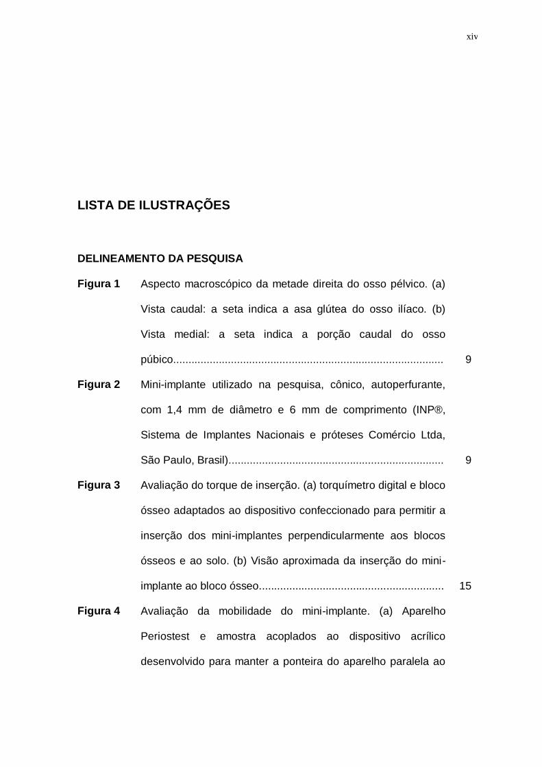
LISTA DE ILUSTRAÇÕES
DELINEAMENTO DA PESQUISA
Figura 1 Aspecto macroscópico da metade direita do osso pélvico. (a)
Vista caudal: a seta indica a asa glútea do osso ilíaco. (b)
Vista medial: a seta indica a porção caudal do osso
púbico.........................................................................................
9
Figura 2 Mini-implante utilizado na pesquisa, cônico, autoperfurante,
com 1,4 mm de diâmetro e 6 mm de comprimento (INP®,
Sistema de Implantes Nacionais e próteses Comércio Ltda,
São Paulo, Brasil).......................................................................
9
Figura 3 Avaliação do torque de inserção. (a) torquímetro digital e bloco
ósseo adaptados ao dispositivo confeccionado para permitir a
inserção dos mini-implantes perpendicularmente aos blocos
ósseos e ao solo. (b) Visão aproximada da inserção do mini-
implante ao bloco ósseo.............................................................
15
Figura 4 Avaliação da mobilidade do mini-implante. (a) Aparelho
Periostest e amostra acoplados ao dispositivo acrílico
desenvolvido para manter a ponteira do aparelho paralela ao

xv
solo e perpendicular ao parafuso. (b) vista aproximada da
ponteira do Periostest, mantida a 2 mm da cabeça do mini-
implante......................................................................................
15
Figura 5 Teste de tração. (a) máquina universal com os dispositivos
acoplados para prender o bloco ósseo, na parte inferior, e
extrair o mini-implante, na parte superior. (b) vista aproximada
dos dispositivos usados no teste de tração................................
15
ARTIGO 1
Figure 1 Flow diagram of literature search................................................... 32
ARTIGO 2
Figure 1 Flow diagram of literature search................................................... 55
Figure 2 Correlation between cortical thickness and primary stability
evaluated through PS…………………………...............................
56
Figure 3 Correlation between cortical thickness and primary stability when
only studies in human beings were considered .............................
56
xvi
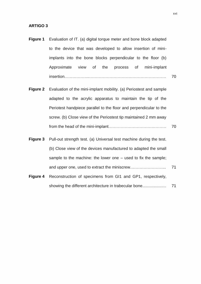
ARTIGO 3
Figure 1 Evaluation of IT. (a) digital torque meter and bone block adapted
to the device that was developed to allow insertion of mini-
implants into the bone blocks perpendicular to the floor (b)
Approximate view of the process of mini-implant
insertion.……………...…………………………………………………
70
Figure 2 Evaluation of the mini-implant mobility. (a) Periostest and sample
adapted to the acrylic apparatus to maintain the tip of the
Periotest handpiece parallel to the floor and perpendicular to the
screw. (b) Close view of the Periostest tip maintained 2 mm away
from the head of the mini-implant…………………………….………
70
Figure 3 Pull-out strength test. (a) Universal test machine during the test.
(b) Close view of the devices manufactured to adapted the small
sample to the machine: the lower one – used to fix the sample;
and upper one, used to extract the miniscrew……………………...
71
Figure 4 Reconstruction of specimens from GI1 and GP1, respectively,
showing the different architecture in trabecular bone.....................
71
xvii
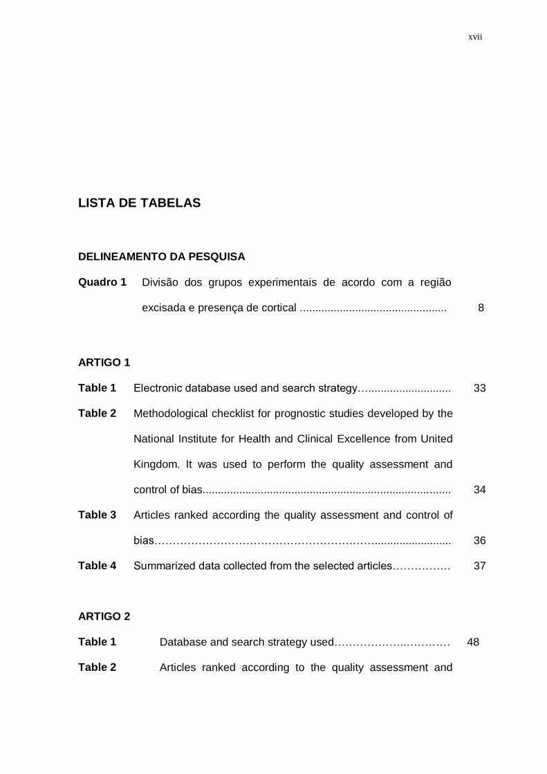
LISTA DE TABELAS
DELINEAMENTO DA PESQUISA
Quadro 1 Divisão dos grupos experimentais de acordo com a região
excisada e presença de cortical ................................................
8
ARTIGO 1
Table 1 Electronic database used and search strategy…........................... 33
Table 2 Methodological checklist for prognostic studies developed by the
National Institute for Health and Clinical Excellence from United
Kingdom. It was used to perform the quality assessment and
control of bias.................................................................................
34
Table 3 Articles ranked according the quality assessment and control of
bias…………………………………………………….........................
36
Table 4 Summarized data collected from the selected articles……………. 37
ARTIGO 2
Table 1 Database and search strategy used………………..………… 48
Table 2 Articles ranked according to the quality assessment and
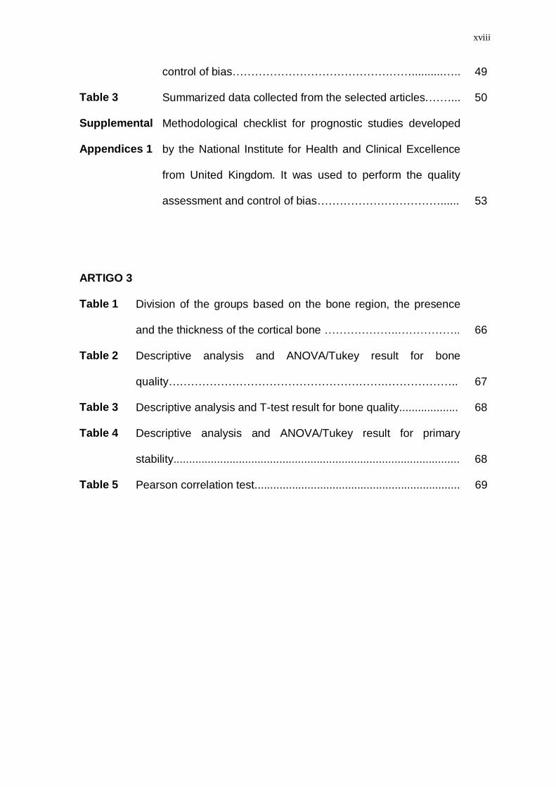
xviii
control of bias…………………………………………..........….. 49
Table 3 Summarized data collected from the selected articles.……... 50
Supplemental
Appendices 1
Methodological checklist for prognostic studies developed
by the National Institute for Health and Clinical Excellence
from United Kingdom. It was used to perform the quality
assessment and control of bias……………………………......
53
ARTIGO 3
Table 1 Division of the groups based on the bone region, the presence
and the thickness of the cortical bone ………………..……………..
66
Table 2 Descriptive analysis and ANOVA/Tukey result for bone
quality………………………………………………….………………..
67
Table 3 Descriptive analysis and T-test result for bone quality................... 68
Table 4 Descriptive analysis and ANOVA/Tukey result for primary
stability............................................................................................
68
Table 5 Pearson correlation test.................................................................. 69

xix
LISTA DE SIGLAS E ABREVIATURAS
ANOVA Analysis of variance
BMD bone mineral density
CBCT cone beam computed tomography
cm centímetro
DeCS Descritores em Ciências da Saúde
g/cm3 grama por centímetro cúbico
HU Hounsfield Unit
ICC coeficiente de correlação intraclasse
ISQ implant stability quotient
IT insertional torque
kgf quilograma força
kV kilovolt
Mand mandible
Max maxilla
MeSH Medical Subject Headings
MI mini-implante
microCT microtomografia computadorizada
mL mililitro
xx
mm milímetro
mm/s milímetros por segundo
MPa megapascal
ms milisegundo
n número
Ncm Newton centímetro
PS pullout strength
PTV Periostest value
R2 coeficiente de determinação
r coeficiente de correlação linear
RFA resonance frequency analysis
ROI region of interest
SD standard deviation
SIGLE System for Information on Grey Literature in Europe
Tb.N trabecular number
Tb.S trabecular separation
Tb.Th trabecular thickness
TBA trabecular bone area
VOI volume of interest
μA microampere
μm micrometro
xxi
ÍNDICE
1 INTRODUÇÃO....................................................................................... 1
2 PROPOSIÇÃO....................................................................................... 5
3 DELINEAMENTO DA PESQUISA.......................................................... 6
3.1 REVISÃO SISTEMÁTICA E META-ANÁLISE.............................. 6
3.2 EXPERIMENTO.......................................................................... 7
3.2.1 AMOSTRA.................................................................................... 7
3.2.1.1 SUBSTRATO ÓSSEO ............................................................ 7
3.2.1.2 MINI-IMPLANTES.................................................................... 8
3.2.2 AVALIAÇÃO DA QUALIDADE ÓSSEA........................................ 9
3.2.3 AVALIAÇÃO DA ESTABILIDADE PRIMÁRIA.............................. 12
3.2.4 ANÁLISE ESTATÍSTICA.............................................................. 14
4 DESENVOLVIMENTO DA PESQUISA................................................... 16
4.1 ARTIGO 1: Marquezan M, Osório A, Sant'Anna E, Souza MM,
Maia L. Does bone mineral density influence the primary
stability of dental implants? A systematic review. Clin Oral
Implants Res. 2012 Jul;23(7):767-74………………………………
17
4.2 ARTIGO 2: Marquezan M, Mattos CT, Sant'Anna EF, Souza
MMG, Maia LC. Does cortical thickness influence the primary
stability of mini-implants? A systematic review and meta-

xxii
analysis. Submitted to The Angle Orthod………………………... 40
4.3 ARTIGO 3: Marquezan M, Lima I, Lopes RT, Sant'anna EF, de
Souza MM. Is trabecular bone related to primary stability of
miniscrews? Accepted in The Angle Orthodontist……...............
57
5 DISCUSSÃO........................................................................................... 72
6 CONCLUSÃO......................................................................................... 82
7 RECOMENDAÇÕES ............................................................................. 83
8 REFERÊNCIAS BIBLIOGRÁFICAS ...................................................... 84
9 APÊNDICES........................................................................................... 90
9.1 APÊNDICE A: Marquezan M, Souza MM, Araújo MT, Nojima LI,
Nojima MC. Is miniscrew primary stability influenced by bone
density? Braz Oral Res. 2011 Sep-Oct;25(5):427-32 …………..
90
9.2 APÊNDICE B: Marquezan M, Lau TC, Mattos CT, Cunha AC,
Nojima LI, Sant'anna EF, Souza MM, Araújo MT. Bone mineral
density. Angle Orthod. 2012 Jan;82(1):62-6…………..................
100
10 ANEXOS................................................................................................ 110
10.1 ANEXO1: Aceite do Artigo 3 ...................................................... 110
1
1 INTRODUÇÃO
Desde a criação da especialidade da Ortodontia, o adequado controle de
ancoragem é reconhecido como fator importante para se alcançar excelentes
resultados nos tratamentos (Angle, 1907). A ancoragem ortodôntica geralmente
é obtida por um dente ou grupo de dentes que apoiam a movimentação de
outros elementos (Melsen e Verna, 1999). Tradicionalmente, essa pode ser
reforçada aumentando-se o número de dentes, utilizando-se aparelhos intra-
bucais auxiliares e/ou aparelhos extra-bucais (Lee, Kim et al., 2009). Certos
casos, entretanto, como severas assimetrias, perdas múltiplas dentárias ou
extenso comprometimento periodontal, podem ser beneficiados pelo uso da
ancoragem esquelética (Melsen e Verna, 1999), pois essa permite a realização
de movimentos dentários nas três dimensões com mínimos efeitos nos demais
dentes (Lee, Kim et al., 2009).
Nas últimas três décadas, têm-se relatado o uso de diferentes
dispositivos para ancoragem esquelética: parafusos de vitallium (Creekmore e
Eklund, 1983), implantes dentários (Justens e De Bruyn, 2008), onplants (Block
e Hoffman, 1995), implantes palatais (Wehrbein, Merz et al., 1996) e mini-
implantes (Kanomi, 1997). As principais vantagens dos mini-implantes são:
tamanho reduzido, baixo custo, fácil inserção e remoção, com pequeno
desconforto ao paciente, e possibilidade de aplicação de carga imediata (Serra,

2
Morais et al., 2008; Luzi, Verna et al., 2009; Wei, Zhao et al., 2011; Topouzelis
e Tsaousoglou, 2012).
O sucesso do uso dos mini-implantes (MI) está relacionado à sua
estabilidade no tecido ósseo. Classicamente, essa pode ser dividida em
primária e secundária. A primeira decorre do estreito contato entre a superfície
do mini-implante e o osso (Gedrange, Hietschold et al., 2005; Iijima, Takano et
al., 2012), sendo definida como a ausência de mobilidade no leito ósseo após a
inserção do dispositivo (Javed e Romanos, 2010). A segunda, dita estabilidade
secundária ou tardia, por sua vez, ocorre após a cicatrização (Gedrange,
Hietschold et al., 2005).
Na técnica de inserção de MI, a estabilidade primária destaca-se como
um importante indício de êxito (Motoyoshi, Hirabayashi et al., 2006; Chaddad,
Ferreira et al., 2008), visto que a maioria das falhas ocorre nos estágios iniciais
pós-inserção (Miyawaki, Koyama et al., 2003; Lim, Cha et al., 2008). A falta de
estabilidade imediata pode levar à mobilidade progressiva do dispositivo e sua
subsequente perda (Mischkowski, Kneuertz et al., 2008). Dada sua relevância,
têm-se sugerido inclusive que, se a retenção mecânica inicial do MI não for
observada, esse seja substituído por um dispositivo de maior diâmetro ou o
sítio de inserção seja modificado (Garfinkle, Cunningham et al., 2008). Por
outro lado, tensões exageradas durante a inserção podem resultar em
aquecimento e danos ao tecido ósseo (Park, Jeong et al., 2006), incluindo
isquemia e necrose, ou até fratura do mini-implante (Wilmes, Rademacher et
al., 2006)
Segundo revisão sistemática, a taxa de insucesso dos MI varia de 13,4 a
20,1% (Schatzle, Mannchen et al., 2009). Em meta-análise recente, confirmou-

3
se que os MI ortodônticos possuem baixa taxa de falha (13,5%), sendo,
portanto, indicados para a prática clínica (Papageorgiou, Zogakis et al., 2012).
Especula-se, entretanto, que a taxa de sucesso em adolescentes seja menor
devido a maior taxa de metabolismo e menor densidade óssea nesses
pacientes (Miyawaki, Koyama et al., 2003; Topouzelis e Tsaousoglou, 2012).
Imediatamente após a instalação de implantes dentários e mini-
implantes ortodônticos, a estabilidade primária tem sido tradicionalmente
verificada através de teste manual (Merheb, Van Assche et al., 2010). Métodos
menos subjetivos de avaliação da estabilidade primária, são descritos na
literatura, como aferição do torque de inserção, do torque de remoção, teste de
tração ou de deslocamento lateral, método de frequência de ressonância e
percussão. Apesar da variedade de métodos disponíveis, não há padrão ouro
para avaliação da estabilidade primária (Cehreli, Karasoy et al., 2009).
Fatores que influenciam a estabilidade imediata de implantes estão
relacionados ao desenho do dispositivo (Wilmes, Rademacher et al., 2006;
Song, Cha et al., 2007; Wilmes, Ottenstreuer et al., 2008), à quantidade e
qualidade óssea (Trisi, Rao et al., 1999; Freudenthaler, Haas et al., 2001;
Cheng, Tseng et al., 2004; Wilmes, Rademacher et al., 2006), e à técnica de
inserção (Wilmes, Rademacher et al., 2006).
O termo “qualidade óssea”, entretanto, não está claramente definido na
literatura. Sugere-se que esse englobe aspectos fisiológicos, estruturais e o
grau de mineralização do tecido ósseo (Bergkvist, Koh et al., 2010). Aspectos
referentes ao metabolismo ósseo, à renovação celular, à maturação óssea, às
propriedades da matriz extracelular e à vascularização do tecido também foram

4
enfatizados (Molly, 2006), mas o papel de cada um deles não é completamente
compreendido (Bergkvist, Koh et al., 2010).
Duas propriedades ósseas, entretanto, já foram relacionadas com a
estabilidade de implantes dentários e mini-implantes: a densidade mineral
óssea (BMD – do inglês bone mineral density) (Turkyilmaz, Tozum et al., 2006;
Turkyilmaz, Tumer et al., 2007; Turkyilmaz e Mcglumphy, 2008a; b; Aksoy,
Eratalay et al., 2009; Song, Jun et al., 2009; Bergkvist, Koh et al., 2010;
Merheb, Van Assche et al., 2010) e a espessura de cortical (Miyawaki, Koyama
et al., 2003; Huja, Litsky et al., 2005; Motoyoshi, Yoshida et al., 2007;
Motoyoshi, Inaba et al., 2009; Pithon, Nojima et al., 2011). Sabe-se que essas
propriedades podem variar de acordo com o paciente e com as regiões da
maxila e mandíbula (Deguchi, Nasu et al., 2006), fazendo-se necessário
investigar a influência de sua variação na estabilidade dos dispositivos. Além
disso, outras propriedades ósseas merecem ser investigadas no que tange à
estabilidade dos MI, tais como número de trabéculas (Tb.N), densidade
trabecular (BV/TV), espessura trabecular (Tb.Th) e separação trabecular
(Tb.S).

5
2 PROPOSIÇÃO
2.1 Realizar revisões sistemáticas com o objetivo de:
2.1.1 investigar a influência da BMD na estabilidade primária de
implantes dentários;
2.1.2 avaliar a associação da espessura de cortical e a estabilidade
primária de mini-implantes;
2.2 Realizar experimento com blocos de ossos bovinos a fim de:
2.2.1 comparar a estabilidade primária de mini-implantes inseridos em
blocos ósseos de diferentes BMD (com e sem cortical);
2.2.2 investigar se propriedades do ósseas, tais como densidade mineral
óssea, densidade trabecular, número de trabéculas, espessura trabecular,
espessura de cortical e separação trabecular, podem influenciar a estabilidade
primária de mini-implantes.

6
3 DELINEAMENTO DA PESQUISA
3.1 REVISÃO SISTEMÁTICA E META-ANÁLISE
A qualidade do tecido ósseo receptor, que tem influência na estabilidade
primária de implantes, tem sido definida nas pesquisas odontológicas como
duas principais propriedades: a densidade mineral óssea (Turkyilmaz, Tozum
et al., 2006; Turkyilmaz, Tumer et al., 2007; Turkyilmaz e Mcglumphy, 2008a; b;
Aksoy, Eratalay et al., 2009; Song, Jun et al., 2009; Bergkvist, Koh et al., 2010;
Merheb, Van Assche et al., 2010) e a espessura de cortical (Miyawaki, Koyama
et al., 2003; Huja, Litsky et al., 2005; Motoyoshi, Yoshida et al., 2007;
Motoyoshi, Inaba et al., 2009; Pithon, Nojima et al., 2011). Dessa maneira,
foram realizadas duas revisões sistemáticas a fim de avaliar a associação
dessas propriedades ósseas com a estabilidade primária.
Na primeira delas (Artigo 1, página 17), avaliou-se a correlação entre a
estabilidade primária de implantes dentários com a BMD. Optou-se por
trabalhar com implantes dentários em vez de MI ortodônticos por não haver
trabalhos suficientes na literatura e com metodologias padronizadas para
execução de uma revisão sistemática. Além disso, o campo da ancoragem
esquelética tem se beneficiado da literatura de implantodontia desde seu
surgimento. A segunda revisão sistemática avaliou a associação da espessura

7
de cortical e estabilidade primária de mini-implantes e deu origem a uma meta-
análise (Artigo 2, página 40).
3.2 EXPERIMENTO
Essa pesquisa constituiu um estudo experimental ex vivo. Previamente à
execução do experimento final, projeto piloto foi desenvolvido a fim de definir
os substratos ósseos a serem utilizados (osso pélvico bovino) e o tamanho
amostral. Cálculo amostral para diferença entre médias (α=5%, poder do
estudo= 80%), sugeriu o uso de 13 amostras por grupo. Os resultados do piloto
ainda geraram dois artigos científicos (APÊNDICE A e B, páginas 90 e 100).
3.2.1 AMOSTRA
3.2.1.1 SUBSTRATO ÓSSEO
A amostra foi constituída de treze ossos pélvicos bovinos (Bos taurus)
da raça Angus, abatidos para consumo humano e obtidos imediatamente após
o sacrifício em frigorífico registrado na ANVISA (Figura 1, página 9). Duas
secções teciduais foram excisadas da face glútea da asa do ilíaco, e outras
duas do púbico, regiões que possuem aproximadamente 1mm de espessura de
cortical. Essas foram excisadas do osso pélvico através de fresa trefina (8 mm
ø, SIN- Sistema de Implante e Nacional Ltda, São Paulo, Brasil) adaptada em
motor de baixa rotação (Beltec LB100, Araraquara, Brasil) sob irrigação com
soro fisiológico. Das secções ósseas removidas de cada região, uma teve sua
cortical removida e outra teve a cortical preservada, sendo ambas
armazenadas por congelamento (-20ºC) em soro fisiológico (Liu, Broucek et al.,
2012) até o momento da inserção dos MI. A divisão dos grupos experimentais

8
se deu de acordo com a região do osso pélvico excisada e a presença de
cortical (Quadro 1).
Quadro 1: Divisão dos grupos experimentais de acordo com a região
excisada e presença de cortical.
Grupo Região da excisão n. de amostras Cortical óssea
GI0 Ilíaco 13 Ausente
GI1 Ilíaco 13 Presente
GP0 Púbico 13 Ausente
GP1 Púbico 13 Presente
3.2.1.2 MINI-IMPLANTES
Cinquenta e dois MI cônicos autoperfurantes com grau de pureza do tipo
V (liga de Ti-6Al-4V) da marca INP (INP®, Sistema de Implantes Nacionais e
próteses Comércio Ltda, São Paulo, Brasil), medindo 1,4 mm de diâmetro por 6
mm de comprimento (Figura 2, página 9), foram inseridos nos blocos ósseos.
Foi realizada perfuração prévia dos sítios de inserção com broca broca
helicoidal de 1mm de diâmetro (INP®, Sistema de Implantes Nacionais e
próteses Comércio Ltda, São Paulo, Brasil), em baixa rotação, controlando-se a
profundidade de inserção com stops de borracha para obter 2 mm. Durante a
inserção dos MI, foi realizada aferição do torque de inserção (ver capítulo 3.2.3,
página 12). Na sequência, procedeu-se à avaliação da micromobilidade
(capítulo 3.2.3) e o escaneamento das peças para avaliação da qualidade
óssea (capítulo 3.2.2).

9
Figura 1: Aspecto macroscópico da metade direita do osso pélvico. (a) Vista caudal: a seta indica a asa glútea do osso ilíaco. (b) Vista medial: a seta indica a porção caudal do osso púbico.
Figura 2: Mini-implante utilizado na pesquisa, cônico, autoperfurante, com 1,4 mm de diâmetro
e 6 mm de comprimento (INP®, Sistema de Implantes Nacionais e próteses Comércio Ltda, São Paulo, Brasil).
3.2.2 AVALIAÇÃO DA QUALIDADE ÓSSEA
A avaliação da qualidade do tecido ósseo receptor foi realizada através
dos exames de microtomografia computadorizada (microCT), sendo assim
possível aferir a densidade mineral e a micro-arquitetura ósseas.
10
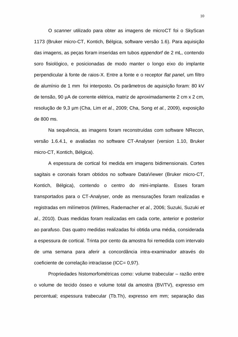
O scanner utilizado para obter as imagens de microCT foi o SkyScan
1173 (Bruker micro-CT, Kontich, Bélgica, software versão 1.6). Para aquisição
das imagens, as peças foram inseridas em tubos eppendorf de 2 mL, contendo
soro fisiológico, e posicionadas de modo manter o longo eixo do implante
perpendicular à fonte de raios-X. Entre a fonte e o receptor flat panel, um filtro
de alumínio de 1 mm foi interposto. Os parâmetros de aquisição foram: 80 kV
de tensão, 90 µA de corrente elétrica, matriz de aproximadamente 2 cm x 2 cm,
resolução de 9,3 µm (Cha, Lim et al., 2009; Cha, Song et al., 2009), exposição
de 800 ms.
Na sequência, as imagens foram reconstruídas com software NRecon,
versão 1.6.4.1, e avaliadas no software CT-Analyser (version 1.10, Bruker
micro-CT, Kontich, Bélgica).
A espessura de cortical foi medida em imagens bidimensionais. Cortes
sagitais e coronais foram obtidos no software DataViewer (Bruker micro-CT,
Kontich, Bélgica), contendo o centro do mini-implante. Esses foram
transportados para o CT-Analyser, onde as mensurações foram realizadas e
registradas em milímetros (Wilmes, Rademacher et al., 2006; Suzuki, Suzuki et
al., 2010). Duas medidas foram realizadas em cada corte, anterior e posterior
ao parafuso. Das quatro medidas realizadas foi obtida uma média, considerada
a espessura de cortical. Trinta por cento da amostra foi remedida com intervalo
de uma semana para aferir a concordância intra-examinador através do
coeficiente de correlação intraclasse (ICC= 0,97).
Propriedades histomorfométricas como: volume trabecular – razão entre
o volume de tecido ósseo e volume total da amostra (BV/TV), expresso em
percentual; espessura trabecular (Tb.Th), expresso em mm; separação das
11
trabéculas (Tb.S), expresso em mm; e número de trabéculas por mm da
amostra (Tb.N), expresso em 1/mm, foram aferidos em imagem tridimensional
no CT-Analyser. Para isso, as imagens escaneadas foram abertas no software
de análise com redimensionamento (Resize 3) a fim de reduzir seu tamanho
original e permitir o adequado funcionamento do software.
Previamente à análise, para a aferição da BMD, foi necessário realizar a
calibração do software através do escaneamento de um padrão de osso
artificial (Sawbones® Pacific Research Laboratories Inc., Washington, EUA),
composto de osso trabecular com densidade de 0,32 g/cm3 e osso cortical com
1,64 g/cm3. Esse padrão possuía as mesmas dimensões da amostra e continha
um mini-implante inserido em seu centro. O escaneamento foi realizado em
frasco eppendorf contendo soro fisiológico, de modo a reproduzir as condições
de escaneamento das amostras. A BMD dos blocos ósseos foi então calculada
pelo software a partir dos valores de coeficiente de atenuação dos ossos
naturais e artificiais e registrada em g/cm3. Foram aferidas as BMDs dos blocos
ósseos considerando porção trabecular e cortical em conjunto (BMD total), do
osso trabecular em separado (BMD trabecular) e do osso cortical (BMD
cortical).
O processo de análise de aferição dos parâmetros histomorfométricos e
da BMD se iniciou pela seleção do volume de interesse (VOI- do inglês volume
of interest). Um cilindro de 3,4 mm de diâmetro foi selecionado em torno do
mini-implante, cobrindo pelo menos 1 mm além das dimensões do parafuso,
partindo de seu perfil transmucoso até a sua ponta. O centro desse cilindro,
contendo o parafuso e os 6 voxels de osso adjacente a ele (54 μm) foram
excluídos do VOI a fim de reduzir o efeito do artefato sobre as análises (Brinley,

12
Behrents et al., 2009). A imagem selecionada foi binarizada por meio de
histograma de tons de cinza, onde o valor de cinza é proporcional ao
coeficiente de atenuação (áreas mais densas aparecem mais claras, enquanto
áreas menos densas, mais escuras). O processamento da imagem foi então
realizado automaticamente para calcular os parâmetros histomorfométricos
desejados e a BMD.
3.2.3 AVALIAÇÃO DA ESTABILIDADE PRIMÁRIA
Para avaliação da estabilidade primária, o torquímetro digital (Lutron TQ-
8800, Taipei, Taiwan) foi adaptado à chave de inserção dos MI, possibilitando a
mensuração do pico do torque de inserção (IT, do inglês intersional torque). O
torquímetro e os blocos ósseos, por sua vez, foram adaptados a um dispositivo
especialmente desenhado para essa finalidade, que permitiu a inserção dos
mini-implantes perpendicularmente aos blocos ósseos e ao solo (Figura 3,
página 15). Os valores obtidos foram registrados em newtons.centímetro
(Ncm).
A mobilidade dos mini-implantes foi aferida imediatamente após sua
instalação através do aparelho Periotest (modelo 3218, Medizintechnik Gulden,
Modautal, Alemanha), que realiza mensuração eletromecânica (Figura 4,
página 15). O aparelho foi calibrado com a luva de calibração fornecida pelo
fabricante previamente à mensuração de cada amostra. O aparelho e a
amostra foram então acoplados a um dispositivo acrílico construído para
manter a ponteira do aparelho paralela ao solo e perpendicular ao mini-
implante, garantindo ainda uma distância de 2 mm entre a ponteira e a cabeça

13
do mini-implante, segundo recomendações do fabricante. Para cada análise, o
aparelho foi acionado e a ponteira realizou 16 percussões, levando cerca de 4
segundos. Na sequência, um valor (Periotest Value- PTV) foi gerado no monitor
do aparelho dentro da escala de -8 a +50. Quanto menor o PTV, menor a
mobilidade e maior a estabilidade do implante (Kim, Ahn et al., 2005). Para
cada amostra, 2 medidas do PTV foram realizadas e a média delas foi
tabulada. Se a diferença entre as medidas fosse maior que 2 pontos, essas
eram desprezadas, o aparelho recalibrado e as medidas refeitas, seguindo-se o
protocolo sugerido pelo fabricante.
Ainda para aferir a estabilidade primária, ensaio mecânico de tração (PS,
do inglês pull-out strength) foi realizado em máquina universal (Emic DL 2000,
São José dos Pinhais, Brasil). Esse é um ensaio destrutivo que consiste em
extrair o mini-implante do tecido ósseo a uma velocidade constante, avaliando-
se dessa forma, a força máxima necessária para remoção do dispositivo do
tecido ósseo (Huja, Litsky et al., 2005). Para tal, dois dispositivos foram
acoplados à máquina: um em forma de pé de cabra, acoplado na parte superior
e usado para prender o mini-implante; o outro na porção inferior, que serviu de
base para fixar o bloco ósseo e manter o mini-implante perpendicular ao solo
(Figura 5, página 15). Para o tracionamento dos mini-implantes foi utilizada
velocidade de 0,05 mm/s (Huja, Litsky et al., 2005; Salmoria, Tanaka et al.,
2008) e célula de carga de 500 kgf. O valor da carga e do deslocamento foram
registrados e a força máxima (Fmax) alcançada foi registrada em Newtons (N).
14
3.2.4 ANÁLISE ESTATÍSTICA
As análises estatísticas foram realizadas por meio do programa
Statistical Package for the Social Science (version 18, SPSS Inc., USA). Os
valores obtidos foram tabulados e submetidos a estatísticas descritivas. A
verificação da normalidade e da homogeinedade das variáveis foi realizada por
meio do teste de Shapiro-Wilk e do teste de Levene, respectivamente. A
diferença entre grupos foi avaliada através dos testes T-student (para as
variáveis espessura de cortical e BMD cortical) e ANOVA/Tukey (para as
demais variáveis). Por fim, para verificar se existe correlação entre as variáveis
referentes à qualidade óssea e àquelas que indicam o comportamento
mecânico dos mini-implantes foi realizado teste de correlação de Pearson. O
nível de significância adotado foi de 0,05.

15
Figura 3: Avaliação do torque de inserção. (a) torquímetro digital e bloco ósseo adaptados ao dispositivo confeccionado para permitir a inserção dos mini-implantes perpendicularmente aos blocos ósseos e ao solo. (b) Visão aproximada da inserção do mini-implante ao bloco ósseo.
Figura 4: Avaliação da mobilidade do mini-implante. (a) Aparelho Periostest e amostra acoplados ao dispositivo acrílico desenvolvido para manter a ponteira do aparelho paralela ao solo e perpendicular ao parafuso. (b) vista aproximada da ponteira do Periostest, mantida a 2 mm da cabeça do mini-implante.
Figura 5: Teste de tração. (a) máquina universal com os dispositivos acoplados para prender o bloco ósseo, na parte inferior, e extrair o mini-implante, na parte superior. (b) vista aproximada dos dispositivos usados no teste de tração.
16
4 DESENVOLVIMENTO DA PESQUISA
4.1 ARTIGO 1
Marquezan M, Osório A, Sant'Anna E, Souza MM, Maia L. Does bone mineral
density influence the primary stability of dental implants? A systematic review.
Clin Oral Implants Res. 2012 Jul;23(7):767-74.
4.2 ARTIGO 2
Marquezan M, Mattos CT, Sant'Anna EF, Souza MMG, Maia LC. Does cortical
thickness influence the primary stability of mini-implants? A systematic review
and meta-analysis. Submitted to The Angle Orthodontist.
4.3 ARTIGO 3
Marquezan M, Lima I, Lopes RT, Sant'anna EF, de Souza MM. Is trabecular
bone related to primary stability of miniscrews? Accepted in The Angle
Orthodontist (ANEXO 1, página 110).
17
Artigo 1
Does bone mineral density influence the primary stability of dental
implants?
A systematic review.
ABSTRACT
Objective: the aim of this systematic review was to investigate the influence of
bone mineral density on the primary stability of dental implants.
Material and Methods: A search of health science databases (Cochrane
Library, MEDLINE-PubMed, ISI Web of Knowledge, EMBASE, LILACS) and
grey literature was performed, including papers published until January 2011.
The main key words used were “bone density” (MeSH/ DeCS), “dental implant”
(MeSH/ DeCS), “implant stability”, “implant stability quotient”, “ISQ”, “resonance
frequency analysis”, “RFA”, “Ostell”, “Periotest value”, “PTV”, “Periostest”,
“insertion torque”, “placement torque”, “cutting torque”. The inclusion criteria
comprised observational clinical studies performed in patients who received
dental implants for rehabilitation; studies that evaluated the association between
bone mineral density and implant primary stability; bone density assessment
performed by measurement of Hounsfield units using cone beam computed
tomography; and dental implant primary stability evaluated by ISQ value, PTV
value or Insertion torque measurement. The articles selected were carefully
read and classified as low, moderate and high methodological quality, and data
of interest were tabulated.
Results: Ten articles met the inclusion criteria, but only seven were included
because of overlapping patients. They were classified as low or moderate
methodological quality and control of bias, and presented positive association
between primary stability and bone density.
Conclusions: There is a positive association between implant primary stability
and bone mineral density of the receptor site. However, the methodological
quality and control of bias of the studies should be improved to produce
stronger evidences.
18
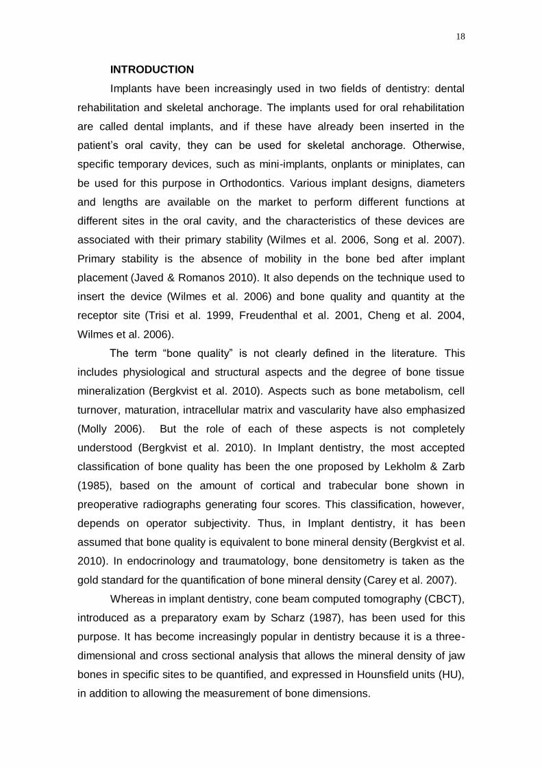
INTRODUCTION
Implants have been increasingly used in two fields of dentistry: dental
rehabilitation and skeletal anchorage. The implants used for oral rehabilitation
are called dental implants, and if these have already been inserted in the
patient’s oral cavity, they can be used for skeletal anchorage. Otherwise,
specific temporary devices, such as mini-implants, onplants or miniplates, can
be used for this purpose in Orthodontics. Various implant designs, diameters
and lengths are available on the market to perform different functions at
different sites in the oral cavity, and the characteristics of these devices are
associated with their primary stability (Wilmes et al. 2006, Song et al. 2007).
Primary stability is the absence of mobility in the bone bed after implant
placement (Javed & Romanos 2010). It also depends on the technique used to
insert the device (Wilmes et al. 2006) and bone quality and quantity at the
receptor site (Trisi et al. 1999, Freudenthal et al. 2001, Cheng et al. 2004,
Wilmes et al. 2006).
The term “bone quality” is not clearly defined in the literature. This
includes physiological and structural aspects and the degree of bone tissue
mineralization (Bergkvist et al. 2010). Aspects such as bone metabolism, cell
turnover, maturation, intracellular matrix and vascularity have also emphasized
(Molly 2006). But the role of each of these aspects is not completely
understood (Bergkvist et al. 2010). In Implant dentistry, the most accepted
classification of bone quality has been the one proposed by Lekholm & Zarb
(1985), based on the amount of cortical and trabecular bone shown in
preoperative radiographs generating four scores. This classification, however,
depends on operator subjectivity. Thus, in Implant dentistry, it has been
assumed that bone quality is equivalent to bone mineral density (Bergkvist et al.
2010). In endocrinology and traumatology, bone densitometry is taken as the
gold standard for the quantification of bone mineral density (Carey et al. 2007).
Whereas in implant dentistry, cone beam computed tomography (CBCT),
introduced as a preparatory exam by Scharz (1987), has been used for this
purpose. It has become increasingly popular in dentistry because it is a three-
dimensional and cross sectional analysis that allows the mineral density of jaw
bones in specific sites to be quantified, and expressed in Hounsfield units (HU),
in addition to allowing the measurement of bone dimensions.
19
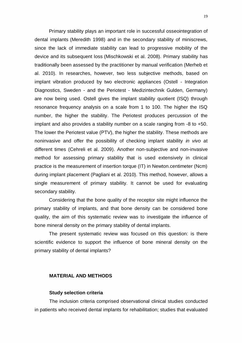
Primary stability plays an important role in successful osseointegration of
dental implants (Meredith 1998) and in the secondary stability of miniscrews,
since the lack of immediate stability can lead to progressive mobility of the
device and its subsequent loss (Mischkowski et al. 2008). Primary stability has
traditionally been assessed by the practitioner by manual verification (Merheb et
al. 2010). In researches, however, two less subjective methods, based on
implant vibration produced by two electronic appliances (Ostell - Integration
Diagnostics, Sweden - and the Periotest - Medizintechnik Gulden, Germany)
are now being used. Ostell gives the implant stability quotient (ISQ) through
resonance frequency analysis on a scale from 1 to 100. The higher the ISQ
number, the higher the stability. The Periotest produces percussion of the
implant and also provides a stability number on a scale ranging from -8 to +50.
The lower the Periotest value (PTV), the higher the stability. These methods are
noninvasive and offer the possibility of checking implant stability in vivo at
different times (Cehreli et al. 2009). Another non-subjective and non-invasive
method for assessing primary stability that is used extensively in clinical
practice is the measurement of insertion torque (IT) in Newton.centimeter (Ncm)
during implant placement (Pagliani et al. 2010). This method, however, allows a
single measurement of primary stability. It cannot be used for evaluating
secondary stability.
Considering that the bone quality of the receptor site might influence the
primary stability of implants, and that bone density can be considered bone
quality, the aim of this systematic review was to investigate the influence of
bone mineral density on the primary stability of dental implants.
The present systematic review was focused on this question: is there
scientific evidence to support the influence of bone mineral density on the
primary stability of dental implants?
MATERIAL AND METHODS
Study selection criteria
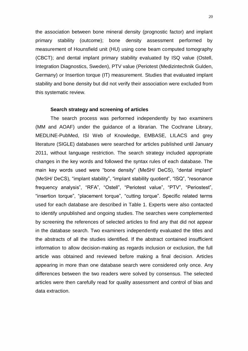
The inclusion criteria comprised observational clinical studies conducted
in patients who received dental implants for rehabilitation; studies that evaluated
20
the association between bone mineral density (prognostic factor) and implant
primary stability (outcome); bone density assessment performed by
measurement of Hounsfield unit (HU) using cone beam computed tomography
(CBCT); and dental implant primary stability evaluated by ISQ value (Ostell,
Integration Diagnostics, Sweden), PTV value (Periotest (Medizintechnik Gulden,
Germany) or Insertion torque (IT) measurement. Studies that evaluated implant
stability and bone density but did not verify their association were excluded from
this systematic review.
Search strategy and screening of articles
The search process was performed independently by two examiners
(MM and AOAF) under the guidance of a librarian. The Cochrane Library,
MEDLINE-PubMed, ISI Web of Knowledge, EMBASE, LILACS and grey
literature (SIGLE) databases were searched for articles published until January
2011, without language restriction. The search strategy included appropriate
changes in the key words and followed the syntax rules of each database. The
main key words used were “bone density” (MeSH/ DeCS), “dental implant”
(MeSH/ DeCS), “implant stability”, “implant stability quotient”, “ISQ”, “resonance
frequency analysis”, “RFA”, “Ostell”, “Periotest value”, “PTV”, “Periostest”,
“insertion torque”, “placement torque”, “cutting torque”. Specific related terms
used for each database are described in Table 1. Experts were also contacted
to identify unpublished and ongoing studies. The searches were complemented
by screening the references of selected articles to find any that did not appear
in the database search. Two examiners independently evaluated the titles and
the abstracts of all the studies identified. If the abstract contained insufficient
information to allow decision-making as regards inclusion or exclusion, the full
article was obtained and reviewed before making a final decision. Articles
appearing in more than one database search were considered only once. Any
differences between the two readers were solved by consensus. The selected
articles were then carefully read for quality assessment and control of bias and
data extraction.

21
Quality assessment and control of bias
The quality assessment and control of bias was realized by the two
authors using the Methodological checklist for prognostic studies developed by
the National Institute for Health and Clinical Excellence of the United Kingdom
(Table 2). Checklist items are worded so that a ‘yes’ response always indicates
that the study has been designed and conducted in such a way as to minimize
the risk of bias for that item. An ‘unclear’ response to a question may arise
when the answer to an item is not reported or is not reported clearly. A study
was classified as having high methodological quality if at least 5 of the 6
parameters received the answer “yes”; moderate methodological quality if at
least 3 of the parameters received the answer “yes”; or low methodological
quality if 2 or less parameters received the answer “yes”. None of the articles
was excluded from the systematic review after this classification, except for
articles on studies conducted by the same author and having some overlapping
patients. In this case, after ranking the studies, the one with the highest score
was included in the systematic review, the others were excluded.
Data extraction
From the selected articles, data on the following issues were extracted
and tabulated by the two authors: a) author and year publication; b)
geographical location; c) type of study; d) sample (sample size, age and
gender); e) implant name and manufacturer; f) implant dimensions and surface
treatment (if present); g) number of implants evaluated; h) regions of implant
insertion; i) bone density of the receptor sites (HU value) and type of bone
evaluated (if cortical and trabecular bones together or only trabecular); j)
implant primary stability number (ISQ, PTV and/or IT (Ncm); k) confounders
included in the analysis (cortical thickness and/or implant dimensions); l)
association between stability and bone density.
RESULTS
The search procedures retrieved 519 articles from EMBASE, 343 articles
from MEDLINE-PubMed, 219 articles from ISI Web of Knowledge, 1338 articles
from the Grey Literature (SIGLE) and 18 articled from the Cochrane Library. No
22
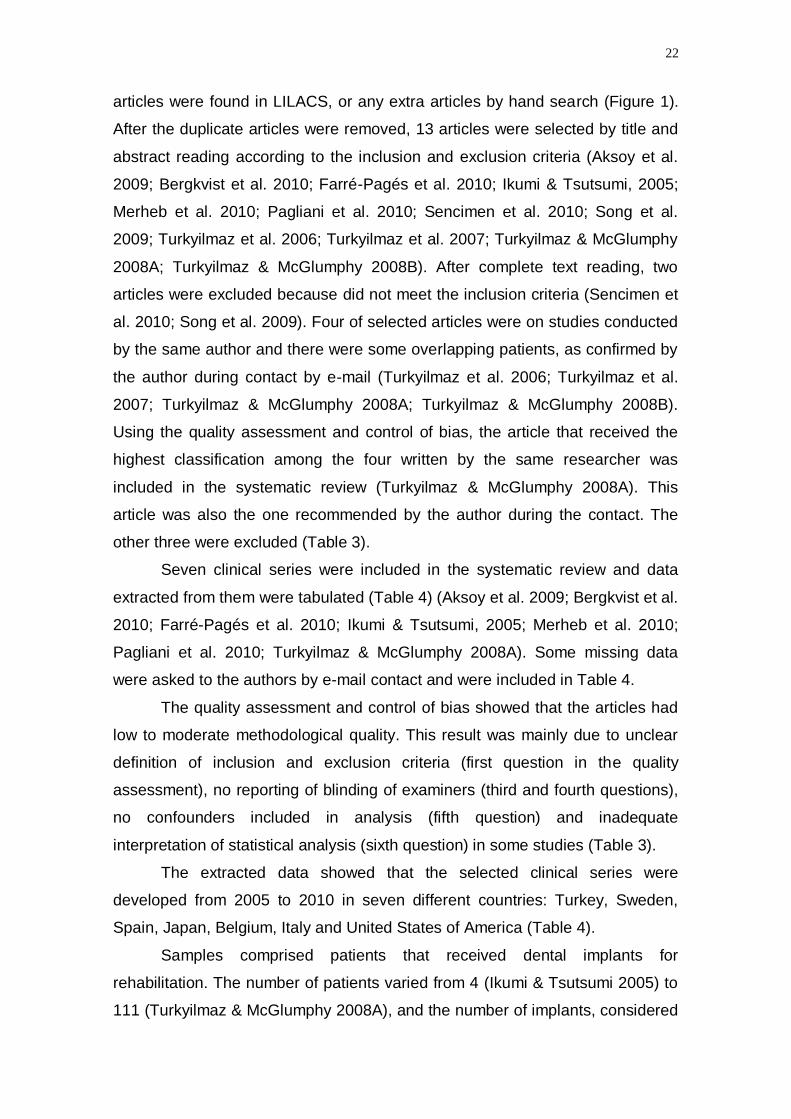
articles were found in LILACS, or any extra articles by hand search (Figure 1).
After the duplicate articles were removed, 13 articles were selected by title and
abstract reading according to the inclusion and exclusion criteria (Aksoy et al.
2009; Bergkvist et al. 2010; Farré-Pagés et al. 2010; Ikumi & Tsutsumi, 2005;
Merheb et al. 2010; Pagliani et al. 2010; Sencimen et al. 2010; Song et al.
2009; Turkyilmaz et al. 2006; Turkyilmaz et al. 2007; Turkyilmaz & McGlumphy
2008A; Turkyilmaz & McGlumphy 2008B). After complete text reading, two
articles were excluded because did not meet the inclusion criteria (Sencimen et
al. 2010; Song et al. 2009). Four of selected articles were on studies conducted
by the same author and there were some overlapping patients, as confirmed by
the author during contact by e-mail (Turkyilmaz et al. 2006; Turkyilmaz et al.
2007; Turkyilmaz & McGlumphy 2008A; Turkyilmaz & McGlumphy 2008B).
Using the quality assessment and control of bias, the article that received the
highest classification among the four written by the same researcher was
included in the systematic review (Turkyilmaz & McGlumphy 2008A). This
article was also the one recommended by the author during the contact. The
other three were excluded (Table 3).
Seven clinical series were included in the systematic review and data
extracted from them were tabulated (Table 4) (Aksoy et al. 2009; Bergkvist et al.
2010; Farré-Pagés et al. 2010; Ikumi & Tsutsumi, 2005; Merheb et al. 2010;
Pagliani et al. 2010; Turkyilmaz & McGlumphy 2008A). Some missing data
were asked to the authors by e-mail contact and were included in Table 4.
The quality assessment and control of bias showed that the articles had
low to moderate methodological quality. This result was mainly due to unclear
definition of inclusion and exclusion criteria (first question in the quality
assessment), no reporting of blinding of examiners (third and fourth questions),
no confounders included in analysis (fifth question) and inadequate
interpretation of statistical analysis (sixth question) in some studies (Table 3).
The extracted data showed that the selected clinical series were
developed from 2005 to 2010 in seven different countries: Turkey, Sweden,
Spain, Japan, Belgium, Italy and United States of America (Table 4).
Samples comprised patients that received dental implants for
rehabilitation. The number of patients varied from 4 (Ikumi & Tsutsumi 2005) to
111 (Turkyilmaz & McGlumphy 2008A), and the number of implants, considered

23
the unit analysis, ranged from 23 (Aksoy et al. 2009) to 300 (Turkyilmaz &
McGlumphy 2008A) (Table 4). Seven types of implants from six different
manufacturers were used: Swiss plus (Zimmer Dental), Standard Plus SLActive
(Straumann), Astra (Astra Tech), Mis-Seven (Biodenix Tech Inc), Mis-Biocom
(Biodenix Tech Inc), TiUnite MK III (Nobel Biocare) and Neoss Dental Implant
System (Neoss Ltd.). They presented different surface treatments, and their
dimensions varied from 3.3 to 5.5 mm in diameter and from 6 to 15 mm in
length (Table 4).
Density values (HU) were higher in men than in women (Aksoy et al.
2009; Turkyilmaz & McGlumphy 2008A). The jaw bone also showed difference,
the mandible presented higher values than maxilla (Aksoy et al. 2009). On the
other hand, the stability value according gender differences, evaluated only by
Aksoy et al. (2009), was lower for men (Table 4).
The IT values ranged from 4 Ncm (Ikumi & Tsutsumi, 2005) to 42.34
Ncm (Farré-Pagés et al. 2010), being higher in the mandible than maxilla, and
in the anterior than posterior region (Farré-Pagés et al. 2010) (Table 4).
Although the implant dimensions were cited in all of the studies, the only
study that included this factor and other confounders in the analysis was the
one conducted by Merheb et al (2010) (Table 4).
Despite methodological differences and weak to moderate
methodological quality (Table 3), all of the selected articles presented a positive
association between primary stability of implants and bone density (Table 4).
DISCUSSION
There was positive association between bone density and dental implant
stability in the selected articles. When evaluating the correlation between ISQ
and HU values, correlation coefficients ranged from 0.46 (moderate correlation-
Merheb et al. 2010) to 0.882 (high correlation- Turkyilmaz & McGlumphy
2008A). Methodological differences might be responsible for this difference.
Even though all of the articles quantified HU in CBCT images, some studies
evaluated the cortical bone and trabecular bone density together (Aksoy et al.
2009, Pagliani et al. 2010, Turkyilmaz & McGlumphy 2008A), a factor that
probably increased the HU value of the unit. Other authors considered only the
24
trabecular bone (Bergkvist et al. 2010, Merheb et al. 2010). Merheb et al.
(2010), who considered only the trabecular bone for density evaluation, found
one of the lower values (r=0.46). Authors who considered the cortical and
trabecular bone as a unit when evaluating bone density observed stronger
correlations (Aksoy et al., 2009; Pagliani et al. 2010, Turkyilmaz & McGlumphy
2008A).
The cortical thickness has been related to primary stability of miniscrews
(Miyawaki et al. 2003; Huja et al. 2005; Motoyoshi et al. 2007; Motoyoshi et al.
2009). Data extracted from one of the selected articles also showed this
correlation: r=0.57 (Merheb et al. 2010). It seems logical that when there was
increased cortical thickness, there would also be an increase in the area of
highly mineralized tissue when bone density was measured. It can be inferred
that the density measured by the sum of cortical and trabecular bone might be
strongly influenced by cortical thickness.
When evaluating the correlation between IT and HU values, only one
study revealed the type of bone evaluated (Turkyilmaz & McGlumphy 2008A-
evaluated cortical and trabecular bone together). The IT values ranged from 4
Ncm (Ikumi & Tsutsumi 2005) to 42.34 Ncm (Farré-Pagés et al. 2010). This big
difference can be explained by methodological differences. Ikumi & Tsutsumi
(2005), who found a low value for IT, evaluated the cutting torque during the
entire implant placement procedure. On the other hand, Farré-Pagés et al.
(2010) evaluated the insertion torque only in the final phase of insertion. The
values for IT in different parts of jaw bones was evaluated only by Farré-Pagés
et al. (2010), who found higher values in the mandible than maxilla and in the
anterior than posterior region, in agreement with their findings for HU values.
Primary stability depends on the implant design, insertion technique and
bone quality (Wilmes et al. 2006). The aim of this systematic review was to
verify the influence of bone quality, considered as bone mineral density, on
primary stability of dental implants. It was also possible to extract some data
concerning the implant design from the selected articles. All of the selected
studies reported the type of implant used and its dimensions, but only one of
them investigated its influence on the results. Merheb et al. (2010) discovered
an important influence of implant dimension on stability when using a
multivariable model. In a stepwise multiple regression analysis an inverse
25
interaction was verified between cortical thickness and implant length. Implant
shape, design and surface characteristics also are important for primary stability
(Javed & Romanos 2010). Each study selected had used only one type of
implant (with only the diameter or length varying), so these characteristics were
not evaluated.
The HU, ISQ and IT values were related to variation of the jaw (maxilla or
mandible) and gender (female or male) in four of the articles involved in this
review (Aksoy et al. 2009, Bergkvist et al. 2010, Farré-Pagés et al. 2010,
Turkyilmaz & McGlumphy 2008A). They revealed different HU values for men
and women, being higher in men (Aksoy et al. 2009, Turkyilmaz & McGlumphy
2008A). On the other hand, the ISQ, evaluated by gender only by Aksoy et al.
(2009), was lower for men. Consequently, the correlation coefficient was not
statistically significant for men in this study. The result might be influenced by
the small number of the sample. The HU values were higher in the mandible
than maxilla (Aksoy et al. 2009, Farré-Pagés et al. 2010) and IT was also higher
in the mandible than maxilla (Farré-Pagés et al. 2010). Even though it is
interesting to look for the influence of these categorical variables on stability of
implants, the bone density and implant stability values might be more related to
the site of observation, because there is a great variation among the different
sites of analysis (Turkyilmaz et al. 2007).
Despite the positive association found between primary stability and
bone density, the methodological quality and control of bias of the studies could
be improved to produce stronger evidences. Inclusion and exclusion criteria for
the sample selection were not clearly defined in some studies. Calibration, error
calculation and blinding of examiners were rarely mentioned by the authors.
Finally, confounders were considered for analysis in only one of the studies
(Merheb et al. 2010). Therefore, the quality assessment and control of bias
ranked six articles as “moderate” and four as “low”.
The purpose of this systematic review was to evaluate whether there was
scientific evidence to support the association between bone density and implant
primary stability. The search was focused on dental implants instead of
miniscrews, or both, because no observational clinical studies evaluating the
primary stability of miniscrews and correlating it to bone density were found in
the consulted literature. Moreover, there were few laboratory studies on this

26
subject and there was no standard to evaluate the stability. As the field of
skeletal anchorage has been improved by the literature on oral rehabilitation to
support clinical procedures, this systematic review might enrich both fields.
Primary stability, known as the absence of mobility in the bone bed, has
traditionally been assessed by the practitioner by manual verification (Merheb et
al. 2010). To avoid subjectivity, three methods for the clinical verification of
primary stability were chosen for analysis in this review: the resonance
frequency method, which generated the ISQ value, the percussion method,
which generates the PTV value, and the insertion torque measurement that
provided the IT value in Ncm. IT was previously considered as a parameter to
assess bone quality during implant surgery (Meredith 1998), but recently many
authors have considered this measurement as an indicator of primary stability
(Homolka et al. 2002; Wilmes et al. 2006; Pithon & Nojima 2007; Lim et al.
2008; Mischkowski et al. 2008; Brinley et al. 2009; Trisi et al. 2009; Cha et al.
2010; Farré-Pagés et al. 2010). Despite the controversy, the three methods
were chosen to cover the maximum number of clinical articles on this subject
and to avoid subjectivity. Although “Periotest value”, “PTV” and “Periotest” were
used as key words none of the selected articles used this method to assess
primary stability.
The data extraction process revealed that there were some items of
missing information in the selected articles. Several of them were filled by e-
mail contact with corresponding authors, however others related to HU values
and HU measurement remained unclear. Although the articles that provided this
information led one to the conclusion that the HU values are higher when
cortical bone and trabecular bone are measured together, we assume that the
lack of complete information in all of the selected articles is a limitation of this
systematic review because there is a possibility of the information omitted by
some studies being in disagreement with the data reported by the other studies.
All the articles presented some indication of positive correlation between
primary stability of dental implants and bone density of receptor site: as the
bone density increases, the primary stability of implants also increases. This
information has clinical relevance. If an implant has to be placed in a site with
little bone density, little primary stability is expected, unless other resources are
resorted to with regard to the implant dimensions and insertion technique.

27
The evidence to support the relationship between bone density and
implant primary stability is still weak to moderate according to the quality
assessment and control of bias of the series of clinical studies found. The
methodological quality of the studies needs to be improved to produce stronger
evidences preferably with the use of multivariate analysis including
confounders.
REFERENCES
1. Aksoy, U., Eratalay, K. & Tözüm, T.F. (2009) The possible
association among bone density values, resonance frequency
measurements, tactile sense, and histomorphometric evaluations of
dental implant osteotomy sites: a preliminary study. Implant Dentistry
18:316-25.
2. Bergkvist, G., Koh, K.J., Sahlholm, S., Klintstrom, E. & Lindh, C.
(2010) Bone density at implant sites and its relationship to
assessment of bone quality and treatment outcome. Internation
Journal of Oral and Maxillofacial Implants 25:321-8.
3. Brinley, C.L., Behrents, R., Kim, K.B., Condoor, S., Kyung, H.M. &
Buschang, P.H. Pitch and longitudinal fluting effects on the primary
stability of miniscrew implants. (2009)The Angle Orthodontist
79:1156-61.
4. Carey, J.J., Delaney, M.F., Love, T.E., Richmond, B.J., Cromer, B.A.,
Miller, P.D., Manilla-McIntosh, M., Lewis, S.A., Thomas, C.L. &
Licata, A.A. (2007) DXA-generated Z-scores and T-scores may differ
substantially and significantly in young adults. Journal of Clinical
Densitometry 10:351-8.
5. Cehreli, M.C., Kökat, A.M., Comert, A., Akkocaoğlu, M., Tekdemir, I.
& Akça, K. (2009) Implant stability and bone density: assessment of
correlation in fresh cadavers using conventional and osteotome
implant sockets. Clinical Oral Implants Research 20:1163-9.
28
6. Cha, J.Y., Kil, J.K., Yoon, T.M. & Hwang, C.J. (2010) Miniscrew
stability evaluated with computerized tomography scanning. American
Journal of Orthodontics and Dentofacial Orthopedics 137:73-9.
7. Cheng, S.J., Tseng, I.Y., Lee, J.J. & Kok, S.H. (2004) A prospective
study of the risk factors associated with failure of mini-implants used
for orthodontic anchorage. Internation Journal of Oral and
Maxillofacial Implants 19:100-106.
8. Farré-Pagés, N., Augé-Castro, M.L., Alaejos-Algarra, F., Mareque-
Bueno, J., Ferrés-Padró, E. & Hernández-Alfaro, F. (2011) Relation
between bone density and primary implant stability. Medicina Oral,
Patología Oral y Cirugía Bucal 16:e62-7.
9. Freudenthaler, J.W., Haas, R. & Bantleon, H.P. (2001) Bicortical
titanium screws for critical orthodontic anchorage in the mandible: a
preliminary report on clinical applications. Clinical Oral Implants
Research. 12:358-363.
10. Homolka, P., Beer, A., Birkfellner, W., Nowotny, R., Gahleitner, A.,
Tschabitscher, M. & Bergmann, H. (2002) Bone mineral density
measurement with dental quantitative CT prior to dental implant
placement in cadaver mandibles: pilot study. Radiology. 224:247-52.
11. Huja, S.S., Litsky, A.S., Beck, F.M., Johnson, K.A. & Larsen, P.E.
Pull-out strength of monocortical screws placed in the maxillae and
mandibles of dogs. (2005) American Journal of Orthodontics and
Dentofacial Orthopedics 127:307-13.
12. Ikumi, N. & Tsutsumi, S. (2005) Assessment of correlation between
computerized tomography values of the bone and cutting torque
values at implant placement: a clinical study. International Journal of
Oral and Maxillofacial Implants 20:253-60.
13. Javed, F. & Romanos, G.E. (2010) The role of primary stability for
successful immediate loading of dental implants. A literature review.
Journal of Dentistry 38:612-20.
14. Lekholm, U. & Zarb, G.(1985) Patient selection and preparation. In:
Brånemark, P.I., Zarb, G., Albrektsson, T. Tissue-Integrated
Prostheses: Osseointegration in Clinical Dentistry. Chicago:
Quintessence.

29
15. Lim, S.A., Cha, J.Y., Hwang, C.J. (2008) Insertion torque of
orthodontic miniscrews according to changes in shape, diameter and
length. The Angle Orthodontist 78:234-40.
16. Meredith, N. (1998) Assessment of implant stability as a prognostic
determinant. International Journal of Prosthodontics 11:491-501.
17. Merheb, J., Van Assche, N., Coucke, W., Jacobs, R., Naert, I. &
Quirynen, M. (2010) Relationship between cortical bone thickness or
computerized tomography-derived bone density values and implant
stability. Clinical Oral Implants Research 21:612-7.
18. Mischkowski, R.A., Kneuertz, P., Florvaag, B., Lazar, F., Koebke, J.
& Zöller, J.E. (2008) Biomechanical comparison of four different
miniscrew types for skeletal anchorage in the mandibulo-maxillary
area. International Journal of Oral and Maxillofacial Surgery 37:948-
54.
19. Miyawaki, S., Koyama, I., Inoue, M., Mishima, K., Sugahara, T. &
Takano-Yamamoto, T. (2003) Factors associated with the stability of
titanium screws placed in the posterior region for orthodontic
anchorage. American Journal of Orthodontics and Dentofacial
Orthopedics 124:373-8.
20. Molly, L. (2006) Bone density and primary stability in implant therapy.
Clinical Oral Implants Research 2:124-35.
21. Motoyoshi, M., Yoshida, T., Ono, A. & Shimizu, N. (2007) Effect of
cortical bone thickness and implant placement torque on stability of
orthodontic mini-implants. International Journal of Oral and
Maxillofacial Implants 22:779-84.
22. Motoyoshi, M., Inaba, M., Ono, A., Ueno, S. & Shimizu, N. (2009) The
effect of cortical bone thickness on the stability of orthodontic mini-
implants and on the stress distribution in surrounding bone.
International Journal of Oral and Maxillofacial Surgery 38:13-8.
23. National Institute for Health and Clinical Excellence (January 2009)
The guidelines manual. London: National Institute for Health and
Clinical Excellence. Available from: www.nice.org.uk
24. Pagliani, L., Motroni, A., Nappo, A. & Sennerby, L. (2010) Short
Communication: Use of a Diagnostic Software to Predict Bone

30
Density and Implant Stability in Preoperative CTs. Clinical Implant
Dentistry and Related Research [Epub ahead of print].
25. Pithon, M.M. & Nojima, L.I. (2007) Evaluation of the primary stability
of orthodontic miniscrew in different regions of maxilla and mandible
of pigs. Innovations Implant Journal - Biomaterials and Esthetics
2:58-63.
26. Schwarz, M.S., Rothman, S.L., Rhodes, M.L. & Chafetz, N. (1987)
Computed tomography: Part I. Preoperative assessment of the
mandible for endosseous implant surgery. International Journal of
Oral and Maxillofacial Implants 2:137-41.
27. Song, Y.D., Jun, S.H. & Kwon, J.J. (2009) Correlation between bone
quality evaluated by cone-beam computerized tomography and
implant primary stability. International Journal of Oral and
Maxillofacial Implants 24:59–64.
28. Song, Y.Y., Cha, J.Y. & Hwang, C.J. (2007) Mechanical
Characteristics of Various Orthodontic Mini-screws in Relation to
Artificial Cortical Bone Thickness. The Angle Orthodontist 77:979-
985.
29. Trisi, P., Rao, W.E. & Rebaudi, A. (1999) A histometric comparison of
smooth and rough titanium implants in human low-density jawbone.
Internation Journal of Oral and Maxillofacial Implants 14:689-98.
30. Turkyilmaz, I., Tözüm, T.F., Tumer, C. & Ozbek, E.N. (2006)
Assessment of correlation between computerized tomography values
of the bone, and maximum torque and resonance frequency values at
dental implant placement. Journal of Oral Rehabilitation. 33:881-8.
31. Trisi, P., Perfetti, G., Baldoni, E., Berardi, D., Colagiovanni, M. &
Scogna, G. (2009) Implant micromotion is related to peak insertion
torque and bone density. Clinical Oral Implants Research. 20:467-71.
32. Turkyilmaz, I., Tumer, C., Ozbek, E.N. & Tözüm, T.F. (2007)
Relations between the bone density values from computerized
tomography, and implant stability parameters: a clinical study of 230
regular platform implants. Journal of Clinical Periodontology. 34:716-
22.
31
33. Turkyilmaz, I. & McGlumphy, E.A. (2008)A Influence of bone density
on implant stability parameters and implant success: a retrospective
clinical study. BMC Oral Health. 24;8:32.
34. Turkyilmaz, I. & McGlumphy, E.A. (2008)B Is there a lower threshold
value of bone density for early loading protocols of dental implants?
Journal of Oral Rehabilitation 35;775-781.
35. Wilmes, B., Rademacher, C., Olthoff, G. & Drescher, D. (2006)
Parameters affecting primary stability of orthodontic mini-implants.
Journal of Orofacial Orthopedics 67:162-74.
36. Wilmes, B., Su, Y.Y. & Drescher, D. (2008) Insertion angle impact on
primary stability of orthodontic mini-implants. The Angle Orthodontist
78:1065-70.
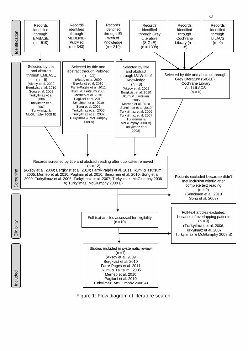
32
Figure 1: Flow diagram of literature search.
Records screened by title and abstract reading after duplicates removed (n = 12)
(Aksoy et al. 2009; Bergkvist et al. 2010; Farré-Pagés et al. 2011; Ikumi & Tsutsumi 2005; Merheb et al. 2010; Pagliani et al. 2010; Sencimen et al. 2010; Song et al.
2009; Turkyilmaz et al. 2006; Turkyilmaz et al. 2007; Turkyilmaz, McGlumphy 2008 A; Turkyilmaz, McGlumphy 2008 B)
Full-text articles excluded, because of overlapping patients
(n = 3)
(Turkyilmaz et al. 2006, Turkyilmaz et al. 2007,
Turkyilmaz & McGlumphy 2008 B)
Studies included in systematic review (n =7)
(Aksoy et al. 2009 Bergkvist et al. 2010
Farré-Pagés et al. 2011 Ikumi & Tsutsumi. 2005
Merheb et al. 2010 Pagliani et al. 2010
Turkyilmaz, McGlumphy 2008 A)
Records excluded because didn’t met inclusion criteria after
complete text reading (n = 2)
(Sencimen et al. 2010 Song et al. 2009)
Full-text articles assessed for eligibility (n =10)
Inclu
ded
Elig
ibili
ty
Scre
enin
g
Identification
Records identified through
MEDLINE-PubMed (n = 343)
Records identified
through Grey Literature (SIGLE)
(n = 1338)
Records identified through LILACS (n =0)
Records identified through
Cochrane Library (n =
18)
Records identified
through ISI Web of
Knowledge (n = 219)
Selected by title and abstract
through EMBASE (n = 6)
(Aksoy et al. 2009 Bergkvist et al. 2010
Song et al. 2009
Turkyilmaz et al. 2006
Turkyilmaz et al.
2007 Turkyilmaz &
McGlumphy 2008 B)
Selected by title and abstract through PubMed
(n = 11) (Aksoy et al. 2009
Bergkvist et al. 2010
Farré-Pagés et al. 2011 Ikumi & Tsutsumi 2005
Merheb et al. 2010
Pagliani et al. 2010 Sencimen et al. 2010
Song et al. 2009
Turkyilmaz et al. 2006 Turkyilmaz et al. 2007
Turkyilmaz & McGlumphy
2008 A)
Selected by title and abstract
through ISI Web of Knowledge
(n = 8) (Aksoy et al. 2009
Bergkvist et al. 2010 Ikumi & Tsutsumi
2005
Merheb et al. 2010 Sencimen et al. 2010 Turkyilmaz et al. 2006
Turkyilmaz et al. 2007 Turkyilmaz &
McGlumphy 2008 B)
Turkyilmaz et al. 2008)
Selected by title and abstract through Grey Literature (SIGLE),
Cochrane Library And LILACS
(n = 0)
Records identified through
EMBASE (n = 519)
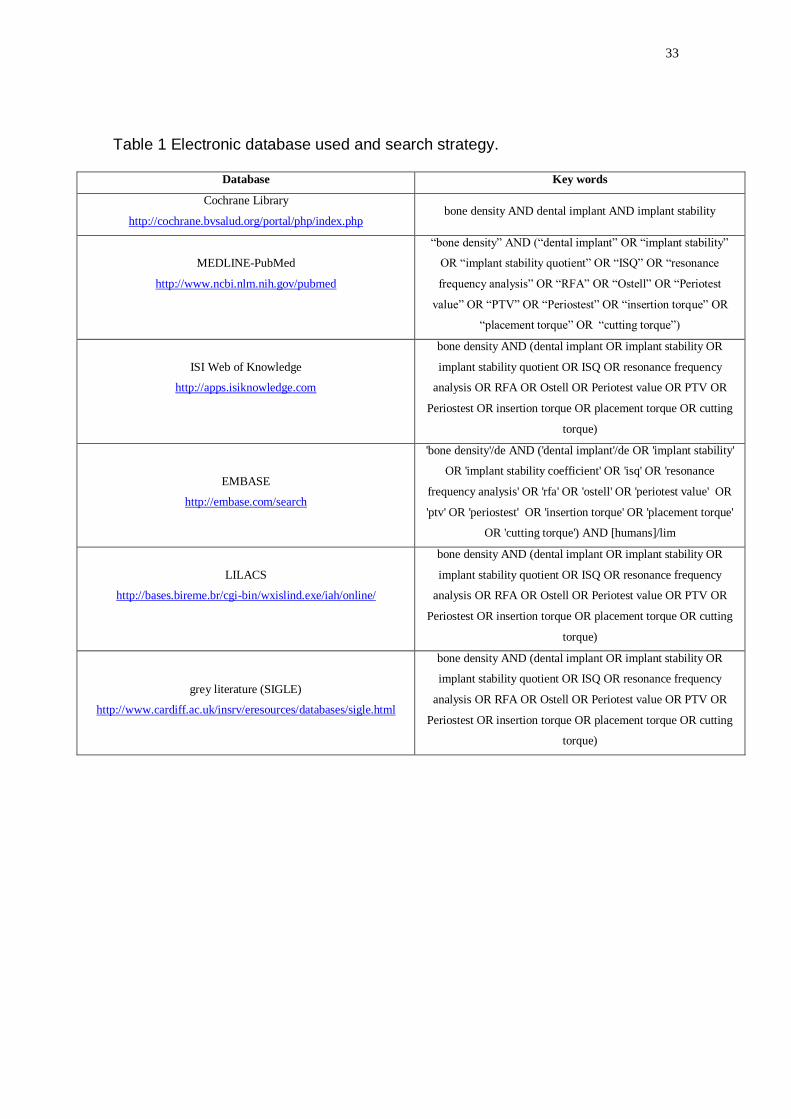
33
Table 1 Electronic database used and search strategy.
Database Key words
Cochrane Library
http://cochrane.bvsalud.org/portal/php/index.php bone density AND dental implant AND implant stability
MEDLINE-PubMed
http://www.ncbi.nlm.nih.gov/pubmed
“bone density” AND (“dental implant” OR “implant stability”
OR “implant stability quotient” OR “ISQ” OR “resonance
frequency analysis” OR “RFA” OR “Ostell” OR “Periotest
value” OR “PTV” OR “Periostest” OR “insertion torque” OR
“placement torque” OR “cutting torque”)
ISI Web of Knowledge
http://apps.isiknowledge.com
bone density AND (dental implant OR implant stability OR
implant stability quotient OR ISQ OR resonance frequency
analysis OR RFA OR Ostell OR Periotest value OR PTV OR
Periostest OR insertion torque OR placement torque OR cutting
torque)
EMBASE
http://embase.com/search
'bone density'/de AND ('dental implant'/de OR 'implant stability'
OR 'implant stability coefficient' OR 'isq' OR 'resonance
frequency analysis' OR 'rfa' OR 'ostell' OR 'periotest value' OR
'ptv' OR 'periostest' OR 'insertion torque' OR 'placement torque'
OR 'cutting torque') AND [humans]/lim
LILACS
http://bases.bireme.br/cgi-bin/wxislind.exe/iah/online/
bone density AND (dental implant OR implant stability OR
implant stability quotient OR ISQ OR resonance frequency
analysis OR RFA OR Ostell OR Periotest value OR PTV OR
Periostest OR insertion torque OR placement torque OR cutting
torque)
grey literature (SIGLE)
http://www.cardiff.ac.uk/insrv/eresources/databases/sigle.html
bone density AND (dental implant OR implant stability OR
implant stability quotient OR ISQ OR resonance frequency
analysis OR RFA OR Ostell OR Periotest value OR PTV OR
Periostest OR insertion torque OR placement torque OR cutting
torque)

34
Table 2: Methodological checklist for prognostic studies developed by the National Institute for Health and Clinical Excellence from United Kingdom. It was used to perform the quality assessment and control of bias.
Study identification:
Circle one option for each question
1.1 The study sample represents the population of interest with regard to key characteristics, sufficient to limit potential bias to the results? To minimise bias, the study population should be clearly defined and described and should represent the source population of interest. Points to consider include the following: • Are the source population or the population of interest adequately described with respect to key characteristics? • Are the sampling frame and recruitment adequately described, possibly including methods to identify the sample (number and type used; for example, referral patterns in healthcare), period of recruitment and place of recruitment (setting and geographical location)? • Are inclusion and exclusion criteria adequately described (for example, including explicit diagnostic criteria or a description of participants at the start of the follow-up period)? • Is participation in the study by eligible individuals adequate? • Is the baseline study sample (that is, individuals entering the study) adequately described with respect to key characteristics?
Yes No Unclear
1.2 Loss to follow-up is unrelated to key characteristics (that is, the study data adequately represent the sample), sufficient to limit potential bias? To minimise bias, completeness of follow-up should be described and adequate. Points to consider include the following: • Is the response rate (that is, proportion of study sample completing the study and providing outcome data) adequate? • Are attempts to collect information on participants who dropped out of the study described? • Are reasons for loss to follow-up provided? • Are the key characteristics of participants lost to follow-up adequately described? • Are there any important differences in key characteristics and outcomes between participants who completed the study and those who did not?
Yes No Unclear
1.3 The prognostic factor of interest is adequately measured in study participants, sufficient to limit potential bias? To minimise bias, prognostic factors should have been defined and measured appropriately. Points to consider include the following: • Is a clear definition or description of the prognostic factor(s) measured provided (including dose, level, duration of exposure, and clear specification of the method of measurement)? • Are continuous variables reported, or appropriate cut-off points (that is, not data-dependent) used? • Are the prognostic factor measured and the method of measurement valid and reliable enough to limit misclassification bias? (This may include relevant outside sources of information on measurement properties, as well as characteristics such as blind measurement and limited reliance on recall.) • Are complete data for prognostic factors available for an adequate proportion of the study sample?
Yes No Unclear
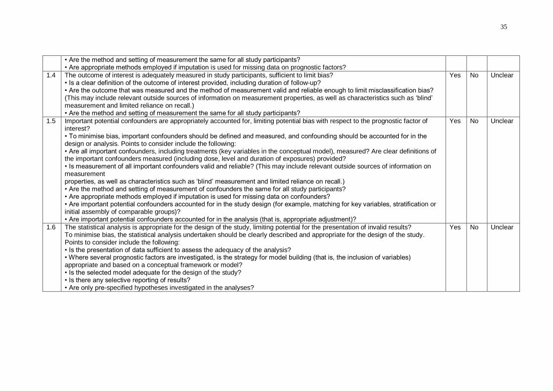
35
• Are the method and setting of measurement the same for all study participants? • Are appropriate methods employed if imputation is used for missing data on prognostic factors?
1.4 The outcome of interest is adequately measured in study participants, sufficient to limit bias? • Is a clear definition of the outcome of interest provided, including duration of follow-up? • Are the outcome that was measured and the method of measurement valid and reliable enough to limit misclassification bias? (This may include relevant outside sources of information on measurement properties, as well as characteristics such as ‘blind’ measurement and limited reliance on recall.) • Are the method and setting of measurement the same for all study participants?
Yes No Unclear
1.5 Important potential confounders are appropriately accounted for, limiting potential bias with respect to the prognostic factor of interest? • To minimise bias, important confounders should be defined and measured, and confounding should be accounted for in the design or analysis. Points to consider include the following: • Are all important confounders, including treatments (key variables in the conceptual model), measured? Are clear definitions of the important confounders measured (including dose, level and duration of exposures) provided? • Is measurement of all important confounders valid and reliable? (This may include relevant outside sources of information on measurement properties, as well as characteristics such as ‘blind’ measurement and limited reliance on recall.) • Are the method and setting of measurement of confounders the same for all study participants? • Are appropriate methods employed if imputation is used for missing data on confounders? • Are important potential confounders accounted for in the study design (for example, matching for key variables, stratification or initial assembly of comparable groups)? • Are important potential confounders accounted for in the analysis (that is, appropriate adjustment)?
Yes No Unclear
1.6 The statistical analysis is appropriate for the design of the study, limiting potential for the presentation of invalid results? To minimise bias, the statistical analysis undertaken should be clearly described and appropriate for the design of the study. Points to consider include the following: • Is the presentation of data sufficient to assess the adequacy of the analysis? • Where several prognostic factors are investigated, is the strategy for model building (that is, the inclusion of variables) appropriate and based on a conceptual framework or model? • Is the selected model adequate for the design of the study? • Is there any selective reporting of results? • Are only pre-specified hypotheses investigated in the analyses?
Yes No Unclear

36
Table 3: Articles ranked according the quality assessment and control of bias.
Aksoy et
al. 2009
Bergkvist
et al. 2010
Farré-
Pagés et
al. 2010
Ikumi &
Tsutsumi
2005
Merheb et
al. 2010
Pagliani et
al. 2010
Turkyilmaz
et al. 2006
Turkyilmaz
et al. 2007
Turkyilmaz &
McGlumphy
2008 A
Turkyilmaz &
McGlumphy
2008B
The study sample represents the population of interest with
regard to key characteristics, sufficient to limit potential bias
to the results.
yes yes unclear unclear unclear unclear unclear unclear yes unclear
Loss to follow-up is unrelated to key characteristics (that is,
the study data adequately represent the sample), sufficient
to limit potential bias.
yes yes yes yes yes yes yes yes yes yes
The prognostic factor of interest is adequately measured in
study participants, sufficient to limit potential bias. (In these
studies the prognostic factor was the BMD).
yes unclear unclear unclear unclear unclear yes yes yes yes
The outcome of interest is adequately measured in study
participants, sufficient to limit bias. (The outcome was the
primary stability).
unclear unclear unclear unclear unclear unclear unclear unclear unclear unclear
Important potential confounders are appropriately
accounted for, limiting potential bias with respect to the
prognostic factor of interest. (For example: implant
dimensions and cortical thickness).
no no no unclear yes no no no no no
The statistical analysis is appropriate for the design of the
study, limiting potential for the presentation of invalid
results
yes yes yes yes yes yes no yes yes yes
Category and situation of the article
4 “yes”:
Moderate
methodol
quality
Included
3 “yes”:
Moderate
methodol
quality
Included
2 “yes”:
Low
methodol
quality
Included
2 “yes” :
Low
methodol
quality
Included
3 “yes”:
Moderate
methodol
quality
Included
2 “yes”:
Low
methodol
quality
Included
2 “yes”:
Low
methodol
quality
Excluded*
3 “yes”:
Moderate
methodol
quality
Excluded*
4 “yes”:
Moderate
methodol
quality
Included
3 “yes”:
Moderate
methodol
quality
Excluded*
*The articles conducted by the same author had some overlapping patients. After ranking these studies, the one with the highest score was included in the systematic review, the others were excluded.
methodol= methodological
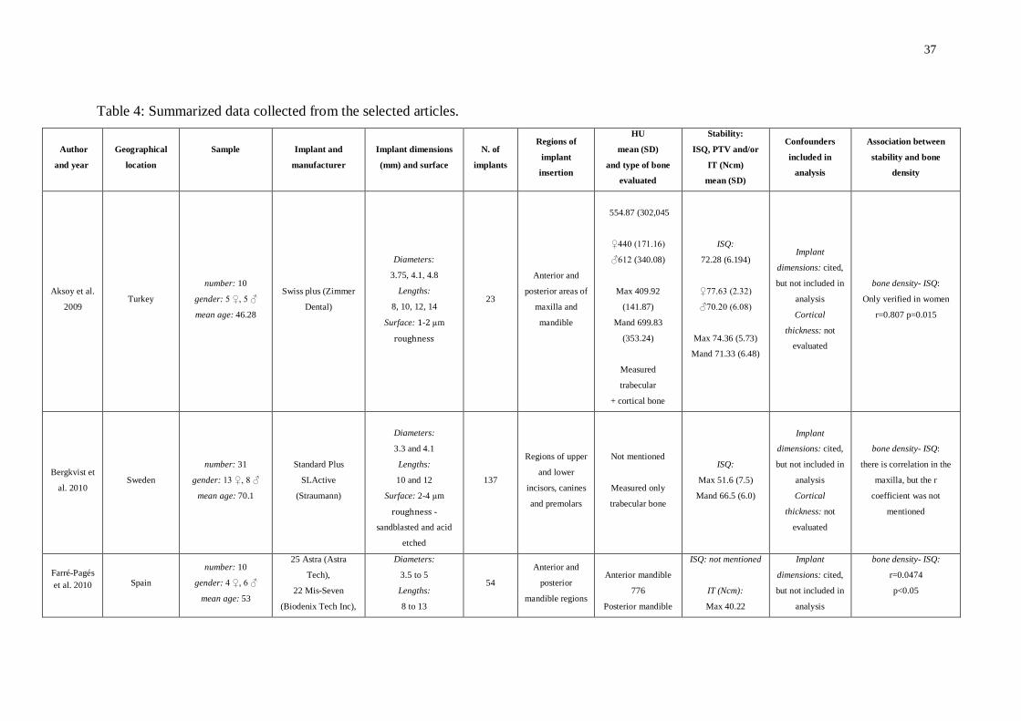
37
Table 4: Summarized data collected from the selected articles.
Author
and year
Geographical
location
Sample
Implant and
manufacturer
Implant dimensions
(mm) and surface
N. of
implants
Regions of
implant
insertion
HU
mean (SD)
and type of bone
evaluated
Stability:
ISQ, PTV and/or
IT (Ncm)
mean (SD)
Confounders
included in
analysis
Association between
stability and bone
density
Aksoy et al.
2009 Turkey
number: 10
gender: 5 ♀, 5 ♂
mean age: 46.28
Swiss plus (Zimmer
Dental)
Diameters:
3.75, 4.1, 4.8
Lengths:
8, 10, 12, 14
Surface: 1-2 μm
roughness
23
Anterior and
posterior areas of
maxilla and
mandible
554.87 (302,045
♀440 (171.16)
♂612 (340.08)
Max 409.92
(141.87)
Mand 699.83
(353.24)
Measured
trabecular
+ cortical bone
ISQ:
72.28 (6.194)
♀77.63 (2.32)
♂70.20 (6.08)
Max 74.36 (5.73)
Mand 71.33 (6.48)
Implant
dimensions: cited,
but not included in
analysis
Cortical
thickness: not
evaluated
bone density- ISQ:
Only verified in women
r=0.807 p=0.015
Bergkvist et
al. 2010 Sweden
number: 31
gender: 13 ♀, 8 ♂
mean age: 70.1
Standard Plus
SLActive
(Straumann)
Diameters:
3.3 and 4.1
Lengths:
10 and 12
Surface: 2-4 μm
roughness -
sandblasted and acid
etched
137
Regions of upper
and lower
incisors, canines
and premolars
Not mentioned
Measured only
trabecular bone
ISQ:
Max 51.6 (7.5)
Mand 66.5 (6.0)
Implant
dimensions: cited,
but not included in
analysis
Cortical
thickness: not
evaluated
bone density- ISQ:
there is correlation in the
maxilla, but the r
coefficient was not
mentioned
Farré-Pagés
et al. 2010
Spain
number: 10
gender: 4 ♀, 6 ♂
mean age: 53
25 Astra (Astra
Tech),
22 Mis-Seven
(Biodenix Tech Inc),
Diameters:
3.5 to 5
Lengths:
8 to 13
54
Anterior and
posterior
mandible regions
Anterior mandible
776
Posterior mandible
ISQ: not mentioned
IT (Ncm):
Max 40.22
Implant
dimensions: cited,
but not included in
analysis
bone density- ISQ:
r=0.0474
p<0.05
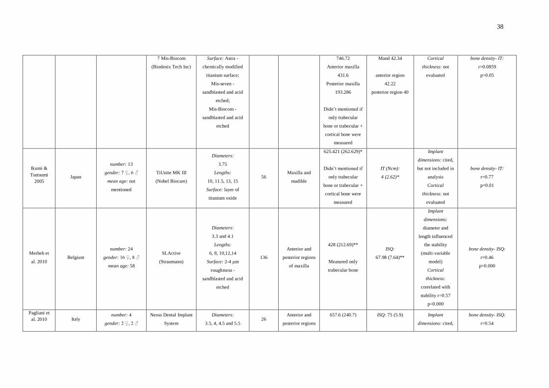
38
7 Mis-Biocom
(Biodenix Tech Inc)
Surface: Astra -
chemically modified
titanium surface;
Mis-seven -
sandblasted and acid
etched;
Mis-Biocom -
sandblasted and acid
etched
746.72
Anterior maxilla
431.6
Posterior maxilla
193.286
Didn’t mentioned if
only trabecular
bone or trabecular +
cortical bone were
measured
Mand 42.34
anterior region
42.22
posterior region 40
Cortical
thickness: not
evaluated
bone density- IT:
r=0.0859
p>0.05
Ikumi &
Tsutsumi
2005
Japan
number: 13
gender: 7 ♀, 6 ♂
mean age: not
mentioned
TiUnite MK III
(Nobel Biocare)
Diameters:
3.75
Lengths:
10, 11.5, 13, 15
Surface: layer of
titanium oxide
56 Maxilla and
madible
625.421 (262.629)*
Didn’t mentioned if
only trabecular
bone or trabecular +
cortical bone were
measured
IT (Ncm):
4 (2.62)*
Implant
dimensions: cited,
but not included in
analysis
Cortical
thickness: not
evaluated
bone density- IT:
r=0.77
p=0.01
Merheb et
al. 2010 Belgium
number: 24
gender: 16 ♀, 8 ♂
mean age: 58
SLActive
(Straumann)
Diameters:
3.3 and 4.1
Lengths:
6, 8, 10,12,14
Surface: 2-4 μm
roughness -
sandblasted and acid
etched
136
Anterior and
posterior regions
of maxilla
428 (212.69)**
Measured only
trabecular bone
ISQ:
67.98 (7.64)**
Implant
dimensions:
diameter and
length influenced
the stability
(multi-variable
model)
Cortical
thickness:
correlated with
stability r=0.57
p=0.000
bone density- ISQ:
r=0.46
p=0.000
Pagliani et
al. 2010
Italy number: 4
gender: 2 ♀, 2 ♂
Neoss Dental Implant
System
Diameters:
3.5, 4, 4.5 and 5.5 26
Anterior and
posterior regions
657.6 (240.7)
ISQ: 75 (5.9)
Implant
dimensions: cited,
bone density- ISQ:
r=0.54
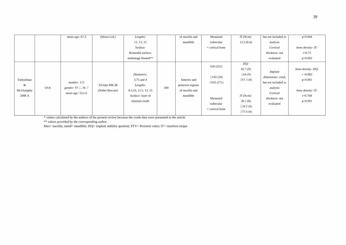
39
mean age: 67.3 (Neoss Ltd.) Lengths:
11, 13, 15
Surface:
Biomodal surface-
multistage blasted**
of maxilla and
mandible
Measured
trabecular
+ cortical bone
IT (Ncm):
12.2 (6.6)
but not included in
analysis
Cortical
thickness: not
evaluated
p=0.004
bone density- IT:
r=0.73
p=0.002
Turkyilmaz
&
McGlumphy
2008 A
USA
number: 111
gender: 55 ♀, 56 ♂
mean age: 55±11
TiUnite MK III
(Nobel Biocare)
Diameters:
3.75 and 4
Lengths:
8.5,10, 11.5, 13, 15
Surface: layer of
titanium oxide
300
Anterior and
posterior regions
of maxilla and
mandible
620 (251)
♀542 (20)
♂692 (271)
Measured
trabecular
+ cortical bone
ISQ:
65.7 (9)
♀64 (9)
♂67.3 (8)
IT (Ncm):
36.1 (8)
♀34.5 (8)
♂37.6 (8)
Implant
dimensions: cited,
but not included in
analysis
Cortical
thickness: not
evaluated
bone density- ISQ:
r =0.882
p˂0.001
bone density- IT:
r=0.768
p<0.001
* values calculated by the authors of the present review because the crude data were presented in the article.
** values provided by the corresponding author.
Max= maxilla; mand= mandible; ISQ= implant stability quotient; PTV= Periotest value; IT= insertion torque
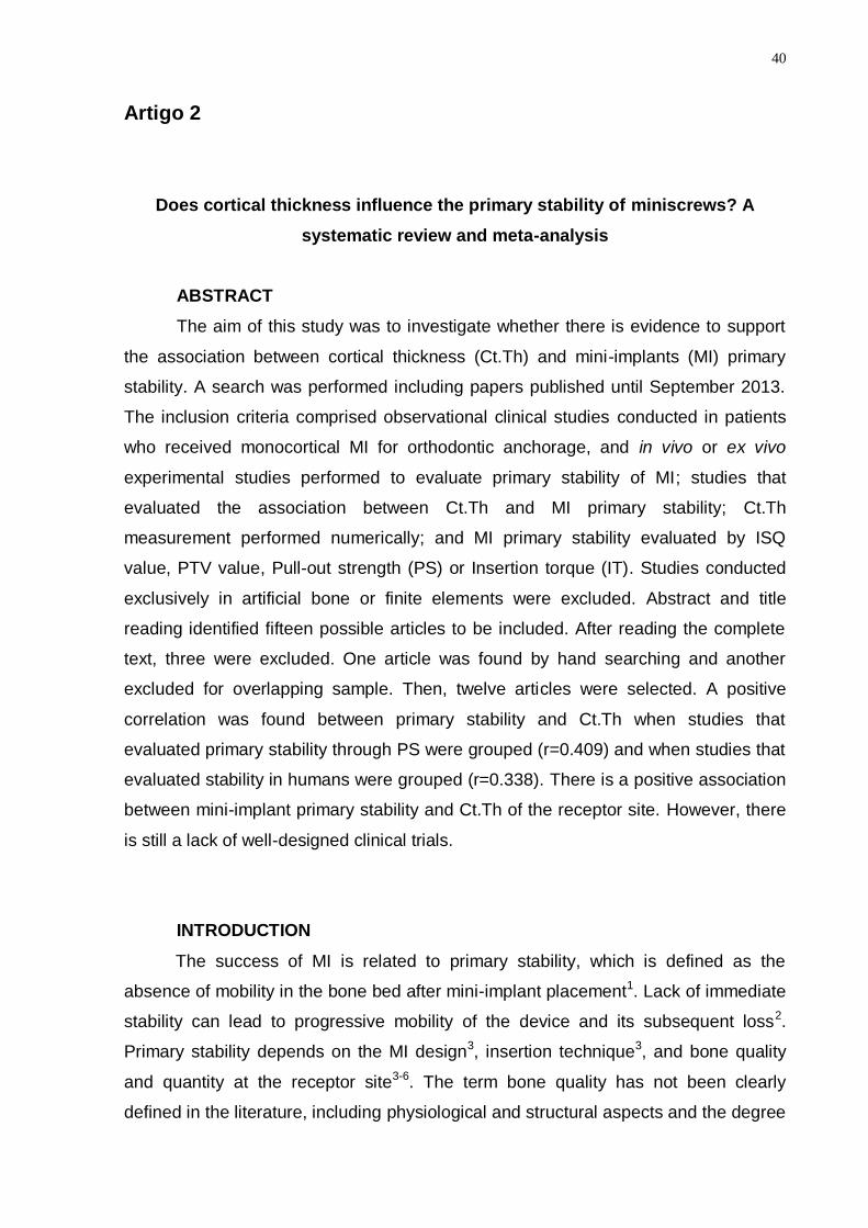
40
Artigo 2
Does cortical thickness influence the primary stability of miniscrews? A
systematic review and meta-analysis
ABSTRACT
The aim of this study was to investigate whether there is evidence to support
the association between cortical thickness (Ct.Th) and mini-implants (MI) primary
stability. A search was performed including papers published until September 2013.
The inclusion criteria comprised observational clinical studies conducted in patients
who received monocortical MI for orthodontic anchorage, and in vivo or ex vivo
experimental studies performed to evaluate primary stability of MI; studies that
evaluated the association between Ct.Th and MI primary stability; Ct.Th
measurement performed numerically; and MI primary stability evaluated by ISQ
value, PTV value, Pull-out strength (PS) or Insertion torque (IT). Studies conducted
exclusively in artificial bone or finite elements were excluded. Abstract and title
reading identified fifteen possible articles to be included. After reading the complete
text, three were excluded. One article was found by hand searching and another
excluded for overlapping sample. Then, twelve articles were selected. A positive
correlation was found between primary stability and Ct.Th when studies that
evaluated primary stability through PS were grouped (r=0.409) and when studies that
evaluated stability in humans were grouped (r=0.338). There is a positive association
between mini-implant primary stability and Ct.Th of the receptor site. However, there
is still a lack of well-designed clinical trials.
INTRODUCTION
The success of MI is related to primary stability, which is defined as the
absence of mobility in the bone bed after mini-implant placement1. Lack of immediate
stability can lead to progressive mobility of the device and its subsequent loss2.
Primary stability depends on the MI design3, insertion technique3, and bone quality
and quantity at the receptor site3-6. The term bone quality has not been clearly
defined in the literature, including physiological and structural aspects and the degree

41
of bone tissue mineralization. Additionally, the role of each of these aspects is not
completely understood7. While some authors assumed that bone quality is equivalent
to bone mineral density (BMD)7, others have considered that the bone quality refers
to Ct.Th3.
The present systematic review and meta-analysis was focused on the
following question: is there scientific evidence to support the influence of Ct.Th on the
primary stability of MI?
MATERIALS & METHODS
Study design
This is a Systematic Review of Prognosis that evaluated the association
between Ct.Th (prognostic factor) and MI primary stability (outcome). It is not a
Systematic Review of Intervention, as described by Cochrane Handbook and
PRISMA. Therefore, PRISMA statement was followed as possible.
Study selection criteria
The inclusion criteria comprised observational clinical studies conducted in
patients who received monocortical MI for orthodontic anchorage, and in vivo or ex
vivo experimental studies performed in animals to evaluate primary stability of MI;
studies that evaluated the association between Ct.Th and MI primary stability; the
Ct.Th measurement should have been performed numerically; and MI primary
stability should have been evaluated by ISQ value (Ostell, Integration Diagnostics,
Gothenburg, Sweden), PTV value (Periotest, Medizintechnik Gulden, Modautal,
Germany), PS or IT measurement. Studies conducted exclusively in artificial bone or
finite elements were excluded.
Search strategy and screening of articles
The search process was performed independently by two examiners (MM and
CTM) under the guidance of a librarian. Articles were searched without language
restriction until September 2013. Appropriate changes in the key words were done to
follow the syntax rules of each database (Table 1).
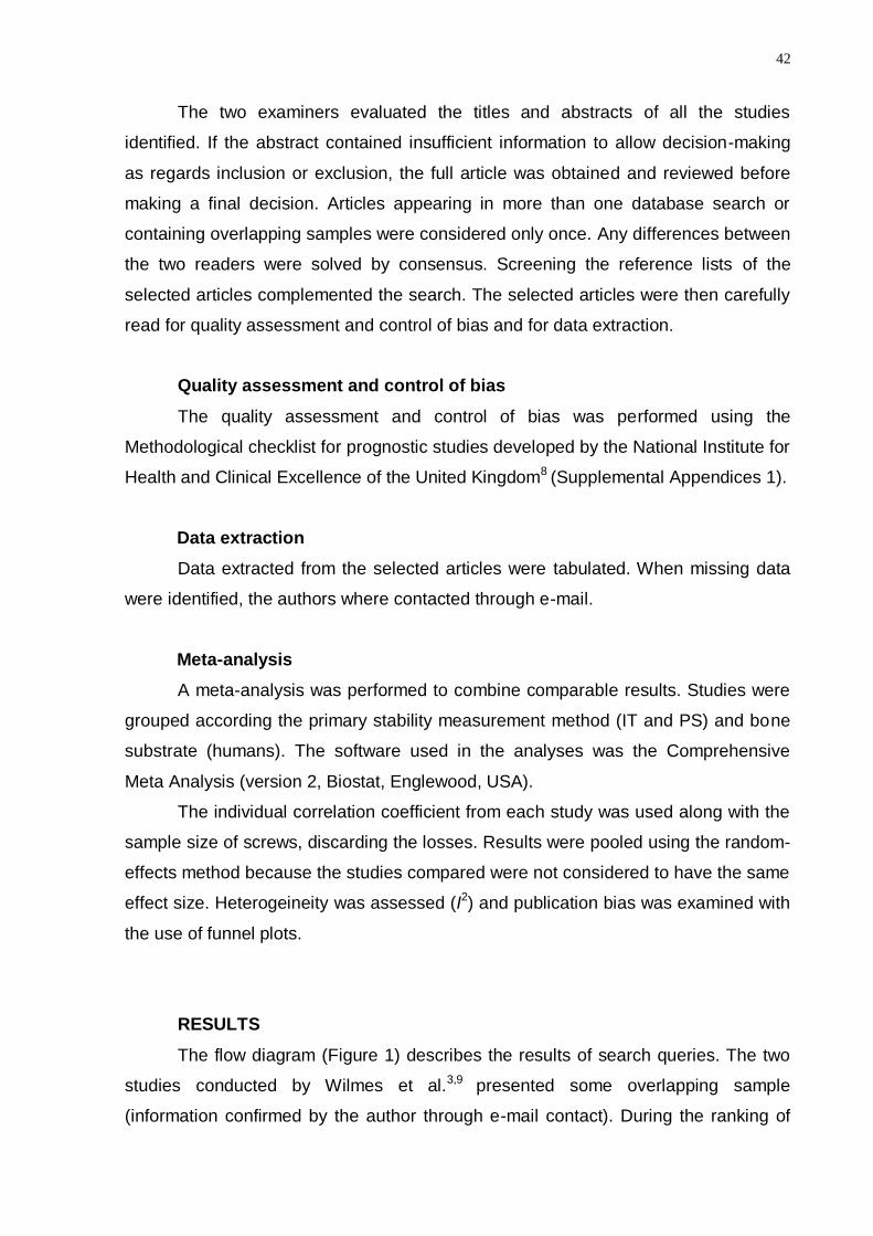
42
The two examiners evaluated the titles and abstracts of all the studies
identified. If the abstract contained insufficient information to allow decision-making
as regards inclusion or exclusion, the full article was obtained and reviewed before
making a final decision. Articles appearing in more than one database search or
containing overlapping samples were considered only once. Any differences between
the two readers were solved by consensus. Screening the reference lists of the
selected articles complemented the search. The selected articles were then carefully
read for quality assessment and control of bias and for data extraction.
Quality assessment and control of bias
The quality assessment and control of bias was performed using the
Methodological checklist for prognostic studies developed by the National Institute for
Health and Clinical Excellence of the United Kingdom8 (Supplemental Appendices 1).
Data extraction
Data extracted from the selected articles were tabulated. When missing data
were identified, the authors where contacted through e-mail.
Meta-analysis
A meta-analysis was performed to combine comparable results. Studies were
grouped according the primary stability measurement method (IT and PS) and bone
substrate (humans). The software used in the analyses was the Comprehensive
Meta Analysis (version 2, Biostat, Englewood, USA).
The individual correlation coefficient from each study was used along with the
sample size of screws, discarding the losses. Results were pooled using the random-
effects method because the studies compared were not considered to have the same
effect size. Heterogeineity was assessed (I2) and publication bias was examined with
the use of funnel plots.
RESULTS
The flow diagram (Figure 1) describes the results of search queries. The two
studies conducted by Wilmes et al.3,9 presented some overlapping sample
(information confirmed by the author through e-mail contact). During the ranking of
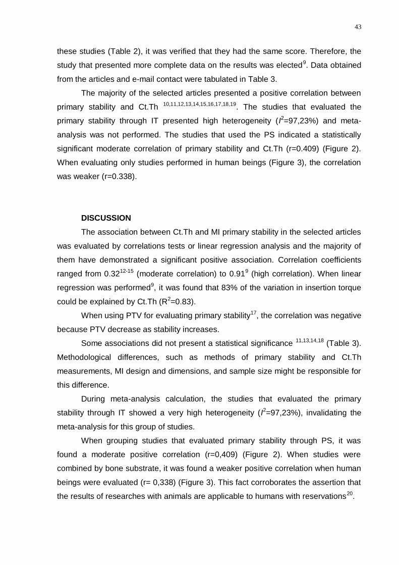
43
these studies (Table 2), it was verified that they had the same score. Therefore, the
study that presented more complete data on the results was elected9. Data obtained
from the articles and e-mail contact were tabulated in Table 3.
The majority of the selected articles presented a positive correlation between
primary stability and Ct.Th 10,11,12,13,14,15,16,17,18,19. The studies that evaluated the
primary stability through IT presented high heterogeneity (I2=97,23%) and meta-
analysis was not performed. The studies that used the PS indicated a statistically
significant moderate correlation of primary stability and Ct.Th (r=0.409) (Figure 2).
When evaluating only studies performed in human beings (Figure 3), the correlation
was weaker (r=0.338).
DISCUSSION
The association between Ct.Th and MI primary stability in the selected articles
was evaluated by correlations tests or linear regression analysis and the majority of
them have demonstrated a significant positive association. Correlation coefficients
ranged from 0.3212-15 (moderate correlation) to 0.919 (high correlation). When linear
regression was performed9, it was found that 83% of the variation in insertion torque
could be explained by Ct.Th (R2=0.83).
When using PTV for evaluating primary stability17, the correlation was negative
because PTV decrease as stability increases.
Some associations did not present a statistical significance 11,13,14,18 (Table 3).
Methodological differences, such as methods of primary stability and Ct.Th
measurements, MI design and dimensions, and sample size might be responsible for
this difference.
During meta-analysis calculation, the studies that evaluated the primary
stability through IT showed a very high heterogeneity (I2=97,23%), invalidating the
meta-analysis for this group of studies.
When grouping studies that evaluated primary stability through PS, it was
found a moderate positive correlation (r=0,409) (Figure 2). When studies were
combined by bone substrate, it was found a weaker positive correlation when human
beings were evaluated (r= 0,338) (Figure 3). This fact corroborates the assertion that
the results of researches with animals are applicable to humans with reservations20.

44
The MI design and dimensions are important parameters for the primary
stability of MI21. All of the selected studies reported the characteristics and
dimensions of the MI used, but few of them used more than one type and
investigated their influence on the result9,10,11. It was verified that the diameter of the
MI seemed to have a bigger influence on primary stability of MI than its length3, 9.
This fact can reinforce the importance of the Ct.Th in primary stability: as the MI
diameter increases, the contact surface between MI and cortical bone also increases.
On the other hand, when the length of MI increases, its contact with the trabecular
bone increases and the stability is not improved to the same extent.
Even though it was found that Ct.Th and primary stability are associated, it’s
important to remember that a very high primary stability is not desirable in clinical
practice because of the risk of bone necrosis and subsequent stability loss3.
Motoyoshi et al16 recommended placement torques between 5 and 10 Ncm.
Chaddad et al22 found higher success rates at torque values above 15 Ncm.
However, Meursinge Reynders et al23 performed a systematic review and stated that
no evidence indicates that specific maximum insertion torque levels are associated
with higher success rates for orthodontic MI mainly because insertional torque
measures are not very accurate24.
There is still a lack of studies with good methodological design evaluating the
relationship between Ct.Th and primary stability of MI. From the selected studies,
only two were conducted in living humans, convenience samples were used, and no
sample size calculation was made. One study was conducted in human cadavers,
without sample size calculation. The other studies were conducted in animals and
only one performed sample size calculation. The authors did not mention calibration,
error calculation and blinding. Therefore, the quality assessment ranked all the
articles as “low” considering the quality of evidence and control of bias. Another
factor that might have contributed to the low methodological quality was the quality
assessment checklist used: The Methodological checklist for prognostic studies
developed by the National Institute for Health and Clinical Excellence from United
Kingdom. This established checklist focused on clinical studies, while the majority of
selected studies were laboratorial.
The evidence to support the relationship between Ct.Th and MI primary
stability is still weak. It is recommended that well-designed clinical trials be conducted
45
to support this question, showing stronger evidence, preferably with the use of
multivariate analysis.
CONCLUSION
There is a positive association between MI primary stability and Ct.Th of the
receptor site. However, there is still a lack of well-designed clinical trials.
ACKNOWLEDGMENTS
We are grateful to CAPES (Cordenação de Aperfeiçoamento de Pessoal de
Nível Superior) for the PhD scholarship. The authors declare no potential conflicts of
interest with respect to the authorship and/or publication of this article.
REFERENCES
1. Javed F, Romanos GE. The role of primary stability for successful immediate loading
of dental implants. A literature review. Journal of Dentistry 2010;38(8):612-20.
2. Mischkowski RA, Kneuertz P, Florvaag B, Lazar F, Koebke J, Zoller JE.
Biomechanical comparison of four different miniscrew types for skeletal anchorage in
the mandibulo-maxillary area. International Journal of Oral and Maxillofac Surgery
2008;37(10):948-54.
3. Wilmes B, Rademacher C, Olthoff G, Drescher D. Parameters affecting primary
stability of orthodontic mini-implants. Journal of Orofacial Orthopedics
2006;67(3):162-74.
4. Cheng SJ, Tseng IY, Lee JJ, Kok SH. A prospective study of the risk factors
associated with failure of mini-implants used for orthodontic anchorage. International
Journal of Oral and Maxillofacial Implants 2004;19(1):100-6.
5. Freudenthaler JW, Haas R, Bantleon HP. Bicortical titanium screws for critical
orthodontic anchorage in the mandible: a preliminary report on clinical applications.
Clin Oral Implants Res 2001;12(4):358-63.
6. Trisi P, Rao W, Rebaudi A. A histometric comparison of smooth and rough titanium
implants in human low-density jawbone. International Journal of Oral Maxillofacial
Implants 1999;14(5):689-98.
7. Bergkvist G, Koh KJ, Sahlholm S, Klintstrom E, Lindh C. Bone density at implant
sites and its relationship to assessment of bone quality and treatment outcome.
International Journal of Oral and Maxillofacial Implants 2010;25(2):321-8.
46
8. Marquezan M, Osorio A, Sant'Anna E, Souza MM, Maia L. Does bone mineral
density influence the primary stability of dental implants? A systematic review.
Clinical Oral Implants Research 2011.
9 Wilmes B, Drescher D. Impact of bone quality, implant type, and implantation site
preparation on insertion torques of mini-implants used for orthodontic anchorage.
International Journal of Oral Maxillofacial Surgery 2011;40(7):697-703.
10. Cha JY, Kil JK, Yoon TM, Hwang CJ. Miniscrew stability evaluated with
computerized tomography scanning. American Journal of Orthodontics and
Dentofacial Orthopedics 2010;137(1):73-9.
11. Cehreli S, Arman-Ozcirpici A. Primary stability and histomorphometric bone-implant
contact of self-drilling and self-tapping orthodontic microimplants. Am J Orthod
Dentofacial Orthop 2012;141(2):187-95.
12. Huja SS, Litsky AS, Beck FM, Johnson KA, Larsen PE. Pull-out strength of
monocortical screws placed in the maxillae and mandibles of dogs. Am J Orthod
Dentofacial Orthop 2005;127(3):307-13.
13. McManus MM, Qian F, Grosland NM, Marshall SD, Southard TE. Effect of
miniscrew placement torque on resistance to miniscrew movement under load. Am J
Orthod Dentofacial Orthop 2011;140(3):e93-8.
14. Migliorati M, Benedicenti S, Signori A, Drago S, Barberis F, Tournier H, et al.
Miniscrew design and bone characteristics: an experimental study of primary stability.
American Journal of Orthodontics and Dentofacial Orthopedics 2012;142(2):7.
15. Motoyoshi M, Uemura M, Ono A, et al. Factors affecting the long-term stability of
orthodontic mini-implants. Am J Orthod Dentofacial Orthop 2010;137(5):588 e1-5;
discussion 88-9.
16. Motoyoshi M, Hirabayashi M, Uemura M, Shimizu N. Recommended placement
torque when tightening an orthodontic mini-implant. Clin Oral Implants Res
2006;17(1):109-14.
17. Nienkemper M, Wilmes B, Panayotidis A, Pauls A, Golubovic V, Schwarz F, et al.
Measurement of mini-implant stability using resonance frequency analysis. Angle
Orthod 2012;00(0):000.
18. Salmoria KK, Tanaka OM, Guariza-Filho O, Camargo ES, de Souza LT, Maruo H.
Insertional torque and axial pull-out strength of mini-implants in mandibles of dogs.
American Journal of Orthodontics and Dentofacial Orthopedics 2008;133(6):790 e15-
22.
19. Su YY, Wilmes B, Honscheid R, Drescher D. Application of a wireless resonance
frequency transducer to assess primary stability of orthodontic mini-implants: an in
vitro study in pig ilia. Int J Oral Maxillofac Implants 2009;24(4):647-54.

47
20. Neto J. Ethics in research. In: Estrela C, editor. Scientific Methodology. São Paulo:
Artes Médicas; 2001.
21. Lim SA, Cha JY, Hwang CJ. Insertion torque of orthodontic miniscrews according to
changes in shape, diameter and length. The Angle Orthodontist 2008;78(2):234-40.
22. Chaddad K, Ferreira AF, Geurs N, Reddy MS. Influence of surface characteristics on
survival rates of mini-implants. Angle Orthod 2008;78(1):107-13.
23. Meursinge Reynders RA, Ronchi L, Ladu L, van Etten-Jamaludin F, Bipat S. Insertion
torque and success of orthodontic mini-implants: a systematic review. Am J Orthod
Dentofacial Orthop 2012;142(5):596-614 e5.
24. Schatzle M, Golland D, Roos M, Stawarczyk B. Accuracy of mechanical torque-
limiting gauges for mini-screw placement. Clin Oral Implants Res 2010;21(8):781-8.

48
Table 1 Database and search strategy used.
Database Key words
Cochrane Library
http://cochrane.bvsalud.org/portal/php/index.php
(cortical OR compacta) AND (miniscrew OR "mini implant" OR
"mini-implant") AND stability
MEDLINE-PubMed
http://www.ncbi.nlm.nih.gov/pubmed
(cortical OR compacta) AND (miniscrew OR "mini implant" OR
"mini-implant") AND (“stability” OR “implant stability quotient” OR
“ISQ” OR “resonance frequency analysis” OR “RFA” OR “Ostell” OR
“Periotest value” OR “PTV” OR “Periostest” OR “insertion torque”
OR "insertional torque" OR “placement torque” OR “cutting torque”)
Web of Knowledge
http://apps.webofknowledge.com
((cortical OR compacta) AND (miniscrew OR mini implant OR mini-
implant) AND (stability OR implant stability quotient OR ISQ OR
resonance frequency analysis OR RFA OR Ostell OR Periotest value
OR PTV OR Periostest OR insertion torque OR insertional torque OR
placement torque OR cutting torque))
Refined by: Subject Areas=(DENTISTRY ORAL SURGERY
MEDICINE OR SURGERY)
EMBASE
http://embase.com/search
(cortical OR compacta) AND (miniscrew OR 'mini implant') AND
(stability OR 'implant stability quotient' OR isq OR 'resonance
frequency analysis' OR rfa OR ostell OR 'periotest value' OR ptv OR
periostest OR 'insertion torque' OR 'insertional torque' OR 'placement
torque' OR 'cutting torque')
VHL
http://regional.bvsalud.org/php/index.php
(cortical OR compacta) AND (miniscrew OR "mini implant") AND
(stability OR "implant stability quotient" OR ISQ OR "resonance
frequency analysis" OR RFA OR Ostell OR "Periotest value" OR PTV
OR Periostest OR "insertion torque" OR "insertional torque" OR
"placement torque" OR "cutting torque")
Grey literature (SIGLE)
http://www.cardiff.ac.uk/insrv/eresources/databases/sigle.html
(cortical OR compacta) AND (miniscrew OR "mini implant" OR
"mini-implant") AND(“stability” OR “implant stability quotient” OR
“ISQ” OR “resonance frequency analysis” OR “RFA” OR “Ostell” OR
“Periotest value” OR “PTV” OR “Periostest” OR “insertion torque”
OR "insertional torque" OR “placement torque” OR “cutting torque”)
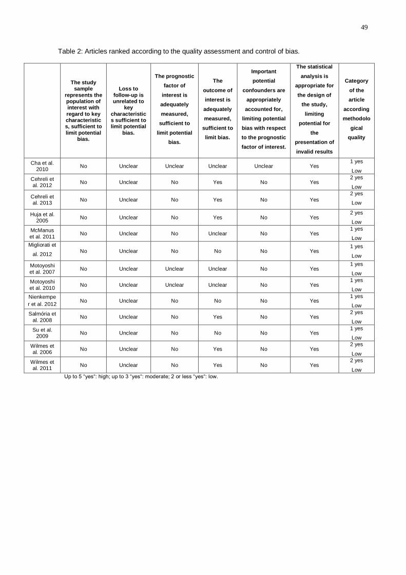
49
Table 2: Articles ranked according to the quality assessment and control of bias.
The study sample
represents the population of interest with
regard to key characteristics, sufficient to
limit potential bias.
Loss to
follow-up is unrelated to
key
characteristics sufficient to limit potential
bias.
The prognostic
factor of
interest is
adequately
measured,
sufficient to
limit potential
bias.
The
outcome of
interest is
adequately
measured,
sufficient to
limit bias.
Important
potential
confounders are
appropriately
accounted for,
limiting potential
bias with respect
to the prognostic
factor of interest.
The statistical
analysis is
appropriate for
the design of
the study,
limiting
potential for
the
presentation of
invalid results
Category
of the
article
according
methodolo
gical
quality
Cha et al. 2010 No Unclear Unclear Unclear Unclear Yes
1 yes
Low
Cehreli et al. 2012 No Unclear No Yes No Yes
2 yes
Low
Cehreli et al. 2013
No Unclear No Yes No Yes 2 yes
Low
Huja et al.
2005 No Unclear No Yes No Yes 2 yes
Low
McManus
et al. 2011 No Unclear No Unclear No Yes 1 yes
Low Migliorati et
al. 2012 No Unclear No No No Yes
1 yes
Low
Motoyoshi
et al. 2007 No Unclear Unclear Unclear No Yes 1 yes
Low
Motoyoshi
et al. 2010 No Unclear Unclear Unclear No Yes 1 yes
Low Nienkempe
r et al. 2012 No Unclear No No No Yes
1 yes
Low
Salmória et
al. 2008 No Unclear No Yes No Yes
2 yes
Low
Su et al.
2009 No Unclear No No No Yes 1 yes
Low
Wilmes et al. 2006 No Unclear No Yes No Yes
2 yes
Low
Wilmes et al. 2011 No Unclear No Yes No Yes
2 yes
Low Up to 5 “yes“: high; up to 3 “yes“: moderate; 2 or less “yes“: low.

50
Table 3: Summarized data collected from the selected articles.
Author/year Study design
Sample/
substrate
N° of
MI
MI model, shape,
classification of insertion
method, dimensions
(diameter x length) and
manufacturer
Method of
Ct.Th
evaluation
Main method of
primary
stability
measurement*
Association
between
stability and
Ct.Th
Confounders included
in analysis
Cha et al. 2010 Experimental
in vivo (dogs)
maxilla and
mandible of
6 dogs
96
model 1: OAS-1507C
cylindrical
non-drilling
1.4x7mm
Biomaterials Korea
(Korea)
model 2: OAS-1507T
tapered
non-drilling
1.4x7mm
Biomaterials Korea
(Korea)
CBCT IT
r=0.476
p=0.001
(Pearson)
BMD of cortical
BMD of total bone
Screw type
Screw position
Insertion technique
(Multiple regression
analysis)
Cehreli et al.
2012
Experimental
ex vivo (bovine)
16 bovine
Iliac crest 72
model 1: AbsoAnchor
cylindrical
self-taping
1.4x7mm
Dentos (Korea)
model 2: AbsoAnchor
cylindrical
self-drilling
1.4x7mm
Dentos (Korea)
CT IT
model 1
r=0.516
p=0.001
(Pearson)
model 2
r=0.544
p=0.001
(Pearson)
Evaluated the
insertional angle of MI,
BMD of cortical and
trabecular bones, but no
multivariate analysis
was performed
Cehreli et al.
2013
Experimental
ex vivo (bovine)
4 bovine
Iliac crest 24
model: AbsoAnchor
cylindrical
self-drilling
1.4x7mm
Dentos (Korea)
CT IT
r=0.194
p>0.05 (not
significant)
(Spearman)
Evaluated the
insertional angle of MI,
BMD of cortical and
trabecular bones, but no
multivariate analysis
was performed
Huja et al.
2005
Experimental
ex vivo (dogs)
maxilla and
mandible of
4 dogs
56
model: not mentioned
shape: not mentioned
self-drilling
2x6mm
Synthes (USA)
Microscope
with a grid PS
r=0.39
p=0.02
(Pearson)
-
McManus et
al. 2011
Experimental ex
vivo (human
cadavers)
24 hemi-
mandibles
and 24
hemi-
maxilla
96
model: not mentioned
nontapered
CIM: not mentioned
1.5x11mm
KLS Martin (USA)
Digital caliper IT
Maxilla
r= not
mentioned
p>0.05 (not
significant)
Mandible
r=0.61
p<0.00001
(Pearson)
Evaluated the
correlation of IT and
bone type (maxilla or
mandible), but no
multivariate analysis
was performed
Experimental
ex vivo (pigs)
Pig ribs
20
model 1: Orthoeasy
cylindrical
self-drilling
1.7x10mm
Forestadent (Germany)

51
Migliorati et
al. 2012
model 2: Orthoscrew
cylindrical
self-drilling
1.65x9mm
Leader Ortodonzia (Italy)
model 3: Tomas
cylindrical
self-drilling
1.6x10mm
Dentaurum (Germany)
model 4: ORTHOImplant
tapered
self-drilling
1.8x10mm
3M Unitek (USA)
CBCT
PS and IT
PS:
T=0.36
p=0.027
IT:
T= 0.27
p=0.18 (not
significant)
(Kendall rank
correlation)
Evaluated BMD of
cortical and trabecular
bones, but no
multivariate analysis
was performed
considering these
aspects
Motoyoshi et
al. 2007
Observational
(humans)
maxilla and
mandible of
32 patients
87
ISA system orthodontics
implants
tapered
CIM: not mentioned
1.6x8mm
Biodent (Japan)
CT IT
r=0.32
p=0.002
(Pearson)
-
Motoyoshi et
al. 2010
Observational
(humans)
maxilla and
mandible of
57 patients
(5 losen)
148
ISA system orthodontics
implants
tapered
CIM: not mentioned
1.6x8mm
Biodent (Japan)
CT IT
Maxilla
r=0.392
p<0.05
Mandible
r= -0.019
p>0.05 (not
significant)
(Pearson)
Evaluated the
correlation of placement
torque and bone type
(maxilla or mandible),
but no multivariate
analysis was performed
Nienkemper et
al. 2012
Experimental
ex vivo (pigs)
Porcine
pelvic bone 110
Benefit s
cylindrical
self-drilling
2x9mm
PSM medical solutions
(Germany)
CBCT RFA and PTV
RFA
r=0.71
p<0.001
PTV
r= -0.64
p<0.001
(Pearson)
-
Salmória et al.
2008
Experimental
in vivo (dogs)
10 mandible
of dogs 60
model: specially
manufactured for this
research**
cylindrical**
self-tapping
1.6x6mm
Neodent Implante
Osteointegrável (Brazil)
Microscope
and a digital
caliper rule
PS and IT
PS:
r=0.44
p=0.05
IT:
r= not
mentioned
p>0.05 (not
significant)
(Pearson)
-
Su et al. 2009
Experimental
ex vivo (pigs)
1 swine
pelvic
bone**
15
Dual top
tapered**
self-drilling**
1.6x8mm
Jeil medical Corp (Korea)
Scanned image
measurement
in Image Pro
Plus software
RFA
r=0.9
p˂0.0001
(Pearson
correlation)
-
Wilmes et al.
2011
Experimental
ex vivo (pigs)
20 swine
pelvic bone 600
model 1: Dual top (DT)
conical
self-drilling
Micro-CT IT
DT 2x10mm:
R2=0.69
DT 1.6x8mm:
Screw type
Screw position
Insertion technique
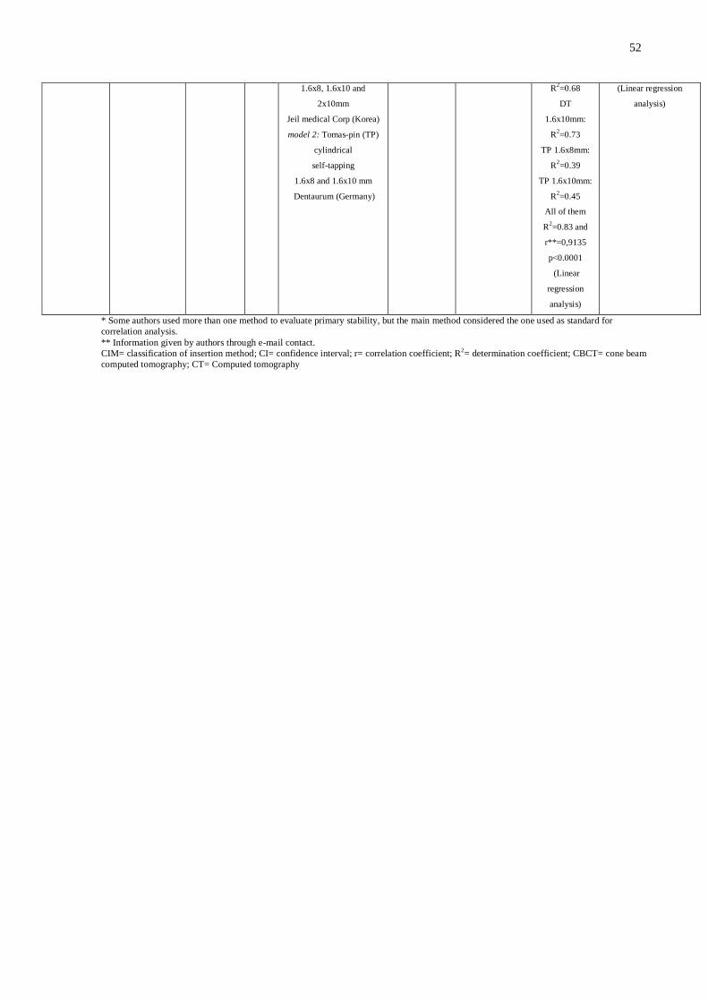
52
1.6x8, 1.6x10 and
2x10mm
Jeil medical Corp (Korea)
model 2: Tomas-pin (TP)
cylindrical
self-tapping
1.6x8 and 1.6x10 mm
Dentaurum (Germany)
R2=0.68
DT
1.6x10mm:
R2=0.73
TP 1.6x8mm:
R2=0.39
TP 1.6x10mm:
R2=0.45
All of them
R2=0.83 and
r**=0,9135
p˂0.0001
(Linear
regression
analysis)
(Linear regression
analysis)
* Some authors used more than one method to evaluate primary stability, but the main method considered the one used as standard for
correlation analysis.
** Information given by authors through e-mail contact.
CIM= classification of insertion method; CI= confidence interval; r= correlation coefficient; R2= determination coefficient; CBCT= cone beam
computed tomography; CT= Computed tomography

53
Supplemental Appendices 1: Methodological checklist for prognostic studies developed by the National Institute for Health and Clinical Excellence from United Kingdom. It was used to perform the quality assessment and control of bias.
Study identification:
Circle one option for each question
1.1 The study sample represents the population of interest with regard to key characteristics, sufficient to limit potential bias to the results? To minimise bias, the study population should be clearly defined and described and should represent the source population of interest. Points to consider include the following: • Are the source population or the population of interest adequately described with respect to key characteristics? • Are the sampling frame and recruitment adequately described, possibly including methods to identify the sample (number and type used; for example, referral patterns in healthcare), period of recruitment and place of recruitment (setting and geographical location)? • Are inclusion and exclusion criteria adequately described (for example, including explicit diagnostic criteria or a description of participants at the start of the follow-up period)? • Is participation in the study by eligible individuals adequate? • Is the baseline study sample (that is, individuals entering the study) adequately described with respect to key characteristics?
Yes No Unclear
1.2 Loss to follow-up is unrelated to key characteristics (that is, the study data adequately represent the sample), sufficient to limit potential bias? To minimise bias, completeness of follow-up should be described and adequate. Points to consider include the following: • Is the response rate (that is, proportion of study sample completing the study and providing outcome data) adequate? • Are attempts to collect information on participants who dropped out of the study described? • Are reasons for loss to follow-up provided? • Are the key characteristics of participants lost to follow-up adequately described? • Are there any important differences in key characteristics and outcomes between participants who completed the study and those who did not?
Yes No Unclear
1.3 The prognostic factor of interest is adequately measured in study participants, sufficient to limit potential bias? To minimise bias, prognostic factors should have been defined and measured appropriately. Points to consider include the following: • Is a clear definition or description of the prognostic factor(s) measured provided (including dose, level, duration of exposure, and clear specification of the method of measurement)? • Are continuous variables reported, or appropriate cut-off points (that is, not data-dependent) used? • Are the prognostic factor measured and the method of measurement valid and reliable enough to limit misclassification bias?
Yes No Unclear

54
(This may include relevant outside sources of information on measurement properties, as well as characteristics such as blind measurement and limited reliance on recall.) • Are complete data for prognostic factors available for an adequate proportion of the study sample? • Are the method and setting of measurement the same for all study participants? • Are appropriate methods employed if imputation is used for missing data on prognostic factors?
1.4 The outcome of interest is adequately measured in study participants, sufficient to limit bias? • Is a clear definition of the outcome of interest provided, including duration of follow-up? • Are the outcome that was measured and the method of measurement valid and reliable enough to limit misclassification bias? (This may include relevant outside sources of information on measurement properties, as well as characteristics such as ‘blind’ measurement and limited reliance on recall.) • Are the method and setting of measurement the same for all study participants?
Yes No Unclear
1.5 Important potential confounders are appropriately accounted for, limiting potential bias with respect to the prognostic factor of interest? • To minimise bias, important confounders should be defined and measured, and confounding should be accounted for in the design or analysis. Points to consider include the following: • Are all important confounders, including treatments (key variables in the conceptual model), measured? Are clear definitions of the important confounders measured (including dose, level and duration of exposures) provided? • Is measurement of all important confounders valid and reliable? (This may include relevant outside sources of information on measurement properties, as well as characteristics such as ‘blind’ measurement and limited reliance on recall.) • Are the method and setting of measurement of confounders the same for all study participants? • Are appropriate methods employed if imputation is used for missing data on confounders? • Are important potential confounders accounted for in the study design (for example, matching for key variables, stratification or initial assembly of comparable groups)? • Are important potential confounders accounted for in the analysis (that is, appropriate adjustment)?
Yes No Unclear
1.6 The statistical analysis is appropriate for the design of the study, limiting potential for the presentation of invalid results? To minimise bias, the statistical analysis undertaken should be clearly described and appropriate for the design of the study. Points to consider include the following: • Is the presentation of data sufficient to assess the adequacy of the analysis? • Where several prognostic factors are investigated, is the strategy for model building (that is, the inclusion of variables) appropriate and based on a conceptual framework or model? • Is the selected model adequate for the design of the study? • Is there any selective reporting of results? • Are only pre-specified hypotheses investigated in the analyses?
Yes No Unclear
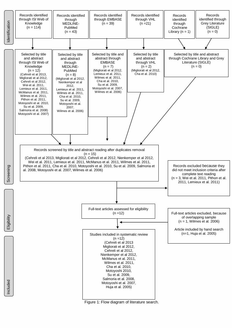
55
Figure 1: Flow diagram of literature search.
Scre
enin
g
Inclu
ded
Elig
ibili
ty
Records screened by title and abstract reading after duplicates removal (n = 15)
(Cehreli et al 2013, Migliorati et al 2012, Cehreli et al 2012, Nienkemper et al 2012, Wei et al. 2011, Lemieux et al. 2011, McManus et al. 2011, Wilmes et al. 2011,
Pithon et al. 2011, Cha et al. 2010, Motoyoshi et al. 2010, Su et al. 2009, Salmoria et al. 2008, Motoyoshi et al. 2007, Wilmes et al. 2006)
Records identified through EMBASE
(n = 39)
Identification
Records identified through
MEDLINE-PubMed (n = 43)
Records identified through Grey Literature
(SIGLE)
(n = 0)
Records identified through VHL
(n =21)
Records identified through
Cochrane Library (n = 1)
Full-text articles assessed for eligibility (n =12) Full-text articles excluded, because
of overlapping sample (n = 1, Wilmes et al. 2006)
Article included by hand search
(n=1, Huja et al. 2005) Studies included in systematic review (n =12)
(Cehreli et al 2013 Migliorati et al 2012, Cehreli et al 2012,
Nienkemper et al 2012, McManus et al. 2011, Wilmes et al. 2011,
Cha et al. 2010, Motoyoshi 2010, Su et al. 2009,
Salmoria et al. 2008, Motoyoshi et al. 2007,
Huja et al. 2005)
Records excluded because they did not meet inclusion criteria after
complete text reading (n = 3, Wei et al. 2011, Pithon et al.
2011, Lemieux et al. 2011)
Records identified through ISI Web of
Knowledge (n = 114)
Selected by title and abstract through
EMBASE (n = 7)
(Migliorati et al 2012, Lemieux et al. 2011,
Wilmes et al. 2011, Cha et al. 2010, Su et al. 2009,
Motoyoshi et al. 2007, Wilmes et al. 2006)
Selected by title and abstract
through MEDLINE-PubMed (n = 8)
(Migliorati et al 2012, Nienkemper et al
2012,
Lemieux et al. 2011, Wilmes et al. 2011,
Cha et al. 2010,
Su et al. 2009, Motoyoshi et al.
2007,
Wilmes et al. 2006)
Selected by title and abstract
through ISI Web of Knowledge
(n = 12) (Cehreli et al 2013, Migliorati et al 2012,
Cehreli et al 2012, Wei et al. 2011,
Lemieux et al. 2011,
McManus et al. 2011, Wilmes et al. 2011, Pithon et al. 2011,
Motoyoshi et al. 2010, Su et al. 2009,
Salmoria et al. 2008)
Motoyoshi et al. 2007)
Selected by title and abstract through Cochrane Library and Grey
Literature (SIGLE) (n = 0)
Selected by title and abstract through VHL
(n = 2) (Migliorati et al 2012,
Cha et al. 2010)
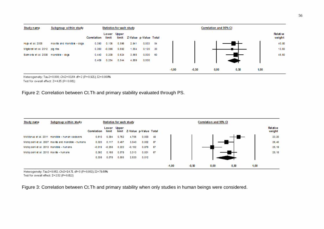
56
Figure 2: Correlation between Ct.Th and primary stability evaluated through PS.
Figure 3: Correlation between Ct.Th and primary stability when only studies in human beings were considered.

57
Artigo 3
Is trabecular bone related to primary stability of miniscrews?
Mariana Marquezana; Inayá Lima
b; Ricardo Tadeu Lopes
b;
Eduardo Franzotti Sant’Annac; Margareth Maria Gomes de Souza
c
a PhD Student, Department of Pedodontics and Orthodontics, Universidade Federal do Rio de Janeiro,
Rio de Janeiro, Brazil
b Professor, Nuclear Instrumentation Laboratory, Nuclear Engineering Program, Universidade Federal
do Rio de Janeiro, Rio de Janeiro, Brazil.
c Professor, Department of Pedodontics and Orthodontics, Universidade Federal do Rio de Janeiro,
Rio de Janeiro, Brazil
Corresponding Author: Mariana Marquezan, Department of Pedodontics and Orthodontics,
Universidade Federal do Rio de Janeiro (UFRJ), Avenida Professor Rodolpho Paulo Rocco, 325,
Ilha do Fundão, Rio de Janeiro, RJ, Brazil, 21941-617.
Email: [email protected]
© 2014 by the EH Angle Education and Research Foundation, Inc.
Submitted: May 2013
Color Figures – no
58
ABSTRACT
Objective: To compare the primary stability of miniscrews inserted into bone blocks of different bone
mineral density (BMD) with and without cortical bone, and investigate whether some trabecular
properties could influence primary stability.
Materials and Methods: Fifty-two bone blocks were extracted from fresh bovine pelvic bone. Four
groups were created considering the bone type (iliac or pubic region) and presence or absence of
cortical. Specimens were microCT imaged to evaluate trabecular thickness, trabecular number,
trabecular separation, bone volume density (BV/TV), BMD and cortical thickness. Miniscrews 1.4 mm
in diameter and 6 mm long were inserted into the bone blocks and primary stability was evaluated by
insertion torque (IT), mini-implant mobility (PTV) and pull-out strength (PS).
Results: Intergroup comparison showed lower level of primary stability when BMD of trabecular bone
was lower and in the absence of cortical (P≤.05). Pearson's correlation test showed correlation
between trabecular number, trabecular thickness, BV/TV, trabecular BMD, total BMD, and IT, PTV and
PS. There was correlation between cortical thickness and IT and PS (P≤.05).
Conclusion: Cancellous bone plays an important role in primary stability of mini-implants in the
presence or absence of cortical bone.
KEY WORDS: Trabecular bone; Mini implant; Miniscrew; Stability
59
INTRODUCTION
The success of skeletal anchorage using miniscrews is related to their stability in the bone. Most of the
failures occur immediately after mini-implant placement1 because the lack of primary stability may lead
to progressive mobility of the device and to its subsequent loss2. Factors that influence the immediate
stability are related to the design of the device, quantity and quality of bone, and insertion technique3.
The term “bone quality” is not clearly defined in the literature. This includes physiological and
structural aspects and the degree of bone tissue mineralization4. Some bone properties such as bone
mineral density4-7 and cortical thickness8-10
have been related to the stability of implants or mini-
implants. Although the role of trabecular bone in the stability of dental implants11has been investigated,
there is still a lack of literature on miniscrews.
The aim of this study was to compare the primary stability of miniscrews inserted into bone blocks of
different bone mineral density (BMD), and investigate whether there were any bone properties such as
trabecular thickness, trabecular number, trabecular separation, cortical thickness, BMD and bone
volume density (BV/TV) that could influence the primary stability of miniscrews in the presence and
absence of cortical bone.
MATERIALS AND METHODS
The sample consisted of thirteen bovine pelve (Bos taurus, Angus lineage) obtained from a
Slaughterhouse immediately after the animals were slaughtered. Four bone sections were taken from
each pelve - two from the pubic and two from the iliac region - with the use of a trephine bur (8mm ø x
20mm long, Sin Implants, São Paulo, Brazil) adapted to a low speed motor handpiece (Beltec LB100,
Araraquara, Brazil), under irrigation. In the samples taken from each bone region, the cortical was
preserved in one and removed in the other, using a diamond disc under irrigation. The final sample
dimension was 8mm ø x 10mm long. The material was immersed in saline solution and stored by
freezing (-20ºC)12. Division of the groups was based on the bone region and the presence of cortical
(Table 1). The number of samples was calculated using the sample size data of a previous pilot study
(α=5%, power of study= 80%).
Evaluation of bone quality
Images of the samples were acquired in the microCT system (Bruker/Skyscan micro-CT, model 1173,
Kontich, Belgium) at a resolution of 9.3 µm, using a 1 mm thick aluminum filter, 80 kV, 90 μA, and
60
exposure of 800 ms. The bone sections were kept in 2 ml Eppendorf tubes containing saline solution
with the bolt head facing upwards. The diameter of the Eppendorf was very close to that of the
sample, so that it was kept stable during image acquisition. The images were reconstructed (NRecon
software, InstaRecon, Inc. Champaign, USA) and evaluated using the CT-Analyzer software
(Bruker/Skyscan micro-CT, Kontich, Belgium).
The Cortical thickness was measured in two-dimensional images. Sagittal and coronal sections were
visualized in DataViewer (Bruker/ Skyscan micro-CT, Kontich, Belgium), containing the center of the
mini-implant. Two measurements were taken of each cross-section, one on the left and other on the
right side of the screw. The average of these four measurements was considered the cortical
thickness. Thirty percent of the sample was measures twice in an interval of 1 week to assess the
intra-examiner reliability by means of the intraclass correlation coefficient (ICC=0.97).
In three-dimensional analysis, the volume of interest (VOI) corresponded to a cylinder 3.4 mm in
diameter. The center of this cylinder, containing the screw, and the bone 6 voxels adjacent to it (54
μm) were excluded from the VOI in order to reduce the effect of artifact on analyses, a previously
reported concern12-15.
In the imaging analysis process, a global threshold was used in order to distinguish trabecular bone
(white pixels) from the background (empty space - black pixels) by means of a histogram analysis of
gray-scale images. Histomorphometric parameters such as trabecular thickness, trabecular number
and trabecular separation were automatically calculated for cancellous bone. For the total bone block,
BV/TV was evaluated.
The BMD, in g/cm3 HA, was computed from the attenuation values of gray scale in the micro CT
images. The phantom used to calibrate the software was an artificial bone block (containing 1 mm
thick cortical - 1.64 g/cm3 - and trabecular bone - 0.32 g/cm
3 - Sawbones®, Washington, USA), with
the same dimensions as those of the sample, and containing a miniscrew inserted in its center. Thus,
the conditions of the artifact were reproduced. The BMD was measured for the total bone block (total
BMD), trabecular bone (trabecular BMD) and cortical bone (cortical BMD).

61
Insertion torque (IT)
Fifty-two conical self-drilling miniscrews Ti-6Al-4V alloy (INP®, São Paulo, Brazil) 1.4 mm in diameter
and 6 mm long were inserted into the bone blocks. The implant sites were predrilled to a depth of 2
mm with a pilot drill 1.0 mm in diameter (INP®, São Paulo, Brazil). The mini-implants were placed by a
single operator with the use of a manual key connected to a digital torque meter (Lutron TQ-8800,
Taipei, Taiwan). To guarantee perpendicular insertion of the miniscrews into the bone, the torque
meter and the bone blocks were adapted to a mechanical device16
(Figure 1). The peak insertion
torque values were recorded in Newton centimeter (Ncm).
Miniscrew mobility
Immediately after miniscrew placement, their mobility was evaluated with the Periotest instrument
(Medizintechnik Gulden, Modautal, Germany). A special acrylic device was used to fix the sample and
Periotest handpiece, and to standardize the distance between the sleeve and the mini-implant (Figure
2). The handpiece was calibrated before each screw was measured. Two recordings were collected
for each mini-implant, and the average value was designated as the Periotest value (PTV), which is on
a scale from -8 to +50. The smaller the PTV is, the smaller is the mobility and the higher is the primary
stability.
Pull-out Strength (PS)
This mechanical test was performed in a universal test machine (Emic DL 2000, São José dos
Pinhais, Brazil), using a 500 Kgf load cell, at a crosshead speed of 0.05 mm per second to remove the
miniscrew16
(Figure 3). The maximum PS was recorded (N).
Statistical Analysis
Data were evaluated with the software SPSS (version 18, SPSS Inc, USA). The normality and
homogeneity of variables were verified by Shapiro-Wilk and Levene’s tests. Intergroup comparisons
were performed by T-test (for cortical thickness and cortical BMD) and ANOVA/ Tukey tests (for the
other variables). Pearson’s correlation test was applied to verify the correlational relationships
between variables. The level of significance was 5%.
62
RESULTS
Mean, standard deviation and intergroup comparison for the bone properties are described in Tables 2
and 3. Trabecular thickness, trabecular number, trabecular separation and trabecular BMD presented
lower values in the iliac than in pubic groups (Figure 4). BV/TV and total BMD presented increasing
values from GI0, GI1, GP0 to GP1 (Table 2). For GI1 and GP1, cortical thickness and cortical BMD did
not differ (Table 3).
Mean, standard deviation and intergroup comparison for the variables used to evaluate miniscrew
stability are shown in Table 4. IT values were lower for GI0, followed by GP0 and GI1 (without
statistical difference between them), and the highest value was for GP1. PTV values in decreasing
order were from GI0, GI1, GP0 to GP1. PS differed statistically among the groups, being higher in
GP1, followed by GI1, GP0 and GI0. Pearson's correlation test results are presented in Table 5.
DISCUSSION
Practitioners have traditionally assessed primary stability by manual verification17. Nevertheless there
are other less subjective methods, such as IT, removal torque, PS – by which mechanical behavior
can be assessed in the axial direction; the mobility and lateral displacement test – by which
mechanical behavior can be assessed laterally. However, there is still no gold standard for primary
stability assessment17
. In this study, three methods were used for evaluating primary stability of mini-
implants: IT, PTV and PS.
The Periotest instrument allows a nondestructive and objective measurement of implant movement18
.
It is probably the method that best reproduces the clinical verification of primary stability: manual
verification of mobility. There is no consensus about the reliability of the Periotest for assessing
implant stability. Some authors consider the Periotest a good tool for measuring stability in dental
implants19-22
and mini-implants23,24
, others disagree25
. In our study, there was correlation between the
PTV value and the other two stability measures. IT presented a substantial negative correlation with
PTV, as has previously been found20,24,26
. The correlation between PTV and PS was also negative.
The two mechanical measures used to evaluate stability axially (IT and PS) presented strong positive
correlation, in agreement with previous studies16
.
It is known that a PTV of -8 to +9 corresponds to a mobility index of 0 with no distinguishable
movement27
. In this study, the only group with PTV above +9 was GI0 (PTV=20.19), which indicated
palpable mobility28
.

63
Considering bone properties, values for trabecular thickness, trabecular number and trabecular BMD
were higher for pubic bone, and trabecular separation was lower. These findings corroborate the
difference between the trabecular characteristics of iliac and pubic bones29
. When the trabecular
number increases, its separation diminishes and its thickness tends to increase. It reflects on BV/TV,
and BMD, which also increase. Both bone density measurements of the total block –BV/TV and BMD
– showed very strong positive correlation. As previously mentioned16
, the BMD of trabecular bone and
the total bone block presented a very strong positive correlation.
Iliac and pubic bone presented similar cortical bone characteristics. No difference was observed for
cortical thickness and Cortical BMD in GI1 and GP1. The presence of cortical bone, per se,
numerically increased the primary stability, corroborating the importance of the cortical effect on
primary stability3,6,8-10,26
. However, when the cortical bone was absent, the importance of the trabecular
bone became more apparent. IT, PTV and PS values differed between GI0 and GP0 with higher
stability in pubic bone. When GI1 and GP0 were compared, IT and PTV values presented no statistical
difference. It seems that the cortical bone plays an important role when trabecular bone has lower
BMD, lower trabecular number, thinner and more separated trabeculae. When comparing the stability
values for GI1 and GP1, groups with similar cortical coverage, IT, PTV and PS were higher for the
pubic bone, which has a higher level of BMD, thicker and less separate trabeculae and has a higher
trabecular number
A positive correlation was found between cortical thickness and IT3,10,26,30-33
, and between cortical
thickness and PS8,34
. Cortical thickness did not influence the PTV value. It was believed that cortical
thickness had a greater influence on axial than on lateral measures. Cortical BMD presented no valid
correlation with IT, PS, or PTV, in disagreement with previous studies16,24,16
. However, the
methodology of BMD evaluation differed. While this study evaluated BMD three dimensionally by
means of microCT, a more accurate method35
, the others used computed tomography slices.
Although the results of the present study cannot be directly extrapolated to clinical practice, because
of the animal model and ex vivo methodology used, it was found that cancellous bone plays an
important role in the primary stability of miniscrews, redirecting the spotlight that was previously
focused only on the cortical bone.
64
CONCLUSIONS
As the BMD of the receptor site increases, the primary stability increases numerically as well;
In the presence and in the absence of cortical bone, trabecular number, trabecular thickness,
BV/TV, trabecular BMD and total BMD are correlated with the primary stability of mini-
implants, showing the importance of trabecular bone in the stability of miniscrews.
ACKNOWLEDGEMENTS
We would like to thank Evi Bongaers, Jeroen Hostens and Phil Salmon (Skyscan/Bruker application
scientists) for their help in healing our doubts regarding the use of software CTAn; CAPES and
FAPERJ, Brazilian government entities, for the financial support provided.
REFERENCES
1. Miyawaki S, Koyama I, Inoue M, et al. Factors associated with the stability of titanium screws placed in the
posterior region for orthodontic anchorage. Am J Orthod Dentofacial Orthop 2003;124(4):373-8.
2. Mischkowski RA, Kneuertz P, Florvaag B, et al. Biomechanical comparison of four different miniscrew
types for skeletal anchorage in the mandibulo-maxillary area. International Journal of Oral and Maxillofac
Surgery 2008;37(10):948-54.
3. Wilmes B, Rademacher C, Olthoff G, Drescher D. Parameters affecting primary stability of orthodontic
mini-implants. Journal of Orofacial Orthopedics 2006;67(3):162-74. 4. Bergkvist G, Koh KJ, Sahlholm S, Klintstrom E, Lindh C. Bone density at implant sites and its relationship
to assessment of bone quality and treatment outcome. International Journal of Oral and Maxillofacial
Implants 2010;25(2):321-8.
5. Aksoy U, Eratalay K, Tozum TF. The possible association among bone density values, resonance
frequency measurements, tactile sense, and histomorphometric evaluations of dental implant osteotomy
sites: a preliminary study. Implant Dent 2009;18(4):316-25.
6. Su YY, Wilmes B, Honscheid R, Drescher D. Application of a wireless resonance frequency transducer to
assess primary stability of orthodontic mini-implants: an in vitro study in pig ilia. Int J Oral Maxillofac
Implants 2009;24(4):647-54.
7. Turkyilmaz I, Tozum TF, Tumer C, Ozbek EN. Assessment of correlation between computerized
tomography values of the bone, and maximum torque and resonance frequency values at dental implant placement. J Oral Rehabil 2006;33(12):881-8.
8. Huja SS, Litsky AS, Beck FM, Johnson KA, Larsen PE. Pull-out strength of monocortical screws placed in
the maxillae and mandibles of dogs. Am J Orthod Dentofacial Orthop 2005;127(3):307-13.
9. Motoyoshi M, Inaba M, Ono A, Ueno S, Shimizu N. The effect of cortical bone thickness on the stability of
orthodontic mini-implants and on the stress distribution in surrounding bone. Int J Oral Maxillofac Surg
2009;38(1):13-8.
10. Motoyoshi M, Yoshida T, Ono A, Shimizu N. Effect of cortical bone thickness and implant placement
torque on stability of orthodontic mini-implants. Int J Oral Maxillofac Implants 2007;22(5):779-84.
11. Merheb J, Van Assche N, Coucke W, et al. Relationship between cortical bone thickness or computerized
tomography-derived bone density values and implant stability. Clin Oral Implants Res 2010;21(6):612-7.
12. Liu S, Broucek J, Virdi AS, Sumner DR. Limitations of using micro-computed tomography to predict
bone-implant contact and mechanical fixation. J Microsc 2012;245(1):34-42. 13. Ikeda H, Rossouw PE, Campbell PM, Kontogiorgos E, Buschang PH. Three-dimensional analysis of peri-
bone-implant contact of rough-surface miniscrew implants. Am J Orthod Dentofacial Orthop
2011;139(2):e153-63.
14. Cha JY, Song JW, Park YC. The influence of peri-implant artifact on microCT analysis. Paper presented at:
SkyScan User Meeting 2009, 2009.
15. Butz F, Ogawa T, Chang TL, Nishimura I. Three-dimensional bone-implant integration profiling using
micro-computed tomography. Int J Oral Maxillofac Implants 2006;21(5):687-95.
16. Marquezan M, Lau TC, Mattos CT, et al. Bone mineral density. The Angle Orthodontist 2011.
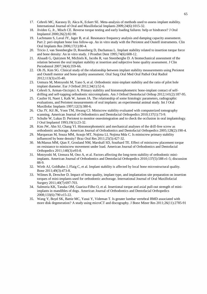
65
17. Cehreli MC, Karasoy D, Akca K, Eckert SE. Meta-analysis of methods used to assess implant stability. International Journal of Oral and Maxillofacial Implants 2009;24(6):1015-32.
18. Jividen G, Jr., Misch CE. Reverse torque testing and early loading failures: help or hindrance? J Oral
Implantol 2000;26(2):82-90.
19. Lachmann S, Laval JY, Jager B, et al. Resonance frequency analysis and damping capacity assessment.
Part 2: peri-implant bone loss follow-up. An in vitro study with the Periotest and Osstell instruments. Clin
Oral Implants Res 2006;17(1):80-4.
20. Tricio J, van Steenberghe D, Rosenberg D, Duchateau L. Implant stability related to insertion torque force
and bone density: An in vitro study. J Prosthet Dent 1995;74(6):608-12.
21. Alsaadi G, Quirynen M, Michiels K, Jacobs R, van Steenberghe D. A biomechanical assessment of the
relation between the oral implant stability at insertion and subjective bone quality assessment. J Clin
Periodontol 2007;34(4):359-66. 22. Oh JS, Kim SG. Clinical study of the relationship between implant stability measurements using Periotest
and Osstell mentor and bone quality assessment. Oral Surg Oral Med Oral Pathol Oral Radiol
2012;113(3):e35-40.
23. Uemura M, Motoyoshi M, Yano S, et al. Orthodontic mini-implant stability and the ratio of pilot hole
implant diameter. Eur J Orthod 2012;34(1):52-6.
24. Cehreli S, Arman-Ozcirpici A. Primary stability and histomorphometric bone-implant contact of self-
drilling and self-tapping orthodontic microimplants. Am J Orthod Dentofacial Orthop 2012;141(2):187-95.
25. Caulier H, Naert I, Kalk W, Jansen JA. The relationship of some histologic parameters, radiographic
evaluations, and Periotest measurements of oral implants: an experimental animal study. Int J Oral
Maxillofac Implants 1997;12(3):380-6.
26. Cha JY, Kil JK, Yoon TM, Hwang CJ. Miniscrew stability evaluated with computerized tomography
scanning. American Journal of Orthodontics and Dentofacial Orthopedics 2010;137(1):73-9. 27. Schulte W, Lukas D. Periotest to monitor osseointegration and to check the occlusion in oral implantology.
J Oral Implantol 1993;19(1):23-32.
28. Kim JW, Ahn SJ, Chang YI. Histomorphometric and mechanical analyses of the drill-free screw as
orthodontic anchorage. American Journal of Orthodontics and Dentofacial Orthopedics 2005;128(2):190-4.
29. Marquezan M, Souza MM, Araujo MT, Nojima LI, Nojima Mda C. Is miniscrew primary stability
influenced by bone density? Braz Oral Res 2011;25(5):427-32.
30. McManus MM, Qian F, Grosland NM, Marshall SD, Southard TE. Effect of miniscrew placement torque
on resistance to miniscrew movement under load. American Journal of Orthodontics and Dentofacial
Orthopedics 2011;140(3):e93-8.
31. Motoyoshi M, Uemura M, Ono A, et al. Factors affecting the long-term stability of orthodontic mini-
implants. American Journal of Orthodontics and Dentofacial Orthopedics 2010;137(5):588 e1-5; discussion 88-9.
32. Wirth AJ, Goldhahn J, Flaig C, et al. Implant stability is affected by local bone microstructural quality.
Bone 2011;49(3):473-8.
33. Wilmes B, Drescher D. Impact of bone quality, implant type, and implantation site preparation on insertion
torques of mini-implants used for orthodontic anchorage. International Journal of Oral Maxillofacial
Surgery 2011;40(7):697-703.
34. Salmoria KK, Tanaka OM, Guariza-Filho O, et al. Insertional torque and axial pull-out strength of mini-
implants in mandibles of dogs. American Journal of Orthodontics and Dentofacial Orthopedics
2008;133(6):790 e15-22.
35. Wang Y, Boyd SK, Battie MC, Yasui Y, Videman T. Is greater lumbar vertebral BMD associated with
more disk degeneration? A study using microCT and discography. J Bone Miner Res 2011;26(11):2785-91

66
Table 1: Division of the groups based on the bone region, the presence and the
thickness of the cortical bone
Group Bone region n. of sample cortical
GI0 Iliac 13 absent
GI1 Iliac 13 1mm
GP0 Pubic 13 absent
GP1 Pubic 13 1mm

67
Table 2: Descriptive analysis and ANOVA/Tukey result for bone quality.
Group Mean (± sd)
Tb.Th (mm) Tb.N (mm-1
) Tb.S (mm) Tb.BMD (g/cm3) total BMD (g/cm
3) BV/TV (%)
GI0 0.14 (0.01) a 1.20 (±0.23) a 0.45 (±0.06) a 0.69 (±0.12) a 0.69 (±0.12) a 17.69 (±4.38) a
GI1 0.15 (0.01) ab 1.23 (±0.14) a 0.49 (±0.02) a 0.76 (±0.13) a 0.93 (±0.15) b 20.39 (±4.07) ab
GP0 0.16 (0.02) b 1.49 (±0.16) b 0.38 (±0.05) b 0.97 (±0.18) b 0.97 (±0.18) b 25.18 (±6.19) bc
GP1 0.17 (0.02) b 1.61 (±0.22) b 0.40 (±0.04) b 1.15 (±0.25) b 1.27 (±0.26) c 30.32 (±7.96) c
Each column presents an independent result for ANOVA/ Tukey. Different letters indicate statistical significant difference at α=0.05% Tb.Th = trabecular thickness; Tb.N = trabecular number; Tb.S = trabecular separation; Tb.BMD = bone mineral density of trabecular bone;
total BMD = bone mineral density of the total bone block; BV/TV = bone volume density.
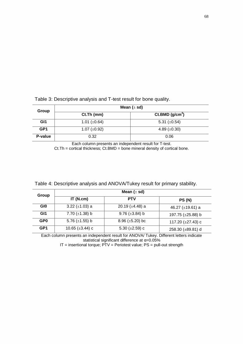
68
Table 3: Descriptive analysis and T-test result for bone quality.
Group Mean (± sd)
Ct.Th (mm) Ct.BMD (g/cm3)
GI1 1.01 (±0.64) 5.31 (±0.54)
GP1 1.07 (±0.92) 4.89 (±0.30)
P-value 0.32 0.06
Each column presents an independent result for T-test. Ct.Th = cortical thickness; Ct.BMD = bone mineral density of cortical bone.
Table 4: Descriptive analysis and ANOVA/Tukey result for primary stability.
Group Mean (± sd)
IT (N.cm) PTV PS (N)
GI0 3.22 (±1.03) a 20.19 (±4.48) a 46.27 (±19.61) a
GI1 7.70 (±1.38) b 9.76 (±3.84) b 197.75 (±25.88) b
GP0 5.76 (±1.55) b 8.96 (±5.20) bc 117.20 (±27.43) c
GP1 10.65 (±3.44) c 5.30 (±2.59) c 258.30 (±89.81) d
Each column presents an independent result for ANOVA/ Tukey. Different letters indicate statistical significant difference at α=0.05%
IT = insertional torque; PTV = Periotest value; PS = pull-out strength

69
Table 5: Pearson correlation test.
Tb.Th Tb.N Tb.S BMD.trab BMD.total BV/TV Ct.Th Ct.BMD IT PTV PS
Tb.Th r 1 ,536
* -,228 ,566
* ,610
* ,745
* ,251 -,109 ,570
* -,487
* ,578
*
P-value
,000 ,115 ,000 ,000 ,000 ,226 ,638 ,000 ,000 ,000
Tb.N r ,536
* 1 -,657
* ,619
* ,561
* ,846
* ,275 ,088 ,541
* -,481
* ,423
*
P-value ,000
,000 ,000 ,000 ,000 ,175 ,697 ,000 ,000 ,002
Tb.S r -,228 -,657
* 1 -,521
* -,373
* -,533
* -,109 ,122 -,238 ,308
* -,145
P-value ,115 ,000
,000 ,011 ,000 ,604 ,597 ,096 ,029 ,320
BMD.trab r ,566
* ,619
* -,521
* 1 ,950
* ,786
* ,353 -,038 ,628
* -,532
* ,521
*
P-value ,000 ,000 ,000
,000 ,000 ,107 ,868 ,000 ,000 ,000
BMD.total r ,610
* ,561
* -,373
* ,950
* 1 ,763
* ,512
* ,063 ,763
* -,604
* ,700
*
P-value ,000 ,000 ,011 ,000
,000 ,015 ,782 ,000 ,000 ,000
BV/TV r ,745
* ,846
* -,533
* ,786
* ,763
* 1 ,385 ,098 ,688
* -,568
* ,553
*
P-value ,000 ,000 ,000 ,000 ,000
,052 ,663 ,000 ,000 ,000
Ct.Th r ,251 ,275 -,109 ,353 ,512
* ,385 1 ,413 ,651
* -,154 ,501
*
P-value ,226 ,175 ,604 ,107 ,015 ,052
,056 ,000 ,454 ,009
Ct.BMD r -,109 ,088 ,122 -,038 ,063 ,098 ,413 1 ,008 ,077 ,120
P-value ,638 ,697 ,597 ,868 ,782 ,663 ,056
,972 ,733 ,595
IT r ,570
* ,541
* -,238 ,628
* ,763
* ,688
* ,651
* ,008 1 -,680
* ,835
*
P-value ,000 ,000 ,096 ,000 ,000 ,000 ,000 ,972
,000 ,000
PTV r -,487
* -,481
* ,308
* -,532
* -,604
* -,568
* -,154 ,077 ,680
* 1 ,655
*
P-value ,000 ,000 ,029 ,000 ,000 ,000 ,454 ,733 ,000
,000
OS r ,578
* ,423
* -,145 ,521
* ,700
* ,553
* ,501
* ,120 ,835
* -,655
* 1
P-value ,000 ,002 ,320 ,000 ,000 ,000 ,009 ,595 ,000 ,000
* Indicates statistical significant difference at α=0.05%
Tb.Th = trabecular thickness; Tb.N = trabecular number; Tb.S = trabecular separation; Tb.BMD = bone mineral density of trabecular bone; total BMD = bone mineral density of the total bone block; BV/TV = bone volume density; Ct.Th = cortical thickness; Ct.BMD = bone mineral density of cortical bone; IT = insertional torque; PTV = Periotest value; PS = pull-out strength.
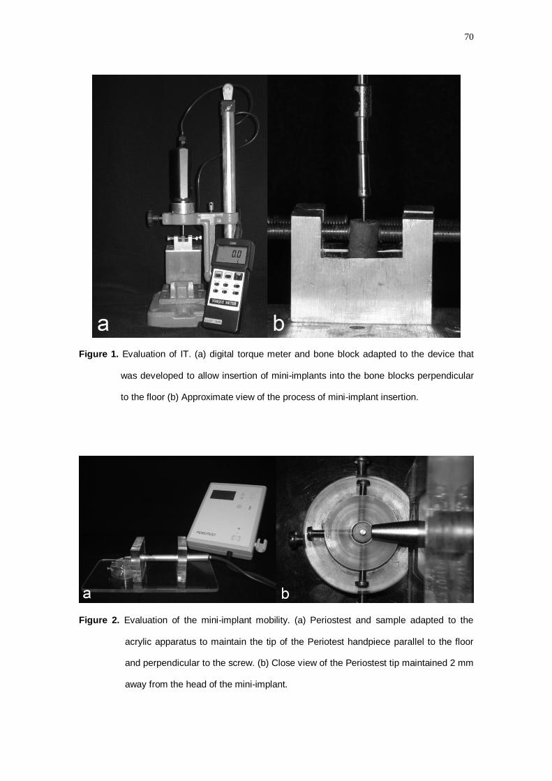
70
Figure 1. Evaluation of IT. (a) digital torque meter and bone block adapted to the device that
was developed to allow insertion of mini-implants into the bone blocks perpendicular
to the floor (b) Approximate view of the process of mini-implant insertion.
Figure 2. Evaluation of the mini-implant mobility. (a) Periostest and sample adapted to the
acrylic apparatus to maintain the tip of the Periotest handpiece parallel to the floor
and perpendicular to the screw. (b) Close view of the Periostest tip maintained 2 mm
away from the head of the mini-implant.
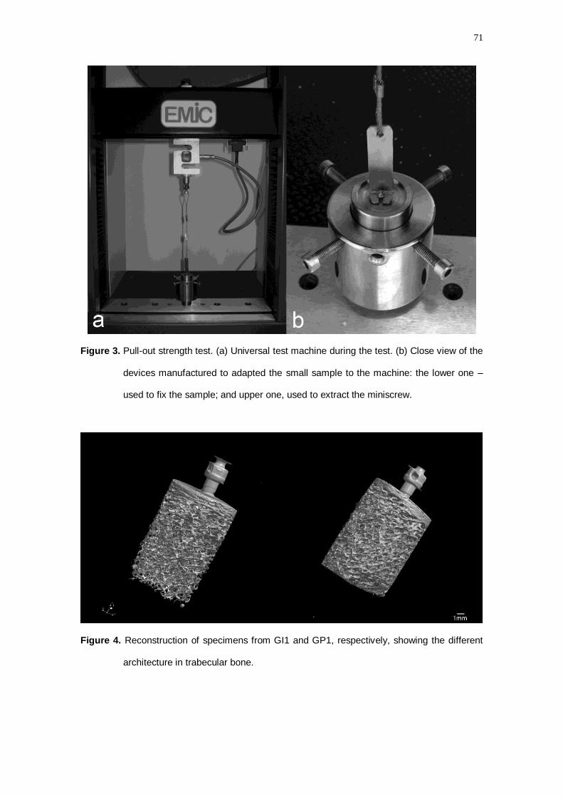
71
Figure 3. Pull-out strength test. (a) Universal test machine during the test. (b) Close view of the
devices manufactured to adapted the small sample to the machine: the lower one –
used to fix the sample; and upper one, used to extract the miniscrew.
Figure 4. Reconstruction of specimens from GI1 and GP1, respectively, showing the different
architecture in trabecular bone.
72
5 DISCUSSÃO
A qualidade do osso receptor é um dos fatores a ser considerado para
o estabelecimento do prognóstico da estabilidade de implantes dentários e
ortodônticos. Sendo o termo "qualidade óssea" subjetivo, buscamos avaliar a
associação entre a estabilidade primária de mini-implantes e diferentes
propriedades ósseas.
As revisões sistemáticas e meta-análise mostraram haver carência de
estudos clínicos observacionais, assim como de estudos laboratoriais de alta
qualidade metodológica, que avaliem a associação entre a estabilidade
primária de MI e a qualidade do sítio receptor.
A primeira revisão sistemática (Artigo 1, página 17) focou, inclusive, na
associação entre a BMD do leito ósseo receptor e a estabilidade de implantes
dentários (em vez de mini-implantes) por não terem sido encontrados, nas
bases de dados consultadas, estudos clínicos observacionais que avaliassem
essa associação. Além disso, naquele momento, apenas poucos estudos
laboratoriais haviam sido publicados com esse objetivo, e esses apresentavam
importantes diferenças metodológicas. Como o ramo da ancoragem
esquelética se beneficiou da literatura de implantodontia, essa revisão visou
enriquecer ambas as áreas.

73
Verificou-se que existe associação positiva entre a estabilidade
primária de implantes dentários e a BMD do sítio receptor. Entretanto, alguns
estudos avaliaram a BMD apenas do osso trabecular, outros avaliaram o
conjunto de osso cortical e trabecular, e outros, ainda, não especificaram em
que região óssea a BMD foi avaliada. Dentre os estudos incluídos na revisão
sistemática, aquele que apresentou o segundo menor valor de correlação entre
BMD e estabilidade primária foi um dos que avaliou apenas o osso trabecular
(Merheb, Van Assche et al., 2010). O estudo que apresentou menor valor de
correlação não relatou se avaliou a cortical em conjunto com o trabeculado
(Farre-Pages, Auge-Castro et al., 2011). Esse dado vem corroborar a
importância da cortical na estabilidade, uma vez que os valores de correlação
são mais altos quando a cortical é envolvida no ROI (region of interest) de
análise da BMD.
A segunda revisão sistemática e meta-análise (Artigo 2, página 40) visou
verificar a influência da espessura de cortical na estabilidade primária de mini-
implantes ortodônticos. Para essa segunda revisão, estudos clínicos
observacionais e experimentais foram considerados, sendo apenas dois dos
doze artigos incluídos na revisão, estudos clínicos observacionais. A qualidade
metodológica dos estudos foi considerada baixa, não sendo descritos em
muitos deles os critérios de inclusão e exclusão da amostra, calibragem e
cegamento, e inclusão de fatores de confundimento na análise estatística.
Os resultados da meta-análise mostraram haver correlação positiva
entre a espessura de cortical e a estabilidade primária de mini-implantes
ortodônticos (r=0.409), quando essa foi aferida através de teste de tração,
confirmando a importância da espessura de cortical para a estabilidade
74
primária (Motoyoshi, Hirabayashi et al., 2006; Papageorgiou, Zogakis et al.,
2012). Quando a estabilidade foi aferida através de torque de inserção, a
heterogeneidade dos estudos foi muito alta (I2=97,23%), não sendo
recomendável a execução da meta-análise. Já quando os estudos foram
agrupadas através do substrato ósseo humano, a meta-análise mostrou
também que a associação entre a espessura de cortical e a estabilidade foi
positiva, porém mais fraca (r= 0.338). Este fato corrobora a afirmação de que
os resultados de pesquisas com animais são aplicáveis a seres humanos com
reservas. Estes estudos têm contribuído grandemente para o conhecimento na
área da prevenção, etiologia e tratamento de doenças orais, tendo as
vantagens de melhor controle das variáveis, menor diversidade genética,
facilidade de obtenção da amostra, possibilidade de maior número de
repetições e facilidade para executar análise microscópica (Neto, 2001). No
entanto, não é possível extrapolar os resultados para os seres humanos. A
experimentação animal é um degrau, na pirâmide científica, que antecede os
estudos em seres humanos, mas não os substitui.
Na parte experimental da pesquisa, optou-se por trabalhar com amostras
de ossos pélvicos bovinos, considerando-se as vantagens e desvantagens
supracitadas. O osso pélvico de bovinos (Trisi, Rao et al., 1999; Cehreli e
Arman-Ozcirpici, 2012; Cehreli, Yilmaz et al., 2013) e suínos (Wilmes,
Rademacher et al., 2006; Wawrzinek, Sommer et al., 2008; Wilmes,
Ottenstreuer et al., 2008; Su, Wilmes et al., 2009) havia sido previamente
utilizado em pesquisas que avaliaram o comportamento biomecânico de mini-
implantes. Esse é composto de três ossos: ísquio, ilíaco e púbico. Durante a
exploração para retirada das secções ósseas, no projeto piloto, pôde-se
75
observar que as três regiões apresentavam diferentes características de
coloração, textura e até resistência ao corte. A região do púbico era mais
escura e mais resistente ao corte em relação ao ilíaco e ísquio, sendo esse
último menos abundante. Esses fatos levaram à investigação das duas
diferentes regiões quando da instalação de mini-implantes. Avaliação
histológica desses ossos revelou que a densidade trabecular (TBA, do inglês
trabecular bone area) do púbico se assemelha a dos maxilares humanos
(APÊNDICE A, página 92), conforme descrito previamente (Aksoy, Eratalay et
al., 2009).
Clinicamente, a classificação de qualidade óssea mais aceita em
Implantodontia foi proposta por Lekholm e Zarb (Lekholm e Zarb, 1985). Essa
se baseia na quantidade de osso cortical e trabecular verificada em radiografias
pré-operatórias. Por essa classificação depender da subjetividade do operador,
alguns autores assumiram que o termo qualidade óssea equivale a BMD
(Bergkvist, Koh et al., 2010). Outros autores, ainda, atribuem à qualidade óssea
à espessura (Wilmes, Rademacher et al., 2006; Iijima, Takano et al., 2012) e
densidade mineral da cortical (Cha, Kil et al., 2010; Iijima, Takano et al., 2012).
Outra propriedade óssea que tem ganhado maior destaque nos últimos anos é
a microarquitetura trabecular (Zhao, Xu et al., 2009; Ikeda, Rossouw et al.,
2011; Wirth, Goldhahn et al., 2011; Zhao, Xu et al., 2011). Buscou-se, assim,
abranger o estudo de todas as propriedades ósseas supracitadas: BMD,
espessura de cortical e microarquitetura trabecular, todas avaliadas através de
microCT.
A aferição da BMD por micro CT foi validada em 2007 (Macneil e Boyd,
2007), tendo as vantagens de permitir a avaliação da BMD em imagens

76
tridimensionais de alta resolução (Ito, Nishida et al., 2002) em sítios específicos
selecionados pelo operador (VOI).
Estima-se que a resistência óssea é dependente da BMD em cerca de
70 a 75% e que o restante é explicado por fatores como arquitetura óssea e
composição do tecido (Krug, Burghardt et al., 2010). Por isso, a mensuração
isolada da BMD não traduz completamente a qualidade óssea, sendo a
estabilidade de implantes afetada também pela qualidade micro-estrutural do
osso trabecular adjacente ao implante (Wirth, Goldhahn et al., 2011). Faz-se,
portanto, interessante avaliar ambas as propriedades ósseas, sendo a microCT
o exame de eleição já que permite ambas avaliações com uma única tomada.
Infelizmente, o atual estágio tecnológico não permite a utilização clínica da
microCT para os ossos maxilares, sendo possível utilizá-la apenas em estudos
laboratoriais.
Um importante parâmetro da arquitetura trabecular é a sua densidade. A
densidade trabecular pode ser medida com técnica histológica (TBA), em 2D,
ou microtomografia (BV/TV), em 3D. A microCT foi validada como um método
para avaliação tridimensional do osso trabecular em 1998 através da
comparação de seus resultados com os de histomorfometria (Muller, Van
Campenhout et al., 1998). Ela apresenta a vantagem de ser mais rápida e
precisa que a análise histológica convencional, que requer o trabalhoso,
demorado e destrutivo preparo histotécnico (Ruegsegger, Koller et al., 1996;
Ito, Nishida et al., 2002; Lima, Lopes et al., 2009). Enquanto a análise na micro
CT é automatizada, a análise histológica é dependente do examinador e sujeita
a seu erro. Além disso, são requeridos muitos cortes até que uma conclusão
sobre a qualidade óssea seja atingida.
77
Um problema das imagens de microCT, quando o osso apresenta um
metal inserido, é o artefato gerado. Foi demonstrado que até mesmo 9 mm
distante do mini-implante a BMD é influenciada pelo artefato do parafuso
(Sabo, Pollmann et al., 2009), e que pelo menos 5 voxels de osso adjacentes
aos parafusos devem ser descartados para avaliação da BV/TV (Cha, Song et
al., 2009). Dessa maneira, nessa pesquisa os 6 voxels (54 μm) de osso
adjacente ao mini-implante foram excluídos do VOI e o padrão de osso artificial
utilizado para calibração do software para avaliação da BMD continha um mini-
implante inserido em seu centro. Assim, as condições de artefato foram
reproduzidas.
Para a aferição da estabilidade primária, não há padrão ouro
estabelecido (Cehreli, Karasoy et al., 2009). Dessa maneira, foram realizados a
mensuração do torque de inserção dos mini-implantes (Kim, Choi et al., 2008;
Mischkowski, Kneuertz et al., 2008; Pithon, 2008; Salmoria, Tanaka et al.,
2008; Wilmes, Ottenstreuer et al., 2008; Brinley, Behrents et al., 2009; Wilmes
e Drescher, 2009; Florvaag, Kneuertz et al., 2010), a avaliação da micro-
mobilidade com Periotest (Kim, Ahn et al., 2005; Inaba, 2009; Su, Wilmes et al.,
2009; Cehreli e Arman-Ozcirpici, 2012), e o teste de tração (Huja, Litsky et al.,
2005; Kim, Choi et al., 2008; Mischkowski, Kneuertz et al., 2008; Pithon, 2008;
Salmoria, Tanaka et al., 2008; Brinley, Behrents et al., 2009; Florvaag,
Kneuertz et al., 2010; Liu, Broucek et al., 2010).
O Periostest é um instrumento eletrônico, originalmente desenvolvido
para medir, através de percussão, as características de amortecimento do
periodonto de dentes naturais traumatizados (Inaba, 2009; Cehreli e Arman-
Ozcirpici, 2012). Seu uso tem sido expandido para verificar a mobilidade de
78
implantes e mini-implantes, em micro escala, através do fornecimento de um
número contido numa escala de -8 a +50. Quanto maior o número fornecido,
maior a mobilidade e menor a estabilidade. Como a estabilidade primária é
comumente avaliada clinicamente através da verificação manual da mobilidade
do parafuso, a aferição da mobilidade do implante com o Periotest parece ser o
teste que melhor reproduz a condição clínica.
O aparelho Periotest permite a avaliação da mobilidade do mini-implante
de maneira objetiva e não destrutiva (Jividen e Misch, 2000). Entretanto, o
aparelho é muito sensível às condições de uso. Durante a mensuração, a peça
de mão do aparelho deve ser mantida paralela ao solo e perpendicular ao
parafuso. A cabeça do aparelho não deve tocar o parafuso, e a sua distância
deve ser de 0,7 à 2 mm. Assim, para esse experimento, um dispositivo acrílico
foi construído a fim de fixar a peça de mão e a amostra, mantendo uma
distância de 2 mm entre a cabeça da peça de mão e o parafuso. A
confiabilidade do Periostest já foi relatada em estudos com mini-implantes (Su,
Wilmes et al., 2009; Cehreli e Arman-Ozcirpici, 2012; Uemura, Motoyoshi et al.,
2012) e implantes dentários (Tricio, Van Steenberghe et al., 1995; Lachmann,
Laval et al., 2006; Alsaadi, Quirynen et al., 2007). Entretanto, sua confiabilidade
para essa finalidade não é consenso na literatura (Caulier, Naert et al., 1997).
Nessa pesquisa, os valores de PTV apresentaram correlação negativa com os
valores de IT e PS, testes já consagrados na literatura, concordando com
estudos prévios (Tricio, Van Steenberghe et al., 1995; Cha, Kil et al., 2010;
Cehreli e Arman-Ozcirpici, 2012) e indicando seu sucesso na avaliação da
estabilidade primária. IT e PS apresentaram forte correlação positiva entre si,
79
concordando com estudos anteriores (Brinley, Behrents et al., 2009;
Marquezan, Lau et al., 2012).
Os resultados dessa pesquisa mostraram que a densidade óssea – tanto
a mineral (BMD total) como a trabecular (BV/TV) – possui correlação com as
medidas de estabilidade. Esses achados concordam com (Wang, Zhao et al.,
2010), que encontraram correlação positiva entre PS e as propriedades ósseas
de BV/TV e BMD quando avaliaram a diferença da estabilidade primária de
mini-implantes inseridos na mandíbula de cães jovens e adultos.
Tanto o papel do osso trabeculado quanto do osso cortical foram
evidenciados. A presença de cortical por si só aumentou os valores de
estabilidade (Tabela 2, Artigo 3, página 67) corroborando sua importância na
estabilidade (Turkyilmaz, Tozum et al., 2006; Wilmes, Rademacher et al., 2006;
Motoyoshi, Yoshida et al., 2007; Su, Wilmes et al., 2009; Cha, Kil et al., 2010).
Entretanto, quando a cortical esteve ausente, a importância do osso trabecular
se tornou mais evidente. Os valores de IT, PTV e PS diferiram para os grupos
GI0 e GP0, havendo maior estabilidade no osso púbico. Quando comparados
os grupos GI1 e GP0, IT e PTV não mostraram diferença estatisticamente
significativa. Esses resultados sugerem que quando a cortical esteve ausente,
a responsabilidade da estabilidade dos parafusos recaiu sobre as propriedades
do osso trabecular. Já quando a cortical esteve presente, ela demostrou seu
papel, especialmente quando o osso trabecular possuía menores valores de
BMD, Tb.N, Tb.Th e maior valor de Tb.S. Essa responsabilidade da cortical
sobre as propriedades mecânicas do osso havia sido relata anteriormente (Ito,
Nishida et al., 2002). Em estudo com elementos finitos, esses autores
encontraram forte correlação entre propriedades mecânicas do tecido ósseo e
80
a microarquitetura trabecular. Entretanto, quando a estrutura trabecular estava
deteriorada (simulando osteoporose), as propriedades mecânicas dependiam
principalmente da cortical.
Para que não se dependa exclusivamente da cortical, quando mini-
implantes são inseridos em ossos de baixa BMD, de pacientes osteoporóticos
ou adolescentes, as propriedades dos mini-implantes (diâmetro, comprimento,
superfície) e o tempo de reparo devem ser considerados (Wang, Zhao et al.,
2010).
Considerando as propriedades da cortical óssea em relação às demais
propriedades do osso, a espessura de cortical apresentou correlação positiva
apenas com a BMD total, corroborando a crença de que quando a espessura
de cortical aumenta, a BMD total também aumenta porque a cortical é a porção
mais mineralizada do osso (Marquezan, Osorio et al., 2012).
Já avaliando a espessura de cortical com relação à estabilidade
primária, foi observada correlação positiva entre a espessura de cortical e o IT
(Wilmes, Rademacher et al., 2006; Motoyoshi, Yoshida et al., 2007; Cha, Kil et
al., 2010; Motoyoshi, Uemura et al., 2010; Mcmanus, Qian et al., 2011; Wilmes
e Drescher, 2011; Wirth, Goldhahn et al., 2011), e entre a espessura de cortical
e o PS (Huja, Litsky et al., 2005; Salmoria, Tanaka et al., 2008). Entretanto, a
espessura de cortical não influenciou o PTV. Acredita-se que a
espessura de cortical tenha maior influência nas medidas axiais de estabilidade
que nas laterais.
O teste de correlação de Pearson mostrou não haver correlação entre a
BMD da cortical e a estabilidade dos mini-implantes, discordando de
Marquezan, Lau et al. (Marquezan, Lau et al., 2012) (APÊNDICE B, página
81
100). Tal discordância provavelmente deve-se à diferença metodológica.
Enquanto em Marquezan, Lau et al. (Marquezan, Lau et al., 2012) a avaliação
da BMD da cortical foi realizada em cortes de CBCT, nesse experimento foi
utilizada microCT, um método de avaliação tridimensional e mais preciso
(Wang, Boyd et al., 2011). A CBCT, entretanto, é atualmente utilizada na clínica
ortodôntica, ao contrário da microCT.
Considerando os três trabalhos que compõe essa tese, ficou evidente
que a qualidade do sítio ósseo receptor tem influência sobre a estabilidade
primária de mini-implantes. Sugere-se que futuras pesquisas confirmem tais
achados através de estudos clínicos.
82
6 CONCLUSÃO
6.1 Nas revisões sistemáticas foi verificado que:
6.1.1 existe associação positiva entre a estabilidade primária de implantes
dentários e da BMD do sítio receptor;
6.1.2 existe associação positiva entre a estabilidade primária de MI e
espessura cortical do sítio receptor;
6.2 No trabalho experimental:
6.2.1 a estabilidade primária (IT, PS e PTV) diferiu estatisticamente entre
os ossos ilíaco e púbico na presença e na ausência de cortical;
6.2.2 a densidade trabecular, a espessura trabecular, a espessura de
cortical, o número de trabéculas, a BMD trabecular e a BMD total, aferidas com
microCT, apresentaram correlação com a estabilidade primária dos MI.

83
7 RECOMENDAÇÕES
Para futuras pesquisas sobre a relação entre qualidade óssea e a
estabilidade primária de MI, sugere-se a utilização de espessuras de cortical
entre 0,1 e 1 mm. Sugere-se ainda a realização de estudos clínicos que
verifiquem a influência da qualidade óssea na estabilidade primária e
secundária.

84
8 REFERÊNCIAS BIBLIOGRÁFICAS
AKSOY, U. et al. The possible association among bone density values, resonance frequency
measurements, tactile sense, and histomorphometric evaluations of dental implant osteotomy sites: a
preliminary study. Implant Dent [S.I.], v. 18, n. 4, p. 316-25, Aug 2009.
ALSAADI, G. et al. A biomechanical assessment of the relation between the oral implant stability at
insertion and subjective bone quality assessment. J Clin Periodontol [S.I.], v. 34, n. 4, p. 359-66, Apr
2007.
ANGLE, E. H. Treatment of malloclusion of the teeth. Angle's System. 7. ed. Philadelphia: SS White
Dental Manufactoring Company, 1907.
BERGKVIST, G. et al. Bone density at implant sites and its relationship to assessment of bone quality
and treatment outcome. Int J Oral Maxillofac Implants [S.I.], v. 25, n. 2, p. 321-8, Mar-Apr 2010.
BLOCK, M. S.; HOFFMAN, D. R. A new device for absolute anchorage for orthodontics. Am J Orthod
Dentofacial Orthop [S.I.], v. 107, n. 3, p. 251-8, Mar 1995.
BRINLEY, C. L. et al. Pitch and longitudinal fluting effects on the primary stability of miniscrew implants. Angle Orthod [S.I.], v. 79, n. 6, p. 1156-61, Nov 2009.
CAULIER, H. et al. The relationship of some histologic parameters, radiographic evaluations, and
Periotest measurements of oral implants: an experimental animal study. Int J Oral Maxillofac Implants
[S.I.], v. 12, n. 3, p. 380-6, May-Jun 1997.
CEHRELI, M. C. et al. Meta-analysis of methods used to assess implant stability. Int J Oral Maxillofac
Implants [S.I.], v. 24, n. 6, p. 1015-32, Nov-Dec 2009.
CEHRELI, S.; ARMAN-OZCIRPICI, A. Primary stability and histomorphometric bone-implant contact
of self-drilling and self-tapping orthodontic microimplants. Am J Orthod Dentofacial Orthop [S.I.], v.
141, n. 2, p. 187-95, Feb 2012.
CEHRELI, S. et al. Cortical bone strains around straight and angulated immediate orthodontic
microimplants: a pilot study. Implant Dent [S.I.], v. 22, n. 2, p. 133-7, Apr 2013.
CHA, J. Y. et al. Miniscrew stability evaluated with computerized tomography scanning. Am J Orthod
Dentofacial Orthop [S.I.], v. 137, n. 1, p. 73-9, Jan 2010.
CHA, J. Y. et al. Influence of the length of the loading period after placement of orthodontic mini-
implants on changes in bone histomorphology: microcomputed tomographic and histologic analysis. Int J
Oral Maxillofac Implants [S.I.], v. 24, n. 5, p. 842-9, Sep-Oct 2009.
85
CHA, J. Y. et al. The influence of peri-implant artifact on microCT analysis. In: SkyScan User Meeting
2009. 2009. p.3.
CHADDAD, K. et al. Influence of surface characteristics on survival rates of mini-implants. Angle
Orthod [S.I.], v. 78, n. 1, p. 107-13, Jan 2008.
CHENG, S. J. et al. A prospective study of the risk factors associated with failure of mini-implants used
for orthodontic anchorage. Int J Oral Maxillofac Implants [S.I.], v. 19, n. 1, p. 100-6, Jan-Feb 2004.
CREEKMORE, T. D.; EKLUND, M. K. The possibility of skeletal anchorage. J Clin Orthod [S.I.], v. 17,
n. 4, p. 266-9, Apr 1983.
DEGUCHI, T. et al. Quantitative evaluation of cortical bone thickness with computed tomographic
scanning for orthodontic implants. Am J Orthod Dentofacial Orthop [S.I.], v. 129, n. 6, p. 721 e7-12, Jun
2006.
FARRE-PAGES, N. et al. Relation between bone density and primary implant stability. Med Oral Patol
Oral Cir Bucal [S.I.], v. 16, n. 1, p. e62-7, Jan 2011.
FLORVAAG, B. et al. Biomechanical properties of orthodontic miniscrews. An in-vitro study. J Orofac
Orthop [S.I.], v. 71, n. 1, p. 53-67, Jan 2010.
FREUDENTHALER, J. W. et al. Bicortical titanium screws for critical orthodontic anchorage in the
mandible: a preliminary report on clinical applications. Clin Oral Implants Res [S.I.], v. 12, n. 4, p. 358-
63, Aug 2001.
GARFINKLE, J. S. et al. Evaluation of orthodontic mini-implant anchorage in premolar extraction
therapy in adolescents. Am J Orthod Dentofacial Orthop [S.I.], v. 133, n. 5, p. 642-53, May 2008.
GEDRANGE, T. et al. An evaluation of resonance frequency analysis for the determination of the
primary stability of orthodontic palatal implants. A study in human cadavers. Clin Oral Implants Res
[S.I.], v. 16, n. 4, p. 425-31, Aug 2005.
HUJA, S. S. et al. Pull-out strength of monocortical screws placed in the maxillae and mandibles of dogs.
Am J Orthod Dentofacial Orthop [S.I.], v. 127, n. 3, p. 307-13, Mar 2005.
IIJIMA, M. et al. Effect of the quantity and quality of cortical bone on the failure force of a miniscrew
implant. Eur J Orthod [S.I.], Oct 4 2012.
IKEDA, H. et al. Three-dimensional analysis of peri-bone-implant contact of rough-surface miniscrew
implants. Am J Orthod Dentofacial Orthop [S.I.], v. 139, n. 2, p. e153-63, Feb 2011.
INABA, M. Evaluation of primary stability of inclined orthodontic mini-implants. J Oral Sci [S.I.], v. 51,
n. 3, p. 347-53, Sep 2009.
ITO, M. et al. Contribution of trabecular and cortical components to the mechanical properties of bone
and their regulating parameters. Bone [S.I.], v. 31, n. 3, p. 351-8, Sep 2002.
JAVED, F.; ROMANOS, G. E. The role of primary stability for successful immediate loading of dental
implants. A literature review. J Dent [S.I.], v. 38, n. 8, p. 612-20, Aug 2010.

86
JIVIDEN, G., JR.; MISCH, C. E. Reverse torque testing and early loading failures: help or hindrance? J
Oral Implantol [S.I.], v. 26, n. 2, p. 82-90, 2000.
JUSTENS, E.; DE BRUYN, H. Clinical outcome of mini-screws used as orthodontic anchorage. Clin
Implant Dent Relat Res [S.I.], v. 10, n. 3, p. 174-80, Sep 2008.
KANOMI, R. Mini-implant for orthodontic anchorage. J Clin Orthod [S.I.], v. 31, n. 11, p. 763-7, Nov
1997.
KIM, J. W. et al. Histomorphometric and mechanical analyses of the drill-free screw as orthodontic
anchorage. Am J Orthod Dentofacial Orthop [S.I.], v. 128, n. 2, p. 190-4, Aug 2005.
KIM, S. H. et al. Peri-implant bone reactions at delayed and immediately loaded implants: an
experimental study. Oral Surg Oral Med Oral Pathol Oral Radiol Endod [S.I.], v. 105, n. 2, p. 144-8, Feb
2008.
KRUG, R. et al. High-resolution imaging techniques for the assessment of osteoporosis. Radiol Clin North Am [S.I.], v. 48, n. 3, p. 601-21, May 2010.
LACHMANN, S. et al. Resonance frequency analysis and damping capacity assessment. Part 2: peri-
implant bone loss follow-up. An in vitro study with the Periotest and Osstell instruments. Clin Oral
Implants Res [S.I.], v. 17, n. 1, p. 80-4, Feb 2006.
LEE, J. S. et al. Aplicações dos miniimplantes ortodônticos. São Paulo: Quintessence, 2009.
LEKHOLM, U.; ZARB, G. A. Patient selection and preparation. In: BRANEMARK, P. I. et al (Ed.).
Tissue integrated protheses. Osteointegration in clinical dentistry. Chicago: Quintessence, 1985.
LIM, S. A. et al. Insertion torque of orthodontic miniscrews according to changes in shape, diameter and length. Angle Orthod [S.I.], v. 78, n. 2, p. 234-40, Mar 2008.
LIMA, I. et al. Análise de estrutura óssea através de microtomografia computadorizada 3D. Revista
Brasileira de Física Médica. [S.I.], v. 2, n. 1, p. 5, 2009.
LIU, S. et al. Using micro-Computed Tomography to Predict Implant Fixation in a Rat Model. In: 56th
Annual Meeting of the Orthopaedic Research Society, March 6-9, New Orleans. 2010.
LIU, S. et al. Limitations of using micro-computed tomography to predict bone-implant contact and
mechanical fixation. J Microsc [S.I.], v. 245, n. 1, p. 34-42, Jan 2012.
LUZI, C. et al. Immediate loading of orthodontic mini-implants: a histomorphometric evaluation of tissue
reaction. Eur J Orthod [S.I.], v. 31, n. 1, p. 21-9, Feb 2009.
MACNEIL, J. A.; BOYD, S. K. Accuracy of high-resolution peripheral quantitative computed
tomography for measurement of bone quality. Med Eng Phys [S.I.], v. 29, n. 10, p. 1096-105, Dec 2007.
MARQUEZAN, M. et al. Bone mineral density. Angle Orthod [S.I.], v. 82, n. 1, p. 62-6, Jan 2012.
MARQUEZAN, M. et al. Does bone mineral density influence the primary stability of dental implants? A
systematic review. Clin Oral Implants Res [S.I.], v. 23, n. 7, p. 767-74, Jul 2012.

87
MCMANUS, M. M. et al. Effect of miniscrew placement torque on resistance to miniscrew movement
under load. Am J Orthod Dentofacial Orthop [S.I.], v. 140, n. 3, p. e93-8, Sep 2011.
MELSEN, B.; VERNA, C. A rational aproach to orthodontic anchorage. Prog Orthod [S.I.], v. 1, p. 12,
1999.
MERHEB, J. et al. Relationship between cortical bone thickness or computerized tomography-derived
bone density values and implant stability. Clin Oral Implants Res [S.I.], v. 21, n. 6, p. 612-7, Jun 2010.
MISCHKOWSKI, R. A. et al. Biomechanical comparison of four different miniscrew types for skeletal
anchorage in the mandibulo-maxillary area. Int J Oral Maxillofac Surg [S.I.], v. 37, n. 10, p. 948-54, Oct
2008.
MIYAWAKI, S. et al. Factors associated with the stability of titanium screws placed in the posterior
region for orthodontic anchorage. Am J Orthod Dentofacial Orthop [S.I.], v. 124, n. 4, p. 373-8, Oct
2003.
MOLLY, L. Bone density and primary stability in implant therapy. Clin Oral Implants Res [S.I.], v. 17
Suppl 2, p. 124-35, Oct 2006.
MOTOYOSHI, M. et al. Recommended placement torque when tightening an orthodontic mini-implant.
Clin Oral Implants Res [S.I.], v. 17, n. 1, p. 109-14, Feb 2006.
MOTOYOSHI, M. et al. The effect of cortical bone thickness on the stability of orthodontic mini-
implants and on the stress distribution in surrounding bone. Int J Oral Maxillofac Surg [S.I.], v. 38, n. 1,
p. 13-8, Jan 2009.
MOTOYOSHI, M. et al. Factors affecting the long-term stability of orthodontic mini-implants. Am J
Orthod Dentofacial Orthop [S.I.], v. 137, n. 5, p. 588 e1-5; discussion 588-9, May 2010.
MOTOYOSHI, M. et al. Effect of cortical bone thickness and implant placement torque on stability of
orthodontic mini-implants. Int J Oral Maxillofac Implants [S.I.], v. 22, n. 5, p. 779-84, Sep-Oct 2007.
MULLER, R. et al. Morphometric analysis of human bone biopsies: a quantitative structural comparison
of histological sections and micro-computed tomography. Bone [S.I.], v. 23, n. 1, p. 59-66, Jul 1998.
NETO, J. Ethics in research. In: ESTRELA, C. (Ed.). Scientific Methodology. São Paulo: Artes Médicas,
2001.
PAPAGEORGIOU, S. N. et al. Failure rates and associated risk factors of orthodontic miniscrew implants: a meta-analysis. Am J Orthod Dentofacial Orthop [S.I.], v. 142, n. 5, p. 577-595 e7, Nov 2012.
PARK, H. S. et al. Factors affecting the clinical success of screw implants used as orthodontic anchorage.
Am J Orthod Dentofacial Orthop [S.I.], v. 130, n. 1, p. 18-25, Jul 2006.
PITHON, M. M. Avaliação mecânica de mini-implantes ortodônticos. (2008). (Mestrado em
Odontologia (Ortodontia)) - Departamento de Odontopediatria e Ortodontia, Universidade Federal do Rio
de Janeiro, Rio de Janeiro, 2008.
PITHON, M. M. et al. In vitro evaluation of insertion and removal torques of orthodontic mini-implants.
Int J Oral Maxillofac Surg [S.I.], v. 40, n. 1, p. 80-5, Jan 2011.
88
RUEGSEGGER, P. et al. A microtomographic system for the nondestructive evaluation of bone
architecture. Calcif Tissue Int [S.I.], v. 58, n. 1, p. 24-9, Jan 1996.
SABO, M. T. et al. Use of co-registered high-resolution computed tomography scans before and after
screw insertion as a novel technique for bone mineral density determination along screw trajectory. Bone
[S.I.], v. 44, n. 6, p. 1163-8, Jun 2009.
SALMORIA, K. K. et al. Insertional torque and axial pull-out strength of mini-implants in mandibles of
dogs. Am J Orthod Dentofacial Orthop [S.I.], v. 133, n. 6, p. 790 e15-22, Jun 2008.
SCHATZLE, M. et al. Survival and failure rates of orthodontic temporary anchorage devices: a
systematic review. Clin Oral Implants Res [S.I.], v. 20, n. 12, p. 1351-9, Dec 2009.
SERRA, G. et al. Sequential bone healing of immediately loaded mini-implants. Am J Orthod
Dentofacial Orthop [S.I.], v. 134, n. 1, p. 44-52, Jul 2008.
SONG, Y. D. et al. Correlation between bone quality evaluated by cone-beam computerized tomography and implant primary stability. Int J Oral Maxillofac Implants [S.I.], v. 24, n. 1, p. 59-64, Jan-Feb 2009.
SONG, Y. Y. et al. Mechanical characteristics of various orthodontic mini-screws in relation to artificial
cortical bone thickness. Angle Orthod [S.I.], v. 77, n. 6, p. 979-85, Nov 2007.
SU, Y. Y. et al. Application of a wireless resonance frequency transducer to assess primary stability of
orthodontic mini-implants: an in vitro study in pig ilia. Int J Oral Maxillofac Implants [S.I.], v. 24, n. 4, p.
647-54, Jul-Aug 2009.
SUZUKI, E. Y. et al. Assessment of miniscrew implant stability by resonance frequency analysis: a study
in human cadavers. J Oral Maxillofac Surg [S.I.], v. 68, n. 11, p. 2682-9, Nov 2010.
TOPOUZELIS, N.; TSAOUSOGLOU, P. Clinical factors correlated with the success rate of miniscrews
in orthodontic treatment. Int J Oral Sci [S.I.], v. 4, n. 1, p. 38-44, Mar 2012.
TRICIO, J. et al. Implant stability related to insertion torque force and bone density: An in vitro study. J
Prosthet Dent [S.I.], v. 74, n. 6, p. 608-12, Dec 1995.
TRISI, P. et al. A histometric comparison of smooth and rough titanium implants in human low-density
jawbone. International Journal of Oral Maxillofacial Implants [S.I.], v. 14, n. 5, p. 689-98, Sep-Oct 1999.
TURKYILMAZ, I.; MCGLUMPHY, E. A. Influence of bone density on implant stability parameters and
implant success: a retrospective clinical study. BMC Oral Health [S.I.], v. 8, p. 32, 2008a.
______. Is there a lower threshold value of bone density for early loading protocols of dental implants? J
Oral Rehabil [S.I.], v. 35, n. 10, p. 775-81, Oct 2008b.
TURKYILMAZ, I. et al. Assessment of correlation between computerized tomography values of the
bone, and maximum torque and resonance frequency values at dental implant placement. J Oral Rehabil
[S.I.], v. 33, n. 12, p. 881-8, Dec 2006.
TURKYILMAZ, I. et al. Relations between the bone density values from computerized tomography, and
implant stability parameters: a clinical study of 230 regular platform implants. J Clin Periodontol [S.I.],
v. 34, n. 8, p. 716-22, Aug 2007.
89
UEMURA, M. et al. Orthodontic mini-implant stability and the ratio of pilot hole implant diameter. Eur J
Orthod [S.I.], v. 34, n. 1, p. 52-6, Feb 2012.
WANG, Y. et al. Is greater lumbar vertebral BMD associated with more disk degeneration? A study using
microCT and discography. J Bone Miner Res [S.I.], v. 26, n. 11, p. 2785-91, Nov 2011.
WANG, Z. et al. Pullout strength of miniscrews placed in anterior mandibles of adult and adolescent
dogs: a microcomputed tomographic analysis. Am J Orthod Dentofacial Orthop [S.I.], v. 137, n. 1, p.
100-7, Jan 2010.
WAWRZINEK, C. et al. Microdamage in cortical bone due to the overtightening of orthodontic
microscrews. J Orofac Orthop [S.I.], v. 69, n. 2, p. 121-34, Mar 2008.
WEHRBEIN, H. et al. The use of palatal implants for orthodontic anchorage. Design and clinical
application of the orthosystem. Clin Oral Implants Res [S.I.], v. 7, n. 4, p. 410-6, Dec 1996.
WEI, X. et al. Effects of cortical bone thickness at different healing times on microscrew stability. Angle Orthod [S.I.], v. 81, n. 5, p. 760-6, Sep 2011.
WILMES, B.; DRESCHER, D. Impact of insertion depth and predrilling diameter on primary stability of
orthodontic mini-implants. Angle Orthod [S.I.], v. 79, n. 4, p. 609-14, Jul 2009.
______. Impact of bone quality, implant type, and implantation site preparation on insertion torques of
mini-implants used for orthodontic anchorage. Int J Oral Maxillofac Surg [S.I.], v. 40, n. 7, p. 697-703,
Jul 2011.
WILMES, B. et al. Impact of implant design on primary stability of orthodontic mini-implants. J Orofac
Orthop [S.I.], v. 69, n. 1, p. 42-50, Jan 2008.
WILMES, B. et al. Parameters affecting primary stability of orthodontic mini-implants. J Orofac Orthop
[S.I.], v. 67, n. 3, p. 162-74, May 2006.
WIRTH, A. J. et al. Implant stability is affected by local bone microstructural quality. Bone [S.I.], v. 49,
n. 3, p. 473-8, Sep 2011.
ZHAO, L. et al. Effect of placement angle on the stability of loaded titanium microscrews: a
microcomputed tomographic and biomechanical analysis. Am J Orthod Dentofacial Orthop [S.I.], v. 139,
n. 5, p. 628-35, May 2011.
ZHAO, L. et al. Orthodontic mini-implant stability in different healing times before loading: a microscopic computerized tomographic and biomechanical analysis. Oral Surg Oral Med Oral Pathol
Oral Radiol Endod [S.I.], v. 108, n. 2, p. 196-202, Aug 2009.

90
9 APÊNDICES
9.1 APÊNDICE A: Marquezan M, Souza MM, Araújo MT, Nojima LI, Nojima
MC. Is miniscrew primary stability influenced by bone density? Braz Oral
Res. 2011 Sep-Oct;25(5):427-32.
Is miniscrew primary stability influenced by bone density?
Mariana Marquezan1
Margareth Maria Gomes de Souza1
Mônica Tirre de Souza Araújo1
Lincoln Issamu Nojima1
Matilde da Cunha Gonçalves Nojima1
Manuscript information
Received for publication: 24/05/2011
Accepted for publication: 20/07/2011
1 Pedodontics and Orthodontics Department, Federal University of Rio de Janeiro (UFRJ), Rio
de Janeiro, RJ, Brazil.

91
ABSTRACT
Primary stability is absence of mobility in the bone bed after mini-implant
placement and depends on bone quality among other factors. Bone quality is a
subjective term frequently considered as bone density. The aim of this
preliminary study was to evaluate bone density in two bovine pelvic regions and
verify the primary stability of miniscrews inserted into them. Forty bone blocks
were extracted from bovine pelvic bones, 20 from iliac and 20 from pubic bone,
all of them containing cortical about 1 mm thick. Half of the sections extracted
from each bone were designated for histological evaluation of bone density
(trabecular bone area - TBA) and the other half for bone mineral density (BMD)
evaluation by means of central dual-energy X-ray absorptiometry (DEXA).
Then, twenty self-drilling miniscrews (INP®, São Paulo, Brazil) 1.4 mm in
diameter and 6 mm long were inserted into the bone blocks used for BMD
evaluation. Peak implant insertion torque (IT) and pull-out strength (PS) were
used for primary stability evaluation. It was found that iliac and pubic bones
present different bone densities, iliac bone being less dense considering BMD
and TBA values (P>0.05). However, the miniscrew primary stability was not
different when varying the bone type (P<0.05). IT and PS were not influenced
by these differences in bone density when cortical thickness was about 1 mm
thick.
Descriptors: Bone and Bones; Bone Density; Orthodontic Anchorage
Procedures.
INTRODUCTION
Primary stability is absence of mobility in the bone bed after implant or
mini-implant placement 1, 2. It is achieved by mechanical contact between the
miniscrew surface and bone 3 and depends on the characteristics of devices 4, 5,
insertion technique 4 and bone quality and quantity of the receptor site 4, 6-8.
The primary stability plays an important role in the successful secondary
stability of miniscrews, since lack of immediate stability can lead to progressive
mobility of the device and its subsequent loss 9. In clinical use, the initial stability
of miniscrews is also considered essential, because of immediate or early load
applied on them in many patients 10. It has been suggested that if initial
92
mechanical retention of the mini-implant is not observed, it should be replaced
by a thicker device, or its insertion site should be changed 11. Primary stability
has traditionally been assessed by the practitioner through manual verification
12. Several other less subjective methods are described in the literature:
histological (BIC-bone to implant contact), which assesses the percentage of
bone to implant contact; mechanical, which assesses insertion and removal
torque or pullout strength of mini-implants, and the percussion method
(Periotest value). However, there is still no gold standard to assess the primary
stability of miniscrews 13.
The term “bone quality” is not clearly defined in the literature. This includes
physiological and structural aspects and degree of bone tissue mineralization 14.
Aspects such as bone metabolism, cell turnover, maturation, intracellular matrix
and vascularity have also been emphasized 1. Nevertheless, the role of each of
these aspects is not completely understood 14. In Implant dentistry, the most
accepted classification of bone quality has been the one proposed by Lekholm
and Zarb 15. This was based on the amount of cortical and trabecular shown in
preoperative radiographs. This classification, however, depends on the
operator’s subjectivity during radiographic evaluation.
A less subjective method for evaluating cortical and trabecular bone
quality is to verify bone mineral density (BMD) 14. Bone densitometry is taken as
the gold standard for quantifying BMD in Endocrinology and Traumatology 16.
The bone mineral content of tissue is measured and divided by the area of
tissue to obtain bone mineral density. Another parameter of bone quality
evaluated in Implant dentistry is trabecular bone density. For this measurement,
however, the percentage of trabecular bone area in the total biopsy area is
calculated. The trabecular bone area (TBA) instead of mineral content is
evaluated. For TBA analysis, histological and morphometrical methods are
considered the gold standard 1.
Considering the above, the aim of this study was to evaluate the primary
stability of miniscrews inserted in two bovine pelvic regions with different
densities, to verify the influence of bone density on stability.

93
METODOLOGY
The sample comprised 40 bone sections extracted from bovine pelvis
(Bos taurus), Angus lineage. Ten pelvic bones were obtained from a
Slaughterhouse (registered with ANVISA – the Brazilian Health Surveillance
Agency) immediately after slaughter. From each bone, two small bone sections
were taken from the gluteal wing of the iliac and from the pubic bone (Figure 1).
Tissue sections were removed by means of a trephine bur (8 mm in diameter x
20 mm long, Sin Implantes, São Paulo, Brazil) adapted to a low speed motor
(Beltec LB100, Araraquara, Brazil) under irrigation. The bone sections were
taken from a region in which cortical bone was about 1 mm thick (measured
with an orthodontic caliper, Odin, Ortho-pli, Philadelphia, USA). One of the two
bone sections taken from each region was used to measure bone mineral
density and evaluate primary stability. These samples were immersed in sterile
physiological solution and stored by freezing (-20 ºC) until the tests were
performed. The other section removed from each bone was used for
histomorphometric analysis. These samples were immersed in 10% buffered
formalin solution for 2 days for fixation.
Bone mineral density evaluation (BMD)
The bone mineral content of specimens was measured and divided into
areas to obtain bone mineral density by means of central dual-energy X-ray
absorptiometry (DEXA) Prodigy (GE/LUNAR, Madison, USA), calibrated for
small animals. To perform the exam, the bone blocks were thawed at room
temperature and were put into plastic boxes (6x11x4 cm) containing raw rice to
simulate soft tissue during irradiation. After this the samples were irradiated by
DEXA for 30 seconds 17.
Histomorphometric evaluation
After being immersed in 10% buffered formalin solution for 2 days, the
samples were decalcified in Morse solution18 (equal parts of 50% formic acid
and 20% sodium citrate - Vetec Química Fina Ltda., Rio de Janeiro, Brazil) by
immersion for 7 days and then embedded in paraffin. Longitudinal sections
were cut into 5 µm slices and stained with picrosirius for histologic evaluation.

94
Histomorphometric analysis of bone samples was performed using Image J
software (National Institute of Mental Health, Bethesda, USA). Digitized
photomicrographs (microscope Nikon Eclipse E600, magnification X40, camera
DS-U2, Nikon Corporation, Tokyo, Japan) were taken and analyzed by the
same examiner (ICC= 0.971). The histomorphometric evaluation was measured
as a percentage of trabecular bone area (TBA).
Primary Stability evaluation
Primary stability was evaluated by means of insertion torque (IT)
measurement and pull-out strength (PS). Twenty miniscrews (INP®, São Paulo,
Brazil) 1.4 mm in diameter and 6 mm long were inserted into the bone blocks
used for BMD evaluation. This was done with the use of a manual placement
key connected to a digital torque meter (Lutron TQ-8800, Taipei, Taiwan), to
allow the measurement of peak implant placement torque. The values were
recorded in Newton centimeter (Ncm). After this, the pull-out test, which
consists of extracting the miniscrew from bone at a constant velocity, was
performed to evaluate the maximum force required to remove it 19. The
mechanical test was performed in a universal test machine (Emic DL 2000, São
José dos Pinhais, Brazil), using a 500 kgf load cell at a crosshead speed of 0.05
mm per second 19 to remove the miniscrew. The maximum pull-out strength was
recorded.
Statistical Analysis
The data were evaluated using the Statistical Package for Social
Sciences (version 17, SPSS Inc., Chicago, USA). The values obtained were
tabulated and submitted to descriptive analysis. The normality and homogeneity
of variables were verified by Shapiro-Wilk and Levene’s tests. Intergroup
comparisons of mean values were performed by the paired T-test at a level of
significance of 5%.
RESULTS
Under light microscopy, the histological sections revealed the presence of
trabecular bone with osteocytes and marrow spaces filled with fat marrow. The
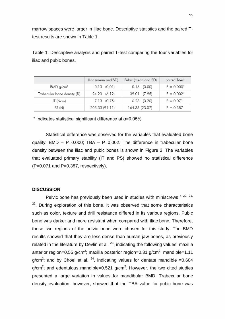
95
marrow spaces were larger in Iliac bone. Descriptive statistics and the paired T-
test results are shown in Table 1.
Table 1: Descriptive analysis and paired T-test comparing the four variables for
iliac and pubic bones.
* Indicates statistical significant difference at α=0.05%
Statistical difference was observed for the variables that evaluated bone
quality: BMD – P=0.000; TBA – P=0.002. The difference in trabecular bone
density between the iliac and pubic bones is shown in Figure 2. The variables
that evaluated primary stability (IT and PS) showed no statistical difference
(P=0.071 and P=0.387, respectively).
DISCUSSION
Pelvic bone has previously been used in studies with miniscrews 4 20, 21,
22. During exploration of this bone, it was observed that some characteristics
such as color, texture and drill resistance differed in its various regions. Pubic
bone was darker and more resistant when compared with iliac bone. Therefore,
these two regions of the pelvic bone were chosen for this study. The BMD
results showed that they are less dense than human jaw bones, as previously
related in the literature by Devlin et al. 23, indicating the following values: maxilla
anterior region=0.55 g/cm2; maxilla posterior region=0.31 g/cm2; mandible=1.11
g/cm2; and by Choel et al. 24, indicating values for dentate mandible =0.604
g/cm2; and edentulous mandible=0.521 g/cm2. However, the two cited studies
presented a large variation in values for mandibular BMD. Trabecular bone
density evaluation, however, showed that the TBA value for pubic bone was

96
similar to the result previously described by Aksoy et al. 25 for
maxilla=38.20±9.65; mandible=44.08±14.97. The results of this study showed
that the iliac and the pubic bones (which the pelvis is composed), present
different bone qualities: bone mineral density and trabecular bone density.
These characteristics, however, had no influence on primary stability of mini-
implants inserted in bone when the cortical was 1mm thick.
Mean values for IT ranged from 6.23 to 7.13 Ncm, representing adequate
primary stability according to Motoyoshi et al. 26, who stated that these values
should range from 5 to 10 Ncm.
Pull-out strength values ranged from 164.33 to 203.33 N, being within the
range found by Huja et al. 19 in a study with dog jaws: 134.5 N, for anterior
mandible, and 388.3 N for posterior mandible. Nevertheless, no landmark for
adequate pull-out strength value was found in the literature.
A previous study evaluated the influence of BMD on primary stability of
miniscrews and despite methodological differences, found a similar result 27. No
correlation was found between BMD, verified by cone beam computed
tomography, and miniscrew stability assessed by placement torque. The
authors also investigated the influence of cortical bone and found that cortical
thickness and cortical BMD were positively correlated with miniscrew stability 27.
No studies evaluating the influence of TBA on miniscrew stability were found.
Two hypotheses were formulated to explain the results of the present
study. The first is that the presence of a cortical thickness of 1 mm in all of the
specimens had an important influence on miniscrew stability, masking the
influence of bone mineral density and trabecular density. Cortical thickness has
been related to primary stability of miniscrews and implants 5, 12 28, 29, 30.
However, there is a lack of studys isolating these two factors: bone density and
cortical thickness. The second hypothesis is that the difference in bone quality
verified statistically may not be clinically relevant. A bigger difference between
BMD and TBA values in bones could perhaps reflect differences in mini-implant
stability.
Despite the limitations of this in vitro study, it can be inferred that in
clinical practice, a cortical thickness of 1mm is sufficient to guarantee the
primary stability of miniscrews, as previous supposed by Motoyoshi et al. 30,
even when there are variations in BMD and TBA values.

97
Further researches are suggested isolating the cortical effect and
increasing difference in density between different types of bone.
CONCLUSIONS
Iliac and pubic bones present different BMD and TBA values, the iliac
being less dense when considering the two parameters;
Miniscrew primary stability was not influenced by these differences in
bone density.
ACKNOWLEDGEMENTS
The authors would like to express their grateful appreciation to the
following entities and persons: CAPES (Cordenação de Aperfeiçoamento de
Pessoal de Nível Superior) and FAPERJ (Fundação de Amparo à Pesquisa do
Estado do Rio de Janeiro) for the financial support provided; INP (Sistema de
Implantes Nacionais e de Próteses) for the donation of the miniscrews;
Frigorífico Silva for the donation of the bones; doctors Maria Lucia Fleiuss
Farias and Laura Maria Carvalho Mendonça for the support during bone mineral
density analysis.
REFERENCES
1. Molly L. Bone density and primary stability in implant therapy. Clin Oral Implants Res. 2006
Oct;17 Suppl 2:124-35.
2. Javed F, Romanos GE. The role of primary stability for successful immediate loading of dental
implants. A literature review. J Dent. 2010 Aug;38(8):612-20. 3. Gedrange T, Hietschold V, Mai R, Wolf P, Nicklisch M, Harzer W. An evaluation of resonance
frequency analysis for the determination of the primary stability of orthodontic palatal implants.
A study in human cadavers. Clin Oral Implants Res. 2005 Aug;16(4):425-31.
4. Wilmes B, Rademacher C, Olthoff G, Drescher D. Parameters affecting primary stability of
orthodontic mini-implants. J Orofac Orthop. 2006 May;67(3):162-74.
5. Song YY, Cha JY, Hwang CJ. Mechanical characteristics of various orthodontic mini-screws in
relation to artificial cortical bone thickness. Angle Orthod. 2007 Nov;77(6):979-85.
6. Trisi P, De Benedittis S, Perfetti G, Berardi D. Primary stability, insertion torque and bone
density of cylindric implant ad modum Branemark: Is there a relationship? An in vitro study.
Clin Oral Implants Res. 2011Nov; 22(5):567-70.
7. Freudenthaler JW, Haas R, Bantleon HP. Bicortical titanium screws for critical orthodontic anchorage in the mandible: a preliminary report on clinical applications. Clin Oral Implants Res.
2001 Aug;12(4):358-63.
8. Cheng SJ, Tseng IY, Lee JJ, Kok SH. A prospective study of the risk factors associated with
failure of mini-implants used for orthodontic anchorage. Int J Oral Maxillofac Implants. 2004
Jan-Feb;19(1):100-6.

98
9. Mischkowski RA, Kneuertz P, Florvaag B, Lazar F, Koebke J, Zoller JE. Biomechanical
comparison of four different miniscrew types for skeletal anchorage in the mandibulo-maxillary
area. Int J Oral Maxillofac Surg. 2008 Oct;37(10):948-54.
10. Melsen B, Costa A. Immediate loading of implants used for orthodontic anchorage. Clin Orthod
Res. 2000 Feb;3(1):23-8.
11. Garfinkle JS, Cunningham LL, Jr., Beeman CS, Kluemper GT, Hicks EP, Kim MO. Evaluation of orthodontic mini-implant anchorage in premolar extraction therapy in adolescents. Am J
Orthod Dentofacial Orthop. 2008 May;133(5):642-53.
12. Merheb J, Van Assche N, Coucke W, Jacobs R, Naert I, Quirynen M. Relationship between
cortical bone thickness or computerized tomography-derived bone density values and implant
stability. Clin Oral Implants Res. 2010 Jun;21(6):612-7.
13. Cehreli MC, Kokat AM, Comert A, Akkocaoglu M, Tekdemir I, Akca K. Implant stability and
bone density: assessment of correlation in fresh cadavers using conventional and osteotome
implant sockets. Clin Oral Implants Res. 2009 Oct;20(10):1163-9.
14. Bergkvist G, Koh KJ, Sahlholm S, Klintstrom E, Lindh C. Bone density at implant sites and its
relationship to assessment of bone quality and treatment outcome. Int J Oral Maxillofac
Implants. 2010 Mar-Apr;25(2):321-8. 15. Lekholm U, Zarb G. Patient selection and preparation. In: Branemark PI, Zarb G, Albrektsson T,
editors. Tissue-integrated prostheses: osseointegration in clinical dentistry. Chicago:
Quintessence; 1985. p. 199-209.
16. Carey JJ, Delaney MF, Love TE, Richmond BJ, Cromer BA, Miller PD, et al. DXA-generated
Z-scores and T-scores may differ substantially and significantly in young adults. J Clin
Densitom. 2007 Oct-Dec;10(4):351-8.
17. Pithon MM, Andrade ACDV, Rodrigues VB, Santos RL. Influence of the immunossuppressant
tracolimus (FK-506) on the flexural strength of femur: a study in rats. Revista Brasileira de
Ortopedia. 2010;45(3):286-89.
18. Morse A. Formic acid-sodium citrate decalcification and butyl alcohol dehydration of teeth and
bones for sectioning in paraffin Journal of Dental Research. 1945;24(3):11.
19. Huja SS, Litsky AS, Beck FM, Johnson KA, Larsen PE. Pull-out strength of monocortical screws placed in the maxillae and mandibles of dogs. Am J Orthod Dentofacial Orthop. 2005
Mar;127(3):307-13.
20. Wawrzinek C, Sommer T, Fischer-Brandies H. Microdamage in cortical bone due to the
overtightening of orthodontic microscrews. J Orofac Orthop. 2008 Mar;69(2):121-34.
21. Wilmes B, Su YY, Drescher D. Insertion angle impact on primary stability of orthodontic mini-
implants. Angle Orthod. 2008 Nov;78(6):1065-70.
22. Su YY, Wilmes B, Honscheid R, Drescher D. Application of a wireless resonance frequency
transducer to assess primary stability of orthodontic mini-implants: an in vitro study in pig ilia.
Int J Oral Maxillofac Implants. 2009 Jul-Aug;24(4):647-54.
23. Devlin H, Horner K, Ledgerton D. A comparison of maxillary and mandibular bone mineral
densities. J Prosthet Dent. 1998 Mar;79(3):323-7. 24. Choel L, Duboeuf F, Bourgeois D, Briguet A, Lissac M. Trabecular alveolar bone in the human
mandible: a dual-energy x-ray absorptiometry study. Oral Surg Oral Med Oral Pathol Oral
Radiol Endod. 2003 Mar;95(3):364-70.
25. Aksoy U, Eratalay K, Tozum TF. The possible association among bone density values,
resonance frequency measurements, tactile sense, and histomorphometric evaluations of dental
implant osteotomy sites: a preliminary study. Implant Dent. 2009 Aug;18(4):316-25.
26. Motoyoshi M, Hirabayashi M, Uemura M, Shimizu N. Recommended placement torque when
tightening an orthodontic mini-implant. Clin Oral Implants Res. 2006 Feb;17(1):109-14.
27. Cha JY, Kil JK, Yoon TM, Hwang CJ. Miniscrew stability evaluated with computerized
tomography scanning. Am J Orthod Dentofacial Orthop. 2010 Jan;137(1):73-9.
28. Pithon MM, Nojima LI. Evaluation of the primary stability of orthodontic miniscrew in different
regions of maxilla and mandible of pigs. Innovations Implant Journal - Biomaterials and Esthetics. . 2007;2(4):58-63.
29. Motoyoshi M, Inaba M, Ono A, Ueno S, Shimizu N. The effect of cortical bone thickness on the
stability of orthodontic mini-implants and on the stress distribution in surrounding bone. Int J
Oral Maxillofac Surg. 2009 Jan;38(1):13-8.
30. Motoyoshi M, Yoshida T, Ono A, Shimizu N. Effect of cortical bone thickness and implant
placement torque on stability of orthodontic mini-implants. Int J Oral Maxillofac Implants. 2007
Sep-Oct;22(5):779-84.
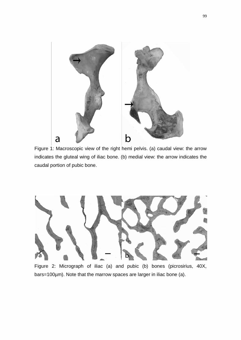
99
Figure 1: Macroscopic view of the right hemi pelvis. (a) caudal view: the arrow
indicates the gluteal wing of iliac bone. (b) medial view: the arrow indicates the
caudal portion of pubic bone.
Figure 2: Micrograph of iliac (a) and pubic (b) bones (picrosirius, 40X,
bars=100µm). Note that the marrow spaces are larger in iliac bone (a).
100
9.2 APÊNDICE B: Marquezan M, Lau TC, Mattos CT, Cunha AC, Nojima LI,
Sant'anna EF, Souza MM, Araújo MT. Bone mineral density. Angle Orthod.
2012 Jan;82(1):62-6.
Bone mineral density: methods of measurement and its
influence on primary stability of miniscrews
Mariana Marquezana; Thiago Chon Leon Lau
a; Claudia Trindade Mattos
a;
Amanda Carneiro da Cunhab; Lincoln Issamu Nojima
c; Eduardo Franzotti Sant'Anna
c;
Margareth Maria Gomes de Souzac ; Mônica Tirre de Souza Araújo
c
a PhD student, Pediatric Dentistry and Orthodontics Department, School of Dentistry, Federal
University of Rio de Janeiro, Rio de Janeiro, Brazil
b Research assistant, Pediatric Dentistry and Orthodontics Department, School of Dentistry,
Federal University of Rio de Janeiro, Rio de Janeiro, Brazil
c Professor, Pediatric Dentistry and Orthodontics Department, School of Dentistry, Federal
University of Rio de Janeiro, Rio de Janeiro, Brazil
© 2011 by the EH Angle Education and Research Foundation, Inc.
Submitted: March 2011

101
ABSTRACT
Objective: To verify whether bone mineral density (BMD) of cortical bone, trabecular bone and
total bone influences the primary stability of orthodontic miniscrews and to verify whether there
is correlation between the measurement of BMD by Cone-Beam Computed Tomography
(CBCT) and Central Dual-energy X-ray Absorptiometry (DEXA).
Material and Methods: Twenty bovine bone sections were extracted from the pubic and iliac
bones from regions with cortical thickness of approximately 1mm. The BMD of the total bone
block was evaluated by two methods: CBCT and DEXA. The BMD of cortical, trabecular and
total bone in the region of interest (ROI) were also evaluated by CBCT. After scanning the bone
blocks, twenty self-drilling miniscrews (INP®) 1.4 mm in diameter and 6 mm long were inserted
into them. The peak implant insertion torque (IT) was registered. After this, the pull-out test (PS)
was performed and the maximum force registered. Pearson’s correlation test was applied to
verify the correlations between variables. Results: The BMD of the total bone block verified by
CBCT and DEXA showed a positive and strong correlation (r=0.866, p=0.000). The BMD of the
ROI for cortical bone influenced the IT (r=0.518, p=0.40) and the PS of miniscrews (r=0.615,
p=0.015). However, the total bone BMD (verified by CBCT and DEXA) and trabecular bone
BMD presented weak and not statistically significant correlations with primary stability.
Conclusions: There was a positive correlation between total bone block BMD measured by
DEXA and CBCT. The cortical BMD influenced the IT and PS.
Keywords: Bone density; Cone-Beam Computed Tomography; Dual-energy X-ray
Absorptiometry; Miniscrew
INTRODUCTION
The primary stability of miniscrews depends on the characteristics of the device, insertion
technique and bone quality and quantity of the receptor site 4. The bone quality classification
most accepted in Implant dentistry has been that proposed by Lekholm and Zarb 15
, based on
the amount of cortical and trabecular bone shown in preoperative radiographs. However, this
classification depends on the operator’s subjective evaluation of the radiograph. A less

102
subjective method for evaluating the quality of cortical and trabecular bone is to verify the bone
mineral density (BMD) 14
.
The quantification of bone mineral density taken as the gold standard in endocrinology and
traumatology is bone densitometry by means of central dual-energy X-ray absorptiometry
(DEXA)16
. In implant dentistry, however, cone-beam computed tomography (CBCT) has been
used for this purpose because it is a three-dimensional analysis that allows the quantification of
the mineral density of jaw bones in specific sites in Hounsfield units (HU). When evaluating the
bone mineral density of the receptor site by CBCT, it is possible to verify the cortical BMD and
trabecular BMD separately or the total bone BMD which is the measurement of mineral density
of trabecular bone and cortical bone together.
The aims of this study were to verify how these parameters influence the primary stability of
orthodontic miniscrews and to verify whether there is correlation between the measurement of
BMD by CBCT and DEXA.
MATERIALS AND METHODS
The sample consisted of twenty bone sections extracted from bovine pelvic bone (Bos taurus,
Angus lineage). The bones were obtained from a slaughterhouse (registered with ANVISA – the
Brazilian Health Surveillance Agency) immediately after slaughter. The bone sections were
extracted from the caudal portion of the pubic bone and from the gluteal wing of iliac bone with
the use of a trephine bur (8mm in diameter x 20mm long, Sin Implantes, São Paulo, Brazil)
adapted to a low speed motor (Beltec LB100, Araraquara, Brazil) under irrigation. The bone
sections (8mm in diameter x 12mm long) were taken from regions in which there was a mean
cortical thickness of 1mm (measured with an orthodontic caliper, Odin, Ortho-pli, Philadelphia,
USA). The extracted material was immersed in sterile physiological solution and stored by
freezing (-20ºC).

103
BMD evaluation
The BMD was evaluated by two methods: DEXA and CBCT. First, the bone mineral content of
specimens was measured and divided by the area to obtain the BMD by means of central dual-
energy X-ray absorptiometry (DEXA) Prodigy (GE/LUNAR, Madison, USA), calibrated for small
animals. To perform the exam, the bone blocks were thawed at room temperature and put into
plastic boxes (6x11x4 cm) containing raw rice to simulate soft tissue 17
. After this, the samples
were irradiated by DEXA for 30 seconds17
. This analysis comprised the entire bone block
(cortical and trabecular bones).
After this, CBCT (I-CAT 3D Dental Imaging System, Pennsylvania, USA) was used to obtain
tomographs in accordance with a standard protocol (120 KV, 47 mA, FOV of 22 cm, voxel of 0.4
mm, and scan time of 30 s). All the samples were arranged identically in a Styrofoam box with
the cortical bone perpendicular to the floor for the purpose of scanning. Data were imported in
DICOM format (Digital Imaging and Communications in Medicine) and handled by I-CAT Vision
(Dental Imaging System, Pennsylvania, USA). Once imported, CT data were reconstructed into
1-mm thick transaxial images. The BMD was obtained using the multiplanar reconstruction
screen and measuring the HU (Hounsfield Unit) of the selected area. First, a sagittal section
was obtained in the center of the bone block . To verify whether the data of CBCT measurement
of BMD were correlated with the data acquired from DEXA, the total area of the bone block in
this section was evaluated (total bone block BMD) . After this, the region of interest (ROI) was
isolated. The ROI was considered the area in the center of the block that would surround the
miniscrew after its placement (2.4 mm X 7 mm). The BMD of cortical bone, trabecular bone and
total bone were measured separately in the ROI (Figure 1).
Primary Stability Evaluation
The primary stability was evaluated by means of insertion torque (IT) measurement and pull-out
strength (PS). Twenty miniscrews (INP®, São Paulo, Brazil) 1.4 mm in diameter and 6 mm long
were inserted into the bone blocks with the use of a manual placement key connected to a
digital torque meter (Lutron TQ-8800, Taipei, Taiwan) to allow measurement of the peak implant
placement torque. The values were recorded in Newton centimeter (Ncm). After this, the pull-out
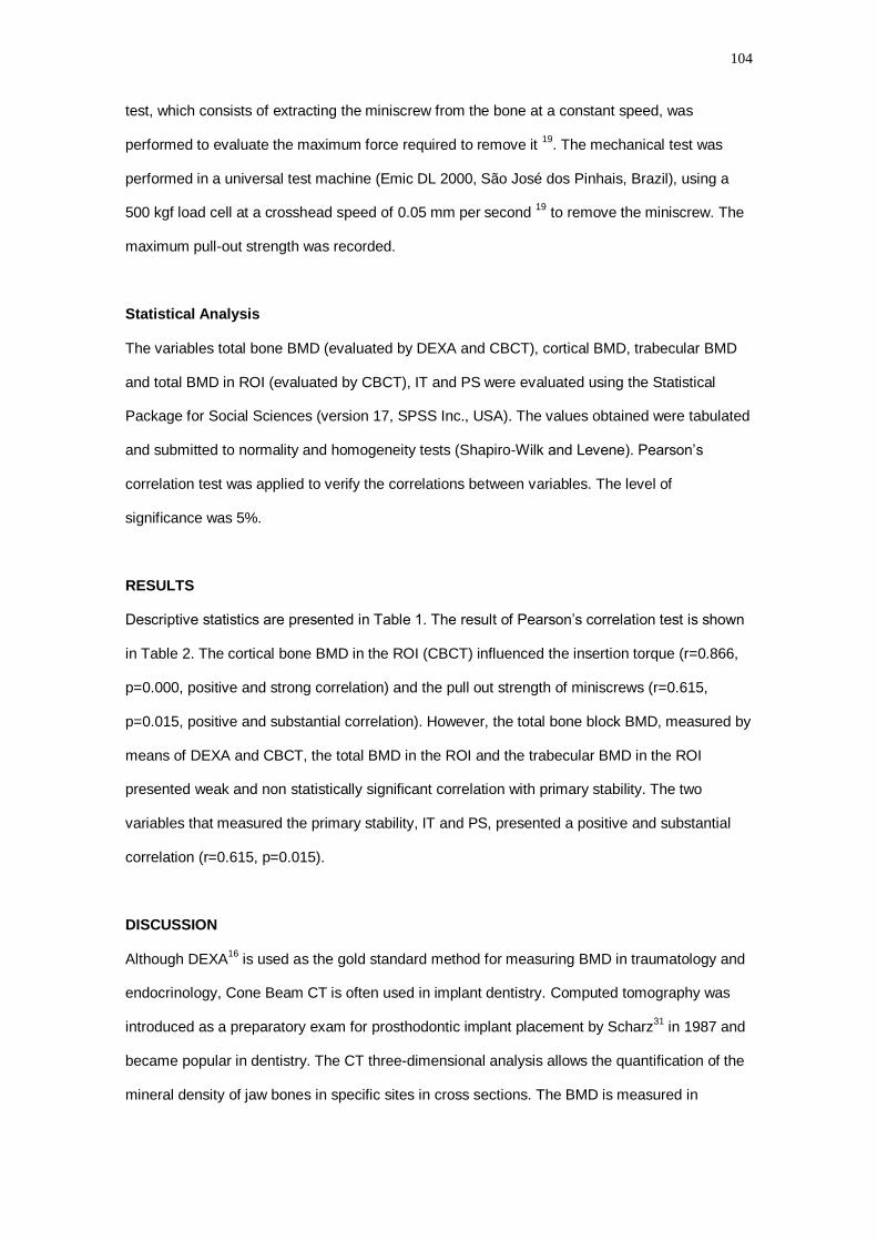
104
test, which consists of extracting the miniscrew from the bone at a constant speed, was
performed to evaluate the maximum force required to remove it 19
. The mechanical test was
performed in a universal test machine (Emic DL 2000, São José dos Pinhais, Brazil), using a
500 kgf load cell at a crosshead speed of 0.05 mm per second 19
to remove the miniscrew. The
maximum pull-out strength was recorded.
Statistical Analysis
The variables total bone BMD (evaluated by DEXA and CBCT), cortical BMD, trabecular BMD
and total BMD in ROI (evaluated by CBCT), IT and PS were evaluated using the Statistical
Package for Social Sciences (version 17, SPSS Inc., USA). The values obtained were tabulated
and submitted to normality and homogeneity tests (Shapiro-Wilk and Levene). Pearson’s
correlation test was applied to verify the correlations between variables. The level of
significance was 5%.
RESULTS
Descriptive statistics are presented in Table 1. The result of Pearson’s correlation test is shown
in Table 2. The cortical bone BMD in the ROI (CBCT) influenced the insertion torque (r=0.866,
p=0.000, positive and strong correlation) and the pull out strength of miniscrews (r=0.615,
p=0.015, positive and substantial correlation). However, the total bone block BMD, measured by
means of DEXA and CBCT, the total BMD in the ROI and the trabecular BMD in the ROI
presented weak and non statistically significant correlation with primary stability. The two
variables that measured the primary stability, IT and PS, presented a positive and substantial
correlation (r=0.615, p=0.015).
DISCUSSION
Although DEXA16
is used as the gold standard method for measuring BMD in traumatology and
endocrinology, Cone Beam CT is often used in implant dentistry. Computed tomography was
introduced as a preparatory exam for prosthodontic implant placement by Scharz31
in 1987 and
became popular in dentistry. The CT three-dimensional analysis allows the quantification of the
mineral density of jaw bones in specific sites in cross sections. The BMD is measured in

105
Hounsfield units (HU) by the difference in grayscale. The CBCT also allows bone dimensions to
be measured before miniscrew placement. The results of this study showed that the BMD of the
total bone block, verified by CBCT and DEXA, had a positive and strong correlation (r=0.866,
p=0.000).
The results of this study showed that the BMD of the ROI of cortical bone influences the primary
stability of miniscrews measured by the insertion torque and pull out strength tests. The total
bone block BMD and the total BMD ROI did not influence the insertion torque and pull-out
strength of miniscrews. Similar result was previously described by Cha et al27
. They evaluated
the primary stability of miniscrews inserted in the jaws of beagle dogs. As in the present study,
the bone density was assessed by CBCT and the primary stability was verified by means of
insertion torque and also by the Periotest value (Periotest - Medizintechnik Gulden, Modautal,
Germany). They found a positive association between cortical BMD and miniscrew primary
stability, but the BMD for the total bone was weak and non statistically significant.
The importance of the cortical in miniscrew stability has previously been established by
assessing the cortical thickness, and a positive association was verified between cortical
thickness and the primary stability of miniscrews 4, 19, 22, 27, 29, 30
. The evaluation of cortical BMD
corroborates its importance in initial stability.
The ROI of total BMD has previously been related to the primary stability of prosthodontic
implants 25, 32, 33
, but it was not verified for miniscrews in the present study or by Cha et al 27
.
The ROI of trabecular BMD also plays an important role in the primary stability of prosthodontic
implants, as verified by Bergkvist et al 14
and Merheb et al 12
. However, in this study, with
cortical thickness of 1mm, there was no association between ROI of trabecular BMD and the
primary stability of mini-implants.
Despite the limitations of our laboratory study, the CBCT proved to be a useful exam for
noninvasive assessment of bone density at the preoperative stage of mini-implant placement.
Moreover, the cortical thickness and the cortical BMD can be measured to estimate the primary
stability of miniscrews.
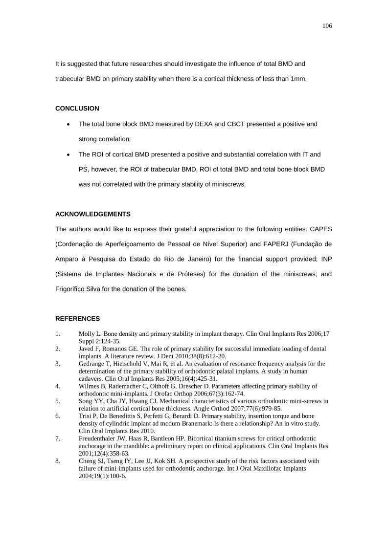
106
It is suggested that future researches should investigate the influence of total BMD and
trabecular BMD on primary stability when there is a cortical thickness of less than 1mm.
CONCLUSION
The total bone block BMD measured by DEXA and CBCT presented a positive and
strong correlation;
The ROI of cortical BMD presented a positive and substantial correlation with IT and
PS, however, the ROI of trabecular BMD, ROI of total BMD and total bone block BMD
was not correlated with the primary stability of miniscrews.
ACKNOWLEDGEMENTS
The authors would like to express their grateful appreciation to the following entities: CAPES
(Cordenação de Aperfeiçoamento de Pessoal de Nível Superior) and FAPERJ (Fundação de
Amparo à Pesquisa do Estado do Rio de Janeiro) for the financial support provided; INP
(Sistema de Implantes Nacionais e de Próteses) for the donation of the miniscrews; and
Frigorífico Silva for the donation of the bones.
REFERENCES
1. Molly L. Bone density and primary stability in implant therapy. Clin Oral Implants Res 2006;17
Suppl 2:124-35.
2. Javed F, Romanos GE. The role of primary stability for successful immediate loading of dental
implants. A literature review. J Dent 2010;38(8):612-20.
3. Gedrange T, Hietschold V, Mai R, et al. An evaluation of resonance frequency analysis for the
determination of the primary stability of orthodontic palatal implants. A study in human
cadavers. Clin Oral Implants Res 2005;16(4):425-31.
4. Wilmes B, Rademacher C, Olthoff G, Drescher D. Parameters affecting primary stability of
orthodontic mini-implants. J Orofac Orthop 2006;67(3):162-74. 5. Song YY, Cha JY, Hwang CJ. Mechanical characteristics of various orthodontic mini-screws in
relation to artificial cortical bone thickness. Angle Orthod 2007;77(6):979-85.
6. Trisi P, De Benedittis S, Perfetti G, Berardi D. Primary stability, insertion torque and bone
density of cylindric implant ad modum Branemark: Is there a relationship? An in vitro study.
Clin Oral Implants Res 2010.
7. Freudenthaler JW, Haas R, Bantleon HP. Bicortical titanium screws for critical orthodontic
anchorage in the mandible: a preliminary report on clinical applications. Clin Oral Implants Res
2001;12(4):358-63.
8. Cheng SJ, Tseng IY, Lee JJ, Kok SH. A prospective study of the risk factors associated with
failure of mini-implants used for orthodontic anchorage. Int J Oral Maxillofac Implants
2004;19(1):100-6.

107
9. Mischkowski RA, Kneuertz P, Florvaag B, et al. Biomechanical comparison of four different
miniscrew types for skeletal anchorage in the mandibulo-maxillary area. Int J Oral Maxillofac
Surg 2008;37(10):948-54.
10. Melsen B, Costa A. Immediate loading of implants used for orthodontic anchorage. Clin Orthod
Res 2000;3(1):23-8.
11. Garfinkle JS, Cunningham LL, Jr., Beeman CS, et al. Evaluation of orthodontic mini-implant anchorage in premolar extraction therapy in adolescents. Am J Orthod Dentofacial Orthop
2008;133(5):642-53.
12. Merheb J, Van Assche N, Coucke W, et al. Relationship between cortical bone thickness or
computerized tomography-derived bone density values and implant stability. Clin Oral Implants
Res 2010;21(6):612-7.
13. Cehreli MC, Kokat AM, Comert A, et al. Implant stability and bone density: assessment of
correlation in fresh cadavers using conventional and osteotome implant sockets. Clin Oral
Implants Res 2009;20(10):1163-9.
14. Bergkvist G, Koh KJ, Sahlholm S, Klintstrom E, Lindh C. Bone density at implant sites and its
relationship to assessment of bone quality and treatment outcome. Int J Oral Maxillofac Implants
2010;25(2):321-8. 15. Lekholm U, Zarb G. Patient selection and preparation. In: Branemark PI, Zarb G, Albrektsson T,
editors. Tissue-integrated prostheses: osseointegration in clinical dentistry. Chicago:
Quintessence; 1985. p. 199-209.
16. Carey JJ, Delaney MF, Love TE, et al. DXA-generated Z-scores and T-scores may differ
substantially and significantly in young adults. J Clin Densitom 2007;10(4):351-8.
17. Pithon MM, Andrade ACDV, Rodrigues VB, Santos RL. Influence of the immunossuppressant
tracolimus (FK-506) on the flexural strength of femur: a study in rats. Revista Brasileira de
Ortopedia 2010;45(3):286-89.
18. Morse A. Formic acid-sodium citrate decalcification and butyl alcohol dehydration of teeth and
bones for sectioning in paraffin Journal of Dental Research 1945;24(3):11.
19. Huja SS, Litsky AS, Beck FM, Johnson KA, Larsen PE. Pull-out strength of monocortical
screws placed in the maxillae and mandibles of dogs. Am J Orthod Dentofacial Orthop 2005;127(3):307-13.
20. Wawrzinek C, Sommer T, Fischer-Brandies H. Microdamage in cortical bone due to the
overtightening of orthodontic microscrews. J Orofac Orthop 2008;69(2):121-34.
21. Wilmes B, Su YY, Drescher D. Insertion angle impact on primary stability of orthodontic mini-
implants. Angle Orthod 2008;78(6):1065-70.
22. Su YY, Wilmes B, Honscheid R, Drescher D. Application of a wireless resonance frequency
transducer to assess primary stability of orthodontic mini-implants: an in vitro study in pig ilia.
Int J Oral Maxillofac Implants 2009;24(4):647-54.
23. Devlin H, Horner K, Ledgerton D. A comparison of maxillary and mandibular bone mineral
densities. J Prosthet Dent 1998;79(3):323-7.
24. Choel L, Duboeuf F, Bourgeois D, Briguet A, Lissac M. Trabecular alveolar bone in the human mandible: a dual-energy x-ray absorptiometry study. Oral Surg Oral Med Oral Pathol Oral
Radiol Endod 2003;95(3):364-70.
25. Aksoy U, Eratalay K, Tozum TF. The possible association among bone density values,
resonance frequency measurements, tactile sense, and histomorphometric evaluations of dental
implant osteotomy sites: a preliminary study. Implant Dent 2009;18(4):316-25.
26. Motoyoshi M, Hirabayashi M, Uemura M, Shimizu N. Recommended placement torque when
tightening an orthodontic mini-implant. Clin Oral Implants Res 2006;17(1):109-14.
27. Cha JY, Kil JK, Yoon TM, Hwang CJ. Miniscrew stability evaluated with computerized
tomography scanning. Am J Orthod Dentofacial Orthop 2010;137(1):73-9.
28. Pithon MM, Nojima LI. Evaluation of the primary stability of orthodontic miniscrew in different
regions of maxilla and mandible of pigs. Innovations Implant Journal - Biomaterials and
Esthetics. 2007;2(4):58-63. 29. Motoyoshi M, Inaba M, Ono A, Ueno S, Shimizu N. The effect of cortical bone thickness on the
stability of orthodontic mini-implants and on the stress distribution in surrounding bone. Int J
Oral Maxillofac Surg 2009;38(1):13-8.
30. Motoyoshi M, Yoshida T, Ono A, Shimizu N. Effect of cortical bone thickness and implant
placement torque on stability of orthodontic mini-implants. Int J Oral Maxillofac Implants
2007;22(5):779-84.
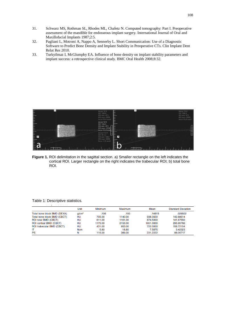
108
31. Schwarz MS, Rothman SL, Rhodes ML, Chafetz N. Computed tomography: Part I. Preoperative
assessment of the mandible for endosseous implant surgery. International Journal of Oral and
Maxillofacial Implants 1987;2:5.
32. Pagliani L, Motroni A, Nappo A, Sennerby L. Short Communication: Use of a Diagnostic
Software to Predict Bone Density and Implant Stability in Preoperative CTs. Clin Implant Dent
Relat Res 2010. 33. Turkyilmaz I, McGlumphy EA. Influence of bone density on implant stability parameters and
implant success: a retrospective clinical study. BMC Oral Health 2008;8:32.
Figure 1. ROI delimitation in the sagittal section. a) Smaller rectangle on the left indicates the cortical ROI. Larger rectangle on the right indicates the trabecular ROI; b) total bone ROI.
Table 1: Descriptive statistics.

109
Table 2: Pearson correlation test.
* Correlation is significant at the 0.05 level.

110
10 ANEXOS
10.1 ANEXO 1: Aceite do Artigo 3.
Manuscript052513-395R Decision Letter
[email protected] <[email protected]> 5 de setembro de 2013 19:43 Para: [email protected]
Dear Dr. Marquezan, I am pleased to inform you that your manuscript "Is trabecular bone related to primary stability of miniscrews?" has been accepted for publication. We look forward to publishing your contribution in The Angle Orthodontist. Allen Press will e-mail instructions to you in about 6 weeks telling you how to view a galley proof of your article on the Internet in PDF format. These proofs will look like your article as it will appear in print. The proofs will contain changes that occurred in the printing process (editing, typesetting or layout). If you have not received these instructions after 8 weeks, please let us know by sending us an e-mail with the manuscript number and title. Please print out the proofs and make ONLY PRINTER ERROR CORRECTIONS on the paper print outs. Further editing of the content of your manuscript is not permissable on the galley copy. Please also include a typed page of the changes/corrections that you are requesting. REPRINT REQUESTS - Our printer will no longer provide reprints effective immediately. Please do not add new material as that will incur costs to the author. After you correct the paper print out, express ship it to me at 5813 Vernon Lane, Edina, MN 55436-2239 (Phone: 952-922-9586). A quick turn-around is important, please. IMPORTANT: Any changes made AFTER the galley has been corrected and submitted to us incur significant costs from the printer. These costs will soon be charged to the authors. Therefore, we urge you to please read your galley VERY carefully before it is returned to us with your corrections/changes. We work hard to keep our costs down so that we can continue to provide the journal at no charge to all readers all over the world. Again, congratulations on your work and thank you for your contributions to the orthodontic literature. I look forward to seeing your article in print. If you have any questions, please be sure to contact us. Sincerely, Steven Lindauer Editor The Angle Orthodontist rji