universidade de sÃo paulo instituto de biociÊncias ... · processo de derivação das...
TRANSCRIPT
UNIVERSIDADE DE SÃO PAULO
INSTITUTO DE BIOCIÊNCIAS
DEPARTAMENTO DE GENÉTICA E BIOLOGIA EVOLUTIVA
Rua do Matão, 277 sala 350– Cidade Universitária – CEP 05508-900 São Paulo – SP – Brasil Caixa Postal 11461 – CEP 05422-970 – São Paulo, SP - Brasil Tel. 55 11 3091-7476 FAX 55 11 3091-7553 - e-mail: lpereira@ usp.br
São Paulo, December 4th, 2015.
To the Editors of Brazilian Journal of Biology,
I would like to ask for the retraction of the article: Derivation of new Brazilian lineages of
human embryonic stem cells under physiological oxygen conditions. Georges JA, Vergani N,
Fraga AM, Fonseca SA, Fujihara LS, Albuquerque MC, Castro FJ, Silva IS, Pereira LV. Braz
J Biol. 2015 Nov 24. pii: S1519-69842015005105121.
It was submitted without consent or any knowledge from the following authors: Vergani N,
Fraga AM, Fonseca SA, Fujihara LS, Albuquerque MC, Castro FJ, Silva IS, Pereira LV; and
all the data described had already been published in: Aberrant patterns of X chromosome
inactivation in a new line of human embryonic stem cells established in physiological oxygen
concentrations. de Oliveira Georges JA, Vergani N, Fonseca SA, Fraga AM, de Mello JC,
Albuquerque MC, Fujihara LS, Pereira LV. Stem Cell Rev. 2014 Aug;10(4):472-9. doi:
10.1007/s12015-014-9505-4.
Sincerely,
Lygia da Veiga Pereira, Ph.D.
Depto. Genética e Biologia Evolutiva
Instituto de Biociências, USP
99Braz. J. Biol., 2015, vol. 75, no. 4, suppl. 1, pp. S9-S16
http://dx.doi.org/10.1590/1519-6984.20813 Original Article
Derivation of new Brazilian lineages of human embryonic stem cells under physiological oxygen conditions
J. A. O. Georgesa,b,c,d*, N. Verganib,c, A. M. Fragab,c, S. A. S. Fonsecab,c, L. S. Fujiharae, M. C. R. M. Albuquerquee, F. J. G. Castroa, I. S. Silvaa and L. V. Pereirab,c
aPrograma de Pós-graduação em Saúde e Desenvolvimento Regional, Faculdade de Medicina – FAMED, Universidade Federal de Mato Grosso do Sul – UFMS, Av. Senador Filinto Muller, Unidade 9,
Cidade Universitária, CP 549, CEP 79070-900, Campo Grande, MS, BrazilbLaboratório de Genética Molecular, Departamento de Genética e Biologia Evolutiva, Instituto de Biociências – IB,
Universidade de São Paulo – USP, Rua do Matão, Travessa 14, 321, Cidade Universitária, CEP 05508-900, São Paulo, SP, Brazil
cLaboratório Nacional de Células-Tronco Embrionárias – LaNCE, Departamento de Genética e Biologia Evolutiva, Instituto de Biociências – IB, Universidade de São Paulo – USP, Rua do Matão, Travessa 14, 321,
Cidade Universitária, CEP 05508-900, São Paulo, SP, BrazildS.INOVA, Programa de Pós-graduação em Biotecnologia, Universidade Católica Dom Bosco – UCDB, Av. Tamandaré, 6000, Bloco Biosaúde, Jardim Seminário, CEP 79117-900, Campo Grande, MS, Brazil
eFertivitro – Centro de Reprodução Humana, Av. Indianópolis, 843, Moema, CEP 04063-001, São Paulo, SP, Brazil*e-mail: [email protected]
Received: November 7, 2013 – Accepted: September 3, 2014 – Distributed: November 30, 2015(With 8 figures)
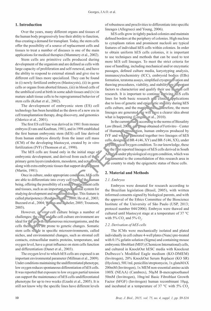
AbstractThe development and application of human embryonic stem (hES) cells is an important experimental tool for understanding differentiation and development, and offer a wide range of potential applications, including prevention, diagnosis and treatment of human diseases. This area is one of the most promising and fascinating contemporary biology. The essential components of the process of embryonic stem cells derivation are the embryos, embryo manipulation methods, feeder cells, culture media, the oxygen level and the general culture conditions in vitro. In 2008, Pereira’s group generated the first hES cell Brazilian lineage. It is important to continue deriving hES cells lines for both basic research and clinical applications due to loss of genetic and epigenetic stability during hES cells culture and the manipulation. Therefore, the more lineages are generated, we will have a clearer idea about what is happening. In the current study, we has generated two lineages of hES cells, designated BR-4 (46, XY) and BR-5 (46, XX), under physiological oxygen condition. To our knowledge, these are the first reported lineages of hES cells derived in South America under physiological oxygen condition, which was fundamental to the consolidation of this research area in our country to study the epigenetic status of these cells.
Keywords: human embryonic stem cells, hypoxia, derivation.
Derivação de novas linhagens brasileiras de células-tronco embrionárias humanas sob condições fisiológicas de oxigênio
ResumoO desenvolvimento e a aplicação das células-tronco embrionárias humanas (hES) é uma importante ferramenta experimental para a compreensão da diferenciação e desenvolvimento, e oferece uma ampla gama de aplicações potenciais, incluindo a prevenção, diagnóstico e tratamento de doenças humanas. Os componentes essenciais do processo de derivação das células-tronco embrionárias são os embriões, os métodos de manipulação de embriões, as matrizes celulares, os meios de cultura, o nível de oxigênio e as condições gerais de cultura in vitro. Em 2008, o grupo da Dra. Lygia da Veiga Pereira gerou a primeira linhagem brasileira de células-tronco embrionárias humanas. É importante continuar a derivar linhagens de células-tronco embrionárias humanas para a investigação básica e as aplicações clínicas devido à perda de estabilidade genética e epigenética durante a cultura de células embrionárias humanas e a manipulação. Portanto, quanto mais linhagens são geradas, teremos uma idéia mais clara sobre o que está acontecendo. No estudo atual, nós geramos duas linhagens de células-tronco embrionárias humanas, denominadas BR-4 (46, XY) e BR-5 (46, XX), sob a condição fisiológica de oxigênio. Para nosso conhecimento, estas são as primeiras linhagens de células-tronco embrionárias humanas reportadas derivadas na América do Sul sob a condição fisiológica de oxigênio, o que foi fundamental para a consolidação desta área de pesquisa em nosso país para estudar o estado epigenético dessas células.
Palavras-chave: células-tronco embrionárias humanas, hipóxia, derivação.
RETRACTED ARTIC
LE
Braz. J. Biol., 2015, vol. 75, no. 4, suppl. 1, pp. S9-S1610
Georges, J.A.O. et al.
10
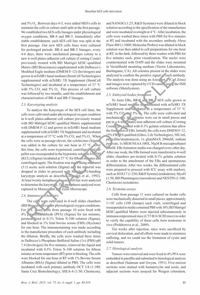
1. Introduction
Over the years, many different organs and tissues of the human body progressively lose their ability to function, thus creating a demand for transplant. Today, the stem cells offer the possibility of a source of replacement cells and tissues to treat a number of diseases is one of the main applications for medical therapies (Mummery et al., 2002).
Stem cells are primitive cells produced during development of the organism and are defined as cells with large capacity of proliferation and self-renewal, and have the ability to respond to external stimuli and give rise to different cell lines more specialized. They can be found in (i) newly fertilized embryos (blastocysts), (ii) in germ cells or organs from aborted fetuses, (iii) in blood cells of the umbilical cord at birth in some adult tissues and (iv) in mature adult tissue cells to be reprogrammed behavior of stem cells (Kehat et al., 2002).
The development of embryonic stem (ES) cell technology has been heralded as the dawn of a new era in cell transplantation therapy, drug discovery, and genomics (Odorico et al., 2001).
The first ES cell line was derived in 1981 from mouse embryos (Evans and Kaufman, 1981), and in 1998 established the first human embryonic stem (hES) cell line derived from human embryos directly from the inner cell mass (ICM) of the developing blastocyst, created by in vitro fertilization (IVF) (Thomson et al., 1998).
The hES cells are found only in the initial stage of embryonic development, and derived from each of three primary germ layers (endoderm, mesoderm, and ectoderm), along with extra embryonic tissues that support development (Martin, 1981).
Once in culture, under appropriate conditions, hES cells are able to differentiate into every cell type of the human being, offering the possibility of a source replacement cells and tissues, such as an important experimental system for studying development and modeling disease. This feature is called pluripotency (Reubinoff et al., 2000; He et al., 2003; Buzzard et al., 2004; Wobus and Boheler, 2005; Trounson, 2005).
However, in vitro cell culture brings a number of challenges; the cells and the cell culture environment are ideal for the growth of numerous microorganisms, and the cells themselves are prone to genetic changes. Somatic stem cells reside in specific microenvironments, called niches, and environmental changes, such as stromal cell contacts, extracellular matrix proteins, temperature, and oxygen level, have a great influence on stem cells function and differentiation (Danet et al., 2003).
The oxygen level to which hES cells are exposed is an important environmental parameter (Millman et al., 2009). Under conditions maintaining the undifferentiated phenotype, low oxygen reduces spontaneous differentiation of hES cells. It was reported that exposure to low oxygen partial tension can support the maintenance of hES cells undifferentiated phenotype for up to two weeks (Ezashi et al., 2005). It is still not know why the specific lines have different levels
of robustness and proclivities to differentiate into specific lineages (Allegrucci and Young, 2006).
hES cells grow in tightly packed colonies and maintain defined borders at the periphery of colonies. High nucleus to cytoplasm ration and prominent nucleoli are typical features of individual hES cells within colonies. In order to obtain uniform hES cells colonies, it is important to use techniques and methods that can be used to test more hES cell lineages. To meet the strict criteria for ease of handling, including mechanical and/or enzymatic passages, defined culture media, karyotyping analysis, immunocytochemistry (ICC), embryoid bodies (EBs) formation, teratoma assays, simplified cryopreservation and thawing procedures, viability, and stability are important factors to characterize and qualify their use in stem cell research. It is important to continue deriving hES cells lines for both basic research and clinical applications due to loss of genetic and epigenetic stability during hES cells culture, and the manipulation. Therefore, the more lineages are generated, we will have a clearer idea about what is happening (Lengner et al., 2010).
In the current study, according to the norms of Biosafety Law (Brasil, 2005), our group obtained of Fertivitro – Center of Human Reproduction, human embryos produced by IVF and we has generated together two lineages of hES cells, designated BR-4 (46, XY) and BR-5 (46, XX), under physiological oxygen condition. To our knowledge, these are the first reported lineages of hES cells derived in South America under physiological oxygen condition, which was fundamental to the consolidation of this research area in our country to study the epigenetic status of these cells.
2. Material and Methods
2.1. EmbryosEmbryos were donated for research according to
the Brazilian legislation (Brasil, 2005), with written informed consents signed by biological parents, and with the approval of the Ethics Committee of the Bioscience Institute of the University of São Paulo (USP, 2013; protocol number 044/2006). Embryos were thawed and cultured until blastocyst stage at a temperature of 37 °C with 5% CO2 and 5% O2.
2.2. Derivation of hES cellsThe ICMs were mechanically isolated and plated
individually in cell culture 4-well plates (Nunc) pre-treated with 0.1% gelatin solution (Sigma) and containing mouse embryonic fibroblast (MEF) (Chemicon International) cells, and cultured in KnockOut hESC media with Knockout Dulbecco’s Modified Eagle medium (KO-DMEM) (Invitrogen), 20% KnockOut Serum Replacer (KO SR) (Hyclone), 50U/mL penicillin/streptomycin, 1x glutaMAX 200mM (Invitrogen), 1x MEM non-essential amino acids 100X (NEAA) (Cambrex), 50µM B-mercaptoethanol 50mM (Invitrogen), 10ng/ml Basic Fibroblast Growth Factor (bFGF) (Invitrogen) human recombinant 10µg, and incubated at a temperature of 37 °C with 5% CO2
RETRACTED ARTIC
LE
Braz. J. Biol., 2015, vol. 75, no. 4, suppl. 1, pp. S9-S16 11
Brazilian hES cells derivation
11
and 5% O2. Between days 4-5, were added MEFs cells to maintain the cells in culture until split at the first passage. We established two hES cells lineages under physiological oxygen conditions, BR-4 and BR-5. Immediately after stable establishment, each hES cell line was split at the first passage. Our new hES cells lines were cultured for prolonged periods. BR-4 and BR-5 lineages, every 4-6 days, there were mechanical passages colony to a new 6-well plates-adherent cell culture (Corning Costar) previously treated with BD Matrigel hESC-qualified Matrix (BD Biosciences) supplemented with Dulbecco’s Modified Eagle medium (DMEM F-12) (Invitrogen) and grown in mTeSR1 basal medium (StemCell Technologies) supplemented with mTeSR1 5X Supplement (StemCell Technologies) and incubated at a temperature of 37 °C with 5% CO2 and 5% O2. This process of cell culture was followed by two months, until the establishment and characterization of BR-4 and BR-5 lineages.
2.3. Karyotyping analysisTo analyze the Karyotype of the hES cell lines, the
cells were cultivated under physiological oxygen condition in 6-well plates-adherent cell culture previously treated with BD Matrigel hESC-qualified Matrix supplemented with DMEM F-12 and grown in mTeSR1 basal medium supplemented with mTeSR1 5X Supplement and incubated at a temperature of 37 °C with 5% CO2 and 5% O2. When the cells were at passage eleven, the colchicines (Sigma) was added in the culture for one hour at 37 °C. After this time, the cells were trypsinized, centrifuged and the pellet was ressuspended in the hypotonic solution 0.075M (KCL) (Sigma) incubated at 37 °C for fifteen minutes and centrifuged again. The fixation was made using methanol (3:1) acetic acid solution (Sigma) and the material was dropped in slides to proceed with Standard G-banding karyotype analysis as described (Gosden et al., 1992). At least twenty metaphases for each cell line were analyzed to determine the karyotype. The metaphases analyzed were captured in Metasystem and Zeiss Microscope.
2.4. ImmunocytochemistryThe cells were cultivated in 4-well slides chambers
(BD Biosciences) under physiological oxygen conditions. For ICC assay, cells from passage 10 were fixed with 4% paraformaldehyde (PFA) (Sigma) for ten minutes, permeabilized in 0.1% Triton X-100 solution (Sigma), and blocked in 1% fetal bovine serum (FBS) (Hyclone) for one hour. The immunostaining was made according to the manufacture procedure of each antibody including the dilution. Briefly, the cells were washed three times in Dulbecco’s Phosphate-Buffered Saline (1x) (PBS) pH 7.4 (Invitrogen) for five minutes, removed the liquid and incubated with 0.5% Triton X-100 solution for fifteen minutes at room temperature (RT) prior to blocking. The cells were blocked for one hour at RT with 1% Bovine Serum Albumin (BSA) (Sigma) diluted in PBS. The cells were incubated with each primary antibody OCT 3/4 (1:100; Santa Cruz Biotechnology), SSEA-4 (1:50; Chemicon),
and NANOG (1:25; R&D Systems) were diluted in block solution according to the specification of the manufacture and were incubated overnight at 4 °C. After incubation, the cells were washed three times with PBS for five minutes at RT and incubated with the secondary antibody Alexa Fluor 488 (1:1000; Molecular Probes) was diluted in block solution was then added to cell preparations for one hour at RT, in the dark, followed by three washes with PBS for five minutes each, prior visualization. The nuclei were counterstained with DAPI and the slides were mounted in VectaShield mounting medium (Vector Laboratories, Burlingame, CA). All colonies present in the slides were analyzed to confirm the positive signal of each antibody. The analysis was done using an Axiophot 2 (Carl Zeiss) and images were captured by CCD camera using the ISIS software (MetaSystem).
2.5. Embryoid bodies formationTo form EBs, BR-4 and BR-5 hES cells grown in
mTeSR1 basal medium supplemented with mTeSR1 5X Supplement and incubated at a temperature of 37 °C with 5% CO2 and 5% O2. The cells were cultivated and mechanically, the colonies were cut in small pieces and put in a 6-well plates-non-adherent cell culture (Corning Costar) pre-treated with 0.1% gelatin solution that allow the formation of EBs. Initially the cells were DMEM F-12, 15% FBS ES qualified (Gibco, Life Technologies), 50U/mL penicillin/streptomycin, 1x glutaMAX 200mM, sodium pyruvate, 1x MEM NEAA 100X, 50µM B-mercaptoethanol 50mM. EBs formation media was changed every other day. After one week, the EBs formed were transferred to 4-well slides chambers pre-treated with 0.1% gelatin solution in order to the attachment of the EBs and spontaneous differentiation. After two weeks, the differentiated cells were prepared to proceed with ICC assay with markers, such as SOX17 (1:250; R&D Sytems) (endoderm); MyoD (1:50; BD Pharmingen) (mesoderm) and NESTIN (1:100; Chemicon) (ectoderm).
2.6. Teratoma assaysCells from passage 11 were cultured on feeder cells
were mechanically dissected in small pieces, approximately 1×10⁵ cells (100 clumps) each vials, centrifuged and ressuspended in media contained PBS with 30% BD Matrigel hESC-qualified Matrix were injected subcutaneously in immunocompromised mice (C57/B16 SCID mice) in order to verify the capability of these cells form teratomas in vivo (Prokhorova et al., 2009).
Ten weeks after injection, mice were sacrificed by cervical dislocation, and all efforts were made to minimize suffering, and we could see the formation of cystic and solid tumors.
2.7. Histological analysisTumors were removed and were fixed in 4% PFA were
embedded in paraffin and submitted to histological analysis as described (Salaman and Gwynn, 1951). Five-micron sections were stained with hematoxylin and eosin, and adjacent sections were assayed for Weigert coloration,
RETRACTED ARTIC
LE
Braz. J. Biol., 2015, vol. 75, no. 4, suppl. 1, pp. S9-S1612
Georges, J.A.O. et al.
12
specific for elastic fiber visualization. Slides were examined and photographed using an Axiovert 200 (Carl Zeiss).
This study was carried out in strict accordance with the recommendations in the Guide for the Care and Use of Laboratory Animals of the National Institutes of Health (NIH, 2013). The protocol was approved by the Committee on the Ethics of Animal Experiments of the Institute of Biosciences, University of São Paulo (Protocol 123/2011).
2.8. CryopreservationSamples of the lineages were frozen used a slow
freezing method. Up to 20 colonies were collected in a cryo-tube (Corning Costar) with as a small amount of medium as possible, and 400-500µL serum replacement (SR) (Hyclone) medium without basic fibroblast growth factor (bFGF) (R&H Systems) supplemented with 10% Dimethyl sulfoxide (DMSO) (Sigma) was added. The cryo-tube was then placed in a Nalgene Cryo 1 °C Freezing Container (5100-0001) (Nalge Nunc International) with isopropanol and placed in –70 °C freezer overnight. The cryo-tube was then stored in liquid nitrogen. Thawed samples could be grown from the thawed aggregates.
3. Results
The essential components of the derivation process of hES cells are the embryos, embryo manipulation methods, feeder cells, culture media and general culture conditions.
All embryos were donated from IVF clinics after parents consent, and cultured until reach blastocyst stage.
Forty-three embryos were thawed and cultured until the blastocyst stage at a temperature of 37 °C with 5% CO2 and 5% O2. We received twenty-two human embryos that sixteen were in the blastocyst stage and six were in the morula stage. The ICMs were mechanically isolated and seeded on MEFs. Surviving outgrows were monitored daily until stable ES-like growth was observed. After two
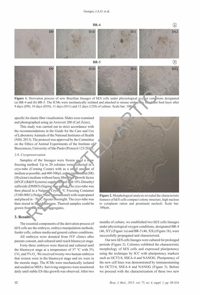
months of culture, we established two hES cells lineages under physiological oxygen conditions, designated BR-4 (46, XY) (Figure 1a) and BR-5 (46, XX) (Figure 1b), were successfully propagated and characterized.
Our new hES cells lineages were cultured for prolonged periods (Figure 2). Colonies exhibited the characteristic morphology of hES cells and expressed pluripotency using the technique by ICC with pluripotency markers such as OCT3/4, SSEA-4 and NANOG. Pluripotency of the new cell lines was demonstrated by immunostaining for OCT3/4, SSEA-4 and NANOG (Figure 3). Before we proceed with the characterization of these two new
Figure 1. Derivation process of new Brazilian lineages of hES cells under physiological oxygen conditions designated (a) BR-4 and (b) BR-5. The ICMs were mechanically isolated and attached to mouse embryonic fibroblast feed layer after 9 days (D9), 10 days (D10), 11 days (D11) and 12 days (12D) of culture. Scale bar: 100µm.
Figure 2. Morphological analysis revealed the characteristic features of hES cells compact colony structure, high nucleus to cytoplasm ration and prominent nucleoli. Scale bar: 100µm.
RETRACTED ARTIC
LE
Braz. J. Biol., 2015, vol. 75, no. 4, suppl. 1, pp. S9-S16 13
Brazilian hES cells derivation
13
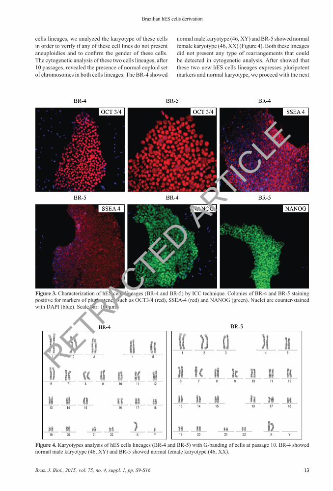
Figure 3. Characterization of hES cells lineages (BR-4 and BR-5) by ICC technique. Colonies of BR-4 and BR-5 staining positive for markers of pluripotency such as OCT3/4 (red), SSEA-4 (red) and NANOG (green). Nuclei are counter-stained with DAPI (blue). Scale bar: 100μm.
Figure 4. Karyotypes analysis of hES cells lineages (BR-4 and BR-5) with G-banding of cells at passage 10. BR-4 showed normal male karyotype (46, XY) and BR-5 showed normal female karyotype (46, XX).
cells lineages, we analyzed the karyotype of these cells in order to verify if any of these cell lines do not present aneuploidies and to confirm the gender of these cells. The cytogenetic analysis of these two cells lineages, after 10 passages, revealed the presence of normal euploid set of chromosomes in both cells lineages. The BR-4 showed
normal male karyotype (46, XY) and BR-5 showed normal female karyotype (46, XX) (Figure 4). Both these lineages did not present any type of rearrangements that could be detected in cytogenetic analysis. After showed that these two new hES cells lineages expresses pluripotent markers and normal karyotype, we proceed with the next
RETRACTED ARTIC
LE
Braz. J. Biol., 2015, vol. 75, no. 4, suppl. 1, pp. S9-S1614
Georges, J.A.O. et al.
14
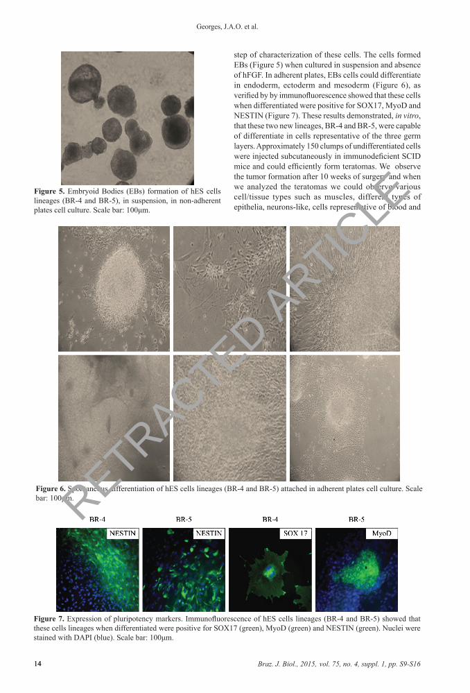
step of characterization of these cells. The cells formed EBs (Figure 5) when cultured in suspension and absence of hFGF. In adherent plates, EBs cells could differentiate in endoderm, ectoderm and mesoderm (Figure 6), as verified by by immunofluorescence showed that these cells when differentiated were positive for SOX17, MyoD and NESTIN (Figure 7). These results demonstrated, in vitro, that these two new lineages, BR-4 and BR-5, were capable of differentiate in cells representative of the three germ layers. Approximately 150 clumps of undifferentiated cells were injected subcutaneously in immunodeficient SCID mice and could efficiently form teratomas. We observe the tumor formation after 10 weeks of surgery and when we analyzed the teratomas we could observe various cell/tissue types such as muscles, different types of epithelia, neurons-like, cells representative of blood and
Figure 5. Embryoid Bodies (EBs) formation of hES cells lineages (BR-4 and BR-5), in suspension, in non-adherent plates cell culture. Scale bar: 100μm.
Figure 6. Spontaneous differentiation of hES cells lineages (BR-4 and BR-5) attached in adherent plates cell culture. Scale bar: 100μm.
Figure 7. Expression of pluripotency markers. Immunofluorescence of hES cells lineages (BR-4 and BR-5) showed that these cells lineages when differentiated were positive for SOX17 (green), MyoD (green) and NESTIN (green). Nuclei were stained with DAPI (blue). Scale bar: 100μm.
RETRACTED ARTIC
LE
Braz. J. Biol., 2015, vol. 75, no. 4, suppl. 1, pp. S9-S16 15
Brazilian hES cells derivation
15
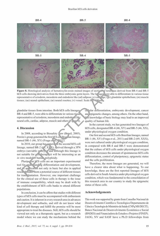
glandular tissues from intestine. Both hES cells lineages, BR-4 and BR-5, were able to differentiate in various tissue representative of ectoderm, mesoderm and endoderm like neural cells, cardiac, adipose, muscle and others (Figure 8).
4. Discussion
In 2008, according to Biosafety Law (Brasil, 2005), Pereira’s group generated the first hES cell Brazilian lineage, named BR-1 (46, XY) (Fraga et al., 2011).
In 2010, our group has generated the second hES cell lineage, named BR-2 (69, XXX), derived through a 3PN embryo (unviable embryo), and although this lineage is not suitable for cell therapy, it will be interesting as an in vitro model of human polyploidy.
Pluripotent hES cells are an important experimental tool for understanding differentiation and development, and its application not only in basic but also in applied research make them a potential source of different tissues for transplantation. However, one important challenge for the clinical use of these cells in therapy is the issue of immune compatibility, which may be dealt with by the establishment of hES cells banks to attend different populations.
In conclusion, it can be affirm that studies with different types of hES cells must be accompanied with enthusiasm and caution. It is inherent in every research area in advances development and setbacks, and still do not know what kind of cell therapy and fulfill the promise will be most appropriate to treat the diseases. Still, hES cells should be viewed not only as a therapeutic agent, but as a research model where we can study the mechanisms behind the
Figure 8. Histological analysis of hematoxylin-eosin stained images of sections of teratomas derived from BR-4 and BR-5 hES cells showing derivatives from the three embryonic germ layers. The lineages were able to differentiate in various tissue representative of ectoderm, mesoderm and endoderm like (ad) adipose; (c) cartilage; (ge) glandular epitethelium; (m) muscle tissues; (ne) neural epithelium; (nr) neural rosettes; (v) vessel. Scale bar: 100μm.
cellular differentiation, embryonic development, cancer and epigenetic changes, among others. On the other hand, this knowledge of basic biology may lead to an improved quality of human life.
In the current study, we has generated two lineages of hES cells, designated BR-4 (46, XY) and BR-5 (46, XX), under physiological oxygen condition.
Our first and second hES cells Brazilian lineages, named BR-1 (46, XY) (Fraga et al., 2011) and BR-2 (69, XXX), were not cultured under physiological oxygen condition, in compared with BR-4 and BR-5 were demonstrated that the culture of hES cells under physiological oxygen condition decreases the amount of spontaneous hES cells differentiation, control of pluripotency, epigenetic status and the cells proliferation.
Therefore, the more lineages are generated, we will have a clearer idea about what is happening. To our knowledge, these are the first reported lineages of hES cells derived in South America under physiological oxygen condition, which was fundamental to the consolidation of this research area in our country to study the epigenetic status of these cells.
Acknowledgements
This work was supported by grants from Conselho Nacional de Desenvolvimento Científico e Tecnológico/Departamento de Ciência e Tecnologia do Ministério da Saúde (CNPq/MS/DECIT), Banco Nacional de Desenvolvimento Econômico e Social (BNDES) and Financiadora de Estudos e Projetos (FINEP). JAOG, NV and SASF have a Ph.D fellowships from
RETRACTED ARTIC
LE
Braz. J. Biol., 2015, vol. 75, no. 4, suppl. 1, pp. S9-S1616
Georges, J.A.O. et al.
16
Coordenação de Aperfeiçoamento de Pessoal de Nível Superior (CAPES); AMF has a Ph.D fellowship from Fundação de Amparo à Pesquisa do Estado de São Paulo (FAPESP). The authors declare no conflicts of interest.
References
ALLEGRUCCI, C. and YOUNG, L., 2006. Differences between human embryonic stem cell lines. Human Reproduction Update, vol. 13, no. 2, pp. 103-120. http://dx.doi.org/10.1093/humupd/dml041. PMid:16936306.
BRASIL, 2005. Lei nº 11.105, de 24 de março de 2005. Diário Oficial União, Brasília, 28 mar.
BUZZARD, J.J., GOUGH, M.M., CROOK, J.M. and COLMAN, A., 2004. Karyotype of human ES cells during extended culture. Nature Biotechnology, vol. 22, no. 4, pp. 381-382, author reply 382. http://dx.doi.org/10.1038/nbt0404-381. PMid:15060545.
DANET, G.H., PAN, Y., LUONGO, J.L., BONNET, D.A. and SIMON, M.C., 2003. Expansion of human SCID-repopulating cells under hypoxic conditions. The Journal of Clinical Investigation, vol. 112, no. 1, pp. 126-135. http://dx.doi.org/10.1172/JCI17669. PMid:12840067.
EVANS, M.J. and KAUFMAN, M.H., 1981. Establishment in culture of pluripotential cells from mouse embryos. Nature, vol. 292, no. 5819, pp. 154-156. http://dx.doi.org/10.1038/292154a0. PMid:7242681.
EZASHI, T., DAS, P. and ROBERTS, R.M., 2005. Low O2 tensions and the prevention of differentiation of human embryonic stem cells. Proceedings of the National Academy of Sciences of the United States of America, vol. 102, no. 13, pp. 4783-4788. http://dx.doi.org/10.1073/pnas.0501283102. PMid:15772165.
FRAGA, A.M., SUKOYAN, M., RAJAN, P., BRAGA, D.P., IACONELLI JUNIOR, A.J.R., FRANCO JUNIOR, J.G., BORGES JUNIOR, E. and PEREIRA, L.V., 2011. Establishment of a Brazilian line of human embryonic stem cells in defined medium: implications for cell therapy in an ethnically diverse population. Cell Transplantation, vol. 20, no. 3, pp. 431-440. http://dx.doi.org/10.3727/096368910X522261. PMid:20719082.
GOSDEN, C., DAVDSON, C. and ROBERTSON, M., 1992. Lymphocyte culture. In: D.E. ROONEY and B.H. CZEPULKOWSKY. Human cytogenetics. Oxford: Oxford University Press, pp. 37-47.
HE, J.Q., MA, Y., LEE, Y., THOMSON, J.A. and KAMP, T.J., 2003. Human embryonic stem cells develop into multiple types of cardiac myocytes: action potential characterization. Circulation Research, vol. 93, no. 1, pp. 32-39. http://dx.doi.org/10.1161/01.RES.0000080317.92718.99. PMid:12791707.
KEHAT, I., GEPSTEIN, A., SPIRA, A., ITSKOVITZ-ELDOR, J. and GEPSTEIN, L., 2002. High-resolution electrophysiological assessment of human embryonic stem cell-derived cardiomyocytes. Circulation Research, vol. 91, no. 8, pp. 659-661. http://dx.doi.org/10.1161/01.RES.0000039084.30342.9B. PMid:12386141.
LENGNER, C.J., GIMELBRANT, A.A., ERWIN, J.A., CHENG, A.W., GUENTHER, M.G., WELSTEAD, G.G., ALAGAPPAN, R., FRAMPTON, G.M., XU, P., MUFFAT, J., SANTAGATA, S., POWERS, D., BARRETT, C.B., YOUNG, R.A., LEE, J.T., JAENISCH, R. and MITALIPOVA, M., 2010. Derivation of pre-X inactivation human embryonic stem cells under physiological
oxygen concentrations. Cell, vol. 141, no. 5, pp. 872-883. http://dx.doi.org/10.1016/j.cell.2010.04.010. PMid:20471072.
MARTIN, G.R., 1981. Isolation of a pluripotent cell line from early mouse embryos cultured in medium conditioned by teratocarcinoma stem cells. Proceedings of the National Academy of Sciences of the United States of America, vol. 78, no. 12, pp. 7634-7638. http://dx.doi.org/10.1073/pnas.78.12.7634. PMid:6950406.
MILLMAN, J.R., TAN, J.H. and COLTON, C.K., 2009. The effect of low oxygen on self-renewal and differentiation of embryonic stem cells. Current Opinion in Organ Transplantation, vol. 14, no. 6, pp. 694-700. http://dx.doi.org/10.1097/MOT.0b013e3283329d53. PMid:19779343.
MUMMERY, C., WARD, D., VAN DEN BRINK, C.E., BIRD, S.D., DOEVENDANS, P.A., OPTHOF, T., DE LA RIVIERE, A.B., TERTOOLEN, L., VAN DER HEYDEN, M. and PERA, M., 2002. Cardiomyocyte differentiation of mouse and human embryonic stem cells. Journal of Anatomy, vol. 200, no. Pt 3, pp. 233-242. http://dx.doi.org/10.1046/j.1469-7580.2002.00031.x. PMid:12033727.
NATIONAL INSTITUTES OF HEALTH – NIH, 2013 [viewed 7 November 2013]. Guide for the care and use of laboratory animals [online]. Bethesda. Available from: http://grants.nih.gov/grants/olaw/Guide-for-the-care-and-use-of-laboratory-animals.pdf
ODORICO, J.S., KAUFMAN, D.S. and THOMSON, J.A., 2001. Multilineage differentiation from human embryonic stem cells. Stem Cells, vol. 19, no. 3, pp. 193-204. http://dx.doi.org/10.1634/stemcells.19-3-193. PMid:11359944.
PROKHOROVA, T.A., HARKNESS, L.M., FRANDSEN, U., DITZEL, N., SCHRODER, H.D., BURNS, J.S. and KASSEM, M., 2009. Teratoma formation by human embryonic stem cells is site-dependent and enhanced by the presence of matrigel. Stem Cells and Development, vol. 18, no. 1, pp. 47-54. http://dx.doi.org/10.1089/scd.2007.0266. PMid:18393673.
REUBINOFF, B.E., PERA, M.F., FONG, C.Y., TROUNSON, A. and BONGSO, A., 2000. Embryonic stem cell lines from human blastocysts: somatic differentiation in vitro. Nature Biotechnology, vol. 18, no. 4, pp. 399-404. http://dx.doi.org/10.1038/74447. PMid:10748519.
SALAMAN, M.H. and GWYNN, R.H., 1951. The histology of co-carcinogenesis in mouse skin. British Journal of Cancer, vol. 5, no. 2, pp. 252-263. http://dx.doi.org/10.1038/bjc.1951.26. PMid:14869594.
THOMSON, J.A., ITSKOVITZ-ELDOR, J., SHAPIRO, S.S., WAKNITZ, M.A., SWIERGIEL, J.J., MARSHALL, V.S. and JONES, J.M., 1998. Embryonic stem cell lines derived from human blastocysts. Science, vol. 282, no. 5391, pp. 1145-1147. http://dx.doi.org/10.1126/science.282.5391.1145. PMid:9804556.
TROUNSON, A., 2005. Human embryonic stem cell derivation and directed differentiation. Ernst Schering Research Foundation Workshop, no. 54, pp. 27-44. PMid:16080285.
UNIVERSIDADE DE SÃO PAULO – USP. Instituto de Biociências – IB, 2013 [viewed 7 November 2013]. Comitê de Ética em Pesquisa: seres humanos [online]. São Paulo. Available from: http://www.ib.usp.br/formularios.php?menu=1
WOBUS, A.M. and BOHELER, K.R., 2005. Embryonic stem cells: prospects for developmental biology and cell therapy. Physical Review, vol. 85, no. 2, pp. 635-678. http://dx.doi.org/10.1152/physrev.00054.2003. PMid:15788707.
RETRACTED ARTIC
LE