universidade de lisboarepositorio.ul.pt/bitstream/10451/8018/1/ulfc102791_tm_ana...ciências da...
TRANSCRIPT
UNIVERSIDADE DE LISBOA
FACULDADE DE CIÊNCIAS
DEPARTAMENTO DE BIOLOGIA VEGETAL
Application of functional genomics in the production of viral
biopharmaceuticals: Vaccines and Vectors for gene therapy
Ana Sofia Formas Oliveira
Dissertação
MESTRADO EM BIOLOGIA CELULAR E BIOTECNOLOGIA
2012

UNIVERSIDADE DE LISBOA
FACULDADE DE CIÊNCIAS
DEPARTAMENTO DE BIOLOGIA VEGETAL
Application of functional genomics in the production of viral
biopharmaceuticals: Vaccines and Vectors for gene therapy
Dissertation supervisors:
External supervisor: Dr. Ana Sofia Coroadinha, Unidade de Tecnologia de Células Animais
from the Instituto de Biologia Experimental e Tecnológica and Instituto de Tecnologia
Química e Biologica, Universidade Nova de Lisboa (IBET/ITQB-UNL, Oeiras).
Internal supervisor: Dr. Rui Gomes, Departamento de Biologia Vegetal in Faculdade de
Ciências da Universidade de Lisboa (FCUL-DBV, Lisboa).
Ana Sofia Formas Oliveira
Dissertação
MESTRADO EM BIOLOGIA CELULAR E BIOTECNOLOGIA
2012

i
Master's Thesis in Cell Biology and Biotechnology,
Faculdade de Ciências, Universidade de Lisboa,
held at the Animal Cell Technology Unit from the
Instituto de Biologia Experimental e Tecnológica
and Instituto de Tecnologia Química e Biologica,
Universidade Nova de Lisboa (IBET/ITQB-UNL)
under the supervision of Dr. Ana Sofia Coroadinha.

ii
i. Acknowledgements
“Let us fight for a world of reason, a world where science and progress will lead to all
men's happiness” (Charlie Chaplin in The Great Dictator, 1940).
This research project would not have been possible without the support of many
people. Thus, I wish to express my gratitude:
To Dr. Ana Sofia Coroadinha, my external advisor, who gave me the opportunity to
join the Cell Line Development and Molecular Biotechnology Laboratory, for the supervision,
fruitful discussions and always being there to help me whenever I needed.
To Dr. Paula Alves, for giving me the opportunity to do my master thesis at Animal
Cell Technology Unit at ITQB/IBET.
To Ana Filipa Rodrigues, for the scientific and technical guidance, for the practical
help and constant support and for always believing in my work and encouraging me to
continue.
To Dr. Rui Gomes, for accepting to be my internal advisor and for always being
available and helpful.
To all the ACTU colleagues for the good working environment, friendship and the help
during this year. A special thanks to: Hélio Tomás, for teaching me all the molecular biology
tricks and support; Miguel Guerreiro and Ana Isabel Almeida for always being there for me
and Catarina Pinto for sharing funny car rides with me.
Um especial obrigado às pessoas mais importantes da minha vida: os meus pais, o
meu mano, a Sofia e o Henrique. Pelo amor sem fim, por acreditarem em mim, por estarem
sempre presentes e pelo apoio incondicional.
Ao Xico, pela constante ajuda e dedicação, por todo o carinho e paciência. Obrigada
pelo exemplo e por fazeres deste último ano possível.
Aos amigos de sempre e para sempre por todo o apoio, compreensão e momentos
de descontracção e diversão proporcionados, especialmente à Andreia, à Marta e à Susana.
iii
ii. Index
i. Acknowledgements ............................................................................................................. ii
iii. Abbreviations List ............................................................................................................... v
iv. Preface ............................................................................................................................ vii
v. Abstract ........................................................................................................................... viii
vi. Resumo ............................................................................................................................ ix
1. Introduction ....................................................................................................................... 1
1.1 Retrovirus based biopharmaceuticals .......................................................................... 1
1.2 Manufacture platforms ................................................................................................. 3
1.3 Metabolic engineering as a tool for cell line improvement ............................................ 6
1.4 Titration methods for viral particles quantification ........................................................11
2. Aim and strategy ..............................................................................................................13
3. Materials and methods .....................................................................................................16
3.1. Plasmids ....................................................................................................................16
3.2 Cell Lines and culture media .......................................................................................18
3.3 Bacterial strains ..........................................................................................................19
3.4 Cloning procedures .....................................................................................................19
3.5 Plasmid purification and quality control .......................................................................19
3.6 Determination of cell concentration and viability ..........................................................20
3.7 Cloning by limiting dilution ...........................................................................................20
3.8 Cell transfection ..........................................................................................................20
3.9 Viral vectors production and titration ...........................................................................20
3.9.1 Production ............................................................................................................20
3.9.2 Titration of infectious particles ..............................................................................22
3.9.3 Titration of genome containing particles ...............................................................23
3.9.4 Retroviral vector decay kinetics ............................................................................24
3.10 Cell line development ................................................................................................24
3.10.1 Target cell line ....................................................................................................24
iv
3.10.2 Cassette exchange .............................................................................................24
3.10.3 mCherry and GaLV insertion ..............................................................................25
3.11 RNA extraction and gene expression quantification by Real-Time PCR ....................25
3.12 Single-step cloning-titration method implementation and validation...........................25
3.12.1 Plate manufacture and culture conditions ...........................................................25
3.12.2 Fluorescence signal detection and calibration curves .........................................26
3.12.3 Method validation ...............................................................................................26
4. Results .............................................................................................................................27
Part I – Experimental validation of transcriptional profiling by culture medium manipulation
.........................................................................................................................................27
Part II – Implementation of a single step cloning-titration protocol for high-throughput
metabolic engineering of retrovirus producer cells. ...........................................................30
1. Culture and cloning conditions ...................................................................................31
2. Plate manufacture and fluorometer reading settings ..................................................32
3. Target Cells Development .........................................................................................34
4. 293 Flex 18 and 293 Ceb 22 development: RMCE and mCherry insertion ................38
5. Cloning-titration protocol ............................................................................................40
Part III – Metabolic engineering ........................................................................................43
5. Discussion and conclusion ...............................................................................................45
6. Ongoing and future work ..................................................................................................50
7. References .......................................................................................................................51
8. Annexes ...........................................................................................................................55

v
iii. Abbreviations List
bp - base pair
CA - Proteic capsid
CMV - Cytomegalovirus
DMEM - Dulbecco’s Modified Eagle’s Medium
Env – Coding genes for envelope glycoproteins
FBS - Foetal Bovine Serum
Flp - Flipase
FRT- Flipase recombinase target sites
Gag-pol – Group specific antigen – polymerase: structural and enzymatic viral proteins
GaLV - Gibbon Ape Leukemia Virus
GFP - Green Fluorescent Protein
HIV - Human Immunodeficiency Virus
hPGK - Human phosphoglycerate kinase
hygtk - hygromycin B phosphotransferase/thymidine kinase
IN - integrase
IP - Infectious particles
IRES - Internal Ribosomal Entry Site
kb – kilobase pair
LTR – Long Terminal Repeat
MA - Matrix proteins
MCS - Multiple cloning site
MLV - Murine Leukemia Virus
NC - Nucleocapsid
neo - Neomycin phosphotransferase gene
PEI- Polietilenamine
pol – polymerase
PPT - polypurine tract
PR - proteases
pro – protease gene
PYC2 - Yeast pyruvate carboxylase 2
RCRs - Replication-competent retroviruses
RMCE - Recombinase-mediated cassette exchange
RT - Reverse transcriptase
RV - Retrovirus based vectors
SIN – Self-inactivating vectors

vi
SU - Surface (SU)
SV40 – Simian Vacuolating virus 40
TCA – Tricarboxylic acid cycle
TM - Transmembrane (TM)
TP - Total particles
VLP – Virus like particle
VSV-G - Vesicular stomatitis virus G protein

vii
iv. Preface
This master thesis is within the scope of the project PTDC/EBB-BIO/100491/2008;
entitled "Using functional genomics to improve mammalian cells for virus based
biopharmaceuticals manufacture" funded by the Portuguese Fundação para a Ciência e
Tecnologia (FCT).
The first part of the results described in this thesis contributed for the following
publication:
A.F.Rodrigues, A.S.Oliveira, V.S.Bandeira, P.M.Alves, W.S.Hu, A.S.Coroadinha,
“Metabolic pathways recruited for the production of a recombinant enveloped virus:
transcriptional profiling and experimental validation” (Submitted to Metabolic Engineering,
Elsevier).

viii
v. Abstract
Retrovirus (RV) based biopharmaceuticals comprise several valuable bioproducts
such as gene therapy vectors, recombinant vaccines and replicative-competent particles for
oncolytic therapy. However, RV manufacture faces several challenges, particularly the low
yields of current producer cell lines and the high content of non-infective particles (an
average of 1 in every 100 total viruses produced are infective), contaminating the viral
preparations. The identification and manipulation of the metabolic requirements for improved
virus production is the scope of this project. In this context, this master thesis proposed to i)
validate metabolic pathways recruited under a virus production state – previously identified
by transcriptional profiling – by culture medium manipulation and ii) implement a novel
method to screen for high-producing clones after gene metabolic engineering.
Medium manipulation enabled increased titers from 1.3 to 2.1 fold upon isolated
medium supplementation strategies, reaching a synergistic effect with their combination of 6
fold increase. The improvement in RV production was not associated to increased viral
genes expression; it was partially due to higher viral vector preparation quality (enhanced
half-life for 2-3 hours and higher infectious particles per total particles content) and to an
enhanced metabolic status of the producer cell. For the screening method a novel protocol
was implemented taking advantage of split proteins technology enabling the isolation of the
high-titer clones from the producer population by the assessment of their titers in the first
stage of clone isolation - Single Step Cloning-Titration. Validated metabolic pathway will now
be object of gene-based metabolic engineering which, resorting to the implemented protocol,
can be conducted in a large-scale multi-gene approach, never attempted before.
This work directly contributes for improved cell line development in RV manufacture
and encompasses pioneering steps in mammalian cell multi-gene metabolic engineering.
Keywords: Retrovirus based biopharmaceuticals; Packaging cells; Cell culture; Metabolic
engineering; Virus titration methods.

ix
vi. Resumo
Os retrovírus (RVs) são vírus com invólucro da família Retroviridae. Caracterizam-se
por possuírem um genoma de cadeia simples de RNA positivo (7-11 kb), pela capacidade da
sua transcrição reversa de RNA para DNA e por possuírem no seu ciclo de replicação uma
etapa obrigatória de integração estável do seu genoma num cromossoma do hospedeiro.
No que diz respeito ao seu genoma e composição proteica, os RV possuem várias
vantagens que lhes conferem grande sucesso na sua utilização como biofármacos, tais
como: (i) genoma bem estudado, passível de ser manipulado por técnicas de biologia
molecular, (ii) capacidade de encapsidação de genomas até 9 kb, (iii) elevada eficiência de
transdução in vivo e in vitro, (iv) capacidade de modificar permanentemente o conteúdo
genético da célula alvo, possibilitando a expressão a longo prazo do gene terapêutico, (v) a
sua baixa imunogenicidade em comparação com outros vectores virais, e (vi) a capacidade
para incorporar proteínas de envelope de uma ampla variedade de vírus heterólogos,
proporcionando um meio de selecionar as células alvo a serem infectadas ou, para a
apresentação de antigénios.
Os vectores derivados de RV têm sido extensivamente utilizados como ferramentas
de transferência de genes em terapia génica; partículas sem genoma demonstraram
resultados promissores como candidatos a vacinas recombinantes e, mais recentemente,
foram desenvolvidas partículas com competência replicativa para serem utilizadas como
agentes oncolíticos para a terapia de vários tipos de cancro.
A produção de vectores retrovirais é realizada em células de mamíferos, geralmente
com origem de ratinho ou humanas, de forma transiente ou estável. A produção estável para
uma produção contínua requer a incorporação sequencial de, pelo menos, três cassetes de
expressão independentes (sistema de terceira geração); gag-pol (proteínas virais estruturais
e enzimáticas), env (glicoproteínas do envelope) e do transgene, que contém o gene de
interesse clonado no esqueleto viral. Cada estágio de inserção requer selecção por
antibiótico, clonagem e uma análise extensa dos clones para encontrar aqueles com maior
expressão e adequada estequiometria dos componentes virais. Assim, o estabelecimento de
uma linha celular produtora demora aproximadamente um ano até estar concluído.
Do ponto de vista de produção, os sobrenadantes de vectores retrovirais caracteriza-
se por títulos relativamente baixos, cerca de 106 partículas infecciosas por ml de meio de
cultura. Considerando que a quantidade média necessária para tratar um paciente num
ensaio clínico ronda 1010 particulas infecciosas, seria previsto cerca de 10 a 100 L de
volume de cultura para cada paciente. Adicionalmente, as preparações virais são
tipicamente caracterizados por razões baixas de partículas infecciosas/partículas totais

x
(cerca de 1:100), o que reduz a eficiência terapêutica dos vectores infecciosos. Assim, os
sistemas de produção existentes estão longe de satisfazer as necessidades requeridas para
a sua ampla utilização terapêutica ou profilática.
A engenharia metabólica surge como uma ferramenta atractiva para melhorar a
produtividade celular. Contudo, os incrementos na produtividade em células de mamífero
são modestos (cerca de 2 vezes) e, normalmente, baseados em aumentos na produtividade
volumétrica em vez de específica (da célula). O metabolismo e as redes metabólicas são
redundantes sendo que o mesmo padrão de produção/consumo de um metabolito pode ser
originado por diferentes combinações de reacções não conceptualizadas enquanto "via
metabólica", no sentido clássico. Assim, manipulações pontuais (um gene) são geralmente
absorvidas pela rede e "diluídas" sem efeitos fenotípicos óbvios. Este problema aumenta de
dimensão com a complexidade, o elevado grau de regulação e o nível de compartimentação
celular das células de mamíferos. Actualmente, realizar engenharia metabólica em células
de mamífero é um processo trabalhoso uma vez que envolve a introdução dos genes para
as vias alvo e o isolamento e caracterização das centenas de clones isolados.
Em suma, a produção de RV enfrenta dois desafios: aumentar a produtividade
celular para dar resposta às quantidades requeridas para uso terapêutico necessitando
portanto de estratégias rápidas para a análise da produtividade dos clones obtidos. Para
responder a este desafio, esta tese de mestrado teve como objetivos (1) validar resultados
de transcriptómica que identificaram vias metabólicas relevantes na produção des retrovírus
por manipulações específicas do meio de cultura de células 293 FLEX e (2) desenvolver um
método de clonagem e titulação com capacidade de análise high-throughput.
As manipulações executadas foram direcionadas para vias metabólicas recrutadas
em células produtoras de fácil manipulação, ou seja, vias de consumo de unidades básicas:
metabolismo de ácidos nucleicos, metabolismo de aminoácidos, destoxificação de radicais
livres e metabolismo das poliaminas. O meio de cultura foi suplementado com uma ou mais
combinações dos seguintes suplementos: aminoácidos, nucleósidos, antioxidantes,
poliaminas e glutationo reduzido. A produtividade viral aumentou em todas as condições
utilizadas. Com suplementos únicos o título viral aumentou 1.3 a 1.9 vezes; com a
conjugação de dois suplementos - aminoácidos e nucleósidos, antioxidantes com poliaminas
ou glutationo reduzido - aumentou 1.5, 2 e 2.1 vezes. No entanto, o maior efeito a nível de
produção foi conseguido pelo cocktail de todos os suplementos, atingindo um aumento de 6
vezes. O incremento na produção de RV não foi associado a um aumento da expressão de
genes virais; foi parcialmente baseado numa maior qualidade do sobrenadante viral e e num
estado metabólico melhorado das células produtoras. Adicionalmente, os suplementos de
aminoácidos e cocktail aumentaram a estabilidade das partículas virais em respectivamente
mais 3 horas e 2 horas.

xi
Estes resultados indicam uma origem metabólica no constrangimento da
produtividade viral. Estes resultados demonstraram que a manipulação orquestrada de vias
independentes conduz a efeitos sinergísticos de aumento de título evidenciando a
necessidade de uma manipulação concertada das vias metabólicas para obter efeitos de
maior impacto. Do ponto de vista do bioprocesso, a adição de suplementos no meio de
cultura para produção de biofármacos não é interessante uma vez que requere a utilização
de sistemas fed-batch aumentanto os custos de produção. Assim, é necessário tornar a
célula produtora mais robusta e independente de estímulos externos para atingir um estado
de elevada produtividade. A engenharia metabólica de múltiplas vias (genes) surge então
como uma ferramenta potencial para atingir um estado celular de híper-produtividade.
Contudo, não só o estabelecimento de uma linha celular produtora é um processo
demorado; as mesmas limitações surgem para populações de células produtoras alvo de
engenharia metabólica. Fenótipos híper-produtores são eventos raros, mesmo quando a
manipulação se revela benéfica, uma vez que dependem de níveis de expressão altamente
regulada dos genes alvo, dependendo do número de cópias inseridas, assim como o seu
local de integração cromossómica. O problema cresce exponencialmente para múltiplas
manipulações génicas. Para responder a esta necessidade, o segundo objectivo desta tese
consistiu no desenvolvimento de um método rápido e eficiente para a clonagem e
simultânea titulação de células produtoras, acoplado a um potencial de análise high-
throughput.
O protocolo implementado usa a recente tecnologia da proteína verde separada (split
GFP) que consiste na proteína GFP dividida em dois fragmentos (GFP S10 e GFP S11) e é
implementado em placas de 96 poços. As células produtoras expressam mCherry e
produzem vírus com um transgene que codifica para uma proteína de fusão LacZ-S11.
Estas células são plaqueadas a 1 célula por poço (clonagem) e ao sexto dia de cultura, as
células alvo, expressando constutivamente a proteína complementar GFP S10, são
adicionadas. Durante o restante tempo de cultura é propiciada a infecção das células alvo
pela adição de polibereno. Por infecção a proteína GFP S10 da células alvo vai sendo
transcomplementada com o fragmento GFP S11 originando sinal de fluorescência
directamente proporcional ao número de vírus produzidos. Após 14 dias de cultura as placas
de cultura são analisadas num fluorimetro e os poços com maior razão GFP/mCherry são
isolados, assegurando assim que os clones seleccionados são os melhores produtores. Os
clones podem ser isolados das células alvo por qualquer marca de resistência a antibiótico
expressa pelos componentes virais (Zeocina, Blasticidina e Neomicina) e/ou através de
puromicina (parte da construção mCherry).
A linha celular alvo foi estabelecida com as células Te671 por infecção com lentivirus
que entregaram o gene da GFP S10. A linha celular produtora foi criada a partir das células

xii
293 FLEX e 293 CEB 22; a primeira produz vectores retrovirais recombinantes baseados no
vírus da leucemia murina (Murine Leukemia Virus, MLV), pseudotipados com a proteína do
invólucro do vírus da leucemia do macaco gibão (Gibbon Ape Leukemia Virus, GaLV) e
contendo um transgene codificante para a enzima β-galactosidase (gene LacZ); a segunda é
a linha celular percussora das 293 FLEX anterior à inserção da cassete génica para a
expressão do invólucro. A tecnologia de troca de cassete mediada pela recombinase
(RMCE) permitiu trocar o transgene LacZ-S11 com o anterior até então expresso (LacZ). O
clone 32 das células 293 CEB 22 S11 foi utilizado para validar o protocolo após ser
submetido a duas etapas de transfecção; a primeira para inserir o marcador celular mCherry
e a segunda para introduzir o envelope GaLV. A população obtida foi então sujeita ao
protocolo de clonagem e titulação permitindo o isolamento de clones de elevadas
produtividades pela quantificação dos seus títulos na primeira etapa de isolamento do clone.
Nenhum dos clones 293 FLEX S11 isolados produzia vírus à mesma escala que as células
parentais, provavelmente devido a uma baixa eficiência de troca de cassete nestas células;
o protocolo estabelecido foi então usado para isolar clones 293 FLEX S11 nesta população,
permitindo isolar um clone produtor. Assim, o protocolo de clonagem e titulação
desenvolvido proporciona uma ferramenta útil para ultrapassar as limitações impostas pela
análise exaustiva de clones por métodos manuais, maximizando a probabilidade de
identificar clones com fenótipo híper-produtor.
As vias metabólicas identificadas por dados de transcriptómica e validadas neste
trabalho serão alvo de engenharia metabólica por manipulação combinada de múltiplos
genes, apenas possível devido à implementação do protocolo de clonagem e titulação
durante a presente tese.
Palavras-chave: Biofármacos derivados de retrovírus, células produtoras de retrovirus;
cultura celular, engenharia metabólica, métodos de titulação de vírus.

1
1. Introduction
1.1 Retrovirus based biopharmaceuticals
Retroviruses (RVs) are animal enveloped viruses of the Retroviridae family, harboring
two copies of a positive-stranded RNA genome with the size of 7–11 kb; they are mainly
characterized by the ability of reverse transcribing it from RNA to DNA, followed by stable
integration into the host chromosomal genome 1,2. Virions measure 100-120 nm in diameter
and the genome is complexed with the nucleocapsid (NC) proteins, enclosed in a proteic
capsid (CA) containing the enzymatic proteins, namely the reverse transcriptase (RT), the
integrase (IN) and proteases (PR), required for viral infection. The matrix proteins (MA) form
a layer outside the capsid core that interacts with the envelope, a lipid bilayer derived from
the host cellular membrane, which surrounds the viral core particle. Anchored on this bilayer,
there are the viral envelope glycoproteins (Env) responsible for recognizing specific
receptors on the host cell and initiating the infection process. Envelope proteins are formed
by two subunits, the transmembrane (TM) that anchors the protein into the lipid membrane
and the surface (SU) which binds to the cellular receptors. (Fig. 1 A) 3,4.
Based on the genome structure, retroviruses are classified into simple (e.g. MLV,
Murine Leukemia Virus) or complex retroviruses (e.g. HIV, Human Immunodeficiency Virus).
Both encode four genes: gag (group specific antigen), pro (protease), pol (polymerase) and
env (envelope). The gag sequence encodes the three main structural proteins: MA, CA, NC.
The pro sequence, encodes a protease (PR) responsible for cleaving Gag and Gag-Pol
during particles assembly, budding and maturation. The pol sequence encodes the enzymes
RT and IN, the former catalyzing the reverse transcription of the viral genome from RNA to
DNA during the infection process and the latter responsible for integrating the proviral DNA
into the host cell genome. The env sequence encodes for both SU and TM subunits of the
envelope glycoprotein. Additionally, retroviral genome presents non-coding cis-acting
sequences such as: two LTRs (long terminal repeats), which contain elements required to
drive gene expression, reverse transcription and integration into the host cell chromosome; a
sequence named packaging signal (ψ) required for specific packaging of the viral RNA into
newly forming virions, and a polypurine tract (PPT) that functions as the site for initiating the
positive strand DNA synthesis during reverse transcription. Additionally to gag, pro, pol and
env, complex retroviruses, such as lentiviruses, have accessory genes including vif, vpr, vpu,
nef, tat and rev that regulate viral gene expression, assembly of infectious particles and
modulate viral replication in infected cells (Fig. 1 B) 3. The main difference between the
simple and complex retrovirus is the ability of the latter to transduce non-dividing cells 4.

2
A
B
Figure 1 – A) Schematic representation of a retrovirus; B) genome organization of MLV and HIV 4.
Murine Leukemia Virus (MLV); Human Immunodeficiency Virus (HIV); nucleocapsid (NC); proteic
capsid (CA); reverse transcriptase (RT); integrase (IN); proteases (PR); matrix proteins (MA);
envelope glycoproteins (Env); transmembrane (TM); surface (SU); group specific antigen (gag);
protease (pro); pol (polymerase); envelope (env); protease (PR); long terminal repeats (LTRs);
packaging signal (ψ); polypurine tract (PPT).
Several aspects of retrovirus biology make them exceptionally apt as vectors for different
biotechnological applications. Replication-incompetent vectors, have been extensively used
as gene transfer tools in gene therapy protocols 5; genome free particles have already
demonstrated promising results as immunogen display platforms in the vaccinology field and
more recently, replication-competent particles (RCP) were developed and efficiently used as
oncolytic agents for cancer therapy 6.
3
Approved gene therapy clinical protocols using replicative deficient retroviral vectors
for gene delivery include a wide range of diseases, e.g. haemophilia, Human Severe
Combined Immunodeficiency (SCID), HIV infection, Gaucher’s disease, β-thalassaemia,
muscular dystrophy and Parkinson’s disease. MLV were the first viral vectors used in gene
therapy clinical trials and account for the second highest number of successful protocols 5.
Regarding oncolytic therapy, replicative competent RV vectors are suited for this proposes
since can only integrate and replicate in dividing cells, providing an inherent means for
targeting malignant cells that divide actively, while surrounding normal cells that are
generally quiescent. Furthermore, after transduction, each tumor cell acquires the capability
to produce more vectors that will improve efficient transduction of the surrounding malignant
cells 6. Hence, these vectors have been investigated for the treatment of a wide range of
cancers including ovarian cancer, breast cancer, brain tumors and lung cancer 7. Virus-like
particles (VLPs) are genome-free particles with the ability to mimic a virus structure and
display specific envelope proteins or an immunogen of interest. Thus, they are safer
alternative to classical attenuated vaccines with minimum concerns of virus
integration/replication. Retro VLPs can be pseudotyped with glycoproteins of several virus
families, such as lymphocytic choriomeningitis virus, spleen necrosis virus, vesicular
stomatitis virus, hepatitis C virus, yellow fever virus, West Nile virus and influenza virus.
Considerable efforts have been gathered to develop a VLP vaccine based in simple RV,
such as MLV, to target HIV and preliminary animal studies have already shown its potential 6.
1.2 Manufacture platforms
RV based vectors are essentially produced in mammalian cells, typically murine or
human derived cell lines. This can be based on a short-term transfer of the viral constructs,
known as transient production, into exponentially growing cells followed by 24-72 hours
vector production and harvesting, or by their stable integration and constitutive expression
into the host cell genome, named packaging cell lines, for continuous production 4.
The establishment of retroviral packaging cell lines has been based on the physical
separation of the viral genome into different transcriptional units, reducing the risk of
generating replicative competent retrovirus (RCRs) 2,4. For both retroviral and lentiviral vector
production, different packaging systems, named generations, have been developed. Each
new generation aimed at minimizing and reducing the risk of RCRs formation face to the
previous one. Currently, third generation is applied for the production of RV derived vectors,
requiring the transfection of three independent expression cassettes encoding gag-pol, env
and transgene functions (Fig. 2 A) 4.

4
Since the third generation of MLV vectors requires the expression of three genetic
cassette it is necessary to perform three transfections and clonal selection steps. This
process for establishing a producer cell line takes an average time-frame of around one year.
To face this challenge, a new generation of retrovirus packaging cells (modular cell lines)
based on cassette exchange systems that allow for flexible switch of the transgene and/or
envelope, as well as selectable marker(s) excision, was developed (Fig. 2 B and C) 8,9.
A
a)
b)
c)
B
i)
ii)
C
Figure 2 - Representation of RV based vector development: A) third generation split genome for
replicative deficient RV production (a) packaging functions, (b) transgene and (c) envelope 4; B)
Schematic representation of the modular cell lines transgene based on the RMCE technology (i)
Integrated retroviral transgene cassette harboring a marker gene and (ii) targeting therapeutic
transgene plasmid allowing a fast exchange and establishment of a new retroviral producer cell 4; C)
Strategy for the generation of the retroviral vector producer cell line with exchangeable transgene 9.
A favorable chromosomal site for stable and high retroviral vector production is first
identified and tagged. Due to the presence of two heterologous non-compatible flipase
recombinase target (FRT) sites flanking the tagged retroviral genome, the subsequent re-use
of this defined chromosomal site by means of recombinase-mediated cassette exchange
(RMCE) is then performed to express a therapeutic gene (Fig. 2 B and C). In order to select
cell clones that underwent correct targeted integration reaction, the targeting viral vector

5
contains a start codon that complements a transcriptionally inactive ATG-deficient selection
marker 2 after recombination. An example of a modular producing cell line is 293 FLEX,
developed by Coroadinha et al (2006) 9, and currently used in our group. This cell line
contains a LacZ transgene tagging the chromosomal locus of high expression that can be
exchanged through RMCE technology, MLV gag-pol packaging functions and Gibbon Ape
Leukemia Virus (GaLV) ecotropic envelope. The modular producer cell lines present several
advantages: they are safer since integration of the vector within the packaging cell line was
identified, the duration of the entire development process is reduced as there is no need for
screening and, in addition, production conditions are favorable due to the possibility of pre-
adaptation of the master cell line to culture conditions and media. Thus, therapeutic virus
production from bench to bedside becomes safer, faster, and cheaper 10.
The development o stable retroviral vector producer cell lines is a tedious and time
consuming process. However, it is compensated by obtaining continuously producing and
highly consistent cell systems, prone to single-effort bioprocess and product characterization,
a critical consideration for market approval. Retroviral vector manufacture, including those
used in clinical trials, has been making use of stable and continuous cell lines for more than
10 years 4.
From the manufacture point of view, retroviral vector production is characterized by
relatively low titers, around 106 infectious particles per mL of culture medium 7,11.
Considering the average amount needed to treat a patient in a clinical trial, in the order of
1010 infectious vectors, around 10-100 L of culture volume can be predicted for each patient.
Also, viral preparations are typically characterized by low ratios of infectious particles to total
particles (around 1:100) which further reduces the therapeutic efficiency of the infectious
ones 4. Several strategies have been attempted to circumvent these limitations.
Understanding the metabolic features that influence titer performances has been one
important work field 4,10.
Serum supplementation is a major concern in RVs production; in one hand its use
difficults product approval by the regulatory agencies quality requirements and increases
product manufacturing and purification costs, on the other hand serum removal results in low
titers and reduced vector stability. Overall, the major metabolic hinge between serum and
high titers has been demonstrated to be the lipids and cellular lipid metabolism 12,13. Glucose,
the traditional sugar source used in animal cell cultures, is present in most of the
commercially supplied media. Glucose, along with glutamine, represents the major energy
and carbon sources for cells in culture. However, it is rapidly consumed and inefficiently
metabolized, being the majority converted to lactate which per se inhibits the cell growth. The
use of alternative sugar sources, such as fructose (alone or in combination with glucose),
has a beneficial effect on vector production, that was correlated with an increase in the

6
intrinsic MLV retroviral vector stability which was associated with reduced sugar oxidative
metabolism and increased lipid synthesis 14,15. This observation was also correlated with an
increase in the medium osmolarity, in which the addition of osmolytes or high sugar
concentrations, was found to be a good approach to improve viral production 16.
In fact, considering that mammalian cells are severely affected by nutrient and by-
products environment 10, medium manipulation and further metabolic engineering is a
promising approach to achieve superior titers.
1.3 Metabolic engineering as a tool for cell line improvement
Since middle 80’s, biotechnologists realized it was possible to change cellular
phenotype towards improved yields of particular bioproducts targeting specific metabolic
genes, by over-expression and/or down-regulation 17,18. Since then, bacteria cell factories
have seen their specific productivities increase by several orders of magnitude. Metabolic
engineering in these simple organisms has become relatively straight forward and, with the
appearance of genome-scale metabolic models in the end of the 90’s, achieved high levels
of predictability 19,20. Contrarily to prokaryotic’s, eukaryotic cells, particularly mammalian’s,
were found to be difficult to manipulate by gene metabolic engineering with similar levels of
predictability, resulting in modest improvements in cellular phenotype and product yields.
Table 1 summarizes the main achievements in mammalian metabolic engineering.
Targeting apoptosis and by-product formation have been the blockbusters of mammalian
metabolism manipulation.

7
Table 1 – Examples of metabolic engineering targets in mammalian cells.
Metabolic Pathway
Genes Cell Line / Product Output Ref.
Apoptosis
E1B-19, Aven
CHO / mAb 1.5 fold titer increase in bioreactors 21
bcl 2 NS0 / mAb 1.4 increase in final antibody titer 22
siRNA: Bax, Bak
CHO / IFN-γ 1.4 fold improvement in productivity 23
By-product Formation
Ldh CHO / mAb 3 fold higher production (30% improvement
in cell density and cell viability) 24
Glut-5 CHO / mAb Only clones with low expression of GLUT5
transporters exhibited reduced lactate production
25
Pyc 2 HEK-293 /
interferon-α2b 1.3 fold increase in product yield
26
Protein Processing
and Secretion
Pace CHO / Coagulation
factor IX 3 fold improvement in the product
specific activity 27
Hsp70 NS0 / mAb Improved stability of secreted product in
culture supernatants 28
PDI CHO / IgG 1.3 fold increase in cell specific productivity 29
CHOP CHO / hTRA-8 Combined with UPR genes or chaperones
enhanced antibody production in 3 fold 30
Redox Balance
VKOR BHK / Coagulation
factor IX 14-fold increase in carboxylation rate 31
PDI, ERO1L
CHO / IgG 1.3 (ERO1L) and 1.5 (ERO1L and PDI) fold
increase in specific productivity 32
Cell Cycle Control
p21, p27, or p53175P
CHO / SEAP 10-15-fold increase in specific production 33
p21CIP1
NS0 / IgG 4 4 fold higher production in induced cells 34
mTOR CHO /
rituximab IgG 4 fold increase in antibody titers
35
Lipid Metabolism
Hsd17b7 NS0 / - Cholesterol autotrophy 36
CERT CHO / HSA, IgG 1.5 fold increase in specific productivity 37
XBP1(S) CHO / SEAP,
SAMY
SEAP - 6 fold; SAMY - 4fold production increases occurred at the post-
translational level
38
Glycosylation
Machinery
Sialidase antisense RNA
CHO / DNase 1.3 fold increase in sialic acid content 39
GnTIII CHO / chimeric anti-CD20 IgG1
antibody
The killing efficiency of CD20-positive target cells was improved in 10-20 fold
40
Bombyx mori 30Kc19
CHO / EPO 2.5-fold increase in productivity 41
mAb - monoclonal antibodies; IgG - human imunoglobulin G; hTRA-8 -humanized death receptor 5 antibody; UPR -
unfolded protein response; SEAP - human model glycoprotein (human placental secreted alkaline phosphatase); SAMY -
synthetic reporter protein (Bacillus stearothermophilus-derived α-amylase); ADCC - antibody dependent cellular
cytotoxicity; EPO – Erythropoietin; E1B-19K - antiapoptotic adenoviral gene; Aven/ bcl2 - anti-apoptosis genes; Bax/ Bak
– pro-apoptotic genes; Ldh – lactate dehydrogenase; Glut-5 - fructose transporter; Slc2a5 gene; Pyc2 - yeast pyruvate
carboxylase 2; Pace - paired basic amino acid cleaving enzyme; Hsp70 - heat shock protein 70; PDI - protein disulfide
isomerase; CHOP - transcription factor C/EBP homologous protein; VKOR - vitamin K oxidoreductase; ERO1L -
essential oxidoreductase; p21, p27, or p53175P - tumor suppressor genes; mTOR - mammalian target of rapamycin;
Hsd17b7 - hydroxysteroid (17-beta) dehydrogenase 7; CERT - ceramide transfer protein ; XBP1 - X-box-binding-protein
1; GnTIII - rat glycosylation enzyme β1–4-N-acetylglucosaminyltransferase III; 30Kc19 - 30Kc19 protein.

8
Lactate accumulation is a major concern in mammalian cell culture, since it adversely
affects cell metabolism, growth and final product quality. This phenotype is connected to the
Warburg effect-like scenario occurring in cultured mammalian cell lines and is associated
with a high glucose consumption and accumulation of significant amounts of lactate leading
to culture medium acidification 42. The main cause of the lactogenic phenotype has been
associated to the low activity of pyruvate dehydrogenase (PDH) leading to reduced
channeling of pyruvate into the citric acid cycle (TCA) and consequently impairing energy
metabolism. In 1999, a recombinant yeast pyruvate carboxylase 2 (PYC2) expressed in the
cytoplasm of BHK-21 cells was shown to reconstitute the missing link between glycolysis and
TCA 43. Later, 2002, these metabolically engineered cells have been additionally transfected
with a plasmid bearing the gene for human erythropoietin. PYC2-expressing clones showed
a 2-fold higher product concentration in a continuously perfused bioreactor. These cells
became more resistant to low glucose concentrations in the culture medium, which enabled a
more prolonged production phase in bioreactors (80% cell viability, 2 days (30%) longer
production time) 44. CHO cells producing recombinant human granulocyte macrophage
colony stimulating factor were as well target with PYC2. The PYC2-expressing cell clones
showed a decreased cell growth, lower maximum cell concentration, only 65% lactate
accumulation and the product yield was 3 fold higher when compared to the control
45.Wlaschin and Hu (2007) 25 evaluated the efficiency of the fructose-specific transporter
(GLUT5)-transfected CHO cells for utilizing fructose as an alternative to glucose and its
effectiveness on reducing lactate production. GLUT5-overexpressing clones could utilize
fructose as a carbon source and sugar consumption was reduced even when cells were
cultured in moderate concentrations of fructose. Only clones with low expression of GLUT5
transporters exhibited reduced lactate production and it was not correlated with production
levels. Last year, the expression of Lactate dehydrogenase A (LDH-A) and PDH Kinase 1, 2,
and 3 was down-regulated simultaneously using a short interference RNA reducing lactate
level and increasing specific productivity and volumetric antibody production by
approximately 90%, 75% and 68%, respectively 46. In this approach, pyruvate conversion into
lactate by LDH-A was suppressed and pyruvate dehydrogenase activity, which is inhibited
when phosphorylated by PDH kinases, was restored.
Controlling cell death and extend cell life enables extending the production. In
general, cell cultures are often terminated because of apoptotic cell death that may be
caused by one of several factors including nutrient depletion, metabolic by-product
accumulation, excessive shear forces or hypoxia 47. Engineering apoptosis network by
changing the expression of its regulators has shown great impact on culture viability and
productivity. Genetic strategies involve the over-expression of anti-apoptotic genes or down-
regulation of pro-apoptotic genes. The manipulation of the expression of the corresponding
9
proteins inhibits the release of pro-apoptotic molecules from the mitochondria and may
prolong the viability of the cell 48,49. Bcl-2 over-expression constitutes the most common
manipulation reported in the literature. The mechanism of action of Bcl-2 protein is broad. For
instance, it can heterodimerize with Bax (death inducer), inactivating it. It also prevents the
release of cytochrome c from the mitochondria, an important step in the apoptotic cascade
which leads to the activation of the caspases (proteases which are largely responsible for the
destruction of the cell) through the cytochrome c/ Apaf-1/ caspase 9 apoptosome or
apoptosome independently 47. Bcl-2 was shown to prevent apoptosis in a number of
commercially relevant cell lines, including CHO 31, NS0 22, hybridoma 52,53, and BHK 51,54,55 ,
although, it failed to increase cell specific productivity 56. Indeed, in the majority of apoptosis
controlling strategies, specific cell productivity does not increase; instead, lifespan does,
consequently enhancing production time and volumetric productivity 47.
Table 1 illustrates a striking problem in mammalian cell metabolic engineering: the
productivity improvements are modest (in the order of 2-fold) and, most of the times, are
based on increases in volumetric rather than specific (cell) productivity. Metabolism and
metabolic networks are redundant 57 and the same metabolite production/consumption
pattern can be generated by different combinations of reactions not conceptualized has a
“pathway”, in the classical sense. The implications for this are substantial as point
manipulations are often absorbed by the network and “diluted” without obvious phenotype
effects. The problem gains enormous relevance with the complexity, tight regulatory control
and high level of cellular compartmentalization of mammalian cells. For instance, targeting
the Warburg effect induced lactogenic phenotype spans across cytosol and mitochondria,
involves several glycolytic and TCA enzymes and a transcription factor (HIF1) 58.
Manipulating mammalian cell metabolism towards a hyper producing phenotype may reveal
to be a daunting challenge.
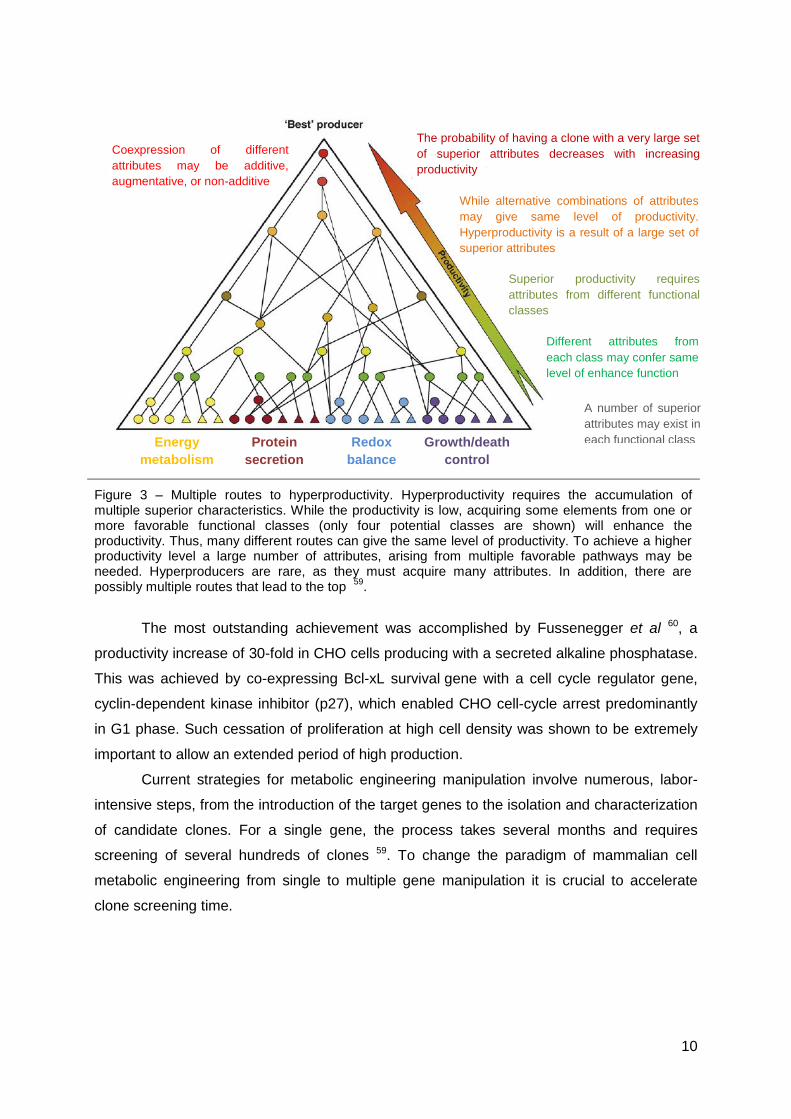
The datasets available so far suggest that hyperproductivity is conferred by a
collection of many positive characteristics encompassing changes of many genes with a wide
range of functions, rather than a transformation caused by a set of “master controllers” (Fig.
3) 59. Although the pyramid represented in figure 3 is a general/theoretical concept, multi-
gene engineering technology, enabling over-expression and/or silencing of combinatorial
target genes has already proven its potential in the pursuit of hyperproductivity.
10
Figure 3 – Multiple routes to hyperproductivity. Hyperproductivity requires the accumulation of multiple superior characteristics. While the productivity is low, acquiring some elements from one or more favorable functional classes (only four potential classes are shown) will enhance the productivity. Thus, many different routes can give the same level of productivity. To achieve a higher productivity level a large number of attributes, arising from multiple favorable pathways may be needed. Hyperproducers are rare, as they must acquire many attributes. In addition, there are possibly multiple routes that lead to the top
59.
The most outstanding achievement was accomplished by Fussenegger et al 60, a
productivity increase of 30-fold in CHO cells producing with a secreted alkaline phosphatase.
This was achieved by co-expressing Bcl-xL survival gene with a cell cycle regulator gene,
cyclin-dependent kinase inhibitor (p27), which enabled CHO cell-cycle arrest predominantly
in G1 phase. Such cessation of proliferation at high cell density was shown to be extremely
important to allow an extended period of high production.
Current strategies for metabolic engineering manipulation involve numerous, labor-
intensive steps, from the introduction of the target genes to the isolation and characterization
of candidate clones. For a single gene, the process takes several months and requires
screening of several hundreds of clones 59. To change the paradigm of mammalian cell
metabolic engineering from single to multiple gene manipulation it is crucial to accelerate
clone screening time.
Coexpression of different
attributes may be additive,
augmentative, or non-additive
The probability of having a clone with a very large set
of superior attributes decreases with increasing
productivity
While alternative combinations of attributes
may give same level of productivity.
Hyperproductivity is a result of a large set of
superior attributes
Superior productivity requires
attributes from different functional
classes
A number of superior
attributes may exist in
each functional class
Different attributes from
each class may confer same
level of enhance function
Energy
metabolism
Protein
secretion
Redox
balance
Growth/death
control
11
1.4 Titration methods for viral particles quantification
Viral titration – quantification of total and/or infectious particles – is crucial in virus
based biopharmaceuticals production as the balance between the therapeutic and adverse
effects of using viral particles lays on precise measurements of particles (total and infectious)
content. Additionally it is an essential step of packaging cell line development to assess the
titer and the transduction efficiency of the generated vector preparation allowing the selection
of the best producing clones. Two types of titration methods are considered: functional and
non-functional 61,62.
The non-functional titration assays rely on the quantification of constitutive viral
proteins (such as the HIV-1 capsid protein p24 by immunological titration or by determining
reverse transcriptase activity) or on the assessment of viral genome (RNA or DNA) in the cell
culture supernatant 63. In general, non-functional assays do not reflect infectious viral
particles content produced by the cells due to the presence of defective interfering particles,
transduction inhibitors, and contaminant genomic DNA from vector production in the
preparation. Additionally, genome quantification is also not the most accurate method for
viral quantification, when viral production involves a transient transfection, supernatant may
be contaminated with plasmid DNA over estimating the titers 64.
Functional titration methods involve the transduction of a target cell and further
quantification of vector reporter gene expression or its integration in transduced cells 63,64.
Reporter genes can be a selectable marker, fluorescent proteins or enzymes and titer is
assessed by resistant colonies after selection, flow cytometry or fluorometer assays and
enzymatic activity quantification, respectively.
The establishment of a producer cell line is a time consuming process not only due to
the time spent in the insertion of viral expression cassettes, allowing the cells to produce the
viral particles, selection of the producer cell population, but also, and mainly, for the time
spent in screening the high titer producer clones 61. The same limitations arise for a
metabolically engineered population. Hyper-producing phenotypes are rare events, even
when the manipulation reveals to be beneficial, as they rely on tight expression levels of the
targeted genes, dependent on the number of inserted copies as well as their chromosomal
integration site. The problem grows exponentially for multiple gene manipulation. In order to
reduce the time needed for this process it is necessary to develop fast, and with high
throughput capacity, titration methods.
Implementing screening schemes with high throughput potential to accurately assess
growth and productivity at earlier stages, ideally during cloning procedure, would certainly
accelerate the process.

12
In 2002 Green and Rasko developed a “Rapid Screening for High-Titer Retroviral
Packaging Cell Lines Using an In Situ Fluorescence Assay” 61 involving the detection of
high-titer producer clones containing fluorescent reporter genes (eGFP, eCFP and eYFP).
The method consisted in removing the clone supernatant at early cloning stage (96 well
plate), and transfer it to infect target cells also in 96 well plate. Upon transduction, target cells
express the fluorescent protein that was detected in multichannel fluorescent reader. The
correlation of cell fluorescence between the fluorescent plate reader assay and flow
cytometric assessment was high (r2 = 0.96). Simultaneous cell density analysis using
alamarBlue fluorescence was proportional to cell number per well (r2 = 1.0) 61. The greatest
contribute of this method was the ability to select rare high producing clones (up to 7 fold) by
adding a screening step at cloning stage, which allowed reducing considerably the time
spent in clone analysis.
13
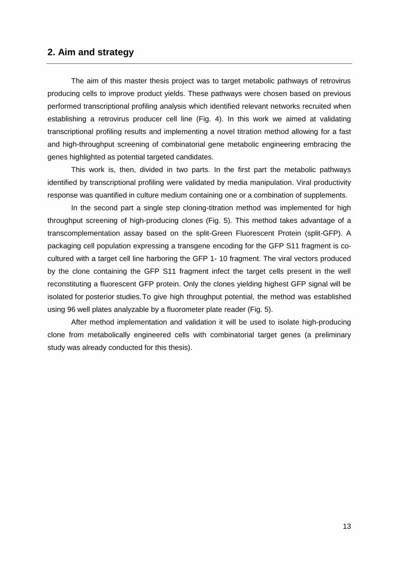
2. Aim and strategy
The aim of this master thesis project was to target metabolic pathways of retrovirus
producing cells to improve product yields. These pathways were chosen based on previous
performed transcriptional profiling analysis which identified relevant networks recruited when
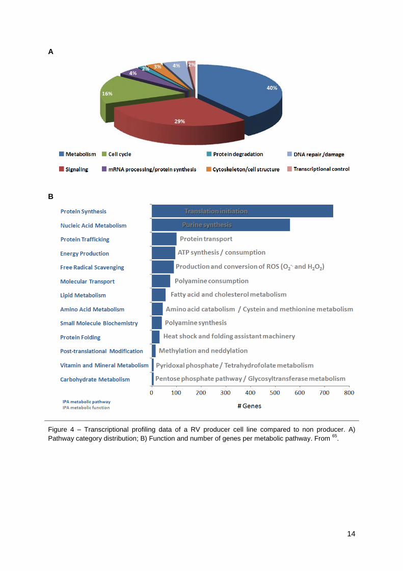
establishing a retrovirus producer cell line (Fig. 4). In this work we aimed at validating
transcriptional profiling results and implementing a novel titration method allowing for a fast
and high-throughput screening of combinatorial gene metabolic engineering embracing the
genes highlighted as potential targeted candidates.
This work is, then, divided in two parts. In the first part the metabolic pathways
identified by transcriptional profiling were validated by media manipulation. Viral productivity
response was quantified in culture medium containing one or a combination of supplements.
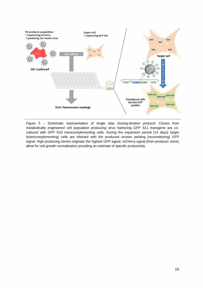
In the second part a single step cloning-titration method was implemented for high
throughput screening of high-producing clones (Fig. 5). This method takes advantage of a
transcomplementation assay based on the split-Green Fluorescent Protein (split-GFP). A
packaging cell population expressing a transgene encoding for the GFP S11 fragment is co-
cultured with a target cell line harboring the GFP 1- 10 fragment. The viral vectors produced
by the clone containing the GFP S11 fragment infect the target cells present in the well
reconstituting a fluorescent GFP protein. Only the clones yielding highest GFP signal will be
isolated for posterior studies. To give high throughput potential, the method was established
using 96 well plates analyzable by a fluorometer plate reader (Fig. 5).
After method implementation and validation it will be used to isolate high-producing
clone from metabolically engineered cells with combinatorial target genes (a preliminary
study was already conducted for this thesis).
14
A
B
Figure 4 – Transcriptional profiling data of a RV producer cell line compared to non producer. A)
Pathway category distribution; B) Function and number of genes per metabolic pathway. From 65
.
15
Figure 5 – Schematic representation of single step cloning-titration protocol. Clones from
metabolically engineered cell population producing virus harboring GFP S11 transgene are co-
cultured with GFP S10 transcomplementing cells. During the expansion period (14 days) target
(transcomplementing) cells are infected with the produced viruses yielding (reconstituting) GFP
signal. High producing clones originate the highest GFP signal; mCherry signal (from producer clone)
allow for cell growth normalization providing an estimate of specific productivity.

16
3. Materials and methods
3.1. Plasmids
For all the vectors constructed in this work the cloning sites, primers and templates
are listed in Table S1. A schematic representation can be found in Fig. S1. Underlined
plasmids indicate vectors constructed in this work.
pCMV mGFP S10 and pCMV mGFP S11 contain a mammalian codon-optimized version of
the GFP under the control of a CMV promotor, split in GFP S10 coding for the first 214
amino-acids, and GFP S11 for the last 15, respectively. These plasmids were acquired
trough SandiaBiotech (Albuquerque, NM, U.S.A) and were used for several constructions
further explained below.
pIRESGALEO is a murine stem cell virus (MSCV) based retroviral vector where LacZ is
under the control of a LTR promoter and the fusion protein gene hygromycin B
phosphotransferase/thymidine kinase (hygtk) is under the control of the
encephalomyocaerditis virus-internal ribosome entry site (EMCV-IRES). This vector contains
two FRT sites in the U3 of 3’LTR, a wild type FRT site (wt) and a spacer mutant FRT site
followed by an ATG defective neomycin phosphotransferase gene (neo) 66.
pEmMFG is a recombinase mediated cassette exchange targeting vector containing a FRT
wt, an MLV based retroviral vector MFG-LTR that drives the eGFP gene followed by an
EMCV-IRES element next to an ATG and F5 FRT site. ATG sequence and the F5 mutant
FRT site complementing the neo gene in pIRESGALEO after targeting and subsequent
cassette exchange 9.
pCMV mGFP LacZS11 codes for a fusion protein LacZ-S11 under the control of a CMV
promoter and was obtained by cloning LacZ gene (from pIRESGALEO) into the multiple
cloning site (MCS) of pCMV mGFP S11. LacZ was amplified by PCR and introduced into the
MCS at AgeI site.
pTAR LacS11 is a RMCE targeting vector derived from the pEmMFG backbone in which
eGFP was replaced by LacZ-S11 obtained from pCMV mGFP LacZS11. Both retroviral
backbone and LacZ-S11 were amplified by PCR, using primers with NotI recognition site.
17
pRRLsin LacZS11 and pRRLsin S10 were derived from pRRLsin eGFP backbone, kindly
provided by Didier Trono from the Swiss Federal Institute of Technology (EPFL) through
Addgene plasmid repository (Cambridge, MA, USA). This backbone is a third generation
lentiviral transgene vector, driving the expression of eGFP from the hPGK promotor (human
phosphoglycerate kinase) as described in Dull et al (1998) 67. For pRRLsin LacZS11 and
pRRLsin S10, eGFP was replaced by LacZ-S11 or GFP S10 obtained from pCMV mGFP
LacZS11 or pCMV mGFP S10, respectively, isolated by PCR with primers carrying the NheI
restriction site. pRRLSin backbone was amplified in two fragments by PCR, with primers
carrying NheI and PciI restriction sites. The cloning step involved three fragments: two
fragments corresponding to the vector (PcII cloning site) and the insert (NheI cloning site).
Lentiviral vectors for metabolic engineering, the lentiviral backbone was isolated from
pRRLsin LacZS11, removing LacZ by restriction with AgeI. Each metabolic gene (isocitrate
dehydrogenase 1 and 2, glutathione synthase and inhibitor of hypoxia inducible factor 1) was
amplified by PCR from the original plasmids obtained from DNASU Plasmid Repository
(Biodesign Institute, Arizona State University) and cloned into AgeI site. Mock vector was
obtained by religating lentiviral backbone after LacZ removal.
pPuro mCherry was derived from pSelect Puro (Invivogen, San Diego, CA, U.S.A.) that
contains a multiple cloning site (MCS) downstream of the EF1 promoter and a puromycin
selectable marker. The construction contains a mCherry gene, amplified by PCR from
pRSET B (described in Shaner et al (2004) 68) cloned into the MCS with NheI.
pSVFLPe contains FLP recombinase gene under a SV40 promoter (a kind gift of Dr F.
Stewart).
pCeB is a vector containing MLV gag-pol and a blasticidin resistance gene (bsr) both driven
by the MLV 5’LTR as described in Cosset et al (1995) 69.
pGaLV expresses the GaLV envelope protein and a zeocin (Zeo) resistance marker under
the control of a CMV and kindly provided by Dr. Otto Merten (Genethon, Department of
Bioprocess Development, France) .
pMD2G expresses the envelope G glycoprotein of the vesicular stomatitis vírus (VSV G)
under the control of CMV promotor (kindly provided by D. Trono through Addgene).
18
pMDLg/pRRE is 3rd generation LV packaging plasmid codifing for gag - virion main structural
proteins - pol, responsible for the retrovirus-specific enzymes; and RRE, a binding site for the
Rev protein which facilitates export of the RNA from the nucleus (kindly provided by D. Trono
through Addgene).
pRSV-REV is a 3rd generation LV packaging plasmid containing the second and third exons
of HIV-1 rev under the transcriptional control of RSV U3 promoter (kindly provided by D.
Trono through Addgene).
3.2 Cell Lines and culture media
HEK 293 is a Human Embryonic Kidney derived cell line (ATCC CRL-1573) and was
used to produce conditioned medium for limiting dilution of all HEK 293 derived cell lines.
Te671 is a Human rhabdomyosarcoma derived cell line (ATCC CCL-136) used to
develop S10 Target cell line, and as target cells to titrate the infectious RV particles. These
cells were additionally used to produce conditioned medium for limiting dilution of all Te671
derived cell lines.
HEK 293T is a HEK 293 derived cell line expressing large T antigen from SV40
(simian vacuolating virus 40) used to produce LV vectors in a transient manner and to test
the newly generated plasmids functionality.
293 FLEX cell line is a HEK 293 derived cell line producing MLV based recombinant
retroviral vectors expressing GaLV ecotropic envelope and harboring a LacZ reporter gene 9.
293 CEB 22 is a HEK 293 derived cell line, the precursor cell line of 293 FLEX prior to
envelope insertion as described in Coroadinha et al (2006) 9. These cells produce envelope-
free MLV-derived virions harboring a LacZ transgene. 293 CEB 22 and 293 FLEX were used
to establish retroviral vector producer cells producing retroviral vectors harboring a LacZ-S11
transgene by RMCE.
293 FLEX GFP cell line is derived from 293 FLEX 8 as described in 9 and was used
for the production of GFP expressing RV.
DK cell line (E1-transcomplementing dog kidney cells), provided by Dr. Eric Kremer
(Institut Génétique Moléculaire Montpellier, France), were used for fluorometer tests
involving mCherry signal.
All cells were maintained in Dulbecco’s modified Eagle’s medium, DMEM, (Gibco,
Paisley, UK) with 25 mM of glucose, 4 mM of glutamine, supplemented with 10% (v/v) Foetal
Bovine Serum (FBS) (Gibco). In the case of fluorometer assays, DMEM without phenol red
was used. For supplemented medium studies, Advanced DMEM with 25 mM of glucose
supplemented with 4 mM of glutamine and 10% (v/v) Foetal Bovine Serum (FBS) (Gibco)
19
was used. All cells were cultured under adherent conditions (T-flask) (Starstedt, Nümbrecht-
Rommelsdorf, Germany) at 37 ◦C in a humidified atmosphere containing 7.5% CO2.
3.3 Bacterial strains
Escherichia coli (E. coli) Library Efficiency® DH5α™ and One Shot® Stbl3™
(Invitrogen, Carlsbad, CA, U.S.A) competent cells were used for the production of the DNA
plasmids. Transformation procedures were carried under manufacturer instructions.
The liquid bacterial cultures were performed with Terrific Broth media (TB) (Fast-
Media® TB from Invivogen) supplemented with the appropriated antibiotic (Ampicilin,
Kanamycin or Puromycin). The media was prepared using ultrapure water (Millipore,
Billerica, MA, U.S.A.), according to the manufacturer's instructions.
3.4 Cloning procedures
All PCR reactions were performed with proof-reading Phusion High-Fidelity DNA
Polymerase (Finnzymes OY, Espoo, Finland), using the following conditions: initial
denaturation step (30 seconds at 98ºC), followed by 30 cycles of denaturation (10 seconds at
98ºC), annealing (30 seconds at temperature of melting (Tm)) and elongation (72ºC – Time
according fragment lenght) and lastly by a final elongation step (6 minutes at 72ºC). The
oligonucleotides used for PCR were custom-made by Sigma Aldrich (St.Louis, MO, U.S.A).
Restriction reactions were incubated 2-4 hours (h) at 37°C. The enzymes in Table S1 (New
England Biolabs, Ipswich, MA, U.S.A) were used with the appropriate buffers and
supplements according to the manufacturer instructions. All the generated fragments were
isolated by agarose gels (Lonza, Basel, Switzerland) 0.7% (w/v) and subsequently purified
with illustra GFX PCR DNA and Gel Band Purification Kit (GE Healthcare, Buckinghamshire,
UK). Ligation reactions were performed using T4 DNA ligase (New England Biolabs) and
incubated overnight following manufacturer’s instructions.
3.5 Plasmid purification and quality control
Plasmid purification was performed in different scales. Small-scale purification (yields
around 20 μg of DNA) was performed with QIAprep® Miniprep (Qiagen, Hilden, Germany). A
larger scale purification (yields around 500 μg of DNA) was performed using Genopure
Plasmid Maxi Kit (Roche, Basel, Switzerland) following manufacturer’s instructions. Working
DNA banks for each plasmid were generated and stored at -20 ºC.
The DNA concentration was determined in a spectophotometer (Nanodrop 2000C
Spectophotometer, Thermo Scientific, USA). Plasmid purity was determined by the
Abs260nm/Abs280nm ratio and plasmid integrity (with and without enzymatic restriction)
20
assessed in 0.7% (w/v) agarose gels (Lonza, Basel, Switzerland) prepared in TE buffer. All
plasmids were sequenced using Macrogen Europe services (Amsterdam, The Netherland).
All generated plasmids were tested for their functionality transfecting HEK 293 T cells
(described in section 3.8).
3.6 Determination of cell concentration and viability
Cell concentration and viability were determined by the trypan blue exclusion method
using a 0.1% (v/v) solution prepared in Phosphate Buffer Saline (PBS, Gibco) and counting
cells in a Fuchs-Rosenthal hemacytometer (Brand, Wertheim, Germany). Each counting was
performed at least twice.
3.7 Cloning by limiting dilution
For clone isolation, serial dilutions were preformed to achieve 0.5 cells per well in 96
well plates. Limiting dilution medium was composed by 50% conditioned (parental cells
supernatant) and 50% fresh medium, 20% FBS and half concentration of antibiotic (specific
for each cell). At day 14, wells with single colonies were trypsinized and amplified for cell
banking and posterior analyzis.
3.8 Cell transfection
For transfection procedures cells were seeded in 6-well plates (Nunc, Rocherster,
NY, U.S.A) at 5x10⁴ cell/cm2. 24 hours later transfection was carried using polyethylenimine
(PEI, Linear 25 kDa from Polysciences, Eppelheim, Germany) at 1:3 (m/m) ratio of DNA:PEI)
8 or by calcium phosphate precipitation method. 5 µg of DNA per 1x10⁶ cell were used. PEI
transfection solution was prepared in fresh serum-free-media
The calcium phosphate precipitation method (CAPHOS SIGMA - Calcium Phosphate
Transfection Kit, Sigma) was executed according to manufacturer instruction and was only
used for RMCE.
3.9 Viral vectors production and titration
3.9.1 Production
For lentiviral transient production a third generation of lentiviral system was used 67.
HEK 293T cells were seeded at 8x10⁴ cell/cm2. PEI transfection was carried out 24 hours
later as described above with a mixture of: pREV and pMDLG RRE (providing the packaging
functions), pMD2G (for the envelope) and pRRLsin derived vectors (section 3.1) providing the
transfer vector (transgene). The DNA ratio used was 1:4:3.6:10 67. Except for transfer
vectors, all plasmids were kindly provided by D. Trono through Addgene (Cambridge, MA,

21
U.S.A). After 24 hours, the medium was replaced with 2/3 of the original volume to
concentrate lentiviral particles stock. The medium containing the viral vectors was collected
after an additional 24 hours production period.
RV particles harboring a LacZ-S11 reporter gene were obtained by transient
production following an identical procedure described above using pCEB to provide the
packaging functions, pMD2g for the envelope and pTAR LacS11 for the transfer vector
(transgene). The DNA ratio used was 2:1:3.
Retroviral vectors with GFP transgene were obtained by stable production using 293
FLEX GFP cells.
All viral vectors supernatant were filtered through 0.45 μm cellulose acetate filter for
clarification, aliquoted and stored at -80ºC.
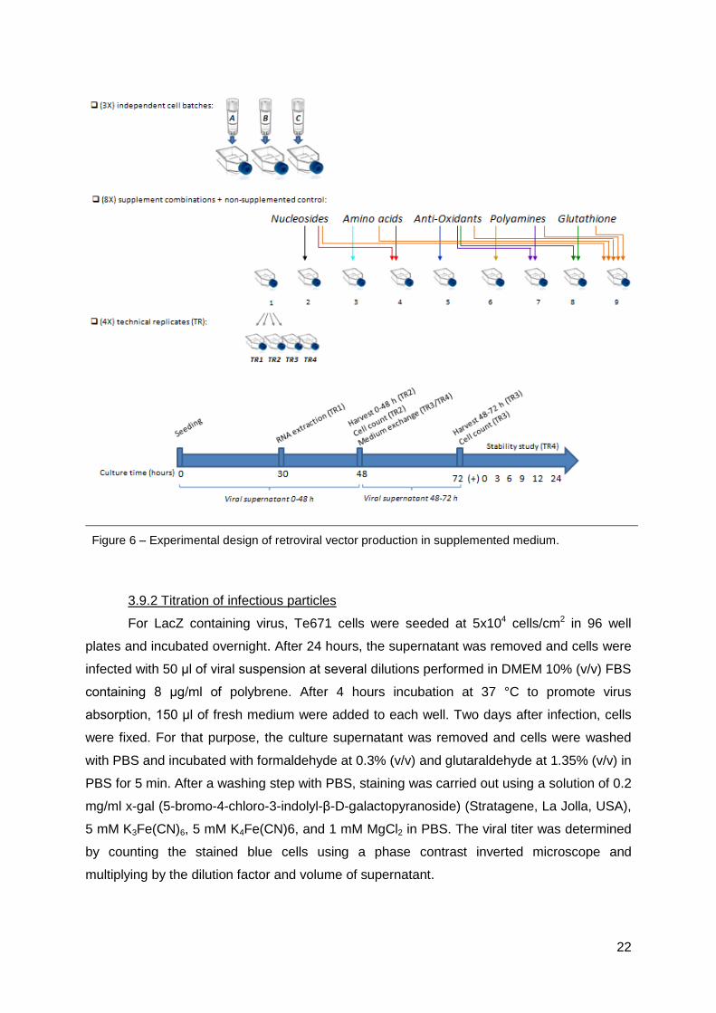
3.9.1.1 Retroviral vector production in supplemented culture medium
For retroviral vector production in supplemented medium a 48+24 hours production
protocol, with medium exchange after the first 48 hours, was used (Fig. 6). 293 FLEX cells
were inoculated at 8x10⁴ cells/cm2 in media containing one or a combination of the following
supplements: i) a amino acid solution (Sigma); ii) a nucleoside supplement (Millipore,
Billerica, MA, U.S.A); iii) an anti-oxidant supplement (Sigma); iv) a polyamine supplement
(Sigma); v) reduced glutathione (Sigma). A non-supplemented control was also prepared. A
detailed schematic representation of this experimental design, final concentrations and
combinations of supplements used can be found in Fig. 6 and supplementary material (Table
S2). Three biological replicates were used (corresponding to three independent freezing
batches), each of them prepared in four technical replicates. At 30 hours post-seeding, total
cellular RNA was extracted from the first technical replicate. At 48 hours, culture supernatant
of the second technical replicate was harvested, filtered through 0.45 μm for clarification,
aliquoted and stored at -80ºC until analysis. Cell concentration and viability was determined
by the trypan blue exclusion method; for the third and fourth technical replicate, medium was
exchanged. After an additional 24 hour period of production, the same procedure described
above was applied to the third technical replicate. The fourth technical replicate was used to
assess retroviral vector decay kinetics.
22
Figure 6 – Experimental design of retroviral vector production in supplemented medium.
3.9.2 Titration of infectious particles
For LacZ containing virus, Te671 cells were seeded at 5x10⁴ cells/cm2 in 96 well
plates and incubated overnight. After 24 hours, the supernatant was removed and cells were
infected with 50 μl of viral suspension at several dilutions performed in DMEM 10% (v/v) FBS
containing 8 μg/ml of polybrene. After 4 hours incubation at 37 °C to promote virus
absorption, 150 μl of fresh medium were added to each well. Two days after infection, cells
were fixed. For that purpose, the culture supernatant was removed and cells were washed
with PBS and incubated with formaldehyde at 0.3% (v/v) and glutaraldehyde at 1.35% (v/v) in
PBS for 5 min. After a washing step with PBS, staining was carried out using a solution of 0.2
mg/ml x-gal (5-bromo-4-chloro-3-indolyl-β-D-galactopyranoside) (Stratagene, La Jolla, USA),
5 mM K3Fe(CN)6, 5 mM K4Fe(CN)6, and 1 mM MgCl2 in PBS. The viral titer was determined
by counting the stained blue cells using a phase contrast inverted microscope and
multiplying by the dilution factor and volume of supernatant.

23
For GFP transgene vectors titration, Te 671 cells were seeded at 5x10⁴ cells/cm2 in
24 well plates one day before infection. Cell concentration was determined at the time of
infection. Transduction was performed in duplicates by removing the cell supernatant and
infecting with 0.3 mL of viral suspension using several dilutions in fresh DMEM with 10%
(v/v) FBS and 8 μg/ml of polyberen. Cells were incubated at 37ºC overnight after which 1 ml
of fresh supplemented DMEM was added. Two days after infection, cells were harvested and
analyzed for GFP fluorescence by Flow Cytometry (CyFlow-space, Partec GmbH).
Titration of virus containing split GFP reporter proteins was carried out has described
above but with double infection steps and lower cell inoculums. The cells were seeded in 24
well plates at the density of 2x10⁴ cell/cm2. The day after, first transduction was performed
with 500μl of LV supernatant carrying the LacZ-S11 transgene (saturating multiplicity of
infection (MOI)). After 4 hours, viral adsorption time, supernatant was replaced by 1 ml of
fresh medium. Second transduction was done 24 hours after with 300μl of serially diluted
viruses of viral supernatant carrying the transcomplementing transgene, GFP S10. At this
time point, cell concentration was determined and 48 hours after cells were analyzed for GFP
transcomplementation signal. This double infection protocol was only used while the Target
(GFP S10 expressing) cell line was not established. Upon the establish of Target cell line,
viral particles harboring LacZ-S11 transgene were titrated using Te 671 S10 cells in a single
infection protocol.
The GFP titer was determined taking into account the percentage of GFP positive
cells, the cell concentration determined at infection time and the dilution factor. Infections that
rendered 2‐20% of infected cells were considered for titer calculations. Viral titers obtained
by flow cytometry method, defined as Infectious particles per mililiter (IP/mL), were
calculated using the following formula:
3.9.3 Titration of genome containing particles
Genome containing particles were titrated using a protocol implemented in our
laboratory and described in Carmo et al (2004) 70 with the following modifications: the
primers used for cDNA synthesis were LacZ reverse and GFP reverse, in Table S3.
The primers used for PCR were LacZ (forward and reverse), given in Table S3.

24
3.9.4 Retroviral vector decay kinetics
To evaluate vector decay kinetic, the 24 hours viral vector productions from
supplemented cultures was used. Culture supernatant was harvested and filtered through a
0.45 μm filter. Part of this supernatant was 1:10 diluted in Advanced DMEM with 10% (v/v)
FBS (neutral medium) and the remaining was not diluted. The viral suspensions were then
incubated at 37 °C; samples were taken at 0, 3, 6, 9, 12, and 24 hours and stored at −80 °C
for viral titer determination. Decay rate constants and half-lives were calculated according to
a first order exponential decay model 71.
3.10 Cell line development
3.10.1 Target cell line
The Target cell line was developed by stable expression of GFP S10 gene into Te671
using LV infection. Since the transgene of LV S10 does not contain a selectable marker, a
MOI of 30 – to assure synchronous infection and multiple copies per cell – was used. Cells
were cloned by limiting dilution and clones were analyzed for GFP transcomplementation
after infection with retrovirus and lentivirus harboring LacZS11 transgene.
3.10.2 Cassette exchange
For site-specific recombinase mediated cassette exchange tagged 293 FLEX and 293
CEB 22 cell lines were seeded at 5x10⁴ cells/cm2 in 6 well plates and co-transfected in the
next day using calcium phosphate precipitation method with 4 µg of targeting plasmid, pTAR
LacS11, and 12 µg of pSVFlpe recombinase expressing plasmid. 48 hours post-transfection,
Neomycin selection was started with G418 (1000 µg/ml; Invivogen). Medium was regularly
exchange during 21 days after which limiting dilution method was applied, in the presence of
ganciclovir (5 µg/ml; Invivogen), to eliminate non-exchanged clones. The clones were
amplified and analysed for cassete exchange and number of pTAR LacS11 copies
integrated.
To evaluate cassette exchange efficiency and number of targeting copies integrated
into the cell genome, genomic DNA was isolated using the "QIAamp® DNA Mini and Blood
Mini Kit" (Qiagen, Valencia, CA, USA) according to manufacturer's instructions.
PCR with specific primers for LacZ and Neo gene (Table S3) was performed and
PCR amplification products were separated in agarose gel. Depending on the PCR product
size it was possible to confirm if cassette exchange was successful (Fig. S2 and Table S4).
To estimate the number of copies per cell, genomic DNA was quantified by qRT-PCR
using primers for LacZ gene (Table S3) on a thermocycler "LightCycler ® 480 Real-Time

25
PCR System" (Roche Applied Science) using "LightCycler® 480 SYBR Green I Master"
(Roche Applied Science) PCR kit. The number of copies per cell was quantified relatively to
a single copy control (293 FLEX) after normalization to a control gene (RPL22) using 2-ΔΔCt
method 72,73. Single copy was considered for values below 1.4 73.
3.10.3 mCherry and GaLV insertion
293 CEB 22 S11 clone 32 was chosen to insert mCherry by PEI transfection
(described in section 3.8). After 15 days of Puromycin selection (Invivogen), cells were
sorted (MoFlo High-Speed Cell Sorter, Dako Cytomation, Glostrup, Denmark) to isolate a
population of cells within a narrow range of mCherry intensity of expression (clones in the
same range were additionally isolated). The sorted mCherry population was transfected
(PEI) with phGaLV. After 15 days of Zeocin selection (Invivogen), this GaLV population was
used for protocol validation.
3.11 RNA extraction and gene expression quantification by Real-Time PCR
To analyze viral components expression in supplemented medium studies total RNA
was extracted using RNeasy Mini Kit (Qiagen, Valencia, CA) according to the manufacturer
instructions.
For real time PCR, RPL-22 was chosen as a control gene. Forward (F) and reverse
(R) primer sequences for control and virus component genes are given in Table S3. The
reverse transcription of total RNA was performed according to Transcriptor High Fidelity
cDNA Synthesis Kit (Roche Applied Science, Mannheim, Germany) protocol for cDNA
synthesis using 2 μg of total RNA and oligo dT primer for total mRNA reverse transcription.
The reverse transcribed (RT) product was aliquoted and stored at -20ºC until further
processing. SYBR Green I dye chemistry was used to detect the PCR products using
LightCycler® 480 SYBR Green I Master (Roche Applied Science) according to the
manufacturer instructions using LightCycler® 480 Real Time PCR System (Roche Applied
Science). cDNA samples were run along with a no RT-reaction control and a no cDNA
template sample. Three independent biological replicates were analyzed, each of them run in
three (technical) replicates.
3.12 Single-step cloning-titration method implementation and validation
3.12.1 Plate manufacture and culture conditions
In order to establish the best culture conditions for the co-culture, different black-
wallet 96 well plates with clear bottom from three manufactures were tested (Corning, New

26
York, NY, USA/ Nunc/ Perkin Elmer, Waltham, MA, USA). In this assays, DMEM without
phenol red was used.
3.12.2 Fluorescence signal detection and calibration curves
For fluorescence signal detection and calibration curves Te671 cells were infected
with serial dilutions of retrovirus codifying for GFP. Cells were seeded at 5×10⁴ cells/cm2 in
the three 96 well plates with clear bottom mentioned above and incubated overnight. After 24
hours, the supernatant was removed and cells were infected overnight with 100 μl of viral
suspension at multiplicity of infection (MOI) ranging from 5 to 0. 48 hours post-infection
medium was replaced by 50 μl of PBS 5% (v/v) FBS to avoid fluorescence signal
interference. Fluorescence reading was performed using plate fluorometer reader FLx800 (
Biotek, Vermont, USA) using the 485/20 emission filter and 528/20 absortion filters (Biotek).
For red fluorescence signal detection and calibration curves DK cells were infected
with serial dilutions of mCherry CAV (canine adenoviral vectors), kindly provided by Paulo
Fernandes (ACT Unit IBET/ITQB, Oeiras, Portugal). The signal was detected 24 hours post-
infection using the 560/20 emission filter and 645/40 absortion filter (Biotek), readings were
performed as described above.
3.12.3 Method validation
For the method validation 293 CEB 22 S11 Cherry sorted population with GaLV was
submitted to a limiting dilution (Corning plate) and 6 days after Target cell line was added at
500 cell/well. Until the last day of the cloning protocol (day 14) three infection rounds were
performed by the addition of polyberen (final concentration of 10 µg/ml) proceeded by 4
hours of agitation, with 48 hours interval in between.
At day 14, GFP and mCherry signal was quantified using a fluorometer reader as
previously described (section 3.12.2). Wells with the highest GFP/mCherry ratio were
trypsinized and clones were amplified and isolated from Target cell line by Puromycin
selection.
27
4. Results
Part I – Experimental validation of transcriptional profiling by culture medium
manipulation
In the first part of this work, metabolic pathways recruited for virus production
previously identified by microarray analysis 65 – comparing producer cell lines with their
corresponding parental cells – were evaluated as targets to improve vector production. From
the highlighted pathways (Fig. 4), were selected those that could be manipulated by medium
supplementation strategies with minimum off-target or growth inhibition effects. Within this
aim, the pathways from Fig. 4 B can be divided in three categories: easy to manipulate,
difficult and intermediate/mixed.
Easy: primary building blocks consumption pathways. These pathways are relatively
easy to target by culture medium manipulation, simply by the addition of the consumed
molecules. They include Nucleic Acid Metabolism, Amino Acid Metabolism, Free Radical
Scavenging, Polyamine Metabolism, Vitamin and Mineral Metabolism and Lipid Metabolism.
These categories were chosen for culture medium manipulation, containing one or a
combination of the following supplements: amino acid solution, nucleoside supplement, anti-
oxidant supplement, polyamine supplement and reduced glutathione.
Difficult: pathways of assembly and modification of intracellularly synthesized
molecules. These pathways are very difficult to target by culture medium supplementation as
they rely on intracellularly synthesized molecules, many non-transportable through the cell
membrane, and specific enzyme modification machinery. These included Protein Trafficking,
Protein Folding and Post-translational Modification. These pathways can be targeted by the
addition of chemical inhibitors and activators. However, this typically results in a myriad of
off-target effects, very difficult to control and often associated to cell growth inhibition.
Intermediate/mixed: pathways that share “easy” and “difficult” features. They include:
Pentose Phosphate Pathway since it proceeds through phosphorylated intermediates – cell
membrane impermeable and thus, not susceptible of addition to the culture medium –
Protein Synthesis, and Energy Production. They can, however, be indirectly targeted by
medium supplementation through their precedent “building blocks consumption pathways”
although the interpretation of the results is not straightforward. Thus, amino acid
supplementation also targets Energy Production, as these are major substrates to feed TCA
cycle reactions, and Protein Synthesis as increased protein synthesis necessarily requires
amino acid consumption. Reduced glutathione supplementation targets Pentose Phosphate
Pathway, since during its oxidation NADP+ is produced, pushing G6PD (glucose-6-
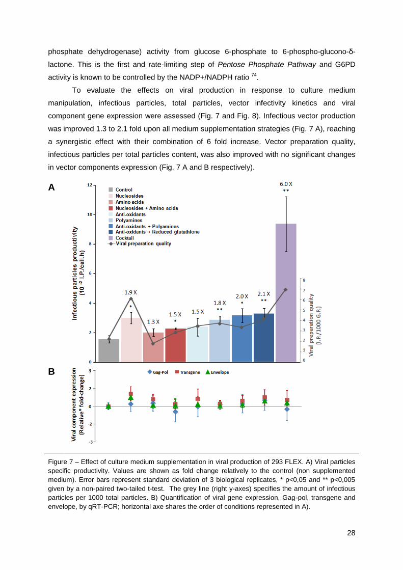
28
phosphate dehydrogenase) activity from glucose 6-phosphate to 6-phospho-glucono-δ-
lactone. This is the first and rate-limiting step of Pentose Phosphate Pathway and G6PD
activity is known to be controlled by the NADP+/NADPH ratio 74.
To evaluate the effects on viral production in response to culture medium
manipulation, infectious particles, total particles, vector infectivity kinetics and viral
component gene expression were assessed (Fig. 7 and Fig. 8). Infectious vector production
was improved 1.3 to 2.1 fold upon all medium supplementation strategies (Fig. 7 A), reaching
a synergistic effect with their combination of 6 fold increase. Vector preparation quality,
infectious particles per total particles content, was also improved with no significant changes
in vector components expression (Fig. 7 A and B respectively).
A
B
Figure 7 – Effect of culture medium supplementation in viral production of 293 FLEX. A) Viral particles
specific productivity. Values are shown as fold change relatively to the control (non supplemented
medium). Error bars represent standard deviation of 3 biological replicates, * p<0,05 and ** p<0,005
given by a non-paired two-tailed t-test. The grey line (right y-axes) specifies the amount of infectious
particles per 1000 total particles. B) Quantification of viral gene expression, Gag-pol, transgene and
envelope, by qRT-PCR; horizontal axe shares the order of conditions represented in A).
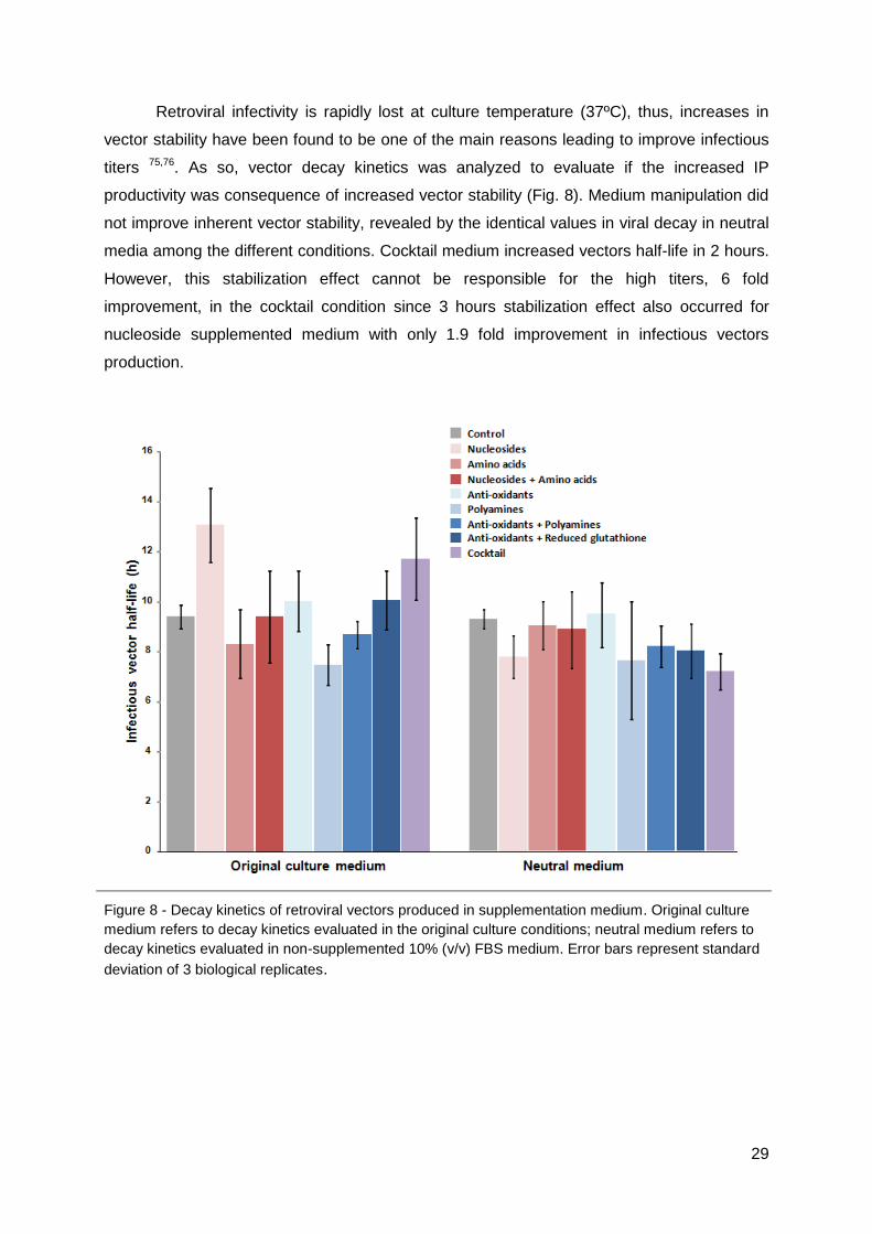
29
Retroviral infectivity is rapidly lost at culture temperature (37ºC), thus, increases in
vector stability have been found to be one of the main reasons leading to improve infectious
titers 75,76. As so, vector decay kinetics was analyzed to evaluate if the increased IP
productivity was consequence of increased vector stability (Fig. 8). Medium manipulation did
not improve inherent vector stability, revealed by the identical values in viral decay in neutral
media among the different conditions. Cocktail medium increased vectors half-life in 2 hours.
However, this stabilization effect cannot be responsible for the high titers, 6 fold
improvement, in the cocktail condition since 3 hours stabilization effect also occurred for
nucleoside supplemented medium with only 1.9 fold improvement in infectious vectors
production.
Figure 8 - Decay kinetics of retroviral vectors produced in supplementation medium. Original culture
medium refers to decay kinetics evaluated in the original culture conditions; neutral medium refers to
decay kinetics evaluated in non-supplemented 10% (v/v) FBS medium. Error bars represent standard
deviation of 3 biological replicates.
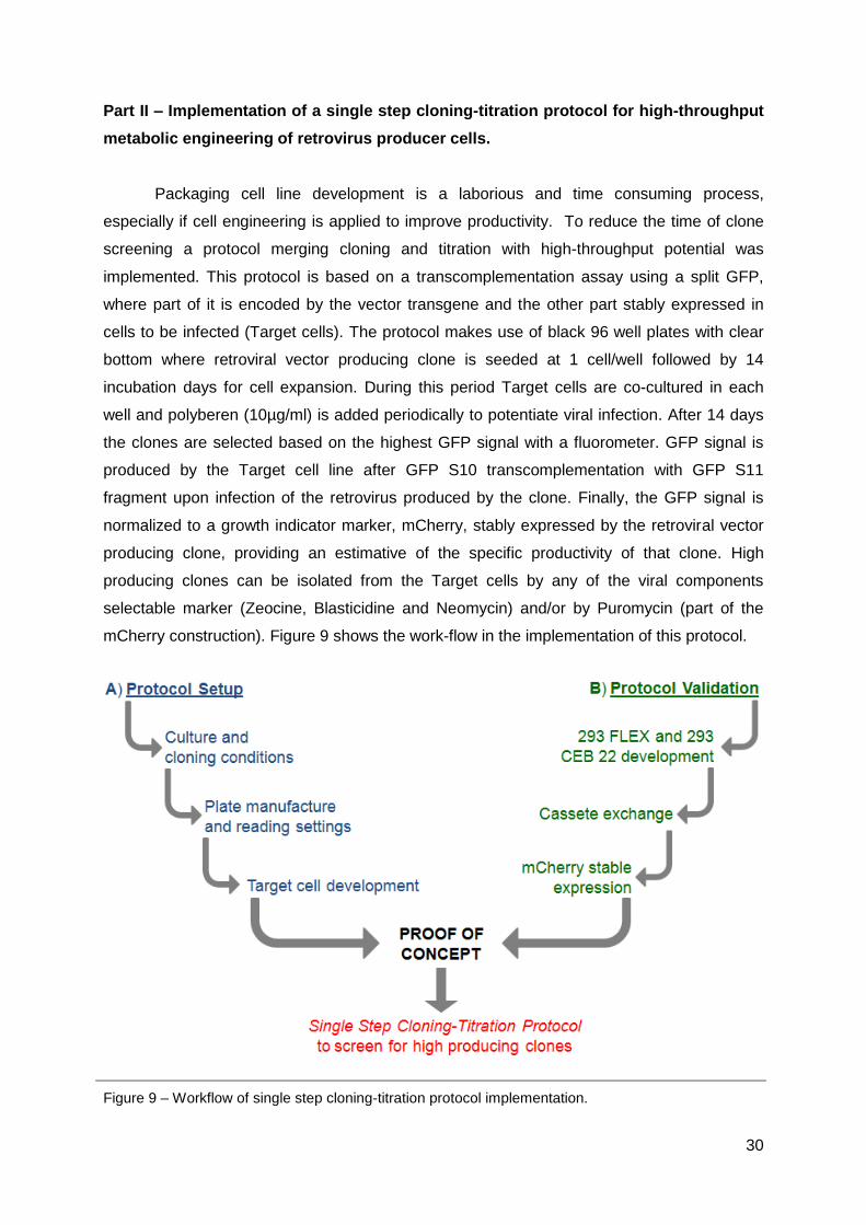
30
Part II – Implementation of a single step cloning-titration protocol for high-throughput
metabolic engineering of retrovirus producer cells.
Packaging cell line development is a laborious and time consuming process,
especially if cell engineering is applied to improve productivity. To reduce the time of clone
screening a protocol merging cloning and titration with high-throughput potential was
implemented. This protocol is based on a transcomplementation assay using a split GFP,
where part of it is encoded by the vector transgene and the other part stably expressed in
cells to be infected (Target cells). The protocol makes use of black 96 well plates with clear
bottom where retroviral vector producing clone is seeded at 1 cell/well followed by 14
incubation days for cell expansion. During this period Target cells are co-cultured in each
well and polyberen (10µg/ml) is added periodically to potentiate viral infection. After 14 days
the clones are selected based on the highest GFP signal with a fluorometer. GFP signal is
produced by the Target cell line after GFP S10 transcomplementation with GFP S11
fragment upon infection of the retrovirus produced by the clone. Finally, the GFP signal is
normalized to a growth indicator marker, mCherry, stably expressed by the retroviral vector
producing clone, providing an estimative of the specific productivity of that clone. High
producing clones can be isolated from the Target cells by any of the viral components
selectable marker (Zeocine, Blasticidine and Neomycin) and/or by Puromycin (part of the
mCherry construction). Figure 9 shows the work-flow in the implementation of this protocol.
Figure 9 – Workflow of single step cloning-titration protocol implementation.

31
1. Culture and cloning conditions
To determine the best culture conditions for fluorometer readings a Nunc plate was
prepared with combinations of: cells, culture medium, PBS and serum (Fig. 10). Medium
without phenol red was used as previous work reported phenol red interference with
fluorometer readings 61.
Figure 10 – Culture conditions influencing fluorometer reading for GFP signal (485/528 nm). Error bars correspond to standard deviation (n=12); RFU – Relative Fluorescence Units.
The greatest contribution for background signal was found to come from medium;
cells and serum content did not show relevant interference. Thus, to minimize background
without compromising cell viability, further readings were carried out by replacing medium
with PBS 5% FBS prior to analysis. Serum was reduced to 5% to guarantee minimal signal
interference avoiding cell damage.
The protocol requires 14 days incubation to allow clone expansion, concomitant with
virus production and subsequent infection of Target cells. To optimize Target cells inoculums
these were seeded at day 0 with 50, 100 and 200 cells/well, 24 hours later the cloning
procedure was started by adding 1 retroviral vector producer cell per well. This strategy
aimed to provide a layer of cells that would be not only the infection targets but also function
as a feeder layer facilitating clone expansion. This approach, however, did not succeed as
clones were unable to expand in co-culture.
In order to enable clone expansion the culture setup was redesigned and cloning
procedure started at day 0 with medium supplemented with 20% (v/v) FBS. Target cells were
added at 3.3 x 10³ cells/well at day 9. After 24 h polyberen was added to a final concentration
of 10 µg/ml and plates were agitated for 2 h. At day 13, polyberen was again supplied and
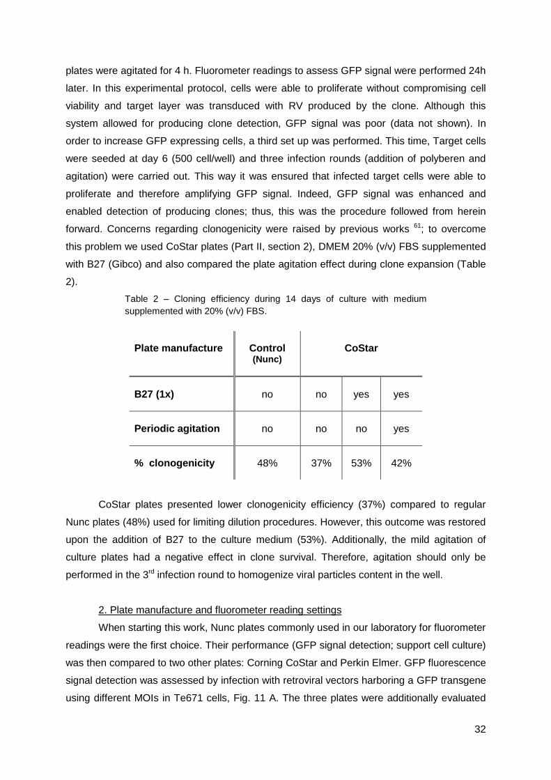
32
plates were agitated for 4 h. Fluorometer readings to assess GFP signal were performed 24h
later. In this experimental protocol, cells were able to proliferate without compromising cell
viability and target layer was transduced with RV produced by the clone. Although this
system allowed for producing clone detection, GFP signal was poor (data not shown). In
order to increase GFP expressing cells, a third set up was performed. This time, Target cells
were seeded at day 6 (500 cell/well) and three infection rounds (addition of polyberen and
agitation) were carried out. This way it was ensured that infected target cells were able to
proliferate and therefore amplifying GFP signal. Indeed, GFP signal was enhanced and
enabled detection of producing clones; thus, this was the procedure followed from herein
forward. Concerns regarding clonogenicity were raised by previous works 61; to overcome
this problem we used CoStar plates (Part II, section 2), DMEM 20% (v/v) FBS supplemented
with B27 (Gibco) and also compared the plate agitation effect during clone expansion (Table
2).
Table 2 – Cloning efficiency during 14 days of culture with medium
supplemented with 20% (v/v) FBS.
CoStar plates presented lower clonogenicity efficiency (37%) compared to regular
Nunc plates (48%) used for limiting dilution procedures. However, this outcome was restored
upon the addition of B27 to the culture medium (53%). Additionally, the mild agitation of
culture plates had a negative effect in clone survival. Therefore, agitation should only be
performed in the 3rd infection round to homogenize viral particles content in the well.
2. Plate manufacture and fluorometer reading settings
When starting this work, Nunc plates commonly used in our laboratory for fluorometer
readings were the first choice. Their performance (GFP signal detection; support cell culture)
was then compared to two other plates: Corning CoStar and Perkin Elmer. GFP fluorescence
signal detection was assessed by infection with retroviral vectors harboring a GFP transgene
using different MOIs in Te671 cells, Fig. 11 A. The three plates were additionally evaluated
Plate manufacture Control (Nunc)
CoStar
B27 (1x) no no yes yes
Periodic agitation no no no yes
% clonogenicity 48% 37% 53% 42%

33
for their ability to provide a suitable growing surface under low inoculums conditions, to be
used both for Target cells as well as for the retroviral vectors producing clone (Fig. 11 B).
A
B
Figure 11 – GFP detection and cell growth performance of Nunc, Corning CoStar and Perkin Elmer
plates. A) Fluorescence intensity (485/528 nm) of GFP-expressing cells 48 hours upon transduction
with retroviral vectors containing GFP transgene at MOI ranging from 0 to 5.5. Line of best-fit for MOI
ranging from 0 to 2 (Nunc R2=0.97, Corning CoStar and Perkin Elmer R2
=0.99). Error bars represent
standard deviation (n=12). B) Cell growth after 14 days culture in each plate (initial inoculums of 50
cells/well). RFU - Relative Fluorescence Units. MOI – multiplicity of infection.
GFP signal could be detected linearly until the multiplicity of infection (MOI) of 2 in all
tested plates. The distinction of GFP signal from the background was similar in all plates,
although Corning CoStar and Perkin Elmer manufacture showed the best signal to noise
ratio (5.3). Both, Nunc and Corning CoStar plates supported good cell proliferation for 14
days, whereas in Perkin Elmer plates, cells failed to reach the confluence. Finally, the
average error between wells of same condition was minimized in Corning CoStar plates
(3.4%). Therefore, they were selected to establish the protocol.
mCherry was chosen as a cell growth normalization marker as contrarily to other red
proteins, its excitation/emission spectra does not overlap with those of GFP 77. To evaluate

34
the potential of mCherry as a future normalization marker, mCherry signal was quantified in
cells infected with canine adenoviral vectors (CAV) already in use in our laboratory (Fig. 12).
Transfection procedures were avoided as they would be difficult to carry in such small scale
(96 well) and could lead to significant cell death capable of interfering with fluorescence
readings.
Figure 12 - Fluorescence intensity (560/645 nm) of mCherry-expressing cells transduced with CAV
mCherry adenoviruses at MOI ranging from 0 to 10. Line of best-fit for MOI ranging from 0 to 1.3
(R2=0.97). Error bars represent standard deviation (n=12). MOI – multiplicity of infection.
As shown in Fig. 12, mCherry signal could be detected above background level in all
MOIs, linearly until MOI 1.3. mCherry signal was higher than that of GFP (Fig. 11 A) and
therefore signal/noise ratio was higher (14.3). Altough the MOIs used in this assay were not
comparable to the ones used in GFP assay, the mCherry signal detection enabled higher
sensitivity and better distinction from background. In addition, this assay showed no signal
interference between the excitation/emission filter sets used for GFP and mCherry detection.
This parameter is extremely important to allow clone selection during protocol
implementation since it enables GFP titer signal normalization to the producer cells signal.
3. Target Cells Development
To establish the Target cell line, two cell lines commonly used to titrate retroviral
vectors, Te671 and 293T, were considered. Infectability was first evaluated; assays were
carried out by infection with retrovirus harbouring common GFP. The results are shown in
Fig. 13.

35
Figure 13 – Infectability of Te671 and 293T cells. The numbers on top are relative fold difference
between the two cell lines, error bars correspond to standard deviation (n=2).
Te671 cells were more prone to infection than 293T cells. Considering that is
important to have a good GFP signal to detect producing clones, Te671 cells showed to be a
better candidate for the development of the Target cell line.
As chemical transfection with pCMV mGFP S10 failed to yield high GFP S10
expressing cells, GFP S10 was cloned in a lentiviral vector and used to infect Te671 cells at
very high MOI (30). This would ensure that a high number of GFP S10 gene copies was
delivered to each cell. The transduced population was amplified and infected with lentiviral
vectors harboring the LacZ-S11 gene confirming a 100% transcomplementation (data not
shown) and, thus, high levels of GFP S10 expression in all Te671 infected cell. From this
population, 38 clones were isolated, amplified and screened for GFP transcomplementation
after retroviral and lentiviral vectors containing LacZ-S11 transgene infection. After this
preliminary clone characterization, 9 clones that presented the highest capacity of
transcomplementation were chosen to assess signal/noise ratio of the GFP signal as cells
that express GFP S10 (Te 671 S10) presented superior intrinsic fluorescence than the
parental cell line. The cut line for GFP signal/noise ratio upon infection with retroviral and
lentiviral vectors harboring LacZ-S11 transgene was 10 and 8, respectively (Fig. 14). From
these clones, #3, #12, #27, and #48 guaranteed at least one cut line for signal/noise ratios
and were selected for a more detailed study.

36
Figure 14 – Screening of Te 671 S10 clones. Ratios of GFP signal to noise of Te671 S10 clones after
infection with viral vectors harboring LacZ-S11 transgene. The dashed lines are the cut line for each
GFP signal depicted by the color.
The four selected clones were tested for infectability using common GFP retrovirus to
evaluate if transduction efficiency was maintained after GFP 1-10 stable expression (Fig. 15).
Except for clone #27, Te671 S10 clones presented lower infectability. Clone #27, with
infectability similar to the control (Te 671 cells), was thus selected to analyze GFP
transcomplementation titer and compare it to LacZ titer of retrovirus harboring LacZ-S11
transgene produced with two distinct envelopes, GaLV and VSV G (Fig. 16).
Figure 15 – GFP retroviral vectors titer calculation in Te671 S10 clones. The dashed line indicates the titer obtained with the control (parental cell line – Te 671). Error bars correspond to standard deviation (n=2). Pop – Te 671 S10 population.

37
Figure 16 – Titers of the two reporter proteins encoded by LacZ-S11 transgene. Titer was assessed in Te671 S10 #27 cells for retrovirus produced with two distinct envelopes, GaLV and VSV G. The numbers on top are relative fold difference between the titers of the two reporters. Error bars correspond to standard deviation (GFP n=2; LacZ n=3). GaLV – Gibbon Ape Leukemia Virus; VSV G – Vesicular Stomatitis Virus G Protein.
GFP titers could not be correlated with LacZ titers (Fig. 16); since GFP titers were
lower than LacZ titers we hypothesis that GFP titration was affected by lack of GFP S10
expression in Te 671 S10 #27 cells. To evaluate if the GFP transcomplementation titer was
compromised by GFP S10 deficiency in the clone candidate to establish the Target cell line
(clone #27) was re-infected with lentivirus containing GFP S10 transgene (MOI of 30) and
analyzed for GFP transcomplementation titer. The results are shown in Fig. 17.
Figure 17 – Titers of the reporter proteins encoded by LacZ-S11 transgene. Titration was performed in Te671 cells (control), Te671 S10 #27 cells and Te671 S10 #27 cells reinfected with lentiviral vectors carrying GFP S10 transgene (MOI 30). The numbers are relative fold difference between the titers. Error bars correspond to standard deviation (GFP n=2; LacZ n=3).
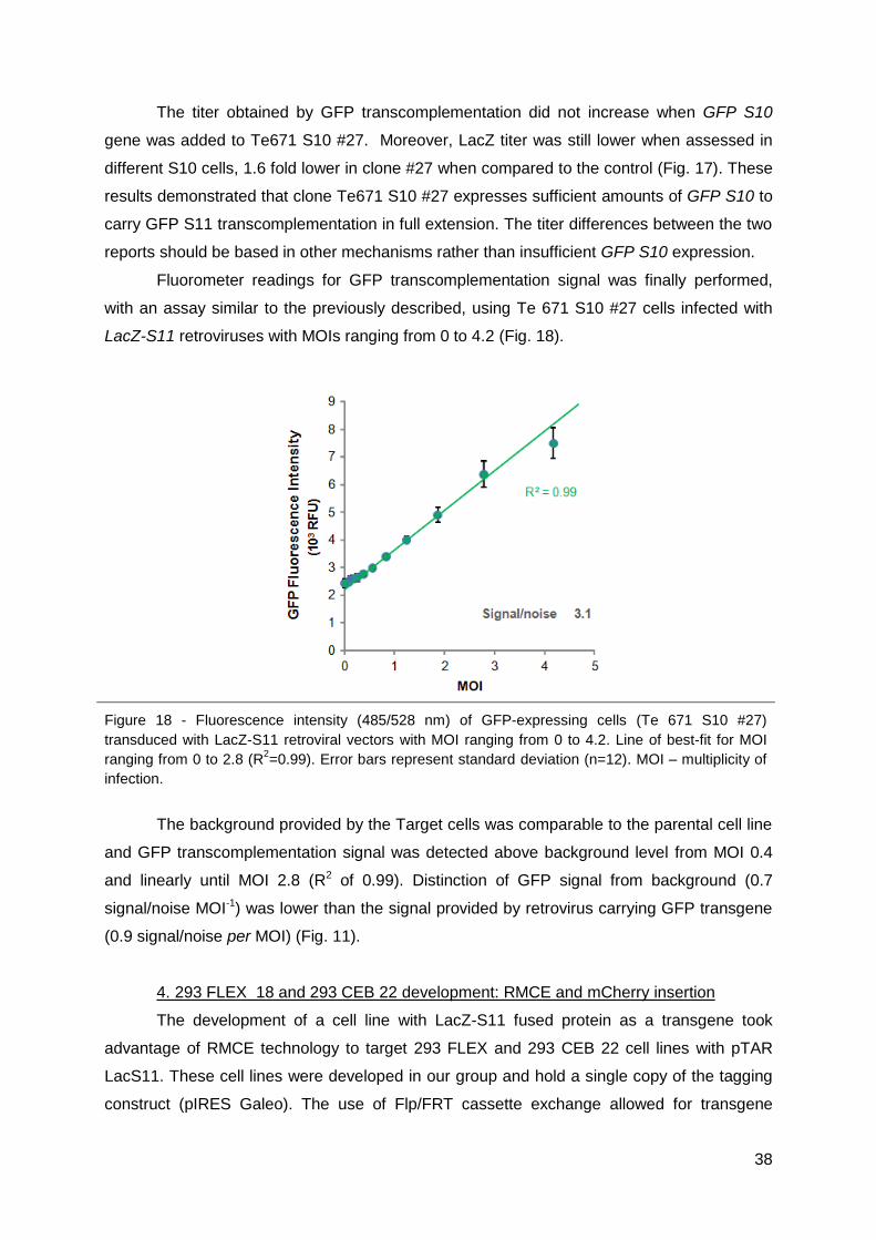
38
The titer obtained by GFP transcomplementation did not increase when GFP S10
gene was added to Te671 S10 #27. Moreover, LacZ titer was still lower when assessed in
different S10 cells, 1.6 fold lower in clone #27 when compared to the control (Fig. 17). These
results demonstrated that clone Te671 S10 #27 expresses sufficient amounts of GFP S10 to
carry GFP S11 transcomplementation in full extension. The titer differences between the two
reports should be based in other mechanisms rather than insufficient GFP S10 expression.
Fluorometer readings for GFP transcomplementation signal was finally performed,
with an assay similar to the previously described, using Te 671 S10 #27 cells infected with
LacZ-S11 retroviruses with MOIs ranging from 0 to 4.2 (Fig. 18).
Figure 18 - Fluorescence intensity (485/528 nm) of GFP-expressing cells (Te 671 S10 #27)
transduced with LacZ-S11 retroviral vectors with MOI ranging from 0 to 4.2. Line of best-fit for MOI
ranging from 0 to 2.8 (R2=0.99). Error bars represent standard deviation (n=12). MOI – multiplicity of
infection.
The background provided by the Target cells was comparable to the parental cell line
and GFP transcomplementation signal was detected above background level from MOI 0.4
and linearly until MOI 2.8 (R2 of 0.99). Distinction of GFP signal from background (0.7
signal/noise MOI-1) was lower than the signal provided by retrovirus carrying GFP transgene
(0.9 signal/noise per MOI) (Fig. 11).
4. 293 FLEX 18 and 293 CEB 22 development: RMCE and mCherry insertion
The development of a cell line with LacZ-S11 fused protein as a transgene took
advantage of RMCE technology to target 293 FLEX and 293 CEB 22 cell lines with pTAR
LacS11. These cell lines were developed in our group and hold a single copy of the tagging
construct (pIRES Galeo). The use of Flp/FRT cassette exchange allowed for transgene

39
exchange in a defined chromosomal locus of the packaging cell line in a single positive
selection step 9. After recombination by the use of two non-interacting FRT sites, the
targeting of the pTAR LacS11 construct restored the neo gene by insertion of the missing
ATG start codon and hygtk selection marker was eliminated. Upon antibiotic selection,
genomic DNA extraction and purification was performed and evaluated by PCR and qRT-
PCR for correct targeting of exchanged 293 CEB 22 S11 and 293 FLEX S11 (Fig. 19 and
Table 3); analyzed clones were previously tested for GFP transcomplementation (data not
shown).
A B
Figure 19 – Electrophoresis in 0.7% (w/v) agarose gels for the PCR reaction performed to amplify the
transgene sequence of: A) 293 FLEX S11 clones; B) 293 CEB 22 S11 clone. L- ladder; G – pIRES
GALEO; C – control (parental cell line); number correspond to targeted clones.
Transgene exchange was verified by the loss of approximately 2Kb in amplicon size
in 10 of the 12 293 FLEX S11 clones and in all 10 clones of 293 CEB 22 S11 (Fig. 19). The
results showed single copy in all 293 CEB 22 S11 clones, whereas for 293 FLEX S11, two
clones presented an additional copy, considering a 1.4 cut off (Table 3).

40
Table 3 – Number of copies of integrated transgene per cell in 293 FLEX S11 and 293 CEB 22 S11
exchanged clones. Quantification of the transgene was performed by qRT-PCR for the LacZ gene and
normalized to the respective parental cell line.
Cell Line Clones Copy / Cell Cell Line Clones Copy / Cell
293 FLEX S11
Parental 1
293 CEB 22 S11
Parental
1
7 2.0 1 0.7
14 1.7 8 0.8
17 1.2 10 0.8
19 1.1 16 1.0
22 1.1 20 0.8
31 1.4 29 0.6
35 1.3 32 1.1
38 0.8 39 1.2
46 1.3 48 1.3
51 1.0 62 0.6
After exchange, the virus production from 293 FLEX S11 clones dropped more than
two orders of magnitude and from 293 CEB 22, only 2 out of 10 clones were able to produce
in 106 I.P./mL range (data not shown). Therefore, only 293 CEB 22 S11 clones were selected
for pPURO mCherry transfection. After Puromycin selection period, the mCherry populations
were sorted to select a more homogenous population for mCherry signal.
5. Cloning-titration protocol
Transgene exchanged resulted in a drop of virus production in the clones of 293
FLEX cells, which was unexpected. On the other hand 293 CEB 22 transgene exchanged
enabled viral production in 2 of the 10 clones analyzed; being 293 CEB 22 the parental cells
of 293 FLEX it was clear that cassette exchange was possible in the later. Thus, we
postulated that 293 FLEX S11 population should contain a small number of correctly
exchanged clones and used the cloning-titration protocol to find them. High Clonogenicity
was ensured (50%), but only one producing clone out of 450 was detected (Fig. 20). Due to
time restriction, it was not possible to analyze the isolated clone in order to determine the
number of copies of integrated transgene and its specific production. If correct transgene
exchange is confirmed, the mCherry cell marker will be inserted.
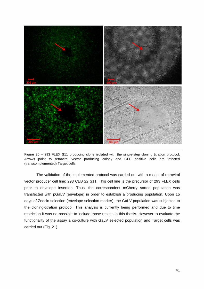
41
Figure 20 – 293 FLEX S11 producing clone isolated with the single-step cloning titration protocol.
Arrows point to retroviral vector producing colony and GFP positive cells are infected
(transcomplemented) Target cells.
The validation of the implemented protocol was carried out with a model of retroviral
vector producer cell line: 293 CEB 22 S11. This cell line is the precursor of 293 FLEX cells
prior to envelope insertion. Thus, the correspondent mCherry sorted population was
transfected with pGaLV (envelope) in order to establish a producing population. Upon 15
days of Zeocin selection (envelope selection marker), the GaLV population was subjected to
the cloning-titration protocol. This analysis is currently being performed and due to time
restriction it was no possible to include those results in this thesis. However to evaluate the
functionality of the assay a co-culture with GaLV selected population and Target cells was
carried out (Fig. 21).

42
Figure 21 - 293 CEB S11 GaLV producing clone co-cultured with Te 671 S10. Arrows point to
retroviral vector producing colony (in red) and GFP positive cells are infected (transcomplemented)
Target cells.
Figure 21 shows Target cells (in green) infected by retrovirus harboring the LacZ-S11
transgene produced by the 293 CEB 22 S11 clone (in red).

43
Part III – Metabolic engineering
From the transcriptional profiling analysis 65, more than 30 genes were identified as
potential manipulation candidates (for over-expression or down-regulation) towards improved
virus production. Four of those genes glutathione synthase, isocitrate dehydrogenase 1 and
2 and hypoxia inducible factor 1 inhibitor were cloned in a lentiviral vector for over-
expression. In the case of hypoxia inducible factor 1 inhibitor, its over-expression aims at
simulating the down-regulation of hypoxia inducible factor I while avoiding the adverse
effects of using knock-down tools 78 (typically, RNA interference). Given that the 293 FLEX
S11 clone isolated was still under expansion, we conducted a preliminary study of metabolic
engineering with these genes using 293 FLEX cells and evaluated the average viral
productivity in cell populations (Table 4).
Table 4 – Increase in viral productivity of 293 FLEX cell populations after transduction
with GSS, IDH1, IDH2 and HIF1AN
Single gene GSS IDH1 IDH2 HIF1AN
2.1 1.8 2.6 2.7
Multiple gene IDH1 IDH2 NP
NP
IDH1 IDH2
2.3 NC 4.5 5.8
Values indicate fold-change increase relatively to the non-transduced cells and after mock
control normalization. NC: no change; NP: not performed. Multiple gene refers to
transduction with the indicated genes of the above single gene transduced population.
GSS: glutathione synthase; IDH: isocitrate dehydrogenase; HIF1AN: inhibitor of hypoxia
inducible factor 1.
For all the tested genes, average viral productivity was found to be increased. More
interestingly, viral production was potentiated in GSS and HIF1AN populations after IDHs
transduction in a relatively predictable way (Fig. 22): i) IDH1 potentiates GSS effect
increasing the flux through the first step of pentose phosphate pathway; ii) HIF1AN –
activating pyruvate dehydrogenase through HIF1 inhibition thus increasing the flux through
the first step of the TCA cycle 58 – is potentiated by IDH2 catalyzing its third step. These
results strongly support the strategy of multiple gene manipulation for improved virus
production. Moreover, increases in average cell population productivity are promising
indicators of hyperproducing clones, isolatable when using the implemented protocol in 293
FLEX S11.

44
Figure 22 – Schematic representation of targeted pathways by the over-expression of GSS, IDH1,
IDH2 and HIF1AN. Genes are shown in grey boxes. For a matter of simplicity, only targeted genes
and directly activated/inactivated genes are shown. Targeted genes boxes are surrounded by a thick
line. GSS: glutathione synthase; IDH: isocitrate dehydrogenase; HIF1AN: inhibitor of hypoxia inducible
factor 1.

45
5. Discussion and conclusion
The increasing demand for recombinant pharmaceutical bioproducts (e.g. antibodies
and virus) has placed significant pressure to develop robust high-yielding mammalian cell-
based production systems. Retrovirus based vectors comprise several valuable
biopharmaceuticals such as gene therapy vectors, recombinant vaccines and replicative-
competent particles for oncolytic therapy 5,6. However, the platforms used for their
productions are characterized by low yield preparations and high contaminant content of
non-infective virus. A transcriptional profiling analysis of relevant networks recruited when
establishing a producer from its correspondent parental cell line showed that the most
relevant changes occurred in metabolic pathways (Fig. 4) 65. Considering these findings, we
validated the results by targeting easy to manipulate pathways through medium
supplementation (Fig. 7). This was our starting point aiming retroviral based vector
production improvement.
Culture medium manipulation of easy to manipulate pathways - Nucleic Acid
Metabolism, Amino Acid Metabolism, Free Radical Scavenging and Polyamine Metabolism –
increased RV titer in all culture supplements (Fig. 7 A). However, the expression of viral
genes (Fig. 7 B) was not significantly affected by these manipulations and particle stability
did not improve significantly (Fig. 8), suggesting that titer improvement must be related with
the increment of infectious particles present in the viral preparation and thus to the quality of
viral supernatants. Therefore, the expression of viral components was not the bottleneck of
RV production and modulating cell metabolism by medium manipulation led to increased
infectious retroviral based vectors production. It was showed that by targeting metabolic
pathways it is possible to increase the production capacity of 293 FLEX cells. In general, the
improvement in viral preparation quality could be a consequence of improvement in cell
robustness and production machinery and/or due to enhanced particle assembly and
budding.
The addition of nucleosides to the culture medium increased infectious viral vectors
production in 1.9 fold; this was the highest titer achieved by the addition of only one
supplement. Nucleosides are the precursor of nucleotides which in turn constitute the
building blocks of nucleic acids 74. This effect may be a result of improved synthesis of vector
genome and/or a general enhance of the transcription rate of the viral components.
Amino acid supplementation modestly increased infectious particles production (1.3
fold), when amino acids and nucleosides were added together the titer improved in 1.5 fold
but production levels were not rescued to the ones described above. Amino acids
supplementation may have an impact in translation processes enabling the producer cell to

46
initiate the translation of more viral proteins. Additionally, amino acids degradation is known
to be a main TCA feeding source to support the energy metabolism 74; thus a more favorable
energy cell state may be potentiated which in turn affects the final viral preparation. To
distinguish these two effects (carbon skeleton or energy suppliers) ammonia production in
cell supernatant will be assessed; if it is identical to the control, the amino acids were likely to
be consumed for the translation processes; on the other hand if it is increased, amino acids
degradation to feed TCA cycle should be occurring.
Polyamines supplementation yielded 1.8 fold-improvement in viral titers. Polyamines
are low molecular weight aliphatic molecules with regularly spaced positive charges. These
molecules can bind to RNA and DNA and participate in DNA stabilization, modulation of
chromatin structure and gene transcription events. Additionally, polyamines also have a role
in signal transduction, cell growth and proliferation, membrane rigidity and possess anti-
oxidant properties 79. All this properties may have an influence in viral production increment,
particularly: stabilization of viral genome leading to easier encapsidation, membrane rigidity
and prevention of its lipid peroxidation can improve viral budding and envelope quality.
Actually it was already reported that polyamines are important for the activity of viral RNA
polymerase 80,81 and may interfere with virus assembly at the level of DNA packaging or
capsid envelopment or both 82.
Down regulation of enzymes involved in oxidative stress defenses indicated that the
“virus producer status” increases oxidative stress in the cells 65. The addition of anti-oxidant
supplements to culture medium may alleviate the burn of this cell status and lead to the 1.5
fold increase in infectious virus production. Additionally, this supplementation could also
influence viral preparation quality by improving particle resistance do oxidizing stress;
however, virus half-life was not enhanced with this supplement, which would be expected if
this was the case. When complemented with polyamines or reduced glutathione supplement,
producing cells gained even more oxidative protection which resulted in increased viral titers
(2 and 2.1 fold, respectively). Moreover, reduced glutathione supplement can also play a role
in pentose phosphate pathway once it can affect its regulation by changing the ratio of
NAD+/NADH. When the cell is converting NADPH to NADP+ upon glutathione oxidation the
level of NADP+ rises, stimulating glucose-6-phosphate dehydrogenase and thereby
increasing the flux of glucose-6-phosphate through the pentose phosphate pathway 74. In the
other hand, the pentose phosphate pathway reduces NADP+ to NADPH and produces
pentose phosphates. NADPH provides reducing power for biosynthetic reactions, and ribose
5-phosphate is a precursor for nucleotide and nucleic acid synthesis, which in turn may lead
to enhanced cell capacity to express viral genes.
Two of the easy to manipulate pathways – Lipid metabolism and Vitamin and mineral
metabolism - were not target in this work. Lipid metabolism and its implications in virus
47
production of 293 FLEX cells was already studied 12,13. The results showed the need of highly
active lipid synthesis machinery to support infectious vector production under serum (lipid
source) deprivation, suggesting lipid biosynthesis as a promising target for improved
productivities. However, under standard 10% (v/v) FBS conditions (those used in this work),
transcriptional profiling results showed a general down-regulation of lipid biosynthetic
machinery in response to virus production, particularly cholesterol, while activating external
lipid consumption. Transcriptional profiling also suggested this phenomenon to be mediated
by AMPK inhibition, a strategy that chronic diseases caused by viruses are known to rely on
to benefit their own replication over cell replication 83. However, in this work, all cultures were
conducted at 10% (v/v) FBS and, as serum provides lipid nutrients in large excess, additional
supplementation should not result in improved productivities. Vitamin and mineral
metabolism, pyridoxal phosphate (PLP, the biological active form of B6 vitamin) and
tetrahydrofolate (THF) metabolism, could also be easily manipulated by culture medium
supplementation. These are important co-factors of several metabolic enzymes serving
different pathways including amino acid metabolism, in both cases, gluconeogenesis,
glycogenolysis and lipid metabolism, in the case of PLP, and nucleic acid metabolism, in the
case of THF. As many of these pathways had already been covered, the addition of these
molecules would probably only potentiate their effects.
Single medium manipulation led to a modest improvement in infectious virus
production (1.3 to 1.9 fold), as well, double manipulation raised titers in approximately 2 fold,
but more impressive was the cocktail medium manipulation which enabled 6 fold higher
productivity. To apply this strategy at larger scale requires a fed-batch system and use of
expensive supplements and thus, not attractive from the bioprocess point of view. However,
these results highlighted a very important point: orchestrated rearrangements of unrelated
metabolic pathways lead to superior titers, supporting multiple gene engineering of metabolic
pathways as the road for cell hyperproductivity.
Metabolic engineering of a single target may take up to one year to be complete as it
requires an extensive clone screening to find those with the highest infectious retrovirus
production as high productivity is a rare event in a producer population. If multiple pathways
were target at once, clone screening time would exponentially increase. Therefore it was it
was essential to develop a protocol that enables the high producing clones at early stages of
the process, such as the cloning step.
A high-throughput screening method was already described by Green and Rasco
(2002) 61; however this method needs the time to titrate clones viral supernatant upon cloning
procedure and makes use of fluorescent proteins that are expressed in both producer and
target cell line. For clinical therapeutic vectors the last mentioned trait it is not desirable,
since the therapeutic vector would need to express a fluorescent reporter.
48
The method implemented in this work reduces the time and manipulation required for
the generation of high-titer producing clone since ensures the titration and cloning
procedures are performed in one single step. The transgene expressed by the producing
cells only enables the expression of a fluorescence protein upon viral infection of Target cells
and it can be exchanged to any therapeutic gene for clinical purposes. Lastly, in Green and
Rasco 61 assay the authors reported low clonogenicity efficiency (10%) which reduces the
chances of finding a clone candidate, whereas in this work it was possible to achieve 53% of
clone survival by culturing cell in Corning CoStar black-walled 96 well plates with DMEM
20% (v/v) FBS and B27 (1x) supplement (Table 2).
The replacement of medium with PBS prior to analysis was already identified as a
critical step to ensure reproducibility and reduce variance in fluorometer readings 61. In the
developed protocol PBS supplemented with 10% FBS was used without compromising
background signal interference. Cell viability and sterility is maintained during analysis and
only producing cells have selection markers; this way we ensure that clones can be further
cultured after the assay and isolated from Target cell. Moreover, it is of outmost importance
to ensure single colony in the selected wells, for that, single cell deposition by cell sorting will
be used in the future. The use of two florescence proteins in the established protocol did not
compromise their detection since the excitation/emission spectra do not overlap 77.
Our main challenge was to establish the Target cell line. Te 671 cell line was the best
candidate since they demonstrated higher infectability (Fig.13). However, it was not possible
to insert GFP S10 gene by chemical transfection of pCMV m GFP S10 plasmid. Its delivery
was only possible upon lentiviral infection at saturating MOI. From all the analyzed clones,
Te 671 S10 #27 was selected to establish the Target cell line because it showed the best
GFP transcomplementation signal and similar infectability compared to parental cell line (Fig.
14 and 15). Nevertheless, GFP titers could not be correlated with LacZ titers (Fig. 16)
although lower GFP titers were not due to lack of GFP S10 expression in Target cells (Fig.
17). LacZ titer was reduced when titration was performed in Te 671 S10 cell compared to the
parental cell line (Fig. 17), which may indicate that upon GFP transcomplementation LacZ
loses enzymatic activity. Therefore, upon transcomplementation, GFP fluorescence might be
also be compromised by LacZ fused protein interactions, leading to the different titer
obtained with the two reporter proteins. However, this should not be a problem since GFP
signal (although under estimating the real titer) is directly proportional to the viral titer
(Fig.18). In fact, it can turn out to be an advantage since signal saturation will only occur for
very high titer clones, the ones to be detected with this method.
Targeted 293 FLEX clones lost retrovirus production capacity; this was not expected
since RMCE technology enables transgene exchange, maintaining a predictable vector
production 9. 293 CEB 22 cells were properly exchanged, thus, due to time restriction we

49
used 293 CEB 22 S11 (clone 32, Fig. 19, Table 3) for mCherry insertion and mCherry sorted
population selected (Zeocin) for GaLV expression, to validate the protocol. Nevertheless,
since only 2 producing clones from 293 CEB 22 (precursor cell line of 293 FLEX) cassette
exchanged population were isolated, we postulated that in general cassette exchange
procedure was inefficient, which in turn lead to rare properly exchanged events. Therefore,
we used the implemented protocol to extensive screen 293 FLEX S11 population. Indeed,
we were able to isolate a producing 293 FLEX S11 clone out of 450, confirming the scarcity
of producing clones in targeted population. Additionally, these results highlighted the
screening potential of the implemented protocol since only 1 clone out of 450 was detected
has a virus producer. However, further analysis needs to be carried to ensure single copy of
the transgene and to assess specific productivity. Moreover, additional screening to find
more 293 FLEX S11 producing clones will be performed.
Optimizing vector titer has important implications for both clinical and research-based
gene transfer. The cloning-titration protocol developed allows high-throughput screening to
overcome the limitations imposed by the current labor intensive and time-consuming flow
cytometric and antibiotic selection methods by maximizing the chance of identifying clones
with rare hyperproductivity. This protocol should find other valuable applications as
shortening the time-frame of establishing new virus producer cells or rapidly screen
alternative cell substrates (cell lines) for virus production. If the analysis could be performed
by using robotics much greater throughput will be achievable. Additionally, this work allowed
extending the knowledge on viral production and gave insights for further metabolic
engineering of 293 FLEX S11 cells. This cell engineering will only be feasible with the proper
tools for clone screening; the established cloning-titration protocol will significantly contribute
for that future work. Multiple metabolic engineering towards the delivery of an improved
mammalian cell platform for the production of RV retrovirus can be later transformed into
healthcare therapeutic services and products. The preliminary results on 293 FLEX
metabolic gene manipulation (Table 4) strongly encourage the continuation of this work in a
multiple gene approach.
The huge potential of HEK293 derived production systems for biotechnological and
pharmaceutical applications renders this master project not only scientifically but also
technologically relevant. Moreover, retroviral vectors constitute an excellent representative of
similarly enveloped virus, potentiating the broadening of the generated knowledge to other
viral vectors (e.g lentivirus), viral vaccines (e.g. influenza) and VLPs (e.g. retroviral VLPs).
Thus, this project contributes greatly to the advancement of virus based biopharmaceutical
production platforms in biotechnology.

50
6. Ongoing and future work
The results obtained during this master project suggest that the low retroviral vectors
production yields from 293 FLEX cells are mainly due to a metabolic constrain. The
hypothesis that metabolic engineering of this platform may lead to a more efficient
metabolism and consequently higher RV production will be confirmed by combinatorial gene
manipulations of the metabolic pathways previously identified and validated in this work. To
follow this work line, the 293 FLEX S11 isolated clone will be analyzed for single copy of the
transgene and, depending on the results, mCherry will be inserted and further metabolic
engineering will be performed. For this purpose, lentivirus library harboring different
metabolic genes will be produced with the constructed plasmids of section 3.1 and used to
infect 293 FLEX S11. Depending on the results other genes can also be tested.
The final validation of the implemented protocol is being carried with 293 CEB 22 S11
population (GaLV clones) in a total of 600 clones. Each well will be analyzed for the
GFP/mCherry signal allowing for determining the productivity profile distribution of this
population. The highest GFP/mCherry yielding clones will be isolated, quantified for specific
productivity and compared to 293 FLEX titers.
The knowledge gathered can also be used in future work for the improvement of
production of other virus based biopharmaceuticals.

51
7. References
1. Hawley, R. G. Therapeutic potential of retroviral vectors. Transfus. Sci 17, 7–14 (1996). 2. Cruz, P. E., Coroadinha, A. S., Rodrigues, T. & Hauser, H. Production of Retroviral Vectors: From
the Producer Cell to the Final Product. Gene and Cell therapy 17–32 (2009). 3. Retroviruses. (Cold Spring Harbor Laboratory Press: NY, 1997).at
<http://www.ncbi.nlm.nih.gov/books/NBK19376/> 4. Rodrigues, A. F., Alves, P. M. & Coroadinha, A. S. Production of Retroviral and Lentiviral Gene
Therapy Vectors : Challenges in the Manufacturing of Lipid Enveloped Virus. Viral Gene Therapy 15–40 (2011).doi:10.5772/18615
5. Edelstein, M. L., Abedi, M. R. & Wixon, J. Gene therapy clinical trials worldwide to 2007 – an update. The journal of gene medicine 9, 833–842 (2007).
6. Dalba, C., Bellier, B., Kasahara, N. & Klatzmann, D. Replication-competent vectors and empty virus-like particles: new retroviral vector designs for cancer gene therapy or vaccines. Molecular therapy : the journal of the American Society of Gene Therapy 15, 457–66 (2007).
7. McTaggart, S. & Al-Rubeai, M. Retroviral vectors for human gene delivery. Biotechnology advances 20, 1–31 (2002).
8. Schucht, R. et al. A new generation of retroviral producer cells: predictable and stable virus production by Flp-mediated site-specific integration of retroviral vectors. Molecular therapy : the journal of the American Society of Gene Therapy 14, 285–292 (2006).
9. Coroadinha, a S. et al. The use of recombinase mediated cassette exchange in retroviral vector producer cell lines: predictability and efficiency by transgene exchange. Journal of biotechnology 124, 457–468 (2006).
10. Coroadinha, A. et al. Production of Retrovial Vectors: Review. Current Gene Therapy 10, 456–473 (2010).
11. Merten, O.-W. State-of-the-art of the production of retroviral vectors. The journal of gene medicine 6, S105–24 (2004).
12. Rodrigues, A. F., Carmo, M., Alves, P. M. & Coroadinha, A. S. Retroviral vector production under serum deprivation: The role of lipids. Biotechnology and bioengineering 104, 1171–1181 (2009).
13. Rodrigues, A. F., Amaral, A. I., Veríssimo, V., Alves, P. M. & Coroadinha, A. S. Adaptation of retrovirus producer cells to serum deprivation: Implications in lipid biosynthesis and vector production. Biotechnology and bioengineering 109, 1269–79 (2012).
14. Coroadinha, A. S. et al. Effect of Medium Sugar Source on the Production of Retroviral Vectors for Gene Therapy. Biotechnology and bioengineering 94, 24–36 (2006).
15. Amaral, A. I., Coroadinha, A. S., Merten, O.-W. & Alves, P. M. Improving retroviral vectors production: role of carbon sources in lipid biosynthesis. Journal of biotechnology 138, 57–66 (2008).
16. Coroadinha, A. S. et al. Effect of Osmotic Pressure on the Production of Retroviral Vectors : Enhancement in Vector Stability. Biotechnology advances 94, 322–329 (2006).
17. Khosla, C. & Bailey, J. E. Heterologous expression of a bacterial haemoglobin improves the growth properties of recombinant Escherichia coli. Nature 331, 633–635 (1988).
18. Hopwood, D., Malpartida, F. & Kieser, H. Production of “hybrid”antibiotics by genetic engineering. 314, 642–644 (1985).
19. Edwards, J. S. & Palsson, B. O. Systems properties of the Haemophilus influenzae Rd metabolic genotype. The Journal of biological chemistry 274, 17410–6 (1999).
20. Kim, T. Y., Sohn, S. B., Kim, Y. B., Kim, W. J. & Lee, S. Y. Recent advances in reconstruction and applications of genome-scale metabolic models. Current opinion in biotechnology 23, 617–623 (2011).
21. Figueroa, B. et al. Enhanced cell culture performance using inducible anti-apoptotic genes E1B-19K and Aven in the production of a monoclonal antibody with Chinese hamster ovary cells. Biotechnology and Bioengineering 97, 877–892 (2007).
22. Tey, B. T., Singh, R. P., Piredda, L., Piacentini, M. & Al-Rubeai, M. Bcl-2 mediated suppression of apoptosis in myeloma NS0 cultures. Journal of biotechnology 79, 147–159 (2000).
23. Lim, S. F. et al. RNAi suppression of Bax and Bak enhances viability in fed-batch cultures of CHO cells. Metabolic engineering 8, 509–522 (2006).
24. Chen, K., Liu, Q., Xie, L., Sharp, P. A. & Wang, D. I. Engineering of a mammalian cell line for reduction of lactate formation and high monoclonal antibody production. Biotechnology and bioengineering 72, 55–61 (2001).
25. Wlaschin, K. F. & Hu, W.-S. Engineering cell metabolism for high-density cell culture via manipulation of sugar transport. Journal of biotechnology 131, 168–176 (2007).
52
26. Henry, O. & Durocher, Y. Enhanced glycoprotein production in HEK-293 cells expressing pyruvate carboxylase. Metabolic engineering 13, 499–507 (2011).
27. Rehemtulla, a et al. In vitro and in vivo functional characterization of bovine vitamin K-dependent gamma-carboxylase expressed in Chinese hamster ovary cells. Proceedings of the National Academy of Sciences of the United States of America 90, 4611–5 (1993).
28. Lasunskaia, E. B. et al. Transfection of NS0 myeloma fusion partner cells with HSP70 gene results in higher hybridoma yield by improving cellular resistance to apoptosis. Biotechnology and bioengineering 81, 496–504 (2003).
29. Borth, N., Mattanovich, D., Kunert, R. & Katinger, H. Effect of increased expression of protein disulfide isomerase and heavy chain binding protein on antibody secretion in a recombinant CHO cell line. Biotechnology progress 21, 106–11 (2005).
30. Nishimiya, D., Mano, T., Miyadai, K., Yoshida, H. & Takahashi, T. Overexpression of CHOP alone and in combination with chaperones is effective in improving antibody production in mammalian cells. Applied microbiology and biotechnology (2012).doi:10.1007/s00253-012-4365-9
31. Hallgren, K. W., Qian, W., Yakubenko, A. V., Runge, K. W. & Berkner, K. L. r-VKORC1 expression in factor IX BHK cells increases the extent of factor IX carboxylation but is limited by saturation of another carboxylation component or by a shift in the rate-limiting step. Biochemistry 45, 5587–5598 (2006).
32. Mohan, C. & Lee, G. M. Effect of inducible co-overexpression of protein disulfide isomerase and endoplasmic reticulum oxidoreductase on the specific antibody productivity of recombinant Chinese hamster ovary cells. Biotechnology and bioengineering 107, 337–46 (2010).
33. Mazur, X., Fussenegger, M., Renner, W. a & Bailey, J. E. Higher productivity of growth-arrested Chinese hamster ovary cells expressing the cyclin-dependent kinase inhibitor p27. Biotechnology progress 14, 705–13 (1998).
34. Watanabe, S., Shuttleworth, J., Al-rubeai, M. & Kingdom, U. Regulation of Cell Cycle and Productivity in NS0 Cells by the Over-Expression. 1–7 (2002).doi:10.1002/bit.10112
35. Dreesen, I. a J. & Fussenegger, M. Ectopic expression of human mTOR increases viability, robustness, cell size, proliferation, and antibody production of chinese hamster ovary cells. Biotechnology and bioengineering 108, 853–866 (2011).
36. Seth, G., McIvor, R. S. & Hu, W.-S. 17Beta-hydroxysteroid dehydrogenase type 7 (Hsd17b7) reverts cholesterol auxotrophy in NS0 cells. Journal of biotechnology 121, 241–52 (2006).
37. Florin, L. et al. Heterologous expression of the lipid transfer protein CERT increases therapeutic protein productivity of mammalian cells. Journal of biotechnology 141, 84–90 (2009).
38. Sriburi, R., Jackowski, S., Mori, K. & Brewer, J. W. XBP1: a link between the unfolded protein response, lipid biosynthesis, and biogenesis of the endoplasmic reticulum. The Journal of cell biology 167, 35–41 (2004).
39. Ferrari, J., Gunson, J., Lofgren, J., Krummen, L. & Warner, T. G. Chinese hamster ovary cells with constitutively expressed sialidase antisense RNA produce recombinant DNase in batch culture with increased sialic acid. Biotechnology and bioengineering 60, 589–95 (1998).
40. Davies, J. et al. Expression of GnTIII in a recombinant anti-CD20 CHO production cell line: Expression of antibodies with altered glycoforms leads to an increase in ADCC through higher affinity for FC gamma RIII. Biotechnology and bioengineering 74, 288–294 (2001).
41. Park, J. H. et al. Enhancement of recombinant human EPO production and glycosylation in serum-free suspension culture of CHO cells through expression and supplementation of 30Kc19. Applied microbiology and biotechnology 96, 671–83 (2012).
42. Kondoh, H. Cellular life span and the Warburg effect. Experimental Cell Research 314, 1923–1928 (2008).
43. Irani, N., Wirth, M., van Den Heuvel, J. & Wagner, R. Improvement of the primary metabolism of cell cultures by introducing a new cytoplasmic pyruvate carboxylase reaction. Biotechnology and bioengineering 66, 238–246 (1999).
44. Irani, N., Beccaria, A. J. & Wagner, R. Expression of recombinant cytoplasmic yeast pyruvate carboxylase for the improvement of the production of human erythropoietin by recombinant BHK-21 cells. Journal of biotechnology 93, 269–282 (2002).
45. Fogolín, M. B., Wagner, R., Etcheverrigaray, M. & Kratje, R. Impact of temperature reduction and expression of yeast pyruvate carboxylase on hGM-CSF-producing CHO cells. Journal of biotechnology 109, 179–91 (2004).
46. Zhou, M. et al. Decreasing lactate level and increasing antibody production in Chinese Hamster Ovary cells (CHO) by reducing the expression of lactate dehydrogenase and pyruvate dehydrogenase kinases. Journal of biotechnology 153, 27–34 (2011).

53
47. Vives, J., Juanola, S., Cairó, J. J. & Gòdia, F. Metabolic engineering of apoptosis in cultured animal cells: implications for the biotechnology industry. Metabolic Engineering 5, 124–132 (2003).
48. Majors, B. S., Betenbaugh, M. J. & Chiang, G. G. Links between metabolism and apoptosis in mammalian cells: applications for anti-apoptosis engineering. Metabolic engineering 9, 317–326 (2007).
49. Butler, M. Animal cell cultures: recent achievements and perspectives in the production of biopharmaceuticals. Applied microbiology and biotechnology 68, 283–291 (2005).
50. Tey, B. T., Singh, R. P., Piredda, L., Piacentini, M. & Al-Rubeai, M. Influence of bcl-2 on cell death during the cultivation of a Chinese hamster ovary cell line expressing a chimeric antibody. Biotechnology and bioengineering 68, 31–43 (2000).
51. Figueroa, B., Sauerwald, T. M., Mastrangelo, a J., Hardwick, J. M. & Betenbaugh, M. J. Comparison of Bcl-2 to a Bcl-2 deletion mutant for mammalian cells exposed to culture insults. Biotechnology and bioengineering 73, 211–22 (2001).
52. Perani, A., Singh, R. P., Chauhan, R. & Al-Rubeai, M. Variable functions of bcl-2 in mediating bioreactor stress- induced apoptosis in hybridoma cells. Cytotechnology 28, 177–188 (1998).
53. Terada, S. et al. Co-expression of bcl-2 and bag-1, apoptosis suppressing genes, prolonged viable culture period of hybridoma and enhanced antibody production. Cytotechnology 31, 143–151 (1999).
54. Mastrangelo, A. J., Hardwick, J. M., Bex, F. & Betenbaugh, M. J. Part I. Bcl-2 and Bcl-x(L) limit apoptosis upon infection with alphavirus vectors. Biotechnology and bioengineering 67, 544–554 (2000).
55. Mastrangelo, A. J., Hardwick, J. M., Zou, S. & Betenbaugh, M. J. Part II. Overexpression of bcl-2 family members enhances survival of mammalian cells in response to various culture insults. Biotechnology and bioengineering 67, 555–564 (2000).
56. Meents, H., Enenkel, B., Eppenberger, H. M., Werner, R. G. & Fussenegger, M. Impact of coexpression and coamplification of sICAM and antiapoptosis determinants bcl-2/bcl-x(L) on productivity, cell survival, and mitochondria number in CHO-DG44 grown in suspension and serum-free media. Biotechnology and bioengineering 80, 706–716 (2002).
57. Palsson, B. O. system biology properties of reconstructed networks. Chemistry & … (Cambridge University Press: 2006).at <http://onlinelibrary.wiley.com/doi/10.1002/cbdv.200490137/abstract>
58. Guerreiro, M. R. Manipulação do efeito de Warburg em células de mamífero em cultura : implicações para o metabolismo energético. FCUL-DBV Master Thesis (2011).
59. Seth, G., Charaniya, S., Wlaschin, K. F. & Hu, W.-S. In pursuit of a super producer-alternative paths to high producing recombinant mammalian cells. Current opinion in biotechnology 18, 557–564 (2007).
60. Fussenegger M, S, S., D, D., X, M. & JE., B. Controlled proliferation by multigene metabolic engineering enhances the productivity of Chinese hamster ovary cells. Nature Biotechnology 16, 468 – 472 (1998).
61. Green, B. J. & Rasko, J. E. J. Rapid screening for high-titer retroviral packaging cell lines using an in situ fluorescence assay. Human gene therapy 13, 1005–13 (2002).
62. Warnock, J. N., Merten, O.-W. & Al-Rubeai, M. Cell culture processes for the production of viral vectors for gene therapy purposes. Cytotechnology 50, 141–162 (2006).
63. Gay, V., Moreau, K., Hong, S.-S. & Ronfort, C. Quantification of HIV-based lentiviral vectors: influence of several cell type parameters on vector infectivity. Archives of virology 157, 217–223 (2012).
64. Sastry, L., Johnson, T., Hobson, M. J., Smucker, B. & Cornetta, K. Titering lentiviral vectors: comparison of DNA, RNA and marker expression methods. Gene therapy 9, 1155–1162 (2002).
65. Rodrigues, A. F. et al. Metabolic pathways recruited for the production of a recombinant enveloped virus-transcriptional profiling and experimental validation. Submitted to Metabolic Engineering, Elsevier (2012).
66. Verhoeyen, E., Hauser, H. & Wirth, D. Evaluation of retroviral vector design in defined chromosomal loci by Flp-mediated cassette replacement. Human gene therapy 12, 933–944 (2001).
67. Dull, T. et al. A third-generation lentivirus vector with a conditional packaging system. Journal of virology 72, 8463–8471 (1998).
68. Shaner, N. C. et al. Improved monomeric red, orange and yellow fluorescent proteins derived from Discosoma sp. red fluorescent protein. Nature biotechnology 22, 1567–1572 (2004).
69. Cosset, F. L., Takeuchi, Y., Battini, J. L., Weiss, R. a & Collins, M. K. High-titer packaging cells producing recombinant retroviruses resistant to human serum. Journal of virology 69, 7430–7436 (1995).

54
70. Carmo, M. et al. Quantitation of MLV-based retroviral vectors using real-time RT-PCR. Journal of virological methods 119, 115–119 (2004).
71. Le Doux, J. M., Davis, H. E., Morgan, J. R. & Yarmush, M. L. Kinetics of retrovirus production and decay. Biotechnology and bioengineering 63, 654–62 (1999).
72. Livak, K. J. & Schmittgen, T. D. Analysis of relative gene expression data using real-time quantitative PCR and the 2(-Delta Delta C(T)) Method. Methods 25, 402–408 (2001).
73. Bodin, L., Beaune, P. H. & Loriot, M.-A. Determination of cytochrome P450 2D6 (CYP2D6) gene copy number by real-time quantitative PCR. Journal of biomedicine & biotechnology 2005, 248–253 (2005).
74. Nelson, D. L. & Cox, M. M. Lehninger principles of biochemistry. (W. H. Freeman: NY, 2004). 75. Carmo, M. et al. Relationship between retroviral vector membrane and vector stability. The Journal
of general virology 87, 1349–1356 (2006). 76. Carmo, M. et al. Stabilization of gammaretroviral and lentiviral vectors : from production to gene
transfer. The journal of gene medicine 11, 670–678 (2009). 77. Shaner, N. C., Steinbach, P. A. & Tsien, R. Y. A guide to choosing fluorescent proteins. Nature
Methods 2, 905–909 (2005). 78. Fish, R. & Kruithof, E. Short-term cytotoxic effects and long-term instability of RNAi delivered using
lentiviral vectors. BMC molecular biology 15, 1–15 (2004). 79. Wallace, H., Fraser, A. & Hughes, A. A perspective of polyamine metabolism. Biochemical Journal
376, 1–14 (2003). 80. Raina, a, Tuomi, K. & Mäntyjärvi, R. Roles of polyamines in the replication of animal viruses.
Medical biology 59, 428–32 (1981). 81. Tuomi, K., Raina, a & Mäntyjärvi, R. Synthesis of Semliki-forest virus in polyamine-depleted baby-
hamster kidney cells. The Biochemical journal 206, 113–9 (1982). 82. Gibson, W., van Breemen, R., Fields, a, LaFemina, R. & Irmiere, a D,L-alpha-
difluoromethylornithine inhibits human cytomegalovirus replication. Journal of virology 50, 145–54 (1984).
83. Mankouri, J. & Harris, M. Viruses and the fuel sensor: the emerging link between AMPK and virus replication. Reviews in Medical Virology 21, 205–212 (2011).

55
8. Annexes
Table S1: Cloning sites, primers and templates for plasmids construction.
* DNASU clone ID

56
A B
C D
E
F
Figure S1 – Final plasmid constructs. A) pCMV mGFP LacZ-S11; B) pTAR LacS11; C) pRRLsin LacS11; D)
pRRLsin S10; E) pRRLsin with metabolic gene (i.e. GSS, HIF1 inhibitor; IDH1 or IDH2); F) pPuro mCherry.

57
Table S2 – Final composition of supplemented culture media.
Base composition NC AAs AO POLY GLUT
Culture
Co
nd
itio
n
1 Advanced DMEM 10% (v/v) FBS, 4mM glutamine - - - - -
2 Advanced DMEM 10% (v/v) FBS, 4mM glutamine 5X - - - -
3 Advanced DMEM 10% (v/v) FBS, 4mM glutamine - - - -
4 Advanced DMEM 10% (v/v) FBS, 4mM glutamine 5X - - -
5 Advanced DMEM 10% (v/v) FBS, 4mM glutamine - - 1X - -
6 Advanced DMEM 10% (v/v) FBS, 4mM glutamine - - - 1X -
7 Advanced DMEM 10% (v/v) FBS, 4mM glutamine - - 1X 1X -
8 Advanced DMEM 10% (v/v) FBS, 4mM glutamine - - 1X - 2 mM
9 Advanced DMEM 10% (v/v) FBS, 4mM glutamine 5x 1X 1X 2mM
NC, nucleosides supplement; AAs, amino acids supplement; AO, anti-oxidant supplement, POLY,
polyamine supplement, GLUT, reduced glutathione. Final concentrations of NC, AO, POLY and GLUT
were chose based on a previous screening, as those yielding the highest infectious vector productivities.
AAs supplement was prepared in Advanced DMEM as a concentrated stock of the following individual
amino acids (final concentration in the culture medium): serine (400 mM), asparagines (400 mM), arginine
(100 mM), methionine (100 mM), phenylalanine (100 mM), leucine (300 mM), valine (300 mM) and
isoleucine (300 mM); AAs were chosen based on previous consumption study by HPLC, as those highly
consumed and/or rapidly depleted from the culture medium.
Table S3 – Primers used for RT PCR
Forward primer (5’ to 3’) Reverse primer (5’ to 3’)
RPL22 CTGCCAATTTTGAGCAGTTT CTTTGCTGTTAGCAACTACGC
LacZ ACTATCCCGACCGCCTTACT TAGCGGCTGATGTTGAACTG
Gag-Pol GTCCACTATCGCCAGTTGCT CTGGGTCCTCAGGGTCATAA
Envelope GGACCAAAATAGCGAATGGA GGTGAACTGTACGCCTGGAT
Neomycin - AGCCATGATGGATACTTTCTCG
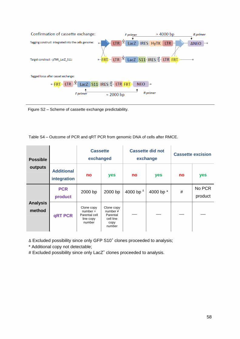
58
Figure S2 – Scheme of cassette exchange predictability.
Table S4 – Outcome of PCR and qRT PCR from genomic DNA of cells after RMCE.
Δ Excluded possibility since only GFP S10+ clones proceeded to analysis;
* Additional copy not detectable;
# Excluded possibility since only LacZ+ clones proceeded to analysis.
Possible
outputs
Cassette
exchanged
Cassette did not
exchange
Cassette excision
Additional
integration no yes no yes no yes
Analysis
method
PCR
product
2000 bp
2000 bp
4000 bp Δ
4000 bp *
#
No PCR
product
qRT PCR
Clone copy number =
Parental cell line copy number
Clone copy number ≠ Parental cell line
copy number
__
__
__
__