universidade da beira interior - ubibliorum.ubi.pt§ao... · a nível mecânico é responsável...
TRANSCRIPT


UNIVERSIDADE DA BEIRA INTERIOR
Ciências da Saúde
Desenvolvimento de Scaffolds para futura
aplicação na Regeneração Óssea
Inês Raquel Tavares Serra
Dissertação para obtenção do Grau de Mestre em
Ciências Biomédicas
(2º ciclo de estudos)
Orientador: Prof. Doutor Ilídio Joaquim Sobreira Correia
Covilhã, Outubro de 2013

iii

iv
List of Publications Torres, A., Gaspar, V., Serra, I., Diogo, G., Fradique, R., Silva, A. and Correia, I.; Bioactive
polymeric-ceramic hybrid 3D scaffold for application in bone tissue regeneration. Materials
Science and Engineering: C. (2013). 33 (7): 4460-4469.

v

vi
“O sucesso é a soma de pequenos esforços repetidos dia após dia.”
(Robert Collier)

vii

viii
Acknowledgments
At the end of another stage of my life, I want to thank all those who, in one way or another,
contributed for reaching this stage of my life.
Firstly, I would like to express my gratitude to my supervisor Professor Ilídio Correia, for the opportunity to develop this project and for all the support, patience and guidance during this
Master Thesis.
I would like to thank the Eng. Ana Paula from the Optics Department of the University of
Beira Interior, for the time that she spent on the acquisition of the scanning electron
microscopy images.
I also acknowledge to Professor Abílio Silva from the Department of Electrochemical
Engineering of the University of Beira Interior, for their availability in carrying out the
mechanical assays.
To my group colleagues, I would thank for everything. Thanks for all the help, support and
courage. A special thanks to Gabriela, who has been present in every moments during this
year. Thanks for all the support, help and especially the patience during the year. To Paulo
thanks for all the help.
To my closest friends thanks for all good moments they that provided me over the years. For
all courage, love and support in moments less good, they have given me and for never let me
give up. A special thanks to my best friend Ana Raquel Nunes, for all the support, not only at
the professional level but also personal. Thanks for never having stopped believe in me!
Lastly, but no less importantly, thank you to my father and my mother. First, the financial
support that they provide me over these five years. But even more important for all the
dedication, support, love and patience. Thanks also to my sister, coined and goddaughter all
the force and support. Thank you for never stopped to believe in me.
Without you, none of this would be possible!

ix

x
Abstract
Bone is the main component of the human skeleton. This tissue is responsible for several
functions at the structural and biological level. It is a tissue that is highly vascularized and
dynamic, with excellent mechanical properties and also has the capacity for self-
regeneration. However, several factors as aging, disorders at metabolic level and fractures,
can lead to a loss of integrity of this tissue. In this context, it is necessary to develop bone
substitutes that mimic the structure of extracellular matrix and induce regeneration of the
damaged tissue. Currently, the most commonly clinical treatments used are based in the
application of bone grafts obtained from the patient or from another. However, these
therapeutic approaches present several limitations, namely immunologic responses and
infectious risks. Tissue Engineering, an area that aims to develop biological substitutes that
are able to enhance, restore or replace the damaged tissue, has emerged as an area of
research that is looking for new solutions to improve bone regeneration. In this field of
research, several types of biomaterials have been developed for this purpose. Among these,
three dimensional structures, also known as scaffolds are the most promising because these
structures possess the essential properties needed for the bone regeneration, such as
biocompatibility, biodegradability and porosity. Besides, scaffolds provide a structure that
mimic extracellular matrix which is fundamental to promote cell adhesion and proliferation
until tissue regeneration occurs. In this context, novel biomaterials based in chitosan, gelatin
and beta-tricalcium phosphate were developed in this work, in order to be applied in a near
future in bone regeneration. Several physical-chemical and mechanical studies were
performed, to study the influence of the incorporation of gelatin and beta tricalcium
phosphate in the structure of chitosan scaffolds. Besides, biological studies were performed
in vitro with human osteoblasts to evaluate the materials cytotoxic profile. The results
obtained demonstrate that the scaffolds developed provide a suitable support for cell
adhesion and proliferation. Moreover, the scaffolds do not present toxicity, highlighting its
application in regenerative medicine.
Keywords:
Beta Tricalcium Phosphate, Bone Remodeling, Chitosan, Gelatin, Scaffolds.

xi

xii
Resumo
O osso é o principal componente do esqueleto humano, desempenhando diversas funções a
nível estrutural e biológico. É um tecido altamente vascularizado e dinâmico, com
propriedades mecânicas excepcionas e com capacidade de se auto-regenerar ao longo da
vida. No entanto, existem diversos factores como o envelhecimento, desordens a nível
metabólico e fraturas, que podem levar à perda da integridade deste tecido. Neste contexto,
surge a necessidade de desenvolver substitutos ósseos que mimetizem a sua estrutura e
induzam a sua regeneração. Atualmente, os tratamentos clínicos mais usados baseiam-se na
aplicação de enxertos ósseos do próprio paciente ou proveniente de outro. Contudo, estas
abordagens terapêuticas apresentam diversas desvantagens para o paciente, nomeadamente
riscos imunológicos e de transmissão de doenças. De forma a colmatar estas limitações, surge
a Engenharia de Tecidos, uma área de investigação que tem procurado desenvolver novos
substitutos biológicos que possuam a capacidade de melhorar, restaurar ou mesmo substituir
o tecido lesado. Com este intuito, têm sido desenvolvidos vários tipos de biomateriais. Neste
campo, estruturas tridimensionais, também conhecidas como scaffolds, revelam ser os mais
promissores. Estes possuem propriedades essenciais no processo de regeneração óssea, tais
como biocompatibilidade, biodegradabilidade e porosidade. Simultaneamente, estas
estruturas tridimensionais permitem a criação de uma estrutura com a capacidade de
mimetizar a matriz extracelular, e desta forma favorecer a adesão e proliferação celular até
ocorrer a regeneração do tecido lesado. Neste contexto, novos biomateriais a base de
quitosano, gelatina e beta fosfato tricálcico, foram desenvolvidos ao longo deste trabalho,
para futura aplicação na regeneração óssea. Vários estudos das propriedades físico-químicas e
mecânicas dos materiais foram realizados com o objectivo de estudar qual a influência da
incorporação da gelatina e do beta fosfato tricálcico em scaffolds de quitosano. Além disso, o
perfil citotóxico dos materiais foi caracterizado in vitro, através da utilização de osteoblastos
humanos. Os resultados obtidos demonstram que as estruturas desenvolvidas fornecem um
suporte adequado à adesão e proliferação celular. Além disso, não apresentam efeitos tóxicos
para as células, evidenciando a sua possível aplicação na área da medicina regenerativa.
Palavras-chave:
Andaimes, Beta Fosfato Tricálcico, Gelatina, Quitosano, Regeneração Óssea.

xiii
xiv
Resumo Alargado
O osso é o principal componente do esqueleto humano, desempenhando várias funções
altamente especializadas. A nível mecânico é responsável pelo suporte e locomoção do corpo,
e confere protecção de alguns órgãos, como o coração e os pulmões. A nível biológico está
envolvido na produção de células sanguíneas e no armazenamento de iões, essencialmente
cálcio e fosfato. É um tecido dinâmico e altamente vascularizado com capacidade de
regeneração intrínseca, o que possibilita a sua constante substituição por tecido novo,
assegurando desta forma a sua integridade. No entanto, existem inúmeros factores que
comprometem a sua integridade: a medida que o indivíduo envelhece ocorre um decréscimo
da densidade óssea, o que torna o osso mais suscetível a fraturas; bem como as forças a que o
osso está sujeito diariamente, muitas vezes superiores àquelas que ele suporta, podendo
resultar em fraturas graves que acabam por afetar a qualidade de vida do paciente. Nestas
situações, é necessário que o mecanismo de remodelação óssea ocorra de forma natural. O
processo de remodelação do osso ocorre em duas fases bastante distintas: fase de reabsorção
e fase de formação, entre as quais deve existir sempre um balanço que permita uma
coordenação efetiva entre os diversos eventos celulares e moleculares, que ocorrem neste
processo. No entanto, em traumatismos ósseos graves que envolvam múltiplas fraturas,
tumores ósseos e desordens a nível metabólico, onde a integridade do tecido é afetada, a
capacidade de auto-remodelação torna-se limitada. Neste contexto, surge a necessidade de
desenvolver substitutos ósseos que mimetizem a sua estrutura e promovam a sua
regeneração, sem causar qualquer rejeição por parte do paciente. Atualmente, as abordagens
terapêuticas mais usadas são os auto-enxertos, alo-enxertos e xeno-enxertos. Os auto-
enxertos, enxertos de osso provenientes do próprio paciente, não apresentam qualquer risco
imunológico, contudo são limitados para grandes defeitos ósseos. Por outro lado, alo-enxertos
e xeno-enxertos, enxertos do osso obtidos de outro paciente da mesma espécie ou de
diferentes espécies, respetivamente, podem causar rejeição pelo organismo do paciente.
Com o intuito de colmatar estas limitações vários investigadores têm tentado desenvolver o
substituto ósseo ideal, surgindo assim uma nova área de pesquisa, a Engenharia de Tecidos. A
Engenharia de Tecidos combina os conhecimentos da engenharia e das ciências da vida e visa
o desenvolvimento de substitutos biológicos que possuam a capacidade de melhorar, restaurar
ou mesmo substituir o tecido lesado. Vários tipos de biomateriais como próteses, substratos
injetáveis e hidrogéis têm sido desenvolvidos com esta finalidade. Neste campo, estruturas
tridimensionais, também conhecidas como scaffolds, revelam ser as mais promissoras pois
possuem propriedades essenciais para o processo de regeneração óssea, tais como
biocompatibilidade, biodegradabilidade e porosidade. Além disso, promovem uma estrutura
que mimetiza a matriz extracelular favorecendo a adesão e proliferação celular até ocorrer a
regeneração do tecido lesado. Para a produção destas estruturas tridimensionais têm sido
utilizados vários tipos de materiais e diversos métodos. Estes fatores tornam-se fulcrais, pois

xv
a aplicação em tecidos biológicos dos scaffolds produzidos exige que os mesmos sejam
produzidos com biomateriais que não desencadeiem nenhuma resposta inflamatória nem
comprometam a viabilidade do tecido em contacto com o material. Neste âmbito, têm sido
usados vários métodos tradicionais como o processamento a alta pressão, lixiviação de
partículas, contudo, apresentam toxicidade devido ao uso de solventes orgânicos, baixa
resistência mecânica e estruturas não porosas, o que os torna inadequados para a
regeneração do tecido ósseo. Assim, para eliminar alguns destes problemas têm sido usados
métodos que envolvem a criopreservação das amostras, permitindo a criação de estruturas
altamente porosas e sem qualquer toxicidade. Neste contexto, scaffolds produzidos com materiais biocompatíveis neste estudo foram
desenvolvidos, para futura aplicação na regeneração óssea, utilizando quitosano, gelatina e
beta fosfato tricálcico. Vários estudos das propriedades físico-químicas e mecânicas dos
materiais foram realizados com o objectivo de estudar qual a influência da incorporação da
gelatina e do beta fosfato tricálcico em scaffolds de quitosano. Além disso, o perfil citotóxico
dos materiais foi caraterizado in vitro, através da utilização de osteoblastos humanos. Os
resultados obtidos demonstram que as estruturas desenvolvidas fornecem um suporte
adequado à adesão e proliferação celular. Além disso, não apresentam efeitos tóxicos para as
células, evidenciando a sua possível aplicação na área da medicina regenerativa.

xvi

xvii
Index
Chapter I - Introduction ..................................................................................... 1
1.1.Bone Tissue ............................................................................................. 2
1.1.1.Types of Bone ..................................................................................... 2
1.1.2.Bone Matrix ........................................................................................ 4
1.1.3.Bone Cells ......................................................................................... 5
1.1.4.Bone Remodelling ................................................................................ 6
1.2.Bone Disorders .......................................................................................... 8
1.3.Bone Grafts ............................................................................................. 9
1.4.Tissue Engineering ..................................................................................... 9
1.4.1.Scaffolds for bone regeneration ............................................................. 10
1.4.2.Cell-biomaterial surface interactions ...................................................... 11
1.4.3.Materials used for scaffolds production .................................................... 12
1.4.4.Processing Techniques ........................................................................ 14
1.5.Objectives ............................................................................................. 16
Chapter II – Materials and Methods ...................................................................... 17
2.1.Materials ............................................................................................... 18
2.2.Methods ................................................................................................ 18
2.2.1.Production of 3D scaffolds .................................................................... 18
2.2.2.Morphological characterization of the 3D scaffolds ...................................... 19
2.2.2.1.Scanning Electron Microscopy .......................................................... 19
2.2.3.Chemical and physical characterization of the scaffolds ............................... 20
2.2.3.1.Fourier Transform Infrared Spectroscopy Analysis ................................. 20
2.2.3.2.X-Ray Diffraction ......................................................................... 20
2.2.3.3.Energy Dispersive Spectroscopy ....................................................... 20
2.2.4.Mechanical characterization of the 3D scaffolds ......................................... 21

xviii
2.2.5.Swelling Studies ................................................................................ 21
2.2.6.Porosity Evaluation ............................................................................ 22
2.2.7.Biolgical characterization of the 3D scaffolds ............................................ 22
2.2.7.1.Culture of osteoblasts in the presence of the scaffolds ........................... 22
2.2.7.2.Cell viability: MTS assay ................................................................ 23
2.2.8.Stastitical analysis ............................................................................. 23
Chapter III – Results and Discussion ..................................................................... 24
3.1. Morphology and Macroscopic Properties of Scaffolds ........................................ 25
3.2.Physical-chemical characterization of the scaffolds .......................................... 28
3.2.1.Fourier Transform Infrared Spectroscopy Analysis ....................................... 28
3.2.2.X-Ray Diffraction analysis ..................................................................... 30
3.2.3.Energy Dispersive Spectroscopy analysis ................................................... 32
3.2.Mechanical characterization of 3D scaffolds .................................................... 32
3.3.Swelling Studies ...................................................................................... 34
3.4.Porosity ................................................................................................ 35
3.5.Analysis of the biological properties of the scaffolds ......................................... 36
Chapter IV – Conclusion.................................................................................... 40
Chapter V – Bibliography .................................................................................. 43
Chapter VI – Appendix ..................................................................................... 50

xix
xx
List of Figures
Figure 1: Representation of anatomy of a long bone ..................................................................... 3 Figure 2: Bone structure at cellular level ........................................................................................ 5 Figure 3: Representation of a bone remodeling cycle .................................................................... 6 Figure 4: Balance between bone formation and bone resorption ................................................. 7 Figure 5: Chemical structure of chitin and chitosan .................................................................... 13 Figure 6: Chemical structure of gelatin ......................................................................................... 13 Figure 7: Macroscopic images of the differents scaffolds surface .............................................. 25 Figure 8: SEM images of the different scaffolds............................................................................ 27 Figure 9: SEM images of pores size of the differnte scaffolds ..................................................... 28 Figure 10: FTIR analysis of the powders and 3D scaffolds ........................................................... 28 Figure 11: X-Ray spectra of the powders and 3D scaffolds .......................................................... 28 Figure 12: Characterization of the compressive strength of scaffolds ....................................... 30 Figure 13: Swelling profile of the scaffolds ................................................................................... 28 Figure 14: Total porosity of the different scaffolds ..................................................................... 28 Figure 15: Microscopic images of human osteoblasts cells in the presence of scaffolds ......... 36 Figure 16: Evaluation of the cellular viability ............................................................................... 37 Figure 17: SEM micrographs of osteoblast morfology in the presence of the scaffolds ............ 37

xxi

xxii
List of Tables
Table 1: Bone classification according to shape ......................................................... 2 Table 2: Mechanical properties of cortical and spongy bone. ......................................... 3 Table 3: Composition and ratios of different scaffolds produced in this study .................. 18 Table 4: Elemental analysis of the manufacture scaffolds........................................... 32

xxiii

xxiv
List of Acronyms
3D Three-Dimensional
BMP Bone Morphogenic Factors
β-TCP Beta Tricalcium Phosphate
BSA Bovine Serum Albumin
CH Chitosan
DMEM-F12 Dulbecco’s Modified Eagle’s Medium
ECM Extracellular Cellular Matrix
EDS Energy Dispersive Spectroscopy
EDTA Ethylenediaminetetraacetic Acid
FBS Fetal Bovine Serum
FTIR Fourier Transform Infrared Spectroscopy
GAGs Glycosaminoglycans
GEL Gelatin
HA Hydroxyapatite
IGF-II Insulin-Like Growth Factor II
M-CSF Macrophage Colony-Stimulating
MMPs Matrix Metalloproteinases
MSC Mesenchymal Stem Cells
MTS 3-(4,5-dimethylthiazol-2-yl)-5-(3-carboxymethoxyphenyl)-2-(4-sulfophenyl)-2H-Tetrazolium Reagent, Inner Salt
NAOH Sodium Hydroxide

xxv
PBS Phosphate Buffered Saline
OB Osteoblasts
OC Osteoclasts
PTH Parathyroid Hormone
RANK Receptor Activator for Nuclear Factor K B
RANKL Receptor Activator for Nuclear Factor K B Ligand
RER Rough Endoplasmatic Reticulum
RT Room Temperature
SEM Scanning Electronic Microscopy
TCPS Tissue Culture Polystyrene
TE Tissue Engineering
TGF Transforming Growth Factor
TPP Sodium Tripolyphosphate
XRD X-Ray Diffraction

xxvi

Chapter I
Introduction

Chapter I – Introduction _________________________________________________________________________________
2
1. Introduction
1.1. Bone Tissue
Bone is a dense tissue that is the main component of human skeleton. It is composed by bone
matrix, cells and water and it is involved in various functions (1). Bone is responsible for
supporting the weight and locomotion of the entire body. Besides, it also confers protection
to some organs, such as the heart and lungs. Moreover, it acts as storage of minerals like
calcium and phosphorous ions, and produces blood cells through hematopoiesis (2).
1.1.1. Types of bone
The human bones can be classified in different classes, depending on the shape, on the
relation between the amount of bone matrix and spaces present and on the organization of
the collagen fibres within bone matrix (2). Relying on shape and size, bone can be classified into long, short, flat or irregular (Table 1).
Table 1: Bone classification according to shape (Adapted from (2, 3)).
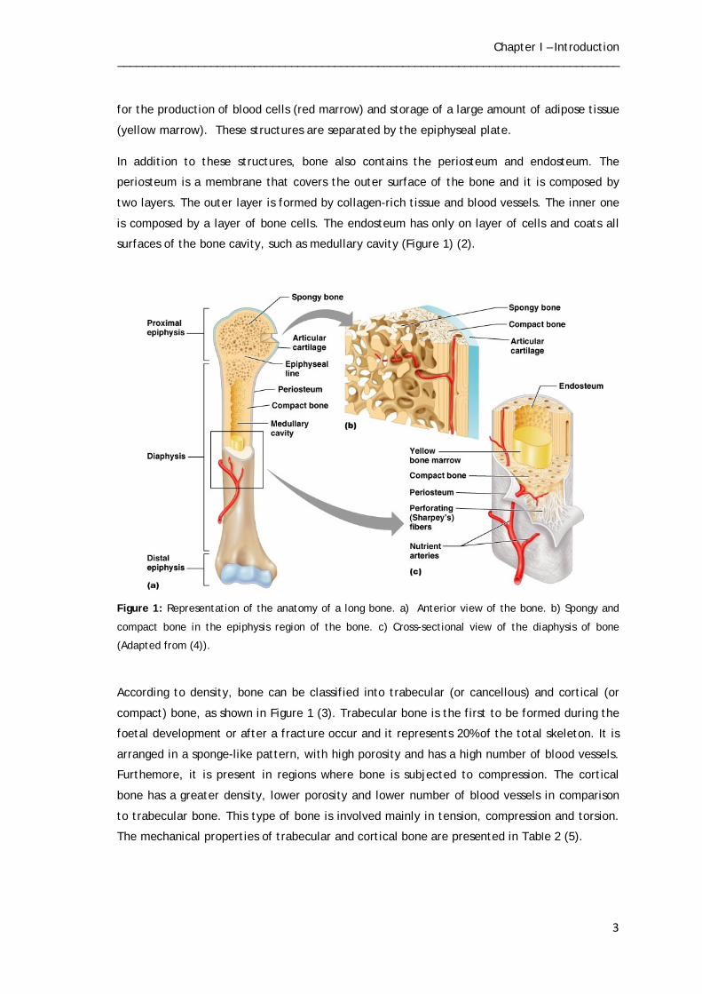
The long bones are constituted by three main components: diaphysis, epiphysis and
epiphyseal plate, as shown in Figure 1. The epiphysis, present at the end of the bone, is
constituted by spongy bone, composed by small cavities surrounded by bone matrix. The
diaphysis is between the distal and proximal epiphysis, being formed by compact bone,
consisting mostly of bone matrix. Besides these cavities, the diaphysis has a cavity of great
biological interest, the medullary cavity, which is filled with bone marrow that is responsible
Bone Classification
Features
Functions
Examples
Long
Cylinder-like shape
Leverage
Femur, tibia, fibula,
humerus, radius
Short
Cube-like shape
Stability and Support
Carpals, tarsals
Flat
Thin and curved
Points of attachment
for muscles; protectors of internal organs;
Sternum, ribs and cranial bones
Irregular
Complex shape
Protect internal organs
Vertebrae and facial
bones
Chapter I – Introduction _________________________________________________________________________________
3
for the production of blood cells (red marrow) and storage of a large amount of adipose tissue
(yellow marrow). These structures are separated by the epiphyseal plate. In addition to these structures, bone also contains the periosteum and endosteum. The
periosteum is a membrane that covers the outer surface of the bone and it is composed by
two layers. The outer layer is formed by collagen-rich tissue and blood vessels. The inner one
is composed by a layer of bone cells. The endosteum has only on layer of cells and coats all
surfaces of the bone cavity, such as medullary cavity (Figure 1) (2).
Figure 1: Representation of the anatomy of a long bone. a) Anterior view of the bone. b) Spongy and
compact bone in the epiphysis region of the bone. c) Cross-sectional view of the diaphysis of bone
(Adapted from (4)).
According to density, bone can be classified into trabecular (or cancellous) and cortical (or
compact) bone, as shown in Figure 1 (3). Trabecular bone is the first to be formed during the
foetal development or after a fracture occur and it represents 20% of the total skeleton. It is
arranged in a sponge-like pattern, with high porosity and has a high number of blood vessels.
Furthemore, it is present in regions where bone is subjected to compression. The cortical
bone has a greater density, lower porosity and lower number of blood vessels in comparison
to trabecular bone. This type of bone is involved mainly in tension, compression and torsion.
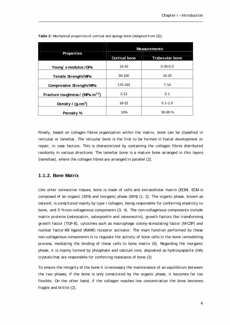
The mechanical properties of trabecular and cortical bone are presented in Table 2 (5).
Chapter I – Introduction _________________________________________________________________________________
4
Table 2: Mechanical properties of cortical and spongy bone (Adapted from (5)).
Finally, based on collagen fibres organization within the matrix, bone can be classified in
reticular or lamellar. The reticular bone is the first to be formed in foetal development or
repair, in case facture. This is characterized by containing the collagen fibres distributed
randomly in various directions. The lamellar bone is a mature bone arranged in thin layers
(lamellae), where the collagen fibres are arranged in parallel (2).
1.1.2. Bone Matrix
Like other connective tissues, bone is made of cells and extracellular matrix (ECM). ECM is
composed of an organic (35%) and inorganic phase (65%) (1, 2). The organic phase, known as
osteoid, is constituted mainly by type I collagen, being responsible for conferring elasticity to
bone, and 5 % non-collagenous components (3, 4). The non-collagenous components include
matrix proteins (osteocalcin, osteopontin and osteonectin), growth factors like transforming
growth factor (TGF-β), cytocines such as macrophage colony-stimulating factor (M-CSF) and
nuclear factor-Kβ ligand (RANK) receptor activator. The main function performed by these
non-collagenous components is to regulate the activity of bone cells in the bone remodelling
process, mediating the binding of these cells to bone matrix (6). Regarding the inorganic
phase, it is mainly formed by phosphate and calcium ions, deposited as hydroxyapatite (HA)
crystals that are responsible for conferring resistance of bone (2).
To ensure the integrity of the bone it is necessary the maintenance of an equilibrium between
the two phases. If the bone is only constituted by the organic phase, it becomes far too
flexible. On the other hand, if the collagen reaches low concentration the bone becomes
fragile and brittle (2).
Properties
Measurements
Cortical bone
Trabecular bone
Young´s modulus /GPa
14-20
0.05-0.5
Tensile Strength/MPa
50-150
10-20
Compressive Strength/MPa
170-193
7-10
Fracture toughness / (MPa.m1/2)
2-12
0.1
Density / (g.cm3)
18-22
0.1-1.0
Porosity %
10%
50-90 %

Chapter I – Introduction _________________________________________________________________________________
5
1.1.3. Bone Cells
Bone is formed by three cell types, namely, osteoblasts (OB), osteocytes and osteoclasts (OC)
that have different origins and functions (Figure 2). OB are mononuclear cells that derive from Mesenchymal Stem Cells (MSC) (7). Structurally,
these cells have large and spherical nucleus presenting high number of Rough Endoplasmic
Reticulum (RER), Golgi apparatus, and mitochondria. OB are involved in the formation of the
bone matrix, being responsible for the production of collagen type I and non-collagenous
proteins. Moreover, OB contains alkaline phosphatase (ALP) that contributes for bone
mineralization (8).
After bone matrix formation be performed by osteoblastic cells, the matrix that is being
produced surrounded the OB and they subsequently differentiate into mature cells, called
osteocytes, which are the most abundant cells in bone tissue. These cells present stellar
shape and, compared to OB, have a lower number of organelles involved in synthesis and
secretion, such as RER and Golgi apparatus. However, osteocytes are essential to maintain
bone homeostasis, since they regulate the levels of calcium and phosphorus in this tissue (8).
Finally, OC are multinucleated cells derived from haematopoietic cells of the macrophage
lineage (9). Those cells are involved essentially in bone reabsorption during growth and bone
remodelling, getting in contact with the bone matrix and forming an area called ruffled
border. This area assumes great importance because it is through it that OC release hydrogen
ions that create an acidic environment and subsequently degrade bone matrix (10). Moreover,
OC have the ability to connect with other osteocytes and even with OB, allowing the
detection of bone alterations and its communication to neighbouring OB and OC.
Figure 2: Bone structure at cellular level (Adapted from (11)).
Chapter I – Introduction _________________________________________________________________________________
6
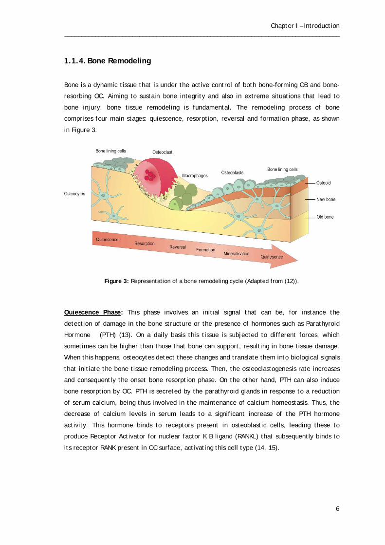
1.1.4. Bone Remodeling
Bone is a dynamic tissue that is under the active control of both bone-forming OB and bone-
resorbing OC. Aiming to sustain bone integrity and also in extreme situations that lead to
bone injury, bone tissue remodeling is fundamental. The remodeling process of bone
comprises four main stages: quiescence, resorption, reversal and formation phase, as shown
in Figure 3.
Figure 3: Representation of a bone remodeling cycle (Adapted from (12)).
Quiescence Phase: This phase involves an initial signal that can be, for instance the
detection of damage in the bone structure or the presence of hormones such as Parathyroid
Hormone (PTH) (13). On a daily basis this tissue is subjected to different forces, which
sometimes can be higher than those that bone can support, resulting in bone tissue damage.
When this happens, osteocytes detect these changes and translate them into biological signals
that initiate the bone tissue remodeling process. Then, the osteoclastogenesis rate increases
and consequently the onset bone resorption phase. On the other hand, PTH can also induce
bone resorption by OC. PTH is secreted by the parathyroid glands in response to a reduction
of serum calcium, being thus involved in the maintenance of calcium homeostasis. Thus, the
decrease of calcium levels in serum leads to a significant increase of the PTH hormone
activity. This hormone binds to receptors present in osteoblastic cells, leading these to
produce Receptor Activator for nuclear factor K B ligand (RANKL) that subsequently binds to
its receptor RANK present in OC surface, activating this cell type (14, 15).

Chapter I – Introduction _________________________________________________________________________________
7
Resorption Phase: The precursors of osteoclasts are attracted to the injury site, and become
differentiated into mature OC. This differentiation requires the action of two major,
cytokines that are released from OB, M-CSF and RANKL. M-CSF is involved mainly in the
proliferation and survival of OC precursors, while RANKL promotes the proliferation of OC
precursors (10). Subsequently, mature OC adhere to bone surface by integrins avβ3. Thus, OC
resorb bone by secreting hydrochloric acid and proteolytic enzimes, through ruffled border.
Hydrochloric acid dissolves hydroxyapatite and allows proteolitic enzymes, essentially
cathepsin K to degrade components (mainly collagen) of the bone matrix (16, 17).
Reversal phase: When OC complete the resorption process, they migrate from the surface of
bone and undergo programmed cell death (apoptosis), in a stage called reversal phase. This
phase begins the process of bone formation (6).
Formation Phase: The precursors of OB are attracted to the area where bone was resorbed
and become differentiated into OB. As previously described, they have in its constitution type
I collagen, osteocalcin and ALP enzyme that are involved in the production of the new matrix.
In the last osteoid phase, bone formation stops and following bone mineralization, bone lining
cells remains in a quiescent state (18).
To ensure the health of the bone tissue it is essential that the process of bone remodeling
occurs correctly. There is a balance between bone formation and bone resorption, as shown
in Figure 4.
Figure 4: Balance between bone formation and bone resorption (Adapted from (19)).

Chapter I – Introduction _________________________________________________________________________________
8
1.2. Bone Disorders
As previously described, to ensure that the bone remodeling process occurs normally, it is
necessary a tight coupling between bone resorption and bone formation. However, factors
like ageing, physiological changes (e.g., menopause and diseases) can cause changes in bone
remodeling cycle and subsequently allow the development of bone disorders like
Osteoporosis, Osteomalacia and Paget´s diseases (20). Osteoporosis is characterized by a reduction of the amount of bone density, which occurs
when the rate of bone resorption exceeds bone formation, making bones more fragile and
subsequently increasing the risk of fracture (21). A variety of factors may be responsible for
this disorder. One of the main factors that can lead to decrease of bone mass in both sexes is
ageing. During adolescence, bones are in development and growth, reaching the peak bone
mass in adulthood. In this phase the amount of bone formed is higher than the amount
reabsorbed. However, bone mass begins to decrease after 35 years of age, due to decreased
levels of calcium in the blood. Moreover, in the case of women, the menopause can also lead
to a decrease in bone mass. This happens due to a reduction of the estrogens released from
the ovaries. Since, they are responsible for the maintenance of the bone mass at normal
values through the inhibition of PTH and subsequently the inhibition of OC activity. However,
at post-menopausal phase the amount of estrogens decrease leading to an increased activity
of the PTH and subsequently, to an increase activity of the OC. Lastly, the increase of
glucocorticoid (GC) level in blood, use in the treatment of inflammatory and autoimmune
diseases, can also lead to development of osteoporosis (22).
Osteomalacia is characterized by impaired mineralization of bone, increasing its flexibility
and consequently causing its deformation. The main factor involved in this disorder is the
insufficiency of vitamin D, due to a poor diet in this vitamin or insufficient exposure to
sunlight. So, for bone grow normally it is essential that the body absorb calcium and
phosphorus ions correctly. However, for a correct absorption of these ions it is necessary the
presence of sufficient quantities of vitamin D. When this vitamin is present in insufficient
quantities in the organism, it loses the ability to properly absorb calcium and phosphorous
ions, resulting in abnormal bone growth (21).
Paget´s disease is defined by excessive and haphazard bone resorption and formation, leading
to structural defects that cause bone pain and deformity. Although, the factors associated
with this disorder are not yet well understood, changes in diet, exposure to infections,
mechanical loading of the skeleton are considered risk factors (12).

Chapter I – Introduction _________________________________________________________________________________
9
1.3. Bone Grafts
Bone has the ability to self-remodeling over life, however, due to injury or traumas it is the
second most common transplanted tissue (23). Every years, millions of people are suffering
from bone defects due to trauma, tumors and bony diseases that in some cases may lead to
death (24). To avoid such situations, it is necessary to find an ideal bone substitute that
promotes bone remodeling. Three different types of bone grafts are currently available and
have been used in the majority of the cases: autografts, allografts and xenografts (25).
Autografts are the most used grafts and are obtained from other parts of the patient´s body.
However, these grafts present several disadvantages. The implantation of these grafts
requires that patients are submitted to surgery, which may lead to a long recovery for
patients, increasing the risk of infection. Moreover, the use of autografts is limited to large
bone defects (26, 27). On the other hand, allografts and xenografts consist in the
transplantation from other individuals, from the same or different species, respectively.
However, these grafts can cause immune rejection by patients or be responsible for
transmission of diseases (28).
1.4. Tissue Engineering
As previously described, when a bone defects occur it is necessary to develop substitutes that
can replace the function of the damaged bone tissue. In order to overcome the drawbacks
associated with autografts, allografts and xenografts, it is necessary to develop new bone
substitutes. In this context, arise a new and revolutionary area called Tissue Engineering (TE).
As it was defined by Langer and Vacanti, TE is ‘‘an interdisciplinary field of research that
applies the principles of engineering and the life sciences towards the development of
biological substitutes that restore, maintain, or improve tissue function’’ (29). Specifically, TE for bone regeneration is focused on the development of alternatives to
replace the injured bone tissue, in order to promote bone regeneration without causing
immune rejection on the patient (30).

Chapter I – Introduction _________________________________________________________________________________
10
1.4.1. Scaffolds for bone regeneration
Bone Tissue Engineering is based on developing three-dimensional (3D) structures, commonly
known as scaffolds that promote bone regeneration. Scaffolds are three-dimensional matrices
that act as temporary templates for cell adhesion and proliferation, while they provide
mechanical support, until the new bone tissue is formed at the affected area (25).
In order to promote bone regeneration it is necessary that the developed scaffolds fulfill
some requirements like:
Biocompatibility: the scaffold is considered biocompatible when implanted in the body, it
does not cause any immune rejection by the patient (3).
Biodegradability: materials used for the production of scaffolds may be biodegradable so that
the rate of new bone formation may be accompanied by degradation of the provisory
template, whose main function is to support cells, while the new tissue is being produced
(31). Osteoconduction: as defined by Davies et al. osteoconduction describes to the ability of the
graft promote the migration of osteogenic cells to the surface of the scaffolds through a fibrin
clot. This property assumes a large importance because it promotes neovascularization, that
is very important for bone regeneration, ensuring the restoration the blood supply and
promoting bone regeneration (25, 32).
Osteoindunction: this property is characterized the ability that scaffolds have to attract stem
or osteoprogenitor cells to the bone healing site, promoting their differentiation into bone-
forming OB (25). Considering that OB play a key role in the bone remodeling, scaffolds must
have good properties of osteoindunction.
Mechanical properties: scaffolds should have sufficient mechanical strength to withstand the
forces to which bone is daily subjected.
Porosity: scaffolds must have a porous structure and open pores that allow cell migration and
proliferation. In addition, interconnectivity is also important for diffusion of nutrients (30).

Chapter I – Introduction _________________________________________________________________________________
11
1.4.2. Cell-biomaterial surface interactions
To assure that correct bone tissue regeneration occurs it is essentially that cell adheres to
biomaterial surface when it is implanted in the organism. The process of cell adhesion occurs
in three stages: cell adsorption, attachment and spreading (33). The first phase is
characterized by an immediately adhesion, in which cells are deposited on the material’s
surface without cell spreading. In a second stage, the cells are attached to the material.
Finally, in the last phase, cell spreading occurs, occurring changes in morphology resulting in
cytoskeleton changes, thus creating a better interaction with the substrate (34, 35). This interaction between cells and materials depends essentially on materials surface
properties, such as their topography, hydrophilicity and chemical composition (36).
Topography: the roughness of material surface has effect on the osteoblastic cell
attachment, proliferation and differentiation (37). Several studies have reported that
materials with rough surfaces at micro and submicro scale with sizes similar to cells enhance
osteoblast differentiation. On the other hand, materials with surfaces with higher roughness
reduce cell adhesion due to the decreased surface area. Materials with low surface area
decrease the ability of cells to establish contact area with the material (37).
Hydrophilicity: when a material is introduced into the body, water molecules are the first to
reach to surface, thus the degree of hydrophilicity of the materials affects cell adhesion. This
parameter is related to the contact angle/wettability of the materials that is defined as the
ability of a liquid to be spread on a surface. The smaller the angle formed between the
droplet and the substrate, the more hydrophilic will be surface and subsequently have a
better cell adhesion (38).
Chemical composition: another parameter that affects the behavior of cells is the chemical
composition of the material. Materials containing on its surface calcium and phosphate ions
improve cell growth (39).
Chapter I – Introduction _________________________________________________________________________________
12
1.4.3. Materials used for scaffolds production
Metals, polymers and ceramics have been the most commonly used materials for the
development of scaffolds for application in TE (25).
Metals are used in the development of bone substitutes due to mechanical properties that
they present. Nevertheless, metals implants start to fail 10-15 years after being implanted in
the body, being necessary its replacement frequently (25).
As alternative, biodegradable polymers of natural or synthetic origin has been used in TE.
Synthetic polymers as poly (lactic acid) (PLA), Poly (glycolic acid) (PGA), poly (£-
caprolactone) (PCL) are being applied in the field of biomedical engineering due to their good
mechanical properties and also by the possibility of controlling the rate of degradation.
However, they present some disadvantages, such as hydrophobic surfaces and poor cell
adhesion, leading to the preferential use of natural polymers (25). These natural polymers
include polysaccharides (starch, alginate, chitosan) or proteins-based polymers (collagen,
fibrin gels, silk, and gelatin). Despite they present low mechanical strength, this natural
polymers have high hydrophilicity, low immune reaction, as well as enhanced cell adhesion
and proliferation (40).
Among these, Chitosan (CH) has gained quite popularity due to its intrinsic properties. CH is a
cationic polysaccharide composed of copolymers of β (1→4)-glucosamine and N-acetyl-D-
glucosamine. This polymer of natural origin, derived from the deacetylation of chitin, which
is the second most abundant biopolymer found in the shells of marine crustaceans and cell
walls of fungi (41). Chitin and CH structures are presented in Figure 5.

Chapter I – Introduction _________________________________________________________________________________
13
Figure 5: Chemical structure of chitin and chitosan (Adapted from (42, 43)).
CH is biocompatible, biodegradable, non-toxic and presents antibacterial properties (44, 45).
Moreover, as mentioned above, due to its properties, CH has attracted much attention in the
field of TE, being used for a wide variety of applications ranging from skin, bone and
cartilage. Several studies have shown that CH has suitable properties for being used in
scaffolds production that are aimed to be used in bone tissue engineering. Since it allows the
production of structures with appropriate porosity and can support osteoblastic cell
attachment, proliferation and differentiation, due to its hydrophilic character conferred by
the presence hydroxyl and amine groups (46, 47). In spite of this, CH has some limitations like
weak mechanical resistance and great instability, caused by its high capacity to swelling (48).
Interestingly, several attempts have been made to enhance the intrinsic biological and
mechanical properties of this polymer, through the incorporation of other natural polymers
such as gelatin (GEL) (49).
GEL is a natural polymer derived from collagen, with valuable characteristics that allow their
use in bone regeneration. It is biodegradable, biocompatible, has low antigenicity in
comparison to collagen (50). Although, it being a precursors of collagen, it still retains some
of the information signals, such as Arginine-Glycine-Asparagine (RGD) sequence, that promote
cell adhesion, differentiation and proliferation (51).

Chapter I – Introduction _________________________________________________________________________________
14
Figure 6: Chemical structure of gelatin (Adapted from (52)).
Although, both CH and GEL exhibit valuable properties to be used for bone regeneration, they
still present weak mechanical resistance. Aiming to overcome this limitation, these natural
polymers have been added to ceramics, such as hydroxyapatite (HA) and Beta Tricalcium
Phosphate (β-TCP). HA is the major inorganic component of bone and due its properties it has
been used in production of scaffolds to be applied in bone tissue engineering. However, HA
has some drawbacks. This ceramic presents low biodegradability and high costs. Conversely,
Beta Tricalcium Phosphate (β-TCP) present low cost and a higher degradation rate than HA.
Moreover, β-TCP is characterized by exhibiting a good bioactivity, biocompatibility,
biodegradability and osteoconductivity (53).
1.4.4. Processing Techniques
Various methods have been explored in the development of scaffolds for bone tissue
engineering, including solvent casting/particulate leaching, high pressure processing and
freeze-drying.
As defined by Mikos et al. solvent casting/particulate leaching technique consists in a solution
of a polymer dissolved in an organic solvent, with the incorporation of particles, mainly salts.
This mixture is placed in a mold and subsequently freeze-dried in order to remove the
solvent. The composite material obtained is thereafter placed in a bath to promote
dissolution of the particles creating a porous structure (54). Although, this technique provides
reasonable results for application in bone tissue engineering field, it presents some
disadvantages. The use of organic solvents used may influence the cytotoxic profile of
scaffolds (3).
Chapter I – Introduction _________________________________________________________________________________
15
The high pressure processing is other technique used in scaffolds production. This technique
consists in the incorporation of gas bubbles at high pressure, usually carbone dioxide (CO2), in
a polymer until saturation occurs. Through the decrease of pressure, the gas evaporates and
creates macro-pores, resulting in a porous structure. Although this technique is not
considered toxic, presents some disadvantages. The scaffolds produced have low mechanical
properties, a non-porous surface and a closed pore structure, affecting this cell behavior (55).
Currently, it has been also used cryogenic processes, based on freezing-drying process (56).
This technique is based on the freezing an aqueous solution followed by a process of
sublimation through freeze-drying. The freeze-drying allows solvent remotion, under the
action of vacuum and low pressure, resulting in a highly porous scaffolds (56). Conversely to
solvent casting/particulate leaching technique, the freeze-drying uses non-toxic solvents,
avoiding problems associated with toxicity. Furthemore and unlike the high pressure
processing, this technique allows to produce highly porous structures with high pore
interconnectivity (55).

Chapter I – Introduction _________________________________________________________________________________
16
1.5. Objectives
The main objective of this work was to develop scaffolds through freeze-drying for future
application in bone tissue engineering. The present work plan had the following aims.
- In order to verify the effect of the incorporation gelatin and β-TCP or both in CH scaffolds,
various types of scaffolds (CH, CH/Gel, CH/β-TCP and CH/Gel/ β-TCP) were produced.
- Evaluation of the mechanical properties of the scaffolds.
- Evaluation and characterization of the biological properties of the scaffolds.
Chapter II
Materials and Methods

Chapter II – Materials and Methods _________________________________________________________________________________
18
2.1. Materials
Amphotericin B, bovine serum albumin (BSA), dulbecco´s modified eagle´s medium (DMEM-
F12), ethylenediaminetetraacetic acid (EDTA), glutaraldehyde, L-glutamine, medium
molecular weight chitosan (MMW CH), penicillin G, phosphate buffered saline (PBS), sodium
hydroxide (NaOH), sodium cacodylate, sodium tripolyphosphate (TPP), streptomycin, trypan
blue and trypsin were purchased from Sigma-Aldrich (Sintra, Portugal). βTCP was purchased
from Pancreac (Barcelona, Spain). 3-(4,5-dimethylthiazol-2-yl)-5-(3-carboxymethoxyphenyl)-
2-(4-sulfophenyl)-2H-tetrazolium reagent, inner salt (MTS) and electron coupling reagent
(phenazine methosulfate; PMS) were acquired from Promega. Aqueous acetic acid was
obtained from Pronalab. Fetal bovine serum (FBS) was purchased from Biochrom AG (Berlin,
Germany). Human osteoblast cells (CRL-11372) were acquired to American Type Culture
Collection (VA, USA).
2.2. Methods
2.2.1. Production of 3D Scaffolds
CH/GEL/β-TCP 3D scaffolds were produced using the freeze-drying technique. Importantly,
four different types of scaffolds were produced and tested, concerning their composition and
the ratio used of different components (Table 3).
Table 3: Composition and ratios of different scaffolds produced in this study.
Scaffold Constituents Ratio
% (wt) Chitosan (CH) Gelatin (Gel) β-TCP
Group I X
Group II X X 1:1
Group III X X 1:1
Group IV X X X 2:1:1
To produce the four different formulations, CH 2% (w/v) was dissolved in a 1% aqueous acetic
acid solution and maintained under magnetic stirring overnight, protected from light.
Following, CH hydrogel preparation, the different types of scaffolds were produced. Briefly,
scaffolds from Group I consisted only on CH hydrogel and those from Groups II and III were
composed by a 1:1 ratio (% w/t) of CH hydrogel and Gelatin (Gel) and CH hydrogel and β-TCP,

Chapter II – Materials and Methods _________________________________________________________________________________
19
respectively. Scaffolds from Group IV were produced after dispersion of Gel and β-TCP within
the CH hydrogel (Table 3). In order to obtain homogenized hydrogels, all the mixtures were
maintained under magnetic stirring, for 24 hours, sonicated and degassed in an ultrasound
bath, for 10 minutes. Following this, all the solutions were deposited into a 48-well
polystyrene plate, frozen for 24 hours at -20ºC and then lyophilized for 24 hours. The
lyophilized 3D structures were removed from the molds and neutralized for 3 hours in a 1M
NaOH (pH=12,8) solution. Then, the structures were washed for 10 minutes with Mili-Q
ultrapure water and crosslinked with a 2,5% (w/v) (TPP) (pH=5,5) solution for 3 hours.
Subsequently, they were washed again for 10 minutes. To conclude the production of the
scaffolds, another similar cycle of freezing and freeze-dried was performed and the final 3D
formulations were obtained.
2.2.2. Morphological characterization of the 3D scaffolds
2.2.2.1. Scanning Electron Microscopy (SEM) analysis
In order to evaluate the cellular behavior in the presence of the scaffolds, as well as the
morphology and the diameter of the pores, Scanning Electron Microscopy (SEM) analysis was
performed following the method adapted from Lima et al.(57). Accordingly, to prepare the
3D structures to be visualized by SEM, samples were washed twice with Phosphate Buffer
Solution (PBS, pH=7.4), its fixed with 2.5 % (v/v) glutaraldehyde and then diluted in a 0.1 M
sodium cacodylate solution. After 30 minutes in glutaraldehyde, the samples were washed
twice with PBS and further dehydrated by incubation for 10 minutes in a graded series of
ethanol solutions (50, 60, 70, 80, 90 and 99 % v/v). Next, samples were dried using CO2 at
critical point and finally sputtered-coated with gold using an Emitech K550 sputter coater
(London, UK). SEM images were acquired with a Hitachi S-2700 (Tokyo, Japan) scanning
electron microscope operating of 20 Kv at different amplifications.

Chapter II – Materials and Methods _________________________________________________________________________________
20
2.2.3. Chemical and physical characterization of the scaffolds
2.2.3.1. Fourier Transform Infrared Spectroscopy Analysis
To evaluate the physicochemical characteristics of the manufactured 3D scaffolds Fourier
Transform Infrared Spectroscopy (FTIR) analysis was performed. All the measurements were
done using a Nicolet iS10 interferometer (Thermo Scientific, Waltham, MA, USA). Briefly, all
the scaffolds were mounted on a diamond window and compressed to improve spectrum
signal to noise ratio. For each sample, 128 interferograms were acquired with a spectral
width ranging from 4000 to 500 cm-1 and a spectral resolution of 4 cm-1. The acquired data
was then processed in Omnic Spectra analysis software. CH, GEL and β-TCP alone were also
analyzed to be used as control (58).
2.2.3.2. X-Ray Diffraction
To evaluate the crystalline phases and crystal orientation of the 3D scaffolds from the
different groups, powder X-ray diffractometer (XRD) measurements were performed. CH, GEL
and β-TCP samples were also analyzed to be used as a control. In order to perform to the XRD
analysis of the materials, the samples were mounted in silica support using a double side
adhesive tape. The data was recorded over a range of 5 º to 90 º 2θ degrees, with continuous
scans at a rate of 1º/min, using a Rigaku Geigger Flex D-max III/C diffractometer with a
copper ray tube operated at 30 kV and 20 mA (58).
2.2.3.3. Energy Dispersive Spectroscopy
In order to do an elemental characterization of scaffolds, Energy dispersive spectroscopy
analysis (EDS) (Rontec) was also performed. Prior to perform all data acquisition, scaffolds
were cut into slices and mounted in aluminum stub supports, air-dried at room temperature
(RT) and sputter-coated with gold (59).

Chapter II – Materials and Methods _________________________________________________________________________________
21
2.2.4. Mechanical characterization of the 3D scaffolds:
To characterize the mechanical properties of the four different scaffolds, four replicates of
each sample were cut with a cylinder-shape and their respective dimensions measured. After
that, the load at the time of the fracture and the strain that each sample can bear was
obtained using a wick® 1435 Material prüfung (Ulm, Germany) with a crosshead speed of 0.2
mm/min and a load cell of 5 kN. Afterwards, compressive strength (Cs) of each type of
scaffold was calculated by applying equation 1 (60).
𝑪𝒔 = 𝐹𝐴 (1)
Where F is the load at the time of the fracture and A represent area of the scaffold.
2.2.5. Swelling Studies
The swelling capacity of scaffolds was determined by following a method adapted from
Coimbra et al.(61). Scaffolds of the different groups were placed in eppendorfs containing
1ml of Tris buffer (pH=7, 4), at 37ºC. At predetermined intervals, the swollen samples were
removed from the solution, the excess of Tris removed with filter paper, and samples were
weighted. After weighed, the samples were reimmersed into the swelling medium.
The scaffolds were measured triplicate to get an average. The swelling ratio was evaluated by
using equation 2:
Swelling ratio (%) = Wt-W0
W0×100% (2)
Where Wt is the final weight of scaffolds and W0 is the initial weight of scaffolds.

Chapter II – Materials and Methods _________________________________________________________________________________
22
2.2.5. Porosity Evaluation
The total porosity (P) of the different scaffolds was determined by liquid displacement
method, using ethanol (EtOH), adapted from Nie et al.(62). In order to determine the total
amount of ethanol absorbed by scaffolds, these were placed during 48 hours in absolute
ethanol, and calculated by the following equation 3, adapted from Correia et al.(63) .
𝐏 (%) =W2−W1
dethanol x Vscaffold x 100 (3)
Where W2 and W1 represent wet and dry weight of the scaffolds, respectively, dethanol
represents the density of the ethanol at room temperature and Vscaffold is the volume of the
wet scaffold.
2.2.7. Biological characterization of the 3D Scaffolds
2.2.7.1. Culture of Osteoblasts in the presence of the scaffolds
Human osteoblasts cells (CRL-11372) were seeded at the surface of the structures. CRL-11372
cells were cultured in (DMEM-F12), supplemented with 10% heat inactivated FBS,
streptomycin (100 μg /mL) and gentamicin (100 μg /mL), in 25 cm2 T-flasks and maintained in
a humidified atmosphere with 5% CO2 at 37ªC. After cells reached confluence, they were
detached using 0.18 % trypsin (1:250) and 5 mM EDTA Subsequently, cells were centrifuged
during 5 minutes at 1300 rpm and then ressuspended in 5ml DMEM-F12. For distinguish
between live and dead cells, they were counted in a Neubauer Chamber using trypan blue. In
order to evaluate cell behavior in the presence of the scaffolds herein produced, cells were
seeded on the scaffolds at a density 8.0x10³ cells/scaffold in 96-well Tissue Culture
Polystyrene (TCPS) plates. Prior to cells seeding, scaffolds were sterilized during 30 minutes
with ultraviolet radiation. Subsequently, they were placed in ethanol (EtOH) during 30
minutes and washed with PBS. Cell growth was monitored using an Olympus CX41 inverted
light microscope (Tokyo, Japan) equipped with an Olympus SP-500 UZ digital camera.

Chapter II – Materials and Methods _________________________________________________________________________________
23
2.2.7.2. Cytotoxic profile of scaffolds: MTS assay
In order to evaluate cytotoxic profile of the scaffolds, the MTS assay was performed, using
method adapted from Ribeiro et al. All scaffolds herein produced were applied into a 96 well
plate (n=5) and irradiated under UV light for 30 minutes before cell seeding. To perform the
MTS assay cells were seeded in a 96 well plate containing the biomaterials, at a density of
10.0x10³ cells per well. Then, 100 µl of culture medium was added to each well and the plate
was incubated at 37ºC, in a 5% CO2 humified atmosphere.
After an incubation of 24, 48 and 72 h, the mitochondrial redox activity of the viable cells
was assessed through the reduction of the MTS into a water-soluble purple formazan product.
At the indicated time points of culture, the medium of wells were removed and 100 µl
medium was added to the wells with 20µl of MTS+PMS (phenazine methosulfate) reagent
solution. The cells were incubated during 4 hours in a humidified atmosphere, at 37 ºC, with 5
% CO2. Afterwards, 80 µL of the supernatant was transferred into a 96-well microplate and
absorbance was measured at 492 nm using a microplate reader (Sanofi, Diagnostics Pauster).
Wells containing cells in the culture medium without materials were used as negative control
(K-). Ethanol 96% was added to wells containing cells as a positive control (K+).
2.2.6. Statistical analysis
The obtained results were expressed as the mean ± the standard error of the mean. The
comparison of the various results obtained for different types of scaffolds was performed by
using one-way analysis of variance (ANOVA), with the Dunnet´s post hoc test and Newman-
Keuls test. A p value less than 0.05 (p<0.05) was deemed statistically significant.

Chapter III
Results and Discussion

Chapter III – Results and Discussion _________________________________________________________________________________
25
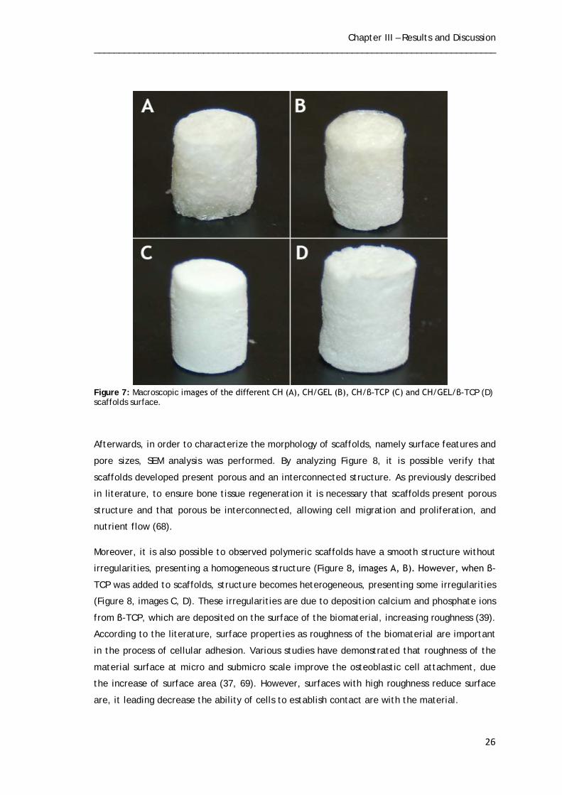
3. Results and Discussion
3.1. Morphology and Macroscopic Properties of Scaffolds
As previously described, scaffolds developed for bone tissue regeneration must present
adequate external and internal structures, as well as desirable physicochemical
characteristics that mimic the ECM of bone and also adequate mechanical properties suitable
to support the forces that bone is subject daily.
In this study, scaffolds was developed and optimized for being used in bone tissue
regeneration. To achieve this goal, scaffolds were produced with natural polymers, namely
CH and GEL, and ceramic, β-TCP.
CH was chosen due to their intrinsic properties, namely biocompatibility, the ability to
produce structures with appropriate porosity and able support osteoblastic cell attachment
and proliferation (46, 64). The GEL was used due their biocompatibility and in order to
improve properties of CH scaffolds (49, 50). Finally, ceramic β-TCP was chosen based on its
biocompatibility, biodegradability, osteoconductivity and the ability to increase the
mechanical properties of scaffolds (65, 66).
Macroscopic images were acquired in order to characterize morphologic properties of the
produced scaffolds. By analysis Figure 7, it is possible observe that despite using the same
manufacturing process for all scaffolds, the images obtained revealed some differences
between scaffolds. Observing the macroscopic images is possible conclude that CH scaffolds
(Figure 7, image A) are those which present a less well-defined structure than other produced
scaffolds. This is due to the fact that CH presents great instability and it is unable to maintain
a predefined shape, as a result of its high capacity swelling (48). However, when adding
mainly β-TCP the production of scaffolds was observed that these present more well-defined
structure than CH scaffolds (Figure 7, images C, D).
Moreover, it is possible observe that produced scaffolds present a porous structure, important
to ensure bone regeneration. However, CH and CH/GEL scaffolds (Figure 7, images A, B)
present a more porous structure than CH/β-TCP and CH/GEL/ β-TCP scaffolds (Figure 7,
images C e B). This fact is due presence of β-TCP that decrease the porosity of scaffolds,
because the presence of hydroxyl groups belonging to β-TCP and CH promote strong
interaction between composite components (47, 67).
Chapter III – Results and Discussion _________________________________________________________________________________
26
Figure 7: Macroscopic images of the different CH (A), CH/GEL (B), CH/β-TCP (C) and CH/GEL/β-TCP (D) scaffolds surface.
Afterwards, in order to characterize the morphology of scaffolds, namely surface features and
pore sizes, SEM analysis was performed. By analyzing Figure 8, it is possible verify that
scaffolds developed present porous and an interconnected structure. As previously described
in literature, to ensure bone tissue regeneration it is necessary that scaffolds present porous
structure and that porous be interconnected, allowing cell migration and proliferation, and
nutrient flow (68).
Moreover, it is also possible to observed polymeric scaffolds have a smooth structure without
irregularities, presenting a homogeneous structure (Figure 8, images A, B). However, when β-
TCP was added to scaffolds, structure becomes heterogeneous, presenting some irregularities
(Figure 8, images C, D). These irregularities are due to deposition calcium and phosphate ions
from β-TCP, which are deposited on the surface of the biomaterial, increasing roughness (39).
According to the literature, surface properties as roughness of the biomaterial are important
in the process of cellular adhesion. Various studies have demonstrated that roughness of the
material surface at micro and submicro scale improve the osteoblastic cell attachment, due
the increase of surface area (37, 69). However, surfaces with high roughness reduce surface
are, it leading decrease the ability of cells to establish contact are with the material.

Chapter III – Results and Discussion _________________________________________________________________________________
27
Figure 8: SEM images of the different scaffolds. Images show the surface characteristics of the CH (A), CH/GEL (B), CH/β-TCP (C) and CH/GEL/β-TCP (D) 3D scaffolds, respectively.
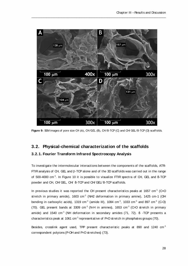
Mean pore size of the scaffolds was also determined by SEM analysis. CH and CH/GEL
presented pores with values of 128 µm and 157 µm (Figure 9, images A, B), respectively,
while CH/ β-TCP and CH/GEL/β-TCP presented values of 104 µm and 131 µm (Figure 9,
images C, D). Based on the results obtained the scaffolds containing β-TCP presented pores
with a smaller diameter. As previously reported, pore size of scaffolds is another important
condition for bone tissue regeneration. These should allow cell growth and migration and also
nutrient diffusion. If pores are too small, cells will cover the pores, influencing cell migration
and inhibiting neovascularization. On the other hand if these are too large reduce the surface
are and subsequently cell adhesion. Although the optimal pore size for bone tissue
engineering is not yet well-defined, recent studies have shown that pore with diameter values
between 75-100 µm promote bone growth with an optimal range of 100-135µm. The scaffolds
developed here present values pore size range of 104-157 µm (68). Thus, the results
presented here in show that the scaffolds developed have a suitable pore size desirable to
promote bone tissue regeneration.
Chapter III – Results and Discussion _________________________________________________________________________________
28
Figure 9: SEM images of pore size CH (A), CH/GEL (B), CH/β-TCP (C) and CH/GEL/β-TCP (D) scaffolds.
3.2. Physical-chemical characterization of the scaffolds
3.2.1. Fourier Transform Infrared Spectroscopy Analysis
To investigate the intermolecular interactions between the components of the scaffolds, ATR-
FTIR analysis of CH, GEL and β-TCP alone and of the 3D scaffolds was carried out in the range
of 500-4000 cm-1. In Figure 10 it is possible to visualize FTIR spectra of CH, GEL and β-TCP
powder and CH, CH/GEL, CH/ β-TCP and CH/GEL/β-TCP scaffolds.
In previous studies it was reported the CH present characteristics peaks at 1657 cm-1 (C=O
stretch in primary amide), 1603 cm-1 (NH2 deformation in primary amine), 1425 cm-1 (OH
bending in carboxylic acids), 1319 cm-1 (amide III), 1084 cm-1, 1033 cm-1 and 897 cm-1 (C-O)
(70). GEL present bands at 3309 cm-1 (N-H in amines), 1653 cm-1 (C=O stretch in primary
amide) and 1540 cm-1 (NH deformation in secondary amides (71, 72). β -TCP presents a
characteristics peak at 1061 cm-1 representative of P=O stretch in phosphates groups (70).
Besides, crosslink agent used, TPP present characteristic peaks at 890 and 1240 cm-1
correspondent polyions (P-OH and P=O stretches) (73).

Chapter III – Results and Discussion _________________________________________________________________________________
29
FTIR spectra of CH powder (Figure 10) show peak around 1591 cm-1 that correspond amino
groups. However, despite the peak also find in CH scaffolds, occurs a slight decrease. As
described in the literature this slight decrease is due interaction that occurs between amine
groups of CH and phosphate groups of TPP (73). In CH/GEL scaffolds is possible to visualize
that besides the peaks corresponding to CH, there are also present characteristics bands of
GEL. In addition, in the CH/ β –TCP and CH/GEL/β –TCP there is a new peak at around ,
characteristic of phosphate groups of β –TCP, being more intense in scaffolds with higher
amount β –TCP ( CH/ β –TCP scaffolds).
Figure 10: FTIR spectra for the CH, GEL and β-TCP powder and different types of scaffolds.

Chapter III – Results and Discussion _________________________________________________________________________________
30
3.2.2. X-Ray Diffraction analysis
XRD patterns of CH, GEL and β-TCP powder and scaffolds are presented in Figure 11. As
reported by Kim et al. CH presents a peak at 2Θ=19.6o (67). By analysing Figure 11 it is
possible to observe that a peak around 2Θ=20o (*) was assigned to CH powder and CH
scaffolds. Like CH the GEL spectra also shows a characteristic peak around at 2Θ=20º (**).
CH/Gel scaffolds present peaks characteristics of polymers as it is possible observe through
Figure 11. However, when β-TCP is added for the production of scaffolds, this peak becomes
wider and flatter. Furthermore, when this ceramic is introduced in the composition of
scaffolds appears a new peak around 2Θ=33º (***) of β-TCP, being these more intense in CH/
β-TCP scaffolds, since it is the group that has higher amount of β-TCP (58).

Chapter III – Results and Discussion _________________________________________________________________________________
31
Figure 11: X-Ray spectra of a) pure CH, GEL and β-TCP powders and b) groups I (CH), II (CH/GEL), III (CH/β-TCP) and IV (CH/GEL/ β-TCP) scaffolds (* characteristic peak of chitosan, ** characteristic peak o GEL and *** characteristic peak of β-TCP).

Chapter III – Results and Discussion _________________________________________________________________________________
32
3.2.4. Energy Dispersive Spectroscopy analysis
An Energy-Dispersive spectroscopy (EDS) analysis was also carried out with the purpose of
characterize chemically the samples. The EDS results show the presence characteristic
elements of the materials used for the production of scaffolds. From the analysis of the table
4 it turns out that all scaffolds present in their composition high amounts of carbon and
oxygen elements. Considering that CH presents a chemical structure based on these elements
and that it is present in their compositions of all scaffolds, the results obtained are in
agreement. Moreover, also calcium and phosphate elements are present, and there is an
increase in the CH/β-TCP scaffolds, followed by CH/GEL/β-TCP. These results are in
agreement with the presence of β-TCP in the composition of scaffolds. Finally, also sodium
elements are present in the scaffolds. Although this element is not part of the constitution
any of the materials used for the manufacture of scaffolds, its presence can be explained by
the crosslinking agent used (TPP).
Table 2: Elemental analysis of the manufacture scaffolds
3.3. Mechanical characterization of the 3D scaffolds
The mechanical properties, mainly resistance to compression of the material, are important
data for the development of scaffolds to be applied in bone tissue regeneration. To evaluate
the mechanical properties of scaffolds, compression strength was determined.
Through the analysis of Figure 12 it is possible to conclude that CH/β-TCP, CH/GEL and
CH/GEL/β-TCP present greater compression strength when compared to CH scaffolds. In
accordance to what is published in literature, the polymers used for the development
scaffolds such as CH, alginate, presents weak mechanical properties, that limit their
application in bone tissue engineering (74). Various studies have been performed for
improving the mechanical resistance of scaffolds. Ceramics as hydroxyapatite (HA) and β-TCP
Sample
C
O
Na
P
Ca
CH
46.89
43.39
3.92
3.30
0.12
CH/GEL
45.53
46.52
3.50
3.14
-
CH/β-TCP
43.72
43.84
2.45
8.15
9.56
CH/GEL/β-TCP
29.88
43.95
5.82
4.59
4.35

Chapter III – Results and Discussion _________________________________________________________________________________
33
have been studied for this purpose, because of its similarity to the inorganic phase of bone
tissue. Thus, as has been described by Zhang composites of CH and calcium phosphate
ceramics present better mechanical properties than CH scaffolds alone (75, 76). Besides, it is
not only the incorporation of ceramics that increases resistance of scaffolds. Several studies
have also shown that the CH blended with other polymers such as GEL, enhance the
mechanical properties (49, 77). Thus, both GEL and β-TCP have the ability enhance
compressive strength of CH scaffolds. However, CH/ β-TCP present better properties than
CH/GEL scaffolds. For all this, it was expected that CHI/GEL/ β-TCP scaffolds presented the
better compressive strength, however this did not happened. One possible explanation for
this is the fact that amount of β-TCP was not enough to increase the compression strength of
the scaffolds.
Figure 12: Characterization of the compressive strength of the scaffolds. Statistical analysis was performed using one-way ANOVA with Newman-Keuls tests (N.S.: not-significant, *p<0, 05).

Chapter III – Results and Discussion _________________________________________________________________________________
34
3.4. Swelling Studies
Swelling and structural stability of scaffolds are also important factors that have to be
characterized in the development of scaffolds for TE.
In order to evaluate the capacity of scaffolds produced herein have to absorb body fluids
when introduced into organism, swelling studies were performed.
The swelling data of scaffolds is shown in Figure 13. All types of scaffolds exhibit a high
capacity of swelling, which is indicative of their hydrophilic character. However, it was
verified that CH and CH/GEL scaffolds present a higher swelling capacity than that of CH/ β-
TCP and CH/GEL/ β-TCP scaffolds. According to the literature, CH and GEL confer the
scaffolds hydrophilic nature and therefore a high swelling capacity. This property is due to
the presence carboxyl, amine and hydroxyl groups in this structure (78, 79). However, the
addition of HA or β-TCP led to decrease the hydrophilic character and consequently a smaller
swelling capacity of scaffolds. This is due to calcium and phosphorous groups of β-TCP, that
are bounded to groups that confer hydrophilicity of these polymers (78).
Moreover, through the analysis of Figure 13 it can be concluded that scaffolds presents
maximum capacity swelling after 30 minutes, starting to stabilize immediately afterwards.
This fact is important for TE because the initial swelling is important for promoting cell
adhesion and growth, however swelling continuous leads to loss of mechanical integrity of
scaffolds.
Figure 13: Swelling profile of the scaffolds.

Chapter III – Results and Discussion _________________________________________________________________________________
35
3.5. Porosity
Scaffolds porosity was evaluated by using liquid displacement method, using ethanol. Figure
14 shows that all the scaffolds present porosity greater than 60%, a suitable value for bone
regeneration (59). Moreover, the results obtained showed that the CH/Gel scaffolds present
lower porosity than CH scaffolds. However, a statistically significant difference was observed
only when β-TCP was added scaffolds composition. Previous studies demonstrate that the
presence of HA particles decrease the porosity (67). Thus, the statistically significant
difference observed is due presence of β-TCP that decrease porosity in CH/Gel/β-TCP
scaffolds and the lower polymer concentration in CH scaffolds, leading to increase of
scaffolds porosity. As previously described, porosity of scaffolds is important to ensure bone
regeneration. The porosity must be sufficient for allow cell growth and nutrient flow during
bone regeneration, however, it also must have enough mechanical strength to support the
frame during tissue regeneration.
Figure 14: Total porosity of the different scaffolds. Statistical analysis was performed using one-way ANOVA with Newman-Keuls tests (N.S.: not-significant, *p<0, 05).
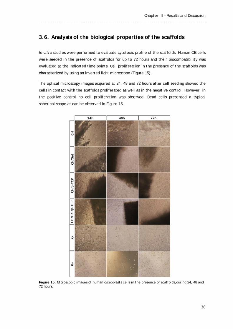
Chapter III – Results and Discussion _________________________________________________________________________________
36
3.6. Analysis of the biological properties of the scaffolds
In vitro studies were performed to evaluate cytotoxic profile of the scaffolds. Human OB cells
were seeded in the presence of scaffolds for up to 72 hours and their biocompatibility was
evaluated at the indicated time points. Cell proliferation in the presence of the scaffolds was
characterized by using an inverted light microscope (Figure 15).
The optical microscopy images acquired at 24, 48 and 72 hours after cell seeding showed the
cells in contact with the scaffolds proliferated as well as in the negative control. However, in
the positive control no cell proliferation was observed. Dead cells presented a typical
spherical shape as can be observed in Figure 15.
Figure 15: Microscopic images of human osteoblasts cells in the presence of scaffolds,during 24, 48 and 72 hours.
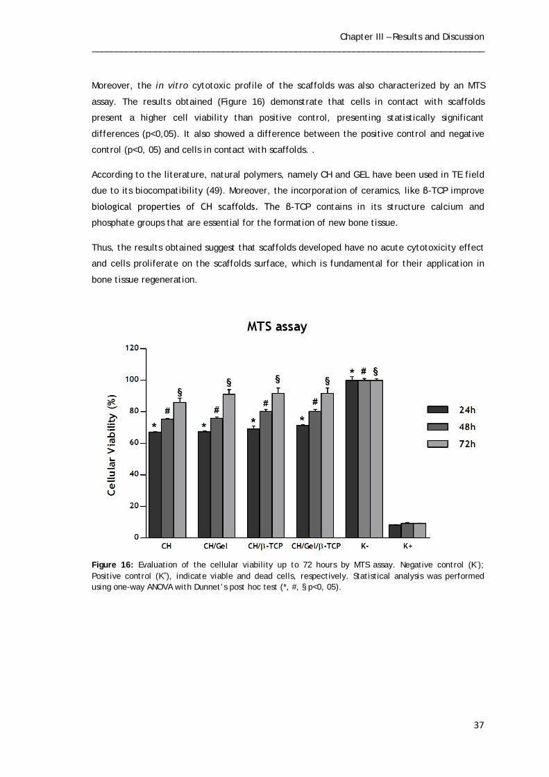
Chapter III – Results and Discussion _________________________________________________________________________________
37
Moreover, the in vitro cytotoxic profile of the scaffolds was also characterized by an MTS
assay. The results obtained (Figure 16) demonstrate that cells in contact with scaffolds
present a higher cell viability than positive control, presenting statistically significant
differences (p<0,05). It also showed a difference between the positive control and negative
control (p<0, 05) and cells in contact with scaffolds. .
According to the literature, natural polymers, namely CH and GEL have been used in TE field
due to its biocompatibility (49). Moreover, the incorporation of ceramics, like β-TCP improve
biological properties of CH scaffolds. The β-TCP contains in its structure calcium and
phosphate groups that are essential for the formation of new bone tissue.
Thus, the results obtained suggest that scaffolds developed have no acute cytotoxicity effect
and cells proliferate on the scaffolds surface, which is fundamental for their application in
bone tissue regeneration.
Figure 16: Evaluation of the cellular viability up to 72 hours by MTS assay. Negative control (K-); Positive control (K+), indicate viable and dead cells, respectively. Statistical analysis was performed using one-way ANOVA with Dunnet’s post hoc test (*, #, § p<0, 05).
Chapter III – Results and Discussion _________________________________________________________________________________
38
To confirm that human OB cells adhered to the surface of the materials a SEM analysis was
also carried out. Figure 17 shows the SEM images of the cells in contact with different
scaffolds produced after 24 and 72 hours. Through the analysis of the images obtained it is
possible observe that in all types of scaffolds developed, the cells adhered and spread across
scaffolds surface. After 72 hours the cells besides being adhered to the material, start to
show the characteristic morphology of osteoblasts and acquired an extended configuration
(filopodia).
Thus, is possible conclude that all types of scaffolds allow cell adhesion, namely human OB,
essentially for promote bone regeneration.

Chapter III – Results and Discussion _________________________________________________________________________________
39
Figure 17: SEM micrographs of osteoblast morphology in the presence of the different scaffolds. SEM images were used to visualize osteoblast on the surface of the CH (A, B), CH/GEL (C,D), CH/β-TCP (E,F) and CH/GEL/β-TCP (G,H) 3D scaffolds, after being cultured for 24 hours (A,C,E and G) and 72 hours (B,D,F and H).
Chapter IV
Conclusion
Chapter IV - Conclusion _________________________________________________________________________________
41
4. Conclusion
In last years, millions of people worldwide have been suffering from bone defects due to
trauma, tumors and bony diseases that, in some cases, may result in death. Several
therapeutic approaches have been investigated in order to improve, restore or replace
damaged tissue. Among the different bone substitutes developed so far, scaffolds are the
most promising. These bone substitutes possess the essential properties that are required for
bone regeneration and mimic the structure of ECM that provide a favorable microenvironment
for cell adhesion and proliferation. In the present study different scaffolds were produced using a freeze-dry technique in order
to be used in a near future in bone tissue engineering. Four types of scaffolds were
successfully produced herein: CH; CH/GEL; CH/β-TCP and CH/GEL/ β-TCP, in order to study
the influence of the incorporation of gelatin and β-TCP in structure of CH scaffolds.
In this study different assays were performed in order to characterize scaffolds structure
through SEM, FTIR, XRD and EDS. SEM analysis of scaffolds showed a porous and
interconnected structure, allowing cell migration and proliferation, as well as nutrient flow.
Besides, SEM images showed that the incorporation of β-TCP in scaffolds enhance their
roughness, that is important for cell adhesion. The results of FTIR, XRD and EDS prove that
the blend between of materials was achieved.
Moreover, the mechanical resistance of scaffolds was also evaluated. The results showed that
incorporation of GEL and β-TCP in CH scaffolds increased significantly the mechanical
resistance of the scaffolds. However, the results obtained were lower than compression
strength of trabecular and cortical bone.
Porosity and swelling behaviour of scaffolds were also evaluated. The results demonstrated
that all types of scaffolds produced have porosity higher than 60%, a suitable value for bone
regeneration. However, the incorporation of β-TCP promoted a decrease in scaffolds porosity
which is agreement with the results obtained in mechanical analysis. Regarding the swelling
capacity, all types of scaffolds presented a similar swelling behaviour. However, the
incorporation of β-TCP impaired the swelling capacity of scaffolds.
Furthemore, SEM analysis of scaffolds showed that human osteoblasts adhered to material
surface. Moreover, cell viability was also assessed by MTS assay and the results confirmed
that human osteoblasts remain viable in contact with scaffolds. Based in the in vitro results
the developed scaffolds promote cell adhesion and proliferation and do not exhibit cytotoxic
effect for the cells, which is fundamental for their application in bone tissue regeneration.

Chapter IV - Conclusion _________________________________________________________________________________
42
In a near future, the mechanical properties will be improved once the values obtained are far
of “ideal” values. To achieve this purpose several strategies must be applied. Firstly,
increasing the concentration of CH solution and the viscosity of the final solution,
subsequently, would increase the mechanical resistance of the scaffolds. Besides, the ratios
between CH, Gel and β-TCP must be optimized. Another strategy would be replacing B-TCP by
other ceramic more resistance, such as hydroxyapatite.

Chapter V
Bibliography

Chapter V - Bibliography _________________________________________________________________________________
44
5. Bibliography
1. Van De Graaff KM. Human Anatomy. 6th ed: The McGraw-Hill Companies, Inc.; 2002. 2. Seeley RR, Stephens TD, Tate P. Anatomia & Fisiologia. 8th ed: Lusociência-Edições Técnicas e Científicas, Lda.; 2008. 3. Salgado AJ, Coutinho OP, Reis RL. Bone tissue engineering: state of the art and future trends. Macromolecular bioscience. 2004;4(8):743-65. 4. Marieb EN, Hoehn K. Human Anatomy & Physiology. 7th ed: Pearson Education, Inc.; 2007. 5. Murugan R, Ramakrishna S. Development of nanocomposites for bone grafting. Composites Science and Technology. 2005;65(15):2385-406. 6. Ralston SH. Structure and metabolism of bone. Medicine. 2005;33(12):58-60. 7. Mackie E. Osteoblasts: novel roles in orchestration of skeletal architecture. The international journal of biochemistry & cell biology. 2003;35(9):1301-5. 8. Jayakumar P, Di Silvio L. Osteoblasts in bone tissue engineering. Proceedings of the Institution of Mechanical Engineers, Part H: Journal of Engineering in Medicine. 2010;224(12):1415-40. 9. Torres A, Santos S, Oliveira M, Barbosa M. Fibrinogen promotes resorption of chitosan by human osteoclasts. Acta biomaterialia. 2013; 9 (5):6553-62. 10. Schindeler A, McDonald MM, Bokko P, Little DG, editors. Bone remodeling during fracture repair: the cellular picture. Seminars in cell & developmental biology; 2008; 19(5): 459-466. 11. Jang J-H, Castano O, Kim H-W. Electrospun materials as potential platforms for bone tissue engineering. Advanced Drug Delivery Reviews. 2009;61(12):1065-83. 12. Feng X, McDonald JM. Disorders of bone remodeling. Annual review of pathology. 2011;6:121. 13. Raggatt LJ, Partridge NC. Cellular and molecular mechanisms of bone remodeling. Journal of Biological Chemistry. 2010;285(33):25103-8. 14. Sims NA, Gooi JH, editors. Bone remodeling: Multiple cellular interactions required for coupling of bone formation and resorption. Seminars in cell & developmental biology; 2008; 19(5): 444-451. 15. Datta H, Ng W, Walker J, Tuck S, Varanasi S. The cell biology of bone metabolism. Journal of clinical pathology. 2008;61(5):577-87. 16. Boyce BF, Xing L. Functions of RANKL/RANK/OPG in bone modeling and remodeling. Archives of biochemistry and biophysics. 2008;473(2):139-46.

Chapter V - Bibliography _________________________________________________________________________________
45
17. Väänänen HK, Laitala-Leinonen T. Osteoclast lineage and function. Archives of biochemistry and biophysics. 2008;473(2):132-8. 18. Robling AG, Castillo AB, Turner CH. Biomechanical and molecular regulation of bone remodeling. Annu Rev Biomed Eng. 2006;8:455-98. 19. Weitzmann MN, Pacifici R. Estrogen deficiency and bone loss: an inflammatory tale. Journal of Clinical Investigation. 2006;116(5):1186-94. 20. Djouad F, Guerit D, Marie M, Toupet K, Jorgensen C, Noel D. Mesenchymal Stem Cells: New Insights into Bone Regenerative Applications. Journal of Biomaterials and Tissue Engineering. 2012;2(1):14-28. 21. Peel N. Bone remodelling and disorders of bone metabolism. Surgery (Oxford). 2009;27(2):70-4. 22. Mazziotti G, Canalis E, Giustina A. Drug-induced osteoporosis: mechanisms and clinical implications. The American journal of medicine. 2010;123(10):877-84. 23. Ngiam M, Nguyen L, Liao S, Chan CK, Ramakrishna S. Biomimetic nanostructured materials: Potential regulators for osteogenesis. Ann Acad Med Singapore. 2011;40(5):213-22. 24. Murugan R, Ramakrishna S. Bioresorbable composite bone paste using polysaccharide based nano hydroxyapatite. Biomaterials. 2004;25(17):3829-35. 25. Chiara G, Letizia F, Lorenzo F, Edoardo S, Diego S, Stefano S, et al. Nanostructured biomaterials for tissue engineered bone tissue reconstruction. International journal of molecular sciences. 2012;13(1):737-57. 26. Van der Stok J, Van Lieshout EM, El-Massoudi Y, Van Kralingen GH, Patka P. Bone substitutes in the Netherlands–A systematic literature review. Acta biomaterialia. 2011;7(2):739-50. 27. Giannoudis PV, Dinopoulos H, Tsiridis E. Bone substitutes: an update. Injury. 2005;36(3):S20-S7. 28. Kneser U, Schaefer D, Polykandriotis E, Horch R. Tissue engineering of bone: the reconstructive surgeon's point of view. Journal of cellular and molecular medicine. 2006;10(1):7-19. 29. Robert L, Vacanti JP. Tissue engineering. Science. 1993;260(2):920-41. 30. Karageorgiou V, Kaplan D. Porosity of 3D biomaterial scaffolds and osteogenesis. Biomaterials. 2005 Sep;26(27):5474-91. 31. Ma PX. Biomimetic materials for tissue engineering. Advanced drug delivery reviews. 2008;60(2):184-98. 32. Davies J. Mechanisms of endosseous integration. The International journal of prosthodontics. 1997;11(5):391-401.

Chapter V - Bibliography _________________________________________________________________________________
46
33. Anselme K. Osteoblast adhesion on biomaterials. Biomaterials. 2000;21(7):667-81. 34. Choi CK, Breckenridge MT, Chen CS. Engineered materials and the cellular microenvironment: a strengthening interface between cell biology and bioengineering. Trends in cell biology. 2010;20(12):705-14. 35. Lee JW, Kim YH, Park KD, Jee KS, Shin JW, Hahn SB. Importance of integrin< i> β</i> 1-mediated cell adhesion on biodegradable polymers under serum depletion in mesenchymal stem cells and chondrocytes. Biomaterials. 2004;25(10):1901-9. 36. Anselme K, Bigerelle M. Topography effects of pure titanium substrates on human osteoblast long-term adhesion. Acta Biomaterialia. 2005;1(2):211-22. 37. Gittens RA, McLachlan T, Olivares-Navarrete R, Cai Y, Berner S, Tannenbaum R, et al. The effects of combined micron-/submicron-scale surface roughness and nanoscale features on cell proliferation and differentiation. Biomaterials. 2011;32(13):3395-403. 38. Zhu X, Chen J, Scheideler L, Reichl R, Geis-Gerstorfer J. Effects of topography and composition of titanium surface oxides on osteoblast responses. Biomaterials. 2004;25(18):4087-103. 39. Zhu X, Kim K-H, Jeong Y. Anodic oxide films containing Ca and P of titanium biomaterial. Biomaterials. 2001;22(16):2199-206. 40. Swetha M, Sahithi K, Moorthi A, Srinivasan N, Ramasamy K, Selvamurugan N. Biocomposites containing natural polymers and hydroxyapatite for bone tissue engineering. International journal of biological macromolecules. 2010;47(1):1-4. 41. Di Martino A, Sittinger M, Risbud MV. Chitosan: a versatile biopolymer for orthopaedic tissue-engineering. Biomaterials. 2005;26(30):5983-90. 42. Shi C, Zhu Y, Ran X, Wang M, Su Y, Cheng T. Therapeutic potential of chitosan and its derivatives in regenerative medicine. Journal of Surgical research. 2006;133(2):185-92. 43. Thein-Han W, Kitiyanant Y, Misra R. Chitosan as scaffold matrix for tissue engineering. Materials Science and Technology. 2008;24(9):1062-75. 44. Jiang T, Deng M, James R, Nair LS, Laurencin CT. Micro-and nanofabrication of chitosan structures for regenerative engineering. Acta biomaterialia. 2013; 45. Li Y, Wang Y, Wu D, Zhang K, Hu Q. A facile approach to construct three-dimensional oriented chitosan scaffolds with in-situ precipitation method. Carbohydrate polymers. 2010;80(2):408-12. 46. Amaral I, Sampaio P, Barbosa M. Three-dimensional culture of human osteoblastic cells in chitosan sponges: The effect of the degree of acetylation. Journal of Biomedical Materials Research Part A. 2006;76(2):335-46. 47. Tanase C, Popa M, Verestiuc L. Biomimetic bone scaffolds based on chitosan and calcium phosphates. Materials Letters. 2011;65(11):1681-3.
Chapter V - Bibliography _________________________________________________________________________________
47
48. Li Z, Ramay HR, Hauch KD, Xiao D, Zhang M. Chitosan–alginate hybrid scaffolds for bone tissue engineering. Biomaterials. 2005;26(18):3919-28. 49. Huang Y, Onyeri S, Siewe M, Moshfeghian A, Madihally SV. In vitro characterization of chitosan–gelatin scaffolds for tissue engineering. Biomaterials. 2005;26(36):7616-27. 50. Malafaya PB, Silva GA, Reis RL. Natural–origin polymers as carriers and scaffolds for biomolecules and cell delivery in tissue engineering applications. Advanced drug delivery reviews. 2007;59(4):207-33. 51. Lien S-M, Ko L-Y, Huang T-J. Effect of pore size on ECM secretion and cell growth in gelatin scaffold for articular cartilage tissue engineering. Acta biomaterialia. 2009;5(2):670-9. 52. Zhang D, Yang H. Gelatin-stabilized copper nanoparticles: Synthesis, morphology, and their surface-enhanced Raman scattering properties. Physica B: Condensed Matter. 2013; 415: 44-48. 53. Cao H, Kuboyama N. A biodegradable porous composite scaffold of PGA/β-TCP for bone tissue engineering. Bone. 2010;46(2):386-95. 54. Mikos AG, Thorsen AJ, Czerwonka LA, Bao Y, Langer R, Winslow DN, et al. Preparation and characterization of poly (L-lactic acid) foams. Polymer. 1994;35(5):1068-77. 55. Leong K, Cheah C, Chua C. Solid freeform fabrication of three-dimensional scaffolds for engineering replacement tissues and organs. Biomaterials. 2003;24(13):2363-78. 56. Fukasawa T, Deng Z-Y, Ando M, Ohji T, Goto Y. Pore structure of porous ceramics synthesized from water-based slurry by freeze-dry process. Journal of Materials Science. 2001;36(10):2523-7. 57. Lima AC, Batista P, Valente TA, Silva AS, Correia IJ, Mano JF. Novel Methodology Based on Biomimetic Superhydrophobic Substrates to Immobilize Cells and Proteins in Hydrogel Spheres for Applications in Bone Regeneration. Tissue Engineering Part A. 2013;19(9-10):1175-87. 58. Santos CFL, Silva AP, Lopes L, Pires I, Correia IJ. Design and production of sintered β-tricalcium phosphate 3D scaffolds for bone tissue regeneration. Materials Science and Engineering: C. 2012;32(5):1293-8. 59. Torres A, Gaspar V, Serra I, Diogo G, Fradique R, Silva A, et al. Bioactive Polymeric-Ceramic Hybrid 3D Scaffold for Application in Bone Tissue Regeneration. Materials Science and Engineering: C. 2013; 33(7): 4460-69. 60. Wu X, Liu Y, Li X, Wen P, Zhang Y, Long Y, et al. Preparation of aligned porous gelatin scaffolds by unidirectional freeze-drying method. Acta Biomaterialia. 2010 ;6(3):1167-77. 61. Coimbra P, Alves P, Valente T, Santos R, Correia I, Ferreira P. Sodium hyaluronate/chitosan polyelectrolyte complex scaffolds for dental pulp regeneration: synthesis and characterization. International journal of biological macromolecules. 2011;49(4):573-9.

Chapter V - Bibliography _________________________________________________________________________________
48
62. Nie HL, Zhu LM. Adsorption of papain with Cibacron Blue F3GA carrying chitosan-coated nylon affinity membranes. International journal of biological macromolecules. 2007;40(3):261-7. 63. Correia TR, Antunes BP, Castilho PH, Nunes JC, Pessoa de Amorim MT, Escobar IC, et al. A bi-layer electrospun nanofiber membrane for plasmid DNA recovery from fermentation broths. Separation and Purification Technology. 2013; 112: 20-25. 64. Dash M, Chiellini F, Ottenbrite R, Chiellini E. Chitosan—A versatile semi-synthetic polymer in biomedical applications. Progress in Polymer Science. 2011;36(8):981-1014. 65. Yang Y, Bumgardner J, Cavin R, Carnes D, Ong J. Osteoblast precursor cell attachment on heat-treated calcium phosphate coatings. Journal of dental research. 2003;82(6):449-53. 66. Suzuki T, Yamamoto T, Toriyama M, Nishizawa K, Yokogawa Y, Mucalo MR, et al. Surface instability of calcium phosphate ceramics in tissue culture medium and the effect on adhesion and growth of anchorage-dependent animal cells. Journal of biomedical materials research. 1997;34(4):507-17. 67. Kim HS, Kim JT, Jung YJ, Ryu SC, Son HJ, Kim YG. Preparation of a porous chitosan/fibroin-hydroxyapatite composite matrix for tissue engineering. Macromolecular Research. 2007;15(1):65-73. 68. Murphy CM, Haugh MG, O'Brien FJ. The effect of mean pore size on cell attachment, proliferation and migration in collagen–glycosaminoglycan scaffolds for bone tissue engineering. Biomaterials. 2010;31(3):461-6. 69. Raines AL, Olivares-Navarrete R, Wieland M, Cochran DL, Schwartz Z, Boyan BD. Regulation of angiogenesis during osseointegration by titanium surface microstructure and energy. Biomaterials. 2010;31(18):4909-17. 70. Manjubala I, Scheler S, Bössert J, Jandt KD. Mineralisation of chitosan scaffolds with nano-apatite formation by double diffusion technique. Acta Biomaterialia. 2006;2(1):75-84. 71. Mi F-L. Synthesis and characterization of a novel chitosan-gelatin bioconjugate with fluorescence emission. Biomacromolecules. 2005;6(2):975-87. 72. Kim S, Nimni ME, Yang Z, Han B. Chitosan/gelatin–based films crosslinked by proanthocyanidin. Journal of Biomedical Materials Research Part B: Applied Biomaterials. 2005;75(2):442-50. 73. Yu S-H, Wu S-J, Wu J-Y, Peng C-K, Mi F-L. Tripolyphosphate Cross-Linked Macromolecular Composites for the Growth of Shape-and Size-Controlled Apatites. Molecules. 2012;18(1):27-40. 74. Cheung H-Y, Lau K-T, Lu T-P, Hui D. A critical review on polymer-based bio-engineered materials for scaffold development. Composites Part B: Engineering. 2007;38(3):291-300. 75. Zhang Y, Zhang M. Calcium phosphate/chitosan composite scaffolds for controlled in vitro antibiotic drug release. Journal of biomedical materials research. 2002;62(3):378-86.
Chapter V - Bibliography _________________________________________________________________________________
49
76. Zhang Y, Ni M, Zhang M, Ratner B. Calcium phosphate-chitosan composite scaffolds for bone tissue engineering. Tissue engineering. 2003;9(2):337-45. 77. Yang C, Xu L, Zhou Y, Zhang X, Huang X, Wang M, et al. A green fabrication approach of gelatin/CM-chitosan hybrid hydrogel for wound healing. Carbohydrate Polymers. 2010;82(4):1297-305. 78. Peter M, Ganesh N, Selvamurugan N, Nair S, Furuike T, Tamura H, et al. Preparation and characterization of chitosan–gelatin/nanohydroxyapatite composite scaffolds for tissue engineering applications. Carbohydrate Polymers. 2010;80(3):687-94. 79. Thein-Han W, Misra R. Biomimetic chitosan–nanohydroxyapatite composite scaffolds for bone tissue engineering. Acta Biomaterialia. 2009;5(4):1182-97.

Chapter VI
Appendix

Materials Science and Engineering C 33 (2013) 4460–4469
Contents lists available at ScienceDirect
Materials Science and Engineering C
j ourna l homepage: www.e lsev ie r .com/ locate /msec
Bioactive polymeric–ceramic hybrid 3D scaffold for application in bonetissue regeneration
A.L. Torres a, V.M. Gaspar a, I.R. Serra a, G.S. Diogo a, R. Fradique a, A.P. Silva b, I.J. Correia a,⁎a CICS-UBI — Health Sciences Research Centre, University of Beira Interior, Av. Infante D. Henrique, 6200-506 Covilhã, Portugalb CAST-UBI — Centre for Aerospace Science and Technologies, University of Beira Interior, Calçada Fonte do Lameiro, 6201-001 Covilhã, Portugal
⁎ Corresponding author. Tel.: +351 275 329 055.E-mail address: [email protected] (I.J. Correia).
0928-4931/$ – see front matter © 2013 Elsevier B.V. Allhttp://dx.doi.org/10.1016/j.msec.2013.07.003
a b s t r a c t
a r t i c l e i n f oArticle history:Received 9 May 2013Received in revised form 29 June 2013Accepted 5 July 2013Available online 13 July 2013
Keywords:3D scaffoldsBioceramicsBone regenerationFoam replication methodVacuum coating
The regeneration of large bone defects remains a challenging scenario from a therapeutic point of view. Infact, the currently available bone substitutes are often limited by poor tissue integration and severe host in-flammatory responses, which eventually lead to surgical removal. In an attempt to address these issues, here-in we evaluated the importance of alginate incorporation in the production of improved and tunableβ-tricalcium phosphate (β-TCP) and hydroxyapatite (HA) three-dimensional (3D) porous scaffolds to beused as temporary templates for bone regeneration. Different bioceramic combinations were tested inorder to investigate optimal scaffold architectures. Additionally, 3D β-TCP/HA vacuum-coated with alginate,presented improved compressive strength, fracture toughness and Young's modulus, to values similar tothose of native bone. The hybrid 3D polymeric–bioceramic scaffolds also supported osteoblast adhesion, mat-uration and proliferation, as demonstrated by fluorescence microscopy. To the best of our knowledge this isthe first time that a 3D scaffold produced with this combination of biomaterials is described. Altogether, ourresults emphasize that this hybrid scaffold presents promising characteristics for its future application inbone regeneration.
© 2013 Elsevier B.V. All rights reserved.
1. Introduction
Bone is a highly vascularized and dynamic tissue, having extraor-dinary mechanical properties and intrinsic regenerative capacity [1].This exceptional characteristic is however rather limited in severebone traumatisms that involve multiple fractures, bone-associatedtumors and degenerative diseases [2]. These are highly debilitatingconditions that commonly require medical intervention to restorebone native properties [3].
Currently, several types of biomaterials, such as biofunctionalprosthesis [4], injectable substrates [5] and hydrogels [6] are beingproduced for bone regeneration purposes. Among them, bioactive3D porous scaffolds are particularly promising for clinical applicationdue to their unique set of characteristics. Scaffolds are porous 3D ma-trices that act as temporary templates for cell adhesion and prolifera-tion, while providing mechanical support until new bone tissue isformed at the affected area [7]. For this purpose these scaffoldsmust be produced with materials that promote proper regenerationwithout eliciting host immune responses or originating toxic metab-olites [8]. The 3D features of the template is another critical parame-ter when therapeutic applications are envisioned, since its spatialarchitecture should be designed to have interconnected pores that si-multaneously induce osteoconductivity of bone-progenitor cells and
rights reserved.
neovascularization [9]. To further improve and accelerate bone regen-eration it is essential that the scaffold is bioactive, i.e., has the abilityto form anchoring points with the surrounding bone and soft tissues,stimulating bone growth. Commonly these bonds are formed throughan HA-like layer and are responsible for increasing osteointegrationand cell growth and differentiation [10].
The desire to gather these complex characteristics into one scaf-fold is a challenging demand, not only in the design and manufactur-ing stages, but also in the translation to in vivo applications. In thiscontext, the combination of ceramics with polymers for the synthesisof hybrid scaffolds has been widely investigated in an attempt tomimic bone tissue native structure [11,12]. β-TCP and HA are themost commonly used ceramics since their mineral compositions aresimilar to those found in human bone [11,13]. Although β-TCP hasbeen extensively used for bone regeneration due to its biodegradability,biocompatibility and osteoconductivity [14,15] its applications to bonetissue regeneration are limited by its poor mechanical properties [16,17].
In different studies bioceramic-based scaffolds have been modifiedwith polymers (as coating or interpenetratingphases) in order to reducetheir brittleness and improve mechanical properties [18–21]. In thepresent study, β-TCP/HA scaffolds were left uncoated or coated withalginate. This polymer is comprised by 1,4′-linked β-D-mannuronicacid and α-L-guluronic acid blocks, which determines alginate physico-chemical properties. This natural biomaterial has valuable characteris-tics, such as biocompatibility, biodegradability, hydrophilicity, lowtoxicity and gelling capacity with divalent cations [22–25]. In fact, its

4461A.L. Torres et al. / Materials Science and Engineering C 33 (2013) 4460–4469
responsiveness to calcium (Ca2+)-rich tissue microenvironments, ren-ders it suitable for scaffold coating in bone tissue engineering, as previ-ously reported by our group [26]. Importantly, the ionically cross-linked alginate mechanical strength rises with increasing divalent ionconcentration, and also when these ions have high affinity for alginate[27]. Due to these valuable characteristics, this polymer has beenwidelyused in biomedical applications, including cell encapsulation [28] anddrug loading and delivery [29].
In this context, thiswork reports the productionofβ-TCP/HA scaffoldsby the foam replicationmethod (FRM), followed by their coatingwith al-ginate. The scaffold production method used is simple, cost-effective, ex-cludes the use of organic solvents and, most importantly, allows themanufacture of reproducible structures. Three types of scaffolds with dif-ferent β-TCP:HA ratios and alginate coatings were developed and theirphysicochemical and biological properties investigated.
2. Materials and methods
2.1. Preparation of β-TCP/HA composite scaffolds followed by alginatecoating
β-TCP/HA scaffolds were produced using a polymer-based FRM,which allows the control of the dimensions, pore size and density ofthe 3D scaffold. Three different types of scaffolds were prepared,concerning the quantity of β-TCP (Panreac®) relatively to HAnanopowder (b200 nm particle size, Sigma-Aldrich): 80/20% (w/w),90/10% (w/w) and 99/1% (w/w), respectively. Poly(vinyl) alcohol(PVA) (Sigma-Aldrich) (β-TCP binder agent) was added to the poly-mer mixture in a ratio of 1:10 (% w/w) PVA:β-TCP. Briefly, to preparethe polymer blends, PVA was dissolved in 15 mL of deionized water,under constant stirring, for 30 min, at 50 °C. Then, both β-TCP andHA powders were added in small amounts to the solution, under con-stant stirring at room temperature (RT). After all the powder waswetted, the mixture was sonicated for 15 min. The used polyurethane(PU) foams had similar structures to that of the human cancellousbone and were cut (5 × 5 × 5 mm3) to be used as a sacrificial tem-plate for the FRM. All the PU foams were cleaned in a 0.1 M sodiumhydroxide (NaOH) (Sigma-Aldrich) solution, followed by rinsingtwice with distilled water before air-dried for 12 h. The PU foamswere then repeatedly immersed in the polymer blends in order topromote polymer penetration into the PU foam pores. The PU foamsimpregnated with the polymer blends were then gently squeezed toremove the excess of the slurry. Homogeneous coating of the PUfoams with β-TCP/HA was possible after several immersions. Thecoated PU foams were exposed to air, allowed to dry overnight, atRT, and then sintered. Briefly, the PU-polymer foams were step-wiseheated in a furnace at 1 °C/min to 900 °C and then kept at this tem-perature for 240 min. Additionally, some of the 3D scaffolds weresubsequently coated with a 2% sodium alginate (Sigma-Aldrich) solu-tion, (molecular weight (Mw) 120.000–190.000 Da), under vacuumat RT, for 30 min, in order to guarantee alginate inclusion within thescaffold pores. These coated scaffolds were then immersed in a 5% cal-cium chloride (CaCl2) solution (Sigma-Aldrich), under vacuum for10 min, in order to cross-link alginate-coated scaffolds. The structureswere maintained in the CaCl2 solution for 24 h and then air dried,prior to use. From this point onwards alginate coated β-TCP/HA scaf-folds are identified as: 80/20/A, 90/10/A and 99/1/A and the uncoatedscaffolds as 80/20, 90/10 and 99/1.
2.2. Chemical, mechanical and morphological characterization of theβ-TCP/HA composite scaffolds
2.2.1. Fourier transform infrared spectroscopyThe physicochemical characteristics of the manufactured 3D scaf-
folds were evaluated by Fourier transform infrared spectroscopy(FTIR) by using a Nicolet iS10 interferometer (Thermo Scientific,
Waltham, MA, USA). Briefly, samples were mounted on a diamondwindow and compressed to improve spectrum signal to noise ratio.For each sample, 128 interferograms were acquired with a spectralwidth ranging from 4000 to 400 cm−1 and a spectral resolution of4 cm−1. The acquired data was then processed in Omnic Spectraanalysis software, where baseline subtraction was performed.
2.2.2. Energy dispersive spectroscopyEnergy-dispersive X-ray spectroscopy (EDS) (Rontec) was used
for the elementary characterization of both, coated and uncoated,β-TCP/HA scaffolds. Prior to all analyses, samples were placed on analuminum stub support, air-dried at RT and sputter-coated with gold.
2.2.3. Resistance to compression, fracture toughness and Young's modulusTo characterize the mechanical behavior of the cuboid-shaped coat-
ed and uncoated scaffolds, uniaxial compression tests were performed.All themeasurementswere performed at RT using a Zwick®1435Mate-rial Prüfung (Ulm, Germany)with a crosshead speed of 0.2 mm/min anda load cell of 5 kN. Four specimens from each sample were tested andtheir dimensions acquired. Afterwards, compressive strength (Cs) ofeach type of scaffold was calculated by applying Eq. (1) [30].
Cs ¼ Fa� l
ð1Þ
where F is the load at the time of the fracture and a and l represent thewidth and length of the scaffold, respectively. The fracture toughness(FT) and Young's modulus (YM) were estimated from the stress–strainrelations calculated and applying Eqs. (2) and (3), respectively [31,32].
FT ¼ Hd� Cs2
ð2Þ
YM ¼ CsHd
ð3Þ
where Hd is the scaffold height deformation and Cs is the scaffold com-pressive strength. Average values and standarddeviations (s.d.)were de-termined for each sample as previously described in the literature [33].
2.2.4. Scanning electron microscopyIn order to assess scaffoldmorphology, porosity and cellular behavior
in the presence of the samples, scanning electron microscopy (SEM)analysis was performed. Samples were washed at RT with phosphatebuffer solution (PBS, pH 7.4) (Sigma-Aldrich) and fixed for 30 minwith 2.5% (v/v) glutaraldehyde (Sigma-Aldrich) diluted in a 0.1 M sodi-um cacodylate solution (Sigma-Aldrich). Then, samples were washedthree times with cacodylate buffer and finally incubated for 10 min ina graded series of ethanol solutions (50, 60, 70, 80, 90 and 99% v/v),for dehydration. Scaffolds were then stored in absolute ethanol, at4 °C, until being subjected to CO2 critical point drying, mounted ontoaluminum stubs with araldite glue and sputtered-coated with goldusing an Emitech K550 sputter coater (London, UK). SEM images wereobtained with a scanning electron microscope Hitachi S-2700 (Tokyo,Japan) with an acceleration voltage of 20 kV at suitable magnifications.
2.2.5. Porosity evaluationThe total porosity (P) of the different types of β-TCP/HA scaffolds,
was determined by following a method described elsewhere [34]. Theamount of absolute ethanol that the scaffolds were able to absorb in24 h, was determined by applying Eq. (4).
P %ð Þ ¼ W2−W1
dethanol � V scaffold� 100 ð4Þ
whereW1 andW2 are the weights of the dry and the wet scaffolds, re-spectively, dethanol is the density of the ethanol at RT and Vscaffold is the

4462 A.L. Torres et al. / Materials Science and Engineering C 33 (2013) 4460–4469
volume of the wet scaffold, directly determined by immersion. Foreach scaffold, three replicates were analyzed and data representsthe average of each replicate.
2.2.6. Contact angle measurementsThe contact angle measurements of the samples were performed
using the sessile drop technique and water was used as reference fluid[35]. Contact angle datawas acquired in a Data Physics Contact Angle Sys-temOCAH200 apparatus, operating in staticmode at RT. For each sample,water drops were placed at various locations of the analyzed surface.
2.3. Biological characterization of the β-TCP/HA composite scaffolds
2.3.1. Culture of human osteoblasts in the presence of the scaffoldsHuman osteoblasts (CRL-11372), purchased from American
Type Culture Collection, were cultured in Dulbecco Modified EagleMedium, Nutrient Mixture F-12 (DMEM-F12) (Sigma-Aldrich), sup-plemented with 10% heat inactivated fetal bovine serum (FBS)(Biochrom AG), streptomycin (Sigma-Aldrich) (100 μg/mL) and gen-tamicin (Sigma-Aldrich) (100 μg/mL), in 75 cm2 T-flasks (OrangeScientific). Cultures were maintained at 37 °C in a 5% CO2 humidifiedatmosphere and the culture medium exchanged twice a week. Con-fluent cell monolayers were subcultured by initially washing themwith PBS, and then detached with 0.18% trypsin (1:250) and 5 mMEDTA (Sigma-Aldrich). A Neubauer chamber was used to determinecell number using trypan blue (Sigma-Aldrich) — exclusion assay,in order to distinguish live from dead cells. Subsequently, cellswere seeded in contact with 80/20, 90/10, 99/1, 80/20/A, 90/10/Aand 99/1/A scaffolds at a density of 2.0 × 10³ cells/well in cell culturetreated polystyrene plates. Cell growth was monitored during 1, 7and 14 days, by using an Olympus CX41 inverted light microscope(Tokyo, Japan) equipped with an Olympus SP-500 UZ digital camera.
2.3.2. Cellular metabolic activity: resazurin assayOsteoblast cells were cultured on scaffold surfaces for 1, 7 and
14 days. A non-toxic dye, specifically, resazurin (Sigma-Aldrich) was
Fig. 1.Macrographs (A, B and C) and SEM images (D, E and F) of the different alginate-coatedthe 80/20/A (A, D), 90/10/A (B, E) and 99/1/A (C, F) 3D scaffolds, respectively.
used to assess cellular metabolic activity during this period. Resazurinis reduced to a fluorescent resorufin substrate by an intracellular en-zyme, specifically oxidoreductase. At the indicated time points of cul-ture, 400 μL of culture mediumwas added to the wells with 40 μL of a10% (v/v) fluorescent dye solution and incubated for 4 h, in a humid-ified atmosphere, at 37 °C, 5% CO2. Then, 80 μL of the supernatant wastransferred into a 96-well plate and the fluorescence intensity mea-sured in a SpectraMax Gemini™ XS spectrofluorometer (MolecularDevices), at λex = 545 nm and λem = 590 nm, respectively. Cellscultured without scaffolds were used as negative control and ethanoltreated cells were used as positive control.
2.3.3. Analysis of 3D scaffolds biologic propertiesThe analysis of osteoblast adhesion and morphology on the 3D scaf-
fold surfaces at various days (1, 7 and 14)was visualized by fluorescenceconfocal laser scanning microscopy (CLSM). Briefly, at pre-determinedtime points the cell culture medium was removed and cells werewashed three times with PBS at RT. The remaining cells were thenfixed with 4% paraformaldehyde (PFA) (Sigma-Aldrich) for 30 min, atRT, and were washed once more with PBS to remove PFA. Afterwards,the cell nucleus was labeled with 2 μM propidium iodide (MolecularProbes, Invitrogen, Carlsbad, MO, USA) solution during 15 min, at RT,followed by five additional washes with PBS. Furthermore, propidiumiodide stained cells were also permeabilized with 1% Triton X-100(Sigma-Aldrich) and then blocked with blocking solution (10% FBS,0.1% Tween (Sigma-Aldrich)) in PBS, for 10 min. Afterwards, cellswere incubated with mouse anti-β-actin antibody (Molecular Probes,Invitrogen) (1:500) for 1 h at RT in a humidified chamber. Followingthe incubation period osteoblasts were thoroughly washed with PBS-T(0.1% Tween in PBS). The anti-mouse Alexa-488 (Molecular Probes,Invitrogen) antibody was then added to cells during 1 h, followed bythe washing steps as previously described. The cell-seeded scaffoldswere then transferred into μ-Slide 8 well Ibidi® chamber coverslips(Ibidi®GmbH,Germany) and imaged in a Zeiss LSM710 confocalmicro-scope (Carl Zeiss SMT Inc., USA) equippedwith a Plan-Neofluar 10×/NA0.3 and a Plan-Apochromat 40×/1.4 Oil DIC objectives. All data was
β-TCP/HA scaffold surfaces. Images show the surface characteristics and the porosity of

Table 1Pore diameter and hydrophilic properties of the produced β-TCP/HA 3D scaffolds.
Scaffold Pore diameter (μm) Contact angle
Minimum Maximum
80/20 80 450 a
80/20/A 60 250 20.6°90/10 90 280 17.2°90/10/A 30 160 21.6°99/1 80 125 20.4°99/1/A 60 115 26.6°
a Lower than the quantification limit.
4463A.L. Torres et al. / Materials Science and Engineering C 33 (2013) 4460–4469
acquired in z-stackmodewith a step of 4.67 μm.Z-stackswere then ren-dered into 3D images in the Zeiss LSM 710 software. Depth coding ren-dering of z-stackswas also performed in Zeiss softwarewith the openGLrenderingmode to provide visualization of cell spatial distributionwith-in the scaffold architecture. Additional image processingwas performedin Image J (ImageJ software) [36].
2.4. Statistical analysis
Comparison of the results obtained for the different groups of scaf-folds at various conditions was performed by using one-way analysisof variance (ANOVA), with the Newman–Keuls post-hoc test [37].
Fig. 2. Total porosity of the different β-TCP/HA scaffolds. Alginate (+) indicates coatedscaffolds and alginate (−) the uncoated scaffolds. N.S.: not significant; *p b 0.05, n = 3.
Fig. 3. Compressive strength (A), fracture toughness (B) and Young's modulus (C) of the uncoN.S.: not significant; *p b 0.05, n = 3.
This statistical test is applied to compare the mean and the differ-ences among three different groups [38]. Thus in this particular casethe ANOVA test was used to evaluate the differences in the meansof three samples of different scaffold formulations by using variances.This test was performed by taking into consideration that the samplesare independent from each other and that the variance of the popula-tions is equal. The addition of the post-hoc Newman–Keuls test wasused to further provide a more detailed analysis of differences inthe means by making a multi-pairwise comparison [39]. A p valuebelow 0.05 (p b 0.05) was considered statistically significant.
3. Results and discussion
In this study the physicochemical, mechanical and biological char-acteristics of 3D porous scaffolds produced with two bioceramics,β-TCP and HA, were investigated. So far, two ratios of β-TCP and HAwere successfully tested in humans, specifically, 50/50 and 40/60[40,41]. Herein, the use of higher percentages of β-TCP relatively toHA was evaluated to produce scaffolds with improved mechanicalproperties for bone regeneration.
Furthermore, an alginate coating was added to the scaffolds to in-clude an additional biomaterial that could mimic the extracellular ma-trix present in native bone cells, and also that would enhance themechanical strength of the structure, improving its osteoconductionand osteointegration within host bone tissues [42]. Alginate was alsochosen to be included in this study since its combination with β-TCPand HA forms a rigid hybrid polymer–ceramic biomimetic composite,without eliciting any immune response, or risk of contamination byallo- or xeno-proteins or viruses, unlike, for instance, collagen [43]. Fur-thermore, the FRMmethod used to produce the 3D structures allows theproduction of interconnected porous scaffoldswith rough surfaces, withcontrollable pore size and porosity within the template geometry[44–46].
3.1. β-TCP/HA composite scaffold production: macroscopic, mechanicaland physicochemical characterization
As previously described in the literature, scaffolds produced for bonetissue regeneration must have adequate external and internal struc-tures, as well as desirable physicochemical characteristics that mimicthe extracellular matrix of bone cells and also bone native properties.The uncoated β-TCP/HA (Supplementary Fig. 1) and alginate-coatedβ-TCP/HA scaffolds (Fig. 1) produced by FRM have well defined
ated (gray squares) and alginate-coated (black circles) 80/20, 90/10 and 99/1 scaffolds.
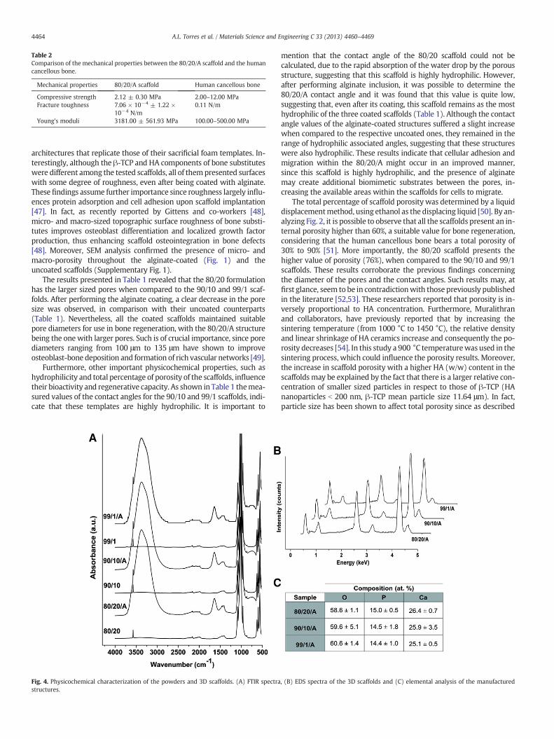
Table 2Comparison of the mechanical properties between the 80/20/A scaffold and the humancancellous bone.
Mechanical properties 80/20/A scaffold Human cancellous bone
Compressive strength 2.12 ± 0.30 MPa 2.00–12.00 MPaFracture toughness 7.06 × 10⁻⁴ ± 1.22 ×
10−4 N/m0.11 N/m
Young's moduli 3181.00 ± 561.93 MPa 100.00–500.00 MPa
4464 A.L. Torres et al. / Materials Science and Engineering C 33 (2013) 4460–4469
architectures that replicate those of their sacrificial foam templates. In-terestingly, although the β-TCP and HA components of bone substituteswere different among the tested scaffolds, all of thempresented surfaceswith some degree of roughness, even after being coated with alginate.These findings assume further importance since roughness largely influ-ences protein adsorption and cell adhesion upon scaffold implantation[47]. In fact, as recently reported by Gittens and co-workers [48],micro- and macro-sized topographic surface roughness of bone substi-tutes improves osteoblast differentiation and localized growth factorproduction, thus enhancing scaffold osteointegration in bone defects[48]. Moreover, SEM analysis confirmed the presence of micro- andmacro-porosity throughout the alginate-coated (Fig. 1) and theuncoated scaffolds (Supplementary Fig. 1).
The results presented in Table 1 revealed that the 80/20 formulationhas the larger sized pores when compared to the 90/10 and 99/1 scaf-folds. After performing the alginate coating, a clear decrease in the poresize was observed, in comparison with their uncoated counterparts(Table 1). Nevertheless, all the coated scaffolds maintained suitablepore diameters for use in bone regeneration, with the 80/20/A structurebeing the one with larger pores. Such is of crucial importance, since porediameters ranging from 100 μm to 135 μm have shown to improveosteoblast-bone deposition and formation of rich vascular networks [49].
Furthermore, other important physicochemical properties, such ashydrophilicity and total percentage of porosity of the scaffolds, influencetheir bioactivity and regenerative capacity. As shown in Table 1 themea-sured values of the contact angles for the 90/10 and 99/1 scaffolds, indi-cate that these templates are highly hydrophilic. It is important to
Fig. 4. Physicochemical characterization of the powders and 3D scaffolds. (A) FTIR spectrastructures.
mention that the contact angle of the 80/20 scaffold could not becalculated, due to the rapid absorption of the water drop by the porousstructure, suggesting that this scaffold is highly hydrophilic. However,after performing alginate inclusion, it was possible to determine the80/20/A contact angle and it was found that this value is quite low,suggesting that, even after its coating, this scaffold remains as the mosthydrophilic of the three coated scaffolds (Table 1). Although the contactangle values of the alginate-coated structures suffered a slight increasewhen compared to the respective uncoated ones, they remained in therange of hydrophilic associated angles, suggesting that these structureswere also hydrophilic. These results indicate that cellular adhesion andmigration within the 80/20/A might occur in an improved manner,since this scaffold is highly hydrophilic, and the presence of alginatemay create additional biomimetic substrates between the pores, in-creasing the available areas within the scaffolds for cells to migrate.
The total percentage of scaffold porosity was determined by a liquiddisplacementmethod, using ethanol as the displacing liquid [50]. By an-alyzing Fig. 2, it is possible to observe that all the scaffolds present an in-ternal porosity higher than 60%, a suitable value for bone regeneration,considering that the human cancellous bone bears a total porosity of30% to 90% [51]. More importantly, the 80/20 scaffold presents thehigher value of porosity (76%), when compared to the 90/10 and 99/1scaffolds. These results corroborate the previous findings concerningthe diameter of the pores and the contact angles. Such results may, atfirst glance, seem to be in contradictionwith those previously publishedin the literature [52,53]. These researchers reported that porosity is in-versely proportional to HA concentration. Furthermore, Muralithranand collaborators, have previously reported that by increasing thesintering temperature (from 1000 °C to 1450 °C), the relative densityand linear shrinkage of HA ceramics increase and consequently the po-rosity decreases [54]. In this study a 900 °C temperaturewas used in thesintering process, which could influence the porosity results. Moreover,the increase in scaffold porosity with a higher HA (w/w) content in thescaffolds may be explained by the fact that there is a larger relative con-centration of smaller sized particles in respect to those of β-TCP (HAnanoparticles b 200 nm, β-TCP mean particle size 11.64 μm). In fact,particle size has been shown to affect total porosity since as described
, (B) EDS spectra of the 3D scaffolds and (C) elemental analysis of the manufactured

Fig. 5. Osteoblast cell metabolic activity when cultured in the presence of differentβ-TCP/HA scaffolds. Cultures were evaluated for 1, 7 and 14 days. K+ and K−, indicatedead and viable cells, representing positive control and negative control, respectively.N.S.: not significant; *p b 0.05, n = 3.
4465A.L. Torres et al. / Materials Science and Engineering C 33 (2013) 4460–4469
throughmolecular dynamics simulations performed by Balakrishnan etal. [55], the formation of agglomerates is dependent on the applicationof higher forces to compact nanoparticles [55]. Therefore, since noforces were applied to form compact nanoparticle aggregates themore space occupied by HApowders in 80/20 scaffolds, presents higherinterfacial voids, and thus, increased total porosity. It is also importantto emphasize that an increasedHA concentration increases the viscosityof the slurry, a fact that will lead to highly porous materials aftersintering as reported by Sopyan et al. [56]. Thus the increasing porosityin the 80/20 scaffold in comparison with the other formulations mayalso be correlated with the higher viscosity promoted by HA.
Regarding the scaffold porosity after the coating procedure, aslight decrease (5%) was observed for the 80/20/A template in com-parison with the respective uncoated scaffold. Even though, this scaf-fold was also the most porous in comparison with all other coatedones (Fig. 2). The existence of pores is fundamental as it promotes nu-trient intake, as well as, interactions between the β-TCP/HA templateand bone cells. The establishment of this microenvironment pro-motes cell attachment, migration and proliferation in a 3D area[57–59]. Apart from pore size, pore interconnectivity is also a desir-able characteristic due to its important influence on cell proliferationand differentiation [60]. In fact, interconnected pores facilitate theformation of vascular networks within the 3D scaffolds and providechanneling pathways for biofluids [61]. These biofluids promote theadsorption of Ca2+ and PO4
3− ions throughout the biomaterial, creat-ing an HA-like layer [10]. This layer establishes an interface betweenthe implant and the 9 surrounding bone tissues and stimulates oste-oblast cell activity, increasing the deposition of bone matrix in the de-fect area. Additionally the formation of this layer increases theosteoconductivity and osteointegration, further contributing for thebone mineralization [17].
Achieving equilibrium between scaffold porosity and adequatemechanical strength, is a demanding objective for the production ofscaffolds, as it was taken into consideration in this study. The me-chanical behavior of the 3D scaffolds was characterized by analyzingtheir resistance to compression, followed by an estimation of thevalues of fracture toughness and Young's modulus. Through the anal-ysis of Fig. 3 it is possible to visualize that the compressive strength,the fracture toughness and the Young's modulus of the 80/20 scaffoldare higher than the values of the 90/10 and 99/1 scaffolds.With increaseof porosity (99/1 to 80/20) there is an increase in the mechanicalstrength by 2 times. This improvement in mechanical compressionstrength is due to the increasing densification of thewalls due to the in-crease of the amount of HA. This is also corroborated by the increasedmaterial stiffness (highest Young'smodulus). After coating the scaffoldswith a 2% alginate solution their mechanical properties improved, withthe 80/20/A maintaining better mechanical resistance to compression,resistance to fracture and elasticity modulus than the other coated 3Dstructures. Accordingly, it was previously described that an increase inthe volume fraction of alginate from 1 to 3% results in an improvementof the compressive modulus [62]. So, it is noticeable that the most po-rous structures (80/20 and 80/20/A) are also thosewith bettermechan-ical properties, when compared to both the uncoated and coated 90/10or 99/1 scaffolds. These findings are likely correlated with the fact thatthe 80/20 and 80/20/A scaffolds have higher amounts of HA andlower quantities of β-TCP, in comparison to the other manufacturedscaffolds. This is consistent with a study performed by Shiota, T. et al.,that revealed that high amounts of β-TCP are responsible for a decreasein scaffold strength resistance [63]. Comparing the mechanical proper-ties of 80/20/A scaffold with those presented by the natural cancellousbone, it is possible to observe that this scaffold is the most promisingfor therapeutic applications, among the scaffolds produce herein(Table 2) [64–66].
The different scaffolds produced by FRM were then characterizedby FTIR spectroscopy. The stretching bands obtained between 500and 600 cm−1 for the manufactured structures are characteristic of
the PO4− ions present in both β-TCP and the HA powder (Fig. 4A).
The stretching of the \OH groups was also observed (3571 cm−1
and libration mode at ≈630 cm−1). This particular band intensity ishigher in all the synthesized scaffolds, since it is the end result ofthe cumulative contribution of both HA and β-TCP (Fig. 4A). In addi-tion, the stretching bands observed at ≈1415 and 1450 cm−1 are at-tributed to a B type apatite [67]. Furthermore, EDS analysis of thescaffolds shows that the chemical composition of all the alginate coat-ed formulations is very similar in terms of phosphate and calcium(Fig. 4C).

4466 A.L. Torres et al. / Materials Science and Engineering C 33 (2013) 4460–4469
3.2. Analysis of the biological properties of the scaffolds
Subsequently, the biological performance of the porous β-TCP/HAstructures was also evaluated. For this purpose, human osteoblastcells were cultured in the 3D scaffolds for up to 14 days and theirmetabolic activity evaluated at the indicated time points (Fig. 5).The results obtained revealed that osteoblast metabolic activitywhen cultured in the presence of all the scaffolds was higher than70% during the 14 days of analysis, indicating that the 3D bioceramicsare biocompatible. Among all the uncoated scaffolds, the 80/20 for-mulation was the one that presented higher biocompatibility alongthe assay and comparatively to 90/10 and 99/1 scaffolds. Cellspresented higher metabolic activity in the presence of alginatecoated-β-TCP/HA scaffolds than in contact with uncoated scaffolds(p b 0.05). The 80/20/A structure was the one for which cellspresented the higher viability, being close to the value presented bythe negative control for all culture days (approximately 100%).
To further characterize cellular adhesion both on the surface andinside the scaffold, SEM analysis was performed and it showed thatosteoblasts were able to adhere preferably inside the pores of thebioceramic scaffolds, either coated (Fig. 6) or not (SupplementaryFig. 2) with the natural polymer. Interestingly, after the first day incontact with the scaffolds, osteoblasts were already attached andspread across the 3D structures, presenting a round shape configura-tion, with some cytoplasm extensions towards the substrate. At day 7,and predominantly after 14 days of culture, cells cultured in all thedifferent structures started to present a typical osteoblastic morphology,
Fig. 6. SEM micrographs of osteoblast morphology in the presence of the different β-TCP/HA80/20/A (A, B and C), 90/10/A (D, E and F) and 99/1/A (G, H and I) 3D scaffolds, at culture d
showing a smooth arrangement with more lamellipodia connecting tosurrounding osteoblast, beginning to form a continuous cell layer.For all the uncoated and coated β-TCP/HA scaffolds, the number of oste-oblasts appeared to increase along time, with cells establishing connec-tion areas between them. SEM analysis was also important toinvestigatewhether alginatewas or not occluding the pores of the coatedscaffolds and if cells were still able to migrate within these scaffolds. Asdepicted in Fig. 6, alginate did not block the pores, but instead created ad-ditional biocompatible substrates, changed the roughness and micro-topography of the scaffolds surface, which contributed to furtherincrease osteoblast adhesion and migration. These results are furtheremphasized by the CLSM analysis during the various stages of osteoblastcontact with the scaffolds (Fig. 7). In fact, as shown in Fig. 7, during oste-oblast contact with the alginate-coated 3D scaffolds, cells were able toadhere and proliferate in all bioceramic formulations, emphasizingtheir biocompatibility and suitable physicochemical properties to pro-mote osteoblast adhesion. These findings were also observed for theuncoated bioceramic scaffolds. Furthermore, at days 7 and 14 it is clearthat osteoblasts migrate into the porous network during the time courseof incubation. A visual analysis of the orthogonal slices of the scaffoldsalso clearly demonstrates that osteoblasts are spread across the entirescaffold and are also located inside the scaffold pores.
To further visualize the cytoplasmic morphology and localizationof the osteoblasts that were proliferating on the 3D hybrid scaffoldsan immunocytochemistry analysis of β-actin was performed. As theCLSM 3D reconstruction images demonstrate, osteoblasts presenttheir characteristic cytoplasmic morphology for all scaffolds (Fig. 8).
/alginate scaffolds. SEM images were used to visualize osteoblasts on the surface of theays 1 (A, D and G), 7 (B, E and H) and 14 (C, F and I). Scale bars correspond to 20 μm.

4467A.L. Torres et al. / Materials Science and Engineering C 33 (2013) 4460–4469
In addition, color coded depth analysis of the alginate coated struc-tures clearly shows that osteoblasts are capable of migrating andattaching into deep sections of the scaffolds with some cells being lo-calized up to 300 μm within the pores (Fig. 8). This is a relevant find-ing since the deposition of bone matrix inside the scaffold willeventually fill the bone defect while the scaffold is biodegraded, re-storing the structure and function of the native bone.
Together, our results demonstrate that the produced β-TCP/HAstructures are highly porous, hydrophilic, biocompatible and resistantto compression, improvements particularly achieved for the 80/20structure. It was also shown that a mixture of β-TCP and HA, hasthe benefit of combining the bioactivity of β-TCP and the stability ofHA. Moreover, higher amounts of HA increased the scaffold mechan-ical strength, while smaller amounts of β-TCP led to an increase ofstructure porosity. Besides, this study also reveals that the combina-tion of both β-TCP and HA improves the biological and mechanicalproperties, in comparison with previously developed scaffolds com-prised of β-TCP or HA alone [68–70]. In fact, the combination ofthese bioceramics originated 3D porous and bioactive structureswith desirable osteoconductive and osteoinductive properties. Ournovel approach involving an additional alginate coating further im-proves the scaffold mechanical, physicochemical and biological prop-erties, as this natural biopolymer bridges the gap between therequired material properties and bioactivity for therapeutic applica-tions. Overall, the manipulation of bioceramic–polymer relative ratioslargely influenced all the 3D scaffold properties and led to the
Fig. 7. Osteoblasts are able to attach to the surface of the different scaffolds. Confocal micrscaffolds at different time points. (A to I) 3D reconstruction of the hybrid scaffolds; (A1 taxis. (A3 to I3) Top view of the 3D scaffolds. Red channel: cell nucleus; blue channel: 3D scaff300 μm.
manufacture of a particularly improved formulation with suitablecharacteristics for repair (Table 3, 80/20/A formulation). The role ofalginate in the improvement of the mechanical properties is evenmore evident when comparing the scaffolds herein presented withthose produced by Ebrahimi, M. et al. [31], which describes the pro-duction of β-TCP and HA scaffolds coated with collagen. An analysisof both types of scaffolds reveals the superior contribution of alginate,when compared to collagen, for the improvement of the scaffold me-chanical strength and because, unlike collagen, alginate does notrepresent a risk of disease transmission [43]. Considering that thein vivo performance of β-TCP and HA has already been tested inhumans [40,41], from this standpoint we envision that the additionof this polymer may actually improve the clinical outcome of thesebiomaterials.
4. Conclusions
In this work we describe the manufacture of 3D tunable porousscaffolds for bone regeneration by using a cost-effective and repro-ducible technique. Our results revealed that by optimizing the ratiosof different bioceramic composites in the scaffolds the physicochem-ical and biological properties of these 3D constructs could be im-proved. In addition, the method employed for template production,allows the synthesis of scaffolds with rough topographies, micro-and macro-pores that are homogeneously distributed in the scaffold3D volume. Furthermore, in order to improve the overall performance
oscopy images were used to investigate osteoblast distribution on the alginate coatedo I1) Orthogonal slices of scaffold xx axis. (A2 to I2) Orthogonal slices of scaffold yyolds. White arrows indicate cells. White ovals indicate pores. White scale bars represent

Fig. 8. 3D reconstruction and depth analysis of the different scaffolds coated with alginate. (A, D and G) Immunocytochemistry analysis of osteoblast seeded scaffolds at day 14.Green channel: β-actin; red channel: cell nucleus; blue channel: 3D hybrid scaffolds. (B, E and H) 3D depth coding of scaffolds and osteoblasts cultured on scaffold's surface.Size bar represents color-coded depth. White area represents a scaffold pore with osteoblasts inside. (C, F and I) 3D surface reconstruction of cells at the surface of the scaffold.White arrows indicate osteoblasts. Black bars indicate 300 μm.
4468 A.L. Torres et al. / Materials Science and Engineering C 33 (2013) 4460–4469
of these structures, the bioceramics were also vacuum-coated with al-ginate. This inclusion greatly enhanced both the mechanical and bio-logical properties of the scaffolds. In fact, osteoblast adhesion,migration and proliferation in contact with the coated scaffolds em-phasize their suitability for regenerative medicine. To the best ofour knowledge this was the first time that this combination of thesebiomaterials was used to manufacture 3D porous scaffolds, whichresulted in unique structures with desirable biological, mechanicaland physical properties that will promote bone regeneration. In anear future these hybrid bioceramic–polymeric composites will be
Table 3Summary of physicochemical and biological properties of the manufactured scaffolds.
Propertiesscaffold
Hydrophilicity Porosity Mechanicalstrength
Biocompatibility
80/20 + ++++++ +++++ +++++80/20/A ++++ +++++ ++++++ ++++++90/10 ++ ++++ +++ +90/10/A +++++ +++ ++++ ++99/1 +++ ++ ++ +++99/1/A ++++++ + + ++++
“+” to “++++++”: symbol-based grade, indicating improvement of the scaffoldsproperties.
implanted in whole animal models with small and large bone defects,in order to evaluate their bone regenerative performance.
5. Acknowledgments
The authors would like to thank to Eng. Ana Paula for her help withthe acquisition of SEMdata. This work was supported by the PortugueseFoundation for Science and Technology (FCT) (PTDC/EME-TME/103375/2008, PTDC/EBB-BIO/114320/2009 and PEst-C/SAU/UI0709/2011 COM-PETE). Vítor M. Gaspar acknowledges a PhD fellowship from FCT (SFRH/BD/80402/2011).
Appendix A. Supplementary data
Supplementary data to this article can be found online at http://dx.doi.org/10.1016/j.msec.2013.07.003.
References
[1] A.M. Martins, C.M. Alves, F.K. Kasper, A.G. Mikos, R.L. Reis, J. Mater. Chem. 20(2010) 1638–1645.
[2] D. Fisher, J. Wong, C. Crowley,W. Khan, Curr. Stem Cell Res. Ther. 8 (2013) 243–252.[3] T. Jiang, S.P. Nukavarapu, M. Deng, E. Jabbarzadeh, M.D. Kofron, S.B. Doty, W.I.
Abdel-Fattah, C.T. Laurencin, Acta Biomater. 6 (2010) 3457–3470.
4469A.L. Torres et al. / Materials Science and Engineering C 33 (2013) 4460–4469
[4] A. Nekora-Azak, G. Evlioglu, M. Ozdemir-Karatas, H. Keskin, J. Oral Rehabil. 32(2005) 693–695.
[5] M.B. Dreifke, N.A. Ebraheim, A.C. Jayasuriya, J. Biomed. Mater. Res. A (2013).[6] A.A. Amini, L.S. Nair, Biomed. Mater. 7 (2012) 024105.[7] J.R. Jones, L.M. Ehrenfried, L.L. Hench, Biomaterials 27 (2006) 964–973.[8] D.F. Williams, Biomaterials 29 (2008) 2941–2953.[9] S. Scaglione, C. Ilengo, M. Fato, R. Quarto, Tissue Eng. Part A 15 (2009) 155–163.
[10] M.G. Raucci, V. Guarino, L. Ambrosio, J. Funct. Biomater. 3 (2012) 688–705.[11] H.S. Yu, G.Z. Jin, J.E. Won, I. Wall, H.W. Kim, J. Biomed. Mater. Res. A 100 (2012)
2431–2440.[12] A. Abdal-hay, F.A. Sheikh, J.K. Lim, Colloids Surf. B Biointerfaces 102 (2013) 635–643.[13] P.P. Cortez, M.A. Silva, M. Santos, P. Armada-da-Silva, A. Afonso, M.A. Lopes, J.D.
Santos, A.C. Mauricio, J. Biomater. Appl. 27 (2012) 201–217.[14] X. Lu, S. Li, J. Zhang, Z. Zhang, B. Lu, H. Bu, Y. Li, J. Cheng, Sheng Wu Yi Xue Gong
Cheng Xue Za Zhi 18 (2001) 497–499.[15] C.F.L. Santos, A.P. Silva, L. Lopes, I. Pires, I.J. Correia, Mater. Sci. Eng. C 32 (2012)
1293–1298.[16] L.L. Hench, Biomaterials 19 (1998) 1419–1423.[17] L.L. Hench, J. Biomed. Mater. Res. 41 (1998) 511–518.[18] C. Wu, Y. Ramaswamy, P. Boughton, H. Zreiqat, Acta Biomater. 4 (2008) 343–353.[19] Y. Zhang, M. Zhang, J. Biomed. Mater. Res. 61 (2002) 1–8.[20] S.J. Florczyk, M. Leung, S. Jana, Z. Li, N. Bhattarai, J.I. Huang, R.A. Hopper, M. Zhang,
J. Biomed. Mater. Res. A 100 (2012) 3408–3415.[21] W. TheinHan, M.D. Weir, C.G. Simon, H.H. Xu, J. Tissue Eng. Regen. Med. (2012),
http://dx.doi.org/10.1002/term.1466.[22] W.R. Gombotz, S.F. Wee, Adv. Drug Deliv. Rev. 31 (1998) 267–285.[23] J.-K. Francis Suh, H.W. Matthew, Biomaterials 21 (2000) 2589–2598.[24] F.A. Johnson, D.Q. Craig, A.D. Mercer, J. Pharm. Pharmacol. 49 (1997) 639–643.[25] A.D. Augst, H.J. Kong, D.J. Mooney, Macromol. Biosci. 6 (2006) 623–633.[26] J.F.A. Valente, V.M. Gaspar, B.P. Antunes, P. Countinho, I.J. Correia, Polymer 54
(2013) 5–15.[27] O. Smidsrød, Trends Biotechnol. 8 (1990) 71–78.[28] A.I. Pravdyuk, Y.A. Petrenko, B.J. Fuller, A.Y. Petrenko, Cryobiology 66 (2013) 215–222.[29] A. Schoubben, P. Blasi, S. Giovagnoli, L. Perioli, C. Rossi, M. Ricci, Eur. J. Pharm. Sci.
36 (2009) 226–234.[30] X. Wu, Y. Liu, X. Li, P. Wen, Y. Zhang, Y. Long, X. Wang, Y. Guo, F. Xing, J. Gao, Acta
Biomater. 6 (2010) 1167–1177.[31] M. Ebrahimi, P. Pripatnanont, N. Monmaturapoj, S. Suttapreyasri, J. Biomed.
Mater. Res. A 100 (2012) 2260–2268.[32] D. Lacroix, A. Chateau, M.P. Ginebra, J.A. Planell, Biomaterials 27 (2006) 5326–5334.[33] J. Martin Bland, D. Altman, Lancet 327 (1986) 307–310.[34] H.L. Nie, L.M. Zhu, Int. J. Biol. Macromol. 40 (2007) 261–267.[35] M. Rouahi, O. Gallet, E. Champion, J. Dentzer, P. Hardouin, K. Anselme, J. Biomed.
Mater. Res. A 78 (2006) 222–235.[36] T.J. Collins, Biotechniques 43 (2007) 25–30.[37] P. Coimbra, P. Ferreira, H. De Sousa, P. Batista, M. Rodrigues, I. Correia, M. Gil, Int.
J. Biol. Macromol. 48 (2011) 112–118.[38] V. Bewick, L. Cheek, J. Ball, Crit. Care 8 (2004) 130–136.[39] A. Hilton, R.A. Armstrong, Microbiologist 2006 (2006) 34–36.[40] Z. Artzi, M. Weinreb, G. Carmeli, R. Lev-Dor, M. Dard, C.E. Nemcovsky, Clin. Oral.
Implants Res. 19 (2008) 686–692.
[41] J.L. Rouvillain, F. Lavalle, H. Pascal-Mousselard, Y. Catonne, G. Daculsi, Knee 16(2009) 392–397.
[42] V. Wang, G. Misra, B. Amsden, J. Mater. Sci. Mater. Med. 19 (2008) 2145–2155.[43] M. Xie, M.O. Olderoy, J.P. Andreassen, S.M. Selbach, B.L. Strand, P. Sikorski, Acta
Biomater. 6 (2010) 3665–3675.[44] M.A.A. Muhamad Nor, L.C. Hong, Z. Arifin Ahmad, H. Md Akil, J. Mater. Process.
Technol. 207 (2008) 235–239.[45] H. Fu, Q. Fu, N. Zhou, W. Huang, M.N. Rahaman, D. Wang, X. Liu, Mater. Sci. Eng. C
29 (2009) 2275–2281.[46] Q. Fu, M.N. Rahaman, B. Sonny Bal, R.F. Brown, D.E. Day, Acta Biomater. 4 (2008)
1854–1864.[47] B.G. Gerberich, S.K. Bhatia, Biotechnol. J. 8 (2013) 73–84.[48] R.A. Gittens, T. McLachlan, R. Olivares-Navarrete, Y. Cai, S. Berner, R. Tannenbaum,
Z. Schwartz, K.H. Sandhage, B.D. Boyan, Biomaterials 32 (2011) 3395–3403.[49] C.M. Murphy, M.G. Haugh, F.J. O'Brien, Biomaterials 31 (2010) 461–466.[50] Y. Zhang, M. Zhang, J. Biomed. Mater. Res. 55 (2001) 304–312.[51] V. Karageorgiou, D. Kaplan, Biomaterials 26 (2005) 5474–5491.[52] A. Tampieri, G. Celotti, S. Sprio, A. Delcogliano, S. Franzese, Biomaterials 22 (2001)
1365–1370.[53] P. Habibovic, M.C. Kruyt, M.V. Juhl, S. Clyens, R. Martinetti, L. Dolcini, N.
Theilgaard, C.A. van Blitterswijk, J. Orthop. Res. 26 (2008) 1363–1370.[54] G. Muralithran, S. Ramesh, Ceram. Int. 26 (2000) 221–230.[55] A. Balakrishnan, P. Pizette, C. Martin, S. Joshi, B. Saha, Acta Mater. 58 (2010)
802–812.[56] I. Sopyan, J. Kaur, Ceram. Int. 35 (2009) 3161–3168.[57] F.C. den Boer, B.W. Wippermann, T.J. Blokhuis, P. Patka, F.C. Bakker, H.J. Haarman,
J. Orthop. Res. 21 (2003) 521–528.[58] H.W. Kim, S.Y. Shin, H.E. Kim, Y.M. Lee, C.P. Chung, H.H. Lee, I.C. Rhyu, J. Biomater.
Appl. 22 (2008) 485–504.[59] M. Okamoto, Y. Dohi, H. Ohgushi, H. Shimaoka, M. Ikeuchi, A. Matsushima, K.
Yonemasu, H. Hosoi, J. Mater. Sci. Mater. Med. 17 (2006) 327–336.[60] J.P. Gleeson, N.A. Plunkett, F.J. O'Brien, Eur. Cell Mater. 20 (2010) 218–230.[61] Y.C. Chen, R.Z. Lin, H. Qi, Y. Yang, H. Bae, J.M. Melero‐Martin, A. Khademhosseini,
Adv. Funct. Mater. 22 (2012) 2027–2039.[62] M.A. LeRoux, F. Guilak, L.A. Setton, J. Biomed. Mater. Res. 47 (1999) 46–53.[63] T. Shiota, M. Shibata, K. Yasuda, Y. Matsuo, J. Ceram. Soc. Jpn. 116 (2008)
1002–1005.[64] D.W. Hutmacher, J.T. Schantz, C.X. Lam, K.C. Tan, T.C. Lim, J. Tissue Eng. Regen.
Med. 1 (2007) 245–260.[65] D.N. Rockwood, E.S. Gil, S.H. Park, J.A. Kluge, W. Grayson, S. Bhumiratana, R.
Rajkhowa, X. Wang, S.J. Kim, G. Vunjak-Novakovic, D.L. Kaplan, Acta Biomater. 7(2011) 144–151.
[66] S. Kashef, A. Asgari, T.B. Hilditch, W. Yan, V.K. Goel, P.D. Hodgson, Mater. Sci. Eng.,A 527 (2010) 7689–7693.
[67] A. Ślósarczyk, Z. Paszkiewicz, C. Paluszkiewicz, J. Mol. Struct. 744–747 (2005)657–661.
[68] Z. Dong, Y. Li, Q. Zou, Appl. Surf. Sci. 255 (2009) 6087–6091.[69] G.S. Lee, J.H. Park, U.S. Shin, H.W. Kim, Acta Biomater. 7 (2011) 3178–3186.[70] P.H. Warnke, H. Seitz, F. Warnke, S.T. Becker, S. Sivananthan, E. Sherry, Q. Liu,
J. Wiltfang, T. Douglas, J. Biomed. Mater. Res. B Appl. Biomater. 93 (2010)212–217.