tania elaine schwingel efeitos dos compostos … · a todos os colegas do laboratório de...
TRANSCRIPT

PONTIFÍCIA UNIVERSIDADE CATÓLICA DO RIO GRANDE DO SUL
FACULDADE DE BIOCIÊNCIAS
PROGRAMA DE PÓS-GRADUAÇÃO EM BIOLOGIA CELULAR E
MOLECULAR
TANIA ELAINE SCHWINGEL
EFEITOS DOS COMPOSTOS QUERCETINA, QUERCETINA EM
NANOEMULSÃO, RESVERATROL E RUTINA SOBRE A
HEPATOTOXICIDADE E NEUROTOXICIDADE INDUZIDAS POR
OXALIPLATINA EM CAMUNDONGOS
PORTO ALEGRE, 2013

TANIA ELAINE SCHWINGEL
EFEITOS DOS COMPOSTOS QUERCETINA, QUERCETINA EM
NANOEMULSÃO, RESVERATROL E RUTINA SOBRE A
HEPATOTOXICIDADE E NEUROTOXICIDADE INDUZIDAS POR
OXALIPLATINA EM CAMUNDONGOS
Dissertação apresentada como requisito parcial
para obtenção do Título de Mestre pelo
Programa de Pós-Graduação em Biologia Celular
e Molecular, Faculdade de Biociências, Pontifícia
Universidade Católica do Rio Grande do Sul.
Orientadora: Prof.ª Dr.ª Fernanda Bueno Morrone
PORTO ALEGRE
2013

Dedico este trabalho a minha família, por todo amor
e dedicação para comigo, por terem me apoiado em
todas as minhas escolhas e, principalmente na
concretização deste sonho. Ao meu marido, pelo
carinho dispensado em todos os momentos que
precisei. Muito obrigada por estarem do meu lado!
AGRADECIMENTOS
Agradeço a Deus e ao Universo por me iluminar em mais esta jornada;
Meus sinceros agradecimentos a minha orientadora Dra. Fernanda
Bueno Morrone, por ter me dado oportunidade de ingressar no mundo da
pesquisa, proporcionado momentos de crescimento e de conhecimento. Por ter
acreditado no meu potencial, pela acolhida calorosa, pela disponibilidade e
orientação prestada. A você, minha eterna gratidão;
À minha família, em especial aos meus pais, meus exemplos de conduta
moral. Por sempre estarem ao meu lado em minhas decisões, vocês são os
meus alicerces, meus melhores exemplos;
Ao meu irmão Badu, pelo apoio na concretização desse sonho.
Ao meu companheiro João, pela paciência em muitos momentos de
estresse durante este período, por todo amor e carinho;
Ao corpo docente do curso de Pós-graduação em Biologia Celular e
Molecular pela formação e incentivo;
À colega e amiga Caroline Klein pela participação e colaboração em
várias etapas do desenvolvimento dessa pesquisa;
À doutoranda Natalia Nicoletti, foi quem me transmitiu as primeiras
instruções no laboratório, e colaborou nos experimentos;
À secretária Zíngara, pelas orientações acadêmicas e pela assistência
prestada em todos os momentos;
Ao técnico do laboratório Juliano, pela presteza em sempre ajudar no
manuseio com os animais;
A todos os colegas do Laboratório de Farmacologia Aplicada e Instituto
de Toxicologia e Farmacologia pela amizade e atenção.
A todos, meus sinceros agradecimentos!

RESUMO
Introdução: A oxaliplatina é um agente antineoplásico e tem sido amplamente
utilizado no tratamento de vários tumores. É um composto derivado da platina
de terceira geração desenvolvido com o propósito de ultrapassar as limitações
da toxicidade, a resistência do tumor e a fraca biodisponibilidade oral associada
à administração de cisplatina. Sintomas de neurotoxicidade estão associados
ao uso da oxaliplatina e não existe tratamento adequado. Doses crescentes de
oxaliplatina podem levar ao desenvolvimento de sensibilidade a frio e
neuropatia sensorial periférica, e a um aumento da intensidade dos sintomas.
Além disso, apesar da sua utilidade, a quimioterapia aumenta a taxa de
desenvolvimento de danos hepáticos com atividade inflamatória. Esta
manifestação pode ser chamada de esteato-hepatite associada à quimioterapia
(CASH), uma forma mais grave da doença hepática gordurosa não alcoólica.
Deste modo, o presente estudo, objetivou comparar o efeito de alguns
compostos antioxidantes no desenvolvimento simultâneo da hepato e
neurotoxicidade induzida pela oxaliplatina em camundungos. Métodos: Os
camundongos Balb/c foram tratados intraperitonealmente (i.p.), com doses de
10 mg/kg de oxaliplatina durante 6 semanas, resultando em anodinia
mecânica, e indução de esteatose hepática. Os compostos rutina (RUT) (20
mg/Kg/d), resveratrol (RSV) (100 mg/Kg/dia), quercetina (QT) (20 mg/Kg/dia) e
nanoquerecetina (NQT) (20 mg/Kg/dia), foram administrados diariamente por
gavagem aos camundongos. N-acetilcisteína foi utilizada como controle. A
eutanásia ocorreu no 43º dia após o tratamento. Foram analisadas a
nocicepção mecânica, as aminotransferases ALT/AST, foi realizada avaliação
histopatológica e da atividade da mieloperoxidase. A análise estatística foi
realizada por ANOVA de uma via seguida do teste de Bonferroni. Resultados:
Os tratamentos com resveratrol, rutina ou quercetina em nanoemulsão foram
capazes de impedir a alodinia mecânica, quando comparados com o grupo
tratado com oxaliplatina. Em relação ao efeito sobre a esteato-hepatite, os
tratamentos com resveratrol, quercetina e quercetina em nanoemulsão
reverteram significativamente o aumento de peso médio do fígado induzido
pela oxaliplatina. Corroborando com estes dados, a avaliação histológica
mostrou uma atenuação em todas as características de esteatose hepática

avaliados nos grupos tratados com resveratrol, rutina, quercetina e quercetina
em nanoemulsão, apresentando características semelhantes ao controle
positivo com N-acetilcisteína. Por outro lado, apenas os tratamentos com
quercetina e quercetina em nanoemulsão foram capazes de reduzir a
migração de neutrófilos medida pela atividade de mieloperoxidase. Conclusão:
Os resultados sugerem que o uso dos compostos resveratrol, rutina, quercetina
e quercetina em nanoemulsão podem se alternativas efetivas para o tratamento
da hepato e neurotoxicidade induzida pela oxaliplatina no modelo testado.
Palavras-chave: esteato-hepatite, neuropatia, compostos fenólicos,
oxaliplatina.
ABSTRACT
Introduction: Oxaliplatin is an antineoplastic agent widely used in the treatment
of some tumors. It is a third-generation platinum compound developed with the
purpose of overcoming the limitations of toxicity, tumor resistance and poor oral
bioavailability associated to cisplatin administration. Oxaliplatin-associated
neurotoxicity represents the main dose limiting and there is not suitable
treatment. Increasing doses of oxaliplatin can leed to the development of
mechanical allodynia, cold sensitivity and peripheral sensory neuropathy, with
increase of symptoms. Furthermore, despite its usefulness, chemotherapy with
oxaliplatin increases the rate of developing hepatic damages together with
inflammatory activity. This might be termed chemotherapy-associated
steatohepatitis (CASH), a most severe form of non-alcoholic fatty liver disease.
Therefore, in the present study, we aimed to compare the effect of antioxidant
compounds on simultaneous development of oxaliplatin-induced hepato and
neurotoxicity in mice. Methods: The Balb/c mice were treated with doses of
oxaliplatin (OXA) for 6 weeks, 10 mg/kg, intraperitoneally (i.p), resulting in
mechanical allodynia, and hepatic steatosis. We administered antioxidants
compounds such as rutin (RUT) (20 mg/Kg/d), resveratrol (RVS) (100 mg/Kg/d),
quercetin (QT) (20 mg/Kg/d) and nanoquerecetin (NQT) (20 mg/Kg/d) daily by
gavage to Balb/c. N-acetyl-cysteine was used as control. Euthanasia occurred
on day 43 after treatment. We evaluated mechanical nociceptive threshold,
ALT/AST, histopathological analysis and MPO activity. Statistical analyses were
made one way ANOVA, followed by Bonferroni post hoc test. Results: The
treatments with RSV, RUT or NQT were able to prevent mechanical allodynia
when compared to OXA group. Regarding the effect on steatohepatitis,
resveratrol, quercetin and quercetin nanoemulsion almost completely reversed
the mean liver weight increase by OXA. In accordance with these previous data,
histological evaluation depicted attenuation all features of hepatic steatosis
evaluated in resveratrol, rutin, quercetin and quercetin nanoemulsion groups.
On the other hand, only quercetin and quercetin nanoemulsion treatments were
able to reduce neutrophils migration measured by MPO activity. Conclusion:
These results suggest that the use of compounds such as resveratrol, rutin,
quercetin and quercetin nanoemulsion can be effective to avoid oxaliplatin-
inducing hepato and neurotoxicity in a rodent model.
Keywords: Oxaliplatin, mechanical allodynia, steatohepatitis, antioxidants.
LISTA DE ABREVIATURAS
AVC: acidente vascular cerebral
CASH: chemotherapy-associated steatohepatitis
CIPN: chemotherapy induced peripheral neuropathy
CRC: câncer colorretal avançado
FA: folic acid, ácido fólico
5-FU: 5-fluorouracil
FDA: Food and Drug Administration
FOLFOX: ácido folínico/fluorouracil/oxaliplatina
GLU: glicose
H2O2: peróxido de hidrogênio
HCO3-: bicarbonato
H2PO4: dihidrogênio fosfato
HE: hematoxilina-eosina
iNOS: óxido nítrico sintase induzível
MPO: mieloperoxidase
NAFLD: non-alcoholic fatty liver disease
NASH: non-alcoholic steatohepatitis
NQT: quercetina em nanoemulsão
OXA: oxaliplatina
Pt: platino
Pt(dach)Cl2: Dicloro(1,2 –diaminociclohexano) platino
Pt(dach)Cl: Monocloro(1,2 –diaminociclohexano) platino
QT: quercetina
ROS: espécies reativas de oxigênio
RSV: resveratrol
RUT: rutina

LISTA DE FIGURAS
Figura 1 Estrutura molecular da oxaliplatina........................................pag 13
Figura 2 Estrutura molecular do resveratrol.........................................pag 17
Figura 3 Estrutura molecular da rutina.................................................pag 19
Figura 4 Estrutura molecular da quercetina.........................................pag 20

Sumário
1. INTRODUÇÃO ................................................................................................................. 09
1.1 Oxaliplatina .................................................................................................. 09
1.2 Esteatose hepática e neurotoxicidade ......................................................... 12
1.3 Agentes antioxidantes resveratrol e rutina .................................................. 15
1.4 Quercetina e quercetina em nanoemulsão ................................................... 18
2. OBJETVOS ...................................................................................................................... 22
2.1 Objetivo Geral .............................................................................................. 22
2.2 Objetivos Específicos ................................................................................... 22
3. ARTIGO CIENTÍFICO ...................................................................................................... 23
4. CONSIDERAÇÕES FINAIS ............................................................................................ 55
5. PERSPECTIVAS ............................................................................................................ 55
6. REFERÊNCIAS .............................................................................................................. 61

1. INTRODUÇÃO 1.1 OXALIPLATINA
Diversos fármacos quimioterápicos vêm sendo utilizadas para o
tratamento de câncer, especialmente no câncer colorretal. Entre os fármacos
mais utilizados, encontra-se a oxaliplatina, que é um agente quimioterápico
alquilante de terceira geração capaz de inibir a síntese e replicação de DNA,
formando adutos com o mesmo (Cardus et al., 2009).
No momento em que a oxaliplatina entra na corrente sanguínea o
HCNO3- e H2PO4 desacoplam seu grupamento oxalato, formando
intermediários não - estáveis, que rapidamente se hidrolisam a espécies
platinas como dicloro (1,2-dach) platino Pt(dach)Cl2 e monocloro (1,2-dach)
platino Pt(dach)Cl, que instantaneamente reagem com o DNA celular,
proteínas e outras macromoléculas levando a apoptose celular (Foltinová et
al., 2008). O grupo alquilante pode formar ligações cruzadas entre dois sítios
nucleofílicos, como o nitrogênio na posição (N-7) da guanina no DNA,
originando ligações cruzadas intrafilamentares e interfilamentares, Foltinová et
al., 2008). Seu principal efeito é observado durante a síntese de DNA (Rang et
al., 2004). A citotoxicidade é inespecífica ao ciclo celular (Goldman et al.,
2009).
A oxaliplatina (cis- [(1R,2R)-1,2-cyclohexanediamine-N,N' ] oxalato (2-)-
O,O'] platinum) é uma substância com peso molecular 397.3 e sua fórmula
molecular (C8H14N2O4Pt) (Figura 1) confere a sua baixa solubilidade em água,
menor solubilidade em metanol e ainda, sua quase total insolubilidade em
etanol e acetona (Alcindor et al., 2011).

13
Figura 1. Estrutura molecular da oxaliplatina.
Fonte: Graham et al., 2000.
A oxaliplatina tem sido utilizada como terapia para o tratamento de
alguns tipos de câncer, devido a uma ampla atividade antitumoral. Ela tem se
mostrado ativa no câncer colorretal, no câncer de ovário, no câncer de células
germinativas e no câncer cervical (Goodman & Gilman’s, 2001; Renn et al.,
2011). Quando usada para tratar câncer colorretal avançado (CRC), ela é
combinada com 5-fluorouracil (5-FU), uma fluoropirimidina, e com leucovorina
(LV), num regime conhecido como FOLFOX (Suzuki et al., 2013; Baekk et al.,
2010).
O tratamento quimioterápico pré-operatório com a oxaliplatina é cada
vez mais utilizado em pacientes com metástases hepáticas, cujo sítio primário
se localiza na porção colorretal (Choti et al., 2009) e para o tratamento de
pacientes submetidos à hepatectomia extensa, particularmente com tumores
grandes ou tumores em localização desfavorável. No entanto, essa terapia
pode contribuir para o desenvolvimento de uma forma de esteato-hepatite
(CASH- chemotherapy – associated steatohepatitis), além de estar relacionada
com a ocorrência de uma dilatação sinusoidal, com o aumento do risco de

14
infecções e de mortalidade após a ressecção de metástases hepáticas (Hebbar
et al., 2009; Morris-Stiff et al., 2007).
A lesão sinusoidal é caracterizada por dilatação sinusoidal, congestão de
eritrócitos e, ocasionalmente, é acompanhada por fibrose e oclusão venular
fibrótica perisinusoidal (Choti, 2009). Essa lesão parece ter uma clara
associação com o uso de oxaliplatina (Choti 2009; Vauthey et al., 2006).
Por outro lado, a esteatose hepática induzida por agentes
quimioterápicos leva a dano hepático significativo e lesões vasculares (Hebbar
et al., 2009), os quais aumentam o risco de morbidade peri-operatória e a taxa
de mortalidade (Keizman et al., 2010). Essa esteato-hepatite é caracterizada
pelo acúmulo de lipídeos/gordura no fígado, o que pode ser considerado
patogênico quando o conteúdo hepático de gordura for superior a 5 % do peso
do fígado (Kahn et al., 2009).
1.2 ESTEATOSE HEPATICA E NEUROTOXICIDADE
Fernandez e colaboradores (2005) verificaram que em biópsias de
pacientes tratados com oxaliplatina, foi constatada hepatite moderada ou
grave, aumentando as evidências acerca dos efeitos da oxaliplatina sobre a
esteatose hepática (Fernandez et al., 2005). A esteatose hepática não
alcoólica, (NASH – non-alcoholic steatohepatitis) associada à quimioterapia
pode ser resultante da deposição de gordura no fígado resultante de outras
causas que não o consumo de álcool (NAFLD – non-alcoholic fatty liver
disease) conforme Kelishadi, 2013. A NASH é a forma mais grave de NAFLD,
uma vez que a infiltração gordurosa no fígado é acompanhada por necrose e

15
inflamação (Kanuri et al.,2013) podendo levar a uma diminuição significativa
da função hepática (Cleary et al., 2009).
A ocorrência de NASH é clinicamente importante, pois 15 a 25 % dos
pacientes têm uma evolução para cirrose. Uma vez que a cirrose é
estabelecida, estima-se que 30 a 40 % dos pacientes virão a óbito ao longo de
um período de 10 anos (Shifflet et al., 2009).
As características histológicas da esteato-hepatite incluem, em
diferentes graus, esteatose/deposição de gordura, inflamação lobular e
balonização dos hepatócitos (Kahn et al., 2009; Tarantino et al., 2013).
Quando este quadro é associado com a realização de quimioterapia para o
câncer, o mesmo é denominado de CASH (Kahn et al., 2009).
Os agentes derivados da platina, incluindo cisplatina, carboplatina e
oxaliplatina, também são conhecidos por causar sintomas clássicos de
neuropatia periférica induzida por quimioterapia (CIPN – chemotherapy induced
peripheral neuropathy). Embora a CIPN esteja associada aos três derivados da
platina, a maior incidência é observada com o uso da cisplatina e da
oxaliplatina (Ogawa et al., 2013).
A neurotoxicidade decorrente da terapia com a oxaliplatina pode ser
dividida em duas síndromes distintas. A primeira é de fase aguda, devido à
hiperexcitabilidade transitória dos nervos periféricos, ocorrendo logo após a
infusão de oxaliplatina. Entre os derivados da platina, este é um efeito único da
oxaliplatina (Wang et al., 2007). Esta forma de neuropatia ocorre normalmente
com baixas doses cumulativas totais, pode ser desencadeada ou agravada
pela exposição ao frio, é sempre reversível e, geralmente, não requer a
descontinuação da terapia (Aoki et al., 2012). Os pacientes podem apresentar

16
parestesia e disestesia das mãos e dos pés, bem como, da laringe e da
mandíbula (Schellingerhout et al., 2012). Estes sintomas geralmente ocorrem
depois de horas da exposição e são reversíveis após algumas horas ou nos
dias seguintes à administração da oxaliplatina (Sprowl, et al., 2013). Segundo
Sittl e colaboradores (2012), após algumas horas ou dias da infusão de
oxaliplatina, cerca de 80% dos pacientes apresentarão parestesias distais e
contrações musculares leves. Manifestações menos comuns incluem
alterações visuais, alterações na voz e parestesia perioral (Kannarkat et al.,
2007). Presumivelmente, o oxalato, metabólito da oxaliplatina, quela o cálcio e
magnésio, afeta os canais de sódio neuronais voltagem-dependentes,
causando os efeitos mencionados acima (Kawashiri et al., 2012).
No entanto, a segunda síndrome, conhecida como neuropatia sensorial
periférica, possui sintomas semelhantes aos observados com o uso da
cisplatina, ocorrendo principalmente nas extremidades distais. Esta forma de
neurotoxicidade possui implicações clínicas mais importantes, e podem durar
vários meses, resultando em uma perturbação grave da função (Wang et al.,
2007). A neuropatia sensorial periférica é induzida pela administração de
múltiplas doses de oxaliplatina (Jamieson et al., 2005) e está relacionada com
o efeito cumulativo destas doses, tornando-se comumente um problema clínico,
quando a dose cumulativa se aproxima de 800 mg/m2 (Amptoulach et al.
2011).
Um dos mecanismos sugeridos para a neurotoxicidade induzida pelos
derivados da platina é devido ao seu acúmulo no sistema nervoso periférico,
principalmente na raiz dorsal dos gânglios, gerando espécies reativas de
oxigênio (Renn et al., 2011). A presença patológica de oxaliplatina no sistema

17
nervoso periférico ocorre devido a eliminação lenta da droga (Holmes et al.,
1998). Park e colaboradores (2009) mostraram que as espécies reativas de
oxigênio geradas pelos compostos da platina desempenham um importante
papel na morte celular por apoptose das células neuronais via sinalização da
p53 (Park et al., 2009).
O FDA (Food and Drug Administration) observou que mais de 70% dos
pacientes que fizeram uso de oxaliplatina são afetados por algum grau de
neuropatia sensorial, sendo muitas vezes causa da interrupção do tratamento
(McWhinney et al., 2009).
1.3 AGENTES ANTIOXIDANTES RESVERATROL E RUTINA
O resveratrol (trans- 3, 5, 4’ trihidroxiestilbeno) é um composto
polifenólico natural, de estrutura molecular relativamente simples que interage
com diversos alvos moleculares (Figura 3) (Leiro et al., 2010; Svajger et al.,
2012). Ele e encontrado em várias fontes alimentares, tais como uvas, frutas,
amendoim, bem como no vinho tinto (Souvik et al, 2011).
Figura 2. Estrutura molecular do resveratrol.
Fonte: Dan Su et al., 2013

18
Na natureza, o resveratrol funciona como um fungicida produzido pela
própria planta para repelir organismos potencialmente letais e combater o
estresse ambiental. O seu valor benéfico para a saúde humana está bem
documentado (Bishayee et al., 2010). Estudos mostram que ele apresenta uma
grande variedade de ações farmacológicas, incluindo efeitos anti-inflamatórios,
podendo prevenir ou retardar a progressão de diversas doenças relacionadas
com a inflamação, incluindo câncer, diabetes, doenças neurodegenerativas,
doenças cardiovasculares, lesões isquêmicas, infecções virais, bem como,
aumentar a resistência ao estresse, modulando, ainda, a atividade de enzimas
antioxidantes (Venturini et al., 2010; Bishayee et al., 2010).
Achados experimentais revelam múltiplos alvos celulares para o
resveratrol que podem afetar o crescimento e proliferação celular, apoptose,
inflamação, invasão, angiogênese e metástase (Ganaphaty et al., 2010). Nas
células de câncer colorretal , o resveratrol apresentou atividade inibitória de
crescimento, diferenciação e proliferação celular e induziu a apoptose (Araújo
et al., 2011). Atualmente, encontra-se resveratrol disponível no mercado na
forma de comprimidos como suplemento dietético seguro e bem tolerado (Bhatt
et al., 2012, Fraczeck et al., 2012).
A rutina é uma glicosil flavona (Figura 4) presente em abundância em
plantas como em sementes de trigo sarraceno, frutas e cascas de frutas,
especialmente frutas cítricas (Isai et al., 2009).
19
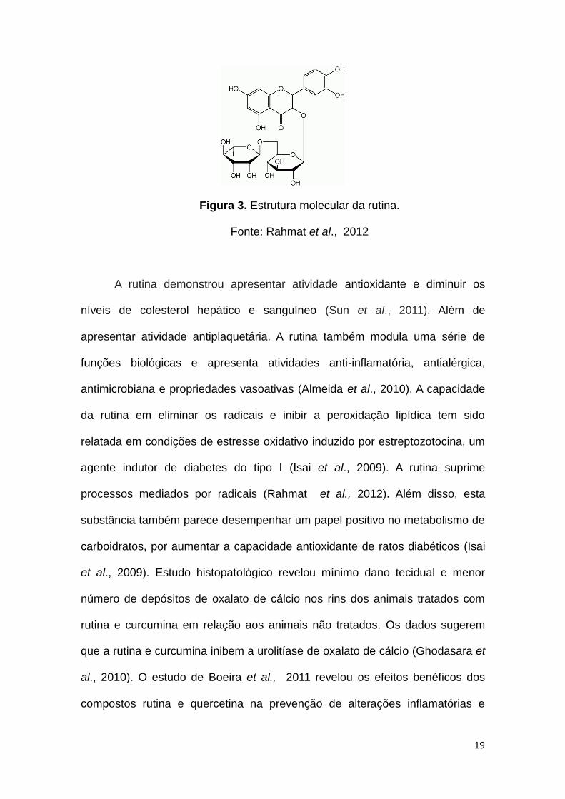
Figura 3. Estrutura molecular da rutina.
Fonte: Rahmat et al., 2012
A rutina demonstrou apresentar atividade antioxidante e diminuir os
níveis de colesterol hepático e sanguíneo (Sun et al., 2011). Além de
apresentar atividade antiplaquetária. A rutina também modula uma série de
funções biológicas e apresenta atividades anti-inflamatória, antialérgica,
antimicrobiana e propriedades vasoativas (Almeida et al., 2010). A capacidade
da rutina em eliminar os radicais e inibir a peroxidação lipídica tem sido
relatada em condições de estresse oxidativo induzido por estreptozotocina, um
agente indutor de diabetes do tipo I (Isai et al., 2009). A rutina suprime
processos mediados por radicais (Rahmat et al., 2012). Além disso, esta
substância também parece desempenhar um papel positivo no metabolismo de
carboidratos, por aumentar a capacidade antioxidante de ratos diabéticos (Isai
et al., 2009). Estudo histopatológico revelou mínimo dano tecidual e menor
número de depósitos de oxalato de cálcio nos rins dos animais tratados com
rutina e curcumina em relação aos animais não tratados. Os dados sugerem
que a rutina e curcumina inibem a urolitíase de oxalato de cálcio (Ghodasara et
al., 2010). O estudo de Boeira et al., 2011 revelou os efeitos benéficos dos
compostos rutina e quercetina na prevenção de alterações inflamatórias e

20
nociceptivas causadas pela cistite hemorrágica em camundongos que
utilizaram ciclofosfamida quando comparados com os grupos tratados com
Mesna.
1.4 QUERCETINA E QUERCETINA EM NANOEMULSÃO
A quercetina (3,5,7,3’- 4’-pentahidroxi flavona) é um flavonóide que se
encontra presente na dieta humana. Os flavonóides são substâncias fenólicas
naturais amplamente presentes em legumes, frutas, óleo de oliva, própolis de
abelha e bebidas como o chá e o vinho tinto, podendo atuar como agentes
antioxidantes em sistemas biológicos. (Sestili et al., 1998; MI et al., 2010).
Além da dieta, a quercetina pode também ser encontrada no Ginkgo biloba,
Hypericum perforatum e Sambucus canadensis (USDA, 2011) e possui uma
variedade de atividades biológicas (Mi et al., 2010), além de funcionar como um
quelante de metais, incluindo o ferro (Sestili et al., 1998; Mi et al., 2010).
Figura 4. Estrutura molecular da quercetina
Fonte: Pecivová et al., 2012
Vários estudos têm indicado que a quercetina inibe a expressão das
enzimas ciclooxigenase induzível (COX-2) e óxido nítrico sintase induzível
(iNOS), inibe também a xantina oxidase, a peroxidação lipídica e alguns danos

21
ao DNA causados por H2O2 (Mi et al., 2010). Adicionalmente, possui efeitos
antinociceptivos (Valério et al., 2009). Ensaios in vitro e in vivo, tem
demonstrado que a quercetina e seus derivados, como outros flavonóides,
possuem ação antioxidante, antimutagênica e antitumoral (Atawodi et al.,
2009), diminuindo a citotoxicidade causada pela administração de
doxorrubicina a células de fígados normais (Wang et al., 2012). Este composto
tem atraído considerável atenção por parecer promissor para a terapia e
prevenção de doenças cardíacas e também a diminuição da incidência de AVC
(Pashevin et al., 2011). Além disso, demonstrou inibir o crescimento e a
proliferação de células de câncer colo retal (HT-29, COLO 201, LS 174T, HCT-
116, SW 480 e Caco-2) induzindo apoptose nestes tipos celulares (Araújo et al.,
2011).
Apesar de suas atividades benéficas, a quercetina possui baixa
solubilidade em água que resulta na fraca absorção, baixa biodisponibilidade e,
portanto, limitando seu potencial na aplicação clínica (Wang et al., 2012).
Visto que as características químicas inerentes a quercetina podem
limitar seus efeitos no organismo, novas preparações procuram melhorar a
solubilidade da quercetina através da adição de emulsões lipossomais
aproveitando suas propriedades terapêuticas, pois permite o controle da
liberação do fármaco e a capacidade de proteger o princípio ativo da
degradação (Dora et al., 2011).
O Instituto Nacional do Câncer reconheceu a nanotecnologia como um
campo emergente para revolucionar a detecção, prevenção e tratamento do
câncer (Kavasaki, et al., 2005).

22
As nanopartículas apresentam alta capacidade de transporte e de
viabilidade de várias vias de administração, inclusive oral, proporcionando a
liberação do princípio ativo de forma sustentável no organismo (Aparajita et al.,
2013).
A capacidade que o sistema lipídico tem de incorporar a quercetina foi
avaliada por um método analítico de espectroscopia UV que foi desenvolvido e
validado por Dora e colaboradores (2011). Na etapa de validação da técnica
foram avaliados os parâmetros de linearidade, limites de detecção e
quantificação e especificidade. As análises da amostra branca demonstraram
que o método foi específico, e não houve nenhuma interferência ou
sobreposição dos excipientes com as absorbâncias de nanoemulsão contendo
quercetina (Dora et al., 2011). A nanoemulsão contendo quercetina
desenvolvida apresentou um teor de fármaco de aproximadamente 1000 µg/mL
e uma recuperação de 98 %, indicando uma excelente capacidade de
solubilizar o fármaco devido sua grande área interfacial, sendo que houve um
aumento de solubilidade de 2720 vezes se comparado com a solubilidade da
quercetina em água (Dora et al., 2011). Sabe-se que o tamanho e a distribuição
das nanopartículas são determinantes para sua estabilidade e eficácia na
absorção celular (Aparajita et al., 2013).

23
2 OBJETIVOS
2.1 OBJETIVO GERAL
O presente estudo teve como objetivo testar o efeito do resveratrol,
rutina, quercetina e quercetina em nanoemulsão sobre a neuro e
hepatotoxicidade induzidas pela oxaliplatina em camundongos.
2.2 OBJETIVOS ESPECÍFICOS
a. Padronizar o modelo de esteatose hepática e de neurotoxicidade
induzida pelo quimioterápico oxaliplatina em camundongos, a fim de avaliar os
efeitos de novas estratégias com potencial hepato e neuroprotetor;
b. Avaliar os efeitos dos compostos resveratrol, rutina, quercetina e
quercetina em nanoemulsão sobre a alodínia através da aplicação do
monofilamento de Von Frey;
c. Verificar os efeitos dos compostos resveratrol, rutina, quercetina e
quercetina em nanoemulsão sobre a esteatose hepática induzida pela
oxaliplatina, através de avaliação macroscópica e histopatológica;
d. Analisar os efeitos dos compostos resveratrol, rutina, quercetina e
quercetina em nanoemulsão sobre a atividade das enzimas séricas AST e ALT;
e. Determinar os efeitos dos compostos resveratrol, rutina, quercetina e
quercetina em nanoemulsão sobre a migração de neutrófilos pela medida da
atividade da mieloperoxidase (MPO), em fígados de camundongos tratados
com oxaliplatina.

24
3. ARTIGO CIENTÍFICO
Os resultados do presente trabalho foram submetidos ao periódico
Naunyn-Schmiedeberg's Archives of Pharmacology
Fator de Impacto (ISI Web of Knowledge): 2.10

25
Effects of the compounds resveratrol, rutin, quercetin and quercetin
nanoemulsion on oxaliplatin-induced hepatotoxicity and neurotoxicity in
mice
Running Head: quercetin, resveratrol and rutin effects in neurotoxicity
and steatohepatitis
1Tania E. Schwingel, 1Caroline P. Klein, 1Natalia F. Nicoletti, 3Vinicius Duval da
Silva, 4Cristiana Dora, 4Gabriela Hadrich, 5Cláudia Bicca, 1,2,6Fernanda B.
Morrone.
1Programa de Pós Graduação em Biologia Celular e Molecular; 2Instituto de
Toxicologia e Farmacologia; 3Laboratório de Patologia do Hospital São Lucas
da PUCRS; 6Faculdade de Farmácia, Pontifícia Universidade Católica do Rio
Grande do Sul, Porto Alegre, RS, Brasil; 4 Laboratório de Nanotecnologia
Aplicada a Saúde da Universidade Federal do Rio Grande, Rio Grande, RS,
Brasil; 5Universidade Federal de Ciências da Saúde de Porto Alegre, RS,
Brasil.
Corresponding Author: Fernanda B. Morrone, Faculty of Pharmacy /Institute
of Toxicology and Pharmacology, Pontifícia Universidade Católica do Rio
Grande do Sul, Avenida Ipiranga, 6681, Partenon, 90619-900, Porto Alegre,
RS, Brazil. Phone number: 55 51 3320 3512; Fax number: 5551 3320 3612.
E-mail: [email protected]; [email protected]

26
ABSTRACT
Oxaliplatin (OXA) is a third-generation platinum compound developed with the
purpose of overcoming the limitations of toxicity, tumor resistance and poor oral
bioavailability associated to cisplatin administration. Despite its usefulness,
oxaliplatin-associated neurotoxicity represents the main dose limiting factor of
this drug and until now there is not suitable treatment. Chemotherapy with
oxaliplatin also increases the rate of developing hepatic damages with
inflammatory activity, termed chemotherapy-associated steatohepatitis (CASH).
In the present study, we aimed to compare the effect of a series of antioxidant
compounds on simultaneous development of oxaliplatin-induced hepato and
neurotoxicity in mice. Mice Balb/c were treated with oxaliplatin for 6 weeks, 10
mg/kg, intraperitoneally, resulting in mechanical allodynia and hepatic steatosis.
We administered antioxidants compounds such as rutin (RT) (20 mg/Kg),
resveratrol (RVS) (100 mg/Kg), quercetin (QT) (20 mg/Kg) and querecetin
nanoemulsion (NQT) (20 mg/Kg) daily by gavage to Balb/c, and N-acetyl-
cysteine (NAC) was used as positive control. The treatments with RSV, RUT or
NQT were able to prevent mechanical allodynia when compared to OXA group.
Regarding the effect on steatohepatitis, RVS, QT and NQT almost completely
reversed the mean liver weight increase by OXA. In accordance with these
previous data, histological evaluation depicted attenuation all the features of
hepatic steatosis evaluated in RSV, RUT, QT and NQT groups. On the other
hand, only QT and NQT treatments were able to reduce neutrophils migration
measured by MPO activity. These results suggest that the compounds tested

27
RSV, RU, QT and NQT would be useful for the clinical treatment of neuro and
hepatoxicity induced by oxaliplatin.
Keywords: CASH, neuropathy, polyphenol compounds, oxaliplatin

28
Introduction
Oxaliplatin is a third-generation platinum compound developed with the
purpose of overcoming the limitations of toxicity, tumor resistance and poor oral
bioavailability associated to cisplatin administration (Shah and Dizon, 2009).
Oxiplatin is used in combination with other chemotherapy agents, such as 5-
fluorouracil/folinic acid, leucovorin, irinotecan, and represents a first-line therapy
on colorectal cancer (Culy et al., 2000). This regimen therapy also includes
management options of colorectal liver metastasis (Brouquet et al., 2009),
ovarian (Lee et al., 2013), gastric (Kim et al., 2012), and non-small cell lung
cancer (Shi et al., 2013). Despite its usefulness, this platinum compound
causes severe side effects. Oxaliplatin-associated neurotoxicity represents the
main dose limiting of this drug and, until now, there is no suitable treatment
(Park et al., 2009). Oxaliplatin might be involved in vascular sinusoidal injury
and neurotoxicity, with the development of peripheral sensory neuropathy, which
symptoms are characterized by dysesthaesia and paraesthesia of extremities
and increase with higher doses of oxaliplatin (Ali, 2010; Amptoulach and
Tsavaris, 2011; Culy et al., 2000).
Oxaliplatin also augments the rate of developing hepatic damage with
increased inflammatory activity (Choti, 2009). This might be termed
chemotherapy-associated steatohepatitis (CASH), a most severe form of non-
alcoholic fatty liver disease (NAFLD) (Malaguarnera et al., 2009). The liver
injury in steatohepatitis presents fatty liver infiltration together with liver
necrosis, and it is characterized by the presence of cellular ballooning, micro
and macrovesicular steatosis and acinar and lobular inflammation (Zorzi et al.,
2007). Another pathogenic factor related to steatohepatitis is the overproduction

29
of free radicals, the reactive oxygen species (ROS), owing to oxidative stress
that contribute to further hepatic injury (Sakaguchi et al., 2011). Different ROS
types are able to cause oxidative damage to the DNA, protein and fatty acids
(Koek et al., 2011). Events like lipid peroxidation and reduction of anti-oxidant
enzymes enhance the oxidative stress including platinum-induced toxicity
(Carozzi et al., 2010). Conversely, there are some antioxidative defenses,
enzymatic and non-enzymatic, in order to avoid or reverse this damage by
means of scavenging the ROS (Apel and Hirt, 2004; Rezazadeh et al., 2012).
On the other side, several antioxidants compounds, from diet or produced
endogenously, had shown amelioration of the steatohepatitis and oxaliplatin-
induced neurotoxicity via reducing the oxidative stress (Koek et al., 2011).
Flavonoids are natural polyphenol substances reported for their powerful
protective activities on different tissues and for their activity against cancer
progression, especially because their antioxidant activities or scavenging
properties. They are present in vegetables, fruits, olive oil, bee propolis and
beverages such as green tea and red wine (Mi et al., 2010; Sestili et al., 1998).
Quercetin, resveratrol and rutin are the major dietary polyphenol compounds
with potent antioxidant activities. Resveratrol can interact with multiple
molecular targets due its simply molecular structure (Calamini et al., 2010;
Delmas et al., 2013; Leiro et al., 2010; Svajger and Jeras, 2012). Recent
findings reveal neoplastic cell growth and proliferation, apoptosis (Ikeda et al.,
2013), inflammation (Leiro et al., 2010), invasion (Chen et al., 2013),
angiogenesis and metastasis (Ganapathy et al., 2010) as cellular targets of
resveratrol. Presently, resveratrol is commercially available as a safe and well
tolerated dietary supplement (Bhatt et al., 2012). Rutin is a glycoside flavone

30
that also modulates a variety of biological functions having activities in oxidative
stress (Patil et al., 2013), inflammatory process and in the reduction of blood
and hepatic cholesterol (Al-Rejaie et al., 2013). Its ability in scavenging free
radicals and inhibiting lipid peroxidation implicated in the etiology of some
pathologies has been shown (Patil et al., 2013). Quercetin has a variety of
actions besides the antioxidant activity, highlighted by antimutagenic (Gupta et
al., 2010) and antitumorigenic (Wang et al., 2012b) properties. Despite its
benefits, quercetin has low solubility in water resulting in poor absorption, low
bioavailability and therefore limiting its potential clinical application (Aebi, 1984;
Wang et al., 2013; Wang et al., 2012a). Thus, the development of a drug
delivery system containing quercetin can improve its solubility, absorption, and
therapeutic index (Dora et al., 2012).
Currently there are few pharmacological options available of
steatohepatitis and peripheral sensory neuropathy induced by oxaliplatin,
making necessary to identify new nontoxic and effective pharmacological
alternatives for the treatment of these disorders. Therefore, the aim of the study
was to compare the effect of the compounds resveratrol, rutin, quercetin and
quercetin nanoemulsion on simultaneous development of oxaliplatin-induced
hepato and neurotoxicity in mice.
Materials and Methods
Drugs and reagents
The following drugs and reagents were used: oxaliplatin (Sanofi-Aventis,
France); carboxymethylcellulose (Natural Pharma), hydrogenated soybean
lecithin (Lipoid), 12-hydroxystearic acid–polyethylene glycol copolymer (Basf),

31
hexadecyltrimethyl ammonium bromide, N-acetylcysteine (NAC), castor oil and
tetramethylbenzidine (Sigma-Aldrich, St. Louis, USA). NaPO4, H2O2, NaCl and
Tween 20 were all purchased from Merck (Haar, Germany). The compounds
resveratrol (RSV), rutin (RU) and quercetin (QT) were obtained from Sigma
Chemical Co. (purity grade >95%). Quercetin nanoemulsion (NQT) was
synthesized in the Laboratory for Nanotechnology Applied to Health, Federal
University of Rio Grande, Brazil. The compounds dilutions were made in NaCl
0.9% (saline solution). Quercetin was diluted in carboxymethylcellulose 0,2%.
Preparation of nano quercetin by solvent diffusion technique hot
The quercetin nanoemulsion (NQT) was prepared by a hot solvent
diffusion method associated to phase inversion technique (Dora et al., 2012).
Briefly, a solution containing quercetin, castor oil and lecithin in acetone: ethanol
(60:40, v / v) at 60°C was added an aqueous phase containing surfactant 12-
hydroxystearic acid–polyethylene glycol copolymer previously heated at 80°C
under magnetic stirring at 700 rpm. The resulting colloidal dispersion was
cooled to room temperature under magnetic stirring, the solvent was
evaporated under reduced pressure, and the final volume was adjusted to 20
mL. Finally, the dispersion was filtered with an 8 microns membrane. The
formulations were made in triplicate. The quercetin nanoemulsion
characterizations were (i) analysis of particle size and polydispersity index (PDI)
by dynamic light scattering (Zetasizer), (ii) analysis of zeta potential by Laser-
Doppler anemometry (Zetaziser) and (iii) analysis of quercetin content and
recovery by UV spectrophotometric method.

32
Animals
Male BALB/c mice (25 to 30 g) were obtained from the Federal University
of Pelotas (Brazil). All animals were maintained under controlled temperature
(22+2oC) and and humidity (60 to 70%), with 12-hour light-dark cycle, with food
and water ad libitum. All animal procedures were performed according with the
Guide for the Care and use of Laboratory Animals, published by National
Institutes for Health (NIH). The experimental protocol was approved by the
Local Animal Ethics Committee (approval number 10/00168). The number of
animals and the intensity of noxious stimuli were the minimum necessary to
demonstrate the consistent effects of the drug treatments.
Oxaliplatin-induced hepato- and neurotoxicity model in mice
The model to induce steatohepatitis (SH) and neurotoxicity in mice was
adapted from Keizman et al (2010). For the OXA group, mice received six
injections of oxaliplatin (10 mg/kg, i.p.), or saline (10 ml/kg, i.p.) for the control
group (SAL), on days 1, 8, 15, 22, 29 and 36. On day 43, the animals were
euthanized by deep anesthesia with isoflurane and the samples (blood and
liver) were collected for further analysis.
Experimental design
The animals were randomly divided into eight treatment groups (n=5): i)
SAL+SAL; ii) OXA+ SAL; iii) OXA+ NAC (200 mg/kg, p.o.) (Thong-Ngam et al.,
2007); iv) OXA+ RSV (100 mg/kg, po); v) OXA+RU (20 mg/kg, po); vi) OXA +
WN (10 ml/kg, p.o.), vii) OXA+ QT (20 mg/kg, p.o.); and viii) OXA+ NQT (20
mg/kg, p.o.). All compounds were administrated daily, during 42 days.

33
Behavioral tests (mechanical allodynia) were performed weekly, after 30 min
post oxaliplatin injection. The animals were fed daily until the day 42 and were
weighed weekly.
Mechanical nociceptive threshold
To evaluate mechanical allodynia threshold, Von Frey monofilaments
(Stoelting, USA) corresponding to 0.6 g were applied up vertically into the right
hind paw of mice with sufficient pressure. The mice were housed 30 min before
the measurement on a wire mesh floor under an acrylic box. Some criteria were
followed: i) paw withdrawal was considered when the animal completely remove
its paw, ii) each animal was stimulated for 10 times with duration of 5 s in each
stimulation, iii) each withdrawal was recorded as 10% of response.
Histopathological analysis
For histological tissue analysis, the tissues removed were fixed in 10%
formalin, processed and then were dehydrated in increasing concentrations of
ethanol (70-100%) and embedded in paraffin. Sections of 5 µm were made and
the slides were stained and mounted in Entellan (Merck, SP, Brazil) at the
Laboratory of Pathology of UFCSPA. Livers were submitted to hematoxilin and
eosin (H&E) staining, performed according standard protocol. The samples
were examined by a pathologist blinded to the experimental groups on a
fluorescent microscope (Zeiss Axioskop 40 equipped with a CoolSnap Pro cf
CCD camera (Media Cybernetics, Rockville, USA) based on criteria described
by Tannapfel et al ( 2010). All images were obtained under magnification of
400X and in a single day, with white, black level calibration and optical density
34
calibration performed and recorded to avoid any shifts in the calibration of light
parameters of optical microscope and thus, maintaining the same configurations
for all images. A total of ten images were used for quantitative assessment in
the Image Pro-Plus 4.5.1 (Media Cybernetics, Rockville, USA). Density was
evaluated as integrated optical density (IOD) (area x optical density), and
roundness was counted as perimeter^2/4 x PI x area.
Myeloperoxidase activity
Myeloperoxidase (MPO) was measured according to the method
described by (Fernandes et al., 2003), to verify neutrophil recruitment. Portion
of the liver was frozen at -80oC immediately after removed. Before analyses the
tissues were partially thawed and homogenized into an ice-containing box in 5%
EDTA buffer/NaCl solution (pH 4.7) and centrifuged at 10.000 RPM for 15
minutes, 4°C. The pellet was suspended in 0.5% haxadecilmetil ammonium
bromide buffer (pH 5.4) and the samples were centrifuged (10.000 RPM, 15
min, 4°C). The supernatant was used (20 µl) for enzymatic reaction of MPO.
Detection was carried out in the presence of tetramethylbenzidine (1.6 mM),
Na3PO4 (80 mM) and hydrogen peroxide (0.3 mM). The absorbance was
measured at 595 nm and results expressed as optical density (OD) per mg of
tissue.
Serum analysis of AST and ALT
Whole blood was centrifuged at 300 g for 10 min; the supernatant
corresponding to the serum was removed and frozen at -80oC until analysis.
The assays to measure alanine-aminotransferase (ALT) and aspartate-
35
aminotransferase (AST), as indirect indicators of liver damage, were performed
following the requirements of manufacturer (Labtest, Brazil). The results were
expressed in U/L.
Statistical analysis
Data are presented as mean + SEM. Statistical analysis were made by
one-way analysis of variance (ANOVA), followed by Bonferroni post hoc test. P
values < 0.05 were considered as significant. Statistical calculations were
performed using GraphPad software (GraphPad Instat, version 4, USA).
Results
In this study we have evaluated the effect of different antioxidant
compounds in neurotoxicity induced by oxaliplatin injection in mice. The
baseline withdrawal thresholds in the mechanical tests in all experimental
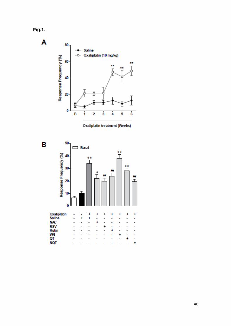
groups before OXA injection were similar. Oxaliplatin (10 mg/kg, i.p., once a
week for 6 weeks) significantly increased the frequency of paw withdrawal
compared to the control group in the von Frey test on day 42 (p<0.0001). This
significant increased frequency had its onset on the 4th week and persisted
throughout the weeks 5 and 6 of OXA-treatment (Figure 1A). Interestingly, all
antioxidants treated groups, reverted the increase of the paw withdrawal
responses caused by OXA injection on day 42, in a similar way of the positive
control NAC (Figure 1B). WN and QT groups did not significantly affect the
mechanical allodynia in any evaluated period of time.
In order to evaluate hepatotoxicity in this mice model induced by OXA,
body and liver weights were analyzed. Mean variation of body weight gain of

36
animals increased over time in control (SAL) and QT-treated group, but
remained unchanged in all other groups (Figure 2A). No animal died during the
course of the experiments. Mean liver weight in OXA group increased 20.3%
relative to control group, and when mice were treated with NAC, RSV, QT and
NQT liver weight reduced significantly (11%, 21% e 17.5%, respectively) in
comparison to OXA group (Figure 2B).
Histological examination of the liver sample slides stained with H&E was
made by quantitative and qualitative analysis. Histopathological findings were
featured according to the following changes: steatosis, hepatocyte ballooning
and inflammation. Ballooned hepatocytes were seen mainly in OXA and WN
group (Figure 3B and 3F). Their microscopic features are presented as
enlarged hepatocytes with swollen vacuolated cytoplasm and the
hyperchromatic nucleus occasionally appears with a prominent nucleolus.
Sinusoidal dilatation between the hepatocytes in these groups was also
observed. In addition, an attenuation of all the features above mentioned in
NAC, RSV, RUT, QT and NQT groups (Figures 3C, D, E, G and H, respectively)
was detected. Performing quantitative analysis, the parameters used to
compare the differences among the groups were: area, density, roundness. The
cytoplasmatic area and total optical density area analysis showed a significant
reduction of the area in OXA group when compared to SAL group; on the other
hand the roundness parameter was not different among the groups. All treated
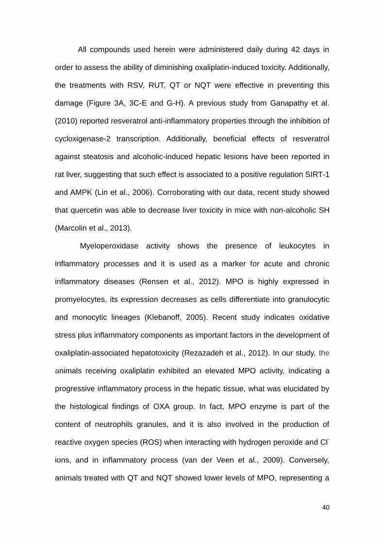
groups augmented area and density when compared to OXA group (Table 1).
OXA treatment increased significantly MPO activity related to the control
group (p < 0.01). Interestingly, QT and NQT treatments were able to reduce

37
neutrophils migration, as indicated by MPO activity (p < 0.05, Figure 4). We did
not observe any differences in the other groups.
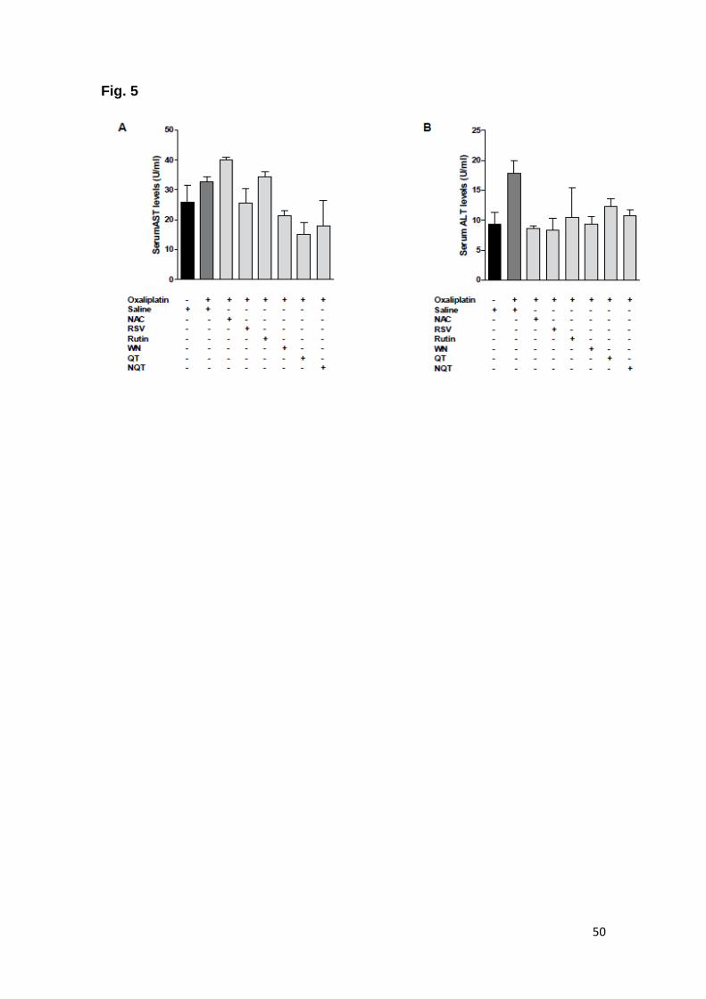
Assessment of transaminases levels (AST and ALT) did not reveal any
significant alteration in mice blood serum after the treatments, although, there
was a clear tendency of diminishing ALT in the treatment groups (Figure 5B).
Discussion
Several studies report that chemotherapy treatment with oxaliplatin can
induce the development of liver damage followed by a progressive stage of SH,
associated to neurotoxicity and its painful consequences (Culy et al., 2000;
Fernandez et al., 2005; Janes et al., 2013; Rubbia-Brandt et al., 2004).
Achieving chemotherapy side effects resolution is a priority and, therefore, the
present study assessed the effects of the antioxidant compounds resveratrol,
rutin, quercetin and quercetin nanoemulsion on steatohepatitis and neurotoxicity
induced by OXA in mice. The mice model presented herein has shown to be
suitable of mimicking clinical features induced by oxaliplatin. In fact, in gross
evaluation, body and liver sizes of OXA group appeared enlarged, and it was
sufficient to produce significant difference in relation to control group (Fig.3).
As mentioned before, neurotoxicity is a very common side effect which
has been associated to the use of antineoplastic agents, especially platinum
derivates (Wang et al., 2007). It has been reported that, since the first oxaliplatin
administration, neurotoxic symptoms appear yielding acute neuropathy and the
ongoing treatment causes cumulative symptoms and worsens the onset
neuropathy, featuring chronic neuropathy (Ali, 2010). After long term oxaliplatin
administration, the worsening of the symptoms is assigned by loss of sensory
38
and motor function. These symptoms are usually associated with pain followed
by a functional impairment in patients (Deuis et al., 2013). Our oxaliplatin-
induced neurotoxicity model resulted in mechanical nociception and the animals
treated with OXA once a week presented a higher frequency of paw withdrawal
in response to mechanical stimulus through Von Frey filaments application. Of
great relevance our data showed that animals receiving OXA plus RSV, RUT,
NQT were able to prevent mechanical allodynia when compared to OXA group.
Two main metabolites of oxaliplatin were identified as responsible by
these side effects: oxalate and platinum (Sakurai et al., 2009). Oxalate is
involved in chelation of extracellular calcium producing an increase in sodium
concentration on neurons input in initial stage of neuropathy (Sakurai et al.,
2009). Platinum is associated with mechanical allodynia due to its neurotoxicity,
causing the formation of morphological changes Pt-DNA in chronic neuropathy,
such as lesions of cell bodies, changes in the nucleus and nucleolus, atrophy in
neurons of the dorsal root ganglia and cell death (Sakurai et al., 2009).
Corroborating to our findings, a previous study from our laboratory has shown
that rutin and quercetin produced a significant attenuation of the nociception
evoked by the antineoplastic cyclophosphamide in mice (Boeira et al., 2011).
Furthermore, quercetin was able to inhibit nociception in a rodent pain model of
colitis (Comalada et al., 2005). Outstandingly, resveratrol exhibited an
antinociceptive effect related to the prevention of COX-2 mRNA increase in
carrageenan-evoked hyperalgesia in rats (Pham-Marcou et al., 2008). In our set
of results it is possible to establish a relationship between the mechanical
nociception test and pathological examination on regard to the amelioration
observed after the treatment in mice, with exception to QT group, which did not

39
show difference in relation to OXA group indicating that the low QT
bioavailability can be a determinant for its use in this condition. On the other
hand, quercetin nonoemulsion formulation lowered the alodynia and liver
histological parameters presented in the OXA group, demonstrating that this
new formulation can be a useful alternative to treat toxicity induced by
oxaliplatin. Dora et al. (2011) showed that quercetin nanoemulsion allows a
controlled releasing of the drug by reducing undesirable effects, and also
increases the drug bioavailability, reaching better therapeutic effects.
We have also investigated the effects of the antioxidant compounds in
hepatotoxicity. Antineoplastic drugs such as platinum compounds are involved
in the increase of membrane-bounded vacuoles containing degenerative
mitochondria, named autophagosome, in nervous tissue; it is believed that the
same occur in hepatic tissue (Carozzi et al., 2010). From histopathological
viewpoint, SH is a progressive morphological spectrum of steatosis (Tannapfel
et al., 2011). It is required at least 5% of fatty deposition within hepatocytes,
ballooning of the hepatocyte and inflammation to be definitely considered SH
(Tannapfel et al., 2010). In this study all histological features founded in OXA
group were assigned to hepatic damage filling in the criteria diagnosis (Figure
3B). In fact, vascular sinusoidal injury appears to have an association with
oxaliplatin use and it is featured by sinusoidal dilatation and erythrocyte
congestion accompanied perisinusoidal fibrosis and fibrotic venular occlusion.
Similar to the results depicted in this study, other studies show tht histological
changes affect mainly the liver parenchyma present in vicinity of perivenular
regions (Hubscher, 2006).
40
All compounds used herein were administered daily during 42 days in
order to assess the ability of diminishing oxaliplatin-induced toxicity. Additionally,
the treatments with RSV, RUT, QT or NQT were effective in preventing this
damage (Figure 3A, 3C-E and G-H). A previous study from Ganapathy et al.
(2010) reported resveratrol anti-inflammatory properties through the inhibition of
cycloxigenase-2 transcription. Additionally, beneficial effects of resveratrol
against steatosis and alcoholic-induced hepatic lesions have been reported in
rat liver, suggesting that such effect is associated to a positive regulation SIRT-1
and AMPK (Lin et al., 2006). Corroborating with our data, recent study showed
that quercetin was able to decrease liver toxicity in mice with non-alcoholic SH
(Marcolin et al., 2013).
Myeloperoxidase activity shows the presence of leukocytes in
inflammatory processes and it is used as a marker for acute and chronic
inflammatory diseases (Rensen et al., 2012). MPO is highly expressed in
promyelocytes, its expression decreases as cells differentiate into granulocytic
and monocytic lineages (Klebanoff, 2005). Recent study indicates oxidative
stress plus inflammatory components as important factors in the development of
oxaliplatin-associated hepatotoxicity (Rezazadeh et al., 2012). In our study, the
animals receiving oxaliplatin exhibited an elevated MPO activity, indicating a
progressive inflammatory process in the hepatic tissue, what was elucidated by
the histological findings of OXA group. In fact, MPO enzyme is part of the
content of neutrophils granules, and it is also involved in the production of
reactive oxygen species (ROS) when interacting with hydrogen peroxide and Cl-
ions, and in inflammatory process (van der Veen et al., 2009). Conversely,
animals treated with QT and NQT showed lower levels of MPO, representing a

41
decreased neutrophil infiltration and therefore indirectly indicating a reduction of
the inflammatory process, contributing to the decreased tissue damage seen in
liver histopathological analysis.
In summary, the data presented in this study shed new lights on the
positive effects of these compounds in diminishing the adverse effects drawn
out by oxaliplatin. Our results indicate the use of compounds such as RSV, RU,
QT and NQT as effective contributors to avoid oxaliplatin-inducing hepato and
neurotoxicity in a rodent model.

42
Acknowledgments
We thank Dr. Maria Martha Campos for help in the manuscript
corrections. This work was supported by FINEP research grant “Implantação,
Modernização e Qualificação de Estrutura de Pesquisa da PUCRS”
(PUCRSINFRA) # 01.11.0014-00 and PRONEM / FAPERGS grant 11/2037-9.
43
Table 1. Quantitative parameters obtained from liver tissues for histopathological examination
Groups
Area (Sum) Density Roundness
Saline (10 ml/kg/week i.p., for 6 weeks)
Saline 996154 (± 9450.9) 123.66 (± 3.58) 2.53 (± 1.76)
Oxaliplatin (10 mg/kg/week i.p., for 6 weeks)
Saline 864314 (± 6528.7)*** 106.76 (± 3.37)***
2.61 (± 3.20)
NAC 1061611 (± 9600.6)### 120.44 (± 3.05)###
2.63 (± 2.26)
RSV 1000468 (±10789.3)### 124.33 (± 3.28)###
2.66 (± 2.54)
RUT 916160 (± 9335.2)## 122.64 (± 2.54)###
2.52 (± 1.91)
WN 892193 (± 6630.9) 113.57(± 3.65)# 2.58 (± 2.40)
NQT 966363 (± 8544.5)### 127.20 (± 2.83)###
2.80 (± 2.64)
QT 992937 (± 8234.3)### 120.03 (± 2.79)###
2.66 (± 2.47)
All quantitative parameters were obtained from images of histological liver
slides (10 images for each liver, n=5/group; imaged in 400X and counted in
spatial calibration of 400 μm) using Image pro-Plus 4.5.1 software and are
represented as mean ±SEM. (P < 0.001 compared with saline, # P < 0.05
compared with OXA+SAL, ## P < 0.01, and ### P < 0.0001 compared with
OXA+SAL).
Abbreviations: NAC, N-acetylcysteine; RSV, resveratrol; RUT, rutin; WN, White
nano; NQT, quercetin nanoemulsion; QT, quercetin.

44
Figure Legends Figure 1. Behavioral tests for assessing nociception. Von Frey filaments of
different weight were used to receive a row of six responses with different
filaments per animal and experiment day. A baseline of paw withdrawal
threshold was measured of each group before treatment. (A) cumulative paw
withdrawal frequency differences among SAL and OXA group; and (B)
mechanical allodynia. Two-way analysis of variance (ANOVA) was used to
calculate the difference of groups compared to control at one point of time and
to make a comparison within one group at different points of time. *** P < 0.001
relative to control (SAL); # P < 0.05, ## P < 0.01 and ### P< 0.001 relative to
OXA group.
Figure 2. Body weight gain variation in the course of study (A), and liver weight
at the end of the study (B). Data represent mean ± SEM. One-way ANOVA,* P <
0.05 and *** P < 0.0001 relative to control (SAL); # P < 0.05 and ### P < 0.001
relative to OXA group.
Figure 3. Representative images of histological evaluation of liver tissue
stained with hematoxylin and eosin. (A) SAL, (B) OXA, (C) NAC, (D) RSV, (E)
RU, (F) WN, (G) QT and (H) NQT groups. 400X magnification.
Figure 4. Effect of antioxidant compounds in myeloperoxidase (MPO) activity in
the liver. Each column represents the mean of 5 animals, and the vertical lines
show the SEM. * P < 0.05 relative to control (SAL). ## P < 0.01 relative to OXA.
45
Figure 5. Transaminases and ALT (A) AST (B) levels were determined from
blood serum. Data represent mean ± SEM. One-way ANOVA.
46
Fig.1.

47
Fig.2.

48
Fig.3.

49
Fig.4.
50
Fig. 5
51
References Aebi, H., 1984. Catalase in vitro. Methods in enzymology 105, 121-126. Al-Rejaie, S.S., Aleisa, A.M., Sayed-Ahmed, M.M., Al-Shabanah, O.A.,
Abuohashish, H.M., Ahmed, M.M., Al-Hosaini, K.A., Hafez, M.M., 2013. Protective effect of rutin on the antioxidant genes expression in hypercholestrolemic male Westar rat. BMC complementary and alternative medicine 13, 136.
Ali, B.H., 2010. Amelioration of oxaliplatin neurotoxicity by drugs in humans and experimental animals: a minireview of recent literature. Basic & clinical pharmacology & toxicology 106, 272-279.
Amptoulach, S., Tsavaris, N., 2011. Neurotoxicity caused by the treatment with platinum analogues. Chemotherapy research and practice 2011, 843019.
Apel, K., Hirt, H., 2004. Reactive oxygen species: metabolism, oxidative stress, and signal transduction. Annual review of plant biology 55, 373-399.
Bhatt, J.K., Thomas, S., Nanjan, M.J., 2012. Resveratrol supplementation improves glycemic control in type 2 diabetes mellitus. Nutr Res 32, 537-541.
Boeira, V.T., Leite, C.E., Santos, A.A., Jr., Edelweiss, M.I., Calixto, J.B., Campos, M.M., Morrone, F.B., 2011. Effects of the hydroalcoholic extract of Phyllanthus niruri and its isolated compounds on cyclophosphamide-induced hemorrhagic cystitis in mouse. Naunyn-Schmiedeberg's archives of pharmacology 384, 265-275.
Brouquet, A., Benoist, S., Julie, C., Penna, C., Beauchet, A., Rougier, P., Nordlinger, B., 2009. Risk factors for chemotherapy-associated liver injuries: A multivariate analysis of a group of 146 patients with colorectal metastases. Surgery 145, 362-371.
Calamini, B., Ratia, K., Malkowski, M.G., Cuendet, M., Pezzuto, J.M., Santarsiero, B.D., Mesecar, A.D., 2010. Pleiotropic mechanisms facilitated by resveratrol and its metabolites. The Biochemical journal 429, 273-282.
Carozzi, V.A., Marmiroli, P., Cavaletti, G., 2010. The role of oxidative stress and anti-oxidant treatment in platinum-induced peripheral neurotoxicity. Current cancer drug targets 10, 670-682.
Chen, Y.J., Chen, Y.Y., Lin, Y.F., Hu, H.Y., Liao, H.F., 2013. Resveratrol inhibits alpha-melanocyte-stimulating hormone signaling, viability, and invasiveness in melanoma cells. Evidence-based complementary and alternative medicine : eCAM 2013, 632121.
Choti, M.A., 2009. Chemotherapy-associated hepatotoxicity: do we need to be concerned? Annals of surgical oncology 16, 2391-2394.
Comalada, M., Camuesco, D., Sierra, S., Ballester, I., Xaus, J., Galvez, J., Zarzuelo, A., 2005. In vivo quercitrin anti-inflammatory effect involves release of quercetin, which inhibits inflammation through down-regulation of the NF-kappaB pathway. European journal of immunology 35, 584-592.
Culy, C.R., Clemett, D., Wiseman, L.R., 2000. Oxaliplatin. A review of its pharmacological properties and clinical efficacy in metastatic colorectal cancer and its potential in other malignancies. Drugs 60, 895-924.
52
Delmas, D., Aires, V., Colin, D.J., Limagne, E., Scagliarini, A., Cotte, A.K., Ghiringhelli, F., 2013. Importance of lipid microdomains, rafts, in absorption, delivery, and biological effects of resveratrol. Annals of the New York Academy of Sciences 1290, 90-97.
Deuis, J.R., Zimmermann, K., Romanovsky, A.A., Possani, L.D., Cabot, P.J., Lewis, R.J., Vetter, I., 2013. An animal model of oxaliplatin-induced cold allodynia reveals a crucial role for Na1.6 in peripheral pain pathways. Pain.
Dora, C.L., Silva, L.F., Putaux, J.L., Nishiyama, Y., Pignot-Paintrand, I., Borsali, R., Lemos-Senna, E., 2012. Poly(ethylene glycol) hydroxystearate-based nanosized emulsions: effect of surfactant concentration on their formation and ability to solubilize quercetin. Journal of biomedical nanotechnology 8, 202-210.
Fernandes, E.S., Passos, G.F., Campos, M.M., Araujo, J.G., Pesquero, J.L., Avelllar, M.C., Teixeira, M.M., Calixto, J.B., 2003. Mechanisms underlying the modulatory action of platelet activating factor (PAF) on the upregulation of kinin B1 receptors in the rat paw. British journal of pharmacology 139, 973-981.
Fernandez, F.G., Ritter, J., Goodwin, J.W., Linehan, D.C., Hawkins, W.G., Strasberg, S.M., 2005. Effect of steatohepatitis associated with irinotecan or oxaliplatin pretreatment on resectability of hepatic colorectal metastases. Journal of the American College of Surgeons 200, 845-853.
Ganapathy, S., Chen, Q., Singh, K.P., Shankar, S., Srivastava, R.K., 2010. Resveratrol enhances antitumor activity of TRAIL in prostate cancer xenografts through activation of FOXO transcription factor. PloS one 5, e15627.
Gupta, C., Vikram, A., Tripathi, D.N., Ramarao, P., Jena, G.B., 2010. Antioxidant and antimutagenic effect of quercetin against DEN induced hepatotoxicity in rat. Phytotherapy research : PTR 24, 119-128.
Hubscher, S.G., 2006. Histological assessment of non-alcoholic fatty liver disease. Histopathology 49, 450-465.
Ikeda, K., Torigoe, T., Matsumoto, Y., Fujita, T., Sato, N., Yotsuyanagi, T., 2013. Resveratrol inhibits fibrogenesis and induces apoptosis in keloid fibroblasts. Wound repair and regeneration : official publication of the Wound Healing Society [and] the European Tissue Repair Society 21, 616-623.
Janes, K., Doyle, T., Bryant, L., Esposito, E., Cuzzocrea, S., Ryerse, J., Bennett, G.J., Salvemini, D., 2013. Bioenergetic deficits in peripheral nerve sensory axons during chemotherapy-induced neuropathic pain result from peroxynitrite mediated post-translational nitration of mitochondrial superoxide dismutase. Pain.
Keizman, D., Maimon, N., Ish-Shalom, M., Buchbut, D., Inbar, M., Klein, B., Bernheim, J., Goldiner, I., Leikin-Frenkel, A., Konikoff, F., 2010. An animal model for chemotherapy-associated steatohepatitis and its prevention by the oral administration of fatty acid bile acid conjugate. Cancer 116, 251-255.
Kim, H.S., Kim, J.H., Kim, H.J., Jang, H.J., Kim, J.B., Kim, J.W., Jung, S.Y., Kim, B.C., Yang, D.H., Park, S., Kim, K.J., Lee, S.I., Zang, D.Y., 2012. Oxaliplatin, 5-fluorouracil and leucovorin (modified FOLFOX-6) as first-
53
line chemotherapy for advanced gastric cancer patients with poor performance status. Oncology letters 3, 425-428.
Klebanoff, S.J., 2005. Myeloperoxidase: friend and foe. Journal of leukocyte biology 77, 598-625.
Koek, G.H., Liedorp, P.R., Bast, A., 2011. The role of oxidative stress in non-alcoholic steatohepatitis. Clinica chimica acta; international journal of clinical chemistry 412, 1297-1305.
Lee, H.J., Kim, H.S., Park, N.H., Chung, H.H., Kim, J.W., Song, Y.S., 2013. Feasibility of Oxaliplatin, Leucovorin, and 5-Fluorouracil (FOLFOX-4) Chemotherapy in Heavily Pretreated Patients with Recurrent Epithelial Ovarian Cancer. Cancer research and treatment : official journal of Korean Cancer Association 45, 40-47.
Leiro, J.M., Varela, M., Piazzon, M.C., Arranz, J.A., Noya, M., Lamas, J., 2010. The anti-inflammatory activity of the polyphenol resveratrol may be partially related to inhibition of tumour necrosis factor-alpha (TNF-alpha) pre-mRNA splicing. Molecular immunology 47, 1114-1120.
Lin, P.C., Lee, M.Y., Wang, W.S., Yen, C.C., Chao, T.C., Hsiao, L.T., Yang, M.H., Chen, P.M., Lin, K.P., Chiou, T.J., 2006. N-acetylcysteine has neuroprotective effects against oxaliplatin-based adjuvant chemotherapy in colon cancer patients: preliminary data. Supportive care in cancer : official journal of the Multinational Association of Supportive Care in Cancer 14, 484-487.
Malaguarnera, M., Di Rosa, M., Nicoletti, F., Malaguarnera, L., 2009. Molecular mechanisms involved in NAFLD progression. J Mol Med (Berl) 87, 679-695.
Marcolin, E., Forgiarini, L.F., Rodrigues, G., Tieppo, J., Borghetti, G.S., Bassani, V.L., Picada, J.N., Marroni, N.P., 2013. Quercetin decreases liver damage in mice with non-alcoholic steatohepatitis. Basic & clinical pharmacology & toxicology 112, 385-391.
Mi, Y., Zhang, C., Li, C., Taneda, S., Watanabe, G., Suzuki, A.K., Taya, K., 2010. Quercetin attenuates oxidative damage induced by treatment of embryonic chicken spermatogonial cells with 4-nitro-3-phenylphenol in diesel exhaust particles. Bioscience, biotechnology, and biochemistry 74, 934-938.
Park, S.B., Goldstein, D., Lin, C.S., Krishnan, A.V., Friedlander, M.L., Kiernan, M.C., 2009. Acute abnormalities of sensory nerve function associated with oxaliplatin-induced neurotoxicity. Journal of clinical oncology : official journal of the American Society of Clinical Oncology 27, 1243-1249.
Patil, S.L., Mallaiah, S.H., Patil, R.K., 2013. Antioxidative and radioprotective potential of rutin and quercetin in Swiss albino mice exposed to gamma radiation. Journal of medical physics / Association of Medical Physicists of India 38, 87-92.
Pham-Marcou, T.A., Beloeil, H., Sun, X., Gentili, M., Yaici, D., Benoit, G., Benhamou, D., Mazoit, J.X., 2008. Antinociceptive effect of resveratrol in carrageenan-evoked hyperalgesia in rats: prolonged effect related to COX-2 expression impairment. Pain 140, 274-283.
Rensen, S.S., Bieghs, V., Xanthoulea, S., Arfianti, E., Bakker, J.A., Shiri-Sverdlov, R., Hofker, M.H., Greve, J.W., Buurman, W.A., 2012. Neutrophil-derived myeloperoxidase aggravates non-alcoholic

54
steatohepatitis in low-density lipoprotein receptor-deficient mice. PloS one 7, e52411.
Rezazadeh, A., Yazdanparast, R., Molaei, M., 2012. Amelioration of diet-induced nonalcoholic steatohepatitis in rats by Mn-salen complexes via reduction of oxidative stress. Journal of biomedical science 19, 26.
Rubbia-Brandt, L., Audard, V., Sartoretti, P., Roth, A.D., Brezault, C., Le Charpentier, M., Dousset, B., Morel, P., Soubrane, O., Chaussade, S., Mentha, G., Terris, B., 2004. Severe hepatic sinusoidal obstruction associated with oxaliplatin-based chemotherapy in patients with metastatic colorectal cancer. Annals of oncology : official journal of the European Society for Medical Oncology / ESMO 15, 460-466.
Sakaguchi, S., Takahashi, S., Sasaki, T., Kumagai, T., Nagata, K., 2011. Progression of alcoholic and non-alcoholic steatohepatitis: common metabolic aspects of innate immune system and oxidative stress. Drug metabolism and pharmacokinetics 26, 30-46.
Sakurai, M., Egashira, N., Kawashiri, T., Yano, T., Ikesue, H., Oishi, R., 2009. Oxaliplatin-induced neuropathy in the rat: involvement of oxalate in cold hyperalgesia but not mechanical allodynia. Pain 147, 165-174.
Sestili, P., Guidarelli, A., Dacha, M., Cantoni, O., 1998. Quercetin prevents DNA single strand breakage and cytotoxicity caused by tert-butylhydroperoxide: free radical scavenging versus iron chelating mechanism. Free radical biology & medicine 25, 196-200.
Shah, N., Dizon, D.S., 2009. New-generation platinum agents for solid tumors. Future Oncol 5, 33-42.
Shi, S.B., Hu, R.H., Qi, J.L., Tang, X.Y., Tian, J., Li, R., Chang, C.X., 2013. Treatment of stage IIIb/IV non-small cell lung cancer with Pemetrexed plus Oxaliplatin after failure of Erlotinib as second-line treatment. Med Oncol 30, 550.
Svajger, U., Jeras, M., 2012. Anti-inflammatory effects of resveratrol and its potential use in therapy of immune-mediated diseases. International reviews of immunology 31, 202-222.
Tannapfel, A., Denk, H., Dienes, H.P., Langner, C., Schirmacher, P., Trauner, M., Flott-Rahmel, B., 2010. [Histopathological diagnosis of non-alcoholic and alcoholic fatty liver disease. Grade 2 consensus-based guidelines]. Der Pathologe 31, 225-237.
Tannapfel, A., Denk, H., Dienes, H.P., Langner, C., Schirmacher, P., Trauner, M., Flott-Rahmel, B., 2011. Histopathological diagnosis of non-alcoholic and alcoholic fatty liver disease. Virchows Archiv : an international journal of pathology 458, 511-523.
Thong-Ngam, D., Samuhasaneeto, S., Kulaputana, O., Klaikeaw, N., 2007. N-acetylcysteine attenuates oxidative stress and liver pathology in rats with non-alcoholic steatohepatitis. World journal of gastroenterology : WJG 13, 5127-5132.
van der Veen, B.S., de Winther, M.P., Heeringa, P., 2009. Myeloperoxidase: molecular mechanisms of action and their relevance to human health and disease. Antioxidants & redox signaling 11, 2899-2937.
Wang, G., Song, L., Wang, H., Xing, N., 2013. Quercetin synergizes with 2-methoxyestradiol inhibiting cell growth and inducing apoptosis in human prostate cancer cells. Oncology reports 30, 357-363.
55
Wang, G., Wang, J.J., Yang, G.Y., Du, S.M., Zeng, N., Li, D.S., Li, R.M., Chen, J.Y., Feng, J.B., Yuan, S.H., Ye, F., 2012a. Effects of quercetin nanoliposomes on C6 glioma cells through induction of type III programmed cell death. International journal of nanomedicine 7, 271-280.
Wang, G., Zhang, J., Liu, L., Sharma, S., Dong, Q., 2012b. Quercetin potentiates doxorubicin mediated antitumor effects against liver cancer through p53/Bcl-xl. PloS one 7, e51764.
Wang, W.S., Lin, J.K., Lin, T.C., Chen, W.S., Jiang, J.K., Wang, H.S., Chiou, T.J., Liu, J.H., Yen, C.C., Chen, P.M., 2007. Oral glutamine is effective for preventing oxaliplatin-induced neuropathy in colorectal cancer patients. The oncologist 12, 312-319.
Zorzi, D., Laurent, A., Pawlik, T.M., Lauwers, G.Y., Vauthey, J.N., Abdalla, E.K., 2007. Chemotherapy-associated hepatotoxicity and surgery for colorectal liver metastases. The British journal of surgery 94, 274-286.
56
4. CONSIDERAÇÕES FINAIS
O câncer é uma doença crônico-degenerativa com impacto econômico
significativo para a sociedade e, atualmente, vem sendo tratado como um
grande problema de saúde púbica (Caponero et al., 2007). Novas terapias têm
sido descobertas e vêm contribuindo muito para a qualidade e sobrevida dos
pacientes, mas os estes ainda sofrem com vários efeitos colaterais dos
fármacos (Authier et al., 2009). Tais efeitos são decorrentes, na sua maioria,
da ação dos antineoplásicos, e podem levar à interrupção do tratamento e/ou
redução de dose (Sonis et al., 2004). A falha na adesão ao tratamento por parte
do paciente se deve principalmente à ação não seletiva dos medicamentos
utilizados para tratar o câncer, os quais atingem também as células normais,
comprometendo assim a eficácia da terapia (Bittencourt et al., 2004).
Dentre as classes farmacológicas mais utilizadas, destacam-se os
compostos da platina, os quais são muito prescritos para o tratamento de
tumores sólidos humanos. Pertencente a este grupo está a oxaliplatina,
fármaco de terceira geração, com estrutura similar à cisplatina (Argyrion et al.,
2008), e que possui efeito citotóxico semelhante aos demais análogos da
platina (Foltinová et al.,2009).
Deste modo, o presente estudo avaliou os efeitos de alguns compostos
antioxidantes como resveratrol, rutina, quercetina e quercetina em
nanoemulsão, sobre a esteato-hepatite e neurotoxicidade induzida pela
oxaliplatina em camundongos Balb/c. Inicialmente realizou-se a padronização
do modelo de neuro-hepatotoxicidade induzida pelo quimioterápico em
camundongos, o qual se mostrou adequado apresentando as principais

57
características clínicas encontradas na neuropatia e na esteato-hepatite
induzida pela oxaliplatina.
Na primeira etapa do presente estudo constatou-se que os animais
apresentaram sintomas de neuropatia peculiar ao uso da oxaliplatina. Os
resultados obtidos mostraram que os compostos antioxidantes resveratrol,
quercetina em nanoemulsão e rutina foram capazes de reverter a alodinia
mecânica induzida por oxaliplatina. De modo muito interessante o resveratrol e
a quercetina em nanoemulsão inibiram a alodínia mecânica em maior
intensidade. Corroborando com nossos dados, um estudo recente demonstrou
que nos primeiros estágios da neuropatia aguda induzida pela oxaliplatina, as
fibras nociceptivas periféricas são lesadas pelo estresse oxidativo e que
substâncias antioxidantes são capazes de prevenir este efeito (Joseph et al.,
2008).
De fato, a neurotoxicidade causada pela oxaliplatina pode apresentar-se
de duas formas distintas: aguda ou crônica, com sintomas iniciais de
parestesias distais e periorais, tais como câimbras, espasmos tetânicos,
náuseas, vômitos, entre outros (Kiernam et al., 2007). Por outro lado, a
neuropatia crônica é caracterizada pela perda da função motora e sensorial,
semelhante ao sintoma neurológico gerado pela cisplatina, ou após a
administração de oxaliplatina, a longo prazo (Deuis et al., 2013; McWhinney et
al., 2009). Doses cumulativas podem levar à ataxia sensitiva e
comprometimento funcional, levando à limitação nas atividades de vida diárias
do paciente (Park et al., 2009). E a toxicidade crônica pode progredir mesmo
após a descontinuação do tratamento (Sprowl et al., 2013).

58
Um segundo achado importante deste estudo foi demonstrado através
do efeito tóxico no fígado, associado à esteato-hepatite induzida pela
oxaliplatina. Em nosso modelo experimental houve indícios de dano hepático
induzido pelo uso do quiomioterápico, observado através da análise histológica
e pela diminuição do peso dos fígados nos animais quando tratados com RSV,
RT, QT e NQT. Além disso, a reversão da hepatoxicidade quando os animais
foram tratados com os compostos testados assemelho-se àquela encontrada
no controle positivo, NAC. Há evidências de que o mecanismo de toxicidade
hepática esteja relacionado ao estresse oxidativo prejudicando os mecanismos
de defesa e iniciando um processo inflamatório, degenerativo e de fibrose
(Ramirez et al., 2010). O envolvimento do estresse oxidativo nesta patologia
sugere que os antioxidantes possam ter efeitos benéficos no tratamento
(Rezazadeh et al., 2012).
Além disso, houve uma evidente tendência à diminuição dos níveis de
ALT nos grupos tratados com os compostos em relação ao grupo OXA,
confirmando assim a ação protetora das substâncias testadas. Resultados
semelhantes foram também identificados no estudo de Kumar et al., (2013)
onde a quercetina apresentou uma propriedade hepatoprotora observada nos
parâmetros medidos no soro de animais tratados comparativamente ao grupo
induzido por cloroquina.
Com relação à análise da atividade da mieloperoxidase no fígado dos
camundongos, observou-se que o grupo de animais tratados com a oxaliplatina
teve um aumento nos valores de MPO em relação ao grupo SAL. Enquanto
que os grupos tratados com os compostos quercetina e quercetina em
nanoemulsão apresentaram valores diminuídos, proveniente da redução de
59
infiltrados de neutrófilos, indicando assim uma diminuição do processo
inflamatório no fígado.
Analisando os resultados apenas entre os grupos quercetina e
quercetina em nanoemulsão observou-se valores diminuídos no grupo da
quercetina em nanoemulsão com relação à alodinia mecânica, análise de ALT e
MPO quando comparados com o grupo tratado com quercetina na sua forma
livre. De acordo com o estudo de Dora e colaboradores (2011), o
desenvolvimento da quercetina em nanoemulsões possibilita um sistema que
tenha capacidade de proteger o fármaco quanto a sua degradação, permite um
controle de liberação do fármaco reduzindo os efeitos indesejáveis, além de
aumentar a biodisponibilidade do fármaco pouco solúvel em água, atingindo os
melhores efeitos terapêuticos. Além de outras vantagens, tais como, excelente
estabilidade física, tolerância e viabilidade de produção.
Em suma, os dados obtidos no presente estudo, somados aos dados da
literatura, reforçam as evidências de que os compostos antioxidantes
quercetina, rutina e resveratrol podem minimizar os efeitos de neuro e
hepatotoxicidade induzidos pelo tratamento com a oxaliplatina, ao menos no
modelo animal aqui apresentado.
60
5 PERSPECTIVAS 1) Analisar a avaliação imunohistoquímica para caspase-3 como indicativo
de apoptose nos fígados tratados com os compostos quercetina,
nonoquercetina, resveratrol e rutina.
2) Testar a avaliação imunohistoquímica para c-Fos, como indicador
indireto de atividade neuronal nas amostras tratadas com os compostos
quercetina, nanoquercetina, resveratrol e rutina.
61
REFERÊNCIAS
ALCINDOR, T. et al., Oxaliplatin: a review in the era of molecular targeted therapy. Curr Oncol, v. 18, n. 1, p. 18-25, 2011.
ALMEIDA, J.S. et al., Nanostructured Systems Containing Rutin: In Vitro Antioxidant Activity and Photostability Studies. Nanoscale Res Lett, v. 5, n. 10, p. 1603-1610, 2010.
AOKI, M et al., Salmon calcitonin reduces oxaliplatin-induced cold and mechanical allodynia in rats. Biol Pharm Bull, v. 36, n. 2, p. 326-329, 2013.
AMPTOULACH, S. et al., Neurotoxicity caused by the treatment with platinum analogues. Chemother Res Pratc, v. 843019, 2011.
ARAUJO, J.R et al., Chemopreventive effect of dietary polyphenols in colorectal cancer cell lines. Nutr.Res, v. 31, n. 2, p. 77-87, 2011. ARGYRION, A.A. et al., A review on oxaliplatin-induced peripheral nerve damage. Cancer Treat Rev, v 34, n. 4, p. 368-377, 2008.
ATAWODI, S.E. et al., Polyphenol composition and antioxidant potential of Hibiscus esculentus L. fruit cultivated in Nigeria. J Med Food, v. 12, n. 6, p. 1316–1320, 2009.
AUTHIER, N. et al., Animal models of chemotherapy-evoked painful peripheral neuropathies, Neurotherapeutics, v. 6, n. 4, p. 620-629, 2009.
BAEK, K. K. et al., Oxaliplatin-induced chronic peripheral neurotoxicity: a
prospective analysis in patients with colorectal cancer. Cancer Res Treat. v.
42, n. 4, p.185-190, 2010.
BENOIT, E. et al., Oxaliplatin an anticancer agent that effects both Na+ and K+
channels in frog peripheral myelinated axons. Gen Physiol Biophys, v. 25, n.
3, p. 263-276, 2006.
BHATT, J. K. et al., Resveratrol supplementation improves glycemic control in type 2 diabetes mellitus. Nutr Res, v. 32, n. 7, p. 537-541, 2012.

62
BISHAYEE, A.; POLITIS, T.; DARVESH, A. S.. Resveratrol in the chemoprevention and treatment of hepatocellular carcinoma. Cancer Treat Rev, v. 36, n. 1, p. 43-53, 2010.
BITTENCOURT, D.G. et al., Congenital duodenal obstruction: does prenatal diagnosis improve the outcome? Pediatr Surg Int. v.20, n. 8, p. 582-585, 2004.
BOEIRA, V.T. et al., Effects of the hydroalcoholic extract of Phyllanthus niruri and its isolated compounds on cyclophosphamide-induced hemorrhagic cystitis in mouse. Naunyn-Schmiedebergs Arch-Pharmacol, v. 384, p. 265-275, 2011
CAPONERO, R. et al., Medical resource use and cost of different first-line treatments for metastatic colorectal cancer in Brazil. J.Med Econ, v. 11, n. 2, p. 311-325, 2008.
CARDUS, M. A. et al., pharmacogenomic approach for identification of novel determinants of acquired resistance to oxaliplatin in colorectal cancer. Mol Cancer Ther, v. 8, n. 1, p. 194-202, 2009.
CHOTI, M. A. et al., Chemotherapy – associated hepatotoxicity: do we need to be concerned? Ann Surg Oncol, v. 16, n. 9, p. 2391-2394, 2009.
CLEARY, J. M. et al., Hepatic toxicities associated with the use of preoperative systemic therapy in patients with metastatic Colorectal Adenocarcinoma to the Liver. The Oncologist, v. 14, n. 11, p. 1095-1105, 2009.
DAN, S. et al., Comparision of Piceid and Resveratrol in Antioxidation and Antiproliferation Activities In Vitro,. PLoS One. v.8, n. 1, p.54505, 2013.
DEUIS, J.R. et al., An animal model of oxaliplatin-induced cold allodynia reveals a crucial role for Nav1.6 in peripheral pain pathways. Pain, v. 154, n. 9, p. 1749-1757, 2013.
DORA, C. et al., Formulation Study of Quercetin-Loaded Lipid-Based Nanocarriers Obtained by Hot Solvent Diffusion Method, Lat. Am. J. Pharm, v. 30, n. 2, p. 289-296, 2011
FERNANDEZ, F.G. et al., Effect of steatohepatitis associated with irinotecan or oxaliplatin pretreatment on respectability of hepatic colorectal metastases. J Am Coll Surg, v. 200, n. 6, p. 845-853, 2005.

63
FOLTINOVÁ, H. et al., Mechanisms of effects of platinum (II) and (IV) complexes.Comparison of cisplatin with saraplatin and LA-12, new Pt(IV)-based drugs. A minireview. Scripts Med. v. 81, n. 2, p. 10-16, 2008.
FRACZEK, M. et al., [Resveratrol--phytophenol with wide activity], Pol Merkur Lekarski, v. 32, n. 188, p. 143-146, 2012.
GANAPATHY, S. et al., Resveratrol enhances antitumor activity of TRAIL in prostate cancer xenografts through activation of FOXO transcription factor. PLos One, v. 5, n. 12, p. 15627, 2010.
GHODASARA, J. et al., Inhibitory effect of rutin and curcumin on experimentally-induced calcium oxalate urolithiasis in rats. Pharmacognosy Res, v. 2, n. 6, p. 388-392, 2010.
GILMAN, A.G.. The pharmacological basis of therapeutics. 10. ed. New York: McGraw-Hill, 2001. 1825 p.
GOLDMAN, M.P. et al., Medicamentos Lexi-Comp manole: uma fonte abrangente para médicos e profissionais da saúde. 1. ed. Barueri: Manole. 2009. 1707 p.
GRAHAN, M.A. et al., Clinical pharmacokinetics of oxaliplatin: a critical review. Clin Cancer Res, v. 6, n. 4, p. 1205-1218, 2000.
HEBBAR, M. et al., Integration of neoadjuvant and adjuvant chemotherapy in patients with resectable liver metastases from colorectal cancer. Cancer Treat Rev, v. 35, n. 8, p. 668-675, 2009.
HOLMES, J. et al., Comparative neurotoxicity of oxaliplatin, cisplatin, and ormaplatin in a Wistar rat model. Toxicol Sci, v. 46, n. 2, p. 342-351, 1998.
ISAI, M. et al., Prevention of selenite-induced cataractogenesis by rutin in Wistar rats. Mol Vis, v. 15, p. 2570-2577, 2009.
JAMIESON, S.M.F. et al., Oxaliplatin causes selective atrophy of a subpopulation of dorsal root ganglion neurons without inducing cell loss. Cancer Chemother Pharmacol, v. 56, n. 4, p. 391-399, 2005.
64
JOSEPH, E.K. et al., Oxaliplatin acts on IB4-positive nociceptors to induce an oxidative stress-dependent acute painful peripheral neuropathy. J.Pain, v. 9, n.5, p. 463-472, 2008.
KEIZMAN, D. et al., An animal model for chemotherapy associated steatohepatitis and its prevention by the oral administration of fatty acid bile acid conjugate. Cancer, v. 116, n. 1, p. 251-255, 2010.
KIERNAM, M.C. et al., The pain with platinum: oxaliplatin and neuropathy. Eur J Cancer, v. 43, n. 18, p. 2631-2633, 2007.
KHAN, A.Z. et al., Patterns of chemotherapy-induced hepatic injury and their implications for patients undergoing liver resection for colorectal liver metastases. J Hepatobiliary Pancreatic Surg, v. 16, n. 2, p. 137-144, 2009.
KUMAR, M.S. Protective effect of quercetin on chloroquine-induced oxidative stress and hepatotoxicity in mice. Malar Res Treat, v. 141734, 2013.
LEIRO, J.M. et al., The anti-inflammatory activity of the polyphenol resveratrol may be partially related to inhibition of tumour necrosis factor-α (TNF- α) pre-mRNA splicing. Mol Immun, v. 47, n. 5, p. 1114-1120, 2010.
McWHINNEY, S.R. et al., Platinum neurotoxicity pharmacogenetics. Mol Cancer Ther, v. 8, n. 1, p. 10-16, 2009
MI, Y. et al., Protective effect of quercetin on the reproductive toxicity of 4-nitrophenol in diesel exhaust particles on male embryonic chickens. J Reprod Dev, v. 56, n. 2, p. 195-199, 2010.
MORRIS-STIFF, G. et al., Hepatic complications following preoperative chemotherapy with oxaliplatin or irinotecan for hepatic colorectal metastases. Eur J Surg Oncol, v. 34, n. 6, p. 609-614, 2008.
PASHEVIN, D.A. et al., Antiatherogenic effect of quercetin is mediated by
proteasome inhibition in the aorta and circulating leukocytes. Pharmacol Rep,
v. 63, n. 4, p. 1009-1018, 2011.
PARK, S.A. et al., Cisplatin-induced apoptotic cell death in mouse hybrid neurons is blocked by antioxidants through suppression of cisplatin-mediated accumulation of p53 but not of FAS/FAS ligand. J Neurochem, v. 75, n. 3, p. 946-953, 2007.
65
PARK, S.B et al., Dose effects of oxaliplatin on persistent and transient Na+ conductances and the development of neurotoxicity. PLoS One, v. 6, n. 4, p. 18469, 2011.
PECIVOVÁ, J. et al., Quercetin inhibits degranulation and superoxide
generation in PMA stimulated neutrophils. Interdiscip Toxicol , v. 5, n. 2, p. 81-
83, 2012.
RAHMAT, A.K. et al., Protective effects of rutin against potassium bromate
induced nephrotoxicity in rats. BMC Complement Altern Med, v. 12, p. 204,
2012.
RAMIREZ, L. et al., Nitric oxide and frataxin: two players contributing to
maintain cellular iron homeostasis, Ann Bot, v. 105, n.5, p.801-810, 2010.
RANG, H.P. et al., Farmacologia. 5. ed. Rio de Janeiro: Guanabara Koogan, 2004. 904 p.
RENN, C. L. et al., Multimodal assessment of painful peripheral neuropathy induced by chronic oxaliplatin-based chemotherapy in mice. Mol Pain, v. 25, p. 7-29, 2011.
REZAZADEH, A. et al., Amelioration of diet-induced nonalcoholic steatohepatitis in rat by Mn-salen complexes via reduction of oxidative stress. J Biomed Sci, v. 29, p. 19-26, 2012.
SCHELLINGERHOUT, D. et al., Impairment of retrograde neuronal transport
in oxaliplatin-induced neuropathy demonstrated by molecular imaging. Plos
One, v.7, n. 9, p. e45776, 2012.
SESTILI, P. et al.. Quercetin prevents DNA single strand breakage and cytotoxicity caused by tert-butylhydroperoxide: free radical scavenging versus iron chelating mechanism. Free Radic Biol Med, v. 25, n. 2, p. 196-200,1998.
SHIFFLET, A. et al., Non-alcoholic Steatohepatitis: an overview. J Formos
Med Assoc, v. 108, n. 1, p. 4-12, 2009.
SITTL, R et al., Anticancer drug oxaliplatin induces acute cooling-aggravated neuropathy via sodium channel subtype Na(V)1.6-resurgent and persistent current. Proc Natl Acad Sci USA, v. 109, n.17, p.6704-09, 2012.

66
SONIS, S.T. et al., Perspectives on cancer therapy-induced mucosal injury: pathogenesis, measurement, epidemiology, and consequences for patients. Cancer, 1;100(9 Suppl):1995-2025, 2004.
SPROWL, J.A. et al., Oxaliplatin-induced neurotoxicity is dependent on the organic cation transporter OCT2. Proc Natl Acad Sci USA, v.110, n. 27, p. 11199-204, 2013.
SUAJGER. U., et al., Anti-inflammatory effects of resveratrol and its potential use in therapy of immune-mediated diseases, Int Rev. Immunol, v. 31, n. 3, p. 202-222, 2012.
SUN, T. et al., Microwave accelerated transglycosylation of rutin by cyclodextrin glucamatransferase from Bacillus sp. SK 13.002. Int J Mol Sci, v. 12, n. 6, p. 3786-3796, 2011
SUZUKI, et al., Peripheral neuropathy in the elderly, Handb Clin Neurol, v. 115, p. 803-813, 2013.
VALÉRIO, D.A. et al.. Quercetin reduces inflamatory pain: inhibition of oxidative stress and cytokine production. J of Nat Prod, v. 72, n. 11, p. 1975-1979, 2009.
VAUTHEY, J.N. et al., Chemotherapy regimen predicts steatohepatitis and an increase in 90-day mortality after surgery for hepatic colorectal metastases. J of Clin Oncol, v. 24, n. 13, p. 2065-2072, 2006.
VENTURINI, C.D. et al., Resveratrol and red wine function as antioxidants in the nervous system without cellular proliferative effects during experimental diabetes. Oxid Med Cell Longev. v. 3, n. 6, p. 434-441, 2010.
WANG., G et al., Effects of quercetin nanoliposomes on C6 glioma cells through
induction of type III programmed cell death. Int J Nanomedicine, v. 7, p, 271-
280, 2012.
WANG, W.S. et al., Oral glutamine is effective for preventing oxaliplatin-induced neuropathy in colorectal cancer patients. Oncologist, v. 12, n. 3, p. 312-319, 2007.