setor 04 – farmacologia cardiovascular e renal · fisiologia e farmacodinâmica introdução: a...
TRANSCRIPT
Setor 04 – Farmacologia Cardiovascular e Renal 04.001 EFEITOS DE TRATAMENTO CRÔNICO COM FÁRMACOS ANTI-HIPERTENSIVOS SOBRE A DENSIDADE CAPILAR MUSCULAR E CUTÂNEA DE RATOS ESPONTANEMENTE HIPERTENSOS (SHR) Sabino, B. D.1; Nascimento, A. R.2; Tibiriça, E.1 - 1FIOCRUZ - IOC - Fisiologia e Farmacodinâmica; 2FIOCRUZ - IOC - Farmacologia e Farmacodinâmica Introdução: A hipertensão arterial já foi atribuída a alterações em praticamente todos os parâmetros hemodinâmicos primários envolvidos no controle da pressão arterial. No entanto, considera-se atualmente que a elevação crônica da resistência vascular sistêmica pode ser considerada como a principal alteração hemodinâmica na hipertensão arterial primária estabelecida. Diversos estudos clínicos e experimentais sugerem que a redução da densidade arteriolar e capilar por volume de tecido (rarefação) contribui de maneira significativa para a elevação da resistência vascular e em conseqüência da pressão arterial na doença hipertensiva. Investigamos os efeitos do tratamento crônico com os anti-hipertensivos atenolol (ATE), enalapril (ENA), nifedipina (NIFE) e losartan (LOS) sobre a densidade capilar funcional média (DCFM) cutânea e muscular esquelética (grácil) e sobre a densidade capilar estrutural média (DCEM) muscular esquelética de SHR. Métodos: Ratos SHR machos com 12-14 semanas, receberam tratamento oral por gavagem com ATE (50 mg/kg/dia), ENA (10 mg/kg/dia), NIFE (20mg/kg/dia), LOS (10mg/kg/dia) ou veículo (grupo controle) durante quatro semanas. Após o término do tratamento avaliou-se a DCFM através de microscopia intravital por epi-iluminação com fluorescência. A seguir, foi avaliada a DCEM através de histoquímica em parafina, utilizando-se a lectina Griffonia simplicifolia GSI-B4. Resultados: O tratamento reduziu de forma similar a pressão arterial sistólica dos ratos SHR tratados com ATE, ENA, NIFE ou LOSA. Observou-se aumento da DCFM cutânea (397±31 e 391±24 capilares/mm2, tratados com NIFE e LOS, respectivamente) comparados com o grupo controle (244±25 capilares/mm2, P<0.05). Foi observado aumento da DCFM muscular esquelética de ratos SHR tratados com NIFE ou LOS (434±26 e 422±18 capilares/mm2, respectivamente) comparados com o grupo controle (264±12 capilares/mm2, P<0.05). A análise da DCEM revelou o aumento da relação capilar/fibra nos animais tratados com ENA, NIFE e LOS (1,65±0,04; 1,78±0,1 e 1,8±0,07, respectivamente) comparados com o grupo controle (1,4±0,06 capilar/fibra, P<0,05). Discussão: Os resultados obtidos na DCFM indicam que as ações vasodilatadoras dos anti-hipertensivos NIFE e LOSA foram capazes de aumentar o número de capilares perfundidos em animais tratados. Além disso, os resultados similares encontrados no tecido cutâneo e no tecido muscular esquelético sugerem alterações microcirculatórias similares em leitos vasculares distintos. Os resultados obtidos na DCEM revelam que o tratamento com as drogas ENA, NIFE e LOS pode contribuir para o restabelecimento do fluxo sanguíneo de capilares não perfundidos e para a formação de novos vasos, possibilitando uma menor incidência de lesões de órgãos alvo em indivíduos tratados. Estamos investigando os efeitos sobre a relação capilar/fibra estrutural no ventrículo esquerdo destes tratamentos através de análises histológicas. Apoio Financeiro: IOC/FIOCRUZ.
04.002 INVESTIGAÇÃO DOS EFEITOS DE ANTI-HIPERTENSIVOS DE AÇÃO CENTRAL SOBRE A DENSIDADE CAPILAR MUSCULAR ESQUELÉTICA E MIOCÁRDICA EM RATOS ESPONTANEAMENTE HIPERTENSOS (SHR) Nascimento, A. R.1; Sabino, B. D.1; Garzoni, L. R.1; Tibiriça, E.1 - 1FIOCRUZ - IOC - Fisiologia e Farmacodinâmica Introdução: A hipertensão arterial, uma entidade clínica multifatorial, constitui a doença cardiovascular de mais alta prevalência e representa um grande desafio para a saúde pública mundial. O aumento da resistência vascular na hipertensão arterial é determinado essencialmente na microcirculação, resultando tanto de alterações funcionais (modificações da reatividade vascular e diminuição do número de capilares perfundidos) quanto estruturais (aumento da secção transversal da parede do vaso e rarefação capilar tecidual). Investigamos os efeitos do tratamento crônico com os anti-hipertensivos de ação central Clonidina (CLO) e Rilmenidina (RIL) sobre a densidade capilar funcional média (DCFM) cutânea e muscular esquelética (grácil) de SHR. Métodos: Ratos SHR machos com 12-14 semanas, receberam tratamento oral com CLO (0,1 mg/kg/dia), RIL (1 mg/kg/dia) ou veículo (grupo controle) durante 4 semanas. Após o término do tratamento avaliou-se a DCFM através de microscopia intravital por epi-iluminação com fluorescência. Resultados: O tratamento reduziu de forma similar a pressão arterial sistólica dos ratos SHR [195±6 mmHg para 116±4 mmHg (n=10) e de 193±6 mmHg para 135±4 mmHg (n=10), com CLO e RIL, respectivamente, P<0.05]. Foi observado aumento da DCFM muscular esquelética de ratos SHR tratados com CLO ou RIL (429±14 e 503±30 capilares/mm2, respectivamente) comparados com o grupo controle (248±11 capilares/mm2, P<0.05). Observou-se aumento da DCFM cutânea (408±47 e 515±30 capilares/mm2, tratados com CLO e RIL, respectivamente) comparados com o grupo controle (201±14 capilares/mm2, P<0.05). Discussão: O presente estudo demonstrou que o tratamento crônico com os anti-hipertensivos CLO e RIL induz aumento da DCFM cutânea e muscular esquelética em ratos SHR. Os resultados sugerem que o tratamento crônico pode trazer benefícios adicionais no tratamento da hipertensão arterial, já que além da redução dos níveis pressóricos elevados, pode contribuir para o restabelecimento do fluxo sanguíneo de capilares não perfundidos. Estamos investigando os efeitos sobre a densidade capilar estrutural destes tratamentos através de análises histológicas. Apoio Financeiro: IOC/FIOCRUZ
04.003 ETHANOL CONSUMPTION INCREASES BLOOD PRESSURE AND ALTERS THE VASCULAR RESPONSSIVENESS OF THE PERFUSED MESENTERIC BED TO PHENYLEPHRINE. Tirapelli, C. R.1; Leone, A. F. C.2; Yogi, A.3; Tostes, R. C. A.3; Lanchote, V. L.4; Uyemura, S. A.4; Resstel, L. B. M.2; Correa, F. M. A.2; De Oliveira, A. M.5; Coelho, E. B.2 - 1EERP - USP - Enfermagem Psiquiátrica e Ciências Humanas; 2FMRP - USP - Clínica Médica; 3USP - Farmacologia; 4FCFRP - USP - Bioquímica; 5FCFRP - USP - Física e Química Introduction: Chronic ethanol consumption and hypertension are related. In the present study, we investigated whether changes in reactivity of the mesenteric arterial bed (MAB) could account for the increased blood pressure associated with chronic ethanol intake. Methods and Results: With this purpose, changes in reactivity to phenylephrine and acetylcholine were investigated in the perfused MAB from 2 and 6-weeks ethanol treated rats and their age-matched controls. Mild hypertension was observed in chronically ethanol-treated rats, which was consequent to a rise in both systolic and diastolic pressures. Treatment of the rats for 6 weeks induced an increase in the contractile response of endothelium-intact (Control: 110.4±3.8 mmHg; Ethanol: 144±7.5 mmHg; p<0.05 ANOVA), but not denuded rat MAB to phenylephrine. Conversely, the phenylephrine-induced increase in perfusion pressure was not altered after treatment for 2 weeks. Moreover, acetylcholine-induced endothelium-dependent relaxation was reduced by ethanol treatment for 6 weeks (Control: 35.4±4.6%; Ethanol: 49.2±1.3%; p<0.05 ANOVA), but not by treatment for 2 weeks. Pre-treatment with indomethacin, a cyclooxygenase inhibitor reduced the Emax (maximum effect induced by the agonist) for phenylephrine in endothelium-intact MAB from both control and ethanol-treated rats. No differences in the Emax values for phenylephrine were observed between groups in the presence of indomethacin. L-NNA, a nitric oxide synthase (NOS) inhibitor, increased the Emax for phenylephrine in endothelium-intact MAB from control but not from ethanol-treated rats. Endothelial NOS (eNOS) mRNA expression was not altered by chronic ethanol consumption. However, chronic ethanol intake strongly reduced eNOS protein expression in the MAB. Discussion: The major contribution of the present study is that it provided evidence that changes in the reactivity of MAB are involved in the increased blood pressure associated with ethanol consumption. Moreover, the increased vascular response to phenylephrine observed in the MAB is maintained by two mechanisms: an increased release of endothelial-derived vasoconstrictor prostanoids and a reduced modulatory action of endothelial NO, which seems to be associated with a reduced expression of eNOS. Supported by: FAPESP and CNPq.
04.004 CANAIS TRPV4 PARTICIPAM NOS RELAXAMENTOS DEPENDENTES DO ENDOTÉLIO CAUSADOS PELO EXTRATO ETANÓLICO DE Combretum leprosum EM ARTÉRIAS ISOLADAS DE COELHO E DE RATA Alves Filho, F. C.1; Cavalcanti, P. M. da S.2; Salgado, M. C. O.1; Ballejo, G.1 - 1FMRP - USP - Farmacologia; 2UFPI - Bioquímica e Farmacologia Introdução: Em estudos preliminares dos efeitos biológicos do Extrato Etanólico de Combretum leprosum (EECL) foi observado uma redução da magnitude das contrações provocadas pela noradrenalina em tiras isoladas de aorta torácica de coelho. No presente estudo realizamos experimentos para determinar se o EECL é capaz de relaxar artérias pré-contraídas e caracterizar os possíveis mecanismos envolvidos nesse efeito. Métodos: Anéis (3-4 mm, com ou sem endotélio) de aorta torácica de rata e coelho, assim como de aorta abdominal e artéria mesentérica de coelho, foram montados num banho para órgão isolado contendo solução de Krebs a 37ºC, borbulhado continuamente com 95% O2 /5% CO2, para registro de contrações isométricas. Após período de estabilização, induziu-se a contração dos anéis mediante a adição de fenilefrina (0.1-0.3µM); EECL (0.5-5.0 µg/ml), 4a-11,12 didecanoato de forbol (4a -PDD, 1-30 µM), acetilcolina (Ach, 1µM), capsaicina (10 -30µM), icilina (10-30µM), alilisotiocianato (1-100µM), timol (1-30µM) foram adicionados no platô da contração. Os vários inibidores (atropina, L-NAME, L-NOARG, ODQ, vitamina B12a, vermelho de rutênio (VR) e capsazepina) foram adicionados no platô da resposta relaxante. Resultados: Em anéis de aorta torácica de rata e coelho, assim como de aorta abdominal e mesentérica superior de coelho, pré-contraídos com fenilefrina, o EECL causou relaxamentos de forma concentração dependente (CE50 de 0.96 (0.74 -1.24), e 1.45 (0.52 - 3.48) µg/ml e relaxamento máximo de 93.51±2.601 (n = 12) e 80±2.08 (n = 8) %, em anéis de aorta de rata e de coelho, respectivamente) somente na presença de endotélio. Estes relaxamentos não foram afetados pela atropina (1µM) nem pelo diclofenaco (10 µM), porém foram completamente revertidos por L-NAME (100µM), L-NOARG (100µM), vitamina B12a (50µM) e ODQ (10µM). Por outro lado, em anéis de aorta abdominal e de mesentérica superior de coelho os relaxamentos induzidos pelo EECL também foram dependentes do endotélio, porém foram reduzidos, mas não abolidos pelo L-NAME ou L-NOARG. Os relaxamentos provocados pelo EECL em anéis de aorta torácica de coelho e de rata, mas não aqueles produzidos por Ach (1µM), CPA (0.1µM) ou A23187 (0.1µM) foram revertidos pelo VR (3-20µM) o que sugere que o EECL pode conter algum(s) composto(s) que leva(m) a ativação direta ou indireta de canais TRP sensíveis ao VR (que incluem os TRPV1-4, TRPA1 e TRPM8) presentes no endotélio. Capasaicina, timol, isoaliltiocianato e icilina, nas concentrações testadas, não causaram relaxamentos dependentes de endotélio em anéis de aorta torácica de coelho e de rata. Em contraste, o 4α-PDD (1-30µM) causou relaxamentos dependentes de endotélio e de concentração em anéis de aorta torácica de coelho e de rata (com CE50 de 9.2 (2.34 - 37.15 )µM e relaxamento máximo de 67.73±6.73 % (n = 4)) ; esses relaxamentos foram revertidos pelo L-NAME (100µM), L-NOARG (100µM), vitamina B12 (50µM), ODQ(10µM) e VR(3µM). Discussão: Estas observações sugerem que substância(s) presente no EECL que ativam canais TRPV4 nas células endoteliais que levam à produção de NO e, conseqüentemente, ao relaxamento do músculo liso. Apoio Financeiro: CAPES e FAPESP
04.005 ENDOTHELIUM-DEPENDENT RELAXATIONS TO ACETYLCHOLINE ARE IMPAIRED WHEREAS THOSE TO RED WINE POLYPHENOLS ARE MAINTAINED IN THE AORTA OF PPARALPHA KO MICE: ROLE OF REACTIVE OXYGEN SPECIES Madeira, S. V. F.1; Schott, C.1; Vanhoutte, P. M.2; Soares de Moura, R.3; Schini-Kerth, V. B.1 - 1UMR - CNRS 7175 , Faculté de Pharmacie, Illkirch, France - Pharmacologie et Physicochimie des Interactions Cellulaires et Moléculaires; 2University of Hong Kong, Hong Kong, China. - Pharmacology; 3IBRAG - CB State University of Rio de Janeiro - Pharmacology Objective: The endothelium plays a key role in the maintenance of vascular homeostasis, in part, by the release of nitric oxide (NO), which prevents pro-inflammatory and pro-atherogenic responses and helps to maintain blood fluidity. Peroxisome proliferator activated receptor alpha (PPARalpha) regulates the expression of several proteins involved in inflammation, atherogenesis, and thrombosis. Therefore, the aim of the present study was to evaluate the role of the PPARa in the endothelial formation of NO. Methods: PPARalpha knockout (KO) and wild type (WT) male (12-14-week old) mice were studied. Aortic rings were suspended in organ chambers for the measurement of changes in isometric tension. The in situ formation of reactive oxygen species (ROS) by aortic segments was assessed using dihydroethidine (DHE) by confocal microscopy. Results: The concentration-dependent relaxations of intact rings to acetylcholine, were significantly reduced in PPARalpha KO mice compared to WT mice, whereas those to red wine polyphenols and sodium nitroprusside assessed in the presence of NG-nitro-L-arginine were similar. Relaxations to acetylcholine were not affected by indomethacin but significantly increased by MnTMPyP plus PEG-catalase in PPARalpha KO mice. An increased DHE staining was observed in aortic sections from PPARalpha KO mice compared to WT mice, and this signal was abolished by MnTMPyP and by PEG-catalase. Conclusions: These results indicate that acetylcholine-induced endothelium-dependent relaxations are impaired in PPARalpha KO mice and that this is due to an excessive vascular formation of reactive oxygen species. Socorro Vanesca Frota Madeira was supported by the Foundation Lefoulon-Delalande (France).
04.006 CONCORD GRAPE JUICE STIMULATES THE ENDOTHELIUM FORMATION OF NO VIA A REDOX-SENSITIVE SRC- AND AKT-DEPENDENT ACTIVATION OF ENOS AND A REDOX-SENSITIVE EXPRESSION OF ENOS Anselm, E.1; Madeira, S. V. F.1; Bronner, C.1; Schini-Kerth, V. B.1 - 1UMR - CNRS 7175 , Faculté de Pharmacie, Illkirch, France - Pharmacologie et Physicochimie des Interactions Cellulaires et Moléculaires Objectives: An enhanced endothelial formation of nitric oxide (NO) is thought to contribute to the protective effect of moderate consumption of red wine on coronary diseases. This study examined whether Concord grape juice (CGJ), a rich source of polyphenols, is able to up-regulate the endothelial formation of NO, and if so to examine the underlying mechanism. Methods: Porcine coronary artery rings were suspended in organ chambers for the measurement of changes in isometric tension. The formation of NO was assessed by electron spin resonance spectroscopy, and the phosphorylation of Src, Akt and eNOS by Western blot analysis in cultured endothelial cells. The fluorescent NO-sensitive-probe diaminofluorescein-2 diacetate (DAF-2DA) was used to determine NO formation in coronary artery sections. eNOS expression was assessed by real-time polymerase chain reaction (PCR) and Western blot. CGJ (2.4 g/l polyphenols) was provided by Welch Food Inc. (USA). The formation of reactive oxygen species (ROS) was assessed using dichlorodihydrofluorescein in porcine coronary sections by confocal microscopy. Results: Endothelium-dependent relaxations to CGJ were significantly reduced by L-NA, not affected by charybdotoxin (CTX) plus apamin (APA, two inhibitors of EDHF-mediated responses) whereas they were abolished by the combination of L-NA, CTX plus APA. CGJ stimulated the formation of reactive oxygen species and the Nω-nitro-L-arginine- (L-NA-), PP2- and wortmannin-sensitive formation of NO in endothelial cells. The formation of NO was associated with a redox-sensitive and time-dependent phosphorylation of Src, Akt and eNOS. CGJ induced a sustained phosphorylation of eNOS and formation of NO (up to 6 h). CGJ upregulated the expression of eNOS mRNA and protein. The stimulatory effect of CGJ was inhibited by MnTMPyP and PEG-catalase. An increased DAF-2 staining was observed in porcine coronary sections treated with CGJ, and this signal was abolished by L-NA. Conclusions: CGJ induced endothelium-dependent relaxations of coronary arteries, which are predominantly mediated by NO. The CGJ-induced NO formation involves the redox-sensitive activation of Src kinase with the subsequent PI3-kinase/Akt-dependent phosphorylation of eNOS. In addition, CGJ caused the redox-sensitive up-regulation eNOS resulting in a sustained formation of NO.
04.007 ENDOTHELIAL NITRIC OXIDE HAS INHIBITORY EFFECT ON THE RHYTHMIC CONTRACTIONS IN AORTAS OF SINOAORTIC DEAFFERENTED RATS Rocha, M. L.1; Bendhack, L. M.2 - 1FMRP - USP - Farmacologia; 2FCFRP - USP - Farmacologia Introduction: Sinoaortic deafferentation (SAD) is characterized by arterial pressure lability, without sustained hypertension. Despite of SAD rats did not become hypertensive, their isolated aortas exhibit rhythmic contractions (RCs), which are intensified by phenylephrine. Methods: We have determined in which concentration of phenylephrine the RCs occurs in higher frequency. It has been demonstrated that RCs in arteries of some species depend upon intact endothelium (Mauban, Am. J. Physiol., 287:H608, 2004), mainly due to release of nitric oxide (NO) (Jackson, Am. J. Physiol., 260:H248, 1991). Thus, we investigated whether RCs in SAD rat aortas are influenced by endothelium, and what is the role played by NO in this modulation. Aortic rings were placed in an organ chamber, and the frequency and amplitude of the RCs were measured in SAD rat aortas with intact- and denuded-endothelium. The participation of endothelial NO and cGMP pathway on the RCs were analyzed through the use of such drugs as L-NAME (100mM), acetylcholine (0.5mM), ODQ (1mM), L-Arginine (1mM) and oxyhemoglobin (10mM). Indomethacin (10mM) was used to evaluate the influence of prostanoids on the RCs. Results: Under phenylephrine stimulus (1nM to 20 nM), 100% of SAD rat aortas presented RCs, with higher frequency and amplitude in denuded-endothelium (8.5±0.50 cycles/2min and 0.465±0.05g, respectively) when compared as intact-endothelium (4.5±0.25 cycles/2min and 0.311±0.04g, respectively). In intact endothelium aortas, L-NAME, ODQ and oxyhemoglobin raised the frequency and amplitude of the RCs to values similar from those found in denuded-endothelium. L-Arginine only increased the concentration of phenylephrine required to induce RCs. Indomethacin did not alter the RCs. Conclusion: SAD rat aortas present rhythmic contractions, which are negatively modulated by the endothelium. Our results indicate that the endothelium-derived NO (and not prostanoids), acting through cGMP pathway, has inhibitory effect on the oscillations. Supported by: FAPESP and CNPq
04.008 LERCANIDIPINE (LER) IMPROVES THE ENDOTHELIAL DYSFUNCTION AND PREVENTS REMODELING VASCULAR IN DIABETIC RATS Martinez, M. L. L.1; Fernandes, K. S.2; Bendhack, L. M.3; Tanus-Santos, J. E.2 - 1FMRP - USP - Pharmacology; 2FMRP - USP - Farmacologia; 3USP - FCFRP Objective: We investigated whether pretreatment with LER for six weeks affects the vascular reactivity to Acethylcoline (Ach) and prevents remodelling renovascular in Diabetic rats. Methods: Male Wistar rats (180-200 g) were used in this study. Insulin-dependent diabetes was induced where indicated by a single i.v. injection of alloxan (40 mg/kg) dissolved in 0.1 N citrate buffer (pH 4.5). Control animals was injected vehicle only. Their systolic blood pressure (SBP) was measure by tail cuff method. Rats were considered to be hypertensive when SBP was higher than 160 mm Hg. LER (2,5 mg/kg/day) was added daily to drinking water, starting 3 weeks after induction of hypertension and maintained for additional six weeks. The animals of this study were divided into four groups as follows: (a) untreated control (non-diabetic; C); (b) lercanidipine-treated control (CL); (c) untreated diabetic (D); (d) lercanidipine-treated diabetic (DL), n=6/group. Experiments of vascular reacitivity was performed with isolated aortic rings to test endothelium-dependent relaxation to Ach. Aortic MMP-2 activity was determined by gelatin zymography, and aortic MMP-2/tissue inhibitor of metaloproteinases (TIMP)-2 RNA levels were determined by Real Time RT-PCR. Results: SBP at the end of period study were: 132±3 mmHg; 128±3 mmHg; 200±2 mmHg; 163±2 mmHg, respectively in C, CL, D and DL groups. LER significantly attenuated the SBP (P<0.0001 for D vs. DL group). Emax (maximum relaxation induced by Ach) at the end of period study were: 98±1%; 98±0.5%; 40±2%; 93±2%, respectively in C, CL, D and DL groups. LER significantly restores the vascular reactivity to Ach (P<0.0001 for D vs. DL group). Increase MMP-2 activity was found in the aortas of D group, and the treatment with lercanidipine reduced the MMP-2 activity in DL group (P<0.01). A 16-fold increase (P<0.01) in the ratio of MMP-2/TIMP-2 mRNA expression was found in D group, whereas this ratio was reduced in DL group (P<0.01). Conclusion: The results show that LER attenuated the SBP and restored the vascular reactivity to Ach in DL group, whereas the MMP-2 plays a role in Diabetes and its structural and functional vascular changes, which were attenuated by lercanidipine. Therefore LER may improves de endothelial dysfunction and prevents remodeling vascular in diabetic rats. Supported by: FAPESP-CAPES-CNPq
04.009 PRESENCE OF ENDOTHELIUM PREVENTS CONTRACTION INDUCED BY PHENYLEPHRINE (PHE) IN AORTA FROM WISTAR AUDIOGENIC RATS (WARS) WHEN THE SARCOPLASMIC CA2+-ATPASE (SERCA) IS INHIBITED. Restini, C. A.1; Oliveira, J. A. C.2; Garcia-Cairasco, N.2; Bendhack, L. M.3 - 1USP - Física e Química; 2FMRP - USP - Physiology; 3USP - FCFRP Introduction. Doretto et al, (Behavior Genetics,33:33-42, 2003) developed an audiogenic seizure susceptible strain (WARs). Seizures are often associated to cardiovascular alterations. Methods. In order to understand alterations in vascular reactivity in WARs submitted to acoustic stimulation, we have studied Ca2+ pumping by SERCA and the contraction stimulated with PHE in aorta rings in the absence (E-) and in the presence of endothelium (E+). Startle induced by acoustic pulses was intercalated 24 h between acute and chronic acoustic stimuli, the latter to test audiogenic seizures. Aorta rings were isolated from acoustically stimulated male Wistar and WARs rats and placed into bath chambers with Krebs Salt Solution (KSS). Results and Discussion. Contraction induced by 0.3 mM PHE was higher in WARs E- (2.8±0.3g) than in WARs E+ (0.86±0.1g) or in Wistar (E- 1.4±0.02g or E+ 1.6±0.4g). After 10 min in zero-Ca2+, contraction induced by PHE was not abolished only in aorta from Wistar E- (0.6±0.01g). Ca2+ addition in presence of PHE did not produce contraction in all groups. Thapsigargin (1uM-TG) in Ca2+-free medium did not produce contraction in any group. When Ca2+ solution was added, in presence of TG, only aorta from Wistar rats contracted (E- 0.5±0.01g; E+ 0.5±0.2g). Contraction induced by PHE in Ca2+ solution plus TG, did not recover only in WARs E+ (0.3±0.06g). Conclusion. The presence of endothelium prevents the contraction induced by PHE in aorta from acoustically stimulated WARs after SERCA inhibition. Demonstration that this response is endogenous to WARs will be investigated. Supported by: FAPESP, CNPq, PROAP-CAPES
04.010 SUPER ALIMENTAÇÃO NO PERÍODO PÓS-NATAL ESTÁ ASSOCIADA AO DESENVOLVIMENTO DE HIPERTENSÃO E DISFUNÇÃO ENDOTELIAL EM RATOS ADULTOS. Pimentel, A. M. L.1; Cordeiro, V. S. C.2; Costa, C. A.2; Carvalho, L. C. dos R. M. de2; Ognibene, D. T.2; Tano, T.2; Soares de Moura, R.1; Resende, A. C.2 - 1UERJ - Farmacologia; 2UERJ - Farmacologia e Psicobiologia Introdução: Sobrepeso no período pós-natal relaciona-se ao desenvolvimento da síndrome metabólica na idade adulta. Em modelos de restrição de prole, ratos submetidos a super alimentação desenvolvem obesidade e distúrbios metabólicos. O objetivo deste estudo é avaliar, neste modelo experimental, o desenvolvimento da hipertensão e possíveis alterações da função endotelial e da reatividade vascular. Métodos: Ratos wistar machos foram separados 24h após o nascimento em grupo controle (GC) 10 filhotes e grupo de restrição (GR) 3 filhotes e mantidos sob amamentação exclusiva. No 24° dia foram submetidos ao desmame e separados em grupos de 3 animais e mantidos sob as mesmas condições ambientais, recebendo água e ração normocalórica por livre demanda. O peso e comprimento foram avaliados através do índice de Lee (Bernardis & Patterson, J Endocrinol., 40: 527-8, 1968) e a ingestão alimentar, do desmame até a idade adulta (3 meses). Medidas de pressão arterial por pletismografia de cauda, dosagem da glicemia e avaliação das respostas vasculares a acetilcolina (ACh) e nitroglicerina (NG) em leito vascular mesentérico (LVM) pré-contraído com norepinefrina foram realizadas aos 3 meses. Resultados: Ratos GR apresentaram consumo diário de ração superior ao GC, após o desmame (12,9 + 1 vs 8,0 + 1g; p<0,05), assim como, na idade adulta (42,5 + 1 vs 22,5 + 2g; p<0,05). O índice de Lee do GR foi significativamente maior que o do GC no período de amamentação (0,81 + 0,1 vs 0,72 + 0,1; p<0,05) e também aos 3 meses (3,62 + 0,1 vs 3,28 + 0,1; p<0,05). Na avaliação da glicemia de jejum (mg/dl), o GR apresentou níveis elevados comparados ao controle (83,2 + 1,2 vs 79 + 1,4; p<0,05). A pressão arterial média (mm Hg) de ratos do GR (3 meses) foi significativamente maior que a de ratos do GC (110,3 + 13 vs 104,4 +12; p<0,05). Em LVM, houve redução significativa da resposta vasodilatadora (expressa como % de queda da resposta pressora induzida pela norepinefrina) induzida pela ACh no GR comparado ao GC (10 pmol: 41+5 vs 59+3), sem alteração da resposta à NG. Discussão: Ratos submetidos a super alimentação no período de amamentação apresentam ganho de peso que se mantém até a idade adulta, e parece se correlacionar com desenvolvimento de hipertensão arterial sistêmica, aumento da glicemia e disfunção endotelial. Apoio Financeiro: FAPERJ
04.011 HIPERTENSÃO E ALTERAÇÕES METABÓLICAS ASSOCIADAS À PROGRAMAÇÃO DO DESENVOLVIMENTO FETAL: INFLUÊNCIA DE EXTRATO DA CASCA DE UVAS (GSE) Vitis vinifera Silva, A. F. E.1; Costa, C. A.1; Amaral, T. A. S.1; Carvalho, L. C. dos R. M. de1; Boaventura, G. T.2; Soares de Moura, R.1; Resende, A. C.1 - 1UERJ - Farmacologia e Psicobiologia; 2UFF - Nutrição Dietética Introdução: Atualmente, a incidência de obesidade e morte por doenças cardiovasculares não é mais característica de países do primeiro mundo, fazendo parte também do cotidiano de países em desenvolvimento como o Brasil. Recentes estudos epidemiológicos e experimentais têm sugerido que os fatores de risco cardiovasculares (hipertensão, dislipidemia, obesidade e homeostasia da glicose alteradas) que caracterizam a síndrome metabólica, e que são em parte atribuídos a influências do ambiente em que vive o indivíduo, podem ser adquiridos no útero. Dessa forma, a nutrição materna deve influenciar a evolução da gravidez e determinar a saúde do feto (Mcardle HJ et al. Placenta. 27(13):S56-60, 2006). O objetivo deste estudo é avaliar o efeito protetor do extrato de cascas de uvas (GSE) Vitis viníferas em prole cujas mães foram submetidas a uma dieta hiperlipídica na lactação. Métodos e Resultados: Foram utilizadas proles (machos e fêmeas) de ratas Wistar subdivididas em 3 grupos: grupo controle (dieta controle), grupo hiperlipídico (dieta hiperlipídica com 24% de gordura) e grupo GSE (GSE foi oferecido concomitantemente com a dieta hiperlipídica, na concentração 200mg/Kg, diluído na mamadeira). A medida da pressão arterial sistólica (PAS) foi realizada pela técnica da pletismografia de cauda em animais adultos (45 até 90 dias); o peso e o comprimento do animal foram aferidos semanalmente. A reatividade vascular e a glicose foram medidas em animais com 3 meses. O sangue foi retirado da artéria aorta abdominal do animal para a medida da glicose e o leito arterial mesentérico (LAM) foi perfundido segundo a técnica de McGregor (J Physiolo. 177:21-30, 1965) para a medida do efeito vascular da acetilcolina (ACh) e nitroglicerina (NG) em preparações pré-contraídas com norepinefrina (30-100 µM). Ratos machos e fêmeas do grupo hiperlipídico tiveram um aumento de peso significativo (p<0,05), nas primeiras semanas (43+1 e 42+1g, respectivamente) em relação ao controle (27+1 e 26+2g), que foi reduzido pelo GSE (28+1 e 27+2; p<0,05). A PAS (mm Hg) de machos e fêmeas do grupo hiperlipídico (136+6 e 135+5; respectivamente) foi maior em relação ao controle (78+3 e 72+3; p<0,05) e significativamente reduzida pelo GSE (78,6+2; 77+3; p<0,05). A glicose (mg/dl) foi aumentada significativamente na prole fêmea do grupo hiperlipídico em relação ao controle (138+2 vs 98+5). Não foi observado alteração da reatividade vascular a ACh, NG e norepinefrina entre os diferentes grupos experimentais. Discussão: Os resultados sugerem que o GSE protege animais machos e fêmeas adultos, cujas mães foram submetidas a dieta rica em gordura durante a lactação, contra o desenvolvimento da hipertensão, ganho de peso e glicemia elevada. Apoio Financeiro: FAPERJ; CNPq
04.012 ANGIOTENSIN I CONVERSION ACTIVITY AND EXPRESSION OF RAT ELASTASE-2 IS AUGMENTED IN SHR TREATED WITH ENALAPRIL Becari, C.1; Sivieri-Jr., D. O.1; Bispo-da-Silva, L. B.1; Salgado, M. C. O.1 - 1FMRP - USP - Farmacologia Introduction: Alternative pathways to angiotensin-converting enzyme (ACE) involved in angiotensin (Ang) II generation have been extensively demonstrated and include chymostatin-sensitive serine proteases like human chymase and rat elastase-2. We investigated the effect of chronic ACE inhibition on the activity and expression is alternative pathway for Ang II generation in carotid of spontaneously hypertensive rats (SHR) and Wistar (normotensive) rats. Methods: Rats received by gavage enalapril (10 mg/kg/day) or vehicle, for 7 days. After blood pressure measurements, carotid artery was isolated and cumulative concentration-response curves to Ang I and Ang II (10-10-10-6M) were obtained before and after the addition of captopril (ACE inhibitor, 10mM) or chymostatin (elastase-2 inhibitor, 100mM) or losartan (AT1 receptor antagonist, 1mM) in both groups. mRNA expression for ACE, AT1 receptor, AT2 receptor and elastase-2 was determined by SYBR Green real time polymerase chain reaction in carotid artery tissue. Results: Enalapril treatment did not change blood pressure of normotensive rats but reduced blood pressure of SHR group from 178±4 to 129±4 mmHg (p<0.0001). Ang I and II produced concentration-dependent vasoconstrictor effects in carotid of normotensive rats and SHR that was abolished by losartan. Enalapril treatment did not affect the responses induced by Ang II in both of groups but induced a rightward shift of the concentration-response curve to Ang I in carotid of normotensive rats (pD2 = 8.2±0.1 vs. 8.8±0.1 in control, p<0.006) and SHR (pD2= 7.8±0.1 vs. 8.1±0.07 in control, p<0.05). Addition of captopril to the nutrient solution did not modify the contractile responses to Ang II of control or enalapril treated groups, but induced a significantly shift to the right (p<0.0001) in the concentration-response curve induced by Ang I in carotid of normotensive rats untreated (pD2= 6.8±0.1 vs. 8.8±0.1, p<0.0001) and treated (pD2 = 6±0.15 vs. 8.2±0.1, p<0.0001) and SHR untreated (pD2 = 7.0±0.14 vs. 8.3±0.1, p<0.0004) and treated with enalapril (pD2= 6.9±0.13 vs. 8±0.12, p<0.0003). In the presence of chymostatin, the responses to Ang II were not affected in carotid of normotensive rats and SHR untreated and treated with enalapril. After chymostatin, concentration response curve to Ang I was shifted to the right in carotid of untreated normotensive (pD2= 8.0±0.1 vs. 8.8±0.07, p<0.0007) and SHR (pD2= 7.8±0.06 vs. 8.1±0.07, p<0.02). While in carotid of enalapril treated SHR the responses to Ang I were abolished, in the treated normotensive group concentration-response curve to Ang I was only shifted to the right (pD2= 8.0±0.07 vs. 8.6±0.12 p<0.02). Enalapril treatment did not alter mRNA expression for ACE, AT1 and AT2 receptors in carotid of normotensive rats and SHR; however, mRNA expression for elastase-2 was increased in carotid artery of SHR by enalapril treatment. Discussion: Although Ang II formation from Ang I is essentially dependent on ACE, our results show the existence of an alternative chymostatin-sensitive pathway in the rat carotid artery, most probably involving elastase-2, and mRNA expression for elastase-2 was increased in carotid of SHR treated with the ACE inhibitor. Supported by: Fapesp
04.013 AVALIAÇÃO DOS EFEITOS DO DIABETES GESTACIONAL SOBRE A PRESSÃO ARTERIAL E A REATIVIDADE VASCULAR DA PROLE ADULTA Porto, N. P. C.1; Magalhaes, P. J. C.2; Santos, S. E.1; Juca, D. M.2; Lahlou, S.3; Duarte, G. P.1 - 1UFPE - Fisiologia e Farmacologia; 2UFC - Fisiologia e Farmacologia; 3UFPE - Fisiologia e Farmacologia Cardiovascular Introdução: Alguns estudos relatam que distúrbios metabólicos maternos durante o período gestacional podem afetar o desenvolvimento fetal contribuindo para diversas patologias na vida adulta. Neste trabalho foi avaliado se o diabetes gestacional (DG) altera a pressão arterial (PA) e a reatividade vascular da prole na fase adulta. Métodos: Diabetes mellitus foi induzida com estreptozotocina (42 mg/kg, i.p.) no sétimo dia de gestação (somente ratas com glicose ≥ 200 mg/dl foram consideradas no estudo). A prole de ratas diabéticas (D) e não diabéticas (ND) foi estudada após 120 dias do nascimento. Os animais foram anestesiados com pentobarbital sódico (60 mg/kg) e um cateter de polietileno heparinizado foi implantado na aorta abdominal. Após 24h, o cateter foi acoplado a um transdutor de pressão conectado a um polígrafo para o registro da PA e freqüência cardíaca (FC) basais. Em seguida, a aorta torácica foi removida, cortada em anéis, montados em câmara de superperfusão para órgão isolado, contendo solução Tyrode modificada à 37° C, pH 7,4, aerada, sob tensão basal de 1g. As contrações foram registradas através de transdutores de força conectados a sistema de aquisição (Windaq, PM-1000). Resultados: A PA média foi similar entre os grupos D e ND (116±12 vs. 123±11 mmHg, respectivamente). Também não houve alteração da FC (381±33 b.p.m.) do grupo D em relação ao ND (373±28 b.p.m.). Nos anéis de aorta, não houve diferença na contração desenvolvida por 60 mM de K+, enquanto que a contração induzida por noradrenalina (NA) (1 mM) foi significativamente (P < 0,05) maior no grupo D (325,8±32,6 mg no grupo ND (n = 20), vs. 453,0±43,4 mg no D (n = 24). Adicionados no platô da contração induzida por NA (1 mM), a acetilcolina (ACh; 0,1, 1 e 10 mM) ou o nitroprussiato de sódio (NPS; 0,1, 1 e 10 nM) produziram efeitos relaxantes, de maneira dependente de concentração (P < 0,001). A magnitude do relaxamento foi significativamente (P < 0,05) menor no tecido de animais D apenas na concentração de 0,1 mM de ACh (31,1±4,1% (n = 24) vs. 45,3±5,5% (n = 20) no grupo ND) e também na concentração de 0,1 nM de NPS (41,1±4,8% (n = 20) vs. 61,3±7,2% (n = 24) no grupo ND. O efeito relaxante da ACh foi igualmente reduzido pelo pré-tratamento dos tecidos com moduladores de canais de K+ tetraetilamônio, 4-aminopiridina ou diazóxido, em tecidos de animais tanto D como em ND. Por outro lado, a glibenclamida reduziu o relaxamento colinérgico apenas nos tecidos de animais ND. Conclusão: Os resultados que o DG induziu um aumento da resposta vascular à NA, sugerindo modificação da responsividade alfa-adrenérgica. Entretanto, os efeitos vasorelaxantes dependentes (ACh) ou independentes (NPS) do endotélio foram diminuídos nos tecidos de animais D por provável comprometimento da atividade de canais de K+ ativados por ATP. Contudo, esse estudo demonstra que a exposição ao diabetes durante a vida fetal embora não induza hipertensão, provoca disfunção vascular na prole adulta. Apoio Financeiro: CNPq (Casadinho) / FACEPE
04.014 A HIGH-FAT, HIGH-SUCROSE (HFS) DIET INDUCES ENDOTHELIAL DYSFUNCTION AND HYPERTENSION IN YOUNG MICE. Capettini, L. S. A.1; Alves, M. N. M.1; Guatimosim, S.1; Cortes, S. F.2; Lemos, V. S.3 - 1ICB - UFMG - Fisiologia e Biofísica; 2ICB - UFMG - Farmacologia; 3ICB - UFMG - Fisiologia e Farmacologia Endothelial dysfunction is known to be associated with atherogenesis and hypertension. Several different types of high-fat and high-sucrose (HFS) diets have been reported to induce obesity and atherosclerosis and adversely affect blood pressure in different types of adult animal models. In this work, we aimed to investigate if a HFS induces endothelial dysfunction in young mice and what are the consequences for the progression of atherosclerosis and blood pressure. Male C57BL/6J (aged 4 weeks) were treated with Paigen diet (fat 16%, sucrose 51%, cholesterol 1% and cholic acid 0,5%) for 5 and 10 weeks. Serum cholesterol (Col) and hepatic lipids (HL) were significantly higher in HFS group after 5 (Colcontrol = 129.76±9.70 mg.dL-1, ColHFS = 257.37±16.88 mg.dL-1; HLcontrol = 5.84±1.30 mg.100mg-1, HLHFS = 17.00±1.73 mg.100mg-1) and 8-10 weeks (Colcontrol = 133.24±2.70 mg.dL-1, ColHFS = 330.71±5.34 mg.dL-1; HLcontrol = 5.84±1.30 mg.100mg-1, HLHFS = 13.66±1.76 mg.100mg-1) of treatment. Blood glucose levels were significantly higher only with 10 weeks of treatment (115.85±7.38 and 144.80±11.46 mg.dL-1 for control and HFS). There was no visible anatomic or morphologic endothelial damage in animals fed for 5 weeks with HFS diet but in animals fed for 10 weeks histological analyses showed discontinuity and morphological alterations in the vascular endothelium of the aortic root associated with inflammatory infiltration. No atherosclerotic plates were seen in animals fed with HFS diet up to 10 weeks. However, aortic rings from HFS group showed impaired endothelium-dependent relaxation to acetylcholine (ACh) fed for 5 or 10 weeks (Emax=96.54±1.35; 29.76±4.20 and 51.08±3.15% to control, 5, 10 weeks, respectively). Blood pressure from HFS mice were higher when compared to control in 5 (BPcontrol = 89.90±1.80 mmHg, BPHFS = 134.44±6.74 mmHg) and 10 (BPcontrol = 89.50±2.09 mmHg, BPHFS =162.85±15.50 mmHg) weeks of treatment. Our data suggest that in young mice Paigen (HFS) diet induced hypercholesterolemia which does not induces formation of atherosclerotic plates but is associated with endothelial dysfunction and hypertension. Supported by: CAPES, FAPEMIG and CNPq.
04.015 EFEITO DO ATENOLOL NA MINERALIZAÇÃO DENTAL DE FILHOTES DE RATAS ESPONTANEAMENTE HIPERTENSAS (SHR) E NORMOTENSAS (WISTAR). Antoniali, C.1; Elias, G. P.2; Sassaki, K. T.1; Okamoto, R.3; Delbem, A. C. B.2 - 1UNESP - FO - Araçatuba - Ciências Básicas; 2UNESP - FO - Araçatuba - Odontologia Infantil e Social; 3UNESP - FO - Araçatuba - Cirurgia e Clínica Integrada Introdução: O Atenolol, um anti-hipertensivo utilizados no tratamento de gestantes hipertensas, atravessa a barreira placentária e é excretado no leite materno. Não é conhecida a influência do atenolol nos processos de formação e desenvolvimento dentais. O objetivo deste trabalho foi avaliar o efeito do Atenolol sobre a microdureza do esmalte e da dentina e sobre a expressão de metaloproteinases de matriz (MMP-9) em dentes incisivos de filhotes de ratas SHR (n=10; 179mmHg±3) e de ratas Wistar (n=10; 110 mmHg±2) tratadas com atenolol (100mg/Kg/dia, via oral) durante a prenhez (0-21dias) e lactação (0-30dias). Métodos: Aos 30 dias de vida, os filhotes foram sacrificados, suas mandíbulas e dentes foram removidos e preparados para a leitura da microdureza (microdurômetro HMV-2 Shimadzu, 15g, 10s) e determinação da expressão da MMP-9 através de reações imunohistoquímicas (imunoperoxidase). Os resultados foram expressos em média ± EPM e analisados por ANOVA (p<0,05). Resultados: Dentes incisivos de filhotes (n=10) de ratas SHR apresentaram menor microdureza de esmalte (256,4±2,4KHN) e dentina (60,63±0,7KHN) do que de filhotes (n=10) Wistar (320,4±4,4KHN e 63,98±0,9KHN, respectivamente). O tratamento com atenolol reduziu a pressão arterial sistólica de ratas SHR (n=10) durante a prenhez e lactação, mas não alterou a pressão das ratas Wistar (n=10). Após o tratamento das ratas com atenolol, foram observadas maiores microdurezas de esmalte e dentina em incisivos de filhotes (n=10) SHR (536,1±2,03KHN, 107,6±0,7KHN, respectivamente) e de filhotes (n=10) Wistar (452,5±3,34KHN e 103,8±1,05KHN). Não houve alteração da espessura do esmalte nos dentes analisados. A expressão de MMP-9 foi observada em ameloblastos (AM), na superfície externa de formação do esmalte (SEFE), em odontoblastos (OD) e na pré-dentina (PD) dos dentes incisivos de ambos os grupos. A imunomarcação para MMP-9 em AM e SEFE foi maior em incisivos de filhotes SHR do que em filhotes Wistar. Nenhuma diferença entre os grupos foi observada em OD e PD. Atenolol não alterou a expressão da MMP-9 em AM, SEFE, PD e OD nos dentes de filhotes SHR. Porém, uma menor expressão foi observada em OD de filhotes Wistar de ratas tratadas. Diferença na marcação da MMP-9 na SEFE foi observada entre os grupos tratados. Na PD, a imunomarcação da MMP-9 após atenolol não diferiu entre os grupos. Discussão: Estes dados sugerem que a maior marcação de MMP-9 nos AM e SEFE de incisivos de filhotes SHR poderia estar associada a reduzida microdureza do esmalte observada nos dentes destes animais. No entanto, esta associação não poderia ser estendida à dentina. Como o Atenolol não interferiu na expressão de MMP-9 nas estruturas dentais analisadas, outros mecanismos devem estar envolvidos no aumento da microdureza dental observado após o tratamento com atenolol. Apoio Financeiro: CAPES.
04.016 THE BALLOON CATHETER INDUCES AN INCREASED TO ANGIOTENSIN II IN REACTIVIY ON DISTANT FROM THE INJURY SITE Olivon, V. C.1; Piantino, M. L.1; Ramalho, L.2; Oliveira, A. M. de1 - 1FCFRP - USP - Física e Química; 2FMRP - USP - Patologia INTRODUCTION: The adverse functional effects of balloon angioplasty include simple procedure failure, compromise of vessel lumen and restenosis. Balloon injury promoted an increased in the responsiveness of contralateral arteries, which was agonist-dependent and occured at different postinjury times1
. These results could indicate the existence of a neurocompensatory mechanism in the contralateral artery that was actived after the balloon catheter procedure. Balloon catheter injury induced transient changes in the innervations of the contralateral artery. At 1 day after balloon angioplasty there was an increase in the density of CGRP and Substance P containing in these nerves, as well vascular hyper-reactivity2. Balloon catheter injury induced an increased Angiotensin II (Ang II)-induced contraction in contralateral carotid arteries while compared to control arteries1. METHODS: For vascular reactivity studies were used carotid arteries from adult Wistar rats control group and contralateral of unilateral balloon catheter injury group after 15 days of surgery. Concentration-response curves for Ang-II were constructed in intact and denuded endothelium carotids to both groups. The same curve for Ang-II was constructed in intact endothelium carotid in the presence of IC50 of AT1 receptor selective antagonist (losartan) or IC50 of AT2 receptor selective antagonist (PD123,319). Morphological analyses were made using hematoxiline-eosine staining. RESULTS: Ang II Emax was increased after balloon injury in contralateral (0,42±0,01 g*) when compared to control (0,29±0,01 g) in presence of endothelium. On the other hand, in the absence of endothelium Emax was not different between the groups (contralateral: 0,36±0,006 g and control artery: 0,39±0,01 g). Losartan pIC50 was not different for control (7,50±0,06g) and contralateral artery (7,00±0,216g). For PD 123,319 pIC50 the same results were observed (control, pIC50: 6,70±0,340g and contralateral, pIC50: 7,00±0,796g). There was no difference in morphological analyses of contralateral when compared to control groups. One-way anova and Neuman Keuls post Test, P< 0,05 * vs control group CONCLUSIONS: An increased in Ang II-induced contraction in contralateral carotid artery when compared to control group is not associated with differences in activity of AT1 and AT2 receptors. REFERENCES: 1- Accorsi-Mendonça et.al.(2004); British Journal of Pharmacology, 142; 79-88 2- Milner,P. et. al., (1997). J. Vasc. Res., 34; 31-34 Supported by: FAPESP and CNPq
04.017 ADMINISTRATION OF TRAMADOL AND CODEINE IN SEPTIC RATS: ANALYSIS OF BIOCHEMISTRY, CELLULAR AND HEMODINAMIC PARAMETERS. Marcos Nardi, G.1; Bet, A. C.2; Sordi, R.2; Fernandes, D.2; Assreuy, J.2 - 1Unoesc - ACBS; 2UFSC - Farmacologia Introduction: Animal models of sepsis have evolved along the years. For instance, the most used models include administration of stimulus (such as lipopolysaccharide-LPS and zymosan), viable pathogens (such as bacteria) and polymicrobial infection (such cecal ligation and puncture, CLP). CLP is the model that more close resemble the human pathology but it is the model that causes more suffering to animals, since it is an invasive procedure in which there is rupture of cecum with leakage of the cecal content to the peritoneum. The objective of this work was to evaluate the effect of analgesics tramadol and codeine, on several parameters of septic animals. Material and methods: The experiment consisted of six groups of animals, Sham-operated (S) and animals submitted to CLP surgery (C), that received tramadol (groups ST and CT) 20 mg/Kg; s.c.; 12/12 h, or codeine (groups SC and CC), 30 mg/Kg; s.c; 12/12 h for 96 h. We evaluated body temperature, ingestion of water and food, variations of weight and mortality. In another experimental group, 24 h after the surgical procedure, we evaluated the latency of the tail flick model, mean arterial blood pressure, vasoconstrictor response to phenylephrine, levels of lactate, myeloperoxidase, NOx, creatinine and urea. Results: The results demonstrate that no significant differences exist between groups C and CT concerning temperature, water and food ingestion. However, groups CT and CC lost more weight than group C 44 % and 72%, respectively. The mortality curve showed no significant differences between groups C and CT. On the other hand, 56% of animals in the group CC died within the first 24 hours. For this reason, all other results do not include group CC. Treatment with tramadol (CT) significantly increased mean arterial blood pressure (79.3 ± 2,5 mmHg), when compared with the control group C (69.6 ± 2.8 mmHg). In the tail flick model, pretreatment with Tramadol (CT) increased the latency 3-fold (20.1 s) in first hour and 2.6-fold (15.2 s) in fourth hour, if compared to control animals (C; 6.7 s, first hour and 5.7 s, fourth hour). It did not exist significant differences between groups C and CT in vasoconstrictor response of three increasing doses of phenylephrine. Animals treated with Tramadol (CT) did not show alterations in the biochemical parameters of lactate, myeloperoxidase, creatinine, urea and NOx, when compared to control group. Discussion: Our results show that treatment with Tramadol can minimize pain and the general suffering of animals submitted to CLP surgery, without affecting several commonly used parameters to evaluate sepsis severity. UNOESC, FAPESC, CNPq, PRONEX e CAPES
04.018 EFFECT OF N-ACETYLCYSTEINE ON ENDOTHELIAL DYSFUNCTION IN ALLOXAN-DIABETIC RATS Capellini, V. K.1; Celotto, A. C.1; Baldo, C. F.1; Evora, P. R. B.1; Rodrigues, A. J.1 - 1FMRP - USP - Cirurgia e Anatomia Introduction: Reduced nitric oxide (NO) bioavailability and increased oxidative stress are related to endothelial and vascular smooth muscle (VSM) dysfunctions in diabetes. The present study aimed to verify if N-acetylcysteine (NAC), an antioxidant, reduces these vascular dysfunctions in diabetic rats. Methods: Male Wistar rats were divided into 3 groups of 6 animals each: control (C), diabetic (D) and NAC treated diabetic (D-NAC). Diabetes was induced with alloxan (40 mg/Kg) and animals with fasting blood glucose ≥ 220 mg/dl were used. D-NAC group received 1.5 g/Kg/day NAC in drinking water during 4 weeks. At the end of the 4th week, animals were killed with overdose of thiopental and thoracic aorta was removed and mounted in organ chambers, containing Krebs solution, for vascular reactivity study. Concentration-response curves (10-10 - 10-5 M) for phenylephrine (Phe), acetylcholine (Ach) and sodium nitroprusside (SNP) were obtained in endothelium intact and/or denuded rings with and without NG-nitro-L-arginina metil ester (L-NAME: 10-4 M). All data are expressed as means±SEM. Statistical two-way ANOVA and Bonferroni post-test data evaluations were performed. Differences with a p-value lower than 0.05 were considered as statistically significant. Results: The Phe Emax was increased in endothelium intact rings from D (2.07±0.16) and D-NAC (1.84±0.27) when compared with C (1.08±0.18). The Phe Emax did not differ between endothelium intact rings from D and D-NAC, neither among denuded rings from C (1.65±0.21), D (2.27±0.25) and D-NAC (2.37±0.35). The Phe Emax was similar in endothelium intact rings from C, D and D-NAC (1.64±0.29, 1.81±0.07 and 2.17±0.39, respectively) after L-NAME incubation. The Ach Emax was lower in endothelium intact rings from group D (64.93±6.43) compared to C (87.35±1.93) and to D-NAC (93.52±2.48), and did not differ between C and D-NAC. When the endothelium intact rings were incubated with L-NAME there were no differences for Ach Emax among C, D and D-NAC (5.95±3.96, 21.86±9.21 and 27.28±9.20, respectively). The SNP Emax was similar in denuded rings from C (107.42±3.02), D (101.68±1.09) and D-NAC (103.26±2.86). Discussion: The increased contraction to Phe in endothelium intact rings from D and D-NAC rats associated with equal responses to Phe in intact rings incubated with L-NAME and in denuded rings from C, D and D-NAC demonstrated impaired participation of NO on vasomotor control in treated and untreated diabetic rats. The reduced relaxation to Ach in endothelium intact rings from D rats associated to similar responses to Ach in intact rings incubated with L-NAME from C, D and D-NAC confirmed impairment in NO bioavailability in diabetic rats. These data associated with unaltered relaxation to SNP shown that diabetes induced endothelial dysfunction without VSM damage. Oral administration of NAC prevented the defective relaxation to Ach, but did not reduce Phe contraction to control values. Supported by: CAPES, FAEPA.
04.019 COMPOUND 48/80 INDUCES ENDOTHELIUM-DEPENDENT AND HISTAMINE RELEASE-INDEPENDENT RELAXATION IN RABBIT AORTA. Baldo, C. F.1; Viaro, F.1; Celotto, A. C.1; Capellini, V. K.1; Rodrigues, A. J.1; Vicente, W. V. A.1; Evora, P. R. B.1 - 1FMRP - USP - Cirurgia e Anatomia Introduction: Compound 48/80 (C48/80) is an experimental drug used since 1950 to induce anaphylactic shock through histamine release. This study aimed to elucidate the mechanism by which this drug induces nitric oxide (NO) release. The specific goals were: a) verify if C48/80’s relaxation occurs through the stimulation of histamine receptors; b) evaluate the endothelium-dependent relaxation induced by C48/80; c) identify NO as the endothelium-relaxing factor released by C48/80; d) identify the NO synthase (NOS) responsible for NO release, e) verify if the relaxation induced by C48/80 is calcium and cGMP dependent. Method: Rabbit aorta segments were suspended in organ chambers filled with Krebs solution maintained at 37ºC, bubbled with 95% O2 / 5% CO2 (pH = 7.4). Concentration-response curves (0 – 125 μg/ml) for C48/80 were obtained in rings with and without endothelium. Phenylephrine (10-5 M) was used to contract the segments and indomethacin (10-5 M) was employed in all protocols to exclude prostanoid participation. Other drugs included H1 and H2 receptors antagonists (pirilamine: 10-5 M and cimetidine: 10-5 M, respectively), NOS inhibitors (L-NAME, L-NIL and L-NIO), guanylyl cyclase inhibitor (ODQ: 10-4 M) and phospholypase C (PLC) inhibitor (U-73122: 10-5 M). Relaxation induced by C48/80 was also studied in calcium-free Krebs solution associated with a calcium chelator, EDTA. Statistical two-way ANOVA and Bonferroni post-test were performed. Values were considered to be statistically significant for p value lower than 0.05. Results: In rings without endothelium, C48/80 did not induce relaxation (7.21±4.85). It caused dose and endothelium-dependent relaxation of rabbit arteries. Relaxation was not significantly affected by the use of the pirilamine (94.23±4.21) nor cimetidine (101.23±4.88) compared to indomethacin (97.28±1.9). Its relaxant effect was inhibited by L-NAME (10.91±4.82). The specific endothelium constitutive NOS (ecNOS) blocker, L-NIO, inhibited relaxation (38.74±2.20), but this was not observed for the specific inducible NOS inhibitor, L-NIL (97.14±1.78). Relaxation was inhibited by ODQ (10.91±4.83). The removal of calcium from the organ bath (87.23±7.21) did not affect relaxation. C48/80 in doses above 12.5ug/ml had its vasodilator effect reduced in approximately 30% after the use of U-73122 (66.85±7.72). Discussion: C48/80 vasodilator action: a) does not depend on H1 and H2 histamine receptors; b) is NO endothelium-dependent; c) is dependent on ecNOS isoform activation; d) is cGMP-dependent; and, ecNOS activation by C48/80: e) is independent of PLC up to 12,5mg/ml; f) is partially dependent of this lypase in higher doses. A possible explanation is that C48/80 at lower doses induces ecNOS activation via pathways independent of calcium increases and performed through direct phosphorylation mediated by kinases. However, the major mechanism of NO release induced by C48/80 in higher doses is still to be identified. Supported by: FAPESP, FAEPA.
04.020 EFEITO DO DOADOR DE ÓXIDO NÍTRICO [RU(BPY)2 SO3NO] PF6 EM AORTA E CORPOS CAVERNOSOS ISOLADOS DE COELHO Nascimento, N. R. F.1; Oliveira, H. C. S.2; Cerqueira, J. B. G.3; Sousa, C. M.2; Cunha, K. M. A.4; Arnaud-Batista, F. J.2; Moreira, I. S.5; Santos, C. F.2; Fonteles, M. C.4 - 1UECE - Medicina Veterinária; 2UECE - Ciências Fisiológicas; 3UFC - Cirurgia; 4UFC - Fisiologia e Farmacologia; 5UFC - Química INTRODUÇÃO. O óxido nítrico é o mediador fisiológico do vasorelaxamento dependente do endotélio e da ereção peniana. Doadores de óxido nítrico são substãncias farmacologicamente ativas que liberam in vivo ou em in vitro a molécula de óxido nítrico livre (NO). A maioria desta drogas tem um grupo funcional nitroso na molécula. Na estrutura da molécula do nitroprussiato de sódio (SNP), o óxido nítrico é coordenado por um complexo bipiramidal contendo cinco (05) ânions cianeto (CN-). A interação do SNP com agentes redutores, na membrana das células do músculo liso vascular, por exemplo, leva à liberação de NO que é acompanhada pela liberação de CN- e pela formação de peroxinitrito (ONOO-), ambos danosos ao tecido vascular. Levando em conta a liberação de metabólitos tóxicos pelo SNP tem se desenvolvido, alguns complexos metalonitrosilados como agentes doadores de NO. Complexos nitrosil-rutênio são interessantes nesse contexto, por conta de sua estabilidade à oscilações térmicas e ao pH fisiológico. Este trabalho visa comparar o efeito relaxante do complexo nitrosil-rutênio cis- and trans-[Ru(bpy)2(SO3)(NO)]+ compará-los ao efeito do SNP em corpos cavernosos isolados de coelhos. MÉTODOS. Coelhos Califórnia pesando entre 2,5-3 Kg foram sacrificados por anestesia barbitúrica (150 mg/Kg; i.v.). Segmentos da aorta torácica em anéis (0,5cm) e tiras de corpos cavernosos (RbCC; 2cm) foram foram excisadas e montados sob tensão de 1 g em sistema de banhos isométricos contendo 5 mL de solução de Krebs-Henseleit, aerados com uma mistura carbogênica de 95% O2 e 5% CO2. Após um período de estabilização de 60 min, curvas concentração resposta (10-10 a 10-4M) foram realizadas para os doadores de NO em tecidos pré-contraídos com fenilefrina (PE; 0,1-1 µM). A resposta relaxante foi expressa como percentagem do platô de contração para PE. AS curvas concentração resposta também foram obtidas em tecidos pré-incubados durante 30 min com oxihemoglobina (10µM) ou L-cisteína (1 mM). RESULTADOS: O complexo RUT-BYP relaxou de maneira concentração-dependente e reversível a contração submáxima induzida por PE em anéis de aorta com resposta máxima (Rmax) de 100% e PD2 de 4,7[5,2-4,1] comparado com Rmax de 100% e PD2 de 5,8[6,3-5,2] para o SNP. Este efeito foi parcialmente bloqueado por oxihemoglobina com Rmax reduzida para 49,8±6,5% e PD2 5,4[6,0-4,8]. Os corpos cavernosos foram também relaxados por Rut-CAF de maneira menos eficaz com Rmax de 31,2±1,2% e PD2 de 6,7[7,8-5,5] comparado com Rmax de 100% e PD2 de 6,0[6,3-5,7] obtido com SNP. O relaxamento induzido por Rut-BYP no RbCC foi bloqueado em 89% pelo pré-tratamento dos tecidos com oxyhemoglobina. A incubação com L-cisteína não foi capaz de alterar a resposta relaxante deste composto em nenhum dos tecidos. CONCLUSÃO: Este complexo nitrosil-rutênio, Rut-BYP, tem eficácia semelhante ao nitroprussiato de sódio no relaxamento da musculatura lisa da aorta e significativamente menor em corpo cavernosos de coelho. O relaxamento em ambos o tecidos é dependente da liberação de NO mas não de radical nitroxil (NO-). Este composto pode ser uma alternativa menos tóxicas aos doadores de NO clássicos. Apoio Financeiro: CNPq, CAPES, FUNCAP
04.021 INTERAÇÃO ENTRE AS VIAS DE FORMAÇÃO DE ÓXIDO NÍTRICO E PROSTANÓIDES NA HIPOREATIVIDADE VASCULAR A CONSTRITORES ASSOCIADA AO TÉRMINO DA GESTAÇÃO EM RATAS Martinez, M. R.1; Ballejo, G.2; Salgado, M. C. O.2 - 1UNIFAL - MG - Enfermagem; 2FMRP - USP - Farmacologia Introdução: A gestação em ratas é caracterizada por hiporeatividade vascular a agentes constritores que tem sido atribuída a fatores endoteliais, como derivados da sintase de óxido nítrico (NOS) e ciclooxigenase (COX). Como a modulação endotelial da contratilidade do músculo liso envolve uma complexa interação entre os derivados de NOS e COX, investigamos a possível participação destas duas vias na resposta constritora a fenilefrina (FE) em aortas obtidas de ratas ao término da gestação. Métodos: Curvas concentração-resposta à FE foram obtidas em anéis de aorta de ratas grávidas (19-20° dia, G), ou não grávidas (NG), na fase estro do ciclo estral, na presença de um inibidor da COX (diclofenaco de sódio; DF, 10-5 M), de um inibidor da enzima sintase de tromboxano (ozagrel; OZ, 10-6 M), de um seqüestrador de NO (hemoglobina; Hb 10-5 M), e de um inibidor da NOS NG-nitro-L- arginina (L-NNA, 10-4 M). Com a finalidade de investigar a natureza da interação do NO com a via de síntese de prostanóides, a incubação de L-NNA foi realizada antes ou após a inibição da formação destes. Resultados: A incubação de anéis de aorta de ratas grávidas e não grávidas com L-NNA ou Hb foi capaz de aumentar a reatividade dos vasos à FE e abolir as diferenças de reatividade associadas à gestação [Emax (g): 0,80±0,06 vs. 1,25±0,06, 1,52±0,12 vs. 1,50±0,17 e 1,51±0,12 vs. 1,53±0,11, em G vs. NG, grupos controle (P<0,05) e na presença de L-NNA e Hb (P>0,05), respectivamente; n=4-7]. A presença de DF não alterou a hiporeatividade à FE observada em grávidas em relação à não grávidas [Emax (g): 0,80±0,04 vs. 1,28±0,10, P<0,05, n=5-6]. No entanto, em anéis de aorta pré-incubados com DF, L-NNA e Hb, embora ainda potencializassem as respostas de G e NG à FE, tornaram-se ineficazes em alterar as diferenças relacionadas à gestação [Emax (g):1,30±0,08 vs. 2,08±0,19 e 1,32±0,13 vs. 1,89±0,13, em G vs. NG na presença de DF+L-NNA e DF+Hb, respectivamente; P<0,05, n=5-7]. Corroborando esses dados, observou-se que a adição de L-NNA, após a incubação prévia com OZ, também não foi capaz de abolir as diferenças relacionadas à gestação [Emax (g):1,08±0,06 vs. 1,44±0,07 e 1,68±0,12 vs. 2,09±0,10, em G vs. NG na presença de OZ e OZ+L-NNA, respectivamente; P<0,05, n=5-7]. Surpreendentemente, quando a inibição da NOS foi realizada previamente à incubação com OZ, as diferenças de reatividade associadas à gestação permaneceram completamente abolidas [Emax (g): 2,00±0,06 vs. 2,13±0,17 e 2,01±0,08 vs. 2,25±0,18, em G vs. NG na presença de L-NNA e L-NNA+OZ, respectivamente; P>0,05, n=6-7]. Discussão: Os resultados apresentados sugerem uma interação complexa entre as vias de formação de NO e prostanóides. Aparentemente, o aumento da reatividade vascular à FE de G, observado após a inibição da produção e efeito do NO, está relacionado a um aumento da formação de tromboxano. No entanto, a produção desse prostanóide constritor parece estar vinculada à pré-existência de NO, desde que a inibição prévia da NOS torna os vasos insensíveis ao efeito da inibição da sintase de tromboxano. Apoio financeiro: Fapesp e Capes
04.022 ENZYMATIC PATHWAYS INVOLVED IN THE GENERATION OF ENDOTHELIN-1(1-31) FROM EXOGENOUS BIG-ENDOTHELIN-1 IN THE RABBIT AORTA. Tirapelli, C. R.1; Fecteau, M. H.2; Honore, J. C.2; Legros, E.2; Gobeil Jr., F.2; D´orleans-Juste, P.2 - 1EERP - USP - Farmacologia; 2Université de Sherbrooke - Pharmacologie Introduction: We investigated whether blood vessels contribute to the production of ET-1(1-31) from exogenous BigET-1 in the rabbit and assessed which enzymes are involved in this process. Methods and Results: Contractions were recorded as changes in the displacement (grams) from baseline and expressed as a percentage of contraction induced by KCl 90 mM (%KCl). Statistically significant differences were calculated by one-way analysis of variance (ANOVA followed by Bonferroni’s multiple comparison test). P<0.05 was considered as statistically significant. Vascular reactivity experiments, using standard muscle bath procedures, showed that big endothelin-1 (BigET-1) induces contraction in endothelium-intact rabbit aortic rings (Emax: 106.3±7.61 %). Pre-incubation of the rings with phosphoramidon (a non selective ECE/NEP inhibitor), CGS35066 (a selective ECE inhibitor) or thiorphan (a selective NEP inhibitor) reduced BigET-1-induced contraction (Emax: 73.10±1.88 %, 69.11±3.97 %, 75.05±4.71 %, respectively). Conversely, chymostatin (a non selective inhibitor of chymase) did not affect BigET-1-induced contraction. Thiorphan and phosphoramidon, but not CGS35066 or chymostatin, reduced ET-1(1-31)-induced contraction. None of the enzymatic inhibitors affected the contraction afforded by ET-1. BQ123, but not BQ788, selective antagonists for ETA and ETB receptors, respectively, produced concentration-dependent rightward displacements of the ET-1(1-31) and ET-1 concentration-response curves. On the other hand, by the use of enzymatic assays, we found that the aorta, as well as the heart, lung, kidney and liver, possess a chymase-like enzymatic activity. Enzyme immunoassay detected significant levels of Ir-ET-1(1-31) in bathing medium of aortas after addition of BigET-1 (30 nmol/L). Neither thiorphan nor chymostatin altered the levels of Ir-ET-1(1-31). Conversely, the levels of Ir-ET-1(1-31) were increased in presence of phosphoramidon. This marked increase of the 31 amino acid peptide was abolished when phosphoramidon and chymostatin were added simultaneously. Discussion: The major new finding of the present work is that the rabbit aorta generates ET-1(1-31) from exogenous administered BigET-1. Additionally, by measuring the production of ET-1(1-31), we showed that a chymase-like enzyme is involved in this process when ECE and NEP are inhibited by phosphoramidon. Our results also suggest that ET-1(1-31) is an alternate intermediate in the production of ET-1 after BigET-1 administration. Finally, we showed that NEP is the predominant enzymatic pathway involved in the cleavage of ET-1 (1–31) to a bioactive metabolite that will act on ETA receptors to induce contraction in the rabbit aorta. Supported by: CAPES (Brazil) and CIHR (Canada).
04.023 ESTUDO DO RELAXAMENTO INDUZIDO POR HISTAMINA EM VEIA PORTA ISOLADA DE RATO Thinti, T.1; Rodrigues, A. D.1; Rossignoli, P. S.1; Pereira, O. C. M.2; Chies, A. B.3 - 1FAMEMA - Farmacologia; 2UNESP - IB - Botucatu - Farmacologia; 3FAMEMA - CCS - Fisiológicas Introdução: A histamina em doses elevadas induz relaxamento de preparações longitudinais de veia porta isolada de rato. Assim, objetivamos caracterizar os mecanismos farmacológicos envolvidos nesta ação vasomotora da histamina. Métodos: Tiras (10 a 15mm) de veia porta obtidas de ratos Wistar adultos machos (300-350g) foram montadas longitudinalmente em cubas para estudo de órgão isolado contendo solução de Krebs-Henseleit, ph 7.4, aerado com mistura carbogênica (95% O2 e 5% CO2) e aquecido a 37°C. As modificações de tônus vascular foram registradas através de transdutores isométricos de tensão acoplados a um fisiógrafo Ugo Basile®. As preparações foram estudadas na ausência e na presença de inibidor das enzimas óxido nítrico sintase (L-NAME) e cicloxigenase (Indometacina) e de antagonistas dos receptores de histamina subtipos H1(astemizol), H2 (cimetidina) e H3 (tioperamida). A contração induzida por fenilefrina 10-4M foi expressa em gramas (g) e o relaxamento por histamina, em %. Comparações (n=8-10) deu-se por Análise de Variância de uma via (ANOVA). Diferenças foram consideradas significativas quando P<0,05. Resultados: Preparações longitudinais de veia porta de rato, desafiadas com fenilefrina 10-4M, formam um platô que mostra um pequeno decaimento ao longo do tempo (relaxamento espontâneo). A histamina 10-2M, administrada durante esse platô, promoveu um relaxamento das preparações pré-contraídas com fenilefrina 10-4M (26,27±2,92%) significativamente maior que o relaxamento espontâneo (10,85±3,01%). Este relaxamento induzido pela histamina 10-2M não foi bloqueado na presença de astemizol (10-6M, 10-5M e10-4M), cimetidina (10-5M, 10-4M e 10-5M) ou tioperamida (10-6M, 10-5M e10-4M). Tampouco a presença de L-NAME (10-4M) ou L-NAME (10-4M) mais Indometacina (10-5M) foi capaz de bloquear o relaxamento induzido pela histamina (10-2M) nessas preparações. Discussão: Histamina em concentrações elevadas é capaz de induzir relaxamento em preparações longitudinais de veia porta de rato. Este relaxamento não parece ocorrer através da estimulação de receptores de histamina dos subtipos H1, H2 e H3. O envolvimento de produtos de óxido nítrico sintase e cicloxigenase também foi descartado. Apoio financeiro: Fapesp (processos nº 06/56342-3 e 06/56341-7).
04.024 INFLUÊNCIA DO EXERCÍCIO NAS RESPOSTAS VASOMOTORAS DE PREPARAÇÕES LONGITUDINAIS DE VEIA PORTA ISOLADA DE RATO Chies, A. B.1; Rossignoli, P. S.2; Pereira, O. C. M.3 - 1FAMEMA - CCS - Fisiológicas; 2FAMEMA - Farmacologia; 3UNESP - IB - Botucatu - Farmacologia Introdução: as alterações vasomotoras induzidas pelo exercício têm sido muito estudadas em artérias, porém pouco em veias. Em preparações circulares de veia porta de rato, observou-se que o exercício induz aumento de resposta vasomotora à fenilefrina, mas não à angiotensina II (AngII) e KCl. Assim, objetivamos estudar a influência do exercício sobre as respostas vasomotoras de preparações longitudinais de veia porta de rato à fenilefrina, Ang II, acetilcolina (ACh) e KCl. Métodos: ratos Wistar adultos machos foram submetidos a exercício físico em esteira, 5 dias/semana por 10 semanas (velocidade correspondente a 60% da capacidade máxima do animal). Os animais treinados e sedentários pesando entre 300-350g foram sacrificados e segmentos (tiras de 10 a 15mm) de veia porta foram montados longitudinalmente em cubas para estudo de órgão isolado com solução de Krebs-Henseleit, pH 7.4, aerado com mistura carbogênica (95% O2 e 5% CO2) e aquecido a 37°C. Atividades contráteis sob tensão basal de 1g foram registradas por transdutores de tensão isométrica acoplados a fisiógrafo Ugo Basile®. A partir desses registros, contruiu-se curvas concentração-resposta para obtenção de pD2 (- log da EC50), indicativo de sensibilidade da preparação ao agente vasomotor, e de resposta máxima (Rmax), indicativo de magnitude de resposta. Esses parâmetros farmacológicos foram comparados (n de 8-10) por meio de teste "t" de Student. Diferenças significativas quando P<0,05. Resultados: O exercício físico reduziu a Rmax à fenilefrina (de 469,5±54,5 para 271,2±40,2; p<0,05), Ang II (de 390,9±31,0 para 188,7±29,6), ACh (de 323,8±49,6 para 211,9±48,5) e KCl (de 249,5±36,0 para 133,3±17,8). Não foram observadas modificações de pD2 para nenhum dos agentes vasomotores estudados. Discussão: O exercício físico diminui as respostas de preparações longitudinais de veia porta de rato à fenilefrina, Ang II, ACh e KCl. Essa redução de resposta se dá em magnitude de reposta, mas não em relação à sensibilidade das preparações aos agentes vasomotores. Apoio financeiro: Fundação de Amparo à Pesquisa do Estado de São Paulo (processo nº 05/51550-4).
04.025 MECHANISMS INVOLVED IN HIPOREACTIVITY TO ANGIOTENSIN II IN MESENTERIC ARTERIAL BED FROM LATE PREGNANT RATS Correa, J. W. N.1; Salgado, M. C. O.1 - 1FMRP - USP - Farmacologia Introduction: Gestation is associated with resistance to the pressor effect of vasoconstrictor agents. It has also been reported that AT1-mediated vasoconstrictor effect of angiotensin II (Ang II) is diminished in isolated vascular beds and arteries from late pregnant animals as compared with those from non-pregnant ones. Endothelium-derived prostaglandins or nitric oxide (NO), as well Ang II AT2 receptor activation (acting in an antagonistic way to the Ang II AT1 receptor activation), could be involved in the depressed response to Ang II associated to pregnancy. In this study, the participation of prostaglandins, NO and Ang II AT2 receptor in the responses to Ang II was investigated in perfused mesenteric arterial bed (MAB) isolated from late pregnant (19-20º day, P) and non pregnant (estrous day, NP) rats. Methods: After determination of arterial blood pressure, dose-response curves to Ang II (5-200 pmol) were obtained in MAB from P and NP rats in the absence or presence of the selective AT1 (losartan, 1 µM) and AT2 (PD123.319, 1 mM) receptor antagonists, the adrenergic neuron blocker (guanetidine, 7.5 µM), the cyclooxigenase inhibitor (sodium diclofenac, 10 µM) and NO synthase inhibitor (LNNA, 200 µM). Results: Mean arterial pressure was significantly lower in P than in NP rats (83±2 vs. 113±2 mmHg, P<0.0001, unpaired t-test). Dose-dependent vasoconstrictor responses induced by Ang II were significantly depressed in MAB from P (n=7) rats when compared with those from NP (n=5) ones (P<0.0001, two-way ANOVA). Ang II vasoconstrictor effects were blocked in the presence of losartan but were not affected by PD123,319 or guanetidine. In the presence of LNNA the vasoconstrictor responses induced by Ang II augmented only in MAB from P rats (P<0.0001, two-way ANOVA) and abolished the differences between P and NP. Diclofenac did not modify the responses to Ang II in MAB from NP rats but depressed that obtained in P ones (P<0.02, two-way ANOVA). Discussion: The blunted responses induced by Ang II in MAB of P rats are not associated with activation of AT2 receptors or the release of vasodilator prostaglandins but are exclusively dependent on activation of Ang II AT1 receptors and related to an enhanced NO production most probably from the endothelium. Supported by: CNPq and FAPESP
04.026 EFEITO VASODILATADOR DE NOVOS DERIVADOS N-ACILIDRAZÔNICOS QUINOXALÍNICOS Louback, L. S.1; Lima, L. M.1; Miranda, A. L. P.1; Monge, A.2; Barreiro, E. J.1 - 1UFRJ - Fármacos - LASSBio; 2Universidad de Navarra - Centro de Investigación en Farmacobiologia Aplicada Introdução: O relaxamento do músculo liso vascular é base para o tratamento da hipertensão. Vários agentes farmacológicos têm sido sintetizados, mas nenhum com ação específica e livre de efeitos colaterais, justificando a busca por novos compostos terapeuticamente ativos. Diante da atividade vasodilatadora apresentada pelos derivados N-cilidrazônicos análogos ao LASSBio 294, o presente estudo tem como objetivo a avaliação dos efeitos sobre a musculatura lisa vascular de uma nova série de derivados N-acilidrazônicos quinoxalínicos. Metodologia: Para avaliação da atividade relaxante em músculo liso vascular foram utilizados ratos wistar de ambos os sexos. A aorta torácica foi removida rapidamente, cortada em anéis que foram mantidos sob tensão de 1g em solução nutriente de Krebs (composição em mM: NaCl 119; KCl 4,7; KH2PO4 1,2; MgSO4.7H2O 1,2; NaHCO3 14,9; CaCl 21,6; Glicose 11,5) a 37ºC e oxigenados com mistura carbogênica. Após período de estabilização de 60 min, a viabilidade do endotélio foi verificada pela observação de um relaxamento superior a 80% induzido por acetilcolina 10mM sobre a aorta pré-contraída com fenilefrina (FNF) 10mM. Relaxamento menor que 10% comprovam a ausência de endotélio. Na etapa de triagem, DMSO ou os derivados LASSBio foram adicionados ao banho na concentração de 100mM, 15 minutos antes da indução da contração por FNF. Numa segunda etapa, os anéis foram pré-contraídos com FNF 10mM e concentrações crescentes dos compostos (1-300mM) foram adicionadas ao banho, gerando uma curva cumulativa concentração-resposta. Para avaliar a influencia dos compostos na mobilização de cálcio, os anéis de aorta foram mantidos em banho livre de cálcio, incubados com os derivados LASSBio, e após a indução da contração por FNF 10mM, concentrações crescentes de cálcio (0,01-30mM) foram adicionadas. Resultados: Foram avaliados 18 compostos nesta etapa de triagem. Três compostos da série inibiram significativamente a contração da aorta de rato induzida por FNF, com endotélio intacto. Os compostos ativos foram LASSBio 1020, LASSBio 1021 e LASSBio 1025 com inibição de 70%*, 54%* e 53%* (n = 4-6; *p<0.05), respectivamente. As curvas cumulativas de concentração-resposta mostrou que os compostos LASSBio 1020, 1021 e 1025 possuem efeito máximo de 68%, 71% e 62% e CI50 de 15,8, 80,5 e 175,3 mM, respectivamente. O derivado LASSBio 1020, na ausência de endotélio, apresentou um efeito máximo de 30%, e parece não interferir na mobilização de cálcio. Discussão e Conclusão: Compostos da série apresentaram perfil vasodilatador, com eficácias similares, em torno de 70%, e potências diferentes, sendo o composto LASSBio 1020 5 vezes mais potente que o composto LASSBio 1021 e 11 vezes mais potente que LASSBio 1025. O efeito do composto LASSBio 1020 é dependente de endotélio e o mesmo não parece interferir na mobilização de cálcio extracelular. Os resultados apontam para um efeito vasodilatador destes compostos e reforçam a importância da N-acilidrazona como grupamento farmacofórico para ações ao nível cardiovascular. Apoio Financeiro: CNPq, FUKB, CAPES, IM-INOFAR
04.027 LERCANIDIPINE RESTORES VASCULAR REACTIVITY AND REDUCES MORTALITY IN RENOVASCULAR HYPERTENSION IN RATS Martinez, M. L. L.1; Bendhack, L. M.2; Tanus-Santos, J. E.1 - 1FMRP - USP - Farmacologia; 2USP - FCFRP Objective: We investigated whether pretreatment with lercanidipine for six weeks affects the vascular reactivity to acethylcoline (Ach) and reduces mortality in 2-kidney 1-clip hypertensive rats (2K-1C). Methods: Male Wistar rats (220-240 g) were used in this study. Renovascular hypertension was produced by placing a silver clip (0.2 mm ID) on the left artery under anesthesia (2K-1C). Normotensive (2K) rats were submitted to laparotomy only. Their systolic blood pressure (SBP) was measure by tail cuff method. Rats were considered to be hypertensive when SBP was higher than 160 mmHg. Lercanidipine (2,5 mg/kg/day) was added daily to drinking water, starting 3 weeks after induction of hypertension and maintained for additional six weeks. The animals of this study were divided into four groups as follows: (a) untreated sham (Sham); (b) lercanidipine-treated sham (Sham + ler); (c) untreated hypertensive (2K-1C); (d) lercanidipine-treated hypertensive (2K-1C), n=6-10/group. Experiments of vascular reactivity was performed with isolated aortic rings to test endothelium-dependent relaxation to Ach. Heart Weigh Indexes was determined with the weight of each one and calculating the ratios. The survive (%) was also determined after the experiment period. Results: SBP at the end of period study were: 130±2 mmHg; 127±2 mmHg; 224±3 mmHg; 183±2 mmHg, respectively in S, SL, H and HL groups. Lercanidipine significantly attenuated the renovascular hypertension (P < 0.0001 for H vs. HL group). Emax (maximum relaxation induced by Ach) at the end of period study were: 99±0.5%; 98±0.5%; 35±2%; 94±1%, respectively in S, SL, H and HL groups. LER significantly restores the vascular reactivity to Ach (P<0.0001 for H vs. HL group). LER significantly reduced the HW, HWI and LVWI in 2K-1C + ler group (P<0.05). Whereas, LER also reduced mortality ratio in 2K-1C + ler group (2K-1C: 46.2% and 2K-1C + ler: 76.9% mortality at the end of period study). Conclusion: The results show that lercanidipine attenuated the renovascular hypertension and restored the vascular reactivity to Ach in HL group. Besides, lercanidipine reduced the HW; HWI and LVWI in 2K-1C + ler group, contributing therefore for the reduced mortality in this group. Therefore lercanidipine may restores vascular reactivity and reduces mortality in renovascular hypertension in rats. Supported by: FAPESP-CAPES-CNPq
04.028 CALCIUM IONOPHORE A23187 INDUCES RELAXATION THAT IS IMPAIRED IN RENAL HYPERTENSIVE RAT CAROTID ARTERY Sendão, A. P.1; Lunardi, C. N.1; Rodrigues, G. J.1; Bendhack, L. M.1 - 1FCFRP - USP - Física e Química This study aimed to evaluate the contribution of nitric oxide (NO) produced in the endothelial cells (EC) to the relaxation induced by A23187 in renal hypertensive (2K-1C) and normotensive (2K) rat arteries. Phenylephrine-intact-endothelium carotid rings were stimulated with A23187 with or without with NO scavenger haemoglobin or inhibitors of NOsynthase (NOS, L-NOARG), guanylyl-cyclase (sGC, ODQ). In carotid rings loaded with Fluo-3AM, the smooth muscle cells (SMC) and EC were simultaneously imaged by confocal microscope. The [Ca2+]c was derived from changes in fluorescence intensities (DIF). The maximal relaxation (ME) induced by A23187 was lower in 2K-1C (48.5±9.3%; n=7) than in 2K (103.7±2.9%; n=7). The decrease in [Ca2+]c was higher in 2K (DIF 22.2±1.7%, n=3) than in 2K-1C SMC (DIF10.5±4.6%, n=3). In EC, A23187 increased DIF in 2K and 2K-1C in a similar way. Haemoglobin almost abolished its relaxation in 2K (4.9±1.1%, n=5) and 2K-1C (2.9±1.7, n=6). ODQ reduced the ME in 2K to 47.4±2.9% and to 2.3±0.89% in 2K-1C. The basal levels of cGMP were higher in 2K (2.69±0.43 pmol/mg protein n=4) than in 2K-1C (0.82±0.67 n=4) that were increased to similar values by A23187. In conclusion, A23187 increases Ca2+, activates NOS and NO production in EC that activates sGC and cGMP production that are impaired in 2K-1C arteries. Supported by: FAPESP and CNPq
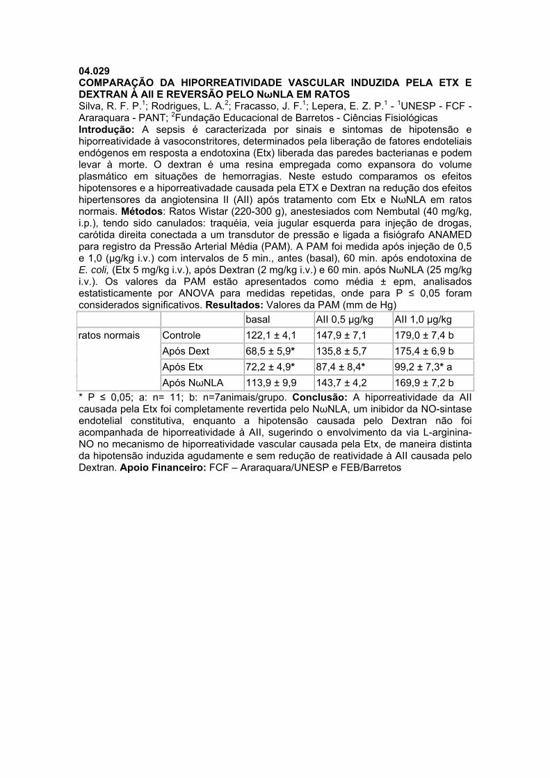
04.029 COMPARAÇÃO DA HIPORREATIVIDADE VASCULAR INDUZIDA PELA ETX E DEXTRAN À AII E REVERSÃO PELO NωNLA EM RATOS Silva, R. F. P.1; Rodrigues, L. A.2; Fracasso, J. F.1; Lepera, E. Z. P.1 - 1UNESP - FCF - Araraquara - PANT; 2Fundação Educacional de Barretos - Ciências Fisiológicas Introdução: A sepsis é caracterizada por sinais e sintomas de hipotensão e hiporreatividade à vasoconstritores, determinados pela liberação de fatores endoteliais endógenos em resposta a endotoxina (Etx) liberada das paredes bacterianas e podem levar à morte. O dextran é uma resina empregada como expansora do volume plasmático em situações de hemorragias. Neste estudo comparamos os efeitos hipotensores e a hiporreativadade causada pela ETX e Dextran na redução dos efeitos hipertensores da angiotensina II (AII) após tratamento com Etx e NωNLA em ratos normais. Métodos: Ratos Wistar (220-300 g), anestesiados com Nembutal (40 mg/kg, i.p.), tendo sido canulados: traquéia, veia jugular esquerda para injeção de drogas, carótida direita conectada a um transdutor de pressão e ligada a fisiógrafo ANAMED para registro da Pressão Arterial Média (PAM). A PAM foi medida após injeção de 0,5 e 1,0 (µg/kg i.v.) com intervalos de 5 min., antes (basal), 60 min. após endotoxina de E. coli, (Etx 5 mg/kg i.v.), após Dextran (2 mg/kg i.v.) e 60 min. após NωNLA (25 mg/kg i.v.). Os valores da PAM estão apresentados como média ± epm, analisados estatisticamente por ANOVA para medidas repetidas, onde para P ≤ 0,05 foram considerados significativos. Resultados: Valores da PAM (mm de Hg) basal AII 0,5 µg/kg AII 1,0 µg/kg ratos normais Controle 122,1 ± 4,1 147,9 ± 7,1 179,0 ± 7,4 b
Após Dext 68,5 ± 5,9* 135,8 ± 5,7 175,4 ± 6,9 b Após Etx 72,2 ± 4,9* 87,4 ± 8,4* 99,2 ± 7,3* a Após NωNLA 113,9 ± 9,9 143,7 ± 4,2 169,9 ± 7,2 b
* P ≤ 0,05; a: n= 11; b: n=7animais/grupo. Conclusão: A hiporreatividade da AII causada pela Etx foi completamente revertida pelo NωNLA, um inibidor da NO-sintase endotelial constitutiva, enquanto a hipotensão causada pelo Dextran não foi acompanhada de hiporreatividade à AII, sugerindo o envolvimento da via L-arginina-NO no mecanismo de hiporreatividade vascular causada pela Etx, de maneira distinta da hipotensão induzida agudamente e sem redução de reatividade à AII causada pelo Dextran. Apoio Financeiro: FCF – Araraquara/UNESP e FEB/Barretos
04.030 CAVEOLAE DYSFUNCTION CONTRIBUTES TO IMPAIRED RELAXATION INDUCED BY NITRIC OXIDE DONOR IN AORTA FROM RENAL HYPERTENSIVE RATS. Rodrigues, G. J.1; Restini, C. A.1; Lunardi, C. N.1; Ferezin, P.2; Moreira, J. E.2; Bendhack, L. M.1 - 1USP - Física e Química; 2FMRP - USP - Biologia Celular e Molecular e Bioagentes Patogênicos Introduction: It has been proposed that caveolae are important in signal transduction and Ca2+ homeostasis. An impaired relaxation to nitric oxide (NO) donors has been observed in arteries from 2 kidney-1clip renal hypertensive rats (2K-1C). Therefore, the present study aimed to investigate the integrity of caveolae in aortic smooth muscle cells (SMC) and its contribution to the effects of NO released from the new NO donor [Ru(NH.NHq)(terpy)NO+]3+ (TERPY) in 2K-1C and control (2K) rats aorta. Methods: Caveolae were counted in isolated aortas from 2K and 2K-1C rats on electron micrographs. In addition, concentration-effect curves for the NO donor were constructed in denuded aortic rings contracted with phenylephrine in the absence or after incubation with the caveolae disassembler methyl-β -ciclodextrin. We investigated the effects of the NO donor in reducing cytosolic Ca2+ concentration ([Ca2+]c) in SMC from 2K and 2K-1C rats loaded with Ca2+ dye Fluo-3AM. These effects were expressed as the difference between the intensity of fluorescence before and after the addition of the NO donor (%ΔIF). Results: Aorta SMC from 2K rats presented larger number of caveolae (73.20 ± 10.74 caveolae/μm2, n=3) than 2K-1C rats (30.33 ± 8.68 caveolae/μm2, n=5). The treatment with methyl-β -ciclodextrin reduced the number of the caveolae in both 2K (38.80 ± 7.55 caveolae/μm2, n=5) and 2K-1C (14.00 ± 0.80 caveolae/μm2, n=3) aortic SMC. The relaxation induced by the new NO donor (TERPY) was concentration-dependent. The potency of TERPY was greater in 2K (pD2: 7.05 ± 0.07, n= 6) than in 2K-1C (pD2: 6.51 ± 0.06, n=6, P<0.01), whereas maximum relaxant effect (ME) was not significantly different between 2K (108.6 ± 2.0%, n=6) and 2K-1C (110.7 ± 0.7%, n=6) rat aortas. Methyl-β -ciclodextrin had no effect on the relaxation to TERPY in 2K-1C aortic rings but it reduced the relaxation to TERPY in 2K aortic rings (ME: 97.9 ± 1.7%, pD2: 6.55 ± 0.09, n=6, P<0.05). The decrease of [Ca2+]c induced by TERPY was greater in 2K (%ΔFI: 55.7 ± 4.8%, n=5) than in 2K-1C cells (%ΔFI: 36.3 ± 2.9%, n=4, P<0.05), and this effect was inhibited by methyl-β -ciclodextrin only in 2K cells (%ΔFI: 35.5 ± 5.5%, n=4). Conclusion: Our results demonstrate that caveolae plays a critical role in the relaxant effect and decrease of [Ca2+]c induced by NO, and could be the reason of impaired aorta relaxation to NO in renal hypertensive rats. Supported by: FAPESP and CNPq.
04.031 IL-10 KO FEMALE MICE INFUSED WITH TNF-α SHOW IMPAIRED ACH INDUCED RELAXATION AS COMPARED TO IL-10 KO MALE MICE. Zemse, S. M.1; Chiao, C. W.1; Hilgers, R. H. P.1; Webb, R. C.1 - 1Medical College of Georgia - Physiology Background: TNF-α is a pro-inflammatory cytokine, principally derived from mononuclear phagocytes. Its primary target is vascular endothelial cells and is an important mediator of maternal endothelial dysfunction. Interleukin-10 (IL-10) is an anti-inflammatory cytokine known to decrease superoxide anion production. In this study, we tested whether IL-10 improves TNF-α induced endothelial dysfunction. Methods: Wild type (C57BL6) and IL-10 KO female and male mice were treated with saline or TNF-α (220ng/kg/min). Following 14 days of treatment, aortic rings were isolated and mounted in a wire myograph (Danish Myotech) and stretched to a tension of 5 mN. Endothelium-dependent relaxation was assessed by constructing cumulative concentration-response curves to acetylcholine (ACh, 0.001-10 μmol/L) during phenylephrine PE, 10 mmol/L) -induced contraction. Endothelium-independent relaxations were assessed by the NO donor sodium nitroprusside (SNP). Results: IL-10 KO female mice treated with TNF-α showed significant impairment in ACh induced relaxation as compared to C57BL6 female mice treated with TNF-α (Emax = 52±3% versus 76±5% respectively; P<0.05). IL-10 KO male mice treated with TNF- α showed no significant impairment in ACh induced relaxation as compared to C57BL6 male mice treated with TNF-α (Emax = 57±3% versus 66±3% respectively). TNF-α infusion had no effect on endothelium-independent relaxations by SNP in all the groups. Conclusion: The anti-inflammatory cytokine IL-10 prevents impairment in endothelium- dependent vasorelaxation in response to 14 days of TNF-α infusion which is more common in females. Supported by: NIH (HL-74167)
04.032 VASODILATAÇÃO INDUZIDA PELA ANGIOTENSINA II ESTÁ REDUZIDA COM A IDADE EM ANIMAIS ESPONTANEAMENTE HIPERTENSOS APESAR DA EXPRESSÃO AUMENTADA DOS RECEPTORES AT2 Ognibene, D. T.1; Oliveira, P. R. B.1; Costa, C. A.1; Carvalho, L. C. dos R. M. de1; Tano, T.1; Soares de Moura, R.1; Resende, A. C.1 - 1UERJ - Farmacologia e Psicobiologia Introdução: Estudos recentes do nosso grupo indicam que a vasodilatação promovida pela angiotensina II (Ang II) em leito arterial mesentérico (LAM) de ratos normotensos é dependente de bradicinina (BK) e modulada por receptores AT2 e AT1-7 (Soares de Moura, Br J Pharmacol, 141:860, 2004). Entretanto, não há evidências do impacto da idade e hipertensão sobre a vasodilatação induzida por Ang II e qual é a exata contribuição dos receptores AT2 para essa resposta. Métodos: No presente estudo, o LAM de ratos Wistar e espontaneamente hipertensos (SHR) com 6 e 22 semanas foi perfundido segundo McGregor (McGregor, J Physiol, 177:21, 1965). O efeito vasodilatador da Ang II foi estudado em preparações pré-contraídas com norepinefrina (30-100 µM) de 4 grupos: ratos normotensos jovens (NJ) e adultos (NA) e hipertensos jovens (HJ) e adultos (HA). Com o objetivo de avaliar a participação da BK, da angiotensina 1-7 (Ang 1-7) e dos receptores AT1 e AT2 na resposta vasodilatadora induzida pela Ang II, o LAM foi tratado com HOE 140 (0.01 µM), A779 (1 µM), losartan (30 µM) e PD123319 (10 µM), respectivamente. A expressão de receptores AT2 foi analisada por Western Blotting. Resultados: O efeito vasodilatador da Ang II (300 nmol), expresso como % de queda da resposta pressora induzida pela norepinefrina, foi significativamente menor (p<0.05) em LAM de ratos adultos (NA: 24±1/ HA: 25±1) quando comparado com ratos jovens (NJ: 54±3/ HJ: 58±2) sendo que a hipertensão não modificou as respostas. O mesmo padrão de resposta foi observado para os efeitos vasodilatadores induzidos pela acetilcolina, bradicinina e nitroglicerina. A vasodilatação induzida pela Ang II (300 nmol) foi significativamente reduzida (p<0.05) em todos os grupos quando o LAM foi tratado com PD 123319 (NJ: 33,7±2 vs 22±4, ausência vs presença do inibidor respectivamente; NA: 24,5±2 vs 16,8±3; HJ: 56±6 vs 31,4±9; HÁ: 21,5±2 vs 13,1±1), HOE 140 (NJ: 42,2±9 vs 24,8±5; NA: 23,8±2 vs 15,6±2; HJ: 51,2±4 vs 34,3±4; HA: 30,4±3 vs 13,3±3) e A779 (NJ: 36,5±5 vs 21,2±3; NA: 25±2 vs 18,3±1; HJ: 44,6±2 vs 18,5±4; HA: 23,1±3 vs 16,8±4). No entanto, o tratamento com losartan não modificou a resposta (NJ: 70±4 vs 70,1±4; NA: 33,3±3 vs 41,4±3; HJ: 48,8±5 vs 49,5±7; HA: 21,7±3 vs 20,2±3). A expressão de receptores AT2 foi menor em LAM de ratos NA comparada a de ratos NJ. Entretanto, a expressão foi maior em LAM de HJ do que em ratos NJ, e esta expressão aumentada foi mantida em HA. Discussão: O presente estudo sugere que a vasodilatação induzida pela Ang II é reduzida com a idade, mas não é modificada pela hipertensão. Este efeito é mediado por receptores AT2 e envolve a participação de BK e Ang 1-7. A disfunção endotelial e/ou comprometimento do músculo liso vascular podem contribuir para a resposta reduzida em animais NA e HA. Em animais adultos hipertensos, a expressão aumentada de receptores AT2 representaria um benefício adicional para os efeitos hemodinâmicos dos antagonistas AT1. Apoio Financeiro: FAPERJ; CNPq
04.033 K+ CHANNELS CONTRIBUTE TO PIPLARTINE-INDUCED RELAXATION ON RAT AORTA Silva, J. L. V.1; Ribeiro, L. A. A.1; Cavalcante, F. de A.2; Araujo Junior, J. X.3; Chaves, M. C.1; Silva, B. A. da4 - 1UFPB - LTF; 2UFAL / UFPB - ICBS / LTF; 3UFAL - ESENFAR; 4UFPB - LTF e DCF Introduction: Piplartine {5,6-dihydro-1-[1-oxo-3-(3,4,5-trimethoxyphenyl)-2-propenyl]-2(1H)pyridinone} is an alkamide isolated from barks and roots of Piper tuberculatum Jacq. Previous results showed that piplartine relaxed rat aorta and uterus pre-contracted, probably, due to blockade Ca2+ influx through voltage-gated Ca2+ channels (CaV) and activation of K+ channels, respectively. Thus, we decided to investigate the involvement of K+ channels in the vasorelaxant mechanism induced by piplartine on rat aorta. Methods: aorta rings without functional endothelium isolated of male rat were suspense in organ bath cubes (6 mL) contained Krebs solution (pH 7.4) bubbled with 95 % O2 and 5 % CO2 mixture, at 37 °C, on tension of 1 g. The isometric contractions were obtained through transducer connect an amplifier coupled to a data-acquisition system BioMed in a personal computer. Results: Piplartine relaxant effect (EC50 = 1.5±0.2 x 10⁻⁵ M, n = 5) was attenuated significantly in the presence of CsCl (EC50 = 1.2±0.05 x 10⁻⁴ M, n = 5), a non-selective blocker of K+ channels. We decided to investigate what type of K+ channels participate in this piplartine response. 4-aminopyridine, a blocker of KV, did not modified piplartine vasorelaxation (EC50 = 2.3±0.4 x 10⁻⁵ M, n = 5). However, piplartine vasorelaxation was reduced significantly by glibenclamide (EC50 = 4.8±0.9 x 10⁻⁵ M, n = 5), blocker of KATP, apamine (EC50 = 4.6±0.4 x 10⁻⁵ M, n = 5), blocker of SKCa, BaCl2 (EC50 = 4.0±0.6 x 10⁻⁵M, n = 5), blocker of Kir, and iberiotoxin (EC50 = 3.8±0.4 x 10⁻⁵ M, n = 5), blocker of BKCa. Raising extracellular [K+] occurs modulation of K+ channel (Lyles et al, Pharmacol Res 37, p. 437, 1998), thus piplartine response was accessed on aorta pre-contracted by phenylephrine in presence of 14.7 mM K+ extracellular, observed that it vasorelaxant effect was reduced significantly (EC50 = 3.8±0.1 x 10⁻⁵ M, n = 5). This finding confirms that K+ channels participate in this vasorelaxant mechanism. Various studies have showed that cAMP and cGMP modulate K+ channels (Fukuzawa et al, Life Sci 53, p. 1129, 1993), we accessed this participation in a indirect manner, using aminophylline, a non-selective phosphodiesterase inhibitor. The vasorelaxing curves of piplartine did not change in presence of aminophylline (EC50 = 1.1±0.3 x 10⁻⁵ M, n = 5). Discussion: KATP, SKCa, Kir, BKCa contribute also to piplartine vasorelaxation, probably, without cAMP/cGMP participation in the response. These findings explain blockade Ca2+ influx through CaV mediated by piplartine, since activation of K+ channels leads membrane repolarization, promoting closure of CaV and thus vasorelaxation. Supported by: CNPq; CAPES; LTF/UFPB.
04.034 POTENTIATION OF BRADYKININ EFFECT BY RAMIPRILAT IS NOT RELATED TO ANGIOTENSIN CONVERTING ENZYME ACTIVITY IN THE RAT MESENTERIC ARTERIES. Sivieri-Jr., D. O.1; Bispo-da-Silva, L. B.1; Oliveira, E. B.2; Resende, A. C.3; Salgado, M. C. O.1 - 1FMRP - USP - Farmacologia; 2FMRP - USP - Biochemistry and Immunology; 3UERJ - Farmacologia e Psicobiologia Introduction: Angiotensin converting enzyme (kininase II, ACE) inhibitors are capable of potentiating bradykinin (BK) effects by enhancing the actions of bradykinin on B2-receptors independent of blocking its inactivation. Objective: To investigate further the importance of ACE kininase activity on BK-induced vasodilation, we determined the effect of inhibiting ACE, as well as other kininases, on both BK metabolism and vasodilator effect in preparations that exhibit increased ACE activity. Methods: Isolated and perfused mesenteric arterial bed (MAB) from normotensive and one-kidney, one clip hypertensive rats were used. ACE activity was determined by fluorimetric assay in MAB homogenate and by the ratio of the doses of angiotensin (Ang) I and II that elicited a 35 mmHg increase in mesenteric perfusion pressure. To analyze the metabolism of BK, MAB was exposed to BK for 15 min, in the absence or in the presence of kininase inhibitors; then, the perfusate was collected for analysis of the products of BK metabolism by HPLC. Dose-response curves to BK were obtained in phenylephrine-constricted MAB isolated from normotensive and hypertensive rats in the absence or presence of kininase inhibitors. Results: Mesenteric arterial beds obtained from hypertensive rats (184±2 mmHg) presented augmented ACE activity (1.40±0.17 vs. 0.59±0.09 pmol His-Leu/min/mg protein, P<0.007) and potency ratio (2.3 fold) of Ang I and Ang II to induce vasoconstriction compared to normotensive (104±1 mmHg) rats. BK was metabolized to the fragments BK(1-8), BK(1-7), and BK(1-5) and the recovery of intact BK was reduced by 47% in the hypertensive group. Recovery of BK was increased in both groups in the presence of a kininase I inhibitor and in the hypertensive group by neutral endopeptidase 24.11 inhibitor; however, ACE inhibition did not affect BK metabolism in both groups. In contrast, only ACE inhibitor (ramiprilat, 5 mM) potentiated the vasodilator effect of BK in mesenteric bed preconstricted with phenylephrine. The dose-response curves to BK were shifted to the left in the presence of ramiprilat in preparations of normotensive (P<0.0001) and hypertensive (P<0.0007) groups. Discussion: Since the ACE inhibitor-induced potentiation of BK effects was not greater in arteries from hypertensive rats that presented an increased ACE activity when compared to normotensive group, we concluded that potentiation of BK vasodilator effects by ACE inhibitor is not related to their actions on BK degradation. These findings strongly suggest a cross-talking between ACE and B2 receptor in MAB. Supported by: FAPESP
04.035 MAST CELL DEGRANULATION DOES NOT MODIFY THE CORONARY VASCULAR REACTIVITY TO ANG I AND II. Bispo-da-Silva, L. B.1; Sivieri-Jr., D. O.1; Becari, C.1; Prado, C. M.2; Rossi, M. A.2; Oliveira, E. B.3; Salgado, M. C. O.1 - 1FMRP - USP - Farmacologia; 2FMRP - USP - Patologia; 3FMRP - USP - Bioquímica e Imunologia Introduction: Cardiac mast cells are apposed to blood vessels and contain proteases capable to metabolize angiotensins (Ang). Moreover, these cells have been associated with cardiac remodeling which occurs in a variety of conditions. In the present study we aimed at evaluating mast cell density, as well as the effect of cellular degranulation on the coronary reactivity to Ang I, Ang II and vasopressin (AVP), at the early phase of cardiac hypertrophy induced by pressure overload. Methods: Male Wistar rats (n=3-10) were submitted to suprarenal aorta coarctation (COA) or to sham surgery (S). Mean carotid arterial blood pressure (MAP), mast cell density and the coronary reactivity to peptides were evaluated 7 days after inducing aortic stenosis. Hearts from COA and S, pretreated or not with 48/80 compound (1 mg/kg, i.v.), were isolated and mounted in a Langendorff apparatus. The coronary reactivity to the different peptides (increase in perfusion pressure in %) was analyzed in the presence (Ang I: 2 μM) or absence (Ang I and Ang II: 2μM; AVP: 2nM) of the angiotensin-converting enzyme inhibitor lisinopril (3 μM). Moreover, the proteolytic activity of cardiac perfusates from isolated hearts of control rats, treated or not with 3 consecutive in bolus injections of 48/80 compound (20 μg), toward Ang I was analyzed by HPLC. Results: COA increased MAP (163±6 vs 127±1 mmHg, P<0.05), but did not alter mast cell density (COA vs S: 2.7±0.2 vs 2.6±0.3 cell/mm2, P>0.05). The pressure overload decreased the maximum response (MR) to Ang I (18.2±2.9 vs 27.4±3.4 %, P<0.05) and Ang II (18.6±2. vs 30.6±3.2 %, P<0.05) compared to S, without altering the MR to AVP (COA vs. S: 8.3±1.4 vs 9.5±1.5 %, P>0.05). Systemic 48/80 compound treatment decreased MAP (from 91±4 to 31.1±1 mmHg, P<0.05) that was associated with a massive cardiac mast cell degranulation (mast cell totally degranulated before and after the treatment: 2.0±0.7 vs 77.2±3.6 %). The coronary vascular reactivity to Ang I in the presence of lisinopril, as well as to Ang II and AVP in the absence of this inhibitor, was not altered by 48/80 compound pretreatment. Cardiac mast cell degranulation did not modify the generation of Ang II and Ang 5-10 from Ang I by cardiac perfusates, activities that could be attributed to mast cell carboxypeptidase and chymase-1, respectively. Discussion: Our data suggest that massive mast cell degranulation is not associated to changes in coronary reactivity to Ang I and Ang II, despite the presence of mast cell proteases at the site of deposition after cellular activation. Moreover, the early phase of cardiac hypertrophy is associated with alteration on the vascular responsiveness to Ang II, but the vasoconstrictor capacity seems to be preserved. Supported by: CAPES and FAEPA.
04.036 ROLE OF CAMP-PKA PATHWAY ON RELAXANT EFFECT OF LABDANE-302 IN RAT AORTA Macedo, C. L.1; Ribeiro, L. A. A.2; Tavares, J. F.1; Silva, M. S.3; Silva, B. A. da3 - 1UFPB - LTF; 2UFPB / IESP - LTF; 3UFPB - DCF e LTF Introduction: the species Xylopia langsdorfiana St. Hil. & Tul. (Annonaceae) is popularly known as “pimenteira da terra” in Southeast of Brazil. The fractionation of the ethanol extract obtained from the stem-bark of this species yielded a labdane-type diterpene identified as 8(17),12E,14-labdatrien-18-oic acid referred here as labdane-302. Previous studies have shown that labdane-302 has a relaxant effect in rat aorta which may depends of K+ channels modulation, more specifically the voltage-gated K+ channels and inward rectifier K+ channel (Ribeiro et al., Z. Naturforsch. C, 2007-in press). This study was performed to access the role of cyclic AMP and protein kinase A (cAMP-PKA ) pathway in the relaxant effect of labdane-302 in rat aorta. Methods: the aorta rings (3 mm) without endothelium functional were obtained from male rats and suspended into organ-bath chambers in Krebs solution at 37 °C and bubbled with 5 % CO2 and 95 % O2 gas mixture. The isometric contractions were measured (n = 5) by a force transducer coupled to an amplifier and a computer system running the BioMed software. The relaxant effect was measured in aortic rings pre-contracted by 0.3 mM phenylephrine. Results: the labdane-302 relaxed aortic rings, pre-contracted by phenylephrine, in a concentration-dependent manner (EC50 = 1.3±0.2 x 10⁻⁵ M). The presence of 100 µM aminophylline, a non-specific phosphodiesterases inhibitor, caused a leftward shift in the labdane-302 concentration-response curve with a significant increase of the potency of labdane-302 (EC50 = 4.0±0.9 x 10⁻⁶ M). In the presence of 100 mM Rp-cAMPS, a selective PKA inhibitor, the concentration-response curve of labdane-302 was rightward shifted with significant decrease of labdane-302 potency (EC50 = 3.5±0.3 x 10⁻⁵ M). As positive control we used forskolin (EC50 = 3.6±0.6 x 10⁻⁸ M), a well known labdane-type diterpene that directly actives adenilyl cyclase enzyme and thus increases cAMP. The presence of aminophylline caused an increase of forskolin relaxant potency (EC50 = 8.2±0.1 x 10⁻⁹ M). In the presence of Rp-cAMPS the concentration-response curve of foskolin was rightward shifted with significant decrease of its potency (EC50 = 4.0±0.2 x 10⁻⁷ M). Discussion: in preview studies we have showed that the relaxant effect of labdane-302 depended of the K+ channels modulation (Ribeiro et al., Z. Naturforsch. C, 2007-in press). It was well known that many K+ channels are modulated by phosphorylation via PKA in response to increased levels of cAMP (Sadoshima J., Am. J. Physiol., 255, H754, 1988). In this study we show that the potency of labdane-302 in to relax the rat aorta was increased when the phosphodiesterases were inhibited by aminophylline, which may indicate participation of cyclic nucleotides, probably cAMP. Indeed, in the presence of Rp-cAMPS the relaxant potency of labdane-302 was significantly reduced, indicating that the increase cAMP contents and activation of PKA may be involved in that relaxant effect of labdane-302. Together, these results suggest that in functional level, the cAMP-PKA pathway seems to be involved in the relaxant action mechanism of labdane-302 in rat aorta. Supported by: CAPES, CNPq and LTF/UFPB.
04.037 VASORELAXANT EFFECT INDUCED BY 6 – [(E) – STYRYL] – PYRON – 2 - ONE (PIR) IN ISOLATED RAT SUPERIOR MESENTERIC ARTERY Assis, T. J. C.1; Oliveira Filho, A. A.1; Assis, V. L.1; Barbosa-Filho, J. M.1; Medeiros, I. A.1 - 1UFPB - LTF INTRODUCTION: 6 - [(E) – styryl] – pyron – 2 - one (PIR) is a natural styrylpyrone isolated from the ethanolic extract of the green fruits of Aniba panurensis (Lauraceae) (Barbosa-Filho, 1986, 470p). The pyrones demonstrate a whole spectrum of bioactivity and have been shown to be antifungal, antibiotic, cytotoxic and phytotoxic (Fairlamb et al., Bioorg. e Med. Chem. 12, 4285-4299, 2004). The aim of this study is to investigate the effects of this pyrone on isolated rat superior mesenteric rings. METHODS: Rat superior mesenteric rings (1-2 mm) were suspended by cotton threads for isometric tension recordings in Tyrode´s solution, 37 ºC, gassed with 95% O2 and 5% CO2, resting tension 0.75 g. RESULTS: In mesenteric rings PIR (10-10–10-3 M) induced relaxation of phenylephrine (10 mM) induced tone (EC50=1.1±0.69x10⁻⁵ M; n=7) and this effect was unaltered after endothelium removal (EC 50 =1.57±0.35 x 10⁻⁵ M; n=7). This vasorelaxant effect induced by PIR was insignificantly altered in the presence of KCl 20 mM (n=6). In rings pre-contracted with KCl 80 mM PIR induced relaxant effect (EC50=2,0±0,38 x 10⁻⁴ M; n=6). PIR (10-7–10-4 M) was not able to inhibit the concentration-response curves to CaCl2, however, at a high concentration (10-3M, n=6) it was able to significantly decrease the maximal response. In depolarizing nominally without calcium solution, PIR (10-8-10-3 M; n=6) antagonized transient contractions induced by Phe (10 µM). PIR (10-4-10-3 M; n=6) slightly antagonized the transient contractions induced by 20 mM caffeine. Sodium orthovanadate (10-5-3x10⁻³ M)-induced contractions were significantly inhibited by higher concentrations of PIR (10-4 and 10-3 M; n=6). CONCLUSION: These results suggest that PIR exerts an endothelium-independent relaxant effect that seems to involve the calcium influx by voltage operated calcium channels and inhibition of Ca2+ release from intracellular IP3 and ryanodine-sensitive calcium stores. They further suggest that PIR-induced response seems to be mediated by a pathway related to G-protein coupled receptors signalling. Supported by: CNPq
04.038 VASCULAR REACTIVITY IN A MODEL OF DIABETES TYPE I IN MOUSE Pinho, J. F.1; Rezende, B. A.1; Capettini, L. S. A.2; Lemos, V. S.1; Cortes, S. F.1 - 1ICB - UFMG - Fisiologia e Farmacologia; 2ICB - UFMG - Fisiologia e Biofísica Introduction: Diabetes mellitus is a metabolic diseases characterized by hyperglycemia resulting from defects in insulin secretion, action or both. Diabetes is usually associated with long-term damage, dysfunction, and failure of various organs, including blood vessels. High concentrations of glucose have been shown to be associated with endothelial dysfunction in vivo and in vitro. Accumulating evidence indicates that reactive oxygen species (ROS) play a major role in the initiation and progression of cardiovascular dysfunction associated with diabetes. However, the mechanisms underlying vascular damage associated with diabetes are far of being well understood. The aim of this work was to investigate the mechanisms underling vascular dysfunction in streptozotocin (STZ)-induced diabetic mice. Method: 3 weeks old C57BL/6 mice received an intraperitoneal injection of STZ (120 mg/kg) or vehicle (citrate buffer). Ten weeks later, the animals were killed and thoracic aorta was isolated and cut in rings. The segments were mounted in a myograph in physiological conditions and muscle tension assessed using isotometric transducers. Nitric Oxide (NO) content in aortic rings was measured by fluorescence using 2,3 diaminonaphthalen (DAN). Results: Plasma glucose level in STZ-treated mice (450±50 mg.dL-1) was higher than in control group (100±30 mg.dL-1; p<0.001) showing that treatment with STZ was effective. Relaxant responses to acethylcholine (ACh) were smaller in diabetic as compared to control mice. Treatment of vessels with tetrahydrobiopterine (THB) a nitric oxide synthase (NOS) co-factor restored vasorelaxation to the level of control animals. L-NAME (300 µM) completely abolished relaxation in both strains. ACh-induced increases in NO contents were smaller in diabetic than in control mice. Contractile responses to phenylephrine in endothelium-containing vessels were strongly enhanced in diabetic mice. Removal of endothelium did not modify these contractile responses. Indomethacin did not affect contractile responses neither in control nor in diabetic mice. Contractile responses to KCl in endothelium-denuded vessels were also much bigger in diabetic than in control mice. Discussion: In this work we found that induction of diabetes by STZ in mouse resulted in a vascular dysfunction characterized by decreased endothelium-dependent vasorelaxation and an increased contractility. In healthy vessels, the endothelium has a dominant vasodilator influence mediated by endothelium-derived relaxing factors (EDRF), including prostacyclin, NO and endothelium-derived hyperpolarizing factor (EDHF). The influence of endothelium-dependent vasoconstrictors may be up regulated in diseases. Reduced production or function of EDRF and/or increased vasoconstrictors will push the balance in favor of vasoconstriction. In this work we found that a decrease in NO production induced by ACh is responsible for the endothelium dysfunction found in diabetic mice. The results with THB support the hypothesis that uncouple of NOS is possibly the responsible for the reduction in NO production. The increased contractility found in diabetic mice is unlike to be mediated by an endothelium-dependent vasoconstrictor but probably due to modifications in contractile properties of smooth muscle cells. Supported by: FAPEMIG and CNPq
04.039 TAMOXIFEN INCREASES SUPEROXIDE DISMUTASE ACTIVITY IN VASCULAR SMOOTH MUSCLE CELLS (VSMC) OF RATS Pessa, L. R.1; Montenegro, M. F.1; Jacob Ferreira, A. L. B.1; Izidoro-Toledo, T. C.1; Desta, Z.2; Flockhart, D. A.2; Tanus-Santos, J. E.1 - 1FMRP - USP - Farmacologia; 2Indiana University School of Medicine - Clinical Pharmacology Introduction: Tamoxifen (TAM) is a selective estrogen receptor modulator, which is used in breast cancer treatment. However, a number of studies reported protective effects of TAM in cardiovascular system. The activity enzyme superoxide dismutase (SOD) is an important mechanism of protection to avoid increased oxidative stress. This study investigated the effects of TAM on SOD activity. The effects of different concentrations of TAM (10-8 at 10-5M) on SOD activity were studied in rat VSMC cell culture. Methods: VSMC line (A7r5) was cultured in DMEM medium serum and phenol red-free for 48 h. The viability cellular was assessed with trypan blue exclusion method. The supernatant was used to measure SOD activity spectrophotometrically at 480nm based on the capacity of SOD to inhibit autoxidation of epinephrine to adrenochrome. One unit of enzyme was defined as the amount of enzyme required to inhibit the rate of epinephrine autoxidation by 50% at 26°C. Results and Discussion: No significant differences in cell viability were found after treatments with different concentrations of TAM (P>0.05). TAM increased SOD activity in a concentration-dependent manner (control=0.35±0.3U/mL; 0.5±0.2U/mL, 0.7±0.1U/mL, 0.86±0.1U/mL and 1.2±0.2U/mL to concentration 10-8, 10-7, 10-6 and 10-5M, respectively; P<0.01 to concentrations 10-6 and 10-5M versus control). These results suggest that TAM increases superoxide dismutase activity in VSMC and provide evidence for a possible beneficial effect of TAM in cardiovascular system. Supported by: CAPES, CNPq, FAPESP
04.040 VASCULAR RELAXATION INDUCED BY D-ARABINOSE OXIME IN RAT SUPERIOR MESENTERIC RINGS Oliveira Junior, F. A.1; Carvalho, E. M.1; Satyro, M. A.1; Dias, K. L. G.1; Alves, R. J.2; Guedes, D. N.3; Medeiros, I. A.1; Correia, N. A.3 - 1UFPB - Tecnologia Farmacêutica - LTF; 2UFMG - Farmácia; 3UFPB - Fisiologia e Patologia INTRODUCTION: Several reports have evidenced the relaxing effects of the compounds with functional group C=NOH and the participation of nitric oxide in these effects. The D-arabinose oxime is an interesting compound of functional group C=NOH and the aim of this study is to evaluate the cardiovascular effecs and the mechanism of action of this oxime. METHOD: Isolated rat superior mesenteric rings (1-2 mm) were suspended by cotton threads for isometric tension recordings in a Tyrode’s solution at 37ºC and gassed with a 95% O2 and 5% CO2, under a resting tension of 0.75g. Concentration-responses curves to D - arabinose oxime (10 -12 to 10 -4 M) were obtained. RESULTS: In isolated rat mesenteric artery rings, with endothelium intact, D-arabinose oxime induced concentration-dependent relaxation of the contractions induced by phenylephrine (pD2 = 5.9±0.14; Emáx = 92,8±3,46, n= 6), which was not attenuated after endothelium removal (pD2 = 5.7±0.11; n= 6, Emáx = 99,3±0,55), demonstrating an endothelium-independent effect. In endothelium-denuded rings, the soluble guanylate cyclase inhibitor, ODQ (1H-(1,2,4-) oxadiazolo (4,3-a)-quinoxalin-1-one (10 μM), caused a significant reduction in the vasorelaxation induced by D-arabinose oxime (Emáx = 10,5±5,18). The nitric oxide (NO) scavenger, hydroxocobalamin (30 μM), also caused significant reduction in the relaxant effect elicited by D-arabinose oxime. (Emáx = 27,6±4,31), when compared with controls. CONCLUSION: Taken together, these data suggest that the relaxant activity induced by D-arabinose oxime may be probably due to the activation of NO/sGC pathway. Supported by: LTF-UFPB/CNPq
04.041 NITRIC OXIDE: A POSSIBLE MEDIATOR OF ACIDOSIS VASODILATION Celotto, A. C.1; Capellini, V. K.1; Baldo, C. F.1; Evora, P. R. B.1 - 1FMRP - USP - Cirurgia e Anatomia Introduction: It has been known for many years that changes in vascular smooth muscle tone plays an important role in the distribution of blood flow and in the control of blood pressure1. Gaskell was probably the first to show that pH is a major determinant of tone when he demonstrated that decreases in the blood pH caused vasodilatation. Extracellular acidosis (EA) has profound effects on vascular homeostasis and the EA-induced tone alterations are specific to vascular bed3. pH may influence the tone of vascular smooth muscle by affecting the release of endothelium-derived vasoactive factors or by directly modulating smooth muscle function4. In the present study, we examined the effects of mild and severe acidosis on dilatory responses of isolated rat thoracic aorta. Methods: Male Wistar rats of 220-270 g body weight were killed with an overdose of thiopental given intraperitoneally and thoracic aorta was carefully removed and placed in organ chambers, containing Krebs solution, for vascular reactivity study. pH-response curves for chloride acid (HCl) (7.4 – 6.5) were obtained in endothelium intact and denuded rings phenylephrine-precontracted. Acidosis was carried through gradual addition of HCl 1M to the preparation in a manner to get a pH-response curve. The curves were done with and without NG-nitro-L-arginina metil ester (L-NAME: 10-4M) and/or indomethacin (Indo: 10-5M). All data were expressed as means±SEM. Statistical analisys were performed using one-way ANOVA and Bonferroni post-test (program Prism 4). The difference between the groups was considered to be significant when p<0.05. Results: The HCl-mediated relaxation was increased in endothelium intact rings (79.48±3.66) when compared with denuded rings (53.36±3.78) in mild (pH 7.0) acidosis, but not in severe acidosis (pH 6.5) (94.33±1.96 to 91.42±0.95). The relaxing effect of mild acidosis, in endothelium intact rings, was markedly reduced by L-NAME (40.74±5.38) and L-NAME+Indo (47.27±7.36) but not by Indo (76.40±4.08). In denuded rings the relaxation was reduced only by L-NAME in mild (20.06±4.18) and in severe acidosis (73.32±6.44). Discussion: These results show that mild acidosis promoted nitric oxide-mediated relaxation in rat aorta. This effect might be caused by an augment in the nitric oxide production in endothelial and/or vascular smooth muscle cells, augment in the cGMP levels and/or prolonged NO relaxing effect due to greater stability of this molecule in mild acid solution. However, in severe acidosis other factors are involved in the relaxation. 1 Smith GL. Cardiovasc Res. May;38(2):316-31, 1998. 2 Gaskell WH. J Physiol. Aug;3(1):48-92.16, 1880. 3 Christou H. Am J Physiol Heart Circ Physiol. 288(6):H2647-52, 2005. 4 Coessens B. J. Orthop Res 14(5):818-22, 1996. Supported by: FAPESP.
04.042 VASCULAR RESPONSIVENESS OF MESENTERIC ARTERY IN EXERCISED RATS WITH DIABETES MELLITUS Delbin, M. A.1; Claudino, M. A.2; Antunes, E.2; Nucci, G. de2; Zanesco, A.1 - 1UNESP - IB - Rio Claro - Educação Física; 2UNICAMP - Farmacologia Diabetes mellitus is chronic disorders leading to severe neuropathy and vascular dysfunction. Furthermore, it is associated with decrease in endothelium-derived NO production and/or an increase in reactive oxygen species. The aim of this work was to evaluate the effect of physical preconditioning for 8 weeks (RT) in the responsiveness of mesenteric rings from diabetic rats. METHODS: Rats were divided into three groups named: control sedentary (C/SD); sedentary diabetic (SD/DB) and trained diabetic (TR/DB). Run training (RT) was performed for 5 days/week, each session of 60 minutes, at a speed of 0.6-1.0 km/h and 0% grade. Diabetes (DB) was induced by administration of streptozotocin (60 mg/kg, i.p.). RT started four weeks previously to diabetes induction and was performed for further four weeks, lasting 8 weeks. Concentration-response curves to acetylcholine (ACh) in the absence or in the presence of L-NAME, sodium nitroprusside (SNP) and phenylephrine (PHE) were obtained in isolated mesenteric artery. Potency (pEC50) and maximal response (EMAX) were determined. RESULTS: Body weight at initial time was similar in all groups (275±10 g) whereas after DB the body weight was significant (P<0.05) decreased (SD/DB: 272±14 and TR/DB: 265±8 g) compared to sedentary group (C/SD: 437±12 g). In TR/DB, plasma glucose levels was reduced compared to SD/DB group. Potency for ACh was decreased in SD/DB (6.93±0.05) compared to C/SD and TR/DB groups (C/SD: 7.32±0.07 and TR/DB: 7.21±0.04). The EMAX was decreased in DB groups (SD/DB: 89±2 and TR/DB: 91±2) compared to C/SD (C/SD: 101±1). The addition of L-NAME abolished the alterations induced by RT. The EMAX was decreased in DB groups (SD/DB: 43.7±6 and TR/DB: 48.3±3) compared to C/SD (C/SD: 78±6). Neither the pEC50 nor EMAX were modified for SNP and PHE in all groups. CONCLUSION: Our findings show that prior physical training promotes beneficial effects in endothelium-dependent relaxing response in mesenteric artery from diabetic rats. Supported by FAPESP
04.043 RELAXANT EFFECT OF BAY 41-2272 IN THE RABBIT ISOLATED DETRUSOR SMOOTH MUSCLE (DSM): INVOLVEMENT OF CGMP-INDEPENDENT MECHANISMS Bau, F. R.1; Teixeira, C. E.1; Nucci, G. de1; Antunes, E.1 - 1UNICAMP - Farmacologia Overactive bladder (OAB) described as urgency with or without incontinence, usually accompanied by frequency and nocturia, reflects involuntary DSM as a consequence of enhanced cholinergic-mediated contractions. The nitric oxide (NO)-cyclic GMP signaling pathway has been described to modulate the muscular tone, neurotransmission and blood flow in DSM, but the exact role of NO in the bladder is not completely understood. It is accepted that NO deficiency contributes to triggering OAB. The compound BAY 41-2272 has been described as a drug that directly activates the soluble guanylate cyclase leading to NO-independent cGMP accumulation. BAY 41-2272 produces potent relaxations in vascular and non-vascular smooth muscle tissues by cGMP- dependent and independent mechanisms, but no studies have been carried out with DSM. Objective: The aim of this study was to evaluate the relaxant effect of BAY 41-2272 in the rabbit isolated bladder and the mechanisms underlying the relaxant responses. Methods: Male New Zealand White rabbits (3 kg) were anaesthetized with urethane (1g/kg, i.v.) After exsanguination, the abdomen was opened and the bladder was removed and placed in Krebs solution. The DSM strips (2 x 4 x 10 mm) were suspended between two metal hooks and mounted in 10 ml organ baths containing with Krebs’ solution at 37oC, pH 7.4, and aerated continuously with 95% O2 and 5% CO2. The development of isometric tension was registered using force transducers connected to a PowerLab 400TM data-acquisition system (ADInstruments, MA, USA). Tissue preparations were equilibrated for 1 h before the start of the experiments. Initially, DSM preparations were challenged with 80 mM KCl to confirm tissue viability. Concentration-response curves for BAY 41-2272 (0.001-10 µM) were contructed in the presence and in the absence of L-NAME (100 µM), ODQ (10 µM) or sildenafil (0.1 µM) The relaxations were plotted as percentages of the contraction induced by carbachol (10 µM). Concentration-response curves to sodium nitroprusside (SNP, 0.01-10 µM) and gliceryl trinitate (GTN, 0.01-10 µM) were also constructed. Results: BAY 41-2272 produced a concentration-response curve (Fig 1a) in isolated DSM with a potency (pEC50) and maximal response (Emax) of 6.71±0.13 and 134.97±4.84, respectively. The pEC50 values for BAY 41-2272 were higher than for SNP (6.04±0.15), but not statistically different from GTN (6.37±0.21). The Emax values for BAY were signicantly higher (p<0.001) than those for SNP (48.87±3.62) and GTN 41.94±4.10. The potency of BAY 41-2272 was not significantly altered in the presence of L-NAME (6.37±0.33), ODQ (6.66±0.15) or sildenafil (6.67±0.20) in comparison with vehicle (6.72±0.12). Similarly the Emax values were not altered in the presence of L-NAME (153.30±9.02), ODQ (125.69±7.06) and sildenafil (126.18±5.14) compared with vehicle (134.97±4.84) (Fig 1b). Conclusions: In contrast with NO donors, BAY 41-2272 causes a complete relaxation of rabbit DSM Our findings that relaxant responses to BAY 41-2272 are unaltered by L-NAME, ODQ and sildenafil indicate that BAY 41-2272 acts in an cGMP-independent manner to produce DSM relaxations. Therefore, BAY 41-2272 constitutes a drug with great potential to treat overactive bladder. Supported by: FAPESP
04.044 CARBOXYPEPTIDASE B, THE MAJOR KININASE IN THE RAT MESENTERIC ARTERIAL BED PERFUSATE Pereira, H. J. V.1; Oliveira, E. B.1; de Souza, L. L.1; Costa-Neto, C. M.1; Sivieri-Jr., D. O.2; Bispo-da-Silva, L. B.2; Machado, L. F.3; Sousa, M. V.3; Salgado, M. C. O.2 - 1FMRP - USP - Bioquímica e Imunologia; 2FMRP - USP - Farmacologia; 3UnB - Biologia Celular Introduction: The rat mesenteric arterial bed (MAB) perfusate is a rich source of vasopeptidases, among which angiotensin II-forming enzymes, kininases, and carboxypeptidases capable of processing a variety of biologically active peptides. Here we describe the isolation and characterization of a basic carboxypeptidase as the major kininase from MAB perfusate. Methods: Kininase was isolated by a two-steps affinity chromatographic procedure on potato carboxypeptidase inhibitor (PCI)-Sepharose and arginine-Sepharose columms. Kininase activity was determined by HPLC analysis of the BK fragments formed in the presence or absence of inhibitors. Protein structure was determined by generating a tryptic peptide mass fingerprint, whose peptides of precisely determined masses were screened against a tryptic fragment database derived from over 50,000 proteins by MASCOT software. Total RNA was extracted from rat mesentery, pancreas, kidney, liver, lung, heart, aorta and carotid and used to perform reverse transcription of mRNAs into cDNAs using oligo-d(T) and SuperScript II protocols. cDNAs for carboxypeptidase B (CPB) and β-actin were amplified by PCR using specific oligonucleotide primers. Aliquots of 10 µL of each PCR product were run on a 1% agarose gel, stained with ethidium bromide and subjected to densitometric scanning by ImageJ software (http://rsb.info.nih.gov/ij/); the intensity of each particular cDNA was normalized to the respective β-actin PCR product. Results: When BK was incubated with MAB perfusate, the major degradation product (>94%) was des-Arg9-BK. The carboxypeptidase responsible by the formation of des-Arg9-BK was purified with a 28% overall yield. This purified basic carboxypeptidase released solely des-Arg9-BK upon incubation with BK, even after prolonged incubation times, and was inhibited by MGTA (30 mM) and PCI (20 mM). The structure of the purified enzyme was shown to be identical with the pancreatic carboxypeptidase B (CPB) and the corresponding mRNA was highly expressed in mesentery, pancreas, liver, and kidney but was below detection level in lung, heart, aorta and carotid artery. Discussion: Contrary to the general belief that CPB is a digestive protease whose production is restricted to the pancreas and that does not participate in regulatory processes, we found that it is the major BK-processing enzyme of the rat MAB perfusate. Indeed, the relative high level of expression of CPB mRNA in mesentery is accompanied by a detectable CPB activity in the MAB perfusate. Supported by: FAPESP, CNPq.
04.045 DIFFERENT MECHANISMS INVOLVED IN HYPOTENSION AND HYPORESPONSIVENESS TO VASOCONSTRICTORS INDUCED BY SEPTIC SHOCK Bet, A. C.1; Fernandes, D.1; Sordi, R.1; Pacheco, L. K.1; Assreuy, J.1 - 1UFSC - Farmacologia Introduction Sepsis and septic shock are the major cause of morbidity and mortality in critically ill patients and is usually associated with hypotension and loss of vascular responsiveness to vasoconstrictors. It is well established that overproduction of NO in sepsis is one of the main causes for excessive vasodilatation and reduced contractile response to vasoconstrictors agents. Clinical assays trying to improve sepsis by inhibiting NO synthases (NOS) had failed, probably due to the lack of selectivity of inhibitors towards NOS isoforms. Notwithstanding the search for selective inhibitors, a better understanding about NO molecular effectors mechanisms may provide new opportunities for therapy development. Methods Sepsis was induced by cecal ligation and puncture (CLP) procedure in female Wistar rats. Methylene blue 5 mg/kg, (MB, a soluble guanylate cyclase inhibitor, n=6) was injected subcutaneously, 4 and 8 hours after the CLP procedure. Blood pressure and vasoconstrictive response to phenylephrine were evaluated 12h after surgery. Two consecutive dose-response curves to Phe (3, 10 and 30 nmol/kg) were obtained. The first curve was obtained 30 min after preparation setup and the second curve was obtained 30 min after intravenous injection of dithiothreitol (DTT, a reduce disulfide bonds, 40 mg/kg) or vehicle. Sham (false operated) animals were used as control. Results The CLP group showed hypotension and decreased in vascular responsiveness to the vasoconstrictors. MB treatment prevent the hypotension in 12hs (Sham 94.3±0.6; CLP 75±2.5; CLP/MB 93.4±2.6 mmHg), but not the hyporesponsiveness to Phe, which was reverted by DTT treatment, even in the group that did not received MB treatment. For instance, the response to the Phe30 nmol/kg was: Sham 48.0±3.2; CLP 29.3±2.4; CLP/MB 25.8±2.3 and CLP/DTT 40.2±3.2 mmHg, n = 4-8). Discussion CLP procedure caused hypotension and hyporeactivity to vasoconstrictors, thus reproducing some important characteristics of clinical septic shock. Animals in which gualylate cyclase was blocked displayed a partial restored in mean arterial pressure, although, it was not able to revert hyporesponsiveness to vasoconstrictors, which was reverted with DTT, in both groups. These preliminary results suggest that hypotension and hyporesponsiveness, characteristics of septic shock, would occur by different signaling ways. Release NO in this case would is able to nitrosilate some proteins, like alpha-1 receptors resulting in hiporesponsiveness. The action of DTT, as reducing agent, could restore the function of the receptor. Supported by: CAPES, CNPq, FAPESC and PRONEX.
04.046 IMPROVEMENT OF THE ERECTILE FUNCTION IN DIABETIC RATS SUBMITTED TO TREADMILL TRAINING Claudino, M. A.1; Nucci, G. de1; Zanesco, A.2; Antunes, E.1 - 1UNICAMP - Farmacologia; 2UNESP - IB - Rio Claro - Educação Física Introduction: Erectile dysfunction (ED), infertility and retrograde ejaculation represent common complications of diabetes mellitus (DM). The prevalence of ED has been estimated to affect approximately 35-75% of diabetic men. Exercise training has long been recommended as a non-pharmacological intervention to minimize injuries including related to blood glucose concentration, insulin action and cardiovascular risk factors DM-associated complications, although the efficacy of this therapy in the management of ED secondary to DM is yet to be investigated. Therefore, we aimed to evaluate the influence of physical training in the reactivity of corpus cavernosum (CC) smooth muscle from diabetic rats. Methods: Wistar rats were divided into three groups: control sedentary (C-SD), diabetes sedentary (D-SD) and diabetes trained (D-TR). The training program consisted of treadmill training (TT) for 8 weeks, 5 days/week, with each session lasting 60 minutes. DM was induced by streptozotocin (STZ, 55 mg/kg, i.p.). Two days after the STZ injection, diabetes induction was confirmed by measuring high blood glucose levels using a glucometer. The induction was performed after 4 weeks of TT and lasted 4 weeks. After the end of the training program, the CC was isolated and concentration-response curves to acetylcholine (ACh 10 nM – 1 mM), sodium nitroprusside (SNP 10 nM – 1 mM), Y-27632 (10 nM – 10 μM), BAY 41-2272 (1 nM – 10 μM) and phenylephrine (PE 10 nM – 100 μM) were obtained. Contractile and relaxant responses induced by electrical field stimulation were also studied in all groups. Results: DM promoted a significant increase in glucose levels (438±13 mg/dl) compared to C-SD (75±3 mg/dl), which was not reduced by TT (404±9 mg/dl). A reduction of potency and decrease of maximum response for ACh were observed in CC from diabetic animals (4.28±0.06 and 29±2%, respectively) compared to C-SD (4.97±0.15 and 40±4%, respectively). TT prevented the impairment induced by DM (5.03±0.27 and 43±4%, respectively). Similar observation was obtained with SNP, since DM evoked a decrease in the maximum response (70±10%) compared to C-SD (92±4%) and D-TR (103±5%). However, the potency of SNP was not significantly altered. In contrast, neither the potency nor the maximal responses to Y-27632 and BAY 41-2272 were affected in these protocols. Relaxant responses mediated by EFS were significantly greater in diabetic rats submitted to TT compared to D-SD. On the other hand, no significant differences were obtained in the contractile responses elicited by EFS. Conclusion: These findings suggest that the impaired relaxant responses to ACh, SNP and EFS were prevented by the physical preconditioning of diabetic rats. Supported by: FAPESP
04.047 EFEITO DE UM COMPLEXO NITROSIL-RUTÊNIO EM AORTA E CORPOS CAVERNOSOS ISOLADOS DE COELHO Oliveira, H. C. S.1; Nascimento, N. R. F.2; Sousa, C. M.1; Cunha, K. M. A.3; Arnaud-Batista, F. J.1; Moreira, I. S.4; Santos, C. F.1; Fonteles, M. C.3; Cerqueira, J. B. G.5 - 1UECE - Ciências Fisiológicas; 2UECE - Medicina Veterinária; 3UFC - Fisiologia e Farmacologia; 4UFC - Química; 5UFC - Cirurgia INTRODUÇÃO. O óxido nítrico (NO) é o mediador fisiológico do vasorelaxamento dependente do endotélio e da ereção peniana. Doadores de óxido nítrico são substâncias farmacologicamente ativas que liberam in vivo ou in vitro a molécula de NO livre . A maioria destas drogas tem um grupo funcional nitroso na molécula. Na estrutura da molécula do nitroprussiato de sódio (SNP), o óxido nítrico é coordenado por um complexo bipiramidal contendo cinco (05) ânions de cianeto (CN-). A interação do SNP com agentes redutores, na membrana das células do músculo liso vascular, por exemplo, leva à liberação de NO que é acompanhada pela liberação de CN- e pela formação de peroxinitrito (ONOO-), ambos danosos ao tecido vascular. Levando em conta a liberação de metabólitos tóxicos pelo SNP e a resistência a outros nitratos como o trinitrato de glicerila, alguns complexos metalonitrosilados tem sido desenvolvidos como doadores de NO alternativos. Complexos nitrosil-rutênio são interessantes nesse contexto, por conta de sua estabilidade a oscilações térmicas e ao pH fisiológico. Este trabalho visa comparar o efeito relaxante do complexo nitrosil-rutênio, denominado LLNO1 , com o efeito do SNP em anéis de aorta e corpos cavernosos isolados de coelhos. MÉTODOS. Coelhos Califórnia (n=5/grupo) pesando entre 2,5-3 Kg foram sacrificados por anestesia barbitúrica (150 mg/Kg; i.v.) e em seguida, segmentos da aorta torácica em anéis (0,5cm) e tiras de corpos cavernosos (2cm) foram excisadas e montados sob tensão de 1 g em sistema de banhos isométricos contendo 5 mL de solução de Krebs-Henseleit, aerados com uma mistura carbogênica de 95% O2 e 5% CO2. Após um período de estabilização de 60 min, curvas concentração resposta (10-10 a 10-4M) foram realizadas para os doadores de NO em tecidos pré-contraidos com fenilefrina (PE; 0,1-1 µM). A resposta relaxante foi expressa como percentagem do platô de contração para PE. As curvas concentração resposta também foram obtidas em tecidos pré-incubados durante 30 min com oxihemoglobina (10µM) ou L-cisteína (1 mM). RESULTADOS: O complexo LLNO1 relaxou de maneira concentração-dependente e reversível a contração submáxima induzida por PE em anéis de aorta com resposta máxima (Rmax) de 100% e pD2 de 4,8[5-4,5] comparado com Rmax de 100% e pD2 de 5,8[6,3-5,2] para o SNP. Este efeito foi parcialmente bloqueado por oxihemoglobina com Rmax reduzida para 34,6±3,8% e pD2 4,3[4,8-3,9]. Os corpos cavernosos foram também relaxados por LLNO1 de maneira menos eficaz com Rmax de 72,6±6,6% e pD2 de 5,9[6,5-5,3]. De maneira similar este relaxamento foi bloqueado em 73% pelo pré-tratamento dos tecidos com oxihemoglobina. A incubação com L-cisteína não foi capaz de alterar a resposta relaxante deste composto em nenhum dos tecidos. CONCLUSÃO: Este complexo nitrosil-rutênio tem eficácia semelhante ao nitroprussiato de sódio no relaxamento da musculatura lisa da aorta e corpo cavernosos de coelho e pode ser uma alternativa menos tóxica aos doadores de NO clássicos. Apoio Financeiro: CNPq, FUNCAP e CAPES
04.048 BLOQUEIO DOS EFEITOS RENAIS DO EXTRATO BRUTO DA ANÊMONA MARINHA Bunodosoma caissarum PELO TEZOSENTAN. Martins, R. D.1; Alves, R. S.1; Jorge, A. R. C.1; Araujo Pereira, C. D. M.1; Oliveira, I. M. S.1; Santos, J. V. A.1; Toyama, M. H.2; Barbosa, P. S. F.1; Fonteles, M. C.1; Monteiro, H. S. A.1 - 1UFC - Fisiologia e Farmacologia; 2UNICAMP - Instituto de Biologia Introdução: A anêmona marinha Bunodosoma caissarum, pertencente à classe Cnidária, é endêmica no litoral do sudeste brasileiro e a avaliação toxicológica da peçonha de B. caissarum revelou peptídios neurotóxicos e citotóxicos. O objetivo deste trabalho é investigar as alterações produzidas pelo extrato bruto de Bunodosoma caissarum (EBBc) na presença do inibidor de endotelina tezosentan, em perfusão de rim isolado. Métodos e Resultados: Foram utilizados ratos Wistar com peso entre 250-300g (n = 6). Fez-se uso da técnica descrita por Fonteles et al (Am. J. Physiol., 244, p. 235, 1983) para perfusão de rim isolado de rato. A solução de Krebs-Hanseleit modificada com 6g/dL de albumina foi empregada para perfusão renal nos grupos controle (C) e tratado com PLA2 do extrato bruto da Bunodosoma caissarum na dose de 3μg/mL. O experimento apresentou duração de 120min e o bloqueio com tezosentan (TZN) ocorreu no tempo 0min, sendo o veneno adicionado no tempo 30min. Os dados foram analisados através do teste ANOVA, com *p<0,05. De acordo com os resultados TZN inibiu os efeitos biológicos do EBBc na pressão de perfusão - PP (60min: PPc=104,17±3,72 , PPEBBc=127,7±5,8 e PPEBBc+TZN=106,5±2,9*mmHg; 90min: PPc=104,73±4,23, PPEBBc=126,1±5,2 e PPEBBc+TZN=106,2±1,6*mmHg; 120min: PPc=107,62±3,15, PPEBBc=140,1 ± 6,5 e PPEBBc+TZN=119,1 ± 3,5* mmHg), na resistência vascular renal - RVR (60min: RVRc=4,9 ± 0,16, RVREBBc=6,48 ± 0,47 e RVREBBc+TZN= 4,54 ± 0,13* mmHg/mL/g/min; 90min: RVRc=4,47 ± 0,2, RVREBBc=6,36 ± 0,37 e RVREBBc+TZN= 4,53 ± 0,08* mmHg/mL/g/min; 120min: RVRc=4,71 ± 0,18, RVREBBc=7,04 ± 0,39 e RVREBBc+TZN=5,08 ± 0,13* mmHg/mL/g/min) e no fluxo urinário - FU (90min: FUc=0,195 ± 0,016, FUEBBc=0,27 ± 0,03 e FUEBBc + TZN=0,19 ± 0,02*; 120min: FUc=0,206 ± 0,019, FUEBBc=0,35 ± 0,05 e FUEBBc + TZN=0,25 ± 0,04* mL/g/min). O TZN interferiu no ritmo de filtração glomerular, não revertendo o efeito apresentado pelo EBBc aos 120min. Discussão e Conclusão: De acordo com os resultados observados conclui-se que o tezosentan exerceu importante ação na manutenção da hemodinâmica renal, pois inibiu a ocorrência de efeitos biológicos apresentados pelo extrato bruto de Bunodosoma caissarum, como pressão de perfusão, resistência vascular renal e fluxo urinário. Apoio Financeiro: CNPq
04.049 ESTUDO DOS EFEITOS BIOLÓGICOS E BLOQUEIO COM INDOMETACINA DO EXTRATO BRUTO DA ANÊMONA MARINHA Bunodosoma caissarum EM RIM ISOLADO DE RATO E LEITO VASCULAR MESENTÉRICO Martins, R. D.1; Alves, R. S.1; Jorge, A. R. C.1; Araujo Lima, A. L. G. C.1; Aragao, F. O. F.1; Studart Neto, A.1; Havt, A.1; Toyama, M. H.2; Barbosa, P. S. F.1; Monteiro, H. S. A.1 - 1UFC - Fisiologia e Farmacologia; 2UNICAMP - Instituto de Biologia INTRODUÇÃO: A Bunodosoma caissarum (VBc) possui uma variedade de compostos biologicamente ativos incluindo algumas toxinas potentes. O objeto deste trabalho foi estudar as alterações funcionais causadas por doses crescentes de 1μg/mL (VBc1), 3 μg/mL (VBc3) e 10 μg/mL (VBc10) em rim isolado de rato e leito vascular mesentérico, além de promover o bloqueio farmacológico da dose mais efetiva utilizando indometacina (10 μg/mL). METODOS: Os rins de ratos Wistar machos (n=6) foram perfundidos com solução de Krebs-Henseileit contendo 6g/dL de albumina por 120min, sendo os 30 min iniciais controle interno. O controle externo (CE) foi perfundido somente com a solução de Krebs-Henseileit. Os dados foram analisados por ANOVA e t de Student (*p<0,05). O leito vascular mesentérico foi perfundido por 80 minutos em um sistema aberto com solução de Krebs, sendo os 20 minutos iniciais controle interno. RESULTADOS: VBc3 e VBc10 elevaram a pressão de perfusão (PP) - (ECPP=104,2±3,7; VBc
3PP=127,7±5,8*; VBc10PP=134,7±7,4*mmHg) e resistência
vascular renal (RVR) - (CERVR= 4,9±0,16; VBc3RVR= 6,48±0,47*; VBc
10RVR= 5,79±0,42* mmHg/ mL/g) aos 60min., VBc3 aos 90min PP -(CEPP=104,7±4,2;VBc
3PP=126,1±5,2* mmHg); RVR – (CERVR=4,47±0,2; VBc
3RVR= 6,36±0,37* mmHg /mL/g/min) e com VBc1
e VBc3 aos 120min, PP - (EcPP =107.6±3,15; VBc1PP= 122.3±5.2* mmHg; VBc
3PP= 140,1±6,5* mmHg) e RVR (ECRVR = 4,71±0,18; VBc
1RVR=5,82±0,44*; VBc3RVR=
7,04±0,39* mmHg/mL/g/ min). Houve aumento do fluxo urinário por VBc3 e VBc10 aos 90min e 120min e alteração do ritmo de filtração glomerular aos 120 min pelas três doses. A adição de indometacina ocorreu com a dose de 0,3 μg/mL do extrato bruto e bloqueou a ocorrência dos efeitos sobre pressão de perfusão, resistência vascular renal, fluxo urinário e ritmo de filtração glomerular . VBc não apresentou efeitos no leito vascular mesentérico. DISCUSSÃO E CONCLUSÃO: Estes achados sugerem que o extrato bruto de B. caissarum não apresenta um comportamento dose resposta, exercendo ação indireta sobre os rins com participação dos mediadores inflamatórios. Apoio Financeiro: CNPq
04.050 AMINOGUANIDINE PRODUCES BENEFICIAL HEMODYNAMIC EFFECTS IN A CANINE MODEL OF ACUTE PULMONARY THROMBOEMBOLISM Dias-Junior, C. A.1; Florencio, B. C.1; Cau Sertorio, J. T.2; Moreno Junior, H.3; Evora, P. R. B.4; Tanus-Santos, J. E.2 - 1USP - Farmacologia; 2FMRP - USP - Farmacologia; 3UNICAMP - Farmacologia; 4USP - Clínica Médica Introduction: Activating the nitric oxide (NO)-cGMP pathway improves hemodynamics following acute pulmonary thromboembolism (APT). However, the role of NO synthases (NOS) isoforms in the responses to APT has not been determined. We examined the effects of selective and non-selective iNOS inhibition. Methods: Hemodynamic evaluations were performed in non-embolized dogs treated with saline (Control group; n=4), or L-NAME (NAME group; n=3), or aminoguanidine (AG group; n=3), and in dogs that received the same drugs and were embolized with 5 ml/kg of clots made with autologous blood (Emb group, n=9; NAME+Emb group, n=4; and AG+Emb group, n=7). The lung concentrations of nitrite/nitrate and cGMP were determined by chemiluminescence and ELISA, respectively. Results: APT increased mean pulmonary arterial pressure (MPAP) and pulmonary vascular resistance index (PVRI) by 20±1 mmHg and by 1051±240 dyn.s.cm-5.m-2, respectively in Emb group. MPAP and PVRI increased to higher levels in the NAME+Emb group 15 min after APT (MPAP by 31±5 mmHg and PVRI by 1996±650 dyn.s.cm-5.m-2 in NAME+Emb group, P<0.05) and all dogs in this group died 15-30 min after APT. Conversely, lower MPAP and PVRI levels were found in the AG+Emb group 2 hours after APT compared with the Emb group (MPAP by 18±3 mmHg and PVRI by 505±88 dyn.s.cm-5.m-2, both P<0.05). Higher NOx concentrations were found only in the Emb group compared with the other groups (all P<0.05). Higher cGMP concentrations were found in the Emb and AG+Emb groups compared with the other groups (all P<0.05). Discussion: These results indicate that endogenous NO protects against APT-induced cardiovascular responses. Moreover, iNOS-derived NO possibly produces unfavorable effects which are counteracted by aminoguanidine. However, non-NO related mechanisms may also be involved. Supported by: CNPq, CAPES and FAPESP
04.051 BLOCKAGE OF POTASSIUM CHANNELS IMPROVES SURVIVAL IN RAT SEPSIS Sordi, R.1; Fernandes, D.1; Assreuy, J.1 - 1UFSC - Farmacologia Introduction: During septic shock, the response to vasoconstrictor agents is seriously blunted. There are reports suggesting that hyperpolarization of smooth muscle cells by opening of potassium channels (KC) underlies this phenomenon. Besides, there is growing evidence that alterations in the activity of ion channels can have significant effects on proteins and enzymes embedded in the plasma membrane. In the present study, we examined the involvement of KC in a rat model of sepsis. Methods: Sepsis was induced by cecal ligation and puncture (CLP) in female Wistar rats, and tetraethylammonium (TEA, a non-selective blocker of KC, 50 µmol/kg, s.c.) was administered 4 h after CLP procedure. The effects of phenylephrine (Phe) on blood pressure as well as blood glucose, plasma lactate, urea, creatinine, nitrite/nitrate (NOx) levels and lung myeloperoxidase activity (MPO) were evaluated 24 h after CLP procedure. Sham (S, false operated) animals were used as control. Results: Vasoconstrictive response to Phe was reduced by 40-50% and mean arterial pressure (MAP) decreased about 20% in CLP group. There was a significant increase in plasma urea (S 20.3±2.4; CLP 35.8±5.8 mg/dL); creatinine (S 0.25±0.02; CLP 0.43±0.07 mg/dL); lactate (S 44.6±21.4; CLP 163.6±40.6 µg/mL); NOx (S 12.1±3.1; CLP 103.2±11.4 µM) and lung MPO (S 42.9±11.7; CLP 202.4±24.7 OD units/mg protein). TEA treatment reduced plasma NOx levels (63.3±9.0 µM; p<0.05) and MPO activity (143.8±12.1 OD/mg protein; p<0.05), and also prevented hyporeactivity to Phe (30 nmol/kg, S 39.7±1.3; CLP 22.8±3.8; CLP+TEA 35±3.3 mmHg, n=6). Blood glucose was reduced in CLP group and TEA treatment did not alter this value (S 341.3±32.3; CLP 108.6±4.8 mg/dL; CLP+TEA 109±10). Finally, a mortality curve was evaluated during 72 h after CLP procedure and TEA treatment improves survival rate by 50%. Discussion: After CLP procedure there are an increase in plasma NOx, lactate, urea, creatinine and lung MPO, besides the hypotension and hyporeactivity to vasoconstritors, thus reproducing in animals some important characteristics of clinical septic shock. Potassium channels activation appear to be important in the blunted response to vasoconstrictors after CLP procedure. The new finding of the present report is that TEA reduced the inflammatory process and decreased mortality rate, besides improving hemodynamic parameters. Therefore, a better understanding of the relationship between KC and sepsis may provide new therapeutic insights for the management of septic shock. Supported by: CAPES, CNPq, FAPESC and PRONEX.
04.052 DOXYCYCLINE ATTENUATES THE RELEASE OF TROPONIN-I IN A CANINE MODEL OF ACUTE PULMONARY EMBOLISM (APE) Florencio, B. C.1; Dias-Junior, C. A.1; Lopes, L. F.2; Gerlach, R. F.3; Tanus-Santos, J. E.2 - 1USP - Farmacologia; 2FMRP - USP - Farmacologia; 3FORP - USP - Morfologia Introduction: APE is an important cause of morbidity and death in the world1. Recent studies have addressed the relevance of pulmonary artery vasoconstriction in APE. A group of zinc-dependent endopeptidases called MMPs are involved in the degradation of components of extracellular matrix and it may play a role in the pulmonary vascular contractility2. Elevation of cTnI and cTnT level in serum is a reliable indicator of myocardial injury3. Recently, studies showed an elevation of cTnI in patients with a ventricular dysfunction caused by APE4. We examined the effects of the administration of doxycycline (Doxy - a MMPs inhibitor) following APE and if it´s associated with the reduction of cTnI level in serum. Methods: Sham operated dogs (N=4) received only saline. APE was induced by autologous blood clots injected into the right atrium in the Emb group (N=9); Doxy group (N=3) received only doxy infusion; and Doxy + Emb group (N=10) received doxy before APE. Fluid-filled catheters were placed into the left femoral artery and right femoral vein for mean arterial pressure (MAP) monitoring via a pressure transducer and fluid administration, respectively. A 7.5F balloon-tipped Swan-Ganz thermodilution catheter was placed into the pulmonary artery via the left femoral vein and it was connected to pressure transducers to allow the monitoring of mean pulmonary artery pressure (MPAP). The transducers were zeroed at the level of the right heart and thermodilution cardiac output was determined in triplicate by injecting 3 ml of saline and the results recorded on a computerized system (DX2010 Dixtal Monitor, Dixtal do Brazil, Manaus, Brazil). Hemodynamic evaluations were performed 5, 15, 30, 45, 60, 90, and 120 min after APE was induced. The cardiac index (CI), systemic vascular resistance index (SVRI), and pulmonary vascular resistance index (PVRI) were calculated by standard formulae. Gelatin zymography of MMP-2 and MMP-9 from plasma samples was performed. Troponins was measured by fluorimetric enzyme immunoassay. Results: No significant hemodynamic changes were found in sham and Doxy groups. Embolization increased MPAP (from 7±1 to 28±2 mmHg) and the pulmonary vascular resistance index (PVRI, from 185±23 to 917±103 dynscm) in Emb group (both P<0,05). Doxy improved the cardiac index, reduced PVRI to 707±114 dynscm and MPAP to 22±2 mmHg, 120 min after APE in Doxy+Emb group (both P<0.05). Plasma pro-MMP-9 and MMP-9 levels increased in Emb group 120 min after APE and MMP-2 remained unaltered. Doxy reduced the elevation of cTnI level in serum from 2,8±2 to 1,2±1 ng/ml. However, doxy inhibited the MMP increase in the Doxy+Emb group and reduced the elevation of cTnI level in serum. Discussion: Doxy-induced inhibition of MMPs attenuated the hemodynamic changes associated with APE, reduced the elevation of cTnI level in serum and it indicates that MMP-9 may be a pharmacological target in APE. References: (1) Smulders YM.,Cardiovasc Res. 48(1):23-33 (2000); (2) Fortuna GM et al, Int J Cardiology 114(1):22-27 (2007); (3) Konstantinides S, Circulation. 106:1263-1268(2002); (4) Punukollu G, Int J Cardiology 99:207-211(2005). Supported by: Fapesp
04.053 METALLOPROTEINASES RELEASE IS RELATED TO THE SEVERITY OF ACUTE PULMONARY THROMBOEMBOLISM (APT) Uzuelli, J. A.1; Dias-Junior, C. A. C.1; Gerlach, R. F.2; Tanus-Santos, J. E.1 - 1FMRP - USP - Farmacologia; 2FORP - USP - Morfologia Introduction: It is possible that acute pulmonary thromboembolism (APT) induces dose-dependent releases of matrix metalloproteinases (MMPs). We have examined this hypothesis here. Methods: APT was induced in mongrel dogs with increasing volumes of autologous blood clots (0, 1, 3, and 5 ml/kg) injected into the right atrium (Control, Emb1, Emb3, and Emb5 groups, respectively). Hemodynamic evaluations were carried out for two hours. MMP-2 and MMP-9 activities in plasma samples were determined by gelatin zymography. Samples were subjected to electrophoresis on 12% SDS-PAGE co-polymerized with gelatin (0.1%). Gels were washed Triton X-100 and incubated at 37°C for 16 h in Tris–CaCl2 buffer, and stained with Coomassie Brilliant Blue. Enzyme activity was assayed by densitometry. Results: Control group no showed significant changes throughout study period. APT produced dose-dependent pulmonary hypertension. Mean pulmonary arterial pressure increased from 10±3 mmHg to 18±4, 25±3 and 29±2 mmHg in Emb1, Emb3 and Emb5 groups, respectively (all P<0.05). pro-MMP-9 activity increased by 60%, 14% and by 26% in Emb1, Emb3 and Emb5 groups, respectively (P<0.05). MMP-9 activity increased by 37%, 64% and by 26% in Emb1, Emb3 and Emb5 groups, respectively (P<0.05). Thus showing no dose-dependent behavior. Discussion: While the circulating levels of MMPs significantly increase after APT, our findings suggest that MMPs are not sensitive markers of APT. Supported by: FAPESP, CAPES and CNPq.
04.054 EFEITOS HEMODINÂMICOS DO BAY 41-2272 DURANTE EMBOLIA PULMONAR AGUDA EM CÃES Cau, S. B. A.1; Dias-Junior, C. A.2; Nucci, G. de3; Antunes, E.3; Tanus-Santos, J. E.1 - 1FMRP - USP - Farmacologia; 2USP - Farmacologia; 3UNICAMP - Farmacologia Introdução: A embolia pulmonar aguda (EPA) é uma condição clínica severa associada a uma taxa de mortalidade elevada. A ativação da via do óxido nítrico demonstrou dilatação seletiva dos vasos pulmonares. O BAY 41-2272, um estimulador da guanilato ciclase solúvel (GCs), atenuou a hipertensão pulmonar induzida experimentalmente em alguns modelos animais. Nós examinamos os efeitos do tratamento com o BAY 41-2272 durante a hipertensão pulmonar (HP) associada à EPA em cães. Métodos: A EPA foi induzida através de injeções endovenosas de micro-esferas de 300 μm de diâmetro em quantidades suficientes para aumentar a pressão média da artéria pulmonar (PMAP) em 20 mmHg. O acompanhamento hemodinâmico foi realizado nos momentos: Basal; 15 e 30 minutos após a EPA; 15, 30, 45 e 60 após a infusão de BAY 41-2272 (respectivamente nas doses de 0,03; 0,1; 0,3; 1,0 mg/Kg/h IV), e 75 minutos após a infusão da primeira dose da droga (grupo Emb+BAY; N=8) ou veículo (grupo Emb; N=9). A análise gasométrica foi realizada nos momentos Basal, 30 minutos após a EPA e 75 minutos após a infusão da primeira dose da droga ou veículo. A dosagem plasmática de GMPc por metodologia de ELISA foi realizada nos momentos 30 minutos após a EPA e 75 minutos após a infusão da primeira dose da droga ou veículo. Resultados: A EPA induzida aumentou a PMAP em cerca de 180% em ambos os grupos (14±1 para 38±3, p<0,05), gerando uma HP constante até o final do experimento no grupo Emb. A infusão do BAY 41-2272 causou redução da HP instalada de 11% (38±4 para 34±4) com a primeira dose, seguida de reduções de aproximadamente 17% (38±4 para 30±2, p<0,05) com as doses subseqüentes. A EPA casou flutuações na pressão arterial média (MAP) que não foram significativas. Entretanto, o tratamento com a droga causou reduções da MAP que oscilaram entre 8% (100±7 para 87±3) nas menores doses, atingindo 27% (100±7 para 69±7, p<0,05) na maior dose. Semelhantemente, os animais que receberam a EPA tiveram um aumento do índice de resistência vascular pulmonar (IRVP) em aproximadamente 345% (289±31 para 1251±174, p<0,05), que se manteve elevado. No grupo Emb+BAY o aumento foi próximo de 478% (204±24 para 1121±275, p<0,05), sendo que o tratamento reduziu o IRVP em cerca de 20 a 30% (1121±275 para 787±101 e para 694±93, p<0,05) com as doses crescentes. O índice de resistência vascular sistêmico (IRVS) no grupo Emb apresentou variações que não foram significativas ao longo do tempo. Porém, no grupo Emb+BAY houve reduções – em relação à medida após a EPA – de 7 a 19% (3488±594 para 2799±272 e 2406±294) com as menores doses e 36% (3488±594 para 1909±183, p<0,05) com a maior dose da droga. Os níveis de GMPc plasmático elevaram significativamente em aproximadamente 35% no grupo Emb+BAY comparado ao grupo Emb. Não foram observadas mudanças expressivas nos demais parâmetros avaliados. Conclusões: O tratamento da EPA induzida em cães com BAY 41-2272 a apresenta efeitos vasodilatadores pulmonares relativamente seletivos somente nas menores doses. Apoio Financeiro: FAPESP, CNPq e CAPES.
04.055 LEVANTAMENTO DO TRATAMENTO DA ECLÂMPSIAE PRÉ-ECLÂMPSIA NO HOSPITAL UNIVERSITÁRIO JÚLIO MULLER EM CUIABÁ – MT Barros, W. M.1; Oliveira, A. R. R.1 - 1UNIVAG - Farmácia Introdução: A Eclampsia e a pré-eclâmpsia são doenças hipertensivas específicas da gravidez (DHEG), também chamadas de moléstia hipertensiva específica da Gravidez (MHEG) ou Toxemia Gravídica. A eclampsia é uma patologia que se manifesta por uma síndrome de edema grave, proteinúria, hipertensão e convulsões, sendo caracterizada pelo surgimento de convulsões tônico-clônicas generalizadas. Objetivos: Levantamento de dados do tratamento medicamentoso de pacientes com pré-eclampsia e Eclampsia, no Hospital Universitário Júlio Muller (HUJM) em Cuiabá-MT, no ano de 2005. Método: Pesquisa analítica, observacional, e retrospectiva com dados obtidos a partir do setor de registros de prontuários de pacientes internados no HUJM, em Cuiabá-MT. Foi consultado o livro de registro da clínica obstétrica do HUJM, no período de Janeiro a Dezembro de 2005, onde constatava a data de internação, de alta, e o número do prontuário, e o diagnóstico inicial e final do paciente. Com os prontuários das pacientes diagnosticadas com DHEG, foram analisados a prescrição médica e os dados pessoais, como: idade, cidade de origem, diagnóstico médico, medicamentos utilizados anterior à medicação, e medicamentos prescritos na internação, e durante a evolução do caso das pacientes, e organização dos dados com auxílio do programa microsoft excel. Resultados: Foram avaliados 37 prontuários de gestantes hipertensivas no ano de 2005 no HUJM, e foram analisados 18 prontuários (48%), 17 pacientes (45%), analisaram-se as prescrições médicas, e um prontuário, (3%), não obteve prescrição médica. O restante dos prontuários 52% não foi analisado os dados, pois 5 prontuários (14%) não foram encontrados dentro do HUJM no local de registro, e os 14 dos prontuários restantes (38%) não poderiam ser analisados devidos ainda estarem mantidos a sala de faturamento do hospital. Os casos mais importantes de eclampsia apresentaram em pacientes primigesta. A idade de prevalência foi de 16 a 39 anos, e os medicamentos mais prescritos foram respectivamente: dipirona, plasil, nifedipina, metildopa, diazepam, oxitocina, buscopam, voltarem, sulfato ferroso, ceftriaxona, cefalexina e cefalotina, e observaram-se dois casos de óbito, um caso morte fetal e o outro de morte de recém nascido (RN). Os dados evidenciam o tratamento da eclampsia é medicamentoso, utilizando antiinflamatórios (28,2%), antihipertensivos (17,2%); com idade predominante entre 21 a 30 nos de idade, e 82% das gestantes eram procedentes de Cuiabá-MT.Conclusão: Em hipertensão crônica leve e moderada da gestante, deve manter o tratamento prévio (à exceção dos fármacos contra-indicados na gestação) ou suspender os medicamentos, monitorando os níveis pressóricos e os sinais de pré-eclâmpsia. Em hipertensão crônica leve e moderada, o tratamento medicamentoso diminui a progressão para hipertensão grave, mas não influencia desfechos maternos e fetais. Não há evidência de que um anti-hipertensivo seja melhor do que outro na redução dos níveis pressóricos. Apoio Financeiro: UNIVAG- CENTRO UNIVERSITÁRIO
04.056 METALLOPROTEINASE INHIBITION AMELIORATES HYPERTENSION AND REVERSES VASCULAR DYSFUNCTION AND REMODELING IN RENOVASCULAR HYPERTENSIVE RATS. Castro, M. M.1; Rizzi, E.1; Lopes, L. F.1; Fernandes, K. S.1; Bendhack, L. M.2; Pitol, D. L.3; Gerlach, R. F.3; Tanus-Santos, J. E.1 - 1FMRP - USP - Farmacologia; 2USP - FCFRP; 3FORP - USP - Morfologia, Estomatologia e Fisiologia Introduction and Objective: Altered activity of matrix metalloproteinases (MMPs) are implicated in the vascular remodeling of hypertension. We examined whether increased MMP-2 expression/activity plays a role in the vascular remodeling and dysfunction found in the two-kidney, one-clip (2K-1C) hypertension. Methods: Sham operated or 2K-1C hypertension rats were treated with doxycycline 30 mg/kg/day (or vehicle). Systolic blood pressure was monitored weekly. After two months of treatment, aortic rings were isolated to assess endothelium-dependent and independent relaxations. Quantitative morphometry of structural changes, collagen, and elastin contents in the aortic wall were studied in hematoxylin/eosin, Sirius Red, and Orceine stained aortic sections, respectively. Aortic MMP-2 activity was determined by gelatin zymography, and aortic MMP-2/tissue inhibitor of metalloproteinases (TIMP)-2 mRNA levels were determined by Real Time RT-PCR. Results: Doxycycline attenuated 2K-1C hypertension (215±8 vs. 167±13 mmHg in 2K-1C rats and 2K-1C + doxy rats, respectively; p<0.01) and prevented the 35% reduction in endothelium-dependent vasorelaxation found in 2K-1C rats. Doxycycline prevented the increases in media thickness, and was associated with lower media/lumen and cross sectional areas (all p<0.01). Doxycycline also prevented excessive collagen and elastin deposition in the vascular wall (70% and 55% lower, respectively). Increased MMP-2 and Pro-MMP-2 activities were found in the aortas of 2K-1C rats (both p<0.05). A 21-fold increase (p<0.01) in the ratio of MMP-2/TIMP-2 mRNA expression was found in the 2K-1C group, whereas this ratio remained unaltered in 2K-1C + doxy rats. Discussion: Our results suggest that MMP-2 plays a role in 2K-1C hypertension and its structural and functional vascular changes, which were attenuated by doxycycline. Supported by: Fapesp.
04.057 ALTERED PLASMA LEVELS OF TISSUE INHIBITOR OF METALLOPROTEINASES (TIMP)-1 AND TIMP-2 IN PRE-ECLAMPTIC PREGNANCY Palei, A. C. T.1; Sandrim, V. C.1; Cavalli, R. C.2; Gerlach, R. F.3; Tanus-Santos, J. E.1 - 1FMRP - USP - Farmacologia; 2FMRP - USP - Ginecologia e Obstetrícia; 3FORP - USP - Morfologia Introduction: Matrix Metalloproteinases (MMPs) are a Zn2+ endopeptidases family that breaks down the extracellular matrix macromolecules in various physiological and pathological conditions, including pre-eclampsia. MMPs can also modulate vascular reactivity, thus affecting endothelial dysfunction present in pre-eclampsia. Tissue Inhibitors of Metalloproteinases (TIMPs) are endogenous MMPs inhibitors that can modulate MMP activity. We hypothesized that abnormalities may exist in the circulating TIMP-1 and -2 in patients with pre-eclampsia compared with healthy pregnant and healthy nonpregnat women. Methods: Blood samples were drawn from 26 women with pre-eclampsia (blood pressure ≥ 140/90 mmHg, on at least two occasions 4 h apart, and proteinúria > 0.3 g/L in 24 h) and from 24 normal pregnant women, both at 30-40 weeks of gestation. Healthy nonpregnant women (n=24) were also studied. Plasma was separated and TIMP-1 and -2 plasma levels were determined by ELISA (RPN2611 and RPN2618 kits, respectively; Amersham Biosciences). Results: Plasma TIMP-1 levels were significantly elevated in pre-eclamptics (86.0±3.1 ng/mL) compared to nonpregnant (71.6±2.6 ng/mL) (P<0.05). No significant differences were found among plasma TIMP-1 levels of preeclamptics and normal pregnant. In addition, plasma TIMP-2 levels were significantly elevated in pre-eclamptics (72.6±4.0 ng/mL) compared to nonpregnant (45.8±2.1 ng/mL) (P<0.05), and both groups are significantly reduced compared to nonpregnant women (87.6±4.0 ng/mL) (P<0,05). Discussion: These results suggest that both pregnancy and pre-eclampsia may modify TIMPs levels, thus indicating that alterations in TIMPs activities may lay a role in the pathophysiology of pre-eclampsia and targeting the MMP system may be a pharmacological target in pre-eclampsia. Supported by: FAPESP, CNPq
04.058 EFFECT OF ANGELIS SALT (AS) IN THE LDLR -/- MICE ATRIA RESPONSE TO CATECHOLAMINES Caceres, V. M.1; Wanschel, A. C. B. A.1; Grassi-Kassisse, D. M.2; Bratfisch, R. C. S.3; Krieger, M. H.1 - 1UNICAMP - Fisiologia e Biofísica; 2Instituto de Biologia - Fisiologia e Biofísica; 3UNIFESP - EPM - Ciências da Saúde Introduction: We have previously shown that atria isolated from LDLr-/-mice fed a 1.25% cholesterol-enriched diet (HC) present lower basal tension and maximum response to isoprenaline (ISO) compared to wild type mice (WT) and to LDLr-/- mice fed a commercial diet. Moreover, in HC mice atria b1- and b2-AR-Gs participate in the chronotropic and inotropic responses to catecholamines whereas in CT mice only b1-AR-Gs mediates these effects. HC mice treatment with the NO donor S-nitroso-N-acetyl-cysteine (SNAC) (0.51µmol/kg/day, ip, 15 days) induced b2-AR coupling to Gi without altering the contractile déficit (Caceres, VM et al. Annals of the 41st Congress Braz. Physiol. Soc., p. 174, 2006). When in vivo administered to dogs, SNAC increased plasma cGMP, blunted beta-stimulation and induced more balanced veno/arterial dilation. Different from SNAC, the AS generates HNO/NO-, the one-electron reduced form of NO that induces positive cardiac inotropy and selective venodilation without altering plasma cGMP (Paolocci, N et al. Proc. Natl. Acad. Sci. USA 29;100:5537-42, 2003). In this report we analyzed the effects of HC mice treatment with AS on the left and right atria responses to ISO. Method: adult, male LDLr-/-mice fed 1.25% cholesterol-enriched diet (HC) and HC mice treated with AS (0.3 mmol/kg /day, ip, 15 days) (AS) were used. Right atria and electrically driven left atria were prepared for beating rate and developed tension register. Cumulative dose-response curves to ISO were obtained to determine basal frequency (beats/min) or basal tension (mN/mg), ISO pD2 values and maximum response. Curves to ISO were also obtained in the presence of 50 nM ICI118,551, a b2–adrenoceptor selective antagonist. Data were compared by Student’s t test and differences were significant when p<0.05. Results: AS significantly increased the basal tension (HC 27.93±3.44; AS 65.1±11.0) but not the ISO maximum response (HC 25.87±2.03, AS 27.3±8.0). The supersensitivity to ISO previously observed in the left atria of HC mice (9.56±0.22) compared to WT (8.66±0.10) was cancelled in AS mice (8.98±0.1). No difference was observed in the right atria basal beating rate, ISO pD2 and maximum response between HC and SA. In the presence of ICI 118.551, the dose-response curve to ISO was shifted to the left at pD2 level in AS group (with ICI118,551, 10.23±0.6; SA without ICI118,551, 9.13±0.18). Conclusion: we concluded that AS treatment in HC mice induced positive inotropy independent of b-stimulation, an effect that is not observed after SNAC treatment. On the other hand, AS induced b2-AR coupling to Gi, an effect which is similar to that of SNAC. Apoio Financeiro: FAPESP, CNPq
04.059 ANGIOTENSIN II AT1 RECEPTOR BLOCKADE WORSEN SEPSIS IN THE RAT Pacheco, L. K.1; Fernandes, D.1; Sordi, R.1; Heckert, B. T.1; Assreuy, J.1 - 1UFSC - Farmacologia Introduction: Sepsis and septic shock are the major cause of morbidity and mortality in critically ill patients and is usually associated with hypotension and loss of vascular responsiveness to vasoconstrictors. Angiotensin is a peptide known to participate in blood pressure and body fluid regulation, as well as in inflammatory responses. The aim of the present study was to investigate the effects of losartan (Los), a highly selective inhibitor of angiotensin II type 1 (AT1R) receptor during the course of sepsis. Methods: Sepsis was induced by cecal ligation and puncture (CLP) procedure in female Wistar rats (n=12). Losartan (15 mg/kg, sc, n=6-12) was injected 2 h after the surgery and 24 h later of CLP procedure, blood pressure, plasma urea, creatinine and nitrite/nitrate (NOx) levels were determined. Vasoconstrictive response to phenylephrine (Phe) was also evaluated. Results: The group CLP/Los showed an increase in plasma NOx when compared to CLP group (Sham 17 ± 4.6; CLP 86.9 ± 39.1* and CLP/Los 181.4 ± 31.2 µM*#). Los/CLP group also exhibited increases in plasma urea (Sham 18.5 ± 2.7; CLP 35.1 ± 6.2 and CLP/Los 101.5 ± 24.2*#, mg/dL) and creatinine (Sham 0.40 ± 0.13; CLP 0.43 ± 0.04 and CLP/Los 1.1 ± 0.33# mg/dL). Group CLP/Los exhibited a more profound hyporeactivity to Phe than CLP group. For instance, Sham 44.0 ± 1.2; CLP 25.8 ± 2.5*; CLP/Los 10.2 ± 2.7*# mmHg, for Phe 30 nmol/kg (Statistical analysis was performed using two-way ANOVA followed by Bonferroni’s post hoc t test; *p<0.05 compared with sham group and #p<0.05 compared with CLP group). Discussion: CLP procedure increased plasma NOx, urea and creatinine, and induced hyporeactivity to vasoconstrictors, thus reproducing some important characteristics of clinical septic shock. Animals in which angiotensin II receptor AT1 was blocked displayed a further increase in NO production, with a clear worsening in all parameters. Therefore, our results suggest that angiotensin II may have a protective effect in early sepsis. The mechanisms of angiotensin II protective effect in sepsis is under investigation in our laboratory. Our data point to a line of study that may open a new therapeutic possibility for sepsis. Supported by: CAPES, CNPq, FAPESC and PRONEX.
04.060 PAPEL DOS RECEPTORES PURINÉRGICOS P1 E P2 EM ÁTRIO DIREITO DE ANIMAIS HIPERTENSOS: CARACTERIZAÇÃO POR MÉTODOS FUNCIONAIS E DE BIOLOGIA MOLECULAR. Galvao, K. M.1; Macedo, G. P. O. S.1; Viviani, A. G.2; Martins, A. H. B.3; Godoy, C. M. G.2; Ulrich, H.3; Jurkiewicz, A.1; Caricati-Neto, A.1 - 1UNIFESP - EPM - Farmacologia; 2UMC - Núcleo de Pesquisas Tecnológicas (NPT); 3USP - Instituto de Química Introdução: O sistema nervoso simpático regula múltiplas funções fisiológicas por meio da ação da noradrenalina (NA) e do 5´trifosfato de adenosina (ATP), liberados das terminações simpáticas, em receptores adrenérgicos alfa e beta e purinérgicos P1 e P2 pré e pós-sinápticos (Burnstock, TIPS 27(3): 176, 2006). Visto que disfunções simpáticas estão associadas à hipertensão arterial (HA) sistêmica (de Champlain et al., Can J Cardiol 15(8), 1999), o objetivo deste trabalho foi investigar o papel dos receptores P1 e P2 na regulação da freqüência (Fr) e da força (Fo) do átrio direito (AD) na HA por métodos funcionais e de biologia molecular. Métodos: Para tanto, o AD de “Spontaneously Hypertensive Rats” (SHR) e de controles normotensos (NWR) adultos (16-20 semanas) foi isolado e montado em banho de órgãos isolados contendo solução de Krebs-Henseleit (36,5°C, pH 7,4) para o estudo dos efeitos de agonistas e antagonistas dos receptores P1 e P2 (ATP, CPA, Suramin, PPADS, DPCPX) sobre a Fr e a Fo. Foram realizados ensaios de RT-PCR para detectar os mRNA codificadores dos subtipos desses receptores. Resultados: O agonista P2 (ATP 10-4M) aumentou a Fo em SHR e NWR, porém este efeito foi maior em SHR (141±10%, n=5) do que em NWR (117±11%, n=4). O efeito do ATP foi bloqueado pelos antagonistas de P2x, PPADS e Suramin (10-6M). O ATP 10-4M não alterou a Fr em SHR (201±35 bpm, n=5) e NWR (204±16 bpm, n=5). Em concentrações maiores (10-3M), o ATP aumentou significativamente a Fo em SHR (235±21%) e aboliu totalmente a resposta atrial em NWR. Os efeitos excitatórios (SHR) e inibitórios (NWR) do ATP 10-3M sobre a Fo foram bloqueados pelo antagonista P1 (A1-seletivo) DPCPX 10-6M. O agonista P1 (A1-seletivo) CPA 10-8M reduziu a Fr em SHR e NWR, porém este efeito foi menor em SHR (27±3%, n=5) do que em NWR (66±7%, n=5). Esses efeitos foram bloqueados pelo DPCPX 10-6M. Os ensaios de RT-PCR mostraram que o AD de SHR e NWR expressa múltiplos subtipos de receptores P2, porém em SHR a expressão dos P2x encontra-se alterada. Conclusão: Esses resultados sugerem que o ATP liberado dos nervos simpáticos pode exercer um papel importante na regulação da freqüência e da força de contração do átrio direito por meio de sua ação em múltiplos subtipos de receptores P1 e P2. Todavia, a função e a expressão desses receptores pode estar alterada na hipertensão arterial. Apoio Financeiro: CAPES; FAPESP, FINEP.

04.061 CARACTERIZAÇÃO DE RECEPTORES PURINÉRGICOS P1 E P2 EM ÁTRIO DIREITO DE RATO POR MÉTODOS FARMACOLÓGICOS E DE BIOLOGIA MOLECULAR. Galvao, K. M.1; Faria, D. M.2; Martins, A. H. B.3; Godoy, C. M. G.2; Ulrich, H.3; Jurkiewicz, A.1; Caricati-Neto, A.1 - 1UNIFESP - EPM - Farmacologia; 2UMC - Núcleo de Pesquisas Tecnológicas (NPT); 3USP - Instituto de Química Introdução: O 5´trifosfato de adenosina (ATP), liberado juntamente com a noradrenalina das terminações nervosas simpáticas, regula múltiplas funções fisiológicas por meio de receptores purinérgicos P1 e P2 pré e pós-sinápticos (Burnstock, TIPS 27(3): 176, 2006). Visto que o papel destes receptores na regulação da função cardíaca é pouco conhecido, decidimos investigar sua participação na regulação da freqüência (Fr) e da força (Fo) de contração em átrio direito isolado de rato. Métodos: O átrio direito (AD) de ratos Wistar (machos, adultos) foi isolado e montado em banho de órgãos isolados contendo solução de Krebs-Henseleit (36,5°C, pH 7,4), visando o estudo das contrações espontâneas do AD. Sobre a Fr e a Fo dessas contrações, foram estudados os efeitos dos agonistas e antagonistas de receptores purinérgicos P1 e P2 (ATP, CPA, Suramin, PPADS, DPCPX). Ensaios de RT-PCR foram realizados para detectar os mRNA codificadores dos subtipos de receptores P2X. Resultados: O ATP 10-4M aumentou a Fo do AD de 60±10mg para 130±40mg (n=4). Este efeito do ATP foi bloqueado pelos antagonistas dos P2x PPADS e Suramin (10-6M), mas não pelo antagonista P1 (A1-seletivo) DPCPX (10-6M). A Fr do AD (315±49bpm, n=4) não foi alterada pelo ATP 10-4M ( 272±48bpm, n=4). Todavia, o ATP 10-3M aboliu totalmente a resposta do AD. Este efeito inibitório do ATP foi bloqueado pelo DPCPX (10-6M), mas não pelo Suramin e PPADS (10-6M). O agonista A1-seletivo CPA (10-8M) reduziu a Fr de 256±5bpm para 155±9bpm (n=4), mas não alterou a Fo do AD. O CPA 10-7M aboliu totalmente a resposta do AD. Este efeito inibitório do CPA foi bloqueado pelo DPCPX (10-6M), mas não pelo Suramin e PPADS (10-6M). Nos ensaios de RT-PCR foram detectados os mRNA codificadores dos subtipos P2x1-7 em AD. Conclusão: Estes resultados indicam que os receptores purinérgicos P1 e P2 podem desempenhar importante papel na regulação da freqüência e força de contração em átrio direito de rato. Apoio Financeiro: CAPES; FAPESP, FINEP.
04.062 ESTUDO DO EFEITO CARDIOTÓXICO DO VENENO DE Apis mellifera IN VITRO E IN VIVO El-Kik, C. Z.1; Fernandes, F. F. A.1; Fonseca, T. F.1; Ricardo, H. D.1; Martins, V.1 - 1UFRJ - Farmacologia Básica e Clínica - ICB Introdução: Nesse trabalho foi avaliada a atividade cardiotóxica do veneno de Apis mellifera in vitro e in vivo. A atividade cardiotóxica do veneno bruto de abelhas foi avaliada in vivo nas alterações na pressão arterial média e in vitro, em corações isolados de ratos (Langendorff), avaliando a tensão desenvolvida, a tensão basal e alterações do ECG. Metodologia e resultados: Foram utilizados ratos Wistar adultos (200-250 g). A cardiotoxicidade foi avaliada em preparações de Langendorff com corações de ratos perfundidos continuamente (2-5 ml/min.) com solução de Ringer a 37o C. A tensão de contração do coração foi registrada continuamente através de um transdutor ligado ao polígrafo Grass 7D. O veneno de abelha foi adicionado à solução nutridora em diferentes concentrações (1-10 µg/ml).Os corações que foram perfundidos com veneno 1 µg/ml, 3 µg/ml e 10 µg/ml apresentaram término do registro da tensão mecânica em 25, 10 e 5 minutos, respectivamente, após o início da perfusão. O veneno de Apis mellifera mostrou ter ação cardiotóxica no coração isolado de forma dependente da concentração. Em outro protocolo foi registrada a pressão arterial de ratos anestesiados com Pentobarbital ( 40 mg/Kg). Os animais receberam infusão contínua pela jugular externa de solução salina com o veneno de abelha.(1.0 mg.kg-
1.min-1). O registro da pressão arterial foi feito pela carótida que foi canulada e ligada ao transdutor. Quando perfundido com taxa de 1 mg/Kg/min-1 observamos óbito do animal cerca de 7 minutos após o início da inoculação do veneno confirmando a ação hipotensora do veneno de Apis mellifera. Conclusões: Os dados obtidos confirmam a atividade cardiotóxica do veneno de Apis mellifera. A atividade hipotensora do veneno pode estar relacionada às alterações hemodinâmicas observadas em nossos estudos e também à ação cardiotóxica direta do veneno avaliada nos experimentos com corações isolados. Apoio Financeiro: CAPES, FAPERJ, CNPq -PRONEX.
04.063 EVALUATION OF A BUFADIENOLIDE ISOLATED FROM Bufo schneideri WERNER (1894) PAROTOID GLAND SECRETIONS. Arnaud-Batista, F. J.1; Nascimento, N. R. F.1; Carvalho, K. M.1; Fonteles, M. C.2; Poças, E. S.3; Noel, F.3 - 1UECE - Ciências Fisiológicas; 2Universidade Presbiteriana Mackenzie - Fisiologia e Farmacologia; 3UFRJ - Farmacologia Básica e Clínica - ICB INTRODUCTION. Bufadienolides increase the contractile force of the heart by inhibiting the enzyme Na+,K+-ATPase (NKA). Drugs prepared from bufadienolides-containing toads and plants are widely used in traditional medicine especially for the treatment of cardiac failure. The aim of this research was to determine the effects of a bufadienolide with yet unknown chemical structure in the guinea-pig isolated atrium and on the enzymatic activity of rat NKA. METHODS. This substance was purified from crude venom of Bufo schneideri by HPLC using the following protocol: 1 g of the venom was dissolved in 80% ethanol (v/v), centrifuged at 10.000 g during 30 min and the supernatant was eluted with HPLC-grade water using a linear gradient of 0-40% acetonitrile on a C18 column. The third peak (namely P3) was then lyophilized and identified by RMN as a steroid of the bufadienolide type (385 MW). P3 was then resuspended in 100% DMSO for pharmacological experiments using (1) guinea-pig right atrium or (2) fractions enriched with NKA from rat kidney (α1 isoform) and brain (mainly α2 and α3 isoforms). RESULTS. The cumulative addition of P3 (1, 3, 10 and 30 mg/mL; n = 4) increased the force of contraction of spontaneously beating right atrium by 33.50±11.87%; 60.50±12.75%; 175.00±29.58% and 562.50±50.20%, compared with 34.59±5.93%; 63.63±13.63%; 180.65±41.56% and 564.14±27.94% achieved with Digoxin (0.1, 0.3, 1.0 and 3.0 mg/mL; n = 3). Propranolol (1 mM) decreased the maximal effect of P3 (increase of 311.36±18.11%, p<0.05 compared with maximal effect of P3 in the absence of Propranolol; n = 4) whereas it did not change significantly the maximal effect of Digoxin (increase of 407.35±69.08%; n = 3). P3 did not inhibit the α1 isoform of NKA present in rat kidney at the higher concentration tested (only 2.68±0.21% inhibition at 100 mM; n = 3), but fully inhibited the NKA isoforms (mainly α2 and α3) present in rat brain (IC50 = 1.16 [CI95 = 0.87 – 1.55] mM; n =3 ). As a comparison, Ouabain fully inhibited both kidney and brain NKA (IC50 = 74.75 [CI95 = 26.14 – 208.10] mM and 0.09 [CI95 = 0.03 – 0.30] mM, respectively; n = 3). Increasing K+ concentration from 1 to 10 mM in brain NKA preparation antagonized the inhibitory effect of P3 (IC50 increased from 0.57 to 1.85 mM; n = 3) as it did for Ouabain. DISCUSSION. This study shows that P3 exhibits at the classical profile of cardiac glycosides, i.e., more high potency for inhibition of the α2/α3 than α1 isoforms of the rat NAK. As Propranolol partially inhibited the maximal contractile effect of P3, but not Digoxin, on guinea-pig atria, we conclude that the inotropic effect of P3 is partially due to activation of cardiac beta-adrenergic receptors, either directly or through an interaction with neuronal NKA. Supported by: FUNCAP, CNPq, CAPES

04.064 EFEITO DO TRATAMENTO COM EXTRATO AQUOSO DE Cynara scolymus linné SOBRE O PERFIL LIPÍDICO DE RATOS Carneiro, F. O.1; Santos, D. R. dos2; Giaculi, P. C.3 - 1UNIARAXÁ - ICS; 2FMRP - USP - Farmacologia; 3UNIARAXÁ - Instituto de Ciências da Saúde Introdução: Os altos níveis séricos de colesterol e triglicérides (dislipidemias), são as principais causas de doenças coronarianas, responsáveis por cerca de 25% das causas mundiais de morte súbita, sendo um grave problema de saúde pública. Na medicina popular, é comum o uso de espécies vegetais como recurso terapêutico no tratamento de dislipidemias. Entre as espécies tradicionalmente utilizadas no combate às dislipidemias está a Cynara scolymus Linné, popularmente conhecida como alcachofra. Materiais / Métodos: Neste trabalho foi avaliado o efeito hipolipidemiante de C. scolymus L. em animais experimentais (Ratus novergicus, albinos, linhagem Wistar, 200-300g), nos quais a dislipidemia foi induzida com dieta hipercalórica por 14 dias. O extrato aquoso foi obtido por infusão das folhas secas de C. scolymus L. e os animais foram tratados com extrato como única fonte hídrica por 10 dias consecutivos. Os animais do grupo controle receberam dieta normal e água potável ad libitum como fonte hídrica (n= 10 por grupo). O peso, a ingesta sólida e hídrica dos animais foram observados diariamente antes e durante os diferentes tratamentos. Após o período de tratamento, os animais foram submetidos à eutanásia para obtenção do soro sangüíneo e dosagem dos níveis de colesterol e triglicérides. Resultados / Discussão: Em relação à aceitação do extrato aquoso com fonte hídrica, verificou-se que este foi bem aceito e mais consumido em comparação aos grupos que consumiram somente água potável. Os resultados revelaram também que a dieta hipercalórica foi eficiente na indução da dislipidemia e que o tratamento com C. scolymus L. reduziu os níveis de colesterol e triglicérides séricos dos animais induzidos à dislipidemia, igualando-os àqueles observados em animais do grupo controle. Os resultados deste estudo constituem-se dados importantes que apontam para o conhecimento científico da ação hipolipidemiante da C. scolymus L. o que pode contribuir para validação de seu emprego como recurso terapêutico.
04.065 COMPARAÇÃO ENTRE DOIS MÉTODOS USADOS PARA COLETA DO AR EXALADO CONDENSADO (EBC) EM PACIENTES SUBMETIDOS À CIRURGIA CARDÍACA COM CIRCULAÇÃO EXTRACORPÓREA (CEC) Reis, G. S.1 - 1FMRP - USP - Cirurgia e Anatomia Introdução: o óxido nítrico (NO) é um gás incolor, moderadamente solúvel em água na ausência de oxigênio. No organismo humano, ele é produzido por um sistema de enzimas, chamada de NO sintetase (NOS). Em situações como cirurgia cardíaca com CEC, ocorre produção anormal do NO, tendo aumento das suas concentrações plasmáticas e exaladas. Devido a essas ocorrências, o NO tem sido considerado um indicador de lesão sistêmica, mas a coleta do ar exalado para fins de dosagem do NO é tecnicamente complicada. Até a década de 1980 eram usadas técnicas invasivas, mas recentemente, surgiu uma técnica não-invasiva, denominada – “exhaled breath condensate” (EBC), em que a coleta do ar exalado e sua posterior condensação, permitem o estudo de moléculas voláteis e não voláteis, provenientes de aerossóis brônquicos e alveolares. No entanto, essa técnica é muito realizada usando como veículo para condensação o nitrogênio líquido, que tem custo elevado e é um agente explosivo. Assim, o objetivo do presente estudo foi tornar a técnica da condensação da respiração exalada mais simples e acessível. Método: foram montados 2 dispositivos em caixas de isopor, sendo que cada dispositivo consistiu de uma extensão plástica atóxica e transparente, com tubo eppendorf afixado na sua porção final. Um bocal foi adaptado ao circuito, para o indivíduo respirar seu volume corrente (eles foram orientados a manterem um padrão respiratório tranqüilo durante o teste, fechando completamente os lábios ao redor do bocal). No interior das caixas foi colocado para a condensação do ar exalado gelo picado com sal grosso (6 partes de gelo para 3 de sal grosso) ou gelo seco com gelo picado, sendo o gelo seco acondicionado em uma caixa plástica, ficando separado do gelo picado. A temperatura nos dois sistemas ficou entre -10 a -15º C. Após a coleta (que teve duração de dez minutos para cada sistema), as amostras foram armazenadas a -70º C. O objetivo de construir esses dois dispositivos foi verificar qual deles coletaria maior volume de líquido num mesmo intervalo de tempo. Para esse estudo, após a aprovação pelo Comitê de Ética, foram selecionados 14 pacientes adultos que realizaram as coletas com os dois dispositivos em três momentos: no pré-operatório, durante a ventilação mecânica e no pós-operatório. É importante ressaltar que as cirurgias cardíacas foram realizadas tanto na temperatura de 34º C quanto na temperatura de 28º C. Resultados: comparando-se os dois métodos de coleta, observamos que o método gelo picado com sal grosso, foi mais eficiente quando comparamos o volume ar exalado liquefeito coletado nos três momentos. Já comparando em função da temperatura sanguínea, observamos que em ambos os métodos, os maiores volumes de ar exalado liquefeito no pós-operatório foram obtidos no grupo em que a cirurgia cardíaca foi realizada na temperatura de 28ºC. Conclusão: os resultados parciais apresentados sugerem que a metodologia utilizada foi tão eficiente quanto à do nitrogênio líquido, tornando a técnica do EBC mais acessível e menos perigosa. Referências Bibliográficas: (1) ADAMS HR. J Am Med Assoc 209:1297, 1996. (2) CERQUEIRA NF. Acta Cir Bras 17:417, 2002. (3) EFFROS RM. Am J Physiol Lung Cell Morf Physiol 287:1073, 2004. (4) GOLDONI M. BMC Pulmonary Medicine 5:1, 2005. Apoio Financeiro: FAPESP
04.066 EVALUATION OF A SECOND BUFADIENOLIDE ISOLATED FROM Bufo schneideri WERNER (1894) PAROTID GLANDS SECRETIONS Poças, E. S.1; Carvalho Silva, L.1; Arnaud-Batista, F. J.2; Fonteles, M. C.3; Carvalho, K. M.2; Nascimento, N. R. F.4; Noel, F.1 - 1UFRJ - Farmacologia Básica e Clínica - ICB; 2UECE - Ciências Fisiológicas; 3Universidade Presbiteriana Mackenzie - Fisiologia e Farmacologia; 4UECE - Medicina Veterinária INTRODUCTION. Bufadienolides increase the contractile force of the heart by inhibiting the enzyme Na+,K+-ATPase (NKA). Drugs prepared from bufadienolides-containing toads and plants are widely used in traditional medicine for the treatment of cardiac failure. The aim of this research was to determine the effects of a bufodienolide with yet unknown chemical structure in guinea-pig isolated atria and on the enzymatic activity of rat NKA. METHODS. This substance was purified from crude venom of Bufo schneideri by HPLC using the following protocol: 1 g of the venom was dissolved in 80% ethanol (v/v), centrifuged at 10.000 g during 30 min and the supernatant was eluted with HPLC-grade water using a linear gradient of 0-40% acetonitrile on a C18 column. The fourth peak (named P4) was then lyophilized and identified by mass spectrometry and RMN as a steroid of the bufadienolide type. P4 was then resuspended in 100% DMSO for pharmacological experiments using (1) guinea-pig atria in isometric baths or (2) fractions enriched with NKA from rat kidney (alpha1 isoform) and brain (mainly alpha2 and alpha3 isoforms). RESULTS. The cumulative addition of P4 (25, 75, 175 and 375 ng/mL) increased the force of contraction of electrically stimulated left atrium by 26.2±4.3%; 83.3±8.6%; 272±17% and 452±52% compared with 34.6±5.9%; 63.6±13.6%; 181±42% and 564±28% achieved with digoxin (0.1, 0.3, 1.0 and 3.0 ug/mL). The frequency of spontaneous beating right atria was slightly increased by 375 ng/mL P4 (from 203±13 bpm to 256±23 bpm, i.e. 25.4±8.7% increase) but this effect was not statistically significant. Propranolol (10 uM) decreased the maximal contractile effect of P4 (272±6%, p<0.05 compared with 564±28% in the absence of propranolol) whereas did not change significantly the maximal effect of digoxin (407±69%). P4 fully inhibited the alpha1 isoform of NKA present in rat kidney (CI50 = 13.1±0.8 uM) and the NKA isoforms (mainly alpha2 and alpha3) present in rat brain (CI50 = 5.6±0.5 nM). As a comparison, ouabain inhibited the rat kidney and brain enzymes with CI50 = 70.0±8.5 uM and 88.6±7.5 nM, respectively. DISCUSSION. The inotropic effect of P4 is partially due to activation of cardiac beta adrenergic receptors. P4 is three times more selective than ouabain for inhibiting rat brain vs kidney NKA and is about 150 more potent than P3 (another bufadienolide isolated from the same venom) for inhibiting the rat brain NKA. Supported by: FUNCAP, CNPq, CAPES.
04.067 AVALIAÇÃO PRELIMINAR DA ATIVIDADE DIURÉTICA DO EXTRATO DAS FOLHAS DE Averrhoa carambola L. Pessoa, D. L. R.1; Cartagenes, M. S. S.1; Borges, M. O. da R.1; Borges, A. C. R.1; Borges, A. C. R.1; Borges, A. C. R.1 - 1UFMA - Ciências Fisiológicas INTRODUÇÃO: A espécie Averrhoa carambola L. (Carambola) é constituída por vitamina C, ácido oxálico e taninos, além de proteínas e lipídios, baixo conteúdo de fósforo e ferro. Popularmente esta espécie vegetal é empregada como febrífugo, diurético, no tratamento de afecções dos rins e bexiga, antidesintérico e estimulante do apetite. No Maranhão é utilizado no tratamento da hipertensão arterial. Em pacientes urêmicos que utilizaram o fruto da carambola, foi evidenciado atividade convulsivante, devido à fração neurotóxica, denominado AcTx (CAROLINO, Neurochemistry International, 46:523,2005.). O objetivo deste trabalho foi de evidenciar a atividade diurética do extrato hidroalcóolico de Averrhoa carambola L. em ratos. MÉTODOS: As folhas de Averrhoa carambola L. foram trituradas e imersas em etanol a 92% (proporção 1:4) para obtenção do extrato hidroalcoólico (EH). Ratos espontaneamente hipertensos (n=15) foram divididos em três grupos, e deixados sem água por um período de 18 horas. Em seguida, foram acondicionados individualmente em gaiolas metabólicas e administrado Furosemida (40mg/Kg), ou EH de Averrhoa carambola L. (600 mg/Kg). Um grupo não recebeu substâncias neste momento. Em seguida, estes animais receberam água e ração ad libitum e a urina foi coletada por um período de 24 horas. Ao final deste período, foi verificado o volume total de urina, e o teste químico deste foi verificado através de tiras reativas (LABTEST®) para os seguintes componentes: bilirrubina, urobilinogênio, cetonas, glicose, proteínas, sangue, nitrito, pH, densidade, leucócitos. Os níveis de uréia e creatinina urinários foram avaliados por meio de dosagens bioquímicas, utilizando kits LABTEST®. Os resultados foram analisados utilizando o programa GraphPrism 4.0, utilizando o teste ANOVA – Newman Keuls para um nível de significância de 5%. RESULTADOS: O extrato hidroalcoólico de Averrhoa carambola L. na dose de 600 mg/Kg não apresentou valores de diurese significativos, em comparação ao grupo tratado com Furosemida ou água: EH (32,3±6,09 mL/24 horas), Furosemida (71,5±49,5 mL/24 horas), água (60,5±46,5 mL/24 horas). O teste químico da urina demonstrou a presença de proteínas em todos os grupos, densidade 1,00 g/L, pH variando de 8,0 a 9,0. Os níveis bioquímicos urinários também não apresentaram diferenças significativas entre os grupos avaliados: Creatinina (mg/dL) - Furosemida (80,6±48,2), água (32,4±13,3), EH (60,1±11,88). Uréia (mg/dL) - Furosemida (9,0±6,0), água (10,0±5,0), EH (16,7±2,47). DISCUSSÃO: Averrhoa carambola L. apresenta entre as suas utilizações populares o tratamento das afecções renais, e também como diurético. Ao estudar o efeito diurético do extrato de Averrhoa carambola L. utilizando como controle positivo a hidroclorotiazida, o extrato nas doses de 5 e 10 mg/Kg não apresentou atividade diurética (SRIPANIDKULCHAI, Journal of Ethnopharmacology 75: 185, 2001). Essa atividade diurética também não foi evidenciada em neste trabalho, embora utilizando como controle positivo a furosemida, que apresenta mecanismo diferente. Os parâmetros bioquímicos renais também não sofreram modificações na urina dos animais tratados com o extrato, em comparação à furosemida e aos animais não tratados (água). Apoio Financeiro: CAPES, CNPq, UFMA
04.068 THE INFLUENCE OF REGULAR PHYSICAL TRAINING ON PLATELET AGGREGATION IN RATS Monteiro, P. F.1; Baldissera Junior, L.1; Zanesco, A.2; Antunes, E.1 - 1UNICAMP - Pharmacology; 2UNESP - IB - Rio Claro - Physical Education Introduction: Physical training is known to increase the nitric oxide (NO) production by endothelial cells (Higashi et al, 2004)1. Moreover, epidemiological and clinical findings clearly show that a physically active life style lead to lower risks of thrombotic events by decreasing the platelet sensitivity and aggregability (Lakka et al, 1994)2. However, the influence of regular physical exercise in the platelet functions has been poorly investigated. Therefore, the aim of this work was to evaluate the effect of physical preconditioning for 8 weeks in the rat washed platelet aggregation in vitro. Methods: Male Wistar rats (190-200 g) were submitted to a 8-weeks run training programme, 5 days/week, 60 min/day, at a speed of 0.6-1.2 Km/h. Arterial blood was collected 48 h after the last training session. Arterial blood from sedentary and trained animals was collected in presence of the anticoagulant acid citrate dextrose (ACD-C), and centrifuged (300 g, 20ºC, 25 min). Platelet-rich plasma (PRP) was added to washing buffer, and centrifuged (800 g, 20ºC, 13 min). The pellet was resuspended in Krebs-Ringer, and platelet number was adjusted to 1.2x10⁸ plat/mL. Stirred platelet samples (0.4mL) were prewarmed for 1 min at 37ºC in a two channel Chrono-log aggregometer before stimulating with either thrombin or adenosine diphosphate (ADP). Results: The physical training programme did no affect the platelet aggregation induced by ADP (50 µM) in comparison with the sedentary groups (n= 5-7) (67.6±3% and 60±5.6 % respectively). Similarly, the thrombin (100 mU/mL)-induced platelet aggregation in trained rats (n= 5-7) did not differ from the sedentary animals (64.5±4.8% and 60.4±3 % respectively). Discussion: Our preliminary data showed that our run training programme does not affect the in vitro platelet aggregation. However, more investigation changing our time programme or pharmacological approaches is necessary before establishing the platelet behavior under physical training. References 1-Higashi,Y.,Yoshizumi,M.,Exercise and endotelial fuction:Role of endothelium-derived nitric oxide and oxidative stress in healthy subjects and hypertensive patients. Pharmacology & Therapeutics (2004)102:87-96 2-Lakka TA, Venalainen JM, RauramaaR, Salonen R, Tuomilehto J, Salonen JT (1994) Relation of leisure – time physical activy infarction. N Engl J Med 330:1549-1554
04.069 EFEITOS DO TERPINEOL NA ATIVIDADE MECÂNICA DO ÁTRIO ISOLADO DE COBAIO EX VIVO E NA PRESSÃO ARTERIAL E PADRÃO ELETROCARDIOGRÁFICO DE COBAIOS IN VIVO Gomes, V. M.1; Costa, P. P. C.2; Oliveira, H. C. S.1; Saraiva, J. C. R.2; Arnaud-Batista, F. J.1; Santos, C. F.1; Fonteles, M. C.3; Nascimento, N. R. F.2 - 1UECE - Ciências Fisiológicas; 2UECE - FAVET; 3Universidade Presbiteriana Mackenzie - Fisiologia e Farmacologia INTRODUÇÃO. O terpineol é um monoterpeno cuja atividade antihipertensiva foi previamente demonstrada em ratos espontaneamente hipertensos. Este composto apresentou efeito cronotrópico negativo destituído de atividade inotrópica negativa quando avaliado em átrios isolados de ratos em concentrações variando de 1 a 300 μg/mL. O presente estudo visa traçar um perfil farmacodinâmico deste composto no sistema cardiovascular de cobaios objetivando um estudo posterior de seu potencial antiarrítmico. MÉTODOS. Cobaios machos (n=5/grupo) com peso entre 300 - 350 g foram sacrificados por deslocamento cervical. A seguir, procedeu-se incisão torácica com rápida retirada de ambos os átrios que foram montados em banhos de registro isométrico Um traçado controle foi registrado captando contrações atriais espontâneas (átrio direito) ou evocadas eletricamente (átrio esquerdo) por pulso de onda quadrada (6 V; 0,5 ms e 1 Hz). Em seguida foram realizadas curvas concentração-respostas cumulativas de terpineol (1 a 300 µg/mL). Os resultados na alteração da força de contração e da frequência de batimentos foram expressos como porcentagem da variação da força e frequência basais. Esses dados foram colhidos no platô da alteração da força 5 minutos após a aplicação da droga ou veículo. Os dados de frequência de contração (bpm) foram expressos como média ± erro padrão da média. Os experimentos in vivo foram realizados com animais anestesiados com uretana (1,2 g/Kg) que tiveram cânulas de polietileno acopladas na veia jugular e artéria carótida para injeção de drogas e registro da pressão arterial, respectivamente. Um registro eletrocardiográfico (DII) foi obtido por intermédio de eletrodos de platina inseridos subcutaneamente. O efeito da infusão durante 30 minutos de terpineol nas taxas de 0,3; 1; 3 e 10 mg/Kg/min foi avaliado e comparado com infusão isovolumétrica do veículo (Tween 3% em salina). RESULTADOS. A adição cumulativa de terpineol diminuiu de maneira concentração-dependente a frequência da contrações espontâneas do átrio direito de valores controle 205±8,4 bpm para 93.6±8,8 bpm (na concentração de 300 μg/mL), após lavagem e retirada do terpineol da solução nutritiva as contrações retornaram às frequências controle (219,2±5,9 bpm). A CE50 calculada para o efeito do terpineol na frequência foi de 682.2 μg/mL [IC95% (367,9 - 1072,0)]. Por outro lado, o veículo adicionado isovolumetricamente não alterou a frequência de contração do átrio direito (222±7,6 vs. 217,4±8,5 bpm). O terpineol na mesma faixa de concentração, tem efeito inotrópico negativo com queda máxima na concentração de 300 μg/mL correspondente a 77±3,5% de redução em relação aos valores iniciais. A CE50 calculada para este efeito foi de 77,1 μg/mL [IC95% (47,7 - 124,5)]. A infusão de terpineol não alterou significativamente nenhum dos parâmetros analisados pelo eletrocardiograma (intervalo PR, QTc ou frequência cardíaca medida pelo intervalo RR). A frequência cardíaca variou de 272,3±13,6 bpm para 221±21,5 ao final do 30º. minuto na taxa de 10 mg/Kg/min, enquanto o intervalo PR variou de 65±5,8 ms para 74±4,7 ms e o QTc de 47,2 ms para 40,4 ms. A pressão arterial média também não sofreu alteração significativa oscilando de 65.8±6,8 mmHg para 58,7±8,4 mmHg. DISCUSSÃO. O terpineol tem efeito cronotrópico e inotrópico negativos ex vivo com uma maior potência para o efeito cronotrópico negativo. Apesar da tendência à bradicardia, o terpineol não alterou significativamente até uma taxa máxima de infusão intravenosa de 10 mg/Kg/min a pressão arterial média ou parâmetros eletrocardiográficos como PR, QTc ou a frequência cardíaca. Apoio Financeiro: FUNCAP, CAPES, CNPq
04.070 AÇÃO CARDIOPROTETORA DA PIRIDOSTIGMINA EM RATOS SUBMETIDOS À ESTIMULAÇÃO SIMPÁTICA Vidal, A. T.1; Guimarães, H. N.2; De Paula, D. C. C.3; Silva-Barcellos, N. M.3; Grabe-Guimaraes, A.3 - 1UFOP - Cipharma; 2UFMG - Engenharia de Elétrica; 3UFOP - Farmácia A ação cardioprotetora da piridostigmina por via oral já foi demonstrada anteriormente no modelo de estimulação central pelo glutamato em ratos, por sua capacidade em atenuar as respostas cardiovasculares excitatórias (Grabe-Guimarães et al., Clin Auton Res, 9:83, 1999). Além disso, a piridostigmina em humanos sadios reduz a freqüência cardíaca sem deprimir a função cardíaca (Nóbrega et al., Braz J Med Biol Res, 29:1461, 1996), inibe o aumento do duplo produto induzido por estresse mental (Nóbrega et al., Clin Auton Res, 8:76, 1999), inibe a resposta cronotrópica ao exercício sem reduzir a capacidade funcional (Serra et al., J Am Coll Cardiol, 31:407C, 1998), diminui a dispersão do intervalo QT do ECG no repouso (Castro et al., Arq Bras Cardiol, 75:205, 2000) e aumenta a função diastólica do ventrículo esquerdo durante o estresse mental (Sant’anna et al., Clin Sci, 105:161, 2003). Assim, a utilização da piridostigmina conferindo cardioproteção, avaliada quanto à capacidade de inibição do aumento do intervalo QT, um importante fator preditor de arritmias cardíacas (Crumb e Cravero, Pharm Sci Technol Today 2:270, 1999), tem potencial utilidade na terapêutica de diferentes formas de doença cardíaca isquêmica. Objetivo: caracterizar o modelo de estimulação simpática periférica em ratos e utilizar este modelo para demonstrar a ação cardioprotetora da piridostigmina como premissa para a sua utilização em outras formulações farmacêuticas. Métodos: foram utilizados 12 ratos Wistar machos (280 a 320 g) anestesiados pelo pentobarbital (60 mg/kg i.p.) que tiveram os sinais de pressão arterial (PA) e ECG na derivação DII periférica, obtidos numa freqüência de amostragem de 1200 Hz. Após o registro controle, os animais receberam salina ou solução de brometo de piridostigmina I.V. (0,3 mg/kg) e após 10 min receberam 1,0 ou 3,0 ug de noradrenalina (NA) I.V. Os sinais foram obtidos por mais 10 min e analisados posteriormente em tempos específicos. Resultados: a NA induziu ao aumento significativo da PAS, PAD e do intervalo QT do ECG, sendo 32,5, 32,4 e 22,3%, respectivamente para a dose de 1,0 ug e 39,4, 40,5 e 27,2%, respectivamente para a dose de 3.0 ug. A piridostigmina não impediu o aumento da PAS (40,9 e 49,4%) e da PAD (32,5 e 18,3%) induzido pela NA, mas foi capaz de impedir o aumento do intervalo QT (2,7 e 7,9%) após 1,0 ug e 3,0 ug de NA, respectivamente. Conclusão: A administração endovenosa de NA se mostrou como um modelo adequado para estudos pré-clínicos de fármacos potencialmente cardioprotetores, pois induz ao aumento significativo do intervalo QT do ECG, e a piridostigmina por via endovenosa manteve suas características de cardioproteção, por inibir os aumentos do intervalo QT, como demonstrado anteriormente em outros modelos de estimulação cardiovascular. Apoio financeiro: FAPEMIG, UFOP
04.071 NORMOCYTHAEMIC MICE BIOASSAY FOR THE POTENCY ASSESSMENT OF RECOMBINANT HUMAN ERYTHROPOIETIN IN PHARMACEUTICAL FORMULATIONS Ferretto, R. M.1; Alcorte, M. D.1; Leal, D. P.1; Silva, L. M.1; Barth, T.1; Dalmora, S. L.1 - 1UFSM - Farmácia Industrial Introduction: Erythropoietin (EPO) is a glycoprotein hormone that is the primary regulator of the rate of erythropoiesis. The recombinant human erythropoietin (rhEPO) consists of a 165 amino acids polypeptide chain (pI 4.4-5.1) heavily glycosilated at three N-linked and one O-linked glycosilation sites with two dissulfide bonds and a molecular mass of 30-34 kDa. Therapeutically, rhEPO has proven beneficial for treating renal anaemia. Methodology: The bioassays were carried out in eight-week old female BALB/c mice weighting between 18 and 23g, which received a multiple daily injections of Ph. Eur. BRP for erythropoietin and sample solutions (3+3), during four days. The blood sampling was performed 24 hours after the last injection and the reticulocytes counted by automated flow cytometry. Method validation investigated parameters such as the linearity, precision, accuracy, specificity, and robustness. Results: The dose-responses curves constructed were found to be linear in the concentration range of 1 to 64 IU/mL. The value of the determination coefficient (r2) was 0.9708, indicted significant linearity of the dose-response curve for the assay based on the biological responses in-vivo. The repeatability, inter-assays, and between-analysts relative standard deviation (%RSD) values were lower than 3.45%. The mean value obtained of 100.36% with a RSD of 1.84% expresses the accuracy of the method. The specifity was evaluated with pharmaceutical samples of rhEPO spiked with known amounts of hormones giving non significant differences potencies lower than 4%. The bioassay was applied for the potency evaluation of alfa and beta rhEPO forms, giving biological potencies within 82.79 and 119.70%, of the stated potency. The precision index calculated by the weight for the independent assays was higher than 247. Discussion: The normocythaemic mice bioassay was validated to demonstrate that the performance characteristics of the assay meet the requirements for the potency evaluation of rhEPO biological medicine. The precision and reproducibility was higher compared to the existing procedures. Moreover, represents a contribution in the context of the refinement of the assays, with advantages for the quality control of the biological medicine assuring the safety and clinical efficacy. Supported by: CNPq.
04.072 CARACTERIZAÇÃO DE PARÂMETROS CARDIOVASCULARES NO MODELO DE INIBIÇÃO DA SÍNTESE DO ÓXIDO NÍTRICO EM RATOS IDOSOS De Paula, D. C. C.1; Vidal, A. T.1; Cardoso, A. S. V.2; Guimarães, H. N.2; Grabe-Guimaraes, A.1 - 1UFOP - Farmácia; 2UFMG - Engenharia de Elétrica Introdução A inibição in vivo da síntese do óxido nítrico leva à hipertensão arterial sistêmica devido à vasoconstrição generalizada e a alterações cardíacas como hipertrofia e isquemia do miocárdio. A associação entre a hipertensão arterial e a presença de lesões isquêmicas cardíacas é considerada fator de risco para o desenvolvimento da insuficiência cardíaca congestiva (ICC), uma doença que apresenta alta incidência de internações hospitalares e mortalidade no Brasil e no mundo. O presente trabalho tem por objetivo caracterizar em ratos idosos os parâmetros cardiovasculares in vivo no modelo de inibição da síntese do óxido nítrico. Métodos Ratos Wistar machos (220 a 250 g, n=22) foram divididos em 3 grupos: 1) grupo controle (n=5); 2) animais tratados com L-NAME (60 mg/kg/dia por 7 dias, i.p.) e após 3 meses submetidos ao protocolo experimental (n=7); 3) animais tratados com L-NAME (60 mg/kg/dia por 7 dias, i.p.) e após 10 meses submetidos ao protocolo experimental (n=10). Os animais foram anestesiados pelo pentobarbital e os sinais de pressão arterial (PA) e ECG (DII) foram amostrados por 10 minutos a 1200 Hz. Desses sinais foram extraídos os parâmetros: pressão arterial sistólica (PAS) e diastólica (PAD), duração do complexo QRS e intervalos PR e QT. Após os experimentos, os corações foram retirados para a determinação do peso úmido. Para avaliação da ICC foi utilizada a análise da variabilidade da freqüência cardíaca (VFC). A VFC foi estimada através da geometria do mapa de Poincaré dos intervalos RR do ECG, do qual foram extraídos os descritores SD1, SD2, SD1up, SD1down e S, conforme metodologia descrita por Guzik e Piskorski (Physiol. Meas. 28:287-300, 2007). Resultados Para os animais avaliados 3 meses após o tratamento com L-NAME foram observados aumentos significativos da duração complexo QRS (0,0467±0,00381 ms), da PAS (186,1±15,79 mmHg), da PAD (132,9±15,26 mmHg) em relação ao grupo controle (0,0334±0,00052 ms, 122,6±6,65 mmHg, 95,0±4,08 mmHg, respectivamente). O intervalo QT aumentou 53,8 % em relação ao grupo controle. Para os animais avaliados 10 meses após o tratamento com L-NAME foi observado aumento significativo do peso do coração (1,69±0,056 g) em relação ao grupo controle (1,18±0,079 g) e a PAS e PAD também se mostraram elevadas (154,9±4,37 e 126,6±3,91 mmHg, respectivamente). Não foram observadas diferenças significativas entre os animais tratados e o grupo controle para os descritores extraídos dos mapas de Poincaré. Discussão A hipertensão induzida pela inibição da síntese do óxido nítrico pôde ser observada em 3 e 10 meses após o término do tratamento com L-NAME. A hipertrofia cardíaca foi detectada após a inibição da síntese do óxido nítrico em ratos idosos. As alterações dos parâmetros do ECG se mostraram muito variáveis para os tempos avaliados, sendo ainda inconsistentes para a caracterização de ICC neste modelo. Os descritores dos mapas de Poincaré, possivelmente devido ao efeito do anestésico de reduzir a VFC, não apresentaram sensibilidade suficiente. Apoio financeiro: Fapemig, UFOP e UFMG.