regulação da temperatura corporal: sensores e efetores
TRANSCRIPT
UNIVERSIDADE FEDERAL DE SÃO CARLOS
PROGRAMA INTERINSTITUCIONAL DE PÓS-GRADUAÇÃO
EM CIÊNCIAS FISIOLÓGICAS UFSCAR/UNESP
Regulação da temperatura corporal: sensores e
efetores térmicos
Carolina da Silveira Scarpellini
Araraquara, SP, 2016

UNIVERSIDADE FEDERAL DE SÃO CARLOS
PROGRAMA INTERINSTITUCIONAL DE PÓS-GRADUAÇÃO
EM CIÊNCIAS FISIOLÓGICAS UFSCAR/UNESP
Regulação da temperatura corporal: sensores e
efetores térmicos
Carolina da Silveira Scarpellini
Orientadora: Kênia Cardoso Bícego
Co-orientadora: Maria Camila Almeida
Araraquara, SP, 2016
Tese apresentada ao Programa Interinstitucional de Pós-Graduação em Ciências Fisiológicas, associação ampla entre a Universidade Federal de São Carlos e a Universidade Estadual Paulista “Júlio de Mesquita Filho”, como parte dos requisitos para obtenção do título de Doutor em Ciências, área de Ciências Fisiológicas.

Ficha catalográfica elaborada pelo DePT da Biblioteca Comunitária UFSCar Processamento Técnico
com os dados fornecidos pelo(a) autor(a)
S286rScarpellini, Carolina da Silveira Regulação da temperatura corporal : sensores eefetores térmicos / Carolina da SilveiraScarpellini. -- São Carlos : UFSCar, 2016. 111 p.
Tese (Doutorado) -- Universidade Federal de SãoCarlos, 2016.
1. Temperatura Corporal. 2. TRPV4. 3. Área pré-óptica. 4. Osmorregulação. 5. Resfriamento encefálico.I. Título.

Dedico este trabalho ao meu esposo, Buyu, aos meus pais, Maria Aparecida e João
Carlos, aos meus irmãos, Ennio e Eduardo, as minhas cunhadas, Mariângela e
Michele e aos meus sobrinhos, Vicente e Antônio, por todo incentivo, paciência e
pelos momentos de infinita felicidade que me proporcionam. A vocês, toda a minha
gratidão e o meu amor.
AGRADECIMENTOS
___________________________________________________________________________
A minha família, por ser o berço dos meus valores, o lugar que renova minhas
esperanças e o porto seguro para onde eu sempre poderei voltar.
À professora Kênia, que após 11 anos de parceria, se tornou minha amiga,
além de orientadora. Obrigada pela paciência ao longo dos anos, pela convivência sempre
harmoniosa e por me ensinar que devagar se vai longe.
À professora Maria Camila, por me co-orientar de forma tão presente e
amigável através de todos os meios de comunicação disponíveis.
Ao professor Glenn J. Tattersall, pela oportunidade oferecida e paciência ao
longo de todo o processo do intercâmbio.
À professora Luciane, por dividir com a Kênia a minha formação nestes anos.
Ao Programa de Pós-Graduação em Ciências Fisiológicas da UFSCar/UNESP,
pela oportunidade.
À UNESP de Jaboticabal, pela estrutura proporcionada e pela minha formação
como profissional desde a aprovação no vestibular até aqui.
À Brock University e a todos que não mediram esforços para me ajudarem no
processo de imigração para o Canadá durante o intercâmbio: Sheila Young, Liv Park e Jade
Bowie.
A todos os companheiros do laboratório de Fisiologia, pela ajuda, convivência,
aprendizado, paciência e boas risadas: Paula, Lucas, Lara, Giuliana, Caroline, Victor,
Gabriela, Vivian, Camila, Jolene, Kassia, Mariane, Luis Gustavo, Danuzia, Elisa, Lays,
Leonardo, Carlos e Jaime. Em especial à Gabriela pela ajuda com as cirurgias e os
experimentos com o gradiente térmico.
Ao Euclides, Gabriela, Renata e Raquel, pelo auxílio diário no laboratório e
cuidado com os animais.
Aos companheiros do laboratório E3PL (Evolutionary, Ecological &
Environmental Physiology), Ian, Jacob e Tom, pelos bons momentos, ensinamentos com as
novas técnicas e o cuidado com os lagartos durante o intercâmbio.
Às amigas descobertas durante o intercâmbio, Laila, Débora e Luciane, pelo
companheirismo em terras estrangeiras.
Aos amigos, Matheus, Vanessa, Roberta, Andreza, Thamyres, Juliana, Neliane,
Felipe, Marta, Débora, Ana Carla, Patrícia, Marita, Lívia e Camila, que mesmo distantes
sempre se fizeram presentes. Em especial à Roberta que além da amizade, emprestou sua
criatividade, talento e profissionalismo na confecção de todos os pôsteres de congresso e
esquemas usados em apresentações.
Aos amigos que fizeram toda a diferença durante a caminhada em direção ao
final do doutorado, tornando este processo muito mais leve e prazeroso, Letícia, Lucas,
Nayara, Giuliana, Breno e Fernando. Obrigada por ouvir os desabafos, pelas terapias
semanais e pelas inúmeras gargalhadas.
À FAPESP (#2011/19131-2), ao CNPq e Capes (Ciência sem Fronteiras,
#209872/2013-6) e ao governo canadense (Emerging Leaders in the Americas Program), pelo
apoio financeiro concedido durante os quatro anos de doutorado.
E a Deus, por me conduzir até aqui.

SUMÁRIO
_____________________________________________________________________________
1. INTRODUÇÃO GERAL .................................................................................................. 10
Regulação da Temperatura corporal (Tc): importância e detecção: ..................................... 10
Regulação da Temperatura corporal (Tc): integração e processamento da informação ...... 13
Regulação da Temperatura corporal (Tc): efetores ............................................................... 16
Regulação da Temperatura corporal (Tc): investigando o papel do TRPV4 periférico e central
................................................................................................................................................ 20
Regulação da Temperatura corporal (Tc): influências de outros sistemas ............................ 22
2. REFERÊNCIAS BIBLIOGRÁFICAS ....................................................................................... 24
Capítulo 1. TRPV4 periférico ativa respostas autonômicas e comportamentais de defesa ao
calor em ratos Wistar ............................................................................................................ 30
RESUMO ........................................................................................................................................ 31
ABSTRACT ..................................................................................................................................... 32
LISTA DE ABREVIAÇÕES ................................................................................................................ 33
1. INTRODUÇÃO ........................................................................................................................ 34
2. MATERIAL E MÉTODOS ......................................................................................................... 36
Animais ................................................................................................................................... 36
Fármacos ................................................................................................................................. 36
Procedimentos cirúrgicos ....................................................................................................... 36
Medidas de Consumo de Oxigênio ......................................................................................... 37
Protocolos experimentais ....................................................................................................... 38
Processamento e análise dos dados ....................................................................................... 40
3. RESULTADOS ......................................................................................................................... 41
Efeito do bloqueio químico do TRPV4 na Tc e nos termoefetores autonômicos, termogênese
e tônus vasomotor da cauda: ................................................................................................. 41
Efeito da manipulação do TRPV4 na termorregulação comportamental – gradiente térmico:
................................................................................................................................................ 44
4. DISCUSSÃO ............................................................................................................................ 45
Papel dos TRPV4 quanto a Ta é fixa ....................................................................................... 46
Papel do TRPV4 quando a Ta pode ser selecionada pelo animal ........................................... 47
Perspectivas ............................................................................................................................ 48
5. REFERÊNCIAS BIBLIOGRÁFICAS ............................................................................................. 49
Capítulo 2. Participação dos TRPV4 centrais na ativação de respostas de perda de calor em
ratos ...................................................................................................................................... 54
RESUMO ........................................................................................................................................ 55
ABSTRACT ..................................................................................................................................... 56
LISTA DE ABREVIAÇÕES ................................................................................................................ 57
1. INTRODUÇÃO ........................................................................................................................ 58
2. MATERIAL E MÉTODOS ......................................................................................................... 60
Animais ................................................................................................................................... 60
Fármaco .................................................................................................................................. 60
Cirurgias .................................................................................................................................. 60
Microinjeção e procedimento histológico .............................................................................. 61
Medidas da Tc e temperatura da pele da cauda (Tp): ........................................................... 62
Medidas de Consumo de Oxigênio ......................................................................................... 62
Medidas de temperatura de preferência em gradiente térmico ........................................... 63
Protocolos Experimentais ....................................................................................................... 64
Análise dos resultados ............................................................................................................ 65
3. RESULTADOS ......................................................................................................................... 66
Sítios de microinjeção na área pré‐óptica medial .................................................................. 66
Efeito da microinjeção intra MPA de antagonista de TRPV4 sobre Tc de ratos expostos a 28°C
................................................................................................................................................ 67
Efeito da microinjeção intra MPA de antagonista de TRPV4 sobre Tc de ratos expostos a
21°C: ........................................................................................................................................ 68
Efeito da microinjeção intra MPA de antagonista de TRPV4 sobre os termoefetores
autonômicos, termogênese e tônus vasomotor da cauda, a 28°C: ....................................... 68
Efeito da microinjeção intra MPA de antagonista de TRPV4 sobre a Ta de preferência: ...... 70
4. DISCUSSÃO ............................................................................................................................ 71
Papel dos TRPV4 centrais no controle da Tc e os termoefetores envolvidos ........................ 71
Modelo proposto para o mecanismo de ação termolítica dos TRPV4 na MPA ..................... 73
Críticas da metodologia .......................................................................................................... 75
5. REFERÊNCIAS BIBLIOGRÁFICAS ............................................................................................. 78

Capítulo 3. Consequências termorreguladoras do aumento da osmolalidade plasmática em
lagartos Pogona vitticeps ...................................................................................................... 81
RESUMO ........................................................................................................................................ 82
ABSTRACT ..................................................................................................................................... 83
LISTAS DE ABREVIAÇÕES ............................................................................................................... 84
1. INTRODUÇÃO ....................................................................................................................... 85
2. MATERIAL E MÉTODOS ......................................................................................................... 88
Animais ................................................................................................................................... 88
Injeções ................................................................................................................................... 88
Concentração plasmática ....................................................................................................... 88
Shuttle box .............................................................................................................................. 89
Gaping ..................................................................................................................................... 91
Consumo de oxigênio ............................................................................................................. 91
Análise estatística ................................................................................................................... 94
3. RESULTADOS ......................................................................................................................... 95
Osmolalidade plasmática e consumo de água ....................................................................... 95
Efeito das injeções de salina hipertônica no comportamento de shuttling .......................... 95
Efeito das injeções de salina hipertônica no gaping .............................................................. 95
Efeito das injeções de salina hipertônica nas temperaturas da superfície da cabeça e do
corpo ....................................................................................................................................... 97
Efetividade do gaping para a PEA e efeito das injeções de salina hipertônica no metabolismo
e na PEA .................................................................................................................................. 99
4. DISCUSSÃO ........................................................................................................................... 101
5. REFERÊNCIAS BIBLIOGRÁFICAS ............................................................................................ 107
Apêndices ............................................................................................................................. 111
Apêndice A ................................................................................................................................... 111
Apêndice B ................................................................................................................................... 130

10
1. INTRODUÇÃO GERAL
___________________________________________________________________________
Regulação da Temperatura corporal (Tc): importância e detecção
Poucos fatores têm tanta influência sobre as variáveis fisiológicas e
bioquímicas de um animal quanto a temperatura (Randall et al., 2000). A temperatura pode
alterar desde a quantidade do oxigênio dissolvido na água até a atividade neuronial,
contratibilidade dos cardiomiócitos ou função imunológica dos animais, interferindo
diretamente na sobrevivência deles (Wenisch et al., 1996; Wasserstrom and Vites, 1999;
Aihara et al., 2001, Odum, 2012). Assim, é evidente que a manutenção da temperatura
corporal (Tc) dentro de certos limites fisiológicos é indispensável para o funcionamento
equilibrado do organismo como um todo.
Os animais em geral, por meio de mecanismos termoefetores autonômicos e/ou
comportamentais, conseguem regular suas Tcs dentro de limites compatíveis com sua
sobrevivência (Bícego et al., 2007). Estes mecanismos serão discutidos adiante, entretanto, já
se faz necessário esclarecer que o uso da palavra “autonômico” no presente trabalho não se
refere necessariamente a mecanismos controlados pelo sistema nervoso autônomo, mas sim a
mecanismos termorreguladores “involuntários”, em oposição aos mecanismos
comportamentais de regulação da Tc, geralmente chamados de “voluntários” ou
“comportamentos motivados” (Glossary of Terms For Thermal Physiology, 2001; Morrison e
Nakamura, 2011).
A forma como a manutenção da Tc acontece nos vertebrados pode diferir entre
as espécies: alguns animais (peixes, anfíbios e répteis) regulam suas Tcs principalmente por
meio de uma fonte externa de energia térmica, comumente o sol, e são chamados de
ectotermos. Já os endotermos (aves e mamíferos) dependem especialmente de uma fonte
interna de calor, vinda do próprio metabolismo (Bicego et al., 2007). Apesar destas
diferenças, a regulação da Tc nos animais em geral depende, primeiramente, que a variação da
temperatura ambiente (Ta) seja detectada por receptores da pele e que tal informação seja
levada ao sistema nervoso central para integração, processamento e elaboração das devidas
respostas que, por sua vez, ao serem efetuadas pelos órgãos periféricos silenciam os
receptores cutâneos fechando a alça de feedback negativo. De fato, pelo menos em
mamíferos, também há termorreceptores na cavidade abdominal e no próprio encéfalo e
medula espinal, mas são aqueles localizados na pele que providenciam, às regiões centrais
11
termorreguladoras, as informações essenciais para uma rápida ativação dos efetores e,
consequente, manutenção da Tc (Bratincsák e Palkovits, 2005).
Recentemente foram identificados alguns canais não seletivos para cátions
(TRPs: “transient receptor potential” channels) que respondem a diversos estímulos, tais
como: hipertonicidade, som, acidez e temperatura (Voets e Nilius, 2003; Talavera et al., 2008;
Cui et al., 2011). Os TRPs são uma família de receptores de membrana com seis domínios
transmembranares cada (Clapham, 2003) e, atualmente, sabe-se que nove deles são sensíveis à
variação de temperatura (termoTRP). Apesar de nove TRPs responderem à temperatura,
apenas seis estão presentes nas terminações nervosas livres cutâneas ou nos queratinócitos da
pele e, então, têm sido sugeridos como possíveis termorreceptores: TRPM8, A1 e V1-V4
(Fig. 1; Patapoutian et al., 2003; Caterina, 2007).
O que é conhecido a respeito dos TRPM8 e A1 é que eles são ativados por
temperaturas frias, não nocivas e nocivas, respectivamente. O TRPM8 pode ser ativado por
substâncias como mentol e eucaliptol e já foi demonstrado que camundongos knock out para
este receptor têm déficit na detecção de temperaturas frias não nocivas (Bautista et al., 2007;
Colburn et al., 2007; Dhaka et al., 2007). Ainda, um estudo mais recente com animais não
geneticamente modificados mostrou que o uso de um antagonista específico para M8
bloqueou as respostas de defesa ao frio e diminuiu a Tc quando a temperatura da cauda dos
animais estava abaixo de 23°C (Almeida et al., 2012), corroborando a ideia de que estes
receptores são importantes para a detecção do frio moderado. Em relação aos TRPVA1, in
vitro, eles são ativados por temperaturas abaixo de 17°C e, por isso, sugeridos como sensores
de frio nocivo (Story et al., 2003; Obata et al., 2005; Katsura et al., 2006), embora outro
estudo tenha demonstrado que a sensação de frio é pouco afetada em camundongos knock out
para TRPA1 (Bautista et al., 2006). Desta forma, ainda são necessários mais estudos para
determinar o papel deste receptor na termorregulação.
Quanto aos receptores TRPV1-4, é conhecido que eles são sensíveis a
temperaturas quentes nocivas (V1-2) ou mornas (V3-4). Os TRPV1 são ativados por
temperaturas acima de 42°C e por agonistas exógenos como a capsaicina, substância
encontrada nas pimentas ardidas, ou o etanol e estão presentes em neurônios sensoriais e
nociceptivos, sendo considerados receptores essenciais para o desenvolvimento da
hiperalgesia térmica (Caterina et al., 2000; Davis et al., 2000; Trevisani et al., 2002;
Moriyama et al., 2005; Caterina, 2007). Já os TRPV2 respondem a temperaturas acima de
52°C e também são encontrados em neurônios sensoriais, embora não há nenhuma evidência
in vivo deste receptor como termosensor (Huang et al., 2006). Estudos in vitro mostram que

12
os TRPV3 e V4 respondem a temperaturas acima de 33°C e acima de 24°C, respectivamente
(Patapoutian et al., 2003), e são encontrados predominantemente nos queratinócitos.
Camungondos knock out para V3 apresentam reduzida sensibilidade a temperaturas mornas
(Moqrich et al., 2005) e os knock out para V4 selecionam Tas mais quentes que os animais
selvagens quando colocados em um gradiente térmico (Lee et al., 2005). Tais dados indicam
que os queratinócitos da pele, via V3 e V4, devem se comunicar com os neurônios sensoriais
primários informando-os sobre os estímulos quentes não nocivos periféricos. Tal
comunicação tem sido proposta ser mediada por ATP (Peier et al., 2002; Mandadi et al.,
2009) ou prostaglandina E2 (Huang et al., 2008).
Sobre o mecanismo de abertura destes canais, é sugerido que nos receptores
sensíveis ao calor, há uma desestabilização do estado “fechado” do canal quando a
temperatura aumenta, facilitando a abertura do mesmo e o contrário ocorre nos receptores de
frio: a estabilização do estado “aberto” acontece quando a temperatura é diminuída,
promovendo a abertura do receptor (Voets et al., 2004).
É ainda interessante notar que a participação dos termoTRPs na
termorrecepção não parece ser característica exclusiva de mamíferos, pois foi observado que a
inibição de TRPM8 e TRPV1 abole a termorregulação comportamental em crocodilos,
indicando que tais sensores são importantes também nesses animais (Seebacher e Murray,
2007) e talvez em todos os vertebrados. Embora muitos estudos ainda sejam necessários para
elucidar completamente a via da termorregulação, a descoberta dos TRPs já contribuiu
Alho
Canela Mentol
Cânfora
Cânfora,
alho e
pimenta
Fig. 1. Representação esquemática dos TRPs e suas respectivas faixas de temperaturas de ativação. Cada termoTRP é também ativado por compostos naturais, como indicado acima dos receptores, que são sugeridos induzirem relevante sensação térmica ou de dor em humanos. Figura adaptada de Vay et. al., 2012.

13
enormemente nos últimos anos para o conhecimento dos mecanismos de transdução da
informação térmica cutânea. Nós voltaremos a este assunto mais tarde já que ele foi foco
dos dois primeiros capítulos deste trabalho, mas primeiramente, discorreremos sobre a
sequência que a informação térmica segue após ser detectada pelos receptores periféricos.
Regulação da Temperatura corporal (Tc): integração e processamento da informação
Existe uma região no sistema nervoso central dos vertebrados chamada área
pré-óptica do hipotálamo (APO) que é, especialmente em mamíferos, de extrema importância
para a termorregulação. A APO situa-se na transição entre o diencéfalo e o telencéfalo e é
sugerida ser termossensível, pois detecta as alterações térmicas locais, além de
termointegradora, já que recebe informações térmicas de várias regiões do organismo por
meio dos receptores periféricos (Lepkovsky et al., 1968; Lillywhite, 1971; Berk e Heath;
1975; Boulant, 1998; Bicego et al., 2007). Na APO são encontrados principalmente neurônios
sensíveis à variação de temperatura (~ 20%), que aumentam a frequência de disparos frente a
um aumento da Ta ou da própria temperatura hipotalâmica (~0.8 potenciais de ação/°C) e
neurônios insensíveis à variação de temperatura (~75%) que, por outro lado, mostram pouca
ou nenhuma mudança na sua atividade espontânea frente a alterações na temperatura local ou
Ta. Recentemente foi sugerido por Wechselberger e colaboradores (2006) que os canais de
potássio são críticos para a determinação da termossensibilidade dos neurônios hipotalâmicos:
quanto maior a corrente iônica do canal de potássio, maior a hiperpolarização e menor a
frequência de disparo dos neurônios, possivelmente como acontece naqueles insensíveis ao
calor. Segundo o modelo proposto por Hammel (1965) e modificado posteriormente por
Boulant (2006), quando os neurônios sensíveis à variação de temperatura são ativados pelo
aumento de temperatura (local ou periférico), eles estimulam os neurônios efetores de perda e
inibem os efetores de produção de energia térmica levando à manutenção da Tc. Entretanto,
quando a temperatura é reduzida, a frequência destes neurônios é diminuída de tal modo que a
frequência daqueles insensíveis à temperatura prevaleça, inibindo os efetores de perda e
ativando os efetores de produção de calor, resultando no controle da Tc frente ao frio (Fig. 2).
As informações térmicas da periferia recebidas pela APO não são enviadas
diretamente pelos termorreceptores cutâneos. Primeiramente, os neurônios sensoriais
primários, que contém os receptores de temperatura ou que recebem informação térmica dos
queratinócitos, fazem sinapse no corno dorsal da medula espinal, de onde neurônios de
segunda ordem dirigem-se ao encéfalo. Duas vias são atualmente conhecidas para a

14
Fig. 2. Modificação sugerida por Boulant (2006) do modelo proposto por H. T. Hammel (1965) para explicar o mecanismo neuronial hipotalâmico de regulação da temperatura corporal. A cor vermelha se refere aos neurônios sensíveis ao calor e as suas respectivas frequências de disparo. A cor verde é relativa aos neurônios insensíveis à variação de temperatura e suas frequências de disparo. Os gráficos mostram a frequência de disparos (FR) de cada neurônio e as mudanças termorreguladoras (à direita; produção e perda de calor) de acordo com as alterações na temperatura hipotalâmica. Linhas pontilhadas indicam a frequência das sinapses excitatórias (+) e inibitórias (-). Quando os neurônios sensíveis ao calor estão ativados (C), eles ativam os neurônios efetores de perda de calor (c) e inibem os efetores de produção de calor (f). Quando a frequência de disparo dos neurônios insensíveis à variação de temperatura (I) é superior a dos C, os c são inibidos e os f são ativados. ME: neurônio do corno dorsal da medula espinhal; OX, quiasma óptico; CM: corpo mamilar. (Adaptado de Boulant, 2006).
transmissão das informações térmicas: uma delas vai da medula ao tálamo e de lá outros
neurônios aferentes seguem para córtex somatossensorial primário e para a ínsula (Bear,
2006, revisão de Romanovsky, 2007). Esta via é conhecida como espinotalamocortical e é
responsável pela percepção e discriminação da temperatura cutânea. A segunda via é a que
chega até a APO e ativa as respostas efetoras de acordo com as alterações na Ta.

15
Nakamura e Morrison (2008, 2010) contribuíram de forma significante para o
conhecimento atual sobre as vias de controle da Tc. No modelo sugerido por estes autores, em
ambientes quentes, os neurônios de segunda ordem da medula espinal, depois de receberem as
informações dos termorreceptores periféricos, ativam o núcleo parabraquial dorsal por
neurotransmissão glutamatérgica que, por sua vez, envia aferências também glutamatérgicas
ao núcleo pré-óptico mediano (MnPO). Do MnPO, neurônios excitatórios (Glu) seguem em
direção à área pré-óptica medial (MPA), ativando então esta região. A MPA, provavelmente
por meio dos neurônios sensíveis ao calor, inibe, via aferências gabaérgicas, regiões
encefálicas caudais (hipotálamo dorso medial e núcleo rostral da rafe) responsáveis pela
ativação dos efetores de produção e conservação de energia térmica. Se tais efetores são
inibidos pela MPA, a produção e conservação de energia térmica são reduzidas, regulando a
Tc. No caso de ambiente frio, os receptores cutâneos transmitem a informação periférica aos
neurônios espinais que seguem em direção ao núcleo parabraquial lateral e, daí, ao MnPO.
Entretanto, do MnPO, aferências gabaérgicas são enviadas à MPA, inibindo esta região e,
portanto, desinibindo as regiões caudais que ativam os efetores de produção e conservação de
calor, controlando assim a Tc (Nakamura, 2011; Fig. 3).
Apesar das vias termossensoras estarem bem descritas para mamíferos (ratos),
este assunto ainda é muito pouco estudado em ectotermos. O que se sabe atualmente é que
três espécies (uma de crocodilo e duas de peixes teleósteos) possuem TRPs sensíveis à
temperatura (Seebacher e Murray, 2007; Gau et al., 2013; Nisembaum et al., 2015) e que a
APO também parece ser um sítio importante para a termorregulação de peixes, anfíbios e
répteis (Lillywhite, 1971; Berk e Heath, 1975; Nelson e Prosser, 1979; Bicego e Branco,
2002), mas ainda não existe nenhum dado se a via termorreguladora em ectotermos é a
mesma ou parecida com a descrita para mamíferos.

16
Regulação da Temperatura corporal (Tc): efetores
Nesta seção, veremos em detalhes a última parte do caminho da informação
térmica: após os sinais serem enviados da periferia, integrados e processados centralmente,
respostas apropriadas são enviadas aos órgãos efetores, aumentando ou diminuindo a
produção, conservação e a perda de energia térmica e regulando a Tc.
Como mencionado no início desta introdução, há duas classes de mecanismos
efetores de termorregulação: os autonômicos e os comportamentais. Os primeiros são aqueles
ditos “involuntários” ou não motivados e são encontrados principalmente em endotermos
(animais que possuem alta produção de energia térmica endógena e eficiente isolante térmico;
Bícego et al., 2007). Dentre os autonômicos em endotermos, alguns exemplos são: a taxa
metabólica basal, a termogênese dependente e independente de tremor (produção de calor), a
vasoconstrição e a piloereção (conservação de energia térmica) e a sudorese e a ofegação
(perda de energia térmica). Por outro lado, os mecanismos comportamentais são aqueles
Fig. 3. Esquema do modelo proposto por Nakamura e Morrison (2008, 2010) para a via termossensora de regulação da temperatura corporal a partir da pele. Em ambiente quente, o MnPO envia aferências excitatórias para a MPA que inibe os neurônios eferentes de produção e conservação de calor, reduzindo então a produção e aumentando a perda de calor. Em ambiente frio, o MnPO inibe a MPA e desinibe os neurônios efetores de produção e conservação de calor, aumentando-as. GRD: gânglio da raiz dorsal da medula espinal; CD: corno da raiz dorsal da medula espinal; LPBd: núcleo parabraquial dorsal; LPBlateral: núcleo parabraquial lateral; MnPO: núcleo pré óptico mediano; MPA: área pré-óptica medial; Glu: glutamato. Adaptado de Nakamura e Morrison (2010).
17
“voluntários” ou motivados (Nakamura e Morrison, 2011), considerados os mais antigos na
escala filogenética, os menos energeticamente dispendiosos e podem ser exemplificados pela
busca por um ambiente mais frio ou mais quente e a adoção de posturas corporais que
facilitam ou dificultam a transferência de calor para o ambiente (Bícego et al., 2007). A
regulação da Tc nos ectotermos, por possuírem baixa produção endógena de energia térmica e
isolante térmico pouco eficiente, depende principalmente dos mecanismos comportamentais
e, por esta razão, estes animais têm suas Tcs mais parecidas com as Tas, uma vez que estão
mais sujeitos às alterações ambientais e dispõe de menos recursos endógenos para
controlarem a Tc. Em contrapartida, os endotermos, por meio da modulação dos mecanismos
autonômicos e por contarem com um isolante térmico mais eficiente, são capazes de regular a
Tc dentro de uma faixa estreita por determinado tempo mesmo em um ambiente com ampla
variação de Ta. Entretanto, é importante dizer que apesar da dependência dos ectotermos em
relação aos mecanismos comportamentais, alguns répteis também podem apresentar
mecanismos autonômicos como variações de fluxo sanguíneo periférico dependente da Ta,
ofegação, termogênese por tremor durante a incubação de seus ovos (observada em pítons),
dentre outros (revisão de Bícego et al., 2007).
Quanto aos mecanismos autonômicos, a conservação de energia térmica ocorre
principalmente por vasoconstrição periférica e piloereção (mamíferos), embora alguns
animais possuam outros artifícios como o mecanismo de contracorrente e/ou camada
subcutânea de gordura. A vasoconstrição periférica implica em um menor fluxo sanguíneo
para a pele causando uma queda na temperatura deste órgão e, dessa forma, a diferença termal
entre a pele e o ambiente é reduzida. Além disso, durante a piloereção, a camada de ar
aprisionada entre os pelos apresenta uma temperatura intermediária entre a temperatura da
pele e a Ta e, assim, a diferença termal entres os dois meios é gradativa ao invés de robusta. A
redução na diferença térmica ou a formação de um gradiente térmico entre os dois meios
contribuem para uma menor transferência de calor do animal para o ambiente e, assim, mais
energia térmica é conservada no organismo. Quanto ao mecanismo de contracorrente, ele é
geralmente observado em animais que vivem em ambientes frios, como golfinhos, baleias e
gaivotas. Nestes animais, as artérias (sangue aquecido) que se direcionam para as
extremidades são rodeadas por muitas veias (sangue resfriado), proporcionando assim
transferência de calor entre os vasos sanguíneos, ou seja, o sangue arterial chega à
extremidade mais resfriado - o que garante um menor gradiente termal entre a superfície
corporal e o meio que a rodeia, implicando em menor perda de energia do animal - enquanto o
sangue venoso chega ao centro do corpo mais aquecido, conservando a energia térmica dentro
18
do animal. A gordura, por sua vez, contribui para a conservação da energia térmica endógena
por ser um bom isolante térmico e um tecido pouco vascularizado (Willmer et al., 2005).
Quanto à produção de energia térmica, esta pode ser classificada em
obrigatória ou facultativa. Todos os processos metabólicos de um organismo resultam na
liberação de energia, assim, termogênese obrigatória é a produção de energia decorrente dos
processos metabólicos basais durante o repouso e em condições de conforto térmico para o
animal. Qualquer aumento na taxa metabólica (decorrente de digestão, atividade física ou
desafio térmico) implica em maior liberação de energia térmica e constitui a chamada
termogênese facultativa. O desafio térmico pode ativar duas vias de produção de calor:
dependente ou independente de tremor. O tremor consiste em movimentos involuntários da
musculatura esquelética sem que haja realização de trabalho mecânico, resultando no aumento
da taxa de produção de energia térmica (Glossary of Terms for Thermal Physiology, 2001). Já
a termogênese independente de tremor é aquela que acontece no tecido adiposo marrom de
mamíferos placentários. Este tecido, localizado próximo às escápulas, ao esterno e aos rins, é
bastante vascularizado e apresenta alta densidade de mitocôndrias, sendo de especial
importância para os animais de pequeno tamanho, recém-nascidos ou aclimatados ao frio
(Mackowiak, 1998; Branco et al., 2005). Nas mitocôndrias das células em geral, a oxidação
dos substratos resulta em um gradiente de concentração de prótons entre o espaço
intermembrana e a matriz mitocondrial e estes prótons retornam para a matriz principalmente
através da enzima ATP sintase, resultando na formação de ATP e dissipação de energia, como
em todo processo bioquímico. No tecido adiposo marrom uma proteína desacopladora de
prótons (UCP1) é encontrada na membrana interna das mitocôndrias e funciona como uma
via para o fluxo de prótons independente da ATP sintase. Desse modo, grande parte da
energia armazenada no gradiente é dissipada como calor ao passar pela UCP1 (já que não há
produção de ATP), o que contribui para o aquecimento dos animais (Branco et al., 2005;
Nelson, et al., 2004). Adicionalmente, já foi encontrado uma proteína homologa à UCP1 no
músculo esquelético de aves, avUCP, sugerindo que tal musculatura também pode ser capaz
de produzir energia térmica independente de tremor, entretanto suas funções ainda não estão
totalmente esclarecidas (Bicudo, et al., 2002; Nelson, et al., 2004; Branco et al., 2005).
A respeito dos mecanismos de perda de energia térmica, eles englobam
basicamente a vasodilatação periférica e a sudorese ou ofego (perda evaporativa). Na
vasodilatação ocorre exatamente o contrário do que acontece na vasoconstrição: o fluxo
sanguíneo periférico é aumentado e, consequentemente, a temperatura da pele também,
contribuindo assim para a transferência de calor do animal para o ambiente, sendo este

19
mecanismo de extrema importância em ratos, especialmente nos vasos da cauda (Little e
Stoner, 1968). Quanto à sudorese e ao ofego, a perda de energia térmica se dá por meio da
evaporação que consiste na passagem da água do estado líquido para o gasoso a partir da
energia térmica fornecida pelo próprio organismo, favorecendo a redução do conteúdo
energético do animal. A evaporação de qualquer líquido da superfície corporal ou das
mucosas é o único meio de perder energia quando a Ta é maior que a Tc, sendo a sudorese de
grande importância para humanos, equinos e bovinos e o ofego mais importante para cães,
gatos, ovelhas e aves. Durante o ofego, a expiração acontece preferencialmente pela abertura
oral ao invés das vias aéreas e, assim, colabora para que a energia térmica contida no ar
expelido pelos pulmões seja dissipada mais eficientemente que durante uma respiração não
ofegante. Além disso, a exposição da língua ao ar favorece a evaporação da água da saliva
(Schmidt-Nielsen et al., 1970; Randall, 1997). No caso do rato, modelo experimental de parte
do presente estudo, a perda evaporativa de energia térmica é realizada pela associação de um
mecanismo autonômico, que é a produção de saliva, e um comportamental, que é o
espalhamento dessa saliva sobre os pelos quando são expostos a ambientes quentes
(Hainsworth e Stricker, 1969). A evaporação da saliva espalhada sobre a superfície corporal
facilita a transferência de calor para o ambiente. Ainda, há animais que utilizam o mecanismo
de contracorrente para manter resfriada, ao invés de aquecida, determinada região do corpo,
como é o caso da gazela africana. Neste animal, o sangue arterial que ascende ao encéfalo
transfere calor para o sangue venoso que retorna das veias nasais ao interior do organismo e,
assim, o encéfalo é mantido mais resfriado do que o restante do corpo (Willmer et al., 2005).
Já os mecanismos comportamentais, como mencionado, são todos aqueles que
contribuem para a conservação ou a dissipação da energia térmica de um organismo. Alguns
exemplos são: adoção de posturas encolhidas ou estiradas, aproximação ou distanciamento
entre membros de um grupo, procura por lugares mais ou menos ventilados, exposição ao sol
ou à sombra, chafurdação (muito comum em suínos), dentre outros. Além de apresentarem
custo energético mais reduzido que os autonômicos, os mecanismos comportamentais
possibilitam ainda a manutenção da Tc sem exonerar as reservas corpóreas, tais como água e
ácidos graxos, aumentando a chance de sobrevivência dos animais em ambientes hostis
(Almeida et al., 2015). Dois comportamentos termorreguladores são de especial importância
para o presente trabalho. Um é conhecido como “shuttling” que é o movimento de vai e vem
entre áreas mais ou menos quentes e que possibilita a manutenção da Tc dentro de uma faixa
estreita. Este comportamento é tipicamente observado em lagartos e de grande relevância para
ectotermos em geral, já que um ambiente de temperatura ótima e constante é raramente

20
encontrado no campo. O outro mecanismo, o gaping, é também uma associação entre
autonômico e comportamental e foi descrito como a abertura da cavidade bucal quando os
animais são expostos a altas Tas. Este comportamento é observado, dentre outros animais, em
lagartos da espécie Pogona vitticeps e tem sido sugerido como mecanismo de perda
evaporativa de energia térmica, pois a abertura da boca é gradual e proporcional ao aumento
da Ta (Tattersall et al., 2006). O gaping difere do ofego principalmente por ter início em Tas
próximas às selecionadas pelos animais em campo, ao passo que o ofego só aparece em Tas
muito altas, próximas às letais (Heatwole et al., 1973), sendo o gaping então considerado um
mecanismo que contribui pra regulação fina da Tc (Tattersall e Gerlach, 2005). Tanto o
shuttling quanto o gaping serão novamente abordados quando o tema do terceiro capítulo for
descrito.
Finalmente, é importante comentar que a via descrita por Nakamura e
Morrison (2008, 2010; Fig. 3) menciona apenas a vasoconstrição e a produção de calor por
tremor ou pelo tecido adiposo marrom como os mecanismos modulados pela MPA. Muito
pouco atualmente é conhecido sobre as vias de ativação/inibição dos mecanismos
comportamentais e, por isso, muitos estudos ainda são necessários nesta área. Até mesmo a
participação da APO na gênese do comportamento termorregulador ainda é discutida: alguns
estudos mostraram que a lesão da APO não interfere na termorregulação comportamental de
mamíferos (Satinoff e Rutstein, 1970; Almeida et al., 2006), enquanto outros demonstram que
a APO participa da ativação deste mecanismo (Refinetti e Carlisle, 1985, Konishi et al.,
2007). Refinetti e Carlisle (1985) mostraram que a alteração da temperatura local do
hipotálamo anterior, onde a APO é encontrada, por meio de termodos induziu respostas
comportamentais de termorregulação em ratos, enquanto o trabalho de Konishi e
colaboradores (2007) mostrou recentemente, na mesma espécie de animal, que lesões do
MnPO inibiu o comportamento de escape de ambientes quentes (heat escape) induzido por
injeções subcutâneas de salina hipertônica. Na tentativa de contribuir com mais evidências a
este respeito, a participação da APO na termorregulação comportamental também foi um dos
temas do presente trabalho, conforme descrito na próxima seção.
Regulação da Tc: investigando o papel do TRPV4 periférico e central
É indiscutível o progresso que foi alcançado nos últimos anos com a descrição
das vias neuronais de integração e modulação dos efetores, propostas por Nakamura e
Morrison (2008, 2010), entretanto pouco ainda é conhecido sobre o começo desta via, a
termorrecepção periférica. Como já mencionado, os TRPs têm sido propostos como sendo os

21
detectores das alterações da Ta e os iniciadores da regulação da Tc e, de fato, alguns estudos,
in vitro ou em animais geneticamente modificados, já mostraram que estes canais são
sensíveis à temperatura e capazes de detectarem a Ta e/ou ativarem alguns efetores (Story et
al., 2003; Lee et al., 2005; Moqrich, et al., 2005; Colburn et al., 2007), o que torna ainda mais
promissor e motivador a continuação dos estudos com os termoTRPs. Entretanto, dos
trabalhos citados até agora, a grande maioria foi feita com animais knock out, o que
compromete a interpretação dos resultados já que outros sistemas/vias podem compensar a
ausência do(s) receptor(es) e levar a uma resposta não específica. Graças ao desenvolvimento
de ferramentas farmacológicas para estes canais, como agonistas e antagonistas, os trabalhos
com animais não geneticamente modificados podem agora ser conduzidos. Recentemente,
Almeida e colaboradores (2012), mostraram a participação dos TRPM8 periféricos na
detecção de temperaturas frias não nocivas a partir do uso de antagonista específico para este
receptor. O bloqueio do M8 periférico, além de causar uma redução na Tc dos animais
expostos ao frio, impediu ou, ao menos, atenuou a ativação de todos os efetores estudados:
busca por um ambiente mais quente, vasoconstrição da cauda e termogênese do tecido
adiposo marrom, sugerindo a importância do TRPM8 na termodetecção.
Outro receptor que responde a estímulos não nocivos é o TRPV4,
aparentemente sensível a temperaturas superiores a 25°C. Estes canais são amplamente
expressos nos tecidos de mamíferos como: coração, rins, glândulas salivares, células
endoteliais da aorta e, de especial interesse para nosso trabalho, em queratinócitos e na APO,
especificamente no MnPO e na MPA (Güler et al., 2002; Watanabe, et al., 2002; Patapoutian,
et al., 2003, Caterina, 2007).
Em relação ao seu papel na termorregulação, sabe-se que, além de
camundongos geneticamente destituídos de canais TRPV4 selecionarem Tas mais altas do
que os camundongos selvagens, eles também apresentam maior latência no teste de retirada
da cauda quando esta é imersa em temperaturas moderadamente altas em comparação aos
animais controles (Lee et al., 2005). Nenhum estudo ainda havia sido publicado sobre o papel
do V4 na termorrecepção e no controle da Tc em animais não geneticamente modificados.
Este constituiu o primeiro objetivo da presente tese (veja capítulo 1), que foi investigar o
efeito da inibição dos TRPV4 sobre a Tc, os mecanismos autonômicos (termogênese e índice
de perda de calor) e os mecanismos comportamentais (temperatura selecionada em gradiente
térmico). Paralelamente ao nosso trabalho, o laboratório da professora Maria Camila Almeida
mostrou que a aplicação tópica de agonista de V4 no dorso de ratos, a 26°C, causava uma
redução na Tc que podia ser atenuada pelo tratamento prévio com o respectivo antagonista.
22
Além disso, o agonista de TRPV4 também induzia vasodilatação da cauda e indicava ativação
de busca pelo frio quando os animais eram colocados em um “shuttle box”. Assim, a
integração dos dados dos dois laboratórios resultou em uma publicação (Vizin, et al., 2015),
que pode ser encontrada no “Apêndice A”.
Além dos resultados do primeiro capítulo, a localização dos TRPV4 em
regiões hipotalâmicas envolvidas com a ativação de termoefetores (Güler et al., 2002), como a
MPA e o MnPO, sugeria que esses canais centrais poderiam também participar da ativação
das vias efetoras (termolíticas) frente a estímulos térmicos. Nesse caso, a sinalização até os
V4 da APO seria provável acontecer via algum mediador neuroquímico e não diretamente
pela temperatura, já que a temperatura encefálica de endotermos praticamente não é alterada
(exceto em torpor) ao longo de toda a vida do animal (revisão de Bícego et al., 2007).
Verificar o papel dos TRPV4 hipotalâmicos na manutenção da Tc foi o segundo objetivo
desta tese. Por meio da microinjeção na MPA de antagonista seletivo para V4, determinamos
as alterações na Tc e, novamente, quais termoefetores são modulados por este receptor: taxa
metabólica, vasodilatação ou seleção de temperatura ambiente de preferência. O trabalho
completo está descrito na forma de manuscrito no capítulo 2.
Regulação da Temperatura corporal (Tc): influências de outros sistemas
Diferentemente do que foi focado até agora, na última parte desta tese, o
objetivo foi estudar como a regulação da Tc acontece quando o sistema encontra alguma
limitação nos recursos disponíveis. No caso, usamos como modelo a limitação de água
corpórea através do uso de injeções periféricas de salina hipertônica. Se levarmos em conta
que, em altas Tas, os mecanismos de perda de energia térmica mais eficientes são os
evaporativos (sudorese, ofegação, gaping?), fica evidente que, se o animal está em condição
de desafio osmótico, a regulação da Tc pode ser comprometida.
Evidências sobre a interação entre estes dois sistemas, termorregulador e
osmorregulador, já foram observadas tanto in vitro (Silva e Boulant, 1984) quanto in vivo
(Schmidt-Nielsen et al., 1957; Parmenter e Heatwole, 1975; Kleinhaus et al., 1985; Preest e
Pough, 1989). No entanto, não havia dados sobre a influência da hiperosmolaridade nos
limiares de ativação/desativação de diferentes termoefetores, como o shuttling e o gaping.
Estes comportamentos são bem descritos em um lagarto da família Agamidae, Pogona
vitticeps, encontrado nas regiões áridas da Austrália central e que, portanto, se caracterizou
como uma espécie ideal para nosso estudo. O shuttling foi escolhido por ser um
comportamento termorregulador frequentemente observado no campo. Além disso, o shuttle

23
box com temperaturas variáveis, aparato utilizado para estudar o shuttling, confere algumas
vantagens quando comparado ao estudo da seleção de Ta de preferência em gradiente térmico
(Cadena e Tattersall, 2009). Pode ser citada, por exemplo, a possibilidade de se determinar o
limiar inferior e superior do shuttling e, como no presente trabalho, como estes limiares
podem ser influenciados por outros sistemas. Em relação ao gaping, ele foi escolhido por ser
um mecanismo exibido em Tas levemente acima daquelas em que os animais são geralmente
encontrados na natureza, ou seja, supostamente contribui para a regulação fina da Tc, ao
contrário do ofego que é observado em Tas próximas às letais e parece agir como um último
recurso para a sobrevivência. Além disso, o gaping é estritamente dependente de água,
enquanto o shuttling não, e seu estudo traria resultados interessantes sobre como o organismo
lida com a manutenção da Tc quando dois sistemas (o de termo e o de osmorregulação) estão
em conflito. Adicionalmente, a efetividade o gaping quanto à perda evaporativa de energia
térmica também foi avaliada. Estes e todos os outros resultados deste estudo estão
apresentados no capítulo 3 e a publicação encontra-se no “Apêndice B”.
24
2. REFERÊNCIAS BIBLIOGRÁFICAS
___________________________________________________________________________
AIHARA, H.; OKADA, Y.; TAMAKI, N. The effects of cooling and rewarming on the neuronal activity of pyramidal neurons in guinea pig hippocampal slices. Brain Res., v. 893, p. 36-45, 2001.
ALMEIDA, M.C.; STEINER, A.A.; BRANCO, L.G.S.; ROMANOVSKY, A.A. Neural Substrate of Cold-Seeking Behavior in Endotoxin Shock. Plos one, 1, e1, 2006.
ALMEIDA, M.C.; HEW-BUTLER, T.; SORIANO, R.N.; RAO, S.; WANG, W.; WANG, J.; TAMAYO, N.; OLIVEIRA, D.L.; NUCCI, T.B.; ARYAL, P.; GARAMI, A.; BAUTISTA, D.; GAVVA. N.R.; ROMANOVSKY, A.A. Pharmacological blockade of the cold receptor TRPM8 attenuates autonomic and behavioral cold defenses and decreases deep body temperature. J Neurosci., v. 32, n. 6, p. 2086-99, 2012.
ALMEIDA, M.C..; VIZIN, R.C.; CARRETTIERO, D.C. Current understanding on the neurophysiology of behavioral thermoregulation. Temperature. 2015. In press. 10.1080/23328940.2015.1095270
BAUTISTA, D. M.; JORDT, S.E.; NIKAI, T.; TSURUDA, P.R.; READ, A.J.; POBLETE, J.; YAMOAH, E.N.; BASBAUM, A.I.; JULIUS, D. TRPA1 mediates the inflammatory actions of environmental irritants and proalgesic agents. Cell, v. 124, p. 1269–1282, 2006.
BAUTISTA, D.M.; SIEMENS, J.; GLAZER, J.M.; TSURUDA, P.R.; BASBAUM, A.I.; STUCKY. C.L., JORDT. S.E.; JULIUS, D The menthol receptor TRPM8 is the principal detector of environmental cold. Nature, v. 448, p. 204 –208, 2007.
BEAR, M. F.; CONNORS, B. W.; PARADISO, M. A. The Somatic Sensory System. In: ________Neuroscience: Exploring the Brain. 3 ed., Philadelphia:Lippincott Williams & Wilkins, 2006, p. 389-422.
BERK, M.L.; HEATH, J.E. Effects of preoptic, hypothalamic, and telencephalic lesions on thermoregulation in the lizard, Dipsosaurus dorsalis. J. Therm. Biol., v. 1, p. 65–78, 1975.
BÍCEGO, K. C.; BRANCO, L. G. S. Discrete electrolytic lesion of the preoptic area prevents LPS-induced behavioral fever in toads. J. Exp. Biol., v. 205, p. 3513-3518, 2002.
BICEGO, K.C.; BARROS, R.C.; BRANCO, L.G.; Physiology of temperature regulation: comparative aspects. Comp Biochem Physiol A Mol Integr Physiol., v. 147, p. 616–639, 2007.
BICUDO, J. E. P. W.; BIANCO, A. C.; VIANNA, C. R. Adaptive thermogenesis in hummingbirds. J. Exp. Biol, v. 205, p. 2267–2273, 2002.
BOULANT, J.A., Hypothalamic neurons: mechanisms of sensitivity to temperature. Ann. N. Y. Acad. Sci. v. 856, p. 108–115, 1998.
BOULANT, J. A. Neuronal basis of Hammel’s model for set-point thermoregulation. J Appl Physiol, v.100, p. 1347–1354, 2006.
25
BRANCO, L.G.S.; STEINER, A.A.; BÍCEGO, K.C. Regulação Endócrina da Temperatura Corporal. In: ANTUNES-RODRIGUES, J.; MOREIRA, A. C.; ELIAS, L. L. K.; Castro, M. Neuroendocrinologia Básica e Aplicada. São Paulo: Guanabara Koogan, 2005. p.64-80.
BRATINCSÁK, A.; PALKOVITS, M. Evidence that peripheral rather than intracranial thermal signals induce thermoregulation. Neuroscience, v.135, p.525–532, 2005.
CADENA, V.; TATTERSALL, G. J. The effect of thermal quality on the thermoregulatory behavior of the bearded dragon Pogona vitticeps: influences of methodological assessment. Physiol. Biochem. Zool. v. 82, p. 203-217, 2009.
CATERINA, M.J.; LEFFLER, A.; MALMBERG, A.B.; MARTIN, W.J.; TRAFTON, J.; PETERSEN-ZEITZ, K.R.; KOLTZENBURG, M.; BASBAUM, A.I.; JULIUS, D. Impaired nociception and pain sensation in mice lacking the capsaicin receptor. Science, v. 288, p. 306–313, 2000.
CATERINA, M. Transient receptor potential ion channels as participants in thermosensation and thermoregulation. Am. J. Physiol.: Regulatory, Integrative and Comparative Physiology, v. 292, p. R64–R76, 2007.
CLAPHAM, D. E. TRP channels as cellular sensors. Nature, v. 426, p. 517-524, 2003.
COLBURN, R.W.; LUBIN, M.L.; STONE, D.J.Jr.; WANG, Y.; LAWRENCE, D.; D’ANDREA, M.R.; BRANDT, M.R.; LIU, Y.; FLORES, C.M.; QIN, N. Attenuated cold sensitivity in TRPM8 null mice. Neuron, v. 54, p. 379 –386, 2007.
CUI, N.; ZHANG, X.; TADEPALLI, J.S.; YU, L.; GAI, H.; PETIT, J.; PAMULAPATI, R.T.; JIN, X.; JIANG, C. Involvement of TRP channels in the CO2 chemosensitivity of locus coeruleus neurons. J Neurophysiol, v. 105, p. 2791–2801, 2011.
DAVIS, J.B.; GRAY, J.; GUNTHORPE, M.J.; HATCHER, J.P.; DAVEY, P.T.; OVEREND. P.; HARRIES. M.H.; LATCHAM. J.; CLAPHAM. C.; ATKINSON. K.; UGHES. S.A.; RANCE. K.; GRAU. E.; HARPER, A.J.; PUGH, P.L.; ROGERS, D.C.; BINGHAM, S.; RANDALL, A.; SHEARDOWN, S.A. Vanilloid receptor-1 is essential for inflammatory thermal hyperalgesia. Nature, v. 405, p. 183–187, 2000.
DHAKA, A.; MURRAY, A.N.; MATHUR, J.; EARLEY, T.J.; PETRUS, M.J.; PATAPOUTIAN. A. TRPM8 is required for cold sensation in mice. Neuron, v. 54, p. 371–378, 2007.
GAU, P.; POON, J.; UFRET-VINCENTY, C.; SNELSON, CD.; GORDON, SE.; RAIBLE, DW.; DHAKA A. The zebrafish ortholog of TRPV1 is required for heat-induced locomotion. J Neurosci, v. 33, p.5249-5260, 2013.
GLOSSARY OF TERMS FOR THERMAL PHYSIOLOGY. 3ªed. Jpn J Physiol., v.51, n.2, 2001.
GÜLER, A. D.; LEE, H.; IIDA, T.; SHIMIZU. I.; TOMINAGA, M.; CATERINA, M. Heat-evoked activation of the ion channel, TRPV4. J. Neurosci., v. 22, n. 15, p. 6408-6414, 2002.
HAINSWORTH, F.R.; STRICKER, E. Evaporative cooling in the rat: effects of partial desalivation. Am J Physiol., v. 217, n. 2, p. 494-497, 1969.

26
HAMMEL H. T. Neurons and temperature regulation. In: YAMAMOTO, W. S.; BROBECK, J. R. Physiological Controls and Regulations. Philadelphia: Saunders, 1965. p. 71–97.
HEATWOLE, H.; FIRTH, B. T.; WEBB, G. J.W. Panting thresholds of lizards-I. Some methodological and internal influences on the panting threshold of an Agamid, Amphibolurus muricatus. Comp. Biochem. Physiol. A Physiol. v. 46, p. 799-826, 1973.
HUANG, J.; ZHANG, X.; MCNAUGHTON, P. A. Modulation of temperature-sensitive TRP channels. Semin Cell Dev Biol., v. 17, p. 638–645, 2006.
HUANG, S.M.; LEE, H.; CHUNG, M.K.; PARK, U.; YU, Y.Y.; BRADSHAW, H.B.; COULOMBE, P.A.; WALKER, J.M.; CATERINA, M.J. Overexpressed transient receptor potential vanilloid 3 ion channels in skin keratinocytes modulate pain sensitivity via prostaglandin E2. J Neurosci., v. 28, p. 3727–13737, 2008.
KATSURA, H.; OBATA, K.; MIZUSHIMA, T.; YAMANAKA, H.; KOBAYASHI, K.; DAI, Y.; FUKUOKA, T.; TOKUNAGA, A.; SAKAGAMI, M.; NOGUCHI, K. Antisense knock down of TRPA1, but not TRPM8, alleviates cold hyperalgesia after spinal nerve ligation in rats. Exp Neurol., v. 200, p. 112-23, 2006.
KLEINHAUS, S.; PINSHOW, B.; BERNSTEIN, M. H.; DEGEN, A. A. Brain temperature in heat-stressed, water deprived desert phasianids: sand partridge (Ammoperdix heyi) and chukar (Alectoris chukar sinaica). Physiol. Zool. v. 58, p. 105-116, 1985.
KONISHI, M.; KANOSUE, K.; KANO, M.; KOBAYASHI, A.; NAGASHIMA, K. The median preoptic nucleus is involved in the facilitation of heat-escape/cold-seeking behavior during systemic salt loading in rats. Am J Physiol Regul Integr Comp Physiol., v. 292, p. R150-R159, 2007.
LEE, H.; IIDA, T.; MIZUNO, A.; SUZUKI, M.; CATERINA, M.J. Altered thermal selection behavior in mice lacking transient receptor potential vanilloid 4. J. Neurosci., v.25, p. 1304–1310, 2005.
LEPKOVSKY, S.; SNAPIN, N.; FUTURA, F. Temperature regulation and appetitive behavior in chickens with hypothalamic lesions. Physiol. Behav., v. 3, p. 911–915, 1968.
LILLYWHITE, H.B.; Temperature selection by the bullfrog, Rana catesbeiana. Comp. Biochem. Physiol., v. 40, p. 213–227, 1971.
LITTLE, R. A.; STONER, H.B. The measurement of heat loss from the rat's tail. Q J Exp Physiol Cogn Med Sci., v.53, p. 76-83, 1968.
MACKOWIAK, P. A. Concepts of fever. Arch Intern Med., v.158, n.17, p. 1870-1881, 1998.
MANDADI, S.; SOKABE, T.; SHIBASAKI, K.; KATANOSAKA, K.; MIZUNO, A.; MOQRICH, A.; PATAPOUTIAN, A.; FUKUMI-TOMINAGA, T.; MIZUMURA, K.; TOMINAGA, M. TRPV3 in keratinocytes transmits temperature information to sensory neurons via ATP. Pflugers Arch, v. 458, p. 1093–1102, 2009.

27
MORIYAMA, T.; HIGASHI, T.; TOGASHI, K.; IIDA, T., SEGI, E., SUGIMOTO, Y.; TOMINAGA, T.; NARUMIYA. S.; TOMINAGA. M. Sensitization of TRPV1 by EP1 and IP reveals peripheral nociceptive mechanism of prostaglandins. Mol Pain. v. 17, p. 1-3, 2005.
MOQRICH, A.; HWANG, S.W.; EARLEY, T.J.; PETRUS, M.J.; MURRAY, A.N.; SPENCER, K.S.; ANDAHAZY, M.; STORY, G.M.; PATAPOUTIAN, A. Impaired thermosensation in mice lacking TRPV3, a heat and camphor sensor in the skin. Science, v. 307, p. 1468–1472, 2005.
MORRISON, S.F.; NAKAMURA, K. Central neural pathways for thermoregulation. Front Biosci., v. 16, p. 74–104, 2011.
NAKAMURA, K. Central circuitries for body temperature regulation and fever. Am J Physiol Regul Integr Comp Physiol. v.301. p. R1207–R1228, 2011.
NAKAMURA, K.; MORRISON, S. F. Preoptic mechanism for cold-defensive responses to skin cooling. J Physiol. v. 586 p. 2611–2620, 2008.
NAKAMURA, K.; MORRISON, S.F. A thermosensory pathway mediating heat-defense responses. Proc Natl Acad Sci U S A. v. 107, p. 8848-8853, 2010
NAKAMURA, K.; MORRISON, S.F. Central efferent pathways for cold-defensive and febrile shivering. J Physiol., v. 589, p. 3641–3658, 2011.
NELSON, D.O.; PROSSER, C.D. Effect of preoptic lesions on behavioral thermoregulation of green sunfish, Lepomis cyanellus, and of goldfish, Carassius auratus. J. Comp. Physiol., v. 129, p. 193–197, 1979.
NELSON, D.L.; COX, M. M.; Principles of Biochemistry. 4ª ed. New York: W. H. Freeman Company, 2004. 1100p.
NISEMBAUM, G.L.; BESSEAU, L.; PAULIN, C.H.; CHARPANTIER, A.; MARTIN, P.; MAGNANOU, E.; FUENTÈS, M.; JESUS DELGADO, M.; FALCÓN, J. In the heat of the night: Thermo TRPV channels in the salmonid pineal photoreceptors and modulation of melatonin secretion. Endocrinology. 2015 in press (doi: 10.1210/en.2015-1684).
OBATA, K; KATSURA, H.; MIZUSHIMA, T.; YAMANAKA, H.; KOBAYASHI, K.; DAI, Y.; FUKUOKA, T.; TOKUNAGA, A.; TOMINAGA, M.; NOGUCHI, K. TRPA1 induced in sensory neurons contributes to cold hyperalgesia after inflammation and nerve injury. J Clin Invest, v. 115, p. 2393–2401, 2005.
ODUM, E. P. Ecologia. Rio de janeiro, Ed. Guanabara S.A., 1983. 434 p.
PARMENTER, C. J. HEATWOLE, H. Panting thresholds of lizards. IV. The effect of dehydration on the panting threshold of Amphibolurus barbatus and Amphibolurus muricatus. J. Exp. Zool. v.191, p. 327-332, 1975.
PATAPOUTIAN, A.; PEIER, A.M.; STORY, G.M.; VISWANATH, V. ThermoTRP channels and beyond: mechanisms of temperature sensation. Nat Rev Neurosci., v. 4, p. 529-539, 2003.

28
PEIER, A.M.; REEVE, A.J.; ANDERSSON, D.A.; MOQRICH, A.; EARLEY, T.J.; HERGARDEN, A.C.; STORY, G.M.; COLLEY, S. HOGENESCH, J.B.; MCINTYRE, P.; BEVAN, S.; PATAPOUTIAN, A. A heat-sensitive TRP channel expressed in keratinocytes. Science, v. 296, p. 2046–2049, 2002.
PREEST, M. R.; POUGH, F. H. Interaction of temperature and hydration on locomotion of toads. Funct. Ecol., v. 3, p. 693-699, 1989.
RANDALL, D.; BURGGREN, W; FRENCH, K. Using energy: meeting environmental challenges. In: _____. Eckert Animal Physiology: Mechanisms and Adaptations. 4.ed. New York:Freeman Company, 1997. p. 665–723.
REFINETTI, R.; CARLISLE, H. J. Effects of anterior and posterior hypothalamic temperature changes on thermoregulation in the rat. Physiol Behav., v. 36, n. 6, p. 1099-1103, 1986.
ROMANOVSKY, A.A. Thermoregulation: some concepts have changed. Functional architecture of the thermoregulatory system. Am J Physiol Regul Integr Comp Physiol., v. 292, p.R37–R46, 2007.
SATINOFF, E.; RUTSTEIN, J. Behavioral thermoregulation in rats with anterior hypothalamic lesions. J Comp Physiol Psychol., v.71, p. 77-82, 1970.
SEEBACHER, F.; MURRAY, S.A. Transient receptor potential ion channels control thermoregulatory behaviour in reptiles. PLoS One. 2(3):e281, 2007.
SCHMIDT-NIELSEN, K.; SCHMIDT-NIELSEN, B.; JARNUM, S.A.; HOUPT, T. R. Body temperature of the camel and its relation to water economy. Am. J. Physiol. v. 188, p. 103-112, 1957.
SCHMIDT-NIELSEN, K.; BRETZ, W.L.; TAYLOR, C.R. Panting in dogs: unidirectional air flow over evaporative surfaces. Science, v.169, n. 950, p.1102-1104, 1970.
SILVA, N.L.; BOULANT, J.A. Effects of osmotic pressure, glucose, and temperature on neurons in preoptic tissue slices. Am. J. Physiol. v. 247, p. R335-R345, 1984.
STORY, G.M.; PEIER, A.M.; REEVE, A.J.; EID, S.R.; MOSBACHER, J.; HRICIK, T.R.; EARLEY, T.J.; HERGARDEN, A.C.; ANDERSSON, D.A.; HWANG, S.W.; MCINTYRE, P.; JEGLA, T.; BEVAN, S.; PATAPOUTIAN. A. ANKTM1, a TRP-like Channel Expressed in Nociceptive Neurons, Is Activated by Cold Temperatures. Cell, v. 112. p. 819-829. 2003.
TALAVERA, K.; NILIUS, B.; VOETS, T. Neuronal TRP channels: thermometers, pathfinders and life-savers. Trends in Neurosci., v.31, n.6, 2008.
TATTERSALL, G. J.; GERLACH, R.M. Hypoxia progressively lowers thermal gaping thresholds in bearded dragons, Pogona vitticeps. J. Exp. Biol. v. 208, p. 3321-3330, 2005.
TATTERSALL, G.J., CADENA, V.; SKINNER, M. C. Respiratory cooling and thermoregulatory coupling in reptiles. Respir. Physiol. Neurobiol. v. 154, p. 302-318, 2006.
TREVISANI, M.; SMART, D.; GUNTHORPE, M.J.; TOGNETTO, M.; BARBIERI, M.; CAMPI, B.; AMADESI, S.; GRAY, J.; JERMAN, J.C.; BROUGH, S.J.; OWEN, D.; SMITH,

29
G.D.; RANDALL, A.D.; HARRISON, S.; BIANCHI, A.; DAVIS, J.B.; GEPPETTI, P. Ethanol elicits and potentiates nociceptor responses via the vanilloid receptor-1. Nat Neurosci. v. 5, p. 546-551, 2002.
VAY, L.; GU, C.; MCNAUGHTON, P.A. The thermo-TRP ion channel family: properties and therapeutic implications. Br J Pharmacol., v. 165, p. 787–801, 2012.
VIZIN, R.C.L.; SCARPELLINI, C.S.; ISHIKAWA, D.T.; CORREA,, G.M.; SOUZA, C.O.; GARGAGLIONI, L.H.; CARRETTIERO, D.C.; BICEGO, K.C.; ALMEIDA, M.C. TRPV4 activates autonomic and behavioural warmth-defence responses in Wistar rats. Acta Physiol., v. 214, p. 275–289, 2015.
VOETS, T.; DROOGMANS, G.; WISSENBACH, U.; JANSSENS, A.; FLOCKERZI, V.; NILIUS, B. The principle of temperature-dependent gating in cold- and heat-sensitive TRP channels. Nature. v. 430, p. 748–754, 2004.
VOETS, T.; NILIUS, B. TRPs Make Sense. J. Membrane. Biol., v. 192, p. 1-8, 2003.
WASSERSTROM, J.A.; VITES, A.M. Activation of contraction in cat ventricular myocytes: effects of low Cd(2+) concentration and temperature. Am J Physiol., v. 277, n. 2, p. H488-H498, 1999.
WATANABE, H.; VRIENS, J.; SUH, S.H.; BENHAM, C.D.; DROOGMANS, G.; NILIUS, B. Heat-evoked activation of TRPV4 channels in a HEK293 cell expression system and in native mouse aorta endothelial cells. J. Biol. Chem., v. 277, n. 49, p. 47044–47051, 2002.
WECHSELBERGER, M.; WRIGHT, C.L.; BISHOP, G.A.; BOULANT, J.A. Ionic channels and conductance-based models for hypothalamic neuronal thermosensitivity. Am J Physiol Regul Integr Comp Physiol., v. 291, p. R518–R529, 2006.
WENISCH, C.; NARZT, E.; SESSLER, D.I.; PARSCHALK, B.; LENHARDT, R.; KURZ, A.; GRANINGER, W. Mild intraoperative hypothermia reduces production of reactive oxygen intermediates by polymorphonuclear leukocytes. Anesth Analg., v. 4, p. 810-816, 1996.
WILLMER, P; STONE, G.; JOHNSTON I. Environmental Physiology of Animals. 2ª ed. Malden: Blackwell Publishing, 2005. 779 p.

30
Capítulo I
TRPV4 periférico ativa respostas autonômicas e
comportamentais de defesa ao calor em ratos Wistar
31
RESUMO
___________________________________________________________________________
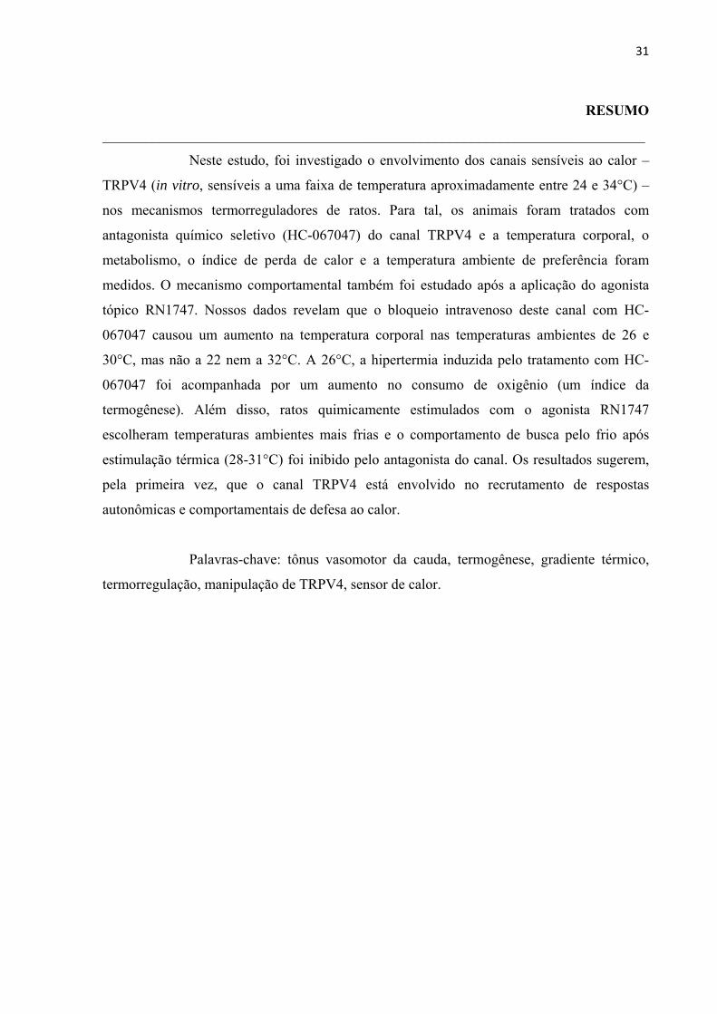
Neste estudo, foi investigado o envolvimento dos canais sensíveis ao calor –
TRPV4 (in vitro, sensíveis a uma faixa de temperatura aproximadamente entre 24 e 34°C) –
nos mecanismos termorreguladores de ratos. Para tal, os animais foram tratados com
antagonista químico seletivo (HC-067047) do canal TRPV4 e a temperatura corporal, o
metabolismo, o índice de perda de calor e a temperatura ambiente de preferência foram
medidos. O mecanismo comportamental também foi estudado após a aplicação do agonista
tópico RN1747. Nossos dados revelam que o bloqueio intravenoso deste canal com HC-
067047 causou um aumento na temperatura corporal nas temperaturas ambientes de 26 e
30°C, mas não a 22 nem a 32°C. A 26°C, a hipertermia induzida pelo tratamento com HC-
067047 foi acompanhada por um aumento no consumo de oxigênio (um índice da
termogênese). Além disso, ratos quimicamente estimulados com o agonista RN1747
escolheram temperaturas ambientes mais frias e o comportamento de busca pelo frio após
estimulação térmica (28-31°C) foi inibido pelo antagonista do canal. Os resultados sugerem,
pela primeira vez, que o canal TRPV4 está envolvido no recrutamento de respostas
autonômicas e comportamentais de defesa ao calor.
Palavras-chave: tônus vasomotor da cauda, termogênese, gradiente térmico,
termorregulação, manipulação de TRPV4, sensor de calor.
32
ABSTRACT
___________________________________________________________________________
In this study, we aimed at investigating the involvement of the warmth-
sensitive channel – TRPV4 (in vitro sensitive to temperatures in the range of approx. 24–
34°C) – on the thermoregulatory mechanisms in rats. We treated rats with a chemical
selective antagonist (HC-067047) of the TRPV4 channel and measured core body
temperature, metabolism, heat loss index and preferred ambient temperature. The behavioral
mechanism was also assessed after treatment with topical agonist (RN-1747). Our data
revealed that intravenous blockade of this channel with HC-067047 caused an increase in core
body temperature at ambient temperature of 26 and 30°C, but not at 22 and 32°C. At 26°C,
HC-067047-induced hyperthermia was accompanied by increase in oxygen consumption (an
index of thermogenesis). Furthermore, rats chemically stimulated with TRPV4 agonist choose
colder ambient temperatures and the cold-seeking behaviour after thermal stimulation (28–
31°C) was inhibited by TRPV4 antagonist. Our results suggest, for the first time, that TRPV4
channel is involved in the recruitment of behavioural and autonomic warmth-defence
responses to regulate core body temperature.
Keywords: tail vasomotor tone, thermogenesis, thermopreferendum
thermoregulation, TRPV4 manipulation, warmth sensor.
33
LISTA DE ABREVIAÇÕES
___________________________________________________________________________
: consumo de oxigênio;
FeCO2: Fração de entrada de gás carbônico (do baseline);
FeO2: Fração de entrada de oxigênio (do baseline);
FRe: Fluxo de entrada do ar;
FsCO2: Fração de saída de gás carbônico;
FsO2: Fração de saída de oxigênio;
i.v.: administração intravenosa.
IPC: índice de perda de calor;
PVA: pressão de vapor de água (kPa);
Ta: temperatura ambiente;
Tc: temperatura corporal;
Tp: temperatura da pele da cauda.

34
1. INTRODUÇÃO
___________________________________________________________________________
A temperatura corporal (Tc) é um dos sinais vitais e a habilidade em regular
esta variável é de fundamental importância, pois todas as reações bioquímicas e funções
fisiológicas são amplamente dependentes da temperatura (Wenisch et al., 1996; Wasserstrom
e Vites, 1999; Aihara et al., 2001). Em animais endotérmicos, tanto os mecanismos
autonômicos quanto os comportamentais são recrutados para regular a Tc (Bícego et al.,
2007). Neste sentido, redes neuronais independentes, controlada pela Tc e/ou temperatura da
pele, que são detectadas por termorreceptores, são usadas por cada termoefetor (tremor,
termogênese sem tremor, controle vasomotor cutâneo; Romanovsky 2007; Morrison e
Nakamura 2011). Adicionalmente, a termorregulação comportamental é essencial para a
sobrevivência; um exemplo é a procura por uma temperatura ambiente de preferência que é
considerada um dos mecanismos mais eficiente, menos energeticamente custoso e mais
amplamente difundido em mamíferos (Gordon, 1990; Bicego et al., 2007).
A recente descoberta e os avanços nos estudos dos canais TRPs (transient
receptor potencial) sensíveis à temperatura contribuíram para a compreensão dos mecanismos
de transdução dos sinais térmicos. Neste estudo, nós focamos no TRPV4 que é ativado in
vitro a temperaturas na faixa de 24-34°C (Güler et al., 2002; Kauer e Gibson, 2009), e
também por outros fatores como hiposmolaridade (Liedtke et al., 2000; Strotmann et al.,
2000), agonistas químicos (Vincent et al., 2009; Vincent e Duncton 2011) e metabólitos
endógenos do ácido araquidônico (Voets et al., 2005). A proteína do TRPV4 está encontrada
em alguns lugares compatíveis com sua função termorreguladora, incluindo neurônios
sensoriais primários, queratinócitos, células endoteliais, glândulas sudoríparas, neurônios
simpáticos e área pré-óptica do hipotálamo (Liedtke et al., 2000; Delany et al., 2001; Güler et
al., 2002; Watanabe et al., 2002; Alessandri-Haber et al., 2003). De fato, o TRPV4 tem sido
proposto atuar como termorreceptor, pois a remoção genética deste receptor levou
camundongos a selecionarem temperaturas mais quentes que os animais controles quando
expostos a um gradiente térmico ou quando eles tinham que escolher entre duas temperaturas
ambientes (Tas; Lee et al., 2005); no entanto, ainda não havia sido avaliado se este canal é
ativado em condições quentes não nocivas in vivo em um animal não geneticamente
modificado e se ele recruta mecanismos termorreguladores autonômicos e/ou
comportamentais para a manutenção da Tc. Então, este estudo investigou se os canais TRPV4
estão envolvidos na termorregulação autonômica e comportamental in vivo usando uma
35
abordagem farmacológica e, para tal, nós avaliamos os efeitos do antagonista e agonista
seletivo para o canal sobre a Tc, o índice de perda de calor (IPC), o consumo de oxigênio e a
Ta selecionada.

36
2. MATERIAL E MÉTODOS
___________________________________________________________________________
Animais
Foram utilizados neste estudo ratos da linhagem Wistar (massa corporal de
290-320g) provenientes da UNESP de Botucatu – SP. Os animais receberam água e ração ad
libitum, foram acondicionados à Ta de 23 ± 2ºC e submetidos a um ciclo claro:escuro de
12:12h. Os experimentos foram realizados entre 8:00 e 17:00h em grupos separados de
animais e os protocolos experimentais foram conduzidos de acordo com o Conselho Nacional
de Controle de Experimentação Animal (CONCEA) e tiveram aprovação da Comissão de
Ética no Uso de Animais (CEUA) da Faculdade de Ciências Agrárias e Veterinárias de
Jaboticabal (Protocolo n# 26430/11, 019414/14).
Fármacos
HC-067047 (2-Metil-1- [3- (4-morfolinil) propil] -5-fenil-N- [3-
(trifluorometil) fenil] -1H-pirrol-3-carboxamida) é um antagonista seletivo para TRPV4
recentemente descrito (Everaerts et al. 2010a) e adquirido da Tocris Bioscience (Bristol, UK).
O HC-067047 foi aliquotado e estocado a -20°C nas concentrações de 1000 ou 5000g/mL
em 100% de etanol e, no dia do experimento, a alíquota foi diluída em salina estéril de modo
a obtermos uma solução a 100 ou 500g/mL para adminstração intravenosa (i.v.; doses: 100
ou 500g/kg; 1 mL/kg).
RN-1747 ([1-(4-Cloro-2-nitrofenil)sulfonil-4-benzilpiperazina]) é um agonista
seletivo para o canal TRPV4 (Vincent et al., 2009) e também comprado da Tocris Bioscience
(Bristol, UK). A droga foi dissolvida em 100% de propilenoglicol e a dose utilizada para
aplicação tópica (3000g/kg; foi aplicado 1000g/mL/animal) foi baseada em resultados
prévios do laboratório colaborador.
Procedimentos cirúrgicos
Os animais foram anestesiados com uma injeção intraperitoneal de 100mg/kg
de cetamina e 10mg/kg de xilazina e profilaticamente tratados com antibiótico (enrofloxacina
5%, subcutâneo, 0.5mg/kg, Schering-Plough) e analgésico e anti-inflamatório (flunixina
meglumina, intramuscular, 2.5mg/kg, Schering-Plough). A área corporal destinada às
cirurgias foram tricotomisadas e limpas com solução de iodo 2% e, em seguida, os animais
foram colocados em decúbito dorsal em uma mesa cirúrgica para implantação de uma cânula
37
intravenosa (jugular). Uma pequena incisão longitudinal foi feita na superfície ventral do
pescoço, esquerda à traqueia. A veia jugular esquerda foi exposta, separada dos tecidos
conectivos adjacentes e amarrada. Um cateter de silicone (diâmetro interno: 0.5mm; diâmetro
externo: 0.9mm) preenchido com salina heparinizada (10U.I/mL) foi inserido na veia cava
superior através da veia jugular e ali fixado. A extremidade livre do cateter foi passada sob a
pele e exteriorizada pela nuca. O corte foi suturado e o cateter foi lavado com salina
heparinizada no dia seguinte à cirurgia e a cada dois dias até o dia do experimento.
Em seguida, os animais foram submetidos a uma laparotomia paramediana
para inserção de um sensor armazenador de temperatura (SubCue Dataloggers, Calgary,
Canadá) e, em seguida à implantação, os músculos abdominais e a pele foram suturados em
camadas. Os animais foram submetidos a um dos protocolos experimentais 3-5 dias depois
das cirurgias (ver item “Protocolos experimentais” abaixo).
Medidas de Consumo de Oxigênio
A taxa metabólica foi inferida por meio da medida de consumo de oxigênio
em sistema de respirometria aberta. O animal foi colocado em um respirômetro (3L) dentro de
uma câmara de temperatura controlada (Fanem, São Paulo, SP, Brasil) que manteve a Ta
dentro do respirômetro a 22, 26, 30 ou 32°C de acordo com cada protocolo. Um fluxo
contínuo de ar foi mantido na câmara experimental (pull mode) a uma taxa de 2000mL/min,
mas apenas uma subamostra (180mL/min) deste ar foi enviada aos analisadores de O2, CO2 e
H2O (SS4; Sable Systems, NV, USA). A pressão de vapor de água (PVA, kPa) foi a primeira
variável analisada (RH300; Sable Systems) e, posteriormente, foi utilizada (juntamente com a
pressão barométrica) para corrigir a porcentagem de O2, CO2 e o fluxo de ar antes do cálculo
do . Tal correção se fez necessária porque o ar não foi seco nem antes de entrar no
respirômetro nem antes de passar pelos analisadores (Lighton, 2008). Em seguida, a
porcentagem de CO2 (CA-10, Sable Systems) foi analisada e, após, a porcentagem de O2 e a
pressão barométrica foram registradas (PA-10; Sable Systems). Cada 20 minutos de registro
foram compostos por 3 minutos de baseline (amostra do ar que entrava no respirômetro), 14
minutos do ar que saia do respirômetro e mais 3 minutos de baseline. Amostras do baseline
foram registradas para monitorar a constância da água e da porcentagem de O2 e CO2 no ar
oferecido ao animal ao longo do experimento e para que o FiO2, FiCO2 e FiH2O fossem
calculados. Um distribuidor múltiplo de gases (RM8 Intelligent Multiplexer; Sable Systems)
foi utilizado para controlar qual amostra de gás (do respirômetro ou do baseline) seria enviada
38
aos analisadores. Os analisadores foram calibrados semanalmente ou sempre que necessário.
O analisador de O2 foi calibrado usando nitrogênio como “zero” e ar seco (20.95%) como
“span”; o analisador de CO2 foi também calibrado com nitrogênio puro como “zero” e uma
mistura de 1% de CO2 como “span”. Os dados foram coletados a cada segundo por um
sistema de aquisição apropriado (EXPEDATA v. 1.4.5; Sable Systems) e o foi
determinado usando a seguinte fórmula (Lighton, 2008):
1
Onde:
FRe: Fluxo de entrada do ar;
FeO2: Fração de entrada de oxigênio (do baseline);
FsO2: Fração de saída de oxigênio;
FeCO2: Fração de entrada de gás carbônico (do baseline);
FsCO2: Fração de saída de gás carbônico.
Protocolos experimentais
Os experimentos foram realizados com animais não anestesiados que passaram
por um dos protocolos abaixo.
1. Efeito do bloqueio químico do TRPV4 na Tc e nos termoefetores
autonômicos, termogênese e tônus vasomotor da cauda:
Os animais foram expostos a 31°C por 40 minutos antes da injeção i.v. de HC-
067047 (500 g/kg; 1 mL/kg) ou veículo (salina: etanol, 90%: 10%; 1 mL/kg) de forma a
aumentar o fluxo sanguíneo para a pele e garantir que a droga alcançaria os receptores TRPV4
na pele (Almeida et al., 2012). Depois da injeção, os animais foram mantidos a 31°C por mais
15 minutos antes de serem colocados no respirômetro. A Ta no respirômetro foi fixada em 22,
26, 30 ou 32°C, como descrito anteriormente (veja “medida de consumo de oxigênio). Uma
vez colocados no respirômetro, a taxa metabólica e as imagens infravermelho (Flir SC660,
Portland, OR, USA) foram registrados por 180 minutos. A Tc foi medida durante 60 minutos
antes e 180 minutos após a injeção da droga. A temperatura da cauda foi qualitativamente
analisada a partir das imagens infravermelho como um indicador do tônus vasomotor da
cauda dos ratos e, então o IPC foi calculado (as imagens foram tiradas a cada 2 minutos; veja
“Processamento e análise dos dados”). A dose de HC-067047 foi baseada em experimentos

39
pilotos (dados não mostrados).
2. Efeito da manipulação do TRPV4 na termorregulação comportamental
– gradiente térmico:
Para avaliar a temperatura selecionada pelos animais após os tratamentos, foi
utilizado um aparato com dois compartimentos completamente separados por uma parede
(cada compartimento consistia de 195 cm de comprimento, 15 cm de largura e 20 cm de
altura, Bonther, Ribeirão Preto, Brasil). As paredes do aparato e o assoalho de grade foram
feitos de aço inoxidável e o teto feito de uma tampa de acrílico transparente que ajudava a
manter a Ta dentro do gradiente. As extremidades foram aquecidas e resfriadas,
respectivamente, por uma resistência elétrica (Novus, RS, Brasil) e por um banho de
circulação externa com água resfriada e etileno-glicol (Polyscience, Il, EUA). Desse modo,
foi gerado um gradiente linear de Ta entre 15 e 35°C (Almeida, et al., 2006a) monitorado por
oito termopares distribuídos uniformemente sob o assoalho de grade. Para cada experimento
(e para cada compartimento do gradiente) foi construída uma regressão linear entre a
temperatura do ar vs distância dos termopares em relação à resistência elétrica para
confirmação da linearidade do gradiente térmico formado. Apenas R2 iguais ou maiores que
0.93 foram aceitos. Uma webcam foi posicionada no topo do gradiente e programada para
tirar fotos a cada 4 minutos ao longo do experimento. Baseado nas posições dos animais
obtidas pelas fotos e na regressão linear, a temperatura de preferência foi calculada.
Dois animais de cada vez (um em cada compartimento) foram aclimatados por
aproximadamente 18 horas no gradiente térmico antes dos tratamentos. Na manhã seguinte ao
intervalo de aclimatação, os ratos foram colocados individualmente em câmaras climáticas
por 40 minutos a 31°C para receberem os tratamentos. Um dos seguintes protocolos foram
feitos: no primeiro, depois da injeção i.v. de veículo (salina: etanol; 90%:10%; 1 mL/kg) ou
HC-067047 (100 g/kg; 1 mL/kg), os animais foram termicamente estimulados (Almeida et
al., 2006a,b) por meio de confinamento em uma porção (25cm) do gradiente onde a Ta estava
entre 28-31°C por 3 horas. No final deste intervalo, as barreiras que confinavam os animais
foram retiradas e eles puderam livremente se mover no seu respectivo compartimento. No
outro protocolo, os animais foram tratados com veículo (propilenoglicol; 1 mL) ou RN-1747
(1000 g/mL; 1 mL administrado topicamente no dorso do rato) e colocados novamente no
gradiente para livre seleção da temperatura de preferência.

40
Processamento e análise dos dados
Os dados são mostrados como média ± EPM. O IPC foi calculado pela
seguinte equação:
IPC = ⁄
Onde:
Tp = temperatura da pele da cauda do rato;
Ta = temperatura ambiente no mesmo momento em que a Tp foi medida;
Tc = temperatura corporal no mesmo momento em que a Tp foi medida.
Este índice tem sido usado para avaliar a resposta vasomotora
termorreguladora da cauda de ratos e seu valor varia de 0 a 1, sendo que zero indica
vasoconstrição máxima e 1 vasodilatação máxima (Romanovsky et al., 1998, 2002). A Tc ao
longo dos experimentos é expressa como variação da Tc basal que, por sua vez, é a média dos
últimos 6 registros de Tc antes de todos os tratamentos (meia hora; os registros eram feitos a
cada 5 minutos).
Os resultados obtidos foram analisados por ANOVA de duas vias (two way
ANOVA de medidas repetidas para testar o efeito do antagonista na Tc e na Ta de preferência
bem como o efeito do agonista na Ta de preferência; ou simplesmente two way ANOVA para
testar o efeito do antagonista no pico do delta da Tc, no e no IPC) seguido por teste post
hoc Holm-Šidák. A variância e a normalidade dos dados foram testadas e os dados foram
transformados para rank ou log quando não atendiam aos requisitos da análise paramétrica.
Foram consideradas diferenças significativas valores de P< 0,05.

41
3. RESULTADOS
___________________________________________________________________________
1. Efeito do bloqueio químico do TRPV4 na Tc e nos termoefetores autonômicos,
termogênese e tônus vasomotor da cauda:
Neste protocolo, foi estudado o efeito da administração i.v. do antagonista
seletivo de TRPV4, HC-067047, na Tc de ratos expostos a 22, 26, 30 e 32°C de Ta (Fig. 1a-
d). A 22 e 32°C, os animais tratados com HC-067047 não apresentaram diferença na Tc
comparada com os controles (efeito da interação entre tempo vs tratamento: F1,42 = 0.729; P =
0.897 e F1,42 = 0.392; P = 1.000, respectivamente). Em Tas intermediárias (26 e 30°C), a Tc
dos ratos tratados com antagonista foi significativamente maior que a dos animais injetados
com veículo (efeito da interação entre tempo vs tratamento: F1,42 = 3.338; P < 0.001 e F1,42 =
2.724; P < 0.001, respectivamente). A tabela 1 mostra o pico que a Tc (maior aumento no
delta da Tc) alcançou depois da injeção de veículo ou de HC-067047. A diferença
intertratamento (HC-067047 menos veículo) foi significante apenas a Ta de 26°C, indicando
que a esta Ta nós tivemos o efeito mais evidente do antagonista.
Devido ao efeito mais forte do bloqueio do TRPV4 na Tc ter sido observado a
26°C, nós selecionamos esta Ta para estudar também os efeitos do antagonista nos
termoefetores autonômicos. A figura 2a mostra o efeito do bloqueio dos canais TRPV4 no
consumo de oxigênio. Estes dados foram usados para criar o gráfico de vs Tc mostrado
na figura 2b. Nesta última plotagem, foi observado que os ratos tratados com veículo
ocuparam uma pequena área do gráfico ao longo do experimento (ou seja, a Tc e o
permaneceram quase constantes durante todo o tempo), ao passo que os animais tratados
com antagonista partiram de um ponto muito próximo ao controle, mas se direcionaram pra
uma área diferente durante o curso do experimento (efeito da interação entre tratamento vs Tc:
F1,3 = 2.988; P = 0.037).
O efeito do bloqueio de TRPV4 no IPC não foi estatisticamente diferente do
grupo controle (efeito da interação entre tratamento vs Tc: F1,3 = 2.296; P = 0.081), embora o
IPC tenha sido levemente menor nos ratos tratados com antagonista que nos animais tratados
com veículo, indicando que estes animais desenvolveram vasoconstrição da cauda (veja Fig.
3a e 3b). Nas figuras 3a e 3b, fica evidente que a temperatura da cauda foi maior nos animais
controle que nos tratados com antagonista.

42
Fig. 1. Efeito da administração i.v. de antagonista de TRPV4 (HC-067047) na Tc a diferentes Tas. Os animais foram expostos às Tas de 22° (a), 26° (b), 30° (c) e 32°C (d). Tc foi significativamente maior nos animais tratados com HC-067047 comparados com os controles quando expostos a 26 e 30°C. Tc basal: (a) 37.3±0.1°C (veículo) e 37.4 ±0.1°C (HC-067047); (b) 37.4±0.2°C (veículo) e 37.3±0.2°C (HC-067047); (c) 37.1±0.1°C (veículo) e 37.5 ±0.1°C (HC-067047); (d) 37.3±0.1°C (veículo) e 37.4±0.1°C (HC-067047). A seta indica o momento da injeção. Números entre parênteses representam o N experimental. * = pontos onde o efeito da interação foi significante. Dados são expressos como média ± EMP.
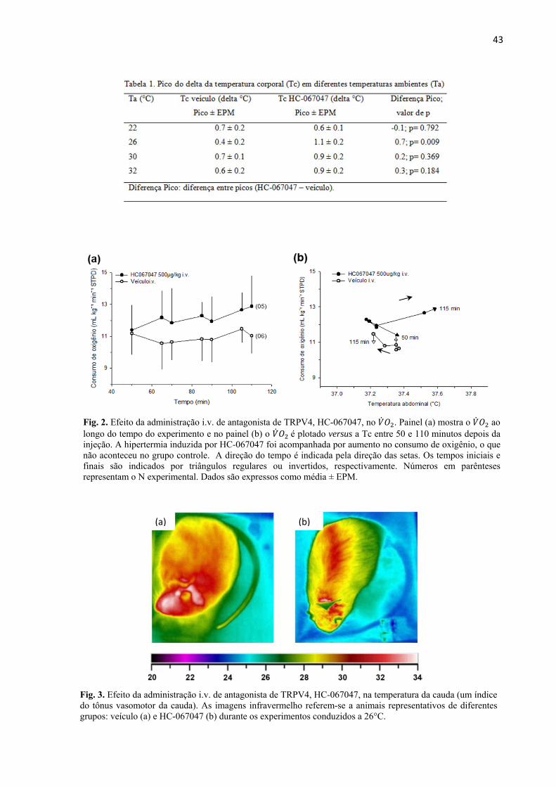
43
Fig. 2. Efeito da administração i.v. de antagonista de TRPV4, HC-067047, no . Painel (a) mostra o ao longo do tempo do experimento e no painel (b) o é plotado versus a Tc entre 50 e 110 minutos depois da injeção. A hipertermia induzida por HC-067047 foi acompanhada por aumento no consumo de oxigênio, o que não aconteceu no grupo controle. A direção do tempo é indicada pela direção das setas. Os tempos iniciais e finais são indicados por triângulos regulares ou invertidos, respectivamente. Números em parênteses representam o N experimental. Dados são expressos como média ± EPM.
(b)(a)
Fig. 3. Efeito da administração i.v. de antagonista de TRPV4, HC-067047, na temperatura da cauda (um índice do tônus vasomotor da cauda). As imagens infravermelho referem-se a animais representativos de diferentes grupos: veículo (a) e HC-067047 (b) durante os experimentos conduzidos a 26°C.

44
2. Efeito da manipulação do TRPV4 na termorregulação comportamental – gradiente
térmico:
A estimulação térmica (28-31°C) levou os animais controle a procurarem por
regiões mais frias do gradiente, depois do final do confinamento, que aqueles tratados com
antagonista (HC-067047; 100g/kg; 1mL/kg; efeito do tratamento: F1,10 = 5.09; P = 0.048;
efeito do tempo: F1,10 = 3.13; P < 0.001; sem efeito de interação; Fig. 4a). A Tc dos animais
tratados com HC-067047 foi maior que a dos animais controle (37.5 ± 0.1°C vs 37.0 ± 0.1°C,
respectivamente; efeito da interação entre tratamento vs tempo: F1,10 = 1.44; P = 0.01, dados
não mostrados). Neste mesmo sentido, a estimulação química dos canais TRPV4 com
agonista (RN-1747; 1000g/mL; 1mL tópico) causou uma redução na Ta selecionada
comparada com os animais tratados com veículo (efeito da interação entre tratamento vs
tempo: F1,9 = 4.18; P < 0.001; Fig. 4b), bem como reduziu a Tc ao longo do tempo (Tc foi
reduzida 0.7 ± 0.2°C depois do tratamento com agonista e apenas 0.1 ± 0.2°C depois do
tratamento controle; efeito da interação entre tratamento vs tempo: F1,8 = 1.41; P = 0.02;
dados não mostrados).
Fig. 4. Efeito da estimulação térmica e química do TRPV4 em ratos expostos ao gradiente térmico. Painel (a) mostra a Ta selecionada pelos animais após o tratamento com veículo ou HC-067047 e o confinamento entre 28 e 31°C por 3 horas (indicado no gráfico pela área cinza). Painel (b) mostra a Ta selecionada por animais tratados com veículo ou agonista de TRPV4 (RN-1747). Números em parênteses representam o N experimental. * = pontos onde há efeito de interação. Dados são expressos como média ± EPM.
(a) (b)
45
4. DISCUSSÃO
___________________________________________________________________________
No presente trabalho, nós testamos se a manipulação dos canais TRPV4
sensíveis ao calor altera a Tc e quais termoefetores (autonômicos e/ou comportamentais) estão
envolvidos nesta resposta. Baseado na faixa de temperatura de ativação destes receptores, in
vitro, nossa hipótese era que temperaturas moderadas ativariam os canais TRPV4 periféricos
in vivo que, por sua vez, ativariam respostas de defesa ao calor, regulando a Tc. Os resultados
indicam que, nas condições empregadas no presente estudo, os TRPV4 cutâneos são
fisiologicamente ativados in vivo em Tas entre 26-30°C e parecem estar envolvidos na
ativação de ambos os termoefetores, autonômicos e comportamentais.
O bloqueio intravenoso deste canal causou um aumento na Tc quando os
animais foram expostos a 26°C de Ta. Tal aumento foi acompanhado por um aumento no
consumo de oxigênio (um índice da termogênese). Além disso, segundo dados do laboratório
colaborador, a estimulação química dos receptores por agonista tópico causou uma redução na
Tc que foi influenciada por aumento na vasodilatação da cauda. Estes e outros resultados
sobre o efeito do agonista de TRPV4 na Tc são apresentados graficamente e melhores
discutidos na publicação encontrada no Apêndice A. Tais resultados (tanto de consumo de
oxigênio quanto de IPC) indicam que os TRPV4 modulam a ativação dos dois termoefetores
autonômicos mais importantes para ratos. Ainda, animais tratados com agonista de TRPV4
apresentaram uma preferência por Tas mais frias quando colocados em um gradiente térmico
em comparação aos controles e os animais que foram termicamente estimulados (28-31°C) e
tratados com o antagonista deste canal preferiam Tas mais quentes que o grupo veículo,
demonstrando que o comportamento de busca pelo frio foi bloqueado após o tratamento com
HC-067047. Estes resultados claramente indicam um papel do TRPV4 na ativação de
respostas de perda de calor via mecanismo comportamental em ratos.
Embora estudos anteriores tenham sugerido o TRPV4 como um termosensor, o
seu papel como termorregulador ainda não havia sido proposto, pois camundongos knock out
(KO) para V4 apresentam Tc similar àquela dos animais controle em normotermia ou quando
são expostos a ambiente frio (Liedtke e Friedman, 2003; Lee et al., 2005). No entanto,
resultados negativos com animais KO são difíceis de interpretar, especialmente porque estes
animais podem apresentar mecanismos compensatórios (Mogil et al., 2000, Lariviere et al.,
2001) e, então, não podíamos excluir a participação dos canais TRPV4 na termorregulação
apenas com base nestes trabalhos. De fato, no caso do TRPV1, também nenhuma mudança na

46
termorregulação foi observada nos estudos com animais KO (Szelényi et al., 2004; Iida et al.,
2005) embora, mais recentemente, tenha sido demonstrado que estes canais são importantes
para a manutenção da Tc (Steiner et al., 2007). Por meio de ferramentas farmacológicas e,
então, alterando a atividade dos canais TRPV4 apenas temporariamente, nosso grupo
conseguiu evitar que mecanismos compensatórios acontecessem a longo prazo e fomos assim
capazes, pela primeira vez, de mostrar o envolvimento deste receptor na manutenção da Tc
em um organismo não geneticamente modificado.
Papel dos TRPV4 quanto a Ta é fixa
Os resultados do laboratório da prof. M. C. Almeida sobre a aplicação tópica
do agonista a 26°C de Ta apontavam o envolvimento dos canais TRPV4 na ativação de
respostas de perda de calor e na consequente redução da Tc, então, para complementar estes
dados, protocolos com o uso do antagonista do canal e sob diferentes Tas (visando uma
estimulação térmica dos canais, ao invés de química como foi feito com o uso do agonista)
foram realizados (ver Apêndice A). Nestes protocolos, para facilitar que o antagonista
alcançasse seus receptores na pele (Patapoutian et al., 2003), os animais, antes de serem
submetidos as suas respectivas Tas experimentais, foram expostos a temperaturas moderadas
de modo a induzir um aumento no fluxo sanguíneo para a pele como já descrito (Almeida et
al., 2012).
Os resultados mostraram que o bloqueio dos receptores TRPV4 causou um
aumento na Tc quando os animais foram expostos a 26 ou a 30°C (Fig. 1 e Tabela 1), o que é
consistente com nossa hipótese inicial que temperaturas quentes não nocivas ativariam estes
canais. Quando estes canais foram bloqueados, as respostas de defesa ao calor não foram
ativadas, promovendo a hipertermia. O aumento na Tc não foi visto quando a Ta era 22°C ou
32°C, possivelmente porque estas temperaturas estão foram da faixa de ativação dos canais
TRPV4 e sejam detectadas por outros receptores sensíveis à temperatura, como o TRPM8 e
V3, respectivamente (Caterina, 2007).
O próximo passo então foi estudar quais termoefetores eram ativados e/ou
inibidos durante a resposta termorreguladora à manipulação do TRPV4. Com os resultados
aqui apesentados (Fig. 2), nós mostramos que a termogênese é modulada pelo V4, pois o
consumo de oxigênio foi discreto, mas significativamente aumentado ao longo do tempo após
o tratamento com HC-067047 quando comparado ao tratamento com veículo. Estes resultados
estão de acordo com estudos que mostram que camundongos KO para TRPV4 e adipócitos

47
isolados desta mesma linhagem de animal apresentam elevado comparado aos animais e
células controle (Kusudo et al., 2012; Ye et al., 2012). No presente estudo, os animais que
tiverem seus receptores TRPV4 farmacologicamente bloqueados pelo antagonista
consumiram 1.24 ± 0.2mL O2/kg/min a mais que os ratos tratados com veículo. Assumindo
que cada mililitro de oxigênio consumido produz 20.3 J de calor (Randall et al., 1997), este
pequeno aumento foi suficiente para produzir 1.5 kJ em 60 minutos (entre 50 e 110 minutos;
1.24 x 60 x 20.3), o que é suficiente para aumentar a Tc em 0.7°C em animais de 300g (Fig.
2b; Q = mct, onde Q é a energia térmica em calorias, m é a massa em gramas, c é o calor
específico, considerado 3.47 J para tecidos biológicos e t é a variação de temperatura, 0.7°C;
Schmidt-Nielsen 1997; Da Silva e Maia 2013). Quanto ao IPC, embora ele pareça ser menor
nos animais tratados com antagonista, ele não foi estatisticamente diferente dos animais
controle (Fig. 3). Estes resultados revelam que o bloqueio da sinalização da informação
quente não nociva mediada por TRPV4, inativa reposta de defesa do calor, aumentando a Tc.
Papel do TRPV4 quando a Ta pode ser selecionada pelo animal
Os resultados do laboratório colaborador indicavam ativação também dos
mecanismos comportamentais depois da estimulação dos TRPV4. Eles demonstraram que
animais controle gastavam mais tempo a 32 que a 26°C quando eles só tinham estas duas Tas
para escolherem, mas os animais tratados com agonista gastavam tempo igual nas duas Tas,
apontando que a preferência por Tas mais quente foi inexistente após o tratamento com RN
1747 (veja Apêndice A). Para melhor compreender o envolvimento dos TRPV4 na
termorregulação comportamental, nós estudamos os ratos em gradiente térmico. No primeiro
experimento, os animais foram temporariamente confinados em uma região do gradiente onde
a Ta ficava entre 28 e 31°C de modo a verificar o esperado comportamento de busca pelo frio
depois da estimulação térmica (Almeida et al., 2006a). De fato, os animais controle preferiram
Tas mais frias que os tratados com antagonista (Fig. 4a). No outro experimento, a estimulação
química dos TRPV4 com agonista em animais não confinados e colocados no gradiente
levou-os a procurarem por Tas mais frias que os controles (Fig. 4b). Ambos os resultados
claramente indicam que a estimulação dos TRPV4 (tanto térmica quanto química) ativa
comportamento de busca pelo frio. Juntos, nossos resultados indicam que se os animais tem a
possibilidade de selecionarem uma Ta de preferência, a estimulação dos TRPV4 ativará o
mecanismo comportamental, de modo a regular a Tc, o que está de acordo com a ativação
prioritária deste tipo de termoefetor já que ele é considerado muito eficiente e menos

48
energicamente custoso que os efetores autonômicos. Por outro lado, quando não é permitido
aos animais termorregularem comportamentalmente, a estimulação dos TRPV4 ativará
termoefetores autonômicos para regulação da Tc. Com estes dados, então, nós mostramos,
pela primeira vez, o papel dos TRPV4 periféricos na ativação da perda de calor não
evaporativa e a inibição do ganho de calor in vivo em ratos.
Perspectivas
Desde que os canais TRPV4 foram caracterizados aproximadamente 10 anos
atrás, vários estudos foram desenvolvidos e diversas funções biológicas para estes canais
foram propostas, tais como regulação e detecção da osmolalidade no sistema nervoso central
(Strotmann et al., 2000; Liedtke & Friedman 2003; Liedtke et al. 2003), termorrecepção em
animais KO (Lee et al., 2005), participação em repostas inflamatórias (Vergnolle et al., 2010;
Bang et al., 2012; Denadai-Souza et al., 2012; Ye et al., 2012; Poole et al., 2013), nocicepção
(Alessandri-Haber et al., 2003, 2005; Nakatsuka e Iwai 2009; Everaerts et al. 2010b),
respostas de hiperalgesia (Todaka et al., 2004; Alessandri-Haber et al., 2009; Ding et al.,
2010, Wang et al., 2011; Segond von Banchet et al., 2013), entre outros. O presente estudo
adiciona ainda a participação dos canais TRPV4 na regulação da Tc em ratos não
geneticamente modificados. Recentemente, os canais termoTRP foram sugeridos como um
alvo para o desenvolvimento de novas estratégias terapêuticas. Contudo, parece que muitos
dos compostos submetidos para testes clínicos ainda estão longe de serem bem sucedidos, em
particular, devido à ocorrência de efeitos colaterais (Ferrer-Montiel et al., 2012). A melhor
compreensão das funções destes canais é essencial para o desenvolvimento de novas
ferramentas que modulem a atividade deles mesmos, mas que causem menos efeitos
colaterais. Com este estudo pretendemos contribuir para um melhor conhecimento a respeito
das funções deste receptor no sistema termorregulador, fornecendo importantes dados que
possam colaborar com o desenvolvimento de novas estratégias terapêuticas.

49
5. REFERÊNCIAS BIBLIOGRÁFICAS
___________________________________________________________________________
AIHARA, H.; OKADA, Y.; TAMAKI, N. The effects of cooling and rewarming on the neuronal activity of pyramidal neurons in guinea pig hippocampal slices. Brain Res.; v. 893, p. 36–45. 2001.
ALESSANDRI-HABER, N.; YEH, J.J.; BOYD, A.E.; PARADA, C.A.; CHEN, X.; REICHLING, D.B.; LEVINE, J.D. Hypotonicity induces TRPV4-mediated nociception in rat. Neuron. v. 39, p. 497–511, 2003.
ALESSANDRI-HABER, N.; JOSEPH, E.; DINA, O.A.; LIEDTKE, W.; LEVINE, J.D. TRPV4 mediates pain-related behavior induced by mild hypertonic stimuli in the presence of inflammatory mediator. Pain, v. 118, p. 70–79, 2005
ALESSANDRI-HABER, N.; DINA, O.A.; CHEN, X.; LEVINE, J.D. TRPC1 and TRPC6 channels cooperate with TRPV4 to mediate mechanical hyperalgesia and nociceptor sensitization. J Neurosci., v. 29, p. 6217–6228, 2009.
ALMEIDA, M.C.; STEINER, A.A.; BRANCO, L.G.; ROMANOVSKY, A.A. Cold-seeking behavior as a thermoregulatory strategy in systemic inflammation. Eur J Neurosci., v. 23, p. 3359–3367, 2006a.
ALMEIDA, M.C.; STEINER, A.A.; BRANCO, L.G.; ROMANOVSKY, A.A. Neural substrate of cold-seeking behavior in endotoxin shock. PLoS ONE, 1, e1., 2006b.
ALMEIDA, M.C.; HEW-BUTLER, T.; SORIANO, R.N.; RAO, S.; WANG, W.; WANG, J.; TAMAYO, N.; OLIVEIRA, D.L.; NUCCI, T.B.; ARYAL, P.; GARAMI, A.; BAUTISTA, D.; GAVVA, N.R.; ROMANOVSKY, A.A. Pharmacological blockade of the cold receptor TRPM8 attenuates autonomic and behavioral cold defenses and decreases deep body temperature. J Neurosci., v. 32, p. 2086–2099, 2012.
BANG, S.; YOO, S.; YANG, T.J.; CHO, H.; HWANG, S.W. Nociceptive and pro-inflammatory effects of dimethylallyl pyrophosphate via TRPV4 activation. Br J Pharmacol., v. 166, p. 1433–1443, 2012.
BICEGO, K.C.; BARROS, R.C.; BRANCO, L.G. Physiology of temperature regulation: comparative aspects. Comp Biochem Physiol A Mol Integr Physiol.; v. 147, p. 616–639, 2007.
CATERINA, M.J. Transient receptor potential ion channels as participants in thermosensation and thermoregulation. Am J Physiol Regul Integr Comp Physiol., v. 292, p. R64–R76, 2007.
DA SILVA, R.G.; MAIA, A.S.C. Thermal balance and thermoregulation. In:_____ Principles of Animal Biometeorology, Netherlands: Springer, 2013. p. 75–106.
DELANY, N.S.; HURLE, M.; FACER, P.; ALNADAF, T.; PLUMPTON, C.; KINGHORN, I.; SEE, C.G.; COSTIGAN, M.; ANAND, P.; WOOLF, C.J.; CROWTHER, D.; SANSEAU, P.; TATE, S.N. Identification and characterization of a novel human vanilloid receptor-like protein, VRL-2. Physiol Genomics, v. 4, p. 165–174, 2001.

50
DENADAI-SOUZA, A.; MARTIN, L.; DE PAULA, M.A.; DE AVELLAR, M.C.; MUSCARA, M.N.; VERGNOLLE, N.; CENAC, N. Role of transient receptor potential vanilloid 4 in rat joint inflammation. Arthritis Rheum., v. 64, p. 1848–1858, 2012.
DING, X.L.; WANG, Y.H.; NING, L.P.; ZHANG, Y.; GE, H.Y.; JIANG, H.; WANG, R.; YUE, S.W. Involvement of TRPV4-NO-cGMP-PKG pathways in the development of thermal hyperalgesia following chronic compression of the dorsal root ganglion in rats. Behav Brain Res., v. 208, p. 194–201, 2010.
EVERAERTS, W.; ZHEN, X.; GHOSH, D.; VRIENS, J.; GEVAERT, T.; GILBERT, J.P.; HAYWARD, N.J.; MCNAMARA, C.R.; XUE, F.; MORAN, M.M.; STRASSMAIER, T.; UYKAL, E.; OWSIANIK, G.; VENNEKENS, R.; DE RIDDER, D.; NILIUS, B.; FANGER, C.M.; VOETS, T. Inhibition of the cation channel TRPV4 improves bladder function in mice and rats with cyclophosphamide-induced cystitis. Proc Natl Acad Sci U S A. v. 107, p. 19084–19089, 2010a. EVERAERTS, W.; NILIUS, B.; OWSIANIK, G. The vanilloid transient receptor potential channel TRPV4: from structure to disease. Prog Biophys Mol Biol., v. 103, p. 2–17, 2010b.
FERRER-MONTIEL, A.; FERNÁNDEZ-CARVAJAL, A.; PLANELLS-CASES, R.; FERNÁNDEZ-BALLESTER, G.; GONZÁLEZ-ROS, J.M.; MESSEGUER, A.; GONZALEZ-MUNIZ, R. Advances in modulating thermosensory TRP channels. Expert Opin Ther Pat., v. 22, p. 999–1017, 2012.
GORDON, C.J. Thermal biology of the laboratory rat. Physiol Behav.; v. 47, p. 963–991, 1990.
GÜLER, A.D.; LEE, H.; IIDA, T.; SHIMIZU, I.; TOMINAGA, M.; CATERINA, M. Heat-evoked activation of the ion channel, TRPV4. J Neurosci.; v. 22, p. 6408–6414, 2002.
IIDA, T.; SHIMIZU, I.; NEALEN, M.L.; CAMPBELL, A.; CATERINA, M. Attenuated fever response in mice lacking TRPV1. Neurosci Lett., v. 378, p. 28–33, 2005.
KAUER, J.A.; GIBSON, H.E. Hot flash: TRPV channels in the brain. Trends Neurosci.; v. 32, p. 215–224, 2009.
KUSUDO, T.; WANG, Z.; MIZUNO, A.; SUZUKI, M.; YAMASHITA, H. TRPV4 deficiency increases skeletal muscle metabolic capacity and resistance against diet-induced obesity. J Appl Physiol., v. 7, p. 1223–1232, 2012.
LARIVIERE, W.R.; CHESLER, E.J.; MOGIL, J.S. Transgenic studies of pain and analgesia: mutation or background genotype? J Pharmacol Exp Ther., v. 297, p. 467–473, 2001.
LEE, H.; IIDA, T.; MIZUNO, A.; SUZUKI, M.; CATERINA, M.J. Altered thermal selection behavior in mice lacking transient receptor potential vanilloid 4. J Neurosci., v. 25, p. 1304–1310, 2005.
LIEDTKE, W.; FRIEDMAN, J.M. Abnormal osmotic regulation in trpv4_/_ mice. Proc Natl Acad Sci U S A, v. 100, p. 13698–13703, 2003.
LIEDTKE, W.; CHOE, Y.; MARTI-RENOM, M.A.; BELL, A.M.; DENIS, C.S.; SALI, A.; HUDSPETH, A.J.; FRIEDMAN, J.M.; HELLER, S. Vanilloid receptor-related osmotically

51
activated channel (VR-OAC), a candidate vertebrate osmoreceptor. Cell, v. 103, p. 525–535, 2000.
LIEDTKE, W.; TOBIN, D.M.; BARGMANN, C.I.; FRIEDMAN, J.M. Mammalian TRPV4 (VR-OAC) directs behavioral responses to osmotic and mechanical stimuli in Caenorhabditis elegans. Proc Natl Acad Sci U S A., v. 100(Suppl 2), p. 14531–14536, 2003.
LIGHTON, J.R.B. Flow-through respirometry: the equations. In: ______ Measuring Metabolic Rates. A Manual for Scientists. New York: Oxford University Press, 2008. p. 96.
MOGIL, J.S.; YU, L.; BASBAUM, A.I. Pain genes?: natural variation and transgenic mutants. Annu Rev Neurosci., v. 23, p. 777–811, 2000.
MORRISON, S.F.; NAKAMURA, K. Central neural pathways for thermoregulation. Front Biosci.; v. 16, p.74–104, 2011.
NAKATSUKA, M.; IWAI, Y. Expression of TRPV4 in the stimulated rat oral mucous membrane–nociceptive mechanisms of lingual conical papillae. Okajimas Folia Anat Jpn., v. 86, p. 45–54, 2009.
PATAPOUTIAN, A.; PEIER, A.M.; STORY, G.M.; VISWANATH, V. ThermoTRP channels and beyond: mechanisms of temperature sensation. Nat Rev Neurosci., v. 4, p. 529–539, 2003.
POOLE, D.P.; AMADESI, S.; VELDHUIS, N.A.; ABOGADIE, F.C.; LIEU, T.; DARBY, W.; LIEDTKE, W.; LEW, M.J.; MCINTYRE, P.; BUNNETT, N.W. Protease-activated receptor 2 (PAR2) protein and transient receptor potential vanilloid 4 (TRPV4) protein coupling is required for sustained inflammatory signaling. J Biol Chem., v. 288, p. 5790–5802, 2013.
RANDALL, D.; BURGGREN, W; FRENCH, K. Using energy: meeting environmental challenges. In: _____. Eckert Animal Physiology: Mechanisms and Adaptations. 4.ed. New York:Freeman Company, 1997. p. 665–723.
ROMANOVSKY, A.A.; SIMONS, C.T.; KULCHITSKY, V.A. “Biphasic” fevers often consist of more than two phases. Am J Physiol Regul Integr Comp Physiol.; v. 275, p. R323–R331, 1998.
ROMANOVSKY, A.A.; IVANOV, A.I.; SHIMANSKY, Y.P. Selected contribution: ambient temperature for experiments in rats: a new method for determining the zone of thermal neutrality. J Appl Physiol., v. 92, p. 2667–2679, 2002.
SCHMIDT-NIELSEN, K. Temperature regulation. In: ____ Animal Physiology: adaptation and environment. Cambridge: Cambridge University Press. 1997, p. 241–300.
SEGOND VON BANCHET, G.; BOETTGER, M.K.; KONIG, C.; IWAKURA, Y.; BRAUER, R.; SCHAIBLE, H.G. Neuronal IL-17 receptor upregulates TRPV4 but not TRPV1 receptors in DRG neurons and mediates mechanical but not thermal hyperalgesia. Mol Cell Neurosci., v. 52, p. 152–160, 2013.
STEINER, A.A.; TUREK, V.F.; ALMEIDA, M.C.; BURMEISTER, J.J.; OLIVEIRA, D.L.; L.ROBERTS, J.; BANNON, A.W.; NORMAN, M.H.; LOUIS, J.C.; TREANOR, J.J.;

52
GAVVA, N.R.; ROMANOVSKY, A.A. 2007. Nonthermal activation of transient receptor potential vanilloid-1 channels in abdominal viscera tonically inhibits autonomic cold-defense effectors. J Neurosci., v. 27, p. 7459–7468, 2007.
STROTMANN, R.; HARTENECK, C.; NUNNENMACHER, K.; SCHULTZ, G.; PLANT, T.D.. OTRPC4, a nonselective cation channel that confers sensitivity to extracellular osmolarity. Nat Cell Biol, v. 2, p. 695–702, 2000.
SZELÉNYI, Z.; HUMMEL, Z.; SZOLCS_ANYI, J.; DAVIS, J.B. Daily body temperature rhythm and heat tolerance in TRPV1 knockout and capsaicin pretreated mice. Eur J Neurosci., v. 19, p. 1421–1424, 2004.
TODAKA, H.; TANIGUCHI, J.; SATOH, J.; MIZUNO, A.; SUZUKI, M. Warm temperature-sensitive transient receptor potential vanilloid 4 (TRPV4) plays an essential role in thermal hyperalgesia. J Biol Chem., v. 279, p. 35133–35138, 2004.
VERGNOLLE, N.; CENAC, N.; ALTIER, C.; CELLARS, L.; CHAPMAN, K.; ZAMPONI, G.W.; MATERAZZI, S.; NASSINI, R.; LIEDTKE, W.; CATTARUZZA, F.; GRADY, E.F.; GEPPETTI, P.; BUNNETT, N.W. A role for transient receptor potential vanilloid 4 in tonicity-induced neurogenic inflammation. Br J Pharmacol., v. 159, p. 1161–1173, 2010.
VINCENT, F.; DUNCTON, M.A. TRPV4 agonists and antagonists. Curr Top Med Chem., v. 11, p. 2216–2226, 2011.
VINCENT, F.; ACEVEDO, A.; NGUYEN, M.T.; DOURADO, M.; DEFALCO J.; GUSTAFSON, A.; SPIRO, P.; EMERLING, D.E.; KELLY, M.G.; DUNCTON, M.A. Identification and characterization of novel TRPV4 modulators. Biochem Biophys Res Commun., v. 3, p. 490–494, 2009.
VOETS, T.; TALAVERA, K.; OWSIANIK, G.; NILIUS, B. Sensing with TRP channels. Nat Chem Biol., v. 1, p. 85–92, 2005.
WANG, C.; NING, L.P.; WANG, Y.H.; ZHANG, Y.; DING, X.L.; GE, H.Y.; ARENDT-NIELSEN, L. & YUE, S.W. Nuclear factor-kappa B mediates TRPV4-NO pathway involved in thermal hyperalgesia following chronic compression of the dorsal root ganglion in rats. Behav Brain Res., v. 221, p. 19–24, 2011.
WASSERSTROM, J.A.; VITES, A.M. Activation of contraction in cat ventricular myocytes: effects of low Cd(2+) concentration and temperature. Am J Physiol.; v. 277, p. H488–H498, 1999.
WATANABE, H.; VRIENS, J.; SUH, S.H.; BENHAM, C.D.; DROOGMANS, G.;NILIUS, B. 2002. Heat-evoked activation of TRPV4 channels in a HEK293 cell expression system and in native mouse aorta endothelial cells. J Biol Chem., v. 277, p. 47044–47051, 2002.
WENISCH, C.; NARZT, E.; SESSLER, D.L.; PARSCHALK, B.; LENHARDT, R.; KURZ, A.; GRANINGER, W. Mild intraoperative hypothermia reduces production of reactive oxygen intermediates by polymorphonuclear leukocytes. Anesth Analg. v. 82, p.810–816, 1996.
YE, L.; KLEINER, S.; WU, J.; SAH, R.; GUPTA, R.K.,; BANKS, A.S.; COHEN, P.; KHANDEKAR, M.J.; BOSTRÖM, P.; MEPANI, R.J.; LAZNIK, D.; KAMENECKA, T.M.;
53
SONG, X.; LIEDTKE, W.; MOOTHA, V.K.; PUIGSERVER, P.; GRIFFIN, P.R.; CLAPHAM, D.E.; SPIEGELMAN, B.M. TRPV4 is a regulator of adipose oxidative metabolism, inflammation, and energy homeostasis. Cell. v. 151. p.96–110. 2012.
54
Capítulo II
Participação dos TRPV4 centrais na ativação de
respostas de perda de calor em ratos
55
RESUMO
___________________________________________________________________________
Recentemente, nosso grupo descreveu, pela primeira vez em animal não
geneticamente modificado, que os TRPV4 periféricos são ativados quando a temperatura
ambiente (Ta) está entre 26-30°C e modulam a taxa metabólica, o tônus vasomotor da cauda e
o mecanismo comportamental de busca pelo frio levando à manutenção da temperatura
corporal (Tc). Além da periferia, os TRPV4 também podem ser encontrados na área pré-
óptica medial (MPA), região envolvida com ativação das respostas autonômicas de perda de
calor durante exposição a Tas moderadamente altas. Entretanto, não era conhecido o papel
dos TRPV4 hipotalâmicos na termorregulação. Então, o objetivo do presente trabalho foi
verificar se os TRPV4 da MPA participam do controle da temperatura corporal (Tc) e quais
efetores estariam envolvidos nesta resposta. Para este fim, sob duas Tas diferentes (21 e
28°C), a Tc de ratos adultos foi medida após o bloqueio dos TRPV4 da MPA com um
antagonista específico. A taxa metabólica, o índice de perda de calor (IPC) e o mecanismo
comportamental de busca pelo frio também foram avaliados após bloqueio dos receptores. A
Tc dos animais foi maior após o tratamento com o antagonista (HC-067047; p< 0.001) e este
resultado foi mais evidente quando a Ta era 28°C. Paralelamente à Tc, o (p= 0.012), mas
não o IPC (p> 0.05) também foi mais elevado nos animais que receberam microinjeção de
HC-067047. E o comportamento de busca pelo frio após os animais serem expostos por duas
horas a Tas moderadamente altas, foi inibido após bloqueio dos receptores (p= 0.01). Em
conjunto, estes resultados mostram que: 1) os TRPV4 da MPA participam da manutenção da
Tc quando a Ta é moderadamente elevada e 2) a MPA é importante para a ativação do
mecanismo comportamental em ratos, ao contrário do que havia sido sugerido anteriormente.
Palavras–chave: TRPV4, área pré-óptica medial, temperatura corporal, taxa
metabólica, termorregulação comportamental
56
ABSTRACT
___________________________________________________________________________
Recently, our group has described, in non-genetically modified rats, that
peripheral TRPV4 is activated at ambient temperatures (Tas) of 26-30°C and warmth-defence
responses are triggers by these receptors. TRPV4 also can be found in medial pre optic area
(MPA), region known by being involved with activation of heat loss mechanism.
Nevertheless no data was available about the role of hypothalamic TRPV4 in
thermoregulation. Then, the aim of this study was to verify if central TRPV4 in the medial
preoptic area (MPA) plays a role in the control of body temperature (Tc) and which
thermoefectors are involved in this response. To this end, under two different Tas (21 e 28°C),
the Tc of rats was measured after TRPV4 in MPA were antagonized by HC-067047.
Metabolic rate, heat loss index (HLI) and preferred temperature (Tp) were also recorded after
central blockage of TRPV4. The antagonism of central TRPV4 caused a further increase in
the Tb of rats (p< 0.001) exposed to 28°C than to 21°C compared to vehicle-treated animals.
The (p= 0.012), but not HLI, was also increased by the antagonist. Lastly, the cold
seeking behavior observed in control animals after exposition to warm Tas for 2 hours (28-
30°C) was inhibited by the microinjection of the antagonist of TRPV4 (p= 0.01). Our results
suggest that: 1) central TRPV4 plays a role in the thermoregulation when Ta is warm and 2)
MPA is an important site for behavioral thermoregulation, in opposition to what has been
suggested formerly.
Key words: TRPV4, medial pre optic area, body temperature, metabolic rate,
behavioural thermoregulation.

57
LISTA DE ABREVIAÇÕES
___________________________________________________________________________
: taxa metabólica (ml O2 kg-1 min-1 STPD);
3V: terceiro ventrículo;
APO: área pré-óptica;
CA: comissura anterior;
Canais TRP: “transient receptor potential channels”;
DMH: hipotálamo dorso medial;
Fe: Fluxo de entrada do ar;
FeO2: Fração de entrada de oxigênio;
FsO2: Fração de saída de oxigênio;
Intra MPA: animais cujo sítio de microinjeção foi localizado dentro da área pré-óptica medial;
IPC: índice de perda de calor, isto é, o grau de vasodilatação/vasoconstrição;
LPBd: núcleo parabraquial, parte dorsal;
LPBl: núcleo parabraquial, parte lateral;
MnPO: núcleo pré óptico mediano;
MPA: área pré-óptica medial;
MPOC: núcleo pré óptico medial, parte central;
MPOL: núcleo pré óptico medial, parte lateral;
MPOM: núcleo pré óptico medial, parte medial;
NMDA: receptor de glutamato dependente de voltagem;
OX: quiasma óptico;
Peri MPA: animais cujo sítio de microinjeção foi localizado fora da área pré-óptica medial;
Ta: temperatura ambiente;
TC: trajeto da cânula;
Tc: temperatura corporal;
Tp: temperatura da pele da cauda.

58
1. INTRODUÇÃO
___________________________________________________________________________
É indiscutível a importância da manutenção da temperatura corporal (Tc)
dentro de determinados limites, já que ela é uma variável que influencia todos os outros
sistemas do organismo. Este controle da Tc depende de uma comunicação sincronizada entre
os receptores periféricos, as regiões centrais responsáveis por integrarem a informação e
elaborarem as respostas e os órgãos efetores que vão, de fato, responder ao sinal que foi
enviado pelos receptores. Ultimamente, estudos foram dedicados à descoberta e descrição da
via somatossensorial da termorregulação (Nakamura e Morrison, 2007, 2008, 2010;
Nakamura, 2011) e o que se sabe atualmente é que as informações de quente e frio seguem
por vias separadas desde a sua detecção na periferia, passagem pela medula espinal e ascensão
até o núcleo parabraquial (LPB dorsal ou lateral, se quente ou frio, respectivamente). Do LPB,
as informações são enviadas à área pré-óptica (APO), conhecida por ser a principal região
envolvida na regulação da Tc e por ter um importante papel integrador de todas as
informações térmicas vindas das várias regiões do organismo (Matsuda et al., 1992; Boulant,
1998). Da APO, mais especificamente do núcleo pré-óptico mediano (MnPO), sai uma rede
de interneurônios excitatórios ou inibitórios em direção à APO medial (MPA), ativando ou
inibindo esta sub-região que, por sua vez, se conecta a outros áreas (hipotálamo dorsomedial,
DMH, e núcleo rostral da rafe) responsáveis pelo controle da produção dependente e
independente de tremor e pela vasoconstrição (Nakamura e Morrison, 2008 e 2010).
Nos últimos dez anos foram descritos alguns canais catiônicos TRPs (do
inglês: transient receptor potential channels) que respondem aos mais diversos estímulos,
dentre eles a temperatura. Estes canais têm sido sugeridos como sensores de temperatura
ambiente (Ta) na periferia e seus estudos certamente contribuiriam pra melhor elucidação da
via de termorrecepção. A respeito dos TRPV4, foco deste trabalho, sabe-se que eles são
amplamente expressos nos tecidos de mamíferos, podendo ser encontrados, dentre outros
lugares, nos queratinócitos e na APO, principalmente na MPA e no MnPO (Güler et al., 2002;
Watanabe et al., 2002; Patapoutian et al., 2003; Caterina, 2007). Em relação ao seu papel na
termorregulação, é conhecido que eles detectam temperaturas altas não nocivas e que
camundongos geneticamente destituídos deste tipo de canal selecionam Tas mais altas do que
os camundongos selvagens, além de apresentarem maior latência no teste de retirada da cauda
quando esta é imersa em temperaturas moderadamente altas em comparação aos animais
controles (Lee et al., 2005). Recentemente, nosso grupo em colaboração com o grupo da Dra.

59
Maria Camila Almeida, descreveu em animais não geneticamente modificados a ativação dos
TRPV4 periféricos entre Tas de 26-30°C e a modulação da taxa metabólica, do tônus
vasomotor da cauda e do mecanismo comportamental decorrente desta ativação (Vizin et al.,
2015). Entretanto, não se conhecia o papel dos TRPV4 da MPA no controle da Tc. Baseado
em nosso recente estudo e no modelo de Nakamura e Morrison (2010) para ativação de
respostas de perda de energia térmica, a presença de TRPV4 na MPA poderia indicar a
participação destes receptores nas vias termolíticas frente a estímulos térmicos. Então, no
presente trabalho, foi verificado se os TRPV4 da MPA estão envolvidos na termorregulação
autonômica e comportamental. Para este fim, primeiramente foi determinada a Tc depois do
bloqueio químico dos receptores TRPV4 hipotalâmicos em duas Tas diferentes. Em um
segundo momento, a taxa metabólica, o índice de perda de calor (IPC) e o mecanismo
comportamental foram também verificados após o bloqueio dos receptores da MPA.
60
2. MATERIAL E MÉTODOS
___________________________________________________________________________
Animais
Foram utilizados neste estudo ratos da linhagem Wistar (massa corporal de
290-320g) provenientes da UNESP de Botucatu – SP. Os animais receberam água e ração ad
libitum, foram acondicionados à Ta de 23 ± 2ºC e submetidos a um ciclo claro:escuro de
12h:12h. Os experimentos foram realizados entre 8:00 e 17:00h em grupos separados de
animais e os protocolos experimentais foram conduzidos de acordo com o Conselho Nacional
de Controle de Experimentação Animal (CONCEA) e tiveram aprovação da Comissão de
Ética no Uso de Animais (CEUA) da Faculdade de Ciências Agrárias e Veterinárias de
Jaboticabal (Protocolo n# 019288/14).
Fármaco
HC-067047 (2-Metil-1- [3- (4-morfolinil) propil] -5-fenil-N- [3-
(trifluorometil) fenil] -1H-pirrol-3-carboxamida; Tocris Bioscience, Bristol, UK) foi
recentemente descrito como um potente e seletivo antagonista para TRPV4 que inibe a
ativação deste receptor por vários estímulos, incluindo o calor, hipotonicidade, ácido
araquidônico e o agonista 4-PDD (Everaerts et al. 2010). O antagonista foi dissolvido em
90% de salina estéril e 10% de etanol e as doses utilizadas (1.5; 15 e 75ng/kg; foram injetados
0.5; 5 ou 25ng/50nL/animal de ~ 330g) foram baseadas em nosso estudo prévio (Vizin et al.,
2015) e experimentos pilotos.
Cirurgias
Os animais foram anestesiados com uma injeção intraperitoneal de 100mg/kg
de cetamina e 10mg/kg de xilazina e profilaticamente tratados com antibiótico (enrofloxacina
5%, subcutâneo, 0.5mg/kg, Schering-Plough) e analgésico e anti-inflamatório (flunixina
meglumina, intramuscular, 2.5mg/kg, Schering-Plough). A área corporal destinada às
cirurgias foram tricotomisadas e limpas com Digluconato de Clorexidina 2% (Rioquímica,
SP, Brasil) e, em seguida, os animais foram fixados em um estereotáxico (David Kopf,
modelo 900, Tujunga, CA, EUA) para a implantação de cânula guia. Uma incisão foi feita no
escalpo e o periósteo foi retirado para que um pequeno orifício pudesse ser feito onde a cânula
seria implantada. A cânula tinha 15mm de comprimento e 0,7mm de diâmetro externo e foi
inserida 2mm acima da MPA (barra do incisivo a -3.3mm; 0,0-0,1mm posterior ao bregma;

61
0,5mm lateral à linha média, e 6.5mm ventral à superfície do crânio) de acordo com as
coordenadas adaptadas do atlas de Paxinos e Watson (2005) e baseadas em estudo prévio
(Scarpellini, et al., 2009). Posteriormente, a cânula foi fixada ao crânio por meio de parafuso
e cimento acrílico e um mandril foi mantido dentro da mesma até o momento das injeções
para prevenir oclusão e infecção.
Após a cirurgia central, os animais foram submetidos a uma laparotomia
paramediana para inserção de um sensor armazenador de temperatura (SubCue Dataloggers,
Calgary, Canadá) e, em seguida à implantação, os músculos abdominais e a pele foram
suturados em camadas. Os animais foram submetidos a um dos protocolos experimentais 6-8
dias depois das cirurgias (ver item “Protocolos experimentais” abaixo).
Microinjeção e procedimento histológico
As microinjeções foram realizadas em ratos não anestesiados utilizando-se
uma agulha dental de injeção (Mizzy, 200 m O.D.) conectada por uma cânula PE-10 a uma
seringa Hamilton de 5µL acoplada a uma bomba injetora (modelo 310, Stoelting CO., IL,
EUA). Foi utilizada uma agulha 2mm mais longa do que a cânula guia, permitindo contato
com a MPA apenas no momento da injeção. O volume injetado foi de 50nL de antagonista ou
veículo por 30 segundos sendo a agulha removida da cânula guia apenas 30 segundos após o
término da injeção para se evitar refluxo.
Ao final de cada experimento, os animais foram anestesiados e 50nL de uma
solução 2% de azul de Evans foi injetada na MPA para confirmar o sítio de microinjeção. Os
animais foram então perfundidos através do ventrículo cardíaco esquerdo (o átrio direito foi
cortado) com 60mL de salina e 60mL de uma solução de formalina a 10%. Em seguida os
animais foram decapitados e os encéfalos removidos da caixa craniana e imersos em solução
de formalina a 10% por pelo menos dois dias, sendo, em seguida, processados para a
realização dos cortes histológicos. Após a inclusão em parafina, foram feitos cortes de 15μm
de espessura, abrangendo toda a extensão da MPA, os quais foram corados pelo método de
Nissl. O sítio de microinjeção foi confirmado por observação em microscópio óptico e foram
considerados para a análise dos resultados dois grupos de animais: intra MPA, aqueles cujo
sítio de microinjeção foi localizado dentro desta região e os que tiveram injeção fora desta
região eram contados como peri MPA.
62
Medidas da Tc e temperatura da pele da cauda (Tp):
Os sensores armazenadores de temperatura foram acoplados a um computador,
via uma conexão óptica, e programados para a coleta de dados ao longo do experimento com
leituras de Tc a cada 5 minutos utilizando-se o programa “SubCue temperature datalogger”.
Ao final dos experimentos, após a eutanásia dos animais, os sensores foram mais uma vez
conectados ao computador para a leitura das Tcs medidas e essas, por sua vez, foram
corrigidas de acordo com as especificações contidas no manual do fabricante.
A Tp foi medida pela obtenção de imagens termográficas (câmera sensível a
infravermelho; Flir SC660, OR, USA) e foi usada para calcular o índice de perda de calor
(IPC) do animal, isto é, o grau de vasodilatação/vasoconstrição da cauda (janela térmica do
rato; Almeida et al., 2006a). O IPC varia de 0 a 1, sendo que zero indica vasoconstrição
máxima e 1 vasodilatação máxima, e é calculado de acordo com a fórmula (Romanovsky et
al., 2002):
IPC = ⁄
Onde:
Tp = temperatura da pele da cauda do rato;
Ta = temperatura ambiente no mesmo momento em que a Tp foi medida;
Tc = temperatura corporal no mesmo momento em que a Tp foi medida.
A técnica da termografia infravermelha detecta a radiação eletromagnética
emitida por um corpo e converte tal radiação em uma imagem colorida de acordo com a
intensidade da mesma. Essa técnica tem sido comumente usada na fisiologia termal para
mensurar a Tp em endotermos (Romanovsky et al., 2002; Tattersall e Milson, 2003; Almeida,
et al., 2006a; Scott et al., 2008; Vizin, et al., 2015) e também em ectotermos (Tattersall et al.,
2004, Scarpellini, et al., 2015). A emissividade usada foi 0.95 e o respirômetro foi vedado
com um plástico filme que implicou em um erro de 35% na detecção das temperaturas. O erro
foi devidamente corrigido antes do cálculo do IPC.
Medidas de Consumo de Oxigênio
A taxa metabólica foi inferida por meio da medida de consumo de oxigênio
em sistema de respirometria aberta. O animal foi colocado em um respirômetro (3L) dentro de
uma câmara de temperatura controlada (Fanem, São Paulo, SP, Brasil) que manteve a Ta
dentro do respirômetro a 21 ou 28°C de acordo com o protocolo estudado. Um fluxo contínuo

63
de ar foi mantido na câmara experimental (pull mode) a uma taxa de 1700 mL/min, mas
apenas uma subamostra (180 mL/min) deste ar foi enviada aos analisadores de O2 e H2O por
um subamostrador de fluxo de ar (SS4; Sable Systems, NV, USA). A pressão de vapor de
água foi a primeira variável analisada (RH300; Sable Systems) e, posteriormente, foi utilizada
(juntamente com a pressão barométrica) para corrigir a porcentagem de O2 e o fluxo de ar
antes do cálculo do . Tal correção se fez necessária porque o ar não foi seco nem antes de
entrar no respirômetro nem antes de passar pelo analisador de O2 (Lighton, 2008). Em
seguida, a porcentagem de O2 e a pressão barométrica foram registradas (PA-10; Sable
Systems). Cada 20 minutos de registro foram compostos por 3 minutos de baseline (amostra
do ar que entrava no respirômetro), 14 minutos do ar que saía do respirômetro e mais 3
minutos de baseline. Amostras do baseline foram registradas para monitorar a constância da
água e da porcentagem de O2 no ar oferecido ao animal ao longo do experimento e para que a
fração de O2 no ar entrando no respirômetro fosse determinado. Um distribuidor múltiplo de
gases (RM8 Intelligent Multiplexer; Sable Systems) foi utilizado para controlar qual amostra
de gás (do respirômetro ou do baseline) seria enviada aos analisadores. Os analisadores foram
calibrados sempre que necessário. O analisador de O2 foi calibrado usando nitrogênio como
“zero” e ar seco (20.95%) como “span” e os dados foram coletados a cada segundo por um
sistema de aquisição apropriado (EXPEDATA v. 1.4.5; Sable Systems). O foi calculado
com base na taxa de fluxo do ar e na diferença de porcentagem do O2 na entrada e na saída da
câmara (Mortola e Besterman, 2006; Mortola e Toro-Velasquez, 2013):
Onde:
Fe: Fluxo de entrada do ar
FeO2: Fração de entrada de oxigênio
FsO2: Fração de saída de oxigênio
Medidas de temperatura de preferência em gradiente térmico
Para avaliar a temperatura selecionada pelos animais após os tratamentos, foi
utilizado um aparato com dois compartimentos completamente separados por uma parede
(cada compartimento consistia de 195 cm de comprimento, 15 cm de largura e 20 cm de
altura, Bonther, Ribeirão Preto, Brasil). As paredes do aparato e o assoalho de grade foram
feitos de aço inoxidável e o teto feito de uma tampa de acrílico transparente que ajudava a

64
isolar a Ta dentro do gradiente. As extremidades foram aquecidas e resfriadas,
respectivamente, por uma resistência elétrica (Novus, RS, Brasil) e por um banho de
circulação externa com água resfriada e etileno-glicol (Polyscience, Il, EUA). Desse modo,
foi gerado um gradiente linear de Ta entre 15 e 35°C (Almeida, et al., 2006a) monitorado por
oito termopares distribuídos uniformemente sob o assoalho de grade. Para cada experimento
(e para cada compartimento do gradiente) foi construída uma regressão linear entre a
temperatura do ar vs a distância dos termopares em relação à resistência elétrica para
confirmação da linearidade do gradiente térmico formado. Apenas R2 iguais ou maiores que
0.93 foram aceitos. Uma webcam foi posicionada no topo do gradiente e programada para
tirar fotos a cada 4 minutos ao longo do experimento. Baseado na posição dos animais obtida
pelas fotos e na regressão linear, a temperatura de preferência foi calculada.
Protocolos Experimentais
Os experimentos foram realizados com animais não anestesiados que passaram
por um dos protocolos abaixo.
1. Efeito da microinjeção intra MPA de antagonista de TRPV4 sobre a Tc
de ratos expostos a 28°C:
Um rato de cada vez foi aclimatado a 28°C por cerca de 15 horas (overnight) e,
em seguida, foi colocado em uma câmara (3L) para registro da Tc pré tratamento. Após 40
minutos, o animal recebeu uma injeção intra MPA de HC-067047 ou veículo e teve sua Tc
registrada por mais 200 minutos.
2. Efeito da microinjeção intra MPA de antagonista de TRPV4 sobre Tc
de ratos expostos a 21°C:
Esse protocolo foi semelhante ao descrito anteriormente com a diferença que
os animais foram aclimatados a 21oC e mantidos a esta temperatura durante todo o
experimento.
3. Efeito da microinjeção intra MPA de antagonista de TRPV4 sobre os
termoefetores autonômicos, termogênese e tônus vasomotor da cauda, a 28°C:
Nos mesmos animais aclimatados a 28°C por 15 horas, do protocolo 1, foram
feitas medidas também de e IPC. Os registros pré-tratamento foram realizados durante os
40 minutos de aclimatação dentro do respirômetro. Em seguida, o animal recebeu uma injeção
65
intra MPA de HC-067047 ou veículo e as mesmas variáveis foram monitoradas por mais 200
minutos.
4. Efeito da microinjeção intra MPA de antagonista de TRPV4 sobre a Ta de
preferência:
Dois animais de cada vez foram aclimatados por aproximadamente 18 horas no
gradiente (overnight; um em cada compartimento). Na manhã após o intervalo de
aclimatação, os ratos foram colocados individualmente em suas caixas por aproximadamente
20 minutos e receberam microinjeção intra MPA de HC-067047 ou veículo. Em seguida, os
animais foram retidos por duas horas em uma porção do gradiente (~20cm) na qual a Ta
variava de 28 a 30°C. Após a exposição a um ambiente de Ta moderadamente elevada, as
barreiras que limitavam os animais foram retiradas e eles puderam selecionar livremente a Ta
pelas próximas 4 horas.
Análise dos resultados
Os dados são apresentados como média EPM. Os resultados obtidos foram
analisados pelo teste de Kruskal Wallis ou por ANOVA de duas vias de medidas repetidas ou
não seguidas por teste post hoc Holm-Šidák. A variância e a normalidade dos dados foram
testadas e os dados foram transformados para rank ou log quando não atendiam aos requisitos
da análise paramétrica. Foram consideradas diferenças significativas valores de P< 0,05. Os
símbolos que denotam diferença estatística (*, # e †) foram utilizados apenas quando o teste
post hoc foi capaz de detectar uma interação pontual entre os tratamentos.

66
3. RESULTADOS
___________________________________________________________________________
Sítios de microinjeção na área pré-óptica medial
Na figura 1A pode ser observada uma fotomicrografia representativa de corte
transversal do diencéfalo de um animal do grupo intra MPA. Na figura 1B estão apresentados
esquemas de encéfalo de rato na região onde é localizada a MPA, mostrando a extensão das
microinjeções do grupo intra MPA.
A
Fig. 1. A) Fotomicrografia de um corte transversal do encéfalo de um animal representativo mostrando o sítio típico de microinjeção (seta) na área pré-óptica medial (MPA). B) Figuras esquemáticas demonstrando a localização aproximada das microinjeções do grupo intra MPA. Os valores acima das figuras indicam a distância em milímetros em relação ao bregma. CA: comissura anterior; OX: quiasma óptico; TC: trajeto da cânula; 3V: terceiro ventrículo; MPA: área pré-óptica medial; MPOC: núcleo pré óptico medial, parte central; MPOL: núcleo pré óptico medial, parte lateral; MPOM: núcleo pré óptico medial, parte medial (esquemas adaptados do Atlas de Paxinos e Watson, 2005).
B

67
1. Efeito da microinjeção intra MPA de antagonista de TRPV4 sobre Tc de ratos
expostos a 28°C
A Tc dos animais expostos a 28°C aumentou após a microinjeção em todos os
grupos, como já reportado para injeções na área pré-óptica (Steiner et al., 2002a, 2002b,
2002c; Scarpellini et al., 2009), mas este aumento foi dependente do tratamento recebido. A
Tc dos ratos que receberam injeção de 5ng/50nL de HC-067047 intra MPA foi maior ao
longo do tempo do que a dos animais que receberam injeção de veículo ou de 0.5ng/50nL do
antagonista na mesma região. Entretanto, a Tc não foi diferente do grupo controle quando a
injeção de 5ng/50nL de HC-067047 era localizada em regiões adjacentes à MPA (peri MPA;
interação entre tempo e tratamento: p< 0.001; F3,24=1.862; Fig. 2).
Fig. 2. Efeito da microinjeção de antagonista de TRPV4, HC-067047 (0.5 e 5 ng/50nL), ou veículo na área pré-óptica medial (MPA) sobre a temperatura corporal de ratos expostos a 28°C. A seta indica o momento da microinjeção. Intra MPA: microinjeção na MPA; peri MPA: microinjeção em sítios próximos à MPA. * = p< 0.05 para HC-067047 5ng/50nL intra MPA vs veículo; # = p< 0.05 para HC-067047 5ng/50nL intra MPA vs HC-067047 0.5ng/50nL; † = p< 0.05 para HC-067047 5ng/50nL intra MPA vs HC-067047 5ng/50nL peri MPA. Valores são expressos como média ± EPM.

68
Fig. 3. Efeito da microinjeção de antagonista de TRPV4, HC-067047 (0.5, 5 e 25ng/50nL), ou veículo na área pré-óptica medial (MPA) sobre a temperatura corporal de ratos expostos a 21°C. A seta indica o momento da microinjeção. Intra MPA: microinjeção na MPA. Valores são expressos como média ± EPM.
2. Efeito da microinjeção intra MPA de antagonista de TRPV4 sobre Tc de ratos
expostos a 21°C:
A Tc dos animais expostos a 21°C também aumentou após a microinjeção em
todos os grupos e este aumento foi dependente do tratamento recebido. Neste protocolo, uma
dose ainda maior (75ng/kg; 25 ng/50nL) foi testada, mas seu efeito na Tc não diferiu dos
outros grupos (interação entre tempo e tratamento: p< 0.001; F3,24=1.93. Fig. 3).
3. Efeito da microinjeção intra MPA de antagonista de TRPV4 sobre os termoefetores
autonômicos, termogênese e tônus vasomotor da cauda, a 28°C:
O consumo de oxigênio de todos os animais foi semelhante antes da
microinjeção, entretanto o tratamento intra MPA com o antagonista na dose de (15ng/kg;
5ng/50nL) causou um aumento no dos animais até o final do experimento (interação
entre tempo e tratamento: p= 0.012; F2,19=3.851; Fig. 4A). Quanto ao IPC, o índice não foi
alterado por nenhum dos tratamentos, mas foi reduzido conforme a Tc aumentou (efeito da
Tc: p= 0.016, F2,19= 2.873; Fig. 4B). Imagens termográficas representativas do grupo controle
e do grupo antagonista (5ng/animal) são apresentadas na figura 5.
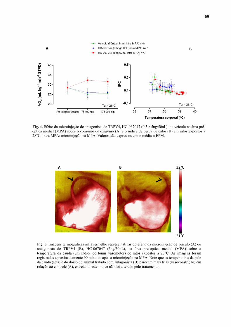
69
32°C
21°C
A
Fig. 5. Imagens termográficas infravermelho representativas do efeito da microinjeção de veículo (A) ou antagonista de TRPV4 (B), HC-067047 (5ng/50nL), na área pré-óptica medial (MPA) sobre a temperatura da cauda (um índice do tônus vasomotor) de ratos expostos a 28°C. As imagens foram registradas aproximadamente 90 minutos após a microinjeção na MPA. Note que as temperaturas da pele da cauda (seta) e do dorso do animal tratado com antagonista (B) parecem mais frias (vasoconstrição) em relação ao controle (A), entretanto este índice não foi alterado pelo tratamento.
B
Fig. 4. Efeito da microinjeção de antagonista de TRPV4, HC-067047 (0.5 e 5ng/50nL), ou veículo na área pré-óptica medial (MPA) sobre o consumo de oxigênio (A) e o índice de perda de calor (B) em ratos expostos a 28°C. Intra MPA: microinjeção na MPA. Valores são expressos como média ± EPM.
A B

70
Fig. 6. Efeito da microinjeção de antagonista de TRPV4, HC-067047 (0.5ng/50nL), ou veículo na área pré-óptica medial (MPA) sobre a temperatura ambiente selecionada em ratos após terem sido restritos no gradiente entre 28 e 30°C por duas horas (indicado pela região cinza no gráfico). A seta indica o momento da microinjeção. Intra MPA: microinjeção na MPA. * = p< 0.05; Valores são expressos como média ± EPM.
4. Efeito da microinjeção intra MPA de antagonista de TRPV4 sobre a Ta de
preferência:
Após a retenção no gradiente em uma região de temperatura moderada (28-
30°C), os animais que receberam injeção de veículo procuraram por regiões mais frias
comparados àqueles que tiveram os receptores TRPV4 da MPA antagonizados (HC-067047,
0.5ng/50nL; 1.5ng/kg). Estes últimos permaneceram por quase quatro horas em Tas parecidas
às da região da restrição, enquanto os ratos do grupo controle selecionaram Tas próximas a
23°C (interação entre tempo e tratamento: p= 0.01. F1,12= 1.505. Fig. 6). Uma imagem
representativa do gradiente é mostrada na figura 7.
Fig. 7. Foto representativa de um experimento realizado no gradiente térmico para avaliação do efeito da microinjeção de antagonista de TRPV4, HC-067047 (0.5ng/50nL), ou veículo na área pré-óptica medial (MPA) sobre a temperatura ambiente selecionada pelos animais. O rato do compartimento A foi tratado com veículo enquanto o do compartimento B foi tratado com HC-067047. A foto foi tirada cerca de 100 minutos após os animais terem sido liberados da região de restrição. Note que o animal A (veículo) encontra-se em uma região mais fria (mais distante da resistência) comparado ao animal B (HC-067047).
71
4. DISCUSSÃO
___________________________________________________________________________
Neste trabalho foi testado se o bloqueio químico dos TRPV4 da área pré-óptica
medial alteraria a Tc de ratos e quais termoefetores (autonômicos e/ou comportamentais)
participariam desta resposta. Recentemente, descrevemos em animais não geneticamente
modificados que a ativação dos TRPV4 na periferia acontece quando a Ta está entre 26 e
30°C e que tanto a taxa metabólica é reduzida quanto o IPC é aumentando, além do
mecanismo comportamental de busca pelo frio ser ativado por estes receptores (Vizin et al.,
2015). Entretanto, não se conhecia o papel dos TRPV4 hipotalâmicos na termorregulação.
Com o uso de microinjeção de um antagonista seletivo para este canal (HC-067047, Everaerts
et al., 2010) na MPA, demonstramos no presente estudo, pela primeira vez, que os receptores
centrais participam da manutenção da Tc quando os animais são expostos a Tas moderadas
(28-30°C) também por meio da modulação da taxa metabólica e do mecanismo
comportamental.
Papel dos TRPV4 centrais no controle da Tc e os termoefetores envolvidos
A Tc de todos os animais foi aumentada após a microinjeção como já era
esperado (veja abaixo “Críticas da metodologia”), entretanto, no último terço dos
experimentos dos protocolos 1 e 2 (135-200 minutos), a Tc dos animais mantidos a 28°C e
tratados intra MPA com HC-067047 na dose de 5 ng/animal foi ainda maior, cerca de 0.87 ±
0.03°C mais alta que a dos animais controle (Fig. 2). Este efeito foi significativamente mais
potente que aquele causado pela mesma dose e por outra cinco vezes maior (25 ng/animal) em
animais mantidos no frio (teste de Kruskal-Wallis: p< 0.001): a 21°C, o HC-067047 5
ng/animal elevou a Tc em apenas 0.45 ± 0.02°C em relação ao grupo veículo e o HC-067047
25 ng/animal causou um aumento de 0.30 ± 0.05°C na Tc dos animais comparado ao grupo
controle considerando o mesmo intervalo de tempo (Fig. 3).
Estes resultados mostram a participação dos TRPV4 hipotalâmicos na
manutenção da Tc quando a Ta é moderadamente alta, respondendo então a primeira pergunta
deste trabalho. Já que o efeito do antagonista foi mais evidente a 28°C, esta Ta foi escolhida
para a realização dos experimentos do protocolo 3 que visava determinar quais efetores
autonômicos (taxa metabólica e tônus vasomotor da cauda) estavam envolvidos no efeito do
HC-067047. De forma similar ao descrito para os TRPV4 periféricos (Vizin et al., 2015), o
antagonista administrado na MPA elevou a Tc via aumento da taxa metabólica (Fig. 4A) e

72
inibição do comportamento de busca por Tas mais frias (Fig. 6 e 7). O aumento do consumo
de oxigênio induzido pelo bloqueio dos TRPV4 está de acordo com estudos recentes (Kusudo
et al., 2012 e Ye et al., 2012) que demonstraram que camundongos knock out para TRPV4 e
adipócitos isolados desta linhagem de animal apresentam maior comparado aos animais e
células controle, respectivamente. É interessante notar que no caso de animais knock out todos
os canais TRPV4 estão ausentes e não é possível diferenciar quais deles, centrais ou
periféricos, estão envolvidos na ativação dos efetores termolíticos. Os resultados do presente
estudo, junto com aqueles do nosso trabalho anterior (Vizin et al., 2015; ver capítulo anterior),
indicam que ambos, centrais e periféricos, são importantes para as respostas autonômicas e
comportamentais de perda de calor.
Em relação ao IPC, apesar da cauda e do dorso dos animais tratados com
antagonista parecerem mais vasocontraídos (mais frios; Fig. 5B) comparados ao grupo
veículo (Fig. 5A), este índice não foi modificado pelas doses de HC-067047 utilizadas, mas
ele foi alterado de forma inversamente proporcional ao aumento da Tc (Fig. 4B). Pode-se
então dizer que uma vasoconstrição (redução do IPC) aconteceu no decorrer dos experimentos
de forma semelhante em todos os tratamentos, provavelmente como consequência da injeção
na MPA.
A respeito do mecanismo comportamental, o bloqueio dos TRPV4 da MPA
seguido pela exposição dos animais a Tas entre 28-30°C (por 2 horas) inibiu a busca por Tas
mais frias como observado nos animais controle (= mecanismo comportamental de perda de
calor. Figuras 6 e 7). Em 2002, Bícego e Branco ao verificarem que sapos cuja APO tinha
sido lesada não apresentaram prejuízo no mecanismo comportamental de termorregulação em
normotermia, mas exibiram reduzida busca por Tas mais quentes após a indução de febre por
LPS, sugeriram que a APO é uma região importante também para o desenvolvimento da febre
comportamental em ectotermos. Em contrapartida, Almeida e colaboradores mais tarde
(2006b) não encontraram nenhuma diferença no mecanismo comportamental, entre ratos com
ou sem lesão na APO, quando eles foram desafiados de quatro formas diferentes:
administração de baixa ou alta dose de LPS e aplicação de mentol (agonista de TRPM8,
indutor de busca por Tas quentes) ou resiniferatoxina (agonista de TRPV1, indutor de busca
por Tas frias). Almeida e colaboradores (2006b) mostraram que, semelhantemente aos
animais controle, os lesados buscaram por Tas quentes após tratamento com baixa dose de
LPS ou mentol e procuraram Tas frias após receberem tratamento com altas doses de LPS
(seguidas por Tas quentes) ou com resiniferatoxina. Estes autores sugeriram então que a APO

73
seria apenas importante para ativação de mecanismos autonômicos, mas não
comportamentais, de controle da Tc, pelo menos em mamíferos. Nos dois trabalhos citados,
de sapos e ratos, foi empregada lesão eletrolítica da APO que pode causar lesão não somente
dos corpos celulares, mas também de fibras de passagem de neurônios provenientes de outras
regiões encefálicas. Os resultados do presente estudo, por outro lado, indicam que, a
manipulação farmacológica de receptores específicos na MPA, no caso dos TRPV4, pode
revelar a participação dessa região na regulação de mecanismo comportamental, pelo menos
de perda de calor.
Modelo proposto para o mecanismo de ação termolítica dos TRPV4 na MPA
De acordo com o modelo da via somatosensorial da termorregulação proposta
por Nakamura e Morrison (2010), as informações detectadas na periferia pelos receptores de
calor são enviadas ao corno dorsal da medula espinal de onde saem os neurônios de segunda
ordem em direção ao núcleo parabraquial dorsal (LPBd) que, por sua vez, envia aferências a
interneurônios glutamatérgicos do núcleo pré-óptico mediano (MnPO). Estes interneurônios
ativam então os neurônios gabaérgicos da MPA que inibem o a rafe rostral e o hipotálamo
dorso medial (DMH). Tal inibição reduz/bloqueia tanto a produção de calor independente
quanto a depende de tremor, além de induzir vasodilatação levando, assim, à manutenção da
Tc em ambientes quentes (ver esquema página 15). Baseado no modelo citado e nos
resultados do presente trabalho, sugerimos então que os TRPV4 estejam localizados nos
neurônios gabaérgicos da MPA e que o bloqueio químico deles reduz a taxa de despolarização
destes neurônios já que o influxo de cátions por estes receptores está inibido. Desta forma,
uma redução na liberação de GABA nos sítios caudais efetores deve ocorrer e então os
mecanismos de produção/conservação de calor controlados por estas regiões são desinibidos,
aumentando o consumo de oxigênio e, consequentemente, a Tc. Quanto ao mecanismo
comportamental de busca pelo frio, sua via ainda não é estabelecida, mas pelos resultados
aqui obtidos com o bloqueio dos TRPV4 hipotalâmicos e exposição do animal ao gradiente
(Fig. 6 e 7), parece que ela também depende das eferências da MPA de forma semelhante aos
mecanismos autonômicos. No entanto, estudos ainda são necessários para elucidar a via
central do comportamento termorregulador.
Ainda, Shibasaki et al. (2007) demonstraram que os neurônios hipocampais de
neonatos de camundongo são significativamente despolarizados a valores próximos ao limiar
de ativação dos receptores NMDA (glutamatérgicos) quando mantidos em banho a 37°C
74
comparados aos neurônios de animais knock out para TRPV4. Neste mesmo sentido, um
trabalho mais recente in vitro (Li et al., 2013), também com fatias de neurônios hipocampais
de camundongos, mostrou que o antagonista HC-067047 atenuou o aumento na corrente pelo
canal NMDA induzida por agonista de V4. Ambos os trabalhos mostram que o influxo de
cátions pelos TRPV4 facilita a despolarização induzida por glutamato. Nossa sugestão então é
que o bloqueio dos TRPV4, possivelmente localizados dos neurônios gabaérgicos da MPA,
tenha comprometido a despolarização destes neurônios pela redução da entrada de cátions
(como mencionado) e, principalmente, por dificultar a ação do glutamato vindo das eferências
da MnPO. Já que o glutamato teria seu efeito reduzido nos neurônios gabaérgicos (por eles
não estarem previamente facilitados), eles não seriam ativados e o GABA, por sua vez, não
seria liberado no DMH nem na rafe rostral e então os mecanismos de produção/conservação
de calor seriam desinibidos levando ao aumento da taxa metabólica e da Tc (e, talvez,
ativação da procura por Tas mais frias), como observado no presente trabalho (Fig. 8, a
legenda da figura está na página seguinte – pág. 77).
Ainda de acordo com o modelo de Nakamura e Morrison (2008), em ambiente
frio a via somatossensorial é semelhante à descrita acima, com exceção que as aferências da
medula espinal chegam ao LPB lateral (LPBl) e que a MnPO envia interneurônios
gabaérgicos à MPA, inibindo então esta região, desinibindo o DMH e a rafe rostral e ativando
os mecanismos de produção/conservação de calor. Neste sentido, nos nossos experimentos
realizados a 22°C, a MnPO possivelmente estava inibindo a MPA, e então o bloqueio do
TRPV4 numa região já previamente inibida resultou em um efeito pouco evidente na Tc.
Finalmente, um trabalho in vitro de Hori e colaboradores (1999) demonstrou
que canais não seletivos para cátions ativados pelo calor modulam a taxa de disparo dos
neurônios sensíveis ao calor na APO. Ainda não é possível dizer que os TRPV4 estejam
dentre os canais sugeridos por Hori e colaboradores (1999) e que os neurônios sensíveis ao
calor sejam os gabaérgicos citados por Nakamura e Morrison (2008, 2010), mas certamente
trabalhos neste sentido são necessários e contribuiriam para a melhor compreensão da via
sensorial térmica.
Desta forma, nossos resultados mostram pela primeira vez que o bloqueio dos
TRPV4 da MPA aumenta a Tc via modulação da taxa metabólica e do mecanismo
comportamental de busca pelo frio, sugerindo que estes receptores participam da manutenção
da Tc quando a Ta é relativamente alta e que a MPA é necessária também para ativação da
termorregulação comportamental, ao contrário do sugerido anteriormente. Ainda, o presente

75
trabalho propõe que os TRPV4 estejam localizados nos neurônios gabaérgicos da MPA,
modulando a excitabilidade destes neurônios e, por sua vez, alterando a ação do
neurotransmissor glutamato liberado pelas eferências do MnPO, de acordo com o modelo
proposto por Nakamura e Morrison (2010). No entanto, ainda são necessários estudos para
testarem esta hipótese.
Críticas da metodologia
Apesar das limitações metodológicas, como o aumento da Tc mesmo nos
animais controle (Steiner, et al., 2002a, 2002b, 2002c; Scarpellini, et al., 2009), a técnica da
microinjeção é amplamente usada e vantajosa. Uma das vantagens é que por meio dela
podemos estudar a especificidade de uma região. Por exemplo, no presente trabalho,
juntamente com o uso de um volume reduzido (50 nL), a microinjeção nos permite dizer que
apenas os receptores TRPV4 da MPA estão envolvidos com o controle da Tc, já que as
injeções em regiões adjacentes (peri MPA) não tiveram resultado diferente do grupo controle,
o que também sugere que a difusão da droga foi restrita. A respeito do aumento da Tc causado
simplesmente pelo contato da agulha com a APO (aparentemente devido ao dano tecidual
causado pela agulha), tal limitação, já mostrada em outros trabalhos (Steiner et al., 2002a,
2002b, 2002c; Scarpellini et al., 2009) pode ser contornada comparando-se os efeitos dos
tratamentos com o respectivo grupo controle ao invés de compará-los apenas com suas
próprias Tcs basais pré tratamento, exatamente como foi feito neste estudo. Ainda, é
importante mencionar que, como a APO é altamente sensível à microinjeção, o uso de cânulas
bilaterais dificultaria a interpretação dos resultados já que neste caso mesmo a injeção de
veículo poderia aumentar a Tc em até 2°C (observações pessoais, dados não publicados), daí a
escolha por cânulas unilaterais.

76
A) Ambiente quente e TRPV4 ativado
C) Ambiente frio e TRPV4 bloqueado
B) Ambiente quente e TRPV4 bloqueado

77
Fig. 8. Esquema do papel do TRPV4 no modelo previamente proposto por Nakamura e Morrison (2010) para a via somatossensorial do controle da Tc. É sugerido que o aquecimento do ambiente seja detectado pelos receptores de calor (RC) localizados nas terminações nervosas livres dos neurônios sensoriais primários e/ou nos queratinócitos. Este sinal é então transmitido ao corno dorsal (CD) da medula espinal e da medula ascendem ao núcleo parabraquial dorsal (LPBd) que, por sua vez, enviam aferências para a APO. Esta via é brevemente esquematizada do lado esquerdo das figuras A e B. Os neurônios do LBPd chegam ao núcleo pré-óptico mediano (MnPO) na APO que enviam aferências glutamatérgicas aos neurônios da área pré-óptica medial (MPA - esquematizado em maior aumento no centro da figura A e B). Quando o TRPV4 possivelmente localizado nos neurônios gabaérgicos da MPA está ativado (A), há um influxo de cátions por este receptor, aumentando o potencial de membrana do neurônio até um valor próximo ao limiar de abertura dos canais para glutamato, NMDA. Quando os canais para glutamato se abrem, um maior influxo de cátions é esperado, levando ao disparo do neurônio gabaérgico (indicado pelo sinal de +) que, por sua vez, inibe os neurônios das regiões caudais (indicado pelo sinal de -) causando uma redução na conservação e na produção de calor e resultando na manutenção da Tc. Em condições ambientais semelhantes, mas com o TRPV4 central bloqueado (B), o influxo de cátions para o neurônio gabaérgico é reduzido e a facilitação neuronal para o glutamato deixa de acontecer. Desta forma, o potencial de ação pode não ser disparado, o GABA provavelmente não é liberado pelo neurônio da MPA e os neurônios das regiões caudais são desinibidos (+), aumentando a produção e a conservação de calor, assim como a Tc. Já em ambientes frios (C), as informações térmicas são detectadas pelos receptores de frio (RF) localizados nas terminações nervosas livres dos neurônios sensoriais primários que enviam os sinais ao CD da medula espinal. Da medula, a informação segue para núcleo parabraquial lateral (LPBl) e de lá para a APO (esquema na lateral esquerda da figura C). O MnPO, quando estimulado pelo LPBl, envia aferências gabaérgicas à MPA, inibindo este neurônio (-) e desinibindo os neurônios das regiões caudais (+), mais uma vez aumentando a produção e a conservação de calor e regulando a Tc. Desta forma, o bloqueio dos canais TRPV4 em neurônios da MPA que já estavam inibidos devido às eferências gabaérgicas do MnPO resulta em discretas alterações da Tc.

78
5. REFERÊNCIAS BIBLIOGRÁFICAS
___________________________________________________________________________
ALMEIDA, M.C.; STEINER, A.A.; BRANCO, L.G.S.; ROMANOVSKY, A.A. Cold-seeking behavior as a thermoregulatory strategy in systemic inflammation. Eur J. Neurosci., v. 23, p. 3359–3367, 2006a. ALMEIDA, M.; STEINER, A.A.; BRANCO, L.G.S.; ROMANOVSKY, A.A. Neural substrate of cold-seeking behavior in endotoxin shock. Plos one 1(1):e1, 2006b. doi:10.1371/journal.pone.0000001 BÍCEGO, K.C.; BRANCO, L.G.S. Discrete electrolytic lesion of the preoptic area prevents LPS-induced behavioral fever in toads. J Exp Biol. v. 205, p. 3513–3518, 2002. BOULANT, J.A. Neural thermal reception and regulation of body temperature. In: Blatteis, C. (Ed.), Physiology and Pathophysiology of Temperature Regulation. World Scientific, Singapure, p.93–105, 1998. CATERINA, M.J. Transient receptor potential ion channels as participants in thermosensation and thermoregulation. Am J Physiol Regul Integr Comp Physiol. v.292. p. R64–R76, 2007 EVERAERTS, W.; ZHEN, X.; GHOSH, D.; VRIENS, J.; GEVAERT, T.; GILBERT, J.P.; HAYWARD, N.J.; MCNAMARA, C.R.; XUE, F.; MORAN, M.M.; STRASSMAIER, T.; UYKAL, E.; OWSIANIK, G.; VENNEKENS, R.; DE RIDDER, D.; NILIUS, B.; FANGER, C.M.; VOETS, T. Inhibition of the cation channel TRPV4 improves bladder function in mice and rats with cyclophosphamide-induced cystitis. Proc Natl Acad Sci U S A. v. 107, p. 19084–19089, 2010. GÜLER, A.D.; LEE, H.; IIDA, T.; SHIMIZU, I.; TOMINAGA, M.; CATERINA, M. Heat-evoked activation of the ion channel, TRPV4. J Neurosci. v. 22, p. 6408–6414, 2002. HORI, A.; MINATO, K.; KOBAYASHI, S. Warming-activated channels of warm-sensitive neurons in rat hypothalamic slices. Neurosci Lett, v. 275, p. 93-96, 1999. KUSUDO, T.; WANG, Z.; MIZUNO, A.; SUZUKI, M.; YAMASHITA, H. TRPV4 deficiency increases skeletal muscle metabolic capacity and resistance against diet-induced obesity. J Appl Physiol, v. 7, p. 1223–1232, 2012. LEE, H.; IIDA, T.; MIZUNO, A.; SUZUKI, M.; CATEINA, M.J. Altered thermal selection behavior in mice lacking transient receptor potential vanilloid 4. J Neurosci. v. 25, p.1304–1310, 2005. LI, L.; QU, W.; ZHOU, L.; LU, Z.; JIE, P.; CHEN, L.; CHEN, L.; Activation of transient receptor potential vanilloid 4 increases NMDA-activated current in hippocampal pyramidal neurons. Front Cell Neurosci. v.7. 2013.

79
LIGHTON, J.R.B. Flow-through respirometry: the equations. In: J.R.B. Lighton (ed.) Measuring Metabolic Rates. A Manual for Scientists. Oxford University Press, New York, NY. pp. 100–104. 2008 MATSUDA, T., HORI T.; NAKASHIMA. T. Thermal and PGE2 sensitivity of the organun vasculosum lamina terminalis region and preoptic area in rat brain slices. J. Physiol. v.454, p.197–212, 1992. MORTOLA, J.P.; BESTERMAN, A. D. Gaseous metabolism of the chicken embryo and hatchling during post-hypoxic recovery. Respir Physiol Neurobiol. v. 156, p. 212–219, 2007
MORTOLA, J.P.; TORO-VELASQUEZ, P.A. Ventilatory response to hypoxia of the 1-day old chicken hatchling after prenatal cold-induced hypometabolism. Respir Physiol Neurobiol.v.188, p. 161– 164, 2013. NAKAMURA, K. Central circuitries for body temperature regulation and fever. Am J Physiol Regul Integr Comp Physiol. v.301. p. R1207–R1228, 2011. NAKAMURA, K.; MORRISON, S.F. A thermosensory pathway mediating heat-defense responses. Proc Natl Acad Sci U S A. v. 107, p. 8848-8853, 2010 NAKAMURA, K.; MORRISON, S. F. Preoptic mechanism for cold-defensive responses to skin cooling. J Physiol. v. 586 p. 2611–2620, 2008. NAKAMURA, K.; MORRISON, S. F. Central efferent pathways mediating skin cooling-evoked sympathetic thermogenesis in brown adipose tissue. Am J Physiol Regul Integr Comp Physiol. v. 292, p. R127-R136, 2007.
PATAPOUTIAN, A.; PEIER, A.M.; STORY, G.M; VISWANATH, V. ThermoTRP channels and beyond: mechanisms of temperature sensation. Nat Rev Neurosci. v.4, p. 529–539, 2003. PAXINOS, G.; WATSON, C. The Rat Brain in Stereotaxic Coordinates. 5th ed. Elsevier Academic Press, San Diego, CA, 2005. ROMANOVSKY, A.A.; IVANOV, A.I.; SHIMANSKY, Y.P. Selected Contribution: Ambient temperature for experiments in rats: a new method for determining the zone of thermal neutrality. J Appl Physiol. v. 92, p. 2667–2679, 2002. SCARPELLINI, C.S.; BÍCEGO, K.C, TATTERSALL, G.J. Thermoregulatory consequences of salt loading in the lizard Pogona vitticeps. J Exp Biol. v. 218. p. 1166-1174. 2015. SCARPELLINI, C.S.; GARGAGLIONI, L.H.; BRANCO, L.G.S.; BÍCEGO, K.C. Role of preoptic opioid receptors in the body temperature reduction during hypoxia. Brain Res. v. 1286. p. 66-74. 2009.

80
SCOTT, G. R.; CADENA, V.; TATTERSALL, G. J.; MILSON, W. Body temperature depression and peripheral heat loss accompany the metabolic and ventilatory responses to hypoxia in low and high altitude birds. J Exp Biol. v. 211, p. 1326-1335, 2008. SHIBASAKI, K.; SUZUKI, M.; MIZUNO, A.; TOMINAGA, A. Effects of Body Temperature on Neural Activity in the Hippocampus: Regulation of Resting Membrane Potentials by Transient Receptor Potential Vanilloid 4. J Neurosci. v. 27. p. 1566 –1575. 2007. STEINER,A.A.; ROCHA, M.J.A.; BRANCO, L.G.S. A neurochemical mechanism for hypoxia-induced anapyrexia. Am J Physiol Regul Integr Comp Physiol. v. 283. p. 1412-1422, 2002a. STEINER, A.A.; RODRIGUES, J.A.; McCANN, S.M.; BRANCO, L.G. Antipyretic role of the NO-cGMP pathway in the anteroventral preoptic region of the rat brain. Am J Physiol Regul Integr Comp Physiol. v. 282. p. 584-593.2002b. STEINER, A.A.; RODRIGUES, J.A.; BRANCO, L.G. Role of preoptic second messenger systems (cAMP and cGMP) in the febrile response. Brain Res. v. 944 p. 135-145. 2002c. TATTERSALL, G.; MILSOM, W.K.; ABE, A.; BRITO, S.; ANDRADE, D. The thermogenesis of digestion in rattlesnakes. J Exp Biol, v. 207, p.579-585, 2004. TATTERSALL, G. J.; MILSOM, W. K. Transient peripheral warming accompanies the hypoxic metabolic response in the golden-mantled ground squirrel. J Exp Biol, v. 206, p. 33–42, 2003. VIZIN, R.C.L.; SCARPELLINI, C.S.; ISHIKAWA, D.T.; CORREA, G.M.; SOUZA, C.O.; GARGAGLIONI, L.H.; CARRETTIERO, D.C.; BÍCEGO, K.C.; ALMEIDA, M.C. TRPV4 activates autonomic and behavioural warmth defence responses in Wistar rats. Acta Physiol v. 214, p. 275–289, 2015. WATANABE, H.; VRIENS, J.; SUH, S.H.; BENHAM, C.D.; DROOGMANS, G.; NILIUS, B.. Heat-evoked activation of TRPV4 channels in a HEK293 cell expression system and in native mouse aorta endothelial cells. J Biol Chem. v. 277, p.47044–47051, 2002. YE, L.; KLEINER, S.; WU, J.; SAH, R.; GUPTA, R.K.,; BANKS, A.S.; COHEN, P.; KHANDEKAR, M.J.; BOSTRÖM, P.; MEPANI, R.J.; LAZNIK, D.; KAMENECKA, T.M.; SONG, X.; LIEDTKE, W.; MOOTHA, V.K.; PUIGSERVER, P.; GRIFFIN, P.R.; CLAPHAM, D.E.; SPIEGELMAN, B.M. TRPV4 is a regulator of adipose oxidative metabolism, inflammation, and energy homeostasis. Cell. v. 151. p.96–110. 2012.
81
Capítulo III
Consequências termorreguladoras do aumento da
osmolalidade plasmática em lagartos Pogona
vitticeps

82
RESUMO
___________________________________________________________________________
Alguns estudos demonstraram que a desidratação aumenta o limiar de
temperatura para o ofego e diminui a preferência térmica de lagartos. No entanto, não é
conhecido se respostas termorreguladoras como o shuttling (movimento de vai e vem entre
áreas de menor e maior radiação solar) e o gaping (abertura da boca, semelhante ao ofego,
sugerido como mecanismo de perda evaporativa de água) são influenciados da mesma forma
pela falta de água. Shuttling é considerado um dos comportamentos termorreguladores mais
efetivos ao passo que o gaping tem sido proposto prevenir o superaquecimento encefálico em
lagartos. Neste estudo, nós avaliamos o efeito de injeções de salina hipertônica, um método
usado para aumentar a osmolalidade plasmática, no shuttling e no gaping in Pogona vitticeps.
Então, foi determinada a temperatura ambiente superior e inferior de escape (TaSE e TaIE,
respectivamente), a porcentagem de tempo em que o animal ficava em gaping, a taxa
metabólica ( ), a perda evaporativa de água (PEA) durante intervalos de gaping e não-
gaping e a efetividade evaporativa do gaping ( / ). Em todos os experimentos foram
usadas injeções de salina isotônica (154mmol/L) e hipertônicas (625, 1250 ou 2500mmol/L).
Apenas a concentração mais alta de salina hipertônica alterou o TaSE e TaIE, mas este efeito
parece ter sido resultado da menor predisposição do animal em se movimentar, ao invés de
uma redução direta nos set-points termorreguladores. Por outro lado, a porcentagem de tempo
gasta em gaping foi proporcionalmente reduzida de acordo com a concentração de salina;
também foi reduzido depois da injeção hipertônica. Imagens termográficas revelaram
temperaturas da superfície da cabeça mais frias que as do corpo durante gaping; no entanto,
esta diferença foi inibida depois do aumento nos níveis corpóreos de sal. Nossos dados
sugerem que a / é aumentada durante o gaping, possivelmente contribuindo para o
aumento da transferência de calor do lagarto para o ambiente e desempenhando um papel
importante no resfriamento da cabeça ou do encéfalo.
Palavras-chave: gaping, shuttling, taxa metabólica, perda evaporativa de água,
resfriamento encefálico.

83
ABSTRACT
___________________________________________________________________________
Previous research has demonstrated that dehydration increases the threshold
temperature for panting and decreases the thermal preference of lizards. Conversely, it is
unknown whether thermoregulatory responses such as shuttling and gaping are similarly
influenced. Shuttling, as an active behavioural response, is considered one of the most
effective thermoregulatory behaviours, whereas gaping has been proposed to be involved in
preventing brain over-heating in lizards. In this study we examined the effect of salt loading,
a proxy for increased plasma osmolality, on shuttling and gaping in Pogona vitticeps. Then,
we determined the upper and lower escape ambient temperatures (UETa and LETa), the
percentage of time spent gaping, the metabolic rate ( ), the evaporative water loss (EWL)
during gaping and non-gaping intervals and the evaporative effectiveness ( / of
gaping. All experiments were performed under isotonic (154mmol l−1) and hypertonic saline
injections (625, 1250 or 2500mmol l−1). Only the highest concentration of hypertonic saline
altered the UETa and LETa, but this effect appeared to be the result of diminishing the
animal’s propensity to move, instead of any direct reduction in thermoregulatory set-points.
Nevertheless, the percentage of time spent gaping was proportionally reduced according to
the saline concentration; was also decreased after salt loading. Thermographic images
revealed lower head than body surface temperatures during gaping; however this difference
was inhibited after salt loading. Our data suggest that a / is raised during gaping,
possibly contributing to an increase in heat transfer away from the lizard, and playing a role
in head or brain cooling.
Key words: gaping, shuttling, metabolic rate, evaporative water loss, brain
cooling

84
LISTAS DE ABREVIAÇÕES
___________________________________________________________________________
CF compartimento frio do shuttle box;
CQ compartimento quente do shuttle box;
DVAe densidade do vapor de água de entrada (μg H2O ml−1);
DVAs densidade do vapor de água de saída (μg H2O ml−1);
Fe fluxo de entrada (ml min-1);
FeCO2 fração de entrada de gás carbônico;
FeH2O fração de entrada do vapor de água;
FeO2 fração de entrada de oxigênio;
FsCO2 fração de saída de gás carbônico;
FsH2O fração de saída do vapor de água;
FsO2 fração de saída de oxigênio;
i.p. injeção intra peritoneal;
PC produção de calor (J min-1);
PEA perda evaporativa de água (mg H2O min-1);
RES resfriamento encefálico seletivo;
STP condições padrões de temperatura e pressão;
Ta temperatura ambiente;
TaIE temperatura ambiente inferior de escape (quando o lagarto saiu do
compartimento frio em direção ao quente);
TaSE temperatura ambiente superior de escape (quando o lagarto saiu do
compartimento quente em direção ao frio);
Tc temperatura corporal;
TEC transferência evaporativa de calor (J min-1);
Tscabeça temperatura da superfície da cabeça do lagarto;
Tscorpo temperatura da superfície corporal do lagarto;
taxa metabólica (ml O2 min-1 STP);
/ efetividade evaporativa do gaping (mg mL-1
).

85
1. INTRODUÇÃO
___________________________________________________________________________
Todas as variáveis fisiológicas são direta ou indiretamente influenciadas pela
temperatura corporal (Tc); desta forma, a manutenção da Tc é vital para o funcionamento
fisiológico e bioquímico de um organismo. No entanto, a Tc não apenas influencia, mas
também pode ser influenciada por outras variáveis ou sistemas fisiológicos. Considerando que
os mecanismos termolíticos (perda de calor) mais eficientes são dependentes de água, como o
ofego, o gaping, o suor e a salivação, é compreensível que o sistema osmorregulador seja um
dos fatores que afetem a Tc. A ativação dos mecanismos evaporativos de perda de energia
térmica leva à perda de água e, caso esta água não seja reposta, o animal pode entrar em um
estado de desidratação. Durante este estado, o sistema osmorregulador é estimulado e,
consequentemente, os efetores que restabelecem os fluídos corporais são ativados,
provavelmente inibindo aqueles efetores termolíticos dependentes de água (McKinley et al.,
2008). Como uma consequência do decréscimo na perda evaporativa de água (PEA), um
aumento na Tc é esperado. De fato, a interação entre osmorregulação e termorregulação tem
sido demonstrada para quase todos os grupos de vertebrados (Schmidt-Nielsen et al., 1957;
Parmenter e Heatwole, 1975; Kleinhaus et al., 1985; Preest e Pough, 1989) e o aumento
esperado na Tc durante a desidratação foi encontrado para muitas espécies de mamíferos,
incluindo humanos, camelos, ungulados africanos, cabras, cães e ovelhas (Schmidt-Nielsen et
al., 1957; Taylor, 1970; Baker, 1989; Baker e Turlejska, 1989; Jessen et al., 1998; McKinley
et al., 2008). Anfíbios e répteis também têm suas preferências térmicas influenciadas pelo
estado de hidratação (Malvin e Wood, 1991; O’Connor e Tracy, 1992; Ladyman e Bradshaw,
2003; Bradshaw et al., 2007), embora nestes casos, a Tc é reduzida pela seleção
comportamental de ambientes com temperaturas mais frias. Evidências sobre o controle
central da interação entre estes dois sistemas (osmorregulador e termorregulador) foram
encontradas, em 1984, por Silva e Boulant quando eles descreveram que a atividade de
neurônios sensíveis à temperatura em slices hipotalâmicos de ratos era alterada pela perfusão
com salina hipertônica: alguns neurônios sensíveis ao aumento de temperatura eram excitados
ao passo que outros eram inibidos pelo estímulo hipertônico.
Outros efeitos da osmorregulação na regulação da Tc foram encontrados em
répteis, especialmente em animais de deserto ou que vivem em ambientes áridos. Parmenter e
Heatwole (1975) demonstraram que um lagarto da família agamidae, Pogona barbatus
(anteriormente, Amphibolurus barbatus), tem o limiar de ofego aumentado quando está

86
desidratado; o mesmo resultado foi observado por Dupré e Crawford (1986) em iguanas
desidratadas (Dipsosaurus dorsalis). Nestes casos, era esperado que a Tc aumentasse já que o
mecanismo de perda de energia térmica, no caso o ofego, é ativado apenas em temperaturas
ambientes (Tas) mais elevadas. No entanto, estudos com a serpente Notechis scutatus
(Ladyman e Bradshaw, 2003) e com o lagarto Ctenophorus ornatos (Bradshaw et al., 2007)
demonstraram que estes animais selecionam Tas mais baixas quando a água corporal é
reduzida. Todos os estudos mencionados indicam que a água corporal é conservada à custa de
uma fina manutenção da Tc quando os dois sistemas estão competindo.
Apesar do fato do estado de hidratação influenciar dois importantes
termoefetores em lagartos (ofego e a escolha pela Ta de preferência), não é claro se este
estado modifica set-points termorreguladores para múltiplos termoefetores, como o shuttling e
o gaping. Shuttling em lagartos é uma forma ativa de termorregulação que consiste na procura
por áreas mais ou menos quentes levando à regulação da Tc, sendo então, considerado um
mecanismo comportamental relevante para ectotermos em geral, pois uma Ta ótima é
raramente encontrada no campo. Tal comportamento é também muito bem descrito em
laboratório (Berk e Heath, 1975; Barber e Crawford, 1979; Cadena e Tattersall, 2009a) e seu
estudo tem sido considerado uma boa ferramenta para a compreensão da termorregulação e de
seus custos em reptilianos (Cadena e Tattersall, 2009a). Quanto ao gaping, ele é descrito por
uma abertura da cavidade bucal proporcional ao aumento da Ta, provavelmente aumentando a
PEA e então contribuindo para a prevenção do superaquecimento encefálico (Spotila et al.,
1977; Tattersall et al., 2006). Apesar de também envolver abertura bucal em resposta ao
aumento da Ta, o ofego difere do gaping principalmente por duas características: no primeiro
há aumento da frequência respiratória e diminuição do volume corrente, enquanto que no
gaping aparentemente não há alteração na ventilação (Tattersall et al., 2006); e, o ofego
normalmente tem início em Tas extremas ou próximas a letais, enquanto o gaping geralmente
se inicia em temperaturas próximas à Ta de preferência (Heatwole et al.,1973), possivelmente
contribuindo para uma regulação fina da Tc (Tattersall e Gerlach, 2005). O gaping já foi
descrito in Pogona vitticeps, uma espécie de lagarto da família Agamidae, naturalmente
encontrado na Austrália central que, no campo, tem preferência por Tas próximas a 33°C
(Melville e Schulte, 2001). Em laboratório, já foi descrito para esta espécie que conforme a Ta
aumenta, o tempo gasto em gaping é elevado (Tattersall e Gerlach, 2005), sugerindo, então,
que a possível PEA induzida pelo gaping é aumentada de acordo com o aquecimento do
ambiente. Além disso, o limiar para o gaping é diminuído durante a hipóxia (uma condição
87
conhecida por reduzir o set point da termorregulação e, então, ativar os mecanismos de perda
de energia térmica em Tc mais baixas), reforçando a ideia de que o gaping atua como um
mecanismo de perda de energia térmica (Tattersall e Gerlach, 2005). Além do gaping, os
lagartos P. vitticeps também apresentam um comportamento de shuttling muito bem definido
(Cadena e Tattersall, 2009a,b) e, por serem animais de deserto, provavelmente sofrem alguma
hipernatremia plasmática sazonal como descrito para outros répteis australianos da família
agamidae (Bradshaw e Shoemaker, 1967). Todas estas características fazem destes lagartos
uma espécie ideal para estudar os fatores que podem influenciar o gaping e o shuttling. Desta
forma, nós avaliamos: 1) o efeito da redução na disponibilidade de água corporal no shuttling
e no gaping, dois termoefetores extremamente importantes da regulação da Tc em lagartos de
forma a verificar se a hiperosmolalidade afeta o controle termorregulador e, 2) a efetividade
do gaping para a PEA quando os animais são expostos a altas Tas. Para tal, injeções de salina
hipertônica foram usadas como um meio de induzir o decréscimo na disponibilidade de água
corpórea (Rice, 1982; Baker e Dawson, 1985; Dupré e Crawford, 1986; Nagashima et al.,
2001; Konishi et al., 2003; Bradshaw et al., 2007) e foi predito que: 1) animais tratados com
injeções de salina hipertônica se movimentariam das áreas mais frias para as mais quentes
e/ou das mais quentes para as mais frias no shuttle box em Tas mais baixas já que a
hipersomolalidade tem sido associada com a diminuição da preferência térmica em
ectotermos; 2) a porcentagem de tempo gasto em gaping seria menor depois das injeções de
salina hipertônica e 3) o gaping seria uma relevante fonte de PEA.
88
2. MATERIAL E MÉTODOS
___________________________________________________________________________
Animais
Dezenove animais (9 fêmeas e 10 machos, Pogona vitticeps Ahl 1926; massa
corporal: 200-500 g) foram aleatoriamente usados em quatro diferentes protocolos. Os lagartos
eram acomodados em terrários que continham uma lâmpada de 40 W para termorregulação,
uma lâmpada UV como fonte para síntese de vitamina D e enriquecidos com um tubo pequeno
e opaco e abrigos feito de papelão. Os animais eram mantidos em ciclo claro:escuro de 12:12h
(luzes acesas às 8am) e alimentados três vezes por semana com uma combinação de vegetais e
frutas e duas vezes por semana com insetos (baratas), mas eles ficavam em jejum por, no
mínimo, 48 horas antes dos experimentos. Os lagartos também recebiam um banho morno
antes e depois de todos os experimentos e um banho extra uma vez por semana como forma de
garantir que eles estavam hidratados, já que eles tinham a oportunidade de beber água durante
o banho. O número de animais que bebiam água após os tratamentos foi contado para
comparação do efeito dos tratamentos com as diferentes concentrações de solução salina. Os
mesmos animais foram usados em quase todos os protocolos, mas eles tiveram, no mínimo, 14
dias de intervalo entre os experimentos. Todos os experimentos foram feitos entre 8:30 e 16:30
h e todos os procedimentos experimentais foram aprovados pelo Comitê de Uso e Cuidado
Animal da Brock University (protocolo #12-11-01).
Injeções
Os lagartos receberam uma injeção isovolêmica (1 mL/100g) intra-peritoneal
(i.p.) de salina isotônica (154mmol/L) ou hipertônica (625, 1250 e 2500mmol/L) de modo a
induzir aumentos na osmolaridade plasmática estimados em 5, 10 e 20%, respectivamente. As
três concentrações de salina hipertônica foram baseadas em estudo prévios (Konishi et al.,
2003; Ford e Bradshaw, 2006) e em cálculos matemáticos (75% de água corporal, 50% de
fluido extracelular e rápida mistura do sal com o plasma) assumindo que a osmolalidade
basal para esta espécie era 308 mmol/L (Smits e Kozubowski, 1984).
Concentração plasmática
Para avaliar a concentração plasmática, amostras de sangue foram coletadas da
veia caudal dos lagartos 1 hora após os tratamentos (injeções iso ou hipertônicas; 154, 625,
1250 ou 2500 mmol/L). O sangue foi coletado em tubos de vidro para microhematócrito e
89
centrifugado por 2 minutos para separação do plasma. O plasma foi estocado a 0°C e alíquotas
de 10 L (triplicatas) foram analisadas em um osmômetro (Vapro vapor pressure, Wescor, Inc,
Logan, UT, USA).
Shuttle box
O shuttle box (Cadena e Tattersall, 2009a,b) era feito de uma caixa de madeira
(119 x 61 x 45 cm) com dois compartimentos separados por uma divisória transparente. Havia
uma abertura (11.5 x 14 cm) na parte inferior da divisória que conectava os dois
compartimentos e possibilitava que o animal se movesse de um lado para o outro
(comportamento de shuttling). As paredes eram posicionadas em forma de funil de modo a
guiar os animais em direção à abertura da divisória, facilitando então o movimento de
shuttling. Uma tampa transparente foi colocada no topo da caixa para ajudar a manter a
temperatura interna e evitar qualquer interferência externa no comportamento do animal.
Câmeras foram colocadas acima da tampa do shuttle box para contínua monitoração, mas
nenhum dado foi coletado a partir delas.
No primeiro protocolo feito no shuttle box, um compartimento estava sempre
10°C mais quente que o outro, de forma que um foi chamado de CQ “compartimento quente” e
o outro de CF “compartimento frio”. A temperatura dentro do shuttle box foi controlada por
um pedal localizado no chão, logo abaixo da abertura que conectava os dois compartimentos.
Pisando neste pedal, os lagartos regulavam a Ta e, consequentemente, suas próprias
temperaturas corporais. Os animais eram colocados no CQ no início dos experimentos de
modo a prevenir um longo intervalo de letargia se colocados no CF, já que estes animais
tinham saído a pouco tempo da fase de repouso (fase escura) e as luzes dentro de seus terrários
tinham sido acesas há cerca de 30 minutos, então suas Tcs ainda eram relativamente baixas.
Uma vez que os animais eram colocados no CQ, a temperatura em ambos os
compartimentos automaticamente aumentava 0.7°C/min (Cadena e Tattersall, 2009a), mas
sempre mantendo 10°C de diferença entre eles. A temperatura aumentava até que o animal se
direcionasse para o CF, acionando o pedal e ativando o sistema de resfriamento. Neste
momento, ambos os compartimentos eram resfriados a 0.7°C/min até que o animal voltasse ao
CQ. A temperatura máxima permitida no CQ foi de 43°C e a mínima permitida no CF foi
10°C, como uma medida de segurança para os animais e porque estes valores estão além dos
limites previamente conhecidos para a termorregulação nesta espécie (Cadena e Tattersall,
2009a).
90
Os sistemas de aquecimento e resfriamento eram controlados por sistemas
eletrônicos automatizados produzidos na universidade (Brock University, Electronics Shop, St
Catharines, ON, CA). A Ta foi medida por um termômetro em cada compartimento e
registrada (bem como a localização do animal, CQ ou CF) pelo mesmo sistema eletrônico a
cada 30 segundos ou sempre que o animal se movia de um lado para o outro. A Ta dentro do
CQ quando o animal se movia do CQ para o CF foi chamada de TaSE (temperatura ambiente
superior de escape) e a Ta dentro do CF quando o animal voltava do CF para o CQ foi
nomeada TaIE (temperatura ambiente inferior de escape). Ao longo de um experimento, várias
TaSE e TaIE eram registradas, indicando a frequência do shuttling. As Tas de escape refletem
de forma acurada as mudanças na Tc desta espécie, sendo então, um método apropriado para
descrever o comportamento termorregulador dos lagartos (Cadena e Tattersall, 2009a).
No segundo protocolo feito no shuttle box não havia diferença entre as
temperaturas dos compartimentos. Ambos os lados tiveram a Ta fixa a 34°C (Ta de preferência
em laboratório para esta espécie; Cadena e Tattersall, 2009a) ao longo de todo o experimento
e, então, foi registrado o número total de movimentos espontâneos entre os compartimentos
produzidos sem estímulo termorregulador (veja abaixo).
Protocolo 1: O objetivo deste protocolo foi examinar se o controle
termorregulador era alterado em lagartos com alta concentração corpórea de sal. As primeiras
3 horas depois do animal ser colocado no shuttle box foram consideradas como shuttling
exploratório normalmente exibido durante a habituação a um novo ambiente e, então, não
foram usadas para as análises (Cadena e Tattersall, 2009a). Depois deste intervalo, os animais
receberam uma injeção intraperitonetal (i.p.) de salina isotônica (154 mmol/L) ou hipertônica
(1250 ou 2500 mmol/L) e foram colocados novamente no shuttle box, por mais 4 horas, no
mesmo compartimento em que eles estavam antes das injeções. A análise dos resultados levou
em conta as TaSE e TaIE e o número de shuttles que aconteceram após as injeções. Para este
experimento, 11 animais foram injetados com todas as três concentrações de salina.
Protocolo 2: Este protocolo foi proposto de modo a examinar se as injeções
salinas interferiam na predisposição natural dos animais em se moverem e se comportarem
espontaneamente. Neste experimento, os animais receberam uma injeção i.p. de solução salina
(154 ou 2500 mmol/L) no início da manhã e foram imediatamente colocados no shuttle box. A
razão pela qual a injeção foi feita de manhã foi aproveitar a tendência natural dos animais em
explorar o novo ambiente. Estudos anteriores mostraram que após o intervalo exploratório, os
lagartos geralmente param o shuttling inteiramente quando não há estímulo termorregulador

91
(Cadena e Tattersall, 2009a). O experimento durou 4 horas e apenas o número de shuttling foi
analisado. Quatorze animais foram utilizados neste protocolo e todos receberam ambas as
concentrações de salina.
Gaping
Os lagartos foram colocados em uma caixa de acrílico (24 x 24 x 40 cm) que
tinha três dos quatro lados cobertos com papel pardo para reduzir o reflexo dos animais nas
paredes. Em cima da caixa foi colocada uma câmera de imagem termográfica (Mikron 7515,
Oakland, NJ, USA) que, juntamente com o respectivo software MickroSpec RT® (Mikron),
foram usados para monitorar a temperatura da superfície da cabeça (Tscabeça) e a do corpo
(Tscorpo). Duas imagens termográficas representativas usadas para calcular a Tscabeça e a
Tscorpo podem ser observadas nas figuras 4a e 4b. A caixa de acrílico foi posicionada em uma
câmera com temperatura controlada (Thermo Forma, Marietta, OH, USA) de modo que a Ta
dentro da caixa fosse mantida dentro de uma faixa estreita (37-39°C: suficientemente alta para
induzir gaping nesta espécie; Tattersall e Gerlach, 2005). Uma webcam foi incorporada à
parede interna da câmara de temperatura controlada, em frente ao único lado da caixa de
acrílico que não estava coberto com papel. Imagens dos lagartos foram coletadas pela webcam
a cada 10 segundos (HandyAvi; Tempe, AZ, USA) e usadas para avaliar a porcentagem de
tempo gasta em gaping. Estes experimentos foram conduzidos para avaliar se a salina
hipertônica influenciava o comportamento de gaping dos lagartos. A câmera de termografia
permitiu que a Tscorpo fosse monitorada e quando ela alcançava no mínimo 37°C
(normalmente 3-4 horas após os animais serem colocados dentro da caixa de acrílico) os
lagartos recebiam uma injeção i.p. de solução salina (154, 625, 1250 ou 2500 mmol/L) e eram
colocados novamente na caixa para observação pelas próximas 3.5 horas. A injeção i.p.
aconteceu antes que a câmara de temperatura controlada atingisse seu equilíbrio térmico
completo porque este processo era bem lento. A porcentagem de tempo gasta em gaping foi
analisada a partir das imagens da webcam (a porcentagem do tempo foi estimada em intervalos
de 30 minutos) antes e após a injeção. Onze animais foram usados neste protocolo para cada
concentração da solução salina.
Consumo de oxigênio
O consumo de oxigênio e a perda evaporativa de água (PEA) foram inferidos
por meio de um sistema de respirometria aberta. O animal foi colocado em um respirômetro

92
cilíndrico (2.8 L) dentro de uma câmara de temperatura controlada (Sable Systems, Las
Vegas, NV,USA) que manteve a Ta dentro do respirômetro a 37.5°C. Um fluxo contínuo de
ar seco foi mantido na câmara experimental (pull mode) a uma taxa de 1000 mL/min, mas
apenas uma subamostra (180 mL/min) deste ar foi enviada aos analisadores de H2O, CO2 e O2
(FlowBat-8; Sable Systems, NV, USA). O ar foi seco por meio do uso de tubos Nafion que
extraía o vapor de água a partir de um sistema de contracorrente (nitrogênio puro foi usado
para extrair o vapor de água). A densidade do vapor de água (DVA, g/mL) foi a primeira
variável analisada (RH200; Sable Systems) e, posteriormente, foi utilizada para calcular a
perda evaporativa de água (PEA). Então, a porcentagem de CO2 (CA-2A, Sable Systems) e,
finalmente, a porcentagem de O2 foram registradas (FC-1B; Sable Systems). Uma amostra de
baseline (o ar que entrava no respirômetro) foi registrada por 5 minutos a cada 25 minutos de
registro do ar que saía do respirômetro. Amostras do baseline foram registradas para
monitorar a constância da água e da porcentagem de O2 e CO2 no ar oferecido ao animal ao
longo do experimento e para que a FiO2, FiCO2 e FiH2O fossem calculadas. Um distribuidor
múltiplo de gases (RM8 Intelligent Multiplexer; Sable Systems) foi utilizado para controlar
qual amostra de gás (do respirômetro ou do baseline) seria enviada aos analisadores. Os dados
foram coletados a cada segundo por um sistema de aquisição apropriado (AcqKnowledge
v.3.8.1, BIOPAC Systems, Goleta, CA,USA). A temperatura dentro da câmara onde ficava o
respirômetro foi monitorada por um termopar (TC-2000; Sable Systems, Las Vegas, NV,
USA) e a temperatura do animal foi medida usando um sensor armazenador de temperatura
(iButtons DS1922L, Maxim Integrate, San Jose, CA), fixado a sua superfície ventral com fita
médica (Transpore, 3M). Os analisadores foram calibrados semanalmente ou sempre que
necessário. O analisador de O2 foi calibrado diariamente antes dos experimentos, usando
nitrogênio como “zero” e ar seco (20.95%) como “span”; o analisador de CO2 foi também
calibrado com nitrogênio puro como “zero” e uma mistura de 1% de CO2 como “span” e, por
último, o analisador de H2O foi também calibrado usando nitrogênio puro como “zero” e o
valor estimado da DVA usado como “span” foi obtido borbulhando-se ar em um recipiente
com água de temperatura conhecida (Dossat e Horan, 2001). O (ml O2/min STP) e a PEA
(mg H2O/min) foram calculadas usando as seguintes equações (Lighton, 2008):
(1)
93
(2)
Onde:
FRe: Fluxo de entrada do ar;
FeO2: Fração de entrada de oxigênio (do baseline);
FsO2: Fração de saída de oxigênio;
FeCO2: Fração de entrada de gás carbônico (do baseline);
FsCO2: Fração de saída de gás carbônico;
FeH2O = Fração de entrada do vapor de água (do baseline);
FsH2O = Fração de saída do vapor de água;
DVAe = Densidade do vapor de água de entrada (μg H2O/mL);
DVAs = Densidade do vapor de água de saída (μg H2O/mL);
Assumindo que 1 mL de oxigênio consumido produz 20 J de calor e que para
cada miligrama de água evaporada, 2.5 J são dissipados, então a produção de calor (PC, J/min)
e a transferência evaporativa de calor (TEC; J/min) também foram calculadas (Randall et al.,
1997):
20 (3)
2.5 (4)
O objetivo destes experimentos era examinar as mudanças globais no consumo
de oxigênio e na perda de água corporal durante intervalos espontâneos de gaping e não-
gaping, antes e depois de injeções de salina isotônica (154 mmol/L) ou hipertônica (625
mmol/L). Neste protocolo, os animais também eram colocados na caixa de acrílico dentro da
câmara com temperatura controlada até que a Tscorpo alcançasse 37°C (monitorada com a
câmera termográfica). Em seguida, os animais eram transferidos para o respirômetro de vidro e
colocados dentro de outra câmara com temperatura controlada conectada ao sistema de
respirometria. O consumo de oxigênio e a PEA foram registrados por 90 minutos antes das
injeções, então uma das concentrações de solução salina (154 ou 625 mmol/L) foi aplicada e as
variáveis foram novamente registradas por mais 90 minutos. A porcentagem do tempo gasto
em gaping foi analisada durante os últimos 30 minutos antes da transferência para o
94
respirômetro e durante todo o tempo que os animais estiveram dentro do respirômetro
(=180min; dados não mostrados). A temperatura da superfície ventral dos animais foi
registrada durante todo o experimento por meio dos sensores armazenadores citados acima.
Oito animais foram usados neste protocolo para cada concentração da solução salina.
Análise estatística
Todos os resultados são apresentados como média ± EPM. A osmolalidade
plasmática estimada e os valores encontrados foram analisados por regressão linear e a
proporção de lagartos que beberam água depois dos tratamentos foi analisada usando teste de
qui-quadrado. Protocolo 1: anova de duas vias de medidas repetidas (fatores: direção – TaSE
ou TaIE – e tratamento) foi usada para comparar o efeito dos tratamentos na TaSE e TaIE e
anova de uma via de medidas repetidas foi usada para comprar o número de shuttles entre os
tratamentos. Protocolo 2: os resultados foram analisados usando teste t pareado para comparar
o número de shuttles entre os tratamentos.
Anova de duas vias de medidas repetidas foi utilizada para analisar o efeito das
concentrações de solução de salina, ao longo do tempo, na porcentagem de tempo gasto em
gaping, na Tscabeça, na Tscorpo e na diferença entres estas duas últimas variáveis (Tscabeça –
Tscorpo). Teste t pareado foi usado para comparar PEA e ⁄ entre intervalos de gaping
e não-gaping e o respectivo teste para dados não paramétricos (Wilcoxon signed-rank test) foi
usado para comparar os dados de (tabela 1). Apenas intervalos de gaping maiores que 15
segundos foram utilizados para a determinação do e dos valores de PEA. Teste t também
foi feito para analisar o efeito da injeção salina na %PEA e na % (valores pós-injeção
comprados com pré-injeção) e o respectivo teste para dados não paramétricos (Mann–Whitney
rank sum) foi utilizado para analisar os dados de ⁄ (tabela 2).
Sempre que a anova de medidas repetidas resultou em efeitos ou interações
significativos, o teste pos hoc de Holm–Šidák foi feito para verificar onde estavam as
diferenças. Os resíduos foram testados para variância e normalidade. Os dados foram
transformados para log ou rank quando não atendiam às premissas para análises paramétricas.
Diferenças foram consideradas significantes quando p≤0,05. Todas as análises foram feitas em
um software adequado.

95
3. RESULTADOS
___________________________________________________________________________
Osmolalidade plasmática e consumo de água
Os aumentos na osmolalidade plasmática foram muito similares àqueles
preditos por nossos cálculos matemáticos. As concentrações de salina hipertônica, 625, 1250 e
2500mmol/L aumentaram a osmoconcentração plasmática em aproximadamente 7, 12 e 22%,
respectivamente, comparado com o grupo controle (154mmol/L), resultando em uma alta
relação linear entre a osmolalidade plasmática medida e os valores esperados (r2=0,95;
p<0,001; y=1,0886x – 4,4073; dados não mostrados). A proporção de animais que consumiram
água foi altamente influenciada pela quantidade de sal dos tratamentos recebidos (χ2 (3,
N=114) =45,65, p< 0,001). Dos animais controle (154mmol/L) apenas 5 dos 38 animais
beberam água após os experimentos (13%); para a concentração de 625mmol/L, 6 de 19
consumiram água (31%); para a concentração de 1250mmol/L, 14 de 22 animais beberam água
(63%) e para a concentração mais alta (2500mmol/L), 31 de 35 beberam água (88%).
Efeito das injeções de salina hipertônica no comportamento de shuttling
A injeção de 1250 mmol/L de salina hipertônica não teve efeito na temperatura
ambiente superior de escape (TaSE; p=0,882) nem na inferior de escape (TaIE; p=0,357)
comparado aos animais controle (Fig. 1a). No entanto, a concentração mais alta (2500
mmol/L) aumentou a TaSE e diminuiu a TaIE (interação direção vs tratamento: p<0,001;
F2,30=18,116; Fig. 1a) além de diminuir o número de shuttles (efeito do tratamento: p=0,01;
F2,30=5,463; Fig. 1b) em relação à injeção de salina isotônica.
Para verificar se o aumento nos níveis de sal corpóreo afetava a predisposição
ao movimento, um experimento similar foi feito no shuttle box a Ta constante de 34°C. Neste
protocolo, apenas a concentração mais alta de salina hipertônica e a controle foram usadas
porque a outra concentração (1200 mmol/L) não teve efeito na TaSE nem na TaIE (Fig. 1a). A
injeção de salina hipertônica reduziu a quantidade de shuttles exploratórios (p=0,004; Fig. 1c)
Efeito das injeções de salina hipertônica no gaping
O aumento no nível de sal plasmático diminuiu a pré-disposição para o gaping
nos lagartos de forma dose e tempo dependente (interação de tempo vs tratamento: p< 0,001.
F3,40 = 5,498; Fig. 2a,b). As duas concentrações mais altas de salina hipertônica (1250 e 2500
mmol/L) reduziram o gaping, no mínimo, em 90% ao final dos experimentos comparado com
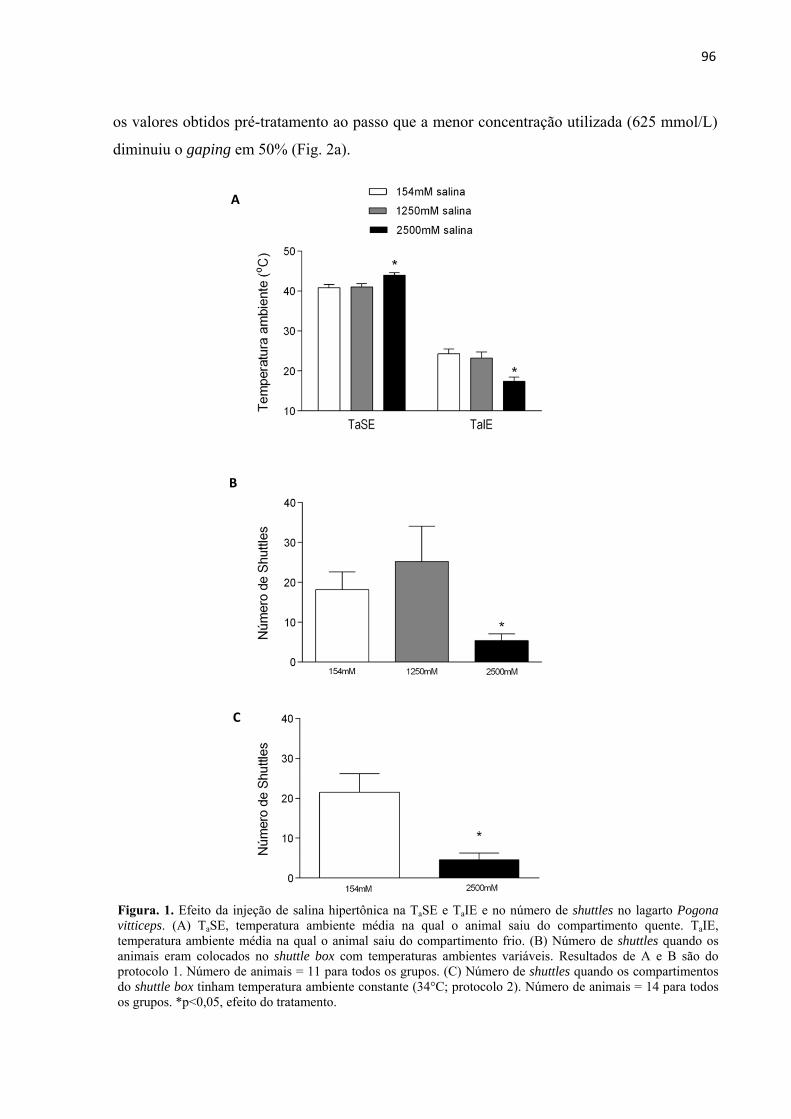
96
os valores obtidos pré-tratamento ao passo que a menor concentração utilizada (625 mmol/L)
diminuiu o gaping em 50% (Fig. 2a).
Figura. 1. Efeito da injeção de salina hipertônica na TaSE e TaIE e no número de shuttles no lagarto Pogona vitticeps. (A) TaSE, temperatura ambiente média na qual o animal saiu do compartimento quente. TaIE, temperatura ambiente média na qual o animal saiu do compartimento frio. (B) Número de shuttles quando os animais eram colocados no shuttle box com temperaturas ambientes variáveis. Resultados de A e B são do protocolo 1. Número de animais = 11 para todos os grupos. (C) Número de shuttles quando os compartimentos do shuttle box tinham temperatura ambiente constante (34°C; protocolo 2). Número de animais = 14 para todos os grupos. *p<0,05, efeito do tratamento.
C
B
A

97
Efeito das injeções de salina hipertônica nas temperaturas da superfície da cabeça e do
corpo
As temperaturas das superfícies do corpo (Tscorpo) e da cabeça (Tscabeça)
foram registradas durante os experimentos de gaping do protocolo descrito acima (Fig. 3a-c).
A Tscabeça aumentou significativamente após a injeção com a concentração mais alta de
salina hipertônica (2500 mmol/L) comparada com o grupo controle e o grupo 625 mmol/L; o
aumento na Tscabeça dos animais tratados com injeções de 625 mmol/L foi maior que o dos
animais controle (interação tempo vs tratamento: p=0,003; F2,17=2,57; Fig. 3a). O aumento no
nível de sal plasmático após o tratamento com a concentração mais alta causou um aumento na
Tscorpo dos lagartos comparado com os controles (interação tempo vs tratamento: p=0,002;
F2,17=2,78; Fig. 3b), mas não houve diferença entre os tratamentos 625 e 2500 mmol/L.
Além disso, o grupo controle e os animais tratados com a concentração mais
baixa de salina hipertônica (625 mmol/L) exibiram uma Tscabeça significativamente mais
baixa que a Tscorpo comparado com aqueles animais que receberam injeção com a maior
concentração de salina hipertônica (2500 mmol/L; efeito do tratamento: p=0,005; F2,17=7,362;
efeito do tempo: p=0,192; F2,17=1,449; sem efeito de interação: p=0,559; F2,17=0,901; Fig. 3c e
Fig. 4a, b). Os animais que receberam o tratamento de 2500 mmol/L tiveram a Tscabeça e a
Tscorpo muito semelhantes após as injeções (Fig. 3c e 4b).
Figura 2. Efeito (A) e dose resposta (B) da injeção de salina hipertônica na porcentagem de tempo gasto em gaping em P. vitticeps expostos a 37–39°C. A seta indica o momento da injeção. N=11 para todos os tratamentos. *p<0,05 comparado com a concentração controle. ‡p<0,05 comparado com o grupo 625 mmol /L.
A B

98
Figura 3. Efeito da injeção de salina hipertônica na (A) temperatura da superfície da cabeça e na (B) temperatura da superfície do corpo ao longo do tempo. (C) Diferença entre as temperaturas superficiais da cabeça e do corpo. N= 11 (154 mmo/L), N= 4 (625 mmol /L) e N=5 (2500 mmol/L). Efeito da interação para A e B (fatores: tempo vs tratamento): *p<0,05 comparado com o grupo controle; ‡p<0,05 comparado com o grupo 625 mmol /L. Em C, o grupo 2500 mmol/L foi diferente dos outros grupos (efeito do tratamento: p<0,05; sem interação entre os fatores).
C
B
A
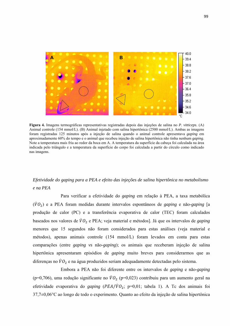
99
Efetividade do gaping para a PEA e efeito das injeções de salina hipertônica no metabolismo
e na PEA
Para verificar a efetividade do gaping em relação à PEA, a taxa metabólica
( ) e a PEA foram medidas durante intervalos espontâneos de gaping e não-gaping [a
produção de calor (PC) e a transferência evaporativa de calor (TEC) foram calculados
baseados nos valores de e PEA; veja material e métodos]. Já que os intervalos de gaping
menores que 15 segundos não foram considerados para estas análises (veja material e
métodos), apenas animais controle (154 mmol/L) foram levados em conta para estas
comparações (entre gaping vs não-gaping); os animais que receberam injeção de salina
hipertônica apresentaram episódios de gaping muito breves para considerarmos que as
diferenças no e na água produzidos seriam adequadamente detectadas pelo sistema.
Embora a PEA não foi diferente entre os intervalos de gaping e não-gaping
(p=0,706), uma redução significante no (p=0,023) contribuiu para um aumento geral na
efetividade evaporativa do gaping ( ⁄ ; p=0,01; tabela 1). A Tc dos animais foi
37,7±0,06°C ao longo de todo o experimento. Quanto ao efeito da injeção de salina hipertônica
B A
Figura 4. Imagens termográficas representativas registradas depois das injeções de salina no P. vitticeps. (A) Animal controle (154 mmol/L). (B) Animal injetado com salina hipertônica (2500 mmol/L). Ambas as imagens foram registradas 125 minutos após a injeção de salina quando o animal controle apresentava gaping em aproximadamente 60% do tempo e o animal que recebeu injeção de salina hipertônica não tinha nenhum gaping. Note a temperatura mais fria ao redor da boca em A. A temperatura da superfície da cabeça foi calculada na área indicada pelo triângulo e a temperatura da superfície do corpo foi calculada a partir do círculo como indicado nas imagens.

100
(625 mmol/L) no e na PEA, a concentração de 625 mmol/L diminuiu o (p=0,025), no
entanto, porque a PEA tendeu a ser menor após a injeção (p=0.12), a ⁄ não foi afetada
pelo aumento no nível de sal corpóreo (p=0,195) quando comparados aos efeitos da injeção de
salina isotônica nas mesmas variáveis (Tabela 2).
Tabela 1. Efeito do gaping na PEA, e / em lagartos P. vitticeps.
Intervalo PEA TEC PC ⁄ ⁄
Não-gaping 5.87±0.65 14.69±1.63 0.97±0.14 19.46±2.8 6.90 ± 1.13 0.86±0.14
Gaping 6.07±0.43 15.18±1.06 0.73±0.09* 14.52±1.83 9.10 ± 1.03* 1.14±0.13
Os resultados aqui apresentados (média ± e.p.m.) são dos lagartos (n=8) que receberam injeção de salina isotônica. *p<0,05 = diferença entre intervalos com gaping e sem gaping.
Tabela 2. Alteração (%) na PEA, e / depois das injeções de salina isotônica e hipertônica em lagartos P. vitticeps
Tratamento PEA % ⁄
154 mM -5.11 ± 8.3 -12.96 ± 8.1 16.1 ± 21.3
625 mM -22.7 ± 6.9 -40.9 ± 7.5* 48.1 ± 25.4
Os valores foram calculados baseados nas alterações pós-injeção comparados com os valores pré-injeção; N= 7 (154mmol/L) e N=8 (625mmol/L); *p<0,05 = diferença entre tratamentos.

101
4. DISCUSSÃO
___________________________________________________________________________
No presente trabalho, injeções de salina hipertônica foram usadas como forma
de induzir um desafio osmótico nos animais. Esta metodologia foi considerada adequada
devido ao aumento linear na osmolalidade plasmática e à proporcional disposição ao consumo
de água por parte dos animais em relação aos tratamentos recebidos. Após as injeções, era
esperado que aqueles lagartos que receberam tratamento hipertônico e foram expostos ao
shuttle box saíssem do CF em direção ao CQ e do CQ para o CC em Tas mais frias, ou seja,
os animais teriam reduzidas TaIE e TaSE comparado aos animais controle, refletindo uma
redução geral nas suas Tcs após a desidratação. De fato, estudos anteriores mostraram que
ratos (Konishi et al., 2007), sapos (Malvin e Wood, 1991), lagartos (Crowley, 1987;
Bradshaw et al., 2007) e serpentes (Ladyman e Bradshaw, 2003) selecionam Tas mais frias
quando a disponibilidade de água é reduzida. No entanto, no presente trabalho, a concentração
mais alta de salina hipertônica (2500mmol/L) alterou ambas as TaIE e a TaSE dos animais,
mas em direções opostas (Fig. 1a) e diminuiu o número de shuttles (Fig. 1B). Uma menor
TaIE e uma maior TaSE sugere que ou a preferência térmica do animal foi diminuída e
aumentada ao mesmo tempo ou que o aumento na osmolalidade afetou a predisposição do
animal em se mover (Fig. 1b) e não o set-point termorregulador.
No experimento seguinte, foi testado o shuttling em um ambiente isotérmico
(shuttle box a 34°C) e foi observado que a concentração de 2500mmol/L reduziu o número de
shuttles (Fig. 1c), indicando que o aumento no nível corpóreo de sal realmente afetou a
tendência ao movimento e não a termorregulação per se. Neste mesmo sentido, já foi descrito
que lagartos das famílias agamidae e iguanidae abandonam todas as atividades e não
termorregulam quando eles estão desnutridos ou em situações de extrema redução na
disponibilidade de água (Bradshaw, 1997), um comportamento similar ao observado no
presente estudo. Ainda não é conhecido se a redução no shuttling depois do aumento na
osmolalidade plasmática é devido a uma redução na motivação ao movimento ou a uma
inibição fisiológica da função neuromuscular. Baseado nos nossos resultados, nós sugerimos
que a concentração mais alta de solução salina usada no presente trabalho foi alta o suficiente
para inibir qualquer resposta termorreguladora que envolva locomoção. Por outro lado, a
concentração intermediária (1250mmol/L), que reduziu 90% do gaping, não alterou a TaIS,
TaSE nem o número de shuttles, indicando que há diferentes limiares para a influência da
osmolalidade nestas respostas termorreguladoras e que a conservação de água pode acontecer

102
mesmo durante uma desidratação moderada sem que os set-points termorreguladores para o
shuttling sejam alterados.
O efeito do sal no gaping confirmou nossa hipótese: este comportamento foi
reduzido pelas injeções de salina hipertônica de acordo com a concentração recebida (Fig.
2a,b), o que é interessante porque, como mencionado, a concentração de 1250mmol/L não
afetou a termorregulação no shuttle box, mas causou uma enorme redução no gaping. Mesmo
a concentração mais baixa utilizada (625mmol/L) foi capaz de diminuir o gaping em 50%.
Embora considerada uma resposta diferente do gaping, a frequência do ofego e seu limiar de
temperatura também foram alterados pela desidratação em endotermos, tais como aves (Arad,
1983; Maloney and Dawson, 1998) e ovelhas (McKinley et al., 2008). Os efeitos da
desidratação sugerem que os termoefetores dependentes de água (gaping/ofego) sejam mais
sensíveis aos níveis de sal na corrente sanguínea que os termoefetores “secos”, como o
comportamento de vai e vem entre ambientes mais ou menos quentes (shuttling). Desta
forma, a redução do gaping induzida pelo aumento do nível corporal de sal parece ser uma
estratégia para a economia de água. Baseado nestes resultados, o esperado era que o gaping
fosse uma fonte significativa de perda evaporativa de calor nesta espécie.
Embora nenhuma diferença entre os intervalos de gaping e não-gaping foi
detectava na PEA per se, o foi reduzido em 25% durante o gaping. Uma redução ainda
maior no e um aumento de cerca de 10 vezes na frequência respiratória (= ofego) foi
descrita em ovelhas quando elas eram expostas ao calor moderado (40°C por 2–3 h). Tal
declínio no foi associado a um decréscimo no fluxo sanguíneo dos músculos esqueléticos
e nos órgãos internos acompanhados por um aumento no fluxo sanguíneo para regiões
periféricas (Hales, 1973; Hales e Brown, 1974). De fato, o gaping tem sido observado no
nosso laboratório em animais muito calmos e que não demonstram nenhum movimento ativo
concomitante com o gaping. Tal redução no movimento simultaneamente com um possível
aumento no fluxo sanguíneo para a região bucal poderia ter contribuído para o decréscimo
geral no observado em nossos animais. Apesar da PEA não ter sido alterada pelo gaping,
a redução no causou um aumento na taxa ⁄ (tabela 1) indicando que este
mecanismo realmente contribui para a perda evaporativa de energia térmica quando
consideramos que ele causa uma redução no metabolismo (a perda evaporativa de energia
corresponde a 86% da PC total durante intervalos de não-gaping, aumentando para 114%
durante o gaping; tabela 1). Como mencionado, Bradshaw (1997) demonstrou que lagartos da
família agamidae (Ctenophorus ornatus) têm seus comportamentos normais de
103
termorregulação inibidos quando enfrentam hipernatremia associada com desidratação
crônica no campo. O presente trabalho além de corroborar os achados de Bradshaw (1997),
providencia mais evidências de que diante de um estresse osmótico, o sistema osmorregulador
parede ser preservado (afinal o gaping, uma fonte de PEA, foi inibido) à custa do sistema
termorregulador. Como dito acima na metodologia, apenas os animais injetados com salina
isotônica foram considerados para as análises do potencial evaporativo do gaping (tabela 1)
porque depois do tratamento com salina hipertônica, os lagartos apresentavam eventos de
gaping muito curtos o que não era adequado para nossas análises. Embora tais registros após
as injeções de salina hipertônica não fossem ideais para esta finalidade, eles eram consistentes
para avaliar o efeito geral do sal no metabolismo (tabela 2). A injeção de salina hipertônica
(625mmol/L) diminuiu o comparado com o efeito da injeção isotônica. A taxa
metabólica também foi reduzida em camelo, gatos e cabras desidratadas (Schmidt-Nielsen et
al., 1967; Doris e Baker, 1981; Dmi’el, 1986). A PEA tendeu a ser menor nos animais
tratados com salina hipertônica, o que está de acordo com a redução no tempo gasto em
gaping após as injeções (Fig. 2a,b), mais uma vez, então, sugerindo que o gaping
normalmente contribui para a perda evaporativa de água e é reduzido quando a osmolalidade
plasmática é aumentada. Devido à redução no e a tendência da PEA em ser menor, a taxa
⁄ não foi significativamente alterada (tabela 2).
As origens neurológicas para a interação entre o sistema osmorregulador e
termorregulador são encontradas no hipotálamo. Foi descrita que, em gatos, a
termossensibilidade dos neurônios hipotalâmicos é reduzida durante a desidratação,
diminuindo então a perda evaporativa de calor (Doris e Baker, 1981). Além disso, elevadas
concentrações de sódio no fluido cerebrospinal reduz a taxa de suor em macacos
(Erythrocebus patas; Owen et al., 1989) e a frequência respiratória (=ofego) em coelhos
(Turlejska e Baker, 1986). Em ovelhas, o efeito inibitório da salina hipertônica no ofego foi
abolido pela lesão da lâmina terminalis, uma região que inclui os osmorreceptores
hipotalâmicos e o núcleo pré-óptico mediano (McKinley et al., 2008) que, por sua vez, está
envolvido com a regulação dos termoefetores em mamíferos (Nakamura e Morrison, 2010).
Assim, em muitos vertebrados, a termorregulação está conectada ao estado osmorregulador
diretamente por meio dos neurônios centrais. Vias similares podem ter sido ativadas em
nossos lagartos ou, em outras palavras, o aumento no nível corpóreo de sal pode ter
aumentado a concentração encefálica de sódio, alterado a termossensilidade dos neurônios e
contribuído para a redução na porcentagem de tempo gasto em gaping.
104
Todos os lagartos do protocolo de gaping demonstraram um aumento na
Tscabeça ao longo do tempo devido ao lento processo de equilíbrio térmico entre o animal e a
câmara de temperatura controlada. No entanto, a Tscabeça dos animais que receberam injeção
da salina mais concentrada (2500mmol/L) foi ainda maior comparado aos animais controle
(Fig. 3a e 4) – o mesmo padrão foi observado para a Tscorpo (Fig. 3B e Fig. 4). É interessante
notar que a diferença entre Tscabeça e Tscorpo foi maior naqueles animais tratados com
salina isotônica ou com a menor concentração de solução salina (625mmol/L) que nos
lagartos que receberam injeção da maior concentração de solução salina (Fig. 3C e Fig. 4).
Assim, no mesmo nível de osmolalidade plasmática (2500mmol/L) que o gaping é
completamente suprimido, as Tscabeça e Tscorpo se igualam, o que pode ter importantes
consequências para a manutenção das temperaturas craniais e funções encefálicas, indicando
que o gaping pode ser um mecanismo de resfriamento encefálico (Tattersall et al., 2006),
apesar das mudanças relativamente pequenas na PEA causadas por este comportamento
(tabela 1).
Um mecanismo chamado “resfriamento encefálico seletivo” (RES) é ativado
em mamíferos quando eles enfrentam reduzida disponibilidade de água corpórea (Caputa et
al., 1976; McConaghy et al., 1995; Jessen, 2001; Mitchell et al., 2002). O RES é definido
como “temperatura encefálica menor que a temperatura do sangue arterial” (IUPS Thermal
Commission, 2001), o que acontece geralmente quando o sangue da carótida é resfriado
durante sua ascensão ao encéfalo (Willmer et al., 2005). Se o encéfalo está mais frio, a PEA
não é ativada porque os neurônios sensíveis ao calor não são estimulados. Neste sentido, tem
sido proposto que o RES não é apenas um mecanismo de proteção neural, mas também uma
estratégia para conservação de água.
Uma combinação de resfriamento respiratório e um sistema de contracorrente
vascular cerebral podem contribuir e levar a um resfriamento encefálico nos lagartos quando
eles são expostos a altas Tas (Tattersall et al., 2006). O gaping claramente contribui para a
evaporação da água nas vias aéreas superiores e na cavidade bucal (Fig. 4), o que pode ter
sido suficiente para resfriar o sangue da carótida no seu caminho ao encéfalo. Quando a Ta é
baixa ou igual à selecionada naturalmente pelos animais, o sangue encefálico volta à periferia
pela veia jugular interna e então, por causa da sua proximidade com a artéria carótida interna,
o calor é transferido para esta artéria, mantendo o encéfalo aquecido (Oelrich, 1956; Heath,
1964, 1966). Já em altas Tas, é sugerido que a veia jugular interna seja contraída e então o
sangue encefálico retorna para a periferia pela jugular externa que se encontra relativamente
105
afastada da artéria carótida. Neste caso, o calor não é transferido para o sangue carotídeo, mas
ao invés, é levado na direção oposta ao encéfalo, contribuindo para o resfriamento do mesmo
(Heath, 1964; Tattersall et al., 2006). É possível que estes dois mecanismos (resfriamento
respiratório e mecanismo de contracorrente vascular) tenham mantido o encéfalo mais frio
quando os lagartos apresentavam no mínimo 50% de gaping (Fig. 3c), mas o resfriamento não
foi preservado quando o gaping foi abolido após os tratamentos com salina hipertônica (Fig.
3c). Os mesmos mecanismos evaporativos/não-evaporativos também podem ter contribuído
para resfriarem a cabeça em um pequeno lagarto lacertídeo (Podarcis muralis) quando eles
foram aquecidos, embora esta hipótese ainda precisa ser experimentalmente testada (Sannolo
et al., 2014).
Há importantes diferenças entre o RES em mamíferos e em répteis. Em
mamíferos, o RES pode ser observado quando a água não está disponível. Mamíferos
hidratados (cabras e ovelhas), mesmo quando expostos a altas Tas raramente apresentam
RES, mas animais desidratados exibem RES mais frequente e em maior grau, corroborando a
ideia de que tal mecanismo é ativado pelo sistema osmorregulador e pode não ter função
termorreguladora nestes animais (Jessen et al.,1998; Fuller et al., 2007). Por outro lado, nos
Pogona vitticeps, o resfriamento da cabeça (e por inferência, do encéfalo) aconteceu quando a
água estava disponível (isto é, quando os animais apresentaram gaping. Fig. 4a e 3c) e, no
sentido oposto, o resfriamento foi diminuído quando a água não estava disponível, ou seja,
quando o gaping foi abolido (após injeções de 2500mmol/L de salina. Fig. 4b e 3c). Além
disso, o nível de gaping apresentada pelos lagartos P. vitticeps, quando estão hidratados, é
aumentado conforme a Ta é elevada, bem como a diferença entre Tscabeça e Tscorpo
(Tattersall and Gerlach, 2005), mas esta diferença foi reduzida quando os animais foram
desidratados à mesma Ta (Fig. 2a). Assim, parece que não é o sistema de osmorregulação que
ativa o resfriamento encefálico em lagartos, mas sim o de termorregulação;
consequentemente, o RES deve funcionar como um mecanismo protetor do encéfalo. Apesar
do RES ser ativado pelo sistema termorregulador, quando os lagartos enfrentam reduzida
disponibilidade de água, a água corporal ainda é conservada à custa da termorregulação,
similar ao que acontece em mamíferos. Embora um estudo prévio já tenha mostrado que o
gaping é um termoefetor efetivo para o Alligator mississippiensis, (Spotila et al., 1977), o
presente trabalho é o primeiro a mostrar que o gaping pode funcionar como um mecanismo de
resfriamento local (encefálico) para lagartos de deserto, baseado no fato que a desidratação
106
reduziu o gaping e aumentou a Tscabeça, eliminando a diferença entre a Tscabeça e Tscorpo
(Fig. 3).
Concluindo então, nós demonstramos que uma concentração intermediária de
salina hipertônica (1250mmol/L) pode afetar os mecanismos de PEA, mas não afeta os
mecanismos não relacionados à água. O fato de o gaping ser mais sensível aos níveis
corpóreos de sal que o shuttling, pode ser uma importante resposta para a economia de água
durante um período de desidratação natural, enquanto os mecanismos termorreguladores não
dependentes de água ainda podem acontecer. No entanto, quando a água está disponível, o
gaping pode desempenhar um papel de mecanismo de resfriamento local que é importante
para evitar o superaquecimento encefálico nestes lagartos, como aquele que pode acontecer
quando estão termorregulando expostos ao sol.

107
5. REFERÊNCIAS BIBLIOGRÁFICAS
___________________________________________________________________________
ARAD, Z. Thermoregulation and acid-base status in the panting dehydrated fowl. J. Appl. Physiol. Respir. Environ. Exerc. Physiol., v. 54, p. 234-243, 1983.
BAKER, M.A. Effects of dehydration and rehydration on thermoregulatory sweating in goats. J. Physiol., v. 417, p. 421-435, 1989.
BAKER, M.A.; DAWSON, D.D. Inhibition of thermal panting by intracarotid infusion of hypertonic saline in dogs. Am. J. Physiol., v. 249, p. R787-R791, 1985.
BAKER, M.A.; TURLEJSKA, E. Thermal panting in dehydrated dogs: effects of plasma volume expansion and drinking. Pflügers Arch., v. 413, p. 511-515, 1989.
BARBER, B.J.; CRAWFORD, E.C. Dual threshold control of peripheral temperature in the lizard Dipsosaurus dorsalis. Physiol. Zool., v. 52, p. 250-263, 1979.
BENTLEY, P.J. Studies on the water and electrolyte metabolism of the lizard Trachysaurus rugosus (Gray). J. Physiol., v. 145, p. 37-47, 1959.
BERK, M.L.; HEATH, J.E. An analysis of behavioral thermoregulation in the lizard, Dipsosaurus dorsalis. J. Therm. Biol., v.1, p. 15-22, 1975.
BOUÂOUDA, H.; ACHÂABAN, M.R.; OUASSAT, M.; OUKASSOU, M.; PIRO, M.; CHALLET, E.; EL ALLALI, K.; PÉVET, P. Daily regulation of body temperature rhythm in the camel (Camelus dromedarius) exposed to experimental desert conditions. Physiol. Rep., v. 2, p. e12151, 2014.
BRADSHAW, S.D. Homeostasis in Desert Reptiles. Berlin: Springer, 1997, 232 p.
BRADSHAW, S.D.; SHOEMAKER, V.H. Aspects of water and electrolyte changes in a field population of Amphibolurus lizards. Comp. Biochem. Physiol., v. 20, p. 855-865, 1967.
BRADSHAW, D.; LADYMAN, M.; STEWART, T. Effect of hypernatraemia and the neurohypophysial peptide, arginine vasotocin (AVT) on behavioural thermoregulation in the agamid lizard, Ctenophorus ornatus. Gen. Comp. Endocrinol., v. 150, p. 34-40, 2007.
CADENA, V.; TATTERSALL, G.J. The effect of thermal quality on the thermoregulatory behavior of the bearded dragon Pogona vitticeps: influences of methodological assessment. Physiol. Biochem. Zool., v. 82, p. 203-217, 2009a.
CADENA, V.; TATTERSALL, G.J. Decreased precision contributes to the hypoxic thermoregulatory response in lizards. J. Exp. Biol., v. 212, p. 137-144, 2009b.
CAPUTA, M.; KADZIELA, W.; NAREBSKI, J. Significance of cranial circulation for the brain homeothermia in rabbits. I. The brain-arterial blood temperature gradient. Acta Neurobiol. Exp. (Wars), v. 36, p. 613-623, 1976.
CROWLEY, S.R. The effect of desiccation upon the preferred body temperature and activity level of the lizard Sceloporus undulatus. Copeia, v. 1987, p. 25-32, 1987.

108
DMI’EL, R. Selective sweat secretion and panting modulation in dehydrated goats. J. Therm. Biol., v. 11, p. 157-159, 1986.
DORIS, P.A.; BAKER, M.A. Hypothalamic control of thermoregulation during dehydration. Brain Res., v. 206, p. 219-222, 1981.
DOSSAT, R.J.; HORAN, T.J. (2001). Psychrometric properties of air. In: _______ Principles of Refrigeration. New York: Prentice Hall, 2001. pp. 57-70.
DUPRÉ, R.K.; CRAWFORD, E.C. Elevation of the panting threshold of the desert iguana, Dipsosaurus dorsalis, during dehydration: potential roles of changes in plasma osmolality and body fluid volume. J. Comp. Physiol. B., v. 156, p. 377-381, 1986.
FORD, S. S.; BRADSHAW, S. D. Kidney function and the role of arginine vasotocin in three agamid lizards from habitats of differing aridity in Western Australia. Gen. Comp. Endocrinol., v. 147, p. 62-69, 2006.
FULLER, A.; MEYER, L.C.R.; MITCHELL, D.; MALONEY, S.K. Dehydration increases the magnitude of selective brain cooling independently of core temperature in sheep. Am. J. Physiol. Regul. Integr. Comp. Physiol., v. 293, p. R438-R446, 2007.
HALES, J.R.S. Effects of exposure to hot environments on the regional distribution of blood flow and on cardiorespiratory function in sheep. Pflügers Arch., v. 344, p. 133-148, 1973.
HALES, J.R.S.; BROWN, G.D. Net energetic and thermoregulatory efficiency during panting in the sheep. Comp. Biochem. Physiol. A Physiol., v. 49, p. 413-422, 1974.
HEATH, J.E. Head–body temperature differences in horned lizards. Physiol. Zool., v. 37, p. 273-279, 1964.
HEATH, J.E. Venous shunts in the cephalic sinuses of horned lizards. Physiol. Zool., v. 39, p. 30-35, 1966.
HEATWOLE, H.; FIRTH, B.T.; WEBB, G.J.W. Panting thresholds of lizards-I. Some methodological and internal influences on the panting threshold of an Agamid, Amphibolurus muricatus. Comp. Biochem. Physiol. A Physiol., v.46, p. 799-826, 1973.
IUPS THERMAL COMMISSION. Glossary of terms for thermal physiology. Jpn. J. Physiol., v. 51, p. 245-280, 2001.
JESSEN, C. Selective brain cooling in mammals and birds. Jpn. J. Physiol., v. 51, p. 291-301, 2001.
JESSEN, C.; DMI’EL, R.; CHOSHNIAK, I.; EZRA, D.; KUHNEN, G. Effects of dehydration and rehydration on body temperatures in the black Bedouin goat. Pflügers Arch. v. 436, p. 659-666, 1998.
KLEINHAUS, S.; PINSHOW, B.; BERNSTEIN, M.H.; DEGEN, A.A. Brain temperature in heat-stressed, water deprived desert phasianids: sand partridge (Ammoperdix heyi) and chukar (Alectoris chukar sinaica). Physiol. Zool., v. 58, p. 105-116, 1985.
109
KONISHI, M.; NAGASHIMA, K.; ASANO, K.; KANOSUE, K. Attenuation of metabolic heat production and cold-escape/warm-seeking behaviour during a cold exposure following systemic salt loading in rats. J. Physiol., v. 551, p. 713-720, 2003.
KONISHI, M.; KANOSUE, K.; KANO, M.; KOBAYASHI, A.; NAGASHIMA, K. The median preoptic nucleus is involved in the facilitation of heat-escape/coldseeking behavior during systemic salt loading in rats. Am. J. Physiol. Regul. Integr. Comp. Physiol., v. 292, p. R150-R159, 2007.
LADYMAN, M. BRADSHAW, D. The influence of dehydration on the thermal references of the Western tiger snake, Notechis scutatus. J. Comp. Physiol. B., v. 173, p. 239-246, 2003.
LIGHTON, J.R.B. Flow-through respirometry: the equations. In: _____ Measuring Metabolic Rates. A Manual for Scientists. New York: Oxford University Press, 2008, pp. 100-104.
MALONEY, S.K.; DAWSON, T.J. Changes in pattern of heat loss at high ambient temperature caused by water deprivation in a large flightless bird, the Emu. Physiol. Zool., v. 71, p. 712-719, 1998.
MALVIN, G. M.; WOOD, S.C. Behavioral thermoregulation of the toad, Bufo marinus: effects of air humidity. J. Exp. Zool., v. 258, p. 322-326, 1991.
McCONAGHY, F.F.; HALES, J.R.S.; ROSE, R.J.; HODGSON, D.R. Selective brain cooling in the horse during exercise and environmental heat stress. J. Appl. Physiol., v. 79, p. 1849-1854, 1995.
McKINLEY, M.J.; MCALLEN, R M.; WHYTE, D.; MATHAI, M.L. Central osmoregulatory influences on thermoregulation. Clin. Exp. Pharmacol. Physiol., v. 35, p. 701-705, 2008.
MELVILLE, J.; SCHULTE, J.A. Correlates of active body temperatures and microhabitat occupation in nine species of central Australian agamid lizards. Aust. Ecol., v. 26, p. 660-669, 2001.
MITCHELL, D., MALONEY, S.K.; JESSEN, C.; LABURN, H.P.; KAMERMAN, P.R.; MITCHELL, G.; FULLER, A. Adaptive heterothermy and selective brain cooling in arid-zone mammals. Comp. Biochem. Physiol. B. Biochem. Mol. Biol., v. 131, p. 571-585, 2002.
NAGASHIMA, K.; NAKAI, S.; KONISHI, M.; SU, L.; KANOSUE, K. Increased heat-escape/cold-seeking behaviour following hypertonic saline injection in rats. Am. J. Physiol. Regul. Integr. Comp. Physiol., v. 280, p. R1031-R1036, 2001.
NAKAMURA, K.; MORRISON, S. F. A thermosensory pathway mediating heat-defense Responses. Proc. Natl. Acad. Sci. USA., v. 107, p. 8848-8853, 2010.
O’CONNOR, M. P.; TRACY, C.R. Thermoregulation by juvenile toads of Bufo woodhousei in the field and in the laboratory. Copeia v. 1992, p. 865-876, 1992.
OELRICH, T.M. The anatomy of the head of Ctenosaura pectinate (Iguanidae). Mus. Zool. Univ. Mich. Misc. Publ., v. 94, p. 1-122, 1956.

110
OWEN, M.D., MATTHES, R.D.; GISOLFI, C.V. Effect of cerebrospinal fluid hyperosmolality on sweating in the heat-stressed patas monkey. J. Appl. Physiol., v. 67, p. 128-133, 1989.
PARMENTER, C.J.; HEATWOLE, H. Panting thresholds of lizards. IV. The effect of dehydration on the panting threshold of Amphibolurus barbatus and Amphibolurus muricatus. J. Exp. Zool., v. 191, p. 327-332, 1975.
PREEST, M.R.; POUGH, F.H. Interaction of temperature and hydration on locomotion of toads. Funct. Ecol., v. 3, p. 693-699, 1989.
RANDALL, D.; BURGGREN, W; FRENCH, K. Using energy: meeting environmental challenges. In: _____. Eckert Animal Physiology: Mechanisms and Adaptations. 4.ed. New York:Freeman Company, 1997. p. 665–723.
RICE, G. E. Plasma arginine vasotocin concentrations in the lizard Varanus gouldii (Gray) following water loading, salt loading, and dehydration. Gen. Comp. Endocrinol., v. 47, p. 1-6, 1982.
SANNOLO, M.; MANGIACOTTI, M.; SACCHI, R.; SCALI, S. Keeping a cool mind: head–body temperature differences in the common wall lizard. J. Zool., v. 293, p. 71-79, 2014.
SCHMIDT-NIELSEN, K.; SCHMIDT-NIELSEN, B.; JARNUM, S. A.; HOUPT, T.R. Body temperature of the camel and its relation to water economy. Am. J. Physiol., v. 188, p. 103-112, 1957.
SCHMIDT-NIELSEN, K.; CRAWFORD, E.C.; JR NEWSOME, A.E.; RAWSON, K.S.; HAMMEL, H.T. Metabolic rate of camels: effect of body temperature and dehydration. Am. J. Physiol., v. 212, p. 341-346, 1967.
SILVA, N.L.; BOULANT, J.A. Effects of osmotic pressure, glucose, and temperature on neurons in preoptic tissue slices. Am. J. Physiol., v. 247, p. R335-R345, 1984.
SPOTILA, J. R.; TERPIN, K. M.; DODSON, P. Mouth gaping as an effective thermoregulatory device in alligators. Nature, v. 265, p. 235-236, 1977.
TATTERSALL, G.J.; GERLACH, R.M. Hypoxia progressively lowers thermal gaping thresholds in bearded dragons, Pogona vitticeps. J. Exp. Biol., v. 208, p. 3321-3330, 2005.
TATTERSALL, G. J.; CADENA, V; SKINNER, M. C. Respiratory cooling and thermoregulatory coupling in reptiles. Respir. Physiol. Neurobiol., v. 154, p. 302-318, 2006.
TAYLOR, C. R. Dehydration and heat. Effects on temperature regulation of East African ungulates. Am. J. Physiol. v. 219, p. 1136-1139, 1970.
TURLEJSKA, E.; BAKER, M.A. Elevated CSF osmolality inhibits thermoregulatory heat loss responses. Am. J. Physiol. v. 251, p. R749-R754, 1986.
WILLMER, P.; STONE, G.; JOHNSTON, I. Temperature and its Effects. In: ______ Environmental Physiology of Animals. Malden: Blackwell Publishing, 2005, pp. 209-210.

111
Apêndices
Apêndice A
“TRPV4 activates autonomic and behavioural warmth
defence responses in Wistar rats”
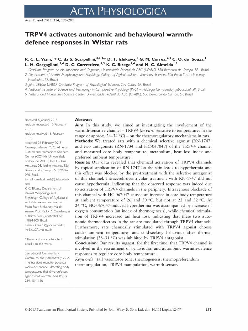
TRPV4 activates autonomic and behavioural warmth-
defence responses in Wistar rats
R. C. L. Vizin,1,* C. da S. Scarpellini,2,3,4,* D. T. Ishikawa,1 G. M. Correa,2,4 C. O. de Souza,1
L. H. Gargaglioni,2,4 D. C. Carrettiero,1,5 K. C. B�ıcego2,4 and M. C. Almeida1,5
1 Graduate Program on Neuroscience and Cognition, Universidade Federal do ABC (UFABC), S~ao Bernardo do Campo, SP, Brazil
2 Department of Animal Morphology and Physiology, College of Agricultural and Veterinary Sciences, S~ao Paulo State University,
Jaboticabal, SP, Brazil
3 Joint UFSCar-UNESP Graduate Program of Physiological Sciences, Sao Carlos, SP, Brazil
4 National Institute of Science and Technology in Comparative Physiology (INCT – Fisiologia Comparada), Jaboticabal, SP, Brazil
5 Natural and Humanities Science Center, Universidade Federal do ABC (UFABC), S~ao Bernardo do Campo, SP, Brazil
Received 6 January 2015,
revision requested 10 February
2015,
revision received 16 February
2015,
accepted 26 February 2015
Correspondence: M. C. Almeida,
Natural and Humanities Sciences
Center (CCNH), Universidade
Federal do ABC (UFABC), Rua
Arcturus, 03, Jardim Antares, S~ao
Bernardo do Campo, SP 09606-
070, Brazil.
E-mail: [email protected]
and
K. C. B�ıcego, Department of
Animal Morphology and
Physiology, College of Agricultural
and Veterinarian Sciences, S~ao
Paulo State University, Via de
Acesso Prof. Paulo D. Castellane, s/
n, Bairro Rural, Jaboticabal, SP
14884-900, Brazil.
E-mails: [email protected];
*These authors contributed
equally to this work.
See Editorial Commentary:
Garami, A. and Romanovsky, A. A.
The transient receptor potential
vanilloid-4 channel: detecting body
temperatures that drive defences
against mild warmth. Acta Physiol
214, 154–156.
Abstract
Aim: In this study, we aimed at investigating the involvement of the
warmth-sensitive channel – TRPV4 (in vitro sensitive to temperatures in the
range of approx. 24–34 °C) – on the thermoregulatory mechanisms in rats.Methods: We treated rats with a chemical selective agonist (RN-1747)
and two antagonists (RN-1734 and HC-067047) of the TRPV4 channel
and measured core body temperature, metabolism, heat loss index and
preferred ambient temperature.Results: Our data revealed that chemical activation of TRPV4 channels
by topical application of RN-1747 on the skin leads to hypothermia and
this effect was blocked by the pre-treatment with the selective antagonist
of this channel. Intracerebroventricular treatment with RN-1747 did not
cause hypothermia, indicating that the observed response was indeed due
to activation of TRPV4 channels in the periphery. Intravenous blockade of
this channel with HC-067047 caused an increase in core body temperature
at ambient temperature of 26 and 30 °C, but not at 22 and 32 °C. At
26 °C, HC-067047-induced hyperthermia was accompanied by increase in
oxygen consumption (an index of thermogenesis), while chemical stimula-
tion of TRPV4 increased tail heat loss, indicating that these two auto-
nomic thermoeffectors in the rat are modulated through TRPV4 channels.
Furthermore, rats chemically stimulated with TRPV4 agonist choose
colder ambient temperatures and cold-seeking behaviour after thermal
stimulation (28–31 °C) was inhibited by TRPV4 antagonist.Conclusion: Our results suggest, for the first time, that TRPV4 channel is
involved in the recruitment of behavioural and autonomic warmth-defence
responses to regulate core body temperature.
Keywords tail vasomotor tone, thermogenesis, thermopreferendum
thermoregulation, TRPV4 manipulation, warmth sensor.
© 2015 Scandinavian Physiological Society. Published by John Wiley & Sons Ltd, doi: 10.1111/apha.12477 275
Acta Physiol 2015, 214, 275–289

The core body temperature (Tc) is one of the vital
signs and the ability to regulate Tc is of fundamental
importance, as all biochemical reactions and physio-
logical functions are widely dependent on temperature
(Wenisch et al. 1996, Wasserstrom & Vites 1999,
Aihara et al. 2001). In endothermic animals, both
autonomic and behavioural mechanisms are
recruited for regulating Tc (B�ıcego et al. 2007). In this
regard, independent neuronal networks, controlled by
Tc and/or skin temperature (Tsk), which are detected
by the thermoreceptors, are used by each thermoeffec-
tor (e.g. shivering, non-shivering thermogenesis, cutane-
ous vasomotor control) (Romanovsky 2007, Morrison
& Nakamura 2011). In addition, behavioural thermo-
regulation is essential for survival, like finding a tem-
perature of preference – one of the most efficient,
energetically less expensive and widespread mechanism
in mammals (Gordon 1990, B�ıcego et al. 2007).
The recent discovery and advances in the study of
the temperature-sensitive transient receptor potential
channels (‘TRP’) have contributed to our understand-
ing of the transduction mechanisms of thermal sig-
nals. In this study, we focused on the TRPV4, which
is activated in vitro at temperatures in the range of
approx. 24–34 °C (G€uler et al. 2002, Kauer & Gib-
son 2009), and also by other factors such as hypo-
osmolarity (Liedtke et al. 2000, Strotmann et al.
2000), chemical agonists (Vincent et al. 2009, Vincent
& Duncton 2011) and endogenous arachidonic acid
metabolites (Voets et al. 2005). Consistent with their
thermosensitivity in vitro, the TRPV4 protein was
located at some anatomical locations compatible with
thermoregulatory function, including primary sensory
neurones, keratinocytes, endothelial cells, sweat
glands, sympathetic neurones and preoptic area of the
hypothalamus (Liedtke et al. 2000, Delany et al.
2001, G€uler et al. 2002, Watanabe et al. 2002, Aless-
andri-Haber et al. 2003). In fact, TRPV4 has been
proposed to act as thermoreceptor as genetic ablation
of TRPV4 lead mice to select warmer temperatures
than control animals when exposed to a thermal gra-
dient or when they had to choose between two Tas
(Lee et al. 2005); however, whether this channel is
activated in warmth conditions in vivo in a non-genet-
ically modified animal and whether it recruits auto-
nomic and/or behavioural thermoregulatory
mechanisms had not been assessed. Thus, this study
aimed at investigating whether TRPV4 channels are
involved in the autonomic and behavioural thermo-
regulation in vivo using a pharmacological approach.
To this end, we evaluated the effects of selective
TRPV4 agonist and antagonists on Tc, heat loss index
(HLI), oxygen consumption and selected ambient
temperature.
Materials and methods
Ethics approval
All the procedures were approved and are in
accordance with the local ethics committee from Fed-
eral University of ABC (CEUA/UFABC, Protocol num-
ber 001/2010) and from Sao Paulo State University
(CEUA/FCAV, Protocol Number 26430/11, 019414/
14), and efforts were made to minimize the number of
animals used and their suffering. This study conforms
with Good Publication Practice in Physiology 2013
Guidelines for Acta Physiologica (Persson 2013).
Animals
Experiments were run in two different facilities: 152
male Wistar rats (8–10 weeks old; weight 290–320 g)
were obtained from ICB/USP and from UNESP Botuc-
atu. All of them were housed in a room with 12/12-
hour light–dark cycle (lights on at 7 am), controlled
temperature (22 � 2 °C) and 60% of humidity with
free access to food and water. Each experiment was
conducted in separated groups of animals. All experi-
ments were conducted from 9 am to 4 pm.
Drugs
RN-1747 ([1-(4-Chloro-2-nitrophenyl)sulfonyl-4-benzy
lpiperazine]) is a selective TRPV4 channel agonist (Vin-
cent et al. 2009) that was purchased from Tocris Biosci-
ence (Bristol, UK). A stock solution of RN-1747
(25 mg mL�1) in 50% ethanol and 50% propylene gly-
col was prepared, aliquoted and stored at �20 °C. On
the day of experiment, the stock was diluted to make a
working solution of RN-1747 in different
concentrations: 100 lg mL�1 in vehicle containing 25%
ethanol, 25% propylene glycol and 50% saline for intra-
venous administration (i.v.); 500 or 1000 lg mL�1 in
propylene glycol for topical administration. For intra-
cerebroventricular (i.c.v.) administration, the RN-1747
was injected directly by stock solution aliquot. RN-1734
([2,4-Dichloro-N-isopropyl-N-(2-isopropylaminoethyl)
benzenesulfonamide]) (Vincent et al. 2009) and HC-
067047 ([2-Methyl-1-[3-(4-morpholinyl)propyl]-5-phe-
nyl-N-[3-(trifluoromethyl) phenyl]-1H-pyrrole-3-carb-
oxamide]) (Everaerts et al. 2010b) are two selective
TRPV4 channel antagonists, both purchased from To-
cris Bioscience. HC-067047 was chosen over RN-1734
for experiments 2 and 4 as it is more potent and pre-
sents a better selectivity for TRPV4 than RN-1734
(Vincent et al. 2009, Everaerts et al. 2010b). A stock
solution of RN-1734 (25 mg mL�1) in 50% ethanol
and 50% propylene glycol was prepared, aliquoted and
© 2015 Scandinavian Physiological Society. Published by John Wiley & Sons Ltd, doi: 10.1111/apha.12477276
TRPV4 on in vivo thermoregulation in rat · R C L Vizin et al. Acta Physiol 2015, 214, 275–289

stored at �20 °C. On the day of experiment, the stock
was diluted in propylene glycol to make a working solu-
tion of RN-1734 100 lg mL�1 for topical administra-
tion. HC-067047 was aliquoted and stored at �20 °Cat a concentration of 1000 or 5000 lg mL�1 in 100%
ethanol. On the day of experiment, the aliquot was
diluted in sterile saline to make a working solution of
HC-067047 100 or 500 lg mL�1 for i.v. administra-
tion.
Surgical procedures
Animals were anesthetized with ketamine–xylazine(100 and 10 mg kg�1 intraperitoneal respectively).
The body area designated for the procedure was
shaved and cleaned with polyvidone iodine solution
(100% polyvidone iodine equivalent to 1% of active
iodine). The animals were prophylactically treated
with antibiotic (enrofloxacin 5%, subcutaneous;
0.5 mg kg�1) and, immediately after surgery, with
analgesic anti-inflammatory (flunixin meglumine,
intramuscular; 2.5 mg kg�1). Animals were used for
the experimental measurements 3–7 days after surgical
procedures, depending on which surgery was per-
formed, to ensure that any inflammatory process
resulting from the surgical procedure was resolved.
The animals were submitted to one or more of the fol-
lowing surgical procedures.
Implantation of temperature-measuring device. Each
rat was implanted with a miniature temperature
datalogger (SubCue Dataloggers, Calgary, AB, Can-
ada) into the peritoneal cavity via midline laparotomy
to record abdominal temperature. After implantation
of the device, the abdominal muscles and skin were
sutured in layers.
Intravenous catheterization. A small longitudinal inci-
sion was made on the ventral surface of the neck, left
of the trachea. The left jugular vein was exposed,
freed from its surrounding connective tissue and
ligated. A silicone catheter (inner diameter, 0.5 mm;
outer diameter, 0.9 mm) filled with heparinized
(10 IU mL�1) saline was passed into the superior vena
cava through the jugular vein and secured in place
with ligatures. The free end of the catheter was
knotted, tunnelled under the skin to the nape of the
neck and exteriorized. The wound was sutured,
and the catheter was flushed with heparinized saline
(10 IU mL�1) the day after surgery and every other
day until the day of the experiment.
Intracerebroventricular cannulation. Each rat was
fixed to a stereotaxic apparatus (David Kopf Instru-
ments, Tujunga, CA, USA) with the incisor bar set at
3.3 mm ventral to the interaural line. The scalp was
incised over the sagittal suture, the periosteum was
excised, two supporting microscrews were driven into
the skull, and a small hole was drilled in the skull
0.8 mm posterior to bregma and 1.5 mm lateral to the
midline. A 22-gauge steel guide cannula (14 mm long)
was fixed into a manipulator used to insert the can-
nula into the brain through the bone role. The tip of
the cannula was placed just dorsal to the roof of the
right lateral ventricle (3.0 mm from the skull surface).
The cannula was secured to the supporting micro-
screws with acrylic cement and released from the
manipulator. A 28-gauge stylet was inserted into the
guide cannula to prevent clogging. After the end of the
experiment, the position of the cannula in lateral ven-
tricle was confirmed with injection of Evan0s blue dye.
Oxygen consumption
Oxygen consumption was obtained by flow-through
respirometry. The animal was placed in a respirometer
(total volume: 3 L) inside a temperature-controlled
environmental chamber (Fanem, Sao Paulo, SP, Bra-
zil). The Ta inside the respirometer varied according
to the protocol (22, 26, 30 or 32 °C). The incurrent
air was pulled into the respirometer at a rate of
2000 mL min�1, but only a subsample of this air was
pulled through the H2O, CO2 and O2 analysers at
180 mL min�1 (SS4; Sable Systems, Las Vegas, NV,
USA). The water vapour pressure (WVP; kPa) was the
first parameter analysed (RH300; Sable Systems) and
this record (and barometric pressure as well, kPa) was
later used to correct the percentage of O2, CO2 and
flow rate as the incurrent air was not dry (Lighton
2008). Then, the CO2 percentage was analysed (CA-
10; Sable Systems) and, finally, the O2 percentage and
barometric pressure were recorded (PA-10; Sable Sys-
tems). Each 20 min of recording was composed of
3 min of baseline, sample of incurrent air, 14 min of
respirometer’s excurrent air and three other minutes
of baseline. Baseline was taken to ensure that the
water, O2 and CO2 in the air offered to the animal
remained constant over the experiment, and to pro-
vide the estimates of FiO2, FiCO2 and FiH2O. One
gas flow distributor (RM8 Intelligent Multiplexer;
Sable Systems) was used to control which gas sample
(from respirometer or baseline) entered the analysers.
All data were collected using a data-acquisition system
(EXPEDATA v. 1.4.5; Sable Systems) collecting values
every second.
The analysers were calibrated every week or when-
ever necessary. The O2 analyser was calibrated using
nitrogen as a zero value and dried air (20.95%) as
span; the CO2 analyser was also calibrated with pure
nitrogen as a zero value and a certified, pre-mixed gas
© 2015 Scandinavian Physiological Society. Published by John Wiley & Sons Ltd, doi: 10.1111/apha.12477 277
Acta Physiol 2015, 214, 275–289 R C L Vizin et al. · TRPV4 on in vivo thermoregulation in rat

source (1% CO2) as a span value. The _VO2 was cal-
culated using the following equation (Lighton 2008):
_VO2 ¼ FRi½ðFiO2 � FeO2Þ � FiO2ðFeCO2 � FiCO2Þ�1� FiO2
where, FRi = incurrent flow rate; FiO2 = incurrent
fractional concentration of oxygen (from baseline);
FeO2 = excurrent fractional concentration of oxygen;
FiCO2 = incurrent fractional concentration of carbon
dioxide (from baseline); FeCO2 = excurrent fractional
concentration of carbon dioxide.
Experimental designs
Experiment 1. Effect of chemical stimulation of
TRPV4 on Tc. The animals were habituated individu-
ally for 2 h at 26 °C in a temperature-controlled
chamber (Eletrolab; Sao Paulo, SP, Brazil). After this
period, rats were treated with the selective agonist of
TRPV4 channel RN-1747 by one of the three routes
described below.
Intravenous—Animals were treated with RN-1747
(100 lg kg�1; 1 mL kg�1) or vehicle (ethanol : pro-
pylene glycol : saline, 25 : 25 : 50% respectively;
1 mL kg�1). Tc was recorded for 60 min before and
180 min after drug injection; during this period, rats
were kept at the same temperature in which they were
habituated. The experimenter did not have any con-
tact with the animals during the i.v. infusion. To this
end, rats were equipped with a harness, connected to
a tether and a swivel (Covance Infusion Harness; Ins-
tech Laboratories, Plymouth Meeting, PA, USA). The
infusion was performed using a pump (Harvard Appa-
ratus, USA) at an infusion rate of 0.08 mL min�1.
Topical—Animals were treated with RN-1747
(500 lg mL�1; 1 mL administered at the skin of the
back of the rat) or vehicle (propylene glycol) at the
same volume of the drug. Tc was recorded for 60 min
before and 180 min after drug application. To confirm
whether the observed effect was due to the stimulation
of TRPV4 channels, another group of animals were,
after a period of habituation, pre-treated 30 min
before administration of the agonist (or the vehicle),
with a selective antagonist of TRPV4 channels,
RN-1734, at a concentration of 100 lg mL�1 (3 mL
administered at the skin of the animals) or vehicle
(control). The volume of antagonist was bigger than
the agonist to ensure that all area of administration of
the agonist was blocked by the antagonist.
Intracerebroventricular—Animals were treated with
RN-1747 (10 lg kg�1; 4 lL kg�1) or vehicle (etha-
nol : propylene glycol, 50% : 50%; 4 lL kg�1). Tc
was recorded for 60 min before and 180 min after
drug injection. The infusions were performed with a
24-gauge needle attached to a polyethylene tube
(PE10) and connected with a 5.0 lL Hamilton syr-
inge. The system was controlled by an infusion pump
at a rate of 0.8 lL per min. The animal remained free
in its cage during the infusion.
Experiment 2. Effect of chemical blockade of TRPV4
on Tc and autonomic thermoeffectors, thermogenesis
and tail vasomotor tone. Animals were exposed to
31 °C for 40 min before i.v. injection of HC-067047
(500 lg kg�1;1 mL kg�1) or vehicle (saline : ethanol,
90% : 10%; 1 mL kg�1) in order to increase blood
flow to the skin and guarantee that the drug would
reach the TRPV4 receptors on the skin (Almeida
et al. 2012). After injection, the animals were kept
at 31 °C for 15 more minutes before being placed at
the respirometer chamber. Ta at the chamber was
set to 22, 26, 30 or 32 °C, as described before
(Oxygen consumption). Once placed in the respirom-
eter, metabolic rate and infrared images (Flir SC660,
Portland, OR, USA) were recorded for 180 min. Tc
was measure during 60 min before and 180 min
after the drug injection. The tail temperature was
qualitatively analysed from infrared images as an
indicator of tail vasomotor tone of the rats, so the
HLI, could be calculated (images were taken at
2 min interval; see Data processing and analysis).
HC-067047 dose was based on pilot experiments
(data not shown).
Experiment 3. Effect of TRPV4 stimulation on tail
vasomotor tone. Animals were habituated at 26 °C of
Ta and then were topically treated with RN-1747
(500 lg mL�1; 1 mL) or vehicle (propylene glycol;
1 mL), as described above. Using an infrared camera
(Flir, A300 – Series), images from the animals were
taken at 5-min interval before and after the treatments
and the HLI was calculated as well.
Experiment 4. Effect of TRPV4 manipulation on the
behavioural thermoregulation. Two-temperature pref-
erence assay—The animals were habituated, 1 day
before the experiments, to the experimental condi-
tions. On the day of experiments, animals were indi-
vidually habituated for 2 h at 26 °C, treated with
RN-1747 (500 lg mL�1; 2 mL per rat administered
at the skin of the back and abdominal region of the
rat) or vehicle (propylene glycol; 2 mL per rat) and,
15 min after the treatment, they were placed on the
apparatus for selection between two temperatures.
The apparatus (Bonther, Ribeirao Preto, SP, Brazil)
consisted of two temperature-controlled aluminium
plates, surrounded by acrylic barriers (length: 27.59
© 2015 Scandinavian Physiological Society. Published by John Wiley & Sons Ltd, doi: 10.1111/apha.12477278
TRPV4 on in vivo thermoregulation in rat · R C L Vizin et al. Acta Physiol 2015, 214, 275–289
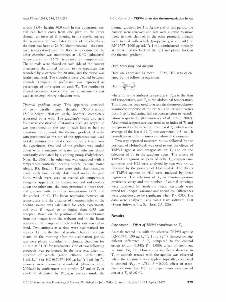
width: 38.09 height: 30.0 cm). In this apparatus, ani-
mal can freely cross from one plate to the other
through an inverted U opening in the acrylic surface
that separates the two plates. In one of the chambers,
the floor was kept at 26 °C (thermoneutral – the refer-
ence temperature) and the floor temperature of the
other chamber was maintained at 18 °C (subneutral
temperature) or 32 °C (supraneutral temperature).
The animals were placed on each side of the camera
alternately, the animal position in the apparatus was
recorded by a camera for 20 min, and the video was
further analysed. The chambers were cleaned between
animals. Temperature preference was expressed as
percentage of time spent on each Ta. The number of
animal crossings between the two environments was
used as an exploratory behaviour rate.
Thermal gradient assay—This apparatus consisted
of two parallel lanes (length: 195.0 9 width:
15.0 9 height: 20.0 cm each; Bonther) completely
separated by a wall. The gradient’s walls and grid
floor were constructed of stainless steel. An acrylic lid
was positioned on the top of each lane to help to
maintain the Ta inside the thermal gradient. A web-
cam positioned at the top of the apparatus was used
to take pictures of animal0s position every 4 min over
the experiment. One end of the gradient was cooled
down with a mixture of water and ethylene glycol
constantly circulated by a cooling pump (PolyScience,
Niles, IL, USA). The other end was equipped with a
temperature-controlled heating source (Novus, Porto
Alegre, RS, Brazil). There were eight thermocouples
inside each lane, evenly distributed under the grid
floor, which were used to record air temperature
along the apparatus. By heating one end and cooling
down the other one, the lanes presented a linear ther-
mal gradient with the hottest temperature 35 °C and
the coolest 15 °C. The linear regression between air
temperature and the distance of thermocouples to the
heating source was calculated for each experiment,
and only R2 equal to or higher than 0.93 was
accepted. Based on the position of the rats obtained
from the images from the webcam and on the linear
regression, the temperature selected by rats was calcu-
lated. Two animals at a time were acclimatized for
approx. 18 h in the thermal gradient before the treat-
ments. In the morning after the acclimation period,
rats were placed individually in climatic chambers for
40 min at 31 °C for treatments. One of two following
protocols was performed. In the first one, after i.v.
injection of vehicle (saline : ethanol; 90% : 10%;
1 mL kg�1) or HC-067047 (100 lg kg�1; 1 mL kg�1),
animals were thermally stimulated (Almeida et al.
2006a,b) by confinement to a portion (25 cm) of Ta of
28–31 °C delimited by Plexiglas barriers inside the
thermal gradient for 3 h. At the end of this period, the
barriers were removed and rats were allowed to move
freely in their channel. In the other protocol, animals
were treated with vehicle (propylene glycol; 1 mL) or
RN-1747 (1000 lg mL�1; 1 mL administered topically
at the skin of the back of the rat) and placed back in
the thermal gradient.
Data processing and analysis
Data are expressed as mean � SEM. HLI was calcu-
lated by the following equation:
HLI ¼ Ttail � Ta
Tc � Ta
where Ta is the ambient temperature, Ttail is the skin
tail temperature, and Tc is the abdominal temperature.
This index has been used to assess the thermoregulatory
vasomotor response of the rat tail and its value varies
from 0 to 1, indicating full vasoconstriction or vasodi-
lation respectively (Romanovsky et al. 1998, 2002).
Abdominal temperature was used as an index of Tc and
is expressed as the variation from basal Tc, which is the
average of the last 6–12 Tc measurements (0.5- to 1-h
period) taken at 5-min intervals before all treatments.
Two-way repeated-measures ANOVA followed by the
post-test of Holm-Sidak was used to test the effects of
TRPV4 agonist and antagonists on Tc and on the
selection of Ta in the gradient assay. The effects of
TRPV4 antagonist on peak of delta Tc, oxygen con-
sumption and HLI were analysed by two-way ANOVA
followed by the post-test of Holm-Sidak. The effects
of TRPV4 agonist on HLI were analysed by linear
regression. The selection of Ta in two-temperature
preference assay and the number of animal crossings
were analysed by Student’s t-test. Residuals were
tested for unequal variance and normality. Differences
were considered to be significant when P < 0.05. The
data were analysed using SIGMA PLOT software 11.0
(Systat Software Inc, San Jose, CA, USA).
Results
Experiment 1. Effect of TRPV4 stimulation on Tc
Animals treated i.v. with the selective TRPV4 agonist
(RN-1747; 100 lg kg�1; 1 mL kg�1) showed no sig-
nificant difference in Tc compared to the control
group (F1,42 = 0.348; P = 1.000; effect of treatment
vs. time; Fig. 1a). However, a significant decrease in
Tc of animals treated with the agonist was observed
when the treatment was applied topically, compared
to control (F1,42 = 1.786; P = 0.002; effect of treat-
ment vs. time; Fig. 1b). Both experiments were carried
out at a Ta of 26 °C.
© 2015 Scandinavian Physiological Society. Published by John Wiley & Sons Ltd, doi: 10.1111/apha.12477 279
Acta Physiol 2015, 214, 275–289 R C L Vizin et al. · TRPV4 on in vivo thermoregulation in rat

To check whether the RN-1747-induced reduction
on Tc was indeed caused by specific stimulation of
TRPV4 channels, we blocked the channels by pre-
treating the animals with a selective TRPV4 antago-
nist (RN-1734; 300 lg per rat; Fig. 1c,d). The
prior antagonist treatment significantly inhibited RN-
1747-induced hypothermia (F1,42 = 1.482; P = 0.028;
effect of treatment vs. time). The pre-treatment with
vehicle or RN-1734 (300 lg per rat) followed by
vehicle treatment showed that there is no significant
difference between groups (F1,42 = 1.263; P = 0.127;
effect of treatment vs. time; Fig. 1d).
The next experiment was designed to check whether
the RN-1747-induced decrease of Tc was caused by
peripheral or central TRPV4 stimulation. The i.c.v.
treatment with RN-1747 (10 lg kg�1; 4 lL kg�1)
resulted in a significant increase in Tc when compared
to the control group (F1,42 = 2.351; P < 0.001; effect
of treatment vs. time), as depicted in Figure 2.
Experiment 2. Effect of TRPV4 blockade on Tc,
thermogenesis and tail vasomotor tonus
Next, we studied the effect of the i.v. administration
of the selective TRPV4 antagonist HC-067047 on Tc
in conscious rats exposed to 22, 26, 30 and 32 °C of
Ta (Fig. 3a–d). At 22 and 32 °C, HC-067047-treated
animals did not present a difference in the Tc
compared to controls (F1,42 = 0.729; P = 0.897 and
F1,42 = 0.392; P = 1.000 respectively; effect of treat-
ment vs. time). At intermediate Ta (26 and 30 °C),Tc of HC-067047-treated rats was significantly higher
(a)
(b) (d)
(c)
Figure 1 Effect of peripheral TRPV4 agonist (RN-1747) on Tc. Panels (a) and (b) depict, respectively, i.v. and topical adminis-
tration of RN-1747 or vehicle. Panel (c) depicts the effect of pre-treatment with TRPV4 antagonist or vehicle on hypothermia
induced by the agonist. Panel (d) shows the effect of pre-treatment with TRPV4 antagonist or vehicle on Tc when vehicle is
applied to the skin. Topical application of RN-1747 promotes hypothermia when compared to vehicle, which disappeared when
animals were pre-treated with the TRPV4 antagonist RN-1734. Tc is demonstrated as variation from basal Tc (delta Tc). Basal
Tc: (a) 37.8 � 0.1 °C (vehicle) and 37.8 � 0.2 °C (RN-1747); (b) 37.2 � 0.1 °C (vehicle) and 37.2 � 0.1 °C (RN-1747); (c)
37.0 � 0.2 °C (vehicle pre-treated) and 36.7 � 0.1 °C (RN-1734 pre-treated); (d) 37.3 � 0.1 °C (vehicle pre-treated) and
37.2 � 0.1 °C (RN-1734 pre-treated). Ta was maintained at 26 °C for this experiment. Arrows indicate the time of injections
and topical applications. Numbers in parenthesis depict experimental n. *Shows points with significant interaction effect, that is
effect of treatment vs. time. Data are expressed as mean � SEM.
© 2015 Scandinavian Physiological Society. Published by John Wiley & Sons Ltd, doi: 10.1111/apha.12477280
TRPV4 on in vivo thermoregulation in rat · R C L Vizin et al. Acta Physiol 2015, 214, 275–289
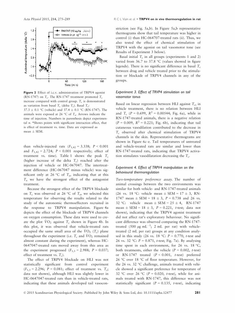
than vehicle-injected rats (F1,42 = 3.338; P < 0.001
and F1,42 = 2.724; P < 0.001 respectively; effect of
treatment vs. time). Table 1 shows the peak Tc
(higher increase of the delta Tc) reached after the
injection of vehicle or HC-067047. The intertreat-
ment difference (HC-067047 minus vehicle) was sig-
nificant only at 26 °C of Ta, indicating that at this
Ta we have the strongest effect of the antagonist
treatment.
Because the strongest effect of the TRPV4 blockade
on Tc was observed at 26 °C of Ta, we selected this
temperature for observing the results related to the
study of the autonomic thermoeffectors recruited in
the response to TRPV4 manipulation. Figure 4a
depicts the effect of the blockade of TRPV4 channels
on oxygen consumption. These data were used to cre-
ate the plot _VO2 against Tc shown in Figure 4b. In
this plot, it was observed that vehicle-treated rats
occupied the same small area of the _VO2 (Tc) plane
throughout the experiment (i.e. Tc and _VO2 remained
almost constant during the experiment), whereas HC-
0647047-treated rats moved away from this area as
the experiment progressed (F1,3 = 2.988; P = 0.037;
effect of treatment vs. Tc).
The effect of TRPV4 blockade on HLI was not
statistically significant from control experiment
(F1,3 = 2.296; P = 0.081; effect of treatment vs. Tc;
data not shown), although HLI was slightly lower in
HC-0647047-treated rats than in vehicle-treated rats,
indicating that these animals developed tail vasocon-
striction (see Fig. 5a,b). In Figure 5a,b representative
thermograms show that tail temperature was higher in
control (i) than HC-064707-treated rats (ii). Thus, we
also tested the effect of chemical stimulation of
TRPV4 with the agonist on tail vasomotor tone (see
Results of Experiment 3 below).
Basal initial Tc in all groups (experiments 1 and 2)
varied from 36.7 to 37.8 °C (values showed in figure
legends). There is no significant difference in basal Tc
between drug and vehicle treated prior to the stimula-
tion or blockade of TRPV4 channels in any of the
groups.
Experiment 3. Effect of TRVP4 stimulation on tail
vasomotor tonus
Based on linear regression between HLI against Tc, in
vehicle treatment, there is no relation between HLI
and Tc (P = 0.699, R2 = 0.00544; Fig. 6a), while in
RN-1747-treated animals, there is a negative relation
(P = 0.009, R2 = 0.221; Fig. 6b), indicating that tail
cutaneous vasodilation contributed to the decrease in
Tc observed after chemical stimulation of TRPV4
channels in the skin. Representative thermograms are
shown in Figure 6c–e. Tail temperatures of untreated
and vehicle-treated rats are similar and lower than
RN-1747-treated rats, indicating that TRPV4 activa-
tion stimulates vasodilatation decreasing the Tc.
Experiment 4. Effect of TRPV4 manipulation on the
behavioural thermoregulation
Two-temperature preference assay. The number of
animal crossings between the two environments was
similar for both vehicle- and RN-1747-treated animals
(26 vs. 18 °C: vehicle mean � SEM = 17 � 3, RN-
1747 mean � SEM = 18 � 3, P = 0.738 and 26 vs.
32 °C: vehicle mean � SEM = 25 � 4, RN-1747
mean � SEM = 18 � 3, P = 0.221, t-test; data not
shown), indicating that the TRPV4 agonist treatment
did not affect rat’s exploratory behaviour. No signifi-
cant difference was observed comparing the RN-1747-
treated (500 lg mL�1; 2 mL per rat) with vehicle-
treated (2 mL per rat) groups at any condition analy-
sed in this study (26 vs. 18 °C: P = 0.770, t-test and
26 vs. 32 °C: P = 0.871, t-test; Fig. 7a). By analysing
time spent in each environment, for 26 vs. 18 °C,both treatments, either the vehicle (P = 0.002, t-test)
or RN-1747 treated (P < 0.001, t-test) preferred
26 °C over 18 °C of floor temperature. However, for
the 26 vs. 32 °C challenge, animals treated with vehi-
cle showed a significant preference for temperature of
32 °C over 26 °C (P = 0.020, t-test), while for ani-
mals treated with RN-1747, this difference was not
statistically significant (P = 0.135, t-test), indicating
Figure 2 Effect of i.c.v. administration of TRPV4 agonist
(RN-1747) on Tc. The RN-1747 treatment promoted Tc
increase compared with control group. Tc is demonstrated
as variation from basal Tc (delta Tc). Basal Tc:
37.3 � 0.1 °C (vehicle) and 37.4 � 0.1 °C (RN-1747). The
animals were exposed at 26 °C of Ta. Arrows indicate the
time of injection. Numbers in parenthesis depict experimen-
tal n. *Shows points with significant interaction effect, that
is effect of treatment vs. time. Data are expressed as
mean � SEM.
© 2015 Scandinavian Physiological Society. Published by John Wiley & Sons Ltd, doi: 10.1111/apha.12477 281
Acta Physiol 2015, 214, 275–289 R C L Vizin et al. · TRPV4 on in vivo thermoregulation in rat

that there is no place preference for TRPV4-stimulated
animals (Fig. 7b) under this condition. Although
weak, this result inspired us to further explore the
involvement of TRPV4 channels in the recruitment of
a thermoregulatory behaviour in a thermal gradient.
Thermal gradient assay. The thermal exposure
(28–31 °C) led the control animals to seek colder
areas than those animals treated with antagonist
(HC-064707; 100 lg kg�1; 1 mL kg�1) after confine-
ment (F1,10 = 5.09; P = 0.048, effect of treatment;
F1,10 = 3.13; P < 0.001, effect of time; no interaction
effect; Fig. 8a). The Tc of HC-064707-treated animals
was higher than control animals (37.5 � 0.1 °C vs.
37.0 � 0.1 °C respectively. F1,10 = 1.44; P = 0.01,
effect of treatment vs. time). Corroborating this result,
the chemical stimulation of the channel with TRPV4
agonist (RN-1747; 1000 lg mL�1; 1 mL topical)
caused a reduction of preferred Ta compared to
vehicle-treated animals (F1,9 = 4.18; P < 0.001, effect
of treatment vs. time; Fig. 8b) and reduced the Tc
over the time as well (the Tc decreased up to
0.7 � 0.2 °C after agonist treatment, but it presented
just a slight change in those vehicle-treated animals:
�0.1 � 0.2 °C; F1,8 = 1.41; P = 0.02, effect of treat-
ment vs. time).
Discussion
In the present study, we tested whether chemical acti-
vation or blockade of the warmth-sensitive TRPV4
channels exerts an effect on Tc and which thermoef-
fector (autonomic and/or behavioural) is involved in
this response. Based on the in vitro temperature range
of TRPV4 activation, we hypothesized that moderate
warmth temperatures activate in vivo peripheral
TRPV4 channels which in turn activate warmth-
defence responses, maintaining Tc at regulated levels.
The results indicate that cutaneous TRPV4 is physio-
logically activated in vivo at Tas in the range of 26–30 °C in the conditions used in the present study.
TRPV4 seems to be involved in the activation of both
autonomic and behavioural warmth-defence responses.
(a)
(b)
(d)
(c)
Figure 3 Effect of i.v. administration of TRPV4 antagonist
(HC-067047) onTc at differentTas. The animals were exposed
at 22° (a), 26° (b), 30° (c) and 32 °C (d) ofTas. Tc was
significantly higher in HC-067047-treated than controls at 26
and 30 °C ofTa. Tc is demonstrated as variation from basalTc
(deltaTc). BasalTc: (a) 37.3 � 0.1 °C (vehicle) and 37.4 �0.1 °C (HC-067047); (b) 37.4 � 0.2 °C (vehicle) and
37.3 �0.2 °C (HC-067047); (c) 37.1 � 0.1 °C (vehicle) and
37.5 � 0.1 °C (HC-067047); (d) 37.3 � 0.1 °C (vehicle) and
37.4 � 0.1 °C (HC-067047). Arrows indicate the time of injec-
tion. Numbers in parenthesis depict experimental n. *Shows
points with significant interaction effect, that is effect of treat-
ment vs. time. Data are expressed as mean � SEM.
© 2015 Scandinavian Physiological Society. Published by John Wiley & Sons Ltd, doi: 10.1111/apha.12477282
TRPV4 on in vivo thermoregulation in rat · R C L Vizin et al. Acta Physiol 2015, 214, 275–289

Moreover, topical activation and intravenous blockade
of this channel caused decrease and increase of Tc,
respectively, when Ta was fixed at 26 °C. TRPV4
blockade-induced rise of Tc at 26 °C of Ta was
accompanied by an increase in oxygen consumption
(an index of thermogenesis), and chemical activation
of TRPV4 channels through topical application of
RN-1747 leads to a reduction of Tc that was influ-
enced by tail vasomotor tone. These results indicate
TRPV4-mediated recruitment of autonomic thermoef-
fectors reduces heat production and increases heat loss
in rats. Moreover, rats chemically stimulated with
TRPV4 agonist lean towards colder Tas, and the ani-
mals thermally stimulated (exposure to 28–31 °C) didnot select colder Tas when injected with the TRPV4
antagonist, compared with vehicle group. These inter-
esting results indicate a clear role of TRPV4 in the
facilitation of heat loss responses through behaviour
in rats.
Although previous studies have suggested TRPV4 to
be a thermosensor, a thermoregulatory role for these
channels has not been proposed as TRPV4 knockout
(KO) mice acclimated at 25 °C of Ta (Lee et al. 2005)
or cold (Liedtke & Friedman 2003) presented similar
Tc compared to its wild-type littermates. However,
negative results with KO animals are difficult to inter-
pret, especially because in these animals compensatory
mechanisms may occur (Mogil et al. 2000, Lariviere
et al. 2001), and thus these data do not exclude the
participation of TRPV4 channels in thermoregulation.
In fact, in the case of TRPV1, no thermoregulatory
change was observed in TRPV1 KO animal studies
(Szel�enyi et al. 2004, Iida et al. 2005) or by TRPV1
gene silencing (Christoph et al. 2006), although it was
later shown that these channels play a role in the
maintenance of Tc (Steiner et al. 2007). Using phar-
macological tools and altering the activity of the
TRPV4 channel temporarily, we did not allow for
long-term compensatory mechanisms to occur and we
were able to show, for the first time, the involvement
Table 1 Peak of delta core body temperature (Tc) at different ambient temperatures (Ta)
Ta (°C)
Tc control (delta °C)Tc HC-067047 (delta °C)
Peak � SEM Peak � SEM Peak Diff; P value
22 0.7 � 0.2 0.6 � 0.1 �0.1; P = 0.792
26 0.4 � 0.2 1.1 � 0.2 0.7; P = 0.009
30 0.7 � 0.1 0.9 � 0.2 0.2; P = 0.369
32 0.6 � 0.2 0.9 � 0.2 0.3; P = 0.184
Peak Diff., peak difference. Peak differences were calculated subtracting the peak value of the HC-067047-treated from the vehi-
cle-treated (control) animals.
(a)
(b)
Figure 4 Effect of i.v. administration of TRPV4 antagonist
(HC-067047) on _VO2 Panel (a) describes the _VO2 over time
of experiment, and in panel (b), _VO2 is plotted against Tc
between 50 and 110 min after injection. HC-067047-induced
hyperthermia was accompanied by increase in, which does
not occur in the control group. The direction of time is
shown with arrows with initial and final times indicated by
regular or inverted triangle symbol respectively. Ta over
experiment was 26 °C. Numbers in parenthesis depict experi-
mental n. Data in panel (a) are expressed as mean � SEM.
© 2015 Scandinavian Physiological Society. Published by John Wiley & Sons Ltd, doi: 10.1111/apha.12477 283
Acta Physiol 2015, 214, 275–289 R C L Vizin et al. · TRPV4 on in vivo thermoregulation in rat
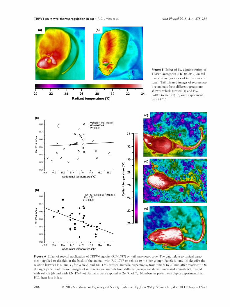
(a) (b)
Figure 5 Effect of i.v. administration of
TRPV4 antagonist (HC-067047) on tail
temperature (an index of tail vasomotor
tone). Tail infrared images of representa-
tive animals from different groups are
shown: vehicle treated (a) and HC-
06047 treated (b). Ta over experiment
was 26 °C.
(a)(c)
(d)
(e)
(b)
Figure 6 Effect of topical application of TRPV4 agonist (RN-1747) on tail vasomotor tone. The data relate to topical treat-
ment, applied to the skin at the back of the animal, with RN-1747 or vehicle (n = 6 per group). Panels (a) and (b) describe the
relation between HLI and Tc for vehicle- and RN-1747-treated animals, respectively, from time 0 to 20 min after treatment. On
the right panel, tail infrared images of representative animals from different groups are shown: untreated animals (c), treated
with vehicle (d) and with RN-1747 (e). Animals were exposed at 26 °C of Ta. Numbers in parenthesis depict experimental n.
HLI, heat loss index.
© 2015 Scandinavian Physiological Society. Published by John Wiley & Sons Ltd, doi: 10.1111/apha.12477284
TRPV4 on in vivo thermoregulation in rat · R C L Vizin et al. Acta Physiol 2015, 214, 275–289
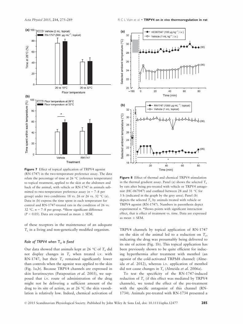
of these receptors in the maintenance of an adequate
Tc in a living and non-genetically modified organism.
Role of TRPV4 when Ta is fixed
Our data showed that animals kept at 26 °C of Ta did
not display changes in Tc when treated i.v. with
RN-1747, but their Tc remained significantly lower
than controls when the agonist was applied to the skin
(Fig. 1a,b). Because TRPV4 channels are expressed in
skin keratinocytes (Patapoutian et al. 2003), we sup-
posed that i.v. route of administration of the drug
might not be delivering a sufficient amount of the
drug to its site of action, as at 26 °C the skin vasodi-
lation is relatively low. Indeed, chemical activation of
TRPV4 channels by topical application of RN-1747
on the skin of the animal led to a reduction on Tc,
indicating the drug was presumably being delivered to
its site of action (Fig. 1b). This topical application has
been previously shown to be quite efficient for induc-
ing hyperthermia after treatment with menthol (an
agonist of the cold-activated TRPM8 channel) (Alme-
ida et al. 2012), whereas i.v. application of menthol
did not cause changes in Tc (Almeida et al. 2006a).
To test the specificity of the RN-1747-induced
reduction of Tc (if this effect was mediated by TRPV4
channels), we tested the effect of the pre-treatment
with the specific antagonist of this channel (RN-
1734). Animals pre-treated with RN-1734 presented a
(a)
(b)
Figure 7 Effect of topical application of TRPV4 agonist
(RN-1747) in the two-temperature preference assay. The data
relate the percentage of time at 26 °C (reference temperature)
to topical treatment, applied to the skin at the abdomen and
back of the animal, with vehicle or RN-1747 in animals sub-
mitted to two-temperature preference assay (n = 7–8 per
group) under two conditions: 18 vs. 26 or 26 vs. 32 °C (a).
Data in (b) express the time spent in each temperature for
control and RN-1747-treated rats in the condition of 26 vs.
32 °C. n = 7–8 per group. *Show significant difference
(P < 0.05). Data are expressed as mean � SEM.
(a)
(b)
Figure 8 Effect of thermal and chemical TRPV4 stimulation
in the thermal gradient assay. Panel (a) shows the selected Ta
by rats after being pre-treated with vehicle or TRPV4 antago-
nist (HC-067047) and confined between 28 and 31 °C for
3 h (indicated in the graph by the grey area). Panel (b)
depicts the selected Ta by animals treated with vehicle or
TRPV4 agonist (RN-1747). Numbers in parenthesis depict
experimental n. *Shows points with significant interaction
effect, that is effect of treatment vs. time. Data are expressed
as mean � SEM.
© 2015 Scandinavian Physiological Society. Published by John Wiley & Sons Ltd, doi: 10.1111/apha.12477 285
Acta Physiol 2015, 214, 275–289 R C L Vizin et al. · TRPV4 on in vivo thermoregulation in rat

reduced hypothermic response to the agonist and the
antagonist itself did not alter the Tc, confirming that
the observed effect was due to the action of the drug
at TRPV4 channels (Fig. 1c,d).
To check whether the effect of TRPV4 activation
on Tc was due to activation of peripheral (skin) or
central (central nervous system) receptors, we pro-
ceeded with the i.c.v. administration of RN-1747. In
this assay, the treated and control animals had an
increase of Tc due to the stress of handling; however,
over time, the RN-1747-treated animals were hyper-
thermic compared to control (Fig. 2). TRPV4 channels
are expressed at the central nervous system, in the
hypothalamus, for example, which is an important site
for thermoregulation (Liedtke et al. 2000, G€uler et al.
2002). Furthermore, there is evidence that TRPV4
channels in the central nervous system work as hypo-
osmolarity sensors (Strotmann et al. 2000, Liedtke &
Friedman 2003, Liedtke et al. 2003, Liedtke 2005) and
osmoregulatory dysfunction can lead to inflammatory
processes involving the TRPV4 channel (Alessandri-
Haber et al. 2005, Vergnolle et al. 2010). If TRPV4
stimulation with RN-1747 induced a false interpreta-
tion of osmolarity alteration, it would promote a local
inflammatory process that would result in the increase
of Tc observed after i.c.v. treatment with the TRPV4
agonist. Because the central vs. peripheral thermoregu-
latory effects of chemical TRPV4 stimulation were
opposite (increase vs. decrease of Tc respectively), we
concluded that the hypothermic effect of RN-1747 was
due to action of the drug at the periphery (receptors
presumably located in the skin).
Although these results point to an involvement of
TRPV4 channels in the induction of warmth-defence
responses, at 26 °C of Ta, skin temperature of rats
(where TRPV4 channels are presumably located) is
approx. 29.5 °C, and thus some activation of TRPV4
channels by temperature may already be occurring,
which would explain why the effects of chemical stim-
ulation of these channel are weak. Thus, for studying
the involvement of these channels in the thermoregula-
tory effectors, we designed an experiment in which we
used blockade of the channel (using a more
potent and selective antagonist of TRPV4 channel,
HC-067047) and increase of Ta (for promoting a ther-
mal, instead of chemical stimulation of the channel).
In this case, to facilitate the antagonist reaching the
receptor sites in the skin after i.v. injection, we tempo-
rarily exposed all the animals to warm temperatures
to increase blood flow to the skin as previously
described (Almeida et al. 2012), before the exposure
to experimental Tas. By blocking TRPV4 channels and
changing Ta in a series of distinct stages and experi-
ments (from 22 to 32 °C), we observed that a rise of
Tc occurred in Ta of 26 and 30 °C (Fig. 3 and
Table 1), which is consistent with our initial hypothe-
sis that warm temperatures activate TRPV4 channels.
When we blocked these channels, the defence
responses to warmth were not activated and hyper-
thermia occurred. We did not see an increase in Tc at
22 °C, as this Ta is below the range of activation of
TRPV4 channels, nor at 32 °C of Ta, possibly because
there is activation of other temperature-sensitive TRP
channels at this Ta, such as TRPV3 (Caterina 2007).
The next step of our study was planned to check
which thermoeffector was activated and/or inhibited
during the thermoregulatory response to TRPV4
manipulation. We showed that both autonomic ther-
moeffectors, thermogenesis and tail vasomotor tone,
are modulated during the TRPV4 thermoregulatory
response. Oxygen consumption, which is an index of
thermogenesis, increased slightly over time in animals
treated with HC-067047 compared to controls (Fig. 4),
corroborating with studies where TRPV4 KO mice and
adipocytes isolated from them had increased oxygen
consumption compared to wild-type cells and animals
respectively (Kusudo et al. 2012, Ye et al. 2012). In the
present study, the animals that had their TRPV4 phar-
macologically blocked by the antagonist consumed
1.24 � 0.2 mL O2 kg�1 min�1 more than vehicle-trea-
ted rats. Assuming that each mL of oxygen consumed
produces 20.3 J (Randall et al. 1997), this small aug-
ment was enough to produce 1.5 kJ in 60 min (between
50 and 110 min; 1.24 9 60 9 20.3), which is sufficient
to increase the Tc by 0.7 °C in rats of 300 g (Fig. 3b;
Q = mc(t), where Q is heat energy, m is mass, c is the
specific heat, considered to be 3.47 J for biological tis-
sues, and (t is temperature variation, 0.7 °C) (Schmidt-
Nielsen 1997, Da Silva & Maia 2013). Although the
HLI along the time seemed to be lower in HC-067047-
treated animals, it was not statistically different from
the control group (Fig. 5a–d). In contrast, animals pre-
sented higher tail temperatures (indicating vasodilation)
in response to the chemical stimulation of TRPV4 chan-
nels (Fig. 6). Together, these data reveal that blocking
TRPV4-mediated warmth signalling from the skin inac-
tivated warmth-defence responses, increasing Tc.
Besides, stimulating warmth receptors with a TRPV4
agonist can activate (at least vasomotor response)
warmth-defence responses and decrease Tc.
Role of TRPV4 when Ta can be selected by the animal
Regarding behavioural thermoregulatory responses,
we used three different approaches. In the first one,
animals had the chance to choose between two tem-
peratures and were challenged with neutral vs.
warmth (26 vs. 32 °C) and cold vs. neutral (18 vs.
26 °C) environments. In this test, we did not observe
a significant effect of the treatment with the TRPV4
© 2015 Scandinavian Physiological Society. Published by John Wiley & Sons Ltd, doi: 10.1111/apha.12477286
TRPV4 on in vivo thermoregulation in rat · R C L Vizin et al. Acta Physiol 2015, 214, 275–289
agonist compared with the control in any studied con-
dition (26 vs. 32 °C and 18 vs. 26 °C) in the time
spent at 26 °C (reference temperature); however, ani-
mals treated with vehicle showed a significant prefer-
ence for the temperature of 32 °C over 26 °C, while
in animals treated with RN-1747, this difference was
not statistically significant (there is no place prefer-
ence, i.e. rats spend similar amount of time in both
environments) due to the slight augmentation of time
spent at 26 °C over 32 °C. This result indicates that
TRPV4 activation might be stimulating a thermoregu-
latory behaviour to facilitate heat loss, and so devel-
oping hypothermia. Under the condition 26 vs. 18 °C,both vehicle- and RN-1747-treated rats preferred
26 °C over 18 °C, indicating, possibly due to the
range of activation of TRPV4 channels, that
RN-1747-treated rats do not necessarily select a Tas
so much colder (like 18 °C) than thermoneutrality to
provide thermal comfort.
To further test the involvement of TRPV4 on
behavioural thermoregulation, we studied rats in a
thermal gradient and we verified very clear thermoregu-
latory responses. In the first approach, animals were
transitorily confined to a region of the thermal gradient
where Tas was between 28 and 31 °C in order to verify
a possible cold-seeking behaviour of rats after this ther-
mal stimulation (Almeida et al. 2006a). In fact, control
animals preferred colder Ta than the HC-067047-trea-
ted ones (Fig. 8a). In another set of experiments, chem-
ical stimulation of the TRPV4 in non-confined animals
in the thermal gradient led them to seek lower Ta than
control ones (Fig. 8b). Both results clearly indicate that
TRPV4 stimulation activates cold-seeking behaviour.
Taken together, our results indicate that if animals
have the chance to select a Ta of preference, TRPV4
stimulation will successfully activate the behaviour ther-
moeffector, in order to regulate Tc, which agrees with a
parsimonious priority given to its kind of effector, which
is very efficient and energetically less expensive than the
autonomic thermoeffectors. However, when animals are
not allowed to thermoregulate behaviourally, TRPV4
stimulation will activate autonomic thermoeffectors to
regulate Tc. We showed here, for the first time, a role of
TRPV4 in the activation of non-evaporative heat loss
and inhibition of heat gain in vivo in rats.
Although TRPV4 channels were characterized
approx. 10 years ago, a range of functional studies
were developed and different biological functions for
these channels were assigned, such as regulation and
sensation of osmolarity in the central nervous system
(Strotmann et al. 2000, Liedtke & Friedman 2003,
Liedtke et al. 2003), thermoreception (Lee et al.
2005), participation in inflammatory responses (Verg-
nolle et al. 2010, Bang et al. 2012, Denadai-Souza
et al. 2012, Ye et al. 2012, Poole et al. 2013), noci-
ceptive (Alessandri-Haber et al. 2003, 2005, Nakat-
suka & Iwai 2009, Everaerts et al. 2010a) and
hyperalgesia responses (Todaka et al. 2004, Alessan-
dri-Haber et al. 2009, Ding et al. 2010, Wang et al.
2011, Segond von Banchet et al. 2013), among others.
The present study adds the participation of TRPV4
channels to the regulation of Tc in rats. In recent
years, thermo-TRP channels have been suggested to be
a target for the development of new therapeutic strate-
gies. However, it appears that many of compounds
submitted for clinical trial are far from successful, in
particular due to the occurrence of side effects (Ferrer-
Montiel et al. 2012). A better understanding of the
functions of these channels is essential for developing
strategies for modulating the activity of thermo-TRP
channels with an improved therapeutic index. The
development of this study aims at contributing to
improve the knowledge regarding the functions of this
receptor with respect to the thermoregulatory system,
thus providing important data that may contribute to
the development of these therapeutic strategies.
Conflict of interest
The authors of this manuscript have no competing
interests.
We would like to thank the Cognition and Complex Systems
Nuclei of UFABC (NCSC/UFABC) and the Department of
Animal Morphology and Physiology of FCAV-UNESP for
providing the infrastructure required for developing the
experimental procedures. This work was financially sup-
ported by CNPq (Grant 470742/2010-0 to M.C.A.) and
FAPESP (Grants 2011/06528-1 to M.C.A, 2012/19966-0 to
LHG, 2009/11446-4 to D.C.C.; FAPESP graduate fellowship
grants: 2012/11836-0 to R.C.L.V. and 2011/19131-2 to
C.S.S.). We thank Samuel P. Wanner for critically reading
the first version of this manuscript.
Author contributions
R.C.L.V., C.S.S., D.I., K.C.B. and M.C.A. involved in
conception and design. L.H.G., D.C.C., K.C.B. and
M.C.A. helped in financial support. R.C.L.V., C.S.S.,
D.I., G.M.C., C.O.S. and M.C.A. participated in col-
lection and assembly of data. R.C.L.V., C.S.S., D.I.,
G.M.C., C.O.S., L.H.G., D.C.C., K.C.B. and M.C.A.
performed data analysis and interpretation. R.C.L.V.,
C.S.S., K.C.B. and M.C.A. performed manuscript
writing.
References
Aihara, H., Okada, Y. & Tamaki, N. 2001. The effects of
cooling and rewarming on the neuronal activity of pyrami-
© 2015 Scandinavian Physiological Society. Published by John Wiley & Sons Ltd, doi: 10.1111/apha.12477 287
Acta Physiol 2015, 214, 275–289 R C L Vizin et al. · TRPV4 on in vivo thermoregulation in rat
dal neurons in guinea pig hippocampal slices. Brain Res
893, 36–45.
Alessandri-Haber, N., Yeh, J.J., Boyd, A.E., Parada, C.A.,
Chen, X., Reichling, D.B. & Levine, J.D. 2003. Hypoto-
nicity induces TRPV4-mediated nociception in rat. Neuron
39, 497–511.
Alessandri-Haber, N., Joseph, E., Dina, O.A., Liedtke, W. &
Levine, J.D. 2005. TRPV4 mediates pain-related behavior
induced by mild hypertonic stimuli in the presence of
inflammatory mediator. Pain 118, 70–79.
Alessandri-Haber, N., Dina, O.A., Chen, X. & Levine, J.D.
2009. TRPC1 and TRPC6 channels cooperate with TRPV4
to mediate mechanical hyperalgesia and nociceptor sensiti-
zation. J Neurosci 29, 6217–6228.
Almeida, M.C., Steiner, A.A., Branco, L.G. & Romanovsky,
A.A. 2006a. Cold-seeking behavior as a thermoregulatory
strategy in systemic inflammation. Eur J Neurosci 23,
3359–3367.
Almeida, M.C., Steiner, A.A., Branco, L.G. & Romanovsky,
A.A. 2006b. Neural substrate of cold-seeking behavior in
endotoxin shock. PLoS ONE 1, e1.
Almeida, M.C., Hew-Butler, T., Soriano, R.N., Rao, S.,
Wang, W., Wang, J., Tamayo, N., Oliveira, D.L., Nucci,
T.B., Aryal, P., Garami, A., Bautista, D., Gavva, N.R. &
Romanovsky, A.A. 2012. Pharmacological blockade of the
cold receptor TRPM8 attenuates autonomic and behavioral
cold defenses and decreases deep body temperature. J Neu-
rosci 32, 2086–2099.
Bang, S., Yoo, S., Yang, T.J., Cho, H. & Hwang, S.W. 2012.
Nociceptive and pro-inflammatory effects of dimethylallyl
pyrophosphate via TRPV4 activation. Br J Pharmacol 166,
1433–1443.
B�ıcego, K.C., Barros, R.C. & Branco, L.G. 2007. Physiology
of temperature regulation: comparative aspects. Comp Bio-
chem Physiol A Mol Integr Physiol 147, 616–639.
Caterina, M.J. 2007. Transient receptor potential ion channels
as participants in thermosensation and thermoregulation.
Am J Physiol Regul Integr Comp Physiol 292, R64–R76.
Christoph, T., Gr€unweller, A., Mika, J., Sch€afer, M.K.,
Wade, E.J., Weihe, E., Erdmann, V.A., Frank, R., Gillen,
C. & Kurreck, J. 2006. Silencing of vanilloid receptor
TRPV1 by RNAi reduces neuropathic and visceral pain in
vivo. Biochem Biophys Res Commun 350, 238–243.
Da Silva, R.G. & Maia, A.S.C. 2013. Thermal balance and
thermoregulation. In: R.G. Da Silva & A.S.C. Maia (eds)
Principles of Animal Biometeorolog, pp. 75–106. Springer,
Dordrecht, Netherlands.
Delany, N.S., Hurle, M., Facer, P., Alnadaf, T., Plumpton, C.,
Kinghorn, I., See, C.G., Costigan, M., Anand, P., Woolf,
C.J., Crowther, D., Sanseau, P. & Tate, S.N. 2001. Identifi-
cation and characterization of a novel human vanilloid
receptor-like protein, VRL-2. Physiol Genomics 4, 165–174.
Denadai-Souza, A., Martin, L., de Paula, M.A., de Avellar,
M.C., Muscara, M.N., Vergnolle, N. & Cenac, N. 2012.
Role of transient receptor potential vanilloid 4 in rat joint
inflammation. Arthritis Rheum 64, 1848–1858.
Ding, X.L., Wang, Y.H., Ning, L.P., Zhang, Y., Ge, H.Y., Ji-
ang, H., Wang, R. & Yue, S.W. 2010. Involvement of
TRPV4-NO-cGMP-PKG pathways in the development of
thermal hyperalgesia following chronic compression of the
dorsal root ganglion in rats. Behav Brain Res 208, 194–201.
Everaerts, W., Nilius, B. & Owsianik, G. 2010a. The vanil-
loid transient receptor potential channel TRPV4: from
structure to disease. Prog Biophys Mol Biol 103, 2–17.
Everaerts, W., Zhen, X., Ghosh, D., Vriens, J., Gevaert, T.,
Gilbert, J.P., Hayward, N.J., McNamara, C.R., Xue, F.,
Moran, M.M. et al. 2010b. Inhibition of the cation chan-
nel TRPV4 improves bladder function in mice and rats
with cyclophosphamide-induced cystitis. Proc Natl Acad
Sci U S A 107, 19084–19089.
Ferrer-Montiel, A., Fern�andez-Carvajal, A., Planells-Cases,
R., Fern�andez-Ballester, G., Gonz�alez-Ros, J.M., Messe-
guer, A. & Gonz�alez-Mu~niz, R. 2012. Advances in modu-
lating thermosensory TRP channels. Expert Opin Ther Pat
22, 999–1017.
Gordon, C.J. 1990. Thermal biology of the laboratory rat.
Physiol Behav 47, 963–991.
G€uler, A.D., Lee, H., Iida, T., Shimizu, I., Tominaga, M. &
Caterina, M. 2002. Heat-evoked activation of the ion
channel, TRPV4. J Neurosci 22, 6408–6414.
Iida, T., Shimizu, I., Nealen, M.L., Campbell, A. & Caterina,
M. 2005. Attenuated fever response in mice lacking
TRPV1. Neurosci Lett 378, 28–33.
Kauer, J.A. & Gibson, H.E. 2009. Hot flash: TRPV channels
in the brain. Trends Neurosci 32, 215–224.
Kusudo, T., Wang, Z., Mizuno, A., Suzuki, M. & Yamashit-
a, H. 2012. TRPV4 deficiency increases skeletal muscle
metabolic capacity and resistance against diet-induced
obesity. J Appl Physiol 7, 1223–1232.
Lariviere, W.R., Chesler, E.J. & Mogil, J.S. 2001. Transgenic
studies of pain and analgesia: mutation or background
genotype? J Pharmacol Exp Ther 297, 467–473.
Lee, H., Iida, T., Mizuno, A., Suzuki, M. & Caterina, M.J.
2005. Altered thermal selection behavior in mice lacking
transient receptor potential vanilloid 4. J Neurosci 25,
1304–1310.
Liedtke, W. 2005. TRPV4 as osmosensor: a transgenic
approach. Pflugers Arch 451, 176–180.
Liedtke, W. & Friedman, J.M. 2003. Abnormal osmotic reg-
ulation in trpv4�/� mice. Proc Natl Acad Sci U S A 100,
13698–13703.
Liedtke, W., Choe, Y., Marti-Renom, M.A., Bell, A.M.,
Denis, C.S., Sali, A., Hudspeth, A.J., Friedman, J.M. &
Heller, S. 2000. Vanilloid receptor-related osmotically acti-
vated channel (VR-OAC), a candidate vertebrate osmore-
ceptor. Cell 103, 525–535.
Liedtke, W., Tobin, D.M., Bargmann, C.I. & Friedman, J.M.
2003. Mammalian TRPV4 (VR-OAC) directs behavioral
responses to osmotic and mechanical stimuli in Caenor-
habditis elegans. Proc Natl Acad Sci U S A 100(Suppl 2),
14531–14536.
Lighton, J.R.B. 2008. Flow-through respirometry: the equa-
tions. In: J.R.B. Lighton (ed.) Measuring Metabolic Rates.
A Manual for Scientists, pp. 100–104. Oxford University
Press, New York, NY.
Mogil, J.S., Yu, L. & Basbaum, A.I. 2000. Pain genes?: natu-
ral variation and transgenic mutants. Annu Rev Neurosci
23, 777–811.
© 2015 Scandinavian Physiological Society. Published by John Wiley & Sons Ltd, doi: 10.1111/apha.12477288
TRPV4 on in vivo thermoregulation in rat · R C L Vizin et al. Acta Physiol 2015, 214, 275–289
Morrison, S.F. & Nakamura, K. 2011. Central neural path-
ways for thermoregulation. Front Biosci 16, 74–104.
Nakatsuka, M. & Iwai, Y. 2009. Expression of TRPV4 in
the stimulated rat oral mucous membrane–nociceptive
mechanisms of lingual conical papillae. Okajimas Folia
Anat Jpn 86, 45–54.
Patapoutian, A., Peier, A.M., Story, G.M. & Viswanath, V.
2003. ThermoTRP channels and beyond: mechanisms of
temperature sensation. Nat Rev Neurosci 4, 529–539.
Persson, P.B. 2013. Good publication practice in physiology
2013 guidelines for Acta Physiologica. Acta Physiol (Oxf)
209, 250–253.
Poole, D.P., Amadesi, S., Veldhuis, N.A., Abogadie, F.C.,
Lieu, T., Darby, W., Liedtke, W., Lew, M.J., McIntyre, P.
& Bunnett, N.W. 2013. Protease-activated receptor 2
(PAR2) protein and transient receptor potential vanilloid 4
(TRPV4) protein coupling is required for sustained inflam-
matory signaling. J Biol Chem 288, 5790–5802.
Randall, D., Burggren, W. & French, K. 1997. Using energy:
meeting environmental challenges. In: D. Randall, W.
Burggren & K. French (eds) Eckert Animal Physiology:
Mechanisms and Adaptations, pp. 665–723. W. H. Free-
man Company, New York.
Romanovsky, A.A. 2007. Thermoregulation: some concepts
have changed. Functional architecture of the thermoregula-
tory system. Am J Physiol Regul Integr Comp Physiol 292,
37–46.
Romanovsky, A.A., Simons, C.T. & Kulchitsky, V.A. 1998.
“Biphasic” fevers often consist of more than two phases.
Am J Physiol Regul Integr Comp Physiol 275, R323–
R331.
Romanovsky, A.A., Ivanov, A.I. & Shimansky, Y.P. 2002.
Selected contribution: ambient temperature for experiments
in rats: a new method for determining the zone of thermal
neutrality. J Appl Physiol 92, 2667–2679.
Schmidt-Nielsen, K. 1997. Temperature regulation. In: K.
Schmidt-Nielsen (ed.) Animal Physiology: adaptation and
environment, pp. 241–300. Cambridge University Press,
Cambridge, UK.
Segond von Banchet, G., Boettger, M.K., K€onig, C., Iwakura,
Y., Br€auer, R. & Schaible, H.G. 2013. Neuronal IL-17
receptor upregulates TRPV4 but not TRPV1 receptors in
DRG neurons and mediates mechanical but not thermal
hyperalgesia. Mol Cell Neurosci 52, 152–160.
Steiner, A.A., Turek, V.F., Almeida, M.C., Burmeister, J.J.,
Oliveira, D.L., L.Roberts, J., Bannon, A.W., Norman,
M.H., Louis, J.C., Treanor, J.J., Gavva, N.R. & Romanov-
sky, A.A. 2007. Nonthermal activation of transient recep-
tor potential vanilloid-1 channels in abdominal viscera
tonically inhibits autonomic cold-defense effectors. J Neu-
rosci 27, 7459–7468.
Strotmann, R., Harteneck, C., Nunnenmacher, K., Schultz,
G. & Plant, T.D. 2000. OTRPC4, a nonselective cation
channel that confers sensitivity to extracellular osmolarity.
Nat Cell Biol 2, 695–702.
Szel�enyi, Z., Hummel, Z., Szolcs�anyi, J. & Davis, J.B. 2004.
Daily body temperature rhythm and heat tolerance in
TRPV1 knockout and capsaicin pretreated mice. Eur J
Neurosci 19, 1421–1424.
Todaka, H., Taniguchi, J., Satoh, J., Mizuno, A. & Suzuki,
M. 2004. Warm temperature-sensitive transient receptor
potential vanilloid 4 (TRPV4) plays an essential role in
thermal hyperalgesia. J Biol Chem 279, 35133–35138.
Vergnolle, N., Cenac, N., Altier, C., Cellars, L., Chapman,
K., Zamponi, G.W., Materazzi, S., Nassini, R., Liedtke,
W., Cattaruzza, F., Grady, E.F., Geppetti, P. & Bunnett,
N.W. 2010. A role for transient receptor potential vanil-
loid 4 in tonicity-induced neurogenic inflammation. Br J
Pharmacol 159, 1161–1173.
Vincent, F. & Duncton, M.A. 2011. TRPV4 agonists and
antagonists. Curr Top Med Chem 11, 2216–2226.
Vincent, F., Acevedo, A., Nguyen, M.T., Dourado, M., DeF-
alco, J., Gustafson, A., Spiro, P., Emerling, D.E., Kelly,
M.G. & Duncton, M.A. 2009. Identification and charac-
terization of novel TRPV4 modulators. Biochem Biophys
Res Commun 3, 490–494.
Voets, T., Talavera, K., Owsianik, G. & Nilius, B. 2005.
Sensing with TRP channels. Nat Chem Biol 1, 85–92.
Wang, C., Ning, L.P., Wang, Y.H., Zhang, Y., Ding, X.L.,
Ge, H.Y., Arendt-Nielsen, L. & Yue, S.W. 2011. Nuclear
factor-kappa B mediates TRPV4-NO pathway involved in
thermal hyperalgesia following chronic compression of the
dorsal root ganglion in rats. Behav Brain Res 221, 19–24.
Wasserstrom, J.A. & Vites, A.M. 1999. Activation of contrac-
tion in cat ventricular myocytes: effects of low Cd(2+) con-
centration and temperature. Am J Physiol 277, H488–H498.
Watanabe, H., Vriens, J., Suh, S.H., Benham, C.D., Droog-
mans, G. & Nilius, B. 2002. Heat-evoked activation of
TRPV4 channels in a HEK293 cell expression system and
in native mouse aorta endothelial cells. J Biol Chem 277,
47044–47051.
Wenisch, C., Narzt, E., Sessler, D.L., Parschalk, B., Lenhardt,
R., Kurz, A. & Graninger, W. 1996. Mild intraoperative
hypothermia reduces production of reactive oxygen inter-
mediates by polymorphonuclear leukocytes. Anesth Analg
82, 810–816.
Ye, L., Kleiner, S., Wu, J., Sah, R., Gupta, R.K., Banks, A.S.,
Cohen, P., Khandekar, M.J., Bostrom, P., Mepani, R.J. et al.
2012. TRPV4 is a regulator of adipose oxidative metabolism,
inflammation, and energy homeostasis. Cell 151, 96–110.
© 2015 Scandinavian Physiological Society. Published by John Wiley & Sons Ltd, doi: 10.1111/apha.12477 289
Acta Physiol 2015, 214, 275–289 R C L Vizin et al. · TRPV4 on in vivo thermoregulation in rat

Editorial
The transient receptor potential vanilloid-4 channel: detecting body temperatures that drive defences
against mild warmth
See related article: Vizin, R.C., Scarpellini, C.D., Ishikawa, D.T.,
Correa, G.M., de Souza, C.O., Gargaglioni, L.H., Carrettiero, D.C.,
Bicego, K.C. & Almeida, M.C. 2015. TRPV4 activates autonomic
and behavioural warmth-defence responses in Wistar rats. Acta
Physiol 214, 275–289.
Over the last two decades, many channels, including
several of the transient receptor potential (TRP) chan-
nels, have been shown to be highly sensitive to tem-
perature and proposed to serve as thermosensors for
physiological functions and behaviours that depend on
temperature. However, it is still largely unknown
which TRP channels detect skin, brain and other body
temperatures that drive physiological (autonomic) and
behavioural cold and heat defences in mammals (Ro-
manovsky 2014). Arguably, the only exception is the
TRP channel melastatin-8 (M8), which has been
shown to sense a decreased skin temperature to drive
tail skin vasoconstriction, non-shivering thermogenesis
and cold-avoidance (warmth-seeking) behaviour in
rodents (Almeida et al. 2012). In the current issue of
Acta Physiologica, Vizin et al. (2015) report the
results of a study aimed at establishing whether
another TRP channel, vanilloid-4 (V4), plays a simi-
larly critical role in heat defences.
TRPV4 is known to be activated by warmth (25–34 °C) in vitro (Guler et al. 2002, Watanabe et al.
2002), but the earlier in vivo studies of its thermoreg-
ulatory role produced contradictory results. On one
side, several groups showed that body temperature of
TRPV4-knockout (Trpv4�/�) mice did not differ from
that of wild-type littermates, whether under thermo-
neutral conditions or during cold or warmth exposure
(Liedtke & Friedman 2003, Lee et al. 2005). These
negative results seem to suggest that TRPV4 is not
essential for thermoregulation. On the other hand,
based on the increased withdrawal latencies in a tail-
immersion test and on the increased preference for
warmth observed in Trpv4�/� mice, Lee et al. (2005)
proposed that TRPV4 plays important roles in the
avoidance responses caused by innocuous warmth (a
thermoregulatory response) or noxious heat (a
response to thermal pain). Yet, in a subsequent work,
the same group argued against such a role for TRPV4
in either response (Huang et al. 2011).
It should be noted, however, that thermophysiologi-
cal studies in mice are notoriously tricky to conduct
(Rudaya et al. 2005) and that any studies in knockout
animals (typically mice) are often difficult to interpret
due to the development of compensation for the
knocked-out gene. To circumvent both the methodo-
logical and interpretational difficulties of studying
genetically modified mice, pharmacological antagonists
are often used in rat experiments. This is exactly the
approach taken by Vizin et al. (2015).
The authors showed that the intravenous adminis-
tration of HC-067047, a potent TRPV4 antagonist
(Everaerts et al. 2010), caused a rise in deep body
temperature at an ambient temperature of 26 °C(which was neutral in the experimental setup used) or
30 °C (a low supraneutral temperature). No body
temperature rise occurred at a subneutral ambient
temperature of 22 °C (at which TRPV4 channels in
the skin were presumably not activated) or at a higher
supraneutral ambient temperature of 32 °C (at which
multiple additional central and/or peripheral thermo-
sensors can be speculated to be involved, thus com-
pensating for the blockade of TRPV4 channels).
Unfortunately, the authors did not confirm in direct
experiments (using Trpv4�/� animals) whether the
hyperthermic effect of HC-067047 was indeed due to
an action on TRPV4 channels. The lack of such a
direct confirmation is the major shortcoming of the
Vizin et al. (2015) study, as it leaves the door open
for the possibility of an off-target action, especially in
view of the contradictory and largely negative results
of the earlier studies discussed above (Liedtke &
Friedman 2003, Lee et al. 2005, Huang et al. 2011).
Another issue that needs clarification is how the
magnitude of the body temperature response to
HC-067047 (when the response occurs) depends on
basal (before HC-067047 administration) peripheral
body temperatures, especially the temperature of hairy
skin, which plays a major role in driving thermoeffec-
tors (Romanovsky 2014). Clarifying this would help
to determine whether HC-067047 affects thermoregu-
lation by blocking non-thermal (e.g. osmotic) or ther-
mal signals. Examples of TRP channel antagonists
affecting body temperature by blocking non-thermal
vs. thermal activation of the corresponding channels
are TRPV1 vs. TRPM8 antagonists, respectively.
© 2015 Scandinavian Physiological Society. Published by John Wiley & Sons Ltd, doi: 10.1111/apha.12510154
Acta Physiol 2015, 214, 154–156

TRPV1 antagonists typically cause hyperthermia at all
ambient and body temperatures studied by blocking
non-thermal (proton) activation of abdominal visceral
TRPV1 channels; this effect does not depend on basal
temperatures (Steiner et al. 2007, Garami et al. 2010).
Hence, TRPV1 is not a thermosensor used by the
rodent thermoregulation system (Romanovsky et al.
2009). On the other hand, TRPM8 antagonists cause
hypothermia only in a cold environment, and they do
so by blocking thermal activation of cutaneous
TRPM8 channels; the effect is the greatest when basal
body temperatures are the lowest (Almeida et al.
2012). Hence, TRPM8 is a thermosensor used by the
rodent thermoregulation system (Almeida et al. 2012).
The study by Vizin et al. (2015) did not allow the
authors to determine definitively the nature of those
signals that were blocked by HC-067047 to cause
hyperthermia. However, the fact that HC-067047
raised deep body temperature in rats only in a certain
range of ambient temperatures suggests that the sig-
nals blocked by HC-067047 could be thermal.
The spectrum of thermoeffectors driven by the
TRPV4-mediated presumably thermal signals blocked
by HC-067047 deserves a separate discussion. In gen-
eral, different effectors are driven by different combi-
nations of peripheral and central body temperatures
and, therefore, are likely to utilize different combina-
tions of thermosensors (Romanovsky 2014). We
applaud Vizin et al. (2015) for running labour-inten-
sive experiments to clarify which thermoeffectors were
recruited in the response to HC-067047 under differ-
ent conditions. Using a variety of experimental setups
and conditions, the authors showed that HC-067047
affected the activity of all autonomic and behavioural
thermoeffector mechanisms studied, viz., tail skin
vasodilation, non-shivering thermogenesis and the
search for preferred ambient temperature.
Vizin et al. (2015) also studied the effects of
RN-1747, a TRPV4 agonist. Importantly, the experi-
ments with RN-1747 suggested a cutaneous location
of TRPV4 channels that drive thermoeffectors. How-
ever, we find any experiments with agonists less
revealing. Indeed, an agonist of a TRP channel can
cause a response in vivo, but this does not mean that
the channels involved are naturally exposed to this (or
any other) agonist. Unlike an effect of an agonist, an
on-target effect of an antagonist almost for sure
reveals a physiological mechanism – the targeted
channels should be already activated via a naturally
occurring mechanism in order for the antagonist to
have an effect. Furthermore, it is not totally unusual
for agonists and antagonists of the same receptor to
affect a physiological function via different mecha-
nisms and via receptors at different locations. For
example, the hyperthermic responses to systemic
administration of TRPV1 antagonists originate in the
periphery, whereas the hypothermic responses to sys-
temic administration of TRPV1 agonists likely origi-
nate in the brain (Romanovsky et al. 2009).
Sometimes, agonists and antagonists of the same
receptor can even cause the same effect on body tem-
perature (Packman et al. 1953)!In summary, using pharmacological modulation of
the TRPV4 channel in an extensive and expertly
performed thermophysiological study, Vizin et al.
(2015) came to a conclusion that TRPV4 detects body
temperature (perhaps superficial skin temperature) sig-
nals that drive both autonomic and behavioural
warmth-defence thermoeffectors. Even though several
aspects of this conclusion remain to be confirmed in
direct experiments, the authors’ story about the role
of TRPV4 in thermoregulation is compelling. The sig-
nificance of the present work may be limited by the
fact that the hyperthermic effect of the TRPV4 antag-
onist used was observed in a very narrow range of
ambient temperatures. Does this mean that other ther-
mosensors, perhaps both peripheral and central, are
more important outside this narrow range? If so, a
search for thermosensors that drive autonomic and
behavioural heat defences should continue.
Conflict of interest
A. Garami discloses no conflict of interest. A. A.
Romanovsky has consulted for TRP programs at
Amgen, Abbott Laboratories and several other phar-
maceutical companies, and his research related to the
thermosensory roles of TRP channels has been funded
by Amgen., Abbott Laboratories and AbbVie.
A. Garami1,2 and A. A. Romanovsky2
1 Department of Pathophysiology andGerontology, Medical School, University of Pecs,
Pecs, Hungary2 Systemic Inflammation Laboratory (FeverLab),
Trauma Research,St. Joseph’s Hospital and Medical Center,
Phoenix, AZ, USAE-mail: [email protected]
References
Almeida, M.C., Hew-Butler, T., Soriano, R.N., Rao, S.,
Wang, W., Wang, J., Tamayo, N., Oliveira, D.L., Nucci,
T.B., Aryal, P., Garami, A., Bautista, D., Gavva, N.R. &
Romanovsky, A.A. 2012. Pharmacological blockade of the
cold receptor TRPM8 attenuates autonomic and behavioral
cold defenses and decreases deep body temperature. J Neu-
rosci 32, 2086–2099.
© 2015 Scandinavian Physiological Society. Published by John Wiley & Sons Ltd, doi: 10.1111/apha.12510 155
Acta Physiol 2015, 214, 154–156 A Garami and A A Romanovsky · Editorial

Everaerts, W., Zhen, X., Ghosh, D., Vriens, J., Gevaert, T.,
Gilbert, J.P., Hayward, N.J., McNamara, C.R., Xue, F.,
Moran, M.M. et al. 2010. Inhibition of the cation channel
TRPV4 improves bladder function in mice and rats with
cyclophosphamide-induced cystitis. Proc Natl Acad Sci U S
A 107, 19084–19089.
Garami, A., Shimansky, Y.P., Pakai, E., Oliveira, D.L.,
Gavva, N.R. & Romanovsky, A.A. 2010. Contributions
of different modes of TRPV1 activation to TRPV1
antagonist-induced hyperthermia. J Neurosci 30, 1435–
1440.
Guler, A.D., Lee, H., Iida, T., Shimizu, I., Tominaga, M. &
Caterina, M. 2002. Heat-evoked activation of the ion
channel, TRPV4. J Neurosci 22, 6408–6414.
Huang, S.M., Li, X., Yu, Y., Wang, J. & Caterina, M.J.
2011. TRPV3 and TRPV4 ion channels are not major con-
tributors to mouse heat sensation. Mol Pain 7, 37.
Lee, H., Iida, T., Mizuno, A., Suzuki, M. & Caterina, M.J.
2005. Altered thermal selection behavior in mice lacking
transient receptor potential vanilloid 4. J Neurosci 25,
1304–1310.
Liedtke, W. & Friedman, J.M. 2003. Abnormal osmotic reg-
ulation in trpv4�/� mice. Proc Natl Acad Sci U S A 100,
13698–13703.
Packman, E.W., Rossi, G.V. & Harrisson, J.W. 1953. The
effect of histamine and antihistamines on body tempera-
ture. J Pharm Pharmacol 5, 301–310.
Romanovsky, A.A. 2014. Skin temperature: its role in ther-
moregulation. Acta Physiol (Oxf) 210, 498–507.
Romanovsky, A.A., Almeida, M.C., Garami, A., Steiner,
A.A., Norman, M.H., Morrison, S.F., Nakamura, K., Bur-
meister, J.J. & Nucci, T.B. 2009. The transient receptor
potential vanilloid-1 channel in thermoregulation: a ther-
mosensor it is not. Pharmacol Rev 61, 228–261.
Rudaya, A.Y., Steiner, A.A., Robbins, J.R., Dragic, A.S. &
Romanovsky, A.A. 2005. Thermoregulatory responses to
lipopolysaccharide in the mouse: dependence on the dose
and ambient temperature. Am J Physiol Regul Integr
Comp Physiol 289, R1244–R1252.
Steiner, A.A., Turek, V.F., Almeida, M.C., Burmeister, J.J.,
Oliveira, D.L., Roberts, J.L., Bannon, A.W., Norman,
M.H., Louis, J.C., Treanor, J.J., Gavva, N.R. & Romanov-
sky, A.A. 2007. Nonthermal activation of transient recep-
tor potential vanilloid-1 channels in abdominal viscera
tonically inhibits autonomic cold-defense effectors. J Neu-
rosci 27, 7459–7468.
Vizin, R.C., Scarpellini, C.D., Ishikawa, D.T., Correa, G.M.,
de Souza, C.O., Gargaglioni, L.H., Carrettiero, D.C., Bic-
ego, K.C. & Almeida, M.C. 2015. TRPV4 activates auto-
nomic and behavioural warmth-defence responses in
Wistar rats. Acta Physiol (Oxf) doi: 10.1111/apha.12477.
[Epub ahead of print].
Watanabe, H., Vriens, J., Suh, S.H., Benham, C.D., Droog-
mans, G. & Nilius, B. 2002. Heat-evoked activation of
TRPV4 channels in a HEK293 cell expression system and
in native mouse aorta endothelial cells. J Biol Chem 277,
47044–47051.
© 2015 Scandinavian Physiological Society. Published by John Wiley & Sons Ltd, doi: 10.1111/apha.12510156
Editorial · A Garami and A A Romanovsky Acta Physiol 2015, 214, 154–156

130
Apêndice B
“Thermoregulatory consequences of salt loading in the
lizard Pogona vitticeps”

RESEARCH ARTICLE
Thermoregulatory consequences of salt loading in the lizardPogona vitticepsCarolina da Silveira Scarpellini1,2,3,*, Kênia C. Bıcego1,2 and Glenn J. Tattersall2,3
ABSTRACTPrevious research has demonstrated that dehydration increasesthe threshold temperature for panting and decreases the thermalpreference of lizards. Conversely, it is unknown whether thermo-regulatory responses such as shuttling and gaping are similarlyinfluenced. Shuttling, as an active behavioural response, isconsidered one of the most effective thermoregulatory behaviours,whereas gaping has been proposed to be involved in preventingbrain over-heating in lizards. In this study we examined the effect ofsalt loading, a proxy for increased plasma osmolality, on shuttlingand gaping in Pogona vitticeps. Then, we determined the upperand lower escape ambient temperatures (UETa and LETa), thepercentage of time spent gaping, the metabolic rate (VO2
), theevaporative water loss (EWL) during gaping and non-gapingintervals and the evaporative effectiveness (EWL/VO2
) of gaping.All experiments were performed under isotonic (154 mmol l−1) andhypertonic saline injections (625, 1250 or 2500 mmol l−1). Only thehighest concentration of hypertonic saline altered the UETa andLETa, but this effect appeared to be the result of diminishing theanimal’s propensity to move, instead of any direct reduction inthermoregulatory set-points. Nevertheless, the percentage of timespent gaping was proportionally reduced according to the salineconcentration; VO2
was also decreased after salt loading.Thermographic images revealed lower head than body surfacetemperatures during gaping; however this difference was inhibitedafter salt loading. Our data suggest that EWL/VO2
is raised duringgaping, possibly contributing to an increase in heat transfer awayfrom the lizard, and playing a role in head or brain cooling.
KEY WORDS: Gaping, Shuttling, Metabolic rate, Evaporative waterloss, Brain cooling
INTRODUCTIONAll physiological variables and processes are either directly orindirectly influenced by body temperature (Tb); as a result, Tbmaintenance is vital to the physiological and biochemicalfunctioning in the body. However, Tb not only influences but alsocan be influenced by physiological processes. In particular, becausethe most efficient thermolytic (i.e. heat-loss) mechanisms are water-related effectors, such as panting, gaping, sweating and salivation, itis not surprising that osmoregulation is one of the physiologicalfunctions that can affect Tb. If body water lost by evaporativecooling mechanisms is not replaced, dehydration ensues, the
osmoregulatory system is stimulated and, consequently, effectorsto restore the body fluid loss will be activated, probably causinginhibition of those water-dependent thermolytic effectors(McKinley et al., 2008). As a result of a decrease in evaporativewater loss (EWL), a concomitant rise in Tb is expected. In fact, theinteraction between osmoregulation and thermoregulation has beenshown for almost all vertebrate groups (Schmidt-Nielsen et al.,1957; Parmenter and Heatwole, 1975; Kleinhaus et al., 1985; Preestand Pough, 1989) and the expected increase in Tb duringdehydration has been found in many species of mammals,including humans, camels, African ungulates, goats, dogs andsheep (Schmidt-Nielsen et al., 1957; Taylor, 1970; Baker, 1989;Baker and Turlejska, 1989; Jessen et al., 1998; McKinley et al.,2008). Amphibians and reptiles also have thermal preferencesinfluenced by hydration status (Malvin and Wood, 1991; O’Connorand Tracy, 1992; Ladyman and Bradshaw, 2003; Bradshaw et al.,2007), although in these cases, defended Tb is reduced through abehavioural selection of lower temperatures. Evidence about thecentral control of these interactions was found by Silva and Boulant(1984) when they described that temperature-sensitive neuronalactivity in isolated hypothalamic slices of rats is altered by perfusionwith hyperosmotic saline; some warm-sensitive neurons are excitedwhereas others are inhibited by hyperosmotic stimulus.
Other effects of osmoregulation on Tb regulation have also beenfound in reptiles, especially xeric-adapted desert-dwellers.Parmenter and Heatwole (1975) demonstrated that an agamidlizard, Pogona barbatus (formerly Amphibolurus barbatus), hasits panting threshold raised when it is dehydrated; the same resultwas observed by Dupré and Crawford (1986) in dehydratediguanas. In these cases, Tb would be expected to rise in the water-stressed state. In contrast, the snake Notechis scutatus (Ladymanand Bradshaw, 2003) and the lizard Ctenophorus ornatus(Bradshaw et al., 2007) select lower ambient temperatures (Ta)when body water availability is reduced. All of these effectsindicate that body water is conserved at the expense of a finelytuned Tb maintenance when both homeostatic systems arecompeting.
Despite the fact that hydration state influences two importantthermoeffectors in lizards (i.e. behaviours associated withhomeostatic control, panting and choosing a preferred Ta), it is notclear whether hydration state modifies thermoregulatory set-pointsconsistently for multiple thermoeffectors, such as shuttling andgaping behaviours. Shuttling in lizards is an active form ofthermoregulation involving the movement between warm andcool areas that leads to an overall regulation of Tb; it is known to be avery important behavioural mechanism observed in reptiles.Optimal Ta is rarely found in the field, so it is common forreptiles to move back and forth between areas of high and lowthermal intensity. This behaviour is also very well documented inthe laboratory (Berk and Heath, 1975; Barber and Crawford, 1979;Cadena and Tattersall, 2009a) and is proving to be a tractableReceived 18 November 2014; Accepted 10 February 2015
1Department of Animal Morphology and Physiology, College of Agricultural andVeterinarian Sciences, Sa o Paulo State University, Jaboticabal, Sa o Paulo 14884-900, Brazil. 2National Institute of Science and Technology in ComparativePhysiology (INCT-Fisiologia Comparada), Brazil. 3Department of BiologicalSciences, Brock University, St. Catharines, Ontario, Canada L2S3A1.
*Author for correspondence ([email protected])
1166
© 2015. Published by The Company of Biologists Ltd | The Journal of Experimental Biology (2015) 218, 1166-1174 doi:10.1242/jeb.116723
TheJournal
ofEx
perim
entalB
iology

approach for the study of temperature sensing and the cost ofthermoregulation in reptiles (Cadena and Tattersall, 2009a).Gaping involves a proportional increase in mouth opening with
increasing Ta, accompanied by no apparent changes in ventilation(Spotila et al., 1977; Tattersall et al., 2006). Panting, which is anopen-mouth form of rapid, shallow breathing usually initiates atextreme or near lethal Ta, acting as a last resort to survival; gapingtypically starts at temperatures very close to preferred Ta (Heatwoleet al., 1973), apparently contributing to the fine-tuning of Tbregulation (Tattersall and Gerlach, 2005). By opening the mouth,the EWL should increase and be important to prevent the brain fromoverheating (Spotila et al., 1977; Tattersall et al., 2006). Oneconsequence that this additional cooling mechanism (gaping) canprovide to lizards is that they would spend longer periods of time atelevated or optimal Tb before having to seek shade.Gaping has been described previously in Pogona vitticeps, an
agamid lizard, naturally found in central Australia. In the field, thislizard prefers Tb close to 33°C (Melville and Schulte, 2001). In thelaboratory, it was found that as Ta increases, the time spent gapingis increased (Tattersall and Gerlach, 2005), suggesting a higherEWL as Ta is elevated. Additionally, the gaping threshold isdecreased during hypoxia (a scenario known to reduce the Tb setpoint), reinforcing the idea that this behaviour acts as a heat-lossmechanism (Tattersall and Gerlach, 2005). Besides gaping,bearded dragons (P. vitticeps) also present a very well definedshuttling behaviour (Cadena and Tattersall, 2009a,b); they aredesert animals that probably face seasonal plasma hypernatremia,as described in other arid (Bentley, 1959) and agamid Australianreptiles (Bradshaw and Shoemaker, 1967). All these characteristicsmake the bearded dragon an ideal species to study the factors thatcan influence gaping and shuttling. Therefore, we aimed to assessthe effect of decreased availability of body water on shuttling andgaping, two extremely important thermoeffectors of Tb regulation
in lizards, in order to ascertain whether hyperosmolarity impactsthermoregulatory control. We also investigated the effectivenessof gaping for EWL when the animals were exposed to high Ta.We used hypertonic saline injections to induce a decrease in theavailability of body water (Rice, 1982; Baker and Dawson, 1985;Dupré and Crawford, 1986; Nagashima et al., 2001; Konishi et al.,2003; Bradshaw et al., 2007) and we predicted that: (1) animalstreatedwith salt loadingwould shuttle from cool towarm areas and/or fromwarm to cool areas in the shuttle box at lower Ta thresholds,since hyperosmolarity has been shown to decrease thermalpreference in ectotherms; (2) the percentage of time spent gapingwould be lower after salt loading; and (3) that gaping would be asignificant source of EWL.
RESULTSPlasma concentration and water consumptionThe increases in plasma osmolality were very similar to thosepredicted by our mathematical calculations. The hypertonic salinedoses, 625, 1250 and 2500 mmol l−1, raised the plasmaosmoconcentration by approximately 7, 12 and 22% comparedwith control saline (154 mmol l−1), resulting in a high linearrelation between the measured plasma osmolality and the expectedvalues (r2=0.95; P<0.001; y=1.0886x−4.4073; data not shown).The proportion of animals that consumed water was stronglyinfluenced by the salt loading level they were injected with(χ23=45.65, N=114; P<0.001). For control animals (154 mmol l−1)only 5 out of 38 animals drank water (13%); for 625 mmol l−1
injection, 6 out of 19 drank water (31%); for 1250 mmol l−1, 14out of 22 drank water (63%) and for 2500 mmol l−1, 31 out of 35drank water (88%).
Effect of salt loading on shuttling behaviourThe injection of 1250 mmol l−1 hypertonic saline had no effect onthe upper escape ambient temperature (UETa; see Materials andmethods; P=0.882) nor on the lower escape ambient temperature(LETa; P=0.357) compared with control animals (Fig. 1A); howeverthe highest concentration (2500 mmol l−1) increased the UETa anddecreased the LETa (interaction of direction×treatment: P<0.001;F2,30=18.116; Fig. 1A) and decreased the number of shuttles(treatment effect: P=0.01; F2,30=5.463; Fig. 1B) compared withisotonic saline injections.
In order to verify whether the salt loading affected the propensityto move, a similar experiment was performed in the shuttle box heldat a constant temperature of 34°C. In this series, only the highest andthe isotonic saline concentrations were used because the otherconcentration (1250 mmol l−1) had no effect on UETa and LETa(Fig. 1A). The salt loading significantly reduced the amount ofexploratory shuttling (P=0.004; Fig. 1C).
Effect of salt loading on gapingSalt loading decreased the propensity for gaping in lizards in a dose-and time-dependent manner (interaction of time×treatment:P<0.001; F3,40=5.498; Fig. 2A,B). The two highest concentrationsof saline injections (1250 and 2500 mmol l−1) reduced the gaping atleast 90% at the end of the experiment compared with the pre-injection values whereas the lowest concentration (625 mmol l−1)diminished gaping at 50% (Fig. 2A).
Effect of salt loading on head and body skin temperaturesLizard’s body (Tbody) and head (Thead) skin temperatures wererecorded during the salt-loading effects on gaping experiments(Fig. 3A–C). The Thead was significantly increased after saline
List of abbreviations and symbolsCC cold compartment of shuttle boxEHT evaporative heat transfer (J min−1)EWL evaporative water loss (mg H2O min−1)EWL/VO2
evaporative effectiveness of gaping (mg ml−1)FECO2
excurrent CO2 fractionalFEH2O excurrent H2O fractionalFEO2
excurrent O2 fractionalFICO2
incurrent CO2 fractional (from baseline)FIH2O incurrent H2O fractional (from baseline)FIO2
incurrent O2 fractional (from baseline)FRI incurrent flow rate (ml min−1)HC hot compartment of shuttle boxHP heat production (J min−1)i.p. intra-peritoneal injectionLETa ambient temperature when the lizard moved from the cold
to the hot compartmentSBC selective brain coolingSTP standard temperature and pressureTa ambient temperatureTb body temperatureTbody lizard’s body skin temperatureThead lizard’s head skin temperatureUETa ambient temperaturewhen the lizardmoved from the hot to
the cold compartmentVO2
metabolic rate assessed as the rate of oxygen consumedby the animal (ml O2 min−1 STP)
WVDE excurrent water vapour density (µg H2O ml−1)WVDI incurrent water vapour density (µg H2O ml−1)
1167
RESEARCH ARTICLE The Journal of Experimental Biology (2015) 218, 1166-1174 doi:10.1242/jeb.116723
TheJournal
ofEx
perim
entalB
iology
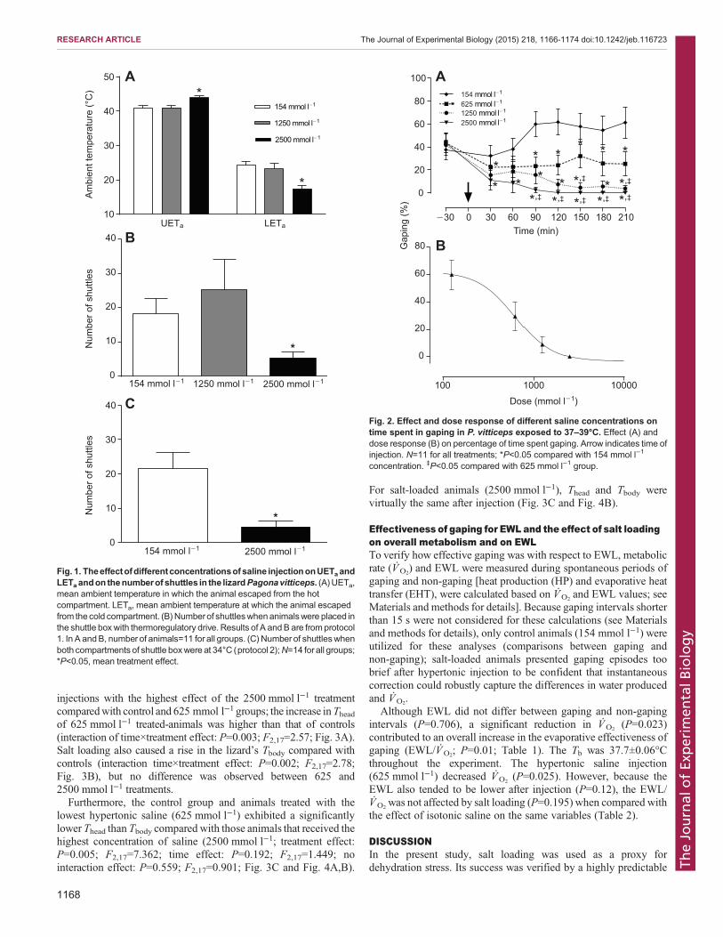
injections with the highest effect of the 2500 mmol l−1 treatmentcomparedwith control and 625 mmol l−1 groups; the increase inTheadof 625 mmol l−1 treated-animals was higher than that of controls(interaction of time×treatment effect: P=0.003; F2,17=2.57; Fig. 3A).Salt loading also caused a rise in the lizard’s Tbody compared withcontrols (interaction time×treatment effect: P=0.002; F2,17=2.78;Fig. 3B), but no difference was observed between 625 and2500 mmol l−1 treatments.Furthermore, the control group and animals treated with the
lowest hypertonic saline (625 mmol l−1) exhibited a significantlylower Thead than Tbody compared with those animals that received thehighest concentration of saline (2500 mmol l−1; treatment effect:P=0.005; F2,17=7.362; time effect: P=0.192; F2,17=1.449; nointeraction effect: P=0.559; F2,17=0.901; Fig. 3C and Fig. 4A,B).
For salt-loaded animals (2500 mmol l−1), Thead and Tbody werevirtually the same after injection (Fig. 3C and Fig. 4B).
Effectiveness of gaping for EWL and the effect of salt loadingon overall metabolism and on EWLTo verify how effective gaping was with respect to EWL, metabolicrate (VO2
) and EWL were measured during spontaneous periods ofgaping and non-gaping [heat production (HP) and evaporative heattransfer (EHT), were calculated based on VO2
and EWL values; seeMaterials and methods for details]. Because gaping intervals shorterthan 15 s were not considered for these calculations (see Materialsand methods for details), only control animals (154 mmol l−1) wereutilized for these analyses (comparisons between gaping andnon-gaping); salt-loaded animals presented gaping episodes toobrief after hypertonic injection to be confident that instantaneouscorrection could robustly capture the differences in water producedand VO2
.Although EWL did not differ between gaping and non-gaping
intervals (P=0.706), a significant reduction in VO2(P=0.023)
contributed to an overall increase in the evaporative effectiveness ofgaping (EWL/VO2
; P=0.01; Table 1). The Tb was 37.7±0.06°Cthroughout the experiment. The hypertonic saline injection(625 mmol l−1) decreased VO2
(P=0.025). However, because theEWL also tended to be lower after injection (P=0.12), the EWL/VO2
was not affected by salt loading (P=0.195) when compared withthe effect of isotonic saline on the same variables (Table 2).
DISCUSSIONIn the present study, salt loading was used as a proxy fordehydration stress. Its success was verified by a highly predictable
�30 0 30 60 90 120 150 180 210
0
20
40
60
80
100
625 mmol l�1
1250 mmol l�1
2500 mmol l�1
154 mmol l�1
**
*,‡
*
*
*
*,‡
*,‡
*
*
*
Time (min)
Gap
ing
(%)
**
**,‡ *,‡ *,‡
*,‡
100 1000 10000
0
20
40
60
80
Dose (mmol l�1)
A
B
Fig. 2. Effect and dose response of different saline concentrations ontime spent in gaping in P. vitticeps exposed to 37–39°C. Effect (A) anddose response (B) on percentage of time spent gaping. Arrow indicates time ofinjection. N=11 for all treatments; *P<0.05 compared with 154 mmol l−1
concentration. ‡P<0.05 compared with 625 mmol l−1 group.
UETa LETa10
20
30
40
50
1250 mmol l�1
154 mmol l�1
2500 mmol l�1
Am
bien
t tem
pera
ture
(°C
)N
umbe
r of s
huttl
esN
umbe
r of s
huttl
es*
*
0
10
20
30
40
154 mmol l�1
154 mmol l�1
1250 mmol l�1 2500 mmol l�1
2500 mmol l�1
*
*
0
10
20
30
40
A
B
C
Fig. 1. Theeffectofdifferentconcentrationsofsaline injectiononUETaandLETaandon thenumberof shuttles in the lizardPagonavitticeps. (A)UETa,mean ambient temperature in which the animal escaped from the hotcompartment. LETa, mean ambient temperature at which the animal escapedfrom the cold compartment. (B)Numberof shuttleswhenanimalswere placed inthe shuttle box with thermoregulatory drive. Results of A andB are from protocol1. In A andB, number of animals=11 for all groups. (C) Number of shuttleswhenboth compartments of shuttle boxwere at 34°C (protocol 2);N=14 for all groups;*P<0.05, mean treatment effect.
1168
RESEARCH ARTICLE The Journal of Experimental Biology (2015) 218, 1166-1174 doi:10.1242/jeb.116723
TheJournal
ofEx
perim
entalB
iology
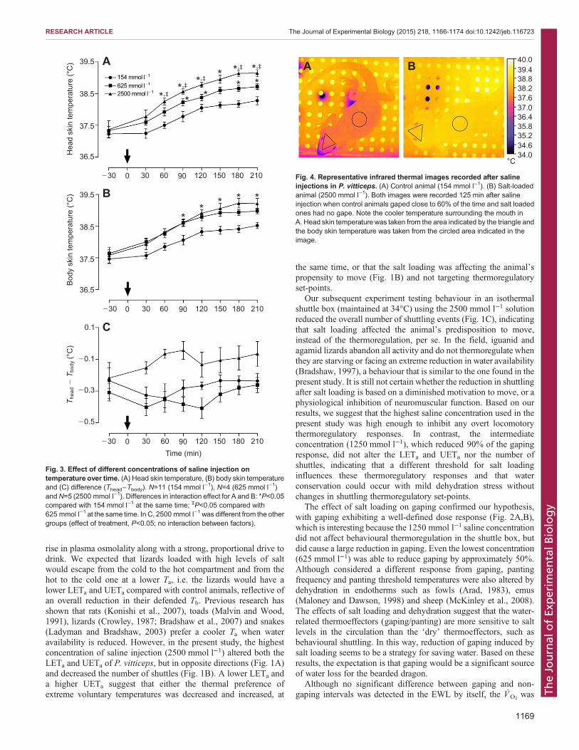
rise in plasma osmolality along with a strong, proportional drive todrink. We expected that lizards loaded with high levels of saltwould escape from the cold to the hot compartment and from thehot to the cold one at a lower Ta, i.e. the lizards would have alower LETa and UETa compared with control animals, reflective ofan overall reduction in their defended Tb. Previous research hasshown that rats (Konishi et al., 2007), toads (Malvin and Wood,1991), lizards (Crowley, 1987; Bradshaw et al., 2007) and snakes(Ladyman and Bradshaw, 2003) prefer a cooler Ta when wateravailability is reduced. However, in the present study, the highestconcentration of saline injection (2500 mmol l−1) altered both theLETa and UETa of P. vitticeps, but in opposite directions (Fig. 1A)and decreased the number of shuttles (Fig. 1B). A lower LETa anda higher UETa suggest that either the thermal preference ofextreme voluntary temperatures was decreased and increased, at
the same time, or that the salt loading was affecting the animal’spropensity to move (Fig. 1B) and not targeting thermoregulatoryset-points.
Our subsequent experiment testing behaviour in an isothermalshuttle box (maintained at 34°C) using the 2500 mmol l−1 solutionreduced the overall number of shuttling events (Fig. 1C), indicatingthat salt loading affected the animal’s predisposition to move,instead of the thermoregulation, per se. In the field, iguanid andagamid lizards abandon all activity and do not thermoregulate whenthey are starving or facing an extreme reduction in water availability(Bradshaw, 1997), a behaviour that is similar to the one found in thepresent study. It is still not certain whether the reduction in shuttlingafter salt loading is based on a diminished motivation to move, or aphysiological inhibition of neuromuscular function. Based on ourresults, we suggest that the highest saline concentration used in thepresent study was high enough to inhibit any overt locomotorythermoregulatory responses. In contrast, the intermediateconcentration (1250 mmol l−1), which reduced 90% of the gapingresponse, did not alter the LETa and UETa nor the number ofshuttles, indicating that a different threshold for salt loadinginfluences these thermoregulatory responses and that waterconservation could occur with mild dehydration stress withoutchanges in shuttling thermoregulatory set-points.
The effect of salt loading on gaping confirmed our hypothesis,with gaping exhibiting a well-defined dose response (Fig. 2A,B),which is interesting because the 1250 mmol l−1 saline concentrationdid not affect behavioural thermoregulation in the shuttle box, butdid cause a large reduction in gaping. Even the lowest concentration(625 mmol l−1) was able to reduce gaping by approximately 50%.Although considered a different response from gaping, pantingfrequency and panting threshold temperatures were also altered bydehydration in endotherms such as fowls (Arad, 1983), emus(Maloney and Dawson, 1998) and sheep (McKinley et al., 2008).The effects of salt loading and dehydration suggest that the water-related thermoeffectors (gaping/panting) are more sensitive to saltlevels in the circulation than the ‘dry’ thermoeffectors, such asbehavioural shuttling. In this way, reduction of gaping induced bysalt loading seems to be a strategy for saving water. Based on theseresults, the expectation is that gaping would be a significant sourceof water loss for the bearded dragon.
Although no significant difference between gaping and non-gaping intervals was detected in the EWL by itself, the VO2
was
A B 40.039.438.838.237.637.036.435.835.234.634.0
°C
Fig. 4. Representative infrared thermal images recorded after salineinjections in P. vitticeps. (A) Control animal (154 mmol l−1). (B) Salt-loadedanimal (2500 mmol l−1). Both images were recorded 125 min after salineinjection when control animals gaped close to 60% of the time and salt loadedones had no gape. Note the cooler temperature surrounding the mouth inA. Head skin temperaturewas taken from the area indicated by the triangle andthe body skin temperature was taken from the circled area indicated in theimage.
39.5
38.5
37.5
36.5
39.5
38.5
37.5
36.5
0.1
�0.1
�0.3
�0.5
154 mmol l�1
625 mmol l�1
2500 mmol l�1
Hea
d sk
in te
mpe
ratu
re (°
C)
Bod
y sk
in te
mpe
ratu
re (°
C)
T hea
d �
Tbo
dy (°
C)
A
B
C
�30 0 30 60 90 120 150 180 210
�30 0 30 60 90 120 150 180 210
�30 0 30 60 90 120 150 180 210Time (min)
*,‡*
** * * *
** **,‡ *,‡ * *,‡ *,‡
Fig. 3. Effect of different concentrations of saline injection ontemperature over time. (A) Head skin temperature, (B) body skin temperatureand (C) difference (Thead−Tbody). N=11 (154 mmol l−1), N=4 (625 mmol l−1)andN=5 (2500 mmol l−1). Differences in interaction effect for A and B: *P<0.05compared with 154 mmol l−1 at the same time; ‡P<0.05 compared with625 mmol l−1 at the same time. In C, 2500 mmol l−1 was different from the othergroups (effect of treatment, P<0.05; no interaction between factors).
1169
RESEARCH ARTICLE The Journal of Experimental Biology (2015) 218, 1166-1174 doi:10.1242/jeb.116723
TheJournal
ofEx
perim
entalB
iology
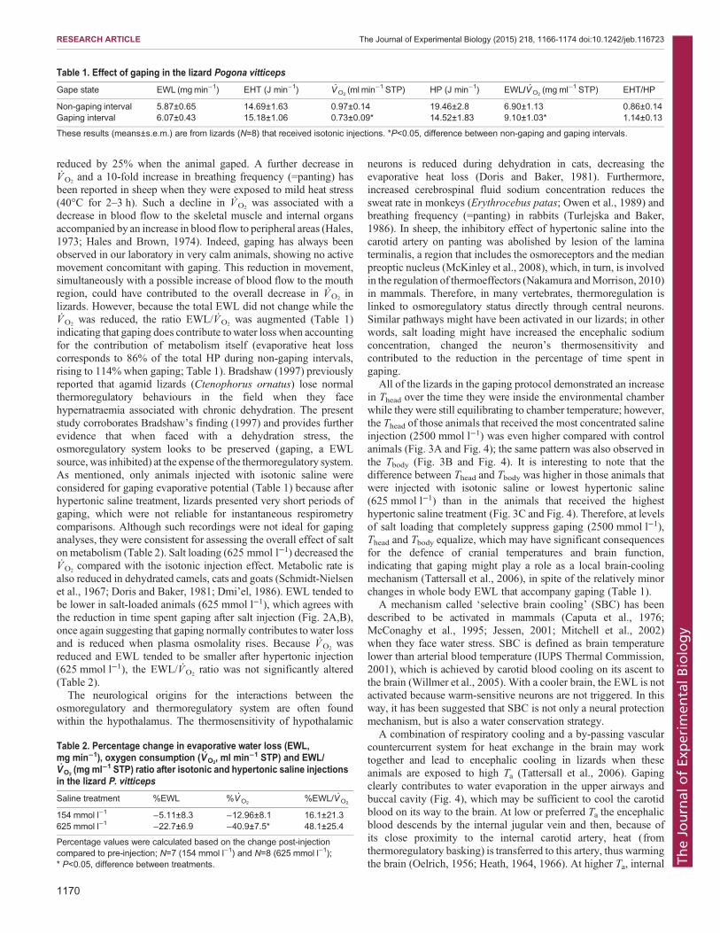
reduced by 25% when the animal gaped. A further decrease inVO2
and a 10-fold increase in breathing frequency (=panting) hasbeen reported in sheep when they were exposed to mild heat stress(40°C for 2–3 h). Such a decline in VO2
was associated with adecrease in blood flow to the skeletal muscle and internal organsaccompanied by an increase in blood flow to peripheral areas (Hales,1973; Hales and Brown, 1974). Indeed, gaping has always beenobserved in our laboratory in very calm animals, showing no activemovement concomitant with gaping. This reduction in movement,simultaneously with a possible increase of blood flow to the mouthregion, could have contributed to the overall decrease in VO2
inlizards. However, because the total EWL did not change while theVO2
was reduced, the ratio EWL/VO2was augmented (Table 1)
indicating that gaping does contribute towater loss when accountingfor the contribution of metabolism itself (evaporative heat losscorresponds to 86% of the total HP during non-gaping intervals,rising to 114% when gaping; Table 1). Bradshaw (1997) previouslyreported that agamid lizards (Ctenophorus ornatus) lose normalthermoregulatory behaviours in the field when they facehypernatraemia associated with chronic dehydration. The presentstudy corroborates Bradshaw’s finding (1997) and provides furtherevidence that when faced with a dehydration stress, theosmoregulatory system looks to be preserved (gaping, a EWLsource, was inhibited) at the expense of the thermoregulatory system.As mentioned, only animals injected with isotonic saline wereconsidered for gaping evaporative potential (Table 1) because afterhypertonic saline treatment, lizards presented very short periods ofgaping, which were not reliable for instantaneous respirometrycomparisons. Although such recordings were not ideal for gapinganalyses, they were consistent for assessing the overall effect of salton metabolism (Table 2). Salt loading (625 mmol l−1) decreased theVO2
compared with the isotonic injection effect. Metabolic rate isalso reduced in dehydrated camels, cats and goats (Schmidt-Nielsenet al., 1967; Doris and Baker, 1981; Dmi’el, 1986). EWL tended tobe lower in salt-loaded animals (625 mmol l−1), which agrees withthe reduction in time spent gaping after salt injection (Fig. 2A,B),once again suggesting that gaping normally contributes towater lossand is reduced when plasma osmolality rises. Because VO2
wasreduced and EWL tended to be smaller after hypertonic injection(625 mmol l−1), the EWL/VO2
ratio was not significantly altered(Table 2).The neurological origins for the interactions between the
osmoregulatory and thermoregulatory system are often foundwithin the hypothalamus. The thermosensitivity of hypothalamic
neurons is reduced during dehydration in cats, decreasing theevaporative heat loss (Doris and Baker, 1981). Furthermore,increased cerebrospinal fluid sodium concentration reduces thesweat rate in monkeys (Erythrocebus patas; Owen et al., 1989) andbreathing frequency (=panting) in rabbits (Turlejska and Baker,1986). In sheep, the inhibitory effect of hypertonic saline into thecarotid artery on panting was abolished by lesion of the laminaterminalis, a region that includes the osmoreceptors and the medianpreoptic nucleus (McKinley et al., 2008), which, in turn, is involvedin the regulation of thermoeffectors (Nakamura andMorrison, 2010)in mammals. Therefore, in many vertebrates, thermoregulation islinked to osmoregulatory status directly through central neurons.Similar pathways might have been activated in our lizards; in otherwords, salt loading might have increased the encephalic sodiumconcentration, changed the neuron’s thermosensitivity andcontributed to the reduction in the percentage of time spent ingaping.
All of the lizards in the gaping protocol demonstrated an increasein Thead over the time they were inside the environmental chamberwhile they were still equilibrating to chamber temperature; however,the Thead of those animals that received the most concentrated salineinjection (2500 mmol l−1) was even higher compared with controlanimals (Fig. 3A and Fig. 4); the same pattern was also observed inthe Tbody (Fig. 3B and Fig. 4). It is interesting to note that thedifference between Thead and Tbody was higher in those animals thatwere injected with isotonic saline or lowest hypertonic saline(625 mmol l−1) than in the animals that received the highesthypertonic saline treatment (Fig. 3C and Fig. 4). Therefore, at levelsof salt loading that completely suppress gaping (2500 mmol l−1),Thead and Tbody equalize, which may have significant consequencesfor the defence of cranial temperatures and brain function,indicating that gaping might play a role as a local brain-coolingmechanism (Tattersall et al., 2006), in spite of the relatively minorchanges in whole body EWL that accompany gaping (Table 1).
A mechanism called ‘selective brain cooling’ (SBC) has beendescribed to be activated in mammals (Caputa et al., 1976;McConaghy et al., 1995; Jessen, 2001; Mitchell et al., 2002)when they face water stress. SBC is defined as brain temperaturelower than arterial blood temperature (IUPS Thermal Commission,2001), which is achieved by carotid blood cooling on its ascent tothe brain (Willmer et al., 2005). With a cooler brain, the EWL is notactivated because warm-sensitive neurons are not triggered. In thisway, it has been suggested that SBC is not only a neural protectionmechanism, but is also a water conservation strategy.
A combination of respiratory cooling and a by-passing vascularcountercurrent system for heat exchange in the brain may worktogether and lead to encephalic cooling in lizards when theseanimals are exposed to high Ta (Tattersall et al., 2006). Gapingclearly contributes to water evaporation in the upper airways andbuccal cavity (Fig. 4), which may be sufficient to cool the carotidblood on its way to the brain. At low or preferred Ta the encephalicblood descends by the internal jugular vein and then, because ofits close proximity to the internal carotid artery, heat (fromthermoregulatory basking) is transferred to this artery, thus warmingthe brain (Oelrich, 1956; Heath, 1964, 1966). At higher Ta, internal
Table 1. Effect of gaping in the lizard Pogona vitticeps
Gape state EWL (mgmin−1) EHT (J min−1) VO2(ml min−1 STP) HP (J min−1) EWL/VO2
(mg ml−1 STP) EHT/HP
Non-gaping interval 5.87±0.65 14.69±1.63 0.97±0.14 19.46±2.8 6.90±1.13 0.86±0.14Gaping interval 6.07±0.43 15.18±1.06 0.73±0.09* 14.52±1.83 9.10±1.03* 1.14±0.13
These results (means±s.e.m.) are from lizards (N=8) that received isotonic injections. *P<0.05, difference between non-gaping and gaping intervals.
Table 2. Percentage change in evaporative water loss (EWL,mg min−1), oxygen consumption (VO2
, ml min−1 STP) and EWL/VO2
(mg ml−1 STP) ratio after isotonic and hypertonic saline injectionsin the lizard P. vitticeps
Saline treatment %EWL %VO2%EWL/VO2
154 mmol l−1 −5.11±8.3 −12.96±8.1 16.1±21.3625 mmol l−1 −22.7±6.9 −40.9±7.5* 48.1±25.4
Percentage values were calculated based on the change post-injectioncompared to pre-injection; N=7 (154 mmol l−1) and N=8 (625 mmol l−1);* P<0.05, difference between treatments.
1170
RESEARCH ARTICLE The Journal of Experimental Biology (2015) 218, 1166-1174 doi:10.1242/jeb.116723
TheJournal
ofEx
perim
entalB
iology
jugular vein is suggested to be constricted, thereby by-passing theblood through the external jugular, which is not very close to thecarotid artery. In this case, the heat is not transferred to the carotidblood, but instead, is carried away from the brain (Heath, 1964;Tattersall et al., 2006). It is possible that these two mechanismstogether (respiratory cooling and by-passing of the vascular heatexchanger) keep the brain cooler when bearded dragons gape at least50% of time (Fig. 3C), but the cooling was not preserved whengaping was completely inhibited (Fig. 3C). The same evaporative/non-evaporative mechanisms might contribute to head cooling in asmall lacertid lizard (Podarcis muralis) when they are heated(Sannolo et al., 2014), although this hypothesis remains to beexperimentally tested.There remain key differences between SBC in mammals and
reptiles. In mammals, SBC can be observed when water is notavailable. Euhydrated mammals (goats and sheep), even whenexposed to heat stress rarely present SBC, but dehydrated animalsshowmore frequent and a larger degree of SBC, supporting the ideathat such a mechanism has an osmoregulatory drive and might notplay a thermoregulatory function in these animals (Jessen et al.,1998; Fuller et al., 2007). In contrast, inPogona, cooling of the head(and by inference, the brain) happened when water was available(i.e. when animals exhibited gaping: 154 and 625 mmol l−1
injections; Fig. 4A and Fig. 3C) and, in an opposite direction, itwas diminished when water was not available, i.e. when gapingwas abolished (2500 mmol l−1 injection; Fig. 4B and Fig. 3C).Furthermore, the degree of gaping in bearded dragons is augmentedas Ta increases in euhydrated animals and the difference betweenThead and Tbody increases as well (Tattersall and Gerlach, 2005), butthis difference was observed to decrease when animals were saltloaded at the same Ta (Fig. 2A). Therefore, it seems that it is notosmoregulation that drives brain cooling in lizards, but ratherthermoregulation; consequently, SBC may operate as a protectiveencephalic mechanism. Despite having a thermoregulatory drive,when lizards are facedwith lowwater availability, bodywater is stillconserved at the expense of thermoregulation, similar to whathappens to mammals. Although a previous study has reported thatgaping is an effective cooling thermoeffector for Alligatormississipiensis (Spotila et al., 1977), the present study is the firstto show that gaping may work as a local (brain) cooling mechanismfor desert lizards, based on the fact that salt loading reduced gapingand increased Thead, eliminating the difference between Thead andTbody (Fig. 3).In conclusion, we found that an intermediate concentration of
hypertonic saline can affect EWL mechanisms, with no effect onnon-EWL mechanisms. Since gaping is more responsive to saltloading than shuttling, this may be an important response for savingwater over a natural dehydration period, while still allowing foroptimal thermoregulatory behaviours. However, when water isavailable, gaping may play a role as a local cooling mechanism thatis important to avoid encephalic superheating in reptiles such asPogona vitticeps that engage in thermoregulatory basking.
MATERIALS AND METHODSAnimalsA total of 19 animals (9 females and 10 males, Pogona vitticepsAhl 1926;body mass: 200–500 g) were randomly used in four different protocols.The lizards were housed in terraria with corn cob bedding, containing a40 W light bulb for thermoregulation and an additional UV light sourcefor vitamin D synthesis, and enriched with a small and opaque tube and acardboard material that provided shelter and extra climbing surfaces. Theanimals were kept on a 12 h:12 h light:dark cycle (lights on at 08:00 h)
and fed three times a week with a combination of chopped vegetables andfruits and twice a week with insects (cockroaches), but they were fastedfor at least 48 h before the experiments. The dragons also received alukewarm bath before and after all the experiments and an extra bathingand drinking opportunity once a week to ensure they were hydrated. Thenumber of animals that drank water within a 5 min period of time wascounted for comparison across all salt loading levels. The same animalswere used in almost all the protocols, but they had, at a minimum, a 14 dayinterval between experiments. All experiments were run between 08:30 hand 16:30 h and all procedures used in this study were approved by theBrock University Animal Care and Use Committee (Protocol #12-11-01).
InjectionsLizards received an isovolemic (1 ml/100 g) intra-peritoneal (i.p.) injectionof either normal isotonic saline (154 mmol l−1 NaCl) or hypertonic saline,in order to induce an estimated 5, 10 and 20% increase on plasmaosmolality. Three hypertonic concentrations were used (625, 1250 and2500 mmol l−1) based on previous studies (Konishi et al., 2003; Ford andBradshaw, 2006) and on mathematical assumptions (75% body water, 50%extracellular fluid and rapid mixing of salt within the plasma) assuming thatthe baseline osmolality for bearded dragons was 308 mosmol l−1 (Smits andKozubowski, 1984).
Plasma concentrationTo assess the plasma concentration, blood samples were taken from thelizard’s tail vein after 1 h of either normal (154 mmol l−1) or hypertonicsaline injections (625, 1250 and 2500 mmol l−1). Blood was collected inmicro-haematocrit glass tubes and centrifuged in a haematocrit centrifugefor 2 min to separate the plasma. The plasma was stored at 0°C and 10 µlaliquots (triplicate) were analysed in a Vapro vapor pressure osmometer(5520, Wescor, Inc, Logan, UT, USA) using a 291 mosmol litre−1 standard.
Shuttle box experimentsThe shuttle box (see Cadena and Tattersall, 2009a,b for details) was awooden chamber (119×61×45 cm) with two compartments separated by atransparent partition. There was a hole (11.5×14 cm) at the bottom of thepartition which connected both compartments and allowed the animal tomove from one side to the other one (shuttling behaviour). The walls wereoriented to naturally funnel the lizard toward the hole in the partition, whichfacilitated the shuttling movement by serving as a guide toward thetransition point. A transparent lid was placed on the top of the box to avoidany disturbance and to help to maintain the internal temperature. Cameraswere mounted over the lid for continuous monitoring, but no data werecollected from them.
In the first protocol performed in the shuttle box, one compartment wasalways 10°C warmer than the other, creating a so-called ‘hot compartment’(HC) and a ‘cold compartment’ (CC). The temperature inside the entire boxwas controlled by a treadle switch located on the floor, right below thetransition point between the compartments. By stepping on this treadle, thelizards regulated the Ta and, consequently, their own temperature. Lizardswere always placed in the HC in the beginning of an experiment to increasemotivation and to prevent long lethargy from exposition to cold, as theanimals had just emerged from the rest phase (dark phase) and the lightsinside their terraria had only just been turned on, so the temperature withintheir housing environments was relatively cold.
Once the animal was placed in the HC, the temperature in bothcompartments automatically rose at 0.7°C min−1 (Cadena and Tattersall,2009a), while maintaining a 10°C difference between them. Thetemperature rose until the animal moved to the CC, stepping on thetreadle and activating the cooling system. At that moment, bothcompartments cooled down at 0.7°C min−1 until the animal moved backto HC. The maximum temperature allowed in the HC was 43°C and theminimum temperature allowed in the CC was 10°C, as a safety precautionfor the animals and because these values fall well outside previously knownlimits for thermoregulation in this species (Cadena and Tattersall, 2009a).
Cooling and heating systems were controlled by an automated electronicsystem (Brock University, Electronics Shop, St Catharines, ON, CA).
1171
RESEARCH ARTICLE The Journal of Experimental Biology (2015) 218, 1166-1174 doi:10.1242/jeb.116723
TheJournal
ofEx
perim
entalB
iology

Ambient temperature was measured by a platinum resistor thermometer ineach compartment and recorded by the same electronic system every 30 sand whenever the animal moved from one side to the other one during theexperiment. The location of the lizard (HC or CC) was recorded at the sametimes as the Ta. The Ta inside the HC when the lizard moved from the HC tothe CC was called UETa and the Ta inside the CC when the lizard movedfrom the CC to the HC was named LETa. Throughout the course of anexperiment, a lizard exhibited numerous UETa and LETa values, based onhow often it shuttled. Ambient escape temperatures were used to describethermoregulatory behaviours based on previous work (Cadena andTattersall, 2009a) showing that these escape Ta measures accuratelyreflect changes in Tb in this species.
The second protocol performed in the shuttle box had no difference in thetemperature between the compartments. Both sides had the Ta fixed at 34°C(normal preferred Tb in the laboratory for bearded dragons; Cadena andTattersall, 2009a) throughout the experiment and the total number ofspontaneous shuttles produced with no thermoregulatory drivewas recorded(see detailed description below).
Protocol 1The purpose of this protocol was to examine whether thermoregulatorycontrol was altered in salt-loaded lizards. The first 3 h after the animal wasplaced in the shuttle box were used to allow habituation to the novelenvironment and were considered to be exploratory shuttling and thereforewere not used in the analyses (Cadena and Tattersall, 2009a). After thisinterval, animals received an i.p. injection of isotonic (154 mmol l−1) orhypertonic (1250 or 2500 mmol l−1) saline solutions and were placed againin the shuttle box (in the same compartment where they were before theinjection) for a further 4 h. This last interval was taken into account for theanalysis of UETa and LETa and the number of shuttles. For this experiment,11 animals were injected with all the three doses of saline.
Protocol 2The aim of this protocol was to examine whether salt loading preventedor impinged on the lizard’s natural propensity to move and behavespontaneously. In this protocol, the animal received the i.p. injection ofsaline solutions (154 or 2500 mmol l−1) first thing in the morning andwas immediately placed in the shuttle box. The reason for this was tocapitalize on the natural tendency for lizards to initially explore theshuttle box when they are first placed into it. Previous research hasshown that following this exploration interval, lizards will generallycease all shuttling entirely when there is no thermoregulatory drive(Cadena and Tattersall, 2009a). The experiment lasted 4 h and thenumber of shuttling events was analysed in this protocol. Fourteenlizards were injected with each saline concentration.
Gaping experimentsLizards were placed in an acrylic box (24×24×40 cm) which had three of itsfour sides covered with paper to prevent distraction and reduce reflection. Onthe top of the box, an infrared thermal imaging camera (Mikron 7515;Oakland, NJ, USA) and accompanying MikroSpec RT® (Mikron) softwarewere used to monitor the Thead and Tbody. Two representative infrared thermalimages used to calculate Thead and Tbody can be observed in Fig. 4A,B.The acrylic box was positioned in an environmental chamber (ThermoForma, Marietta, OH, USA) which was used to maintain a narrow range ofTa [Ta inside the acrylic box was about 37–39°C: a level sufficiently high toinduce gaping in bearded dragons (Tattersall and Gerlach, 2005)].
A webcam was attached to the internal wall of the environmentalchamber, facing the only side of the acrylic box that was not covered by thepaper. Images of the lizard were collected by the webcam every 10 s(HandyAvi; Tempe, AZ, USA) and used to assess the percentage timeengaged in gaping.
These experiments were conducted to assess whether salt loadinginfluenced the lizard’s gaping behaviour. The infrared thermal cameraallowed us to monitor the lizard’s Tbody and when it reached at least 37°C(usually 3–4 h after they were placed in the acrylic box inside theenvironmental chamber) the animals received an i.p. saline injection (154,625, 1250 or 2500 mmol l−1) and were placed back in the observation
chamber for a further 3.5 h. Because the environmental chamber was slowlywarmed, the i.p. injection happened before the chamber reached itscomplete thermal equilibrium. The percentage of the time spent in gapingwas analysed from the webcam images monitoring the lizard (percent timewas estimated over 30 min intervals from images captured every 10 s) priorto and following the injection. Eleven animals were injected in this protocolfor each dose of saline.
Respirometry experimentsVO2
and EWL values were obtained by flow-through respirometry. Theanimal was placed in a cylindrical respirometer (total volume: 2.8 l) inside atemperature controlled environmental chamber (Sable Systems, Las Vegas,NV, USA). The Ta inside the respirometer was about 37.5°C. The incurrent airwas dried through Nafion tubing using a counter-current water vapourextraction system (pure nitrogenwas used to extract water vapour) and pushedinto the respirometer at a rate of 1000 ml min−1. A subsample of this air waspushed through the H2O, CO2 and O2 analysers at 180 ml min−1 (FlowBar-8;Sable Systems, Las Vegas, NV, USA). The water vapour density (WVD;µgml−1) was the first parameter analysed (RH200; Sable Systems, Las Vegas,NV, USA) and this record was later used to calculate the EWL. Then, the CO2
percentage (CA-2A; Sable Systems, Las Vegas, NV, USA) and, finally, theO2 percentage were recorded (FC-1B; Sable Systems, Las Vegas, NV, USA).Following acquisition, the three channels of gases were time shifted toaccount for the delay in sample transfer from each analyser (8–12 s delaydetermined by a bolus injection of nitrogen into the respirometer). A baselinesample of dry incurrent air was taken for 5 min every 25 min to ensure that thewater, O2 and CO2 in the air offered to the animal remained constant over theexperiment, and to provide the estimates of FIO2
, FICO2and FIH2O. One gas
flow distributor (RM8 Intelligent Multiplexer; Sable Systems, Las Vegas,NV, USA) was used to control which gas sample (from respirometer orbaseline) entered the analysers. All data were collected by using a data-acquisition system (AcqKnowledge v.3.8.1, BIOPAC Systems, Goleta, CA,USA) collecting values every second. Prior to analysis, all time-aligned gaschannel data were Z-transformed to obtain instantaneous values (Lighton,2008). The temperature inside the environmental chamber wasmonitored by athermocouplemeter (TC-2000; Sable Systems, Las Vegas, NV, USA) and thelizard’s surface temperature was measured using a data logger (iButtonsDS1922L, Maxim Integrate, San Jose, CA, USA) attached to its ventralsurface with medical tape (Transpore, 3M).
The analysers were calibrated weekly or whenever was necessary (O2 wascalibrated daily prior to experimentation). The O2 analyser was calibratedusing dried air (20.95%); the CO2 was calibrated using pure nitrogen as azero value and a certified, pre-mixed gas source (1% CO2) as a span value,and lastly, the H2O analyser was also calibrated using pure nitrogen as a zerovalue and air bubbled through water of a known temperature as the spanvalue using WVD estimates (Dossat and Horan, 2001). TheVO2
(ml O2 min−1 STP) and EWL (mg H2O min−1) were calculated usingthe following equations (Lighton, 2008):
_VO2¼ FRI FIO2
� FEO2
1� FIO2� FICO2
� FIH2O
1� FEO2� FECO2
� FIH2O
!#;
"ð1Þ
EWL ¼ FRIðWVDE �WVDIÞ½ �1000
; ð2Þ
where FRI is incurrent flow rate, FIO2is incurrent fractional concentration of
oxygen (from baseline), FEO2is excurrent fractional concentration of
oxygen, FICO2is incurrent fractional concentration of carbon dioxide
(from baseline), FECO2is excurrent fractional concentration of carbon
dioxide, FIH2O is incurrent fractional concentration of water vapour (frombaseline), FEH2O is excurrent fractional concentration of water vapour,WVDE is excurrent water vapour density and WVDI is incurrent watervapour density.
Assuming that 1.0 ml of consumed oxygen produces 20 J of heat and foreach milligram of water evaporated, 2.5 J is dissipated, then heat production(HP; J min−1) and evaporative heat transfer (EHT; J min−1) were also
1172
RESEARCH ARTICLE The Journal of Experimental Biology (2015) 218, 1166-1174 doi:10.1242/jeb.116723
TheJournal
ofEx
perim
entalB
iology

calculated as follows (Randall et al., 1997):
HP ¼ _VO2� 20; ð3Þ
EHT ¼ EWL� 2:5: ð4ÞThe objective of these experiments was to examine the overall changes inoxygen consumption and whole body water loss during spontaneousperiods of gaping and non-gaping, before and after isotonic (154 mmol l−1)or hypertonic (625 mmol l−1) saline injection. In this protocol, the animalswere also placed in the acrylic box inside the environmental chamber untilthe Tbody reached 37°C (monitored with the infrared thermal camera),following which the animals were transferred to the glass respirometer andplaced inside an environmental chamber (also at 37°C) connected to therespirometry system. VO2
and EWL were recorded for 90 min, then animalsreceived a saline injection (154 or 625 mmol l−1) and oxygen consumptionand water loss were registered for another 90 min. The percentage of thetime spent gaping was analysed during the last 30 min before the transfer tothe respirometer and during all the time that the animals were inside therespirometer (i.e. 180 min; data not shown). The ventral surface temperaturewas recorded throughout the experiment using a data logger attached to theskin. Eight animals were used for each saline concentration.
Statistical analysisAll the results are presented as means±s.e.m. The predicted plasmaconcentration and the values found were analysed by linear regression andthe proportion of lizards that drank water within a 5 min period after saltloading was analysed using a chi-square test. Protocol 1 results were analysedusing a two-wayRMANOVA (factors: direction –UETa or LETa – and salinetreatment) was used to compare the effect of treatment on the UETa and LETaand one-wayRMANOVAwas run to compare the number of shuttles betweensaline treatments (factor: treatment). Protocol 2 results were analysed using apaired t-test to compare the number of shuttles between saline treatments.Two-way RMANOVAwas performed to analyse the effect of salt loading onpercentage of time spent in gaping, on Thead, on Tbody and on the difference inthese values (Thead−Tbody) (factors for all comparisons: time and salinetreatment) over time. A paired t-test was used to compare EWL and EWL/VO2
betweengapingandnon-gaping intervals and aWilcoxon signed-rank testwas run to analyse VO2
data. Only gaping periods longer than 15 s in durationwere utilized for the determination of VO2
and EWL values. A t-test wasperformed to analyse the effect of salt injection on %EWL and %VO2
(postinjection values compared to pre injection values) and aMann–Whitney rank-sum test was performed to compare % EWL/VO2
data.Whenever RMANOVA resulted in significant main or interaction effects,
a Holm–Šidák post hoc test was performed to verify where the differencesexisted. Residuals were tested for unequal variance and normality. In caseswhere log transformation was insufficient in terms of model assumptions,ranked data were analysed. Differences were considered significant whenP≤0.05. All analyses were performed using SigmaPlot 11.
AcknowledgementsThis study is part of the activities developed by C.S.S. during her PhD at the JointUFSCar-UNESP Graduate Program of Physiological Sciences, Sao Carlos, SP,Brazil. We thank Tomasz Eles and Ian Black for their excellent assistance withmaintenance of animals, and Dr Todd Gillis (University of Guelph) for assistancewith the plasma osmolality measurements.
Competing interestsThe authors declare no competing or financial interests.
Author contributionsG.J.T., K.C.B. and C.d.S.S. designed research, performed data analysis andprepared the manuscript prior to submission. C.d.S.S. and G.J.T. performedexperiments.
FundingC.d.S.S. was financially supported by Science Without Borders (Brazil) andEmerging Leaders in Americas Program (ELAP; Canada) fellowships. The researchwas supported by Natural Sciences and Engineering Council of Canada (NSERC)Discovery grant to G.J.T. [grant number RGPIN-2014-05814].
ReferencesArad, Z. (1983). Thermoregulation and acid-base status in the panting dehydrated
fowl. J. Appl. Physiol. Respir. Environ. Exerc. Physiol. 54, 234-243.Baker, M. A. (1989). Effects of dehydration and rehydration on thermoregulatory
sweating in goats. J. Physiol. 417, 421-435.Baker, M. A. and Dawson, D. D. (1985). Inhibition of thermal panting by intracarotid
infusion of hypertonic saline in dogs. Am. J. Physiol. 249, R787-R791.Baker, M. A. and Turlejska, E. (1989). Thermal panting in dehydrated dogs: effects
of plasma volume expansion and drinking. Pflugers Arch. 413, 511-515.Barber, B. J. and Crawford, E. C. (1979). Dual threshold control of peripheral
temperature in the lizard Dipsosaurus dorsalis. Physiol. Zool. 52, 250-263.Bentley, P. J. (1959). Studies on the water and electrolyte metabolism of the lizard
Trachysaurus rugosus (Gray). J. Physiol. 145, 37-47.Berk, M. L. and Heath, J. E. (1975). An analysis of behavioral thermoregulation in
the lizard, Dipsosaurus dorsalis. J. Therm. Biol. 1, 15-22.Bouaouda, H., Achaaban, M. R., Ouassat, M., Oukassou, M., Piro, M., Challet,
E., El Allali, K. and Pevet, P. (2014). Daily regulation of body temperature rhythmin the camel (Camelus dromedarius) exposed to experimental desert conditions.Physiol. Rep. 2, e12151.
Bradshaw, S. D. (1997). Homeostasis in Desert Reptiles. Berlin: Springer.Bradshaw, S. D. and Shoemaker, V. H. (1967). Aspects of water and electrolyte
changes in a field population of Amphibolurus lizards. Comp. Biochem. Physiol.20, 855-865.
Bradshaw, D., Ladyman, M. and Stewart, T. (2007). Effect of hypernatraemia andthe neurohypophysial peptide, arginine vasotocin (AVT) on behaviouralthermoregulation in the agamid lizard, Ctenophorus ornatus. Gen. Comp.Endocrinol. 150, 34-40.
Cadena, V. and Tattersall, G. J. (2009a). The effect of thermal quality on thethermoregulatory behavior of the bearded dragon Pogona vitticeps: influences ofmethodological assessment. Physiol. Biochem. Zool. 82, 203-217.
Cadena, V. and Tattersall, G. J. (2009b). Decreased precision contributes to thehypoxic thermoregulatory response in lizards. J. Exp. Biol. 212, 137-144.
Caputa, M., Kadziela, W. and Narebski, J. (1976). Significance of cranialcirculation for the brain homeothermia in rabbits. I. The brain-arterial bloodtemperature gradient. Acta Neurobiol. Exp. (Wars) 36, 613-623.
Crowley, S. R. (1987). The effect of desiccation upon the preferred bodytemperature and activity level of the lizard Sceloporus undulatus. Copeia 1987,25-32.
Dmi’el, R. (1986). Selective sweat secretion and panting modulation in dehydratedgoats. J. Therm. Biol. 11, 157-159.
Doris, P. A. and Baker, M. A. (1981). Hypothalamic control of thermoregulationduring dehydration. Brain Res. 206, 219-222.
Dossat, R. J. and Horan, T. J. (2001). Psychrometric properties of air. In Principlesof Refrigeration (ed. R. J. Dossat and T. J. Horan), pp. 57-70. New York, NY:Prentice Hall.
Dupre, R. K. and Crawford, E. C. (1986). Elevation of the panting threshold of thedesert iguana, Dipsosaurus dorsalis, during dehydration: potential roles ofchanges in plasma osmolality and body fluid volume. J. Comp. Physiol. B. 156,377-381.
Ford, S. S. and Bradshaw, S. D. (2006). Kidney function and the role of argininevasotocin in three agamid lizards from habitats of differing aridity in WesternAustralia. Gen. Comp. Endocrinol. 147, 62-69.
Fuller, A., Meyer, L. C. R., Mitchell, D. and Maloney, S. K. (2007). Dehydrationincreases the magnitude of selective brain cooling independently of coretemperature in sheep. Am. J. Physiol. Regul. Integr. Comp. Physiol. 293,R438-R446.
Hales, J. R. S. (1973). Effects of exposure to hot environments on the regionaldistribution of blood flowand on cardiorespiratory function in sheep.Pflugers Arch.344, 133-148.
Hales, J. R. S. and Brown, G. D. (1974). Net energetic and thermoregulatoryefficiency during panting in the sheep. Comp. Biochem. Physiol. A Physiol. 49,413-422.
Heath, J. E. (1964). Head–body temperature differences in horned lizards. Physiol.Zool. 37, 273-279.
Heath, J. E. (1966). Venous shunts in the cephalic sinuses of horned lizards.Physiol. Zool. 39, 30-35.
Heatwole, H., Firth, B. T. andWebb, G. J.W. (1973). Panting thresholds of lizards-I.Some methodological and internal influences on the panting threshold of anAgamid, Amphibolurus muricatus. Comp. Biochem. Physiol. A Physiol. 46,799-826.
IUPS Thermal Commission. (2001). Glossary of terms for thermal physiology.Jpn. J. Physiol. 51, 245-280.
Jessen, C. (2001). Selective brain cooling in mammals and birds. Jpn. J. Physiol.51, 291-301.
Jessen, C., Dmi’el, R., Choshniak, I., Ezra, D. and Kuhnen, G. (1998). Effects ofdehydration and rehydration on body temperatures in the black Bedouin goat.Pflugers Arch. 436, 659-666.
Kleinhaus, S., Pinshow, B., Bernstein, M. H. and Degen, A. A. (1985). Braintemperature in heat-stressed, water deprived desert phasianids: sand partridge
1173
RESEARCH ARTICLE The Journal of Experimental Biology (2015) 218, 1166-1174 doi:10.1242/jeb.116723
TheJournal
ofEx
perim
entalB
iology

(Ammoperdix heyi) and chukar (Alectoris chukar sinaica). Physiol. Zool. 58,105-116.
Konishi, M., Nagashima, K., Asano, K. and Kanosue, K. (2003). Attenuation ofmetabolic heat production and coldescape/warm-seeking behaviour during a coldexposure following systemic salt loading in rats. J. Physiol. 551, 713-720.
Konishi, M., Kanosue, K., Kano, M., Kobayashi, A. and Nagashima, K. (2007).The median preoptic nucleus is involved in the facilitation of heat-escape/cold-seeking behavior during systemic salt loading in rats.Am. J. Physiol. Regul. Integr.Comp. Physiol. 292, R150-R159.
Ladyman, M. and Bradshaw, D. (2003). The influence of dehydration on thethermal references of the Western tiger snake, Notechis scutatus. J. Comp.Physiol. B. 173, 239-246.
Lighton, J. R. B. (2008). Flow-through respirometry: the equations. In MeasuringMetabolic Rates. A Manual for Scientists (ed. J. R. B. Lighton), pp. 100-104.New York: Oxford University Press.
Maloney, S. K. and Dawson, T. J. (1998). Changes in pattern of heat loss at highambient temperature caused by water deprivation in a large flightless bird, theEmu. Physiol. Zool. 71, 712-719.
Malvin, G. M. and Wood, S. C. (1991). Behavioral thermoregulation of the toad,Bufo marinus: effects of air humidity. J. Exp. Zool. 258, 322-326.
McConaghy, F. F., Hales, J. R. S., Rose, R. J. and Hodgson, D. R. (1995).Selective brain cooling in the horse during exercise and environmental heatstress. J. Appl. Physiol. 79, 1849-1854.
McKinley, M. J., McAllen, R. M., Whyte, D. and Mathai, M. L. (2008). Centralosmoregulatory influences on thermoregulation. Clin. Exp. Pharmacol. Physiol.35, 701-705.
Melville, J. and Schulte, J. A. (2001). Correlates of active body temperatures andmicrohabitat occupation in nine species of central Australian agamid lizards. Aust.Ecol. 26, 660-669.
Mitchell, D., Maloney, S. K., Jessen, C., Laburn, H. P., Kamerman, P. R.,Mitchell, G. and Fuller, A. (2002). Adaptive heterothermy and selective braincooling in arid-zone mammals. Comp. Biochem. Physiol. B. Biochem. Mol. Biol.131, 571-585.
Nagashima, K., Nakai, S., Konishi, M., Su, L. and Kanosue, K. (2001). Increasedheat-escape/cold-seeking behaviour following hypertonic saline injection in rats.Am. J. Physiol. Regul. Integr. Comp. Physiol. 280, R1031-R1036.
Nakamura, K. and Morrison, S. F. (2010). A thermosensory pathway mediatingheat-defense Responses. Proc. Natl. Acad. Sci. USA 107, 8848-8853.
O’Connor, M. P. and Tracy, C. R. (1992). Thermoregulation by juvenile toads ofBufo woodhousei in the field and in the laboratory. Copeia 1992, 865-876.
Oelrich, T. M. (1956). The anatomy of the head of Ctenosaura pectinata(Iguanidae). Mus. Zool. Univ. Mich. Misc. Publ. 94, 1-122.
Owen, M. D., Matthes, R. D. and Gisolfi, C. V. (1989). Effect of cerebrospinal fluidhyperosmolality on sweating in the heatstressed patas monkey. J. Appl. Physiol.67, 128-133.
Parmenter, C. J. and Heatwole, H. (1975). Panting thresholds of lizards. IV. Theeffect of dehydration on the panting threshold of Amphibolurus barbatus andAmphibolurus muricatus. J. Exp. Zool. 191, 327-332.
Preest, M. R. and Pough, F. H. (1989). Interaction of temperature and hydration onlocomotion of toads. Funct. Ecol. 3, 693-699.
Randall, D., Burggren, W. and French, K. (1997). Using energy: meetingenvironmental challenges. In Eckert Animal Physiology: Mechanisms andAdaptations (ed. D. Randall, W. Burggren and K. French), pp. 665-723.New York, NY: W. H. Freeman Company.
Rice, G. E. (1982). Plasma arginine vasotocin concentrations in the lizard Varanusgouldii (Gray) following water loading, salt loading, and dehydration. Gen. Comp.Endocrinol. 47, 1-6.
Sannolo, M., Mangiacotti, M., Sacchi, R. and Scali, S. (2014). Keeping a coolmind: head–body temperature differences in the common wall lizard. J. Zool. 293,71-79.
Schmidt-Nielsen, K., Schmidt-Nielsen, B., Jarnum, S. A. and Houpt, T. R.(1957). Body temperature of the camel and its relation to water economy.Am. J. Physiol. 188, 103-112.
Schmidt-Nielsen, K., Crawford, E. C., Jr, Newsome, A. E., Rawson, K. S. andHammel, H. T. (1967). Metabolic rate of camels: effect of body temperature anddehydration. Am. J. Physiol. 212, 341-346.
Silva, N. L. and Boulant, J. A. (1984). Effects of osmotic pressure, glucose, andtemperature on neurons in preoptic tissue slices. Am. J. Physiol. 247, R335-R345.
Smits, A. W. and Kozubowskij, M. M. (1985). Partitioning of body fluids andcardiovascular responses to circulatory hypovolaemia in the turtle, Pseudemysscripta elegans. J. Exp. Biol. 116, 237-250.
Spotila, J. R., Terpin, K. M. and Dodson, P. (1977). Mouth gaping as an effectivethermoregulatory device in alligators. Nature 265, 235-236.
Tattersall, G. J. and Gerlach, R. M. (2005). Hypoxia progressively lowers thermalgaping thresholds in bearded dragons, Pogona vitticeps. J. Exp. Biol. 208,3321-3330.
Tattersall, G. J., Cadena, V. and Skinner, M. C. (2006). Respiratory cooling andthermoregulatory coupling in reptiles. Respir. Physiol. Neurobiol. 154, 302-318.
Taylor, C. R. (1970). Dehydration and heat. Effects on temperature regulation ofEast African ungulates. Am. J. Physiol. 219, 1136-1139.
Turlejska, E. and Baker, M. A. (1986). Elevated CSF osmolality inhibitsthermoregulatory heat loss responses. Am. J. Physiol. 251, R749-R754.
Willmer, P., Stone, G. and Johnston, I. (2005). Temperature and its Effects. InEnvironmental Physiology of Animals (ed. P. Willmer, G. Stone and I. Johnston),pp. 209-210. Malden: Blackwell Publishing.
1174
RESEARCH ARTICLE The Journal of Experimental Biology (2015) 218, 1166-1174 doi:10.1242/jeb.116723
TheJournal
ofEx
perim
entalB
iology