qual É o melhor protocolo do teste sit-to- … e por ter enviado anjos (conhecido como amigos) no...
TRANSCRIPT

ANDREA AKEMI MORITA
QUAL É O MELHOR PROTOCOLO DO TESTE SIT-TO-STAND EM PACIENTES COM DOENÇA PULMONAR
OBSTRUTIVA CRÔNICA?
Londrina
2016

ANDREA AKEMI MORITA
QUAL É O MELHOR PROTOCOLO DO TESTE SIT-TO-STAND EM PACIENTES COM DOENÇA PULMONAR
OBSTRUTIVA CRÔNICA?
Dissertação apresentada ao Programa de Pós-Graduação em Ciências da Reabilitação (Programa Associado entre Universidade Estadual de Londrina [UEL] e Universidade Norte do Paraná [UNOPAR]), como requisito parcial à obtenção do título de Mestre em Ciências da Reabilitação.
Orientadora: Prof. Dra. Vanessa S. Probst
Londrina 2016

ANDREA AKEMI MORITA
QUAL É O MELHOR PROTOCOLO DO TESTE SIT-TO-STAND EM PACIENTES COM DOENÇA PULMONAR
OBSTRUTIVA CRÔNICA?
Dissertação apresentada ao Programa de Pós-Graduação em Ciências da Reabilitação (Programa Associado entre Universidade Estadual de Londrina [UEL] e Universidade Norte do Paraná [UNOPAR]), como requisito parcial à obtenção do título de Mestre em Ciências da Reabilitação.
BANCA EXAMINADORA
____________________________________
Profª Drª Vanessa S. Probst Universidade Estadual de Londrina
____________________________________ Prof. Dr. Fabio Pitta
Universidade Estadual de Londrina
____________________________________ Profª Drª Simone Dal Corso Universidade Nove de Julho
Londrina, 09 de março de 2016.

DEDICATÓRIA
Dedico este trabalho as pessoas que dão
sentido à minha vida e que acreditaram sempre
em mim: miha mãe Elza, meu pai Mario e
minha irmã Luciene.

AGRADECIMENTOS
Agradeço primeiramente a Deus, por ter me proporcionado a vida,
por permitir que eu chegasse até aqui e realizasse mais esse sonho. Por estar
sempre comigo, nos momentos felizes e difíceis, por ter me dado uma família
maravilhosa e por ter enviado anjos (conhecido como amigos) no meu caminho.
A minha imensurável gratidão a minha família, o meu alicerce e o
meu bem mais valioso. Obrigada a minha mãe, meu pai e minha irmã por todo o
amor, dedicação e auxílio. Aos meus pais, agradeço pelos valores que me
ensinaram. E se hoje cheguei aqui, foi graças a vocês. Vocês sempre me ensinaram
que a educação, a dedicação e o respeito eram os caminhos para um dia ser
alguém. E fazendo disso um ideal, nunca mediram esforços para proporcionar a mim
e a minha irmã tudo o que vocês não tiveram. Muito obrigada mesmo, amo vocês
para sempre. Obrigada à minha irmãzinha querida que amo infinitamente. Obrigada
por sempre estar perto e me ajudar em todos os momentos. Agradeço aos demais
membros da família, tios, primos e agregados queridos, que mesmo estando longe
sempre estiveram na torcida para que eu continuasse crescendo.
O meu muitíssimo obrigado à minha orientadora, professora e amiga
Vanessa Probst. Você realmente foi um presente de Deus nessa caminhada
chamada “mestrado”. Obrigada por ter aceitado me orientar durante esses dois
anos, período esse que eu a conheci melhor, e a admiração que eu tinha por você
só cresceu. Além de ser uma profissional competente e dedicada, é também uma
mulher de características admiráveis. Tenho me inspirado muito em você para ser
uma pessoa e uma profissional melhor. Obrigada pela amizade, pela paciência, por
sempre estar disposta a sanar as minhas dúvidas, por ser tão humana na solução
dos meus problemas, por ter acreditado e confiado em mim, muito mais do que eu
acreditava e merecia. Muito obrigada!
O meu muito obrigada ao professor e amigo Fabio Pitta. Obrigada
por ter me acolhido no laboratório há algum tempo atrás e por ter confiado em mim.
Você é uma pessoa por quem tenho um grande respeito e admiração, e é um
grande exemplo de profissional e de pessoa que quero seguir.
Agradeço à professora Dra. Simone Dal Corso, por ter aceitado
participar da minha banca prontamente e pelas valiosas contribuições para este
trabalho. É um prazer e uma honra tê-la como parte da banca examinadora.
Agradeço a todos os colegas do LFIP, pela convivência e por toda a
ajuda. Em especial, quero agradecer algumas pessoas que foram entrando devagar
na minha vida, as quais hoje não consigo viver sem. Vocês, Fernanda Morakami,
Antenor Rodrigues, Igor Brito, Thaís Paes, Mahara Proença e mais recentemente
Larissa Castro e Débora de Carvalho, foram e são essenciais no meu processo de
aprendizado e de crescimento pessoal. Com certeza, o período do mestrado foi um
dos mais agradáveis da minha vida por causa de vocês.

Quero agradecer também aos meus queridos: Juliana Zabatiero,
Leandro Mantoani, Mahara Proença e Karina Furlanetto por terem me acolhido no
EPAFT e por terem me ajudado muito em um início não muito fácil. Obrigada por
toda a paciência e serenidade para que eu pudesse aprender um pouco mais sobre
pesquisa.
Agradeço também a minha dupla infalível, Gianna Bisca. Tudo não
teria dado tão certo, se não fosse você. Obrigada por toda a paciência, por aceitar o
meu jeito de ser e as minhas manias, por me ensinar muito sobre pesquisa e a vida,
e obrigada sobretudo pela amizade. É uma amiga admirável que vou levar para a
vida toda.
Obrigada também aos alunos da linha do exercício do LFIP.
Agradeço por serem tão prestativos, compreensivos e por terem me ajudado nesse
trabalho. Tenho um carinho muito grande por todos vocês e torço muito pelo
sucesso de cada um.
Quero agradecer a professora Carrie Galvan, uma amiga e
profissional que eu admiro e adoro. Obrigada por estar sempre perto de mim, por me
ajudar com ensinamentos e conselhos tão valiosos e por toda a confiança
depositada.
Agradeço a professora Laryssa Bellinetti, por ter proporcionado a
primeira oportunidade do contato com a pesquisa durante a minha graduação. Com
certeza, esse foi um pontapé inicial para que eu continuasse seguindo em frente e
chegasse aqui.
À todos os meus amigos, muito obrigada por estarem perto de mim e
por me entenderem mais do que eu mesma. Estar com vocês é sempre uma alegria
imensa e que me faz muito bem.
Agradeço também aos pacientes que participaram do estudo e que
colaboraram com essa pesquisa.
Por fim, agradeço a tudo e a todos que acreditaram em mim e no
meu trabalho!

Epígrafe
“ Escolha um trabalho que você ame e não terá de trabalhar um único dia de sua vida.”
Confúcio

MORITA, Andrea Akemi. Qual é o melhor protocolo do teste sit-to-stand em pacientes com doença pulmonar obstrutiva crônica? 2016. 75 páginas. Dissertação de Mestrado do Programa de Pós-Graduação em Ciências da Reabilitação (Programa Associado entre Universidade Estadual de Londrina [UEL] e Universidade Norte do Paraná [UNOPAR])– Universidade Estadual de Londrina, Londrina, 2016.
RESUMO
INTRODUÇÃO: Existem diferentes protocolos do teste sit-to-stand (STS) para avaliação da capacidade funcional em pacientes com doença pulmonar obstrutiva crônica (DPOC). Entretanto, não existem evidências sobre qual é o melhor protocolo a ser utilizado nessa população. OBJETIVOS: Correlacionar cada protocolo do STS (5-repetições [5-rep], 30-segundos [30-seg] e 1-minuto [1-min]) com desfechos clínicos importantes avaliados em pacientes com DPOC; comparar os três protocolos do STS assim como verificar a associação e a concordância entre eles; verificar se os três protocolos são capazes de predizer a capacidade funcional de exercício e atividade física de vida diária (AFVD). MÉTODOS: Vinte e três pacientes com DPOC (11 homens; VEF1: 53±15%pred) realizaram três protocolos do teste STS de forma aleatorizada. Considerando que os desfechos dos protocolos foram obtidos em unidades de medida diferentes, a velocidade (número de repetições por segundo [rep/seg]) foi utilizada para a análise dos três testes. Além disso, os pacientes foram submetidos às seguintes avaliações: Incremental Shuttle Walking Test (ISWT), Teste de Caminhada de 6 Minutos (TC6min), 4-metre gait speed test (4MGS), 1-repetição máxima (1RM) de quadríceps femoral, avaliação da atividade física de vida diária (AFVD) e questionários de qualidade de vida e estado funcional. RESULTADOS: O protocolo de 1-min correlacionou-se significativamente com o TC6min (r=0,40), 4MGS (r=0,64) e AFVD (r≥0,47). Para os testes de 5-rep e 30-seg, as melhores correlações obtidas foram com o 4MGS (r=0,54 e r=0,52, respectivamente). Foi encontrada diferença na velocidade de execução dos três protocolos (5-rep: 0,53±0,16 rep/seg; 30-seg: 0,48±0,13rep/seg; 1-min: 0,45±0,11rep/seg; P= 0,01), ou seja, entre o STS de 5-rep e 1-min. Apesar dessa diferença, eles apresentaram boa concordância (CCI≥0,73 para todos) e houve moderada associação entre eles (r≥0,68). Foram verificadas maiores mudanças na saturação periférica de oxigênio, frequência cardíaca, pressão arterial e sintomas de dispneia e fadiga após o protocolo de 1-min (P≤0,01 para todos). Além disso, os três protocolos foram capazes de discriminar pacientes com capacidade de exercício baixa e preservada (Área sob a curva [AUC]≥0,71). No entanto, isso não ocorreu com a AFVD (AUC≤0,67). CONCLUSÃO: O teste de 1-min apresenta maior demanda hemodinâmica, e é o que melhor correlaciona-se com importantes desfechos clínicos avaliados em pacientes com DPOC. Apesar de ter sido verificada diferença na velocidade de execução e na resposta fisiológica entre o STS de 5-rep e 1-min, há boa concordância e correlação entre os três diferentes protocolos do STS. Adicionalmente, todos os protocolos possuíram poder discriminativo para pacientes com capacidade de exercício baixa e preservada. Palavras-chave: Doença pulmonar obstrutiva crônica; exercício; atividade motora; atividades cotidianas, hemodinâmica; avaliação de desempenho.

MORITA, Andrea Akemi. Which is the best protocol of the sit-to-stand test in patients with chronic obstructive pulmonary disease? 2016. 75 páginas. Dissertação de Mestrado do Programa de Pós-Graduação em Ciências da Reabilitação (Programa Associado entre Universidade Estadual de Londrina [UEL] e Universidade Norte do Paraná [UNOPAR])– Universidade Estadual de Londrina, Londrina, 2016.
ABSTRACT
BACKGROUND: Different protocols of the sit-to-stand test (STS) are available to assess functional capacity in patients with chronic obstructive pulmonary disease (COPD). However, it is unclear which is the best protocol to assess this population. AIMS: To correlate each protocol of the STS (5-repetition [5-rep], 30-second [30-sec] and 1-minute [1-min]) with important clinical outcomes in patients with COPD; to compare three STS, as well as to verify their association and agreement; and to verify whether the three protocols are able to predict functional exercise capacity and physical activity in daily life (PADL). METHODS: Twenty-three patients with COPD (11 men; FEV1:53±15%pred) performed three protocols of the STS in a randomized sequence. Since the outcomes units of measure were different, the speed (number of repetitions per second [rep/sec]) was used for the analysis of all three tests. Patients also underwent the following assessments: Incremental Shuttle Walking Test (ISWT), 6-minute walk test (6MWT), 4-metre gait speed test (4MGS), 1-repetition maximum (1RM) of quadriceps muscle, PADL and questionnaires of health-related quality of life and functional status. RESULTS: The 1-min presented significant correlations with the 6MWT (r=0.40), 4MGS (r=0.64) and PADL (r≥0.47). Regarding the 5-rep and 30-sec test, the best correlation obtained were with the 4MGS (r=0.54 and r=0.52, respectively). The speed differed among protocols (5-rep:0.53±0.16 rep/sec; 30-sec: 0.48±0.13rep/sec; 1-min: 0.45±0.11rep/sec; P= 0.01), i.e. between the 5-rep and 1-min STS. However, they presented moderate agreement (ICC≥0.73 for all) and correlated well with each other (r≥0.68). More marked changes in peripheral oxygen saturation, heart rate, blood pressure, dyspnea and leg fatigue were observed after the 1-min protocol (P≤0.01 for all). Furthermore, the three protocols were able to discriminate patients with low and preserved exercise capacity (area under the curve [AUC]≥0.71) but did not predict PADL (AUC≤0.67). CONCLUSION: The 1-min STS generates higher hemodynamic demands and correlates better with clinical outcomes in patients with COPD. Although there was difference between the 5-rep and 1-min protocol in terms of speed performance and physiologic demand, there was good level of agreement among the STS. In addition, all three protocols discriminated patients with low and preserved exercise capacity. Key words: pulmonary disease, chronic obstructive; exercise test; motor activity; activities of daily living; hemodynamics; patient outcome assessment.

LISTA DE ILUSTRAÇÕES
Figura 1 (Contextualização) – Valores de referência para o teste STS de 1 minuto
(Strassmann et al., 2012) ......................................................................................... 18
Figura 1 (Artigo) –Comparação da velocidade entre os protocolos do teste sit-to-
stand de 5-repetições, 30-segundos e 1-minuto ...................................................... 46
Figura 2 (Artigo) – Gráfico de Bland & Altman com as diferenças entre dois
protocolos do sit-to-stand vs a média da velocidade de dois testes (5-rep e 30-seg
[A]; 5-rep e 1-min [B]; 30-seg e 1-min [C]) ................................................................ 47
Figura 3 (Artigo) – Comparação entre as áreas sob a curva (AUC) dos protocolos
do teste sit-to-stand de 5-repetições, 30-segundos, 1-minuto em pacientes com
capacidade de exercício no TC6min baixa e preservada (A) e classificados como
ativos e inativos pelo PAD (B). ................................................................................. 48

LISTA DE TABELAS
Tabela 1 (Artigo) – Características dos pacientes com DPOC ............................... 49
Tabela 2 (Artigo) – Correlações entre os protocolos do teste sit-to-stand de 5-rep,
30-seg e 1-min e variáveis clínicas avaliadas em pacientes com DPOC ................. 50
Tabela 3 (Artigo) – Comparação das variáveis de saturação de oxigênio,
cardiovasculares e sintomas mensuradas antes de cada protocolo do STS ........... 51
Tabela 4 (Artigo) – Comparação das mudanças na saturação periférica de oxigênio,
variáveis cardiovasculares e sintomatológicas de cada protocolo do teste sit-to-stand
................................................................................................................................. 52
Tabela 5 (Artigo) – Comparação da saturação periférica de oxigênio, variáveis
hemodinâmicas e sintomatológicas mensuradas antes e após cada protocolo do
teste sit-to-stand ....................................................................................................... 53

LISTA DE ABREVIATURAS E SIGLAS
%pred percentage of predicted
1-min Teste sit-to-stand de 1 minuto, sit-to-stand test of 1 minute
1-RM 1-repetition maximum, 1-repetição máxima
30-seg Teste sit-to-stand de 30 segundos
30-sec Sit-to-stand test of 30 seconds
4MGS Four-metre gait speed
5-rep Teste sit-to-stand de 5 repetições, sit-to-stand test of 5 repetitions
6MWT Six-minute walk test
ACMS American College of Sports and Medicine
AFVD Atividade Física de Vida Diária
ANOVA Analysis of variance
AUC Area under the curve
BODE Body mass index, airflow Obstruction, Dyspnea and Exercise capacity
BP Blood pressure
CAPES Coordenação de Aperfeiçoamento de Pessoal de Nível Superior
CCQ Clinical COPD Questionnaire
CNPq Conselho Nacional de Desenvolvimento Científico e Tecnológico
COPD Chronic Obstructive Pulmonary Disease
DAM Dynaport Activity Monitor
DPOC Doença Pulmonar Obstrutiva Crônica
FEV1 Forced expiratory volume in the first second
GOLD Global Initiative for Chronic Lung Disease
HR Heart rate
ICC Intraclass Correlation Coefficient
IQR Interquartile range
ISWT Incremental Shuttle Walking Test
LFIP Laboratório de Pesquisa em Fisioterapia Pulmonar
MET Metabolic equivalent
MID Minimal Important Difference
MRC Medical Research Council

OMS Organização Mundial da Saúde
PAD Physical activity duration
PADL Physical Activity in Daily Life
PFSDQ-M Pulmonary Functional Status and Dyspnea Questionnaire Modified version
rep/sec number of repetitions per second
ROC Receiver Operating Characteristics
SD Standard deviation
SGRQ St. George’s Respiratory Questionnaire
SpO2 Oxygen saturation
SPSS Statistical Package for the Social Sciences
STS Teste sit-to-stand, sit-to-stand test
SWA SenseWear Armband
TC6min Teste de Caminhada de Seis Minutos
UEL Universidade Estadual de Londrina
UNOPAR Universidade Norte do Paraná
VEF1 Volume expiratório forçado no primeiro segundo

SUMÁRIO
1 INTRODUÇÃO ....................................................................................................... 12
2 REVISÃO DE LITERATURA – CONTEXTUALIZAÇÃO ........................................ 14
2.1 DOENÇA PULMONAR OBSTRUTIVA CRÔNICA ......................................................... 14
2.2 DOENÇA PULMONAR OBSTRUTIVA CRÔNICA E CAPACIDADE FUNCIONAL .................. 15
2.3 TESTE SIT-TO-STAND EM INDIVÍDUOS SAUDÁVEIS ................................................... 16
2.4 TESTE SIT-TO-STAND E DOENÇA PULMONAR OBSTRUTIVA CRÔNICA ....................... 19
3 ARTIGO ORIGINAL ............................................................................................... 21
4 CONCLUSÃO GERAL ........................................................................................... 54
5 REFERÊNCIAS ...................................................................................................... 55
6 APÊNDICE ............................................................................................................. 58
APÊNDICE A - Ficha de avaliação do teste sit-to-stand ........................................ 59
APÊNDICE B - Ficha de avaliação da espirometria .............................................. 60
APÊNDICE C- Ficha do Teste de Caminhada de Seis Minutos ............................ 61
APÊNDICE D - Ficha do Incremental Shuttle Walking Test................................... 62
APÊNDICE E - Ficha do 4-metre gait speed test ................................................... 63
APÊNDICE F - Ficha de avaliação de 1 repetição máxima (1RM) ........................ 64
7 ANEXOS ................................................................................................................ 65
ANEXO A - Termo de Consentimento Livre e Esclarecido do estudo ................... 66
ANEXO B - Parecer do Comitê de Ética ............................................................... 69
ANEXO C - Normas de formatação do artigo no periódico Respiratory Care ........ 70

12
1 INTRODUÇÃO
A Doença Pulmonar Obstrutiva Crônica (DPOC) é considerada uma
doença sistêmica que não pode ser analisada somente pelo seu comprometimento
pulmonar. Os efeitos extrapulmonares são diversos, e características como a
disfunção muscular, alterações cardiovasculares e metabólicas[1] e limitação da
capacidade funcional são potenciais fatores de risco para o prejuízo do estado de
saúde a longo prazo[2]. Dentre esses fatores, a limitação da capacidade funcional é
uma característica que merece notoriedade nesse cenário. Essa condição implica
limitação de atividades físicas básicas diárias[3], tais como vestir-se, banhar-se e
realizar atividades domésticas. Por esse motivo, a sua avaliação faz-se necessária e
é de extrema importância[4].
Uma das formas mais comuns e conhecidas de avaliar a capacidade
funcional desses indivíduos, é por meio do Teste de Caminhada de Seis Minutos
(TC6min)[5, 6]. Esse é um teste válido, reprodutível e bem descrito na literatura em
pacientes com DPOC[5]. Embora seja um teste simples, a realização do TC6min
requer uma equipe treinada, necessita de espaço (um corredor de pelo menos 30
metros), e demanda tempo (intervalo de no mínimo trinta minutos entre dois testes)[5].
Portanto, testes ainda mais simples, como o teste sit-to-stand (STS), têm surgido na
literatura como uma alternativa para obter informações sobre a capacidade funcional
nessa população. O teste STS e o TC6min possuem uma excelente correlação[7] e
apresentam respostas fisiológicas similares, corroborando assim o STS como uma
opção[8].
O teste sit-to-stand é um teste prático, amplamente utilizado e
primeiramente descrito[9] para avaliar a funcionalidade em idosos saudáveis[10-12].
Os movimentos de sentar e levantar são importantes funções da vida diária, e a
incapacidade de realizá-los está associada à mortalidade, assim como prejuízos na
mobilidade e funcionalidade[13]. Estudos do STS em diversas populações como em
condições neurológicas[14] e ortopédicas[15, 16] têm sido realizados; no entanto,
apenas recentemente estudos tem explorado o tema em profundidade nas doenças
pulmonares crônicas[17-19]. O teste é considerado válido, reprodutível,
responsivo[17, 20] e é um preditor de mortalidade em indivíduos com DPOC[19].
Uma variedade de estudos descrevem a associação do STS e
desfechos clínicos importantes comumente avaliados na DPOC, tais como:
13
capacidade de exercício, dispneia, qualidade de vida, força muscular periférica, entre
outros[17, 21-25]. Entretanto, esses resultados têm sido observados em protocolos
diversificados[20], como no STS de 5 repetições (5-rep), 30 segundos (30-seg) e um
minuto (1-min). O teste de 5-rep considera o tempo (em segundos) gasto para que o
indivíduo sente-se e levante-se cinco vezes de uma cadeira [17]. Por outro lado, nos
protocolos de 30-seg[21] e 1-min[7], é necessário realizar o mesmo movimento
durante 30 e 60 segundos, respectivamente. Assim, o desfecho avaliado é o número
de repetições obtido nos intervalos de tempo supracitados. Embora haja resultados
consistentes sobre o uso de diversos protocolos do STS como uma ferramenta de
avaliação funcional em pacientes com DPOC, é possível observar uma lacuna na
literatura relacionada às diferenças de protocolos nessa população. Por conseguinte,
os objetivos do estudo foram: 1) Correlacionar cada protocolo (5-rep, 30-seg e 1-min)
com desfechos clínicos importantes em pacientes com DPOC, tais como: capacidade
máxima e submáxima de exercício, força muscular periférica, atividade física de vida
diária (AFVD), qualidade de vida e estado funcional; 2) Comparar três protocolos do
STS, assim como verificar a associação e concordância entre eles em pacientes com
DPOC; 3) Verificar se os três protocolos propostos são capazes de predizer a
capacidade funcional de exercício e a AFVD.
A hipótese inicial para o estudo foi a de que houvesse diferença na
velocidade dos protocolos, principalmente entre o de 5-rep e 1-min. Além disso,
esperava-se que protocolos mais longos, como o STS de 1 minuto, pudesse ser
discriminativo e portanto, correlacionasse melhor com capacidade funcional, força
muscular periférica, capacidade máxima de exercício, qualidade de vida e AFVD do
que protocolos mais curtos como o de 5 repetições e 30 segundos.

14
2 REVISÃO DE LITERATURA – CONTEXTUALIZAÇÃO
2.1 DOENÇA PULMONAR OBSTRUTIVA CRÔNICA
A Doença Pulmonar Obstrutiva Crônica é definida como uma “doença
prevenível e tratável, caracterizada pela limitação persistente ao fluxo aéreo que é
geralmente progressiva e está associada ao aumento da resposta inflamatória crônica
nas vias aéreas e à partículas e gases nocivos nos pulmões”, segundo a Global
Initiative for Chronic Obstructive Lung Disease. A limitação ao fluxo aéreo observada
nesses pacientes é causada por alterações estruturais (ex. destruição do parênquima
pulmonar) e estreitamento de vias aéreas decorrentes da resposta inflamatória
crônica, induzidas principalmente pelo uso do cigarro e inalação de gases prejudiciais.
Como conseqüência dessas alterações, é possível verificar o aumento da resistência
de vias aéreas, diminuição na complacência pulmonar, assim como sintomas de
dispneia e tosse produtiva nesses pacientes[26].
De acordo com a Organização Mundial de Saúde (OMS),
aproximadamente 65 milhões de pessoas no mundo possuem o diagnóstico de
DPOC, com grau de obstrução moderada a grave. Além disso, em 2002, a doença foi
considerada a quinta maior causa de morte no mundo. Estima-se que mais de três
milhões de indivíduos morreram em 2005 pela doença, o que corresponde a 5% da
taxa de mortalidade global. No entanto, a perspectiva é a de que daqui a 10 anos, a
porcentagem de óbitos possa aumentar em 30%, caso não haja medidas públicas
para evitar a doença[27].
Embora a DPOC seja considerada uma doença que acomete o
sistema respiratório, manifestações extrapulmonares e comorbidades podem estar
associadas, causando complicações ao curso natural da doença. Dentre as condições
agravantes estão: a disfunção muscular, osteoporose, câncer pulmonar, doenças
cardiovasculares, distúrbios metabólicos e nutricionais, ansiedade e depressão[1, 26].
Desses, indubitavelmente, as alterações cardiovasculares são as que estão mais
relacionadas à morbidade e mortalidade[28]. Esses fatores estão diretamente
associados ao prognóstico e declínio da qualidade de vida dos pacientes. Além disso,
todas as alterações sistêmicas geradas pela doença promovem a intolerância ao
exercício, limitação da capacidade funcional, dessaturação ao esforço, diminuição da
atividade física, fraqueza muscular e dispneia[1].

15
Sabendo-se da complexidade das manifestações apresentadas pela
DPOC, é necessário encontrar estratégias para a melhor forma de avaliação e
tratamento. Assim, a terapia deve considerar as alterações sistêmicas e as
comorbidades, e não somente o controle dos sintomas e redução de exacerbações.
Para isso, além do tratamento farmacológico, a reabilitação pulmonar pode ser a
melhor alternativa para abranger o amplo espectro de manifestações sintomatológicas
da doença[29].
2.2 DOENÇA PULMONAR OBSTRUTIVA CRÔNICA E CAPACIDADE FUNCIONAL
Pacientes com DPOC apresentam intolerância ao exercício, a qual
não pode ser explicada singularmente pela limitação ventilatória[30]. Dentre os fatores
contribuintes, está a redução na capacidade funcional de membros inferiores e que
concomitantemente com a fraqueza muscular periférica, contribui para a limitação ao
exercício e desenvolvimento de condições incapacitantes da doença[31]. A redução
da capacidade funcional de exercício reflete uma limitação nas atividades cotidianas,
tais como banhar-se, realizar compras e atividades domésticas[4, 31].
A capacidade funcional tem importante papel no impacto sobre a
DPOC. Eisner et al. (2011) verificaram em seu estudo que fatores como limitação
funcional e o desenvolvimento de limitações “não-respiratórias”, como fraqueza
muscular, redução da capacidade de exercício e mobilidade, são potenciais fatores de
risco para o desenvolvimento de condições incapacitantes após dois anos de
seguimento. Portanto, a avaliação e o tratamento somente relacionados ao aspecto
pulmonar não são suficientes para prevenir essa progressão[2]. Assim, a avaliação
quanti e qualitativa da capacidade funcional em pacientes com DPOC é de extrema
importância. Uma avaliação adequada irá fornecer informações relacionadas à
doença, o que possibilita um manejo mais efetivo e auxilia os pacientes no
enfrentamento da doença[4]. A avaliação funcional compreende a análise de dados,
objetivos e subjetivos relacionados à doença no intuito de obter uma descrição do
estado funcional e das reais condições do paciente em realizar atividades de vida
diária. As indicações para que essa avaliação seja realizada abrangem: determinação
da causa da alteração do estado funcional, verificação do grau de limitação física,

16
determinação da terapia adequada e dos riscos pré-operatórios. Embora inúmeras
sejam as opções disponíveis para avaliar a condição funcional, faz-se necessária
determinar a mais adequada para contribuir com um tratamento mais efetivo[4].
Dentre as opções de avaliação da capacidade funcional, o TC6min é
o recurso que avalia de forma objetiva e é amplamente utilizado nessa população[5].
O TC6min é um teste válido, reprodutível e responsivo aos programas de reabilitação
pulmonar. Além disso, é considerado importante preditor de mortalidade e
hospitalizações em pacientes com DPOC[5]. No entanto, alguns aspectos técnicos
relacionados ao teste, como o tempo e espaço, além da necessidade de equipe
treinada, dificultam sua ampla utilização na prática clínica[6]. A praticidade do teste é
comprometida, pois para a sua realização, são necessários dois testes com um
intervalo mínimo de 30 minutos entre eles, demandando um tempo prolongado. Da
mesma forma, é necessário um corredor de 30 metros para que o teste seja aplicado,
limitando a sua aplicação em espaços menores. Embora alguns TC6min com
corredores menores tenham sido propostos, como por exemplo em um corredor de 10
metros, sabe-se que corredores menores podem alterar a interpretação do teste[32].
Portanto, novos testes que avaliam a capacidade funcional de membros inferiores dos
indivíduos com DPOC, como o 4-metre gait speed (4MGS), teste do sit-to-stand e o
teste do degrau, têm surgido como alternativas viáveis na prática clínica[20].
2.3 TESTE SIT-TO-STAND EM INDIVÍDUOS SAUDÁVEIS
O teste sit-to-stand (STS) foi primeiramente descrito por Csuka e
McCarty (1985) como um método alternativo para avaliar a força muscular de
membros inferiores em idosos saudáveis. O protocolo consistia na realização de 10
repetições de movimentos de sentar e levantar o mais rapidamente possível,
utilizando o tempo como desfecho. Dessa forma, o teste mostrou ser um método
simples, rápido, e reprodutível de avaliar o constructo proposto em idosos[9]. Após
esse estudo, outros foram surgindo seqüencialmente, para investigar o teste em
profundidade[33, 34].
O STS é considerado um teste prático que avalia a capacidade
funcional e abrange uma atividade comum no cotidiano: movimentos de sentar e
levantar de uma cadeira. Em idosos saudáveis, a incapacidade de realização desse

17
movimento básico na vida diária está relacionada com institucionalizações, limitações
de mobilidade e funcionalidade. Portanto, a avaliação do ato de sentar e levantar
torna-se importante e tem sido utilizada como uma das ferramentas para identificação
do estado funcional[13].
Vários estudos têm explorado a associação do teste STS com
desfechos clínicos importantes em indivíduos saudáveis, tais como: força muscular de
membros inferiores[34-37], quedas[11, 38], equilíbrio[39] e até mesmo com
incapacidades cognitivas[33]. Essa variedade de estudos inclui diferentes protocolos
do STS, como o teste que utiliza 5 repetições[11, 34, 38], 30 segundos[35], e 1
minuto[40]. Além da diversidade de protocolos, alguns aspectos técnicos também
diferem entre si, como a altura da cadeira, velocidade, posição dos membros
inferiores e do tronco[13]. Sabe-se que a altura da cadeira pode influenciar no
resultado do teste, pois cadeiras mais baixas dificultam a realização do movimento.
Há um aumento da velocidade angular do tronco, quadril e joelhos ao levantar-se e
maior necessidade de estabilização dos pés[13]. Chorin et al. (2015) demonstraram
que uma diferença de 19 cm na altura da cadeira aumenta em 12% o tempo
despedido para levantar[41]. Da mesma forma que a altura da cadeira, a velocidade
do teste pode influenciar nos resultados obtidos, visto que há uma variação de 60%
na duração do movimento quando realizados de forma auto ritmada e rápida. A
velocidade pode modificar a angulação da flexão do quadril, a extensão dos joelhos e
a dorsiflexão dos tornozelos. Embora alguns autores tenham realizado o STS com
uma velocidade rápida, ou ditada por um metrônomo ou auto selecionada pelo
indivíduo, a melhor escolha é a velocidade rápida. Isso ocorre pois uma velocidade
usual fornece dados subjetivos, isto é, o indivíduo pode ajustar a velocidade para uma
condição que seja confortável enquanto isso não acontece com a velocidade
rápida[41]. O posicionamento dos pés, do tronco e dos membros superiores também
são aspectos importantes na execução do teste. Quando os pés estão posicionados
posteriormente à cadeira e o quadril inicia-se em flexão, o tempo exigido para a
realização do movimento é maior[13]. Portanto, a posição ideal para a realização do
teste é sentado com 90º de flexão de quadril, joelhos e tornozelos[41]. Quando os
membros superiores estão livres, a propensão dos idosos é utilizá-los para auxiliar no
movimento. Por isso, é necessária uma padronização da posição de membros
superiores para que isso não ocorra[13]. A melhor opção demonstrada por Chorin et
al. (2015) é mantê-los cruzados sobre o tórax[41].
18
Valores de referência foram desenvolvidos para o STS a fim de
fornecer uma melhor interpretação da capacidade funcional. Os protocolos que
possuem esses valores estabelecidos são o de 5 repetições, 30 segundos e 1
minuto[10, 40, 42]. Bohannon (2006) encontrou valores de referência para a
população de idosos saudáveis e determinou um intervalo do tempo gasto para sentar
e levantar em 5 repetições de acordo com a idade. Os indivíduos que excedem o
limite de tempo estabelecido, de 60 a 69 anos (11,4 segundos), de 70 a 79 anos (12,6
segundos) e de 80 a 89 anos (14,8 segundos), podem ser classificados com um
desempenho ruim no teste[10]. No protocolo de 30 segundos, Rikli e Jones (2012),
determinam o número de repetições conforme a idade: 60 a 64 anos (14 a 19
repetições), de 65 a 69 anos (12 a 18 repetições), de 70 a 74 anos (12 a 17
repetições), 75 a 79 anos (11 a 17 repetições), de 80 a 84 anos (10 a 15 repetições),
de 85 a 89 anos (de 8 a 14 repetições) e de 90 a 94 anos (7-12 repetições)[42]. Já no
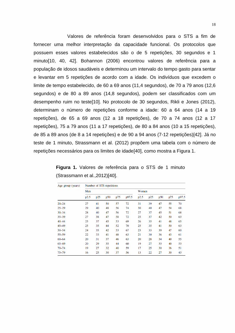
teste de 1 minuto, Strassmann et al. (2012) propõem uma tabela com o número de
repetições necessários para os limites de idade[40], como mostra a Figura 1.
Figura 1. Valores de referência para o STS de 1 minuto
(Strassmann et al.,2012)[40].
19
2.4 TESTE SIT-TO-STAND E DOENÇA PULMONAR OBSTRUTIVA CRÔNICA
O teste sit-to-stand tem sido melhor explorado na DPOC nos últimos
anos, sendo que o primeiro estudo comparou e correlacionou o STS realizado durante
um minuto com respostas obtidas no TC6min[7]. Outros estudos mostram que o STS
é um teste válido[21], reprodutível e responsivo em pacientes com DPOC, com valor
de Minimum Important Difference (MID) estabelecido em 1,7 segundos para o STS de
5 repetições[17].
Algumas vantagens podem ser observadas no STS como uma
ferramenta de avaliação em pacientes com DPOC. O teste é considerado rápido, ao
contrário do TC6min e do Incremental Shuttle Walking Test (ISWT), que necessitam
de corredores maiores e um tempo maior; é acessível, pois necessita somente de
uma cadeira e um cronômetro e requer pouco espaço, possibilitando a sua prática em
clínicas e na beira dos leitos de hospitais. A avaliação de sentar e levantar também
pode ser utilizado como um acessório de estratificação para aqueles que possuem
capacidade de exercício reduzida[17].
Puhan et al. (2013) demonstraram que o STS de 1 minuto é um
importante preditor de mortalidade em pacientes com DPOC (AUC=0,78), com valores
melhores que o índice de massa corporal (AUC=0,52), VEF1 (AUC=0,61), dispneia
(AUC=0,63) e força muscular (AUC=0,62). Para complementar o valor prognóstico do
STS, o número de repetições obtido no teste foi inserido no índice Body mass index,
airflow, Obstruction, Dyspnea and Exercise capacity (BODE) no lugar do TC6min, e o
valor discriminativo permaneceu excelente [19]. Essa semelhança no valor
discriminativo do STS de 1 minuto e do TC6min pode ser explicada pelo fato de
ambos os testes possuírem aspectos similares que compõe a capacidade de
exercício [19], como a interação do sistema respiratório, cardiovascular e muscular
[43].
Além de ser um preditor de mortalidade, o STS associa-se com outras
variáveis comumente avaliadas na DPOC, como: capacidade de exercício[7, 17],
força muscular periférica[17, 21, 22], qualidade de vida[17], mobilidade[24] e
AFVD[25]. Adicionalmente, é possível verificar que pacientes com DPOC possuem
maior déficit de controle postural para a execução do teste, em relação a indivíduos
saudáveis. Esse dado foi confirmado por Janssens et al. (2014) que compararam dois

20
grupos (DPOC vs idosos saudáveis) enquanto realizavam o movimento de sentar e
levantar da cadeira durante 5 repetições, posicionada em cima de uma plataforma de
força. Os indivíduos com DPOC demandaram maior tempo para realizar o teste em
comparação aos idosos, o que foi explicado pelo maior tempo na posição em pé e em
pé para sentado, fases essas que requerem maior controle postural[18].
Assim como em estudos com idosos saudáveis, existe uma variedade
de protocolos propostos para avaliação da capacidade funcional em indivíduos com
DPOC. Os protocolos descritos na literatura são: sentar e levantar da cadeira
utilizando 5 repetições, ou durante 30 segundos, 1 minuto e 2 minutos[20]. Os testes
mais utilizados internacionalmente são o de 5 repetições, 30 segundos e 1 minuto e
em apenas 2 estudos brasileiros foram utilizados o protocolo de 2 minutos[20].
Somente um estudo foi encontrado com um protocolo diferente dos demais, descrito
por Aguilaniu et al. e denominado “3-minute chair rise test”. Nesse protocolo o
avaliador dita um ritmo durante o primeiro minuto, e nos dois minutos seguintes, o
indivíduo realiza o teste para perfazer o maior número de repetições[8]. Existem
também diferenças técnicas em relação à altura da cadeira, no entanto, somente no
estudo de Janssens et al. (2014) a altura da cadeira foi ajustada para manter 90º de
flexão de quadril, joelhos e tornozelos[18].
A avaliação funcional pelo STS é de extrema importância, visto que
essa atividade de sentar e levantar faz parte do cotidiano de muitos indivíduos. O
teste tem se mostrado prático e simples, capaz de refletir a capacidade funcional de
pacientes com DPOC, embora haja diferenças metodológicas. Portanto, é necessário
conhecer as diferenças existentes entre os três STS mais comumente utilizados e
verificar qual teste possui melhor relação com aspectos como força muscular,
capacidade de exercício, qualidade de vida e AFVD e, assim, fazer a escolha do
protocolo que melhor auxilie na investigação de comprometimentos funcionais nessa
população.

21
3 ARTIGO ORIGINAL
Which is the best protocol of the sit-to-stand test in patients with chronic
obstructive pulmonary disease?
Andrea Akemi Morita, MSc1, 2; Gianna Waldrich Bisca, MSc1; Felipe Vilaça Cavallari
Machado, PT1; Nidia Aparecida Hernandes, PhD1; Fabio Pitta, PhD1; Vanessa
Suziane Probst, PhD1.
1 Laboratory of Research in Respiratory Physiotherapy (LFIP), Department of Physiotherapy, State
University of Londrina (UEL), Londrina, Brazil.
2 Program of Masters and Doctoral degree in Rehabilitation Sciences, State University of Londrina
(UEL) and University North of Paraná (UNOPAR), Londrina, Brazil.
Author contributions:
Ms. Andrea Akemi Morita: literature search, data collection, study design, data
analysis, manuscript preparation, review of manuscript.
Ms. Gianna Waldrich Bisca: literature search, data collection, study design, data
analysis, review of manuscript.
Mr. Felipe Vilaça Cavallari Machado: data collection, study design, data analysis,
review of manuscript.
Dr. Nidia Aparecida Hernandes: study design, data analysis, review of manuscript.
Dr. Fabio Pitta: study design, data analysis, review of manuscript.
Dr. Vanessa Suziane Probst: study design, data analysis, manuscript preparation,
review of manuscript.

22
The study was carried out at the Laboratory of Research in Respiratory Physiotherapy
(LFIP) of State University of Londrina, Paraná, Brazil.
Ms. Morita has presented this research data at the 25th Annual Congress of the
European Respiratory Society, in September 26–30 2015, in Amsterdam, Netherlands.
Financial support:
Andrea Akemi Morita is supported by a MSc grant from CAPES, Brazil.
Gianna Waldrich Bisca is supported by a PhD grant from CAPES, Brazil.
Fabio Pitta is supported by a research grant from CNPq, Brazil.
Vanessa Suziane Probst is supported by a research grant from Fundação Araucária,
Brazil.
All authors have no conflict of interest to disclose.
Corresponding author:
Vanessa Suziane Probst, PhD
Departamento de Fisioterapia – Centro de Ciências da Saúde, Hospital Universitário
de Londrina
Avenida Robert Koch, 60 – Vila Operária, 86038-350 - Londrina, Paraná, Brazil
E-mail: [email protected]

23
Abstract:
BACKGROUND: Different protocols of the sit-to-stand test (STS) are available to
assess functional capacity in patients with COPD. AIMS: To correlate each protocol of
the STS (5-repetition [5-rep_STS], 30-second [30-sec_STS] and 1-minute [1-
min_STS]) with clinical outcomes in patients with COPD; to compare three protocols of
the STS as well as to verify their association and agreement; to verify whether the
three protocols are able to predict functional exercise capacity and physical activity in
daily life (PADL). METHODS: Twenty-three patients with COPD (11 men;
FEV1:53±15%pred) performed three protocols of the STS. Patients also underwent
the following assessments: Incremental Shuttle Walking Test, 6-minute walk test
(6MWT), 4-metre gait speed test (4MGS), 1-repetition maximum of quadriceps muscle,
assessment of PADL and questionnaires of health-related quality of life and functional
status. RESULTS: The 1-min_STS presented significant correlations with 6MWT (r=
0.40), 4MGS (r= 0.64) and PADL (0.40≤ r ≤ 0.52) and the 5-rep_STS and 30-sec_STS
were associated with 4MGS (r= 0.54 and r= 0.52, respectively). The speed differed
between 5-rep_STS and 1-min_STS protocols (5-rep_STS: 0.53± 0.16 rep/sec; 30-
sec_STS: 0.48± 0.13rep/sec; 1-min_STS: 0.45± 0.11rep/sec; P= .01). However, they
presented good agreement (ICC≥ 0.73 for all) and correlated well with each other (r≥
0.68 for all). More marked changes in peripheral oxygen saturation, heart rate, blood
pressure, dyspnea and leg fatigue were found after the 1-min_STS protocol (P≤ .01 for
all). Furthermore, the three protocols were equally able to discriminate patients with
low and preserved exercise capacity. CONCLUSION: The 1-min_STS generates
higher hemodynamic demands and correlates better with clinical outcomes in patients
with COPD. Despite the difference in speed performance and physiologic demand

24
between the 5-rep_STS and 1-min_STS there is good level of agreement among the
three different protocols. In addition, all three tests were able to discriminate patients
with low and preserved exercise capacity.
KEY WORDS: pulmonary disease, chronic obstructive; exercise test; motor activity;
activities of daily living; hemodynamics; patient outcome assessment.

25
Introduction
It is well known that chronic obstructive pulmonary disease (COPD) is outlined as a
systemic disease and not only by pulmonary impairment. The extra-pulmonary effects,
such as muscle dysfunction, cardiovascular and metabolic disorders1 and decreased
functional capacity are potential risk factors for health status decline over time2.
Concerning these factors, the marked impairment observed in subjects with COPD is
the functional capacity limitation. This condition implies in constrains for basic physical
actions3. For this reason, assessment of functional capacity is extremely important and
necessary4.
One of the most common and reliable tools to assess functional capacity in patients
with COPD is the six-minute walk test (6MWT)5, 6. Although it is considered a simple
test, it requires space, time and trained staff5. To overcome these barriers, recent
studies have suggested the use of the sit-to-stand test (STS) as an alternative in this
population. The STS and the 6MWT correlate well with each other7 and present similar
responses such as hemodynamic and symptoms, supporting the STS as an option8.
The STS is a simple and practical test, widely adopted to evaluate functionality in
community-dwelling elderly9-11. The movement of standing up and sitting down are
important functions of daily life and the inability to perform these basic skills is
associated with mortality, functioning and mobility impairment12. The application of the
STS in different populations such as in neurologic13 and orthopedic14, 15 conditions has
been largely investigated; conversely, in COPD, only recent studies have explored the
test in depth16-18. Among the available information, it is known that the test is
considered valid, reliable, responsive16 and it is a predictor of mortality in patients with
COPD18.

26
Additionally, many studies describe the relationship between the STS and commonly
measured outcomes in subjects with COPD, such as exercise capacity, dyspnea,
health-related questionnaires and muscle strength, amongst others16,19-23. However,
these results were observed in a variety of protocols (i.e. 5-repetition [5-rep_STS], 30-
second [30-sec_STS] and 1-minute [1-min_STS])24 and it is unknown which is the best
protocol to assess this population. Therefore, the aims of this study were: 1) to verify
which is the best protocol concerning the correlation of each test (5-rep_STS, 30-
sec_STS and 1-min_STS), with important clinical outcomes in subjects with COPD
such as maximal and functional exercise capacity, peripheral muscle strength,
physical activity in daily life (PADL) and questionnaires of health-related quality of life
and functional status; 2) to compare three protocols of the STS as well as to verify
their association and agreement in patients with COPD; 3) to verify whether the three
protocols are able to predict functional exercise capacity and PADL. The initial
hypothesis was that there was difference among protocols, mainly between the 5-
rep_STS and 1-min_STS. Furthermore, it was hypothesized that longer lasting tests,
such as the 1-min_STS could be a more discriminative test and therefore, would be
better correlated to functional capacity, muscle strength, maximal exercise capacity,
health-related questionnaires and PADL.
Methods
Study Design
This is a cross-sectional and observational study conducted from January 2014 to May
2015 at the Laboratory of Research in Respiratory Physiotherapy of State University of
Londrina (UEL), Brazil. All subjects with COPD underwent the following assessments:
27
lung function, maximal and functional exercise capacity, peripheral muscle strength,
PADL and health-related quality of life and functional status questionnaires. Individuals
were also submitted to three different protocols of the STS (5-rep_STS, 30-sec_STS
and 1-min_STS) in a randomized sequence. The study was approved by the ethics
committee of the State University of Londrina, Brazil (080/2014) and all patients signed
a written informed consent.
Participants
The inclusion criteria of this study were: subjects with diagnosis of COPD according to
the Global Initiative for Chronic Obstructive Lung Disease (GOLD) criteria25, clinical
stability (absence of exacerbations within the previous 3 months) and no participation
in regular rehabilitation or physical exercise programs in the last year.
Participants were excluded if they had any severe comorbidity that could impair
performance in the tests such as severe cardiovascular, orthopedic or neuromuscular
conditions; or in case they, for any reason, did not complete all the assessments.
Sit-to-stand test protocols
Three protocols of STS (5-rep_STS, 30-sec_STS and 1-min_STS) have been chosen
in order to make comparisons among tests. The reason for choosing them was that
these three protocols are the most usually described in the literature in patients with
COPD24. All three tests were performed in an armless chair with a height of 46 cm;
subjects were instructed to fold their arms across their chest and perform the
movement of stand up and sit down as fast as possible. According to the protocol of
Jones et al.16, in the 5-rep test, patients performed five movements of standing up and
sitting down as fast as they could and the stopwatch was used to measure the time
28
spent in the activity. On the other hand, in the protocol of 30 seconds and 1 minute,
patients were instructed to sit down and stand up as many times as possible within 30
seconds19 and one minute7, respectively; and the number of repetitions was recorded.
Moreover, since the outcomes of these three protocols were different (time and
number of repetitions), the speed (number of repetitions per second) was considered
in the analysis. Heart rate (HR), oxygen saturation (SpO2), blood pressure (BP) and
symptoms of dyspnea and fatigue (rated by the Modified Borg Scale26) were measured
before and after each test. The order of the tests was randomized and the rest period
between them was empirically standardized. After the 5-rep_STS test, there was at
least five minutes of interval; after the 30-sec_STS protocol, at least, 15 minutes; and
after the 1-min_STS, at least 30 minutes of rest.
Assessments
Lung function was assessed by spirometry using a portable spirometer (Spiropalm®;
COSMED, Italy). The test was performed according to the American Thoracic
Society/European Respiratory Society (ATS/ERS) statement27 and the predicted
values were calculated using a specific equation for the Brazilian population28.
Maximal and functional exercise capacity were assessed by the Incremental Shuttle
Walking Test (ISWT) and 6MWT, respectively. The ISWT and the 6MWT were
performed following the ERS/ATS technical standard of field walking tests in chronic
respiratory disease5, and reference values described for Brazilians were used for each
test29, 30. Oxygen supplementation was provided when necessary. Patients were
separated into two groups according to the percentage of predicted value of the six-
minute walk distance (6MWD): low and preserved exercise capacity group. The ones
who had less than 82% of predicted distance31 were classified as having low exercise

29
capacity, and those above this threshold were considered with preserved
performance. Functional capacity was also evaluated by the 4-metre gait speed test
(4-MGS), in which patients were instructed to walk at their own pace, with a self-
selected speed, in a 4-meter corridor32. Two tests without rest were performed and the
fastest one was considered.
Quadriceps muscle strength was assessed by the one-repetition maximum (1RM) test
in a multi-gym equipment (CRW 1000, Brazil). The test was performed according to
the Procedures Recommendations for Accurate Assessment of Muscular Strength and
Power guideline33.
Two activity monitors assessed PADL: the DynaPort Activity Monitor (DAM)
(McRoberts BV, The Hague, the Netherlands) and the multisensor SenseWear
Armband (SWA) (BodyMedia Inc., Pittsburgh, PA, USA). The main variables obtained
by these devices were: time spent walking, standing, sitting, lying and locomotion per
day (assessed by the DAM); total energy expenditure and time spent in sedentary,
moderate, intense and very intense activities (assessed by the SWA). Patients were
instructed to wear both devices during one week, for 12 hours per day, and the mean
of the seven days was considered. For statistical analysis, patients were separated
into two groups: active and inactive according to the American College of Sports and
Medicine (ACSM) recommendations34. The active and inactive groups performed
respectively above or below 30 minutes per day of physical activity at least at
moderate intensity. In order to classify moderate intensity of physical activity, the
metabolic equivalents (METs) threshold was considered, according to the ACSM (4.0
to 5.9 METs for patients with 40-64 years old and 3.2 to 4.7 METs for subjects older
than 65 years old)34.

30
Health-related quality of life was assessed by the St. George's Respiratory
Questionnaire (SGRQ)35 and self-reported functional status by the Pulmonary
Functional Status and Dyspnea Questionnaire modified version (PFSDQ-M)36.
Statistical Analyses
All analyses were performed using the statistics software packages SPSS version 20.0
(Statistical Package for the Social Sciences Inc., Chicago, Illinois, USA), GraphPad
Prism 6.0 (GraphPad Software Inc., San Diego, California, USA) and Medcalc version
15.8 (MedCalc Software, Ostend, Belgium). The normality of continuous variables was
checked by the Shapiro-Wilk test and described as mean ± SD or median [IQR].
Correlations between the STS test and clinical outcomes were obtained using the
Pearson or Spearman correlation coefficients. Comparisons amongst three protocols
of STS test and variables measured before and after each test were analyzed by
repeated measures ANOVA or Friedman test followed by the Tukey or Dunn’s post-
hoc tests, respectively. Likewise, the differences of HR, SpO2, BP and scores of
dyspnea and fatigue before and after each test were assessed by the Students’ t-test
or the Wilcoxon test. To analyze agreement between protocols, the intraclass
correlation coefficient (ICC) and Bland & Altman plot37 were used. P≤ .05 was set as
statistically significant.
The Receiver Operating Characteristic (ROC curve) analysis38 was used in order to
show the ability of each STS protocol to discriminate patients with low or preserved
functional exercise capacity (6MWT) and as physically inactive or active in terms of
PADL. The comparisons of areas under the ROC curve (AUC) were performed by the
Hanley & McNeil method39.

31
The sample size calculation was performed with the GPower 3.1.3 software (Franz
Faul, Universitat Kiel, Germany) considering the study of Ozalevli et al.7 Taking into
account a correlation between the 6MWT and the 1-min_STS test of -0.75, alpha value
of 0.01, power of 90% and 20% of loss during the evaluation period, the number of
subjects for this study was 23 patients.
Results
Twenty-nine patients with COPD took part in the study; however, six subjects were
excluded due to the following reasons: two patients for vascular and orthopedic
limitations, one had an exacerbation during the assessment period, one did not
complete all the assessments for personal reason and two patients died during this
period. Thus, data from 23 patients were available for analyses and their
characteristics are described in Table 1. Overall, majority of the sample was
composed by patients with moderate degree of airflow obstruction and was classified
as GOLD II. Subjects walked 476±63 m in the 6MWT and nine (39%) patients used
oxygen supplementation during the test.
Association of STS protocols with clinical outcomes
Table 2 presents the correlations found between the three protocols of the STS and
clinical outcomes such as functional exercise capacity, functional status and PADL. A
significant association was found between the 5-rep_STS and functional capacity
evaluated by the 4-MGS. The 30-sec_STS protocol was also correlated with the 4-
MGS. Some variables of PADL measured by the DAM, such as walking and
locomotion were also associated with this protocol (Table 2).

32
Many other variables such as 6MWT, activity score of the PFSDQ-M questionnaire,
physical activity duration (PAD) and standing position were associated with the 1-
min_STS; as well as the 4-MGS, walking and locomotion variables. The values of
correlation of this protocol and the 4-MGS and PADL outcomes were higher than for
the other two STS tests (Table 2).
None of the other assessed outcomes such as lung function, ISWT, SGRQ and
quadriceps strength were associated with the 5-rep_STS, 30-sec_STS and 1-
min_STS protocols (P> .05).
Comparisons among protocols
The speed (number of repetitions per second) of the three STS protocols was different
(5-rep_STS: 0.53± 0.16 rep/s vs 30-sec_STS: 0.48± 0.13 rep/s vs 1-min_STS: 0.45±
0.11 rep/s; P= .01) (Figure 1). The post-hoc analysis showed difference between the
5-rep_STS and 1-min_STS, thus patients performed the 5-rep_STS protocol faster
than in the 1-min_STS (P= .01).
Patients started all the tests with similar SpO2, HR, systolic and diastolic BP.
Moreover, there were no differences on symptoms of dyspnea and fatigue measured
before each protocol (Table 3).
The comparisons of the changes in oxygen saturation, hemodynamic and symptom
variables, described in Table 4, differed among protocols, except for the diastolic BP.
The 1-min_STS protocol had a greater variation of SpO2, HR, systolic BP and
symptoms of dyspnea and fatigue when compared to the 5-rep_STS. Differences were
also found between the delta of SpO2, HR and symptoms of fatigue between the 1-
min_STS and 30-sec_STS protocols. Finally, the 30-sec_STS presented higher

33
changes in terms of HR, systolic BP and symptoms of dyspnea when compared to the
5-rep_STS protocol (Table 4).
In addition to the aforementioned analysis, Table 5 shows the differences between
oxygen saturation, hemodynamic and symptom variables measured before and after
each protocol. In the 5-rep_STS, the final HR, systolic BP and symptoms of dyspnea
were higher than at the beginning of the test. The protocol of 30-sec_STS and 1-
min_STS showed significant desaturation, and increase in HR, systolic BP and
symptoms of dyspnea and fatigue. The diastolic BP significantly increased in the 1-
min_STS and did not present changes in the 30-sec_STS protocol.
Correlation and agreement among protocols
The 5-rep_STS, 30-sec_STS and 1-min_STS had a significant correlation with each
other, with r values higher than 0.68 (Table 2). Additionally, they showed a good
agreement pairwise (ICC: 5-rep_STS and 30-sec_STS of 0.85 [95% CI 0.61- 0.93; P<
.0001]; 5-rep_STS and 1-min_STS of 0.73 [95% CI 0.22- 0.89; P< .0001]; 30-
sec_STS and 1-min_STS of 0.77 [95% CI 0.46- 0.90; P< .0001]), illustrated by the
Bland & Altman plot (Figure 2).
Sit-to-stand test to predict functional exercise capacity and physical activity in
daily life
Concerning functional exercise capacity assessed by the 6MWT, fifteen (65%)
patients were considered as having preserved capacity and eight (35%) subjects with
the predicted distance lower than 82%. The AUC calculated for the three protocols had
discriminative values for poor and preserved exercise capacity: 0.71 (5-rep_STS),
0.85 (30-sec_STS) and 0.82 (1-min_STS). Subsequently, the comparison of AUCs
34
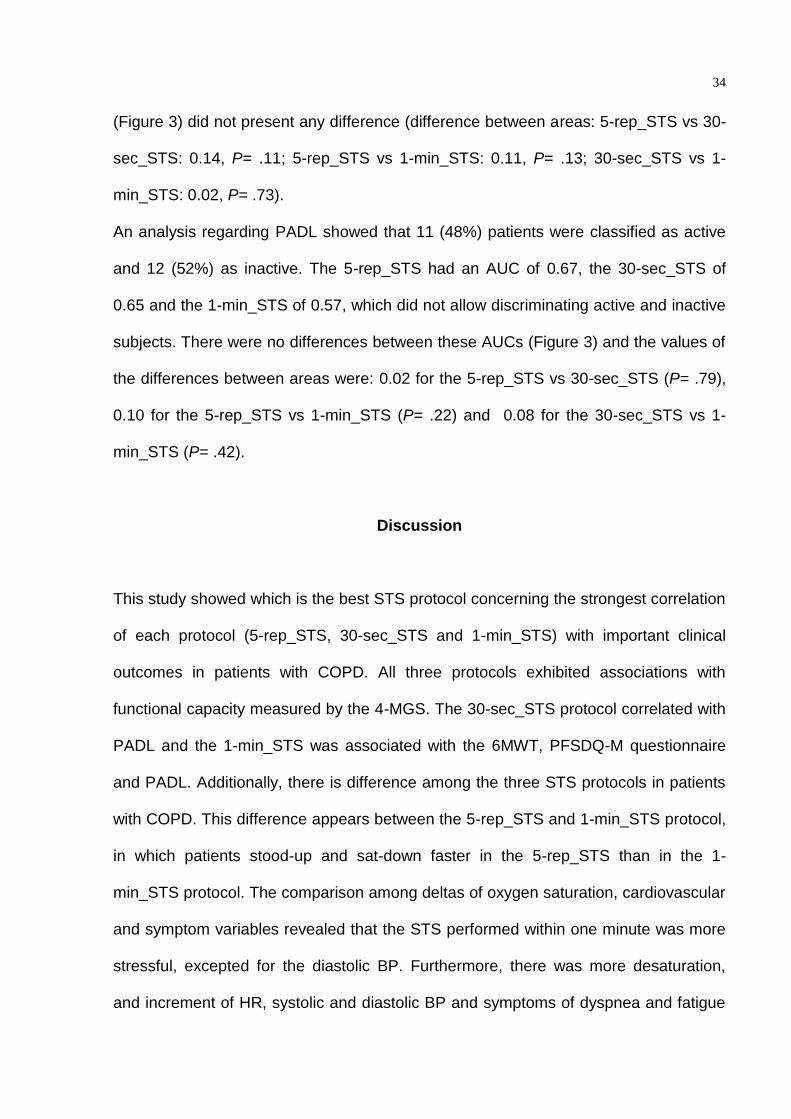
(Figure 3) did not present any difference (difference between areas: 5-rep_STS vs 30-
sec_STS: 0.14, P= .11; 5-rep_STS vs 1-min_STS: 0.11, P= .13; 30-sec_STS vs 1-
min_STS: 0.02, P= .73).
An analysis regarding PADL showed that 11 (48%) patients were classified as active
and 12 (52%) as inactive. The 5-rep_STS had an AUC of 0.67, the 30-sec_STS of
0.65 and the 1-min_STS of 0.57, which did not allow discriminating active and inactive
subjects. There were no differences between these AUCs (Figure 3) and the values of
the differences between areas were: 0.02 for the 5-rep_STS vs 30-sec_STS (P= .79),
0.10 for the 5-rep_STS vs 1-min_STS (P= .22) and 0.08 for the 30-sec_STS vs 1-
min_STS (P= .42).
Discussion
This study showed which is the best STS protocol concerning the strongest correlation
of each protocol (5-rep_STS, 30-sec_STS and 1-min_STS) with important clinical
outcomes in patients with COPD. All three protocols exhibited associations with
functional capacity measured by the 4-MGS. The 30-sec_STS protocol correlated with
PADL and the 1-min_STS was associated with the 6MWT, PFSDQ-M questionnaire
and PADL. Additionally, there is difference among the three STS protocols in patients
with COPD. This difference appears between the 5-rep_STS and 1-min_STS protocol,
in which patients stood-up and sat-down faster in the 5-rep_STS than in the 1-
min_STS protocol. The comparison among deltas of oxygen saturation, cardiovascular
and symptom variables revealed that the STS performed within one minute was more
stressful, excepted for the diastolic BP. Furthermore, there was more desaturation,
and increment of HR, systolic and diastolic BP and symptoms of dyspnea and fatigue

35
at the end of the 1-min_STS test. In spite of these differences, the three protocols
correlated well with each other and had a good agreement. Moreover, the three
protocols were able to discriminate patients with low and preserved exercise capacity
in the 6MWT with AUC > 0.71, however, this was not the case for PADL. None of the
three STS protocols were able to adequately predict patients classified as active an
inactive.
The STS test was firstly described by Csuka and McCarty40, in which the protocol of
ten repetitions was used. It was considered an inexpensive, simple, rapid and
reproducible test to assess lower limb muscle strength in healthy subjects. After this
study, many other protocols of the STS were risen to test functional capacity of lower
limbs in a variety of populations, including patients with COPD. A large number of
protocols are available and the present study is the first to describe the differences
among them in this population. Indeed, we observed that patients in the present study
completed the 5-rep_STS faster than in the 1-min_STS. This fact might have occurred
due to an increase of symptoms during the 1-min_STS test compared to the 5-
rep_STS protocol (Table 4), resulting in lower performance. Similar results are
demonstrated in the gait speed test, in which healthy elderly had a higher value of
speed in shorter distances (comparison of 5 m and 10 m)41. Moreover, the 1-min_STS
test presented a higher oxygen desaturation, hemodynamic and symptom demands in
comparison to the other two tests (Table 4 and 5). It is reasonable that the longest test
(1-min_STS) demanded higher physical effort. Despite this difference, the three STS
protocols had moderate association (r≥ 0.68) and good agreement (ICC≥ 0.73) among
them.
All three protocols were associated with the 4-MGS, which is also considered a
functional test24. This result was expected, since both tests have the purpose to

36
assess lower limb function with movements usually adopted in daily life. Ozalevli et al.
showed that the STS correlated with the score of the physical function domain
(consisted of daily walking activities) of the Nottingham Health Profile Survey
questionnaire in patients with COPD7. Likewise, Whitney et al. verified moderate
correlation between the 5-rep STS and Dynamic Gait Index in subjects with balance
disorders42. Other important correlation found in the present study was between PADL
and the 30-min_STS protocol, as well as with the 1-min_STS protocol. The 1-min_STS
had better association with PADL in COPD, similarly to the study by Van Gestel et al23,
in which they showed correlation between the 1-min_STS and objectively measured
physical activity23. Indeed, this association could be explained because motion
sensors devices (i.e. accelerometers) are reliable to provide kinetic and kinematic data
from the STS in elderly43-45. Only the 1-min_STS correlated with other variables such
as the 6MWT and the activity score of the PFSDQ-M questionnaire. There is some
evidence in the literature suggesting the STS as an option to the 6MWT to measure
functional capacity7, 8. Both tests have good correlation and the advantage of the STS
is that it causes less hemodynamic stress to patients with COPD7. Moreover, the
functional status obtained by the PFSDQ-M questionnaire was significantly associated
with the 1-min_STS test. This finding reinforces the STS as a tool to assess functional
condition.
Lower limb strength is a meaningful factor that could potentially limit the ability of
standing up and sitting down from a chair in elderly46. Even though many studies in the
literature have shown correlation between the STS and quadriceps muscle strength,
both in healthy46-51 and COPD16,19-22 in this study we did not find any correlation
between these variables. One plausible explanation for this could be related to the
limitation of the assessment method for muscle force used in the present study (i.e.,
37
the 1RM). The use of a more accurate tool to assess muscle strength, for instance,
dynamometry, could have led to different findings. The absence of association
between STS and quadriceps muscle strength could also be explained by the
requirement of other abilities in daily life rather than muscle force, such as postural
and balance control17, 42, hip and ankle motion50.
In a discriminative analysis, patients with COPD with low and preserved exercise
capacity according to the 6MWT were adequately identified by the proposed STS
protocols. All three tests showed similar AUC meaning that they have similarity to
predict exercise capacity. Similar results were found in the study by Jones et al., in
which the STS was considered a stratification tool to identify impaired walking capacity
obtained by the ISWT (AUC= 0.82)16. On the other hand, none of the tests was able to
predict active and inactive patients. This finding resembles the study of Van Gestel et
al., which the STS was also not able to predict inactive lifestyle in COPD (AUC=
0.31)23. This result was expected since there was no correlation with the STS and
physical activity duration variable in this study. Additionally, considering the STS as a
functional capacity test, Pitta et al. showed that tests such as 6MWT might predict
activities in daily life better than single variables of physical functioning52.
Although many protocols of the STS are available in the literature, choosing one of the
three most broadly used in patients with COPD is important to evaluate lower limb
function. Hence, this study suggests that the 1-min_STS protocol of STS could be
better to evaluate functional capacity in this population, since it had better correlations
with important clinical outcomes. Albeit the time spent performing the 1-min_STS
protocol is longer than the other two protocols, it still remains a quick, practical and
useful test in clinical settings.

38
Limitations
Firstly, although it is known that the chair height can influence the performance of the
STS53-55, this study could not ensure that patients were positioned to have exactly 90
degrees of hip and knee flexion in the chair. However, we standardized the same chair
of 46 cm height to all the patients, similarly to the majority of previous studies16, 18, 21.
Additionally, we believe this limitation did not negatively affect the study results, since
in our daily life, the chair height is standardized to the overall population and it is not
possible to adjust the chair individually24.
Secondly, the three protocols of the STS were performed in a randomized sequence
and resting time after each test was empirically determined. Even though this study did
not analyze this recovery time in depth, this period between the tests appeared to be
adequate, since there were no differences among SpO2, HR, systolic and diastolic BP,
and symptoms of dyspnea and fatigue recorded before each test (Table 3).
Finally, there was no patient classified as GOLD I in the present sample. The lack of
subjects classified in the mild stage of COPD and the average FEV1 as well, restricts
these results to patients with moderate to severe degree of airflow obstruction.
Conclusion
In summary, the 1-min_STS seems to be the best protocol to evaluate patients with
COPD, since it was better associated with important clinical outcomes such as
functional exercise capacity, functional status and PADL; and it generates higher
hemodynamic demands than the other studied tests. The 5-rep_STS and 1-min_STS
protocols were significantly different in terms of speed performance and physiologic
demand; however, they were correlated and had a good level of agreement.

39
Furthermore, all of the three protocols were able to identify patients with low and
preserved exercise capacity, nevertheless, they could not discriminate subjects
classified as active and inactive in daily life.
Acknowledgements
All the authors are grateful to the colleagues of the Laboratory of Research in
Respiratory Physiotherapy of State University of Londrina – Brazil and CAPES for the
contribution to the study.
References
1. Couillard A, Muir JF, Veale D. COPD recent findings: impact on clinical practice.
Copd 2010;7(3):204-213.
2. Eisner MD, Iribarren C, Blanc PD, Yelin EH, Ackerson L, Byl N, et al. Development
of disability in chronic obstructive pulmonary disease: beyond lung function. Thorax
2011;66(2):108-114.
3. Eisner MD, Blanc PD, Yelin EH, Sidney S, Katz PP, Ackerson L, et al. COPD as a
systemic disease: impact on physical functional limitations. Am J Med
2008;121(9):789-796.
4. Downs CA. Functional assessment of chronic obstructive pulmonary disease. J
Am Acad Nurse Pract 2011;23(4):161-167.
5. Holland AE, Spruit MA, Troosters T, Puhan MA, Pepin V, Saey D, et al. An official
European Respiratory Society/American Thoracic Society technical standard: field
walking tests in chronic respiratory disease. Eur Respir J 2014;44(6):1428-1446.
40
6. Kocks JW, Asijee GM, Tsiligianni IG, Kerstjens HA, van der Molen T. Functional
status measurement in COPD: a review of available methods and their feasibility in
primary care. Prim Care Respir J 2011;20(3):269-275.
7. Ozalevli S, Ozden A, Itil O, Akkoclu A. Comparison of the Sit-to-Stand Test with 6
min walk test in patients with chronic obstructive pulmonary disease. Respir Med
2007;101(2):286-293.
8. Aguilaniu B, Roth H, Gonzalez-Bermejo J, Jondot M, Maitre J, Denis F, et al. A
simple semipaced 3-minute chair rise test for routine exercise tolerance testing in
COPD. Int J Chron Obstruct Pulmon Dis 2014;9:1009-1019.
9. Bohannon RW. Reference values for the five-repetition sit-to-stand test: a
descriptive meta-analysis of data from elders. Percept Mot Skills 2006;103(1):215-222.
10. Zhang F, Ferrucci L, Culham E, Metter EJ, Guralnik J, Deshpande N. Performance
on five times sit-to-stand task as a predictor of subsequent falls and disability in older
persons. J Aging Health 2013;25(3):478-492.
11. Millor N, Lecumberri P, Gomez M, Martinez-Ramirez A, Izquierdo M. An evaluation
of the 30-s chair stand test in older adults: frailty detection based on kinematic
parameters from a single inertial unit. J Neuroeng Rehabil 2013;10:86.
12. Janssen WG, Bussmann HB, Stam HJ. Determinants of the sit-to-stand
movement: a review. Phys Ther 2002;82(9):866-879.
13. Silva PF, Quintino LF, Franco J, Faria CD. Measurement properties and feasibility
of clinical tests to assess sit-to-stand/stand-to-sit tasks in subjects with neurological
disease: a systematic review. Braz J Phys Ther 2014;18(2):99-110.
14. Abujaber SB, Marmon AR, Pozzi F, Rubano JJ, Zeni JA, Jr. Sit-To-Stand
Biomechanics Before and After Total Hip Arthroplasty. J Arthroplasty 2015.
41
15. Bouchouras G, Patsika G, Hatzitaki V, Kellis E. Kinematics and knee muscle
activation during sit-to-stand movement in women with knee osteoarthritis. Clin
Biomech (Bristol, Avon) 2015;30(6):599-607.
16. Jones SE, Kon SS, Canavan JL, Patel MS, Clark AL, Nolan CM, et al. The five-
repetition sit-to-stand test as a functional outcome measure in COPD. Thorax
2013;68(11):1015-1020.
17. Janssens L, Brumagne S, McConnell AK, Claeys K, Pijnenburg M, Goossens N, et
al. Impaired postural control reduces sit-to-stand-to-sit performance in individuals with
chronic obstructive pulmonary disease. PLoS One 2014;9(2):e88247.
18. Puhan MA, Siebeling L, Zoller M, Muggensturm P, ter Riet G. Simple functional
performance tests and mortality in COPD. Eur Respir J 2013;42(4):956-963.
19. Benton MJ, Alexander JL. Validation of functional fitness tests as surrogates for
strength measurement in frail, older adults with chronic obstructive pulmonary disease.
Am J Phys Med Rehabil 2009;88(7):579-583; quiz 584-576, 590.
20. Butcher SJ, Pikaluk BJ, Chura RL, Walkner MJ, Farthing JP, Marciniuk DD.
Associations between isokinetic muscle strength, high-level functional performance,
and physiological parameters in patients with chronic obstructive pulmonary disease.
Int J Chron Obstruct Pulmon Dis 2012;7:537-542.
21. Rausch-Osthoff AK, Kohler M, Sievi NA, Clarenbach CF, van Gestel AJ.
Association between peripheral muscle strength, exercise performance, and physical
activity in daily life in patients with Chronic Obstructive Pulmonary Disease. Multidiscip
Respir Med 2014;9(1):37.
22. Roig M, Eng JJ, MacIntyre DL, Road JD, Reid WD. Deficits in muscle strength,
mass, quality, and mobility in people with chronic obstructive pulmonary disease. J
Cardiopulm Rehabil Prev 2011;31(2):120-124.

42
23. van Gestel AJ, Clarenbach CF, Stowhas AC, Rossi VA, Sievi NA, Camen G, et al.
Predicting daily physical activity in patients with chronic obstructive pulmonary
disease. PLoS One 2012;7(11):e48081.
24. Bisca GW, Morita AA, Hernandes NA, Probst VS, Pitta F. Simple lower limb
functional tests in patients with Chronic Obstructive Pulmonary Disease: a systematic
review. Arch Phys Med Rehabil 2015.
25. Global strategy for the diagnosis, management and prevention for chronic
obstructive pulmonary disease (updated 2015). Global Initiative for Chronic
Obstructive Pulmonary Disease 2015:1-117.
26. Mahler DA, Wells CK. Evaluation of clinical methods for rating dyspnea. Chest
1988;93(3):580-586.
27. Miller MR, Hankinson J, Brusasco V, Burgos F, Casaburi R, Coates A, et al.
Standardisation of spirometry. Eur Respir J 2005;26(2):319-338.
28. Pereira CA, Sato T, Rodrigues SC. New reference values for forced spirometry in
white adults in Brazil. J Bras Pneumol 2007;33(4):397-406.
29. Probst VS, Hernandes NA, Teixeira DC, Felcar JM, Mesquita RB, Goncalves CG,
et al. Reference values for the incremental shuttle walking test. Respir Med
2012;106(2):243-248.
30. Britto RR, Probst VS, de Andrade AF, Samora GA, Hernandes NA, Marinho PE, et
al. Reference equations for the six-minute walk distance based on a Brazilian
multicenter study. Braz J Phys Ther 2013;17(6):556-563.
31. Troosters T, Gosselink R, Decramer M. Six minute walking distance in healthy
elderly subjects. Eur Respir J 1999;14(2):270-274.
32. Kon SS, Patel MS, Canavan JL, Clark AL, Jones SE, Nolan CM, et al. Reliability
and validity of 4-metre gait speed in COPD. Eur Respir J 2013;42(2):333-340.

43
33. Lee EB, Weir JP. Procedures Recommendation I: Accurate Assessment Of
Muscular Strength And Power. Journal of Exercise Physiology 2001;4(3):1-21.
34. Garber CE, Blissmer B, Deschenes MR, Franklin BA, Lamonte MJ, Lee IM, et al.
American College of Sports Medicine position stand. Quantity and quality of exercise
for developing and maintaining cardiorespiratory, musculoskeletal, and neuromotor
fitness in apparently healthy adults: guidance for prescribing exercise. Med Sci Sports
Exerc 2011;43(7):1334-1359.
35. Sousa TCd, Jardim JR, Jones P. Validation of the Saint George’s Respiratory
Questionnaire in patients with chronic obstructive pulmonary disease in Brazil. J
Pneumol 2000;26(3):119-128.
36. Kovelis D, Segretti NO, Probst VS, Lareau SC, Brunetto AF, Pitta F. Validation of
the Modified Pulmonary Functional Status and Dyspnea Questionnaire and the
Medical Research Council scale for use in Brazilian patients with chronic obstructive
pulmonary disease. J Bras Pneumol 2008;34(12):1008-1018.
37. Bland JM, Altman DG. Statistical methods for assessing agreement between two
methods of clinical measurement. Lancet 1986;1(8476):307-310.
38. Hanley JA, McNeil BJ. The meaning and use of the Area Under a Receive
Operating Characterist (ROC) Curve. Radiology 1982;143(1):29-36.
39. Hanley JA, McNeil BJ. A method of comparing the areas under receiver operating
characteristic curves derived from the same cases. Radiology 1983;148(3):839-843.
40. Csuka M, McCarty DJ. Simple method for measurement of lower extremity muscle
strength. Am J Med 1985;78(1):77-81.
41. Graham JE, Ostir GV, Kuo YF, Fisher SR, Ottenbacher KJ. Relationship between
test methodology and mean velocity in timed walk tests: a review. Arch Phys Med
Rehabil 2008;89(5):865-872.

44
42. Whitney SL, Wrisley DM, Marchetti GF, Gee MA, Redfern MS, Furman JM.
Clinical measurement of sit-to-stand performance in people with balance disorders:
validity of data for the Five-Times-Sit-to-Stand Test. Phys Ther 2005;85(10):1034-
1045.
43. Schwenk M, Gogulla S, Englert S, Czempik A, Hauer K. Test-retest reliability and
minimal detectable change of repeated sit-to-stand analysis using one body fixed
sensor in geriatric patients. Physiol Meas 2012;33(11):1931-1946.
44. Millor N, Lecumberri P, Gomez M, Martinez-Ramirez A, Izquierdo M. Kinematic
parameters to evaluate functional performance of sit-to-stand and stand-to-sit
transitions using motion sensor devices: a systematic review. IEEE Trans Neural Syst
Rehabil Eng 2014;22(5):926-936.
45. Janssen WG, Bussmann JB, Horemans HL, Stam HJ. Validity of accelerometry in
assessing the duration of the sit-to-stand movement. Med Biol Eng Comput
2008;46(9):879-887.
46. Hughes MA, Myers BS, Schenkman ML. The role of strength in rising from a chair
in the functionally impaired elderly. J Biomech 1996;29(12):1509-1513.
47. Hardy R, Cooper R, Shah I, Harridge S, Guralnik J, Kuh D. Is chair rise
performance a useful measure of leg power? Aging Clin Exp Res 2010;22(5-6):412-
418.
48. McCarthy EK, Horvat MA, Holtsberg PA, Wisenbaker JM. Repeated chair stands
as a measure of lower limb strength in sexagenarian women. J Gerontol A Biol Sci
Med Sci 2004;59(11):1207-1212.
49. Lord SR, Murray SM, Chapman K, Munro B, Tiedemann A. Sit-to-stand
performance depends on sensation, speed, balance, and psychological status in

45
addition to strength in older people. J Gerontol A Biol Sci Med Sci 2002;57(8):M539-
543.
50. Netz Y, Ayalon M, Dunsky A, Alexander N. 'The multiple-sit-to-stand' field test for
older adults: what does it measure? Gerontology 2004;50(3):121-126.
51. Jones CJ, Rikli RE, Beam WC. A 30-s chair-stand test as a measure of lower body
strength in community-residing older adults. Res Q Exerc Sport 1999;70(2):113-119.
52. Pitta F, Troosters T, Spruit MA, Probst VS, Decramer M, Gosselink R.
Characteristics of physical activities in daily life in chronic obstructive pulmonary
disease. Am J Respir Crit Care Med 2005;171(9):972-977.
53. Chorin F, Rahmani A, Beaune B, Cornu C. Determination of reliable force platform
parameters and number of trial to evaluate sit-to-stand movement. Aging Clin Exp Res
2015;27(4):473-482.
54. Ng SS, Cheung SY, Lai LS, Liu AS, Ieong SH, Fong SS. Five Times Sit-To-Stand
test completion times among older women: Influence of seat height and arm position. J
Rehabil Med 2015;47(3):262-266.
55. Ng SS, Cheung SY, Lai LS, Liu AS, Ieong SH, Fong SS. Association of seat height
and arm position on the five times sit-to-stand test times of stroke survivors. Biomed
Res Int 2013;2013:642362.

46
FIGURA 1
ST
S s
pe
ed
(r
ep
/s)
0 .0
0 .2
0 .4
0 .6
0 .8
5-rep 3 0 - s e c 1 - m in
*
Figure 1. Comparison of the speed among the 5-repetition_STS, 30-second_STS and 1-minute_STS
protocols of the sit-to-stand test. *P= .01. STS: sit-to-stand; rep/s: repetitions per second; 5-rep: 5-repetition;
30-sec: 30-seconds; 1-min: 1-minute.

47
FIGURA 2
A B C
3 0 s e c + 5 r e p /2 ( r e p /s )
5r
ep
- 3
0s
ec
(r
ep
/s)
0 .0 0 .2 0 .4 0 .6 0 .8 1 .0
-0 .2
-0 .1
0 .0
0 .1
0 .2
0 .3
0 .0 4
0 .2 4
- 0 .1 4
1 m in + 5 r e p /2 ( r e p /s )
5r
ep
- 1
min
(r
ep
/s)
0 .0 0 .2 0 .4 0 .6 0 .8 1 .0
-0 .2
0 .0
0 .2
0 .4
0 .6
0 .0 8
- 0 .1 3
0 .3 0
3 0 s e c + 1 m in /2 ( r e p /s )
30
se
c -
1m
in (
re
p/s
)
0 .0 0 .2 0 .4 0 .6 0 .8
-0 .2
-0 .1
0 .0
0 .1
0 .2
0 .3
0 .0 3
- 0 .1 7
0 .2 3
Figure 2. Bland & Altman plots of the difference between two STS protocols plotted against the mean value
of the two STS (5-rep_STS vs 30-sec_STS [A]; 5-rep_STS vs 1-min_STS [B] and 30-sec_STS vs 1-
min_STS [C]). ICC values: P< .0001 for all.

48
FIGURA 3
A
B
Figure 3. Comparison among the areas under the curve (AUC) of the 5-repetion_STS, 30-second_STS and
1-minute_STS protocols in patients with (A) low and preserved exercise capacity in the 6MWT and in
patients classified as (B) active and inactive regarding PADL.

49
TABELA 1
Table 1. Characteristics of the patients with COPD.
n=23
Gender (M/F) 11/12
Age (years) 68±8
BMI (kg.m-2
) 26±5
VEF1 (L) 1.31±0.43
VEF1 (%predicted)
FEV1/FVC (%)
53±15
55±11
GOLD (II/III/IV)
5-rep_STS (sec)
30-sec_STS (repetitions)
1-min_STS (repetitions)
6MWT (m)
6MWT (%predicted)
ISWT (m)
ISWT (%predicted)
1-RM Quadriceps (kg)
4-MGS test (m/s)
SGRQ total score
PFSDQ-M dyspnea score
PFSDQ-M fatigue score
PFSDQ-M activity score
PAL index
16/6/1
10±3
14±3
27±6
476±63
90±14
448±102
77±26
16±7
1.42±0.27
38 [22-42]
6 [2-13]
5 [0.75-10]
5 [0.75-9]
1.47±0.11
TEE (kcal) 2029±395
PAD > moderate intensity (min) 48 [18-138] Data expressed as absolute frequency, mean±SD or median [IQR]. M:male; F:female;
BMI:body mass index; FEV1:forced expiratory volume in the first second; FVC: forced
vital capacity; GOLD: Global Initiative for Chronic Lung Disease; 5-rep_STS: 5-
repetition sit-to-stand test; 30-sec_STS: 30-second sit-to-stand test; 1-min_STS: 1-minute
sit-to-stand test; 6MWT: six-minute walk test; ISWT: incremental shuttle walking test; 1-
RM: one-repetition maximum; 4MGS: four-metre gait speed; SGRQ: St’ George’s
Respiratory Questionnaire; PFSDQ-M: Modified Pulmonary Functional Status and
Dyspnea Questionnaire; PAL: physical activity level index; TEE: total energy
expenditure; PAD: physical activity duration.

50
TABELA 2
Table 2. Correlations between the 5-rep_STS, 30-sec_STS and 1-min_STS protocol of the STS and
clinical variables in patients with COPD.
5-rep_STS (rep/s)
30-sec_STS (rep/s) 1-min_STS (rep/s)
r P r P r P
5-repetition_STS (rep/sec) NA NA 0.71 .0001* 0.71 .0001*
30-second_STS (rep/sec)
1-minute_STS (rep/sec)
6MWT (m)
4MGS test (m/s)
0.71
0.71
0.16
0.54
.0001*
.0001*
.46
.009*
NA
0.68
0.26
0.52
NA
.0003*
.21
.01*
0.68
NA
0.40
0.64
.0003*
NA
.05*
.001*
PFSDQ-M activity score 0.38 .08 0.04 .85 0.43 .04*
PAL index
Walking (min)
Standing (min)
Locomotion (min)
0.33
0.19
0.35
0.37
.13
.37
.10
.08
0.30
0.46
0.17
0.48
.30
.02*
.42
.02*
0.48
0.40
0.49
0.52
.02*
.05*
.01*
.01* * Statistical significant correlations; r: Pearson or Spearman correlation coefficient value. STS: sit-to-stand test; 5-rep: 5-
repetition; 30sec: 30-second; 1-min:1-minute; 6MWT: six-minute walk test; 4MGS: 4-metre gait speed; PFSDQ-M: Modified
Pulmonary Functional Status and Dyspnea Questionnaire; PAL: physical activity level index; NA: not applicable.

51
TABELA 3
Table 3. Comparisons of oxygen saturation, cardiovascular and symptoms variables measured
before each STS protocols. 5-rep_STS 30-sec_STS 1-min_STS P
SpO2 (%) 96 [92-97] 96 [93-98] 95 [94-97] .29
HR (bpm) 87±13 86±12 86±12 .39
Systolic BP (mmHg) 120 [110-140] 120 [110-130] 130 [120-140] .28
Diastolic BP (mmHg) 80 [70-90] 80 [70-90] 80 [80-90] .96
Dyspnea 0 [0-1] 0 [0-1] 0 [0-2] .08
Fatigue 0 [0-2] 0 [0-0.5] 0 [0-0.5] .30 Data expressed as mean ±SD and median [IQR]. STS: sit-to-stand test; 5-rep: 5-repetition; 30sec: 30-second;
1-min: 1-minute; SpO2: pulsed oxygen saturation; HR: heart rate; bpm: beats per minute; BP: blood pressure;
Dyspnea: score according to Borg scale; Fatigue: score according to Borg scale.
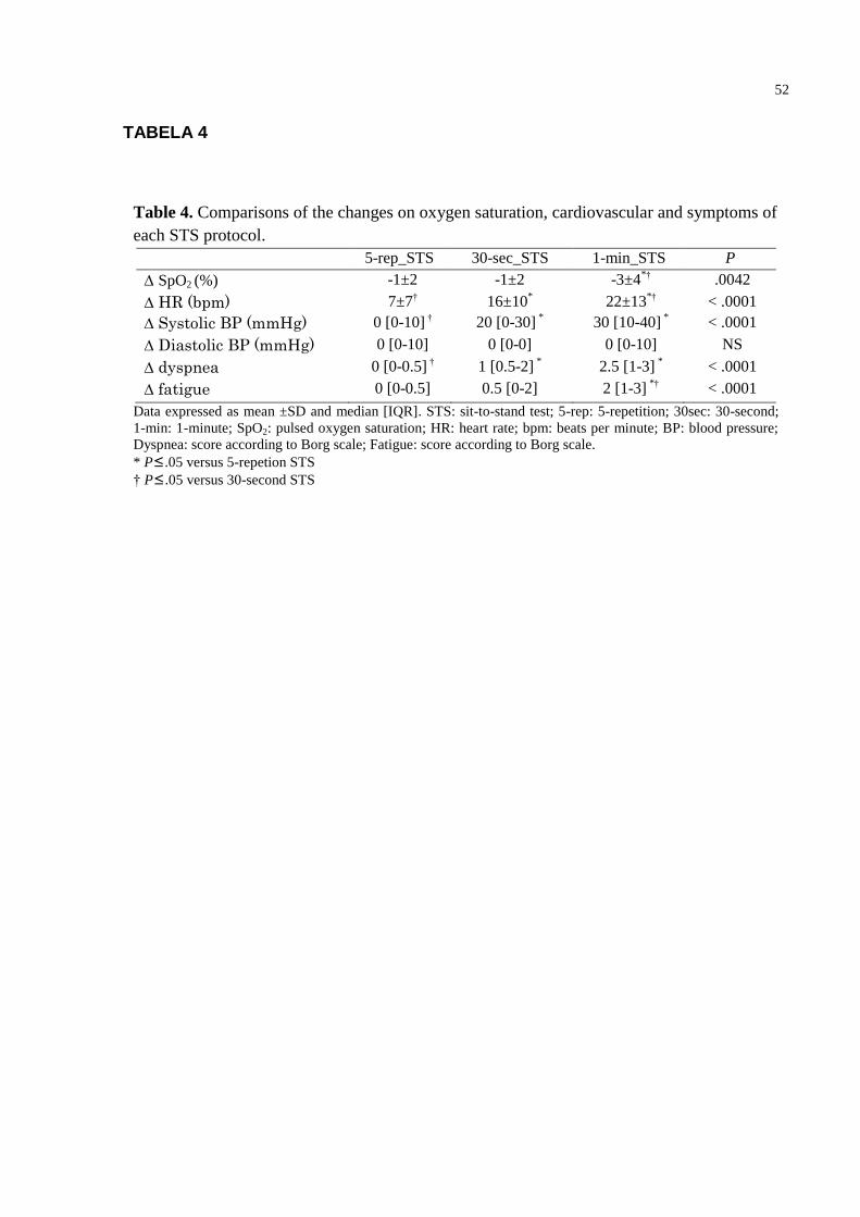
52
TABELA 4
Table 4. Comparisons of the changes on oxygen saturation, cardiovascular and symptoms of
each STS protocol.
5-rep_STS 30-sec_STS 1-min_STS P
∆ SpO2 (%) -1±2 -1±2 -3±4*† .0042
∆ HR (bpm) 7±7† 16±10* 22±13
*† < .0001
∆ Systolic BP (mmHg) 0 [0-10] † 20 [0-30] * 30 [10-40]
* < .0001
∆ Diastolic BP (mmHg) 0 [0-10] 0 [0-0] 0 [0-10] NS
∆ dyspnea 0 [0-0.5] † 1 [0.5-2] * 2.5 [1-3]
* < .0001
∆ fatigue 0 [0-0.5] 0.5 [0-2] 2 [1-3] *† < .0001
Data expressed as mean ±SD and median [IQR]. STS: sit-to-stand test; 5-rep: 5-repetition; 30sec: 30-second;
1-min: 1-minute; SpO2: pulsed oxygen saturation; HR: heart rate; bpm: beats per minute; BP: blood pressure;
Dyspnea: score according to Borg scale; Fatigue: score according to Borg scale.
* P≤.05 versus 5-repetion STS
† P≤.05 versus 30-second STS

53
TABELA 5
Table 5. Comparisons of oxygen saturation, hemodynamic and symptom variables measured pre and post
each STS protocol. 5-rep_STS 30-sec_STS 1-min_STS
pre post P pre post P pre post P
SpO2 (%) 95±3 94±2 .12 95±2 94±3 .01 95±2 91±5 < .0001
HR (bpm) 87±13 95±13 < .0001 86±12 102±13 < .0001 86±12 109±17 < .0001
Systolic BP
(mmHg)
120
[110-140]
130
[120-140]
.003
120
[110-130]
140
[130-150]
< .0001
130
[120-140]
150
[140-180]
< .0001
Diastolic
BP (mmHg)
80
[70-90]
90
[80-90]
.11
80
[70-90]
80
[70-90]
.88
80
[80-90]
90
[80-100]
.007
Dyspnea 0 [0-1] 0 [0-2] .03 0 [0-1] 2 [1-3] < .0001 0.5 [0-2] 3 [2-5] < .0001
Fatigue 0 [0-2] 0 [0-2] .24 0 [0-0.5] 1 [0-3] .0001 0 [0-0.5] 2 [1-5] < .0001
Data expressed as mean ±SD and median [IQR]. STS:sit-to-stand test; 5-rep: 5-repetition; 30sec: 30-second; 1-min:1-minute;
SpO2: pulsed oxygen saturation; HR: heart rate; BP: blood pressure; Dyspnea: score according to Borg scale; Fatigue: score
according to Borg scale.

54
4 CONCLUSÃO GERAL
O presente estudo revela que o protcolo de 1-min do STS é o melhor
para avaliar pacientes com DPOC, visto que este está melhor associado com
desfechos clínicos importantes avaliados na DPOC, como: capacidade funcional de
exercício, estado funcional e AFVD. Além disso, o teste de 1-min apresenta maiores
demandas hemodinâmicas e sintomatológicas comparado aos outros dois testes
estudados. O STS de 5-rep e 1-min são diferentes quanto à velocidade de execução e
à resposta hemodinâmica e sintomatológica, no entanto, apesar dessa diferença, os
testes correlacionam-se e apresentam uma excelente concordância entre si.
Adicionalmente, todos os protocolos são capazes de discriminar os pacientes que
apresentam capacidade de exercício no TC6min baixa e preservada.
Assim, a partir dos resultados mostrados no presente estudo, sugere-
se que o protocolo de 1-min, embora apresente maior estresse hemodinâmico e
sintomatológico, seja o mais indicado para avaliar a capacidade funcional de
pacientes com DPOC na prática clínica.
55
5 REFERÊNCIAS
1. Couillard A, Muir JF, Veale D. COPD recent findings: impact on clinical practice. Copd. 2010;7(3):204-13. 2. Eisner MD, Iribarren C, Blanc PD, Yelin EH, Ackerson L, Byl N, et al. Development of disability in chronic obstructive pulmonary disease: beyond lung function. Thorax. 2011;66(2):108-14. 3. Eisner MD, Blanc PD, Yelin EH, Sidney S, Katz PP, Ackerson L, et al. COPD as a systemic disease: impact on physical functional limitations. Am J Med. 2008;121(9):789-96. 4. Downs CA. Functional assessment of chronic obstructive pulmonary disease. J Am Acad Nurse Pract. 2011;23(4):161-7. 5. Holland AE, Spruit MA, Troosters T, Puhan MA, Pepin V, Saey D, et al. An official European Respiratory Society/American Thoracic Society technical standard: field walking tests in chronic respiratory disease. Eur Respir J. 2014;44(6):1428-46. 6. Kocks JW, Asijee GM, Tsiligianni IG, Kerstjens HA, van der Molen T. Functional status measurement in COPD: a review of available methods and their feasibility in primary care. Prim Care Respir J. 2011;20(3):269-75. 7. Ozalevli S, Ozden A, Itil O, Akkoclu A. Comparison of the Sit-to-Stand Test with 6 min walk test in patients with chronic obstructive pulmonary disease. Respir Med. 2007;101(2):286-93. 8. Aguilaniu B, Roth H, Gonzalez-Bermejo J, Jondot M, Maitre J, Denis F, et al. A simple semipaced 3-minute chair rise test for routine exercise tolerance testing in COPD. Int J Chron Obstruct Pulmon Dis. 2014;9:1009-19. 9. Csuka M, McCarty DJ. Simple method for measurement of lower extremity muscle strength. Am J Med. 1985;78(1):77-81. 10. Bohannon RW. Reference values for the five-repetition sit-to-stand test: a descriptive meta-analysis of data from elders. Percept Mot Skills. 2006;103(1):215-22. 11. Zhang F, Ferrucci L, Culham E, Metter EJ, Guralnik J, Deshpande N. Performance on five times sit-to-stand task as a predictor of subsequent falls and disability in older persons. J Aging Health. 2013;25(3):478-92. 12. Millor N, Lecumberri P, Gomez M, Martinez-Ramirez A, Izquierdo M. An evaluation of the 30-s chair stand test in older adults: frailty detection based on kinematic parameters from a single inertial unit. J Neuroeng Rehabil. 2013;10:86. 13. Janssen WG, Bussmann HB, Stam HJ. Determinants of the sit-to-stand movement: a review. Phys Ther. 2002;82(9):866-79. 14. Silva PF, Quintino LF, Franco J, Faria CD. Measurement properties and feasibility of clinical tests to assess sit-to-stand/stand-to-sit tasks in subjects with neurological disease: a systematic review. Braz J Phys Ther. 2014;18(2):99-110. 15. Abujaber SB, Marmon AR, Pozzi F, Rubano JJ, Zeni JA, Jr. Sit-To-Stand Biomechanics Before and After Total Hip Arthroplasty. J Arthroplasty. 2015. 16. Bouchouras G, Patsika G, Hatzitaki V, Kellis E. Kinematics and knee muscle activation during sit-to-stand movement in women with knee osteoarthritis. Clin Biomech (Bristol, Avon). 2015;30(6):599-607. 17. Jones SE, Kon SS, Canavan JL, Patel MS, Clark AL, Nolan CM, et al. The five-repetition sit-to-stand test as a functional outcome measure in COPD. Thorax. 2013;68(11):1015-20.
56
18. Janssens L, Brumagne S, McConnell AK, Claeys K, Pijnenburg M, Goossens N, et al. Impaired postural control reduces sit-to-stand-to-sit performance in individuals with chronic obstructive pulmonary disease. PLoS One. 2014;9(2):e88247. 19. Puhan MA, Siebeling L, Zoller M, Muggensturm P, ter Riet G. Simple functional performance tests and mortality in COPD. Eur Respir J. 2013;42(4):956-63. 20. Bisca GW, Morita AA, Hernandes NA, Probst VS, Pitta F. Simple lower limb functional tests in patients with Chronic Obstructive Pulmonary Disease: a systematic review. Arch Phys Med Rehabil. 2015. 21. Benton MJ, Alexander JL. Validation of functional fitness tests as surrogates for strength measurement in frail, older adults with chronic obstructive pulmonary disease. Am J Phys Med Rehabil. 2009;88(7):579-83; quiz 84-6, 90. 22. Butcher SJ, Pikaluk BJ, Chura RL, Walkner MJ, Farthing JP, Marciniuk DD. Associations between isokinetic muscle strength, high-level functional performance, and physiological parameters in patients with chronic obstructive pulmonary disease. Int J Chron Obstruct Pulmon Dis. 2012;7:537-42. 23. Rausch-Osthoff AK, Kohler M, Sievi NA, Clarenbach CF, van Gestel AJ. Association between peripheral muscle strength, exercise performance, and physical activity in daily life in patients with Chronic Obstructive Pulmonary Disease. Multidiscip Respir Med. 2014;9(1):37. 24. Roig M, Eng JJ, MacIntyre DL, Road JD, Reid WD. Deficits in muscle strength, mass, quality, and mobility in people with chronic obstructive pulmonary disease. J Cardiopulm Rehabil Prev. 2011;31(2):120-4. 25. van Gestel AJ, Clarenbach CF, Stowhas AC, Rossi VA, Sievi NA, Camen G, et al. Predicting daily physical activity in patients with chronic obstructive pulmonary disease. PLoS One. 2012;7(11):e48081. 26. Global strategy for the diagnosis, management and prevention for chronic obstructive pulmonary disease (updated 2015). Global Initiative for Chronic Obstructive Pulmonary Disease. 2015:1-117. 27. World Health Organization - Chronic obstructive pulmonary disease (COPD) http://www.who.int/respiratory/copd/en/2015. 28. Choudhury G, Rabinovich R, MacNee W. Comorbidities and systemic effects of chronic obstructive pulmonary disease. Clin Chest Med. 2014;35(1):101-30. 29. Nussbaumer-Ochsner Y, Rabe KF. Systemic manifestations of COPD. Chest. 2011;139(1):165-73. 30. Maltais F, Decramer M, Casaburi R, Barreiro E, Burelle Y, Debigare R, et al. An official American Thoracic Society/European Respiratory Society statement: update on limb muscle dysfunction in chronic obstructive pulmonary disease. Am J Respir Crit Care Med. 2014;189(9):e15-62. 31. Eisner MD, Iribarren C, Yelin EH, Sidney S, Katz PP, Ackerson L, et al. Pulmonary function and the risk of functional limitation in chronic obstructive pulmonary disease. Am J Epidemiol. 2008;167(9):1090-101. 32. Beekman E, Mesters I, Hendriks EJ, Klaassen MP, Gosselink R, van Schayck OC, et al. Course length of 30 metres versus 10 metres has a significant influence on six-minute walk distance in patients with COPD: an experimental crossover study. J Physiother. 2013;59(3):169-76. 33. Annweiler C, Schott AM, Abellan van Kan G, Rolland Y, Blain H, Fantino B, et al. The Five-Times-Sit-to-Stand test, a marker of global cognitive functioning among community-dwelling older women. J Nutr Health Aging. 2011;15(4):271-6.

57
34. Bohannon RW, Bubela DJ, Magasi SR, Wang YC, Gershon RC. Sit-to-stand test: Performance and determinants across the age-span. Isokinet Exerc Sci. 2010;18(4):235-40. 35. Jones CJ, Rikli RE, Beam WC. A 30-s chair-stand test as a measure of lower body strength in community-residing older adults. Res Q Exerc Sport. 1999;70(2):113-9. 36. Gross MM, Stevenson PJ, Charette SL, Pyka G, Marcus R. Effect of muscle strength and movement speed on the biomechanics of rising from a chair in healthy elderly and young women. Gait Posture. 1998;8(3):175-85. 37. Hughes MA, Myers BS, Schenkman ML. The role of strength in rising from a chair in the functionally impaired elderly. J Biomech. 1996;29(12):1509-13. 38. Buatois S, Miljkovic D, Manckoundia P, Gueguen R, Miget P, Vancon G, et al. Five times sit to stand test is a predictor of recurrent falls in healthy community-living subjects aged 65 and older. J Am Geriatr Soc. 2008;56(8):1575-7. 39. Whitney SL, Wrisley DM, Marchetti GF, Gee MA, Redfern MS, Furman JM. Clinical measurement of sit-to-stand performance in people with balance disorders: validity of data for the Five-Times-Sit-to-Stand Test. Phys Ther. 2005;85(10):1034-45. 40. Strassmann A, Steurer-Stey C, Lana KD, Zoller M, Turk AJ, Suter P, et al. Population-based reference values for the 1-min sit-to-stand test. Int J Public Health. 2013;58(6):949-53. 41. Chorin F, Rahmani A, Beaune B, Cornu C. Determination of reliable force platform parameters and number of trial to evaluate sit-to-stand movement. Aging Clin Exp Res. 2015;27(4):473-82. 42. Rikli RE, Jones CJ. Development and validation of criterion-referenced clinically relevant fitness standards for maintaining physical independence in later years. Gerontologist. 2013;53(2):255-67. 43. Sue DY, Wasserman K. Impact of integrative cardiopulmonary exercise testing on clinical decision making. Chest. 1991;99(4):981-92.

58
APÊNDICES

59
6 APÊNDICE
APÊNDICE A
Ficha de avaliação do teste sit-to-stand
60
APÊNDICE B
Ficha de avaliação da espirometria
Espirometria Nome: _________________________________________________ Data:
____/____/____
Idade: ________ Peso: ________ Altura: ________
Avaliador: _______________________
Treinamento: Endurance e força ( ) RCTP ( )
Avaliação: Pré-tto ( ) 3 meses ( ) 6 meses ( ) 6 meses pós-tto ( )
Espirometria
Pré-Bd CVF (L / %): __________ Pós-Bd CVF (L / %): _____
VEF1 (L / %): __________ VEF1 (L / %): _____
VEF1/CVF: __________ VEF1/CVF: _____
Qualidade: __________ Qualidade: __________
Laudo: _______________________ GOLD:
______________________
OBS.1: O laudo é dado segundo as Diretrizes para Teste de Função Pulmonar, da Sociedade Brasileira de Pneumologia e Tisiologia (2002), enquanto o GOLD é segundo as diretrizes do Global Initiative for Chronic Obstructive Lung Disease. OBS.2: Os valores de referência deverão ser segundo - Pereira CAC, Sato T, Rodrigues SC. New reference values for forced spirometry in white adults in Brazil. J Bras Pneumol. 2007; 33(4):397-406. Observações:
________________________________________________________________
________________________________________________________________

61
APÊNDICE C
Ficha do Teste de Caminhada de Seis Minutos

62
APÊNDICE D
Ficha do Incremental Shuttle Walking Test

63
APÊNDICE E
Ficha do 4-metre gait speed test

64
APÊNDICE F
Ficha de avaliação de1 repetição máxima (1RM)

65
ANEXOS

66
7 ANEXOS
ANEXO A
Termo de Consentimento Livre e Esclarecido do estudo
Título da pesquisa:
“Qual teste funcional de exercício reflete melhor as mudanças na atividade física de vida
diária após programa de treinamento físico em pacientes com doença pulmonar
obstrutiva crônica?”
Prezado(a) Senhor(a):
Gostaríamos de convidá-lo (a) a participar da pesquisa “Qual teste funcional de exercício
reflete melhor as mudanças na atividade física de vida diária após programa de treinamento
físico em pacientes com doença pulmonar obstrutiva crônica?” realizada no Laboratório de
Pesquisa em Fisioterapia Pulmonar da Universidade Estadual de Londrina (Londrina, Brasil). O
objetivo da pesquisa é: identificar o teste funcional de exercício que reflete melhor a atividade
física de vida diária (AFVD) após programa de treinamento físico em pacientes com DPOC. A
sua participação é muito importante e ela se daria da seguinte forma: os pacientes incluídos
serão submetidos a algumas avaliações em dois momentos: no início do protocolo e ao término
(após 12 semanas de tratamento). Serão realizadas as seguintes avaliações: função pulmonar
por meio do teste de espirometria, capacidade funcional de exercício por meio do teste da
caminhada de 6 minutos, 4-meter-gait-speed e do Sit-to-stand test, capacidade máxima de
exercício por meio do Incremental Shuttle Walk Test, força muscular de pernas e braços por
meio do teste de uma repetição máxima, e a atividade física de vida diária que será realizada
durante 7 dias consecutivos pelos aparelhos Dynaport (DynaPort Activity Monitor, McRoberts,
Holanda) e Armband (SenseWear Armband, BodyMedia, Estados Unidos). Esses aparelhos são
pequenos e leves, utilizados um na cintura e o outro no braço acima do cotovelo, de manuseio
extremamente simples que monitoram todas as atividades físicas realizadas pelo paciente,
permitindo saber o quanto ativo ele é. Nos 7 dias de avaliação, o paciente permanecerá durante
24 horas com os aparelhos, havendo a necessidade de retirá-los apenas durante o banho e
atividades realizadas em piscina (por exemplo: natação, hidroginástica). O paciente não deverá
67
fazer nenhuma atividade extra, ou seja, será orientado a não mudar suas atividades de rotina.
Após a avaliação inicial, os pacientes realizarão um programa de treinamento baseado em
exercícios aeróbicos e de força para braços e pernas realizado 3 vezes por semana, 1 hora por
sessão, durante 12 semanas. Todas as avaliações e as sessões de treinamento físico serão
realizadas no Laboratório de Pesquisa em Fisioterapia Pulmonar da Universidade Estadual de
Londrina (Londrina, Brasil). Gostaríamos de esclarecer que sua participação é totalmente
voluntária, podendo você: recusar-se a participar, ou mesmo desistir a qualquer momento sem
que isto acarrete qualquer ônus ou prejuízo à sua pessoa. Informamos ainda que as informações
serão utilizadas somente para os fins desta pesquisa e serão tratadas com o mais absoluto sigilo
e confidencialidade, de modo a preservar a sua identidade.
Os benefícios esperados são: os participantes do estudo receberão tratamento baseado em
exercício físico o qual resultará em redução dos sintomas (falta de ar e cansaço), melhora da
capacidade física e redução do risco de crises respiratórias. Também esperamos contribuir com
a prática clínica visto que alguns testes funcionais de exercício talvez possam refletir o nível de
AFVD e assim tornar possível a sua utilização como alternativa às técnicas complexas e de alto
custo. Neste estudo, nenhum dos procedimentos utilizados constitui risco direto para a
integridade física ou moral dos participantes. Informamos que o(a) senhor(a) não pagará nem
será remunerado por sua participação. Garantimos, no entanto, que todas as despesas
decorrentes da pesquisa serão ressarcidas, quando devidas e decorrentes especificamente de sua
participação na pesquisa.
Caso você tenha dúvidas ou necessite de maiores esclarecimentos pode nos
contactar nos telefones (43) 3371-2477 ou 3371-2252 ou pessoalmente no
Ambulatório de Fisioterapia Respiratória do Hospital Universitário Regional
Norte do Paraná: Av. Robert Koch, 60 – Vila Operária – Londrina – PR
(perguntar pelo Professor Fábio de Oliveira Pitta – [email protected]), ou no
telefone (43) 9647 4010 (falar com Gianna Bisca), ou ainda procurar o Comitê de
Ética em Pesquisa Envolvendo Seres Humanos da Universidade Estadual de
Londrina, na Avenida Robert Kock, nº 60, ou no telefone 3371-2490. Este termo
deverá ser preenchido em duas vias de igual teor, sendo uma delas, devidamente
preenchida e assinada entregue a você.
Londrina, ___ de ________de 2014.
Prof. Fábio de Oliveira Pitta

68
RG: 3626743-7
Pesquisador Responsável e Coordenador do Projeto
_____________________________________ (nome por extenso do sujeito de pesquisa),
tendo sido devidamente esclarecido sobre os procedimentos da pesquisa, concordo em
participar voluntariamente da pesquisa descrita acima.
Assinatura (ou impressão dactiloscópica):____________________________
Data:___________________
Obs: Caso o participante da pesquisa seja menor de idade, deve ser incluído o campo para assinatura do menor e
do responsável.

69
ANEXO B
Parecer do Comitê de Ética em Pesquisa Envolvendo Seres Humanos (UEL)

70
ANEXO C
Normas de formatação do artigo no periódico Respiratory Care
RESPIRATORY CARE welcomes original manuscripts related to the science of respiratory care.
The Journal is published in both print and electronic formats and appears online at
www.rcjournal.com.
Manuscripts must be submitted electronically using Manuscript Central. Prepare your
manuscript according to these instructions. For consultation regarding manuscript types or for
queries about the submission process, contact the editorial office at [email protected].
PREPARING THE MANUSCRIPT
Title Page
For each author include: First name, middle initial, last name
Academic degrees (eg, MSc, PhD, EdD). The Journal does not publish bachelor degrees
Credentials (eg, RRT, MD, RN)
FAARC (Fellow of the American Association for Respiratory Care). The Journal does not
publish any other honorary titles
Institutional affiliation and location (division, department, hospital, university, city,
state/province, country)
Indicate the specific contributions of each author to the paper: Literature search
Data collection
Study design
Analysis of data
Manuscript preparation
Review of manuscript
Title Page must also include: Name and location of the institution where the study was performed
Name, date, and location of any meeting or forum where research data were previously
presented, and who presented
Sources of financial support
Conflict of interest statement. If no potential conflicts of interest exist, a statement to this
effect must be included
Identify corresponding author and provide contact information
Abstract
Structured Abstract includes these sections: Introduction, Methods (how the study was
performed, including the number of subjects or patients), Results (brief summary of the data),
and Conclusions. Abstracts must not contain any facts or conclusions that do not also appear
in the text.
Narrative Abstracts are written as a narrative paragraph and fewer than 300 words.
Include the Abstract in the main manuscript text file.
Key Words

71
List 6–10 key words or phrases that reflect the content of your manuscript. Key words may be
selected from the Medical Subject Headings (MeSH terms) used by MEDLINE.
Text
Double-space all text (including Tables and References). Number the pages. Center and bold
1st level headings; flush-left and bold 2nd level headings; indent and bold 3rd level headings.
References
References must be listed and numbered in the sequence in which they are first cited in the
text. Citations must conform to Journal style; see examples below. Authors are responsible for
accuracy of their references.
EndNote contains the style for Respiratory Care: http://endnote.com/downloads/style/respiratory-care Journal Article
Article. List the first 6 authors, then “et al”. Exception – in a paper with 7 total authors, list all
7:
Wallet F, Delannoy B, Haquin A, Debord S, Leray V, Bourdin G, et al. Evaluation of recruited
lung volume at inspiratory plateau pressure with PEEP using bedside digital chest x-ray in
patients with acute lung injury/ARDS. Respir Care 2013;58(3):416-423.
Corporate authors:
Chang SY, Dabbagh O, Gajic O, Patrawalla A, Elie MC, Talmor DS, et al; on behalf of the United
States Critical Illness and Injury Trials Group: Lung Injury Prevention Study Investigators
(USCIITG-LIPS). Contemporary ventilator management in patients with and at risk of ALI/ARDS.
Respir Care 2013;58(4):578-588.
Article in a supplement:
del Giudice MM, Leonardi S, Ciprandi G, Galdo F, Gubitosi A, La Rosa M, et al. Probiotics in
childhood: allergic illness and respiratory infections. J Clin Gastroenterol 2012;46(Suppl):S69-S72. Corrected article:
Mireles-Cabodevila E, Hatipoğlu U, Chatburn RL. A rational framework for selecting modes of
ventilation. Respir Care 2013;58(2):348-366. Erratum in: Respir Care 2013;58(4):e51. Articles e-published online ahead of print: Nozoe M, Mase K, Murakami S, Okada M, Ogino T, Matsushita K, et al. The relationship between spontaneous
expiratory flow-volume curve configuration and airflow obstruction in elderly COPD patients. Respir Care
2013 [Epub ahead of print] doi: 10.4187/respcare.02296
Abstract. Citing abstracts is highly discouraged. Those more than 3 years old should not be
used:
Blakeman TC, Rodriquez D, Branson RD. Evaluation of five chemical oxygen generators
(abstract). Respir Care 2012;57(10):1751.
Editorial:
Rouby JJ, Arbelot C, Brisson H, Lu Q, Bouhemad B. Measurement of alveolar recruitment at the
bedside: the beginning of a new era in respiratory monitoring? (editorial). Respir Care
2013;58(3):539-542.
Editorial, no author given:
Asthma: not just for kids (editorial). Johns Hopkins Med Lett Health After 50 2012;24(8):6.
Letter:
Haynes JM. Expiratory reserve volume maneuver may be the preferred method for some
patients during spirometry testing (letter). Respir Care 2013;58(2):e14-e15. author response:
e15.

72
Books
Book. Corresponding pages should be cited whenever reference is made to specific statements
or content:
Wilkins RL, Stoller JK, Kacmarek RM. Egan’s fundamentals of respiratory care, 9th edition. St
Louis: Mosby|Elsevier; 2009:400-404, 917.
Corporate authors:
Panel on Understanding Cross-National Health Differences Among High-Income Countries;
Committee on Population Division of Behavioral and Social Sciences and Education; Board on
Population Health and Public Health Practice; National Research Council; Institute of Medicine
of the National Academies. U.S. health in international perspective: shorter lives, poorer
health. Washington, DC: National Academies Press; 2013. Chapter:
Heffner JE. Chronic obstructive pulmonary disease. In: Hess DR, MacIntyre NR, Mishoe SC,
Galvin WF, Adams AB. Respiratory care principles and practice, 2nd edition. Sudbury, MA:
Jones & Bartlett; 2012:735-764. Online Material
Static material must be listed in the References and include the digital object identifier (DOI).
Use a DOI for content published online only. Because these items are static, there is no need
to include an access date:
Ng S, King CS, Hang J, Clifford R, Lesho EP, Kuschner RA, et al. Severe cavitary pneumonia
caused by a non-equi Rhodococcus species in an immunocompetent patient. Respir Care
2013;58(4):e47-e50. doi:10.4187/respcare.02017
Frequently changing material, such as an organization’s homepage, should be cited in the text
using the URL and access date. Do not include in References:
“….as recommended by the American Association for Respiratory Care
(http://www.aarc.org, Accessed January 27, 2015) …” News sources:
Productivity at work improved for sleep apnea patients using CPAP. Medical News Today: April
15, 2013. http://www.medicalnewstoday.com/releases/259016.php Accessed January 27, 2015. Unpublished Work
Manuscript accepted but not yet published. A copy of cited unpublished manuscripts should be
uploaded:
Strickland SL. Year in review: airway clearance. Respir Care 2015 (in press).
Research not yet accepted for publication should be cited in the text as personal communication. You
must obtain written permission from the authors to cite unpublished data.
“Recently, Smith et al found this treatment effective in 45 of 83 patients (Smith R, personal
communication, 2015).”
Your own unpublished work that has not been accepted for publication should be mentioned in the
text: “We found this type of aerosol is no more effective than placebo (unpublished data).” Figures
Use of Figures is encouraged. Include only Figures that clarify and augment the text. All
Figures must be called-out in the text. Number consecutively as Figure 1, Figure 2, etc.
The first Figure in the report of a clinical trial must be a flow diagram showing phases of the
trial (ie, enrollment, subject allocation, follow-up, and analysis). See CONSORT.
Each Figure must be uploaded to Manuscript Central as a separate image file, NOT embedded
in the text.

73
Minimum 1200 dpi required for line art (graphs or drawings), 600 dpi required for images
with labeling, and 300 required dpi for images (color or black and white) without labeling.
Radiographs must clearly identify the relevant details and contain no patient identifiers.
Any identifiable image must be accompanied with written consent (see Ethics of Investigation).
Identify stains and magnifications for all photomicrographs.
Arrows, numbers, letters, lines and other markers used to identify parts of a Figure must be
defined in the Figure Legend.
Figures are redrawn for stylistic consistency. Contact the Editorial Office if you would like
assistance in creating an original Figure.
Figure Legends
Every Figure must have a legend explaining every component of the Figure. The legend should
be self-sufficient and allow the reader to understand the figure without referring to the text.
Legends are placed at the very end of the manuscript text file. Do not include legends in the
Figure image files.
Tables
Each Table must be uploaded to Manuscript Central as a separate Microsoft Word file, NOT
embedded in the text. Tables must have a title. The title should be self-sufficient and allow
readers to understand the Table without referring to the text.
Tables should be numbered and cited consecutively in the text, Table 1, Table 2, etc. Any
abbreviations and symbols must be explained in footnotes at the bottom of the Table. For
footnotes use the following symbols, superscripted, in the following order: *, †, ‡, §, ||, ¶, **,
††.
Borrowed Figures and Tables
To include previously published Figures and Tables, you must obtain permission from the
original copyright holder. Provide the reference citation in the Table footer so that appropriate
credit can be acknowledged in accordance with copyright law.
Copyright is most often held by the publisher of the journal or book in which the Figure or
Table originally appeared. It is the author’s responsibility to secure permission. Payment of
any fees required for borrowed material is the responsibility of the author.
Upload permissions documentation with your manuscript files.
Acknowledgements
Names of persons not eligible for authorship, and their contribution and institutional
affiliation, should be listed in the Acknowledgments. You must obtain written permission from
all individuals named in the Acknowledgments because inclusion can be taken as the
individuals’ approval of the paper’s contents.
Equations
Write equations as normal text. Do not use the equation function in Microsoft Word or other
mathematics software.
Statistical Analysis
For original research papers, the Editor recommends working with a biostatistician to assure
appropriate analysis. The Editor may request a letter from your biostatistician assuring that
the analysis is correct.
74
In the Methods section, identify the statistical tests used to analyze the data. Indicate the P-
value that was taken to indicate significance. State whether tests were one-tailed or two-
tailed; justify the use of one-tailed tests. Identify post-hoc analyses. Cite references to
support your choice of tests and identify any statistical analysis software used. Indicate how
the power analysis was conducted to determine appropriate sample size.
Report measurements with an appropriate degree of precision. Report both numerators and
denominators for percentages.
For continuous data, description statistics should be expressed as mean and standard
deviation (not standard error). For ordinal data, median and interquartile range should be
reported.
For ratios (odds ratio, relative risk, etc.), provide 95% confidence interval.
Report actual P values rather than thresholds. Example: write “P = .18”, not “P > .05” or “P =
NS.” Note that P cannot equal 0 or 1.
P values should be expressed to 2 digits for P ≥ .01. P < .001, rather than P < .0001 or P =
.00001. If P > .99, P = .999 for example, it should be expressed as P > .99.
An exception is P values between .07 and .03, which the Journal expresses to 3 digits. This is
to preserve potential meaning of values near .05.
Authors are encouraged to enlist the expertise of a local statistician. If questions arise during
the peer review process regarding the statistical analysis, the Editor may ask for proof of input
from a statistician when the revised manuscript is submitted.
Units of Measurement
Always report the units of measurement according to current scientific usage. Standard units
of measurement and scientific terms may be abbreviated without explanation (eg, L/min, mm
Hg, pH, O2). The Journal uses most values in Systeme Internationale (SI) units. For blood gas
values, we prefer mm Hg to kPa. For airway pressure, we prefer cm H2O rather than millibars.
Pulmonary Terms and Symbols
Use the Preferred Pulmonary Terms and Symbols (Appendix 1). Use abbreviations sparingly.
Do NOT invent new abbreviations for terms with long-held standard abbreviations. Use an
abbreviation only if the term occurs 4 or more times in the manuscript.
The following commonly used abbreviations do not need to be defined: ARDS, CI, COPD,
CPAP, DNA, FDA, FEV1, FIO2, FVC, ICU, PaO2, PaCO2, PO2, PCO2, PEEP, SD, SpO2. We also do not define
units (eg, mL, cm, µm, µL).
Drugs and Commercial Products
Precisely identify all drugs and chemicals, doses, and methods of administration.
Use generic names instead of trade (proprietary) names for both drugs and equipment.
At first mention, trade names may be given parenthetically after generic names, including the
name and location of the manufacturer. For equipment, provide model numbers if available.
Subjects versus Patients
Individuals enrolled in research are referred to as subjects, not patients. This applies to both
retrospective and prospective studies. Ventilator Modes
Use the Preferred Ventilator Mode Nomenclature (Appendix 2).
Language Editing Services

75
Poorly written papers will not be accepted. Particularly for authors whose native language is
not English, it is strongly recommended to work with someone fluent in English science
writing. If the quality of the English is not acceptable, the Editor may ask the author to submit
evidence of help by someone fluent in English science writing when the revised manuscript is
submitted. If you need assistance, below are some companies that provide language and
copyediting services. Use of such a service is at the discretion and cost of the authors, and
does not guarantee acceptance. Inclusion on this list does not represent endorsement by the
Journal. American Journal Experts Bio Science Writers Boston BioEdit Enago ScienceDocs SPI Publisher Services Text Check The Medical Editor