peri-implantitis: etiology, risk factors and treatment ... · ptg: porous titanium granules ppd:...
TRANSCRIPT

Roger Nicolas Bou Serhal
Peri-Implantitis: Etiology, Risk Factors and Treatment Concepts
Universidade Fernando Pessoa
Faculdade de Ciências da Saúde
Porto, 2018

Roger Nicolas Bou Serhal
Peri-Implantitis: Etiology, Risk Factors and Treatment Concepts
Universidade Fernando Pessoa
Faculdade de Ciências da Saúde
Porto, 2018
Roger Nicolas Bou Serhal
Peri-Implantitis: Etiology, Risk Factors and Treatment Concepts
Dissertação apresentada à Universidade
Fernando Pessoa como parte dos requisitos para
obtenção do grau de Mestre em Medicina
Dentária
______________________________________________
Peri-Implantitis: Etiology, Risk Factors and Treatment Concepts
v
ACKNOWLEDGMENTS :
« What You Are Is Where You Where When…..AGAIN » Morris Massey.
« Whatever you thought,think again. » Willem Meiners.
« Ils le peuvent parce qu’ils pensent qu’ils le peuvent. » Virgile.
A mon pere Nicolas et ma mere Amal qui sans rien n'aurait ete fait
A mon frere Toto qui est mon soutien
Peri-Implantitis: Etiology, Risk Factors and Treatment Concepts
vi
RESUMO
Objectivo : A Peri-implantitis é uma doença inflamatória que afeta os tecidos moles e
duros que cercam um implante em função. A eliminação do biofilme estabelecido e a
descontaminação da superfície do implante é o objetivo principal no tratamento da peri-
implantite.Métodos: Algunas técnicas cirúrgicas podem ser necessárias para nos
proporcionar um acesso adequado para desgranular eficazmente os tecidos inflamados,
bem como para descontaminar e, se indicado, modificar a superfície do implante. Antes
da utilização de técnicas cirúrgicas, a terapia não cirúrgica e o reforço da higiene bucal
devem ser realizados. Resultados: Como em qualquer doença, a prevenção é a melhor
forma de tratamento e a peri-implantita não é exceção. O diagnóstico precoce da doença
peri-implantar pode ajudar a controlar a progressão e permite a sua resolução por meios
não cirúrgicos no caso de mucosite peri-implante. Conclusão: As doenças peri-
implantes são doenças importantes como resultado de sua alta prevalência e da falta de
um modo padrão de terapia. Um estudo completo dos fatores de risco locais e
sistêmicos do paciente é importante antes de qualquer tratamento com implante.
Palavra-chave: Osseointegração, sobrecarga oclusal, excesso de cimento, sangramento
na sondagem, superfície descontaminação, terapia regenerativa.
Peri-Implantitis: Etiology, Risk Factors and Treatment Concepts
vii
ABSTRACT
Aim: Peri-implantitis is an inflammatory disease affecting soft and hard tissues
surrounding a functional implant. The elimination of the established biofilm and
decontamination of the implant surface are the main objectives in the treatment of peri-
implantitis. Methods : Surgical techniques may be necessary to provide adequate access
to degranulate the inflamed tissues effectively as well as to decontaminate and, if
indicated, modify the implant surface. Before surgical techniques are employed,
nonsurgical therapy and reinforcement of the oral hygiene should be performed.
Results: As with every disease, prevention is the best treatment. Early peri-implant
disease diagnosis can help in controling the progression and its resolution by non-
surgical means in the case of peri-implant mucositis. Conclusion: Peri-implant diseases
are important disease entities because of their high prevalence and the lack of a standard
mode of therapy. Thorough study of the patient’s local and systemic risk factors is
important before any implant treatment.
Key-words: Osseointegration, occlusal overload, excess of cement, bleeding on
probing, surface decontamination, regenerative therapy.
Peri-Implantitis: Etiology, Risk Factors and Treatment Concepts
viii
INDEX
ACKNOWLEDGMENTS : ........................................................................................................ v
RESUMO .....................................................................................................................................vi
ABSTRACT ................................................................................................................................ vii
INDEX OF FIGURES ................................................................................................................. ix
TABLE INDEX ........................................................................................................................... x
INDEX OF ABBREVIATION ................................................................................................... xi
I. INTRODUCTION ................................................................................................................... 1
1. Definitions ............................................................................................................................ 1
II. RESULTS ............................................................................................................................... 2
1. ANATOMIC AND HISTOLOGICAL CONSIDERATIONS ........................................ 2
i. Peri-Implant mucosa ........................................................................................................ 2
ii. Attachment apparatus .................................................................................................... 2
2. PHYSIOLOGIC CONSIDERATIONS ............................................................................. 2
i. Osseointegration ............................................................................................................... 2
3. ETIOLOGY AND RISK FACTORS OF MARGINAL BONE LOSS ........................... 2
ii. Periodontal tissues........................................................................................................... 3
iii. Implant properties ......................................................................................................... 4
iv. Surgery and implant placement .................................................................................... 5
v. Prosthetic aspects ............................................................................................................ 6
vi. Systemic factors .............................................................................................................. 7
4. DIAGNOSIS ........................................................................................................................ 8
i. Clinical diagnosis .............................................................................................................. 8
ii. Radiological diagnosis of peri-implantitis ................................................................... 10
iii. Classification of peri-implantitis................................................................................. 10
5. TREATMENT CONCEPTS ............................................................................................ 10
i. Prevention and maintenance ......................................................................................... 10
iii. Surgical therapy ........................................................................................................... 12
III. DISCUSSION : ................................................................................................................... 13
IV. CONCLUSION ................................................................................................................... 15
V. REFERENCES ..................................................................................................................... 16
Peri-Implantitis: Etiology, Risk Factors and Treatment Concepts
ix
INDEX OF FIGURES
Figure 1: .........................................................................................................................25
Figure 2: .........................................................................................................................25
Figure 3: .........................................................................................................................26
Figure 4: .........................................................................................................................26
Figure 5: .........................................................................................................................27
Figure 6: .........................................................................................................................27
Figure 7: .........................................................................................................................28
Peri-Implantitis: Etiology, Risk Factors and Treatment Concepts
x
TABLE INDEX
Table 1 : Classification of peri-implantitis......................................................................24
Table 2 : Comparison between teeth and implants .........................................................24
Peri-Implantitis: Etiology, Risk Factors and Treatment Concepts
xi
INDEX OF ABBREVIATION
BIC : Bone To Implant Contact
MBL : Marginal Bone loss
HA : Hydroxyapatite
BP : Biphosphonate
PTG : Porous Titanium Granules
PPD : Probing Pocket Depth
BOP : Bleeding On Probing
CAL : Clinical Attachement Level
Peri-Implantitis: Etiology, Risk Factors and Treatment Concepts
1
I. INTRODUCTION
Today, inserting a dental implant represents a safe choice following a rigorous protocol, providing
a comfortable and permanent tooth replacement as an alternative to removable dentures and
conventional bridges. Favorable conditions for placing a dental implant were examined and
defined in order to assure its sustainability and limit the indications of implant placement, in order
to avoid failures. However, increasing evidence shows the possibility of development of peri-
implant tissue inflamation, affecting the surrounding tissues. This inflammation can be either
reversible affecting only the soft tissues: peri-implant mucocitis, or irreversible attacking both soft
and hard tissues, it is called peri-implantitis. Peri-implantitis is a site-specific infectious disease
that causes an inflammatory process in soft tissues and bone loss arround an osseointegrated
implant in function. The etiology of the implant infection is conditioned by the status of the tissue
surrounding the implant, implant design, degree of roughness, external morphology, and
excessive mechanical load. The microorganisms most commonly associated with implant failure
are spirochetes and mobile forms of gram-nega tive anaerobes, unless the origin is the result of
simple mechanical overload. Diagnosis is based on changes of color in the gingiva, bleeding and
probing depth of peri-implant pockets, suppuration, X-ray, and gradual loss of bone height around
the tooth.( Lindhe. 2008 )
1. Definitions
The term peri-implantitis was introduced more than two decades ago by Levignac in 1965 and
Mombelli in 1997, to describe pathological conditions of infections nature around implants.
At the first European Workshop on Periodontology in 1993, two disease patterns associated with
oral implants were identified and defined: Peri-implants mucositis and peri-implantitis. ( Lindhe.
2008 )
During the Sixth European Workshop on Periodontology in 2008, it was confirmed that peri-
implant diseases are infectious and the lack of marked microbiological differences between
mucositis and peri-implantitis may reflect the fact that, in most cases, the disease develops from
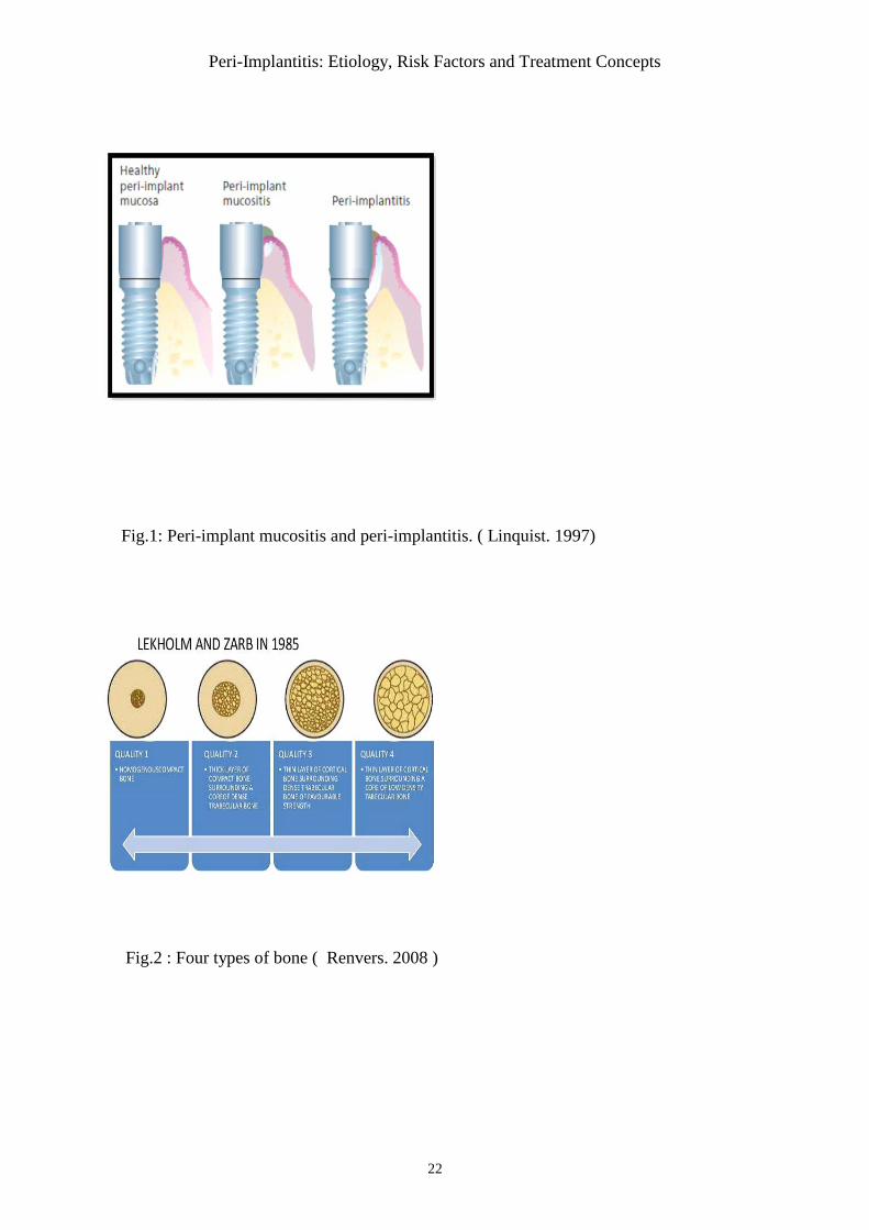
mucositis to peri-implantitis. Peri-implant mucositis describes an inflammatory lesion that resides
in the mucosa, may be identified clinically by redness and swelling of the soft tissue, but bleeding
on probing is currently recognized as the important feature. It is a reversible process surrounding a
functioning implant. While in peri-implantitis the mucosal lesion is often associated with
suppuration and depended pockets, but always accompanied by loss of supporting marginal bone.
It is a progressive and irreversible disease. ( Fig.1) ( Linquist. 1997 )
Peri-Implantitis: Etiology, Risk Factors and Treatment Concepts
2
II. RESULTS
1. ANATOMIC AND HISTOLOGICAL CONSIDERATIONS
i. Peri-Implant mucosa
The soft tissue that surrounds dental implants is termed peri-implant mucosa. The outer surface of
the peri-implant mucosa is also covered by a keratinized oral epithelium, which in the marginal
border connected with a thin barrier epithelium that faced the abutment part of the implant. The
connective tissue in the compartment above the bone appeared to be in direct contact with the
surface of the implant. The collagen fibers in this connective tissue apparently originated from the
periosteum of the bone crest and extend towards the margin of the soft tissue in directions parallel
to the surface of the abutment.( Lindhe. 2008)
ii. Attachment apparatus
Healing of the mucosa results in the establishment of a soft tissue attachment (transmucosal
attachment) to the implant. This attachment serves as a seal that prevents products from the oral
cavity reaching the bone tissue, and thus ensures osseointegration and the rigid fixation of the
implant. The biologic width is established within 6-8 weeks around a trans mucosal implant. The
apical seal of the implant sulcus is less resistant to periodontal probing (Mombelli et al. 1997;
Sucleanet et al. 1997).
2. PHYSIOLOGIC CONSIDERATIONS
i. Osseointegration
Osseointegration has been described by Branemark (1983) , as consisting of a highly
differentiated tissue making “a direct structural and functional connection between ordered, living
bone and the surface of a load-carrying implant". In 2008 Albrektsson suggested a new definition
of a Osseointegration: “ A process whereby clinically asymptomatic rigid fixation of alloplastic
materials is achieved and maintained in bone during functional loading”.
3. ETIOLOGY AND RISK FACTORS OF MARGINAL BONE LOSS
In recent consensus meetings, peri-implantitis has been described as peri-implant pathology with
multifactorial etiology, including implant related factors, clinician factors, and patients’ factors. It
is accepted that all implants will display some extent of bone loss after integration and through
time of function. ( Lindhe. 2008 )
Peri-Implantitis: Etiology, Risk Factors and Treatment Concepts
3
i. Microbial Factor
In 2011, Mombelli & Decailletconcluded that peri-implantitis may be viewed as a mixed
anaerobic infection and that in most cases the composition of the flora is similar to the
subgingival flora at chronic periodontitis.
History of periodontitis
The presence of periodontal pathogens around failing implants could suggest a direct link between
periodontitis and peri-implantitis, via a translocation of these species from their intra-oral niches
to the freshly inserted implants implants (Heitz-Mayfield et lang. 2010)
Poor oral hygiene
Plaque accumulation adjacent to dental implants is clearly associated with the development of
peri-implant mucositis. Dental floss is usually recommended in conjunction with interproximal
oral hygiene practices to achieve and maintain healthy oral conditions.( Maruyama. 2014)
Endodontic infection
The presence of untreated or insufficiently treated endodontic infections adjacent to the site of
implant placement can adversely affect the outcome ( lindhe. 2008)
ii. Periodontal tissues
Tissue architecture
Tissue architecture is defined as the distance between the contact point between two neighboring
teeth and a line connecting the mid-facial soft tissue margin of these teeth, has been categorized as
flat or scalloped. Studies reported that high scalloped tissues have a higher risk to undergo
recession. In contrast, flat tissue scallop tends to mimic the underlying osseous scallop facially.
The greater the discrepancy is the scallop and higher is the risk for recession
( Lindhe. 2008)
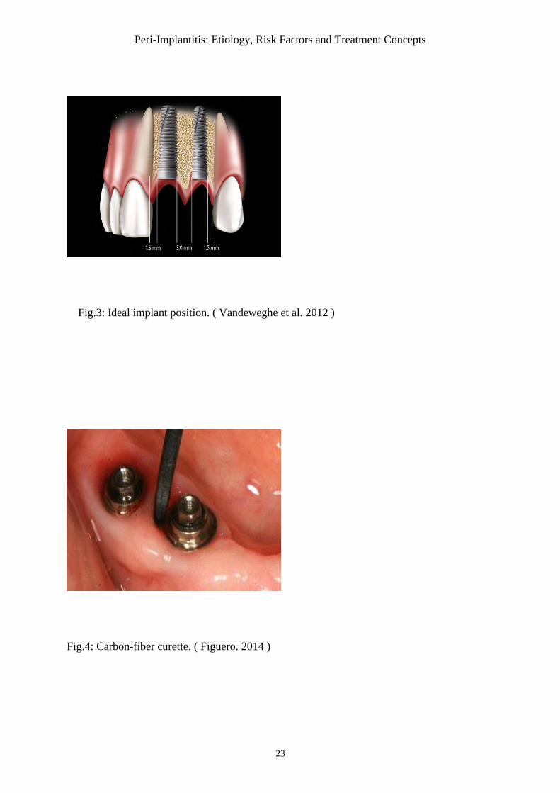
Bone quality
Mineral density, microarchitecture, and trabecular thickness were indicated as defining factors for
bone quality. Lekholm and Zarb determined in 1985 four types of bone depending of their
proportion of cortical and cancellous bone on radiographic appearances. ( Fig.2) ( Renvert. 2008)
Peri-Implantitis: Etiology, Risk Factors and Treatment Concepts
4
iii. Implant properties
Implant surface
It can occur that the coronal portion of the implant, which was initially designed to facilitate
osseointegration, become exposed to the oral environment as a result of peri-implantitis. Some
types of rough-surfaced implants, for instance those coated with hydroxyapatite (HA), were
reported to have a higher incidence of complications. (Renvert. 2015 )
Implant shape
In a study by De Bruyn (2010), it was suggested that the higher insertion and removal torque
forces reported with tapered implant have the advantage of achieving primary stability more
easily, but also increase the stress in the surrounding bone, which is highest in the cortical area
around the implant neck and induces more bone loss. The ultimate goal of the scalloped implant
design is to minimize the remodeling process seen around implants, thus improving the quality of
survival by maintaining three dimensional osseous and soft tissue contours. (Salvi. 2004 )
Microthreads
Recently, it was also claimed by several manufacturers that the introduction of retention grooves
(microthreads) at the neck of the implant may further reduce the amount of bone loss following
the implant installation.( Bratu. 2009)
Implant length and diameter
A systematic review of marginal bone loss showed that short dental implants (<10mm)had similar
peri-implant marginal bone loss as standard implants (>=10 mm) for implant- supported fixed
prostheses. It was suggested that ultra-short implants (6mm long) had an acceptable outcome as
also did wide diameter short implants (5 and 6 mm) which had less marginal bone loss than
implants 4.2 mm in diameter.(Van Velzen. 2016)
Bone level and tissue level
The initial depth of implant placement has very important clinical implications in terms of
esthetics. In most 2-stage implant systems, after the abutment is connected, a microgap exists
between the implant and abutment at or below the alveolar crest. In nonsubmerged implant
designs, the implant itself extends above the alveolar crest level; therefore, such a micrograp does
not exist at the level of the bone.( Palacci et Ericsson. 2001)
Peri-Implantitis: Etiology, Risk Factors and Treatment Concepts
5
iv. Surgery and implant placement
Surgical trauma has been regarded as one of the most commonly suspected etiologies proposed
for early implant failure. ( Palacci et Ericsson. 2001 )
Flap design
The periosteal elevation has been speculated as one of the possible contributing factors for crestal
implant bone loss. Vascular ischemia associated with flap reflection for second stage surgery has
been implicated as a potential source of MBL ( Marginal bone loss ).( Berberi et al. 2014 )
Wilderman et al. (1967) reported that the mean horizontal bone loss after osseous surgery with
periosteal elevation is approximately 0.8mm, and the reparative potential is highly dependent
upon the amount of cancellous bone existing underneath the cortical bone.
Implant position
In a study by Tarnow et al (2000) on the height of the inter-implant bone crest in relation to the
inter-implant distance reported a lower height of the bone crest in areas with an inter-implant
distance of <3mm as compared to areas with>3mm between the implants, and suggested that this
difference may be the result of lateral bone loss at the implants.
Based on the finding from a 10-years longitudinal study that the amount of bone loss was of
similar magnitude at implants with a neighbouring tooth and at those with a neighbouring implant,
the authors concluded that presence of remaining teeth may not influence the marginal bone loss
around an implant.(Fig.3) ( Vandeweghe et al 2012).
The limitations on bone thickness at the anterior of the maxilla and the mandible and anatomic
variations, such as concavity of the buccal aspects of the bone, can make it difficult to place a
dental implant in the proper position to achieve the best esthetic results. In these cases, titled
implants are used. The term titled implants refers to implants placed at an angle of normally 30
degrees or more with respect to axially or vertically positioned implants.(Parma-Benfenati. 2013)
Ata-Ali et al. (2013) conducted a literature review on titled implants from 1999 through 2010 and
concluded that there was no evidence of differences in the success rates between tilted and axial
implants. In another paper it was concluded that resorption in buccal areas may be less intensive
as the angulation of placed implants increases towards the palatal area in the maxilla, where as for
the mandibule a greater inclination towards the lingual area could be negative. In the mandibular,
Peri-Implantitis: Etiology, Risk Factors and Treatment Concepts
6
when the implant is slightly angulated in the direction of the distal area, bone resorption seemed
to be more marked in the buccal area.
Overheating and pressure
In 1984, Eriksson and Albreltsson reported that the critical temperature for implant site
preparation was 47C for 1 minute or 40C for 7 minutes. When the bone is overheated the risk of
implant failure is significantly increased. Matthews and Hirsch demonstrated that temperature
elevation was influenced more by the force applied than drill speed. ( Berberi et al. 2014 )
v. Prosthetic aspects
Abutment considerations
Atieh et al. (2010) conducted a systematic review on the clinical relevance of platform switching
and preservation of peri-implant crest bone levels.
Through the selection of a shorter abutment the mucosa can be compressed. Results indicate that
the choice of a shorter abutment may increase MBL. ( Galindo-Moreno et al. 2014 )
Occlusal factor: stress and number of implants
Occlusal overload can result in progressive marginal bone loss or even complete loss of
osseointegration, and when traumatic occlusion is combined with inflammation, the progression
of bone destruction is accelerated. Contributing factors associated with increased bending
overload in dental implants include: prostheses supported by 1 or 2 implants in the posterior
region, straight alignment of implants, significant deviation of the implant axis from the line of
action, high crown/implant ratio, and excessive cantilever length, discrepancy in dimensions
between the occlusal table and implant head and parafunctional habits.(Palacci et al. 2001)
The survival of implant-supported fixed prostheses by a smaller number of implants shows good
results both in the mandible and in the maxilla. In this context, it is suggested that the fewer
implants in total rehabilitation, the better. ( Persson et al. 2014 )
Excess of cement
Wilson (2008) found a correlation of 81% between excess cement left in the soft tissue and the
occurrence of sulcular bleeding and/or suppuration. A biofilm may form on the cement left in the
peri-implant tissue. ( Wohlfahrt et al. 2012 )
Peri-Implantitis: Etiology, Risk Factors and Treatment Concepts
7
Immediate Loading
During the one-stage surgery, the concept of immediate placement of definitive abutments and
insertion of immediate interim prostheses appeared to protect the blood clot and to prevent
interruption of the early mineralization of marginal bone. This was in contrast with several studies
that reported no differences in MBL between immediate and delayed loading of dental implants.
This phenomenon was more obvious in fresh extraction sockets, which demonstrated early MBL
during the first 8 weeks, after which a constant marginal bone height was observed over 5 years.
Two-phase surgical procedures have been associated with continuous MBL of between 0.01 and
0.02 mm/year, which could be in part related to the additional surgical procedure. During the
removal of cover screws and tightening of implant abutments, greater stress is delivered to the
initially mineralizing marginal bone; this manipulation could interface with the healing process
and thus result in increased MBL. ( Attieh et al. 2010 )
vi. Systemic factors
General risk factors are factors related to the individual and factors that may influence the
patient's susceptibility to infection.
Smoking
Tobacco consumption, in a dose-dependent manner, represents the third risk factor for the
progression of periodontal disease. Tobacco negatively affects the outcome of almost all
therapeutic procedures performed in the oral cavity. However, it was suggested that increased
implant failure in smokers is not the result of poor healing or osseointegration, but because of
exposure of peri-implant tissues to tobacco smoke. (Koldsland. 2010 )
Smoking upregulates the expression of pro-inflammatory cytokine, which contributes to increased
tissue damage and alveolar bone resorption. Lindquist reported significantly greater marginal
bone loss around implants in heavy smokers (>14 cigarettes per day) than in those with low
cigarette consumption (<14 cigarettes per day).( Mailoa. 2014).
Effects of smoking on implant survival and success are more pronounced in areas of poor quality
trabecular bone. In smokers, maxillary implants have more failure rate as compared to mandibular
implants. Probably because maxillary bone is of lower quality and consequently more susceptible
to the damaging effects of smoking. ( Kazat. 2012)
Peri-Implantitis: Etiology, Risk Factors and Treatment Concepts
8
Diabetes mellitus
Although there is a slight tendency for more failures of implants in a diabetic compared to a non-
diabetic population, the risk is not substantial in patients who are under good metabolic control.
In the general population the 5-year overall success rate for implants is approximately 95%,
where as in a diabetes population the rate is approximately 86%. Diabetes under suboptimal
metabolic control often experience wound-healing difficulties and have an increases susceptibility
to infections due to a variety of problems associated with immune dysfunctions. ( Lindhe. 2008 )
Alcohol consumption
Alcohol consumption may cause a vitamin K deficit disrupting the production of prothrombin,
thus affecting coagulation mechanisms. Only one study concluded that peri-implant marginal
bone loss was statistically linked to alcohol consumption > 10g per day and that alcohol induced
more serious peri-implantitis than cigarettes. ( Oltra. 2013 )
Post-menopausal osteoporosis
A study by Dvorak et al. (2011) found no relation between osteoporosis and peri-implantitis in
adult women despite the high success rates of implants placed in patients taking BP(
bisphosphonate) complications have been reported.
Data from a study by Cohen et al. (2011) suggested that patients taking BP may be at greater risk
of peri-implant bone loss ( BP inhibit osteoclast action and thereby bone resorption)
Hyposalivation
Hyposalivation could be a risk factor for peri-implantitis. In fact, decrease of salivary flow was
often accompanied by a change in its composition and reducing bacterial clearance in the oral
cavity. ( Lindhe. 2008 )
4. DIAGNOSIS
i. Clinical diagnosis
Peri-implant lesions are commonly asymptomatic and frequently detected in follow up visits.
These lesions are clinically defined by the presence of redness and swelling of the mucosa,
bleeding and/or suppuration on probing, deepening of the pockets adjacent to the dental implants,
Peri-Implantitis: Etiology, Risk Factors and Treatment Concepts
9
and loss of the implant-supporting bone. Palpation reveals pus, swelling and pain, and the
exudation of grafting practices. (Lindhe. 2008 )
Probing
Probing is essential for diagnosis of peri-implant diseases. In the European consensus report, it
was concluded that the probing depth, the presence of BOP ( Bleeding On Probing ) and
suppuration should be assessed regularly for the diagnosis for peri-implant diseases.( Colet.2011)
Probing Pocket Depth (PPD) is the distance from the edge of peri-implant tissue to the bottom of
peri-implant sulcus/pocket and clinical attachement level (CAL) is the distance from the implant
platform or connection area to the bottom of peri-implant sulcus/pocket. ( Ferreira. 2014 )
An increase in probing depth over time is associated with the loss of attachment and supporting
bone. Because healthy implants generally have probing depths that are less than 4mm, peri-
implant pockets of 5 mm or more should be regarded as an indicator of bone loss and hence, a
radiographic evaluation is required. Also, peri-implant hyperplasia is often found in an area of
absence of keratinized gingiva or the overflow of supra-implant structures. ( Oltra. 2013 )
Heitz-Mayfielod (2008) concluded that probing using a light force (0.25N) does not damage the
peri-implant tissues. BOP represents histological inflammatory tissue changes.
Suppuration
The presence of pus is always a sign of infection associated with destruction of peri-implant
tissues. When finger pressure is exerted on the peri-implant mucosa, it is common to observe pus
discharge. This phenomenon is never present in the case of a peri-implant mucositis.( Heitz-
Mayfield. 2010 )
Implant mobility
Presence or absence of mobility is checked digitally or with the help of instruments trying to
move the implants. A perceived mobility of the restoration per se does not necessarily mean that
the implant is loose. The connection suprastructure/abutment and/or abutment/implant might be
loose leading to the mobility. ( Lindhe. 2008)
Implant mobility is not used to diagnose peri-implantitis because it indicates the complete loss of
osseointegration and the failure of the implant. Osstell uses Resonance Frequency Analysis to
determine implant stability and osseointegration.( Ferreira. 2014 )
Peri-Implantitis: Etiology, Risk Factors and Treatment Concepts
10
ii. Radiological diagnosis of peri-implantitis
Radiographic examination allows clinicians to assess the relationship between implants and
prosthetic components. In addition, they are required to evaluate supporting bone levels around
implants. Hence, regular radiographical check-up for marginal bone loss has been emphasized for
early detection of peri-implantitis. Periapical radiography using fixed angulators to prevent image
deformation is often used to verify marginal bone level or interproximal bone loss in peri-
implantitis. ( Ferreira. 2014 )
The use of digital image analysis had expanded into implant dentistry to monitor peri-implant
bone healing and gain or loss of alveolar bone density, as well as infrabony lesions in three
planes, true to scale and without any overlay or distortion. (Oltra. 2013)
Peri-implantitis lesions appear crater like or saucer-shaped defects. It is essential to differentiate a
biological process (saucerization) from a pathological one (peri-implantitis). Saucerization is a
biological process of peri-implant cervical bone remodeling starting in the first year after implant
placement that is observed radiographically around certain implant type. It ranges from 0.4mm to
1.6mm within the first year and then approximately 0.1 mm/year. ( Palacci. 2001 )
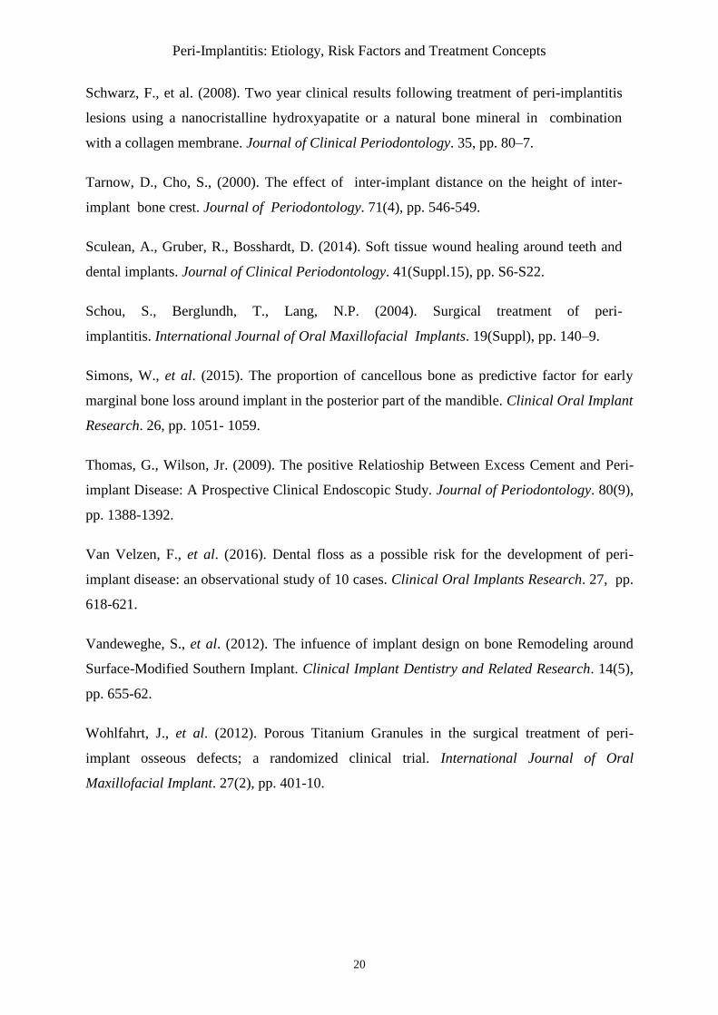
iii. Classification of peri-implantitis
To date, there have been no standardized parameters to clinically differentiate the various stages
and severities of peri-implantitis. A proposed classification by Rosen et al. (2012) is based on
three distinct clinical stages of peri-implantitis: early, moderate, and advanced. It is depending on
Pocket deph and bone loss. ( Table. 1)
5. TREATMENT CONCEPTS
Although peri-implantitis is reported to be a common problem, there is lack of scientific
guidelines in how to treat peri-implantitis. The ideal goal of the treatment of peri-implantitis
would be the resolution of disease, i.e. no suppuration or bleeding on probing, no further bone
loss, and the reestablishment and maintenance of healthy peri-implant tissues, knowing that re-
osseointegration on a previously contaminated implant surface is possible. (Sahm. 2011 )
i. Prevention and maintenance
The prognosis of the implant is highly dependent on appropriate case selection, recall sessions for
maintenance and primary and secondary prevention. (Simons. 2015)
Peri-Implantitis: Etiology, Risk Factors and Treatment Concepts
11
ii. Non-surgical therapy
It was stated that non-surgical treatment is recommended for marginal bone loss less than 2mm
around implant and surgical treatment is recommended for marginal bone loss greater than 2mm.(
Simons. 2015 )
Mechanical debridement
Different types of curettes have been studied and most of the protocols included an adjunctive
polish with a rubber cup and polishing paste (fluor of pumice, nupro Fine, tin oxide).Steel curettes
have an external hardness higher than titanium and accordingly are not indicated for cleaning
titanium implants. Titanium-coated curettes have a similar hardness to the titanium surface and
thus do not scratch its surface. Carbon-fiber curettes are softer than the implant surface and
therefore remove bacterial deposits without damaging the surface, although they break easily.
(Fig.4) Teflon curettes have similar properties to carbon-fiber curettes. Plastic curettes are the
most fragile of all curette types and have limited debriding capacity.( Figuero. 2014 )
Ultrasonic devices
Ultrasonic devices with polyetheretherketone coated tips have been used to debride the implant
surface easily and is comfortable for the patient.( Figuero. 2014 )
The study by Karring and al. (2005) demonstrated that sub-mucosal debridement alone,
accomplished by utilizing either an ultrasonic device or carbon fiber curettes, is not sufficient for
the decontamination of the surface of implants with peri-implant pockets bigger than 5mm and
exposed implant threads.
Air abrasion
Recently, a powered air abrasive system, based on a low-abrasive amino-acid glycine powder, has
been demonstrated as an effective method of biofilm removal from the implant surface. ( Figuero.
2014 )
Adjunctive use of antimicrobial products :
Adjunctive therapies, such as antiseptics and antibiotics, have been proposed to improve the
results of nonsurgical debridement. ( Lang. 2011 )
Peri-Implantitis: Etiology, Risk Factors and Treatment Concepts
12
The addition of antiseptic therapy (Chlorhexidine-based products) to mechanical debridment ,
seems to provide additional clinical improvements is deep peri-implant lesion mean PPD> 5mm. (
Renvert et al. 2006)
The study by Renvert et al. (2008) demonstrated that the adjunctive benefits derived from the
addition of an antibiotic to mechanical debridement tend to be greater, although to a limited
extent, than those achieved by the combined use of an antiseptic (chlorhexidine) and mechanical
debridement.
Significant reductions in bleeding on probing were found after combining mechanical therapy
with the use of a tetracycline-containing fiber. There are no clinical trials available nowadays on
the systemic administration of antibiotics for the therapy of peri-implantitis.( Sahm. 2011 )
iii. Surgical therapy
In the many patients with advanced lesions, mechanical nonsurgical therapy alone is insufficient
in the treatment of peri-implantitis and a surgical intervention is often needed. The primary aim of
such a procedure is to provide access for decontamination of the infected implant surface in order
to allow undistributed healing and reduce the risk for further disease progression. The decision on
whether to use a resective or a regenerative surgical technique depends on the clinical situation.
However, even if surgery seems to be the therapy of choice, nonsurgical therapy should always be
performed before surgical interventions.( Figuero. 2014)
Access flap surgery and implant surface decontamination.
The objective of the access flap surgery intervention is to conserve and to maintain all the soft
tissues around the affected implant and to focus mainly on the decontamination of the implant
surface. Usually, intracrevicular incisions are made around the affected implants and
mucoperiostal flaps are raised both buccally and palatally/lingually. As this technique aims to
maintain the position of the soft-tissue margin around the implant neck, this can only be attained
when the peri-implant bone loss is shallow. Apically positioned flaps have been advocated in
order to enhance self-performed oral hygiene and reduce the pockets around the affected implants.
Numerous approaches have been used for implant surface decontamination during peri-implant
surgery, including mechanical, chemical, laser treatments, photodynamic therapy. The rationale
for the use of chemical treatments is to disinfect/decontaminate the implant surface by direct
application of appropriate substances. Citric acid, hydrogen peroxide, chlorhexidine and/or saline
have been utilized, and all have given similar results in experimental studies.( Figuero. 2014 )
Peri-Implantitis: Etiology, Risk Factors and Treatment Concepts
13
Photodynamic therapy generates reactive oxygen species by multiplicity with help of a high-
energy single-frequency light (e.g. diode lasers) in combination with photosensitizers (e.g.
toluidine blue). As a recommendation, photodynamic therapy has to be considered as an
additional treatment option.( Thomas. 2009 ) ( Fig.5)
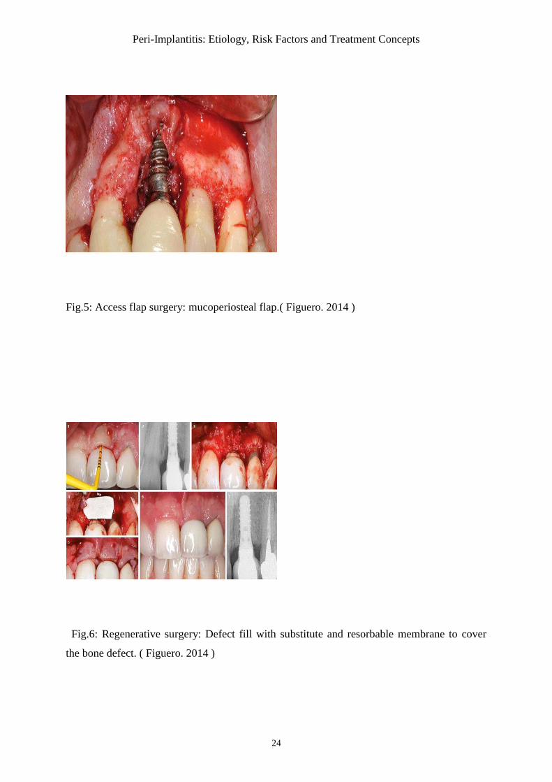
Regenerative therapy
In this technique, intracrevicular incisions are often performed in order to maintain the total
amount of soft tissues. Degranulation of the defect is performed using titanium instruments. And a
graft is placed around the implant, filling the intrabony component of the defect. Grafting may be
performed with either autologous bone or bone substitutes. The graft may be covered with a
resorbable or a nonresorbable membrane (Fig.6). Finally, the flaps are coronally positioned and
sutured in order to determine healing. Following mechanical decontamination, the implant surface
and the wound area shoud be rinsed with sterile saline solution for a minimum of 2min.
Reconstruction with PTG ( Porous Titanium Granules ) resulted in significantly better
radiographic peri-implant defect fill.( Figuero. 2014 )
III. DISCUSSION :
Treatment of peri-implant mucositis and peri-implantitis must be focused on early diagnosis and
controlling the risk factors or indicators to prevent it from occurring.
In a non-surgical approach to treat peri-implantitis, local debridment was used to clean the surface
of implants. Instruments softer than titanium, such as polishing with a rubber cup and paste, floss,
interdental brushes, or using plastic scaling instruments have been shown not to roughen the
implant surface unlike metal and ultrasonic scalers. (Matarasso. 1996). Although implant surface
damage can almost be prevented by using either ultrasonic scalers with a nonmetallic tip or
resin/carbon fiber curettes, the presence of implant threads and/or implant surface roughness may
compromise the access for cleaning. (Schou. 2004)Another study demonstrated that sub-mucosal
debridement alone, accomplished by utilizing either an ultrasonic device or carbon fiber curettes,
is not sufficient for the decontamination of the surfaces of implants with peri-implant pockets ≥ 5
mm and exposed implant threads. (Karring.2005). So it seems reasonable to suggest that
mechanical or ultrasonic debridement alone may not be an adequate modality for the resolution of
peri-implantitis.
Treatment of peri-implant infection by mechanical debridement with plastic curettes combined
with antiseptic (0.2% chlorhexidine) therapy may lead to significant improvements in bleeding on
Peri-Implantitis: Etiology, Risk Factors and Treatment Concepts
14
probing, peri-implant probing pocket depth, and clinical attachment level in 6 months
interval.(Schwarz.2008) This contradicts with another study where the addition of antiseptic
therapy to mechanical debridement does not provide adjunctive benefits in shallow peri-implant
lesions where the mean probing pocket depth was <4 mm. (Renvert.2008)
Patients suffering from localized peri-implant problems in the absence of other infections may be
candidates for treatment by local drug-delivery devices. Local application of antibiotics by the
insertion of tetracycline fibers for 10 dayscan provide a sustained high dose of the antimicrobial
agent precisely into the affected site for several days. (Lang.2000)
The use of minoccline microspheres as an adjunct to mechanical therapy is beneficial in the
treatment of peri-implant lesions, but the treatment may have to be repeated. A study showed that
the adjunctive benefits derived from the addition of an antibiotic minocycline to mechanical
debridement tend to be greater, although to a limited extent, than those achieved by the combined
use of an antiseptic (chlorhexidine) and mechanical debridement.(Renvert.2008)
If the problem is generalized, specific microbiological information is collected and antibiotics are
administered systemically. If peri-implantitis is associated with persisting periodontal disease,
then both conditions need to be treated. In this case, the adjunctive use of systemic antibiotics may
be considered. There are no clinical trials available nowadays on the systemic administration of
antibiotics for the therapy of peri-implantitis. (Lang.2000)
Provided that mechanical and antiseptic protocols are followed prior to administering antibiotic
therapy, it appears that shallow peri-implant infection may be successfully controlled using
antibioics. But it is still contoversial whether deeper peri-implant lesions can be adequately treated
non-surgically by a combination of a local antibiotic and mechanical debridement. Thus surgical
techniques to treat prei-implantitis might be necessary.
Surgical resection is generally confined to implants placed in non-aesthetic sites. Surgical flap
helps in comprehensive debridement and decontamination of the affected implant. Surgical
therapy can by placing autogenous bone grafts covered by membranes, autogenous bone grafts
alone, membranes alone, or a control access flap procedure. It was shown that defects treated with
membrane-covered autogenous bone demonstrated significantly larger amounts of bone
regeneration and reosseointegration than those treated with the other three procedures. However,
membrane exposure is a frequent complication after such procedures. (Schou.2004) As
conclusion, the combination of guided bone regeneration with either demineralized freeze-dried
Peri-Implantitis: Etiology, Risk Factors and Treatment Concepts
15
bone or resorbable hydroxyapatite appears to be the treatment of choice for plaque-induced peri-
implant defects. (Hürzeler.1997)
A randomized comparative clinical trial concluded that resective surgical procedures coupled with
implantoplasty could have a positive influence on the survival rates of rough-surfaced implants
affected by peri-implantitis as well as on peri-implant clinical parameters, such as pocket-probing
depth, suppuration, and sulcus bleeding. (Romeo.2005,2007)
In another study, nanocrystalline hydroxyapatite and guided bone regeneration provided
clinically significant improvements in clinical parameters. Following up 2-year results, both
treatment modalities were efficient in providing clinically significant reductions of pocket-probing
depth and gains in clinical attachment level. The combination of application of natural bone
mineral and collagen membrane seemed to correlate with greater improvements in those clinical
parameters and, hence, was associated with a more predictable and enhanced healing outcome.
(Schwarz.2000,2006)
In a recent 2-11-year retrospective study study, long-term outcome of surgical treatment of peri-
implantitis was investigated. The treatment included oral hygiene instruction, professional supra-
mucosal instrumentation, and surgical therapy aiming at pocket elimination. The results of the
study revealed that surgical treatment of peri-implantitis was effective in the long-term, the
outcome was better at implants with non-modified than with modified surfaces, and preservation
of crestal bone support was consistent with healthy peri-implant tissue conditions.
(Berglundh.2018)
IV. CONCLUSION
Peri-implant diseases are important disease entities as a result of their high prevalence and the
lack of a standard mode of therapy. Patient selection is a key factor in the implant therapy; a
thorough study of the patient’s local and systemic risk factors is important before any implant
treatment. Risk factors are also practitioner dependent (surgical trauma, implant choice and
positioning…) Surgical techniques may be necessary to provide us with adequate access to
degranulate the inflamed tissues effectively as well as to decontaminate and, if indicated, modify
the implant surface. Before surgical techniques are employed, nonsurgical therapy and
reinforcement of the oral hygiene should be performed. Regular follow-up appointments and close
monitoring of the treated sites is, of course, important for preventing relapse.
Peri-Implantitis: Etiology, Risk Factors and Treatment Concepts
16
V. REFERENCES
Albrektsson, T. (2008). Hard tissue implant interface. Australian Dental Journal, 53(Suppl),
pp. S34-S38.
Atieh, M., Ibrahim, H., Atieh, A. (2010). Platform Switching for Marginal Bone Preservation
Around Dental Implants: A Systematic Review and Meta- Analysis. Journal of
Periodontology, 81(10), pp. 1350-366.
Berberi, A., et al. (2014). Influence of Surgical and Prosthetic Techniques on Marginal Bone
Loss around titanium Implants. Part I: Immediate Loading in fresh Extraction Sockets.
Journal of Prosthodontics, 23(7), pp. 521-527.
Berglundh, T., Wennström, J.L., Lindhe, J. (2018). Long-term outcome of surgical treatment
of peri-implantitis. A 2-11-year retrospective study. Clinical oral implants Research,
Disponivel em: <https://doi.org/10.1111/clr.13138>.
Brånemark, P.I. (1983). Osseointegration and its experimental background. Journal of
Prosthetic Dentistry. 50(3), pp. 399-410.
Bratu, E.A., Tandlich, M., Shapira, L. (2011). A rough surface implant neck with
microthreads reduces the amount of marginal bone loss: a prospective clinical study. Clinical
Oral Implants Research. 20(8), pp. 827-32.
Colet, D., et al. (2011). Peri-implantitis: Literature Review. Dental Press Periodontal Implant
Dentistry. 5(4), pp. 56-65.
Dvorak, G., et al. (2011). Peri-implantitis and late implant failures in postmenopausal
women: a cross-sectional study. Journal of Clinical Periodontology. 38(10), pp. 950-5.
Eriksson, A., Albrektsson, T., Albrektsson, B. (1984). Heat caused by drilling cortical bone
Temperature measured in vivo in patients and animals. Acta Orthopeadia Scandinavica.
55(6), pp. 629-631.
Ferreira, R., et al. (2014). Peri-implantitis: Critical an current overview of etiological factors,
clinical/radiografic diagnosis and prognosis. A literature review. Dental Press Implantology.
8(3), pp. 76-84.
Peri-Implantitis: Etiology, Risk Factors and Treatment Concepts
17
Figuero, E., et al. (2014). Management of peri-implant mucositis and peri-implantitis.
Periodontology 2000, 66, pp. 255-273.
Galindo-Moreno, P., et al. (2014). Prosthetic Abutment Height is a key Factor in peri-implant
Marginal Bone Loss. JDR Clinical. Suppl, pp. 80S-85S.
Heitz-Mayfield, L., Lang, N. (2010). Comparative biology of chronic and aggressive
periodontitis vs. peri-implantitis. Periodontology 2000. 53, pp. 167-181.
Heitz-Mayfield L. (2008). Peri-implant diseases: diagnosis and risk indicators. Journal of
Clinical Periodontology. 8(Suppl), pp. 292-304.
Hürzeler, M.B., et al. (1997). Treatment of peri-implantitis using guided bone regeneration
and bone grafts, alone or in combination, in beagle dogs. Part 2: Histologic findings.
International Journal of Oral & Maxillofacial Implants. 12(2), pp. 168-75.
Karring E.S., et al. (2005). Treatment of periimplantitis by the Vectors system. A pilot
study. Clinical Oral Implants Research. 16, pp. 288–93.
Kasat, V., Ladda, R. (2012). Smoking and dental implants. Journal of International Society of
Preventive and Community Dentistry. 2(2), pp. 38-41.
Koldsland, O.C., Scheie, A.A., Aass, A.M. (2010). Prevalence of peri-implantitis related to
severity of the disease with different degrees of bone loss. Journal of Periodontology. 81(2),
pp. 231-8.
Kotsovilis, S., et al. (2008). Therapy of peri-implantitis: a systematic review. Journal of
Clinical Periodontology. 35, pp. 621-629.
Lang, N.P., Berglundh, T. (2011). On behalf of working Group 4 of the seventh European
Workshop on Periodontology: Peri-implant diseases: where are we now? – Consensus of the
seventh European Workshop on Periodontology. Journal of Clinical Periodontology. 38(11
Suppl), pp. 178-181.
Peri-Implantitis: Etiology, Risk Factors and Treatment Concepts
18
Lang, N.P., Wilson, T.G., Corbet, E.F. (2000). Biological complications with dental implants:
Their prevention, diagnosis and treatment. Clinical Oral Implants Research. 11(Suppl 1),
pp. 146–55.
Lindhe, J., Lang, N., Karring T. (2008). Clinical Periodontology and implant dentistry.
Wiley-Blackwell, 5(1).
Lindhe, J., Meyle, J. (2008). Peri-implant diseases: consensus Report of the Sixth European
Workshop on Periodontolgy. Journal of Clinical Periodontology. 35(Suppl.8), pp. 282-285.
Linquist, L.W., Carlsson, G.E., Jemt, T. (1997). Association between marginal bone loss
around osseointegration mandibular implants and smoking habits: a 10-year follow-up study.
Journal of Dental Research. 76(10), pp. 1667-74.
Maruyama, N., et al. (2014). Intraindividual variation in core microbiota in peri-implatitis and
periodontitis. Scientific Reports. 4, p. 6602.
Matarasso, S., et al. (1996). Maintenance of implants: An in vitro study of titanium implant
surface modifications subsequent to the application of different prophylaxis
procedures. Clinical Oral Implants Research. 7, pp. 64–72.
Mombelli, A., et al. (1997). Comparison of periodontal and peri-implant probing by depth-
force pattern analysis. Clinical Oral Implants Research. 8, pp. 448-454.
Mombelli, A., De’caillet, F. (2011). The characteristics of biofilms in peri-implant disease.
Journal of Clinical Periodontology. 38(Suppl.11), pp. 203-213.
Oltra, D., et al. (2013). Rehabilitation of the Atrophic Maxilla with Tilted implant: review of
the literature . Journal of Implantology. 39(5), pp. 625-32.
Palacci, P., Ericsson, I., (2001). Esthetic Implant Dentistry; Soft And Hard Tissue
Management. Quintessence Books, Chicago.
Parma-Benfenati, S., Roncati, M., Tinti, C. (2013). Treatment of Peri-Implantitis: Surgical
Therapeutic Approaches Based on Peri-implantitis Defects. International Journal of
Periodontics Restorative Dentistry. 33, pp. 627-633.
Peri-Implantitis: Etiology, Risk Factors and Treatment Concepts
19
Persson, G., Renvert, S. (2014). Cluster of bacteria Associated with Peri-implantitis. Clinical
Implant Dentistry and Related Research. 16(6), pp. 783-93.
Renvert, S., Quirynen, M. (2015). Risk indicators for peri-implantitis. A narrative review.
Clinical Oral Implants Research. Suppl. 11, pp. 15-44.
Renvert, S., Roos-Jansaker, A.M., Claffey, N. (2008). Non-surgical treatment of peri-implant
mucositis and peri- implantitis: a literature review. Journal of Clinical Periodontology.
35(Suppl.8), pp. 305-315.
Renvert, S., et al. (2008). Mechanical and repeated antimicrobial therapy using a local drug
delivery system in the treatment of peri implantitis: A randomized clinical trial. Journal of
Periodontology. 79, pp. 836–44.
Romeo, E., et al. (2005). Therapy of peri-implantitis with resective surgery. A 3-year clinical
trial on rough screw-shaped oral implants. Part I: Clinical outcome. Clinical Oral Implants
Research. 16, pp. 9–18.
Romeo, E., et al. (2007). Therapy of peri-implantitis with resective surgery. A 3- year clinical
trial on rough screw-shaped oral implants. Part II: Radiographic outcome. Clinical Oral
Implants Research. 18, pp. 179–87.
Sahm, N., et al. (2011). Non-surgical treatment of peri-implantitis using an air-abrasive
device or mechanical debridement and local application of chlorhexidine: a prospective,
randomized, controlled clinical study. Journal of Clinical Periodontology. 38, pp. 872-878.
Salvi, G., Lang, N., (2004). Diagnostic Parameters for Monitoring Peri-implant Conditions.
International Journal of Oral Maxillofacial Implants. 19(Suppl.), pp.116-127.
Schwarz, F., et al. (2008). Two year clinical results following treatment of peri-implantitis
lesions using a nanocristalline hydroxyapatite or a natural bone mineral in combination
with a collagen membrane. Journal of Clinical Periodontology. 35, pp. 80–7.
Schwarz, F., et al. (2006). Healing of intrabony peri-implantitis defects following
application of a nanocrystalline hydroxyapatite (Ostimt) or a bovine-derived xenograft (Bio-
Osst) in combination with a collagen membrane (Bio-Gidet). A case series. Journal of
Clinical Periodontology. 33, pp. 491–9.
Peri-Implantitis: Etiology, Risk Factors and Treatment Concepts
20
Schwarz, F., et al. (2008). Two year clinical results following treatment of peri-implantitis
lesions using a nanocristalline hydroxyapatite or a natural bone mineral in combination
with a collagen membrane. Journal of Clinical Periodontology. 35, pp. 80–7.
Tarnow, D., Cho, S., (2000). The effect of inter-implant distance on the height of inter-
implant bone crest. Journal of Periodontology. 71(4), pp. 546-549.
Sculean, A., Gruber, R., Bosshardt, D. (2014). Soft tissue wound healing around teeth and
dental implants. Journal of Clinical Periodontology. 41(Suppl.15), pp. S6-S22.
Schou, S., Berglundh, T., Lang, N.P. (2004). Surgical treatment of peri-
implantitis. International Journal of Oral Maxillofacial Implants. 19(Suppl), pp. 140–9.
Simons, W., et al. (2015). The proportion of cancellous bone as predictive factor for early
marginal bone loss around implant in the posterior part of the mandible. Clinical Oral Implant
Research. 26, pp. 1051- 1059.
Thomas, G., Wilson, Jr. (2009). The positive Relatioship Between Excess Cement and Peri-
implant Disease: A Prospective Clinical Endoscopic Study. Journal of Periodontology. 80(9),
pp. 1388-1392.
Van Velzen, F., et al. (2016). Dental floss as a possible risk for the development of peri-
implant disease: an observational study of 10 cases. Clinical Oral Implants Research. 27, pp.
618-621.
Vandeweghe, S., et al. (2012). The infuence of implant design on bone Remodeling around
Surface-Modified Southern Implant. Clinical Implant Dentistry and Related Research. 14(5),
pp. 655-62.
Wohlfahrt, J., et al. (2012). Porous Titanium Granules in the surgical treatment of peri-
implant osseous defects; a randomized clinical trial. International Journal of Oral
Maxillofacial Implant. 27(2), pp. 401-10.
Peri-Implantitis: Etiology, Risk Factors and Treatment Concepts
21
Table.1: Classification of peri-implantitis. (Rosen et al. 2012)
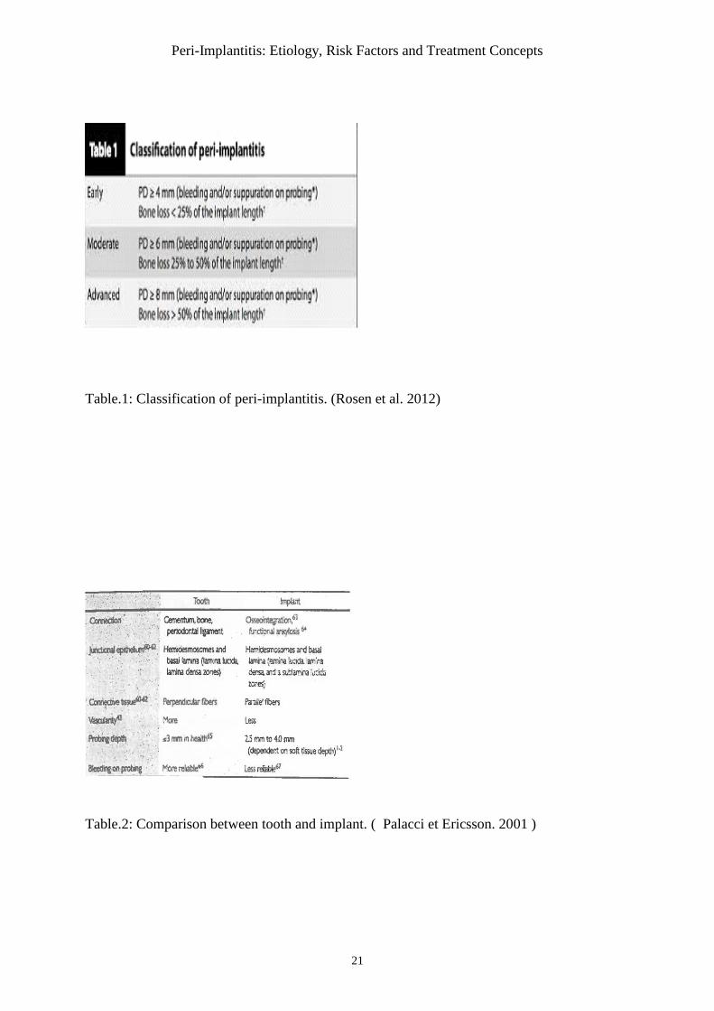
Table.2: Comparison between tooth and implant. ( Palacci et Ericsson. 2001 )
Peri-Implantitis: Etiology, Risk Factors and Treatment Concepts
22
Fig.1: Peri-implant mucositis and peri-implantitis. ( Linquist. 1997)
Fig.2 : Four types of bone ( Renvers. 2008 )
Peri-Implantitis: Etiology, Risk Factors and Treatment Concepts
23
Fig.3: Ideal implant position. ( Vandeweghe et al. 2012 )
Fig.4: Carbon-fiber curette. ( Figuero. 2014 )
Peri-Implantitis: Etiology, Risk Factors and Treatment Concepts
24
Fig.5: Access flap surgery: mucoperiosteal flap.( Figuero. 2014 )
Fig.6: Regenerative surgery: Defect fill with substitute and resorbable membrane to cover
the bone defect. ( Figuero. 2014 )
Peri-Implantitis: Etiology, Risk Factors and Treatment Concepts
25
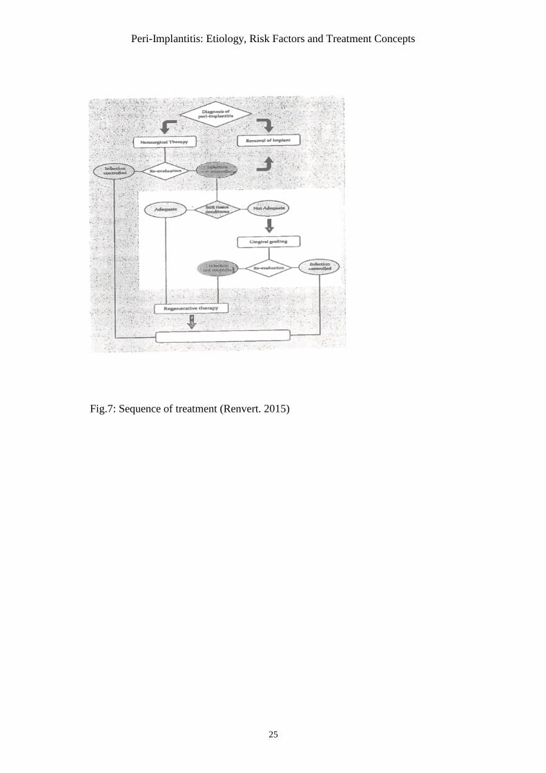
Fig.7: Sequence of treatment (Renvert. 2015)