implicações Ósseas na sepse sistêmica aguda: uma análise ... · o rio não pode voltar....
TRANSCRIPT

I
Emiacutelia Maria Gomes Aguiar
Implicaccedilotildees Oacutesseas na Sepse Sistecircmica Aguda Uma anaacutelise quiacutemica e estrutural
Acute Systemic Sepsis Bone Implications A chemical and structural
analysis
Dissertaccedilatildeo apresentada agrave Faculdade de Odontologia da Universidade de Uberlacircndia para a obtenccedilatildeo do Tiacutetulo de Mestre em Odontologia na Aacuterea de Cliacutenica Odontoloacutegica Integrada
Uberlacircndia 2018
II
Emiacutelia Maria Gomes Aguiar
Implicaccedilotildees Oacutesseas na Sepse Sistecircmica Aguda Uma anaacutelise quiacutemica e estrutural
Acute Systemic Sepsis Bone Implications A chemical and structural
analysis Dissertaccedilatildeo apresentada agrave Faculdade de Odontologia da Universidade Federal de Uberlacircndia para a obtenccedilatildeo do Tiacutetulo de Mestre em Odontologia Aacuterea de Cliacutenica Odontoloacutegica Integrada
Orientador Prof Dr Robinson Sabino-Silva
Banca Examinadora
Prof Dr Robinson Sabino-Silva Prof Dra Priscilla Barbosa Ferreira Soares
Prof Dra Roberta Okamoto
Uberlacircndia 2018
Dados Internacionais de Catalogaccedilatildeo na Publicaccedilatildeo (CIP)
Sistema de Bibliotecas da UFU MG Brasil
A282i
2018
Aguiar Emiacutelia Maria Gomes 1993
Implicaccedilotildees oacutesseas na sepse sistecircmica aguda uma anaacutelise quiacutemica e
estrutural Emiacutelia Maria Gomes Aguiar - 2018
45 p il
Orientador Robinson Sabino da Silva
Dissertaccedilatildeo (mestrado) - Universidade Federal de Uberlacircndia
Programa de Poacutes-Graduaccedilatildeo em Odontologia
Disponiacutevel em httpdxdoiorg1014393ufudi2018702
Inclui bibliografia
1 Odontologia - Teses 2 Osteoclastos - Teses 3 Biomecacircnica -
Teses 4 Ossos - Doenccedilas - Teses I Silva Robinson Sabino da II
Universidade Federal de Uberlacircndia Programa de Poacutes-Graduaccedilatildeo em
Odontologia III Tiacutetulo
CDU 616314
Angela Aparecida Vicentini Tzi Tziboy ndash CRB-6947
Scanned by CamScanner
III
DEDICATOacuteRIA
Dedico esse trabalho a todos aqueles que
participaram comigo durante essa jornada em
especial minha avoacute Maria que nos deixou
recentemente e que mesmo sem entender o que
esse caminho significava sorria sempre com as
minhas vitoacuterias Aos meus pais Juliano e Graccedila
meu irmatildeo Mateus e meu namorado Henrique por
tornarem esse sonho possiacutevel contribuindo sempre
com todo cuidado amor e carinho Amo vocecircs
IV
AGRADECIMENTOS
Agrave minha famiacutelia por me apoiar sempre e ser a base de sustentaccedilatildeo dos meus sonhos
Ao meu noivo Henrique que percorreu mais essa etapa ao meu lado fazendo com que
fosse mais faacutecil e mais feliz
Ao meu orientador Robinson Sabino-Silva por dividir comigo sua experiecircncia
contribuindo para o meu crescimento e finalizaccedilatildeo deste projeto
Agrave Leacuteia Cardoso que se tornou uma grande amiga e que esteve por traz de todo o
aprendizado adquirido durante o Mestrado
Agrave Faculdade de Odontologia da Universidade Federal de Uberlacircndia por permitir o
meu desenvolvimento profissional
Agrave FAPEMIG que ofereceu suporte financeiro para a concretizaccedilatildeo desse sonho
Ao ICBIM e CPBio que disponibilizaram sua estrutura fiacutesica para que o
desenvolvimento da minha pesquisa fosse possiacutevel
Ao Prof Dr Gustavo Rabelo que apesar do pouco tempo prestou preciosas
informaccedilotildees para a realizaccedilatildeo deste trabalho
A todas as pessoas que participaram e que contribuiacuteram de alguma forma com o
desenvolvimento deste trabalho meus sinceros agradecimentos
V
EPIacuteGRAFE
Dizem que antes de um rio entrar no mar ele treme de medo
Olha para traacutes para toda a jornada que percorreu para os cumes
as montanhas para o longo caminho sinuoso que trilhou atraveacutes
de florestas e povoados e vecirc agrave sua frente um oceano tatildeo vasto
que entrar nele nada mais eacute do que desaparecer para sempre Mas
natildeo haacute outra maneira O rio natildeo pode voltar Ningueacutem pode
voltar Voltar eacute impossiacutevel na existecircncia O rio precisa de se
arriscar e entrar no oceano E somente quando ele entrar no
oceano eacute que o medo desaparece porque apenas entatildeo o rio
saberaacute que natildeo se trata de desaparecer no oceano mas de tornar-
se oceano
Osho
SUMAacuteRIO
VI
RESUMO 7
ABSTRACT 8
1 INTRODUCcedilAtildeO E REFERENCIAL TEOacuteRICO 9
11 SEPSE DEFINICcedilAtildeO ASPECTOS PATOFISIOLOacuteGICOS E EPIDEMIOLOacuteGICOS 9
12 MODELOS ANIMAIS DE SEPSE 10
13 OSSO E SEPSE 11
2 CAPIacuteTULOS 14
ARTIGO 1 - ACUTE SYSTEMIC SEPSIS BONE IMPLICATIONS A CHEMICAL AND STRUCTURAL ANALYSIS 14
Abstract 16
1 Introduction 17
2 Methods 18
21 Experimental design 18
22 FTIR and RAMAN spectroscopy 19
23 Biomechanical testing 20
24 Micro-CT evaluation 20
25 Calcemia 21
26 Immunohistochemical analysis 21
27 Analysis by Atomic Force Microscopy (AFM) 22
28 Statistical analysis 22
3 Results 23
31 FITR and RAMAN spectroscopy 23
32 Biomechanical testing 28
33 Micro-CT evaluation 28
34 Calcemia 31
35 Immunohistochemical analysis 31
36 Analysis by Atomic Force Microscopy (AFM) 32
4 Discussion 33
ADDITIONAL INFORMATION 36
Competing financial 36
Authorsrsquo contributions 36
Acknowledgments 36
References 37
REFEREcircNCIAS 42
ANEXOS 42
7
Resumo
A sepse eacute uma resposta inflamatoacuteria do hospedeiro agrave uma infecccedilatildeo associada a
alta mortalidade causada por danos em muacuteltiplos oacutergatildeos associados a alteraccedilotildees na
calcemia Jaacute se sabe que as doenccedilas inflamatoacuterias afetam a sauacutede do tecido oacutesseo no
entanto o efeito da sepse sistecircmica aguda no tecido oacutesseo ainda natildeo estaacute bem elucidado
O objetivo deste estudo foi investigar o efeito da sepse sistecircmica aguda na atividade dos
osteoclastos nas propriedades mecacircnicas e estruturais do osso na composiccedilatildeo oacutessea e
na rugosidade superficial da tiacutebia Os animais foram aleatoriamente divididos em grupo
SHAM (passaram pelo procedimento ciruacutergico mas natildeo tiveram a sepse induzida) e
CLP (tiveram a sepse induzida pelo meacutetodo de ligaccedilatildeo e perfuraccedilatildeo cecal (CLP) e vinte
e quatro horas apoacutes a cirurgia os animais foram eutanasiados e as tiacutebias removidas Os
dados foram analisados pelo teste parameacutetrico t-Student e natildeo parameacutetrico de Mann-
Whitney com um niacutevel de significacircncia de 5 Natildeo foram observadas diferenccedilas
significativas no conteuacutedo mineral da tiacutebia entre os ratos SHAM e CLP A sepse induziu
o aumento (p lt005) no conteuacutedo de amida II amida III e colaacutegeno que contribuiu para
reduzir (p lt005) o grau de mineralizaccedilatildeo total (relaccedilatildeo entre total de matriz
orgacircnicatotal de matriz mineral) na tiacutebia de CLP em comparaccedilatildeo com ratos SHAM Os
ratos CLP tambeacutem apresentaram valores mais elevados de fenilalanina (62 p lt005)
em comparaccedilatildeo com ratos SHAM Aleacutem disso a sepse levou ao aumento da expressatildeo
de osteoclastos na tiacutebia cortical associada agrave reduccedilatildeo da rugosidade superficial da tiacutebia
Em conjunto mostramos que a sepse aguda promove a reduccedilatildeo da rugosidade
nanomeacutetrica associada agrave atividade aumentada de osteoclastos sugerindo potencial efeito
oacutesseo na concentraccedilatildeo plasmaacutetica de caacutelcio na sepse Finalmente nosso estudo
desvenda novos efeitos da sepse aguda na composiccedilatildeo oacutessea e sugere que os pacientes
acometidos pela sepse correm risco de danos oacutesseos
Palavras-chaves Biomecacircnica Osso Sepse Rugosidade nanomeacutetrica
Osteoclastos
8
Abstract
Sepsis is a host inflammatory response to infection associated with high
mortality that is caused by multiple organs damages associated with changes in
calcemia It is already known that inflammatory diseases impact on the health of the
bone tissue however the effect of systemic sepsis on bone tissue has not yet been well
elucidated The aim of this study was to investigate the effect of sepsis on osteoclast
activity bone mechanical bone composition and surface roughness of cortical tibia
The animals were randomly divided into SHAM (passed by the surgical procedure but
did not have sepsis induced) and CLP (underwent cecal ligation and puncture
procedure) and twenty-four hours after surgery animals were anesthetized to remove
tibia Data were analyzed by non-paired student t-test and Mann-Whitney non-
parametric test with a significant level of 5 No significant differences in mineral
compartments of the cortical tibia could be observed between SHAM and CLP rats
Sepsis induced (p lt 005) increase in amide II amide III and collagen which
contributes to reduce (p lt 005) total mineralization degree (total mineral-to-total matrix
ratio) in tibia of CLP compared with SHAM rats CLP rats also showed higher values of
phenylalanine (62 p lt005) as compared with SHAM rats Besides sepsis led to
increased expression of osteoclasts on cortical tibia associated with reduction in surface
roughness of cortical tibia In summary we showed that acute sepsis promotes
reduction in nanometric rugosity associated with increased osteoclasts activity
suggesting a potential bone effect in plasma calcium concentration in sepsis Finally
our study unravels new effects of acute sepsis on bone composition and suggests that
septic patients are at risk of bone damage
Keywords Biomechanics Bone Sepsis Surface roughness Osteoclasts
9
1 Introduccedilatildeo e Referencial Teoacuterico
11 Sepse definiccedilatildeo aspectos patofisioloacutegicos e epidemioloacutegicos
O sistema imune inato eacute o responsaacutevel pela regulaccedilatildeo e manutenccedilatildeo do equiliacutebrio
das respostas inflamatoacuterias Quando esse equiliacutebrio eacute quebrado e a reaccedilatildeo inflamatoacuteria
se torna desregulada a ativaccedilatildeo do sistema imune inato se torna excessiva
desencadeando a Sepse (Nguyen amp Smith 2007) A sepse eacute caracterizada como uma
siacutendrome cliacutenica resultante de uma complexa interaccedilatildeo entre o microorganismo
infectante e a resposta imunoloacutegica proacute-inflamatoacuteria e proacute-coagulante do hospedeiro De
acordo com esta interaccedilatildeo ocorre uma manifestaccedilatildeo em diferentes estaacutegios de
gravidade o que evolui quando as alteraccedilotildees homeostaacuteticas e inflamatoacuterias aumentam
em resposta a infecccedilatildeo A sepse eacute uma condiccedilatildeo cliacutenica severa que representa uma
resposta do corpo a um foco de infecccedilatildeo podendo levar a danos em muacuteltiplos oacutergatildeos
(Zhang et al 2016) A resposta imunoloacutegica e as caracteriacutesticas do microrganismo
infectante satildeo os principais fatores envolvidos na fisiopatologia da sepse ocorre
progreccedilatildeo quando o organismo natildeo consegue controlar essa infecccedilatildeo A sepse inicia-se
com a presenccedila de pelo menos um foco de infecccedilatildeo e inflamaccedilatildeo sistecircmica pode
evoluir para uma sepse grave quando ocorre disfunccedilatildeo em um oacutergatildeo Nos casos em que
ocorrem hipotensatildeo arterial associado com inflamaccedilatildeo e infecccedilatildeo a doenccedila evolui para
choque seacuteptico (Russel 2006)
A alta taxa de prevalecircncia acompanhada de uma alta taxa de mortalidade e da falta
de tratamentos efetivos fazem com que a sepse gere altos custos para o sistema de sauacutede
brasileiro Aleacutem disso a morbidade e a presenccedila de alteraccedilotildees funcionais que podem ser
desencadeadas pela sepse demonstram a importacircncia da elucidaccedilatildeo dos processos
patoloacutegicos envolvidos nessa siacutendrome Um estudo epidemioloacutegico realizado em
unidades de terapia intensiva (UTI) do Brasil mostrou que em uma populaccedilatildeo de 3128
pacientes 167 foram acometidos pela Sepse e desses 466 foram a oacutebito (Silva et
al 2004) Outro estudo desenvolvido nos estados de Satildeo Paulo e Santa Catarina
mostrou uma incidecircncia de sepse de 469 com mortalidade de 339 desses
pacientes Sabe-se que esses tipos de estudos epidemioloacutegicos sobre a sepse ainda satildeo
escassos no Brasil (Sales Jr et al 2006) No entanto a real incidecircncia e a demanda de
10
recursos para tratamento da sepse permanecem incertos pela precariedade de
informaccedilotildees referentes agrave epidemiologia da sepse Considerando-se uma prevalecircncia
aumentada de infecccedilotildees agudas que podem levar agrave sepse nos paiacuteses de baixa e meacutedia
renda (ou seja paiacuteses em que os estudos epidemioloacutegicos da sepse satildeo reduzidos ou ateacute
inexistentes) qualquer estimativa derivada de paiacuteses desenvolvidos pode subestimar a
real incidecircncia da sepse na Ameacuterica Latina (Ruiz amp Castell 2016)
A sepse resposta sistecircmica agrave infecccedilatildeo eacute mediada por citocinas produzidas pelas
ceacutelulas inflamatoacuterias que podem estimular uma seacuterie de receptores em ceacutelulas do
sistema imunoloacutegico e de oacutergatildeos alvo A interleucina 1 beta (IL-1β) interleucina 6 (IL-
6) interleucina 8 (IL-8) e fator de necrose tumoral-alfa (TNF-α) satildeo eventos
deflagrados precocemente neste processo (Medzhitov amp Janeway 2000 Kaisho amp
Akira 2001) Estas citocinas inflamatoacuterias estimulam a liberaccedilatildeo de outros mediadores
de resposta inflamatoacuteria que podem exacerbar e contribuir na disseminaccedilatildeo da
inflamaccedilatildeo em outros oacutergatildeos Entre os mediadores inflamatoacuterios estatildeo o aacutecido
aracdocircnico (PGE2) fator ativador de plaquetas (PAF) e peptiacutedeos vasoativos como
bradicinina angiotensina e peptiacutedeo intestinal vasoativo (Kaisho amp Akira 2001)
Percebe-se que a accedilatildeo descontrolada do sistema imunoloacutegico na detecccedilatildeo e eliminaccedilatildeo
de microorganismos pode ser tatildeo maleacutefica quanto agrave presenccedila do patoacutegeno considerando
que a sinalizaccedilatildeo para combate e eliminaccedilatildeo do patoacutegeno tambeacutem eacute evidenciada na
patofisiologia da sepse (Prescott amp Angus 2018)
12 Modelos animais de sepse
Considerando a apresentaccedilatildeo de microbiota variada composta de bacteacuterias gram-
negativas gram-positivas aeroacutebias e anaeroacutebias do proacuteprio organismo o modelo de
sepse por ligaccedilatildeo e perfuraccedilatildeo cecal (CLP) eacute amplamente utilizado e descrito como
bastante eficiente Aleacutem disso o modelo de CLP apresenta injuacuteria tecidual foco
infeccioso liberaccedilatildeo de produtos bacterianos e progressatildeo de infecccedilatildeo semelhante aos
casos cliacutenicos de sepse por peritonites causadas por perfuraccedilotildees colocircnicas e
diverticulites (Kingsley amp Bhat 2016)
Apesar da evidente importacircncia dos modelos experimentais para compreensatildeo da
fisiopatologia da sepse existe a necessidade de grande cuidado na projeccedilatildeo de dados
obtidos em ensaios experimentais para cliacutenicos tendo em vista que os modelos
11
existentes apresentam dificuldade em mimetizar as complexidades cliacutenicas e a
heterogenicidade dos pacientes com sepse (Dejager et al 2011)
O modelo de CLP eacute classicamente reconhecido pela relativa reprodutibilidade e
capacidade de controle da intensidade da infecccedilatildeo por meio da alteraccedilatildeo do diacircmetro e
da quantidade de perfuraccedilotildees cecais e tambeacutem pelo volume fecal extravasado na
cavidade peritoneal (Hubbard et al 2005)
13 Osso e sepse
O osso eacute um tecido conjuntivo mineralizado constituiacutedo por uma matriz inorgacircnica
formada por fosfato de caacutelcio sob a forma de cristais de hidroxiapatita associada a uma
matriz orgacircnica composta predominantemente por colaacutegeno tipo I e proteiacutenas natildeo
colaacutegenas que juntas formam um arcabouccedilo para a deposiccedilatildeo dos cristais de
hidroxiapatita garantindo rigidez e resistecircncia oacutessea (Boskey et al 2002) A matriz
colaacutegena fornece ao osso a capacidade de suportar forccedilas de tensatildeo enquanto que os
constituintes minerais promovem capacidade de suportar cargas compressivas Isto
indica que a composiccedilatildeo oacutessea e as propriedades mecacircnicas desempenham um papel
crucial na manutenccedilatildeo da integridade do esqueleto (Raghavan et al 2012)
Macroscopicamente o tecido oacutesseo pode ser classificado como cortical e trabecular
O osso cortical que representa 80 da massa esqueleacutetica predomina em ossos longos
como tiacutebia e fecircmur O osso trabecular que corresponde a cerca de 20 da massa
esqueleacutetica eacute encontrado no esqueleto axial e no interior dos ossos longos dentro de suas
extremidades expandidas (metaacutefises e epiacutefises) O tecido oacutesseo por meio de ossos
corticais e trabeculares eacute o principal territoacuterio de depoacutesito mineral em organismos
vertebrados ao longo da vida (Rodan amp Martin 2000) e exerce importantes funccedilotildees no
organismo como a locomoccedilatildeo suporte e proteccedilatildeo dos tecidos moles armazenamento
de caacutelcio e fosfato aleacutem de abrigar a medula oacutessea (Kang 2016) O tecido oacutesseo
consiste de uma porccedilatildeo celular e de uma matriz extracelular A matriz extracelular
possui componentes orgacircnicos e inorgacircnicos O componente orgacircnico eacute formado por
colaacutegeno proteiacutenas natildeo-colaacutegenas como osteocalcina e osteonectina lipiacutedeos e
mucopolissacariacutedeos A fase inorgacircnica eacute formada principalmente por caacutelcio e foacutesforo
que satildeo depositados como sais amorfos sobre a matriz extracelular e durante o processo
de mineralizaccedilatildeo oacutessea formam estruturas cristalinas similares aos cristais de
12
hidroxiapatita [Ca10(PO4)6(OH)2] As moleacuteculas de colaacutegeno formam uma estrutura
tridimensional criando espaccedilos para acomodar esses cristais (Bolean et al 2017)
As ceacutelulas oacutesseas representam menos de 2 do tecido oacutesseo No entanto satildeo as
responsaacuteveis pelas duas funccedilotildees metaboacutelicas chaves do osso remodelaccedilatildeo oacutessea e
manutenccedilatildeo dos niacuteveis circulantes de caacutelcio e foacutesforo Neste tecido satildeo encontrados 3
tipos principais de ceacutelulas osteoblastos osteoacutecitos e osteoclastos cada qual exercendo
uma funccedilatildeo importante no processo de remodelaccedilatildeo oacutessea contiacutenua realizada por
reabsorccedilatildeo oacutessea osteoclaacutestica e formaccedilatildeo oacutessea osteoblaacutestica (Boyle et al 2003) Os
osteoblastos satildeo ceacutelulas diferenciadas a partir das ceacutelulas mesecircnquimais isoladas da
medula oacutessea Os osteoblastos satildeo responsaacuteveis por secretar a matriz orgacircnica do osso e
pela induccedilatildeo do processo de mineralizaccedilatildeo (Harada amp Rodan 2003) Na fase final de
remodelaccedilatildeo os osteoblastos podem sofrer apoptose ou se incorporarem agrave matriz oacutessea
recebendo o nome de osteoacutecito Essa ceacutelula encontrada embebida na matriz extracelular
eacute a responsaacutevel pela manutenccedilatildeo da matriz oacutessea coordenando a atividade osteoblaacutestica
e osteoclaacutestica (Walsh et al 2006 Marie amp Kassem 2011 Bellido 2014) Jaacute os
osteoclastos satildeo ceacutelulas multinucleadas de origem hematopoieacuteticas que degradam o
tecido oacutesseo atraveacutes dos processos de descalcificaccedilatildeo aacutecida e dissoluccedilatildeo proteoliacutetica
(Boyle et al 2003)
Uma seacuterie de fatores sistecircmicos e locais satildeo responsaacuteveis pela regulaccedilatildeo do processo
de remodelaccedilatildeo Atualmente sabe-se que a interaccedilatildeo entre proteiacutenas localizadas nas
superfiacutecies dos osteoblastos e osteoclastos satildeo as responsaacuteveis pela regulaccedilatildeo local e
sistecircmica do processo de remodelaccedilatildeo oacutessea Neste sentido a inflamaccedilatildeo aleacutem de
promover a eliminaccedilatildeo de patoacutegenos pode estimular a osteoclastogecircnese por meio das
citocinas envolvidas no processo inflamatoacuterio (Thomson et al1987)
Lesotildees oacutesseas induzem esse processo que eacute beneacutefico para o reparo desde que seja
um processo agudo e bem controlado No entanto uma resposta inflamatoacuteria suprimida
desregulada ou crocircnica pode promover a destruiccedilatildeo deste tecido (OKeefe amp Mao 2011
Waters et al 2000) Jaacute se sabe que doenccedilas inflamatoacuterias impactam na sauacutede do tecido
oacutesseo e que mesmo processos inflamatoacuterios sub-cliacutenicos de baixa intensidade podem
impactar na remodelaccedilatildeo oacutessea promovendo um aumento no risco de fraturas (Schett et
al 2006) Os pacientes com sepse ficam expostos a uma inflamaccedilatildeo generalizada
acidemia e hipoacutexia e isso pode comprometer rapidamente a funccedilatildeo oacutessea (Puthucheary
13
et al 2017) Os testes realizados no tecido oacutesseo supotildeem que ocorra uma supressatildeo da
osteogecircnese e um aumento na atividade de reabsorccedilatildeo oacutessea em doenccedilas inflamatoacuterias
como a sepse (Yang et al 2014) Aleacutem disso verificou-se que o lipopolissacariacutedeo
(LPS) mimetiza a infecccedilatildeo bacteriana e induz expressotildees elevadas de genes
relacionados agrave diferenciaccedilatildeo de osteoclastos e genes de inflamaccedilatildeo (Yang et al 2014)
Em um trabalho realizado por Puthucheary et al (2017) avaliou-se alteraccedilotildees nas
propriedades mecacircnicas estruturais e histomorfomeacutetricas em fecircmur de ratos acometidos
pela sepse durante 24 e 96 horas Neste trabalho verificou-se uma diminuiccedilatildeo do
moacutedulo de elasticidade do colaacutegeno em animais com sepse apoacutes 24 e 96 horas da CLP
aleacutem da diminuiccedilatildeo das propriedades mecacircnicas do pescoccedilo femoral
Um estudo realizado por Costa et al (2013) avaliando a regulaccedilatildeo diferencial da
atividade de osteoblastos e osteoclastos por topografia superficial de revestimento de
hidroxiapatita constatou que a ligaccedilatildeo e a diferenciaccedilatildeo dos osteoblastos foram maiores
nas superfiacutecies micro-aacutesperas do que nas superfiacutecies com topografias mais lisas
enquanto que a atividade dos osteoclastos foi maior em superfiacutecies mais lisas em
comparaccedilatildeo as micro-aacutesperas
Dessa forma uma vez que os ratos natildeo fazem remodelaccedilatildeo intracortical (Bentolila
et al 1998) e que a rugosidade superficial do osso interfere na atividade dos
osteoblastos e osteoclastos esperamos que ocorra uma alteraccedilatildeo na superfiacutecie da
cortical oacutessea Considerando ainda que tem sido observado diminuiccedilatildeo da sauacutede oacutessea
em pacientes internados em UTI e que mais de 50 desses pacientes satildeo acometidos
pela sepse (Vincent et al 2006 Orford et al 2011) esperamos que a sepse sistecircmica
aguda pode promover alteraccedilotildees nas propriedades mecacircnicas do tecido oacutesseo Embora a
relaccedilatildeo direta entre condiccedilatildeo inflamatoacuteria e defeitos oacutesseos tenha sido bem
caracterizada a capacidade da sepse sistecircmica aguda de modular a atividade
osteoclaacutestica a composiccedilatildeo oacutessea sua topografia superficial e suas propriedades
mecacircnicas ainda natildeo estatildeo estabelecidas na sepse Dessa forma esse trabalho teve como
objetivo investigar os efeitos da sepse sistecircmica aguda i) na composiccedilatildeo oacutessea ii) na
rugosidade superficial do osso cortical iii) na atividade dos osteoclastos iv) nas
propriedades mecacircnicas e v) na microarquitetura oacutessea
14
2 CAPIacuteTULOS
ARTIGO 1 - Acute Systemic Sepsis Bone Implications A chemical and
structural analysis
Artigo a ser enviado para publicaccedilatildeo no perioacutedico BONE
Emiacutelia Maria Gomes Aguiar Stephanie Wutke Leacuteia Cardoso-Sousa Douglas Carvalho
Caixeta Alexandre Vieira Paula Dechichi Carlos Joseacute Soares Priscilla Barbosa
Ferreira Soares Luiz Ricardo Goulart Roberta Okamoto Robinson Sabino-Silva
15
Acute Systemic Sepsis Bone Implications A chemical and structural analysis
Emiacutelia Maria Gomes Aguiar1 Stephanie Wutke1 Leacuteia Cardoso-Sousa1 Douglas
Carvalho Caixeta2 Alexandre Vieira1 Paula Dechichi3 Carlos Joseacute Soares4 Priscilla
Barbosa Ferreira Soares5 Luiz Ricardo Goulart26 Roberta Okamoto7 Robinson
Sabino-Silva1
1Department of Physiology Institute of Biomedical Sciences Federal University of
Uberlandia Minas Gerais Brazil 2Institute of Genetics and Biochemistry Federal University of Uberlandia Minas
Gerais Brazil 3Department of Morphology Institute of Biomedical Sciences Federal University of
Uberlandia Minas Gerais Brazil 4Department of Dental Materials School of Dentistry Federal University of Uberlndia
Minas Gerais Brazil
5Department of Periodontology and Implantology School of Dentistry Federal
University of Uberlndia Minas Gerais Brazil
6Department of Medical Microbiology and Immunology University of California
Davis California USA
7Department of Basic Sciences School of Dentistry Satildeo Paulo State University Satildeo
Paulo Brazil
Corresponding author
E-mail robinsonsabinogmailcom
16
Abstract
Sepsis is a host inflammatory response to infection that is caused by multiple
organs damages It is already known that inflammatory diseases promote damage on
bone tissue however the chemical and structural effects of systemic sepsis on bone has
not yet been well elucidated The aim of this study was to investigate the effect of acute
systemic sepsis on osteoclast activity bone mechanical bone composition and surface
roughness of cortical tibia Sepsis was induced by cecal ligation and puncture surgery
(CLP) and twenty-four hours after surgery animals were anesthetized to remove tibia
No significant differences in mineral compartments of the cortical tibia could be
observed between SHAM and CLP rats Sepsis induced (p lt 005) increase in amide II
amide III and collagen which contributes to reduce (p lt 005) total mineralization
degree (total mineral-to-total matrix ratio) in tibia of CLP compared with SHAM rats
CLP rats also showed higher values of phenylalanine (62 p lt005) as compared with
SHAM rats Besides sepsis led to increased expression of osteoclasts on cortical tibia
associated with reduction in surface roughness of cortical tibia In summary we showed
that acute sepsis promotes reduction in nanometric rugosity associated with increased
osteoclasts activity in sepsis Finally our study unravels new effects of acute sepsis on
bone composition and suggests that septic patients are at risk of bone damage
17
1 Introduction
Sepsis is a serious clinical condition that represents a response to an infection
that may lead to multiple organ damage [1] In severe sepsis a poor prognosis with high
mortality rates is achieved when organs are affected [2 3] Sepsis survivors are exposed
to an inflammation acidosis and hypoxia that may rapidly compromise bone health [4]
A decrease in plasma calcium was described 24 h after intraperitoneal sepsis induction
[5] however hypocalcemia is detected around ~20 of patients [6]
Bone is a main territory of mineral deposit in vertebrate organisms throughout
the life [7] The bone promote its functions of delivering calcium to plasma while
maintaining strength [8] Bone is a metabolically active organ that undergoes
continuous remodeling by osteoclastic bone resorption and osteoblastic bone formation
[9] Osteoblast attachment and differentiation were greater on micro-rough surfaces than
on smoother topographies whereas the activity of the osteoclast was greater on
smoother surfaces [10] Osteoclasts are specialized cells that adhere to bone matrix then
secrete acid and lytic enzymes to degrade bone in extracellular compartment [11]
Whole bone strength can be determined by intrinsic properties of bone
microarchitecture [12] Several evidences indicate that bone composition and
mechanical properties plays a critical role in skeletal integrity [13] It has been
suggested that Fourier-transform infrared spectroscopy (FTIR) and Raman spectroscopy
can indicate changes in bone composition which are predictors of changes in
mechanical properties [13]
Bone displays suppressed osteogenesis and enhanced bone resorption activity in
inflammatory diseases such as sepsis [14] Lipopolysaccharide (LPS) induces elevated
expressions of osteoclast differentiation-related genes and inflammation genes at an
early stage in bone [14] In an experimental sepsis model trabecular femoral strength
and collagen elastic modulus were reduced at 24 hours and 96 hours [4] Although the
direct relationship between inflammatory condition and bone defects was well
characterized the capacity of acute sepsis to modulate bone composition surface
topography and bone mechanical were not considered in sepsis
We hypothesized that acute systemic sepsis promotes changes on bone
composition and development of smoother surface roughness of cortical bone promoted
by cortical osteoclasts Thus the aim of the present study was to evaluate the effects of
18
acute sepsis on bone tissue by osteoclast activity composition roughness
biomechanical and microarchitecture
2 Methods
21 Experimental design
Animal procedures were approved by the Ethics Committee for Animal
Research of the Federal University of Uberlandia (Proposal 452015) After
randomization Cecal Ligation and Puncture (CLP) induced a sublethal polymicrobial
sepsis in male Wistar rats (weighing ~260g) Under anesthesia (ketamine (90 mgkg)
and xylasine (10 mgkg) intraperitoneally) the rats received an aseptic midline
laparotomy and the portion of cecum was exteriorized and placed outside of the
abdominal cavity The cecum was partially ligated using a 40 silk tie and perforated
with a 22-gauge needle for nine times The cecum was gently squeezed to extrude a
small amount of feces from the perforation After that the portion of cecum was
inserted into the abdominal cavity and the laparotomy was closed using a 40 silk
sutures Sham animals received the same procedure by the same operator however the
cecum was not perforated [15] During the experimental procedures the animals were
kept in dorsal recumbence Body temperature of animals was maintained at 375 plusmn 15
ordmC with a heating blanket All rats were caged and allowed free access to water and
standard rodent chow diet (Nuvilab CR-1 Nuvital Curitiba Brazil) Sham and CLP rats
were studied 24 hours after sepsis induction A direct cardiac puncture was performed in
the left ventricle to collect the whole blood of the animal After euthanasia tibias were
dissected and removed Rights tibia were frozen in PBS in freezer -20ordmC to be used in
the Mechanical Test Micro-CT FTIR spectroscopy Raman spectroscopy and Atomic
Force Microscopy (AFM) analysis In contrast those on the left side were fixed in
formalin and underwent histological processing All efforts were made to minimize
animal suffering The methods were carried out in accordance with the approved
guidelines
19
22 FTIR and RAMAN spectroscopy
FTIR and RAMAN spectroscopy are techniques used to evaluate the chemical
profile of the samples For this the ends of tibia (between the diaphysis and the
proximal epiphyses) were stored ndash 80 degC and macerated with a sterile mortar and pestle
to obtain sample spectra Chemical profile in bone by FTIR spectra were recorded in
4000-400 cm-1 region using FTIR spectrophotometer Vertex 70 (Bruker Billerica
Massachusetts USA) associated with a micro-attenuated total reflectance (ATR)
accessory All spectra were recorded at room temperature (23plusmn1 degC) The crystal
material unit of ATR unit was a diamond disc as internal-reflection element The sample
penetration depth ranges between 01 and 2 μm and depends on the wavelength the
refractive index of the ATR-crystal material and the incidence angle of the beam The
infrared beam was reflected at the interface toward the sample The air spectrum was
considered as a background in FTIR analysis Samples spectrum were taken with 4 cm-1
of resolution and 32 scans were performed to each analysis The FTIR spectra were
normalized and the baselines were corrected using OPUS 65 software (Bruker
Billerica Massachusetts USA) [16] None noise removal techniques were applied
Table 1 shows the frequencies and assignments of the vibrational modes identified on
bone Briefly in original spectra the vibrational mode 1645 cm-1 is identified as υNH
(Amide I) bending vibrations [17 18 16] The δNH (Amide II) bending vibration is
usually represented in ~1548 cm-1 [16 19] The vibrational mode 1415 cm-1 and ~870
cm-1 are identified as carbonate [20] The vibrational modes at ~1238 and ~1201 cm-1
are attributed to stretching vibrations of carboxyl group υCOO (amide III) and collagen
respectively [21] Besides spectral areas of 1010 598 and 555 cm-1 vibrational modes
indicate phosphate [20] The mineral phase is defined by the parameters of carbonate
substitution (Area of 850ndash890 cm-1 bandarea of phosphate band) mineralization degree
(area of 900ndash1200 cm minus 1 phosphate bandarea of amide I band (1585ndash1720 cm minus 1)
mineral cristallinity (Area of 1030 cm ndash 1 subbandarea of 1020 cm ndash 1 subband) and total
mineralization degree (total mineral ratiototal matrix ratio) The protein phase can be
analyzed by collagen maturity (Area of 1660 cmminus1 subbandarea of 1690 cmminus1 subband)
To assess chemical components changes in cortical tibia by Raman spectroscopy
specimens were lyophilized and measurement were recorded in a QE65000
Spectrometer (Ocean Optics) with a 785 nm diode laser (LASER-785-IP-LAB)
20
assembled with diffraction grating H6 slit of 100 microm and Raman shift measurements
were obtained and plotted from 200 to 2000 cm-1 and resolution 4 cm-1 Data were
processed using SpectraSuite and ASP-QE softwares The ~959 cm-1 vibrational mode
is attributed to Phosphate [22] The 1070 cm-1 vibrational mode is attributed to
carbonate [22] The 1242 and 1272 cm-1 vibrational modes indicates amide III stretch
observed in several proteins [22] The 1660 cm-1 vibrational mode is attributed to amide
I also frequently observed proteins [22]
23 Biomechanical testing
Twenty-four hours before mechanical test of flexion in 3 points the tibias were
thawed at -4 degC After that the tibias were inserted at room temperature 1-hour before
testing The specimens were maintained wrapped in the PBS-soaked gauzes except
during mechanical analysis Each right tibia was subjected to a three-point bending test
until failure using a material testing machine (EMIC DL 2000 EMIC Equipamentos e
Sistemas de Ensaio Ltda Sao Joseacute dos Pinhais Brazil) Each specimen was positioned
horizontally on the two holding fixtures of the machine with the tibial tuberosity facing
outwards while the upper loading fixture applied the load of 50 Kgf from lateral to
medial at a loading rate of 10 mmmin (Figure 2 A e B) The span length between
holding fixtures was 12 mm and the load was applied at the center of the lengthened
area Load and displacement data were recorded and subsequently load vs
displacement curves were plotted Maximum load values were derived from data force
(N) energy-to-fracture (mJ) and stiffness values (Nmm) were calculated as the slope of
the initial linear uploading portion of the curves After the three-point bending test the
portion of the tibia diaphysis was sectioned with a diamond disk with constant irrigation
for obtain two bone segments The distal segment was used for Micro-CT evaluation
and the proximal segment for FTIR and RAMAN spectroscopy and atomic force
microscopy analysis
24 Micro-CT evaluation
For assessing bone micro-architecture in the experimental groups the distal tibial
diaphysis were examined using a desktop micro-CT system commercially available as
21
SkyScan 1272 (Bruker Kontich Belgium) During scanning the tibia were placed in
the polyethylene tube and immobilized inside the tubes by means of soft modeling clay
The scanning parameters were 15 μm pixel size 50 kV X-ray voltage 160 mA electric
current and 05 mm Al filter Subsequently the reconstructed 3D data sets were
quantified using CTAn3 automated image analysis system Fig 3A (Bruker Kontich
Belgium) For this the volume-of-interest (VOI) for cortical analyses was selected and
extending totally 190 slices Cortical architecture was assessed in the diaphysis and was
characterized by bone cortical volume (CtV ) bone cortical thickness (CtTh mm)
closed porosity (CtPo ) bone surfacevolume ratio (BSBV mm-1) fractal dimension
(CtFD au) and degree of anisotropy (CtDA au) according to standard procedures
[23 24]
25 Calcemia
The blood collected by the cardiac puncture was placed in an eppendorf (Satildeo Paulo
Satildeo Paulo Brazil) with 10 μL of heparin To obtain the plasma the blood samples were
centrifuged at 1000 rpm for 15 minutes using a centrifuge (Waltham Massachusetts
USA) After centrifugation it was possible to observe the separation of the blood in 3
layers in which the plasma was the supernatant the Leukocytes the intermediate layer
and the red blood cells the pellet Plasma calcium dosing was performed using the
Calcium Liquiform kit (Labtest Lagoa Santa Minas Gerais Brazil) following the
manufacturers instructions A final solution formed was homogenized and the reading
was performed on a Genesys 10S UV-Vls spectrophotometer (Waltham Massachusetts
USA) at a wavelength of 570 nm The absorption values obtained were recorded and the
plasma calcium concentration obtained
26 Immunohistochemical analysis
Left tibias were dissected and fixed in 4 buffered formalin for 24 hours and
washed in running water for 24 hours The tibias were subsequently submitted to
decalcification in 2 ethylene diamine tetra acetic acid (EDTA) with changes every 3
days until their complete descaling After decalcification the tibias were stored in 70
alcohol followed by the processing of the samples for inclusion in paraffin with alcohol
22
dehydration in increasing concentrations (70 85 95 and absolute alcohol)
diaphanization in xylol inclusion in paraffin blocks and microtomy (Leica RM2125
Wetzlar Germany) of the 5 μm thick samples
For immunohistochemistry the cuts were mounted on gelatinized slides and the
activity of the endogenous peroxidase was inhibited with hydrogen peroxide Next the
slides underwent the antigenic recovery step with phosphate buffer citrate (pH 60)
Polyclonal antibody produced in goats (Santa Cruz Biotechnology) against Tartrate-
Resistant Acid Phosphatase (TRAP) (SC30832) was used A choice of this antibody
was found in its need to evaluate a cellular response in the bone resorption process
(TRAP) Immunohistochemical experiments were performed using immunoperoxidase
as a detection method Biotinylated secondary rabbit-produced goat anti-rabbit antibody
(Pierce Biotechnology) was used the amplifier was Avidin and Biotin (Vector
Laboratories) and diaminobenzidine (Dako) as chromogen At the end of the
development with diaminobenzidine the non-staining was performed by Harris
Hematoxylin The analysis was performed using a 20x objective Nikon (Eclipse 80i
Shinagawa Tokyo Japan) microscope
27 Analysis by Atomic Force Microscopy (AFM)
The surface roughness of cortical tibia was analyzed with a scanning probe
atomic force microscopy (AFM) (SPM-9600 Shimadzu Tokyo Japan) The images
were recorded in tapping mode with a silicium cantilever (tip curvature radius of lt
10nm Bruker Billerica Massachusetts USA) at room temperature Randomly selected
sites of samples were scanned with the scanning rate of 200 kHz and 3-dimensional
images were obtained and analyzed with AFM systemic software (Gwyddion)
28 Statistical analysis
All values reported as Mean plusmn SEM Number of animals is informed in legends
Comparisons of the means were performed by non-paired student t-test and Mann-
Whitney non-parametric test (GraphPad Prism version 500 for Windows GraphPad
Software San Diego CA USA) Values of p lt 0005 were considered as statistically
significant
23
3 Results
All rats survived to the end of the protocol The body weight at the time of samples
collection was similar in sham and CLP rats
31 FITR and RAMAN spectroscopy
The infrared spectrum obtained in tibia of sham and CLP rats are represented in
Figure 1 The comparison of these bones spectra showed clearly changes between sham
and CLP rats The band area at 1645 cm-1 was identified as amide I CLP does not
change this parameter (p gt 005) compared to sham rats (Figure 2A) The band area at
1548 cm-1 indicates amide II This bone component was strongly increased (p lt 005) in
CLP than sham rats (Figure 2B) The band area at 1415 cm-1 determinates carbonate
This mineral component was not affected (p gt 005) by CLP compared with sham rats
(Figure 2C) Amide III is identified in the band area at 1238 cm-1 This band area was
increased (p lt 005) in tibia of CLP compared with sham rats (Figure 2D) Collagen is
identified in the band area at 1201 cm-1 This band area was increased (p lt 005) in tibia
of CLP compared with sham rats (Figure 2E)The bands at 1010 cm-1 870 cm-1 598
cm-1 and 555 cm-1 are identified as phosphate carbonate phosphate and phosphate
respectively Both mineral bone components were unchanged (p gt 005) in CLP
compared with sham rats (Figure 2F-I)
Carbonate substitution mineralization degree (phosphate-to-amide I ratio) collagen
maturity mineral crystallinity were unchanged in tibia of CLP than sham rats (Figure
3A-D) However the total mineralization degree (total mineral-to-total matrix ratio) was
reduced (p lt 005) in tibia of CLP compared with sham rats (Figure 3E)
The RAMAN spectrum obtained in tibia of sham and CLP rats are represented in
Figure 4A The comparison of these bones spectra showed similar components between
sham and CLP rats CLP does not change (p gt 005) several phosphate components
proline carbonate CH2 deformation and Amide I However phenylalanine component
25
Figure 2 FTIR compositional results Bone components evaluated in tibia of 7
SHAM and CLP rats (n = 7) A Mean plusmn SEM of percentage of Amide I (1645 cm -1)
analyzed by the non-parametric Mann-Whitney test (p lt 005) B Mean plusmn SEM of
Amide II (1548 cm-1) analyzed by t- Student parametric test (p lt 005) C Mean plusmn SEM
of the Carbonate (1415 cm-1) analyzed by t- Student parametric test (p lt 005) D Mean
plusmn SEM of Amide III (1238 cm-1) analyzed by the non-parametric Mann-Whitney test (p
lt 005) E Mean plusmn SEM of Collagen (1201 cm-1) analyzed by t- Student parametric test
(p lt 005) F Mean plusmn SEM of Phosphate (1010 cm-1) analyzed by t- Student parametric
test (p lt 005) G Mean plusmn SEM of Carbonate (870 cm-1) analyzed by t- Student
parametric test (p lt 005) H Mean plusmn SEM of Phosphate (598 cm-1) analyzed by the
non-parametric Mann-Whitney test (p lt 005) and I Mean plusmn SEM of Phosphate (555
cm-1) analyzed by t- Student parametric test (p lt 005)
26
Figure 3 FTIR parameters results Parameters evaluated in tibia of 7 SHAM and
CLP rats (n = 7) and analyzed by t-Student parametric test results are Mean plusmn SEM (p
lt 005) A Carbonate Substitution B Mineralization Degree C Mineral Crystallinity
D Total Mineralization Degree E Collagen Maturity
28
32 Biomechanical testing
The 3-point bending test produces mid-diaphysis fractures under controlled loading
conditions The primary analysis of the test measures 3 elements of biomechanical
performance force (load or the force applied to the bone) energy to fracture (the area
under the load-displacement curve) and stiffness Sepsis had no effect (p gt 005) in
force energy to fracture and stiffness than sham rats (Figure 5 A-C)
Figure 5 Biomechanical testing Biomechanical parameters evaluated in the 3-point
flexion test performed on tibiae of 7 SHAM and CLP rats (n = 7) and analyzed by the t-
Student parametric test (p lt 005) A Mean plusmn SEM of maximum force reached until
fracture (Force N) B Mean plusmn SEM of energy spent to fracture the bone (Energy-to-
fracture mJ) C Mean plusmn SEM of stiffness offered by bone during force application
(stiffness Nmm)
33 Micro-CT evaluation
The representative 3D reconstructed micro-CT images of the cortical tibia from
sham and CLP rats are shown in Figure 6 (A-B) The percent of bone volume closed
porosity cortical thickness fractal dimension and degree of anisotropy remained
unaltered between CLP and sham rats (p gt 005) (Figure 7C-G)
29
Figure 6 Micro-CT 3D rendering Three-dimensional images of the cortical bone
morphology of the rats tibia A Image of SHAM rats B Image of CLP rats
30
Figure 7 Micro-CT parameters Bone Quality Parameters evaluated in the Micro-CT
in tibias of 4 SHAM and CLP rats (n = 4) and analyzed by the t-Student parametric test
(p lt 005) A Mean plusmn SEM of Percent of Bone Volume B Mean plusmn SEM of Porosity
C Mean plusmn SEM of the Thickness of the Bone Cortical D Mean plusmn SEM of the Fractal
Dimension E Mean plusmn SEM of Degree of Anisotropy
31
34 Calcemia
To determine whether sepsis promoted changes in calcemia calcium
concentration was analyzed in plasma of CLP and sham rats The calcemia was
unchanged (p gt 005) in CLP as compared with sham rats (Figure 8)
Figure 8 Calcium concentration in plasma Mean plusmn SEM of the plasma calcium
dosage performed in the tibia of 6 Sham and CLP rats (n = 6) animals and analyzed by
t- Student parametric test (p lt 005)
35 Immunohistochemical analysis
Immunodetection of osteoclasts activity by TRAP is shown in Figure 8 This
figure shows the TRAP Immunodetection in cortical tibiae In parallel with presence of
osteoclasts in cortical tibiae the activity of osteoclasts by TRAP in the same territory
was increased in CLP than sham rats (Figure 9A e 9B)
32
Figure 9 Tartrate-resistant acid phosphatase (TRAP) activity
Immunohistochemistry images to detect immunostaining positive for TRAP at the tibial
shaft of 6 Sham and CLP rats (n = 6) A Immunostaining positive for TRAP in Sham
rats B Immunostaining positive for TRAP showing the increase of the osteoclast
activity in CLP rats
36 Analysis by Atomic Force Microscopy (AFM)
AFM measurement was implemented to examine the difference of the surface
roughness of cortical bone in CLP and sham rats As shown in Figure 10A and 10C
cortical bone of sham rats presents a nanometric roughness On the other hand cortical
bone in CLP rats present smoother surfaces (Figure 10B and 10D)
33
Figure 10 Bone surface roughness by Atomic force microscopy (AFM) AFM
Imaging in Cortical Bone A and C Represent the surface roughness in SHAM rats B
and D represent a decrease in surface roughness in CLP rats
4 Discussion
This study clearly demonstrates that acute sepsis can promote increased
osteoclasts activity on cortical tibia associated with reduction in surface roughness in
this territory Besides Amide II amide III and collagen were increased in tibia after
acute systemic sepsis induction indicating changes in protein composition These
changes are also accompanied by the maintenance of the bone 3-D microarchitecture
and mechanical properties
Sepsis can be induced experimentally by a procedure known as CLP which
mimic human sepsis [25 26] Inflammation induces bone damage by osteoblast
ablation which affects early lymphopoiesis through IL-7-mediated regulation in sepsis
Besides it is well characterized that inflammatory-mediated osteopenia occurs via
activation of osteoclast [27 28] Here the functional role of cortical osteoclasts and
changes in cortical bone composition were undiscovered in a classic CLP model of
sepsis
34
Several chronic inflammatory diseases are strongly related with bone loss [29]
Considering that almost half of patients admitted to the Intensive Care Unit are affected
by sepsis [30] and bone fractures is a common endpoint in patients discharged from of
intensive care unit [31] we expected change in mechanical or 3-D micro architectural
properties of tibia after sepsis in rats However the present study indicates that acute
systemic sepsis does not change force energy to fracture and stiffness of tibial shaft
These data are in accordance with similar measurements using femoral shaft in septic
rats after 24 h [5] On the other hand the maximum load required to fracture the femoral
neck was reduced in septic rats after 24 h [5] indicating that acute systemic sepsis could
promote mechanical changes in specific parts of bone In the present study there was no
significant difference in 3-D Micro-CT reconstruction analysis in tibial shaft of septic
rats which corroborate with recently published data on bone volumetotal volume ratio
trabecular thickness and separation connectivity density DA and bone mineral density
(BMD) in femoral neck of septic rats [5] It is possible that this duration of mild sepsis
is not sufficient to accumulate changes in mechanical or 3-D micro architectural
properties detected by mechanical and micro-CT analyses respectively Although the
present study may suggest that duration of sepsis could promote significant changes in
mechanical and architectural properties sepsis is an extremely turbulent process that
brings about several temporary changes which are not the focus of our study
High blood phenylalanine levels and phenylketonuria are associated with bone
damage [32] Besides a parallel relationship between plasma phenylalanine
concentrations with osteoclastogenesis was described with peripheral blood
mononuclear cell of patients with phenylketonuria [33] The increase of phenylalanine
content in cortical tibia by Raman spectroscopy analysis suggests a possible role of this
component in enhancing osteoclastogenesis differentiation and consequently promoting
bone resorption
Cortical bone from rats with acute sepsis had greater collagen fibers amide II
and amide III content as assessed by FTIR and greater phenylalanine content as
assessed by Raman spectroscopy Bone matrix is a two-phase system composed by
mineral and collagen phases Mineral content is related in determining bone stiffness
while collagen fibers play a pivotal role in its toughness (capacity to absorb energy)
[34] TRAP is expressed by osteoclasts it has a critical role in synthesis of type I
35
collagen the main constituent of the organic matrix of bone tissue [35] However
TRAP also produces reactive oxygen species that promote degradation of this protein
Thus it is evident that TRAP acts on both synthesis and collagen degradation In the
present work a positive balance was carried out in the synthesis of collagen verified
through the FTIR Considering that reduction of collagen content can negatively affect
mechanical properties of bone and increase the fracture susceptibility the increased in
collagen content may even be key molecular event for maintaining biomechanical
properties of tibial shaft
In addition TRAP is related to the regulation of osteoblasts so that the
mineralization process occurs more quickly in situations where TRAP is not present
[36] In the present study there was an increase in the positive immunostaining for
TRAP in CLP rats as well as an decrease in the degree of total mineralization in septic
condition which indicates a regulatory role of TRAP on mineralization process
Amide II is a mixed CndashN stretch associated with NndashH in plane bend Alterations
in this component could indicate changes in protein secondary structure [37] The
higher content of Amide III in bone indicates increased in α- elastins [38] which
imparts the properties of extensibility and reversible recoil enabling tissues to
withstand repetitive mechanical stress [39 40] The increase in protein bone
composition of septic rats suggests that these alterations could prevent expected changes
in mechanical properties in tibial shaft
Osteoblast attachment and differentiation were higher on more complex micro-
rough hydroxyapatite surfaces (Ra ~2 μm) than on smoother topographies (Ra ~1 μm)
In contrast the osteoclast marker tartrate-resistant acid phosphatase activity was
increased on smoother than on micro-rough surfaces [41] Furthermore scanning
electron microscopy revealed the presence of resorption lacunae exclusively on
smoother hydroxyapatite coatings [41] Regarding that the present study showed
increased in osteoclast activity associated with reduction of nanometric roughness in
cortical bone shaft of rats with experimental sepsis It is important to highlight that the
potential regulations of osteoclast activity in sepsis may contribute with regulation of
calcemia which is commonly reduced in severe sepsis Although the attachment of
osteoclast on smoother topographies has already been described in in vitro studies [41]
36
and to the best of our knowledge this is the first in vivo study evaluating nanometric
roughness in cortical bone of animal models with experimental sepsis
The present study could alert to the potential risk of sepsis in bone function In
summary we showed that acute sepsis induces reduction in nanometric rugosity
associated with increase in osteoclasts activity suggesting a potential effect in plasma
calcium concentration in sepsis Finally our study unravels new effects of acute sepsis
on bone composition and suggests that septic patients are at risk of bone damage
ADDITIONAL INFORMATION
Competing financial
The authors declare no competing financial interests
Authorsrsquo contributions
Aguiar MGA Wutke S Caixeta DC Cardoso-Sousa L and Vieira AA collected the
data conceived the research hypothesis and wrote the manuscript Soares CJ and Soares
PBF assisted with the research assays and data collection of bone mechanical and
microarchitecture Dechichi P and Okamoto R were involved in data analysis and
interpretation of bone histopathological changes Goulart LR was involved in data
analysis and interpretation of raman spectroscopy Soares CJ Soares PBF Dechichi P
Goulart LR and Okamoto R reviewing and editing all parts of the final document for
publication Sabino-Silva R was involved in conception and design of the study
conceived the research hypothesis and wrote the manuscript All authors read and
approved the final manuscript
Acknowledgments
This research was supported by a grant from CAPESCNPq (4581432014)
FAPEMIG (APQ-02872-16) and National Institute of Science and Technology in
Theranostics and Nanobiotechnology (CNPq Process N 4656692014-0) Aguiar EMG
37
was recipient of a FAPEMIG fellowship We would like to thank our collaborators at
the Dental Research Center in Biomechanics Biomaterials and Cell Biology (CPbio)
References
[1] X Zhang N Chang Y Zhang M Ye et al Bakuchiol Protects Against Acute
Lung Injury in Septic Mice Inflammation 40 (2) (2017) 351-359
httpsdoiorg101007s10753-016-0481-5
[2] L Yicong H Coedy C Anthonya A Asli et al Sepsis-induced elevation in
plasma serotonin facilitates endothelial hyperpermeability Sci Rep 9 (6) (2016) 22747
httpsdoiorg101038srep22747
[3] H Kang Z Mao Y Zhao T Yin et al Ethyl pyruvate protects against by
regulating energy metabolism Ther Clin Risk Manag 12 (2016) 287-94
httpsdoiorg102147TCRMS97989
[4] Z A Puthucheary Y Sun K Zeng L H Vu Z W Zhang R Z L Lim N S Y
Chew M E Cove Sepsis Reduces Bone Strength Before Morphologic Changes Are
Identifiable Crit Care Med 45 (12) (2017) e1254-e1261
httpsdoiorg101097CCM0000000000002732
[5] A D Cumming Changes in plasma calcium during septic shock Journal of
Accident amp Emergency Medicine 11 (1) (1994) 3-7
[6] G P Zaloga B Chernow The Multifactorial Basis for Hypocalcemia During
Sepsis Studies of the Parathyroid Hormone-Vitamin D Axis Ann Intern Med 107 (1)
(1987) 36ndash41 httpsdoiorg1073260003-4819-107-1-36
[7] GA Rodan TJ Martin Therapeutic approaches to bone diseases Science 289
(5484) (2000) 1508-14 httpsdoiorg101126science28954841508
[8] SHarada GA Rodan Control of osteoblast function and regulation of bone mass
Nature 423 (6937) (2003) 349 httpsdoi101038nature01660
[9] W J Boyle WS Simonet D L Lacey Osteoclast differentiation and activation
Nature 423 (6937) (2003) 337-42 httpsdoiorg101038nature01658
[10] D O Costa P D Prowse T Chrones S M Sims D W Hamilton A S
Rizkalla S J Dixon The differential regulation of osteoblast and osteoclast activity by
surface topography of hydroxyapatite coatings Biomaterials 30 (2013)7215-26
httpsdoiorg101016jbiomaterials201306014
38
[11] W J Boyle W S Simonet D L Lacey Osteoclast differentiation and activation
Nature 423 (6937) (2003) 337-42
[12] J Wegrzyn J P Roux D Farlay H Follet R Chapurlat The role of bone
intrinsic properties measured by infrared spectroscopy in whole lumbar vertebra
mechanics organic rather than inorganic bone matrix Bone 56 (2) (2013) 229-33
httpsdoiorg101016jbone201306006
[13] M Raghavan N D Sahar D H Kohn M D Morris Age-specific profiles of
tissue-level composition and mechanical properties in murine cortical bone Bone 50
(4) (2012) 942-53 httpsdoiorg101016jbone201112026
[14] J Yang N Su X Du L Chen Gene expression patterns in bone following
lipopolysaccharide stimulation Cell Mol Biol Lett 19 (4) (2014) 611-22
httpsdoiorg102478s11658-014-0216-2
[15] M B Santiago A A Vieira L L Elias J A Rodrigues A Giusti-Paiva
Neurohypophyseal response to fluid resuscitation with hypertonic saline during septic
shock in rats Exp Physiol 98 (2013) 556-563
httpsdoiorg101113expphysiol2012066241
[16] S Khaustova M Shkurnikov E Tonevitsky V Artyushenko A Tonevitsky
Noninvasive biochemical monitoring of physiological stress by Fourier transform
infrared saliva spectroscopy Analyst 135 (12) (2010) 3183-92
httpsdoiorg101039c0an00529k
[17] M J Baker J Trevisan P Bassan R Bhargava et al Using Fourier transform IR
spectroscopy to analyze biological materials Nat Protoc 9 (8) (2014) 1771-91
httpsdoiorg101038nprot2014110
[18] N Kourkoumelis A Lani M Tzaphlidou Infrared spectroscopic assessment of
the inflammation-mediated osteoporosis (IMO) model applied to rabbit bone J Biol
Phys 38 (4) (2012) 623-35 httpsdoiorg101007s10867-012-9276-6
[19] P C C Juacutenior J F Strixino L Raniero Analysis of saliva by Fourier transform
infrared spectroscopy for diagnosis of physiological stress in athletes Research on
Biomedical Engineering 31 (2) (2015) 116-124 httpdxdoiorg1015902446-
47400664
[20] M M Figueiredo JAF Gamelas AG Martins Characterization of Bone and
Bone-Based Graft Materials Using FTIR Spectroscopy Infrared Spectroscopy Life and
39
Biomedical Sciences Prof Theophanides Theophile (Ed) InTech 2012
httpdxdoiorg10577236379 Available from
httpswwwintechopencombooksinfrared-spectroscopy-life-and-biomedical-
sciencescharacterization-of-bone-and-bone-based-graft-materials-using-ftir-
spectroscopy
[21] L Rieppo S Saarakkala T Naumlrhi H J Helminen et al Application of second
derivative spectroscopy for increasing molecular specificity of Fourier transform
infrared spectroscopic imaging of articular cartilage Osteoarthritis Cartilage 20 (5)
(2012) 451-9 httpdxdoiorg101016jjoca201201010
[22] G S Mandair M D Morris Contributions of Raman spectroscopy to the
understanding of bone strength Bonekey Reports 4 (2015) 620
httpdxdoiorg101038bonekey2014115
[23] G D Rabelo CCoutinho-Camillo L P Kowalski N Portero-Muzy J-P Roux
P Chavassieux F A Alves Evaluation of cortical mandibular bone in patients with
oral squamous cell carcinoma Clin Oral Invest (2017)
httpdxdoiorg101007s00784-017-2153-8
[24] M L Bouxsein S K Boyd B A Christiansen R E Guldberg K J Jepsen R
Muumlller Guidelines for assessment of bone microstructure in rodents using microndash
computed tomography J Bone Miner Res 25 (2010) 1468ndash1486
httpdxdoiorg101002jbmr141
[25] K A Wichterman A E Baue I H Chaudry Sepsis and septic shock--a review of
laboratory models and a proposal J Surg Res 29 (2) (1980) 189-201
httpdxdoiorg1010160022-4804(80)90037-2
[26] J A Buras B Holzmann M Sitkovsky Animal models of sepsis setting the
stage Nat Rev Drug Discov 4 (10) (2005) 854-65 httpsdoiorg101038nrd1854
[27] A Terashima K Okamoto T Nakashima S Akira et al Sepsis-Induced
Osteoblast Ablation Causes Immunodeficiency Immunity 44 (6) (2016) 1434-43
httpsdoiorg101016jimmuni201605012
[28] A TerashimA Aberrant bone remodeling during sepsis Clin Calcium 27 (12)
(2017) 1759-1766 httpsdoiorgCliCa171217591766
[29] R Hardy M S Cooper Bone loss in inflammatory disorders J Endocrinol 201
(3) (2009) 309-20 httpsdoiorg101677JOE-08-0568
40
[30] J L Vincent Y Sakr CL Sprung V M Ranieri Sepsis in European intensive
care units Results of the SOAP study Critical Care Medicine 34 (2) (2006)344-353
FEB 2006 httpsdoiorg10109701CCM0000194725489283A
[31] N R Orford K Saunders E Merriman M Henry J Pasco P Stow M
Kotowicz Skeletal morbidity among survivors of critical illness Crit Care Med 39 (6)
(2011) 1295-300 httpsdoiorg101097CCM0b013e318211ff3d
[32] A B Mendes F F Martins W M Cruz L E da Silva C B Abadesso G T
Boaventura Bone development in children and adolescents with PKU J Inherit Metab
Dis 35 (3) (2012) 425-30 httpsdoiorg101007s10545-011-9412-7
[33] I Roato F Porta A Mussa L DAmico L Fiore D Garelli M Spada R
Ferracini Bone impairment in phenylketonuria is characterized by circulating osteoclast
precursors and activated T cell increase PLoS One 5 (11) (2010) e14167
httpsdoiorg101371journalpone0014167
[34] S Viguet-Carrin P Garnero P D Delmas The role of collagen in bone strength
Osteoporos Int 17 (3) (2006) 319-36 httpsdoiorg101007s00198-005-2035-9
[35] H C Roberts L Knott NC Avery T M Cox M J Evans A R Hayman
Altered collagen in tartrate-resistant acid phosphatase (TRAP)-deficient mice A role for
TRAP in bone collagen metabolism Calcif Tissue Int 80 (2007) 400ndash410
httpsdoiorg101007s00223-007-9032-2
[36] Alison R Hayman Tartrate-resistant acid phosphatase (TRAP) and the
osteoclastimmune cell dichotomy Autoimmunity 41 (3) (2008) 218-223
httpsdoiorg10108008916930701694667
[37] E P Paschalis R Mendelsohn A L Boskey Infrared Assessment of Bone
Quality A Review Clinical Orthopaedics and Related Research 69 (8) (2011) 2170-
2178 httpsdoiorg101007s11999-010-1751-4
[38] M C Popescu C Vasile O Craciunescu Structural analysis of some soluble
elastins by means of FT-IR and 2D IR correlation spectroscopy Biopolymers 93 (12)
(2010) 1072-84 httpsdoiorg101002bip21524
[39] L D Muiznieks F W Keeley Molecular assembly and mechanical properties of
the extracellular matrix A fibrous protein perspective Biochim Biophys Acta 1832 (7)
(2013) 866-75 httpsdoiorg101016jbbadis201211022
41
[40] S Rauscher R Pomegraves Structural disorder and protein elasticity Adv Exp Med
Biol 725 (2012) 159-83 httpsdoiorg101007978-1-4614-0659-4_10
[41] D O Costa P D Prowse T Chrones S M Sims D W Hamilton A S
Rizkalla S J Dixon The differential regulation of osteoblast and osteoclast activity by
surface topography of hydroxyapatite coatings Biomaterials 34 (30) (2013) 7215-26
httpsdoiorg101016jbiomaterials201306014
42
Referecircncias
Bellido T Osteocyte-driven bone remodeling Calcif Tissue Int 20149425ndash34
httpsdoiorg101007s00223-013-9774-y
Bentolila V Boyce TM Fyhrie DP Drumb R Skerry TM Schaffler MB Intracortical
remodeling in adult rat long bones after fatigue loading Bone 1998 Sep23(3)275-81
httpsdoiorg101016S8756-3282(98)00104-5
Bolean M Simatildeo AMS Barioni MB Favarin BZ Sebinelli HG Veschi EA et al
Biophysical aspects of biomineralization Biophys Rev 2017 Oct9(5)747-760
httpsdoiorg101007s12551-017-0315-1
Boskey AL Spevak L Paschalis E Doty SB McKee MD Osteopontin deficiency
increases mineral content and mineral crystallinity in mouse bone Calcified Tissue
International 200271(2)145ndash154 httpsdoiorg101007s00223-001-1121-z
Boyle WJ Simonet WS Lacey DL Osteoclast differentiation and activation Nature
2003423337ndash342 httpsdoiorg101038nature01658
Costa DO Prowse PD Chrones T Sims SM Hamilton DW Rizkalla AS et al The
differential regulation of osteoblast and osteoclast activity by surface topography of
hydroxyapatite coatings Biomaterials 2013307215-26
httpsdoiorg101016jbiomaterials201306014
Dejager L Pinheiro I Dejonckheere E Libert C Cecal ligation and puncture the gold
standard model for polymicrobial sepsis Trends Microbiol 2011 Apr19(4)198-208
httpsdoiorg101016jtim201101001
Harada S Rodan GA Control of osteoblast function and regulation of bone mass
Nature 2003423349ndash355 httpsdoiorg101038nature01660
Hubbard WJ Choudhry M Schwacha MG Kerby JD Rue L Bland KI Chaudry IH
Cecal ligation and puncture Shock 2005 Dec24 Suppl 152-7
httpsdoiorg10109701shk0000191414944617e
Kaisho T amp Akira S Dendritic-cell function in Toll-like receptor- and MyD88-
knockout mice Trends Immunol 200122(2)78-83 httpsdoiorg101016S1471-
4906(00)01811-1
Kang H et al Ethyl pyruvate protects against sepsis by regulating energy metabolism
Ther Clin Risk Manag 2016 12287-94
43
Kingsley SM amp Bhat BV Differential Paradigms in Animal Models of Sepsis Curr
Infect Dis Rep 201618(9)26 httpsdoiorg101007s11908-016-0535-8
Marie PJ Kassem M Osteoblasts in osteoporosis past emerging and future anabolic
targets Eur J Endocrinol 20111651ndash10 httpsdoiorg101530EJE-11-0132
Medzhitov R Janeway C Jr Innate immune recognition mechanisms and pathways
Immunol Rev 2000173 (2000)89-97 httpsdoiorg101034j1600-
065X2000917309x
Nguyen HB amp Smith D Sepsis in the 21st century recent definitions and therapeutic
advances Am J Emerg Med 2007 25564ndash571
httpsdoiorg101016jajem20060801
OKeefe RJ Mao J Bone Tissue Engineering and Regeneration From Discovery to the
ClinicmdashAn Overview Tissue Engineering Part B Reviews 201117(6)389-392
httpsdoiorg101089tenteb20110475
Orford NR Saunders K Merriman E Henry M Pasco J Stow P et al Skeletal
morbidity among survivors of critical illness Crit Care Med 2011 39 (6)1295-300
httpsdoiorg101097CCM0b013e318211ff3d
Prescott HC amp Angus DC Enhancing Recovery From Sepsis A Review JAMA 2018
Jan 2319(1)62-75 httpsdoiorg101001jama201717687
Puthucheary ZA Sun Y Zeng K Vu LH Zhang ZW Lim RZL et al Sepsis Reduces
Bone Strength Before Morphologic Changes Are Identifiable Crit Care Med
201745(12)e1254-e1261 httpsdoiorg101097CCM0000000000002732
Puthucheary Y Sun K Zeng L H Vu Z W Zhang R Z L Lim N S Y Chew M
E Cove Sepsis Reduces Bone Strength Before Morphologic Changes Are Identifiable
Crit Care Med 45 (12) (2017) e1254-e1261
httpsdoiorg101097CCM0000000000002732
Raghavan M Sahar ND Kohn DH Morris MD Age-specific profiles of tissue-level
composition and mechanical properties in murine cortical bone Bone 2012
Apr50(4)942-53 httpsdoiorg101016jbone201112026
Rodan GA Martin TJ Therapeutic approaches to bone diseases Science 2000 Sep
1289(5484)1508-14 httpsdoiorg101126science28954841508
Ruiz GO amp Castell CD Epidemiology of severe infections in Latin American intensive
care units Revista Brasileira de Terapia Intensiva 2016 28(3)261ndash263
44
httpsdoiorg1059350103-507X20160051
Russel JA Management of sepsis N Engl J Med 200613(355)1699-713
httpsdoiorg101056NEJMra043632
Sales Jr JAL David CM Hatum R et al Sepse Brasil 11 Estudo epidemioloacutegico da
sepse em unidades de terapia intensiva brasileiras Rev Bras Ter Intensiva 2006 189-
17 httpsdoiorg101590S0103-507X2006000100003
Schett G Kiechl S Weger S Pederiva A Mayr A Petrangeli M et al Highsensitivity
C-reactive protein and risk of nontraumatic fractures in the Bruneck study Archives of
Internal Medicine 2006 1662495ndash2501 httpsdoiorg101001archinte166222495
Silva E Pedro MA Sogayar ACB et al Brazilian Sepsis 10 Epidemiological Study
(BASES Study) Crit Care 20048 R251-R260 httpsdoiorg101186cc2892
Thomson BM Mundy GR Chambers TJ Tumor necrosis factors alpha and beta induce
osteoblastic cells to stimulate osteoclastic boneacute resorption Journal of Immunology
1987138775ndash779
Vincent JL Sakr Y Sprung CL Ranieri VM Sepsis in European intensive care units
Results of the SOAP study Critical Care Medicine 2006 34(2)344-353
httpsdoiorg10109701CCM0000194725489283A
Walsh MC Kim N Kadono Y Rho J Lee SY Lorenzo J et al Osteoimmunology
interplay between the immune system and bone metabolismo Annu Rev Immunol
20062433ndash63 httpsdoiorg101146annurevimmunol24021605090646
Waters R V Gamradt S C Asnis P Vickery B H Avnur Z Hill E et al Systemic
corticosteroids inhibit bone healing in a rabbit ulnar osteotomy model Acta Orthop
Scand 2000 71(3)316-21 httpsdoiorg101080000164700317411951
Yang J Su N Du X Chen L Gene expression patterns in bone following
lipopolysaccharide stimulation Cell Mol Biol Lett 2014 Dec19(4)611-22
httpsdoiorg102478s11658-014-0216-2
Zhang X Chang N Zhang Y Han Z Li J Zhang J Bakuchiol Protects Against Acute
Lung Injury in Septic Mice Inflam 2017Apr40(2)351-359
httpsdoiorg101007s10753-016-0481-5
45
Anexos
Anexo 1 Parecer de Aprovaccedilatildeo da Comissatildeo de Eacutetica na Utilizaccedilatildeo de Animais da Universidade Federal de Uberlacircndia
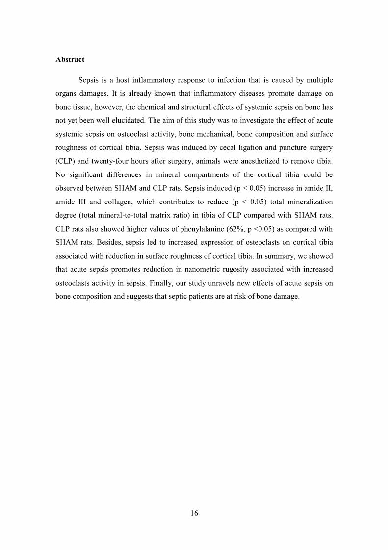
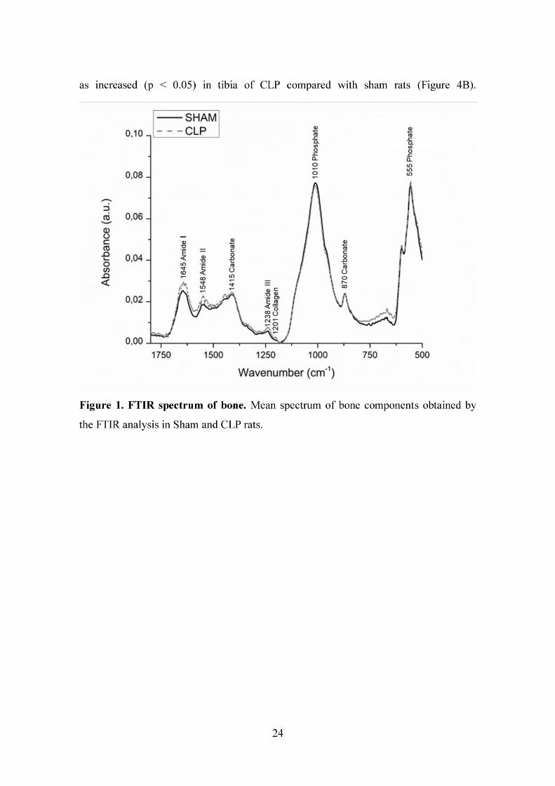
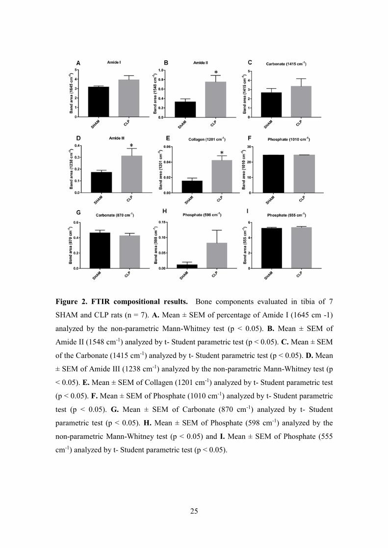
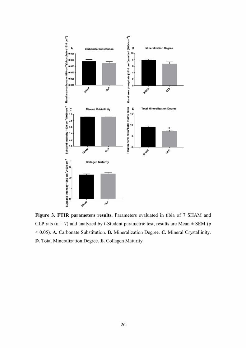
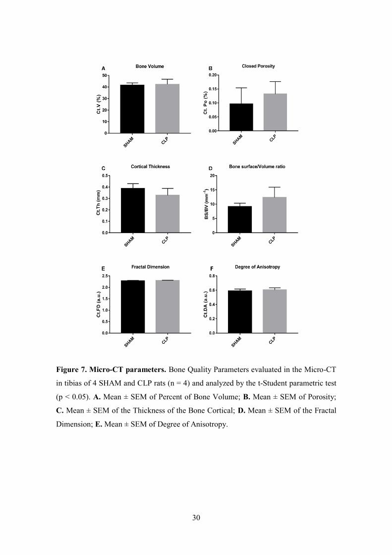
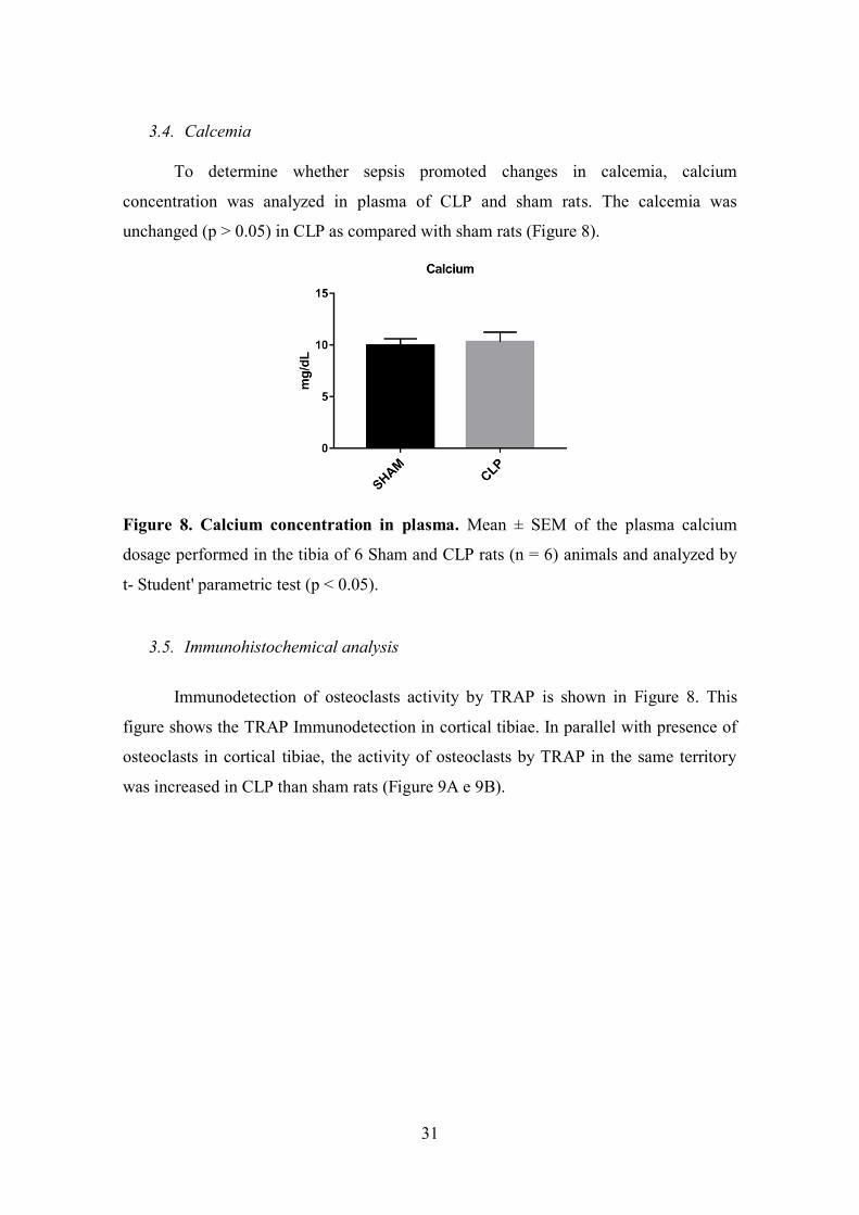
45
Anexos
Anexo 1 Parecer de Aprovaccedilatildeo da Comissatildeo de Eacutetica na Utilizaccedilatildeo de Animais da Universidade Federal de Uberlacircndia