estudo longitudinal da leptospirose urbana: … de... · devido à expansão das favelas em todo o...
TRANSCRIPT

FUNDAÇÃO OSWALDO CRUZ
CENTRO DE PESQUISAS GONÇALO MONIZ
FIOCRUZ
CURSO DE PÓS-GRADUAÇÃO EM BIOTECNOLOGIA EM SAÚDE E MEDICINA INVESTIGATIVA
TESE DE DOUTORADO
ESTUDO LONGITUDINAL DA LEPTOSPIROSE URBANA: INVESTIGAÇÃO DE FATORES DE RISCO PARA INFECÇÃO
E PARA O DESENVOLVIMENTO DE FORMAS GRAVES APÓS A INFECÇÃO
GUILHERME DE SOUSA RIBEIRO
Salvador - Bahia – Brasil 2008
FUNDAÇÃO OSWALDO CRUZ
CENTRO DE PESQUISAS GONÇALO MONIZ
FIOCRUZ
CURSO DE PÓS-GRADUAÇÃO EM BIOTECNOLOGIA EM SAÚDE E MEDICINA INVESTIGATIVA
GUILHERME DE SOUSA RIBEIRO
ESTUDO LONGITUDINAL DA LEPTOSPIROSE URBANA: INVESTIGAÇÃO DE FATORES DE RISCO PARA INFECÇÃO
E PARA O DESENVOLVIMENTO DE FORMAS GRAVES APÓS A INFECÇÃO
Tese submetida à Coordenação da Pós-Graduação
em Biotecnologia em Saúde e Medicina
Investigativa, Fundação Oswaldo Cruz, para a
obtenção do Título de Doutor.
Orientador: Dr. Mitermayer Galvão dos Reis
Co-orientador: Dr. Albert Icksang Ko
Salvador - Bahia – Brasil 2008
2

3
Ficha Catalográfica elaborada pela Biblioteca do
Centro de Pesquisas Gonçalo Moniz / FIOCRUZ - Salvador - Bahia.
Ribeiro, Guilherme de Sousa R484e Estudo longitudinal da leptospirose urbana: investigação de fatores de risco para infecção
e para o desenvolvimento de formas graves após a infecção [manuscrito] / Guilherme de Sousa Ribeiro. - 2008.
70 f.; 30 cm
Datilografado (fotocópia).
Tese (doutorado) – Centro de Pesquisa Gonçalo Moniz, Fundação Oswaldo Cruz. 2008. Orientador: Prof. Mitermayer Galvão dos Reis, Laboratório de Patologia e Biologia
Molecular.
1. Leptospirose. 2. Leptospira. 3. Epidemiologia. 4. Fatores de risco. 5. Distribuição por idade e sexo. I.Título.
CDU 616.986:614.4


5
A Carine, minha esposa e companheira,
pela compreensão e amor durante a caminhada.
Aos meus pais, irmãs e amigos, pelo apoio e incentivo.

6
AGRADECIMENTOS
AGRADECIMENTO ESPECIAL aos meus orientadores e amigos Mitermayer e Albert, que
nesses dez anos de convivência me ensinaram fundamentos da ciência e da pesquisa:
compromisso social, ética, dedicação e paciência.
AGRADECIMENTO ESPECIAL a todos os estudantes e membros do grupo que contribuíram
para a construção destes resultados.
AGRADECIMENTO ESPECIAL aos pacientes com leptospirose e aos moradores de Pau da
Lima que se voluntariaram para participarem deste trabalho.
Aos colegas e amigos da FIOCRUZ com quem aprendo um pouco a cada dia.
Aos familiares e amigos que me incentivam a ir em frente.

7
RESUMO
A leptospirose grave emergiu como um importante problema de saúde pública urbana
devido à expansão das favelas em todo o mundo. Medidas de prevenção são necessárias para
reduzir a morbidade e mortalidade associadas à doença. Entretanto, a ausência de informações
populacionais sobre determinantes para infecção e para o desenvolvimento de doença grave
após a infecção tem limitado a implementação de intervenções. Nossos objetivos são
identificar fatores de risco para infecção pela Leptospira em uma comunidade urbana e
determinar a influência da idade e do sexo sobre o risco de infecção e progressão da doença
após a infecção. Em um estudo de corte transversal realizado na comunidade de Pau da Lima,
Salvador, nós identificamos que 15% dos 3.171 residentes inclusos no estudo apresentavam
evidência sorológica para infecção prévia pela Leptospira. Uso de Sistema de Informação
Geográfica e de abordagens de modelagem estatística identificaram deficiências de
saneamento – esgotos abertos, áreas de risco para alagamento e acúmulo de lixo – como
fatores de risco independentes para infecção pela Leptospira. Baixo nível sócio-econômico
também foi associado com risco de infecção. Em um segundo estudo, nós comparamos a
incidência de infecção pela Leptospira, determinada para uma coorte seguida por três anos no
Pau da Lima, com as incidências de desfechos graves da leptospirose, determinadas para
população de Salvador através de vigilância hospitalar no mesmo período. Nós identificamos
que grupos etários mais velhos e sexo masculino estavam associados a maiores incidências de
infecção pela Leptospira, leptospirose grave, hemorragia pulmonar maciça e mortalidade.
Entretanto, o risco associado à idade adulta e ao sexo masculino foi maior para leptospirose
grave (RR: 7,2 e 6,6, respectivamente) do que para infecção pela Leptospira (RR: 2,5 e 1,6,
respectivamente), sugerindo que idade e sexo apresentam influência no risco de progressão
para doença grave após a infecção. Entre os casos de leptospirose grave, idade acima de 45
anos foi fator de risco para óbito e sexo feminino foi fator de risco para hemorragia pulmonar
maciça. Nossos achados sugerem que intervenções para controle da leptospirose em
comunidades carentes devem ser direcionadas para criação de infra-estrutura de saneamento e
redução da desigualdade social. Além disso, estudos para determinar os mecanismos
patogênicos da influência da idade e do sexo na progressão para leptospirose grave após a
infecção serão importantes para o desenvolvimento de vacinas ou tratamentos capazes de
modular a progressão da infecção e prevenir formas graves de doença.
8
ABSTRACT
Severe leptospirosis has emerged as a significant urban health problem due to the
worldwide expansion of slum settlements. Prevention measures are critical to reduce the
morbidity and mortality associated with the disease. However, the lack of population-based
information on determinants for Leptospira infection and on determinants for development of
severe disease after infection has hampered the implementation of interventions. Our aims are
to identify risk factors for Leptospira infection in an urban slum and to determine the
influence of age and sex on the risk for infection and disease progression after infection. In a
cross-sectional study conducted at Pau da Lima community in Salvador, we found that 15%
of the 3,171 residents included in the study had serological evidence for prior Leptospira
infection. Application of Geographical Information System and statistical modeling
approaches identified sanitation deficiencies - open sewers, flooding-risk areas and refuse
collections - as independent risk factors for Leptospira infection. Low socio-economic status
was also associated with infection risk. In a second study, we compared the incidence of
Leptospira infection, determined for a cohort followed for three years in Pau da Lima, with
the incidences for severe outcomes of leptospirosis, determined for the Salvador population
during hospital-based surveillance in the same period. We found that older age groups and
male sex were associated with increased incidences of Leptospira infection, severe
leptospirosis, severe pulmonary hemorrhage syndrome and mortality. However, the associated
risk for adult age and male sex was greater for severe leptospirosis (relative risk [RR]: 7.2 and
6.6, respectively) than for Leptospira infection (RR: 2.5 and 1.6, respectively), suggesting that
age and sex influence the risk for disease progression after infection. Among the cases of
severe leptospirosis, age greater than 45 years old was risk factor for death and female sex
was risk factor for severe pulmonary hemorrhage. Our findings suggest that interventions to
control urban leptospirosis should address the creation sanitation infra-structure and reduction
of social inequalities. In addition, studies to determine the pathogenic mechanisms for the age
and sex influence on the progression to severe leptospirosis after infection will be significant
for the development of vaccines or treatments which can modulate infection progression and
prevent severe forms of the disease.

9
LISTA DE ABREVIATURAS ArcGIS:Programa para análises de dados espaciais
CDC: Centro para Controle e Prevenção de Doenças, EUA
CI: Intervalo de confiança
CONDER: Companhia de Desenvolvimento Urbano do Estado da Bahia
ELISA: Ensaio de Imunoabsorbância Ligado a Enzima
Epi Info: Programa para análises de dados estatísticos
f(infection): Coeficiente de risco estimado por modelos aditivos generalizados
GAM: Modelos aditivos generalizados
GIS: Sistema de Informação Geográfica
IQR: Intervalo interquartil
MAT: Teste de microaglutinação
m: metros
m2: metros quadrados
N: Número
NS: Não significante
PCR: Reação em cadeia da polimerase
PR: Razão de prevalência
R: Programa para análises de dados estatísticos
RR: Razão de risco
Real-time PCR: Reação em cadeia da polimerase em tempo real
SAS: Programa para análises de dados estatísticos
SD: Desvio padrão
SHPL: Síndrome de hemorragia pulmonar grave associada à leptospirose
SPHS: Síndrome de hemorragia pulmonar grave associada à leptospirose

10
SIG: Sistema de Informação Geográfica
UTI: Unidade de terapia intensiva
WHO: Organização Mundial de Saúde
WinBUGS: Programa para análises de dados estatísticos

11
SUMÁRIO
CAPÍTULO 1. INTRODUÇÃO 12
CAPÍTULO 2. JUSTIFICATIVA 19
CAPÍTULO 3. OBJETIVOS 21
CAPÍTULO 4. MANUSCRITO 1 22
Impacto do Gradiente Ambiental e Social na Infecção pela Leptospira
em Favelas Urbanas
CAPÍTULO 5. MANUSCRITO 2 23
Influência da Idade e do Sexo na Infecção pela Leptospira, na Progressão
da Doença após a Infecção e nos Desfechos Graves da leptospirose
CAPÍTULO 6. DISCUSSÃO 51
CAPÍTULO 7. CONCLUSÕES 53
CAPÍTULO 8. REFERÊNCIAS BIBLIOGRÁFICAS 54
CAPÍTULO 9. ANEXOS 57
1. INTRODUÇÃO
A leptospirose, doença causada por espiroquetas do gênero Leptospira, é considerada
a zoonose de maior distribuição mundial (WHO 1999; Levett 2001). As leptospiras são
espiroquetas móveis, obrigatoriamente aeróbicas que dividem características de bactérias
Gram-positivas e Gram-negativas. Até o presente, mais de 200 sorovares patogênicos de
Leptospira foram descritos com base em diferenças genéticas e antigênicas entre os isolados
(Faine et al. 1999; Levett 2001). O genoma da Leptospira consiste de dois cromossomos
circulares e o seqüenciamento completo foi recentemente concluído para isolados dos
sorovares Lai, Copenhageni e Hardjo (Ren et al. 2003; Nascimento et al. 2004; Bulach et al.
2006). O conhecimento do genoma da Leptospira e os avanços nas técnicas de manipulação
genética permitiram a identificação da lipoproteína de superfície Loa22 como o primeiro fator
de virulência associado à Leptospira (Ristow et al. 2007).
Diversas espécies de mamíferos servem de reservatórios para a Leptospira e mantém a
transmissão da leptospirose na natureza. Como determinadas espécies de reservatórios
costumam estar associadas a alguns sorovares, o conhecimento sobre quais são os
reservatórios e os sorovares circulantes em uma região é essencial para o entendimento da
epidemiologia da leptospirose no local (Levett 2001; Bharti et al. 2003). Uma vez infectados,
as espécies de reservatórios apresentam colonização persistente dos túbulos proximais renais e
disseminam de forma assintomática o organismo para o ambiente através da urina (Levett
2001; Bharti et al. 2003). Usualmente a transmissão entre reservatórios da mesma espécie
ocorre por contato direto entre os animais e a prevalência da infecção aumenta com idade
(Levett 2001). O sorovar que predomina no ambiente urbano é o Copenhageni e o seu
reservatório principal é o Rattus norvegicus (rato marrom ou rato de esgoto) (Ko et al. 1999;
12

13
Pereira et al. 2000; Romero et al. 2003). A infecção dos humanos ocorre de forma acidental
por contato direto ou indireto da pele não íntegra ou de superfícies mucosas com a urina de
um animal infectado (Levett 2001).
O período de incubação médio após a infecção de um hospedeiro humano por
leptospiras patogênicas é de 7 a 14 dias. A infecção é capaz de produzir uma grande variedade
de manifestações clínicas que vão de uma infecção sub-clínica seguida de soroconversão, até
a duas formas clínicas bem reconhecidas: a de uma doença febril aguda auto-limitada, e a de
uma doença grave e potencialmente letal que pode se apresentar por qualquer combinação
entre insuficiência renal aguda, icterícia, sangramentos e pneumonite (Levett 2001; Bharti et
al. 2003; McBride et al. 2005). A forma grave que se manifesta por icterícia, insuficiência
renal aguda e sangramento é conhecida como síndrome de Weil e tem letalidade >10%. A
forma grave associada a pneumonite e sangramento pulmonar maciço é conhecida como
síndrome de hemorragia pulmonar associada à leptospirose (SHPL) e apresenta letalidade
>50% (Gouveia et al. 2008). Estima-se que 90-95% dos pacientes que apresentam
manifestações clínicas da leptospirose desenvolvam a forma leve e auto-limitada da doença e
que 5-10% evoluam para formas graves (Levett 2001; Bharti et al. 2003). Os mecanismos
patogênicos que determinam a progressão para formas graves da doença ou para infecções
sub-clínicas permanecem desconhecidos, mas devem estar relacionados a características de
virulência da Leptospira, a dose de inóculo durante a infecção, a características da resposta
imune do hospedeiro ou a uma interação entre estes fatores (Bharti et al. 2003; McBride et al.
2005).
O início da doença, tanto nas formas brandas e auto-limitadas quanto nas formas
graves, costuma ser súbito. O paciente apresenta febre alta, algumas vezes acompanhada de
calafrios, cefaléia, mialgia, anorexia, prostração, náuseas e vômitos (Bharti et al. 2003). Este

14
quadro inicial é de difícil diagnóstico e se confunde com doenças como dengue, influenza,
gastrenterite e outras viroses (Levett 2001). Para 90-95% dos pacientes, este quadro se resolve
espontaneamente em poucos dias e não deixa seqüelas. Entretanto, 5-10% dos pacientes pode
evoluir para formas graves da doença, o que em geral ocorre ainda na primeira semana de
sintomas. Os pacientes apresentam intensificação das dores musculares e da cefaléia e podem
apresentar complicações graves como icterícia, insuficiência renal aguda (inicialmente não
oligúrica e hipocalêmica, mas que pode progredir para necrose tubular aguda), arritmias,
hemorragias (que são bem características quando acometem as conjuntivas), tosse seca ou
produtiva, hemoptise e insuficiência respiratória (Levett 2001; Bharti et al. 2003; McBride et
al. 2005).
Diversos estudos foram realizados para determinar fatores prognósticos associados à
doença. Embora não exista um escore de gravidade, pacientes que apresentam alteração do
estado mental, oligúria, hipercalemia, altos níveis de creatinina e uréia, arritmias, hipotensão e
sobretudo hemorragia pulmonar e insuficiência respiratória tem um elevado risco de óbito e
devem ser tratados hospitalizados e em unidade de terapia intensiva se necessário (Daher
1999; Ko et al. 1999; Lopes et al. 2001; Panaphut et al. 2002; Lopes et al. 2004; Abgueguen
et al. 2008).
Embora a leptospirose tenha distribuição mundial, a grande maioria dos casos ocorre
nos países tropicais e em desenvolvimento onde a doença se apresenta de forma endêmica
(Levett 2001; Bharti et al. 2003). A maior incidência da doença ocorre durante as estações
chuvosas e provavelmente se devem a surtos (Ko et al. 1999; Tassinari et al. 2008). A
Organização Mundial de Saúde estima que mais de um milhão de casos de leptospirose ocorra
por ano (WHO 1999). No Brasil, mais de 10.000 casos de leptospirose grave são notificados a

15
cada ano (McBride et al. 2005). Entretanto, a dificuldade para estabelecimento do diagnóstico
da doença em pacientes que apresentam formas leves da doença e a inexistência de um teste
diagnóstico rápido e acessível faz com que o impacto global da leptospirose seja subestimado
e baseado principalmente nos pacientes com formas graves da doença (Pappas et al. 2008).
Mais de 80% dos casos de leptospirose grave são identificados na população
masculina adulta (Everard et al. 1995; Ko et al. 1999; WHO 1999; Lopes et al. 2004;
Tangkanakul et al. 2005; Jansen et al. 2007 ). Este achado tem sido justificado pelo dogma de
que a leptospirose é uma doença ocupacional e esporádica, associada a profissões masculinas
como agricultura, pecuária, mineração, manutenção de esgotos e serviços militares (Levett
2001; Bharti et al. 2003). Somente na última década a leptospirose ganhou atenção como um
importante problema de saúde pública global. Este reconhecimento deveu-se à emergência da
SHPL em todo o mundo (Trevejo et al. 1998; McBride et al. 2005; Gouveia et al. 2008), à
identificação de surtos de leptospirose durante desastres (Campanella 1999; Sanders et al.
1999) e atividades de recreação e turismo (CDC 1998; CDC 2001; Morgan 2002; Sejvar et al.
2003), e às grandes epidemias, como a ocorrida na Tailândia no final da década de 1990, onde
mais de 45.000 casos de leptospirose foram identificados (Tangkanakul et al. 2005).
Embora a população de maior risco seja tradicionalmente representada por agricultores
de subsistência da zona rural (Faine et al. 1999; Levett 2001; Bharti et al. 2003), atualmente a
leptospirose emerge como uma doença urbana que acomete os moradores pobres de favelas de
países em desenvolvimento (McBride et al. 2005; Riley et al. 2007). A rápida urbanização e a
crescente desigualdade social levaram a um dramático aumento da população das
comunidades carentes urbanas, onde a falta de saneamento básico favorece a transmissão da
leptospirose pelos ratos. A importância da leptospirose urbana como problema de saúde
16
pública deverá tornar-se ainda maior no futuro, pois as Nações Unidas estimam que nos
próximos vinte e cinco anos a população de moradores de favelas dobrará de um para dois
bilhões de pessoas (UN-HABITAT 2003).
O diagnóstico laboratorial da leptospirose pode ser feito por visualização direta, por
isolamento do organismo, por testes sorológicos ou por métodos moleculares (Levett 2001). A
visualização direta do organismo em amostras de sangue ou urina é feita com o uso da
microscopia de campo escuro, entretanto, este método tem baixa sensibilidade e
especificidade sendo pouco empregado. Visualização direta do microorganismo em tecidos é
tradicionalmente realizada pela coloração com prata, embora imunohistoquímica tenha maior
sensibilidade e especificidade. Sangue, urina, líqüor e outras amostras biológicas podem ser
utilizadas para isolamento do organismo por cultura. A amostra deve ser colhida antes do
início de antibióticos e cedo no curso da doença, uma vez que a chance de isolamento é maior
nos primeiros dias de sintomas. Uma ou duas gotas de sangue devem ser inoculadas em meio
semi-sólido contendo 5-fluorouracil e as culturas devem ser examinadas por microscopia de
campo escuro uma vez por semana por até treze semanas.
Os testes sorológicos são os mais utilizados para o diagnóstico da leptospirose. A
micro-aglutinação (MAT) é considerada o teste de referência e baseia-se na identificação por
microscopia de campo escuro de aglutinação do soro do paciente com antígenos vivos de
diferentes sorogrupos da Leptospira. O teste é complexo e tem baixa sensibilidade com uso de
soro agudo, sendo necessária uma amostra convalescente para avaliar se houve soroconvesão
ou aumento de títulos entre as duas amostras. Outros testes sorológicos comumente utilizados
incluem hemaglutinação indireta e ELISA (Enzyme Linked Immuno Sorbent Assay).
Técnicas de biologia molecular como PCR (polymerase chain reaction) e Real-time PCR
17
(PCR em tempo real) vêm sendo empregadas como ferramentas de pesquisa e deve demorar
até que os custos permitam o seu emprego rotineiro.
O tratamento da leptospirose é baseado no uso de antibióticos e de terapias de suporte
(Levett 2001; Bharti et al. 2003; McBride et al. 2005). Embora haja controvérsia do benefício
da antibioticoterapia na redução da letalidade associada à doença grave (Guidugli et al. 2000),
o emprego de antibióticos parece reduzir a duração da febre, o tempo para normalização da
função renal e o tempo de hospitalização (McClain et al. 1984 ; Watt et al. 1988). Penicilina
cristalina ou ceftriaxone, e doxiciclina são os antibióticos recomendados para os pacientes
hospitalizados e ambulatoriais, respectivamente (Levett 2001; Bharti et al. 2003; McBride et
al. 2005). Seu uso deve ser iniciado tão cedo quanto possível após o diagnóstico clínico e
epidemiológico. Medidas de suporte devem ser empregadas de acordo com a indicação clínica
e incluem hidratação vigorosa, monitorização em unidade de terapia intensiva (UTI), uso de
drogas vasoativas, ventilação mecânica e diálise (McBride et al. 2005). Experiência bem
sucedida na UTI do Hospital Emílio Ribas, São Paulo, sugere que o início precoce e diário de
hemodiálise em vez de tardio e em dias alternados, em conjunto com melhorias no cuidado do
paciente que incluíram admissão precoce em UTI, uso de ventilação mecânica protetora e
fisioterapia respiratória, foi capaz de reduzir a letalidade de pacientes com SHPL de 67% para
17% (Andrade et al. 2007).
A prevenção da leptospirose pode ser feita por medidas individuais e coletivas.
Medidas individuais envolvem evitar ou reduzir a exposição a águas e solos potencialmente
contaminados por leptospiras, assim com evitar o contato com animais potencialmente
contaminados. Quando não for possível evitar o contato, o mesmo deve ser feito com luvas e
botas. Evidências sugerem que uso de quimioprofilaxia com doxiciclina antes ou após
18
exposição é capaz de reduzir a taxa de ataque da doença, mas não impede a ocorrência da
infecção sorológica (Takafuji et al. 1984 ; Sehgal et al. 2000 ). Portanto, seu uso deve ser
considerado antes e após exposições de alto risco para infecção (Bharti et al. 2003). Embora
Cuba e China tenham desenvolvido vacinas para uso humano, estas vacinas têm proteção de
curta duração e não geram proteção cruzada contra sorogrupos que não constam na vacina.
Tais preocupações fizeram com que estas vacinas não fossem licenciadas fora de seus países
(McBride et al. 2005). Medidas de prevenção coletivas, como desratização, são utilizadas com
freqüência, entretanto, são custosas e precisam ser repetidas rotineiramente para ter benefício.

19
2. JUSTIFICATIVA
Nas últimas décadas a leptospirose emergiu como uma doença de moradores de
comunidades carentes urbanas. A rápida migração populacional da zona rural para a zona
urbana e a desigualdade social levaram ao crescimento desorganizado de comunidades pobres
nas periferias de todas as grandes cidades brasileiras. Nestas comunidades, as deficiências
sanitárias e a pobreza propiciam a transmissão da Leptospira por roedores. As Nações Unidas
estima que nos próximos vinte e cinco anos a população mundial de moradores de favelas
dobrará de um para dois bilhões de pessoas o que deverá aumentar a importância da
leptospirose como um problema de saúde pública urbana.
Em Salvador, estudos sobre a epidemiologia da leptospirose urbana vêm sendo
conduzidos desde 1996 e contribuíram substancialmente para identificar um novo padrão
epidemiológico de epidemias cíclicas associadas a fortes chuvas. As principais características
destas epidemias são: 1) ocorrência de surtos anuais acometendo as mesmas comunidades
carentes durante os meses de chuva e associados a desfechos clínicos graves (Ko et al. 1999;
Flannery et al. 2001; Sarkar et al. 2002); 2) ocorrência de transmissão no ambiente peri-
domiciliar, influenciada por fatores ambientais e pela infestação por roedores (Sarkar et al.
2002; Maciel et al. 2008); e 3) o agente causador dos surtos é um único sorovar, L.
interrogans sorovar Copenhageni, que é associado com ratos de esgoto como reservatório (Ko
et al. 1999; Barocchi et al. 2001; Tucunduva de Faria et al. 2007).
Medidas de prevenção da leptospirose são necessidades urgentes. Entretanto, na
conjuntura de escassez de recursos governamentais para grandes obras de urbanização e infra-
estrutura, a identificação de áreas de maior risco para transmissão da bactéria e a

20
determinação de fatores de risco para a infecção são essenciais para guiar intervenções
comunitárias de prevenção. Nós propomos um estudo para identificar deficiências de
saneamento na comunidade que atuem como fatores de risco para infecção pela Leptospira.
Além disso, não está claro porque a maioria das pessoas expostas a leptospiras
evoluem com infecções sub-clínicas enquanto uma pequena fração desenvolve formas graves
da doença. Como mais de 80% dos casos de leptospirose grave ocorre entre homens adultos é
possível que fatores relacionados ao sexo e à idade, como níveis hormonais e maturidade do
sistema imune influenciem o risco de progressão da doença após a infecção. Esta hipótese tem
sido pouco aventada porque tradicionalmente a leptospirose é considerada uma doença
ocupacional. Entretanto, estudos de soroprevalência identificaram que homens e mulheres são
infectados por leptospiras na mesma freqüência e que infecção em crianças é comum (Raoult
et al. 1985; Johnson et al. 2004). Além disso, um estudo na Alemanha observou que homens
foram hospitalizados por leptospirose mais freqüentemente que mulheres e tiveram maior
risco de evoluir com sintomas graves como icterícia, insuficiência renal e sangramentos
(Jansen et al. 2007 ). Os autores concluíram que a predominância de leptospirose no sexo
masculino se deve a uma maior incidência de leptospirose grave nos homens e não a
diferenças na incidência de infecção entre os sexos.
A definição do papel do hospedeiro na história natural da leptospirose será relevante
para guiar estratégias de prevenção e para o desenvolvimento de vacinas ou tratamentos que
visem modular a progressão da infecção para formas graves da doença. Nós pretendemos
determinar a influência da idade e do sexo no risco de infecção e de desenvolvimento de
formas graves da leptospirose ao comparar as incidências de infecção, leptospirose grave,
SHPL e óbito, de acordo com idade e sexo.

3. OBJETIVOS
Geral
Identificar fatores de risco para infecção e para progressão para formas graves da
doença a fim de guiar intervenções de prevenção na comunidade.
Específicos
1.1 Determinar a prevalência de infecção prévia pela Leptospira, definida pela
presença de anticorpos aglutinantes contra a Leptospira, em moradores de uma
comunidade de Salvador;
1.2 Determinar fatores de risco ambientais e sociais para a presença de anticorpos
aglutinantes contra a Leptospira em moradores de uma comunidade de Salvador;
1.3 Determinar a influência da idade e do sexo sobre as incidências de infecção pela
Leptospira, de leptospirose grave, da SHPL e da mortalidade por leptospirose;
1.4 Estimar o risco de desenvolvimento de leptospirose grave após infecção pela
Leptospira e o risco de progressão para SHPL e óbito após desenvolvimento de
leptospirose grave, de acordo com a idade e o sexo.
21

4. MANUSCRITO 1
TÍTULO:
Impact of Environment and Social Gradient on Leptospira Infection in Urban Slums.
[Impacto do Gradiente Ambiental e Social na Infecção pela Leptospira em Favelas
Urbanas].
Publicado no PLoS Neglected Tropical Diseases, em 23 de Abril de 2008.
RESUMO:
A leptospirose, uma zoonose potencialmente letal, tornou-se um importante problema
de saúde urbana em conseqüência da expansão de comunidades (favelas) com
condições ambientais favoráveis para a transmissão da doença pelos ratos. Neste
estudo de prevalência com mais de 3.000 moradores de uma comunidade de Salvador,
Brasil, a aplicação de Sistema de Informação Geográfica (SIG) e de modelagem
estatística permitiu identificar deficiências específicas na infra-estrutura de
saneamento da comunidade - esgotos abertos, acúmulo de lixo, sistema de drenagem
de água inadequado - que servem como fontes de transmissão da Leptospira. Além dos
atributos ambientais da comunidade, baixo nível socioeconômico foi identificado
como fator de risco independente para infecção. Estes resultados indicam medidas de
prevenção da leptospirose deverão ser direcionadas para melhorar o saneamento
básico e para reduzir as diferenças sociais responsáveis por desfechos de saúde
desiguais entre moradores de uma mesma comunidade.
22

Impact of Environment and Social Gradient onLeptospira Infection in Urban SlumsRenato B. Reis1., Guilherme S. Ribeiro1., Ridalva D. M. Felzemburgh1, Francisco S. Santana1,2, Sharif
Mohr1, Astrid X. T. O. Melendez1, Adriano Queiroz1, Andreia C. Santos1, Romy R. Ravines3, Wagner S.
Tassinari3,4, Marılia S. Carvalho3, Mitermayer G. Reis1, Albert I. Ko1,5*
1 Centro de Pesquisas Goncalo Moniz, Fundacao Oswaldo Cruz, Ministerio da Saude, Salvador, Brazil, 2 Secretaria Estadual de Saude da Bahia, Salvador, Brazil, 3 Escola
Nacional da Saude Publica, Fundacao Oswaldo Cruz, Ministerio da Saude, Rio de Janeiro, Brazil, 4 Universidade Federal Rural do Rio de Janeiro, Rio de Janeiro, Brazil,
5 Division of International Medicine and Infectious Diseases, Weill Medical College of Cornell University, New York, New York, United States of America
Abstract
Background: Leptospirosis has become an urban health problem as slum settlements have expanded worldwide. Efforts toidentify interventions for urban leptospirosis have been hampered by the lack of population-based information onLeptospira transmission determinants. The aim of the study was to estimate the prevalence of Leptospira infection andidentify risk factors for infection in the urban slum setting.
Methods and Findings: We performed a community-based survey of 3,171 slum residents from Salvador, Brazil. Leptospiraagglutinating antibodies were measured as a marker for prior infection. Poisson regression models evaluated theassociation between the presence of Leptospira antibodies and environmental attributes obtained from GeographicalInformation System surveys and indicators of socioeconomic status and exposures for individuals. Overall prevalence ofLeptospira antibodies was 15.4% (95% confidence interval [CI], 14.0–16.8). Households of subjects with Leptospira antibodiesclustered in squatter areas at the bottom of valleys. The risk of acquiring Leptospira antibodies was associated withhousehold environmental factors such as residence in flood-risk regions with open sewers (prevalence ratio [PR] 1.42, 95%CI 1.14–1.75) and proximity to accumulated refuse (1.43, 1.04–1.88), sighting rats (1.32, 1.10–1.58), and the presence ofchickens (1.26, 1.05–1.51). Furthermore, low income and black race (1.25, 1.03–1.50) were independent risk factors. Anincrease of US$1 per day in per capita household income was associated with an 11% (95% CI 5%–18%) decrease ininfection risk.
Conclusions: Deficiencies in the sanitation infrastructure where slum inhabitants reside were found to be environmentalsources of Leptospira transmission. Even after controlling for environmental factors, differences in socioeconomic statuscontributed to the risk of Leptospira infection, indicating that effective prevention of leptospirosis may need to address thesocial factors that produce unequal health outcomes among slum residents, in addition to improving sanitation.
Citation: Reis RB, Ribeiro GS, Felzemburgh RDM, Santana FS, Mohr S, et al. (2008) Impact of Environment and Social Gradient on Leptospira Infection in UrbanSlums. PLoS Negl Trop Dis 2(4): e228. doi:10.1371/journal.pntd.0000228
Editor: Ricardo E. Gurtler, Universidad de Buenos Aires, Argentina
Received January 22, 2008; Accepted March 27, 2008; Published April 23, 2008
Copyright: � 2008 Reis et al. This is an open-access article distributed under the terms of the Creative Commons Attribution License, which permits unrestricteduse, distribution, and reproduction in any medium, provided the original author and source are credited.
Funding: This work was supported by the US National Institutes of Health (grants NIAID R01 AI052473, FIC D43 TW00919), Brazilian National Research Council(grants 300861/1996, 473082/2004, 420067/2005, 305723/2006, 150176/2007), and Oswaldo Cruz Foundation (grant 0250.250.102). The funders had no role instudy design, data collection and analysis, decision to publish, or preparation of the manuscript.
Competing Interests: The authors have declared that no competing interests exist.
* E-mail: [email protected]
. These authors contributed equally to this work.
Introduction
At present, one billion of the world’s population resides in slum
settlements [1]. This number is expected to double in the next 25
years [1]. The growth of large urban populations which are
marginalized from basic services has created a new set of global
health challenges [2,3]. As part of the Millennium Development
Goals [4], a major priority has been to address the underlying poor
sanitation and environmental degradation in slum communities
which in turn, are the cause of a spectrum of neglected diseases
which affect these populations [2,3,5].
Leptospirosis is a paradigm for an urban health problem that
has emerged due to recent growth of slums [6,7]. The disease,
caused by the Leptospira spirochete, produces life-threatening
manifestations, such as Weil’s disease and severe pulmonary
hemorrhage syndrome for which fatality is more than 10% and
50%, respectively [7–9]. Leptospirosis is transmitted during direct
contact with animal reservoirs or water and soil contaminated with
their urine [8,9]. Changes in the urban environment due to
expanding slum communities has produced conditions for rodent-
borne transmission [6,10]. Urban epidemics of leptospirosis now
occur in cities throughout the developing world during seasonal
heavy rainfall and flooding [6,11–18]. There is scarce data on the
burden of specific diseases that affect slum populations [2],
however leptospirosis appears to have become a major infectious
disease problem in this population. In Brazil alone, more than
www.plosntds.org 1 April 2008 | Volume 2 | Issue 4 | e228

10,000 cases of severe leptospirosis are reported each year due to
outbreaks in urban centers [19], whereas roughly 3,000, 8,000 and
1,500 cases are reported annually for meningococcal disease,
visceral leishmaniasis and dengue hemorrhagic fever, respectively,
which are other infectious disease associated with urban poverty
[20–22]. Case fatality (10%) from leptospirosis [19] is comparable
to that observed for meningococcal disease, visceral leishmaniasis
and dengue hemorrhagic fever (20%, 8% and 10%, respectively)
in this setting [20,23,24]. Furthermore, leptospirosis is associated
with extreme weather events, as exemplified by the El Nino-
associated outbreak in Guayaquil in 1998 [25]. Leptospirosis is
therefore expected to become an increasingly important slum
health problem as predicted global climate change [26,27] and
growth of the world’s slum population [1] evolves.
Urban leptospirosis is a disease of poor environments since it
disproportionately affects communities that lack adequate sewage
systems and refuse collection services [6,10,11]. In this setting,
outbreaks are often due to transmission of a single serovar, L.
interrogans serovar Copenhageni, which is associated with the Rattus
norvegicus reservoir [6,28–30]. Elucidation of the specific determi-
nants of poverty which have led to the emergence of urban
leptospirosis is essential in guiding community-based interventions
which, to date, have been uniformly unsuccessful. Herein, we
report the findings of a large seroprevalence survey performed in a
Brazilian slum community (favela). Geographical Information
System (GIS) methods were used to identify sources for Leptospira
transmission in the slum environment. Furthermore, we evaluated
whether relative differences in socioeconomic status among slum
residents contributed to the risk of Leptospira infection, in addition
to the attributes of the environment in which they reside.
Methods
Study site and populationThe study was conducted in the Pau da Lima community
(Figure 1A) which is situated in the periphery of Salvador, a city of
2,443,107 inhabitants [31] in Northeast Brazil. Pau da Lima is a
region of hills and valleys, which was a sparsely inhabited area of
Atlantic rain forest in the 1970s and subsequently transformed into
a densely-populated slum settlement (Figure 1B) due to in-
migration of squatters. In total, 67% of the population of Salvador
and 37% of the urban population in Brazil reside in slum
communities with equal or greater levels of poverty as that found
in Pau da Lima [32,33].
A study site was established which comprised of four valleys in
an area of 0.46 km2 (Figure 1A). Active hospital-based surveillance
found that the mean annual incidence of severe leptospirosis was
57.8 cases per 100,000 population at the study site between 1996
and 2001 (unpublished data). The study team conducted a census
during visits to 3,689 households within the site in 2003 and
identified 14,122 inhabitants. Households were assigned sequential
numbers. A computer-based random number generator was used
to select a list of 1,079 sample households from a database of all
enumerated households. Eligible subjects who resided in sample
households and had five or more years of age were invited to be a
Author Summary
Leptospirosis, a life-threatening zoonotic disease, hasbecome an important urban slum health problem.Epidemics of leptospirosis now occur in cities throughoutthe developing world, as the growth of slum settlementshas produced conditions for rat-borne transmission of thisdisease. In this prevalence survey of more than 3,000residents from a favela slum community in Brazil,Geographical Information System (GIS) and modelingapproaches identified specific deficiencies in the sanitationinfrastructure of slum environments—open sewers, refuse,and inadequate floodwater drainage—that serve assources for Leptospira transmission. In addition to theenvironmental attributes of the slum environment, lowsocioeconomic status was found to independently con-tribute to the risk of infection. These findings indicate thateffective prevention of leptospirosis will need to addressthe social factors that produce unequal health outcomesamong slum residents, in addition to improving sanitation.
Figure 1. Slum community site in the city of Salvador, Brazil. (A) The yellow line in the aerial photograph is the boundary of the study site inthe Pau da Lima community. The map in the bottom left corner shows the location of Salvador in Brazil and the study site (red) within the city. (B)Photograph of the typical environment at the community study site, which shows a valley in which households is situated and the proximity ofhouseholds to open sewers and refuse. (C) Close-up view of the orthomap used to georeference households (red and black dots) and environmentalattributes, such as open sewers (blue line) and refuse deposits, for the region marked as a yellow box in Panel A. The red arrow represents thedirection from which the photograph in Panel B was taken.doi:10.1371/journal.pntd.0000228.g001
Leptospira Transmission in Urban Slums
www.plosntds.org 2 April 2008 | Volume 2 | Issue 4 | e228

study participant. Subjects were enrolled into the study between
April 2003 and May 2004 according to written informed consent
approved by the Institutional Review Boards of the Oswaldo Cruz
Foundation, Brazilian National Commission for Ethics in
Research, and Weill Medical College of Cornell University.
Household surveyThe study team of community health workers, nurses and
physicians conducted interviews during house visits and adminis-
tered a standardized questionnaire to obtain information on
demographic and socioeconomic indicators, employment and
occupation, and exposures to sources of environmental contamina-
tion and potential reservoirs in the household and workplace.
Responses reported by subjects were used to obtain information on
race. The study team evaluated literacy according to the ability to
read standardized sentences and interpret their meaning. Informal
work was defined as work-related activities for which the subject did
not have legal working documents. The head-of-household, defined
as the member who earned the highest monthly income, was
interviewed to determine sources and amounts of income for the
household. Subjects were asked to report the highest number of rats
sighted within the household property and the site of work-related
activities. The study team surveyed the area within the household
property to determine the presence of dogs, cats and chickens.
Geographical Information System (GIS) surveyAn ArcView version 8.3 software system (Environmental
Systems Research Institute) database was constructed with
georeferenced aerial photographs and topographic maps provided
by the Company for Urban Development of the State of Bahia
(CONDER). Photographs of the study site, which had a scale of
1:2,000 and spatial resolution of 16cm, were taken in 2002.
During the census, the study team identified households within the
study site and marked their positions onto hard copy 1:1,500 scale
maps (Figure 1C), which were then entered into the ArcView
database. A survey was conducted during the seasonal period of
heavy rainfall between April and August 2003 to geocodify the
location of open sewage and rainwater drainage systems. During
three time points within this period, the study team mapped the
sites of open accumulated refuse and measured the area of these
deposits. Mean values for areas of refuse deposits were calculated
and used for the analyses.
Serological analysisSera were processed from blood samples collected from subjects
during house visits. The microscopic agglutination test (MAT) was
performed to evaluate for serologic evidence of a prior Leptospira
infection [34]. A panel of five reference strains (WHO Collabo-
rative Laboratory for Leptospirosis, Royal Tropical Institute,
Holland) and two clinical isolates [6] were used which included L.
interrogans serovars Autumnalis, Canicola and Copenhageni, L.
borgspetersenii serovar Ballum, and L. kirschneri serovar Grippoty-
phosa. The use of this panel had the same performance in
identifying MAT-confirmed cases of leptospirosis during surveil-
lance in Salvador [6,16] as did the WHO recommended battery of
19 reference serovars [34]. Screening was performed with serum
dilutions of 1:25, 1:50 and 1:100. When agglutination was
observed at a dilution of 1:100, the sample was titrated to
determine the highest titer.
Statistical methodsInformation for subjects was double entered into an EpiInfo
version 3.3.2 software system (Centers for Diseases Control and
Prevention) database. Chi-square and Wilcoxon rank sum tests
were used to compare categorical and continuous data, respec-
tively, for eligible subjects who were and were not enrolled in the
study. A P value #0.05 in two sided testing was used as the
criterion for a significant difference. Preliminary analyses evalu-
ated a range of MAT titers as criteria for prior Leptospira infection
and found that the use of different cut-off values (1:25–1:100)
identified similar associations with respect to the spatial distribu-
tion of seropositive subjects and risk factors for acquiring Leptospira
antibodies. A titer greater or equal to 1:25 was therefore used to
define the presence of Leptospira antibodies in the final analyses.
The presumptive infecting serovar was defined as the serovar
against which the highest agglutination titre was directed [34].
Crude prevalence rates were reported since age and gender-
adjusted values did not differ significantly from crude values.
Ninety-five percent confidence intervals (CI) were adjusted for the
cluster sampling of households.
Kernel density estimation analysis was performed with a range of
bandwidths (10–120 meters) to evaluate smoothed spatial distribu-
tions of subjects with Leptospira antibodies and all subjects. The R
version 2.4.1 statistical package (R Foundation for Statistical
Computing) was used to obtain estimates which were adjusted for
boundary effects. The ratio of the Kernel density estimators for
subjects with Leptospira antibodies and all subjects was measured to
determine the smoothed population-adjusted risk distribution. A
digital terrain model of topographic data was used (ArcGIS 3D
Analyst Extension software) to obtain continuous estimates of
altitude for the study area. The distances, calculated in three-
dimensional space, of households to nearest open drainage systems
and refuse deposits were evaluated as proxies of exposure to these
sources of environmental attributes. Elevation of households with
respect to the lowest point in the valley in which they were situated
was used as a surrogate for flood risk. Generalized additive models
(GAM) [35] were used to evaluate the functional form of the
association between continuous variables and the risk of acquiring
Leptospira antibodies. When indicated, continuous variables were
categorized in multivariate analyses according to the x-intercept
value observed in the plots of fitted smoothed values.
We used Poisson regression [36] to estimate the effect of
demographic, socioeconomic, household and workplace-related
factors on the prevalence of Leptospira antibodies. A Bayesian
inference approach was used which incorporated two random
effects in order to account for overdispersion and cluster sampling
within households. This approach has been used to estimate
parameters in complex models [37] and is less sensitive to sparse
data [38]. Standard non-informative prior distributions were used
in models which were fitted with WinBUGS version 1.4.2 (MRC
Biostatistics Unit). In multivariate analysis, all variables which had
a P value below 0.10 in univariate analyses were included in the
initial model. In order to address co-linearity among variables, we
identified sets of covariates with the high Spearman correlation
coefficients (.0.3 or ,20.3). Highly correlated variables were
aggregated in a single variable when indicated, and evaluated in
the model. The final model was obtained which used backward
variable selection with an inclusion rule of P value ,0.05.
Results
Among 3,797 eligible residents from the slum community site,
3,171 (84%) were enrolled in the study. Study subjects had a
higher proportion of females (56% of 3,171 subjects versus 37% of
626 subjects, respectively; P,0.05) and younger mean age
(25.8615.2 versus 28.1614.6 years, respectively; P,0.05) than
eligible residents who did not participate in the study. The kernel
Leptospira Transmission in Urban Slums
www.plosntds.org 3 April 2008 | Volume 2 | Issue 4 | e228
distribution of enrolled subjects according to place of residence
was similar on visual inspection to that of residents who did not
participate (data not shown). The majority (85%) of subjects were
squatters who did not have legal title to their domiciles. Subjects
belonged to mostly mixed (pardo, 66%) or black (28%) racial
groups. Median household per capita income for study subjects
was US$ 1.30 per day. Among the subjects, 76% had not
completed elementary school education and 23% were illiterate.
Among 2,077 subjects $18 years of age, 77% did not have formal
employment and 35% engaged in informal work.
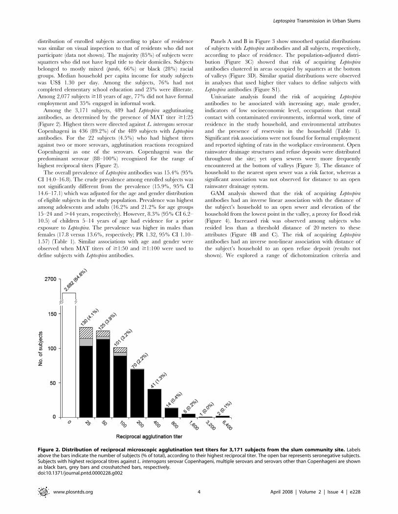
Among the 3,171 subjects, 489 had Leptospira agglutinating
antibodies, as determined by the presence of MAT titer $1:25
(Figure 2). Highest titers were directed against L. interrogans serovar
Copenhageni in 436 (89.2%) of the 489 subjects with Leptospira
antibodies. For the 22 subjects (4.5%) who had highest titers
against two or more serovars, agglutination reactions recognized
Copenhageni as one of the serovars. Copenhageni was the
predominant serovar (88–100%) recognized for the range of
highest reciprocal titers (Figure 2).
The overall prevalence of Leptospira antibodies was 15.4% (95%
CI 14.0–16.8). The crude prevalence among enrolled subjects was
not significantly different from the prevalence (15.9%, 95% CI
14.6–17.1) which was adjusted for the age and gender distribution
of eligible subjects in the study population. Prevalence was highest
among adolescents and adults (16.2% and 21.2% for age groups
15–24 and .44 years, respectively). However, 8.3% (95% CI 6.2–
10.5) of children 5–14 years of age had evidence for a prior
exposure to Leptospira. The prevalence was higher in males than
females (17.8 versus 13.6%, respectively; PR 1.32, 95% CI 1.10–
1.57) (Table 1). Similar associations with age and gender were
observed when MAT titers of $1:50 and $1:100 were used to
define subjects with Leptospira antibodies.
Panels A and B in Figure 3 show smoothed spatial distributions
of subjects with Leptospira antibodies and all subjects, respectively,
according to place of residence. The population-adjusted distri-
bution (Figure 3C) showed that risk of acquiring Leptospira
antibodies clustered in areas occupied by squatters at the bottom
of valleys (Figure 3D). Similar spatial distributions were observed
in analyses that used higher titer values to define subjects with
Leptospira antibodies (Figure S1).
Univariate analysis found the risk of acquiring Leptospira
antibodies to be associated with increasing age, male gender,
indicators of low socioeconomic level, occupations that entail
contact with contaminated environments, informal work, time of
residence in the study household, and environmental attributes
and the presence of reservoirs in the household (Table 1).
Significant risk associations were not found for formal employment
and reported sighting of rats in the workplace environment. Open
rainwater drainage structures and refuse deposits were distributed
throughout the site; yet open sewers were more frequently
encountered at the bottom of valleys (Figure 3). The distance of
household to the nearest open sewer was a risk factor, whereas a
significant association was not observed for distance to an open
rainwater drainage system.
GAM analysis showed that the risk of acquiring Leptospira
antibodies had an inverse linear association with the distance of
the subject’s household to an open sewer and elevation of the
household from the lowest point in the valley, a proxy for flood risk
(Figure 4). Increased risk was observed among subjects who
resided less than a threshold distance of 20 meters to these
attributes (Figure 4B and C). The risk of acquiring Leptospira
antibodies had an inverse non-linear association with distance of
the subject’s household to an open refuse deposit (results not
shown). We explored a range of dichotomization criteria and
Figure 2. Distribution of reciprocal microscopic agglutination test titers for 3,171 subjects from the slum community site. Labelsabove the bars indicate the number of subjects (% of total), according to their highest reciprocal titer. The open bar represents seronegative subjects.Subjects with highest reciprocal titres against L. interrogans serovar Copenhageni, multiple serovars and serovars other than Copenhageni are shownas black bars, grey bars and crosshatched bars, respectively.doi:10.1371/journal.pntd.0000228.g002
Leptospira Transmission in Urban Slums
www.plosntds.org 4 April 2008 | Volume 2 | Issue 4 | e228

found significant risk associations when subjects resided less than
20 meters from an open refuse deposit (Table 1). This association
was not influenced by the size of the refuse deposit. Subjects who
reported sighting two or more rats in the household environment
had increased risk of acquiring Leptospira antibodies (Figure 4D).
Household per capita income had an inverse linear association with
the presence of Leptospira antibodies (Figure 4A). Of note, the
distance of the household to an open sewer was highly correlated
(Spearmen correlation coefficient = 0.71) with household elevation
(Figure S2A) since open sewers drain into the bottom of valleys. An
aggregate variable, distance of household located less than 20 meters
from an open sewer and lowest point in a valley, was therefore used
to examine the association between open sewer and flood-related
exposure and infection risk (Table 1). In contrast household per
capita income was not highly correlated (Spearmen correlation
coefficient = 0.16) with the elevation of the household (Figure S2B).
Table 1. Risk factors for Leptospira antibodies among subjects at the slum community site.
Variables Leptospira antibodies PR (95% CI)
Yes (n = 489) No (n = 2,682) Univariatea Multivariateb
No. (%) or median (IQR)c
Demographic
Age, years
05–14 71 (15) 781 (29) 1.00 1.00
15–24 136 (28) 704 (26) 1.98 (1.47–2.61) 2.02 (1.50–2.69)
25–34 122 (25) 524 (20) 2.31 (1.71–3.07) 2.54 (1.86–3.41)
35–44 73 (15) 350 (13) 2.11 (1.50–2.88) 2.24 (1.59–3.08)
$45 87 (18) 323 (12) 2.60 (1.88–3.51) 2.92 (2.10–4.00)
Male gender 247 (51) 1140 (43) 1.32 (1.10–1.57) 1.38 (1.14–1.64)
Socioeconomic indicators
Black raced 169 (35) 724 (27) 1.35 (1.11–1.62) 1.25 (1.03–1.50)
Household per capita income, US$/day 1.14 (0.39–1.88) 1.30 (0.61–2.20) 0.91 (0.85–0.97)e 0.89 (0.82–0.95)e
Did not complete primary school 394 (81) 2018 (75) 1.32 (1.04–1.65) -
Household factors
Time of residence in household, years 8 (3–17) 7 (3–12) 1.02 (1.01–1.03)e -
Level above lowest point in valley, meters 18.78 (8.59–31.05) 24.71 (13.00–36.04) 0.99 (0.98–0.99)e -
Distance from an open sewer, meters 14.95 (7.34–31.00) 21.04 (8.99–38.11) 0.99 (0.99–1.00)e -
Distance of household from an open sewer/lowest point in valley
$20 m/$20 m 158 (32) 1198 (45) 1.00 1?00
$20 m/,20 m 38 (8) 211 (8) 1.32 (0.89–1.83) 1.19 (0.81–1.67)
,20 m/$20 m 73 (15) 360 (13) 1.46 (1.09–1.91) 1.30 (0.97–1.71)
,20 m/,20 m 220 (45) 913 (34) 1.68 (1.36–2.05) 1.42 (1.14–1.75)
Distance from an open refuse deposit, meters 60.59 (38.48–107.54) 64.90 (42.56–103.16) 1.00 (1.00–1.00)e -
,20 meters from open refuse deposit 51 (10) 174 (6) 1.53 (1.12–2.02) 1.43 (1.04–1.88)
Vegetationf 373 (76) 1,822 (68) 1.45 (1.17–1.79) -
Reservoirs present in household
Sighting of .2 rats 256 (52) 1039 (39) 1.60 (1.33–1.91) 1.32 (1.10–1.58)
Dog 231 (47) 1028 (38) 1.36 (1.14–1.62) -
Chicken 227 (46) 988 (37) 1.40 (1.17–1.66) 1.26 (1.05–1.51)
Cat 106 (22) 406 (15) 1.44 (1.15–1.77) -
Work-related exposures
Informal work 157 (32) 637 (24) 1.42 (1.17–1.71) -
Contact with contaminated environmentg 83 (17) 284 (11) 1.57 (1.22–1.96) -
Risk occupationh 49 (10) 127 (5) 1.90 (1.37–2.51) -
aUnivaritate prevalence ratios (PR) and 95% confidence intervals (CI) are shown for variables which were significant (P,0.05) in the univariate analyses.bMultivariate PR and 95% CI are shown for covariates which were included in the final best fit Poisson regression model.cNumbers and percentages are shown for categorical variables. Median and interquartile range (IQR) are shown for continuous variables of per capita householdincome, time of residence in study household, level above lowest point in valley and distance from an open sewer and refuse deposit.
dData is missing for two non-infected subjects.ePR and 95% CI are shown for continuous data.fData is missing for one infected and two non-infected subjects.gReported exposure to mud, refuse, flooding water or sewage water in the workplace.hOccupation as construction worker, refuse collector or mechanic, which is associated with a workplace environment characterized by high rat infestation.doi:10.1371/journal.pntd.0000228.t001
Leptospira Transmission in Urban Slums
www.plosntds.org 5 April 2008 | Volume 2 | Issue 4 | e228

Multivariate analyses found that the risk for acquiring Leptospira
antibodies was associated with exposures in the household
environment and not in the workplace setting (Table 1). Subjects
who resided less than 20 meters from an open sewer and the lowest
point in the valley had a 1.42 times (95% CI 1.14–1.75) increased
risk for acquiring Leptospira antibodies than those who lived
20 meters or more from these attributes. Residence less than
20 meters from accumulated refuse was associated with a 1.43
times (95% CI 1.04–1.88) increased risk. Sighting of two or more
rats and presence of chickens, a marker for rat infestation, in the
household were significant reservoir-associated risk factors. After
controlling for age, gender and significant environmental expo-
sures, indicators of low socioeconomic level, household per capita
income (PR 0.89 for an increase of US$1.00 per day, 95% CI 0.82–
0.95) and black race (PR 1.25, 95% CI 1.03–1.50) were risk factors
for acquiring Leptospira antibodies (Table 1).
Discussion
Efforts to identify interventions for urban leptospirosis have been
hampered by the lack of population-based information on
transmission determinants. In this large community-based survey
of a slum settlement in Brazil, we found that 15% of the residents had
serologic evidence for a prior Leptospira infection. The prevalence rate
of Leptospira antibodies in the study slum community was similar to
that (12%) found in a city-wide survey performed in Salvador [39].
Risk factors for acquiring Leptospira antibodies were associated with
exposures in the household environment. Interventions therefore
need to target the environmental sources of transmission - open
sewers, flooding, open refuse deposits and animal reservoirs - in the
places where slum inhabitants reside. After controlling for the
influence of poor environment, indicators of low socioeconomic
status were found to be independently associated with the risk of
Figure 3. Spatial distribution of subjects with Leptospira antibodies and all enrolled subjects, according to place of residence, andenvironmental attributes of the community site. Panels A and B show the smoothed Kernel density distribution of subjects with Leptospiraantibodies (N = 489) and all (N = 3,171) subjects, respectively, according to place of residence. The yellow-to-red gradient represents increasingdensity in smoothing analyses which used 40 meters as the bandwidth. Black circles show the location of subject households. Panel C shows thedistribution of the population-adjusted Kernel density estimator for subjects with Leptospira antibodies which was calculated as the ratio of theestimators for subjects with Leptospira antibodies and all subjects. Panel D shows a topographic map generated by the digital terrain model. Theyellow line is the level that is 20 meters above the lowest point in the four valleys within the community site. Panels E and F show the distribution of,respectively, open rainwater and sewage drainage systems and accumulated refuse according to size (m2).doi:10.1371/journal.pntd.0000228.g003
Leptospira Transmission in Urban Slums
www.plosntds.org 6 April 2008 | Volume 2 | Issue 4 | e228

acquiring Leptospira antibodies. This finding suggests that in slum
communities with overall high levels of absolute poverty, relative
differences in socioeconomic level contribute to unequal outcomes
for leptospirosis.
Leptospirosis has been traditionally considered an occupational
disease, since work-related activities are frequently identified as
risk exposures [9]. However slum inhabitants reside in close
proximity to animal reservoirs and environmental surface waters
which contain Leptospira [10]. We previously found that Leptospira
infection clusters within households in slum communities in
Salvador [40]. In this study, we found that after controlling for
confounding, significant risk exposures were those associated with
the household environment rather than workplace. As a caveat,
interview-elicited responses were used to evaluate work-related
exposures since GIS surveys were not performed at the sites where
subjects worked. It is possible that slum residents may have had
work-related risk exposures which were not detected by our
survey. Nevertheless, our findings support the conclusion that the
slum household is an important site for Leptospira transmission and
provides the rationale for interventions that target risk exposures in
this environment.
The study’s findings indicate that the domestic rat was the
principal reservoir for Leptospira transmission in the study
community. Highest agglutination titers among 89% of the
subjects were directed against L. interrogans serovar Copenhageni,
the serovar associated with the R. norvegicus reservoir. Reported
sighting of rats is considered to be an unreliable marker of rat
infestation. However we found that the number of rats sighted by
residents was correlated with their risk of acquiring Leptospira
antibodies (Figure 4D), indicating that rat sightings may be a useful
Figure 4. Generalized additive models (GAM) of the association between the risk of acquiring Leptospira antibodies and continuousvariables of (A) per capita daily household income, (B) level of household in meters above the lowest point in valley, and (C)distance in meters to the nearest open sewer, and (D) reported number of rats sighted in the household environment. Thecoefficient, f(infection), in the GAM model is a measure for the risk of acquiring Leptospira antibodies. In Panels A, B, C and D, the x axis interceptvalues, where f(infection) equals zero, were US$1.70/day, 22 meters, 22 meters and 2 rats, respectively.doi:10.1371/journal.pntd.0000228.g004
Leptospira Transmission in Urban Slums
www.plosntds.org 7 April 2008 | Volume 2 | Issue 4 | e228

marker of infection risk in slum communities where inhabitants
are accustomed to the presence of rats. Although dogs were not
found to be a risk factor, detailed investigations of Leptospira
carriage in urban reservoirs need to be performed. Of note, the
presence of chickens in households was a risk factor, although they
in of themselves are not reservoirs. This association may reflect a
rat-related exposure not accounted for by reported sightings, since
rats are attracted to chicken feed and waste. Raising chickens is a
widespread practice in slum communities-48% (519) of the 1079
study households raised chickens. Control of rodent reservoir
populations may therefore need to incorporate measures that
directly address this practice.
Our findings confirm hypotheses raised by previous ecologic
studies [6,10,11] that infrastructure deficiencies related to open
sewers, flooding and open refuse deposits are transmission sources
for leptospirosis in the slum environment. Furthermore, there
appears to be defined areas of risk associated with open sewers and
refuse deposits, which serve as habitats and sources of food for rats.
Home range radius of the domestic rat varies from 30–150 meters
[41,42], but home range use decreases from the centre to the edge.
GAM analysis demonstrated that slum residents had a positive risk
for acquiring Leptospira antibodies when households were situated
within 20 meters from open sewers and refuse deposits. In
addition, infection risk increased as distances from an open sewer
or refuse deposit decreased, suggesting that households which are
situated closer to these foci have a higher degree of environmental
contamination with Leptospira and inhabitants of these households
are exposed to higher inoculum doses during infection. Molecular
approaches to quantify Leptospira in environmental samples [10]
will be useful in answering this question and guiding recommen-
dations for environmental decontamination and barrier control
measures which can be implemented in slum communities.
In addition, GAM analysis found that residents had positive risk
for Leptospira infection when their households were situated within
20 meters from the lowest point in the valley (Figure 4B). In
Salvador [6,12,16,40] and other urban centers [11,13,15,17,18],
outbreaks of leptospirosis occur during heavy rainfall and flooding
events. Slum communities are built on the poor land quality and
often in areas susceptible to frequent flooding. At the study site and
other slum settlements in Salvador, the water table rises up to one
meter during flooding events because of inadequate rainwater
drainage and blockage of drainage systems with silt and refuse.
The finding that subjects had increased infection risk when their
households were located within 20 meters from the lowest point in
the valley suggests that this distance was a proxy for the degree of
contact which residents encounter flood-related exposures in the
peri-domiciliary environment.
We found that in addition to attributes of the environment
where slum inhabitants reside, low per capita household income
and black race, an indicator of health inequality in Brazil [43,44],
were independent risk factors for Leptospira infection. The social
gradient in health is a widespread phenomenon [45,46]. Our
findings, although not unexpected, are noteworthy since they
suggest that differences in status contribute to unequal health
outcomes in a slum community where the household per capita
income was less than US$1 per day for 44% of the inhabitants.
Although errors in the measurement of risk exposures and residual
confounding were a possibility, the strength of the association
indicates a role for social determinants in Leptospira transmission.
These factors may relate to risky behaviors, such as cleaning open
sewers after flooding events, or limited use of protective clothing
which reduce the risk of abrasions that facilitate entry of the
Leptospira spirochete [47]. Low status and lack of access to
amenities and social support are features of disadvantaged
communities [45] which conceivably influence risk behaviors for
leptospirosis. Further research is needed to evaluate the role of
social factors such that effective interventions, including health
education, can be implemented at the community level.
A limitation of our study was the cross-sectional design which used
serologic evidence for a prior Leptospira infection as the outcome. The
MAT is the standard assay used in prevalence surveys [9], yet there is
not an established titer criterion for defining seropositive reactions.
We previously found that a MAT titer of $1:25 was a specific
marker for prior Leptospira infection among slum residents from
Salvador and when applied, identified household clustering of
infection risk [40]. In this study, cutoff titers from 1:25 and above
identified similar risk associations. In Salvador, leptospirosis is due to
transmission of a single agent, L. interrogans serovar Copenhageni
[6,28]. Titers of 1:25, as well as higher titers, were directed against
this serovar (Figure 1), indicating that this cutoff was a specific and
more sensitive criteria for identifying prior infections in a region
where a single serovar agent is circulating. In the study, there were
more men and younger subjects among non-participating subjects
than participating subjects. Crude prevalence was not different from
the prevalence of Leptospira antibodies which was adjusted by the age
and gender distribution of the overall study population, indicating
that differences between participating and non-participating subjects
may not have introduced a significant bias in the estimates.
Infections may have occurred up to five years prior to the survey
since agglutinating antibodies may persist for this period [48,49].
Major interventions to improve basic sanitation were not imple-
mented in the study community, yet the possibility that environ-
mental exposures were modified over time can not be excluded.
Migration may have affected our ability to estimate prevalence and
risk associations. An on-going cohort investigation of subjects
enrolled in this study found that the annual out-migration rate is
approximately 12% (unpublished data). The study’s findings
therefore need to be confirmed in prospective studies.
We found that Leptospira transmission was due to the interaction
of factors associated with climate, geography and urban poverty.
Since the study was performed in a single community in Salvador,
Brazil, our findings may not be generalizable to other slum
settings. However, a large proportion of the world’s slum
population resides in tropical climates similar to that in Salvador.
Moreover, similar conditions of poverty and environmental
degradation encountered at the study site (Figure 1B) are found
in many slum settlements. In Brazil, 37% of the urban population
resides in slums with equal or greater levels of poverty as found in
the study community [33]. Our findings may therefore be relevant
to other slum communities where leptospirosis is endemic and
have increasing significance as global climate change [26,27] and
growth of the world’s slum population occur in the future [1,33].
The infrastructure deficiencies which were found to be
transmission factors for Leptospira in this study can be readily
addressed by improving sanitation in slum communities. Invest-
ment in sanitation is a cost-effective health intervention [50,51]. In
Salvador, a city-wide sanitation program (Bahia Azul) was recently
shown to have a major beneficial impact for diarrheal disease [52].
However, as frequently encountered with large-scale sanitation
projects, the Bahia Azul program did not provide coverage to the
study community and many of the slum settlements in the city’s
periphery. Equitable access to improved sanitation is therefore
essential in reducing the burden of the large number of
environmentally-transmitted infectious diseases, including lepto-
spirosis, which affects slum populations. Furthermore, the finding
that the social gradient within slum communities, in addition to
the unhealthy environment, contributes to the risk of Leptospira
infection suggests that prevention of urban leptospirosis will need
Leptospira Transmission in Urban Slums
www.plosntds.org 8 April 2008 | Volume 2 | Issue 4 | e228

to combine approaches for improving sanitation with approaches
that identify and address the social determinants which produce
unequal health outcomes.
Supporting Information
Figure S1 Smoothed Kernel density distribution of subjects with
microscopic agglutination test titres of $1:25 (A), $1:50 (B) and
$1:100 (C), according to place of residence at the study site. The
yellow-to-red gradient represents increasing density in smoothing
analyses which used 40 meters as the bandwidth.
Found at: doi:10.1371/journal.pntd.0000228.s001 (2.61 MB TIF)
Figure S2 Spot plots of the relationship between elevation of
household level from the lowest point in valley and distance of the
household to the nearest open sewer (A) and household per capita
daily income (B). Closed and open dots represent houses with at
least one seropositive subject and without a seropositive subject,
respectively.
Found at: doi:10.1371/journal.pntd.0000228.s002 (1.02 MB TIF)
Alternative Language Abstract S1 Abstract translated into
Portuguese by Dr. Guilherme Ribeiro.
Found at: doi:10.1371/journal.pntd.0000228.s003 (0.03 MB
DOC)
Acknowledgments
We would like to thank team members from the Urban Health Council of
Pau da Lima and Oswaldo Cruz Foundation, Rosan Barbosa, Reinaldo
Barreto, Jorge Costa, Maria Raimunda da Cruz, Ana Carla Duarte, Leila
Gouveia, Analea Lima, Simone Nascimento, Osmar Paixao, Amaro Silva
and Erika Sousa who participated in the data collection for the study. We
would also like to thank the Company for Urban Development of the State
of Bahia (CONDER) for providing digital maps of the study site, Elves
Maciel and Alicia Chang for their assistance in designing the study;
Claudio Pereira da Sa (in memorium) and Edilane Gouveia for assistance
with the statistical analysis; Earl Francis Cook Jr. for critical advice during
data analysis and manuscript preparation; and Lee Riley, Art Reingold and
Maurıcio Barreto for reviewing the final manuscript. Finally, this work
could not be accomplished without the joint collaborative effort of the
resident associations that comprise the Urban Health Council of Pau da
Lima and community leaders and residents.
Author Contributions
Conceived and designed the experiments: RBR RF FS SM AM RRR MR
AK. Performed the experiments: RBR RF FS SM AM AQ AS MR AK.
Analyzed the data: RBR GR RF RRR WT MC AK. Contributed
reagents/materials/analysis tools: AK. Wrote the paper: RBR GR RF
RRR MC AK. Reviewed and revised the final version of the manuscript:
RBR GR RF FS AM AQ AS RRR WT MC MR.
References
1. United Nations Human Settlements Programme (2003) The challenge of slums:
Global report on human settlements 2003. London: Earthscan Publications Ltd.
310 p.
2. Riley LW, Ko AI, Unger A, Reis MG (2007) Slum health: Diseases of neglected
populations. BMC Int Health Hum Rights 7: 2.
3. Sclar ED, Garau P, Carolini G (2005) The 21st century health challenge of
slums and cities. Lancet 365: 901–903.
4. The General Assembly of United Nations (2000) United Nations Millennium
Declaration. Available: http://www.un.org/millennium/. Accessed 11 Mar
2008.
5. Bartram J, Lewis K, Lenton R, Wright A (2005) Focusing on improved water
and sanitation for health. Lancet 365: 810–812.
6. Ko AI, Reis MG, Ribeiro Dourado CM, Johnson WD Jr, Riley LW (1999)
Urban epidemic of severe leptospirosis in Brazil. Salvador Leptospirosis Study
Group. Lancet 354: 820–825.
7. McBride AJ, Athanazio DA, Reis MG, Ko AI (2005) Leptospirosis. Curr Opin
Infect Dis 18: 376–386.
8. Bharti AR, Nally JE, Ricaldi JN, Matthias MA, Diaz MM, et al. (2003)
Leptospirosis: A zoonotic disease of global importance. Lancet Infect Dis 3:
757–771.
9. Levett PN (2001) Leptospirosis. Clin Microbiol Rev 14: 296–326.
10. Ganoza CA, Matthias MA, Collins-Richards D, Brouwer KC, Cunningham CB,
et al. (2006) Determining risk for severe leptospirosis by molecular analysis of
environmental surface waters for pathogenic Leptospira. PLoS Med 3: e308.
doi:10.1371/journal.pmed.0030308.
11. Barcellos C, Sabroza PC (2000) Socio-environmental determinants of the
leptospirosis outbreak of 1996 in western Rio de Janeiro: A geographical
approach. Int J Environ Health Res 10: 301–313.
12. Caldas EM, Sampaio MB (1979) Leptospirosis in the city of Salvador, Bahia,
Brazil: A case-control seroepidemiologic study. Int J Zoonoses 6: 85–96.
13. Karande S, Kulkarni H, Kulkarni M, De A, Varaiya A (2002) Leptospirosis in
children in Mumbai slums. Indian J Pediatr 69: 855–858.
14. LaRocque RC, Breiman RF, Ari MD, Morey RE, Janan FA, et al. (2005)
Leptospirosis during dengue outbreak, Bangladesh. Emerg Infect Dis 11:
766–769.
15. Romero EC, Bernardo CC, Yasuda PH (2003) Human leptospirosis: A twenty-
nine-year serological study in Sao Paulo, Brazil. Rev Inst Med Trop Sao Paulo
45: 245–248.
16. Sarkar U, Nascimento SF, Barbosa R, Martins R, Nuevo H, et al. (2002)
Population-based case-control investigation of risk factors for leptospirosis during
an urban epidemic. Am J Trop Med Hyg 66: 605–610.
17. Tassinari W de S, Pellegrini D da C, Sabroza PC, Carvalho MS (2004) [Spatial
distribution of leptospirosis in the city of Rio de Janeiro, Brazil, 1996–1999].
Cad Saude Publica 20: 1721–1729.
18. Kupek E, de Sousa Santos Faversani MC, de Souza Philippi JM (2000) The
relationship between rainfall and human leptospirosis in Florianopolis, Brazil,
1991–1996. Braz J Infect Dis 4: 131–134.
19. Health Surveillance Secretary, Brazilian Ministry of Health (2007) [Leptospirosis
case notification records, Brazil].
20. Health Surveillance Secretary, Brazilian Ministry of Health (2008) [Dengue
epidemiological report, January–December 2007]. Available: http://portal.
saude.gov.br/portal/arquivos/pdf/boletim_dengue_010208.pdf. Accessed 11
Mar 2008.
21. Health Surveillance Secretary, Brazilian Ministry of Health (2007) [Confirmed
cases of meningococcal disease, Brazil, major regions and federal units, 1990–
2006]. Available: http://portal.saude.gov.br/portal/arquivos/pdf/tabela_
meningites_brasil.pdf. Accessed 11 Mar 2008.
22. Health Surveillance Secretary, Brazilian Ministry of Health (2007) [Visceral
leishmaniasis case notification records, Brazil].
23. Health Surveillance Secretary, Brazilian Ministry of Health (2007) [Meningo-
coccal disease deahts, Brazil, major regions and federal units, 1990–2006].
Available: http://portal.saude.gov.br/portal/arquivos/pdf/tabela_obitos_dm_
brasil.pdf. Accessed 11 Mar 2008.
24. Health Surveillance Secretary, Brazilian Ministry of Health (2007) [Visceral
leishmaniasis case fatality rate, Brazil, major regions and federal units, 2000–
2006]. Available: http://portal.saude.gov.br/portal/arquivos/pdf/tabela_lv_
letalidade.pdf. Accessed 11 Mar 2008.
25. Ministerio de Salud Publica, Organizacion Panamericana de la Salud (1998) El
fenomeno El Nino en Ecuador. In: Organizacion Panamericana de la Salud,
editor. El Nino, 1997–1998. Washington, D.C.: Organizacion Panamericana de
la Salud. pp 175–230.
26. Epstein PR (1999) Climate and health. Science 285: 347–348.
27. Patz JA, Campbell-Lendrum D, Holloway T, Foley JA (2005) Impact of regional
climate change on human health. Nature 438: 310–317.
28. Barocchi MA, Ko AI, Ferrer SR, Faria MT, Reis MG, et al. (2001)
Identification of new repetitive element in Leptospira interrogans serovar
copenhageni and its application to PCR-based differentiation of Leptospira
serogroups. J Clin Microbiol 39: 191–195.
29. Pereira MM, Matsuo MG, Bauab AR, Vasconcelos SA, Moraes ZM, et al.
(2000) A clonal subpopulation of Leptospira interrogans sensu stricto is the major
cause of leptospirosis outbreaks in Brazil. J Clin Microbiol 38: 450–452.
30. Romero EC, Yasuda PH (2006) Molecular characterization of Leptospira sp.
strains isolated from human subjects in Sao Paulo, Brazil using a polymerase
chain reaction-based assay: A public health tool. Mem Inst Oswaldo Cruz 101:
373–378.
31. Instituto Brasileiro de Geografia e Estatıstica (2002) Censo demografico 2000 -
resultados do universo. Rio de Janeiro: Instituto Brasileiro de Geografia e
Estatıstica.
32. Secretaria de Combate a Pobreza e as Desigualdades Sociais/SECOMP (2005)
Mapamento da pobreza em areas urbanas do estado da Bahia. CD-ROM 2005.
33. UN-HABITAT (2003) Slums of the world: The face of urban poverty in the new
millennium? Nairobi: UN-HABITAT. 94 p.
34. World Health Organization (2003) Human leptospirosis: Guidance for
diagnosis, surveillance and control. Malta: World Health Organization. 122 p.
35. Hin LY, Lau TK, Rogers MS, Chang AM (1999) Dichotomization of
continuous measurements using generalized additive modelling–application in
predicting intrapartum caesarean delivery. Stat Med 18: 1101–1110.
Leptospira Transmission in Urban Slums
www.plosntds.org 9 April 2008 | Volume 2 | Issue 4 | e228

36. Barros AJ, Hirakata VN (2003) Alternatives for logistic regression in cross-
sectional studies: An empirical comparison of models that directly estimate theprevalence ratio. BMC Med Res Methodol 3: 21.
37. Ashby D (2006) Bayesian statistics in medicine: A 25 year review. Stat Med 25:
3589–3631.38. Greenland S, Schwartzbaum JA, Finkle WD (2000) Problems due to small
samples and sparse data in conditional logistic regression analysis.Am J Epidemiol 151: 531–539.
39. Dias JP, Teixeira MG, Costa MC, Mendes CM, Guimaraes P, et al. (2007)
Factors associated with Leptospira sp infection in a large urban center inNortheastern Brazil. Rev Soc Bras Med Trop 40: 499–504.
40. Maciel EAP, Carvalho ALF, Nascimento SF, Matos RB, Gouveia EL, et al.(2008) Household transmission of Leptospira infection in urban slum communities.
PLoS Negl Trop Dis 2: e154. doi:10.1371/journal.pntd.0000154.41. Jackson WB (1982) Norway rat and allies. In: Chapman JA, Feldhamer GA, eds.
Wild mammals of North America. Baltimore: The Johns Hopkins University
Press. pp 1077–1088.42. Nowak RM (1991) Walker’s mammals of the world. Baltimore: The John
Hopkins University Press.43. Barros FC, Victora CG, Horta BL (2001) Ethnicity and infant health in
Southern Brazil. A birth cohort study. Int J Epidemiol 30: 1001–1008.
44. Travassos C, Williams DR (2004) The concept and measurement of race andtheir relationship to public health: A review focused on Brazil and the United
States. Cad Saude Publica 20: 660–678.
45. Marmot M (2001) Inequalities in health. N Engl J Med 345: 134–136.
46. Marmot M (2005) Social determinants of health inequalities. Lancet 365:
1099–1104.
47. Phraisuwan P, Whitney EA, Tharmaphornpilas P, Guharat S,
Thongkamsamut S, et al. (2002) Leptospirosis: Skin wounds and control
strategies, Thailand, 1999. Emerg Infect Dis 8: 1455–1459.
48. Cumberland P, Everard CO, Wheeler JG, Levett PN (2001) Persistence of anti-
leptospiral IgM, IgG and agglutinating antibodies in patients presenting with
acute febrile illness in Barbados 1979–1989. Eur J Epidemiol 17: 601–608.
49. Lupidi R, Cinco M, Balanzin D, Delprete E, Varaldo PE (1991) Serological
follow-up of patients involved in a localized outbreak of leptospirosis. J Clin
Microbiol 29: 805–809.
50. Hutton G, Haller L, Bartram J (2007) Global cost-benefit analysis of water
supply and sanitation interventions. J Water Health 5: 481–502.
51. Laxminarayan R, Chow J, Salles S, Maslen P (2006) Intervention cost-
effectiveness: Overview of general messages. In: Jamison DT, Breman JG,
Measham AR, Alleyne G, Claeson M et al., eds. Disease control priorities in
developing countries. New York: Oxford University Press. pp 35–86.
52. Barreto ML, Genser B, Strina A, Teixeira MG, Assis AM, et al. (2007) Effect of
city-wide sanitation programme on reduction in rate of childhood diarrhoea in
northeast Brazil: Assessment by two cohort studies. Lancet 370: 1622–1628.
Leptospira Transmission in Urban Slums
www.plosntds.org 10 April 2008 | Volume 2 | Issue 4 | e228

23
5. MANUSCRITO 2
TÍTULO:
Influence of Age and Sex on Leptospira Infection, Disease Progression after Infection
and Severe Outcomes from Leptospirosis.
[Influência da Idade e do Sexo na Infecção pela Leptospira, na Progressão da Doença
após a Infecção e nos Desfechos Graves da Leptospirose].
A ser submetido.
RESUMO:
Mais de 80% dos casos de leptospirose grave ocorrem em homens adultos, portanto
fatores associados ao sexo e à idade devem influenciar o risco de infecção por
Leptospira ou o risco de progressão da doença após a infecção ou ambos. Nós
determinamos o papel do sexo e da idade sobre estes riscos através da comparação das
incidências estratificadas por sexo e idade de: infecção pela Leptospira – determinada
em uma coorte seguida por três anos na comunidade de Pau da Lima, Salvador-BA –
leptospirose grave, hemorragia pulmonar maciça por leptospirose e mortalidade –
determinados para a cidade de Salvador no mesmo período. Nós observamos que as
incidências de infecção pela Leptospira, leptospirose grave, hemorragia pulmonar
maciça e mortalidade foram maiores para os homens e para os grupos etários mais
velhos. Entretanto, a indivíduos do sexo masculino e com mais de 15 anos tiveram
maior risco de leptospirose grave (RR: 7,2 e 6,6, respectivamente) do que de infecção
pela Leptospira (RR: 2,5 e 1,6, respectivamente), sugerindo que idade e sexo
influenciam o risco de progressão para doença grave após a infecção. Entre os casos
de leptospirose grave, idade acima de 45 anos foi fator de risco para óbito e sexo
feminino foi fator de risco para hemorragia pulmonar maciça. Estudos para determinar
os mecanismos patogênicos da influência da idade e do sexo na progressão para
leptospirose grave após a infecção poderão ajudar no desenvolvimento de vacinas ou
imunomoduladores capazes de intervir na progressão da infecção e prevenir formas
graves de doença.
24
Influence of age and sex on Leptospira infection, disease progression after infection and
severe outcomes from leptospirosis
Running Title: Role for age and sex on outcomes from leptospirosis
Guilherme S. Ribeiro,1 Ridalva D. M. Felzemburgh,1 Renato B. Reis,1 Federico C. Costa,1
Débora Bittencourt,1 Andréia C. Santos,1 Nilzelene Silva,1 Thiago P. Cavalcanti,1 Luise R.
Daltro,1 Daniel Sakaki,1 Talita Ayres,1 Edilane Gouveia,1 Mitermayer G. Reis,1 Albert I.
Ko1,2
1Centro de Pesquisas Gonçalo Moniz, Fundação Oswaldo Cruz, Ministério da Saúde,
Salvador, Brazil, and 2Division of International Medicine and Infectious Diseases, Weill
Cornell Medical College, New York, US
Reprints or correspondence:
Dr. Albert I. Ko
Centro de Pesquisas Gonçalo Moniz, Fundação Oswaldo Cruz/MS
Rua Waldemar Falcão, 121
Salvador, Bahia 40296-710; Brazil
Telephone: (55 71) 3176-2302. Fax: (55 71) 3176-2281
e-mail: [email protected]
25
Abstract
Background. The larger burden of severe leptospirosis among males and adults is thought to
be related to an increased risk for Leptospira infection in this population. Whether male adults
also have increased risk for development of severe leptospirosis after infection is not clear.
We conducted an observational study to investigate the influence of age and sex on the risk
for Leptospira infection, severe leptospirosis and severe outcomes of the disease.
Methods. In Salvador, Brazil, we measured annual incidence of Leptospira infection during
three years of follow up of a community-based cohort. We measured annual incidence of
severe leptospirosis, leptospirosis-associated severe pulmonary hemorrhage syndrome (SPHS)
and leptospirosis mortality during three years of hospital-based surveillance for severe
leptospirosis. We used the microagglutination test (MAT) to identify cases of Leptospira
infection, and MAT and ELISA to confirm suspected cases of severe leptospirosis. Incidences
were stratified by age and sex and used to estimate age and sex-specific risks for progression
to severe leptospirosis after Leptospira infection. We used logistic regression analysis to
determine the age and sex-specific risks for death and SPHS among the cases of severe
leptospirosis.
Results. Age increase and male sex were associated with higher incidences of Leptospira
infection, severe leptospirosis, SPHS and mortality. In comparison to children with 5-14 years
of age, subjects ≥15 years had 2.49 (95% CI: 1.53-4.07) and 7.15 (95% CI: 3.92-13.03)
greater risk for Leptospira infection and severe leptospirosis, respectively. In comparison to
females, males had 1.55 (95% CI: 1.12-2.14), and 6.64 (95% CI: 4.92-8.96) greater risk for
Leptospira infection and severe leptospirosis, respectively. Males had a fourfold greater risk
for progression to severe leptospirosis after infection in comparison to females. Among the
patients with severe leptospirosis, age increase was associated with death (P value for trend <

26
.05), but not with SPHS. In contrast, female sex increased the risk of SPHS in 2.49 (95% CI:
1.05-5.75) times, but had no influence on the risk of death.
Conclusions. Older age and male sex positively influence the risk for Leptospira infection
and development of severe leptospirosis after infection. Age also influences the risk of death
among cases of severe disease. In contrast, female sex increases the risk for SPHS. Studies to
determine the pathogenic mechanisms behind the influences of age and sex on the Leptospira
infection progression are warranted.
Key words: Leptospirosis, age, sex, incidence, mortality, pulmonary hemorrhage syndrome
27
INTRODUCTION
Leptospirosis is a zoonotic disease transmitted to humans by exposure to environment
contaminated by urine of reservoirs [1]. Individuals who are infected can develop a wide
spectrum of clinical manifestations, ranging from asymptomatic infections to severe
icterohaemorrhagic presentations, such as Weil’s disease and severe pulmonary hemorrhage
syndrome (SPHS), for which case fatality rates are greater than 10% and 50%, respectively [1,
2]. It is not known why certain exposed individuals develop asymptomatic or mild infections
while others progress to develop severe disease, but it may be related to age and sex factors
since population-based studies have shown that severe disease is uncommon among children
and that 80% of the cases of severe leptospirosis occurs among males [3-7].
Traditionally, the disproportional increased incidence of severe leptospirosis among adult
males has been explained by the dogma that leptospirosis is a disease of occupational
transmission, typically affecting the male adults who works as miners, fishers, farmers or in
military or sewer services [1]. However, leptospirosis epidemics have been increasingly
recognized in association with environmental exposures that are not primarily associated with
age and sex, such as during extreme rainfall events [8-10], weather-related disasters [11, 12]
and living in conditions of inadequate sanitation [13, 14]. In addition, seroprevalence studies
have identified that presence of antibodies against Leptospira is common among children [15,
16] and occurs in similar frequency between males and females [15, 17]. An alternative
explanation is that age and sex-related factors influence the risk for progression to severe
leptospirosis after infection acquisition. This hypothesis is supported by studies about the
severity of the disease that found that older age is associated with higher case fatality rate [5,
6] and that male sex increases the risk for jaundice, renal failure, hemorrhage and
hospitalization [4].

28
In Salvador, Brazil, we were able to estimate the age and sex-specific incidences of
Leptospira infection from a community-based cohort study, and the age and sex-specific
incidences of severe leptospirosis, leptospirosis-associated SPHS and leptospirosis mortality
from a city-wide surveillance for severe leptospirosis. As a result, we had a unique
opportunity to investigate the influence of age and sex on the risk for Leptospira infection and
progression to severe outcomes after infection.
METHODS
Community-based cohort study.
During three years, we conducted a cohort study in Pau da Lima, a community situated in the
periphery of Salvador, Brazil. The study site has been previously described [16]. Briefly, 85%
of its population were squatters who did not have legal title to their domiciles. The median
household per capita income was US$ 1.30 per day.
Between February 2003 and May 2004, the study team conducted a census within the study
site and identified 14,122 inhabitants living in 3,689 households. We used a computer-based
random number generator to select a list of 684 (19%) households from a database of all
enumerated households within the study site. All subjects who slept three or more nights per
week in a sampled household and had five or more years of age were eligible to be a cohort
member. Among 2,419 subjects who were eligible to be a cohort-member, 2,003 (83%)
agreed to participate and were enrolled in the study according to written informed consent
procedures approved by the Fundação Oswaldo Cruz, Brazilian Ministry of Health and Weill
Medical College of Cornell University.

29
During cohort enrollment the study team administered a standardized questionnaire to obtain
information on demographic and socioeconomic indicators, employment and occupation,
exposures to sources of environmental contamination and presence of potential reservoirs in
the household and workplace [16]. At enrolment and once a year during three consecutive
years, the study team visited the household of the study participants in the post-rainy season
(October to January in the 1st year of follow up and November to February in the 2nd and 3rd
years of follow up) to collect sera samples that were used to identify cases of Leptospira
infection.
Serologic evaluation for Leptospira infection
We used the microagglutination test (MAT) to evaluate subjects’ sera for the presence of
agglutinating antibodies against Leptospira. The MAT panel included five of the reference
strains recommended by the World Health Organization (Royal Tropical Institute, the
Netherlands), which included Leptospira interrogans serovars Autumnalis, Canicola and
Copenhageni, L. borgspetersenii serovar Ballum, L. kirschneri serovar Grippotyphosa, and
one clinical isolate, L. interrogans serovar Copenhageni strain Fiocruz L1–130 [5]. The use of
this panel had the same performance in identifying MAT-confirmed cases of severe
leptospirosis during surveillance in Salvador than did the extended battery recommended by
the WHO [5]. Screening was performed with serum dilutions of 1:25, 1:50 and 1:100. When
agglutination was observed at a dilution of 1:100, the sample was titrated to determine the
highest agglutination titer. Positive and negative control sera were always included in the
battery. A case of Leptospira infection was defined as a cohort subject who seroconverted
from a MAT titer of zero to a follow-up titer ≥1:50 or who presented a fourfold rise in the
MAT titer between two consecutive evaluations.

30
Hospital-based surveillance for leptospirosis. Active surveillance for severe leptospirosis has
been conducted in Salvador, Brazil (population 2.4 million inhabitants) since 1996. We
included in this study leptospirosis cases identified during the three-year period between
January 1, 2003 and December 31, 2005. Cases were identified at Hospital Couto Maia, the
state infectious diseases hospital, which is the reference center for leptospirosis in Salvador.
Notification of leptospirosis cases is mandatory in Brazil and this hospital reports >95% of the
cases from the city. The study team consecutively identified case-patients who were admitted
to this hospital and met the clinical definition for severe leptospirosis, which included
characteristic late phase manifestations of leptospirosis (jaundice and serum aminotransferase
activities less than 20 times the normal upper limit [30 U/L]; oliguria, creatinine ≥1.5 mg/dl
or urea ≥75 mg/dl; or hemorrhagic diathesis) in a patient with an acute febrile disease [5].
Patients who fulfilled the case definition were enrolled into the study according to informed
consent protocols approved by the Fundação Oswaldo Cruz, Brazilian Ministry of Health and
Weill Medical College of Cornell University. We used standardized forms to interview the
patients or their family members and to extract information from medical charts on clinical
presentation, laboratorial exams and outcome.
Laboratory confirmation of leptospirosis
Laboratory confirmation of the cases were performed using blood samples collected during
hospital admission, on day 4 or 5 of hospitalization, and 2 weeks after the collection of the
first sample. A laboratory-confirmed case of leptospirosis was defined as a case who
presented a fourfold rise in the MAT titer between paired samples, a single titer ≥1:800 [5], or
a positive result on the immunoglobulin M (IgM) ELISA (Bio-Manguinhos, Rio de Janeiro,
Brazil) [19]. The same panel of five reference strains (WHO Collaborative Laboratory for
Leptospirosis, Royal Tropical Institute, Holland) and one clinical isolate [5] used in the cohort

31
study was used in the surveillance for severe leptospirosis. Positive and negative control sera
were always included in the panel.
Statistical analysis.
Data from both the cohort study and from the hospital-based surveillance were double-entered
and validated using an Epi Info 3.3.2 software system (Centers for Disease Control and
Prevention, USA) database. Epi Info 3.4.3 and SAS 9.1 were used for data analysis. Mean
annual cumulative incidence of Leptospira infection was calculated for the cohort subjects
who completed the three years of follow up. Mean annual cumulative incidences of severe
leptospirosis, leptospirosis-associated SPHS and leptospirosis mortality were calculated for
the population of Salvador using the cases identified during the three-year surveillance period.
Incidences were stratified by age and sex and presented as the number of cases per 1,000 or
100,000 population, with respective 95% confidence intervals. For this study, we defined
subjects with 5-14 years of age and subjects with ≥15 years of age as children and adults,
respectively. The Salvador population count from the 2000 national census was used for
incidence calculations [20].
The chi square test was used to evaluate whether there were annual variations among the
incidences measured in each of the three years of study. The chi square test for linear trend
was used to determine whether there was linear association between age and the incidences of
Leptospira infection, severe leptospirosis, leptospirosis-associated SPHS and leptospirosis
mortality. Risk ratios and 95% confidence intervals were used to evaluate whether adults and
males were associated with Leptospira infection and severe leptospirosis.

32
We used the mean annual incidence of Leptospira infection for the Pau da Lima cohort to
project the number of cases of Leptospira infection in the city of Salvador. We used the
projected number of cases of Leptospira infections and the ascertained number of cases of
severe leptospirosis for the period between 2003 and 2005 in Salvador to estimate the risk
ratio and 95% confidence interval for development of severe leptospirosis after infection,
according to age and sex. We used logistic regression analysis to estimate the influence of age
and sex as independent risk factors for progression to SPHS and death among the cases of
severe leptospirosis identified during the study.
RESULTS
Among the 2,003 cohort-members, 1,300 (65%) subjects completed the three years of follow-
up. The mean annual dropout rate was 13.4%. Reasons to lose the follow-up included
migration (469 cases; 67%), inability to find the participant (109; 16%), refusal o participate
(104; 15%) and death (14; 2%). Subjects who were lost-to-follow-up had higher proportions
of males than subjects who completed follow up (49 vs. 42%, P<0.01) but were not different
with respect to age, socioeconomic level and exposure to environmental risks. We identified
140 infections among the 1,300 subjects who completed follow-up. For 93% of the infections
the highest MAT titer was against serovar Copenhageni. The mean annual incidence of
Leptospira infection was 35.9 (95% confidence interval [CI]: 30.3-42.2) per 1,000 population
(Table 1). Incidences of infection in 1st, 2nd and 3rd year of follow up did not differ
significantly (40.8, 33.8 and 33.1 per 1,000 persons, respectively; P > .05). Among 322 and
71 subjects who completed only one and two follow-up years, respectively, the annual
incidence of infection was 40.8 and 14.1 per 1,000 population, respectively. These rates did
not differ significantly from those for the closed cohort (P < .05).

33
During the three-year period between January, 2003 and December 2005, the surveillance
identified 341 cases of severe leptospirosis in Salvador. Laboratorial confirmation of the
diagnosis was made for 281 (82%) of the cases. For the 252 cases that were confirmed by
MAT, 97% had the highest MAT titer against the serovar Copenhageni. Confirmed and
unconfirmed cases had similar clinical presentation, except that unconfirmed cases were older
and had more severe outcomes, such as SPHS and death (Supplemental table 1).
Unavailability of paired blood sample was the main reason for non-confirmation of cases
(Supplemental table 1). Mean annual incidence of severe leptospirosis, leptospirosis-
associated SPHS and leptospirosis mortality for Salvador were 5.1, 0.4 and 0.9 per 100,000
population, respectively (Table 1). Annual incidence of severe leptospirosis, leptospirosis-
associated SPHS and leptospirosis mortality were lower in 2003 and 2004 in comparison to
2005 (4.3 and 4.0 vs. 5.7 per 100,000 population for severe leptospirosis, P < .05; 0.3 and 0.2
vs. 0.8 per 100,000 population for SPHS, P < .05; and 0.6 and 0.7 vs. 1.1 per 100,000
population for mortality, P > .05, respectively).
Incidences of Leptospira infection, severe leptospirosis, leptospirosis-associated SPHS and
leptospirosis mortality were found to have linear trend associations with age (P value for
linear trend < .05 for all incidences; Figure 1). Incidence of Leptospira infection was high for
all age groups, including children, and peaked among young adults (25-34 years of age) and
adults with ≥55 years of age (Table 1, Figure 1A). In contrast, incidence of severe
leptospirosis was very low among children and peaked among adults 45-54 years old (Table
1, Figure 1B).
Adults had increased risk for Leptospira infection (RR: 2.49; 95% CI: 1.53-4.07), and severe
leptospirosis (RR: 7.15; 95% CI: 3.92-13.03) in comparison to children. The incidence of

34
SPHS and death for children was virtually null, since none case of SPHS and death was
identified among children. In addition, the male population had increased risk for Leptospira
infection (RR: 1.55; 95% CI: 1.12-2.14), severe leptospirosis (RR: 6.64; 95% CI: 4.92-8.96),
leptospirosis-associated SPHS (RR: 2.92; 95% CI: 1.35-6.30) and leptospirosis mortality (RR:
4.04; 95% CI: 2.18-7.47) in comparison to the female population. Of note, adult age and male
sex had much stronger associations with severe leptospirosis than with Leptospira infection.
Thus, the burden of severe leptospirosis among male adults appears to be more influenced by
age and sex-specific differences in the risk for progression to severe leptospirosis after
infection than by differences in the infection risk.
Assuming that the age and sex-specific incidences of Leptospira infection found during the
cohort study were the same for the population of Salvador, we estimated the risk for
progression to severe leptospirosis after Leptospira infection (Table 2). The overall risk for
progression to severe leptospirosis after infection was low (1.4 cases of severe disease per
1,000 infections). The age group 45-54 years of age had the higher risk for progression to
severe leptospirosis after infection and, among males, children had the lowest risk. The male
population had an estimated risk for development of severe leptospirosis after infection four
times greater than the female population (2.1 and 0.5 cases of severe disease per 1,000
infections, respectively). The male increased risk for progression to severe leptospirosis after
infection was identified for all age groups except than for children.
Univariate analysis for the cases of severe leptospirosis identified an association between age
and case fatality rate. While none of the children 5-14 years old evolved to death, 10%, 14%,
15%, 27% and 37% of the patients with 15-24, 25-34, 35-44, 45-54 and ≥55 years old died
during hospitalization, respectively (P value for linear trend < .05; Figure 2A). Although case

35
fatality rate was higher among women than among men, the difference was not statistically
significant (27% vs. 16%, RR: 1.65; 95% CI: 0.97-2.82). Logistic regression analysis with
adjustment for age and sex confirmed a positive association between age increase and death
(Table 3).
In contrast, univariate analysis found that age was not associated with SPHS (P value for
linear trend < .05; Figure 2B), and that female sex was a risk factor for SPHS. While 18% of
the women with severe leptospirosis developed SPHS during hospitalization, only 8% of the
men presented this outcome (RR: 2.28, 95% CI: 1.12-4.63). Logistic regression analysis with
adjustment for age and sex confirmed the association between female sex and SPHS (Table
3).
DISCUSSION
Key questions on the epidemiology of leptospirosis concerns why only a fraction of the
infected persons develop severe disease after infection and why the majority of cases of
severe leptospirosis occur among male adults. In this investigation, we combined data from a
cohort study for Leptospira infection and from a hospital-based surveillance for severe
leptospirosis to simultaneously determine age and sex-specific incidences of Leptospira
infection, severe leptospirosis, leptospirosis-associated SPHS and leptospirosis mortality. Our
findings indicate that age and sex have significant influences on the risks for Leptospira
infection and progression to severe outcomes from leptospirosis after infection.
Traditionally, the dogma that leptospirosis is an occupational disease that place adult males at
increased risk for infection has justified why severe leptospirosis is an uncommon disease
among children and women. However, our results suggest that this concept may be only part

36
of the truth. We found that adults and males do have higher risks for Leptospira infection, but,
in addition, we provided evidences to support that age increase and male sex are also at
increased risk for developing severe leptospirosis after infection. The evidences for an
association between age increase and risk for progression to severe disease were: (1) adults
had 2.5 and 7.2 times greater risk for Leptospira infection and severe leptospirosis,
respectively, in comparison to children, suggesting that older age has a larger impact on the
risk for progression to severe disease than on the risk for infection; (2) the age group of 45-54
years of age had the highest estimated risk for progression to severe leptospirosis after
infection, while, among males, children with 5-14 years of age had the lowest risk; and (3)
age increase was associated with higher case fatality rate. In addition, none of the chidren
evolved to death. The evidences for an association between male sex and risk for progression
to severe disease were: (1) men had 1.6 and 6.6 times greater risk for Leptospira infection and
severe leptospirosis, respectively, suggesting that male sex has a larger impact on the risk for
progression to severe disease than on the risk for infection; and (2) males had an estimated
risk for progression to severe leptospirosis after infection four times greater than the females.
Several studies have presented results that support the hypothesis that age influences the risk
for progression to severe disease after infection. Children with severe leptospirosis has been
shown to have a better prognosis than adults [3, 6], which indicates that progression to severe
outcomes is less common among children. Older age has also been shown to be an
independently associated with risk for death [5]. In addition, evidence that women have a
milder leptospirosis has been published. Jansen et al. (2004) evaluated 338 patients with
laboratory-confirmed leptospirosis reported in Germany during 1997–2005 and identified that
male patients were more likely than female patients to be hospitalized and exhibit severe
symptoms, such as jaundice, renal dysfunction and hemorrhages [4]. They concluded that

37
“reports on male predominance in leptospirosis may thus reflect sex-related variability in the
incidence of severe disease, rather than different infection rates”.
An interesting finding of our study was that although women had a lower risk for progression
to severe disease, they had a higher risk for the development of SPHS after adjusting for age.
An association between female sex and SPHS has been shown previously in Salvador, but the
leptospirosis casuistic was similar than that included in this study [21]. Epidemiological
investigations in other settings are necessary to confirm this finding.
The pathogenic mechanisms through which age and sex influence the risk for progression to
severe disease and severe outcomes after infection remains to be elucidate. It is possible that
different behaviors between children and adults and between males and females determine the
intensity of exposure to a contaminated environment and consequently the inoculum dose of
Leptospira during infection. As an alternative hypothesis, differences in host-specific immune
response after Leptospira infection may be driven by age and sex-related factors, such as
immune system maturity and hormone levels. In addition, repetitive Leptospira infections and
prior acquisition of antibodies against Leptospira are potentially influenced by age and sex
and may have a role on the risk for progression to severe disease after infection.
Our investigation has strengths and limitations. This is the first study to simultaneously
estimate the incidence of Leptospira infection, severe leptospirosis, leptospirosis-associated
SPHS and leptospirosis mortality for a large urban population. Since it is not practical to
determine all cases of Leptospira infection for a large city as Salvador, we used the incidence
of Leptospira infection from the Pau da Lima cohort as a proxy for the incidence of
38
Leptospira infection for Salvador. This approach enabled us to estimate the age and sex-
specific risk ratios for progression to severe disease after infection.
As a limitation, we assumed that the cohort population from the Pau da Lima community
represents the population of Salvador. Although this assumption is reasonable, since 67% of
the population of Salvador lives in similar social and environmental conditions than the
inhabitants of Pau da Lima, it is not completely correct. Pau da Lima is a poor community
with high incidence of severe leptospirosis. Thus, it is possible that the Leptospira infection
incidences measured for the Pau da Lima cohort were higher than the real Leptospira
infection incidence for Salvador. This inaccuracy may have lead to an underestimation of the
risk for progression to severe disease after infection. In addition, bias may have resulted from
eligible participants refusal to be enrolled into the study and from loss-to-follow-up of cohort-
members. However, we compared incidences of Leptospira infection between those who
completed and those who did not complete follow up but were followed for at least one year
and we found similar incidences between them. Finally, other variables, such as days of
symptoms, could have confounded the association between age and death, and between sex
and SPHS. The inclusion of days of symptoms prior to hospitalization did not alter the found
associations, but we cannot discard that others unanticipated confounders were present.
In conclusion, this study presented evidences for a role of age and sex on the risk for
progression to severe leptospirosis after infection. Reasons why adults and men have higher
risk for progression to severe disease after infection remains to be elucidate, but may involve
differences in the Leptospira inoculum, presence of antibodies against Leptospira acquired
during a prior infection and differences in inflammatory and immune response due to immune
system maturity or hormone levels. Understanding the mechanisms behind age and sex

39
influence on the risk for disease progression may have obvious implications for future
research on vaccine development and use of anti-inflammatory and immune modulator drugs
in the treatment of leptospirosis.
Acknowledgments
We thank team members from the Urban Health Council of Pau da Lima and Oswaldo Cruz
Foundation who participated in the data collection for the cohort study; the staff at Hospital
Couto Maia for their assistance in conducting the study and most of all the resident
associations that comprise the Urban Health Council of Pau da Lima, the community leaders
and residents and the patients and their families. This work was supported by the Brazilian
National Research Council (grants 300861/1996, 473082/2004, 420067/2005, 305723/2006,
554788/2006, 150176/2007), Oswaldo Cruz Foundation (grant 0250.250.102) and the
National Institutes of Health (grants NIAID R01 AI052473, FIC D43 TW00919).The funders
had no role in study design, data collection and analysis, decision to publish, or preparation of
the manuscript.

40
References
1. Bharti AR, Nally JE, Ricaldi JN, et al. Leptospirosis: A zoonotic disease of global
importance. Lancet Infect Dis 2003 Dec;3(12):757-71.
2. McBride AJ, Athanazio DA, Reis MG, Ko AI. Leptospirosis. Curr Opin Infect Dis
2005 Oct;18(5):376-86.
3. Everard CO, Edwards CN, Everard JD, Carrington DG. A twelve-year study of
leptospirosis on Barbados. Eur J Epidemiol 1995 Jun;11(3):311-20.
4. Jansen A, Stark K, Schneider T, Schöneberg I. Sex differences in clinical leptospirosis
in Germany: 1997-2005. Clin Infect Dis 2007 May 1;44(9):e69-72.
5. Ko AI, Galvao Reis M, Ribeiro Dourado CM, Johnson Jr WD, Riley LW. Urban
epidemic of severe leptospirosis in Brazil. Lancet 1999;354(9181):820-5.
6. Lopes AA, Costa E, Costa YA, et al. Comparative study of the in-hospital case-fatality
rate of leptospirosis between pediatric and adult patients of different age groups. Rev Inst
Med Trop S Paulo 2004;46(1):19-24.
7. Tangkanakul W, Smits HL, Jatanasen S, Ashford DA. Leptospirosis: An emerging
health problem in Thailand. Southeast Asian J Trop Med Public Health 2005 Mar;36(2):281-8
8. Karande S, Kulkarni H, Kulkarni M, De A, Varaiya A. Leptospirosis in children in
Mumbai slums. Indian journal of pediatrics 2002;69(10):855-8.
9. LaRocque RC, Breiman RF, Ari MD, et al. Leptospirosis during dengue outbreak,
Bangladesh. Emerg Infect Dis 2005 May;11(5):766-9.
10. Maskey M, Shastri JS, Saraswathi K, Surpam R, Vaidya N. Leptospirosis in Mumbai:
Post-deluge outbreak 2005. Indian J Med Microbiol 2006 Oct;24(4):337-8.
11. Epstein PR, Pena OC, Racedo JB. Climate and disease in Colombia. Lancet
1995;346(8985):1243-4.

41
12. Sanders EJ, Rigau-Perez JG, Smits HL, et al. Increase of leptospirosis in dengue-
negative patients after a Hurricane in Puerto Rico in 1966. Am J Trop Med Hyg
1999;61(3):399-404.
13. Maciel EAP, de Carvalho ALF, Nascimento SF, de Matos RB, Gouveia EL, al. e.
Household Transmission of Leptospira Infection in Urban Slum Communities. PLoS Negl
Trop Dis 2008;2(e154 doi:10.1371/journal.pntd.0000154).
14. Sarkar U, Nascimento SF, Barbosa R, et al. Population-based case-control
investigation of risk factors for leptospirosis during an urban epidemic. Am J Trop Med Hyg
2002;66(5):605-10.
15. Johnson MA, Smith H, Joeph P, et al. Environmental exposure and leptospirosis, Peru.
Emerg Infect Dis 2004 Jun;10(6):1016-22.
16. Reis RB, Ribeiro GS, Felzemburgh RD, et al. Impact of environment and social
gradient on leptospira infection in urban slums. PLoS Negl Trop Dis 2008;2(4):e228.
17. Raoult D, Mailloux M, De Chanville F, Chaudet H. [Seroepidemiologic study on
leptospirosis in the Camargue region]. Bull Soc Pathol Exot Filiales 1985;78(4):439-45.
18. SECOMP. Secretaria de Combate à Pobreza e às Desigualdades Sociais. Mapamento
da Pobreza em áreas urbanas do Estado da Bahia. CD-ROM 2005. 2005.
19. McBride AJ, Santos BL, Queiroz A, et al. Evaluation of four whole-cell Leptospira-
based serological tests for diagnosis of urban leptospirosis. Clin Vaccine Immunol 2007
Sep;14(9):1245-8.
20. IBGE. Resultados do Universo. Rio de Janeiro: Instituto Brasileiro de Geografia e
Estatística. Instituto Brasileiro de Geografia e Estatística. Censo Demográfico 2000. 2002.
21. Gouveia EL, Metcalfe JMD, de Carvalho ALF, et al. Leptospirosis-associated severe
pulmonary hemorrhagic syndrome, Salvador, Brazil. Emerg Infect Dis 2008 Mar; 14(3).
Available from http://www.cdc.gov/EID/content/14/3/505.htm.

Table 1. Annual incidence of Leptospira infection, severe leptospirosis and mortality from leptospirosis, according to age group.
Age group Infection Severe disease Mortality
No. casesa Population Incidenceb
(95% CI)
No. casesa Population Incidencec
(95% CI)
No. casesa Population Incidencec
(95% CI)
Totald 140 1,300 35.9 (30.3-42.2) 341 2,234,688 5.1 (4.6-5.7) 59 2,234,688 0.9 (0.7-1.1)
5-14 y 18 350 17.2 (10.2-27.0) 11 430,057 0.9 (0.4-1.5) 0 430,057 0.0 (0.0-0.3)
15-24 y 35 328 35.5 (24.9-49.1) 89 557,437 5.3 (4.3-6.5) 9 557,437 0.5 (0.2-1.0)
25-34 y 43 223 64.2 (46.8-85.5) 74 435,680 5.7 (4.4-7.1) 10 435,680 0.8 (0.4-1.4)
35-44 y 23 191 40.2 (25.7-59.7) 71 355,698 6.7 (5.2-8.4) 10 355,698 0.9 (0.4-1.7)
45-54 y 11 140 26.2 (13.2-46.4) 50 224,605 7.4 (5.5-9.8) 13 224,605 1.9 (1.0-3.3)
≥55 y 10 68 49.0 (23.8-88.3) 46 231,211 6.6 (4.9-8.8) 17 231,211 2.5 (1.4-3.9)
a Cases identified from 2003 through 2005.
b Mean annual incidence is represented as cases per 1,000 population.
c Mean annual incidence is represented as cases per 100,000 population.
d Total includes all age groups ≥5 years of age.
42

43
Table 2. Annual incidence of severe leptospirosis among the population of Salvador and
Leptospira infection among the Pau da Lima cohort subjects, according to age and sex.
Population Incidencea Risk Ratio (95%CI)b
Severe disease Infection
Totalc 5.1 (4.6-5.7) 3,589.7 (3,575.7-3,603.9) 1.4 (1.3-1.6)
Malesc 9.3 (8.3-10.4) 4,531.3 (3,568.4-5,664.1) 2.1 (1.8-2.3)
5-14 y 1.4 (0.6-2.6) 2,766.8 (1,520.7-4,598.7) 0.5 (0.3-1.0)
15-24 y 9.9 (7.8-12.3) 4,328.0 (2,625.5-6,676.4) 2.3 (1.8-2.8)
25-34 y 10.9 (8.4-13.8) 8,560.3 (5,442.5-12,673.6) 1.3 (1.0-1.6)
35-44 y 12.2 (9.3-15.7) 5,025.1 (2,435.7-9,047.0) 2.4 (1.9-3.1)
45-54 y 13.3 (9.6-18.1) 2,816.9 (772.7-7,055.2) 4.7 (3.5-6.4)
≥55 y 13.2 (9.2-18.3) 5,882.4 (1,625.8-1,4381.8) 2.1 (1.8-2.3)
Femalesc 1.4 (1.0-1.8) 2,927.0 (2,275.5-3,702.5) 0.5 (0.4-0.6)
5-14 y 0.3 (0.0-1.1) 736.7 (201.1-1,875.3) 0.4 (0.1-1.7)
15-24 y 1.1 (0.6-2.1) 2,930.4 (1,684.1-4,715.3) 0.4 (0.2-0.7)
25-34 y 1.1 (0.5-2.3) 5,084.7 (3,174.7-7,667.8) 0.2 (0.1-0.4)
35-44 y 1.9 (1.0-3.4) 3,485.3 (1,868.6-5,886.2) 0.5 (0.3-1.0)
45-54 y 2.5 (1.1-4.7) 2,518.0 (1,018.2-5,119.0) 1.0 (0.5-1.9)
≥55 y 2.4 (1.1-4.4) 4,411.8 (1,636.0-9,355.1) 0.5 (0.3-1.0)
a Mean annual incidence for severe disease and infection (cases per 100,000 population)
were determined based on information obtained during population-based surveillance in
Salvador and annual follow up serosurveys of the Pau da Lima cohort, respectively,
from 2003 through 2005.

44
b Risk ratios were calculated as the ratio between the rates for acquiring severe disease
and infection, multiplied by 1,000.
c Includes all age groups ≥5 years of age.

45
Table 3. Age and sex-adjusted risk of death and pulmonary
hemorrhage syndrome among hospitalized cases of severe
leptospirosis (N=341).
Adjusted odds ratio (95% CI)a
Age group Deathb SPHS
Female gender 1.63 (0.78-3.40) 2.46 (1.05-5.75)
Age groups
5-24 yc 1.00 1.00
25-34 y 1.59 (0.61-4.15) 1.22 (0.42-3.55)
35-44 y 1.68 (0.64-4.40) 0.84 (0.26-2.69)
45-54 y 3.49 (1.37-8.92) 1.20 (0.37-3.91)
≥55 y 5.60 (2.24-13.97) 1.87 (0.63-5.60)
NOTE. SPHS, severe pulmonary hemorrhage syndrome.
a Odds ratios were adjusted for age and sex.
b Information on outcome was missing for six patients who
were transferred to another hospital.
c Reference age group was 5-24 years old because there
were no cases of death and SPHS in the age group 5-14
years old.

46
FIGURE LEGENDS
Figure 1. Mean annual incidence of Leptospira infection (A), severe leptospirosis (B),
leptospirosis-associated severe pulmonary hemorrhage syndrome (SPHS, C) and mortality
from leptospirosis (D), according to age and sex. Male and female cases are represented as
gray and white bars, respectively. Lines represent 95% confidence intervals.
Figure 2. Distribution of the case fatality rate (A) and frequency of severe pulmonary
hemorrhage syndrome (SPHS, B) among cases of severe leptospirosis, according to gender
and age group. Male and female cases are represented as gray and white bars, respectively.
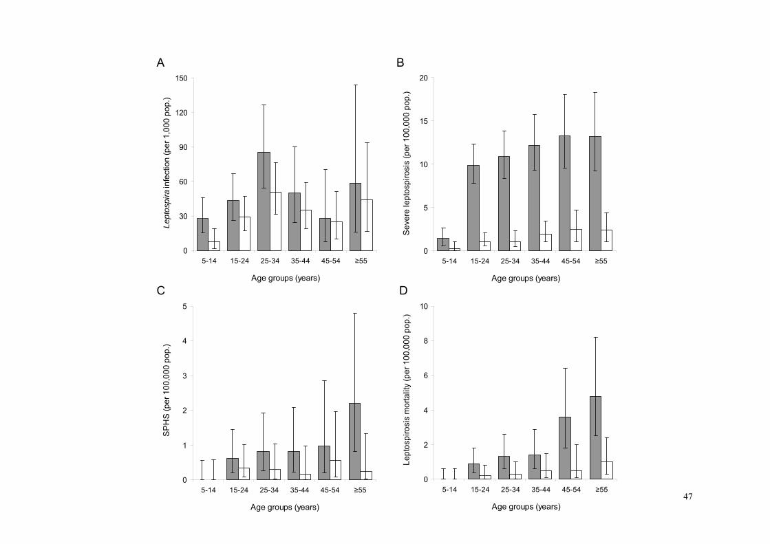
A B
47
0
30
60
90
120
150
5-14 15-24 25-34 35-44 45-54 ≥55
Age groups (years)
Lept
ospi
rain
fect
ion
(per
1,0
00 p
op.)
0
5
10
15
20
5-14 15-24 25-34 35-44 45-54 ≥55
Age groups (years)
Sev
ere
lept
ospi
rosi
s (p
er 1
00,0
00 p
op.)
C D
0
2
4
6
8
10
5-14 15-24 25-34 35-44 45-54 ≥55
Age groups (years)
Lep
tosp
irosi
s m
orta
lity
(per
100
,000
pop
.)
0
1
2
3
4
5
5-14 15-24 25-34 35-44 45-54 ≥55
Age groups (years)
SP
HS
(per
100
,000
pop
.)

0
5
10
15
20
25
30
35
40
45
5-14 15-24 25-34 35-44 45-54 ≥55
Age groups (years)
Cas
e fa
talit
y ra
te (%
)
A B
0
5
10
15
20
25
30
35
5-14 15-24 25-34 35-44 45-54 ≥55
Age groups (years)
Freq
uenc
y of
SPH
S (%
)
48
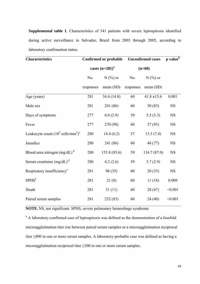
49
Supplemental table 1. Characteristics of 341 patients with severe leptospirosis identified
during active surveillance in Salvador, Brazil from 2003 through 2005, according to
laboratory confirmation status.
Characteristics Confirmed or probable
cases (n=281)a
Unconfirmed cases
(n=60)
p valueb
No.
responses
N (%) or
mean (SD)
No.
responses
N (%) or
mean (SD)
Age (years) 281 34.4 (14.8) 60 41.8 ±15.6 0.001
Male sex 281 241 (86) 60 50 (83) NS
Days of symptoms 277 6.0 (2.9) 59 5.5 (3.3) NS
Fever 277 270 (98) 60 57 (95) NS
Leukocyte count (103 cells/mm3)c 280 14.4 (6.2) 57 13.5 (7.4) NS
Jaundice 280 241 (86) 60 46 (77) NS
Blood urea nitrogen (mg/dL) d 280 153.8 (93.6) 59 134.7 (87.0) NS
Serum creatinine (mg/dL) d 280 4.2 (2.6) 59 3.7 (2.9) NS
Respiratory insufficiencye 281 98 (35) 60 20 (33) NS
SPHSf 281 21 (8) 60 11 (18) 0.009
Death 281 31 (11) 60 28 (47) <0.001
Paired serum samples 281 232 (83) 60 24 (40) <0.001
NOTE. NS, not significant. SPHS, severe pulmonary hemorrhage syndrome
a A laboratory-confirmed case of leptospirosis was defined as the demonstration of a fourfold
microagglutination titer rise between paired serum samples or a microagglutination reciprocal
titer ≥800 in one or more serum samples. A laboratory-probable case was defined as having a
microagglutination reciprocal titer ≥200 in one or more serum samples.

50
b The chi squared or Fisher exact test, and Student’s t-test were used to evaluate for significant
differences (P value<0.05) for proportions and continuous data, respectively.
c Obtained at the time of hospital admission.
d Maximum levels during hospitalization.
e Respiratory insufficiency was defined as respiratory rate ≥28 per minute or use of
supplemental O2 therapy.
f SPHS was defined as massive pulmonary bleeding and respiratory failure.
51
6. DISCUSSÃO
Nas últimas décadas, a leptospirose urbana emergiu como um importante problema de
saúde pública. Comunidades carentes e sem infra-estrutura de saneamento são as mais
atingidas. Embora a cobertura do serviço de esgotamento da cidade de Salvador tenha
aumentado de 26% para mais de 80% através de um projeto (Bahia Azul) para reduzir a
poluição dos mares da cidade, nem todos os bairros, como Pau da Lima, foram beneficiados
pela intervenção. Portanto, estudos para definir fatores de risco para infecção pela Leptospira
são fundamentais para guiar quais e onde futuras intervenções de prevenção devem ser
priorizadas. Além disso, entender porque algumas pessoas desenvolvem leptospirose grave
enquanto a maioria apresenta infecções sub-clínicas ou leves pode ter grandes implicações
tanto na identificação de pacientes com maior risco de desfechos graves associados à doença,
quanto no desenvolvimento de vacinas e drogas capazes de modular a evolução da infecção.
No primeiro manuscrito desta tese, são apresentados resultados de um estudo de corte
transversal realizado em uma comunidade carente de Salvador. Nós avaliamos mais de 3.000
residentes da comunidade e identificamos que 15% deles apresentavam evidência sorológica
de infecção prévia por leptospiras. O uso de SIG (Sistema de Informação Geográfica) e de
abordagens de modelagem permitiram identificar que deficiências de saneamento nesta
comunidade, como esgotos abertos, áreas propensas a alagamento e acúmulo de lixo, são
fatores de risco para infecção pela Leptospira. Além disso, identificamos que baixo nível
sócio-econômico está associado a risco de infecção independentemente das condições
ambientais. Nossos resultados são limitados pelo desenho de corte transversal e a
impossibilidade de identificar temporalidade entre a exposição e o desfecho. Estudos
prospectivos estão sendo realizados nesta população e servirão para confirmar nossos
achados.

52
No segundo manuscrito desta tese, nós apresentamos as incidências de infecção pela
Leptospira, leptospirose grave, SHPL, e mortalidade por leptospirose, de acordo com sexo e
idade. As incidências de infecção pela Leptospira foram estimadas a partir do seguimento de
três anos de uma coorte na comunidade de Pau da Lima em Salvador, e as incidências de
leptospirose grave, SHPL, e mortalidade foram estimadas através de vigilância hospitalar para
leptospirose em Salvador no mesmo período. Nossos resultados sugerem que a maior carga da
leptospirose grave na população de homens adultos se deve inicialmente a um maior risco de
infecção neste grupo. Entretanto, aumento na idade e o sexo masculino parecem ter influência
ainda maior no risco de progressão para leptospirose grave após a infecção. Além disso,
identificamos entre os casos de leptospirose grave que idade ≥45 anos e sexo feminino são
fatores de risco para óbito e para SHPL, respectivamente. Estes achados trazem importante
contribuição no entendimento da história natural da infecção pela Leptospira e reforçam a
hipótese de que a resposta imune do hospedeiro é o principal fator responsável pela variedade
de manifestações clínicas que ocorrem após a infecção pela Leptospira. Futuras investigações
serão necessárias para confirmar nossos resultados em outras populações e para determinar os
mecanismos patogênicos por trás da influência da idade e do sexo no desenvolvimento de
leptospirose grave.

53
7. CONCLUSÕES
Os resultados apresentados nesta tese sugerem que a prevenção da leptospirose urbana
é possível através da universalização do acesso ao saneamento básico e de estratégias para
identificar e modificar os determinantes sociais que causam aumento no risco de infecção.
Além disso, a evidência de que indivíduos do sexo masculino e ≥15 anos tem maior risco de
desenvolvimento de leptospirose grave após infecção sinaliza para a importância de realização
de estudos que esclareçam os mecanismos patogênicos por trás da associação entre idade e
sexo com risco de progressão da doença. Este conhecimento pode ajudar a guiar o
desenvolvimento de vacinas e de drogas imunomoduladoras capazes de modificar o curso da
infecção e prevenir o surgimento de formas graves da leptospirose.
54
8. REFERÊNCIAS BIBLIOGRÁFICAS
Abgueguen, P., V. Delbos, et al. Clinical aspects and prognostic factors of leptospirosis in adults. Retrospective study in France. J Infect. 2008 Jul 23. Andrade, L., S. Cleto, et al. Door-to-dialysis time and daily hemodialysis in patients with leptospirosis: Impact on mortality. Clin J Am Soc Nephrol. 2007 Jul;2(4):739-44. Barocchi, M. A., A. I. Ko, et al. Identification of new repetitive element in Leptospira interrogans serovar copenhageni and its application to PCR-based differentiation of Leptospira serogroups. J Clin Microbiol. 2001;39(1):191-195. Bharti, A. R., J. E. Nally, et al. Leptospirosis: A zoonotic disease of global importance. Lancet Infect Dis. 2003 Dec;3(12):757-71. Bulach, D. M., R. L. Zuerner, et al. Genome reduction in Leptospira borgpetersenii reflects limited transmission potential. Proc Natl Acad Sci U S A. 2006 Sep 26;103(39):14560-5. Campanella, N. Infectious diseases and natural disasters: The effects of Hurricane Mitch over Villanueva municipal area, Nicaragua. Public Health Rev. 1999;27:311-9. CDC. Outbreak of acute febrile illness among athletes participating in triathlons--Wisconsin and Illinois, 1998. MMWR Morb Mortal Wkly Rep. 1998 Jul 24;47(28):585-8. CDC. From the Centers for Disease Control and Prevention. Update: Outbreak of acute febrile illness among athletes participating in Eco-Challenge-Sabah 2000--Borneo, Malaysia, 2000. JAMA. 2001 Feb 14;285(6):728-30. Daher, E. Risk factors for death and changing patterns in leptospirosis acute renal failure. Am J Trop Med Hyg. 1999;61(4):630-634. Everard, C. O., C. N. Edwards, et al. A twelve-year study of leptospirosis on Barbados. Eur J Epidemiol. 1995 Jun;11(3):311-20. Faine, S. B., B. Adler, et al. Leptospira and leptospirosis. A. M. Melbourne, editor.; 1999. Flannery, B., M. M. Pereira, et al. Referral pattern of Leptospirosis cases during a large urban epidemic of dengue. Am J Trop Med Hyg. 2001;65(5):657-663. Gouveia, E. L., J. M. D. Metcalfe, et al. Leptospirosis-associated severe pulmonary hemorrhagic syndrome, Salvador, Brazil. Emerg Infect Dis. 2008 Mar; 14(3):[serial on the Internet]. Available from http://www.cdc.gov/EID/content/14/3/505.htm. Guidugli, F., A. A. Castro, et al. Antibiotics for treating leptospirosis. Cochrane Database Syst Rev. 2000(2):CD001306. Jansen, A., K. Stark, et al. Sex differences in clinical leptospirosis in Germany: 1997-2005. Clin Infect Dis. 2007 May 1;44(9):e69-72.

55
Johnson, M. A., H. Smith, et al. Environmental exposure and leptospirosis, Peru. Emerg Infect Dis. 2004 Jun;10(6):1016-22. Ko, A. I., M. Galvao Reis, et al. Urban epidemic of severe leptospirosis in Brazil. Lancet. 1999;354(9181):820-825. Levett, P. N. Leptospirosis. Clin Microbiol Rev. 2001;14(2):296-326. Lopes, A. A., E. Costa, et al. The association between serum potassium at hospital admission and the case-fatality rate of leptospirosis in men. Rev Inst Med Trop Sao Paulo. 2001 Jul-Aug;43(4):217-20. Lopes, A. A., E. Costa, et al. Comparative study of the in-hospital case-fatality rate of leptospirosis between pediatric and adult patients of different age groups. Rev Inst Med Trop Sao Paulo. 2004;46(1):19-24. Maciel, E. A. P., A. L. F. de Carvalho, et al. Household Transmission of Leptospira Infection in Urban Slum Communities. PLoS Negl Trop Dis 2008;2(e154 doi:10.1371/journal.pntd.0000154). McBride, A. J., D. A. Athanazio, et al. Leptospirosis. Curr Opin Infect Dis. 2005 Oct;18(5):376-86. McClain, J. B., W. R. Ballou, et al. Doxycycline therapy for leptospirosis. Ann Intern Med. 1984 May;;100(5):696-8. Morgan, J. Outbreak of leptospirosis among triathlon participants and community residents in Springfield, Illinois, 1998. Clin Infect Dis. 2002;34(12):1593-1599. Nascimento, A. L. T. O., A. I. Ko, et al. Comparative Genomics of Two Leptospira interrogans Serovars Reveals Novel Insights into Physiology and Pathogenesis. J. Bacteriol. 2004 April 1;186(7):2164-2172. Panaphut, T., S. Domrongkitchaiporn, et al. Prognostic factors of death in leptospirosis: A prospective cohort study in Khon Kaen, Thailand. Int J Infect Dis. 2002;6(1):52-59. Pappas, G., P. Papadimitriou, et al. The globalization of leptospirosis: Worldwide incidence trends. Int J Infect Dis. 2008 Jul;12(4):351-7. Pereira, M. M., M. G. S. Matsuo, et al. A clonal subpopulation of Leptospira interrogans sensu stricto is the major cause of leptospirosis outbreaks in Brazil. J Clin Microbiol. 2000;38(1):450-452. Raoult, D., M. Mailloux, et al. [Seroepidemiologic study on leptospirosis in the Camargue region]. Bull Soc Pathol Exot Filiales. 1985;78(4):439-45. Ren, S. X., G. Fu, et al. Unique physiological and pathogenic features of Leptospira interrogans revealed by whole-genome sequencing. Nature. 2003 Apr 24;422(6934):888-93.

56
Riley, L. W., A. I. Ko, et al. Slum health: Diseases of neglected populations. BMC Int Health Hum Rights. 2007;7(2). Ristow, P., P. Bourhy, et al. The OmpA-Like Protein Loa22 Is Essential for Leptospiral Virulence. PLoS Pathog. 2007 Jul;13(3(7)):e97 Romero, E. C., C. C. M. Bernardo, et al. Human leptospirosis: A twenty-nine-year serological study in São Paulo, Brazil. Rev. Inst. Med. Trop. Sao Paulo. 2003;45(5):245-248. Sanders, E. J., J. G. Rigau-Perez, et al. Increase of leptospirosis in dengue-negative patients after a Hurricane in Puerto Rico in 1966. Am J Trop Med Hyg. 1999;61(3):399-404. Sarkar, U., S. F. Nascimento, et al. Population-based case-control investigation of risk factors for leptospirosis during an urban epidemic. Am J Trop Med Hyg. 2002;66(5):605-610. Sehgal, S. C., A. P. Sugunan, et al. Randomized controlled trial of doxycycline prophylaxis against leptospirosis in an endemic area. Int J Antimicrob Agents. 2000 Feb;13(4):249-55. Sejvar, J., E. Bancroft, et al. Leptospirosis in "Eco-Challenge" athletes, Malaysian Borneo, 2000. Emerg Infect Dis. 2003 Jun;9(6):702-7. Takafuji, E. T., J. W. Kirkpatrick, et al. An efficacy trial of doxycycline chemoprophylaxis against leptospirosis. N Engl J Med. 1984 Feb 23;310(8):497-500. Tangkanakul, W., H. L. Smits, et al. Leptospirosis: An emerging health problem in Thailand. Southeast Asian J Trop Med Public Health. 2005 Mar;36(2):281-8. Tassinari, W. S., D. C. Pellegrini, et al. Detection and modelling of case clusters for urban leptospirosis. Trop Med Int Health. 2008 Apr;13(4):503-12. Trevejo, R. T., J. G. Rigau-Perez, et al. Epidemic leptospirosis associated with pulmonary hemorrhage-Nicaragua, 1995. J Infect Dis. 1998 Nov;178(5):1457-63. Tucunduva de Faria, M., D. A. Athanazio, et al. Morphological alterations in the kidney of rats with natural and experimental leptospira infection. J Comp Pathol 2007 Nov;137(4):231-8. UN-HABITAT. The challenge of the slums: Global report on human settlements. N. U. Nations, editor.; 2003. Watt, G., L. P. Padre, et al. Placebo-controlled trial of intravenous penicillin for severe and late leptospirosis. Lancet. 1988 Feb 27;1(8583):433-5. WHO. Leptospirosis worldwide, 1999. Weekly epidemiological record. Releve epidemiologique hebdomadaire. 1999;74(29):237-242

57
9. ANEXOS
Manuscritos publicados durante o doutorado relacionado a outras doenças:

Vaccine 25 (2007) 4420–4428
Haemophilus influenzae meningitis 5 years after introduction of theHaemophilus influenzae type b conjugate vaccine in Brazil
Guilherme S. Ribeiro a, Josilene B.T. Lima a, Joice N. Reis a,b, Edilane L. Gouveia a,Soraia M. Cordeiro a, Tatiana S. Lobo a, Ricardo M. Pinheiro a, Cassio T. Ribeiro a,
Alan B. Neves a, Katia Salgado c, Hagamenon R. Silva c,d,Mitermayer G. Reis a, Albert I. Ko a,e,∗
a Goncalo Moniz Research Center, Oswaldo Cruz Foundation, Brazilian Ministry of Health, Salvador, Brazilb School of Pharmacy, Federal University of Bahia, Salvador, Brazil
c Hospital Couto Maia, Secretary of Health for the State of Bahia, Salvador, Brazild School of Medicine, Federal University of Bahia, Salvador, Brazil
e Division of International Medicine and Infectious Diseases, Department of Medicine, Weill Medical College of Cornell University, NY, USA
Received 15 November 2006; received in revised form 6 March 2007; accepted 13 March 2007Available online 30 March 2007
Abstract
The long-term impact of Haemophilus influenzae type b (Hib) conjugate vaccine, introduced throughout Latin America in the late 1990s,has not been evaluated. Active surveillance for H. influenzae meningitis was performed from August 9, 1996 to August 8, 2004 in MetropolitanSalvador, Brazil. Five years after the introduction of Hib conjugate vaccine, Hib meningitis incidence decreased from 2.39 to 0.06 cases per100,000 population (98%) overall, and from 60.9 to 3.1 cases per 100,000 population (95%) in children <1 year of age. A transient serotypereplacement phenomenon was observed associated with a small increase of meningitis due to two H. influenzae type a clonal groups. Thesefindings indicate that Hib immunization campaign has led to the virtual elimination of Hib disease in this region.© 2007 Elsevier Ltd. All rights reserved.
Keywords: Haemophilus influenzae; Vaccine effectiveness; Serotype replacement
1. Introduction
Introduction of Haemophilus influenzae type b (Hib)polysaccharide-protein conjugate vaccines have dramaticallyreduced the burden of Hib invasive disease in the last 20 years[1–3]. Clinical trials demonstrated that conjugate vaccineshave an efficacy of 90–100% in preventing Hib invasive dis-ease [4,5]. Furthermore, the vaccine has been highly effective,even in populations where coverage is not complete, due toits ability to reduce nasopharyngeal colonization in immu-
∗ Corresponding author at: Centro de Pesquisas Goncalo Moniz, FundacaoOswaldo Cruz, Ministerio da Saude, Rua Waldemar Falcao, 121, Salvador,Bahia 40296-710, Brazil. Tel.: +55 71 3176 2302; fax: +55 71 3176 2281.
E-mail address: [email protected] (A.I. Ko).
nized individuals [6,7] and induce herd immunity [8,9]. Inthe United States (US) and most industrialized countries, Hibinvasive disease has been virtually eliminated [1–3]. Whereasmore than 10,000 cases of Hib invasive disease were reportedannually in the US prior to introduction of conjugate vaccines,less than 100 cases are now reported each year [1].
The priority for Hib invasive disease is to extend the ben-efits afforded by conjugate vaccines to developing countries[2]. Worldwide, in 2003, 61% of children in target age groupsfor immunization did not receive the conjugate vaccine [10].Hib diseases remain the cause of an estimated 386,000 deathsyearly [11]. However, major progress has been made in Southand Central Americas and the Caribbean where Hib conju-gate vaccines were introduced in the late 1990s and are usedin all but four countries [12]. Although several studies have
0264-410X/$ – see front matter © 2007 Elsevier Ltd. All rights reserved.doi:10.1016/j.vaccine.2007.03.024

Transactions of the Royal Society of Tropical Medicine and Hygiene (2007) 101, 1147—1153
avai lab le at www.sc iencedi rec t .com
journa l homepage: www.e lsev ierhea l th .com/ journa ls / t rs t
Hospital-based surveillance of meningococcalmeningitis in Salvador, Brazil
Soraia M. Cordeiroa, Alan B. Nevesa, Cassio T. Ribeiroa, Maya L. Petersenb,Edilane L. Gouveiaa, Guilherme S. Ribeiroa, Tatiana S. Loboa,Joice N. Reisa,c, Katia M. Salgadod, Mitermayer G. Reisa, Albert I. Koa,e,∗
a Goncalo Moniz Research Center, Oswaldo Cruz Foundation, Brazilian Ministry of Health, Rua Waldemar Falcao 121, Salvador,Bahia 40296-710, Brazilb Joint Medical Program/Division of Epidemiology, School of Public Health, University of California, Berkeley, CA, USAc School of Pharmacy, Federal University of Bahia, Salvador, Brazild Couto Maia Hospital, Bahia Secretary of Health, Salvador, Brazile Division of International Medicine and Infectious Diseases, Weill Medical College of Cornell University, New York, USA
Received 21 March 2007; received in revised form 14 June 2007; accepted 14 June 2007Available online 6 August 2007
KEYWORDSMeningococcaldisease;Meningitis;Neisseriameningitidis;Serogroup;Epidemiology;Brazil
Summary This study aimed to describe the clinical, epidemiological and microbiological fea-tures of meningococcal meningitis in Salvador, Brazil. Between February 1996 and January2001, a hospital-based surveillance prospectively identified cases of culture-positive meningo-coccal meningitis. Demographic and clinical data were collected through interview and medicalchart review. Antisera and monoclonal antibodies were used to determine the serogroup andserotype:serosubtype of the isolates, respectively. Surveillance identified a total of 408 cases ofmeningococcal meningitis, with a case fatality rate of 8% (32/397). The mean annual incidencefor the 304 culture-positive cases residing in metropolitan Salvador was 1.71 cases per 100 000population. Infants <1 year old presented the highest incidence (14.7 cases per 100 000 popula-tion). Of the 377 serogrouped isolates, 82%, 16%, 2% and 0.3% were serogroups B, C, W135 andY, respectively. A single serotype:serosubtype (4,7:P1.19,15) accounted for 64% of all cases.Continued surveillance is necessary to characterise strains and to define future prevention andcontrol strategies.© 2007 Royal Society of Tropical Medicine and Hygiene. Published by Elsevier Ltd. All rightsreserved.
∗ Corresponding author. Tel.: +55 71 3176 2302;fax: +55 71 3176 2281.
E-mail address: [email protected] (A.I. Ko).
1. Introduction
Neisseria meningitidis is a common cause of disease world-wide, responsible for significant morbidity and mortality ininfants and young children (Bilukha and Rosenstein, 2005).Without appropriate antimicrobial treatment, most cases
0035-9203/$ — see front matter © 2007 Royal Society of Tropical Medicine and Hygiene. Published by Elsevier Ltd. All rights reserved.doi:10.1016/j.trstmh.2007.06.012
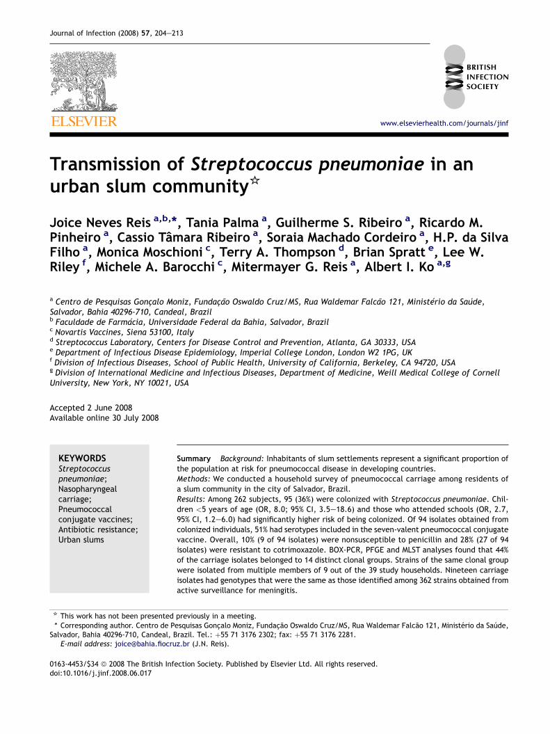
Journal of Infection (2008) 57, 204e213
www.elsevierhealth.com/journals/jinf
Transmission of Streptococcus pneumoniae in anurban slum community*
Joice Neves Reis a,b,*, Tania Palma a, Guilherme S. Ribeiro a, Ricardo M.Pinheiro a, Cassio Tamara Ribeiro a, Soraia Machado Cordeiro a, H.P. da SilvaFilho a, Monica Moschioni c, Terry A. Thompson d, Brian Spratt e, Lee W.Riley f, Michele A. Barocchi c, Mitermayer G. Reis a, Albert I. Ko a,g
a Centro de Pesquisas Goncalo Moniz, Fundacao Oswaldo Cruz/MS, Rua Waldemar Falcao 121, Ministerio da Saude,Salvador, Bahia 40296-710, Candeal, Brazilb Faculdade de Farmacia, Universidade Federal da Bahia, Salvador, Brazilc Novartis Vaccines, Siena 53100, Italyd Streptococcus Laboratory, Centers for Disease Control and Prevention, Atlanta, GA 30333, USAe Department of Infectious Disease Epidemiology, Imperial College London, London W2 1PG, UKf Division of Infectious Diseases, School of Public Health, University of California, Berkeley, CA 94720, USAg Division of International Medicine and Infectious Diseases, Department of Medicine, Weill Medical College of CornellUniversity, New York, NY 10021, USA
Accepted 2 June 2008Available online 30 July 2008
KEYWORDSStreptococcuspneumoniae;Nasopharyngealcarriage;Pneumococcalconjugate vaccines;Antibiotic resistance;Urban slums
* This work has not been presented* Corresponding author. Centro de P
Salvador, Bahia 40296-710, Candeal, BE-mail address: [email protected]
0163-4453/$34 ª 2008 The British Infdoi:10.1016/j.jinf.2008.06.017
Summary Background: Inhabitants of slum settlements represent a significant proportion ofthe population at risk for pneumococcal disease in developing countries.Methods: We conducted a household survey of pneumococcal carriage among residents ofa slum community in the city of Salvador, Brazil.Results: Among 262 subjects, 95 (36%) were colonized with Streptococcus pneumoniae. Chil-dren <5 years of age (OR, 8.0; 95% CI, 3.5e18.6) and those who attended schools (OR, 2.7,95% CI, 1.2e6.0) had significantly higher risk of being colonized. Of 94 isolates obtained fromcolonized individuals, 51% had serotypes included in the seven-valent pneumococcal conjugatevaccine. Overall, 10% (9 of 94 isolates) were nonsusceptible to penicillin and 28% (27 of 94isolates) were resistant to cotrimoxazole. BOX-PCR, PFGE and MLST analyses found that 44%of the carriage isolates belonged to 14 distinct clonal groups. Strains of the same clonal groupwere isolated from multiple members of 9 out of the 39 study households. Nineteen carriageisolates had genotypes that were the same as those identified among 362 strains obtained fromactive surveillance for meningitis.
previously in a meeting.esquisas Goncalo Moniz, Fundacao Oswaldo Cruz/MS, Rua Waldemar Falcao 121, Ministerio da Saude,razil. Tel.: þ55 71 3176 2302; fax: þ55 71 3176 2281.z.br (J.N. Reis).
ection Society. Published by Elsevier Ltd. All rights reserved.