estudo epidemiológico de 261 dentes permanentes ... · identificação do material bibliográfico:...
TRANSCRIPT
UNIVERSIDADE FEDERAL DE GOIÁS PROGRAMA DE PÓS-GRADUAÇÃO EM CIÊNCIAS DA SAÚDE
ORLANDO AGUIRRE GUEDES
Estudo epidemiológico de 261 dentes permanentes avulsionados de pacientes tratados em um serviço de
urgência odontológica
Goiânia 2012

TERMO DE CIÊNCIA E DE AUTORIZAÇÃO PARA DISPONIBILIZAR AS TESES E
DISSERTAÇÕES ELETRÔNICAS (TEDE) NA BIBLIOTECA DIGITAL DA UFG Na qualidade de titular dos direitos de autor, autorizo a Universidade Federal de Goiás (UFG) a disponibilizar, gratuitamente, por meio da Biblioteca Digital de Teses e Dissertações (BDTD/UFG), sem ressarcimento dos direitos autorais, de acordo com a Lei nº 9610/98, o documento conforme permissões assinaladas abaixo, para fins de leitura, impressão e/ou download, a título de divulgação da produção científica brasileira, a partir desta data.
1. Identificação do material bibliográfico: [ ] Dissertação [X] Tese 2. Identificação da Tese ou Dissertação
Autor (a): Orlando Aguirre Guedes E-mail: [email protected] Seu e-mail pode ser disponibilizado na página? [X]Sim [ ] Não
Vínculo empregatício do autor Nenhum Agência de fomento: Coordenação de Aperfeiçoamento
de Pessoal de Nível Superior Sigla: CAPES
País: Brasil UF:GO CNPJ: Título: Estudo epidemiológico de 261 dentes permanentes avulsionados de pacientes tratados
em um serviço de urgência odontológica Palavras-chave: Traumatismo dentário, avulsão dentária, reimplante, epidemiologia oral Título em outra língua: Epidemiological study of 261 avulsed permanent teeth treated in a
dental urgency service Palavras-chave em outra língua: Dental traumatology, tooth avulsion, tooth replantation, oral
epidemiology Área de concentração: Dinâmica do Processo Saúde-Doença Data defesa: (dd/mm/aaaa) 09/01/2012 Programa de Pós-Graduação: Ciências da Saúde Orientador (a): Prof. Dr. Carlos Estrela E-mail: [email protected] Co-orientador (a): Profa. Dra. Ana Helena Gonçalves de Alencar E-mail: [email protected] 3. Informações de acesso ao documento: Liberação para disponibilização?1 [X] total [ ] parcial Em caso de disponibilização parcial, assinale as permissões: [ ] Capítulos. Especifique: __________________________________________________ [ ] Outras restrições: _____________________________________________________ Havendo concordância com a disponibilização eletrônica, torna-se imprescindível o envio do(s) arquivo(s) em formato digital PDF ou DOC da tese ou dissertação. O Sistema da Biblioteca Digital de Teses e Dissertações garante aos autores, que os arquivos contendo eletronicamente as teses e ou dissertações, antes de sua disponibilização, receberão procedimentos de segurança, criptografia (para não permitir cópia e extração de conteúdo, permitindo apenas impressão fraca) usando o padrão do Acrobat. ________________________________________ Data: ____ / ____ / _____ Assinatura do (a) autor (a)
1 Em caso de restrição, esta poderá ser mantida por até um ano a partir da data de defesa. A extensão deste prazo suscita justificativa junto à coordenação do curso. Todo resumo e metadados ficarão sempre disponibilizados
ORLANDO AGUIRRE GUEDES
Estudo epidemiológico de 261 dentes permanentes avulsionados de pacientes tratados em um serviço de
urgência odontológica
Tese de Doutorado apresentada ao Programa
de Pós-Graduação em Ciências da Saúde da
Universidade Federal de Goiás para obtenção
do Título Doutor em Ciências da Saúde.
Orientador: Prof. Dr. Carlos Estrela Co-orientadora: Profa. Dra. Ana Helena Gonçalves de Alencar
Goiânia 2012

Dados Internacionais de Catalogação na Publicação (CIP) GPT/BC/UFG
Guedes, Orlando Aguirre. Estudo epidemiológico de 261 dentes permanentes
avulsionados de pacientes tratados em um serviço de urgência brasileiro / Orlando Aguirre Guedes. – 2012.
80 f.: 02 figs, 02 tabs. Orientador: Prof. Dr. Carlos Estrela. Tese (Doutorado) – Universidade Federal de Goiás,
Programa de Pós-Graduação em Ciências da Saúde, 2012. Bibliografia. 1. Traumatismo Dentário 2. Avulsão Dentária 3. Reimplante 4.
Epidemiologia bucal

iii
Programa de Pós-Graduação em Ciências da Saúde da Universidade Federal de Goiás
BANCA EXAMINADORA DA TESE DE DOUTORADO
Aluno: Orlando Aguirre Guedes
Orientador: Prof. Dr. Carlos Estrela
Co-orientadora: Profa. Dra. Ana Helena Gonçalves de Alencar
Membros:
1. Prof. Dr. Carlos Estrela
2. Profa. Dra. Ana Helena Gonçalves de Alencar
3. Prof. Dr. José Antônio Poli de Figueiredo
4. Profa. Dra. Lilian de Fátima Guedes de Amorim
5. Profa. Dra. Maristela Gutiérrez de Borba
Suplentes:
1. Profa. Dra. Heloisa Helena Pinho Veloso
2. Profa. Dra. Kely Firmino Bruno
Data: 09/01/2012
iv
Dedicatória
Dedico, carinhosamente, este trabalho à minha querida mãe, Elisabet Aguirre, o meu amor e gratidão pelo estímulo constante em relação aos estudos, pela formação que me foi oferecida e pelo exemplo de vida.

v
Agradecimentos
A Deus, que sempre iluminou meus caminhos e que me sustentou com
vontade e perseverança na busca dos meus ideais;
Ao meu pai Claudionor Dantas Guedes, pelo incentivo dado durante toda
minha caminhada;
Aos meus irmãos Mariela, Claúdio e Áliva, pelo apoio, união, amor e
respeito;
A minha namorada Luísa de Mello Florentino, meu grande amor, pelo
carinho e apoio, e pelo consolo nos momentos de incerteza;
Ao professor, orientador e amigo Carlos Estrela, pelas conversas
rotineiras e informais, pelos ensinamentos dentro e fora da endodontia e por
permitir a minha iniciação no mundo científico;
Aos profs. Dr. Hugo Alexandre de Sousa e Dra. Ana Helena Gonçalves
de Alencar, pela especial atenção, amizade e colaboração.
A Cyntia Estrela, pela paciência, auxílio, amizade e acolhimento em sua
casa durante a realização deste trabalho;
Aos Professores do Curso de Doutorado em Ciências da Saúde da
Universidade Federal de Goiás, pelos ensinamentos que me passaram.
Ao meu sogro e minha sogra Plínio A. G. Florentino e Sálua B. de Mello
Florentino, que me tratam como um filho e me dão o prazer de conviver em sua
família;
Aos meus queridos “irmãos” Pedro e Bárbara Florentino e Thiago Palma,
pela camaradagem, receptividade, convivência e integração;
Aos amigos Daniel de Almeida Decurcio, Júlio Almeida Silva, Rafael de
Almeida Decurcio, Olavo Cesar Lyra Porto, Luiz Eduardo G. Rabelo, Iury
Castro, Felipe Cavalcanti Sampaio, André Luiz Gomide Morais, Marcel Garrote,
Omar Zina e Helder Fernandes de Oliveira pelo companheirismo e amizade;

vi
A todos os amigos, presentes ou não neste mundo, que torceram por
mim ou que de alguma forma ajudaram na realização deste trabalho.

vii
SUMÁRIO
Tabelas, Figuras e Anexos viii
Símbolos, Siglas e Abreviaturas ix
Resumo x
Abstract xii
1. Introdução 13
2. Objetivo 16
3. Material e Método 17
4. Resultados 20
5. Discussão 27
6. Conclusão 33
7. Referências Bibliográficas 34
8. Publicação 40
9. Anexos 64

viii
TABELAS, FIGURAS E ANEXOS
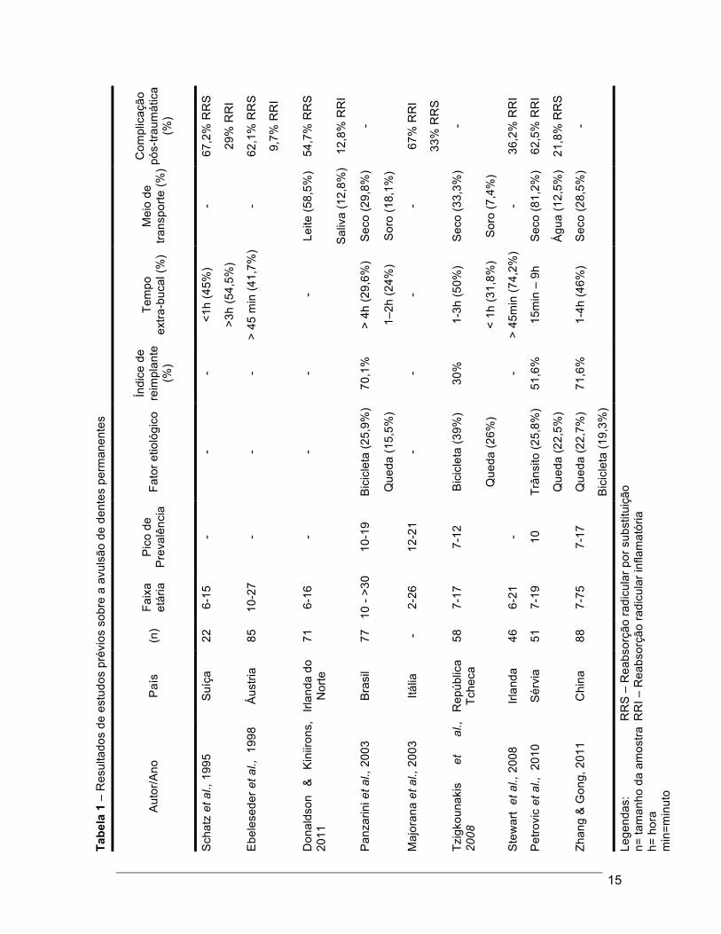
Tabela 1. Resultados de estudos prévios sobre a avulsão de dentes permanentes.
14
Tabela 2. Distribuição dos fatores etiológicos das avulsões dentárias em função da idade e do gênero.
24
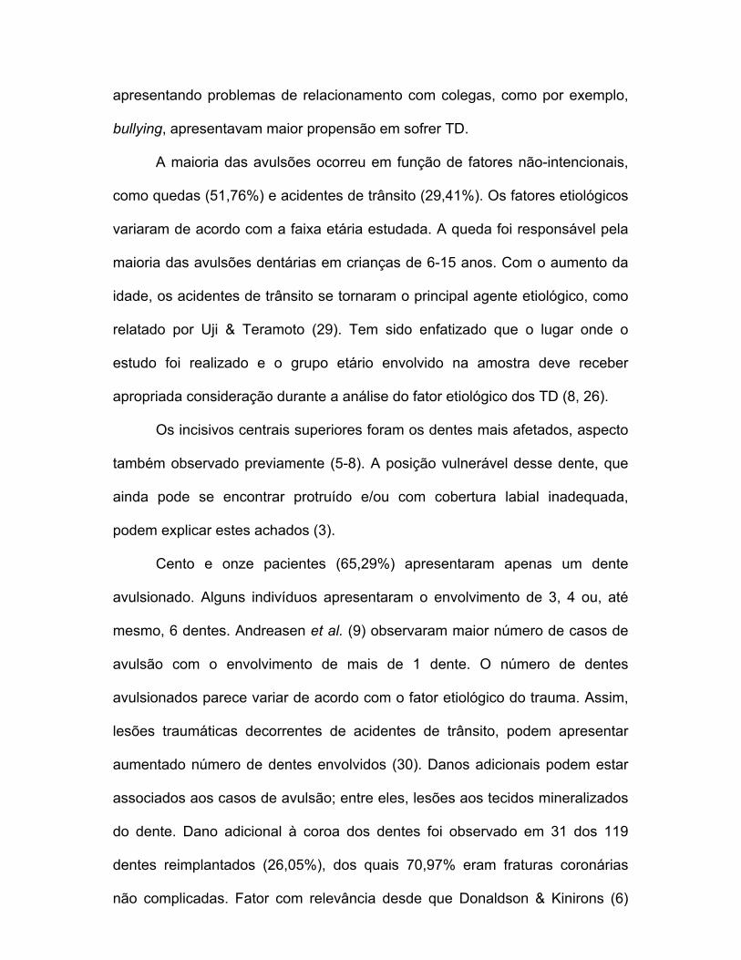
Figura 1. Informações coletadas dos prontuários e das radiografias dos pacientes.
18
Figura 2. Distribuição dos casos de avulsão dentária em função do dente envolvido.
25
Anexo 1. Parecer do Comitê de Ética 62
Anexo 2. Publicações no biênio 2010/2011 64
Anexo 3. Normas de publicação do periódico 67

ix
SÍMBOLOS, SIGLAS E ABREVIATURAS
% Porcentagem
= Igual
et al. e outros
IIRR Internal inflammatory root resorption
IRR Inflammatory root resorption
OCR Obliteração do canal radicular
PCO Pulp canal obliteration
RRI Reabsorção radicular inflamatória
RRII Reabsorção radicular interna inflamatória
RRR Replacement root resorption
RRS Reabsorção radicular por substituição
TD Traumatismo dentário
TDI Traumatic dental injury
α Nível de significância
x
RESUMO
Objetivo: Avaliar os aspectos epidemiológicos e fatores clínicos da avulsão de dentes permanentes. Material e Método: A amostra do estudo era composta por 170 pacientes (261 dentes avulsionados) atendidos no Serviço de Urgência Odontológica da Faculdade de Odontologia da Universidade Federal de Goiás, entre os anos de 2000 e 2008. As seguintes informações foram retiradas dos registros odontológicos de cada paciente: gênero, idade, fator etiológico, distribuição sazonal, grupo dentário, número de dentes avulsionados, reimplante, estágio de desenvolvimento radicular, meio de transporte, período extra-bucal, presença de dano adicional ao dente, tipo de tratamento e o tipo de complicação pós-traumática. O tratamento estatístico analisou os dados frente à distribuição de frequência e qui-quadrado. O nível de significância foi de p<0,05. Resultados: Observou-se elevada ocorrência de avulsões em indivíduos do gênero masculino (71,18%) e com idade entre 6-10 e 11-15 anos (30,59% cada um). Os principais fatores etiológicos foram quedas (51,76%), acidentes de trânsito (29,41%) e violência (6,47%). A distribuição sazonal evidenciou elevado número de traumatismos no outono (março a junho; 31,18%) e no inverno (junho a setembro; 27,65%). O dente mais comumente afetado foi o incisivo central superior (62,45%), seguido pelo incisivo lateral superior (21,46%). Significativa proporção (67,23%) dos dentes traumatizados apresentava os ápices radiculares completamente formados. Cento e dezenove dentes avulsionados (45,59%) foram reimplantados. A maioria dos dentes (89,08%) recebeu reimplante tardio. Trinta e oito dentes (31,93%) foram conservados à seco. Reparo do periodonto foi observado em 41 dentes (34,45%), reabsorção radicular inflamatória em 44 dentes (36,97%) e reabsorção radicular por substituição em 22 dentes (18,49%). As modalidades terapêuticas mais comumente realizadas nos dentes reimplantados foram o tratamento endodôntico com a obturação temporária do canal radicular com hidróxido de cálcio (58,82%) e o tratamento endodôntico com a obturação definitiva do canal radicular (26,89%). Conclusão: Verificou-se elevado número de avulsões dentárias em indivíduos do gênero masculino, com idade inferior a 15 anos, decorrentes de quedas e envolvendo principalmente dentes superiores anteriores; e baixo índice de reimplantes acompanhado de significativo número de dentes acondicionados em meios não fisiológicos por longo período de

xi
tempo. Palavras-chave: Traumatismo dentário, avulsão dentária, reimplante, epidemiologia bucal.
xii
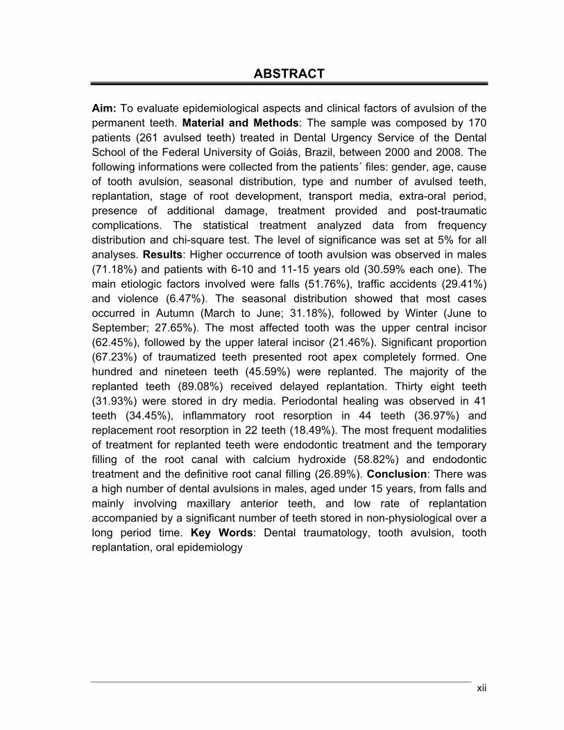
ABSTRACT Aim: To evaluate epidemiological aspects and clinical factors of avulsion of the permanent teeth. Material and Methods: The sample was composed by 170 patients (261 avulsed teeth) treated in Dental Urgency Service of the Dental School of the Federal University of Goiás, Brazil, between 2000 and 2008. The following informations were collected from the patients´ files: gender, age, cause of tooth avulsion, seasonal distribution, type and number of avulsed teeth, replantation, stage of root development, transport media, extra-oral period, presence of additional damage, treatment provided and post-traumatic complications. The statistical treatment analyzed data from frequency distribution and chi-square test. The level of significance was set at 5% for all analyses. Results: Higher occurrence of tooth avulsion was observed in males (71.18%) and patients with 6-10 and 11-15 years old (30.59% each one). The main etiologic factors involved were falls (51.76%), traffic accidents (29.41%) and violence (6.47%). The seasonal distribution showed that most cases occurred in Autumn (March to June; 31.18%), followed by Winter (June to September; 27.65%). The most affected tooth was the upper central incisor (62.45%), followed by the upper lateral incisor (21.46%). Significant proportion (67.23%) of traumatized teeth presented root apex completely formed. One hundred and nineteen teeth (45.59%) were replanted. The majority of the replanted teeth (89.08%) received delayed replantation. Thirty eight teeth (31.93%) were stored in dry media. Periodontal healing was observed in 41 teeth (34.45%), inflammatory root resorption in 44 teeth (36.97%) and replacement root resorption in 22 teeth (18.49%). The most frequent modalities of treatment for replanted teeth were endodontic treatment and the temporary filling of the root canal with calcium hydroxide (58.82%) and endodontic treatment and the definitive root canal filling (26.89%). Conclusion: There was a high number of dental avulsions in males, aged under 15 years, from falls and mainly involving maxillary anterior teeth, and low rate of replantation accompanied by a significant number of teeth stored in non-physiological over a long period time. Key Words: Dental traumatology, tooth avulsion, tooth replantation, oral epidemiology

13
1 INTRODUÇÃO
Os traumatismos dentários (TD) representam um sério problema de
saúde pública entre indivíduos jovens. Nas últimas décadas, vários estudos têm
reportado aumento significativo na prevalência dessas lesões, com significativa
ameaça à qualidade de vida de crianças e adolescentes (1-3).
A avulsão dentária é um dos tipos mais graves de TD. É definida como o
completo deslocamento do dente para fora do seu alvéolo (2). Sua prevalência
na dentição permanente varia de 0,5% a 18,30% (2, 4), ocorre mais
frequentemente em pacientes do gênero masculino com idade inferior a 14
anos (1) e o dente mais afetado é o incisivo central superior (5-8) (Tabela 1).
Após uma avulsão dentária, são observados danos severos aos tecidos
pulpares e periodontais, o que pode resultar em uma série de complicações
pós-traumáticas, tais como necrose pulpar, anquilose e reabsorção radicular (6,
9, 10). Eventualmente, a avulsão dentária e as suas complicações podem levar
ao desenvolvimento de sequelas nos dentes permanentes (11, 12), à perda do
dente traumatizado (10) ou podem afetar o crescimento do rebordo alveolar, a
erupção e o posicionamento dos dentes adjacentes (13-15). O restabelecimento
da estética e da função em pacientes com crescimento facial incompleto
representa um desafio único para o clínico (16).
Uma vez que o prognóstico do dente reimplantado depende de medidas
adequadas tomadas no local do acidente, estudos (17-19) têm sugerido a

14
necessidade do desenvolvimento de campanhas educativas para a
conscientização da população a respeito da prevenção e do tratamento
emergencial de dentes avulsionados. Entretanto, um programa de prevenção
efetivo sobre a avulsão dentária deve ser preferencialmente, precedido por um
estudo sobre a sua distribuição em uma determinada população e sobre os
seus fatores determinantes.
A partir de evidências do reduzido número de estudos epidemiológicos
na população brasileira (20) e, por considerar as especificidades e diferenças
demográficas, culturais e socioeconômicas, se torna justificável analisar alguns
aspectos epidemiológicos e clínicos da avulsão dentária na dentição
permanente em pacientes tratados em um serviço de urgência odontológica.
15
Tabe
la 1
– R
esul
tado
s de
est
udos
pré
vios
sob
re a
avu
lsão
de
dent
es p
erm
anen
tes
Com
plic
ação
pó
s-tra
umát
ica
(%)
67,2
% R
RS
29%
RR
I
62,1
% R
RS
9,7%
RR
I
54,7
% R
RS
12,8
% R
RI
-
67%
RR
I
33%
RR
S
-
36,2
% R
RI
62,5
% R
RI
21,8
% R
RS
-
RR
S –
Rea
bsor
ção
radi
cula
r por
sub
stitu
ição
R
RI –
Rea
bsor
ção
radi
cula
r inf
lam
atór
ia
Mei
o de
tra
nspo
rte (%
)
- -
Leite
(58,
5%)
Sal
iva
(12,
8%)
Sec
o (2
9,8%
)
Sor
o (1
8,1%
)
-
Sec
o (3
3,3%
)
Sor
o (7
,4%
)
-
Sec
o (8
1,2%
)
Águ
a (1
2,5%
)
Sec
o (2
8,5%
)
Tem
po
ex
tra-b
ucal
(%)
<1h
(45%
)
>3h
(54,
5%)
> 45
min
(41,
7%)
-
> 4h
(29,
6%)
1–2h
(24%
)
-
1-3h
(50%
)
< 1h
(31,
8%)
> 45
min
(74,
2%)
15m
in –
9h
1-4h
(46%
)
Índi
ce d
e re
impl
ante
(%
) - - -
70,1
%
-
30%
-
51,6
%
71,6
%
Fato
r etio
lógi
co
- - -
Bic
icle
ta (2
5,9%
)
Que
da (1
5,5%
)
-
Bic
icle
ta (3
9%)
Que
da (2
6%)
Trân
sito
(25,
8%)
Que
da (2
2,5%
)
Que
da (2
2,7%
)
Bic
icle
ta (1
9,3%
)
Pic
o de
P
reva
lênc
ia
- - -
10-1
9
12-2
1
7-12
- 10
7-17
Faix
a et
ária
6-15
10-2
7
6-16
10 -
>30
2-26
7-17
6-21
7-19
7-75
(n)
22 85 71 77 - 58 46
51 88
Paí
s
Suí
ça
Áus
tria
Irlan
da d
o N
orte
Bra
sil
Itália
Rep
úblic
a Tc
heca
Irlan
da
Sér
via
Chi
na
Aut
or/A
no
Sch
atz
et a
l., 1
995
Ebe
lese
der e
t al.,
199
8
Don
alds
on
&
Kin
iiron
s,
2011
Pan
zarin
i et a
l., 2
003
Maj
oran
a et
al.,
200
3
Tzig
koun
akis
et
al
., 20
08
Ste
war
t et
al.,
200
8
Pet
rovi
c et
al.,
201
0
Zhan
g &
Gon
g, 2
011
Lege
ndas
:
n= ta
man
ho d
a am
ostra
h=
hor
a m
in=m
inut
o

16
2 OBJETIVOS
O objetivo deste estudo foi investigar alguns aspectos epidemiológicos e
clínicos da avulsão dentária na dentição permanente em pacientes atendidos
em um serviço de urgência odontológica.

17
3 MATERIAL E MÉTODO
Este estudo transversal retrospectivo envolveu pacientes atendidos no
Serviço de Urgência da Faculdade de Odontologia da Universidade Federal de
Goiás, Goiânia (latitude 16o43´S; longitude 49o20´W), Brasil e com histórico de
avulsão de dentes permanentes. A busca foi desenvolvida no período de
outubro de 2000 a dezembro de 2008.
Informações relacionadas ao aspecto epidemiológico e aos fatores
clínicos da avulsão de dentes permanentes (Figura 1) foram obtidas dos
registros odontológicos e radiografias dos pacientes, com o auxílio de planilhas
digitais, por dois estudantes de pós-graduação. Previamente à coleta dos
dados, um estudo piloto, envolvendo 10% da amostra final, foi realizado para
testar a viabilidade do estudo e treinar e calibrar os examinadores com relação
aos critérios utilizados. Pacientes apresentando documentação incompleta e
com acompanhamento clínico e radiográfico inferior a um ano foram excluídos
do estudo.
O estágio de desenvolvimento radicular foi avaliado de acordo com
Stewart et al. (21). Os ápices radiculares foram classificados como abertos se,
na radiografia, as paredes do canal radicular apresentavam-se divergentes ou
paralelas e foram considerados como fechados se as paredes do canal
apresentavam-se convergentes. O período extra-bucal foi considerado como o
tempo total em que o dente permaneceu fora do alvéolo. Dentes reimplantados

18
com um período extra-bucal superior a 5 minutos foram considerados como
reimplante tardio (22).
As complicações pós-traumáticas foram registradas analisando todas as
radiografias de proservação. A reabsorção radicular por substituição (RRS) foi
registrada na presença da perda de tecido radicular com reposição óssea
concomitante, estando à perda do espaço do ligamento periodontal não
associada à radiolucidez; a reabsorção radicular inflamatória (RRI) foi
diagnosticada sempre que o dente apresentava, radiograficamente, perda de
tecido radicular com concomitante perda de tecido ósseo associada à
radiolucidez (5); a reabsorção radicular interna inflamatória (RRII) foi registrada
quando um alargamento radiolúcido, de forma oval ou arredondado do espaço
do canal radicular foi observado nas radiografias (23) e a obliteração do canal
radicular (OCR) foi diagnosticada pela obliteração da cavidade pulpar
observada radiograficamente (24).
O protocolo do estudo foi aprovado pelo Comitê de Ética em Pesquisa da
Universidade Federal de Goiás (Protocolo no 055/2005).
A análise estatística dos dados foi realizada com o programa SPSS for
Windows 19.0 (SPSS Inc., Chicago, IL, USA) e incluiu distribuição de frequência
e teste de associação. A significância estatística para a associação entre as
variáveis foi determinada pela utilização do teste qui-quadrado. O nível de
significância estabelecido foi de p<0,05.

19
Figura 1. Informações coletadas dos prontuários e das radiografias dos pacientes.
Gênero n=170 Masculino Feminino
Aspectos epidemiológicos (n=170)
Fatores clínicos (n=119)
Faixa etária (n=170) 6-10 21-25
11-15 26-30 16-20 >30
Fator etiológico (n=170) Não Intencional Intencional Quedas Violência Esporte Colisão Acidente de Trânsito
Distribuição sazonal (n=170) Primavera Outono Verão Inverno
Dente (n=261) Grupo Número de dentes avulsionados
Prontuário odontológico (n=170)
Tratamento emergencial (n=119)
Estágio de desenvolvimento radicular (n=119)
Completo Incompleto
Meio de Transporte (n=119) Seco Soro Leite Saliva Água Gelo Álcool
Período extra-bucal (n=119) < 5 min > 5 min
Dano adicional (n=119) Ao tecido mineralizado Ao osso de sustentação
Tratamento (n=261) Endodôntico Protético
Complicação pós-traumática (n=110) RRI RRS OCR RRII

20
4 RESULTADOS
Faixa etária e gênero
A análise envolveu 170 pacientes com histórico de avulsão dentária, com
idade variando entre 6 e 43 anos (média de 15,9 anos, desvio padrão de 7,97).
As maiores frequências de avulsão foram registradas nos participantes de 6-10
(30,59%) e 11-15 anos de idade (30,59%), seguido dos participantes de 16-20
anos de idade (15,88%) (Tabela 2). Do total de registros analisados, 121
(71,18%) eram do gênero masculino e 49 (28,82%) do gênero feminino, sendo
observada uma proporção entre homens e mulheres de 2,4:1.
Fator etiológico
Cento e cinquenta e nove participantes (93,53%) sofreram avulsão
devido a causas não-intencionais e onze (6,47%) a causas intencionais.
Quedas (51,76%), acidentes de trânsito (29,41%) e violência (6,47%) foram os
principais fatores etiológicos. Observou-se uma variação entre o fator etiológico
e o gênero dos participantes. Atividades esportivas, violência e colisões
resultaram em 4 (2,35%), 9 (5,29%) e 5 (2,94%) eventos traumáticos em
pacientes do gênero masculino, respectivamente, e 0 (0,00%), 2 (1,18%) e 0
(0,00%) em pacientes do gênero feminino, respectivamente. No entanto, está
variação não foi estatisticamente significativa (p>0,05). O teste do qui-quadrado
revelou uma diferença estatisticamente significante entre o fator etiológico e a

21
faixa etária (p<0,05). Quedas foram a causa mais comum de avulsão dentária
entre os participantes com idade inferior a 15 anos (41,18%). No entanto, com o
aumento da idade, os acidentes automobilísticos constituíram-se no principal
agente etiológico das lesões traumáticas, sendo responsável por 16,47% dos
acidentes (Tabela 2).
Distribuição sazonal
A distribuição sazonal revelou que a maioria dos casos ocorreu no
outono (março a junho; 31,18%), seguido pelo inverno (junho a setembro;
27,65%), primavera (setembro a dezembro; 23,53%) e verão (dezembro a
março; 17,65%).
Grupo dentário e número de dentes avulsionados
Os pacientes apresentaram um total de 261 dentes permanentes
avulsionados, representando cerca de 1,5 dentes avulsionados por acidente.
Duzentos e vinte e seis (86,59%) localizavam-se na maxila e 35 (13,41%) na
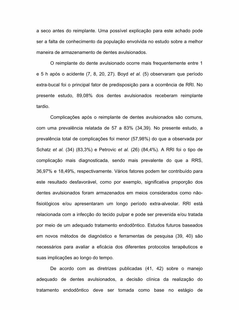
mandíbula. Os dentes mais comumente afetados foram os incisivos centrais
superiores (62,45%), seguidos pelos incisivos laterais superiores (21,46%) e
incisivos centrais inferiores (5,75%) (Figura 2). Cento e onze participantes
(65,29%) apresentaram o envolvimento de apenas um dente, 38 (22,35%) de
dois dentes, e 21 (12,35%) apresentaram três ou mais dentes avulsionados.

22
Tratamento emergencial e o estágio de desenvolvimento radicular
Dos 261 dentes avulsionados, 119 (45,59%) foram reimplantados após o
acidente. Destes, 80 (67,23%) apresentavam completa formação radicular,
enquanto 39 (32,77%) apresentavam rizogênese incompleta. Cento e quarenta
e dois dentes (54,41%) não foram reimplantados.
Meio de transporte
Trinta e oito dentes (31,93%) foram armazenados em meio seco, 20
(16,81%) em solução salina, 20 (16,81%) em leite, 15 (12,61%) em água, 5
(4,20%) em álcool, 4 (3,36%) em saliva e 1 (0,84%) em gelo. Informação sobre
o meio de transporte não pôde ser determinada em 16 (13,45%) dos 119 dentes
reimplantados.
Período extra-bucal
Nenhum dente foi reimplantado com o tempo inferior a 5 minutos. Cento
e seis dentes (89,08%) receberam reimplante tardio. Destes, 42 (35,29%) foram
reimplantados até 60 minutos após a lesão traumática, enquanto 26 (21,85%)
foram reimplantados entre 1 e 2 horas e 38 (31,93%) com mais de 2 horas após
o acidente. Informação sobre o período extra-bucal não pôde ser avaliada em
13 dentes (10,92%).
Presença de dano adicional ao dente
A associação entre outros tipos de TD e a avulsão foi observada em 31
(26,05%) dos 119 dentes reimplantados. O tipo mais comum de lesão foi a

23
fratura não complicada da coroa (n=22; 70,97%), seguida pela fratura
complicada da coroa (n=5; 16,13%), fratura do processo alveolar (n=3; 9,67%) e
fratura corono radicular complicada (n=1; 3,23%). Apenas 4 dentes (3,36%)
apresentaram TD recorrentes.
Tratamento odontológico
A avaliação do tipo de tratamento realizado foi subdividida em tratamento
para dentes reimplantados e tratamento para dentes não-reimplantados. Para
os dentes reimplantados, a modalidade mais frequente foi o tratamento
endodôntico com o preenchimento temporário do canal radicular com hidróxido
de cálcio (n=70; 58,82%), seguido do tratamento endodôntico com a obturação
definitiva do canal radicular (n=32; 26,89%), extração (n=4; 3,36%), reabilitação
protética (n=4; 3,36%), tratamento endodôntico com obturação definitiva do
canal radicular associado à movimentação ortodôntica (n=2; 1,68%),
mantenedor de espaço (n=1; 0,84%) e reabilitação protética associada a
movimentação ortodôntica (n=1; 0,84%). Para os dentes não reimplantados,
foram realizados exame clínico e anamnese (n=117; 82,39%), reabilitação
protética (n=22; 15,49%), mantenedor de espaço (n=1; 0,70%), movimentação
ortodôntica (n=1; 0,70%) e transplante dentário associado a tratamento
endodôntico com o preenchimento temporário do canal radicular com hidróxido
de cálcio (n=1; 0,70%).

24
Complicações pós-traumáticas
Os dentes reimplantados foram proservados por um período de 1 a 8
anos. O acompanhamento radiográfico revelou 44 dentes (36,97%) com RRI,
41 dentes (34,45%) com reparo periodontal, 22 dentes (18,49%) com RRS, 2
dentes (1,68%) com OCR e 1 dente (0,84%) com RRII. Informação relacionada
ao tipo de complicação pós-traumática não pôde ser coletada em 9 dentes
(7,56%).
Cinquenta e dois dentes foram proservados por um período de 1 a 2
anos e apresentaram os seguintes resultados: reparo periodontal (24 dentes;
46,15%); RRS (9 dentes; 17,31%), RRI (18 dentes; 34,62%) e OCR (1 dente;
1,92%). Onze dentes foram acompanhados por um período de 2 a 3 anos,
sendo observado 1 dente com reparo periodontal (9,09%); 5 dentes com RRS
(45,45%), 3 dentes com RRI (27,27%), 1 dente com OCR (9,09%) e 1 dente
com RRII (9,09%). Trinta e três dentes foram acompanhados por mais de 3
anos, período em que foram observados 8 dentes com reparo periodontal
(24,24%), 8 dentes com RRS (24,24%) e 16 dentes com RRI (51,52%).
A análise entre o período de acompanhamento e as complicações pós-
traumáticas não pôde ser estabelecida em 14 dentes (12,73%), devido ao fato
de que alguns pacientes mudaram de cidade, perdendo a consulta de
acompanhamento ou, simplesmente, abandonaram o tratamento.

25
Tabe
la 2
- D
istri
buiç
ão d
os fa
tore
s et
ioló
gico
s da
s av
ulsõ
es d
entá
rias
em fu
nção
da
faix
a et
ária
e d
o gê
nero
Tota
l (%
)
52 (3
0,59
%)
52 (3
0,59
%)
27 (1
5,88
%)
17 (1
0,00
%)
12 (7
,07%
)
10 (5
,88%
)
121
(71,
17%
)
49 (2
8,83
%)
Out
ros
(%)
3 (1
,76%
)
1 (0
,59%
)
1 (0
,59%
)
3 (1
,76%
)
1 (0
,59%
)
3 (1
,76%
)
5 (2
,94%
)
7 (4
,12%
)
Fato
res
etio
lógi
cos
Col
isão
(%
) 2
(1,1
8%)
0 (0
,00%
)
1 (0
,59%
)
0 (0
,00%
)
1 (0
,59%
)
1 (0
,59%
)
5 (2
,94%
)
0 (0
,00%
)
Vio
lênc
ia
(%)
0 (0
,00%
)
3 (1
,76%
)
5 (2
,94%
)
1 (0
,59%
)
1 (0
,59%
)
1 (0
,59%
)
9 (5
,29%
)
2 (1
,18%
)
Prá
ticas
es
porti
vas
(%)
1 (0
,59%
)
2 (1
,18%
)
1 (0
,59%
)
0 (0
,00%
)
0 (0
,00%
)
0 (0
,00%
)
4 (2
,35%
)
0 (0
,00%
)
Aci
dent
e au
tom
obilí
stic
o (%
) 7
(4,1
2%)
15 (8
,82%
)
10 (5
,88%
)
7 (4
,12%
)
7 (4
,12%
)
4 (2
,35%
)
35 (2
0,59
%)
15 (8
,82%
)
Que
da (%
)
39
(22,
94%
)
31 (1
6,76
%)
9 (5
,29%
)
6 (3
,53%
)
2 (1
,18%
)
1 (0
,59%
)
63 (3
7,06
%)
25 (1
4,71
%)
Faix
a et
ária
(n=1
70)
6-10
11-1
5
16-2
0
21-2
5
26-3
0
> 30
G
êner
o (n
=170
)
Mas
culin
o
Fem
inin
o

26
Figura 2 - Distribuição dos casos de avulsão dentária em função do
dente envolvido.
163
56
7 15 14 5 1020406080
100120140160180
N°d
ede
ntes
Dentes

27
5 DISCUSSÃO
O conhecimento dos fatores clínicos envolvidos com o resultado
epidemiológico dos TD é importante em função de algumas implicações, como
por exemplo, as reabsorções radiculares e as alterações pulpares.
Do ponto de vista epidemiológico, os resultados do presente estudo
estão em concordância com os dados apresentados previamente sobre a
avulsão na dentição permanente (7, 8, 20, 25, 26). Os dados coletados
confirmam que indivíduos do gênero masculino sofrem, significativamente,
mais avulsões dentárias do que os do gênero feminino (2,4:1). Em geral,
homens estão mais envolvidos em atividades físicas de maior contato físico e
ainda sem a utilização de proteção adequada (1, 3). No entanto, estudos
recentes apontam uma redução, ou até mesmo, uma inversão nessa
disparidade entre os gêneros (5, 21, 27).
A avulsão dentária tem sido observada com maior frequência entre as
idades de 7 a 14 anos (1). No presente estudo, a idade dos pacientes com
histórico de avulsão variou de 6 a 43 anos. Elevada prevalência foi observada
nos grupos de 6 a 10 e 11 a 15 anos, juntos representaram, aproximadamente,
61% da amostra. Zhang & Gong (8) observaram maior frequência de avulsões
na faixa etária de 7 a 17 anos (35,22%), enquanto Panzarini et al. (20), no
grupo etário de 10 a 19 anos (53,24%). Odoi et al. (28) analisaram a
associação entre problemas de comportamento e a ocorrência de TD em
crianças de 7-15 anos de idade. Estes autores observaram que crianças
28
apresentando problemas de relacionamento com colegas, como por exemplo,
bullying, apresentavam maior propensão em sofrer TD.
A maioria das avulsões ocorreu em função de fatores não-intencionais,
como quedas (51,76%) e acidentes de trânsito (29,41%). Os fatores etiológicos
variaram de acordo com a faixa etária estudada. A queda foi responsável pela
maioria das avulsões dentárias em crianças de 6-15 anos. Com o aumento da
idade, os acidentes de trânsito se tornaram o principal agente etiológico, como
relatado por Uji & Teramoto (29). Tem sido enfatizado que o lugar onde o
estudo foi realizado e o grupo etário envolvido na amostra deve receber
apropriada consideração durante a análise do fator etiológico dos TD (8, 26).
Os incisivos centrais superiores foram os dentes mais afetados, aspecto
também observado previamente (5-8). A posição vulnerável desse dente, que
ainda pode se encontrar protruído e/ou com cobertura labial inadequada,
podem explicar estes achados (3).
Cento e onze pacientes (65,29%) apresentaram apenas um dente
avulsionado. Alguns indivíduos apresentaram o envolvimento de 3, 4 ou, até
mesmo, 6 dentes. Andreasen et al. (9) observaram maior número de casos de
avulsão com o envolvimento de mais de 1 dente. O número de dentes
avulsionados parece variar de acordo com o fator etiológico do trauma. Assim,
lesões traumáticas decorrentes de acidentes de trânsito, podem apresentar
aumentado número de dentes envolvidos (30). Danos adicionais podem estar
associados aos casos de avulsão; entre eles, lesões aos tecidos mineralizados
do dente. Dano adicional à coroa dos dentes foi observado em 31 dos 119
dentes reimplantados (26,05%), dos quais 70,97% eram fraturas coronárias
não complicadas. Fator com relevância desde que Donaldson & Kinirons (6)

29
detectaram risco aumentado de reabsorção radicular de início precoce em
casos apresentando danos adicionais à coroa no momento do reimplante.
Com relação ao estágio de desenvolvimento radicular, 39 dos 119
dentes reimplantados (32,77%) apresentavam rizogênese incompleta. Esta
prevalência é semelhante à observada por Gonda et al. (27) (31,03%), Barret &
Kenny (10) (38,46%) e Stewart et al. (21) (28,78%), maior do que a registrada
por Andreasen et al. (22) (15,07%) e menor do que a observada por Petrovic et
al. (26) (48,38%). Schatz et al. (31) em um estudo clínico e radiográfico
observaram elevada porcentagem de dentes com formação radicular completa
e ápices abertos. Entretanto, deve-se ter cautela ao se comparar os resultados
entre estudos realizados em diferentes grupos etários, visto que alguma
variação entre a idade dentária e a biológica pode ser esperada (32).
O estágio de desenvolvimento radicular tem sido identificado como um
preditor para o reparo periodontal e pulpar, no qual o diâmetro do forame apical
está diretamente relacionado com a possibilidade de revascularização deste
tecido (33). Embora Ebeleseder et al. (14) tenham identificado elevado número
de casos com revascularização da polpa em dentes com formação radicular
incompleta, Petrovic et al. (26) diagnosticaram significativamente maior
porcentagem de complicações em dentes com essa característica. Elevada
taxa de insucesso no tratamento de dentes avulsionados com ápices abertos
também foi observada por Andreasen et al. (21) e Barret & Kenny (10).
Mesmo na presença de um prognóstico duvidoso, o tratamento de
escolha para dentes avulsionados deveria ser o reimplante. No presente estudo
foi observado baixo índice de dentes reimplantados (45,59%). Este achado
coincide com as observações de Kinoshita et al. (34) no Japão (43,75%),

30
Tzigkounakis et al.(7) na República Checa (30%) e Petrovic et al. (26) na
Sérvia (48,38%). Panzarini et al. (20), no Brasil, e Zhang Gong (8), na China,
observaram elevadas taxas de dentes reimplantados, 70,13% e 70,83%,
respectivamente. As principais razões para a não realização do reimplante são:
falta de conhecimento das pessoas no local do acidente sobre como manusear
o dente avulsionado, o dente avulsionado não ser encontrado, demora na
busca pelo atendimento odontológico ou hospitalização devido a ferimentos
mais graves (7, 8, 20).
Os fatores mais críticos para a condição do ligamento periodontal de
dentes avulsionados são o meio de transporte e o período extra-bucal (21, 35).
Pohl et al. (36) avaliaram a cicatrização do ligamento periodontal em 28 dentes
permanentes avulsionados. O ligamento periodontal de 6 dentes, armazenados
em um meio de cultura celular por 1-53 h, foi classificado como não
comprometido. Dezesseis dentes foram inadequadamente conservados por um
curto período de tempo e tiveram o seu ligamento periodontal classificado
como comprometido. Outros seis dentes também armazenados em meios
classificados como não fisiológicos, só que por um longo período de tempo,
tiveram o seu ligamento periodontal considerado irreparável. Chappuis & von
Arx (37) em um estudo de acompanhamento de 1 ano de 45 dentes
avulsionados observaram elevada ocorrência de RRS em dentes mantidos a
seco por longos períodos. A RRS variou de 9,5% nos dentes mantidos em
condição insatisfatória por até 15 minutos a 100% nos dentes mantidos nessa
mesma condição por período maior que 60 minutos. No presente estudo, a
maioria dos pacientes manteve seus dentes armazenados em meios de
conservação inadequados, 38/119 dentes reimplantados foram acondicionados
31
a seco antes do reimplante. Uma possível explicação para este achado pode
ser a falta de conhecimento da população envolvida no estudo sobre a melhor
maneira de armazenamento de dentes avulsionados.
O reimplante do dente avulsionado ocorre mais frequentemente entre 1
e 5 h após o acidente (7, 8, 20, 27). Boyd et al. (5) observaram que período
extra-bucal foi o principal fator de predisposição para a ocorrência de RRI. No
presente estudo, 89,08% dos dentes avulsionados receberam reimplante
tardio.
Complicações após o reimplante de dentes avulsionados são comuns,
com uma prevalência relatada de 57 a 83% (34,39). No presente estudo, a
prevalência total de complicações foi menor (57,98%) do que a observada por
Schatz et al. (34) (83,3%) e Petrovic et al. (26) (84,4%). A RRI foi o tipo de
complicação mais diagnosticada, sendo mais prevalente do que a RRS,
36,97% e 18,49%, respectivamente. Vários fatores podem ter contribuído para
este resultado desfavorável, como por exemplo, significativa proporção dos
dentes avulsionados foram armazenados em meios considerados como não-
fisiológicos e/ou apresentaram um longo período extra-alveolar. RRI está
relacionada com a infecção do tecido pulpar e pode ser prevenida e/ou tratada
por meio de um adequado tratamento endodôntico. Estudos futuros baseados
em novos métodos de diagnóstico e ferramentas de pesquisa (39, 40) são
necessários para avaliar a eficácia dos diferentes protocolos terapêuticos e
suas implicações ao longo do tempo.
De acordo com as diretrizes publicadas (41, 42) sobre o manejo
adequado de dentes avulsionados, a decisão clínica da realização do
tratamento endodôntico deve ser tomada como base no estágio de

32
desenvolvimento radicular e o tempo extra-alveolar. Neste estudo, para os
dentes reimplantados, a modalidade terapêutica mais registrada foi o
tratamento endodôntico com obturação temporária do canal radicular com
hidróxido de cálcio e o tratamento endodôntico com obturação definitiva do
canal radicular (85,71%).
A falta de dados epidemiológicos sobre a avulsão de dentes
permanentes em várias regiões geográficas do Brasil motivou a realização
deste estudo, que teve como objetivo, coletar informações sobre diversos
fatores relacionados a avulsão dentária na cidade de Goiânia, cidade localizada
na região Centro-Oeste do Brasil. Para que ações em saúde bucal sejam
efetivas, estas devem ser baseadas em dados de prevalência e fatores
clínicos.
Baseado nos dados obtidos pode ser sugerido o estabelecimento de
campanhas capazes de estimular a propagação das diretrizes sobre o manejo
adequado de dentes avulsionados. Campanhas de prevenção certamente
apresentam menores custos quando comparadas as implicações terapêuticas
dos TD (43-45).

33
6 CONCLUSÃO
Baseado na metodologia em apreço é prudente concluir que:
Os aspectos epidemiológicos e clínicos da avulsão de dentes
permanentes no serviço de urgência odontológica são semelhantes aos
observados em estudos realizados em outras populações. Em que se verifica:
1. Elevado número de avulsões dentárias em indivíduos do gênero
masculino, com idade inferior a 15 anos, decorrentes de quedas e envolvendo
principalmente os dentes superiores anteriores;
2. Baixo índice de reimplantes acompanhado de significativo
número de dentes acondicionados por longos períodos de tempo em meios
inapropriados.

34
7 REFERÊNCIAS
1. Bastone EB, Freer TJ, McNamara JR. Epidemiology of dental trauma: a
review of literature. Aust Dent J 2000; 45:2-9.
2. Andreasen JO, Andreasen FM, Andersson L. Textbook and color atlas of
traumatic injuries to the teeth. 4th ed. Oxford: Blackwell Munksgaard;
2007, 770p.
3. Glendor U. Epidemiology of traumatic dental injuries – a 12 year review
of the literature. Dent Traumatol 2008; 24:603-11.
4. Guedes OA, Alencar AHG, Lopes LG, Pécora JD, Estrela C. A
retrospective study of traumatic dental injuries in a Brazilian dental
urgency service. Braz Dent J 2010; 21:153-7.
5. Boyd DH, Kinirons MJ, Gregg TA. A prospective study of factors affecting
survival of replanted permanent incisors in children. Int J Paediatr Dent.
2000; 10:200-5.
6. Donaldson M, Kinirons MJ. Factors affecting the onset of resorption in
avulsed and replanted incisor teeth in children. Dent Traumatol 2001; 17:
205-9.
7. Tzigkounakis V, Merglová V, Hecová H, Netolicky J. Retrospective
clinical study of 90 avulsed permanent teeth in 58 children. Dental
Traumatol 2008; 24: 598-602.
8. Zhang X, Gong Y. Characteristics of avulsed permanent teeth treated at
Beijing Stomatological Hospital. Dent Traumatol 2011; 27:379-84.

35
9. Andreasen JO, Borum MK, Jacobsen HL, Andreasen FM. Replantation
of 400 avulsed permanent incisors. 1. Diagnosis of healing
complications. Endod Dent Traumatol 1995; 11:51-8.
10. Barret EJ, Kenny DJ. Survival of avulsed permanent maxillary incisors in
children following delayed replantation. Endod Dent Traumatol 1997;
13:269-75.
11. Onetto JE, Flores MT, Garbarino ML. Dental trauma in children and
adolescents in Valparaiso, Chile. Endod Dent Traumatol 1994; 10:223-7.
12. Guedes de Amorim LF, Estrela C, Sucasas da Costa LRR. Effects of
traumatic dental injuries to primary teeth on permanent teeth – a clinical
follow-up study. Dent Traumatol 2011; 27:117-21.
13. Kawanami M, Andreasen JO, Borum MK, Schou S, Hjorting-Hansen E,
Kato H. Infraposition of ankylosed permanent maxillary incisors after
replantation related to age and sex. Endod Dent Traumatol 1999; 15:50-
6.
14. Ebeleseder KA, Friehs S, Ruda C, Pertl C, Glockner K, Hulla H. A study
of replanted permanent teeth in different age groups. Endod Dent
Traumatol 1998; 14: 274-278.
15. Malmgren B, Malmgren O. Rate of infraposition of reimplanted ankylosed
incisors related to age and growth in children and adolescents. Dent
Traumatol 2002; 18: 28-36.
16. Werder P, von Arx T, Chappius V. Treatment outcome of 42 replanted
permanent incisors with a median follow-up of 2.8 years. Schweiz
Monatsschr Zahnmed 2011; 121:312-20.
36
17. Holan G, Cohenca N, Brin I, Sgan-Cohen H. An oral health promotion
program for the prevention of complication following avulsion: the effect
on the knowledge of physical education teachers. Dent Traumatol 2006;
22:323-7.
18. Al-Astour A, Andersson A, Al-Jame Q. School teachers´ knowledge to
tooth avulsion and dental first aid before and after receiving information
about avulsed teeth and replantation. Dent Traumatol 2008; 24:43-9.
19. Feldens EG, Feldens CA, Kramer PF, da Silva KG, Munari CC, Brei VA.
Understanding school teacher’s knowledge regarding dental trauma: a
basis for future interventions. Dent Traumatol 2010; 26: 158-63.
20. Panzarini SR, Saad-Neto M, Sonoda C, Poi WR, Perri de Carvalho AC.
Dental avulsion in Young and adult patients in the region of Araçatuba.
Rev Assoc Paul Cir Dent 2003; 57:27-31.
21. Stewart CJ, Elledge RO, Kinirons MJ, Welbury RR. Factors affecting the
timing of pulp extirpation in a sample of 66 replanted avulsed teeth in
children and adolescents. Dent Traumatol 2008; 24: 625-7.
22. Andreasen JO, Borum MK, Jacobsen HL, Andreasen FM. Replantation
of 400 avulsed permanent incisors. 4. Factors related to periodontal
ligament healing. Endod Dent Traumatol 1995; 11:76-89.
23. Haapasalo M, Endal U. Internal inflammatory root resorption: the
unknown resorption of the tooth. Endodontic Topics 2006; 14:60-79.
24. McCabe PS, Dummer PMH. Pulp canal obliteration: an endodontic
diagnosis and treatment challenge. Int Endod J 2011; 17: doi:
10.1111/j.1365-2591.2011.01963.x. [Epub ahead of print].

37
25. Kargul B, Welbury R. An audit of the time to initial treatment in avulsion
injuries. Dent Traumatol 2009; 25:123-25.
26. Petrovic B, Marković D, Peric T, Blagojevic D. Factors related to
treatment and outcomes of avulsed teeth. Dent Traumatol 2010; 26:52-
59.
27. Gonda F, Nagase M, Chen R-B, Yakata H, Nakajima T. Replantation: an
analysis of 29 teeth. Oral Surg Oral Med Oral Pathol 1990; 70:650-5.
28. Odoi R, Croucher R, Wong F, Marcenes W. The relationship between
problem behavior and traumatic dental injury amongst children aged 7-
15years old. Community Dent Oral Epidemiol 2002; 30:392-6.
29. Uji T, Teramoto T. Occurrence of traumatic injuries in oromaxillary region
of children in a Japanese prefecture. Endod Dent Traumatol 1988; 4:63-
4.
30. Gulinelli JL, Saito CTMH, Garcia-Júnior IR, Panzarini SR, Poi WR,
Sonada CK, Jardim EC, Faverani LP. Occurrence of tooth injuries in
patients treated in hospital environment in the region of Araçatuba, Brazil
during a 6-year period. Dent Traumatol 2008; 24: 640-644.
31. Schatz JP, Hausherr C, Joho JP. A restrospective clinical and radiologic
study of teeth re-implanted following traumatic avulsion. Endod Dent
Traumatol 1995; 11:235-9.
32. Estrela C, Valladares-Neto J, Bueno MR, Guedes OA, Porto OLC,
Pécora JD. Linear measurements of human permanent dental
development stages using cone beam computed tomography: a
preliminary study. Dental Press J Orthod 2010; 15:44-78.

38
33. Andreasen JO, Vinding TR, Christensen SSA. Predictors for healing
complications in the permanent dentition after dental trauma. Endodontic
Topics 2006; 14:20-7.
34. Kinoshita S, Kojima R, Taguchi Y, Noda T. Tooth replantation after
traumatic avulsion: a report of 10 cases. Dent Traumatol 2002; 18:1153-
6.
35. de Sousa HA, de Alencar AHG, Bruno KF, Batista AC, de Carvalho ACP.
Microscopic evaluation of the effect of different storage media on the
periodontal ligament of surgically extracted human teeth. Dent Traumatol
2008; 24:628-32.
36. Pohl Y, Filippi A, Kirschner H. Results after replantation of avulsed
permanent teeth. II. Periodontal healing and the role of physiologic
storage and antiresorptive-regenerative therapy. Dent Traumatol 2005;
21:93-101.
37. Chappuis V, von Arx T. Replantation of 45 avulsed permanent teeth: a 1-
year follow-up study. Dent Traumatol 2005; 21:289-96.
38. Kinirons MJ, Gregg TA, Welbury R, Cole B. Variations in the presenting
and treatment factors in reimplanted permanent incisors in children and
their effect on the prevalence of root resorption. Br Dent J 2000;
189:263-6.
39. Estrela C, Bueno MR, Azevedo BC, Azevedo JR, Pécora JD. A new
periapical index based on cone beam computed tomography. J Endod
2008; 34:1325-331.
40. Estrela C, Bueno MR, Alencar AHG, Mattar R, Valladares-Neto J,
Azevedo BC, Estrela CRA. Method to evaluate inflammatory root

39
resorption by using cone beam computed tomography. J Endod 2009;
35:1491-7.
41. Gregg TA, Boyd DH. Treatment of avulsed permanent teeth in children.
UK National Guidelines in Paediatric Dentistry. Royal College of
Surgeons, Faculty of Dental Surgery. Int J Paediatr Dent 1998; 8:75-81.
42. Flores MT, Andersson L, Andreasen JO, Bakland LK, Malmgren B,
Barnett F, Bourguignon C, Diagelis A, Hicks L, Sigurdsson A, Trope M,
Tsukiboshi M, Von Arx T. Guidelines for management of traumatic dental
injuries. II. Avulsion of permanent teeth. Endodontic Topics 2006;
14:102-18.
43. Borum MK, Andreasen JO. Therapeutic and economic implications of
traumatic dental injuries in Denmark: an estimate based on 7549 patients
treated at a major trauma center. Int J Paediatr Dent 2001; 11:249-58.
44. Wong FSL, Kolokotsa K. The cost of treating children and adolescents
with injuries to their permanent incisors at a dental hospital in the United
Kingdom. Dent Traumatol 2004; 20:327-33.
45. Nguyen P-MT, Kenny DJ, Barret EJ. Socio-economic burden of
permanent incisor replantation on children and parents. Dent Traumatol
2004; 20:123-33.

40
8 PUBLICAÇÃO
Artigo: Epidemiological study of 261 avulsed permanent teeth of patients treated in a
Dental Urgency Service
Autores:
Orlando Aguirre Guedes, DDS, MSc
Felipe Cavalcanti Sampaio, DDS
Ana Helena Gonçalves de Alencar, DDS, MSc, PhD
Carlos Estrela , DDS, MSc, PhD
Revista (Submetido): Dental Traumatology

41
Epidemiological study of 261 avulsed permanent teeth of patients treated in a
Dental Urgency Service
Orlando Aguirre Guedes
Dental School, Department of Stomatologic Sciences, Federal University of
Goiás, Goiânia, GO, Brazil
Felipe Cavalcanti Sampaio
Dental School, Department of Stomatologic Sciences, Federal University of
Goiás, Goiânia, GO, Brazil
Ana Helena Gonçalves de Alencar
Dental School, Department of Stomatologic Sciences, Federal University of
Goiás, Goiânia, GO, Brazil
Carlos Estrela
Dental School, Department of Stomatologic Sciences, Federal University of
Goiás, Goiânia, GO, Brazil
Running title: Epidemiological study of 261 avulsed teeth
Key Words: Dental trauma, tooth avulsion, tooth replantation, oral epidemiology
Correspondence and offprint requests to:
Professor Carlos Estrela
Federal University of Goiás, Department of Stomatologic Sciences
Praça Universitária s/n, Setor Universitário
CEP 74605-220, Goiânia, GO, Brazil.
E-mail address: [email protected]

42
Abstract: Aim: To evaluate the epidemiological aspects and clinical factors associated
with avulsion of permanent teeth. Material and Methods: The sample
consisted of 261 avulsed teeth of 170 patients seen in the Dental School of the
Federal University of Goiás, Brazil, from 2000 to 2008. Results: The highest
incidence was found among boys (71.18%) aged 6-15 years (61.18%). The
main etiologic factors were falls (51.76%) and traffic accidents (29.41%). Most
cases occurred in autumn (March to June; 31.18%) and winter (June to
September; 27.65%). Most avulsed teeth were the maxillary central incisor
(62.45%), followed by the maxillary lateral incisor (21.46%). A high proportion
(67.23%) of injured teeth had a completely formed root apex. Replantation was
used to treat 190 teeth (45.59%). Most replantations were delayed (89.08%).
Thirty eight teeth (31.93%) were stored in dry media. Periodontal healing was
found in 41 teeth (34.45%), inflammatory root resorption, in 44 (36.97%) and
replacement root resorption, in 22 (18.49%). The most frequent treatments for
replanted teeth were endodontic treatment and temporary filling of the root
canal with calcium hydroxide (58.92%) and endodontic treatment and definitive
root canal filling (26.89%). Conclusion: The epidemiological and clinical
aspects of tooth avulsion in this study were similar to those reported in other
studies. The number of replantations was low, the number of teeth stored in
non-physiological conditions was high, and replantation was often delayed.

43
Introduction
Traumatic dental injuries (TDI) in young people are a serious public
health problem. Several studies have reported that TDI prevalence has
increased along the past few decades, which poses a significant threat to the
dental health of children and adolescents (1-3).
Tooth avulsion, one of the most serious types of TDI, is defined as the
complete displacement of a tooth out of its socket (2). The prevalence of
avulsed teeth in permanent dentition ranges from 0.5% to 18.30% (2, 4). Male
patients younger than 14 years experience it more often (1), and the tooth most
commonly affected is the maxillary central incisor (5-8).
After tooth avulsion, there may be extensive damage to pulp and
periodontal tissues, which may result in post-traumatic complications, such as
pulp necrosis, ankylosis and root resorption (6, 9, 10). Tooth avulsion and its
complications may lead to the development of sequelae to permanent teeth (11,
12) and loss of the injured tooth (10), or may affect the growth of the alveolar
ridge and the eruption and position of adjacent teeth (13-15). The
reestablishment of aesthetics and function in patients with incomplete facial
growth is a unique challenge to clinicians (16).
The prognosis of avulsed teeth depends on the proper measures taken
at the place of accident, and studies (17-19) have suggested the need to
develop educational campaigns and produce knowledge about prevention and
emergency management of avulsed teeth. However, an effective educational
program about tooth avulsion should be preceded by an investigation on the
occurrence of this injury in the community.
44
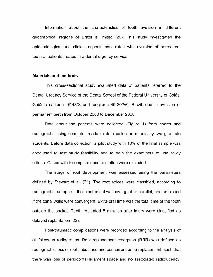
Information about the characteristics of tooth avulsion in different
geographical regions of Brazil is limited (20). This study investigated the
epidemiological and clinical aspects associated with avulsion of permanent
teeth of patients treated in a dental urgency service.
Materials and methods
This cross-sectional study evaluated data of patients referred to the
Dental Urgency Service of the Dental School of the Federal University of Goiás,
Goiânia (latitude 16o43´S and longitude 49o20´W), Brazil, due to avulsion of
permanent teeth from October 2000 to December 2008.
Data about the patients were collected (Figure 1) from charts and
radiographs using computer readable data collection sheets by two graduate
students. Before data collection, a pilot study with 10% of the final sample was
conducted to test study feasibility and to train the examiners to use study
criteria. Cases with incomplete documentation were excluded.
The stage of root development was assessed using the parameters
defined by Stewart et al. (21). The root apices were classified, according to
radiographs, as open if their root canal was divergent or parallel, and as closed
if the canal walls were convergent. Extra-oral time was the total time of the tooth
outside the socket. Teeth replanted 5 minutes after injury were classified as
delayed replantation (22).
Post-traumatic complications were recorded according to the analysis of
all follow-up radiographs. Root replacement resorption (RRR) was defined as
radiographic loss of root substance and concurrent bone replacement, such that
there was loss of periodontal ligament space and no associated radiolucency;

45
inflammatory root resorption (IRR) was diagnosed in case of loss of root
substance according to radiographs and concurrent loss of bone substance and
associated radiolucency (5); internal inflammatory root resorption (IIRR) was
recorded when a radiolucent, round and symmetrical widening of the root canal
space was observed on follow-up radiographs (23) and pulp canal obliteration
(PCO) was diagnosed by the apparent loss of t pulp space on radiographs (24).
This study was approved by the local Research Ethics Committee
(Process #055/2005).
Data were analyzed using the SPSS for Windows 19.0 (SPSS Inc.,
Chicago, IL), including frequency distribution and cross-tabulation. Chi-square
tests were used to compare qualitative data, and the level of statistical
significance was set at 5%.
Results
Patient age and sex
This study enrolled 170 patients (121 male patients, 71.18%; male-to-
female ratio = 2.4:1), aged 6 to 43 years (mean = 15.9 years, standard deviation
= 7.97). The highest avulsion frequencies were in the groups of patients aged 6-
10 and 11-15 years (30.59% each) and 16-20 years (15.88%) (Table 1).
Cause of tooth avulsion
One hundred fifty nine participants (93.53%) had avulsion because of
non-intentional reasons, and 11 (6.47%), because of intentional reasons. Falls
(51.76%), traffic accidents (29.41%) and violence (6.47%) were the main
causes of tooth avulsion. There were differences between sexes, with sport

46
activities, violence and collisions resulting in 4 (2.35%), 9 (5.29%) and 5
(2.94%) teeth avulsion among male patients, and 0 (0%), 2 (1.18%) and 0 (0%)
among female patients. However, this variation was not statistically significant
(P>0.05). The chi-square test showed a statistically significant difference
between etiological factor and age (P<0.05). Falls were the most common
cause in patients younger than 15 years (41.18%). As age increased, traffic
accidents were the main etiological factor of tooth avulsion (16.47%) (Table 1).
Seasonal distribution
The season when most cases occurred was autumn (March to June;
31.18%), followed by winter (June to September; 27.65%), spring (July to
December; 23.53%) and summer (December to March; 17.65%).
Type and number of avulsed teeth
There were 261 avulsed permanent teeth, at about 1.5 teeth per TDI.
Two hundred and twenty six (86.59%) avulsed teeth were in the maxillary arch,
and 35 (13.41%), in the mandibular arch. Most avulsed teeth were maxillary
central incisors (62.45%), followed by maxillary lateral incisors (21.46%), and
mandibular central incisors (5.75%) (Figure 2). One hundred and eleven
participants (65.29%) had one tooth avulsed, 38 (22.35%), two teeth, and 21
(12.35%), three or more teeth.
Replantation and stage of root development
Of the 261 avulsed teeth, 119 teeth (45.59%) were treated with
replantation. The percentage of replanted teeth with complete root formation

47
and closed apex was 67.23%, whereas 32.77% had the apices just about to be
completed. One hundred and forty two teeth (54.41%) were treated without
replantation.
Transport medium
Thirty eight teeth (31.93%) were stored in dry media, 20 (16.81%) in
saline, 20 (16.81%) in milk, 15 (12.61%) in water, 5 (4.20%) in alcohol, 4 teeth
(3.36%) in the patient´s oral cavity and 1 tooth (0.84%) was stored in ice.
Information about the transport medium could not be determined in 16 (13.45%)
of the 119 replanted teeth.
Extra-oral time
No tooth was replanted less than 5 minutes after avulsion. Replantation
was delayed for 106 replanted teeth (89.08%): 42 (35.29%) were replanted 60
minutes after injury, 26 (21.85%), 1 to 2 hours and 38 (31.93%), more than 2
hours after the accident. Information about extra-oral time was not available for
13 teeth (10.92%).
Additional tooth damage
Other TDI associated with tooth avulsion were found in 31 replanted
teeth (26.05%). The most common type of injury was uncomplicated crown
fracture (n=22; 70.97%), followed by complicated crown fracture (n=5; 16.13%),
fracture of the alveolar process (n=3; 9.68%) and complicated crown-root
fracture (n=1; 3.23%). Only 4 teeth (3.36%) had recurrent TDI.

48
Treatment
Treatments were subdivided into treatment for replanted teeth and
treatment for non-replanted teeth. Most replanted teeth received endodontic
treatment and root canal filling with calcium hydroxide (70 teeth; 58.82%)
followed by endodontic treatment and root canal filling (32 teeth; 26.89%),
extraction (4 teeth; 3.36%), prosthodontic rehabilitation (4 teeth; 3.36%),
endodontic treatment and root canal filling associated with orthodontic
movement (2 teeth; 1.68%), space maintainer (1 tooth; 0.84%) and
prosthodontic rehabilitation associated with orthodontic movement (1 tooth;
0.84%). For the non-replanted teeth, the most frequent treatment was analysis
of history and clinical exam (117 teeth; 82.39%) followed by prosthodontic
rehabilitation (22 teeth; 15.49%), space maintainer (1 tooth; 0.70%), orthodontic
movement (1 tooth; 0.70%) and tooth transplantation associated to endodontic
treatment and the filling of the root canal with calcium hydroxide (1 tooth;
0.70%).
Post-traumatic complications
All teeth were followed up for 1 to 8 years. Follow-up radiographs
revealed that 44 (36.97%) had inflammatory root resorption, 41 (34.45%),
periodontal healing, 22 (18.49%), replacement root resorption, 2 (1.68%), pulp
canal obliteration, and 1 (0.84%), internal inflammatory root resorption.
Information about post-traumatic complication was not available for 9 teeth
(7.56%).

49
Fifty-two teeth were followed up for 1 to 2 years and had the following
results: periodontal healing (24 teeth; 46.15%); replacement root resorption (9
teeth; 17.31%), inflammatory root resorption (18 teeth; 34.62%) and pulp canal
obliteration (1 tooth; 1.92%). Eleven teeth were followed up for 2 to 3 years and
had the following results: periodontal healing (1 tooth; 9.09%); replacement root
resorption (5 teeth; 45.45%), inflammatory root resorption (3 teeth; 27.27%),
pulp canal obliteration (1 tooth; 9.09%), and internal inflammatory root
resorption (1 tooth; 9.09%). Thirty-three were followed up for more than 3 years
and had the following results: periodontal healing (8 teeth; 24.24%);
replacement root resorption (8 teeth; 24.24%) and inflammatory root resorption
(16 teeth; 51.52%).
Follow-up time and post-traumatic complications were not analyzed for
14 teeth (12.73%) because patients moved from town and missed their
scheduled appointment or simply dropped out of treatment.
Discussion
Clinical factors and epidemiological outcomes of TDI should be known
because of their important implications, such as root resorption and pulp
changes. Epidemiologically, our results are in agreement with previously
reported data about tooth avulsion (7, 8, 20, 25, 26). The data collected
confirmed that male patients had significantly more tooth avulsion than female
patients (2.5:1). However, recent studies have shown a reduction or even a
reversal in this sex disparity (5, 21, 27). In general and in comparison with
women, men engage in more vigorous physical activities, such as physical
50
contact sports, usually without wearing adequate protection, and in aggressive
activities, such as fights (1, 3).
Tooth avulsion is more frequent in patients aged 7 to 14 years (1). In this
study, the age of patients that had tooth avulsion ranged from 6 to 43 years.
The highest prevalence was found in the 6 to 10 and 11 to 15 years age groups,
which together made up about 61% of the sample. Zhang & Gong (8) found the
highest frequency of tooth avulsion in the 7 to 17 years group (35.22%), but
Panzarini et al. (20), in the 10 to 19 years group (53.24%). Odoi et al. (28)
analyzed the association between problem behavior and TDI amongst children
aged 7-15 years. Children who had peer relationship problems, such as being
picked on or bullied by other children, were more likely to have TDI.
The most frequent causes of tooth avulsion were non-intentional, such as
falls (51.76%) and traffic accidents (29.41%). Etiological factors varied
according to age group. Most tooth avulsions due to falls were found in the 6-10
and 11-15 years groups. As age increased, traffic accidents became the main
etiological agent, as reported elsewhere (29). The place where the study was
conducted and the age group should be taken into account when analyzing
injury etiology (8, 26).
Maxillary central incisors were the teeth most often affected, as
previously reported (5-8). The vulnerable position of this teeth, which may often
be protracted and have inadequate lip coverage, may explain this result (3).
One hundred and eleven patients (65.29%) had only one tooth avulsed.
Some individuals had 3, 4 or even 6 avulsed teeth. Previous studies confirmed
that most injuries involve multiple teeth (9). The number of avulsed teeth seems
to vary according to etiology. Therefore, traumatic injuries due to traffic

51
accidents may result in a greater number of avulsed teeth (30). Additional
damage may be associated with avulsion, such as dental hard tissue injury. In
31 replanted teeth (26.05%), there was additional crown damage, of which
70.97% were uncomplicated crown fractures. Donaldson & Kinirons (6)
detected an increased risk of earlier root resorption onset for teeth with
additional coronal damage at the time of replantation.
The analysis of the stage of root development revealed that 39/119
replanted teeth (32.77%) had incomplete root formation. This prevalence is
similar to those found by Gonda et al. (27) (31.03%), Barret & Kenny (10)
(38.46%) and Stewart et al. (21) (28.78%), higher than the one recorded by
Andreasen et al. (22) (15.07%) and lower than the one found by Petrovic et al.
(26) (48.38%). Schatz et al. (31) conducted a clinical and radiological study and
found that most avulsed teeth had fully developed roots with an open apex.
However, the outcomes of the studies that used different age groups should be
compared carefully because some variation between dental age and biological
age may be expected (32).
The stage of root development may be a predictor of periodontal and
pulp healing, and the diameter of the apical foramen is directly associated with
the chance of pulp revascularization (33). Although Ebeleseder et al. (14) found
more cases of pulp revascularization in teeth with incomplete root formation,
Petrovic et al. (26) diagnosed significantly more complications in teeth with that
characteristic. Higher failure rates in the treatment of avulsed teeth with open
apex were also found by Andreasen et al. (21) and Barret & Kenny (10).
The treatment of choice for avulsed teeth should be the replantation,
even when the prognosis is unclear. Our study found a low replantation rate
52
(45.59%), which is in agreement with the rate reported by Kinoshita et al. (34) in
Japan (43.75%), Tzigkounakis et al. (7) in Czech Republic (30%) and Petrovic
et al. (26) in Serbia (48.38%). Panzarini et al. (20) in Brazil and Zhang & Gong
(8) in China found higher replantation rates: 70.13% and 70.83%. The main
reasons for no replantation are the fact that people at the accident site had
insufficient knowledge about how to manage an avulsed tooth or reacted
inappropriately, the teeth were not found, the patient sought dental treatment
long after avulsion, or the patient was hospitalized to treat more serious injuries
(7, 8, 20).
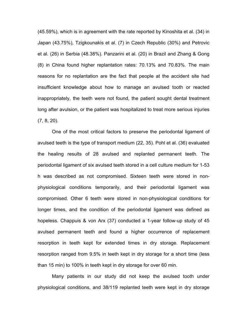
One of the most critical factors to preserve the periodontal ligament of
avulsed teeth is the type of transport medium (22, 35). Pohl et al. (36) evaluated
the healing results of 28 avulsed and replanted permanent teeth. The
periodontal ligament of six avulsed teeth stored in a cell culture medium for 1-53
h was described as not compromised. Sixteen teeth were stored in non-
physiological conditions temporarily, and their periodontal ligament was
compromised. Other 6 teeth were stored in non-physiological conditions for
longer times, and the condition of the periodontal ligament was defined as
hopeless. Chappuis & von Arx (37) conducted a 1-year follow-up study of 45
avulsed permanent teeth and found a higher occurrence of replacement
resorption in teeth kept for extended times in dry storage. Replacement
resorption ranged from 9.5% in teeth kept in dry storage for a short time (less
than 15 min) to 100% in teeth kept in dry storage for over 60 min.
Many patients in our study did not keep the avulsed tooth under
physiological conditions, and 38/119 replanted teeth were kept in dry storage

53
before replantation. A possible explanation for this finding is that the patients in
the study did not know how to store the avulsed tooth.
Avulsed teeth are usually replanted between 1 and more than 5 h after
avulsion (7, 8, 20, 27). Boyd et al. (5) found that total extra-oral time was the
best predictor of inflammatory root resorption. In our study, 89.08% of the teeth
received delayed replantation.
Complications after replantation of avulsed teeth are common, at a
reported prevalence of 57-83% (31, 38). The overall prevalence of
complications was lower (57.98%) in our study than in those conducted by
Schatz et al. (31) (83.3%) and Petrovic et al (26) (84.4%). IRR was the most
common type and much more prevalent than RRR (36.97% and 18.49%).
Several factors might have contributed to this unfavorable outcome, such as the
significant proportion of avulsed teeth stored in a non-physiological medium and
the long extra-alveolar storage time. IRR is associated with pulp infection and
may be prevented or treated using proper endodontic treatment. Future studies
based on new diagnostic methods and more powerful research tools (39, 40)
are needed to evaluate the efficacy of the therapeutic protocols and their
implications over time.
According to published guidelines for management of tooth avulsion (41,
42), the clinical decision to perform endodontic treatment should be based on
the stage of root development and the extra-alveolar time. For the replanted
teeth in our study, the most common management strategies were endodontic
treatment and temporary filling of the root canal with calcium hydroxide and
endodontic treatment and definitive root canal filling (85.71%).

54
The lack of epidemiological data about permanent tooth avulsion in the
several geographical regions of Brazil has motivated this study. This
retrospective study was conducted to describe tooth avulsion in Goiânia, a city
in Midwestern Brazil. Moreover, oral health initiatives should be based on
prevalence data and clinical factors to implement adequate policies. This study
data suggest the establishment of accurate prevention policies to promote
guidelines for the proper management of tooth avulsion. Epidemiological
surveys support decisions about public health, and preventive campaigns have
lower costs than the treatment of TDI (43-45). The most serious problems
detected in this study were the low number of replanted teeth, the use of
inappropriate storage medium to transport avulsed teeth and the prolonged
extra-oral time.
References
1. Bastone EB, Freer TJ, McNamara JR. Epidemiology of dental trauma: a
review of literature. Aust Dent J 2000; 45:2-9.
2. Andreasen JO, Andreasen FM, Andersson L. Textbook and color atlas of
traumatic injuries to the teeth. 4th ed. Oxford: Blackwell Munksgaard;
2007, 770p.
3. Glendor U. Epidemiology of traumatic dental injuries – a 12 year review
of the literature. Dent Traumatol 2008; 24:603-11.
4. Guedes OA, Alencar AHG, Lopes LG, Pécora JD, Estrela C. A
retrospective study of traumatic dental injuries in a Brazilian dental
urgency service. Braz Dent J 2010; 21:153-7.

55
5. Boyd DH, Kinirons MJ, Gregg TA. A prospective study of factors affecting
survival of replanted permanent incisors in children. Int J Paediatr Dent.
2000; 10:200-5.
6. Donaldson M, Kinirons MJ. Factors affecting the onset of resorption in
avulsed and replanted incisor teeth in children.Dent Traumatol 2001; 17:
205-9.
7. Tzigkounakis V, Merglová V, Hecová H, Netolicky J. Retrospective
clinical study of 90 avulsed permanent teeth in 58 children. Dental
Traumatol 2008; 24: 598-602.
8. Zhang X, Gong Y. Characteristics of avulsed permanent teeth treated at
Beijing Stomatological Hospital. Dent Traumatol 2011; 27:379-84.
9. Andreasen JO, Borum MK, Jacobsen HL, Andreasen FM. Replantation
of 400 avulsed permanent incisors. 1. Diagnosis of healing
complications. Endod Dent Traumatol 1995; 11:51-8.
10. Barret EJ, Kenny DJ. Survival of avulsed permanent maxillary incisors in
children following delayed replantation. Endod Dent Traumatol 1997;
13:269-75.
11. Onetto JE, Flores MT, Garbarino ML. Dental trauma in children and
adolescents in Valparaiso, Chile. Endod Dent Traumatol 1994; 10:223-7.
12. Guedes de Amorim LF, Estrela C, Sucasas da Costa LRR. Effects os
traumatic dental injuries to primary teeth on permanent teeth – a clinical
follow-up study. Dent Traumatol 2011; 27:117-21.
13. Kawanami M, Andreasen JO, Borum MK, Schou S, Hjorting-Hansen E,
Kato H. Infraposition of ankylosed permanent maxillary incisors after
56
replantation related to age and sex. Endod Dent Traumatol 1999; 15:50-
6.
14. Ebeleseder KA, Friehs S, Ruda C, Pertl C, Glockner K, Hulla H. A study
of replanted permanente teeth in diferente age groups.Endod Dent
Traumatol 1998; 14: 274-278.
15. Malmgren B, Malmgren O. Rate of infraposition of reimplanted ankylosed
incisors related to age and growth in children and adolescents. Dent
Traumatol 2002; 18: 28-36.
16. Werder P, von Arx T, Chappius V. Treatment outcome of 42 replanted
permanent incisors with a median follow-up of 2.8 years. Schweiz
Monatsschr Zahnmed 2011; 121:312-20.
17. Holan G, Cohenca N, Brin I, Sgan-Cohen H. An oral health promotion
program for the prevention of complication following avulsion: the effect
on the knowledge of physical education teachers. Dent Traumatol 2006;
22:323-7.
18. Al-Astour A, Andersson A, Al-Jame Q. School teachers´ knowledge to
tooth avulsion and dental first aid before and after receiving information
about avulsed teeth and replantation. Dent Traumatol 2008; 24:43-9.
19. Feldens EG, Feldens CA, Kramer PF, da Silva KG, Munari CC, Brei VA.
Understanding school teacher’s knowledge regarding dental trauma: a
basis for future interventions. Dent Traumatol 2010; 26: 158-63.
20. Panzarini SR, Saad-Neto M, Sonoda C, Poi WR, Perri de Carvalho AC.
Dental avulsion in Young and adult patients in the region of Araçatuba.
Rev Assoc Paul Cir Dent 2003; 57:27-31.

57
21. Stewart CJ, Elledge RO, Kinirons MJ, Welbury RR. Factors affecting the
timing of pulp extirpation in a sample of 66 replanted avulsed teeth in
children and adolescents. Dent Traumatol 2008; 24:625-7.
22. Andreasen JO, Borum MK, Jacobsen HL, Andreasen FM. Replantation
of 400 avulsed permanent incisors. 4. Factors related to periodontal
ligament healing. Endod Dent Traumatol 1995; 11:76-89.
23. Haapasalo M, Endal U. Internal inflammatory root resorption: the unknow
resorption of the tooth. Endodontic Topics 2006; 14:60-79.
24. McCabe PS, Dummer PMH. Pulp canal obliteration: an endodontic
diagnosis and treatment challenge. Int Endod J 2011; 17: doi:
10.1111/j.1365-2591.2011.01963.x. [Epub ahead of print].
25. Kargul B, Welbury R. An audit of the time to initial treatment in avulsion
injuries. Dent Traumatol 2009; 25:123-25.
26. Petrovic B, Marković D, Peric T, Blagojevic D. Factors related to
treatment and outcomes of avulsed teeth. Dent Traumatol 2010; 26:52-
59.
27. Gonda F, Nagase M, Chen R-B, Yakata H, Nakajima T. Replantation: an
analysis of 29 teeth. Oral Surg Oral Med Oral Pathol 1990; 70:650-5.
28. Odoi R, Croucher R, Wong F, Marcenes W. The relationship between
problem behavior and traumatic dental injury amongst children aged 7-
15years old. Community Dent Oral Epidemiol 2002; 30:392-6.
29. Uji T, Teramoto T. Occurrence of traumatic injuries in oromaxillary region
of children in a Japanese prefecture. Endod Dent Traumatol 1988; 4:63-
4.

58
30. Gulinelli JL, Saito CTMH, Garcia-Júnior IR, Panzarini SR, Poi WR,
Sonada CK, Jardim EC, Faverani LP. Occurrence of tooth injuries in
patients treated in hospital environment in the region of Araçatuba, Brazil
during a 6-year period. Dent Traumatol 2008;24:640-644.
31. Schatz JP, Hausherr C, Joho JP. A restrospective clinical and radiologic
study of teeth re-implanted following traumatic avulsion. Endod Dent
Traumatol 1995; 11:235-9.
32. Estrela C, Valladares-Neto J, Bueno MR, Guedes OA, Porto OLC,
Pécora JD. Linear measurements of human permanent dental
development stages using cone beam computed tomography: a
preliminary study. Dental Press J Orthod 2010; 15:44-78.
33. Andreasen JO, Vinding TR, Christensen SSA. Predictors for healing
complications in the permanent dentition after dental trauma. Endodontic
Topics 2006; 14:20-7.
34. Kinoshita S, Kojima R, Taguchi Y, Noda T. Tooth replantation after
traumatic avulsion: a report of 10 cases. Dent Traumatol 2002; 18:1153-
6.
35. de Sousa HA, de Alencar AHG, Bruno KF, Batista AC, de Carvalho ACP.
Microscopic evaluation of the effect of different storage media on the
periodontal ligament of surgically extracted human teeth. Dent Traumatol
2008; 24:628-32.
36. Pohl Y, Filippi A, Kirschner H. Results after replantation of avulsed
permanent teeth. II. Periodontal healing and the role of physiologic
storage and antiresorptive-regenerative therapy. Dent Traumatol 2005;
21:93-101.

59
37. Chappuis V, von Arx T. Replantation of 45 avulsed permanent teeth: a 1-
year follow-up study. Dent Traumatol 2005; 21:289-96.
38. Kinirons MJ, Gregg TA, Welbury R, Cole B. Variations in the presenting
and treatment factors in reimplanted permanent incisors in children and
their effect on the prevalence of root resorption. Br Dent J 2000;
189:263-6.
39. Estrela C, Bueno MR, Azevedo BC, Azevedo JR, Pécora JD. A new
periapical index based on cone beam computed tomography. J Endod
2008; 34:1325-331.
40. Estrela C, Bueno MR, Alencar AHG, Mattar R, Valladares-Neto J,
Azevedo BC, Estrela CRA. Method to evaluate inflammatory root
resorption by using cone beam computed tomography. J Endod 2009;
35:1491-7.
41. Gregg TA, Boyd DH. Treatment of avulsed permanent teeth in children.
UK National Guidelines in Paediatric Dentistry. Royal College of
Surgeons, Faculty of Dental Surgery. Int J Paediatr Dent 1998; 8:75-81.
42. Flores MT, Andersson L, Andreasen JO, Bakland LK, Malmgren B,
Barnett F, Bourguignon C, Diagelis A, Hicks L, Sigurdsson A, Trope M,
Tsukiboshi M, Von Arx T. Guidelines for management of traumatic dental
injuries. II. Avulsion of permanent teeth. Endodontic Topics 2006;
14:102-18.
43. Borum MK, Andreasen JO. Therapeutic and economic implications of
traumatic dental injuries in Denmark: an estimate based on 7549 patients
treated at a major trauma center. Int J Paediatr Dent 2001; 11:249-58.

60
44. Wong FSL, Kolokotsa K. The cost of treating children and adolescents
with injuries to their permanent incisors at a dental hospital in the United
Kingdom. Dent Traumatol 2004; 20:327-33.
45. Nguyen P-MT, Kenny DJ, Barret EJ. Socio-economic burden of
permanent incisor replantation on children and parents. Dent Traumatol
2004; 20:123-33.

61
Figure 1.
Gender n=170 Male Female
Epidemiological Aspects n=170
Clinical Factors n=119
Age n=170 6-10 21-25
11-15 26-30 16-20 >30
Etiological Factor n=170 Non-intentional Intentional
Falls Violence Sports
Collision Traffic Accident
Seasonal Distribution n=170 Spring Autumn
Summer Winter
Tooth n=261 Type Number of avulsed teeth
Dental Records n=170
Replantation n=119
Stage of Root Development n=119 Completed Incomplete
Transport Medium n=119 Dry media Saline
Milk Oral Cavity Water Ice
Alcohol
Extra-oral Time n=119 Immediate Delayed
Additional Damage n=119 Dental Hard
Tissue Supporting
Tissue
Treatment n=261 Reimplanted
Tooth Non-reimplanted
Tooth
Post-traumatic Complication n=119 PH RRR IRR IIRR PCO

62
Figure 2.
Figure legends
Figure 1. Data collected from patient charts and radiographs.
Figure 2. Distribution of avulsed permanent teeth according to tooth type.
163
56
715 14 5 1
0
20
40
60
80
100
120
140
160
180No.ofteeth
Teeth

63
Table 1. Distribution of the etiological factors of the tooth avulsion according to age and gender
Fall (%)
Traffic accidents (%)
Sports injuries (%)
Violence (%)
Collision (%)
Others (%)
Total (%)
Age (n=170)
6-10 39 (22.94%) 7 (4.12%) 1 (0.59%) 0 (0.00%) 2 (1.18%) 3 (1.76%) 52 (30.59%) 11-15 31 (18.24%) 15 (8.82%) 2 (1.18%) 3 (1.76%) 0 (0.00%) 1 (0.59%) 52 (30.59%) 16-20 9 (5.29%) 10 (5.88%) 1 (0.59%) 5 (2.94%) 1 (0.59%) 1 (0.59%) 27 (15.88%) 21-25 6 (3.53%) 7 (4.12%) 0 (0.00%) 1 (0.59%) 0 (0.00%) 3 (1.76%) 17 (10.00%) 26-30 2 (1.18%) 7 (4.12%) 0 (0.00%) 1 (0.59%) 1 (0.59%) 1 (0.59%) 12 (7.07%) > 30 1 (0.59%) 4 (2.35%) 0 (0.00%) 1 (0.59%) 1 (0.59%) 3 (1.76%) 10 (5.88%)
Gender (n=170)
Male 63 (37.06%) 35 (20.59%) 4 (2.35%) 9 (5.29%) 5 (2.94%) 5 (2.94%) 121 (71.17%) Female 25 (14.71%) 15 (8.82%) 0 (0.00%) 2 (1.18%) 0 (0.00%) 7 (4.12%) 49 (28.83%)

64
9 ANEXOS
Anexo 1. Parecer do Comitê de Ética

65

66
Anexo 2. Publicações no biênio 2010/2011

67
Artigos científicos publicados em periódicos nacionais e internacionais, com corpo editorial:
1) Veloso HHP, Sampaio FC, Guedes OA. Tratamento interdisciplinar de
dente permanente avulsionado em paciente com crescimento facial
incompleto. Dental Press Endod. 2011 Oct-Dec;1(3):65-70.
2) Estrela C, Guedes AO, Silva JA, Leles CR, Estrela CRA, Pécora JD.
Diagnostic and clinical factors associated with pulpal and periapical pain.
Braz Dent J 2011; 22: 306-11.
3) Guedes OA, Rabelo LEG, Porto OCL, Alencar AHG, Estrela C. Avaliação
radiográfica da posição e forma do forame mentual em uma
subpopulação Brasileira. Robrac 2011; 20: 160-5.
4) Guedes OA, Alencar AHG, Lopes LG, Pécora JD, Estrela C. A
retrospective study of Traumatic Dental Injuries in a Brazilian dental
urgency service. Braz Dent J 2010; 21: 153-7.
5) Pereira Júnior W, Moura MS, Guedes OA, Decurcio RA, Estrela C.
Análise de critérios de sucesso em endodontia e implantodontia.
ROBRAC 2010; 19: 108-18.
6) Valladares-Neto J, Estrela C, Bueno MR, Guedes OA, Porto OCL,
Pécora JD. Mandibular condyle dimensional changes in subjects from 3
to 20 years of age using Cone-Beam Computed Tomography: a
preliminary study. Dent Press J Orthodont 2010; 15: 172-81.
7) Estrela C, Valladares-Neto J, Bueno MR, Guedes OA, Porto OCL,
Pécora JD. Linear measurements of human permanent dental
development stages using Cone-Beam Computed Tomography: a
preliminary study. Dent Press J Orthodont 2010; 15: 44-78.

68
Artigos científicos aceitos para publicação em periódicos nacionais e internacionais, com corpo editorial:
1) Borges RN, Arantes BM, Vieira DF, Guedes OA, Estrela C. Occlusal
adjustment in the treatment of primary traumatic injury. Stomatos (in
press).
2) Borges RN, Arantes BM, Vieira DF, Guedes OA, Estrela C. Occlusal
adjustment in the treatment of secondary traumatic injury. Stomatos (in
press).
Livro(s), organização de livro(s) e capítulo(s) de livro:
1) Estrela C, Sydney GB, Guedes OA, Figueiredo JAP. Protocolo de
sucesso no tratamento endodôntico. In: Cláudio Pinheiro Fernandes.
(Org.). Uma odontologia de classe mundial. 1 ed. São Paulo-SP: Editora
Santos, 2010, v. 1, p. 59-88.
2) Estrela C, Sydney GB, Guedes OA, Figueiredo JAP. Successful protocol
for endodontic tretament. In: Cláudio Pinheiro Fernandes. (Org.). A world
class dentistry. 1 ed. São Paulo-SP: Editora Santos, 2010, v. 1, p. 59-86.

69
Anexo 3. Normas de publicação do periódico

70
Dental Traumatology Official Publication of the International Association for Dental Traumatology and the International Academy of Sports Dentistry Edited by: Lars Andersson Print ISSN: 1600-4469 Online ISSN: 1600-9657 Frequency: Bi-monthly Current Volume: 27 / 2011 ISI Journal Citation Reports® Ranking: 2010: Dentistry, Oral Surgery & Medicine: 44 / 74 Impact Factor: 1.268 Top Author Guidelines Content of Author Guidelines: 1. General, 2. Ethical Guidelines, 3. Submission of Manuscripts, 4. Manuscript Types Accepted, 5. Manuscript Format and Structure, 6. After Acceptance Relevant Documents: Copyright Transfer Agreement Useful Websites: Submission Site, Articles published in Dental Traumatology, Author Services, Wiley-Blackwell's Ethical Guidelines, Guidelines for Figures 1. GENERAL Dental Traumatology is an international journal which aims to convey scientific and clinical progress in all areas related to adult and pediatric dental traumatology. It aims to promote communication among clinicians, educators, researchers, administrators and others interested in dental traumatology. The journal publishes original scientific articles, review articles in the form of comprehensive reviews or mini reviews of a smaller area, short communication about clinical methods and techniques and case reports. The journal focuses on the following areas related to dental trauma: Epidemiology and Social Aspects Tissue, Periodontal, and Endodontic Considerations Pediatrics and Orthodontics Oral and Maxillofacial Surgery / Transplants/ Implants Esthetics / Restorations / Prosthetics Prevention and Sports Dentistry Please read the instructions below carefully for details on the submission of manuscripts, the journal's requirements and standards as well as information concerning the procedure after a manuscript has been accepted for publication in

71
Dental Traumatology. Authors are encouraged to visit Wiley-Blackwell Author Services for further information on the preparation and submission of articles and figures. 2. ETHICAL GUIDELINES Dental Traumatology adheres to the below ethical guidelines for publication and research. 2.1. Authorship and Acknowledgements Authors submitting a paper do so on the understanding that the manuscript have been read and approved by all authors and that all authors agree to the submission of the manuscript to the Journal. ALL named authors must have made an active contribution to the conception and design and/or analysis and interpretation of the data and/or the drafting of the paper and ALL must have critically reviewed its content and have approved the final version submitted for publication. Participation solely in the acquisition of funding or the collection of data does not justify authorship. Dental Traumatology adheres to the definition of authorship set up by The International Committee of Medical Journal Editors (ICMJE). According to the ICMJE authorship criteria should be based on 1) substantial contributions to conception and design of, or acquisiation of data or analysis and interpretation of data, 2) drafting the article or revising it critically for important intellectual content and 3) final approval of the version to be published. Authors should meet conditions 1, 2 and 3. It is a requirement that all authors have been accredited as appropriate upon submission of the manuscript. Contributors who do not qualify as authors should be mentioned under Acknowledgements. Acknowledgements: Under acknowledgements please specify contributors to the article other than the authors accredited. 2.2. Ethical Approvals Experimentation involving human subjects will only be published if such research has been conducted in full accordance with ethical principles, including the World Medical Association Declaration (version, 2008 http://www.wma.net/en/30publications/10policies/b3/index.html) and the additional requirements, if any, of the country where the research has been carried out. Manuscripts must be accompanied by a statement that the experiments were undertaken with the understanding and written consent of each subject and according to the above mentioned principles. A statement regarding the fact that the study has been independently reviewed and approved by an ethical board should also be included. In the online submission process we also require that all authors submitting manuscripts to Dental Traumatology online must answer in the affirmative to a statement 'confirming that all research has been carried out in accordance with legal requirements of the study country such as approval of ethical commitees for human
72
and/or animal research or other legislation where applicable.' Editors reserve the right to reject papers if there are doubts as to whether appropriate procedures have been used. 2.3 Clinical Trials Clinical trials should be reported using the CONSORT guidelines available at www.consort-statement.org. A CONSORT checklist should also be included in the submission material. All manuscripts reporting results from a clinical trial must indicate that the trial was fully registered at a readily accessible website, e.g., www.clinicaltrials.gov. 2.4 DNA Sequences and Crystallographic Structure Determinations Papers reporting protein or DNA sequences and crystallographic structure determinations will not be accepted without a Genbank or Brookhaven accession number, respectively. Other supporting data sets must be made available on the publication date from the authors directly. 2.5 Conflict of Interest Dental Traumatology requires that sources of institutional, private and corporate financial support for the work within the manuscript must be fully acknowledged, and any potential grant holders should be listed. Acknowledgements should be brief and should not include thanks to anonymous referees and editors. The Conflict of Interest Statement should be included as a separate document uploaded under the file designation 'Title Page' to allow blinded review. 2.6 Appeal of Decision The decision on a paper is final and cannot be appealed. 2.7 Permissions If all or parts of previously published illustrations are used, permission must be obtained from the copyright holder concerned. It is the author's responsibility to obtain these in writing and provide copies to the Publishers. 2.8 Copyright Assignment Authors submitting a paper do so on the understanding that the work and its essential substance have not been published before and is not being considered for publication elsewhere. The submission of the manuscript by the authors means that the authors automatically agree to assign exclusive copyright to Wiley-Blackwell if and when the manuscript is accepted for publication. The work shall not be published elsewhere in any language without the written consent of the publisher. The articles published in this journal are protected by copyright, which covers translation rights and the exclusive right to reproduce and distribute all of the articles printed in the journal. No material published in the journal may be stored on microfilm or videocassettes or in electronic
73
database and the like or reproduced photographically without the prior written permission of the publisher. Upon acceptance of a paper, authors are required to assign the copyright to publish their paper to Wiley-Blackwell. Assignment of the copyright is a condition of publication and papers will not be passed to the publisher for production unless copyright has been assigned. (Papers subject to government or Crown copyright are exempt from this requirement; however, the form still has to be signed). A completed Copyright Transfer Agreement must be sent before any manuscript can be published. Authors must send the completed Copyright Transfer Agreement upon receiving notice of manuscript acceptance, i.e., do not send the Copyright Transfer Agreement at submission. Please return your completed form to: Angelo Morales Production Editor Wiley Services Singapore Pte Ltd 1 Fusionopolis Walk, #07-01 Solaris South Tower, Singapore 138628 Alternatively a scanned version of the form can be emailed to [email protected] or faxed to ++65 6643 8599. For questions concerning copyright, please visit Wiley-Blackwell's Copyright FAQ 2.9 OnlineOpen OnlineOpen is available to authors of primary research articles who wish to make their article available to non-subscribers on publication, or whose funding agency requires grantees to archive the final version of their article. With OnlineOpen, the author, the author's funding agency, or the author's institution pays a fee to ensure that the article is made available to non-subscribers upon publication via Wiley Online Library, as well as deposited in the funding agency's preferred archive. For the full list of terms and conditions, see http://wileyonlinelibrary.com/onlineopen#OnlineOpen_Terms. Any authors wishing to send their paper OnlineOpen will be required to complete the payment form available from our website at: https://wileyonlinelibrary.com/onlineopen Prior to acceptance there is no requirement to inform an Editorial Office that you intend to publish your paper OnlineOpen if you do not wish to. All OnlineOpen articles are treated in the same way as any other article. They go through the journal's standard peer-review process and will be accepted or rejected based on their own merit. For questions concerning copyright, please visit Wiley-Blackwell's Copyright FAQ

74
3. MANUSCRIPT SUBMISSION PROCEDURE Manuscripts should be submitted electronically via the online submission site http://mc.manuscriptcentral.com/dt. The use of an online submission and peer review site enables immediate distribution of manuscripts and consequentially speeds up the review process. It also allows authors to track the status of their own manuscripts. Complete instructions for submitting a paper is available online and below. Further assistance can be obtained from Editorial Assistant Karin Andersson at [email protected]. 3.1. Getting Started • Launch your web browser (supported browsers include Internet Explorer 6 or higher, Netscape 7.0, 7.1, or 7.2, Safari 1.2.4, or Firefox 1.0.4) and go to the journal's online Submission Site: http://mc.manuscriptcentral.com/dt • Log-in or click the 'Create Account' option if you are a first-time user. • If you are creating a new account. - After clicking on 'Create Account', enter your name and e-mail information and click 'Next'. Your e-mail information is very important. - Enter your institution and address information as appropriate, and then click 'Next.' - Enter a user ID and password of your choice (we recommend using your e-mail address as your user ID), and then select your area of expertise. Click 'Finish'. • If you have an account, but have forgotten your log in details, go to Password Help on the journals online submission system http://mc.manuscriptcentral.com/dt and enter your e-mail address. The system will send you an automatic user ID and a new temporary password. • Log-in and select 'Author Centre.' 3.2. Submitting Your Manuscript • After you have logged into your 'Author Centre', submit your manuscript by clicking the submission link under 'Author Resources'. • Enter data and answer questions as appropriate. You may copy and paste directly from your manuscript and you may upload your pre-prepared covering letter. • Click the 'Next' button on each screen to save your work and advance to the next screen. • You are required to upload your files. - Click on the 'Browse' button and locate the file on your computer. - Select the designation of each file in the drop down next to the Browse button. - When you have selected all files you wish to upload, click the 'Upload Files' button. • To allow double blinded review, please submit (upload) your main manuscript and title page as separate files. Please upload: - Your manuscript without title page under the file designation 'main document' - Figure files under the file designation 'figures'. - The title page, Acknowledgements and Conflict of Interest Statement where applicable, should be uploaded under the file designation 'title page'

75
• Review your submission (in HTML and PDF format) before completing your submission by sending it to the Journal. Click the 'Submit' button when you are finished reviewing. All documents uploaded under the file designation 'title page' will not be viewable in the html and pdf format you are asked to review in the end of the submission process. The files viewable in the html and pdf format are the files available to the reviewer in the review process. 3.3. Manuscript Files Accepted Manuscripts should be uploaded as Word (.doc) or Rich Text Format (.rft) files (not write-protected) plus separate figure files. GIF, JPEG, PICT or Bitmap files are acceptable for submission, but only high-resolution TIF or EPS files are suitable for printing. The files uploaded as main manuscript documents will be automatically converted to HTML and PDF on upload and will be used for the review process. The files uploaded as title page will be blinded from review and not converted into HTML and PDF. The main manuscript document file must contain the entire manuscript including abstract, text, references, tables, and figure legends, but no embedded figures. In the text, please reference figures as for instance 'Figure 1', 'Figure 2' etc to match the tag name you choose for the individual figure files uploaded. Manuscripts should be formatted as described in the Author Guidelines below. Please note that any manuscripts uploaded as Word 2007 (.docx) will be automatically rejected. Please save any .docx file as .doc before uploading. 3.4. Blinded Review All manuscripts submitted to Dental Traumatology will be reviewed by two experts in the field. Dental Traumatology uses double blinded review. The names of the reviewers will thus not be disclosed to the author submitting a paper and the name(s) of the author(s) will not be disclosed to the reviewers. To allow double blinded review, please submit (upload) your main manuscript and title page as separate files. Please upload: • Your manuscript without title page under the file designation 'main document' • Figure files under the file designation 'figures' • The title page, Acknowledgements and Conflict of Interest Statement where applicable, should be uploaded under the file designation 'title page' All documents uploaded under the file designation 'title page' will not be viewable in the html and pdf format you are asked to review in the end of the submission process. The files viewable in the html and pdf format are the files available to the reviewer in the review process. 3.5. Suggest a Reviewer Dental Traumatology attempts to keep the review process as short as possible to enable rapid publication of new scientific data. In order to facilitate this process, please suggest the names and current email addresses of a potential international reviewer whom you consider capable of reviewing your manuscript. In addition to your choice the
76
journal editor will choose one or two reviewers as well. When the review is done you will be notified under 'Manuscripts with decision' and through e-mail. 3.6. Suspension of Submission Mid-way in the Submission Process You may suspend a submission at any phase before clicking the 'Submit' button and save it to submit later. The manuscript can then be located under 'Unsubmitted Manuscripts' and you can click on 'Continue Submission' to continue your submission when you choose to. 3.7. E-mail Confirmation of Submission After submission you will receive an e-mail to confirm receipt of your manuscript. If you do not receive the confirmation e-mail after 24 hours, please check your e-mail address carefully in the system. If the e-mail address is correct please contact your IT department. The error may be caused by some sort of spam filtering on your e-mail server. Also, the e-mails should be received if the IT department adds our e-mail server (uranus.scholarone.com) to their whitelist. 3.8. Manuscript Status You can access ScholarOne Manuscripts (formerly known as Manuscript Central) any time to check your 'Author Center' for the status of your manuscript. The Journal will inform you by e-mail once a decision has been made. 3.9. Submission of Revised Manuscripts To submit a revised manuscript, locate your manuscript under 'Manuscripts with Decisions' and click on 'Submit a Revision'. Please remember to delete any old files uploaded when you upload your revised manuscript. Please also remember to upload your manuscript document separate from your title page. 4. MANUSCRIPT TYPES ACCEPTED Original Research Articles in all areas related to adult and pediatric dental traumatology are of interest to Dental Traumatology. Examples of such areas are Epidemiology and Social Aspects, Tissue, Periodontal, and Endodontic Considerations, Pediatrics and Orthodontics, Oral and Maxillofacial Surgery/ Transplants / Implants, Esthetics / Restorations / Prosthetics and Prevention and Sports Dentistry. Review Papers: Dental Traumatology commissions review papers of comprehensive areas and mini reviews of small areas. The journal also welcomes uninvited reviews. Reviews should be submitted via the online submission site and are subject to peer-review. Comprehensive Reviews should be a complete coverage of a subject discussed with the Editor in Chief prior to preparation and submission. Comprehensive review articles should include a description of search strategy of relevant literature, inclusion criteria, evaluation of papers and level of evidence.

77
Mini Reviews are covering a smaller area and may be written in a more free format. Case Reports: Dental Traumatology accepts Case Reports but these will only be published online and will not be included in the printed version unless specifically requested by the Editor-in-Chief. Case Reports illustrating unusual and clinically relevant observations are acceptable, but their merit needs to provide high priority for publication in the journal. They should be kept within 3-4 printed pages and need not follow the usual division into material and methods etc, but should have an abstract. The introduction should be kept short. Thereafter the case is described followed by a discussion. Short Communications of 1-2 pages are accepted for quick publication. These papers need not follow the usual division into Material and Methods, etc., but should have an abstract. They should contain important new information to warrant publication and may reflect improvements in clinical practice such as introduction of new technology or practical approaches. They should conform to a high scientific and a high clinical practice standard. Letters to the Editor, if of broad interest, are encouraged. They may deal with material in papers published in Dental Traumatology or they may raise new issues, but should have important implications. Meetings: advance information about and reports from international meetings are welcome, but should not be submitted via the online submission site, but send directly to the journal administrator Karin Andersson at [email protected] 5. MANUSCRIPT FORMAT AND STRUCTURE 5.1. Format Language: The language of publication is English. Authors for whom English is a second language must have their manuscript professionally edited by an English speaking person before submission to make sure the English is of high quality. It is preferred that manuscript is professionally edited. A list of independent suppliers of editing services can be found at http://authorservices.wiley.com/bauthor/english_language.asp. All services are paid for and arranged by the author, and use of one of these services does not guarantee acceptance or preference for publication. Abbreviations, Symbols and Nomenclature: Abbreviations should be kept to a minimum, particularly those that are not standard. Non-standard abbreviations must be used three or more times and written out completely in the text when first used. Consult the following sources for additional abbreviations: 1) CBE Style Manual Committee.

78
Scientific style and format: the CBE manual for authors, editors, and publishers. 6th ed. Cambridge: Cambridge University Press; 1994; and 2) O'Connor M, Woodford FP. Writing scientific papers in English: an ELSE-Ciba Foundation guide for authors. Amsterdam: Elsevier-Excerpta Medica; 1975. Font: When preparing your file, please use only standard fonts such as Times, Times New Roman or Arial for text, and Symbol font for Greek letters, to avoid inadvertent character substitutions. In particular, please do not use Japanese or other Asian fonts. Do not use automated or manual hyphenation. Use double spacing when writing. 5.2. Structure All papers submitted to Dental Traumatology should include: Title Page, Abstract, Main text, References and Tables, Figures, Figure Legends, Conflict of Interest Statement and Acknowledgements where appropriate. Title page, Conflict of Interest Statement and any Acknowledgements must be submitted as separate files and uploaded under the file designation Title Page to allow blinded review. Manuscripts must conform to the journal style. Manuscripts not complying with the journal style will be returned to the author(s). Title Page: should be uploaded as a separate document in the submission process under the file designation 'Title Page' to allow blinded review. It should include: Full title of the manuscript, author(s)' full names (Family names should be underlined) and institutional affiliations including city, country, and the name and address of the corresponding author. If the author does not want the e-mail address to be published this must be clearly indicated. The title page should also include a running title of no more than 60 characters and 3-6 keywords. Abstract is limited to 250 words in length and should contain no abbreviations. The abstract should be included in the manuscript document uploaded for review as well as inserted separately where specified in the submission process. The abstract should convey the essential purpose and message of the paper in an abbreviated form. For original articles the abstract should be structured with the following headings: Background/Aim, Material and Methods, Results and Conclusions. For other article types, please choose headings appropriate for the article. Main Text of Original Articles should be divided into Introduction, Material and Methods, Results and Discussion. During the editorial process reviewers and editors frequently need to refer to specific portions of the manuscript, which is difficult unless the pages are numbered. Authors should number all of the pages consecutively. Introduction should be focused, outlining the historical or logical origins of the study and not summarize the results; exhaustive literature reviews are inappropriate. Give only strict and pertinent references and do not include data or conclusions from the work

79
being reported. The introduction should close with the explicit statement of the specific aims of the investigation or hypothesis tested. Materials and Methods must contain sufficient detail such that, in combination with the references cited, all clinical trials and experiments reported can be fully reproduced. As a condition of publication, authors are required to make materials and methods used freely available to academic researchers for their own use. Describe your selection of observational or experimental participants clearly. Identify the method, apparatus and procedures in sufficient detail. Give references to established methods, including statistical methods, describe new or modify methods. Identify precisely all drugs used including generic names and route of administration. (i) Clinical trials should be reported using the CONSORT guidelines available at www.consort-statement.org. A CONSORT checklist should also be included in the submission material. All manuscripts reporting results from a clinical trial must indicate that the trial was fully registered at a readily accessible website, e.g., www.clinicaltrials.gov. (ii) Experimental subjects: experimentation involving human subjects will only be published if such research has been conducted in full accordance with ethical principles, including the World Medical Association Declaration (version, 2008 http://www.wma.net/en/30publications/10policies/b3/index.html) and the additional requirements, if any, of the country where the research has been carried out. Manuscripts must be accompanied by a statement that the experiments were undertaken with the understanding and written consent of each subject and according to the above mentioned principles. A statement regarding the fact that the study has been independently reviewed and approved by an ethical board should also be included. Editors reserve the right to reject papers if there are doubts as to whether appropriate procedures have been used. (iii) Suppliers of materials should be named and their location (town, state/county, country) included. Results should present the observations with minimal reference to earlier literature or to possible interpretations. Present your results in logical sequence in the text, tables and illustrations giving the main or most important findings first. Do not duplicate data in graphs and tables. Discussion may usually start with a brief summary of the major findings, but repetition of parts of the Introduction or of the Results sections should be avoided. The section should end with a brief conclusion and a comment on the potential clinical relevance of the findings. Link the conclusions to the aim of the study. Statements and interpretation of the data should be appropriately supported by original references.

80
Main Text of Review Articles comprises an introduction and a running text structured in a suitable way according to the subject treated. A final section with conclusions may be added. Acknowledgements: Under acknowledgements please specify contributors to the article other than the authors accredited. Acknowledgements should be brief and should not include thanks to anonymous referees and editors. Conflict of Interest Statement: All sources of institutional, private and corporate financial support for the work within the manuscript must be fully acknowledged, and any potential grant holders should be listed. The Conflict of Interest Statement should be included as a separate document uploaded under the file designation 'Title Page' to allow blinded review. 5.3. References As the Journal follows the Vancouver system for biomedical manuscripts, the author is referred to the publication of the International Committee of Medical Journal Editors: Uniform requirements for manuscripts submitted to biomedical journals. Ann Int Med 1997;126:36-47. Number references consecutively in the order in which they are first mentioned in the text. Identify references in texts, tables, and legends by Arabic numerals (in parentheses). Use the style of the examples below, which are based on the format used by the US National Library of Medicine in Index Medicus. For abbreviations of journals, consult the 'List of the Journals Indexed' printed annually in the January issue of Index Medicus. We recommend the use of a tool such as EndNote or Reference Manager for reference management and formatting. EndNote reference styles can be searched for here: www.endnote.com/support/enstyles.asp. Reference Manager reference styles can be searched for here: www.refman.com/support/rmstyles.asp