departamento de ciÊncias da vida sónia... · departamento de ciÊncias da vida faculdade de...
TRANSCRIPT
DEPARTAMENTO DE CIÊNCIAS DA VIDA
FACULDADE DE CIÊNCIAS E TECNOLOGIA UNIVERSIDADE DE COIMBRA
Analysis of the antimicrobial content of amphibian skin secretions and structural and functional characterization of a novel
member of the temporin family (temporin-SHe)
Sónia Maria Costa André
2012
Sónia
Maria C
ost
a A
ndré
Str
uct
ura
l and funct
ional ch
ara
cteriza
tion o
f te
mporin-S
He
2012

DEPARTAMENTO DE CIÊNCIAS DA VIDA
FACULDADE DE CIÊNCIAS E TECNOLOGIA UNIVERSIDADE DE COIMBRA
Analysis of the antimicrobial content of amphibian skin secretions and structural and functional characterization of a novel
member of the temporin family (temporin-SHe)
Sónia Maria Costa André
2012
Dissertação apresentada à Universidade de Coimbra para cumprimento dos requisitos necessários à obtenção do grau de Mestre em Biologia Celular e Molecular, realizada sob a orientação científica do Professor Doutor Ali Ladram (Universidade de Pierre et Marie Curie, France) e do Professor Doutor Paula Veríssimo (Universidade de Coimbra)
AKNOWLEDGMENTS
I owe sincere and earnest thankfulness to Professor Thierry Foulon for warmly
welcoming me in his laboratory, as well as all the team for their friendship, support and
cheerfulness, providing me an excellent atmosphere for doing research.
I offer my sincerest gratitude to my supervisor, Dr. Ali Ladram, who has supported me,
and for his availability, patience, knowledge and care along this internship.
I would like to thanks also my co-supervisor, Dr. Paula Veríssimo, for her confidence in
me during this year and for advising and supervising me.
I am also indebted to François Lemoine who took care of “my frogs” Trachycephalus
resinifictrix, allowing me to study them.
I would like to show my gratitude to the PhD student Zahid Raja, for helping me with
his technical advices and for performing leishmanicidal and cytotoxic assays in
collaboration with Dr. Bruno Oury and Dr. Denis Sereno.
I would like also to show my gratitude to Amandine Anastasio, Louis Guibout and
Christophe Piesse. They cheer me up and stood by me through good and bad times.
Finally, I would like to thank my family, specially my mother for supporting me
through the duration of my studies and for her encouragement and endless love.
ABSTRACT
The growing problem of resistance to conventional antibiotics and the need to
develop new compounds with original modes of action has stimulated interest in
antimicrobial peptides (AMPs) as substitutable pharmaceuticals. AMPs are key
components of the innate immune system of several organisms (from microorganisms to
vertebrates), acting as the first line of host defense against pathogens. Amphibian skin
secretions represent one of the richest natural sources of AMPs. Indeed, 50.6% of
antibacterial peptides reported in the Antimicrobial Peptide Database
(http://aps.unmc.edu/AP/main.php) come from amphibians. Thus, amphibian skin
represents a good model for the identification of novel potent AMPs with therapeutic
potential and for studying the mechanism of action of these peptides.
The first aim of my Master 2 internship was to analyze the AMP content of frogs of
the subfamily Hylinae which have been very poorly studied, and particularly those of the
genus Trachycephalus which were not studied. Trachycephalus resinifictrix is a South
American tree frog (family Hylidae, subfamily Hylinae) also referred to as Amazon Milk
Frog because of its milky and poisonous secretions when threatened. Through bioguided
fractionation of its skin secretions, antibacterial activity against Staphylococcus aureus
was detected in HPLC fractions. We attempt to identify the active compounds by tandem
mass spectrometry (MS/MS) on a fraction displaying high inhibitory bacterial growth
activity. However, no sequence information was obtained due to insufficient material.
The second aim was to characterize the structure and function of temporin-SHe, a
small AMP (16 residues) from a North African ranid frog (Pelophylax saharica) that was
recently identified by the host team by molecular cloning of the precursor. We have
produced temporin-SHe by solid phase peptide synthesis and used circular dichroism to
investigate its secondary structure. Our results indicated that temporin-SHe can adopt -
helical conformation when bound to negatively charged membranes, while no ordered
structure (random coil) is observed for the peptide in solution. In contrast with many
members of the Temporin family, temporin-SHe exhibited broad-spectrum antimicrobial
activity with high potency against Gram-positive bacteria (including multiresistant
Staphylococcus aureus) and yeasts (MIC = 1.5-12.5 µM), and to a lesser extent, toward
Gram-negative bacteria and Candida (MIC = 25-60 µM). Interestingly, temporin-SHe was
potent against the promastigote form of the human protozoan parasite Leishmania (IC50
around 10 µM). A preliminary study using monocytes as mammalian cells revealed that
temporin-SHe was more cytotoxic (LC50 = 21 µM) than temporin-SHd (LC50 = 66 µM). We
have shown that bactericidal activity of temporin-SHe was correlated with membrane
permeabilization of bacteria. Moreover, a strong disturbance of the membrane bilayer
was induced upon interaction of the peptide with negatively charged phospholipid
vesicles (differential scanning calorimetry studies).
The short length, high potency, and broad-spectrum activity of temporins-SH,
suggest them as good candidates for the development of therapeutic antimicrobial agents
with new mode of action, although pharmacomodulation is needed to improve their
therapeutic index (LC50/MIC ratio). To date, very few AMPs, including only four temporins
(A, B, SHa and SHd), are active against parasites. Our results indicate that temporin-SHe
represents a valuable additional tool for understanding the antiparasitic mechanism of
action of AMPs, which still remains unknown.
Keywords: Amphibians, antimicrobial peptides, bioguided fractionation, temporin-SHe,
structure/activity/mechanism.
RESUMO
O problema da resistência aos antibibióticos convencionais e a necessidade de
desenvolver novos compostos com diferentes modos de ação, têm estimulado o interesse
em péptidos antimicrobianos (PAMs) como possíveis pharmacêuticos de substituição. Os
PAMs são componentes-chave do sistema imunitário inato de vários organismos agindo
como a primeira linha de defesa contra patogénios. As secreções de pele dos anfíbios
representa uma fonte rica em PAMs. De facto, segundo a base de dados Antimicrobial
Peptide Database (http://aps.unmc.edu/AP/main.php), 50,6% dos péptidos
antibacterianos provêm de anfíbios. Assim, a pele de anfíbio representa um ótimo
modelo para a identificação de novos PAMs com potencial terapêutico e para estudar o
mescanismo de ação destes.
O primeiro objetivo da minha tése de mestrado era analisar o conteúdo em PAMs
a partir de Trachycephalus resinifictrix, uma rã arborícola da América do Sul (família
Hylidae, subfamília Hylinae) pertencente ao género Trachycephalus, e que nunca foi
estudada. Através do fracionamento bioguiado das suas secreções, uma actividade
antibacteriana contra Staphylococcus aureus foi detectada em frações de HPLC. Por
espectrometria de massa em tandem (MS/MS) numa fração de HPLC que mostrava uma
potente actividade antibacteriana, tentamos identificar os compostos ativos. No entanto,
devido à fraca quantidade de material nenhuma informação de sequência foi obtida.
O segundo objetivo era a caracterização estrutural e funcional da temporin-SHe,
um pequeno PAM (16 resíduos) proveniente da rã Norte Africana Pelophylax saharica
(família Ranidae), identificado recentemente pela equipa por clonagem molecular do
precursor. Este péptido foi sintetizado em fase sólida e a técnica de dicroísmo circular foi
utilizada para investigar a sua estrutrura secundária. Os nossos resultados indicam que a
temporin-SHe adota uma estrutura em hélice- quando associada a membranas
carregadas negativamente, sendo não estrurada em solução. Em contraste com outros
membros da família das Temporins, a temporin-SHe exibiu um largo espectro
antimicrobiano com uma elevada atividade contra bactérias Gram-positivas (incluindo
Staphylococcus aureus multiresistentes) e leveduras (CMI = 1,5-12,5 µM), e em menor
extensão, contra bactérias Gram-negativas e Candida (CMI = 25-60 µM). De maneira
interessante, a temporin-SHe mostrou-se também ativa contra a forma promastigote do
parasita Leishmania (IC50 à volta de 10 µM). Um estudo preliminar usando monócitos
como células mamíferas revelou que a temporin-SHe era mais citotóxica (LC50 = 21 µM)
que a temporin-SHd (LC50 = 66 µM). Mostramos igualmente que a atividade bactericida
da temporin-SHe estava correlacionada com a permeabilização da membrana bacteriana.
Adicionalmente, uma forte perturbação da camada lipídica foi induzida após interação do
péptido com vesículas phospholipídicas carregadas negativamente.
O pequeno tamanho, a elevada potência, e o largo espectro de atividade das
temporins-SH, suggerem-nas como potentes candidatos para o desenvolvimento de
agentes terapêuticos com novos modos de ação, embora seja necessário amelhorar o
índice terapêutico (razão LC50/CMI). Presentemente, pouco PAMs, incluindo somente 4
temporins (A, B, SHa e SHd) são ativos contra os parasitas. Os nossos resultados
indicam que a temporin-SHe representa uma ferramenta adicional valiosa para a
compreensão do mecanismo de ação anti-parasitário dos PAMs, que ainda permanece
desconhecido.
Palavras-Chave: Anfíbios, péptidos antimicrobianos, fracionamento bioguiado,
temporin-SHe, estrutura/atividade/mecanismo.

1
INDEX
ABBREVIATIONS 3
INTRODUCTION 5
1. CLASSIFICATION OF AMPHIBIANS 6
2. DERMAL GLANDS OF AMPHIBIANS 7
3. ANTIMICROBIAL PEPTIDE BIOSYNTHESIS IN AMPHIBIAN SKIN 8
4. STRUCTURES OF AMPHIBIAN ANTIMICROBIAL PEPTIDES 11
5. MECHANISM OF ACTION OF ANTIMICROBIAL PEPTIDES 12
5.1. MEMBRANE COMPOSITION 14
5.2. BARREL-STAVE MODEL 16
5.3. CARPET MODEL 17
5.4. TOROIDAL MODEL OR WORMHOLE MECHANISM 18
6. ANTIMICROBIAL PEPTIDES FROM HYLID AND RANID FROGS 19
6.1. AMPS FROM FROGS BELONGING TO THE FAMILY HYLIDAE 19
6.2. AMPS FROM FROGS BELONGING TO THE FAMILY RANIDAE 22
7. PURPOSE OF THE STUDY 25
MATERIALS AND METHODS 26
8. ANALYSIS OF ANTIMICROBIAL PEPTIDES FROM SKIN SECRETIONS OF T. RESINIFICTRIX AND MOLECULAR
CLONING OF AMP CDNA PRECURSORS 26
COLLECTION OF SKIN SECRETIONS AND PRE-PURIFICATION OF PEPTIDES 26
REVERSED-PHASE HPLC (RP-HPLC) FRACTIONATION OF SKIN SECRETIONS 26
ANTIMICROBIAL ASSAYS 27
MASS SPECTROMETRY ANALYSIS OF ANTIBACTERIAL HPLC FRACTIONS 28
ISOLATION OF MRNA AND REVERSE TRANSCRIPTION 28
PCR 29
CLONING OF PCR PRODUCTS INTO PGEM-T EASY VECTOR 30
PLASMID DNA PURIFICATION AND DETERMINATION OF THE INSERT SIZE 32
9. STRUCTURAL AND FUNCTIONAL CHARACTERIZATION OF TEMPORIN-SHE 33
SOLID PHASE PEPTIDE SYNTHESIS 33
PREPARATION OF MULTILAMELLAR AND LARGE UNILAMELLAR VESICLES 35
2
CIRCULAR DICHROISM SPECTROSCOPY 35
ANALYSIS OF PEPTIDE-LIPID INTERACTION BY DIFFERENTIAL SCANNING CALORIMETRY 36
PERMEABILIZATION ASSAY 37
TIME KILLING ASSAY 38
RESULTS 39
ANALYSIS OF SKIN SECRETIONS OF T. RESINIFICTRIX 39
CDNA CLONING OF AMP PRECURSORS FROM SKIN SECRETIONS OF T. RESINIFICTRIX 43
STRUCTURAL AND FUNCTIONAL CHARACTERIZATION OF TEMPORIN-SHE 44
SYNTHESIS AND PURIFICATION OF TEMPORIN-SHE 45
SECONDARY STRUCTURE OF TEMPORIN-SHE 45
INTERACTION OF TEMPORIN-SHE WITH ANIONIC MODEL MEMBRANES 48
ANTIMICROBIAL AND CYTOTOXIC ACTIVITIES OF TEMPORIN-SHE 49
PERMEABILIZATION OF THE BACTERIAL CYTOPLASMIC MEMBRANE 50
TIME-DEPENDENT KILLING OF GRAM-NEGATIVE AND GRAM-POSITIVE BACTERIA 52
DISCUSSION 54
CONCLUSION 58
REFERENCES 59

3
ABBREVIATIONS
ACN: Acetonitrile
AMPs: Antimicrobial peptides
APD: Antimicrobial Peptide Database (http://aps.unmc.edu/AP/main.php)
BHI: Brain heart infusion
CD: Circular dichroism
CFU: Colony-forming unit
CL: Cardiolipin
DEPC: Diethylpyrocarbonate
DMPC: Dimyristoyl phosphatidyl choline
DMPG: Dimyristoyl phosphatidyl glycerol
dNTPs: Deoxynucleotide triphosphates
DPC: Dodecylphophocholine
DRP: Dermaseptin-related peptide
DRS: Dermaseptin
DSC: Differential scanning calorimetry
HIV: Human immunodeficiency virus
IPTG: Isopropyl β-D-1-thiogalactopyranoside
LB: Luria-Bertani
LPS: Lipopolysaccharides
LUVs: Large unilamellar vesicles
MALDI-TOF: Matrix-assisted laser desorption/ionization-time of flight
MH: Mueller-Hinton
MIC: Minimal inhibitory concentration
MLVs: Multilamellar vesicles
MMLV: Moloney-Murine Leukemia Virus
MS/MS: Tandem mass spectrometry
ONP: Ortho-nitrophenol
ONPG: Orthonitrophenyl-β-D-galactopyranoside,
PBS: Phosphate buffered saline
PC: Phosphatidylcholine
PCR: Polymerase chain reaction
4
PE: Phosphatidylethanolamine
PG: Phosphatidylglycerol
PS: Phosphatidylserine
RP-HPLC: Reversed-phase high performance liquid chromatography
SDS: Sodium dodecyl sulphate
SM: Sphingomyelin
SOC: Super optimal broth (SOB) with catabolite repression (glucose)
ST: Sterols
TFA: Trifluoroacetic acid
TIS: Triisopropylsilane
UTR: Untranslated region
X-Gal: 5-bromo-4-chloro-3-indolyl-β-D-galactopyranoside
YPD: Yeast Peptone Dextrose
5
INTRODUCTION
Disease-causing microbes that have become resistant to antibiotic drug therapy
are an increasing public health problem. Therefore, there is an urgent need to develop
new antibiotic lead compounds with original modes of action. Antimicrobial peptides
(AMPs) have raised much interest as a promising class of novel therapeutic agents and a
possible alternative to conventional antibiotics.
AMPs are innate immune effectors produced by several organisms, including
microorganisms, insects, plants and vertebrates. They kill rapidly a broad spectrum of
microorganisms (Gram-negative and Gram-positive bacteria, fungi, protozoa, yeasts and
enveloped viruses) by acting through a non-receptor-mediated membrane lytic
mechanism that limits the induction of microbial resistance [1]. AMPs also operate as
immunomodulators through direct interactions with host cells and modulation of the
inflammatory/immune processes (cytokine release, angiogenesis, chemotaxis, wound
healing, cell proliferation…) [2, 3].
Amphibian skin secretions are one of the richest sources of natural broad-
spectrum antimicrobial peptides. Approximately 40% of AMPs reported in The
Antimicrobial Peptide Database (APD, http://aps.unmc.edu/AP/main.php) [4] belong to
amphibians. Several other pharmacologically active compounds are also present in the
amphibian skin such as biogenic amines, bufogenines, steroids, alkaloids, peptides and
proteins [5, 6]. Many of the amphibian peptides have their counterparts in tissues with
the same embryonic-ectodermal origin, such as mammalian gastrointestinal tract and
brain, leading to the concept of the existence of a brain-gut-skin peptide triangle
(reviewed in [7]). The basis of this hypothesis is that a peptide found in one of these
compartments (amphibian skin, mammalian brain or gut) should also be present in the
other two, with a similar or identical structure [8]. This observation has provided further
stimulus to the study of frog-skin peptides [7], and particularly AMPs which has
prompted an interest over the last few decades.
6
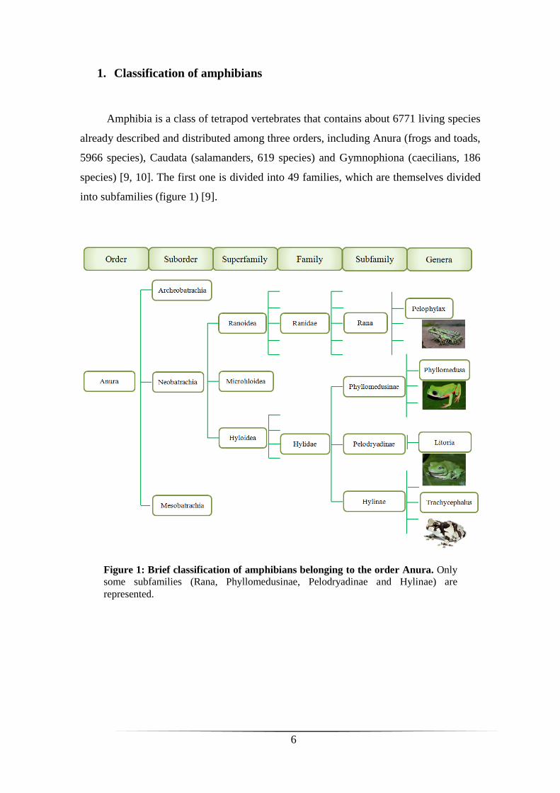
1. Classification of amphibians
Amphibia is a class of tetrapod vertebrates that contains about 6771 living species
already described and distributed among three orders, including Anura (frogs and toads,
5966 species), Caudata (salamanders, 619 species) and Gymnophiona (caecilians, 186
species) [9, 10]. The first one is divided into 49 families, which are themselves divided
into subfamilies (figure 1) [9].
Figure 1: Brief classification of amphibians belonging to the order Anura. Only
some subfamilies (Rana, Phyllomedusinae, Pelodryadinae and Hylinae) are
represented.

7
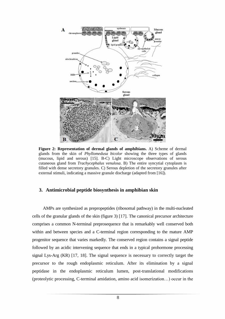
2. Dermal glands of amphibians
Amphibians have established the transition between aquatic and terrestrial
environments due to a gradual acquisition of a set of physiological and morphological
adaptations, such as the presence of a highly specialized integument – the skin [10].
The anuran skin exhibits morphofunctional diversity adapted to a number of
adverse factors present in the species habitat environment [11]. Generally, the frog skin
contains three types of cutaneous glands (figure 2), which differ in size, distribution and
secretory activity [12, 13]. The lipid glands (figure 2 A) promote the
impermeabilization of the skin in order to decrease water loss and are localized mostly
in the dorsal and dorsolateral regions [12]. The mucous glands (figure 2 A), which
usually are smaller and more numerous, secrete mucins in order to maintain skin
lubrification, moisture and thermoregulation and to prevent mechanical damage [5, 13,
14]. For some amphibians, these glands are also involved in reproduction and defense
(reviewed in [14]) and are fairly abundant in the ventral surface skin [12]. The third type
of glands is the serous or granular glands (also called venom glands) (figure 2 A-C)
formed by syncytial cells, with the nuclei located at the periphery of the syncytium [7].
These glands are widely distributed on the dorsal/dorsolateral cutaneous region and are
responsible for the synthesis and storage of a wide range of noxious or toxic compounds
which provide protection against bacterial and fungal infections, as well as predators
[12, 14]. Their cytoplasm is rich of peptide-containing secretory granules, which fill the
totality of the gland (figure 2 B). Upon external stimuli, a massive granule discharge is
induced by a holocrine-like mechanism involving the contraction of myoepithelial cells
surrounding the glands (figure 2 C) [7, 11]. Approximately 15 days are needed to refill
the gland with secretory granules.
8
3. Antimicrobial peptide biosynthesis in amphibian skin
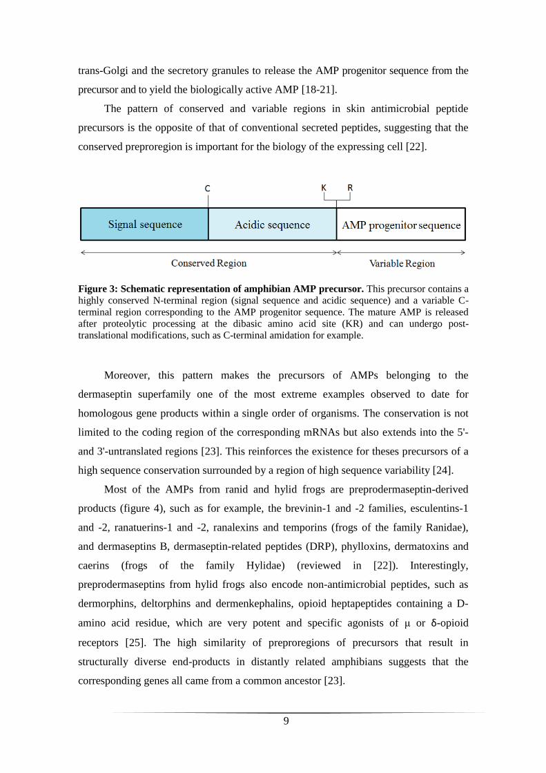
AMPs are synthesized as prepropeptides (ribosomal pathway) in the multi-nucleated
cells of the granular glands of the skin (figure 3) [17]. The canonical precursor architecture
comprises a common N-terminal preprosequence that is remarkably well conserved both
within and between species and a C-terminal region corresponding to the mature AMP
progenitor sequence that varies markedly. The conserved region contains a signal peptide
followed by an acidic intervening sequence that ends in a typical prohormone processing
signal Lys-Arg (KR) [17, 18]. The signal sequence is necessary to correctly target the
precursor to the rough endoplasmic reticulum. After its elimination by a signal
peptidase in the endoplasmic reticulum lumen, post-translational modifications
(proteolytic processing, C-terminal amidation, amino acid isomerization…) occur in the
Figure 2: Representation of dermal glands of amphibians. A) Scheme of dermal
glands from the skin of Phyllomedusa bicolor showing the three types of glands
(mucous, lipid and serous) [15]. B-C) Light microscope observations of serous
cutaneous gland from Trachycephalus venulosa. B) The entire syncytial cytoplasm is
filled with dense secretory granules. C) Serous depletion of the secretory granules after
external stimuli, indicating a massive granule discharge (adapted from [16]).
9
trans-Golgi and the secretory granules to release the AMP progenitor sequence from the
precursor and to yield the biologically active AMP [18-21].
The pattern of conserved and variable regions in skin antimicrobial peptide
precursors is the opposite of that of conventional secreted peptides, suggesting that the
conserved preproregion is important for the biology of the expressing cell [22].
Moreover, this pattern makes the precursors of AMPs belonging to the
dermaseptin superfamily one of the most extreme examples observed to date for
homologous gene products within a single order of organisms. The conservation is not
limited to the coding region of the corresponding mRNAs but also extends into the 5'-
and 3'-untranslated regions [23]. This reinforces the existence for theses precursors of a
high sequence conservation surrounded by a region of high sequence variability [24].
Most of the AMPs from ranid and hylid frogs are preprodermaseptin-derived
products (figure 4), such as for example, the brevinin-1 and -2 families, esculentins-1
and -2, ranatuerins-1 and -2, ranalexins and temporins (frogs of the family Ranidae),
and dermaseptins B, dermaseptin-related peptides (DRP), phylloxins, dermatoxins and
caerins (frogs of the family Hylidae) (reviewed in [22]). Interestingly,
preprodermaseptins from hylid frogs also encode non-antimicrobial peptides, such as
dermorphins, deltorphins and dermenkephalins, opioid heptapeptides containing a D-
amino acid residue, which are very potent and specific agonists of μ or δ-opioid
receptors [25]. The high similarity of preproregions of precursors that result in
structurally diverse end-products in distantly related amphibians suggests that the
corresponding genes all came from a common ancestor [23].
Figure 3: Schematic representation of amphibian AMP precursor. This precursor contains a
highly conserved N-terminal region (signal sequence and acidic sequence) and a variable C-
terminal region corresponding to the AMP progenitor sequence. The mature AMP is released
after proteolytic processing at the dibasic amino acid site (KR) and can undergo post-
translational modifications, such as C-terminal amidation for example.

10
Targeted hypermutation of the C-terminal antimicrobial-coding region of
preprodermaseptin genes might have evolved as a way of increasing genetic diversity and
so accelerating the adaptation of frogs to noxious microbial fauna with a maximum
protection against a large range of pathogens [17, 24].
Figure 4: Conserved preproregion and hypervariable antimicrobial domain of preprodermaseptins. Alignment of the predicted amino acid sequences (single-letter code) of preprodermaseptin cDNAs obtained from
hylid and ranid frogs, including the signal sequence, the acidic propiece and the antimicrobial peptide progenitor
sequence. Gaps (-) have been introduced to maximize sequence similarities. Identical (black background) and
similar (shaded background) amino acid residues are highlighted. Among the hylid sequences, DRS, dermaseptin B
from P. bicolor, DRP, dermaseptin-related peptide (appended with AA, AC or PD to indicate that the sequences
were identified from A. annae, A. callidryas and P. danicolor, respectively). Among the ranid sequences, temporins
B, H and G and brevinins 2Ta and 2Tb are from R. temporia, brevinins 1E and 2Ef and esculentin 1B from R.
esculenta, ranalexin from R. catesbeiana, gaegurins 4 and 5 from R. rugosa, and ranatuerin-2P and 2Pa from R.
pipiens. Raninae, Pelodryadinae and Phyllomedusinae are subfamilies belonging to the family Ranidae (the first
one) and the family Hylidae (the other two). (adapted from [24]).
11
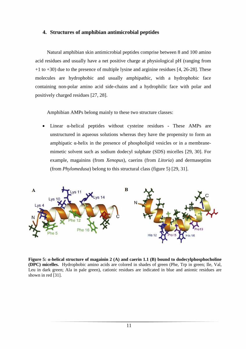
4. Structures of amphibian antimicrobial peptides
Natural amphibian skin antimicrobial peptides comprise between 8 and 100 amino
acid residues and usually have a net positive charge at physiological pH (ranging from
+1 to +30) due to the presence of multiple lysine and arginine residues [4, 26-28]. These
molecules are hydrophobic and usually amphipathic, with a hydrophobic face
containing non-polar amino acid side-chains and a hydrophilic face with polar and
positively charged residues [27, 28].
Amphibian AMPs belong mainly to these two structure classes:
Linear α-helical peptides without cysteine residues - These AMPs are
unstructured in aqueous solutions whereas they have the propensity to form an
amphipatic α-helix in the presence of phospholipid vesicles or in a membrane-
mimetic solvent such as sodium dodecyl sulphate (SDS) micelles [29, 30]. For
example, magainins (from Xenopus), caerins (from Litoria) and dermaseptins
(from Phylomedusa) belong to this structural class (figure 5) [29, 31].
Figure 5: α-helical structure of magainin 2 (A) and caerin 1.1 (B) bound to dodecylphosphocholine
(DPC) micelles. Hydrophobic amino acids are colored in shades of green (Phe, Trp in green; Ile, Val,
Leu in dark green; Ala in pale green), cationic residues are indicated in blue and anionic residues are
shown in red [31].

12
Peptides with -hairpin-like structure and cysteine residues - These AMPs
contain a C-terminal loop stabilized by an intramolecular disulfide bond. This
region is also called “Rana box” because many such structured AMPs are
present in ranid frogs, like esculentins, ranalexins, brevinins and gaegurins, for
example (figure 6) [29, 31, 32]. Like α-helical peptides, these AMPs are
unfolded in aqueous solutions but the N-terminal can adopt an amphipathic α-
helical conformation in hydrophobic environments, depending on the size of this
segment and its hydrophobicity [32, 33].
5. Mechanism of action of antimicrobial peptides
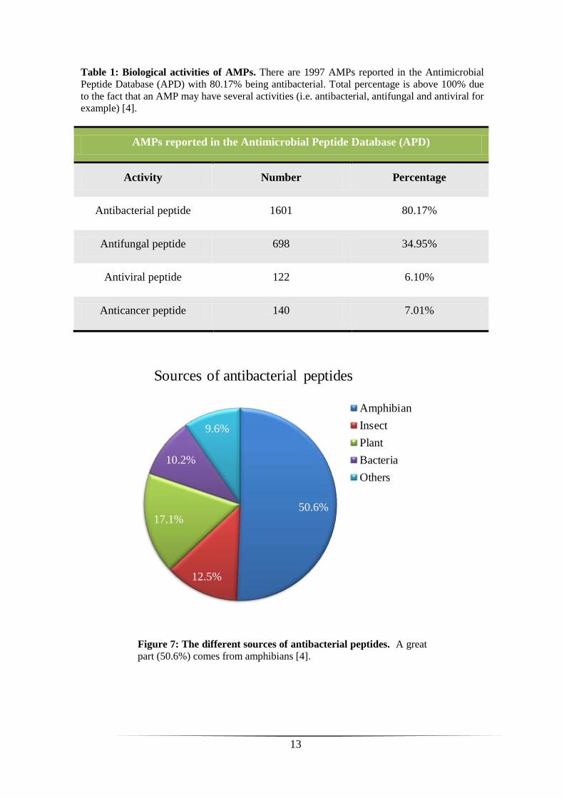
AMPs from amphibians exhibit a broad-spectrum activity and can kill aerobic and
anaerobic Gram-positive and Gram-negative bacteria, yeast, filamentous fungi,
protozoa, viruses and tumor cells (Table 1). These peptides may have synergistic
effects, which increase their effectiveness, and also hemolytic activity [32]. Today,
there are 1997 antimicrobial peptides reported in the database APD, of which 1601 are
antibacterial (80.17%) (Table 1) with approximately 50% coming from amphibians
(figure 7) [4].
Figure 6: Solution structure of gaegurin-4 in 80 % deuterated
methanol. Hydrophobic amino acids are colored in shades of green (Phe,
Trp in green; Ile, Val, Leu in dark green; Ala in pale green), cationic
residues are indicated in blue and anionic residues are shown in red. The
C-terminal disulfide bond is colored in gold. Note that the N-terminal has
a -helical conformation [31].
13
Table 1: Biological activities of AMPs. There are 1997 AMPs reported in the Antimicrobial
Peptide Database (APD) with 80.17% being antibacterial. Total percentage is above 100% due
to the fact that an AMP may have several activities (i.e. antibacterial, antifungal and antiviral for
example) [4].
AMPs reported in the Antimicrobial Peptide Database (APD)
Activity Number Percentage
Antibacterial peptide 1601 80.17%
Antifungal peptide 698 34.95%
Antiviral peptide 122 6.10%
Anticancer peptide 140 7.01%
50.6%
12.5%
17.1%
10.2%
9.6%
Sources of antibacterial peptides
Amphibian
Insect
Plant
Bacteria
Others
Figure 7: The different sources of antibacterial peptides. A great
part (50.6%) comes from amphibians [4].
14
The exact mechanism by which AMPs exert their killing actions is not
completely understood, but a common property is their interaction with the
phospholipids of the cytoplasmic membrane, leading to permeabilization and
subsequently lysis of the cell [33]. However, how AMPs can selectively distinguish
between microbial and host cytoplasmic membranes?
5.1. Membrane composition
The biological membrane is a fluid structure with various proteins embedded in or
attached to a bilayer of phospholipids (fluid mosaic model). In some organisms like
eukaryotes, sterols and glycerides also contribute to the topology surface and
biochemical architecture of biomembranes [26]. Differences between microbial and host
membranes exist. In fact, bacterial membranes are predominantly composed of
negatively charged phospholipids, such as phosphatidylglycerol (PG), cardiolipin (CL)
or phosphatidylserine (PS), which gives to whole membrane an electronegative net
charge [26]. In addition, the outer surface of Gram-negative and Gram-positive bacteria
contains lipopolysaccharides (LPS) and acidic polysaccharides (teichoic acids),
respectively, that enhance the electronegative charge of biomembranes (rewieved in
[34]). In contrast, mammalian cytoplasmic membranes have usually a neutral net charge
because they are mainly composed of zwitterionic phospholipids, such as
phosphatidylethanolamine (PE), phosphatidylcholine (PC) or sphingomyelin (SM), as
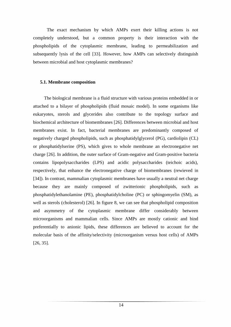
well as sterols (cholesterol) [26]. In figure 8, we can see that phospholipid composition
and asymmetry of the cytoplasmic membrane differ considerably between
microorganisms and mammalian cells. Since AMPs are mostly cationic and bind
preferentially to anionic lipids, these differences are believed to account for the
molecular basis of the affinity/selectivity (microorganism versus host cells) of AMPs
[26, 35].
15
Another important factor for the selectivity of AMPs toward microbial cells is the
transmembrane potential (Δψ), an electrochemical gradient determined by extents and
rates of proton flux across the membrane [26]. Normal mammalian cells exhibit a Δψ
ranging from -90 to -110 mV, whereas a Δψ of -130 mV to -150 mV is observed for
bacteria in logarithmic phase growth [26].
As a result, antimicrobial activity and selective toxicity of a peptide against
pathogens is determined by a complex interaction between parameters such as
conformation, charge, hydrophobicity and amphipathicity [26].
Several studies on both live organisms and model membranes have indicated that
most AMPs induce plasma membrane permeabilization by mechanisms involving the
formation of transmembrane pores (barrel-stave and wormhole models) or micellization
of the cytoplasmic membrane by a detergent-like action (carpet model).
Figure 8: Comparative architecture of microbial and mammalian cytoplasmic
membranes. The relative composition and distribution between inner and outer membrane
leaflets are indicated for cytoplasmic microbial (E. coli, S. aureus, B. subtilis, C. albicans) and
mammalian (human erythrocyte) membranes. CL: cardiolipin; PG: phosphatidylglycerol; PE:
phosphatidylethanolamine; PC: phosphatidylcholine; SM: sphingomyelin and ST: sterols
(cholesterol or ergosterol). (adapted from [26]).
16
5.2. Barrel-stave model
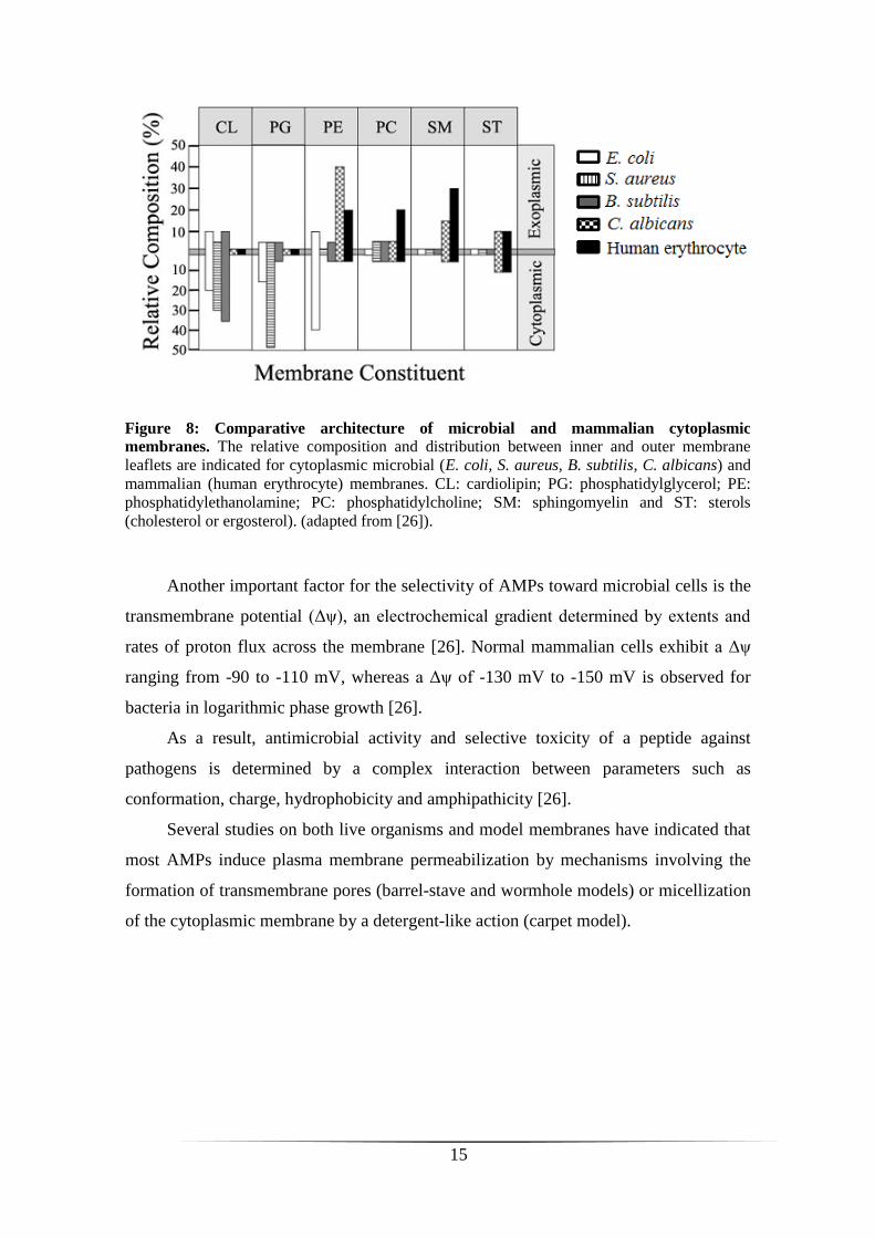
The barrel-stave mechanism describes the formation of transmembrane
channels/pores by bundles of amphipathic α-helices peptides, where their hydrophobic
surfaces interact with the lipid core of the membrane and their hydrophilic surfaces
point inward, producing an aqueous pore (figure 9) [36]. Initially, AMP binds to the
membrane surface, promoting the transition of the peptide from the random coil to the
α-helical conformation (figure 9 A). When bound peptide achieves a threshold
concentration, peptides monomers insert into the hydrophobic core of the membrane
bilayer, occurring a progressive recruitment of additional monomers to increase the pore
size (figure 9 B) [26].
Since these peptides can insert into the hydrophobic core of the membrane, it is
logical to presume that such interaction is determined predominantly by hydrophobic
interactions [36].
Figure 9: The barrel-stave model. AMPs (either as monomers or oligomers) interact with the
membrane and assemble on the surface (A), then insert into the lipid bilayer to form
transmembrane pores following recruitment of additional AMP molecules (B). The hydrophobic
peptide faces align with the lipid core region and the hydrophilic peptide faces form the interior
region of the pore. Hydrophilic and hydrophobic faces of the peptide are shown colored red and
blue, respectively [34].

17
5.3. Carpet model
Carpet mechanism or detergent-like mechanism, as the name suggests, is a
mechanism by which cytoplasmic membrane micellization occurs after action of AMPs
(figure 10). Initially, the cationic AMP targets the membrane via electrostatic
interactions with the anionic phospholipid headgroups, covering the surface in a carpet-
like manner (figure 10 A) [29]. When a threshold concentration of peptide is reached,
changes in membrane fluidity and/or reductions in membrane barrier properties are
observed, leading to the loss of membrane integrity (figure 10 B), and consequently the
formation of micelles (figure 10 C) [26]. In contrast to the barrel-stave model, this
mechanism does not require a specific peptide structure [37]. Therefore, membrane lysis
takes place in a dispersion-like manner that does not engage insertion of peptides into
the hydrophobic core of the membrane, and consequently pore formation [26, 36].
Figure 10: The carpet model. AMPs reach the membrane either as monomers or oligomers, and
then bind to the surface of the membrane with their hydrophobic regions facing the membrane and
their hydrophilic regions facing the solvent (A). When a threshold concentration is reached, the
membrane is permeabilized (B) and membrane lysis occurs with formation of micelles (C).
Hydrophilic and hydrophobic faces of the peptide are shown colored red and blue, respectively
[34].
18
5.4. Toroidal model or wormhole mechanism
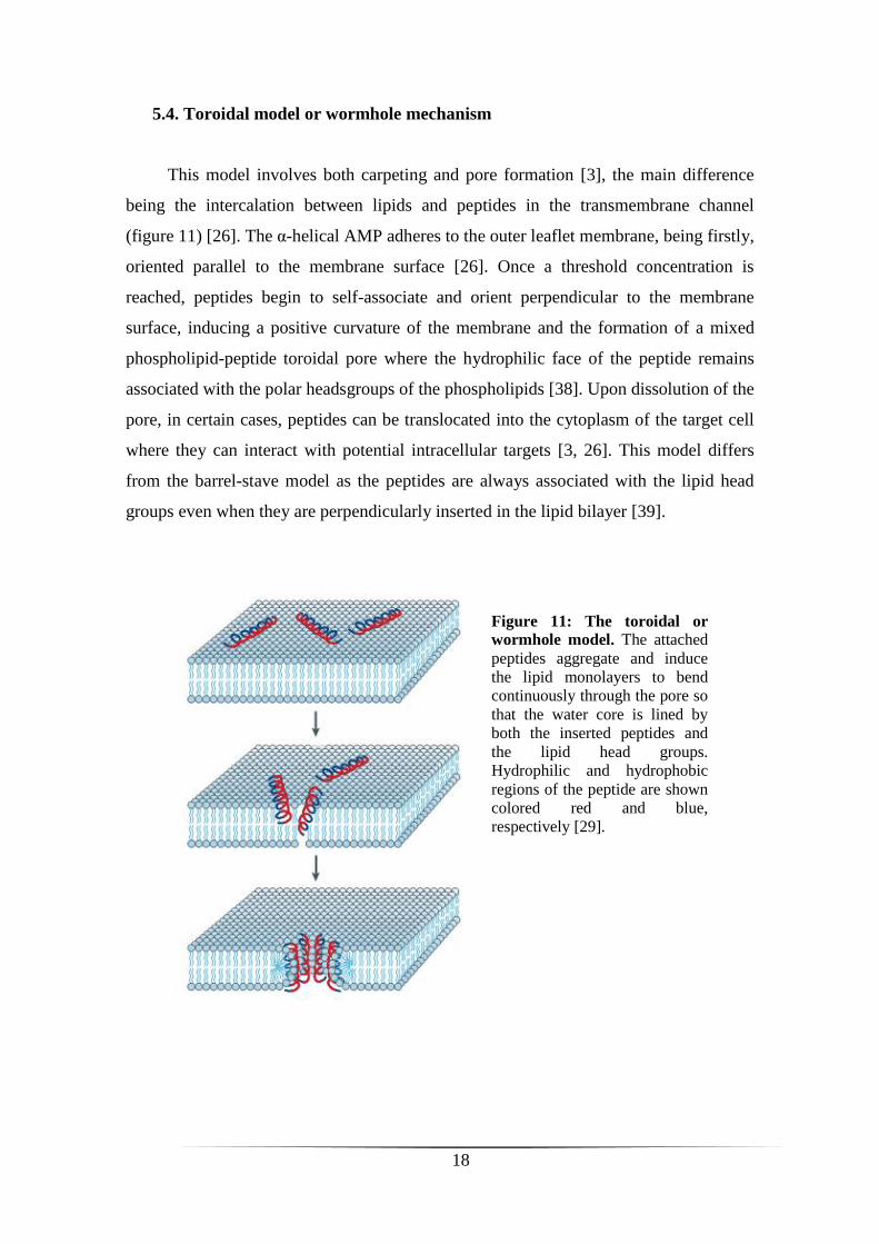
This model involves both carpeting and pore formation [3], the main difference
being the intercalation between lipids and peptides in the transmembrane channel
(figure 11) [26]. The α-helical AMP adheres to the outer leaflet membrane, being firstly,
oriented parallel to the membrane surface [26]. Once a threshold concentration is
reached, peptides begin to self-associate and orient perpendicular to the membrane
surface, inducing a positive curvature of the membrane and the formation of a mixed
phospholipid-peptide toroidal pore where the hydrophilic face of the peptide remains
associated with the polar headsgroups of the phospholipids [38]. Upon dissolution of the
pore, in certain cases, peptides can be translocated into the cytoplasm of the target cell
where they can interact with potential intracellular targets [3, 26]. This model differs
from the barrel-stave model as the peptides are always associated with the lipid head
groups even when they are perpendicularly inserted in the lipid bilayer [39].
Figure 11: The toroidal or
wormhole model. The attached
peptides aggregate and induce
the lipid monolayers to bend
continuously through the pore so
that the water core is lined by
both the inserted peptides and
the lipid head groups.
Hydrophilic and hydrophobic
regions of the peptide are shown
colored red and blue,
respectively [29].
19
In any case, the final result is the permeabilization of the cytoplasmic membrane,
and several events can happen like membrane depolarization, outflow of essential
metabolites, loss of compositional specificity, exchange with the inner leaflet of the
outer membrane, translocation of the peptides to the cytoplasmic side of the membrane
where they can interfere with cellular mechanisms, as well as components (reviewed in
[40]). All this processes lead to lysis of pathogens [20].
Thus, since AMPs are cidal by targeting the membrane of microorganisms and
inducing permeabilization/disruption, it is very difficult for pathogens to develop
resistance toward these molecules. This would require a change of the composition
and/or organization of the lipid membrane, a probably too expensive solution for the
majority of microorganisms [3]. In relation to conventional antibiotics, pathogens
become resistant because these drugs act on specific intracellular targets without
altering deeply their morphology [26].
6. Antimicrobial peptides from hylid and ranid frogs
6.1. AMPs from frogs belonging to the family Hylidae
Numerous AMPs were identified from different amphibian families. In the present
work, we will focus on frogs of the family Hylidae (subfamily Hylinae) and also
Ranidae.
The family Hylidae is composed of three subfamilies (Table 2 and 3) [9]:
Phyllomedusinae, containing 58 species identified today that are divided into 5
genera: Agalychnis, Cruziohyla, Phasmahyla, Phrynomedusa and Phyllomedusa
(Table 2) [9];
Pelodryadinae, comprising 197 species and only one genus: Litoria (Table 2)
[9];
Hylinae, containing 646 species and divided into 40 genera. We will pay more
attention to the following genera: Hyla, Pseudis, Hypsiboas and Trachycephalus
(Table 3) [9].
20
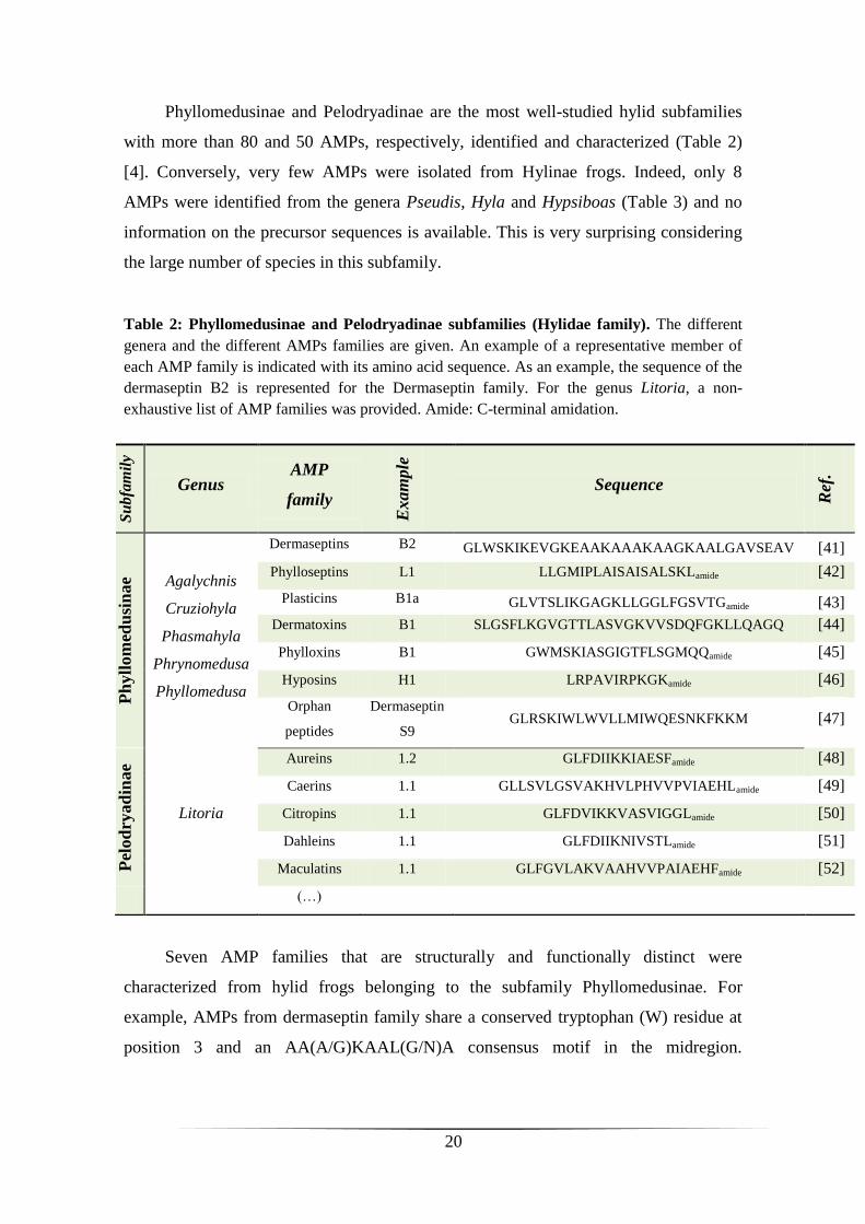
Phyllomedusinae and Pelodryadinae are the most well-studied hylid subfamilies
with more than 80 and 50 AMPs, respectively, identified and characterized (Table 2)
[4]. Conversely, very few AMPs were isolated from Hylinae frogs. Indeed, only 8
AMPs were identified from the genera Pseudis, Hyla and Hypsiboas (Table 3) and no
information on the precursor sequences is available. This is very surprising considering
the large number of species in this subfamily.
Table 2: Phyllomedusinae and Pelodryadinae subfamilies (Hylidae family). The different
genera and the different AMPs families are given. An example of a representative member of
each AMP family is indicated with its amino acid sequence. As an example, the sequence of the
dermaseptin B2 is represented for the Dermaseptin family. For the genus Litoria, a non-
exhaustive list of AMP families was provided. Amide: C-terminal amidation.
Seven AMP families that are structurally and functionally distinct were
characterized from hylid frogs belonging to the subfamily Phyllomedusinae. For
example, AMPs from dermaseptin family share a conserved tryptophan (W) residue at
position 3 and an AA(A/G)KAAL(G/N)A consensus motif in the midregion.
Su
bfa
mil
y
Genus AMP
family
Exam
ple
Sequence
Ref
.
Ph
yll
om
edu
sin
ae Agalychnis
Cruziohyla
Phasmahyla
Phrynomedusa
Phyllomedusa
Dermaseptins B2 GLWSKIKEVGKEAAKAAAKAAGKAALGAVSEAV [41]
Phylloseptins L1 LLGMIPLAISAISALSKLamide [42]
Plasticins B1a GLVTSLIKGAGKLLGGLFGSVTGamide [43]
Dermatoxins B1 SLGSFLKGVGTTLASVGKVVSDQFGKLLQAGQ [44]
Phylloxins B1 GWMSKIASGIGTFLSGMQQamide [45]
Hyposins H1 LRPAVIRPKGKamide [46]
Orphan
peptides
Dermaseptin
S9 GLRSKIWLWVLLMIWQESNKFKKM [47]
Pel
od
ryad
inae
Litoria
Aureins 1.2 GLFDIIKKIAESFamide [48]
Caerins 1.1 GLLSVLGSVAKHVLPHVVPVIAEHLamide [49]
Citropins 1.1 GLFDVIKKVASVIGGLamide [50]
Dahleins 1.1 GLFDIIKNIVSTLamide [51]
Maculatins 1.1 GLFGVLAKVAAHVVPAIAEHFamide [52]
(…)

21
Conversely, orphan peptides family does not resemble any members of the other
peptides families [53].
Table 3: Hylinae subfamily (Hylidae family). Only 4 of the 40 genera are referenced in the
present table with all the AMP families identified until today. Amide: C-terminal amidation.
Su
bfa
mil
y
Genus AMP
family
Exam
ple
Sequence
Ref
.
Hyli
nae
(646 s
p.)
Pseudis
(9 sp.) Pseudins
1 GLNTLKKVFQGLHEAIKLINNHVQ
[54] 2 GLNALKKVFQGIHEAIKLINNHVQ
3 GINTLKKVIQGLHEVIKLVSNHE
4 GINTLKKVIQGLHEVIKLVSNHA
Hyla
(35 sp.)
Hylaseptins P1 GILDAIKAIAKAAGamide [55]
Hylains 1
2
GILDAIKAFANALGamide
GILDPIKAFAKAAGamide [56]
Hypsiboas
(84 sp.) Hylins a1 IFGAILPLALGALKNLIKamide [57]
Trachycephalus
(12 sp.) ?
As stated earlier, AMPs have a broad-spectrum activity, which can be interesting
for therapeutic use. In fact, besides having bactericidal, as well as fungicidal activities,
many AMPs have antitumor activity against the major human cancer cell lines and also
antidiabetic and antiviral properties [30, 58, 59]. For example, among AMPs listed in
Table 2, dermaseptin B2 [58], phylloseptin L1 [42], aurein 1.2 [48], caerin 1.1, citropin
1.1 and maculatin 1.1 [60] are potent antineoplasic peptides. The recognition of
cancerous cells from healthy cells is not fully understood. However, many processes
have been proposed, such as changes in membrane potential due to higher metabolism,
higher exposure of acidic phospholipids in the outer leaflet of membrane, cytoskeleton
alterations and possible changes in the extracellular matrix (reviewed in [20]).
Furthermore, caerin 1.1, as an example, also has antiviral activity and can inhibits
human immunodeficiency virus (HIV) infection of T cells [61]. Ultimately, some

22
studies realized in rat clonal BRIN-BD11 β-cells demonstrated that pseudin-2 (Table 3)
[62], as well as phylloseptin-L2 [59], are able to induce insulin release at concentrations
that are not toxic to the cells. Consequently, these peptides could be interesting for
treatment of type 2 diabetes [59].
In order to develop new peptide antibiotics with improved therapeutic potential,
design strategies and structure-activity relationship studies were used to improve
potency and selectivity of AMPs (reviewed in [13]). These modifications might offer
significant advantages over natural AMPs as therapeutic agents [63].
Additionally, nanoscale biofunctionalization of biomolecules (i.e.
nanobiotechnology) like AMPs is a very promising strategy for applications in the
pharmaceutical industry and diagnosis. The interest in AMPs as active materials in
bionanostructures is due to their properties, such as the presence of an α-helix structure
and positive charges [64]. These structures consist of cationic nanoparticles formed by
the conjugation of cholesterol and AMPs that are able to cross blood-brain barrier for
treatment of infections, such as fatal Cryptococcal meningitis in patients with late-stage
HIV infection [65]. These nanoparticules may also be used as sensor elements for
detection of Leishmania cells [64].
Thus, AMPs appears to be promising candidates as therapeutic agents for the
treatment of several diseases.
6.2. AMPs from frogs belonging to the family Ranidae
The family Ranidae is composed of 347 species organized into 16 genera. The
genus Rana constituted of 48 species of Eurasian and North American frogs [9] was
particularly studied. As shown in Table 4, 12 peptide families have been identified from
several ranid frog species. Except temporins, all these families contain a C-terminal
domain with an intramolecular disulfide bridge called the “Rana box” (figure 6) [1, 66].
In this study, we focus on temporin family, in fact, despite that the others have a
broad-spectrum activity against numerous pathogens, as well as insulinotropic
properties [67], temporins have characteristics that make them interesting for in-depth
investigation of their biological function and mechanism of action.

23
Table 4: AMP families of ranid frogs. A representative member of each AMP family is
indicated with its amino acid sequence [4]. As an example, the sequence of brevinin-1 is given
for the Brevinin-1 family. The C-terminal Rana box containing a disulfide bridge is represented
in green color. Amide: C-terminal amidation.
Initially identified in 1996 in the skin secretion of the frog Rana temporaria
[66], temporins are among the shortest amphipatic α-helical AMPs found to date, with a
single 8-21 amino acid chain. They have a low net positive charge at neutral pH ranging
from 0 to +3 and are amidated at their carboxyl end [1]. Temporins are predominantly
active toward Gram-positive bacteria, including methicillin- and vancomycin-resistant
staphylococci and enterococci, as well as yeasts [66, 68, 69]. They are inactive or
weakly active toward Gram-negative bacteria and are generally not toxic to human
blood cells at their antimicrobial concentration [66], except for temporin L which has a
broad spectrum of activity (Gram-positive and Gram-negative bacteria, yeasts, cancer
cells and human erythrocytes) [70]. Furthermore, it has also been demonstrated that
some members of the temporin family could have antiparasitic activity. This is the case
for temporins A and B, isolated from Rana temporaria, and also temporin-SHa
(Pelophylax saharica) that are active against both the insect (promastigote) and
mammalian intracellular stage (amastigote) of the human protozoan parasite Leishmania
Fam
ily
AMP
family
Ex
am
ple
Sequence
Ran
ida
e
Brevinin-1 1 FLPVLAGIAAKVVPALFCKITKKC
Brevinin-2 2 GLLDSLKGFAATAGKGVLQSLLSTASCKLAKTC
Esculentin-1 1 GIFSKLGRKKIKNLLISGLKNVGKEVGMDVVRTGIDIAG
CKIKGEC
Esculentin-2 2a GILSLVKGVAKLAGKGLAKEGGKFGLELIACKIAKQC
Ranatuerin-1 1 SMLSVLKNLGKVGLGFVACKINKQC
Ranatuerin-2 PLa GIMDTVKNVAKNLAGQLLDKLKCKITAC
Palustrin-2 2a GFLSTVKNLATNVAGTVLDTIRCKVTGGCRP
Japonicin-1 1 FFPIGVFCKIFKTC
Japonicin-2 2 FGLPMLSILPKALCILLKRKC
Nigrocin-2 2 GLLSKVLGVGKKVLCGVSGLC
Ranacyclin T GALRGCWTKSYPPKPCKamide
Temporin A FLPLIGRVLSGILamide
24
[71, 72]. Besides their antimicrobial effects, temporins possess additional biological
activities. For example, temporin A has chemotactic effects on human phagocytes [73].
Temporins B and L modulate the hydrolytic activity of secretory phospholipase A2 in
human lacrymal fluid, thus improving the efficiency of the immune response to
infections [74]. In contrast to many natural AMPs, it was demonstrated that temporins A
and B maintain activity in physiological salt concentration, as well as in serum, making
them attractive lead compounds for the development of anti-infective drugs [71].
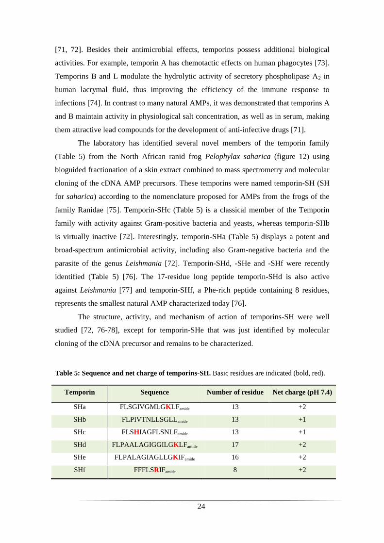
The laboratory has identified several novel members of the temporin family
(Table 5) from the North African ranid frog Pelophylax saharica (figure 12) using
bioguided fractionation of a skin extract combined to mass spectrometry and molecular
cloning of the cDNA AMP precursors. These temporins were named temporin-SH (SH
for saharica) according to the nomenclature proposed for AMPs from the frogs of the
family Ranidae [75]. Temporin-SHc (Table 5) is a classical member of the Temporin
family with activity against Gram-positive bacteria and yeasts, whereas temporin-SHb
is virtually inactive [72]. Interestingly, temporin-SHa (Table 5) displays a potent and
broad-spectrum antimicrobial activity, including also Gram-negative bacteria and the
parasite of the genus Leishmania [72]. Temporin-SHd, -SHe and -SHf were recently
identified (Table 5) [76]. The 17-residue long peptide temporin-SHd is also active
against Leishmania [77] and temporin-SHf, a Phe-rich peptide containing 8 residues,
represents the smallest natural AMP characterized today [76].
The structure, activity, and mechanism of action of temporins-SH were well
studied [72, 76-78], except for temporin-SHe that was just identified by molecular
cloning of the cDNA precursor and remains to be characterized.
Table 5: Sequence and net charge of temporins-SH. Basic residues are indicated (bold, red).
Temporin Sequence Number of residue Net charge (pH 7.4)
SHa FLSGIVGMLGKLFamide 13 +2
SHb FLPIVTNLLSGLLamide 13 +1
SHc FLSHIAGFLSNLFamide 13 +1
SHd FLPAALAGIGGILGKLFamide 17 +2
SHe FLPALAGIAGLLGKIFamide 16 +2
SHf FFFLSRIFamide 8 +2

25
7. Purpose of the study
Amphibian skin represents a good model for the identification of novel AMPs
with potent activity and therapeutic potential, and for studying the mechanism of action
of these peptides.
A first part of my research project was to investigate the AMP content of frogs of
the subfamily Hylinae, which have been very poorly studied, and particularly those of
the genus Trachycephalus. Therefore, we have analyze for the first time by bioguided
fractionation the skin secretions of Trachycephalus resinifictrix, a South American tree
frog also referred to as Amazon Milk Frog because of its milky and poisonous
secretions when threatened (figure 12) [79]. Fractionation was performed by semi-
preparative and analytical HPLC, and antibacterial activity against Staphylococcus
aureus was monitored by a liquid growth inhibition assay. We also attempted to
characterize the AMP precursors by performing mRNA extraction, RT-PCR and
molecular cloning of the cDNAs.
The second part of my Master 2 research project was to perform the structural and
functional characterization of temporin-SHe that was previously identified by the host
team from the ranid frog Pelophylax saharica [76]. We have produced this peptide by
solid phase peptide synthesis and determined its structure, antimicrobial activity, and
mechanism of action using biochemical and biophysical techniques. Moreover,
temporin-SHe was compared to its paralog, temporin-SHd.
Figure 12: Trachycephalus resinifictrix (left) and Pelophylax saharica (right).
26
MATERIALS AND METHODS
8. Analysis of antimicrobial peptides from skin secretions of T.
resinifictrix and molecular cloning of AMP cDNA precursors.
Collection of skin secretions and pre-purification of peptides
Specimens of Trachycephalus resinifictrix (2 males and 2 females) were bred
and fed crickets by François Lemoine (National Museum of Natural History, MNHN,
Paris, France). The temperature was maintained at approximately 25°C and water bowls
were provided for bath. Frogs were mildly stressed by electrical stimulation (9 V) and
skin secretion was collected, diluted in Milli-Q H2O and lyophilized.
Lyophilized secretions were dissolved in H2O containing 0.1% trifluoroacetic acid
(TFA), then sonicated for 10 min at 25°C (Ultrasonic cleaner, VWR) and centrifuged
(4500 rpm, 20 min, 4°C). The supernatant was lyophilized and dissolved in 0.1%
TFA/H2O in order to obtain a concentration of 1 mg/ml. After sonication for 5 min and
centrifugation (16000 x g, 15 min, 4°C), the solution was filtered (0.20 µm) and then
loaded onto Sep-Pak C-18 cartridges. After a washing step (0.1% TFA/H2O), the
material was eluted with 60% acetonitrile (ACN) and lyophilized. All these steps were
intended to prevent clogging of the HPLC column because of the high viscosity of the
sample.
Reversed-phase HPLC (RP-HPLC) fractionation of skin secretions
The lyophilized pre-purified extract (14.5 mg) was reconstituted in 0.1%
TFA/H2O to obtain a concentration of 1 mg/ml, sonicated for 10 min and centrifuged
(16000 x g, 10 min, 4°C). Subsequently, the supernatant was lyophilized and dissolved
into 6 ml of 20% ACN, followed by sonication for 10 min and centrifugation (13000 x
g, 10 min, 4°C). The final supernatant was fractionated by RP-HPLC on a semi-
preparative Nucleosil C18 column (5 µm, 250 x 10 mm, Interchim) using a two solvent
system: (A) 0.1% TFA/H2O and (B) 0.07% TFA/ACN.
27
Elution was performed with a 20-60% linear gradient of solvent B (1%/min) at a flow
rate of 4 ml/min. Collected fractions (4 ml) were lyophilized, reconstituted in 500 µl of
sterile Milli-Q H2O and tested for antibacterial activity against the reference strain
Staphylococcus aureus (see below). In order to improve peaks separation, the active
fractions (25, 29 and 37) were rechromatographed on an Uptisphere C18 analytic
column (modulo-cart QS, 5 µm, ODS2, 250 x 4.6 mm, Interchim) using a 20-60% linear
gradient of solvent B (0.5%/min) at a flow rate of 0.75 ml/min. Major peaks were
harvested manually, lyophilized, dissolved into 120 µL of sterile Milli-Q H2O and
tested again against the bacterial strain Staphylococcus aureus. Absorbance was
monitored at 220 and 280 nm.
Antimicrobial assays
Antibacterial activity of the lyophilized fractions was monitored by a liquid
growth inhibition assay against the Gram-positive reference strain Staphylococcus
aureus. Bacteria were cultured in LB medium for 2-3 h at 37°C with vigorous shaking
(250 rpm). After determination of the absorbance at 600 nm (A600), the bacterial culture
was centrifuged (1000 x g, 10 min, 4°C), resuspended in MH broth to A600 = 0.01 (106
cfu/ml) and diluted 80 fold in MH broth (1.25 x 10⁴ cfu/ml). Diluted bacteria (50 µl)
were mixed with 50 µl of either RP-HPLC fractions or sterile Milli-Q H2O (negative
growth inhibition control) in 96-well microtitration plates. 0.7% formaldehyde was used
as positive control. After 18 h of incubation at 37°C with shaking (150 rpm), the
bacterial growth was monitored by measuring the change in A600 value using a
microplate spectrophotometer (Asys Hitech UVM 340). Each assay was performed in
duplicate to minimize fraction consume.
The minimal inhibitory concentration (MIC) of synthetic temporin-SHe was
determined against Gram-positive bacteria (Staphylococcus aureus ATCC 25923, S.
aureus ST1065, Enterococcus faecalis ATCC 29212, Bacillus megaterium, Listeria
ivanovii), Gram-negative bacteria (Escherichia coli ATCC 25922, E. coli ATCC 35218,
E. coli ML-35p, Pseudomonas aeruginosa ATCC 27853) and yeasts (Saccharomyces
cerevisiae, Candida albicans ATCC 90028, C. parapsilosis ATCC 22019). Bacteria
were cultured at 37°C in LB medium and then diluted in MH broth to A600 = 0.01 (106
cfu/ml), except for E. faecalis and L. ivanovii which were diluted in LB medium and
28
BHI, respectively. Yeasts were cultured in YPD medium at 30ºC and diluted in the
same medium to A600 = 0.01 (106
cfu/ml). The MIC was determined by measuring the
absorbance at 600 nm in 96-well microtitration plates by growing 50 µl of the
microorganism suspension (106
cfu/ml) with 50 µl of 2-fold serial dilutions of synthetic
temporin-SHe (200-1 µM) 18 h at 37ºC (30ºC for yeasts). MIC was expressed as the
lowest concentration of peptide that inhibited bacterial growth completely and as the
average value from three independent experiments, each performed in triplicate with
positive (0.7% formaldehyde) and negative (without peptide) inhibition control, and
sterility control (H2O).
Mass spectrometry analysis of antibacterial HPLC fractions
HPLC fractions with antibacterial activity were subjected to MALDI-TOF-MS
(Voyager DE-Pro, Applied Biosystems – Proteomics and Mass Spectrometry Platform
of IFR83, UPMC) in order to determine the mass of the material present in these
fractions. Briefly, 1 µl of HPLC fractions were mixed with 1 µl of saturated matrix
solution (α-cyano-4-hydroxycinnamic acid) and spotted on a sample plate. The MS
positive ion spectra were carried out in the reflector mode with external calibration,
using the 4700 Standard Kit (Applied Biosystems).
Isolation of mRNA and reverse transcription
Four adult specimens of T. resinifictrix were mildly stressed by electrical
stimulation (9 V) and skin secretions were collected with a sterile spatula, diluted in
DEPC-treated H2O and lyophilized. Poly(A)+ RNA was isolated from the lyophilized
powder (16.7 mg) using the Micro-FastTrackTM
2.0 mRNA Isolation kit (Invitrogen)
according to the manufacturer’s protocol. Briefly, 1 ml of lysis buffera supplemented
with proteinase K (20 mg/ml) was added to the powder and the solution was incubated
at 45ºC for 30 min. The lysate was homogenized by several passages through a sterile
syringe and the final NaCl concentration was adjusted to 0.5 M. mRNA was purified by
oligo(dT) cellulose binding (90 min at room temperature with gentle rotation). After
several washing steps with bindingb and low salt wash buffer
c (to removes SDS and
nonpolyadenylated RNAs), mRNA was elutedd
and precipitated by adding 10 µl of 2

29
mg/ml glycogen carrier, 30 µl of 2 M sodium acetate and 600 µl of 100% ethanol
(incubation overnight at -80°C). Finally, the sample was centrifuged at high speed
(16000 x g, 15 min, 4°C) and the pellet containing mRNA was dried under heat lamp
for 30 min to remove traces of ethanol. Reverse transcription of mRNA was performed
using Advantage RT-for-PCR kit (Clontech). The pellet of mRNA was reconstituted in
12.5 µl of DEPC-treated H2O and 1 µl of 20 µM oligo(dT) primer was added. First, the
mix was heated at 70°C for 2 min and rapidly transferred on ice to remove RNA
secondary structure. Then, the reverse transcription was initiated by adding to the
sample 4 µl of 5X reaction buffere, 1 µl of dNTP mix (10 mM each), 0.5 µl of
Recombinant RNase inhibitor (40 units/µl) and 1 µl of MMLV reverse transcriptase
(200 units/µl), followed by incubation at 42°C (Mastercycler, Eppendorf). After 1 h, the
reaction was stopped by incubation 5 min at 94ºC. Finally, the sample was diluted with
DEPC-treated H2O to obtain 100 µl of cDNA and stored at -20°C until PCR.
aLysis buffer: 200 mM NaCl; 200 mM Tris-HCl, pH 7.5 ; 1.5 mM MgCl2 ; 2 % SDS
bBinding buffer: 500 mM NaCl; 10 mM Tris-HCl, pH 7.5 in DEPC-treated H2O
cLow salt wash buffer: 250 mM NaCl; 10 mM Tris-HCl, pH 7.5 in DEPC-treated H2O
dElution buffer: 10 mM Tris-HCl, pH 7.5 in DEPC-treated H2O
e5X reaction buffer: 250 mM Tris-HCl, pH 7.5; 375 mM KCl; 15 mM MgCl2
PCR
PCR was performed using specific primers designed in the conserved 5’- and 3’-
UTR of cDNAs encoding AMP precursors of the dermaseptin superfamily (figure 13).
Forward primer 1: 5’-TGACCTTCAGTACCCAGCACTTTC-3’ (24 bp, Tm: 56.3°C)
Reverse primer 1: 5’- GCATTTAGCTAAATGATATTCCACATCA-3’ (28 bp, Tm:
56.2°C)
Figure 13: Position of the specific primers used for amplification of AMP cDNA precursors.
30
The following components were added in a tube:
25 µl of GoTaq Green Master Mix*, 2X (Promega)
18 µl of H2O
1 µl of forward primer
1 µl of reverse primer
5 µl of cDNA
*GoTaq Green Master Mix, 2X: DNA polymerase; reaction buffer, pH 8.5; 400 µM dATP; 400 µM
dGTP; 400 µM dCTP; 400 µM dTTP; 3 mM MgCl2
PCR was done under the following conditions:
94 °C, 2 min – Initial denaturation
94 °C, 45 s – Denaturation
55 °C, 1 min – Annealing
72 °C, 3 min – Extension
72 °C, 10 min – Final extension
After amplification, PCR products were analyzed by 1% agarose gel electrophoresis in
the presence of GelRed (1:10000) (FluoProbes, Interchim). PCR fragments of interest
corresponding to approximately 150-450 bp were then extracted and purified
(Nucleospin Extract II kit, Macherey-Nagel) according to the manufacturer’s protocol.
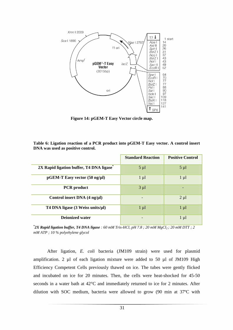
Cloning of PCR products into pGEM-T Easy vector
PCR products were cloned into pGEM-T Easy vector (pGEM-T Easy Vector
Systems II, Promega), a plasmid that contain an ampicillin resistance gene as well as a
β-galactosidase gene (lacZ) where fragment is cloned (figure 14). Ligation of the PCR
product was performed by incubating all the components indicated in Table 6 at room
temperature. A control insert DNA provided in the kit was used as positive control. The
samples were incubated at 4°C during the weekend in order to increase the number of
transformants.
30 cycles
31
Table 6: Ligation reaction of a PCR product into pGEM-T Easy vector. A control insert
DNA was used as positive control.
Standard Reaction Positive Control
2X Rapid ligation buffer, T4 DNA ligase* 5 µl 5 µl
pGEM-T Easy vector (50 ng/µl) 1 µl 1 µl
PCR product 3 µl -
Control insert DNA (4 ng/µl) - 2 µl
T4 DNA ligase (3 Weiss units/µl) 1 µl 1 µl
Deionized water - 1 µl
*2X Rapid ligation buffer, T4 DNA ligase : 60 mM Tris-HCl, pH 7.8 ; 20 mM MgCl2 ; 20 mM DTT ; 2
mM ATP ; 10 % polyehylene glycol
After ligation, E. coli bacteria (JM109 strain) were used for plasmid
amplification. 2 µl of each ligation mixture were added to 50 µl of JM109 High
Efficiency Competent Cells previously thawed on ice. The tubes were gently flicked
and incubated on ice for 20 minutes. Then, the cells were heat-shocked for 45-50
seconds in a water bath at 42°C and immediately returned to ice for 2 minutes. After
dilution with SOC medium, bacteria were allowed to grow (90 min at 37ºC with
Figure 14: pGEM-T Easy Vector circle map.
32
shaking at 250 rpm) and were then spread on LB agar/ampicillin (100 mg/ml) Petri
dishes containing IPTG (0.1 M) and X-gal (50 mg/ml) for blue/white screening. Plates
were incubated overnight at 37ºC and bacteria containing recombinant plasmids (white
colonies) were cultured in 5 ml of LB/ampicillin (100 mg/ml) overnight at 37 °C with
shacking (250 rpm).
Plasmid DNA purification and determination of the insert size
Plasmid DNA purification was performed by Nucleospin Plasmid kit
(Macherey-Nagel). Briefly, bacterial cultures were centrifuged (3000 rpm, 15 min,
4°C). The pellet was resuspended in an appropriate buffer* and plasmid DNA was
liberated from E. coli host cells by SDS/alkaline lysis. After neutralization of lysate,
precipitated protein, genomic DNA and cells debris were pelleted by centrifugation
(11000 x g, 10 min), and supernatant was loaded onto a silica column that retains
specifically plasmid DNA. Contaminants (salts, metabolites and soluble
macromolecular cellular components) were removed by a washing step with an
ethanolic buffer* and centrifugation (11000 x g, 1 min). After drying the silica
membrane, pure plasmid DNA was eluted by centrifugation (11000 x g, 2 min) under
low ionic strength conditions with 50 µl of 5 mM Tris-HCl, pH 8.5.
*The buffer composition was not provided by the supplier.
As it can be seen in figure 14, pGEM-T Easy vector is flanked by two recognition
sites for the restriction enzyme EcoR I. So, digestion of pure plasmid DNA by this
restriction enzyme was performed to release the insert and determine its size. The
following components were added in a tube and incubated for 90 min at 37°C:
5 µl of plasmid DNA
1 µl of 10X EcoR I buffer (900 mM Tris-HCl, pH 7.5; 100 mM MgCl2; 500
mM NaCl, Promega)
0.5 µl of EcoR I enzyme (12 units/µl, Promega)
3.5 µl of Milli-Q H2O
After digestion, 5 µl of this mix was analyzed by 1% agarose gel electrophoresis in the
presence of GelRed (1:10000) and insert size were estimated using 100 bp DNA ladder
(Promega). Plasmids containing inserts of the appropriate size (between 150 and 450
bp) were sequenced using T7 primer (Cogenics, Beckman Coulter Genomics, France).
33
9. Structural and functional characterization of temporin-SHe
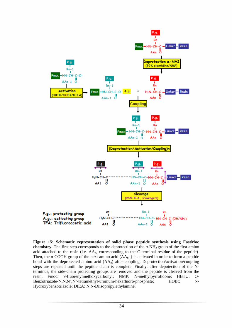
Solid phase peptide synthesis
Temporin-SHe was synthesized by solid phase method using standard FastMoc
chemistry (Applied Biosystems 433A automated peptide synthesizer - Peptide synthesis
platform of IFR83, UPMC). Briefly, amino acids with α-NH2 and side-chain protecting
groups were added sequentially on a Rink amide PEG MBHAa resin from the C-
terminus to the N-terminus (see figure 15 for the principle of peptide synthesis). Fmocb
protecting group were removed from α-NH2 by addition of 20% piperidine/NMPc
(deprotection step) and the activation of the carboxyl group was obtained with 0.5 M
HBTUd/0.5 M HOBt
e/DMF
f and 2 M DIEA
g/NMP (figure 15). At the end of synthesis,
the N-terminus of the peptide was deprotected with piperidine and a scavenger mixture
(95% TFA/2.5% TISh/2.5% H2O) was used to release peptide from the resin and remove
protecting groups. Finally, after precipitation of the peptide with ether and several
washing steps, the peptide was resuspended in 10% acetic acid and lyophilized. The
crude synthetic temporin-SHe was purified by RP-HPLC on a semi-preparative column
(Phenomenex Luna C18, 10 µm, 250 × 10 mm) with a 40-80% linear gradient of solvent
B (1%/min) at a flow rate of 5 ml/min. The homogeneity and identity of temporin-SHe
was confirmed by MALDI-TOF mass spectrometer (Voyager DE-Pro, Applied
Byosystems) and analytical RP-HPLC on an Uptisphere C18 column (modulo-cart QS,
5 μm, ODS2, 250 x 4.6 mm, Interchim) using the conditions above with a flow rate of
0.75 ml.
aPEG MBHA: Polyethylene glycol 4-methylbenzhydrylamine
bFmoc: 9-fluorenylmethoxycarbonyl
cNMP: N-methylpyrrolidone
dHBTU: O-Benzotriazole-N,N,N’,N’-tetramethyl-uronium-hexafluoro-phosphate
eHOBt: N-Hydroxybenzotriazole
fDMF: Dimethylformamide
gDIEA: N,N-Diisopropylethylamine
hTIS: (triisopropylsilane)
34
Figure 15: Schematic representation of solid phase peptide synthesis using FastMoc
chemistry. The first step corresponds to the deprotection of the α-NH2 group of the first amino
acid attached to the resin (i.e. AAn, corresponding to the C-terminal residue of the peptide).
Then, the α-COOH group of the next amino acid (AAn-1) is activated in order to form a peptide
bond with the deprotected amino acid (AAn) after coupling. Deprotection/activation/coupling
steps are repeated until the peptide chain is complete. Finally, after depotection of the N-
terminus, the side-chain protecting groups are removed and the peptide is cleaved from the
resin. Fmoc: 9-fluorenylmethoxycarbonyl; NMP: N-methylpyrrolidone; HBTU: O-
Benzotriazole-N,N,N’,N’-tetramethyl-uronium-hexafluoro-phosphate; HOBt: N-
Hydroxybenzotriazole; DIEA: N,N-Diisopropylethylamine.

35
Preparation of multilamellar and large unilamellar vesicles
DMPC (dimyristoyl phosphatidyl choline) and DMPG (dimyristoyl phosphatidyl
glycerol) were purchased from Avanti Polar lipids. DMPC (1.5 mg) was dissolved in
150 μl of chloroform and a mix of DMPC/DMPG 3:1 (mol/mol) (i.e. 1.12 mg of DMPC
and 0.38 mg of DMPG) was also prepared and dissolved in chloroform/methanol (1:1).
The samples were then dried under a nitrogen stream, and lipid films were kept under
vacuum for 3h at 45°C to remove all traces of organic solvents. Multilamellar vesicles
(MLVs) were obtained by hydrating the dry lipid films with 1.5 ml of PBS buffer (10
mM Na2HPO4, 100 mM NaCl, pH 7.3) at 37°C (10°C above the lipid phase transition)
and vortexing until a homogeneous suspension was formed (1 mg of MLVs/ml). MLVs
were used for differential scanning calorimetry experiments. For circular dichroism
experiments, large unilamellar vesicles (LUVs) were obtained from dry lipid films by
hydration with 1.5 ml of phosphate buffer (10 mM Na2HPO4, pH 7.3), followed by
seven rounds of freeze-thawing (liquid nitrogen/water bath at 37°C) and extrusion
through different polycarbonate membranes (400, 200 and 100 nm pore size).
Circular dichroism spectroscopy
The secondary structure of temporin-SHe was determined by circular dichroism
(CD) spectroscopy using zwitterionic DMPC LUVs (eukaryote membrane model) or
negatively charged DMPC/DMPG (3:1) LUVs (bacterial membrane model). CD spectra
were recorded at 25°C with a Jobin Yvon CD6 spectropolarimeter in a 0.1-cm quartz
cell over a wavelength range from 185 to 260 nm. Spectra were acquired with a spectral
bandwidth of 2-nm, a step size of 0.5 nm and a time constant of 3.0 s. Experiments were
done with different temporin-SHe/lipid molar ratios (1:200, 1:100 and 1:50) and also
with peptide (30 µM) in phosphate buffer (10 mM Na2HPO4, pH 7.3) or 80 mM SDS.
The baselines (DMPC, DMPC/DMPG 3:1, phosphate buffer and 80 mM SDS) were
acquired independently under the same conditions and then subtracted from the
corresponding peptide spectra. CD measurements were reported as Δε (M-1
. cm-1
) per
residue. Δε: dichroic increment.
36
Analysis of peptide-lipid interaction by differential scanning calorimetry
The interaction of synthetic temporin-SHe with membrane vesicles (MLVs) was
analyzed by differential scanning calorimetry (DSC). DMPC/DMPG 3:1 was used as a
model system for bacterial membranes. Different peptide/lipid molar ratios (1:200,
1:100 and 1:50) were used. Calorimetry experiments were performed with a Nano III
calorimeter (Calorimetry Sciences Corp., USA) using a temperature range of 0-35°C
with heating and cooling rates of 0.5°C/min and 1.5°C/min, respectively. Several scans
(>20) were run for each sample with a 10 min equilibration time between each scan.
The raw data were analyzed with the CpCalc software. Thermograms corresponding to
the heating scans were converted to molar heat capacity (ΔCp) using average lipid
molecular weight, partial specific volume (0.73 ml/g), peptide concentration and cell
volume (299 µl), and the value of the transition temperature was estimated.
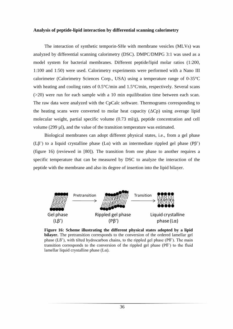
Biological membranes can adopt different physical states, i.e., from a gel phase
(Lβ’) to a liquid crystalline phase (Lα) with an intermediate rippled gel phase (Pβ’)
(figure 16) (reviewed in [80]). The transition from one phase to another requires a
specific temperature that can be measured by DSC to analyze the interaction of the
peptide with the membrane and also its degree of insertion into the lipid bilayer.
Figure 16: Scheme illustrating the different physical states adopted by a lipid
bilayer. The pretransition corresponds to the conversion of the ordered lamellar gel
phase (Lβ’), with tilted hydrocarbon chains, to the rippled gel phase (Pβ’). The main
transition corresponds to the conversion of the rippled gel phase (Pβ’) to the fluid
lamellar liquid crystalline phase (Lα).
Gel phase (Lβ’)
Pretransition Transition
Liquid crystallinephase (Lα)
Rippled gel phase (Pβ’)
37
Permeabilization assay
The ability of temporin-SHe to permeabilize the cytoplasmic membrane of
Gram-positive (S. aureus ST1065) and Gram-negative (E. coli ML-35p) bacteria was
determined. E. coli ML-35p and S. aureus ST1065 were kindly provided by Prof. Sylvie
Rebuffat (National Museum of Natural History, MNHN, Paris, France) and Dr. Tarek
Msadek (Institut Pasteur, Paris, France), respectively. These bacterial strains express
constitutively cytoplasmic β-galactosidase. E. coli ML-35p is ampicillin resistant and
lactose permease deficient, whereas S. aureus ST1065 is only chloramphenicol
resistant. If the peptide permeates the bacterial inner membrane, the chromogenic
substrate ONPG (ortho-nitrophenyl-β-D-galactopyranoside, Sigma) can enter the
cytoplasm and be hydrolyzed into ONP (ortho-nitrophenol) by cytoplasmic β-
galactosidase. So, the permeabilization of the bacterial membrane can be measured by
monitoring ONP production at 405 nm. Briefly, strains were cultured in LB medium for
2-3 h at 37°C with shaking (250 rpm), centrifuged (1000 x g, 10 min, 4°C) washed three
times with sterile PBS buffer (10 mM Na2HPO4, 100 mM NaCl, pH 7.3) and
resuspended in the same buffer to obtain A600 = 0.05. The assay was performed in
sterilized 96-well plates in a final volume of 150 µl: 15 µl of the bacterial suspension
were added to 135 µl of PBS buffer supplemented with 2.5 mM ONPG and containing
the peptide at different concentrations (at MIC, below and above the MIC). Hydrolysis
of ONPG was monitored by measuring absorbance at 405 nm every 5 min during 120
min at 37°C (Fluostar Galaxy, BMG Labtech). PBS buffer with 2.5 mM ONPG but
without peptide was used as negative control, and temporin-SHx (10 µM), a potent
synthetic temporin analogue, was used as positive control. Three independent
experiments were performed in quadruplicate. Results were expressed as the mean ±
S.E.M. of a representative experiment.

38
Time killing assay
To study the bactericidal effect and the rate of killing of temporin-SHe, the
peptide was added to a bacterial suspension of E. coli ATCC 25922 or S. aureus
ST1065 and the number of viable bacteria was determined at 37°C according to the
time. Bacteria were cultured in LB medium for 2-3 h at 37°C with shaking (250 rpm),
centrifuged (1000 x g, 10 min, 4°C), washed three times with sterile PBS buffer (10
mM Na2HPO4, 100 mM NaCl, pH 7.3) and resuspended in the same buffer to obtain
approximately 106 cfu/ml. 100 µl of temporin-SHe at a final concentration two-fold
above the MIC (50 µM for E. coli ATCC 25922 and 6.25 µM for S. aureus ST1065)
were mixed with 100 µl of the bacterial suspension (106 cfu/ml). At each time (0, 5, 15,
30, 45, 60, 90 and 120 min), 10 µl of the mixture was withdrawn and diluted 40000-fold
in LB medium and spread on LB agar plates for cell counting after overnight incubation
at 37ºC. Control was run without peptide (100 µl of PBS buffer). Three independent
experiments were performed in triplicate. Results were expressed as the mean ± S.E.M.
of a representative experiment.
39
RESULTS
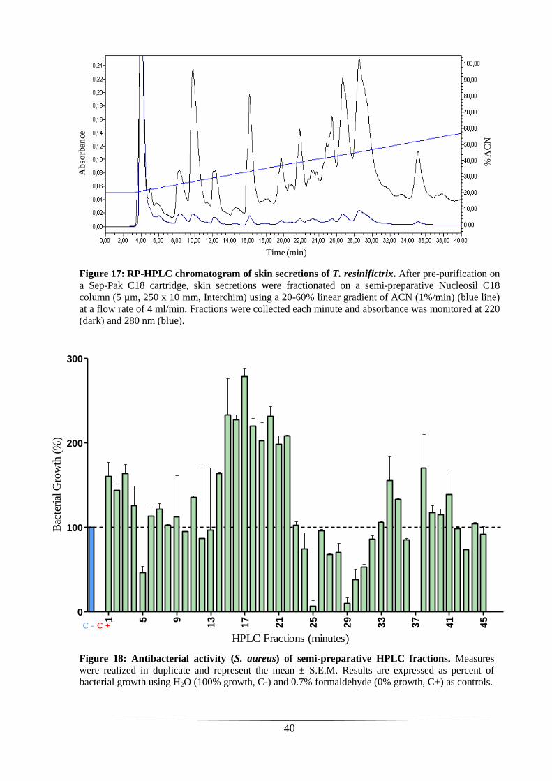
Analysis of skin secretions of T. resinifictrix
126 mg of lyophilized skin secretions were obtained from four adult specimens of
T. resinifictrix by electrical stimulation. The lyophilisate was pre-purified on a Sep-Pak
C18 cartridge and then chromatographed on a semi-preparative RP-HPLC C18 column
(figure 17). The eluted material was fractionated into tubes at 1 min per tube (i.e.
fractions of 4 ml). Tubes of four series injection (1.5 ml per injection) corresponding to
the same time were pooled and lyophilized in order to concentrate the peptidic material.
Fractions were reconstituted in sterile H2O and tested for their ability to inhibit growth
of the Gram-positive reference strain S. aureus. As shown in figure 18, several fractions
inhibited strongly the growth of S. aureus. A complete inhibition was observed for
fraction 37, and about 90% inhibition for fractions 25 and 29. Other fractions (5, 30 and
31) were also able to inhibit bacterial growth, although to a lesser extent (around 50%).
By contrast, stimulation was observed for several fractions, particularly for fractions 15-
22 displaying potent stimulation (≥ 200%).
Fractions with high inhibitory activity (i.e. 25, 29 and 37) were
rechromatographed on an analytical RP-HPLC C18 column using a slower gradient of
ACN (0.5%/min) to improve peaks separation (figure 19). Regions corresponding to
peaks in the different fractions were collected manually (figure 19) and then tested
against S. aureus. Only peak 6 of fraction 25 (figure 19 A) showed a potent bacterial
growth inhibition (figure 20). The material present in this peak was subjected to
MALDI-TOF mass spectrometry. As shown in figure 21, several ionic species with
monoisotopic masses ([M+H]+) ranging from approximately 1000 to 1900 Da (1051.46,
1165.36, 1334.32, 1448.22, 1822.81 Da) were observed and could thus correspond to
AMPs of 10-17 residues long. We attempted to determine their primary structure by
tandem mass spectrometry (MS/MS) in collaboration with Prof. Edwin De Pauw (Mass
Spectrometry Laboratory, University of Liège, Belgium). Unfortunately, due to
insufficient material in the sample, we were unable to obtain sequence information.
40
Time (min)
% A
CN
Ab
sorb
ance
Figure 18: Antibacterial activity (S. aureus) of semi-preparative HPLC fractions. Measures
were realized in duplicate and represent the mean ± S.E.M. Results are expressed as percent of
bacterial growth using H2O (100% growth, C-) and 0.7% formaldehyde (0% growth, C+) as controls.
Figure 17: RP-HPLC chromatogram of skin secretions of T. resinifictrix. After pre-purification on
a Sep-Pak C18 cartridge, skin secretions were fractionated on a semi-preparative Nucleosil C18
column (5 µm, 250 x 10 mm, Interchim) using a 20-60% linear gradient of ACN (1%/min) (blue line)
at a flow rate of 4 ml/min. Fractions were collected each minute and absorbance was monitored at 220
(dark) and 280 nm (blue).
C - C +
1 5 9
13
17
21
25
29
33
37
41
45
0
100
200
300
HPLC Fractions (minutes)
Bac
teri
al G
row
th (
%)
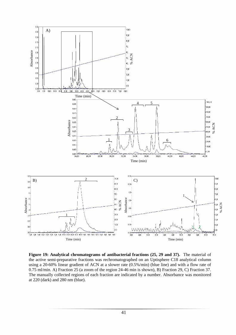
41
Fraction 25
Time (min)
% A
CN
Ab
sorb
ance
Time (min)
% A
CN
Ab
sorb
ance
1
2
3
4 5
6
A)
Time (min)
% A
CN
Ab
sorb
ance
Time (min)
% A
CN
Ab
sorb
ance
1
2
1
B) C)
Figure 19: Analytical chromatograms of antibacterial fractions (25, 29 and 37). The material of
the active semi-preparative fractions was rechromatographed on an Uptisphere C18 analytical column
using a 20-60% linear gradient of ACN at a slower rate (0.5%/min) (blue line) and with a flow rate of
0.75 ml/min. A) Fraction 25 (a zoom of the region 24-46 min is shown), B) Fraction 29, C) Fraction 37.
The manually collected regions of each fraction are indicated by a number. Absorbance was monitored
at 220 (dark) and 280 nm (blue).

42
Mass (m/z)
Inte
nsi
ty(%
)
Figure 20: Antibacterial activity of the different peaks of fractions 25,
29 and 37. Each number corresponds to peaks harvested manually (see
figure 19). Measures were realized in duplicate and represent the mean ±
S.E.M. Results are expressed as percent of bacterial growth using H2O
(100% growth, C-) and 0.7% formaldehyde (0% growth, C+) as controls.
Figure 21: MALDI-TOF mass spectrum of peak 6 (fraction 25)
1 2 3 4 5 6 1 2 1
0
100
200
300
400
F. 25 F. 29 F. 37
C - C +
Bacte
rial G
row
th (
%)
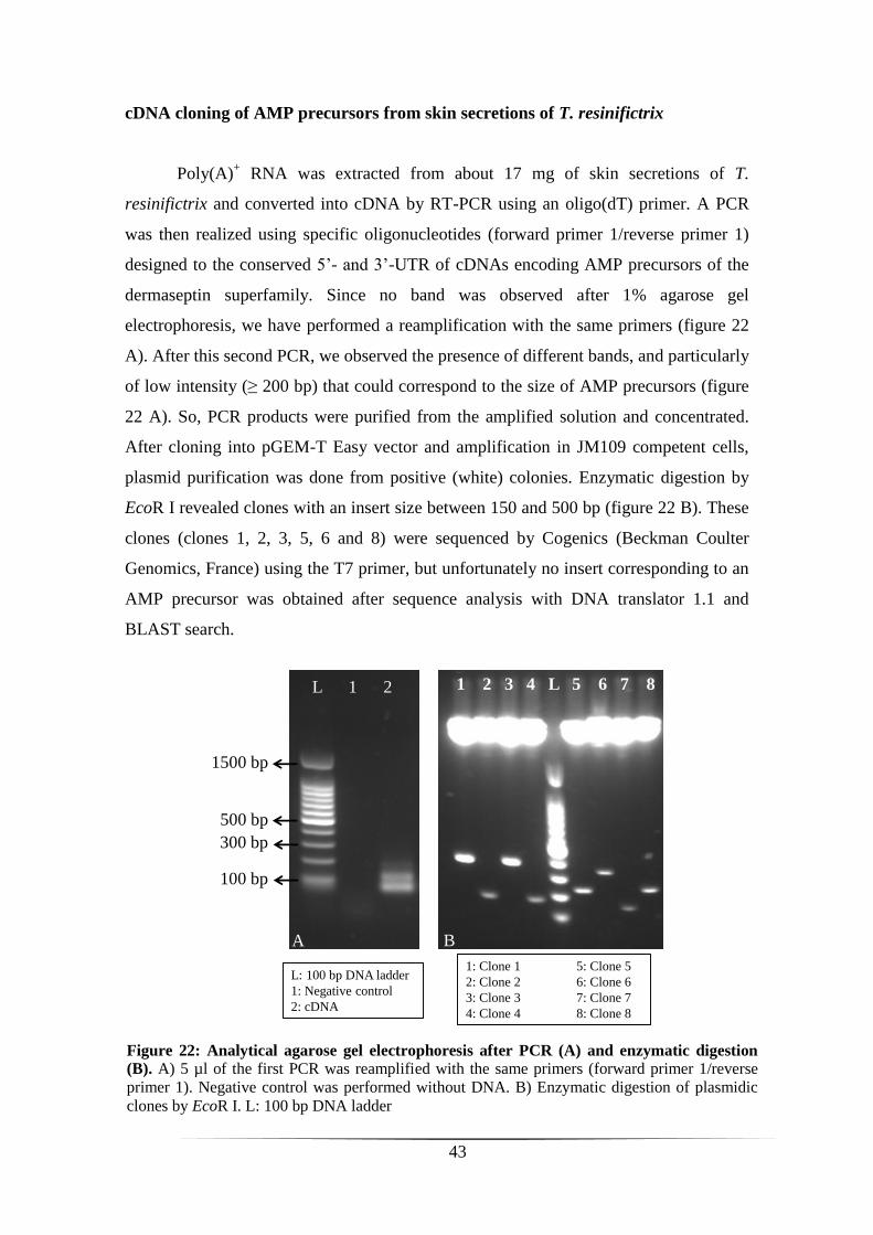
43
L 1 2
1500 bp
500 bp
300 bp
100 bp
L: 100 bp DNA ladder
1: Negative control
2: cDNA
1 2 3 4 L 5 6 7 8
1: Clone 1 5: Clone 5
2: Clone 2 6: Clone 6
3: Clone 3 7: Clone 7
4: Clone 4 8: Clone 8
A B
cDNA cloning of AMP precursors from skin secretions of T. resinifictrix
Poly(A)+ RNA was extracted from about 17 mg of skin secretions of T.
resinifictrix and converted into cDNA by RT-PCR using an oligo(dT) primer. A PCR
was then realized using specific oligonucleotides (forward primer 1/reverse primer 1)
designed to the conserved 5’- and 3’-UTR of cDNAs encoding AMP precursors of the
dermaseptin superfamily. Since no band was observed after 1% agarose gel
electrophoresis, we have performed a reamplification with the same primers (figure 22
A). After this second PCR, we observed the presence of different bands, and particularly
of low intensity (≥ 200 bp) that could correspond to the size of AMP precursors (figure
22 A). So, PCR products were purified from the amplified solution and concentrated.
After cloning into pGEM-T Easy vector and amplification in JM109 competent cells,
plasmid purification was done from positive (white) colonies. Enzymatic digestion by
EcoR I revealed clones with an insert size between 150 and 500 bp (figure 22 B). These
clones (clones 1, 2, 3, 5, 6 and 8) were sequenced by Cogenics (Beckman Coulter
Genomics, France) using the T7 primer, but unfortunately no insert corresponding to an
AMP precursor was obtained after sequence analysis with DNA translator 1.1 and
BLAST search.
Figure 22: Analytical agarose gel electrophoresis after PCR (A) and enzymatic digestion
(B). A) 5 µl of the first PCR was reamplified with the same primers (forward primer 1/reverse
primer 1). Negative control was performed without DNA. B) Enzymatic digestion of plasmidic
clones by EcoR I. L: 100 bp DNA ladder
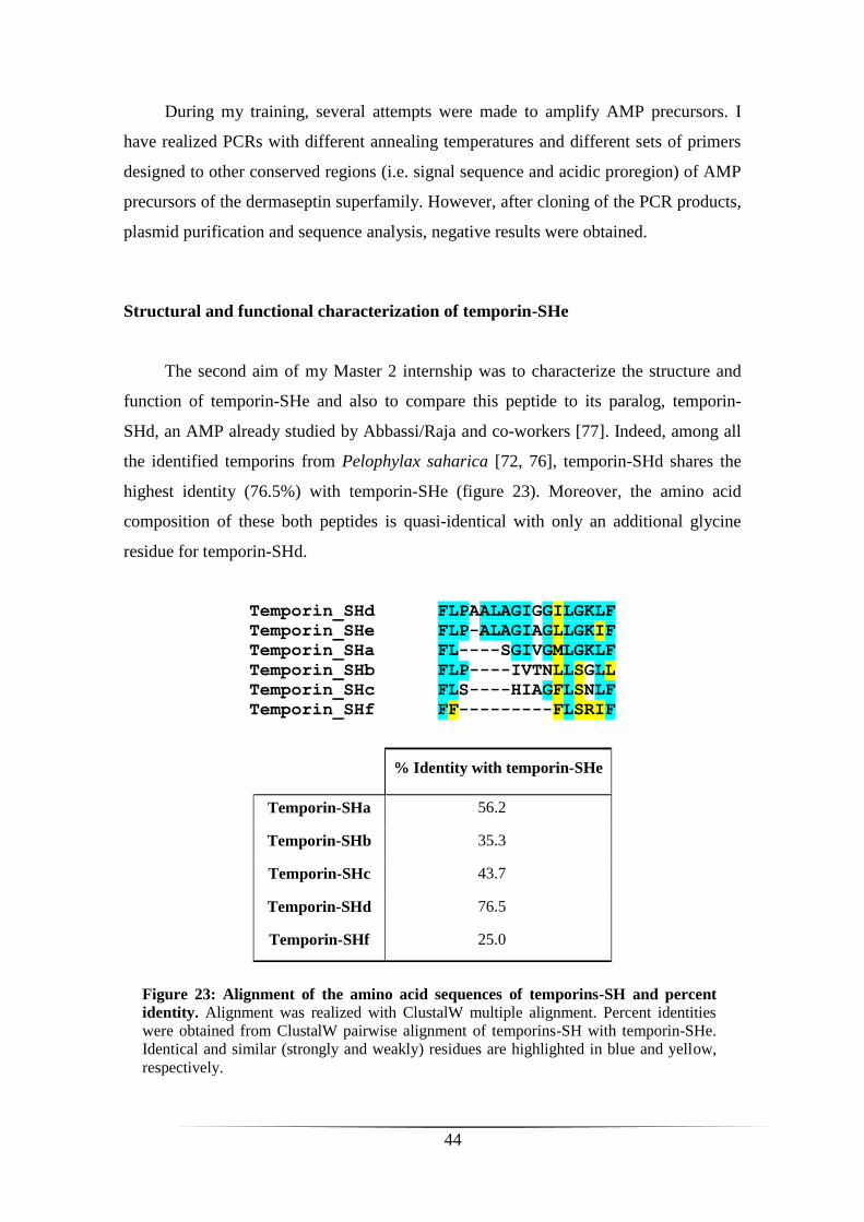
44
During my training, several attempts were made to amplify AMP precursors. I
have realized PCRs with different annealing temperatures and different sets of primers
designed to other conserved regions (i.e. signal sequence and acidic proregion) of AMP
precursors of the dermaseptin superfamily. However, after cloning of the PCR products,
plasmid purification and sequence analysis, negative results were obtained.
Structural and functional characterization of temporin-SHe
The second aim of my Master 2 internship was to characterize the structure and
function of temporin-SHe and also to compare this peptide to its paralog, temporin-
SHd, an AMP already studied by Abbassi/Raja and co-workers [77]. Indeed, among all
the identified temporins from Pelophylax saharica [72, 76], temporin-SHd shares the
highest identity (76.5%) with temporin-SHe (figure 23). Moreover, the amino acid
composition of these both peptides is quasi-identical with only an additional glycine
residue for temporin-SHd.
Temporin_SHd FLPAALAGIGGILGKLF
Temporin_SHe FLP-ALAGIAGLLGKIF
Temporin_SHa FL----SGIVGMLGKLF
Temporin_SHb FLP----IVTNLLSGLL
Temporin_SHc FLS----HIAGFLSNLF
Temporin_SHf FF---------FLSRIF
% Identity with temporin-SHe
Temporin-SHa 56.2
Temporin-SHb 35.3
Temporin-SHc 43.7
Temporin-SHd 76.5
Temporin-SHf 25.0
Figure 23: Alignment of the amino acid sequences of temporins-SH and percent
identity. Alignment was realized with ClustalW multiple alignment. Percent identities
were obtained from ClustalW pairwise alignment of temporins-SH with temporin-SHe.
Identical and similar (strongly and weakly) residues are highlighted in blue and yellow,
respectively.
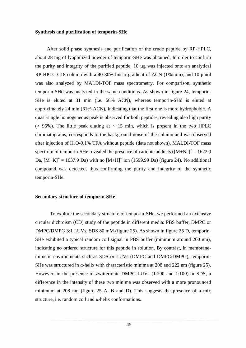
45
Synthesis and purification of temporin-SHe
After solid phase synthesis and purification of the crude peptide by RP-HPLC,
about 28 mg of lyophilized powder of temporin-SHe was obtained. In order to confirm
the purity and integrity of the purified peptide, 10 µg was injected onto an analytical
RP-HPLC C18 column with a 40-80% linear gradient of ACN (1%/min), and 10 pmol
was also analyzed by MALDI-TOF mass spectrometry. For comparison, synthetic
temporin-SHd was analyzed in the same conditions. As shown in figure 24, temporin-
SHe is eluted at 31 min (i.e. 68% ACN), whereas temporin-SHd is eluted at
approximately 24 min (61% ACN), indicating that the first one is more hydrophobic. A
quasi-single homogeneous peak is observed for both peptides, revealing also high purity
(> 95%). The little peak eluting at ~ 15 min, which is present in the two HPLC
chromatograms, corresponds to the background noise of the column and was observed
after injection of H2O-0.1% TFA without peptide (data not shown). MALDI-TOF mass
spectrum of temporin-SHe revealed the presence of cationic adducts ([M+Na]+ = 1622.0
Da, [M+K]+ = 1637.9 Da) with no [M+H]
+ ion (1599.99 Da) (figure 24). No additional
compound was detected, thus confirming the purity and integrity of the synthetic
temporin-SHe.
Secondary structure of temporin-SHe
To explore the secondary structure of temporin-SHe, we performed an extensive
circular dichroism (CD) study of the peptide in different media: PBS buffer, DMPC or
DMPC/DMPG 3:1 LUVs, SDS 80 mM (figure 25). As shown in figure 25 D, temporin-
SHe exhibited a typical random coil signal in PBS buffer (minimum around 200 nm),
indicating no ordered structure for this peptide in solution. By contrast, in membrane-
mimetic environments such as SDS or LUVs (DMPC and DMPC/DMPG), temporin-
SHe was structured in α-helix with characteristic minima at 208 and 222 nm (figure 25).
However, in the presence of zwitterionic DMPC LUVs (1:200 and 1:100) or SDS, a
difference in the intensity of these two minima was observed with a more pronounced
minimum at 208 nm (figure 25 A, B and D). This suggests the presence of a mix
structure, i.e. random coil and α-helix conformations.
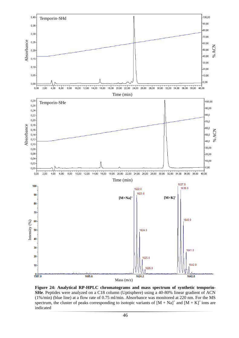
46
Time (min)
% A
CN
Ab
sorb
an
ce
Temporin-SHe
Time (min)
% A
CN
Ab
sorb
an
ce
Temporin-SHd
Figure 24: Analytical RP-HPLC chromatograms and mass spectrum of synthetic temporin-
SHe. Peptides were analyzed on a C18 column (Uptisphere) using a 40-80% linear gradient of ACN
(1%/min) (blue line) at a flow rate of 0.75 ml/min. Absorbance was monitored at 220 nm. For the MS
spectrum, the cluster of peaks corresponding to isotopic variants of [M + Na]+ and [M + K]
+ ions are
indicated
Mass (m/z)
Inte
nsi
ty(%
)
[M+K]+[M+Na]+
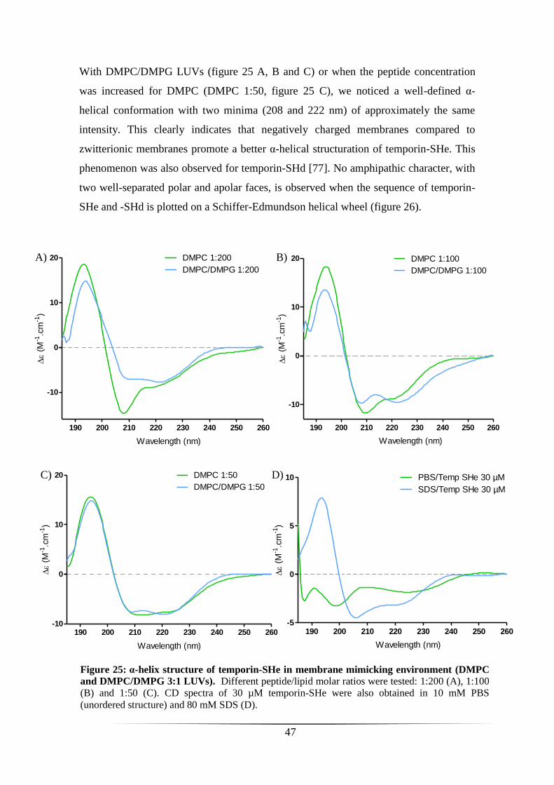
47
With DMPC/DMPG LUVs (figure 25 A, B and C) or when the peptide concentration
was increased for DMPC (DMPC 1:50, figure 25 C), we noticed a well-defined α-
helical conformation with two minima (208 and 222 nm) of approximately the same
intensity. This clearly indicates that negatively charged membranes compared to
zwitterionic membranes promote a better α-helical structuration of temporin-SHe. This
phenomenon was also observed for temporin-SHd [77]. No amphipathic character, with
two well-separated polar and apolar faces, is observed when the sequence of temporin-
SHe and -SHd is plotted on a Schiffer-Edmundson helical wheel (figure 26).
Figure 25: α-helix structure of temporin-SHe in membrane mimicking environment (DMPC
and DMPC/DMPG 3:1 LUVs). Different peptide/lipid molar ratios were tested: 1:200 (A), 1:100
(B) and 1:50 (C). CD spectra of 30 µM temporin-SHe were also obtained in 10 mM PBS
(unordered structure) and 80 mM SDS (D).
190 200 210 220 230 240 250 260
-10
0
10
20 DMPC 1:200
DMPC/DMPG 1:200
Wavelength (nm)
(
M-1
.cm
-1)
A)
190 200 210 220 230 240 250 260
-10
0
10
20 DMPC 1:100
DMPC/DMPG 1:100
Wavelength (nm)
(
M-1
.cm
-1)
B)
190 200 210 220 230 240 250 260-10
0
10
20 DMPC 1:50
DMPC/DMPG 1:50
Wavelength (nm)
(
M-1
.cm
-1)
C)
190 200 210 220 230 240 250 260-5
0
5
10 PBS/Temp SHe 30 µM
SDS/Temp SHe 30 µM
Wavelength (nm)
(
M-1
.cm
-1)
D)

48
Interaction of temporin-SHe with anionic model membranes
Differential scanning calorimetry (DSC) technique was used to study the
thermotropic behavior of DMPC/DMPG multilamellar vesicles (MLVs) upon addition
of temporin-SHe. Negatively charged DMPC/DMPG (3:1) MLVs were chosen as a
model for bacterial membranes. These MLVs exhibit two endothermic events on
heating, a weakly energetic pretransition near 13°C (conversion of the ordered gel
phase, Lβ’, to the rippled gel phase, Pβ’) and a strongly energetic main transition near
24°C (conversion of the rippled gel phase to the liquid-crystalline phase, Lα) (Figure
27). The pretransition is due to interactions between the phospholipid headgroups, and
increasing the distance between them causes the pretransition peak to disappear
(reviewed in [76]). We observed that the pretransition peak was reduced (peptide-lipid
ratio 1:200) or abolished (ratios 1:100 and 1:50) in the presence of temporin-SHe
(figure 27), indicating electrostatic interactions between the cationic peptide and the
anionic lipid headgroups. The main phase transition is mainly due to trans-gauche
interconversion of the acyl chains, which decreases the acyl chain packing and increases
fluidity of the membrane (reviewed in [76, 81]). As shown in figure 27, increasing
concentrations of temporin-SHe induced a two-component main phase transition
(peptide-lipid ratio 1:50) consisting of a broad higher temperature and less cooperative
component superimposed over a sharper lower temperature component. This indicates
Figure 26: Schiffer-Edmundson helical wheel diagram of temporin-SHe and -SHd. Residues are represented proportional to amino acid volume. Apolar residues are represented in
green and polar residues in blue. No amphipathic character, with two well-separated polar and
apolar faces, is observed.
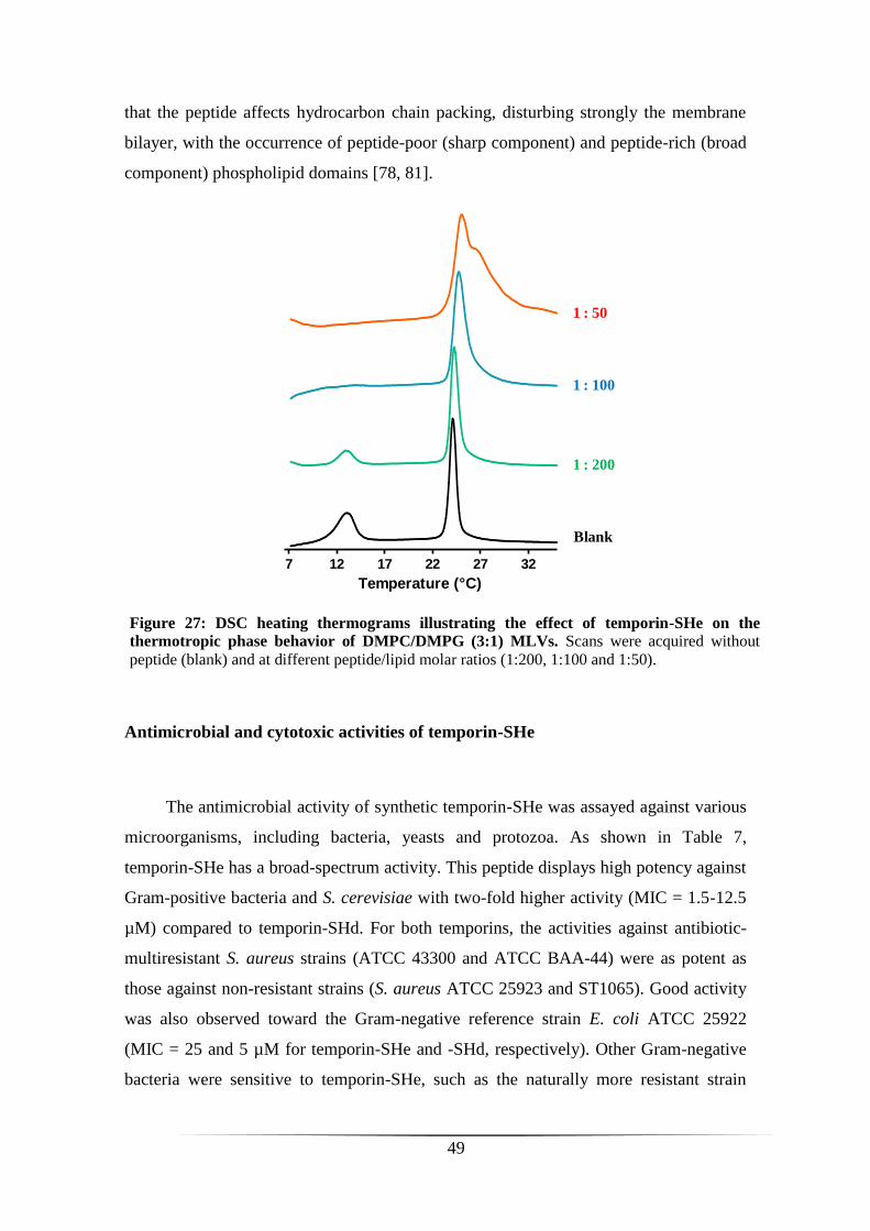
49
7 12 17 22 27 32
Temperature (°C)
Blank
1 : 200
1 : 100
1 : 50
that the peptide affects hydrocarbon chain packing, disturbing strongly the membrane
bilayer, with the occurrence of peptide-poor (sharp component) and peptide-rich (broad
component) phospholipid domains [78, 81].
Antimicrobial and cytotoxic activities of temporin-SHe
The antimicrobial activity of synthetic temporin-SHe was assayed against various
microorganisms, including bacteria, yeasts and protozoa. As shown in Table 7,
temporin-SHe has a broad-spectrum activity. This peptide displays high potency against
Gram-positive bacteria and S. cerevisiae with two-fold higher activity (MIC = 1.5-12.5
µM) compared to temporin-SHd. For both temporins, the activities against antibiotic-
multiresistant S. aureus strains (ATCC 43300 and ATCC BAA-44) were as potent as
those against non-resistant strains (S. aureus ATCC 25923 and ST1065). Good activity
was also observed toward the Gram-negative reference strain E. coli ATCC 25922
(MIC = 25 and 5 µM for temporin-SHe and -SHd, respectively). Other Gram-negative
bacteria were sensitive to temporin-SHe, such as the naturally more resistant strain
Figure 27: DSC heating thermograms illustrating the effect of temporin-SHe on the
thermotropic phase behavior of DMPC/DMPG (3:1) MLVs. Scans were acquired without
peptide (blank) and at different peptide/lipid molar ratios (1:200, 1:100 and 1:50).
50
Pseudomonas aeruginosa (MIC = 60 µM) or the fungus Candida parapsilosis (MIC =
50 µM), whereas they were resistant to temporin-SHd (MIC ≥ 200 µM).
Antiparasitic activity of temporin-SHe was also analyzed by Z. Raja, a PhD
student of the team, in collaboration with Dr. B. Oury and Dr. D. Sereno (Research
Institute for Development, Montpellier, France). As indicated in table 7, temporin-SHe
kills efficiently Leishmania promastigotes (insect stage) with an activity (IC50 ~ 10 µM)
in the same range as temporin-SHd. A preliminary study, also realized in Montpellier,
was done to evaluate the cytotoxicity of temporins on mammalian cells (THP-1
monocytes). It was observed that temporin-SHe was three-fold more cytotoxic than
temporin-SHd (Table 7).
Permeabilization of the bacterial cytoplasmic membrane
The ability of temporin-SHe to permeate the cytoplasmic membrane of the
Gram-negative E. coli ML-35p and the Gram-positive S. aureus ST1065 was studied by
monitoring the hydrolysis of the chromogenic extracellular substrate ONPG into ONP
by bacterial cytoplasmic β-galactosidase. As shown in figure 28, temporin-SHe
permeabilized the cytoplasmic membranes of both bacteria in a time- and concentration-
dependent manner. However, S. aureus bacteria were permeabilized faster and to a
higher extent than E. coli. Indeed, at concentrations above the MIC (> 3.1 µM), the
efficiency of temporin-SHe was similar to temporin-SHx (10 µM), a potent temporin-
SHa analog used as positive control (figure 28 B). Membrane permeability was
observed even at concentration below the MIC. Our results are similar to those obtained
for temporin-SHd by Abbassi/Raja and co-workers [77].
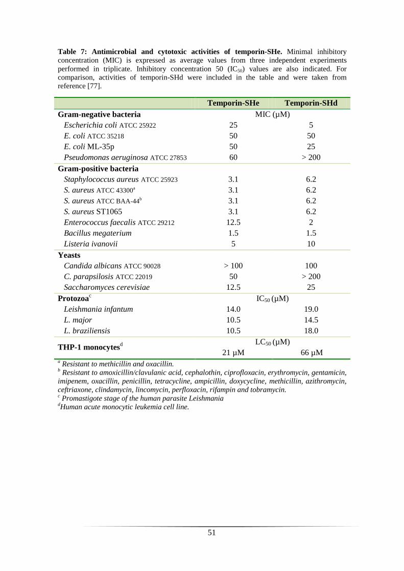
51
Table 7: Antimicrobial and cytotoxic activities of temporin-SHe. Minimal inhibitory
concentration (MIC) is expressed as average values from three independent experiments
performed in triplicate. Inhibitory concentration 50 (IC50) values are also indicated. For
comparison, activities of temporin-SHd were included in the table and were taken from
reference [77].
Temporin-SHe Temporin-SHd
Gram-negative bacteria MIC (µM)
Escherichia coli ATCC 25922 25 5
E. coli ATCC 35218 50 50
E. coli ML-35p 50 25
Pseudomonas aeruginosa ATCC 27853 60 > 200
Gram-positive bacteria
Staphylococcus aureus ATCC 25923 3.1 6.2
S. aureus ATCC 43300a 3.1 6.2
S. aureus ATCC BAA-44b
3.1 6.2
S. aureus ST1065 3.1 6.2
Enterococcus faecalis ATCC 29212 12.5 2
Bacillus megaterium 1.5 1.5
Listeria ivanovii 5 10
Yeasts
Candida albicans ATCC 90028 > 100 100
C. parapsilosis ATCC 22019 50 > 200
Saccharomyces cerevisiae 12.5 25
Protozoac
IC50 (µM)
Leishmania infantum 14.0 19.0
L. major 10.5 14.5
L. braziliensis 10.5 18.0
THP-1 monocytesd
LC50 (µM)
21 µM 66 µM a Resistant to methicillin and oxacillin.
b Resistant to amoxicillin/clavulanic acid, cephalothin, ciprofloxacin, erythromycin, gentamicin,
imipenem, oxacillin, penicillin, tetracycline, ampicillin, doxycycline, methicillin, azithromycin,
ceftriaxone, clindamycin, lincomycin, perfloxacin, rifampin and tobramycin. c Promastigote stage of the human parasite Leishmania
dHuman acute monocytic leukemia cell line.
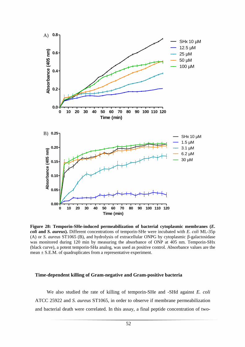
52
Time-dependent killing of Gram-negative and Gram-positive bacteria
We also studied the rate of killing of temporin-SHe and -SHd against E. coli
ATCC 25922 and S. aureus ST1065, in order to observe if membrane permeabilization
and bacterial death were correlated. In this assay, a final peptide concentration of two-
Figure 28: Temporin-SHe-induced permeabilization of bacterial cytoplasmic membranes (E.
coli and S. aureus). Different concentrations of temporin-SHe were incubated with E. coli ML-35p
(A) or S. aureus ST1065 (B), and hydrolysis of extracellular ONPG by cytoplasmic β-galactosidase
was monitored during 120 min by measuring the absorbance of ONP at 405 nm. Temporin-SHx
(black curve), a potent temporin-SHa analog, was used as positive control. Absorbance values are the
mean ± S.E.M. of quadruplicates from a representative experiment.
0 10 20 30 40 50 60 70 80 90 100 110 1200.0
0.2
0.4
0.6
0.8
SHx 10 µM
12.5 µM
25 µM
50 µM
100 µM
Time (min)
Ab
so
rban
ce (
405 n
m)
A)
0 10 20 30 40 50 60 70 80 90 100 110 1200.00
0.05
0.10
0.15
0.20
0.25SHx 10 µM
1.5 µM
3.1 µM
6.2 µM
30 µM
Time (min)
Ab
so
rban
ce (
405 n
m)
B)
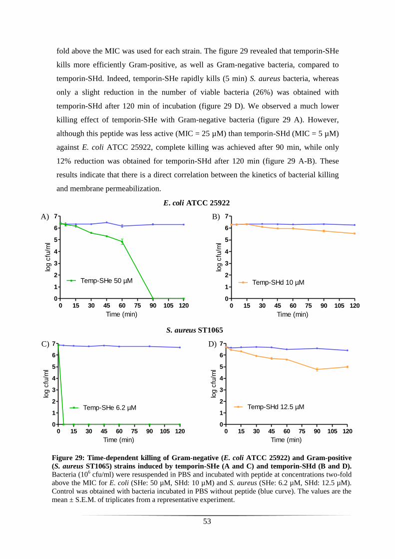
53
0 15 30 45 60 75 90 105 1200
1
2
3
4
5
6
7
Temp-SHe 50 µM
Time (min)
log c
fu/m
l
0 15 30 45 60 75 90 105 1200
1
2
3
4
5
6
7
Temp-SHd 10 µM
Time (min)
log c
fu/m
l
E. coli ATCC 25922
A) B)
fold above the MIC was used for each strain. The figure 29 revealed that temporin-SHe
kills more efficiently Gram-positive, as well as Gram-negative bacteria, compared to
temporin-SHd. Indeed, temporin-SHe rapidly kills (5 min) S. aureus bacteria, whereas
only a slight reduction in the number of viable bacteria (26%) was obtained with
temporin-SHd after 120 min of incubation (figure 29 D). We observed a much lower
killing effect of temporin-SHe with Gram-negative bacteria (figure 29 A). However,
although this peptide was less active (MIC = 25 µM) than temporin-SHd (MIC = 5 µM)
against E. coli ATCC 25922, complete killing was achieved after 90 min, while only
12% reduction was obtained for temporin-SHd after 120 min (figure 29 A-B). These
results indicate that there is a direct correlation between the kinetics of bacterial killing
and membrane permeabilization.
Figure 29: Time-dependent killing of Gram-negative (E. coli ATCC 25922) and Gram-positive
(S. aureus ST1065) strains induced by temporin-SHe (A and C) and temporin-SHd (B and D).
Bacteria (106 cfu/ml) were resuspended in PBS and incubated with peptide at concentrations two-fold
above the MIC for E. coli (SHe: 50 µM, SHd: 10 µM) and S. aureus (SHe: 6.2 µM, SHd: 12.5 µM).
Control was obtained with bacteria incubated in PBS without peptide (blue curve). The values are the
mean ± S.E.M. of triplicates from a representative experiment.
0 15 30 45 60 75 90 105 1200
1
2
3
4
5
6
7
Temp-SHe 6.2 µM
Time (min)
log c
fu/m
l
0 15 30 45 60 75 90 105 1200
1
2
3
4
5
6
7
Temp-SHd 12.5 µM
Time (min)
log c
fu/m
l
S. aureus ST1065
C) D)
54
DISCUSSION
During a first part of my training, I was interested in the identification of novel
AMPs from skin secretions of the hylid frog Trachycephalus resinifictrix, which
belongs to the subfamily Hylinae containing 646 species. Nowadays, only eight AMPs
were identified from frogs of this subfamily, including 4 pseudins (23-24 amino acid
residues), 1 hylaseptin/2 hylains (14 residues) and 1 hylin (18 residues). Moreover,
surprisingly, no cDNA encoding AMP precursors were characterized. So, we attempt
also to clone the precursors of AMPs present in the skin secretions of T. resinifictrix.
This frog has the particularity to secrete a milky and viscous resin from its skin,
thus rendering difficult the extraction of AMPs, but we have successfully overcome this
inconvenience. Indeed, by fractionation of skin secretions and monitoring of
antibacterial activity against Staphylococcus aureus, we were able to identified three
semi-preparative HPLC fractions (25, 29 and 37) with potent bacterial growth
inhibition. After analytical HPLC, only one collected peak of the fraction 25 (peak 6)
revealed potent growth-inhibitory activity. Despite the fact that interesting molecular
species ([M+H]+ = 1000-1900 Da) were revealed in this peak by MALDI-TOF mass
spectrometry analysis, we didn’t get any primary sequence after tandem mass
spectrometry (MS/MS) due to small amounts of peptides in the sample. Therefore, it
will be necessary to concentrate the peptide material in the fraction 25, as well as
fractions 29 and 37, using the same protocol of extraction and purification but with a
much higher amount of skin secretions. During my internship, I didn’t have enough
time for that. Moreover, it will be also interesting to extract AMPs from the entire skin.
We attempted to amplify AMP precursor cDNAs by PCR using different sets of
specific primers designed to conserved regions of AMP precursors of the dermaseptin
superfamily. Unfortunately, after cloning and sequence analysis of numerous clones of
plasmid DNA, no AMP precursor was identified. The team has previously cloned AMP
precursors from the skin exudate of Phyllomedusa sauvagii (hylid frog) but it is possible
for the skin exudate of T. resinifictrix that there is too low amount or no mRNA. So, it
will be necessary to sacrify a frog specimen to extract mRNA from the skin. We also
cannot exclude the possibility that AMP precursors from frogs of the subfamily are not
so well conserved. Therefore, it will be essential to design degenerated primers from
alignment of several AMP precursors of hylid frogs. If no results are obtained, we will
study another frog species of the Hylinae subfamily.

55
During the second part of my training, I studied AMPs of ranid frogs, and
particularly small hydrophobic and cationic peptides of a family called Temporin [1].
Although these AMPs were mainly characterized from Eurasian and North American
frog species, six novel temporins were also identified by the team from a North African
frog (Pelophylax saharica), for the first time [72, 76]. Among these six temporins-SH
(SHa to SHf, SH for saharica), only temporin-SHe remained unstudied. Therefore, in
this project, my objective was to characterize the structure and function of temporin-
SHe. I have also compared the properties of temporin-SHe and temporin-SHd because
these peptides share high identity (76.5%) and temporin-SHd is a well-characterized
paralog [77].
Circular dichroism spectroscopy studies revealed that temporin-SHe adopts a
well-defined α-helical structure, preferentially when bound to anionic model
membranes. This was also observed with temporin-SHd [77]. In contrast to many linear
α-helical AMPs, the Schiffer-Edmundson helical-wheel projections of these two
peptides showed a non-amphipathic structure with no well-separated polar and apolar
faces. When the peptide-lipid molar ratio was increased up to 1:50, we observed from
calorimetric data (DSC) that temporin-SHe perturbs strongly the membrane of anionic
model vesicles with disappearance of the pretransition peak and alteration of the main
transition peak (existence of two components). The loss of the pretransition is a
consequence of the peptide interaction with phospholipid headgroups which increase
the spacing between them, thus eliminating the driving force for the formation of a
rippled gel phase. The shift of the temperature (Tm) of the main phase transition toward
higher values indicates a rigidification of the membrane, whereas the apparition of a
second peak at the right wing of the main transition peak (ratio 1:50) indicates regions
of two coexisting phases, one peptide-rich (higher temperature) and the other peptide-
poor (lower temperature) [78, 81]. This gradual phase segregation between these two
domains may eventually lead to membrane disruption [78].
Like temporin-SHd, we have shown that temporin-SHe has potent broad-
spectrum activity, acting against both Gram-positive (MIC = 1.5-12.5 µM) and Gram-
negative bacteria, yeasts (S. cerevisiae, MIC = 12.5 µM), as well as against the
extracellular promastigote form of the human parasite Leishmania (IC50 ~ 10 µM). The
bacterial activity against Gram-negative strains was lower (MIC = 25-60 µM). Although
the spectrum of activity of both temporins were quite similar, Pseudomonas aeruginosa

56
(MIC = 60 µM) and Candida parapsilosis (MIC = 50 µM) were more susceptible to
temporin-SHe, while these strains were resistant to temporin-SHd. P. aeruginosa is an
opportunistic human bacteria responsible of biofilm formation in medical tools [82].
Interestingly, temporin-SHe, as well as temporin-SHd, are active against antibiotic-
multiresistant S. aureus (ATCC 43300 and ATCC BAA-44) with the same order of
magnitude as non-resistant S. aureus strains (ST1065 and ATCC 25923). This
emphasizes the original mode of action (membranolytic mechanism) of these AMPs
compared to conventional antibiotics and also indicates that temporins are promising
candidates in the fight against antibiotic-resistant pathogens. Another interesting result
is that temporin-SHe is an additional temporin with antiparasitic activity against
Leishmania. Only four temporins (A, B, SHa and SHd) were previously shown to have
such activity [71, 72, 77] and very few AMPs of other families are active [38, 83]. Like
temporin-SHd, temporin-SHe is active against different Leishmania species responsible
of visceral (L. infantum), cutaneous (L. major) and muco-cutaneous (L. braziliensis)
leishmaniasis.
We have shown that temporin-SHe is three-fold more cytotoxic than temporin-
SHd toward human monocytes. This can be explained by the higher intrinsic
hydrophobicity of temporin-SHe since it is known that increasing the hydrophobic
character leads to more cytotoxic AMPs [84] and that hydrophobic interaction play a
major role in the activity of peptides with PC-rich membranes [85]. This preliminary
study of cytotoxicity needs to be extended on other cells, such as erythrocytes or
macrophages (the host cells of Leishmania), for example.
In order to investigate the mode of action of temporin-SHe, we have analyzed
the effect of this peptide on the cytoplasmic membrane of bacteria (permeabilization
assay) and its ability to kill bacteria. With two-fold concentration above the MIC, we
observed a complete killing which was very rapid (within 5 min) for the Gram-positive
S. aureus and much lower (90 min) for the Gram-negative E. coli. Temporin-SHe was
more efficient than temporin-SHd because for the latter, no complete killing occurred
(S. aureus and E. coli) after 120 min of incubation with a concentration two-fold above
the MIC. However, Abbassi/Raja and co-workers have shown for temporin-SHd that a
three-fold concentration above the MIC (20 µM) was effective to induce rapid (15 min)
and complete killing of S. aureus, with also rapid permeabilization of the cytoplasmic

57
membrane [77]. Thus, a peptide concentration threshold is needed for rapid killing of
bacteria by AMP.
In addition, temporin-SHe was able to permeabilize the bacterial cytoplasmic
membrane (S. aureus and E. coli) in a time- and concentration-dependent manner. The
kinetics of membrane permeabilization was correlated to the bacterial killing. We
observed that even at concentrations below the MIC membrane permeability occurred
but does not cause bacterial lysis. This could be explained by the two-state model [86].
In this model, peptide has two physical states of binding to the lipid bilayer, one at low
peptide/lipid ratios (P/L) and another at a high P/L. When a threshold ratio is reached,
the peptide tends to form a stable multi-pore state, whereas the few pores formed below
the threshold concentration are usually unstable [86]. However, according to several
studies (reviewed in [87]), including those of the laboratory [76, 78], only the first
physical state (lower concentrations) fits into the mechanism of action commonly
accepted for temporins. These results indicate that temporins rather act by a carpet-like
mechanism. In fact, at low temporin/lipid ratio, peptide induces the formation of
transient, toroidal lipid-peptide pores, with possibility of peptide translocation into the
cell. At a higher temporin/lipid ratio, peptide induces membrane disruption by a
detergent-like effect, as it has been demonstrated for temporin-SHf [76] (figure 30).
Figure 30: Hypothetical mechanism of action of temporins. The first step involves reversible
electrostatic interactions of the cationic peptide with the negatively charged membrane (carpet-
like mechanism). In the second step, the hydrophobicity of the peptide allows its insertion into
the hydrophobic core of the bilayer and then i) at low concentrations, peptide induces the
formation of transient toroidal lipid-peptide pores with possibility of peptide translocation into
the cell; ii) at higher concentrations, peptide provokes membrane micellization by a detergent-
like effect. The black area of the AMP corresponds to the hydrophobic region and the white area
to the cationic region of the peptide. Gray circles of the membrane correspond to polar
headgroups and squiggly lines to the acyl chains of the phospholipids (adapted from [87]).
58
CONCLUSION
Skin secretions of the frog T. resinifictrix contain probably AMPs because after
fractionation we were able to detect HPLC peaks at 220 nm displaying antibacterial
activity against S. aureus. We need now to characterize these AMPs.
The structural characterization of temporin-SHe revealed that this peptide
adopts a well-defined α-helical conformation when bound to negatively charged model
membranes. Temporin-SHe interacts with the phospholipid headgroups of these model
membranes and induces membrane perturbations probably by insertion into the
hydrophobic core of the phospholipid bilayer. The membranolytic activity of temporin-
SHe was revealed by killing of bacteria, as well as Gram-positive and Gram-negative,
and concomitant permeabilization of the bacterial cytoplasmic membrane. This activity,
which is concentration-dependent, involves probably the formation of either transient
toroidal lipid-peptide pores (at low peptide concentration) or membrane micellization
(detergent-like effect at a higher peptide concentration). Temporin-SHe, as well as
temporin-SHd and -SHa, are particularly interesting AMPs with potent and broad-
spectrum activity against a wide range of microorganisms, including both Gram-
positive and Gram-negative bacteria, yeasts, fungi and human protozoa. Moreover,
these peptides display high potency toward antibiotic-multiresistant strains.
Therefore, temporins-SH represent good templates for the development of
therapeutic antimicrobial agents with new mode of action. Today, the antiparasitic
mechanism of action of AMPs is unknown and very few AMPs are active against
protozoa. So, our results indicate that temporin-SHe represents a good additional tool to
analyze this mechanism.
59
REFERENCES
1. Mangoni, M. L. (2006) Temporins, anti-infective peptides with expanding
properties. Cell Mol Life Sci 63, 1060-1069.
2. Hancock, R. E. W., and Diamond, G. (2000) The role of cationic
antimicrobial peptides in innate host defences. Trends Microbiol 8, 402-
410.
3. Zasloff, M. (2002) Antimicrobial peptides of multicellular organisms.
Nature 415, 389-395.
4. Wang, G., Li, X. and Wang, Z. (2009) APD2 : the updated antimicrobial
peptide database and its application in peptide design. Nucleic Acids Res
37, D933-D937.
5. Clarke, B. T. (1997) The natural history of amphibian skin secretions,
their normal functioning and potential medical applications. Biol Rev 72,
365-379.
6. Lazarus, L. H., and Attila, M. (1993) The toad, ugly and venomous, wears
yet a precious jewel in his skin. Prog Neurobiol 41, 473-507.
7. Simmaco, M., Mignogna, G., and Barra, D. (1998) Antimicrobial peptides
from amphibian skin: What do they tell us? Biopolymers 47, 435-450.
8. Amiche, M., Delfour, A., and Nicolas, P. (1999) La peau des grenouilles,
un mini-laboratoire pour les biotechnologies. In Revue du Palais de la
découverte, pp 11-21.
9. Frost, D. R. (2011) Amphibian Species of the World: an Online
Reference. version 5.5. In American Museum of Natural History.
10. Nascimento, A. C., Fontes, W., Sebben, A., and Castro, M. S. (2003)
Antimicrobial peptides from anurans skin secretions. Protein Pept Lett 10,
227-238.
11. Barra, D., and Simmaco, M. (1995) Amphibian Skin - a Promising
Resource for Antimicrobial Peptides. Trends Biotechnol 13, 205-209.
12. Lacombe, C. (2000) Peptide secretion in the cutaneous glands of South
American tree frog: an ultrastructural study. Eur J Cell Biol 79, 631-641.

60
13. Azevedo Calderon, L. d., Silva, A. d. A. E., Ciancaglini, P., and Stábeli, R.
G. (2010) Antimicrobial peptides from Phyllomedusa frogs: from
biomolecular diversity to potential nanotechnologic medical applications.
Amino Acids 40, 29-49.
14. Toledo, R. C., and Jared, C. (1995) Cutaneous granular glands and
amphibian venoms. Comp Biochem Physiol 111A, 1-29.
15. El Amri, C., and Nicolas, P. (2008) Plasticins: membrane-damaging
peptides with 'chameleon-like' properties. Cell Mol Life Sci 65, 895-909.
16. Delfino, G., Brizzi, R., Nosi, D., and Terreni, A. (2002) Serous Cutaneous
Glands in New World Hylid Frogs: An Ultrastructural Study on Skin
Poisons Confirms Phylogenetic Relationships Between Osteopilus
septentrionalis and Phrynohyas venulosa. J Morphol 253, 176-186.
17. Nicolas, P., and El Amri, C. (2009) The dermaseptin superfamily: a gene-
based combinatorial library of antimicrobial peptides. Biochim Biophys
Acta 1788, 1537-1550.
18. Nicolas, P., and Amiche, M. (1999) Les précurseurs des dermaseptines:
une boÎte de Pandore. In Médecine/sciences, pp 1443-1447.
19. Bevins, C. L., and Zasloff, M. (1990) Peptides from frog skin. Annu Rev
Biochem 59, 395-414.
20. Andreu, D., and Rivas, L. (1998) Animal antimicrobial peptides: an
overview. Biopolymers 47, 415-433.
21. Peschel, A., and Sahl, H. G. (2006) The co-evolution of host cationic
antimicrobial peptides and microbial resistance. Nat Rev Microbiol 4, 529-
536.
22. Nicolas, P., Vanhoye, D., and Amiche, M. (2003) Molecular strategies in
biological evolution of antimicrobial peptides. Peptides 24, 1669-1680.
23. Amiche, M., Seon, A. A., Pierre, T. N., and Nicolas, P. (1999) The
dermaseptin precursors: a protein family with a common preproregion and
a variable C-terminal antimicrobial domain. FEBS Lett 456, 352-356.
24. Vanhoye, D., Bruston, F., Nicolas, P., and Amiche, M. (2003)
Antimicrobial peptides from hylid and ranin frogs originated from a 150-
million-year-old ancestral precursor with a conserved signal peptide but a
hypermutable antimicrobial domain. Eur J Biochem 270, 2068-2081.
61
25. Amiche, M., Ducancel, F., Mor, A., Boulain, J. C., Menez, B., and
Nicolas, P. (1994) Precursors of Vertebrate Peptide Antibiotics
Dermaseptin b and Adenoregulin Have Extensive Sequence Identities with
Precursors of Opioid Peptides Dermorphin, Dermenkephalin, and
Deltorphins. J Biol Chem 269, 17847-17852.
26. Yeaman, M. R., and Yount, N. Y. (2003) Mechanisms of antimicrobial
peptide action and resistance. Pharmacol Rev 55, 27-55.
27. Hancock, R. E. (1997) Peptide antibiotics. Lancet 349, 418-422.
28. Conlon, J. M. (2010) The contribution of skin antimicrobial peptides to the
system of innate immunity in anurans. Cell Tissue Res 343, 201-212.
29. Brogden, K. A. (2005) Antimicrobial peptides: pore formers or metabolic
inhibitors in bacteria? Nat Rev Microbiol 3, 238-250.
30. Pukala, T. L., Bowie, J. H., Maselli, V. M., Musgrave, I. F., and Tyler, M.
J. (2006) Host-defence peptides from the glandular secretions of
amphibians: structure and activity. Nat Prod Rep 23, 368-393.
31. Haney, E. F., Hunter, H. N., Matsuzaki, K., and Vogel, H. J. (2009)
Solution NMR studies of amphibian antimicrobial peptides: linking
structure to function? Biochim Biophys Acta 1788, 1639-1655.
32. Bulet, P., Stöcklin, R., and Menin, L. (2004) Anti-microbial peptides:
from invertebrates to vertebrates. Immunol Rev 198, 169-184.
33. Rinaldi, A. C. (2002) Antimicrobial peptides from amphibian skin: an
expanding scenario. Curr Opin Chem Biol 6, 799-804.
34. Shai, Y. (2002) Mode of action of membrane active antimicrobial
peptides. Biopolymers 66, 236-248.
35. Epand, R. M., and Vogel, H. J. (1999) Diversity of antimicrobial peptides
and their mechanisms of action. Biochim Biophys Acta 1462, 11-28.
36. Shai, Y. (1999) Mechanism of the binding, insertion and destabilization of
phospholipid bilayer membranes by α-helical antimicrobial and cell non-
selective membrane-lytic peptides. Biochim Biophys Acta 1462, 55-70.
37. Shai, Y., and Oren, Z. (2001) From "carpet" mechanism to de-novo
designed diastereomeric cell-selective antimicrobial peptides. Peptides 22,
1629-1641.

62
38. Rivas, L., Luque-Ortega, J. R., and Andreu, D. (2009) Amphibian
antimicrobial peptides and Protozoa: lessons from parasites. Biochim
Biophys Acta 1788, 1570-1581.
39. Yang, L., Harroun, T. A., Weiss, T. M., Ding, L., and Huang, H. W.
(2001) Barrel-stave Model or Toroidal Model? A case study on melittin
pores. Biophys J 81, 1475-1485.
40. Tossi, A., Sandri, L., and Giangaspero, A. (2000) Amphipathic, alpha-
helical antimicrobial peptides. Biopolymers 55, 4-30.
41. Mor, A., and Nicolas, P. (1994) Isolation and structure of novel defensive
peptides from frog skin. Eur J Biochem 219, 145-154.
42. Conlon, J. M., Woodhams, D. C., Raza, H., Coquet, L., Leprince, J.,
Jouenne, T., Vaudry, H., and Rollins-Smith, L. A. (2007) Peptides with
differential cytolytic activity from skin secretions of the lemur leaf frog
Hylomantis lemur (Hylidae: Phyllomedusinae)☆. Toxicon 50, 498-506.
43. Vanhoye, D., Bruston, F., El Amri, S., Ladram, A., Amiche, M., and
Nicolas, P. (2004) Membrane association, electrostatic sequestration, and
cytotoxicity of Gly-Leu-rich peptide orthologs with differing functions.
Biochemistry 43, 8391-8409.
44. Amiche, M., Seon, A. A., Wroblewski, H., and Nicolas, P. (2000)
Isolation of dermatoxin from frog skin, an antibacterial peptide encoded
by a novel member of the dermaseptin genes family. Eur J Biochem 267,
4583-4592.
45. Pierre, T. N., Seon, A. A., Amiche, M., and Nicolas, P. (2000) Phylloxin,
a novel peptide antibiotic of the dermaseptin family of
antimicrobial/opioid peptide precursors. Eur J Biochem 267, 370-378.
46. Thompson, A. H., Bjourson, A. J., Orr, D. F., Shaw, C., and McClean, S.
(2007) Amphibian skin secretomics: application of parallel quadrupole
time-of-flight mass spectrometry and peptide precursor cDNA cloning to
rapidly characterize the skin secretory peptidome of Phyllomedusa
hypochondrialis azurea: discovery of a novel peptide family, the hyposins.
J Proteom Res 6, 3604-3613.
47. Lequin, O., Ladram, A., Chabbert, L., Bruston, F., Convert, O., Vanhoye,
D., Chassaing, G., Nicolas, P., and Amiche, M. (2006) Dermaseptin S9, an
alpha-helical antimicrobial peptide with a hydrophobic core and cationic
termini. Biochemistry 45, 468-480.
63
48. Rozek, T., Wegener, K. L., Bowie, J. H., Olver, I. N., Carver, J. A.,
Wallace, J. C., and Tyler, M. J. (2000) The antibiotic and anticancer active
aurein peptides from the Australian Bell Frogs Litoria aurea and Litoria
raniformis the solution structure of aurein 1.2. Eur J Biochem 267, 5330-
5341.
49. Wong, H., Bowie, J. H., and Carver, J. A. (1997) The solution structure
and activity of caerin 1.1, an antimicrobial peptide from the Australian
green tree frog, Litoria spzendida. Eur J Biochem 247, 545-557.
50. Wegener, K. L., Wabnitz, P. A., Carver, J. A., Bowie, J. H., Chia, B. C.,
Wallace, J. C., and Tyler, M. J. (1999) Host defence peptides from the
skin glands of the Australian blue mountains tree-frog Litoria citropa.
Solution structure of the antibacterial peptide citropin 1.1. Eur J Biochem
265, 627-637.
51. Wegener, K. L., Brinkworth, C. S., Bowie, J. H., Wallace, J. C., and Tyler,
M. J. (2001) Bioactive dahlein peptides from the skin secretions of the
Australian aquatic frog Litoria dahlii: sequence determination by
electrospray mass spectrometry. Rapid Commun Mass Spectrom 15, 1726-
1734.
52. Rozek, T., Waugh, R. J., Steinborner, S. T., Bowie, J. H., Tyler, M. J., and
Wallace, J. C. (1998) The maculatin peptide from the skin glands of the
tree frog Litoria genimaculata. A comparison of the structures and
antibacterial activities of maculatin 1.1 and caerin 1.1. J Pept Sci 4, 111-
115.
53. Amiche, M., Ladram, A., and Nicolas, P. (2008) A consistent
nomenclature of antimicrobial peptides isolated from frogs of the
subfamily Phyllomedusinae. Peptides 29, 2074-2082.
54. Olson III, L., Soto, A. M., Knoop, F. C., and Conlon, J. M. (2001)
Pseudin-2: An antimicrobial peptide with low hemolytic activity from the
skin of the Paradoxical Frog. Biochem Biophys Res Commun 288, 1001-
1005.
55. Prates, M. V., Sforca, M. L., Regis, W. C., Leite, J. R., Silva, L. P.,
Pertinhez, T. A., Araujo, A. L., Azevedo, R. B., Spisni, A., and Bloch, C.,
Jr. (2004) The NMR-derived solution structure of a new cationic
antimicrobial peptide from the skin secretion of the anuran Hyla punctata.
J Biol Chem 279, 13018-13026.
64
56. Wu, J., Liu, H., Yang, H., Yu, H., You, D., Ma, Y., Ye, H., and Lai, R.
(2011) Proteomic analysis of skin defensive factors of tree frog Hyla
simplex. J Proteome Res 10, 4230-4240.
57. Castro, M. S., Ferreira, T. C., Cilli, E. M., Crusca, E., Jr., Mendes-
Giannini, M. J., Sebben, A., Ricart, C. A., Sousa, M. V., and Fontes, W.
(2009) Hylin a1, the first cytolytic peptide isolated from the arboreal
South American frog Hypsiboas albopunctatus ("spotted treefrog").
Peptides 30, 291-296.
58. van Zoggel, H., Hamma-Kourbali, Y., Galanth, C., Ladram, A., Nicolas,
P., Courty, J., Amiche, M., and Delbe, J. (2010) Antitumor and angiostatic
peptides from frog skin secretions. Amino Acids.
59. Abdel-Wahab, Y. H., Power, G. J., Flatt, P. R., Woodhams, D. C., Rollins-
Smith, L. A., and Conlon, J. M. (2008) A peptide of the phylloseptin
family from the skin of the frog Hylomantis lemur (Phyllomedusinae) with
potent in vitro and in vivo insulin-releasing activity. Peptides 29, 2136-
2143.
60. Apponyi, M., Pukala, T., Brinkworth, C., Maselli, V., Bowie, J., Tyler,
M., Booker, G., Wallace, J., Carver, J., and Separovic, F. (2004) Host-
defence peptides of Australian anurans: structure, mechanism of action
and evolutionary significance. Peptides 25, 1035-1054.
61. VanCompernolle, S. E., Taylor, R. J., Oswald-Richter, K., Jiang, J.,
Youree, B. E., Bowie, J. H., Tyler, M. J., Conlon, J. M., Wade, D., Aiken,
C., Dermody, T. S., KewalRamani, V. N., Rollins-Smith, L. A., and
Unutmaz, D. (2005) Antimicrobial Peptides from Amphibian Skin
Potently Inhibit Human Immunodeficiency Virus Infection and Transfer of
Virus from Dendritic Cells to T Cells. J Virol 79, 11598-11606.
62. Abdel-Wahab, Y. H. A., Power, G. J., Ng, M. T., Flatt, P. R., and Conlon,
J. M. (2008) Insulin-releasing properties of the frog skin peptide pseudin-2
and its [Lys18]-substituted analogue. Biol Chem 389, 143-148.
63. Rotem, S., and Mor, A. (2009) Antimicrobial peptide mimics for
improved therapeutic properties. Biochim Biophys Acta 1788, 1582-1592.
65
64. Zampa, M. F., Araújo, I. M. S., Costa, V., Nery Costa, C. H., Santos, J. R.,
Zucolotto, V., Eiras, C., and Leite, J. R. S. A. (2009) Leishmanicidal
Activity and Immobilization of dermaseptin 01 antimicrobial peptides in
ultrathin films for nanomedicine applications. Nanomedicine 5, 352-358.
65. Wang, H., Xu, K., Liu, L., Tan, J. P. K., Chen, Y., Li, Y., Fan, W., Wei,
Z., Sheng, J., Yang, Y.-Y., and Li, L. (2010) The efficacy of self-
assembled cationic antimicrobial peptide nanoparticles against
Cryptococcus neoformans for the treatment of meningitis. Biomaterials
31, 2874-2881.
66. Simmaco, M., Mignogna, G., Canofeni, S., Miele, R., Mangoni, M. L.,
and Barra, D. (1996) Temporins, antimicrobial peptides from the
European red frog Rana temporaria. Eur J Biochem 242, 788-792.
67. Marenah, L., Flatt, P. R., Orr, D. F., Shaw, C., and Abdel-Wahab, Y. H.
(2006) Skin secretions of Rana saharica frogs reveal antimicrobial
peptides esculentins-1 and -1B and brevinins-1E and -2EC with novel
insulin releasing activity. J Endocrinol 188, 1-9.
68. Giacometti, A., Cirioni, O., Kamysz, W., D'Amato, G., Silvestri, C., Del
Prete, M. S., Licci, A., Lukasiak, J., and Scalise, G. (2005) In vitro activity
and killing effect of temporin A on nosocomial isolates of Enterococcus
faecalis and interactions with clinically used antibiotics. J Antimicrob
Chemother 55, 272-274.
69. Wade, D., Silberring, J., Soliymani, R., Heikkinen, S., Kilpelainen, I.,
Lankinen, H., and Kuusela, P. (2000) Antibacterial activities of temporin
A analogs. FEBS Lett 479, 6-9.
70. Rinaldi, A. C., Mangoni, M. L., Rufo, A., Luzi, C., Barra, D., Zhao, H.,
Kinnunen, P. K., Bozzi, A., Di Giulio, A., and Simmaco, M. (2002)
Temporin L: antimicrobial, haemolytic and cytotoxic activities, and effects
on membrane permeabilization in lipid vesicles. Biochem J 368, 91-100.
71. Mangoni, M. L., Saugar, J. M., Dellisanti, M., Barra, D., Simmaco, M.,
and Rivas, L. (2005) Temporins, small antimicrobial peptides with
leishmanicidal activity. J Biol Chem 280, 984-990.
66
72. Abbassi, F., Oury, B., Blasco, T., Sereno, D., Bolbach, G., Nicolas, P.,
Hani, K., Amiche, M., and Ladram, A. (2008) Isolation, characterization
and molecular cloning of new temporins from the skin of the North
African ranid Pelophylax saharica. Peptides 29, 1526-1533.
73. Chen, Q., Wade, D., Kurosaka, K., Wang, Z. Y., Oppenheim, B. J., and
Yang, D. (2004) Temporin a and related frog antimicrobial peptides use
formyl peptide receptor-like 1 as a receptor to chemoattract phagocytes. J
Immunol 173, 2652-2659.
74. Zhao, H., and Kinnunen, P. K. (2003) Modulation of the activity of
secretory phospholipase A2 by antimicrobial peptides. Antimicrob Agents
Chemother 47, 965-971.
75. Conlon, J. M. (2008) Reflections on a systematic nomenclature for
antimicrobial peptides from the skins of frogs of the family Ranidae.
Peptides 29, 1815-1819.
76. Abbassi, F., Lequin, O., Piesse, C., Goasdoue, N., Foulon, T., Nicolas, P.,
and Ladram, A. (2010) Temporin-SHf, a New Type of Phe-rich and
Hydrophobic Ultrashort Antimicrobial Peptide. J Biol Chem 285, 16880-
16892.
77. Abbassi, F*., Raja, Z
*., Oury, B., Gazanion, E., Piesse, C., Sereno, D.,
Foulon, T., Nicolas, P., and Ladram, A. Antibacterial and leishmanicidal
activities of temporin-SHd, a 17-residue long membrane-damaging
peptide. Submitted to Antimicrob Agents Chemother *These authors
contributed equally to this work.
78. Abbassi, F., Galanth, C., Amiche, M., Saito, K., Piesse, C., Zargarian, L.,
Hani, K., Nicolas, P., Lequin, O., and Ladram, A. (2008) Solution
structure and model membrane interactions of temporins-SH,
antimicrobial peptides from amphibian skin. A NMR spectroscopy and
differential scanning calorimetry study. Biochemistry 47, 10513-10525.
79. Tapley, B., and Bradfield, K. S. (2007) Mission golden-eyed tree frog
Trachycephalus resinifictrix DWCT.
80. Eeman, M., and Deleu, M. (2010) From biological membranes to
biomimetic model membranes. Biotechnol. Agron. Soc. Environ. 14, 719-
736.
67
81. Lohner, K., Latal, A., Lehrer, R. I., and Ganz, T. (1997) Differential
scanning microcalorimetry indicates that human defensin, HNP-2,
interacts specifically with biomembrane mimetic systems. Biochemistry
36, 1525-1531.
82. Batoni, G., Maisetta, G., Brancatisano, F. L., Esin, S., and Campa, M.
(2011) Use of Antimicrobial Peptides Against Microbial Biofilms:
Advantages and Limits. Curr Med Chem 18, 256-279.
83. Cobb, S. L., and Denny, P. W. (2010) Antimicrobial peptides for
leishmaniasis. Curr Opin Investig Drugs 11, 868-875.
84. Conlon, J. M., Al-Ghaferi, N., Abraham, B., and Leprince, J. (2007)
Strategies for transformation of naturally-occurring amphibian
antimicrobial peptides into therapeutically valuable anti-infective agents.
Methods 42, 349-357.
85. Wieprecht, T., Dathe, M., Beyermann, M., Krause, E., Maloy, W. L.,
MacDonald, D. L., and Bienert, M. (1997) Peptide hydrophobicity
controls the activity and selectivity of magainin 2 amide in interaction
with membranes. Biochemistry 36, 6124-6132.
86. Huang, H. W. (2000) Action of antimicrobial peptides: two-state model.
Biochemistry 39, 8347-8352.
87. Amiche, M., and Galanth, C. (2011) Dermaseptins as Models for the
Elucidation of Membrane-Acting Helical Amphipathic Antimicrobial
Peptides. Curr Pharm Biotechno 12, 1184-1193.