biochemical bases of xerostomia - repositorio-aberto.up.pt · complex xerostomia or oral dryness...
TRANSCRIPT
ARTIGO DE REVISÃO BIBLIOGRÁFICA
MESTRADO INTEGRADO EM MEDICINA DENTÁRIA
BIOCHEMICAL BASES OF XEROSTOMIA
Joana Carreiro de Figueiredo
Student of 5th degree of Integrated Master Dentistry of Porto University
Supervisor: Prof. Dr. João Miguel Silva e Costa Rodrigues
Porto, 2012
I
AACCKKNNOOWWLLEEDDGGMMEENNTTSS
To Prof. Dr. João Miguel Silva e Costa Rodrigues, for consented to guide me on
this review and for all the help that he gave me during the same.
To my parents, João Luís Figueiredo e Maria Figueiredo, for giving me the
chance to graduate as Medical Dentist, supporting me all the time.
To all my fellow year, that contributed to my growth as Medical Dentist and for
all the moments of joy that provided me during these five years.
II
AABBSSTTRRAACCTT
Introduction: Saliva has been described as the “mirror of the body”, reflecting the
body’s general state of health. In cases of the hypofunction of the salivary glands, the
harmony of the oral cavity and, consequently, of the organism, becomes
compromised. In most cases, patients complain of xerostomia, itching, burning, and
difficulty when speaking. The incidence of dry mouth and its public health impact are
increasing and it can produce serious negative effects on the patient’s quality of life by
affecting dietary habits, nutritional status, speech, taste, tolerance to dental prosthesis
and varying severity injuries in the mucosa.
Aim: The aim of this review was to evaluate the salivary gland hypofunction
mechanisms, hypossalivation and altered saliva composition associated with
xerostomia in the oral biochemistry.
Materials and Methods: Clinical trials, research and review papers (1996 – 2012) were
searched in PubMed, MedLine, Biomed and also in books available in the library of
FMDUP.
Development: Saliva is an aqueous, translucent fluid, with neutral pH, secreted by
three paired extrinsic salivary glands: the parotid, submandibular, and sublingual
glands, as well as hundreds of smaller intrinsic salivary glands that are distributed
throughout the oral cavity. Saliva has a very complex composition, containing
antimicrobial agents, immunoglobulins, electrolytes, components with buffer
properties and proteins (S-IgA, mucins, lysozymes, histatins, lactoferrin and amylases).
In digestive system, saliva has an important role in esophageal physiology, digestion,
and protecting the gastric cells. The production of saliva occurs in 2 phases: The acinar
fluid secretion and ductal fluid secretion, both controlled by autonomic nervous
system. The major causes of xerostomia are objectively assessed salivary gland
hypofunction, which could be attributed to radiation of head and neck cancer therapy
and Sjögren syndrome. Some common problems associated with xerostomia, include
oral candidiasis and dental caries. The management of xerostomia must be oriented by
its etiology and directed towards the disease effects in the patient comfort and quality
of life.
III
Conclusions: Xerostomia has a significant impact on the oral health. The management
of this condition contributes positively to the restoration of patient welfare, since the
treatment is directed towards symptomatic relief and prevention of complications
originated from salivary decreased flow. Reliable diagnostic methods and effective
new treatments are expected to lead to its complete cure in the near future.
KKEEYY--WWOORRDDSS:: Biochemical + xerostomia; biochemical + mechanisms + saliva,
hypossalivation + mechanisms; hypossalivation + xerostomia
IV
RREESSUUMMOO
Introdução: A saliva tem sido descrita como o "espelho do corpo", refletindo o estado
geral de saúde. Quando existe hipofunção das glândulas salivares, a harmonia da
cavidade oral e, consequentemente, do organismo, torna-se comprometida. Na
maioria dos casos, os pacientes apresentam xerostomia, prurido, ardor e dificuldade
na fala. A incidência de xerostomia e o seu impacto na saúde pública estão a aumentar
e pode produzir graves efeitos na qualidade de vida do paciente, afetando os seus
hábitos alimentares, estado nutricional, fala, paladar, tolerância à prótese dentária e
lesões na mucosa oral.
Objetivo: O objetivo desta revisão consiste em avaliar os mecanismos da hipofunção
das glândulas salivares, hipossalivação e consequentes alterações na composição
salivar, associados à xerostomia na bioquímica oral.
Materiais e Métodos: Foi realizada uma pesquisa nas bases de dados PubMed,
MedLine e Biomed sobre artigos referentes a ensaios clínicos, artigos de pesquisa e
artigos de revisão, e ainda em livros disponíveis na biblioteca da FMDUP, entre 1996 e
2012.
Desenvolvimento: A saliva é um fluido aquoso, translúcido, com pH neutro, secretada
por três pares de glândulas salivares extrínsecas: as glândulas parótidas,
submandibulares, e sublinguais, bem como centenas de glândulas minor intrínsecas
que estão distribuídas por toda a cavidade oral. A saliva tem uma composição muito
complexa, contendo agentes antimicrobianos, imunoglobulinas, eletrólitos,
componentes com propriedades tampão e proteínas (S-IgA, mucinas, lisozimas,
Histatinas, lactoferrina e amilases). No sistema digestivo tem um papel importante na
fisiologia esofágica, na digestão, e na proteção das células gástricas. A produção de
saliva ocorre em 2 fases: a secreção acinar e secreção ductal, ambas controladas pelo
sistema nervoso autónomo. As principais causas de xerostomia devem-se
essencialmente à hipofunção das glândulas salivares, o que acontece nos casos em que
há irradiação da cabeça e do pescoço na terapia do cancro e SS. Alguns problemas
associados à xerostomia, incluem candidíase oral e cárie dentária. O tratamento desta
V
condição deve ser orientado pela etiologia e dirigida para os efeitos da doença no
conforto e qualidade de vida do paciente.
Conclusão: A xerostomia tem um impacto significativo sobre a saúde oral. O
tratamento desta condição contribui positivamente para a restauração do bem-estar
do paciente, uma vez que é orientado para o alívio sintomático e prevenção de
complicações oriundas da diminuição do fluxo salivar. Métodos confiáveis de
diagnóstico e novos tratamentos efetivos são esperados para levar à cura completa da
xerostomia, num futuro próximo.
PPAALLAAVVRRAASS--CCHHAAVVEE:: Biochemical + xerostomia; biochemical + mechanisms + saliva,
hypossalivation + mechanisms; hypossalivation + xerostomia
VI
IINNDDEEXX
1. INTRODUCTION .................................................................................................... 1
2. MATERIALS AND METHODS ................................................................................... 4
3. SALIVA COMPONENTS AND FUNCTIONS ................................................................ 5
4. COMMON CAUSES OF XEROSTOMIA .................................................................... 11
5. SALIVARY GLAND HYPOFUNCTION ...................................................................... 15
6. COMMON ORAL COMPLICATIONS ASSOCIATED WITH XEROSTOMIA .................... 17
7. MANAGEMENT OF HYPOSALIVATION .................................................................. 19
8. CONCLUSIONS .................................................................................................... 21
9. REFERENCES ....................................................................................................... 22
1
1. INTRODUCTION
Saliva is the viscous, clear, watery fluid(1, 2) secreted from the parotid,
submaxillary, sublingual and smaller salivary glands of the mouth(1, 3, 4) and it has
been described as the “mirror of the body”, reflecting the body’s general state of
health(5). In fact, it is now known that many substances found in peripheral blood are
also found in saliva(5). That is why it is growingly considered as an essential,
noninvasive diagnostic tool(3, 5) of hereditary disorders, autoimmune diseases,
malignant and infectious diseases, and endocrine disorders, as well as in the
assessment of therapeutic levels of drugs and the monitoring of illicit drug use(3).
Saliva possesses many important functions:(1) it plays a role in
remineralization of teeth, maintains constant alkaline balance in the oral cavity(6),
provides lubrication for mucous membranes, teeth and food, regulates composition of
the microflora of the oral cavity, and facilitates the functions of primary digestion,
taste perception, and water balance control in the body(1, 6, 7).
In cases of the hypofunction of the salivary glands, the harmony of the oral
cavity and, consequently, of the organism, becomes compromised(6). Favorable
conditions can be created for certain microflora to thrive and this heightens the
probability of developing intensive caries, periodontal disease, candidiasis(6, 8-10),
tooth loss(9, 10) and halitosis(8). In most cases, patients complain of oral dryness,
itching, burning, and difficulty when speaking(6, 8), which are conducive to
psychosomatic disorders. At this point, the so-called syndrome of multifactorial
complex xerostomia or oral dryness develops(6).
Xerostomia is the subjective feeling of mouth dryness, mainly caused by a low
function of salivary glands, with decrease of saliva quality or quantity(8), with
compromised biochemical properties(11). Xerostomia could be more adequately
defined as oral dryness or deficient salivation, which impels the patient to seek
treatment by turning to a specialist because of the abovementioned problems or
sensations in the oral cavity(6).
2
The incidence of dry mouth and its public health impact are increasing due to
the aging population, the effects of some systemic diseases, and medical management
and commonly prescribed medications that reduce saliva production(10). Therefore,
hyposalivation associated to these factors contributes to a number of health
problems(11). Patients submitted to radiotherapy to treat head and neck cancer also
present a significant reduction of salivary flow rate, buffering capacity and alterations
in protein electrophoretic pattern, compared to healthy individuals(11).
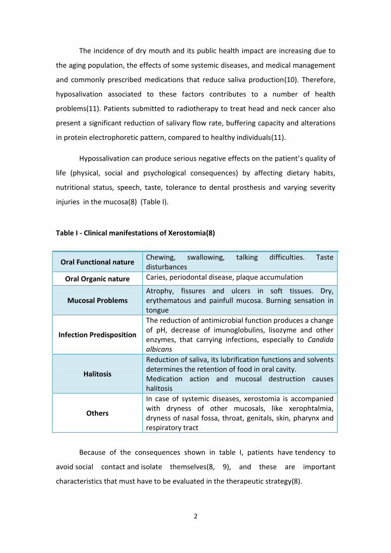
Hypossalivation can produce serious negative effects on the patient’s quality of
life (physical, social and psychological consequences) by affecting dietary habits,
nutritional status, speech, taste, tolerance to dental prosthesis and varying severity
injuries in the mucosa(8) (Table I).
Because of the consequences shown in table I, patients have tendency to
avoid social contact and isolate themselves(8, 9), and these are important
characteristics that must have to be evaluated in the therapeutic strategy(8).
Table I - Clinical manifestations of Xerostomia(8)
Oral Functional nature Chewing, swallowing, talking difficulties. Taste disturbances
Oral Organic nature Caries, periodontal disease, plaque accumulation
Mucosal Problems Atrophy, fissures and ulcers in soft tissues. Dry, erythematous and painfull mucosa. Burning sensation in tongue
Infection Predisposition
The reduction of antimicrobial function produces a change of pH, decrease of imunoglobulins, lisozyme and other enzymes, that carrying infections, especially to Candida albicans
Halitosis
Reduction of saliva, its lubrification functions and solvents determines the retention of food in oral cavity. Medication action and mucosal destruction causes halitosis
Others
In case of systemic diseases, xerostomia is accompanied with dryness of other mucosals, like xerophtalmia, dryness of nasal fossa, throat, genitals, skin, pharynx and respiratory tract
3
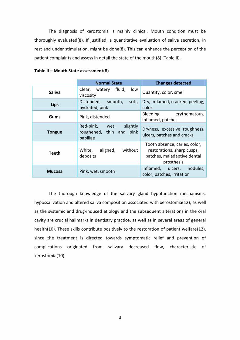
The diagnosis of xerostomia is mainly clinical. Mouth condition must be
thoroughly evaluated(8). If justified, a quantitative evaluation of saliva secretion, in
rest and under stimulation, might be done(8). This can enhance the perception of the
patient complaints and assess in detail the state of the mouth(8) (Table II).
Table II – Mouth State assessment(8)
Normal State Changes detected
Saliva Clear, watery fluid, low viscosity
Quantity, color, smell
Lips Distended, smooth, soft, hydrated, pink
Dry, inflamed, cracked, peeling, color
Gums Pink, distended Bleeding, erythematous, inflamed, patches
Tongue Red-pink, wet, slightly roughened, thin and pink papillae
Dryness, excessive roughness, ulcers, patches and cracks
Teeth White, aligned, without deposits
Tooth absence, caries, color, restorations, sharp cusps,
patches, maladaptive dental prosthesis
Mucosa Pink, wet, smooth Inflamed, ulcers, nodules, color, patches, irritation
The thorough knowledge of the salivary gland hypofunction mechanisms,
hypossalivation and altered saliva composition associated with xerostomia(12), as well
as the systemic and drug-induced etiology and the subsequent alterations in the oral
cavity are crucial hallmarks in dentistry practice, as well as in several areas of general
health(10). These skills contribute positively to the restoration of patient welfare(12),
since the treatment is directed towards symptomatic relief and prevention of
complications originated from salivary decreased flow, characteristic of
xerostomia(10).
4
2. MATERIALS AND METHODS
Clinical trials, research and review papers (1996 – 2012) were searched in
PubMed, MedLine, Biomed and also books available in the library of FMDUP, using the
following keywords:
Biochemical + xerostomia; biochemical + mechanisms + saliva, hypossalivation +
mechanisms; hypossalivation + xerostomia
5
3. SALIVA COMPONENTS AND FUNCTIONS
Saliva is an aqueous, translucent fluid, with neutral pH, secreted by the salivary
glands into the oral cavity(2). It is currently known that it has an important role in
maintaining normal physiological conditions of the tissues of the mouth(8). Saliva can
be divided as gland-specific saliva and whole saliva(3, 4). Gland-specific saliva is
collected directly from a gland. Whole saliva (mixed saliva) is a mixture of oral fluids
and includes secretions from both the major and minor salivary glands, in addition to
several constituents of non-salivary origin, such as gingival crevicular fluid,
expectorated bronchial and nasal secretions, serum and blood derivatives from oral
wounds, bacteria and bacterial products(3, 4), viruses and fungi, desquamated
epithelial cells, other cellular components, and food debris(3). There are three paired
extrinsic salivary glands in humans: the parotid, submandibular, and sublingual
glands(1, 3-5), as well as hundreds of smaller intrinsic salivary glands that are
distributed throughout the oral cavity, including the tip of the tongue(5, 13).
Secretions from both the submandibular and sublingual salivary glands enter the oral
cavity through Wharton's duct, and, thus, the separate collection of saliva from each of
these two glands is difficult(3). Interestingly, the histological composition of the major
extrinsic glands differs dramatically, with the larger parotid glands only containing
serous cells and contributing to around 25% of total saliva(5). The submandibular
glands, which account for approximately 70% of total salivary output, contain around
equal amounts of serous and mucous cells. Finally, the sublingual glands contain
mainly mucous cells and account for only 5% of total salivary output(5). Salivary flow is
under direct and indirect control from the autonomic nervous system (sympathetic
and parasympathetic)(3, 5, 8). Dramatically increased sympathetic stimulation can lead
to reduced salivary flow and dry mouth(5).
Saliva is mainly composed of water (95-99.4%)(3-5).The organic and inorganic
molecules are dissolved in water, and their concentration may significantly vary
between individuals, and in the same individual under various conditions(3, 4). Saliva
has a very complex composition, containing antimicrobial agents, immunoglobulins,
electrolytes (Na+, K+, Cl-, calcium, bicarbonate, phosphate) components with buffer
6
properties and proteins (S-IgA, mucins, lysozymes, histatins, lactoferrin and amylases)
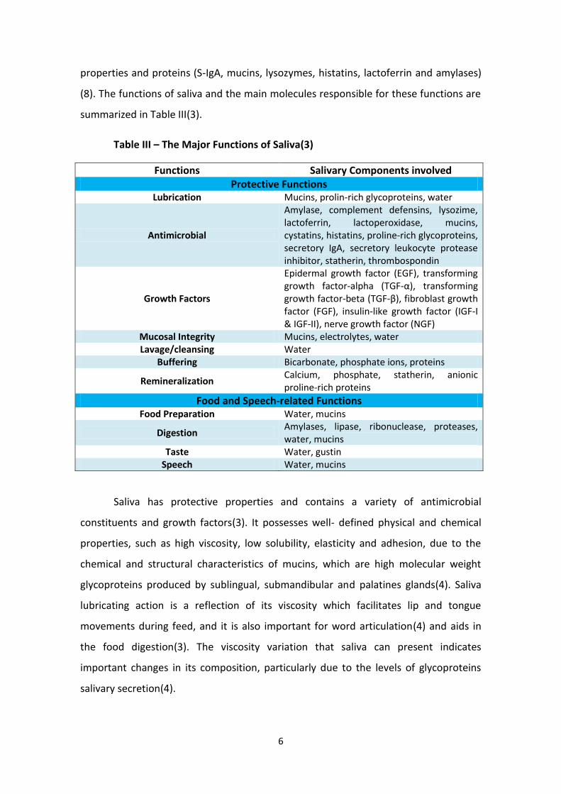
(8). The functions of saliva and the main molecules responsible for these functions are
summarized in Table III(3).
Table III – The Major Functions of Saliva(3)
Functions Salivary Components involved Protective Functions
Lubrication Mucins, prolin-rich glycoproteins, water
Antimicrobial
Amylase, complement defensins, lysozime, lactoferrin, lactoperoxidase, mucins, cystatins, histatins, proline-rich glycoproteins, secretory IgA, secretory leukocyte protease inhibitor, statherin, thrombospondin
Growth Factors
Epidermal growth factor (EGF), transforming growth factor-alpha (TGF-α), transforming growth factor-beta (TGF-β), fibroblast growth factor (FGF), insulin-like growth factor (IGF-I & IGF-II), nerve growth factor (NGF)
Mucosal Integrity Mucins, electrolytes, water Lavage/cleansing Water
Buffering Bicarbonate, phosphate ions, proteins
Remineralization Calcium, phosphate, statherin, anionic proline-rich proteins
Food and Speech-related Functions Food Preparation Water, mucins
Digestion Amylases, lipase, ribonuclease, proteases, water, mucins
Taste Water, gustin Speech Water, mucins
Saliva has protective properties and contains a variety of antimicrobial
constituents and growth factors(3). It possesses well- defined physical and chemical
properties, such as high viscosity, low solubility, elasticity and adhesion, due to the
chemical and structural characteristics of mucins, which are high molecular weight
glycoproteins produced by sublingual, submandibular and palatines glands(4). Saliva
lubricating action is a reflection of its viscosity which facilitates lip and tongue
movements during feed, and it is also important for word articulation(4) and aids in
the food digestion(3). The viscosity variation that saliva can present indicates
important changes in its composition, particularly due to the levels of glycoproteins
salivary secretion(4).
7
Many of salivary glycoproteins interact with the tooth surface contributing to
their protection and calcium fixation(8). Besides that, other glycoproteins, as is the
case of s-IgA, can also inhibit microbial adhesion. Globally, mucins are large
hydrophilic(8) glycoproteins that are highly viscous and relatively insoluble, two highly
desirable qualities for a substance responsible for maintaining membrane integrity
under harsh conditions(5). Mucins therefore protect the soft tissues of the oral cavity
from dehydration and abrasion damage by forming a lubricating(5, 8) coating on
tissues(5). They are also responsible for the selective adhesion of bacterial and fungal
agents and may help to prevent biofilm formation(5, 8). Lysozyme is an enzyme that
also has an antimicrobial activity, causing damage to the cell walls of bacteria by
hydrolysis of peptidoglycan(8). It is present in different mucosal secretions, such as
saliva and tears(5). However, Gram-negative bacteria, which contain
lipopolysaccharides in their cell walls, are largely resistant to this type of agent(5).
Histatins have antifungal function(8) and are secreted by the parotid and
submandibular glands(5). Histatin 1, 3, and 5 are the major histatins detected in
human saliva. Initially, histatin 1 was shown to inhibit hydroxylapatite crystal
formation and was proposed to maintain the surface integrity of enamel(5). Later, the
direct antimicrobial effects of histatins were investigated and shown to be limited to
Candida albicans, with histatin 5 being the most potent agent(5). Together, these data
suggested that histatins in parotid and submandibular gland secretions may play a
major role in the primary innate defense mechanism. Moreover, the presence of a
phosphorylated serine in histatin 1 suggests that it could be a precursor of the
acquired enamel pellicle(5). Lactoferrin is a glycoprotein found, not only in milk, but
also in various exocrine secretions, including saliva, and in various tissues(5). Its
antimicrobial activity is one of the least controversial properties and although several
modes of action have been suggested for this protein, an iron-scavenging function that
prevents microbial growth is the most accepted(5). Lactoferrin was found to be
present in saliva, with a marked elevation in the parotid secretion during the active
phase of chronic recurrent parotitis (5). Inflammatory stimulation of lactoferrin
expression suggests a basic protective mechanism in exocrine glands towards iron
scavenging and microbial growth prevention(5).
8
In digestive system, saliva has an important role in esophageal physiology,
digestion, and protecting the gastric cells(4, 8). In mouth, saliva participates in
chewing, speech, swallowing, taste sensitivity, lubrication of the tissues, mucosal
protection against invasion of certain substances(4, 8, 9) , antibacterial, antifungal and
antiviral activity, post eruptive maturation, ionic balance regulation in the enamel
remineralization, acquired pellicle deposition and acid spread limitation(4, 8).
Digestive components include α-amylase, which is involved in the conversion of
polysaccharide carbohydrates, such as starch, into the polysaccharides maltose and
dextrin, and lingual lipase secreted from the intrinsic salivary glands(5). Lingual lipase
initiates the breakdown of lipids prior to food entry into the duodenum and works
optimally at low pH(5).
In a dry mouth, natural remineralization and protection may not occur because
of the lack of salivary calcium and phosphate ions(14). Salivary pH ranges from 6 (1, 11)
to 7.4(1, 5, 8). The loss of its buffering capacity may cause oral pH to become acidic,
dissolving calcium and phosphate ions from the enamel (as the pH falls below 5.5,
demineralization starts to occur)(14). Changes in salivary flow rate may affect both the
concentration and availability (mainly due to changes in salivary pH)(3) of salivary
constituents. This may affect many factors that contribute to the development of
caries, such as proliferation of acid-producing bacteria, inability to buffer the acid
produced by bacteria or from ingested foods, loss of minerals from tooth surfaces and
inability to replenish the lost minerals, and loss of lubrication(14). A low flow rate
combined with a low or moderate buffer effect clearly indicates poor salivary
resistance against microbial attack(12). The acquired enamel pellicle protects teeth
from acid challenges by preventing direct contact between the acids and the
enamel(14). It is an organic film covering the surfaces of teeth(12). Lipids,
glycoproteins and proteins contained in saliva are the main components of dental
pellicle, acting as a selectively permeable barrier to calcium and phosphate ions, but
not to acids, which aids in the prevention of demineralization and in the promotion of
teeth remineralization(14). Finally, salivary flow, even with food in mouth, prevents
the accumulation of microbial particles and its potential harmful effects(7).

9
Saliva Production
Generally, salivary secretions have different characteristics depending on their
origin. The parotid fluid has very low viscosity (low-mucin) and is mainly secreted
during mastication – chewing saliva(7). Submaxillary fluid is a very viscous saliva (rich
in mucin) and it is secreted especially when exists flavoring substances in the mouth -
tasting saliva. Sublingual saliva has the same characteristics as the submaxillary, but
more pronounced in terms of viscosity - swallowing saliva(7).
Salivary glands are composed of specialized epithelial cells(3, 5), and their
structure can be divided into two specific regions, the acinar and the ductal regions(3).
The acinar region is where fluid is generated and most of the protein synthesis and
secretion takes place. Aminoacids enter the acinar cells by means of active transport,
and after intracellular protein synthesis, the majority of proteins are stored in storage
granules that are released in response to secretory stimulation(3).
The production of saliva is an active process that occurs in 2 phases:
1) Primary secretion – occurs in the acinar cells. This results in a product
similar in composition and osmolality to plasma(15).
2) Ductal secretion – results in a hypotonic salivary fluid(15, 16),
because ductal epithelium is poorly permeable to water.(17) It also results in
decreased sodium and increased potassium in the end product. (15) (16)
The acinar fluid secretion includes the active transport of anions into the lumen
and passage of water according to the osmotic gradient from the interstitial fluid into
the salivary lumen(3). Acinar cells secrete a NaCl-rich fluid called primary saliva(17).
The initial fluid is isotonic and is derived from the local vasculature(3). Acinar cells
secrete fluid in a Cl--dependent manner. The coordinated activity of ion channels,
water channels, pumps, cotransporters and exchangers results in the primary saliva
formation(17).
10
While acinar cells are water-permeable, ductal cells are not. However, ductal
cells actively absorb most of the Na+ and Cl- ions from the primary salivary secretion
and secrete small amounts of K+, HCO3- and some proteins(15, 16). The primary
salivary secretion is thus modified (NaCl reabsorption exceeds KHCO3 secretion)(17)
and the final salivary secretion, as it enters the oral cavity, becomes hypotonic(3, 15,
16). The degree of modification of saliva in the ducts heavily depends on the salivary flow
rate. Fast rates result in a salivary product more like the primary secretion. Slow rates
result in an increasingly hypotonic and potassium rich saliva(3).
In addition to the salivary glands morphological and functional heterogeneity,
there are also important differences in the output regulatory mechanisms(7). The
autonomic nervous system (sympathetic and parasympathetic) controls the salivary
secretion(3, 8, 16). In a general way, parasympathetic stimulation by the production of
acetylcholine excites the salivary glands and causes vasodilatation(7). Cholinesterase
counteracts this effect(11). On the other hand, sympathetic stimulation causes
vasoconstriction (11).
Saliva secretion may be stimulated by psychic stimuli, including the thought,
sight and smell of food, as well as the sound of cooking of some foods(13). The
mechanical stimuli – those involving touch or pressure on oral structures and
movements of the masticatory muscles and mandible – and those involving chemical
substances that stimulate the taste receptors are also important players on saliva
secretion.(13)
11
4. COMMON CAUSES OF XEROSTOMIA
Hypofunction of salivary glands, which results in qualitative and / or quantitive
change of saliva(18), causing xerostomia, has multiple oral health consequences and
affects quality of life(9). It may result from factors that affect the salivary center, the
secretion of saliva and functional salivary gland changes(8).
Factors that affect the salivary center
Emotions
The protein composition of human saliva depends on psycho-emotional state of
individuals(19). Psychogenic causes, such as depression, anxiety, stress or fear, can
also result in xerostomia(1, 9). Depression is usually accompanied by a decrease of
proteins of molecular masses ranging from 20 to 200 kD, whereas emotionally positive
intellectual activity cause the opposite effect. Due to these observations, it is
suggested that human saliva may be used as an experimental model for the
development of diagnostics of various psycho-physiological states(19).
Fasting
It must be observed that during fasting, the individual is subjected to
psychological and physiological reactions to starvation, involving stress and behavioral
changes(20). A short period of fasting therefore does not create persisting effects on
the oral ecology. However, other extreme and longer-lasting conditions such as
starvation, anorexia nervosa and some general diseases, can lead to intestinal
malresorption and/or malnutrition. This might give longer lasting effects on saliva
production and/or composition(20), causing xerostomia.
Menopause
Menopause is accompanied by a number of characteristic physical changes;
some of which occur in the oral cavity(21). It seems that a positive relationship exists
between ovarian hormone modifications and changes in the oral mucosa, and sex
hormone withdrawal might be a cause in incidence of oral dryness feeling in
12
menopausal women(21). The exact mechanisms that cause sensation of oral dryness in
menopausal women have not been firmly established. However, there are reports on
amelioration of these symptoms by estrogen treatment(21).
Alzheimer’s disease
In cases of Alzheimer’s disease or stroke, patients may complain of dry mouth
in the presence of normal salivary secretion due to altered perception(1, 9).
Factors that affect the secretion of saliva
Tobacco
Dry mouth is often exacerbated by activities such as hyperventilation, breathing
through the mouth, drinking alcohol(1) and smoking(1, 18, 22). Oral squamous cell
carcinoma is the most common malignancy of the head and neck and it major inducer
is exposure to tobacco. Cigarette smoke also contains oxidants such as oxygen and
nitrogen free radicals and volatile aldehydes, known to increase oxidative stress in
biological systems(23). Exposure of these molecules in cigarette smoke is probably the
major cause of damage to biomolecules in saliva, such as α-amylase(23). Dehydration
due to impaired fluid intake, emesis, diarrhea or polyuria can result in
hyposalivation(1, 9).
Medications
Medications are the most common cause of decreased salivar function (1, 8, 9,
18, 22) (table III). It has been reported that 80% of the most commonly prescribed
medications cause xerostomia(9, 18). The drugs mostly responsible for dry mouth are
tricyclic antidepressants, antipsychotics, atropinics, beta blockers and antihistamines,
thus the complaint of dry mouth (1, 8, 22). It should be noted that, while there are
many drugs that affect the quantity and/or quality of saliva, these effects are generally
not permanent(1).
13
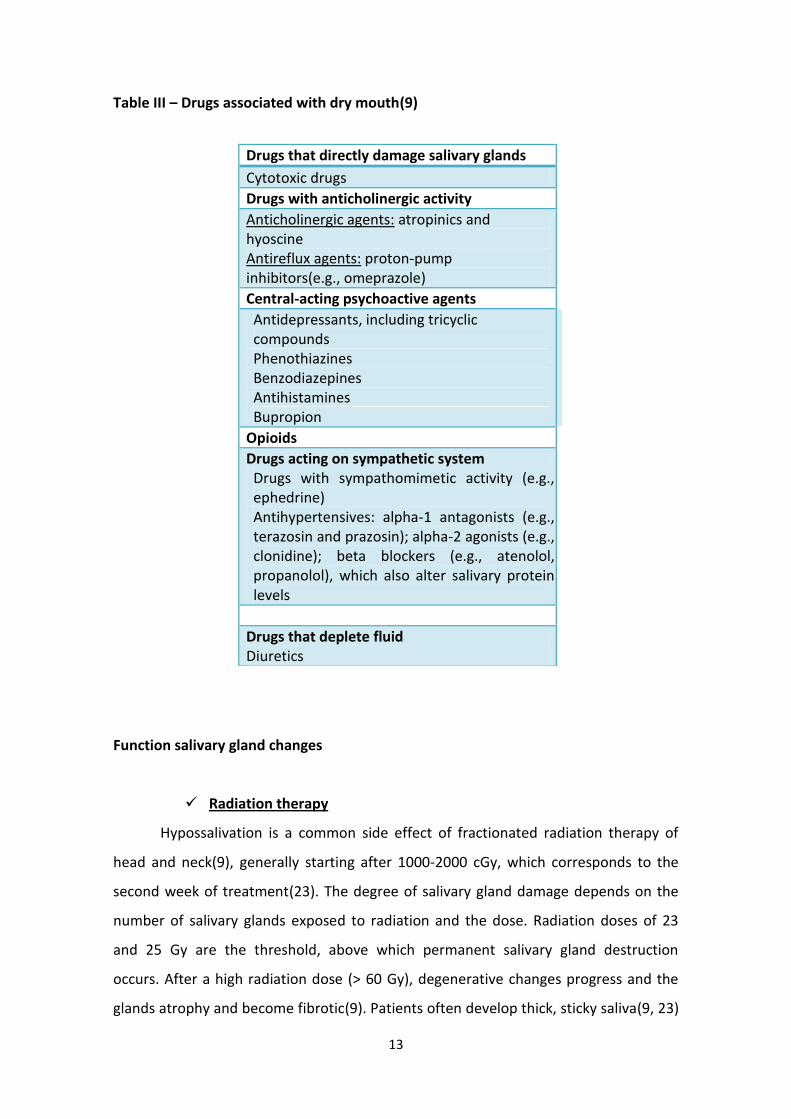
Table III – Drugs associated with dry mouth(9)
Drugs that directly damage salivary glands
Cytotoxic drugs
Drugs with anticholinergic activity
Anticholinergic agents: atropinics and hyoscine Antireflux agents: proton-pump inhibitors(e.g., omeprazole)
Central-acting psychoactive agents
Antidepressants, including tricyclic compounds Phenothiazines Benzodiazepines Antihistamines Bupropion
Opioids
Drugs acting on sympathetic system Drugs with sympathomimetic activity (e.g., ephedrine) Antihypertensives: alpha-1 antagonists (e.g., terazosin and prazosin); alpha-2 agonists (e.g., clonidine); beta blockers (e.g., atenolol, propanolol), which also alter salivary protein levels
Drugs that deplete fluid Diuretics
Function salivary gland changes
Radiation therapy
Hypossalivation is a common side effect of fractionated radiation therapy of
head and neck(9), generally starting after 1000-2000 cGy, which corresponds to the
second week of treatment(23). The degree of salivary gland damage depends on the
number of salivary glands exposed to radiation and the dose. Radiation doses of 23
and 25 Gy are the threshold, above which permanent salivary gland destruction
occurs. After a high radiation dose (> 60 Gy), degenerative changes progress and the
glands atrophy and become fibrotic(9). Patients often develop thick, sticky saliva(9, 23)
14
after starting head and neck radiotherapy, due to loss of serous secretion initially,
followed by lack of any secretion and xerostomia(9). When major salivary glands are
affected by radiation, salivary flow may decrease 90%. There is also a marked
reduction in pH, due to a change in the concentrations of calcium, sodium and
bicarbonatos(23).
Sjögren syndrome
Sjögren syndrome (SS) is the most common disease causing xerostomia in the
elderly(1, 9, 18). It is an autoimmune chronic inflammatory disease with
preponderance among females (female to male ratio 9:1)(1, 9) predominantly in
postmenopause(1). The disease is characterized by lymphocyte infiltration of salivary
and lacrimal glands resulting in hypofunction(1, 9). The disease can occur in 2 forms:
primary, which involves the salivary glands, or secondary, which occurs along with
other autoimmune disorders (mainly rheumatoid arthritis). Xerostomia and
xerophthalmia are the main symptoms of primary SS(1, 9). Secondary SS also presents
with symptoms of associated systemic conditions(9).
15
5. SALIVARY GLAND HYPOFUNCTION
The subjective symptom of xerostomia may not always correlate with salivary
flow rates(10), because it can be caused by several medications without actually no
reduction in salivary flow(24). For understanding this phenomenon, one should be
aware that saliva enters the mouth at several locations, and that the different
glandular secretions are not well mixed(10). For example, the contribution of parotid
saliva to (un)stimulated whole saliva varies from site to site, ranging from being the
major contributor to whole saliva collected buccally from the maxillary molars region,
to being almost non-contributing to whole saliva collected in the incisor region(10).
The wide variation in local contribution of the various salivary glands to whole saliva is
also obvious when assessing mucosal wetness as the thickness of the salivary layer on
the oral mucosa is much thinner in the labial and anterior hard palatal region than on
the buccal mucosa and anterior tongue(10). These phenomena might explain, at least
in part, the differences reported in the literature about level of hyposalivation and
sensation of oral dryness(10).
The major causes of xerostomia are objectively assessed salivary gland
hypofunction, which could be attributed to radiation of head and neck cancer
therapy(11, 23) and several systemic conditions (11, 24). Diseases such as alcoholism,
diabetes mellitus (type 1), malnutrition, anorexia nervosa, bulimia, viral or bacterial
infections and medications used to treat HIV patients may cause parotid hypertrophy
and consequences in salivary production and function(8). Salivary flow rate
measurement is essential in diagnosing salivary gland hypofunction as the cause of
xerostomia(24). In this context, it is important to note that salivary flow is inconstant,
being more reduced in the morning, higher during the afternoon, and almost
inexistent during the sleep (2). The daily volume of normal saliva production is around
0,5 and 1,5 liters(8). Unstimulated saliva secretion is 0.3 ml/min and stimulated is 1.5
ml / min. It is considered hyposalivation if values are lower than 0.1 ml/min and 0.7
ml/min, respectively(8).
16
Radiation injury of salivary gland tissue
Clinical and biochemical alterations in the oral cavity and in the saliva can occur
in patients exposed to radiotherapy(11). Irradiation of the salivary glands during the
treatment of head and neck cancer results in a progressive loss of gland function
(hyposalivation)(10, 11) beginning early in the course of radiotherapy(10). Radiation
induces DNA damage, which can impair a proper cell division, resulting in cell death or
senescence. As a consequence, this can promote changes in the quantity and in the
composition of saliva(8, 10). Radiation injury leads primarily to the loss of saliva-
producing acinar cells, however interestingly, the ducts, although deprived of function,
mostly remain intact(10). Nevertheless, several studies show no cell loss during the
first days after irradiation, although saliva flow is dramatically reduced, and water
secretion is selectively hampered(10).
Radiation-induced loss of salivary gland function occurs over four phases. The
first phase (0–10 days) is characterized by a rapid decline in flow rate without changes
in amylase secretion or acinar cell number(10). The second phase (10–60 days) consists
of a decrease in amylase secretion paralleled by acinar cell loss(10). Reduction of the
specific activity of the α-amylase indicates that radiation acts specifically on some
proteins(11). Flow rate, amylase secretion and acinar cell numbers do not change in
the third phase (60–120 days). In the fourth phase (120–240 days) further
deterioration of gland function is seen, but this is accompanied by an increase in acinar
cell number, albeit with poor tissue morphology(10).
Patient’s saliva after treatment is scarce(11), thicker and adhering(8), it means
considerably more viscous, leading to oral discomfort(11). Palpation of the parotid
papilla and milking of the gland and duct may reveal little or no saliva output(9).
Hyposalivation occurs in almost 100% of cases after doses of radiation in excess of 50
Gy(6). Serous glands are more radiosensitive than the mucinous(8, 10), but both glands
have been shown to be sensitive to radiotherapy, at least with respect to their
function(8).
17
6. COMMON ORAL COMPLICATIONS ASSOCIATED WITH XEROSTOMIA
Hypofunction of the salivary glands resulting from different etiologies can have
different manifestations(6), affecting nutrition, oral health , as well as psychological
health(1).
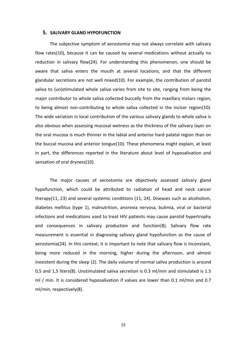
Some common problems associated with xerostomia, include a constant sore
throat, burning sensation, difficulty in speaking and swallowing(1, 6, 9), hoarseness
and/or dry nasal passages(1). It is well known that hyposalivation contributes
considerably to disruption of the natural barrier of the oral mucous membranes(6).
The dry mucosa is more susceptible to trauma and infection(1, 9) and may appear dry
and shiny (Fig.1)(9).
Oral Candidiasis
Although it is estimated that Candida species are present in the normal oral
flora of healthy adults, certain conditions, like xerostomia, increase the risk of Candida
overgrowth(25). Oral candidiasis is one of the most common oral infections seen in
association with xerostomia(2). Extraoral findings include dry and cracked lips, which
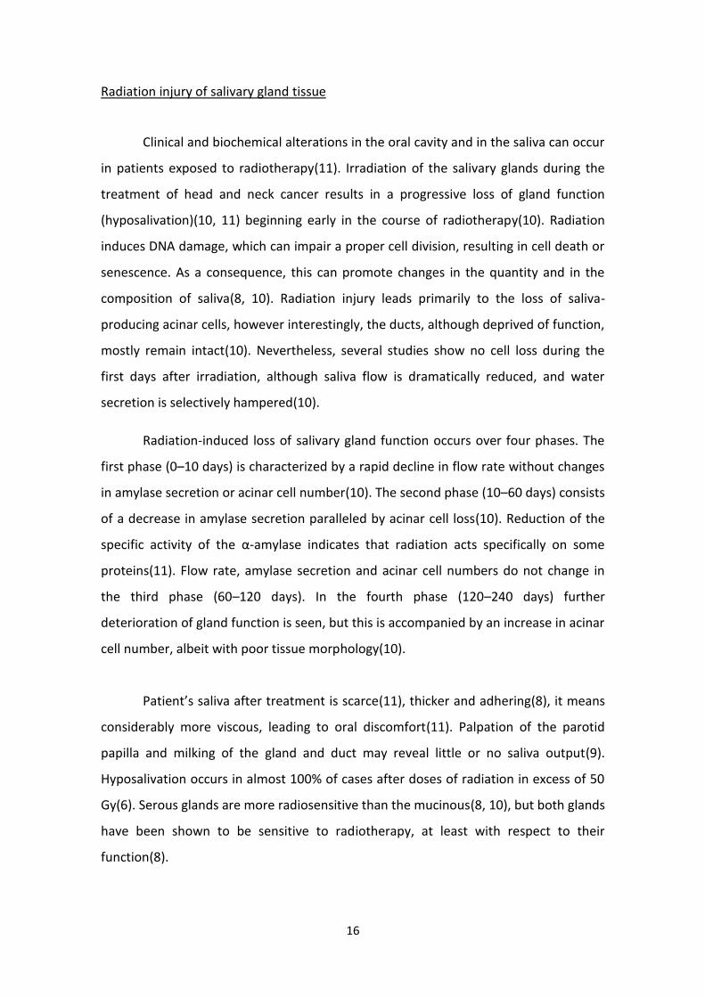
may be infected with Candida, and occasionally enlarged salivary glands(9). The clinical
manifestations of candidiasis include angular cheilitis and erythematous candidiasis.
Both are more common than pseudomembranous candidiasis, which is more readily
recognized(9). It is characterized by adherent, curd-like plaques that can be removed
by wiping firmly with a tongue blade or gauze(25).The tongue can become furrowed,
dry and sticky or it can undergo partial or complete depapillation of the dorsum (Fig.
2)(9).
A presumptive candidiasis diagnosis can usually be made based on the patient’s
history, clinical presentation, and response to antifungal treatment. The diagnosis can
be confirmed with a cytological smear of the lesion(25).
18
Dental Caries
The biggest cariogenic risk is resulted from salivary gland dysfunctions(18). An
inadequate salivary flow changes the cleaning of teeth, carried by this fluid, by
transporting the alimentary cariogenic substrates(26). The development of a dental
cavity depends on various factors, such as the host condition, microbiota, time and
diet(2). Microbiologically, the dental cavity may be defined as the interrelation
between the oral cavity, bacteria and dental tissues; chemically, the processes
involved in the formation of the cavity are described as interrelating pH and mineral
flow and solubility on the tooth saliva interface(2).
Functions of saliva include the development of dental pellicle, a film that forms
on tooth enamel and protects it against mineral loss from the tooth surface;
replenishment of tooth surface minerals such as calcium and phosphate; the provision
of antibacterial activity and buffering activity, which keeps oral pH neutral; and
mechanical removal of residual food particles from the teeth(14, 26). In a dry mouth,
natural remineralization and protection may not occur properly because of the lack of
salivary calcium and phosphate ions(14). A lack of saliva exposes the teeth to acidic
challenges from food and drinks, as well as to the acid produced by acidogenic
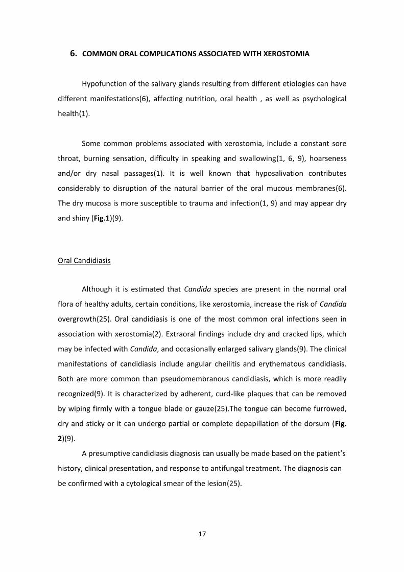
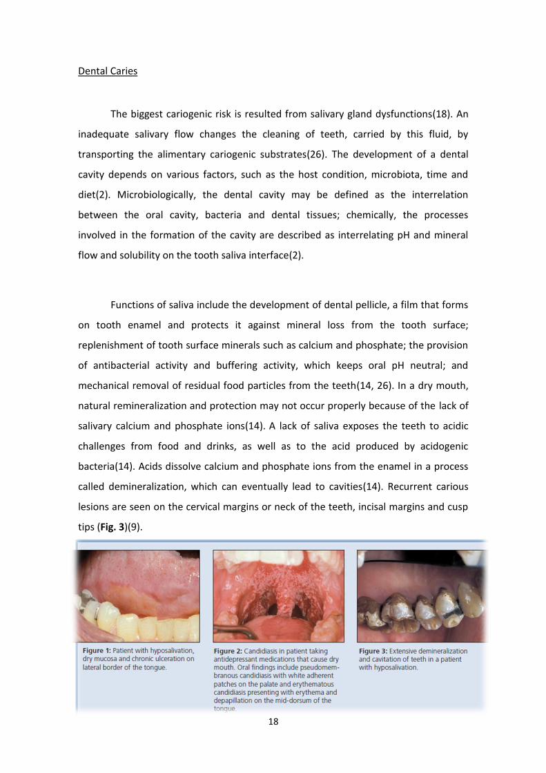
bacteria(14). Acids dissolve calcium and phosphate ions from the enamel in a process
called demineralization, which can eventually lead to cavities(14). Recurrent carious
lesions are seen on the cervical margins or neck of the teeth, incisal margins and cusp
tips (Fig. 3)(9).
19
7. MANAGEMENT OF HYPOSALIVATION
Patients with xerostomia should undergo frequent dental evaluations for early
diagnosis of oral complications(9). They should be encouraged to carry out daily oral
self-examinations for any mucosal ulcers, lesions or tooth decay and to report any
unusual findings(9).
The management of xerostomia should begin with identification and
management of the underlying cause(1, 8, 9), and it must be oriented by its etiology
and directed towards the disease effects in the patient comfort and quality of life(8).
It is important that health workers explain to patients with xerostomia what are
the best ways to get relieved and the measures to prevent its complications(8).
Management of Salivary Flow and oral comfort
For patients with hyposalivation, the lack of saliva not only increases the risk of
caries, but also creates an uncomfortable sensation of dryness. Oral products to treat
dry mouth, which include toothpastes, mouthwashes with antibacterial enzymes,
mouth sprays and liquids, among others, are helpful in the relief of the discomfort of
xerostomia(14). For some patients, chewing gum can also help to increase salivary flow
and increase mouth comfort(14). For patients who are able to chew gum, gums
containing 100% xylitol are ideal, since xylitol cannot be fermented by acid-producing
bacteria, so it helps to prevent caries(14) and can be easily incorporated into a
patient’s routine(9).
If the salivary glands are not working properly but can still produce some saliva,
some drugs help the glands to work better, or the use of artificial saliva can keep the
mouth wet(1, 9). Parasympathetic drugs, such as pilocarpine(8, 14), bethanechol and
the cholagogue anethole trithione (Sialor, Solvay Pharma Inc., ON) are used to
stimulate salivary production in those with residual salivary-gland function and it can
help to relieve oral dryness(14). Although these agents are effective sialogogues in
patients with intact salivary glands, their efficacy is reduced in patients with changes in
salivary glands due to radiation or disease(14).
20
Nowadays there is no definitive treatment for patients with dry mouth due to
radiation therapy or SS, but high-potency fluoride treatment is recommended, and
patients should be advised against using alcohol and tobacco(1, 9), as well as drinks
with caffeine(1), and to adopt a low-sugar diet to control dental caries(9). In this cases,
treatment is directed toward local and systemic salivary gland stimulation,
symptomatic relief and preventing and treating complications due to
hyposalivation(9). Stimulation of residual salivary secretory capacity by acupuncture
has shown some promising results in head and neck radiotherapy patients(10). If saliva
secretion cannot be stimulated, symptomatic treatment involves the use of saliva
substitutes(1, 8, 9). Patients should be encouraged to take frequent sips of water
throughout the day. Use of water during meals can aid in swallowing and improve
taste perception(9). Use of bedside humidifiers, particularly at night, may lessen
discomfort due to oral dryness(9). Commercially available saliva substitutes containing
thickening agents, such as carboxymethyl cellulose or mucins, are the most
common(9).
Taken together, dry mouth treatment will depend on what is causing the
problem(27). Summarizing, if dry mouth is caused by medicine, the physician might
change medicine or adjust the dosage, If salivary glands are not working right but can
still produce some saliva, physicians or dentists might give a medicine that helps the
glands to work better(27). Also, the patient preference plays an important role in
determining which products and procedures can be easily incorporated into a daily
home care routine, and which offer the best relief for the symptoms of dry mouth(14).

21
8. CONCLUSIONS
Saliva is an aqueous fluid produced by salivary glands. Its complex composition
reflects the wide range of functions attributed to saliva. Indeed, it is involved in
processes such as digestion, speech, as well as in teeth metabolism and defense
against microorganisms. Deficiencies in saliva production and/or composition are
defined as xerostomia. This pathological condition has a significant impact on the
health of the patients, especially in their oral health.
Since xerostomia often occurs in combination with a variety of other
pathological conditions, and presents numerous etiologies(16) and multiple oral health
consequences, it really affects the quality of life of the individuals.(9) The management
of this condition should begin with the identification and the understanding of the
underlying cause(1, 8, 9), and it must be oriented by its etiology and directed towards
the disease effects(8), contributing positively to the restoration of patient welfare.(12)
Nowadays there is no definitive treatment for patients with dry mouth(9), but reliable
diagnostic methods and effective new treatments are expected to lead to its complete
cure in the near future(16).
22
9. REFERENCES
1. Bartels L. Helping Patients with dry mouth. Oral Cancer Foundation
2. Cavasin Filho JC, Giovani EM. Xerostomy, dental caries and periodontal disease
in HIV+ patients. The Brazilian journal of infectious diseases : an official publication
of the Brazilian Society of Infectious Diseases. 2009;13(1):13-7. Epub 2009/07/07.
3. Kaufman E, Lamster IB. The diagnostic applications of saliva--a review. Critical
reviews in oral biology and medicine : an official publication of the American
Association of Oral Biologists. 2002;13(2):197-212. Epub 2002/07/05.
4. Mourai S, Medeiros A, Costa F, Moraes P, Oliveira Filho S. Diagnostic Value of
Saliva in Oral and Systemic Diseases: A Literature Rewiew. 2007;v. 7:187-94.
5. Farnaud SJ, Kosti O, Getting SJ, Renshaw D. Saliva: physiology and diagnostic
potential in health and disease. TheScientificWorldJournal. 2010;10:434-56. Epub
2010/03/23.
6. Guobis Z, Kareiviene V, Baseviciene N, Paipaliene P, Niedzelskiene I, Sabalys G,
et al. Microflora of the oral cavity in patients with xerostomia. Medicina (Kaunas).
2011;47(12):646-51. Epub 2012/03/01.
7. Reis C. AM, Azevedo I. Práticas de Bioquímica para as Ciências da Saúde. 2002.
8. Feio M, Sapeta, P. Xerostomia em Cuidados Paliativos Acta Med Port.
2005;18:18: 459-66.
9. Gupta A, Epstein JB, Sroussi H. Hyposalivation in elderly patients. J Can Dent
Assoc. 2006;72(9):841-6. Epub 2006/11/18.
23
10. Vissink A, Mitchell JB, Baum BJ, Limesand KH, Jensen SB, Fox PC, et al. Clinical
management of salivary gland hypofunction and xerostomia in head-and-neck
cancer patients: successes and barriers. International journal of radiation oncology,
biology, physics. 2010;78(4):983-91. Epub 2010/10/26.
11. Pontes P, Spadaro. Clinical and biochemical evaluation of the saliva of patients
with xerostomia induced by radiotherapy. Braz Oral Res. 2004;18(1):69-74.
12. Tabak LA. Advances in Dental Research : Saliva in Health and Disease. 2000;14.
13. Ferguson D. Oral Bioscience. 2006.
14. Su N, Marek CL, Ching V, Grushka M. Caries prevention for patients with dry
mouth. J Can Dent Assoc. 2011;77:b85. Epub 2011/07/22.
15. Frederick S. BJ. Anatomy and physiology of the salivary glands source Grand
Rounds Presentation. 2001.
16. Elsevier BV. Oral Biosciences:The annual review 2011. Journal of Oral
Biosciences. 2012:1-4.
17. Catalan MA, Nakamoto T, Melvin JE. The salivary gland fluid secretion
mechanism. The journal of medical investigation : JMI. 2009;56 Suppl:192-6. Epub
2009/01/01.
18. Côrte-Real I. FM, Reis Campos J. As doenças orais no idoso – Considerações
gerais. rev port estomatol med dent cir maxilofac. 2011;52(3):175–180.
19. Grigoriev N, Artamonov. Protein content of human saliva in various psycho-
emotional states. Biochemistry (Moscow). 2002;68:501-3.
24
20. Johansson E, Steen. Studies of the Effect of Diet on Saliva Secretion and Caries
Development: The Effect of Fasting on Saliva Composition of Female Subjects. 2012.
21. Farzaneh D. Serum 17β-estradiol and oral dryness feeling in menopause.
Health. 2011;Vol.3:258-62.
22. Petersen PE, Yamamoto T. Improving the oral health of older people: the
approach of the WHO Global Oral Health Programme. Community Dent Oral
Epidemiol. 2005;33(2):81-92. Epub 2005/02/24.
23. Lopes C, Alves, Abbade, Rossi. Reconhecendo e controlando os efeitos
colaterais da radioterapia. Revista da APCD. 1998;V. 52.
24. Navazesh M, Kumar SK. Measuring salivary flow: challenges and opportunities. J
Am Dent Assoc. 2008;139 Suppl:35S-40S. Epub 2008/07/03.
25. Gonsalves WC, Wrightson AS, Henry RG. Common oral conditions in older
persons. American family physician. 2008;78(7):845-52. Epub 2008/10/10.
26. Ramos JA. Bioquímica Bucodental. 1996;1.
27. Research NIoDaC. Dry Mouth. NIH Publication No 11-3174. 2011.