alessandro spalenza maciel fungos nematÓfagos
TRANSCRIPT
i
ALESSANDRO SPALENZA MACIEL
FUNGOS NEMATÓFAGOS PREDADORES NO CONTROLE DE
ANCILOSTOMATÍDEOS DE CÃES
Tese apresentada à Universidade
Federal de Viçosa, como parte das
exigências do Programa de Pós-
Graduação em Medicina Veterinária,
para obtenção do título de Doctor
Scientiae
VIÇOSA
MINAS GERAIS – BRASIL
2009
ii
"Eu espero que eu sempre possua firmeza e virtude suficientes para manter o
que eu considero o mais invejável de todos os títulos, o caráter" (George
Washington)
A Deus,
À minha querida mãe,
Dedico.
iii
AGRADECIMENTOS
A Deus, pelo cuidado, amor e conforto da alma.
À minha querida mãe, Maria de Lourdes, que apesar do Mal de
Alzheimer, sempre demonstrou seu amor e certamente, inconscientemente,
está torcendo por mim.
À Universidade Federal de Viçosa (UFV), em particular ao Departamento
de Veterinária e ao Laboratório Bionema, pela estrutura disponibilizada e assim
possibilitar a realização deste trabalho.
À Coordenação de Aperfeiçoamento de Pessoal de Nível Superior
(CAPES) e ao Conselho Nacional de Desenvolvimento Científico e Tecnológico
(CNPq), pelo apoio financeiro.
Ao professor Jackson Victor de Araújo, pela oportunidade e orientação.
Aos funcionários do Laboratório de Parasitologia do Departamento de
Veterinária, José Geraldo e Ademir, pela colaboração.
À secretária de Pós-graduação do Departamento de Veterinária Rosinéia
Aparecida da Cunha Andrade, pelo carinho, gentileza e preocupação.
Aos amigos Artur Kanadani Campos e Everaldo Antônio Lopes, pelas
sugestões, correções, conversas, amizade e presença nas horas difíceis.
Ao professor Leandro Grassi de Freitas que abriu as portas de seu
laboratório além da orientação, respeito, confiança, incentivo, conselhos e
amizade. Sobretudo por acreditar em meu potencial.
Ao professor Laércio dos Anjos Benjamin, pela amizade, conselhos e
apoio moral.
À Claúdia Alencar Vanetti do Núcleo de Microscopia e Microanálise pelo
carinho e apoio técnico fundamental no uso dos equipamentos para a
realização da microscopia eletrônica de varredura.
Ao professor José Lino Neto pela atenção e apoio profissional no
registro das micrografias de luz.
À minha namorada Cristiane de Fátima da Silva Gomes pelo carinho,
apoio, incentivo, paciência e compreensão.
À minha cadela Juni, pelo companherismo e por tornar minha vida mais
alegre.
Aos amigos do Bionema, Paulo Afonso Ferreira, Rosângela Dallemole
Giaretta, Deisy Xavier Amora, Ronaldo João Falcão Zooca, Guilherme Silva de

iv
Podestá e Vanessa Sabioni de Almeida pela amizade, alegre convívio e pela
ajuda quando precisei.
Aos amigos Phellipe Kelbert Ribeiro, Charles André Souza Bispo, Lucas
Marcon, Alisson Sanguinetti e Sidimar Sossai, pela amizade, apoio e
companheirismo.
A todos que direta ou indiretamente contribuiram para moldar minhas
opiniões e as abordagens deste trabalho, muito obrigado!
v
BIOGRAFIA
ALESSANDRO SPALENZA MACIEL, filho de Maria de Lourdes
Spalenza Maciel e José Gonçalves Maciel, nasceu em 16 de janeiro de 1974,
em Colatina, Espírito Santo.
Iniciou seus estudos na Universidade Federal de Viçosa – UFV em
1994, graduando-se em Medicina Veterinária em 1999 pelo Departamento de
Medicina Veterinária desta instituição.
Em março de 2000, iniciou o mestrado no Departamento de Economia
Rural da Universidade Federal de Viçosa obtendo o título de Magister Scientiae
em Economia Aplicada em junho de 2002.
Em março de 2004, iniciou um segundo mestrado no Departamento de
Medicina Veterinária da Universidade Federal de Viçosa obtendo o título de
Magister Scientiae em Medicina Veterinária em abril de 2005.
Em agosto de 2005, iniciou o doutorado no Departamento de Medicina
Veterinária da Universidade Federal de Viçosa
vi
SUMÁRIO
RESUMO.................................................................................................... vii
ABSTRACT................................................................................................ viii
INTRODUÇÃO GERAL.............................................................................. 1
CAPÍTULO 1 - Predation of Ancylostoma spp. dog infective larvae by
nematophagous fungi in different conidial concentrations......................... 13
Abstract...................................................................................................... 14
Introduction................................................................................................ 14
Material and Methods................................................................................. 16
Results....................................................................................................... 18
Discussion.................................................................................................. 25
CAPÍTULO 2 - Scanning electron microscopy of Ancylostoma spp. dog
infective larvae captured and destroyed by the nematophagous fungus
Duddingtonia flagrans................................................................................ 31
Abstract...................................................................................................... 32
Introduction................................................................................................ 32
Material and Methods................................................................................. 33
Results....................................................................................................... 35
Discussion.................................................................................................. 41
CAPÍTULO 3 - Predatory behaviour of the nematode-trapping fungi
Arthrobotrys cladodes, A. robusta, A. oligospora and Duddingtonia
flagrans on Ancylostoma spp. dog infective larvae in soil microcosm....... 47
Abstract...................................................................................................... 48
Introduction................................................................................................ 48
Material and Methods................................................................................. 50
Results....................................................................................................... 54
Discussion.................................................................................................. 57
CAPÍTULO 4 - Biological control of Ancylostoma spp. dog infective
larvae in soil under semi-natural................................................................ 64
Abstract...................................................................................................... 65
Introduction................................................................................................ 66
Material and Methods................................................................................. 68
Results....................................................................................................... 75
Discussion.................................................................................................. 80
CONCLUSÕES GERAIS............................................................................ 87
REFERÊNCIAS BIBLIOGRÁFICAS........................................................... 88

vii
RESUMO
MACIEL, Alessandro Spalenza, D. Sc., Universidade Federal de Viçosa, Julho de 2009. Fungos nematófagos predadores no controle de ancilostomatídeos de cães. Orientador: Jackson Victor de Araújo. Co-orientadores: Artur Kanadani Campos, Laércio dos Anjos Benjamin e Leandro Grassi de Freitas.
Os nematóides do gênero Ancylostoma são endoparasitas cosmopolitas de cães
que causam diversas patologias nesses animais e também são geohelmintos
zoonóticos que podem infectar o ser humano via solo. O controle do estágio adulto
destes nematóides é baseado na utilização de anti-helmínticos, no entanto, o uso
de agentes biocontroladores pode ser uma medida complementar reduzindo a
população dos estágios pré-parasitários em desenvolvimento no solo. Isto se
justifica pelo fato de que cães vadios parasitados são os principais responsáveis
pela infestação ambiental com ovos de ancilostomídeos veiculados nas fezes.
Dentre os organismos biocontroladores, sabe-se que os fungos nematófagos
predadores têm tido eficácia contra os nematóides de animais domésticos. Neste
contexto, o objetivo deste trabalho foi testar diversos isolados de fungos
predadores, selecionando o mais infectivo para ser utilizado no controle da forma
larval infectante (L3) de Ancylostoma spp. no solo sob condições semi-naturais. O
fungo Duddingtonia flagrans (Isolado CG768) foi considerado o mais infectivo
devido à redução do número de L3 recuperadas e pela característica de produzir
numerosos esporos de resistência capazes de sobreviver no solo, prolongando a
vida do fungo na ausência da presa. A infectividade deste isolado pôde ser
observada em micrografias eletrônicas de varredura que mostraram a destruição
completa da L3 de Ancylostoma spp. em cerca 48 horas após a captura. Para
testar o seu antagonismo no ambiente foi utilizado um sistema prático e
economicamente viável de produção massal de inóculo fúngico em grãos de arroz
branco e grãos de milho moído. O fungo foi incorporado ao solo colonizado em
grãos de milho moído tendo como parâmetro a concentração de 10.000
clamidósporos/g de solo, efetiva em ensaios in vitro preliminares. Tal abordagem
provavelmente favoreceu o estabelecimento do antagonista, havendo redução
significativa da população do nematóide alvo em solo não tratado sob condições
semi-naturais, mostrando o potencial regulador deste agente biológico.

viii
ABSTRACT
MACIEL, Alessandro Spalenza, D. Sc., Universidade Federal de Viçosa, July of 2009. Nematode-trapping fungi in the control of dog’s hookworms. Adviser: Jackson Victor de Araújo. Co-Advisers: Artur Kanadani Campos, Laércio dos Anjos Benjamin and Leandro Grassi de Freitas.
The nematodes of the genus Ancylostoma are cosmopolitan endoparasites of
dogs that cause various diseases in these animals and also are zoonotic
geohelminths that can infect humans from soil. The control of the adult stage of
these nematodes is based on the use of anti-helminthic; however, the use of
biocontrol agents may be an additional measure reducing the population of pre-
parasitic stages in development in the soil. This is justified by the fact that stray
dogs parasitized are primarily responsible for environmental infestation with
hookworm eggs transmitted in faeces. Among the biocontrol organisms, it is known
that nematode-trapping fungi have been effective against nematodes of domestic
animals. In this context, the objective of this study was to test various fungi isolated
of predacious fungi, selecting the most infective for use in the control of infective
larval form (L3) of Ancylostoma spp. in soil under semi-natural conditions. The
Duddingtonia flagrans fungus (Isolate CG768) was considered the most infective
due to the reduction in the recovery of the target nematode L3 and by the
characteristic of producing many spores of resistance that can survive in soil,
prolonging the life of the fungus in the absence of prey. The infectivity of this isolate
could be observed in scanning electron micrographs that showed the destruction of
L3 of Ancylostoma spp. in about 48 hours after capture. To test the antagonism in
the environment it was used a practical and economically viable system of mass
production of fungal inoculum in grains of white rice and milled maize. The fungus
was incorporated into the soil colonized in milled maize, taking as parameter the
concentration of 10,000 chlamydospores/g of soil, previously effective in vitro tests.
This approach probably favored the establishment of the antagonist with significant
reduction of the target nematode population in untreated soil under semi-natural
conditions, showing the potential regulator of this biological agent.

1
INTRODUÇÃO GERAL
O potencial papel dos animais de companhia de reservatório para
zoonoses tem sido reconhecido como um problema de saúde pública no
mundo (Schantz, 1994). Eles estão expostos a numerosos microorganismos
tais como bactérias, vírus, rickétsias, micoplasmas, clamídias, protozoários,
fungos e parasitos (Plaut et al., 1996; Geffray, 1999; Macpherson, 2005).
Dentre estes animais, o cão (Canis familiaris) é o que mais convive com o ser
humano (Leite et al., 2004), e historicamente, foi a primeira espécie
domesticada cujo relacionamento começou em tempos pré-históricos, cerca de
12.000-15.000 anos atrás (Morey, 1994).
Este animal é um importante veiculador ambiental de parasitos (Traub et
al., 2007) e sua população tem crescido consideravelmente nas zonas urbanas
devido à procriação descontrolada (Dutta, 2002). Tal fato junto com a falta de
saneamento e o crescimento desordenado das cidades em países em
desenvolvimento aumenta os riscos de transmissão de zoonoses devido à
proximidade com os seres humanos (Robertson et al., 2000). Segundo
Macpherson et al. (2005) a população mundial de cães é estimada em mais de
500 milhões de animais. Entretanto, em 2003 a Organização Mundial da Saúde
(OMS) já estimava em 600 milhões o número de cães em todo o mundo, dos
quais 80% eram animais de rua abandonados, projetando o dobro desse
número para 2013 (Martins, 2003). No Brasil a proporção homem:cão varia de
3:1 a 9:1 (Serafini et al., 2008), o que é preocupante uma vez que a OMS
considera em 10% a percentagem máxima de cães em relação à população
humana que não represente um fator de risco (Bâgel et al., 1990).
Os cães são hospedeiros de inúmeros parasitos e estão envolvidos na
transmissão involuntária de mais de 60 infecções zoonóticas (Macpherson et
al., 2000). Dentre elas, as parasitoses gastrintestinais, causadas por
geohelmintos, estão entre as mais importantes e prevalentes infecções
parasitárias destes animais (Silva et al., 1991; Blagburn et al., 1996). Uma vez
que o solo de espaços públicos é freqüentemente infestado por ovos de
geohelmintos veiculados nas fezes de cães, ele se torna um foco potencial de
transmissão destes enteroparasitos (Santarém et al., 2004; Blazius et al., 2005;
Macpherson, 2005). Epidemiologicamente, os cães errantes são os principais
responsáveis pela contaminação do solo com ovos de helmintos (Ragozo et al.,
2
2002; Scaini et al., 2003; Santarém et al., 2004) uma vez que uma maior
prevalência de parasitismo é observada nestes animais em comparação aos
domiciliados, em virtude de não receberem tratamento antiparasitário e à
facilidade com que circulam por áreas públicas (Alves et al., 2005; Palmer et
al., 2007). Isto aumenta consideravelmente o risco de transmissão desses
helmintos via solo para a população humana e animal (Corrêa e Moreira, 1995;
Robertson et al., 2000; Labruna et al., 2006).
Os principais helmintos de interesse médico e veterinário pertencem ao
Filo Nemathelminthes, que compreende os nematóides, e ao Filo
Platyhelminthes, formado por cestóides e trematodes sendo que os cães
podem ser parasitados por aproximadamente 17 espécies de trematodes, 17
de cestóides e 20 de nematóides (Soulsby, 1982). O nematóide que tem
requerido grande atenção pelo seu potencial zoonótico, que tem o cão como
hospedeiro definitivo, é do gênero Ancylostoma (Robertson et al., 2000;
Miranda et al., 2008a; Katagiri e Oliveira-Sequeira, 2008). As espécies
Ancylostoma braziliense Gomez de Faria (1910), A. caninum Ercolani (1859) e
A. ceylanicum Looss (1911) são as mais prevalentes nestes animais (Soulsby,
1982; Baker et al., 1989; Traub et al., 2004) sendo importantes do ponto de
vista veterinário e de saúde pública (Prociv, 1998; Robertson et al., 2000;
Schantz, 2002). Segundo Urquhart et al. (1998) os ancilostomídeos são
responsáveis por ampla morbidade e mortalidade em cães sendo que a
espécie A. caninum é a mais patogênica devido à maior espoliação sanguínea
(Soulsby, 1982; Burrows et al., 1995; Bowman et al., 2003).
O parasitismo intestinal por ancilostomídeos afeta cães de todas as
idades sendo que a doença é comum nos animais com menos de um ano de
idade, uma vez que nos adultos o desenvolvimento gradual de resistência
etária torna menos provável o aparecimento de sintomas clínicos (Soulsby,
1982). Não há o desenvolvimento de uma imunidade efetiva (Boag et al., 2003;
Blazius et al., 2005), por isso os cães são suscetíveis à infecção com
ancilostomídeos ao longo da vida, mas a maior prevalência e o maior nível de
infecção tendem a ser maiores em filhotes devido a um sistema imune imaturo
em adição ao fato de que a principal via de infecção ser a lactogênica
(Kalkofen, 1987; Ramírez-Barrios et al., 2004). Em cães filhotes, idosos e
adultos imunossuprimidos podem ocorrer severos sinais clínicos como
gastrenterites, obstrução intestinal, prolapso retal, abscessos, lesões
3
intestinais, hemorragias, anemia moderada ou severa, afecções respiratórias,
hipoproteinemia, diarréia com sangue, além de emagrecimento e retardo no
desenvolvimento, podendo evoluir para caquexia e morte (Georgi e Georgi
1989; Urquhart et al., 1998). Diferentemente, os cães adultos apresentam uma
prolongada pré-patência e uma curta patência caracterizada por ser uma
doença subclínica crônica (Reinemeyer, 1995). Em humanos, a infecção por
ancilostomídeos de cães pode causar a síndrome da dermatite linear
serpiginosa, mais conhecida como larva migrans cutânea (Morrison, 2001;
Brenner e Patel, 2003), enterite eosinofílica (Dowd et al., 1994; Loukas et al.,
1994), síndrome de Löffler‟s (Waldamez e Lizama, 1995; Schaub et al., 2002) e
neurorretinite subaguda unilateral difusa (Venkatesh et al., 2005; Vedantham et
al., 2006).
Os ancilostomídeos estão amplamente distribuídos geograficamente nas
regiões tropicais e subtropicais, cujo solo retém umidade e está submetido a
temperaturas mais elevadas (Chan, 1997) que são importantes para o
embrionamento dos ovos e desenvolvimento e sobrevivência das larvas
(Soulsby, 1982). Segundo Vinha (1965) o solo pode ser considerado um
“hospedeiro intermediário” de geohelmintos dando condições para o
desenvolvimento dos estágios não-infectantes e albergando a forma infectante
para transmiti-la ao ser humano. O ciclo destes parasitos no solo tem início
quando a larva rabditóide de primeiro estágio (L1) é liberada do ovo após 24
horas de embrionamento sob condições adequadas (Ribeiro, 2004). É
relevante ressaltar que diariamente uma fêmea de A. caninum pode ovipor uma
média de 16.000 ovos, enquanto que uma de A. braziliense pode ovipor 4.000
ovos (Freitas, 1982). Esta larva de primeiro estágio se alimenta de bactérias e
em 3 dias muda para larva rabditóide de segundo estágio (L2), que também se
alimenta, crescendo e mudando para larva filarióide de terceiro estágio (L3),
que é infectante (Soulby, 1982). A L3 é impedida de se alimentar devido à
permanência da cutícula do estágio anterior, permeável apenas por gases
(Velho et al., 2003), que a protege e por isso pode sobreviver e permanecer
infectiva por vários meses em um solo quente e úmido, ao abrigo da luz solar
direta e da desidratação (Heukelbach e Feldmeier, 2008).
Os hospedeiros definitivos podem se infectar pelas vias oral, percutânea,
transplacentária e lactogênica sendo a via oral a mais comum (Soulsby, 1982;
Urquhart et al., 1998; Fortes, 2004). Na infecção oral, as larvas que não

4
migraram sistemicamente penetram nas glândulas gástricas ou nas glândulas
de Lieberkühn do intestino delgado e se desenvolvem em larvas de quarto
estágio (L4). Posteriormente, elas voltam ao lúmen intestinal onde terminam o
seu desenvolvimento, mudando para o estágio adulto após fixar-se na mucosa
intestinal para realizar a hematofagia e a cópula, atingindo a maturidade 15 a
26 dias após a infecção, mas a espécie A. caninum pode atingi-la em 14 dias
(Georgi e Georgi, 1989; Reinemeyer, 1995). O helminto adulto no cão pode
viver uma média de 6 meses (Soulsby, 1982), no entanto, este período de vida
pode se estender até dois anos para a espécie A. caninum (Fortes, 2004).
Quando a infecção é percutânea, as L3 penetram a pele ou a mucosa oral e
atingem os capilares sangüíneos ou linfáticos de forma ativa podendo chegar
ao coração e aos pulmões, carreadas passivamente pelo sangue do sistema
venoso e dos ductos torácicos. No pulmão, as larvas penetram nos alvéolos
pulmonares, permanecendo por 48 horas, e migram como L4 para os
bronquíolos, brônquios e traquéia chegando à faringe por expectoração
(Fortes, 2004) e por deglutição atingem o intestino delgado onde se
desenvolvem em vermes adultos imaturos ao sexto dia pós-infecção atingindo
maturidade sexual ao 17º dia (Soulsby, 1982; Mittra et al., 1984). Segundo
Velho et al. (2003) a infecção percutânea se deve ao tropismo das L3 pela alta
concentração de dióxido de carbono do tecido epitelial animal e humano. As L3
que seguiram uma rota migratória somática na infecção percutânea ficam em
hipobiose no tecido muscular de cães sensibilizados por infecções anteriores
sobrevivendo por até 240 dias, no caso da espécie A. caninum (Soulsby, 1982).
Em cadelas elas são reativadas durante o estro e no final da gestação podendo
auto-infectar endogenicamente a fêmea ou infectar a prole intra-uterinamente
via placenta e lactogenicamente via glândula mamária (Soulsby, 1982; Fortes,
2004). As larvas quiescentes nas cadelas podem ser transmitidas aos recém
nascidos por até três ninhadas e em cada ninhada ser transmitida por até 20
dias após o parto (Soulsby, 1982).
Diversos autores têm relatado por meio de técnicas coproparasitológicas
a elevada prevalência de endoparasitos em cães em diferentes cidades do
Brasil, especialmente para o gênero Ancylostoma, o que indica um risco
potencial de contaminação do solo (Tabela 1).
5
Tabela 1 – Porcentagem de amostras de fezes de cães positivas para ovos de
Ancylostoma spp. em diferentes cidades do Brasil
*Fezes coletadas de áreas públicas
**Fezes coletadas de cães domiciliados
A maioria dos nematóides não é parasita, no entanto algumas espécies
são parasitas de plantas ou animais (Bekal e Becker, 2000). Os nematóides
parasitas gastrintestinais de animais têm seu ciclo de vida dividido em duas
fases: endoparasitária e de vida livre (ovos e larvas). Muitas vezes o
parasitismo não se desenvolve com a infecção porque o agente é impedido de
se estabelecer ou permanece em latência frente aos mecanismos de defesa do
sistema imunológico do hospedeiro (Gronvold et al., 1996a), mas este equilíbrio
pode ser comprometido por fatores como o clima, o nível nutricional, a raça, a
Cidade Amostras Ancylostoma spp. Referência
Anápolis, GO 66* 47 Francisco et al. (2008)
Araçatuba, SP 314* 45,2
Farias et al. (1995)
Araçatuba, SP 401** 53,1 Táparo et al. (2006)
Araguaína, TO 175**
50,28 Santos et al. (2006)
Balneário Cassino, RS 237* 71,3
Scaini et al. (2003)
Botucatu, SP 152* e 119
* 17,1 e 31,9 Oliveira-Siqueira et al. (2002)
Campo Grande, MS 74* 56,8 Araújo et al. (1999)
Campos dos Goytacazes, RJ 68** 44,12
Miranda et al. (2008b)
Cuiabá, MT 121*
31,40 Almeida et al. (2007)
Curitiba, PR 264**
29,2 Leite et al. (2004)
Goiânia, GO 50* e 384
** 22 e 9,9
Alves et al. (2005)
Guarulhos, SP 166** 10,8 Santos e Castro (2006)
Ilhéus, BA 150**
17,1 Magalhães et al. (2006)
Itabuna, BA 119* 47,9 Campos Filho et al. (2008)
Itapema, SC 158*
70,9 Blazius et al. (2005)
Itaperuna, RJ 77* e 97
** 79,22 e 40,20 Silva et al. (2006)
Itaqui/Uruguaiana, RS 36*/36
* 55,5/33,34 Moro et al. (2008)
Lages, SC 253**
21,32 Souza et al. (2006)
Lavras, MG 174** 58 Guimarães et al. (2005)
Londrina, PR 889** 39,8 Guimarães Júnior et al. (1996)
Monte Negro, RO 95**
73,7 Labruna et al. (2006)
Porto Alegre, RS 316*
79,1 Hoffmann et al. (1990)
Porto Alegre, RS 173** 42
Castro et al. (2001)
Praia Grande, SP 257* 45,9
Castro et al. (2005)
Ribeirão Preto 331* 41,7
Capuano e Rocha (2006)
Rio de Janeiro, RJ 204**
34,8 Vasconcellos et al. (2006)
Rio de Janeiro e Niterói, RJ 212**
20,28 Brener et al. (2005)
Santa Maria, RS 240**
69,6 Silva et al. (2007)
Santos, SP 150* 51,33 Jesus et al. (2006)
São Paulo, SP 903**
53,3 Fenerich et al. (1972)
São Paulo, SP 9.150* 59,8 Côrtes et al. (1988)
São Paulo, SP 1755**
12,7 Funada et al. (2007)
São Paulo, SP 353*
20,4 Gennari et al. (1999)
Uberlândia, MG 11.563**
61,2 Oliveira et al. (1990)
Uberlândia, MG 142**
57,02 Milken et al. (2007)
Viçosa, MG 437**
49,6 Araújo et al. (1986)
6
idade e o estado fisiológico dos animais (Coop e Kyriazakis, 1999).
Epidemiologicamente a fase de vida livre ou ambiental do ciclo de vida é a mais
importante por garantir a disseminação e perpetuação da nova geração do
parasito com a infecção de mais hospedeiros (Soulsby, 1982; Neves, 2005).
Em contrapartida, pelo fato de se desenvolverem no solo e habitá-lo até
encontrar um hospedeiro, estas formas de vida de livre ficam vulneráveis não
só a fatores abióticos, como condições meteorológicas, ou fatores bióticos,
como alimento (quantidade e qualidade) e competições interespecíficas ou
intraespecíficas, mas também a organismos vivos que naturalmente ocorrem
no ambiente (Gronvold et al., 1996a; Stromberg, 1997) e são tidos como
agentes de controle biológico (Kerry, 1987). Na ausência de inimigos naturais a
população de vida livre de um parasita de animais poderia aumentar
indiscriminadamente (Gronvold et al., 1996a; Delfosse, 2005).
No solo, diversos organismos como protozoários (Sayre, 1971; Canning,
1973), amebas, turbelários, tardígrados (Sayre e Wergin, 1979), copépodes
(Lehman e Reid, 1993), colêmbolas (Gilmore e Potter, 1993; Lee e Widden,
1996), ácaros (Lysek, 1963; Imbriani e Mankau, 1983), nematóides predadores
(Stirling, 1991), oligoquetas (Gronvold et al., 1996a), bactérias ,(Sayre, 1986;
Stirling, 1988; Larsen, 1999) e fungos (Barron, 1977; Nordbring-Hertz, 1988)
podem ser antagonistas das formas pré-parasitárias de nematóides. Destes
microrganismos antagonistas da microflora do solo, os fungos têm grande
potencial como agentes de biocontrole de nematóides de animais (Larsen,
2000). Aproximadamente 75% dos antagonistas de nematóides são fungos
nematófagos (Van Gundy, 1985; Nordbring-Hertz, 1988), existindo mais de 200
espécies que utilizam hifas vegetativas modificadas morfologicamente ao longo
do micélio para a captura e infecção de nematóides no solo (Li et al., 2000).
Os fungos nematófagos estão amplamente distribuídos geograficamente
pelo mundo em diversos climas, habitando todos os tipos de solo,
especialmente os que são ricos em matéria orgânica (Barron, 1977; Gray,
1987; Nordbring-Hertz et al., 2006). As pesquisas mostram que no campo sob
condições experimentais ou naturais, espécies de fungos nematófagos são
bons agentes de controle biológico de nematóides de animais (Larsen, 1999),
uma vez que influenciam negativamente a transmissão destes parasitas
(Ciarmela et al., 2002). Estes microrganismos são isolados a partir do solo e de
fezes novas ou velhas de animais (Gronvold et al., 1993; Larsen et al., 1994;
7
Mahoney e Strongman, 1994; Hay et al., 1997). Tradicionalmente os fungos
nematófagos são divididos em quatro grupos, de acordo com o seu modo de
parasitismo sobre nematóides: predadores, que utilizam armadilhas adesivas
ou hifais mecânicas; endoparasitas, que utilizam esporos; parasitas de ovos e
cistos ou de fêmeas de fitonematóides, que utilizam apressórios; e produtores
de toxinas, que paralisam os nematóides antes da invasão (Li et al., 2000;
Nordbring-Hertz, 2004; Liu et al., 2009). Muitos dos fungos predadores e
parasitas de ovos sobrevivem no solo utilizando os nematóides como uma
fonte de nutrientes complementar a uma existência saprofítica, ao contrário dos
endoparasitas que são na sua maioria parasitas obrigatórios (Nordbring-Hertz
et al., 2006; De e Sanyal, 2009). Uma vantagem dos predadores e parasitas de
ovos é que eles são fáceis de serem produzidos em laboratório (Jansson e
Nordbring-Hertz, 1980).
Os fungos nematófagos, em sua maioria, são considerados
mitospóricos, classificados como Deuteromycetes, classe Hyphomycetes,
ordem Hyphomycetales e família Moliniaceae, apresentando micélio septado e
bem desenvolvido, reproduzindo-se agamicamente por espóros exógenos, que
são formados sobre ramificações das hifas (Van Oorschot, 1985; Barnett e
Hunter, 1998). Estágios de reprodução sexuada destes fungos foram
observados para algumas espécies que estão sendo reconhecidas como
pertencentes ao filo Ascomycota (Griffin, 1994; Pfister, 1997). Em
classificações mais recentes os fungos predadores mais comuns são
considerados pertencentes à família Orbiliaceae, do filo Ascomycota (Yang et
al., 2007; Liu et al., 2009).
A maioria das espécies de fungos nematófagos é predadora (Larsen,
1999) e se caracteriza por desenvolver um extenso sistema de hifas
vegetativas ao longo das quais ocorre diferenciação morfológica em estruturas
funcionais denominadas armadilhas que capturam e retêm nematóides vivos
para poder infectá-los e nutrir-se de seu conteúdo interno (Barron, 1977). Tal
mudança de uma fase saprofítica para uma fase parasitária com diferenciação
morfológica em armadilhas e utilização de nematóides como alimento,
influenciada por fatores bióticos e abióticos, provê uma vantagem nutricional
para estes microrganismos no solo (Nordbring-Hertz, 1988; Nordbring-Hertz et
al., 2006). Segundo Gray (1987) os fungos predadores diferenciam suas hifas
vegetativas em seis estruturas de captura: hifas adesivas não modificadas ou
8
não diferenciadas; ramos adesivos; redes adesivas (bidimensionais ou
tridimensionais); botões adesivos; anéis constritores e anéis não constritores.
As armadilhas mais comuns são as redes tridimensionais, formadas por
espécies do gênero Arthrobotrys, Duddingtonia e Monacrosporium (Gray, 1987)
e são originadas de um ramo lateral que dá voltas anastomosadas com a hifa
de origem formando uma rede pegajosa (Bird e Herd, 1995).
É possível que a interação antagônica entre estes organismos já ocorra
naturalmente a milhões de anos devido à descoberta no México de um fóssil do
nematóide Oligaphelenchoides atrebora parasitado por um fungo nematófago
com aproximadamente 22,5-26 milhões de anos (Jansson e Poinar, 1986).
Contudo, descobriu-se que o fungo predador Orbilia fimicola foi o primeiro
derivado de seu ancestral Ascomiceto (não predador) a mais de 900 milhões
de anos atrás (Padovan et al., 2005), sendo muito mais antigo do que o fóssil
registrado. Esta relação predador/presa entre fungos e nematóides é conhecida
cientificamente a mais de cento e vinte anos, em 1888, quando pela primeira
vez na história Zopf relatou que nematóides ativos podiam ser capturados,
infectados, mortos e digeridos pelo fungo Arthrobotrys oligospora, que foi
isolado e descrito por Fresenius em 1852 (Barron, 1977). Em 1933, Drechsler
mostrou que as armadilhas deste fungo possuíam um poderoso adesivo que
auxiliava na fixação da presa (Barron, 1977). No entanto, observações da
habilidade de fungos em destruir nematóides foram relatadas por Lohde em
1874 para o fungo endoparasita Harposporium anguillullae Zopf (Kerry, 1984;
Gray, 1988), e por Kuhn em 1877 para um fungo descrito apenas em 1881
como Tarichum auxiliare (Gray, 1987). Posteriormente diversas espécies de
fungos nematófagos foram isoladas e descritas (Gray, 1987).
O progresso no biocontrole usando fungos nematófagos em recentes
anos tem sido notável (Akhtar e Malik, 2000). O uso de biocontroladores teve
início com a crise dos nematicidas na década de 70 uma vez que não reduziam
as populações de nematóides, apresentavam elevado custo, causavam
problemas ao ambiente e à microbiota do solo, contaminavam lençóis freáticos
e intoxicavam plantas e animais (Mankau, 1981; Thompson, 1987). Já as
pesquisas envolvendo estratégias alternativas sustentáveis de controle de
nematóides parasitos de animais com o uso de fungos nematófagos foram
conseqüência da preocupação com a resistência às drogas anti-helmínticas, a
degradação do ambiente e os resíduos nos alimentos (Waller e Larsen, 1993).

9
O interesse para fins de controle biológico de nematóides teve início quando
Linford e Yap (1939) testaram alguns fungos predadores no controle de
Meloidogyne spp. em plantas de abacaxi no Havaí. Paralelamente no final dos
anos 30 começaram os estudos pioneiros do antagonismo destes fungos
predadores sobre nematóides parasitos de animais (Descazeaux, 1939;
Descazeaux e Capelle, 1939; Deschiens, 1939ab; Roubaud e Descazeaux,
1939; Roubaud e Deschiens, 1939). Já os primeiros estudos sobre biocontrole
de ancilostomídeos foram realizados em 1958 por Soprunov (1966) que
verificou significativa redução na infecção de mineradores russos, ao espalhar
de 100 a 150g de esporos/m2 de fungos predadores no interior de minas de
carvão. No Brasil, o primeiro relato de fungos parasitando nematóides foi feito
por Freire e Bridge (1985) que observaram ovos, juvenis e fêmeas de
Meloidogyne incognita parasitados por Paecilomyces lilacinus e Verticillium
chlamydosporium (sin. Pochonia chlamydosporia). No caso de parasitos de
animais os estudos brasileiros pioneiros foram realizados por Araújo e
colaboradores (Araújo et al., 1992; 1993) que reportaram que isolados do
gênero Arthrobotrys e a espécie Monacrosporium ellipsosporum foram eficazes
no controle de larvas de Haemonchus placei.
De acordo com Stirling (1991), os fungos predadores exibem uma
grande diversidade quanto ao seu requerimento nutricional, sendo, portanto,
algumas espécies certamente mais eficientes que outras como agentes de
controle biológico. Da mesma maneira, a variabilidade é freqüentemente
observada em cultura quando isolados de um mesmo táxon diferem
marcantemente em cor, grau de esporulação e taxa de crescimento. Essa
variação reflete diferenças genéticas, as quais também podem se manifestar
como diferenças na virulência. Tais diferenças indicam que o primeiro passo no
programa de controle biológico é a seleção de isolados mais virulentos. Por
isso, apesar do grande número de espécies de fungos nematófagos, a maioria
dos estudos têm se concentrado em espécies predadoras pertencentes aos
gêneros Arthrobotrys, Duddingtonia e Monacrosporium (Larsen, 2000). Dentre
os fungos predadores, a espécie Duddingtonia flagrans tem sido a mais
estudada, havendo consideráveis características que propiciam o seu uso
como agente de controle biológico de parasitos de animais, destacando-se a
produção de grande número de clamidósporos que resistem as condições
adversas (Larsen, 1999; Faedo et al., 2000; Dimander et al., 2003a; Waghorn

10
et al., 2003; Terrill et al., 2004). As pesquisas com D. flagrans foram
inicialmente empreendidas nos países escandinavos (Gronvold et al., 1993;
Larsen, 2000), mas este fungo foi descrito pela primeira vez na Grã-Bretanha
como Trichothecium flagrans por Duddington (1949) e, posteriormente, incluído
no gênero monoespecífico Duddingtonia por Cooke (1969) e por fim no gênero
Arthrobotrys como A. flagrans por Scholler et al. (1999), mas para manter a
continuidade da literatura de controle biológico tem-se mantido Duddingtonia
(Skipp et al., 2002). De acordo com Van Oorschot (1985), esta espécie é
caracterizada pela produção de conídios na extremidade de conidióforos, com
formato elíptico a ovóide, septo mediano e medindo 25-50 μm de comprimento
por 10-15 μm de largura (Cooke e Godfrey, 1964), mas principalmente pela
produção de numerosos clamidósporos intercalados às hifas vegetativas
(Scholler e Rubner, 1994). Sua atividade nematófaga é garantida por meio de
hifas adesivas e redes tridimensionais adesivas (Barron, 1977; Larsen, 2000),
mas segundo Bogus et al. (2005) seus metabólitos podem prejudicar
significativamente a motilidade de larvas de nematóides.
Geograficamente tem ampla distribuição mundial com relatos na Nova
Zelândia (Skipp et al., 2002), Austrália (Larsen et al., 1994), Europa continental
(Virat, 1977; Larsen et al., 1991), América do Norte (Mahoney e Strongman,
1994), América Central (Mendoza-de-Gives et al., 1998) e Ásia
(Chandrawathani et al., 1998; Sanyal, 2000), podendo ser isolado de esterco
bovino e eqüino, silagem e vegetação em decomposição (Larsen et al., 1991;
Mahoney e Strongman, 1994; Skipp et al., 2002). Devido à capacidade dos
clamidósporos de D. flagrans de suportar condições adversas, eles têm sido
administrados na alimentação de bovinos, ovinos, caprinos e eqüinos por
resistirem à passagem através trato gastrintestinal destes animais e
posteriormente germinarem nas fezes, formando hifas e armadilhas (Peloille,
1991; Larsen et al., 1994; Wolstrup et al., 1994; Fernandez et al., 1997; Faedo
et al., 1998; Skipp et al., 2002; Chartier e Pors, 2003; Nordbring-Hertz et al.,
2006). Este procedimento tem sido utilizado com sucesso na Austrália (Larsen
et al., 1994; Faedo et al., 1998), Dinamarca (Gronvold et al., 2000; Faedo et al.,
2002), Holanda (Eysker et al., 2005), Lituânia (Sarkunas et al., 2000), Nova
Zelândia (Waghorn et al., 2003), Suécia (Dimander et al., 2003a), França
(Paraud et al., 2007), Estados Unidos (Terrill et al., 2004) e Brazil (Dias et al.,
2007).
11
A administração oral regular de biocontroladores fúngicos não é uma
opção prática para cães devido à atual inviabilidade de incorporá-los em rações
ou outros alimentos. Tal procedimento seria desmerecido pela maioria dos
proprietários de cães que esporadicamente ou raramente controlam as
endoparasitoses de seus animais de estimação com vermífugos por
desconhecimento ou displicência. No entanto, a vermifugação deveria ser feita
continuamente, pois animais domiciliados também assumem importância na
contaminação de locais públicos (Robertson et al., 2000; Labruna et al., 2006).
Isto justifica a utilização de uma abordagem onde o inóculo de fungos
nematófagos seja incorporado ao solo, o que deve ser feito juntamente com um
substrato de crescimento fúngico para favorecer o seu estabelecimento. Tal
abordagem também é reforçada pelo elevado número de cães errantes, que
assumem grande importância na manutenção e disseminação de helmintos no
ambiente devido à maior freqüência de parasitismo em conseqüência do
abandono (Labruna et al., 2006). Além do que, ações governamentais, como a
informação da população sobre os riscos de transmissão, o controle das
zoonoses transmitidas por animais domésticos e o controle da população de
cães errantes nas zonas urbanas, mediante captura e castração (Uga e
Kataoka, 1995), bem como a restrição ao acesso de locais públicos tanto para
animais domiciliados quanto errantes são praticamente inexistentes na maioria
das cidades brasileiras, resultando em um aumento do risco de exposição às
zoonoses transmitidas por estes animais (Oliveira-Siqueira et al., 2002).
O controle biológico com fungos nematófagos poderá ser uma forte arma
no combate de parasitas de cães no solo num futuro próximo. Entretanto, ele
deve ser empregado como parte de um programa integrado em
complementação ao controle químico da forma endoparasitária em cães
domiciliados e ao controle da população de cães abandonados, uma vez que
reduz satisfatóriamente as formas pré-parasitárias de um nematóide-alvo no
ambiente (Waller e Larsen, 1993; Thamsborg et al., 1999) dificultando o
processo no qual a infestação do ambiente torna-se uma infecção no
hospedeiro final (Hashmi e Connan, 1989). Diante desta perspectiva formulou-
se a hipótese que fungos nematófagos predadores podem ser agentes de
controle biológico de larvas infectantes de espécies do gênero Ancylostoma
que parasitam cães reduzindo a população dessas formas pré-parasitárias no
solo.
12
O presente trabalho teve como objetivos:
1. Avaliar o efeito dos fungos nematófagos predadores Arthrobotrys cladodes,
A. conoides, A. musiformis, A. oligospora, A. oviformis, A. robusta,
Duddingtonia flagrans, Monacrosporium appendiculatum, M. sinense, M.
thaumasium e Nematoctonus robustus sobre larvas infectantes (L3) de
Ancylostoma spp., selecionando o mais infectivo;
2. Avaliar o efeito de diferentes concentrações do isolado mais infectivo sobre
L3 de Ancylostoma spp. em solo pasteurizado;
3. Registrar com micrografias de luz a interação entre o isolado mais infectivo
e larvas infectantes de Ancylostoma spp.;
4. Registrar com elétron-micrografias de varredura os processos envolvidos
na captura e destruição de L3 de Ancylostoma spp. pelo isolado mais
infectivo após o início da interação;
5. Avaliar o efeito do isolado mais infectivo sobre L3 de Ancylostoma spp. em
solo não tratado sob condições semi-naturais.

13
CAPÍTULO 1
Predation of Ancylostoma spp. dog infective larvae by nematophagous
fungi in different conidial concentrations
Vet. Parasitol. 161(3-4):239-247, 2009
14
Abstract: In the present work, it was evaluated the in vitro effect of 12 isolates
from the fungal species Arthrobotrys, Duddingtonia, Nematoctonus and
Monacrosporium genera in different conidial concentrations on the capture of
Ancylostoma spp. dog infective larvae (L3), on 2% water-agar medium at 25 °C,
at the end of a period of 7 days. The concentrations used for each
nematophagous fungus were 1,000, 5,000, 10,000, 15,000 and 20,000
conidia/Petri dish plated with 1,000 Ancylostoma spp. L3. All nematode-trapping
fungi isolates tested reduced the averages of the uncaptured Ancylostoma spp.
L3 recovered, with the increase of the fungal inoculum concentration, in
comparison to the fungus-free control (p < 0.05). The adhesive network
producing species were better predators than the constricting ring or adhesive
knob producing species. Duddingtonia flagrans (Isolate CG768) was the most
effective, reducing the averages of the uncaptured Ancylostoma spp. L3
recovered in 92.8%, 96.3%, 97.5%, 98.3% and 98.9%, respectively in five
fungal inoculum concentrations established. Other effective nematophagous
fungi were Arthrobotrys robusta (Isolate I31), which reduced the averages of the
uncaptured Ancylostoma spp. L3 recovered in 85.4%, 88.3%, 90.7%, 92.5% and
95.2%, and Arthrobotrys oligospora (Isolate A183), with reductions of 66.6%,
79.8%, 86.8%, 89.5% and 90.8%, respectively for both, in the five fungal
inoculum concentrations established. No difference was found between Isolates
A183 and I31 in the conidial concentrations of 15,000/Petri dish. Nematoctonus
robustus (Isolate D1) and Arthrobotrys bronchophaga (Isolate AB) had the
smallest percentages of reduction among the tested isolates and showed the
lowest predacious activity. The isolates CG768, I31 and A183 were considered
potential biological control agents of Ancylostoma spp. dog free-living stages,
being directly influenced by the fungal inoculum concentration.
Keywords: Nematophagous fungi, Ancylostoma spp., dogs, biological control
1. Introduction
A significant growth of domestic and street dog population is observed
today, which, associated with the reduction in physical space, especially in
urban areas, has narrowed the contact between those animals and humans,
increasing the possibility of zoonotic infections (Gennari et al., 1999; Scaini et
al., 2003; Santarém et al., 2004; Labruna et al., 2006). The presence of infected

15
dogs in public places contributes to environmental contamination by hookworm
eggs because their feces are easily mixed into the soil, remaining in the
environment for a long time and significantly increasing the risk of transmission
of these parasitic nematodes to human and canine population (Robertson et al.,
2000; Gennari et al., 2001). The nematode species Ancylostoma caninum
Ercolani (1859) and A. braziliense Faria (1910) are geohelminth enteroparasites
of domestic and wild dogs (Soulsby, 1982; Ribeiro, 2004) and require special
attention because they are zoonotic parasites that cause a public health
problem (Robertson et al., 2000; Schantz, 2002).
Several microorganisms parasitize or prey upon nematodes, whose
action is known as biological control, since they decrease the level of nematode
free-living stages in the soil ecosystem (Stirling, 1991; Chen and Dickinson,
2004). Among these microorganisms, the nematophagous fungi Arthrobotrys,
Duddingtonia, Nematoctonus and Monacrosporium genera are able to capture,
kill and digest animal parasitic nematodes, serving as potential biological control
agents (Alves et al., 2003; Dimander et al., 2003a; Melo et al., 2003; Araújo et
al., 2004; Campos, 2006).
Nematophagous fungi can undergo a process of hyphal differentiation
into adhesive trap structures. For all trap-forming fungi, this process can be
induced by external stimuli, such as the presence of nematodes (Jansson and
Nordbring-Hertz, 1980), substances derived or excreted by them (Nordbring-
Hertz, 1988), in adverse conditions such as water and/or nutrient shortage
(Balan and Lechevalier, 1972), or spontaneously in some species (Feder et al.,
1960). According to Nordbring-Hertz et al. (2006), the nematode-trapping
fungus A. oligospora can be induced to form traps by the presence of small
peptides, such as the phenylalanyl valine, with high proportion of nonpolar and
aromatic amino acids or their amino acid components in combination with low-
nutrient conditions or nutrient shortage in both liquid and solid media.
The nematode predating process starts when the fungus attracts the
nematodes with traps or organic and inorganic substances such as CO2,
ammonia (Barron, 1977) and sialic acid (Jansson and Nordbring-Hertz, 1984),
and then captures them in the traps. After the capture, regardless of the trap
type, the fungus penetrates the nematode and develops inside it, consumes its
content and its vegetative and reproductive structures emerge on the surface
(Mota et al., 2003).
16
This survey used nematophagous fungi isolates of Arthrobotrys,
Duddingtonia, Monacrosporium and Nematoctonus genera to evaluate, under in
vitro conditions, the trapping of Ancylostoma spp. dog infective larvae (L3) as to
the increasing fungal inoculum concentration, with the objective of exploiting
them as biocontrol agents.
2. Material and methods
Twelve isolates of predatory nematophagous fungi obtained from
Brazilian soil samples and samples of animal feces were used in this
experiment: Arthrobotrys cladodes (Isolate CG719), A. conoides (Isolate I40), A.
musiformis (Isolate A144), A. oligospora (Isolate A183), A. oviformis (Isolate
A121B), A. robusta (Isolate I31), A. bronchophaga (Isolate AB) Duddingtonia
flagrans (Isolate CG768), Monacrosporium appendiculatum (Isolate CGI), M.
sinense (Isolate SF53), M. thaumasium (Isolate NF34A) and Nematoctonus
robustus (Isolate D1). The identification was based on the direct observation of
the morphological characteristics, as the conidial size and the morphology of
conidiophores in micro-culture, and trapping organs on nematode-infected
culture, mainly following the descriptions furnished by Cooke and Godfrey
(1964), Cooke (1969), Van Oorschot (1985) and Liu and Zhang (1994).
The isolates were stored at 4 °C in the Parasitology Laboratory of the
Department of Veterinary of the Universidade Federal de Viçosa – UFV, Brazil,
in test tubes containing 2% corn-meal-agar (2% CMA). To induce fungal
inoculum proliferation, culture disks of each isolate, with a 5 mm diameter,
containing 2% CMA were transferred to 50 mm x 10 mm Petri dishes containing
a 2% water-agar (2% WA) culture medium. After the mycelial growth of this new
culture, a 5 mm diameter culture block was cut out from the colony and placed
upside down on the V8-agar (200 mL of Campbell‟s V8 juice, 3 g of CaCO3, 18
g of agar and 800 mL of distilled water) in a 90 mm x 15 mm Petri dish. The
Petri dishes were kept at 25 °C in the dark for 10 days to induce fungal
inoculum proliferation.
After the incubation period, 10 mL of distilled water were added to the
grown fungal cultures to remove the mycelial fragments and conidia from the
V8-agar surface, with the use of a brush. The suspension was transferred to a
50 mL polypropylene centrifuge tube with a lid. The conidial residue was then

17
washed with 5 mL of distilled water taken from the medium surface. The fungal
suspension was filtered through gauze (4 layers) and collected in a 50 mL
Griffin glass, to reduce the mycelial fragments and obtain only the conidial
suspension. The fungal suspension was stirred for 2 min on a magnetic stirrer
with 1 drop of dispersant Tween 80 to maximize the conidial separation. After
the fungal suspension was homogenized, two aliquots of 10 mL were collected
with a micropipette to fill the Neubauer hemacytometer chambers and estimate
the conidial average, according to Alfenas and Mafia (2007).
The Ancylostoma spp. L3 were obtained from fresh feces of naturally
infected urban street dogs by vermiculite-coproculture kept at 26 °C for 10 days.
At the end of the incubation period, Ancylostoma spp. L3 were harvested using
the modified Baermann techniques (Ueno and Gonçalves, 1998), in which they
were concentrated by gravity in 5 mL vacutainer-like glass tubes connected to a
funnel. After 12 h, the sediment containing Ancylostoma spp. L3 was transferred
to centrifuge tubes and washed by centrifugation and resuspension in distilled
water, five times at 1,000 rpm for 5 min. The supernatant was disposed at the
end of each centrifugation. The methodology described by Barçante et al.
(2003) was used to filter and eliminate debris to obtain viable and active
Ancylostoma spp. L3 in a clean preparation. The Ancylostoma spp. L3
suspension selected was homogenized and six aliquots of 10 mL were collected
with a micropipette and placed upside down on a slide marked with longitudinal
lines to facilitate the counting of the L3 individuals. Each aliquot was covered
with a glass coverslip after the addition of 10 mL of lugol‟s solution to kill
Ancylostoma spp. L3. Then, they were counted and identified under a light
microscope with the magnification of 40x. The average of Ancylostoma L3
aliquots was calculated, allowing the estimation of the average in the total
suspension. Before used in the experiment, the motility of Ancylostoma spp. L3
was checked by microscopical examination.
The experiment consisted of five treatments containing 1,000 L3 and
increasing amounts of fungal inoculum concentration: 1,000 (1:1), 5,000 (5:1),
10,000 (10:1), 15,000 (15:1) and 20,000 (20:1) conidia/Ancylostoma spp. L3.
The control treatment contained only 1,000 L3. Each treatment was tested in 48
mm x 12 mm Petri dishes containing 5 mL of 2% WA, at 25 °C, in the dark, for 7
days. A standard volume totalizing 250 mL of Ancylostoma spp. L3 and conidia
was added to the center of each Petri dish. The Petri dishes were unsealed to

18
reduce the excessive moisture inside the lid, avoiding consequent L3 migration
by hydrotropism.
The interaction between the isolates and L3 on the Petri dishes was
observed daily with a light microscope (40x and 100x). At the end of 7 days, the
uncaptured L3 were harvested from the 2% WA culture medium by the modified
Baermann techniques (Ueno and Gonçalves, 1998), in which they were
concentrated by gravity in 5 mL vacutainer-like glass tubes connected to a
funnel. After discarding 3 mL of water without larvae, two drops of lugol‟s
solution were added in the remaining volume containing the sediment to kill the
Ancylostoma spp. L3. Then, this volume of 2 mL containing dead Ancylostoma
spp. L3 was transferred for Peter‟s counting slide and counted under light
microscope (40x), according to Peters (1952). The interaction between the most
effective isolate against Ancylostoma spp. L3 was examined by scanning
electron microscopy using the methodology described by Nordbring-Hertz
(1983). The scanning electron micrographs were taken 12 h after the initial
capture.
The experiment was arranged in a complete randomized design with six
replications per treatment, each experimental plot consisting of a Petri dish. A
comparison among the averages of the fungal treatments was carried out by the
Duncan‟s test, as well as a comparison using the fungus-free control by the
Dunnett‟s test, both at 5% significance level, using the Statistica software,
version 7.0 (Statsoft, 2004).
3. Results
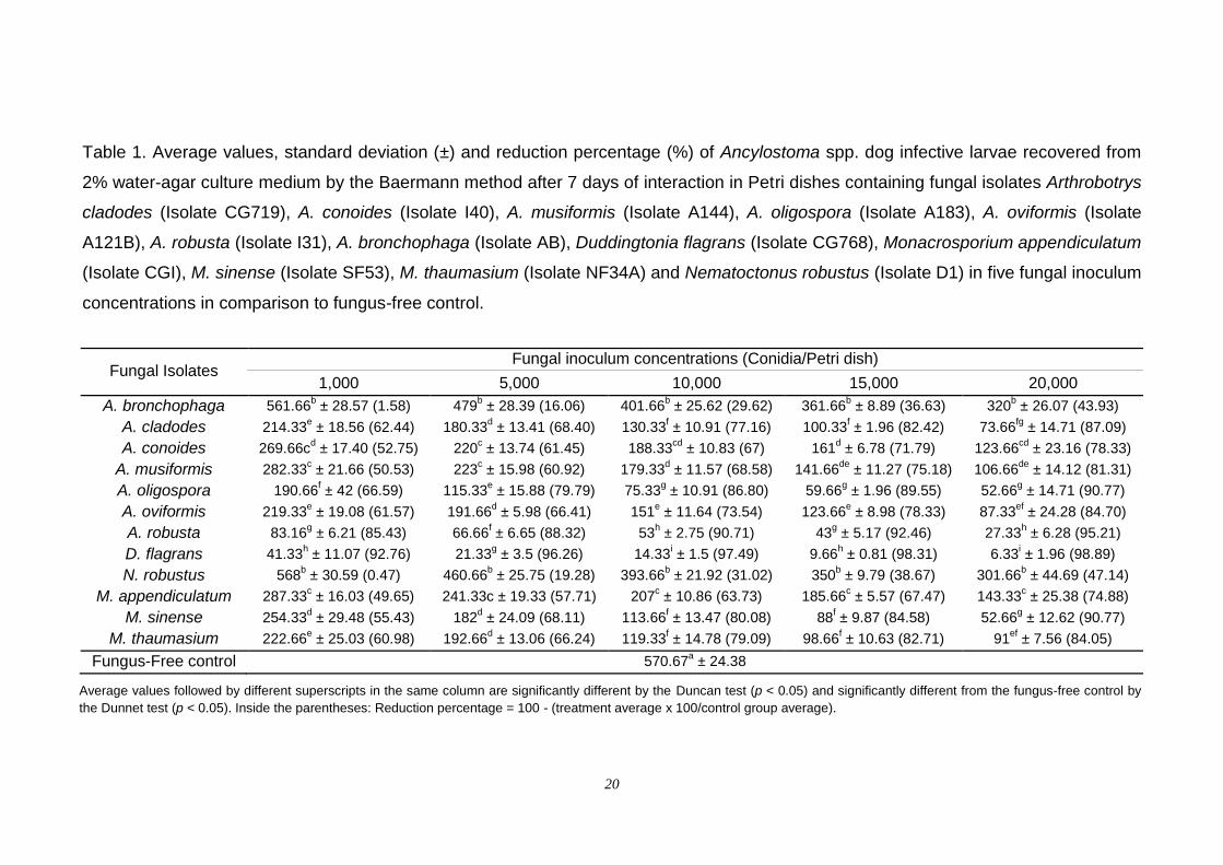
Table 1 shows the averages, standard deviation and reduction
percentages of Ancylostoma spp. L3 in Petri dishes with a 2% WA culture
medium, treated with the fungal isolates, in comparison to fungus-free control.
All nematode-trapping fungi isolates used in the in vitro test were able to
capture Ancylostoma spp. L3 and significantly reduced the average of the
uncaptured L3 recovered in comparison to the fungus-free control (p < 0.05),
within the period of observation. In general, the highest and the lowest
predatory activity were achieved by adding 20,000 and 1,000 conidia per Petri
dish, respectively.
All isolates in the five concentrations were significantly different (p <
19
0.05) when compared to the fungus-free control. The interaction between the
isolates and the conidial concentration was significant (p < 0.05). The
comparison between the treatments showed a variation in the efficiency of the
isolates in reducing Ancylostoma spp. L3, according to the fungal inoculum
concentration. The Isolates CG768, I31 and A183 showed a better capture and
destruction results and consequently greater decrease in the average of the
uncaptured L3 recovered.
The Isolate CG768 showed a high individual predatory activity against
Ancylostoma spp. L3. This effect has a direct relation with the increase of the
fungal inoculum concentration: 92.8%, 96.3%, 97.5%, 98.3% and 98.9% of
reduction of uncaptured Ancylostoma spp. L3, for 1,000, 5,000, 10,000, 15,000
or 20,000 conidia added per Petri dish, respectively (Fig. 1H). The Isolate I31
reduced the average of uncaptured Ancylostoma spp. L3 recovered by 85.4%,
88.3%, 90.7%, 92.5% and 95.2%, also with increasing fungal inoculum
concentration (Fig. 1F), followed by the Isolate A183, with reductions of 66.6%,
79.8%, 86.8%, 89.5% and 90.8% (Fig. 1D). However, there was no difference
between the Isolates I31 and A183 in the concentration of 15,000 conidia (Table
1). The same occurred in the concentration of 20,000 conidia among the
Isolates A183, CG719 and SF53 (Table 1). The lowest reduction occurred in
treatments with the Isolates D1 and AB with the same reduction of the
uncaptured Ancylostoma spp. L3 recovered in all concentrations. In general,
trap formations were much more pronounced in Isolate CG768 Petri dishes in
response to the addition of Ancylostoma spp. L3, while the Isolates D1 and AB
were the poorest trap-inducers, producing very few traps towards the end of the
observation period, even after an increase of the inoculum.
20
Table 1. Average values, standard deviation (±) and reduction percentage (%) of Ancylostoma spp. dog infective larvae recovered from
2% water-agar culture medium by the Baermann method after 7 days of interaction in Petri dishes containing fungal isolates Arthrobotrys
cladodes (Isolate CG719), A. conoides (Isolate I40), A. musiformis (Isolate A144), A. oligospora (Isolate A183), A. oviformis (Isolate
A121B), A. robusta (Isolate I31), A. bronchophaga (Isolate AB), Duddingtonia flagrans (Isolate CG768), Monacrosporium appendiculatum
(Isolate CGI), M. sinense (Isolate SF53), M. thaumasium (Isolate NF34A) and Nematoctonus robustus (Isolate D1) in five fungal inoculum
concentrations in comparison to fungus-free control.
Fungal Isolates Fungal inoculum concentrations (Conidia/Petri dish)
1,000 5,000 10,000 15,000 20,000
A. bronchophaga 561.66b ± 28.57 (1.58) 479
b ± 28.39 (16.06) 401.66
b ± 25.62 (29.62) 361.66
b ± 8.89 (36.63) 320
b ± 26.07 (43.93)
A. cladodes 214.33e ± 18.56 (62.44) 180.33
d ± 13.41 (68.40) 130.33
f ± 10.91 (77.16) 100.33
f ± 1.96 (82.42) 73.66
fg ± 14.71 (87.09)
A. conoides 269.66cd ± 17.40 (52.75) 220
c ± 13.74 (61.45) 188.33
cd ± 10.83 (67) 161
d ± 6.78 (71.79) 123.66
cd ± 23.16 (78.33)
A. musiformis 282.33c ± 21.66 (50.53) 223
c ± 15.98 (60.92) 179.33
d ± 11.57 (68.58) 141.66
de ± 11.27 (75.18) 106.66
de ± 14.12 (81.31)
A. oligospora 190.66f ± 42 (66.59) 115.33
e ± 15.88 (79.79) 75.33
g ± 10.91 (86.80) 59.66
g ± 1.96 (89.55) 52.66
g ± 14.71 (90.77)
A. oviformis 219.33e ± 19.08 (61.57) 191.66
d ± 5.98 (66.41) 151
e ± 11.64 (73.54) 123.66
e ± 8.98 (78.33) 87.33
ef ± 24.28 (84.70)
A. robusta 83.16g ± 6.21 (85.43) 66.66
f ± 6.65 (88.32) 53
h ± 2.75 (90.71) 43
g ± 5.17 (92.46) 27.33
h ± 6.28 (95.21)
D. flagrans 41.33h ± 11.07 (92.76) 21.33
g ± 3.5 (96.26) 14.33
i ± 1.5 (97.49) 9.66
h ± 0.81 (98.31) 6.33
i ± 1.96 (98.89)
N. robustus 568b ± 30.59 (0.47) 460.66
b ± 25.75 (19.28) 393.66
b ± 21.92 (31.02) 350
b ± 9.79 (38.67) 301.66
b ± 44.69 (47.14)
M. appendiculatum 287.33c ± 16.03 (49.65) 241.33c ± 19.33 (57.71) 207
c ± 10.86 (63.73) 185.66
c ± 5.57 (67.47) 143.33
c ± 25.38 (74.88)
M. sinense 254.33d ± 29.48 (55.43) 182
d ± 24.09 (68.11) 113.66
f ± 13.47 (80.08) 88
f ± 9.87 (84.58) 52.66
g ± 12.62 (90.77)
M. thaumasium 222.66e ± 25.03 (60.98) 192.66
d ± 13.06 (66.24) 119.33
f ± 14.78 (79.09) 98.66
f ± 10.63 (82.71) 91
ef ± 7.56 (84.05)
Fungus-Free control 570.67a ± 24.38
Average values followed by different superscripts in the same column are significantly different by the Duncan test (p < 0.05) and significantly different from the fungus-free control by
the Dunnet test (p < 0.05). Inside the parentheses: Reduction percentage = 100 - (treatment average x 100/control group average).
21
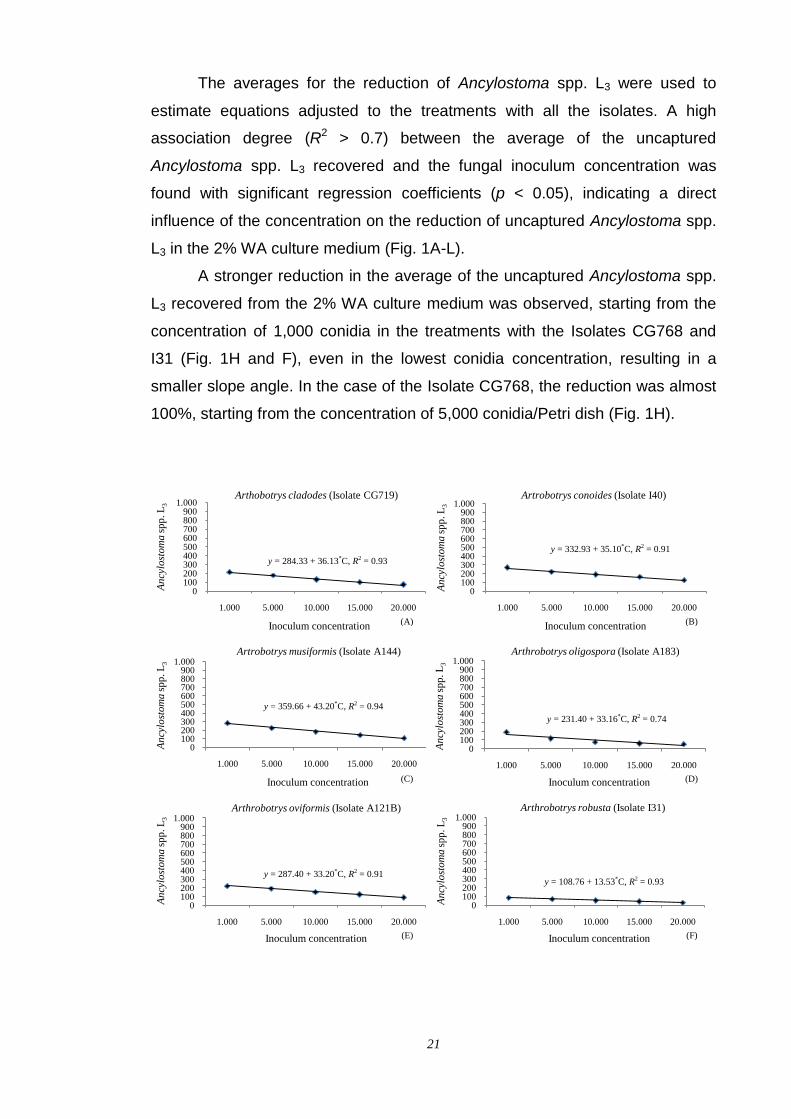
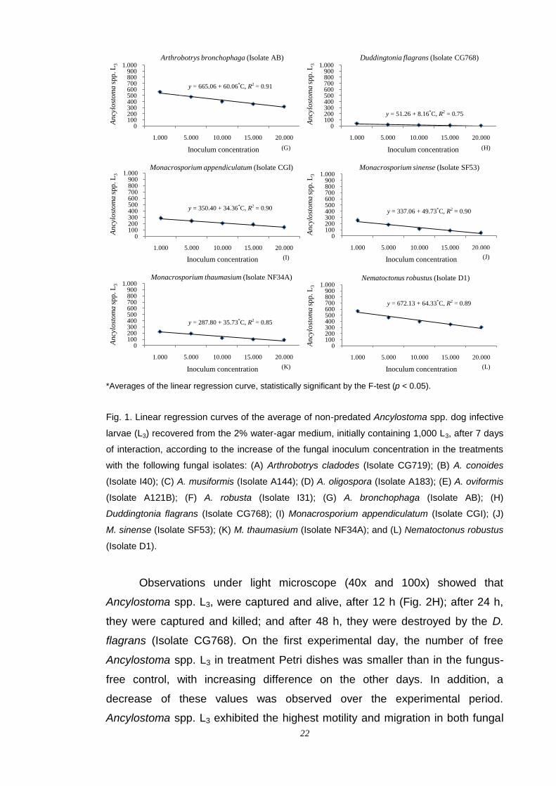
The averages for the reduction of Ancylostoma spp. L3 were used to
estimate equations adjusted to the treatments with all the isolates. A high
association degree (R2 > 0.7) between the average of the uncaptured
Ancylostoma spp. L3 recovered and the fungal inoculum concentration was
found with significant regression coefficients (p < 0.05), indicating a direct
influence of the concentration on the reduction of uncaptured Ancylostoma spp.
L3 in the 2% WA culture medium (Fig. 1A-L).
A stronger reduction in the average of the uncaptured Ancylostoma spp.
L3 recovered from the 2% WA culture medium was observed, starting from the
concentration of 1,000 conidia in the treatments with the Isolates CG768 and
I31 (Fig. 1H and F), even in the lowest conidia concentration, resulting in a
smaller slope angle. In the case of the Isolate CG768, the reduction was almost
100%, starting from the concentration of 5,000 conidia/Petri dish (Fig. 1H).
0100200300400500600700800900
1.000
1.000 5.000 10.000 15.000 20.000
An
cylo
stom
asp
p. L
3
Inoculum concentration
Arthobotrys cladodes (Isolate CG719)
0100200300400500600700800900
1.000
1.000 5.000 10.000 15.000 20.000
An
cylo
stom
asp
p. L
3
Inoculum concentration
Artrobotrys conoides (Isolate I40)
0100200300400500600700800900
1.000
1.000 5.000 10.000 15.000 20.000
An
cylo
stom
asp
p. L
3
Inoculum concentration
Artrobotrys musiformis (Isolate A144)
0100200300400500600700800900
1.000
1.000 5.000 10.000 15.000 20.000
An
cylo
stom
asp
p. L
3
Inoculum concentration
Arthrobotrys oligospora (Isolate A183)
0100200300400500600700800900
1.000
1.000 5.000 10.000 15.000 20.000
An
cylo
stom
asp
p. L
3
Inoculum concentration
Arthrobotrys oviformis (Isolate A121B)
0100200300400500600700800900
1.000
1.000 5.000 10.000 15.000 20.000
An
cylo
stom
asp
p. L
3
Inoculum concentration
Arthrobotrys robusta (Isolate I31)
y = 287.40 + 33.20*C, R2 = 0.91
(A)
(E)
y = 284.33 + 36.13*C, R2 = 0.93
(B)
y = 332.93 + 35.10*C, R2 = 0.91
(C)
y = 359.66 + 43.20*C, R2 = 0.94
(D)
y = 231.40 + 33.16*C, R2 = 0.74
(F)
y = 108.76 + 13.53*C, R2 = 0.93
22
*Averages of the linear regression curve, statistically significant by the F-test (p < 0.05).
Fig. 1. Linear regression curves of the average of non-predated Ancylostoma spp. dog infective
larvae (L3) recovered from the 2% water-agar medium, initially containing 1,000 L3, after 7 days
of interaction, according to the increase of the fungal inoculum concentration in the treatments
with the following fungal isolates: (A) Arthrobotrys cladodes (Isolate CG719); (B) A. conoides
(Isolate I40); (C) A. musiformis (Isolate A144); (D) A. oligospora (Isolate A183); (E) A. oviformis
(Isolate A121B); (F) A. robusta (Isolate I31); (G) A. bronchophaga (Isolate AB); (H)
Duddingtonia flagrans (Isolate CG768); (I) Monacrosporium appendiculatum (Isolate CGI); (J)
M. sinense (Isolate SF53); (K) M. thaumasium (Isolate NF34A); and (L) Nematoctonus robustus
(Isolate D1).
Observations under light microscope (40x and 100x) showed that
Ancylostoma spp. L3, were captured and alive, after 12 h (Fig. 2H); after 24 h,
they were captured and killed; and after 48 h, they were destroyed by the D.
flagrans (Isolate CG768). On the first experimental day, the number of free
Ancylostoma spp. L3 in treatment Petri dishes was smaller than in the fungus-
free control, with increasing difference on the other days. In addition, a
decrease of these values was observed over the experimental period.
Ancylostoma spp. L3 exhibited the highest motility and migration in both fungal
0100200300400500600700800900
1.000
1.000 5.000 10.000 15.000 20.000
An
cylo
stom
asp
p. L
3
Inoculum concentration
Arthrobotrys bronchophaga (Isolate AB)
0100200300400500600700800900
1.000
1.000 5.000 10.000 15.000 20.000
An
cylo
stom
asp
p. L
3
Inoculum concentration
Duddingtonia flagrans (Isolate CG768)
0100200300400500600700800900
1.000
1.000 5.000 10.000 15.000 20.000
An
cylo
stom
asp
p. L
3
Inoculum concentration
Monacrosporium appendiculatum (Isolate CGI)
0100200300400500600700800900
1.000
1.000 5.000 10.000 15.000 20.000
An
cylo
stom
asp
p. L
3
Inoculum concentration
Monacrosporium sinense (Isolate SF53)
0100200300400500600700800900
1.000
1.000 5.000 10.000 15.000 20.000
An
cylo
stom
asp
p. L
3
Inoculum concentration
Monacrosporium thaumasium (Isolate NF34A)
0100200300400500600700800900
1.000
1.000 5.000 10.000 15.000 20.000
An
cylo
stom
asp
p. L
3
Inoculum concentration
Nematoctonus robustus (Isolate D1)
y = 665.06 + 60.06*C, R2 = 0.91
(G) (H)
y = 51.26 + 8.16*C, R2 = 0.75
y = 350.40 + 34.36*C, R2 = 0.90
(I) (J)
y = 337.06 + 49.73*C, R2 = 0.90
(K)
y = 287.80 + 35.73*C, R2 = 0.85
(L)
y = 672.13 + 64.33*C, R2 = 0.89

23
treatments and fungus-free control, being widely dispersed over the WA 2%
surface. In the fungus-free control group, some of them died without the
presence of the predatory fungi, while others migrated to the periphery and
tended to concentrate in the border of the WA 2% in Petri dishes.
The similarities, in terms of general morphology, nematophagous habits
and conidial dimensions in this study are consistent with the published
descriptions of the fungal species tested. In all treatments, the conidial
production was observed around 2 days after the initial capture of Ancylostoma
spp. L3. However, the sporulation was higher between 4 and 6 days. During the
experimental period, free Ancylostoma spp. L3 were occasionally seen
migrating freely, with traps and conidia attached to their cuticle, suggesting that
they had succeeded in releasing themselves and evidencing the presence of
adhesive substance on the conidia surface. These fungi were relatively easy to
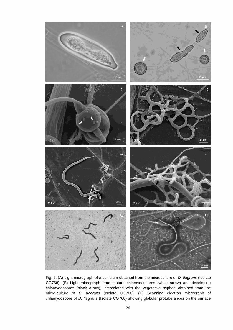
culture on artificial media with V8-agar. The conidia of the fungus D. flagrans
were elliptical-shaped (Fig. 2A). The fungal colonization of the 2% WA medium
and capture of Ancylostoma spp. L3 were not observed in the fungus-free
control (Fig. 2G). In the treatments with the fungus D. flagrans (Isolate CG768),
the formation of abundant chlamydospores intercalating the hyphae (Fig. 2B)
was observed on the sixth day. These chlamydospores had globular
protuberances on their surface (Fig. 2C).
A scanning electron micrograph showed that the fungus D. flagrans
(Isolate CG768) captured the Ancylostoma spp. L3 by adhesive three-
dimensional network traps, grown on the surface of the dialysis membrane (Fig.
2D, E and F). Two fungus-larvae interaction points were identified 12 h after the
initial capture, with the presence of bacillus rod-shaped bacteria associated to
fungal/nematode interaction sites in possible penetration points. In one of these
points, the modified hyphae involved the Ancylostoma spp. L3 (Fig. 2F).
24
Fig. 2. (A) Light micrograph of a conidium obtained from the microculture of D. flagrans (Isolate
CG768). (B) Light micrograph from mature chlamydospores (white arrow) and developing
chlamydospores (black arrow), intercalated with the vegetative hyphae obtained from the
micro-culture of D. flagrans (Isolate CG768). (C) Scanning electron micrograph of
chlamydospore of D. flagrans (Isolate CG768) showing globular protuberances on the surface
25
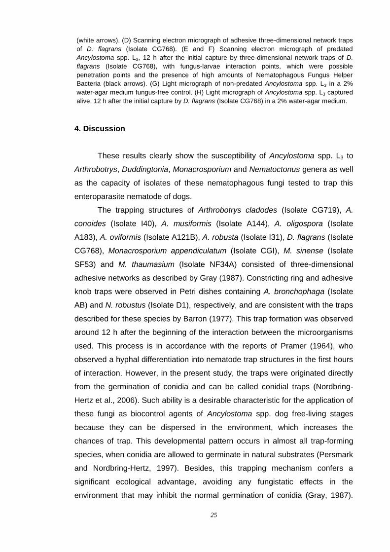
(white arrows). (D) Scanning electron micrograph of adhesive three-dimensional network traps
of D. flagrans (Isolate CG768). (E and F) Scanning electron micrograph of predated
Ancylostoma spp. L3, 12 h after the initial capture by three-dimensional network traps of D.
flagrans (Isolate CG768), with fungus-larvae interaction points, which were possible
penetration points and the presence of high amounts of Nematophagous Fungus Helper
Bacteria (black arrows). (G) Light micrograph of non-predated Ancylostoma spp. L3 in a 2%
water-agar medium fungus-free control. (H) Light micrograph of Ancylostoma spp. L3 captured
alive, 12 h after the initial capture by D. flagrans (Isolate CG768) in a 2% water-agar medium.
4. Discussion
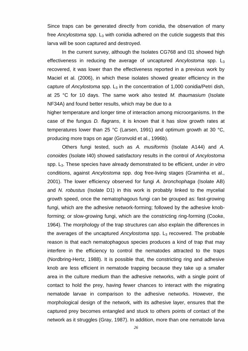
These results clearly show the susceptibility of Ancylostoma spp. L3 to
Arthrobotrys, Duddingtonia, Monacrosporium and Nematoctonus genera as well
as the capacity of isolates of these nematophagous fungi tested to trap this
enteroparasite nematode of dogs.
The trapping structures of Arthrobotrys cladodes (Isolate CG719), A.
conoides (Isolate I40), A. musiformis (Isolate A144), A. oligospora (Isolate
A183), A. oviformis (Isolate A121B), A. robusta (Isolate I31), D. flagrans (Isolate
CG768), Monacrosporium appendiculatum (Isolate CGI), M. sinense (Isolate
SF53) and M. thaumasium (Isolate NF34A) consisted of three-dimensional
adhesive networks as described by Gray (1987). Constricting ring and adhesive
knob traps were observed in Petri dishes containing A. bronchophaga (Isolate
AB) and N. robustus (Isolate D1), respectively, and are consistent with the traps
described for these species by Barron (1977). This trap formation was observed
around 12 h after the beginning of the interaction between the microorganisms
used. This process is in accordance with the reports of Pramer (1964), who
observed a hyphal differentiation into nematode trap structures in the first hours
of interaction. However, in the present study, the traps were originated directly
from the germination of conidia and can be called conidial traps (Nordbring-
Hertz et al., 2006). Such ability is a desirable characteristic for the application of
these fungi as biocontrol agents of Ancylostoma spp. dog free-living stages
because they can be dispersed in the environment, which increases the
chances of trap. This developmental pattern occurs in almost all trap-forming
species, when conidia are allowed to germinate in natural substrates (Persmark
and Nordbring-Hertz, 1997). Besides, this trapping mechanism confers a
significant ecological advantage, avoiding any fungistatic effects in the
environment that may inhibit the normal germination of conidia (Gray, 1987).
26
Since traps can be generated directly from conidia, the observation of many
free Ancylostoma spp. L3 with conidia adhered on the cuticle suggests that this
larva will be soon captured and destroyed.
In the current survey, although the Isolates CG768 and I31 showed high
effectiveness in reducing the average of uncaptured Ancylostoma spp. L3
recovered, it was lower than the effectiveness reported in a previous work by
Maciel et al. (2006), in which these isolates showed greater efficiency in the
capture of Ancylostoma spp. L3 in the concentration of 1,000 conidia/Petri dish,
at 25 °C for 10 days. The same work also tested M. thaumasium (Isolate
NF34A) and found better results, which may be due to a
higher temperature and longer time of interaction among microorganisms. In the
case of the fungus D. flagrans, it is known that it has slow growth rates at
temperatures lower than 25 °C (Larsen, 1991) and optimum growth at 30 °C,
producing more traps on agar (Gronvold et al., 1996b).
Others fungi tested, such as A. musiformis (Isolate A144) and A.
conoides (Isolate I40) showed satisfactory results in the control of Ancylostoma
spp. L3. These species have already demonstrated to be efficient, under in vitro
conditions, against Ancylostoma spp. dog free-living stages (Graminha et al.,
2001). The lower efficiency observed for fungi A. bronchophaga (Isolate AB)
and N. robustus (Isolate D1) in this work is probably linked to the mycelial
growth speed, once the nematophagous fungi can be grouped as: fast-growing
fungi, which are the adhesive network-forming; followed by the adhesive knob-
forming; or slow-growing fungi, which are the constricting ring-forming (Cooke,
1964). The morphology of the trap structures can also explain the differences in
the averages of the uncaptured Ancylostoma spp. L3 recovered. The probable
reason is that each nematophagous species produces a kind of trap that may
interfere in the efficiency to control the nematodes attracted to the traps
(Nordbring-Hertz, 1988). It is possible that, the constricting ring and adhesive
knob are less efficient in nematode trapping because they take up a smaller
area in the culture medium than the adhesive networks, with a single point of
contact to hold the prey, having fewer chances to interact with the migrating
nematode larvae in comparison to the adhesive networks. However, the
morphological design of the network, with its adhesive layer, ensures that the
captured prey becomes entangled and stuck to others points of contact of the
network as it struggles (Gray, 1987). In addition, more than one nematode larva
27
can be trapped in the same network. This suggests that predatory fungi with
adhesive networks would be able to capture and consume larger Ancylostoma
spp. L3 than fungi with constricting ring or adhesive knob.
A daily observation of the Petri dishes showed numerous Ancylostoma
spp. L3 captured at the cephalic portion, which may probably happen because
this portion is the first part of the nematode body to get in contact with the traps
when they are attracted by this structure. The carbohydrates on the surface are
found scattered over the whole surface, but they are frequently found at the
head or at the tail, which explains the frequent binding of fungal traps to such
nematode region, as reported by Jansson (1987). It was known that the
adhesive substances on the surface of the trap hyphae hold the nematode,
although the carbohydrate-binding protein (lectin) found on the fungal traps,
which specifically bind to the carbohydrate receptor found on the nematode
cuticle, would probably be enough to hold the nematode (Zuckerman and
Jansson, 1984; Jansson, 1987; Murray and Wharton, 1990). Other interesting
observation was the presence of scattered rod-shaped bacteria on the cellulose
membrane surface of the Petri dishes, mainly at the points of interaction
between the fungus D. flagrans and Ancylostoma spp. L3. These bacteria
probably act as helpers of the fungus D. flagrans activity and, according to
Dupponois et al. (1998), they were called Nematophagous Fungus Helper
Bacteria (NHB) and may be involved in the predation, sporulation and
pathogenicity systems of nematophagous fungi. Moreover, the same researcher
reported that these bacteria produce substances that would act as molecular
bridges between fungi and nematodes, which could explain the high amount of
such bacteria at the interaction points 12 and 24 h after the beginning of the
capture of Ancylostoma spp. L3. Due to this fact, the study of the effect of these
bacteria in association to the fungus D. flagrans could provide a better
understanding of their function in the interaction process with Ancylostoma spp.
L3, since only Campos et al. (2008) observed this bacterial association with
fungus D. flagrans in literature, although the researcher has worked with
predation of Haemonchus contortus L3. The association of bacteria with
nematophagous fungi is also recorded by Graminha et al. (2001), who studied
the pathogenicity of the fungi Arthrobotrys musiformis and A. conoides on the of
H. contortus and Ancylostoma spp. L3.
It is known that some nematophagous fungi may not need stimulation for
28
trap formation, but others may be stimulated to form the traps by the presence
of the nematode, inductive substances and others in adverse conditions. It is
possible that the presence of Ancylostoma spp. L3, with its rapid movements, or
substances derived or excreted by it, in the Petri dishes containing 2% WA,
stimulated trap formation by the tested fungal isolates. This can be explained by
the fact that a culture medium poor in nutrients, such as 2% WA, encourages
the nematophagous fungi to use nematodes as an alternative source of
available nutrients (Scholler and Rubner, 1994). Then, the formation of trap
structures will be a symptom of nutritional deficiency in nematophagous fungi,
since in a culture medium with substrates containing adequate carbon and
nitrogen concentrations, these fungi live as saprophytes, forming few or no trap
structures (Scholler and Rubner, 1994; Morgan et al., 1997). Barron (1977),
discussing about the induction of trap formation, emphasizes that researchers
studying morphogenesis in predatory fungi, usually confined their studies to a
single predator, hampering comparisons because most studies use other
species. In addition, different species or even different strains of the same
species may behave differently in their response to both kind and concentration
of morphogenic inductor, although many factors are involved in trap formation
(Barron, 1977). In natural environments, the nutritional conditions are generally
unfavorable. Then, not only the presence of the nematode prey, but also the
lack of nutrients may favor the production of trapping devices (Balan and
Lechevalier, 1972). In addition, there is a range of peptides or amino acids in
the soil, resulting from the biological degradation of proteinaceous material,
capable of eliciting trap formation (Nordbring-Hertz, 1973). Based on these
previous statements and in vitro results of the present survey, it is probable that
the trap formation and consequent capture of Ancylostoma spp. L3 by the
nematophagous fungi D. flagrans (Isolate CG 768), A. robusta (Isolate I31) and
A. oligospora (Isolate A183) will be favored in the soil.
In the present study, D. flagrans (Isolate CG768) not only showed greater
virulence against Ancylostoma spp. L3, presenting the best results in
comparison to the other fungi tested, but also produced abundant
chlamydospores, as described for this species (Juarez and Mendoza-de-Gives,
1998; Larsen, 1999), which are thick-walled structures, developed from the
hyphae, that appear in unfavorable growth conditions (Scholler and Rubner,
1994) and can originate hyphae, conidiophores and conidia (Barron, 1977).
29
Chlamydospores are persistent in the environment, which favors their
establishment and survival, and consequently their use as a biological control
agent (Faedo et al., 2000). This fungal species is the most studied
nematophagous fungus in experiments throughout the world and is considered
promising for the biological control of gastrointestinal nematodiosis of several
species of domestic animals (Gronvold et al., 1996a; Larsen, 2000; Faedo et
al., 2002; Fontenot et al., 2003). Then, these characteristics and statements
show that D. flagrans (Isolate CG768) could be an alternative for the biological
control of Ancylostoma spp. L3.
The administration of anthelminthics to animals has been the method
used to prevent environmental contamination by Ancylostoma spp. eggs and
larvae. However, the nematophagous fungi could be used jointly in situations
where the environment is already contaminated, offering an environmentally
sustainable method of control. The researchers Waller and Larsen (1993)
discuss that the fungal application in the biological control of free-living stages
of parasitic nematodes can help the chemical control. This control should be
used not only in the prediction of major environmental infestation by free-living
forms of helminths but also when there are better conditions for the fungal
growth in the environment, preventing clinical parasitism and allowing the
survival of sufficient larvae to develop animal immunity. The application of the
fungus as a biological control agent of Ancylostoma spp. dog free-living stages
could be of great use in Brazil, since dog‟s hookworm larvae are widespread in
the soils, public squares and public parks (Santarém et al., 2004). This
applicability can be enhanced, since street dogs must be considered in a worm
control program in urban centers (Labruna et al., 2006). Although these animals
play an important role in the maintenance and dissemination of these parasites
in the urban environment, they have been excluded from programs. Given the
close interaction between dogs and humans, it becomes absolutely necessary
to use an adequate control in order to reduce the environmental contamination
by infective larvae and thus minimize the risk of human and canine infection.
Nematophagous fungi present different predatory activities. While some
isolates need only a few conidia to get the expected effect, others need a large
inoculum to get the same effect. The varying susceptibility of Ancylostoma spp.
L3 to the different fungal isolates suggests that in vitro studies are important to
select nematophagous fungi before their application for biological control.

30
Although such tests usually overestimate the activity of a biological control
agent by not allowing the nematode to escape or even by not reproducing the
possible environmental changes and interferences commonly found in the soil,
they have nevertheless the advantages of a limited physical space and shorter
evaluation time (Gomes et al., 2001). To choose a fungal isolate, it is also
necessary to consider field tests that can prove the efficiency of these
microorganisms as potential biological control agents. Research should focus
on the best way to implement these fungi in the environment, evaluating if the
formulations consisting of vegetative, reproductive or resistance structures
would allow their establishment in the complex soil ecosystem and enable the
control of Ancylostoma spp. dog free-living stages, breaking the life cycle of this
parasite. Results showed that most fungi isolates tested were efficient in the in
vitro destruction of Ancylostoma spp. L3 and are promising alternatives in the
control of free-living stages nematodes in infested areas. However, further
studies are necessary to evaluate if it could bring damage to the soil
microfauna.
31
CAPÍTULO 2
Scanning electron microscopy of Ancylostoma spp. dog infective larvae
captured and destroyed by the nematophagous fungus Duddingtonia
flagrans
Micron 40(4):463-470, 2009
32
Abstract: The interaction between the nematode-trapping fungus Duddingtonia
flagrans (Isolate CG768) and Ancylostoma spp. dog infective larvae (L3) was
evaluated by means of scanning electron microscopy. Adhesive network trap
formation was observed 6 h after the beginning of the interaction, and the
capture of Ancylostoma spp. L3 was observed 8 h after the inoculation these
larvae on the cellulose membranes colonized by the fungus. Scanning electron
micrographs were taken at 0, 12, 24, 36 and 48 h, where 0 is the time when
Ancylostoma spp. L3 was first captured by the fungus. Details of the capture
structure formed by the fungus were described. Nematophagous Fungus Helper
Bacteria (NHB) were found at interactions points between the D. flagrans and
Ancylostoma spp. L3. The cuticle penetration by the differentiated fungal hyphae
with the exit of nematode internal contents was observed 36 h after the capture.
Ancylostoma spp. L3 were completely destroyed after 48 h of interaction with
the fungus. The scanning electron microscopy technique was efficient on the
study of this interaction, showing that the nematode-trapping fungus D. flagrans
(Isolate CG768) is a potential exterminator of Ancylostoma spp. L3.
Keywords: Nematophagous fungus, Duddingtonia flagrans, Ancylostoma spp.,
dogs, biological control
1. Introduction
Nematophagous fungi have been studied as an alternative for controlling
several nematodes (Araújo et al., 1993; Nansen et al., 1996; Chandrawathani et
al., 1998; Araújo and Guimarães, 2002; Oliveira et al., 2002; Castro et al.,
2003).
The species Duddingtonia flagrans is an important nematode-trapping
fungus that has been used as a biological control agent of the free-living stages
of gastrointestinal parasites of domestic animals (Baudena et al., 2000; Paraud
et al., 2005; Maciel et al., 2006; Rocha et al., 2007).
Since dog parasite helminths of the Ancylostoma genus have been
requesting attention for their zoonotic potential (Soulsby, 1982; Schantz, 1991;
Prociv and Croese, 1996; Bahgat et al., 1999; Robertson et al., 2000), the
destruction of their infective stages by using nematophagous fungi may be an
alternative for the control of animal parasitic nematodes under environmental
conditions.

33
Thus, the characterization of the fungus x larvae interaction processes
would be very important, once this interaction is beneficial to the
nematophagous fungi but not to the nematode larvae. This could influence on
the selection of promising fungal isolates which might be used in biological
control programs (Mendoza-de-Gives, 1999). Despite the studies that have
been carried out, there is little information on the ultrastructural aspects of the
interaction between the fungus D. flagrans and the gastrointestinal parasite
nematode infective larvae of domestic animals.
The scope of this study was to observe and understand the interaction
processes between the nematode-trapping fungus D. flagrans (Isolate CG768)
against Ancylostoma spp. dog infective larvae (L3).
2. Materials and methods
2.1. Acquirement of Ancylostoma spp. infective larvae
The Ancylostoma spp. L3 were obtained from fresh feces of naturally
infected urban street dogs by means of vermiculite-coproculture kept for 10
days at 26 °C. At the end of the incubation period, the L3 were extracted by the
modified Baermann‟s method, as described by Ueno and Gonçalves (1998).
After 12 h, the sediment containing Ancylostoma spp. L3 was transferred to a
centrifuge tube and washed with distilled water five times at 1,000 rpm for 5
min. The supernatant was disposed at the end of each centrifugation. The
methodology described by Barçante et al. (2003) was used to filter and
eliminate debris to obtain viable and active Ancylostoma spp. L3 in a clean
preparation.
The selected Ancylostoma spp. L3 suspension was homogenized and six
aliquots of 10 mL were collected with a micropipette and placed over a slide
marked with longitudinal lines to help orientation. Each aliquot was covered with
a glass coverslip after the addition of 10 mL of lugol‟s solution to kill the
Ancylostoma spp. L3. Then, they were counted and identified under a light
microscope in magnification of 40x. The average of the Ancylostoma L3 aliquots
was calculated, which enabled the estimation of the average in the total
suspension.
34
2.2. Scanning electron microscopy
The material preparation for the observation of larva/fungus interaction
under the scanning electron microscopy followed the methodologies described
by Nordbring-Hertz (1983) and Campos et al. (2008).
Seven 8 cm-diameter cellulose membrane discs (dialysis membrane)
were used, containing proteins which weigh more than 12,000 Da, with filtering
capacity of 640 mL/ft (Sigma-Aldrich®, USA). These discs were placed in a 100
mL Griffin cup that was filled with distilled water up to the 50 mL mark, closed
with foil, and sealed with PCV film. The cups were later autoclaved at 121 °C for
15 min.
After being removed from the Griffin cup with tweezers, the discs were
placed in seven 80 mm x 15 mm sterile Petri‟s dishes with 10 mL of 2% water-
agar (2% WA Merse1, Brazil) so that the membrane covered the whole surface
of the medium culture
and its border touched the glass wall of the Petri‟s dishes. On several sites of
such a junction, small amounts of 2% WA were dropped with aid of a repeater
pipette calibrated at 10 mL in order to seal it and prevent L3 from escaping
under the membrane.
Whole rice grains colonized by the nematode-trapping fungus D. flagrans
(Isolate CG768) were stored at 4 °C in the Laboratório de Controle Biológico,
Universidade Federal de Viçosa, Brazil, in the absence of light inside 5 mL
vacutainer-like glass tubes filled up to their halves with blue silica-gel. A single
rice grain colonized by this fungal isolate was transferred to the centre of 50 mm
x 10 mm sterile Petri‟s dishes containing 5 mL of 2% WA. The plates were then
incubated in the dark at 25 °C for 7 days. After the mycelial growth of this new
culture, a 5 mm-diameter disc was replicated at the cellulose membrane
surfaces of 80 mm x 15 mm Petri‟s dishes containing 10 mL of 2% WA medium
(Nordbring-Hertz, 1983) that were afterwards incubated in the dark at 25 °C for
5 days. After that, the 5 mm-diameter culture disc was removed from the
cellulose membrane surface and 20 mL of a suspension with nearly 10,000
Ancylostoma spp. L3 were dropped on the D. flagrans culture. Two controls
were performed, one with Ancylostoma spp. L3 and the other with the
nematode-trapping fungus D. flagrans (Isolate CG768).
During the first hours following the nematode inoculation, the Petri‟s

35
dishes colonized by the nematode-trapping fungus D. flagrans (Isolate CG768)
were observed hourly under light microscope using magnification of 10x. After
observing the capture of Ancylostoma spp. L3 by this fungal isolate in a certain
site of the membrane, marks were made on the outer surface of the Petri‟s
dishes by using a fine-tip permanent ink maker, aiming to determine the D.
flagrans/Ancylostoma spp. L3 interaction time and find the initial capture site in
further observations.
At the capture site, the cellulose membrane was cut into 7 mm2 patches
with aid of a scalpel blade. The patches had samples of recently captured L3 (0
h) and samples collected 12, 24, 36 and 48 h after the beginning of predation.
The patches were collected by using fine-tip tweezers and then fixed in 2.5%
glutaraldehyde in 0.05 M phosphate buffer pH 7.4 at 4 °C, for 5 days, inside
closed 1 mL cryopreservation tubes. Afterwards, the patches were removed and
washed for three times in the same buffer and dehydrated through an ethanol
series (30, 50, 60, 70, 95 and 100%) during 10 min for each concentration, and
the highest one was repeated three times. The same material was submitted to
Critical Point Drier (Balzers® CPD 020) by using carbon dioxide, covered with
gold in Sputter-Coater (Balzers® SCA 010), after which electron micrographs
were taken with a scanning electron microscope (LEO 1430 VP) at 20 kV.
3. Results
The initial formation of adhesive three-dimensional network traps, more
robust than the vegetative hyphae, was observed 6 h after the Ancylostoma
spp. L3 were added to the Petri‟s dishes with cellulose membrane colonized by
the fungus D. flagrans (Fig. 1 A and B). The three-dimensional networks,
spaced at intervals along the length of the vegetative hyphae, were found in the
whole cellulose membrane surface probably due to the fact that the L3 were
active and therefore dispersed on it (Fig. 1C). Initially, the traps did not show the
network shape but were a simple loop of a lateral branch from a vegetative
hypha that grew out, curved round and anastomosed with the parent hypha
(Fig. 1A). New hyphal anastomoses of the branch from this primary loop or from
another branch of the vegetative hypha were observed with the advance of the
trap formation process, resulting in the adhesive three-dimensional network 12
h after the start of the interaction (Fig. 1B). It was not observed the formation of
36
traps in the control without Ancylostoma spp. L3, but only the vegetative growth
coating the surface of the membrane (Fig. 1D). The Ancylostoma spp. L3 were
active, moving with their characteristic serpentine motion, turgid and with
integral membrane in the control without fungi (Fig. 1E).
Nearly 8 h after the addition of Ancylostoma spp. L3 in the Petri‟s dishes,
it was observed the capture of Ancylostoma spp. L3 by the differentiated fungal
hyphae, which had not evolved into a three-dimensional network yet (Fig. 1F).
Pressure points could be seen on the cuticle of the Ancylostoma spp. L3
recently captured by the trap hyphae, causing a small depression in the sheath
where the penetration process began (Fig. 2G). In another situation, the
Ancylostoma spp. L3 captured was able to abandon the outer cuticle adhered in
the fungal trap before being penetrated by the invasive hyphae (Fig. 2H). The
majority of the larvae captured by the fungus D. flagrans were trapped at their
cephalic portion (Fig. 2I); however, some Ancylostoma spp. L3 were trapped at
other parts of the body (Fig. 2J) and it was observed that some larvae were
trapped even at both portions (Figs. 1F and 2G).
With the advance of the predation process, 12 h after the beginning of L3
capture by the differentiated fungal hyphae, it was observed an increase in the
size of the three-dimensional network and the enlargement of the infection bulb
in the pressure points on the cuticle of the nematode (Fig. 2K and L). Randomly
distributed rod-shaped bacteria were observed on the membrane surface and
associated to fungus/larva interaction sites. No bacteria were observed at the
interaction sites in the electron micrographs with recently captured larvae (Figs.
1F and 2G and I); however, high amounts of them were observed in electron
micrographs at 12 h (Fig. 2J-L) and 24 h (Fig. 3M and N) after the beginning of
capture.
In Petri‟s dishes with fungus/larva interactions, observed with a light
microscope (40x and 100x), living Ancylostoma spp. L3 were not visualized at
24 h after the beginning of predation. Besides, the larvae presented less turgor,
which could indicate that they had possibly been penetrated by differentiated
hyphae in the period between 12 and 24 h. That was also observed in scanning
electron micrographs, with larvae showing less turgor, which may be probably a
result of the dehydration caused by the translocation of the material from the
nematode into the fungal infection bulb (Fig. 3M and N). It is important to
emphasize that in the control Petri‟s dishes without fungus, the Ancylostoma
37
spp. L3 were alive and vigorously moving during all evaluation period. Such
observation was also performed under light microscopy with both 40x and 100x
magnification.
Ancylostoma spp. L3 were very dehydrated 36 h after the beginning of
capture, exhibiting what could be the digestive hyphae inside the nematode
coated by the cuticle (Fig. 3O). At that time, it was observed the exit of the
nematode internal contents with the rupture of the cuticle (Fig. 3P). Forty-eight
hours after the beginning of the capture, the nematodes were almost completely
destroyed, and fungal invasive hyphae could be seen on the internal surface of
the cuticle longitudinally split. Those were the remains of a previously whole
nematode whose internal content was digested by the hyphae (Fig. 3Q and R).
Two types of physical strength performed by the trap hyphae on the
Ancylostoma spp. L3 could be observed. The differentiated fungal hyphae were
able to show constriction strength on recently captured L3. It was possible to
see the wrinkles caused by the smashing pressure on the cuticle (Figs. 2J and
4S), similar to the constriction caused by constricting ring traps, showing the
speed and intensity of the initial capture processes to prevent the prey from
escaping. Thirty-six hours after the capture, it could also be observed the
emission of a hyphal branch from the network trap towards the L3, tractioning,
attracting and stretching it, since it was caught in the extremities by two other
network traps (Fig. 4T).
Forty-eight hours after the beginning of the capture, the following
reproductive and resistant structures of the fungus D. flagrans were observed: a
young non-septate conidium, a mature septate conidium in a terminal
conidiophore (Fig. 4U), and chlamydospores intercalated to the vegetative
hyphae, with globular protuberances on their surface. In one of them, the
beginning of a probable „„germination‟‟ could be observed (Fig. 3V).

38
Fig. 1. Scanning electron micrographs of the trap formation process of the nematode-trapping
fungus Duddingtonia flagrans (Isolate CG768) and the interaction against Ancylostoma spp.
dog infective larvae (L3) on the cellulose membrane surface. (A) Formation of the fungal trap
(white arrow) after 6 h of the fungus/larva interaction; (B) fungal trap in anastomosis (white
arrow), forming the scattered adhesive three-dimensional network trap and septate conidium of
the D. flagrans fungus (black arrow); (C) scattered adhesive three-dimensional network traps
(white arrow); (D) fungal hyphae of the control without Ancylostoma spp. L3; (E) Ancylostoma
spp. L3 in the control without fungus; (F) Ancylostoma spp. L3 captured, after 8 h of interaction
with the fungus, at the cephalic portion (white arrow) and at the middle of the body (black
arrow) by traps in the anastomosis process (dashed white arrow).

39
Fig. 2. Scanning electron micrographs of the interaction process between the nematode-
trapping fungus Duddingtonia flagrans (Isolate CG768) against Ancylostoma spp. dog infective
larvae (L3) on the cellulose membrane surface. (G) Ancylostoma spp. L3 captured after 8 h of
interaction with the fungus by traps in the anastomosis process (dashed white arrow), pointing
out the pressure on the cuticle caused by the modified hyphae (white arrows); (H) cuticle
abandoned by Ancylostoma spp. L3 (crosses), captured in an adhesive three-dimensional
network trap (white arrows) and fungal conidium (dashed white arrow); (I) Ancylostoma spp. L3
recently captured by the trap at the cephalic portion of the body after 8 h of interaction with the
fungus, pointing out the pressure on the cuticle caused by the modified hyphae (white arrow);
(J) Ancylostoma spp. L3 recently captured by the trap at the middle of the body after 8 h of
interaction with the fungus, pointing out the pressure on the cuticle (white arrows) and little
constriction strength cuticle (dashed white arrows) caused by the modified hyphae; (K and L)
Ancylostoma spp. L3 12 h after the beginning of fungal predation. Note the swelling of the
modified hyphae at the interaction sites (white arrow) and the presence of high amounts of
Nematophagous Fungus Helper Bacteria (black arrows); rod-shaped bacteria detail (K –
smaller figure).

40
Fig. 3. Scanning electron micrographs of the interaction process between the nematode-
trapping fungus Duddingtonia flagrans (Isolate CG768) against Ancylostoma spp. dog infective
larvae (L3) on the cellulose membrane surface. (M and N) Ancylostoma spp. L3 24 h after the
beginning of fungal predation by means of the adhesive three-dimensional network trap (white
arrow) and the presence of high amounts of “Nematophagous Fungus Helper Bacteria”
(dashed white arrow). Note the lower turgor of Ancylostoma spp. L3 (black arrows); (O)
Ancylostoma spp. L3 36 h after the beginning of fungal predation by means of the adhesive
three-dimensional network trap (dashed white arrow). Ancylostoma spp. L3 is not turgid and the
cuticle is wrinkled where suspected digestive hyphae are found inside Ancylostoma spp. L3
(white arrow); (P) Ancylostoma spp. L3 36 h after the beginning of predation by the fungus,
showing the penetration of the nematode body by the modified fungal hyphae (white arrows),
with cuticle breaking (dashed white arrow) and the exit of the nematode internal contents
(crosses); (Q and R) Ancylostoma spp. L3 at 48 h after the beginning predation by the fungus.
Note the fungal hyphae (white arrows) that had their inner contents digested, leaving only the
cuticle (white dashed arrows).
41
Fig. 4. Scanning electron micrographs of the interaction process between nematode-trapping
fungus Duddingtonia flagrans (Isolate CG768) against Ancylostoma spp. dog infective larvae
(L3) on the cellulose membrane surface and of the reproductive and resistant structures of this
fungus; (S) Constriction strength on the recently captured Ancylostoma spp. L3 cuticle (dashed
white arrows) showed by the modified fungal hypha (black arrow); (T) traction strength (white
dashed arrows) showed by the modified hypha (white arrow) on the Ancylostoma spp. L3
captured in the adhesive three-dimensional network trap (black arrows) after 36 h of predation;
(U) elliptical shape conidia held on the conidiophore (white dashed arrows): non-septate young
conidium (black arrows) and mature conidium showing a medium septum (white arrow); (V)
chlamydospore intercalated to the vegetative hyphae (white dashed arrows), showing globular
protuberances on the surface (white arrow), the beginning of a probable germination (black
arrow), detail (smaller figure).
4. Discussion
The methodology used in the material preparation on cellulose
membrane for analysis under scanning electron microscopy, proposed by
Nordbring-Hertz (1983) and Campos et al. (2008), showed to be applicable and
efficient for both material recording and the study of the interaction between the
nematode-trapping fungus D. flagrans against Ancylostoma spp. L3. The
addition of 2% WA to the borders of the membrane prevented the Ancylostoma
spp. L3 from going under the cellulose membrane and maintained it on the
surface, which favored their capture.

42
It is possible that the presence of Ancylostoma spp. L3, or substances
derived or excreted by them, stimulated the hyphal morphogenesis into traps by
the fungus D. flagrans in Petri‟s dishes, with cellulose membrane on a 2% WA
medium. This can be explained by the fact that a culture medium poor in
nutrients, such as 2% WA, encourages nematophagous fungi to use nematodes
as an alternative source of available nutrients (Scholler and Rubner, 1994),
inducing the modification of the vegetative hyphae into nematode-trap
structures (Nordbring-Hertz and Jansson, 1984; Nordbring-Hertz, 1988). Living
nematodes are able to induce trap formation quicker than their extracts and
their vigorous motility is probably involved in this process (Nordbring-Hertz,
1977; Jansson and Nordbring-Hertz, 1980).
According Scholler and Rubner (1994) the capture of nematode can be a
symptom of nutritional deficiency in nematophagous fungi, since a culture
medium with substrates containing adequate carbon and nitrogen
concentrations, these fungi live as saprophytes forming few, or no trap
structures. In this trial, carefully avoided agar-nutrient culture medium but only
2% WA, so the fungus could use only the Ancylostoma spp. L3 as its nutritional
source. However, according to Den Belder (1994), the formation of traps is a
more complex response to the fungal needs. The enlargement observed in the
differentiated fungal hyphae of the D. flagrans can be explained by the higher
potassium level and higher turgor pressure inside this structure, according to
Nordbring-Hertz (1977) and the resistance to high turgor is possible for the fact
the trap wall is thicker than the vegetative hyphal wall (Nordbring-Hertz and
Stalhammar-Carlemalm, 1978). These modifications allow the differentiated
fungal hyphae to penetrate the nematode cuticle after capturing it, and grow
within it, digest its inner contents and throw its own vegetative hyphae to the
surface (Barron, 1977; Gray, 1987), as could be evidenced in the scanning
electron micrographs recorded from 0 to 48 h.
In the present study, the time needed for the nematode-trapping fungus
D. flagrans (Isolate CG768) to form traps was longer than that achieved by
Nansen et al. (1986), who observed the presence of Arthrobotrys oligospora
traps from 3 to 6 h after the addition free-living stages of Cooperia oncophora
larvae on the surface of a colonized corn-meal-agar medium. However, it is
important to remark that they use not only a different culture medium, but also a
different fungus and nematode species. In a study conducted by Campos et al.

43
(2008), the trap formation by the fungus D. flagrans was observed 9 h after the
initial interaction with Haemonchus contortus L3. The same researcher reported
that the first predated H. contortus L3 by the fungus D. flagrans were observed
11 h after adding larvae to plates. Nevertheless, in present study, the first larvae
in trap fungal structures were observed nearly 8 h after the addition of
Ancylostoma spp. L3 in the colonized Petri‟s dishes, which leads to the
conclusion that the fungus D. flagrans was more skilled in trapping this
nematode‟s larvae.
The expansion of the infective hyphae of the fungus D. flagrans observed
in micrographs recorded at the first hours after capture may be possible the
result of the translocation of material from the Ancylostoma spp. L3 into the
invasive structure or to an osmotic influx of water (Murray and Wharton, 1990).
According to Den Belder et al. (1996) and Nordbring-Hertz et al. (2006), the
fibrils of the cell wall of the traps become oriented towards the nematode,
probably to facilitate the anchoring, after a penetration tube forms and pierces
the nematode cuticle during the first hours of capture, being responsible for an
intense traffic of cellular components at the point of contact with the nematode.
In the micrographs was observed that the infective hyphae of D. flagrans in
contact with the recently captured Ancylostoma spp. L3 begin to swell, causing
a small depression in the sheath, evidencing the physical pressure. Murray and
Wharton (1990) reported that in these pressure points the complex penetration
processes begin and this penetration force generated by the infective structure
must be sufficient not only to penetrate the cuticle but also to overcome the high
internal turgor pressure of the nematode itself. According to Nordbring-Hertz et
al. (2006), not only the mechanical pressure but also the activity of hydrolytic
enzymes, which solubilize the macromolecules of the nematode cuticle, could
be also involved in the penetration processes. It is probably that the fungus D.
flagrans produced enzymes that were involved in the penetration processes of
the Ancylostoma spp. L3.
According to Murray and Wharton (1990), the adhesive substances on
the surface of the trap hyphae hold the nematode, nevertheless carbohydrate-
binding protein (lectin) found on the fungal traps, which specifically bind to the
carbohydrate receptor found on the nematode cuticle would probably be
enough to hold the nematode (Zuckerman and Jansson, 1984; Jansson, 1987).
It was observed a performance similar to constricting ring traps by the trap

44
structure of the fungus D. flagrans on recently captured L3, revealing the
intensity and speed of this fungus to prevent the prey from escaping and ensure
its own nutrition. Nematodes may usually be attracted by compounds released
from the mycelium and traps of nematode-trapping fungi (Nordbring-Hertz et al.,
2006). However, in present study, it could be observed that the trap was able to
emit a trap branch towards an Ancylostoma spp. L3 that had already been
captured, which may indicate that not only the fungus D. flagrans attracts these
nematode larvae, but also these nematode larvae attracts this fungus. It was
observed a higher number of Ancylostoma spp. L3 captured at the cephalic
portion, which may probably happen because this portion is the first part of the
nematode body to get in contact with the traps when they are attracted by this
trap structure. This observation can be explained by the fact that, sometimes,
the carbohydrates on the surface are found scattered over the whole surface,
but are frequently found at the head or at the tail, what gives us an explanation
for the frequent binding of fungal traps to such nematode region, as reported by
Jansson (1987).
Ancylostoma spp. L3 stopped moving in less than 24 h in captivity, which
leads to the conclusion that they were dead. According to Nordbring-Hertz
(1988), this death is a consequence of its capture, because after adhering to the
traps, the interaction process is irreversible, since the cuticle is penetrated by
the fungus and the inner nutrients are digested. The fact that the Ancylostoma
spp. L3 presented less turgor in the scanning electron micrographs observed at
24, 36 and 48 h after the beginning of the capture is consistent with the
translocation of water and other substances into the fungal infective hyphae,
which would lead to a subsequent loss of the nematode movements, since they
depend upon a high internal turgor pressure for locomotion (Wharton, 1986). In
this study, the time of survival of the Ancylostoma spp. L3, from the start of the
capture, in which it showed motility in the traps, was longer than 12 h and
shorter than 24 h. That may be explained by the difficulty of D. flagrans in
penetrating the outer cuticle, a characteristic of the infective larvae of the animal
parasitic nematode studied. The same does not happen to L1 and L2, but they
quickly die (Gronvold et al., 1996a). Besides, according to Murray and Wharton
(1990), the nematode and the fungus may have to be in a physical orientation
favorable for the successful penetration of the cuticle by the invasive hypha,
which may explain the length of the infection process in L3 and the formation of

45
several infection bulbs. Nansen et al. (1986) observed that C. oncophora larvae
showed less motility 20 h after being preyed by Arthorbotrys oligospora, but
factors such as the fungus and nematode species used by them hampered
comparisons with the current study.
Although the invasion in L3 takes longer, in the present study,
Ancylostoma spp. L3 was penetrated and later destroyed, showing that the
outer cuticle did not prevent the capture of this larvae by the fungus D. flagrans,
which is in accordance with Murray and Wharton (1990), who recorded that the
outer cuticle of the L3 of parasite nematodes of domestic animals does not
seem to protect them from predation by nematophagous fungi, since fungi are
less skilled at preying ensheathed L3. This was also verified by Araújo (1998),
who observed that Arthrobotrys spp. isolates showed lower capacity to prey
ensheathed Cooperia punctata L3, revealing that there is a complex relationship
between the surface receptors of free-living stages of the nematode and the
ligands that are found on the cell wall of fungi.
The observation in the scanning electron micrographs of completely
destroyed Ancylostoma spp. L3, 48 h after the beginning of the capture, showed
that the nematode larvae body was completely exploited by the hyphae and that
this ability in consuming all internal content of the free-living stage of nematodes
with the fungal biomass earnings and spore production can be a fungal strategy
to increase their chances of environmental survival (Nordbring-Hertz, 1988).
The conidia and chlamydospores that were observed in scanning electron
micrographs are typical of D. flagrans species, which is in accordance with the
morphology described by Van Oorschot (1985) and Gronvold et al. (1996b). As
it was appreciated in the scanning electron micrograph observation, the
predator fungus D. flagrans was able to destroy the infective Ancylostoma third
stage larvae used in this experimental trial under in vitro conditions. These
results make it possible to include Ancylostoma spp. of dogs in the range of the
nematodes attacked by this predatory fungus. This ability of the fungus D.
flagrans to capture, digest and therefore destroy free-living stages of several
nematode species through adhesive three-dimensional network traps was
reported by Larsen (2000).
The scattered rod-shaped bacteria observed on the cellulose membrane
surface and at the fungus D. flagrans/Ancylostoma spp. L3 interaction point
were reported in others studies as helpers in the fungal activity. These bacteria
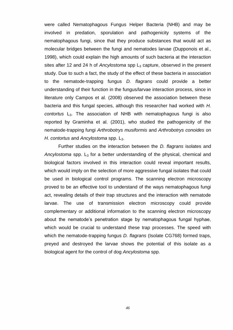
46
were called Nematophagous Fungus Helper Bacteria (NHB) and may be
involved in predation, sporulation and pathogenicity systems of the
nematophagous fungi, since that they produce substances that would act as
molecular bridges between the fungi and nematodes larvae (Dupponois et al.,
1998), which could explain the high amounts of such bacteria at the interaction
sites after 12 and 24 h of Ancylostoma spp L3 capture, observed in the present
study. Due to such a fact, the study of the effect of these bacteria in association
to the nematode-trapping fungus D. flagrans could provide a better
understanding of their function in the fungus/larvae interaction process, since in
literature only Campos et al. (2008) observed the association between these
bacteria and this fungal species, although this researcher had worked with H.
contortus L3. The association of NHB with nematophagous fungi is also
reported by Graminha et al. (2001), who studied the pathogenicity of the
nematode-trapping fungi Arthrobotrys musiformis and Arthrobotrys conoides on
H. contortus and Ancylostoma spp. L3.
Further studies on the interaction between the D. flagrans isolates and
Ancylostoma spp. L3 for a better understanding of the physical, chemical and
biological factors involved in this interaction could reveal important results,
which would imply on the selection of more aggressive fungal isolates that could
be used in biological control programs. The scanning electron microscopy
proved to be an effective tool to understand of the ways nematophagous fungi
act, revealing details of their trap structures and the interaction with nematode
larvae. The use of transmission electron microscopy could provide
complementary or additional information to the scanning electron microscopy
about the nematode‟s penetration stage by nematophagous fungal hyphae,
which would be crucial to understand these trap processes. The speed with
which the nematode-trapping fungus D. flagrans (Isolate CG768) formed traps,
preyed and destroyed the larvae shows the potential of this isolate as a
biological agent for the control of dog Ancylostoma spp.

47
CAPÍTULO 3
Interaction between the nematode-trapping fungi Arthrobotrys cladodes,
A. robusta, A. oligospora and Duddingtonia flagrans and Ancylostoma
spp. dog infective larvae in soil microcosm
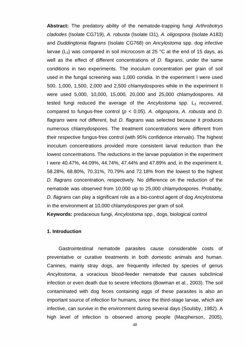
48
Abstract: The predatory ability of the nematode-trapping fungi Arthrobotrys
cladodes (Isolate CG719), A. robusta (Isolate I31), A. oligospora (Isolate A183)
and Duddingtonia flagrans (Isolate CG768) on Ancylostoma spp. dog infective
larvae (L3) was compared in soil microcosm at 25 °C at the end of 15 days, as
well as the effect of different concentrations of D. flagrans, under the same
conditions in two experiments. The inoculum concentration per grain of soil
used in the fungal screening was 1,000 conidia. In the experiment I were used
500, 1,000, 1,500, 2,000 and 2,500 chlamydospores while in the experiment II
were used 5,000, 10,000, 15,000, 20,000 and 25,000 chlamydospores. All
tested fungi reduced the average of the Ancylostoma spp. L3 recovered,
compared to fungus-free control (p < 0.05). A. oligospora, A. robusta and D.
flagrans were not different, but D. flagrans was selected because it produces
numerous chlamydospores. The treatment concentrations were different from
their respective fungus-free control (with 95% confidence intervals). The highest
inoculum concentrations provided more consistent larval reduction than the
lowest concentrations. The reductions in the larvae population in the experiment
I were 40.47%, 44.09%, 44.74%, 47.44% and 47.89% and, in the experiment II,
58.28%, 68.80%, 70.31%, 70.79% and 72.18% from the lowest to the highest
D. flagrans concentration, respectively. No difference on the reduction of the
nematode was observed from 10,000 up to 25,000 chlamydospores. Probably,
D. flagrans can play a significant role as a bio-control agent of dog Ancylostoma
in the environment at 10,000 chlamydospores per gram of soil.
Keywords: predaceous fungi, Ancylostoma spp., dogs, biological control
1. Introduction
Gastrointestinal nematode parasites cause considerable costs of
preventative or curative treatments in both domestic animals and human.
Canines, mainly stray dogs, are frequently infected by species of genus
Ancylostoma, a voracious blood-feeder nematode that causes subclinical
infection or even death due to severe infections (Bowman et al., 2003). The soil
contaminated with dog feces containing eggs of these parasites is also an
important source of infection for humans, since the third-stage larvae, which are
infective, can survive in the environment during several days (Soulsby, 1982). A
high level of infection is observed among people (Macpherson, 2005),

49
particularly those at pediatric age (Hotez, 1989), who are infected through skin
contact or ingestion of infective larvae from contaminated soil (McCarthy and
Moore, 2000; Hotez et al., 2004).
Currently, in small animal practice, the preventative health measure and
the classic treatment against Ancylostoma spp. have been performed
exclusively with the use of anthelminthic drugs, which act only on adult
hookworms inside the host (Lappin, 2001; Almeida and Ayres, 2006). However,
a high level of anthelmintic resistance in A. caninum was reported (Jackson et
al., 1987; Kopp et al., 2007), besides the possibility of environmental damage.
Thus, alternative methods are required, such as biological control, which is
characterized by the action of natural enemies (parasites, predators, pathogens,
antagonists or competitors) that maintains the population of a target organism at
a lower average than would occur in their absence, without causing alterations
in the environment (Gronvold et al., 1996a; Larsen, 1999; Chen and Dickinson,
2004).
The biological component of the soil ecosystem limits or stabilizes the
nematode population by mechanisms of competition, parasitism and production
of toxic compounds (Stirling, 1991). Fungi play an important role in the function
and dynamics of terrestrial ecosystems, directly influencing the structure of
plant, animal, and bacterial communities through interactions that span the
mutualism-parasitism continuum (Peay et al., 2008). Many animal-
enteroparasitic nematodes develop their pre-parasitic phase in the soil.
Epidemiologically, this is the most important phase, due to the possibility of re-
infection or new infection occurrence (Soulsby, 1982). In this environment, they
are particularly vulnerable to a wide range of soilborne fungi, called nematode-
trapping fungi or predaceous fungi, found in all sorts of soil habitats, especially
in organically rich environments (Larsen et al., 1994). They attack nematode
larvae by means of trap structures and use them as an additional nutrient
source (Gray, 1987; Nordbring-Hertz et al., 2006). Thus, nematode-trapping
fungi may reduce the population density of nematodes in the soil and keep them
under a threshold level (Gronvold et al., 1996a). Biological control of cattle,
sheep and horses helminthiasis through nematode-trapping fungi was reported
by many researchers (Waller and Faedo, 1993; Bird and Herd, 1995; Araújo et
al., 2004). Recently, a high level of predation of Ancylostoma spp. L3 was
achieved by different nematode-trapping fungi (Maciel et al., 2006; 2009;

50
Carvalho et al., 2009).
Despite these reports, there is not enough information about whether
nematode-trapping fungi can be used in strategies to control free-living forms of
dog Ancylostoma in the environment. Therefore, this work presented results of
the in vitro activity of the nematode-trapping fungi Arthrobotrys cladodes (Isolate
CG719), A. robusta (Isolate I31), A. oligospora (Isolate A183) and Duddingtonia
flagrans (Isolate CG768) on Ancylostoma spp. L3 mortality in soil microcosm
and investigates the effect of more infective fungi in low and high inoculum
concentration, evaluating the possible use of them as biocontrollers of this dog-
parasitic nematode in the environment.
2. Material and Methods
2.1. Fungal inoculum
Pure cultures of nematode-trapping fungi Arthrobotrys cladodes
Drechsler (Isolate CG719), A. robusta Duddington (Isolate I31), A. oligospora
Fresenius (Isolate A183) and Duddingtonia flagrans Cooke (Isolate CG768)
were colonized, according the modified technique of Smith and Onions (1983),
in integral rice grains and stored inside 5 mL BD Vacutainer® glass tubes
(Becton Dickinson, Brazil) containing blue silica gel and kept at 4 °C in
darkness. These tubes were closed with conventional stopper and sealed with
polyvinyl chloride (PVC) transparent film. All fungi capture nematodes through
three-dimensional adhesive networks. These tubes were closed with
conventional stopper and sealed with polyvinyl chloride (PVC) transparent film.
The identification of the species was based on the direct observation of the
morphological characteristics, such as conidial size, morphology of
conidiophores in micro-culture and trapping organs on nematode-infected
culture, based on descriptions of Cooke and Godfrey (1964), Cooke (1969) and
Van Oorschot (1985).
One integral rice grain, colonized by a single isolate, was transferred to
the center of Petri dishes of 60 mm x 15 mm, containing 20 mL of 2% water-
agar (2% WA) culture medium. Then, the fungi grew for seven days at 25 °C in
the darkness, when mycelial discs of 5 mm diameter were removed from the
fungal culture edges and transferred to Petri dishes of 90 mm x 15 mm
containing V8-agar medium (200 mL of Campbell‟s V8 juice, 3 g of CaCO3, 18 g
51
of agar and 800 mL of distilled water). Finally, the Petri dishes were kept at
25°C for ten days in the darkness to produce conidia. To induce D. flagrans
chlamydospores, the Petri dishes were incubated under the same conditions,
but for fifteen days (Fig. 1).
Fig. 1. Light micrograph of mature chlamydospore obtained from the micro-culture of
Duddingtonia flagrans (Isolate CG768) show its thick-walled feature pattern.
Mycelial fragments and conidia were harvested by gently scraping the
surface of the V8-agar with a brush after the addition of 10 mL distilled water.
The conidial residue was then washed with 5 mL of distilled water taken from
the medium surface. The fungal suspension was filtered through gauze (4
layers) and collected in a 50 mL Griffin glass, to reduce the mycelial fragments
and obtain the conidial suspension. One drop of dispersant polysorbate
(Tween® 80) was added in the fungal suspension under shaking for 2 minutes in
a magnetic stirrer, in order to maximize the conidial separation. Shortly after,
two aliquots of 10 μL were collected using a micropipette and placed on a
Neubauer-counting chamber to estimate the conidia number per milliliter of
suspension, according to Alfenas and Mafia (2007).
2.2. Ancylostoma spp. L3
Ancylostoma spp. L3 were obtained by incubating infected dog feces
mixed with moist vermiculite kept at 26 °C for 10 days. Feces containing eggs
were obtained from naturally infected stray dogs. These infected feces were
identified by the method for the detection of hookworm eggs, according to Willis
(1921). The infective larvae were extracted from the faecal cultures after using
52
the modified Baermann‟s technique (Baermann, 1917) for 12 hours and in this
time were concentrated by gravity inside 5 mL BD Vacutainer® glass tubes
(Becton Dickinson, Brazil) connected to the funnel. The sediment containing
larvae was transferred to 15 mL BD Falcon® centrifuge polystyrene tubes
(Becton Dickinson, Brazil), washed by centrifugation and resuspended in
distilled water, five times at 1,000 rpm, for 5 minutes. The supernatant was
gently aspirated by a plastic pipette and disposed at the end of each
centrifugation.
The methodology described by Barçante et al. (2003) was used in order
to filter the suspension, to eliminate debris and then obtain viable and active
Ancylostoma spp. L3 in a clean preparation. This selected suspension was
vigorously homogenized and six aliquots of 10 μL were collected with a
micropipette and placed on a slide marked with longitudinal lines to avoid the
repeated counting of a same larva. Each aliquot was covered with a glass
coverslip after the addition of 10 μL of lugol‟s solution to kill larvae. Thus, they
were counted and identified under a light microscope with the magnification of
40x, allowing the estimation of the total number of larvae in the suspension. The
motility of Ancylostoma spp. L3 was checked by microscopical examination,
before they were used in the experiment.
2.3. Experimental procedure
In order to evaluate the action of nematophagous fungi on Ancylostoma
spp. L3, in all controlled experiments, it was used a loamy sand soil (30%
coarse sand, 17% fine sand, 14% silt and 39% clay) originated from the region
of Viçosa, Minas Gerais, Brazil, presenting the following chemical
characteristics: organic matter = 1.6 dag/kg; phosphorus = 12.6 mg/dm3;
potassium = 37 mg/dm3; calcium = 4.1 cmolc/dm3; magnesium = 0.7 cmolc/dm3;
aluminum = 0 cmolc/dm3; pH = 6.4. This soil was sieved (5 mm mesh), placed
inside polyethylene bags, and then pasteurized twice in water vapor for 1 hour,
with an interval of 48 hours, in horizontal autoclave with vacuum valve open and
water level controlled, before being used in experiments (Fig 2A).
An experiment was performed to compare the predatory activity of
Arthrobotrys cladodes (Isolate CG719), A. robusta (Isolate I31), A. oligospora
(Isolate A183) and Duddingtonia flagrans (Isolate CG768) on Ancylostoma spp.

53
L3 in soil microcosm. Treatments consisted of the application of each fungal
isolate and one fungus-free control. Based on the results of this experiment, two
other experiments (I and II) were performed in order to evaluate the effect of
five concentrations of D. flagrans chlamydospores on the nematode in study.
All experiments were assessed under the same conditions using 145 mL
transparent polypropylene pots containing 40 g of soil with 25% of moisture
(Fig. 2B). The soil was artificially infested with Ancylostoma spp. L3 and conidia
of the fungi (screening experiment) or chlamydospores of D. flagrans
(experiments I and II). A standardized suspension of 100 µL, containing
approximately 1,000 specimens of Ancylostoma spp. L3, was placed in each
pot. In the screening experiment, 1,000 conidia of each isolate per gram of soil
were used. In experiment I, the chlamydospores ratios per gram of soil were
500:1, 1,000:1, 1,500:1, 2,000:1 and 2,500:1, while in the experiment II, the
ratios were 5,000:1, 10,000:1, 15,000:1, 20,000:1 and 25,000:1. The
chlamydospore concentrations required in each test resulted in different
volumes of suspension. In order to compensate these differences, distilled
water was added to the soil until reaching the standardized final volume of 500
µL in each one of the six replications per treatment. The fungus-free controls
received the same volume of distilled water. Microorganisms were mixed with
soil using a disposable wooden toothpick per replicate. Then, the pots were
closed with a conventional lid and incubated for 15 days at 25 °C in an
acclimatized darkened room. Every three days the pots were agitated to favor
the interaction between microorganisms.
Fig. 2. (A) Soil pasteurization in autoclavate polyethylene bags; (B) Soil microcosm: 145 cm3
transparent polypropylene pot containing 40 g of pasteurized soil.
54
At the end of the period, the larvae were recovered from the soil
microcosm using the modified Baermann‟s technique, as described before.
After discarding 3 mL of water without larvae from tubes, the Ancylostoma spp.
L3 were killed adding two drops of lugol‟s solution in the remaining volume
containing the sediment. Then, this suspension of 2 mL containing dead larvae
was transferred to Peter‟s counting slide and enumerated under light
microscope (40x), according to Peters (1952).
2.4. Statistical method
The experiment was carried out once, arranged in a complete
randomized design with six replicates per treatment. The larval recovery data
were transformed into logarithm (log10) in order to equalize variances and
normalize the residuals prior to the analysis using ANOVA. For comparisons
among isolates, the post hoc Tukey‟s multiple range test was performed to the
level of significance of 5%. Statistical inferences were made on transformed
data and back-transformed averages were presented. Descriptive analyses of
panels were used to compare the effects between the chlamydospores rates
and the reduction of nematode larvae by calculating averages with 95%
confidence intervals. All statistical procedures were performed using the
Statistica software, version 7.0 (Statsoft, 2004).
Larval recovery average values from the treatments were compared with
the fungus-free control, and the trapping percentage was calculated as follows:
y
xRP
100.100
Where RP = reduction percentage; x = average of larvae recovered from
the treatment; y = average of larvae recovered from the fungus-free control
3. Results
The fungal inoculum was able to germinate in pasteurized soil and
trapping larvae. Results summarized in the Table 1 show averages and
reduction percentages of Ancylostoma spp. L3 recovered from the soil caused
by the nematophagous fungi tested, after fifteen days of incubation, in
55
comparison to the fungus-free control. The medium value of the larvae
recovered from the treated groups was lower than those from the fungus-free
control (p < 0.05), at 1,000 conidia per gram of soil. The reduction percentages
of Ancylostoma spp. L3 recovered from the pasteurized soil, infested by fungi A.
cladodes, A. oligospora, A. robusta and D. flagrans were 30.92%, 42.27%,
43.90% and 46.60%, respectively. The fungus A. cladodes presented low
infectivity against Ancylostoma spp. L3. There were no differences among the
fungi A. oligospora, A. robusta and D. flagrans. However, since D. flagrans
produces numerous chlamydospores (Figures 3 and 4), it was considered the
more able fungus to be used in concentration tests.
Table 1. Average values, standard deviation (±) and reduction percentage (%)
of Ancylostoma spp. dog infective larvae (L3) recovered from soil, initially with
1,000 L3, by the Baermann method after fifteen days of interaction in microcosm
containing nematophagous fungi A. cladodes (Isolate CG719), A. robusta
(Isolate I31), A. oligospora (Isolate A183) and D. flagrans (Isolate CG768) in the
concentration of 1,000 conidia/g of soil compared to fungus-free control
Fungal isolates Average number Reduction percentage (%)
A. oligospora 352.67c ± 16.31 42.27
A. cladodes 422,00b ± 19.09 30.92
D. flagrans 326.17c ± 11.53 46.60
A. robusta 342.67c ± 17.40 43.90
Fungus-free control 610.85ª ± 22.48 -
Back-transformed averages followed by different superscripts in the same column are
significantly different by the Tukey test (p < 0.05).
Reduction percentage = 100 - (treatment average x 100/control group average).
Figures 3 and 4 present the average values of Ancylostoma spp. L3
recovered from the soil, containing different inoculum concentrations of D.
flagrans chlamydospore. All chlamydospore rates were significantly different
when individually compared with their respective fungus-free control (with 95%
confidence interval). In general, the highest and the lowest predatory activity
were achieved by adding low and high amounts of chlamydospores per gram of
soil, respectively. Then, the high inoculum concentrations used in experiment II
provided more consistent larval reduction than the low concentrations used in
experiment I. Either way, irrespective of the concentration used, there was a
56
reduction in the number of Ancylostoma spp. L3 recovered when compared to
the fungus-free control. In experiment I, the results did not indicate differences
among all D. flagrans treatments, probably due to the small increase in number
of chlamydospores between each interval concentrations (Figure 3). This
fungus caused a high and progressive reduction in Ancylostoma spp. L3
harvested from the soil in experiment II. Nevertheless, there was no difference
among concentrations from 10,000 up to 25,000 chlamydospores per gram of
soil (Figure 4). It was observed a stronger reduction in the Ancylostoma spp. L3
recovered in the concentration of 5,000 chlamydospores per gram of soil and
the predation increased using 10,000 chlamydospores per gram of soil.
However, the level of reduction in higher concentrations was similar to the
10,000 chlamydospores per gram of soil, tending to stabilize (Figure 4). This
indicated that an increase in this concentration of D. flagrans chlamydospores in
the soil microcosm did not caused more damage to the population of
Ancylostoma spp. L3. A higher inoculum concentration facilitated the distribution
of the fungus, reaching a larger area in the soil microcosm and, undoubtedly,
increased the chances of catching larvae, in comparison to the experiment I,
with lower concentrations.
* Two independent averages are significantly different by the no-overlap between 95% IC.
Fig 3. Descriptive analysis by 95% confidence intervals (IC) of the average of uncaptured
Ancylostoma spp. dog infective larvae (L3) recovered from soil, initially with 1,000 L3, by the
Baermann method after 15 days of interaction in microcosm containing nematophagous fungus
Duddingtonia flagrans (Isolate CG768) in five inoculum concentrations and respective fungus-
free control of the experiment I.

57
* Two independent averages are significantly different by the no-overlap between 95% IC.
Fig. 4. Descriptive analysis by 95% confidence intervals (IC) of the average of uncaptured
Ancylostoma spp. dog infective larvae (L3) recovered from soil, initially with 1,000 L3, by the
Baermann method after 15 days of interaction in microcosm containing nematophagous fungus
Duddingtonia flagrans (Isolate CG768) in five inoculum concentrations and respective fungus-
free control of the experiment II.
There was a direct relationship between the effectiveness on the
predation of Ancylostoma spp. L3 and the inoculum concentration in the soil
microcosm. In the experiment I, the reduction percentages of Ancylostoma spp.
L3 with increasing fungal inoculum concentration, compared to the respective
fungus-free control were 40.47%, 44.09%, 44.74%, 47.44% and 47.89% in the
inoculum concentration of 500, 1,000, 1,500, 2,000 and 2,500 chlamydospores
per gram of soil, respectively. In the experiment II, with chlamydospores
concentrations ten times higher than in the experiment I, the reduction
percentages of Ancylostoma spp. L3 were 58.28%, 68.80%, 70.31%, 70.79%
and 72.18%, in the inoculum concentrations of 5,000, 10,000, 15,000, 20,000
and 25,000 chlamydospores per gram of soil, respectively.
4. Discussion
The current experiment showed that the incorporation of nematophagous
fungi to the soil microcosm reduced the Ancylostoma spp. L3 population,
suggesting that these nematode larvae may have been used as food source.

58
The capacity of the nematophagous fungi to capture nematodes is well known
(Gray, 1988; Larsen et al., 1994). However, the efficiency of nematode-trapping
fungi in nematode control may vary (Galper et al., 1995). Besides, experimental
conditions vary and hamper comparisons related to the capture efficiency of
fungi on other nematodes, as, for example, on Ancylostoma spp. Nematode-
trapping fungi A. cladodes, A. robusta, A. oligospora and D. flagrans reduced
the number of Ancylostoma spp. L3 in pasteurized soil, evidencing that they can
be used in the environmental control of pre-parasitic forms of this dog-
enteroparasitic nematode. Despite the poor results in screening experiment, the
small percentage reduction of the larvae recovered in relation to the fungus-free
control could be attributed to the interaction between the incubation period and
the low ratio of conidia per gram of soil used. It is evident that the fungal
inoculum concentration affects the performance of nematode-trapping fungi on
nematodes. So, in higher inoculum concentrations, these fungi might probably
reduce the population density of Ancylostoma spp. L3. Furthermore, it must be
pointed out that the effectiveness may also be affected by the increase of the
period of interaction, resulting in a low number of recovered larvae.
It is not surprising that a significant improved reduction has been
observed in current experiment, with the use of high inoculum amount of the
fungus D. flagrans. This fungus has been successfully used as a biological
control agent of gastrointestinal nematodes of domestic animals, due to its
predaceous efficiency (Faedo et al., 1998; Baudena, 2000; Sanyal, 2000; Terrill
et al., 2004). Its potential is due to the great production of thick-walled
chlamydospores (Waghorn et al., 2003), which are highly resistant under
adverse conditions (Larsen et al., 1991; Waller et al., 2001) and can originate
hyphae and traps using only their considerable reserve of nutrients (Faedo et
al., 2000; Skipp et al., 2002). According to earlier in vitro studies, D. flagrans
entraps larvae through adhesive three-dimensional network traps, which are
produced, in great amounts, in the presence of Ancylostoma spp. L3 (Maciel et
al., 2006; 2009). It should be emphasized that it attacks predominantly
helminths that have short stage of development inside eggs, as the nematode
here studied, whose larva of first stage is liberated in 20-24 hours, under
appropriate conditions (Soulsby, 1982). Sometimes, the fungus survives for
long periods, attacking also nematodes whose eggs hatch after 12-16 weeks,
since, while the larva is in the egg, this fungus is not capable to capture it
59
(Faedo et al., 2000). The addition of large amounts of fungi to the soil is one of
two general ways of applying the nematode biological control using
nematophagous fungi (Nordbring-Hertz et al., 2006). The strong, intermediate
and weak responses to chlamydospore concentrations were consistent in
experiments with the fungus D. flagrans, with more evident predation of
Ancylostoma spp. L3 from 10,000 chlamydospores per gram of soil, with
comparable efficiency in the other high concentrations. It is relevant that such
virulence may be higher on L1 and L2 free-living forms, since Nansen et al.
(1988) reported that these stages of development are more susceptible to
action by nematophagous fungi.
The nematode mortality is influenced by the density of the traps, which
are directly related to the number of conidia or chlamydospores, as pointed out
by Gaspard and Mankau (1987). This was seemingly the cause of the damage
level occurred on Ancylostoma spp. L3 population in relation to its respective D.
flagrans inoculum amount in soil microcosm. This is in agreement with Ribeiro
(1999) and Maciel et al. (2009) who observed greater predation of nematodes
with the increase of the fungal inoculum level, evidencing that the nematode
control requests a great concentration of the antagonist. Since chlamydospores
allow the establishment and survival of this fungus in the environment, it is the
appropriate inoculum form to be used in biological control (Faedo et al., 2000),
and that is why it was used in concentration tests with D. flagrans. According to
Knox et al. (2002), D. flagrans could be detected in up to 30 cm deep below the
soil profiles directly under the point of deposition, as the result of inadvertent
chlamydospores relocation, by the activity of macroinvertebrates, such as
arthropods and earthworms, or also by percolation into the soil with rain water.
Thereby, once traps are stationary, it is obvious that the bigger the soil area
containing chlamydospores of D. flagrans distributed homogeneously, the
bigger the chances of interaction between the prey and its antagonists,
regulating its population in this habitat. Based on this information, the
movement of the fungal inoculum in artificial environment was guaranteed by
the continuous agitation of the pots, and probably resulted in more interaction
between the confronting microorganisms, ensuring the potential of a single
predator on the prey available in the soil microcosm. Thus, due to the good
fungus distribution on the soil profile, Ancylostoma spp. L3 needed to migrate for
a shorter distance towards network traps, whose form provides several contact
60
points for its prey. Nevertheless, the fungal predatory capacity is connected with
both the capacity of the nematode to induce the hyphal morphogenesis in traps
and the trapping efficiency of these structures (Nansen et al., 1988). This was
observed in a previous in vitro survey using the same microorganisms here
studied (Maciel et al., 2009). In the environment, the trapping ability of
nematode-trapping fungi depends on the high inoculum level that will ensure the
colonization of the soil through extensive mycelial growth, at intervals along
which networks may be formed in high numbers (Gray, 1987; Gronvold, 1989).
However, there is a delicate balance between hyphal development and trap
formation and it is regulated by environmental factors (Scholler and Rubner,
1994), such as temperature, nematode density, oxygen tension, light and
nutrient level (Gronvold, 1989). Above all, the presence of nematode larvae is
the most important biotic factor involved in induction of trapping structures
(Nordbring-Hertz et al., 2006). Moreover, the density of this target organism is
also relevant to stimulate the predation by nematophagous fungi (Jaffee et al.,
1992; 1993), such as D. flagrans, because when nematodes are in plentiful
supply, perhaps between certain threshold densities, this fungus increase
trapping rates (Morgan et al., 1997). In the present study, it seems that the
number of Ancylostoma spp. L3 used in soil microcosm was sufficient to
stimulate the food-finding mechanism of fungus to capture them.
The pasteurization of the soil would eliminate the background fungi, but
would create an overly artificial environment for the study of organisms with
saprophytic potential. Differences in environmental conditions between
laboratory and field may be relevant in the expression of the predation of
nematophagous fungi, and the evaluation of its potential is influenced by the
conditions of the experiment. In soils previously treated, as the one used in the
current experiments, which present fewer competitors, fungi can feed on
substrates other than nematodes, but in soil under natural conditions, several
microorganisms are present and compete for nutrients, so predaceous fungi will
use nematodes as food source (Hayes and Blackburn, 1966; Gray, 1987).
However, unlike the previous report, in the present study, D. flagrans attacked
Ancylostoma spp. L3 in the absence of competitors with greater virulence. Such
behavior indicated the importance of nematodes as suitable food, suggesting
that this antagonist could be used against Ancylostoma spp. L3 in the soil,
irrespectively of the organic matter available.
61
Considering that nitrogen is a limiting factor for the growth and
reproduction of nematode-trapping fungi, they get their nitrogen requirement
from digesting the nematode‟s biomass (Barron, 1992; Barron, 2003).
Therefore, it is clear that Ancylostoma spp. L3 was the nitrogen source for D.
flagrans in the soil microcosm. Thereby, it may mean that predatory fungi are
not true saprotrophs, once two phases are necessary to supply them with the
correct nutrients for growth – the saprotrophic phase and the predatory parasitic
phase. This is in agreement with the findings of Cooke (1962) who indicated
that the nematophagous habit supplement the saprophytic mode of life of fungi,
but on the other hand, the nematode alone could not provide all the energy
necessary for growth. Gray (1985) also reported that nematodes are necessary
to initiate the formation of trapping organs, but fungi are incapable of remaining
in a predaceously active state in the absence of an organic energy source other
than nematode population. Adhesive network-forming fungi, as D. flagrans, may
develop a saprophytically survival in the environment in the absence of prey,
using the available organic matter for nutrition (Larsen et al., 1991). The
relevant trapping activity of D. flagrans on Ancylostoma spp. L3, associated with
its noteworthy ecology, described above, suggests that this fungus would be
able to maintain its predaceous efficiency in the hemiedaphic zone; rich in
organic matter, moisture, soil nutrients and microorganisms where nematodes
are available (Gray and Bailey, 1985). This is the appropriate habitat for the
development of rhabditiform larvae (first and second stages), because they feed
on bacteria and organic matter, and where the filariform larva (third stage) lives,
encased in the cast cuticle of second molt, until depleting its reserves stored in
intestinal cells (Soulsby, 1982) for up to forty nine days for the species A.
caninum, if it does not find a host (Mark, 1975). Since nematophagous fungi are
most likely to utilize nematodes only when their population is increasing in
response to a temporary, most likely seasonal, increase in available soil
nutrients (Gray, 1987), the purpose of using D. flagrans is to achieve
prophylactic worm control, whereby future populations of pre-parasitic
Ancylostoma spp. in environment are reduced. It is important to consider that
this approach will almost certainly need to be applied in combination with other
parasite control alternatives, including the timely and effective use of
anthelminthics, because nematophagous fungi do not have any effect on worms
established in hosts (Jackson and Miller, 2006).
62
The selection of possible nematode biocontrollers will have to take
account the problems caused by antagonistic effects on the soil and also the
target nematode (Gray, 1987). Despite the in vitro effectiveness, results showed
that the D. flagrans is a potential antagonist of Ancylostoma spp. L3, little is
known about the biology and epidemiology of this fungus under natural
conditions. This knowledge is necessary, because the competition among soil
microbial communities in the environment is so intense that it would forbid the
introduction of a new fungal species, mainly due to fungistasis (De Boer et al.,
2003). So, the microbial activity is of paramount importance for the
establishment of nematophagous fungi in soil because they will have to
overcome such antagonism (Monfort et al., 2006). According to Gray (1983),
the lack of success in the use of nematophagous fungi as biological control
agents is generally due to the selection of the fungus, rarely based on its
suitability to the habitat where the target nematode is found; but rather, on the
ease of its isolation and subsequent culture or its relative ability in destroying
nematodes under controlled conditions in the laboratory. The soil microcosm
used in the present study can provide a model system for obtaining preliminary
information about fungal viability and the extent of trapping on nematode
species. Therefore, the promising fungi presented in this study should be the
subject of new researches, either in controlled conditions or in natural systems.
Furthermore, little is know about the environmental impact of the introduction of
high fungal inoculum on non-target organisms in the soil ecosystem (Knox et
al., 2002). While its contribution in the natural system is not fully understood yet,
such statements indicate how future exploitation of D. flagrans for biological
control should proceed, since it can be used strategically against Ancylostoma
spp. without negative effects on the abundance of the mesofauna. Further
studies on the ecological factors that affect the dynamic of the fungus D.
flagrans in the environment may provide conclusive information to improve its
efficiency in the nematode control and to determine its ecological impact,
although no adverse effects were observed with the use of this fungus on non-
target soil mesofauna organisms, as free-living nematodes, earthworms,
microarthropods nor any other nematophagous fungi (Gronvold et al., 2000;
Yeates et al., 2002).
The present work demonstrated a slightly better performance of the
nematode-trapping fungus D. flagrans to reduce the number of Ancylostoma
63
spp. in pasteurized soil under laboratory conditions, but prospects for biological
control depend on the methods to introduce fungal material in the environment,
and on a sufficient amount to effectively reduce the parasite larval population.
This fungus is an alternative as a biological control agent, but its concentration
will determine the predation level. Future semi-natural and natural controlled
experiments will demonstrate if D. flagrans can be used to fight off pre-parasitic
forms of Ancylostoma spp. under natural soil conditions.
64
CAPÍTULO 4
Biological control of Ancylostoma spp. dog infective larvae in soil under
semi-natural conditions
65
Abstract: Studies were undertaken to evaluate the predatory ability of the
nematode-trapping fungus Duddingtonia flagrans on Ancylostoma spp. L3 in soil
pasteurized microcosm at 25 °C at the end of 15 days, as well as in natural soil
at environmental temperature under greenhouse conditions by five
assessments along of 30 days at intervals of 5 days among them. The fungus
was inoculated by means of pre-colonized substrates at the standard rate of
10,000 chlamydospores per grain of soil. Milled maize and white rice were
tested for massal production of D. flagrans chlamydospores, and the fungus
produced 525,347 and 640,625 chlamydospores per gram of substrate,
respectively, there was no difference between them. In microcosm assay, the
fungus reduced the larval number recovered in comparison to the fungus-free
control (p < 0.05). Duddingtonia flagrans alone, D. flagrans in white rice and D.
flagrans in milled maize reduced larval population in 71.99, 78.38 and 79.41%,
respectively. However there was no difference between D. flagrans substrates.
The controls with fungus-free substrates, milled maize and white rice, caused
small damage on the larval population, reducing it in just 8.28 and 13.39%,
respectively, but no differences were observed among them and fungus-free
control. In the biocontrol assay, the fungus was incorporated to the natural soil
in pre-colonized milled maize. In all days of assessment, the average values of
larvae recovered from fungus-free control were larger than those recovered
from treatment with the fungus (p < 0.05). No differences were found between
fungus-free substrate and fungus-free control when considering the average
values of days 25 and 30 (p < 0.05). Considering the assessment days 10, 15,
20, 25 and 30 the reduction percentages observed in treatment with D. flagrans
were 61.37, 73.23, 70.84, 64.50 and 56.99%, respectively, whereas in control
with substrate the reduction percentages were 16.39, 27.99, 20.36, 14.45 and
9.20%. There was an inverse correlation between the larval recovered as a
function of the days (p < 0.05). Duddingtonia flagrans showed predatory effect
on plant nematodes present in natural soil. These results in semi-natural
conditions demonstrate that D. flagrans can be considered as a non-
chemotherapeutic alternative approach to the control of pre-parasitic forms of
dog Ancylostoma spp.
Keywords: Duddingtonia flagrans, Ancylostoma spp., dogs, biological control
66
1. Introduction
Gastrointestinal parasitic nematodes, members of the family
Ancylostomatidae, are blood-feeding intestinal parasites of mammalian hosts
(Mulvenna et al. 2009) of considerable veterinary and medical importance (Datu
et al., 2008). Some species of genus Ancylostoma are specific to humans or to
animals, but others are zoonotic (Miranda et al., 2008a), such as Ancylostoma
caninum and A. braziliense (Costa et al., 2008), since dogs are the major
definitive host and humans, an accidental host (Davies et al., 1993). These
ubiquitous hookworms generally live in the intestines of dogs and cats in
tropical and subtropical regions, where conditions are conducive for hookworm
survival (Traub et al., 2004; Zhan et al., 2008).
The potential role of companion animals as reservoirs for zoonotic
diseases has been recognized as a significant public health problem worldwide
(Schantz, 1994; Robertson et al., 2000). A large proportion of the Ancylostoma
spp. population would be expected to be in refugia provided by free-living and
somatic reservoir stages of the life cycle, as well as by parasites infecting free-
ranging dogs and untreated pets (Kopp et al., 2007). These animals with patent
infections put other canines at risk after contaminate the soil of public areas with
feces containing eggs of the hookworm (Krämer et al., 2009). Contaminated soil
is a major source of human infection, especially to the children (Macpherson,
2005).
Given the importance of the human-animal bond in modern society, and
the high level of pet ownership, canine hookworms control is important to
minimize its transmission to animals and human population (Schad, 1994;
Bowman et al., 2003; Kopp et al., 2008a). While appropriate hygiene measures
are essential, regular treatment of pets with an effective anthelmintic also has a
key role in minimizing environmental contamination with these nematodes
(Schad, 1994), but rapid reinfection and the high cost of treatment hamper
control efforts (Albonico et al., 1995). Besides, resistance of the canine
hookworm A. caninum to anthelmintic therapy with pyrantel is an emerging
problem that has been reported (Jackson et al., 1987; Hopkins et al., 1989;
Hopkins and Gyr, 1991; Kopp et al., 2007; Kopp et al., 2008b).
Therefore, there is an ongoing need for the development of safe,
convenient and environmentally friendly products to reduce the potential for the
67
development of resistance (Colgravea et al., 2009). The aim of all prophylatic
approaches to control is simply to limit host parasite contact to levels which do
not compromise performance and/or welfare of animals by limiting the size of
the parasitic population (Jackson and Miller, 2006). Alternative or
complementary measures, when combined with other forms, of control may
maintain effective control while reducing anti-helminthic usage comprise both
the infective forms in the environment as the nematodes parasitizing the adult
animal (Barger, 1999; Larsen, 1999; Stromberg and Averbeck, 1999), like
biological control using nematophagous fungi by targeting larvae in environment
(Sanyal and Mukhopadhyaya, 2003).
Considering the fact of nematophagous fungi are natural enemies of
nematodes (Nordbring-Hertz et al., 2006) and commonly found worldwide,
occurring in soil and environments rich in organic matter of several ecosystems
(Gray, 1987), they can become part of an non-chemotherapeutic alternative
approach (Larsen, 2000). Whereas that parasitic-nematode of animals spend
part of their life cycle in soil (Soulsby, 1982), biocontrol agents have been
investigated with aim to achieve a significant reduction in the larval population
on environment, which will consequently result in a significant reduction of the
worm burden in animals (Waller and Larsen, 1996; Wolstrup et al., 1996).
Nematode-trapping fungi are able to trap and kill nematodes and the species
Duddingtonia flagrans has been reported as the most promising to combat
nematodes of several animals (Nansen et al., 1996; Fernández et al., 1997;
Faedo et al., 2002; Chandrawathani et al., 2003; Dimander et al., 2003a). The
potential of this natural nematode regulator is due to its capacity to produce
abundant thick-walled restingspores, called chlamydospores, which have high
degree of survival in harsh environmental conditions (anaerobic, enzymatic and
thermal) such as animal gut (Larsen, 1999; Terril et al., 2004). These structures
can originate hyphae and adhesive three-dimensional net-type traps using only
its considerable reserve of nutrients (Faedo et al., 2000; Skipp et al., 2002).
Although a promising approach has been presented in feeding the
grazing animals with D. flagrans chlamydospores (Larsen, 2000; Nordbring-
hertz et al., 2006), the direct addition of these to the contaminated soil of
backyards, public parks and playgrounds should be focused as the most
appropriate way of applying biological control using nematophagous fungi as
part of an integrated public veterinary and medical health system, in view that

68
the main canine hookworm reservoir are stray dogs (Heukelbach et al., 2002;
Kopp et al., 2007). This fungal inoculum incorporation into the soil requires the
addition of organic substrate (Cayrol and Ritter, 1984) because it is believed
that the activity of nematophagous fungi might be stimulated by organic matter
(Siddiqui and Mahmood, 1996). The biological control of canine nematodes has
been neglected and there is little information about the effectiveness of
nematophagous fungi on pre-parasitic forms of these parasites in the soil.
Studies show that D. flagrans is antagonist of dog Ancylostoma causing
significant reduction on infective larvae population (Maciel et al., 2006; 2009;
Carvalho et al., 2009). This paper describes the results of experimentation
carried out to enable assessment of the impact of D. flagrans on Ancylostoma
spp. L3 population in natural soil under greenhouse semi-natural conditions and
provides information about two organic substrates favorable to inoculum
production of this fungus.
2. Material and Methods
2.1. Fungal inoculum
Pure culture, previously isolated, of nematode-trapping fungus
Duddingtonia flagrans Cooke (Isolate CG768), colonized in integral rice grains,
was stored in the Laboratório de Controle Biológico de Nematóides,
Universidade Federal de Viçosa, Brazil, at 4 °C in darkness inside 5 mL BD
Vacutainer® glass tubes (Becton Dickinson, Brazil) containing blue silica gel,
similar to the conservation technique of Smith and Onions (1983). These tubes
were closed with conventional stopper and sealing with polyvinyl chloride (PVC)
transparent film. The identification was based on the direct observation of the
morphological characteristics, as the conidial size and the morphology of
conidiophores in micro-culture, and trapping organs on nematode-infected
culture, mainly following the descriptions furnished by Cooke (1969).
Duddingtonia flagrans was carefully selected as candidate specie for
introduction in soil by previous assay.
Mass production of D. flagrans chlamydospores was performed on
cereals grains inside propylene bags with biological filter (Fig. 1). Inside of each
bag were deposed 300 g of one single substrate, white rice grains or milled
maize grains, and 40 mL of distilled water. Bags were sealed and sterilized for

69
30 minutes at 121 °C. Prior to fungal inoculation of the substrate, one integral
rice grain colonized by D. flagrans was transferred to the center of Petri dishes
of 60 mm x 15 mm, containing 20 mL of 2% water agar (2% WA) culture
medium during seven days at 25 °C in the darkness. After mycelial growth,
inside a vertical laminar flow cabinet, six medium disks of approximately 5 mm
diameter, containing fungal mycelium, were removed from 2% WA fungal
culture edges and transferred into the propylene bags containing substrate at
environment temperature. Bags were again sealed to prevent contamination
and D. flagrans grew on substrates as monocultures under acclimatized room at
25 °C in the darkness for 15 days. At intervals of 5 days until the end of the
incubation period, blocks of colonized grains inside sealed bags were undone
by pressure of fingers before shake this substrate with the purpose of ensure
uniform fungal growth.
Fig. 1. Mass production of D. flagrans chlamydospores on milled maize and white rice inside
propylene bags with biological filter. White rice (black arrow) and milled maize (dashed black
arrow) with 10 days of fungal colonization. White rice (white arrow) and milled maize (dashed
white arrow) with 15 days of fungal colonization, showing brown staining.
After the subsequent period of growth, the substrate was homogenized
and 6 samples of 1 g were removed of each bag. Each sample was placed in
an Erlenmeyer containing 10 mL of distilled water plus dispersant polysorbate
(Tween® 80) to 0.2% (v/v) and was shaken for 2 minutes in a magnetic stirrer in
order to maximize chlamydospores separation. The fungal suspension was
filtered through gauze (4 layers) in order to reduce fragments, and collected in a
20 mL Griffin glass. Soon after, the number of chlamydospores per milliliter of
the suspension, corresponding to the number of chlamydospores per grain of
substrate, was estimate in aliquots of 10 μL placed on Neubauer-counting

70
chamber, according to Alfenas and Mafia (2007).
In order to obtain only D. flagrans chlamydospores, the colonized
substrate was shaken with distilled water in vials closed with conventional lid.
Then, fungal suspension was filtered through gauze (4 layers) and collected in a
500 mL Griffin glass to reduce the mycelial fragments. After 6 hours of the
decanting by gravity, the chlamydospores of the bottom of the Griffin glass were
harvested by the aspiration. The aspirated material was homogenized and the
number of chlamydospores per milliliter was estimate as described before.
2.2. Ancylostoma spp. L3
Ancylostoma spp. L3 were obtained by incubating infected dog feces
mixed with moist vermiculite kept at 26 °C for 10 days. Feces containing eggs
were obtained from naturally infected urban street dogs. These infected feces
were identified by the method for the detection of hookworm eggs, according to
Willis (1921). Following incubation period, Ancylostoma spp. L3 were extracted
from the fecal cultures over 12 h using the modified Baermann technique
(Baermann, 1917) through which they were concentrated by gravity inside 5 mL
BD Vacutainer® glass tubes (Becton Dickinson, Brazil) connected to funnel. The
sediment containing larvae was transferred to 15 mL BD Falcon® centrifuge
polystyrene tubes (Becton Dickinson, Brazil), washed by centrifugation and
resuspended in distilled water, five times at 1,000 rpm, for 5 minutes. The
supernatant was gently aspirated by a plastic pipette and disposed at the end of
each centrifugation.
The methodology described by Barçante et al. (2003) was used in order
to filter the suspension, to eliminate debris and then obtain viable and active
Ancylostoma spp. L3 in a clean preparation. This selected suspension was
vigorously homogenized and six aliquots of 10 μL were collected with a
micropipette and placed on a slide marked with longitudinal lines to avoid the
repeated counting of a same larva. Each aliquot was covered with a glass
coverslip after the addition of 10 μL of lugol‟s solution to kill larvae. Thus, they
were counted and identified under a light microscope with the magnification of
40x, allowing the estimation of the total number of larvae in the suspension. The
motility of Ancylostoma spp. L3 was checked by microscopical examination,
before they were used in the experiment.
71
2.3. Experimental procedure
In order to evaluate the action of nematophagous fungus on Ancylostoma
spp. L3, in all controlled experiments, it was used a loamy sand soil (30%
coarse sand, 17% fine sand, 14% silt and 39% clay) originated from the region
of Viçosa, Minas Gerais, Brazil, presenting the following chemical
characteristics: organic matter = 1.6 dag/kg; phosphorus = 12.6 mg/dm3;
potassium = 37 mg/dm3; calcium = 4.1 cmolc/dm3; magnesium = 0.7 cmolc/dm3;
aluminum = 0 cmolc/dm3; pH = 6.4. The soil was collected from the soil profile
(0-20 cm deep), sieved (5 mm mesh), and placed inside polyethylene bags.
After that, soil was steam pasteurized twice in water vapor for 1 hour, with an
interval of 48 hours, in horizontal autoclave with vacuum valve open and water
level controlled, before being used in experiments. In the experiment under
semi-natural conditions, the soil was air-dried at room temperature for one week
to reduce its water content eliminating pre-parasitic forms of Ancylostoma spp.,
since they are susceptible to the dehydration (Soulsby, 1982).
2.3.1. Soil microcosm assay
The experiment was conducted to evaluate the predatory ability of
nematode-trapping fungus D. flagrans on Ancylostoma spp. L3 when the fungal
inoculum was incorporated to the pasteurized soil without substrate or on pre-
colonized substrates: white rice grains or milled maize grains. The experiment
was performed in 145 cm3 transparent polypropylene pot (Internal dimensions:
height = 6 cm, mouth diameter = 6.7 cm, bottom diameter = 5.5 cm) containing
40 g of soil with 25% of moisture (25g water per 100 g of dry soil) (Fig. 2). A
standardized suspension of 100 µL, containing approximately 1,000 specimens
of Ancylostoma spp. L3, was placed in each pot. Three treatments were
established, one using only D. flagrans chlamydospores and the other two using
individual application of a substrate pre-colonized by the nematode-trapping
fungus. Three fungus-free controls were performed: two of them using the same
substrates no colonized and other without any substrate. The uncolonized
substrates were pre-autoclaved and the same amounts used in the treatments
were used in the respective fungus-free controls.
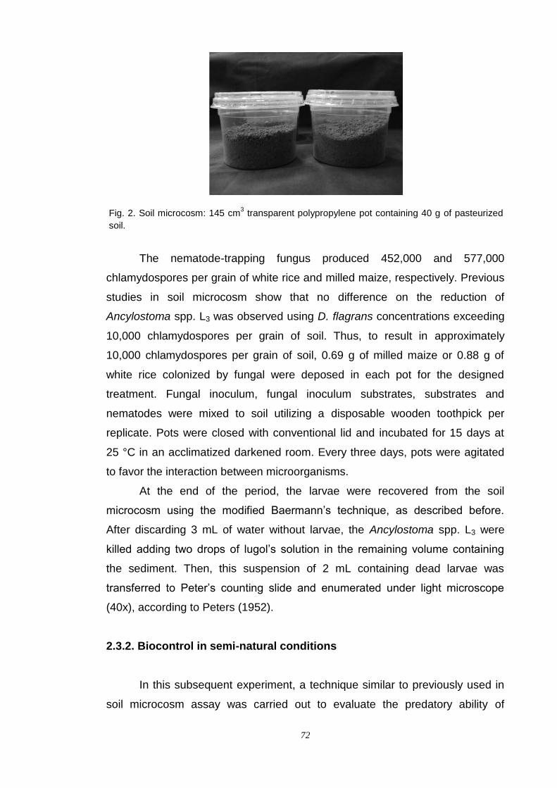
72
Fig. 2. Soil microcosm: 145 cm3 transparent polypropylene pot containing 40 g of pasteurized
soil.
The nematode-trapping fungus produced 452,000 and 577,000
chlamydospores per grain of white rice and milled maize, respectively. Previous
studies in soil microcosm show that no difference on the reduction of
Ancylostoma spp. L3 was observed using D. flagrans concentrations exceeding
10,000 chlamydospores per grain of soil. Thus, to result in approximately
10,000 chlamydospores per grain of soil, 0.69 g of milled maize or 0.88 g of
white rice colonized by fungal were deposed in each pot for the designed
treatment. Fungal inoculum, fungal inoculum substrates, substrates and
nematodes were mixed to soil utilizing a disposable wooden toothpick per
replicate. Pots were closed with conventional lid and incubated for 15 days at
25 °C in an acclimatized darkened room. Every three days, pots were agitated
to favor the interaction between microorganisms.
At the end of the period, the larvae were recovered from the soil
microcosm using the modified Baermann‟s technique, as described before.
After discarding 3 mL of water without larvae, the Ancylostoma spp. L3 were
killed adding two drops of lugol‟s solution in the remaining volume containing
the sediment. Then, this suspension of 2 mL containing dead larvae was
transferred to Peter‟s counting slide and enumerated under light microscope
(40x), according to Peters (1952).
2.3.2. Biocontrol in semi-natural conditions
In this subsequent experiment, a technique similar to previously used in
soil microcosm assay was carried out to evaluate the predatory ability of

73
nematode-trapping fungus D. flagrans on target nematode in semi-natural
conditions. The interaction between the target microorganisms was performed
in 350 cm3 transparent polypropylene pot (Internal dimensions: height = 8 cm,
mouth diameter = 9 cm, bottom diameter = 7 cm) containing 200 g of untreated
soil (Fig. 3). A standardized suspension of 250 µL, containing approximately
5,000 specimens of Ancylostoma spp. L3, was placed in each pot. In treatment,
the fungal inoculum was inoculated together with pre-colonize milled maize
containing approximately 565,000 chlamydospores per grain. Thus 3.54 g of
this pre-colonized substrate were added to result in 10,000 chlamydospores per
grain of soil. Microorganisms were mixed to soil as described before. Two
fungus-free controls were performed: one composed by the equal amount of the
milled maize pre-autoclaved and no colonized and other without substrate. They
were included to account for the probable activity of other antagonists of the
target nematode, which would be naturally present in the raw soil. The pots
were kept open, inside 500 cm3 opaque plastic vase, under greenhouse
conditions for a period of 30 days and protected from direct sunlight. The soil
was moistened in alternate days, without flooding of water. These procedures
were adopted based on the knowledge that the larvae of dog Ancylostoma live
naturally in shaded and moist environments, because they do not resist to
dehydration (Soulsby, 1982). Concurrently moisture is also necessary for the
germination of D. flagrans chlamydospores (Knox et al., 2002).
Fig. 2. Soil microcosm: 350 cm3 transparent polypropylene pot containing 200 g of untreated
soil, inside 500 cm3 opaque plastic vase, under greenhouse conditions
The assay was assessed on the tenth day and at intervals of five days,
over thirty days period. In order to recovery of the larvae, the soil of each pot

74
was spread on one paper towel sheet (1 ply sheet, about 22 x 20 cm), and
placed in a flat plastic sieve (20 cm in diameter). The sieve was adjusted on a
plastic bottom plate filled with water at 41 ºC in contact with the surface of the
liquid. This modified Baermann‟s apparatus (Baermann, 1917) was used to
favoring the recovery of Ancylostoma spp. L3 due to the increase of the contact
area between soil and water. After 12 hours, the sediment containing larvae
was poured into a funnel. The residue in the bottom of the plate was washed
with jets of water from a plastic wash bottle on the top of the funnel. Then, the
larvae were concentrated by gravity inside 5 mL BD Vacutainer™ glass tubes
(Becton Dickinson, Brazil) connected to funnel for 6 hours. After discarding 3
mL of water without larvae from tubes, two drops of lugol‟s solution were added
in the remaining volume containing the sediment to kill the Ancylostoma spp. L3.
Then, this volume of 2 mL containing dead Ancylostoma spp. L3 was transferred
for Peter‟s counting slide and enumerated as described before.
2.4. Statistical method
The experiment was carried out once, arranged in a complete
randomized design with six replicates per treatment and fungus-free control.
Descriptive analyses of panels were used to compare the production of
chlamydospores between both substrates calculating averages with 95%
confidence intervals. The larval recovery data were transformed into logarithm
(log10) in order to equalize variances and normalize the residuals prior to the
analysis using ANOVA. For comparison of the predatory ability of nematode-
trapping fungus between both substrates pre-colonized, the post hoc Tukey‟s
multiple range test was performed to the level of significance of 5%. Statistical
inferences were made on transformed data and back-transformed averages
were presented. The regression analysis was applied to show the relationship
between interaction days and reduction of parasite larvae. All statistical
procedures were performed using the Statistica software, version 7.0 (Statsoft,
2004).
Larval recovery average values from the treatments were compared with
the fungus-free control, and trapping efficacy (expressed as a percentage) was
calculated as follows:
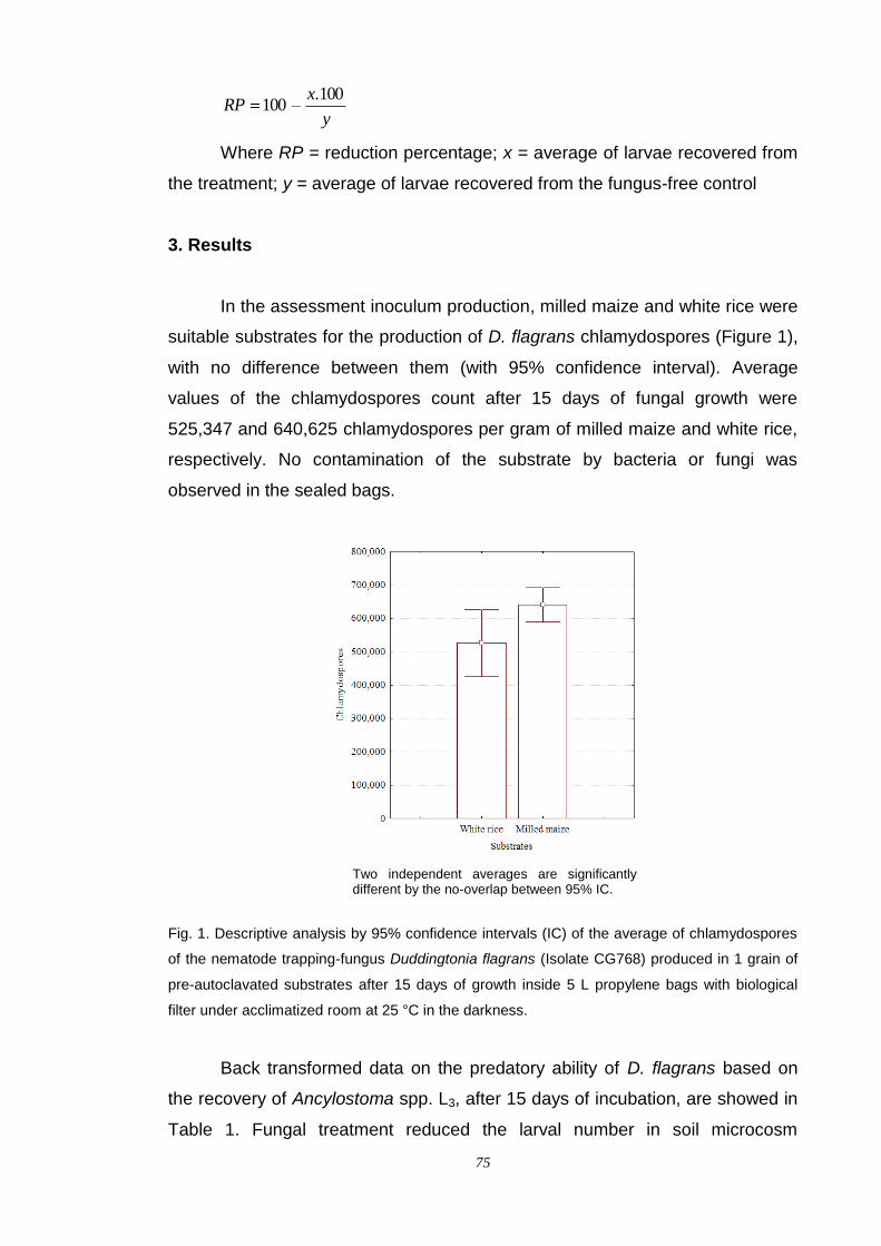
75
y
xRP
100.100
Where RP = reduction percentage; x = average of larvae recovered from
the treatment; y = average of larvae recovered from the fungus-free control
3. Results
In the assessment inoculum production, milled maize and white rice were
suitable substrates for the production of D. flagrans chlamydospores (Figure 1),
with no difference between them (with 95% confidence interval). Average
values of the chlamydospores count after 15 days of fungal growth were
525,347 and 640,625 chlamydospores per gram of milled maize and white rice,
respectively. No contamination of the substrate by bacteria or fungi was
observed in the sealed bags.
Two independent averages are significantly different by the no-overlap between 95% IC.
Fig. 1. Descriptive analysis by 95% confidence intervals (IC) of the average of chlamydospores
of the nematode trapping-fungus Duddingtonia flagrans (Isolate CG768) produced in 1 grain of
pre-autoclavated substrates after 15 days of growth inside 5 L propylene bags with biological
filter under acclimatized room at 25 °C in the darkness.
Back transformed data on the predatory ability of D. flagrans based on
the recovery of Ancylostoma spp. L3, after 15 days of incubation, are showed in
Table 1. Fungal treatment reduced the larval number in soil microcosm
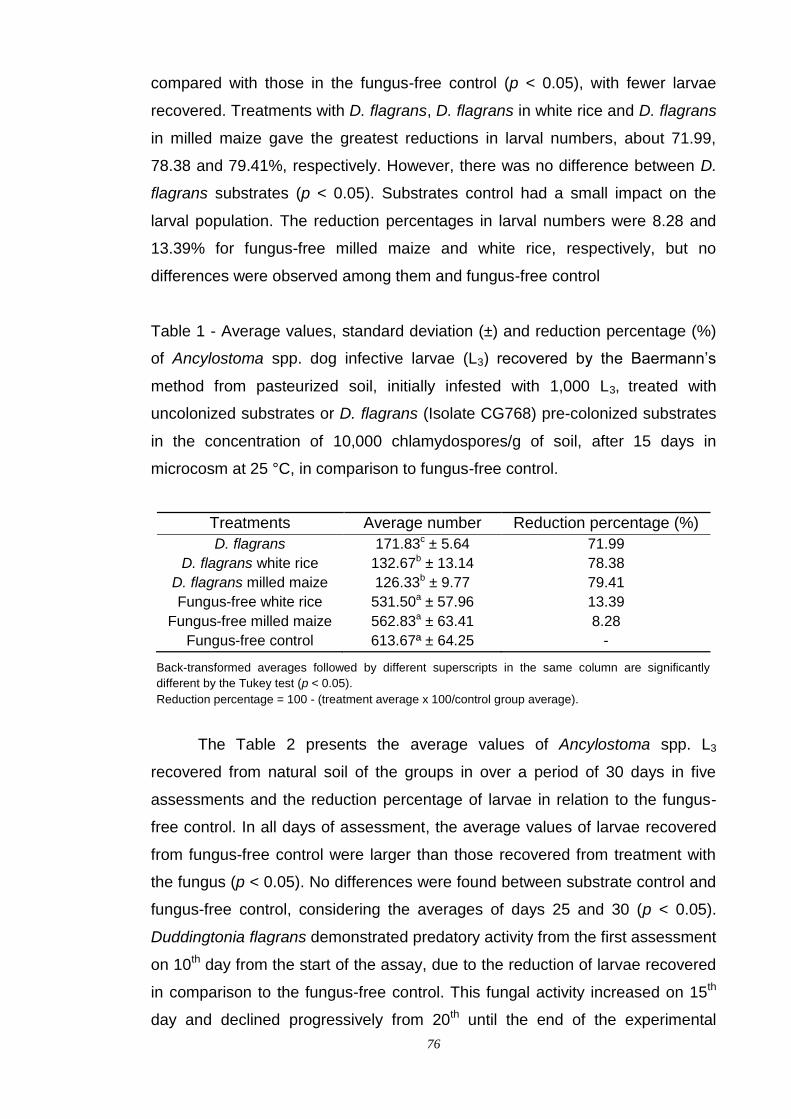
76
compared with those in the fungus-free control (p < 0.05), with fewer larvae
recovered. Treatments with D. flagrans, D. flagrans in white rice and D. flagrans
in milled maize gave the greatest reductions in larval numbers, about 71.99,
78.38 and 79.41%, respectively. However, there was no difference between D.
flagrans substrates (p < 0.05). Substrates control had a small impact on the
larval population. The reduction percentages in larval numbers were 8.28 and
13.39% for fungus-free milled maize and white rice, respectively, but no
differences were observed among them and fungus-free control
Table 1 - Average values, standard deviation (±) and reduction percentage (%)
of Ancylostoma spp. dog infective larvae (L3) recovered by the Baermann‟s
method from pasteurized soil, initially infested with 1,000 L3, treated with
uncolonized substrates or D. flagrans (Isolate CG768) pre-colonized substrates
in the concentration of 10,000 chlamydospores/g of soil, after 15 days in
microcosm at 25 °C, in comparison to fungus-free control.
Treatments Average number Reduction percentage (%)
D. flagrans 171.83c ± 5.64 71.99
D. flagrans white rice 132.67b ± 13.14 78.38
D. flagrans milled maize 126.33b ± 9.77 79.41
Fungus-free white rice 531.50a ± 57.96 13.39
Fungus-free milled maize 562.83a ± 63.41 8.28
Fungus-free control 613.67ª ± 64.25 -
Back-transformed averages followed by different superscripts in the same column are significantly
different by the Tukey test (p < 0.05).
Reduction percentage = 100 - (treatment average x 100/control group average).
The Table 2 presents the average values of Ancylostoma spp. L3
recovered from natural soil of the groups in over a period of 30 days in five
assessments and the reduction percentage of larvae in relation to the fungus-
free control. In all days of assessment, the average values of larvae recovered
from fungus-free control were larger than those recovered from treatment with
the fungus (p < 0.05). No differences were found between substrate control and
fungus-free control, considering the averages of days 25 and 30 (p < 0.05).
Duddingtonia flagrans demonstrated predatory activity from the first assessment
on 10th day from the start of the assay, due to the reduction of larvae recovered
in comparison to the fungus-free control. This fungal activity increased on 15th
day and declined progressively from 20th until the end of the experimental

77
period, nevertheless resulted in low levels in the number of infective larvae.
Comparable behavior throughout the observation period was demonstrated
when the substrate was added to the soil, however with low reduction in number
of recovered larvae. The single concentration rate (10,000 chlamydospores per
grain of soil) showed a consistent reduction in larval population. In treatment
with D. flagrans, the reduction percentages observed were 61.37, 73.23, 70.84,
64.50 and 56.99%, considering the assessment on days 10, 15, 20, 25 and 30,
respectively. On the other hand, in substrate control the reduction percentages
were 16.39, 27.99, 20.36, 14.45 and 9.20%, respectively from first to last
assessment day established.
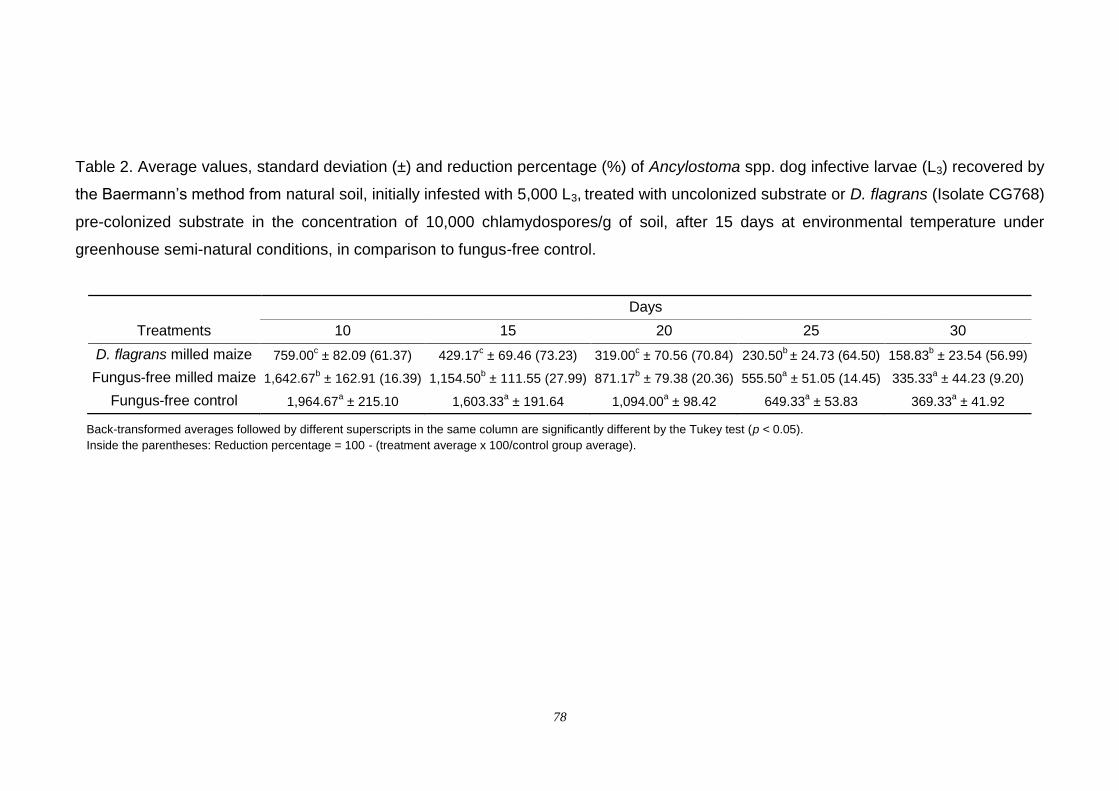
78
Table 2. Average values, standard deviation (±) and reduction percentage (%) of Ancylostoma spp. dog infective larvae (L3) recovered by
the Baermann‟s method from natural soil, initially infested with 5,000 L3, treated with uncolonized substrate or D. flagrans (Isolate CG768)
pre-colonized substrate in the concentration of 10,000 chlamydospores/g of soil, after 15 days at environmental temperature under
greenhouse semi-natural conditions, in comparison to fungus-free control.
Treatments
Days
10 15 20 25 30
D. flagrans milled maize 759.00c ± 82.09 (61.37)
429.17
c ± 69.46 (73.23) 319.00
c ± 70.56 (70.84) 230.50
b ± 24.73 (64.50) 158.83
b ± 23.54 (56.99)
Fungus-free milled maize 1,642.67b ± 162.91 (16.39) 1,154.50
b ± 111.55 (27.99) 871.17
b ± 79.38 (20.36) 555.50
a ± 51.05 (14.45) 335.33
a ± 44.23 (9.20)
Fungus-free control 1,964.67a ± 215.10 1,603.33
a ± 191.64 1,094.00
a ± 98.42 649.33
a ± 53.83 369.33
a ± 41.92
Back-transformed averages followed by different superscripts in the same column are significantly different by the Tukey test (p < 0.05).
Inside the parentheses: Reduction percentage = 100 - (treatment average x 100/control group average).
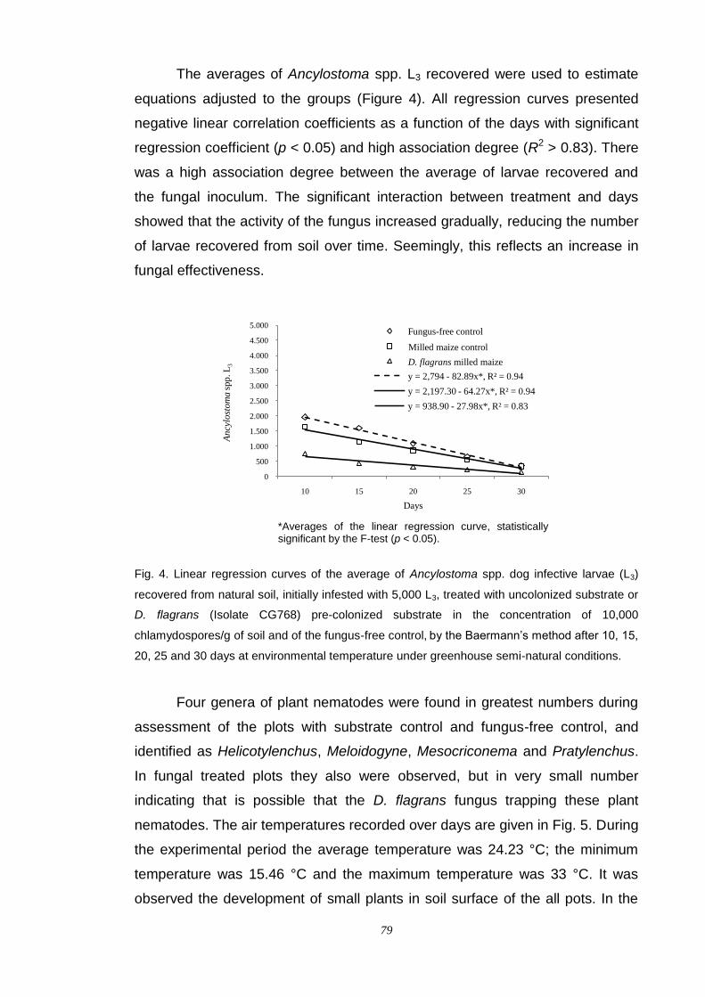
79
The averages of Ancylostoma spp. L3 recovered were used to estimate
equations adjusted to the groups (Figure 4). All regression curves presented
negative linear correlation coefficients as a function of the days with significant
regression coefficient (p < 0.05) and high association degree (R2 > 0.83). There
was a high association degree between the average of larvae recovered and
the fungal inoculum. The significant interaction between treatment and days
showed that the activity of the fungus increased gradually, reducing the number
of larvae recovered from soil over time. Seemingly, this reflects an increase in
fungal effectiveness.
*Averages of the linear regression curve, statistically significant by the F-test (p < 0.05).
Fig. 4. Linear regression curves of the average of Ancylostoma spp. dog infective larvae (L3)
recovered from natural soil, initially infested with 5,000 L3, treated with uncolonized substrate or
D. flagrans (Isolate CG768) pre-colonized substrate in the concentration of 10,000
chlamydospores/g of soil and of the fungus-free control, by the Baermann‟s method after 10, 15,
20, 25 and 30 days at environmental temperature under greenhouse semi-natural conditions.
Four genera of plant nematodes were found in greatest numbers during
assessment of the plots with substrate control and fungus-free control, and
identified as Helicotylenchus, Meloidogyne, Mesocriconema and Pratylenchus.
In fungal treated plots they also were observed, but in very small number
indicating that is possible that the D. flagrans fungus trapping these plant
nematodes. The air temperatures recorded over days are given in Fig. 5. During
the experimental period the average temperature was 24.23 °C; the minimum
temperature was 15.46 °C and the maximum temperature was 33 °C. It was
observed the development of small plants in soil surface of the all pots. In the
0
500
1.000
1.500
2.000
2.500
3.000
3.500
4.000
4.500
5.000
10 15 20 25 30
An
cylo
stom
asp
p. L
3
Days
Fungus-free control
Ground corn control
D. flagrans ground corn
y = 2,794 - 82.89x*, R² = 0.94
y = 2,197.30 - 64.27x*, R² = 0.94
y = 938.90 - 27.98x*, R² = 0.83
D. flagrans milled maize
Milled maize control
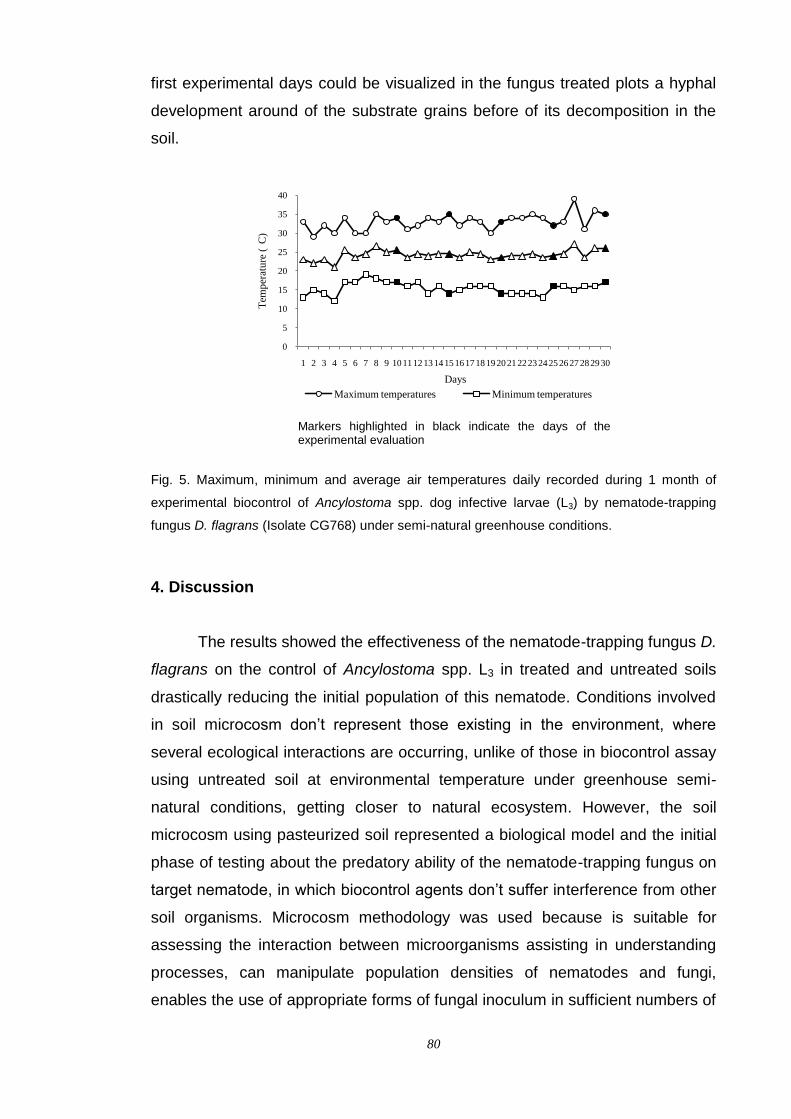
80
first experimental days could be visualized in the fungus treated plots a hyphal
development around of the substrate grains before of its decomposition in the
soil.
Markers highlighted in black indicate the days of the experimental evaluation
Fig. 5. Maximum, minimum and average air temperatures daily recorded during 1 month of
experimental biocontrol of Ancylostoma spp. dog infective larvae (L3) by nematode-trapping
fungus D. flagrans (Isolate CG768) under semi-natural greenhouse conditions.
4. Discussion
The results showed the effectiveness of the nematode-trapping fungus D.
flagrans on the control of Ancylostoma spp. L3 in treated and untreated soils
drastically reducing the initial population of this nematode. Conditions involved
in soil microcosm don‟t represent those existing in the environment, where
several ecological interactions are occurring, unlike of those in biocontrol assay
using untreated soil at environmental temperature under greenhouse semi-
natural conditions, getting closer to natural ecosystem. However, the soil
microcosm using pasteurized soil represented a biological model and the initial
phase of testing about the predatory ability of the nematode-trapping fungus on
target nematode, in which biocontrol agents don‟t suffer interference from other
soil organisms. Microcosm methodology was used because is suitable for
assessing the interaction between microorganisms assisting in understanding
processes, can manipulate population densities of nematodes and fungi,
enables the use of appropriate forms of fungal inoculum in sufficient numbers of
0
5
10
15
20
25
30
35
40
1 2 3 4 5 6 7 8 9 10 1112 1314 1516 1718 1920 21 2223 2425 2627 2829 30
Tem
per
atu
re (
C)
Days
Maximum temperatures Minimum temperatures
81
which cannot be obtained for infesting large volumes of soil, besides achieve
sufficient replication to obtain acceptable accuracy and precision of the results
(Jaffee, 1996). In addition to being experimentally convenient, the use of small
volumes of soil is biologically appropriate given the small size and limited
motility of fungi and nematodes (Wiens, 1989). Meanwhile, the understanding
natural soil processes involved in the biological control of pre-parasitic forms of
Ancylostoma spp. by D. flagrans at the ecosystem level requires comparison of
microcosm and field data.
Good results were observed when some environmental conditions were
incorporated in the model of microcosm design. Biological control was evident
in subsequent experiment under semi-natural conditions in response to the
inoculation of D. flagrans with change in the pool of larvae reflected continuous
decay rate of recovered larvae over days of assessement. Herein, undoubtedly,
the antagonist maintained its activity until the end of the period capturing the
infective larvae in the soil suggesting the survivability of the fungus. Knox et al.
(2002) reported the D. flagrans persistence for 8-24 weeks after initial
contamination in association with the soil under the point of fecal deposition,
however this time can be prolonged until 20 months in dry environments
(Gronvold et al., 1996a). Seemingly the uncolonized milled maize reduced the
larvae population in soil. In addition to natural death of the larvae, this reduction
might be explained by the stimulation of the microbial activity of microorganisms
present in the untreated soil. The naturally occurring of nematophagous
indigenous species considers the possibility that manipulation of the
environment to enhance existing populations of fungi may also be feasible
(Waghorn et al., 2002; Monfort et al., 2006). When considered in conjunction
with the D. flagrans effect, such observation suggests that the population
damage unlikely to be simply due to predation, but is possible that it has been
the result of the combination of effects. These results are still somewhat
surprising because suggest that even in the presence of organic matter the D.
flagrans may be capable of trapping pre-parasitic forms of animal parasitic
nematodes such as Ancylostoma spp. An interesting observation that should be
taken in account was the caught of phytonematodes by D. flagrans showed the
potential of this fungus as biocontrol agent these parasites that are pests in
agriculture and horticulture, causing severe yield losses (Nordbring-Hertz et al.,
2006).
82
The trap formation in nematophagous fungi is influenced by
environmental factors such as nutrient level, pH, temperature, light and
moisture (Nordbring-Hertz, 1973), but especially temperature (Bogus et al.,
2005). In particular, the soil temperature is perhaps the main physical variable
in terms of biological significance and should be taken into account when
biological control systems are being developed (Stirling, 1991), because the
optimum temperature for nematophagous fungi varies with each species
influencing the percentage of arrested larvae (Morgan et al., 1997). It is likely
that the air temperatures during the experimental period were relevant for the D.
flagrans activity due to the low number of Ancylostoma spp. L3 recovered from
treated plots. According to Gronvold et al. (1996b) and Morgan et al. (1997), the
optimal growth temperature for D. flagrans ranges between 25-33 °C. At
temperatures lower than 25 °C it grows slowly (Larsen, 1991), presenting an
upper limit of growth at 35 °C and not developing at 37 °C (Peloille, 1991). The
highest temperature regimen in the present study exposed the D. flagrans to
temperatures that exceeded 30 °C, but the maximum temperature in the soil
was typically smaller than that recorded and may mean that the maximum air
temperatures overestimate those real in the soil. Regarding the formation of
tridimensional trapping nets, Gronvold et al. (1999) reported a higher trap
formation rate at 30 °C than at 20 °C and observed that the slower development
of D. flagrans at 10 °C does not decrease its trapping capacity, which remained
high for a prolonged period of time, but can be compromised at temperatures
persistently lower than 10 °C (Fernández et al., 1999). The trapping efficacy of
D. flagrans may also be dependent on the duration of exposure to high
temperatures rather than temperature alone (Kahn et al. 2007). In addition to
the absolute effect of temperature, the effect of temperature fluctuation on
trapping efficacy was demonstrated to be temperature dependent, there was
reduction larvae between 15-25 °C (Fernández et al., 1999). The temperatures
reported above are similar those recorded during experimental period between
14.5-33 °C. So, it is reasonable to state that temperature conditions and larval
availability were conducive to optimal fungus activity which supports the results
observed.
Mycelial growth and traps formation are processes that require energy
prior to predation in non-spontaneous trap forming predators. According to
Cooke (1969), the energy required for the mycelial growth and formation of
83
traps can be supplied by a source of carbohydrates. Thus, a factor that may be
important for successful establishment of fungal biocontrol agents is the
addition of a nutritional food base to the inoculum (Monfort et al., 2006). This
procedure can increase the activity of fungal predators, because helps the
germination of spores and mycelial growth, and favors the fungi in competition
with other soil microorganisms (Payne and Lynch, 1988; Stirling, 1991), hence
the D. flagrans was incorporated into the soil together with its substrate for
growth. Thus, fungi with an energy source readily available probably would not
have limitations to colonize the soil and form structures to capture nematodes
(Dias and Ferraz, 1994; Cannayane and Rajendran, 2001). The soil can be
more receptive to its indigenous isolates than to nonindigenous isolates.
Apparently, the microbiota can determine the ability of nematophagous fungi to
proliferate in soil (Monfort et al., 2006), probably due to fungistasis (Van Gundy,
1985) that can inhibit the germination, growth and action of the nematophagous
fungus (Kerry, 1984). This antagonist effect on nematophagous fungi may due
to microorganisms such as Aspergillus terreus, Penicillium sp., Rhizopus sp.,
Trichoderma sp. (Mankau, 1961; 1962), Saccharomyces cerevisiae
(Rosenzweig and Ackroyd, 1984) and Bacillus pimulus (B‟Chir and Namouch,
1988).
The fungistase occurs in untreated soil (Morgan-Jones and Rodriguez-
Kabana, 1987), however the results showed that the possible presence of
others microorganisms did not affect the D. flagrans efficacy, once that it
presented consistent control. Therefore, a reasonable interpretation based on
the above-mentioned results is that the milled maize was used by D. flagrans to
establish in soil of pots resulting in the trapping of Ancylostoma spp. L3 in this
confined environmental system. It seems like that the growth of fungus tested
was not inhibited in nonpasteurized soil and the reason for good results could
be the ability of the D. flagrans to proliferate in soil. This is possible because
predators fungi adhesive network formers are good saprophytes (Nordbring-
Hertz, 1988) could be nurtured in the absence of other sources of nematode
allowing their survival in soil (Gray, 1983). Furthermore, the resistant thick-
walled D. flagrans chlamydospores, used as fungal inoculum in present study,
have a long-term viability in soil under unfavourable environments (Larsen et
al., 1991; Sanyal and Mukhopadhyaya, 2003) could germinate to form mycelium
and trap in response to nematode activity (Skipp et al., 2002) providing high
84
trapping efficacies (Jackson and Miller, 2006).
Aspects such as the selection of an isolate with rapid growth, abundant
sporulation and high pathogenic for the target organism, as well as the use of a
substrate, minimally processed, quick and easy to use, low cost and viable on a
large scale were taken into account in the present research based to the
assumptions of Samsinakova et al. (1981) for the success in the large
production and in the use of nematophagous fungi to the biocontrol. The
methodology of fungal mass production employed using plastic bags with
biological filter showed the practical feasibility to obtain large number of D.
flagrans chlamydospores, providing a sterile environment for fungal growth
without contamination of the substrates. Concurrently moistened white rice and
milled maize grains were suitable substrates for the production of fungal
inoculum in a short period of 15 days. They are related as good substrates for
the production of D. flagrans chlamydospores (Waller et al., 2001), but other
cereal grains as wheat, sorghum, and barley are also used for the inoculum
production of this fungus (Sanyal, 2000; Sanyal and Mukhopadhyaya, 2003).
The Baermann‟s funnel is the method most used to extract nematodes
from soil (Gray, 1987). The conventional Baermann‟s method was regarded
practical and applicable to recovery the larvae added to the soil and determine
the predatory efficiency of the nematode-trapping fungus. This is consistent with
the fact that 61.37% of the total larvae added were recovered from soil
microcosm when fungi were not added and this reflects the extraction efficiency
in the absence of antagonists. It was observed that not all the larvae added to
the soil were recovered, but this decrease was never associated with any signs
of parasitism. The Baermann‟s apparatus modified also was efficient in recovery
of larvae added to the untreated soil in biocontrol assay. The adaptation of this
method, providing major contact area between soil and water, was relevant for
the recovery of larvae from soil in the present study. However, the number of
larvae recovered never exceeded 39% of total larvae added. Furthermore, the
number of nematodes recovered from the nonpasteurized soil decreased in the
subsequent days of assessment, recovering only 7% of them on the last
experimental day. Understandably, since the soil was not sterilized, this larval
reduction in control plots suggests the presence of indigenous antagonists of
nematodes not used as experimental inoculum in addition to natural death of
them over time.
85
The presence of Ancylostoma spp. L3 in last day of evaluation showed
the long survival this nematode. Up to a point, the soil may act as a reservoir of
infective larvae (Krecek and Murrell, 1988) providing a more favourable
environment for the development of larvae by protecting them from the more
extreme environmental conditions on the soil surface (Bryan, 1976).
Furthermore, for many parasitic nematodes, as Ancylostoma spp., be arrest at
the L3 facilitates survival in the environment (Datu et al., 2008). According to
Hayes and Blackburn (1966) the predatory phase may continue until the
nutritional requirement of the fungus is answered, and probably was the cause
of the no extermination of the larvae in the microenvironment. Another
explanation would be that the ability to attract the nematode by fungi could be
influenced by spatial trap location in the soil since the uniform distribution would
increase the chances of interaction between the microorganisms studied.
However, the biological control may not lead to complete reduction of larval
populations, it may reduce them sufficiently to prevent adverse effects, yet allow
the stimulation of naturally acquired immunity (Barnes et al., 1995; Larsen et al.,
1997). The effect of an artificial increase in nematophagous fungi in the
environment is likely to be short-lived due to ecological factors that limit fungal
populations in the field (Cooke and Satchuthananthavale, 1968). Despite this,
there is the possibility that new incorporation of fungal inoculum may result in
eliminating the larvae of the microcosm. The nematode trapping activity of D.
flagrans is not specific to parasitic larvae and therefore there is the possible of
perturbation of the soil nematode community (Dimander et al., 2003b). The
introduction high levels this fungus in soil arouses the question as to adverse
environmental impacts on soil indigenous populations in the ecosystem.
However, studies with D. flagrans showed no effects on free-living nematodes,
microarthropods and even on other nematode trapping-fungi (Yeates et al.,
1997; Gronvold et al., 2000; Faedo et al., 2002; Knox et al., 2002). This
indicates a future potential to use of D. flagrans as an organically acceptable
alternative.
This study showed that biological control in nonpasteurized soil using D.
flagrans presented satisfactory results in a small-scale of soil on Ancylostoma
spp. L3. The used concentration proved to be sufficient in controlling larvae
under semi-natural conditions, but there is no guarantee that results obtained
will always perform to the same degree when used in field situations provided

86
satisfactory control. Nevertheless, nematophagous fungi promising such as D.
flagrans should continue being object of research, both under controlled
conditions in the field, since it is necessary to know more of the ecological
requirements, specificity, competitive capacity and establishment of introduced
isolates. However, given that the fungi have no effect on established worm
populations in host, this approach will almost certainly need to be applied in
conjunction with other forms of control. An integrated management strategy,
based upon epidemiological knowledge of the parasite involved, could be
composed of two or more of the following elements: biological control agents,
anthelminthics (minimal treatment) and hygienic measures. The results
presented provide considerable encouragement for further research into the
biological control of pre-parasitic forms of Ancylostoma spp. and suggest the
need to develop some field trials determining biotic and abiotic factors that
could interfere with the action of D. flagrans under adverse and varying climatic
conditions through all seasons. Studies about formulation, delivery and
inoculum concentration in natural soil also should be made. Significant potential
exists for applying this fungus as biological control agent of Ancylostoma spp.
L3.

87
CONCLUSÕES GERAIS
Pode-se concluir que:
1. Os testes in vitro não representam as condições ambientais e por isso
possuem limitações, mas são necessários no estudo da capacidade
predatória de fungos nematófagos e na seleção de isolados mais infectivos,
constituindo-se num método preliminar de estudo da interação do fungo
antagonista com o nematóide alvo;
2. Existe variabilidade na infectividade sobre ancilostomatídeos pelas
diferentes espécies fúngicas estudadas;
3. O isolado CG768 de D. flagrans tem elevada infectividade sobre larvas
infectantes de ancilostomídeos, podendo destruí-las completamente em
menos de 48 horas após a captura;
4. O isolado CG768 de D. flagrans é um promissor biocontrolador de
ancilostomídeos de cães, pois reduziu o número de larvas infectantes
destes nematóides recuperadas de solo não tratado em ensaio sob
condições semi-naturais;
5. Grãos de arroz branco e de milho moído são substratos adequados para a
produção massal de inóculo de fungos nematófagos como o isolado de D.
flagrans, mas outros cereais devem ser testados;
6. A incorporação ao solo do fungo em seu substrato de crescimento é prático
e pode viabilizar o estabelecimento do antagonista nematófago no ambiente
por propiciar suporte nuticional inicial à sua fase pré-parasitária;
7. O isolado CG768 de D. flagrans tem potencial para reduzir a infestação
ambiental com formas larvais pré-parasitárias de ancilostomídeos de cães,
podendo ser uma alternativa viável e complementar ao uso de anti-
helmínticos, ao controle populacional de cães de errantes e à educação da
população;
8. Em vista dos percentuais de redução de L3 de Ancylostoma spp. nos
ensaios conduzidos, é necessário reforçar os resultados prosseguindo com
estudos em solo sob condições naturais.
88
REFERÊNCIAS BIBLIOGRÁFICAS
Akhtar, M.; Malik, A. Roles of organic soil
amendments and soil organisms in the
biological control of plant-parasitic
nematodes: a review. Biores. Technol.
74:35-47, 2000.
Albonico, M.; Smith, P.G.; Ercole, E.; Hall, A.;
Chwaya, H.M.; Alawi, K.S.; Savioli, L. Rate
of reinfection with intestinal nematodes
after treatment of children with
mebendazole or albendazole in a highly
endemic area. Trans. R. Soc. Trop. Med.
Hyg. 89:538-541, 1995.
Alfenas, A.C.; Mafia, R.G. Métodos em
Fitopatologia. 1ª Edição. Viçosa: Editora
UFV, 2007, 382p.
Almeida, A.B.P.F.; Sousa, V.R.F.; Dalcin, L.;
Justino, C.H.S. Contaminação por fezes
caninas das praças públicas de Cuiabá,
Mato Grosso. Braz. J. Vet. Res. Anim. Sci.
44:132-136, 2007.
Almeida, M.A.O.; Ayres, M.C.C. Agentes
antinematódeos. In: Spinosa, H.S.;
Górniak, S.L.; Bernardi, M.M. (Eds.).
Farmacologia Aplicada à Medicina
Veterinária. 4ª Edição. Rio de Janeiro:
Ganabara Koogan, 2006, p. 535-550.
Alves, O.F.; Gomes, A.G.; Silva, A.C.
Ocorrência de enteroparasitos em cães do
município de Goiânia, Goiás: comparação
de técnicas de diagnóstico. Ciênc. Anim.
Bras. 6:127-133, 2005.
Alves, P.H.; Araújo, J.V.; Guimarães, M.P.;
Assis, R.C.L.; Sarti, P.; Campos, A.K.
Aplicação de formulação do fungo
predador de nematóides Monacrosporium
thaumasium (Drechsler, 1937) no controle
de nematóides de bovinos. Arq. Bras. Med.
Vet. Zootec. 55:568-573. 2003.
Araújo, F.R.; Crocci, A.J.; Rodrigues, R.G.C.;
Avalhaes, J.S.; Miyoshi, M.I.; Salgado,
F.P.; Silva, M.A.; Pereira, M.L.
Contaminação de praças públicas de
Campo Grande, MS, Brasil, por ovos de
Toxocara e Ancylostoma em fezes de
cães. Rev. Soc. Bras. Med. Trop. 32:581-
583, 1999.
Araújo, J.V. Capture of Cooperia punctata
infective larvae by nematode-trapping fungi
Arthrobotrys. Braz. J. Vet. Parasitol. 7:101-
105, 1998.
Araújo, J.V.; Assis, R.C.L.; Campos, A.K.;
Mota, M.A. Atividade in vitro dos fungos
nematófagos dos gêneros Arthrobotrys,
Duddingtonia e Monacrosporium sobre
nematóides Trichostrongilídeos
(Nematoda: Trichostrongyloidea) parasitos
gastrintestinais de bovinos. Rev. Bras.
Parasitol. Vet. 13:65-71, 2004.
Araújo, J.V.; Guimarães, M.P. Ação do fungo
predador de nematóides Arthrobotrys
conoides sobre larvas infectantes de
Cooperia punctata. Rev. Bras. Cienc. Vet.
9:5-7, 2002.
Araújo, J.V.; Santos, M.A.; Ferraz, S.; Maia,
A.S. Antagonistic effect of predacious
Arthrobotrys fungi on infective
Haemonchus placei larvae. J. Helmintol.
67:136-138, 1993.
Araújo, J.V.; Santos, M.A.; Ferraz, S.; Maia,
A.S.; Magalhães, A.C.M. Controle de
larvas infectantes de Haemonchus placei
por fungos predadores da espécie
Monacrosporium ellypsosporum em
condições de laboratório. Arq. Bras. Med.
Vet. Zootec. 44:521-526, 1992.
Araújo, R.B.; Ferreira, P.M.; Assis, C.B.; Del
Carlo, R.J. Helmintoses intestinais em
cães da microregião de Viçosa, Minas
Gerais. Arq. Bras. Med. Vet. Zootec.
38:197-203, 1986.
B‟Chir, M.M.; Namouch, N. Effect of Bacillus
pimulus on Monacrosporium salinum, a
nematode-trapping fungus. Rev. Nématol.
11:263-266, 1988.
Baermann, G. Eine einfache methode zur
auffindung von Ancylostomum
(Nematoden) larven in erdproben,
Gennesk. Tijds. Ned. Ind. 57:131-137,
1917.
Bâgel, K.; Frucht, K.; Drysdale, G.; Remfry, J.
Guidelines for dog population
management. Geneve: World Health
Organization, 1990, 116p.
Bahgat, M.A.; Gindy, E.I.; Mahamoud, L.A.;
Hegab, M.H.; Shahin, A.M. Evaluation of
the role of Ancylostoma caninum in
humans as a cause of acute and recurrent
abdominal pain. J. Egypt. Soc. Parasitol.
29:873-882, 1999.
Baker, M.K.; Lange, L.; Verster, A.; Van Der
Plaat, S. A survey of helminths in
domestic cats in the Pretoria area of
Transvaal, Republic of South Africa. Part
1: The prevalence and comparison of
89
burdens of helminths in adult and juvenile
cats. J. S. Afr. Vet. Assoc. 60:139-142,
1989.
Balan, J.; Lechevalier, H.A. The predaceous
fungus Arthrobotrys dactyloides: induction
of trap formation. Mycologia 64:919-922,
1972.
Barçante, J.M.P.; Barçante, T.A.; Dias,
S.R.C.; Vieira, L.Q.; Lima, W.S.; Negrão-
Corrêa, D. A method to obtain axenic
Angiostrongylus vasorum first-stage larvae
from dog feces. Parasitol. Res. 89:89-93,
2003.
Barger, I.A. The role of epidemiological
knowledge and grazing management for
helminth control in small ruminants. Int. J.
Parasitol. 29:41-47, 1999.
Barnes, E.; Dobson, R.; Barger, I. Worm
control and anthelmintic resistance:
adventures with a model. Parasitol. Today
11:56-63, 1995.
Barron, G.L. Lignolytic and cellulolytic fungi as
predators and parasites. In: Carroll, G.C.;
Wicklow, D.T. (Eds.). The fungal
community: its organization and role in the
ecosystem. 2nd
Edition. New York: Marcel
Dekker, 1992, p. 311-326.
Barron, G.L. The Nematode-destroying Fungi.
Topics in Mycobiology, vol. I. Guelph:
Canadian Biological Publications, 1977,
140p.
Barron, G.L. Predatory fungi, wood decay,
and the carbon cycle. Biodiversity 4:3-9,
2003.
Baudena, M.A.; Chapman, M.R.; Larsen, M.;
Klei, T.R. Efficacy of the nematophagous
fungus Duddingtonia flagrans in reducing
equine cyathostome larvae on pasture in
South Lousiana. Vet. Parasitol. 89:219-
230, 2000.
Bird, J.; Herd, R.P. “In vitro” assessment of
two species of nematophagous fungi
(Arthrobotrys oligospora and Arthrobotrys
flagrans) to control the development of
infective cyathostome larvae from naturally
infected horses, Vet. Parasitol. 56:181-187,
1995.
Blagburn, B.L.; Lindsay, D.S.; Vaughan, J.L.;
Rippey, N.S.; Wright, J.C.; Lynn, R.C.;
Keleh, W.J.; Ritchie, G.C.; Hepler, D.I.
Prevalence of canine parasites based on
fecal flotation. Comp. Cont. Ed. Pract. Vet.
18:483-509, 1996.
Blazius, R.D.; Emerick, S.; Prophiro, J.S.;
Roosevelt, P.; Romão, T.; Silva, O.S.
Ocorrência de protozoários e helmintos
em amostras de fezes de cães errantes
da cidade de Itapema, Santa Catarina.
Rev. Soc. Bras. Med. Trop. 38:73-74,
2005.
Boag, P.R.; Parsons, J.C.; Presidente, P.J.;
Spithill, T.W.; Sexton J.L. Characterization
of humoral immune responses in dogs
vaccinated with irradiated Ancylostoma
caninum. Vet. Immunol. Immunopathol.
92:87-94, 2003.
Bogus, M. I.; Czygier, M.; Kedra, E.;
Samborski, J. In vitro assessment of the
influence of nutrition and temperature on
growing rates of five Duddingtonia flagrans
isolates, their insecticidal properties and
ability to impair Heligmosomoides
polygyrus motility. Exp. Parasitol. 109:115-
123, 2005.
Bowman, D.D.; Lynn, R.C.; Eberhard, M.L.;
Georgi, J.R. Georgis‟ parasitology for
veterinarians. 8th Edition. St. Louis:
Saunders, 2003, 422p.
Brener, B.; Lisboa, L.; Mattos, D.P.B.G.;
Arashiro, E.K.N.; Millar, P.R.; Sudré, A.P.;
Duque, V. Freqüência de enteroparasitas
em amostras fecais de cães e gatos dos
municípios do Rio de Janeiro e Niterói. R.
Bras. Ciênc. Vet. 12:102-105, 2005.
Brenner, M.A.; Patel, M.B. Cutaneous larva
migrans: the creeping eruption. Cutis
72:111-115, 2003.
Bryan, R.P. The effect of the dung beetle,
Onthophagus gazella, on the ecology of
the infective larvae of gastrointestinal
nematodes of cattle. Aust. J. Agric. Res.
27:567-574, 1976.
Bugg, R.J.; Robertson, I.D.; Elliot, A.D.;
Thompson, R.C. Gastrointestinal
parasites of urban dogs in Perth, Western
Australia. J. Vet. 157:295-301, 1999.
Burrows, C.F.; Batt, R.M.; Sherding, R.G.
Diseases of the small intestine. In:
Ettinger, S.J.; Feldman, E.C. (Eds.)
Textbook of Veterinary Internal Medicine.
4th Edition. Philadelphia: WB Saunders,
1995, p. 1169-1232.
Campos, A.K. Fungos nematófagos no
controle de nematóides gastrintestinais de
ruminantes. Tese de Doutorado,
Universidade Federal de Minas Gerais,
Belo Horizonte, Minas Gerais, Brasil, 2006,
139p.

90
Campos, A.K.; Araújo, J.V.; Guimarães, M.P.
Interaction between the nematophagous
fungus Duddingtonia flagrans and infective
larvae of Haemonchus contortus
(Nematoda: Trichostrongyloidea). J.
Helminthol. 82:337-341, 2008.
Campos Filho, P.C.; Barros, L.M.; Campos,
J.O.; Braga, V.B.; Cazorla, I.M.;
Albuquerque, G.R.; Carvalho, S.M.S.
Parasitas zoonóticos em fezes de cães em
praças públicas do município de Itabuna,
Bahia, Brasil. Rev. Bras. Parasitol. Vet.
17:206-209, 2008.
Cannayane, I.; Rajendran, G.S.O. Application
of biocontrol agents and oil cakes for the
management for Meloidogyne incognita in
brinjal (Solanum melongena L.). Curr.
Nematol. 12:51-55, 2001.
Canning, E.U. Protozoal parasites as agents
for biological control of plant-parasitic
nematodes. Nematologica 19:342-348,
1973.
Capuano, D.M.; Rocha, G.M. Ocorrência de
parasitas com potencial zoonótico em
fezes de cães coletadas em áreas
públicas do município de Ribeirão Preto,
SP, Brasil. Rev. Bras. Epidemiol. 9:81-86,
2006.
Carvalho, R.O.; Araújo, J.V.; Braga, F.R.;
Araujo, J.M.; Silva, A.R.; Tavela, A.O.
Predatory activity of nematophagous fungi
on infective larvae of Ancylostoma sp.:
evaluation in vitro and after passing
through the gastrointestinal tract of dogs, J.
Helminthol. 2009
doi:10.1017/S0022149X08158965.
Castro, A.A.; Oliveira, C.R.C.; Anjos, D.H.S.;
Ornelas, E.I.; Bittencourt, V.R.E.P.; Araújo,
J.V.; Sampaio, I.B.M.; Rodrigues, M.L.A.
Potencial dos fungos nematófagos
Arthrobotrys sp. e Monacrosporium
thaumasium para o controle de larvas de
ciatostomíneos de eqüinos (nematoda:
Cyathostominae). Braz. J. Vet. Parasitol.
12:53-57, 2003.
Castro, E.S.; Mattos, M.J.T.; Bastos, C.D.
Gastrenterites parasitárias em cães
atendidos na clínica hospitalar da
UFRGS. Rev. Bras. Med. Vet. 23:76-77,
2001.
Castro, J.M.; Santos, S.V.; Monteiro, N.A.
Contaminação de canteiros da orla
marítima do município de Praia Grande,
São Paulo, por ovos de Ancylostoma e
Toxocara em fezes de cães. Rev. Soc.
Bras. Med. Tropical 38:199-201, 2005.
Cayrol, J.C.; Ritter M. Donnés recentes sur
l‟utilization de champignons du sol comme
antagonistes de nématodes. Bull. Soc.
Franc. Parasitol 2:53-56, 1984.
Chan, M.S. The global burden of intestinal
nematode infections-fifty years on.
Parasitol. Today 13:438-443, 1997.
Chandrawathani, P.; Jamnah, O.; Waller, P.J.;
Larsen, M.; Gillespie, A.; Zahari, W.M.
Biological control of nematode parasites of
small ruminants in Malaysia using the
nematophagous fungus Duddingtonia
flagrans. Vet. Parasitol. 117:173-183,
2003.
Chandrawathani, P.; Omar, J.; Waller, P. The
control of free-living stages of
Strongyloides papillosus by the
nematophagous fungus Arthrobotrys
oligospora. Vet. Parasitol. 76:321-325,
1998.
Chartier, C.; Pors, I. Effect of the
nematophagous fungus, Duddingtonia
flagrans, on the larval development of goat
parasitic nematodes: a plot study. Vet.
Res. 34:221-230, 2003.
Chen, S.; Dickinson, D.W. Biological control of
nematodes by fungal antagonists. In:
Chen, Z.; Chen, S.; Dickinson, D.W. (Eds.).
Nematology – Advances and perspectives:
nematode management and utilization.
Wallingford: CABI Publishing, 2004, p.
979-1039.
Ciarmela, M.L.; Lori, M.G.; Basualdo, J.A.
Biological interaction between soil fungi
and Toxocara canis eggs. Vet. Parasitol.
103:251-257, 2002.
Colgravea, M.L.; Kotze, A.C.; Kopp, S.;
McCarthy, J.S.; Coleman, G.T.; Craik, D.J.
Anthelmintic activity of cyclotides: In vitro
studies with canine and human
hookworms. Acta Trop. 109:163-166,
2009.
Cooke, R.C. Ecological characteristics of
nematode-trapping Hyphomycetes.
Germination of conidia in soil. Ann. Appl.
Biol. 54:375-379, 1964.
Cooke, R.C. Relationships between
nematode-destroying fungi and soil-borne
phytonematodes. Phytopathology 58:909-
913, 1968.
Cooke, R.C. The ecology of nematode-
trapping fungi in the soil. Ann. App. Biol.
91
50:507-513, 1962.
Cooke, R.C. Two nematode-trapping
Hyphomycetes, Duddingtonia flagrans gen.
et comb. nov. and Monacrosporium
mutabilis sp. nov. Trans. Brit. Mycol. Soc.
53:315-319, 1969.
Cooke, R.C.; Godfrey, B.E.S. A key of
nematode-destroying fungi. Trans. Brit.
Mycol. Soc. 47:61-74, 1964.
Cooke, R.C.; Satchuthananthavale, V.
Sensitivity to myconstasis of nematode-
trapping hyphomycetes. Trans. Brit. Mycol.
Soc. 51:555-561, 1968.
Coop, R.L.; Kyriazakis, I. Nutrition-parasite
interaction. Vet. Parasitol. 84:187-204,
1999.
Corrêa, G.L.B.; Moreira, W.S. Contaminação
do solo por ovos de Ancylostoma spp. em
praças públicas, na cidade de Santa
Maria, RS, Brasil. Rev. Fac. Zootec. Vet.
Agro. 3:29-31, 1995.
Côrtes, V.A.; Paim, G.V.; Alencar Filho, R.A.
Infestação por ancilostomídeos e
toxocarídeos em cães e gatos
apreendidos em vias públicas, São Paulo
(Brasil). Rev. Saúde Públ. 22:341-343,
1988.
Costa, A.F.D.V.; Gomes-Ruiz, A.C.; Rabelo,
E.M.L. Identification of gender-regulated
genes in Ancylostoma braziliense by real-
time RT-PCR. Vet. Parasitol. 153:277-284,
2008.
Datu, B.J.D.; Gasser, R.B.; Nagaraj, S.H.;
Ong, E.K.; O'Donoghue, P.; McInnes, R.;
Ranganathan, S.; Loukas, A.
Transcriptional changes in the hookworm,
Ancylostoma caninum, during the transition
from a free-living to a parasitic larva. PLoS
Negl. Trop. Dis. 1:1-15, 2008.
Davies, H.D.; Sakuls, P.; Keystone, J.S.
Creeping eruption. A review of clinical
presentation and management of 60 cases
presenting to a tropical disease unit. Arch.
Derm. 129:588-591, 1993.
De Boer, W.; Verheggen, P.; Gunnewiek,
P.J.A.K.; Kowalchuk, G.A.; Van Veen, J.A.
Microbial community composition affects
soil fungistasis, Appl. Environ. Microbiol.
69:835-844, 2003.
De, S.; Sanyal, P.K. Biological control of
helminth parasites by predatory fungi. 4:1-
8, 2009.
Delfosse, E.S. Risk and ethics in biological
control. Biol. Control 35:319-329, 2005.
Den Belder, E. Trapping of root-knot
nematodes by the adhesive hyphaeforming
fungus Arthrobotrys oligospora. Thesis,
Agricultural University Wageningen. 1994,
147p.
Den Belder, E.; Jansen, E.; Donkers, J.
Adhesive hyphae of Arthrobotrys
oligospora: an ultrastructural study. Eur. J.
Plant Pathol. 102:471-478, 1996.
Descazeaux, J. Action de champignons
Hypomicétes prédateurs sur les larves de
certains nématodes parasites des
ruminants. Bull. Soc. Pathol. Exot. 32:457-
459, 1939.
Descazeaux, J.; Capelle, R. Contribuition à
l'etude des champignons prédateurs des
larves de nématodes parasites des
animaux domestiques. Bull. Acad. Vét.
Franc. 12:284-288, 1939.
Deschiens, R. Capture et destruction de
larves de Strongyloides du singe et du
boeuf par des Hyphomycetes. Bull. Soc.
Path. Exot. 32:394-398, 1939a.
Deschiens, R. Conditions de capture des
larves de Dictyocaulus par des
Hyphomycétes prédateurs. Bull. Soc. Path.
Exot. 32:698-700, 1939b.
Dias, A.S.; Araújo, J.V.; Campos, A.K.; Braga,
F.R.; Fonseca, T.A. Application of a
formulation of the nematophagous fungus
Duddingtonia flagrans in the control of
cattle gastrointestinal nematodiosis. W. J.
Microbiol. Biotechnol. 23:1245-1252,
2007.
Dias, W.P.; Ferraz, S. Avaliação de espécies
de Arthrobotrys para o controle de
Meloidogyne incognita. Fitopatol. Bras.
19:189-192, 1994.
Dimander, S.O.; Höglund, J.; Uggla, A.;
Spörndly, E.; Waller, P.J. Evaluation of
gastro-intestinal nematode parasite control
strategies for first-season grazing cattle in
Sweden. Vet. Parasitol. 111:193-209,
2003a.
Dimander, S.O.; Höglund, J.; Waller, P.J.
Disintegration of dung pats from cattle
treated with the ivermectin anthelmintic
bolus, or the biocontrol agent Duddingtonia
flagrans. Acta Vet. Scand. 44:171-180,
2003b.
Dowd, A.J.; Dalton, J.P.; Loukas, A.C.; Prociv,
P.; Brindley, P.J. Secretion of cysteine
proteinase activity by zoonotic hookworm
Ancylostoma caninum. Am. J. Trop. Med.
92
Hyg. 51:341-347, 1994.
Duddington, C.L. A new predaceous species
of Trichothecium. Trans. Brit. Mycol. Soc.
32: 284-287, 1949.
Dupponois, R.; Amadou, M.B.; Matteile, T.
Effect of some rhizospher bacteria for the
biocontrol of nematodes of the genus
Meloidogyne with Arthrobotrys oligospora.
Fund. Appl. Nematol. 2:157-163, 1998.
Dutta, J.K. Disastrous results of indigenous
methods of rabies prevention in developing
countries. Int. J. Infect. Dis. 6:236-237,
2002.
Eysker, M.; Bakker, N.; Kooyman, F.N.J.; Van
Der Linden, D.; Schrama, C.; Ploeger,
H.W. Consequences of the unusually
warm and dry summer of 2003 in the
Netherlands: poor development of free
living stages, normal survival of infective
larvae and long survival of adult
gastrointestinal nematodes of sheep. Vet.
Parasitol. 133:313-321, 2005.
Faedo, M.; Barnes, E.H.; Dobson, R.J.;
Waller, P.J. The potential of
nematophagous fungi to control the free-
living stages of nematode parasites of
sheep: pasture plot study with
Duddingtonia flagrans. Vet. Parasitol.
76:129-135, 1998.
Faedo, M.; Larsen, M.; Dimander, S.O.;
Yeates, G.W.; Höglund, J.; Waller, P.J.
Growth of the fungus Duddingtonia
flagrans in soil surrounding feces
deposited by cattle or sheep fed the fungus
to control nematode parasites, Biol. Control
23:64-70, 2002.
Faedo, M.; Larsen, M.; Thamsborg, S. Effect
of different times of administration of the
nematophagous fungus Duddingtonia
flagrans on the transmission of ovine
parasitic nematodes on pasture – a plot
study. Vet. Parasitol. 94:55-65, 2000.
Farias, N.A.; Christovão, M.L.; Stobbe, N.S.
Freqüência de parasitas intestinais em
cães (Canis familiaris) e gatos (Felis catus
domestica) em Araçatuba, São Paulo.
Rev. Bras. Parasitol. Vet. 4:57-60, 1995.
Feder, W.A.; Everard, C.O.R.; Duddington,
C.L. Heterocaryotic nature of ring formation
in the predaceous fungus Dactylella
doedycoides. Science 131:922-924, 1960.
Fenerich, F.L.; Santos, S.M.; Amaral, V.
Análise dos resultados obtidos em 903
amostras de fezes oriundas da espécie
canina. Biológico 38:175-177, 1972.
Fernández, A.S.; Larsen, M.; Henningsen, E.;
Nansen, P.; Gronvold, J.; Bjorn, H.;
Wolstrup, J. Effect of Duddingtonia
flagrans against Ostertagia ostertagi in
cattle grazing at different stocking rates.
Parasitology 119:105-111, 1999.
Fernández, A.S.; Larsen, M.; Nansen, P.;
Gronvold, J.; Henriksen, S.A.; Wolstrup, J.
Effect of the nematode-trapping fungus
Duddingtonia flagrans on the free-living
stages of horse parasitic nematodes; a plot
study. Vet. Parasitol. 73:257-266, 1997.
Fontenot, M.E.; Miller, J.E.; Peña, M.T.;
Larsen, M.; Gillespie, A. Efficiency of
feeding Duddingtonia flagrans
chlamydospores to grazing ewes on
reducing availability of parasitic nematode
larvae on pasture. Vet. Parasitol. 118:203-
213, 2003.
Fortes, E. Parasitologia Veterinária. 4ª
Edição. São Paulo: Icone, 2004, 607p.
Francisco, M.M.S.; Silva, R.C.; Figueiredo,
D.L.V.; Souza, J.N.; Ramalho, P.C.D.;
Caetano, A.L. Prevalência de ovos e
larvas de Ancylostoma spp. e de
Toxocara spp. em praças públicas da
cidade de Anápolis - GO. Ens. Ciênc.
12:131-137, 2008.
Freire, F.C.O.; Bridge, J. Parasitism of eggs,
females and juveniles of Meloidogyne
incognita by Paecilomyces lilacinus and
Verticillium chlamydosporium. Fitopatol.
Bras. 10:577-596, 1985.
Freitas, M.G. Helmintologia veterinária. 6ª
Edição. Belo Horizonte: Precisa, 1982,
396p.
Funada, M.R.; Pena, H.F.J.; Soares, R.M.;
Amaku, M.; Gennari, S.M. Freqüência de
parasitos gastrintestinais em cães e gatos
atendidos em hospital-escola veterinário
da cidade de São Paulo. Arq. Bras. Med.
Vet. Zootec. 59:1338-1340, 2007.
Galper, S.; Eden, M.L.; Stirling, G.R.; Smith,
J.L. Simple screening methods for
assessing the predacious activity of
nematode-trapping fungi, Nematologica
41:130-140, 1995.
Gaspard, J.T.; Mankau, R. Density-
dependence and host-specificity of the
nematode-trapping fungus
Monacrosporium ellipsosporum, Rev.
Nematol. 10:241-246, 1987.
Geffray, L. Infections associated with pets.

93
Rev. Med. Interne 20:888-901, 1999.
Gennari, S.M.; Kasai, N.; Pena, H.F.J.;
Cortez, A. Ocorrência de protozoários e
helmintos em amostras de fezes de cães e
gatos da cidade de São Paulo. Braz. J.
Vet. Res. Anim. Sci. 36:87-91, 1999.
Gennari, S.M.; Pena, H.F.J.; Blasques, L.S.
Freqüência de ocorrência de parasitas
gastrintestinais em amostras de fezes de
cães e gatos da cidade de São Paulo. Vet.
News 8:10-12, 2001.
Georgi, J.R.; Georgi, M.E. Helminths. In:
Georgi, J.R.; Georgi, M.E. (Eds.).
Parasitology for veterinarians. 5th Edition.
Philadelphia: WB Saunders Company,
1989, p. 171-177.
Gilmore, S.K.; Potter, D.A. Potential role of
Collembola as biotic mortality agents for
entomopathogenic fungi nematodes.
Pedobiologia 37:30-38, 1993.
Gomes, A.P.S.; Vasconcellos, R.S.; Ramos,
M.L.; Guimarães, M.P.; Yatsuda, A.P.;
Vieira-Bressan, M.C.R. In vitro interaction
of Brazilian strains of the nematode-
trapping fungi Arthrobotrys spp. on
Panagrellus sp. and Cooperia punctata.
Mem. Inst. Oswaldo Cruz 96:861-864,
2001.
Graminha, E.B.N.; Maia, A.S.; Santos, J.M.;
Cândido, R.C.; Silva, G.S.; Costa, A.J.
Avaliação in vitro da patogenicidade de
fungos predadores de nematóides
parasitos de animais domésticos. Semina
22:11-16, 2001.
Gray, N.F. Ecology of nematophagous fungi:
distibution and habitat. Ann. App. Biol.
102:501-509, 1983.
Gray, N.F. Ecology of nematophagous fungi:
effect of soil moisture, organic matter, pH
and nematode density on distribution. Soil
Biol. Biochem. 17:499-507, 1985.
Gray, N.F. Fungi attacking vermiform
nematodes. In: Poinar, G.O.; Jansson,
H.B. (Eds.). Disease of nematodes. Vol. II.
Boca Raton: CRC Press, 1988, p. 3-38.
Gray, N.F. Nemathophagus fungi with
particular reference to their ecology. Biol.
Rev. 62:245-304, 1987.
Gray, N.F.; Bailey, F. Ecology of
nematophagous fungi: vertical distribution
in a deciduous woodland. Plant Soil
86:217-223, 1985.
Griffin, D.H. Fungal Physiology. 2nd
Edition.
New York: Wiley-Liss, 1994, 458p.
Gronvold, J. Induction of nematode-trapping
organs in the predacious fungus
Athrobotrys oligospora (Hyphomycetales)
by infective larvae of Ostertagia ostertagi
(Trichostrongylidae). Acta Vet. Scand.
30:77-87, 1989.
Gronvold, J.; Henriksen, S.A.; Larsen, M.;
Nansen, P.; Wolstrup, J. Biological control.
Aspects of biological control – with special
reference to arthropods, protozoa and
helminthes of domesticated animals. Vet.
Parasitol. 64:47-64, 1996a.
Gronvold, J.; Nansen, P.; Henriksen, S.A.;
Larsen, M.; Wolstrup, J.; Bresciani, J.;
Rawat, H.; Fribert, L. Induction of traps by
Ostertagia ostertagi larvae, chlamydospore
production and growth rate in the
nematode-trapping fungus Duddingtonia
flagrans. J. Helmintol. 70:291-297, 1996b.
Gronvold, J.; Wolstrup, J.; Nansen, P.;
Henriksen, S.A. Nematode-trapping fungi
against parasitic cattle nematodes.
Parasitol Today 9:137-140, 1993.
Gronvold, J.; Wolstrup, J.; Nansen, P.;
Larsen, M.; Bjorn, H. Absence of obvious
short term impact of the nematode-trapping
fungus Duddingtonia flagrans on survival
and growth of the earthworm Aporrectodea
longa. Acta Vet. Scand. 41:147-151, 2000.
Gronvold, J.;Wolstrup, J.; Nansen, P.; Larsen,
M.; Henriksen, S.A.; Bjorn, H.; Kirchheiner,
K.; Lassen, K.; Rawat, H.; Kristiansen, H.L.
Biotic and abiotic factors influencing growth
rate and production of traps by the
nematode trapping fungus Duddingtonia
flagrans when induced by Cooperia
oncophora larvae. J. Helminthol. 73:129-
136, 1999.
Guimarães, A.M.; Alves, E.G.L.; Rezende,
G.F.; Rodrigues, M.C. Ovos de Toxocara
sp. e larvas de Ancylostoma sp. em praça
pública de Lavras, MG. Rev. Saúde Públ.
39:293-295, 2005.
Guimarães Júnior, J.S.; Vidotto, O.;
Yamamura, M.H.; Ross, G.M.; Fonseca,
N.A.N.; Pereira, A.B.L. Helmintos
gastrointestinais em cães (Canis
familiaris) na região de Londrina, PR.
Semina 17:29-32, 1996.
Hashmi, H.A.; Connan, R.M. Biological control
of ruminant trichostrongylidae by
Arthrobotrys oligospora, a predacious
fungus. Parasitol. Today 5:28-30, 1989.
Hay, F.S.; Niezen, J.H.; Miller, C.; Bateson,
94
L.; Robertson, H. Infestation of sheep dung
by nematophagous fungi and implications
for the control of free-living stages of
gastro intestinal nematodes. Vet. Parasitol.
70:247-254, 1997.
Hayes, W.A.; Blackburn, F. Studies on the
nutrition of Arthrobotrys oligospora Fres.
and Arthrbotrys robusta Dudd. Ann. Appl.
Biol. 58:51-60, 1966.
Heukelbach, J.; Mencke, N.; Feldmeier, H.
Cutaneous larva migrans and tungiasis:
the challenge to control zoonotic
ectoparasitoses associated with poverty.
Trop. Med. Int. Health 7:907-910, 2002.
Heukelbach, J.; Feldmeier, H. Epidemiological
and clinical characteristics of hookworm-
related cutaneous larva migrans. Lancet
Infect. Dis. 8:302-309, 2008.
Hoffmann, R.P.; Fortes, E.; Pandolfo, R.A.;
Kaiser, J.C.; Belló, A.R.R.; Motta-Neto,
A.A. Prevalência de helmintos
gastrointestinais do cão errante do
município de Porto Alegre, Rio Grande do
Sul. Arq. Fac. Vet. UFRGS 18:61-68,
1990.
Hopkins, T.J.; Gyr, P. Synergism of a
combination of febantel and pyrantel
embonate against Ancylostoma caninum
on dogs. Vet. Med. Rev. 61:3-9, 1991.
Hopkins, T.J.; Gyr, P.; Schimmel, A. The
effect of pyrantel embonate with oxantel
embonate-praziquantal, pyrantel embonate
with febantel-praziquantal and milbemycin
oxime on natural infestations of
Ancylostoma caninum in dogs. Aust. Vet.
Pract. 28:53-56, 1989.
Hotez, P.J. Hookworm disease in
children. Pediatr. Infect. Dis. J. 8:516-520,
1989.
Hotez, P.J.; Brooker, S.; Bethony, J.M.;
Bottazzi, M.E.; Loukas, A.; Xiao, S.
Hookworm infection. N. Engl. J.
Med. 351:799-807, 2004.
Imbriani, I.; Mankau, R. Studies on Lasioseius
scapulatus, a Mesostigmatid mite
predaceous on nematodes. J. Nematol.
15:523-528, 1983.
Jackson, F.; Miller, J. Alternative approaches
to control – Quo vadit? Vet. Parasitol.
139:371-384, 2006.
Jackson, R.; Lance, D.; Townsend, K.
Isolation of anthelmintic resistant
Ancylostoma caninum. N. Z. Vet. J.
35:215-216, 1987.
Jaffee, B.; Phillips, R.; Muldoon, A.; Mangel,
M. Density-dependent host-pathogen
dynamics in soil microcosms. Ecology
73:495-506, 1992.
Jaffee, B.A. Soil microcosms and the
population biology of nematophagus fungi.
Ecology 77:690-693, 1996.
Jaffee, B.A.; Tedford, E.C.; Muldoon, A.E.
Tests of density-dependent parasitism of
nematodes by nematode-trapping and
endoparasitic fungi, Biol. control 3:329-
336, 1993.
Jansson, H.B. Receptors and recognition in
nematodes. In: Veech, J.A.; Dickson, D.W.
(Eds.). Vistas on nematology. Hyattsville:
Society of Nematologists, 1987, p. 153-
158.
Jansson, H.B.; Nordbring-Hertz, B.
Interactions between nematophagous fungi
and plant parasitic nematodes: attraction,
induction of trap formation and capture.
Nematologica 26:383-389, 1980.
Jansson, H.B.; Nordbring-Hertz, B.
Involvement of sialic acid in nematode
chemotaxis and infection by an
endoparasitic nematophagous fungi. J.
Gen. Microbiol. 130:39-43, 1984.
Jansson, H.B.; Poinar, O.G. Some possible
fossil nematophagous fungi. Trans. Brit.
Mycol. Soc. 87:471-474, 1986.
Jesus, J.C.; Pereira, P.P.; Simões, F.;
Rodrigues, A.C. Observação de ovos de
helmintos intestinais nas fezes de cães
coletadas em vias públicas no canal 6 do
município de Santos, São Paulo, Brasil.
Biológico 68:25-82, 2006.
Juarez, R.D.L.; Mendoza-de-Gives, P.
Resistance of nematophagous fungi
chlamydospores to the digestive processes
of sheep in Mexico. J. Helminthol. 72:155-
158, 1998.
Kahn, L.P.; Norman, T.M.; Walkden-Brown,
S.W.; Crampton, A.; O‟Connor, L.J.
Trapping efficacy of Duddingtonia flagrans
against Haemonchus contortus at
temperatures existing at lambing in
Australia. Vet. Parasitol. 146:83-89, 2007.
Kalkofen, V.P. Hookworms of dogs and cats. Vet. Clin. North Am. Small Anim. Pract. 17:1341-1354, 1987.
Katagiri, S.; Oliveira-Sequeira, T.C.G.
Prevalence of dog intestinal parasites and
risk perception of zoonotic infection by
dog owners in São Paulo state, Brazil.
Zoon. Pub. Health 55:406-413, 2008.
95
Kerry, B.R. Biological Control. In: Brown,
R.H.; Kerry, B.R. (Ed.). Principles and
Practice of nematode control in crops.
New York: Academic Press, 1987, p. 233-
263.
Kerry, B.R. Nematophagous fungi and
regulation of nematode populations in soil.
Helmithol. Abst. 53:1-14, 1984.
Knox, M.R.; Josh, P.F.; Anderson, L.J.
Deployment of Duddingtonia flagrans in an
improved pasture system: dispersal,
persistence, and effects on free-living soil
nematodes and microarthropods, Biol.
Control 24:176-182, 2002.
Kopp, S.R.; Coleman, G.T.; McCarthy, J.S.;
Kotze, A.C. Application of in vitro
anthelmintic sensitivity assays to canine
parasitology: Detecting resistance to
pyrantel in Ancylostoma caninum. Vet.
Parasitol. 152:284-293, 2008b.
Kopp, S.R.; Kotze, A.C.; McCarthy, J.S.;
Coleman, G.T. High-level pyrantel
resistance in the hookworm Ancylostoma
caninum. Vet. Parasitol. 143:299-304,
2007.
Kopp, S.R.; Kotze, A.C.; McCarthy, J.S.;
Traub, R.J.; Coleman, G.T. Pyrantel in
small animal medicine: 30 years on. Vet. J.
178:177-184, 2008a.
Krämer, F.; Epe, C.; Mencke, N.
investigations into the prevention of
neonatal Ancylostoma caninum infections
in puppies by application of imidacloprid
10% plus moxidectin 2.5% topical solution
to the pregnant dog. Zoon. Publ. Health
56:34-40, 2009.
Krecek, R.C.; Murrell, K.D. Observations on
the ability of larval Ostertagia ostertagi to
migrate through pasture soil. Proc.
Helminthol. Soc. Wash. 55:24-27, 1988.
Labruna, M.B.; Pena, H.F.J.; Souza, S.L.P.;
Pinter, A.; Silva, J.C.R.; Ragozo, A.M.A.;
Camargo, L.M.A.; Gennari, S.M.
Prevalência de endoparasitas em cães da
área urbana do município de Monte Negro,
Rondônia. Arq. Inst. Biol. 73:183-193,
2006.
Lappin, M.R. Doenças infecciosas –
Zoonoses. In: Nelson, R.W.; Couto, C.G.
(Eds.). Medicina interna de pequenos
animais. Rio de Janeiro: Guanabara
Koogan, 2001, p. 1041-1049.
Larsen, M. Biological control of helminths. Int.
J. Parasitol. 29:139-146, 1999.
Larsen, M. Prospects for controlling animal
parasitic nematodes by predacious micro
fungi. Parasitology 120:121-131, 2000.
Larsen, M. Studies on the capacity of
microfungi to destroy animal parasitic
nematode. Thesis (Ph.D.). The Royal
Veterinary and Agricultural University,
Copenhagen, Denmark, 1991, 61p.
Larsen, M.; Faedo, M.; Waller, P.J. The
potential of nematophagous fungi to control
the free-living stages of nematode
parasites of sheep: survey for the presence
of fungi in fresh faeces of grazing livestock
in Australia. Vet Parasitol. 53:275-281,
1994.
Larsen, M.; Nansen, P.; Gronvold, J.;
Wolstrup, J.; Henriksen, S.A. Biological
control of gastro-intestinal nematodes –
facts, future, or fiction? Vet. Parasitol.
72:479-492, 1997.
Larsen, M.; Wolstrup, J.; Henriksen, S.A.;
Dackman, C.; Gronvold, J.; Nansen, P. In
vitro stress selection of nematophagous
fungi for biocontrol of parasitic nematodes
in ruminants. J. Helminthol. 65:193-200,
1991.
Lee, Q.; Widden, P. Folsomia candida, a
“fungivorous” collembolan, feeds
preferentially on nematodes rather than
soil fungi. Soil Biol. Bioch. 28:689-690,
1996.
Lehman, P.S.; Reid, J.W. Phyllognathopus
viguieri (Crustácea: Harpacticoida), a
predaceous copepod of phytoparasitic,
entomophatogenic and free-living
nematodes. Soil Crop Sci. Soc. Fla. Proc.
52:78-82, 1993.
Leite, L.C.; Marinoni, L.P.; Círio, S.M.; Diniz,
J.M.F.; Silva, M.A.N.; Luz, E.; Molinari,
H.P.; Vargas, C.S.G.; Leite, S.C.;
Zadorosnei, A.C.B.; Veronesi, E.M.
Endoparasitas em cães (Canis familiaris)
na cidade de Curitiba - Paraná - Brasil.
Arch. Vet. Sci. 9:95-99, 2004.
Li, T.F.; Zhang, K.Q.; Liu, X.Z. Taxonomy of
nematophagous fungi. Beijing: Chinese
Scientific and Technological Publications,
2000, 313p.
Linford, M.B.; Yap, F. Root-knot nematode
injury restricted by a fungus.
Phytopathology 29:596-608, 1939.
Liu, X.Z.; Xiang, M.C.; Che, Y.S. The living
strategy of nematophagous fungi.
Mycoscience 50:20-25, 2009.
96
Liu, X.Z.; Zhang, K.Q. Nematode-trapping
species of Monacrosporium with special
reference to two new species. Mycol. Res.
98:862-868, 1994.
Loukas, A.; Opdebeeck, J.; Croese, J.; Prociv,
P. Immunologic incrimination of
Ancylostoma caninum as a human enteric
pathogen. Am. J. Trop. Med. Hyg. 50:69-
77, 1994.
Lysek, H. Effect of certain soil organisms on
the eggs of parasitic roundworms. Nature
199:925, 1963.
Maciel, A.S.; Araújo, J.V.; Campos, A.K.;
Lopes, E.A.; Freitas, L.G. Predation of
Ancylostoma spp. dog infective larvae by
nematophagous fungi in different conidial
concentrations. Vet. Parasitol. 161:239-
247, 2009.
Maciel, A.S.; Araújo, J.V.; Cecon, P.R.
Atividade predatoria in vitro dos fungos
Arthrobotrys robusta, Duddingtonia
flagrans e Monacrosporium thaumasium
sobre larvas infectantes de Ancylostoma
spp. de cães. Rev. Bras. Parasitol. Vet.
15:71-75, 2006.
Macpherson, C.N.L. Human behaviour and
the epidemiology of parasitic zoonoses. Int.
J. Parasitol. 35:1319-1331, 2005.
Macpherson, C.N.L.; Meslin, F.X.; Wandeler,
A.I. Dogs, zoonoses and public health.
Wallingford: CAB International, 2000,
382p.
Magalhães, J.M.S.; Muniz, Neta E.S.;
Almeida, V.A.; Munhoz, A.D. Prevalência
de parasitos gastrointestinais em cães no
município de Ilhéus, Bahia. In: 14º
Congresso Brasileiro de Parasitologia
Veterinária e 2º Simpósio Latino-
Americano de Rickettsioses, 2006,
Ribeirão Preto. Anais... Rev. Bras.
Parasitol. Vet. 14:278, 2006.
Mahoney, C.J.; Strongman, D.B.
Nematophagous fungi from cattle manure
in four state of decomposition at three sites
in Nova Scotia. Can. Mycol. 86:371-375,
1994.
Mankau, R. Antagonisms to nematode
trapping fungi in soil. Phytopathology
51:66, 1961.
Mankau, R. Microbial control of nematodes. In: Zuckerman, B.M.; Rohde, R.A. (Eds.). Plant parasitic nematodes. Vol. III. New York: Academic Press, 1981, p. 475-492.
Mankau, R. Soil fungistasis and
nematophagous fungi. Phytopathology
52:611-615, 1962.
Mark, D.L. Survival of Ancylostoma caninum
on bluegrass pasture. J. Parasitol. 63:484-
488, 1975.
Martins, L. Superpopulação preocupa a OMS.
São Paulo: Jornal O Estado de São
Paulo, Geral, 4 de setembro de 2003, p.
13.
McCarthy, J.; Moore, T.A. Emerging
helminthes zoonoses. Int. J. Parasitol.
30:1351-1360, 2000.
Melo, L.M.; Bevilaqua, C.M.L.; Araújo, J.V.;
Melo, A.C.F.L. Atividade predatória do
fungo Monacrosporium thaumasium contra
o nematóide Haemonchus contortus, após
passagem pelo trato gastrintestinal de
caprinos. Ciênc. Rural 33:169-171, 2003.
Mendoza-de-Gives, P. Interaction between
nematodes and biocontrol agents with
potential for use in biomanagement
systems. Thesis (Ph.D.). University of
Nottingham, England, 1999, 219p.
Mendoza-de-Gives, P.; Crespo, J.F.;
Rodriguez, D.H.; Prats, V.V.; Hernandez,
E.L.; Fernandez, G.E.O. Biological control
of Haemonchus contortus infective larvae
in ovine faeces by administering an oral
suspension of Duddingtonia flagrans
chlamydospores to sheep. J. Helminthol.
72:343-347, 1998.
Milken, V.M.F.; Cunha, G.N.; Cabral, A.D.;
Cabral, D.D. Endoparasitoses em canis
do munícipio de Uberlândia, Minas
Gerais. Acta Scient. Vet. 35:707-708,
2007.
Miranda, F.J.B.; Albernaz, A.P.; Gomes, F.F.;
Garcia, L.N.N.; Freitas, A.C.; Pereira,
M.A.V.C. Parasitismo por
ancilostomatídeos em cães (Canis
familiaris) domiciliados em Campos dos
Goytacazes, RJ. Ciênc. Anim. Bras.
9:1042-1045, 2008b.
Miranda, R.R.; Tennessen, J.A.; Blouin, M.S.;
Rabelo, E.M. Mitochondrial DNA variation
of the dog hookworm Ancylostoma
caninum in Brazilian populations. Vet.
Parasitol. 151:61-67, 2008a.
Mittra, S.; Sasmal, N.K.; Sinha, P.K. Infectivity
of Ancylostoma caninum in dogs by
different routes of inoculation. Vet.
Parasitol. 16:289-293, 1984.
Monfort, E.; Lopez-Llorca, L.V.; Jansson,
H.B.; Salinas, J. In vitro soil receptivity
assays to egg-parasitic nematophagous
97
fungi. Mycol. Prog. 5:18-23, 2006.
Morey, D.F. The early evolution of the
domestic dog. Sci. Am. 82:336-347, 1994.
Morgan, M.; Behnke, J.M.; Lucas, J.A.;
Peberdy, J.F. In vitro assessment of the
influence of nutrition temperature and
larval density on trapping of the infective
larvae of Heligmosomoides polygyrys by
Arthrobotrys oligospora, Duddingtonia
flagrans and Monacrosporium
megalosporum. Parasitology 115:303-310,
1997.
Morgan-Jones, G.; Rodigues-Kabana, R.
Fungal biocontrol for the management of
nematodes. In: Veech, J.A.; Dickson, D.W.
(Eds.) Vistas on nematology. Maryland:
Society of Nematologists, 1987, p. 94-99.
Moro, F.C.B.; Pradebon, J.B.; Santos, H.T.;
Querol, E. Ocorrência de Ancylostoma
spp. e Toxocara spp. em praças e
parques públicos dos municípios de Itaqui
e Uruguaiana, fronteira oeste do Rio
Grande do Sul. Biod. Pamp. 6:25-29,
2008.
Morrison, G. Zoonotic infections from pets.
Understanding the risks and treatment.
Postgrad. Med. 110:24-26, 2001.
Mota, M.A.; Campos, A.K.; Araújo, J.V.
Controle biológico de helmintos parasitos
de animais: estádio atual e perspectivas
futuras. Pesq. Vet. Bras. 23:93-100, 2003.
Mulvenna, J.; Hamilton, B.; Nagaraj, S.H.;
Smyth, D.; Loukas, A.; Gorman, J.J.
Proteomics analysis of the
excretory/secretory component of the
blood-feeding stage of the hookworm,
Ancylostoma caninum. Mol. Cel. Prot.
8:109-121, 2009.
Murray, D.S.; Wharton, D.A. Capture and
penetration processes of the free-living
juveniles of Trichostrongylus colubriformis
(Nematoda) by the nematophagous
fungus, Arthrobotrys oligospora.
Parasitology 101:93-100, 1990.
Nansen, P.; Gronvold, J.; Henriksen, S.A.;
Wolstrup, J. Interactions between the
predacious fungus Arthrobotrys oligospora
and third-stage larvae of a series of
animal-parasitic nematodes. Vet. Parasitol.
26:329-337, 1988.
Nansen, P.; Gronvold, J.; Henriksen, S.A.;
Wolstrup, J. Predaceous activity of the
nematode-destroying fungus Arthrobotrys
oligospora on preparasitic larvae of
Cooperia oncophora and on soil
nematodes. Proc. Nematol. 53:237-243,
1986.
Nansen, P.; Larsen, M.; Roepstorff, A.;
Gronvold, J.; Wolstrup, J.; Henriksen, S.A.
Control of Oesophagostomum dentatum
and Hyostrongylus rubidus in outdoor-
reared pigs through daily feeding with the
microfungus Duddingtonia flagrans.
Parasitol. Res. 82:580-584, 1996.
Neves, D.P. Parasitologia humana. 11ª
Edição. São Paulo: Atheneu, 2005, 494p.
Nordbring-Hertz, B. Dialysis membrane
technique for studying microbial
interaction. Appl. Environ. Microbiol.
45:399-407, 1983.
Nordbring-Hertz, B. Morphogenesis in the
nematode-trapping fungus Arthrobotrys
oligospora: an extensive plasticity of
infection structures. Mycologist 18:125-
133, 2004.
Nordbring-Hertz, B. Nemathophagus fungi:
strategies for nematode exploitation and
for survival. Microbiol. Sci. 5:108-168,
1988.
Nordbring-Hertz, B. Nematode induced
morphogenesis in the predacious fungus
Arthrobotrys oligospora. Nematologia
23:443-451, 1977.
Nordbring-Hertz, B. Peptide-induced
morphogenesis in the nematode trapping
fungus Arthrobotrys oligospora. Physiol.
Plant. 29:223-233, 1973.
Nordbring-Hertz, B.; Jansson, H.B. Fungal
development, predacity, and recognition of
prey in nematode-destroying fungi. In:
Klug, M.J.; Reddy, C.A. (Eds.). Current
perspectives in microbial ecology.
Washington: American Society for
Microbiology, 1984, p. 327-333.
Nordbring-Hertz, B.; Jansson, H.B.; Tunlid, A.
Nematophagous fungi. Encyclopedia of life
sciences. Chichester: John Wiley & Sons,
2006, p. 1-11.
Nordbring-Hertz, B.; Stalhammar-Carlemalm,
M. Capture of nematodes by Arthrobotrys
oligospora, an electron microscope study.
Can. J. Bot. 56:1297-1307, 1978.
Oliveira, P.R.; Silva, P.L.; Parreira, V.F.;
Ribeiro, S.C.A.; Gomes, J.B. Prevalência
de endoparasitas em cães da região de
Uberlândia, Minas Gerais. Braz. J. Vet.
Res. Anim. Sci. 27:193-197, 1990.
Oliveira, R.D.L.; Ferraz, S.; Dias-Arieira, C.R.
98
Eficácia de isolados de Arthrobotrys spp.
no controle de Meloidogyne incognita, M.
javanica e Heterodera glycines. Nematol.
Bras. 26:49-57, 2002.
Oliveira-Siqueira, T.C.G.; Amarante, A.F.T.;
Ferrari, T.B.; Nunes, L.C. Prevalence of
intestinal parasites in dogs from São
Paulo state, Brazil. Vet. Parasitol. 103:19-
27, 2002.
Padovan, A.C.B.; Sanson, G.F.O.; Brunstein,
A.; Briones, M.R.S. Fungi evolution
revisited: application of the penalized
likelihood method to a bayesian fungal
phylogeny provides a new perspective on
phylogenetic relationships and divergence
dates of Ascomycota groups. J. Mol. Evol.
60:726-735, 2005.
Paraud, C.; Hoste, H.; Lefrileux, Y.;
Pommaret, A.; Paolini, V.; Pors, I.;
Chartier, C. Administration of Duddingtonia
flagrans chlamydospores to goats to
control gastro-intestinal nematodes: dose
trials. Vet. Res. 36:157-166, 2005.
Paraud, C.; Pors, I.; Chartier, C. Efficiency of
feeding Duddingtonia flagrans
chlamydospores to control nematode
parasites of first-season grazing goats in
France. Vet. Res. Comm. 31:305-315,
2007.
Payne, C.C.; Lynch, J.M. Biological control.
In: Lynch, J.M.; Hobbie, J.E. Concepts and
applications in microbial ecology. Oxford:
Blackwell Scientific Publications, 1988, p.
261-287.
Peay, K.G.; Kennedy, P.G.; Bruns, T.D.
Fungal community ecology: a hybrid beast
with a molecular master. BioScience
58:799-810, 2008.
Peloille, M. Selection of nematode-trapping
fungi to use in biological control. In: Kerry,
B.R.; Crump, D.H. (Eds.). Integrated
control of soil pests. Methods for studying
nematophagous fungi. IOBC/WPRS
Bulletin, XIV/2, 1991. p. 13-17.
Persmark, L.; Nordbring-Hertz, B. Conidial
trap formation of nematode-trapping fungi
in soil and soil extracts. Microbiol. Ecol.
22:313-324, 1997.
Peters, B.G. Toxicity tests with vinegar
eelworm. Counting and culturing. J.
Helminth. 26:97-110, 1952.
Pfister, D.H. Castor, Pollux and life histories of
fungi. Mycologia 89:1-23, 1997.
Plaut, M.; Zimmerman, E.M.; Goldstein, R.A.
Health hazards to humans associated with
domestic pets. Annu. Rev. Public. Health
17:221-245, 1996.
Pramer, D. Nematode-trapping fungi. An
intriguing group of carnivorous plants
inhabit the microbial world. Science
144:382-388, 1964.
Prociv, P. Zoonotic hookworm infections
(Ancylostomosis). In: Palmer, S.R.;
Soulsby, E.J.L.; Simpson, D.I.H. (Ed.).
Zoonoses: biology, clinical practice, and
public health control. Oxford: Oxford
University Press, 1998, p. 803-822.
Prociv, P.; Croese, J. Human enteritic
infection with Ancylostoma caninum:
hookworms reappraised in the light of a
“new” zoonosis. Acta Trop. 62: 23-44,
1996.
Ragozo, A.M.A.; Silva, J.C.R.; Caravieri, R.;
Amajoner, V.R.; Magnabosco, C.; Gennari,
S.M. Ocorrência de parasitos
gastrintestinais em fezes de gatos das
cidades de São Paulo e Guarulhos. Braz.
J. Vet. Res. Anim. Sci. 39:244-246, 2002.
Ramírez-Barrios, R.A.; Barboza-Mena, G.;
Muñoz, J.; Angulo-Cubillan, F.;
Hernandez, E.; Gonzalez, F.; Escalona, F.
Prevalence of intestinal parasites in dogs
under veterinary care in Maracaibo,
Venezuela. Vet. Parasitol. 121:11-20,
2004.
Reinemeyer, C.R. Canine gastrointestinal
parasites. In: Bonagura, J.D.; Kirk, R.W.
(Eds.). Kirk‟s current veterinary therapy –
Small animal practice. 12th Edition.
Philadelphia: WB Saunders Company, p.
713, 1995.
Ribeiro, R.C.F. Avaliação da eficiência de
isolados de Monacrosporium spp. no
controle de Meloidogyne javanica e
Heterodera glycines. Tese de Doutorado
em Fitopalogia, Universidade Federal
Viçosa, Viçosa, Minas Gerais, 1999, 87p.
Ribeiro, V.M. Controle de helmintos de cães e
gatos. Rev. Bras. Parasitol. Vet. 13:88-95,
2004.
Robertson, I.D.; Irwin, P.J.; Lymbery, A.J.;
Thompson, R.C.A. The role of companion
animals in the emergence of parasitic
zoonoses. Int. J. Parasitol. 30:1369-1377,
2000.
Rocha, R.A.; Araujo, J.V.; Amarante, A.F.T.
Efficacy of the nematode-trapping fungus
Duddingtonia flagrans against infections by

99
Haemonchus and Trichostrongylus species
in lambs at pasture. J. Helminthol. 81:387-
392, 2007.
Rosenzweig, W.D.; Ackroyd, D. Influence of
soil microorganisms on the trapping of
nematodes by nematophagous fungi. Can.
J. Microbiol. 30:1437-1439, 1984.
Roubaud, E.; Descazeaux, J. Action de
certains champignons prédateurs sur les
larves de Strongylidés du cheval. Bull. Soc.
Path. Exot. 32:290-294, 1939.
Roubaud, E.; Deschiens, R. Destruction des
larves infectieuses d‟Anguillules
intestinales par Dactylella ellipsospora.
Bull. Soc. Path. Exot. 32:160-165, 1939.
Samsinakova, A.; Kalalova, S.; Vlcek, V.;
Kybal, J. Mass production of Beauveria
bassiana for regulation of Leptinotarsa
decemlineata populations. J. Invertebrate
Pathol. 38:169-174, 1981.
Santarém, V.A.; Giuffrida, R.; Zanin, G.A.
Larva migrans cutânea: ocorrência de
casos humanos e identificação de larvas
de Ancylostoma spp. em parque público
do município de Taciba, São Paulo. Rev.
Soc. Bras. Med. Trop. 37:179-181, 2004.
Santos, H.D.; Nascimento-Rocha, J.M.N.;
Santos, S.R.A.; Ferreira, R.S.; Castro,
S.V. Freqüência de helmintoses
intestinais em cães domiciliares da cidade
de Araguaína - TO, Brasil. In: 14º
Congresso Brasileiro de Parasitologia
Veterinária e 2º Simpósio Latino-
Americano de Rickettsioses, Ribeirão
Preto. Anais... Rev. Bras. Parasitol. Vet
14:299, 2006
Santos, S.V.; Castro, J.M. Ocorrência de
agentes parasitários com potencial
zoonótico de transmissão em fezes de
cães domiciliados do município de
Guarulhos, SP. Arq. Inst. Biol. 73:255-
257, 2006.
Sanyal, P.K. Screening for Indian isolates of
predacious fungi for use in biological
control against nematode parasites of
ruminants. Vet. Res. Commun. 24:55-62,
2000.
Sanyal, P.K.; Mukhopadhyaya, P.N. Top
dressing of feeding with desiccated
chlamydospores of Duddingtonia flagrans
for biological control of the pre-parasitic
stages of ovine Haemonchus contortus.
Vet. Res. Comm. 27:381-390, 2003.
Sarkunas, M.; Larsen, M.; Nansen, P.;
Hansenet, J.W. Biological control of
trichostrongyle infections in calves on
pasture in Lithuania using Duddingtonia
flagrans, a nematode-trapping fungus. J,
Helminthol. 74:355-359, 2000.
Sayre, R.M. Biotic influences in soil
environment. In: Zuckerman, B.M.; Rhode,
R.A. (Eds.). Plant parasitic nematodes.
New York: Academic Press 1:235-256,
1971.
Sayre, R.M. Pathogens for biological control
of nematodes. Crop Plot. 5:268-276, 1986.
Sayre, R.M.; Wergin, W.P. The use of SEM to
classify and evaluate the parasites and
predators of pest nematodes. Scan. Elect.
Microsc. 3:89-96, 1979.
Scaini, C.J.; Toledo, R.N.; Lovatel, R.;
Dionello, M.A.; Gatti, F.A.; Susin, L.;
Signorini, V.R.M. Contaminação ambiental
por ovos e larvas de helmintos em fezes
de cães na área central do Balneário
Cassino, Rio Grande do Sul. Rev. Soc.
Bras. Med. Trop. 36:617-619, 2003.
Schad, G.A. Hookworms: pets to humans.
Ann. Int. Med. 120:434-435, 1994.
Schantz, P.M. Of worms, dogs, and human
hosts: continuing challenges for
veterinarians in prevention of human
disease. J. Am. Vet. Med. Assoc.
204:1023-1028, 1994.
Schantz, P.M. Parasitic zoonosis in
perspective. Int. J. Parasitol. 21:161-170,
1991.
Schantz, P.M. Zoonotic ascarids and
hookworms. The role for veterinarians in
preventing human disease. Comp. Cont.
Edu. Pract. Vet. 24:47-52, 2002.
Schaub, N.A.; Perruchoud, A.P.; Buechner,
S.A. Cutaneous larva migrans associated
with Löffler's syndrome. Dermatology
205:207-209, 2002.
Scholler, M.; Hagedorn, G.; Rubner, A. A
reevaluation of predatory orbiliaceous
fungi. II. A new generic concept. Sydowia
51:89-113, 1999.
Scholler, M.; Rubner, A. Predacious activity of
the nematode-destroying fungus
Arthrobotrys oligospora in dependence of
the medium composition. Microbiol. Res.
149:145-149, 1994.
Sequeira, T.C.G.O.; Amarante, A.F.T.; Ferrari,
T.B.; Nunes, L.C. Prevalence of intestinal
parasites in dogs from São Paulo State,
Brazil. Vet. Parasitol. 103:19-27, 2002.
100
Serafini, C.A.V.; Rosa, G.A.; Guimaraes,
A.M.S.; Morais, H.A.; Biondo, A.W. Survey
of owned feline and canine populations in
apartments from a neighbourhood in
Curitiba, Brazil. Zoon. Publ. Health 55:402-
405, 2008.
Siddiqui, Z.A.; Mahmood, I. Biological control
of plant parasitic nematodes by fungi: A
review. Biores. Technol. 58:229-239, 1996.
Silva, A.C.; Caldeira, M.S.; Paula, P.H.;
Escocard, J.F.C.; Ferreira, P.R.T.; Pillar,
J.F.R.; Gomes, J.S.; Souza, L.J. Estudo
epidemiológico de parasitos
gastrintestinais em cães (Canis familiaris)
domiciliados e errantes no município de
Itaperuna, Rio de Janeiro. In: 14º
Congresso Brasileiro de Parasitologia
Veterinária e 2º Simpósio Latino-
Americano de Rickettsioses, Ribeirão
Preto. Anais... Rev. Bras. Parasitol. Vet
14:279, 2006.
Silva, A.S.; Ceolin, L.V.; Cargnelutti, J.F.;
Pessoa, G.A.; Oliveira, C.B.; Quintal,
A.P.N.; Monteiro, S.G. Prevalência de
parasitismo em cães domiciliados num
bairro de Santa Maria - RS. Saúde 33:27-
31, 2007.
Silva, J.P.; Marzochi, M.C.A.; Santos, E.C.L.
Avaliação da contaminação experimental
de areias de praias por enteroparasitas.
Pesquisa de ovos de helmintos. Cad.
Saúde Públ. 7:90-99, 1991.
Skipp, R.A.; Yeates, G.W.; Chen, L.Y.; Glare,
T.R. Occurrence, morphological
characteristics and ribotyping of New
Zealand isolates of Duddingtonia flagrans,
a candidate for biocontrol of animal
parasitic nematodes. N. Z. J. Agric. Res.
45:187-196, 2002.
Smith, D.; Onions, A.H.S. The preservation
and maintenance of living fungi. Surrey:
Commonwealth Mycological Institute,
1983, 51p.
Soprunov, F.F. Predacious hyphomycetes
and their application in the control of
pathogenic nematodes. Ashkhabad:
Academy of Science of Turkmen. Israel
Program for Scientific Translations, 1966,
292p.
Soulsby, E.J.L. Helminths, arthropods and
protozoa of domesticated animals. 7th
Edition. London: Baillière Tindall, 1982,
806p.
Souza, A.P.; Sartor, A.A.; Bellato, V.; Moura,
A.B.; Stallivieri, F.M.; Hilzendeger, C.;
Rosa, L.D. Helmintos intestinais de
importância como zoonoses, em Canis
familiaris da cidade de Lages, SC. In: 14º
Congresso Brasileiro de Parasitologia
Veterinária e 2º Simpósio Latino-
Americano de Rickettsioses, Ribeirão
Preto. Anais... Rev. Bras. Parasitol. Vet
14:290, 2006.
Statsoft. Statistica for Windows. Computer
Program Manual. Version 7.0. Tulsa:
Statsoft, 2004. http://www.statsoft.com.
Stirling, G.R. Biological control of plant
parasitic nematodes. In: Poinar G.O.,
Jansson H.B. Diseases of nematodes.
Boca Raton: CRC Press, 1988, p. 93-139.
Stirling, G.R. Biological control of plant
parasitic nematodes: progress, problems
and prospects. Wallingford: CAB
International, 1991, 282p.
Stromberg, B.E. Environmental factors
influencing transmission. Vet. Parasitol.
72:247-264, 1997.
Stromberg, B.E.; Averbeck, G.A. The role of
parasite epidemiology in the management
of grazing cattle. Int. J. Parasitol. 29:33-39,
1999.
Táparo, C.V.; Perri, S.H.V.; Serrano, A.C.M.;
Ishizaki, M.N.; Costa, T.P.; Amarante,
A.F.T.; Bresciani, K.D.S. Comparação
entre técnicas coproparasitológicas no
diagnóstico de ovos de helmintos e
oocistos de protozoários em cães. Rev.
Bras. Parasitol. Vet. 15:1-5, 2006.
Terrill, T.H.; Larsen, M.; Samples, O.; Husted,
S.; Miller, J.E.; Kaplan, R.M.; Gelaye, S.
Capability of the nematode-trapping fungus
Duddingtonia flagrans to reduce infective
larvae of gastrointestinal nematodes in
goat feces in the southeastern United
States: dose titration and dose time interval
studies. Vet. Parasitol. 120:285-296, 2004.
Thamsborg, S.M.; Poepstorff, A.; Larsen, M.
Integrated and biological control of
parasites in organic and conventional
production systems. Vet. Parasitol. 84:169-
186, 1999.
Thomason, I.J. Challenges facing nematology: Environmental risks with nematicides and the need for new approaches. In: Veech, J.A.; Dickson, D.W. (Eds.). Vistas on nematology. Hyattsville: Society of nematologists, 1987, p. 469-476.
Traub, R.J.; Hobbs, R.P.; Adams, P.J.;
Behnke, J.M.; Harris, P.D.; Thompson,
101
R.C.A. A case of mistaken identity –
reappraisal of the species of canid and
felid hookworms (Ancylostoma) present in
Australia and India. Parasitology 134:113-
119, 2007.
Traub R.J.; Robertson, I.D.; Irwin, P.; Mencke,
N.; Thompson, R.C.A. Application of a
species-specific PCR-RFLP to identify
Ancylostoma eggs directly from canine
faeces. Vet. Parasitol. 123:245-255, 2004.
Ueno, H.; Gonçalves, P.C. Manual para
diagnóstico das helmintoses de
ruminantes. 4ª Edição. Tokyo: Japan
International Cooperation Agency, 1998,
143p.
Uga, S.; Kataoka, N. Measures to control
Toxocara egg contamination in sandpits of
public parks. Am. J. Trop. Med. Hyg.
52:21-24, 1995.
Urquhart, G.M.; Armour, J.; Duncan, J.L.;
Dunn, A.M.; Jennigs, F.W. Parasitologia
Veterinária. 2ª Edição. São Paulo:
Guanabara Koogan, 1998, 273p.
Van Gundy, S.D. Biological control of
nematodes: status and prospects in
agricultural IPM systems. In: Hoy, M.A.;
Herzog, D.C. (Eds.). Biological control in
agricultural IPM systems. New York:
Academic Press, 1985, p. 476-478.
Van Oorschot, C.A.N. Taxonomy of the
Dactylaria complex. A review of
Arthrobotrys and allied genera. Stud.
Mycol. 26:61-96, 1985.
Vasconcellos, M.C.; Barros, J.S.L.; Oliveira,
C.S. Parasitas gastrointestinais em cães
institucionalizados no Rio de Janeiro, RJ.
Rev. Saúde Públ. 40:321-323, 2006.
Vedantham, V.; Vats, M.; Kakade, S.;
Ramasamy, K. Diffuse unilateral subacute
neuroretinitis with unusual findings. Am. J.
Ophthalmol. 142:880-883, 2006.
Velho, P.E.N.F.; Faria, A.V.; Cintra, M.L.;
Souza, E.M.; Moraes, A.M. Larva migrans:
A case report and review. Rev. Inst. Med.
trop. São Paulo 45:167-171, 2003.
Venkatesh, P.; Sarkar, S.; Garg, S. Diffuse
unilateral subacute neuroretinitis: report of
a case from the Indian subcontinent and
the importance of immediate
photocoagulation Int. Ophthalmol. 26:251-
254, 2005.
Vinha, C. Fundamentos e importância das
campanhas contra os geo-helmintos no
Brasil. Rev. Bras. Malariol. 17:379-406,
1965.
Virat, M. Sûr deux hyphomycetes predateurs
de nematodes isoles de prairie et notes sûr
les genres Candelabrella et Duddingtonia.
Rev. Mycol. 41: 415-426. 1977.
Waghorn, T.S.; Leathwick, D.M.; Chen, L.Y.;
Gray, R.A.J.; Skipp, R.A. Influence of
nematophagous fungi, earthworms and
dung burial on development of the free-
living stages of Ostertagia (Teladorsagia)
circumcincta in New Zealand. Vet.
Parasitol. 104:119-129, 2002.
Waghorn, T.S.; Leathwick, D.M.; Chen, L.Y.;
Skipp, R.A. Efficacy of the nematode-
trapping fungus Duddingtonia flagrans
against three species of gastro-intestinal
nematodes in laboratory faecal cultures
from sheep and goats. Vet. Parasitol.
118:227-234, 2003.
Waldamez, W.; Lizama, S.E. Bullous larva
migrans accompanied by Loeffler‟s
syndrome. Int. J. Dermatol. 34:570-571,
1995.
Waller, P.J.; Faedo, M. The potencial of
nematophagous fungi to control the free-
living stages of nematode parasites of
sheep: screening studies. Vet. Parasitol.
49:285-297, 1993.
Waller, P.J.; Knox, M.R.; Faedo, M. The
potential of nematophagous fungi to control
the free-living stages of nematode
parasites of sheep: feeding and block
studies with Duddingtonia flagrans. Vet.
Parasitol. 102:321-330, 2001.
Waller, P.J.; Larsen, M. The role of
nematophagous fungi in the biological
control of nematode parasites of livestock.
Int. J. Parasitol. 23:539-546, 1993.
Waller, P.J.; Larsen, M. Workshop sumary:
biological control of nematodes parasites
of livestock. Vet. Parasitol. 64:135-137,
1996.
Wharton, D.A. A functional biology of
nematodes. Baltimore: Johns Hopkins
University Press, 1986, 192p.
Wiens, J.A. Spatial scaling in ecology. Funct.
Ecol. 3:385-397, 1989.
Willis, I.I. A simple levitation method for the
detection of hookworm ova. Med. J. Aust.
8:375-376, 1921.
Wolstrup, J.; Nansen, P.; Gronvold, J.;
Henriksen, S.A.; Larsen, M. Towards
practical biological control of parasitic
nematodes in domestic animals. J.
102
Nematol. 28:129-132, 1996.
Yang, Y.; Yang, E.; An, Z.; Liu, X. Evolution of
nematode-trapping cells of predatory fungi
of the Orbiliaceae based on evidence from
rRNA-encoding DNA and multiprotein
sequences. Proc. Natl. Acad. Scienc. USA
104:8379-8384, 2007.
Yeates, G.W.; Dimander, S.O.; Waller, P.J.;
Hoglund, J. Environmental impact on the
soil nematodes following the use of either
ivermectin sustained release boluses or
the nematophagous fungus Duddingtonia
flagrans to control nematode parasites of
cattle in Sweden. Acta Agric. Scand. A,
Anim. Sci. 52:233-242, 2002.
Yeates, G.W.; Waller, P.J.; King, K.L. Soil
nematodes as indicators of the effects of
management on grasslands in the New
England Tablelands (NSW): effect of
measures for control of parasites of sheep.
Pedobiologia 41:537-548, 1997.
Zhan, B.; Gupta, R.; Wong, S.P.Y.; Bier, S.;
Jiang, D.S.; Goud, G.; Hotez, P. Molecular
cloning and characterization of Ac-TMP-2,
a tissue inhibitor of metalloproteinase
secreted by adult Ancylostoma caninum.
Mol. Bioch. Parasitol 162:142-148, 2008.
Zuckerman, B.M.; Jansson, H. Nematode
chemotaxis and possible mechanisms of
host/prey recognition. Ann. Rev.
Phytopathol. 22:95-113, 1984.