agentes poliaminÉrgicos modulam a extinÇÃo do medo...
TRANSCRIPT
UNIVERSIDADE FEDERAL DE SANTA MARIA CENTRO DE CIÊNCIAS NATURAIS E EXATAS
PROGRAMA DE PÓS-GRADUAÇÃO EM CIÊNCIAS BIOLÓGICAS: BIOQUÍMICA TOXICOLÓGICA
AGENTES POLIAMINÉRGICOS MODULAM A EXTINÇÃO DO MEDO CONDICIONADO
CONTEXTUAL EM RATOS
DISSERTAÇÃO DE MESTRADO
Guilherme Monteiro Gomes
Santa Maria, RS, Brasil 2009
AGENTES POLIAMINÉRGICOS MODULAM A
EXTINÇÃO DO MEDO CONDICIONADO CONTEXTUAL
EM RATOS
por
Guilherme Monteiro Gomes
Dissertação apresentada ao Curso de Mestrado do Programa de Pós-Graduação em Ciências Biológicas: Bioquímica Toxicológica, da
Universidade Federal de Santa Maria (UFSM, RS), como requisito parcial para obtenção do grau de
Mestre em Bioquímica Toxicológica.
Orientador: Maribel Antonello Rubin
Santa Maria, RS, Brasil 2009
UNIVERSIDADE FEDERAL DE SANTA MARIA CENTRO DE CIÊNCIAS NATURAIS E EXATAS
PROGRAMA DE PÓS-GRADUAÇÃO EM CIÊNCIAS BIOLÓGICAS: BIOQUÍMICA TOXICOLÓGICA
A Comissão Examinadora, abaixo assinada, aprova a Dissertação de Mestrado
AGENTES POLIAMINÉRGICOS MODULAM A EXTINÇÃO DO MEDO CONDICIONADO CONTEXTUAL EM RATOS
Elaborada por Guilherme Monteiro Gomes
Como requisito parcial para obtenção do grau de Mestre em Bioquímica Toxicológica
Comissão Examinadora
________________________________ Profª. Drª. Maribel Antonello Rubin (UFSM)
Presidente/Orientadora
________________________________ Prof. Dr. Jorge Alberto Quillfeldt (UFRGS)
________________________________ Drª. Patrícia Dutra Sauzem (UFSM)
Santa Maria, 23 de Novembro de 2009.
Morena (minha mãe), Carlos (meu pai)
Ana (minha irmã) e Leonardo (meu irmão)
.
v
AGRADECIMENTOS
A realização deste trabalho não seria possível sem a importante colaboração de
algumas pessoas, e a estas agradeço:
Meus pais Carlos e Morena, meus irmãos Ana e Leonardo, os verdadeiros
pilares para construção desta conquista.
Professora Maribel Rubin, por sua orientação, que me proporcionou três anos de
intenso aprendizado. Professor Carlos Fernando de Mello sempre disposto a ajudar,
desde a estatística a conclusão. Agradeço também ao professor Juliano Ferreira, por sua
colaboração, e pelos ensinamentos que não se findam neste trabalho.
A “minha I.C.” Michelle Melgarejo da Rosa, pelo exemplo de dedicação e
disposição, deixando dias de experimentos muito mais alegres.
A colega Nádia Aléssio Velloso, por dividir seu conhecimento e coleguismo,
servindo como molde de conduta.
Aos estimados colegas de LabNeuro, que de alguma maneira colaboraram com
este trabalho, e mais importante, com meu crescimento.
Ao CNPQ e demais entidades financiadoras, pela concessão de auxílio para
execução deste trabalho.
vi
“-Quero ter um aliado, mas também desejo saber tudo o que puder. Você mesmo já
disse que saber é poder.
-Não! – disse ele, com ênfase. – O poder reside no tipo de conhecimento que a gente
tem.
De que adianta saber coisas inúteis?”
(Carlos Castaneda – A Erva do Diabo)
vii
RESUMO
Dissertação de Mestrado Programa de Pós-Graduação em Ciências Biológicas: Bioquímica
Toxicológica Universidade Federal de Santa Maria
AGENTES POLIAMINÉRGICOS MODULAM A EXTINÇÃO DO
MEDO CONDICIONADO CONTEXTUAL EM RATOS AUTOR: GUILHERME MONTEIRO GOMES
ORIENTADORA: MARIBEL ANTONELLO RUBIN Data e Local da Defesa: Santa Maria, 23 de Novembro de 2009.
As poliaminas, como espermidina e espermina, são aminas alifáticas que estão
presentes no sistema nervoso central e que se ligam na subunidade NR2B do receptor
N-metil-D-aspartato (rNMDA). Tem-se demonstrado que a administração sistêmica,
intrahipocampal e intraamígdala de poliaminas melhoram a aquisição e retenção da
memória em ratos. Entretanto, seu efeito sobre a extinção do medo condicionado não foi
investigado. No presente estudo, investigamos se a administração intrahipocampal de
espermidina e de antagonistas seletivos para a subunidade NR2B do rNMDA alteram a
extinção do medo condicionado contextual em ratos Wistar machos. A administração
intrahipocampal de espermidina (2 nmol/sítio) facilitou a extinção do medo
condicionado, enquanto que a injeção dos antagonistas do rNMDA, arcaína (0,2
nmol/sítio), ifenprodil (20 nmol/sítio) e traxoprodil (0,2 nmol/sítio), bloquearam a
extinção do medo condicionado contextual. Já a administração dos antagonistas do
rNMDA, em doses sem efeito per se, reverteu a facilitação da extinção induzida por
espermidina. Estes resultados sugerem que as poliaminas facilitam a extinção do medo
condicionado contextual através da ativação da subunidade NR2B do rNMDA
hipocampal. Tendo em vista que a terapia baseada em exposição é um método
amplamente utilizado como tratamento para diversos tipos de distúrbios relacionados
com ansiedade, incluindo fobias e estresse pós-traumático, a facilitação da extinção
causada pela administração de espermidina coloca este composto com um possível
candidato para o desenvolvimento de novos fármacos para o tratamento destas
patologias.
Palavras-Chaves: Extinção do medo, poliaminas, memória, espermidina, ifenprodil,
traxoprodil, arcaína.
viii
ABSTRACT
Dissertation of Master’s degree Post Graduation Program in Biology Science: Toxicology Biochemistry
Federal University of Santa Maria, RS, Brazil
POLIAMINERGIC AGENTS MODULATE CONTEXTUAL FEAR EXTINCTION IN RATS
AUTHOR: GUILHERME MONTEIRO GOMES ADVISOR: MARIBEL ANTONELLO RUBIN
Date and defense place: Santa Maria, 23 de Novembro de 2009
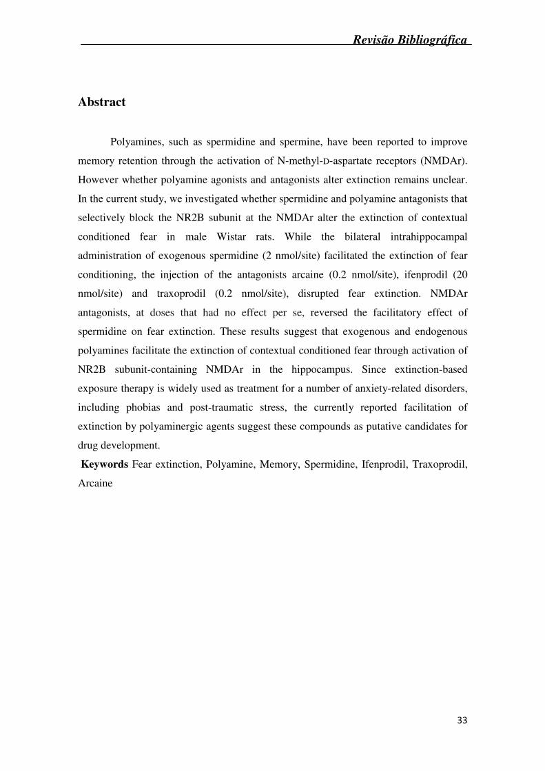
Polyamines, such as spermidine and spermine, have been reported to improve
memory retention through the activation of N-methyl-D-aspartate receptors (NMDAr).
However whether polyamine agonists and antagonists alter extinction remains unclear.
In the current study, we investigated whether spermidine and polyamine antagonists that
selectively block the NR2B subunit at the NMDAr alter the extinction of contextual
conditioned fear in male Wistar rats. While the bilateral intrahippocampal
administration of exogenous spermidine (2 nmol/site) facilitated the extinction of fear
conditioning, the injection of the antagonists arcaine (0.2 nmol/site), ifenprodil (20
nmol/site) and traxoprodil (0.2 nmol/site), disrupted fear extinction. NMDAr
antagonists, at doses that had no effect per se, reversed the facilitatory effect of
spermidine on fear extinction. These results suggest that exogenous and endogenous
polyamines facilitate the extinction of contextual conditioned fear through activation of
NR2B subunit-containing NMDAr in the hippocampus. Since extinction-based
exposure therapy is widely used as treatment for a number of anxiety-related disorders,
including phobias and post-traumatic stress, the currently reported facilitation of
extinction by polyaminergic agents suggest these compounds as putative candidates for
drug development.
Keywords: Fear extinction; Polyamine; Memory; Spermidine; Ifenprodil;
Traxoprodil; Arcaine.
ix
LISTA DE ABREVIATURAS
CR Resposta Condicionada
CS Estímulo Condicionado
US Estímulo Não-Condicionado
rNMDA Receptor N-Metil-D-Aspartato
SPD Espermidina
L-NAME Metil éster de NG-Nitro-L-Arginina
AMPA Receptor amino-hidroxi-metil-isoxazol-propiônico
mGlur Receptor glutamatérgico metabotrópico
CAMKII Cálcio/calmodulina tipo II
PAF Fator de agregação plaquetária
PKC Proteína quinase dependente de cálcio
PKG Proteína quinase dependente de GMPc
PKA Proteína quinase dependente de AMPc
MAPK Proteína quinase ativada por mitógeno
CREB Proteína ligante do elemento responsivo ao AMPc
CO Monóxido de carbono
NO Óxido nítrico
ADC Arginina descarboxilase
ODC Ornitina descarboxilase
SAM S-adenosilmetionina
SAMDC S-adenosilmetionina descarboxilase
SSAT Espermidina/espermina acetiltransferase
PAO Poliamina oxidase
MTA Metionina
LTP Potencialização de Longa Duração
CPP (±)3-(2-carboxipiperazina-4-il)-propil-1-fosfônico
NOS Óxido nítrico sintase
x
LISTA DE FIGURAS
Revisão Bibliográfica
Figura 1: Cascata de eventos que ocasionam a formação de memórias............. 21
Figura 2: Fases de processamento de memórias, e seu decurso temporal.......... 22
Figura 3: Estruturas cerebrais envolvidas no processamento da extinção do
medo....................................................................................................................
24
Figura 4: Estrutura química das três poliaminas endógenas............................... 25
Figura 5: Rotas de metabolismo e interconversão das poliaminas..................... 27
Figura 6: Distintas ações da espermina sobre o rNMDA.................................... 29
Manuscrito
Fig. 1. Schematic representation of the behavioral procedure…………............. 47
Fig. 2. Effect of intrahippocampal spermidine administration on extinction of
conditioned fear………………………………………………………………...
47
Fig. 3. Effect of intrahippocampal administration of different antagonists of
the NMDAr on extinction of conditioned fear…………………………………
48
Fig. 4. Effect of the intrahippocampal coadministration of spermidine (2
nmol) and (A) ifenprodil (2 nmol), (B) traxoprodil (0.02 nmol) or (C) arcaine
(0.02 nmol) on the extinction of conditioned fear……………………………...
49
xi
SUMÁRIO
AGRADECIMENTOS ..................................................................................................... v
RESUMO ....................................................................................................................... vii
ABSTRACT .................................................................................................................. viii
LISTA DE ABREVIATURAS........................................................................................ ix
LISTA DE FIGURAS ...................................................................................................... x
APRESENTAÇÃO......................................................................................................... xii
1. Introdução................................................................................................................ 14
1.1 Objetivos .......................................................................................................... 16
2. Revisão Bibliográfica.............................................................................................. 18
2.1 Memória................................................................................................................ 18
2.2 Mecanismos de Memória...................................................................................... 20
2.3 Extinção da Memória............................................................................................ 22
2.4 Poliaminas............................................................................................................. 25
2.5 Biosíntese das poliaminas ..................................................................................... 26
2.6 Poliaminas e o receptor NMDA............................................................................ 28
2.7 Interação Poliaminas, receptor NMDA e memória............................................... 29
3. Manuscrito .................................................................................................................. 32
4. Discussão….................................................................................................................50
5. Conclusão ................................................................................................................... 55
6. Referências Bibliográficas.......................................................................................... 56
7. Anexo ......................................................................................................................... 62
xii
APRESENTAÇÃO
Na introdução está descrita uma breve abordagem geral sobre os temas
abordados nesta dissertação. A revisão bibliográfica apresenta uma revisão sucinta
sobre os temas trabalhados nesta dissertação. As seções discussão e conclusão,
encontradas ao fim desta dissertação, apresentam interpretações e comentários gerais
sobre a mesma. As referências bibliográficas encontradas ao final desta dissertação
referem-se somente as citações que aparecem na introdução, revisão bibliográfica e
discussão.
Os resultados que fazem parte dessa dissertação estão apresentados sob forma de
manuscrito, submetido para o periódico Neurobiology of Learning and Memory, em
fase de revisão. As seções Introdução, Materiais e Métodos, Resultados, Discussão e
Referências Bibliográficas encontram-se no próprio manuscrito e representam a íntegra
deste estudo. Em anexo encontram-se as cartas dos revisores do manuscrito submetido
para publicação.

xiii
1. INTRODUÇÃO
13
Introdução
14
1. Introdução
A extinção da memória refere-se à alteração de um comportamento condicionado,
quando a associação, que condiciona a resposta, muda ou é retirada do contexto. Por
exemplo, no condicionamento Pavloviano, a resposta condicionada (CR = medo)
diminui gradativamente a cada exposição ao estímulo condicionado (CS = ambiente)
quando este é aplicado sem a exposição ao estímulo não-condicionado (US = choque),
processo denominado extinção (Lovibond, 2004). Entretanto, com o passar do tempo,
uma reexposição ao CS pode reativar a resposta extinta, um fenômeno denominado
“recuperação espontânea” (Bouton, 2004). A extinção da memória é considerada um
novo aprendizado e não um simples esquecimento, e esta envolve o córtex ventro-
medial, pré-frontal, hipocampo, núcleo basolateral da amígdala e córtex entorrinal
(Szapiro et. al., 2003, Lebron et. al., 2004, Cammarota et. al., 2005, Bevilaqua et. al.,
2006). As bases biológicas da extinção são semelhantes às da formação da memória:
envolvem ativação da subunidade NR2B do receptor N-Metil-D-Aspartato (rNMDA),
expressão gênica e síntese protéica (Lin et. al., 2003, Vianna et. al., 2003, Cammarota
et. al., 2005).
As poliaminas, como a espermidina e espermina, são um grupo de aminas
alifáticas necessárias para o crescimento e diferenciação celular, estando presentes em
altas concentrações no sistema nervoso central. As poliaminas são importantes
moduladores de alguns canais iônicos, incluindo o rNMDA (Williams, 1997a), o qual
está envolvido na formação da memória.
Vários relatos demonstram que as poliaminas melhoram a memória e atenuam
déficits de memória induzidos por diferentes agentes amnésicos (Shimada et. al., 1994,
Kishi et. al., 1998, Meyer et. al., 1998, Rubin et. al., 2000, Rubin et. al., 2001,
Mikolajczak et. al., 2002, Rubin et. al., 2004, Tadano et. al., 2004, Berlese et. al., 2005,
Camera et. al., 2007). A administração sistêmica, intra-hipocampal e intra-amígdala de
espermidina (SPD) melhora o desempenho dos animais nas tarefas de esquiva inibitória
(Rubin et. al., 2000, Rubin et. al., 2001) e de medo condicionado (Rubin et. al., 2004,
Camera et. al., 2007). O efeito da SPD no teste de esquiva inibitória ocorre somente nas
fases de aquisição e início da consolidação da memória, não ocorrendo nas fases de
consolidação final e nem na evocação da memória (Berlese et. al., 2005). Além disso, a
administração sistêmica e intra-amígdala, mas não intra-hipocampal de arcaína,
Introdução
15
antagonista do sítio das poliaminas no rNMDA, piora o desempenho de animais no teste
de esquiva inibitória e medo condicionado (Rubin et. al., 2004, Camera et. al., 2007).
Este efeito facilitador da memória induzido por SPD parece depender do rNMDA, uma
vez que a administração de MK-801, antagonista deste receptor, e de arcaína revertem a
melhora da memória induzida por SPD (Rubin et. al., 2000, Rubin et. al., 2001, Rubin
et. al., 2004, Camera et. al., 2007). Além disso, o efeito da SPD parece depender da
atividade da enzima óxido nítrico sintase hipocampal e da produção de óxido nítrico,
uma vez que a administração intra-hipocampal de metil éster de NG-Nitro-L-arginina (L-
NAME), um inibidor não específico da enzima óxido nítrico sintase, imediatamente
após o treino previne a melhora da memória causada por SPD na tarefa de esquiva
inibitória . A SPD aumenta os níveis de nitratos e nitritos, e a co-administração de L-
NAME previne este efeito (Guerra et. al., 2006). Apesar de os agentes poliaminérgicos
modularem o rNMDA e a memória, seu efeito sobre a extinção da memória ainda é
pouco estudado.
Levando em consideração que os agentes poliaminérgicos ligam-se no rNMDA e
alteram a aquisição e consolidação de memórias, estas substâncias também poderiam
modular a extinção de memórias traumáticas. Portanto, neste trabalho investigamos se a
administração intrahipocampal de espermidina modula a extinção do medo
condicionado e se o efeito da espermidina sobre a extinção depende da subunidade
NR2B do rNMDA.
Introdução
16
1.1 Objetivos
O objetivo geral do presente estudo foi avaliar os efeitos dos agentes
poliaminérgicos sobre a extinção do medo condicionado contextual em ratos.
Objetivos específicos
1- Avaliar o efeito do agonista do sítio das poliaminas no rNMDA espermidina,
sobre a extinção do medo condicionado contextual.
2- Avaliar o efeito dos antagonistas da subunidade NR2B do rNMDA
ifenprodil, traxoprodil e arcaína, sobre a extinção do medo condicionado contextual.
3- Avaliar se a ação da espermidina sobre a extinção do medo condicionado é dependente da subunidade NR2B do rNMDA.
Revisão Bibliográfica
17
2. REVISÃO BIBLIOGRÁFICA
Revisão Bibliográfica
18
2. Revisão Bibliográfica
2.1 Memória
Memória é o substrato do nosso ser, o portal da nossa existência. Todas nossas
vivências e ações são dependentes de nossos aprendizados, experiências e lembranças.
Não podemos executar tarefas que não sabemos como fazer, nem mesmo comunicar
fatos que desconhecemos.
Uma memória forma-se a partir de uma experiência, adquirida através dos
sentidos na maioria das vezes. O perfume de uma flor, andar de bicicleta, aprender uma
língua estrangeira são exemplos de experiências que podem ficar armazenadas por
minutos, meses ou anos, e que também podem ser adquiridas nesse mesmo período de
tempo. Estas experiências, para tornarem-se um fragmento de memória devem passar
por três fases: aquisição, consolidação e evocação (Izquierdo, 2002).
Há tantas memórias possíveis como há experiências, por isso, é útil fazer uma
classificação dos tipos de memória, de acordo com sua função, conteúdo e duração. A
memória para fatos e eventos é denominada memória declarativa, que faz referência não
só à possibilidade de evocar conscientemente fatos e eventos, mas sugere também que
se possa fazê-lo mediante fala, empregando linguagem complexa, com vocabulário e
sintaxe. Estas memórias são frequentemente formadas com facilidade, mas também
podem ser facilmente esquecidas. As memórias declarativas podem ser divididas em
episódicas, quando se referem a eventos que assistimos ou dos quais participamos,
como o cardápio do almoço, ou ainda a janta entre amigos do fim-de-semana, e
semânticas, quando se referem a conhecimentos gerais, como a capital do Japão ou o
conteúdo de uma aula de história. (Eichenbaum, 2001, Izquierdo, 2002). O
processamento das memórias declarativas envolve o hipocampo, o córtex entorrinal e
outras áreas corticais. Entre as memórias declarativas, as mais aversivas, emocionais ou
alertantes são fortemente moduladas pelos núcleos basal e lateral da amígdala
(Izquierdo and McGaugh, 2000).
Existem memórias de capacidades ou habilidades motoras ou sensoriais, o que
usualmente chamamos de hábitos, denominadas de memórias não-declarativas ou
memórias de procedimentos, como por exemplo, andar de bicicleta, dirigir um
Revisão Bibliográfica
19
automóvel ou tocar violino. São memórias difíceis de “declarar” e normalmente não são
evocadas de maneira consciente. A formação de memórias não-declarativas necessita de
repetição e prática durante certo período, mas essas memórias têm menor probabilidade
de serem esquecidas As memórias de procedimentos são processadas pelo neostriatum e
pelo cerebelo e sistemas a eles associados (Izquierdo and McGaugh, 2000, Izquierdo,
2002, Robertson et. al., 2004),
A Memória de trabalho é muito breve e fugaz, e serve para gerenciar a realidade, e
determinar o contexto em que fatos e acontecimentos ocorrem. Este tipo de informação
dura poucos segundos, e assim o cérebro reconhece se a informação que está sendo
processada é nova ou não, se é importante, e se requer resposta imediata ou não. Um
bom exemplo é quando perguntamos para alguém o número de telefone de um médico,
informação esta que dura o tempo suficiente para fazermos a ligação, pois logo após a
esquecemos (Izquierdo, 2002, de Fockert, 2005). A memória de trabalho depende da
transmissão glutamatérgica no córtex pré-frontal e colinérgica na amígdala (Artiges et.
al., 2000, Ashby and O'Brien, 2005).
As memórias também podem ser classificadas de acordo com sua duração,
existindo memórias que deixam traços de curta (horas) ou longa duração (dias,
décadas). As memórias de curta duração duram pouco tempo (minutos ou 3 a 6 horas)
enquanto a memória de longa duração está sendo formada. Se estas memórias durarem
muitos meses ou anos costumam ser denominadas de memórias remotas (Kandel, 1997,
Izquierdo, 2002). A memória de curta duração faz o processamento mnemônico
enquanto a memória de longa duração não foi ainda construída. A fase em que somente
possuímos completa a memória de curta duração e a de longa duração não está fixada, é
lábil: um traumatismo craniano, um eletrochoque ou intoxicação alcoólica impedem que
se fixem memórias que acabam de ser adquiridas (Izquierdo, 2002). As memórias de
curta e de longa duração requerem as mesmas estruturas nervosas, mas envolvem
mecanismos separados como, por exemplo, ativação de genes e produção de novas
proteínas (Izquierdo et. al., 1998, Izquierdo, 2002).
Revisão Bibliográfica
20
2.2 Mecanismos de Memória
As memórias não são adquiridas imediatamente na sua formal final. A formação
de memórias de longa duração envolve uma série de processos metabólicos em distintas
estruturas cerebrais que compreendem diversas fases e que requerem entre três a oitos
horas para serem processadas (Abel and Lattal, 2001). Aquisição é o período em que
ocorre o aprendizado de uma nova informação. Denomina-se consolidação o conjunto
de processos necessários para passar uma informação recém adquirida de um estado
lábil a um estado estável, o que pode durar horas. Durante a fase de consolidação as
memórias estão suscetíveis a interferência por outras memórias, por drogas ou outros
tratamentos (Izquierdo and McGaugh, 2000). Quando recordamos a informação
adquirida, nossa memória passa por um processo de evocação. Evidências sugerem que
aquisição e evocação possuem mecanismos bioquímicos distintos e envolvem uma série
de eventos moleculares que ocorrem de maneira específica e coordenada em diferentes
regiões cerebrais (Abel and Lattal, 2001, Izquierdo et. al., 2006).
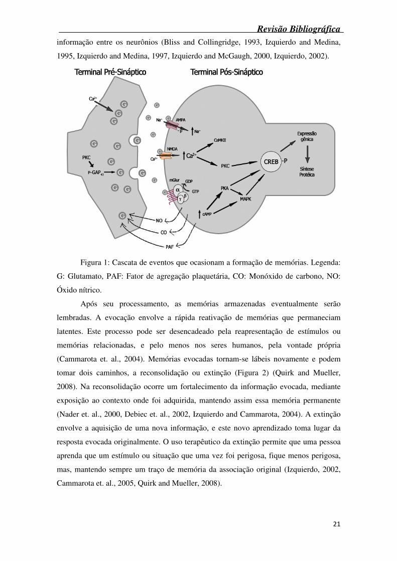
A formação de uma nova memória inicia-se em uma cascata de eventos
decorrentes da liberação de glutamato (Figura 1), que se liga a receptores específicos na
membrana pós-sináptica, são eles ácido amino-hidroxi-metil-isoxazol-propiônico
(AMPA), rNMDA e receptor glutamatérgico metabotrópico (mGlur). A despolarização
causada pela ativação destes receptores leva a um aumento na concentração de Ca2+
intracelular. Este aumento na [Ca2+], somado aos efeitos da ativação de proteínas G
pelos receptores glutamatérgicos metabotrópicos, ativa uma série de enzimas, tais como
proteínas quinase dependente de cálcio/calmodulina tipo II (CaMKII – atua fosforilando
e ativando os receptores AMPA) e proteínas quinase dependentes de GMPc (PKG).
Ativação de PKG irá liberar substâncias como óxido nítrico, monóxido de carbono e
fator de agregação plaquetária (PAF) as quais aumentam ainda mais a liberação de
glutamato. A proteína quinase dependente de cálcio (PKC), também atua neste
processo, fosforilando a proteína do terminal axônico, GAP-43, que leva a liberação de
mais glutamato. Passadas 3 a 4 horas, são ativadas as proteínas quinase dependentes de
AMPc (PKA) e as proteínas ativadas por mitógeno (MAPK). Estas junto com a PKC
irão fosforilar fatores de transcrição protéicos no núcleo, denominada proteína ligante
do elemento responsivo ao AMPc (CREB), que ativa vários loci genéticos e induz a
síntese de diversas proteínas, aumentando assim a efetividade de transmissão de
Revisão Bibliográfica
21
informação entre os neurônios (Bliss and Collingridge, 1993, Izquierdo and Medina,
1995, Izquierdo and Medina, 1997, Izquierdo and McGaugh, 2000, Izquierdo, 2002).
Figura 1: Cascata de eventos que ocasionam a formação de memórias. Legenda:
G: Glutamato, PAF: Fator de agregação plaquetária, CO: Monóxido de carbono, NO:
Óxido nítrico.
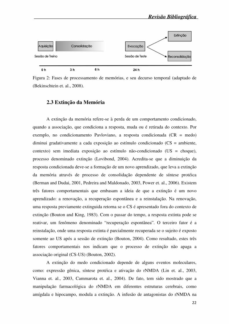
Após seu processamento, as memórias armazenadas eventualmente serão
lembradas. A evocação envolve a rápida reativação de memórias que permaneciam
latentes. Este processo pode ser desencadeado pela reapresentação de estímulos ou
memórias relacionadas, e pelo menos nos seres humanos, pela vontade própria
(Cammarota et. al., 2004). Memórias evocadas tornam-se lábeis novamente e podem
tomar dois caminhos, a reconsolidação ou extinção (Figura 2) (Quirk and Mueller,
2008). Na reconsolidação ocorre um fortalecimento da informação evocada, mediante
exposição ao contexto onde foi adquirida, mantendo assim essa memória permanente
(Nader et. al., 2000, Debiec et. al., 2002, Izquierdo and Cammarota, 2004). A extinção
envolve a aquisição de uma nova informação, e este novo aprendizado toma lugar da
resposta evocada originalmente. O uso terapêutico da extinção permite que uma pessoa
aprenda que um estímulo ou situação que uma vez foi perigosa, fique menos perigosa,
mas, mantendo sempre um traço de memória da associação original (Izquierdo, 2002,
Cammarota et. al., 2005, Quirk and Mueller, 2008).
Revisão Bibliográfica
22
Figura 2: Fases de processamento de memórias, e seu decurso temporal (adaptado de
(Bekinschtein et. al., 2008).
2.3 Extinção da Memória
A extinção da memória refere-se à perda de um comportamento condicionado,
quando a associação, que condiciona a resposta, muda ou é retirada do contexto. Por
exemplo, no condicionamento Pavloviano, a resposta condicionada (CR = medo)
diminui gradativamente a cada exposição ao estímulo condicionado (CS = ambiente,
contexto) sem imediata exposição ao estímulo não-condicionado (US = choque),
processo denominado extinção (Lovibond, 2004). Acredita-se que a diminuição da
resposta condicionada deve-se a formação de um novo aprendizado, que leva a extinção
da memória através de processo de consolidação dependente de síntese protéica
(Berman and Dudai, 2001, Pedreira and Maldonado, 2003, Power et. al., 2006). Existem
três fatores comportamentais que embasam a ideia de que a extinção é um novo
aprendizado: a renovação, a recuperação espontânea e a reinstalação. Na renovação,
uma resposta previamente extinguida retorna se o CS é apresentado fora do contexto de
extinção (Bouton and King, 1983). Com o passar do tempo, a resposta extinta pode se
reativar, um fenômeno denominado “recuperação espontânea”. O terceiro fator é a
reinstalação, onde uma resposta extinta é parcialmente recuperada se o sujeito é exposto
somente ao US após a sessão de extinção (Bouton, 2004). Como resultado, estes três
fatores comportamentais nos indicam que o processo de extinção não apaga a
associação original (CS-US) (Bouton, 2002).
A extinção do medo condicionado depende de alguns eventos moleculares,
como: expressão gênica, síntese protéica e ativação do rNMDA (Lin et. al., 2003,
Vianna et. al., 2003, Cammarota et. al., 2004). De fato, tem sido mostrado que a
manipulação farmacológica do rNMDA em diferentes estruturas cerebrais, como
amígdala e hipocampo, modula a extinção. A infusão de antagonistas do rNMDA na
Revisão Bibliográfica
23
amígdala ou hipocampo impede a extinção da memória tanto na tarefa de medo
pontecializado pelo susto como na esquiva inibitória (Falls et. al., 1992, Szapiro et. al.,
2003). Ainda, a subunidade NR2B do rNMDA exerce papel fundamental na modulação
da extinção do medo. Sotres-Bayon e colaboradores, utilizando ifenprodil, um
antagonista específico desta subunidade, mostraram que a aquisição da extinção do
medo depende da subunidade NR2B do rNMDA da amígdala lateral (Sotres-Bayon et.
al., 2007). A administração de ifenprodil no córtex prefrontal ou sistemicamente,
imediatamente após o treino da extinção, piora a consolidação da extinção (Sotres-
Bayon et. al., 2009). Já a administração de agonistas do rNMDA, como a D-cicloserina,
facilita a extinção da memória em tarefas comportamentais como medo condicionado
Pavloviano e labirinto vertical (Gabriele and Packard, 2007, Langton and Richardson,
2008, Yamamoto et. al., 2008). Estes dados mostram o papel crucial do rNMDA, e sua
subunidade NR2B, na modulação da extinção de memórias.
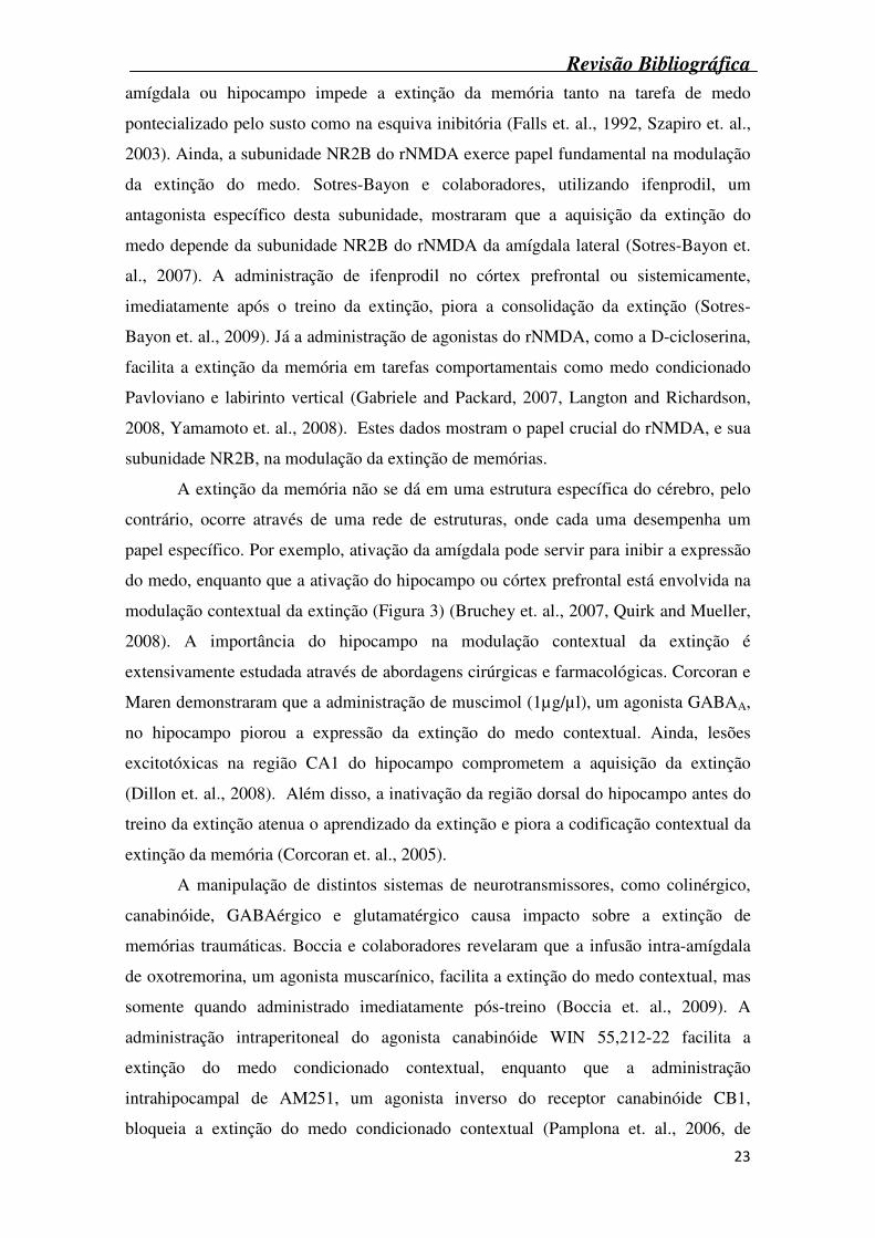
A extinção da memória não se dá em uma estrutura específica do cérebro, pelo
contrário, ocorre através de uma rede de estruturas, onde cada uma desempenha um
papel específico. Por exemplo, ativação da amígdala pode servir para inibir a expressão
do medo, enquanto que a ativação do hipocampo ou córtex prefrontal está envolvida na
modulação contextual da extinção (Figura 3) (Bruchey et. al., 2007, Quirk and Mueller,
2008). A importância do hipocampo na modulação contextual da extinção é
extensivamente estudada através de abordagens cirúrgicas e farmacológicas. Corcoran e
Maren demonstraram que a administração de muscimol (1µg/µl), um agonista GABAA,
no hipocampo piorou a expressão da extinção do medo contextual. Ainda, lesões
excitotóxicas na região CA1 do hipocampo comprometem a aquisição da extinção
(Dillon et. al., 2008). Além disso, a inativação da região dorsal do hipocampo antes do
treino da extinção atenua o aprendizado da extinção e piora a codificação contextual da
extinção da memória (Corcoran et. al., 2005).
A manipulação de distintos sistemas de neurotransmissores, como colinérgico,
canabinóide, GABAérgico e glutamatérgico causa impacto sobre a extinção de
memórias traumáticas. Boccia e colaboradores revelaram que a infusão intra-amígdala
de oxotremorina, um agonista muscarínico, facilita a extinção do medo contextual, mas
somente quando administrado imediatamente pós-treino (Boccia et. al., 2009). A
administração intraperitoneal do agonista canabinóide WIN 55,212-22 facilita a
extinção do medo condicionado contextual, enquanto que a administração
intrahipocampal de AM251, um agonista inverso do receptor canabinóide CB1,
bloqueia a extinção do medo condicionado contextual (Pamplona et. al., 2006, de
Revisão Bibliográfica
24
Oliveira Alvares et. al., 2008). Ainda, um aumento na transmissão GABAérgica parece
ter efeito facilitatório sobre a extinção. Muscimol, quando administrado na amígdala
basolateral, antes do treino da extinção, foi capaz de facilitar a extinção, sugerindo que a
neurotransmissão via GABAA atua na consolidação da extinção do medo (Akirav et. al.,
2006). Além disso, a modulação do sistema glutamatérgico pode alterar a aquisição e
consolidação da extinção. A administração sistêmica ou intra-amígdala de D-
cicloserina, um agonista parcial do rNMDA, melhora a extinção do medo condicionado
quando injetado imediatamente pós-treino (Ledgerwood et. al., 2003). A ação da D-
cicloserina também foi testada em humanos, onde esta mostrou-se capaz de melhorar a
extinção do medo quando administrada logo após o aprendizado (Kalisch et. al., 2009).
Figura 3: Estruturas cerebrais envolvidas no processamento da extinção do medo.
CS: Estímulo condicionado. (Adaptado de (Quirk and Mueller, 2008).
Revisão Bibliográfica
25
2.4 Poliaminas
As poliaminas putrescina, espermidina e espermina, são um grupo de aminas
alifáticas presentes em quase todas as células, incluindo células do sistema nervoso
central. Também são constituintes de muitos compostos encontrados em plantas e
insetos. Seus nomes triviais, putrescina, espermidina e espermina provem da fonte de
onde foram primariamente isoladas, carne em putrefação e líquido seminal (Coffino,
2001). Desde o seu descobrimento, por Antoni van Leeuwenhoek em 1678, até o
recente desenvolvimento de camundongos transgênicos expressando enzimas que
alteram os níveis de poliaminas de maneira tecido-específica, o estudo de poliaminas
aprofundou o conhecimento de diversos processos fisiológicos e patológicos.
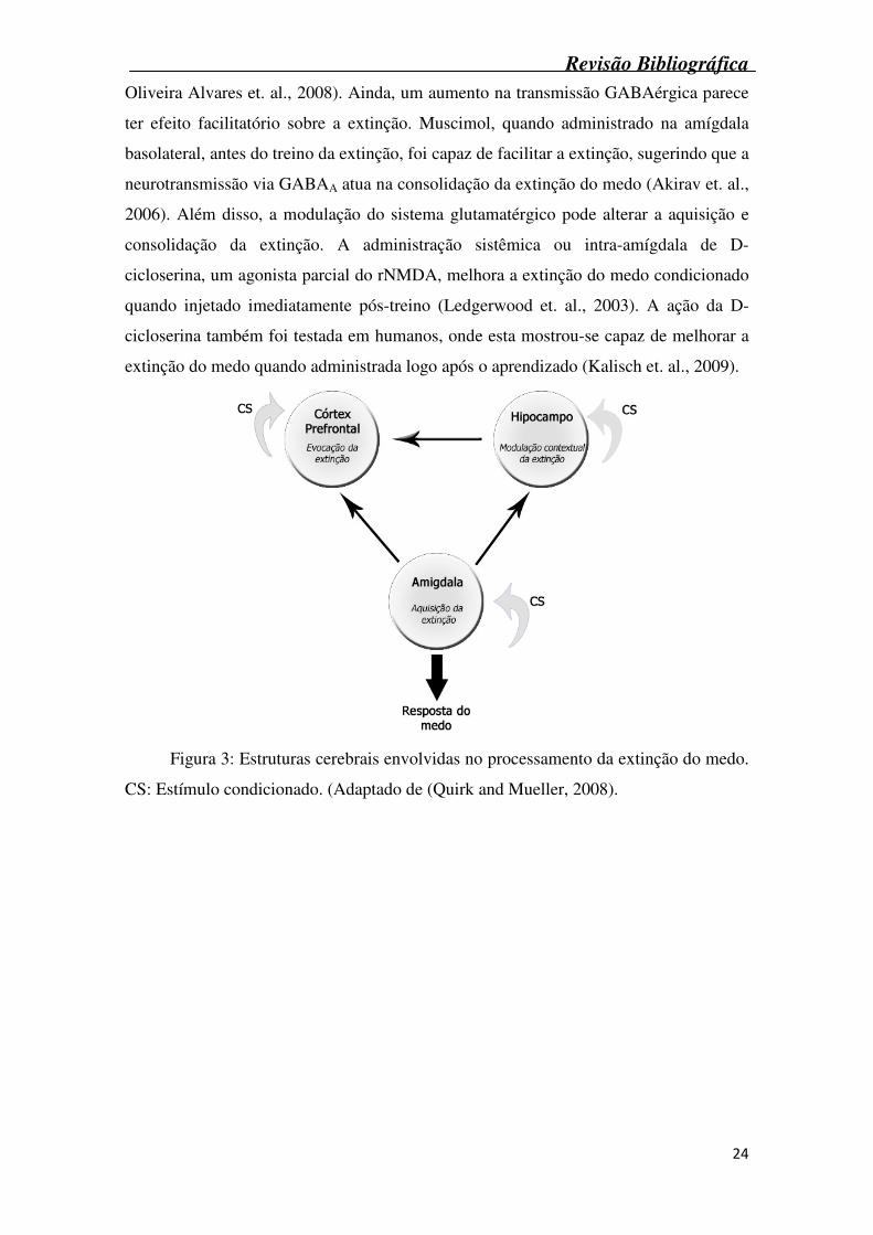
A caracterização da estrutura química das poliaminas mostra que a putrescina é
uma di-amina primária (1,4 – diaminobutano), espermidina é uma tri-amina (mono-N-3-
aminopropil-1,4-diaminobutano) e a espermina é uma tetra-amina (bis-N-3-
aminopropil-1,4-diaminobutano) (Figura 4) (Teti et. al., 2002). Em concentrações
fisiológicas, as poliaminas estão envolvidas em distintos processos, como: replicação de
DNA, expressão gênica, síntese protéica e funcionamento de canais iônicos e receptores
de membrana (Coffino, 2001, Wang et. al., 2003, Gugliucci, 2004). De fato, sua
importância na proliferação celular é tamanha que somente duas espécies –
Methanobacteriales e Halobacteriales, parecem crescer na ausência de níveis
detectáveis de poliaminas (Marton and Pegg, 1995)
Figura 4: Estrutura química das três poliaminas endógenas.
Revisão Bibliográfica
26
2.5 Biosíntese das poliaminas
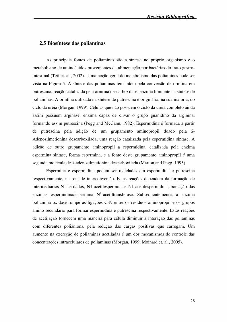
As principais fontes de poliaminas são a síntese no próprio organismo e o
metabolismo de aminoácidos provenientes da alimentação por bactérias do trato gastro-
intestinal (Teti et. al., 2002). Uma noção geral do metabolismo das poliaminas pode ser
vista na Figura 5. A síntese das poliaminas tem início pela conversão de ornitina em
putrescina, reação catalizada pela ornitina descarboxilase, enzima limitante na síntese de
poliaminas. A ornitina utilizada na síntese de putrescina é originária, na sua maioria, do
ciclo da uréia (Morgan, 1999). Células que não possuem o ciclo da uréia completo ainda
assim possuem arginase, enzima capaz de clivar o grupo guanidino da arginina,
formando assim putrescina (Pegg and McCann, 1982). Espermidina é formada a partir
de putrescina pela adição de um grupamento aminopropil doado pela S-
Adenosilmetionina descarboxilada, uma reação catalizada pela espermidina sintase. A
adição de outro grupamento aminopropil a espermidina, catalizada pela enzima
espermina sintase, forma espermina, e a fonte deste grupamento aminopropil é uma
segunda molécula de S-adenosilmetionina descarboxilada (Marton and Pegg, 1995).
Espermina e espermidina podem ser recicladas em espermidina e putrescina
respectivamente, na rota de interconversão. Estas reações dependem da formação de
intermediários N-acetilados, N1-acetilespermina e N1-acetilespermidina, por ação das
enzimas espermidina/espermina N1-acetiltransferase. Subsequentemente, a enzima
poliamina oxidase rompe as ligações C-N entre os resíduos aminopropil e os grupos
amino secundário para formar espermidina e putrescina respectivamente. Estas reações
de acetilação fornecem uma maneira para célula diminuir a interação das poliaminas
com diferentes poliânions, pela redução das cargas positivas que carregam. Um
aumento na excreção de poliaminas acetiladas é um dos mecanismos de controle das
concentrações intracelulares de poliaminas (Morgan, 1999, Moinard et. al., 2005).
Revisão Bibliográfica
27
Figura 5: Rotas de metabolismo e interconversão das poliaminas. ADC: Arginina
descarboxilase; ODC: Ornitina descarboxilase; SAM: S-adenosilmetionina; SAMDC:
S-adenosilmetionina descarboxilase; SSAT: espermidina/espermina acetiltransferase;
PAO: Poliamina oxidase; MTA: Metionina. (adaptado de (Urdiales et. al., 2001).
O catabolismo das poliaminas ocorre através de reações de desaminação
oxidativa, pela ação de amino-oxidases dependentes de cobre. Pela desaminação
oxidativa do grupamento amino primário, cada intermediário da interconversão pode ser
transformado em um aldeído, que é posteriormente oxidado em um aminoácido ou em
um grupamento gama-lactâmico. Os produtos finais do catabolismo bem como
poliaminas acetiladas são excretadas por via renal como poliaminas inalteradas
(Gugliucci, 2004, Seiler, 2004).
As três enzimas que regulam a biosíntese de poliaminas são ornitina
descarboxilase, S-adenosilmetionina descarboxilase e espermidina/espermina N-
acetiltransferase. A interferência na atividade destas enzimas irá regular os mecanismos
envolvendo os três passos do metabolismo das poliaminas: Síntese, rota de
interconversão e catabolismo (Morgan, 1999, Seiler, 2004).
Revisão Bibliográfica
28
2.6 Poliaminas e o receptor NMDA
As poliaminas possuem a característica de serem importantes moduladores de
alguns canais iônicos, podendo interagir com subtipos específicos de canais de potássio
e receptores glutamatérgicos, entre eles o NMDA (Carter, 1994, Williams, 1997a).
Ramson e Stec (1988) mostraram que espermidina e espermina aumentam a
afinidade do rNMDA pelo [3H]MK-801, efeito constatado na presença ou ausência de
concentrações saturantes de glutamato e glicina, mostrando que o efeito estimulatório
das poliaminas deve-se a sua ligação com o rNMDA (Ransom and Stec, 1988). Altas
concentrações de espermidina e espermina não são efetivas em aumentar a ligação do
MK-801, já baixas concentrações aumentam a afinidade do rNMDA pelo ligante,
levando a uma curva de concentração-resposta bifásica (Williams et. al., 1989, Rock
and Macdonald, 1995). Estudos de eletrofisiologia, juntamente com o uso de receptores
recombinantes, demonstram que as poliaminas não ativam diretamente o rNMDA, mas
sim atuam potencializando ou inibindo respostas mediadas pelo glutamato (Johnson,
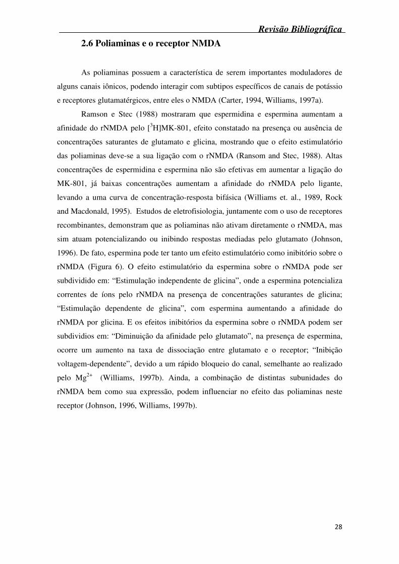
1996). De fato, espermina pode ter tanto um efeito estimulatório como inibitório sobre o
rNMDA (Figura 6). O efeito estimulatório da espermina sobre o rNMDA pode ser
subdividido em: “Estimulação independente de glicina”, onde a espermina potencializa
correntes de íons pelo rNMDA na presença de concentrações saturantes de glicina;
“Estimulação dependente de glicina”, com espermina aumentando a afinidade do
rNMDA por glicina. E os efeitos inibitórios da espermina sobre o rNMDA podem ser
subdividios em: “Diminuição da afinidade pelo glutamato”, na presença de espermina,
ocorre um aumento na taxa de dissociação entre glutamato e o receptor; “Inibição
voltagem-dependente”, devido a um rápido bloqueio do canal, semelhante ao realizado
pelo Mg2+ (Williams, 1997b). Ainda, a combinação de distintas subunidades do
rNMDA bem como sua expressão, podem influenciar no efeito das poliaminas neste
receptor (Johnson, 1996, Williams, 1997b).
Revisão Bibliográfica
29
Figura 6: Distintas ações da espermina sobre o rNMDA. (Adaptado de (Williams,
1997a).
2.7 Interação Poliaminas, receptor NMDA e memória
A ativação do rNMDA está associada com diferentes formas de plasticidade
sináptica, como Potencialização de Longa Duração (LTP) e aprendizado e memória
(Lee and Silva, 2009). Existem vários relatos demonstrando o envolvimento das
poliaminas em processos de aprendizado e memória, tanto melhorando a memória de
ratos em distintas tarefas, como atenuando o déficit de memória induzido por diferentes
agentes amnésicos (Shimada et. al., 1994, Kishi et. al., 1998, Meyer et. al., 1998, Rubin
et. al., 2000, Rubin et. al., 2001, Mikolajczak et. al., 2002, Rubin et. al., 2004, Tadano
et. al., 2004, Berlese et. al., 2005, Camera et. al., 2007).
Estudo realizado por Conway (1998) mostrou que doses diárias de espermina
bloqueiam o aprendizado no labirinto aquático de Morris. Esta dose também produz
morte neuronal (Conway, 1998). Já na tarefa do labirinto em T com 14 braços,
espermidina (80mg/kg) administrada intraperitonealmente potencializa o déficit de
memória produzido por dizocilpina (Shimada et. al., 1994). (Halonen et. al., 1993, Rock
and Macdonald, 1995).
Nos últimos anos foi demonstrado que a administração sistêmica,
intrahipocampal e intraamígdala de espermidina (SPD) melhora o desempenho de ratos
nas tarefas de esquiva inibitória (Rubin et. al., 2000, Rubin et. al., 2001, Guerra et. al.,
2006) e de medo condicionado (Rubin et. al., 2004, Camera et. al., 2007). O efeito da
SPD no teste de esquiva inibitória ocorre somente nas fases de aquisição e início da
consolidação da memória, não ocorrendo nas fases de consolidação final e nem na
evocação da memória (Berlese et. al., 2005). A administração sistêmica e intra-amígdala
Revisão Bibliográfica
30
de arcaína, antagonista do sítio das poliaminas no rNMDA, piora a performance dos
animais no teste de esquiva inibitória (Rubin et. al., 2000, Rubin et. al., 2001) e de medo
condicionado (Rubin et. al., 2004, Camera et. al., 2007). Este efeito facilitador da
memória induzido por SPD parece envolver o rNMDA, uma vez que a administração de
MK-801, antagonista do rNMDA, e de arcaína revertem a melhora da memória induzida
por SPD (Rubin et. al., 2000, Rubin et. al., 2001, Rubin et. al., 2004, Camera et. al.,
2007). Em adição, Meyer e colaboradores demonstraram que a co-administração
sistêmica de espermina e D-cicloserina reverte o déficit cognitivo induzido pelo
antagonista do rNMDA ácido (±)3-(2-carboxipiperazina-4-il)-propil-1-fosfônico (CPP).
Além disso, o efeito da SPD parece depender da atividade da enzima óxido nítrico
sintase hipocampal e da produção de óxido nítrico, uma vez que a administração intra-
hipocampal de metil éster de NG-Nitro-L-arginina (L-NAME), um inibidor não
específico da enzima óxido nítrico sintase, imediatamente depois do treino previne a
melhora da memória causada por SPD na tarefa de esquiva inibitória (Guerra et. al.,
2006). A SPD aumenta os níveis de nitratos e nitritos, e a co-administração de L-NAME
previne este efeito (Guerra et. al., 2006).
Apesar do já descrito efeito das poliaminas sobre a formação e consolidação da
memória, ainda não se tem relatos do efeito destas substâncias sobre a extinção do medo
condicionado contextual. Levando-se em consideração o envolvimento do rNMDA e do
hipocampo na extinção do medo condicionado, o presente trabalho irá avaliar o efeito
da administração intra-hipocampal de espermidina e distintos antagonistas do rNMDA
na extinção do medo condicionado contextual.
Revisão Bibliográfica
31
3. MANUSCRITO
Revisão Bibliográfica
32
3. Manuscrito
Polyaminergic agents modulate contextual fear extinction in rats
Guilherme Monteiro Gomesa, Carlos Fernando Mellob, Michelle Melgarejo da Rosaa,
Guilherme Vargas Bochia, Juliano Ferreiraa, Susan Barronc, Maribel Antonello Rubina*
aDepartment of Chemistry, Center of Exact and Natural Sciences,
Universidade Federal de Santa Maria, Santa Maria, 97105-900 RS, Brazil.
*Corresponding author. Fax: Fax: + 55 55 3220 8978
E-mail adress: [email protected] (M.A. Rubin).
bDepartment of Physiology and Pharmacology, Center of Health Sciences,
Universidade Federal de Santa Maria, Santa Maria, 97105-900 RS, Brazil.
c Department of Psychology, University of Kentucky, Lexington, Kentucky 40506,
USA.
Revisão Bibliográfica
33
Abstract
Polyamines, such as spermidine and spermine, have been reported to improve
memory retention through the activation of N-methyl-D-aspartate receptors (NMDAr).
However whether polyamine agonists and antagonists alter extinction remains unclear.
In the current study, we investigated whether spermidine and polyamine antagonists that
selectively block the NR2B subunit at the NMDAr alter the extinction of contextual
conditioned fear in male Wistar rats. While the bilateral intrahippocampal
administration of exogenous spermidine (2 nmol/site) facilitated the extinction of fear
conditioning, the injection of the antagonists arcaine (0.2 nmol/site), ifenprodil (20
nmol/site) and traxoprodil (0.2 nmol/site), disrupted fear extinction. NMDAr
antagonists, at doses that had no effect per se, reversed the facilitatory effect of
spermidine on fear extinction. These results suggest that exogenous and endogenous
polyamines facilitate the extinction of contextual conditioned fear through activation of
NR2B subunit-containing NMDAr in the hippocampus. Since extinction-based
exposure therapy is widely used as treatment for a number of anxiety-related disorders,
including phobias and post-traumatic stress, the currently reported facilitation of
extinction by polyaminergic agents suggest these compounds as putative candidates for
drug development.
Keywords Fear extinction, Polyamine, Memory, Spermidine, Ifenprodil, Traxoprodil,
Arcaine
Revisão Bibliográfica
34
Introduction
Pavlovian fear conditioning and its extinction are the most extensively studied
models that provide the laboratory the tools to understand the neural mechanisms of fear
and anxiety disorders in humans (Kim & Jung, 2006; Myers & Davis, 2002). Fear
conditioning is a form of associative learning in which an animal (typically a rat) is
exposed to the pairing of a neutral conditional stimulus (CS), such as a context, tone or
light, with an aversive unconditioned stimulus (US), such as a footshock. This
procedure yields a conditioned fear response to the CS, such as freezing (Lovibond,
2004). When the CS is successively presented in the absence of the US, fear is
extinguished. Fear extinction is a new learning and requires activation of brain
structures known to be crucial for learning, including ventromedial prefrontal cortex,
basolateral amygdala, entorhinal cortex and hippocampus (Bevilaqua, Bonini, Rossato,
Izquierdo, Cammarota & Izquierdo, 2006; Cammarota, Bevilaqua, Rossato, Ramirez,
Medina & Izquierdo, 2005; Laurent, Marchand & Westbrook, 2008; Lebron, Milad &
Quirk, 2004; Szapiro, Vianna, McGaugh, Medina & Izquierdo, 2003). Some studies
have also suggested an important role for the hippocampus in extinction of contextual
fear conditioning (Corcoran & Maren, 2001; Frohardt, Guarraci & Bouton, 2000). A
growing body of evidence suggests that the hippocampus not only plays a role in
contextual encoding and retrieval of fear extinction memories, but also interacts with
other brain structures to regulate the context-specificity of fear extinction (Corcoran,
Desdmond Frey & Maren, 2005; Maren & Hobin, 2007).
Extinction depends on specific molecular events: gene expression, protein
synthesis and activation of the N-methyl-D-aspartate receptor (NMDAr) (Cammarota et
al., 2005; Lin, Yeh, Lu& Gean, 2003; Vianna, Igaz, Coitinho, Medina & Izquierdo,
2003). For instance, the administration of D-cycloserine, an agonist of the NMDAr,
facilitates fear extinction in distinct memory tasks (Gabriele & Packard, 2007; Langton
& Richardson, 2008; Yamamoto, Morinobu, Fuchikami, Kurata, Kozuru & Yamawaki,
2008). Moreover, the administration of NMDAr antagonists, like CPP and ifenprodil,
disrupt the acquisition or consolidation of fear extinction (Burgos-Robles, Vidal-
Gonzalez, Santini & Quirk, 2007; Sotres-Bayon, Bush & LeDoux, 2007).
Revisão Bibliográfica
35
Spermidine and spermine are naturally occurring polyamines required for cell
growth and differentiation, which are present at high concentrations in the brain
(Johnson, 1996; Shimada, Spangler, London & Ingram, 1994; Williams, 1997;
Williams, Romano, Dichter & Molinoff, 1991). Polyamines modulate some ion
channels, among them the NMDAr (Ransom & Stec, 1988; Rock & Macdonald, 1995;
Williams, 1997; Williams et al., 1991). The systemic, intra-hippocampal and intra-
amygdalar administration of spermidine improves memory in distinct memory tasks
(Berlese, Sauzem, Carati, Guerra, Stiegemeier, Mello & Rubin, 2005; Camera, Mello,
Ceretta & Rubin, 2007; Guerra, Mello, Sauzem, Berlese, Furian, Tabarelli & Rubin,
2006; Rubin, Berlese, Stiegemeier, Volkweis, Oliveira, dos Santos, Fenili & Mello,
2004; Rubin, Boemo, Jurach, Rojas, Zanolla, Obregon, Souza & Mello, 2000; Rubin,
Stiegemeier, Volkweis, Oliveira, Fenili, Boemo, Jurach & Mello, 2001; Shimada et al.,
1994). Moreover, the intrastriatal administration of spermine reverses the deficits
induced by quinolinic acid on an object recognition task (Velloso, Dalmolin, Gomes,
Rubin, Canas, Cunha & Mello, 2009). The facilitatory effect of polyamines on memory
appear to depend on NMDAr and nitric oxide synthase (NOS) activation (Camera et al.,
2007; Guerra et al., 2006; Rubin et al., 2001).
Since the facilitatory effects of spermidine on memory are blocked by minute
amounts of arcaine, an antagonist of the polyamine binding site on the NMDAr
(Reynolds & Miller, 1990), the involvement of this receptor in the memory
improvement induced by spermidine (Camera et al., 2007; Rubin et al., 2004; Rubin et
al., 2000; Rubin et al., 2001) has been suggested. In line with this view, the
noncompetitive NMDAr antagonist, MK-801, reverses the facilitatory effect of
spermidine on the memory of fear (Camera et al., 2007) and the systemic and intra-
amygdalar administration of arcaine, at doses higher than those required to block the
facilitatory effects of spermidine, impairs the memory of inhibitory avoidance and fear
conditioning tasks (Camera et al., 2007; Ceretta, Camera, Mello & Rubin, 2008; Rubin
et al., 2004; Rubin et al., 2001).
Since polyaminergic binding site ligands at the NMDAr alter acquisition and
consolidation of fear memories, they could also modulate the extinction of fear
memories. Therefore, in this study we investigated whether the intrahippocampal
infusion of spermidine could modulate the extinction of contextual fear memory in rats
and if the effect of spermidine could involve the NR2B subunit of NMDAr.
Revisão Bibliográfica
36
Methods
Animals
Male Wistar rats (3 month old), housed in plastic cages (four to six per cage) and
maintained on a 12 h light/dark cycle (lights on at 07:00 a.m.), in a temperature and
humidity controlled environment were used. Food and water were available ad libitum
(Guabi, Santa Maria, Rio Grande do Sul, Brazil). Behavioral tests were conducted
during the light phase of the cycle (between 9:00 A.M. and 5:00 P.M.) using
independent experimental groups of rats. All animal experimentation reported in this
study was conducted in accordance with the Policies on the Use of Animals and
Humans in Neuroscience Research, revised and approved by the Society for
Neuroscience Research in January 1995 and with the Institutional and National
regulations for animal research (process 0206).
Surgery
Rats were implanted, under Equithesin anesthesia (1% Phenobarbital, 2%
magnesium sulphate, 4% chloral hydrate, 42% propylene glycol, 11% ethanol; 3 mL/kg,
i.p.) with two guide cannulae (27-gauge) stereotaxically aimed 1 mm above the CA1
region of the hippocampus, in accordance with coordinates (A -4.0mm; L 3.0mm; V 2.0
mm) taken from the atlas of (Paxinos & Watson, 1986).
Behavioral Procedures
Conditioning, extinction training and extinction test sessions were conducted in
a 25x25x30 cm test chamber. The front and ceiling walls of the chamber were made of
clear acrylic plastic, whereas the lateral and rear walls were made of opaque plastic and
the floor consisted of 32 stainless steel rods wired to a shock generator.
Revisão Bibliográfica
37
Six days after surgery, rats were submitted to fear conditioning according to
Pamplona and collaborators (2006), with some modifications. Briefly, the animals were
placed in the chamber (CS), and after a habituation period (3 min) they received three 2
sec, 0.6 mA scrambled footshocks (US). The shocks were 50 sec apart. After the last
foot shock, the animals were left in the chamber for an additional 60 sec and were then
returned to their home cages.
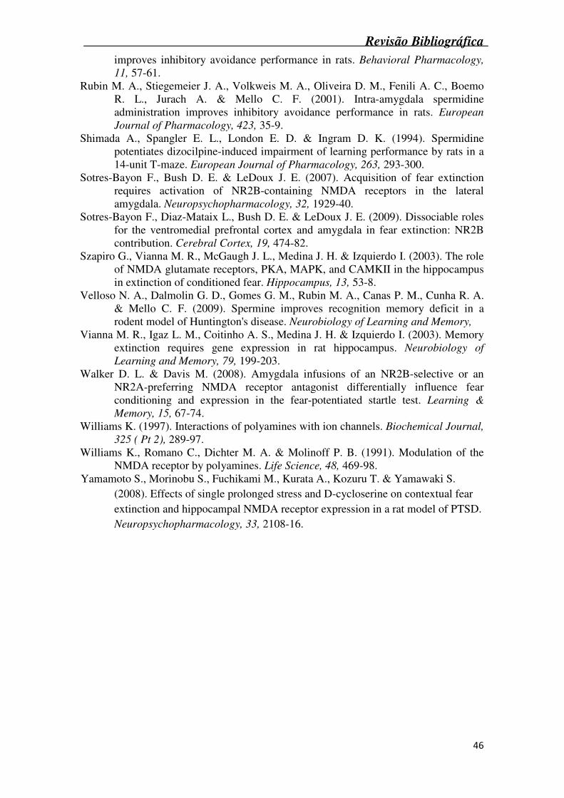
Twenty four hours post conditioning, animals began extinction training, which
consisted of placing the animal in the same chamber for 6 min, with no shock delivered.
On the next four consecutive days, rats were tested for extinction. Again they were
placed in the chamber for 6 min and no shock was given (Figure 1). Freezing, defined
as a stereotypic crouching position with complete immobility (except for respiratory
movements), was used as a memory index during the extinction procedure (Blanchard
& Blanchard, 1969). Every 4 sec throughout the test period, a time-sampling procedure
was used to assess whether the animal was moving or freezing. The data was then
converted into a percentage of the number of observations in which the animal
displayed freezing behavior (Rubin et al., 2004). The behavior was scored by an
experienced observer blind to treatment condition.
After the last test session, cannula placement was verified. All animals were infused
with 0.5 µl of 4% methylene blue through the cannula. Only data from the animals with
correct cannula placement were analyzed.
Drugs and treatment
Spermidine (N-[3-aminopropyl]-1,4-butanediamine trihydrochloride; Sigma)
and arcaine (1,4-diguanidinobutane sulfate; Sigma St. Louis, MO) were dissolved in 50
mM phosphate buffer solution (PBS), pH 7.4. Traxoprodil (CP-101,606; Pfizer) and
ifenprodil (alpha-(4-Hydroxyphenyl)-beta-methyl-4-benzyl-1-piperidineethanol tartrate
salt; Sigma) were dissolved in 0.3% Tween 80 in saline solution.
In a first set of experiments dose-effect curves for spermidine and NMDAr antagonists
were performed to characterize the facilitatory effect of spermidine on fear extinction
and determine the doses of the antagonists that had no effect on their own. To this end,
the animals received bilateral 0.5 µL injections into the hippocampi of PBS, spermidine
(0.02-2 nmol), ifenprodil (0.2-20 nmol), traxoprodil (0.02-2 nmol) or arcaine (0.02-0.2
Revisão Bibliográfica
38
nmol). immediately after the extinction training over 1 min. The injections were
performed using a Hamilton syringe and a 30-gauge needle fitted into the guide cannula,
with the tip of the infusion needle protruding 1.0 mm beyond the guide cannula, aimed
at the CA1 region of the hippocampus. The Hamilton syringe was driven by an
automated syringe pump (Insight, Brazil) at a rate of 0.5 µl/min. After infusions were
completed, the injector needles were left in place for an additional 60 sec, to avoid
backflow. Immediately following the infusions, the animals were returned to their home
cages.
In a second set of experiments, it was determined whether NMDAr antagonists,
at doses that had no effect per se, reverse the facilitatory effect of spermidine on fear
extinction. To this end, the animals received bilateral 0.5 µL injections into the
hippocampi of PBS, spermidine (2 nmol) plus ifenprodil (2 nmol), spermidine (2 nmol)
plus traxoprodil (0.02 nmol) or spermidine (2 nmol) plus arcaine (0.02 nmol), as
described above.
Statistical Analysis
Statistical analyses were carried out using one-, two- or three-way analysis of
variance (ANOVA) with the “sessions” factor treated as within-subject factor, followed
by the Student–Newman-Keuls (SNK) Test, depending on the experiment. A p<0.05
was considered significant.
Results
Repeated exposure of the CS without the US caused extinction of the original
memory. Statistical analysis (one-way ANOVA) revealed that the 5-day extinction
protocol decreased the freezing time of the vehicle group across successive reexposures
to the conditioning chamber (p<0.05, Figures 2-4).
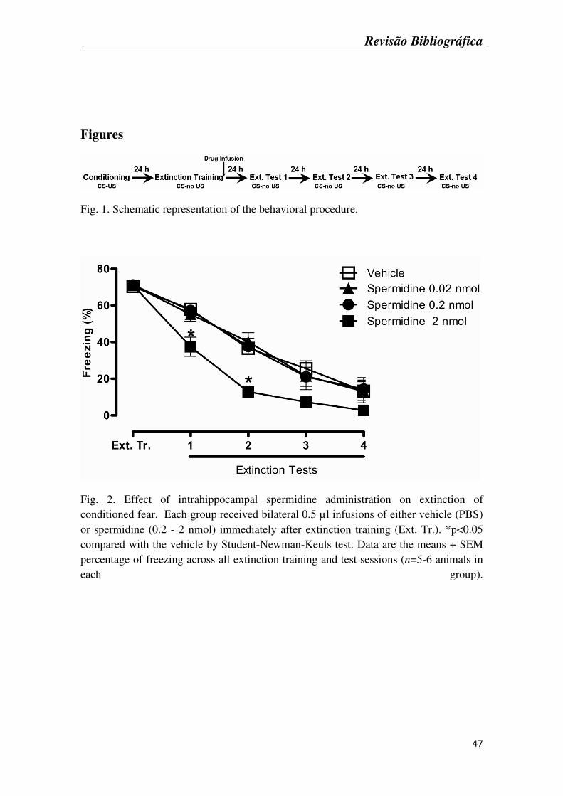
Fig. 2 shows the effect of intrahippocampal administration of spermidine (0.02 –
2 nmol) on the extinction of conditioned fear. Statistical analysis (two-way ANOVA)
revealed significant effects of pharmacological treatment (F3,18 = 7.6, p<0.05) and
sessions (F3,54 = 137.9, p<0.05). Post hoc comparisons (Student-Newman-Keuls test)
Revisão Bibliográfica
39
showed that spermidine at the dose of 2 nmol/site, facilitated contextual fear extinction,
as indicated by a decrease in freezing scores on Extinction tests 1 and 2, compared with
the vehicle control group.
Fig. 3A shows the effects of intrahippocampal administration of the NR2B
antagonist ifenprodil (0.2 – 20 nmol) on the extinction of conditioned fear. Statistical
analysis (two-way ANOVA) revealed a significant effect of pharmacological treatment
(F3,18 = 5.15, p<0.05) and sessions (F3,54 = 67,84, p<0.001) but no interaction. Post hoc
analysis (Student-Newman-Keuls test) revealed that ifenprodil (at the dose of 20 nmol)
impaired the extinction of contextual fear memory on Extinction tests 1, 2 and 3.
Fig. 3B shows the effects of intrahippocampal administration of the NR2B
antagonist traxoprodil (0.02 – 2 nmol), on the extinction of conditioned fear. Statistical
analysis (two-way ANOVA) revealed a significant interaction between traxoprodil
treatment and extinction tests (F12,76 = 2.85, p<0.01). Post hoc analysis (Student-
Newman-Keuls test) revealed that 0.2 nmol of traxoprodil impaired the extinction of
contextual fear memory through all the extinction tests.
The effects of intrahipocampal administration of arcaine (0.02 – 0.2 nmol), a
competitive antagonist of the NMDAr polyamine-binding site, on the extinction of
conditioned fear are show in Fig. 3C. Statistical analysis (two-way ANOVA) revealed a
significant effect of arcaine treatment (F2,15 = 6.57, p<0.01) and extinction tests (F3,45 =
14.2, p<0.001) but no treatment by extinction test interactions. Post hoc analyses
(Student-Newman-Keuls test) revealed that 0.2 nmol of arcaine impaired the extinction
of contextual fear memory on test 1.
The intrahippocampal coadministration of ifenprodil, at a dose that had no effect
alone (2 nmol), when administered in combination with spermidine (2 nmol) reversed
the facilitatory effect of spermidine alone on extinction of conditioned fear [significant
NR2B agonist treatment (spermidine or PBS) X NR2B antagonist treatment (ifenprodil
or saline) interaction: F1,36 = 13.28, p=0.001, Fig. 4A]. The coadministration of
traxoprodil, again at a dose that had no effect alone (0.02 nmol), when administered in
combination with spermidine (2 nmol) reversed the facilitatory effect of spermidine on
extinction of conditioned fear [significant NR2B agonist treatment (spermidine or PBS)
X NR2B antagonist treatment (traxoprodil or saline) interaction: F1,33 = 9.48, p=0.004,
Fig. 4B]. Data from these experiments (Fig. 4A and 4B) suggest that the facilitatory
effect of spermidine is a consequence of its effects on the NR2B subunit of the
hippocampal NMDAr.
Revisão Bibliográfica
40
The coadministration of arcaine, at a dose that had no effect alone (0.02 nmol),
in combination with spermidine (2 nmol) also reversed the facilitatory effect of
spermidine on extinction of conditioned fear [significant NR2B agonist (spermidine or
PBS) X NR2B antagonist (arcaine or saline) interaction: F1,33 = 11.06, p=0.002, Fig. 4C,
suggesting that the facilitatory effect of spermidine may involve the polyamine-binding
site on hippocampal NMDArs.
Discussion
In the present study, we showed that intrahippocampal administration of
spermidine facilitated the extinction of conditioned fear (Fig. 2) in adult male Wistar
rats. We also showed that intrahippocampal infusion of the antagonists of the polyamine
binding site on the NR2B subunit of the NMDAr arcaine, traxoprodil or ifenprodil,
immediately after the first extinction session, impaired the extinction of conditioned
fear (Fig. 3). These findings suggest that endogenous polyamines modulate contextual
conditioned fear extinction in the hippocampus. Furthermore, the coadministration of
these NR2B antagonists, at doses that had no effect on their own, reversed the
improvement of fear extinction induced by spermidine, suggesting that the effect of
spermidine on extinction of conditioned fear involves NR2B-containing NMDAr. To
our knowledge, this is the first study to demonstrate that a polyamine facilitates
conditioned fear extinction in the hippocampus.
Previous studies have demonstrated that spermidine can modulate fear
memories. Systemic, intrahippocampal and intra-amygdalar injections of spermidine
improve the memory of inhibitory avoidance and fear conditioning tasks (Camera et al.,
2007; Rubin et al., 2004; Rubin et al., 2000; Rubin et al., 2001). This effect on memory
appears to depend on nitric oxide synthase (NOS) activity, since the administration of
N(G)-nitro L-arginine methyl ester (L-NAME), a nonspecific inhibitor of NOS,
prevents the facilitatory effects of spermidine on the inhibitory avoidance task (Guerra
et al., 2006). In addition, the effect of spermidine seems to involve the NMDAr, since
administration of MK-801 or arcaine, both antagonists of the NMDAr, eliminate the
facilitatory effect of spermidine on memory (Camera et al., 2007; Rubin et al., 2001).
While it is well established that fear extinction depends on NMDAr activation (Myers &
Davis, 2002), only recently has a role for NR2B subunit-containing NMDA receptors in
fear extinction been proposed. For instance Sotres-Bayon and coworkers have shown
Revisão Bibliográfica
41
that both systemic and intra-amygdala injection of the NR2B antagonist ifenprodil,
before extinction training, impairs the initial acquisition and subsequent retrieval of fear
extinction. In addition, systemic or cortical administration of ifenprodil, immediately
after extinction training disrupts extinction consolidation (Sotres-Bayon, Diaz-Mataix,
Bush & LeDoux, 2009), suggesting that NR2B subunit-containing NMDA receptors are
essential for both acquisition and consolidation of fear extinction. The currently
described impairment of contextual fear extinction by the intrahippocampal injection of
ifenprodil not only is in full agreement with the previous studies that have implicated
the hippocampus (Corcoran & Maren, 2001; Corcoran & Quirk, 2007; Ji & Maren,
2007) and NR2B-containing NMDA receptors (Sotres-Bayon et al., 2007) in fear
memory extinction, but also suggest a role for endogenous polyamines in fear extinction
in the hippocampus. It is also remarkable that spermidine administration improved fear
extinction, and that the administration of the three NR2B antagonists used in this study,
at doses that had no effect on their own on memory, prevented the facilitatory effect of
spermidine (Fig. 4A-C).
From a pharmacological perspective, it is particularly interesting that while the
intrahippocampal injection of 0.2 nmol of traxoprodil impaired contextual fear
extinction, the injection of 2 nmol of this compound, did not alter memory extinction.
This biphasic effect of traxoprodil is similar to that previously described for arcaine in
the amygdala (Rubin et al., 2004) and mirrors the dose-effect curve obtained for the
intrahippocampal injection of spermidine, obtained in a previous study (Berlese et al.,
2005). Therefore, it is reasonable that traxoprodil does not differ from other compounds
that bind to and modulate NR2B-containing NMDAr, which also present inverted-U
shaped dose-effect curves. It also does not mean that arcaine and ifenprodil behave
differently from traxoprodil, since a biphasic effect for these compounds might have
been found if more doses were tested, as has been shown previously (Rubin et al.,
2004). Therefore, this interesting biphasic effect may be one of the reasons for the
conflicting results of traxoprodil on memory, which includes lack of effect (Guscott,
Clarke, Murray, Grimwood, Bristow & Hutson, 2003), impairment (Walker & Davis,
2008) and even improvement (Higgins, Ballard, Enderlin, Haman & Kemp, 2005). It is
evident, however, that further studies are needed to further elucidate this paradox,
particularly the traxoprodil-induced memory improvement.
In summary, the findings from the current study showed that while spermidine
facilitated fear extinction, NMDAr antagonists disrupted this fear extinction and
reversed the facilitatory effect of spermidine on fear extinction. Since extinction-based
Revisão Bibliográfica
42
exposure therapy is widely used as treatment for a number of anxiety-related disorders,
including phobias and post-traumatic stress (Hellstrom & Ost, 1995; Powers, Smits &
Telch, 2004) the current findings showing facilitation of extinction by polyaminergic
agents suggest a possible role for these compounds as putative candidates for drug
development. In addition, the development of modulators of the NMDAr, such as
polyaminergic agents, that act on the memory of fear may be particularly interesting
from a clinical perspective. More classic NMDAr blockers, such as MK-801 work via
channel blockade (and demonstrates little receptor subtype specificity). The
consequences of this are wide-ranging effects including neurotoxicity and
abuse/psychomimetic potential (Grant, Knisely, Tabakoff, Barrett & Balster, 1991;
Ikonomidou, Bosch, Miksa, Bittigau, Vockler, Dikranian, Tenkova, Stefovska, Turski &
Olney, 1999; Klein, Calderon & Hayes, 1999). In contrast, modulators of the NMDAr
(such as drugs polyaminergic agents) could be effective without obstructing critical
NMDAR functions, making these drugs also candidates for pharmacological
interventions.
Revisão Bibliográfica
43
Acknowledgements:
This study was supported by CNPq (504363/2007-7, 301558/2007-8, 477836/2007-0, 563222/2008-5). G.M. Gomes, C.F. Mello, G. V. Bochi, J. Ferreira, M.M. Rosa and M.A. Rubin are recipients of CNPq fellowships. All the experiments comply with the current laws of Brazil.
Revisão Bibliográfica
44
References Berlese D. B., Sauzem P. D., Carati M. C., Guerra G. P., Stiegemeier J. A., Mello C. F.
& Rubin M. A. (2005). Time-dependent modulation of inhibitory avoidance memory by spermidine in rats. Neurobiology of Learning and Memory, 83, 48-53.
Bevilaqua L. R., Bonini J. S., Rossato J. I., Izquierdo L. A., Cammarota M. & Izquierdo I. (2006). The entorhinal cortex plays a role in extinction. Neurobiology of
Learning and Memory, 85, 192-7. Blanchard R. J. & Blanchard D. C. (1969). Passive and active reactions to fear-eliciting
stimuli. Journal of Comparative and Physiological Psychology, 68, 129-35. Burgos-Robles A., Vidal-Gonzalez I., Santini E. & Quirk G. J. (2007). Consolidation of
fear extinction requires NMDA receptor-dependent bursting in the ventromedial prefrontal cortex. Neuron, 53, 871-80.
Camera K., Mello C. F., Ceretta A. P. & Rubin M. A. (2007). Systemic administration of polyaminergic agents modulate fear conditioning in rats. Psychopharmacology (Berl), 192, 457-64.
Cammarota M., Bevilaqua L. R., Rossato J. I., Ramirez M., Medina J. H. & Izquierdo I. (2005). Relationship between short- and long-term memory and short- and long-term extinction. Neurobiology of Learning and Memory, 84, 25-32.
Ceretta A. P., Camera K., Mello C. F. & Rubin M. A. (2008). Arcaine and MK-801 make recall state-dependent in rats. Psychopharmacology (Berl), 201, 405-11.
Corcoran K. A., Desmond T. J., Frey K. A. & Maren S. (2005). Hippocampal inactivation disrupts the acquisition and contextual encoding of fear extinction. Journal of Neuroscience, 25, 8978-87.
Corcoran K. A. & Maren S. (2001). Hippocampal inactivation disrupts contextual retrieval of fear memory after extinction. Journal of Neuroscience, 21, 1720-6.
Corcoran K. A. & Quirk G. J. (2007). Recalling safety: cooperative functions of the ventromedial prefrontal cortex and the hippocampus in extinction. CNS
Spectrums, 12, 200-6. Frohardt R. J., Guarraci F. A. & Bouton M. E. (2000). The effects of neurotoxic
hippocampal lesions on two effects of context after fear extinction. Behavioral
Neuroscience, 114, 227-40. Gabriele A. & Packard M. G. (2007). D-Cycloserine enhances memory consolidation of
hippocampus-dependent latent extinction. Learning & Memory, 14, 468-71. Grant K. A., Knisely J. S., Tabakoff B., Barrett J. E. & Balster R. L. (1991). Ethanol-
like discriminative stimulus effects of non-competitive n-methyl-d-aspartate antagonists. Behav Pharmacol, 2, 87-95.
Guerra G. P., Mello C. F., Sauzem P. D., Berlese D. B., Furian A. F., Tabarelli Z. & Rubin M. A. (2006). Nitric oxide is involved in the memory facilitation induced by spermidine in rats. Psychopharmacology (Berl), 186, 150-8.
Guscott M. R., Clarke H. F., Murray F., Grimwood S., Bristow L. J. & Hutson P. H. (2003). The effect of (+/-)-CP-101,606, an NMDA receptor NR2B subunit selective antagonist, in the Morris watermaze. European Journal of
Pharmacology, 476, 193-9. Hellstrom K. & Ost L. G. (1995). One-session therapist directed exposure vs two forms
of manual directed self-exposure in the treatment of spider phobia. Behaviour
Research and Therapy, 33, 959-65.
Revisão Bibliográfica
45
Higgins G. A., Ballard T. M., Enderlin M., Haman M. & Kemp J. A. (2005). Evidence for improved performance in cognitive tasks following selective NR2B NMDA receptor antagonist pre-treatment in the rat. Psychopharmacology (Berl), 179, 85-98.
Ikonomidou C., Bosch F., Miksa M., Bittigau P., Vockler J., Dikranian K., Tenkova T. I., Stefovska V., Turski L. & Olney J. W. (1999). Blockade of NMDA receptors and apoptotic neurodegeneration in the developing brain. Science, 283, 70-4.
Ji J. & Maren S. (2007). Hippocampal involvement in contextual modulation of fear extinction. Hippocampus, 17, 749-58.
Johnson T. D. (1996). Modulation of channel function by polyamines. Trends in
Pharmacological Science, 17, 22-7. Kim J. J. & Jung M. W. (2006). Neural circuits and mechanisms involved in Pavlovian
fear conditioning: a critical review. Neuroscience and Biobehavioral Reviews,
30, 188-202. Klein M., Calderon S. & Hayes B. (1999). Abuse liability assessment of
neuroprotectants. Ann N Y Acad Sci, 890, 515-25. Langton J. M. & Richardson R. (2008). D-Cycloserine Facilitates Extinction the First
Time but not the Second Time: An Examination of the Role of NMDA Across the Course of Repeated Extinction Sessions. Neuropsychopharmacology,
Laurent V., Marchand A. R. & Westbrook R. F. (2008). The basolateral amygdala is necessary for learning but not relearning extinction of context conditioned fear. Learning & Memory, 15, 304-14.
Lebron K., Milad M. R. & Quirk G. J. (2004). Delayed recall of fear extinction in rats with lesions of ventral medial prefrontal cortex. Learning & Memory, 11, 544-8.
Lin C. H., Yeh S. H., Lu H. Y. & Gean P. W. (2003). The similarities and diversities of signal pathways leading to consolidation of conditioning and consolidation of extinction of fear memory. Journal of Neuroscience, 23, 8310-7.
Maren S. & Hobin J. A. (2007). Hippocampal regulation of context-dependent neuronal activity in the lateral amygdala. Learning & Memory, 14, 318-24.
Myers K. M. & Davis M. (2002). Behavioral and neural analysis of extinction. Neuron,
36, 567-84. Pavlov I. P. (1927). Conditioned Reflexes. Oxford UP: London Paxinos G. & Watson C. (1986). The Rat Brain in stereotaxic coordinates. Academic:
San Diego Powers M. B., Smits J. A. & Telch M. J. (2004). Disentangling the effects of safety-
behavior utilization and safety-behavior availability during exposure-based treatment: a placebo-controlled trial. Journal of Consulting and Clinical
Psychology, 72, 448-54. Ransom R. W. & Stec N. L. (1988). Cooperative modulation of [3H]MK-801 binding to
the N-methyl-D-aspartate receptor-ion channel complex by L-glutamate, glycine, and polyamines. Journal of Neurochemistry, 51, 830-6.
Reynolds I. J. & Miller R. J. (1990). Allosteric modulation of N-methyl-D-aspartate receptors. Advances in Pharmacology, 21, 101-26.
Rock D. M. & Macdonald R. L. (1995). Polyamine regulation of N-methyl-D-aspartate receptor channels. Annual Review of Pharmacology and Toxicology, 35, 463-82.
Rubin M. A., Berlese D. B., Stiegemeier J. A., Volkweis M. A., Oliveira D. M., dos Santos T. L., Fenili A. C. & Mello C. F. (2004). Intra-amygdala administration of polyamines modulates fear conditioning in rats. Journal of Neuroscience, 24, 2328-34.
Rubin M. A., Boemo R. L., Jurach A., Rojas D. B., Zanolla G. R., Obregon A. D., Souza D. O. & Mello C. F. (2000). Intrahippocampal spermidine administration
Revisão Bibliográfica
46
improves inhibitory avoidance performance in rats. Behavioral Pharmacology,
11, 57-61. Rubin M. A., Stiegemeier J. A., Volkweis M. A., Oliveira D. M., Fenili A. C., Boemo
R. L., Jurach A. & Mello C. F. (2001). Intra-amygdala spermidine administration improves inhibitory avoidance performance in rats. European
Journal of Pharmacology, 423, 35-9. Shimada A., Spangler E. L., London E. D. & Ingram D. K. (1994). Spermidine
potentiates dizocilpine-induced impairment of learning performance by rats in a 14-unit T-maze. European Journal of Pharmacology, 263, 293-300.
Sotres-Bayon F., Bush D. E. & LeDoux J. E. (2007). Acquisition of fear extinction requires activation of NR2B-containing NMDA receptors in the lateral amygdala. Neuropsychopharmacology, 32, 1929-40.
Sotres-Bayon F., Diaz-Mataix L., Bush D. E. & LeDoux J. E. (2009). Dissociable roles for the ventromedial prefrontal cortex and amygdala in fear extinction: NR2B contribution. Cerebral Cortex, 19, 474-82.
Szapiro G., Vianna M. R., McGaugh J. L., Medina J. H. & Izquierdo I. (2003). The role of NMDA glutamate receptors, PKA, MAPK, and CAMKII in the hippocampus in extinction of conditioned fear. Hippocampus, 13, 53-8.
Velloso N. A., Dalmolin G. D., Gomes G. M., Rubin M. A., Canas P. M., Cunha R. A. & Mello C. F. (2009). Spermine improves recognition memory deficit in a rodent model of Huntington's disease. Neurobiology of Learning and Memory,
Vianna M. R., Igaz L. M., Coitinho A. S., Medina J. H. & Izquierdo I. (2003). Memory extinction requires gene expression in rat hippocampus. Neurobiology of
Learning and Memory, 79, 199-203. Walker D. L. & Davis M. (2008). Amygdala infusions of an NR2B-selective or an
NR2A-preferring NMDA receptor antagonist differentially influence fear conditioning and expression in the fear-potentiated startle test. Learning &
Memory, 15, 67-74. Williams K. (1997). Interactions of polyamines with ion channels. Biochemical Journal,
325 ( Pt 2), 289-97. Williams K., Romano C., Dichter M. A. & Molinoff P. B. (1991). Modulation of the
NMDA receptor by polyamines. Life Science, 48, 469-98. Yamamoto S., Morinobu S., Fuchikami M., Kurata A., Kozuru T. & Yamawaki S.
(2008). Effects of single prolonged stress and D-cycloserine on contextual fear extinction and hippocampal NMDA receptor expression in a rat model of PTSD. Neuropsychopharmacology, 33, 2108-16.
Revisão Bibliográfica
47
Figures
Fig. 1. Schematic representation of the behavioral procedure.
Fig. 2. Effect of intrahippocampal spermidine administration on extinction of conditioned fear. Each group received bilateral 0.5 µl infusions of either vehicle (PBS) or spermidine (0.2 - 2 nmol) immediately after extinction training (Ext. Tr.). *p<0.05 compared with the vehicle by Student-Newman-Keuls test. Data are the means + SEM percentage of freezing across all extinction training and test sessions (n=5-6 animals in each group).
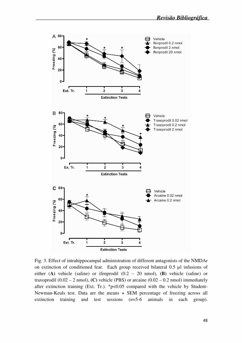
Revisão Bibliográfica
48
Fig. 3. Effect of intrahippocampal administration of different antagonists of the NMDAr on extinction of conditioned fear. Each group received bilateral 0.5 µl infusions of either (A) vehicle (saline) or ifenprodil (0.2 – 20 nmol), (B) vehicle (saline) or traxoprodil (0.02 – 2 nmol), (C) vehicle (PBS) or arcaine (0.02 – 0.2 nmol) immediately after extinction training (Ext. Tr.). *p<0.05 compared with the vehicle by Student-Newman-Keuls test. Data are the means + SEM percentage of freezing across all extinction training and test sessions (n=5-6 animals in each group).
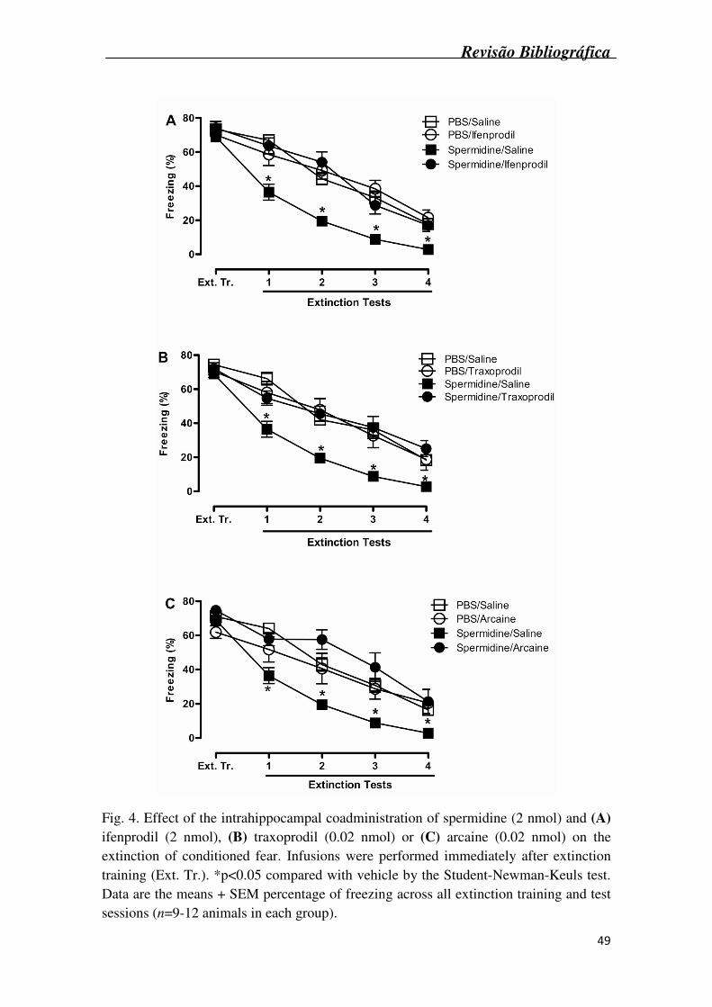
Revisão Bibliográfica
49
Fig. 4. Effect of the intrahippocampal coadministration of spermidine (2 nmol) and (A) ifenprodil (2 nmol), (B) traxoprodil (0.02 nmol) or (C) arcaine (0.02 nmol) on the extinction of conditioned fear. Infusions were performed immediately after extinction training (Ext. Tr.). *p<0.05 compared with vehicle by the Student-Newman-Keuls test. Data are the means + SEM percentage of freezing across all extinction training and test sessions (n=9-12 animals in each group).
Discussão
50
4. DISCUSSÃO
Discussão
51
4. Discussão
O presente estudo teve como propósito pesquisar o efeito de agentes
poliaminérgicos sobre a extinção do medo condicionado contextual em ratos Wistar
machos.
Os resultados encontrados demonstraram que a administração intrahipocampal
de espermidina, imediatamente após o treino da extinção, facilitou a extinção do medo
condicionado contextual em ratos. Ainda, demonstrou-se que a infusão dos antagonistas
da subunidade NR2B do rNMDA arcaína, ifenprodil e traxprodil pioraram a extinção do
medo condicionado contextual, quando injetados imediatamente após o treino da
extinção. Sabendo que as poliaminas melhoram a memória em ratos, via ativação do
rNMDA (Rubin et. al., 2000, Rubin et. al., 2001, Rubin et. al., 2004, Berlese et. al.,
2005), foi verificado se o efeito da espermidina em facilitar a extinção do medo
condicionado dependia da ativação do rNMDA. De fato, a coadministração de
espermidina e de antagonistas da subunidade NR2B em doses sem efeito per se,
reverteu à facilitação da extinção promovida pela espermidina. Estes resultados sugerem
que agentes poliaminérgicos modulam a extinção do medo condicionado, quando
injetados no hipocampo de ratos, e que este efeito é depende da subunidade NR2B do
rNMDA hipocampal.
Estudos prévios mostram que a espermidina possui grande potencial no que diz
respeito à melhora do desempenho cognitivo. Visto que sua administração sistêmica,
intrahipocampal ou intraamigdala melhora o desempenho de ratos em tarefas de
memória como medo condicionado e esquiva inibitória (Rubin et. al., 2000, Rubin et.
al., 2001, Berlese et. al., 2005). Este efeito da espermidina sobre a memória parece
depender basicamente de dois mecanismos: atividade da enzima óxido nítrico sintase
(NOS) e ativação do rNMDA. Estudo realizado por Guerra e colaboradores mostrou que
a administração de N(G)-nitro L-arginina metil Ester (L-NAME), um inibidor
inespecífico da enzima NOS, previne a melhora de memória induzida pela espermidina
na tarefa de esquiva inibitória. No que se refere ao rNMDA, foi demonstrado que a
administração de MK-801 ou arcaína, ambos antagonistas do rNMDA, suprimem o
efeito da espermidina sobre a memória (Rubin et. al., 2001, Camera et. al., 2007).
Discussão
52
A extinção do medo é um processo ativo, que depende do rNMDA (Myers and
Davis, 2002). Recentemente demonstrou-se o papel da subunidade NR2B no processo
de extinção. Sotres-Bayon e colaboradores demonstraram que a administração tanto
sistêmica como intraamigdala de ifenprodil, antes do treino da extinção, bloqueia a
aquisição e evocação da extinção do medo. Ainda, ifenprodil quando administrado
imediatamente após treino da extinção, impede que ocorra consolidação da mesma
(Sotres-Bayon et. al., 2009). Estes resultados sugerem que o rNMDA contendo a
subunidade NR2B são cruciais para o processamento da extinção do medo. Ainda,
resultados apresentados neste trabalho corroboram com estudos prévios que mostram a
importância do hipocampo e da subunidade NR2B do rNMDA na extinção do medo
(Corcoran and Maren, 2001, Corcoran and Quirk, 2007, Ji and Maren, 2007, Sotres-
Bayon et. al., 2007).
O papel do hipocampo na modulação contextual da extinção pode ser avaliado
através de abordagens cirúrgicas e farmacológicas (para revisão ver Ji & Maren, 2007).
Lesões excitotóxicas realizadas na região CA1 do hipocampo impedem a aquisição da
extinção, enquanto que a administração intrahipocampal de muscimol bloqueia a
expressão da extinção (Corcoran and Maren, 2001, Dillon et. al., 2008). Nossos
resultados sugerem um papel para poliaminas hipocampais sobre a extinção do medo
condicionado. É importante notar que a administração intrahipocampal de espermidina
facilitou a extinção, e que a administração dos três antagonistas da subunidade NR2B,
em doses sem efeito sobre a memória, preveniu o efeito facilitatório da espermidina.
No presente trabalho, traxoprodil na dose de 0,2 nmol/sítio bloqueou a extinção
do medo condicionado, mas a maior dose testada, 2 nmol/sítio, não apresentou efeito
sobre a memória. Este efeito bifásico é semelhante ao relatado com arcaína, quando
administrada intraamigdala (Rubin 2004) e também é semelhante com a curva dose-
resposta apresentada pela espermidina em trabalhos anteriores (Berlese et. al., 2005).
Portanto, é plausível que a maneira que o traxoprodil interage com o rNMDA seja
semelhante a de outros compostos que ligam-se na subunidade NR2B do rNMDA, os
quais apresentam curvas dose-resposta em U-invertido. O mesmo padrão de resposta
não foi encontrado com arcaína ou ifenprodil, mas isso não significa que estes
compostos interajam com o receptor de maneira diferente, já que um efeito bifásico
poderia ter sido encontrado se mais doses destes compostos tivessem sido testadas. O
efeito do traxoprodil sobre a memória parece conflitante, e trabalhos já relataram que
este composto não possui efeito (Guscott et. al., 2003), piora (Myers and Davis, 2002),
Discussão
53
ou até melhora a memória (Higgins et. al., 2005), sendo o efeito bifásico deste
composto um dos possíveis motivos para seus diferentes efeitos sobre a memória.
A facilitação da extinção do pode ser obtida através da manipulação de distintos
sistemas, como o colinérgico, canabinóide, GABAérgico e glutamatérgico. A infusão
intraamigdala do agonista muscarínico oxotremorina facilita a extinção do medo
condicionado contextual (Boccia et. al., 2009). Adicionalmente, a administração
intraperitoneal de WIN 55,212-2, um agonista canabinóide, facilita a extinção do medo
condicionado contextual (Pamplona et. al., 2006). Ainda, a administração de muscimol,
um agonista GABAA, antes do treino da extinção, facilita a extinção (Akirav et. al.,
2006). Além disso, o sistema glutamatérgico parece desempenhar um importante papel
na modulação da extinção. D-cicloserina, um agonista parcial do rNMDA, facilita a
extinção do medo condicionado quando injetado tanto sistemicamente quanto
intraamigdala imediatamente após o treino da extinção (Ledgerwood et. al., 2003).
Resultados apresentados neste trabalho corroboram com o trabalhos prévios
(Ledgerwood et. al., 2003, Ledgerwood et. al., 2004, Richardson et. al., 2004,
Yamamoto et. al., 2008), demonstrando que a ativação do rNMDA resulta em melhora
da retenção da extinção.
Resumidamente, os resultados apresentados no presente trabalho demonstram
que, enquanto a espermidina facilitou a extinção do medo condicionado, antagonistas da
subunidade NR2B do rNMDA arcaína, ifenprodil e traxoprodil, bloquearam e
reverteram o efeito da espermidina sobre a extinção. Sabendo que a terapia de exposição
é um método amplamente utilizado no tratamento de distúrbios de ansiedade, como
fobias e estresse pós-traumático (Hellstrom and Ost, 1995, Powers et. al., 2004), os
resultados deste trabalho mostrando uma facilitação da extinção pela espermidina,
colocam os compostos poliaminérgicos como possíveis protótipos para o
desenvolvimento de novos fármacos para o tratamento destes distúrbios. Ainda, de um
ponto de vista clínico, mostra-se interessante o desenvolvimento de moduladores do
rNMDA que atuam sobre memórias de medo. Bloqueadores clássicos do rNMDA,
como MK-801, atuam bloqueando o poro do receptor, assim apresentando pouca ou
nenhuma especificidade por subtipos de receptores. A conseqüência disso é uma ampla
gama de efeitos adversos, como neurotoxicidade e potencial abusivo (Grant et. al.,
1991, Ikonomidou et. al., 1999, Klein et. al., 1999). Em contraste, moduladores do
rNMDA (como os agentes poliaminérgicos), poderiam ser efetivos sem afetar funções
vitais do rNMDA, dando a essas drogas possível uso em intervenções farmacológicas.
Referências Bibliográficas
54
5. CONCLUSÃO
Referências Bibliográficas
55
5. Conclusão
Através da análise dos resultados obtidos no presente trabalho, podemos concluir
que:
• A administração intrahipocampal de espermidina na dose de 2 nmol/sítio,
imediatamente após o treino da extinção, , facilitou a extinção do medo
condicionado contextual em ratos, sugerindo que o rNMDA está envolvido na
extinção do medo condicionado;
• A administração intrahipocampal dos diferentes antagonistas da subunidade NR2B
do rNMDA, arcaína (0,2 nmol/sítio), traxoprodil (0,2 nmol/sítio) e ifenprodil (20
nmol/sítio), imediatamente após o treino da extinção, bloqueou a extinção do medo
condicionado contextual em ratos. Isto sugere que a subunidade NR2B do rNMDA
está envolvida na extinção do medo condicionado;
• A coadministração dos antagonistas da subunidade NR2B do rNMDA, em doses
sem efeito per se, reverteram a facilitação da extinção do medo condicionado
provocada pela espermidina. Isto sugere o envolvimento da subunidade NR2B do r
NMDA no efeito da espermidina.
Referências Bibliográficas
56
6. Referências Bibliográficas
Abel, T. and Lattal, K. M., 2001. Molecular mechanisms of memory acquisition, consolidation and retrieval. Curr Opin Neurobiol. 11, 180-187.
Akirav, I., Raizel, H. and Maroun, M., 2006. Enhancement of conditioned fear extinction by infusion of the GABA(A) agonist muscimol into the rat prefrontal cortex and amygdala. European Journal of Neuroscience. 23, 758-764.
Artiges, E., Salame, P., Recasens, C., Poline, J. B., Attar-Levy, D., De La Raillere, A., Paillere-Martinot, M. L., Danion, J. M. and Martinot, J. L., 2000. Working memory control in patients with schizophrenia: a PET study during a random number generation task. Am J Psychiatry. 157, 1517-1519.
Ashby, F. G. and O'Brien, J. B., 2005. Category learning and multiple memory systems. Trends Cogn Sci. 9, 83-89.
Bekinschtein, P., Cammarota, M., Izquierdo, I. and Medina, J. H., 2008. BDNF and memory formation and storage. Neuroscientist. 14, 147-156.
Berlese, D. B., Sauzem, P. D., Carati, M. C., Guerra, G. P., Stiegemeier, J. A., Mello, C. F. and Rubin, M. A., 2005. Time-dependent modulation of inhibitory avoidance memory by spermidine in rats. Neurobiology of Learning and Memory. 83, 48-53.
Berman, D. E. and Dudai, Y., 2001. Memory extinction, learning anew, and learning the new: dissociations in the molecular machinery of learning in cortex. Science. 291, 2417-2419.
Bevilaqua, L. R., Bonini, J. S., Rossato, J. I., Izquierdo, L. A., Cammarota, M. and Izquierdo, I., 2006. The entorhinal cortex plays a role in extinction. Neurobiology of Learning and Memory. 85, 192-197.
Bliss, T. V. and Collingridge, G. L., 1993. A synaptic model of memory: long-term potentiation in the hippocampus. Nature. 361, 31-39.
Boccia, M. M., Blake, M. G., Baratti, C. M. and McGaugh, J. L., 2009. Involvement of the basolateral amygdala in muscarinic cholinergic modulation of extinction memory consolidation. Neurobiology of Learning and Memory. 91, 93-97.
Bouton, M. E., 2002. Context, ambiguity, and unlearning: sources of relapse after behavioral extinction. Biol Psychiatry. 52, 976-986.
Bouton, M. E., 2004. Context and behavioral processes in extinction. Learn Mem. 11, 485-494.
Bouton, M. E. and King, D. A., 1983. Contextual control of the extinction of conditioned fear: tests for the associative value of the context. J Exp Psychol Anim Behav Process. 9, 248-265.
Bruchey, A. K., Shumake, J. and Gonzalez-Lima, F., 2007. Network model of fear extinction and renewal functional pathways. Neuroscience. 145, 423-437.
Camera, K., Mello, C. F., Ceretta, A. P. and Rubin, M. A., 2007. Systemic administration of polyaminergic agents modulate fear conditioning in rats. Psychopharmacology (Berl). 192, 457-464.
Referências Bibliográficas
57
Cammarota, M., Barros, D. M., Vianna, M. R., Bevilaqua, L. R., Coitinho, A., Szapiro, G., Izquierdo, L. A., Medina, J. H. and Izquierdo, I., 2004. The transition from memory retrieval to extinction. An Acad Bras Cienc. 76, 573-582.
Cammarota, M., Bevilaqua, L. R., Barros, D. M., Vianna, M. R., Izquierdo, L. A., Medina, J. H. and Izquierdo, I., 2005. Retrieval and the extinction of memory. Cell Mol Neurobiol. 25, 465-474.
Carter, C., 1994. The neuropharmacology of polyamines, London. Coffino, P., 2001. Regulation of cellular polyamines by antizyme. Nat Rev Mol Cell
Biol. 2, 188-194. Conway, E. L., 1998. Brain lesions and delayed water maze learning deficits after
intracerebroventricular spermine. Brain Res. 800, 10-20. Corcoran, K. A., Desmond, T. J., Frey, K. A. and Maren, S., 2005. Hippocampal
inactivation disrupts the acquisition and contextual encoding of fear extinction. Journal of Neuroscience. 25, 8978-8987.
Corcoran, K. A. and Maren, S., 2001. Hippocampal inactivation disrupts contextual retrieval of fear memory after extinction. Journal of Neuroscience. 21, 1720-1726.
Corcoran, K. A. and Quirk, G. J., 2007. Recalling safety: cooperative functions of the ventromedial prefrontal cortex and the hippocampus in extinction. CNS Spectrums. 12, 200-206.
de Fockert, J. W., 2005. Keeping priorities: the role of working memory and selective attention in cognitive aging. Sci Aging Knowledge Environ. 2005, pe34.
de Oliveira Alvares, L., Pasqualini Genro, B., Diehl, F., Molina, V. A. and Quillfeldt, J. A., 2008. Opposite action of hippocampal CB1 receptors in memory reconsolidation and extinction. Neuroscience. 154, 1648-1655.
Debiec, J., LeDoux, J. E. and Nader, K., 2002. Cellular and systems reconsolidation in the hippocampus. Neuron. 36, 527-538.
Dillon, G. M., Qu, X., Marcus, J. N. and Dodart, J. C., 2008. Excitotoxic lesions restricted to the dorsal CA1 field of the hippocampus impair spatial memory and extinction learning in C57BL/6 mice. Neurobiology of Learning and Memory. 90, 426-433.
Eichenbaum, H., 2001. The hippocampus and declarative memory: cognitive mechanisms and neural codes. Behav Brain Res. 127, 199-207.
Falls, W. A., Miserendino, M. J. and Davis, M., 1992. Extinction of fear-potentiated startle: blockade by infusion of an NMDA antagonist into the amygdala. J Neurosci. 12, 854-863.
Gabriele, A. and Packard, M. G., 2007. D-Cycloserine enhances memory consolidation of hippocampus-dependent latent extinction. Learning & Memory. 14, 468-471.
Grant, K. A., Knisely, J. S., Tabakoff, B., Barrett, J. E. and Balster, R. L., 1991. Ethanol-like discriminative stimulus effects of non-competitive n-methyl-d-aspartate antagonists. Behav Pharmacol. 2, 87-95.
Guerra, G. P., Mello, C. F., Sauzem, P. D., Berlese, D. B., Furian, A. F., Tabarelli, Z. and Rubin, M. A., 2006. Nitric oxide is involved in the memory facilitation induced by spermidine in rats. Psychopharmacology (Berl). 186, 150-158.
Gugliucci, A., 2004. Polyamines as clinical laboratory tools. Clin Chim Acta. 344, 23-35.
Guscott, M. R., Clarke, H. F., Murray, F., Grimwood, S., Bristow, L. J. and Hutson, P. H., 2003. The effect of (+/-)-CP-101,606, an NMDA receptor NR2B subunit selective antagonist, in the Morris watermaze. European Journal of Pharmacology. 476, 193-199.
Referências Bibliográficas
58
Halonen, T., Sivenius, J., Miettinen, R., Halmekyto, M., Kauppinen, R., Sinervirta, R., Alakuijala, L., Alhonen, L., MacDonald, E., Janne, J. and et al., 1993. Elevated seizure threshold and impaired spatial learning in transgenic mice with putrescine overproduction in the brain. Eur J Neurosci. 5, 1233-1239.
Hellstrom, K. and Ost, L. G., 1995. One-session therapist directed exposure vs two forms of manual directed self-exposure in the treatment of spider phobia. Behaviour Research and Therapy. 33, 959-965.
Higgins, G. A., Ballard, T. M., Enderlin, M., Haman, M. and Kemp, J. A., 2005. Evidence for improved performance in cognitive tasks following selective NR2B NMDA receptor antagonist pre-treatment in the rat. Psychopharmacology (Berl). 179, 85-98.
Ikonomidou, C., Bosch, F., Miksa, M., Bittigau, P., Vockler, J., Dikranian, K., Tenkova, T. I., Stefovska, V., Turski, L. and Olney, J. W., 1999. Blockade of NMDA receptors and apoptotic neurodegeneration in the developing brain. Science. 283, 70-74.
Izquierdo, I., 2002. Memória, Porto Alegre. Izquierdo, I., Bevilaqua, L. R., Rossato, J. I., Bonini, J. S., Medina, J. H. and
Cammarota, M., 2006. Different molecular cascades in different sites of the brain control memory consolidation. Trends Neurosci. 29, 496-505.
Izquierdo, I. and Cammarota, M., 2004. Neuroscience. Zif and the survival of memory. Science. 304, 829-830.
Izquierdo, I., Izquierdo, L. A., Barros, D. M., Mello e Souza, T., de Souza, M. M., Quevedo, J., Rodrigues, C., Sant'Anna, M. K., Madruga, M. and Medina, J. H., 1998. Differential involvement of cortical receptor mechanisms in working, short-term and long-term memory. Behav Pharmacol. 9, 421-427.
Izquierdo, I. and McGaugh, J. L., 2000. Behavioural pharmacology and its contribution to the molecular basis of memory consolidation. Behav Pharmacol. 11, 517-534.
Izquierdo, I. and Medina, J. H., 1995. Correlation between the pharmacology of long-term potentiation and the pharmacology of memory. Neurobiol Learn Mem. 63, 19-32.
Izquierdo, I. and Medina, J. H., 1997. Memory formation: the sequence of biochemical events in the hippocampus and its connection to activity in other brain structures. Neurobiol Learn Mem. 68, 285-316.
Ji, J. and Maren, S., 2007. Hippocampal involvement in contextual modulation of fear extinction. Hippocampus. 17, 749-758.
Johnson, T. D., 1996. Modulation of channel function by polyamines. Trends in Pharmacological Science. 17, 22-27.
Kalisch, R., Holt, B., Petrovic, P., De Martino, B., Kloppel, S., Buchel, C. and Dolan, R. J., 2009. The NMDA agonist D-cycloserine facilitates fear memory consolidation in humans. Cereb Cortex. 19, 187-196.
Kandel, E. R., 1997. Genes, synapses, and long-term memory. J Cell Physiol. 173, 124-125.
Kishi, A., Ohno, M. and Watanabe, S., 1998. Spermidine, a polyamine site agonist, attenuates working memory deficits caused by blockade of hippocampal muscarinic receptors and mGluRs in rats. Brain Res. 793, 311-314.
Klein, M., Calderon, S. and Hayes, B., 1999. Abuse liability assessment of neuroprotectants. Ann N Y Acad Sci. 890, 515-525.
Langton, J. M. and Richardson, R., 2008. D-cycloserine facilitates extinction the first time but not the second time: an examination of the role of NMDA across the
Referências Bibliográficas
59
course of repeated extinction sessions. Neuropsychopharmacology. 33, 3096-3102.
Lebron, K., Milad, M. R. and Quirk, G. J., 2004. Delayed recall of fear extinction in rats with lesions of ventral medial prefrontal cortex. Learning & Memory. 11, 544-548.
Ledgerwood, L., Richardson, R. and Cranney, J., 2003. Effects of D-cycloserine on extinction of conditioned freezing. Behavioral Neuroscience. 117, 341-349.
Ledgerwood, L., Richardson, R. and Cranney, J., 2004. D-cycloserine and the facilitation of extinction of conditioned fear: consequences for reinstatement. Behav Neurosci. 118, 505-513.
Lee, Y. S. and Silva, A. J., 2009. The molecular and cellular biology of enhanced cognition. Nat Rev Neurosci. 10, 126-140.
Lin, C. H., Yeh, S. H., Lu, H. Y. and Gean, P. W., 2003. The similarities and diversities of signal pathways leading to consolidation of conditioning and consolidation of extinction of fear memory. Journal of Neuroscience. 23, 8310-8317.
Lovibond, P. F., 2004. Cognitive processes in extinction. Learning & Memory. 11, 495-500.
Marton, L. J. and Pegg, A. E., 1995. Polyamines as targets for therapeutic intervention. Annu Rev Pharmacol Toxicol. 35, 55-91.
Meyer, R. C., Knox, J., Purwin, D. A., Spangler, E. L. and Ingram, D. K., 1998. Combined stimulation of the glycine and polyamine sites of the NMDA receptor attenuates NMDA blockade-induced learning deficits of rats in a 14-unit T-maze. Psychopharmacology (Berl). 135, 290-295.
Mikolajczak, P., Okulicz-Kozaryn, I., Kaminska, E., Niedopad, L., Polanska, A. and Gebka, J., 2002. Effects of acamprosate and some polyamine site ligands of NMDA receptor on short-term memory in rats. Eur J Pharmacol. 444, 83-96.
Moinard, C., Cynober, L. and de Bandt, J. P., 2005. Polyamines: metabolism and implications in human diseases. Clin Nutr. 24, 184-197.
Morgan, D. M., 1999. Polyamines. An overview. Mol Biotechnol. 11, 229-250. Myers, K. M. and Davis, M., 2002. Behavioral and neural analysis of extinction.
Neuron. 36, 567-584. Nader, K., Schafe, G. E. and Le Doux, J. E., 2000. Fear memories require protein
synthesis in the amygdala for reconsolidation after retrieval. Nature. 406, 722-726.
Pamplona, F. A., Prediger, R. D., Pandolfo, P. and Takahashi, R. N., 2006. The cannabinoid receptor agonist WIN 55,212-2 facilitates the extinction of contextual fear memory and spatial memory in rats. Psychopharmacology (Berl). 188, 641-649.
Pedreira, M. E. and Maldonado, H., 2003. Protein synthesis subserves reconsolidation or extinction depending on reminder duration. Neuron. 38, 863-869.
Pegg, A. E. and McCann, P. P., 1982. Polyamine metabolism and function. Am J Physiol. 243, C212-221.
Power, A. E., Berlau, D. J., McGaugh, J. L. and Steward, O., 2006. Anisomycin infused into the hippocampus fails to block "reconsolidation" but impairs extinction: the role of re-exposure duration. Learn Mem. 13, 27-34.
Powers, M. B., Smits, J. A. and Telch, M. J., 2004. Disentangling the effects of safety-behavior utilization and safety-behavior availability during exposure-based treatment: a placebo-controlled trial. Journal of Consulting and Clinical Psychology. 72, 448-454.
Referências Bibliográficas
60
Quirk, G. J. and Mueller, D., 2008. Neural mechanisms of extinction learning and retrieval. Neuropsychopharmacology. 33, 56-72.
Ransom, R. W. and Stec, N. L., 1988. Cooperative modulation of [3H]MK-801 binding to the N-methyl-D-aspartate receptor-ion channel complex by L-glutamate, glycine, and polyamines. Journal of Neurochemistry. 51, 830-836.
Richardson, R., Ledgerwood, L. and Cranney, J., 2004. Facilitation of fear extinction by D-cycloserine: theoretical and clinical implications. Learn Mem. 11, 510-516.
Robertson, E. M., Pascual-Leone, A. and Miall, R. C., 2004. Current concepts in procedural consolidation. Nat Rev Neurosci. 5, 576-582.
Rock, D. M. and Macdonald, R. L., 1995. Polyamine regulation of N-methyl-D-aspartate receptor channels. Annual Review of Pharmacology and Toxicology. 35, 463-482.
Rubin, M. A., Berlese, D. B., Stiegemeier, J. A., Volkweis, M. A., Oliveira, D. M., dos Santos, T. L., Fenili, A. C. and Mello, C. F., 2004. Intra-amygdala administration of polyamines modulates fear conditioning in rats. Journal of Neuroscience. 24, 2328-2334.
Rubin, M. A., Boemo, R. L., Jurach, A., Rojas, D. B., Zanolla, G. R., Obregon, A. D., Souza, D. O. and Mello, C. F., 2000. Intrahippocampal spermidine administration improves inhibitory avoidance performance in rats. Behavioral Pharmacology. 11, 57-61.
Rubin, M. A., Stiegemeier, J. A., Volkweis, M. A., Oliveira, D. M., Fenili, A. C., Boemo, R. L., Jurach, A. and Mello, C. F., 2001. Intra-amygdala spermidine administration improves inhibitory avoidance performance in rats. European Journal of Pharmacology. 423, 35-39.
Seiler, N., 2004. Catabolism of polyamines. Amino Acids. 26, 217-233. Shimada, A., Spangler, E. L., London, E. D. and Ingram, D. K., 1994. Spermidine
potentiates dizocilpine-induced impairment of learning performance by rats in a 14-unit T-maze. European Journal of Pharmacology. 263, 293-300.
Sotres-Bayon, F., Bush, D. E. and LeDoux, J. E., 2007. Acquisition of fear extinction requires activation of NR2B-containing NMDA receptors in the lateral amygdala. Neuropsychopharmacology. 32, 1929-1940.
Sotres-Bayon, F., Diaz-Mataix, L., Bush, D. E. and LeDoux, J. E., 2009. Dissociable roles for the ventromedial prefrontal cortex and amygdala in fear extinction: NR2B contribution. Cerebral Cortex. 19, 474-482.
Szapiro, G., Vianna, M. R., McGaugh, J. L., Medina, J. H. and Izquierdo, I., 2003. The role of NMDA glutamate receptors, PKA, MAPK, and CAMKII in the hippocampus in extinction of conditioned fear. Hippocampus. 13, 53-58.
Tadano, T., Hozumi, S., Yamadera, F., Murata, A., Niijima, F., Tan-No, K., Nakagawasai, O. and Kisara, K., 2004. Effects of NMDA receptor-related agonists on learning and memory impairment in olfactory bulbectomized mice. Methods Find Exp Clin Pharmacol. 26, 93-97.
Teti, D., Visalli, M. and McNair, H., 2002. Analysis of polyamines as markers of (patho)physiological conditions. J Chromatogr B Analyt Technol Biomed Life Sci. 781, 107-149.
Urdiales, J. L., Medina, M. A. and Sanchez-Jimenez, F., 2001. Polyamine metabolism revisited. Eur J Gastroenterol Hepatol. 13, 1015-1019.
Vianna, M. R., Igaz, L. M., Coitinho, A. S., Medina, J. H. and Izquierdo, I., 2003. Memory extinction requires gene expression in rat hippocampus. Neurobiology of Learning and Memory. 79, 199-203.
Referências Bibliográficas
61
Wang, C., Delcros, J. G., Cannon, L., Konate, F., Carias, H., Biggerstaff, J., Gardner, R. A. and Phanstiel, I. V. O. t., 2003. Defining the molecular requirements for the selective delivery of polyamine conjugates into cells containing active polyamine transporters. J Med Chem. 46, 5129-5138.
Williams, K., 1997a. Interactions of polyamines with ion channels. Biochem J. 325 ( Pt 2), 289-297.
Williams, K., 1997b. Modulation and block of ion channels: a new biology of polyamines. Cell Signal. 9, 1-13.
Williams, K., Romano, C. and Molinoff, P. B., 1989. Effects of polyamines on the binding of [3H]MK-801 to the N-methyl-D-aspartate receptor: pharmacological evidence for the existence of a polyamine recognition site. Mol Pharmacol. 36, 575-581.
Yamamoto, S., Morinobu, S., Fuchikami, M., Kurata, A., Kozuru, T. and Yamawaki, S., 2008. Effects of single prolonged stress and D-cycloserine on contextual fear extinction and hippocampal NMDA receptor expression in a rat model of PTSD. Neuropsychopharmacology. 33, 2108-2116.
Anexo
62
7. Anexo
Em anexo carta de resposta dos revisores do periódico Neurobiology of Learning and
memory, com parecer sobre manuscrito “Poliaminergic agents modulate contextual fear extinction in rats”:
Ms. No.: NLM-09-161
Title: Polyaminergic agents modulate contextual fear extinction in rats
Corresponding Author: Dr maribel Antonello rubin
Authors: Guilherme M Gomes, Msc; Carlos F Mello, PhD; Michelle M da Rosa,
Undergraduating; Guilherme V Bochi, Undergraduating; Juliano Ferreira, PhD; Susan
Barron, PhD;
Dear Dr rubin,
Thank you for submitting your manuscript to Neurobiology of Learning and Memory.
Without significant modifications, this manuscript will not be acceptable for
publication. We encourage you to consider these comments and make appropriate
revision of your manuscript. Upon receipt, the manuscript will be re-reviewed promptly.
The reviewers' comments are below. In particular, please note the request from both
referees for "no-extinction, drug controls" to be included in the experimental design.
Please submit your revision online within 90 days by logging onto the Elsevier Editorial
System for Neurobiology of Learning and Memory:
When submitting your revised paper, please include a separate document uploaded as
"Response to Reviews" that carefully addresses the issues raised in the below
comments, point by point. You should also include a suitable rebuttal to any specific
request for change that has not been made.
To facilitate the electronic publication of your manuscript (should it be accepted), we
request that your manuscript text, tables and figure legend be submitted in an editable
format (Word, WordPerfect, or LaTex only), and all figures uploaded individually as
Anexo
63
TIF or EPS files.
Thank you, and we look forward to receiving your revised manuscript.
With kind regards,
Paul E. Gold, Editor
Wickliffe C. Abraham, Associate Editor
Eric Klann, Associate Editor
Neurobiology of Learning and Memory
Neurobiology of Learning and Memory, Editorial Office
Elsevier
Email: [email protected]
Reviewers' comments:
Reviewer #1: The manuscript examines the effects of hippocampal infusions of
polyaminergic agents on the extinction of context fear conditioning in rats. The
procedures and results are straight forward. Rats are subjected to contextual fear
conditioning on day 1. On day 2 rats receive extinction training which involves 6 min
non-reinforced exposure to the context. Rats received hippocampal infusions
immediately after this extinction training. The next 4 days were extinction test days and
were identical to extinction training with the exception that rats received no infusions.
The results showed that post-extinction training hippocampal infusions of spermidine
apparently facilitated extinction whereas infenprodil, taxoprodil, and arcaine all
impaired extinction. Interestingly, combined infusions of spermidine plus each of the
antagonists blocked the facilitatory effects of spermidine and this was achieved at doses
of the antagonists which themselves had no effect on
extinction learning. The authors conclude that exogenous and endogenous polyamines
facilitate fear extinction via actions at NR2B-subunit containing hippocampal NMDA
receptors. The manuscript is generally well written (with the exceptions described
below) and the pharmacology is sound. However, I am less enthusiastic than the authors
regarding these results and their interpretation. I have the following major comments on
the manuscript.
Anexo
64
1. The key claim of the manuscript is that polyamines act at hippocampal NMDA
receptors to modulate contextual fear extinction. This finding is potentially interesting
but is also not secured by the data. In particular, there was no evidence presented that
the effects of hippocampal infusions actually depended on prior extinction training.
That is, the experiments could have benefited immensely from control groups which
received conditioning, no initial extinction training but do receive infusion, then 4 days
of test. This would show that the effects of infusions in these experiments depended
upon extinction training. An equally important control would be to match the
PBS/saline and drug groups on hippocampal infusions of the drug so that, for example,
6 hr after initial extinction training the groups received the opposite infusions. Both of
these controls would strengthen considerably the claim that the hippocampal infusions
were actually affecting extinction learning or
memory. This is important because hippocampal manipulations can affect a variety of
processes other than extinction such as locomotor activity, contextual
configural/conjunctive processing etc and the effects of these infusions can be quite
long-lived. Moreover, it is important because, in contrast to the author's assertion, the
available evidence suggests that hippocampus is important for regulating where and
when extinction learning is expressed but is not important for extinction learning per se.
This is the key conclusion from the papers by Corcoran, Maren, and Bouton cited in the
manuscript.
2. The manuscript could also be improved by addressing why the effects of a single
infusion after extinction training were still observed across the four days of testing.
Which specific process do the authors think could produce this long-term effect?
3. Related to this, I think the authors need a much stronger rationale in their
introduction for why they studied hippocampal contributions to contextual fear
extinction learning. After all, fear extinction can survive hippocampal lesions and
inactivation (see the papers cited in the manuscript).
4. The neuroanatomical specificity of these manipulations is unclear. What happens
when these substances are infused into the cortex dorsal to the hippocampus?
5. Finally, although Izquierdo and colleagues have made important contributions to
Anexo
65
the neurobiology of learning and memory it would be appropriate to cite the primary
empirical work of others in the introduction when discussing the neural substrates for
extinction learning in the first paragraph of the introduction.
Other points
1. The procedure is simple and straightforward. I am not sure readers will require
Figure 1 to understand it.
2. p. 5 "minute amounts" could be re-phrased.
3. p. 7, freezing is a fear-indicant behaviour not a "memory index"
4. More details on histology would be helpful, including some indication of where
microinjection tips were actually located.
Reviewer #2: This paper reports that spermidine, which evidently binds to the NR2b
subunit, can enhance contextual fear extinction when it is injected into the dorsal
hippocampus. The claim that it acts on the NR2B subunit is based on the additional
finding that several NR2B antagonist when co-administer with spermidine, prevent
enhanced extinction when administered a doses that themselves have no effect on
extinction.
Spermidine has previously been reported to have memory enhancing effects however,
this might be the first reported that it enhances the retention of an fear extinction
experience.
The paper itself was very well written. The only worthwhile comment I have is that
there is no control in the first experiment that would determine that the effect of the
drug depended on extinction training. It probably does but the authors have no drug
only control.
The authors might want to replace their reference to Pavlov, 1927. The current
placement might mislead one to infer that Pavlov studied conditioned fear.