valeska paula casanova - ufsmw3.ufsm.br/ppgmv/images/dissertacoes2016/valeska paula casanova.pdf ·...
TRANSCRIPT

0
UNIVERSIDADE FEDERAL DE SANTA MARIA
CENTRO DE CIÊNCIAS RURAIS
PROGRAMA DE PÓS-GRADUAÇÃO EM MEDICINA VETERINÁRIA
Valeska Paula Casanova
SUPLEMENTAÇÃO COM FERRO INJETÁVEL EM CORDEIROS
INFECTADOS EXPERIMENTALMENTE POR Haemonchus contortus
Santa Maria, RS, Brasil
2016
1
Valeska Paula Casanova
SUPLEMENTAÇÃO COM FERRO INJETÁVEL EM CORDEIROS INFECTADOS
EXPERIMENTALMENTE POR Haemonchus contortus
Dissertação apresentada ao Curso de Mestrado do
Programa de Pós-Graduação em Medicina
Veterinária, Área de Clínica e Cirurgia Animal,
da Universidade Federal de Santa Maria
(UFSM,RS), como requisito parcial para
obtenção do grau de Mestre em Medicina
Veterinária
Orientador: Profa Marta Lizandra do Rêgo Leal
Santa Maria, RS, Brasil
2016

2
1.
© 2016
Todos os direitos autorais reservados a Valeska Paula Casanova. A reprodução de partes ou de
todo este trabalho só poderá ser feita mediante a citação da fonte.
Fone: (55) 3220-8000 - e-mail: [email protected]
3
Valeska Paula Casanova
SUPLEMENTAÇÃO COM FERRO INJETÁVEL EM CORDEIROS INFECTADOS
EXPERIMENTALMENTE POR Haemonchus contortus
Dissertação apresentada ao Curso de Mestrado do
Programa de Pós-Graduação em Medicina
Veterinária, Área de Clínica e Cirurgia Animal,
da Universidade Federal de Santa Maria
(UFSM,RS), como requisito parcial para
obtenção do grau de Mestre em Medicina
Veterinária
Aprovado em 23 de fevereiro de 2016:
_______________________________
Marta Lizandra do Rêgo Leal (UFSM)
(Presidente/orientador)
______________________________
Dr. Ricardo Xavier da Rocha
_______________________________
Fernanda Silveira Flores Vogel (UFSM)
Santa Maria, RS.
2016
4
DEDICATÓRIA
Dedico mais esta conquista ao meu pai, Ademir. Por ser um exemplo de ser humano,
dedicação, competência e solidariedade. Por sempre ter uma palavra de incentivo,
independente da situação. E por me fazer chegar até aqui. Dedico!
5
AGRADECIMENTOS
Sempre, à Deus. Por todas as oportunidades concedidas, dificuldades enfrentadas e sonhos
realizados.
A minha família, porto seguro, alicerce primordial por mais esta conquista. À minha vó, pelas
horas diárias no telefone me dando forças para mais um dia. E ao meu pai, exemplo de
pessoa, tudo o que faço é para poder orgulhá-lo!
Minha mãe, por me dar luz e me guiar. Lembro-me da sua principal característica, a
persistência, onde procuro me espelhar.
Ao Getulio, meu namorado, por me incentivar a iniciar o mestrado e por nunca me deixar
desistir. Só cheguei até aqui por sua causa. Obrigada por tudo!
À todos meus colegas da Clínica de Ruminantes. Adelina, Zé e Pivoto, pelas infinitas vezes
que precisei de ajuda, pelo companheirismo e amizade construída. À todos os estagiários, de
modo especial a Brunna, Alexia, Gustavo e Limana, foram muito importantes durante toda
essa caminhada e se tornaram inesquecíveis, sentirei saudades!
A distância da minha família foi o obstáculo mais difícil pra mim, mas lembrava da Profa
Marta e sabia que precisava ter força de vontade para alcançar meus objetivos. Te agradeço
Marta, por ter tido paciência comigo, por tudo o que me ensinou e por ser um exemplo pra
mim, tanto de pessoa e quanto mais de profissional. Devo a ti meu crescimento, serei para
sempre grata!
Ao professor Alexandre Krause, por todo o auxílio durante o experimento, com as coletas e
análises de medula óssea. E ao Guilherme Bochi, pela realização da parte bioquímica do
experimento.
À UFSM pelas oportunidades, a secretária da pós-graduação Maria, pelas inúmeras vezes que
precisei de auxílio e a FAPERGS pelo suporte financeiro.

6
“Porque creram, todas as pessoas foram aprovadas por Deus,
mas não receberam o que Ele havia prometido. Pois Deus
tinha preparado um plano ainda melhor para nós,
a fim de que, somente conosco,
fosse aperfeiçoado.”
Hebreus 11 : 39-40

7
RESUMO
SUPLEMENTAÇÃO COM FERRO INJETÁVEL EM CORDEIROS
INFECTADOS EXPERIMENTALMENTE POR Haemonchus contortus
AUTORA: VALESKA PAULA CASANOVA
ORIENTADORA: MARTA LIZANDRA DO RÊGO LEAL
As verminoses gastro-intestinais é o principal entrave na produção de ovinos. O helminto
Haemonchus contortus é a espécie que mais parasita ovinos no Brasil. Localiza-se no
abomaso do hospedeiro e possui como hábito a hematofagia. Como sinais clínicos os animais
apresentam anemia e hipoproteínemia, o que diminui a produtividade dos animais, pode levar
a morte e traz sérios prejuízos para a atividade. A suplementação com ferro em ovinos
parasitados auxilia no processo hematopoiético, porém ainda são necessários estudos para
avaliar o metabolismo do ferro nestes animais. Diante do exposto, o presente estudo teve
como objetivo avaliar os efeitos da suplementação de ferro injetável em ovinos infectados
experimentalmente com Haemonchus contortus. Utilizou-se 24 cordeiros divididos em quatro
grupos experimentais, sendo o Grupo 1 – animais não infectados, Grupo 2 – animais
infectados com 10000 larvas de terceiro estágio de H. contortus e tratados com três doses de
20mg /Kg de peso vivo de ferro dextrano por via intramuscular, um dose a cada sete dias,
Grupo 3 – animais não infectados e tratados com três doses de 20mg /Kg de peso vivo de
ferro dextrano por via intramuscular, um dose a cada sete dias e Grupo 4 – animais infectados
com 10000 larvas de terceiro estágio de H. contortus. O período experimental foi de 21 dias.
As amostras de sangue foram coletadas nos dias 10 (D10), 17 (D17), 24 (D24) e 31 (D31)
pós-infecção parasitária para avaliação do metabolismo do ferro (Ferritina, Transferrina, Ferro
Sérico, Índice de saturação à transferrina, Capacidade Total de Ligação do Ferro e estoques de
Ferro Medular) e do eritrograma (Contagem de Hemácia, Hematócrito, Hemoglobina,
Reticulócitos, Volume Corpuscular Médio e a Concentração de Hemoglobina Corpuscular
Média). Amostras de fezes para quantificar o número de ovos por grama de fezes (OPG)
foram obtidas nos mesmos momentos experimentais. Valores de OPG foram positivos
somente nos Grupos 2 e 4. Quanto aos índices hematológicos, os grupos de animais infectado
e tratados com ferro apresentaram maiores valores de hematócrito e hemoglobina em relação
ao grupo dos animais infectado e não tratado com ferro. Os resultados demonstraram que as
reservas de ferro medular foram maiores nos grupos suplementados com ferro. Ovinos
infectados com H. contortus apresentam exaustão das reservas medulares de ferro, com isso o
uso de ferro injetável auxilia na manutenção de estoque e torna o mineral mais disponível para
a eritropoiese, melhorando a resposta orgânica dos animais contra à infecção.
Palavras-chave: Infecção parasitária. Ovinos. Anemia.
8
ABSTRACT
INJECTABLE IRON SUPPLEMENTATION IN SHEEP INFECTED
EXPERIMENTALLY BY Haemonchus contortus
AUTHOR: VALESKA PAULA CASANOVA
ADVISER: MARTA LIZANDRA DO RÊGO LEAL
Gastrointestinal worms are the main obstacle in the production of sheep. The helminth
Haemonchus contortus is the species that most parasites sheep in Brazil. It is located in the
abomaso of the host and has a habit of blood feeding. Considering clinical signs, the animals
present anemia and hypoproteinemia, which reduces the productivity of animals; it can lead to
death and causes serious damage to the activity. Iron supplementation in parasitized sheep
assists in the hematopoietic process, although further studies are needed to assess the iron
metabolism in such animals. Given the above, the following study aimed to evaluate the
effects of injectable iron supplementation in sheep experimentally infected with Haemonchus
contortusTwenty-four lambs have been used, divided into four groups, being Group 1 -
uninfected animals, Group 2 - animals infected with 10000 third stage larvae of H. contortus
and treated with three doses of 20mg/kg of body weight of dextran iron intramuscularly, one
dose every seven days, Group 3 - non-infected animals and treated with three doses of
20mg/kg body weight iron dextran intramuscularly, one dose every seven days, and Group 4 -
animals infected with 10000 third stage larvae of H. contortus. The experiment has lasted 21
days. Blood samples were collected on days 10 (D10), 17 (D17), 24 (D24), and 31 (D31) after
parasitic infection to assess the metabolism of iron (ferritin, transferrin, Serum Iron, saturation
index to transferrin, Iron Connection Total Capacity, and Spinal Cord Iron stocks) and
erythrocyte (red blood cell count, Hematocrit, hemoglobin, reticulocytes, mean corpuscular
hemoglobin concentration). Samples of feces for measuring the number of eggs per gram of
feces (EPG) have been obtained at the same experimental time. OPG values were positive in
Groups 2 and 4. Regarding hematological indices, groups of animals infected and treated with
iron had higher values of hematocrit and hemoglobin compared to the group of animals
infected and not treated with iron. The results showed that the spinal cord iron stocks were
greater in the groups supplemented with iron. Sheep infected with H. contortus presented
exhaustion of iron spinal cord reserves, thus the use of injectable iron aids in maintaining the
stock and makes the mineral available for erythropoiesis by improving the organic response of
animals against infection.
Keywords: Parasitic Infection. Sheep. Anemia.
9
LISTA DE FIGURAS
REVISÃO BIBLIOGRÁFICA
Figura 01 Ciclo evolutivo do Haemonchus contortus. ............................................................ 15
Figura 02 Absorção de ferro, distribuição e reciclagem no corpo e troca quantitativa entre as
suas fontes no organismo. .......................................................................................... 18
Figura 03 Metabolismo do ferro ............................................................................................... 20
CAPÍTULO I
Figure 1 Erythrocytes number (A), hematocrit (B), hemoglobin concentration (C), MCV (D),
MCHC (E), reticulocytes number (F)......................................................................... 43
Figure 2 Seric iron (A); Transferrin (B); Ferritin (C); TSI (D); TIBC (E); Iron in the bone
marrow (F).. ................................................................................................................ 44
Figure 3 Feces examination (McMaster) of sheep naturally infected with Haemonchus
contortus, results presented in eggs per gram of feces (EPG).................................... 45
Figure 4 Bone marrow of lambs infected by Haemonchus contortus, untreated with iron
dextran, with inventory absent marrow iron (A). Lamb bone marrow infected with H.
contortus treated with iron dextran, it can be visualized iron stores in the bone
marrow particles blue indicated by arrow (B) ............................................................ 46
10
SUMÁRIO
INTRODUÇÃO ...................................................................................................................... 12
REVISÃO BIBLIOGRÁFICA .............................................................................................. 14
2.1 Haemonchus contortus ....................................................................................................... 14
2.2 METABOLISMO DO FERRO .......................................................................................... 17
CAPÍTULO I .......................................................................................................................... 23
Abstract ................................................................................................................................... 25
Introduction ............................................................................................................................ 26
Material and methods ............................................................................................................ 28
Animals .....................................................................................................................................28
Experimental design ................................................................................................................. 29
Infection .................................................................................................................................... 29
Samples colection ..................................................................................................................... 30
Hemogram ................................................................................................................................ 30
Iron metabolism ........................................................................................................................ 30
Feces parasitology .................................................................................................................... 31
Body weight .............................................................................................................................. 32
Statistical analysis .................................................................................................................... 32
Results .....................................................................................................................................32
Parasitological exams ............................................................................................................... 32
Hemogram ................................................................................................................................ 33
Iron metabolism ........................................................................................................................ 33
Discussion ................................................................................................................................ 34
Conclusion ............................................................................................................................... 36
References................................................................................................................................ 37
CONSIDERAÇÕES FINAIS ................................................................................................. 47
REFERÊNCIAS ..................................................................................................................... 48
11
APRESENTAÇÃO
Os resultados que fazem parte desta dissertação estão apresentados sob a forma de
artigo científico submetido no periódico Veterinary Parasitology disponível no capítulo I. As
seções Material e Métodos, Resultados, Discussão e Referências Bibliográficas encontram-se
no próprio artigo e representam a íntegra deste estudo. As REFERÊNCIAS
BIBLIOGRÁFICAS se referem somente às citações que aparecem nos itens INTRODUÇÃO
e REVISÃO BIBLIOGRÁFICA desta dissertação.
12
INTRODUÇÃO
Na pecuária comercial, o foco é o aumento da produtividade e, consequentemente, da
rentabilidade na criação dos animais. Para isso, é necessária a melhora na sanidade dos
rebanhos com maior eficiência nas medidas de diagnóstico, tratamento e prevenção das
enfermidades que mais causam prejuízos. Dentre estas doenças, as verminoses gastrintestinais
são as que mais acometem ovinos trazendo sérios prejuízos econômicos (GENNARI,
AMARANTE, 2006).
Os ovinos podem ser parasitados simultaneamente por várias espécies de nematóides
gastrintestinais. A importância de cada espécie varia em função da combinação de três
fatores: intensidade da infecção, prevalência, e patogenicidade do parasita. Com base nisso,
pode-se afirmar que o Haemonchus contortus é a principal espécie que parasita ovinos no
Brasil (AMARANTE, 2009).
A haemoncose trata-se de uma das mais patogênicas helmintoses, pois em infecções
leves, já é possível observar redução na relação custo benefício na ovinocultura. O
Haemonchus contortus possui hábito hematófago, desencadeando como principais sinais
clínicos a anemia e a hipoproteínemia, que podem causar a morte dos animais (CLIMENI et
al., 2008).
O entendimento da dinâmica populacional dos nematódeos e dos sinais clínicos nos
animais faz parte da estratégia de controle da haemoncose, e o uso de anti-helmíntico faz-se
necessário, porém, associar medidas de manejo e buscar o equilíbrio entre o hospedeiro e
parasita pode em algumas situações ser mais importante do que o uso da medicação
(GENNARI, AMARANTE, 2006).
A atual busca por um mercado que prioriza a saúde, o meio ambiente e o bem-estar
animal, o tratamento seletivo auxiliado à medidas de suporte, pode ser estratégia viável para a
produção agropecuária (MOLENTO et al., 2004). Sabendo disso, os minerais estão
envolvidos na maioria das vias metabólicas do organismo animal, sendo em função
reprodutiva, energética, crescimento e imunidade, pois quando há falta dos mesmos,
alterações ocorrem diretamente relacionadas à produtividade animal (MENDONÇA et al.,
2011).

13
O ferro é essencial para a formação da molécula heme, que participa da formação da
hemoglobina, constituinte chave no eritrócito, além de participar na síntese de várias outras
proteínas (GROTTO, 2010). A utilização de ferro por via intramuscular em ovinos
parasitados tem eficácia, auxiliando na resposta a anemia causada pelo parasita como
afirmado por Rocha et al. (2007), porém, há poucos estudos demonstrando a verdadeira
função do ferro nesses animais. Desse modo, o objetivo do presente estudo foi avaliar o
mecanismo de ação do mineral quando utilizado em animais parasitados.
14
REVISÃO BIBLIOGRÁFICA
2.1 Haemonchus contortus
Dentre os fatores que interferem no desenvolvimento pleno da ovinocultura, a infecção
por nematódeos gastrintestinais representam um problema econômico na pecuária, pois
acarreta estresse aos animais, desnutrição, diminuição de peso e como conseqüência
diminuição da produção, e ainda custos com tratamentos profiláticos e curativos (ANENE et
al., 1994; AMARANTE, 2009).
A haemoncose é uma doença parasitária que acomete principalmente ovinos e
caprinos, em regiões tropicais e subtropicais. É provocada pelo nematóide do gênero
Haemonchus, sendo a espécie dominante que parasita pequenos ruminantes o Haemonchus
contortus (H. contortus), o qual se localiza no abomaso de seus hospedeiros (CLIMENI et al.,
2008).
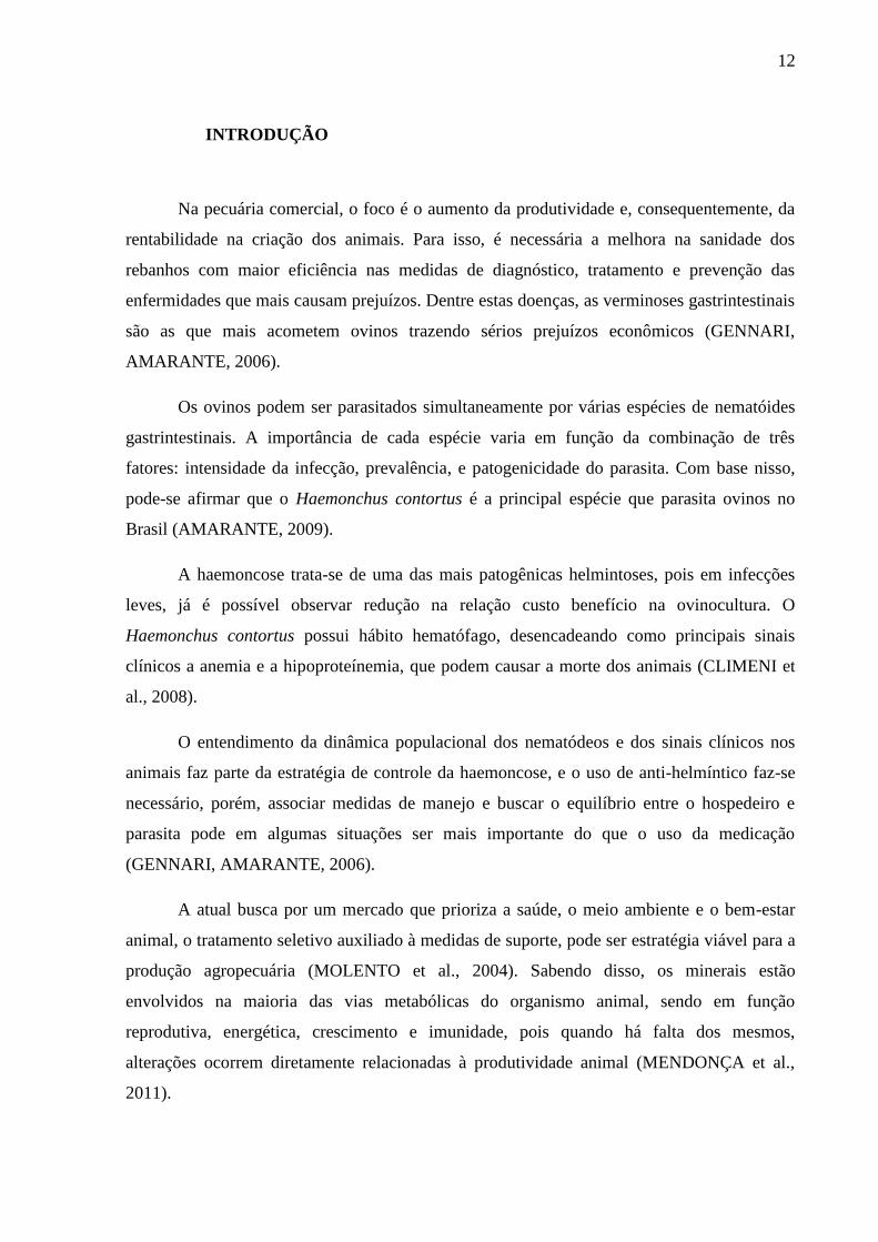
O H. contortus possui ciclo direto (Figura 01), onde as fêmeas adultas no abomaso
produzem ovos que são eliminados pelas fezes, os quais darão origem às larvas de primeiro
estágio (L1). Ainda no bolo fecal, em condições ideais de temperatura e umidade, em
aproximadamente 5 dias, sofrem duas mudas e originando as larvas de estágio infectante (L3).
As L3 contaminam as pastagens, e permanecem neste local até serem ingeridas pelos
hospedeiros. Depois de ingeridas, inicia-se a fase de vida parasitária. Prendem-se a mucosa do
abomaso, local onde se alimentam de sangue, sofrem mais duas mudas e tornam-se adultas
(L5), quando iniciam a ovoposição (ZAJAC, 2006).
O H. contortus é um dos mais prolíficos nematóides. A ovoposição diária da fêmea
pode chegar a 10.000 ovos, resultando em um acúmulo de larvas nas pastagens na estação de
pastoreio (ROMERO; BOERO, 2001; ZAJAC, 2006).

15
Figura 1 - Ciclo evolutivo do Haemonchus contortus.
Fonte: http://labmedvet.blogspot.com.br/2011_12_01_archive.html
Possui como característica a hematofagia, que causa perdas sanguíneas, resultando em
anemia e hipoproteinemia (MOLENTO et. al, 2004; ONYIAH; ARSLAN, 2005). Diminuição
dos teores de albumina e proteína sérica reduz a pressão oncótica dos vasos sanguíneos,
provocando edema, principalmente na região submandibular. E em infecções maciças, até
10% do sangue do animal pode ser removido por dia pelo parasita, causando morte súbita
(HOSTE, CHARTIER, 1998).
A haemoncose pode ser caracterizada em forma hiperaguda: após a primo infecção por
um grande número de larvas, ocorre severa perda sanguínea com consequente morte súbita do
animal. Forma aguda: o número de larvas situa-se ente 1.000 e 10.000, contudo já ocorre
hematopoiese compensatória, o que acarreta uma diminuição das reservas de ferro e de
albumina, resultando em anemia e edema. Forma crônica: desencadeada por um pequeno
número de larvas em parasitismo constante. Este processo ativa a eritropoiese, desorganiza a
resposta imune e exaure as reservas de ferro e de proteína. Sabendo da hematofagia provocada
pelo parasita e a conseqüente diminuição ou exaustão das reservas de ferro, o Haemonchus
contortus desencadeia anemia ferropriva nos animais parasitados (FONSECA, 2006).
Por seu habito hematófago, o H. contortus provoca anemia que pode ser visualizada
pela coloração das mucosas aparentes (pálidas) e pode ser avaliada pela determinação do
16
eritrograma, principalmente pelo numero total de hemácia, pelo volume globular, e pelo teor
de hemoglobina. Com a diminuição das reservas de ferro que comprometem a síntese de heme
e, consequentemente, a eritropoiese, a anemia desencadeada pelo H. contortus é dita
ferropriva (AMARANTE, 2009; AMARANTE et al., 2004).
O diagnóstico da infecção por H. contortus pode ser realizado através da contagem de
ovos por grama de fezes (OPG), tendo como vantagens a fácil coleta de material e análise
(BISHOP et al., 1996). Porém, a técnica não é específica para H. contortus, e a interpretação
dos resultados necessita de cuidados, pois nem sempre o número de nematóides presente nos
animais é refletido no teste, fenômeno chamado densidade dependente, onde em animais com
alta carga parasitária, as fêmeas de H. contortus diminuem ou cessam a ovipostura. Além de
que, a imunidade dos animais, espécies e ovipostura dos diferentes parasitos, consistência das
fezes e estágio dos parasitos no interior dos hospedeiros podem interferir nos resultados
(RUAS BERNE, 2007).
O diagnóstico definitivo pode ser realizado através da técnica de Roberts & O´Sulivan,
onde se efetua o cultivo de fezes para a identificação de larvas de 3º estágio do parasita (VAN
WYK; CABARET; MICHAEL, 2004).
A utilização de anti-helmínticos como forma de tratamento parasitário, é notavelmente
a forma mais bem sucedida para tal, porém a sua utilização é ameaçada pela evolução da
resistência dos parasitas à estes medicamentos (STEAR; MURRAY, 1994).
Por conta do ineficiente repasse de informações sobre tecnologias e frequência de
tratamento antiparasitário, tem se observado diminuição da eficácia destes produtos nas
principais regiões produtoras de ruminantes do país, com o aparecimento de cepas
multiresistentes a vários compostos químicos. Tais observações estimulam alternativas para
manter a eficácia das drogas antiparasitárias, assim como a sustentabilidade da agropecuária.
O objetivo central é diminuir o uso errôneo de anti-helmínticos e a concentração das drogas
no meio ambiente, no leite e na carne, e possibilitar melhoria da qualidade de produtos
animais (MOLENTO et al., 2004).
O verdadeiro mecanismo compensatório da anemia é a eritropoiese que visa a maior
produção de eritrócitos. A medula óssea estimulada pela condição anêmica libera grandes
quantidades de células jovens e imaturas, os reticulócitos, os quais não são observados em
condições normais na circulação periférica de ovinos (BIRGEL, 1999).
Os distúrbios gastrointestinais causados pelo H. contortus podem estar associados com
alterações nos níveis de hormônios gastrintestinais circulantes, como por exemplo a gastrina,
responsável pela secreção de suco gástrico. Estudos feitos por Nicholls et al. (1989)

17
mostraram uma média maior nos valores de pH da secreção do abomaso em animais
parasitados, essas variações prejudicam a absorção de ferro intestinal, aumentando a
deficiência do mineral. Birgel (2013) observou que ovinos com verminose gastrintestinal
possuem teores séricos de ferro menores quando comparado à animais sadios.
Estudos demonstraram que a suplementação de ferro injetável auxilia a resposta
hematopoiética em cordeiros anêmicos (ROCHA et al., 2007, 2012, 2014), pois 80% do ferro
corporal é capturado pela medula óssea para a formação de hemoglobina. No entanto, se faz
necessário avaliar a utilização de ferro em animais severamente infectados por H. contortus
para entender o metabolismo do mineral e sua ação em animais com intensa anemia.
2.2 METABOLISMO DO FERRO
O ferro é um metal de transição que possui vastas utilizações biológicas, entre elas:
está presente no organismo em diferentes estados de oxidação, age como um centro catalítico
em funções metabólicas, além de formar muitos complexos. É um metal de suma importância
para a respiração tanto aeróbica quanto anaeróbica, pois está presente na hemoglobina, em 60
a 70% do seu total, fazendo o transporte de oxigênio e dióxido de carbono. Atua no
funcionamento do sistema imunológico e formação de energia (CARPENTER, MAHONEY,
1992).
O ferro possui atividade no fornecimento de energia celular, pois é o metal mais
abundante presente na mitocôndria, que por sua vez fornece energia para todo e qualquer
compartimento celular. Além disso, a mitocôndria, por utilizar grande quantidade de ferro,
auxilia na manutenção de seu metabolismo, em questões de consumo, transporte e
armazenamento (LEVI, ROVIDA, 2009).
Não existe processo fisiológico de excreção de ferro, portanto, o balanço sistêmico do
mineral é regulado exclusivamente pelo sítio de absorção (KNOVICH et al. 2009). A
deficiência de ferro é mais freqüente em consequência do aumento das perdas (perda de
sangue gastrointestinal), exigências excessivas (durante rápido crescimento) ou o
fornecimento da dieta inadequada (ANDREWS, 2005).
Quando livre, o ferro possui toxicidade em relação às células, pois pode contribuir
para formação de espécies reativas ao oxigênio e ao nitrogênio. Para isso, necessita de
proteínas de transporte específicas para torná-lo intracelular, pois não tem capacidade de
entrar passivamente nas mesmas, por não conseguir atravessar a sua camada lipídica. Como

18
também, células epiteliais intestinais, macrófagos e hepatócitos necessitam de mecanismo
para exportá-lo (ANDREWS, 2005).
O ferro ser considerado como um oligoelemento, necessário em pequenas quantidades
como cofator de diversas enzimas. Porém, quando sua presença é necessária em maior
quantidade, como na hemoglobina, isso faz com que as necessidades do organismo sejam
mais difíceis de corrigir do que as de outros oligoelementos metálicos, sendo mais comum seu
desequilíbrio (PANTOPOULOS et al., 2013).
Mais da metade de ferro do corpo em indivíduos normais é encontrado na forma de
hemoglobina em eritrócitos e em seus precursores. De 20-25 mg de ferro é necessário em uma
base diária para a hemoglobinização de novos eritrócitos, porém de 0,5-2,0 mg são adquiridos
através da absorção intestinal, provenientes de reciclagem de ferro já circulante (Figura 02)
(ANDREWS, 2005).
Figura 02: Absorção de ferro, distribuição e reciclagem no corpo e troca quantitativa entre as suas
fontes no organismo.
Fonte: Pantopoulos et al., 2012.
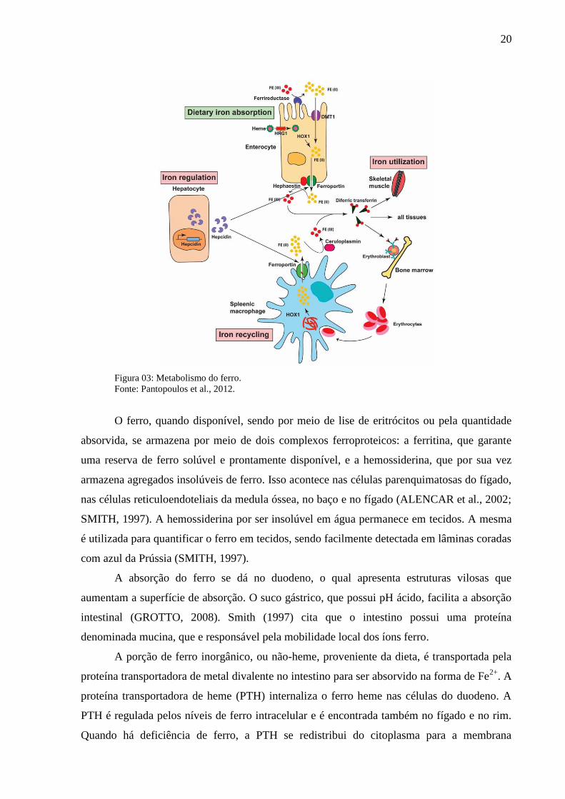
Os macrófagos de baço e medula óssea, e em menor número as células de Kupffer do
fígado, reconhecem modificações bioquímicas na parede de hemácias velhas e danificadas, o
que permite a fagocitose das mesmas. Ao ser fagocitada, a hemoglobina é degradada pela
enzima heme oxigenase, que retira o ferro contido na porção heme da hemoglobina (Figura
19
03). O ferro, que se encontra na forma ferrosa (Fe2+
), contido nessas hemácias fagocitadas,
será armazenado no próprio macrófago em forma de ferritina, ou será exportado pela
ferroportina. Quando exportado, o Fe2+
será oxidado em ferro férrico (Fe3+
) pela hefaestina,
semelhante a ceruloplasmina sérica, sintetizada no fígado. O Fe3+
será transportado pela
ceruloplasmina até os locais de utilização, e então retorna a participar da hemoglobinização de
novos eritrócitos (GROTTO, 2008; ANDREWS, 2005).
O ferro é o segundo metal mais abundante do planeta, depois do alumínio, porém a sua
deficiência é uma questão comum entre os vertebrados. Isso se deve ao fato de que o ferro
quando em contato com oxigênio, oxida-se à forma férrica, o qual é consideravelmente menos
biodisponível devido a sua baixa solubilidade (KLAUSNER et al., 1993).
Em pH fisiológico e na presença de oxigênio, há a predominância do íon Fe3+
, o qual é
altamente insolúvel, pois o Fe2+
é altamente oxidável. Diante disso, existe um cofator de
redução especializado, que reduz o Fe3+
em Fe2+
na superfície extracelular, permitindo assim
que a célula ocupe a forma Fe2+
(McKIE et al., 2001).
Os teores de ferro podem ser aumentados através de uma fonte primária que é por
meio da fagocitose de hemácias remanescentes, ou por meio de fontes adicionais, podendo ser
obtido através da mobilização de ferro celular, especialmente de hepatócitos (ANDREWS,
2005).
A transferrina, uma proteína de 80 KDa sintetizada e secretada pelo fígado, é
responsável pela entrada de ferro nas células. Para haver a internalização de ferro celular, é
necessário que receptores de alta afinidade da transferrina estejam presentes. Possui dois sítios
homólogos com afinidade para o ferro na forma férrica, além de solubilizar e atenuar a sua
reatividade, também facilita a sua disponibilidade para as células. O nível de expressão do
receptor de transferrina nas células varia de acordo com a necessidade de ferro pelas mesmas,
que é influenciada pela multiplicação de células sanguínea da série vermelha, bem como a
produção de hemoglobina ou mioglobina, e necessidades metabólicas específicas (Figura 03)
(KLAUSNER et al., 1993). A transferrina é um meio de avaliação da quantidade de ferro
funcional disponível no organismo (KNOVICH et al., 2009).
20
Figura 03: Metabolismo do ferro.
Fonte: Pantopoulos et al., 2012.
O ferro, quando disponível, sendo por meio de lise de eritrócitos ou pela quantidade
absorvida, se armazena por meio de dois complexos ferroproteicos: a ferritina, que garante
uma reserva de ferro solúvel e prontamente disponível, e a hemossiderina, que por sua vez
armazena agregados insolúveis de ferro. Isso acontece nas células parenquimatosas do fígado,
nas células reticuloendoteliais da medula óssea, no baço e no fígado (ALENCAR et al., 2002;
SMITH, 1997). A hemossiderina por ser insolúvel em água permanece em tecidos. A mesma
é utilizada para quantificar o ferro em tecidos, sendo facilmente detectada em lâminas coradas
com azul da Prússia (SMITH, 1997).
A absorção do ferro se dá no duodeno, o qual apresenta estruturas vilosas que
aumentam a superfície de absorção. O suco gástrico, que possui pH ácido, facilita a absorção
intestinal (GROTTO, 2008). Smith (1997) cita que o intestino possui uma proteína
denominada mucina, que e responsável pela mobilidade local dos íons ferro.
A porção de ferro inorgânico, ou não-heme, proveniente da dieta, é transportada pela
proteína transportadora de metal divalente no intestino para ser absorvido na forma de Fe2+
. A
proteína transportadora de heme (PTH) internaliza o ferro heme nas células do duodeno. A
PTH é regulada pelos níveis de ferro intracelular e é encontrada também no fígado e no rim.
Quando há deficiência de ferro, a PTH se redistribui do citoplasma para a membrana
21
plasmática das células duodenais, enquanto em casos de excesso de ferro, ocorre o processo
inverso (GROTTO, 2008).
O organismo não possui mecanismos para eliminar o excesso de ferro absorvido ou
acumulado. A hepcidina, um peptídeo sintetizado no fígado, secretado e processado na
circulação possui função de regulação sistêmica do ferro, pois interliga os locais de absorção,
utilização e estoque de ferro. Teores altos de ferro aumentam a sua expressão, enquanto que
em casos de deficiência de ferro e na hipóxia, a expressão da hepcidina diminui, visando uma
maior absorção de ferro pelos enterócitos e maior exportação de ferro do sistema
reticuloendotelial, aumentando a disponibilidade do mineral para a eritropoiese; sendo este
peptideo um regulador negativo de ferro (Figura 03) (GROTTO, 2008; KEMNA et al., 2008).
Em casos inflamatórios, há uma estimulação da síntese de hepcidina pelas citocinas
inflamatórias (IL-1; IL-6; TNF-α), o que diminui a absorção de ferro por enterócitos e
macrófagos, limitando a quantidade do mineral disponível para a eritropoiese (GANZ, 2007).
A IL-1 é responsável pela estimulação de proteínas de fase aguda em processos infecciosos,
como o fibronogênio, a haptoglobulina, a ceruloplasmina e a ferritina, causando consequente
redução dos teores de ferro circulantes (HERSHKO, 1993). A IL-6, principal estimulador de
síntese de hepcidina pelos hepatócitos, fará com que a mesma iniba a absorção pelos
enterócitos e bloqueie a liberação de ferro pelos macrófagos, tornando-o menos disponível
para formação de novas células sanguíneas (ALTÉS et al., 2014; GROTTO, 2008). A
hepcidina, tem se mostrado um dos principais reguladores da homeostase do ferro e um
provável mediador da anemia por doença crônica ou por inflamação (LEMOS et al., 2010).
A ferroportina é o único canal de transporte de ferro em todos os tecidos que o
exercem, na qual, a hepcidina se liga para controlar a exportação do mineral no plasma. Em
baixos níveis de hepcidina, a ferroportina se liga a membrana plasmática celular para a
exportação de ferro. A ferroportina induz a internalização e posterior degradação do ferro,
diminuindo a sua liberação, quando os níveis de hepcidina aumentam (GANZ, 2007).
A ferritina é uma proteína solúvel que armazena ferro no interior das células, e é o
primeiro mecanismo de estoque de ferro, sendo fundamental para a homeostase do mineral.
Uma pequena quantidade de ferrtina circula livre no sangue, demonstrando um parâmetro
indireto de depósitos corporais de ferro. Além disso, contém e mantém os átomos que ferro
que poderia formar agregados tóxicos, além de disponibilizar o ferro para processos celulares
necessários. Como marcador, a ferritina é um dos parâmetros mais sensíveis para a detecção
de deficiência quanto de sobrecarga de ferro no organismo (ALTÉS et al., 2014; RAMÍREZ
et al., 2004; GROTTO, 2008; KNOVICH et al., 2009).
22
Quando há aumento da necessidade de ferro pela célula, não há armazenamento do
mineral e, por consequência, os níveis de ferritina estão diminuídos. Simultaneamente, ocorre
aumento dos receptores de transferrina para que o ferro absorvido seja utilizado pela célula
(KLAUSNER et al., 1993).
Exames citológicos de medula óssea são de suma importância para a avaliação do
metabolismo do ferro. A coloração com azul da Prússia ou reação de Perls, onde a
hemossiderina, forma degradada da ferritina, será corada com ferrocianeto de potássio na
presença de ácido clorídrico, é considerado o teste padrão ouro para tal finalidade (GROTTO,
2008). Segundo Nimeh e Bishop (1980), pode-se afirmar que a presença de ferro corável nas
células reticuloendoteliais da medula óssea elimina a deficiência de ferro como causador da
anemia.
A grande vantagem da avaliação do ferro medular, é que se pode diferenciar a
depleção dos estoques de ferro, das condições associadas aos defeitos da liberação de ferro
pelo sistema mononuclear fagocitário, como ocorre em casos de anemia por doença
inflamatória (ALENCAR et al., 2002).
Tivemos como hipótese que a suplementação com ferro injetável em ovinos infectados
por Haemonchus contortus, aumenta o estoque medular de ferro e a eritropoiese, reduz a
gravidade da anemia e aumenta a resistência orgânica dos animais infectados.
Para isso, nosso objetivo foi avaliar o efeito da suplementação com ferro injetável por
via intramuscular em cordeiros experimentalmente infectados por Haemonchus contortus.
23
CAPÍTULO I –
(Artigo sibmetido ao períodico Veterinary Parasitology)
Injectable iron supplementation in sheep infected experimentally by Haemonchus
contortus
Valeska Paula Casanova, Adelina Rodrigues Aires, Felipe Lamberti Pivoto, Alexia Pretto,
João Francisco Tadielo Limana, Gustavo Potrich Marchioretto, Brunna de Mattos Granja,
Silvana Giacomini Collet, Alexandre Krause, Rafael N. Moresco, Guilherme Vargas Bochi
Aleksandro Schafer da Silva, Marta Lizandra do Rêgo Leal
24
Injectable iron supplementation in sheep infected experimentally with Haemonchus
contortus
Valeska Paula Casanovaa*
, Adelina Rodrigues Airesa, Felipe Lamberti Pivoto
a, Alexia Pretto
a,
João Francisco Tadielo Limanaa, Gustavo Potrich Marchioretto
a, Brunna de Mattos Granja
a,
Silvana Giacomini Colleta, Alexandre Krause
b, Rafael N. Moresco
c, Guilherme Vargas
Bochic, Aleksandro Schafer da Silva
d, Marta Lizandra do Rêgo Leal
a*
a Department of Large Animals, Universidade Federal de Santa Maria, Brazil
b Department of Small Animal, Universidade Federal de Santa Maria, Brazil
c Department of Pharmacy, Universidade Federal de Santa Maria, Brazil
d Department of Animal Science, Universidade do Estado de Santa Catarina, Brazil
*Author for mail: Departamento de Grandes Animais, Universidade Federal de Santa Maria
Avenida Roraima, 1000 Santa Maria, Rio Grande do Sul, Brasil
Telephone: + 55 55 3220-8815; fax: + 55 55 3220-8815.
Email Addresses: [email protected] (V. P. Casanova); [email protected]
(M. L. R. Leal).

25
Abstract
Anemia has been often related to the reduction of iron in consequence of infection by
gastrointestinal parasites causing great economic losses in sheep. Therefore, the following
research aimed to evaluate the effect of injectable supplementation by iron dextran on blood
variables and iron metabolism in sheep experimentally infected with Haemonchus contortus.
In this study, 24 male lambs were used, crosses Corriedale and Texel, at an average 17 kg
body weight and four months old. The animals were divided into four groups of six animals
each: Uninfected group and untreated with iron (G1); Infected group and treated with iron
(G2); Uninfected group and treated with iron (G3); and infected group and untreated (G4).
Groups 2 and 4 received 10,000 H. contortus larvae (L3), orally. The lambs G2 and G3
receive three doses of 20mg/kg of iron dextran, intramuscularly with an interval of seven days
between doses. The blood and feces samples were made on 10, 17, 24, and 31 post-infection
(PI), and blood was analyzed erythrogram (total erythrocyte count, hemoglobin concentration,
hematocrit, mean corpuscular volume (MCV) and mean corpuscular hemoglobin
concentration (MCHC)), and the number of reticulocytes. Iron metabolism was determined by
the analysis of serum levels of iron, ferritin, transferrin, transferrin saturation index, total iron
binding capacity (TIBC), and bone marrow iron stores. The EPG (eggs per gram of feces) was
negative in groups 1 and 3 during the trial period on days 24 and 31 PI the EPG values were
positive and similar in Groups 2 and 4. The number of erythrocytes, and hematocrit and
hemoglobin concentration were lower on days 24 and 31 PI in the infected group when
compared to those uninfected. However, on day 31 the animals G2 presented higher values of
hematocrit and hemoglobin than animals of G4. Higher reticulocyte counts were obtained in
G2 and G4. There was no difference between groups in the VCM and there were greater
numerical values of MCHC in G1 and G3 groups on D31. The lambs of the infected group
and untreated (G4) had lower serum levels of iron on D31 compared to the other groups.

26
Higher values of transferrin and TIBC were observed in non-supplemented animals with iron.
Metal stores in the bone marrow, quantified by scores (0-6) were higher in animals treated
with iron compared to animals of untreated groups. The injectable iron supplementation
reduces the severity of the anemia caused by infection with H. contortus in sheep and retains
stocks of this mineral in the bone marrow, thus enabling the synthesis of hemoglobin.
Keywords: helminths, anemia, bone marrow, iron, sheep.
Introduction
The sheep production is an important source of income through the exploration of
meat, milk, and wool from the animals (Pinheiro et al., 2000). In semi-intensive and intensive
production systems, infection with gastrointestinal nematodes is one of the biggest problems
of this farming activity. Infections by parasites of the genus Haemonchus are the most
common in Brazilian sheep flocks (Amarante et al., 2004). In consequence of the blood
sucking habit, the animals infected by this parasite have iron deficiency anemia and
hypoproteinemia (Rocha et al., 2013). Such alterations cause sharp reduction in average daily
gain (ADG) and in many cases, the death of infected animals. The anemic level was high
about two weeks after infection, i.e. when the parasitic plunder exceeds the erythropoietic
capacity of the animal, then there is a decrease in the number of red blood cells, hemoglobin
concentration and hematocrit, with variations in the values according to sex, age, species of
parasite and resistance of infected animals (Feldman et al., 2006).
The metal iron is vital to cellular homeostasis, as well as key component for the
formation of heme fraction that participates in the synthesis of several proteins, which are
responsible for oxygen transport, cell power generation, and detoxification. The heme fraction
is formed on all nucleated cells, and the largest amount is produced by the erythroid tissue
(Andrews, 2005; Grotto, 2008). In cases of iron deficiency anemia, such as the one caused by
27
Haemonchus contortus, there may be a reduction of iron in the body, reducing the metal
stocks, possibly leading to tissue deficiency (Nastiti et al., 2004).
The distribution of iron in the body may occupy different compartments (inventory,
shipping, and functional), which are interconnected, but which can be measured separately.
These compartments are affected sequentially as iron decreases in the body. Whenever there
is iron loss by the animal organism, the stock is the first to be affected due to metal
mobilization in order to meet the organic requirements. Then there is a deficit in iron fraction
in transportation and, finally, iron deficiency in the functional erythroid compartment ,
progressively (Krishnamurthy et al., 2007). Therefore, the correct way to evaluate body iron
status is to conduct assessments of seric concentrations of iron, ferritin, transferrin, transferrin
saturation, the total iron binding capacity of transferrin and iron stores in the bone marrow
(Frazer and Anderson, 2003).
Transferrin, a protein synthesized and secreted by the liver , in charge of transporting
iron into cells, one way to assess functional amount of available iron in the body (Knovich et
al., 2009). The level of transferrin receptor expression in the cells varies according to the need
of iron through, which is influenced by multiplying the number of red blood cells and the
production of hemoglobin or myoglobin, and by specific metabolic needs (Klausner et al.,
1993). Ferritin is a soluble protein that stores iron in the cells. In addition, it contains and
maintains the iron atoms possibly forming toxic aggregates, in addition to providing the iron
necessary for cellular processes. Ferritin is one of the most sensitiveparameters for the
detection of iron deficiency in relation to the metal overload in the body (Ramírez et al., 2004;
Grotto, 2008; Knovich et al., 2009; Altés et al., 2014).
Iron is also stored in the bone marrow in the form of hemosiderin, which in turn
corresponds to degraded form ferritin. The hemosiderin may be located inside or outside the
medullary macrophages, being the visualization technique of iron in bone marrow considered
28
essential for the assessment of the stocks of this metal (Grotto, 2010). Studies show that the
supplementation with iron dextran positively influences the blood profile in anemic lambs
(Rocha et al., 2007). Thus we hypothesized that supplementation with injectable iron in sheep
infected by H. contortus increases bone marrow iron stores and erythropoiesis, reducing the
severity of anemia and increasing the resistance of the organic infected animals. In this
context, this study aims to evaluate the effect of injectable iron supplementation in lambs
experimentally infected by H. contortus.
Material and methods
The experimental protocol was approved by the Committee of Ethics in Animal Use of
the Federal University of Santa Maria (119/2014).
Animals
In this experiment, 24 male lambs with approximately four months of age and average
weight of 17 kg, crosses of Corriedale and Texel were used. The animals were placed in
collective boxes (one per group), located in the Ruminants Clinic of the Veterinary Hospital
of the Federal University of Santa Maria (UFSM). The lambs have gone through a period of
30 days for adaptation to diet and environment (covered shed). During this period the animals
were treated with anthelmintic monepantel (Zolvix® - Novartis Animal Health), and fed three
times a day by diet oat hay base (Avena sativa) and concentrated (more soybean meal wheat
bran) in the proportion of 60:40, respectively, totaling 13% crude protein. This diet contained
39 mg of iron per/kg/dry matte. The same diet was offered to animals throughout the
experimental period.
29
Experimental design
The animals were divided into four groups of six lambs each, as follows: uninfected
and untreated group (G1); Infected group and treated with iron (G2); uninfected group and
treated with iron (G3); and infected and untreated group (G4). The lambs of the treated groups
received three doses of 20mg/kg of iron dextran (Ferrodex® - Tortuga Company Agrarian
Animal Science), intramuscularly, with seven days of intervals between doses (days 10, 17,
and 24 of the experiment). This dosing interval is defined from the erythropoiesis, which is
approximately seven days (Feldman et al., 2006).
Infection
The third-stage larvae (L3) of H. contortus (pure culture, mono-specific) were
obtained from the Department of Parasitology of the Biosciences Institute of the Universidade
Estadual Paulista Julio de Mesquita Filho. Larvae were used to infect a sheep (free of
parasites), which served as a donor. After collection, the feces were homogenized in water
and the contents filtered through a set of sieves (250, 75, 43, 25 μm). Subsequently, the eggs
were separated by centrifugation (3000 rpm in for 5 minutes) with a saturated solution of
sodium chloride (Coles et al., 1992; Biziminyera et al., 2006). The L3 were then obtained by
culturing larvae (Ueno and Gonçalves, 1994), and used to infect the animals of the
experimental groups.
The infection procedure was performed as described by Rowe et al. (2008) with
modifications. It was used 10 thousand larvae of stage L3 for each animal (G2 and G4) which
were diluted in 5 mL distilled water, and administered orally for three days with an interval of
two days between infections. The first day of infection was set as day 0 (zero) of the
experiment (D0).

30
Samples colection
Blood samples were collected by venipuncture of the jugular vein using vacuum
collection tubes containing the anticoagulant such as ethylene diamine tetra-acetic acid
(EDTA) to perform the hemogram; and collection in tube without anticoagulant to obtain
serum for the determination of variables related to iron metabolism. The analyses were
performed on days 10, 17, 24, and 31 post-infection (PI).
Hemogram
Evaluations of blood variables (red blood cells count and hematocrit) were performed
using macrodilution techniques recommended by Feldman et al. (2006). The counts of total
red blood cells were made in a Neubauer chamber. The hematocrit was performed on
calibrated capillary tubes with homogeneous diameter using specific microcentrifuge.
Hemoglobin concentration was determined in a spectrophotometer, using commercial kit
(Bioclin, Belo Horizonte, Minas Gerais). With the results of erythrogram the mean
corpuscular volume values [MCV] and mean corpuscular hemoglobin concentration [MCHC]
were calculated. The differentiation of reticulocytes was performed during the count of 1000
erythrocytes in homogeneous microscopic fields (Birgel, 1982).
Iron metabolism
For the determination of iron stores in the bone marrow, a semi-quantitative
assessment of iron particles in bone marrow smears undergoing Pearls’ reaction has been
conducted (Rath and Finch, 1948). The bone marrow harvesting was performed on days 17,
24, and 31 of the experiment. For collection, the animal received as analgesics (2.0 mg/kg
tramadol hydrochloride 4%), intramuscularly, 30 min prior to the procedure. The animals
were placed in dorsal decubitus, and it was performed trichotomy of brisket and antisepsis

31
with alcohol-iodine-alcohol. In the middle region of the sternum, near the site of the puncture
needle insertion was conducted a local anesthetic blockade (1 mL of 2% lidocaine
hydrochloride in subcutaneous, and periosteal region). The aspiration was performed with a
Komiyashiki needle type, coupled to a 20 mL syringe. After the fixation of the needle,
approximately 200 microliters was aspirated from bone marrow, which were immediately
transferred to glass slides for the preparation of smears. Subsequently, the smears were
subjected to Pearls reaction, using the reagents potassium ferrocyanide, hydrochloric acid, and
Carmalumem Mayer (EasyPath, Erviegas Ltda. São Paulo, Brazil). The assessment of iron
medullary stocks was made by observation and quantification of colored particles in smears
and graded on scales of 0, 1, 2, 3, 4, 5, and 6, representing the medullary iron scores: none,
very slight, mild, moderate, moderately heavy, heavy, and very heavy, respectively (Rath and
Finch, 1948).
The total iron binding capacity of transferrin (TIBC) was determined by the
colorimetric method of Goodwin (FerroZine). Serum iron levels were then measured using a
commercial kit (Bioclin, Belo Horizonte, Minas Gerais). Ferritin was analyzed by the
turbidity method using a commercial kit (Bioclin, Belo Horizonte, Minas Gerais). From the
TIBC, the value of transferrin (Tf) was obtained by the formula [Tf (mg/dl) = TIBC x 0.7
(Kaneko et al., 2008)] and also the transferrin saturation index (STI) was obtained with the
formula [STI (%) = (Seric iron/TIBC) x 100 (Kaneko et al., 2008)].
Feces parasitology
The egg count per gram of feces (EPG) was individual in the four experimental periods
(days 10, 17, 24, and 31 PI), using the technique recommended by Gordon and Whitlock
(1939).
32
Body weight
Body weight was measured in specific balance (ICS-300 Coima, São Paulo, Brasil) on
days 10, 17, 24, and 31 post-infection (PI).
Statistical analysis
The data were tested for normality and homogeneity of variance by the Kolmogorov-
Smirnov test. Those who had normal distribution were submitted to analysis of variance,
followed by the comparison Tukey’s test. The nonparametric data (number of reticulocytes,
the bone marrow scores iron, ferritin, and EPG) underwent logarithmic transformation (Log x
+ 1) followed by the Tukey’s test (Sampaio, 1998). The significance level was 5%. For a
description of the results were used averages and their standard errors. The analyses were
performed with the aid of statistical software Minitab 17.0.
Results
The results obtained in the evaluation of erythrogram, EPG, and iron metabolism can
be viewed in Figures 1, 2, and 3. On day 14 PI one of the animals in infected and untreated
group (G4) was excluded from the study for presenting hematocrit below 15% threshold for
retention of lambs in the experiment. This lamb was treated with anthelmintic and received
blood transfusion. There was no difference in the weight of the animals during the
experimental times.
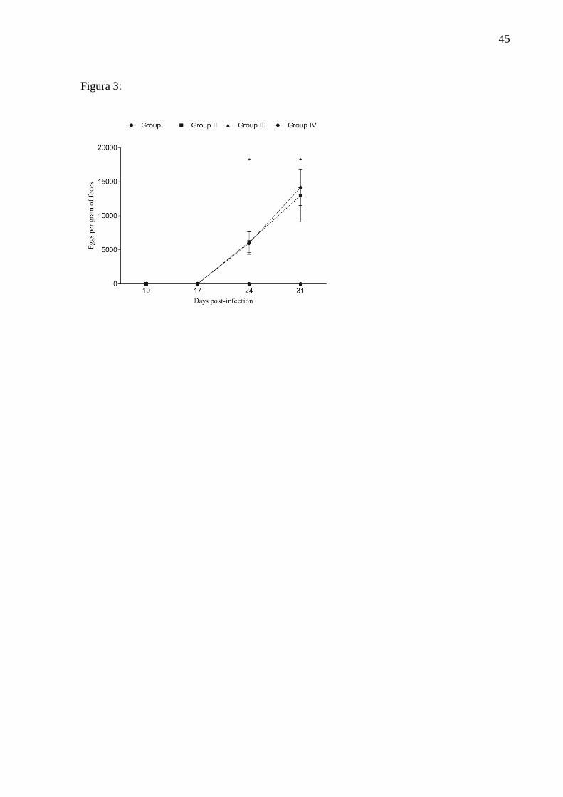
Parasitological exams
No eggs were detected per gram of feces in groups 1 and 3 during the experimental
period (Figure 3). On days 24 and 31 PI animals of groups 2 and 4 had high levels of EPG,
demonstrating the success of the infection. However, the EPG values were similar in infected
animals and in those treated for infected and non-supplemented (p>0.005).

33
Hemogram
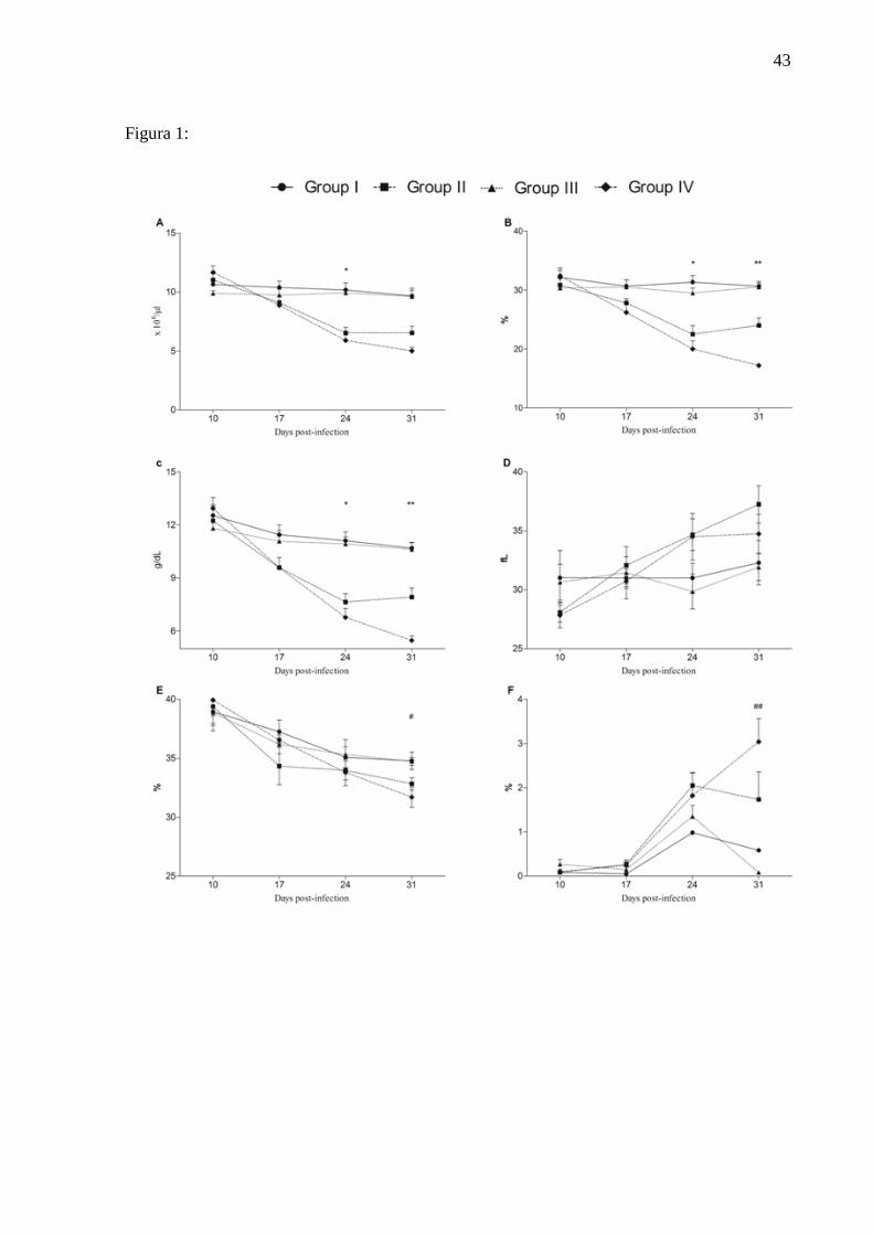
The results of red blood cell number were shown in Figure 1. Infected animals (G2
and G4) showed lower values compared with the uninfected group (G1 and G3) on days 24
and 31 PI (p<0.0001), similar results were observed for the variables hematocrit and
hemoglobin concentration (Figure 1) in this experiment. G4 on day 21 PI showed lower
numerical values of MCHC in groups 1 and 3. There was no difference in the MCV during
the trial period. On day 31PI the reticulocyte count was higher in infected groups (G2 and G4)
when compared to uninfect ones (G1 and G3) (Figure 1).
Iron metabolism
The serum iron levels were higher in G3 (uninfected and supplemented with iron) in
respect of G1 and G2 on day 17 PI (Figure 2; p<0.0006). On day 24 PI animals of G4 had
lower serum iron compared to groups 1, 2, and 3 (p<0.0008). TIBC was higher in the G1 than
in G3 at day 17 PI (p<0.0157). On day 24 PI, groups 1 and 4 had greater iron binding
capacity compared to groups 2 and 3 (p<0.0001). Serum transferrin levels were higher (day
17 PI) in uninfected animals and untreated group (G1) in relation to the lambs uninfected and
treated group (G3) (Figure 2; p<0.0015). On day 24 PI, the average values of transferrin were
higher in infected and untreated group (G4) compared to groups supplemented with iron (G2
and G3) (p<0.0001). The STI was higher in G3 compared to the other groups in day 24 PI
(Figure 2; p<0.0001). There was no difference in ferritin levels during the experimental
period (p>0.05). The medullary iron scores (0-6) were higher in animals supplemented with
iron on 17, 24, and 31 PI (G2 and G3) than those who were not supplemented (G1 and G4)
(Figures 2 and 4) (p<0.0001).
34
Discussion
In H. contortus infections, the prepatent period elapsing from infection to onset of
detectable forms of the infectious agent, i.e. days 17 and 21 PI (Jazac, 2006). In this study, a
significant increase in the EPG has been observed on days 24 and 31 PI in infected animals.
The similarity of the EPG values between infected animals and treated with iron only in
relation to those infected showed that the injectable iron did not affect the degree of infection
of lambs by H. contortus.
The animal body can meet the needs of iron under physiological conditions by
recycling senescent erythrocytes (Dunn et al., 2007). In infections by H. contortus there is a
reduction of circulating red blood cells as a result of severe blood loss, resulting in increased
iron requirements. In these cases, the bone marrow is stimulated to release large quantities of
young cells (reticulocytes) into the bloodstream. (González and Silva, 2003; Birgel et al.,
2014).
Smaller amounts of circulating hemoglobin indicate less oxygen supply to the tissues,
stimulating the bone marrow to produce new cells. However, the erythropoietic response
speed of the host, which depends on the amount of iron in the body stock, is essential to
determine the degree and duration of the anemia (Rahman and Collins, 1990), possibly
leading to the death of the sheep infected by H. contortus. The groups infected with H.
contortus had higher reticulocyte count in response to blood loss, and developed anemia
function by reducing the number of erythrocytes, hematocrit and hemoglobin concentration.
However, the anemia was more severe in the infected animals that were not supplemented
with iron. On the last trial day (31 PI), animals infected and treated group showed hematocrit
values 25% higher than those infected and untreated, suggesting improved responsiveness to
anemia. The MCHC values on day 21 PI were numerically lower in the infected and untreated
group compared to uninfected groups. It may indicate that the animals have presented severe

35
iron deficiency, since this metal is preferably diverted to other compartments for hemoglobin
synthesis, being the last to suffer from iron deficiency changes (Smith, 1997).
Researchers report thatthe peak absorption of 60% of the iron dextrose occurs within
72 hours after application of the product (Gonzáles 2000). Rocha et al. (2013) supplement
iron by oral and parenteral way (2 doses of 25 mg/kg BW of iron dextran seven days apart)
sheep with soft infection by gastrointestinal helminth, and detected higher serum iron values
and hematological variables in treated animals. They also observed that the serum iron levels
remained high for a longer period. Three doses of 20 mg/kg BW of iron dextran used
intramuscularly (at times 10, 17, and 24 PI), released higher amounts of the metal to the
treated groups. In the supplemented group and uninfected of serum iron was more available
than in the infected group and treated in day 17 PI, demonstrating the need of iron for
erythropoiesis.
According to Grotto (2010), bone marrow aspirate is the gold standard test for
measuring iron stores in relation to the determination of the serum iron, the TIBC and STI,
since these markers may also be changed depending on an inflammation (Krause and Stolc,
1979; Kaneko et al., 2008). The group that was infected and treated presented higher scores of
medullary iron when compared to the infected group (G4), demonstrating that the treatment
with iron in infected animals helps in the maintenance of the mineral stocks.
The largest consumer of primary iron is bone marrow, since for red blood cell
synthesis, it is required large quantities of this metal to meet the demand of the hemoglobin
production. In the bone marrow, erythroid precursors express transferrin receptors on their
surface, that when coupled, they suffer the endocytosis to release the iron. After this process,
the free transferrin receptor material returns to the cell surface, undergoes dissociation and
returns to circulation. In cases of non-hematopoietic cells, transferrin receptors are not
required for the absorption of iron (Huebers et al., 1990; Knovich et al., 2009).
36
Transferrin is a negative acute phase protein, which makes it an excellent tool for
differentiating an iron deficiency anemia from an anemia of a chronic disease (Punnonen et
al., 1994). When there is an increased need for iron for erythropoiesis there is also an increase
in transferrin receptors and therefore the TIBC increases, while ferritin levels decrease
(Klausner et al.,1993; Skikne, 2008), since transferrin represents the need for iron by the cell,
while ferritin indicates the metal reserves. Lower concentrations of transferrin and TIBC were
observed in the group of animals infected and untreated with iron (G3) on day 24 PI, in
relation to the untreated groups (G1 and G4) indicating that for these animals, there was no
cellular iron required.
Although greater concentrations of ferritin are present in the liver and spleen, only a
small amount is circulating in the serum, which is totally free of iron. The quantification of
ferritin determines the iron content in the stock compartment (Grotto, 2010). During the study
period, there were no differences in serum ferritin levels. However, smaller numerical values
of this protein were detected in lambs from the infected group and untreated in relation to
other groups on days 10, 17, and 31 PI, suggesting iron deficiency in animals of this group.
Under physiological conditions, 30% of transferrin is saturated with iron, being STI an
important evaluation score of this mineral metabolism. Their values are related to the amount
of circulating iron, as it is the transferrin linked to the metal (Grotto, 2010). In this study, we
observed that animals of G3 (treated with iron and uninfected), had the highest STI values on
day 24 PI, showing that the animals were with significant amounts of available iron.
Conclusion
The results indicate that injectable iron reduces the severity of the anemia caused by
infection with Haemonchus contortus in lambs, maintaining inventories of this metal in the
bone marrow, thus enabling the synthesis of hemoglobin.
37
Acknowledgement
To Prof. Dr. Alessandro F. T. do Amarante and Universidade Estadual Paulista –
UNESP by provision of 10,000 infective larvae of Haemonchus contortus. Also, to Fundação
de Amparo à Pesquisa do Rio Grande do Sul (FAPERGS) for granting my Master’s Degree
scholarship.
References
Altés, A., Lucena, M.J.P., Bruguera, M., 2014. Sistemática diagnóstica en la
hiperferritinemia. Medicina Clinica 142:412–417.
Amarante, A.F.T., Bricarello, P.A., Rocha, R.A., Gennari, S.M., 2004. Resistance of Santa
Ines, Suffolk and Ile de France sheep to naturally acquired gastrointestinal nematode
infections. Veterinary Parasitology. 120:91–106.
Andrews, N.C., 2005. Molecular control of iron metabolism. Best Practice & Research
Clinical Haematology 18:159-169.
Birgel, D.B., Muller, A.F., Neto, P.F., Storillo, V.M., Benesi, F., Birgel, E.H., 2014.
Avaliação do quadro eritrocitário e da repercussão do estado anêmico no leucograma de
caprinos com verminose gastrintestinal. Pesquisa Veterinária Brasileira 34:199-204.
Birgel, E.H. 1982. Hematologia clínica veterinária. In: Birgel E.H. & Benesi F.J. (Eds),
Patologia Clínica Veterinária. Sociedade Paulista de Medicina Veterinária, São Paulo,
SP, p.2-34.
Bizimenyera, E.S., Githiori, J.B., Eloff, J.N., Swan, G.E. 2006 In vitro activity of
Peltophorum africanum Sond. (Fabacea) extracts on the egg hatching and larval
development of the parasitic nematode Trichostrongylus colubriformis. Veterinary
Parasitology. 142, 336-343.
38
Coles, G.C., Bauerb, C., Borgsteedec, F.H.M., Geertsd, S., Kleie, T.R., Taylora, M.A.,
Wallerf, P.J. 1992. World Association for the Advancement of Veterinary Parasitology
(W.A.A.V.P.) methods for the detection of anthelmintic resistance in nematodes of
veterinary importance. Veterinary Parasitology. 44, 35-44.
Dunn, L.L., Rahmanto, S., Richardson, D.R., 2007. Iron uptake and metabolism in the new
millennium. Trends in Cell Biology 17:93-100.
Feldman, B.F., Zinkl, J.G., Jain, N.C., 2006. Schalm’s veterinary hematology. Blackwell
4:105-109.
Frazer, D.M., Anderson, G.J., 2003. The orchestration of body iron intake: how and where do
enterocytes receive their cues? Blood Cells, Molecules, and Diseases 30:288-297.
Gonzáles, A.R., 2000. Anemias: tratamiento farmacológico. Bol Farmacoter Castilla La
Mancha 82:1-8.
González, F.D., Silva, S. C., 2003. Introdução à bioquímica clínica veterinária. Porto Alegre,
UFRGS.
Gordon, H. Mc.L., Whitlock, A.V., 1939. A new technique for counting nematode eggs in
sheep feces. Journal of the Cuncil for Scientific and Industrial Research. 12:50-52.
Grotto, H.Z.W., 2008. Metabolismo do ferro: uma revisão sobre os principais mecanismos
envolvidos em sua homeostase. Revista Brasileira de Hematologia e Hemoterapia
30:390-397.
Grotto, H.Z.W., 2010. Fisiologia e metabolismo do ferro. Revista Brasileira de Hematologia e
Hemoterapia 32:8 – 17.
Huebers, H.A., Beguin, Y., Pootrakul, P., Einspahr, D., Finch, C.A., 1990. Intact transferrin
receptors in human plasma and their relation to erythropoiesis. Blood 75:102-107.
Jazac, A.M. 2006. Gastrointestinal nematodes of small ruminants: life cycle, anthelmintics,
and diagnosis. Veterinary Clinics Food Animal Practice 22:529 - 541.
39
Kaneko, J.J., Harvey, J.W., Bruss, M.L. 2008. Clinical Biochemistry of domestic animals.
Academic Press.6:178-184.
Kawano, E.L., Yamamura, M.H., Ribeiro, E.L.A., 2001. Efeitos do tratamento com anti-
helmíntico em cordeiros naturalmente infectados com helmintos gastrintestinais sobre
os parâmetros hematológicos, ganho de peso e qualidade da carcaça. Arquivos
Faculdade de Veterinária, UFRGS. 29:113- 121.
Klausner, R.D., Rouault, T.A., Harford, J.B., 1993. Regulating the fate of mRNA: the control
of cellular iron metabolism. Cell Biology 72:19-28.
Knovich, M.A., Storey, J.A., Coffman, L.G., Torti, S.V., Torti, F.M. 2009. Ferritin for the
clinician. Blood Rev. 23:95-104.
Krause, J.R., Stolc, V., 1979. Serum ferritin and bone marrow iron stores I: Correlation with
absence if iron biopsy specimens. American Journal of Clinical Pathology 72:817-820.
Krishnamurthy, P., Xie, T., Schuetz, J.D., 2007. The role of transporters in cellular heme and
porphyrin homeostasis. Pharmacology & Therapeutics 114:345-358.
Nastiti, W., Norbert, K., Stephan, I., 2004. Biology of heme in health and disease. Medical
Chemistry. 11:981-986.
Pinheiro, R.R., Gouveia, A.M.G., Alves, F.S.F., Haddad, J.P.A., 2000. Aspectos
epidemiológicos na caprinocultura cearense. Arquivo Brasileiro de Medicina
Veterinária e Zootecnia. 52:534-543.
Punnonem, K., Irjala, K., Rajamaki, A., 1994. Iron-deficiency anemia is associated with high
concentrations of transferrin receptor in serum. General Clinical Chemistry 40:774 –
776.
Rahman, W.A., Collins, G.H., 1990. Changes in live weight gain, blood constituents and
worm egg output in goats artificially infected with a sheepderived strain of Haemonchus
contortus. British Veterinary Journal. 146:543-550.
40
Ramírez, C., Rubio, C., Puebla, R.A.F., Aguilera, C., Espejo, I., Fuentes, F., 2004.
Significado clínico de los valores elevados de ferritina sérica. Medicina Clínica
122:532-534.
Rath, C.E., Finch, C.A., 1948. Sternal marrow haemosiderin: a method for the determination
of available iron stores in man. Journal of Laboratory and Clinical Medicine 6:33-81.
Roberts, F.H.S., O’Sullivan, P.J., 1950. Methods for egg counts and larval cultures for
strongyles infesting the gastrointestinal tract of cattle, Australian Journal of Agricultural
Research. 1:99–102.
Rocha, R.X., Bondan, C., Marinho, R., Lopes, S.T.A., Cecim, M., 2007. Dextran iron in
anemic lambs: effects on reticulocytosis and free radical production. Ciência Rural
37:1344-1348.
Rocha, R.X., Filappi, A., Rodrigues, A., Soares, E., Fernandes, G., Leal M.L.R., Cecim M.,
2013. Desempenho ponderal, função hepática e hemograma de borregos naturalmente
infectados por helmintos gastrointestinais suplementados com ferro oral ou parenteral.
Revista Brasileira Ciências Veterinárias. 20:189-193.
Rowe, A., Gondro, C., Emery, D., Sangster, N., 2008. Genomic analyses of Haemonchus
contortus infection in sheep: Abomasal fistulation and two Haemonchus strains do not
substantially confound host gene expression in microarrays. Veterinary Parasitology.
154, 71-81.
Sampaio, I.B.M., 1998. Estatística aplicada à experimentação animal. Fund. Ens. Pesquisa
Medicina Veterinária e Zootecnia. 1: 1-221.
Smith, J.E., 1997. lron metabolism and itsdiseases. ln: Kaneko J.J., Harvey J.W. & Bruss
M.L. Clinical biochemistry of domestic animais. New York: Academic Press. p. 223-
240.

41
Skikne, B.S., 2008. Serum transferrin receptor. American Journal of Hematology. 83:872 –
875.

42
Figure:
Figure 1: Erythrocytes number (A), hematocrit (B), hemoglobina concentration (C), MCV
(D), MCHC (E), reticulocytes number (F). Uninfected group and untreated (G1);
Infected group and treated with iron (G2); Uninfected group and treated with iron (G3)
and infected group and untreated (G4). *G1 and G3 ≠ G2 and G4; ** G4 ≠ G1, G2 and
G3; G2 ≠ G1 and G3; # G1 and G3 ≠ G4; ## G1 ≠ G4; G3 ≠ G2 and G4.
Figure 2: Seric iron (A); Transferrin (B); Ferritin (C); TSI (D); TIBC (E); Iron in the bone
marrow (F). Uninfected group and untreated (G1); Infected group and treated with iron
(G2); Uninfected group and treated with iron (G3) and infected group and untreated
(G4). *G1 and G2 ≠ G3; ** G1, G2 and G3 ≠ G4; *** G1 ≠ G3; # G1 ≠ G3; G2 and G3
≠ G4; ## G1, G2 and G4 ≠ G3; ### G1 and G4 ≠ G2 and G3; G2 ≠ G3; T G1 and G4 ≠
G2 and G3; G1 ≠ G4; TT G1 and G4 ≠ G2 and G3.
Figure 3: Stool examination (McMaster) of sheep naturally infected by Haemonchus
contortus, results presented in eggs per gram of feces (EPG). Uninfected group and
untreated (G1); Infected group and treated with iron (G2); Uninfected group and treated
with iron (G3) and infected group and untreated (G4).
Figure 4: Bone marrow of lambs infected by Haemonchus contortus, untreated with iron
dextran, with inventory absent marrow iron (A). Lamb bone marrow infected with H.
contortus treated with iron dextran, it can be visualized iron stores in the bone marrow
particles blue indicated by arrow (B).
43
Figura 1:

44
Figura 2:
45
Figura 3:

46
Figure 4:
A B

47
CONSIDERAÇÕES FINAIS
A anemia ferropriva causada pelo H. contortus é o mais grave sinal clínico e a
principal causa de morte de animais na criação de ovinos (FONSECA, 2006). A
suplementação de ferro em animais infectados com H. contortus diminui a severidade da
anemia, tornando o mineral mais disponível e em maior estoque para a produção de
hemoglobina, componente funcional do eritrócito, além de ser uma ótima ferramenta no
auxílio do tratamento parasitário.
48
REFERÊNCIAS
ALENCAR, N. X. et al. Metabolismo do ferro nos animais domésticos: revisão. Revista
educacional contínua. v. 5, p. 196-205, 2002.
ALTÉS, A. et al. Sistemática diagnóstica en la hiperferritinemia. Medicina Clinica.v. 142, p.
412–417, 2014.
AMARANTE, A. F. T. et al. Resistence of Santa Inês, Suffolk and Ile de France lambs to
naturally acquired gastrointestinal nematode infections. Veterinary Parasitology. v.120,
p.91-106, 2004.
AMARANTE, A. F. T. Nematoides gastrintestinais em ovinos. In: CAVALCANTE, A.C.R.;
VIEIRA, L.S.; CHAGAS, A.C.S; MOLENTO, M.B., ed. Doenças parasitárias de caprinos e
ovinos: epidemiologia e controle. Brasilia, DF: Embrapa Informação Tecnológica, p. 19-
61. 2009.
ANDREWS, N. C. Molecular control of iron metabolism. Best Practice & Research
Clinical Haematology. v. 18, n. 2, p. 159–169, 2005.
ANENE, B. M. et al. Gastrointestinal parasites in sheep and goats of southeastern Nigeria.
Small Ruminant Research. v. 13, p. 187-192, 1994.
BIRGEL, D. B. et al. Avaliação do quadro eritrocitário e da repercussão do estado anêmico no
leucograma de caprinos com verminose gastrintestinal. Pesquisa Veterinária Brasileira. v.
34, p.199-204, 2013.
BISHOP, S. C. et al. Genetic parameters for feacal egg count following mixed, natural
predominntly Ostertagia circumcincta infection and relationships with live weight in young
lambs. Animal Science, Hadddington. v. 63. p. 423-428. 1996.
CARPENTER, C. E.; MAHONEY, A. Contributions of heme and non-heme iron to human
nutrition. Critical Reviews in Food Science and Nutrition, v.31, p.333-367, 1992.
CLIMENI, B. C. et al. Hemoncose ovina. Revista Científica Eletrônica de Medicina
Veterinária. Garça, SP, n. 11, 2008.
FONSECA, A. H. Helmintoses gastrointestinais dos ruminantes. UFRRJ, Material didático,
2006.
GANZ, T. Molecular control of iron transport. Journal of the American Society of
Nephrology, v.18, p.394-400, 2007.
GENARI, S. M., AMARANTE, A. F. T. Helmintos de ovinos e caprinos. Biológico, v.67,
p.13-17, 2006.
GROTTO, H. Z. W. Fisiologia e metabolismo do ferro. Revista Brasileira de hematologia e
hemoterapia. v. 32, p. 8-17, 2010.
49
GROTTO, H. Z. W. Metabolismo do ferro: uma revisão sobre os principais mecanismos
envolvidos em sua homeostase. Revista Brasileira de hematologia e hemoterapia. v. 30, p.
390-397, 2008.
HOSTE, H.; CHARTIER, C. Response to challenge infection with Haemonchus
contortus and Trichostrongylus colubriformis in dairy goats. Consequences on milk
production. Veterinary Parasitology, v. 74, p. 43-54, 1998.
HERSHKO, C. Iron, infection and immune function. Proceedings of the Nutrition Society,
v. 52, p.165-174, 1993.
KEMNA, E. H. J. M. et al. Hepcidin: from discovery to differential diagnosis.
Haematologica. v. 93, p. 90-97. 2008.
KLAUSNER, R. D. et al. Regulating the fate of mRNA: the control of cellular iron
metabolism. Cell Biology, v. 72, p.19-28, 1993.
KNOVICH, M. A. et al. Ferritin for the clinician. Blood Reviews. v. 23, p. 95 – 104, 2009.
LEMOS, A. R. et al. A hepcidina como parâmetro bioquímico na avaliação da anemia por
deficiência de ferro. Revista da Associação Médica Brasileira. v. 56, p. 596-599, 2010.
LEVI, S.; ROVIDA, E. The role of iron in mitochondrial function. Biochimica et Biophysica
Acta, v. 1790, p. 629–636, 2009.
McKIE, A. T. et al., An iron-regulated ferric reductase associated with the absorption of
dietary iron. Science. v. 291, p. 1755-1759, 2001.
MENDONÇA, A. F. et al. minerais: importância de uso na dieta de ruminantes.
Agropecuária científica no semi-árido, Pato, PB. v. 07, p. 01-13, 2011.
MOLENTO, M. B. et al. Método Famacha como parâmetro clínico individual de infecção por
Haemonchus contortus em pequenos ruminantes. Ciência Rural, Santa Maria. v.34, n.4,
2004.
NICHOLLS, C. D. et al. Hypergastrinaemia of sheep infected with Haemonchus contortus.
Research in Veterinary Science. v. 45, p.124-126, 1989.
NIMEH, N., BISHOP, R. Distúrbios do metabolismo do ferro. Clínicas médicas da América
do Norte. v. 8, p. 633-648, 1980.
ONYIAH, L. C.; ARSLAN, O. Simulating the development period of a parasite of sheep on
pasture under varying temperature conditions. Journal of Thermal Biology, v. 30, p. 203–
211, 2005.
PANTOPOULOS, K. et al. Mechanisms of mammalian iron homeostasis. Biochemistry. v.
51, p. 5705–5724, 2012.
RAMÍREZ, C. et al. Significado clínico de los valores elevados de ferritina sérica. Medicina
clinica. v. 122, p. 532-534, 2004.
50
ROCHA, R. X. et al. Dextran iron in anemic lambs: effects on reticulocytosis and free radical
production, Ciência Rural, v.37, p.1344-1348, 2007.
ROCHA, R. X. et al. Eritrograma, estresse oxidativo e interação mineral em cordeiros
naturalmente infectados por parasitas gastrintestinais suplementados com diferentes formas de
ferro oral. Semina: Ciências Agrárias, v. 33, p. 723-730, 2012.
ROCHA, R. X. et al. Desempenho ponderal, função hepática e hemograma de borregos
naturalmente infectados por helmintos gastrointestinais suplementados com ferro oral ou
parenteral, Revista brasileira Ciência Veterinária. v. 20, p. 189-193, 2013.
ROMERO, J. R., BOERO, C.A. Epidemiologia de Las Gastroenterites Verminosa de los
Ovinos em Las Regiones Templadas Y Cálidas de La Argentina. Analecta Veterinaria. v.
21, p. 21 – 37, 2001.
RUAS, J. L.; BERNE, M. E. A. Parasitoses por nematóides gastrintestinais em bovinos
e ovinos. Doenças de ruminantes e equinos. v. 1. p.584-604. 2007.
SMITH, J. E. Iron metabolism and its disorders. In: KANEKO, J.J.; HARVEY, J. W.;
BRUSS, M. L. Clinical biochemistry of domestic animals. San Diego: Academic Press,
p.223-239. 1997.
STEAR, M. J., MURRAY, M. Genetic resistance to parasitic disease: particularly of
resistance in ruminants to gastrointestinal nematodes. Veterinary Parasitology. v. 54, p. 161-
176. 1994.
VAN WYK, J. A.; CABARET, J.; MICHAEL, L. M. Morphological identification of nematode
larvae of small ruminants and cattle simplified. Veterinary Parasitology, v. 119, n. 4, p. 277-306,
2004.
ZAJAC, A. M. Gastrointestinal Nematodes of Small Ruminants: Life Cycle, Anthelmintics,
and Diagnosis. Veterinary Clinics of North America: Food Animal Practice, v. 22, n. 3, p.
529-541, 2006.