universidade do estado do rio de janeiro · formada pelos corpos cavernosos, os quais se originam...
TRANSCRIPT

Universidade do Estado do Rio de Janeiro
Centro Biomédico
Faculdade de Ciências Médicas
Bruno Felix Patrício
Analise estrutural do corpo cavernoso de pacientes com
priapismo isquêmico
Rio de Janeiro
2009

Bruno Felix Patrício
Análise estrutural do corpo cavernoso de pacientes com priapismo isquêmico
Dissertação apresentada, como requisito parcial, para a obtenção do título de Mestre, ao Programa de Pós-graduação em Fisiopatologia e Ciências Cirúrgicas,
Orientador: Prof. Dr. Waldemar Silva Costa
Coorientador: Prof. Dr. Francisco José Barcelos Sampaio
Rio de Janeiro
2009

CATALOGAÇÃO NA FONTE UERJ / REDE SIRIUS / BIBLIOTECA CB/A
P314 Patrício, Bruno Felix. Análise estrutural do corpo cavernoso de pacientes com priapismo isquêmico / Bruno Felix Patrício. – 2009.
33f.
Orientador: Waldemar Silva Costa. Coorientador: Francisco José Barcelos Sampaio. Dissertação (Mestrado) – Universidade do Estado do Rio de Janeiro, Faculdade de Ciências Médicas.
1. Disfunção erétil - Teses 2. Priapismo – Teses. I. Costa, Waldemar Silva. II. Sampaio, Francisco José Barcelos. III. Título.
CDU 616.692
Autorizo, apenas para fins acadêmicos e científicos, a reprodução total ou parcial desta
dissertação, desde que citada a fonte.
__________________________________________ ____________________________ Assinatura Data

Bruno Felix Patrício
Análise estrutural do corpo cavernoso de pacientes com priapismo isquêmico
Dissertação apresentada, como requisito parcial, para a obtenção do título de Mestre, ao Programa de Pós-graduação em Fisiopatologia e Ciências Cirúrgicas.
Orientador: Prof. Dr. Waldemar Silva Costa Coorientador: Prof. Dr. Francisco José Barcelos Sampaio.
Aprovada em 25 de fevereiro de 2009.
Banca Examinadora: ____________________________________________________ Prof. Dr. Waldemar Silva Costa (Orientador) Universidade do Estado do Rio de Janeiro – UERJ _____________________________________________________ Prof.a Dra. Cristiane da Fonte Ramos Universidade do Estado do Rio de Janeiro – UERJ _____________________________________________________ Prof. Dr. Luciano Alves Favorito Universidade do Estado do Rio de Janeiro – UERJ ______________________________________________________ Prof. Dr. Marcio Antônio Babinski Universidade do Estado do Rio de Janeiro – UERJ
Rio de Janeiro
2009
AGRADECIMENTOS
Ao Dr. André Cavalcante chefe do Serviço de Urologia do Hospital Souza Aguiar pelo
material cirúrgico estudado nesse trabalho.
Ao pós-graduando da Unidade de Pesquisa Urogenital, Jorge Medeiros pela realização
da técnica de imunohistoquímica.
Ao pós-graduando da Unidade de Pesquisa Urogenital João Pedro Rosado pelos dados
relativos ás amostras.

RESUMO
PATRÍCIO, Bruno Félix. Análise estrutural do corpo cavernoso de pacientes com priapismo isquêmico. 2009. 33 f. Dissertação (Mestrado em Fisiopatologia e Ciências Cirúrgicas) – Faculdade de Ciências Médicas, Universidade do Estado do Rio de Janeiro, Rio de Janeiro, 2009.
O objetivo do presente trabalho é avaliar através de métodos quantitativos e qualitativos, as alterações nos corpos cavernosos de indivíduos com priapismo isquêmico. Foram estudados fragmentos de corpos cavernosos obtidos por biópsia de sete pacientes com, pelo menos, 48 horas de priapismo isquêmico, com idade entre 27 e 44 anos (média de 38 anos). Os pacientes foram submetidos a tratamento cirúrgico pela técnica de Al-ghorab. O material foi submetido a técnicas histoquímicas e imunohistoquímicas para a caracterização e quantificação do músculo liso e dos elementos fibrosos do tecido conjuntivo. A análise do corpo cavernoso dos pacientes com priapismo e do grupo controle, mostrou os seguintes resultados: Colágeno: controle = 34.76 ± 4.64, priapismo = 39.64 ± 2.91 (p = 0.0019); sistema elástico: controle 28.10 ± 2.85, priapismo 36.10 ± 3.06 (p = 0.0012) fibras musculares: controle = 43.37 ± 4.96, priapismo = 26.48 ± 5.00 (p < 0.0001). Ficou caracterizado aumento estatisticamente significativo dos elementos fibrosos do tecido conjuntivo e diminuição significativa nas fibras musculares lisas do corpo cavernoso dos pacientes com priapismo isquêmico. O presente estudo mostrou que o priapismo isquêmico está associado a alterações significativas nos componentes da matriz extracelular e na musculatura lisa do corpo cavernoso. Esses resultados poderiam explicar a disfunção erétil que acompanha os pacientes com priapismo isquêmico.
Palavras-chave: pênis; priapismo; disfunção erétil; músculo liso; matriz extracelular; quantificação/estereologia
ABSTRACT
The purpose of the present study was to evaluate through quantitative and qualitative methods, the changes in the corpora cavernosa of patients with ischemic priapism. We obtained samples of corpora cavernosa from 7 patients with ischemic priapism, aged between 28 and 44 years (mean = 38), who underwent a cavernosal-glandular shunt. The control tissues were fragments of corpora cavernosa obtained from autopsies of 7 age-matched men who died of causes not related to the urogenital tract. Histochemical and immunohistochemical techniques were used to assess and quantify the extra-cellular matrix and smooth muscle fibers. The volumetric density of smooth muscle, elastic fibers and collagen were determined in corpora cavernosa. The stereological analysis showed the following values of volumetric density in the structures studied. Collagen: controls = 34.76 ± 4.64, priapism = 39.64 ± 2.91 (p = 0.0019); elastic system fibers: controls 28.10 ± 2.85, priapism 36.10 ± 3.06 (p = 0.0012), smooth muscle fibers: controls = 43.37 ± 4.96, priapism = 26.48 ± 5.00 (p < 0.0001). Our results demonstrated a significant increase in the fibrous elements of the connective tissue and a significant decrease of smooth muscle fibers in the corpora cavernosa of patients with ischemic priapism, when compared to controls. As conclusion, this study showed that ischemic priapism is associated with early significant changes in the components of the extra cellular matrix and smooth muscle fibers of corpora cavernosa. This could explain the frequent occurrence of erectile dysfunction found in patients with ischemic priapism. Keywords: penis; priapism; erectile dysfunction; smooth muscle; extracellular matrix;
stereology/quantification
SUMÁRIO
1 INTRODUÇÃO 1
1.1 Morfologia 2
1.1.1 Anatomia 2
1.1.2 Histologia 4
1.1.2.1 Matriz extracelular 5
1.1.2.2 Fibras Musculares lisas 7
1.1.2 Fisiologia da Ereção 8
1.1.3 Priapismo 10
2 JUSTIFICATIVA E OBJETIVO 14
3 MATERIAL E MÉTODO 15
4 RESULTADOS 18
5 DISCUSSÃO 21
6 CONCLUSÃO 24
7 REFERÊNCIAS 25
ANEXO A – Termo de consentimento informado 32
ANEXO B – International Journal of Impotence Research 33
ANEXO C – Journal of Radiation Research 34
ANEXO D – Structural Analysis of the Corpora Cavernosa in Patients with Ischemic Priapism
35
ANEXO E – Short-term effects of radiation on the density and structural organization of smooth muscle and connective tissue in the corpus cavernosum of rats supplemented with l-glutamine
53

1
1 INTRODUÇÃO
O nome priapismo tem sua origem no deus Príapo da mitologia
grega. Príapo foi caracterizado por possuir um longo falus em ereção. Na
mitologia grega, era filho de Dionísio e Afrodite, de Baco e de Venus na
mitologia romana. É considerado o deus da fertilidade, dos vinhedos, dos
rebanhos, dos pescadores, dos jardins, simbolizando a energia fecundante.
Por ciúmes, ou por sentir-se ultrajada pela promiscuidade de Afrodite, Hera
fez Príapo nascer com alguns exageros como, por exemplo, os genitais
enormes. Conhecido em Roma com o nome de Facinum, ficou muito
popular em Pompéia, onde é retratado trajando um longo vestido que deixa
à mostra seu órgão genital (Figura 1).
Fig. 1 – Afresco da Imagem de Priapus, na Casa dos Vetti, Pompeia
O Priapismo foi descrito pela primeira vez por Tripe (1845) como
sendo uma ereção prolongada, frequentemente dolorosa, que pode ou não
ser desencadeado por um estímulo sexual. Esta doença atinge apenas os
corpos cavernosos, não afetando o corpo esponjo e a glande, o que facilita
o seu diagnóstico (Adeyoju et al. 2002, Yuan et al. 2008).
2
1.1 Morfologia do Pênis
1.1.1 Anatomia macroscópica
O pênis é um órgão que faz parte de dois sistemas: sistema urinário e
sistema reprodutor. É o órgão de cópula masculino, composto por três
corpos cilíndricos de estrutura erétil: um par de corpos cavernosos, situados
dorsalmente e um corpo esponjoso, situado ventralmente além de fáscias,
nervos e vasos, todos recobertos pela pele. A maior parte do pênis é
formada pelos corpos cavernosos, os quais se originam na sínfise púbica.
Correm lado a lado separados por um septo incompleto e estão envolvidos
intimamente pela túnica albugínea. Anterior aos corpos cavernosos está
localizado o corpo esponjoso, que abriga no seu interior a uretra e possui
duas dilatações: uma proximal, o bulbo esponjoso e outra distal, a glande
do pênis. Os três corpos de estrutura erétil estão envoltos por uma camada
de tecido fibroso, a túnica albugínea, que varia em espessura tornando-se
mais delgada ao envolver o corpo esponjoso. Além da túnica albugínea as
três estruturas apresentam duas fáscias: a fáscia Buck ou profunda e a
fáscia Dartos, também conhecida como túnica Dartos ou superficial. A
derme e a epiderme recobrem estas estruturas (Gray et al. 1988, Latarjet et
al. 1993, Brooks 2002) (Figura 2).

3
Corpos Cavernosos
Túnica Albugínea
Corpo Esponjoso
Uretra
As artérias que suprem os corpos eréteis são as artérias do bulbo do
pênis e artéria dorsal do pênis, ambas provenientes das artérias pudendas
internas (ramos das artérias ilíacas internas). Formam uma importante rede
vascular cujas artérias de maior diâmetro são as artérias profundas do pênis
(Gray et al. 1988, Latarjet et al. 1993).
Figura 2 - Corte transversal do pênis humano normal (Sampaio, F. J. B.).
A drenagem venosa do pênis é feita através da veia profunda do
pênis, que leva a maior parte do sangue dos corpos cavernosos e do corpo
esponjoso para o plexo prostático. A pele e a tela subcutânea são drenadas
pela veia dorsal superficial do pênis, tributárias da veia pudenda externa,
que drena o sangue para a veia safena magna (Gray et al. 1988, Latarjet et
al. 1993).
Os vasos linfáticos da pele e prepúcio drenam a linfa para os
linfonodos inguinais superficiais. Os da glande e corpos eréteis drenam
para linfonodos inguinais profundos, femorais, retrofemorais e pré-
viscerais (Gray et al. 1988, Latarjet et al. 1993).
A inervação do pênis é feita por fibras sensitivas, através dos ramos
do nervo pudendo, nervo dorsal do pênis e do nervo ilioinguinal. Fibras
simpáticas, parassimpáticas e fibras não adrenérgicas e não-colinérgicas,

4
estão relacionados com o controle da circulação do sangue no pênis (Gray
et al. 1988, Latarjet et al. 1993).
Fibras autônomas que inervam o tecido erétil têm sua origem em
gânglios simpáticos lombares. A ereção depende de ramos provenientes do
plexo hipogástrico inferior, do qual participam os nervos esplâncnicos
pélvicos, que são elementos do plexo parassimpático pélvico (Gray et al.
1988, Latarjet et al. 1993).
1.1.2 Histologia
O tecido erétil do pênis humano (corpo cavernoso) é composto de
fibras elásticas, fibras colágenas, músculo liso, artérias e veias, que são
envolvidas pela túnica albugínea formada principalmente de colágeno e
tecido elástico. Estas estruturas penianas possuem funções importantes no
mecanismo da ereção (Goldstein et al. 1982, Lue 2002, Wespes et al.
1991).
O interior do corpo cavernoso e esponjoso estão divididos por
trabéculas que formam numerosos espaços vasculares, denominados
sinusóides. As trabéculas são formadas por fibras do sistema elástico, fibras
colágenas e fibras musculares lisas. Estes elementos dão sustentação aos
nervos, sinusóides e artérias (Iacono et al. 1994).

5
1.1.2.1 Matriz extracelular
A matriz extracelular é a uma rede estrutural complexa formada por
macromoléculas que circundam e sustentam as células dentro do tecido
conjuntivo. É formada por diferentes moléculas que são produzidas e
exportadas pelas células modulando a estrutura, fisiologia e biomecânica
dos tecidos. A matriz extracelular é dividida em três componentes
principais:
• Componentes fibrilares - colágeno e fibras do sistema elástico;
• Componentes não fibrilares – proteoglicanos e glicoproteínas não
colagênicas (Gartner et al. 2007, Ross et al. 2008).
Elementos fibrosos da matriz extracelular (componentes fibrilares)
• Colágeno
As fibras colágenas são as proteínas fibrosas mais abundantes no
reino animal, representando cerca de 1/3 do total das proteínas encontradas
nos tecidos e resistentes a tensão (Van der Rest et al. 1991). As fibras
colágenas são constituídas por três cadeias protéicas longas organizadas em
α-hélice. Uma característica do colágeno é que 30% dos aminoácidos
correspondem à glicina. A estrutura primária das cadeias é formada por
uma sequência de três aminoácidos: Gly-X-Y (domínios colagênicos), que
se repetem por grandes extensões. Os aminoácidos colocados nas posições
x e y são frequentemente prolina e hidroxiprolina, respectivamente. Cada
6
cadeia de colágeno tem suas próprias características quanto à composição
de aminoácidos, que são utilizados para identificar o tipo de colágeno
(Kadler et al. 1996).
O tamanho e a forma das fibras colágenas variam dependendo do
tecido e do órgão, mesmo dentro da mesma espécie. Seu diâmetro pode
variar de 1 a 20 µm e apresentam um curso ondulado, mesmo quando
formam fibras densas de tecido conjuntivo como, por exemplo, nos
tendões. A forma ondulada dessas fibras aumenta a resistência dessas
fibras, resistindo melhor às tensões (Ushiki 2002).
• Fibras do sistema elástico
No tecido conjuntivo, as fibras do sistema elástico distinguem-se
facilmente das colágenas por serem mais delgadas e não apresentarem
estriação transversal. Essas fibras cedem facilmente mesmo a trações
mínimas e retomam sua forma inicial após o término das forças
deformantes (Junqueira et al. 2004).
As fibras do sistema elástico apresentam uma cor amarelada quando
observadas a fresco e são caracterizadas pelo alto grau de extensibilidade
que apresentam. São encontradas em tecidos que são constantemente
submetidos a grandes forças de estiramento (Cotta-Pereira et al. 1984).
A fibra elástica é uma estrutura complexa formada por elastina,
proteína microfibrilar, lisil-oxidase, e proteoglicanos (Mecham 1991).
Sabe-se que, durante o processo de formação de uma fibra elástica, o
componente microfibrilar é o primeiro que aparece. Em seguida a elastina é
depositada provavelmente devido a uma interação iônica entre a elastina e a

7
superfície microfibrilar como conseqüência de suas cargas opostas
(Rosenbloom et al. 1993, Ross et al. 1969).
De acordo com o grau de associação entre esses componentes as
fibras do sistema elástico são divididas em 3 tipos:
1- Fibras elásticas: constituídas em sua maior parte de elastina, em
posição central, e um número reduzido de microfibrilas em posição
periférica;
2- Fibras elaunínicas: com pouca elastina e grande número de
microfibrilas organizadas em feixes;
3- Fibras oxitalânicas: compostas somente por microfibrilas (Gartner
et al. 2007, Ross et al. 1969).
1.1.2.2 Fibras Musculares lisas
A musculatura lisa dos corpos cavernosos do pênis apresenta-se
geralmente como feixes ou folhetos de células fusiformes alongadas, com
extremidades finas e gradativamente afiladas. As células variam quanto ao
comprimento: de 20µm nas paredes de pequenos vasos sanguíneos a cerca
de 200 µm. Os núcleos das células musculares lisas estão localizados no
centro da célula e, com frequência aparecem nos cortes transversais com a
forma de saca rolhas. Essa característica é o resultado da contração celular
durante a fixação, muito útil para se distinguir as células musculares lisas
dos miofibroblastos nos cortes histológicos de rotina. Na célula não
contraída, o núcleo aparece como uma estrutura alongada com
extremidades afiladas gradativamente, situada no eixo central da célula.
Quando em corte transversal, o núcleo de uma fibra muscular lisa aparece
8
com um perfil arredondado ou circular, dependendo se a célula estiver
contraída ou relaxada (Gartner et al. 2007, Ross et al. 2008).
A célula muscular lisa é o principal componente de contração e
relaxamento do corpo cavernoso. Para se alcançar a tumescência e manter a
rigidez peniana, é preciso uma determinada percentagem de células
musculares lisas (Wespes et al. 1991). O papel do músculo liso no corpo
cavernoso é a manutenção da ereção, aumentando a pressão intra-cavernosa
durante a ereção a qual não pode ser obtida apenas por mecanismos
vasculares (Goldstein et al. 1982).
1.2 Fisiologia da ereção
A ereção é uma resposta do pênis a um estímulo sexual. O grau de
ereção é proporcional ao grau de estimulação psíquica ou física, e é
causada por impulsos parassimpáticos iniciando-se na porção sacral da
medula espinhal, continuando pelos nervos pélvicos e chegando até o pênis
(Guyton et al. 1997). Segundo Jevtich et al. (1990), o mecanismo de ereção
peniana é um processo fisiológico complexo que é baseado em fatores
neurais, vasculares, hormonais e de estruturais.
Sabe-se que a capacidade de ter e manter a ereção depende de uma
função neurológica e vascular intacta em presença de fatores hormonais e
psicológicos adequados. A ereção é fundamentalmente um fenômeno que
depende do sistema nervoso autônomo parassimpático, embora estejam
aumentando as provas histoquímicas e neurofarmacológicas do papel
exercido pelo sistema nervoso simpático. A ereção peniana é um evento
vascular controlado pela integridade das fibras de músculo liso e as
trabéculas do corpo cavernoso. A dilatação dos vasos arteriais resulta no

9
aumento do influxo arterial e provoca relaxamento do músculo liso intra-
cavernoso e distensão dos espaços cavernosos (Sattar et al. 1994, Smith
1985).
A liberação de adrenalina mantém contraída a musculatura lisa dos
corpos eréteis e faz com que o pênis permaneça no estado flácido.
Segundo Smith (1985), Andersson et al. (1995) e Saenz de Tejada et
al. (1996) são necessários três eventos vasculares para o início e
manutenção da ereção. São eles:
1. Aumento do fluxo sanguíneo arterial para os corpos cavernosos.
Após estimulação sexual, um grande fluxo arterial penetra para
expandir todo o sistema sinusoidal, até que seja estabelecido um novo
equilíbrio que ocorre com uma pressão de aproximadamente 100mmHg,
quando apenas quantidades limiares de fluxo entram e saem dos corpos
cavernosos para manter a ereção. O processo de ereção ocorre devido a
alterações nos espaços cavernosos que se dilatam e se enchem de sangue na
ereção e se tornam colapsados e com pouco sangue no estado de flacidez.
Essa pequena quantidade de sangue nos corpos cavernosos, no estado de
flacidez, visa somente atender as suas necessidades metabólicas.
2. Relaxamento do músculo liso cavernoso.
As estruturas musculares lisas presentes no corpo cavernoso são de
grande importância no mecanismo da ereção. É indispensável o
relaxamento dessa musculatura, tanto cavernosa quanto vascular, para que
ocorra um aumento do fluxo arterial, e, conseqüentemente, o
preenchimento dos espaços sinusoidais por sangue.
3. Diminuição do retorno venoso, mantendo o sangue no interior do corpo
cavernoso.
Quando os espaços sinusoidais já estão expandidos, a pressão
intracavernosa começa a aumentar, havendo uma compressão na região sob
a túnica albugínea, através da qual passam as veias emissárias resultando

10
em um grande aumento da resistência ao fluxo de saída. Isto faz com que
pare quase que totalmente a saída de sangue pelas veias, acumulando
sangue dentro dos corpos cavernosos levando a rigidez do pênis e à ereção.
Estes fenômenos são conhecidos como mecanismo de veno-oclusão normal
dos corpos cavernosos (Andersson et al. 1995, Lue 2002, Saenz de Tejada
et al. 1996).
1.3 Priapismo
Descrito pela primeira vez por Tripe (1845) o priapismo foi definido
como uma doença que se caracteriza por uma ereção prolongada, mantida
por mais de 4 horas de duração, frequentemente dolorosa, desencadeada ou
não pelo estímulo sexual (Adeyoju et al. 2002, Maan et al. 2003, Burnett et
al. 2007, Chow et al. 2008, Yuan et al. 2008). O priapismo afeta os corpos
cavernosos e é normalmente bilateral não afeta o corpo esponjoso e a
glande. Essa característica permite diagnosticar a doença. O fator
responsável pela persistência da ereção é a congestão sanguínea nos
sinusóides do corpo cavernoso (Adeyoju et al. 2002).
De acordo com fluxo sanguíneo pode ser classificado em:
priapismo isquêmico, de baixo fluxo ou venoclusivo; priapismo não-
isquêmico, de alto fluxo ou arterial.
• Priapismo isquêmico, de baixo fluxo ou venoclusivo.
O priapismo isquêmico está associado à diminuição do retorno venoso com
estase vascular o que determina a hipóxia tecidual. Neste tipo de priapismo
a drenagem venosa demora a ser feita. Geralmente é doloroso devido a
isquemia. O priapismo isquêmico está associado com um alto risco de
fibrose dos corpos cavernosos e impotência (Minardi et al. 2004).

11
• Priapismo não isquêmico, de alto fluxo ou arterial.
Priapismo não isquêmico pode ser definido como uma doença de afluência
arterial aumentada nos corpos cavernosos (Kuefer et al. 2005). É
geralmente indolor, ocorrendo aumento do fluxo arterial e o retorno venoso
normal, elevando a pressão parcial de O2. É causado na maioria das vezes
por trauma perineal ou peniano. Pode ser idiopático com algumas exceções
(Lue 2002).
O tipo isquêmico é o mais comum e responsável por 80% a 90% dos
casos. É uma emergência urológica, que pode evoluir para o quadro de
disfunção erétil, mesmo após um tratamento efetivo (Minardi et al. 2004,
Pryor et al. 2004, Van der Horst et al. 2003). Relaciona-se à congestão
sanguínea intra-cavernosa, associada a baixos níveis de O2 e altos níveis de
CO2, o que leva a um impacto significativo na oxigenação tecidual (Pryor
et al. 2004, Van der Horst et al. 2003). Por outro lado, o priapismo não
isquêmico está associado ao surgimento de uma fístula artério-venosa
(traumática ou iatrogênica) associada a níveis normais de O2, não levando a
um impacto mínimo na oxigenação tecidual (Lue et al. 1986).
De acordo com a classificação etiológica do priapismo ele pode ser
primário (idiopático) ou secundário. O priapismo secundário está associado
a uma causa conhecida, como por exemplo, abuso de álcool ou uso
excessivo de drogas, trauma perineal e doenças hematológicas (Lue 2002,
Mulhall et al. 1996).
A maior incidência dos casos de priapismo isquêmico é encontrada
nos indivíduos com idade entre 16 a 45 anos. Na maioria das vezes, trata-se
de priapismo idiopático. Em indivíduos mais jovens com doenças
hematológicas, como a anemia falciforme, o primeiro episódio de
priapismo pode ocorrer entre 15 e 20 anos sendo que em 75% dos casos
ocorre até os 20 anos (Adeyoju et al. 2002). Outros casos mais freqüentes
12
são devidos à drogas injetáveis utilizadas para o diagnóstico e tratamento
de disfunção erétil e no uso indiscriminado de drogas ilícitas (Lue 2002).
Numa revisão de literatura envolvendo 230 casos, foram encontradas
as seguintes causas mais frequentes: idiopáticas (35%); associação com
abuso de álcool ou drogas (21%); trauma perineal (12%); anemia
falciforme (11%) e doença inflamatória do trato genital (8%). De um modo
geral, o priapismo idiopático representa quase a metade dos casos, os quais,
em sua grande maioria, têm relatos de episódios prévios. As injeções
intracavernosas para o tratamento da disfunção erétil não estão incluídas
nessas casuísticas, uma vez que tornaram o priapismo muito mais
freqüente.
Os riscos de ereções prolongadas (entre quatro e seis horas) com a
prostaglandina são de 0,4% a 1,7%, e com a papaverina podem ser de até
15%, sendo mais prevalentes entre os pacientes neurogênicos ou
psicogênicos (Lue 2002).
O priapismo isquêmico tem ocorrido com freqüência crescente em
pessoas com anemia falciforme e em segundo lugar estão os casos sem
causas conhecidas(Adeyoju et al. 2002).
Segundo Adeyoju et al. 2002 a forma mais comum de priapismo na
anemia falciforme é o tipo isquêmico.
Estimativas da Organização Mundial de Saúde, mostraram que a
cada ano nascem no Brasil cerca de 2500 crianças portadoras de Doença
Falciforme. O diagnóstico precoce e o tratamento adequado representam
papel fundamental na redução da morbidade e mortalidade destas crianças.
Entre os sinais e sintomas mais freqüentes da doença falciforme está o
priapismo. O priapismo afeta cerca de 42% dos adultos portadores de
anemia falciforme (Emond et al. 1980).
Na anemia falciforme as células rígidas obstruem os pequenos
capilares e vênulas, causando isquemia tecidual, dor aguda e a lesão

13
gradual de órgãos terminais. Esse componente isquêmico geralmente
domina a evolução clinica (Braunwald et al. 2002).
Uma das condições do priapismo persistente é determinada pela
congestão prolongada de sangue nos sinusóides do corpo cavernoso,
podendo evoluir para uma disfunção erétil (Van der Horst et al. 2003).
Sharpsteen et al. (1993) analisando 461 pacientes com anemia
falciforme constatou que 87% dos pacientes apresentaram priapismo e no
caso de priapismo prolongado ou recidivado, 56% dos casos evoluíram
para disfunção erétil (Sharpsteen et al. 1993).

14
2 JUSTIFICATIVA E OBJETIVO
Os trabalhos sobre priapismo assinalam que a disfunção erétil é uma
das principais complicações apresentadas pelos pacientes com priapismo
isquêmico (Emond et al. 1980, Sharpsteen et al. 1993, Burnett et al. 2007,
Chow et al. 2008, Yuan et al. 2008, Hekal et al. 2008,). A análise da
literatura mostrou que os três componentes fundamentais dos corpos
cavernosos (colágeno, fibras do sistema elástico e músculo liso) formam o
substrato morfológico causador da disfunção erétil grave (Costa et al.
2006). No priapismo isquêmico, os estudos mostraram, apenas de forma
qualitativa, alterações na musculatura lisa e no tecido conjuntivo (Minardi
et al. 2004, Muneer et al. 2005, Spycher et al. 1986). Portanto uma análise
quantitativa dos três componentes se faz necessária na tentativa de, não
somente caracterizar de forma precisa os componentes morfológicos que
poderiam estar envolvidos no priapismo isquêmico, como também
encontrar uma resposta sobre a disfunção erétil que frequentemente
acompanha os pacientes com priapismo isquêmico.
O objetivo do presente trabalho é avaliar através de métodos
quantitativos e qualitativos as possíveis alterações no músculo liso, fibras
do sistema elástico e colágeno nos corpos cavernosos do pênis de pacientes
com priapismo isquêmico e comparar estes resultados com o grupo
controle.
15
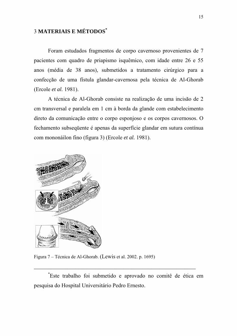
3 MATERIAIS E MÉTODOS*
Foram estudados fragmentos de corpo cavernoso provenientes de 7
pacientes com quadro de priapismo isquêmico, com idade entre 26 e 55
anos (média de 38 anos), submetidos a tratamento cirúrgico para a
confecção de uma fístula glandar-cavernosa pela técnica de Al-Ghorab
(Ercole et al. 1981).
A técnica de Al-Ghorab consiste na realização de uma incisão de 2
cm transversal e paralela em 1 cm à borda da glande com estabelecimento
direto da comunicação entre o corpo esponjoso e os corpos cavernosos. O
fechamento subseqüente é apenas da superfície glandar em sutura contínua
com mononáilon fino (figura 3) (Ercole et al. 1981).
Figura 7 – Técnica de Al-Ghorab. (Lewis et al. 2002. p. 1695)
__________________________________________________________ *Este trabalho foi submetido e aprovado no comitê de ética em
pesquisa do Hospital Universitário Pedro Ernesto.
16
Nos pacientes analisados o tempo de evolução do priapismo foi de
48 a 72 horas (média de 56 horas) sendo o primeiro episódio de priapismo
diagnosticado em todos os pacientes. Técnicas menos invasivas como
punção / irrigação cavernosas ou injeção de agentes vasoconstrictores
falharam antes da indicação cirúrgica. Os pacientes analisados não
apresentavam doenças como diabetes e hipertensão, que poderiam
contribuir para alterar a estrutura do pênis. Dois casos (28,58%) foram de
origem idiopática e cinco casos (72,42%) estavam relacionados à anemia
falciforme.
O grupo controle foi composto por fragmentos do corpo cavernoso,
obtidos de 7 cadáveres frescos, até 6 horas após a morte com idade entre 36
e 57 anos (média de 42 anos), onde a causa mortis não estava relacionada
ao sistema urogenital.
O material foi processado para inclusão em parafina. Os cortes foram
feitos com 5µm de espessura e submetidos a diferentes técnicas de
coloração. Inicialmente os cortes foram corados pela hematoxilina / eosina
(HE) verificando-se a integridade do tecido. Posteriormente foram
submetidos a outras técnicas histoquímicas e imunohistoquímicas.
• Histoquímica
Para a caracterização, quantificação e a análise qualitativa das fibras
colágenas, foram realizados cortes corados pelo Tricrômico de Masson e
pelo Vermelho de Picrosirius.
A caracterização e quantificação das fibras do sistema elástico, foram
realizadas em cortes corados pela Resorcina-fucsina de Weigert com prévia
oxidação pela oxona.
• Imunohistoquímica
As fibras musculares lisas foram evidenciadas e quantificadas pela
imunohistoquímica utilizando o anticorpo anti α Actina de músculo liso,
Zymed Laboratories. Para o anticorpo primário foi utilizado um controle
17
negativo e controle positivo, usando fragmentos de tecido que apresentam
os antígenos pesquisados. A revelação foi feita com solução de 3,3,
diamino-benzidina tetrahidrocloridro (DAB) a 0,1% em H2O2, lavados em
água destilada, desidratado em uma serie crescente de etanol diafanizados
em xilol e montados com Ethelan.
Foram obtidos 10 cortes de cada uma das amostras e contados 10
campos por corte totalizando 100 campos por indivíduo para cada uma das
técnicas utilizadas.
Todos os elementos estudados foram quantificados através de
métodos estereológicos que determinam parâmetros tridimensionais a partir
de cortes bidimensionais (Mandarim-de-Lacerda 2003).
Os resultados foram obtidos pelo método de contagem de pontos
utilizando-se o sistema teste M-42 sobre um monitor de um computador.
De acordo com os princípios estereológicos, a distribuição por área é
proporcional à distribuição por volume, quando a região considerada é
homogênea (Mandarim-de-Lacerda 2003, Costa et al. 2008).
O teste t de Student não pareado foi utilizado para determinar se há
diferença significativa entre as médias dos dois grupos analisados.
18
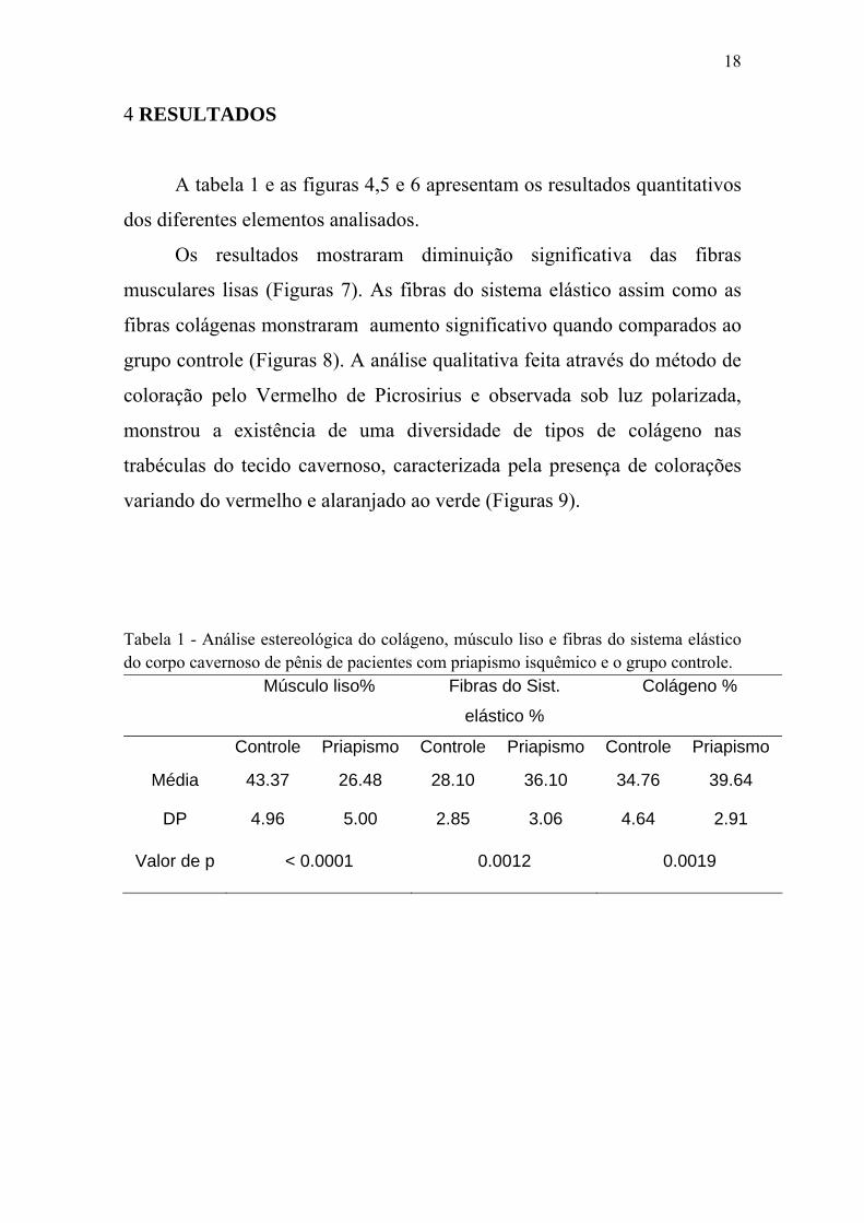
4 RESULTADOS
A tabela 1 e as figuras 4,5 e 6 apresentam os resultados quantitativos
dos diferentes elementos analisados.
Os resultados mostraram diminuição significativa das fibras
musculares lisas (Figuras 7). As fibras do sistema elástico assim como as
fibras colágenas monstraram aumento significativo quando comparados ao
grupo controle (Figuras 8). A análise qualitativa feita através do método de
coloração pelo Vermelho de Picrosirius e observada sob luz polarizada,
monstrou a existência de uma diversidade de tipos de colágeno nas
trabéculas do tecido cavernoso, caracterizada pela presença de colorações
variando do vermelho e alaranjado ao verde (Figuras 9).
Tabela 1 - Análise estereológica do colágeno, músculo liso e fibras do sistema elástico do corpo cavernoso de pênis de pacientes com priapismo isquêmico e o grupo controle.
Músculo liso% Fibras do Sist.
elástico %
Colágeno %
Controle Priapismo Controle Priapismo Controle Priapismo
Média 43.37 26.48 28.10 36.10 34.76 39.64
DP 4.96 5.00 2.85 3.06 4.64 2.91
Valor de p < 0.0001 0.0012 0.0019

19
Controle Priapismo0
10
20
30
40
50
Groupos
%
*
Figura 4 – Análise estereológica das fibras musculares lisas no corpo cavernoso do pênis. P< 0.0001
Controle Priapismo0
10
20
30
40
Groupos
%
*
Controle Priapismo
010
2030
4050
Groupos
%
Figura 5 – Análise estereológica das fibras do sistema elástico no corpo cavernoso do pênis. P= 0.0012
*
Figura 6 – Análise estereológica das fibras colágenasno corpo cavernoso do pênis. P= 0.0019

20
A B
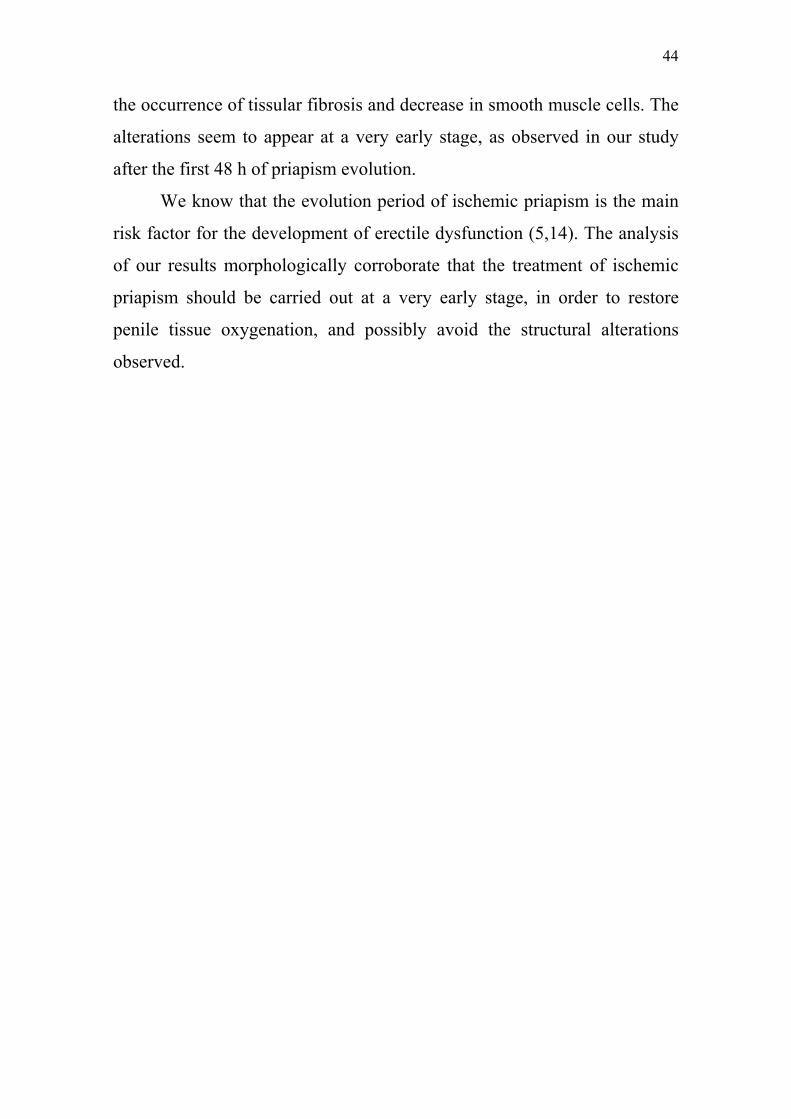
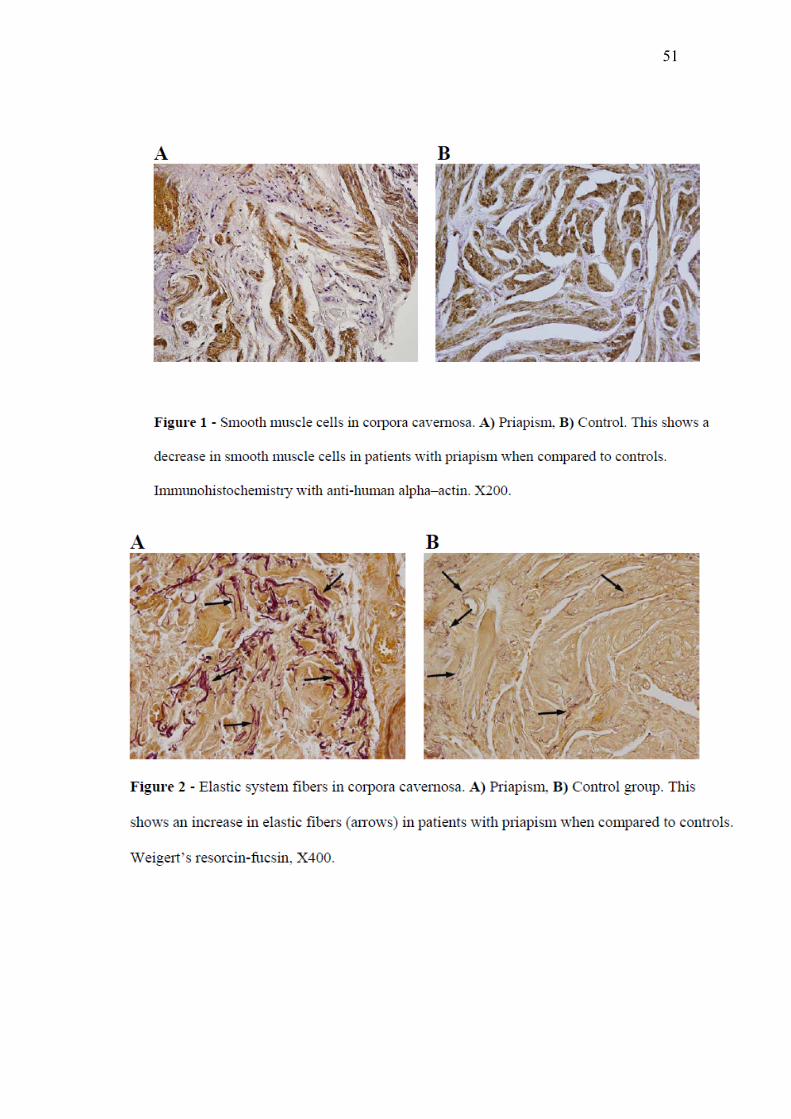
Figuras 7 - Fibras musculares lisas no corpo cavernoso. A) Grupo controle, B) Grupo Priapismo. Há diminuição das fibras musculares lisas em pacientes com priapismo, pode-se observar uma diminuição das fibras musculares lisas no grupo priapismo, quando comparados ao grupo controle. Imunohistoquímica com anti-α-actina de músculo liso. 200X A B
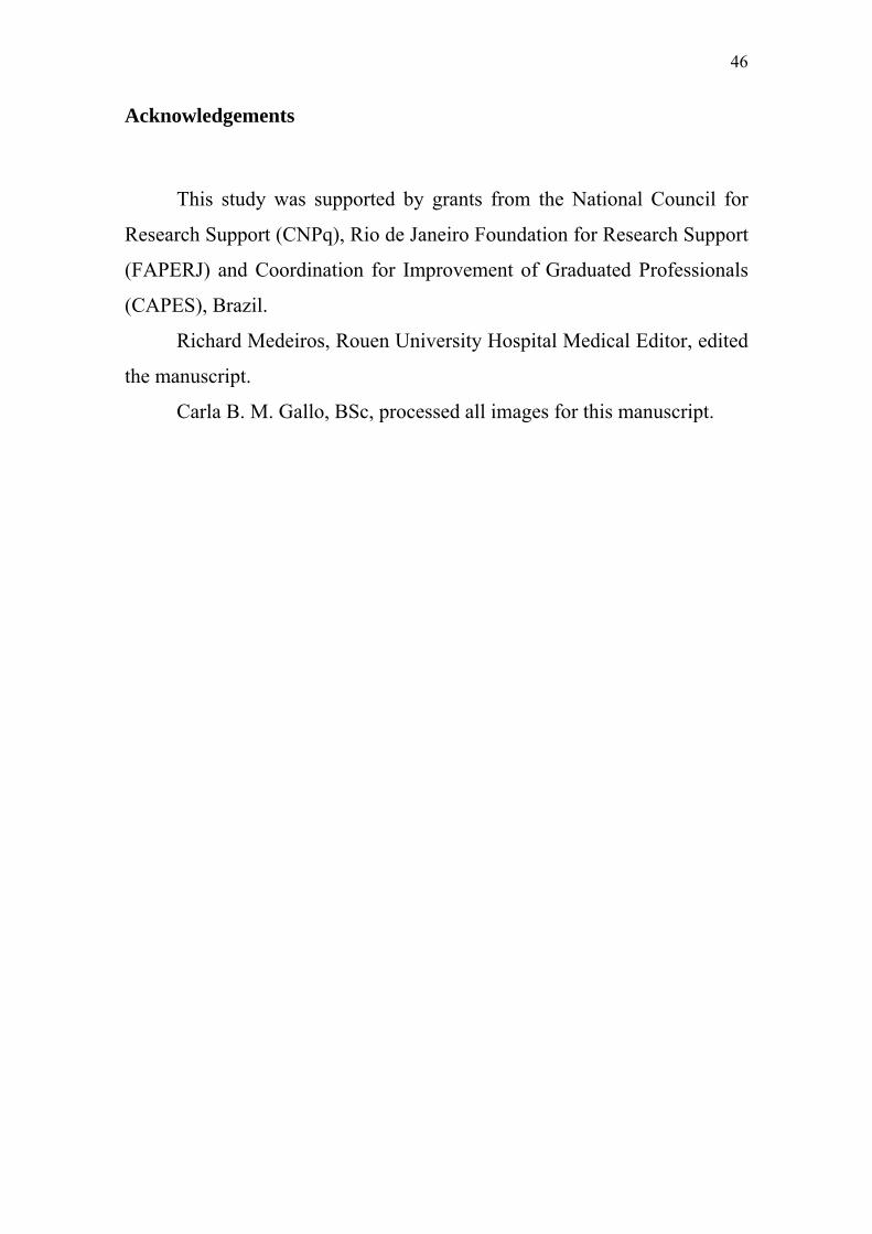
Figuras 8 - Fibras do sistema elástico no corpo cavernoso. A) Grupo controle, B) Grupo priapismo. Existe aumento de fibras do sistema elástico (setas) em pacientes com priapismo, quando comparados ao controle. Resorcina-fucsina de Weigert. 400X A B
Figuras 9 – Fibras colágenas no corpo cavernoso. A) Grupo controle, B) Grupo priapismo. Há predomínio da cor vermelha, no grupo controle, enquanto que no grupo priapismo há predomínio da cor verde. Vermelho de Picro Sirius observado sob luz polarizada. 200X.

21
5 DISCUSSÃO
No processo de priapismo observamos uma ereção mantida com
ausência de detumescência peniana o que corresponde a um desarranjo da
fisiologia eretora (Muneer et al. 2005, Spycher et al. 1986).
É possível supor que a disfunção erétil, frequentemente encontrada
em pacientes com priapismo persistente por mais de 48 horas, possa
encontrar um substrato morfológico em modificações conspícuas nos
diferentes elementos da matriz extracelular dos corpos cavernosos do pênis.
Uma análise quantitativa desses elementos pode trazer uma contribuição
para se entender melhor os efeitos do priapismo sobre os corpos cavernosos
do pênis e disfunção erétil.
O mecanismo necessário para se alcançar o estado flácido, e sua
permanência, envolve a contração da musculatura lisa. Por isso ela é a
estrutura de maior importância no processo de ereção e detumescência
(Van der Horst et al. 2003).
Muneer et al. (2005), assinalam que durante o priapismo, o tecido
cavernoso sofre hipóxia, acidose e glicopenia. Segue-se uma infiltração de
células inflamatórias e proliferação de fibroblastos, resultando em necrose
do músculo liso dos corpos cavernosos e o aparecimento de fibrose em
diferentes graus (Hekal et al. 2008, Muneer et al. 2005, Ralph et al. 2008).
Esta substituição de tecido erétil normal por um tecido fibrótico deve estar
diretamente envolvida na gênese do distúrbio erétil tardio que
frequentemente acompanha os pacientes com priapismo (Muneer et al.
2005, Ralph et al. 2008). Segundo Spycher et al. (1986) e Muneer et al.
(2005) é possível observar, após 24h de evolução do priapismo isquêmico,
uma necrose focal da musculatura lisa e a sua transformação em células
fibroblasto-like (Hekal et al. 2008, Ralph et al. 2008). Essas observações

22
poderiam justificar a redução marcante do componente muscular (16,89%)
quando analisado num tempo superior a 48 horas de evolução como foi o
caso no presente trabalho.
As fibras do sistema elástico foram também significativamente
afetadas apresentando um aumento de 8%. É possível supor que a isquemia
que gera a transformação de células musculares lisas em células fibrobasto-
like também seja a causa do aumento de fibras do sistema elástico no corpo
cavernoso, o que poderia contribuir para promover a detumescência
peniana como ocorre na túnica albugínea, onde as fibras do sistema elástico
são co-responsáveis por promover uma rápida e adequada recuperação do
estado de detumescência (Hsu et al. 2006). O comportamento das fibras do
sistema elástico parece ser sui generis manifestando-se de forma diferente
dependendo do estímulo recebido. Costa et al. (2006), analisando o sistema
elástico num estudo sobre disfunção erétil grave, no qual o priapismo não
foi incluído na amostra, mostrou que estas fibras diminuem
significativamente. Isto nos leva a supor que o mecanismo de reação do
tecido erétil, com relação às fibras do sistema elástico, é peculiar no
priapismo isquêmico.
O aumento do colágeno foi estatisticamente significativo nos
pacientes analisados, porém proporcionalmente menor, quando comparado
aos outros elementos estudados: sistema elástico e músculo liso. O
colágeno apresentou um aumento de 4,8%, caracterizando o aparecimento
de fibrose. Esta transformação parece ocorrer em um tempo relativamente
curto. Em nosso grupo de pacientes o tempo de evolução variou de 48 a 72
horas sendo este o primeiro episódio de priapismo. Costa et al. (2006) e
Luangkhot et al. (1992), assinalaram o fato de não ocorrer um aumento
significativo no conteúdo de colágeno no tecido cavernoso de pacientes
com disfunção erétil. No entanto, estes estudos não enfocaram o priapismo
isquêmico como fator etiológico da disfunção sexual erétil. A isquemia

23
tecidual é conhecida como fator estimulador da produção de colágeno. A
transformação de células da musculatura lisa em células fibrobasto-like
como foi descrito anteriormente, também poderia ter um papel relevante
neste processo justificando o aumento do colágeno nos pacientes analisados
(Spycher et al. 1986).
A observação qualitativa, através da microscopia de polarização, dos
cortes corados pelo Vermelho de Picrosirius, mostra diferentes cores sobre
as trabéculas do corpo cavernoso. A predominância da cor verde observada
nos cortes dos pacientes com priapismo nos permite supor, que existe
maior quantidade de um tipo de colágeno, possivelmente o colágeno tipo
III. O grupo controle se caracterizou por uma cor que varia do vermelho a
vermelho alaranjado e que seria possivelmente o colágeno tipo I. Parece,
portanto, ocorrer um turnover importante, com um processo ativo de
formação de colágeno no grupo de pacientes com priapismo.
Os resultados apresentados no presente trabalho mostram que o
priapismo isquêmico está associado a significativas alterações nos
componentes dos corpos cavernosos, principalmente pela diminuição da
musculatura lisa. Este processo parece iniciar-se precocemente, tendo sido
observado no presente trabalho após as primeiras 48 horas de evolução.
Sabemos que o tempo de evolução do priapismo isquêmico é o
principal fator de risco para o desenvolvimento de disfunção erétil (El-
Bahnasawy et al. 2002, Minardi et al. 2004). Portanto a analise e os
resultados aqui apresentados corroboram morfologicamente para que o
tratamento do priapismo isquêmico deva ser baseado em uma abordagem
agressiva e precoce, visando a detumescência peniana e o restabelecimento
de uma adequada oxigenação tecidual.

24
6 CONCLUSÃO
O presente estudo mostrou que o priapismo isquêmico está associado
à diminuição da musculatura lisa, aumento das fibras do sistema elástico
assim como alterações no colágeno dos corpos cavernosos de pacientes
com priapismo isquêmico. Estas observações poderiam explicar a
disfunção erétil que ocorre frequentemente em pacientes com este tipo de
priapismo.

25
7 REFERÊNCIAS • Adeyoju AB, Olujohungbe AB, Morris J, Yardumian A, Bareford D,
Akenova A, Cinkotai K, O'Reilly PH. Priapism in sickle-cell disease;
incidence, risk factors and complications - an international multicentre
study. BJU Int. 2002; 90: 898-902.
• Andersson KE, Wagner G. Physiology of penile erection. Physiol Rev.
1995; 75: 191-236.
• Braunwald E, Fauci AS, Kasper DL, Hauser SL, Longo DL, Jameson JL.
Harrison medicina interna. 15.ed. Rio de Janeiro: McGraw-Hill
Interamericana do Brasil. 2002; vol. 1, p. 711.
• Brooks JD. Anatomy of the lower urinary tract and male genitalia. In:
Walsh PC, Retik AB, Vaughan ED, Wein.AJ. Campbell's Urology. 8.ed.
Philadelphia, Pennsylvania: Elsevier Science. 2002. vol. 1, pp. 72-75
• Burnett AL, Bivalacqua TJ. Priapism: current principles and practice.
Urol Clin North Am 2007; 34: 631-642.
• Chow K, Payne S. The pharmacological management of intermittent
priapismic states.BJU Int. 2008; 102: 1515-1521.
• Costa WS, Carrerete FB, Horta WG, Sampaio FJ. Comparative analysis of
the penis corpora cavernosa in controls and patients with erectile
dysfunction. BJU Int. 2006; 97: 567-9.

26
• Costa WS, Rebello SB, Cardoso LE, Cavalcanti AG, Sampaio FJ.
Stereological and biochemical analysis of muscular and connective tissue
components in the penile corpus cavernosum adjacent to the fibrous plaque
of Peyronie's disease. BJU Int. 2008; 103: 212 – 216.
• Cotta-Pereira G, Del-Caro LM, Montes GS. Distribution of elastic system
fibers in hyaline and fibrous cartilages of the rat. Acta Anat (Basel). 1984;
119: 80-5.
• El-Bahnasawy MS, Dawood A, Farouk A. Low-flow priapism: risk
factors for erectile dysfunction. BJU Int. 2002; 89: 285-90.
• Emond AM, Holman R, Hayes RJ, Serjeant GR. Priapism and impotence
in homozygous sickle cell disease. Arch Intern Med. 1980; 140: 1434-7.
• Ercole CJ, Pontes JE, Pierce JM, Jr. Changing surgical concepts in the
treatment of priapism. J Urol. 1981; 125: 210-1.
• Gartner LP, Hiatt JL. Tratado de Histologia 3. ed. Rio de Janeiro:
Elsevier. 2007. p. 131-154.
• Goldstein AM, Meehan JP, Zakhary R, Buckley PA, Rogers FA. New
observations on microarchitecture of corpora cavernosa in man and
possible relationship to mechanism of erection. Urology. 1982; 20: 259-66.
• Gray H, Gross GF. Anatomia. 29 ed. Rio de Janeiro: Guanabara Koogan.
1988. p. 1072-178.

27
• Guyton, Hall. Tratado de Fisiologia Médica. 9 ed. Rio de Janeiro:
Guanabara Koogan. 1997. p. 862.
• Hekal IA, Meuleman EJ. Idiopathic low-flow priapism in prepuberty: a
case report and a review of literature. Adv Urol. 2008; 2008: 549861.
• Hsu L, Diwan B, Ward JM, Noguchi CT. Pathology of "Berkeley" sickle-
cell mice includes gallstones and priapism. Blood. 2006; 107: 3414-5.
• Iacono F, Barra S, de Rosa G, Boscaino A, Lotti T. Microstructural
disorders of tunica albuginea in patients affected by impotence. Eur Urol.
1994; 26: 233-9.
• Jevtich MJ, Khawand NY, Vidic B. Clinical significance of ultrastructural
findings in the corpora cavernosa of normal and impotent men. J Urol.
1990; 143: 289-93.
• Junqueira L, Carneiro J. Histologia Básica. 10. ed. Rio de Janeiro:
Guanabara Koogan. 2004. pp. 124.
• Kadler KE, Holmes DF, Trotter JA, Chapman JA. Collagen fibril
formation. Biochem J. 1996; 316 (Pt 1):1-11.
• Kuefer R, Bartsch G, Jr., Herkommer K, Kramer SC, Kleinschmidt K,
Volkmer BG. Changing diagnostic and therapeutic concepts in high-flow
priapism. Int J Impot Res. 2005; 17: 109-13.
• Latarjet M, Ruiz-Liard A. Anatomia Humana. 2. ed. São Paulo:
Panamericana. 1993. vol. 2, p. 1703-1709
28
• Lewis RW, Jordan G. Surgery for erectile dysfunction. In: Walsh PC,
Retik AB, Vaughan ED, Wein.AJ. Campell’s Urology. 8. ed. Philadelfia:
.
ollagen alterations in the corpus cavernosum of men with sexual
o EA. Priapism: a refined
pproach to diagnosis and treatment. J Urol. 1986; 136: 104-8.
impotence.
: Walsh PC, Retik AB, Vaughan ED, Wein.AJ. Campell’s Urology. 8. ed.
of the medical
anagement. Expert Opin Pharmacother. 2003; 4: 2271-7.
al research. An
cad Bras Cienc. 2003; 75: 469-86.
on mammalian cells. FASEB J. 1991;
: 2538-46.
, Milanese G, Galosi AB, Donatelli G, Muzzonigro G.
ersistent priapism and histological modifications of the erectile tissue.
Two case reports. Arch Ital Urol Androl. 2004; 76: 97-9.
WB Saunders. 2002a; vol. 2, pp. 1695-6.
• Luangkhot R, Rutchik S, Agarwal V, Puglia K, Bhargava G, Melman A
C
dysfunction. J Urol. 1992; 148 (2 Pt 1): 467-71.
• Lue TF, Hellstrom WJ, McAninch JW, Tanagh
a
• Lue TF. Physiology of penis erection and pathophysiology of
In
Philadelfia: WB Saunders. 2002a; vol. 2, pp. 1591-1613.
• Maan Z, Arya M, Patel HR. Priapism - a review
m
• Mandarim-de-Lacerda CA. Stereological tools in biomedic
A
• Mecham RP. Receptors for laminin
5
• Minardi D
P

29
• Mulhall JP, Honig SC. Priapism: etiology and management. Acad Emerg
Med. 1996; 3: 810-6.
• Muneer A, Cellek S, Dogan A, Kell PD, Ralph DJ, Minhas S.
itro model. Int J Impot Res. 2005; 17: 10-8.
Ralph DJ, Garaffa G, Muneer A, Freeman A, Rees R, Christopher AN, et
.
Ross MH, Pawlina W. Histologia: texto e atlas. Em correlação com a
Investigation of cavernosal smooth muscle dysfunction in low flow
priapism using an in v
• Pryor J, Akkus E, Alter G, Jordan G, Lebret T, Levine L, et al. Priapism. J
Sex Med. 2004; 1: 116-20.
•
al. The Immediate Insertion of a Penile Prosthesis for Acute Ischaemic
Priapism. Eur Urol. 2008; 1
• Rosenbloom J, Abrams WR, Mecham R. Extracellular matrix 4: the
elastic fiber. FASEB J. 1993; 7: 1208-18.
•
biologia celular e molecular. 5.ed. Rio de Janeiro: Guanabara Koogan.
2008; pp. 147-181.
• Ross R, Bornstein P. The elastic fiber. I. The separation and partial
characterization of its macromolecular components. J Cell Biol. 1969; 40:
366-81.
• Saenz de Tejada YGI. [Physiology of penile erection]. Arch Esp Urol.
1996; 49: 202-5.

30
• Sattar AA, Wespes E, Schulman CC. Computerized measurement of
Sharpsteen JR, Jr., Powars D, Johnson C, Rogers ZR, Williams WD,
Spycher MA, Hauri D. The ultrastructure of the erectile tissue in priapism.
Ushiki T. Collagen fibers, reticular fibers and elastic fibers. A
-26.
Wespes E, Goes PM, Schiffmann S, Depierreux M, Vanderhaeghen JJ,
patients. J Urol. 1991; 146: 1015-7.
penile elastic fibres in potent and impotent men. Eur Urol. 1994; 25: 142-4.
•
Posch RJ. Multisystem damage associated with tricorporal priapism in
sickle cell disease. Am J Med. 1993; 94: 289-95.
• Smith RD. Urologia geral. 10 ed. Rio de Janeiro: Guanabara Koogan.
1985.
•
J Urol. 1986; 135: 142-7.
•
comprehensive understanding from a morphological viewpoint. Arch
Histol Cytol. 2002; 65: 109
• Van der Horst C, Stuebinger H, Seif C, Melchior D, Martinez-Portillo FJ,
Juenemann KP. Priapism - etiology, pathophysiology and management. Int
Braz J Urol. 2003; 29: 391-400.
• Van der Rest M, Garrone R. Collagen family of proteins. FASEB J. 1991;
5: 2814-23.
•
Schulman CC. Computerized analysis of smooth muscle fibers in potent
and impotent

31
• Yuan J, Desouza R, Westney OL, Wang R. Insights of priapism
mechanism and rationale treatment for recurrent priapism.Asian J Androl.
2008; 10: 88-101.

32
ANEXO A – Termo de consentimento informado
Universidade do Estado do Rio de Janeiro Centro Biomédico
Pós Graduação em Fisiocirurgia e Ciências Cirúrgica Hospital Municipal Souza Aguiar - Serviço de Urologia
Termo de Consentimento Informado
TÍTULO DO PROJETO: ESTUDO ESTRUTURAL DO CORPO CAVERNOSO DO PÊNIS DE INDIVÍDUOS COM PRIAPISMO DO TIPO VENOCLUSIVO.
O Sr. vai ser submetido a uma cirurgia para o tratamento de priapismo. Normalmente, durante esta cirurgia, retira-se um fragmento que depois é enviado para exame histopatológico, procedimento de rotina para o serviço de Anatomia Patológica, para inclusão em estudos de rotina e arquivamento.
Gostaríamos de solicitar ao senhor que tenha a gentileza de nos autorizar a estudar um fragmento do material doente que será extraído durante a cirurgia a qual o Sr. será submetido.
Esta extração será feita durante a cirurgia e não implicará em aumento do tempo de cirurgia nem alterará os resultados operatórios.
Será respeitado o segredo médico e sua identidade não será revelada. O grupo de médicos e professores que estão fazendo parte deste trabalho estarão
à sua disposição no Serviço de Urologia deste hospital e na Pós Graduação em Fisiopatologia e Ciências Cirúrgicas da Universidade do Estado do Rio de Janeiro- Departamento de Anatomia do Hospital Universitário Pedro Ernesto, à Av 28 de Setembro, número 77, fundos. Telefone 2587-6499. Sendo o pesquisador responsável o Dr Bruno Félix Patróicio estando sempre à disposição para maiores esclarecimentos.(deixe aqui o seu cel- apenas para constar)
Os dados e resultados finais serão utilizados somente para esta pesquisa. Eu, _____________________________________________________ (nome completo), portador da identidade ________________________, declaro estar ciente do que foi exposto acima e autorizo a utilização, para fins científicos, do fragmento do pênis que será extraída durante a cirurgia a qual serei submetido.
Rio de Janeiro, de de 2008.
_______________________________________ Paciente

33
ANEXO B

34
ANEXO III
35
ANEXO D - Structural Analysis of the Corpora Cavernosa in Patients
with Ischemic Priapism
Running title: Corpora Cavernosa in Ischemic Priapism
Waldemar S. Costa, Bruno Felix Patricio, Andre G. Cavalcanti, Jorge
Medeiros Jr., Francisco J. B. Sampaio
Urogenital Research Unit, State University of Rio de Janeiro, UERJ, Rio de
Janeiro, RJ, Brazil
Conflict of Interest: None
36
Abstract The purpose of the present study was to evaluate through quantitative and qualitative methods, the changes in the corpora cavernosa of patients with ischemic priapism. We obtained samples of corpora cavernosa from 7 patients with ischemic priapism, aged between 28 and 44 years (mean = 38), who underwent a cavernosal-glandular shunt. The control tissues were fragments of corpora cavernosa obtained from autopsies of 7 age-matched men who died of causes not related to the urogenital tract. Histochemical and immunohistochemical techniques were used to assess and quantify the extra-cellular matrix and smooth muscle fibers. The volumetric density of smooth muscle, elastic fibers and collagen were determined in corpora cavernosa. The stereological analysis showed the following values of volumetric density in the structures studied. Collagen: controls = 34.76 ± 4.64, priapism = 39.64 ± 2.91 (p = 0.0019); elastic system fibers: controls 28.10 ± 2.85, priapism 36.10 ± 3.06 (p = 0.0012), smooth muscle fibers: controls = 43.37 ± 4.96, priapism = 26.48 ± 5.00 (p < 0.0001). Our results demonstrated a significant increase in the fibrous elements of the connective tissue and a significant decrease of smooth muscle fibers in the corpora cavernosa of patients with ischemic priapism, when compared to controls. As conclusion, this study showed that ischemic priapism is associated with early significant changes in the components of the extra cellular matrix and smooth muscle fibers of corpora cavernosa. This could explain the frequent occurrence of erectile dysfunction found in patients with ischemic priapism. Keywords: penis; priapism; erectile dysfunction; smooth muscle; extracellular matrix; stereology/quantification
37
Introduction
Priapism is defined as a pathological condition of prolonged and
persistent painful erection of the penis not associated with sexual
stimulation and desire (1-3). Persistent erection is caused by blood
congestion in the corpora cavernosa sinusoids, which usually are bilaterally
involved, with no involvement of the corpus spongiosum (3). Priapism is a
urologic emergency and may evolve towards erectile dysfunction, even
after effective treatment (1-7).
Priapism is classified as ischemic, low-flow or veno-occlusive and as
high flow, non-ischemic or arterial. The first type is the most common
accounting for 80% to 90% of cases. Ischemic priapism is related to
intracavernous congestion by high-viscosity blood, due to low levels of O2
and high levels of CO2, which leads to a significant impact on tissue
oxygenation (4,6). In contrast, high-flow priapism is usually associated
with arteriovenous fistula (traumatic or iatrogenic) and with normal levels
of O2, and, therefore, does not lead to an important effect on tissue
oxygenation (8).
Ischemic priapism is often idiopathic, being the secondary causes
often associated with different etiologies. In a study involving a review of
230 cases of priapism, 35% corresponded to idiopathic cases distributed as
follows; 21% associated with alcohol or drug abuse, 12% with perineal
trauma, 11% with sickle cell anemia and 8% with inflammatory disease of
the genital tract (9). The use of intracavernous vasoactive drugs is also
linked to the risk of prolonged erections. It is estimated that the risk is 0.4%
to 1.7% for the use of prostaglandins and may reach up to 15% with the use
of papaverine (10). In prolonged and recurrent cases, the rate of erectile
dysfunction can be as high as 56% (11).
38
The majority of cases of ischemic priapism occur in patients aged
between 16 and 45 years old, being idiopathic in most cases. In young
patients, with hematologic diseases as sickle cell anemia, the first episode
of priapism can occur between 15 and 20 years old, and in 75% of cases,
the first episode occurs before 20 years old (12). Priapism is among the
most frequent manifestation of sickle cell disease, affecting between 42%
and 87% of the patients (11-13). Several studies have shown that erectile
dysfunction is the most serious complication of ischemic (low-flow)
priapism (1-4,14,15). A literature review shows that most studies regarding
the alterations of low-flow priapism focused only on the smooth muscle as
the pathological substrate for erectile dysfunction and did not in fact,
examine other important elements that could also contribute to erectile
dysfunction.
In most cases, erectile dysfunction, which occurs frequently in
patients with priapism lasting for more than 48 hours, can have a
morphological substrate with conspicuous changes in the different elements
of penile corpora cavernosa. A detailed study concerning these elements
could contribute to a better understanding of the effects of priapism in the
corpora cavernosa. The aim of this work was to evaluate, based on
quantitative and qualitative methods, the structural changes in the penile
corpora cavernosa of patients with ischemic priapism and compare these
results with those found in the penile samples of age-matched controls.

39
Patients and Methods
The present work received institutional review committee approval and was
carried out in accordance with the ethical standards of the responsible
institutional committee on human experimentation.
Samples from 7 patients with ischemic priapism aged between 28
and 44 yearsold (mean = 38), underwent surgery to obtain a cavernosal-
glandular shunt (Al-Ghorab technique) (16).
The priapism in the analyzed patients lasted from 48 to 72 hours
(mean = 56), and it was the first episode in all cases. Minimally invasive
techniques like puncture and cavernosal irrigation or injection of
vasoconstrictor agents have been used without success before surgery.
None of the patients presented previous history of diabetes or
hypertension, which could have led to alterations in the normal penile
structure. Two cases (28.58%) were idiopathic and 5 cases (72.42%) were
related to sickle cell disease.
The control group was composed of samples from corpora cavernosa
obtained from autopsies, until 6 hours after death, of 7 age-matched
subjects, who died of causes not related to the urogenital tract. The tissue
samples of corpora cavernosa were immersed in PBS formalin fixative (pH
7.2) for 24 hours and then routinely processed for paraffin embedding.
Sections of 5-μm thick were obtained and all samples were initially stained
with Hematoxylin-Eosin and analyzed by a pathologist to confirm the
tissue integrity. Then, the samples were processed using histochemical and
immunohistochemical techniques, as follows: Masson’s trichromic and
Picrosirius red to evidence and quantify the collagen, and Weigert resorcin-
fuscin with previous oxidation to demonstrate the elastic system fibers.
Smooth muscle fibers were shown by Masson’s trichromic and

40
immunohistochemical analysis. We used the anti-alpha actin antibody
(Zimed Laboratories). All elements evaluated were quantified by
stereological methods previous described (17). Briefly, for each individual
and each histological staining technique, 10 sections of corpus cavernous
were obtained, and for each section, 10 fields were analyzed. All images
were photographed with a digital camera directly coupled to a microscope
at X200. The volumetric density (Vv) of histological structures was then
evaluated by superimposing an M-42 test system on the digital images
following techniques that have been described in detail elsewhere (17). The
non-paired Student’s t-test was used to determine the differences between
the groups.

41
Results
Table-1 shows the results of the different elements analyzed.
The stereological quantification in the corpora cavernosa showed that
the Vv of smooth muscle fibers was significantly decreased in patients with
priapism (Figure-1). In contrast, there was a significant parallel increase in
the Vv of elastic system fibers in priapism patients (Figure-2). Also, a
significant increase in Vv of collagen was found in patients with priapism
when compared with controls. The qualitative analysis of Picrosirius red
stain, observed under polarization, revealed the existence of diverse
collagen types in the corpora cavernosa trabeculae, with a predominance of
green color collagen (increased collagen turnover) in patients with
priapism, and a predominance of red color collagen (stable collagen state)
in the control group (Figure-3).

42
Discussion
In priapism, a sustained erection followed by no penile
detumescence, corresponding to a breakdown in the physiology of erection,
occurs (4,18).
Corpora cavernosa smooth muscle fibers are of utmost importance
for normal erection and detumescence. Smooth muscle relaxation is
necessary to achieve erection, whereas corpora cavernosa smooth muscle
contraction is necessary to obtain detumescence (4). Muneer et al. reported
that in priapism, the cavernous tissue suffers hypoxia, acidosis and
glycopenia (18). Subsequently, an infiltration of inflammatory cells and
proliferation of fibroblasts occur, resulting in necrosis of smooth muscle
cells in the corpora cavernosa with development of different grades of
fibrosis (7,18). The normal smooth muscle replacement by fibrosis, in most
cases, should be directly involved in erectile dysfunction that often occurs
in patients with priapism (6,16). According to Spycher et al. (19) and
Muneer et al. (18), it is possible to observe a focal necrosis of smooth
muscle cells and their transformation into fibroblast-like cells after 24 h of
priapism development. These observations could explain the marked
decrease in the muscular component that we found in the present study,
when the patients were analyzed after more than 48 h of priapism
evolution.
Interestingly, the elastic system fibers were also affected, showing a
significant increase. We can speculate that the ischemia that causes the
transformation of smooth muscle cells in fibroblast-like cells, could also be
the cause of the increase in corpora cavernosa elastic fibers. This
phenomenon could help to promote detumescence, which occurs in the
penile tunica albuginea, where the fibers of the elastic system are

43
coresponsible for promoting detumescence (20). The response of elastic
system fibers to different stimuli could be manifested in different ways.
Costa et al., analyzing the elastic system fibers in a study on severe erectile
dysfunction, in which patients with priapism were not included, showed
that the elastic fibers were significantly decreased (21). This suggests that
the reaction mechanism of elastic system fibers is a particular characteristic
of ischemic priapism. The increase in corpora cavernosa collagen content
was statistically significant in the patients studied; nevertheless, it was
proportionally lower when compared to the other histological features
assessed (elastic system and smooth muscle fibers). Collagen content
showed an increase of 4.8%, with concomitant occurrence of fibrosis,
which seems to occur in a relatively short period of time. Costa et al. (21)
and Luanghot et al. (22), reported that there was no significant increase in
collagen content in corpora cavernosa of their patients with erectile
dysfunction. However, these studies did not focus on ischemic priapism as
the etiological factor of erectile dysfunction. Tissue ischemia is a well
known factor for stimulating collagen production. The transformation of
smooth muscle cells in fibrobast-like cells, as described previously (19),
could also play a significant role in this process, justifying the increase in
collagen content in the patients studied.
The qualitative analysis of Picrosirius stain sections observed under
polarization showed different collagen colors on the trabeculae of the
corpora cavernosa, for priapism patients and controls. The predominance of
green color collagen in patients with priapism would signify an increase in
collagen turnover, with an active process of collagen formation. On the
other hand, we found a predominance of red color collagen in controls,
which implies a stable collagen state.
Our results demonstrated that ischemic priapism is associated with
significant changes in corpora cavernosa components, mainly as regards
44
the occurrence of tissular fibrosis and decrease in smooth muscle cells. The
alterations seem to appear at a very early stage, as observed in our study
after the first 48 h of priapism evolution.
We know that the evolution period of ischemic priapism is the main
risk factor for the development of erectile dysfunction (5,14). The analysis
of our results morphologically corroborate that the treatment of ischemic
priapism should be carried out at a very early stage, in order to restore
penile tissue oxygenation, and possibly avoid the structural alterations
observed.

45
Conclusion
The significant changes observed in corpora cavernosa extracellular
matrix and smooth muscle cells, mainly smooth muscle decrease and
elastic system fibers increase, as well as collagen alterations, could explain
the frequent occurrence of erectile dysfunction found in patients with
ischemic priapism.
46
Acknowledgements
This study was supported by grants from the National Council for
Research Support (CNPq), Rio de Janeiro Foundation for Research Support
(FAPERJ) and Coordination for Improvement of Graduated Professionals
(CAPES), Brazil.
Richard Medeiros, Rouen University Hospital Medical Editor, edited
the manuscript.
Carla B. M. Gallo, BSc, processed all images for this manuscript.

47
References
1. Burnett AL, Bivalacqua TJ. Priapism: current principles and practice.
Urol Clin North Am 2007; 34: 631-642.
2. Chow K, Payne S. The pharmacological management of intermittent
priapismic states.BJU Int. 2008; 102: 1515-1521.
3. Yuan J, Desouza R, Westney OL, Wang R. Insights of priapism
mechanism and rationale treatment for recurrent priapism.Asian J Androl.
2008; 10: 88-101.
4. Van der Horst C, Stuebinger H, Seif C, Melchior D, Martinez-Portillo
FJ, Juenemann KP. Priapism - etiology, pathophysiology and management.
Int Braz J Urol 2003; 29: 391-400.
5. Minardi D, Milanese G, Galosi AB, Donatelli G, Muzzonigro G.
Persistent priapism and histological modifications of the erectile tissue.
Two case reports. Arch Ital Urol Androl 2004; 76: 97-99.
6. Pryor J, Akkus E, Alter G, Jordan G, Lebret T, Levine L, et al. Priapism.
J Sex Med 2004; 1: 116-120.
7. Ralph DJ, Garaffa G, Muneer A, Freeman A, Rees R, Christopher AN, et
al. The Immediate insertion of a penile prosthesis for acute ischaemic
priapism. Eur Urol 2008, Oct 1 [Epub ahead of print].
8. Lue TF, Hellstrom WJ, McAninch JW, Tanagho EA. Priapism: a refined
approach to diagnosis and treatment. J Urol 1986; 136: 104-108.
9. Pohl J, Pott B, Kleinhans G. Priapism: a three-phase concept of
management according to aetiology and prognosis. Br JUrol 1986; 58: 113-
118.
10. Juenemann KP, Lue TF, Abozeid M, Hellstrom WJ, Tanagho EA.
Blood gas analysis in drug-induced penile erection. Urol Int 1986; 41: 207-
211.

48
11. Sharpsteen JR Jr., Powars D, Johnson C, Rogers ZR, Williams WD,
Posch RJ. Multisystem damage associated with tricorporal priapism in
sickle cell disease. Am J Med 1993; 94: 289-295.
12. Adeyoju AB, Olujohungbe AB, Morris J, Yardumian A, Bareford D,
Akenova A, et al. Priapism in sickle-cell disease; incidence, risk factors
and complications – an international multicentre study. BJU Int 2002; 90:
898-902.
13. Emond AM, Holman R, Hayes RJ, Serjeant GR. Priapism and
impotence in homozygous sickle cell disease. Arch Intern Med 1980; 140:
1434-1437.
14. El-Bahnasawy MS, Dawood A, Farouk A. Low-flow priapism: risk
factors for erectile dysfunction. BJU Int 2002; 89: 285-290.
15. Brant WO, Garcia MM, Bella AJ, Chi T, Lue TF. T-Shaped Shunt and
Intracavernous Tunneling for Prolonged Ischemic Priapism. J Urol. 2009,
Feb 20 [Epub ahead of print].
16. Ercole CJ, Pontes JE, Pierce JM, Jr. Changing surgical concepts in the
treatment of priapism. J Urol 1981; 125: 210-1.
17. Costa WS, Rebello SB, Cardoso LE, Cavalcanti AG, Sampaio FJ.
Stereological and biochemical analysis of muscular and connective tissue
components in the penile corpus cavernosum adjacent to the fibrous plaque
of Peyronie's disease. BJU Int 2008; 103: 212-216.
18. Muneer A, Cellek S, Dogan A, Kell PD, Ralph DJ, Minhas S.
Investigation of cavernosal smooth muscle dysfunction in low flow
priapism using an in vitro model. Int J Impot Res 2005; 17: 10-18.
19. Spycher MA, Hauri D. The ultrastructure of the erectile tissue in
priapism. J Urol 1986; 135: 142-147.
20. Hsu GL, Brock G, Von Heyden B, Nunes L, Lue TF, Tanagho EA. The
distribution of elastic fibrous elements within the human penis. Br J Urol
1994; 73: 566-571.

49
21. Costa WS, Carrerete FB, Horta WG, Sampaio FJ. Comparative analysis
of the penis corpora cavernosa in controls and patients with erectile
dysfunction. BJU Int 2006; 97: 567-569.
22. Luangkhot R, Rutchik S, Agarwal V, Puglia K, Bhargava G, Melman
A. Collagen alterations in the corpus cavernosum of men with sexual
dysfunction. J Urol 1992; 148: 467-471.

50
51

52

1
SHORT-TERM EFFECTS OF RADIATION ON THE DENSITY AND STRUCTURAL ORGANIZATION OF SMOOTH MUSCLE AND CONNECTIVE TISSUE IN THE CORPUS
CAVERNOSUM OF RATS SUPPLEMENTED WITH L-GLUTAMINE
Francisco J.B. SAMPAIO, José G.A. RIBEIRO, Waldemar S. COSTA, Jorge L. MEDEIROS JR., Bruno FELIX, Rodolfo ACATAUASSÚ, Luiz E.M. CARDOSO*
Urogenital Research Unit and University Center for Cancer Control, State University of Rio de Janeiro, Rio de Janeiro, RJ, Brazil
Running title: Effects of radiation on the rat penis
Keywords: penis, corpus cavernosum, rat, glutamine, radiotherapy
*Corresponding author:
Luiz E.M. Cardoso, MD, PhDUrogenital Research Unit – UERJAv. 28 de Setembro, 87 – fundos - FCM - térreoRio de Janeiro, RJ, 20551-030, BRAZILTelephone: (55 21) 2587-6117Fax: (55 21) 2587-6121E-mail: [email protected]
Number of:Text pages: 18Figures: 2
Word count of abstract: 248

2
ABSTRACT
Data on the side effects of pelvic radiotherapy on penile tissue are mostly qualitative and long-term,
and lack information on the extracellular matrix. Here we quantitated the short-term effects of pelvic
radiation on the trabeculae of the rat corpus cavernosum and investigated whether L-glutamine
(GLN), which protects intestinal tissue against radiation-induced lesions, has similar effects on the
penis. Groups of adult Wistar rats containing ten animals each received: (a) no radiation and no GLN,
and were used as controls; (b) one dose of radiation; and (c) radiation and GLN supplementation. All
animals were sacrificed seven days after radiation. The penile proximal shaft was paraffin embedded
and stained with Masson's trichrome for smooth muscle and connective tissue, and with Weigert's
resorcin-fuchsin for elastic fibers. Stereological quantitations were done as volume fraction (Vv).
Collagen organization was measured as a red/green ratio using the Picrosirius-polarization staining
method and color analysis software. After radiation, Vv of smooth muscle increased by 40.8%
compared with controls (13.62 ± 2.96% vs 9.67 ± 2.52%; p < 0.05), while Vv of connective tissue and
of elastic fibers were unchanged among the groups. The collagen red/green ratio was reduced by
54.1% in animals submitted to radiation only (6.10 ± 2.69 vs 2.80 ± 1.27; p < 0.05). None of these
changes were prevented by GLN supplementation. In conclusion, these short-term cavernosal
alterations following pelvic radiation imply early inflammatory and repair reactions which might
affect the normal functioning of the erectile tissue. GLN does not prevent these injuries.

3
INTRODUCTION
External beam radiation therapy has been a mainstay in the management of malignant tumors of the
pelvis (1). Although the anatomical area receiving the treatment is calculated to be as restricted as
possible to reduce side effects, tissues adjacent to the target tumor may nonetheless be affected. In the
case of prostate cancer, structures such as the small and large bowels (2) and the genitourinary tract
(3) are often and undesirably injured by radiation. As a consequence, gastrointestinal and
genitourinary complications may occur, in addition to pelvic abscesses and thrombophlebitis (4). The
corpus cavernosum tissue and the hemodynamic properties of the penis may be altered as well
(5,6,7,8), and complications include a high incidence of erectile dysfunction, which may affect as
much as 59% of patients undergoing radiotherapy (8,9). In fact, attempts to optimize positioning and
other physical parameters of radiation have been made specifically to decrease this incidence (10).
Ultrasonographic studies suggest that erectile dysfunction following pelvic radiotherapy has an
arteriogenic cause (6,7) and involves mainly penile proximal structures (9). However, other factors
may be at play, as physiological alterations in the erectile tissue itself have also been reported. For
example, it has been shown that, in the corpus cavernosum, radiation affects the expression of myosin
isoforms in humans (11) and the density of nitric oxide-positive nerves in rats (12). The corpus
cavernosum also undergoes morphological alterations as a result of radiation, including irregularities
in blood vessel wall, loss of endothelial cells (13), and a decrease in smooth muscle (12). These
evaluations, however, are mostly qualitative and long-term, so that the magnitude, significance, and
early course of the alterations are still unsettled. In addition, these investigations lack more detailed
information on the trabecular extracellular matrix, whose integrity is pivotal for normal penile
function during erection (14,15).
Part of these radiation-induced modifications in the penile tissue may be mediated by free radical
stress, and treatment of rats with melatonin, a hormone with antioxidant and immunostimulatory
activities, can revert acute phase damages to vascular walls in the corpus cavernosum (13).

4
Nutritional supplementation with certain aminoacids, administered before and after radiotherapy
sessions, also protects against the deleterious effects of radiation. For example, experiments with
irradiated rats have shown that L-glutamine, which enhances the healing of injured mucosae (16),
prevents the appearance of early intestinal alterations (17,18). However, there is no data on whether
this aminoacid can protect the corpus cavernosum from being injured by radiation.
The objective of this study was thus to assess the short-term effects of pelvic radiation on the corpus
cavernosum of the rat penis. We focused the analysis on major cavernosal components that are
involved in erection, such as smooth muscle cells and extracellular matrix fibrillar proteins, and used
quantitative histological and image analysis techniques. We also investigated whether L-glutamine
supplementation has protective effects against radiation-induced penile injuries.

5
MATERIALS AND METHODS
The Ethics Committee on Animal Research of the State University of Rio de Janeiro reviewed and
approved this study.
Animals and treatments
Thirty male Wistar rats aged from three to four months were randomly assigned to one of the
following groups of ten animals each: (a) controls, which consisted of non-irradiated, non-treated
animals; (b) irradiated-only rats; and (c) irradiated rats receiving L-glutamine supplementation.
Animal housing and maintenance conditions were as described elsewhere (18).
Previous investigations have characterized a rat model of single-dose, pelvico-abdominal radiation
that causes extensive short-term morphological and functional damage to the intestinal tissue, most of
which can be prevented by L-glutamine nutritional supplementation (17,18). These established
experimental protocols of radiation and treatment were thus used herein to ascertain whether the
penile tissue is also affected. Briefly, immobilized animals were exposed in one session to a total dose
of 10 Gy using a 10 MeV photon beam generated by a linear accelerator (Clinac 2100C, Varian, Palo
Alto, USA). The dose was delivered at a source-to-skin distance of 100 cm, at a rate of 2.4 Gy/minute
for 4.16 minutes, and was aimed at the pelvico-abdominal region, whereas other fields were shielded
off. L-glutamine (Resource Glutamine, Novartis, Rio de Janeiro, Brazil) was prepared as a 4%
aqueous solution and was administered once a day by gavage at a dose of 0.65 g per kg of body
weight, starting seven days before radiation and continuing until sacrifice. The groups that were not
under L-glutamine supplementation received, also by gavage, a corresponding volume of water for
the same period. All animals were sacrificed seven days after radiation by ether inhalation.
Tissue preparation and histological techniques

6
Shortly after sacrifice, the penile proximal shaft up to the flexure was excised, and from this segment
the mid-third was obtained and used in all analyses. Thus, the region under study was located close to
the penile basis. Samples were briefly rinsed in 0.9% NaCl, immediately fixed in 10% formalin
prepared in PBS, and then routinely processed for paraffin embedding. Tissue sections were 5-µm
thick and were perpendicular to the long axis of the penile shaft. The sections were stained with
Masson's trichrome for the detection of smooth muscle and connective tissue, and with Weigert's
resorcin-fuchsin technique with previous oxidation by peracetic acid to label all fibers of the elastic
fiber system (19). The arrangement and aggregation of the connective tissue was assessed by
Picrosirius red (Direct Red 80, Sigma-Aldrich 365548, Saint Louis, MO, USA) staining followed by
examination of sections under polarized light, which highlights collagen fibers in shades of yellow,
orange, red, and green on a dark field background (19). The specificity of the histochemical staining
of elastic fibers and smooth muscle was confirmed by immunolabeling with antibodies against elastin
(monoclonal, E 4013, Sigma, Saint Louis, MO, USA) and smooth muscle alpha actin (Zymed
Laboratories, 08-0106 predilute antibody, Carlsbad, CA, USA), respectively, as previously described
(20).
Image analysis and stereology
All tissue sections were photographed under the same conditions and at a resolution of 2040 X 1536
pixels, with a digital camera (Olympus DP70, Tokyo, Japan) directly coupled to the microscope
(Olympus BX51, Tokyo, Japan), and stored in a TIFF file format.
In sections treated by the Picrosirius-polarization method and captured at a final magnification of
X200, the surface area occupied by the different color birefringences of collagen were separately
measured using a color-based computer technique (21). Thus, these areas were determined as the
number of pixels in either the yellow-orange-reddish (R) or greenish (G) color ranges. For each
image, a R/G ratio was then calculated by dividing these two areas. Five random fields were analyzed
per animal, from which a mean individual ratio was calculated.

7
Stereological evaluation of the contents of smooth muscle and connective tissue was carried out at a
final magnification of X200, while for elastic fibers images were captured at X1000. The ImageJ
version 1.41 software (NIH, Bethesda, USA), loaded with its own Grid_.class plugin
(http://rsb.info.nih.gov/ij/plugins/), was used to generate a grid probe containing 100 points, which
was superimposed on the digital photographs of the corpus cavernosum so as to totally include the
cavernosal tissue but excluding the tunica albuginea. Thus the reference space included only
trabeculae and sinusoids. Volume fraction (Vv) of cavernosal smooth muscle, connective tissue, and
elastic fibers was then determined by point counting and expressed as percent of the reference space
(22). Each stereological variable was measured on five random fields per section, and on five sections
per animal. The 25 values thus obtained were then used to determine the individual mean value for
each animal, from which group means were calculated.
Statistics
Statistical procedures followed Sokal and Rohlf (23). Differences among groups for each of the
quantitative parameters were analyzed by one-way ANOVA, which was followed by pairwise
planned comparisons using the Bonferroni method for multiple comparisons. All results are reported
as means ± their standard deviations (SD), and statistical significance was considered when p<0.05.

8
RESULTS
The Vv of cavernosal smooth muscle was significantly affected by the experimental conditions (p <
0.025) (fig. 1A). Thus, after radiation, the relative amount of smooth muscle increased by 40.8%
compared with control animals (13.62 ± 2.96% vs 9.67 ± 2.52%; p < 0.05). In irradiated animals
supplemented with glutamine, smooth muscle Vv was increased by a similar amount (15.27 ± 3.86%;
p < 0.05) with regard to controls, but this value was not significantly different from that of irradiated-
only animals. Therefore, enlargement of smooth muscle in the corpus cavernosum induced by
radiation was not prevented by glutamine supplementation.
Vv of connective tissue as a whole (fig. 1B) and of elastic fibers (fig. 1C) in the cavernosal tissue
were unchanged among the groups. Thus, irradiated-only animals had similar values of connective
tissue (63.18 ± 4.97%) and elastic fibers (19.31 ± 3.57%) compared with the controls (61.39 ± 6.01
and 18.31 ± 2.11%, respectively). In irradiated animals which received glutamine supplementation,
connective tissue (65.28 ± 9.85%) and elastic fibers (19.65 ± 3.93%) were likewise unaffected as
compared with the non-irradiated and irradiated-only groups.
The organization of collagen was investigated by staining corpus cavernosum samples with
Picrosirius red and observing the tissue sections under polarized light. The obtained images revealed
that, in the irradiated groups (figs. 2B and 2C), the relative amount of greenish birefringence was
greater than in control animals (fig. 2A). Also, the color patterns of the two irradiated groups were
essentially similar. These findings were confirmed by using color-based computer analysis to
quantitate the surface area occupied by the different colors produced by this staining technique. The
results were calculated as a R/G ratio, which showed that the organization of cavernosal collagen
fibers was significantly affected in the irradiated groups (p < 0.005) (fig. 1D). Accordingly, and
compared with the controls, this ratio was reduced by 54.1% in the animals that were submitted to
radiation only (6.10 ± 2.69 vs 2.80 ± 1.27; p < 0.05). Values for the irradiated animals receiving
glutamine supplementation (2.60 ± 0.98) were not significantly different from this latter group, but

9
were similarly reduced (p < 0.05) compared with controls. Thus, and as with smooth muscle,
radiation led to changes in the organization of cavernosal collagen, but this effect was not prevented
by glutamine supplementation.

10
[ FIGURE 1 ]
Figure 1. Histological analysis of the rat corpus cavernosum using quantitative methods. The relative
amounts of smooth muscle (A), connective tissue (B), and elastic fibers (C) were determined by point
counting on a stereological grid and are expressed as volume fraction (%) of cavernosal tissue. The
arrangement of collagen (D) was determined by color-based image analysis of sections stained by the
Picrosirius-polarization method and is expressed as a ratio between the yellow-orange-reddish and
greenish areas (R/G). Bars represent mean and standard deviation of ten animals each in the control
(Contr), irradiated only (Irrad), and irradiated plus glutamine supplementation (Irrad+Glu) groups.
Data were analyzed by one-way ANOVA followed by pairwise comparisons using the Bonferroni
test. (*) indicates significantly different from the control group, p < 0.05.

11
[ FIGURE 2 ]
Figure 2. Arrangement and organization of collagen in the trabeculae of the rat corpus cavernosum as
revealed by the Picrosirius-polarization method. Collagen fibers are specifically highlighted in shades
of yellow, orange, reddish, and greenish birefringence as shown in representative sections from
animals in the control (A), irradiated only (B), and irradiated plus glutamine supplementation (C)
groups. The dark field background is the vascular lumen of the cavernosal sinusoids. Images were
captured at an original magnification of X200, and the scale bar represents 100 µm.

12
DISCUSSION
The trabeculae of the corpus cavernosum are made up of endothelial cells, smooth muscle cells, and a
dense extracellular matrix that contains mostly fibrillar proteins such as collagen and elastic fibers
(20,24,25). These components play important and different roles during erection, and alterations in
their structural organization is thought to be one of the key pathophysiological events underlying
erectile dysfunction (14). Relaxation of trabecular smooth muscle, for example, is one of the initial
steps of normal erection, elevating blood flow and pressure in the cavernosal vascular spaces (15).
Trabecular smooth muscle, however, was significantly increased seven days after one dose of
radiation aimed at the pelvic region, as shown by our results. On the other hand, in the long run the
effects may be different, as findings using this same rat model showed that smooth muscle in the
corpus cavernosum was visually decreased five months after a single dose of radiation (12). Thus, our
results showing an enlargement of smooth muscle may be a short-term effect of radiation on the cells
of this tissue. Indeed, if arterial tissue is subjected to radiation, the resulting acute injuries are marked
by an enhanced release of the mitogen PDGF and by an ensuing smooth muscle cell proliferation
(26). Because the corpus cavernosum is a vascular tissue and cavernosal cells in the rat express PDGF
subunits and receptor (27), a similar inflammatory mechanism might be responsible for the increase
in smooth muscle in our irradiated animals.
In addition to affecting relaxation of the corpus cavernosum, this short-term increase in trabecular
smooth muscle cells may have other negative impacts on erection. Accordingly, we have shown
previously that these cells in the rat corpus cavernosum are arranged as a subendothelial layer,
whereas in humans they are more homogeneously distributed in the trabeculae (20). Further,
preliminary data obtained in our laboratory indicate that, in the aged rat, when there is already a loss
of sexual function (28), this smooth muscle layer is markedly thicker and the sinusoidal vascular
space is diminished (29). As the amount of trabecular connective tissue and of elastic fibers was
unchanged in the irradiated groups, as per our results, the 40% increase in smooth muscle after this

13
treatment should lead to a corresponding reduction in the lumen of the cavernosal sinusoids. This
alteration should then have adverse effects on blood flow and other erection-related characteristics of
the corpus cavernosum. Indeed, clinical exams indicate that there are hemodynamic changes in the
corpus cavernosum of men who have been submitted to pelvic radiation therapy for prostate cancer
(7).
As mentioned above, smooth muscle relaxation is necessary to initiate erection. Once the corpus
cavernosum starts to expand, its noncircular cross section is maintained by the trabeculae, thereby
decreasing compressive forces that are exerted on the corpus spongiosum (15). This function of the
trabeculae of limiting expansion should therefore depend directly on the non-distensible collagen
fibers thereof. However, and although the overall amount of trabecular connective tissue was
unchanged by radiation, our results using the Picrosirius-polarization method revealed a significant
shift in the staining pattern from yellow-orange-reddish to a more greenish birefringence. This
staining change typically occurs in places of the connective tissue that are undergoing collagen
degradation and/or repair due to recent injury and inflammatory process, and is indicative of a looser
matrix (19,30,31). Thus, such an alteration in the cavernosal collagen fibers as a result of radiation
might impair the expansion limiter function of the trabeculae, which would in turn compromise the
inflation and normal functioning of the urethra during copulation.
A radiation model similar to the one used in the present investigation induced vascular alterations in
the rat penis, and the fact that antioxidant treatment prevented these lesions strongly indicates that
they are mediated by free radicals (13). Moreover, it has been shown both in vivo (32) and in vitro
(33) that free radicals can directly degrade collagen fibers. Because our results suggest an enhanced
turnover of trabecular collagen following pelvic radiation, this alteration in the corpus cavernosum
might be mediated, at least in part, by free radicals. It should also be mentioned that an acutely
degraded and/or disrupted extracellular matrix should lead, in the long run, to the formation of a
fibrotic tissue. Interestingly, a Peyronie's disease patient submitted to radiotherapy presented
extensive fibrosis in the corpus cavernosum five months after treatment (5).
14
In general, complications or side effects of radiotherapy are treated conventionally, but occasionally
surgical intervention is necessary (4). There is more recent experimental evidence, however,
indicating that treatment with certain aminoacids may have a protective effect against radiation-
induced injuries. For example, investigations using the same radiation protocol as described herein
have shown that nutritional supplementation with L-glutamine alone protects against intestinal
injuries in rats. Thus, this aminoacid caused a marked decrease in bacterial invasion into the intestinal
wall and prevented decreases in the height and density of jejunal/ileal villi and in the thickness of the
colonic mucosa (17). L-glutamine also prevented a decrease in the thickness of the colonic wall (18).
In spite of its beneficial effects on the intestinal wall, L-glutamine did not prevent the adverse effects
of radiation on cavernosal smooth muscle and collagen, as shown by our results. When comparing
these effects, however, differences between the intestinal wall and the corpus cavernosum should be
taken into account. The short-term protective effects on the intestinal epithelium may be attributed to
a topical effect of L-glutamine, which itself stimulates proliferation of these cells (16). Such an effect
is less likely to affect cells in the corpus cavernosum. On the other hand, a protection against a
decrease in the volume of both the colonic wall and of its smooth muscle layers (18) suggests a more
systemic effect of L-glutamine. This aminoacid, however, was ineffective in preventing smooth
muscle enlargement in the corpus cavernosum after radiation. Such a differential effect of L-
glutamine might be attributed to a metabolic heterogeneity among smooth muscle cells, which are
known to have different proliferative responses and react differently to drugs depending on their
tissue of origin (34).
In conclusion, the short-term adverse effects of pelvic radiation on the rat corpus cavernosum include
smooth muscle growth and a disruption of trabecular collagen. These alterations imply early
inflammatory and repair reactions which may affect the normal functioning of the erectile tissue. L-
glutamine, which protects intestinal tissue against radiation, did not prevent these injuries.

15
ACKNOWLEDGMENTS
Supported by grants from the National Council of Scientific and Technological Development (CNPq)
and Foundation for Research Support of Rio de Janeiro (FAPERJ), Brazil.
16
REFERENCES
1 - Montie, J.E., Hussain, M. (2007) Outcomes for radiation therapy after radical prostatectomy for prostate cancer: what really matters? BJU Int. 100: 485-486.
2 - Faria, S.L., Souhami, L., Joshua, B., Vuong, T., Freeman, C.R. (2008) Reporting late rectal toxicity in prostate cancer patients treated with curative radiation treatment. Int. J. Radiat. Oncol. Biol. Phys. 72: 777-781.
3 - Pinkawa, M., Fischedick, K., Asadpour, B., Gagel, B., Piroth, M.D., Eble, M.J. (2006) Low-grade toxicity after conformal radiation therapy for prostate cancer--impact of bladder volume. Int J Radiat Oncol Biol Phys. 64: 835-841.
4 - Perez, C.A., Breaux, S., Bedwinek, J.M., Madoc-Jones, H., Camel, H.M., Purdy, J.A., Walz, B.J. (1984) Radiation therapy alone in the treatment of carcinoma of the uterine cervix. II. Analysis of complications. Cancer 54: 235-246.
5 - Hall, S.J., Basile, G., Bertero, E.B., de las Morenas, A., Goldstein, I. (1995) Extensive corporeal fibrosis after penile irradiation. J. Urol. 153: 372-377.
6 - Zelefsky, M.J., Eid, J.F. (1998) Elucidating the etiology of erectile dysfunction after definitive therapy for prostatic cancer. Int. J. Radiat. Oncol. Biol. Phys. 40: 129-133.
7 - Mulhall, J., Ahmed, A., Parker, M., Mohideen, N. (2005) The hemodynamics of erectile dysfunction following external beam radiation for prostate cancer. J. Sex Med. 2: 432-437.
8 - Pinkawa, M., Gagel, B., Piroth, M.D., Fischedick, K., Asadpour, B., Kehl, M., Klotz, J., Eble, M.J. (2009) Erectile dysfunction after external beam radiotherapy for prostate cancer. Eur. Urol. 55: 227-236.
9 - van der Wielen, G.J., Mulhall, J.P., Incrocci, L. (2007) Erectile dysfunction after radiotherapy for prostate cancer and radiation dose to the penile structures: a critical review. Radiother. Oncol. 84: 107-113.
10 - McLaughlin, P.W., Narayana, V., Meirovitz, A., Troyer, S., Roberson, P.L., Gonda, R. Jr, Sandler, H., Marsh, L., Lawrence, T., Kessler, M. (2005) Vessel-sparing prostate radiotherapy: dose limitation to critical erectile vascular structures (internal pudendal artery and corpus cavernosum) defined by MRI. Int. J. Radiat. Oncol. Biol. Phys. 61: 20-31.
11 - Koi, P.T., Milhoua, P.M., Monrose, V., Melman, A., DiSanto, M.E. (2007) Expression of myosin isoforms in the smooth muscle of human corpus cavernosum. Int. J. Impot. Res. 19: 62-68.
12 - Carrier, S., Hricak, H., Lee, S.S., Baba, K., Morgan, D.M., Nunes, L., Ross, G.Y., Phillips, T.L., Lue, T.F. (1995) Radiation-induced decrease in nitric oxide synthase--containing nerves in the rat penis. Radiology 195: 95-99.
13 - Sener, G., Atasoy, B.M., Ersoy, Y., Arbak, S., Sengoz, M., Yegen, B.C. (2004) Melatonin protects against ionizing radiation-induced oxidative damage in corpus cavernosum and urinary bladder in rats. J. Pineal Res. 37: 241-246.
14 - Moreland, R.B. (2000) Pathophysiology of erectile dysfunction: the contributions of trabecular structure to function and the role of functional antagonism. Int. J. Impot. Res. 12: S39-S46.
15 - Kelly, D.A. (2007) Penises as variable-volume hydrostatic skeletons. Ann. N. Y. Acad. Sci.

17
1101: 453-463.
16 - Klimberg, V.S., Salloum, R.M., Kasper. M., Plumley, D.A., Dolson, D.J., Hautamaki, R.D., Mendenhall, W.R., Bova, F.C., Bland, K.I., Copeland, E.M. 3rd, et al. (1990) Oral glutamine accelerates healing of the small intestine and improves outcome after whole abdominal radiation. Arch. Surg. 125: 1040-1045.
17 - Ersin, S., Tuncyurek, P., Esassolak, M., Alkanat, M., Buke, C., Yilmaz, M., Telefoncu, A., Kose, T. (2000) The prophylactic and therapeutic effects of glutamine- and arginine-enriched diets on radiation-induced enteritis in rats. J. Surg. Res. 89: 121-125.
18 - Diestel, C.F., Marques, R.G., Lopes-Paulo, F., Paiva, D., Horst, N.L., Caetano, C.E., Portela, M.C. (2007) Role of L-glutamine and glycine supplementation on irradiated colonic wall. Int. J. Colorectal Dis. 22: 1523-1529.
19 - Montes, G.S. (1996) Structural biology of the fibres of the collagenous and elastic systems. Cell Biol. Int. 20: 15-27.
20 - Pinheiro, A.C.A.D., Costa, W.S., Cardoso, L.E.M., Sampaio, F.J.B. (2000) Organization and relative content of smooth muscle cells, collagen, and elastic fibers in the corpus cavernosum of the rat penis. J. Urol. 164: 1802-1806.
21 - Lehr, H.A., van der Loos, C.M., Teeling, P., Gown, A.M. (1999) Complete chromogen separation and analysis in double immunohistochemical stains using Photoshop-based image analysis. J. Histochem. Cytochem. 47: 119-126.
22 - Gundersen, H.J., Bendtsen, T.F., Korbo, L., Marcussen, N., Møller, A., Nielsen, K., Nyengaard, J.R., Pakkenberg, B., Sørensen, F.B., Vesterby, A., et al. (1988) Some new, simple and efficient stereological methods and their use in pathological research and diagnosis. APMIS 96: 379-394.
23 - Sokal, R.R., Rohlf, F.J. (1995) Biometry. 3rd ed. W.H. Freeman, New York, 887 pp.
24 - Bastos, A.L., Costa, W.S., Cardoso, L.E., Sampaio, F.J. (1999) Collagen and elastic fibers in the penis of human fetuses at 28 weeks postconception. Eur. Urol. 36: 158-163.
25 - Bastos, A.L., Sampaio, F.J., Cardoso, L.E. (2005) Compositional changes of collagen and glycosaminoglycans in the tunica albuginea and corpus cavernosum from the human penis during the fetal and postnatal periods. J. Urol. 173: 1039-1043.
26 - Sinzinger, H., Firbas, W. (1985) Irradiation depresses prostacyclin generation upon stimulation with the platelet-derived growth factor. Br. J. Radiol. 58: 1023-1026.
27 - Aversa, A., Basciani, S., Visca, P., Arizzi, M., Gnessi, L., Frajese, G., Fabbri, A. (2001) Platelet-derived growth factor (PDGF) and PDGF receptors in rat corpus cavernosum: changes in expression after transient in vivo hypoxia. J. Endocrinol. 170: 395-402.
28 - Saito, T.R., Terada, M., Moritani, N., Hashimoto, H., Oikawa, T., Soeta, S. (2003) Developmental expression of penile reflexes and copulatory behavior in male rats. Exp. Anim. 52: 153-157.
29 - Machado, F.A., Costa, W.S., Souza, A., Sampaio, F.J.B., Cardoso, L.E.M. (2008) Age-related modifications in the structural organization of the trabeculae and endothelial region in the corpus cavernosum of the rat penis. J. Urol. 179 Suppl: 337.
30 - Burke, A.P., Kolodgie, F.D., Farb, A., Weber, D.K., Malcom, G.T., Smialek, J., Virmani, R.

18
(2001) Healed plaque ruptures and sudden coronary death: evidence that subclinical rupture has a role in plaque progression. Circulation 103: 934-940.
31 - Borges, L.F., Gutierrez, P.S., Marana, H.R., Taboga, S.R. (2007) Picrosirius-polarization staining method as an efficient histopathological tool for collagenolysis detection in vesical prolapse lesions. Micron 38: 580-583.
32 - Trenam, C.W., Dabbagh, A.J., Morris, C.J., Blake, D.R. (1991) Skin inflammation induced by reactive oxygen species (ROS): an in-vivo model. Br. J. Dermatol. 125: 325-329.
33 - Arisawa, S., Arisawa, T., Ohashi, M., Nitta, Y., Ikeya, T., Asai, J. (1996) Effect of the hydroxyl radical on fibroblast-mediated collagen remodelling in vitro. Clin. Exp. Pharmacol. Physiol. 23: 222-228.
34 - Rodat-Despoix, L., Crevel, H., Marthan, R., Savineau, J.P., Guibert, C. (2008) Heterogeneity in 5-HT-induced contractile and proliferative responses in rat pulmonary arterial bed. J. Vasc Res. 45: 181-192.
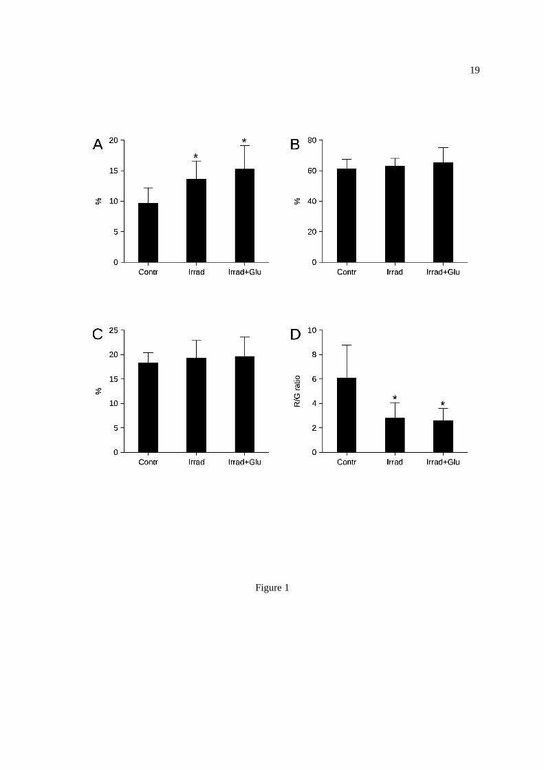
19
Figure 1

20
Figure 2
A
B
C